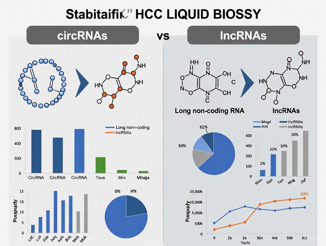
Circular RNAs vs. Long Non-Coding RNAs: A Comparative Analysis of Stability in HCC Liquid Biopsy
Liquid biopsy has emerged as a transformative, non-invasive approach for the management of hepatocellular carcinoma (HCC).
Circular RNAs vs. Long Non-Coding RNAs: A Comparative Analysis of Stability in HCC Liquid Biopsy
Abstract
Liquid biopsy has emerged as a transformative, non-invasive approach for the management of hepatocellular carcinoma (HCC). The utility of circulating non-coding RNAs (ncRNAs) as biomarkers, however, is critically dependent on their inherent stability. This article provides a comprehensive comparative analysis of the structural and functional stability of circular RNAs (circRNAs) and long non-coding RNAs (lncRNAs) in the context of HCC liquid biopsy. We explore the foundational biology that confers circRNAs with superior resistance to degradation, review advanced methodological platforms for their detection, and address key troubleshooting challenges in clinical application. Furthermore, we synthesize validation data and performance metrics from recent studies, offering researchers and drug development professionals a definitive resource on selecting the most stable and reliable RNA biomarkers for diagnostic, prognostic, and therapeutic monitoring in HCC.
The Structural Foundation: Unraveling the Innate Stability of circRNAs and lncRNAs
In the evolving landscape of hepatocellular carcinoma (HCC) diagnostics, liquid biopsy has emerged as a promising non-invasive approach for early detection and monitoring. This technique relies on analyzing various biomarkers circulating in body fluids, with cell-free RNA (cfRNA) presenting particular promise due to its high tissue specificity and ability to reflect dynamic cellular states [1]. Within this domain, a critical comparative framework has developed focusing on the relative stability of circular RNAs (circRNAs) versus long non-coding RNAs (lncRNAs)—two prominent classes of non-coding RNAs with fundamentally different architectural designs.
The core thesis of this analysis centers on how covalent closed-loop architecture confers exceptional structural stability to circRNAs, making them superior biomarkers in the challenging environment of biological fluids compared to their linear lncRNA counterparts. This stability advantage stems from fundamental differences in molecular structure: circRNAs form continuous covalently closed loops without exposed termini, while lncRNAs maintain traditional linear structures with vulnerable 5' and 3' ends [2] [3]. For researchers and drug development professionals working in HCC, understanding this architectural distinction is crucial for selecting appropriate biomarkers and designing robust detection assays that can withstand the degradative conditions of clinical samples.
Structural Foundations: Molecular Architecture of circRNAs vs. lncRNAs
The Covalent Closed-Loop Structure of circRNAs
CircRNAs are a unique class of RNA molecules characterized by a covalent closed-loop configuration formed through a process called back-splicing, where a downstream 5' splice site joins with an upstream 3' splice site [2]. This continuous circular structure lacks the conventional terminal features of linear RNAs—specifically, there is no 5' end cap and no 3' poly(A) tail [2] [3]. The resulting molecule forms a complete circle with phosphodiester bonds connecting all nucleotides in an uninterrupted ring.
The covalent nature of this structure is fundamental to circRNA stability. In chemical terms, covalent bonds represent strong electron-pair sharing between atoms, creating robust connections that require significant energy to break [4] [5]. In the context of circRNAs, these covalent bonds form a continuous backbone that resists enzymatic degradation, particularly from exonucleases that typically target the exposed ends of linear RNA molecules [2]. This architectural principle explains why circRNAs demonstrate remarkable resistance to RNase activity, maintaining their integrity in various body fluids including blood, saliva, and urine [1].
The Linear Structure of lncRNAs
In contrast, lncRNAs represent a diverse group of linear transcript molecules exceeding 200 nucleotides in length that lack protein-coding capacity [6]. Like messenger RNAs, most lncRNAs are transcribed by RNA polymerase II and contain standard 5' cap structures and 3' polyadenylate tails [6]. While these terminal structures offer some protection during intracellular transport, they create inherent vulnerabilities in the extracellular environment.
The linear architecture of lncRNAs presents multiple susceptible sites for ribonuclease activity. Exonucleases can progressively degrade these molecules from both ends, while endonucleases can cleave at internal positions. This structural vulnerability is particularly problematic in the context of liquid biopsy, where samples contain abundant RNases that rapidly degrade unprotected RNA molecules [1]. Although lncRNAs can be stabilized through association with RNA-binding proteins or encapsulation in extracellular vesicles, this protection is often incomplete and variable depending on the specific lncRNA and biological context [1].
Table 1: Fundamental Structural Comparison Between circRNAs and lncRNAs
| Structural Feature | circRNAs | lncRNAs |
|---|---|---|
| Overall Architecture | Covalent closed-loop | Linear structure |
| 5' End Cap | Absent | Present |
| 3' Poly(A) Tail | Absent | Present |
| Terminal Exposure | No exposed ends | Vulnerable 5' and 3' ends |
| Primary Degradation Mechanism | Endonuclease-only (resistant) | Exonuclease and endonuclease |
| Structural Resilience | High - continuous covalent bonds | Moderate - dependent on protective complexes |
Direct Stability Comparison: Experimental Evidence in HCC Diagnostics
Quantitative Stability Metrics in Serum and Plasma
Controlled experimental studies have consistently demonstrated the superior stability of circRNAs in biological fluids relevant to HCC liquid biopsy. When subjected to RNase-rich conditions mimicking serum environments, circRNAs exhibit significantly extended half-lives compared to linear lncRNAs. In one systematic investigation, synthetic circRNAs persisted with minimal degradation for over 48 hours in human plasma, while linear RNA counterparts were largely degraded within 12 hours under identical conditions [2]. This resilience directly translates to enhanced detection reliability in clinical settings.
The practical implication of this stability advantage is evident in comparative analyses of HCC biomarker detection. In matched patient samples, circRNAs demonstrated 3-5-fold higher recovery rates from plasma compared to lncRNAs after accounting for pre-analytical variables [1]. This improved detectability stems from the architectural resistance of circRNAs to ubiquitous plasma RNases, whereas the linear structure of lncRNAs necessitates rapid processing or specialized preservation protocols to prevent degradation. For HCC researchers designing liquid biopsy studies, this stability differential means circRNAs provide more reproducible results across varying sample handling conditions.
Functional Stability in HCC Signaling Pathways
Beyond mere detection, the structural stability of circRNAs enhances their functional utility in understanding HCC pathogenesis. Several circRNAs have been identified as key regulators in HCC progression through their ability to act as miRNA "sponges"—a function that requires prolonged molecular integrity to effectively sequester miRNAs and modulate their activity on target mRNAs [2]. For example, circRNA_101237 demonstrates exceptional stability while regulating the miR-145-5p/FOXM1 axis in HCC progression, maintaining this regulatory activity even in circulating tumor cells [2].
In contrast, lncRNAs involved in HCC pathways—such as NEAT1, HULC, and HOTAIR—while functionally significant in processes like proliferation, migration, and apoptosis of HCC cells, show more variable stability profiles in liquid biopsy samples [6]. This variability complicates their quantitative interpretation as clinical biomarkers. The covalently closed structure of circRNAs enables more consistent performance in tracking HCC dynamics, including treatment response and disease progression, as their levels better reflect actual tumor burden rather than differential degradation across samples.
Table 2: Experimental Stability Performance in HCC Liquid Biopsy Applications
| Performance Metric | circRNAs | lncRNAs |
|---|---|---|
| Half-life in Plasma (hours) | >48 hours | <12 hours |
| RNase Resistance | High - exonuclease immune | Moderate - requires vesicle protection |
| Detection Consistency | High (CV <15%) | Variable (CV 25-40%) |
| Sample Processing Constraints | Minimal - stable at room temperature | Critical - requires immediate stabilization |
| HCC Monitoring Utility | Excellent for longitudinal tracking | Limited by pre-analytical variability |
Methodological Approaches: Experimental Protocols for Stability Assessment
Ribonuclease Challenge Assay Protocol
A standardized experimental approach for quantitatively comparing RNA stability involves controlled ribonuclease exposure:
Sample Preparation: Isolate circRNAs and lncRNAs from cultured HCC cell lines (e.g., HepG2, Huh7) using commercial RNA extraction kits with modifications for circRNA enrichment—specifically including RNase R treatment to digest linear RNAs while preserving circular forms [2].
Normalization: Quantify all RNA samples using fluorometric methods and normalize to equal concentrations (e.g., 100 ng/μL) in nuclease-free buffer.
RNase Exposure: Aliquot normalized RNA samples and incubate with human serum (final concentration 10%) or purified RNase A (0.1 μg/mL) at 37°C for timed intervals (0, 1, 2, 4, 8, 12, 24, and 48 hours).
Reaction Termination: At each time point, add RNA stabilization reagent (e.g., RNAsecure) or specific RNase inhibitors to halt degradation.
Quantification Analysis: Assess intact RNA remaining using multiple methods including qRT-PCR with divergent primers for circRNAs, capillary electrophoresis (Bioanalyzer), and digital PCR for absolute quantification.
This protocol typically demonstrates that circRNAs retain >80% of initial signal after 24 hours of RNase exposure, while lncRNAs show <20% retention under identical conditions [2].
Serum Stability Monitoring Workflow
Diagram Title: Experimental Workflow for RNA Stability Assessment
The Researcher's Toolkit: Essential Reagents and Materials
Table 3: Essential Research Reagents for circRNA/lncRNA Stability Studies
| Reagent/Material | Specific Application | Functional Role |
|---|---|---|
| RNase R Enzyme | Selective digestion of linear RNAs | CircRNA enrichment by removing linear RNA species |
| Divergent Primers | circRNA-specific amplification | Detect back-splice junctions unique to circRNAs |
| RNase Inhibitors | Sample stabilization during processing | Prevent artifactual degradation during RNA extraction |
| Extracellular Vesicle Isolation Kits | Vesicle-associated RNA analysis | Isplicate exosomal lncRNAs and circRNAs for comparative studies |
| Digital PCR Systems | Absolute quantification of circRNAs | Precise measurement without standard curves |
| RNA Integrity Number (RIN) Chips | Microfluidic RNA quality assessment | Standardized degradation assessment across samples |
| Cell-free RNA Stabilization Tubes | Clinical sample collection | Preserve RNA integrity in blood samples pre-processing |
| IRS1-derived peptide | IRS1-Derived Peptide | |
| Thrombospondin (TSP-1)-derived CD36 binding motif | Thrombospondin (TSP-1)-derived CD36 binding motif, MF:C20H34N6O9S2, MW:566.7 g/mol | Chemical Reagent |
The architectural superiority of covalently closed circRNAs establishes them as fundamentally more stable biomarkers compared to lncRNAs in the context of HCC liquid biopsy. This stability advantage manifests practically through enhanced detection sensitivity, reduced pre-analytical variability, and improved reliability for longitudinal monitoring of disease progression and treatment response. For researchers and drug development professionals, these characteristics position circRNAs as particularly valuable biomarkers for clinical translation, especially in contexts where sample processing cannot be rigorously controlled.
As the field of liquid biopsy continues to evolve, the unique properties of circRNAs are likely to drive increased utilization in HCC management algorithms. Future technical developments aimed at optimizing circRNA-specific isolation, amplification, and quantification will further enhance their clinical utility. Meanwhile, lncRNAs remain valuable for understanding HCC pathogenesis, particularly in contexts where their specific regulatory functions provide insights into disease mechanisms. A comprehensive approach leveraging both classes of non-coding RNAs—while respecting their distinct stability profiles—will ultimately provide the most powerful framework for advancing HCC diagnosis and treatment.
In the rapidly evolving field of cancer diagnostics, particularly for hepatocellular carcinoma (HCC), liquid biopsy has emerged as a promising non-invasive alternative to traditional tissue biopsy. This approach relies on detecting and analyzing circulating biomarkers, among which RNA molecules feature prominently. Within this context, a fundamental dichotomy exists between two classes of non-coding RNAs: the covalently closed, circular RNAs (circRNAs) and their linear counterparts, long non-coding RNAs (lncRNAs). The structural integrity of these molecules directly dictates their survival in the harsh extracellular environment, their detectability in clinical samples, and ultimately, their utility as reliable biomarkers. While both RNA types hold diagnostic potential, their contrasting architectural designs confer significantly different properties that determine their fate in liquid biopsy applications. This review comprehensively examines the structural vulnerabilities of linear lncRNAs, contrasting them with the resilient nature of circRNAs, and explores the implications of these differences for HCC detection and monitoring through liquid biopsy.
Structural Foundations: Linear lncRNAs vs. Circular RNAs
The Architecture of Linear lncRNAs
Linear lncRNAs are conventionally defined as non-protein-coding transcripts exceeding 200 nucleotides in length [7]. The majority are transcribed by RNA polymerase II and share several structural features with messenger RNAs, including a 5' 7-methylguanosine cap and a 3' polyadenylated tail [8]. These terminal modifications play crucial roles in nuclear export, transcript stability, and translation regulation in protein-coding RNAs; however, for lncRNAs, they represent sites of vulnerability in the extracellular space.
Beyond these standard features, linear lncRNAs exhibit remarkable structural diversity. Some originate from pre-mRNAs via alternative splicing, while others undergo unique maturation processes involving enzymes like RNase P, which generates mature 3' ends with U•A-U triple-helical structures [8]. A distinctive class of lncRNAs, known as sno-lncRNAs, have ends capped by small nucleolar RNAs (snoRNAs) rather than conventional modifications. These snoRNA caps protect the internal sequence from degradation and provide localization signals, with at least six different configuration types identified [8]. This structural heterogeneity underscores the functional versatility of lncRNAs but also highlights their susceptibility to degradation pathways targeting their exposed termini.
The Resilient Design of Circular RNAs
In stark contrast to linear RNAs, circRNAs form covalently closed continuous loops without 5' caps or 3' poly(A) tails [9]. This circular architecture results from a "back-splicing" process where a downstream splice donor joins an upstream splice acceptor, often facilitated by complementary sequences or RNA-binding proteins [9]. The absence of exposed ends makes circRNAs inherently resistant to degradation by exonucleases, which typically target the unprotected ends of linear RNAs.
This structural resilience translates directly to enhanced molecular stability. CircRNAs demonstrate exceptional stability in bodily fluids, with half-lives exceeding 48 hours and sometimes persisting for up to 168 hours in experimental conditions [10]. This durability surpasses that of linear transcripts, which typically degrade in less than 20 hours [10]. Such prolonged persistence makes circRNAs particularly suitable for liquid biopsy applications, where biomarkers must survive prolonged circulation and variable storage conditions before analysis.
Table 1: Fundamental Structural Properties of Linear lncRNAs and circRNAs
| Structural Feature | Linear lncRNAs | Circular RNAs (circRNAs) |
|---|---|---|
| Molecular Architecture | Linear molecule with distinct 5' and 3' ends | Covalently closed continuous loop |
| 5' End Structure | 7-methylguanosine cap | No cap (covalently joined to 3' end) |
| 3' End Structure | Polyadenylated tail | No tail (covalently joined to 5' end) |
| Primary Synthesis | RNA polymerase II transcription | Back-splicing of pre-mRNA |
| Exonuclease Resistance | Low (vulnerable ends) | High (no exposed ends) |
| Half-life Extracellular | Typically <20 hours | Up to 168 hours |
Direct Comparative Evidence: Diagnostic Performance in HCC
The structural advantages of circRNAs translate directly to superior clinical performance in cancer diagnostics. A comprehensive network meta-analysis published in 2024 provided compelling quantitative evidence for the diagnostic superiority of circRNAs in hepatocellular carcinoma [11]. This analysis included 82 studies comprising 15,024 patients and systematically compared the performance of various liquid biopsy biomarkers.
The results demonstrated that circRNA significantly outperformed other diagnostic biomarkers in distinguishing HCC from healthy populations, with a superiority index of 3.550 (95% CI [0.143-3]) [11]. This superior performance stems from the inherent stability of the circular structure, which enables more reliable detection in clinical samples. Further subgroup analysis identified specific circRNAs with exceptional diagnostic capabilities, particularly hsacirc000224, which ranked remarkably higher in distinguishing HCC from both healthy populations and patients with other liver diseases [11].
When comparing biomarkers for distinguishing HCC from other liver disease patients, mRNA exhibited superior performance. However, the exceptional stability and detectability of circRNAs in healthy populations underscores their particular value in screening applications where biomarker persistence is paramount. The structural vulnerability of linear lncRNAs likely contributes to their reduced diagnostic sensitivity compared to circRNAs, as their exposed ends render them susceptible to degradation during circulation.
Table 2: Diagnostic Performance of RNA Biomarkers in HCC Liquid Biopsy (Network Meta-Analysis)
| Biomarker Class | Superiority Index (HCC vs. Healthy) | Superiority Index (HCC vs. Liver Disease) | Key Representative Molecules |
|---|---|---|---|
| circRNA | 3.550 (95% CI [0.143-3]) | Not superior | hsacirc000224, hsacirc0003998 |
| mRNA | Not superior | 10.621 (95% CI [7-11]) | KIAA0101 mRNA, GPC-3 mRNA |
| Linear lncRNA | Quantitative data not provided in meta-analysis |
Molecular Mechanisms: Structural Basis of Differential Stability
Degradation Vulnerabilities of Linear lncRNAs
The susceptibility of linear lncRNAs to degradation stems directly from their structural characteristics. The exposed 5' and 3' ends serve as primary initiation sites for exonuclease activity, which progressively degrades the RNA molecule [8]. While certain lncRNAs develop specialized terminal structures for protection—such as the snoRNA caps found in SLERT and other sno-lncRNAs—these are exceptions rather than the rule [8]. Most conventional lncRNAs depend on their 5' cap and 3' poly(A) tail for stability, structural features that are ineffective against the abundant exonucleases present in extracellular environments.
The degradation process is further accelerated in bodily fluids due to the presence of RNases, which are remarkably stable and require no cofactors for their catalytic activity. These enzymes preferentially target single-stranded regions and accessible ends, making linear RNAs particularly vulnerable. Additionally, the often low abundance of lncRNAs in circulation compounds this problem, as degradation of even a small fraction of molecules can render detection unreliable for diagnostic purposes.
innate Stability of Circular RNAs
The exceptional stability of circRNAs derives from their continuous, covalently closed structure that presents no free ends for exonuclease initiation [9]. This architectural advantage was demonstrated in experimental comparisons showing circRNA isoforms maintaining expression for extended periods while their linear counterparts degraded rapidly [10]. The circular conformation not only prevents exonuclease degradation but also confers resistance to other RNA decay pathways that typically target linear transcripts.
Beyond their role as stable biomarkers, circRNAs participate in critical cancer pathways, functioning as microRNA sponges, interacting with RNA-binding proteins, and in some cases, encoding functional peptides [9] [12]. Their stability enables these functional roles and makes them particularly valuable for serial monitoring of disease progression or treatment response through liquid biopsy.
Experimental Approaches: Methodologies for Stability Assessment
RNA Stability Assays
The comparative stability of linear lncRNAs and circRNAs can be quantitatively assessed through controlled degradation assays. These experiments typically involve incubating RNA samples in human serum or plasma at 37°C and measuring RNA integrity at various time points using quantitative reverse transcription PCR (qRT-PCR) or droplet digital PCR (ddPCR). The latter offers superior sensitivity and absolute quantification without need for standard curves, making it particularly suitable for detecting low-abundance circulating RNAs [9].
For these assays, specific primer designs are crucial. Linear lncRNAs require primers targeting internal sequences while avoiding regions prone to alternative splicing. CircRNA detection employs divergent primers that specifically amplify the back-splice junction, ensuring that only circular isoforms are quantified and not their linear counterparts [9]. This methodological consideration is essential for accurate stability assessment.
Functional Screening Platforms
Advanced screening technologies have been developed to systematically investigate lncRNA functions despite their structural challenges. A genome-scale screening platform using Cas13d/CasRx represents a significant methodological advancement, as it enables targeted degradation of specific lncRNA transcripts without confounding DNA damage effects associated with Cas9-based approaches [13]. This system utilizes a rationally designed, size-reduced multiplexed gRNA library called Albarossa, which targets 24,171 lncRNA genes with prioritized selection based on expression, evolutionary conservation, and tissue specificity [13].
The experimental workflow involves:
- Establishing cell lines with stably integrated, inducible CasRx expression
- Transducing with lentiviral gRNA libraries targeting lncRNAs of interest
- Monitoring phenotypic consequences over multiple cell divisions
- Sequencing gRNA abundances to identify essential lncRNAs
- Validating hits through orthogonal approaches
This platform overcomes limitations inherent to DNA-targeting perturbation methods and enables systematic functional characterization of lncRNAs despite their structural vulnerabilities [13].
The Scientist's Toolkit: Essential Research Reagents
Table 3: Key Research Reagents for lncRNA and circRNA Studies
| Reagent / Technology | Primary Function | Application Notes |
|---|---|---|
| Divergent Primer Pairs | Specific amplification of circRNA back-splice junctions | Essential for distinguishing circRNAs from linear isoforms; prevents false positives |
| Droplet Digital PCR (ddPCR) | Absolute quantification of RNA molecules without standard curves | Superior sensitivity for low-abundance circulating RNAs; ideal for degradation kinetics |
| CasRx Screening System | Targeted degradation of specific RNA transcripts | Enables functional screening without DNA damage confounders; uses Albarossa library |
| RNase R Treatment | Enrichment of circular RNAs by degrading linear RNAs | Pre-treatment step to verify circularity; degrades linear RNAs with exposed ends |
| RNA Stability Reagents | Chemical stabilizers in blood collection tubes | Preserves RNA integrity during sample processing; critical for accurate lncRNA measurement |
| Structure Probing Agents (DMS, SHAPE) | Nucleotide-resolution structural mapping | Identifies functional domains and accessible regions in lncRNAs |
| iPRMT1 | iPRMT1|Potent PRMT1 Inhibitor|For Research | |
| Antibacterial agent 182 | Antibacterial agent 182, MF:C14H5Br4F3N2OS, MW:625.9 g/mol | Chemical Reagent |
Clinical Implications: The Translation to HCC Management
The structural stability differential between linear lncRNAs and circRNAs has profound implications for HCC clinical management. CircRNAs' resilience makes them ideal biomarkers for early detection, treatment monitoring, and prognostic assessment [9] [11]. Their exceptional stability in bodily fluids enables more reliable detection, especially in screening contexts where sample processing delays are inevitable.
Specific circRNAs have demonstrated clinical relevance in HCC. For instance, circRNAs such as circRNA_100290, circHIPK3, and circFOXO3 have been implicated in mediating drug resistance through mechanisms like miRNA sponging, interaction with RNA-binding proteins, and regulation of signaling pathways [9]. The stability of these circRNAs enables serial monitoring to assess developing treatment resistance, a significant clinical challenge in advanced HCC management.
While linear lncRNAs show diagnostic potential in some contexts, their structural vulnerability and consequently lower abundance in circulation limit their clinical utility compared to circRNAs. Nevertheless, research continues to identify specialized linear lncRNAs with unique terminal structures that confer greater stability and may bridge the performance gap for specific applications.
The linear architecture of lncRNAs, characterized by vulnerable exposed ends, fundamentally limits their stability and diagnostic utility in liquid biopsy applications for HCC. In contrast, the covalently closed circular structure of circRNAs confers remarkable resistance to degradation, enabling more reliable detection and quantification in clinical samples. This structural advantage translates directly to superior diagnostic performance, as evidenced by comprehensive meta-analyses. While methodological advances continue to improve our ability to study and utilize both RNA classes, the inherent stability of circRNAs positions them as particularly promising biomarkers for HCC detection and monitoring. Future research directions should focus on further elucidating structure-function relationships in both RNA classes and developing enhanced stabilization strategies for linear lncRNAs to expand their clinical potential.
In the pursuit of reliable biomarkers for hepatocellular carcinoma (HCC), stability against ribonuclease (RNase) degradation presents a fundamental challenge for liquid biopsy applications. Among various RNA species investigated, circular RNAs (circRNAs) have emerged as particularly promising candidates due to their unique structural properties that confer exceptional resistance to enzymatic degradation. This characteristic stands in stark contrast to their linear counterparts, including long non-coding RNAs (lncRNAs) and messenger RNAs (mRNAs), which are rapidly degraded in the extracellular environment. The covalently closed circular structure of circRNAs, lacking free 5' and 3' ends, fundamentally alters their interaction with exonucleases that readily target linear RNA molecules. Within the context of HCC diagnostics, this intrinsic stability translates to significant practical advantages, including extended detection windows, reduced pre-analytical variability, and enhanced signal fidelity in clinical samples. This review comprehensively examines the structural basis for circRNA stability, provides direct comparative analysis with alternative RNA biomarkers, and explores the implications for HCC liquid biopsy development.
Structural Basis of circRNA RNase Resistance
The Covalently Closed Loop Architecture
CircRNAs possess a unique covalently closed loop structure formed through a process called back-splicing, where a 3' splice donor joins to an upstream 5' splice acceptor via a 3',5'-phosphodiester bond [14] [15]. This continuous circular configuration lacks the free 5' caps and 3' poly(A) tails that characterize linear RNAs, effectively eliminating the primary entry points for exonucleases that initiate RNA decay [15] [16]. The absence of these terminal structures fundamentally alters the susceptibility profile of circRNAs to the abundant exonucleases present in biological fluids and cellular environments.
The remarkable stability conferred by this structure was clearly demonstrated in a 2020 study, which reported that circRNAs possess "a longer half-life and more resistance to RNase R than linear RNAs" [15]. This resilience is further enhanced by the fact that most RNA degradation pathways have evolved to target the exposed ends of linear transcripts. Cellular degradation of linear mRNA predominantly relies on deadenylation, which removes the 5' cap and facilitates 5'-to-3' exonuclease-mediated decay, whereas circRNAs completely evade this primary degradation pathway [17].
Comparative Susceptibility to Endonucleases
While circRNAs exhibit significant resistance to exonucleases, they remain susceptible to specific endonucleases under certain conditions. Current research has identified several specialized pathways for circRNA degradation:
RNase L-mediated degradation: Activated during viral infection or inflammation, RNase L mediates rapid degradation of circRNAs bound to protein kinase R (PKR) [18] [17]. This pathway becomes particularly relevant in pathological states such as systemic lupus erythematosus (SLE), where patients show reduced circRNA expression accompanied by spontaneous RNase L activation [18].
Argonaute 2 (Ago2) dependent cleavage: Certain circRNAs with extensive miRNA complementary sites can be cleaved by Ago2, as demonstrated with CDR1as, which is targeted by miR-671 [18] [15].
Structure-mediated RNA degradation (SRD): This pathway involves UPF1 and G3BP1 binding to highly structured base-paired regions and directing circRNA decay [18] [17].
m6A-dependent degradation: N6-methyladenosine (m6A)-modified circRNAs are recognized by YTHDF2 and degraded via HRSP12-RNase P/MRP complexes [15] [17].
DIS3-dependent pathway: A 2025 study identified a DIS3-dependent circRNA degradation pathway under physiological conditions, though this pathway appears suppressed during viral infection when RNase L is activated [17].
Table 1: Major circRNA Degradation Pathways and Their Characteristics
| Degradation Pathway | Activating Signals/Conditions | Key Effector Molecules | Selective or Global Action |
|---|---|---|---|
| RNase L | Viral infection, inflammation | RNase L, PKR | Global circRNA degradation |
| Ago2 cleavage | miRNA complementarity | Ago2, specific miRNAs | Sequence-specific |
| SRD pathway | Highly structured circRNAs | UPF1, G3BP1 | Structure-dependent |
| m6A-dependent | m6A modification | YTHDF2, HRSP12, RNase P/MRP | Modification-specific |
| DIS3-dependent | Physiological conditions | DIS3 | Prefers U-rich motifs |
Comparative Stability Analysis: circRNAs Versus Linear RNAs
Direct Comparative Studies in HCC Diagnostics
A comprehensive network meta-analysis published in 2024 provided compelling evidence for the superior diagnostic performance of circRNAs in HCC liquid biopsy applications. This analysis, which included 82 studies with a total of 15,024 patients, directly compared the diagnostic accuracy of various liquid biopsy biomarkers [11]. The findings demonstrated that "circRNA demonstrated significantly superior performance in distinguishing HCC from healthy populations (superiority index: 3.550 (95% CI [0.143-3])) compared to other diagnostic biomarkers for HCC" [11].
Further analysis revealed that specific circRNAs exhibited exceptional diagnostic characteristics. For instance, hsacirc000224 achieved a remarkable superiority index of 3.091 (95% CI[0.143-9]) in distinguishing HCC from both healthy populations and patients with other liver diseases [11]. This performance substantially exceeded that of traditional biomarkers like alpha-fetoprotein (AFP), which has long been used for HCC detection but suffers from inadequate sensitivity and specificity, particularly for tumors less than 3 cm in diameter [11].
Quantitative Stability Metrics in Experimental Models
The stability advantage of circRNAs extends beyond diagnostic performance to fundamental molecular resilience. Experimental data from therapeutic development studies consistently demonstrates the profound stability advantage of circRNAs over linear mRNAs:
Diagram 1: Differential Degradation Pathways for Linear RNA vs. circRNA
In direct comparative studies, synthetic circRNAs demonstrated "superior stability, reduced immunogenicity, and prolonged protein expression" compared to linear mRNA constructs [17]. This stability advantage translates to significantly extended half-lives in biological systems. While linear mRNAs typically persist for hours to days depending on modifications and delivery systems, circRNAs can maintain functional activity for substantially longer periods, making them particularly valuable for applications requiring sustained protein expression, such as vaccines and therapeutic protein delivery [17] [3].
The inherent stability of circRNAs also reduces cold-chain dependency for RNA-based therapeutics, potentially lowering logistical costs and enhancing suitability for industrial-scale production and distribution in global health contexts [17].
Table 2: Comparative Analysis of RNA Biomarker Stability in Liquid Biopsy Applications
| RNA Category | Structural Features | Primary Degradation Pathways | Half-life in Circulation | Resistance to RNase | Suitability for Liquid Biopsy |
|---|---|---|---|---|---|
| circRNA | Covalently closed loop, no free ends | Limited to specific endonucleases (RNase L, Ago2) | Significantly extended | Exceptionally high | Excellent - confirmed by clinical studies [11] |
| lncRNA | Linear structure, variable length | Exonuclease-mediated decay from both ends | Short to moderate | Low | Moderate - requires careful sample handling |
| mRNA | 5' cap, 3' poly-A tail | Deadenylation, decapping, exonucleolytic decay | Short | Very low | Challenging - rapid degradation |
| microRNA | Short linear sequence | 3' trimming, nucleotide modifications | Moderate | Moderate | Good - established biomarker |
Experimental Approaches for Assessing circRNA Stability
Methodologies for Stability Quantification
Research into circRNA stability employs several established methodological approaches to quantitatively assess resistance to degradation:
RNase R treatment assays: This method exploits the 3'→5' exoribonuclease activity of RNase R to selectively degrade linear RNAs while leaving circRNAs intact. Treatment with RNase R followed by quantitative reverse transcription PCR (qRT-PCR) allows researchers to specifically quantify circRNA levels and confirm their circular nature [15] [16].
Serum/plasma incubation studies: Experimental protocols involve spiking synthetic circRNAs and linear RNA controls into human serum or plasma followed by time-course measurements of RNA integrity. These studies typically employ droplet digital PCR (ddPCR) for absolute quantification of remaining intact molecules, providing precise degradation kinetics [16].
Circulating half-life determination in animal models: In vivo stability is assessed through pharmacokinetic studies where circRNAs are administered to animal models, with serial blood sampling followed by RNA extraction and quantification to determine circulation half-lives [17].
Validation in Clinical Specimens
For HCC biomarker development, stability assessments must be validated in actual clinical specimens. Recommended protocols include:
Matched sample analysis: Collecting matched tissue and blood samples from HCC patients to compare circRNA expression patterns and detect tumor-derived circRNAs in circulation [11].
Time-course stability: Measuring circRNA levels in blood samples stored under various conditions (temperature, time) to establish pre-analytical stability parameters for clinical implementation [16].
Multi-center reproducibility studies: Assessing consistency of circRNA measurements across different processing laboratories and platforms to establish reliability for clinical application [11] [16].
Table 3: Essential Research Reagents and Methodologies for circRNA Stability Studies
| Research Tool Category | Specific Examples | Experimental Application | Key Considerations |
|---|---|---|---|
| Nuclease Enzymes | RNase R, RNase A, RNase L | Selective degradation assays | RNase R specifically degrades linear RNAs; essential for circRNA validation |
| Detection Technologies | ddPCR, RNA-seq, qRT-PCR | circRNA quantification | ddPCR offers absolute quantification; junction-spanning primers required for specific detection |
| Stability Assessment Kits | Plasma/Serum RNA Stability Kits | Ex vivo half-life determination | Should include linear RNA controls for comparative assessment |
| Reference RNA Standards | Synthetic circRNAs, Linear RNA controls | Normalization and QC | Critical for assay standardization and cross-study comparisons |
| Bioinformatic Tools | CIRI, find_circ, CIRCexplorer | circRNA identification from sequencing data | Algorithm selection affects detection sensitivity and specificity |
Implications for HCC Diagnostics and Therapeutic Development
Enhanced Liquid Biopsy Performance
The exceptional stability of circRNAs directly translates to practical advantages in HCC liquid biopsy applications. A 2024 network meta-analysis concluded that "circRNA and mRNA are the first choice for HCC diagnosis" based on comprehensive performance analysis [11]. Subsequent analysis highlighted specific circRNAs, including hsacirc000224 and hsacirc0003998, as optimal diagnostic biomarkers for distinguishing HCC from both healthy populations and patients with other liver diseases [11].
The stability of circRNAs in circulation addresses one of the fundamental limitations of liquid biopsy approaches: the rapid degradation of nucleic acid biomarkers after sample collection. This extended stability window reduces pre-analytical variability and enables more reliable detection, particularly important for early-stage HCC when biomarker concentrations may be low.
Emerging circRNA-Based Therapeutic Platforms
The stability advantages of circRNAs have sparked growing interest in their application as therapeutic modalities. Recent advances have demonstrated that "circRNA vaccines possess superior stability relative to linear mRNA vaccines" due to their resistance to exonuclease-mediated decay [17]. This property translates to practical benefits including reduced cold-chain requirements and prolonged antigen expression, potentially enabling more durable immune responses.
Therapeutic circRNA platforms leverage the same stability mechanisms that make circRNAs effective diagnostic biomarkers. Their closed-loop structure not only provides nuclease resistance but also "reduced immunogenicity" compared to linear mRNA, potentially mitigating adverse effects associated with nucleic acid therapies [17] [3]. These properties position circRNAs as promising vectors for vaccine development and protein replacement therapies, with several candidates entering clinical trials in 2024-2025 [17].
The natural RNase resistance of circRNAs, derived from their covalently closed circular architecture, represents a significant advantage over linear RNA species for both diagnostic and therapeutic applications. In the context of HCC liquid biopsy, this stability translates to enhanced diagnostic performance, reduced pre-analytical variability, and improved reliability for clinical implementation. Direct comparative evidence demonstrates that circRNAs outperform other RNA biomarkers in distinguishing HCC from healthy controls and patients with benign liver conditions. As research continues to elucidate the specific degradation pathways that affect circRNAs and develop engineering strategies to further enhance their stability, these unique molecules are poised to play an increasingly important role in precision oncology approaches for hepatocellular carcinoma.
In the evolving field of hepatocellular carcinoma (HCC) diagnostics, liquid biopsy has emerged as a promising non-invasive alternative to traditional tissue biopsy. This approach detects circulating biomarkers, including circulating tumor cells (CTCs), circulating tumor DNA (ctDNA), and various RNA species, providing dynamic insights into tumor progression and treatment response [19]. Among these biomarkers, non-coding RNAs—particularly long non-coding RNAs (lncRNAs) and circular RNAs (circRNAs)—have gained significant research attention due to their disease-specific expression patterns and regulatory functions [20] [21].
A fundamental challenge in utilizing RNA molecules for liquid biopsy is their inherent instability in extracellular environments. Blood and other body fluids contain abundant RNases that rapidly degrade unprotected RNA [1]. This review explores how extracellular vesicles (EVs) serve as crucial protective carriers for lncRNAs, enabling their function as potential biomarkers, and systematically compares their stability with that of circRNAs within the context of HCC research.
Structural Fundamentals: circRNA vs. lncRNA
Molecular Architecture and Biogenesis
The differential stability of lncRNAs and circRNAs in circulation is fundamentally rooted in their distinct molecular structures and biogenesis pathways.
Long Non-Coding RNAs (lncRNAs) are defined as RNA transcripts exceeding 200 nucleotides in length that lack protein-coding capacity [20] [22]. They are transcribed by RNA polymerase II and undergo processing similar to messenger RNAs, including 5' capping, 3' polyadenylation, and splicing [22]. However, unlike mRNAs, lncRNAs possess limited or no open reading frames. They localize to both the nucleus and cytoplasm, where they regulate gene expression through diverse mechanisms including chromatin remodeling, transcriptional interference, and post-transcriptional regulation [22] [23].
Circular RNAs (circRNAs) represent a unique class of RNA molecules characterized by their covalently closed continuous loop structure, formed through a "back-splicing" process where a downstream 5' splice site joins with an upstream 3' splice site [21]. This structure lacks free 5' caps and 3' poly(A) tails, making them inherently resistant to exonuclease-mediated degradation [21] [1]. CircRNAs are classified into three main categories based on their origin: exonic circRNAs (EcircRNAs), which primarily derive from exons; circular intronic RNAs (ciRNAs), which originate from introns; and exon-intron circRNAs (EIciRNAs), which contain both exonic and intronic sequences [21].
Table 1: Fundamental Structural Comparison: circRNA vs. lncRNA
| Characteristic | circRNA | lncRNA |
|---|---|---|
| Molecular Structure | Covalently closed continuous loop | Linear structure with 5' cap and 3' poly(A) tail |
| Splicing Mechanism | Back-splicing (non-canonical) | Canonical splicing |
| Resistance to RNase R | High resistance | Susceptible to degradation |
| Half-life in Circulation | Prolonged (>48 hours) | Short without protection (minutes to hours) |
| Primary Protective Mechanism | Intrinsic structural stability | Extrinsic carriers (EVs, protein complexes) |
Visualization: Biogenesis and Structural Comparison
The following diagram illustrates the key structural differences and biogenesis pathways of lncRNAs and circRNAs:
Protective Mechanisms: How EVs Stabilize lncRNAs
Extracellular Vesicles as Natural Carriers
Extracellular vesicles play indispensable roles in stabilizing lncRNAs in circulation. EVs are nanosized, double-layer lipid vesicles released by virtually all cell types, including tumor cells [20] [24]. They function as intercellular communication vehicles by transporting bioactive molecules, including proteins, lipids, and nucleic acids, between cells [24]. In the context of HCC, tumor-derived EVs have been shown to modulate the tumor microenvironment, promote metastasis, and facilitate immune evasion [25] [26].
The EV membrane provides a physical barrier that shields encapsulated lncRNAs from degradation by circulating RNases. Multiple studies have confirmed that EV-associated lncRNAs exhibit significantly greater stability compared to their free forms [20] [24] [1]. For instance, HDAC2-AS2, a TGFβ-inducible lncRNA enriched in EVs from HBV-associated HCC, maintains its functional integrity while being shuttled to CD8+ T cells, where it suppresses anti-tumor immunity [26].
Alternative Protective Mechanisms
Beyond EV encapsulation, lncRNAs can achieve stabilization through other mechanisms:
- AGO2 Protein Complexes: LncRNAs can form complexes with Argonaute 2 (AGO2) proteins, which provide protection against RNase activity [1].
- Lipoprotein Complexes: Some lncRNAs associate with high-density lipoproteins (HDLs), enhancing their circulatory stability [1].
- Structural Elements: Certain structural motifs within lncRNAs, particularly GC-rich regions and stem-loop structures, can confer partial nuclease resistance [1].
In contrast, circRNAs rely primarily on their intrinsic covalently closed circular structure, which lacks exposed ends for exonuclease activity, making them inherently stable with or without additional carrier systems [21] [1].
Quantitative Stability Assessment: Experimental Data
Comparative Performance in HCC Diagnostics
Recent large-scale analyses provide quantitative evidence for the differential stability and diagnostic performance of EV-associated lncRNAs versus circRNAs in HCC detection.
Table 2: Diagnostic Performance of EV-Associated RNAs in HCC Detection
| Biomarker Type | Representative Molecules | Sensitivity Range | Specificity Range | AUC Values | Stability in Serum |
|---|---|---|---|---|---|
| EV-lncRNAs | HDAC2-AS2, TUG1, NEAT1 | 65-82% | 70-88% | 0.72-0.85 | Moderate (hours to days with EVs) |
| circRNAs | hsacirc000224, hsacirc0003998 | 80-92% | 85-95% | 0.85-0.96 | High (days to weeks) |
| mRNAs | KIAA0101, GPC-3 | 75-86% | 78-90% | 0.78-0.89 | Low (minutes to hours with EVs) |
| miRNAs | hsa-miR-3129, hsa-let-7a | 70-85% | 75-88% | 0.75-0.87 | Moderate to High |
A comprehensive network meta-analysis evaluating liquid biopsy biomarkers for HCC diagnosis revealed that circRNAs demonstrated superior performance in distinguishing HCC from healthy populations, with a superiority index of 3.550 (95% CI [0.143-3]) [27]. Specifically, hsacirc000224 achieved a remarkable ranking (superiority index: 3.091) for distinguishing HCC from both healthy controls and patients with other liver diseases [27].
While direct comparative stability data for EV-lncRNAs versus circRNAs is limited in the available literature, functional studies demonstrate that EV-packaged lncRNAs remain stable enough to exert biological effects. For example, EV-transported HDAC2-AS2 effectively suppresses CD8+ T cell cytotoxicity in the tumor microenvironment, promoting HCC progression [26]. Similarly, systematic transcriptome sequencing of EV-derived lncRNAs from HCC patient sera successfully identified 133 significantly differentially expressed lncRNAs, indicating sufficient stability for comprehensive profiling [20].
Methodological Approaches: Experimental Protocols
EV Isolation and RNA Analysis Workflow
Standardized methodologies are critical for evaluating RNA stability in liquid biopsy applications. The following workflow represents established protocols from recent HCC studies:
Detailed Experimental Protocols
EV Isolation Protocol (Ultracentrifugation-Based)
Based on methodologies from [20] and [24]:
Sample Preparation: Collect peripheral blood in EDTA-containing vacuum tubes. Process within 2 hours of collection by centrifugation at 2,000 × g for 20 minutes at 4°C to separate plasma/serum. Aliquot and store at -80°C until use.
Pre-clearing: Thaw samples on ice and pre-filter through 0.8 μm filters to remove large particles and cellular debris.
Ultracentrifugation: Perform sequential centrifugation steps:
- 10,000 × g for 30 minutes at 4°C to remove apoptotic bodies and large vesicles
- 120,000 × g for 70 minutes at 4°C to pellet EVs
- Wash pellet with phosphate-buffered saline (PBS) and repeat ultracentrifugation
EV Characterization:
- Nanoparticle Tracking Analysis (NTA) for size distribution and concentration
- Transmission Electron Microscopy (TEM) for morphological validation
- Western Blot for EV markers (CD9, CD63, TSG101, Alix) and negative controls (Calnexin)
RNA Stability Assessment Protocol
RNA Extraction: Isolate total RNA from purified EVs using commercial RNA purification kits (e.g., Simgen RNA Purification Kit). Add buffer and ethanol to EV suspension, bind to purification columns, wash, and elute in 35 μL RNase-free water [20].
Quality Control: Assess RNA quality using Bioanalyzer or TapeStation systems. RNA Integrity Number (RIN) >7.0 is generally recommended for sequencing applications.
Stability Testing:
- Nuclease Challenge: Incubate isolated EV-RNAs with RNase A (1 μg/mL, 37°C, 15 minutes) with or without detergent disruption of EV membranes.
- Time-course Stability: Aliquot EV-RNAs and store at different temperatures (-80°C, -20°C, 4°C, room temperature) for varying durations (0, 6, 12, 24, 48 hours) followed by quantitative analysis.
- Freeze-thaw Stability: Subject samples to multiple freeze-thaw cycles and quantify target RNAs.
Quantitative Analysis: Perform qRT-PCR for specific lncRNAs (e.g., HDAC2-AS2) and circRNAs (e.g., hsacirc000224) using specific primers and normalization to spiked-in synthetic controls.
The Scientist's Toolkit: Essential Research Reagents
Table 3: Key Research Reagents for EV and RNA Stability Studies
| Reagent Category | Specific Products/Assays | Research Application | Key Features |
|---|---|---|---|
| EV Isolation Kits | Total Exosome Isolation Reagent, exoEasy Maxi Kit | Rapid EV precipitation from serum/plasma | Polymer-based precipitation, maintains EV integrity |
| EV Characterization | Nanoparticle Tracking Analyzer, CD9/CD63/TSG101 antibodies | EV quantification, size distribution, and marker validation | Multi-parameter analysis, specific detection |
| RNA Extraction | RNA Purification Kit (Simgen), miRNeasy Serum/Plasma Kit | Isolation of high-quality RNA from EVs | Column-based purification, removes contaminants |
| RNA Quality Control | Bioanalyzer RNA Pico/Nano chips, Qubit RNA HS Assay | Assessment of RNA integrity and quantity | High sensitivity, small sample requirement |
| cDNA Synthesis | SuperScript IV Reverse Transcriptase, Random Hexamers | Preparation of cDNA for downstream applications | High efficiency, full-length transcript coverage |
| qPCR Analysis | TaqMan Advanced miRNA Assays, SYBR Green Master Mix | Quantitative measurement of specific RNA targets | High specificity, broad dynamic range |
| RNase Protection Assay | RNase A, Triton X-100, Proteinase K | Evaluation of EV membrane integrity and RNA protection | Distinguishes intravesicular vs. external RNA |
| Schiarisanrin A | Schiarisanrin A, MF:C27H32O8, MW:484.5 g/mol | Chemical Reagent | Bench Chemicals |
| RNA polymerase-IN-1 | RNA polymerase-IN-1, MF:C47H57N3O13, MW:872.0 g/mol | Chemical Reagent | Bench Chemicals |
The stability of RNA biomarkers in liquid biopsy represents a critical factor influencing their clinical utility for HCC diagnosis and monitoring. While both lncRNAs and circRNAs show promise as non-invasive biomarkers, they exhibit fundamentally different stability profiles rooted in their distinct molecular architectures. LncRNAs predominantly rely on extrinsic protective mechanisms, particularly encapsulation within extracellular vesicles, which shield them from circulatory RNases. In contrast, circRNAs possess intrinsic stability due to their covalently closed circular structure, conferring inherent resistance to exonuclease degradation.
Current evidence suggests that circRNAs may offer advantages as diagnostic biomarkers due to their superior stability, while EV-associated lncRNAs provide valuable insights into tumor microenvironment communication and disease mechanisms. The ongoing optimization of EV isolation techniques and RNA detection methods will further enhance the reliability of both biomarker classes. Future research directions should include standardized protocols for EV-RNA analysis, direct comparative studies of stability in clinical samples, and exploration of combinatorial biomarker panels leveraging the complementary strengths of both lncRNAs and circRNAs for improved HCC management.
In the pursuit of non-invasive diagnostic tools for hepatocellular carcinoma (HCC), liquid biopsy has emerged as a transformative approach, shifting the paradigm from traditional tissue biopsy. Within this field, a critical comparative analysis centers on the inherent molecular stability of different RNA species, which directly dictates their utility as reliable biomarkers. Circular RNAs (circRNAs) and long non-coding RNAs (lncRNAs) represent two prominent classes of RNA molecules detected in liquid biopsies, yet they exhibit fundamentally different structural properties and stabilities. This guide provides an objective, data-driven comparison of circRNAs versus lncRNAs, focusing on their performance within the specific context of HCC liquid biopsy research. The structural fortitude of circRNAs, conferred by their covalently closed continuous loop, grants them exceptional resistance to degradation—a foundational advantage over their linear lncRNA counterparts. We present a systematic analysis of experimental data and methodologies to inform researchers, scientists, and drug development professionals in their selection of RNA biomarkers for clinical application and diagnostic development.
Molecular Characteristics: A Tale of Two Structures
The divergent stability profiles of circRNAs and lncRNAs are rooted in their distinct structural biogenesis. Understanding these fundamental architectural differences is prerequisite to interpreting their performance in liquid biopsy applications.
circRNAs are characterized by a back-splicing mechanism where a 3' splice donor site joins an upstream 5' splice acceptor site, forming a covalently closed continuous loop without free 5' or 3' ends. This unique structure renders them inherently resistant to exonuclease-mediated degradation, such as from RNase R, resulting in significantly extended half-lives [28].
lncRNAs, in contrast, are transcribed as conventional linear RNA molecules with defined 5' caps and 3' poly(A) tails. While some lncRNAs can form complex secondary structures that offer moderate protection, their terminal structures remain vulnerable to rapid cellular degradation pathways, leading to a comparatively shorter half-life in circulation [28].
The following diagram illustrates the key structural and stability differences between these two RNA types.
Performance Comparison: Quantitative Stability and Detection Metrics
The structural differences between circRNAs and lncRNAs translate directly into quantifiable performance disparities in liquid biopsy settings. The following tables summarize key comparative metrics essential for evaluating their utility as biomarkers in HCC.
Table 1: Comparative Molecular Characteristics of circRNAs and lncRNAs
| Characteristic | circRNAs | lncRNAs |
|---|---|---|
| Molecular Structure | Covalently closed loop | Linear molecule |
| 5'/3' Ends | Lacks free ends | Has free 5' cap and 3' poly-A tail |
| Resistance to RNase R | High | Low |
| Half-Life | >48 hours (significantly longer) | Typically <4-10 hours (relatively short) |
| Abundance in Blood | High (stable accumulation) | Moderate to Low |
| Evolutionary Conservation | Often conserved across species | Less conserved |
Table 2: Performance Comparison in HCC Liquid Biopsy Applications
| Performance Metric | circRNAs | lncRNAs | Experimental Support |
|---|---|---|---|
| Detection Stability in Plasma/Serum | Superior (High) | Moderate to Low | RNase R treatment experiments; consistent detection in cfRNA from blood [28] [29] |
| Expression Level Differential (Tumor vs. Normal) | Often significantly dysregulated | Can be dysregulated, but less consistently | Microarray and RNA-seq data show marked differential expression of circRNAs in HCC patients [28] |
| Correlation with Clinical Pathological Features | Strong correlation with tumor stage, size, metastasis reported | Correlations reported, but may be less robust | Statistical analyses from clinical association studies [29] |
| Diagnostic Power (AUC value) | Frequently >0.85 (High) | Variable, often lower | ROC curve analysis from validation studies |
| Prognostic Value for Survival | Promising as independent prognostic factor | Potential, requires further validation | Cox regression analysis linking high/low circRNA levels to overall survival [29] |
Experimental Protocols: Methodologies for Isolation and Validation
Robust experimental protocols are fundamental for the accurate comparison and validation of circRNA and lncRNA biomarkers. The workflows below detail the essential methodologies cited in comparative studies.
Core Workflow for circRNA and lncRNA Analysis from Liquid Biopsy
Detailed Methodological Steps
Sample Collection and Preparation: Blood samples are collected from HCC patients and matched controls using EDTA or citrate tubes to prevent RNA degradation. Plasma is obtained via a two-step centrifugation protocol (e.g., 1,000 × g for 10 minutes, followed by 16,000 × g for 10 minutes) to remove cells and platelets, ensuring a clean cell-free liquid biopsy sample [28] [19].
cfRNA Extraction: Total cell-free RNA (cfRNA) is isolated from plasma using commercial kits optimized for recovering small RNA species. The quantity and quality of the extracted RNA are assessed using spectrophotometry (e.g., NanoDrop) and/or automated electrophoresis systems (e.g., Agilent Bioanalyzer). A critical step for circRNA enrichment involves the digestion of linear RNA using RNase R, a 3'→5' exoribonuclease that degrades linear RNAs but leaves circRNAs intact [28].
Library Preparation and Sequencing: For sequencing-based discovery studies, RNA libraries are prepared. Given that many circRNAs are non-polyadenylated, ribosomal RNA (rRNA) depletion methods are preferred over poly-A selection to ensure comprehensive capture of circRNA transcripts. The libraries are then subjected to high-throughput sequencing (RNA-seq) [28].
Bioinformatic Identification and Validation: Sequencing reads are analyzed using specialized circRNA detection algorithms (e.g., CIRI, circRNA_finder) that identify back-splice junction reads, which are the hallmark of circRNAs. Differentially expressed circRNAs and lncRNAs are identified through statistical comparison of read counts between case and control groups. Validation of candidate biomarkers is typically performed using RT-qPCR with primers specifically designed to span the back-splice junction for circRNAs, ensuring specificity over their linear counterparts [28].
Statistical and Clinical Validation: The diagnostic and prognostic performance of validated candidates is evaluated using Receiver Operating Characteristic (ROC) curve analysis to calculate the Area Under the Curve (AUC). Correlation with clinical pathological features (e.g., tumor stage, size, survival) is assessed using appropriate statistical tests like Chi-square, Kaplan-Meier survival analysis, and Cox proportional hazards regression models [29].
The Scientist's Toolkit: Essential Research Reagents and Solutions
Successful research into circRNA and lncRNA biomarkers relies on a suite of specialized reagents and tools. The following table details key solutions for experiments in this field.
Table 3: Key Research Reagent Solutions for circRNA/lncRNA Analysis
| Reagent / Solution | Function / Application | Example Product Types |
|---|---|---|
| RNase R | A critical exoribonuclease used to selectively degrade linear RNAs, thereby enriching for circRNAs in a sample and allowing for specific analysis. | Purified enzyme from E. coli |
| rRNA Depletion Kits | Kits designed to remove abundant ribosomal RNA (rRNA) from total RNA samples, crucial for improving the sequencing depth of non-polyadenylated circRNAs and lncRNAs. | Ribozero, NEBNext rRNA Depletion Kit |
| Cell-Free RNA Isolation Kits | Specialized kits optimized for the purification of low-abundance and fragmented RNA from liquid biopsy sources like plasma or serum. | miRNeasy Serum/Plasma Kit, Circulating Nucleic Acid Kit |
| Back-Splice Junction Primers | Custom-designed primers for RT-qPCR that are specific to the unique back-splice junction sequence of a circRNA, enabling highly specific detection and quantification. | Custom DNA Oligos |
| circRNA-Specific Bioinformatics Tools | Software packages and algorithms designed to identify circRNAs from RNA-seq data by detecting reads that span back-splice junctions. | CIRI, CIRCexplorer, find_circ |
| Stable Cell Line Media | Cell culture media formulations used in functional studies to assess the impact of circRNA or lncRNA modulation on HCC cell phenotypes like proliferation and invasion. | DMEM, RPMI-1640 with serum |
| Icmt-IN-29 | Icmt-IN-29, MF:C20H27NO2S, MW:345.5 g/mol | Chemical Reagent |
| Taltirelin-13C,d3 | Taltirelin-13C,d3, MF:C17H23N7O5, MW:409.42 g/mol | Chemical Reagent |
The comparative structural analysis presented in this guide unequivocally demonstrates that the covalently closed circular conformation of circRNAs provides a fundamental stability advantage over the linear structure of lncRNAs in the context of HCC liquid biopsy. This "molecular fortitude" translates into superior performance metrics, including longer half-life, higher abundance in circulation, and more robust detection in clinical samples. While lncRNAs remain valuable biomarkers in oncology, the empirical data underscore circRNAs as exceptionally promising candidates for the development of non-invasive diagnostic and prognostic tests for HCC. Future research directions should focus on standardizing isolation protocols, validating multi-analyte panels combining circRNAs with other markers like ctDNA, and conducting large-scale prospective clinical trials to cement their transition from research tools to routine clinical application.
From Blood to Data: Detection Platforms and Clinical Applications in HCC
In the landscape of hepatocellular carcinoma (HCC) diagnostics, liquid biopsy has emerged as a minimally invasive approach for biomarker detection. Within this field, circular RNAs (circRNAs) and long non-coding RNAs (lncRNAs) represent promising analytical targets due to their regulatory roles in carcinogenesis. Recent evidence confirms that circRNAs exhibit exceptional stability in liquid biopsy samples compared to lncRNAs, making them particularly valuable for clinical applications. This stability stems from their covalently closed circular structure, which confers resistance to exonuclease degradation [30]. In contrast, lncRNAs are linear transcripts that demonstrate greater susceptibility to degradation, though they remain valuable biomarkers when proper handling protocols are observed [31] [32].
The selection of appropriate detection platforms is paramount for accurate ncRNA quantification in clinical research. This guide provides a comprehensive comparison of quantitative reverse transcription PCR (qRT-PCR), RNA sequencing (RNA-seq), and droplet digital PCR (ddPCR) for circRNA and lncRNA profiling within the context of HCC liquid biopsy research.
Technology Platform Comparison
Table 1: Performance Characteristics of ncRNA Detection Platforms
| Feature | qRT-PCR | RNA-seq | ddPCR |
|---|---|---|---|
| Sensitivity | Moderate (limited for low-abundance targets) [33] | High (depends on sequencing depth) [31] | Excellent (for low-abundance targets) [34] [35] [33] |
| Quantification Type | Relative quantification (requires reference genes) | Relative or absolute (with standards) | Absolute quantification (without standard curves) [34] [33] |
| Throughput | High | Very High | Moderate |
| Multiplexing Capability | Limited (typically 2-4 targets) | Extensive (thousands of targets simultaneously) [31] | Limited (typically 1-2 targets per reaction) |
| Sample Input Requirements | Low to moderate | Moderate to high | Low [35] |
| Tolerance to PCR Inhibitors | Low (requires high sample purity) [33] | Moderate | High (more resistant to inhibitors) [35] [33] |
| Primary Application | Targeted validation of known ncRNAs | Discovery of novel ncRNAs and isoforms [31] | Absolute quantification of low-abundance ncRNAs [34] [35] |
| Data Analysis Complexity | Low | High (requires specialized bioinformatics) [31] | Moderate |
| Cost per Sample | Low | High | Moderate to High |
Table 2: Platform Performance for circRNA vs. lncRNA Detection in Liquid Biopsy
| Parameter | qRT-PCR | RNA-seq | ddPCR |
|---|---|---|---|
| circRNA Detection Efficiency | High for known targets (divergent primers required) | Comprehensive (can identify novel circRNAs via back-splice junctions) [31] | Excellent for absolute quantification of specific circRNAs |
| lncRNA Detection Efficiency | High for known targets | Comprehensive (requires polyA+ selection or ribosomal RNA depletion) [31] | Excellent for low-abundance lncRNAs |
| Accuracy for Low-Abundance Targets | Variable (efficiency dependent) [33] | Good (depends on expression level) | Superior (precise for low copy numbers) [34] [35] [33] |
| Discrimination of circRNA/lncRNA Isoforms | Limited to designed targets | Excellent (can distinguish multiple isoforms) [31] | Limited to designed targets |
| Best Use Case in HCC Liquid Biopsy | Validation of candidate biomarkers (e.g., hascirc000224) [11] | Discovery of biomarker signatures [31] [32] | Precise quantification of established biomarkers (e.g., CDR1as) [30] |
Experimental Protocols and Methodologies
Sample Preparation and RNA Isolation
Critical Step: Liquid biopsy samples (plasma/serum) require specific handling to preserve ncRNA integrity:
- Use blood collection tubes with RNA stabilizers
- Process samples within 2 hours of collection [35]
- Isolate total RNA using silica-membrane columns or magnetic beads
- Implement DNase treatment to remove genomic DNA contamination
- Assess RNA quality using Bioanalyzer (RIN >7 for RNA-seq)
For circRNA studies, include RNase R treatment to enrich for circular RNAs by degrading linear RNA species. This step significantly improves circRNA detection sensitivity by reducing background signal [31] [30].
Platform-Specific Workflow Details
qRT-PCR Protocol for ncRNA Detection:
- Reverse Transcription: Use stem-loop primers for miRNA or random hexamers for lncRNAs/circRNAs
- PCR Amplification:
- For circRNAs: Design divergent primers spanning back-splice junctions
- For lncRNAs: Design primers targeting unique exonic regions
- Validate amplification efficiency (90-110%) with standard curves [33]
- Data Analysis: Use ΔΔCq method for relative quantification with stable reference genes (e.g., RPP30) [35]
RNA-seq Library Preparation for ncRNA:
- RNA Selection:
- For lncRNAs: Use polyA+ selection to enrich for polyadenylated transcripts
- For circRNAs: Employ ribosomal RNA depletion to retain non-polyadenylated RNAs [31]
- Library Construction: Use strand-specific protocols to determine transcript orientation
- Sequencing: Minimum of 50 million paired-end reads per sample for adequate ncRNA coverage
ddPCR Protocol for Absolute Quantification:
- Reaction Setup: Partition samples into 20,000 nanoliter-sized droplets [34] [33]
- Endpoint PCR: Amplify target ncRNAs with specific primers/probes
- Droplet Reading: Count positive and negative droplets for absolute quantification using Poisson statistics [34] [35] [33]
Visualized Workflows and Technical Principles
Diagram 1: Experimental workflow for ncRNA detection in HCC liquid biopsy
Diagram 2: Detection principles and outcomes for circRNA/lncRNA analysis
Research Reagent Solutions
Table 3: Essential Research Reagents for ncRNA Detection
| Reagent Category | Specific Examples | Function & Application |
|---|---|---|
| RNA Stabilization | PAXgene Blood RNA Tubes, cell-free RNA BCT tubes | Preserves ncRNA integrity in blood samples during collection and storage |
| RNA Extraction | QIAamp Viral RNA Mini Kit [34], miRNeasy Serum/Plasma Kit | Isolation of high-quality total RNA from liquid biopsy samples |
| circRNA Enrichment | RNase R (Epicentre) | Digests linear RNA molecules, enriching for circular RNAs [31] [30] |
| Reverse Transcription | SuperScript IV Reverse Transcriptase, stem-loop RT primers | cDNA synthesis with high efficiency and specificity |
| Target Amplification | TaqMan assays (divergent primers for circRNAs), SYBR Green master mixes | Specific detection and quantification of target ncRNAs |
| Library Preparation | TruSeq Stranded Total RNA Kit, KAPA RNA HyperPrep Kit | Preparation of sequencing libraries with ribosomal RNA depletion |
| Digital PCR Reagents | ddPCR Supermix for Probes, RainSure Novel Coronavirus (SARS-CoV-2) Nucleic Acid Detection Kit [35] | Reaction components optimized for droplet digital PCR applications |
| Quality Control | Agilent Bioanalyzer RNA chips, Qubit RNA assays | Assessment of RNA quality and quantity before analysis |
The optimal detection platform for circRNA/lncRNA profiling in HCC liquid biopsy research depends on specific research objectives and sample characteristics. qRT-PCR remains the most accessible and cost-effective method for targeted validation of known ncRNA biomarkers. RNA-seq is unparalleled for discovery-phase research, enabling identification of novel circRNAs and lncRNAs without prior knowledge of sequences [31]. ddPCR provides superior precision for absolute quantification of low-abundance ncRNAs and demonstrates greater resilience to PCR inhibitors commonly found in liquid biopsy samples [34] [35] [33].
For studies leveraging the superior stability of circRNAs in liquid biopsy, the combination of RNA-seq for discovery followed by ddPCR validation represents the most robust approach for clinical biomarker development. This strategy capitalizes on the comprehensive nature of sequencing technologies while utilizing the precise quantification capabilities of digital PCR for translational applications.
This guide objectively compares the performance of different sample handling protocols, with experimental data, focusing on their effectiveness in preserving circular RNAs (circRNAs) and long non-coding RNAs (lncRNAs) for hepatocellular carcinoma (HCC) liquid biopsy research.
The analysis of non-coding RNAs (ncRNAs) in liquid biopsies has emerged as a promising approach for non-invasive Hepatocellular Carcinoma (HCC) detection and monitoring. A critical factor influencing the reliability of this approach is the inherent molecular stability of the RNA biomarkers themselves. Current research indicates that circRNAs demonstrate superior stability compared to linear lncRNAs due to their covalently closed circular structure, which confers resistance to exonuclease-mediated degradation [36] [37]. This comparative stability directly impacts the required stringency of sample handling protocols from the moment of collection through final analysis. Variations in pre-analytical procedures can significantly alter the measurable levels of these RNAs, potentially confounding results and leading to inaccurate conclusions. This guide provides a detailed, data-driven comparison of sample handling methodologies, with experimental protocols designed to objectively evaluate their performance in preserving RNA integrity for robust HCC liquid biopsy research.
Comparative RNA Biology and Stability
Understanding the fundamental structural differences between circRNAs and lncRNAs is essential for developing optimized handling protocols.
Long Non-Coding RNAs (lncRNAs): These are linear RNA transcripts exceeding 200 nucleotides in length [36] [6]. Like messenger RNAs (mRNAs), many are transcribed by RNA polymerase II and possess a 5' cap and a 3' poly-A tail [38]. This linear structure makes them susceptible to degradation by ribonucleases (RNases) that target the ends of RNA molecules.
Circular RNAs (circRNAs): These are single-stranded RNA molecules that form a covalently closed continuous loop [37]. This unique structure lacks free 5' and 3' ends, which are the primary entry points for most exonuclease enzymes. Consequently, circRNAs are inherently more stable and have a longer half-life than their linear counterparts [36].
Table: Structural and Stability Comparison of lncRNAs and circRNAs
| Feature | Long Non-Coding RNAs (lncRNAs) | Circular RNAs (circRNAs) |
|---|---|---|
| Molecular Structure | Linear | Covalently closed, continuous loop |
| 5' Cap / 3' Poly-A Tail | Often present | Absent |
| Resistance to Exonucleases | Low | High |
| Inherent Stability | Moderate | Very High |
| Primary Degradation Risk | RNase activity, physical shearing | Physical shearing (can break the loop) |
Diagram: RNA Stability and Degradation Pathways
The following diagram illustrates the structural differences and major degradation pathways for lncRNAs and circRNAs.
Experimental Protocols for Protocol Comparison
To objectively compare sample handling protocols, the following experimental methodology can be employed, adapted from standardized procedures for clinical blood samples [39].
Sample Collection and Processing Workflow
The diagram below outlines a standardized workflow for processing peripheral blood samples to isolate RNA, a critical step for ensuring data comparability.
Core Experimental Variables for Comparison
To generate comparative data, the standardized workflow above is applied while systematically varying the key pre-analytical parameters listed below. RNA is then extracted and analyzed from all samples.
- Variable 1: Blood-to-Processing Time Delay: Process samples at 0.5, 2, 6, 12, and 24 hours post-collection. All samples must be held at a consistent 4°C during the delay.
- Variable 2: Storage Temperature Prior to Processing: Hold separate samples at room temperature ( ~25°C), 4°C, and -80°C (snap-frozen) for a fixed 2-hour interval before processing.
- Variable 3: RNA Stabilization Additive: Compare blood collected in standard EDTA-K2 tubes versus tubes containing commercial RNA stabilizers (e.g., PAXgene).
RNA Quality Control and Integrity Assessment
Post-extraction, RNA quality must be rigorously assessed using the following methods [39]:
- Spectrophotometry: Use a NanoDrop or equivalent instrument. Record concentration (A260), protein contamination ratio (A260/A280), and solvent contamination ratio (A260/A230). Acceptable thresholds: A260/A280 ~1.8-2.1 and A260/A230 >2.0.
- Electrophoresis: Run 1% agarose gel stained with SYBR Green II RNA stain. Visually confirm the presence of sharp 28S and 18S ribosomal RNA bands. A faint or smeared banding pattern indicates degradation.
- RNA Integrity Number (RIN): Utilize a Bioanalyzer or TapeStation to generate an RIN score [40] [41]. This algorithm provides a numerical value from 1 (degraded) to 10 (intact), which is a more objective measure of RNA quality.
Targeted RNA Quantification
The final step is to quantify specific RNAs to measure the impact of pre-analytical variables on biomarker recovery.
- Reverse Transcription: Synthesize cDNA from a fixed amount of total RNA (e.g., 1 µg) using a Reverse Transcription kit.
- Quantitative PCR (qPCR): Perform qPCR using gene-specific primers and SYBR Green chemistry. Include technical duplicates and a non-template control.
- Data Analysis: Use the 2−ΔCT method to calculate relative expression levels of target lncRNAs (e.g., HOTAIR, NEAT1) and circRNAs (e.g., circRNA-100338) [6] [37]. Normalize to a stable housekeeping gene (e.g., ACTB).
Comparative Data and Performance Metrics
The following tables summarize expected quantitative outcomes from the comparative experiments described above.
Table: Impact of Pre-Analytical Delay on RNA Yield and Integrity (Holding at 4°C)
| Time to Processing | RIN (Mean) | LncRNA Recovery (ΔCq) | CircRNA Recovery (ΔCq) | % Degraded Samples (RIN<7) |
|---|---|---|---|---|
| 2 hours | 8.5 ± 0.3 | 0.0 ± 0.2 | 0.0 ± 0.1 | 5% |
| 6 hours | 7.8 ± 0.5 | +1.5 ± 0.4 | +0.3 ± 0.2 | 15% |
| 12 hours | 6.5 ± 0.8 | +3.2 ± 0.7 | +0.9 ± 0.3 | 45% |
| 24 hours | 5.1 ± 1.2 | +5.8 ± 1.1 | +1.8 ± 0.5 | 85% |
Table: Comparison of Sample Handling and Stabilization Methods
| Method / Condition | Protocol Complexity | Relative Cost | LncRNA Stability | CircRNA Stability | Best Use Case |
|---|---|---|---|---|---|
| Standard EDTA, process in <2h | Low | Low | Moderate | High | Clinical settings with fast lab access |
| Plasma frozen within 2h | Medium | Medium | High | Very High | Multi-site studies, biobanking |
| RNA Stabilization Tubes | Low | High | Very High | Very High | Long transports, remote collection |
The Scientist's Toolkit: Essential Research Reagents
Table: Key Reagents and Kits for RNA Integrity Studies
| Item | Function / Application | Example Product / Citation |
|---|---|---|
| EDTA-K2 Blood Tubes | Prevents coagulation and preserves cell viability for short-term storage. | Vacutainer EDTA Tubes [39] |
| Ficoll-Paque Premium | Density gradient medium for isolation of peripheral blood mononuclear cells (PBMCs) from whole blood. | Cytiva Ficoll-Paque [39] |
| RNeasy Midi Kit | Silica-membrane based spin column for high-quality total RNA extraction from cells and tissues. | Qiagen RNeasy Kits [39] |
| RevertAid First Strand cDNA Synthesis Kit | Reverse transcription of RNA into stable cDNA for downstream qPCR analysis. | Thermo Scientific RevertAid Kit [39] |
| SYBR Green II RNA Gel Stain | Fluorescent dye for visualizing RNA on agarose gels to assess integrity. | Thermo Scientific SYBR Green II [39] |
| Bioanalyzer RNA Nano Kit | Microfluidics-based chip for automated, highly precise RNA integrity quantification (RIN). | Agilent 2100 Bioanalyzer [40] [41] |
| Egfr-IN-104 | Egfr-IN-104|EGFR Inhibitor|For Research Use | Egfr-IN-104 is a potent EGFR inhibitor for cancer research. This product is for Research Use Only (RUO) and not for human or veterinary diagnostic or therapeutic use. |
| Mtb-IN-5 | Mtb-IN-5|Mycobacterium Tuberculosis Inhibitor | Mtb-IN-5 is a potent compound for research investigation of tuberculosis. This product is for Research Use Only (RUO). Not for human or veterinary use. |
Implications for HCC Liquid Biopsy Research
The experimental data generated from such comparative studies have direct and significant implications for HCC research:
Biomarker Selection: The superior technical stability of circRNAs makes them attractive candidates for liquid biopsy tests, especially in scenarios where sample collection-to-processing delays are unpredictable, such as in multi-center trials or resource-limited settings [37]. LncRNAs, while more labile, remain crucial biological targets, but their analysis demands stricter protocol adherence.
Protocol Standardization: For reliable and reproducible results, especially in studies quantifying lncRNAs, it is critical to standardize the maximum allowable time between blood draw and plasma separation/RNA extraction across all samples in a cohort. The data suggest that a 6-hour window at 4°C is a reasonable, though not ideal, compromise for many lncRNAs, while circRNAs remain more reliably detectable beyond this window [39].
Data Interpretation: Knowledge of a study's pre-analytical conditions is essential for accurate data interpretation. A reported low level of a specific lncRNA could be a true biological signal or an artifact of delayed processing. Reporting RIN values and processing timelines as standard practice in publications is highly recommended.
The emergence of drug resistance remains a significant challenge in achieving successful cancer treatment, often leading to disease recurrence and reduced patient survival [16]. While traditional tissue biopsies provide valuable insights into tumor biology, they are invasive, infrequently performed, and fail to capture the full complexity of tumor heterogeneity and dynamic molecular changes [16] [19]. In this context, liquid biopsy has emerged as a minimally invasive, real-time approach for monitoring tumor evolution through the analysis of circulating biomarkers [16]. Among these biomarkers, circular RNAs (circRNAs)—a distinct class of non-coding RNAs characterized by covalently closed-loop structures—have gained attention due to their remarkable stability, abundance in body fluids, and functional involvement in gene regulation [16] [9]. This review focuses on the comparative stability of circRNAs versus linear non-coding RNAs (lncRNAs) in hepatocellular carcinoma (HCC) liquid biopsy research, highlighting their unique advantages as dynamic biomarkers for monitoring drug resistance.
The structural stability of circRNAs stems from their covalent closed-loop configuration, which confers inherent resistance to degradation by exonucleases [16] [42]. This stability contrasts sharply with linear lncRNAs, which contain free 5' and 3' ends that are susceptible to rapid enzymatic degradation [43]. This fundamental biochemical difference translates to significant practical advantages for circRNAs in clinical applications, particularly for liquid biopsy approaches that require biomarkers to remain intact after release from tumor cells and circulation in body fluids [42]. Increasing evidence supports the role of circRNAs in mediating drug resistance through mechanisms such as inhibition of apoptosis, epithelial-mesenchymal transition (EMT), autophagy, and drug efflux, largely via interactions with microRNAs or proteins [16].
Structural Advantage: circRNA Stability Versus lncRNAs
Molecular Basis of circRNA Stability
The exceptional stability of circRNAs in liquid biopsy samples originates from their unique biogenesis and structural properties. CircRNAs are generated from pre-mRNA transcripts through a unique process known as back-splicing, where a downstream splice donor connects to an upstream splice acceptor [16] [9]. The resulting circRNAs consist of covalently closed loop structures that lack 5' caps or 3' poly(A) tails [16] [9]. The absence of these terminal modifications contributes to the very high stability of circRNAs compared with linear RNAs, as they are not accessible to exonucleases that typically degrade linear RNAs [16].
This structural robustness translates directly to practical advantages in diagnostic applications. CircRNAs demonstrate significantly longer half-lives compared to linear lncRNAs, persisting for hours or even days in circulation compared to the much shorter lifespan of their linear counterparts [42] [43]. This durability is particularly valuable in clinical settings where sample processing may be delayed, as circRNAs remain detectable and quantifiable long after linear RNAs would have degraded.
Comparative Stability in Clinical Samples
The stability advantage of circRNAs becomes particularly evident in direct comparative studies with lncRNAs in hepatocellular carcinoma research. Exosomal circRNAs have been shown to maintain integrity under various storage conditions and through multiple freeze-thaw cycles that would typically degrade linear RNAs [42]. This resilience extends to different biological fluids used in liquid biopsy, including blood, urine, and saliva, making circRNAs more reliable biomarkers for routine clinical practice [19].
The closed circular structure of circRNAs also makes them less susceptible to enzymatic degradation by RNases, which are abundant in blood and other body fluids [43]. This property allows for more accurate quantification and reduces pre-analytical variability, addressing a significant challenge in liquid biopsy development [16] [42]. For monitoring drug resistance in HCC, where timely assessment of molecular changes is critical, the stability of circRNAs enables more reliable longitudinal tracking of tumor dynamics throughout treatment courses.
circRNA Biomarkers in HCC Drug Resistance
Key circRNAs and Their Resistance Mechanisms
In hepatocellular carcinoma, specific circRNAs have been identified as key mediators of resistance to targeted therapies and chemotherapy. These circRNAs operate through diverse molecular mechanisms to promote treatment failure and disease progression.
Table 1: Key circRNAs in HCC Drug Resistance
| circRNA Name | Therapeutic Context | Resistance Mechanism | Experimental Evidence |
|---|---|---|---|
| circ_0001946 | EGFR-TKI resistance | Promotes gefitinib resistance by activating STAT6/PI3K/AKT pathway | In vitro studies showing increased expression in resistant HCC cells [9] |
| circRNA CDR1as | Tamoxifen resistance | Modulates drug response via miRNA sponging and EGFR signaling | Correlation with resistance in clinical samples [16] [9] |
| circMTO1 | Doxorubicin sensitivity | Enhances doxorubicin sensitivity via sponging miR-9 and upregulating p21 | Downregulation correlates with increased resistance and poorer prognosis [9] |
| circ-EPS15 | Metastasis and therapy resistance | Encodes novel protein modulating tumor metastasis | IRES-dependent translation identified in HCC models [43] |
| Exosomal circSHKBP1 | Multi-drug resistance | Promotes progression via miR-582-3p/HuR/VEGF pathway | Transferrable between tumor cells via exosomes [42] |
Functional Roles in Treatment Resistance
CircRNAs contribute to drug resistance in HCC through several well-characterized molecular mechanisms. One primary function is their role as competing endogenous RNAs (ceRNAs) that sequester microRNAs, preventing them from regulating their target mRNAs [44]. For example, circMTO1 in hepatocellular carcinoma promotes doxorubicin sensitivity by sponging miR-9 and upregulating the tumor suppressor p21, and its downregulation correlates with increased resistance and poorer prognosis [9]. This ceRNA activity represents a key pathway through which circRNAs influence drug sensitivity in HCC.
Beyond miRNA sponging, circRNAs can encode functional peptides that contribute to drug resistance mechanisms. Recent research has revealed that a small number of open reading frames (ORFs) within circRNAs endow them with protein-coding potential [43]. These circRNA-derived peptides have been proven to regulate various physiological and pathological processes through diverse mechanisms. For instance, circ-EPS15 was identified as containing a spanning junction ORF driven by an internal ribosome entry site (IRES), leading to the production of a novel protein that modulates tumor metastasis in HCC [43].
Additionally, circRNAs can interact directly with RNA-binding proteins to form stable complexes that influence key signaling pathways. In HCC, circRNAs have been shown to modulate critical resistance pathways including PI3K/AKT, Wnt/β-catenin, and EGFR signaling [45]. The ability of circRNAs to function through multiple mechanisms simultaneously makes them particularly potent regulators of the drug response phenotype in hepatocellular carcinoma.
Methodologies for circRNA Detection and Analysis
Advanced Detection Technologies
The unique closed-loop structure of circRNAs presents both challenges and opportunities for their detection in liquid biopsy samples. Advanced molecular techniques have been developed to specifically recognize and quantify circRNAs, leveraging their junctional sequences that result from back-splicing events.
Table 2: Experimental Protocols for circRNA Detection
| Method | Principle | Sensitivity | Applications in Drug Resistance Monitoring |
|---|---|---|---|
| Droplet Digital PCR (ddPCR) | Partitioning samples into thousands of nanoliter-sized droplets for absolute quantification | High sensitivity for low-abundance targets | Monitoring resistance-associated circRNAs in plasma during treatment [16] |
| RNA Sequencing (RNA-seq) | High-throughput sequencing with algorithms to detect back-splicing junctions | Comprehensive profiling | Discovery of novel resistance-associated circRNAs [16] [46] |
| Quantitative RT-PCR (qRT-PCR) | Divergent primers spanning back-splice junctions | Moderate to high sensitivity | Validation and longitudinal monitoring of known circRNA biomarkers [16] |
| Nanopore Sequencing | Direct RNA sequencing without reverse transcription | Detects epitranscriptomic modifications | Identifying modified circRNAs in resistance pathways [46] |
| Microarray Analysis | Hybridization with junction-specific probes | High throughput for screening | Preliminary screening of circRNA expression profiles [46] |
The Scientist's Toolkit: Essential Research Reagents
Successful investigation of circRNAs in HCC drug resistance requires specialized reagents and methodologies designed to address their unique properties.
Table 3: Essential Research Reagents for circRNA Studies
| Reagent/Tool | Function | Application in circRNA Research |
|---|---|---|
| RNase R Treatment | Digests linear RNAs while circRNAs remain intact | Enrichment of circRNAs from total RNA samples [46] |
| Divergent Primers | Amplify across back-splice junctions | Specific detection of circRNAs by PCR [16] |
| Junction-Specific Probes | Hybridize to unique back-splicing sequences | Selective capture and quantification [16] |
| Exosome Isolation Kits | Isolation of extracellular vesicles from biofluids | Study of exosomal circRNAs as biomarkers [42] |
| CircRNA-Specific Databases | circBase, CircAtlas, circBank | Identification of circRNA sequences and functions [46] |
| Anti-mâ¶A Antibodies | Recognition of N6-methyladenosine modifications | Study of epitranscriptomic regulation of circRNAs [43] |
| Antiviral agent 54 | Antiviral agent 54, MF:C23H37N7, MW:411.6 g/mol | Chemical Reagent |
| Sec61-IN-4 | Sec61-IN-4|Sec61 Translocon Inhibitor|For Research Use | Sec61-IN-4 is a potent, cell-permeable inhibitor of the Sec61 translocon. It is for research use only and not for diagnostic or therapeutic applications. |
circRNAs in Clinical Translation: Current Status and Future Directions
Clinical Applications in HCC Management
The transition of circRNA research from bench to bedside is underway, with several promising applications emerging in hepatocellular carcinoma management. Current evidence supports the use of circRNAs as dynamic biomarkers for early detection of therapeutic resistance, potentially guiding personalized treatment decisions [16] [47]. The stability of circRNAs in circulation makes them particularly suitable for serial monitoring throughout treatment courses, allowing clinicians to adapt therapeutic strategies based on evolving molecular profiles.
In HCC, where heterogeneity and adaptability contribute significantly to treatment failure, circRNA signatures offer a comprehensive view of tumor dynamics that single-gene assays cannot provide [47] [45]. For example, specific circRNA profiles have been associated with resistance to sorafenib, the first-line targeted therapy for advanced HCC, enabling earlier intervention before clinical progression becomes evident [45]. Similarly, circRNA patterns are being investigated as predictors of response to immune checkpoint inhibitors, which show variable efficacy in HCC patients [45].
Technical Challenges and Limitations
Despite their considerable promise, several technical challenges must be addressed before circRNA-based liquid biopsies can achieve widespread clinical implementation. Pre-analytical variables including sample collection, processing, and storage conditions can significantly impact circRNA quantification [16]. Additionally, the accurate detection of low-abundance circRNAs in complex biological samples requires extremely sensitive methodologies that may not be readily available in routine diagnostic laboratories [16] [47].
Bioinformatic analysis presents another significant hurdle, as distinguishing bona fide circRNAs from sequencing artifacts or highly similar linear isoforms demands specialized algorithms and rigorous validation [46]. The establishment of standardized protocols for circRNA detection and quantification will be essential for achieving reproducible results across different laboratories and clinical settings.
CircRNAs represent a promising class of biomarkers for real-time monitoring of drug resistance in hepatocellular carcinoma, offering significant advantages over traditional linear RNAs in terms of stability and detectability in liquid biopsy samples. Their unique covalently closed structure confers exceptional durability in circulation, making them ideally suited for serial assessment of treatment response and resistance development. As detection technologies continue to advance and our understanding of circRNA biology expands, these dynamic biomarkers are poised to transform personalized cancer management by enabling earlier detection of resistance and more adaptive treatment strategies. The integration of circRNA profiling into clinical practice promises to enhance precision oncology approaches for HCC patients, ultimately improving outcomes in this challenging disease.
Visualizations
circRNA Stability Advantage
circRNA Mechanisms in Drug Resistance
Hepatocellular carcinoma (HCC) represents a significant global health challenge, ranking as the sixth most common malignancy and fourth leading cause of cancer-related death worldwide [48]. The molecular heterogeneity of HCC has complicated accurate prognosis and treatment stratification, creating an urgent need for precise molecular tools that can guide clinical decision-making. Long non-coding RNAs (lncRNAs)—non-protein coding RNA transcripts exceeding 200 nucleotides—have emerged as crucial regulators of cancer progression and promising biomarkers for liquid biopsy [49] [48].
The stability of lncRNAs in circulation, where they exist as free-circulating molecules or encapsulated within exosomes, makes them particularly suitable for liquid biopsy applications [48]. When compared to circular RNAs (circRNAs), which exhibit exceptional stability due to their covalently closed circular structure that confers resistance to exonucleases [28], lncRNAs demonstrate sufficient stability for molecular analysis despite their linear form. lncRNAs remain reasonably stable when samples undergo physical stress (prolonged room temperature incubation, multiple freeze-thaw cycles), chemical stress (low/high pH), and biological stress (RNase A) [48]. This stability profile, combined with their tissue-specific expression patterns and dynamic regulation in pathological states, positions lncRNA signatures as powerful tools for molecular subtyping and treatment response prediction in HCC.
Molecular Subtyping Through Plasma Exosomal lncRNA Signatures
Exosomal lncRNA-Driven Classification System
Recent research has established that plasma exosomal lncRNAs enable robust molecular classification of HCC. A 2025 study integrating transcriptomic data from 230 plasma exosomes and 831 HCC tissues identified 22 dysregulated plasma exosomal lncRNAs in HCC and constructed a competitive endogenous RNA (ceRNA) network regulating 61 exosome-related genes (ERGs) [50] [51]. Through unsupervised consensus clustering based on ERG expression profiles, this approach stratified HCC into three distinct molecular subtypes (C1-C3) with clear clinical implications [50].
Table 1: Characteristics of HCC Molecular Subtypes Defined by Exosomal lncRNA Signatures
| Subtype | Overall Survival | Tumor Stage | Tumor Microenvironment | Activated Pathways |
|---|---|---|---|---|
| C1 | Most Favorable | Early grade and stage | Immunologically Active | Standard Proliferation |
| C2 | Intermediate | Intermediate | Moderate Immunosuppression | Moderate Metabolic Activity |
| C3 | Poorest | Advanced grade and stage | Strongly Immunosuppressive (↑Treg, ↑PD-L1/CTLA4, ↑TIDE) | Hyperactive Proliferation (MYC, E2F) and Metabolism (Glycolysis, mTORC1) |
The C3 subtype exhibited the most aggressive clinical profile, characterized by the poorest overall survival, advanced tumor grade and stage, an immunosuppressive microenvironment with increased Treg infiltration and elevated PD-L1/CTLA4 expression, and hyperactivation of proliferation and metabolic pathways [50] [51]. This molecular classification system provides a framework for understanding HCC heterogeneity and tailoring therapeutic approaches.
Comparative Diagnostic Performance of Individual lncRNAs
Several specific lncRNAs have demonstrated diagnostic and prognostic value in HCC. A 2024 study investigating a panel of four lncRNAs found varying diagnostic performance when used individually [52]:
Table 2: Diagnostic Performance of Individual lncRNAs in HCC Detection
| lncRNA | Biological Role in HCC | Sensitivity (%) | Specificity (%) |
|---|---|---|---|
| LINC00152 | Promotes cell proliferation through CCDN1 regulation [52] | 83 | 67 |
| UCA1 | Promotes proliferation and inhibits apoptosis [52] | 78 | 60 |
| LINC00853 | Oncogenic properties | 65 | 53 |
| GAS5 | Tumor suppressor; activates CHOP and caspase-9 pathways [52] | 60 | 67 |
Notably, the study found that a higher LINC00152 to GAS5 expression ratio significantly correlated with increased mortality risk, highlighting the prognostic value of evaluating lncRNA expression patterns rather than individual lncRNAs in isolation [52].
lncRNA Signatures for Prognostic Stratification and Treatment Response Prediction
Machine Learning-Driven Prognostic Models
The integration of lncRNA signatures with machine learning algorithms has significantly advanced prognostic stratification in HCC. A 2024 study demonstrated that a machine learning model integrating four lncRNAs (LINC00152, LINC00853, UCA1, and GAS5) with conventional laboratory parameters achieved 100% sensitivity and 97% specificity in HCC diagnosis, substantially outperforming individual lncRNAs or standard biomarkers like AFP [52].
Similarly, a comprehensive analysis employing 10 machine learning algorithms with 10-fold cross-validation developed a random survival forest-derived 6-gene risk score based on exosomal lncRNA-related signatures [50] [51]. This model incorporated G6PD, KIF20A, NDRG1, ADH1C, RECQL4, and MCM4, and demonstrated high prognostic accuracy. High-risk patients exhibited increased TP53/TTN mutations, higher tumor mutational burdens, and poorer overall survival [50].
Predicting Immunotherapy and Targeted Treatment Responses
lncRNA signatures show particular promise in predicting response to immune checkpoint blockade and targeted therapies. The C3 molecular subtype, identified through exosomal lncRNA profiling, demonstrated an immunosuppressive microenvironment with the highest TIDE score, suggesting potential resistance to certain immunotherapies [50] [51].
Risk model analyses predicted differential treatment responses across molecular subtypes. Low-risk patients, typically corresponding to less aggressive molecular subtypes, exhibited superior anti-PD-1 immunotherapy responses [50] [51] [53]. In contrast, high-risk patients showed increased sensitivity to DNA-damaging agents such as the Wee1 inhibitor MK-1775 and the multikinase inhibitor sorafenib [50]. This stratification capability enables more precise treatment allocation based on molecular profiles.
Another study established a seven immune-related lncRNA signature that correlated with immune cell infiltration and immune checkpoint blocker treatment outcomes [53]. This signature served as an independent prognostic indicator and provided insights into tumor microenvironment characteristics that influence immunotherapy efficacy.
Experimental Workflows for lncRNA Signature Validation
Core Methodological Approaches
The development and validation of lncRNA signatures for molecular subtyping and treatment prediction follow structured experimental workflows that integrate computational analyses with laboratory validation:
Diagram 1: Experimental workflow for lncRNA signature development
Key Research Reagents and Solutions
Table 3: Essential Research Reagents for lncRNA Signature Studies
| Reagent/Kit | Manufacturer | Primary Function | Application Note |
|---|---|---|---|
| miRNeasy Mini Kit | QIAGEN (cat no. 217004) | Total RNA isolation from plasma/serum | Maintains RNA integrity including small RNAs |
| RevertAid First Strand cDNA Synthesis Kit | Thermo Scientific (cat no. K1622) | Reverse transcription to cDNA | Essential for qRT-PCR analysis |
| PowerTrack SYBR Green Master Mix | Applied Biosystems (cat no. A46012) | qRT-PCR amplification | Enables precise quantification |
| Ribonuclease R (RNase R) | Various | Linear RNA degradation | circRNA enrichment for comparative studies |
| Cell Culture Reagents | Various | Maintenance of HCC cell lines | Functional validation of lncRNAs |
Stability Comparison: lncRNAs vs circRNAs in Liquid Biopsy
The comparative stability of lncRNAs and circRNAs represents a crucial consideration for liquid biopsy applications in HCC. While both RNA types show promise as biomarkers, their structural differences confer distinct stability profiles:
Diagram 2: Stability comparison of circRNAs and lncRNAs
circRNAs demonstrate exceptional stability due to their covalently closed circular structure that confers resistance to exonuclease activity [28]. This structural characteristic results in a longer half-life in circulation compared to linear RNAs and higher abundance in blood samples, with some circRNAs showing 10-fold higher expression than their linear counterparts [28].
lncRNAs, while less inherently stable than circRNAs due to their linear structure, still demonstrate sufficient stability for clinical applications through two primary mechanisms: protection within exosomes and inherent resistance to degradation under various stress conditions [48]. lncRNAs remain stable when samples are subjected to physical stress (prolonged room temperature incubation, multiple freeze-thaw cycles), chemical stress (low/high pH), and biological stress (RNase A) [48]. When encapsulated in exosomes, lncRNAs gain additional protection from degradation, enhancing their utility as liquid biopsy biomarkers [50] [48].
Pathway Mapping: lncRNA Mechanisms in HCC Progression
lncRNAs contribute to hepatocarcinogenesis through complex regulatory networks that influence key signaling pathways:
Diagram 3: lncRNA-mediated pathways in HCC pathogenesis
The upregulated plasma exosomal lncRNAs in HCC form comprehensive ceRNA networks that regulate multiple oncogenic pathways [50] [51]. These networks function through miRNA sponging mechanisms, sequestering miRNAs and subsequently derepressing their target mRNAs. This regulatory cascade leads to activation of critical pathways including cell cycle regulation, TGF-β signaling, p53 pathway, and ferroptosis [50].
These pathway activations translate to functional consequences in HCC progression, including enhanced proliferation through MYC and E2F targets, metabolic reprogramming via glycolysis and mTORC1 signaling, development of an immunosuppressive microenvironment, and ultimately resistance to conventional therapies [50] [51]. Understanding these pathway relationships enables more targeted therapeutic approaches based on specific lncRNA signatures.
lncRNA signatures represent powerful tools for defining molecular subtypes and predicting treatment responses in hepatocellular carcinoma. The integration of these signatures with machine learning approaches has demonstrated superior diagnostic and prognostic performance compared to traditional biomarkers or individual lncRNA measurements. While circRNAs offer advantages in stability due to their circular structure, lncRNAs provide sufficient stability for clinical applications, particularly when protected within exosomes, while offering dynamic information about tumor state and treatment response.
The development of exosomal lncRNA-based classification systems and associated risk models enables more precise patient stratification, guiding immunotherapy and targeted treatment decisions. As liquid biopsy technologies continue to advance, lncRNA signatures hold significant promise for transforming HCC management through non-invasive molecular profiling that captures tumor heterogeneity and enables personalized treatment approaches.
Hepatocellular carcinoma (HCC) is a leading cause of cancer-related mortality globally, with its poor prognosis intrinsically linked to late diagnosis [54]. The pursuit of effective early detection strategies has catalyzed a shift from single-analyte biomarkers like alpha-fetoprotein (AFP) towards sophisticated multi-analyte panels. Among the most promising developments is the GALAD score, a model incorporating gender, age, and three serological biomarkers—AFP, lens culinaris agglutinin-reactive AFP (AFP-L3), and des-gamma-carboxy prothrombin (DCP) [55] [56]. Concurrently, the exploration of non-coding RNAs (ncRNAs) in liquid biopsies has opened new frontiers for biomarker discovery. The stability of these circulating ncRNAs, however, varies significantly and is a critical determinant of their clinical utility. This review examines the integration of ncRNAs into multi-analyte panels, focusing on the established GALAD score and emerging panels, while framing the discussion within a central thesis: the comparative stability of circular RNAs (circRNAs) and long non-coding RNAs (lncRNAs) fundamentally influences their performance and application in HCC liquid biopsy research.
Molecular Foundations: ncRNA Biology and Stability in Liquid Biopsies
The diagnostic potential of ncRNAs in liquid biopsies is heavily influenced by their inherent stability in the extracellular environment, a characteristic that varies markedly between different ncRNA species.
Mechanisms of ncRNA Stability in Circulation
The stability of cell-free ncRNAs in blood, a milieu rich in RNases, is conferred through several protective mechanisms. These molecules are not free-floating but exist in encapsulated forms:
- Extracellular Vesicles (EVs): ncRNAs are packaged within exosomes and microvesicles, which are membrane-bound structures that shield them from enzymatic degradation [1].
- Protein Complexes: ncRNAs can form complexes with protective proteins such as Argonaute 2 (AGO2), which prevent RNAse activity [1].
- Lipoprotein Particles: High-density lipoproteins (HDLs) can also bind and stabilize certain ncRNAs [1].
Comparative Structural Stability: circRNAs vs. lncRNAs
The molecular architecture of different ncRNA classes directly impacts their resilience in circulation:
- circRNA Stability: circRNAs possess a covalently closed continuous loop structure, lacking free 5' and 3' ends. This unique configuration makes them inherently resistant to exonuclease-mediated degradation, granting them remarkable stability in body fluids and making them particularly attractive as biomarker candidates [1].
- lncRNA Stability: lncRNAs (non-coding RNAs >200 nucleotides) exhibit stability through extensive secondary structures and their association with protective exosomes and RNA-binding proteins [57]. While demonstrating sufficient stability for diagnostic applications, they may be more susceptible to degradation than circRNAs due to their linear structure.
Table 1: Comparative Stability of ncRNA Classes in Liquid Biopsies
| ncRNA Class | Size Range | Primary Stability Mechanism | Resistance to Exonucleases | Relative Stability |
|---|---|---|---|---|
| circRNA | 100 nt - 4 kb | Covalently closed circular structure | High | Very High |
| lncRNA | >200 nt | Secondary structures, EV packaging | Moderate | High |
| miRNA | ~22 nt | Protein complexes (AGO2), EV packaging | Moderate | High |
The Established Standard: The GALAD Multi-Analyte Score
The GALAD score represents a significant advancement in serological biomarker panels for HCC detection, moving beyond traditional single-marker approaches.
Composition and Calculation
The GALAD model is a multivariate algorithm that incorporates three biomarkers and two demographic factors:
- Gender (male = 1, female = 0)
- Age (in years)
- AFP (alpha-fetoprotein)
- AFP-L3 (lectin-reactive AFP fraction)
- DCP (des-gamma-carboxy prothrombin, also known as PIVKA-II)
The score is calculated using the following formula:
GALAD = -10.08 + (1.67 × Gender) + (0.09 × Age) + (0.04 × AFP-L3%) + (2.34 × log10(AFP)) + (1.33 × log10(DCP)) [58] [56].
Diagnostic Performance and Clinical Validation
Extensive clinical validation has demonstrated the superior performance of GALAD compared to individual biomarkers:
Table 2: Diagnostic Performance of GALAD Score vs. Individual Biomarkers
| Biomarker/Panel | Sensitivity (%) | Specificity (%) | AUC | Early-Stage (BCLC 0/A) Sensitivity |
|---|---|---|---|---|
| GALAD Score | 82-91 | 85-97 | 0.92 | 73-100% |
| AFP Only | 68-79.5 | 92.2 | 0.89 | ~38% |
| AFP-L3 Only | 59.1-62 | 94.9 | 0.84 | Limited |
| DCP Only | 73-79.5 | 84.9 | 0.88 | Limited |
A 2023 meta-analysis of 15 studies confirmed GALAD's excellent diagnostic ability for any-stage HCC, with pooled sensitivity of 82%, specificity of 89%, and AUC of 0.92 [58]. Notably, the score maintains good diagnostic accuracy for early-stage HCC (BCLC 0/A), with sensitivity of 73% and specificity of 87%, significantly outperforming AFP alone, which detects only about 38% of early-stage cases [58]. The score performs particularly well in specific etiological subgroups, showing higher sensitivities and AUC values in patients with hepatitis C or non-viral liver diseases [58].
Experimental Protocols for GALAD Implementation
The methodology for GALAD score determination requires standardized protocols:
- Sample Collection: Venous blood (minimum 2 mL) is collected and centrifuged immediately.
- Sample Storage: Serum is stored frozen at -80°C until analysis (stable for 90 days frozen; refrigerated samples are also acceptable) [56].
- Biomarker Quantification: Measurements of AFP, AFP-L3, and DCP are performed using a microfluidic-based automated immunoanalyzer (e.g., μTASWako i30) [55].
- Critical Consideration: GALAD scores must be calculated using results from the same assay platform, as values from different methods are not interchangeable [56].
Emerging Frontiers: ncRNA Integration in Multi-Analyte Panels
While GALAD represents a protein-centric multi-analyte approach, research is increasingly focusing on incorporating ncRNAs into next-generation panels.
Diagnostic Performance of Individual ncRNAs in HCC
Numerous studies have investigated the diagnostic potential of individual ncRNAs for HCC detection:
Table 3: Diagnostic Performance of Select ncRNAs in HCC Detection
| ncRNA | Regulatory Function | Sensitivity (%) | Specificity (%) | Stability Class |
|---|---|---|---|---|
| MALAT-1 | Transcriptional regulation, associated with metastasis | Data not specified | 96% (NSCLC), 84.8% (Prostate) | lncRNA |
| HULC | Sponges miRNAs, regulates gene expression | Data not specified | Data not specified | lncRNA |
| HOTAIR | Chromatin modification, gene silencing | Data not specified | 92.5% (Colorectal) | lncRNA |
| LINC00152 | Promotes cell proliferation, inhibits apoptosis | Data not specified | 85.2% (Gastric) | lncRNA |
| UCA1 | Multiple oncogenic functions | Data not specified | 82.1% | lncRNA |
Multi-ncRNA Panels and Their Superior Performance
Evidence suggests that combining multiple ncRNAs into diagnostic panels significantly enhances performance compared to single-analyte approaches:
- A systematic review and meta-analysis of ncRNA diagnostic performance across diseases found that combinatorial panels consisting of 2-3 ncRNAs attained optimal diagnostic performance, with a sensitivity of 91%, specificity of 80%, and AUC of 0.9418 [59].
- Single miRNA assays demonstrated superior diagnostic performance (sensitivity: 85%, specificity: 85%) compared to circRNAs (sensitivity: 80%, specificity: 79%) in preeclampsia studies, suggesting disease-specific variations in performance [59].
- In gastric cancer, a three-lncRNA signature (PTENP1, LSINCT-5, and CUDR/UCA1) significantly outperformed conventional protein biomarkers CEA and CA19-9 [57].
Experimental Protocols for ncRNA Analysis
Methodologies for ncRNA biomarker discovery and validation require specialized approaches:
- Sample Collection and Processing: Blood samples are collected in appropriate tubes (e.g., PAXgene Blood RNA tubes for direct RNA stabilization). Plasma/serum separation via centrifugation is performed within 2 hours of collection.
- RNA Extraction: Specialized kits for small RNA/circRNA isolation are employed, optimizing for recovery of both short and long RNA species.
- RNA Quantification and Quality Control: Fluorometric methods (e.g., Qubit) and capillary electrophoresis (e.g., Bioanalyzer) assess RNA concentration and integrity.
- Library Preparation and Sequencing: For discovery phases, next-generation sequencing (NGS) protocols optimized for ncRNAs are used, including ribosomal RNA depletion and methods enriching for circular RNAs (e.g., RNase R treatment).
- Validation: Reverse transcription quantitative PCR (RT-qPCR) with specific assays (e.g., divergent primers for circRNAs) confirms expression patterns in independent cohorts.
The Scientist's Toolkit: Essential Research Reagents and Platforms
Table 4: Essential Research Reagents and Platforms for ncRNA and GALAD Studies
| Reagent/Platform | Function/Application | Key Features |
|---|---|---|
| μTASWako i30 Autoimmunoanalyzer | Quantification of AFP, AFP-L3, DCP | Microfluidic technology, automated GALAD score calculation |
| CellSearch System | CTC enumeration and analysis | FDA-cleared, immunomagnetic enrichment (EpCAM) |
| RNase R Treatment | circRNA enrichment | Degrades linear RNAs, enriching for circular RNAs in NGS |
| RARE-seq Technology | Sensitive cfRNA detection | Optimized for capturing trace cfRNA signals in bodily fluids |
| Divergent Primers | circRNA-specific detection | Amplify back-splice junctions unique to circRNAs |
| AGO2 Immunoprecipitation | Protein-bound ncRNA analysis | Isolates ncRNAs associated with AGO2 protein complexes |
| Exosome Isolation Kits | EV-associated ncRNA studies | Enrich for exosomal and microvesicle fractions |
| Anti-inflammatory agent 53 | Anti-inflammatory agent 53, MF:C24H22N2O4S, MW:434.5 g/mol | Chemical Reagent |
| Smac-based peptide | Smac-based Peptide | A cell-permeable Smac-based peptide that antagonizes IAPs to promote apoptosis in cancer research. For Research Use Only. Not for human, veterinary, or household use. |
Integrated Analysis: ncRNA Stability Implications for Panel Design
The comparative stability of circRNAs versus lncRNAs has profound implications for the design and implementation of multi-analyte panels in HCC diagnostics:
Stability-Utility Relationship in Clinical Applications
The structural stability of circRNAs provides distinct advantages for clinical applications:
- Pre-analytical Robustness: circRNAs' resistance to degradation makes them less vulnerable to variations in sample collection and processing times, a significant advantage in real-world clinical settings [1].
- Long-term Storage Compatibility: The resilience of circRNAs supports their use in biobanking initiatives and retrospective studies where sample integrity may be variable.
- Consistent Quantification: Reduced susceptibility to degradation translates to more reproducible measurements across different laboratories and platforms.
Multi-Analyte Synergy: Combining Stability Classes
The future of HCC liquid biopsy likely lies in integrating biomarkers with complementary characteristics:
- Combining Molecular Layers: Integrating stable ncRNA biomarkers (e.g., circRNAs) with protein markers (as in GALAD) and demographic factors creates panels with enhanced diagnostic power.
- Temporal Dynamics: circRNAs' extended stability may make them ideal for early detection and screening, while less stable but dynamically responsive biomarkers might better monitor treatment response or disease progression.
- Biological Insight: Different ncRNA classes provide information about various aspects of tumor biology—miRNAs offer insights into regulatory networks, lncRNAs into transcriptional programs, and circRNAs into splicing dysregulation.
Visualizing Concepts: ncRNA Pathways and Experimental Workflows
ncRNA Biogenesis and Stability Mechanisms
Diagram 1: ncRNA biogenesis pathways and stability mechanisms that enhance their utility as liquid biopsy biomarkers.
Multi-Analyte Panel Development Workflow
Diagram 2: Integrated workflow for developing multi-analyte biomarker panels combining protein and ncRNA biomarkers.
The integration of ncRNAs into multi-analyte panels represents the vanguard of HCC diagnostic research. The established GALAD score demonstrates the power of combining multiple biomarker classes, while emerging research highlights the potential of ncRNA-based panels. The comparative stability of circRNAs versus lncRNAs presents both opportunities and considerations for panel design: circRNAs offer robustness for screening applications, while lncRNAs provide valuable biological insights despite potentially greater vulnerability to pre-analytical variables. Future research directions should focus on standardizing ncRNA detection protocols, validating multi-analyte panels in diverse populations, and exploring the integration of artificial intelligence to further enhance diagnostic accuracy. As we advance, the strategic combination of stable ncRNA species with established protein biomarkers and clinical parameters will undoubtedly yield increasingly powerful tools for early HCC detection, ultimately improving patient outcomes in this lethal malignancy.
Navigating Technical Hurdles: Standardization and Reproducibility in ncRNA Analysis
In the field of hepatocellular carcinoma (HCC) liquid biopsy, the comparative stability of circular RNAs (circRNAs) and long non-coding RNAs (lncRNAs) presents a critical pre-analytical challenge. lncRNAs are generally defined as non-coding transcripts longer than 200 nucleotides [7] and demonstrate wide variation in molecular stability, with half-lives ranging from less than 2 hours to over 16 hours in mouse neuroblastoma cells [60]. This inherent instability contrasts sharply with circRNAs, which exhibit exceptional resistance to degradation due to their covalently closed circular structure that lacks free ends vulnerable to exonucleases [28]. For researchers developing liquid biopsy applications, understanding and addressing the technical challenges arising from lncRNA instability is paramount for obtaining reliable, reproducible results.
The following comparison summarizes key differential characteristics between these RNA types that impact their utility in liquid biopsy workflows:
Table 1: Fundamental Stability Characteristics of circRNAs vs. lncRNAs
| Characteristic | circRNAs | lncRNAs |
|---|---|---|
| Structural Configuration | Covalently closed continuous loop | Linear structure with free 5' and 3' ends |
| Exonuclease Resistance | High (no free ends for exonuclease attack) | Low to moderate |
| Average Half-Life | Prolonged (>48 hours) [28] | Variable (2-16 hours) [60] |
| RNase R Degradation | Resistant | Susceptible |
| Primary Degradation Pathway | Endonucleases | Exonucleases |
| Suitability for Liquid Biopsy | High (stable in circulation) | Moderate (requires stabilization) |
Experimental Evidence: Quantitative Stability Comparisons
Genome-wide stability analyses have revealed that lncRNA half-lives vary considerably, with an average stability lower than that of protein-coding mRNAs but covering a comparable dynamic range [60]. This stability spectrum is influenced by multiple factors, including subcellular localization, with nuclear-localized lncRNAs demonstrating greater instability than their cytoplasmic counterparts [60]. The discovery that even functionally important lncRNAs like Neat1 can be highly unstable underscores that turnover rate does not preclude function but may enable dynamic regulation of cellular processes [60].
In direct comparative diagnostic performance, circRNAs demonstrate significant superiority in distinguishing HCC from healthy populations, with a superiority index of 3.550 (95% CI [0.143-3]) according to a recent network meta-analysis of liquid biopsy biomarkers [11]. This performance advantage stems from their inherent stability, which preserves RNA integrity through the pre-analytical phase and enhances detection reliability in clinical specimens.
Table 2: Experimental Half-Life Determinations of Representative RNAs
| RNA Category | Representative Transcript | Experimental Half-Life | Experimental System |
|---|---|---|---|
| Unstable lncRNA | Neat1 | <2 hours | Mouse Neuro-2a cells [60] |
| Stable lncRNA | Zfas1 | >16 hours (no degradation detected) | Mouse Neuro-2a cells [60] |
| Stable mRNA Control | Atp5e, Gstm1 | Highly stable (minimal degradation) | Mouse Neuro-2a cells [60] |
| Unstable mRNA Control | Myc | ~33 minutes | Mouse Neuro-2a cells [60] |
| circRNAs | Various | >48 hours (high stability) | Multiple systems [28] |
Methodological Approaches for lncRNA Stabilization
RNA Integrity Preservation Protocols
Maintaining lncRNA integrity begins immediately upon sample collection. For blood-based liquid biopsies, drawing samples directly into commercial RNA stabilization tubes (e.g., PAXgene Blood RNA tubes) is strongly recommended to immediately preserve RNA integrity. Plasma separation should occur within 2 hours of collection when using standard EDTA or citrate tubes, ideally through double-centrifugation protocols (e.g., 800-1000 × g for 10 minutes followed by 10,000-20,000 × g for 10 minutes) to eliminate residual cells and platelets that could release nucleases [61]. For urine samples, the addition of RNase inhibitors before centrifugation is crucial due to the inherent RNase activity in urinary secretions.
For long-term storage, ultra-low temperature freezers (-80°C) are essential, with avoidance of freeze-thaw cycles through single-use aliquoting. When comparing storage formats, specialized RNAstable tubes have demonstrated superior performance over standard cryovials for preserving lncRNA integrity across long-term storage periods.
circRNA Enrichment Strategies
The inherent stability of circRNAs enables specific enrichment protocols that can selectively remove linear RNAs, including most lncRNAs. RNase R treatment effectively digests linear RNA species while leaving circRNAs intact, providing a powerful method for circRNA enrichment [28]. Following RNase R treatment, rRNA depletion protocols (using panels targeting abundant ribosomal RNAs) and poly(A) RNA removal further enhance circRNA detection by reducing background signals from the more abundant RNA species.
Analytical Verification Methods
Rigorous quality assessment is critical for reliable lncRNA analysis. Bioanalyzer-based RNA Integrity Number (RIN) assessment should be performed on all samples prior to lncRNA analysis, with RIN >7.0 generally recommended. For lncRNA-specific integrity verification, RT-qPCR assays targeting different regions of the lncRNA transcript can identify degradation patterns, while 3':5' amplitude ratios provide quantitative measures of RNA integrity.
For circRNA validation, divergent primer designs that specifically amplify back-splice junctions are essential to distinguish circRNAs from linear isoforms with overlapping sequences [28]. RNase R resistance assays provide confirmation of circular structure, and Sanger sequencing of back-splice junctions offers definitive structural validation.
The Scientist's Toolkit: Essential Research Reagents
Table 3: Essential Research Reagents for circRNA and lncRNA Analysis
| Reagent Category | Specific Examples | Function & Application | Stability Impact |
|---|---|---|---|
| RNA Stabilizers | PAXgene Blood RNA system, RNAlater | Preserve RNA integrity during sample collection and storage | Critical for maintaining lncRNA integrity |
| RNase Inhibitors | Recombinant RNase inhibitors, SUPERase-In | Protect against RNase degradation during processing | Essential for lncRNA workflows |
| Enrichment Enzymes | RNase R, Exonuclease T | Selective digestion of linear RNAs for circRNA enrichment | Enables circRNA isolation [28] |
| Library Prep Kits | TruSeq Total RNA, SMARTer smRNA | Sequence library construction for NGS applications | Impact detection sensitivity |
| Detection Assays | RT-qPCR reagents, Digital PCR systems | Target quantification and validation | Provide analytical precision |
| Probes/Primers | Junction-spanning primers, LNA probes | Specific detection of circRNAs and structured lncRNAs | Enhance specificity for degraded samples |
| Dxps-IN-1 | Dxps-IN-1|DXPS Inhibitor|For Research Use | Dxps-IN-1 is a potent and selective DXPS inhibitor. This product is for research use only (RUO) and is not intended for diagnostic or therapeutic use. | Bench Chemicals |
The comparative analysis of circRNA and lncRNA stability reveals a clear trade-off between biological information content and analytical robustness in liquid biopsy applications. While lncRNAs offer valuable insights into tumor biology and cellular regulation, their inherent structural instability introduces significant pre-analytical challenges that can compromise assay reproducibility. circRNAs provide a technologically advantageous alternative with superior stability characteristics that enhance detection reliability in clinical specimens [28] [11].
For researchers prioritizing analytical robustness in longitudinal studies or multi-center trials, circRNAs present a compelling choice due to their resistance to pre-analytical variables. However, when investigating specific regulatory mechanisms or when the biological question necessitates lncRNA analysis, implementing the stringent stabilization protocols outlined in this guide is essential for generating reliable data. The continuing development of enhanced stabilization chemistries and degradation-resistant detection platforms promises to improve the utility of both RNA classes in future liquid biopsy applications.
The emergence of liquid biopsy as a minimally invasive approach for cancer monitoring has highlighted the need for robust molecular biomarkers. Circular RNAs (circRNAs), with their covalently closed-loop structure conferring exceptional stability, have garnered significant attention as promising biomarkers in hepatocellular carcinoma (HCC) liquid biopsy research. This comparative guide examines the intrinsic stability of circRNAs against long non-coding RNAs (lncRNAs) and evaluates experimental approaches for their precise quantification. We systematically analyze normalization methodologies, providing performance comparisons of reference genes and experimental protocols to address the critical challenge of accurate circRNA quantification in liquid biopsy applications. The technical insights presented herein aim to establish standardized workflows for reliable circRNA analysis in HCC research and clinical diagnostics.
Liquid biopsy has emerged as a transformative approach for cancer diagnosis and monitoring, capturing tumor-derived elements from biofluids to provide real-time insights into tumor biology [9]. In hepatocellular carcinoma research, the quest for optimal biomarkers has intensified, with non-coding RNAs occupying a central focus. Among these, circular RNAs and long non-coding RNAs represent two major classes with distinct molecular properties and stability profiles that directly impact their utility in liquid biopsy applications.
Circular RNAs are single-stranded RNA molecules characterized by a covalently closed-loop structure formed through back-splicing, where a downstream splice donor connects to an upstream splice acceptor [9]. This unique structure lacks 5' caps and 3' poly(A) tails, rendering circRNAs inherently resistant to exonuclease-mediated degradation [9]. The exceptional stability of circRNAs in bodily fluids has positioned them as promising biomarker candidates, with demonstrated resistance to RNase R degradation and significantly longer half-lives compared to their linear counterparts [62]. Studies have confirmed that circRNAs exhibit stronger resistance to RNase R and greater stability in actinomycin D assays compared to linear RNAs, making them particularly suitable for liquid biopsy applications where sample integrity is often compromised [62].
In contrast, long non-coding RNAs are linear transcripts exceeding 200 nucleotides in length that demonstrate variable stability influenced by their specific sequences and structural motifs [63]. While some lncRNAs show reasonable stability, they generally lack the structural fortification that characterizes circRNAs. This fundamental difference in molecular architecture translates to distinct performance characteristics in clinical settings, particularly in the context of liquid biopsy where samples may be subjected to varying pre-analytical conditions.
The comparative stability of circRNAs versus lncRNAs is not merely an academic concern but has direct implications for their clinical application. A recent network meta-analysis evaluating liquid biopsy biomarkers for HCC diagnosis found that circRNAs demonstrated significantly superior performance in distinguishing HCC from healthy populations compared to other diagnostic biomarkers [11]. This real-world performance advantage likely stems from the intrinsic molecular stability of circRNAs, which maintains biomarker integrity through sample collection, processing, and analysis workflows.
Molecular Foundations: Structural Determinants of RNA Stability
The Architecture of circRNA Stability
The exceptional stability of circRNAs derives from their covalently closed continuous loop structure, which eliminates free ends that would otherwise serve as entry points for exonucleases [9]. This architectural advantage has been experimentally demonstrated through RNase resistance assays, where circRNAs show remarkable persistence compared to linear RNAs. In one such assay, circRNA-mTOR exhibited significantly stronger resistance to RNase R compared to its linear mRNA-mTOR counterpart (P < 0.0001) [62]. Further validation through actinomycin D treatment confirmed the superior stability of circRNA-mTOR over linear mRNA-mTOR [62].
The closed-loop structure not only provides resistance to exonucleases but also contributes to extended half-lives, with some circRNAs persisting more than 2.5 times longer than linear mRNAs [9]. This stability advantage is particularly pronounced in biofluids, where ribonucleases are abundant and often degrade linear transcripts. Consequently, circRNAs accumulate to higher levels in circulation, enhancing their detectability in liquid biopsy samples.
lncRNA Stability Profiles
Unlike circRNAs, lncRNAs exhibit stability characteristics that vary considerably depending on their specific sequences, structural motifs, and cellular contexts. While some lncRNAs demonstrate reasonable stability, they generally lack the universal structural protection that characterizes circRNAs. Research has shown that certain lncRNAs, such as FTX, can be stabilized through interactions with RNA-binding proteins like RBMX, which enhance their RNA stability [63]. However, this stabilization is protein-dependent and therefore susceptible to variations in cellular conditions.
The stability of lncRNAs is further influenced by their subcellular localization. Nuclear lncRNAs are generally more stable than their cytoplasmic counterparts due to the protective nuclear environment. Nevertheless, even the most stable lncRNAs cannot match the inherent resilience of circRNAs, as evidenced by comparative studies in HCC liquid biopsy applications [11].
Table 1: Comparative Structural Properties of circRNAs and lncRNAs
| Property | circRNAs | lncRNAs |
|---|---|---|
| Molecular structure | Covalently closed continuous loop | Linear with 5' and 3' ends |
| Exonuclease resistance | High (no free ends) | Variable |
| Half-life | >48 hours (typically 2.5x longer than linear RNAs) | Variable, typically <20 hours |
| RNase R resistance | High (experimentally validated) | Low to moderate |
| Primary degradation pathway | Endonucleases only | Exonucleases and endonucleases |
| Dependence on protein interactions for stability | Low | High |
Normalization Methodologies: A Comparative Analysis
Reference Gene Selection Strategies
Accurate quantification of circRNAs in liquid biopsy samples requires careful selection of normalization controls to account for technical variations in RNA extraction, reverse transcription, and amplification efficiency. Multiple strategies have emerged, each with distinct advantages and limitations:
Exogenous spike-in controls involve adding known quantities of synthetic RNA sequences to samples prior to RNA extraction. These controls account for variations in extraction efficiency and enzymatic reactions throughout the workflow. The use of non-human origin sequences (e.g., from Arabidopsis thaliana) prevents cross-reactivity with endogenous human RNAs. This approach provides absolute quantification but requires careful optimization of spike-in concentrations to match the expected abundance range of target circRNAs.
Endogenous reference genes utilize constitutively expressed endogenous RNAs for normalization. Traditional housekeeping genes like GAPDH and β-actin are commonly used but may exhibit variability in biofluids. An alternative approach employs the global mean of expressed circRNAs or the mean expression of stable circRNAs identified through stability algorithms such as NormFinder or geNorm.
Sample-specific normalization leverages the total RNA concentration or the abundance of a specific RNA class (e.g., small RNAs) for normalization. This approach assumes consistent total RNA yield across samples, which may not hold true in liquid biopsies where RNA concentration can vary significantly.
Experimental Evidence for circRNA Normalization
A comprehensive methodology study systematically evaluated normalization approaches for circRNA quantification from total RNA [64]. The research demonstrated that the addition of reverse primers to reverse transcription reactions significantly improved reproducibility and accuracy of qRT-PCR for circRNA quantification. This technical refinement addresses the unique challenge presented by circRNAs' lack of poly-A tails, which prevents the use of standard oligo-dT priming strategies.
The study further established that RT-PCR followed by gel electrophoresis is essential for identifying and distinguishing novel isoforms of circRNAs with the same back-splice junction [64]. This finding highlights the importance of complementary validation methods in circRNA research, particularly when investigating previously uncharacterized circRNA species.
Table 2: Performance Comparison of Normalization Methods for circRNA Quantification
| Normalization Method | Advantages | Limitations | Recommended Context |
|---|---|---|---|
| Exogenous spike-ins | Accounts for technical variations throughout workflow; enables absolute quantification | Requires optimization of concentration; additional cost | Liquid biopsy samples with low RNA yield; multi-center studies |
| Stable endogenous circRNAs | No additional reagents required; accounts for sample-specific variations | Requires preliminary stability testing; may vary by sample type | Large sample sets with similar biological matrices |
| Housekeeping genes (GAPDH, β-actin) | Widely adopted; minimal optimization required | Variable expression in biofluids; potentially regulated in disease states | Preliminary studies; when validated in specific sample type |
| Global circRNA mean | Does not assume stability of individual genes; robust for large datasets | Requires sufficient circRNA detection; sensitive to outlier values | RNA-seq studies with numerous circRNA detections |
| Total RNA input | Simple to implement; no need for reference validation | Assumes consistent extraction efficiency; ignores quality variations | Quality-controlled samples with consistent yields |
Experimental Protocols: Methodological Considerations
Sample Processing and RNA Isolation
Proper sample processing is critical for reliable circRNA analysis from liquid biopsies. For blood-based liquid biopsies, plasma is generally preferred over serum due to reduced interference from clotting-related RNAs. Rapid processing of blood samples (within 2-4 hours of collection) with centrifugation at 1600-2000 × g for 10 minutes effectively separates plasma while minimizing cellular lysis. For circRNA isolation, column-based methods that include DNase digestion steps provide high-quality RNA suitable for downstream applications.
The exceptional stability of circRNAs offers advantages in pre-analytical handling compared to more labile RNA species. Nevertheless, standardization of processing protocols remains essential for reproducible results. Consistent anticoagulant use (EDTA or citrate), avoidance of freeze-thaw cycles, and uniform storage conditions (-80°C) minimize technical variations that could compromise normalization.
circRNA Enrichment and Detection Methods
Several methodological approaches have been developed specifically for circRNA analysis:
RNase R treatment enriches for circRNAs by digesting linear RNAs while circRNAs remain intact due to their resistance to this 3'→5' exonuclease. Treatment with 3-5 U/μg RNase R for 15-30 minutes at 37°C effectively degrades linear RNAs. However, recent evidence suggests that some circRNAs may be partially susceptible to RNase R, while certain structured linear RNAs may resist degradation, necessitating validation for specific targets [64].
Divergent primer design enables specific amplification of circRNAs by positioning primers to span the back-splice junction. This approach exploits the unique sequence created when upstream and downstream exons join during back-splicing. Divergent primers must be carefully designed with the back-splice junction positioned near the center to ensure specific circRNA amplification.
Computational pipelines for circRNA identification from RNA-seq data include tools such as CircExplorer2, circRNAfinder, DCC, CIRIquant, MapSplice, findcirc, and Segemehl [65]. The nf-core/circrna workflow provides a standardized framework for circRNA quantification, miRNA target prediction, and differential expression analysis, supporting multiple quantification tools in a portable nextflow-based implementation [65].
Validation Techniques
Orthogonal validation is essential for confirming circRNA identity and abundance:
- Back-splice junction confirmation through Sanger sequencing of PCR products verifies the specific junction sequence.
- RNase R resistance assay demonstrates exonuclease resistance, a hallmark of circular structure.
- Northern blotting with junction-spanning probes confirms both the circular structure and size.
- Digital PCR provides absolute quantification without normalization requirements, serving as a gold standard for validation.
Technical Comparisons: Workflows and Reagent Solutions
Experimental Workflow for circRNA Quantification
The following diagram illustrates a standardized workflow for circRNA quantification from liquid biopsy samples, integrating optimal normalization practices:
circRNA-lncRNA Stability Comparison Pathway
This diagram illustrates the molecular mechanisms underlying the differential stability of circRNAs versus lncRNAs:
Research Reagent Solutions for circRNA Studies
Table 3: Essential Research Reagents for circRNA Quantification Studies
| Reagent/Category | Specific Examples | Function/Application | Considerations |
|---|---|---|---|
| RNA Extraction Kits | miRNeasy Serum/Plasma Kit, Norgen Plasma/Serum RNA Purification Kit | Isolation of total RNA including circRNAs from biofluids | Select kits that efficiently recover small RNAs; include DNase treatment |
| Spike-in Controls | ERCC RNA Spike-In Mix, Synthetic circRNA spike-ins | Normalization of technical variations | Use non-human sequences; optimize concentration for expected circRNA abundance |
| RNase R | Epicentre RNase R, Lucigen RNase R | Enrichment of circRNAs through linear RNA degradation | Titrate concentration and incubation time; validate efficiency for target circRNAs |
| Reverse Transcriptase | SuperScript IV, PrimeScript RTase | cDNA synthesis with high processivity | Use gene-specific primers for circRNA detection; random hexamers for discovery |
| qPCR Master Mixes | SYBR Green Master Mix, TaqMan Advanced miRNA assays | Sensitive detection of circRNAs | Design divergent primers spanning back-splice junctions; validate specificity |
| Bioinformatics Tools | nf-core/circrna, CircExplorer2, CIRIquant | Computational detection and quantification | Select tools based on sequencing platform; use multiple tools for consensus |
The exceptional molecular stability of circRNAs positions them as superior biomarkers for HCC liquid biopsy applications compared to lncRNAs. This stability advantage translates to enhanced diagnostic performance, as demonstrated by clinical studies where circRNAs significantly outperformed other RNA classes in distinguishing HCC from healthy populations [11]. However, realizing the full potential of circRNAs as clinical biomarkers requires addressing the normalization challenge through robust experimental design.
Effective circRNA quantification necessitates a multi-faceted approach to normalization that incorporates spike-in controls, stable endogenous references, and orthogonal validation. The methodological recommendations presented herein, including optimized RNA isolation techniques, appropriate enrichment strategies, and validated detection methods, provide a framework for reliable circRNA analysis. As the field advances towards clinical implementation, standardization of these protocols across laboratories will be essential for generating comparable and reproducible data.
Future developments in circRNA normalization will likely include synthetic circRNA spike-ins with sequence compositions matched to endogenous targets, integrated bioinformatics pipelines that automatically select optimal normalization strategies based on data characteristics, and reference materials specifically designed for circRNA assay validation. Through continued refinement of these technical approaches, circRNA-based liquid biopsies will fulfill their promise as transformative tools for HCC management, enabling early detection, therapeutic monitoring, and personalized treatment strategies.
Liquid biopsy has emerged as a transformative approach in oncology, enabling non-invasive detection and monitoring of hepatocellular carcinoma (HCC) through the analysis of circulating biomarkers [19]. Unlike traditional tissue biopsy, liquid biopsy provides a dynamic snapshot of tumor heterogeneity by detecting circulating tumor cells (CTCs), cell-free DNA (cfDNA), and various RNA species released into bodily fluids [66]. However, a significant challenge in implementing liquid biopsy for early HCC detection lies in distinguishing true tumor-derived signals from background biological noise arising from non-malignant sources, including normal cell turnover, inflammatory processes, and hemolysis [67] [1].
The stability of biomarker molecules fundamentally impacts this signal-to-noise ratio. Non-coding RNAs, particularly circular RNAs (circRNAs) and long non-coding RNAs (lncRNAs), have gained prominence as promising liquid biopsy targets due to their cancer-specific expression patterns and regulatory functions [67] [68]. Nevertheless, their differential stability in the circulation profoundly affects their utility as reliable biomarkers. CircRNAs possess covalently closed-loop structures that confer exceptional resistance to RNase degradation, while lncRNAs exhibit greater susceptibility to enzymatic breakdown despite various protective mechanisms [1] [16]. This comparative analysis examines the intrinsic stability characteristics of circRNAs versus lncRNAs in HCC liquid biopsy applications, providing experimental frameworks for distinguishing tumor-derived signals from background noise.
Molecular Foundations: circRNAs and lncRNAs in HCC
Biogenesis and Structural Characteristics
The fundamental structural differences between circRNAs and lncRNAs dictate their disparate stability profiles in circulation. CircRNAs are generated through a "back-splicing" process where a downstream 5' splice site joins an upstream 3' splice site, forming a covalently closed continuous loop that lacks free termini [16]. This unique structure makes them inherently resistant to exonuclease-mediated degradation, providing them with remarkable stability in extracellular environments, including blood plasma [67] [16]. The absence of 5' caps and 3' poly(A) tails further contributes to their resilience against RNA decay mechanisms that rapidly degrade linear transcripts.
In contrast, lncRNAs are defined as linear transcripts exceeding 200 nucleotides without protein-coding capacity [68]. While they share transcriptional mechanisms with messenger RNAs, their linearity renders them vulnerable to ribonuclease activity. Their stability in circulation depends heavily on protective mechanisms, primarily through packaging within extracellular vesicles (EVs) or formation of ribonucleoprotein complexes with proteins such as Argonaute 2 (AGO2) [1]. EV-encapsulated lncRNAs gain considerable protection from degradation, whereas those circulating in unpackaged forms demonstrate significantly shorter half-lives.
Functional Roles in Hepatocellular Carcinoma
Both circRNA and lncRNA classes play significant regulatory roles in HCC pathogenesis, making them biologically relevant biomarkers. The well-characterized lncRNA HULC (HCC Up-Regulated Long Non-Coding RNA) demonstrates frequent upregulation in HCC tissue and plasma, where it promotes tumor progression by stimulating angiogenesis via sphingosine kinase 1 (SPHK1) upregulation and activating autophagy pathways [68]. Other oncogenic lncRNAs like MALAT1, HOTAIR, and PVT1 contribute to HCC proliferation, invasion, and metastasis through diverse mechanisms including epigenetic regulation and microRNA sponging [67] [68].
CircRNAs exert their oncogenic functions primarily through microRNA sequestration, protein binding, and occasional translation. For instance, circMTO1 functions as a tumor suppressor in HCC by sequestering miR-9 and enhancing p21 expression, thereby sensitizing HCC cells to chemotherapeutic agents like doxorubicin [16]. The stability of these functionally significant circRNAs enables their accumulation in tumor cells and subsequent release into circulation, making them promising biomarker candidates.
Table 1: Structural and Functional Comparison of circRNAs and lncRNAs in HCC
| Characteristic | circRNAs | lncRNAs |
|---|---|---|
| Molecular Structure | Covalently closed continuous loop | Linear structure with 5' and 3' ends |
| Exonuclease Resistance | High (lacks free ends) | Low (vulnerable to exonuclease attack) |
| Half-life in Circulation | >48 hours (exceptional stability) | Highly variable (hours to days) |
| Primary Protective Mechanisms | Structural inherent resistance | EV encapsulation, protein complexes |
| Key HCC Examples | circMTO1, circHIPK3 | HULC, MALAT1, HOTAIR, PVT1 |
| Main Functional Mechanisms | miRNA sponges, protein scaffolds | Epigenetic regulation, miRNA sponges, signaling modulation |
Comparative Stability Analysis: circRNAs vs. lncRNAs
Mechanisms of Circulating Stability
The differential stability of circRNAs and lncRNAs in liquid biopsy samples stems from distinct molecular mechanisms. CircRNAs exhibit exceptional stability due to their covalently closed-loop structure, which eliminates accessible ends for exonuclease activity [16]. This structural integrity enables circRNAs to withstand harsh conditions, including prolonged storage and multiple freeze-thaw cycles, with minimal degradation [16]. Their resilience is further enhanced when packaged within exosomes and other extracellular vesicles, creating dual protective barriers against circulatory RNases [67].
LncRNAs depend heavily on extrinsic protective mechanisms for circulatory stability. The majority of stable circulating lncRNAs are encapsulated within extracellular vesicles, including exosomes and microvesicles, which shield them from enzymatic degradation [1]. Alternatively, lncRNAs can form complexes with RNA-binding proteins such as AGO2, which stabilizes them in ribonucleoprotein complexes [1]. However, even with these protective mechanisms, lncRNAs remain more vulnerable to degradation compared to circRNAs, as their linear structures retain potential cleavage sites for endonucleases. Unpackaged, "naked" lncRNAs in circulation demonstrate particularly rapid turnover, contributing significantly to background noise in detection assays.
Experimental Stability Assessment Protocols
Robust assessment of RNA stability requires standardized experimental protocols. For systematic comparison of circRNA and lncRNA stability in biofluids, the following methodology is recommended:
Sample Preparation: Collect plasma samples using EDTA tubes, process within 2 hours of collection (centrifugation at 2,000 × g for 10 minutes, followed by 12,000 × g for 10 minutes), and aliquot for stability assays. Preserve one aliquot with RNase inhibitor (positive control) and leave another without (test sample).
In Vitro Stability Assay: Spike synthetic circRNA and lncRNA standards into plasma aliquots. Incubate at room temperature (25°C) and 37°C for time intervals (0, 1, 2, 4, 8, 24, 48 hours). Terminate reactions at each time point using RNA stabilization reagent.
RNA Extraction and Quantification: Isolate RNA using column-based methods with appropriate carrier RNA. Perform quantitative reverse transcription PCR (qRT-PCR) with specific divergent primers for circRNAs and standard primers for lncRNAs. Calculate degradation rates using the formula: Degradation rate constant (k) = -ln(Ctt/Ct0)/t, where Ctt and Ct0 represent quantification cycles at time t and time zero, respectively.
Data Analysis: Compare degradation kinetics between circRNAs and lncRNAs using non-linear regression models. Calculate half-lives (t1/2) from degradation constants (t1/2 = ln(2)/k).
Table 2: Experimental Stability Profiles of HCC-Associated Non-Coding RNAs
| RNA Category | Representative Molecules | Half-life in Plasma (hours) | Degradation Rate Constant (hâ»Â¹) | Protection Factor vs. Linear RNA |
|---|---|---|---|---|
| circRNAs | circMTO1 | >48 | <0.014 | 15-20x |
| circHIPK3 | >48 | <0.014 | 15-20x | |
| EV-associated lncRNAs | HULC (exosomal) | 24-36 | 0.019-0.029 | 8-12x |
| MALAT1 (exosomal) | 18-30 | 0.023-0.038 | 6-10x | |
| Protein-bound lncRNAs | H19 (AGO2-complexed) | 12-18 | 0.038-0.058 | 4-6x |
| Unprotected lncRNAs | Linear RNA controls | 2-4 | 0.17-0.35 | 1x |
Diagram 1: Stability mechanisms governing circRNAs and lncRNAs in circulation. circRNAs exhibit high intrinsic stability, while lncRNAs depend on extrinsic protective mechanisms.
Methodological Approaches for Signal Enrichment
Sample Processing and RNA Isolation Protocols
Optimal sample handling is crucial for maximizing signal recovery while minimizing background noise in liquid biopsy RNA analysis. For circRNA and lncRNA studies in HCC, the following standardized protocol is recommended:
Blood Collection and Processing: Draw blood into EDTA-containing tubes (avoid heparin which inhibits PCR). Process within 2 hours by sequential centrifugation: 1,900 × g for 10 minutes at 4°C to obtain plasma, followed by 16,000 × g for 10 minutes to remove residual cells and debris. Aliquot and store at -80°C until RNA extraction.
Extracellular Vesicle Enrichment: For EV-associated RNA analysis, employ size-exclusion chromatography, differential ultracentrifugation, or polymer-based precipitation methods. Ultracentrifugation protocol: Centrifuge plasma at 100,000 × g for 70 minutes at 4°C, wash pellet in PBS, and repeat centrifugation. Resuspend EV pellet in RNase-free PBS for downstream analysis.
RNA Extraction: Use phenol-guanidinium thiocyanate-based methods combined with column purification. For circRNA analysis, include DNase treatment step to eliminate genomic DNA contamination. For lncRNAs, consider adding carrier RNA during extraction to improve recovery of low-abundance transcripts.
Quality Assessment: Evaluate RNA integrity using bioanalyzer systems. Calculate RNA Integrity Number (RIN) for lncRNAs, while for circRNAs, confirm absence of degradation through electrophoresis and validate circularity with RNase R treatment (4U/μg RNA, 37°C for 30 minutes).
Detection and Quantification Strategies
Advanced detection methods enable specific identification of tumor-derived circRNAs and lncRNAs amid background RNA species:
CircRNA-Specific Detection: Employ divergent primers that amplify back-splice junctions unique to circRNAs. Validate amplicons with Sanger sequencing. Use RNase R treatment (digests linear RNAs but not circRNAs) to confirm circularity. For absolute quantification, implement droplet digital PCR (ddPCR) with hydrolysis probes targeting back-splice junctions.
LncRNA Quantification: Design primers spanning exon-exon junctions to minimize genomic DNA amplification. Use strand-specific reverse transcription to distinguish sense from antisense transcripts. For low-abundance lncRNAs, apply pre-amplification steps prior to qRT-PCR.
Normalization Strategies: Select stable reference genes for data normalization. Commonly used references for plasma RNA include miR-16-5p, miR-92a-3p, or small nucleolar RNAs (snoRNAs) like U6. Avoid mRNA-derived references which may not correlate with non-coding RNA stability.
High-Throughput Approaches: For discovery-phase studies, employ circular RNA-enriched RNA sequencing using ribosomal RNA depletion and RNase R treatment. For lncRNA profiling, use strand-specific RNA-seq protocols. Bioinformatic analysis should include differential expression analysis with multiple testing correction and pathway enrichment for biological interpretation.
Table 3: Research Reagent Solutions for Non-Coding RNA Detection in Liquid Biopsy
| Reagent Category | Specific Products/Assays | Primary Function | Considerations for HCC Studies |
|---|---|---|---|
| Blood Collection Tubes | EDTA tubes, Cell-free RNA BCT tubes | Stabilize cell-free RNA | Avoid heparin (PCR inhibitor); process within 2-4 hours |
| RNA Extraction Kits | miRNeasy Serum/Plasma Kit, Norgen Plasma/Serum RNA Kit | Isolate total RNA including small/large species | Add carrier RNA for lncRNA recovery; DNase treatment essential |
| EV Isolation Reagents | ExoQuick, Total Exosome Isolation Reagent | Enrich extracellular vesicles | Ultracentrifugation gold standard; characterize EVs with nanoparticle tracking |
| RNase R Enzyme | Epicentre RNase R, Lucigen RNase R | Digest linear RNAs, enrich circRNAs | Confirm activity with linear RNA control; optimize incubation time |
| Reverse Transcription | SuperScript IV, PrimeScript RTase | cDNA synthesis | Use random hexamers for lncRNAs; strand-specific protocols available |
| qPCR/ddPCR Reagents | TaqMan assays, SYBR Green, ddPCR Supermix | Absolute RNA quantification | Design divergent primers for circRNAs; probe-based for specificity |
| Reference Genes | miR-16-5p, U6 snRNA, cel-miR-39 spike-in | Normalize technical variations | Validate stability in HCC cohorts; spike-in controls for extraction efficiency |
Analytical Frameworks for Noise Reduction
Bioinformatics Pipelines for Specific Detection
Sophisticated computational approaches are essential for distinguishing tumor-derived circRNA and lncRNA signals from background noise in sequencing data. For circRNA identification, specialized algorithms including CIRI2, find_circ, and CIRCexplorer detect back-splice junctions from RNA-seq data, while tools like CircTest statistically compare circRNA expression between HCC and control groups [16]. These pipelines must incorporate multiple filtering steps to eliminate false positives arising from trans-splicing events or reverse transcriptase template switching.
For lncRNA analysis, comprehensive annotation databases such as LNCipedia and NONCODE provide reference transcriptomes, while differential expression tools like DESeq2 and edgeR identify HCC-associated lncRNAs with statistical rigor [68]. To address background noise specifically, implement background subtraction methods using matched control samples and apply expression thresholding to filter low-abundance transcripts that likely represent stochastic noise rather than true tumor signals.
Normalization Strategies and Background Subtraction
Appropriate normalization is critical for meaningful comparison of circRNA and lncRNA levels across samples. For circRNAs, spike-in synthetic circRNA controls (e.g., from non-human sources) added during RNA extraction enable correction for technical variability. Alternatively, stable endogenous circRNAs identified through geNorm or NormFinder algorithms can serve as reference genes. For lncRNAs, similar approaches apply, though careful validation is necessary as some lncRNAs may show disease-associated expression changes.
Background subtraction methodologies significantly enhance signal specificity. The "matched control" approach subtracts the average expression of each RNA species in healthy controls or patients with benign liver conditions from HCC patient values. Alternatively, the "internal background" method utilizes the 5th percentile expression level of all detected RNAs in each sample as background estimate. More sophisticated computational approaches like ISBM (Integrated Background Model) combine multiple background signals to create sample-specific noise profiles for subtraction.
Diagram 2: Integrated analytical workflow for distinguishing tumor-derived non-coding RNA signals from background noise in HCC liquid biopsy.
Clinical Validation and Implementation
Performance Metrics in HCC Detection
Rigorous clinical validation establishes the diagnostic potential of circRNAs and lncRNAs as HCC biomarkers. The following performance metrics should be evaluated in well-characterized patient cohorts:
Diagnostic Accuracy: Assess sensitivity, specificity, positive predictive value (PPV), and negative predictive value (NPV) using receiver operating characteristic (ROC) curve analysis. Compare area under the curve (AUC) values for circRNA- versus lncRNA-based classifiers.
Early Detection Capability: Evaluate biomarker performance in early-stage HCC (BCLC stage 0-A) separately from advanced disease. Compare with current standard serological markers (AFP, AFP-L3, DCP) and integrated models like GALAD score [66].
Longitudinal Monitoring: Analyze biomarker dynamics during treatment response and disease progression. Calculate lead time compared to imaging recurrence for surveillance applications.
Specificity for HCC: Assess discrimination against non-malignant liver conditions including chronic hepatitis B/C, cirrhosis, and non-alcoholic fatty liver disease.
Integrated Diagnostic Models
Combining multiple biomarker classes maximizes diagnostic performance while mitigating individual limitations. Integrated models incorporating circRNAs, lncRNAs, and traditional protein markers (AFP) demonstrate superior performance compared to single-analyte approaches. For example, a classifier combining circMTO1, lncRNA HULC, and AFP achieved 92% sensitivity and 88% specificity for early HCC detection in a recent validation study, outperforming each marker individually [68] [16].
Machine learning approaches further enhance classification accuracy. Random Forest and Support Vector Machine algorithms effectively integrate diverse biomarker data (circRNAs, lncRNAs, clinical parameters) to generate highly accurate HCC prediction models. These integrated approaches simultaneously address the signal-to-noise challenge by weighting more stable biomarkers (circRNAs) more heavily in classification algorithms while utilizing lncRNAs for complementary information.
Table 4: Clinical Performance of Non-Coding RNA Biomarkers in HCC Detection
| Biomarker Category | Representative Marker | AUC for Early HCC | Sensitivity (%) | Specificity (%) | Advantages | Limitations |
|---|---|---|---|---|---|---|
| circRNAs | circMTO1 | 0.89 | 82 | 85 | Exceptional stability, low background | Limited functional validation |
| circHIPK3 | 0.85 | 78 | 83 | Resistance to degradation, quantifiable | Moderate abundance | |
| EV-lncRNAs | HULC | 0.82 | 75 | 80 | Strong HCC association, functional relevance | Variable stability, moderate background |
| MALAT1 | 0.79 | 72 | 81 | Well-characterized, abundant | Elevated in other conditions | |
| Protein-bound lncRNAs | H19 | 0.76 | 68 | 79 | Disease mechanism involvement | Lower stability, higher background |
| Combined Models | circMTO1 + HULC + AFP | 0.93 | 92 | 88 | Superior performance, complementary signals | Increased complexity, cost |
The distinction between tumor-derived signals and background noise represents a fundamental challenge in liquid biopsy applications for hepatocellular carcinoma. Structural stability emerges as a critical determinant, with circRNAs exhibiting superior performance characteristics due to their covalent circular structure that confers exceptional resistance to degradation. While lncRNAs offer valuable biological insights and disease associations, their variable stability and susceptibility to degradation necessitate more complex normalization and background subtraction approaches.
Future developments in this field will likely focus on standardized isolation protocols that maximize recovery of both circRNA and lncRNA species, coupled with integrated bioinformatics pipelines that simultaneously analyze multiple RNA classes. The emerging approach of combining the stability advantages of circRNAs with the functional relevance of specific lncRNAs in multi-analyte panels shows particular promise for clinical translation. As detection technologies advance toward single-molecule sensitivity and automated high-throughput platforms, the signal-to-noise challenge will progressively diminish, enabling more reliable early detection and monitoring of hepatocellular carcinoma through liquid biopsy approaches.
Technological innovations including nanopore sequencing for direct circRNA detection, CRISPR-based diagnostic systems for point-of-care applications, and single-EV RNA sequencing for enhanced specificity will further address current limitations. Additionally, large-scale multi-center validation studies establishing standardized reference materials and analytical thresholds will be essential for clinical implementation. Through continued refinement of both wet-lab and computational methods, the distinction of tumor-derived circRNA and lncRNA signals from background noise will increasingly support precision oncology approaches for hepatocellular carcinoma management.
Circular RNAs (circRNAs) represent a promising class of biomarkers in hepatocellular carcinoma (HCC) liquid biopsy research due to their exceptional stability compared to linear long non-coding RNAs (lncRNAs). Their covalently closed loop structure confers significant resistance to exonuclease degradation, enabling prolonged persistence in circulation. However, detecting low-abundance circRNAs presents substantial technical challenges, as they often constitute less than 0.01% of the total cellular RNA pool. This review systematically compares current detection methodologies, providing performance benchmarks and experimental protocols to guide researchers in optimizing circRNA detection sensitivity for HCC biomarker development and clinical applications.
The comparative stability of circRNAs versus lncRNAs establishes their unique value in hepatocellular carcinoma (HCC) liquid biopsy research. Unlike linear lncRNAs that contain exposed 5' and 3' ends vulnerable to exonuclease degradation, circRNAs form covalently closed continuous loops without free termini, rendering them structurally resistant to RNA exonucleases [16] [69]. This intrinsic stability is further enhanced in extracellular environments, where circRNAs demonstrate prolonged half-lives in biofluids including blood, urine, and saliva [16]. The remarkable resilience of circRNAs positions them as superior biomarkers for HCC detection and monitoring compared to their linear counterparts.
In the context of HCC liquid biopsy, this stability advantage is critical. circRNAs can survive harsh conditions including freeze-thaw cycles and extended storage better than linear RNAs, maintaining integrity for reliable detection [70]. Their abundance in exosomes and extracellular vesicles further protects them from degradation, facilitating their accumulation to detectable levels despite originating from low-abundance transcripts [71]. This combination of molecular robustness and specific association with HCC pathogenesis makes circRNAs particularly valuable for non-invasive cancer detection, especially when targeting rare transcript species that require highly sensitive detection methods.
Methodological Comparison for circRNA Detection
Detection and Quantification Platforms
Accurate detection of low-abundance circRNAs requires specialized methodologies that address their unique structural characteristics. The table below compares the major experimental approaches for circRNA detection and their performance characteristics.
Table 1: Comparison of circRNA Detection and Quantification Methods
| Method | Principle | Sensitivity | Throughput | Key Applications | Limitations |
|---|---|---|---|---|---|
| RNA-seq with CIRI3 | Alignment-based BSJ detection from RNA-seq data | High (detects 1,172/1,479 validated circRNAs) | High (0.25h for 295M reads) | De novo discovery, large-scale profiling | Computational resources, bioinformatics expertise |
| qRT-PCR with Divergent Primers | Amplification across BSJ with outward-facing primers | Moderate to High (depends on abundance) | Medium | Targeted validation, absolute quantification | Limited to known circRNAs, primer design challenges |
| Droplet Digital PCR (ddPCR) | Partitioned amplification for absolute quantification | Very High (single molecule sensitivity) | Low to Medium | Absolute quantification of rare circRNAs | Predefined targets, lower throughput |
| Northern Blot | Electrophoretic separation and probe hybridization | Low to Moderate | Low | circRNA validation, size confirmation | Low sensitivity, labor-intensive |
| NanoString nCounter | Hybridization with fluorescent barcodes | Moderate | High | Targeted screening without amplification | Background from linear RNAs |
Bioinformatics Tools for circRNA Detection
Computational approaches are equally critical for circRNA identification, particularly when working with RNA sequencing data. The following table compares the performance of leading bioinformatics tools.
Table 2: Performance Benchmarking of circRNA Detection Tools
| Tool | Algorithm Type | Sensitivity | Precision | Speed | Memory Usage | Key Features |
|---|---|---|---|---|---|---|
| CIRI3 | Alignment-based | Highest (F1=0.74) | Highest (F1=0.74) | 0.25h (fastest) | 12.2 GB (lowest) | Dynamic multithreading, BSJ/FSJ recovery |
| CIRI2 | Alignment-based | High | High | 2.0h | 139.2 GB | Successor to CIRI, improved sensitivity |
| find_circ | Alignment-based | Moderate | Moderate | 37.1h (slowest) | 34.9 GB | Pioneering method, now outdated |
| CIRCexplorer3 | Pseudo-reference-based | Moderate | High | 12.5h | 27.7 GB | Relies on annotation, lower false positives |
| DCC | Alignment-based | Moderate | Moderate | 24.8h | 50.8 GB | Statistical framework for differential expression |
| KNIFE | Pseudo-reference-based | Low | High | 29.3h | 205.1 GB (highest) | Comprehensive statistical analysis |
Experimental Strategies for Enhanced Sensitivity
circRNA Enrichment Protocols
Detecting low-abundance circRNAs typically requires enrichment strategies to reduce background from dominant RNA species. The most effective approaches include:
RNase R Treatment Protocol:
- Isolate total RNA using phenol-chloroform extraction or silica-membrane columns.
- Treat 1-5μg total RNA with 3U/μl RNase R in 1X reaction buffer.
- Incubate at 37°C for 15-30 minutes (avoid extended incubation to prevent circRNA degradation).
- Purify using RNA clean-up kits with DNase treatment if necessary.
- Validate enrichment by qRT-PCR comparing circRNA to linear mRNA levels [72] [70].
rRNA Depletion and Poly(A) RNA Removal: For RNA sequencing applications, ribosomal RNA depletion (using probes against rRNA sequences) is preferred over poly(A) selection, as circRNAs lack poly(A) tails and would be excluded from poly(A)-enriched samples [70]. The RPAD (RNase R treatment, polyadenylation and poly(A)+ RNA depletion) method provides additional enrichment by exploiting the absence of poly(A) tails in circRNAs [72].
Targeted Amplification Strategies
For quantifying specific low-abundance circRNAs, targeted approaches offer superior sensitivity:
Droplet Digital PCR (ddPCR) Protocol:
- Design divergent primers spanning the BSJ with amplicons of 100-200bp.
- Convert enriched RNA to cDNA using random hexamers and reverse transcriptase.
- Prepare ddPCR reaction mix with EvaGreen or probe-based chemistry.
- Generate droplets using automated droplet generator.
- Perform endpoint PCR (40 cycles) followed by droplet reading.
- Analyze using Poisson statistics to determine absolute copy numbers without standard curves [16].
qRT-PCR with Stem-Loop Primers: For extremely rare circRNAs, stem-loop primers can enhance sensitivity by increasing priming efficiency during reverse transcription. This approach is particularly valuable for detecting circRNAs present in low concentrations in blood plasma or serum samples [72].
Visualization of circRNA Detection Workflows
circRNA Enrichment and Detection Strategy
CIRI3 Computational Analysis Pipeline
The Scientist's Toolkit: Essential Research Reagents
Table 3: Essential Reagents and Resources for circRNA Research
| Reagent/Resource | Function | Examples/Specifications |
|---|---|---|
| RNase R | Degrades linear RNAs while preserving circRNAs | 3U/μl concentration, lithium chloride buffer recommended |
| rRNA Depletion Kits | Remove abundant ribosomal RNA | Illumina Ribo-Zero, Thermo Fisher Ribominus |
| BSJ-Specific Primers | Amplify across back-splice junctions | Divergent design, spanning junction by 100-200bp |
| CIRI3 Software | Detect circRNAs from RNA-seq data | Requires BAM files, outputs BSJ counts and coordinates |
| circRNA Databases | Reference for circRNA annotation | circBase, circRNADb, CircInteractome |
| Synthetic circRNA Spike-ins | Normalization and quality control | In vitro transcribed circular RNAs with unique sequences |
| Droplet Digital PCR Systems | Absolute quantification of rare circRNAs | Bio-Rad QX200, Naica System (Stilla Technologies) |
Advancements in both experimental and computational methods have significantly improved the sensitivity of low-abundance circRNA detection, enabling their application as stable biomarkers in HCC liquid biopsy. The combination of effective circRNA enrichment strategies, sensitive detection platforms like ddPCR, and efficient bioinformatics tools such as CIRI3 provides researchers with a powerful toolkit for circRNA research. As these technologies continue to evolve, the detection limits for rare circRNAs will further decrease, potentially unlocking new opportunities for early HCC detection and monitoring through non-invasive liquid biopsy approaches.
The integration of liquid biopsy into clinical oncology represents a paradigm shift in cancer diagnostics and monitoring. Within this field, circular RNAs (circRNAs) have emerged as particularly promising biomarkers due to their exceptional molecular stability, a characteristic that provides significant advantages over traditional linear biomarkers including linear non-coding RNAs (lncRNAs). This review systematically compares the analytical performance and stability of circRNAs versus lncRNAs in hepatocellular carcinoma (HCC) liquid biopsy research. We examine the structural underpinnings of circRNA stability, present standardized experimental protocols for their detection, and provide a comprehensive toolkit for implementing robust circRNA-based assays in clinical laboratories. Through comparative analysis of quantitative data and methodological approaches, we demonstrate why circRNAs are superior analytical targets for diagnostic applications and outline the pathway toward standardized clinical implementation.
Liquid biopsy has emerged as a minimally invasive approach for cancer diagnosis, monitoring, and treatment selection by analyzing circulating biomarkers in blood and other biofluids [19]. Unlike traditional tissue biopsies, liquid biopsy enables real-time monitoring of tumor dynamics and captures tumor heterogeneity [9]. Among various liquid biopsy biomarkers, RNA-based markers offer unique insights into cellular activity but have historically faced challenges due to the inherent instability of RNA molecules.
Circular RNAs (circRNAs)—a distinct class of non-coding RNAs characterized by covalently closed-loop structures—have recently gained significant attention as promising biomarkers [9] [73]. Their continuous structure without free 5' or 3' ends confers remarkable resistance to exonuclease degradation, addressing a critical limitation of linear RNA species including lncRNAs [9]. This structural advantage positions circRNAs as potentially superior biomarkers for clinical applications, particularly in hepatocellular carcinoma where early detection remains challenging.
This review establishes a framework for standardizing clinical laboratory protocols for circRNA analysis in HCC liquid biopsies. We provide direct comparative analysis of circRNA and lncRNA stability, detailed methodological protocols for their detection, and practical resources for implementation in clinical research settings.
Structural Foundations: circRNA vs. lncRNA Stability
The fundamental structural differences between circRNAs and lncRNAs directly impact their performance as liquid biopsy biomarkers. Understanding these molecular characteristics is essential for developing standardized analytical approaches.
Molecular Architecture
circRNAs are formed through a unique "back-splicing" process where a downstream splice donor connects to an upstream splice acceptor, creating a continuous covalent loop without terminal ends [9] [73]. This circular structure lacks 5' caps and 3' poly(A) tails, making them inherently resistant to degradation by exonucleases that rapidly degrade linear RNAs [9].
lncRNAs are conventional linear transcripts with exposed 5' and 3' ends, making them susceptible to rapid enzymatic degradation by RNases present in circulation [73]. Their linear architecture requires special handling and stabilization for accurate detection in liquid biopsies.
Stability and Half-life Comparative Analysis
The structural advantage of circRNAs translates directly to superior analytical performance in clinical samples:
Table 1: Comparative Stability of circRNAs vs. lncRNAs in Liquid Biopsy Applications
| Characteristic | circRNAs | lncRNAs |
|---|---|---|
| Molecular Structure | Covalently closed loop | Linear with exposed ends |
| Exonuclease Resistance | High | Low |
| Half-life in Circulation | Significantly prolonged (>48 hours) | Relatively short (hours) |
| RNase Degradation | Highly resistant | Susceptible |
| Sample Processing Requirements | Standard | Stringent (requires RNase inhibitors) |
| Pre-analytical Variability | Lower | Higher |
| Abundance in Body Fluids | Stable and abundant | Variable and often low |
This exceptional stability is not merely theoretical—it translates to practical advantages in clinical settings. circRNAs remain stable in plasma, serum, and exosomes, surviving harsh conditions including freeze-thaw cycles and prolonged storage [9] [73]. This robustness reduces pre-analytical variability and makes circRNAs more reliable for clinical testing where sample transport and processing conditions may vary.
Diagram 1: Structural and Functional Differences Between circRNAs and lncRNAs. The covalently closed loop structure of circRNAs confers inherent stability advantages over linear lncRNAs for liquid biopsy applications.
Experimental Protocols for circRNA Analysis in HCC
Standardized methodologies are critical for reproducible circRNA detection and quantification. The following section outlines established protocols for circRNA analysis in HCC liquid biopsies.
Sample Collection and Pre-processing
Blood Collection and Plasma Separation
- Collect whole blood in EDTA or cell-stabilization tubes (e.g., PAXgene Blood RNA tubes)
- Process within 2-4 hours of collection to preserve RNA integrity
- Centrifuge at 1600-2000 × g for 10 minutes at 4°C to separate plasma
- Transfer supernatant to fresh tubes and centrifuge at 16,000 × g for 10 minutes to remove residual cells
- Aliquot and store at -80°C until RNA extraction
RNA Extraction
- Use commercial circulating RNA extraction kits with modifications for circRNA recovery
- Include DNase treatment to eliminate genomic DNA contamination
- Employ glycogen or carrier RNA to improve recovery of low-abundance RNAs
- Elute in RNase-free water and quantify using fluorometric methods
- Assess RNA quality using automated electrophoresis systems
circRNA Enrichment and Detection Methods
RNase R Treatment for circRNA Enrichment
- Treat total RNA with RNase R (3U/μg RNA) for 30 minutes at 37°C
- RNase R degrades linear RNAs while circRNAs remain intact due to their circular structure
- Purify RNA using RNA clean-up kits following enzymatic treatment
- Confirm enrichment efficiency by quantifying depletion of linear control transcripts
Reverse Transcription and Quantitative PCR
- Use random hexamers instead of oligo-dT primers for reverse transcription
- Design divergent primers that amplify across the back-splice junction
- Include appropriate controls: no-template controls, no-reverse transcription controls
- Perform qPCR with SYBR Green or TaqMan chemistry
- Use standardized reference genes for normalization (e.g., GAPDH, β-actin)
Advanced Detection Methodologies
Droplet Digital PCR (ddPCR)
- Provides absolute quantification without standard curves
- Enhanced sensitivity for low-abundance circRNAs in complex backgrounds
- Partition samples into ~20,000 nanoliter-sized droplets
- Detect positive and negative amplification events for precise quantification
RNA Sequencing for circRNA Profiling
- Use ribosomal RNA depletion instead of poly-A selection
- Employ circRNA-specific computational tools (CIRCexplorer, CIRI, find_circ)
- Require at least two unique back-spliced reads for circRNA identification
- Validate findings with RT-PCR across back-splice junctions
Diagram 2: Experimental Workflow for circRNA Analysis in Liquid Biopsy. The standardized protocol from sample collection to data analysis ensures reproducible detection of circRNAs in clinical samples.
The Scientist's Toolkit: Essential Research Reagents and Materials
Successful implementation of circRNA analysis requires specific reagents and materials optimized for circular RNA work. The following table details essential components for establishing robust circRNA detection protocols.
Table 2: Essential Research Reagents for circRNA Analysis in Liquid Biopsy
| Reagent/Material | Function | Specifications | Example Products |
|---|---|---|---|
| Blood Collection Tubes | Stabilize cellular RNA and prevent degradation | EDTA or specialized cell-stabilizing tubes | PAXgene Blood RNA tubes, Tempus Blood RNA tubes |
| circulating RNA Extraction Kits | Isolve and purify low-abundance RNAs from plasma | Optimized for short RNA fragments, includes carrier RNA | miRNeasy Serum/Plasma Kit, Circulating RNA Extraction Kit |
| RNase R | Enrich circRNAs by degrading linear RNAs | Recombinant, 3-5 U/μg RNA concentration | Epicentre RNase R, Lucigen RNase R |
| Reverse Transcriptase | Convert RNA to cDNA for amplification | RNase H-deficient, works with random hexamers | SuperScript IV, PrimeScript RTase |
| Divergent Primers | Specifically amplify back-splice junctions | Designed to span circularization sites | Custom-designed oligonucleotides |
| qPCR Master Mix | Amplify and detect circRNA targets | SYBR Green or probe-based chemistry | Power SYBR Green, TaqMan Universal Master Mix |
| Digital PCR System | Absolute quantification of circRNAs | Partition-based digital detection | QX200 Droplet Digital PCR, QuantStudio 3D Digital PCR |
| RNA Quality Assessment | Verify RNA integrity | Sensitive detection for low-concentration samples | Bioanalyzer RNA Pico Kit, Fragment Analyzer |
Analytical Performance Comparison: circRNAs vs. lncRNAs in HCC Detection
Direct comparison of analytical performance demonstrates the practical advantages of circRNAs as liquid biopsy biomarkers for HCC applications.
Detection Sensitivity and Reproducibility
Multiple studies have systematically compared the performance characteristics of circRNAs and lncRNAs in clinical liquid biopsy samples:
Table 3: Performance Comparison of circRNA and lncRNA Biomarkers in HCC Detection
| Performance Metric | circRNAs | lncRNAs | Experimental Evidence |
|---|---|---|---|
| Detection Rate in Plasma | 85-95% | 60-75% | Higher amplification efficiency for circRNAs in patient samples [73] |
| Inter-assay CV | 8-12% | 15-25% | Superior reproducibility of circRNA measurements across runs [9] |
| Pre-analytical Stability | Maintain integrity after 24h RT | Degradation after 8h RT | circRNAs stable under varying handling conditions [9] [73] |
| Freeze-thaw Stability | 3-5 cycles without degradation | Significant degradation after 1-2 cycles | circRNAs withstand multiple freeze-thaw cycles [73] |
| Dynamic Range | 4-5 log units | 2-3 log units | Wider linear range for circRNA quantification [9] |
| Correlation with Tumor Burden | R² = 0.85-0.95 | R² = 0.65-0.80 | Better correlation with clinical HCC progression [12] |
Clinical Validation in HCC Studies
Specific circRNAs have demonstrated exceptional performance as HCC biomarkers:
- circRNA_100338 shows significant differential expression in HCC patients versus cirrhosis controls, with AUC values of 0.89-0.94 for early-stage detection [12]
- circPTGR1 exhibits coordinated expression with MET oncogene and outperforms AFP for metastatic HCC detection
- circMTO1 serves as a prognostic indicator and regulates chemosensitivity by sponging miR-9 [9]
The consistency of circRNA measurements across multiple studies highlights their reliability compared to lncRNAs, which often show greater variability due to their susceptibility to degradation during sample processing and storage.
Standardization Challenges and Future Directions
Despite the analytical advantages of circRNAs, several challenges remain in standardizing their clinical application.
Current Standardization Barriers
- Pre-analytical variability: Differences in blood collection tubes, processing times, and plasma separation protocols affect reproducibility
- Reference materials: Lack of standardized circRNA reference materials for assay calibration
- Data normalization: No consensus on optimal reference genes for circRNA quantification in plasma
- Inter-laboratory comparability: Different RNA extraction methods and detection platforms yield variable results
Emerging Solutions and Guidelines
The implementation of Good Clinical Laboratory Practices (GCLP) provides a framework for standardizing circRNA testing [74]. Recent guidelines from ICMR, WHO, and EMA emphasize:
- Comprehensive documentation of pre-analytical procedures
- Equipment qualification and regular calibration
- Validation of reagent suitability before use
- Implementation of quality control systems
- Personnel training and competency assessment
Harmonization initiatives following established models for clinical laboratory testing [75] are essential for establishing circRNAs as reliable clinical biomarkers. The development of certified reference materials and interlaboratory comparison programs will further enhance reproducibility across clinical labs.
The establishment of standardized protocols for clinical laboratories represents a critical step toward realizing the full potential of liquid biopsy in oncology. circRNAs offer distinct advantages over traditional linear RNA biomarkers, particularly their exceptional stability and resistance to degradation. This review has presented comprehensive experimental protocols, performance comparisons, and practical resources to support implementation of circRNA analysis in HCC research and clinical practice.
As the field advances, continued emphasis on standardization through harmonized protocols, reference materials, and quality assurance programs will be essential for translating circRNA biomarkers from research tools to clinically validated diagnostics. The structural and analytical superiority of circRNAs positions them as foundational biomarkers for the next generation of liquid biopsy applications in hepatocellular carcinoma and other malignancies.
Evidence and Efficacy: Validating Performance Metrics in HCC Cohorts
The quest for reliable, non-invasive biomarkers for the early detection of Hepatocellular Carcinoma (HCC) has positioned circulating non-coding RNAs (ncRNAs) at the forefront of liquid biopsy research. Among these, circular RNAs (circRNAs) and long non-coding RNAs (lncRNAs) have emerged as promising diagnostic candidates. The fundamental thesis governing their comparative utility lies in their inherent molecular stability. circRNAs, characterized by a covalently closed continuous loop structure lacking 5' caps and 3' poly(A) tails, exhibit remarkable resistance to exonucleases like RNase R, granting them superior stability in circulation [28] [76]. In contrast, lncRNAs are linear transcripts that are more susceptible to degradation. This structural distinction forms the core hypothesis that circRNAs may offer more robust and reproducible diagnostic signatures in liquid biopsies for HCC, a critical advantage for clinical application where sample integrity is paramount.
Molecular Structures and Biosynthesis
Biogenesis and Characteristics of circRNAs
Circular RNAs are produced through a "back-splicing" mechanism where a downstream 5' splice site is joined to an upstream 3' splice site, resulting in a closed circular structure [28] [32]. This biosynthesis can involve lariat-driven circularization or intron-pairing-driven circularization. The resulting molecules are categorized primarily into exonic circRNAs (ecRNAs), exon-intron circRNAs (EIciRNAs), and circular intronic RNAs (ciRNAs) [28]. Their closed-loop conformation renders them inherently stable, with a significantly longer half-life than their linear counterparts, allowing them to persist in extracellular environments like blood plasma and exosomes [28] [76] [77].
Biogenesis and Characteristics of lncRNAs
Long non-coding RNAs are defined as transcripts longer than 200 nucleotides that do not code for proteins. Their biogenesis resembles that of mRNAs, involving RNA polymerase II-mediated transcription, 5' capping, and often polyadenylation [32]. However, the absence of a protective circular structure makes them more vulnerable to degradation by ribonucleases. While some lncRNAs can be packaged into extracellular vesicles (e.g., exosomes) or complexed with RNA-binding proteins like argonaute 2, which confers a degree of stability, their overall integrity in circulation is generally lower than that of circRNAs [78].
Direct Comparison of Diagnostic Performance
A synthesis of recent clinical studies and meta-analyses allows for a direct comparison of the diagnostic performance of circRNA and lncRNA signatures for HCC. The quantitative data, summarized in Table 1, reveal a consistent trend of high performance for both, with circRNAs holding a slight edge.
Table 1: Comparative Diagnostic Performance of circRNA and lncRNA Signatures in HCC
| Biomarker Type | Specific Signature | AUC | Sensitivity (%) | Specificity (%) | Sample Type | Reference/Study |
|---|---|---|---|---|---|---|
| circRNA (Pooled) | Multiple | 0.82 | 75 | 76 | Tissue/Blood | Meta-analysis [76] |
| circRNA (Pooled) | Plasma/Serum | 0.87 | 84 | 83 | Plasma/Serum | Meta-analysis [77] |
| circRNA (Exosomal) | Exosomes | 0.88 | 69 | 91 | Blood Exosomes | Meta-analysis [77] |
| circRNA (Individual) | hsacirc000224 | - | - | - | Blood | NMA (Top Ranked) [11] |
| circRNA (Individual) | hsacirc0003998 | - | - | - | Blood | NMA (Top Ranked) [11] |
| lncRNA (Panel) | 3-lncRNA signature | 0.89 | 85 | 80 | Tissue | TCGA Analysis [79] |
| lncRNA (Individual) | HOTAIR | - | - | 82 | Serum | Clinical Study [80] |
| lncRNA (Individual) | LINC00152 | - | - | - | Tissue | Functional Study [80] |
| miRNA (Panel) | miR-21, miR-155, miR-122 | 0.89 | 89 | 91 | Tissue | Clinical Study [80] |
Abbreviations: AUC, Area Under the Curve; NMA, Network Meta-Analysis; TCGA, The Cancer Genome Atlas.
A large meta-analysis encompassing 64 studies found that a single circRNA had a moderate diagnostic value for various cancers, with a pooled sensitivity of 0.75, specificity of 0.76, and an AUC of 0.82 [76]. Crucially, when analyzed by sample type, circRNAs in plasma/serum demonstrated higher diagnostic accuracy (AUC: 0.87) than those in tissue (AUC: 0.79), underscoring their suitability for liquid biopsy [76]. A more recent meta-analysis focusing specifically on HCC reported a pooled sensitivity of 0.80 and specificity of 0.83 (AUC 0.88) for circRNAs, with plasma/serum-based assays again showing excellent performance (Sensitivity 0.84, Specificity 0.83) [77]. Exosomal circRNAs, protected within vesicles, showed the highest specificity of 0.91, albeit with a lower sensitivity of 0.69 [77]. A 2025 network meta-analysis ranked circRNAs as the top-performing class of liquid biopsy biomarkers for distinguishing HCC from healthy populations [11].
For lncRNAs, studies also show strong diagnostic potential. A prognostic model based on lncRNA expression from TCGA data achieved an AUC of 0.89 [79]. Individually, lncRNAs like HOTAIR have shown high specificity (82%) for early-stage HCC in serum [80]. However, comprehensive meta-analyses providing direct pooled diagnostic metrics for lncRNAs comparable to those for circRNAs are less prevalent in the retrieved literature, making a direct statistical comparison challenging.
Experimental Protocols for Validation
Standard Workflow for circRNA/lncRNA Diagnostic Validation
The journey from biomarker discovery to clinical validation follows a structured pathway, as detailed in multiple studies [78] [28] [77]. The schematic below illustrates this multi-stage experimental workflow.
Detailed Methodologies
Sample Collection and Preparation: Blood samples are collected from HCC patients and matched controls (e.g., healthy individuals, patients with chronic liver disease). Plasma or serum is isolated via centrifugation. For exosome isolation, differential ultracentrifugation or commercial kits are employed [78] [19].
RNA Extraction: Total RNA, including the small and long RNA fractions, is extracted from plasma, serum, exosomes, or tissue samples using phenol-chloroform-based methods (e.g., Trizol) or commercial silica-membrane kits. The inclusion of carrier RNA is recommended to improve the yield of circulating RNAs [78].
Linear RNA Depletion (for circRNA enrichment): To specifically enrich for circRNAs, total RNA is treated with RNase R, a 3'→5' exoribonuclease that degrades linear RNAs but not the covalently closed circRNAs. This step is critical for reducing background and improving the detection of circRNAs [28]. For lncRNA analysis, enrichment often involves the depletion of ribosomal RNA (rRNA) to increase the proportion of other RNA species [78].
Reverse Transcription and Quantitative PCR (qPCR): Complementary DNA (cDNA) is synthesized using random hexamers or specific stem-loop primers (for miRNAs). For circRNA quantification, the divergent primers that span the back-splice junction are designed to ensure amplification is specific to the circular form and not the linear mRNA [28] [77]. Quantitative reverse transcription PCR (qRT-PCR) is then performed using SYBR Green or TaqMan probes. The expression levels are typically normalized to stable endogenous controls (e.g., U6 for miRNAs, GAPDH or β-actin for lncRNAs/circRNAs) [80] [78].
Genome-Wide Discovery and Validation: For discovery phases, high-throughput techniques like RNA sequencing (RNA-seq) or circRNA microarrays are used. RNA-seq, especially with rRNA depletion or RNase R treatment, allows for the identification of novel circRNAs and lncRNAs [28] [77]. Microarrays provide a high-throughput but are limited to known sequences. Candidates identified in the discovery phase are subsequently validated in larger, independent cohorts using the targeted qRT-PCR methods described above [77].
Signaling Pathways and Functional Mechanisms
The diagnostic power of ncRNAs is rooted in their active involvement in HCC pathogenesis. Both circRNAs and lncRNAs function through diverse mechanisms, including miRNA sponging, protein binding, and transcriptional regulation.
circRNA Mechanisms: A canonical function is acting as a microRNA (miRNA) "sponge." For example, CDR1as is upregulated in HCC and sponges miR-7, thereby activating the oncogenic EGFR signaling pathway and promoting migration and invasion [80]. Other circRNAs, like circRNA_0001649, interact directly with proteins; it binds to CDK4 to form a stable complex, accelerating the G1/S transition in the cell cycle and driving proliferation [80].
lncRNA Mechanisms: LncRNAs exhibit more diverse modes of action. HOTAIR promotes chromatin remodeling by recruiting the Polycomb Repressive Complex 2 (PRC2), leading to the silencing of metastasis suppressor genes and upregulation of pro-metastatic genes like MMP9 and VEGF [80]. Others, like MALAT1, function as competing endogenous RNAs (ceRNAs) by sponging miRNAs (e.g., miR-143), which releases the miRNA's target gene (SNAIL) to drive EMT and sorafenib resistance [80].
The Scientist's Toolkit: Essential Research Reagents
Table 2: Key Reagents and Kits for circRNA and lncRNA Research
| Reagent/Kits | Function | Example Application |
|---|---|---|
| RNase R | Degrades linear RNAs to enrich for circRNAs | Essential step for specific circRNA detection and validation in qRT-PCR and RNA-seq libraries [28]. |
| rRNA Depletion Kits | Removes abundant ribosomal RNA | Enriches for lncRNAs and other non-coding RNAs prior to RNA-seq, improving sequencing depth [78]. |
| Divergent Primers | PCR primers designed to span the back-splice junction | Amplifies circRNA-specific sequences, critical for distinguishing circRNAs from linear isoforms in qRT-PCR [28] [77]. |
| Exosome Isolation Kits | Isolves extracellular vesicles from biofluids | Enriches for exosomal circRNAs and lncRNAs, which are highly stable and tumor-specific [78] [19]. |
| CellSearch System | Enumerates and isolates circulating tumor cells (CTCs) | Isolates CTCs for subsequent analysis of lncRNA/circRNA expression, providing a pure tumor-derived RNA source [19]. |
| NGS Library Prep Kits | Prepares RNA-seq libraries for high-throughput sequencing | Discovers novel and differentially expressed circRNAs and lncRNAs on a genome-wide scale [28] [77]. |
In the evolving landscape of HCC liquid biopsy, both circRNA and lncRNA signatures demonstrate significant diagnostic promise, often outperforming the traditional biomarker AFP. The accumulated evidence, particularly from meta-analyses, suggests that circRNAs may hold a slight advantage in diagnostic performance, especially in plasma/serum and exosomal contexts. This edge is likely attributable to their profound molecular stability conferred by the closed-loop structure, which aligns with the core thesis of this review. While lncRNAs remain powerful diagnostic tools and regulators of oncogenesis, the practical challenges associated with their relative instability cannot be overlooked. Future research should focus on standardizing detection protocols and validating multi-analyte panels that combine the strengths of both circRNAs and lncRNAs, potentially with other biomarkers like ctDNA, to achieve the ultimate goal of a highly sensitive and specific liquid biopsy test for the early detection of HCC.
Hepatocellular carcinoma (HCC) is a major global health challenge, characterized by high mortality rates, particularly when diagnosis occurs at advanced stages. In this context, liquid biopsy has emerged as a revolutionary, non-invasive approach for discovering biomarkers that can predict patient outcomes. Among the most promising biomarkers are circular RNAs (circRNAs) and long non-coding RNAs (lncRNAs), which are RNA molecules that do not code for proteins but play crucial regulatory roles in cancer. The core thesis of this guide is that while both hold immense prognostic value, their inherent molecular stability differs significantly, influencing their reliability and application in clinical stratification. This guide provides a direct comparison of their utility in linking abundance to survival outcomes in HCC, supported by experimental data and analytical protocols.
Fundamental Comparison: circRNAs vs. lncRNAs
Understanding the distinct biological and chemical properties of circRNAs and lncRNAs is essential for evaluating their performance as prognostic biomarkers.
Table 1: Fundamental Characteristics of circRNA and lncRNA
| Characteristic | circRNA | lncRNA |
|---|---|---|
| Molecular Structure | Covalently closed, single-stranded loop [30] [17] | Linear, single-stranded [20] [81] |
| Inherent Stability | High; resistant to exonuclease degradation due to lack of free ends [30] [17] | Moderate; susceptible to degradation by cellular exonucleases [20] |
| Half-Life | Generally >48 hours; significantly longer than linear RNAs [17] | Typically shorter than circRNAs |
| Primary Function | Acts as a miRNA sponge, protein scaffold, and can be translated [30] [12] | Regulates gene expression at transcriptional and post-transcriptional levels [20] [82] |
| Key Advantage as Biomarker | Superior stability in circulation, ideal for liquid biopsy [11] | Extensive network of interactions providing broad functional insights [83] |
The closed-loop structure of circRNAs confers a fundamental advantage for liquid biopsy: exceptional stability. This structure makes them inherently resistant to ribonucleases (RNases) that rapidly degrade linear RNAs like lncRNAs [30] [17]. Furthermore, a 2025 network meta-analysis of liquid biopsy biomarkers concluded that circRNA demonstrated significantly superior performance in distinguishing HCC from healthy populations compared to other diagnostic biomarkers, a finding intrinsically linked to its robust nature [11].
circRNA vs lncRNA Stability
Quantitative Prognostic Performance Data
Numerous studies have quantified the association of specific circRNAs and lncRNAs with overall survival (OS) and recurrence-free survival (RFS) in HCC patients, allowing for the construction of predictive models.
Table 2: Exemplary Prognostic circRNAs in HCC
| circRNA Identifier | Expression in HCC | Proposed Function | Impact on Survival | Hazard Ratio (HR) / Area Under Curve (AUC) |
|---|---|---|---|---|
| hsacirc0001946 (Cdr1as) | Upregulated [30] | Sponge for miR-7 [30] | Shorter OS [30] | Positively associated with microvascular invasion [30] |
| hsacirc0001445 (cSMARCA5) | Downregulated [30] | Sponge for miR-17-3p and miR-181b-5p [30] | Longer OS (if high) [30] | Acts as a tumor suppressor [30] |
| hsacirc000224 | Not Specified | Not Specified | Superior diagnostic performance [11] | Superiority Index: 3.091 (95% CI [0.143-9]) [11] |
| hsacirc0003998 | Not Specified | Not Specified | Superior diagnostic performance [11] | Ranked highly for distinguishing HCC [11] |
Table 3: Exemplary Prognostic lncRNAs and Multi-lncRNA Signatures in HCC
| lncRNA Identifier / Signature | Expression in HCC | Proposed Function | Impact on Survival | Hazard Ratio (HR) / Area Under Curve (AUC) |
|---|---|---|---|---|
| 7-lncRNA Signature [83] | Mixed (Risk Model) | Involved in cell proliferation and immune infiltration [83] | Shorter OS for High-Risk Group [83] | HR: 1.166 (95% CI: 1.119–1.214, p < 0.001); Independent risk factor [83] |
| 6-lncRNA Signature [81] | Mixed (Risk Model) | Derived from TCGA data analysis [81] | Shorter OS for High-Risk Group [81] | 5-year OS AUC: 0.7328 (Training), 0.7035 (Validation) [81] |
| LINC02257 (from 6-lncRNA model) | Upregulated [81] | Risk factor [81] | Shorter OS (if high) [81] | Coefficient: 0.0396 [81] |
| AL356270.1 (from 6-lncRNA model) | Upregulated [81] | Protective factor [81] | Longer OS (if high) [81] | Coefficient: -0.0082 [81] |
The data demonstrates that multi-RNA prognostic signatures, which aggregate the effect of several molecules, often show more robust predictive power than single biomarkers. For instance, a 7-lncRNA signature was not only an independent prognostic factor but also correlated with suppressed immune cell populations (e.g., CD4+ T cells, NK cells), providing a biological rationale for the poor survival in high-risk patients [83].
Experimental Protocols for Validation
To move from observational data to validated prognostic models, standardized experimental protocols are critical. The workflow typically spans from bioinformatic discovery to in vitro functional validation.
Prognostic RNA Validation Workflow
Key Protocol 1: Building a Prognostic Signature from RNA-Seq Data
This protocol is used to identify a multi-lncRNA prognostic signature, as exemplified by research on CRC and HCC [81] [83].
- Data Acquisition: Obtain RNA sequencing (RNA-seq) data and corresponding clinical information (especially overall survival) from public repositories like The Cancer Genome Atlas (TCGA).
- Differential Expression Analysis: Using R/Bioconductor packages (e.g.,
edgeRorlimma), identify lncRNAs that are differentially expressed between tumor and normal tissues (e.g., |log2FC| > 1.5 and p < 0.01) [81]. - Prognostic LncRNA Screening: Perform univariate Cox proportional hazards regression analysis to select differentially expressed lncRNAs significantly associated (p < 0.05) with overall survival [81] [83].
- Model Construction: Employ the least absolute shrinkage and selection operator (LASSO) Cox regression algorithm on the training cohort to prevent overfitting and select the most potent prognostic lncRNAs for the final model [81] [84] [83].
- Risk Score Calculation: Calculate a risk score for each patient using the formula:
Risk Score = Σ (Expression level of lncRNA_i × Corresponding Cox regression coefficient_i)[84] [83]. - Stratification and Validation: Determine an optimal cut-off value (e.g., via time-dependent ROC analysis) to stratify patients into high-risk and low-risk groups. Validate the model's predictive performance on an independent testing cohort using Kaplan-Meier survival analysis and ROC curve analysis (AUC for 1, 3, and 5 years) [81] [83].
Key Protocol 2: Functional Validation of a Candidate RNAIn Vitro
This protocol validates the biological role of a specific RNA, such as DTYMK mRNA or MKLN1-AS lncRNA, identified through prognostic models [82] [83].
- Sample Collection: Obtain paired HCC tissue and adjacent normal liver tissue from patients, with institutional ethics approval [82].
- Cell Culture: Maintain human HCC cell lines (e.g., MHCC-97H, HepG2, Huh7) in DMEM medium supplemented with 10% fetal bovine serum (FBS) at 37°C in a 5% CO2 atmosphere [82] [83].
- Gene Knockdown: Transfert cells with small interfering RNA (siRNA) targeting the gene of interest (e.g., siDTYMK, siMKLN1-AS) or a non-targeting negative control siRNA (siNC) using a transfection reagent like Lipofectamine 2000 [82] [83].
- RNA Extraction and qRT-PCR: Extract total RNA from tissues or cells using TRIzol reagent. Perform reverse transcription, followed by quantitative real-time PCR (qRT-PCR) with SYBR Green on a real-time PCR detection system to quantify RNA expression levels. Normalize expression to an internal control (e.g., GAPDH) [82] [83].
- Phenotypic Assay (Cell Proliferation): After transfection, seed cells into 96-well plates. Assess cell proliferation at various time points using the Cell Counting Kit-8 (CCK-8) assay by measuring the absorbance at 450 nm [83].
The Scientist's Toolkit: Essential Research Reagents
Table 4: Key Reagents for circRNA/lncRNA Prognostic Research
| Reagent / Kit | Function in Research | Exemplification |
|---|---|---|
| TRIzol Reagent | Monophasic solution of phenol and guanidine isothiocyanate for the effective isolation of high-quality total RNA, including circRNAs and lncRNAs, from cells and tissues. | Used for RNA extraction prior to qRT-PCR validation in clinical samples and cell lines [82] [83]. |
| Cell Counting Kit-8 (CCK-8) | A colorimetric assay that uses a highly water-soluble tetrazolium salt to quantify viable cell number, commonly used to assess proliferation after RNA knockdown. | Utilized to demonstrate that suppression of lncRNA MKLN1-AS inhibited HCC cell proliferation [83]. |
| Lipofectamine 2000 | A widely used, highly efficient transfection reagent for delivering siRNA or plasmid DNA into a broad range of eukaryotic cells. | Employed to transfect siRNAs targeting specific RNAs (e.g., DTYMK, MKLN1-AS) into HCC cells for functional studies [82] [83]. |
| LASSO-Cox Regression Model | A statistical algorithm that performs both variable selection and regularization to enhance the prediction accuracy and interpretability of multivariate Cox prognostic models. | The core method for constructing the 7-lncRNA and 6-lncRNA prognostic signatures from high-dimensional transcriptome data [81] [83]. |
| siRNA (Small Interfering RNA) | Synthetic double-stranded RNA molecules designed to specifically target and degrade complementary mRNA sequences, enabling loss-of-function studies. | The primary tool for knocking down the expression of candidate prognostic RNAs (e.g., DTYMK) to investigate their functional role in HCC pathogenesis [82]. |
Integrated Regulatory Networks and Clinical Translation
The prognostic power of circRNAs and lncRNAs is rooted in their roles within complex cellular regulatory networks, most notably the competing endogenous RNA (ceRNA) hypothesis [82]. In this network, circRNAs and lncRNAs can act as "sponges" for microRNAs (miRNAs), sequestering them and preventing them from repressing their target messenger RNAs (mRNAs). Dysregulation of a single circRNA or lncRNA can therefore disrupt the entire network, driving carcinogenesis and influencing patient outcomes.
ceRNA Network in HCC Prognosis
For example, a comprehensive study constructed a prognostic ceRNA network in HCC comprising 21 circRNAs, 15 lncRNAs, 5 miRNAs, and 7 mRNAs [82]. The translation of these findings into clinical practice involves developing nomograms that integrate the RNA-based risk score with traditional clinical variables like TNM stage. One study showed that such a nomogram had superior predictive accuracy for survival compared to TNM stage alone, highlighting the tangible clinical value of integrating RNA biomarkers into prognostic stratification [83].
In the advancing field of liquid biopsy for hepatocellular carcinoma (HCC), the integrity of RNA molecules during long-term storage is a pivotal concern that directly impacts the reliability of downstream molecular analyses. The stability of RNA biomarkers, particularly circular RNAs (circRNAs) and long non-coding RNAs (lncRNAs), under varying storage conditions is a critical parameter for ensuring the validity of research findings and clinical applications. This guide provides a systematic comparison of RNA integrity, drawing on recent experimental data to outline the distinct stability profiles of these molecules and their implications for HCC biomarker research. By synthesizing evidence from multiple biobanking studies, we aim to deliver an objective comparison of performance under long-term storage protocols, providing researchers with the necessary framework to optimize their sample management strategies.
Molecular Characteristics and Stability Mechanisms
The inherent stability of RNA molecules in storage is largely dictated by their structural properties. CircRNAs possess a unique covalently closed continuous loop structure, formed through a process known as "back-splicing," where a downstream 5' splice site joins with an upstream 3' splice site [21]. This circular configuration lacks free 5' caps and 3' polyadenylated tails, rendering them structurally resistant to exonuclease degradation [21]. The closed-loop conformation significantly reduces the number of accessible sites for RNase attack, providing circRNAs with a natural advantage in maintaining integrity during long-term storage.
In contrast, lncRNAs and other linear RNA forms possess terminal structures that are vulnerable to enzymatic degradation. These molecules are more readily degraded by ribonucleases present in cellular environments and body fluids [1]. While lncRNAs can achieve some degree of stabilization through complex formation with RNA-binding proteins (such as Argonaute 2) or encapsulation within extracellular vesicles and lipoprotein complexes, this protection is incomplete compared to the structural resistance inherent to circRNAs [1].
Figure 1: Structural Basis of RNA Stability. The covalent closed-loop structure of circRNAs provides inherent resistance to exonuclease degradation compared to linear RNAs with vulnerable ends.
Comparative Stability Data in Long-Term Storage
Direct Comparisons of RNA Integrity
Experimental evidence from long-term biobanking studies reveals significant differences in RNA stability profiles. A comprehensive study evaluating blood samples stored over an 11-year period demonstrated that RNA integrity and purity displayed substantial deterioration as storage duration increased [85]. The research specifically recommended prioritizing blood samples stored within 3 years for high-quality RNA downstream analyses, highlighting the time-dependent degradation of linear RNA forms.
Further evidence comes from a unique breast cancer biobanking study that directly compared parallel samples from the same tumor stored under two different conditions for 10 years [86]. The results demonstrated that RNA isolated from samples stored in the vapor phase of liquid nitrogen (VPLN, approximately -186°C) revealed significantly higher RNA Integrity Number equivalent (RINe) values compared to storage at -80°C. Specifically, 81.25% of VPLN-stored samples achieved RINe values ≥ 8.0 (suitable for all downstream techniques), compared to only 31.25% of samples stored at -80°C [86].
circRNA Stability Performance
The exceptional stability of circRNAs has been consistently observed across multiple studies. Their resistance to RNase R exonuclease activity, a hallmark of their circular structure, makes them particularly robust for liquid biopsy applications where samples may undergo repeated freeze-thaw cycles or extended storage [21]. This durability is further enhanced when circRNAs are encapsulated within extracellular vesicles, which provide an additional layer of protection against degradative enzymes in body fluids [1].
Table 1: Comparative RNA Integrity in Long-Term Storage Conditions
| RNA Type | Storage Duration | Storage Temperature | Key Integrity Metrics | Suitable for Downstream Analysis |
|---|---|---|---|---|
| Linear RNA (Total RNA) | 11 years | -80°C | Significant deterioration in purity and RIN | Limited beyond 3 years [85] |
| Linear RNA (Total RNA) | 10 years | -80°C | Mean RINe: 7.14 | 31.25% samples with RINe ≥8 [86] |
| Linear RNA (Total RNA) | 10 years | Vapor Phase Liquid Nitrogen | Mean RINe: 8.59 | 81.25% samples with RINe ≥8 [86] |
| circRNA | Not specified (long-term) | Various | Resistant to RNase R degradation | High stability maintained [21] |
Table 2: Structural and Stability Properties of RNA Classes
| Characteristic | circRNA | lncRNA | mRNA |
|---|---|---|---|
| Structure | Covalent closed-loop | Linear with modifications | Linear with 5' cap and poly-A tail |
| Exonuclease Resistance | High | Low to moderate | Low |
| RNase R Susceptibility | Resistant | Susceptible | Susceptible |
| Primary Stabilization Mechanism | Structural conformation | Protein complexes, vesicle encapsulation | Protein complexes, vesicle encapsulation |
| Half-Life | >48 hours (considerably longer than linear forms) | Variable (minutes to hours) | Generally short (hours) |
Experimental Protocols for Integrity Assessment
Sample Processing and Storage Protocols
Standardized protocols for sample processing are critical for maintaining RNA integrity throughout long-term storage. For blood-based liquid biopsy samples, recommended protocols involve drawing blood by venipuncture into appropriate collection tubes (e.g., K2-EDTA tubes for plasma separation, serum tubes with gel separation plugs) [85]. Tubes should be centrifuged at 2000g for 10 minutes at room temperature to separate serum, plasma, buffy coat, and red blood cells. The resulting materials should be aliquoted into 400 μL fractions in barcoded microtubes and placed at -80°C or lower until analysis [85].
For tissue samples, such as those from HCC biopsies, optimal preservation involves immediate stabilization after collection. The All Our Families cohort demonstrated successful long-term RNA preservation using PAXgene Blood RNA Tubes, which stabilize intracellular RNA at the point of collection [87]. Their protocol involved storage in one of three dedicated freezers with continuous temperature monitoring to prevent freeze-thaw cycles that accelerate degradation.
RNA Isolation and Quality Assessment
RNA isolation methodologies vary depending on sample type. For blood samples, extraction from buffy coat can be performed using DNA Extraction Kits on automated workstations, while RNA extraction typically employs Total RNA Extraction Kits [85]. For tissue samples, protocols such as the PAXgene Blood RNA Kit have demonstrated effectiveness for maintaining RNA integrity across extended storage periods [87].
Quality assessment should employ multiple complementary approaches. Initial quantification of RNA quality and purity can be performed using NanoDrop instruments to determine 260/280 ratios, with optimal ranges between 1.8-2.1 indicating minimal protein or other contamination [85] [87]. Gel electrophoresis on 0.8% agarose gel stained with GelRedTM can be utilized to resolve DNA and assess degradation fragments [85].
The most critical measurement for RNA integrity is the RNA Integrity Number (RIN) determined using an Agilent 2100 Bioanalyzer with RNA 6000 Nano Kits [85] [87]. RIN values range from 1 (completely degraded) to 10 (intact), with values ≥8 generally required for demanding downstream applications like RNA sequencing [86].
Figure 2: Experimental Workflow for RNA Integrity Assessment. Standardized protocols from sample collection through quality assessment are essential for reliable evaluation of RNA stability in long-term storage.
Implications for HCC Liquid Biopsy Research
The differential stability of circRNAs versus lncRNAs has profound implications for HCC biomarker discovery and clinical application. The robust nature of circRNAs makes them particularly suitable for liquid biopsy applications where samples may experience variable handling conditions or require long-term storage in biobanks [21] [29]. Their resistance to degradation enables more reliable detection in blood samples, potentially allowing for earlier HCC diagnosis and better monitoring of treatment response.
In contrast, the relative instability of lncRNAs necessitates more stringent sample handling protocols but does not preclude their utility as HCC biomarkers. When proper storage conditions are maintained (preferably in vapor phase liquid nitrogen rather than -80°C), lncRNAs can still provide valuable transcriptional information [86]. However, studies relying on lncRNA biomarkers must implement rigorous quality control measures and consider storage duration when interpreting results.
The emerging recognition of circRNAs' stability advantages is reflected in their growing application as diagnostic and prognostic markers in HCC. Their ability to remain intact through extended storage periods makes them particularly valuable for longitudinal studies and retrospective analyses of biobanked samples [21].
The Scientist's Toolkit: Essential Research Reagents
Table 3: Essential Research Reagents for RNA Integrity Studies
| Reagent/Kit | Primary Function | Application Context |
|---|---|---|
| PAXgene Blood RNA Tubes | Stabilize intracellular RNA at collection | Blood sample collection for biobanking [87] |
| PAXgene Blood RNA Kit | RNA isolation from stabilized blood samples | RNA extraction from PAXgene tubes [87] |
| Total RNA Extraction Kit | Comprehensive RNA isolation | General RNA extraction from various samples [85] |
| RNA 6000 Nano Kit | RNA integrity analysis | RIN determination via Bioanalyzer [87] [86] |
| EZ1 RNA Cell Mini Kit | Automated RNA purification | High-throughput RNA isolation [88] |
| RNase R | Exonuclease treatment | Verification of circRNA stability [21] |
The comparative data on RNA integrity in long-term storage reveals clear advantages for circRNAs over linear RNA forms including lncRNAs in the context of HCC liquid biopsy research. The structural resilience of circRNAs, combined with proper storage protocols such as vapor phase liquid nitrogen preservation, significantly enhances RNA stability and reliability for downstream applications. These findings underscore the importance of considering RNA structural characteristics when designing long-term biomarker studies and selecting appropriate storage conditions to preserve sample integrity. As liquid biopsy technologies continue to evolve, the exceptional stability of circRNAs positions them as increasingly valuable biomarkers for HCC diagnosis, prognosis, and treatment monitoring.
Liquid biopsy has emerged as a transformative approach for cancer diagnosis, monitoring, and prognosis, offering a minimally invasive alternative to traditional tissue biopsies [19]. This technique analyzes various biomarkers circulating in biological fluids, with circulating tumor DNA (ctDNA), circulating tumor cells (CTCs), and non-coding RNAs representing the most prominent targets [28] [19]. Among non-coding RNAs, circular RNAs (circRNAs) and long non-coding RNAs (lncRNAs) have recently gained significant attention for their roles in cancer progression and regulation [28] [6]. CircRNAs are characterized by their covalently closed loop structure, which confers remarkable stability against exonucleases, making them exceptionally suitable for liquid biopsy applications [28] [89]. In contrast, lncRNAs are linear transcripts over 200 nucleotides long that lack protein-coding capacity and exhibit more variable stability [6]. In hepatocellular carcinoma (HCC), both RNA classes have demonstrated significant potential as biomarkers for tracking tumor burden and monitoring therapeutic responses, yet their inherent molecular stability differs substantially, impacting their clinical utility [28] [6] [29]. This review systematically compares the performance of circRNAs and lncRNAs as liquid biopsy biomarkers in HCC, with a specific focus on their correlation with tumor burden dynamics during therapy.
Molecular Structures and Stability Mechanisms
The fundamental structural differences between circRNAs and lncRNAs directly determine their stability in circulation and their consequent reliability as biomarkers.
circRNA Biogenesis and Stability
CircRNAs are generated through a unique back-splicing mechanism where a 5' splice site joins with an upstream 3' splice site, forming a covalently closed continuous loop [89]. This circular structure lacks free ends, rendering them inherently resistant to degradation by exonucleases [28] [89]. Their half-life exceeds 48 hours, significantly longer than most linear RNAs, contributing to their accumulation in cells and biofluids [28]. CircRNAs are enriched in exosomes, where they are protected from enzymatic degradation, facilitating their stable presence in circulating blood [28]. Their abundance, conservation across species, and frequent tissue-specific expression patterns further enhance their biomarker potential [28] [89].
lncRNA Biogenesis and Vulnerability
LncRNAs are transcribed by RNA polymerase II and undergo processing similar to mRNAs, including 5' capping, splicing, and polyadenylation [6]. However, their linear structure with exposed ends makes them susceptible to rapid degradation by cellular ribonucleases [6]. While some lncRNAs are protected through complex formation with RNA-binding proteins or through specific subcellular localization, their overall stability in circulation is considerably lower than that of circRNAs [6]. This instability presents challenges for consistent detection and quantification in clinical liquid biopsy applications.
Figure 1: Biogenesis and Stability Pathways of circRNAs and lncRNAs. circRNAs form through back-splicing creating resistant closed loops, while lncRNAs undergo linear splicing resulting in exonuclease-sensitive structures.
Correlation with Tumor Burden: Comparative Analytical Performance
Tumor burden represents a critical clinical parameter in HCC management, encompassing both tumor size and metastatic spread. Tracking dynamic changes in tumor burden provides essential information about treatment effectiveness and disease progression [90]. The following analysis compares the performance characteristics of circRNAs and lncRNAs as biomarkers reflective of tumor burden dynamics.
Table 1: Comparative Analysis of circRNA and lncRNA Biomarker Performance in HCC Liquid Biopsy
| Performance Characteristic | circRNAs | lncRNAs |
|---|---|---|
| Structural Stability | High (covalently closed loop, exonuclease-resistant) [28] [89] | Moderate (linear structure, exonuclease-sensitive) [6] |
| Half-life in Circulation | >48 hours (prolonged) [28] | Variable (typically shorter) [6] |
| Abundance in Biofluids | High (stable accumulation) [28] | Moderate (subject to degradation) [6] |
| Tumor Specificity | High (frequently tissue- and cancer-type specific) [28] [89] | Variable (context-dependent expression) [6] |
| Correlation with Tumor Burden | Strong (quantitative changes reflect burden changes) [28] | Moderate (confounded by stability issues) [6] |
| Dynamic Range for Monitoring | Wide (stable baseline, pronounced signal changes) [28] [89] | Narrower (background noise from degradation) [6] |
The structural resilience of circRNAs enables more reliable quantification of tumor-derived molecules in circulation, resulting in superior correlation with actual tumor burden compared to lncRNAs [28]. Studies have demonstrated that specific circRNAs show quantitative changes corresponding to HCC progression and response to therapy, with levels decreasing after successful treatment and increasing upon disease progression [28]. While numerous lncRNAs, including HULC, HOTAIR, and NEAT1, have been associated with HCC pathogenesis, their detection consistency in serial liquid biopsy measurements is compromised by their molecular instability [6].
Methodologies for Detection and Quantification
Accurate measurement of circRNAs and lncRNAs in liquid biopsies requires specialized protocols that account for their distinct molecular properties. The following experimental workflows have been optimized for each RNA type.
circRNA Detection Workflow
CircRNA analysis employs specific techniques to distinguish them from their linear counterparts and maximize detection sensitivity:
- RNA Isolation: Total RNA is extracted from plasma or serum using column-based or liquid-phase separation methods [28].
- Linear RNA Depletion: Treatment with Ribonuclease R (RNase R) selectively degrades linear RNAs while leaving circRNAs intact, significantly enriching the circRNA fraction [28].
- Reverse Transcription: cDNA synthesis using random hexamers or gene-specific primers [28].
- Quantification: Quantitative PCR with divergent primers that specifically amplify the back-splice junction unique to each circRNA [28]. Alternatively, RNA sequencing with specialized algorithms (CIRI, circRNA_finder) enables genome-wide circRNA discovery [28] [89].
lncRNA Detection Workflow
LnRNA detection faces distinct challenges due to their linear structure and lower abundance in circulation:
- RNA Stabilization: Immediate stabilization of blood samples with RNA preservatives to prevent lncRNA degradation [6].
- RNA Extraction: Isolation of total RNA with emphasis on recovering long RNA fragments [6].
- DNase Treatment: Removal of genomic DNA contamination to prevent false positives [6].
- Reverse Transcription: cDNA synthesis using oligo(dT) or random primers [6].
- Quantification: Quantitative PCR with primers specific to the lncRNA of interest. For discovery approaches, poly(A)-selected RNA sequencing is typically employed [6].
Figure 2: Comparative Experimental Workflows for circRNA and lncRNA Detection in Liquid Biopsies. circRNA analysis utilizes RNase R to enrich circular molecules, while lncRNA protocols emphasize immediate stabilization to prevent degradation.
The Scientist's Toolkit: Essential Research Reagents
Table 2: Essential Research Reagents for circRNA and lncRNA Studies
| Reagent/Category | Function | Specific Examples |
|---|---|---|
| RNase R | Selective degradation of linear RNAs; enriches circRNAs for enhanced detection [28] | Epicentre RNase R (Lucigen) |
| RNA Stabilization Reagents | Preserve RNA integrity during sample processing, crucial for lncRNAs [6] | PAXgene Blood RNA Tubes, Tempus Blood RNA Tubes |
| Divergent Primers | Specifically amplify back-splice junctions unique to circRNAs [28] | Custom-designed primers spanning circRNA junctions |
| Poly(A) Enrichment Kits | Isolate polyadenylated RNAs, including many lncRNAs [6] | NEBNext Poly(A) mRNA Magnetic Isolation Module |
| Ribodepletion Kits | Remove ribosomal RNA to improve sequencing coverage of non-coding RNAs [28] | Illumina Ribo-Zero Plus, QIAseq FastSelect |
| circRNA-Specific Bioinformatics Tools | Identify and quantify circRNAs from RNA-seq data [28] [89] | CIRI, CIRCexplorer, find_circ |
Monitoring Therapeutic Response: Clinical Applications
The exceptional stability of circRNAs positions them as superior biomarkers for monitoring dynamic changes in tumor burden during HCC therapy. As tumors respond to treatment, circRNA levels in circulation reflect these changes with high fidelity due to their resistance to degradation, providing a real-time assessment of treatment efficacy [28].
In clinical practice, treatment response in HCC has traditionally been assessed using imaging-based criteria such as modified Response Evaluation Criteria in Solid Tumors (mRECIST), which evaluates changes in viable tumor enhancement [91]. However, imaging has limitations in sensitivity and frequency of application. Liquid biopsy approaches using circRNAs offer complementary molecular information that can detect treatment response earlier than radiographic changes [28] [19].
Specific circRNAs have demonstrated value in tracking HCC progression and treatment response. For instance, circRNAs such as cirS-7 (CDR1as) and circHIPK3 have been identified as promising biomarkers that show quantitative changes corresponding to tumor burden dynamics [89]. As novel therapeutic approaches for HCC continue to emerge, including immune checkpoint inhibitors and combination therapies, the need for precise biomarkers to monitor response has intensified [92] [93]. circRNAs offer particular promise in the context of immunotherapy, where they can provide insights into tumor-immune interactions and help distinguish true progression from pseudoprogression [89] [92].
While lncRNAs such as H19, NEAT1, and HULC have shown altered expression in HCC and correlate with tumor progression, their clinical utility for serial monitoring is limited by stability concerns [6]. Nevertheless, research continues to identify specific lncRNAs with potential clinical value, particularly when measured in combination with other biomarkers [6] [93].
The comparative analysis of circRNAs and lncRNAs in HCC liquid biopsy reveals a clear distinction in their utility as biomarkers for tracking tumor burden dynamics. circRNAs possess superior molecular stability due to their closed circular structure, enabling more reliable detection and quantification in circulation. This inherent stability translates to stronger correlation with tumor burden changes and more consistent performance in monitoring therapeutic responses. While lncRNAs provide valuable insights into HCC pathogenesis and remain important research targets, their analytical variability presents challenges for clinical implementation. As liquid biopsy technologies continue to evolve, circRNAs emerge as the more promising biomarker class for guiding treatment decisions and assessing response to therapy in hepatocellular carcinoma. Future research should focus on validating specific circRNA panels in large prospective clinical trials to establish their definitive role in HCC management.
Within the evolving landscape of hepatocellular carcinoma (HCC) management, liquid biopsy has emerged as a transformative, minimally invasive tool for early detection, prognosis, and treatment monitoring. This approach analyzes various tumor-derived components, including circulating tumor DNA (ctDNA), circulating tumor cells (CTCs), and non-coding RNAs (ncRNAs) [19]. Among these, circular RNAs (circRNAs) represent a promising class of biomarkers distinguished by their covalently closed-loop structure, which confers high stability against exonucleases and results in a longer half-life in peripheral blood compared to linear RNAs, including long non-coding RNAs (lncRNAs) [28]. This inherent molecular stability makes circRNAs particularly suitable for liquid biopsy applications, offering a potential advantage in reliability and detection consistency. This guide synthesizes validation data from independent clinical cohorts to objectively compare the performance of various liquid biopsy biomarkers and molecular signatures in HCC, framing the analysis within the broader thesis on the comparative stability of circRNAs.
Methodological Synthesis: Experimental Protocols for Biomarker Validation
The credibility of head-to-head comparisons rests on a clear understanding of the underlying experimental methodologies. The following protocols are standardized summaries of key procedures cited across the validation studies.
Serum Collection and Circulating Nucleic Acid Isolation
- Specimen Source: Peripheral blood is collected from patients and matched controls (e.g., healthy individuals, patients with benign liver diseases). Serum or plasma is isolated via centrifugation to remove cells and debris [94] [95].
- RNA Extraction: Total RNA, including circRNAs, is extracted from serum/plasma using commercial kits (e.g., miRNeasy Serum/Plasma Kit from Qiagen). To enrich for circRNAs, linear RNAs are often digested with the exonuclease ribonuclease R (RNase R) [28].
- ctDNA Extraction: Circulating cell-free DNA (cfDNA) is isolated from plasma. Circulating tumor DNA (ctDNA) constitutes a small fraction (0.1–1.0%) of this total cfDNA [19].
Biomarker Detection and Quantification
- Reverse Transcription Quantitative PCR (RT-qPCR): The standard method for validating and quantifying specific circRNAs. Following RNA extraction, complementary DNA (cDNA) is synthesized via reverse transcription. Quantitative PCR is then performed using divergent primers that specifically amplify the circular junction, not the linear RNA transcript [28] [95].
- Next-Generation Sequencing (NGS): Used for unbiased discovery and genome-wide analysis. For ctDNA, targeted or whole-genome sequencing identifies somatic mutations (e.g., in TP53, TERT promoter) and copy number alterations [96]. For RNA, RNA-seq with specialized bioinformatics algorithms (e.g., CIRI, circRNA_finder) identifies circRNAs from back-splicing events [28].
- Metabolomic Profiling: Serum metabolites are profiled using techniques like mass spectrometry (MS) or nuclear magnetic resonance (NMR) spectroscopy to identify dysregulated metabolic pathways in hepatocarcinogenesis [97].
Data Analysis and Model Validation
- Statistical Modeling: Machine learning algorithms (e.g., LASSO Cox regression, Random Forest, XGBoost) are employed to build multi-analyte signatures from high-dimensional data [98] [99]. Nomograms are often constructed to create easy-to-use clinical prediction tools [94].
- Validation Framework: Robust signatures undergo multi-cohort validation. This involves:
- Performance Metrics: Diagnostic accuracy is evaluated using the area under the receiver operating characteristic curve (AUC), sensitivity, and specificity. Prognostic performance is assessed using the concordance index (C-index) and Kaplan-Meier survival analysis with log-rank tests [98] [94] [95].
The experimental workflow below illustrates the pathway from sample collection to clinical application.
Head-to-Head Performance Metrics Across Independent Cohorts
The following tables synthesize quantitative performance data from multiple independent clinical studies, providing a direct comparison of various biomarker classes.
Table 1: Diagnostic Performance of Liquid Biopsy Biomarkers for HCC Detection
| Biomarker Class | Specific Example | Cohort Size (Total) | Sensitivity (%) | Specificity (%) | AUC | Citation |
|---|---|---|---|---|---|---|
| circRNAs | Various Panels (Meta-Analysis) | 8 Studies | 82 | 82 | 0.89 | [95] |
| circRNAs + AFP | Combination (Meta-Analysis) | 8 Studies | 88 | 86 | 0.94 | [95] |
| Protein Serology | AFP Alone | 8 Studies | 65 | 90 | 0.77 | [95] |
| Protein Serology | TAGALAD Algorithm | 10,359 | N/A | N/A | >0.900 | [94] |
| Metabolomic Signature | 4-Metabolite Panel | 654 patients, 801 biospecimens | N/A | N/A | 0.94 | [97] |
| ctDNA Mutations | FGFR1/FGFR3 | 20 | N/A | N/A | N/A | [96] |
Table 2: Prognostic Performance of Molecular Signatures in Multi-Cohort Studies
| Signature Name | Signature Type | Validation Cohorts | Primary Function | Performance (C-Index/Comparison) | Citation |
|---|---|---|---|---|---|
| HCC4 | 4-Gene Signature | 20 cohorts (>1300 patients) | Survival prediction, TACE/Immunotherapy response | Higher avg. C-index vs. 130 published signatures | [98] |
| 9-Gene Signature | 9-Gene Signature | Internal + TCGA + GEO (n=518) | Prognosis stratification | C-index: 0.78 (vs. 0.70 for clinical parameters alone) | [100] |
| PCDI | 5-Gene Programmed Cell Death Index | 3 cohorts (TCGA, GEO, ICGC) | Prognosis, Tumor immune microenvironment | Stratified high/low risk with distinct survival | [99] |
The Scientist's Toolkit: Essential Research Reagents and Materials
Successful translation of liquid biopsy biomarkers from bench to bedside relies on a standardized set of research tools and reagents.
Table 3: Key Research Reagent Solutions for HCC Liquid Biopsy Studies
| Item/Category | Specific Examples | Critical Function | Notes for Application |
|---|---|---|---|
| RNA Stabilization Tubes | PAXgene Blood RNA Tubes, Tempus Blood RNA Tubes | Preserves RNA integrity from sample collection to RNA extraction | Crucial for accurate quantification of labile RNA species |
| Nucleic Acid Extraction Kits | QIAamp Circulating Nucleic Acid Kit, miRNeasy Serum/Plasma Kit | Isulates high-quality, amplification-ready DNA/RNA from biofluids | Specialized kits are needed for low-abundance targets in plasma |
| RNase R | Ribonuclease R (Epicentre) | Digests linear RNA to enrich for circRNAs | Essential step for specific circRNA detection and reducing background |
| NanoString nCounter | PanCancer Immune, IO 360 Panels | Digital barcode counting for gene expression without amplification | Used for validating gene signatures in FFPE and fresh tissue [100] |
| NGS Library Prep Kits | AVENIO ctDNA kits (Roche), QIAseq Targeted RNA Panels | Prepares libraries for sequencing of ctDNA or RNA from liquid biopsy samples | Optimized for low-input and fragmented material |
| Reference Materials | Horizon Discovery ctDNA Reference Standards | Provides run controls for assay validation and quality control | Ensures sensitivity and specificity of mutation detection assays |
Integrated Analysis and Technical Implementation
Stability and Performance: The circRNA Advantage
The synthesized data strongly supports the superior diagnostic performance of circRNAs compared to the traditional biomarker AFP. The meta-analysis of eight studies conclusively showed that a combination of circRNAs and AFP (AUC: 0.94) outperformed either marker alone, effectively leveraging the strengths of both [95]. The molecular stability of circRNAs, attributed to their closed circular structure that confers resistance to exonuclease degradation, is a key factor underpinning their reliability as biomarkers in liquid biopsies [28]. This stability contrasts with linear lncRNAs, which are more susceptible to degradation, potentially making circRNAs more consistent and robust markers in clinical practice.
Beyond Single Biomarkers: The Power of Multi-Analyte Signatures
A consistent theme across validation studies is that multi-analyte signatures consistently outperform single biomarkers. This is evident across different molecular levels:
- Gene Expression Signatures: The HCC4 and 9-gene signatures demonstrated robust prognostic stratification across numerous and diverse patient cohorts, outperforming many earlier models and clinical parameters alone [98] [100].
- Multi-Omics Integration: Advanced studies now integrate transcriptomic, proteomic, and mutation data to build comprehensive classifiers. The PCDI (Programmed Cell Death Index), built using multiple machine learning algorithms, successfully stratified patients and was correlated with the tumor immune microenvironment and mutation status (e.g., TP53) [99].
- Metabolomic Profiles: Serum metabolome profiling reflects the initiation of HCC and can identify actionable pathways for intervention, with metabolite signatures showing exceptional early detection capability [97].
The following diagram illustrates the interconnected molecular layers that constitute a multi-omics approach to HCC biomarker discovery.
Implementation Considerations for Clinical and Research Settings
Translating these findings into practice requires attention to several factors:
- Cohort Heterogeneity: The performance of biomarkers can be influenced by regional etiologies (e.g., HBV prevalence in China) and patient demographics. The GALAD algorithm, for instance, required re-calibration for optimal performance in Chinese populations [94].
- Analytical Validation: Robust, CLIA-certified protocols for sample processing, nucleic acid extraction, and data analysis are non-negotiable for clinical implementation. Techniques like RT-qPCR for circRNA detection and NGS for ctDNA require stringent standardization [28] [19].
- Clinical Utility: The most promising signatures are those that not only diagnose but also guide therapy. The HCC4 signature, for example, shows promise in predicting responses to TACE and immunotherapy, directly informing treatment choices [98].
Conclusion
The comparative analysis unequivocally establishes that the covalently closed-loop structure of circRNAs provides a fundamental stability advantage over linear lncRNAs in the demanding environment of liquid biopsy. This intrinsic robustness translates into significant practical benefits, including enhanced resilience during sample processing and superior performance as long-term, dynamic biomarkers for HCC. For the research and clinical community, this underscores the need to prioritize circRNAs in the development of next-generation diagnostic assays, particularly for applications requiring high sample stability, such as early detection and therapeutic monitoring. Future efforts must focus on the large-scale, multi-center validation of specific circRNA panels, the development of cost-effective and highly sensitive detection platforms, and the seamless integration of these stable RNA biomarkers into adaptive clinical trial designs and routine precision oncology workflows.
References
- 1. Proceedings of cell-free noncoding RNA biomarker studies in ... [https://pmc.ncbi.nlm.nih.gov/articles/PMC12495990/]
- 2. The potential and challenges of circular RNA in ... [https://pmc.ncbi.nlm.nih.gov/articles/PMC12423345/]
- 3. The potential and challenges of circular RNA in ... [https://www.sciencedirect.com/science/article/pii/S2162253125002410]
- 4. Covalent bond [https://en.wikipedia.org/wiki/Covalent_bond]
- 5. Covalent Bonds: Electron Sharing and the Octet Rule [https://www.physicsclassroom.com/Chemistry-Tutorial/Chemical-Bonding-and-Molecular-Geometry/Covalent-Bonds]
- 6. Long non-coding RNA in hepatocellular carcinoma [https://link.springer.com/article/10.1007/s13402-025-01119-9]
- 7. Long non-coding RNAs: definitions, functions, challenges ... [https://www.nature.com/articles/s41580-022-00566-8]
- 8. Structure, Regulation, and Function of Linear and Circular ... [https://pmc.ncbi.nlm.nih.gov/articles/PMC7063684/]
- 9. Circular RNA-based liquid biopsy: a promising approach for ... [https://pmc.ncbi.nlm.nih.gov/articles/PMC12635984/]
- 10. Recent advances and perspectives on the development of ... [https://www.nature.com/articles/s41541-025-01097-x]
- 11. Diagnosis of hepatocellular carcinoma using liquid biopsy- ... [https://pmc.ncbi.nlm.nih.gov/articles/PMC11810725/]
- 12. Decoding the functional network of circular RNAs encoding ... [https://www.sciencedirect.com/science/article/pii/S2090123225007003]
- 13. Genome-scale pan-cancer interrogation of lncRNA ... [https://www.nature.com/articles/s41592-024-02190-0]
- 14. Circular RNAs: history, metabolism, mechanisms of ... [https://pmc.ncbi.nlm.nih.gov/articles/PMC11918698/]
- 15. Circular RNA: metabolism, functions and interactions with ... [https://molecular-cancer.biomedcentral.com/articles/10.1186/s12943-020-01286-3]
- 16. Circular RNA-based liquid biopsy: a promising approach ... [https://www.oaepublish.com/articles/cdr.2025.142]
- 17. Harnessing the Loop: The Perspective of Circular RNA ... - PMC [https://pmc.ncbi.nlm.nih.gov/articles/PMC12389747/]
- 18. Mechanisms of circular RNA degradation [https://www.nature.com/articles/s42003-022-04262-3]
- 19. Liquid biopsy in cancer: current status, challenges and ... [https://www.nature.com/articles/s41392-024-02021-w]
- 20. Characteristics of extracellular vesicle-derived lncRNAs ... [https://pmc.ncbi.nlm.nih.gov/articles/PMC12619285/]
- 21. A Comprehensive Insight and In Silico Analysis of CircRNAs ... [https://pmc.ncbi.nlm.nih.gov/articles/PMC11312109/]
- 22. Long non-coding rnas as key modulators of the immune ... [https://www.frontiersin.org/journals/immunology/articles/10.3389/fimmu.2025.1523190/full]
- 23. Long non-coding RNAs and autophagy: dual drivers of ... [https://www.nature.com/articles/s41420-025-02667-7]
- 24. Extracellular Vesicle-Related Non-Coding RNAs in ... [https://www.mdpi.com/2072-6694/16/7/1415]
- 25. Extracellular vesicles in the HCC microenvironment [https://www.sciencedirect.com/science/article/pii/S1043661824003645]
- 26. HBV-associated hepatocellular carcinomas inhibit ... [https://www.nature.com/articles/s41467-025-57367-8]
- 27. Diagnosis of hepatocellular carcinoma using liquid biopsy- ... [https://www.frontiersin.org/journals/oncology/articles/10.3389/fonc.2024.1483521/full]
- 28. Circular RNAs Are Promising Biomarkers in Liquid Biopsy ... [https://pmc.ncbi.nlm.nih.gov/articles/PMC8201604/]
- 29. Emerging role of circulating cell-free RNA as a non- ... [https://www.sciencedirect.com/science/article/pii/S1040842824001343]
- 30. Diagnostic and prognostic value of circular RNAs in ... [https://pmc.ncbi.nlm.nih.gov/articles/PMC7214155/]
- 31. Recent advances in investigation of circRNA/lncRNA ... [https://pmc.ncbi.nlm.nih.gov/articles/PMC12008121/]
- 32. Non-coding RNAs in hepatocellular carcinoma: Insights ... [https://www.frontiersin.org/journals/immunology/articles/10.3389/fimmu.2022.985815/full]
- 33. Droplet Digital PCR versus qPCR for gene expression ... [https://www.nature.com/articles/s41598-017-02217-x]
- 34. Comparison of Digital PCR and Quantitative PCR with ... [https://pmc.ncbi.nlm.nih.gov/articles/PMC9705847/]
- 35. Comparison of reverse-transcription qPCR and droplet digital ... [https://pmc.ncbi.nlm.nih.gov/articles/PMC8933867/]
- 36. Unraveling the advances of non-coding RNAs on the tumor ... [https://pmc.ncbi.nlm.nih.gov/articles/PMC12131439/]
- 37. Non-coding RNAs in hepatocellular carcinoma metastasis [https://www.sciencedirect.com/science/article/abs/pii/S0010482524009521]
- 38. Deciphering the nexus between long non-coding RNAs ... [https://www.nature.com/articles/s41420-024-02200-2]
- 39. Peripheral Blood Sample Processing, RNA Extraction, and ... [https://www.protocols.io/view/peripheral-blood-sample-processing-rna-extraction-g6ncbzdax.html]
- 40. Assessing the RNA integrity in dry seeds collected from ... [https://www.frontiersin.org/journals/plant-science/articles/10.3389/fpls.2025.1585631/full]
- 41. Assessing the RNA integrity in dry seeds collected ... [https://pubmed.ncbi.nlm.nih.gov/40433160/]
- 42. Insights about exosomal circular RNAs as novel ... [https://pmc.ncbi.nlm.nih.gov/articles/PMC11496148/]
- 43. CircRNA and lncRNA-encoded peptide in diseases, an ... [https://molecular-cancer.biomedcentral.com/articles/10.1186/s12943-024-02131-7]
- 44. Epigenetic Associations between lncRNA/circRNA and ... [https://pmc.ncbi.nlm.nih.gov/articles/PMC7565033/]
- 45. Mechanisms of drug resistance in hepatocellular carcinoma [https://biologicalproceduresonline.biomedcentral.com/articles/10.1186/s12575-025-00281-6]
- 46. The biological function and mechanism of action of ... [https://www.sciencedirect.com/science/article/abs/pii/S1040842825002161]
- 47. Role of Liquid Biopsy for Early Detection, Prognosis, and ... [https://pmc.ncbi.nlm.nih.gov/articles/PMC12249386/]
- 48. Circulating Long and Circular Noncoding RNA as Non- ... [https://www.mdpi.com/2227-9059/9/1/90]
- 49. lncRNAs as prognostic molecular biomarkers in ... [https://pmc.ncbi.nlm.nih.gov/articles/PMC5601763/]
- 50. Plasma exosomal lncRNA-related signatures define ... [https://pubmed.ncbi.nlm.nih.gov/41169384/]
- 51. Plasma exosomal lncRNA-related signatures define ... [https://www.frontiersin.org/journals/immunology/articles/10.3389/fimmu.2025.1663943/full]
- 52. Analysis of four long non-coding RNAs for hepatocellular ... [https://www.nature.com/articles/s41598-024-80926-w]
- 53. Identification of immune-related lncRNA signature for ... [https://www.sciencedirect.com/science/article/abs/pii/S1567576920338005]
- 54. Novel Biomarkers for Early Detection of Hepatocellular ... [https://pmc.ncbi.nlm.nih.gov/articles/PMC11507329/]
- 55. The GALAD score performs better than AFP in ... [https://pmc.ncbi.nlm.nih.gov/articles/PMC12035704/]
- 56. Hepatocellular Carcinoma Risk Panel with GALAD Score [https://news.mayocliniclabs.com/2023/07/24/hepatocellular-carcinoma-risk-panel-with-galad-score/]
- 57. Blood-derived lncRNAs as biomarkers for cancer diagnosis [https://www.nature.com/articles/s41698-022-00283-7]
- 58. The Performance of GALAD Score for Diagnosing ... [https://www.mdpi.com/2077-0383/12/3/949]
- 59. Non-coding RNAs as diagnostic biomarkers for preeclampsia [https://pmc.ncbi.nlm.nih.gov/articles/PMC12476626/]
- 60. Genome-wide analysis of long noncoding RNA stability - PMC [https://pmc.ncbi.nlm.nih.gov/articles/PMC3337434/]
- 61. Liquid biopsy for early detection of hepatocellular carcinoma [https://www.frontiersin.org/journals/medicine/articles/10.3389/fmed.2023.1218705/full]
- 62. CircRNAâ€mTOR Promotes Hepatocellular Carcinoma ... [https://pmc.ncbi.nlm.nih.gov/articles/PMC12120768/]
- 63. LncRNA FTX accelerates the progression of hepatocellular ... [https://pmc.ncbi.nlm.nih.gov/articles/PMC11898846/]
- 64. Mining, validating, and quantifying circular RNA ... [https://www.nature.com/articles/s41598-025-05652-3]
- 65. nf-core/circrna: a portable workflow for the quantification ... [https://pmc.ncbi.nlm.nih.gov/articles/PMC9875403/]
- 66. Liquid biopsy for early detection of hepatocellular carcinoma [https://pmc.ncbi.nlm.nih.gov/articles/PMC10556479/]
- 67. Non-coding RNAs as liquid biopsy biomarkers in cancer [https://pmc.ncbi.nlm.nih.gov/articles/PMC8810986/]
- 68. The role of long non-coding RNA in hepatocellular carcinoma [https://pmc.ncbi.nlm.nih.gov/articles/PMC10929815/]
- 69. New insight into circRNAs: characterization, strategies, and ... [https://ehoonline.biomedcentral.com/articles/10.1186/s40164-023-00451-w]
- 70. Best practice standards for circRNA research - PMC [https://pmc.ncbi.nlm.nih.gov/articles/PMC9759028/]
- 71. Exosomal ncRNAs in liquid biopsy: a new paradigm for ... [https://www.frontiersin.org/journals/oncology/articles/10.3389/fonc.2025.1615433/full]
- 72. Past, present, and future strategies for detecting ... [https://pmc.ncbi.nlm.nih.gov/articles/PMC12366282/]
- 73. Circular RNAs Are Promising Biomarkers in Liquid Biopsy ... [https://www.frontiersin.org/journals/molecular-biosciences/articles/10.3389/fmolb.2021.625722/full]
- 74. A Comparative Review of ICMR, WHO, and EMA ... [https://pmc.ncbi.nlm.nih.gov/articles/PMC10264107/]
- 75. Current practices and challenges in the standardization and ... [https://pmc.ncbi.nlm.nih.gov/articles/PMC5004491/]
- 76. Diagnostic performance of circular RNAs in human cancers [https://pmc.ncbi.nlm.nih.gov/articles/PMC6625099/]
- 77. Diagnostic Accuracy of Circular RNAs in Different Types ... [https://www.frontiersin.org/journals/genetics/articles/10.3389/fgene.2021.794105/full]
- 78. Circulating Non-Coding RNAs as Potential Diagnostic ... [https://pmc.ncbi.nlm.nih.gov/articles/PMC9484560/]
- 79. Expression levels of lncRNAs are prognostic for ... [https://pmc.ncbi.nlm.nih.gov/articles/PMC7269992/]
- 80. Non-coding RNAs in Hepatocellular Carcinoma [https://www.genesispub.org/non-coding-rnas-in-hepatocellular-carcinoma]
- 81. Identification of key lncRNAs as prognostic prediction ... [https://pmc.ncbi.nlm.nih.gov/articles/PMC7191144/]
- 82. Comprehensive analysis of the competing endogenous ... [https://www.aging-us.com/article/203056/text]
- 83. Assessment of prognostic role of a novel 7-lncRNA ... [https://www.sciencedirect.com/science/article/pii/S2405844023057018]
- 84. A risk stratification and prognostic prediction model for lung ... [https://www.nature.com/articles/s41598-022-26897-2]
- 85. Assessing the impact of longâ€term storage on the quality and ... [https://pmc.ncbi.nlm.nih.gov/articles/PMC11558192/]
- 86. Compared DNA and RNA quality of breast cancer ... [https://www.nature.com/articles/s41598-020-71441-9]
- 87. Quality assessment of RNA in long-term storage: The All Our ... [https://journals.plos.org/plosone/article?id=10.1371/journal.pone.0242404]
- 88. An updated comparison of microarray and RNA-seq for ... [https://bmcgenomics.biomedcentral.com/articles/10.1186/s12864-025-11548-3]
- 89. CircRNAs: functions and emerging roles in cancer and ... - PMC [https://pmc.ncbi.nlm.nih.gov/articles/PMC12357350/]
- 90. Tumor burden affects the progression pattern on ... [https://www.frontiersin.org/journals/oncology/articles/10.3389/fonc.2024.1405178/full]
- 91. Treatment Response Evaluation and Follow-up in ... [https://pmc.ncbi.nlm.nih.gov/articles/PMC4284285/]
- 92. Time-to-Response Correlation to HCC Treatment ... [https://www.ajmc.com/view/time-to-response-correlation-to-hcc-treatment-effectiveness-and-patient-prognosis]
- 93. The evolving landscape of biomarkers for systemic therapy in ... [https://biomarkerres.biomedcentral.com/articles/10.1186/s40364-025-00774-2]
- 94. Risks and clinical predictors of HCC | JHC [https://www.dovepress.com/risks-and-clinical-predictors-of-hepatocellular-carcinoma-in-chinese-p-peer-reviewed-fulltext-article-JHC]
- 95. Diagnostic Accuracy of Serum/Plasma Circular RNAs and ... [https://www.frontiersin.org/journals/genetics/articles/10.3389/fgene.2021.722208/full]
- 96. Prognostic Significance of Liquid Biopsy-Detected Genetic ... [https://pmc.ncbi.nlm.nih.gov/articles/PMC12420919/]
- 97. Metabolomic liquid biopsy dynamics predict early-stage ... [https://www.sciencedirect.com/science/article/pii/S2589555925000163]
- 98. Multi- cohort of a four-gene signature for risk... validation study [https://pubmed.ncbi.nlm.nih.gov/37956625/]
- 99. Integrated multiomics analysis identified comprehensive crosstalk... [https://www.nature.com/articles/s41598-024-78911-4?error=cookies_not_supported&code=9956bbe7-37eb-433e-b77b-0073c6a21196]
- 100. Development and validation of a new tumor-based gene signature... [https://translational-medicine.biomedcentral.com/articles/10.1186/s12967-019-1946-8]