Decoding HCC Heterogeneity: Single-Cell RNA Sequencing Reveals ncRNA Drivers of Tumor Progression and Therapy Resistance
This comprehensive review explores the critical role of single-cell RNA sequencing (scRNA-seq) in unraveling non-coding RNA (ncRNA) heterogeneity in Hepatocellular Carcinoma (HCC).
Decoding HCC Heterogeneity: Single-Cell RNA Sequencing Reveals ncRNA Drivers of Tumor Progression and Therapy Resistance
Abstract
This comprehensive review explores the critical role of single-cell RNA sequencing (scRNA-seq) in unraveling non-coding RNA (ncRNA) heterogeneity in Hepatocellular Carcinoma (HCC). We detail how scRNA-seq moves beyond bulk sequencing to identify distinct ncRNA-defined malignant cell subtypes, their functional roles in metabolism, proliferation, and metastasis, and their dynamic interactions within the tumor ecosystem. The article provides a methodological framework for scRNA-seq application in HCC ncRNA research, addresses key technical challenges, and discusses integrative validation approaches. By synthesizing foundational knowledge with advanced applications, this resource equips researchers and drug development professionals with the insights needed to leverage scRNA-seq for discovering ncRNA-based biomarkers and therapeutic targets, ultimately guiding the development of personalized anti-HCC strategies.
Unraveling the Landscape: How scRNA-Seq Exposes ncRNA-Driven Heterogeneity in HCC
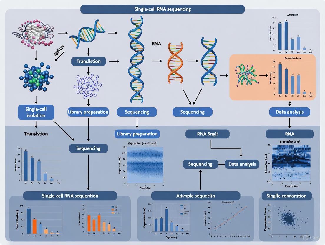
Intratumoral heterogeneity (ITH) is a defining characteristic of hepatocellular carcinoma (HCC), representing the coexistence of diverse cellular subpopulations with distinct genetic, molecular, and phenotypic profiles within a single tumor [1]. This heterogeneity manifests at multiple levels, encompassing cellular diversity, molecular signaling, and the tumor microenvironment (TME), and is a pivotal factor contributing to late diagnosis, treatment resistance, and disease recurrence [1]. The emergence of single-cell RNA sequencing (scRNA-seq) has revolutionized our ability to dissect this complexity, providing unprecedented resolution to identify novel cell clusters, delineate cellular developmental trajectories, and characterize intricate cell-cell communication networks that underlie HCC progression and therapeutic resistance [2] [3]. Furthermore, scRNA-seq analyses have revealed that non-coding RNAs (ncRNAs), including long non-coding RNAs (lncRNAs) and microRNAs (miRNAs), are integral components of this heterogeneous landscape, serving as key regulators and biomarkers with significant implications for patient stratification and therapy [4] [5]. This Application Note delineates standardized protocols for leveraging scRNA-seq to define ITH in HCC, with a focused investigation on ncRNA subtypes, providing a comprehensive framework for researchers and drug development professionals.
Key Concepts and Definitions
- Intratumoral Heterogeneity (ITH): The presence of genetically and phenotypically distinct subpopulations of cancer cells within a single tumor mass, leading to variations in biological behavior and treatment response [1].
- Tumor Microenvironment (TME): The ecosystem surrounding tumor cells, composed of various host cells including immune cells (T cells, macrophages, NK cells), endothelial cells, and fibroblasts, which interacts with cancer cells to influence tumor progression and immunotherapy response [2] [3].
- Single-Cell RNA Sequencing (scRNA-seq): A high-resolution genomic technology that enables the profiling of gene expression at the individual cell level, allowing for the deconvolution of cellular heterogeneity and the identification of rare cell populations within complex tissues [6].
- Non-Coding RNA (ncRNA): Functional RNA molecules that are not translated into proteins, including microRNAs (miRNAs) and long non-coding RNAs (lncRNAs), which play crucial regulatory roles in gene expression and are increasingly recognized as key players in tumorigenesis and tumor heterogeneity [4] [5].
Experimental Protocols for scRNA-seq in HCC Heterogeneity Analysis
Protocol 1: Single-Cell Suspension Preparation from HCC Tissue
Principle: Generate high-quality, viable single-cell suspensions from primary HCC tissues and paired adjacent non-tumoral tissues while preserving RNA integrity and cellular diversity [2] [3].
Materials:
- HCC tissue samples: Fresh tumor and paired adjacent non-tumoral tissues (e.g., from surgical resection).
- Digestion enzyme cocktail: Collagenase IV (1-2 mg/mL), Dispase (1-2 mg/mL), DNase I (0.1 mg/mL) in HBSS.
- RBC Lysis Buffer: For red blood cell removal.
- Cell Staining Buffer: PBS supplemented with 0.04% BSA.
- Viability dye: 7-AAD or propidium iodide.
- Cell strainers: 40 μm and 70 μm nylon mesh.
- Centrifuge: Refrigerated, capable of 300-500 × g.
Procedure:
- Tissue Collection and Transport: Collect fresh HCC tissues in cold preservation medium (e.g., DMEM + 10% FBS) and process within 1 hour of resection.
- Tissue Dissociation:
- Mince tissues into 1-2 mm³ fragments using sterile scalpels.
- Transfer fragments to digestion enzyme cocktail (5 mL per gram of tissue).
- Incubate at 37°C for 30-60 minutes with gentle agitation.
- Triturate every 10 minutes using a serological pipette to dissociate clusters.
- Cell Suspension Processing:
- Filter cell suspension through 70 μm and 40 μm cell strainers sequentially.
- Centrifuge at 300 × g for 5 minutes at 4°C.
- Aspirate supernatant and resuspend pellet in 10 mL RBC lysis buffer. Incubate for 2 minutes at room temperature.
- Add 20 mL PBS to stop lysis and centrifuge at 300 × g for 5 minutes.
- Cell Viability and Counting:
- Resuspend cell pellet in 1 mL cell staining buffer.
- Mix 10 μL cell suspension with 10 μL viability dye and count using a hemocytometer or automated cell counter.
- Ensure viability >80% and target concentration of 700-1,200 cells/μL for 10x Genomics platform.
Quality Control:
- Assess cell viability using trypan blue exclusion or automated cell counters.
- Evaluate single-cell suspension quality under microscope to confirm absence of cell clumps.
- Use Bioanalyzer or TapeStation to check RNA integrity if performing bulk RNA-seq comparisons.
Protocol 2: Single-Cell RNA Sequencing Library Preparation and Data Processing
Principle: Generate barcoded scRNA-seq libraries from single-cell suspensions using droplet-based encapsulation (10x Genomics Chromium System) for high-throughput profiling [7] [3].
Materials:
- 10x Genomics Chromium Controller and Single Cell 3' Reagent Kits (v3 or v3.1)
- Thermal cycler with 96-well block
- Bioanalyzer or TapeStation system (Agilent)
- Library quantification kit (Qubit dsDNA HS Assay Kit)
- SPRIselect beads (Beckman Coulter)
- Seurat R package (v4.0.0 or higher)
- Harmony package for batch correction
Procedure:
- Single-Cell Partitioning and cDNA Synthesis:
- Load single-cell suspension (1,000-10,000 cells) onto 10x Genomics Chromium Chip B.
- Perform GEM generation and barcoding following manufacturer's protocol.
- Reverse transcribe barcoded RNA to generate cDNA.
- Amplify cDNA via PCR (12 cycles).
- Library Construction:
- Fragment amplified cDNA and add adaptors via end-repair, A-tailing, and ligation.
- Perform sample index PCR (10-14 cycles) to incorporate dual indexes.
- Clean up libraries using SPRIselect beads (0.6x and 0.8x ratios).
- Library QC and Sequencing:
- Assess library quality using Bioanalyzer High Sensitivity DNA kit (expect peak ~450-550 bp).
- Quantify libraries using Qubit dsDNA HS Assay.
- Pool libraries at appropriate molar ratios and sequence on Illumina NovaSeq 6000 (Target: 50,000 reads/cell).
- Data Preprocessing and Quality Control:
- Demultiplex raw sequencing data using Cell Ranger (10x Genomics) with default parameters.
- Align reads to reference genome (GRCh38) and generate feature-barcode matrices.
- Filter cells using Seurat: Retain cells with 200-10,000 detected genes and <20% mitochondrial reads [7] [3].
- Normalize data using SCTransform and integrate multiple samples using Harmony to correct for batch effects.
Protocol 3: Identification of ncRNA Subtypes and Cellular Trajectories
Principle: Utilize scRNA-seq data to identify ncRNA-enriched cell subpopulations, construct gene regulatory networks, and infer developmental trajectories using pseudotime analysis [4] [3].
Materials:
- Processed scRNA-seq data (from Protocol 2)
- R packages: Monocle3, SCENIC, iTALK, clusterProfiler
- Reference databases: JASPAR (TF motifs), MSigDB (gene sets)
Procedure:
- Cell Clustering and Annotation:
- Perform principal component analysis (PCA) on highly variable genes.
- Cluster cells using graph-based methods (Seurat::FindClusters, resolution=0.1-1.2).
- Annotate cell types using known marker genes:
- ncRNA Subtype Identification:
- Gene Regulatory Network Analysis:
- Construct regulatory networks using SCENIC with default parameters.
- Identify transcription factors (TFs) and their target ncRNAs using cisTarget databases.
- Regulon activity is calculated per cell to identify TF-ncRNA regulatory modules.
- Pseudotime and Trajectory Analysis:
- Convert Seurat object to CellDataSet format for Monocle3.
- Learn trajectory graph using learn_graph() function with reduced dimensions.
- Order cells in pseudotime to infer developmental trajectories and state transitions.
- Identify ncRNAs that are differentially expressed along branches.
Data Analysis and Interpretation
Key Cellular Populations in HCC Heterogeneity
Table 1: Key Cell Populations Identified Through scRNA-seq in HCC and Their Functional Significance
| Cell Type | Key Marker Genes | Subpopulations | Functional Role in HCC | Therapeutic Implications |
|---|---|---|---|---|
| Hepatocytes/Cancer Cells | ALB, APOE, APOC1 [3] | HCC_HP (HP+) [3], Proliferative subcluster [8] | Tumor initiation, metabolic reprogramming, expression of HP linked to higher differentiation [3] | Potential targets for differentiation therapy |
| T Cells | CD3D, CD3E, CD8A, CD4 | CD8+ exhausted T (Tex) [4], CD4+ proliferative T [2] | CD8 Tex associated with immunotherapy resistance; CD4+ proliferative T linked to MVI [4] [2] | Targets for immune checkpoint inhibitors |
| Macrophages | CD68, AIF1 | SPP1+ macrophages [2] | Promote immunosuppression, MVI formation, and tumor progression [2] | Potential target for SPP1 inhibition |
| Natural Killer (NK) Cells | NCAM1, KLRD1, GNLY [7] | High vs. Low NK score subsets [7] | Cytotoxic anti-tumor activity, correlated with better prognosis [7] | Basis for NK cell-based therapies |
| Neutrophils | CSF3R, FCGR3B | Neu_AIF1 [3] | Extensive communication with HCC cells, TECs, and CAFs [3] | Potential target for inhibition to block pro-tumor signaling |
ncRNA Biomarkers in HCC Heterogeneity
Table 2: Clinically Significant Non-Coding RNAs in Hepatocellular Carcinoma
| ncRNA Type | Specific Molecules | Expression in HCC | Biological Function | Clinical Utility |
|---|---|---|---|---|
| Circulating microRNA | miRNA-122 [5] | Downregulated [5] | Tumor suppressor; inhibits cyclin G1, IGF1R pathway; suppresses HBV replication [5] | Early detection biomarker; levels significantly elevated in early-stage HCC vs healthy controls [5] |
| Circulating microRNA | let-7 family [5] | Downregulated [5] | Tumor suppressor; regulates multiple oncogenic pathways [5] | Diagnostic biomarker; often combined with AFP for improved sensitivity [5] |
| Circulating microRNA | miRNA-221, miRNA-222, miRNA-224 [5] | Upregulated [5] | Oncogenic; promote cell proliferation and survival [5] | Prognostic biomarkers; associated with advanced disease |
| lncRNA | MCM3AP-AS1, MAPKAPK5-AS1, PART1 [4] | Upregulated in high-risk HCC | Promote cell proliferation, suppress apoptosis; CD8 Tex-related [4] | Prognostic signature; knockdown suppresses proliferation, induces apoptosis [4] |
| lncRNA Signature | 28 CD8 Tex-related lncRNAs [4] | Defines HCC subtypes | Regulate T cell exhaustion and immunotherapy response [4] | Predictive biomarker for immunotherapy sensitivity; classifies patients into distinct prognosis groups [4] |
Computational Analysis Workflows
Figure 1: scRNA-seq Computational Analysis Workflow. The pipeline encompasses data preprocessing, cell type identification, advanced ncRNA and trajectory analyses, and clinical validation.
Figure 2: Multidimensional Landscape of HCC Intratumoral Heterogeneity. ITH manifests at cellular, molecular, and ncRNA regulatory levels, collectively influencing clinical outcomes and therapeutic responses.
The Scientist's Toolkit
Table 3: Essential Research Reagents and Computational Tools for scRNA-seq Studies in HCC
| Category | Item/Reagent | Specification/Application | Function/Purpose |
|---|---|---|---|
| Wet Lab Reagents | Collagenase IV | 1-2 mg/mL in HBSS | Tissue dissociation to generate single-cell suspensions |
| DNase I | 0.1 mg/mL | Degradation of DNA to prevent cell clumping | |
| 10x Genomics Chromium Kit | Single Cell 3' v3 or v3.1 | Barcoding and library preparation for scRNA-seq | |
| RBC Lysis Buffer | Ammonium-chloride-based | Removal of red blood cells from cell suspensions | |
| BSA | 0.04% in PBS | Blocking non-specific binding in cell staining | |
| Bioinformatics Tools | Seurat R Package | v4.0.0+ | scRNA-seq data integration, normalization, and clustering |
| Monocle3 | Trajectory analysis | Pseudotime ordering and developmental trajectory inference | |
| SCENIC | v1.1.3+ | Gene regulatory network reconstruction from scRNA-seq data | |
| InferCNV | Copy number variation | Inference of CNVs in tumor cells vs. reference normal cells | |
| iTALK | Cell-cell communication | Analysis of ligand-receptor interactions in TME | |
| Reference Databases | JASPAR Database | TF binding profiles | Transcription factor motif analysis for regulatory networks |
| MSigDB | Gene sets | Pathway analysis and functional enrichment | |
| TCGA-LIHC | Bulk RNA-seq data | Validation of scRNA-seq findings in large cohorts | |
| 1,3,5-Trihydroxy-4-prenylxanthone | 1,3,5-Trihydroxy-4-prenylxanthone, CAS:53377-61-0, MF:C18H16O5, MW:312.3 g/mol | Chemical Reagent | Bench Chemicals |
| Primidone-D5 | Primidone-d5|CAS 73738-06-4|High-Purity Reference Standard | High-quality Primidone-d5 (CAS 73738-06-4), a stable-labeled internal standard for LC-MS/MS research. This product is For Research Use Only (RUO). Not for human or veterinary use. | Bench Chemicals |
Application Notes and Technical Considerations
Integration with Bulk RNA-seq Data
Validation of scRNA-seq findings through integration with bulk RNA-seq data from repositories like TCGA-LIHC and ICGC enhances statistical power and clinical translatability [7] [3]. This integrated approach enables:
- Construction of prognostic models based on cell-type specific gene signatures
- Validation of ncRNA subtypes across independent cohorts
- Correlation of specific cell subpopulations with clinical outcomes
- Development of risk scores for patient stratification
For instance, a recent study combined scRNA-seq data from 27 HCC tumors with TCGA-LIHC bulk RNA-seq data to identify a novel HP-positive HCC cell cluster associated with tumor differentiation [3]. Similarly, CD8 Tex-related lncRNAs identified through scRNA-seq were validated in TCGA and ICGC cohorts, demonstrating their utility in classifying HCC patients into distinct prognostic groups [4].
Functional Validation of ncRNA Subtypes
While scRNA-seq identifies potential ncRNA biomarkers, functional validation is essential:
- In vitro models: HCC cell lines and patient-derived organoids for knockdown/overexpression studies [4]
- Functional assays: Proliferation (MTT), apoptosis (Annexin V), migration (transwell) following ncRNA modulation
- Mechanistic studies: Luciferase reporter assays for miRNA-mRNA interactions, RNA immunoprecipitation for lncRNA-protein interactions
For example, experimental validation of CD8 Tex-related lncRNAs (MCM3AP-AS1, MAPKAPK5-AS1, PART1) in HCC cell lines and organoids demonstrated that their downregulation suppressed cell proliferation and induced apoptosis [4].
The application of scRNA-seq technologies has fundamentally advanced our understanding of HCC intratumoral heterogeneity, revealing complex cellular ecosystems and ncRNA-mediated regulatory networks that drive disease progression and treatment resistance. The protocols and analytical frameworks outlined in this Application Note provide a standardized approach for researchers to systematically characterize ITH, identify clinically relevant ncRNA subtypes, and develop novel therapeutic strategies. As single-cell technologies continue to evolve, integrating multi-omics data at single-cell resolution (including epigenomics, proteomics, and spatial transcriptomics) will further enhance our ability to decipher the full complexity of HCC heterogeneity and translate these findings into improved patient outcomes.
Single-cell RNA sequencing (scRNA-seq) has revolutionized our understanding of tumor heterogeneity by enabling the dissection of malignant cell populations at unprecedented resolution. In hepatocellular carcinoma (HCC), this technology has revealed distinct malignant cell subtypes with unique functional roles in tumor progression, metastasis, and therapy resistance. This Application Note details the experimental and computational protocols for identifying and characterizing three core malignant cell phenotypes in HCC—pro-metastatic, proliferative, and metabolic subtypes—within the broader context of single-cell analysis of non-coding RNA heterogeneity. We provide comprehensive methodologies for cell subtype identification, validation, and functional analysis to support researchers in implementing these approaches in HCC research and drug development.
Key Malignant Cell Subtypes in HCC
ScRNA-seq analyses of HCC tissues have consistently identified three major malignant cell subtypes characterized by distinct transcriptional programs and functional properties.
Table 1: Key Malignant Cell Subtypes in Hepatocellular Carcinoma
| Subtype Name | Key Marker Genes | Functional Characteristics | Clinical Prognosis | Identification Study |
|---|---|---|---|---|
| EMT-subtype (Pro-metastatic) | S100A6, S100A11 | Epithelial-mesenchymal transition, hypoxia response, high cancer stem cell scores | Unfavorable prognosis, associated with metastasis | [9] |
| Prol-phenotype (Proliferative) | TOP2A, STMN1 | Cell cycle progression, proliferation, G2M checkpoint activation | Variable prognosis | [9] |
| Metab-subtype (Metabolic) | ARG1, ALDOB | Xenobiotic metabolism, bile acid metabolism, metabolic reprogramming | Favorable prognosis | [9] |
| Glycan-HCC | Glycan biosynthesis genes | Glycan metabolism, proliferative pathways, exhausted immune microenvironment | Worse overall survival | [10] |
| Lipid-HCC | Lipid metabolism genes | Lipid metabolism pathways | Better survival | [10] |
The EMT-subtype (pro-metastatic) demonstrates strong association with epithelial-mesenchymal transition, hypoxia response, and elevated cancer stem cell properties, characterized by high expression of S100 calcium-binding proteins A6 and A11 [9]. The Prol-phenotype (proliferative) exhibits marked enrichment of cell cycle and proliferation pathways with high expression of TOP2A and STMN1 [9]. The Metab-subtype shows predominant engagement in metabolic processes including bile acid and xenobiotic metabolism [9]. Additional metabolic stratification reveals Glycan-HCC and Lipid-HCC subtypes with distinct clinical outcomes and immune microenvironments [10].
Experimental Workflow for Malignant Cell Subtyping
The comprehensive identification of malignant cell subtypes requires an integrated multi-omics approach combining scRNA-seq with complementary spatial and functional validation techniques.
Sample Preparation and Single-Cell Sequencing
Protocol 1: Single-Cell Suspension Preparation from HCC Tissues
- Tissue Collection: Obtain fresh HCC tissues from surgical resection, including paired non-tumor liver tissues as controls. Immediate preservation in cold preservation medium is critical [11] [12].
- Tissue Dissociation: Mechanically dissociate tissues using scalpels followed by enzymatic digestion with collagenase IV (1-2 mg/mL) and DNase I (0.1 mg/mL) in PBS at 37°C for 30-45 minutes with gentle agitation [12].
- Cell Viability Enhancement: Use density gradient centrifugation (e.g., Percoll or Ficoll) to remove debris and dead cells. Filter through 40-μm cell strainers to obtain single-cell suspensions [12].
- Quality Control: Assess cell viability (>85%) using trypan blue exclusion and count cells with an automated cell counter. Adjust concentration to 700-1,200 cells/μL for 10x Genomics platform [12].
Protocol 2: Single-Cell RNA Sequencing Library Preparation
- Platform Selection: Utilize the 10x Genomics Chromium platform for high-throughput scRNA-seq following manufacturer's instructions [12].
- Cell Capture and Barcoding: Load cells onto the Chromium Chip to achieve target recovery of 2,000-10,000 cells per sample. Implement multiplexing using cell hashing technologies if processing multiple samples [11] [12].
- Library Construction: Perform reverse transcription, cDNA amplification, and library construction using the Chromium Single Cell 3' Reagent Kits. Include sample indexes for multiplexing [12].
- Sequencing: Sequence libraries on Illumina platforms (NovaSeq 6000) aiming for a minimum of 50,000 reads per cell with 150 bp paired-end reads [13] [10].
Computational Analysis Pipeline
Protocol 3: Data Preprocessing and Quality Control
- Initial Processing: Use Cell Ranger (10x Genomics) to demultiplex raw sequencing data, align reads to the reference genome (GRCh38), and generate feature-barcode matrices [12].
- Quality Control Filtering: Apply stringent QC filters using Seurat R package: exclude cells with <200 or >8,000 detected genes, >10% mitochondrial reads, and potential doublets identified by DoubletFinder [14] [15].
- Normalization: Normalize data using the LogNormalize method with a scale factor of 10,000, followed by identification of 2,000-3,000 highly variable genes for downstream analysis [14] [15].
Protocol 4: Cell Clustering and Annotation
- Integration and Batch Correction: Apply Harmony algorithm to integrate multiple datasets and correct for technical batch effects while preserving biological variation [14] [15].
- Dimensionality Reduction: Perform principal component analysis (PCA) on highly variable genes, followed by uniform manifold approximation and projection (UMAP) using the first 15-30 principal components [16] [9].
- Cell Clustering: Use graph-based clustering (FindNeighbors and FindClusters in Seurat) at appropriate resolution (typically 0.4-1.2) to identify cell populations [11] [9].
- Cell Type Annotation: Identify cluster marker genes using Wilcoxon rank sum test and annotate cell types based on canonical markers: hepatocytes (ALB, APOE), T cells (CD3D, CD8A), B cells (CD79A, MS4A1), macrophages (CD68, CD163), endothelial cells (PECAM1, VWF), and fibroblasts (ACTA2, COL1A1) [11] [9].
Protocol 5: Malignant Cell Identification and Subtyping
- CNV Analysis: Use inferCNV R package to infer copy number variations in putative tumor cells compared to normal epithelial cells as reference. Identify malignant cells with overall CNV > 0.2 and correlation with top 5% tumor cells > 0.2 [16] [9].
- Subtype Classification: Re-cluster malignant cells and identify differentially expressed genes (Wilcoxon test, |log2FC| > 0.5, adjusted p-value < 0.05) to define subtypes. Calculate signature scores for EMT, proliferation, and metabolism using AddModuleScore in Seurat [9].
- Trajectory Analysis: Apply Monocle2 or Slingshot to reconstruct developmental trajectories and identify transitions between subtypes [9] [15].
The Scientist's Toolkit: Essential Research Reagents
Table 2: Essential Research Reagents for HCC scRNA-Seq Studies
| Reagent/Category | Specific Examples | Function/Application | Protocol Reference |
|---|---|---|---|
| Tissue Dissociation | Collagenase IV, DNase I, HBSS with calcium and magnesium | Tissue digestion to single-cell suspension | Protocol 1 |
| Cell Viability Enhancement | Percoll gradient, Ficoll-Paque, Trypan blue | Debris removal and viability assessment | Protocol 1 |
| Single-Cell Platform | 10x Genomics Chromium Controller, Chip B | Single-cell partitioning and barcoding | Protocol 2 |
| Library Prep Kits | Chromium Single Cell 3' Reagent Kits v3 | cDNA synthesis, amplification, and library preparation | Protocol 2 |
| Sequencing Reagents | Illumina NovaSeq 6000 S4 Flow Cell, XP workflow | High-throughput sequencing | Protocol 2 |
| Bioinformatics Tools | Seurat v4, Harmony, inferCNV, Monocle2 | Data analysis, integration, and visualization | Protocols 3-5 |
| Cell Type Markers | ALB (hepatocytes), CD3D (T cells), CD68 (macrophages) | Cell type identification and annotation | Protocol 4 |
| Subtype Marker Panels | S100A6 (EMT), TOP2A (proliferation), ARG1 (metabolism) | Malignant cell subtyping | Protocol 5 |
| Ramipril diketopiperazine | Ramipril diketopiperazine, CAS:108731-95-9, MF:C23H30N2O4, MW:398.5 g/mol | Chemical Reagent | Bench Chemicals |
| Ethyl 2-cyano-2-phenylbutanoate | Ethyl 2-cyano-2-phenylbutanoate, CAS:718-71-8, MF:C13H15NO2, MW:217.26 g/mol | Chemical Reagent | Bench Chemicals |
Signaling Pathways and Cellular Interactions
Malignant cell subtypes engage in specific signaling pathways and cellular interactions that drive HCC progression and shape the tumor microenvironment.
Key Signaling Pathways in Malignant Subtypes
EMT-Subtype Signaling: The pro-metastatic subtype demonstrates exclusive activation of SMAD3 and TGF-β signaling pathways, which drive epithelial-mesenchymal transition and metastatic progression [9]. Hypoxia response pathways are markedly enriched in this subtype, contributing to its invasive characteristics [9] [17].
Prol-Phenotype Signaling: The proliferative subtype shows activation of cell cycle progression pathways including E2F targets and G2M checkpoint signaling, with high expression of DNA replication and chromosome segregation genes [9].
Metabolic Subtype Signaling: The metabolic subtype engages diverse metabolic pathways including glycan biosynthesis (glycan-HCC) or lipid metabolism (lipid-HCC), with distinct clinical outcomes and immune microenvironments [10].
Cellular Communication Networks
A critical fibroblast-tumor cell interaction loop mediated by SPP1-CD44 and CCN2/TGF-β-TGFBR1 interaction pairs reinforces the EMT-subtype phenotype [9]. Experimental inhibition of CCN2 disrupts this feedback loop, mitigates transformation to EMT-subtype, and suppresses metastasis [9]. Additionally, VEGFA+ cancer-associated fibroblasts promote intra-tumoral angiogenesis through cellular communication with capillary endothelial cells, facilitating tumor progression [18].
Validation Methodologies
Protocol 6: Spatial Validation of Malignant Subtypes
- Multiplexed Immunofluorescence: Perform sequential staining of formalin-fixed paraffin-embedded (FFPE) tissue sections with antibodies against subtype markers (ARG1, TOP2A, S100A6) using Opal TM multiplex IHC kits. Validate exclusive expression patterns of subtype markers in malignant cells [9].
- Spatial Transcriptomics: Integrate 10x Visium spatial transcriptomics data with scRNA-seq clusters to map the spatial distribution of malignant subtypes within tumor regions [9] [13].
- Validation in Independent Cohorts: Calculate subtype signature scores in bulk RNA-seq datasets (TCGA-LIHC, ICGC) using single-sample gene set enrichment analysis (ssGSEA) to confirm clinical relevance and prognostic associations [11] [9].
Protocol 7: Functional Characterization of Subtypes
- Trajectory Analysis: Use Monocle2 to reconstruct the developmental trajectory from prol-phenotype to both metab-subtype and EMT-subtype, confirming lineage relationships [9].
- Cell-Cell Communication Analysis: Apply CellChat R package to infer communication probabilities between malignant subtypes and stromal cells based on ligand-receptor interactions, identifying key signaling pathways [16] [15].
- Metabolic Pathway Analysis: Utilize scMetabolism R package to quantify metabolic activity at single-cell resolution using KEGG and Reactome metabolism-related gene sets [16] [10].
The identification of pro-metastatic, proliferative, and metabolic malignant cell subtypes in HCC through scRNA-seq provides critical insights into tumor heterogeneity and progression mechanisms. The experimental and computational protocols detailed in this Application Note establish a comprehensive framework for characterizing these subtypes, validating their clinical significance, and investigating their functional roles in HCC ecosystems. These approaches enable researchers to dissect the complex cellular architecture of HCC tumors, with important implications for developing subtype-specific therapeutic strategies and predictive biomarkers. Integration of these methodologies with ncRNA heterogeneity studies will further enhance our understanding of HCC biology and treatment resistance mechanisms.
Non-coding RNAs (ncRNAs) constitute a critical layer of regulatory control in hepatocellular carcinoma (HCC), orchestrating key pathological processes including epithelial-mesenchymal transition (EMT), metabolic reprogramming, and proliferation. This Application Note delineates the multifaceted roles of long non-coding RNAs (lncRNAs), microRNAs (miRNAs), and circular RNAs (circRNAs) within the HCC tumor microenvironment, with emphasis on single-cell RNA sequencing (scRNA-seq) methodologies for resolving ncRNA heterogeneity. We provide detailed experimental protocols for identifying and validating ncRNA functions, alongside comprehensive signaling pathway diagrams and reagent solutions to facilitate rigorous investigation of ncRNA-driven oncogenic mechanisms in HCC research and drug development.
Hepatocellular carcinoma exhibits profound molecular heterogeneity, driven significantly by dysregulated non-coding RNAs that fine-tune transcriptional and post-transcriptional programs. Single-cell transcriptomic approaches have begun to unravel the complex spatial and temporal dynamics of ncRNA expression across malignant and stromal cell populations within the HCC ecosystem. The functional spectrum of ncRNAs encompasses regulation of EMT—a critical process in metastasis—metabolic rewiring that sustains rapid proliferation, and direct control of cell cycle progression. This technical resource provides a structured framework for investigating these interconnected pathways, with practical methodologies tailored for researchers exploring ncRNA biology in HCC.
Table 1: Principal ncRNA Regulators in HCC Pathogenesis
| ncRNA Category | Specific Molecule | Regulatory Role | Primary Targets/Pathways | Functional Outcome in HCC |
|---|---|---|---|---|
| OncomiRs | miR-221 | Upregulated | DDIT4/mTOR, PTEN, TIMP3 [19] | Promotes proliferation, inhibits apoptosis |
| miR-34a | Dysregulated | TGF-β, SMAD4 [20] | Regulates EMT and metastasis | |
| Tumor Suppressor miRNAs | miR-122 | Downregulated | Multiple oncogenes [19] | Suppresses tumor growth; delivery inhibits HCC in models |
| miR-29 | Downregulated | IGF2BP1, VEGFA, BCL2 [19] | Contrasts HCC progression & angiogenesis | |
| miR-101 | Downregulated | ROCK [19] | Inhibits metastasis & EMT | |
| Oncogenic lncRNAs | SNHG17 | Upregulated | Metabolic pathways, PI3K-Akt [21] | Promotes proliferation, migration, inhibits apoptosis |
| NEAT1 | Upregulated | miR-155/Tim-3 [22] | Modulates T-cell exhaustion in TIME | |
| H19 | Upregulated | CDC42/PAK1, miR-324-5p [23] | Enhances proliferation & metastasis | |
| Metabolism-associated circRNAs | circMET | Upregulated | miR-30-5p/Snail/DPP4 axis [22] | Promotes immune evasion, reduces CD8+ T-cell infiltration |
Table 2: ncRNA Involvement in Key Signaling Pathways in HCC
| Signaling Pathway | Regulating ncRNAs | Molecular Mechanism | Biological Consequence |
|---|---|---|---|
| Wnt/β-catenin | miR-612, miR-122, LncRNA CCAL [20] | Targets FZD5, Snail1/2; regulates AP-2α [20] | Activates EMT program, enhances invasion |
| TGF-β | miR-449, miR-130a-3p [20] | Targets Smad4; modulates TGF-β receptors [20] | Induces EMT, promotes metastasis |
| HIF-1α | Linc-RoR [23] | Sponges miR-145; upregulates HIF-1α [23] | Drives glycolysis, enhances hypoxic adaptation |
| IL-6/JAK/STAT3 | LncRNA STAT3-mediated UPREGULATION [20] | Modulates IL-6 expression and signaling [20] | Promotes inflammatory microenvironment |
| PI3K-Akt | SNHG17 [21] | Activates PI3K-Akt signaling [21] | Enhances cell survival and proliferation |
Experimental Protocols for ncRNA Functional Analysis
Single-Cell RNA Sequencing for ncRNA Heterogeneity
Principle: scRNA-seq enables resolution of ncRNA expression patterns across individual cells within heterogeneous HCC tumors, identifying rare cell populations and transitional states driven by ncRNA activity.
Protocol:
- Sample Preparation:
- Obtain fresh HCC tissue specimens (tumor and paired adjacent non-tumor) under institutional review board-approved protocols.
- Process tissues to single-cell suspensions using gentle mechanical dissociation and enzymatic digestion (Collagenase IV, 2 mg/mL, 37°C, 30-45 min).
- Remove debris using density gradient centrifugation and assess cell viability (>85%) with trypan blue exclusion.
Single-Cell Library Construction:
- Load cells onto appropriate scRNA-seq platform (10X Genomics Chromium recommended).
- Following manufacturer's instructions, perform cell capture, barcoding, and reverse transcription.
- Amplify cDNA and construct libraries with incorporation of unique molecular identifiers (UMIs).
Sequencing and Data Analysis:
- Sequence libraries on Illumina platform to minimum depth of 50,000 reads per cell.
- Process raw data using Cell Ranger pipeline for demultiplexing and alignment.
- Utilize Seurat package for quality control, normalization, and clustering [24] [25].
- Identify ncRNA-enriched clusters and perform pseudotime analysis with Monocle to reconstruct ncRNA dynamics along differentiation trajectories [26] [25].
Troubleshooting Tips:
- High mitochondrial gene percentage may indicate poor cell viability; optimize digestion conditions.
- For ncRNA-specific analysis, ensure library preparation method captures small RNAs (miRNAs) or long RNAs (lncRNAs, circRNAs) as research focus requires.
Functional Validation of ncRNAs in EMT Regulation
Principle: Gain- and loss-of-function experiments establish causal relationships between ncRNA expression and EMT phenotypes in HCC models.
Protocol:
- Cell Culture and Transfection:
- Maintain HCC cell lines (e.g., Hep3B, PLC/PRF/5, Huh-7) in appropriate media supplemented with 10% FBS.
- For loss-of-function: Transfect cells with specific siRNAs targeting lncRNAs (e.g., si-SNHG17: 5'-CGGATCCACTGTTCAATCT-3') using Lipofectamine 3000 [21].
- For gain-of-function: Clone full-length ncRNA sequences into pcDNA3.1 vector and transfect cells.
EMT Phenotype Assessment:
- Wound Healing Assay: Create scratch wound in confluent monolayer, monitor closure at 0, 24, 48h; calculate migration rate.
- Transwell Invasion Assay: Seed transfected cells in serum-free medium into Matrigel-coated transwell inserts; count cells migrating toward chemoattractant after 24-48h [24] [21].
- qRT-PCR Analysis: Extract total RNA, synthesize cDNA, and quantify EMT markers (E-cadherin, N-cadherin, Vimentin) using SYBR Green-based qPCR.
Pathway Analysis:
- Perform RNA sequencing on transfected cells to identify differentially expressed genes.
- Conduct pathway enrichment analysis using clusterProfiler to map ncRNA targets to EMT-related pathways [21].
Metabolic Reprogramming Assays
Principle: ncRNAs regulate HCC metabolic rewiring, including glycolysis, oxidative phosphorylation, and lipid metabolism, measurable through metabolic flux assays.
Protocol:
- Metabolic Pathway Activity Scoring:
- Utilize scRNA-seq data and reference metabolic gene sets (e.g., Human-GEM model) to calculate metabolic pathway scores for individual cells [27].
- Identify ncRNA expression correlated with metabolic cluster identities.
Functional Metabolomics:
- Culture ncRNA-modulated HCC cells in Seahorse XF analyzer plates.
- Measure extracellular acidification rate (ECAR) for glycolysis and oxygen consumption rate (OCR) for mitochondrial respiration under basal and stressed conditions.
- Treat with pathway inhibitors (e.g., 2-DG for glycolysis, oligomycin for ATP synthase) to assess metabolic flexibility.
Validation of Metabolic Targets:
- Use Western blotting to assess expression of metabolic enzymes (e.g., PDK4, GLUT1) in ncRNA-manipulated cells [21].
- Perform liquid chromatography-mass spectrometry (LC-MS) to quantify metabolite levels in response to ncRNA perturbation.
Signaling Pathway Diagrams
Diagram 1: ncRNA Regulatory Networks in HCC Progression. This map illustrates how different classes of ncRNAs converge on core signaling pathways to drive malignant processes in hepatocellular carcinoma.
Diagram 2: scRNA-seq Workflow for ncRNA Heterogeneity Analysis in HCC. This experimental pipeline outlines the integrated approach from sample processing through computational analysis for resolving ncRNA expression patterns at single-cell resolution.
The Scientist's Toolkit: Research Reagent Solutions
Table 3: Essential Research Reagents for ncRNA Functional Studies in HCC
| Reagent Category | Specific Product/Kit | Application | Key Features |
|---|---|---|---|
| scRNA-seq Platforms | 10X Genomics Chromium | Single-cell transcriptomics | Captures ncRNA expression with cellular resolution |
| SMART-Seq v4 Ultra Low Input RNA Kit | Full-length scRNA-seq | Enhanced detection of lncRNAs and circRNAs | |
| ncRNA Modulation | Lipofectamine 3000 | siRNA/plasmid delivery | High-efficiency transfection for gain/loss-of-function studies [21] |
| Silencer Select Pre-designed siRNAs | ncRNA knockdown | High specificity and reduced off-target effects | |
| Functional Assays | Corning Transwell Permeable Supports | Migration/invasion assays | Quantifies EMT phenotypes [24] [21] |
| Seahorse XF Glycolysis Stress Test Kit | Metabolic flux analysis | Measures glycolytic function in live cells | |
| Detection & Analysis | miScript miRNA PCR Arrays | miRNA expression profiling | Simultaneous analysis of multiple miRNAs |
| Arraystar ncRNA Microarrays | LncRNA/circRNA screening | Comprehensive ncRNA expression profiling | |
| R Package SCENIC | Gene regulatory networks | Identifies ncRNA-regulated networks from scRNA-seq data [25] | |
| Piperic acid | Piperic Acid|High-Purity Research Compound | High-purity Piperic Acid for research. Explore applications in neuroscience, oncology, and metabolic studies. This product is For Research Use Only (RUO). Not for human consumption. | Bench Chemicals |
| Daidzein-d6 | Daidzein-d6, CAS:291759-05-2, MF:C15H10O4, MW:260.27 g/mol | Chemical Reagent | Bench Chemicals |
Concluding Remarks
The functional spectrum of ncRNAs in HCC spans regulation of EMT, metabolic reprogramming, and proliferative signaling, creating a complex regulatory network that drives disease progression and therapeutic resistance. Single-cell RNA sequencing technologies provide unprecedented resolution to dissect this heterogeneity, revealing cell-type-specific ncRNA functions within the tumor ecosystem. The protocols and resources outlined in this Application Note establish a foundation for systematic investigation of ncRNA mechanisms in HCC, with potential to accelerate the discovery of novel biomarkers and therapeutic targets. Future directions should emphasize spatial transcriptomics to map ncRNA expression within tissue architecture, and the development of ncRNA-targeted therapeutics that can modulate these critical regulatory networks in hepatocellular carcinoma.
Application Note: Mapping ncRNA Heterogeneity in HCC Clonal Architecture
Hepatocellular carcinoma (HCC) demonstrates profound molecular heterogeneity that significantly impacts therapeutic response and clinical outcomes. Single-cell RNA sequencing (scRNA-seq) has emerged as a transformative technology for delineating tumor cell subpopulations and their evolutionary relationships based on non-coding RNA (ncRNA) expression profiles. This application note outlines standardized protocols for tracing long non-coding RNA (lncRNA) and microRNA (miRNA) dynamics across HCC clonal lineages, enabling researchers to identify novel therapeutic targets and biomarkers within specific tumor subclones.
Key HCC Tumor Cell Subtypes Defined by scRNA-seq
Recent single-cell transcriptomic studies have established a classification system for HCC malignant cells into three predominant subtypes, each with distinct ncRNA expression patterns and functional characteristics [9]:
Table 1: HCC Tumor Cell Subtypes Identified via scRNA-seq
| Subtype | Marker Genes | Functional Enrichment | ncRNA Associations | Clinical Correlation |
|---|---|---|---|---|
| Metabolism Subtype (Metab-subtype) | ARG1, ALDOB | Bile acid metabolism, Xenobiotic metabolism | Potentially tumor-suppressive miRNAs | Well-differentiated tumors, Better prognosis |
| Proliferation Phenotype (Prol-phenotype) | TOP2A, STMN1 | G2M checkpoint, E2F targets | OncomiRs (e.g., miR-221), Pro-proliferative lncRNAs | Proliferative tumor subclass |
| EMT Subtype (EMT-subtype) | S100A6, S100A11 | Epithelial-mesenchymal transition, Hypoxia | Metastasis-associated lncRNAs, miR-101 downregulation | Poor prognosis, Metastasis, Cancer stem cell properties |
Integration of 52 scRNA-seq datasets comprising 35,981 tumor cells from 52 HCC samples revealed that these subtypes coexist within tumors and demonstrate distinct developmental trajectories, with both Metab-subtype and EMT-subtype potentially originating from the Prol-phenotype [9]. This hierarchical organization suggests a branching evolutionary model where ncRNA dysregulation drives phenotypic diversification.
Quantitative ncRNA Dysregulation in HCC Progression
The dysregulation of specific ncRNAs has been quantitatively correlated with HCC clinical outcomes and molecular subtypes:
Table 2: Key ncRNAs in HCC Pathogenesis and Their Clinical Significance
| ncRNA Category | Specific ncRNAs | Expression Change | Molecular Targets/Pathways | Functional Consequences |
|---|---|---|---|---|
| Tumor Suppressor miRNAs | miR-122, miR-29, miR-195, miR-101, miR-497 | Downregulated | IGF2BP1, VEGFA, BCL2 (miR-29); VEGF, VAV2, CDC42 (miR-195); ROCK (miR-101); Rictor/AKT (miR-497) | Reduced inhibition of proliferation, angiogenesis, and metastasis |
| Oncogenic miRNAs | miR-221 | Upregulated | DDIT4/mTOR, PTEN, TIMP3 | Enhanced proliferation, apoptosis evasion |
| Oncogenic lncRNAs | HULC, MALAT1, NEAT1, H19, HOTAIR | Upregulated | Multiple signaling pathways | Promotion of proliferation, metastasis, and treatment resistance |
| Tumor Suppressor lncRNAs | FAM99B, TLNC1 | Downregulated | p53 signaling, Ribosome biogenesis | Loss of tumor suppressive functions |
The highly specific expression patterns of lncRNAs make them particularly valuable as markers of tumor evolution. scRNA-seq studies in triple-negative breast cancer models have demonstrated that lncRNAs show heterogeneous expression patterns including ubiquitous expression, subpopulation-specific expression, and hybrid patterns where they are expressed in several but not all subpopulations [28]. Similar principles apply to HCC, where lncRNA expression profiles can delineate tumor cell subpopulations with distinct evolutionary trajectories.
Experimental Protocols
Protocol 1: Single-Cell RNA Sequencing for ncRNA Heterogeneity Analysis in HCC
Sample Preparation and Quality Control
- Tissue Dissociation: Fresh HCC tissue samples obtained via surgical resection should be immediately processed using a gentle dissociation protocol. Utilize the Human Tumor Dissociation Kit with incubation at 37°C for 30-60 minutes with continuous agitation [12].
- Cell Viability and Counting: Assess cell viability using Trypan Blue exclusion or automated cell counters. Only preparations with >80% viability should proceed to sequencing.
- Fluorescence-Activated Cell Sorting (FACS): For xenograft models, sort GFP+ cells to exclude host mouse cells [28]. For clinical samples, sort using epithelial (EPCAM+) and/or HCC stem cell markers (CD133+, CD44+) to enrich for malignant populations.
- Library Preparation: Use the 10X Chromium scRNA-seq kit following manufacturer's protocols. Aim to capture 5,000-10,000 cells per sample to adequately represent tumor heterogeneity [12].
Sequencing Parameters and Quality Control
- Sequencing Depth: Target 50,000-100,000 reads per cell to adequately capture low-abundance ncRNAs.
- Quality Metrics: Filter cells with <200 or >2,000 detected features and >2.5% mitochondrial reads, indicating poor viability or apoptotic cells [28].
- Batch Effect Correction: Apply harmony algorithm to integrate multiple datasets and remove technical artifacts [9].
ncRNA-Focused Computational Analysis
- Read Alignment: Map sequencing reads to GRCh38 using 10X Genomics Cellranger with inclusion of lncRNA annotations from databases such as LNCipedia and NONCODE.
- Clustering and Subpopulation Identification: Perform principal component analysis using the top 2,000 most variable features. Use the first 15 principal components for graph-based clustering with Seurat's FindNeighbours and FindClusters functions [28].
- Differential ncRNA Expression: Identify subpopulation-defining ncRNAs using FindAllMarkers function with thresholds: minimum 25% of cells in cluster, log2 fold change >0.25, and Bonferroni-adjusted p-value ≤0.05 [28].
- Trajectory Inference: Apply pseudotime analysis tools (Monocle2, Slingshot) to reconstruct evolutionary relationships between subpopulations based on ncRNA expression patterns.
Protocol 2: Functional Validation of HCC-Associated ncRNAs
Gain- and Loss-of-Function Studies
- In Vitro Models: Utilize HCC cell lines (HepG2, Huh7, Hep3B, PLC/PRF/5) representing different molecular subtypes.
- ncRNA Modulation:
- Knockdown: Apply siRNA or LNA GapmeRs specifically targeting candidate lncRNAs. Use lipofectamine RNAiMAX with 25-50 nM final concentration.
- Overexpression: Clone full-length lncRNAs into pcDNA3.1 or lentiviral vectors for stable expression.
- Functional Assays:
- Proliferation: MTT assay daily for 5 days or real-time cell analysis (RTCA).
- Invasion and Migration: Transwell assays with Matrigel coating (invasion) or without (migration), quantifying after 24-48 hours.
- Stemness Properties: Tumorsphere formation in ultra-low attachment plates with serum-free media.
Mechanistic Studies
- Identifying ncRNA-Protein Interactions: RNA immunoprecipitation (RIP) using Magna RIP kit with antibodies against potential protein partners.
- miRNA Sponging Validation: Dual-luciferase reporter assays with wild-type and mutant lncRNA constructs.
- Pathway Analysis: Western blotting for key signaling pathways (AKT, mTOR, TGF-β) following ncRNA modulation.
The Scientist's Toolkit: Research Reagent Solutions
Table 3: Essential Research Reagents for ncRNA Studies in HCC
| Reagent Category | Specific Products | Application | Key Considerations |
|---|---|---|---|
| scRNA-seq Platforms | 10X Genomics Chromium | Single-cell transcriptomics | Optimal for capturing 5,000-10,000 cells/sample; compatible with ncRNA analysis |
| Cell Separation | Human Tumor Dissociation Kit, FACS antibodies (EPCAM, CD133, CD44) | Tumor cell enrichment | Preservation of cell viability critical for library quality |
| Bioinformatics Tools | Seurat, Cellranger, Monocle2 | Data analysis | Must include custom ncRNA annotations in reference genomes |
| ncRNA Modulation | LNA GapmeRs, siRNA, pcDNA3.1 overexpression vectors | Functional validation | Requires careful optimization of delivery efficiency |
| Animal Models | Patient-derived xenografts, Transgenic mouse models | In vivo validation | Recapitulates tumor microenvironment interactions |
| Spatial Transcriptomics | 10X Visium, Multiplexed error-robust FISH (MERFISH) | Spatial context of ncRNA expression | Validates scRNA-seq predicted localization patterns |
| Tosufloxacin Tosylate | Tosufloxacin Tosylate|High-Purity Research Grade | Bench Chemicals | |
| 1-(Cyanomethyl)cyclohexanecarbonitrile | 1-(Cyanomethyl)cyclohexanecarbonitrile|CAS 4172-99-0 | Bench Chemicals |
Signaling Pathways in ncRNA-Mediated HCC Progression
The integration of scRNA-seq data with functional studies has revealed several key pathways through which ncRNAs drive HCC heterogeneity and evolution:
Recent studies have demonstrated that lncRNAs such as NEAT1 and HULC interact with autophagy pathways, creating context-dependent effects that either suppress tumor initiation or promote progression in advanced stages [29]. The TGF-β/SMAD pathway has been specifically associated with the EMT-subtype identified through scRNA-seq, suggesting this pathway may be particularly important in metastatic subclones [9].
Discussion and Future Perspectives
The integration of scRNA-seq technologies with ncRNA biology has fundamentally advanced our understanding of HCC evolution. The recognition that HCC tumors contain multiple molecularly distinct subpopulations with different ncRNA expression profiles explains many clinical challenges, including therapeutic resistance and metastatic propensity.
Future applications of these findings include:
- Diagnostic Applications: Development of ncRNA-based liquid biopsies targeting subtype-specific markers (e.g., EMT-subtype associated lncRNAs) for early detection of aggressive subclone emergence.
- Therapeutic Targeting: Exploitation of subtype-specific ncRNA dependencies using antisense oligonucleotides or small molecule inhibitors.
- Treatment Stratification: Integration of ncRNA-based subtyping into clinical trial design to match targeted therapies with susceptible subpopulations.
The protocols and frameworks outlined herein provide a standardized approach for investigating ncRNA dynamics in HCC evolution, enabling more reproducible and clinically translatable research in this rapidly advancing field.
Hepatocellular carcinoma (HCC) represents a paradigm of complex cellular ecosystems, where malignant hepatocytes coexist with diverse stromal and immune cells within a dynamically organized tumor microenvironment (TME). Single-cell RNA sequencing (scRNA-seq) has revolutionized our understanding of this ecosystem by deconvoluting the profound molecular heterogeneity and intricate cell-cell communication networks that drive tumor progression and therapy resistance [11] [30]. Recent advances have illuminated that non-coding RNAs (ncRNAs) serve as critical mediators of intercellular communication within the TME, influencing virtually every aspect of tumor biology [31] [32].
The HCC TME comprises malignant cells, fibroblasts, endothelial cells, and diverse immune populations including T cells, B cells, natural killer (NK) cells, and myeloid-derived cells such as macrophages and dendritic cells [30]. ScRNA-seq analyses of primary and metastatic HCC tissues have revealed significant individual variations in cellular composition and spatial organization, with metastatic sites showing similar stromal patterns to primary tumors [11]. This ecosystem is not static; it exhibits remarkable cellular plasticity where both tumor and stromal cells can dynamically alter their phenotypic states in response to various stimuli [30].
ncRNAs - particularly microRNAs (miRNAs), long non-coding RNAs (lncRNAs), and circular RNAs (circRNAs) - have emerged as crucial regulators within this ecosystem. These molecules, which do not encode proteins, function as master regulators of gene expression and facilitate intercellular communication via exosomes and other carriers [31]. Their structural diversity, tissue specificity, and functional versatility enable them to orchestrate complex biological processes that govern tumor initiation, progression, and immune evasion.
Cellular Composition and Heterogeneity in HCC
Malignant Cell Subpopulations
ScRNA-seq profiling has uncovered extensive heterogeneity among HCC malignant cells, which can be categorized into distinct molecular subtypes with clinical relevance. Integrated analysis of 52 scRNA-seq datasets revealed three predominant malignant cell subtypes [9]:
Table 1: Malignant Cell Subtypes in HCC
| Subtype Name | Key Marker | Functional Characteristics | Clinical Association |
|---|---|---|---|
| Metabolism Subtype (Metab-subtype) | ARG1 | Enriched in bile acid and xenobiotic metabolism | Better differentiation; favorable prognosis |
| Proliferation Phenotype (Prol-phenotype) | TOP2A | High cell cycle and proliferation activity | Intermediate prognosis |
| EMT Subtype (EMT-subtype) | S100A6 | Epithelial-mesenchymal transition; stemness features | Poor prognosis; metastatic potential |
Trajectory analysis indicates that both Metab-subtype and EMT-subtype cells originate from the proliferative phenotype, suggesting a hierarchical organization of HCC malignant cells [9]. The EMT-subtype demonstrates exclusive activation of SMAD3 and TGF-β signaling pathways and exhibits elevated cancer stem cell (CSC) scores, expressing markers such as EPCAM, CD24, KRT19, and SOX9 [9]. This subpopulation plays a crucial role in metastasis and therapeutic resistance.
Immune Landscape
The immune microenvironment of HCC is characterized by diverse lymphocyte and myeloid populations that exhibit distinct functional states and spatial distributions [11] [30]. ScRNA-seq analyses of 25,591 T/NK cells from HCC tissues identified 14 distinct clusters, including CD8+ cytotoxic T lymphocytes (CTLs), mucosal-associated invariant T (MAIT) cells, effector memory T (TEM) cells, and tissue-resident memory T (TRM) cells [11].
CD4+ T cell populations show remarkable diversity, encompassing naïve CD4+ T cells, regulatory T cells (Tregs), helper T cells (Th1, Th2, Tfh), and cytotoxic CD4+ T cells [30]. Notably, Tregs are consistently enriched in primary tumors, while central memory T (TCM) cells are specifically enriched in early tertiary lymphoid structures (E-TLS) [11]. These E-TLS serve as depositories for antitumor TCM and CD20+ B cells, with higher abundances associated with improved patient survival [11].
Myeloid populations in HCC include heterogeneous macrophage subpopulations, with MMP9+ macrophages identified as terminally differentiated tumor-associated macrophages (TAMs) whose differentiation is driven by the transcription factor PPARγ [11]. The interplay between these immune populations and malignant cells creates either permissive or restrictive microenvironments for tumor growth.
Stromal Components
Cancer-associated fibroblasts (CAFs) in HCC display significant molecular and functional heterogeneity. ScRNA-seq has identified multiple fibroblast subtypes expressing characteristic gene signatures [30]:
- ECM-organizing fibroblasts: High expression of extracellular matrix signature genes
- Lipid-processing fibroblasts: Enriched for lipid metabolism genes
- Antigen-presenting fibroblasts: Expression of MHC class II genes
- Inflammatory fibroblasts: Chemokine and microvasculature gene signatures
Among these, CD36-positive fibroblast subtypes (including lipid-processing matrix fibroblasts) enhance the capacity of myeloid-derived suppressor cells (MDSCs) to promote an immunosuppressive TME and tumor stemness, predicting poor prognosis and better immunotherapy response [30]. Targeting these fibroblasts with CD36 inhibitors can synergistically enhance immunotherapy efficacy.
ncRNA-Mediated Intercellular Communication
Biogenesis and Functional Mechanisms
ncRNAs constitute a diverse class of regulatory molecules that govern gene expression through multiple mechanisms without encoding proteins [31]. The major classes include:
MicroRNAs (miRNAs) are approximately 22 nucleotides long and regulate gene expression post-transcriptionally by binding to specific sequences in the 3' untranslated region or coding region of target mRNAs [31]. Their biogenesis involves multiple steps: transcription of primary miRNAs (pri-miRNAs) by RNA polymerase II, nuclear processing by Drosha-DGCR8 complex to produce precursor miRNAs (pre-miRNAs), export to cytoplasm by XPO5, and final processing by Dicer into mature miRNAs that incorporate into the RNA-induced silencing complex (RISC) [31].
Long non-coding RNAs (lncRNAs) exceed 200 nucleotides in length and function through diverse mechanisms including epigenetic modification, transcriptional regulation, and serving as miRNA sponges [31] [32]. Their secondary structures (hairpins, stem-loops, pseudoknots) enable functional specificity, while their subcellular localization (nuclear vs. cytoplasmic) determines their mechanistic roles [32]. Nuclear lncRNAs regulate transcription and chromatin organization, while cytoplasmic lncRNAs affect mRNA stability, translation, and protein functions [23].
Circular RNAs (circRNAs) form covalently closed continuous loops without 5' caps or 3' poly(A) tails, providing exceptional stability [31]. They function primarily as miRNA sponges, protein decoys, and in some cases, templates for translation.
ncRNA-Mediated Cross-Talk in the TME
ncRNAs facilitate sophisticated communication networks between malignant and stromal cells within the HCC TME through various mechanisms:
Exosome-Mediated Transfer: Tumor-derived exosomes enriched with specific ncRNAs can reprogram recipient cells in the TME. For instance, exosomal miR-3184-3p from glioma cells promotes M2-like macrophage polarization, enhancing tumor aggression [31]. Similarly, in triple-negative breast cancer, circulating circPS-MA1 activates the miR-637/Akt1/β-catenin axis to promote tumorigenesis and metastasis [31].
Immune Cell Regulation: lncRNAs extensively modulate immune cell function in HCC. NEAT1 and Tim-3 are significantly upregulated in peripheral blood mononuclear cells of HCC patients [32]. Downregulation of NEAT1 inhibits CD8+ T cell apoptosis and enhances cytolytic activity against HCC cells by regulating the miR-155/Tim-3 pathway [32]. Similarly, lnc-Tim3 binds to Tim-3 and modulates T cell function, though its specific mechanism in HCC requires further elucidation [32].
Fibroblast-Malignant Cell Communication: A positive feedback loop between EMT-subtype tumor cells and fibroblasts mediated by SPP1-CD44 and CCN2/TGF-β-TGFBR1 interaction pairs promotes metastatic progression [9]. Inhibiting CCN2 disrupts this loop, mitigating transformation to EMT-subtype and suppressing metastasis [9].
Table 2: Key ncRNAs Regulating the HCC Immune Microenvironment
| ncRNA | Class | Target/Mechanism | Functional Outcome |
|---|---|---|---|
| NEAT1 | lncRNA | miR-155/Tim-3 pathway | Regulates CD8+ T cell apoptosis and cytotoxicity |
| TUG1 | lncRNA | Multiple miRNAs | Influences T cell activity; promotes tumor progression |
| LINC01116 | lncRNA | Cascading signaling pathways | Modulates T cell function; oncogenic |
| CRNDE | lncRNA | Epigenetic regulation | Promotes immunosuppression |
| MIAT | lncRNA | miRNA sponging | Contributes to immune evasion |
| H19 | lncRNA | miR-15b/CDC42/PAK1 axis | Stimulates HCC cell proliferation |
| linc-RoR | lncRNA | miR-145 sponge | Regulates self-renewal; upregulates p70S6K1, PDK1, HIF-1α |
Experimental Protocols for scRNA-seq in HCC Research
Sample Preparation and Single-Cell Suspension
Protocol: Tissue Dissociation and Quality Control
- Fresh Tissue Processing: Mechanically dissociate fresh HCC tissue and digest in collagenase/dispase/DNaseI solution (2 mg/ml collagenase/dispase, 0.001% DNaseI) for 30 minutes at room temperature [33].
- Red Blood Cell Lysis: Lyse contaminated red blood cells with ammonium chloride solution following manufacturer's instructions.
- Cell Filtering: Filter cell suspension through a 45 μM filter to remove aggregates and ensure single-cell suspension.
- Viability Assessment: Resuspend cells in staining buffer (PBS/ph 7.2, 0.5% bovine serum albumin, 2 mM EDTA) with penicillin and streptomycin.
- Dead Cell Exclusion: Eliminate dead cells using Sytox Blue dead cell stain to increase sorting efficiency of robust, live cells for single-cell experiments [33].
Quality Control Parameters:
- Cells with 300-7,000 features expressed in more than three cells
- Mitochondrial gene proportion <10% (calculated using PercentageFeatureSet function)
- Minimum UMI count of 1,000 per cell [34] [35]
scRNA-seq Library Preparation and Sequencing
Protocol: SMART-seq2 Based Library Construction
- Single-Cell Isolation: Sort individual cells directly into ice-cold lysis buffer using FACS with forward-scatter height versus width and side-scatter height versus width parameters to eliminate doublets and ensure single-cell sorting [33].
- cDNA Synthesis: Perform whole transcriptome amplification with SMART-Seq v4 Ultra Low Input RNA Kit with modifications to the manufacturer's protocol [33].
- cDNA Purification: Purify amplified cDNA using Agencourt AMPure XP PCR purification kit.
- Library Preparation: Barcode full-length cDNA libraries using Nextera XT DNA Library Preparation Kit.
- Sequencing: Pool libraries and sequence on HiSeq2500 with Illumina TruSeq V4 chemistry (126 bp paired-end reads) aiming for 4-6 million mapped reads per cell [33].
Computational Analysis Pipeline
Protocol: Data Processing and Cell Type Identification
- Read Alignment: Align reads to human reference genome GRCH38 using STAR version 2.5.1 [33].
- RNA Quantification: Perform quantification using RSEM version 1.2.22 [33].
- Data Normalization: Normalize data using 'NormalizeData' function in Seurat and identify top 3,000 highly variable genes with 'FindVariableFeatures' function [35].
- Batch Correction: Use 'FindIntegrationAnchors' function to identify 2,000 anchor points and 'IntegrateData' function to integrate multiple samples based on these anchors [35].
- Dimensionality Reduction: Scale integrated data using 'ScaleData' function, followed by principal component analysis (PCA) on HVGs using 'RunPCA' function.
- Clustering and Visualization: Conduct cell cluster analysis using 'FindNeighbors' function (dims = 1:20) and 'FindClusters' function (resolution = 0.8), with visualization via t-SNE [35].
- Cell Type Annotation: Annotate cell types using SingleR with Human Primary Cell Atlas as reference, followed by marker gene identification using 'FindAllMarkers' function (logfc threshold = 0.25, Wilcoxon algorithm, adjusted p-value < 0.05) [35].
Visualization of ncRNA Signaling Networks
Diagram 1: ncRNA Regulatory Networks in HCC TME. This diagram illustrates how different classes of ncRNAs (miRNAs, lncRNAs, circRNAs) regulate cellular processes in the HCC tumor microenvironment through diverse molecular mechanisms, ultimately contributing to immune evasion and tumor progression.
Diagram 2: scRNA-seq Workflow for HCC ncRNA Analysis. This diagram outlines the comprehensive experimental and computational workflow for single-cell RNA sequencing analysis of ncRNAs in hepatocellular carcinoma, from sample collection to data interpretation.
Table 3: Essential Research Reagents for HCC scRNA-seq Studies
| Reagent/Resource | Function/Purpose | Example Products/Sources |
|---|---|---|
| Tissue Dissociation Kits | Generate single-cell suspensions from HCC tissues | Collagenase/Dispase/DNaseI solution; Miltenyi Biotec Tumor Dissociation Kits |
| Cell Viability Stains | Distinguish live/dead cells for quality control | Sytox Blue Dead Cell Stain; Propidium Iodide; 7-AAD |
| Surface Marker Antibodies | Identify and sort specific cell populations | Anti-EpCAM, Anti-CD133, Anti-CD24, Anti-CD45 |
| Single-Cell RNA Prep Kits | Whole transcriptome amplification from single cells | SMART-Seq v4 Ultra Low Input RNA Kit; 10x Genomics Single Cell 3' Reagent Kits |
| Library Prep Kits | Prepare sequencing libraries from amplified cDNA | Nextera XT DNA Library Preparation Kit; Illumina Tagmentation-based kits |
| Bioinformatics Tools | Process, analyze, and visualize scRNA-seq data | Seurat R package; SingleR; CellChat; Monocle; SCENIC |
| Reference Databases | Annotate cell types and validate findings | Human Cell Landscape (HCL); Human Primary Cell Atlas (HPCA) |
| Spatial Transcriptomics | Correlate single-cell data with spatial context | 10x Genomics Visium; Nanostring GeoMx Digital Spatial Profiler |
Concluding Perspectives
The integration of scRNA-seq technologies with functional studies of ncRNAs has fundamentally transformed our understanding of the HCC cellular ecosystem. The molecular heterogeneity of both malignant and stromal components, coupled with the sophisticated ncRNA-mediated communication networks, reveals an extraordinarily complex tumor microenvironment that dynamically adapts to therapeutic pressures.
Future research directions should focus on:
- Spatial Mapping of ncRNA Expression: Integrating scRNA-seq with spatial transcriptomics to precisely localize ncRNA activity within specific TME niches.
- Dynamic ncRNA Profiling: Longitudinal tracking of ncRNA expression changes during disease progression and therapeutic intervention.
- Therapeutic Targeting of ncRNAs: Developing innovative approaches to modulate specific ncRNA functions for clinical benefit.
- Multi-omics Integration: Combining scRNA-seq with epigenomic, proteomic, and metabolomic data to build comprehensive models of HCC biology.
The methodological frameworks and experimental protocols outlined herein provide a foundation for advancing these efforts, potentially unlocking new opportunities for biomarker discovery and therapeutic innovation in hepatocellular carcinoma.
From Data to Discovery: scRNA-Seq Workflows and ncRNA Biomarker Identification in HCC
Single-cell RNA sequencing (scRNA-seq) has revolutionized hepatocellular carcinoma (HCC) research by enabling unprecedented resolution in analyzing tumor heterogeneity and the tumor microenvironment (TME). This Application Note provides a comprehensive technical workflow covering the entire scRNA-seq process specifically optimized for HCC tissues, from single-cell dissociation through computational cluster analysis. The protocol addresses the unique challenges posed by HCC's dense extracellular matrix and high cellular diversity, with particular emphasis on applications in ncRNA heterogeneity studies. The detailed methodologies presented herein are designed to ensure the generation of high-quality, reproducible single-cell data from clinical HCC specimens, facilitating the identification of rare cell populations and molecular subtypes driving hepatocarcinogenesis.
Single-Cell Suspension Preparation from HCC Tissues
Tissue Collection and Transportation
Proper tissue handling is critical for preserving cell viability and RNA integrity. Fresh HCC tissues and paired non-cancerous liver tissues obtained from surgical resection should be immediately placed in a refrigerated container with complete transport medium (90% Dulbecco's Modified Eagle Medium [DMEM] with 10% fetal bovine serum [FBS]) and transported to the laboratory on ice within 3 hours post-resection [36]. For tissues intended for multi-omics approaches involving DNA methylation analysis, storage in MACS Tissue Storage Solution on ice is recommended to preserve epigenetic information [37].
Mechanical Dissociation
The dense connective tissue architecture of liver specimens requires optimized mechanical disruption:
- Tissue Preparation: Wash tissue three times with 1× PBS to remove residual blood and contaminants [36].
- Mincing: Using sterile surgical scissors, dissect tissue into small fragments (approximately 1-3 mm³) on a UV-sterilized surface [36] [37].
- Optional Automation: For standardized processing, the gentleMACS Octo Dissociator with Heaters can be employed using manufacturer-specified programs optimized for liver tissues [37].
Enzymatic Dissociation
Enzymatic digestion must be tailored to overcome HCC's extensive extracellular matrix while preserving cell surface epitopes. The table below compares enzymatic approaches:
Table 1: Enzymatic Dissociation Methods for HCC Tissues
| Approach | Enzyme Composition | Concentration | Incubation Conditions | Target Components |
|---|---|---|---|---|
| Standard Enzymatic Cocktail [36] | Collagenase I + Collagenase II + Hyaluronidase + Liberase + DNase I | 1 mg/mL + 1 mg/mL + 60 U/mL + 10 U/mL + 0.02 mg/mL | 90 min at 37°C with agitation | Collagen, hyaluronic acid, DNA networks |
| Commercial Kits [37] | MACS Tumor Dissociation Kit | Manufacturer specified | 37°ChTDK_3 program on gentleMACS | Comprehensive tumor ECM |
| Alternative Enzymes [38] | Collagenase IV + Dispase + Hyaluronidase | Variable by tissue type | 30-120 min at 37°C | Tissue-specific matrix components |
Emerging Dissociation Technologies
Recent advancements address limitations of conventional enzymatic methods:
- Microfluidic Platforms: Enable integrated dissociation with continuous fluid flow, processing minced tissue in 20-60 minutes with improved viability for specific cell populations (e.g., ~90% for epithelial cells from mouse kidney) [38].
- Non-Enzymatic Approaches:
- Electric Field Facilitation: Achieves 95% ± 4% dissociation efficiency for bovine liver tissue in 5 minutes with 90% ± 8% viability [38].
- Ultrasound Dissociation: Sonication alone achieves 53% ± 8% efficiency for bovine liver tissue, increasing to 72% ± 10% when combined with enzymatic treatment [38].
Cell Strainer and Erythrocyte Removal
Following dissociation:
- Filtration: Sequentially filter cell suspension through 100μm and 40μm cell strainers to remove debris and cell clumps [36].
- Centrifugation: Pellet cells at 300-400 × g for 5-10 minutes at 4°C [36] [37].
- Erythrocyte Lysis: Use ammonium chloride-based red blood cell lysis buffer according to manufacturer instructions [36] [37].
- Final Resuspension: Wash cells with DPBS containing 0.5% BSA and resuspend in appropriate buffer for counting [36].
Quality Control of Cell Suspension
Rigorous QC is essential before proceeding to library preparation:
- Viability Assessment: >90% viability recommended using trypan blue exclusion or fluorescent viability dyes [38].
- Cell Yield: Typically 2.4×10ⶠviable cells from triple-negative human breast cancer tissue (83.5% ± 4.4% viability); ~24,000 cells/4mm skin biopsy punch (92.75% viability) [38].
- Debris and Doublet Removal: Use density gradient centrifugation or microfluidic clearing if necessary.
Single-Cell Library Preparation and Sequencing
Single-Cell Capture and Barcoding
The 10× Genomics Chromium platform represents the most widely adopted approach for HCC scRNA-seq studies:
- Cell Preparation: Adjust viable cell concentration to 700-1,200 cells/μL in PBS with 0.04% BSA [36].
- Chip Loading: Load cell suspension together with Single Cell 3' Gel Beads onto a Chromium Chip [36].
- Partitioning: Utilize the Chromium Controller to generate oil-emulsion droplets (Gel Bead-In-EMulsions, GEMs) where each GEM contains a single cell, a barcoded gel bead, and RT reagents [36].
- Reverse Transcription: Within GEMs, RNA molecules are reverse-transcribed with cell-specific barcodes and Unique Molecular Identifiers (UMIs) [36].
Library Construction and Sequencing
Following the 10× Genomics Single Cell 3' Reagent Kit V3.1 protocol:
- cDNA Amplification: Break emulsions, recover barcoded cDNA, and amplify via PCR [36].
- Library Construction: Fragment amplified cDNA, add adapters, and index via PCR [36].
- Quality Control: Assess library quality using Bioanalyzer or TapeStation [36].
- Sequencing: Perform on Illumina platforms (NovaSeq recommended) with sequencing parameters adjusted to achieve >50,000 reads per cell for standard transcriptome analysis [36].
Computational Analysis of HCC scRNA-seq Data
Raw Data Processing and Quality Control
Initial processing begins with converting raw sequencing data into a gene expression matrix:
- Demultiplexing: Use Cell Ranger (version 6.0.2) mkfastq to demultiplex raw base call files into sample-specific FASTQ files [36].
- Alignment and Counting: Align reads to the GRCh38 reference genome using Cell Ranger count to generate a feature-barcode matrix containing UMI counts for each gene and cell [36].
- Quality Control Metrics: Apply stringent filters to remove low-quality cells:
Table 2: Quality Control Thresholds for HCC scRNA-seq Data
| QC Parameter | Threshold | Rationale |
|---|---|---|
| Genes per Cell (nFeature_RNA) | 200-7000 [39] | Eliminates empty droplets and multiplets |
| UMI Counts per Cell (nCount_RNA) | >3× mean excluded [37] | Removes potential doublets |
| Mitochondrial Gene Percentage | <10-20% [24] [39] | Filters dying/stressed cells |
| Ribosomal Gene Percentage | <50% [24] | Excludes cells with abnormal transcription |
The following diagram illustrates the complete bioinformatics workflow from raw data to cluster annotation:
Normalization, Feature Selection, and Dimensionality Reduction
Following quality control, several computational steps prepare data for clustering:
- Normalization: Use SCTransform (Seurat) or log-normalization (10,000 reads/cell) to account for sequencing depth variation [24] [37].
- Feature Selection: Identify 2,000-3,000 highly variable genes (HVGs) that drive biological heterogeneity using the
FindVariableGenesfunction in Seurat [40] [24]. - Dimensionality Reduction:
- Principal Component Analysis (PCA): Linear dimensionality reduction on HVGs; select optimal number of PCs (typically 10-30) using elbow plots of standard deviation [40] [41].
- Non-linear Reduction: Apply UMAP (Uniform Manifold Approximation and Projection) or t-SNE (t-distributed Stochastic Neighbor Embedding) on top PCs for 2D/3D visualization [40].
Clustering and Cell Type Annotation
Cell clustering reveals distinct populations within the heterogeneous HCC TME:
- Graph-based Clustering: Use the Louvain algorithm on a k-nearest neighbor graph built in PCA space [40] [37]. Resolution parameter (typically 0.3-1.0) controls cluster granularity [40].
- Differential Gene Expression: Identify cluster-specific marker genes using the
FindAllMarkersfunction (Wilcoxon rank sum test) with thresholds of |logâ‚‚FC| > 1 and adjusted p-value < 0.05 [41]. - Cell Type Annotation:
- Automated Annotation: Utilize SingleR or SCINA with reference datasets (HPCA, Blueprint/ENCODE) [40].
- Manual Annotation: Cross-reference marker genes with canonical cell type signatures:
Advanced Analytical Applications for HCC Research
Trajectory Inference and Pseudotime Analysis
Pseudotime analysis reconstructs cellular dynamics and transition states in HCC progression:
- Tool Selection: Utilize Monocle2 or Slingshot to order cells along pseudotemporal trajectories [40] [24] [41].
- Branch Analysis: Identify genes differentially expressed across trajectory branches using Branch Expression Analysis Modeling (BEAM) [24].
- HCC Applications:
The following diagram illustrates a representative trajectory analysis identifying HCC progression states:
Cell-Cell Communication Analysis
Understanding signaling networks within the HCC TME reveals mechanisms of immune evasion and tumor-stroma crosstalk:
- Tool Implementation: Utilize CellChat R package (version 2.1.2) with built-in ligand-receptor databases [24] [37].
- Network Inference: Calculate communication probabilities based on expressed ligand-receptor pairs and network analysis methods [24].
- Key HCC-Relevant Pathways:
Integration with Bulk RNA-seq and Multi-omics Approaches
Combining scRNA-seq with complementary data enhances biomarker discovery and validation:
- Bulk Integration: Identify consensus prognostic signatures by intersecting scRNA-seq DEGs with bulk RNA-seq DEGs from TCGA-LIHC and GEO datasets [24] [39].
- Multi-omics Sequencing:
Essential Research Reagent Solutions
Table 3: Key Reagents and Resources for HCC scRNA-seq Workflow
| Category | Specific Product/Kit | Application Note |
|---|---|---|
| Tissue Dissociation | MACS Tumor Dissociation Kit (Miltenyi) [37] | Optimized for human HCC tissues; used with gentleMACS dissociator |
| Collagenase I/II (Gibco) [36] | Component of enzymatic cocktail for primary HCC digestion | |
| Liberase (Roche) [36] | Research-grade protease blend for gentle tissue dissociation | |
| Single-Cell Platform | Chromium Next GEM Single Cell 3' Kit v3.1 (10× Genomics) [36] | Standardized library preparation with cell barcoding |
| Single Cell 3' Gel Beads (10× Genomics) [36] | Barcoded beads for partitioning and reverse transcription | |
| Cell Sorting | APC anti-human CD45 Antibody (Biolegend) [37] | Immune cell isolation prior to scRNA-seq |
| 7AAD Viability Staining Solution (BD) [37] | Dead cell exclusion during fluorescence-activated cell sorting | |
| Computational Tools | Seurat R package (v4.3.0+) [24] [39] | Primary tool for scRNA-seq data analysis and integration |
| CellChat R package (v2.1.2) [24] [37] | Cell-cell communication analysis from scRNA-seq data | |
| Monocle2 R package [24] [41] | Trajectory inference and pseudotime analysis | |
| Validation Reagents | Anti-ARG1, Anti-TOP2A, Anti-S100A6 [9] | Multiplex immunofluorescence validation of HCC subtypes |
| Anti-YIF1B (Abcam) [39] | Validation of PANoptosis-related prognostic biomarkers |
This comprehensive workflow outlines an optimized end-to-end pipeline for scRNA-seq analysis of hepatocellular carcinoma, from viable single-cell suspension preparation through advanced computational analysis of cellular heterogeneity. The integration of robust experimental protocols with sophisticated bioinformatic approaches enables researchers to deconvolute the complex cellular ecosystems driving HCC progression, metastasis, and therapeutic resistance. As single-cell technologies continue to evolve, this foundation will support increasingly sophisticated multi-omics investigations of ncRNA heterogeneity and molecular networks in liver cancer, ultimately accelerating the development of precision oncology approaches for this deadly malignancy.
Hepatocellular carcinoma (HCC) is characterized by profound intratumoral heterogeneity (ITH) which drives therapeutic resistance and poor clinical outcomes. Traditional bulk sequencing approaches mask cellular diversity, limiting our understanding of the complex molecular networks underlying HCC progression. The integration of single-cell RNA sequencing (scRNA-seq) with genomics and spatial transcriptomics has emerged as a powerful framework for deconvoluting this heterogeneity, providing unprecedented resolution of tumor ecosystems. This protocol outlines comprehensive methodologies for multi-omics integration to dissect HCC heterogeneity, tumor microenvironment (TME) dynamics, and cellular ecosystems, with particular relevance for investigating non-coding RNA (ncRNA) heterogeneity in HCC research.
Experimental Design and Workflows
Study Design Considerations
Proper experimental design is crucial for generating high-quality multi-omics data. For HCC studies, researchers should consider:
- Sample Collection: Collect paired tissue samples from HCC lesions, adjacent non-tumor liver, peripheral blood, and when possible, matched cirrhotic nodules. Immediate sample preservation is critical for maintaining RNA integrity [13].
- Patient Stratification: Include patients with varying etiologies (HBV, HCV, MASLD), clinical outcomes (early vs. rapid recurrence), and pathological characteristics (vascular invasion, differentiation status) to capture biological diversity [13] [43].
- Multi-omics Modalities: Plan for coordinated analysis of scRNA-seq, spatial transcriptomics, whole exome sequencing, bulk transcriptomics, proteomics, and metabolomics from the same patient cohort [13].
Integrated Single-Cell and Spatial Transcriptomics Workflow
Figure 1: Integrated workflow for simultaneous scRNA-seq and spatial transcriptomics analysis.
Quality Control Parameters
Table 1: Quality control thresholds for scRNA-seq and spatial transcriptomics data
| Data Type | Parameter | Threshold | Purpose |
|---|---|---|---|
| scRNA-seq | Genes detected | 500-6,000 per cell | Remove empty droplets and doublets [44] |
| UMI counts | 1,000-30,000 per cell | Filter low-quality cells [44] | |
| Mitochondrial content | <10% | Remove stressed/dying cells [44] | |
| Cell number | >50,000 cells recommended | Capture heterogeneity [13] | |
| Spatial Transcriptomics | Spot resolution | Capture 10-50 cells/spot | Balance resolution and sensitivity [13] |
| Spatial spots | >25,000 spots recommended | Comprehensive tissue coverage [13] | |
| Tissue coverage | >80% of tissue area | Ensure representative sampling |
Wet-Lab Protocols
Single-Cell RNA Sequencing Library Preparation
Protocol: 10x Genomics Chromium Single Cell 3' Reagent Kits Time Required: 2-3 days Sample Input: 50,000-100,000 cells per sample
Single-Cell Suspension Preparation:
- Digest HCC tissue pieces (1-2 mm³) in collagenase IV (1 mg/mL) and DNase I (0.1 mg/mL) for 30-45 minutes at 37°C with gentle agitation [44].
- Filter through 40-μm strainers, perform RBC lysis if needed, and resuspend in PBS with 0.04% BSA.
- Assess viability (>90%) and cell count using trypan blue exclusion.
Library Construction:
- Load cells onto Chromium Chip B to target 10,000 cells per sample.
- Perform GEM generation and barcoding, reverse transcription, and cDNA amplification according to manufacturer protocols.
- Execute library construction with sample index PCR and quality check using Bioanalyzer High Sensitivity DNA chips.
Spatial Transcriptomics Library Preparation
Protocol: 10x Genomics Visium Spatial Gene Expression Time Required: 3 days Sample Input: Fresh frozen or OCT-embedded HCC tissue sections (10 μm thickness)
Tissue Preparation and Imaging:
- Cryosection tissues at 10 μm thickness and transfer to Visium slides.
- Fix sections in pre-chilled methanol for 30 minutes at -20°C.
- Stain with H&E and image using brightfield microscopy at 20x magnification.
On-Slide Permeabilization and cDNA Synthesis:
- Permeabilize tissue with optimized permeabilization time (12-18 minutes for HCC).
- Perform reverse transcription directly on slides to capture spatially barcoded cDNA.
- Harvest cDNA, amplify, and fragment for library construction.
Multi-Omics Sample Processing for Coordinated Analysis
For studies integrating scRNA-seq with genomics and spatial transcriptomics, process adjacent sections or splits from the same original sample:
- Sample Division Strategy:
- Divide fresh HCC tissue into three portions: one for scRNA-seq (immediate processing), one for spatial transcriptomics (flash freezing), and one for genomic DNA extraction (flash freezing) [13].
- Extract DNA from the genomic portion using Qiagen DNeasy Blood & Tissue Kit for whole exome sequencing.
- Collect paired plasma samples for cell-free DNA analysis (liquid biopsy).
Computational and Statistical Methods
Single-Cell RNA-seq Data Processing
Tools: Seurat (v4.3.0) and Scanpy (v1.6) pipelines [44]
Quality Control and Normalization:
Dimensionality Reduction and Clustering:
- Perform principal component analysis (PCA) on highly variable genes.
- Construct nearest neighbor graphs using top 50 principal components.
- Apply Louvain clustering (resolution=0.1-2.0) and UMAP for visualization [44].
Figure 2: Computational workflow for scRNA-seq data analysis.
Multi-Omics Data Integration Methods
Integration Categories and Tools:
Vertical Integration (same cells, multiple modalities):
Diagonal Integration (different cells, same modality):
Spatial Integration (scRNA-seq + spatial transcriptomics):
- Methods: Cell2location, Tangram, SpaOTsc
- Application: Map single-cell clusters to spatial locations and infer cellular communication
Cell Type Annotation and Validation
Table 2: Marker genes for major cell types in HCC ecosystem
| Cell Type | Canonical Markers | HCC-Specific Markers | Functional Role in TME |
|---|---|---|---|
| Malignant Hepatocytes | GPC3, AFP [13] | NPW, IFI27, LGALS4 [13] | Tumor progression, ITH drivers |
| M2-like TAMs | CD163, CD206, MRC1 [42] | CCL18, MSR1, CD209 [13] | Immunosuppression, angiogenesis |
| Exhausted CD8+ T cells | PDCD1, CTLA4, LAG3 [13] | MT1E+ T cells [47] | Impaired antitumor immunity |
| Cancer-Associated Fibroblasts | LUM, COL1A1 [44] | HLA-DRB1+, MMP11+, VEGFA+ [44] | ECM remodeling, therapy resistance |
| Vascular Endothelial Cells | VWF, CD34 [13] | PLVAP, CD36 | Angiogenesis, nutrient supply |
Cellular Communication Analysis
Ligand-Receptor Interaction Mapping:
- Use CellChat or NicheNet to infer intercellular communication networks.
- Identify significant ligand-receptor pairs such as APOA1-TREM2 and APOA2-TREM2 in hepatocyte-macrophage crosstalk, and VTN-PLAUR in cholangiocyte-macrophage communication [48].
Spatial Neighborhood Analysis:
- Apply spatialDE or SPARK to identify spatially variable genes.
- Calculate Ro/e (ratio of observed to expected) to determine cell type enrichment in specific regions [44].
Key Applications in HCC Research
Deconvoluting Intratumoral Heterogeneity
The integration of scRNA-seq with spatial transcriptomics has revealed that ITH in HCC primarily derives from diverse malignant hepatocyte subclones with distinct molecular signatures [13]. These subclones pervade the genome-transcriptome-proteome-metabolome network and drive ecosystem evolution.
Protocol for ITH Analysis:
- Identify malignant cells based on copy number variation (CNV) inference from scRNA-seq data using inferCNV.
- Perform subclustering of malignant cells at high resolution.
- Identify subclone-specific markers (e.g., REG1A, MT1G) [13].
- Map subclones to spatial locations to understand geographical distribution.
- Correlate subclone distribution with clinical outcomes (e.g., rapid recurrence).
Characterizing the Tumor Immune Microenvironment
Multi-omics integration enables comprehensive profiling of the HCC immune landscape, revealing immunosuppressive niches and therapeutic targets.
Table 3: Immune cell subsets in HCC and their clinical significance
| Immune Cell Type | Subsets | Frequency in TME | Functional State | Clinical Association |
|---|---|---|---|---|
| Macrophages (TAMs) | M0, M1 (FCGR1A+), M2 (MSR1+, CD163+) [13] | 30-40% of myeloid cells [42] | M1: Metabolic disturbance, poor antigen presentation; M2: Immunosuppression [13] | High M2 TAMs correlate with ICI resistance [42] |
| CD8+ T cells | Virgin, cytotoxic (GNLY+, IFNG+), exhausted (CTLA4+, LAG3+) [13] | Varies by subtype | Exhaustion markers in 60% of HCC cases [42] | Clonal expansion in tumors [47] |
| NK cells | Multiple dysfunctional subsets | 30-50% of intrahepatic lymphocytes [45] | Reduced cytotoxicity in tumors | High infiltration associated with better prognosis [45] |
| B cells | CD79A+, MS4A1+ [13] | Varies by subtype | Antigen presentation, antibody production | Emerging therapeutic target |
Fibroblast Heterogeneity and Spatial Organization
Recent studies integrating single-cell and spatial transcriptomics have identified three distinct fibroblast subpopulations in HCC with specific spatial distributions and functions [44]:
- HLA-DRB1+ CAFs: Primarily located in normal tissue regions.
- MMP11+ CAFs: Enriched at tumor boundaries.
- VEGFA+ CAFs: Localized in tumor interiors, associated with poor prognosis, and promoted by hypoxic microenvironments.
Trajectory Analysis Protocol:
- Use Monocle2 or Slingshot to infer fibroblast differentiation trajectories.
- Identify key transcription factors driving differentiation.
- Correlate VEGFA+ CAF abundance with patient survival using Cox regression.
Multi-Omics Classification of HCC
Integrative analysis of transcriptomic, genomic, epigenomic, and proteomic data has enabled refined molecular stratification of HCC:
Protocol for Multi-Omics Classification:
- Apply ten machine learning algorithms (Consensus Clustering, iClusterPlus, SNN, etc.) using MOVICS package [48].
- Identify three consensus molecular subtypes (C1-C3) with distinct clinical outcomes.
- Characterize subtype-specific features: C3 exhibits high CNV burden, mutation load, and methylation silencing with worst prognosis [48].
- Validate subtypes in independent cohorts (TCGA, ICGC).
The Scientist's Toolkit
Table 4: Essential research reagents and computational tools for multi-omics integration
| Category | Item | Specification/Version | Application | Key Features |
|---|---|---|---|---|
| Wet-Lab Reagents | Chromium Single Cell 3' Reagent Kits | v3.1 | scRNA-seq library preparation | High cell throughput, optimized chemistry |
| Visium Spatial Gene Expression Reagent Kit | - | Spatial transcriptomics | Tissue morphology preservation, spatial barcoding | |
| Collagenase IV | 1 mg/mL concentration | Tissue dissociation | Maintains cell viability, effective for liver tissue | |
| DNase I | 0.1 mg/mL concentration | Tissue dissociation | Reduces cell clumping | |
| Computational Tools | Seurat | v4.3.0+ [44] | Single-cell analysis | Multi-modal integration, spatial mapping |
| Scanpy | v1.6+ [44] | Single-cell analysis | Python-based, scalable to millions of cells | |
| Harmony | v1.2.0+ [44] | Batch correction | Integration across samples and datasets | |
| MOVICS | v0.99.17+ [48] | Multi-omics clustering | 10 integrated algorithms for subtype discovery | |
| CIBERSORTx | - [48] | Bulk deconvolution | Cell-type abundance estimation from bulk data | |
| Reference Databases | CellMarker | - | Cell type annotation | Curated marker genes for multiple tissues |
| MSigDB | - | Pathway analysis | Gene sets for functional enrichment | |
| Artemisinin-d3 | Artemisinin-d3 Stable Isotope | High-purity Artemisinin-d3 (CAS 176652-07-6), a stable isotopically labeled compound for research. For Research Use Only. Not for human or veterinary diagnostic or therapeutic use. | Bench Chemicals | |
| 4-Methoxyestradiol | 4-Methoxyestradiol | Bench Chemicals |
Troubleshooting and Optimization
Common Technical Challenges and Solutions
Low Cell Viability in HCC Samples:
- Problem: HCC tissues often have high fibrosis content, leading to poor cell viability after dissociation.
- Solution: Optimize collagenase concentration and digestion time (start with 30 minutes and monitor visually). Add viability-enhancing reagents like RevitaCell.
Batch Effects in Multi-Sample Studies:
- Problem: Technical variability between samples processed in different batches.
- Solution: Process samples in randomized batches and apply computational batch correction methods like Harmony [44].
Integration of scRNA-seq and Spatial Data:
- Problem: Discrepancies in gene detection sensitivity between platforms.
- Solution: Use integration methods like Cell2location that account for technical differences and enable probabilistic mapping.
Quality Assessment Metrics
- Integration Performance: Evaluate using iLISI (integration local inverse Simpson's index) and cLISI (cell-type LISI) metrics [46].
- Cluster Validation: Assess using silhouette scores, concordance with known markers, and biological consistency.
- Spatial Mapping Accuracy: Validate using known anatomical structures and marker genes with defined spatial patterns.
The integration of scRNA-seq with genomics and spatial transcriptomics provides a powerful framework for dissecting the complex molecular architecture of HCC. These multi-omics approaches have revealed previously unappreciated heterogeneity in malignant hepatocytes, immune cells, and stromal components, with important implications for understanding therapy resistance and disease progression. The protocols outlined here provide a comprehensive roadmap for implementing these cutting-edge technologies in HCC research, with particular relevance for investigating ncRNA heterogeneity and its role in tumor ecosystem dynamics. As these methods continue to evolve, they will undoubtedly yield new insights into HCC biology and enable the development of more effective precision medicine approaches for this lethal malignancy.
The emergence of single-cell RNA sequencing (scRNA-seq) has revolutionized our understanding of hepatocellular carcinoma (HCC) heterogeneity, revealing complex cellular ecosystems and molecular dynamics that drive tumor progression and therapy resistance. This protocol details comprehensive methodologies for leveraging public data repositories—including the Gene Expression Omnibus (GEO), The Cancer Genome Atlas (TCGA), and the International Cancer Genome Consortium (ICGC)—to investigate non-coding RNA (ncRNA) heterogeneity in HCC. We provide integrated analysis frameworks combining scRNA-seq with bulk RNA-seq data to identify ncRNA-based prognostic signatures, elucidate tumor microenvironment interactions, and uncover novel therapeutic targets. These standardized approaches enable researchers to decode the spatial and temporal dimensions of ncRNA heterogeneity in HCC, facilitating the development of precision oncology strategies.
Hepatocellular carcinoma represents a paradigm of cancer heterogeneity, with intratumoral diversity contributing significantly to treatment failure and disease recurrence [1]. The integration of scRNA-seq technologies with bulk transcriptomic data from large-scale consortia has enabled unprecedented resolution in deconvoluting HCC complexity, particularly for ncRNAs including long non-coding RNAs (lncRNAs), microRNAs (miRNAs), and circular RNAs (circRNAs) that regulate key oncogenic pathways. This application note establishes standardized protocols for mining, integrating, and analyzing multi-modal HCC data to elucidate ncRNA functions across cellular subpopulations and clinical contexts, with particular emphasis on workflow reproducibility and analytical validation.
Repository-Specific HCC Datasets
Table 1: Key Public Data Repositories for HCC ncRNA Research
| Repository | Primary Data Types | Notable HCC Datasets | Sample Size | Clinical Data |
|---|---|---|---|---|
| GEO | scRNA-seq, bulk RNA-seq, methylation | GSE189175 [49], GSE149614 [34] [35], GSE146115 [50], GSE151530 [9] | Variable (6-52 samples per dataset) [49] [9] | Limited, dataset-dependent |
| TCGA | Bulk RNA-seq, DNA sequencing, clinical data | TCGA-LIHC [50] [34] [51] | 347 patients [51] | Comprehensive (survival, pathology, staging) |
| ICGC | Bulk RNA-seq, whole-genome sequencing, clinical data | LIRI-JP [7] [35] | 203-242 patients [7] [34] | Clinical outcomes, treatment history |
Dataset Selection Criteria
- Inclusion Criteria: Prioritize datasets with (1) paired clinical outcome data, (2) sample size >50 patients for bulk RNA-seq or >10 patients for scRNA-seq, and (3) comprehensive sample annotation including etiology (HBV/HCV, NAFLD), stage, and treatment history [7] [11].
- Quality Control: For scRNA-seq data, apply Seurat-based filtering to retain cells with 200-10,000 detected genes and mitochondrial content <10-25% [7] [34] [35]. For bulk RNA-seq, remove samples with low mapping rates (<70%) or extreme library size deviations.
Integrated Analysis Framework
Computational Workflow for ncRNA Heterogeneity Analysis
The following diagram illustrates the integrated computational workflow for analyzing ncRNA heterogeneity in HCC using multi-modal data sources:
Single-Cell RNA-seq Processing Protocol
Data Preprocessing and Normalization
- Tool: Seurat R package (v4.3.0.1 or higher) [35]
- Code Implementation:
Batch Effect Correction and Integration
ncRNA-Specific Analysis Modules
Identification of Heterogeneous ncRNA Expression
- Differential Expression Analysis:
Trajectory Inference for ncRNA Dynamics
- Tool: Monocle2 [50] or Monocle3
- Application: Map ncRNA expression changes along pseudotemporal trajectories of T cell exhaustion [50] or malignant hepatocyte evolution [9].
Integrative Analysis with Bulk RNA-seq Data
Prognostic Signature Development
Table 2: Analytical Methods for ncRNA Prognostic Model Construction
| Analytical Step | Method Options | Key Parameters | Software/Tool |
|---|---|---|---|
| Feature Selection | WGCNA [7] [50], LASSO [34] [35] | softPower = 6, minModuleSize = 30 [7] | WGCNA R package |
| Model Construction | Cox regression, StepCox, machine learning | lambda.min in 10-fold cross-validation [34] | glmnet, survival R packages |
| Validation | Time-dependent ROC, Kaplan-Meier analysis | 1-, 3-, 5-year AUC calculation [34] | timeROC, survminer R packages |
| Clinical Utility | Nomogram development | C-index calculation [34] | rms R package |
Multi-Omics Integration Framework
The following diagram illustrates the strategic integration of single-cell and bulk sequencing data to elucidate ncRNA functions in HCC progression:
Cell-Cell Communication Analysis
- Tool: CellChat [35]
- Protocol Focus: Identify ncRNA-mediated intercellular signaling networks in HCC tumor microenvironment.
- Code Implementation:
The Scientist's Toolkit
Essential Research Reagent Solutions
Table 3: Key Reagents and Computational Tools for HCC ncRNA Research
| Category | Item/Reagent | Specification/Function | Application Example |
|---|---|---|---|
| Wet-Lab Reagents | 10X Chromium Platform | Single-cell partitioning & barcoding | Library preparation [49] |
| Illumina NovaSeq 6000 | High-throughput sequencing | scRNA-seq & bulk RNA-seq [49] | |
| Multiplex Immunofluorescence | Protein co-localization validation | Verification of ncRNA-associated protein expression [9] | |
| Computational Tools | Seurat R Package | Single-cell data analysis | QC, clustering, differential expression [35] |
| Harmony Algorithm | Batch effect correction | Multi-dataset integration [9] [51] | |
| Monocle2/3 | Trajectory inference | Pseudotemporal ordering of ncRNA expression [50] | |
| CellChat | Cell-cell communication | ncRNA-mediated signaling networks [35] |
Experimental Validation Framework
Functional Validation Protocol
- In Vitro Models: Primary hepatocytes, HCC cell lines (HepG2, Huh-7)
- ncRNA Modulation: CRISPRa/i, ASO, or siRNA approaches
- Functional Assays:
- Migration/Invasion: Transwell assays post-ncRNA perturbation
- Proliferation: CCK-8, EdU incorporation assays
- Immune Interactions: Coculture systems with T cells/macrophages
Spatial Validation Techniques
- Multiplexed Immunofluorescence: Validate computational predictions of ncRNA-enriched niches [9]
- Spatial Transcriptomics: Correlate ncRNA expression with histological context
- Single-molecule FISH: Spatial localization of specific ncRNAs in HCC tissues
This application note provides a comprehensive framework for investigating ncRNA heterogeneity in HCC by leveraging integrated analysis of public data repositories. The standardized protocols enable reproducible identification of clinically relevant ncRNA signatures, functional characterization of ncRNA-mediated regulatory networks, and development of ncRNA-based prognostic models. As single-cell technologies continue to evolve, these methodologies will facilitate deeper understanding of ncRNA biology in HCC pathogenesis and therapeutic resistance, ultimately advancing precision oncology approaches for this heterogeneous malignancy.
Single-cell RNA sequencing (scRNA-seq) has revolutionized the study of cellular heterogeneity in complex tissues, particularly in cancers such as hepatocellular carcinoma (HCC). This technology enables researchers to investigate transcriptional programs at unprecedented resolution, revealing cellular diversity, developmental trajectories, and communication networks that drive biological processes and disease progression. Within the broader context of studying non-coding RNA (ncRNA) heterogeneity in HCC research, two analytical frameworks have proven particularly valuable: pseudotime ordering for reconstructing cellular trajectories and CellChat for inferring cell-cell communication. These computational approaches transform static snapshots of cellular states into dynamic models of transcriptional changes and signaling interactions, providing critical insights into ncRNA functions during HCC development and progression.
The integration of these analytical methods allows researchers to move beyond descriptive cataloging of cell types toward mechanistic understanding of how ncRNA contributions to cellular plasticity, fate decisions, and ecosystem-level communication within the tumor microenvironment. As HCC exhibits remarkable heterogeneity both between and within tumors, these approaches are especially suited for unraveling the complex roles of ncRNAs in disease pathogenesis, potentially revealing novel therapeutic targets and biomarkers for this lethal malignancy.
Theoretical Foundations and Analytical Principles
Pseudotime Analysis: Reconstructing Cellular Trajectories
Pseudotime analysis is a computational method that orders individual cells along an inferred trajectory representing a biological process such as differentiation, activation, or malignant transformation. This ordering is achieved by reconstructing a "pseudotemporal" sequence based on transcriptional similarity, effectively modeling continuous changes in gene expression from scRNA-seq data [41]. The fundamental assumption underlying pseudotime analysis is that cells captured in a static snapshot actually represent different timepoints along a continuous biological process, and that by measuring transcriptional similarity between cells, their progression along this process can be reconstructed.
The methodology typically begins with dimensionality reduction (e.g., PCA, UMAP) to capture the main axes of transcriptional variation, followed by the construction of a minimum spanning tree or graph that connects cells based on their similarity in reduced-dimensional space. Cells are then ordered along this graph structure, with the root or starting point either defined by the user or algorithmically determined. The resulting pseudotime value assigned to each cell represents its relative position along the inferred trajectory, enabling researchers to study the dynamics of gene expression changes during biological transitions [52].
For ncRNA studies in HCC, pseudotime analysis can reveal how ncRNA expression patterns evolve during hepatocarcinogenesis, tumor subtype differentiation, or metastasis formation. For instance, such analyses have identified trajectory relationships between proliferative, metabolic, and EMT-subtypes of HCC tumor cells, with both metabolic and EMT-subtypes potentially originating from proliferative progenitor populations [9]. Similar approaches can be applied specifically to investigate ncRNA expression dynamics along these trajectories.
CellChat: Deciphering Cell-Cell Communication
CellChat is a computational tool that systematically infers and analyzes intercellular communication networks from scRNA-seq data using a comprehensive database of ligand-receptor interactions [53]. Unlike methods that consider only simple ligand-receptor pairs, CellChat incorporates the known composition of heteromeric molecular complexes, including multimeric ligands and receptors, soluble agonists/antagonists, and stimulatory/inhibitory membrane-bound co-receptors. This provides a more biologically accurate representation of cell signaling.
The algorithm operates by first identifying differentially over-expressed ligands and receptors within each cell group, then calculating communication probabilities using a mass action-based model that incorporates the expression of all subunits and cofactors. Statistical significance is assessed through permutation testing, and the resulting network is analyzed using methods from graph theory, pattern recognition, and manifold learning [53]. CellChat can identify major signaling sources and targets, predict key incoming and outgoing signals for specific cell types, and detect coordinated responses between different cell populations.
When applied to HCC ecosystems, CellChat has revealed critical interactions between tumor cells and their microenvironment. For example, it has been used to identify a positive feedback loop between EMT-subtype tumor cells and cancer-associated fibroblasts mediated by SPP1-CD44 and CCN2/TGF-β-TGFBR1 interaction pairs [9]. For ncRNA studies, similar approaches could be adapted to investigate how ncRNAs modulate these communication networks, either by regulating ligand/receptor expression or by functioning themselves as communication molecules.
Table 1: Key Analytical Tools for scRNA-seq Data Analysis
| Tool Name | Primary Function | Key Features | Applicability to ncRNA Studies |
|---|---|---|---|
| Monocle2 | Pseudotime analysis | Reconstructs differentiation trajectories using reversed graph embedding | Study ncRNA dynamics during HCC progression |
| CellChat | Cell-cell communication inference | Incorporates heteromeric complexes; provides multiple visualization outputs | Investigate ncRNA roles in intercellular signaling |
| CytoTRACE | Differentiation state prediction | Predicts cellular differentiation states using gene counts per cell | Correlate ncRNA expression with differentiation states |
| SCENIC | Transcription factor network analysis | Identifies transcription factor regulons and activity | Explore TF-ncRNA regulatory networks in HCC |
Experimental Protocols and Workflows
Sample Preparation and Quality Control
The initial phase of any scRNA-seq study requires careful sample preparation and quality control to ensure reliable downstream analyses. For HCC tissues, this begins with obtaining single-cell suspensions through enzymatic digestion using collagenase type IV (1 mg/mL) and DNase I (20 μg/mL) in DMEM supplemented with 5% FBS at 37°C for 30 minutes with gentle agitation [54]. Following digestion, cell suspensions are filtered through 100 μm strainers and immune cells can be purified using 35% Percoll gradient centrifugation. For tissues that are difficult to dissociate or when working with frozen samples, single-nucleus RNA sequencing (snRNA-seq) provides an alternative approach that minimizes stress-induced transcriptional artifacts [55].
Critical quality control metrics must be applied before proceeding to sequencing. Cells should express between 200-6,000 genes, with mitochondrial gene percentages below 25% and a minimum UMI count of 1,000 per cell [34]. For ncRNA-focused studies, these thresholds may require adjustment depending on the abundance of target ncRNAs. The Seurat package in R provides standard functions for applying these quality filters and removing cells with aberrant gene expression profiles.
scRNA-seq Library Preparation and Sequencing
Following quality control, library preparation proceeds using established platforms such as the 10x Genomics Chromium system, which utilizes microfluidics to capture single cells in droplets containing barcoded beads [12]. During reverse transcription, each mRNA molecule (including ncRNAs if targeted) is tagged with a cell-specific barcode and a unique molecular identifier (UMI) to account for amplification biases. cDNA amplification typically employs PCR-based methods like SMART technology, which takes advantage of the template-switching activity of Moloney Murine Leukemia Virus reverse transcriptase [55].
For ncRNA studies, specific modifications to standard mRNA-focused protocols may be necessary, particularly for capturing small non-coding RNAs. The choice of sequencing depth depends on the research goals, with typical recommendations of 50,000 reads per cell for standard gene expression analysis, though deeper sequencing may be required for ncRNA detection due to their generally lower expression levels compared to protein-coding genes.
Computational Analysis of ncRNA Expression
The analysis of ncRNAs in scRNA-seq data requires specialized approaches distinct from standard mRNA analysis. Long non-coding RNAs (lncRNAs) can typically be analyzed using standard scRNA-seq pipelines, though their lower expression levels may necessitate adjustments to detection thresholds. For small non-coding RNAs like miRNAs, specialized library preparation methods are usually required as standard protocols primarily capture polyadenylated transcripts.
A critical step in ncRNA analysis is comprehensive annotation, incorporating resources such as LNCipedia (for lncRNAs) and miRBase (for miRNAs). Differential expression analysis of ncRNAs across cell types or conditions can be performed using the same statistical frameworks as for protein-coding genes (e.g., Wilcoxon rank-sum test in Seurat's FindMarkers function), though with appropriate multiple testing corrections. For trajectory analysis, Monocle2 can be applied to ncRNA expression matrices to reconstruct their dynamics along biological processes, while CellChat can be adapted to investigate ncRNA-mediated communication by incorporating ncRNAs as potential ligands or regulators of signaling pathways.
Diagram 1: Integrated workflow for ncRNA analysis in HCC scRNA-seq studies, covering both experimental and computational phases.
Application to HCC Research: Protocols and Case Studies
Pseudotime Analysis of HCC Tumor Cell Heterogeneity
The application of pseudotime analysis to HCC scRNA-seq data has revealed remarkable plasticity and hierarchical relationships among malignant cell subtypes. A landmark study integrating 52 scRNA-seq datasets identified three main subtypes of HCC tumor cells: ARG1+ metabolic subtype (Metab-subtype), TOP2A+ proliferation phenotype (Prol-phenotype), and S100A6+ pro-metastatic subtype (EMT-subtype) [9]. Pseudotime analysis using Monocle2 demonstrated that both Metab-subtype and EMT-subtype cells originate from the Prol-phenotype, suggesting a branching differentiation model of HCC progression.
To implement similar analyses for investigating ncRNA roles in these transitions, researchers can follow this detailed protocol:
Extract tumor cells: Subset malignant cells from the complete scRNA-seq dataset using established markers (ALB, ALDOB) and inferred CNV profiles [9].
Normalize and scale data: Process the tumor cell subset using SCTransform normalization in Seurat to remove technical variations while preserving biological heterogeneity.
Perform dimensionality reduction: Run PCA on highly variable genes, then UMAP using the top 30 principal components as input.
Construct trajectory: Using Monocle2, create a CellDataSet object from the tumor cell expression matrix, reduce dimensions with DDRTree, and order cells along the trajectory.
Identify branch-dependent genes: Apply BEAM (Branch Expression Analysis Modeling) to detect genes, including ncRNAs, that show significant branch-dependent expression patterns.
Validate findings: Correlate pseudotime ordering with spatial transcriptomics data when available, and confirm key ncRNA expressions using multiplexed fluorescence in situ hybridization.
This approach can specifically illuminate how ncRNAs drive or accompany critical transitions in HCC, such as the acquisition of metastatic potential in EMT-subtype cells or metabolic reprogramming in Metab-subtype cells.
CellChat Analysis of HCC Microenvironment Communication
CellChat has been instrumental in revealing how HCC tumor cells communicate with stromal and immune cells to create a permissive tumor microenvironment. In one study, CellChat analysis uncovered a positive feedback loop between EMT-subtype tumor cells and cancer-associated fibroblasts mediated by SPP1-CD44 and CCN2/TGF-β-TGFBR1 interactions [9]. Disrupting this loop by inhibiting CCN2 impaired metastasis, highlighting the therapeutic potential of targeting intercellular communication networks.
For researchers interested in how ncRNAs modulate these communication pathways, the following protocol provides a systematic approach:
Prepare input data: Create a Seurat object containing all cell types in the HCC ecosystem with appropriate cell type annotations.
Create CellChat object: Instantiate a CellChat object using the expression matrix and cell metadata, then select the relevant ligand-receptor database (CellChatDB.human for HCC studies).
Preprocess data: Identify over-expressed ligands and receptors within each cell group, then project data onto the protein-protein interaction network.
Compute communication probability: Calculate the communication probability between cell groups using the law of mass action, then infer significant interactions via permutation testing.
Visualize networks: Use netVisualbubble, netVisualaggregate, or netVisual_individual functions to display communication patterns.
Perform systems-level analysis: Identify major signaling sources and targets using network centrality measures, detect coordinated response patterns among recipient cells, and classify signaling pathways based on functional and topological similarity.
Integrate with ncRNA expression: Correlate ncRNA expression patterns with outgoing or incoming communication strength from specific cell types to identify potential regulatory relationships.
This protocol can be adapted to specifically investigate ncRNA-mediated communication by incorporating ncRNA-mRNA interactions into the ligand-receptor database or by analyzing how ncRNA perturbations affect communication networks.
Table 2: Key Research Reagents and Computational Tools for HCC scRNA-seq Studies
| Category | Reagent/Tool | Specification/Function | Application in HCC ncRNA Studies |
|---|---|---|---|
| Wet Lab Reagents | Collagenase Type IV | 1 mg/mL in digestion buffer | Tissue dissociation for single-cell suspension |
| DNase I | 20 μg/mL in digestion buffer | Prevents cell clumping during dissociation | |
| Percoll | 35% gradient solution | Immune cell purification from liver tissue | |
| Fetal Bovine Serum | 5-10% in media | Cell viability maintenance during processing | |
| Computational Tools | Seurat R package | Version 4.3.0.1+ | scRNA-seq data integration and clustering |
| Monocle2 | Version 2.28.0 | Pseudotime trajectory analysis | |
| CellChat | Version 1.6.1+ | Cell-cell communication inference | |
| InferCNV | Version 1.20.0 | Malignant cell identification via CNV inference |
Integration with Spatial Transcriptomics and Functional Validation
Correlating Pseudotime with Spatial Localization
Spatial transcriptomics technologies enable the validation of pseudotime trajectories by providing physical context to transcriptional states identified in scRNA-seq data. In HCC research, spatial transcriptomics has confirmed the existence of the three major tumor cell subtypes (Metab-subtype, Prol-phenotype, and EMT-subtype) in distinct tissue regions, with Metab-subtype enriched in certain tumor regions while EMT-subtype predominated in others [9]. This spatial validation strengthens confidence in trajectory inferences and helps contextualize ncRNA functions within tissue architecture.
To integrate pseudotime analyses with spatial transcriptomics:
Map scRNA-seq clusters to spatial data: Use integration tools like SPOTlight or Seurat's integration functions to transfer cell type labels from scRNA-seq to spatial data.
Visualize pseudotime patterns in spatial context: Project pseudotime values onto spatial coordinates to identify geographical patterns in cellular maturation or activation states.
Correlate ncRNA expression with spatial features: Examine how ncRNA expression correlates with specific tissue microenvironments such as the tumor invasive front, perivascular niches, or immune cell aggregates.
This integrated approach can reveal how ncRNA expression is regulated by microenvironmental cues and how they potentially influence local signaling in a spatially restricted manner.
Experimental Validation of ncRNA Functions
Computational predictions from pseudotime and CellChat analyses require experimental validation to establish causal roles for candidate ncRNAs in HCC biology. A multi-modal approach provides the most compelling evidence:
Functional assays in vitro:
- Perform gain- and loss-of-function experiments using siRNA, shRNA, or CRISPR-based approaches in HCC cell lines representing different subtypes.
- Assess phenotypic consequences including proliferation (CCK-8 assay), migration (transwell assay), invasion (Matrigel-coated transwells), and stemness (spheroid formation).
- For ncRNAs implicated in cell-cell communication, collect conditioned media from manipulated cells and test its effects on recipient cells.
Validation in vivo:
- Utilize orthotopic or subcutaneous xenograft models in immunocompromised mice to assess tumor growth and metastasis.
- For metastasis studies, employ tail vein or splenic injection models to evaluate specific steps in the metastatic cascade.
- Implement in vivo imaging to monitor tumor progression longitudinally.
Molecular mechanism elucidation:
- Identify direct targets of lncRNAs using CHIRP-seq or RNA pulldown coupled with mass spectrometry.
- For miRNAs, validate mRNA targets using luciferase reporter assays.
- Examine pathway modulation through Western blotting, immunohistochemistry, and RNA-seq of manipulated cells.
This validation framework ensures that computational predictions regarding ncRNA functions in HCC trajectories and communication networks are rigorously tested using orthogonal experimental approaches.
Diagram 2: Parallel analytical workflows for pseudotime and CellChat analyses, showing how both approaches integrate to provide comprehensive insights into ncRNA functions in HCC.
The integration of pseudotime ordering and CellChat analysis provides a powerful framework for investigating ncRNA functions in HCC heterogeneity and ecosystem communication. These computational approaches, when combined with careful experimental design and rigorous validation, can transform static snapshots of ncRNA expression into dynamic models of their roles in disease progression. As single-cell technologies continue to evolve, several future directions appear particularly promising for advancing ncRNA research in HCC.
Emerging methods for direct ncRNA capture in scRNA-seq protocols will greatly enhance our ability to study these molecules at single-cell resolution. Multi-omic approaches that simultaneously profile gene expression and chromatin accessibility in the same cells will help elucidate how ncRNAs regulate transcriptional programs in different HCC subtypes. The integration of spatial transcriptomics with scRNA-seq data will continue to provide critical contextual information about ncRNA functions in tissue organization. Finally, computational methods specifically designed for ncRNA analysis in single-cell data will need to be developed to fully leverage these emerging datasets.
For researchers studying ncRNA heterogeneity in HCC, the protocols and applications outlined here provide a solid foundation for designing studies that can effectively bridge computational predictions with biological mechanisms. By systematically applying these analytical frameworks, the scientific community can accelerate the discovery of ncRNA-based biomarkers and therapeutic targets for this devastating malignancy.
Hepatocellular carcinoma (HCC) represents a significant global health challenge, ranking as the sixth most commonly diagnosed cancer and the third leading cause of cancer-related mortality worldwide [10]. Its pronounced molecular heterogeneity, characterized by diverse genetic, transcriptomic, and epigenetic alterations, has consistently hampered the effectiveness of prognostic prediction and therapeutic intervention [37] [33]. Within this complex landscape, non-coding RNAs (ncRNAs), particularly long non-coding RNAs (lncRNAs), have emerged as crucial regulators of tumor initiation, metastasis, and therapy resistance, offering unprecedented opportunities for biomarker discovery [23] [56].
The integration of single-cell RNA sequencing (scRNA-seq) with bulk transcriptomic data presents a transformative approach for deciphering HCC heterogeneity. This integrated framework enables researchers to resolve cellular complexity at unprecedented resolution while leveraging the statistical power of large cohorts, thereby facilitating the construction of robust prognostic ncRNA signatures with enhanced clinical translatability [57] [10] [58]. This Application Note provides a comprehensive protocol for constructing and validating prognostic ncRNA signatures in HCC by leveraging the synergistic potential of single-cell and bulk sequencing technologies.
Background
HCC Heterogeneity and ncRNA Biology
Hepatocellular carcinoma exhibits substantial heterogeneity at multiple levels, encompassing intertumor variations (between different patients) and intratumor heterogeneity (within individual tumors) [33]. This diversity is partly attributed to the presence of cancer stem cells (CSCs), which drive tumor initiation, progression, and therapeutic resistance. Single-cell analyses have revealed that hepatic CSCs are phenotypically, functionally, and transcriptionally heterogeneous, with different subpopulations containing distinct molecular signatures that independently influence HCC prognosis [33].
Long non-coding RNAs, defined as transcripts longer than 200 nucleotides with limited protein-coding potential, have been increasingly recognized as critical players in HCC pathogenesis. These molecules demonstrate high tissue specificity and exert diverse regulatory functions through various mechanisms, including chromatin remodeling, miRNA sponging, and protein interactions [23]. Notably, lncRNAs such as NEAT1, DSCR8, HULC, and HOTAIR have been implicated in regulating HCC cell proliferation, migration, and apoptosis through distinct pathways [23]. Their expression patterns and functional roles are closely intertwined with autophagic processes, which play paradoxical context-dependent roles in HCC—acting as tumor suppressors during early stages while promoting survival and progression in advanced disease [56].
Analytical Rationale for Single-Cell to Bulk Integration
The integration of scRNA-seq with bulk sequencing data addresses fundamental limitations inherent to each approach when used in isolation. While scRNA-seq excels at resolving cellular heterogeneity and identifying rare cell populations, it often suffers from limited sample size, high costs, and technical noise [57] [59]. Conversely, bulk sequencing provides robust gene expression measurements across large cohorts but obscures cell-type-specific signals through averaging effects [57].
Bulk deconvolution methods bridge this gap by leveraging scRNA-seq references to estimate cell-type proportions and cell-type-specific gene expression from bulk transcriptomic data [59]. Advanced computational approaches, including generative methods like sc-CMGAN (Generative Adversarial Network based on cell markers for single-cell genomics data), can augment limited scRNA-seq reference data, thereby enhancing deconvolution accuracy and mitigating challenges posed by inter-subject heterogeneity [59].
Table 1: Advantages of Integrated Single-Cell and Bulk Sequencing Approach
| Analytical Aspect | Single-Cell Sequencing | Bulk Sequencing | Integrated Approach |
|---|---|---|---|
| Cellular Resolution | High (individual cells) | Low (population average) | Contextualized (deconvoluted populations) |
| Heterogeneity Capture | Excellent for intra-tumor diversity | Limited | Comprehensive (both inter- and intra-tumor) |
| Sample Throughput | Typically lower due to cost | High (large cohorts) | Balanced (reference + cohort scaling) |
| Prognostic Signature Development | Identifies cell-type-specific markers | Validates clinical associations | Constructs clinically applicable multi-cellular signatures |
| Technical Challenges | Dropout events, sparsity | Cellular composition confounding | Computational integration complexity |
Computational Workflow and Experimental Protocols
The following workflow outlines a comprehensive pipeline for developing prognostic ncRNA signatures through integrated analysis of single-cell and bulk sequencing data:
Step-by-Step Experimental Procedures
Sample Preparation and Single-Cell Sequencing
Procedure:
- Tissue Acquisition and Dissociation: Obtain fresh HCC and adjacent non-tumor liver tissues (at least 2 cm from tumor margin) from surgical resections. Immediately place tissues in MACS Tissue Storage Solution on ice and process within 3 hours post-dissection. Mechanistically dissociate tissues using the MACS Tumor Dissociation Kit (Miltenyi Biotec) according to manufacturer's instructions [37].
- Cell Sorting and Quality Control: Filter dissociated cells through 70μm and 40μm strainers sequentially. Stain cells with APC anti-human CD45 antibody for immune cell identification and 7AAD for viability assessment. Sort CD45+ and CD45- populations using a fluorescence-activated cell sorter (e.g., BD FACSAria III). For scTrio-seq2, manually pick single cells via mouth pipetting from the CD45- population [37].
- Library Preparation and Sequencing: For scRNA-seq, utilize either the 10X Genomics Chromium Single Cell Controller or Drop-Seq droplet generation platform according to manufacturer's protocols. Construct libraries using the Single Cell 3' Library and Gel Bead Kit v3.1 (10X Genomics) or equivalent. Sequence libraries on an Illumina NovaSeq 6000 platform with target depth of at least 50,000 reads per cell [37] [33].
- scTrio-seq2 for Multi-omics Profiling: For simultaneous assessment of transcriptome, methylome, and copy number variations, employ the scTrio-seq2 protocol. Briefly, lyse single cells to separate nuclear and cytoplasmic components using magnetic beads. Process RNA fraction for transcriptome sequencing and nuclei for whole-genome bisulfite sequencing [37].
Computational Analysis of scRNA-seq Data
Procedure:
- Quality Control and Preprocessing: Process raw sequencing data using Cell Ranger (10X Genomics) or dropEst (Drop-Seq) pipelines. Align reads to the GRCh38 reference genome. Filter out low-quality cells expressing <300 or >5,000 genes, with mitochondrial content >20%, or those identified as doublets by DoubletFinder [37] [57].
- Cell Clustering and Annotation: Normalize and scale filtered expression matrices using Seurat v4.1. Identify highly variable genes and perform principal component analysis. Cluster cells using the Louvain algorithm and visualize with UMAP. Annotate cell types based on canonical marker genes: hepatocytes (ALB, APOA1), cholangiocytes (KRT19, EPCAM), endothelial cells (PECAM1, VWF), macrophages (CD68, CD163), T cells (CD3D, CD3E), B cells (CD79A, MS4A1), and NK cells (NKG7, GNLY) [57] [58].
- Identification of TIME-related ncRNAs: Identify differentially expressed lncRNAs across cell types using the "FindAllMarkers" function in Seurat with parameters: |log2(fold change)| > 1 and adjusted p-value < 0.05. Perform cell-cell communication analysis using CellChat to identify lncRNAs implicated in intercellular signaling networks within the tumor immune microenvironment (TIME) [57].
Bulk RNA-seq Deconvolution and Signature Construction
Procedure:
- Bulk Data Acquisition and Processing: Download bulk RNA-seq data from public repositories (TCGA, ICGC, GEO) and process uniformly. Transform count data to transcripts per kilobase million (TPM) values and log2-transform. For microarray data, normalize using the robust multi-array average (RMA) algorithm [57] [58].
- Bulk Deconvolution with Augmented References: Utilize scRNA-seq data as reference for deconvolution algorithms (SCDC, MuSiC, BisqueRNA). Augment reference data using generative methods (e.g., sc-CMGAN) to improve performance. Apply stepwise selection of cell markers in sc-CMGAN with recommended parameters: 100 epochs and generation of 100 cells per cell type [59].
- Machine Learning-Based Signature Construction: Integrate ten machine learning algorithms (SurvivalSVM, CoxBoost, LASSO, SuperPC, Enet, StepCox, Ridge, plsRcox, RSF, GBM) with 101 combinations in a consensus framework. Select optimal algorithm based on highest C-index across validation cohorts. Build final signature using expression values weighted by regression coefficients [60] [58].
Table 2: Key Analytical Tools for ncRNA Signature Development
| Tool Category | Specific Tool/Algorithm | Primary Function | Key Parameters |
|---|---|---|---|
| scRNA-seq Analysis | Seurat v4.1 | Single-cell data preprocessing, normalization, clustering | nPCs=40, resolution=0.2-1.2 |
| Bulk Deconvolution | MuSiC, BisqueRNA, SCDC | Estimating cell-type proportions from bulk data | - |
| Data Augmentation | sc-CMGAN | Generating synthetic scRNA-seq data to enhance reference | Epochs=100, generated cells=100/cell type |
| Signature Construction | CoxBoost, LASSO, StepCox | Feature selection and prognostic model building | 10-fold cross-validation |
| Pathway Analysis | clusterProfiler | Functional enrichment of signature genes | pvalueCutoff=0.05, qvalueCutoff=0.05 |
| Cell Communication | CellChat | Inferring cell-cell communication networks | - |
Table 3: Essential Research Reagents and Resources
| Category | Specific Item | Manufacturer/Resource | Application |
|---|---|---|---|
| Tissue Dissociation | MACS Tumor Dissociation Kit | Miltenyi Biotec (Cat. 130-095-929) | Gentle enzymatic dissociation of tumor tissues |
| Cell Staining | APC anti-human CD45 Antibody | Biolegend (Cat. 368512) | Immune cell identification during sorting |
| Viability Stain | 7AAD Viability Staining Solution | BD Biosciences (Cat. 559925) | Dead cell exclusion in flow cytometry |
| scRNA-seq | Chromium Single Cell 3' Kit v3.1 | 10X Genomics | Library preparation for single-cell transcriptomics |
| Multi-omics | scTrio-seq2 Protocol | Customized [37] | Simultaneous profiling of transcriptome, methylome, and CNVs |
| Cell Culture | Ultra-Low Attachment Plates | Corning | 3D spheroid culture for functional validation |
| qPCR Validation | SYBR Green Mastermix | Solarbio (Cat. SY1020) | Validation of signature ncRNAs expression |
| Computational | Seurat v4.1 R Package | CRAN/Bioconductor | Comprehensive scRNA-seq data analysis |
| Deconvolution | MuSiC R Package | CRAN | Bulk RNA-seq deconvolution using scRNA-seq references |
Case Studies and Applications
Practical Implementation of the Integrated Framework
The utility of the integrated single-cell to bulk approach is exemplified by several recent studies in HCC. A 2023 study developed an NK cell-related prognostic signature by combining scRNA-seq data from GSE162616 with bulk sequencing data from TCGA-LIHC, GEO, and ICGC cohorts [58]. The researchers identified NK cell markers from scRNA-seq data and applied an integrated machine learning framework encompassing 77 algorithms to construct an 11-gene signature. The resulting signature effectively stratified HCC patients into high- and low-risk groups with distinct overall survival rates and differential responses to immune checkpoint inhibitors [58].
Similarly, a 2024 study on lung adenocarcinoma demonstrated the integration of scRNA-seq and bulk sequencing data to develop a TIME-related lncRNA signature (TRLS) [57]. The TRLS exhibited robust performance in predicting overall survival across six independent cohorts and successfully identified patients with enhanced responsiveness to immunotherapy. Patients with low TRLS scores displayed abundant immune cell infiltration and active lipid metabolism, while those with high TRLS scores exhibited significant genomic alterations and elevated PD-L1 expression [57].
In a 2025 study focusing on HCC metabolic heterogeneity, researchers identified two distinct metabolic subtypes—glycan-HCC and lipid-HCC—by integrating single-cell and bulk RNA sequencing data [10]. Glycan-HCCs demonstrated worse overall survival, characterized by high genomic instability, activation of proliferation-related pathways, and an exhausted immune microenvironment. The study further developed clinical translation strategies using gene signatures, radiomics, contrast-enhanced ultrasound, and serum biomarkers for subtype determination [10].
Signaling Pathways Regulated by Prognostic ncRNAs
The ncRNAs identified through integrated analyses frequently converge on critical oncogenic pathways in HCC. The diagram below illustrates key signaling axes regulated by prognostic ncRNAs in hepatocellular carcinoma:
Validation and Clinical Translation
Analytical Validation Protocols
Procedure:
- Multi-Cohort Validation: Validate prognostic signatures in a minimum of three independent HCC cohorts with adequate sample sizes (n>100 each). Assess predictive performance using time-dependent receiver operating characteristic (ROC) analysis at 1, 3, and 5 years. Calculate concordance indices (C-indices) to evaluate discriminatory power and compare against established clinical parameters (TNM stage, BCLC stage) and published signatures [60] [58].
- Clinical Correlation Analysis: Examine associations between signature risk scores and clinicopathological features including tumor stage, grade, vascular invasion, and serum AFP levels using appropriate statistical tests (Kruskal-Wallis for continuous variables, chi-square for categorical variables). Perform multivariate Cox regression adjusting for age, sex, and stage to confirm independent prognostic value [61] [60].
- Therapeutic Response Prediction: Evaluate signature performance in predicting responses to various HCC treatments including transarterial chemoembolization (TACE), tyrosine kinase inhibitors (sorafenib, lenvatinib), and immune checkpoint inhibitors. Utilize immunophenoscore (IPS), Tumor Immune Dysfunction and Exclusion (TIDE) scores, and drug sensitivity indices (IC50) from public pharmacogenomic databases (CTRP, PRISM) [60] [58].
Functional Validation Experiments
Procedure:
- In Vitro Knockdown/Overexpression: Select top candidate ncRNAs from the signature for functional validation. Design and synthesize siRNA, shRNA, or CRISPR/Cas9 constructs for knockdown, and expression vectors for overexpression. Transfect HCC cell lines (Huh7, HepG2, PLC/PRF/5) using appropriate transfection reagents. Assess impacts on cell proliferation (CCK-8 assay), apoptosis (Annexin V/PI staining), migration (wound healing assay), and invasion (Transwell assay) [60].
- In Vivo Xenograft Studies: Subcutaneously inject stable knockdown/overexpression HCC cells into immunodeficient mice (n=6-8 per group). Monitor tumor growth weekly for 4-6 weeks. Measure tumor volumes and weights at endpoint. Analyze proliferation (Ki-67 immunohistochemistry) and apoptosis (TUNEL staining) in formalin-fixed paraffin-embedded xenograft sections [60].
- Mechanistic Studies: Perform RNA immunoprecipitation (RIP) to identify protein interaction partners of signature lncRNAs. Conduct chromatin isolation by RNA purification (ChIRP) to map genomic binding sites for nuclear-enriched lncRNAs. Validate pathway alterations identified in bioinformatics analyses through Western blotting of key signaling molecules (e.g., β-catenin, p-AKT, LC3) [56].
The integration of single-cell and bulk RNA sequencing technologies provides a powerful framework for constructing robust prognostic ncRNA signatures in HCC. This comprehensive protocol outlines a standardized workflow from data generation to clinical translation, emphasizing the importance of addressing tumor heterogeneity, employing rigorous computational methods, and performing thorough functional validation. The resulting signatures not only enhance prognostic stratification but also offer insights into therapeutic response prediction and novel therapeutic target identification, ultimately advancing precision oncology in hepatocellular carcinoma.
Navigating Technical Challenges and Analytical Pitfalls in HCC scRNA-Seq Studies
The reliable detection of non-coding RNA (ncRNA) heterogeneity in Hepatocellular Carcinoma (HCC) through single-cell RNA sequencing (scRNA-seq) is fundamentally dependent on the initial quality of cell suspension preparation. Tissue dissociation represents a major technical bottleneck that can introduce significant artifacts, potentially distorting the true biological signals of ncRNA expression [38]. The complex architecture of liver tissue, combined with the fragile nature of primary HCC samples, presents unique challenges that require optimized, standardized dissociation approaches to preserve cellular viability, minimize stress-induced transcriptional changes, and accurately represent the tumor's native cellular ecosystem [38] [37].
The transition from tissue to single-cell suspension is a critical juncture where technical artifacts can be introduced, compromising downstream analyses. These artifacts can manifest as altered gene expression patterns, loss of specific cell populations, or introduction of stress-related transcriptional signatures that confound the identification of biologically relevant ncRNA heterogeneity [62]. Within the context of HCC research, where understanding tumor evolution and intratumoral heterogeneity is paramount, optimizing dissociation protocols is not merely a technical concern but a fundamental prerequisite for generating biologically meaningful data [37] [63].
Current Challenges in Tissue Dissociation for HCC Research
Key Limitations of Conventional Methods
Traditional tissue dissociation approaches for scRNA-seq often involve compromises that can significantly impact data quality and biological interpretation. Enzymatic methods, while effective at breaking down extracellular matrix, can damage cell surface proteins and receptors crucial for cell identification and sorting [38]. Furthermore, extended processing times—sometimes requiring hours or even overnight digestion—increase the window for transcriptional changes and contamination risk [38]. The table below summarizes the primary challenges in HCC tissue dissociation:
Table 1: Major Challenges in HCC Tissue Dissociation for scRNA-seq
| Challenge | Impact on Data Quality | Consequences for HCC ncRNA Studies |
|---|---|---|
| Low Cell Viability | Increased apoptosis signatures, loss of fragile cell populations | Underrepresentation of sensitive immune or stromal subsets; skewed cellular composition |
| Incomplete Dissociation | Cell clumping, multiplets in sequencing data | Artificial "hybrid" transcriptomes misinterpreted as novel cell states |
| Transcriptional Stress Responses | Upregulation of immediate early genes, heat shock proteins | Obscured true biological heterogeneity; difficulty distinguishing stress artifacts from real ncRNA signatures |
| Selective Cell Loss | Biased representation of cell populations in final suspension | Loss of rare cell types potentially important for HCC progression or treatment resistance |
| Extended Processing Times | Progressive RNA degradation, altered gene expression | Compromised data quality, particularly for labile ncRNA species |
HCC-Specific Considerations
HCC tissues present additional unique challenges due to their dense fibrotic nature, particularly in advanced disease or specific subtypes. Confluent multinodular (CMN) HCC samples have been shown to exhibit more heterogeneous cellular ecosystems compared to single nodular (SN) HCC, requiring dissociation protocols capable of handling this structural complexity [37]. The need to preserve both malignant hepatocytes and diverse immune populations—including the recently identified immunosuppressive B-cell landscapes—further complicates protocol optimization [36]. Recent multiregional scRNA-seq studies have highlighted extensive spatial heterogeneity within HCC tumors, emphasizing that dissociation methods must effectively capture this diversity without introducing biases that distort evolutionary inferences [63].
Quantitative Comparison of Dissociation Technologies
Recent advancements in tissue dissociation technologies have provided researchers with multiple options for preparing single-cell suspensions from HCC specimens. The table below summarizes the performance characteristics of various dissociation methods based on current literature:
Table 2: Performance Comparison of Tissue Dissociation Technologies
| Technology | Tissue Type | Dissociation Efficacy | Cell Viability | Processing Time | Key Advantages |
|---|---|---|---|---|---|
| Optimized Chemical-Mechanical Workflow [38] | Bovine Liver Tissue, Breast Cancer cells | 92% ± 8% (with mechanical) | >90% | 15 minutes | Rapid processing; high viability |
| Automated Mechanical Dissociation Device [38] | Mouse Lung, Kidney, Heart | 1-6×10^5 cells (tissue-dependent) | 50-80% (tissue-dependent) | ~1 hour | Standardization across tissue types |
| Mixed Modal Microfluidic Platform [38] | Mouse Kidney, Breast Tumor, Liver, Heart | ~20,000 cells/mg (kidney epithelial) | ~95% (kidney epithelial) | 1-60 minutes | Preserves rare populations; rapid processing |
| Electric Field Facilitated Dissociation [38] | Bovine liver, Glioblastoma | 95% ± 4% (bovine liver) | 90% ± 8% (MDA-MB-231) | 5 minutes | Enzyme-free; extremely rapid |
| Ultrasound High Frequency Sonication [38] | Bovine liver, Breast cancer cells | 53% ± 8% (sonication alone) | 91-98% (sonication only) | 30 minutes | Reduced enzymatic requirement; cold processing option |
| Enzyme-Free Cold Acoustic Method [38] | Mouse heart, lung, brain, melanoma | 3.6×10^4 live cells/mg (heart) | 36.7% (heart) | Not specified | Minimal enzymatic damage; cold process |
Optimized Protocols for HCC Tissue Dissociation
Comprehensive HCC Dissociation Protocol
Based on recently published methodologies for liver cancer single-cell studies [37] [36], the following protocol has been optimized for HCC tissues with emphasis on preserving ncRNA integrity:
Reagents and Equipment:
- MACS Tissue Storage Solution or complete DMEM (90% DMEM + 10% FBS)
- Enzymatic cocktail: Collagenase I (1 mg/mL), Collagenase II (1 mg/mL), Hyaluronidase (60 U/mL), Liberase (10 U/mL), DNase I (0.02 mg/mL)
- DPBS with 0.5% BSA
- Red blood cell lysis buffer
- GentleMACS Octo Dissociator with Heaters (Miltenyi Biotec) or PythoN i system (Singleron)
- Cell strainers (100μm and 40μm)
- Refrigerated centrifuge
Step-by-Step Procedure:
Sample Transport and Preparation:
Tissue Processing:
- Using sterile instruments, dissect tissue into approximately 1 mm³ pieces on a UV-sterilized surface.
- Divide tissue aliquots for multiregional analysis when possible to capture spatial heterogeneity [63].
Enzymatic Digestion:
Cell Recovery and Purification:
- Pass digested suspension sequentially through 100μm and 40μm cell strainers.
- Centrifuge at 300-400 × g for 5-10 minutes at 4°C.
- Carefully remove supernatant and resuspend pellet in red blood cell lysis buffer.
- Incubate for 5 minutes at room temperature to lyse erythrocytes.
- Wash cells with DPBS containing 0.5% BSA.
Cell Counting and Viability Assessment:
- Resuspend final cell pellet in appropriate volume of DPBS + 0.5% BSA.
- Count cells using automated cell counter or hemocytometer.
- Assess viability using Trypan Blue exclusion or fluorescent dyes (SYTO9/PI) [62].
- Adjust concentration to 700-1,200 cells/μL for 10x Genomics workflows.
Critical Timing and Quality Control Parameters
The success of HCC dissociation for ncRNA studies depends heavily on strict adherence to timing and quality control checkpoints:
Table 3: Quality Control Checkpoints for HCC Dissociation
| Parameter | Target Value | Assessment Method | Corrective Action if Suboptimal |
|---|---|---|---|
| Warm Ischemia Time | <30 minutes | Documentation of surgical timing | Prioritize processing; use preservation solutions |
| Cold Ischemia Time | <3 hours | Documentation of transport timing | Optimize logistics; consider nucleus isolation |
| Final Cell Viability | >85% | Trypan Blue, SYTO9/PI staining | Adjust enzyme concentrations; reduce processing time |
| Cell Clumping | <10% doublets | Microscopic examination | Additional filtration; DNase treatment |
| Debris Content | Minimal | Flow cytometry forward scatter | Density gradient purification |
| Stress Gene Expression | Low levels | qPCR for FOS, JUN, HSP genes | Reduce processing temperature; shorten times |
Quality Control Framework for Dissociated HCC Cells
Comprehensive Viability and Stress Assessment
Rigorous quality control is essential after tissue dissociation to ensure that cells entering scRNA-seq workflows accurately represent their in vivo state. The following multiparameter assessment should be performed prior to library preparation:
Viability Assessment Methods:
- Trypan Blue Exclusion: Quick assessment of membrane integrity, though limited by debris staining [62].
- Fluorescent Viability Staining: SYTO9 (green, membrane-permeable) with Propidium Iodide (red, membrane-impermeable) provides superior discrimination of live/dead populations [62].
- Metabolic Assays: Calcein-AM conversion in live cells can complement membrane integrity tests.
Stress Marker Detection:
- Transcriptional Analysis: Monitor immediate early genes (FOS, JUN) and heat shock proteins (HSPA1A, HSPA1B) using qPCR on bulk aliquots [62].
- Surface Marker Changes: Flow cytometry for stress-induced surface proteins.
Artifact Mitigation Strategies
Several specific strategies can minimize dissociation-induced artifacts in HCC scRNA-seq data:
Cold-Active Enzyme Considerations: Using cold-active proteases or reduced-temperature processing (4-10°C) can significantly reduce stress responses while maintaining dissociation efficiency [38].
Antioxidant Supplementation: Addition of antioxidants (e.g., N-acetylcysteine, ascorbic acid) to digestion buffers may reduce oxidative stress during processing.
Metabolic Suppression: Transient metabolic inhibition during dissociation can "pause" cellular responses to dissociation stressors.
Nucleus Isolation Alternative: For particularly challenging samples where viability targets cannot be met, single-nucleus RNA sequencing provides an alternative approach, though with limitations for certain ncRNA species.
The Scientist's Toolkit: Essential Reagents and Equipment
Table 4: Essential Research Reagents and Equipment for HCC Dissociation
| Category | Specific Product/Equipment | Function | Application Notes |
|---|---|---|---|
| Enzymatic Digestion | Collagenase I/II Blend | Degrades collagen types I, II, III | Essential for fibrous HCC stroma; optimize concentration |
| Liberase | Research-grade purified enzyme blend | Reduces batch-to-batch variability | |
| Hyaluronidase | Degrades hyaluronic acid | Important for ECM-rich HCC microenvironments | |
| DNase I | Prevents cell clumping | Critical after mechanical disruption | |
| Mechanical Dissociation | GentleMACS Octo Dissociator | Standardized mechanical processing | Program 37ChTDK_3 for liver tissues [37] |
| PythoN i System (Singleron) | Automated dissociation | Achieves ~90% viability; 8 parallel samples [62] | |
| Viability Assessment | SYTO9/Propidium Iodide | Fluorescent viability staining | Superior to Trypan Blue for accurate counting [62] |
| Acridine Orange | Cell cycle and viability analysis | Distinguishes RNA/DNA content [62] | |
| Cell Processing | MACS Tissue Storage Solution | Tissue preservation during transport | Maintains viability for extended cold ischemia |
| Ficoll-Paque | Density gradient media | Immune cell isolation from digested tissue | |
| RBC Lysis Buffer | Erythrocyte removal | Critical for blood-rich HCC specimens |
Optimized tissue dissociation protocols represent a critical foundation for reliable scRNA-seq studies of ncRNA heterogeneity in HCC. The methods outlined here, emphasizing rapid processing, enzymatic optimization, and rigorous quality control, provide a framework for generating high-quality single-cell suspensions that preserve the native transcriptional states of HCC ecosystems. As single-cell technologies continue to evolve toward multi-omics approaches—simultaneously capturing transcriptomic, epigenomic, and proteomic information from the same cells [37]—the importance of optimized sample preparation will only increase.
Future directions in tissue dissociation technology include the development of integrated systems that combine dissociation with immediate cell preservation, potentially through rapid fixation or cryopreservation methods that lock in transcriptional states. Additionally, spatial transcriptomics technologies are emerging as powerful complements to scRNA-seq, allowing validation that dissociation artifacts have not significantly altered cellular representation [63]. For HCC research specifically, the development of subtype-specific dissociation protocols accounting for the distinct microenvironments of different HCC morphological classifications (SN vs. CMN) will enhance our ability to study tumor evolution and therapeutic resistance mechanisms.
By implementing the standardized protocols and quality control frameworks presented here, researchers can significantly improve the reliability of their HCC single-cell studies, leading to more accurate characterization of ncRNA heterogeneity and its role in liver cancer biology.
Mitigating Batch Effects and Data Integration Challenges in Multi-Sample Studies
Batch effects represent a fundamental challenge in single-cell RNA sequencing (scRNA-seq), particularly in multi-sample studies investigating non-coding RNA (ncRNA) heterogeneity in hepatocellular carcinoma (HCC). These technical variations arise from differences in experimental conditions, including sample processing, reagent lots, personnel, sequencing platforms, and library preparation protocols [64] [65]. In HCC research, where discerning subtle ncRNA expression patterns is critical, batch effects can mask true biological signals, lead to incorrect conclusions, and contribute to irreproducibility [64]. The complex tumor microenvironment of HCC, comprising malignant hepatocytes, immune cells, and stromal cells, exhibits inherent biological heterogeneity that batch effects can further confound [36]. Computational removal of batch-to-batch variation enables researchers to combine data across multiple batches for consolidated downstream analysis, thereby enhancing the statistical power to detect biologically relevant ncRNA expression patterns in hepatocarcinogenesis [66].
Batch effects emerge at virtually every stage of scRNA-seq workflows, with significant implications for HCC studies focusing on ncRNA heterogeneity. The table below categorizes common sources of batch effects across experimental phases:
Table 1: Sources of Batch Effects in scRNA-seq Studies
| Experimental Phase | Specific Sources of Variation | Impact on HCC ncRNA Research |
|---|---|---|
| Study Design | Confounded design, non-randomized sample collection, variable sample sizes | May artificially associate technical variations with HCC disease states |
| Sample Preparation | Different preservation methods (cryopreservation vs. methanol fixation), enzymatic digestion protocols, isolation techniques | Affects RNA integrity and ncRNA recovery from clinical HCC specimens |
| Library Preparation | Reagent lot variations, protocol differences, personnel effects, platform choices (e.g., 10X Genomics vs. other platforms) | Introduces technical noise in ncRNA expression measurements |
| Sequencing | Different sequencing depths, machines, flow cells, and read lengths | Creates platform-specific biases in ncRNA detection sensitivity |
| Data Analysis | Different processing pipelines, normalization methods, and quality thresholds | Affects comparative analysis of ncRNA expression across HCC datasets |
Profound Impacts on Data Interpretation
The consequences of unaddressed batch effects in HCC scRNA-seq studies can be severe. In the most benign cases, batch effects increase variability and decrease statistical power to detect real biological signals [64]. More problematically, when batch effects correlate with biological outcomes of interest, they can lead to spurious findings. For instance, in cross-species comparisons, batch effects have been responsible for apparent differences between human and mouse gene expression that disappeared after appropriate correction, with data instead clustering by tissue type rather than species [64]. In clinical contexts, one documented case involved a change in RNA-extraction solution that resulted in incorrect classification outcomes for 162 patients, 28 of whom subsequently received incorrect or unnecessary chemotherapy regimens [64]. For HCC research specifically, where identifying subtle ncRNA heterogeneity patterns could reveal critical biomarkers or therapeutic targets, undetected batch effects pose a significant threat to validity.
Experimental Protocols for Batch Effect Mitigation
Pre-sequencing Experimental Design
Protocol: Randomized Sample Processing for HCC Studies
- Sample Collection: Obtain HCC and paired non-tumor liver tissues from surgical resections, ensuring consistent tissue handling across all samples [36].
- Storage Considerations: Immediately place tissues in complete medium (90% DMEM + 10% FBS) and transport on ice. For temporary storage, employ either cryopreservation with cryoprotectants (e.g., DMSO) or methanol fixation, as neither method significantly alters single-cell transcriptome profiles compared to fresh processing [67].
- Single-Cell Isolation: Digest tissue pieces (1-3 mm³) using enzymatic cocktail (1 mg/mL collagenase I, 1 mg/mL collagenase II, 60 U/mL hyaluronidase, 10 U/mL liberase, 0.02 mg/mL DNase I) at 37°C for 90 minutes with agitation [36].
- Randomization: Process samples from different experimental groups (e.g., different HCC stages) in parallel rather than sequentially to avoid confounding biological conditions with processing dates.
- Quality Control: Assess cell viability and count using standardized methods before proceeding to library preparation. Filter out low-quality cells using consistent thresholds (e.g., 200-8,000 genes per cell and mitochondrial content below 20%) [36].
Computational Batch Correction Methods
Protocol: Data Integration Using Mutual Nearest Neighbors (MNN)
The MNN approach, implemented in tools like the batchelor package, identifies cells across batches that are mutual nearest neighbors in expression space, presuming they represent the same biological state [66].
Data Preparation:
- Subset all batches to a common feature set (e.g., intersecting genes across all datasets)
- Perform multiBatchNorm normalization to adjust for differences in sequencing depth between samples
- Select highly variable genes (HVGs) using the
combineVarfunction, which averages variance components across batches
Batch Correction:
- Apply the
quickCorrect()function followed by MNN correction to compute corrected values across datasets - The function returns a SingleCellExperiment object with a "corrected" reduced dimension matrix for downstream analyses
- Apply the
Quality Assessment:
- Visualize integrated data using t-SNE or UMAP plots to assess batch mixing
- Employ clustering algorithms and check cluster composition to ensure cells from multiple batches appear in the same clusters
- Use metrics like graph integration local inverse Simpson's Index (iLISI) to quantitatively evaluate batch mixing [68]
Protocol: Seurat Integration for HCC scRNA-seq Data
The Seurat package provides a widely-used integration workflow particularly suited for HCC studies combining multiple patients or conditions [69] [65].
Data Preprocessing:
- Normalize each dataset independently using the
NormalizeDatafunction - Identify HVGs for each dataset (typically 2,000-3,000 genes) using the
FindVariableFeaturesfunction - Select integration features across datasets using the
SelectIntegrationFeaturesfunction
- Normalize each dataset independently using the
Data Integration:
- Identify integration anchors using the
FindIntegrationAnchorsfunction with canonical correlation analysis (CCA) - Integrate datasets using the
IntegrateDatafunction, which removes technical differences between datasets
- Identify integration anchors using the
Downstream Analysis:
- Perform scaled principal component analysis on the integrated data
- Conduct clustering and visualization using UMAP or t-SNE
- Identify conserved cell type markers across batches
Table 2: Comparison of Batch Correction Methods for HCC ncRNA Studies
| Method | Underlying Algorithm | Advantages | Limitations | Suitability for HCC ncRNA Research |
|---|---|---|---|---|
| Mutual Nearest Neighbors (MNN) [66] | Identifies mutual nearest neighbors across batches | Does not require a priori knowledge of cell population composition | May overcorrect with large batch effects | Excellent for exploratory HCC studies with unknown cell states |
| Seurat CCA Integration [69] [65] | Canonical Correlation Analysis | Effectively aligns shared cell types across datasets; widely adopted | May remove population-specific biological signals | Ideal for integrating HCC datasets from multiple patients or centers |
| Harmony [69] | Iterative clustering and linear correction | Scalable to large datasets; preserves fine-grained subpopulations | Requires careful parameter tuning | Suitable for large-scale HCC atlas projects |
| sysVI (VAMP + CYC) [68] | Conditional Variational Autoencoder with VampPrior and cycle consistency | Handles substantial batch effects (cross-species, technology); preserves biological variation | Computational complexity; newer method with less community validation | Promising for integrating disparate HCC models (e.g., organoids, primary tissue) |
| Linear Regression Methods [66] [65] | Linear model fitting | Statistically efficient when assumptions hold; familiar to many researchers | Assumes additive batch effects and similar cell type composition | Limited utility for heterogeneous HCC samples with varying cell type proportions |
The Scientist's Toolkit: Essential Research Reagents and Computational Tools
Table 3: Research Reagent Solutions for scRNA-seq Batch Effect Mitigation in HCC Studies
| Category | Specific Product/Technology | Function in Batch Effect Mitigation | Implementation Considerations |
|---|---|---|---|
| Sample Preservation | Cryoprotectants (e.g., DMSO) | Maintains cell integrity and RNA quality during frozen storage | Standardize concentration and freezing protocols across all samples |
| Tissue Dissociation | Enzymatic cocktails (Collagenase I/II, Hyaluronidase, Liberase) | Enables reproducible single-cell suspension preparation | Use consistent lots and concentrations; validate dissociation efficiency |
| Cell Viability | Dead cell removal kits (e.g., magnetic bead-based) | Reduces technical variation from RNA degradation in dead cells | Apply consistent viability thresholds across all samples |
| Library Preparation | Chromium Next GEM Single Cell Kits (10X Genomics) | Standardizes library construction across batches | Use kits from the same manufacturing lot when possible |
| Sample Multiplexing | Cell hashing (e.g., TotalSeq antibodies) | Enables sample pooling before processing, reducing batch effects | Optimize antibody concentration to ensure clear sample identification |
| Quality Assessment | Bioanalyzer/TapeStation systems | Provides standardized RNA quality metrics | Establish consistent QC thresholds for sample inclusion |
Application to HCC ncRNA Heterogeneity Research
Workflow for Integrating Multi-batch HCC scRNA-seq Data
The following diagram illustrates a comprehensive workflow for mitigating batch effects in HCC scRNA-seq studies focusing on ncRNA heterogeneity:
Case Study: Identifying B Cell-Related Immunosuppressive Landscape in HCC
A recent scRNA-seq study of HCC provides a compelling example of batch-aware analysis [36]. Researchers analyzed 73,707 single-cell transcriptomes from 5 primary HCC patients, comparing tumor tissues with corresponding noncancerous tissues. To ensure robust findings:
Batch-aware Processing: All samples underwent identical processing protocols - tissue dissociation using standardized enzymatic cocktails, cell viability assessment, and library preparation with Chromium Next GEM technology.
Integration Approach: The researchers likely employed integration methods to harmonize data across the five patients, though specific computational methods were not detailed in the available excerpt.
B Cell Heterogeneity Discovery: The batch-corrected analysis revealed a significantly reduced number of B cells in HCC tissues, particularly naïve B cells, suggesting a B cell-related immunosuppressive landscape. This finding would be challenging to discern without proper batch management across patients.
Biomarker Validation: The identification of serum amyloid A2 (SAA2) as a potential tumor suppressor was validated in external datasets (TCGA, GTEx) and through immunohistochemistry and western blot analyses, confirming the biological relevance of findings initially observed in the integrated scRNA-seq data.
Special Considerations for ncRNA Heterogeneity Studies
Investigating ncRNA heterogeneity in HCC presents unique challenges for batch correction:
Feature Selection: Most batch correction methods prioritize highly variable protein-coding genes for integration, potentially overlooking ncRNA features. Consider including known ncRNAs in the feature selection process or applying ncRNA-specific integration approaches.
Low Abundance Considerations: ncRNAs often exhibit lower expression levels than protein-coding genes, making them more susceptible to technical noise. More aggressive batch correction may be necessary, but must be balanced against the risk of removing true biological variation.
Spatial Context Preservation: For spatial transcriptomics data integrated with scRNA-seq, batch correction must preserve spatial localization patterns while removing technical artifacts.
Effective mitigation of batch effects is not merely a technical preprocessing step but a fundamental requirement for robust single-cell RNA sequencing studies of ncRNA heterogeneity in hepatocellular carcinoma. By implementing rigorous experimental designs, standardized protocols, and appropriate computational integration methods, researchers can distinguish true biological signals—including subtle ncRNA expression patterns—from technical artifacts. The continuous development of improved batch correction algorithms, particularly those capable of handling substantial batch effects across diverse systems while preserving biological variation, promises to further enhance the reliability and reproducibility of HCC research. As single-cell technologies evolve and multi-omic integration becomes increasingly commonplace, vigilant attention to batch effects will remain essential for unlocking the full potential of scRNA-seq in elucidating HCC pathogenesis and identifying novel therapeutic targets.
Addressing Sparsity and Drop-out in ncRNA Detection
Technical artifacts like sparsity and drop-out events present significant challenges in single-cell RNA sequencing (scRNA-seq) for detecting non-coding RNAs (ncRNAs) in hepatocellular carcinoma (HCC) research. Sparsity refers to the phenomenon where many genes show zero counts in the expression matrix due to biological heterogeneity, while drop-outs are false zeros caused by technical limitations where transcripts present in the cell fail to be captured or amplified during library preparation [70] [33]. These issues are particularly pronounced for ncRNAs, which are often expressed at lower levels than protein-coding mRNAs [23] [71]. Addressing these challenges is crucial for accurately characterizing ncRNA heterogeneity in HCC, which drives tumor progression, immune evasion, and therapeutic resistance [72] [23].
Quantitative Data and Experimental Evidence
Impact of Sparsity on HCC ncRNA Detection
Recent studies in HCC have quantified the substantial effects of sparsity and drop-out rates on ncRNA detection:
Table 1: Quantitative Impact of Sparsity on ncRNA Detection in HCC Studies
| Study Focus | Detection Rate/Impact | Experimental System | Key Findings |
|---|---|---|---|
| Single-cell CSC heterogeneity [33] | ~4 million mapped reads per cell for primary HCC cells | Primary HCC biopsy (20 cells), Huh1/Huh7 cells | Normal distribution of non-zero data points across datasets; sparse data requires specialized normalization |
| CTC characterization [73] | Median 1,098 genes detected per cell (range: 202-4,109) | Circulating tumor cells from HCC patients | High technical variation impacts detection of liver-specific genes and ncRNAs like HULC |
| Immune-related lncRNAs [72] | 748 lncRNAs correlated with 71 survival-associated mRNAs | TCGA-LIHC cohort (377 patients) | Drop-outs affect co-expression network reliability; 84 survival-associated lncRNAs identified after rigorous filtering |
| scRNA-seq of HCC TME [70] | Removal of cells with >5% mitochondrial content; ~2,794 high-quality cells retained | HCC tumor and adjacent normal tissue (25,189 cells initially) | Quality control critical for reducing technical sparsity; feature selection of 2,000 HVGs captured ~85% of total variance |
Key Research Reagent Solutions
Table 2: Essential Research Reagents and Platforms for Addressing ncRNA Sparsity
| Reagent/Platform | Function | Application in HCC ncRNA Studies |
|---|---|---|
| SMART-Seq v4 Ultra Low Input RNA Kit [33] | Whole transcriptome amplification from single cells | Enabled sequencing of 118 single cells from HCC cell lines and primary tissue with improved ncRNA coverage |
| 10X Genomics Chromium [70] [73] | High-throughput scRNA-seq library preparation | Used in HCC tumor microenvironment analysis; captured median 3,046 RNA molecules per cell in CTCs |
| BD FACSAria Fusion cell sorter [33] | Single-cell sorting based on surface markers | Precise isolation of HCC cancer stem cell populations for downstream transcriptomic analysis |
| DEPArray system [33] | Image-based single-cell isolation | Isolation of single cells from primary HCC tissue based on surface marker status for ncRNA heterogeneity studies |
| Salmon [70] | Alignment and quantification of transcript expression | Used in scRNA-seq pipelines for accurate quantification despite technical noise and drop-outs |
| Seurat v4 [37] [10] | scRNA-seq data analysis and integration | Quality control filtering, data normalization, and integration of multiple HCC datasets to address sparsity |
Experimental Protocols for Robust ncRNA Detection
Comprehensive Quality Control Pipeline for HCC scRNA-seq Data
Purpose: To eliminate poor-quality cells and reduce technical artifacts in ncRNA detection from HCC samples.
Reagents and Equipment:
- Single-cell suspension from HCC tissue or blood samples
- CD45 antibody for immune cell depletion [73]
- Cell viability stain (SYTOX Blue) [33]
- scRNA-seq platform (10X Genomics Chromium, SMART-seq v4)
Procedure:
- Cell Preparation and Sorting:
- Mechanically dissociate and enzymatically digest fresh HCC tissue using collagenase/dispase/DNaseI solution (2 mg/ml collagenase/dispase, 0.001% DNaseI) for 30 minutes at room temperature [33].
- Perform CD45 negative selection to enrich for epithelial cells and reduce immune cell contamination [73].
- Stain cells with viability dye (SYTOX Blue) and sort single live cells using FACS Aria Fusion with forward-scatter height versus width gating to eliminate doublets [33].
Quality Control Metrics:
- Remove cells with fewer than 200 or more than 2,500 detected genes to eliminate empty droplets and doublets [70].
- Exclude cells with >5% mitochondrial gene content to remove apoptotic or stressed cells [70].
- Filter out cells with unique molecular identifier (UMI) counts >3 times the mean UMI count [37].
- Apply DoubletFinder algorithm to identify and remove potential doublets [37].
Data Normalization:
scRNA-seq Library Preparation with Enhanced ncRNA Recovery
Purpose: To maximize capture efficiency of ncRNAs in HCC single-cell studies.
Reagents and Equipment:
- SMART-Seq v4 Ultra Low Input RNA Kit (Takara Bio) [33]
- Nextera XT DNA Library Preparation Kit (Illumina) [33]
- Agencourt AMPure XP PCR purification beads (Beckman Coulter) [33]
- Poly-A selection or ribosomal RNA depletion reagents [10]
Procedure:
- Cell Lysis and cDNA Synthesis:
- Sort single cells directly into ice-cold lysis buffer containing RNase inhibitors [33].
- Perform whole transcriptome amplification with SMART-Seq v4 Ultra Low Input RNA Kit using template switching mechanism.
- Use limited PCR amplification cycles (18-21) to maintain representation while minimizing amplification bias.
Library Preparation:
- Fragment amplified cDNA using Nextera XT transposase-based fragmentation [33].
- Attach dual index adapters with 10-12 PCR cycles to minimize duplicate reads.
- Clean up libraries using AMPure XP beads with size selection to retain fragments >300 bp.
Quality Assessment and Sequencing:
- Validate library quality using Bioanalyzer High Sensitivity DNA chips (Agilent).
- Sequence on Illumina platforms (NovaSeq 6000, HiSeq2500) with minimum 50,000 reads per cell for ncRNA detection [33].
- Use paired-end sequencing (2×75 bp or 2×100 bp) to improve mapping accuracy.
Computational Imputation and Data Integration for ncRNA Analysis
Purpose: To address drop-out events and enhance ncRNA signal in HCC scRNA-seq data.
Software and Tools:
- Seurat v4 (R package) [37] [10]
- Harmony algorithm for batch correction [37]
- SCTransform for normalization and variance stabilization
- scMetabolism for pathway analysis [10]
Procedure:
- Data Preprocessing:
Imputation and Batch Correction:
- Apply imputation algorithms (SAVER, MAGIC) specifically to ncRNA counts to recover drop-out events.
- Integrate multiple HCC datasets using Harmony algorithm to address technical variability while preserving biological heterogeneity [37].
- Use SCTransform for normalized variance stabilization of ncRNA counts.
Differential ncRNA Expression:
Signaling Pathways and Biological Networks
The following diagrams illustrate key regulatory networks involving ncRNAs in HCC identified through single-cell technologies, highlighting how proper detection despite sparsity reveals critical biological insights.
Diagram 1: ncRNA Regulatory Networks in HCC. This diagram illustrates the complex regulatory axes involving ncRNAs in hepatocellular carcinoma, particularly those associated with CTNNB1 mutations, which can be elucidated through proper single-cell RNA sequencing despite technical sparsity. Key lncRNAs (blue), microRNAs (red), and target genes (green) form interconnected networks that drive HCC progression [23] [71].
Experimental Workflow for Robust ncRNA Detection
The following workflow diagram outlines the comprehensive pipeline for addressing sparsity and drop-out in ncRNA detection from HCC samples.
Diagram 2: Comprehensive Workflow for ncRNA Detection in HCC. This workflow outlines the integrated experimental and computational pipeline for robust ncRNA detection in hepatocellular carcinoma single-cell studies, highlighting critical steps for addressing sparsity and drop-out events throughout the process [70] [37] [33].
The integrated experimental and computational approaches described herein provide a robust framework for addressing the technical challenges of sparsity and drop-out in ncRNA detection from HCC scRNA-seq data. Through rigorous quality control, optimized library preparation, and advanced computational imputation, researchers can more accurately characterize the diverse ncRNA landscape driving HCC heterogeneity, progression, and treatment resistance. These protocols enable the identification of critical regulatory ncRNAs such as HULC, NEAT1, and CTNNB1-mutation-associated ncRNAs that would otherwise be obscured by technical artifacts, ultimately advancing our understanding of HCC biology and therapeutic opportunities.
Resolving Cell Type Annotation Ambiguity and Doublet Detection
Single-cell RNA sequencing (scRNA-seq) has revolutionized our understanding of cellular heterogeneity in hepatocellular carcinoma (HCC), particularly in the context of non-coding RNA biology. However, two significant technical challenges—cell type annotation ambiguity and doublet detection—can compromise data integrity and biological interpretation. This application note provides detailed protocols and frameworks to address these challenges, ensuring reliable resolution of HCC tumor microenvironments at single-cell resolution.
Table 1: Key Quality Control Metrics for scRNA-seq Data in HCC Studies
| Metric | Threshold Value | Biological/Technical Interpretation | Reference Example |
|---|---|---|---|
| Genes per Cell (nFeature_RNA) | 200 - 2,500 | Filters empty droplets/doublets; too low: poor quality; too high: potential doublets | [70] |
| UMI Counts per Cell (nCount_RNA) | ≤ 100,000 | Excludes cells with abnormally high counts, often doublets or multiplets | [36] |
| Mitochondrial Gene Percentage (percent.mt) | < 5-20% | Indicator of cell stress or apoptosis; threshold can vary by sample type | [70] [36] |
| Cell Doublet Rate (Expected) | Sample-dependent | Adjusted based on cell concentration and Poisson distribution | [74] |
Protocols for Doublet Detection and Removal
Protocol 1: Computational Doublet Identification Using DoubletFinder
Purpose: To identify and remove statistical doublets from scRNA-seq data in silico after initial quality control. Reagents: Processed Seurat object containing post-QC scRNA-seq data. Procedure:
- Input Preparation: Begin with a Seurat object that has undergone standard preprocessing, including normalization, scaling, and principal component analysis (PCA).
- Parameter Estimation: Use the
DoubletFinderfunction to estimate the expected doublet formation rate based on the initial cell concentration and Poisson statistics [74]. - Doublet Detection: Execute the core
DoubletFinderalgorithm, which generates artificial doublets and projects them into the PCA space to identify real cells with similar expression profiles, marking them as predicted doublets. - Result Integration: Remove the cells identified as doublets from the dataset before proceeding to clustering and downstream analysis.
Protocol 2: Quality Control and Doublet Exclusion via Metrics
Purpose: To perform initial filtering of low-quality cells and obvious doublets using standard QC metrics. Reagents: Raw UMI count matrix from a scRNA-seq experiment (e.g., from Cell Ranger). Procedure:
- Metric Calculation: Calculate key QC metrics for every cell barcode: the number of detected genes (
nFeature_RNA), total UMI counts (nCount_RNA), and the percentage of reads mapping to mitochondrial genes (percent.mt). - Threshold Application: Apply pre-defined thresholds to filter the cell barcodes. As applied in HCC studies, these typically include:
- Data Verification: Post-filtering, verify the data by checking for a strong positive correlation between
nFeature_RNAandnCount_RNA, and a weak or negative correlation betweenpercent.mtandnCount_RNA[70].
Frameworks and Protocols for Cell Type Annotation
Protocol 3: Automated and Reference-Based Annotation with SingleR
Purpose: To assign cell type labels to clusters or individual cells using curated reference transcriptomes. Reagents: A normalized and clustered scRNA-seq dataset (e.g., as a Seurat object); a reference dataset (e.g., Human Primary Cell Atlas (HPCA) or Blueprint/ENCODE). Procedure:
- Reference Selection: Select an appropriate reference dataset that contains the expected cell types in the tissue of interest (e.g., liver and immune cell types for HCC).
- Annotation Execution: Run the
SingleRfunction on the query dataset, using the selected reference. This algorithm correlates the expression profile of each single cell in the query with the reference cell types. - Confidence Assessment: Examine the assignment scores and per-cell confidence metrics provided by
SingleRto evaluate the robustness of the annotation. Low-confidence labels may require further investigation. - Annotation Transfer: Transfer the high-confidence labels to the corresponding clusters or cells in the Seurat object for downstream analysis [70].
Protocol 4: Advanced Annotation with Gene Expression Program (GEP) Analysis
Purpose: To resolve continuous cell states and activation programs that are obscured by discrete clustering, particularly in complex populations like T cells. Reagents: A scRNA-seq dataset subset to a specific lineage (e.g., T cells); a predefined catalog of GEPs. Procedure:
- Program Definition: Utilize a pre-established, fixed catalog of GEPs—co-regulated gene modules reflecting biological functions like exhaustion, cytotoxicity, or proliferation. These can be derived from large-scale integration of multiple datasets using methods like consensus Nonnegative Matrix Factorization (cNMF) [75].
- Program Scoring: Use a specialized tool like
T-CellAnnoTator (TCAT)or the generalizedstarCATto quantify the activity (usage) of each predefined GEP in every single cell. This is typically done via nonnegative least squares regression. - State Interpretation: Annotate cells based on the combination of dominant GEP activities, which allows for the identification of complex, mixed, or intermediate states that traditional marker-based annotation misses [75].
Table 2: The Scientist's Toolkit: Key Research Reagent Solutions
| Item/Category | Function/Application | Example Specifics |
|---|---|---|
| 10x Genomics Chromium | Single-cell partitioning, barcoding, and library prep | Chromium Next GEM Single Cell 3' Kit v3.1; Single Cell 3' Chip G [36] |
| Tissue Dissociation Enzymes | Creation of single-cell suspensions from solid HCC tissues | Collagenase I, Collagenase II, Hyaluronidase, Liberase, DNase I [36] |
| Cell Strainers | Removal of cell clumps and debris post-digestion | 100 μm and 40 μm mesh sizes used in sequence [36] |
| Percoll / Density Gradient Media | Immunocyte purification from liver/tumor digests | 35% Percoll solution for enriching immune cells [54] |
| Fetal Bovine Serum (FBS) & DMEM | Base component of transport and wash media for tissue | DMEM with 10% FBS for tissue transport; DPBS with 0.5% BSA for cell resuspension [36] |
| Viability Staining Dye | Discrimination of live/dead cells during FACS or analysis | Live/Dead Fixable Viability Dye (e.g., eFluor780) [54] |
| Fluorochrome-conjugated Antibodies | Cell surface and intracellular protein staining for validation | Used for flow cytometry and CITE-seq to validate scRNA-seq findings [75] [54] |
| Reference Transcriptome | Essential for sequence alignment and automated annotation | GRCh38 genome with GENCODE v32/Ensembl 98 annotation [36] |
Workflow Visualization
ScRNA-seq Analysis Workflow
Annotation Challenges & Solutions
Best Practices for Robust Clustering and Differential ncRNA Expression Analysis
Single-cell RNA sequencing (scRNA-seq) has revolutionized our ability to dissect cellular heterogeneity, a hallmark of complex diseases like hepatocellular carcinoma (HCC). While protein-coding genes have been extensively studied, non-coding RNAs (ncRNAs) represent a vast and underexplored layer of transcriptional regulation. In HCC, characterized by its pronounced intratumor heterogeneity (ITH) and diverse cellular ecosystem, ncRNAs contribute significantly to tumorigenesis, progression, and therapy resistance [37] [33]. Analyzing ncRNAs at single-cell resolution presents unique challenges, including their generally lower expression levels and cell-type-specific functions [76] [77]. This protocol details a comprehensive workflow for robust clustering and differential ncRNA expression analysis, framed within the context of HCC research. By integrating advanced clustering frameworks and tailored bioinformatic pipelines, this guide aims to empower researchers to uncover the critical roles of ncRNAs in HCC heterogeneity.
Best Practices for Robust scRNA-seq Clustering
Accurate cell clustering is the foundational step for all subsequent analyses, including the identification of ncRNA-expressing cell subpopulations. The high dimensionality, sparsity, and technical noise inherent to scRNA-seq data necessitate robust and sophisticated clustering approaches.
Preprocessing and Quality Control
Rigorous quality control (QC) is essential to eliminate technical artifacts that can confound downstream clustering and differential expression analysis.
- Cell Calling and QC Metrics: Process raw FASTQ files using the
Cell Rangerpipeline (10x Genomics) to generate feature-barcode matrices [78]. Theweb_summary.htmlfile provides critical metrics for initial assessment. Key parameters include:- Number of Cells Recovered: Should align with the expected cell load.
- Median Genes per Cell: Varies by sample type (e.g., ~3,274 for PBMCs).
- Confidently Mapped Reads in Cells: Should be high (>90%).
- Mitochondrial Read Percentage: A threshold of <10% is commonly used for most cell types to filter out low-quality or dying cells, though this should be adjusted for cell types with naturally high mitochondrial activity [78].
- Data Normalization and Filtering: Normalize the count data using
SCTransformin the Seurat package, which effectively stabilizes variances and mitigates the influence of technical noise [79]. Select top Highly Variable Genes (HVGs), typically 2000-3000, for downstream dimensionality reduction and clustering. For clustering analyses aimed at identifying rare cell types, an alternative filtering strategy that retains genes with a minimum number of distinct expression values (e.g., Q ≥ 20) can help preserve biological signals from rare populations [80].
Table 1: Key Quality Control Metrics and Recommended Thresholds
| QC Metric | Description | Recommended Threshold |
|---|---|---|
| Mitochondrial Read % | Percentage of reads mapping to mitochondrial genes. | <10% (cell-type dependent) |
| Number of Genes Detected | Unique genes detected per cell. | 300 - 7,000 (filter extremes) |
| UMI Counts per Cell | Total transcripts detected per cell. | Filter extreme outliers (high & low) |
| Cell Ranger "Critical Issues" | Automated quality flag from 10x pipeline. | None identified |
Advanced Clustering Frameworks
Moving beyond standard workflows, several advanced clustering frameworks have been developed to enhance accuracy and robustness.
- Multi-Scale Clustering Framework (scMSCF): This method integrates a multi-dimensional PCA strategy with K-means clustering and a weighted ensemble meta-clustering approach. A key innovation is the use of a voting mechanism to select high-confidence cells, which then train a self-attention-driven Transformer model. This model captures complex dependencies in the gene expression data, leading to reported improvements of up to 10-15% in metrics like Adjusted Rand Index (ARI) and Normalized Mutual Information (NMI) compared to existing methods [79].
- Multi-View Clustering based on Graph Fusion (scMCGF): This algorithm addresses data complexity by constructing a multi-view representation of the scRNA-seq data. It integrates:
- Linear characteristics (via Principal Component Analysis - PCA)
- Non-linear characteristics (via Diffusion Maps)
- Pathway-level information (via cell-pathway score matrix)
- The pre-processed gene expression data itself scMCGF iteratively refines and fuses similarity graphs from each view into a unified graph matrix, demonstrating superior performance across 13 real datasets [81].
- Hyperdimensional Computing (HDC): A brain-inspired computational framework, HDC represents data as high-dimensional vectors (hypervectors). Its inherent noise robustness makes it particularly suitable for sparse and noisy scRNA-seq data. HDC has been shown to outperform established methods like XGBoost and scANVI in both classification and clustering tasks, especially under varying noise levels [80].
The following diagram illustrates the core workflow integrating these advanced clustering methods:
Differential ncRNA Expression Analysis in HCC
With confidently defined cell clusters, the investigation can focus on the role of ncRNAs in HCC heterogeneity.
Constructing a Single-Cell ncRNA Atlas
A critical first step is to build a comprehensive reference of ncRNA expression across the hematopoietic hierarchy or tissue of interest.
- Data Integration and Annotation: As demonstrated in the construction of a human hematopoietic lncRNA atlas, integrate multiple large-scale scRNA-seq datasets (e.g., from 30 healthy donors) [76]. Process raw data through a standardized pipeline (e.g.,
Cell Ranger). For ncRNA-specific analysis, build a custom reference genome that incorporates comprehensive ncRNA annotations from databases like NONCODE v5 [76]. - Cell Type Identification and Marker Assignment: Perform unsupervised clustering on the integrated dataset using protein-coding gene expression to identify major cell types (e.g., HSPCs, neutrophils, monocytes, B cells, T/NK cells in blood). Subsequently, transfer these cell type annotations to the ncRNA expression atlas [76]. Use differential expression analysis (e.g.,
FindAllMarkersin Seurat with Wilcoxon rank-sum test) to identify lineage-specific ncRNAs. Significantly upregulated ncRNAs can be defined with an adjusted p-value < 0.05 and logâ‚‚ fold change > 0.5 [76].
Table 2: Key Steps for Single-Cell ncRNA Atlas Construction
| Step | Protocol Description | Tools & Databases |
|---|---|---|
| Reference Genome | Create a custom reference combining standard genomic and ncRNA annotations. | GENCODE, NONCODE v5, Cell Ranger |
| Data Integration | Merge multiple scRNA-seq datasets to build a comprehensive cell atlas. | Seurat (v4.3.0), Scanpy (v1.10.2), BBKNN |
| Cell Clustering | Identify cell populations based on protein-coding gene expression. | Leiden algorithm, Seurat FindClusters |
| ncRNA Assignment | Annotate cell types in the ncRNA atlas and find cell-type-specific markers. | Seurat, Scanpy rankgenesgroups |
Analyzing ncRNA Heterogeneity in HCC
Leveraging the atlas and robust clustering, researchers can probe ncRNA functions in normal and malignant states.
- Identifying HCC-Associated ncRNAs: To study pediatric leukemias, researchers distinguished malignant from normal cells using copy number variation (CNV) inference with tools like
CopyKat[76]. This approach is directly applicable to HCC for identifying tumor cells. Differential expression analysis between malignant and normal cell types then reveals ncRNAs upregulated in HCC, which are often associated with critical pathways like oxygen response and immune regulation [76]. - Linking ncRNAs to HCC Prognosis: Cross-reference identified HCC-associated ncRNAs with bulk RNA-seq data from cohorts like TCGA-LIHC and ICGC LIRI-JP. This integration can help establish ncRNA prognostic signatures. For instance, a 37-lncRNA signature has shown prognostic value in pediatric AML, highlighting the clinical potential of such analyses [76].
- Multi-Omics Integration for Driver ncRNA Discovery: Combine scRNA-seq with single-cell multi-omics technologies, such as
scTrio-seq2, which simultaneously profiles transcriptome, DNA methylation, and copy number alterations [37]. This powerful approach can identify potential epigenetic drivers; for example, global DNA hypomethylation in HCC occurs in partially methylated domains (PMDs), and genes likeGADD45AandSNHG6(a ncRNA) have been predicted and validated as drivers of this hypomethylation [37].
The workflow for differential ncRNA analysis, from atlas construction to biological insight, is summarized below:
The Scientist's Toolkit: Essential Reagents and Computational Tools
Successful execution of the protocols outlined above relies on a suite of wet-lab and dry-lab resources.
Table 3: Research Reagent and Computational Solutions
| Item | Function/Description | Example/Source |
|---|---|---|
| Chromium Controller | Single-cell partitioning instrument for library preparation. | 10x Genomics |
| Single Cell 3' Reagent Kits | Chemistry for 3' gene expression library construction. | 10x Genomics (e.g., v4) |
| MACS Tumor Dissociation Kit | Enzymatic digestion of tissue into single-cell suspensions. | Miltenyi Biotec |
| APC anti-human CD45 Antibody | Cell surface marker for immune cell sorting and identification. | Biolegend (#368512) |
| Cell Ranger Suite | Primary analysis pipeline for aligning reads and generating count matrices. | 10x Genomics |
| Seurat R Package | Comprehensive toolkit for scRNA-seq data analysis and visualization. | CRAN / Satija Lab |
| NONCODE Database | Curated database for non-coding RNA annotations (excluding miRNAs). | NONCODE v5 |
| CopyKat Tool | Computational inference of copy number variations from scRNA-seq data. | R Package |
| CellChat R Package | Analysis and visualization of cell-cell communication networks. | R Package |
The integration of robust, multi-scale clustering frameworks with specialized pipelines for differential ncRNA expression analysis provides a powerful strategy for deconvoluting the complex heterogeneity of HCC. Adherence to rigorous quality control, leveraging multi-view and ensemble clustering methods, and building comprehensive ncRNA atlases are paramount for success. The protocols detailed herein—from experimental wet-lab processing to advanced computational integration—offer a structured pathway for researchers to identify and validate novel ncRNA drivers of HCC, ultimately contributing to improved prognostic models and targeted therapeutic strategies.
Bridging Resolution: Validating scRNA-Seq Findings with Orthogonal Methods and Clinical Data
Cross-Platform and Cross-Study Validation of ncRNA-Defined HCC Subtypes
Hepatocellular carcinoma (HCC) ranks as the third leading cause of cancer mortality globally, characterized by profound molecular heterogeneity that complicates prognosis and therapeutic targeting [82] [43]. Current diagnostic tools, including ultrasound and serum alpha-fetoprotein (AFP), lack sufficient sensitivity for early detection, highlighting the urgent need for more precise molecular stratification [43]. Long non-coding RNAs (lncRNAs) have emerged as crucial regulators of tumor biology, influencing metastasis, immune evasion, and therapeutic resistance through roles in hypoxia response, anoikis resistance, and immune modulation [82] [83].
The integration of single-cell RNA sequencing (scRNA-seq) has revealed unprecedented dimensions of HCC heterogeneity, uncovering diverse cancer stem cell subpopulations and complex tumor microenvironment interactions at cellular resolution [37] [33]. However, translating ncRNA-based classifications into clinical practice requires rigorous cross-platform and cross-study validation to ensure robustness across different technological platforms and patient populations. This Application Note establishes a standardized framework for validating ncRNA-defined HCC subtypes, integrating bulk tissue analysis, single-cell profiling, and liquid biopsy approaches to advance personalized oncology in HCC management.
Established ncRNA-Defined HCC Subtypes and Classification Systems
Experimentally Validated HCC Subtyping Systems
Table 1: Experimentally Validated ncRNA-Defined HCC Molecular Subtypes
| Subtype Classification System | Key Defining Features | Prognostic Significance | Therapeutic Implications |
|---|---|---|---|
| Hypoxia/Anoikis-Related (9-lncRNA) [82] | C1: Immunosuppressive (Tregs, M0 macrophages); C2: Immunoactive | C1: Poor prognosis; C2: Better survival | C1: Limited immunotherapy response; Differential chemotherapy sensitivity |
| Plasma Exosomal lncRNA (3 Subtypes) [84] | C3: Immunosuppressive (↑Tregs, ↑PD-L1/CTLA4); Activated proliferation pathways | C3: Worst overall survival | C3: Potential sensitivity to DNA-damaging agents; Sorafenib response |
| Immune Disorder-Related (4 Clusters) [83] | Group 3: ↑Immune checkpoints; ↓Immune cell infiltration; Tumor pathway activation | Group 3: Poor prognosis versus Group 1 | Group 3: Potential ICI sensitivity; Targeted pathway inhibition |
| Consensus Transcriptomic (5 Subtypes) [85] | STM: Stem cell features; IMH: Immune high; BCM: β-catenin activation | STM: Poor prognosis; DLP: Best prognosis | BCM: Sorafenib sensitivity; Subtype-specific vulnerabilities |
Molecular and Clinical Characteristics
The hypoxia- and anoikis-related lncRNA signature identifies two molecular subtypes (C1 and C2) with distinct clinical outcomes. The C1 subtype demonstrates immunosuppressive characteristics with increased Tregs and inactivated M0 macrophages, suggesting limited immunotherapy efficacy [82]. Specific lncRNAs including LINC01554, FIRRE, LINC01139, LINC01134, and NBAT1 were downregulated in high-risk groups, potentially contributing to apoptotic resistance under stress conditions [82].
Plasma exosomal lncRNA profiling stratifies HCC into three subtypes (C1-C3), where the C3 subtype exhibits the poorest overall survival, advanced grade and stage, an immunosuppressive microenvironment with increased Treg infiltration and elevated PD-L1/CTLA4 expression, and hyperactivation of proliferation pathways including MYC and E2F targets [84]. This classification system enabled development of a random survival forest-derived 6-gene risk score (G6PD, KIF20A, NDRG1, ADH1C, RECQL4, MCM4) with high prognostic accuracy.
Immune disorder-related lncRNAs identify four cluster groups with distinct immune infiltration patterns and checkpoint expression. Group 3 shows the worst prognosis, characterized by significant upregulation of immune checkpoint pathways including PD-L1 and CTLA4, suppression of immune cell infiltration, and activation of tumor proliferation and migration pathways [83].
Cross-Platform Validation Methodologies
Computational Validation Framework
Table 2: Core Computational Methods for Cross-Platform Validation
| Validation Method | Key Parameters | Output Metrics | Interpretation Guidelines |
|---|---|---|---|
| Consensus Clustering [82] [84] | Algorithm: PAM; Resampling: 80%; Iterations: 1000; Distance: Euclidean | Consensus matrix; CDF curve; PAC score | Optimal k determined by cluster stability; Minimal ambiguity |
| Prognostic Model Development [82] [86] | LASSO Cox regression; 10-fold cross-validation; λ value: lambda.min | Risk score; Hazard ratio; C-index | High/low-risk stratification; Significance: p<0.05 |
| Immune Microenvironment Analysis [82] [84] [83] | CIBERSORT (LM22); ESTIMATE; ssGSEA; MCP-counter | Immune cell fractions; Stromal/immune scores; Pathway enrichment | Immunosuppressive vs. immunoactive phenotypes |
| Single-Cell Integration [37] [33] | Seurat pipeline; Harmony batch correction; UMAP visualization | Cell type proportions; Differential expression; Cell-cell communication | Identification of cellular origins of signature |
Experimental Validation Workflows
Bulk Tissue RNA Sequencing Analysis RNA-seq data from TCGA, ICGC, and GEO databases should be processed through a standardized pipeline including: (1) quality control using FastQC; (2) adapter trimming with Trimmomatic; (3) alignment to GRCh38 using HISAT2 or STAR; (4) quantification via featureCounts; and (5) normalization to TPM or FPKM followed by log2 transformation [82] [84]. For lncRNA-specific analysis, comprehensive re-annotation of probes to lncRNA ENSG IDs is essential, retaining only probes with median absolute deviation greater than one-quarter of all probe values [86].
Single-Cell RNA Sequencing Integration scRNA-seq data processing should include: (1) cellranger or dropEst pipeline for raw data processing; (2) quality control filtering (genes <300 or >5000 excluded; mitochondrial percentage >20% excluded); (3) doublet identification and removal with DoubletFinder; (4) data integration using CCA or Harmony; (5) clustering and cell type annotation using canonical markers [37] [33]. The scTrio-seq2 methodology enables simultaneous profiling of transcriptome, DNA methylation, and copy number variations from single cells, providing multi-omics validation of ncRNA-defined subtypes [37].
Liquid Biopsy Validation For plasma exosomal lncRNA validation: (1) isolate exosomes from patient plasma using ultracentrifugation or commercial kits; (2) extract RNA using validated commercial kits; (3) perform RT-qPCR for candidate lncRNAs using specific primers; (4) normalize expression using stable reference genes [84]. Competitive endogenous RNA (ceRNA) network construction should integrate predictions from miRcode, miRTarBase, TargetScan, and miRDB databases to establish functional relevance [84].
Diagram 1: Comprehensive Workflow for Cross-Platform Validation of ncRNA-Defined HCC Subtypes. The integrated approach combines bulk tissue analysis, single-cell profiling, and liquid biopsy methodologies to establish robust molecular classification.
Table 3: Essential Research Reagents and Computational Tools for ncRNA HCC Subtyping
| Category | Specific Reagent/Tool | Application Purpose | Key Features |
|---|---|---|---|
| Wet Lab Reagents | SMART-Seq v4 Ultra Low Input RNA Kit | Single-cell whole transcriptome amplification | High sensitivity for low input RNA |
| MACS Tumor Dissociation Kit | Tissue dissociation for single-cell studies | Maintains cell viability; Comprehensive tissue types | |
| TRIzol Reagent | RNA extraction from tissues/cells | Preserves ncRNA integrity; Compatible with multiple samples | |
| Agencourt AMPure XP beads | cDNA purification post-amplification | Size selection; PCR purification | |
| Computational Tools | ConsensusClusterPlus R package | Unsupervised molecular subtyping | Multiple algorithms; Stability assessment |
| CIBERSORT algorithm | Immune cell infiltration estimation | LM22 signature matrix; Deconvolution approach | |
| Seurat R package | Single-cell RNA-seq analysis | Comprehensive workflow; Integration capabilities | |
| TIDE algorithm | Immunotherapy response prediction | Biomarker evaluation; Treatment outcome prediction | |
| Database Resources | exoRBase 2.0 | Plasma exosomal RNA reference | Healthy and HCC patient data; lncRNA profiles |
| TCGA-LIHC | Multi-omics HCC data | Clinical annotations; Multi-platform data | |
| ICGC-LIRI-JP | Validation cohort data | International cohort; Genomic and clinical data |
Application to Single-Cell RNA Sequencing for ncRNA Heterogeneity
The integration of bulk tissue ncRNA signatures with single-cell transcriptomics is essential for resolving cellular heterogeneity in HCC. Single-cell analysis of HCC has revealed previously unappreciated diversity in cancer stem cell (CSC) subpopulations, with distinct molecular signatures that independently associate with patient prognosis [33]. This cellular heterogeneity directly impacts intratumor molecular variation and therapeutic resistance.
The scTrio-seq2 methodology enables simultaneous profiling of transcriptomic profiles, DNA methylation levels, and genomic copy number alterations from the same single cells, providing unprecedented resolution for mapping ncRNA functions to epigenetic and genomic alterations [37]. This approach has demonstrated that confluent multi-nodular HCC samples exhibit more heterogeneous immune landscapes compared to single nodular samples, with increased transcriptome heterogeneity and more complex immune-related interactions [37].
When validating ncRNA-defined subtypes at single-cell resolution, researchers should: (1) map bulk-derived ncRNA signatures to single-cell clusters; (2) identify cellular origins of signature genes; (3) assess subtype heterogeneity across cellular subpopulations; and (4) validate subtype-specific functional pathways through pseudotime analysis and cell-cell communication inference using tools like CellChat [37] [33].
Diagram 2: Integration of Bulk ncRNA Signatures with Single-Cell Multi-Omics Data. This framework enables resolution of cellular heterogeneity and validation of ncRNA-defined subtypes across molecular layers.
Concluding Remarks and Future Directions
The cross-platform and cross-study validation of ncRNA-defined HCC subtypes represents a critical advancement in molecular oncology, enabling robust classification systems that transcend technological platforms and cohort-specific biases. The integration of bulk tissue analysis with single-cell profiling and liquid biopsy approaches provides complementary validation, establishing ncRNA signatures as reliable biomarkers for prognosis and treatment stratification.
Future developments should focus on standardizing validation protocols across research centers, establishing consensus bioinformatics pipelines, and developing clinically accessible platforms for ncRNA-based subtyping in routine practice. The incorporation of multi-omics data—including genomic, epigenomic, and proteomic dimensions—will further refine HCC classification, ultimately enabling truly personalized therapeutic approaches for this molecularly heterogeneous disease.
The translational potential of validated ncRNA signatures extends beyond prognostication to include treatment selection, monitoring of therapeutic response, and detection of minimal residual disease. As single-cell technologies continue to evolve and liquid biopsy approaches become more sensitive, ncRNA-based stratification promises to revolutionize HCC management by matching the right patients with the right therapies at the right time.
Hepatocellular carcinoma (HCC) is characterized by profound cellular heterogeneity, which presents a significant challenge in understanding its progression and developing effective treatments [9]. Single-cell RNA sequencing (scRNA-seq) has revolutionized our ability to resolve this complexity, revealing distinct cellular subpopulations and transcriptional states within tumors [55]. However, a critical limitation of scRNA-seq is the loss of native spatial context due to tissue dissociation, which obscures the architectural organization of cells and their communication networks within the tumor microenvironment (TME) [87].
Spatial validation bridges this gap by integrating scRNA-seq findings with complementary technologies that preserve topological information. This approach combines the high-resolution cellular profiling of scRNA-seq with the spatial localization capabilities of spatial transcriptomics (ST) and multiplexed immunofluorescence (mIF) [88]. For HCC research focused on non-coding RNA (ncRNA) heterogeneity, this integrated framework is particularly valuable. It enables researchers to precisely map ncRNA expression patterns within specific tissue compartments, identify spatially restricted ncRNA subtypes, and correlate these findings with histological features and clinical outcomes.
The synergistic application of these technologies provides a powerful strategy for validating scRNA-seq-derived hypotheses about ncRNA functions in HCC progression, tumor-stroma interactions, and the formation of specialized functional niches within the liver ecosystem.
Complementary Techniques for Spatial Validation
The integration of scRNA-seq with spatial technologies forms a complementary workflow where each method addresses specific limitations of the others. scRNA-seq provides high-resolution gene expression profiling at the individual cell level, enabling the identification of rare cell populations and transitional states that are masked in bulk analyses [87]. However, it requires tissue dissociation, which destroys native spatial relationships [55].
Spatial transcriptomics technologies preserve the architectural context of tissues while capturing transcriptome-wide expression data. These can be broadly classified into imaging-based and sequencing-based approaches [89]. Imaging-based methods (e.g., MERFISH, seqFISH) use fluorescence in situ hybridization to detect hundreds of target genes at subcellular resolution, while sequencing-based methods (e.g., 10x Visium, Slide-seq) capture transcriptome-wide data at varying spatial resolutions [89].
Multiplexed immunofluorescence represents another cornerstone of spatial validation, allowing simultaneous visualization of multiple protein biomarkers within intact tissue sections [88]. This technology complements transcriptomic approaches by providing protein-level validation of identified targets and enabling sophisticated cell phenotyping within morphological context.
Technical Comparison of Spatial Validation Technologies
Table 1: Comparative analysis of technologies used in spatial validation workflows
| Technology | Resolution | Throughput | Measured Targets | Key Applications | Primary Limitations |
|---|---|---|---|---|---|
| scRNA-seq | Single-cell | High (thousands of cells) | Whole transcriptome | Cell type identification, rare population discovery, trajectory inference | Loss of spatial information, tissue dissociation required |
| Sequencing-based ST | Multi-cell (10-100μm spots) | High (thousands of spots) | Whole transcriptome | Spatial domain identification, region-specific expression | Limited single-cell resolution, higher RNA capture bias |
| Imaging-based ST | Subcellular | Medium (hundreds of genes) | Targeted gene panels | High-resolution mapping, cell-cell interactions | Limited gene multiplexing capacity, predefined targets |
| Multiplexed IF | Single-cell | Medium (dozens of markers) | Protein epitopes | Protein validation, cellular phenotyping, spatial interaction analysis | Antibody availability and quality, epitope preservation |
Experimental Design and Workflow
Integrated Experimental Framework for HCC ncRNA Studies
A robust spatial validation workflow for HCC ncRNA research involves sequential application of complementary technologies, with each step informing the next to build a comprehensive understanding of ncRNA heterogeneity in its spatial context.
The recommended workflow begins with discovery phase using scRNA-seq to characterize the full spectrum of cellular heterogeneity and identify candidate ncRNA subtypes associated with HCC progression. This is followed by spatial mapping using ST technologies to localize these ncRNA subtypes within intact tissue architecture. Finally, validation and functional context is established through mIF to confirm protein-level correlates and elucidate cellular interactions within identified niches.
This integrated approach was successfully demonstrated in a recent HCC study that identified three distinct tumor subtypes (Metab-subtype, Prol-phenotype, and EMT-subtype) through scRNA-seq, then validated their spatial distributions using ST and mIF [9]. The study further revealed a pro-metastatic feedback loop between S100A6+ tumor cells and fibroblasts, highlighting how spatial validation can uncover clinically relevant mechanisms.
Computational Integration Strategies
The integration of scRNA-seq and ST data requires sophisticated computational approaches to bridge the resolution gap and extract biologically meaningful insights. Several strategies have been developed for this purpose:
Deconvolution methods leverage scRNA-seq data to estimate the cellular composition of ST spots, enabling inference of cell-type distributions across tissue sections [87]. Mapping approaches project scRNA-seq clusters onto ST data based on transcriptional similarity, allowing spatial localization of identified cell states [87]. Multimodal intersection analysis statistically tests for spatial enrichment of cell types identified through scRNA-seq, revealing structured cellular relationships within tissues [87].
For ncRNA-focused studies, special consideration is needed in computational analysis due to the distinct characteristics of ncRNAs compared to protein-coding genes. Specifically, analysis pipelines must account for generally lower expression levels, different normalization requirements, and specialized functional annotation resources.
Protocols and Methodologies
Integrated scRNA-seq and Spatial Transcriptomics Protocol
This protocol outlines a standardized workflow for integrating scRNA-seq and spatial transcriptomics data to validate ncRNA heterogeneity findings in HCC research.
Table 2: Key research reagents and solutions for spatial validation workflows
| Reagent Category | Specific Examples | Function and Application |
|---|---|---|
| Tissue Preservation | RNAlater, Optimal Cutting Temperature (OCT) compound | Preserve RNA integrity and tissue morphology for downstream processing |
| Single-Cell Isolation | Collagenase IV, Dispase, DNase I, RBC lysis buffer | Tissue dissociation into single-cell suspensions while maintaining cell viability |
| Spatial Transcriptomics | 10x Visium Spatial Gene Expression slide, Capture Area | Spatially barcoded oligonucleotide arrays for transcriptome-wide spatial profiling |
| Multiplexed Imaging | OPAL Polychromatic IHC kits, CODEX, MACSima Imaging System | Enable cyclic labeling and imaging for high-plex protein detection |
| Library Preparation | 10x Chromium Single Cell 3' Reagent Kits, SMART-Seq HT Kit | Generate barcoded sequencing libraries from single cells or spatial spots |
| Bioinformatics Tools | Seurat, Harmony, Palo, STUtility | Data integration, batch correction, visualization, and spatial analysis |
Sample Preparation and Quality Control
- Obtain fresh HCC tissue samples and adjacent non-tumor liver tissue as control
- Divide each sample into three portions: one for scRNA-seq (immediate processing or freezing in appropriate preservative), one for ST (snap-freezing in OCT compound), and one for mIF (formalin-fixation and paraffin-embedding)
- For scRNA-seq, process tissue within 30 minutes of resection using gentle dissociation protocols to minimize stress response artifacts [55]. Consider single-nuclei RNA-seq for archived or difficult-to-dissociate samples
- Assess cell viability and quality using trypan blue exclusion and flow cytometry, ensuring >80% viability for scRNA-seq
- For ST, cryosection tissues at appropriate thickness (10-15μm) and adhere to optimized capture slides following manufacturer protocols
scRNA-seq Library Preparation and Sequencing
- Prepare single-cell suspensions according to established protocols (e.g., 10x Genomics Chromium System)
- Utilize unique molecular identifiers (UMIs) to correct for amplification biases and enable accurate transcript quantification [55]
- Target sequencing depth of 50,000-100,000 reads per cell for adequate ncRNA detection
- Include sample multiplexing using cell hashing techniques to reduce batch effects and costs
Spatial Transcriptomics Processing
- Process ST sections according to platform-specific protocols (e.g., 10x Visium Spatial Gene Expression)
- Perform H&E staining and high-resolution imaging prior to library preparation for morphological correlation
- Implement rigorous quality control metrics including RNA quality number (RQN > 7), library complexity, and spatial registration accuracy
Computational Data Integration
- Process scRNA-seq data using standard pipelines (Seurat v4+ or Scanpy) including normalization, highly variable gene detection, dimensionality reduction, and clustering
- Annotate cell types using established marker genes and reference datasets, with special attention to ncRNA expression patterns
- Process ST data with spatial-aware methods, identifying spatially variable genes (SVGs) using specialized algorithms [89]
- Integrate scRNA-seq and ST data using anchor-based integration (e.g., Seurat CCA, Harmony) or deconvolution approaches (e.g., SPOTlight, RCTD) [87]
- Validate integration quality by assessing conservation of cell-type markers across modalities and examining spatial coherence of projected cell types
Figure 1: Integrated workflow for spatial validation of scRNA-seq findings in HCC research. The approach combines single-cell dissection, spatial mapping, and protein-level validation to comprehensively characterize ncRNA heterogeneity.
Multiplexed Immunofluorescence Validation Protocol
This protocol details the use of mIF to validate protein-level correlates of ncRNA-identified HCC subtypes and characterize their spatial contexts.
Tissue Processing and Sectioning
- Cut 4-5μm sections from FFPE HCC tissue blocks
- Mount sections on charged slides and bake at 60°C for 1 hour to ensure adhesion
- Deparaffinize and rehydrate sections through xylene and graded ethanol series
Multiplexed Immunofluorescence Staining
- Perform antigen retrieval using appropriate buffer (citrate or EDTA-based) and heating method (pressure cooker or water bath)
- Block endogenous peroxidase activity and non-specific binding sites
- Design antibody panel based on scRNA-seq/ST findings, including markers for:
- Malignant cell subtypes (e.g., ARG1, TOP2A, S100A6 for HCC subtypes) [9]
- Immune populations (CD8, CD4, CD68, CD20)
- Stromal components (α-SMA, CD31)
- ncRNA protein correlates (if applicable)
- Implement sequential staining approach using Opal tyramide signal amplification system:
- Apply primary antibody #1, then corresponding HRP-conjugated secondary antibody
- Develop with Opal fluorophore #1
- Perform microwave heat treatment to strip antibodies
- Repeat process for additional markers (typically 4-7 markers per panel)
- Counterstain with DAPI and mount with anti-fade medium
Image Acquisition and Analysis
- Acquire whole slide images using multispectral microscopy systems (e.g., Vectra, Mantra)
- Capture multiple regions of interest representing diverse histological features
- Extract spectral components using unmixing algorithms to remove autofluorescence
- Perform cell segmentation using DAPI nuclear staining and membrane/cytoplasmic markers
- Quantify marker expression at single-cell level and assign phenotyping based on predetermined thresholds
- Analyze spatial relationships including cell-cell proximity, neighborhood analysis, and compartment-specific localization
Data Analysis and Interpretation
Analytical Framework for Spatial ncRNA Heterogeneity
The analysis of integrated scRNA-seq and spatial data requires specialized approaches to extract meaningful biological insights about ncRNA function in HCC. Key analytical components include:
Spatially Variable Gene Detection Identify genes with non-random spatial patterns using specialized algorithms. These methods can be categorized into three classes: those detecting overall SVGs, cell-type-specific SVGs, and spatial-domain-marker SVGs [89]. For ncRNA studies, focus on methods that can detect patterns specific to cell types identified in scRNA-seq data.
Spatial Domain Identification Partition tissue sections into structurally and molecularly distinct regions using clustering approaches that incorporate spatial information. Methods like spaGCN leverage both gene expression and spatial coordinates to identify domains that may represent functional tissue units [89].
Cell-Cell Communication Inference Predict ligand-receptor interactions between spatially proximal cells using tools that incorporate spatial constraints. This is particularly valuable for understanding how ncRNAs might influence intercellular signaling within the HCC microenvironment.
Trajectory Analysis in Spatial Context Reconstruct cellular transition paths (e.g., differentiation, activation) and visualize how these trajectories map onto tissue architecture. This approach can reveal how ncRNA expression changes along spatial gradients.
Visualization Strategies for Spatial Data
Effective visualization is crucial for interpreting complex spatial data and communicating findings. The following strategies enhance spatial data interpretation:
Integrated Cluster Visualization Use spatially-aware color assignment algorithms (e.g., Palo) to optimize color palette selection for cluster visualization, ensuring adjacent clusters are visually distinct [90]. This is particularly important when visualizing the numerous cell states identified in scRNA-seq data.
Multi-modal Overlay Superimpose scRNA-seq-derived cell type mappings onto H&E images from ST data to correlate molecular signatures with histological features.
Spatial Expression Mapping Visualize expression patterns of prioritized ncRNAs across tissue sections to identify expression hotspots, gradients, and compartment-specific enrichment.
Figure 2: Pro-metastatic interaction loop in HCC. Spatial validation revealed a feedback loop between EMT-subtype tumor cells and cancer-associated fibroblasts mediated by SPP1-CD44 and CCN2/TGF-β-TGFBR1 interactions [9].
Applications in HCC ncRNA Research
Case Study: Spatial Characterization of HCC Subtypes
A recent study exemplifies the power of integrated spatial validation in HCC, where researchers combined scRNA-seq data from 52 patients with ST and mIF to define a novel classification system for HCC malignant cells [9]. This approach revealed three molecularly distinct subtypes:
- ARG1+ Metab-subtype: Enriched in metabolic processes and associated with well-differentiated tumors
- TOP2A+ Prol-phenotype: Characterized by proliferative signatures and serving as origin for other subtypes
- S100A6+ EMT-subtype: Displaying epithelial-mesenchymal transition features, stemness properties, and strong association with metastasis
Spatial validation was crucial for confirming the existence and distribution of these subtypes within intact tissue architecture. mIF demonstrated mutual exclusion of subtype markers in individual tumor cells, while ST analysis revealed distinct spatial distributions across tumor regions [9]. Furthermore, this integrated approach uncovered a clinically relevant pro-metastatic feedback loop between EMT-subtype tumor cells and cancer-associated fibroblasts, highlighting how spatial context informs mechanistic understanding of HCC progression.
Identification of FLAD1 as a Spatial Biomarker
In another HCC study, integrative analysis of scRNA-seq and ST data identified FLAD1 as a mitochondrial-related gene significantly upregulated in HCC tissues and associated with advanced disease stages and poor outcomes [91] [92]. Spatial transcriptomics provided critical insights by demonstrating that FLAD1 upregulation occurred within specific structural contexts, particularly in regions where an intact tumor capsule created an immune-exempt microenvironment [92].
This finding illustrates how spatial validation moves beyond simple marker identification to reveal structural determinants of immune evasion. The combination of scRNA-seq for discovery and ST for contextualization positioned FLAD1 not just as a diagnostic biomarker but as a potential therapeutic target linked to the spatial organization of the immunosuppressive HCC microenvironment.
Troubleshooting and Optimization
Addressing Technical Challenges
Successful integration of scRNA-seq with spatial technologies requires careful attention to potential technical pitfalls:
Minimizing Dissociation Artifacts Tissue dissociation for scRNA-seq can induce stress responses that alter transcriptional profiles. To mitigate this:
- Perform dissociations at lower temperatures (4°C) when possible [55]
- Validate findings with single-nuclei RNA-seq, which is less susceptible to dissociation artifacts
- Include stress response genes in quality control metrics
Managing Batch Effects Technical variability between scRNA-seq and ST experiments can confound integration:
- Process matched samples in parallel when feasible
- Utilize batch correction algorithms (e.g., Harmony) that preserve biological signal while removing technical variation [9]
- Validate integration quality by assessing conservation of known cell-type markers
Optimizing Multiplexed Panel Design Effective mIF validation requires careful antibody panel design:
- Prioritize targets based on scRNA-seq findings and biological relevance
- Include markers for major cell types to enable spatial context interpretation
- Validate antibody specificity and compatibility in HCC tissue
- Optimize staining order and conditions for each antibody
Analytical Considerations
Resolution Mismatch The discrepancy between single-cell resolution of scRNA-seq and multi-cellular resolution of most ST platforms presents analytical challenges:
- Employ deconvolution methods to infer cellular composition of ST spots
- Use spatial cross-correlation analysis to validate predicted spatial patterns
- Leverage high-resolution imaging-based ST for validation of critical findings
Spatial Data Complexity The high dimensionality and spatial dependencies in ST data require specialized statistical approaches:
- Account for spatial autocorrelation in differential expression testing
- Use spatial permutation schemes to establish significance thresholds
- Employ multiple hypothesis correction appropriate for spatially correlated tests
Future Perspectives
The integration of scRNA-seq with spatial technologies is rapidly evolving, with several emerging trends particularly relevant to HCC ncRNA research. Multimodal omics integration approaches now enable simultaneous profiling of transcriptome and epigenome in single cells, potentially revealing how ncRNA expression is regulated in spatial context. Temporal-spatial dynamics can be captured through metabolic RNA labeling combined with ST, enabling reconstruction of gene expression histories within spatial contexts [93]. High-plex protein imaging technologies are advancing rapidly, with newer platforms enabling detection of 50+ protein markers simultaneously, providing unprecedented resolution of cellular phenotypes in tissue architecture [88].
For HCC research specifically, these technological advances will enable more comprehensive mapping of ncRNA heterogeneity across the spectrum of liver disease, from cirrhosis to advanced carcinoma. The integration of spatial validation approaches with clinical data will facilitate the identification of ncRNA signatures with prognostic and predictive value, potentially guiding patient stratification for targeted therapies. Furthermore, as spatial proteomics technologies mature, they will provide crucial insights into the functional consequences of ncRNA expression at the protein level, closing the loop from gene expression to cellular phenotype within the native tissue context.
As these technologies become more accessible and analytical methods more sophisticated, spatial validation will transition from specialized application to standard approach in HCC research, fundamentally advancing our understanding of ncRNA biology in liver cancer and opening new avenues for therapeutic intervention.
In the context of hepatocellular carcinoma (HCC) research, single-cell RNA sequencing (scRNA-seq) has revealed profound non-coding RNA (ncRNA) heterogeneity, which is pivotal to tumor progression, metastasis, and drug resistance [9] [94] [95]. Functional validation of candidate ncRNAs through perturbation experiments is therefore essential for deciphering their mechanistic roles and therapeutic potential. This Application Note provides detailed protocols for perturbing ncRNA activity and analyzing outcomes using model systems relevant to HCC, integrating computational and experimental approaches to bridge the gap from ncRNA discovery to validation.
Perturbation Modeling for ncRNA Functional Screening
Advances in single-cell technologies enable multiplexed perturbation experiments to measure cellular responses to hundreds of unique conditions [96]. Perturbation modeling computationally predicts the effects of ncRNA manipulation, helping to prioritize targets for wet-lab experiments.
Table: Computational Frameworks for Perturbation Modeling
| Method | Primary Application | Key Inputs | Key Outputs | Considerations |
|---|---|---|---|---|
| Augur [96] | Ranking cell types by response to perturbation | scRNA-seq data (treatment vs. control), cell type labels | Augur score (AUC) per cell type; prioritization of responsive cell types | Requires distinct cell types; insensitive to continuous processes or abundance changes. |
| scGen [96] | Predicting single-cell transcriptional responses to perturbation | scRNA-seq data from control and perturbed conditions | Predicted gene expression states for unseen perturbations | Useful for in silico screening of perturbation effects. |
| Mixscape [96] | Quantifying sensitivity in CRISPR perturbations | scRNA-seq data post-CRISPR perturbation | Identification of perturbation-sensitive cells; corrected gene expression | Optimized for pooled CRISPR screens with multimodal readouts. |
| Perturbation MPRA [97] | Characterizing regulatory element function & motif impact | Library of regulatory sequences with perturbed motifs; lentiMPRA data | Quantitative effect of motif perturbation on reporter gene expression | Directly tests sequence-function relationships; high-throughput. |
Protocol: Cell Type Prioritization with Augur
This protocol uses Augur to identify which cell types in the HCC tumor microenvironment (TME) are most affected by a specific perturbation, such as ncRNA knockdown [96].
- Step 1: Environment Setup and Data Loading
- Step 2: Data Preprocessing
- Step 3: Initialize and Run Augur
- Step 4: Interpret Results
Experimental Perturbation of ncRNAs in HCC Models
Following computational prioritization, experimental validation in relevant model systems is crucial. Key ncRNAs in HCC include microRNAs (miRNAs), long non-coding RNAs (lncRNAs), and circular RNAs (circRNAs) [94].
Protocol: Functional Validation of an ncRNA in HCC Cell Lines
This protocol outlines steps for validating the role of a candidate ncRNA (e.g., hsacirc0001380 or lnc-LRR1-1:2) identified in HCC scRNA-seq studies [98].
- Step 1: Cell Culture
- Use authenticated HCC cell lines (e.g., HepG2, Huh-7) and a normal control cell line (e.g., fibroblast NIH line).
- Culture cells in recommended media (e.g., DMEM with 10% FBS) at 37°C in a 5% CO₂ incubator [98].
- Step 2: ncRNA Perturbation
- Knockdown: For lncRNAs or circRNAs, transfert cells with 50-100 nM of specific siRNA or antisense oligonucleotides (e.g., LNA-GapmeRs) using a suitable transfection reagent.
- Overexpression: For gain-of-function studies, transfert cells with plasmids or viral vectors expressing the full-length ncRNA sequence.
- Step 3: RNA Extraction and QC
- Lyse cells in TRIzol reagent. Extract total RNA following the manufacturer's protocol.
- Treat RNA with DNase I to remove genomic DNA contamination. Assess RNA quality and concentration using a spectrophotometer [98].
- Step 4: cDNA Synthesis and qRT-PCR
- Perform cDNA synthesis using 1 µg of total RNA and a reverse transcription kit with random hexamers and/or specific stem-loop primers for miRNAs.
- Prepare qPCR reactions with 10 µL SYBR Green Master Mix, 10 pmol/µL of each primer, and 50 ng of cDNA in a 20 µL final volume.
- Run reactions on a real-time PCR system. Use the ΔΔCt method for quantification, normalizing to a stable endogenous control (e.g., β-actin) [98].
Table: Key Research Reagent Solutions for ncRNA Perturbation
| Reagent/Category | Specific Examples | Function/Application |
|---|---|---|
| HCC Model Systems | HepG2, Huh-7, Hep3B cell lines; patient-derived organoids | In vitro models for functional perturbation studies. |
| Perturbation Tools | siRNA, shRNA, LNA GapmeRs, CRISPR-Cas13d, CRISPRi/a | Knockdown or overexpression of specific ncRNAs. |
| Reverse Transcription & qPCR Kits | Biofact cDNA synthesis kit, Takara SYBR Green Master Mix | Experimental validation of ncRNA expression and interactions. |
| Key Databases | LncPedia, CircBank, miRBase, miRWalk, miRTarBase | Resource for ncRNA sequences, targets, and validated interactions. |
| Analysis Tools | LncTAR, GEO2R, pertpy (Augur, scGen) | Computational prediction of interactions and analysis of perturbation responses. |
Analyzing ncRNA Interactions and Pathways
Understanding the functional impact of ncRNA perturbation requires analyzing their interactions with target genes and pathways, such as the Hippo signaling pathway in HCC [98].
Protocol: Analysis of ncRNA-mRNA Interactions
- Step 1: Sequence Retrieval
- Obtain full sequences of your ncRNA of interest from specialized databases: lncRNAs from LncPedia, circRNAs from CircBank, and miRNAs from miRBase [98].
- Retrieve all transcript variants for your target mRNAs (e.g., LEF1, MOB1A, PRKCB, SMARCA2 for Hippo/HCC pathways) from NCBI.
- Step 2: In Silico Interaction Prediction
- Use the LncTAR tool with the FASTA sequences of the ncRNA and target mRNA.
- Set parameters (e.g., Minimum Free Energy threshold of -15 kcal/mol) to predict physical interactions based on complementary base pairing and thermodynamic stability [98].
- Step 3: ceRNA Network Analysis
- Identify miRNAs that potentially target your mRNA of interest using the miRWalk database, filtering for interactions validated in miRTarBase.
- Investigate if your ncRNA (e.g., circRNA or lncRNA) contains binding sites for these miRNAs, suggesting a competing endogenous RNA (ceRNA) mechanism [98].
Signaling Pathways Involving ncRNAs in HCC
Perturbation experiments in HCC have revealed that ncRNAs are critical regulators of key oncogenic signaling pathways. The diagram below illustrates the Hippo signaling pathway, a key pathway regulated by ncRNAs in HCC, and a generalized ceRNA mechanism.
Quantitative Data from HCC Perturbation Studies: The following table summarizes key findings from an integrative analysis of ncRNAs and the Hippo signaling pathway in HCC, providing a reference for expected experimental outcomes [98].
Table: Observed Expression Changes and Key Interactions in HCC
| Gene/ncRNA | Expression in HCC (vs. Normal) | Associated Pathway | Predicted/Validated Interaction | Functional Implication |
|---|---|---|---|---|
| LEF1 | Significant Upregulation | Hippo / Wnt | Target of YAP/TAZ transcription factor | Promotes proliferation. |
| PRKCB | Significant Downregulation | HCC Pathway | Targeted by hsa-miR-193b-3p | Loss of tumor-suppressive function. |
| MOB1A | Not Significantly Changed | Hippo | Strongest predicted binding with lnc-LRR1-1:2 | Potential post-transcriptional regulation. |
| lnc-LRR1-1:2 | Slight Downregulation in cell lines | Hippo / HCC | Physical interaction with MOB1A mRNA | May modulate Hippo signaling activity. |
| hsacirc0001380 | Upregulated in HCC cell lines | HCC / ceRNA | Acts as sponge for hsa-miR-193b-3p | Novel regulatory mechanism in HCC progression. |
Concluding Remarks
The integration of computational perturbation modeling with rigorous experimental validation in relevant HCC models provides a powerful framework for elucidating the functional consequences of ncRNA activity. The protocols and analyses detailed herein—from cell type prioritization with Augur to mechanistic dissection of ncRNA interactions—offer a structured approach for researchers to validate ncRNA targets, ultimately contributing to the development of novel diagnostic markers and therapeutic strategies for HCC.
Hepatocellular carcinoma (HCC) demonstrates profound molecular heterogeneity, which is a significant factor contributing to its high recurrence rates and variable treatment responses. Non-coding RNAs (ncRNAs), including long non-coding RNAs (lncRNAs), microRNAs (miRNAs), and circular RNAs (circRNAs), have emerged as crucial regulators of tumor biology and promising biomarkers for patient stratification. Single-cell RNA sequencing (scRNA-seq) technologies have begun to reveal the complex landscape of ncRNA heterogeneity within the tumor microenvironment (TME), providing unprecedented insights into how distinct ncRNA subtypes correlate with clinical outcomes. This protocol outlines comprehensive approaches for linking ncRNA molecular subtypes to prognosis, recurrence risk, and therapeutic efficacy in HCC, enabling more precise patient management and treatment selection.
Molecular Subtyping of HCC Based on ncRNA Signatures
Hypoxia- and Anoikis-Related lncRNA Classification
The tumor microenvironment imposes selective pressures that shape ncRNA expression patterns. Hypoxia and resistance to anoikis (anchorage-independent cell death) are two critical stress responses that drive HCC progression and metastasis.
Protocol: Consensus Clustering for Molecular Subtyping
- Input Data: RNA-seq data from HCC tissues (e.g., from TCGA-LIHC database).
- Signature Identification: Identify hypoxia- and anoikis-related lncRNAs by correlating lncRNA expression with established hypoxia- and anoikis-related gene signatures [99] [82].
- Clustering Algorithm: Utilize the
ConsensusClusterPlusR package with the following parameters:- Clustering algorithm:
km(K-means) - Distance metric:
euclidean - Number of repetitions: 500
- Sampling proportion: 80%
- Clustering algorithm:
- Subtype Validation: Determine the optimal cluster number (k) based on the cumulative distribution function (CDF) curve and delta area plot. Studies consistently identify two main subtypes (C1 and C2) with distinct clinical outcomes [99] [82].
Table 1: Clinical and Molecular Characteristics of Hypoxia/Anoikis-Related lncRNA Subtypes
| Feature | C1 Subtype | C2 Subtype | Clinical Implications |
|---|---|---|---|
| Prognosis | More Favorable | Poorer Overall Survival | C2 subtype associated with significantly reduced survival [99] [82] |
| Immune Context | Immuno-active | Immunosuppressive | C2 shows increased Tregs, inactivated M0 macrophages [82] |
| Therapy Response | Better Immunotherapy Response | Limited Immunotherapy Efficacy | C1 may benefit more from ICB; C2 may require combinatorial approaches [99] |
| Pathway Activity | -- | Hyperactivated Proliferation/Metabolism | Enriched E2F targets, glycolysis, mTORC1 signaling [82] |
Plasma Exosomal lncRNA-Based Stratification
Liquid biopsy approaches using plasma exosomal lncRNAs offer a non-invasive method for molecular subtyping and dynamic monitoring.
Protocol: Construction of Exosomal lncRNA ceRNA Network
- Exosomal RNA Isolation: Isolate exosomes from patient plasma using ultracentrifugation or commercial kits. Extract total RNA.
- Identification of Dysregulated lncRNAs: Profile lncRNA expression via RNA-seq or RT-qPCR. Identify significantly upregulated exosomal lncRNAs in HCC compared to healthy controls [84].
- ceRNA Network Construction:
- Predict miRNA binding sites on the identified lncRNAs using the miRcode database.
- Identify validated miRNA-mRNA interactions using miRTarBase, TargetScan, and miRDB.
- Intersect the target genes of dysregulated lncRNAs with upregulated mRNAs in HCC tissues to define exosome-related genes (ERGs).
- Visualize the lncRNA-miRNA-mRNA network using Cytoscape.
- Molecular Subtyping: Perform unsupervised consensus clustering on HCC cohorts based on ERG expression profiles, defining 3 distinct subtypes (C1-C3). The C3 subtype is characterized by the poorest overall survival, advanced tumor stage, and an immunosuppressive TME [84].
Single-Cell Resolution of ncRNA-Mediated Immune Evasion
scRNA-seq provides a high-resolution view of how ncRNAs shape the immunosuppressive landscape.
Key Findings from scRNA-seq Studies:
- SPP1+ Macrophages: A specific macrophage subpopulation identified by scRNA-seq in MVI-positive (MVIP) HCC drives the formation of an immunosuppressive ("cold") tumor microenvironment [100].
- T Cell Exhaustion: scRNA-seq analyses reveal CD8+ T cell subpopulations with exhausted phenotypes, characterized by upregulation of inhibitory receptors like PD-1 and TIGIT [12].
- CircMET Mechanism: The circRNA circMET is implicated in suppressing CD8+ T cell infiltration into tumors via the miR-30-5p/Snail/DPP4 axis, contributing to immunotherapy resistance [22].
Linking ncRNA Subtypes to Clinical Outcomes
Prognostic Risk Model Construction
Protocol: Developing a Machine Learning-Based Prognostic Signature
- Feature Selection: Start with differentially expressed genes (DEGs) between molecular subtypes. Perform univariate Cox regression analysis to identify genes significantly associated with overall survival (OS) [99] [84].
- Model Building and Optimization: Apply multiple machine learning algorithms to refine the gene signature. A recommended workflow includes:
- Algorithm Integration: Systematically integrate ten algorithms (e.g., CoxBoost, Lasso Cox, Ridge, Enet, survival-SVM, Random Survival Forest).
- Cross-Validation: Use 10-fold cross-validation to optimize hyperparameters and prevent overfitting.
- Model Selection: Evaluate models using the Concordance Index (C-index) and select the most stable and predictive signature [84].
- Risk Score Calculation: For a selected N-gene signature, calculate the risk score for each patient:
Risk Score = Σ (Expression of Gene_i * Coefficient_i) - Validation: Validate the prognostic model in independent cohorts (e.g., ICGC, GEO) to ensure generalizability [84].
Table 2: Exemplary ncRNA and Gene Signatures with Prognostic Value in HCC
| Signature Type | Specific Molecules / Model | Prognostic Value (Hazard Ratio, HR) | Clinical Correlation |
|---|---|---|---|
| lncRNA | SNHG16 (High expression) | HR = 1.837 for OS [101] | Associated with higher recurrence rates and shorter survival [101] |
| Hypoxia/Anoikis 9-lncRNA Model | High-risk score | Significant for OS [99] [82] | Predicts increased immunosuppressive elements and limited immunotherapy efficacy [99] [82] |
| Plasma Exosomal 6-Gene Model | High-risk score (G6PD, KIF20A, etc.) | High prognostic accuracy [84] | Associated with TP53/TTN mutations, high TMB, and specific therapy responses [84] |
Predicting Recurrence Risk
ncRNA signatures are strongly associated with disease-free survival (DFS) and recurrence.
- lncRNA SNHG16: High expression in tumor tissue is a significant predictor of shorter DFS (HR = 1.711) and higher recurrence rates (p < 0.001) [101].
- LINC01134 and NBAT1: Identified within prognostic hypoxia/anoikis signatures, with downregulation linked to poorer outcomes, suggesting potential tumor-suppressive roles [99] [82].
Therapeutic Implications and Treatment Response Prediction
Predicting Immunotherapy Response
The ncRNA-defined TME directly influences the efficacy of immune checkpoint blockade (ICB).
Protocol: Computational Assessment of Immunotherapy Response
- Tumor Immune Dysfunction and Exclusion (TIDE) Analysis:
- Use the TIDE algorithm (http://tide.dfci.harvard.edu/) on the gene expression data of stratified patient groups.
- A higher TIDE score predicts immune evasion and poorer response to anti-PD-1/anti-CTLA-4 therapy [84].
- SubMap Analysis:
- Perform SubMap analysis (GenePattern platform) to compare the transcriptional profiles of ncRNA-defined HCC subgroups with known responders to ICB.
- This predicts the likelihood of a positive response to immunotherapy based on molecular similarity [84].
- Immune Phenoscore (IPS): Calculate IPS based on the expression of key immunomodulators to quantify the immunogenic state of the tumor [82].
Table 3: Treatment Response Predictions Based on ncRNA Subtypes
| ncRNA Subtype / Risk Group | Predicted Response to Immunotherapy | Predicted Response to Targeted/Chemotherapy |
|---|---|---|
| Hypoxia/Anoikis C1 Subtype | Better Response [99] | -- |
| Hypoxia/Anoikis C2 Subtype | Limited Efficacy [99] [82] | -- |
| Plasma Exosomal Low-Risk | Superior anti-PD-1 response [84] | -- |
| Plasma Exosomal High-Risk | Poor Response [84] | Increased sensitivity to DNA-damaging agents (e.g., Wee1 inhibitor MK-1775) and sorafenib [84] |
| m7G-LncRNA Cluster 1 | -- | Better response to conventional chemotherapy [102] |
| m7G-LncRNA Cluster 2 | More likely to benefit from ICB [102] | -- |
Predicting Chemotherapy and Targeted Therapy Sensitivity
Protocol: Drug Sensitivity Prediction using oncoPredict
- Data Preparation: Input the normalized gene expression matrix (e.g., TPM) of the HCC samples.
- Algorithm Application: Use the
oncoPredictR package, which leverages the Genomics of Drug Sensitivity in Cancer (GDSC2) database. - Output Interpretation: The algorithm outputs half-maximal inhibitory concentration (IC50) values for a wide range of drugs. Lower predicted IC50 values in a patient group indicate higher predicted sensitivity to that drug [84].
Table 4: Key Research Reagent Solutions for ncRNA HCC Studies
| Reagent / Resource | Function / Application | Example / Specification |
|---|---|---|
| CIBERSORT | Computational tool for deconvoluting immune cell fractions from bulk RNA-seq data. | Uses LM22 signature matrix to estimate abundances of 22 immune cell types [99] [84]. |
| ConsensusClusterPlus | R package for unsupervised consensus clustering of molecular data. | Critical for defining robust molecular subtypes; uses resampling to assess stability [99] [84]. |
| TaqMan miRNA RT Kit | Reverse transcription for mature miRNAs prior to qRT-PCR. | Essential for specific and sensitive detection of miRNAs like let-7c [101]. |
| PrimeScript RT Kit (with gDNA Eraser) | Reverse transcription for lncRNAs/circRNAs, including genomic DNA removal. | Ensures cDNA synthesis from RNA >200 nt without genomic DNA contamination [101]. |
| exoRBase 2.0 | Public database of exosomal RNA profiles from human blood. | Reference for plasma exosomal lncRNA expression in HCC vs. normal [84]. |
| TIDE Algorithm | Web-based tool to model tumor immune evasion and predict ICB response. | Integrates expression of T-cell dysfunction and exclusion markers [84]. |
| oncoPredict R Package | Predicts drug sensitivity from gene expression data. | Links transcriptomic profiles to GDSC2 drug screening data [84]. |
Visualizing ncRNA-Mediated Immunosuppression in HCC
The following diagram, generated using Graphviz DOT language, summarizes key ncRNA-mediated mechanisms that contribute to an immunosuppressive tumor microenvironment in HCC, as revealed by single-cell and bulk molecular profiling.
Diagram 1: ncRNA-Mediated Immunosuppressive Pathways in HCC. This flowchart illustrates how dysregulated ncRNAs (e.g., Lnc-Tim3, SNHG16, circMET) promote an immunosuppressive tumor microenvironment by sponging miRNAs, activating downstream pathways, and driving CD8+ T cell exhaustion, Treg infiltration, and M2 macrophage polarization, ultimately leading to a "cold" tumor phenotype and immunotherapy resistance [101] [12] [22].
Experimental Validation Protocol
Functional Validation of Prognostic lncRNAs
Protocol: Assessing lncRNA Function in Apoptosis Under Stress Conditions
- Cell Culture: Maintain human HCC cell lines (e.g., Li-7, Huh7) in appropriate media.
- Stress Induction:
- Hypoxia: Culture cells in a tri-gas incubator with 1% O2, 5% CO2, and 94% N2 for 24-48 hours [82].
- Anoikis Induction: Seed cells on ultra-low attachment plates to prevent adhesion, forcing suspension culture.
- Gene Modulation: Transfect cells with lncRNA-specific siRNAs (for knockdown) or overexpression plasmids.
- Phenotypic Assay: Perform Annexin V/Propidium Iodide staining followed by flow cytometry 48-72 hours post-transfection to quantify apoptosis rates under stress conditions [82].
- Validation: Confirm lncRNA knockdown/overexpression and assess expression of related miRNAs and target genes via RT-qPCR.
Protocol: RT-qPCR for ncRNA Quantification
- RNA Extraction: Use TRIzol reagent or commercial kits (e.g., RNeasy Mini Kit). Include a DNase digestion step.
- cDNA Synthesis:
- For miRNA: Use a TaqMan MicroRNA Reverse Transcription Kit with stem-loop primers.
- For lncRNA/circRNA: Use PrimeScript RT Master Mix with random hexamers and oligo dT primers.
- qPCR: Use SYBR Green or TaqMan chemistry. Normalize data using stable endogenous controls:
- miRNA: U6 snRNA.
- lncRNA/circRNA: GAPDH or β-actin.
- Data Analysis: Calculate relative expression using the 2^(-ΔΔCt) method [101].
Single-cell RNA sequencing (scRNA-seq) has revolutionized hepatocellular carcinoma (HCC) research by resolving cellular heterogeneity that traditional bulk sequencing and conventional methods inevitably obscure. This application note delineates the technical advantages of scRNA-seq through quantitative comparisons, provides actionable protocols for its implementation in HCC research, and visualizes key workflows and biological insights. By enabling the discovery of rare cell subpopulations, delineating tumor microenvironment (TME) interactions, and revealing dynamic disease trajectories, scRNA-seq provides unprecedented resolution for understanding ncRNA heterogeneity in HCC, ultimately advancing biomarker discovery and therapeutic development.
HCC represents a formidable oncological challenge characterized by profound cellular heterogeneity and complex tumor ecosystems. Traditional bulk RNA sequencing averages gene expression across thousands to millions of cells, masking critical cell-to-cell variations that drive disease progression and therapeutic resistance [103] [95]. Similarly, conventional immunohistochemistry (IHC) and flow cytometry approaches are limited by pre-defined markers and insufficient multiplexing capacity. scRNA-seq transcends these limitations by capturing complete transcriptomes from individual cells, enabling unbiased characterization of cellular diversity, intercellular communication networks, and rare but functionally critical cell states within the HCC TME [12] [42].
The integration of scRNA-seq with bulk sequencing and spatial techniques has emerged as a powerful paradigm for linking cellular phenotypes to clinical outcomes. This comparative analysis details the experimental and analytical frameworks for implementing scRNA-seq in HCC research, with particular emphasis on its application for dissecting non-coding RNA (ncRNA) heterogeneity and its functional consequences in hepatocarcinogenesis.
Comparative Performance: scRNA-seq Versus Alternative Methodologies
Table 1: Technical comparison of scRNA-seq, bulk RNA-seq, and traditional methods in HCC research
| Parameter | scRNA-seq | Bulk RNA-seq | IHC/Flow Cytometry |
|---|---|---|---|
| Resolution | Single-cell | Tissue-level (averaged) | Single-cell (targeted) |
| Discovery Capability | Unbiased profiling of all transcripts | Unbiased profiling of all transcripts | Limited to pre-selected markers |
| Heterogeneity Analysis | Identifies rare populations (<1%) and continuous states | Masks cellular subsets | Limited to known subtypes |
| Throughput | Thousands to millions of cells per run | Population-level | Low to medium multiplexing |
| Key Applications in HCC | TME mapping, trajectory inference, cell-cell communication | Molecular subtyping, prognostic signatures | Validation, spatial context (IHC) |
| Limitations | High cost, complex computational analysis, technical noise | Cannot resolve cellular composition | Limited multiplexing, antibody availability |
Quantitative Insights from HCC Studies
scRNA-seq has revealed remarkable cellular diversity within HCC that was previously unappreciated. Studies consistently identify 35,000-92,000 cells from individual HCC cohorts, comprising 10-30 distinct cell clusters across malignant, immune, and stromal compartments [9] [36]. This resolution has enabled the discovery of previously unrecognized cellular states, including:
- Malignant cell subtypes: Three functionally distinct HCC malignant cell subtypes—metabolism (ARG1+), proliferation (TOP2A+), and pro-metastatic (S100A6+)—with differential clinical outcomes and therapeutic sensitivities [9].
- Immune cell diversity: Exhausted T cell subsets co-expressing multiple inhibitory receptors (PD-1, LAG-3, TIGIT) and immunosuppressive macrophage populations (CD163+ CCL18+) that correlate with disease progression and immunotherapy resistance [103] [12] [42].
- Rare populations: Cancer stem cell (CSC) subsets (LGR5+, EPCAM+) representing 8-15% of tumor cells that drive recurrence and therapy resistance [42].
Experimental Workflow: From Single-Cell Suspension to Biological Insight
Single-Cell Isolation and Library Preparation
Protocol: Tissue Processing and Single-Cell Isolation from HCC Specimens
- Tissue Collection: Obtain fresh HCC and paired non-tumor liver tissues (≥2 cm from tumor margin) from surgical resection. Transport immediately in cold preservation medium (90% DMEM/10% FBS) [36].
- Mechanical Dissociation: Mince tissues into 1-3 mm³ fragments using sterile surgical scissors on a UV-sterilized surface.
- Enzymatic Digestion: Incubate tissue fragments with enzymatic cocktail (1 mg/mL collagenase I, 1 mg/mL collagenase II, 60 U/mL hyaluronidase, 10 U/mL liberase, 0.02 mg/mL DNase I) for 90 minutes at 37°C with continuous agitation [36].
- Cell Suspension Processing: Filter digested tissue through 100μm and 40μm cell strainers. Centrifuge at 300 × g for 5 minutes. Lyse red blood cells using appropriate lysis buffer.
- Quality Control: Assess cell viability (>80%) and concentration using trypan blue exclusion or automated cell counters. Ensure single-cell suspension with minimal debris and aggregates.
Protocol: Single-Cell Library Preparation (10x Genomics Platform)
- Cell Partitioning: Load single-cell suspension with barcoded gel beads onto Chromium Chip to generate oil-separated droplets (Gel Beads-in-Emulsion, GEMs).
- Reverse Transcription: Perform reverse transcription within GEMs to barcode cDNA with cell-specific barcodes and unique molecular identifiers (UMIs).
- Library Construction: Break emulsions, purify barcoded cDNA, and amplify via PCR. Add sample indices and sequencing adapters following manufacturer's protocol (Chromium Single Cell 3' Reagent Kit v3.1) [36].
- Quality Assessment: Validate library quality using Bioanalyzer/TapeStation (appropriate size distribution) and quantify via qPCR.
- Sequencing: Load libraries onto Illumina platforms (NovaSeq 6000) targeting minimum 50,000 reads per cell for sufficient transcriptome coverage.
Computational Analysis Pipeline
Protocol: scRNA-seq Data Processing and Quality Control
- Raw Data Processing: Use Cell Ranger (10x Genomics) to demultiplex raw sequencing data, align reads to reference genome (GRCh38), and generate feature-barcode matrices.
- Quality Control Filtering: Filter out low-quality cells using the following criteria [24] [104]:
- Remove cells with <200 or >6,000 detected genes
- Exclude cells with >15-20% mitochondrial gene content
- Remove potential doublets (cells with abnormally high UMI counts)
- Normalization and Scaling: Normalize data using SCTransform (Seurat) or similar approaches to account for sequencing depth variation.
- Batch Effect Correction: Apply integration algorithms (Harmony, CCA) when analyzing multiple samples to remove technical batch effects while preserving biological variation.
- Dimensionality Reduction and Clustering: Perform principal component analysis (PCA) on highly variable genes, followed by graph-based clustering (Louvain algorithm) and visualization using UMAP.
Diagram Title: scRNA-seq Experimental and Computational Workflow
Key Applications in HCC Research
Deconstructing HCC Heterogeneity
scRNA-seq has fundamentally advanced understanding of HCC heterogeneity by identifying molecularly distinct subpopulations within tumors:
Table 2: HCC malignant cell subtypes identified by scRNA-seq
| Subtype | Marker Genes | Functional Features | Clinical Correlation |
|---|---|---|---|
| Metabolism Subtype (Metab-subtype) | ARG1, ALDOB | Enhanced bile acid and xenobiotic metabolism | Better differentiation, favorable prognosis |
| Proliferation Phenotype (Prol-phenotype) | TOP2A, STMN1 | Cell cycle progression, DNA replication | Rapid growth, intermediate prognosis |
| EMT/Pro-metastatic Subtype (EMT-subtype) | S100A6, S100A11 | Epithelial-mesenchymal transition, migration | Metastasis, poor survival, CSC enrichment |
These subtypes exhibit distinct clinical behaviors and therapeutic vulnerabilities. The EMT-subtype demonstrates particularly aggressive features, including elevated cancer stem cell scores and enrichment in poorly differentiated tumors [9].
Characterizing the Tumor Immune Microenvironment
scRNA-seq provides unprecedented resolution of immune cell composition and functional states in HCC:
- T cell exhaustion: Identification of exhausted CD8+ T cells co-expressing multiple inhibitory receptors (PD-1, LAG-3, TIGIT) with progressively impaired effector function [103] [12].
- Macrophage diversity: Revealed immunosuppressive TAM subsets (CD163+, CCL18+) that suppress T cell function and promote angiogenesis, comprising 30-40% of myeloid cells in advanced HCC [12] [42].
- B cell alterations: Demonstrated significant reduction in naïve B cells in HCC tissue compared to non-tumor liver, suggesting B cell-mediated immunosuppressive mechanisms [36].
Diagram Title: Cellular Heterogeneity in HCC Tumor Microenvironment
Integrating scRNA-seq with Bulk Sequencing and Other Modalities
The true power of scRNA-seq emerges when integrated with complementary approaches:
Protocol: Integration of scRNA-seq and Bulk RNA-seq Data
- Cell Type Deconvolution: Use scRNA-seq-derived cell type signatures (e.g., CIBERSORT, MuSiC) to estimate cellular composition from bulk RNA-seq data [103] [10].
- Prognostic Model Development:
- Multi-omics Integration: Combine scRNA-seq with genomic, epigenomic, and spatial data to build comprehensive models of HCC biology.
This integrated approach has yielded clinically relevant insights, including:
- T cell exhaustion signatures predictive of immunotherapy response [103]
- Metabolic subtypes (glycan-HCC vs. lipid-HCC) with distinct clinical behaviors and therapeutic vulnerabilities [10]
- Macrophage-based prognostic models that stratify cirrhotic patients by HCC risk [104]
Table 3: Key research reagents and computational tools for scRNA-seq in HCC
| Category | Product/Resource | Application | Key Features |
|---|---|---|---|
| Single-Cell Platform | 10x Genomics Chromium | Single-cell partitioning | High throughput, optimized chemistry |
| Enzymatic Dissociation | Collagenase I/II, Liberase | Tissue dissociation | Efficient cell release, viability preservation |
| Cell Viability Assay | Trypan blue, Fluorescent viability dyes | Quality control | Accurate viability assessment |
| Analysis Toolkit | Seurat (R), Scanpy (Python) | scRNA-seq analysis | Comprehensive preprocessing, normalization, clustering |
| Cell-Cell Communication | CellChat, NicheNet | Interaction inference | Ligand-receptor database, signaling pathways |
| Trajectory Analysis | Monocle, PAGA | Cell differentiation modeling | Pseudotime ordering, branch point detection |
| Annotation Databases | CellMarker, CellTaxonomy | Cell type identification | Curated marker genes, ontology hierarchy |
scRNA-seq represents a transformative technology that has fundamentally advanced our understanding of HCC heterogeneity, tumor ecosystem organization, and disease progression mechanisms. By providing single-cell resolution that bulk sequencing and traditional methods cannot achieve, this approach has revealed functionally distinct malignant cell subtypes, complex immune cell states, and dynamic cellular interactions within the HCC microenvironment. The protocols and applications detailed in this document provide a roadmap for implementing scRNA-seq in HCC research, with particular relevance for investigating ncRNA heterogeneity and its functional consequences. As single-cell technologies continue to evolve, their integration with spatial, genomic, and clinical data promises to further accelerate the development of precision medicine approaches for HCC patients.
Conclusion
Single-cell RNA sequencing has fundamentally transformed our understanding of HCC by revealing a complex landscape of ncRNA-driven intratumoral heterogeneity. The identification of distinct malignant cell subtypes, each with unique ncRNA expression profiles and functional roles, provides a new dimensional understanding of tumor biology, metastasis, and therapy resistance. The integration of scRNA-seq with multi-omics data and spatial techniques is paving the way for highly refined molecular classifications of HCC that extend beyond histology. Future efforts must focus on standardizing analytical pipelines, improving ncRNA capture efficiency, and translating these discoveries into clinically actionable biomarkers and novel therapeutic strategies. The ongoing challenge lies in effectively targeting the dynamic and heterogeneous ncRNA networks that drive HCC progression, moving the field toward truly personalized medicine for liver cancer patients.
References
- 1. Heterogeneity of hepatocellular carcinoma [https://www.nature.com/articles/s41417-024-00764-w]
- 2. - Single reveals cell and... RNA sequencing intratumoral heterogeneity [https://pubmed.ncbi.nlm.nih.gov/39309949/]
- 3. - Single revealed potential targets for... cell RNA sequencing [https://www.nature.com/articles/s41598-023-46132-w?error=cookies_not_supported&code=428f47dc-cea9-4071-981c-8ba76a0e4f7c]
- 4. New HCC Subtypes Based on CD8 Tex-Related lncRNA ... [https://pubmed.ncbi.nlm.nih.gov/38983937/]
- 5. Research progress of circulating non-coding RNA in ... [https://pmc.ncbi.nlm.nih.gov/articles/PMC10400719/]
- 6. Quantitative assessment of single-cell RNA-sequencing ... [https://pubmed.ncbi.nlm.nih.gov/24141493/]
- 7. Single-cell sequencing combined with bulk RNA seq ... [https://www.nature.com/articles/s41598-025-99638-w]
- 8. - Single transcriptomics analysis reveals cell ... intratumoral [https://pubmed.ncbi.nlm.nih.gov/35169832/]
- 9. Single-cell tumor heterogeneity landscape of hepatocellular ... [https://molecular-cancer.biomedcentral.com/articles/10.1186/s12943-024-02062-3]
- 10. Integrating single-cell and bulk RNA sequencing data to ... [https://translational-medicine.biomedcentral.com/articles/10.1186/s12967-025-06347-z]
- 11. A single-cell atlas of the multicellular ecosystem of primary ... [https://www.nature.com/articles/s41467-022-32283-3]
- 12. Single-cell RNA sequencing shows the ... [https://www.nature.com/articles/s41467-021-24010-1]
- 13. Single cell-spatial transcriptomics and bulk multi-omics ... [https://www.nature.com/articles/s41698-024-00752-1]
- 14. Single-cell RNA sequencing identifies a novel proliferation ... [https://pmc.ncbi.nlm.nih.gov/articles/PMC10433893/]
- 15. Single-cell sequencing reveals cell heterogeneity and ... [https://www.frontiersin.org/journals/cell-and-developmental-biology/articles/10.3389/fcell.2025.1449624/full]
- 16. Single-cell transcriptome analysis reveals the metabolic ... [https://pmc.ncbi.nlm.nih.gov/articles/PMC9969971/]
- 17. Single-cell multiomics reveals heterogeneous cell states ... [https://pmc.ncbi.nlm.nih.gov/articles/PMC8850337/]
- 18. Integration of single-cell and spatial transcriptomics reveals ... [https://pubmed.ncbi.nlm.nih.gov/39966876/]
- 19. Role of Non-Coding RNAs in Hepatocellular Carcinoma ... [https://pmc.ncbi.nlm.nih.gov/articles/PMC10647270/]
- 20. Non-coding RNAs in hepatocellular carcinoma metastasis [https://www.sciencedirect.com/science/article/abs/pii/S0010482524009521]
- 21. LncRNA SNHG17 Contributes to Proliferation, Migration ... [https://pmc.ncbi.nlm.nih.gov/articles/PMC8455207/]
- 22. Non-coding RNAs in hepatocellular carcinoma: Insights ... [https://www.frontiersin.org/journals/immunology/articles/10.3389/fimmu.2022.985815/full]
- 23. Long non-coding RNA in hepatocellular carcinoma [https://link.springer.com/article/10.1007/s13402-025-01119-9]
- 24. Integration of single-cell and bulk RNA sequencing data to ... [https://www.nature.com/articles/s41598-025-20691-6]
- 25. Single-cell RNA sequencing revealed potential targets for ... [https://www.nature.com/articles/s41598-023-46132-w]
- 26. Identification of EMT signaling cross-talk and gene ... [https://pmc.ncbi.nlm.nih.gov/articles/PMC8126782/]
- 27. A single-cell network approach to decode metabolic ... [https://www.nature.com/articles/s41540-025-00506-0]
- 28. Single-Cell RNA-Seq Reveals Heterogeneous lncRNA ... [https://pmc.ncbi.nlm.nih.gov/articles/PMC8533545/]
- 29. Long non-coding RNAs and autophagy: dual drivers of ... [https://www.nature.com/articles/s41420-025-02667-7]
- 30. Single-cell RNA sequencing reveals the landscape of the ... [https://cancerci.biomedcentral.com/articles/10.1186/s12935-024-03574-0]
- 31. Unraveling the advances of non-coding RNAs on the tumor ... [https://pmc.ncbi.nlm.nih.gov/articles/PMC12131439/]
- 32. Long non-coding rnas as key modulators of the immune ... [https://www.frontiersin.org/journals/immunology/articles/10.3389/fimmu.2025.1523190/full]
- 33. Single cell analysis reveals cancer stem cell heterogeneity ... [https://pmc.ncbi.nlm.nih.gov/articles/PMC6033650/]
- 34. Analysis on heterogeneity of hepatocellular carcinoma ... [https://pmc.ncbi.nlm.nih.gov/articles/PMC9606610/]
- 35. Integration of single-cell and bulk RNA sequencing identifies ... [https://journals.plos.org/plosone/article?id=10.1371/journal.pone.0322706]
- 36. Single-cell RNA sequencing reveals a B cell-related ... [https://translational-medicine.biomedcentral.com/articles/10.1186/s12967-025-06869-6]
- 37. Interrogating subclonal heterogeneity of liver cancer with ... [https://www.nature.com/articles/s41598-025-24732-y]
- 38. Recent advancements in tissue dissociation techniques for ... [https://pmc.ncbi.nlm.nih.gov/articles/PMC12622302/]
- 39. Single-Cell RNA Sequencing Integrated with Bulk-RNA ... [https://pmc.ncbi.nlm.nih.gov/articles/PMC12318859/]
- 40. Integrating single-cell RNA sequencing and artificial ... [https://www.nature.com/articles/s41698-025-00952-3]
- 41. Single-cell RNA sequencing analysis reveals potential key ... [https://pmc.ncbi.nlm.nih.gov/articles/PMC11618547/]
- 42. Single-Cell Sequencing in Hepatocellular Carcinoma [https://www.genesispub.org/single-cell-sequencing-in-hepatocellular-carcinoma-a-retrospective-analysis]
- 43. Decoding the molecular and genomic landscape of ... [https://www.nature.com/articles/s44355-025-00038-8]
- 44. Integration of single-cell and spatial transcriptomics reveals ... [https://translational-medicine.biomedcentral.com/articles/10.1186/s12967-025-06192-0]
- 45. Uncovering the heterogeneity of NK cells on the prognosis ... [https://www.frontiersin.org/journals/oncology/articles/10.3389/fonc.2025.1570647/full]
- 46. Multitask benchmarking of single-cell multimodal omics ... [https://www.nature.com/articles/s41592-025-02856-3]
- 47. Uncovering immune cell heterogeneity in hepatocellular ... [https://pubmed.ncbi.nlm.nih.gov/40027555/]
- 48. Multi-omics and single-cell approaches reveal molecular ... [https://www.frontiersin.org/journals/pharmacology/articles/10.3389/fphar.2025.1605162/full]
- 49. GSE189175 - GEO Accession viewer - NIH [https://www.ncbi.nlm.nih.gov/geo/query/acc.cgi?acc=GSE189175]
- 50. Singleâ€cell RNAâ€seq and bulk RNAâ€seq explore the ... [https://pmc.ncbi.nlm.nih.gov/articles/PMC10439497/]
- 51. Integrated analysis of single-cell and bulk RNA sequencing ... [https://www.frontiersin.org/journals/cell-and-developmental-biology/articles/10.3389/fcell.2024.1407428/full]
- 52. 14 Trajectory Analysis | ANALYSIS OF SINGLE CELL RNA ... [https://broadinstitute.github.io/2020_scWorkshop/trajectory-analysis.html]
- 53. Inference and analysis of cell-cell communication using ... [https://www.nature.com/articles/s41467-021-21246-9]
- 54. Single-cell transcriptome reveals the reprogramming of ... [https://molecular-cancer.biomedcentral.com/articles/10.1186/s12943-025-02370-2]
- 55. Singleâ€cell RNA sequencing technologies and applications [https://pmc.ncbi.nlm.nih.gov/articles/PMC8964935/]
- 56. Long non-coding RNAs and autophagy: dual drivers ... [https://pmc.ncbi.nlm.nih.gov/articles/PMC12339970/]
- 57. Single-cell RNA sequencing integrated with bulk ... [https://pmc.ncbi.nlm.nih.gov/articles/PMC10960411/]
- 58. Combining bulk and single-cell RNA-sequencing data to ... [https://eurjmedres.biomedcentral.com/articles/10.1186/s40001-023-01300-6]
- 59. New generative methods for single-cell transcriptome data ... [https://www.nature.com/articles/s41598-024-54798-z]
- 60. a multi-center and large-scale study | npj Precision Oncology [https://www.nature.com/articles/s41698-025-01010-8]
- 61. Prognostic Signature of Hepatocellular Carcinoma and ... [https://pmc.ncbi.nlm.nih.gov/articles/PMC8435865/]
- 62. Quality controls after tissue dissociation for single cell RNA ... [https://singleron.bio/quality-controls-after-tissue-dissociation-for-single-cell-rna-sequencing/]
- 63. Spatial heterogeneity reveals an evolutionary signature ... [https://www.frontiersin.org/journals/bioinformatics/articles/10.3389/fbinf.2025.1669236/full]
- 64. Assessing and mitigating batch effects in large-scale omics ... [https://genomebiology.biomedcentral.com/articles/10.1186/s13059-024-03401-9]
- 65. Integration of Single-Cell RNA-Seq Datasets [https://pmc.ncbi.nlm.nih.gov/articles/PMC9982060/]
- 66. Chapter 1 Correcting batch effects | Multi-Sample Single- ... [https://bioconductor.org/books/3.14/OSCA.multisample/integrating-datasets.html]
- 67. Single-Cell Sequencing and Its Applications in Liver Cancer [https://pmc.ncbi.nlm.nih.gov/articles/PMC9097917/]
- 68. Integrating single-cell RNA-seq datasets with substantial ... [https://bmcgenomics.biomedcentral.com/articles/10.1186/s12864-025-12126-3]
- 69. Batch Effect Correction [https://www.10xgenomics.com/analysis-guides/introduction-batch-effect-correction]
- 70. Integrating single-cell RNA sequencing and artificial ... [https://pmc.ncbi.nlm.nih.gov/articles/PMC12405525/]
- 71. Integrated analysis of ncRNA in hepatocellular carcinoma ... [https://www.sciencedirect.com/science/article/abs/pii/S1590865824008119]
- 72. Potential role of immune-related LncRNAs in prognosis of ... [https://pmc.ncbi.nlm.nih.gov/articles/PMC12399479/]
- 73. High-density single cell mRNA sequencing to characterize ... [https://www.nature.com/articles/s41598-018-30047-y]
- 74. Integrative spatial and single-cell transcriptomics elucidate ... [https://pmc.ncbi.nlm.nih.gov/articles/PMC12308848/]
- 75. Reproducible single-cell annotation of programs ... [https://www.nature.com/articles/s41592-025-02793-1]
- 76. Single-cell profiling of long non-coding RNAs in the ... [https://pmc.ncbi.nlm.nih.gov/articles/PMC12445416/]
- 77. Single-cell RNA profiling links ncRNAs to spatiotemporal ... [https://www.nature.com/articles/s41598-020-75801-3]
- 78. Best Practices for Analysis of 10x Genomics Single Cell ... [https://www.10xgenomics.com/analysis-guides/best-practices-analysis-10x-single-cell-rnaseq-data]
- 79. A robust multi-scale clustering framework for single-cell ... [https://www.nature.com/articles/s41598-025-03603-6]
- 80. Robust Single-Cell RNA-Seq Analysis Using ... [https://www.mdpi.com/2673-2688/6/5/94]
- 81. Multi-view clustering for single-cell RNA-seq data based on ... [https://pubmed.ncbi.nlm.nih.gov/40413869/]
- 82. Hypoxia- and Anoikis-Related lncRNA Signature Defines ... [https://pmc.ncbi.nlm.nih.gov/articles/PMC12533731/]
- 83. Molecular characteristics of novel immune subtypes ... [https://www.nature.com/articles/s41598-022-13013-7]
- 84. Plasma exosomal lncRNA-related signatures define ... [https://www.frontiersin.org/journals/immunology/articles/10.3389/fimmu.2025.1663943/full]
- 85. Consensus subtypes of hepatocellular carcinoma ... [https://pmc.ncbi.nlm.nih.gov/articles/PMC9519807/]
- 86. Identification and Clinical Validation of 4-lncRNA Signature for ... [https://pmc.ncbi.nlm.nih.gov/articles/PMC7457573/]
- 87. Frontiers | Single - cell and spatial : new... transcriptomics integration [https://www.frontiersin.org/journals/immunology/articles/10.3389/fimmu.2025.1649468/full]
- 88. Standard Analytical Workflows and Tools for Pathologists [https://pubmed.ncbi.nlm.nih.gov/40744223/]
- 89. Categorization of 34 computational methods to detect ... [https://www.nature.com/articles/s41467-025-56080-w]
- 90. spatially aware color palette optimization for single-cell and ... [https://pmc.ncbi.nlm.nih.gov/articles/PMC9272793/]
- 91. Integrative single-cell and spatial transcriptomics analysis ... [https://pubmed.ncbi.nlm.nih.gov/41235234/]
- 92. Integrative single-cell and spatial transcriptomics analysis ... [https://www.frontiersin.org/journals/immunology/articles/10.3389/fimmu.2025.1680101/full]
- 93. Metabolic Integrating Labeling-Based Time-Resolved... RNA [https://pubmed.ncbi.nlm.nih.gov/39390840/]
- 94. Non-coding RNAs in hepatocellular carcinoma: Insights into ... [https://pmc.ncbi.nlm.nih.gov/articles/PMC9590653/]
- 95. Integration of single-cell and bulk RNA-sequencing to ... [https://pubmed.ncbi.nlm.nih.gov/37994394/]
- 96. 19. Perturbation modeling [https://www.sc-best-practices.org/conditions/perturbation_modeling.html]
- 97. Massively parallel reporter perturbation assays uncover ... [https://pmc.ncbi.nlm.nih.gov/articles/PMC8938438/]
- 98. Integrative Analysis of Non-coding RNAs and Hippo ... [https://brieflands.com/journals/ijcm/articles/157228]
- 99. Hypoxia- and Anoikis-Related lncRNA Signature Defines ... [https://pubmed.ncbi.nlm.nih.gov/41112326/?utm_source=FeedFetcher&utm_medium=rss&utm_campaign=pubmed-2&utm_content=181raCNN1P9YKD8yxxytf8d_PACmrMg56d1qYVDvQ_03qbvcyV&fc=20220427103446&ff=20251113132019&v=2.18.0.post22+67771e2]
- 100. Research article Single-cell RNA sequencing reveals ... [https://www.sciencedirect.com/science/article/pii/S2405844024132641]
- 101. The Long Non-Coding RNA SNHG16 Negatively Regulates ... [https://pmc.ncbi.nlm.nih.gov/articles/PMC12558101/]
- 102. Unsupervised classification based on m7G-related ... [https://www.sciencedirect.com/science/article/pii/S2950435X25000435]
- 103. Bulk and Single-Cell Transcriptomes Reveal Exhausted ... [https://pmc.ncbi.nlm.nih.gov/articles/PMC12469618/]
- 104. Integrative Single-Cell and Bulk Transcriptomic Analysis ... [https://pmc.ncbi.nlm.nih.gov/articles/PMC12562699/]