Ligand Pose Prediction: Mastering Molecular Docking from Foundations to AI-Driven Advances
This article provides a comprehensive guide to molecular docking for ligand pose prediction, a critical technique in structure-based drug design.
Ligand Pose Prediction: Mastering Molecular Docking from Foundations to AI-Driven Advances
Abstract
This article provides a comprehensive guide to molecular docking for ligand pose prediction, a critical technique in structure-based drug design. It explores the fundamental physical principles of protein-ligand interactions, compares traditional search algorithms and scoring functions, and details modern best practices for troubleshooting and validation. A significant focus is placed on the emerging role of AI and deep learning methods, including co-folding models and deep learning pose selectors, benchmarking their performance against established physics-based docking programs. Designed for researchers, scientists, and drug development professionals, this review synthesizes current methodologies to enhance the accuracy and biological relevance of docking studies for virtual screening and lead optimization.
The Physical Basis of Molecular Recognition: From Lock-and-Key to Conformational Selection
Molecular docking is a cornerstone computational technique in structure-based drug design that predicts the preferred orientation and conformation of a small molecule (ligand) when bound to a biological target (receptor) [1]. This method has evolved from a theoretical concept in the 1980s to an indispensable tool in modern drug discovery pipelines, enabling researchers to efficiently explore molecular interactions in a simulated environment [1]. By virtually screening massive compound libraries, molecular docking significantly accelerates the identification and optimization of potential drug candidates while reducing reliance on costly and time-consuming experimental methods alone [1] [2].
The fundamental principle underlying molecular docking is molecular complementarity - the concept that interacting molecules fit together like jigsaw pieces due to complementary shapes and chemical properties [1]. Docking simulations predict the binding pose (three-dimensional orientation and conformation) and estimate the binding affinity (strength of interaction) between ligands and their targets, typically proteins or enzymes involved in disease pathways [1] [3]. This capability makes docking particularly valuable for rational drug design, where understanding interaction mechanisms at the atomic level guides the development of more effective therapeutics.
Key Methodologies and Approaches
Molecular Docking Types and Flexibility
Docking approaches are primarily categorized based on how they handle molecular flexibility:
- Rigid Docking: Treats both the ligand and receptor as fixed structures, searching only for optimal relative orientation. This approach is computationally efficient but may overlook important interactions that require conformational adjustments [1].
- Flexible Docking: Accounts for ligand conformational flexibility, and sometimes receptor flexibility, allowing for a more accurate representation of the binding process. This approach demands significantly more computational resources but provides more biologically realistic results [1].
- Induced-Fit Docking: An advanced form of flexible docking that models conformational changes in the receptor upon ligand binding, addressing the challenge where both molecules adjust their shapes to achieve optimal complementarity [4].
Conformational Search Algorithms
Docking programs employ various algorithms to explore the vast conformational space of ligand-receptor interactions:
Systematic Methods: These exhaustively explore conformational space by systematically rotating rotatable bonds at fixed intervals. Examples include:
- Systematic Search: Used in programs like Glide and FRED, this method thoroughly explores all possible conformations but faces exponential complexity with increasing rotatable bonds [3].
- Incremental Construction: Employed by FlexX and DOCK, this approach fragments molecules into rigid components and flexible linkers, docking fragments sequentially to reduce computational complexity [3].
Stochastic Methods: These utilize random sampling and probabilistic approaches to explore conformational space:
- Monte Carlo Algorithms: Make random changes to molecular conformation, accepting or rejecting based on energy criteria and Boltzmann distribution probabilities [3].
- Genetic Algorithms (GA): Inspired by natural selection, GA encodes conformational degrees of freedom and evolves populations of poses through selection, crossover, and mutation operations based on fitness scores. Used in AutoDock and GOLD [3].
- Lamarckian Genetic Algorithm: A variant used in AutoDock that incorporates local optimization, allowing individuals to pass acquired traits to offspring [5].
Diffusion Models: Emerging deep learning approaches that generate poses through a denoising process, showing exceptional pose accuracy in benchmarks [6].
Scoring Functions
Scoring functions estimate binding affinity by evaluating protein-ligand interactions, serving as the objective function for search algorithms. The binding free energy (ΔG_binding) comprises both enthalpy (ΔH) and entropy (ΔS) components [3]:
ΔG_binding = ΔH - TΔS
Scoring function types include:
- Force Field-Based: Calculate energy based on molecular mechanics terms (van der Waals, electrostatics, bond stretching, angle bending)
- Empirical: Use weighted sums of interaction types fitted to experimental binding data
- Knowledge-Based: Derive potentials from statistical analyses of atom pair frequencies in known structures
- Machine Learning-Based: Train models on diverse structural and interaction data to predict binding affinities
Experimental Protocols and Workflows
Standard Molecular Docking Protocol
The following workflow outlines a comprehensive docking procedure, adaptable to various software platforms:
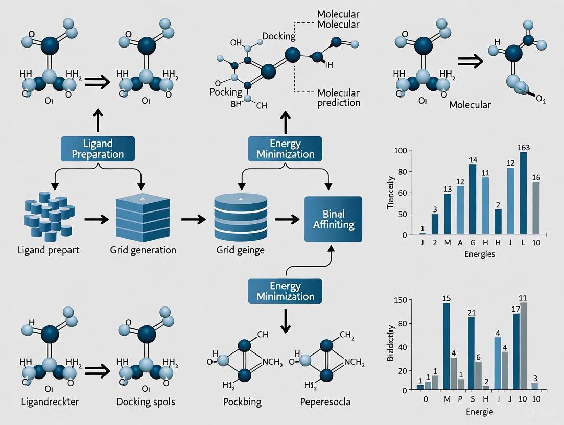
Diagram 1: Comprehensive molecular docking workflow.
Step 1: Protein Preparation
- Obtain the 3D structure of the target protein from experimental sources (Protein Data Bank) or computational predictions (AlphaFold) [7] [5].
- Remove water molecules, cofactors, and unnecessary ions, except those critical for binding.
- Add hydrogen atoms and assign appropriate protonation states to ionizable residues at physiological pH.
- Calculate and assign partial charges using appropriate force fields (AMBER, CHARMM, OPLS).
- Model any missing residues or loops using homology modeling or database searching.
- Energy minimization to relieve steric clashes and optimize geometry.
Step 2: Ligand Preparation
- Generate 3D coordinates from 2D structures using tools like BIOVIA Draw or Avogadro [5].
- Perform geometry optimization and energy minimization using semi-empirical methods (PM3) or molecular mechanics [5].
- Identify rotatable bonds for flexible docking treatments.
- Generate multiple low-energy conformers if using rigid docking approaches.
- Assign appropriate partial charges and atom types.
Step 3: Binding Site Definition and Grid Generation
- Identify the binding site using known experimental data, cavity detection algorithms, or blind docking approaches.
- Define a grid box encompassing the binding site with sufficient margin for ligand movement.
- Common approaches include:
- Site-Specific Docking: Grid centered on known binding site with 20-30Ã… dimensions
- Blind Docking: Large grid covering entire protein surface to identify novel binding sites [5]
- Calculate energy maps for efficient scoring function evaluation during docking.
Step 4: Docking Execution and Parameter Setting
- Select appropriate search algorithm based on ligand flexibility and computational resources.
- Configure docking parameters:
- Number of docking runs and poses to generate
- Population size and number of generations (for genetic algorithms)
- Maximum number of energy evaluations
- Cluster analysis parameters for result diversity
- Execute docking simulations, typically generating 10-100 poses per ligand.
Step 5: Pose Analysis and Validation
- Visualize top-ranked poses using molecular graphics software (PyMOL, Chimera, Discovery Studio)
- Analyze specific protein-ligand interactions: hydrogen bonds, hydrophobic contacts, π-π stacking, salt bridges
- Calculate binding energies and interaction energies for different pose clusters
- Validate results by redocking known ligands and comparing with experimental structures
- Check physical plausibility using tools like PoseBusters to identify geometric and chemical inconsistencies [6]
Advanced Protocol: Template-Based Docking (TEMPL)
For targets with known ligand complexes, template-based approaches can significantly improve accuracy:
Diagram 2: Template-based docking (TEMPL) workflow.
Application Context: This approach is particularly valuable for congeneric series or targets with abundant structural data, such as SARS-CoV-2 Main Protease [8].
Methodology Details:
- Reference Identification: Search structural databases (PDB) for complexes with similar ligands or binding sites
- Maximal Common Substructure (MCS): Use the RascalMCES algorithm to identify maximum common edge substructure between query and reference ligands [8]
- Constrained Embedding: Generate conformers using knowledge-enhanced distance geometry (ETKDGv3) with MCS atoms constrained to reference coordinates [8]
- Scoring and Ranking: Rank poses using ShapeTanimoto and ColorTanimoto scores measuring shape and feature complementarity [8]
Performance Comparison and Benchmarking
Docking Software Comparison
Table 1: Performance comparison of molecular docking methods across key metrics.
| Method Category | Representative Tools | Pose Accuracy (RMSD ≤ 2Å) | Physical Validity (PB-Valid) | Virtual Screening Performance | Computational Speed | Key Strengths |
|---|---|---|---|---|---|---|
| Traditional Physics-Based | Glide SP, AutoDock Vina, GOLD | 60-80% [6] | >94% [6] | High enrichment [6] | Medium to Fast | Excellent physical plausibility, proven reliability [9] [6] |
| Generative Diffusion Models | SurfDock, DiffBindFR | 70-92% [6] | 40-64% [6] | Variable | Fast (after training) | Superior pose accuracy, efficient sampling [6] |
| Regression-Based Models | KarmaDock, GAABind | 30-60% [6] | 20-50% [6] | Limited | Very Fast | Rapid prediction, but often produces invalid geometries [6] |
| Hybrid Methods | Interformer | 70-85% [6] | 80-90% [6] | Good | Medium | Balanced performance, combines AI scoring with traditional search [6] |
| Template-Based | TEMPL, FRED, HYBRID | Comparable to traditional [8] | High (structure-based) | Good for analogous compounds | Fast when templates available | Excellent for congeneric series, interpretable results [8] [4] |
Performance Across Dataset Types
Recent comprehensive evaluations reveal distinct performance patterns across different benchmarking scenarios:
Known Complexes (Astex Diverse Set): Traditional methods and hybrid approaches show robust performance with high physical validity (>94% for Glide SP), while diffusion models achieve exceptional pose accuracy (>91% for SurfDock) but with reduced physical plausibility (63.5%) [6].
Unseen Complexes (PoseBusters Benchmark): Performance gaps widen, with traditional methods maintaining stability while some AI methods show significant drops in both pose accuracy and physical validity, highlighting generalization challenges [6].
Novel Binding Pockets (DockGen Dataset): All methods show reduced performance, but traditional and hybrid methods demonstrate better adaptation to novel protein environments compared to pure AI approaches [6].
Table 2: Essential resources for molecular docking studies.
| Resource Category | Specific Tools/Sources | Key Function | Access Information |
|---|---|---|---|
| Protein Structure Databases | Protein Data Bank (PDB), AlphaFold Protein Structure Database | Source experimental and predicted protein structures | https://www.rcsb.org/, https://alphafold.ebi.ac.uk/ [1] [7] |
| Compound Libraries | ZINC, PubChem, ChEMBL, DrugBank | Source commercially available and bioactive compounds for virtual screening | https://zinc.docking.org/, https://pubchem.ncbi.nlm.nih.gov/ [1] |
| Docking Software | AutoDock Vina, Glide, GOLD, DOCK, FRED, HYBRID | Perform docking simulations and virtual screening | Varies: open-source (AutoDock) to commercial (Glide) [1] [4] |
| Structure Preparation Tools | CHARMM-GUI, AutoDock Tools, BIOVIA Discovery Studio | Prepare and optimize protein and ligand structures | https://www.charmm-gui.org/, https://autodocksuite.scripps.edu/ [5] |
| Visualization & Analysis | PyMOL, UCSF Chimera, BIOVIA Discovery Studio Visualizer | Visualize docking results and analyze interactions | https://pymol.org/, https://www.cgl.ucsf.edu/chimera/ [1] [5] |
| Validation Tools | PoseBusters | Check physical plausibility and geometric quality of docking poses | https://github.com/posebusters/posebusters [6] |
Advanced Applications and Future Directions
AI and Machine Learning Integration
The field of molecular docking is undergoing rapid transformation with the integration of artificial intelligence:
Deep Learning Pose Prediction: Methods like EquiBind, DiffDock, and TankBind use geometric deep learning and diffusion models to achieve superior pose accuracy, though concerns about physical plausibility and data leakage remain [8] [6].
Cofolding Approaches: AlphaFold3 and related methods simultaneously predict protein structure and ligand placement, showing promise particularly when experimental structures are unavailable [8] [9].
AI-Enhanced Scoring Functions: Machine learning models are being developed to improve binding affinity predictions by learning complex patterns from large structural datasets, addressing limitations of traditional scoring functions [3] [6].
Large-Scale Virtual Screening
Ultra-large virtual screening campaigns involving billions of compounds have become feasible with current computing resources [2]. Best practices for such campaigns include:
- Pre-screening Filters: Use rapid similarity searching and property-based filters to reduce library size before docking
- Staged Docking Protocols: Implement multi-tiered approaches with increasing precision at each stage
- Control Calculations: Include known actives and decoys to validate screening performance for each target
- Cluster Computing: Leverage distributed computing resources for processing massive compound libraries
Challenges and Limitations
Despite significant advances, important challenges persist:
- Generalization Gap: AI methods often struggle with novel protein targets or binding pockets not well-represented in training data [6]
- Interaction Recovery: Many ML docking methods prioritize low RMSD but fail to recapitulate key molecular interactions critical for biological activity [9]
- Receptor Flexibility: Accurate modeling of full receptor flexibility remains computationally challenging, though molecular dynamics simulations can provide post-docking refinement [3]
- Solvation Effects: Explicit treatment of water molecules and solvation energies in binding remains difficult in standard docking protocols
- Accuracy vs. Speed Trade-offs: Balancing computational efficiency with prediction accuracy continues to drive method development
The continued integration of physical principles with data-driven approaches, improved handling of flexibility, and enhanced generalization capabilities represent the most promising directions for advancing molecular docking methodologies in computer-aided drug design.
Non-covalent interactions are fundamental forces governing molecular recognition in biological systems, forming the physical basis for protein-ligand interactions in structure-based drug design [10]. These weak, reversible forces—hydrogen bonds, ionic interactions, van der Waals forces, and hydrophobic effects—collectively determine binding specificity and affinity between pharmaceutical compounds and their protein targets [10]. Unlike covalent bonds, non-covalent interactions range from 1-5 kcal/mol in strength but produce highly stable and specific associations through cumulative effects at binding interfaces [10]. Understanding these interactions is crucial for predicting ligand binding poses and accelerating rational drug discovery through molecular docking approaches [10] [11].
The binding process is governed by the thermodynamic principle of Gibbs free energy (ΔG = ΔH - TΔS), where favorable binding requires a negative ΔG value achieved through complementary balancing of enthalpic (ΔH) and entropic (ΔS) contributions [10]. Molecular docking algorithms leverage this principle to predict how small molecule ligands interact with protein targets by simulating the complex formation through computational methods [10] [11]. This document provides a comprehensive overview of these key non-covalent interactions, their quantitative characteristics, and experimental protocols for their investigation in the context of molecular docking research.
Theoretical Foundations of Non-Covalent Interactions
Hydrogen Bonds
Hydrogen bonds are polar electrostatic interactions represented as D—H···A, where D is an electron donor atom, H is a hydrogen atom attached to the donor, and A is an electron acceptor atom [10]. The donor atom must be electronegative (typically oxygen or nitrogen in biological systems), while the acceptor possesses lone electron pairs [10]. With a strength of approximately 5 kcal/mol—significantly weaker than covalent bonds (~110 kcal/mol for O-H)—hydrogen bonds play crucial roles in biomolecular recognition and stability [10]. In aqueous environments, the extensive hydrogen bonding network with solvent molecules creates a dynamic equilibrium where bonds constantly break and reform, significantly influencing the enthalpy and entropy of protein-ligand complex formation [10].
Ionic Interactions
Ionic interactions (also called salt bridges or electrostatic interactions) occur between permanently charged groups or strongly polarized atoms, creating attractive forces between oppositely charged ionic pairs [10]. These highly specific electrostatic interactions are strongly influenced by the solvent environment, particularly in aqueous solutions where ions become surrounded by hydration shells of water molecules, modulating their interaction strength [10]. The dielectric constant of the medium significantly affects the strength of ionic interactions, making them particularly important in partially shielded protein binding pockets where the local dielectric constant may be lower than in bulk solvent [10].
Van der Waals Interactions
Van der Waals interactions arise from transient fluctuations in electron distribution around atoms and molecules, creating temporary dipoles that induce complementary dipoles in neighboring molecules [10]. These nonspecific interactions are relatively weak (~1 kcal/mol) but become biologically significant when numerous atoms at optimal separation distances (typically 3-4 Ã…) contribute collectively to molecular recognition [10]. Recent research has revealed that van der Waals interactions in multilayer structures exhibit many-body characteristics that cannot be adequately described by simple pairwise addition, highlighting their quantum mechanical complexity [12]. Atomic force microscopy studies demonstrate that these interactions are significantly influenced by the broader molecular context, including underlying substrates in supported molecular systems [12].
Hydrophobic Interactions
Hydrophobic interactions describe the tendency of nonpolar molecules and surfaces to associate in aqueous environments, primarily driven by entropy changes in the surrounding water molecules rather than direct attractive forces between the nonpolar entities [10]. When nonpolar groups aggregate, they release structured water molecules from hydration shells into bulk solvent, increasing system entropy and providing a favorable thermodynamic driving force (ΔG < 0) despite minimal enthalpy changes [10]. According to scaled-particle theory, the molecular mechanisms of hydrophobic effects are multifaceted and depend on solute size, with different thermodynamic principles governing small versus large hydrophobic surfaces [10] [13].
Quantitative Comparison of Non-Covalent Interactions
Table 1: Key Characteristics of Major Non-Covalent Interactions in Protein-Ligand Complexes
| Interaction Type | Strength (kcal/mol) | Distance Dependence | Directionality | Key Role in Binding |
|---|---|---|---|---|
| Hydrogen Bonds | ~5 [10] | ~1/r³ [10] | High (linear D-H···A preferred) | Specificity and orientation |
| Ionic Interactions | 3-8 (context dependent) [10] | ~1/r² (in vacuum) [10] | Moderate (charge-centered) | Binding affinity, especially in buried pockets |
| Van der Waals | ~1 [10] | ~1/rⶠ[10] | None (nonspecific) | Shape complementarity, close contact |
| Hydrophobic | ~0.1-1 per Ų [10] [13] | Entropy-driven | None | Driving force for association |
Table 2: Experimental and Computational Techniques for Studying Non-Covalent Interactions
| Technique | Spatial Resolution | Key Measured Parameters | Applicable Interactions |
|---|---|---|---|
| X-ray Crystallography | ~1-3 Ã… [10] | Atomic positions, distances, angles | All types, especially hydrogen bonds |
| Cryo-EM | ~3-5 Ã… [10] | Molecular shapes, interfaces | Hydrophobic, van der Waals |
| NMR Spectroscopy | Atomic [10] | Dynamics, distances, chemical shifts | All in solution state |
| Atomic Force Microscopy | Sub-nanometer [12] | Adhesion forces, interaction energy | Van der Waals, hydrophobic |
| Molecular Dynamics | Atomic | Energy components, stability, kinetics | All, with computational models |
| Isothermal Titration Calorimetry | N/A | ΔH, ΔS, Ka, stoichiometry | Overall binding thermodynamics |
Experimental Protocols
Protocol 1: Computational Assessment of Non-Covalent Interactions via Molecular Docking
Purpose: To predict and characterize non-covalent interactions between a protein target and small molecule ligands using molecular docking approaches [10] [11].
Materials and Reagents:
- Protein Structure: experimentally determined (PDB format) or computationally predicted structure [10] [14]
- Ligand Structures: 2D or 3D chemical structures in SDF, MOL2, or similar formats [15]
- Docking Software: AutoDock Vina, Glide, GOLD, or deep learning-based tools (DiffDock, SurfDock) [6]
- Computational Resources: Workstation with multi-core CPU, GPU acceleration recommended for deep learning methods [11]
Procedure:
- Protein Preparation:
- Obtain protein structure from Protein Data Bank or predictive models (AlphaFold2, ESMFold) [10] [14]
- Remove water molecules and co-crystallized ligands, except crucial structural waters [15]
- Add hydrogen atoms, assign partial charges, and define protonation states at physiological pH [15]
- Identify binding site residues based on experimental data or binding site prediction tools (LABind, DeepSite) [14]
Ligand Preparation:
Docking Execution:
- Define search space using grid boxes centered on binding site (typically 30×30×30 ų) [15]
- For blind docking, encompass entire protein surface or predicted binding regions [11] [14]
- Set docking parameters (exhaustiveness, number of poses) based on method requirements [6]
- Execute docking runs and generate multiple pose predictions (typically 10-20 per ligand) [15]
Interaction Analysis:
- Visualize top-ranked poses in molecular visualization software [15]
- Identify specific non-covalent interactions (hydrogen bonds, ionic pairs, hydrophobic contacts) [10] [13]
- Measure interaction distances and geometries against optimal values [10]
- Calculate binding free energy estimates using scoring functions [6]
Troubleshooting Tips:
- If poses lack chemical realism, apply post-docking minimization or use physics-based refinement [6]
- For poor pose prediction accuracy, consider flexible residue docking or ensemble docking approaches [11]
- If binding affinity correlations are weak, use consensus scoring or machine-learning scoring functions [6]
Protocol 2: Binding Free Energy Calculation Using MM/GBSA
Purpose: To estimate protein-ligand binding free energies by molecular mechanics approaches with generalized Born and surface area solvation [15].
Materials and Reagents:
- Molecular Dynamics Software: GROMACS, AMBER, or similar packages [15] [16]
- Force Fields: CHARMM, AMBER, or OPLS parameters for proteins and ligands [16]
- Trajectory Analysis Tools: In-built packages or custom scripts for energy decomposition [15]
Procedure:
- System Setup:
Equilibration:
Production MD:
MM/GBSA Calculation:
Notes: The MM/GBSA method provides more reliable binding affinity estimates than docking scoring functions but requires significantly more computational resources [15]. Entropy calculations remain challenging and may be omitted for high-throughput applications [15].
Protocol 3: Experimental Validation of Non-Covalent Interactions
Purpose: To experimentally characterize non-covalent interactions in protein-ligand complexes using biophysical and structural biology techniques.
Materials and Reagents:
- Purified Protein: ≥95% purity, correctly folded, in appropriate buffer [10]
- Ligand Compounds: High-purity (>95%) compounds dissolved in DMSO or buffer [15]
- Crystallization Reagents: Commercially available screening kits [10]
- Biophysical Instruments: X-ray diffractometer, isothermal titration calorimeter, surface plasmon resonance biosensor [10]
Procedure:
- X-ray Crystallography:
Isothermal Titration Calorimetry (ITC):
- Degas protein and ligand solutions to eliminate air bubbles [10]
- Load protein solution into sample cell and ligand solution into syringe [10]
- Program automated injections with adequate spacing between injections [10]
- Measure heat changes upon each injection and fit data to binding model [10]
- Extract ΔH, ΔS, Ka, and stoichiometry (n) parameters [10]
Atomic Force Microscopy (for surface interactions):
Data Interpretation: Crystallography provides atomic-level interaction details, ITC delivers complete thermodynamic profiles, and AFM measures single-molecule interaction forces under various conditions [10] [12].
Workflow Visualization
Diagram 1: Workflow for comprehensive characterization of non-covalent interactions in protein-ligand complexes, integrating computational and experimental approaches.
The Scientist's Toolkit
Table 3: Essential Research Reagents and Computational Tools for Non-Covalent Interaction Studies
| Tool/Reagent | Category | Primary Function | Example Applications |
|---|---|---|---|
| AutoDock Vina | Docking Software | Protein-ligand docking with scoring function [15] [6] | Initial pose prediction, virtual screening |
| GROMACS | Molecular Dynamics | MD simulation with free energy calculations [16] | Binding stability, conformational sampling |
| LABind | Binding Site Prediction | Graph transformer for ligand-aware site prediction [14] | Binding residue identification |
| PoseBusters | Validation Toolkit | Checks physical/chemical plausibility of poses [6] | Pose quality assessment |
| DiffDock | Deep Learning Docking | Diffusion model for blind docking [11] [6] | Pose prediction without predefined site |
| PDBBind Database | Structural Database | Curated protein-ligand complexes with binding data [10] [11] | Method training and benchmarking |
| CHARMM Force Field | Molecular Mechanics | Potential functions for energy calculations [16] | MD simulations, energy minimization |
| ITC Instrument | Experimental Setup | Measures binding thermodynamics directly [10] | ΔH, ΔS, and Ka determination |
| Cytosaminomycin D | Cytosaminomycin D, MF:C23H36N4O8, MW:496.6 g/mol | Chemical Reagent | Bench Chemicals |
| Asparenomycin A | Asparenomycin A, MF:C14H16N2O6S, MW:340.35 g/mol | Chemical Reagent | Bench Chemicals |
Non-covalent interactions represent the fundamental language of molecular recognition in biological systems, with hydrogen bonds, ionic interactions, van der Waals forces, and hydrophobic effects collectively dictating the specificity and affinity of protein-ligand binding [10]. Molecular docking methodologies continue to evolve, with traditional physics-based approaches now complemented by deep learning methods that show promising results in pose prediction, though challenges remain in ensuring physical plausibility and generalization to novel targets [11] [6]. The integration of computational predictions with experimental validation through structural biology and biophysical techniques provides the most robust approach for characterizing these complex interactions [10] [15] [12]. As molecular docking continues to advance, particularly with incorporation of protein flexibility and many-body physical effects, researchers are better equipped to leverage understanding of non-covalent interactions for accelerated drug discovery and biological mechanism elucidation [11] [12].
The thermodynamics of protein-ligand interactions form the fundamental basis for understanding molecular recognition in biological systems and rational drug design. The binding affinity between a protein and ligand is quantitatively expressed by the Gibbs free energy change (ΔG), which relates to the binding constant through the equation ΔG = -RTlnKeq [10]. This free energy change comprises two competing components: the enthalpy change (ΔH), representing the heat released or absorbed during binding primarily through formation and breaking of chemical bonds, and the entropy change (ΔS), representing the change in system disorder, multiplied by temperature (TΔS) [10].
A phenomenon frequently observed in protein-ligand interactions is enthalpy-entropy compensation (EEC), where a more favorable (negative) enthalpy change is counterbalanced by a less favorable (negative) entropy change, or vice versa, resulting in minimal net change in the overall binding free energy [17]. This compensation effect presents significant challenges in drug discovery, where structural modifications designed to improve binding affinity often yield disappointing results due to this thermodynamic balancing act.
The Enthalpy-Entropy Compensation Phenomenon
Experimental Evidence and Quantitative Analysis
Enthalpy-entropy compensation is a well-documented phenomenon in protein-ligand interactions. Statistical analysis of 3025 protein-ligand affinities from the Protein Data Bank reveals that ΔG values for protein-ligand interactions follow a Gaussian distribution centered around -36.5 kJ/mol, with approximately 70% of cases falling between -46 and -26 kJ/mol [17]. This narrow range of ΔG values occurs despite enormously varied enthalpy and entropy values spanning ranges of -232 kJ/mol to 59.2 kJ/mol for ΔH and -190 kJ/mol to 64 kJ/mol for TΔS [17].
The linear relationship between ΔH and TΔS leads to an approximately constant value of ΔG around -30 to -35 kJ/mol across diverse protein-ligand systems [17]. This compensation behavior has been observed consistently for over fifty years in thermodynamic studies of protein-ligand interactions in aqueous solution and is particularly problematic in drug discovery campaigns where medicinal chemists seek to optimize lead compounds through structural modifications.
Table 1: Thermodynamic Parameter Ranges in Protein-Ligand Interactions
| Parameter | Typical Range | Average Value | Observations |
|---|---|---|---|
| ΔG | -46 to -26 kJ/mol | -36.5 kJ/mol | Gaussian distribution, narrow range |
| ΔH | -232 to 59.2 kJ/mol | Variable | Large variability between systems |
| TΔS | -190 to 64 kJ/mol | Variable | Large variability between systems |
| Compensation Slope | ~1 | — | Linear ΔH vs TΔS relationship |
Molecular Origins and Implications
The molecular origin of enthalpy-entropy compensation remains controversial and has been attributed to various factors. From an evolutionary perspective, the narrow range of ΔG values may reflect adaptive optimization of proteins to achieve maximum regulatory capacity through conformational versatility and exchange of minute energy quanta with the environment [17]. At the molecular level, binding involves complex rearrangements of water molecules, protein conformational changes, and formation of non-covalent interactions, all of which contribute to both enthalpy and entropy changes.
The functional implication of this compensation is profound for drug discovery. When structural modifications to a lead compound produce a more favorable enthalpy change through improved interactions with the target protein, this gain is frequently offset by entropy losses due to reduced flexibility or increased solvent ordering [17]. Consequently, substantial efforts in optimizing ligand-receptor interactions often yield disappointingly small improvements in binding affinity.
Non-Covalent Interactions in Molecular Recognition
Fundamental Interaction Types
Protein-ligand recognition is mediated through several types of non-covalent interactions, each with characteristic energy contributions and structural properties:
- Hydrogen bonds: Polar electrostatic interactions between hydrogen bond donors (D-H) and acceptors (A) with typical strengths of approximately 5 kcal/mol (~21 kJ/mol). These interactions are highly directional and strongly influenced by the solvent environment [10].
- Van der Waals interactions: Non-specific attractions between transient dipoles in electron clouds with strengths of approximately 1 kcal/mol (~4 kJ/mol). These interactions, while weak individually, contribute significantly to binding through cumulative effects [10].
- Ionic interactions: Electrostatic attractions between oppositely charged groups, highly specific but influenced by solvent screening and solvation shells [10].
- Hydrophobic interactions: Associations of non-polar groups in aqueous solution, primarily driven by entropy gain from water molecule reorganization [10].
Table 2: Non-Covalent Interactions in Protein-Ligand Complexes
| Interaction Type | Strength (kJ/mol) | Characteristics | Role in Binding |
|---|---|---|---|
| Hydrogen Bonds | ~21 | Directional, solvent-sensitive | Specificity, enthalpy contribution |
| Van der Waals | ~4 | Non-specific, distance-dependent | Packing, shape complementarity |
| Ionic Interactions | Variable | Distance and dielectric-dependent | Strong electrostatic contributions |
| Hydrophobic Effect | Variable | Entropy-driven, area-dependent | Major entropy contribution |
Molecular Recognition Models
Three conceptual models describe the mechanism of molecular recognition in protein-ligand binding:
Lock-and-Key Model: Theorizes pre-complementary binding interfaces between rigid proteins and ligands, representing an entropy-dominated binding process with minimal conformational changes [10].
Induced-Fit Model: Proposes conformational changes in the protein during binding to optimally accommodate the ligand, adding flexibility to the lock-and-key concept [10].
Conformational Selection Model: Suggests ligands bind selectively to the most suitable conformational state from an ensemble of pre-existing protein conformations [10].
Experimental Protocols for Thermodynamic Characterization
Isothermal Titration Calorimetry (ITC) Protocol
Purpose: Direct measurement of binding thermodynamics including ΔG, ΔH, ΔS, and binding stoichiometry.
Materials:
- Isothermal titration calorimeter (e.g., MicroCal VP-ITC)
- Purified protein solution (>95% purity)
- Ligand solution in matching buffer
- Dialysis buffer for exact solvent matching
- Degassing apparatus
Procedure:
- Precisely match the buffer composition between protein and ligand solutions through dialysis or careful preparation.
- Degas all solutions to prevent bubble formation during measurements.
- Load the protein solution into the sample cell (typically 1.4 mL volume) and ligand solution into the injection syringe.
- Program the titration method: typically 25-30 injections of 2-10 μL each with 120-180 second intervals between injections.
- Perform the experiment at constant temperature (typically 25°C or 37°C).
- Include a control experiment of ligand injections into buffer alone for heat of dilution correction.
- Analyze data using nonlinear least-squares fitting to obtain ΔH, Ka (association constant), and stoichiometry (n).
- Calculate ΔG from ΔG = -RTlnKa and ΔS from ΔS = (ΔH - ΔG)/T.
Data Interpretation:
- Exothermic reactions (negative ΔH) typically indicate favorable hydrogen bonding or van der Waals interactions.
- Entropy-driven binding (positive TΔS) often suggests hydrophobic effects or release of bound water molecules.
- Enthalpy-entropy compensation manifests when improvements in ΔH are offset by decreases in TΔS.
Computational Docking and Free Energy Calculations
Purpose: Prediction of binding modes and affinities through computational approaches.
Materials:
- High-performance computing resources
- Protein structure (experimental or predicted)
- Ligand structure database
- Docking software (e.g., Glide, AutoDock, GOLD)
- Molecular dynamics simulation packages
Procedure:
- Structure Preparation:
- Obtain protein structure from PDB or generate with AlphaFold2 [18]
- Remove crystallographic water molecules except those mediating key interactions
- Add hydrogen atoms, assign protonation states at physiological pH
- Energy minimization to relieve steric clashes
Binding Site Identification:
- Define binding pocket from known complexes or predicted active sites
- Generate grid maps encompassing the binding region
Molecular Docking:
- Perform flexible ligand docking with multiple conformational searches
- Score poses using empirical, force-field, or knowledge-based scoring functions
- Cluster results and select representative binding modes
Molecular Dynamics Refinement (Optional):
- Solvate the protein-ligand complex in explicit water molecules
- Apply physiological ionic concentration
- Equilibrate system with positional restraints followed by free dynamics
- Run production simulation (typically 50-500 ns) [18]
- Analyze trajectories for stability and interaction persistence
Free Energy Calculations:
- Employ methods like MM/PBSA, MM/GBSA, or free energy perturbation
- Decompose energy contributions per residue or interaction type
Research Reagent Solutions and Materials
Table 3: Essential Research Reagents for Thermodynamic Studies
| Reagent/Material | Specifications | Function/Application |
|---|---|---|
| ITC Instrumentation | MicroCal VP-ITC or equivalent | Direct measurement of binding thermodynamics |
| Protein Purification System | FPLC with affinity columns | Production of pure, functional protein |
| Buffer Components | High-purity salts, buffers | Maintain physiological conditions |
| AlphaFold2 | Computational structure prediction | Generate protein models when experimental structures unavailable [18] |
| Molecular Dynamics Software | GROMACS, AMBER, NAMD | Refine structures and simulate dynamics [18] |
| Docking Software | Glide, AutoDock Vina, TankBind | Predict binding modes and affinities [18] |
Visualization of Thermodynamic Relationships and Methodologies
Thermodynamic Relationships in Protein-Ligand Binding
ITC Experimental Workflow
Implications for Drug Discovery and Molecular Docking
The phenomenon of enthalpy-entropy compensation has profound implications for structure-based drug design. While molecular docking approaches have advanced significantly, challenges remain in accurately predicting binding affinities, particularly for protein-protein interactions [18]. Recent benchmarking studies demonstrate that AlphaFold2-generated structures perform comparably to experimental structures in docking protocols, expanding the structural database available for drug discovery [18].
Local docking strategies generally outperform blind docking approaches, with TankBind_local and Glide providing particularly robust results across diverse protein structures [18]. Integration of molecular dynamics simulations and ensemble-based approaches can improve docking outcomes in selected cases, though performance improvements vary significantly across different conformations [18].
The limited range of ΔG values observed across diverse protein-ligand systems (-46 to -26 kJ/mol) suggests evolutionary optimization of protein flexibility and interaction energies to achieve optimal regulatory function [17]. This fundamental constraint underscores the importance of considering thermodynamic profiles in lead optimization, moving beyond simple affinity measurements to understand the enthalpic and entropic drivers of molecular recognition.
{#introduction} Molecular recognition, the process by which biological molecules interact specifically with each other and with small ligands, forms the cornerstone of all biological processes and structure-based drug design. The conceptual models describing these interactions have evolved significantly from Emil Fischer's initial "lock-and-key" analogy proposed in 1894 to more sophisticated frameworks that account for protein dynamics and flexibility [19] [20]. This evolution reflects our growing understanding of the intricate dance between proteins and ligands, which is crucial for advancing molecular docking methodologies and improving the accuracy of ligand pose prediction [19] [10]. As the central thesis of this article, we posit that the progression from static to dynamic recognition models has been, and continues to be, the primary driver of innovation in computational drug discovery, enabling researchers to tackle increasingly complex challenges in predicting protein-ligand complex structures.
{##conceptual-evolution}
Conceptual Evolution of Molecular Recognition Models
{###lock-and-key}
The Lock-and-Key Model
Introduced by Emil Fischer in 1894, the lock-and-key model conceptualizes molecular recognition through a simple analogy: the enzyme (lock) and the substrate (key) possess complementary, pre-formed geometric shapes that fit perfectly together [19] [21]. This model posits that both interacting partners are essentially rigid, and their conformations remain unchanged during the binding event [10]. While this model successfully explained early observations of enzyme specificity, its major limitation was the failure to account for the inherent flexibility of proteins and the conformational changes that often accompany ligand binding [19]. Despite its simplicity, the lock-and-key paradigm profoundly influenced the philosophical underpinnings of early molecular docking approaches, which treated both the protein receptor and the ligand as static entities [19].
{###induced-fit}
The Induced-Fit Model
In 1958, Daniel Koshland proposed the induced-fit model as a necessary modification to address the shortcomings of the lock-and-key analogy [19] [20]. This model suggests that the active site of an enzyme is not a static cavity; rather, it is reshaped during interactions with the substrate [19]. The ligand induces conformational changes in the protein, leading to an optimal binding arrangement that would not occur with a rigid protein structure [10] [20]. This concept is more akin to a "pin tumbler lock," where the key (ligand) allows internal components (protein residues) to move into the correct alignment [19]. The induced-fit model accounts for why certain ligands that appear sterically compatible may not bind, as they fail to induce the necessary conformational adjustments. It also explains phenomena like allosteric and non-competitive inhibition [20]. From a computational perspective, incorporating induced-fit effects remains a significant challenge due to the vast conformational space that must be sampled [19].
{###conformational-selection}
The Conformational Selection Model
The conformational selection model, sometimes referred to as selected-fit, represents a further refinement of our understanding [21]. In this model, the protein exists in a dynamic equilibrium between multiple conformational states even in the absence of ligand [10] [21]. The ligand does not "induce" a new conformation but rather selectively binds to the pre-existing conformational state for which it has the highest affinity, thereby stabilizing that state and shifting the equilibrium population [10] [21]. In an extended recognition mechanism, ligands may first bind to a favorable initial protein conformation, which is then followed by additional conformational adjustments [10]. This model aligns with the modern understanding of proteins as dynamic ensembles and provides a more robust thermodynamic explanation for many allosteric effects.
{###keyhole-lock-key}
The Keyhole-Lock-Key Model
For enzymes with deeply buried active sites, a more specialized model has been proposed: the keyhole-lock-key model [21]. This model incorporates the critical role of access tunnels (keyholes) that connect the active site (lock) to the bulk solvent [21]. These tunnels are not merely passive conduits; their anatomy, physico-chemical properties, and dynamics can discriminate between substrates, control the entry of co-substrates, and prevent cellular damage by sequestering reactive intermediates [21]. The catalytic cycle, therefore, involves the passage of the ligand through the tunnel, reorganization of water molecules, binding to catalytic residues, chemical transformation, and finally, product exit [21]. This model is particularly relevant for engineering enzyme activity, specificity, and stability by modifying these access pathways rather than the active site itself [21].
{##comparison}
Comparative Analysis of Recognition Models
{###table-comparison} Table 1: Comparative Analysis of Molecular Recognition Models
| Model | Proposed Year | Core Principle | View of Protein Structure | Thermodynamic Driver | Key Limitation |
|---|---|---|---|---|---|
| Lock-and-Key [19] [10] | 1894 | Steric and geometric complementarity | Rigid and static | Entropy-dominated (ΔS) [10] | Oversimplified; ignores flexibility |
| Induced-Fit [19] [20] | 1958 | Ligand binding induces conformational change | Flexible and adaptable | Enthalpy-driven (ΔH) | Can be computationally prohibitive to model |
| Conformational Selection [10] [21] | ~2000s | Ligand binds to and stabilizes a pre-existing conformation | Dynamic ensemble of states | Combination of ΔH and ΔS | Requires knowledge of multiple states |
| Keyhole-Lock-Key [21] | ~2000s | Access tunnels (keyholes) are critical for catalysis | Dynamic, with gated access | Kinetically controlled by tunnels | Most applicable to enzymes with buried active sites |
The following diagram illustrates the logical and temporal relationships between the different molecular recognition models, showing how each new theory built upon and refined its predecessors.
{caption="Figure 1: Evolution of molecular recognition models over time"}
{##applications-docking}
Applications in Molecular Docking and Ligand Pose Prediction
The evolution of molecular recognition theories has directly informed the development and application of computational docking methodologies. Modern docking approaches strive to incorporate the dynamic principles of induced-fit and conformational selection to improve predictive accuracy.
{###table-computational-tools} Table 2: Computational Tools Implementing Dynamic Recognition Principles
| Computational Tool | Underlying Recognition Model | Key Methodology | Application in Pose Prediction |
|---|---|---|---|
| ColdstartCPI [22] | Induced-Fit | Uses Transformers to learn flexible, context-dependent features for compounds and proteins. | Treats proteins and compounds as flexible entities during inference, improving predictions for unseen compounds and proteins. |
| DynamicBind [23] | Conformational Selection & Dynamics | Deep equivariant generative model that constructs a smooth energy landscape. | Efficiently samples large protein conformational changes to recover ligand-specific holo-structures from apo-like inputs. |
| Traditional Rigid Docking [19] | Lock-and-Key | Treats protein as rigid and samples only ligand flexibility. | Fast but often fails when significant protein side-chain or backbone movement is required for binding. |
{###protocol}
Protocol: Implementing a Dynamic Docking Workflow with DynamicBind
Purpose: To predict the binding pose of a ligand to a protein target, accounting for substantial protein conformational changes, using the DynamicBind model [23].
{####materials}
Research Reagent Solutions & Essential Materials
Table 3: Essential Materials for DynamicBind Docking Protocol
| Item Name | Function/Description | Specification/Format |
|---|---|---|
| Target Protein Sequence | The primary amino acid sequence of the protein target. | FASTA format string. |
| AlphaFold-Predicted Structure | Provides the initial apo-like protein conformation for docking. | PDB file format. |
| Ligand Structure | The small molecule to be docked. | SMILES string or SDF file. |
| DynamicBind Software | The deep learning model for dynamic docking. | Publicly available code (e.g., from GitHub repository). |
| RDKit Library | Open-source cheminformatics library. | Used for generating initial ligand conformations [23]. |
{####procedure}
Procedure
Input Preparation:
- Protein Structure: Obtain the 3D structure of your target protein. If an experimental holo-structure is unavailable, use an AlphaFold-predicted conformation as the input PDB file [23].
- Ligand Structure: Provide the ligand structure in a SMILES string or SDF format. RDKit will be used internally to generate an initial 3D conformation [23].
Ligand Placement:
- Run DynamicBind, which will begin by randomly placing the ligand around the putative binding site of the input protein structure [23].
Iterative Pose Optimization:
- The model will run for a default of 20 iterations. The process involves two phases [23]:
- Steps 1-5: The model optimizes the ligand's pose by translating, rotating, and adjusting its internal torsional angles. The protein remains fixed during this initial phase.
- Steps 6-20: The model simultaneously optimizes both the ligand and the protein. It adjusts the protein's conformation by translating/rotating residues and modifying side-chain chi angles to accommodate the ligand [23].
- The model will run for a default of 20 iterations. The process involves two phases [23]:
Pose Selection and Validation:
- DynamicBind generates multiple output complex structures. Use the built-in contact-LDDT (cLDDT) scoring module to rank the predictions. A higher cLDDT score correlates with a lower ligand RMSD, indicating a more reliable pose [23].
- Select the top-ranked pose for downstream analysis. The final output is a PDB file of the protein-ligand complex, often with a protein conformation closer to the true holo-state than the initial AlphaFold input [23].
The workflow for this protocol is summarized in the diagram below.
{caption="Figure 2: DynamicBind dynamic docking workflow"}
{##discussion}
Discussion and Future Perspectives
The progression from the rigid lock-and-key model to dynamic and ensemble-based views has fundamentally transformed the field of structure-based drug design. Modern deep learning approaches, such as ColdstartCPI and DynamicBind, are now explicitly embedding the principles of induced-fit and conformational selection into their architectures, leading to significant improvements in handling cold-start scenarios and predicting large-scale protein conformational changes [22] [23]. However, formidable challenges remain. Accurately modeling the role of water molecules in mediating binding interactions and achieving a comprehensive representation of full protein flexibility continue to be active areas of research [19]. The future of molecular docking and ligand pose prediction lies in the development of even more sophisticated models that can seamlessly integrate multiple recognition mechanisms, fully account for solvent dynamics, and efficiently explore the vast energy landscape of protein-ligand complexes. This will be crucial for unlocking new therapeutic targets and accelerating the drug discovery process.
Docking Methodologies in Practice: Algorithms, Software, and Workflow Strategies
Molecular docking is a fundamental computational technique in structural biology and drug discovery that predicts the preferred orientation of a small molecule (ligand) when bound to a target macromolecule (receptor) [24]. The core challenge docking aims to solve is identifying the ligand's binding mode and affinity, which requires efficiently searching the vast conformational and positional space available to the ligand [25]. Systematic search algorithms represent a class of docking methods characterized by their deterministic exploration of this space, in contrast to stochastic methods which rely on random sampling [24] [25]. These algorithms are crucial for reproducing experimental binding modes and have become integral to structure-based drug design, enabling researchers to understand molecular interactions at an atomic level and accelerate the identification of potential therapeutic compounds [26] [27].
The development of systematic approaches is rooted in the evolution of binding theory. The earliest "lock-and-key" theory, proposed by Fischer, treated both ligand and receptor as rigid bodies [24]. This conceptual foundation led to the first docking methods which employed rigid-body treatment. Koshland's "induced-fit" theory advanced this understanding by recognizing that the active site of a protein is often reshaped by interactions with ligands, highlighting the need for algorithms that could account for molecular flexibility [24]. Systematic search algorithms emerged as a solution to this challenge, providing methodologies to comprehensively explore conformational space while maintaining computational feasibility. Their development has been instrumental in transitioning docking from a conceptual model to a practical tool that can accurately predict binding geometries, with modern algorithms capable of reproducing experimentally observed binding modes with root-mean-square deviations (RMSD) of 0.5 to 1.2 Ã… [26].
Classification and Theoretical Framework of Systematic Search Methods
Systematic search algorithms in molecular docking can be broadly categorized into three main approaches: exhaustive methods, incremental construction, and database searches. Each employs distinct strategies to manage the computational complexity of exploring the ligand's conformational and positional degrees of freedom [24] [25].
Exhaustive or Direct Methods involve the systematic enumeration of a ligand's degrees of freedom through gradual changes to its translational, rotational, and torsional parameters [25]. This approach aims to comprehensively explore the conformational space but often requires strategies to prune the search tree and avoid combinatorial explosion. The method guarantees that all possible configurations within defined constraints are evaluated, making it particularly valuable when a complete mapping of the energy landscape is required [25].
Incremental Construction (IC) methods, also known as fragmentation approaches, decompose the ligand into multiple fragments by breaking rotatable bonds [24] [28]. The largest fragment or the one with significant functional interactions is typically selected as the "base" or "anchor" and docked first into the active site [24] [28]. Subsequent fragments are then added incrementally, with different orientations generated to fit the active site, thereby reconstructing the complete ligand while accounting for its flexibility [26] [24]. This method significantly reduces the search space compared to exhaustive approaches and has been implemented in successful docking programs like FlexX, DOCK 4.0, and SLIDE [24]. Research has demonstrated that with multiple automated base selection, the quality of docking predictions is nearly as good as with manually preselected base fragments, making the approach practical for large-scale virtual screening [28].
Database Search methods leverage pre-existing structural information to enhance docking efficiency [24] [25]. These approaches generate multiple reasonable conformations for small molecules already cataloged in structural databases and dock them as rigid bodies [25]. The method capitalizes on the known structural diversity of chemical compounds to limit the conformational search space, offering significant computational advantages for screening large compound libraries [24]. Tools utilizing this approach include FLOG, which applies matching algorithms based on molecular shape to map ligands into active sites according to shape features and chemical information [24].
Table 1: Classification of Systematic Search Algorithms in Molecular Docking
| Algorithm Type | Key Principle | Representative Software | Advantages |
|---|---|---|---|
| Exhaustive/Direct Methods | Systematic enumeration of torsional, translational, and rotational degrees of freedom | DOCK (early versions) | Comprehensive exploration of conformational space; deterministic results |
| Incremental Construction | Fragment-based ligand reconstruction in binding site | FlexX, DOCK 4.0, Hammerhead, SLIDE, eHiTS | Efficient handling of ligand flexibility; fast execution suitable for virtual screening |
| Database Search | Rigid docking of pre-generated conformations from structural databases | FLOG, LibDock, SANDOCK | High speed; excellent for database enrichment and screening large compound libraries |
Quantitative Performance Comparison of Systematic Search Algorithms
Evaluating the performance of systematic search algorithms requires multiple metrics that reflect their computational efficiency, sampling accuracy, and practical utility in drug discovery applications. The performance characteristics vary significantly across different algorithm types, with inherent trade-offs between sampling comprehensiveness and computational demand [24].
The computational speed of these algorithms spans several orders of magnitude, with database search methods typically achieving the highest throughput due to their reliance on pre-computed conformations [24]. Incremental construction approaches offer a balanced compromise, with methods like FlexX capable of docking ligands in seconds to minutes depending on complexity [26] [24]. Exhaustive methods generally demand the greatest computational resources but provide the most complete exploration of the conformational landscape [25]. The accuracy of pose prediction is commonly measured by RMSD between predicted and experimentally determined crystal structures, with values below 2.0 Ã… generally considered successful reproduction of the binding mode [29]. Incremental construction algorithms have demonstrated particular effectiveness, achieving RMSD deviations of 0.5 to 1.2 Ã… across diverse test cases [26].
Sampling effectiveness varies according to each algorithm's approach to managing flexibility. Incremental construction efficiently handles ligand flexibility by focusing on fragment assembly rather than whole-molecule conformational sampling [24] [28]. This approach has proven highly effective, with studies showing that incremental construction can correctly identify experimental binding modes among the highest-ranking conformations in most test cases [26]. The method's performance can be further enhanced through multiple automatic base selection, which generates more diverse solutions and identifies alternative binding modes with low scores [28].
Table 2: Quantitative Performance Metrics of Systematic Search Algorithms
| Performance Metric | Exhaustive Methods | Incremental Construction | Database Search |
|---|---|---|---|
| Computational Speed | Slowest (hours to days per ligand) | Fast (seconds to minutes per ligand) | Fastest (multiple ligands per second) |
| Accuracy (RMSD) | Variable (dependent on sampling granularity) | High (0.5-1.2 Ã… reported) [26] | Moderate to High (dependent on database coverage) |
| Ligand Flexibility Handling | Comprehensive but computationally expensive | Efficient through fragmentation | Limited to pre-computed conformations |
| Virtual Screening Applicability | Low due to speed constraints | High (balanced speed and accuracy) | Excellent for primary screening |
| Pose Prediction Success Rate | High with sufficient sampling | High (71-85% for optimized algorithms) [26] | Moderate to High |
Experimental Protocols for Systematic Search Algorithms
Protocol for Incremental Construction with FlexX
The FlexX docking software implements the incremental construction algorithm and has been widely validated for molecular docking applications. The following protocol outlines a standardized approach for pose prediction using this method [24]:
Step 1: System Preparation
- Obtain the three-dimensional structure of the target protein from the Protein Data Bank (PDB) or through homology modeling. Prepare the structure by removing heteroatoms except essential cofactors, adding hydrogen atoms, and assigning partial charges using appropriate force fields [27] [29].
- Prepare the ligand structure by energy minimization using molecular mechanics force fields. Assign Gasteiger-Huckel charges to structure atoms and perform molecular dynamics (e.g., Simulated Annealing) to obtain conformations with minimum global energy [29].
Step 2: Base Fragment Selection
- FlexX automatically fragments the ligand at rotatable bonds, identifying multiple base fragments according to predefined rules and algorithms [28]. The base is typically the largest rigid fragment or the fragment with the most potential interaction points.
- Alternatively, manual selection of a specific base fragment can be performed if prior knowledge suggests its importance in binding [28].
Step 3: Placement of Base Fragment
- The base fragment is positioned in the active site using a pattern-matching algorithm based on chemical feature complementarity [24]. Triangle matching is used to place the base fragment by matching hydrogen bond donors, acceptors, and interaction centers between the protein and ligand [29].
- Multiple placements are generated to ensure adequate sampling of possible orientations.
Step 4: Incremental Reconstruction
- The remaining fragments are added incrementally to the base fragment in a tree-growing process. At each step, conformational space is sampled by rotating around rotatable bonds [24].
- The maximum number of solutions per iteration is typically set to 1000, with the maximum number of solutions per fragmentation set to 200 to balance comprehensiveness and computational efficiency [29].
Step 5: Scoring and Ranking
- Generated poses are evaluated using an empirical scoring function that accounts for hydrophobic contact surfaces, hydrogen bonding, ionic interactions, and aromatic stacking [24] [29].
- The top poses (typically 10-30) are retained for further analysis, with the highest-ranking conformation representing the predicted binding mode [29].
Validation: The docking protocol should be validated by redocking a known co-crystallized ligand and calculating the RMSD between the docked and native conformation. An RMSD value ≤ 2.0 Å indicates a validated protocol [29].
Protocol for Database Search with FLOG
FLOG (Flexible Ligands Oriented on Grid) utilizes a database search approach to molecular docking, offering high-throughput screening capabilities [24]:
Step 1: Database Preparation
- Compile a database of diverse, drug-like compounds in a standardized format (e.g., SMILES, SDF). Filter compounds according to Lipinski's Rule of Five to enhance drug-likelihood [30].
- Generate multiple conformers for each compound using rule-based or knowledge-based methods. Energy minimization should be performed for each conformer to ensure structural stability.
Step 2: Receptor Setup
- Prepare the protein structure by defining the active site through reference to known binding ligands or active site prediction algorithms (e.g., GRID, PASS) [24].
- Generate molecular interaction fields representing shape and chemical complementarity using programs like DOCK.
Step 3: Shape-Based Matching
- Each conformer from the database is systematically fitted into the active site based on steric and chemical complementarity [24] [25].
- The matching algorithm aligns ligand atoms with complementary interaction sites in the protein binding pocket.
Step 4: Scoring and Prioritization
- Poses are evaluated using force field-based or empirical scoring functions [25].
- High-ranking compounds are selected for further experimental validation or more detailed computational analysis.
Workflow Visualization of Systematic Search Algorithms
The following diagram illustrates the conceptual workflow and logical relationships between different systematic search algorithms in molecular docking:
Systematic Search Algorithms Workflow in Molecular Docking
The incremental construction algorithm specifically follows a detailed workflow for flexible ligand docking, as illustrated below:
Incremental Construction Algorithm Workflow
Research Reagent Solutions for Molecular Docking
Successful implementation of systematic search algorithms requires a suite of specialized software tools and computational resources. The table below details essential research reagents and their functions in molecular docking workflows:
Table 3: Essential Research Reagent Solutions for Molecular Docking Studies
| Resource Category | Specific Tool/Resource | Function in Docking Workflow | Key Features |
|---|---|---|---|
| Docking Software | FlexX | Implements incremental construction algorithm for flexible ligand docking | Fragment-based docking; fast execution; integration with BioSolveIT suite [24] [29] |
| DOCK | Versatile docking package employing multiple algorithms including database search | Geometry-based approach; suitable for virtual screening and database enrichment [24] [2] | |
| FLOG | Database search docking using pre-computed conformations | High-speed screening; shape-based matching [24] | |
| Structure Preparation | AutoDock Tools (MGL Tools) | Prepares receptor and ligand files in PDBQT format | Adds Gasteiger charges; defines rotatable bonds; grid parameter generation [27] [31] |
| Protein Preparation Wizard | Processes protein structures for docking | Adds hydrogens; assigns partial charges; optimizes hydrogen bonding [32] | |
| Structure Databases | Protein Data Bank (PDB) | Repository of experimentally determined protein structures | Source of receptor structures and co-crystallized ligands for validation [30] |
| ChEMBL Database | Curated database of bioactive molecules with drug-like properties | Source of compound libraries for virtual screening [30] | |
| Visualization & Analysis | PyMOL | Molecular visualization and manipulation | Structure analysis; image generation; cavity detection [31] |
| Discovery Studio Visualizer | Comprehensive suite for structural analysis | Interaction analysis; binding pose assessment; visualization of docking results [27] |
Molecular docking is a pivotal technique in computer-aided drug design (CADD) that predicts the preferred orientation and binding affinity of a small molecule (ligand) when bound to a target macromolecule (receptor) [33] [10]. The core challenge lies in efficiently searching the vast conformational and orientational space of the ligand to identify the binding pose that minimizes the free energy of the system. This search space is exceptionally complex and high-dimensional, making exhaustive systematic searches computationally intractable for all but the simplest systems [25] [34].
Stochastic search algorithms provide a powerful solution to this challenge by incorporating an element of randomness, allowing them to navigate complex energy landscapes effectively without being trapped in local minima [25] [35]. Unlike systematic methods that explore every possible conformation, stochastic methods sample the search space intelligently, making them particularly suitable for docking simulations where computational efficiency is crucial [25]. These algorithms have become fundamental components of many widely used docking programs, enabling researchers to perform virtual screening, lead optimization, and mechanistic studies in structural biology [33] [10].
The three predominant stochastic approaches in molecular docking are Monte Carlo methods, Genetic Algorithms, and Tabu Search. Each employs distinct strategies for managing the trade-off between exploration (searching new regions of the conformational space) and exploitation (refining promising solutions) [25] [35]. Their performance is critical for predicting accurate binding modes, which directly impacts the success of structure-based drug design campaigns [34].
Table 1: Core Stochastic Search Algorithms in Molecular Docking
| Algorithm | Core Principle | Key Advantages | Common Implementations |
|---|---|---|---|
| Monte Carlo | Random sampling with probabilistic acceptance criteria | Simple implementation, avoids local minima | AutoDock Vina, MCDock, ICM |
| Genetic Algorithms | Population-based evolution through selection, crossover, mutation | Effective for complex spaces, parallelizable | AutoDock, GOLD, rDock |
| Tabu Search | Memory-based guidance to avoid revisiting solutions | Prevents cycling, efficient for rugged landscapes | PRO_LEADS, Molegro Virtual Docker |
Algorithm Fundamentals and Comparative Analysis
Monte Carlo Methods
Monte Carlo (MC) methods in molecular docking rely on random sampling of the ligand's conformational and orientational degrees of freedom [25]. The fundamental principle involves generating random changes to the ligand's position, orientation, and torsion angles, then evaluating the resulting binding energy using a scoring function [25]. A key feature of MC algorithms is the Metropolis criterion, which determines whether to accept or reject new configurations based on the change in energy (ΔE) [25].
The Metropolis criterion accepts energetically favorable moves (ΔE < 0) outright, while permitting unfavorable moves (ΔE > 0) with a probability proportional to e^(-ΔE/kT), where k is the Boltzmann constant and T is the temperature parameter [25]. This controlled acceptance of worse solutions allows the algorithm to escape local energy minima and explore a broader region of the conformational space [25]. Modern docking programs often enhance basic MC with iterative search strategies; for instance, AutoDock Vina 1.2.0 combines Monte Carlo with the Broyden-Fletcher-Goldfarb-Shanno (BFGS) method for local refinement of ligand conformations [31].
Monte Carlo approaches are particularly valuable in specialized docking scenarios. Recent advancements have integrated MC with other techniques, such as Grand Canonical Monte Carlo (GCMC), to simulate the insertion and deletion of fragments in binding sites, overcoming sampling limitations in molecular dynamics-based simulations [36]. Furthermore, combined DFT, Monte Carlo, and molecular docking studies demonstrate the utility of MC in probing adsorption processes and corrosion inhibition properties, highlighting its versatility beyond traditional drug-target applications [37].
Genetic Algorithms
Genetic Algorithms (GAs) are population-based optimization techniques inspired by biological evolution [25] [35]. In molecular docking, GAs operate on a population of candidate ligand poses, each represented as a "chromosome" encoding translational, rotational, and torsional degrees of freedom [25] [34]. The algorithm iteratively improves this population through selection, crossover, and mutation operations, with the "fitness" of each pose typically being the predicted binding affinity [25].
The Lamarckian Genetic Algorithm (LGA), implemented in AutoDock 4.2, represents a significant advancement by incorporating local search to refine individual poses within the evolutionary framework [34]. This hybrid approach accelerates convergence by allowing individuals to adapt within their lifetime (Lamarckian evolution) rather than relying solely on genetic operations [34]. Parameter tuning is crucial for GA performance; a study on algorithm selection for protein-ligand docking examined 28 distinct LGA variants, highlighting how parameters like population size, mutation rates, and crossover operations significantly impact docking accuracy and efficiency [34].
GAs have proven particularly effective for de novo drug design. AutoGrow4, an open-source toolkit for semi-automated computer-aided drug discovery, exploits a genetic algorithm combined with molecular docking to generate novel ligands for a given target [38]. This approach efficiently explores chemical space through an evolutionary process that builds new molecules from fragment libraries, though it may exhibit bias toward high molecular weight compounds [38].
Tabu Search
Tabu Search (TS) employs adaptive memory structures to guide the search process, explicitly preventing revisiting recently explored solutions [25] [35]. The core mechanism maintains a "tabu list" of forbidden moves or solutions, effectively prohibiting the algorithm from cycling back to previously visited regions of the conformational space [25]. This memory-based approach allows TS to navigate rugged energy landscapes more efficiently than memoryless algorithms [25].
At each iteration, Tabu Search generates candidate moves from the current solution and selects the best move that is not in the tabu list [25]. This strategy encourages exploration of new territories in the search space, making it particularly effective for problems with numerous local optima [25]. The tabu list is typically maintained as a FIFO (first-in, first-out) queue, with the size of the list carefully balanced to prevent cycling without overly restricting promising directions [25].
Tabu Search has been implemented in docking software such as PRO_LEADS and Molegro Virtual Docker (MVD) [25]. Its ability to strategically avoid previously sampled conformations makes it valuable for thorough pose prediction, especially when dealing with flexible ligands that have many rotatable bonds [25] [35].
Table 2: Performance Characteristics of Stochastic Search Algorithms
| Parameter | Monte Carlo | Genetic Algorithms | Tabu Search |
|---|---|---|---|
| Search Space Coverage | Broad but can miss optima | Excellent due to population | Targeted with memory guidance |
| Convergence Speed | Variable, depends on cooling schedule | Moderate to slow | Fast for local regions |
| Memory Usage | Low | High (maintains population) | Moderate (maintains tabu list) |
| Handling of Local Minima | Good (via probability) | Very good (via diversity) | Excellent (via tabu list) |
| Parallelization Potential | Moderate | High | Low to moderate |
Experimental Protocols
Protocol 1: Standard Monte Carlo Docking with AutoDock Vina
Purpose: To predict the binding pose and affinity of a small molecule ligand within a protein active site using Monte Carlo search.
Materials:
- Receptor Preparation: 3D protein structure from PDB, prepared by removing water molecules, adding hydrogen atoms, and assigning partial charges using MGLTools [31].
- Ligand Preparation: 3D ligand structure in PDBQT format, with optimized geometry and correct torsion trees defined [33] [31].
- Software: AutoDock Vina 1.2.0 or newer [31].
- Computing Resources: Multi-core CPU workstation or computing cluster.
Procedure:
- Define Search Space:
- Identify binding site coordinates based on known catalytic residues or co-crystallized ligands.
- Create a grid box centered on the binding site with dimensions sufficient to accommodate the ligand (typically 20×20×20 Å) [31].
- Set the exhaustiveness parameter (default 8) to control search intensity [31].
Parameter Configuration:
- Configure the
search_alg_PSOparameter to 0 to ensure use of the standard MC/BFGS algorithm [31]. - For flexible side chains, define flexible residues in the receptor configuration file.
- Configure the
Execute Docking:
- Run Vina with the prepared receptor, ligand, and configuration file.
- Perform multiple runs (recommended: 10-30 repetitions) to assess consistency [31].
Pose Analysis:
- Extract top-ranked poses based on binding affinity (kcal/mol).
- Calculate Root Mean Square Deviation (RMSD) relative to crystallographic reference if available.
- Cluster similar conformations to identify predominant binding modes.
Troubleshooting:
- Poor convergence may require increasing exhaustiveness parameter or expanding search space dimensions.
- Physically implausible poses may indicate issues with ligand torsion tree definition or insufficient sampling.
Protocol 2: Lamarckian Genetic Algorithm with AutoDock
Purpose: To employ evolutionary search with local optimization for comprehensive exploration of ligand binding modes.
Materials:
- Receptor Preparation: Grid maps for electrostatic, desolvation, and van der Waals interactions generated using AutoGrid [34].
- Ligand Preparation: Ligand in PDBQT format with defined rotatable bonds as active torsion angles.
- Software: AutoDock 4.2 with LGA implementation [34].
Procedure:
- Algorithm Parameterization:
- Set population size to 150-300 individuals.
- Configure maximum number of generations (typically 27,000-100,000 energy evaluations).
- Set mutation and crossover rates (default 0.02 and 0.80 respectively).
- Enable local search with a relatively high probability (e.g., 0.06) [34].
Execution:
- Run AutoDock with the prepared configuration file (.dpf).
- Perform multiple independent runs (minimum of 10) with different random seeds.
Result Analysis:
- Extract lowest energy cluster from docking results.
- Compare binding energies across runs to verify convergence.
- Analyze interaction patterns (hydrogen bonds, hydrophobic contacts) in best poses.
Optimization Tips:
- For large ligands with many rotatable bonds, increase population size and maximum evaluations.
- Algorithm selection systems like ALORS can recommend optimal LGA parameters for specific protein-ligand pairs [34].
Protocol 3: Multiple Ligand Docking with Moldina
Purpose: To simultaneously dock multiple ligands or fragments that may interact cooperatively within a binding site.
Materials:
- Receptor: Prepared protein structure in PDBQT format.
- Multiple Ligands: Two or more ligands in separate PDBQT files.
- Software: Moldina, built upon AutoDock Vina framework [31].
Procedure:
- System Preparation:
- Prepare a text file listing all ligand files (one per line).
- Define a unified search space encompassing potential binding regions for all ligands.
Algorithm Selection:
- Enable Particle Swarm Optimization (PSO) by setting
search_alg_PSO=1for enhanced multiple ligand sampling [31]. - Alternatively, use standard Vina algorithm for baseline comparison.
- Enable Particle Swarm Optimization (PSO) by setting
Simultaneous Docking:
- Execute Moldina with the ligand list and defined parameters.
- Perform multiple replicates (recommended: 30) to account for stochastic variability [31].
Interaction Analysis:
- Evaluate binding poses for all ligands simultaneously.
- Assess potential cooperative or competitive interactions between ligands.
- For fragment-based design, analyze complementarity of binding modes.
Applications: This protocol is particularly valuable for fragment-based drug design, studies of enzymatic mechanisms, substrate inhibition, and competitive binding scenarios [31] [36].
Workflow Integration and Visualization
The integration of stochastic search algorithms into the molecular docking workflow follows a logical progression from system preparation through pose refinement. The diagram below illustrates this process, highlighting decision points where different algorithms may be selected based on system characteristics.
Molecular Docking Workflow with Algorithm Selection
The researcher's toolkit for implementing stochastic search algorithms in molecular docking includes both computational resources and specialized software components.
Table 3: Research Reagent Solutions for Stochastic Docking
| Tool/Category | Specific Examples | Function/Role |
|---|---|---|
| Docking Software | AutoDock 4.2, AutoDock Vina, GOLD, Molegro Virtual Docker | Provides implementation of search algorithms and scoring functions |
| System Preparation | MGLTools, PyMOL, AmberTools | Prepares receptor and ligand structures, assigns charges, defines flexibility |
| Analysis Tools | RCSB PDB, PyMOL, Chimera | Visualizes and analyzes docking results, calculates RMSD |
| Computational Resources | GPU clusters, High-performance computing (HPC) | Accelerates docking simulations and virtual screening |
| Specialized Algorithms | Moldina, AutoGrow4, S4MPLE | Enables advanced applications like multiple ligand docking or de novo design |
Stochastic search algorithms form the computational backbone of modern molecular docking, providing efficient solutions to the complex optimization problem of predicting ligand-receptor interactions. Monte Carlo methods, Genetic Algorithms, and Tabu Search each offer distinct advantages for different docking scenarios, with ongoing research focused on hybrid approaches and algorithm selection systems to further improve accuracy and efficiency [34].
The integration of these algorithms with machine learning approaches and advanced computing architectures, including quantum computing algorithms like QAOA, represents the future direction of the field [39]. As molecular docking continues to evolve, stochastic search methods will remain essential tools for researchers and drug development professionals seeking to understand molecular recognition processes and accelerate the discovery of novel therapeutic agents.
Molecular docking is a cornerstone computational technique in modern structure-based drug discovery, enabling researchers to predict how a small molecule (ligand) binds to a target protein receptor. By simulating this interaction, docking methods provide critical insights into binding affinity, binding mode, and molecular recognition, which are essential for hit identification and lead optimization in pharmaceutical development. The fundamental process involves two main components: pose prediction (sampling possible ligand conformations and orientations within the binding site) and scoring (evaluating and ranking these poses based on estimated binding affinity). As the field has evolved, numerous docking software packages have been developed, each with distinct algorithms, scoring functions, and sampling methodologies. This application note provides a comprehensive technical overview of five widely used molecular docking programs—AutoDock Vina, GOLD, Glide, FlexX, and DOCK—focusing on their application for ligand pose prediction within research environments. The content is framed within the context of rigorous validation studies and practical implementation protocols to ensure reproducible results in academic and industrial settings.
Key Software Characteristics
Table 1: Fundamental characteristics and methodologies of popular docking software
| Software | Sampling Algorithm | Scoring Function Type | License Model | Key Strengths |
|---|---|---|---|---|
| AutoDock Vina | Markov Chain Monte Carlo (MCMC) | Empirical (with machine learning extensions in GNINA) | Open-source | High speed, ease of use, active development community [40] [41] |
| GOLD | Genetic Algorithm | Empirical (GoldScore, ChemScore) | Commercial | Excellent accuracy for pose prediction, handling of flexibility [42] |
| Glide | Hierarchical filter system | Empirical (GlideScore) with quantum mechanics options | Commercial | High docking accuracy, robust virtual screening performance [43] [6] |
| FlexX | Incremental construction | Empirical | Commercial | Efficient handling of ligand flexibility |
| DOCK | Shape matching & anchor-and-grow | Force field & empirical | Open-source | Pioneering algorithm, flexible sampling approaches |
Performance Metrics and Validation
Table 2: Comparative performance in pose prediction accuracy across validation studies
| Software | Pose Prediction Accuracy (<2.0 Ã… RMSD) | Virtual Screening Enrichment | Handling of Challenging Targets | Notable Validation Studies |
|---|---|---|---|---|
| AutoDock Vina | Moderate to high (varies by target) | Moderate | Struggles with highly flexible ligands and metalloenzymes | Benchmarking against GNINA shows limitations in distinguishing true positives [40] [41] |
| GOLD | High (58.8% success in top pose for FDA-approved drugs) | High | Effective across diverse target classes including nuclear hormone receptors | FDA-approved drug complex study showed high performance [42] |
| Glide | High (38.7% success in top-ranked pose for FDA-approved drugs) | High | Consistently high accuracy across protein classes, including peptides and macrocycles [43] | Superior physical validity (≥94% PB-valid rates across benchmarks) [6] |
| FlexX | Moderate | Moderate | Efficient for drug-like molecules | FDA-approved drug complex evaluation [42] |
| DOCK | Moderate | Moderate to high | Customizable for specialized applications | Extensive literature validation across multiple target types |
Recent comprehensive benchmarking studies reveal important insights into docking software performance. A 2025 evaluation demonstrated that traditional methods like Glide SP consistently excelled in physical validity, maintaining PB-valid rates above 94% across diverse datasets, while some deep learning-enhanced methods sometimes produced physically implausible structures despite favorable RMSD scores [6]. In a study of FDA-approved drug-target complexes, GOLD achieved the highest accuracy (58.8%) when considering the top RMSD pose across 199 complexes, while Glide performed best (38.7%) for top-ranked poses [42]. These results highlight the importance of considering both positional accuracy and chemical plausibility when evaluating docking performance.
Experimental Protocols for Pose Prediction
Standardized Docking Workflow
The following diagram illustrates the generalized experimental workflow for molecular docking and pose prediction:
Protein Preparation Protocol
Objective: Generate a biologically relevant, optimized protein structure for docking simulations.
Source Selection: Obtain protein structures from the Protein Data Bank (PDB) with the following criteria:
Structure Processing:
- Remove water molecules, except those mediating key protein-ligand interactions
- Add missing hydrogen atoms considering physiological pH (7.4)
- Assign appropriate protonation states for histidine residues and acidic/basic amino acids
- For GPCR targets: retain crystallographic waters in allosteric sites
Binding Site Definition:
- For re-docking studies: define the binding site using the centroid of the co-crystallized ligand
- For virtual screening: utilize known catalytic sites or allosteric pockets from literature
- For blind docking: employ binding site prediction algorithms or define the entire protein surface
Energy Minimization (optional but recommended):
- Apply constrained minimization to relieve steric clashes while preserving the crystal structure
- Use AMBER or OPLS force fields with harmonic restraints on heavy atoms
Ligand Preparation Protocol
Objective: Generate accurate, energetically optimized 3D structures for all ligands to be docked.
Structure Input:
- Obtain ligand structures from databases (PubChem, ZINC, ChEMBL) or build manually
- For virtual screening: apply standard drug-like filters (Lipinski's Rule of Five, molecular weight ≤ 500 Da)
Geometry Optimization:
- Generate possible tautomers and protomers at physiological pH using tools like LigPrep (Schrödinger) or MOE
- Perform conformational search to identify low-energy 3D conformations
- Assign appropriate atom types and partial charges (Gasteiger-Marsili charges are commonly used)
File Format Conversion:
- Convert all prepared ligands to appropriate formats for each docking software (PDBQT for Vina, MOL2 for GOLD and DOCK)
Docking Execution Parameters
Objective: Implement software-specific docking protocols with optimized parameters for reproducible pose prediction.
Table 3: Software-specific docking parameters for optimal pose prediction
| Software | Binding Site Definition | Exhaustiveness/Sampling | Poses Generated | Key Parameters |
|---|---|---|---|---|
| AutoDock Vina | Center coordinates and box size | Exhaustiveness: 8-32 | 20 | cpu: 4, energy_range: 4 |
| GOLD | Binding site radius (10-15 Ã…) | Genetic algorithm runs: 10-100 | 10-100 | Population size: 100, Selection pressure: 1.1 |
| Glide | Grid box dimensions | Standard Precision (SP) or Extra Precision (XP) | 10-20 | Sample ring conformations: Yes, Add epik state penalties: Yes |
| FlexX | Interaction patterns and placement points | Incremental construction steps | 10-50 | Max number of solutions: 1000 |
| DOCK | Matching spheres generation | Anchor orientation and growth cycles | 10-100 | Minimum anchor size: 5, Maximum orientations: 1000 |
Pose Selection and Validation Protocol
Objective: Identify and validate the most biologically relevant docking pose using multiple criteria.
Cluster Analysis:
- Group similar poses using RMSD clustering (typically 2.0 Ã… cutoff)
- Select the lowest-energy representative from the largest cluster
Energy Evaluation:
- Consider both intermolecular interaction energy and internal strain energy
- For consensus approaches: rank poses using multiple scoring functions
Interaction Analysis:
- Identify key hydrogen bonds, hydrophobic contacts, and π interactions with the protein
- Compare interaction patterns to known crystallographic complexes when available
Structural Validation:
- Calculate Root Mean Square Deviation (RMSD) against experimental structures for validation
- Use PoseBusters or similar tools to check for physical plausibility and geometric integrity [6]
The Scientist's Toolkit: Essential Research Reagents and Materials
Table 4: Key research reagents and computational resources for molecular docking studies
| Resource Category | Specific Tools/Solutions | Function in Docking Workflow | Application Notes |
|---|---|---|---|
| Protein Structure Sources | Protein Data Bank (PDB), AlphaFold Protein Structure Database | Provides experimentally determined or predicted protein structures | Prioritize high-resolution structures (<2.5 Ã…) with relevant co-crystallized ligands [40] |
| Ligand Databases | ZINC, PubChem, ChEMBL, Enamine REAL | Sources of small molecules for virtual screening | Enamine REAL contains >48 billion commercially available compounds for ultra-large screening [44] |
| Benchmarking Sets | DUD-E, Astex Diverse Set, PoseBusters Benchmark | Validation of docking protocols and performance assessment | DUD-E provides active binders and decoys for diverse targets [44] |
| Structure Preparation Tools | Schrödinger Protein Preparation Wizard, OpenBabel, RDKit | Process and optimize protein and ligand structures for docking | RDKit provides molecular descriptors and cheminformatics capabilities [44] |
| Analysis & Visualization | PyMOL, Chimera, PoseBusters, R | Analyze, visualize, and validate docking results | PoseBusters checks physical plausibility of predicted poses [6] |
| Computing Infrastructure | CPU clusters, GPU accelerators, Cloud computing (AWS, Google Cloud) | Enable high-throughput docking and resource-intensive algorithms | GNINA and deep learning methods benefit from GPU acceleration [40] [11] |
| CP-316819 | CP-316819, CAS:186392-43-8, MF:C21H22ClN3O4, MW:415.9 g/mol | Chemical Reagent | Bench Chemicals |
| Alisamycin | Alisamycin, MF:C29H32N2O7, MW:520.6 g/mol | Chemical Reagent | Bench Chemicals |
Advanced Applications and Emerging Trends
The field of molecular docking continues to evolve with several emerging trends that enhance pose prediction capabilities. Deep learning approaches are increasingly being integrated into docking workflows, with methods like GNINA demonstrating superior performance in virtual screening enrichment compared to traditional tools [40] [41]. These approaches use convolutional neural networks (CNNs) for scoring protein-ligand poses, potentially modeling nonlinear relationships in molecular interactions more effectively than empirical scoring functions.
Flexible docking methods represent another significant advancement, addressing the long-standing challenge of protein flexibility in molecular docking. Traditional methods typically treat proteins as rigid bodies, despite the known importance of induced fit effects in molecular recognition [11]. Emerging tools like FlexPose and DynamicBind use deep learning to incorporate protein flexibility into docking predictions, more accurately capturing the dynamic nature of biomolecular interactions [11].
For challenging drug discovery targets, hybrid approaches that combine different computational methodologies often yield the best results. Recent benchmarks show that hybrid methods, which integrate traditional conformational searches with AI-driven scoring functions, offer an excellent balance between pose accuracy and physical validity [6]. These approaches can be particularly valuable for difficult targets like GPCRs, kinases, and metalloenzymes, where single-method docking may be insufficient.
The following diagram illustrates the decision pathway for selecting appropriate docking methodologies based on research objectives:
These advanced approaches are particularly valuable for addressing real-world docking challenges such as cross-docking (docking to alternative receptor conformations), apo-docking (using unbound receptor structures), and identifying cryptic pockets (transient binding sites revealed through protein dynamics) [11]. As these methodologies continue to mature, they offer increasingly robust solutions for the complex challenges of molecular docking in drug discovery.
Molecular docking serves as a cornerstone technique in structure-based drug discovery, enabling researchers to predict how small molecules (ligands) interact with biological targets (proteins) [3]. The reliability of any docking experiment, however, is profoundly dependent on the quality and biological relevance of the initial structural models [45]. Apo protein structures often lack bound ligands and may exhibit conformational differences from the holo state, while raw ligand structures frequently lack essential chemical information [23]. Proper pre-docking preparation addresses these challenges by ensuring that both the receptor and ligand are modeled with correct geometry, protonation states, and charge distributions, thereby creating a physically realistic system for simulation [45]. This protocol outlines comprehensive, step-by-step procedures for preparing proteins and ligands, establishing a critical foundation for achieving biologically meaningful and reproducible docking results in ligand pose prediction research [3].
Protein Preparation Workflow
The goal of protein preparation is to create a clean, complete, and energetically realistic receptor structure from an initial coordinate file, typically from the Protein Data Bank (PDB). This process involves removing irrelevant components, correcting structural defects, and adding missing atoms [45].
Initial Structure Examination and Cleaning
- Visual Inspection: Begin by visually inspecting the PDB file in a molecular visualization program like UCSF Chimera or ChimeraX. Identify the protein chain(s) of interest, bound ligands, water molecules, ions, and cofactors [45].
- Removal of Extraneous Components: Delete all components not directly involved in the binding event or essential for structural integrity. This includes:
Handling Structural Defects and Missing Atoms
Protein structures, especially from X-ray crystallography, may contain residues with incomplete side chains or other errors.
- Identifying Problematic Residues: Use the
Dock Preptool in UCSF Chimera or similar utilities in other software. These tools generate warnings for residues with non-integral charges or missing heavy atoms [45]. - Correcting Incomplete Residues: For residues with missing side-chain atoms, the recommended practice is to mutate them to alanine (if the Cβ atom is present) or to glycine. This can be done using a command like
swapaa gly :306in UCSF Chimera, which ensures a proper atom count and integral charge for the residue [45].
Protonation and Charge Assignment
Accurate assignment of hydrogen atoms and partial charges is crucial for modeling hydrogen bonding and electrostatic interactions.
- Adding Hydrogens: Use dedicated tools (e.g.,
Dock Prepin Chimera) to add hydrogen atoms. It is critical to select an appropriate method that optimizes the hydrogen-bonding network and determines protonation states at the experimental pH [45]. - Determining Protonation States: Pay special attention to histidine residues, which can be protonated on the ND1, NE2, or both atoms. Other residues like aspartic acid, glutamic acid, lysine, and arginine may also have unusual protonation states in the binding site. Tools like
Dock Prepcan automate this, but manual inspection of the binding site environment is recommended [45]. - Assigning Partial Charges: DOCK typically uses AMBER force field parameters with Sybyl atom type labels. The
Dock Prepprocedure handles this assignment, resulting in a finalmol2file with formal partial charges [45].
Table 1: Common Software Tools for Protein Preparation
| Software Tool | Primary Function | Key Features | Considerations |
|---|---|---|---|
| UCSF Chimera/ChimeraX [45] | Structure visualization and preparation | Integrated Dock Prep tool; mutation capabilities; hydrogen optimization |
Freely available; excellent for academic use and beginners |
| HADDOCK Server [46] | Web-based biomolecular docking | Handles NMR ensembles; defines active/passive residues from experimental data | Useful for incorporating NMR data like chemical shift perturbations |
| Molecular Dynamics (MD) [3] | Conformational sampling | Generates multiple receptor conformations for docking | Computationally demanding; used for advanced, dynamic docking protocols |
Ligand Preparation Workflow
Ligand preparation focuses on generating accurate, energetically reasonable 3D structures with correct stereochemistry, protonation, and charge.
Source and Initial Processing
- Isolation from a Complex: If the ligand is extracted from a PDB complex, delete all other molecules and select a single conformation if multiple are present [45].
- Database Sourcing: For large virtual screens, use pre-prepared libraries like ZINC, which contains billions of compounds in ready-to-dock, 3D formats with assigned protonation states [45]. CartBlanche provides an interface to access even larger molecular libraries [45].
- 2D to 3D Conversion: If starting from a 2D structure (e.g., a SMILES string), use tools like RDKit (integrated into many docking software) to generate an initial 3D conformation [23].
Protonation and Tautomer Generation
- Determine Predominant State: At a physiological pH (typically 7.4), determine the ligand's predominant protonation state. Tools like
antechamber(via Chimera'sAdd Chargetool) can automate this [45]. - Consider Tautomers: For ligands that can exist as multiple tautomers, it is good practice to generate the most stable tautomer or multiple relevant tautomers for docking.
Charge Assignment and File Format
- Calculating Partial Charges: The recommended method for small organic molecules is to use the AM1-BCC charge model, which provides a good balance of accuracy and computational efficiency. This is implemented in the
Add Chargetool in UCSF Chimera, which calls theantechamberprogram [45]. - Final Output: Save the fully prepared ligand in a
mol2file format, which preserves atomic coordinates, bond information, and partial charges essential for docking [45].
Table 2: Key Steps and Reagents for Ligand Preparation
| Step | Reagent/Solution | Function/Explanation | Protocol Notes |
|---|---|---|---|
| Source Generation | RDKit [23] | Generates 3D conformations from SMILES strings; optimizes geometry | Standard for converting 2D chemical representations |
| Protonation/Charges | Antechamber (AM1-BCC) [45] | Assigns fast, accurate semiempirical partial charges | Default for many docking programs; good for drug-like molecules |
| Ready-to-Dock Library | ZINC Database [45] | Curated library of commercially available compounds; pre-assigned charges | Ideal for high-throughput virtual screening (VTS) |
The Scientist's Toolkit: Research Reagent Solutions
The following table details essential software, databases, and computational tools required for effective pre-docking preparation.
Table 3: Essential Research Reagents and Software for Pre-Docking
| Reagent/Solution | Type | Function in Pre-Docking | Access/Reference |
|---|---|---|---|
| UCSF Chimera/X [45] | Software Suite | Visualization, structure cleaning, hydrogen addition, charge assignment | https://www.cgl.ucsf.edu/chimera/ (Free for academics) |
| PDBbind Database [23] | Curated Dataset | Benchmarking set of protein-ligand complexes with binding data | Used for training and validating docking protocols |
| ZINC Database [45] | Compound Library | Source of readily available, pre-enumerated compounds for virtual screening | http://zinc.docking.org/ (Free) |
| AMBER Force Field [45] | Force Field | Provides parameters for atom typing and partial charge assignment; used in DOCK | Standard for molecular mechanics calculations |
| DOCK3.7 [2] | Docking Software | Program for which this preparation protocol is primarily designed | http://dock.docking.org/ (Free for non-profit research) |
| RDKit [23] | Cheminformatics Library | Handles ligand conformation generation and file format conversion | Open-source cheminformatics |
| AChE/nAChR-IN-1 | AChE/nAChR-IN-1, MF:C16H31NO2, MW:269.42 g/mol | Chemical Reagent | Bench Chemicals |
| RWJ-445167 | RWJ-445167, MF:C18H24N6O5S, MW:436.5 g/mol | Chemical Reagent | Bench Chemicals |
In molecular docking for ligand pose prediction, the accurate definition of the protein binding site is a critical prerequisite that fundamentally determines the success and reliability of the computational experiment. The binding site represents the specific cavity or pocket on the target protein where the ligand binds through intermolecular forces, resulting in conformational changes and functional modulation of the target [47]. Within the broader context of molecular docking research, binding site identification serves as the foundational step that enables all subsequent structure-based drug design efforts, making its precise definition paramount for meaningful scientific outcomes.
The strategic approach to binding site definition exists on a spectrum from known active site docking to blind docking methods, each with distinct applications, advantages, and limitations. When the binding site is known from experimental structures or mutation studies, researchers can employ precise local docking protocols for efficient and accurate pose prediction [47] [24]. Conversely, when binding site information is unavailable, investigators must resort to blind docking approaches that probe the entire protein surface to identify potential binding regions [48] [49]. This article provides comprehensive application notes and protocols to guide researchers in selecting and implementing appropriate binding site definition strategies within their molecular docking workflows.
Binding Site Definition Methodologies
Known Active Site Docking
When the binding site information is available from experimental co-crystal structures or validated mutation studies, known active site docking provides the most accurate and computationally efficient approach for ligand pose prediction. This method relies on predefined binding site coordinates to constrain the docking search space, significantly enhancing both precision and performance [24].
Protocol 1: Known Binding Site Docking with AutoDock Vina
- Binding Site Identification: Extract binding site coordinates from a protein-ligand co-crystal structure (PDB format). The binding site is typically defined as a hollow pocket on the drug target surface [47].
- Coordinate Calculation: Calculate the center (x, y, z) and dimensions of the binding box using the co-crystallized ligand's centroid as reference. Standard box sizes typically range from 20×20×20 Å to 25×25×25 Å, ensuring complete coverage of the known binding pocket [48].
- Configuration Setup: Create a Vina configuration file specifying the binding box center and size:
- Docking Execution: Run Vina docking with the configured binding site:
- Pose Analysis: Evaluate predicted poses using Root Mean Square Deviation (RMSD) calculations relative to the experimental binding mode, with values <2.0 Ã… indicating successful pose reproduction [50].
Binding Site Detection Methods
When direct binding site information is unavailable, computational binding site detection methods can identify potential druggable cavities on the protein surface. These methods employ diverse algorithms including geometric, energy-based, and evolutionary conservation approaches [47].
Table 1: Binding Site Prediction Servers and Programs
| Program/Server | Availability | Prediction Method | URL |
|---|---|---|---|
| Cavitator | Standalone program | Grid-based geometric analysis | http://cssb.biology.gatech.edu/Cavitator |
| PocketFinder | PyMOL plugin | Shape descriptors | http://www.modeling.leeds.ac.uk/pocketfinder/ |
| fpocket | Standalone program | Alpha sphere theory | http://fpocket.sourceforge.net/ |
| ConCavity | Standalone & webserver | Evolutionary sequence conservation & 3D structure | http://compbio.cs.princeton.edu/concavity/ |
| ProBis | Web server | Local structural alignments | http://probis.cmm.ki.si/index.php |
| 3DLigandSite | Web server | Structure similarity | http://www.sbg.bio.ic.ac.uk/~3dligandsite/ |
| eFindSite | Standalone & webserver | Meta-threading, machine learning, & auxiliary ligands | http://brylinski.cct.lsu.edu/efindsite |
| ConSurf | Web server | Surface-mapping | http://consurf.tau.ac.il/2016/ |
Protocol 2: Binding Site Detection with fpocket
- Input Preparation: Obtain a high-resolution protein structure in PDB format. Remove water molecules and heteroatoms unless they are known cofactors essential for binding [51].
- Pocket Detection: Execute fpocket on the prepared protein structure:
- Output Analysis: Examine the generated output files containing predicted pockets ranked by druggability score. fpocket employs Voronoi tessellation and alpha sphere theory to identify geometrically suitable binding cavities [47].
- Site Validation: Cross-reference top-ranked pockets with evolutionary conservation data if available, or experimental mutagenesis studies to confirm biological relevance.
- Docking Grid Definition: Use the coordinates of the highest-ranked predicted pocket to define the docking search space for subsequent molecular docking experiments.
Blind Docking Approaches
Blind docking represents the most challenging scenario where no prior binding site information is available, requiring the docking algorithm to search the entire protein surface for potential ligand binding sites [48] [49]. This approach is particularly valuable for discovering novel allosteric sites or when studying proteins with completely unknown binding regions.
Table 2: Performance Comparison of Docking Programs in Binding Pose Prediction
| Docking Program | Sampling Algorithm | Scoring Function | Pose Prediction Accuracy (RMSD <2.0 Ã…) |
|---|---|---|---|
| Glide | Systematic search | Empirical & force field-based | 100% (COX enzymes) |
| GOLD | Genetic algorithm | Empirical (GoldScore) | 82% (COX enzymes) |
| AutoDock | Lamarckian GA | Empirical free energy | 76% (COX enzymes) |
| FlexX | Incremental construction | Empirical | 59% (COX enzymes) |
Protocol 3: Conventional Blind Docking with AutoDock Vina
- Protein Preparation: Prepare the protein structure by adding hydrogens, assigning partial charges, and removing non-essential water molecules using preparation tools like MGL Tools or PyMOL [49].
- Search Space Definition: Define a large docking box that encompasses the entire protein structure. For typical proteins, box sizes of 60×60×60 Å to 80×80×80 Å may be necessary, centered on the protein's geometric centroid [48].
- Exhaustive Sampling: Increase sampling parameters to enhance pose exploration across the large search space. In Vina, adjust the
exhaustivenessparameter (e.g., 32-64) to improve sampling thoroughness. - Result Clustering: Cluster output poses based on spatial coordinates to identify consensus binding regions across multiple predicted orientations.
- Binding Site Inference: Identify potential binding sites as regions with high cluster densities and favorable binding affinity scores.
Protocol 4: Consensus Blind Docking with CoBDock
CoBDock represents a recent advancement that integrates multiple docking algorithms and cavity detection tools through machine learning to improve blind docking reliability [49].
- Input Preparation: Prepare target protein (.pdb) and ligand files (.sdf, .mol2) using CoBDock's automated preprocessing pipeline, which handles protonation at pH 7.4 using AMBER force fields.
- Parallel Docking Execution: CoBDock automatically executes four molecular docking programs (AutoDock Vina, GalaxyDock3, ZDOCK, PLANTS) with diverse scoring functions and sampling algorithms [49].
- Cavity Detection Parallelization: Simultaneously run two cavity detection tools (P2Rank, Fpocket) to identify potential binding pockets.
- Machine Learning Integration: CoBDock employs a trained ML model to score and rank predicted binding sites from all docking and cavity detection results, aggregated on a 10 Ã…-resolution grid across the protein surface.
- Final Pose Prediction: Execute local docking with PLANTS at the top-ranked consensus binding site to generate the final predicted binding mode.
Experimental Design and Workflows
The logical relationship between different binding site definition strategies and their appropriate applications can be visualized through the following workflow:
Table 3: Essential Computational Tools for Binding Site Definition and Docking
| Tool/Resource | Type | Primary Function | Application Context |
|---|---|---|---|
| AutoDock Vina | Docking Software | Molecular docking with empirical scoring | Known site docking, conventional blind docking |
| fpocket | Binding Site Detection | Geometry-based pocket prediction | Binding site detection prior to docking |
| P2Rank | Binding Site Detection | Machine learning-based pocket prediction | Binding site detection prior to docking |
| CoBDock | Consensus Blind Docking Platform | ML-integration of multiple docking/cavity tools | High-reliability blind docking |
| RCSB PDB | Database | Experimental protein-ligand structures | Source of known binding site information |
| PyMOL | Molecular Visualization | Structure analysis & visualization | Binding site visualization & analysis |
| DOCK 3.7 | Docking Software | Grid-based docking with energy evaluation | Large-scale virtual screening |
Validation and Benchmarking Strategies
Rigorous validation is essential for assessing the performance of binding site definition and docking methodologies. The Critical Assessment of Structure Prediction (CASP) experiments provide valuable benchmarks for evaluating protein-ligand pose prediction accuracy [52]. In recent assessments, the best-performing groups achieved mean LDDT-PLI values of 0.69 (on a 0-1 scale), with AlphaFold 3 demonstrating particularly strong performance at 0.8, outperforming many specialized docking approaches [52].
For binding affinity predictions, current methods show modest correlation with experimental data (maximum Kendall's Ï„ = 0.42), significantly below the theoretical maximum possible given experimental uncertainty (~0.73) [52]. This highlights the ongoing challenges in scoring function development and underscores the importance of using multiple validation metrics.
Protocol 5: Validation Framework for Binding Site Predictions
- Pose Reproduction: For known complexes, evaluate the docking method's ability to reproduce experimental binding modes using RMSD calculations (<2.0 Ã… threshold) [50].
- Decoy Discrimination: Assess virtual screening performance using receiver operating characteristics (ROC) analysis and enrichment factors (typically 8-40 fold for effective methods) [50].
- Experimental Correlation: Validate computational predictions with experimental binding assays (e.g., ICâ‚…â‚€, Káµ¢ measurements) to establish functional relevance.
- Consensus Evaluation: Employ multiple scoring functions and docking programs to identify consensus poses and minimize method-specific biases.
The accurate definition of binding sites represents a critical foundation for successful molecular docking and structure-based drug design. Researchers must strategically select their approach based on available structural information, from known active site docking when experimental data exists to sophisticated blind docking methods like CoBDock for novel target exploration. As computational methods continue to advance, particularly through machine learning integration and consensus approaches, the reliability of binding site prediction continues to improve. However, rigorous validation against experimental data remains essential to ensure the biological relevance and predictive power of computational docking studies.
Overcoming Docking Challenges: Tips for Enhanced Accuracy and Reproducibility
Addressing Target Flexibility and Induced-Fit Effects
Within the broader thesis on advancing molecular docking for accurate ligand pose prediction, addressing the dynamic nature of biological macromolecules remains a pivotal challenge. Molecular docking, a cornerstone of structure-based drug design (SBDD), traditionally often treats the protein receptor as a rigid body, which contrasts with the inherent flexibility of proteins and the induced-fit effects that occur upon ligand binding [10] [53]. Target flexibility refers to the ability of a protein to sample different conformational states, while induced-fit effects describe the specific conformational changes a protein undergoes to optimally accommodate a ligand [10]. These phenomena are critical for molecular recognition, as accurately modeled by Koshland's induced-fit and the conformational selection models [10]. Ignoring these dynamics can lead to inaccurate pose predictions and failed drug discovery campaigns. This document provides detailed application notes and protocols for methodologies that explicitly account for these effects, thereby enhancing the reliability of docking outcomes in structural research.
Quantitative Comparison of Docking Methodologies
The following tables summarize the performance and characteristics of various docking approaches, highlighting their capability to handle target flexibility.
Table 1: Performance Benchmarking of Docking Methods Across Datasets. This table compares the success rates of various methods on three benchmark sets, highlighting their performance on novel binding pockets (DockGen), which is a key test for handling flexibility. Data adapted from a 2025 comprehensive evaluation [6].
| Method Category | Specific Method | Astex Diverse Set (% RMSD ≤ 2Å & PB-valid) | PoseBusters Set (% RMSD ≤ 2Å & PB-valid) | DockGen Set (% RMSD ≤ 2Å & PB-valid) |
|---|---|---|---|---|
| Traditional | Glide SP | 68.24% | 63.55% | 58.33% |
| Generative Diffusion | SurfDock | 61.18% | 39.25% | 33.33% |
| Generative Diffusion | DiffBindFR (SMINA) | 46.47% | 34.58% | 23.28% |
| Regression-Based | KarmaDock | 22.35% | 14.95% | 8.33% |
| Hybrid (AI Scoring) | Interformer | 57.06% | 47.66% | 42.13% |
Table 2: Characteristics of Docking Approaches for Handling Flexibility. This table outlines the fundamental mechanisms, advantages, and limitations of different methodological categories concerning target flexibility and induced-fit effects.
| Method Category | Representative Tools | Mechanism for Handling Flexibility | Key Advantages | Major Limitations |
|---|---|---|---|---|
| Traditional Physics-Based | Glide SP, AutoDock Vina | Limited side-chain flexibility, rigid backbone [53]. | High physical validity and computational efficiency [6]. | Struggles with large-scale backbone movements and novel pockets [53]. |
| Generative Diffusion Models | SurfDock, DiffBindFR | Generates ligand poses within a protein field using learned distributions [6]. | Superior pose prediction accuracy [6]. | Often produces physically implausible poses; high steric tolerance [6]. |
| Regression-Based Models | KarmaDock, GAABind | Directly predicts ligand coordinates from input structure [6]. | Very fast prediction speed. | Prone to generating chemically invalid structures; poor generalization [6]. |
| MD-Based Approaches | aMD, Relaxed Complex Scheme | Explicitly simulates protein dynamics before docking to ensemble of receptor conformations [53]. | Captures cryptic pockets and full flexibility; high biological relevance [53]. | Extremely high computational cost; not for high-throughput screening. |
| Hybrid Methods | Interformer | Integrates AI-based scoring functions with traditional conformational search algorithms [6]. | Good balance between accuracy, physical validity, and efficiency [6]. | Search efficiency can be a limiting factor [6]. |
Protocols for Advanced Docking Experiments
Protocol: The Relaxed Complex Scheme (RCS) for Capturing Receptor Flexibility
The RCS leverages Molecular Dynamics (MD) simulations to generate an ensemble of receptor conformations for docking, thereby accounting for both intrinsic flexibility and induced-fit effects [53].
Experimental Workflow:
Step-by-Step Procedure:
System Preparation:
- Obtain the initial protein structure from the PDB or an AlphaFold2 model [53].
- Use protein preparation tools (e.g., in Maestro or MOE) to add hydrogen atoms, assign protonation states, and optimize side-chain conformations.
- Solvate the protein in an explicit water box (e.g., TIP3P water model) and add counterions to neutralize the system.
Molecular Dynamics Simulation:
- Employ a MD package like AMBER, GROMACS, or NAMD.
- First, minimize the energy of the system to remove bad contacts.
- Gradually heat the system from 0 K to 300 K over 100-500 ps under constant volume (NVT ensemble).
- Equilibrate the system at constant pressure (NPT ensemble, 1 atm) for at least 1 ns until density stabilizes.
- Run a production MD simulation for a timescale relevant to the protein's dynamics (typically 100 ns to 1 µs). Save trajectory frames every 10-100 ps.
Trajectory Clustering and Ensemble Generation:
- Analyze the MD trajectory to ensure stability (e.g., check RMSD of the protein backbone).
- Use clustering algorithms (e.g., hierarchical, k-means) based on the root-mean-square deviation (RMSD) of protein Cα atoms or binding site residues to identify dominant conformational states.
- Select a representative structure (e.g., the central structure of the largest cluster) from each major cluster to form the receptor ensemble for docking.
Ensemble Docking:
- Prepare each protein conformation and the ligand for docking using standard protocols.
- Using a docking program (e.g., AutoDock Vina, Glide, Surfdock), dock the ligand into each protein conformation in the ensemble.
- Generate multiple poses per ligand per conformation.
Analysis and Pose Ranking:
- Collect all poses from all ensemble members.
- Rank poses based primarily on docking scores and secondarily on consensus across multiple receptor conformations and structural analysis.
- The final predicted pose is selected from this ranked list, representing the most favorable binding mode considering protein flexibility.
Protocol: Induced-Fit Docking (IFD) with a Hybrid AI/Physics-Based Tool
This protocol uses a tool like Interformer, which integrates AI-driven scoring with traditional search, offering a balance of accuracy and efficiency for simulating induced-fit effects [6].
Experimental Workflow:
Step-by-Step Procedure:
Input Preparation:
- Prepare the protein structure by removing any native ligands and water molecules, followed by adding hydrogens and optimizing hydrogen bonds.
- Prepare the ligand structure by generating likely tautomers and protonation states at physiological pH, and performing a conformational search.
Initial Rigid Receptor Docking:
- Perform an initial docking run with the protein held rigid and the ligand fully flexible. This step uses a fast search algorithm (e.g., Monte Carlo, Genetic Algorithm) to broadly sample the ligand's conformational space within the binding site.
- Retain a large number of initial poses (e.g., 100-500) for subsequent refinement.
Protein Structure Refinement:
- For each of the top-ranked ligand poses from the previous step, perform a protein structure refinement. This typically involves:
- Optimizing the side-chain conformations of residues within a defined radius (e.g., 5-10 Ã…) of the ligand pose.
- Optionally, allowing for limited backbone flexibility in the binding site region.
- For each of the top-ranked ligand poses from the previous step, perform a protein structure refinement. This typically involves:
Final Docking and Scoring:
- Redock the ligand into the refined protein structure(s) generated in the previous step. The search space can be constrained around the initial pose.
- Use the AI-powered scoring function to evaluate the binding affinity of the poses from the redocking step.
Output and Analysis:
- The final output is a ranked list of protein-ligand complex structures that account for induced-fit adjustments.
- Analyze the top poses for key molecular interactions (hydrogen bonds, ionic bonds, hydrophobic contacts) that were enabled by the protein's conformational changes.
Table 3: Key Research Reagent Solutions for Flexibility-Focused Docking. This table details essential software, databases, and datasets required to implement the protocols described in this document.
| Item Name | Type/Format | Primary Function in Protocol | Key Considerations |
|---|---|---|---|
| Molecular Dynamics Software (AMBER, GROMACS, NAMD) | Software Suite | Generates an ensemble of protein conformations via physics-based simulation for the RCS protocol [53]. | High computational resource requirement; expertise needed for setup and analysis. |
| Relaxed Complex Scheme (RCS) | Computational Method | Integrates MD-generated receptor ensembles with docking to predict binding poses to flexible targets [53]. | Systematically accounts for full protein flexibility and cryptic pockets. |
| AlphaFold2 Protein Structure Database | Online Database | Provides high-accuracy predicted protein structures for targets without experimental structures, usable as input for docking and MD [53]. | Model quality may vary; missing loops or cofactors. |
| DockGen Dataset | Benchmark Dataset | A curated set of complexes for testing docking performance on novel protein binding pockets, critical for evaluating method generalization [6]. | Serves as a rigorous benchmark for assessing flexibility handling. |
| PoseBusters Validator | Validation Software | Suite of checks for chemical and geometric plausibility of predicted poses, critical for validating outputs from all protocols [6]. | Identifies steric clashes, bad bond lengths, and other physical inaccuracies. |
| Induced-Fit Docking (IFD) Module (e.g., in Schrödinger) | Integrated Software Protocol | Combines initial rigid docking, protein refinement, and redocking in a single, automated workflow [6]. | More efficient than full MD but may miss large-scale conformational changes. |
| PLINDER Benchmark Dataset | Benchmark Dataset | Provides a standardized set of protein-ligand systems for training and zero-shot benchmarking of co-folding and docking methods [54]. | Ensures fair evaluation and comparison of different methodological approaches. |
Molecular docking is a cornerstone of computational drug design, aiming to predict the binding mode and affinity of a small molecule within a target protein's binding site [55] [10]. The efficacy of this process critically depends on the scoring function, a mathematical model used to predict the strength of protein-ligand interactions [55] [56]. Scoring functions are pivotal for three main tasks: predicting the correct binding pose (pose prediction), classifying active versus inactive compounds (virtual screening), and estimating the binding affinity (affinity prediction) [55]. Despite their central role, the accurate prediction of binding affinity remains a significant challenge, often due to the complexities of modeling solvation, entropy, and the dynamic nature of molecular recognition [55] [57] [10].
The development of more accurate and robust scoring functions is therefore strategic for the advancement of structure-based drug design [55]. This article explores the critical role of scoring functions, providing a detailed examination of the major classes—force field-based, empirical, knowledge-based, and the emerging machine-learning and consensus approaches. We will summarize their performance, provide protocols for their application, and discuss the key reagents and tools essential for modern docking research.
Classification and Mechanisms of Major Scoring Functions
Scoring functions are traditionally classified into several categories based on their theoretical foundations and development methodology [55] [56]. The following diagram illustrates the logical relationships between these major classes and their hybrid combinations.
Force Field-Based Scoring Functions
These functions calculate binding energy using classical molecular mechanics force fields [55] [58]. They typically sum non-bonded interaction terms, including van der Waals forces (modeled by Lennard-Jones potentials) and electrostatic interactions (modeled by Coulomb's law) [59] [55]. Some advanced versions incorporate solvation energy calculated through continuum models like Poisson-Boltzmann (PB) or Generalized Born (GB) [55] [58]. The physical rigor of these methods allows for a detailed description of interactions, but it often comes with high computational cost, and their accuracy can be limited by the inherent approximations in the force field parameters and the neglect of certain entropic contributions [55] [56]. Examples include the scoring functions in DOCK and AutoDock [55] [58].
Empirical Scoring Functions
Empirical scoring functions are developed to reproduce experimental binding affinity data [55]. The core idea is to correlate the free energy of binding (ΔG) with a set of weighted descriptors that capture key physicochemical interactions, such as hydrogen bonding, ionic interactions, hydrophobic effects, and ligand strain energy [59] [55]. The coefficients for these terms are derived through multiple linear regression (MLR) against a training set of protein-ligand complexes with known affinities [55]. While they are computationally efficient and intuitive, their performance is heavily dependent on the quality and representativeness of the training dataset [55]. Prominent examples are GlideScore, ChemScore, and the London dG function in MOE [59] [55] [32].
Knowledge-Based Scoring Functions
Knowledge-based scoring functions, also known as statistical potentials, derive interaction potentials from structural databases of known protein-ligand complexes (e.g., the Protein Data Bank) [57] [58]. They operate on the inverse Boltzmann principle, which posits that interatomic distances observed more frequently in experimental structures correspond to more favorable interactions [57] [56]. A key advantage is their ability to implicitly capture complex effects like solvation and entropy at a low computational cost [57]. However, their accuracy is limited by the size and quality of the structural database used for their derivation. The Potential of Mean Force (PMF) is a well-known example of this category [57] [58].
Machine Learning-Based Scoring Functions
Machine learning (ML) and deep learning (DL) represent a paradigm shift in scoring function development [57] [6] [58]. These models learn complex, nonlinear relationships between structural features and binding affinity from large datasets [55] [57]. They can use a wide variety of descriptors, including 3D structural grids, graph networks of atoms and bonds, and even molecular fingerprints for both ligands and proteins [57] [58]. While they have demonstrated superior correlation with experimental binding affinities in benchmark tests, they can be susceptible to overfitting and may lack physical interpretability [6] [58]. Recent examples include AK-Score2, RTMScore, and PIGNet [58].
Consensus Scoring
Consensus scoring strategies combine the results from multiple different scoring functions to improve virtual screening outcomes [60] [61]. The underlying principle is that the weaknesses of individual scoring functions may be counterbalanced when their results are aggregated [60]. Traditional consensus methods select molecules that rank highly by all constituent functions, while more advanced strategies, such as Exponential Consensus Ranking (ECR), use mathematical distributions to weight and sum ranks from different programs, often yielding better performance [60].
Performance Comparison of Scoring Approaches
The performance of scoring functions varies significantly across different tasks, such as pose prediction, virtual screening enrichment, and affinity estimation. The table below summarizes a comparative assessment of various classical and machine-learning scoring functions.
Table 1: Performance Comparison of Selected Scoring Functions
| Scoring Function | Type | Primary Application | Key Performance Metric | Remarks |
|---|---|---|---|---|
| Glide (SP) [6] [32] | Empirical | Pose Prediction, VS | 85% pose prediction success (<2.5 Ã… RMSD) on Astex set [32] | High physical validity (>94% PB-valid rate) [6] |
| AutoDock Vina [6] | Empirical | Pose Prediction, VS | Performance varies by system [60] [6] | Fast, widely used; outperformed by newer ML methods [6] |
| PMF [57] [58] | Knowledge-Based | Affinity Prediction | Baseline for knowledge-based methods [57] | Implicitly accounts for solvation/entropy [57] |
| ML-PMF [57] | Machine Learning | Affinity Prediction | Pearson R = 0.79 with experimental affinity [57] | Incorporates ligand and protein fingerprints [57] |
| AK-Score2 [58] | Machine Learning (Hybrid) | Virtual Screening | Top 1% EF = 32.7 (CASF2016) [58] | Combines GNN with physics-based terms [58] |
| SurfDock [6] | Generative DL | Pose Prediction | >75% success rate (RMSD ≤ 2 Å) [6] | High pose accuracy but lower physical validity [6] |
A comprehensive 2025 benchmark study further categorized docking methods into performance tiers based on their combined success rate (RMSD ≤ 2 Å and physically valid poses). The ranking placed traditional methods (e.g., Glide SP) at the top, followed by hybrid AI scoring with traditional search, generative diffusion models (e.g., SurfDock), and finally regression-based DL models, which often failed to produce physically valid poses [6]. This highlights that while some DL methods excel in pose accuracy, ensuring physical plausibility remains a challenge.
Experimental Protocols for Scoring Function Evaluation
Protocol for Pairwise Comparison of Scoring Functions
This protocol, adapted from a 2025 study, outlines the steps for a systematic pairwise comparison of scoring functions using the CASF benchmark and InterCriteria Analysis (ICrA) [59].
- Dataset Preparation: Obtain the Comparative Assessment of Scoring Functions (CASF-2013 or CASF-2016) benchmark set from the PDBbind database. This provides a curated collection of high-quality protein-ligand complexes with experimental binding affinity data [59] [58].
- Molecular Docking: For each complex in the dataset, perform re-docking of the native ligand into the prepared protein structure using the software containing the scoring functions of interest (e.g., MOE with its London dG, Alpha HB, Affinity dG, ASE, and GBVI/WSA dG functions). Generate and save multiple poses (e.g., 30) per ligand [59].
- Data Extraction: For each docking run, extract the following key outputs for every scoring function:
- Best Docking Score (BestDS): The most favorable (lowest) docking score among all generated poses.
- Best RMSD (BestRMSD): The lowest Root-Mean-Square Deviation between a predicted pose and the native co-crystallized ligand structure.
- RMSD of BestDS Pose (RMSDBestDS): The RMSD of the pose that achieved the best docking score.
- Docking Score of BestRMSD Pose (DSBestRMSD): The docking score assigned to the pose with the lowest RMSD [59].
- InterCriteria Analysis (ICrA):
- Format the extracted data, treating each protein-ligand complex as an "object" and each scoring function's outputs as "criteria".
- Apply the ICrA algorithm to calculate the degrees of agreement (µ) and disagreement (ν) between all pairs of criteria.
- Interpret the (µ, ν) pairs to identify positive consonance (high similarity), dissonance (disagreement), or negative consonance between scoring functions. The thresholds for these zones (e.g., α=0.75, β=0.25) can be varied to investigate the robustness of the relationships [59].
- Correlation and Performance Analysis: Juxtapose ICrA results with traditional statistical analyses (e.g., Pearson correlation) to provide a multi-faceted comparison of scoring function performance and similarity [59].
Protocol for Exponential Consensus Docking
This protocol describes the implementation of the Exponential Consensus Ranking (ECR) method, which has been shown to outperform traditional consensus strategies [60].
- Multiple Program Docking: For a given virtual screening library and target protein, perform docking using multiple, diverse docking programs (e.g., AutoDock Vina, ICM, rDock, LeDock, etc.). The goal is to generate a ranked list of molecules from each program.
- Rank Aggregation: For each molecule ( i ) in the library, compile its rank ( r_i^j ) from each individual docking program ( j ).
- Exponential Score Calculation: Calculate an exponential score for each molecule from each program using the formula: ( p(ri^j) = \frac{1}{\sigma} \exp (-\frac{ri^j}{\sigma}) ) where ( \sigma ) is a parameter that sets the scale of the exponential decay, effectively determining how heavily top ranks are weighted [60].
- Final Consensus Score: Sum the exponential scores from all ( J ) programs to obtain the final ECR score for each molecule: ( P(i) = \sum{j=1}^J p(ri^j) )
- Rescoring and Validation: Re-rank the entire compound library based on the final ( P(i) ) scores. The top-ranked compounds constitute the virtual screening hit list, which should be prioritized for experimental validation [60].
Successful docking studies rely on a suite of software, datasets, and computational resources. The following table details key components of a modern molecular docking pipeline.
Table 2: Essential Research Reagents and Resources for Docking Studies
| Category | Item | Description / Function |
|---|---|---|
| Software & Tools | Molecular Operating Environment (MOE) [59] | Commercial software suite; includes several empirical (London dG, Alpha HB) and one force-field (GBVI/WSA dG) scoring function for comparative studies. |
| Glide [6] [32] | A widely cited docking program (Schrödinger) known for its high accuracy in pose prediction and robust empirical scoring function (GlideScore). | |
| AutoDock Vina [60] [6] | A very popular, open-source docking program with a good balance of speed and accuracy, often used in consensus docking. | |
| PoseBusters [6] | A validation toolkit used to check the physical plausibility and chemical correctness of docked poses, complementing RMSD-based metrics. | |
| Benchmark Datasets | PDBbind & CASF [59] [58] | The standard benchmark database (PDBbind) and its curated core sets (CASF) for the training and objective testing of scoring functions. |
| DUD-E [58] [32] | A database of useful decoys for virtual screening benchmark studies, designed to evaluate a method's ability to enrich active compounds over chemically similar but non-binding decoys. | |
| LIT-PCBA [58] | A challenging benchmark set for virtual screening derived from PubChem, useful for testing the generalizability of scoring functions. | |
| Computational Resources | GPU Clusters | Essential for training large machine-learning-based scoring functions and for running docking screens on ultra-large libraries. |
| High-Performance Computing (HPC) | Needed for computationally intensive tasks like Induced Fit Docking, molecular dynamics simulations, and free energy calculations. |
Scoring functions are the linchpin of molecular docking, directly determining the success of virtual screening and pose prediction campaigns. The landscape of scoring functions is diverse, encompassing physics-based, empirical, knowledge-based, and increasingly, machine-learning and consensus approaches. While traditional methods like Glide remain highly competitive, particularly in producing physically valid poses, novel ML-based functions show great promise in improving the accuracy of binding affinity prediction. The integration of these different approaches into hybrid models and the use of consensus strategies represent the forefront of the field, offering a path to overcome the limitations of any single method. As structural biology and artificial intelligence continue to advance, the development of more robust, generalizable, and physically insightful scoring functions will be crucial for accelerating drug discovery.
Ten Quick Tips for Biologically Relevant and Reproducible Docking Results
Molecular docking stands as a cornerstone technique in structure-based drug design, enabling researchers to predict how small molecules interact with biological targets at an atomic level [3]. While the core objectives of predicting binding affinity and identifying new chemical entities have remained consistent for decades, the methodologies are continuously evolving, now incorporating advanced machine learning and artificial intelligence [3] [6]. However, the increasing sophistication of these tools does not automatically guarantee biologically meaningful outcomes. The reliability of docking results fundamentally depends on rigorous preparation, appropriate method selection, and thorough validation [62]. This protocol outlines ten essential tips to ensure that molecular docking studies yield reproducible, accurate, and biologically relevant results that can effectively guide drug discovery campaigns.
Tip 1: Thoroughly Understand and Prepare the Drug Target
A comprehensive understanding of the target protein is the foundational step for successful docking. Before beginning any computational analysis, invest time in studying the target's biological function, known active sites, and any documented conformational changes upon ligand binding.
Experimental Protocol: Target Analysis and Preparation
- Retrieve and Validate Structure: Obtain the protein structure from the Protein Data Bank (PDB) or generate a high-quality model using AlphaFold2 [18]. For PDB structures, prioritize those with high resolution (preferably < 2.0 Ã…) and complete active site residues.
- Analyze Binding Site Characteristics: Identify the binding pocket using tools like CASTp or from known ligand coordinates in holo structures. Characterize the physicochemical properties of the pocket, including hydrophobicity, electrostatic potential, and hydrogen bonding potential.
- Prepare Protein Structure: Using tools like Schrödinger's Protein Preparation Wizard or OpenEye's Spruce:
- Add missing hydrogen atoms appropriate for physiological pH (7.0-7.4)
- Optimize hydrogen bonding networks
- Remove crystallographic water molecules unless they mediate crucial ligand interactions
- Address missing side chains or loops through homology modeling
- Generate Multiple Conformations: When possible, create an ensemble of receptor conformations using molecular dynamics simulations or algorithms like AlphaFlow to account for protein flexibility [18].
Table 1: Key Considerations for Target Preparation
| Consideration | Impact on Docking | Recommended Action |
|---|---|---|
| Resolution | Higher resolution provides more accurate atomic coordinates | Prefer structures with resolution < 2.0 Ã… |
| Protein State | Holo structures often better represent binding conformation | Use apo structures with caution; consider induced fit |
| Missing Residues | Incomplete active sites lead to inaccurate interaction predictions | Model missing residues using comparative modeling |
| Protonation States | Affects hydrogen bonding and electrostatic interactions | Calculate pKa values for acidic/basic residues |
| Structural Waters | Some mediate crucial protein-ligand interactions | Retain waters with high occupancy in crystal structures |
Figure 1: Workflow for comprehensive target protein preparation, emphasizing critical steps to ensure structural completeness and proper atomistic representation.
Tip 2: Select the Appropriate Docking Algorithm
Docking programs employ different search algorithms and scoring functions, each with distinct strengths and limitations. The choice of algorithm should align with your specific research objectives and the characteristics of your system.
Experimental Protocol: Algorithm Selection
- Define Project Goals: Match algorithm capabilities to your needs:
- Virtual Screening: Programs like Glide HTVS, AutoDock Vina, or GNINA offer speed for large compound libraries [62] [32]
- Binding Mode Prediction: Glide SP/XP, GOLD, or SurfDock provide higher pose accuracy [32] [6]
- Challenging Systems: For protein-protein interfaces or highly flexible targets, consider specialized protocols like TankBind_local or induced-fit docking [18]
- Understand Algorithm Types:
- Systematic Methods: Exhaustively explore conformational space (e.g., Glide, FRED) [3]
- Stochastic Methods: Use random sampling (Monte Carlo, Genetic Algorithms) as in AutoDock and GOLD [3]
- AI-Enhanced Methods: Leverage machine learning for improved scoring and pose generation (e.g., GNINA, DiffDock) [62] [6]
- Benchmark Performance: Test multiple algorithms on known protein-ligand complexes before applying to novel systems. Evaluate based on RMSD ≤ 2.0 Å and physical plausibility metrics [6].
Table 2: Docking Algorithm Classification and Characteristics
| Algorithm Type | Representative Programs | Strengths | Limitations |
|---|---|---|---|
| Systematic | Glide, FRED, DOCK | Comprehensive search, reproducible | Computational cost increases with rotatable bonds |
| Stochastic | AutoDock, GOLD | Effective for flexible ligands, global minima search | Results may vary between runs |
| Incremental Construction | FlexX | Efficient for fragment-based design | May miss unconventional binding modes |
| AI-Enhanced | GNINA, DiffDock, SurfDock | Improved pose prediction, faster execution | Training data dependencies, potential physical implausibility |
Tip 3: Carefully Prepare and Curate Ligand Libraries
The quality of ligand structures directly impacts docking accuracy. Proper preparation ensures appropriate chemistry, protonation states, and conformational diversity.
Experimental Protocol: Ligand Preparation
- Generate Accurate 3D Structures: Convert 2D structures (SMILES) to 3D using tools like LigPrep, CORINA, or RDKit. Ensure correct stereochemistry and tautomeric states.
- Assign Protonation States: Calculate predominant protonation states at physiological pH (7.4) using tools like Epik or ChemAxon. Consider multiple states for ligands with ambiguous titration.
- Generate Conformational Ensembles: Use systematic (systematic rotor search) or stochastic (Monte Carlo) methods to sample flexible torsions. For virtual screening, balance computational cost with coverage (typically 20-50 conformers per ligand).
- Account for Stereochemistry: Explicitly define chiral centers to avoid enantiomer mismatches. Dock all possible stereoisomers when chirality is undefined.
Tip 4: Implement Proper Validation Strategies
Robust validation is essential to assess docking protocol reliability before application to novel compounds. Multiple complementary validation approaches provide confidence in results.
Experimental Protocol: Protocol Validation
- Self-Docking Validation: Redock known ligands into their original protein structures. Success criteria: RMSD ≤ 2.0 Å from crystal structure pose [62] [63].
- Cross-Docking Validation: Dock ligands from multiple co-crystal structures into a single receptor structure. Tests transferability across different chemotypes.
- Decoy-Based Validation: Use datasets like DUD or DEKOIS to assess virtual screening enrichment. Calculate AUC-ROC and early enrichment factors (EF1) [32].
- Interaction Fingerprint Analysis: Compare protein-ligand interaction fingerprints (PLIFs) between predicted and experimental poses using tools like ProLIF [63]. Aim for >70% interaction recovery for critical interactions.
Figure 2: Comprehensive validation workflow for docking protocols, incorporating multiple orthogonal assessment strategies to ensure reliability before experimental application.
Tip 5: Account for Solvation and Electrostatic Effects
Water molecules and electrostatic interactions play crucial roles in molecular recognition. Simplified implicit solvent models in docking may miss key energetic contributions.
Experimental Protocol: Solvation Handling
- Explicit Water Considerations: Retain crystallographic water molecules that make multiple hydrogen bonds to the protein and ligand. Use tools like WaterMap to identify high-occupancy, energetically favorable waters.
- Implicit Solvent Models: Understand the limitations of distance-dependent dielectric constants in approximating solvent screening. Consider post-docking rescoring with more sophisticated GB/SA models.
- Metal Ion Coordination: Properly model metal coordination geometry in metalloenzymes. Use specialized parameters for different oxidation states and coordination preferences.
Tip 6: Address Protein Flexibility
The rigid receptor approximation remains a major limitation in molecular docking. Incorporating flexibility improves accuracy for systems with induced-fit binding.
Experimental Protocol: Flexibility Considerations
- Ensemble Docking: Dock ligands into multiple receptor conformations from:
- Induced-Fit Docking: Use protocols like Schrödinger's IFD or similar approaches that allow sidechain and backbone flexibility in the binding site [32].
- Normal Mode Analysis: Generate low-frequency collective motions that may influence binding site accessibility.
Tip 7: Critically Evaluate and Filter Docking Poses
Docking programs often generate poses with favorable scores but unrealistic geometries. Implement multiple filters to prioritize biologically relevant poses.
Experimental Protocol: Pose Evaluation and Filtering
- Physical Plausibility Checks: Use tools like PoseBusters to identify steric clashes, incorrect bond lengths/angles, and improper stereochemistry [6].
- Ligand Strain Assessment: Calculate the energy difference between the bound conformation and the global minimum (E~bound~ - E~min~). Filter poses with strain > 5 kcal/mol [64].
- Interaction Pattern Analysis: Ensure poses recapitulate key interactions known from structure-activity relationships or analogous complexes.
- Consensus Scoring: Rank poses using multiple scoring functions to reduce method-specific biases.
Table 3: Critical Pose Filtering Criteria and Thresholds
| Filter Category | Specific Metrics | Acceptance Threshold |
|---|---|---|
| Geometric Quality | RMSD to reference (if available) | ≤ 2.0 Å |
| Physical Plausibility | PoseBusters validity | Pass all checks [6] |
| Energetic Reasonableness | Ligand strain energy | ≤ 5 kcal/mol [64] |
| Interaction Quality | Key interaction recovery | ≥ 70% of critical interactions [63] |
| Chemical Sense | Unfavorable donor-donor/acceptor-acceptor | No violations |
Tip 8: Integrate Artificial Intelligence Methods Judiciously
AI-based docking methods show promising performance but have distinct limitations. Understand when and how to incorporate them into your workflow.
Experimental Protocol: AI-Docking Integration
- Method Selection: Choose AI methods based on performance benchmarks:
- Validation: Rigorously validate AI methods on your specific target, as performance varies across protein families and binding pocket types [6].
- Complement with Traditional Methods: Use AI-predicted poses as starting points for refinement with physics-based methods.
Tip 9: Incorporate Interaction Fingerprint Analysis
Beyond RMSD, interaction fingerprint recovery provides critical insights into biological relevance of predicted poses.
Experimental Protocol: Interaction Analysis
- Generate Reference Fingerprints: From experimental structures (if available), create reference interaction patterns using ProLIF or similar tools [63].
- Calculate Interaction Recovery: For each docked pose, compute the percentage of recovered critical interactions compared to reference.
- Prioritize Poses: Favor poses that recapitulate key interactions (e.g., catalytic site hydrogen bonds, anchor points) even if overall RMSD is slightly higher.
Tip 10: Ensure Reproducibility and Comprehensive Reporting
Complete documentation of methods and parameters enables result reproduction and meaningful comparison across studies.
Experimental Protocol: Documentation Standards
- Parameter Reporting: Document all software versions, search algorithms, scoring functions, and key parameters (grid boxes, sampling intensity).
- Structure Preparation Details: Record all preprocessing steps including protonation states, added hydrogens, and treated residues.
- Validation Results: Report validation metrics including self-docking RMSD, enrichment factors, and interaction recovery rates.
- Pose Selection Rationale: Justify why specific poses were selected for further analysis based on multiple criteria beyond docking scores alone.
Table 4: Critical Computational Tools for Molecular Docking Workflows
| Tool Category | Specific Tools | Primary Function | Access |
|---|---|---|---|
| Docking Software | AutoDock Vina, GNINA, Glide, GOLD | Core docking algorithms | Free/Commercial |
| Structure Preparation | Protein Preparation Wizard, Spruce, OpenBabel | Protein and ligand preprocessing | Commercial/Free |
| Validation Tools | PoseBusters, ProLIF, RDKit | Pose quality assessment | Free |
| Structure Prediction | AlphaFold2, RoseTTAFold | Protein structure prediction | Free |
| Conformational Sampling | Omega, CONFGEN, RDKit | Ligand conformer generation | Commercial/Free |
| Visualization | PyMOL, ChimeraX, Maestro | Results analysis and visualization | Free/Commercial |
Molecular docking remains an indispensable tool in structure-based drug design, but its biological relevance and reproducibility depend critically on rigorous implementation. By following these ten tips—from comprehensive system preparation through multi-faceted validation and thoughtful AI integration—researchers can significantly enhance the reliability of their docking outcomes. The field continues to evolve rapidly, with AI methods offering exciting opportunities alongside persistent challenges. Ultimately, successful docking requires both technical excellence and biochemical intuition, combining computational predictions with experimental validation to drive meaningful advances in drug discovery.
Molecular docking serves as a cornerstone of structure-based drug discovery, providing critical predictions of ligand binding poses and affinities. However, its utility is intrinsically limited by approximations, particularly the treatment of proteins as rigid bodies and the neglect of dynamic events such as induced fit and solvation [65] [3]. These limitations can lead to inaccurate pose predictions and unreliable affinity estimates. Within the broader context of molecular docking research for ligand pose prediction, post-docking refinement with Molecular Dynamics (MD) simulations has emerged as a powerful strategy to overcome these hurdles. By transitioning from static snapshots to dynamic ensembles, MD simulations incorporate the critical effects of protein flexibility, explicit solvent, and full atomic mobility, thereby refining docking results and providing a more physiologically realistic assessment of binding stability and interactions [65]. This application note details the rationale, protocols, and practical considerations for integrating MD simulations into standard docking workflows to enhance the accuracy and reliability of pose prediction for researchers and drug development professionals.
The Need for Post-Docking Refinement
The primary strength of molecular docking—its computational speed—is also the source of its major weaknesses. Docking algorithms rely on scoring functions that are often unable to accurately capture the complex physics of binding, leading to two central challenges: inaccurate pose prediction and poor affinity ranking [3]. A significant issue is the rigid receptor approximation used in many docking protocols, which fails to account for side-chain rearrangements and backbone shifts upon ligand binding, a phenomenon known as induced fit [65]. Furthermore, the simplified treatment of solvation and entropic effects in docking scoring functions can misrepresent true binding thermodynamics [3].
MD simulations address these shortcomings directly. They move beyond static models to provide a time-resolved view of the protein-ligand complex [65]. This allows for:
- Sampling of Receptor Flexibility: MD explores the conformational landscape of the protein, capturing loop motions, side-chain rotations, and larger domain movements that are inaccessible to rigid docking [65] [66].
- Explicit Solvent and Ions: The inclusion of explicit water molecules and ions enables a more realistic modeling of hydrophobic effects, hydrogen bonding, and specific water-mediated interactions [65].
- Stability Assessment: A stable binding pose during an MD simulation provides greater confidence in the docking prediction, while a pose that rapidly unravels suggests a false positive [65].
- Improved Energetics: Methods like Molecular Mechanics Generalized Born Surface Area (MM-GBSA) and Molecular Mechanics Poisson-Boltzmann Surface Area (MM-PBSA) can be applied to MD trajectories to calculate binding free energies with greater accuracy than docking scores alone [65] [67].
Table 1: Quantitative Improvements in Pose Prediction Accuracy from Post-Docking Refinement
| Refinement Method | Initial Docking Success Rate | Post-Refinement Success Rate | Key Metric | Reference/Context |
|---|---|---|---|---|
| Structural Filtering & Clustering | 53% | 78% | % of targets with RMSD < 2 Ã… | [67] |
| MM-GB/SA Rescoring | Not Specified | Improved | Ranking power & enrichment | [67] |
| MD Simulations (General) | Variable | Significantly Improved | Pose stability & affinity prediction | [65] |
Integrated Workflow: From Docking to Dynamic Refinement
A robust protocol for integrating MD with docking involves a series of deliberate steps, each designed to build upon and validate the previous one. The workflow below encapsulates the entire process, from the initial docking output to the final analysis of the dynamically refined complex.
Detailed Experimental Protocols
Protocol 1: Molecular Dynamics Refinement of Docked Poses
This protocol describes the steps to refine a single, top-ranked docked pose using MD simulations to assess its stability.
Step 1: System Preparation
- Input: Take the protein-ligand complex from the docking step in PDB or similar format.
- Parameterization: Generate force field parameters for the ligand (e.g., using GAFF2). For the protein and solvent, use standard force fields like AMBER, CHARMM, or OPLS-AA/M [65].
- Solvation: Immerse the complex in a pre-equilibrated water box (e.g., TIP3P), ensuring a minimum distance of 10-12 Ã… between the complex and the box edges.
- Neutralization and Ion Concentration: Add ions (e.g., Naâº, Clâ») to neutralize the system's net charge. Further, add ions to match a physiological salt concentration (e.g., 150 mM NaCl).
Step 2: Energy Minimization
- Objective: Remove steric clashes and bad contacts introduced during the solvation and ionization process.
- Procedure: Perform 5,000-10,000 steps of steepest descent followed by conjugate gradient minimization. Restrain the heavy atoms of the protein and ligand with a harmonic potential (e.g., force constant of 10 kcal/mol/Ų) to allow primarily the solvent and ions to relax.
Step 3: System Equilibration
- Objective: Gently warm the system and equilibrate the density without stressing the solute.
- NVT Ensemble: Heat the system from 0 K to the target temperature (e.g., 310 K) over 100-200 ps using a Langevin thermostat. Restrain the heavy atoms of the protein and ligand.
- NPT Ensemble: Equilibrate the system for another 100-200 ps at the target temperature and pressure (e.g., 1 atm) using a Berendsen or Monte Carlo barostat. Maintain restraints on the protein and ligand heavy atoms.
Step 4: Production MD Simulation
- Objective: Generate a stable, unbiased trajectory for analysis.
- Procedure: Run an unrestrained simulation for a duration sufficient to capture relevant dynamics. For initial pose refinement, 10-100 ns is often adequate [65]. Use a 2 fs integration time step, applying constraints to bonds involving hydrogen atoms. Save atomic coordinates every 10-100 ps for analysis.
Step 5: Trajectory Analysis
- Root Mean Square Deviation (RMSD): Calculate the protein backbone and ligand heavy atom RMSD relative to the starting structure to monitor system stability and convergence.
- Root Mean Square Fluctuation (RMSF): Assess the flexibility of individual protein residues.
- Interaction Analysis: Quantify specific protein-ligand interactions (hydrogen bonds, hydrophobic contacts, salt bridges) over the course of the trajectory.
Protocol 2: Ensemble-Based Rescoring with MM-GB/SA
This protocol uses short MD simulations to generate an ensemble of conformations for more accurate binding free energy estimation.
Step 1: Generate Multiple Starting Poses
- Use a docking program to generate multiple (e.g., 10-50) diverse ligand poses, not just the top-ranked one [67]. Alternatively, use methods like GLOW or IVES to ensure a wider sampling of pose space, especially for challenging cases [68].
Step 2: Short MD Simulation per Pose
- For each docked pose, follow Protocol 1 to set up and run a relatively short, unrestrained MD simulation (e.g., 5-20 ns).
Step 3: Trajectory Clustering and Frame Extraction
- Cluster the ligand conformations from the stable portion of each trajectory based on RMSD.
- Extract multiple representative snapshots (e.g., 50-100 frames from the last 10 ns) from each simulation for energy calculations.
Step 4: MM-GB/SA Calculations
- For each extracted snapshot, calculate the binding free energy using the MM-GB/SA (or MM-PBSA) method.
- The binding free energy (ΔGbind) is computed as: ΔGbind = Gcomplex - (Gprotein + G_ligand), where each term is an average over the analyzed snapshots [67].
- Rank the initial poses based on the average MM-GB/SA binding free energy.
Table 2: Typical MD Simulation Parameters for Post-Docking Refinement
| Parameter | Energy Minimization | Equilibration | Production Run | Notes |
|---|---|---|---|---|
| Force Field | AMBER/CHARMM/OPLS | AMBER/CHARMM/OPLS | AMBER/CHARMM/OPLS | OPLS-AA/M used in Glide [32] |
| Water Model | TIP3P/SPC | TIP3P/SPC | TIP3P/SPC | |
| Restraints | Protein/Ligand Heavy Atoms | Protein/Ligand Heavy Atoms | None | |
| Ensemble | - | NVT then NPT | NPT | |
| Temperature | - | 310 K | 310 K | |
| Pressure | - | 1 atm | 1 atm | |
| Duration | 5,000-10,000 steps | 100-200 ps each phase | 10-100 ns | System-dependent |
| Time Step | - | 1-2 fs | 1-2 fs | Constraints on H-bonds |
The Scientist's Toolkit: Essential Research Reagents and Software
A successful integration of docking and MD requires a suite of specialized software tools and resources.
Table 3: Key Research Reagent Solutions for Docking and MD Refinement
| Tool/Resource Name | Type | Primary Function | Key Features/Context |
|---|---|---|---|
| Glide (Schrödinger) | Docking Software | High-accuracy pose prediction and virtual screening. | HTVS, SP, and XP modes; strong performance in pose prediction (85% success on Astex set) [32]. |
| rDock | Docking Software | Open-source program for HTVS and binding mode prediction. | Fast, versatile, supports proteins and nucleic acids; easy parallelization [69]. |
| AutoDock Vina / smina | Docking Software | Widely-used open-source docking. | Fast, configurable; foundation for tools like EasyDock for large-scale screening [70]. |
| GLOW & IVES | Sampling Protocol | Improved ligand pose sampling for docking. | Augments rigid docking to generate poses closer to the correct structure, even with protein flexibility [68]. |
| OpenMM | MD Engine | High-performance MD simulations. | Open-source library for GPU-accelerated MD; used in GLOW/IVES for minimization [68]. |
| GNINA | Docking/Scoring | Deep learning-based docking and scoring. | Uses neural networks for scoring; can improve pose prediction accuracy [70]. |
| EasyDock | Docking Pipeline | Customizable and scalable docking tool. | Python module for distributed docking of large libraries using Vina/gnina [70]. |
| MM-GBSA/PBSA | Scoring Method | Binding free energy estimation from MD trajectories. | Post-processing method for more reliable affinity ranking than docking scores alone [65] [67]. |
| Tubotaiwine | Tubotaiwine, MF:C20H24N2O2, MW:324.4 g/mol | Chemical Reagent | Bench Chemicals |
Advanced Applications and Future Directions
The synergy between docking and MD is expanding into new frontiers of drug discovery. One significant area is the computational design of heterobifunctional degraders (PROTACs), which require the stabilization of a ternary complex between a target protein, an E3 ligase, and the degrader molecule. MD simulations are critical for modeling the dynamics and cooperative interactions within these complex systems [65]. Furthermore, the integration of machine learning (ML) is revolutionizing the field. Machine-learned force fields, such as those built with the symmetrized gradient-domain machine learning (sGDML) approach, promise to achieve coupled-cluster level accuracy in MD simulations, bridging the gap between quantum mechanics and classical MD [66]. ML is also being used to develop better scoring functions and to analyze interaction fingerprints from MD trajectories, enhancing the prediction of binding modes and affinities [65] [3]. These advancements, combined with the growing availability of predicted protein structures from AI tools like AlphaFold, underscore the evolving and critical role of post-docking MD refinement in modern computational biology and drug discovery [65] [68].
Handling Water Molecules and Cofactors in the Binding Site
Accurately predicting the binding mode of a ligand to its biological target is a fundamental objective in structure-based drug design. A significant challenge in achieving this goal involves the appropriate treatment of key structural elements within the binding site, specifically ordered water molecules and cofactors. These components play critical roles in mediating protein-ligand interactions, influencing both the geometry and affinity of binding. Over 85% of high-resolution protein-ligand complexes feature one or more water molecules bridging the interaction, with an average of 3.5 such molecules per complex [71]. Similarly, cofactors are essential for the function of many enzymes, and their interaction with the protein can induce conformational changes that profoundly affect ligand binding [72]. This application note details advanced protocols for handling these elements to enhance the accuracy of molecular docking and ligand pose prediction.
Handling Ordered Water Molecules in Docking
The Significance of Ordered Waters
Ordered water molecules are integral to protein-ligand recognition, either being displaced upon ligand binding or forming bridging networks that stabilize the complex [71]. A major weakness in conventional docking protocols is the treatment of these water-mediated interactions. The central challenge lies in predicting whether a specific water molecule should be treated as displaceable or as a fixed part of the binding site, as this can change from one ligand to another [71].
A Linear-Scaling Protocol for Sampling Multiple Water Configurations
A robust method for incorporating water flexibility involves sampling multiple water positions during docking screens. The following protocol allows for the efficient exploration of an exponential number of water configurations with only a linear increase in computational cost, by assuming additivity between independent flexible regions [71].
Experimental Protocol:
- Identification of Key Waters: From the protein structure, select all water molecules within 5 Ã… of the bound ligand. Prioritize waters that bridge the protein and ligand, and those forming at least two hydrogen bonds with the protein-ligand complex or with primary bridging waters [71].
- Optimization of Water Orientations: Optimize the positions of hydrogen atoms for the selected water molecules using a tool like the Protein Local Optimization Program (PLOP) [71].
- Generation of Water States: For each water molecule, define one "off" state (displaced) and one or several "on" states (retained in different orientations). All waters are treated as equally displaceable, without considering differential water binding energies [71].
- Potential Grid Calculation: Calculate separate electrostatic and van der Waals potential maps for each individual water molecule in its "on" state(s), as well as an overall grid for the rest of the invariant protein [71].
- Docking and State Selection: Dock each molecule from the library. The ligand is scored against the invariant protein grid and each individual water potential grid. For every water molecule, the state ("on" or "off") that yields the best overall interaction energy with the docked ligand is automatically selected [71].
- Score Assembly: The final score for the docked molecule is the sum of the ligand–protein and the ligand–water interactions from the optimally chosen states [71].
Performance Data: This protocol was tested against 24 targets from the DUD database, exploring up to 256 water configurations. The table below summarizes the impact on ligand enrichment for a selection of targets [71].
Table 1: Impact of Including Displaceable Water Molecules on Docking Enrichment
| Protein Target | Number of Waters Sampled | Number of Configurations | Substantial Enrichment Increase? |
|---|---|---|---|
| CDK2 | 7 | 128 | Yes |
| AChE | 8 | 256 | Yes |
| COMT | 2 | 4 | Yes |
| EGFr | 6 | 64 | Yes |
| HSP90 | 6 | 96 | Yes |
| SAHH | 1 | 2 | No (Little room for improvement) |
| VEGFr2 | 6 | 64 | Slight Diminishment |
The following workflow diagram illustrates the key steps in this protocol:
Figure 1: Workflow for sampling displaceable water molecules in molecular docking.
Incorporating Cofactors in Docking Simulations
Cofactor-Protein Interactions and Conformational Plasticity
Cofactors, such as NADâº, are non-protein chemical compounds that are essential for the catalytic activity of many enzymes. Molecular docking and structural analysis are used to investigate the binding orientation of cofactors and identify key residues involved in their recognition [73]. A critical consideration is that the binding of a cofactor can significantly alter the conformational dynamics and stability of the biological receptor. The inclusion of an artificial organometallic cofactor can induce structural modulations in the host protein, which are key to achieving the desired catalytic activity [72]. Neglecting these dynamics is a major weakness in de novo design and can lead to artificially created enzymes with low catalytic efficiency [72].
Protocol for Docking with Flexible Cofactors
This protocol outlines an approach for docking ligands to proteins with bound cofactors, accounting for the conformational flexibility induced by the cofactor.
Experimental Protocol:
- Structure Preparation: Obtain the protein structure with the bound cofactor. If an experimental structure is unavailable, a high-quality model from AlphaFold2 can be a suitable starting point, performing comparably to native structures in docking benchmarks [18].
- Ensemble Generation (Optional but Recommended): To account for protein and cofactor flexibility, generate an ensemble of conformations. This can be achieved through:
- System Parameterization: Ensure accurate force field parameters for the cofactor, especially if it contains metal ions or non-standard chemistries. This remains a challenging area in molecular mechanics [72].
- Docking Execution: Perform molecular docking against the generated ensemble of protein-cofactor structures. Local docking around the binding site often outperforms blind docking [18].
- Pose Selection and Analysis: Use advanced scoring functions, including deep learning-based pose selectors, to identify the correct binding mode, as classical scoring functions often fail at this task [74]. Analyze the poses for key interactions between the ligand, protein, and cofactor.
Table 2: Key Reagents and Tools for Docking with Cofactors
| Item Name | Type | Function / Application |
|---|---|---|
| AlphaFold2 | Software | Predicts high-resolution protein structures, useful when experimental structures are unavailable [18]. |
| Molecular Dynamics (MD) Software | Software | Simulates protein-cofactor dynamics to generate conformational ensembles for docking [18]. |
| PLOP | Software | Optimizes the positions of hydrogen atoms, including those of water molecules and protein side chains [71]. |
| Artificial Metalloenzyme (ArM) | Experimental System | A biohybrid system (e.g., LmrR protein with synthetic cofactor) for studying host-cofactor interactions [72]. |
| (2,2'-bipyridin-5yl)alanine (BpyA) | Unnatural Amino Acid | Incorporated via sequencing to create a metal-binding site within a protein scaffold [72]. |
The relationship between cofactor binding, conformational change, and docking is summarized below:
Figure 2: The challenge of cofactor-induced conformational changes and the ensemble-based solution.
Benchmarking and Validation
Evaluating Docking Performance
The quality of a docking protocol must be rigorously assessed. For pose prediction, the Root Mean Square Deviation (RMSD) between the docked pose and the experimental binding mode is a standard metric, with an RMSD < 2 Ã… indicating a successful prediction [50]. For virtual screening, enrichment factors measure the ability to prioritize known active ligands over decoys in a ranked database [71]. Receiver Operating Characteristics (ROC) curves and the Area Under the Curve (AUC) are also practical for measuring the overall performance of a docking algorithm in distinguishing active from inactive compounds [50].
Comparative Performance of Docking Software
Different docking programs employ various sampling algorithms and scoring functions, leading to variations in performance. Benchmarking studies are essential for selecting the optimal tool.
Table 3: Benchmarking Docking Programs for Pose Prediction on COX Enzymes
| Docking Program | Performance (Poses with RMSD < 2 Ã…) | Notes |
|---|---|---|
| Glide | 100% | Outperformed other methods in the tested set [50]. |
| GOLD | 82% | Shows reliable performance [50]. |
| FlexX | 59% | Lower success rate in pose prediction [50]. |
| AutoDock | Not explicitly stated | Included in virtual screening benchmarks [50]. |
| Molegro Virtual Docker (MVD) | Not explicitly stated | Evaluated in comparative studies [50]. |
The following integrated protocol combines the elements discussed for a comprehensive approach to handling waters and cofactors.
Integrated Protocol for Docking to Complex Binding Sites:
- Preparation: Prepare the protein structure, identifying and optimizing key ordered waters and ensuring the cofactor is properly parameterized.
- Ensemble Generation: Use MD simulations or other algorithms to generate an ensemble of protein-cofactor structures, sampling different water configurations linearly.
- Docking: Execute docking against the ensemble using a high-performing program like Glide, TankBind, or GOLD.
- Pose Selection: Apply advanced, potentially deep learning-based, scoring functions to select the most accurate binding pose.
- Validation: Validate the protocol's performance using known ligands and activity data, calculating RMSD for poses and enrichment factors for virtual screens.
In conclusion, the sophisticated treatment of ordered water molecules and cofactors is not merely an advanced technique but a necessity for achieving predictive accuracy in molecular docking. By employing protocols that explicitly sample water displaceability and account for cofactor-induced conformational plasticity, researchers can significantly improve the reliability of ligand pose prediction, thereby accelerating structure-based drug design.
Benchmarking and Validation: From Traditional Docking to AI and Co-Folding Models
Molecular docking is an indispensable tool in structural biology and computer-aided drug design, with accurate ligand pose prediction being a fundamental objective. The primary goal is to computationally predict the three-dimensional structure of a ligand within a protein's binding site. The evaluation of these predicted poses relies on metrics that quantify their similarity to an experimentally determined reference structure, most often from X-ray crystallography. For decades, the Root-Mean-Square Deviation (RMSD) has been the cornerstone metric for this task. However, as computational methods advance, the limitations of relying solely on RMSD have become increasingly apparent, necessitating a broader set of validation criteria to fully assess predicted poses' geometric, chemical, and biological relevance [75] [63]. This document outlines the role of RMSD, its key limitations, and the essential complementary metrics and protocols that constitute a modern, robust validation framework for ligand pose prediction.
The RMSD Metric: Workhorse of Pose Validation
Definition and Calculation
Root-Mean-Square Deviation (RMSD) is a quantitative measure of the average distance between the atoms in a predicted pose and their corresponding atoms in a reference structure after optimal superposition. The standard calculation is defined as follows:
Equation 1: RMSD Calculation
[ \text{RMSD} = \sqrt{\frac{1}{n} \sum{i=1}^{n} di^2} ]
In this equation, ( n ) represents the number of atom pairs being compared (typically all heavy atoms or a specific subset of the ligand), and ( d_i ) is the distance between the ( i )-th pair of equivalent atoms after the two structures have been superimposed [76]. The result is expressed in Ångströms (Å), providing a direct measure of geometric deviation.
Established RMSD Interpretation Guidelines
A widely accepted benchmark in the field is that an RMSD value of 2.0 Ã… or less between the predicted and experimental ligand pose generally indicates a successful prediction [50] [63]. This threshold, however, should be interpreted with an understanding of the context and the metric's inherent limitations.
Table 1: Conventional Interpretation of RMSD Values in Pose Prediction
| RMSD Range (Ã…) | Typical Interpretation |
|---|---|
| ≤ 2.0 | Successful prediction; pose is considered correct. |
| 2.0 - 3.0 | Acceptable/Intermediate accuracy; may require further inspection. |
| ≥ 3.0 | Unsuccessful prediction; pose is considered incorrect. |
Critical Limitations of the RMSD Metric
Despite its prevalence, RMSD possesses several well-documented drawbacks that can lead to a misleading assessment of pose quality.
- Lack of Chemical and Energetic Insight: RMSD is a purely geometric measure. It does not account for the chemical validity of the pose, such as proper bond orders, valency, stereochemistry, or the energetic feasibility of the ligand's conformation. A ligand could have a low RMSD but be in a high-energy, strained conformation that is physically unrealistic [75].
- Sensitivity to Outliers: The RMSD calculation is dominated by the largest errors because deviations are squared before averaging. A single, highly deviating flexible terminal group can disproportionately inflate the global RMSD, even if the core of the ligand—often more critical for binding—is correctly positioned [76].
- Global vs. Local Accuracy: RMSD provides a global average and can mask critical local inaccuracies. A pose may have a low overall RMSD yet fail to correctly orient a key functional group responsible for a specific interaction (e.g., a hydrogen bond donor), thereby rendering the pose biologically irrelevant [63].
- Ambiguity in Superimposition: The RMSD value is dependent on the prior superimposition of the model onto the reference structure. Finding the optimal superposition is non-trivial and can have multiple solutions, introducing ambiguity. Superpositions that minimize global RMSD may not best represent the similarity in the biologically critical binding site region [76].
Beyond RMSD: A Multi-Faceted Validation Toolkit
A comprehensive evaluation requires moving beyond pure geometry to assess physical plausibility and interaction fidelity.
Physical Plausibility and Chemical Validity Checks
The PoseBusters suite exemplifies the rigorous checks needed to validate a pose's physical and chemical reasonableness [75]. These checks ensure that the predicted structure represents a realistic molecule and its interaction with the protein.
Table 2: Key Physical and Chemical Validity Checks for Predicted Poses
| Check Category | Specific Criteria | Function and Importance |
|---|---|---|
| Chemical Validity | Bond orders, atom valency, formal charges. | Ensures the ligand is a chemically plausible entity. |
| Stereochemistry | Tetrahedral chirality, double bond geometry (E/Z). | Verifies the correct 3D spatial arrangement of atoms. |
| Geometry | Bond lengths, bond angles, planarity of aromatic rings. | Confirms the molecule's internal geometry is physically realistic. |
| Internal Clashes | Distance between all pairs of non-bonded atoms. | Preposes with destabilizing internal steric clashes. |
| Energetic Feasibility | Conformational energy compared to reference conformers. | Filters out high-energy, unstable ligand conformations. |
| Intermolecular Packing | Minimum distances and volume overlap with protein/cofactors. | Identifies physically implausible overlaps with the protein environment. |
Protein-Ligand Interaction Fingerprints (PLIFs)
A biologically critical validation metric is the recovery of key protein-ligand interactions. Interaction fingerprints provide a vectorized representation that summarizes the specific interactions between a ligand and its protein binding site [63].
- Definition: PLIFs map interactions such as hydrogen bonds, halogen bonds, ionic interactions, and π-stacking (π-π, cation-π, etc.) to specific protein residues and, optionally, ligand atoms.
- Advantage over RMSD: A pose with a low RMSD may still fail to form crucial interactions observed in the experimental structure. Conversely, a pose with a slightly higher RMSD could perfectly recapitulate all key interactions, making it functionally superior. PLIF recovery directly measures this biological relevance [63].
- Application: Tools like ProLIF can calculate these fingerprints. The recovery rate—the percentage of crystal structure interactions reproduced in the predicted pose—is a powerful complementary metric to RMSD.
Experimental Protocols for Pose Prediction and Validation
Protocol 1: Standard Pose Prediction and RMSD Validation
Objective: To predict the binding pose of a ligand to a protein target and validate the prediction using RMSD against a known experimental structure.
Materials and Reagents:
- Software: A docking program (e.g., AutoDock Vina, GOLD, Glide, DiffDock-L).
- Input Files:
- Protein structure file (e.g., PDB format), preferably with bound waters and cofactors removed.
- Ligand structure file (e.g., MOL2, SDF format) with defined bond orders and protonation states.
Methodology:
- System Preparation:
- Prepare the protein structure by adding hydrogen atoms, assigning protonation states, and optimizing the hydrogen-bonding network using a tool like PDB2PQR or commercial suites.
- Prepare the ligand by ensuring correct bond orders, tautomerization, and protonation states using a cheminformatics toolkit like RDKit.
- Docking Simulation:
- Define the search space (binding site) using coordinates from a co-crystallized ligand or a known binding site residue.
- Execute the docking run to generate a set of candidate poses (e.g., 10-20 poses).
- Pose Selection and RMSD Calculation:
- Select the top-ranked pose based on the docking program's internal scoring function.
- Superimpose the predicted pose onto the reference crystal structure using the protein's binding site alpha-carbon atoms.
- Calculate the ligand RMSD for all heavy atoms using the standard formula (Eq. 1).
- Validation:
- A pose with an RMSD ≤ 2.0 Å is typically considered successfully predicted [50].
Protocol 2: Comprehensive Pose Validation with PoseBusters and PLIFs
Objective: To perform a rigorous, multi-dimensional validation of predicted ligand poses that goes beyond simple RMSD measurement.
Materials and Reagents:
- Software:
- PoseBusters (available via PyPI:
pip install posebusters). - ProLIF (for interaction fingerprint analysis).
- RDKit.
- PoseBusters (available via PyPI:
Methodology:
- Generate Poses: Follow Protocol 1, Steps 1-2 to generate candidate poses.
- Run PoseBusters Validation:
- For each predicted pose and the reference crystal structure, run the PoseBusters validation suite.
- The tool will automatically check all criteria listed in Table 2 (e.g., chemical validity, stereochemistry, internal clashes, etc.).
- A pose that passes all checks is deemed "PB-valid" [75].
- Perform Interaction Fingerprint Analysis:
- Use ProLIF to generate interaction fingerprints for both the reference crystal structure and the predicted pose.
- Calculate the PLIF recovery rate: (Number of interactions in the crystal pose that are recovered in the predicted pose) / (Total number of interactions in the crystal pose).
- Integrated Assessment:
- The final assessment should integrate all three metrics: RMSD (geometric accuracy), PB-validity (physical plausibility), and PLIF recovery (biological relevance). A high-quality pose should perform well on all fronts.
Figure 1: A comprehensive workflow for ligand pose prediction and multi-faceted validation, integrating geometric, physical, and biological metrics.
The Scientist's Toolkit: Essential Research Reagents and Solutions
Table 3: Key Software and Metrics for Pose Prediction and Validation
| Tool/Metric Name | Category | Primary Function | Key Application in Pose Validation |
|---|---|---|---|
| AutoDock Vina | Classical Docking | Samples ligand conformations and scores poses using an empirical scoring function. | Generating candidate binding poses for a given protein structure. |
| GOLD | Classical Docking | Uses a genetic algorithm for pose sampling and various scoring functions (e.g., PLP). | Known for high pose prediction accuracy and interaction-seeking scoring [63]. |
| DiffDock-L | ML Docking | Uses a diffusion model over ligand degrees of freedom to generate poses. | State-of-the-art ML method for rapid and accurate pose generation [63]. |
| RDKit | Cheminformatics | Open-source toolkit for cheminformatics and molecule manipulation. | Essential for ligand preparation, structure sanitization, and basic conformer generation. |
| PoseBusters | Validation Suite | A comprehensive toolkit for assessing the physical and chemical plausibility of poses. | Identifying poses with chemical errors, steric clashes, or unrealistic geometry [75]. |
| ProLIF | Interaction Analysis | Calculates protein-ligand interaction fingerprints from 3D structures. | Quantifying the recovery of key molecular interactions (H-bonds, halogen bonds, etc.) [63]. |
| Root-Mean-Square Deviation (RMSD) | Geometric Metric | Measures the average atomic distance between predicted and reference poses. | Primary, initial assessment of geometric fidelity to a known structure. |
| Protein-Ligand Interaction Fingerprint (PLIF) | Biological Metric | A vectorized summary of specific interactions between a ligand and protein. | Assessing the biological relevance and functional accuracy of a predicted pose [63]. |
Figure 2: The triad of essential criteria for declaring a successful and biologically meaningful ligand pose prediction.
While Root-Mean-Square Deviation (RMSD) remains a necessary and valuable initial filter for assessing the geometric accuracy of predicted ligand poses, it is an insufficient standalone metric. A modern, robust validation protocol must incorporate a triad of assessments: geometric fidelity (RMSD), physical and chemical plausibility (as enforced by tools like PoseBusters), and biological relevance (quantified by protein-ligand interaction fingerprint recovery). Embracing this multi-dimensional approach is critical for advancing the reliability of molecular docking in drug discovery and ensuring that computational predictions translate into biologically meaningful insights.
Molecular docking is a cornerstone computational technique in modern drug discovery, enabling researchers to predict the binding orientation and affinity of small molecule ligands within a target protein's binding site [1]. The efficacy of structure-based drug design hinges on the ability of docking programs to accurately reproduce experimental binding modes and reliably rank potential drug candidates [50]. Over the past decades, numerous docking software packages have been developed, each employing distinct sampling algorithms and scoring functions [6].
With the availability of diverse tools such as GOLD, AutoDock, Glide, and FlexX, researchers face the critical challenge of selecting the most appropriate method for their specific target and application [50]. This application note provides a structured benchmark of popular molecular docking programs, summarizing quantitative performance data and detailing experimental protocols to guide researchers in making informed decisions for their ligand pose prediction studies. Furthermore, we explore the emerging impact of deep learning methodologies on the docking landscape, offering insights into both traditional and next-generation approaches [6].
Comparative Performance of Docking Software
Pose Prediction Accuracy
The fundamental task of any docking program is to predict the correct binding pose of a ligand. Performance is typically measured by the root-mean-square deviation (RMSD) between the docked pose and the experimental crystal structure, with an RMSD ≤ 2.0 Å generally considered a successful prediction [50].
Table 1: Comparative Pose Prediction Success Rates (RMSD ≤ 2.0 Å)
| Docking Program | Sampling Algorithm | Scoring Function | Success Rate (%) | Evaluation Context |
|---|---|---|---|---|
| Glide | Systematic search | XP (Extra Precision) | 100 | COX-1/COX-2 inhibitors [50] |
| GOLD | Genetic Algorithm | ChemPLP, GoldScore | 82 | COX-1/COX-2 inhibitors [50] |
| AutoDock | Lamarckian GA | Empirical force field | 73 | COX-1/COX-2 inhibitors [50] |
| FlexX (LeadIT) | Incremental construction | Empirical | 59 | COX-1/COX-2 inhibitors [50] |
| SurfDock | Generative diffusion | Neural network | 91.8 (Astex) | Multiple benchmarks [6] |
| Glide SP | Systematic search | Standard Precision | ~70 (Astex) | Multiple benchmarks [6] |
Benchmarking studies across diverse protein targets reveal significant variation in pose prediction capabilities. In a systematic evaluation of cyclooxygenase (COX) inhibitors, Glide demonstrated exceptional performance by correctly predicting binding poses for all studied co-crystallized ligands [50]. Other widely used programs like GOLD and AutoDock also showed robust performance, though with lower success rates. The performance variation highlights the importance of method selection for specific target classes.
Virtual Screening Enrichment
Beyond pose prediction, docking programs are extensively used in virtual screening to identify active compounds from large chemical libraries. This capability is typically evaluated using receiver operating characteristic (ROC) analysis and enrichment factors [50].
Table 2: Virtual Screening Performance Metrics
| Docking Program | Area Under Curve (AUC) | Enrichment Factor | Evaluation Context |
|---|---|---|---|
| Glide XP | 0.61 - 0.92 | 8 - 40x | COX-1/COX-2 inhibitors [50] |
| GOLD | Not specified | Superior to DOCK | Diverse pharmaceutical targets [77] |
| AutoDock Vina | Not specified | Moderate | Traditional physics-based method [78] |
| Boltz-2 (DL) | Not specified | >80% accuracy | SARS-CoV-2/MERS-CoV datasets [78] |
In comparative enrichment studies, Glide's Extra Precision (XP) methodology consistently yielded superior enrichment compared to alternative approaches, successfully identifying true binders while filtering out decoy molecules [77]. The screening power of docking programs is particularly important for lead identification phases in drug discovery, where computational efficiency and early enrichment significantly impact experimental follow-up.
Experimental Protocols for Docking Benchmarking
Standardized Benchmarking Workflow
To ensure reproducible and meaningful docking benchmarks, researchers should follow a structured experimental protocol. The workflow below outlines key stages from data preparation to performance evaluation, illustrating the process for both traditional and deep learning-based docking methods:
Data Set Preparation Protocol
Protein Structure Preparation:
- Source: Obtain high-resolution crystal structures (≤ 2.5 Å resolution recommended) from the Protein Data Bank (https://www.rcsb.org) [50].
- Processing: Remove redundant chains, water molecules, and cofactors using molecular visualization software (e.g., DeepView/Swiss-PDBViewer) [50].
- Completeness: Add essential cofactors (e.g., heme groups in COX enzymes) to structures missing these components [50].
- Protonation: Add hydrogen atoms with appropriate protonation states for residues in the binding site at physiological pH.
Ligand Database Curation:
- Active Compounds: Compile known active ligands for the target from databases like ChEMBL, BindingDB, or literature sources [77].
- Decoy Molecules: Generate property-matched decoy molecules using tools such as DUD-E or Directory of Useful Decoys to assess screening enrichment [50] [77].
- "Fitting" vs "Non-Fitting" Ligands: For rigorous scoring function evaluation, separate ligands that sterically fit the rigid receptor from those requiring receptor flexibility, as the latter may introduce confounding factors in scoring assessment [77].
Docking Execution Parameters
Traditional Docking Programs:
- Glide: Use Standard Precision (SP) for initial screening and Extra Precision (XP) for refined docking and scoring. Employ OPLS4 force field for geometry optimization [77].
- GOLD: Apply the genetic algorithm with default parameters. Test multiple scoring functions (ChemPLP, GoldScore, ChemScore) and utilize consensus scoring where appropriate [79].
- AutoDock Vina: Use a search space encompassing the entire binding site with exhaustiveness value set to 16-32 for improved sampling [78].
Deep Learning Docking:
- SurfDock: For generative diffusion models, ensure proper input formatting of protein pockets and ligand structures as defined in the original implementation [6].
- DynamicBind: Configure for blind docking scenarios where the binding site is not pre-defined [6].
Performance Evaluation Metrics
Pose Prediction:
- Calculate RMSD between docked poses and experimental crystal structures after structural alignment of protein structures.
- Consider a pose successful if heavy-atom RMSD ≤ 2.0 Å relative to the crystallographic reference [50].
- Report success rates as percentage of correctly predicted poses across the entire test set.
Virtual Screening:
- Generate ROC curves and calculate Area Under the Curve (AUC) values to measure screening efficiency [50].
- Compute enrichment factors (EF) at early timepoints (e.g., EF1% and EF10%) to assess early recognition capability [50].
Physical Validity:
- Use validation tools like PoseBusters to check chemical and geometric plausibility of predicted complexes, including bond lengths, angles, and steric clashes [6].
Table 3: Key Computational Tools for Docking Research
| Tool Name | Type | Primary Function | Access |
|---|---|---|---|
| RCSB PDB | Database | Experimental protein structures | https://www.rcsb.org [50] |
| ChEMBL | Database | Bioactive molecules with drug-like properties | https://www.ebi.ac.uk/chembl [1] |
| Glide | Software | Molecular docking with SP/XP precision | Commercial (Schrödinger) [50] |
| GOLD | Software | Genetic algorithm-based docking | Commercial (CCDC) [79] |
| AutoDock Vina | Software | Open-source docking with efficient sampling | Open source [25] |
| PoseBusters | Validation Tool | Physical plausibility checks for docked poses | Open source [6] |
| CASF Benchmark | Benchmark Set | Standardized assessment of scoring functions | Publicly available [80] |
Emerging Trends and Methodological Considerations
Deep Learning in Molecular Docking
Recent advances in artificial intelligence have introduced deep learning (DL) approaches to molecular docking, creating a new paradigm beyond traditional physics-based methods [6]. These methods can be categorized into:
Generative Diffusion Models (e.g., SurfDock): These approaches demonstrate superior pose accuracy, achieving RMSD ≤ 2.0 Å success rates exceeding 70% across multiple benchmark datasets [6].
Regression-Based Models: These methods directly predict binding conformations and affinities but often struggle with producing physically valid poses, with some studies reporting significant steric clashes despite favorable RMSD values [6].
Hybrid Methods: Combining traditional conformational searches with AI-driven scoring functions, these approaches offer a balanced performance profile, maintaining physical plausibility while enhancing prediction accuracy [6].
Current evidence suggests that DL methods excel in pose prediction but face challenges in generalization, particularly with novel protein binding pockets not represented in training data [6]. Furthermore, physical validity remains a concern, as many DL-generated poses exhibit chemical inaccuracies despite acceptable RMSD values [6].
Protein Flexibility and Ensemble Docking
Protein flexibility represents a significant challenge in molecular docking. Recent approaches address this limitation through:
Ensemble Docking: Utilizing multiple receptor conformations from molecular dynamics (MD) simulations, NMR ensembles, or multiple crystal structures [77]. Studies demonstrate that docking into multiple receptor structures can decrease screening error when evaluating diverse active compounds [77].
AlphaFold2 Integration: AF2-predicted structures perform comparably to experimental structures in docking for protein-protein interactions, providing viable alternatives when experimental structures are unavailable [18]. However, AF2 models of full-length proteins may contain unstructured regions that affect interface prediction quality [18].
MD Refinement: Short molecular dynamics simulations (500 ns) can refine both experimental and AF2-predicted structures, improving docking outcomes in selected cases, though performance gains vary across systems [18].
Structure Quality Metrics
The quality of input structures significantly impacts docking performance. Recent research introduces quantitative metrics for prioritizing protein-ligand complexes:
Ligand B-Factor Index (LBI): A novel metric comparing atomic displacements in the ligand and binding site, defined as the ratio of the median atomic B-factor of the binding site to that of the bound ligand [80]. LBI shows moderate correlation (Spearman Ï â‰ˆ 0.48) with experimental binding affinities and improved redocking success, outperforming metrics like crystal resolution alone [80].
Traditional Metrics: Crystal resolution, R-factor, and free R-factor remain widely used despite limitations in fully capturing model quality [80].
This benchmarking study demonstrates that docking program performance varies significantly across different evaluation metrics and target systems. Traditional workhorses like Glide and GOLD continue to offer robust, physically plausible predictions, while emerging deep learning methods show promise in pose accuracy but require further development to ensure physical validity and generalizability.
Researchers should select docking tools based on their specific application needs: Glide for high-precision pose prediction and virtual screening enrichment, GOLD for flexible handling of diverse docking scenarios, and AutoDock Vina for accessible, open-source solutions. As the field evolves, deep learning approaches are likely to close current gaps in physical plausibility and generalization, potentially transforming the docking landscape in coming years.
For critical applications, we recommend a multi-method approach combining traditional physics-based docking with experimental validation, supplemented by emerging AI tools for specific challenges. This integrated strategy leverages the respective strengths of each methodology while mitigating their individual limitations.
Molecular docking, a cornerstone of computational drug discovery, aims to predict the bound structure of a protein-ligand complex. The critical challenge has traditionally been pose selection—identifying the correct binding mode (the "pose") from millions of possibilities. Classical methods, which rely on physics-based scoring functions and search algorithms, often struggle with accuracy and computational efficiency [10] [11]. The advent of deep learning (DL) has catalyzed a paradigm shift, introducing data-driven approaches that learn to predict binding poses directly from structural data [74] [81]. This document details the application and protocols for two key innovations in this domain: AI-Bind, a novel binding site identification and docking method, and Deep Learning Pose Selectors, which refine pose prediction accuracy. Framed within a broader thesis on molecular docking, these notes provide researchers with the practical tools to implement these cutting-edge techniques.
Quantitative Benchmarking of Docking Methods
The performance of docking methods is typically evaluated using metrics like Root-Mean-Square Deviation (RMSD), which measures the average distance between the atoms of a predicted ligand pose and its experimentally determined native structure. A lower RMSD indicates a more accurate prediction. Cross-docking, where a ligand is docked into a protein conformation derived from a different ligand complex, provides a rigorous test of generalizability that mirrors real-world drug discovery challenges [82].
Table 1: Performance Comparison of Docking Method Categories on the PoseX Benchmark
| Method Category | Key Characteristics | Representative Examples | Reported Pose Prediction Accuracy (RMSD) | Key Strengths | Key Limitations |
|---|---|---|---|---|---|
| Traditional Physics-Based | Relies on force fields and search algorithms; protein is typically treated as rigid [82]. | Schrödinger Glide, AutoDock Vina, MOE, Discovery Studio [82] | Lower than AI methods in overall benchmark accuracy [82] | Strong generalizability to novel targets; physically plausible poses [82] [9] | Computationally demanding; struggles with protein flexibility [11] |
| AI Docking Methods | Uses deep learning to predict ligand pose given a fixed protein structure [82]. | DiffDock, EquiBind, TankBind, DeepDock [82] | Higher than traditional methods in overall benchmark accuracy [82] | High speed and accuracy; superior binding site identification [74] [82] | Can produce stereochemical errors; generalization challenges [81] [82] |
| AI Co-folding Methods | Simultaneously predicts the structure of both the protein and the ligand [82]. | AlphaFold3, RoseTTAFold-All-Atom, Boltz-2 [82] [9] | Rapidly improving; early models performed poorly [9] | Models full protein flexibility; no need for a crystal structure [82] [9] | Prone to ligand chirality issues; computationally intensive [82] |
Recent large-scale benchmarks, such as PoseX which evaluates 22 different methods, reveal that cutting-edge AI-based approaches now dominate in overall docking accuracy, surpassing traditional physics-based methods [82]. However, the same studies show that traditional methods can exhibit better generalizability to unseen protein targets due to their physical nature. A critical insight is that the stereochemical deficiencies of AI-based approaches can be greatly alleviated with post-processing energy minimization (relaxation), combining the strengths of both paradigms [82].
Application Notes & Experimental Protocols
Protocol 1: Implementing a Deep Learning Pose Selection Workflow
This protocol outlines the use of a deep learning pose selector, such as a model inspired by DiffDock, to rank candidate poses generated by a docking algorithm [74] [11].
1. Research Reagent Solutions
Table 2: Essential Reagents for DL Pose Selection
| Item Name | Function/Application | Example Sources/Formats |
|---|---|---|
| Protein Data Bank (PDB) | Source of experimentally determined 3D protein structures for model training and testing [10]. | https://www.rcsb.org/ (File format: .pdb) |
| PDBBind Dataset | Curated database of protein-ligand complexes with binding affinity data, used for training and benchmarking [11]. | http://www.pdbbind.org.cn/ (File format: .mol2, .sdf) |
| Deep Learning Pose Selector | A DL model that scores and selects the most native-like pose from a pool of candidates [74]. | DiffDock, custom-trained models (File format: Python script, trained weights) |
| Traditional Docking Software | Generates the initial pool of candidate ligand poses for the DL selector to evaluate [82]. | AutoDock Vina, GNINA (Open-source) or Schrödinger Glide (Commercial) |
| Relaxation/Energy Minimization Tool | Post-processing software that refines the selected pose to ensure physical plausibility and correct stereochemistry [82]. | OpenMM, Schrodinger's Prime |
2. Methodology
- Step 1: Input Preparation. Prepare the 3D structure files for the target protein (in .pdb format) and the ligand (in .sdf or .mol2 format). Ensure the protein structure is properly pre-processed (e.g., adding hydrogen atoms, assigning protonation states).
- Step 2: Candidate Pose Generation. Use a traditional docking program (e.g., AutoDock Vina) to generate a large ensemble of diverse candidate ligand poses (e.g., 50-100 poses) within the defined binding pocket.
- Step 3: Deep Learning-Based Pose Selection. Feed the protein structure and the ensemble of candidate poses into the deep learning pose selector. The DL model will analyze each pose and output a likelihood or score representing its confidence that the pose is correct.
- Step 4: Pose Ranking and Selection. Rank all candidate poses based on the DL-generated scores. Select the top-ranked pose as the final prediction.
- Step 5: Post-Processing Relaxation. Subject the top-ranked pose to a brief energy minimization procedure using a force field-based tool (e.g., OpenMM). This critical step minimizes steric clashes and improves bond geometries without significantly altering the binding mode [82].
- Step 6: Validation. Quantitatively validate the predicted pose by calculating its RMSD against a known experimental structure, if available.
Protocol 2: A Hybrid AI-Bind Docking Strategy
AI-Bind represents a class of approaches that improve binding site identification. This protocol describes a hybrid strategy that leverages AI for pocket detection followed by precise pose optimization [11] [82].
1. Research Reagent Solutions
Table 3: Essential Reagents for Hybrid AI-Bind Strategy
| Item Name | Function/Application | Example Sources/Formats |
|---|---|---|
| AI-Bind Model | A deep learning model capable of identifying potential binding pockets on a protein surface without prior knowledge [11]. | EquiBind, TankBind, or a custom pocket-detection network |
| Pose Refinement Tool | A high-precision docking or optimization algorithm used to refine the ligand pose within the AI-predicted pocket. | DiffDock, Schrödinger Glide SP/XP, AutoDock Vina |
| Molecular Force Field | A set of parameters describing interatomic forces, used for energy minimization and scoring. | CHARMM, AMBER, OPLS4 |
| Cross-docking Benchmark Dataset | A curated set of protein structures and ligands for validating method performance on realistic docking tasks [82]. | PoseX Dataset, DUD-E |
2. Methodology
- Step 1: Blind Binding Site Prediction. Input the 3D structure of the target protein (which can be an apo form) into the AI-Bind model. The model will predict one or more potential binding site locations on the protein surface.
- Step 2: Binding Site Definition. Define the search space for subsequent docking based on the AI-predicted binding site coordinates (e.g., creating a grid box centered on the predicted pocket).
- Step 3: Focused Ligand Docking. Perform a traditional or AI-based docking calculation, but confine the conformational search to the AI-identified binding pocket. This focused approach increases sampling efficiency and accuracy.
- Step 4: Pose Refinement and Scoring. Use a precise, potentially slower scoring function (either physics-based or DL-based) to rank the generated poses and select the final model. Energy minimization is again recommended as a final post-processing step.
- Step 5: Analysis of Protein-Ligand Interactions. Critically evaluate the final pose not just by its RMSD, but by its recovery of key molecular interactions (e.g., hydrogen bonds, hydrophobic contacts) which are crucial for drug design [9].
The Scientist's Toolkit: Key Research Reagents
The following table compiles essential resources for researchers implementing AI-enhanced molecular docking protocols.
Table 4: Essential Research Reagent Solutions for AI-Enhanced Docking
| Reagent / Resource | Type | Primary Function in AI-Enhanced Docking |
|---|---|---|
| PDBBind [11] | Dataset | Provides a curated benchmark of protein-ligand complexes for training and testing data-driven models. |
| PoseX Benchmark [82] | Dataset & Framework | Offers a practical dataset and leaderboard for evaluating docking performance on self- and cross-docking tasks. |
| DiffDock [11] | Software (AI Docking) | A diffusion-based generative model that provides state-of-the-art pose prediction accuracy. |
| GNINA [82] | Software (Hybrid) | An open-source docking tool that uses convolutional neural networks as scoring functions. |
| AutoDock Vina [82] | Software (Traditional) | A widely used, open-source traditional docking program for generating candidate poses. |
| Schrödinger Glide [82] | Software (Traditional) | A high-accuracy commercial docking software often used as a benchmark for pose prediction. |
| AlphaFold3 [82] | Software (AI Co-folding) | A co-folding model that predicts the joint structure of a protein and ligand, accounting for flexibility. |
| Boltz-2 [9] | Software (AI Co-folding) | An AI model designed to tackle binding affinity prediction and improve protein-ligand interaction recovery. |
| OpenMM | Software Toolkit | A toolkit for molecular simulation that can be used for the essential post-docking relaxation step. |
| PoseBusters [9] | Validation Tool | A tool to evaluate the physical plausibility and chemical correctness of predicted molecular complexes. |
The field of computational structural biology has been revolutionized by the advent of deep learning-based co-folding models, which represent a paradigm shift in predicting protein-ligand complex structures. Unlike traditional docking approaches that position a flexible ligand into a rigid protein receptor, co-folding models simultaneously predict the structure of both protein and ligand through a unified architectural framework [83] [84]. AlphaFold 3 (AF3) and RoseTTAFold All-Atom (RFAA) stand at the forefront of this innovation, extending the capabilities of their predecessors to model complexes comprising proteins, nucleic acids, small molecules, and ions [83] [84] [85].
These models operate on an end-to-end deep learning approach, with AF3 implementing a substantially updated diffusion-based architecture that replaces the complex stereochemical losses and residue-specific frames of AlphaFold 2 with a simplified process that operates directly on raw atom coordinates [84]. This architectural advancement allows AF3 to train on nearly all structural data available in the Protein Data Bank, dramatically expanding its biomolecular modeling capabilities [83] [85]. The implications for drug discovery are profound, as these tools promise to accelerate the identification and optimization of small molecules that modulate protein function for therapeutic purposes [86].
Performance Benchmarking and Quantitative Assessment
Comparative Performance Across Methodologies
Initial benchmarks demonstrated exceptionally promising results for co-folding models, particularly AF3. When evaluated on the PoseBuster benchmark set comprising protein-ligand structures released after AF3's training data cutoff, AF3 achieved unprecedented accuracy levels, significantly outperforming both traditional docking tools and specialized deep learning docking methods [83] [84].
Table 1: Comparative Pose Prediction Accuracy (% of ligands with RMSD < 2Ã…)
| Method | Type | Blind Docking | Specified Binding Site |
|---|---|---|---|
| AlphaFold 3 | Co-folding | ~81% | ~93% |
| DiffDock | ML Docking | ~38% | - |
| AutoDock Vina | Traditional Docking | - | ~60% |
| RoseTTAFold All-Atom | Co-folding | - | - |
However, a multidimensional evaluation reveals a more nuanced picture of performance. When assessing methods across five critical dimensions—pose prediction accuracy, physical plausibility, interaction recovery, virtual screening efficacy, and generalization—distinct patterns emerge across methodological categories [6].
Table 2: Multidimensional Performance Assessment Across Docking Methodologies
| Method Category | Pose Accuracy | Physical Validity | Interaction Recovery | Generalization |
|---|---|---|---|---|
| Traditional Methods | Moderate | High | High | Moderate |
| Generative Diffusion | High | Moderate | Moderate | Limited |
| Regression-based Models | Moderate | Low | Low | Limited |
| Hybrid Methods | High | High | High | Moderate |
| Co-folding Models | Variable | Variable | Variable | Limited |
Traditional methods like Glide SP maintain consistently high physical validity (exceeding 94% across datasets), while generative diffusion models like SurfDock achieve exceptional pose accuracy (exceeding 70% across all datasets) but demonstrate suboptimal physical validity [6]. This stratification highlights the diverse strengths and limitations of each approach and underscores that high pose accuracy does not necessarily translate to physical plausibility or biological relevance.
Critical Limitations and Physical Realism
Despite their impressive benchmark performance, rigorous adversarial testing has revealed fundamental limitations in co-folding models' understanding of physical principles. When subjected to biologically plausible perturbations, these models demonstrate notable discrepancies from expected physical behaviors [83] [86] [85].
In binding site mutagenesis challenges where residues critical for ligand binding were mutated to glycine (effectively removing side-chain interactions) or phenylalanine (sterically blocking the binding pocket), AF3 and other co-folding models frequently continued to place ligands in the original binding site despite the loss of favorable interactions or the introduction of steric hindrances [83] [85]. In some cases, these predictions resulted in physically impossible structures with severe atom overlaps [86].
This lack of physical understanding extends to interaction recovery. Studies evaluating protein-ligand interaction fingerprints (PLIFs) found that co-folding models often fail to recapitulate key molecular interactions essential for biological activity, even when producing poses with low root-mean-square deviation [87]. For example, in the case of protein target 6M2B with ligand EZO, RoseTTAFold All-Atom failed to recover any of the ground truth crystal interactions while also producing a pose with steric clashes [87].
Experimental Protocols and Methodologies
Standard Co-folding Protocol for Protein-Ligand Complex Prediction
Purpose: To predict the three-dimensional structure of a protein-ligand complex using co-folding models. Input Requirements: Protein sequence in FASTA format; ligand structure in SMILES notation.
Procedure:
- Input Preparation:
- Obtain protein amino acid sequence from UniProt or similar database
- Generate ligand specification using Simplified Molecular-Input Line-Entry System (SMILES)
- For multiple ligands or modified residues, specify all components and their connectivity
Model Configuration:
- Select appropriate model (AF3, RFAA, or open-source alternatives like Chai-1, Boltz-1)
- Configure diffusion parameters (number of steps, noise levels)
- Set recycling iterations (typically 3-6 for balance of accuracy and computational cost)
Structure Generation:
- Execute co-folding prediction
- Generate multiple models (minimum of 5) to assess consistency
- Record confidence metrics (pLDDT, pTM, ipTM, PAE)
Output Analysis:
- Extract atomic coordinates for protein-ligand complex
- Assess model quality using confidence scores
- Select top-ranked model based on composite confidence score
Troubleshooting:
- Low confidence scores (pLDDT < 70): Consider increasing recycles or generating more models
- Steric clashes: Manually refine or use as starting point for molecular dynamics simulation
- Unphysical ligand geometry: Validate with chemical structure checkers
Adversarial Testing Protocol for Physical Robustness Assessment
Purpose: To evaluate the physical understanding and robustness of co-folding models through controlled perturbations.
Procedure:
- Baseline Establishment:
- Select a well-characterized protein-ligand complex with known structure
- Run standard co-folding protocol to establish baseline prediction accuracy
Binding Site Mutagenesis:
- Identify all binding site residues forming contacts with the ligand
- Implement three mutagenesis strategies:
- Binding site removal: Mutate all binding residues to glycine
- Steric blockade: Mutate all binding residues to phenylalanine
- Chemical inversion: Mutate residues to residues with opposite chemical properties
Ligand Modification:
- Identify key functional groups critical for binding
- Systematically remove or alter these functional groups
Evaluation Metrics:
- Calculate RMSD between predicted and expected ligand positions
- Quantify steric clashes using molecular mechanics
- Assess conservation of key interactions despite perturbations
Interpretation: Models demonstrating significant deviation from physically expected behaviors (e.g., maintaining ligand position despite removal of key interactions) indicate limitations in physical understanding and potential overreliance on pattern recognition rather than principled reasoning [83] [85].
Visualization of Co-folding Workflows and Limitations
Co-folding Workflow and Limitations: This diagram illustrates the standard co-folding prediction process alongside critical limitations identified through rigorous testing.
Table 3: Essential Research Reagents and Computational Tools for Co-folding Research
| Resource | Type | Function | Access |
|---|---|---|---|
| AlphaFold 3 | Co-folding Model | Predicts structures of protein-ligand complexes | Limited access via DeepMind |
| RoseTTAFold All-Atom | Co-folding Model | Open-source alternative for biomolecular complexes | Publicly available |
| Chai-1 | Co-folding Model | Open-source model achieving AF3-level accuracy | Publicly available |
| Boltz-1 | Co-folding Model | Optimized architecture for co-folding | Publicly available |
| PoseBusters | Validation Toolkit | Checks physical plausibility and chemical validity | Open source |
| ProLIF | Interaction Analysis | Generates protein-ligand interaction fingerprints | Open source |
| PDBbind | Database | Curated collection of protein-ligand complexes | Academic access |
| PoseBustersV2 | Benchmark Set | 428 protein-ligand structures for validation | Publicly available |
Discussion and Future Perspectives
The emergence of co-folding models represents both a remarkable technological achievement and a paradigm shift in computational structural biology. While their performance in standardized benchmarks is impressive, critical evaluations reveal that these models operate primarily through sophisticated pattern recognition rather than explicit physical understanding [83] [86] [85]. This distinction has profound implications for their application in drug discovery and protein engineering.
The dependency on training data similarity poses particular challenges for novel drug targets or unique chemical scaffolds, where these models may struggle to generalize effectively [86] [6]. Furthermore, the generation of physically implausible structures with steric clashes or incorrect bonding patterns underscores the necessity of rigorous validation and the potential benefits of hybrid approaches that integrate physical principles [87] [6].
Future developments will likely focus on incorporating stronger physical and chemical priors into model architectures, potentially through hybrid approaches that combine deep learning with physics-based methods [86] [88]. The integration of co-folding predictions with molecular dynamics simulations, free energy calculations, and expert-guided validation represents a promising path forward [86]. As these models continue to evolve, they hold tremendous potential to accelerate structural biology and drug discovery, provided researchers maintain a critical understanding of their current limitations and appropriate application domains.
Molecular docking, the computational prediction of how a small molecule (ligand) binds to a protein target, is a cornerstone of modern structure-based drug discovery [82] [11]. The accurate prediction of the ligand's bound conformation, or pose, is critical for understanding biological interactions and guiding the optimization of potential therapeutics. For decades, this field was dominated by traditional physics-based methods, which rely on force fields and sampling algorithms to explore possible binding modes [82]. However, the recent influx of artificial intelligence (AI) has catalyzed a paradigm shift, with deep learning models demonstrating remarkable speed and accuracy in pose prediction [89] [11].
Despite these rapid advancements, a critical evaluation of AI models reveals persistent concerns regarding their physical understanding and generalization capabilities. Evidence suggests that some state-of-the-art AI models may rely on pattern recognition from training data rather than learning the underlying physicochemical principles of molecular interactions [90]. This limitation becomes acutely apparent when these models are applied to novel protein targets or scenarios not well-represented in their training sets, leading to degraded performance and physically implausible predictions [11] [90]. This application note provides a critical framework for assessing the physical realism and generalizability of AI-driven docking tools, offering protocols and benchmarks to guide their rigorous evaluation in a research setting.
Performance Benchmarking: AI vs. Traditional Methods
Comparative benchmarking on standardized datasets is essential for evaluating the current state of molecular docking methods. The PoseX benchmark, a comprehensive evaluation framework, provides key insights by comparing 22 different methods across self-docking and the more challenging cross-docking tasks [82].
Table 1: Performance Comparison of Docking Method Categories on the PoseX Benchmark
| Method Category | Key Examples | Key Strengths | Key Limitations | Representative Pose Accuracy (RMSD) |
|---|---|---|---|---|
| Traditional Physics-Based | Schrödinger Glide, AutoDock Vina, MOE | Strong generalizability to unseen proteins; Physically plausible poses [82] [90] | Computationally demanding; Limited scoring accuracy [11] [2] | Varies by software and target |
| AI Docking Methods | DiffDock, EquiBind, TankBind | High speed & accuracy on known protein types; State-of-the-art on standard benchmarks [82] [11] | Stereochemical errors; Poor generalization to novel proteins [82] [90] | Top-performing (e.g., DiffDock) |
| AI Co-Folding Methods | AlphaFold3, RoseTTAFold-All-Atom | End-to-end complex prediction; Models protein flexibility [82] | Severe ligand chirality issues; High computational cost [82] | Limited by chirality errors |
The data indicates that while cutting-edge AI docking methods can surpass traditional physics-based approaches in overall docking accuracy on standardized tests, this superiority is context-dependent [82]. AI models, including co-folding approaches, frequently exhibit stereochemical deficiencies and generate poses with incorrect bond lengths, angles, or steric clashes [82] [11]. A key finding is that the generalization issues which previously plagued AI docking have been "significantly alleviated in the latest models," though not fully resolved [82].
The Generalization Challenge in AI Docking Models
A model's ability to generalize—to perform accurately on novel inputs not present in its training data—is a critical test of its true understanding. For AI in drug discovery, this is a significant vulnerability.
Empirical Evidence of Failure Modes
Research demonstrates that even advanced AI co-folding models often fail when confronted with proteins that have binding sites with novel charge distributions or that are structurally blocked [90]. In one study, when researchers modified the amino acid sequences of sample proteins to alter binding sites, AI models predicted the same complex structure as if no modification had occurred in over half of the cases [90]. This suggests these models are recognizing spurious patterns from training data rather than learning the fundamental physics of binding [90]. This failure is particularly pronounced for proteins with low similarity to those in the training data, which are often the most interesting targets for innovative drug discovery [90].
The Critical Role of Data and Training
The root of poor generalization often lies in the training data. AI models for docking are typically trained on existing protein-ligand complexes from structural databases like the PDB. With only approximately 100,000 elucidated structures available for training, the data is limited relative to other AI domains [90]. Consequently, models may memorize the limited conformational space of known complexes instead of inferring general rules of molecular recognition. This makes them highly susceptible to failure in real-world scenarios such as cross-docking (docking a ligand to a non-cognate receptor structure) or apo-docking (docking to a receptor structure without a bound ligand) [11].
Protocols for Assessing Physical Understanding and Generalization
To critically evaluate an AI docking model, researchers should employ the following experimental protocols designed to probe its physical realism and robustness.
Protocol 1: Evaluating Robustness to Physicochemical Perturbations
This protocol tests whether a model's predictions are based on correct physical principles by systematically perturbing the system.
- Select a Benchmark Complex: Choose a high-resolution protein-ligand complex from a database like PDBBind [68].
- Perturb the Binding Site:
- Charge Reversal: Mutate key binding site residues to oppositely charged amino acids (e.g., Asp to Lys).
- Steric Blockage: Mutate flexible binding site residues to bulky residues (e.g., Trp) to physically block the pocket.
- Ligand Modification: Chemically modify the ligand to remove critical functional groups (e.g., protonatable atoms or hydrogen bond donors/acceptors) necessary for binding.
- Run Predictions: Dock the original and modified ligands into the original and mutated protein structures using the AI model.
- Analysis: A physically understanding model should predict significantly different, weaker, or no binding for the perturbed scenarios. A model that fails this test likely relies on pattern matching and memorization [90].
Protocol 2: Cross-Docking and Apo-Docking Benchmarking
This protocol assesses a model's ability to handle realistic protein conformational flexibility, a key test for generalization.
- Dataset Curation:
- For cross-docking, select a target protein with multiple published structures, each bound to a different ligand. Use one structure as the receptor and predict the poses of the other ligands [82] [91].
- For apo-docking, use a structure of the protein without any bound ligand (apo form) and attempt to dock known ligands [11].
- Pose Generation and Evaluation:
- Use improved sampling protocols like GLOW (auGmented sampLing with sOftened vdW potential) or IVES (IteratiVe Ensemble Sampling) to generate candidate ligand poses. These methods enhance the likelihood of sampling correct poses by allowing for minor protein flexibility and clash mitigation [68].
- Generate candidate poses for the provided cross-docking and apo-docking datasets.
- Refine the generated poses using a physics-based energy minimization (relaxation) post-processing step, which has been shown to significantly improve the physicochemical consistency of AI-generated poses [82].
- Evaluate the top-ranked prediction against the experimental structure using the Root-Mean-Square Deviation (RMSD) of ligand heavy atoms.
- Analysis: Compare the model's success rate (e.g., percentage of predictions with RMSD < 2.0 Ã…) on these challenging tasks against its performance on standard self-docking. A large performance gap indicates poor generalization to realistic protein flexibility.
The following workflow diagram illustrates the key steps in this cross-docking evaluation protocol:
Experimental Workflow for Assessing Generalization in AI Docking Models
To implement the evaluation protocols outlined above, researchers require a suite of software tools and data resources.
Table 2: Key Research Reagent Solutions for Critical AI Model Evaluation
| Resource Name | Type | Primary Function in Evaluation | Key Features / Notes |
|---|---|---|---|
| PoseX Benchmark [82] | Dataset & Leaderboard | Provides a standardized framework for comparing docking methods on self- and cross-docking tasks. | Contains 718 self-docking and 1,312 cross-docking entries; incorporates 22 docking methods. |
| GLOW & IVES [68] | Sampling Software | Enhances pose sampling for cross-docking, especially when protein conformation changes. | Open-source Python implementation; improves likelihood of sampling near-native poses. |
| PDBBind Database | Dataset | A comprehensive database of protein-ligand complexes for training and benchmarking. | Provides experimental structures and binding data for method validation [68]. |
| AlphaFold DB [92] | Protein Structure Repository | Source of predicted protein structures for testing model performance on non-experimental targets. | Useful for assessing performance on proteins without solved structures; models may have state limitations. |
| HADDOCK [91] | Docking Software | Information-driven flexible docking platform useful for benchmarking and control experiments. | Integrates experimental data; allows for flexibility in docking. |
| Smina [68] | Docking Software | A fork of AutoDock Vina optimized for scoring and customization; used as a base for GLOW/IVES. | Open-source; allows for custom scoring functions and detailed parameter control. |
| OpenMM [68] | Molecular Simulation | High-performance toolkit for molecular simulation; used for energy minimization and relaxation. | Used in the relaxation post-processing step to refine AI-generated poses. |
The critical evaluation of AI models for ligand pose prediction reveals a rapidly evolving field where AI has demonstrated superior accuracy in controlled benchmarks but continues to face significant challenges in physical understanding and generalization. The propensity for models to memorize training data patterns rather than learn underlying physicochemical principles necessitates rigorous validation using the protocols described herein.
Future progress hinges on the development of hybrid models that seamlessly integrate the physical rigor of traditional force fields with the powerful pattern recognition capabilities of deep learning [90] [92]. Incorporating physical constraints directly into model architectures and training procedures, alongside the curation of more diverse and challenging training datasets, will be essential to overcome current limitations. As these models evolve, a rigorous, critical, and physics-aware approach to their assessment will remain paramount for their successful application in de novo drug discovery.
Conclusion
Molecular docking for ligand pose prediction remains an indispensable, yet evolving, tool in the drug discovery pipeline. While foundational physical principles and robust methodological protocols provide a reliable framework, the field is being transformed by the integration of artificial intelligence. The emergence of deep learning pose selectors and co-folding models like AlphaFold3 demonstrates remarkable predictive accuracy, yet critical studies reveal ongoing challenges regarding their physical understanding and generalization beyond training data. The future of accurate pose prediction lies in hybrid approaches that leverage the strengths of both physics-based simulations and data-driven AI, ensuring that computational predictions are not only precise but also biophysically sound. This synergy promises to accelerate structure-based drug design, enabling more efficient virtual screening and the rational optimization of novel therapeutics for complex diseases.
References
- 1. Assessing molecular docking tools: understanding drug ... [https://fjps.springeropen.com/articles/10.1186/s43094-025-00862-y]
- 2. A practical guide to large-scale docking [https://www.nature.com/articles/s41596-021-00597-z]
- 3. Ten quick tips to perform meaningful and reproducible ... [https://pmc.ncbi.nlm.nih.gov/articles/PMC12064015/]
- 4. Molecular Docking Software for Virtual Screening [https://www.eyesopen.com/oedocking]
- 5. Molecular Docking and Pharmacological In Silico Evaluation ... [https://pmc.ncbi.nlm.nih.gov/articles/PMC11941075/]
- 6. Decoding the limits of deep learning in molecular docking for ... [https://pubs.rsc.org/en/content/articlehtml/2025/sc/d5sc05395a]
- 7. Computational-aided drug design strategies for ... [https://www.frontiersin.org/journals/pharmacology/articles/10.3389/fphar.2025.1678652/full]
- 8. TEMPL: A Template-Based Protein–Ligand Pose Prediction ... [https://pmc.ncbi.nlm.nih.gov/articles/PMC12570141/]
- 9. The Quest for Better Protein-Ligand Interaction Modeling [https://www.recursion.com/news/the-quest-for-better-protein-ligand-interaction-modeling---improving-ml-methods-offer-promise-for-ai-drug-discovery]
- 10. Docking strategies for predicting protein-ligand interactions ... [https://pmc.ncbi.nlm.nih.gov/articles/PMC11583305/]
- 11. Beyond rigid docking: deep learning approaches for fully ... [https://pmc.ncbi.nlm.nih.gov/articles/PMC12406700/]
- 12. Many-body van der Waals interactions in multilayer ... [https://www.nature.com/articles/s41467-024-54484-8]
- 13. Molecular docking approach on the binding stability of ... [https://www.sciencedirect.com/science/article/pii/S2666934X23000235]
- 14. LABind: identifying protein binding ligand-aware sites via ... [https://www.nature.com/articles/s41467-025-62899-0]
- 15. Identification of potential natural analgesic compounds ... [https://www.nature.com/articles/s41598-025-23655-y]
- 16. Structural insights and ADMET analysis of CAFI: hydrogen ... [https://bmcchem.biomedcentral.com/articles/10.1186/s13065-025-01383-8]
- 17. Gibbs Free Energy and Enthalpy–Entropy Compensation ... [https://www.mdpi.com/2673-4125/4/2/21]
- 18. Evaluating ligand docking methods for drugging protein ... [https://jcheminf.biomedcentral.com/articles/10.1186/s13321-025-01067-4]
- 19. Molecular Docking: From Lock and Key to Combination Lock [https://pmc.ncbi.nlm.nih.gov/articles/PMC5764188/]
- 20. The Crucial Role of Molecular Recognition [https://pharmafeatures.com/unlocking-lifes-secrets-the-crucial-role-of-molecular-recognition/]
- 21. Lock-Key Model - an overview [https://www.sciencedirect.com/topics/biochemistry-genetics-and-molecular-biology/lock-key-model]
- 22. ColdstartCPI: Induced-fit theory-guided DTI predictive ... [https://www.nature.com/articles/s41467-025-61745-7]
- 23. DynamicBind: predicting ligand-specific protein ... [https://www.nature.com/articles/s41467-024-45461-2]
- 24. Molecular Docking: A powerful approach for structure-based ... [https://pmc.ncbi.nlm.nih.gov/articles/PMC3151162/]
- 25. Molecular docking as a tool for the discovery ... [https://www.nature.com/articles/s41598-023-40160-2]
- 26. A Fast Flexible Docking Method using an Incremental ... [https://www.sciencedirect.com/science/article/pii/S0022283696904775]
- 27. A simple click by click protocol to perform docking [https://pmc.ncbi.nlm.nih.gov/articles/PMC4669947/]
- 28. Multiple automatic base selection: protein-ligand docking ... [https://pubmed.ncbi.nlm.nih.gov/9334903/]
- 29. 4.4. Molecular Docking Procedure [https://bio-protocol.org/exchange/minidetail?id=7455889&type=30]
- 30. Protein-ligand docking [https://training.galaxyproject.org/training-material/topics/computational-chemistry/tutorials/cheminformatics/tutorial.html]
- 31. Moldina: a fast and accurate search algorithm for ... [https://jcheminf.biomedcentral.com/articles/10.1186/s13321-025-01005-4]
- 32. Docking and scoring - Schrödinger [https://www.schrodinger.com/life-science/learn/white-papers/docking-and-scoring/]
- 33. Docking (Molecular) - an overview [https://www.sciencedirect.com/topics/biochemistry-genetics-and-molecular-biology/docking-molecular]
- 34. Algorithm selection for protein–ligand docking: strategies ... [https://www.nature.com/articles/s41598-023-35132-5]
- 35. Molecular Docking in the Study of Ligand-Protein ... [https://www.intechopen.com/chapters/83584]
- 36. Accelerating fragment-based drug discovery using grand ... [https://www.nature.com/articles/s41467-025-60561-3]
- 37. Combined DFT, Monte Carlo and molecular docking ... [https://www.sciencedirect.com/science/article/abs/pii/S002228602402965X]
- 38. Evaluating AutoGrow4 - an open-source toolkit for semi- ... [https://pubmed.ncbi.nlm.nih.gov/40299468/]
- 39. Quantum Approximate Optimization Algorithms for ... [https://arxiv.org/html/2503.04239v1]
- 40. Benchmarking GNINA and AutoDock Vina for Precision ... [https://pmc.ncbi.nlm.nih.gov/articles/PMC12388557/]
- 41. Benchmarking GNINA and AutoDock Vina for Precision ... [https://pubmed.ncbi.nlm.nih.gov/40871514/]
- 42. FDA approved drugs complexed to their targets [https://www.lifescience.net/publications/1654789/fda-approved-drugs-complexed-to-their-targets-eval/]
- 43. Glide | Schrödinger [https://www.schrodinger.com/platform/products/glide/]
- 44. Using Machine Learning Surrogate Models to Increase ... [https://nhsjs.com/2025/using-machine-learning-surrogate-models-to-increase-throughput-of-virtual-screening-with-docking/]
- 45. Preparing Molecules for DOCKing [https://dock.compbio.ucsf.edu/DOCK_6/tutorials/struct_prep/prepping_molecules.htm]
- 46. HADDOCK2.4 basic protein-protein docking tutorial [https://www.bonvinlab.org/education/HADDOCK24/HADDOCK24-protein-protein-basic/]
- 47. Binding Site - an overview | ScienceDirect Topics [https://www.sciencedirect.com/topics/pharmacology-toxicology-and-pharmaceutical-science/binding-site]
- 48. Blind docking methods have been inappropriately used in ... [https://pmc.ncbi.nlm.nih.gov/articles/PMC11973355/]
- 49. Cobdock: an accurate and practical machine learning-based ... [https://jcheminf.biomedcentral.com/articles/10.1186/s13321-023-00793-x]
- 50. Benchmarking different docking protocols for predicting the ... [https://pmc.ncbi.nlm.nih.gov/articles/PMC10945300/]
- 51. How to discover and define binding sites in structure-based ... [https://hyperlab.hits.ai/en/blog/binding-site-sbdd]
- 52. Assessment of Pharmaceutical Protein-Ligand Pose and ... [https://pubmed.ncbi.nlm.nih.gov/41045049/]
- 53. Structure and dynamics in drug discovery [https://www.nature.com/articles/s44386-024-00001-2]
- 54. plinder-org/runs-n- poses : A benchmark dataset for protein- ligand ... [https://github.com/plinder-org/runs-n-poses]
- 55. Empirical Scoring Functions for Structure-Based Virtual ... [https://pmc.ncbi.nlm.nih.gov/articles/PMC6165880/]
- 56. A comprehensive survey of scoring functions for protein ... [https://bmcbioinformatics.biomedcentral.com/articles/10.1186/s12859-024-05991-4]
- 57. Machine-Learning- and Knowledge-Based Scoring Functions ... [https://pmc.ncbi.nlm.nih.gov/articles/PMC9178954/]
- 58. Accurate prediction of protein–ligand interactions by ... [https://jcheminf.biomedcentral.com/articles/10.1186/s13321-024-00912-2]
- 59. Pairwise Performance Comparison of Docking Scoring ... [https://www.mdpi.com/1420-3049/30/13/2777]
- 60. Exponential consensus ranking improves the outcome in ... [https://www.nature.com/articles/s41598-019-41594-3]
- 61. Comprehensive Survey of Consensus Docking for High ... [https://pmc.ncbi.nlm.nih.gov/articles/PMC9821981/]
- 62. how to get the best results when using docking for ... [https://www.frontiersin.org/journals/bioinformatics/articles/10.3389/fbinf.2025.1536504/full]
- 63. Assessing interaction recovery of predicted protein-ligand poses [https://jcheminf.biomedcentral.com/articles/10.1186/s13321-025-01011-6]
- 64. Protein–Ligand Docking - Rowan Newsletter [https://rowansci.substack.com/p/proteinligand-docking]
- 65. Molecular docking and dynamics in protein serine/threonine ... [https://pmc.ncbi.nlm.nih.gov/articles/PMC12598042/]
- 66. Towards exact molecular dynamics simulations with ... [https://www.nature.com/articles/s41467-018-06169-2]
- 67. Improving the accuracy of pose prediction in molecular ... [https://www.sciencedirect.com/science/article/abs/pii/S1001841713003422]
- 68. Enhancing Ligand Pose Sampling for Molecular Docking [https://pmc.ncbi.nlm.nih.gov/articles/PMC10705564/]
- 69. rDock | A Fast, Versatile and Open Source Program for ... [https://rdock.github.io/]
- 70. EasyDock: customizable and scalable docking tool - PMC [https://pmc.ncbi.nlm.nih.gov/articles/PMC10619229/]
- 71. Exploiting Ordered Waters in Molecular Docking - PMC [https://pmc.ncbi.nlm.nih.gov/articles/PMC2719016/]
- 72. The Effect of Cofactor Binding on the Conformational ... [https://www.frontiersin.org/journals/chemistry/articles/10.3389/fchem.2019.00211/full]
- 73. Molecular docking and structural analysis of cofactor ... [https://pubmed.ncbi.nlm.nih.gov/21667072/]
- 74. Addressing docking pose selection with structure-based ... [https://www.sciencedirect.com/science/article/pii/S2001037024001727]
- 75. Beyond RMSD: What Are the 8 Chemical and Physical ... [https://info.genophore.com/posts/beyond-rmsd-what-are-the-8-chemical-and-physical-criteria-needed-to-evaluate-ligand-docking-methods]
- 76. Methods of protein structure comparison - PMC [https://pmc.ncbi.nlm.nih.gov/articles/PMC4321859/]
- 77. Enrichment Studies for a Diverse Set of Pharmaceutically ... [https://pmc.ncbi.nlm.nih.gov/articles/PMC2547888/]
- 78. Deep-Learning vs Physics-Based Docking Tools for Future ... [https://pubmed.ncbi.nlm.nih.gov/41126583/]
- 79. Protein–Ligand Docking with GOLD [https://www.ccdc.cam.ac.uk/solutions/software/gold/]
- 80. Ligand Bâ€Factor Index: A Metric for Prioritizing Proteinâ€Ligand ... [https://pmc.ncbi.nlm.nih.gov/articles/PMC12423484/]
- 81. Decoding the Limits of Deep Learning in Molecular Docking ... [https://pubs.rsc.org/en/content/articlelanding/2025/sc/d5sc05395a]
- 82. PoseX: AI Defeats Physics Approaches on Protein-Ligand ... [https://arxiv.org/html/2505.01700v1]
- 83. Investigating whether deep learning models for co-folding ... [https://www.nature.com/articles/s41467-025-63947-5]
- 84. Accurate structure prediction of biomolecular interactions ... [https://www.nature.com/articles/s41586-024-07487-w]
- 85. Investigating whether deep learning models for co-folding ... [https://pmc.ncbi.nlm.nih.gov/articles/PMC12501370/]
- 86. The Limitations of Protein-Ligand Co-folding with ... [https://nexco.ch/blog/The-Limitations-of-Protein-Ligand-Co-folding-with-AlphaFold-3,-Unveiled]
- 87. Assessing interaction recovery of predicted protein-ligand poses [https://pmc.ncbi.nlm.nih.gov/articles/PMC12090448/]
- 88. Reliable protein-protein docking with AlphaFold, Rosetta, ... [https://elifesciences.org/reviewed-preprints/94029]
- 89. Revolutionizing pharmacology: AI-powered approaches in ... [https://www.sciencedirect.com/science/article/pii/S259009862500020X]
- 90. AI Drug Discovery Models Fail On Novel Proteins [https://www.technologynetworks.com/drug-discovery/news/ai-drug-discovery-models-fail-on-novel-proteins-406296]
- 91. Protein–ligand pose and affinity prediction [https://pmc.ncbi.nlm.nih.gov/articles/PMC6373529/]
- 92. AI meets physics in computational structure-based drug ... [https://www.nature.com/articles/s44386-025-00019-0]