LncRNA Classification and Molecular Functions in Hepatocellular Carcinoma: From Mechanisms to Precision Therapeutics
Hepatocellular carcinoma (HCC) remains a global health challenge with high mortality, driving urgent need for novel diagnostic biomarkers and therapeutic strategies.
LncRNA Classification and Molecular Functions in Hepatocellular Carcinoma: From Mechanisms to Precision Therapeutics
Abstract
Hepatocellular carcinoma (HCC) remains a global health challenge with high mortality, driving urgent need for novel diagnostic biomarkers and therapeutic strategies. Long non-coding RNAs (lncRNAs), once considered transcriptional 'noise', are now recognized as pivotal regulators of HCC pathogenesis. This article provides a comprehensive synthesis for researchers and drug development professionals, exploring the foundational biology of lncRNA classification based on genomic context and molecular mechanisms. We examine methodological advances in studying lncRNA functions through molecular subtyping and interaction networks, address troubleshooting in experimental approaches and clinical translation challenges, and validate lncRNAs as biomarkers and therapeutic targets through comparative analysis of oncogenic and tumor-suppressive roles. The integration of these perspectives highlights the transformative potential of lncRNAs in revolutionizing HCC management through precision medicine approaches.
Decoding the Landscape: LncRNA Classification and Fundamental Biology in HCC
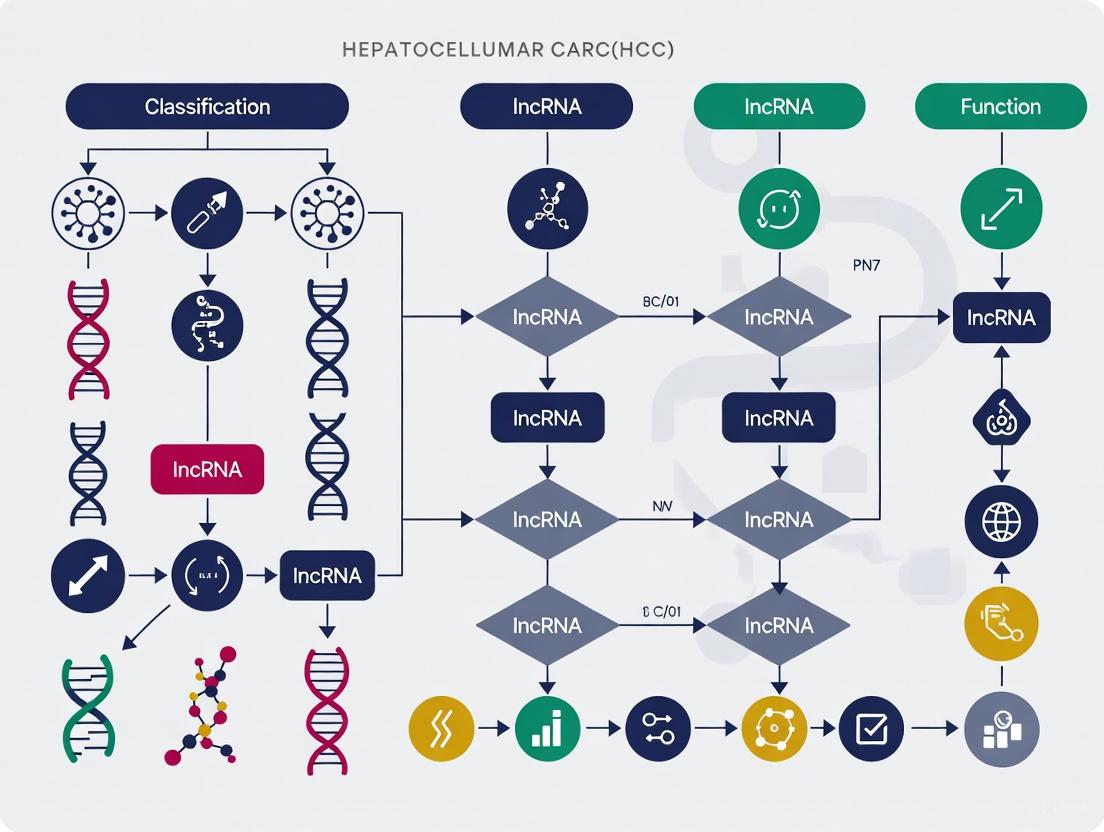
Long non-coding RNAs (lncRNAs) represent a vast class of RNA transcripts exceeding 200 nucleotides in length that lack protein-coding potential [1]. Once considered "transcriptional noise" or genomic "junk," lncRNAs are now recognized as critical regulators of gene expression across diverse biological processes, with particular significance in cancer biology, including hepatocellular carcinoma (HCC) [2] [3]. The human transcriptome contains an estimated 55,000 to 60,000 lncRNAs, far surpassing the approximately 20,000 protein-coding genes [2]. This complexity necessitates robust classification systems to facilitate functional annotation and mechanistic studies.
The genomic organization of lncRNAs provides a fundamental framework for their systematic categorization [4]. This classification approach, based on a lncRNA's position and orientation relative to nearby protein-coding genes, offers researchers a standardized method to describe and investigate these molecules [5] [6]. Within HCC research, understanding these genomic relationships provides critical insights into lncRNA biogenesis, regulatory networks, and potential therapeutic applications [7]. The precise classification of lncRNAs enables researchers to predict their potential mechanisms of action, with nuclear-enriched lncRNAs often involved in transcriptional and epigenetic regulation, while cytoplasmic lncRNAs frequently regulate mRNA stability and translation [1].
Table 1: Overview of Major lncRNA Classification Systems
| Classification Basis | Categories | Primary Utility |
|---|---|---|
| Genomic Location | Sense, Antisense, Intergenic, Intronic, Bidirectional | Standardized genomic annotation and positional relationship to coding genes |
| HGNC System | 9 categories including lincRNAs, antisense RNAs, host genes | Official nomenclature and standardized reporting |
| Functional Mechanism | Signal, Decoy, Guide, Scaffold | Understanding molecular roles and regulatory functions |
| Effect on Cancer | Oncogenic, Tumor-Suppressive | Clinical relevance and therapeutic targeting |
Genomic Location-Based Classification System
The genomic location-based classification system categorizes lncRNAs according to their transcriptional coordinates relative to adjacent protein-coding genes [6]. This system provides a structural framework for understanding lncRNA origins, regulation, and potential functional relationships with neighboring genomic elements. The classification comprises five principal categories, each with distinct genomic architectures and regulatory implications.
Long Intergenic Non-Coding RNAs (lincRNAs)
Long intergenic non-coding RNAs (lincRNAs) represent autonomously transcribed transcripts located in the genomic intervals between protein-coding genes [4] [6]. These lncRNAs do not overlap with annotated coding regions and are transcribed from independent promoters, often exhibiting their own distinct regulatory elements [3]. This genomic independence suggests lincRNAs may function as distinct regulatory units within the genome. In HCC, numerous lincRNAs have been identified as critical regulators of tumorigenesis, with functions including epigenetic regulation of cancer-associated genes and modulation of key signaling pathways [2]. Prominent examples include HOTAIR, HULC, and LINC00152, which have been extensively studied for their roles in HCC proliferation, metastasis, and prognosis [8] [7].
Antisense lncRNAs
Antisense lncRNAs are transcribed from the opposite DNA strand of protein-coding genes, frequently overlapping exonic or intronic regions of their sense counterparts [4] [3]. This complementary positioning enables diverse regulatory mechanisms, including the formation of RNA-DNA triplex structures, direct interaction with sense transcripts, or recruitment of epigenetic modifiers to the overlapping locus [1]. In hepatocellular carcinoma, antisense lncRNAs demonstrate significant pathological relevance, with examples such as ANRIL (CDKN2B-AS1) promoting cell proliferation through epigenetic silencing of Kruppel-like factor 2 (KLF2) via recruitment of polycomb repressive complex 2 (PRC2) [3]. Another HCC-associated antisense lncRNA, PCNA-AS1, forms RNA duplexes with proliferating cell nuclear antigen (PCNA) mRNA, stabilizing its structure and promoting tumor growth [3].
Sense lncRNAs
Sense lncRNAs are transcribed from the same DNA strand as protein-coding genes and overlap with one or more exons of these genes [5] [6]. These transcripts can originate from within intronic regions or extend across exon-intron boundaries of their associated coding genes. The overlapping nature of sense lncRNAs enables unique regulatory relationships with their host genes, including modulation of splicing patterns, transcript stability, or translational efficiency [5]. The sense lncRNA COLDAIR, while initially characterized in plants, represents a classic functional example of this category, demonstrating how sense-oriented transcripts can regulate overlapping gene expression through epigenetic mechanisms [5].
Intronic lncRNAs
Intronic lncRNAs are transcribed entirely from within the intronic regions of protein-coding genes, without overlapping exonic sequences [4] [6]. These transcripts can be synthesized in either the sense or antisense orientation relative to the host gene and may be processed from intronic sequences that escape degradation during splicing [5]. Unlike other lncRNA categories, intronic lncRNAs are not dependent on the transcriptional activity of their host genes and often exhibit independent regulation [5]. Their genomic position within intronic regions suggests potential co-regulation with host genes or roles in fine-tuning host gene expression through cis-regulatory mechanisms.
Bidirectional lncRNAs
Bidirectional lncRNAs are characterized by their transcriptional initiation in close proximity (within 1 kb) and in the opposite direction to protein-coding gene promoters [6] [3]. This head-to-head orientation suggests shared promoter elements and potential co-regulation between the lncRNA and its divergently transcribed coding partner [5]. The transcriptional coordination in bidirectional pairs implies functional relationships, where the lncRNA may modulate the expression or activity of its adjacent coding gene. In HCC, the lncRNA HCCL5 represents a clinically relevant example, functioning as a bidirectional transcript that contributes to disease progression [5]. Another well-characterized bidirectional lncRNA, LEENE (lncRNA-enhancing eNOS expression), demonstrates the regulatory potential of this category in endothelial function, though its specific role in HCC requires further investigation [5].
Diagram 1: Genomic classification of lncRNAs and representative examples in HCC research. The classification system is based on the transcript's position relative to protein-coding genes, with each category associated with specific molecular examples studied in hepatocellular carcinoma.
Table 2: Characteristics and HCC Examples of Genomic lncRNA Categories
| Category | Genomic Position | Key Features | HCC Examples | Functional Implications |
|---|---|---|---|---|
| Intergenic (lincRNA) | Between protein-coding genes | Independent promoters, distinct regulation | HOTAIR, HULC, LINC00152 | Epigenetic regulation, miRNA sponging |
| Antisense | Opposite strand to coding gene | Overlaps exons/introns, complementary sequence | ANRIL, PCNA-AS1 | Epigenetic silencing, mRNA stability |
| Sense | Same strand as coding gene | Overlaps exons, same orientation | COLDAIR | Splicing regulation, transcript stability |
| Intronic | Within introns of genes | Independent transcription, no exon overlap | Various intronic transcripts | cis-regulation, host gene modulation |
| Bidirectional | Opposite direction near promoter | Shared promoter elements, head-to-head orientation | HCCL5, LEENE | Coordinated expression, promoter regulation |
Alternative Classification Systems and Nomenclature
Beyond the genomic location framework, researchers utilize several complementary classification systems that provide additional perspectives on lncRNA organization and function. The HUGO Gene Nomenclature Committee (HGNC) has established a comprehensive categorization system that includes nine distinct lncRNA subgroups, offering standardized nomenclature for consistent scientific communication [4]. This official classification encompasses: (1) microRNA non-coding host genes, (2) small nucleolar RNA non-coding host genes, (3) long intergenic non-protein coding RNAs (LINC), (4) antisense RNAs, (5) overlapping transcripts, (6) intronic transcripts, (7) divergent transcripts, (8) long non-coding RNAs with non-systematic symbols, and (9) long non-coding RNAs with FAM root systems [4].
Functional classification systems categorize lncRNAs according to their molecular mechanisms rather than genomic position. This approach groups lncRNAs into four primary functional archetypes: (1) Signal lncRNAs that function as molecular indicators of transcriptional activity in response to specific stimuli; (2) Decoy lncRNAs that act as molecular sinks by binding and sequestering transcription factors or miRNAs away from their targets; (3) Guide lncRNAs that direct ribonucleoprotein complexes to specific genomic loci to regulate gene expression; and (4) Scaffold lncRNAs that serve as structural platforms for assembling multiple protein complexes into functional units [1] [6]. In HCC, this functional classification provides direct insights into pathological mechanisms, such as HOTAIR's scaffold function in bridging PRC2 and Snail to suppress epithelial gene expression during EMT [2].
Additionally, lncRNAs can be classified based on their functional effects in cancer as either oncogenic or tumor-suppressive [7]. Oncogenic lncRNAs (such as HULC, MALAT1, and H19) are frequently overexpressed in HCC and promote tumorigenesis through various mechanisms, while tumor-suppressive lncRNAs (including GAS5 and TSLNC8) are often downregulated, with their loss contributing to disease progression [8] [3]. This clinically relevant classification has significant implications for therapeutic development, as oncogenic lncRNAs represent potential drug targets, while tumor-suppressive lncRNAs offer opportunities for replacement therapies.
Experimental Protocols for lncRNA Classification and Functional Characterization
Genomic Mapping and Classification Workflow
The accurate classification of lncRNAs begins with comprehensive genomic mapping using integrated computational and experimental approaches. The standard methodology involves RNA sequencing (RNA-Seq) of HCC tissues and matched non-tumorous liver samples, followed by transcriptome assembly and coding potential assessment [2] [9]. The experimental workflow typically includes: (1) Total RNA extraction from clinical specimens or cell lines using miRNeasy or similar kits; (2) Ribosomal RNA depletion to enrich for non-coding transcripts; (3) Library preparation and high-throughput sequencing; (4) De novo transcript assembly using tools such as Cufflinks or StringTie; (5) Coding potential assessment using CPAT, CPC2, or similar algorithms; (6) Genomic annotation using tools like ANNOVAR with reference databases (GENCODE, Ensembl) to determine positional relationships to protein-coding genes [8] [9].
For classification confirmation, researchers employ several validation approaches: Strand-specific RT-PCR to determine transcriptional orientation; Rapid Amplification of cDNA Ends (RACE) to define transcript boundaries; Chromatin Immunoprecipitation (ChIP) of histone modifications to identify active promoters and enhancers; and in situ hybridization to determine subcellular localization [1] [2]. This integrated approach enables precise categorization within the genomic classification system and provides initial functional insights based on subcellular distribution.
Diagram 2: Experimental workflow for lncRNA identification and genomic classification. The process begins with sample collection and proceeds through RNA sequencing, bioinformatic analysis, and experimental validation to achieve precise categorization of lncRNAs based on genomic position.
Functional Characterization of Classified lncRNAs
Once classified, lncRNAs undergo functional characterization to elucidate their mechanistic roles in HCC pathogenesis. For nuclear-enriched lncRNAs (e.g., intergenic and antisense categories), common approaches include RNA Immunoprecipitation (RIP) to identify interacting chromatin-modifying complexes (PRC2, SWI/SNF), Chromatin Isolation by RNA Purification (ChIRP) to map genomic binding sites, and reporter assays to assess transcriptional regulatory activity [2] [3]. For cytoplasmic lncRNAs, methodologies include RNA-protein pull-down assays to identify interacting partners, ribonuclease protection assays to assess stability, and miRNA sponge analysis through Ago2-RIP and luciferase reporter constructs [1] [3].
High-throughput functional screening approaches include CRISPR-based loss-of-function screens to assess phenotypic impact, lncRNA-specific ASO libraries for knockdown studies, and transcriptomic analyses following perturbation to identify downstream pathways [7] [9]. For HCC research, these are complemented by disease-specific assays: sphere formation assays to assess effects on cancer stem cell properties, transwell migration and invasion assays to evaluate metastatic potential, and xenograft models to determine impacts on tumor growth and metastasis in vivo [3].
Table 3: Research Reagent Solutions for lncRNA Classification and Functional Studies
| Reagent/Category | Specific Examples | Primary Function | Application Context |
|---|---|---|---|
| RNA Extraction Kits | miRNeasy Mini Kit (QIAGEN) | High-quality total RNA isolation | Preserves lncRNA integrity for sequencing |
| cDNA Synthesis Kits | RevertAid First Strand cDNA Synthesis Kit | Reverse transcription with strand specificity | Determines transcriptional orientation |
| qRT-PCR Reagents | PowerTrack SYBR Green Master Mix | Quantitative expression analysis | Validates sequencing results and expression patterns |
| Sequencing Platforms | Illumina, Ion Torrent | High-throughput transcriptome sequencing | Identifies and classifies novel lncRNAs |
| Bioinformatic Tools | CPAT, CPC2, ANNOVAR, Cufflinks | Coding potential assessment and genomic annotation | Classifies lncRNAs based on genomic position |
| Functional Assay Reagents | ChIRP, RIP, Luciferase Reporter Systems | Mechanistic studies of molecular interactions | Determines functional classification |
Clinical Applications in Hepatocellular Carcinoma
The genomic classification of lncRNAs provides a crucial framework for understanding their clinical applications in hepatocellular carcinoma, particularly in the domains of diagnosis, prognosis, and therapeutic development. Specific lncRNA categories demonstrate distinct expression patterns and functional associations with HCC pathogenesis, offering valuable insights for clinical translation.
Intergenic lncRNAs (lincRNAs) have emerged as particularly promising diagnostic biomarkers due to their independent transcriptional regulation and frequent detection in liquid biopsies [8]. A 2024 study demonstrated that a panel of four lincRNAs (LINC00152, LINC00853, UCA1, and GAS5) in plasma could discriminate HCC patients from healthy controls with moderate individual accuracy (sensitivity 60-83%, specificity 53-67%), while machine learning integration of these lincRNAs with conventional laboratory parameters achieved remarkable diagnostic performance (100% sensitivity, 97% specificity) [8]. The LINC00152 to GAS5 expression ratio further provided prognostic value, with higher ratios correlating with increased mortality risk [8].
Antisense lncRNAs frequently contribute to HCC progression through epigenetic mechanisms, with ANRIL representing a prototypical example that promotes cell proliferation by recruiting PRC2 to silence the tumor suppressor KLF2 [3]. The strategic positioning of antisense lncRNAs enables precise regulation of their sense counterparts, making them attractive therapeutic targets. Bidirectional lncRNAs offer unique insights into coordinated gene regulation in HCC, with pairs such as HCCL5 and its divergently transcribed partner providing information about shared regulatory elements dysregulated in hepatocarcinogenesis [5].
From a therapeutic perspective, the genomic classification informs targeted intervention strategies. Antisense oligonucleotides (ASOs) can be designed to specifically target nuclear-enriched categories (intergenic, antisense), while RNAi approaches may be more effective for cytoplasmic lncRNAs [7]. The molecular functions associated with each category further guide therapeutic development: scaffold lncRNAs can be disrupted to dismantle functional complexes, while decoy lncRNAs can be targeted to release sequestered regulatory factors [2]. Recent advances have identified several HCC-associated lncRNAs as potential therapeutic targets, including NEAT1, DSCR8, PNUTS, HULC, and HOTAIR, which contribute to proliferation, migration, and apoptosis resistance through diverse mechanisms [7].
The genomic classification system encompassing sense, antisense, intergenic, intronic, and bidirectional categories provides an essential organizational framework for lncRNA research in hepatocellular carcinoma. This structured approach enables researchers to systematically catalogue lncRNAs, predict functional mechanisms based on genomic context, and communicate findings using standardized nomenclature. The integration of this classification with complementary systems based on molecular function and clinical impact creates a multidimensional understanding of lncRNA biology in HCC.
As research advances, the genomic classification system continues to evolve, incorporating new insights from single-cell sequencing, spatial transcriptomics, and multi-omics integration. These technological innovations are refining our understanding of lncRNA categorization and revealing context-specific functions within the complex landscape of hepatocellular carcinoma. The ongoing characterization of lncRNAs within this genomic framework promises to accelerate the development of novel diagnostic biomarkers and targeted therapeutic strategies, ultimately improving outcomes for HCC patients through precision medicine approaches.
Long non-coding RNAs (lncRNAs) are defined as RNA transcripts longer than 200 nucleotides that lack protein-coding capacity [7]. These molecules have emerged as critical regulators of gene expression and are involved in almost every cellular process, with their dysregulation increasingly implicated in hepatocellular carcinoma (HCC) pathogenesis [10] [11]. LncRNAs exhibit several characteristic features: they are typically transcribed by RNA polymerase II, undergo 5' capping and 3' polyadenylation, and are frequently spliced similarly to mRNAs [12]. Despite these similarities with coding transcripts, lncRNAs are generally expressed at lower levels and show greater tissue- and time-specificity in their expression patterns [10].
The functional classification of lncRNAs into mechanistic archetypes provides a valuable framework for understanding their diverse roles in normal physiology and disease states, particularly in complex malignancies like HCC. According to the seminal model proposed by Wang and Chang, lncRNAs primarily function through four core mechanisms: as signals, decoys, guides, and scaffolds [13] [10]. Each archetype represents a distinct mode of action through which lncRNAs regulate cellular processes by interacting with DNA, RNA, and proteins. In HCC, these mechanisms underlie critical pathological processes including uncontrolled proliferation, metastasis, apoptosis evasion, and therapy resistance [7]. This classification system has become foundational for lncRNA research, providing researchers with a structured approach to investigate the biological significance and therapeutic potential of these molecules in cancer biology.
The Four Archetypal Mechanisms of LncRNA Action
Signal LncRNAs
Mechanism Overview: Signal lncRNAs function as molecular indicators of biological context, responding to specific cellular stimuli by expressing at precise times and locations [10]. The very act of their transcription or their subsequent interactions serves to communicate regulatory information, often by recruiting chromatin-modifying complexes to specific genomic loci [10] [12].
Molecular Interactions: These lncRNAs typically execute their functions through interactions with chromatin-modifying enzymes such as histone methyltransferases, leading to target gene silencing through heterochromatin formation [10]. The transcription of signal lncRNAs integrates developmental cues, cellular context, and environmental stimuli, making them excellent biomarkers of functionally significant biological events [12].
HCC Examples: The lncRNA KCNQ1OT1 exemplifies signal functionality in HCC by recruiting PRC2 and chromatin-modifying enzymes like histone methyltransferases to specific genomic targets [10]. Another significant example is linc-RoR (Long intergenic non-coding RNA-ROR), which is directly regulated by key pluripotency factors Oct4, Sox2, and Nanog through their co-localization near its promoter region. Its expression is enriched during somatic cell reprogramming to induced pluripotent stem cells (iPSCs) and downregulated during differentiation, positioning it as a critical signal lncRNA in cell fate determination [12].
Decoy LncRNAs
Mechanism Overview: Decoy lncRNAs act as molecular sinks that sequester regulatory factors such as transcription factors, miRNAs, or chromatin modifiers, preventing them from binding their intrinsic targets [10]. This "titration" mechanism represents an indirect form of regulation where the lncRNA effectively competes with natural binding sites for their shared targets.
Molecular Interactions: These lncRNAs contain specific binding domains that mimic the native binding sites of their target molecules. By sequestering these regulators, decoy lncRNAs negatively modulate transcription, translation, or signaling pathways [10]. Notably, many decoy lncRNAs function as competitive endogenous RNAs (ceRNAs) that isolate miRNAs from their mRNA targets, thereby protecting those mRNAs from degradation or translational repression [10].
HCC Examples: The lncRNA PANDA regulates apoptosis in HCC by sequestering the transcription factor NF-YA, preventing it from activating apoptosis-related genes [10]. HULC (Highly Up-regulated in Liver Cancer) acts as an oncogenic decoy in HCC by preventing transcription factors or miRNAs from binding with their target sites, thereby regulating translation and transcription [10]. Similarly, GAS5 (Growth Arrest-Specific 5) contains a hairpin structure that mimics the DNA motif of glucocorticoid response elements, effectively competing for hormone binding and acting as a molecular decoy that contributes to glucocorticoid resistance in cancer cells [12].
Guide LncRNAs
Mechanism Overview: Guide lncRNAs direct the localization of ribonucleoprotein complexes to specific genomic targets, either in cis (neighboring genes) or in trans (distantly located genes) [10] [12]. This targeting function enables precise spatial and temporal control of gene expression through the recruitment of epigenetic modifiers or transcription factors to specific genomic loci.
Molecular Interactions: These lncRNAs form complex ribonucleoprotein complexes through RNA-protein, RNA-RNA, and RNA-DNA interactions, then direct these complexes to specific chromosomal locations [12]. The guidance mechanism enables lncRNAs to reprogram chromatin states and manage the recruitment of epigenetic modifiers to definitive loci, bringing about changes in the epigenome that either activate or repress target genes [10].
HCC Examples: HOTAIR guides the polycomb repressive complex 2 (PRC2) to specific genomic targets, facilitating histone H3 lysine 27 trimethylation and transcriptional repression of tumor suppressor genes in HCC [7] [10]. XIST, while best known for its role in X-chromosome inactivation, also functions as a guide lncRNA in various cancers including HCC by recruiting repressive complexes to specific genomic regions [10]. KCNQ1OT1 also exhibits guide functionality by binding to PRC2 and directing this chromatin modifier to regulate target genes in cis or trans, thereby inhibiting gene expression [10].
Scaffold LncRNAs
Mechanism Overview: Scaffold lncRNAs provide structural platforms for assembling multiple molecular components into functional ribonucleoprotein (RNP) complexes [10]. These lncRNAs serve as central organizers that bring together proteins and/or nucleic acids that might not otherwise interact, enabling the formation of higher-order complexes with emergent regulatory functions.
Molecular Interactions: As structural cores, scaffold lncRNAs contain multiple distinct binding domains that interact with different protein partners simultaneously [10]. The assembly of these complexes can lead to either transcriptional activation or repression depending on the nature of the associated proteins [12]. Some scaffold lncRNAs demonstrate remarkable stability, while others may form more dynamic, transient complexes that respond to cellular signals.
HCC Examples: The telomerase RNA component TERC represents a classic scaffold lncRNA that brings together the telomerase reverse transcriptase (TERT) with other accessory proteins to form the functional telomerase complex, which is frequently reactivated in HCC [10] [12]. MALAT1 (Metastasis Associated Lung Adenocarcinoma Transcript 1) serves as a dynamic scaffold in HCC by associating with both PRC1 and PRC2 complexes to stimulate target gene suppression or activation [10]. ANRIL and TUG1 also function as scaffolds by interacting with polycomb group proteins to facilitate the repression of target genes involved in HCC progression [10].
Table 1: Functional Classification of Key LncRNAs in Hepatocellular Carcinoma
| Mechanism | LncRNA | Molecular Interactions | Functional Role in HCC |
|---|---|---|---|
| Signal | KCNQ1OT1 | Recruits PRC2, histone methyltransferases | Regulates imprinting, cell proliferation |
| linc-RoR | Binds pluripotency factors (Oct4, Sox2, Nanog) | Modulates cell reprogramming, stemness | |
| Decoy | PANDA | Sequesters transcription factor NF-YA | Suppresses apoptosis-related genes |
| HULC | Binds miRNAs, transcription factors | Promotes proliferation, metastasis | |
| GAS5 | Mimics glucocorticoid response elements | Induces glucocorticoid resistance | |
| Guide | HOTAIR | Recruits PRC2 to specific genomic loci | Promotes metastasis, EMT |
| XIST | Guides repressive complexes to chromatin | Facilitates gene silencing programs | |
| KCNQ1OT1 | Directs PRC2 to target genes | Regulates gene expression in cis/trans | |
| Scaffold | TERC | Assembles telomerase complex with TERT | Maintains telomere length, immortality |
| MALAT1 | Binds PRC1, PRC2 complexes | Promotes aggressive tumor phenotypes | |
| ANRIL | Interacts with polycomb group proteins | Regulates cell proliferation, senescence |
Experimental Approaches for Studying LncRNA Mechanisms
Methodologies for Mechanism Characterization
Determining the specific mechanistic archetype of a lncRNA requires integrated experimental approaches that assess its subcellular localization, molecular interactions, and functional consequences. The following methodologies represent core techniques in the lncRNA researcher's toolkit:
Subcellular Localization Analysis: Since lncRNA function is closely tied to its cellular compartmentalization, initial experiments typically involve fractionation followed by qRT-PCR or RNA-FISH to determine nuclear versus cytoplasmic distribution [7]. Nuclear lncRNAs typically regulate transcription, chromatin organization, or RNA processing, while cytoplasmic lncRNAs often influence mRNA stability, translation, or protein function [7].
Interaction Partner Identification: RNA immunoprecipitation (RIP) and chromatin isolation by RNA purification (ChIRP) are essential for identifying lncRNA interaction partners [7]. RIP characterizes RNA-protein interactions, while ChIRP maps genomic binding sites. For lncRNAs acting as miRNA decoys, high-throughput sequencing of crosslinked immunoprecipitates (HITS-CLIP) can identify miRNA binding sites.
Functional Validation: Loss-of-function approaches using siRNA, shRNA, or CRISPR/Cas9 systems are crucial for establishing phenotypic consequences [14] [15]. For scaffold lncRNAs, domain deletion mapping through truncated constructs helps identify functional regions responsible for complex assembly.
Table 2: Essential Research Reagents and Experimental Tools for LncRNA Mechanism Studies
| Research Tool | Specific Application | Experimental Utility |
|---|---|---|
| shRNA/siRNA | Gene knockdown | Functional validation of lncRNA effects; example: AL590681.1 knockdown in HCC cell lines [14] |
| CRISPR/Cas9 | Gene editing | Complete lncRNA deletion or specific domain manipulation |
| RNA-FISH | Spatial localization | Subcellular localization and expression pattern analysis |
| RIP/ChIRP | Interaction mapping | Identification of protein and DNA binding partners |
| Luciferase Reporters | Interaction validation | Confirmation of miRNA binding or promoter regulation |
| RT-qPCR | Expression quantification | Measurement of lncRNA levels across samples; example: Plasma lncRNA detection [16] |
| Next-Generation Sequencing | Transcriptome analysis | Discovery of differentially expressed lncRNAs; example: Identification of HCC-associated lncRNAs [16] |
Representative Experimental Workflow
A comprehensive study investigating amino acid metabolism-related lncRNAs in HCC exemplifies an integrated mechanistic approach [14]. This research began with transcriptomic data analysis from TCGA databases to identify lncRNAs correlated with amino acid metabolism genes. Following bioinformatic identification, researchers employed Univariate Cox analysis, LASSO, and Multivariate Cox analysis to construct a prognostic risk model based on four hub lncRNAs.
Functional validation involved in vitro models using HCC cell lines (Hep-3B, Huh-1, Huh-7, HCCLM3) transfected with lncRNA-specific short hairpin RNA (shRNA) via Lipofectamine 3000 reagent [14]. Knockdown efficiency was assessed by RT-qPCR after 48 hours, followed by functional assays including:
- CCK-8 assays to measure cell viability
- Colony formation assays to evaluate growth potential (14-day incubation with crystal violet staining)
- Transwell assays to assess migratory and invasive capabilities
This multifaceted methodology enabled both the identification of clinically relevant lncRNAs and the characterization of their functional roles in HCC pathogenesis.
Diagram 1: Experimental Workflow for LncRNA Mechanism Studies
LncRNA Mechanisms in Hepatocellular Carcinoma Pathogenesis
Cross-Mechanism Regulation in HCC Pathways
In HCC, lncRNAs employing different mechanistic archetypes frequently converge to regulate the same critical signaling pathways, creating complex regulatory networks that drive hepatocarcinogenesis. Understanding these integrated mechanisms provides insights for therapeutic targeting.
PI3K/AKT/mTOR Pathway Regulation: Multiple lncRNAs using distinct mechanisms regulate this crucial pathway in HCC. For instance, the lncRNA LINC01343 functions through a guide mechanism to regulate the PI3K/Akt/mTOR pathway, significantly affecting HCC progression [7]. Simultaneously, lncRNAs that modulate autophagy, such as those regulating the Beclin-1-VPS34 complex, impact this pathway through scaffold functions, assembling protein complexes that control the delicate balance between autophagy's tumor-suppressive and tumor-promoting roles in HCC [15].
Wnt/β-catenin Pathway Modulation: The Wnt/β-catenin pathway represents another key signaling axis regulated by multiple lncRNA mechanisms in HCC. Several lncRNAs drive cancer stem cell self-renewal and tumor proliferation by activating this pathway [7]. Guide lncRNAs direct chromatin modifiers to regulate expression of pathway components, while decoy lncRNAs sequester inhibitors of the pathway, collectively enhancing oncogenic signaling.
Immune Microenvironment Reprogramming: LncRNAs utilize all four mechanistic archetypes to shape the immunosuppressive HCC microenvironment. NEAT1 functions as a decoy lncRNA that binds and sequesters miR-155 in CD8+ T cells, leading to increased Tim-3 expression and subsequent T cell exhaustion [11]. Scaffold lncRNAs such as Lnc-Tim3 assemble protein complexes that prevent Tim-3 from interacting with Bat3, thereby inhibiting downstream signaling in the Lck/NFAT1/AP-1 pathway and contributing to immune evasion [11].
Diagram 2: LncRNA Mechanisms in HCC Signaling Pathways
Diagnostic and Therapeutic Implications
The mechanistic classification of lncRNAs directly informs their development as clinical tools in HCC management. Different mechanistic archetypes offer distinct advantages for diagnostic and therapeutic applications:
Diagnostic Biomarker Development: Signal lncRNAs are particularly valuable as diagnostic biomarkers because their expression reflects specific pathological states. Studies have identified plasma lncRNAs including HULC, RP11-731F5.2, and KCNQ1OT1 as promising non-invasive biomarkers for HCC risk assessment and liver damage monitoring [16]. The development of lncRNA-based diagnostic panels that integrate multiple mechanistic types has shown enhanced performance. For example, a combination of LINC00152, LINC00853, UCA1, and GAS5 achieved 100% sensitivity and 97% specificity for HCC detection when analyzed using machine learning algorithms [8].
Therapeutic Target Considerations: Each mechanistic archetype presents unique opportunities and challenges for therapeutic targeting:
- Decoy lncRNAs can be targeted using antisense oligonucleotides (ASOs) that block their interaction with sequestered molecules
- Guide lncRNAs may be inhibited through small molecules that disrupt their protein interactions
- Scaffold lncRNAs often contain structured domains that can be targeted with small molecule inhibitors
- Signal lncRNAs can be manipulated by targeting their transcriptional regulation
Emerging approaches include CRISPR/Cas systems for precise lncRNA editing and siRNA/shRNA technologies for targeted degradation [15]. The functional classification framework enables researchers to select the most appropriate targeting strategy based on a lncRNA's primary mechanism of action.
The functional classification of lncRNAs into signal, decoy, guide, and scaffold mechanisms provides an essential framework for understanding their diverse roles in hepatocellular carcinoma. This systematic categorization enables researchers to predict molecular interactions, design appropriate experimental approaches, and develop targeted therapeutic strategies. In HCC, lncRNAs representing all four archetypes have been shown to regulate critical pathological processes including metabolic reprogramming, immune evasion, and therapeutic resistance.
The translational potential of this mechanistic understanding is substantial. As diagnostic tools, lncRNA signatures that incorporate multiple mechanistic types demonstrate enhanced performance for early detection and prognosis prediction. Therapeutically, distinguishing between lncRNA mechanisms informs the selection of optimal targeting strategies, whether using antisense oligonucleotides for decoy lncRNAs or small molecules for structured scaffold lncRNAs. Future research efforts should focus on comprehensive mechanistic characterization of HCC-associated lncRNAs and the development of mechanism-specific targeting approaches that can be integrated into multimodal treatment strategies for this challenging malignancy.
The contemporary view of the eukaryotic genome reveals a vast transcriptional output dominated by RNA molecules that do not code for proteins. Long non-coding RNAs (lncRNAs), once dismissed as transcriptional "noise," are now recognized as crucial regulators of gene expression, playing pivotal roles in cell differentiation, development, and disease [17] [18]. Their discovery resolved the apparent paradox between organismal complexity and a relatively static number of protein-coding genes, highlighting the regulatory potential of the non-coding genome [17]. In the specific context of Hepatocellular Carcinoma (HCC), lncRNAs have emerged as central players in tumorigenesis, metastasis, and therapy resistance [7] [19]. This whitepaper delineates the fundamental molecular hallmarks that distinguish lncRNAs from messenger RNAs (mRNAs) and other non-coding RNAs (ncRNAs), providing a technical guide for research and drug development in HCC and beyond.
Fundamental Definitions and Genomic Context
The Non-Coding RNA Landscape
The non-coding transcriptome is composed of multiple RNA classes with distinct functions. Table 1 provides a comparative overview of the primary RNA species discussed in this guide.
Table 1: Overview of Key RNA Types
| RNA Type | Length (Nucleotides) | Polymerase | Primary Function | Conservation |
|---|---|---|---|---|
| mRNA | Variable (typically 500-10,000+) | Pol II | Template for protein synthesis | Moderate to High |
| lncRNA | >200 (mostly >500) | Primarily Pol II | Regulatory (see Section 4) | Generally Low |
| miRNA | 18-24 | Pol II | Post-transcriptional gene silencing | High |
| siRNA | 20-25 | Pol II | Transcriptional and post-transcriptional gene silencing | High |
| circRNA | Variable (often 100-10,000) | Pol II | miRNA sponging, protein decoys | Variable |
| tRNA | 73-91 | Pol III | Amino acid transfer in protein synthesis | High |
| rRNA | ~120-5000 (e.g., 18S, 5.8S, 28S) | Pol I (18S, 5.8S, 28S); Pol III (5S) | Catalytic core of the ribosome | Very High |
| snRNA | ~100-200 | Pol II | mRNA splicing (spliceosome components) | High |
| snoRNA | 60-300 | Pol II | Guide chemical modifications of rRNA | High |
LncRNAs are functionally defined as transcripts longer than 200 nucleotides (nt) with no protein-coding potential, a practical size cut-off that distinguishes them from small regulatory RNAs and infrastructural RNAs like tRNAs and snRNAs [17] [20]. Most lncRNAs are RNA Polymerase II (Pol II) transcripts and can exhibit mRNA-like features such as 5' capping, splicing, and polyadenylation, though some are processed from introns or transcribed by Pol I or Pol III [17].
Genomic Classification of lncRNAs
LncRNAs are classified based on their genomic position relative to protein-coding genes, which often provides initial clues to their potential function [21] [22]. The major categories include:
- Intergenic lncRNAs (lincRNAs): Transcribed from regions between protein-coding genes [17] [22].
- Intronic lncRNAs: Derived entirely from within the introns of a protein-coding gene [21] [22].
- Antisense lncRNAs: Transcribed from the opposite strand of a protein-coding gene and overlap its exons or introns [21] [22].
- Sense lncRNAs: Overlap one or more exons of a protein-coding gene on the same strand [22].
- Enhancer RNAs (eRNAs): Transcribed from enhancer regions, though conclusive evidence for these in plants is still lacking [21].
- Promoter-associated RNAs (PATs): Originate from promoter-proximal regions [21] [22].
Molecular Hallmarks: LncRNAs vs. mRNAs and Other ncRNAs
Sequence and Expression Characteristics
Table 2: Key Distinguishing Molecular Hallmarks
| Molecular Characteristic | LncRNAs | mRNAs | Small ncRNAs (e.g., miRNAs, siRNAs) |
|---|---|---|---|
| Protein-Coding Potential | None or very limited (may encode micropeptides) [21] | High (defined open reading frame) | None |
| Sequence Conservation | Generally low, though promoter elements may be conserved [17] [20] | Generally high, especially in coding regions | Very high, especially in seed regions (miRNAs) [20] |
| Expression Level | Typically low abundance and highly cell type-specific [17] [21] | Can range from low to very high | Variable, can be highly abundant |
| Exon Count / Transcript Structure | Fewer exons on average than mRNAs [21] | Multiple exons, defined UTRs | Single short sequence (miRNAs are ~22 nt) |
| Tissue Specificity | High, responsive to developmental cues and stimuli [17] [20] | Broad to specific | Broad to specific |
LncRNAs demonstrate several defining hallmarks. They are functionally non-coding, though some contain small open reading frames (smORFs) that can encode micropeptides approximately 100 amino acids in length, suggesting bi-functional potential [21] [7]. They evolve more rapidly than protein-coding sequences, displaying low primary sequence conservation across species, which suggests their function may be more dependent on secondary or tertiary structure [17] [20]. Furthermore, their expression is typically characterized by low abundance and high cell type, tissue, and developmental stage specificity, allowing them to integrate diverse stimuli and contextual cues [17] [20].
Functional Archetypes of LncRNAs
A widely adopted framework classifies lncRNA molecular functions into four archetypes, which are not mutually exclusive [20]:
- Signals: LncRNAs serve as molecular indicators of transcriptional activity, marking specific biological events such as allele-specific expression (e.g., Xist in X-chromosome inactivation), anatomic position (e.g., HOTAIR from the HOXC locus), or cellular stress (e.g., linc-p21 induced by DNA damage) [20].
- Decoys: These lncRNAs act as "molecular sponges," sequestering transcription factors, chromatin modifiers, or other RNAs (like miRNAs) away from their genomic targets. This is exemplified by PANDA, which binds to the transcription factor NF-YA to modulate the DNA damage response [20].
- Guides: LncRNAs can direct ribonucleoprotein complexes to specific genomic loci, often in cis (on the same chromosome) or trans (on different chromosomes). For instance, HOTTIP guides chromatin-modifying complexes to the HOXA locus, influencing histone methylation and gene activation [20].
- Scaffolds: As central platforms, scaffold lncRNAs nucleate the assembly of multiple proteins into functional complexes. HOTAIR exemplifies this by simultaneously binding the PRC2 and LSD1 complexes, facilitating coordinated histone modifications for gene repression [20].
Experimental Protocols for lncRNA Identification and Functional Validation
Workflow for Constructing a ceRNA Network in Disease
The Competing Endogenous RNA (ceRNA) hypothesis posits that lncRNAs can sequester miRNAs, thereby modulating the expression of miRNA target mRNAs. The following workflow, adapted from a study on Down Syndrome, can be applied to HCC research to investigate lncRNA-mediated mechanisms [23].
Figure 1: Experimental workflow for ceRNA network construction.
- Step 1: Sample Collection and RNA Sequencing. Obtain HCC tissue samples and matched non-tumor liver tissues. Isolate total RNA and prepare sequencing libraries for lncRNA/mRNA (typically producing 150-bp paired-end reads) and miRNA (producing 50-bp single-end reads) [23].
- Step 2: Differential Expression Analysis. Quantify expression levels using FPKM (Fragments Per Kilobase per Million) for lncRNAs/mRNAs and TPM (Transcripts Per Million) for miRNAs. Identify significantly differentially expressed (DE) RNAs using packages like
edgeR(for lncRNAs/mRNAs) andDEGseq(for miRNAs) with thresholds of FDR < 0.01 and |log2(Fold Change)| > 1 [23]. - Step 3: Functional Enrichment and PPI Network Analysis. Perform Gene Ontology (GO) and Kyoto Encyclopedia of Genes and Genomes (KEGG) pathway enrichment analysis on DEmRNAs to understand their functional implications. Simultaneously, construct a Protein-Protein Interaction (PPI) network using the STRING database to identify hub genes critical to HCC pathogenesis [23].
- Step 4: CeRNA Network Construction. Predict miRNA-mRNA and miRNA-lncRNA interactions using multiple algorithms (e.g., miRanda, PITA, and RNAhybrid). Include only interactions identified by all tools for high-confidence. Integrate negatively correlated DEmiRNA-DEmRNA and DEmiRNA-DElncRNA pairs, and positively correlated DElncRNA-DEmRNA pairs into a comprehensive network, which is visualized using Cytoscape (v. 3.5.0 or higher) [23].
- Step 5: Experimental Validation. Candidate interactions, particularly those involving key hub nodes, require validation. For miRNA-lncRNA binding, use Luciferase Reporter Assays where the lncRNA sequence is cloned downstream of a luciferase gene. Co-transfection with the candidate miRNA should show reduced luminescence if binding occurs. Functional consequences are validated using siRNA/shRNA-mediated knockdown or CRISPR-based overexpression of the lncRNA, followed by assays for proliferation, apoptosis, and invasion in HCC cell lines [7] [19].
Key Research Reagent Solutions
Table 3: Essential Reagents and Tools for lncRNA Research
| Reagent / Tool | Function / Application | Example Use in Protocol |
|---|---|---|
| Trizol Reagent | Total RNA isolation from cells and tissues. | Initial RNA extraction from HCC patient samples or cell lines [23]. |
| Stranded RNA-Seq Library Prep Kits | Preparation of sequencing libraries for transcriptome analysis. | Construction of lncRNA/mRNA and miRNA sequencing libraries [23]. |
| DEGseq / edgeR R Packages | Statistical analysis of differentially expressed genes from RNA-Seq data. | Identification of DElncRNAs, DEmiRNAs, and DEmRNAs with statistical rigor [23]. |
| miRanda / RNAhybrid | Algorithms for predicting miRNA binding sites on target transcripts. | In silico prediction of lncRNA-miRNA and miRNA-mRNA interactions for ceRNA network building [23]. |
| Cytoscape | Open-source platform for complex network visualization and analysis. | Visualization and analysis of the constructed PPI and ceRNA networks [23]. |
| siRNA / shRNA Lentiviral Vectors | Transient or stable knockdown of specific lncRNAs in cell culture. | Functional validation of candidate lncRNAs in HCC cell models (e.g., proliferation, invasion assays) [7] [19]. |
| Luciferase Reporter Vectors (e.g., pmirGLO) | Quantification of miRNA-mRNA or miRNA-lncRNA interactions. | Experimental validation of predicted binding between a miRNA and its target sequence within a lncRNA [23]. |
LncRNA Mechanisms in Hepatocellular Carcinoma (HCC)
In HCC, lncRNAs operate through the archetypal mechanisms described, impacting all hallmarks of cancer. Their function is largely determined by subcellular localization: nuclear lncRNAs primarily regulate transcription and chromatin organization, while cytoplasmic lncRNAs affect mRNA stability, translation, and signaling pathways [7].
The following diagram illustrates how specific lncRNAs contribute to HCC pathogenesis by integrating into key signaling axes.
Figure 2: LncRNA mechanisms in HCC pathogenesis.
- miRNA Sponging (Decoy Function): The lncRNA HULC is highly upregulated in HCC and promotes proliferation and EMT by acting as a decoy for miRNAs like miR-372 and miR-186, preventing these miRNAs from repressing their oncogenic target mRNAs [19]. Similarly, Linc-RoR sponges miR-145 to promote self-renewal of cancer stem cells [7].
- Chromatin Remodeling (Guide/Scaffold Function): HOTAIR, also upregulated in HCC, acts as a scaffold to recruit chromatin-modifying complexes like PRC2 and LSD1, leading to the epigenetic silencing of tumor suppressor genes. This drives HCC cell proliferation, migration, and invasion [19].
- Signaling Pathway Activation: LncTCF7 (also known as WSPAR) activates the Wnt/β-catenin signaling pathway, a key driver in liver cancer. It promotes EMT, invasion, and self-renewal of liver cancer stem cells, contributing to tumor initiation and progression [19].
LncRNAs are distinguished from mRNAs and other ncRNAs by a unique set of molecular hallmarks: their non-coding nature, low sequence conservation, highly specific expression patterns, and diverse functional archetypes. In HCC, these molecules are integral to the disease's molecular circuitry, influencing critical processes from epigenetic regulation to metabolic reprogramming. The precise molecular classification and functional dissection of lncRNAs, supported by robust experimental protocols and a growing toolkit of reagents, are paving the way for novel RNA-based diagnostic biomarkers and therapeutic strategies for complex diseases like hepatocellular carcinoma.
Long non-coding RNAs (lncRNAs), defined as RNA transcripts exceeding 200 nucleotides in length that lack protein-coding capacity, have emerged as critical regulators of gene expression in both physiological and pathological contexts [24] [1]. Their functional diversity is profoundly influenced by their subcellular localization, which determines their mechanistic roles and biological impacts [7] [25]. In hepatocellular carcinoma (HCC), understanding these localization patterns provides crucial insights into lncRNA-driven oncogenesis and reveals potential therapeutic vulnerabilities. The compartmentalization of lncRNAs between nuclear and cytoplasmic spaces is not random but rather dictates their molecular partnerships—whether with DNA, chromatin-modifying complexes, other RNAs, or proteins—ultimately shaping their functional outcomes in hepatocarcinogenesis [1] [25] [26]. This review systematically examines the distinct functional mechanisms employed by nuclear versus cytoplasmic lncRNAs in HCC, details experimental approaches for studying their localization, and explores the implications for diagnostic and therapeutic development.
Subcellular Distribution and Functional Mechanisms of lncRNAs
Nuclear Localized lncRNAs: Primary Functions and Mechanisms
The majority of lncRNAs are preferentially localized to the nucleus, where they exert regulatory control over genomic function and transcriptional programs through diverse mechanisms [7] [1]. These nuclear lncRNAs function as essential epigenetic and transcriptional regulators in HCC pathogenesis, often through guiding chromatin-modifying complexes to specific genomic loci or organizing nuclear subdomains.
Table 1: Primary Functional Mechanisms of Nuclear lncRNAs in HCC
| Mechanism | Functional Consequence | Example lncRNAs in HCC |
|---|---|---|
| Chromatin Modification Guidance | Recruitment of histone modifiers to specific genomic loci; regulation of DNA methylation | HOTAIR, HOTTIP [1] [26] |
| Transcriptional Regulation | Direct interaction with transcription factors or RNA polymerase II; modulation of transcription initiation/elongation | ASTILCS [27] |
| Nuclear Body Organization | Scaffolding for formation of subnuclear domains and protein complexes | NEAT1 [28] [26] |
| cis-Regulatory Action | Regulation of neighboring genes on the same chromosome | ASTILCS [27] |
Nuclear lncRNAs typically function through several well-characterized mechanistic paradigms. As guides, they direct chromatin-modifying complexes to specific genomic locations, enabling precise epigenetic regulation. For example, HOTAIR recruits Polycomb Repressive Complex 2 (PRC2) to silence tumor suppressor genes, facilitating HCC progression [26]. As scaffolds, nuclear lncRNAs provide structural platforms for assembling multi-protein complexes, such as NEAT1's role in organizing nuclear paraspeckles that influence mRNA processing and retention in HCC cells [28] [26]. As signals, they reflect transcriptional responses to specific cellular stimuli or states, while as decoys, they sequester transcription factors or regulatory proteins away from their genomic targets [26].
Cytoplasmic Localized lncRNAs: Primary Functions and Mechanisms
Cytoplasmic lncRNAs operate through distinct post-transcriptional mechanisms that influence mRNA stability, translation, and signaling pathways. These molecules frequently function as molecular sponges or scaffolds that modulate protein activity and signaling cascades relevant to HCC progression.
Table 2: Primary Functional Mechanisms of Cytoplasmic lncRNAs in HCC
| Mechanism | Functional Consequence | Example lncRNAs in HCC |
|---|---|---|
| miRNA Sponging (ceRNA) | Sequestration of tumor-suppressive miRNAs; derepression of oncogenic targets | SNHG6, HULC, linc-RoR [7] [29] [28] |
| mRNA Stability Regulation | Modulation of mRNA decay pathways; influence on transcript half-lives | HULC [24] |
| Protein Activity Modulation | Direct interaction with signaling proteins; alteration of enzymatic activity or stability | HULC [24] |
| Translation Regulation | Influence on ribosomal loading and protein synthesis | Various [1] |
The most extensively characterized mechanism for cytoplasmic lncRNAs in HCC is their function as competing endogenous RNAs (ceRNAs) or miRNA sponges. For instance, SNHG6 promotes HCC progression by competitively binding multiple tumor-suppressive miRNAs including miR-26a/b, miR-101-3p, and let-7c-5p, thereby derepressing oncogenic targets like TGF-β-activated kinase 1 and c-Myc [29]. Similarly, linc-RoR functions as a sponge for miR-145 under hypoxic conditions, leading to upregulation of HIF-1α and enhancement of HCC cell survival [28]. Beyond miRNA sponging, cytoplasmic lncRNAs can directly modify signaling pathways, as demonstrated by HULC's interaction with sphingosine kinase 1 (SPHK1) to promote angiogenesis in HCC [24].
Dual Localization and Context-Dependent Functions
Some lncRNAs exhibit dynamic localization patterns, shuttling between nuclear and cytoplasmic compartments or performing distinct functions in different cellular contexts. This dual localization expands their functional repertoire and allows for integrated regulation of gene expression at multiple levels. The lncRNA H19 provides a notable example, with documented functions in both nuclear epigenetic regulation and cytoplasmic miRNA sponging in HCC models [7]. The mechanisms governing lncRNA localization remain an active area of investigation, but current evidence suggests that specific sequence motifs, RNA-binding proteins, and post-transcriptional modifications (including m6A methylation) play decisive roles in determining subcellular destination [1] [26].
Experimental Approaches for Determining lncRNA Localization and Function
Methodologies for Subcellular Localization Analysis
Accurate determination of lncRNA subcellular localization is prerequisite for functional characterization. Several methodological approaches enable precise localization assessment, each with distinct advantages and limitations.
Table 3: Key Methodologies for lncRNA Subcellular Localization Studies
| Method | Principle | Key Applications in HCC Research | Technical Considerations |
|---|---|---|---|
| RNA Fluorescence In Situ Hybridization (RNA-FISH) | Fluorescently labeled probes for direct visualization of RNA molecules within fixed cells | Spatial localization of specific lncRNAs in HCC cell lines and tissues; single-molecule RNA-FISH enhances sensitivity [1] | Requires optimization of fixation and permeabilization; sensitivity limitations with conventional approaches |
| Subcellular Fractionation with RT-qPCR | Biochemical separation of nuclear and cytoplasmic compartments followed by RNA quantification | Quantitative assessment of lncRNA distribution across cellular compartments; high-throughput capability [27] | Requires rigorous validation of fraction purity; potential for cross-contamination between fractions |
| Chromatin Isolation by RNA Purification (ChIRP) | Identification of chromatin regions associated with specific lncRNAs using biotinylated antisense oligonucleotides | Mapping genomic binding sites for nuclear lncRNAs; elucidating epigenetic mechanisms [30] | Optimal for chromatin-associated lncRNAs; requires careful design of tiling oligonucleotides |
Figure 1: Experimental Approaches for lncRNA Subcellular Localization Analysis. This workflow illustrates the primary methodologies used to determine lncRNA localization and their key applications in functional studies.
Functional Validation Methodologies
Following localization determination, functional validation requires targeted perturbation approaches that account for subcellular context. Multiple effective strategies exist for probing lncRNA function in HCC models.
RNA Interference (RNAi) and Antisense Oligonucleotides (ASOs): RNAi approaches, including shRNA and siRNA, enable transcript-specific knockdown, with recent evidence supporting their utility for both cytoplasmic and nuclear lncRNAs [27]. ASOs designed to target specific lncRNAs through RNase H-mediated degradation have shown particular efficacy for nuclear-localized transcripts. In a screening approach for HCC-essential lncRNAs, shRNA libraries enabled identification of ASTILCS as a nuclear lncRNA critical for HCC cell survival [27]. The protocol involves: (1) Design of 4-5 shRNAs per target lncRNA; (2) Lentiviral library delivery at low MOI (0.3); (3) Puromycin selection of transduced cells; (4) Monitoring cell survival over 4 weeks; (5) NGS-based quantification of shRNA representation to identify essential genes [27].
CRISPR Interference (CRISPRi): CRISPRi utilizes catalytically dead Cas9 (dCas9) fused to transcriptional repressors to specifically silence lncRNA transcription at the promoter level. This approach is particularly valuable for nuclear lncRNAs and those that overlap regulatory elements for other genes. However, careful design is required to avoid confounding effects on neighboring genes [27]. The typical workflow includes: (1) Identification of lncRNA promoter regions; (2) Design of sgRNAs targeting transcriptional start sites; (3) Delivery of dCas9-KRAB fusion protein and sgRNAs; (4) Assessment of knockdown efficiency by RT-qPCR; (5) Functional validation of phenotypic effects [27].
Table 4: Key Research Reagents for lncRNA Localization and Function Studies
| Reagent Category | Specific Examples | Research Applications | Functional Role |
|---|---|---|---|
| Localization Tools | RNA-FISH probes, Cellular fractionation kits, Lamin A/C antibodies | Determination of subcellular distribution; validation of compartment identity | Enable precise subcellular mapping and compartment purity validation |
| Functional Perturbation Tools | shRNA libraries, CRISPRi systems (dCas9-KRAB), Antisense oligonucleotides (ASOs) | Loss-of-function studies; phenotypic screening; therapeutic target validation | Facilitate targeted depletion of specific lncRNAs with consideration for subcellular context |
| Detection Reagents | Strand-specific RNA-seq kits, RT-qPCR reagents, RNA-binding protein immunoprecipitation kits | Expression quantification; interaction partner identification; mechanistic studies | Allow comprehensive molecular profiling and interaction mapping |
| Cell Culture Models | HUH7, HepG2, Hep3B HCC cell lines, Primary hepatocytes, Patient-derived organoids | In vitro functional studies; pathway analysis; drug screening | Provide biologically relevant systems for HCC-specific mechanistic investigations |
Implications for HCC Diagnostics and Therapeutics
The subcellular localization of lncRNAs has profound implications for their utility as biomarkers and therapeutic targets in HCC. Cytoplasmic lncRNAs often demonstrate greater utility as circulating biomarkers due to their enhanced stability and secretion into extracellular vesicles, while nuclear lncRNAs may offer more specific therapeutic targets due to their central roles in transcriptional regulation.
Nuclear lncRNAs present attractive targets for epigenetic therapies and transcriptional modulation. For instance, HOTAIR, which guides chromatin-modifying complexes to silence tumor suppressor genes, could be targeted to reverse aberrant epigenetic states in HCC [26]. The nuclear-enriched lncRNA ASTILCS represents another promising target, as its knockdown induces apoptosis in HCC cells through downregulation of the neighboring PTK2 gene [27].
Cytoplasmic lncRNAs functioning as ceRNAs offer distinct therapeutic opportunities. SNHG6, which sponges multiple tumor-suppressive miRNAs in HCC, could be targeted using antisense oligonucleotides to disrupt its oncogenic function [29]. Similarly, HULC's diverse cytoplasmic functions in promoting autophagy and angiogenesis through SPHK1 regulation make it a compelling multi-mechanistic target [24].
Figure 2: Therapeutic and Diagnostic Implications Based on lncRNA Subcellular Localization. This diagram illustrates how subcellular localization informs diagnostic applicability and therapeutic strategy selection for lncRNA-targeting approaches in HCC.
Subcellular localization serves as a fundamental determinant of lncRNA function in hepatocellular carcinoma, dictating mechanistic capabilities and biological impacts. Nuclear lncRNAs predominantly regulate gene expression through epigenetic and transcriptional mechanisms, while cytoplasmic lncRNAs operate through post-transcriptional modalities including miRNA sponging and signaling pathway modulation. This compartmentalization has profound implications for both biomarker development and therapeutic targeting in HCC. Future research should prioritize understanding the molecular determinants of lncRNA localization, developing delivery systems that account for subcellular destination, and exploiting localization patterns for precision oncology approaches. As our knowledge of lncRNA biology expands, subcellular localization will remain an essential consideration in the rational design of lncRNA-targeted diagnostics and therapeutics for hepatocellular carcinoma.
Evolutionary Conservation and Tissue Specificity in Hepatic Contexts
Long non-coding RNAs (lncRNAs) represent a vast class of RNA transcripts exceeding 200 nucleotides in length that lack significant protein-coding potential [31]. Once considered transcriptional "noise," lncRNAs are now recognized as crucial regulators of diverse biological processes, including cell differentiation, growth, and gene development [5] [31]. In the context of hepatocellular carcinoma (HCC), lncRNAs have emerged as significant players in tumorigenesis, metastasis, and therapy resistance [28] [7]. This technical guide examines two fundamental characteristics of lncRNAs—their evolutionary conservation patterns and tissue specificity—within hepatic contexts, providing researchers with methodological frameworks and conceptual approaches for advancing HCC research.
The molecular landscape of HCC is characterized by complex genetic and epigenetic modifications where lncRNAs exert influence through multiple mechanisms [7]. They function as molecular signals, decoys, scaffolds, and sponges for microRNAs, often acting as competing endogenous RNAs (ceRNAs) to regulate gene expression [4]. Understanding the evolutionary conservation and tissue-specific expression patterns of these molecules is paramount for elucidating their functional significance and translational potential in liver cancer biology.
Evolutionary Conservation of Hepatic lncRNAs
Conservation Patterns and Functional Significance
Unlike protein-coding genes, lncRNAs demonstrate notably lower sequence conservation across species [31]. This characteristic initially complicated their functional annotation. However, emerging evidence suggests that although primary sequences may diverge, functional domains within lncRNAs often exhibit higher conservation, and syntenic conservation (genomic position relative to neighboring genes) frequently persists even when sequence homology is low [32].
Table 1: Conservation Patterns of Selected Hepatic lncRNAs
| lncRNA | Sequence Conservation | Syntenic Conservation | Functional Conservation | Role in HCC |
|---|---|---|---|---|
| H19 | Moderate | Conserved | Partially conserved | Oncogenic [7] |
| HOTAIR | Low | Conserved | Partially conserved | Promotes metastasis [32] |
| MALAT1 | High | Conserved | Conserved | Promotes proliferation [31] [32] |
| PHAROH | Moderate | Conserved | Unknown | Regulates MYC translation [32] |
| HULC | Low | Conserved | Unknown | Oncogenic [28] [33] |
The conservation of genomic organization and positional relationships often provides more reliable indicators of functional importance than sequence similarity alone. For example, the identification of PHAROH (Gm19705) involved analyzing syntenic conservation between mouse and human genomes to pinpoint functional homologs [32]. This approach revealed that PHAROH is overexpressed in HCC and regulates MYC translation through sequestration of the translation repressor TIAR, despite moderate sequence conservation [32].
Methodological Approaches for Conservation Analysis
Researchers employ several bioinformatic and experimental strategies to evaluate lncRNA conservation:
Comparative Genomics: Tools like BLAST and UCSC Genome Browser facilitate sequence alignment across species, while synteny analysis examines genomic context preservation [32].
Functional Domain Mapping: Techniques such as RNA antisense pulldown combined with mutagenesis can identify conserved functional domains, as demonstrated with the 71-nt hairpin in PHAROH essential for TIAR binding [32].
Expression Pattern Analysis: Cross-species expression profiling during liver development and regeneration can reveal conserved regulatory roles, as many hepatic lncRNAs show enrichment in embryonic liver and regenerative contexts [32].
Tissue Specificity of Hepatic lncRNAs
Characteristics and Mechanisms
LncRNAs exhibit remarkable tissue specificity, with distinct expression patterns across different liver cell types and disease states [33]. This specificity surpasses that of protein-coding genes, making lncRNAs particularly attractive as diagnostic biomarkers and therapeutic targets [31] [33]. In hepatic contexts, lncRNA expression is tightly regulated and demonstrates precise subcellular localization patterns that directly inform their functional mechanisms.
Table 2: Tissue-Specific Hepatic lncRNAs and Their Regulatory Roles
| lncRNA | Expression Pattern | Subcellular Localization | Regulatory Mechanism | Function in Liver |
|---|---|---|---|---|
| HULC | Upregulated in HCC [28] | Cytoplasm | miRNA sponge [28] | Promotes growth, metastasis [28] |
| H19 | Upregulated in HCC [7] | Nuclear/Cytoplasmic | miRNA precursor, epigenetic regulator [7] | Affects proliferation, apoptosis [7] |
| LINC00152 | Upregulated in HCC [8] | Not specified | Regulates CCDN1 [8] | Promotes cell proliferation [8] |
| GAS5 | Downregulated in HCC [8] | Not specified | Triggers CHOP, caspase-9 [8] | Inhibits proliferation, activates apoptosis [8] |
| MEG3 | Downregulated in HCC [33] | Nuclear | Epigenetic regulation | Tumor suppressor [33] |
| linc00176 | Upregulated in HCC [33] | Not specified | Transcriptional regulation | Oncogenic [33] |
The molecular mechanisms governing tissue specificity involve complex regulatory networks:
Epigenetic Regulation: DNA methylation and histone modifications significantly control lncRNA expression. In HCC, DNA methyltransferases (DNMTs) mediate hypermethylation of tumor suppressor lncRNAs like MEG3, while histone acetylation marks (H3K9ac, H3K27ac) activate oncogenic lncRNAs such as GHET1 and linc00441 [33].
Transcription Factor Activation: Specific transcription factors drive liver-specific lncRNA expression. For instance, oncogenic transcription factors Myc and SP regulate the expression of linc00176 and HULC, respectively, in HCC contexts [33].
Post-Transcriptional Regulation: RNA-binding proteins (RBPs) and RNA modifications influence lncRNA stability and function. IGF2BP1 can either degrade (HULC) or stabilize (linc01138) different lncRNAs, while m6A modifications serve as recognition sites for RBPs that determine lncRNA fate [33].
Experimental Framework for Assessing Tissue Specificity
Figure 1: Experimental workflow for determining lncRNA tissue specificity in hepatic contexts.
Establishing tissue specificity requires integrated methodological approaches:
Transcriptomic Profiling: High-throughput sequencing technologies (RNA-seq) of paired tumor and non-tumor liver tissues from HCC patients enables comprehensive lncRNA identification and quantification [31]. This should be complemented by analysis of diverse tissue types to establish liver specificity.
Subcellular Localization Analysis: Techniques including RNA fluorescence in situ hybridization (FISH) and nuclear/cytoplasmic fractionation determine lncRNA compartmentalization, which strongly correlates with function [33]. Nuclear lncRNAs typically regulate transcription and chromatin organization, while cytoplasmic lncRNAs often influence mRNA stability and translation.
Functional Validation: Loss-of-function experiments (siRNA, CRISPR/Cas9) and gain-of-function approaches (ectopic expression) in relevant hepatic cell models establish physiological roles. For example, PHAROH depletion impaired HCC cell proliferation and migration, rescueable by ectopic expression [32].
Research Reagent Solutions for Hepatic lncRNA Studies
Table 3: Essential Research Reagents for Hepatic lncRNA Investigation
| Reagent Category | Specific Examples | Application in Hepatic lncRNA Research |
|---|---|---|
| RNA Isolation Kits | miRNeasy Mini Kit (QIAGEN) [8] | High-quality total RNA extraction from liver tissues and plasma |
| cDNA Synthesis Kits | RevertAid First Strand cDNA Synthesis Kit (Thermo Scientific) [8] | cDNA synthesis for lncRNA expression analysis |
| qRT-PCR Reagents | PowerTrack SYBR Green Master Mix (Applied Biosystems) [8] | Quantitative measurement of lncRNA expression levels |
| Sequencing Platforms | Illumina RNA-seq [32] | Transcriptome-wide lncRNA profiling and discovery |
| Functional Validation Tools | siRNA, CRISPR/Cas9 systems [32] | Loss-of-function studies to determine lncRNA roles |
| Bioinformatics Tools | GEO2R, DAVID, Cytoscape, GEPIA [34] | Differential expression, pathway analysis, and survival analysis |
| Cell Culture Models | HCC cell lines (HepG2, Huh7, HCCLM3) [31] [32] | In vitro functional studies of hepatic lncRNAs |
| Animal Models | Mouse HCC models [32] | In vivo validation of lncRNA functions |
Experimental Protocols for Key Investigations
Protocol 1: Identification of Dysregulated lncRNAs in HCC
Sample Collection: Obtain paired tumor and adjacent non-tumor liver tissues from HCC patients with appropriate ethical approval [8]. Preserve samples immediately in RNAlater or similar stabilizer.
RNA Extraction: Use the miRNeasy Mini Kit or equivalent following manufacturer's protocol with DNase treatment to eliminate genomic DNA contamination [8]. Assess RNA quality using Agilent Bioanalyzer (RIN >7).
Library Preparation and Sequencing: Perform ribosomal RNA depletion followed by stranded RNA library preparation. Sequence on Illumina platform (minimum 30 million paired-end reads per sample) [32].
Bioinformatic Analysis:
- Quality control with FastQC and adapter trimming with Trimmomatic
- Alignment to reference genome (GRCh38) using STAR aligner
- Transcript assembly and quantification with StringTie
- Differential expression analysis using DESeq2 (FDR <0.05, |log2FC| >1) [34]
Protocol 2: Functional Validation of Hepatic lncRNA
Knockdown Experiments:
- Design 2-3 independent siRNAs targeting the lncRNA of interest
- Transfect HCC cell lines (e.g., HepG2, Huh7) using Lipofectamine RNAiMAX
- Include non-targeting siRNA as negative control [32]
Phenotypic Assays:
- Cell proliferation: MTT assay or real-time cell analysis
- Migration: Transwell or wound healing assay
- Apoptosis: Annexin V staining by flow cytometry [32]
Rescue Experiments:
- Clone full-length lncRNA into mammalian expression vector
- Co-transfect with targeting siRNA or establish stable expression lines
- Assess rescue of phenotypic effects [32]
Mechanistic Studies:
- RNA immunoprecipitation (RIP) to identify protein interaction partners
- Chromatin isolation by RNA purification (ChIRP) for nuclear lncRNAs
- Luciferase reporter assays for ceRNA interactions [32]
Clinical Implications and Therapeutic Perspectives
The evolutionary conservation and tissue specificity of hepatic lncRNAs have profound implications for HCC diagnosis and treatment. Tissue-specific expression patterns make lncRNAs promising biomarkers for liquid biopsy approaches in HCC [8]. For instance, a machine learning model incorporating four lncRNAs (LINC00152, LINC00853, UCA1, and GAS5) achieved 100% sensitivity and 97% specificity in HCC diagnosis, outperforming individual markers [8].
Figure 2: Clinical translation pathway for hepatic lncRNA research.
From a therapeutic perspective, the tissue specificity of lncRNAs offers potential for targeted interventions with reduced off-t effects. Several strategies are under investigation:
Antisense Oligonucleotides (ASOs): Chemically modified ASOs can efficiently degrade target lncRNAs, as demonstrated against multiple oncogenic lncRNAs in preclinical HCC models [35].
RNAi Approaches: siRNA-based knockdown of lncRNAs like PHAROH, HULC, and MALAT1 has shown promise in reducing HCC proliferation and metastasis in vitro and in vivo [32].
CRISPR/Cas Systems: CRISPR interference (CRISPRi) can selectively repress lncRNA transcription, offering precise functional manipulation for therapeutic purposes [35].
Small Molecule Inhibitors: Identification of compounds that disrupt lncRNA-protein interactions represents an emerging therapeutic avenue, such as inhibitors targeting the PHAROH-TIAR interaction [32].
Evolutionary conservation and tissue specificity represent fundamental biological properties of hepatic lncRNAs that inform their functional roles in HCC pathogenesis. While lncRNAs generally exhibit lower sequence conservation than protein-coding genes, their genomic organization and functional domains often show significant preservation across species. Meanwhile, their remarkable tissue specificity provides unprecedented opportunities for biomarker development and targeted therapeutic interventions. The integrated methodological frameworks presented in this technical guide offer comprehensive approaches for investigating these molecules, from initial discovery to functional characterization. As research in this field advances, a deeper understanding of lncRNA conservation patterns and tissue-specific functions will undoubtedly catalyze the development of novel diagnostic and therapeutic strategies for hepatocellular carcinoma.
The Transition from 'Transcriptional Noise' to Key Regulatory Molecules
Long non-coding RNAs (lncRNAs), once dismissed as mere "transcriptional noise," are now recognized as fundamental regulatory molecules in hepatocellular carcinoma (HCC). This whitepaper examines the paradigm shift in understanding lncRNA biology, detailing their classification systems, diverse molecular mechanisms, and critical functions in hepatocarcinogenesis. We synthesize current knowledge on how lncRNAs regulate key signaling pathways, influence the tumor microenvironment, and contribute to HCC progression. The document also provides comprehensive experimental methodologies for lncRNA research and explores their emerging potential as diagnostic biomarkers and therapeutic targets. By framing lncRNA biology within the context of HCC research, this work aims to equip scientists and drug development professionals with the technical foundation necessary to advance this rapidly evolving field.
The historical classification of lncRNAs as "transcriptional noise" stemmed from early observations that less than 2% of the human genome encodes proteins, with the majority of transcripts initially appearing to lack biological function [24] [36]. This perception has undergone a dramatic reversal following extensive transcriptome sequencing studies, particularly those from the FANTOM (Functional Annotation of the Mammalian Genome) project, which revealed the extensive regulatory potential of non-coding regions [36]. The first eukaryotic lncRNA, H19, was identified in mice in 1984 and was highly expressed during embryonic development, providing early clues to its functional importance [7]. Subsequent discovery of Xist in 1990 further solidified the biological relevance of lncRNAs [36].
In hepatocellular carcinoma, this paradigm shift carries particular significance. The complex etiology of HCC, involving viral hepatitis, metabolic factors, and chronic liver injury, creates a cellular environment where lncRNAs serve as critical regulatory hubs [37]. These molecules, defined as RNA transcripts exceeding 200 nucleotides without protein-coding capacity, have emerged as pivotal players in HCC initiation, progression, invasion, and metastasis [24]. Their dysregulation affects fundamental cellular processes including proliferation, apoptosis, metabolic reprogramming, and treatment resistance, establishing them as central elements in hepatocarcinogenesis rather than transcriptional byproducts [7] [35].
LncRNA Classification and Genomic Organization
The systematic categorization of lncRNAs provides a framework for understanding their biological functions and mechanisms of action. Multiple classification systems have been developed based on genomic context, functional characteristics, and structural features.
Table 1: LncRNA Classification Systems Based on Genomic Context
| Classification Type | Categories | Genomic Position Relative to Protein-Coding Genes |
|---|---|---|
| Primary Genomic Classification | Long intergenic non-coding RNAs (lincRNAs) | Located between two protein-coding genes |
| Antisense RNAs | Overlap with exons in the opposite direction | |
| Intronic lncRNAs | Exist within intronic regions of protein-coding genes | |
| Sense lncRNAs | Overlap with exons in the same strand | |
| Bidirectional/Divergent lncRNAs | Share promoter with protein-coding genes, transcribe oppositely | |
| HGNC Classification System | MicroRNA non-coding host genes | Host genes for miRNA precursors |
| Small nucleolar RNA non-coding host genes | Host genes for snoRNA precursors | |
| Long intergenic non-protein coding RNAs (LINC) | Intergenic transcripts systematically named | |
| Overlapping transcripts | Overlap with other transcripts | |
| Intronic transcripts | Derived entirely from introns | |
| Divergent transcripts | Transcribed from bidirectional promoters | |
| Non-systematic and FAM-based | Lacking systematic nomenclature |
Beyond genomic positioning, lncRNAs can be classified according to their molecular functions, including their roles as signals, decoys, guides, and scaffolds [4]. Additionally, a separate class of circular RNAs (circRNAs) shares characteristics with lncRNAs and is divided into exonic, intronic, and intronic-exonic types [4]. The complexity of lncRNA classification reflects their diverse biogenesis pathways and functional mechanisms, with ongoing efforts to develop more systematic categorization as new functions are discovered.
Molecular Mechanisms of LncRNAs in HCC
LncRNAs exert their regulatory functions through sophisticated mechanisms that vary based on their subcellular localization. Nuclear lncRNAs primarily regulate transcription and epigenetic modifications, while cytoplasmic lncRNAs influence mRNA stability, translation, and post-translational modifications.
Epigenetic Regulation
LncRNAs serve as guides and scaffolds for chromatin-modifying complexes, enabling targeted epigenetic regulation. They recruit DNA methyltransferases, histone modifiers, and chromatin-remodeling complexes to specific genomic loci, influencing gene expression patterns in HCC [1]. For example, lncRNA DLEU2 interacts with the Enhancer of Zeste Homolog 2/Polycomb Repressive Complex 2 (EZH2/PRC2) to promote transcriptional repression in HBV-related HCC [37]. This epigenetic regulation extends to fundamental processes including X-chromosome inactivation, genomic imprinting, and cell identity maintenance [1].
Transcriptional and Post-Transcriptional Control
LncRNAs modulate gene expression through diverse transcriptional mechanisms, including transcription factor recruitment, RNA polymerase II interaction, and interference with adjacent gene transcription [1]. At the post-transcriptional level, they influence mRNA splicing, editing, transport, translation, and degradation [24]. Many cytoplasmic lncRNAs function as competing endogenous RNAs (ceRNAs) or molecular sponges for microRNAs, sequestering these regulators and preventing them from targeting their natural mRNA targets [4]. This ceRNA network creates an additional layer of regulatory complexity in HCC pathophysiology.
Protein Interactions and Signaling Pathway Modulation
LncRNAs form intricate networks with proteins, influencing their stability, localization, and activity [36]. They serve as scaffolding molecules that facilitate the formation of multi-protein complexes or as decoys that inhibit protein function [4]. Through these interactions, lncRNAs modulate key signaling pathways in HCC, including Wnt/β-catenin, PI3K/AKT, Hippo, and p53 signaling networks [7] [38]. The context-dependent nature of these interactions allows lncRNAs to fine-tune cellular responses to environmental cues and therapeutic interventions.
LncRNAs in Hepatocellular Carcinoma Pathogenesis
LncRNAs contribute to multiple aspects of HCC development and progression through their regulatory roles in critical cellular processes. Their functions extend from initial transformation to metastasis and treatment resistance.
Regulation of Cell Proliferation and Death
LncRNAs modulate the balance between cell proliferation and programmed cell death in HCC. For instance, lncRNA H19 stimulates the CDC42/PAK1 axis by downregulating miRNA-15b expression, increasing HCC cell proliferation rates [7]. Conversely, ferroptosis-related lncRNAs such as HEPFAL promote iron-dependent cell death by downregulating the oncogene SLC7A11, inhibiting tumor progression [39]. The lncRNA-p21 forms a hypoxia-responsive positive feedback loop with HIF-1α to drive glycolysis and promote tumor growth [7]. These examples illustrate how lncRNAs coordinate competing cellular processes to influence hepatocarcinogenesis.
Metastasis and Angiogenesis
LncRNAs play crucial roles in HCC metastasis by regulating epithelial-mesenchymal transition (EMT), invasion, and angiogenesis. HCC Up-Regulated Long Non-Coding RNA (HULC) promotes tumor angiogenesis by upregulating sphingosine kinase 1 (SPHK1) [24]. Similarly, linc01134 accelerates HCC progression by downregulating structure-specific recognition protein 1 (SSRP1) [7]. The dysregulation of these lncRNAs facilitates the metastatic cascade, contributing to HCC's aggressive clinical behavior.
Stemness and Treatment Resistance
Cancer stem cells (CSCs) represent a subpopulation responsible for tumor initiation, recurrence, and therapy resistance. LncRNAs maintain CSC properties by regulating stemness transcription factors including OCT4, SOX2, NANOG, and KLF4 [36]. For example, lncRNA ROR maintains embryonic stem cells in an undifferentiated state and is implicated in generating induced pluripotent stem cells [36]. In HCC, lncRNA PVT1 overexpression promotes carcinogenesis by enhancing CSC properties [36]. These mechanisms contribute to conventional therapy resistance and highlight lncRNAs as potential targets for eliminating CSCs.
Table 2: Key Functional LncRNAs in HCC Pathogenesis
| LncRNA | Expression in HCC | Molecular Function | Impact on HCC |
|---|---|---|---|
| H19 | Upregulated | Downregulates miRNA-15b, stimulates CDC42/PAK1 axis | Increased proliferation, apoptosis regulation |
| HULC | Upregulated | Upregulates SPHK1, acts as ceRNA for miRNAs | Promotes angiogenesis, autophagy, progression |
| NEAT1 | Upregulated | Forms paraspeckles, regulates miR-155/Tim-3 pathway | Modulates immune response, cell proliferation |
| MEG3 | Downregulated | Tumor suppressor, inhibits cell growth | Promotes apoptosis, frequently downregulated |
| Linc-RoR | Upregulated | Sponges miR-145, upregulates p70S6K1/PDK1/HIF-1α | Enhances proliferation, hypoxia adaptation |
| PVT1 | Upregulated | Promotes CSC properties | Drives carcinogenesis, therapy resistance |
| HEPFAL | Downregulated | Downregulates SLC7A11 | Promotes ferroptosis, inhibits progression |
The Tumor Microenvironment and Immune Regulation
The tumor microenvironment (TME) in HCC represents a complex ecosystem where lncRNAs mediate critical interactions between cancer cells and various stromal components, particularly immune cells.
LncRNA-Mediated Immune Cell Regulation
LncRNAs shape the immune landscape of HCC by influencing the infiltration, differentiation, and function of immune cells. They regulate T cell activity through multiple pathways; for instance, NEAT1 is significantly upregulated in peripheral blood mononuclear cells of HCC patients and promotes CD8+ T cell apoptosis through the miR-155/Tim-3 pathway [11]. Lnc-Tim3 binds directly to Tim-3, preventing interaction with Bat3 and inhibiting downstream signaling in the Lck/NFAT1/AP-1 pathway, ultimately leading to T cell exhaustion [11]. These mechanisms contribute to the immunosuppressive TME that characterizes HCC.
Cytokine and Immune Checkpoint Modulation
LncRNAs influence the cytokine milieu and immune checkpoint expression in HCC. They regulate pro-inflammatory cytokines such as IL-6 and TNF-α, which dominate the HCC microenvironment and promote tumor proliferation [11]. Additionally, lncRNAs including HOTAIR and MALAT1 regulate PD1-PDL1 signaling, a critical immune checkpoint pathway [36]. The upregulation of PD-L1 on tumor cells enables immune evasion and is associated with poor clinical outcomes [11]. Understanding these regulatory networks provides opportunities for enhancing immunotherapeutic approaches in HCC.
Experimental Approaches in LncRNA Research
The investigation of lncRNA functions requires sophisticated methodological approaches spanning computational analyses to experimental validation.
Bioinformatics and Computational Analyses
Bioinformatic methods enable the systematic identification and characterization of lncRNAs in HCC. The following workflow represents an integrative approach:
Differential expression analysis identifies lncRNAs with significant expression changes between HCC and normal tissues using tools such as GEO2R with the limma R package [38]. Physical interactions between lncRNAs and target mRNAs can be predicted using resources like the long non-coding RNA-target analysis resource (LncTAR) tool, which identifies putative interactions based on complementary base pairing and thermodynamic stability [38]. miRNA target prediction utilizes databases such as miRWalk, filtered to include only miRNAs validated in miRTarBase [38].
Functional Validation assays
Experimental validation of lncRNA functions employs a multifaceted approach:
- Gene Expression Analysis: RNA extraction, cDNA synthesis, and quantitative RT-PCR validate lncRNA expression patterns in HCC cell lines and tissues [38] [39].
- Phenotypic Assays: Cell Counting Kit-8 (CCK-8) assays measure cell viability, transwell assays evaluate migration and invasion capabilities, and colony formation assays assess clonogenic potential following lncRNA modulation [39].
- In Vivo Models: Nude mouse xenograft models enable evaluation of tumor growth and metastasis following lncRNA knockdown or overexpression [39].
- Mechanistic Studies: RNA immunoprecipitation, luciferase reporter assays, and protein interaction studies elucidate molecular mechanisms underlying lncRNA functions.
Table 3: Essential Research Reagents and Resources for LncRNA Studies
| Resource Category | Specific Examples | Primary Function | Key Features |
|---|---|---|---|
| Bioinformatics Databases | TCGA database, GEO (GSE14520) | Provide transcriptome and clinical data | Large-scale datasets with clinical correlations |
| LncPedia, CircBank | LncRNA and circRNA sequence repository | Comprehensive sequence information | |
| miRWalk, miRTarBase | miRNA target prediction and validation | Experimentally validated interactions | |
| LncTAR tool | Physical interaction prediction | Based on complementary pairing and MFE | |
| Experimental Reagents | TRIzol reagent | RNA extraction from cells and tissues | Maintains RNA integrity |
| cDNA synthesis kits | Reverse transcription for qPCR | High-efficiency reverse transcription | |
| SYBR Green PCR Master Mix | Quantitative real-time PCR | Sensitive detection of expression changes | |
| Transwell chambers | Migration and invasion assays | With or without Matrigel coating | |
| Cell Lines and Models | HepG2 cells | HCC cell line for in vitro studies | Well-characterized hepatoma cells |
| Nude BALB/c mice | In vivo xenograft models | Assess tumor growth and metastasis |
Diagnostic and Therapeutic Applications
The specific expression patterns and critical regulatory functions of lncRNAs position them as promising diagnostic biomarkers and therapeutic targets in HCC.
Diagnostic and Prognostic Biomarkers
LncRNAs demonstrate significant potential as diagnostic and prognostic biomarkers in HCC. For example, HULC detection rate in plasma of HCC patients is significantly higher than in healthy individuals, indicating its potential as a novel plasma tumor marker [24]. Ferroptosis-related lncRNA signatures can stratify HCC patients into high-risk and low-risk groups with distinct prognosis, with area under the curve values of 0.745, 0.745, and 0.719 for 1-, 2-, and 3-year overall survival prediction, respectively [39]. These molecular signatures provide superior prognostic accuracy compared to conventional biomarkers and enable risk-adapted treatment approaches.
Therapeutic Targeting Strategies
Several innovative approaches are being developed to target lncRNAs therapeutically in HCC:
- Antisense Oligonucleotides (ASOs): Synthetic nucleotides designed to bind complementary lncRNA sequences and promote their degradation or functional inhibition.
- Small Interfering RNAs (siRNAs): RNA molecules that direct sequence-specific degradation of target lncRNAs.
- CRISPR/Cas Systems: Genome editing technology that enables precise manipulation of lncRNA genes or their regulatory elements.
- Small Molecule Inhibitors: Chemical compounds that disrupt lncRNA-protein or lncRNA-DNA interactions.
These approaches capitalize on the unique properties of lncRNAs, including their tissue-specific expression and critical regulatory roles, to develop targeted therapies with potentially favorable toxicity profiles.
The transition of lncRNAs from "transcriptional noise" to key regulatory molecules represents a fundamental paradigm shift in molecular oncology and HCC research. These RNAs constitute a critical layer of biological regulation that influences virtually all aspects of hepatocarcinogenesis. As research progresses, several key challenges and opportunities emerge.
Future studies should focus on elucidating the hierarchical mechanistic networks of lncRNAs, developing more sophisticated animal models that recapitulate the human disease, and advancing lncRNA-targeted therapeutics toward clinical application. The integration of multi-omics approaches will be essential for validating key lncRNA-autophagy axes and other regulatory networks in HCC. Additionally, lncRNA-based risk-stratification models hold promise for improving clinical decision-making in HCC management.
The therapeutic targeting of lncRNAs presents both unprecedented opportunities and unique challenges, including delivery efficiency, tissue specificity, and potential off-target effects. However, the immense regulatory potential of lncRNAs and their position at the intersection of multiple signaling pathways justify continued investment in this rapidly advancing field. As our understanding of lncRNA biology deepens, these molecules are poised to revolutionize diagnostic, prognostic, and therapeutic approaches in hepatocellular carcinoma.
Mechanistic Insights: How LncRNAs Regulate HCC Progression Through Molecular Interactions
Long non-coding RNAs (lncRNAs) have emerged as pivotal architects of the epigenetic landscape in hepatocellular carcinoma (HCC), orchestrating gene expression programs through sophisticated interactions with chromatin-modifying complexes. This review delineates the molecular mechanisms by which lncRNAs direct the activities of Polycomb Repressive Complex 2 (PRC2) and DNA methylation machinery to establish and maintain oncogenic states. We synthesize evidence from recent studies demonstrating how specific lncRNAs, including HOTAIR, MALAT1, and others, recruit epigenetic regulators to silence tumor suppressor genes and activate oncogenic pathways. The content is framed within a broader thesis on lncRNA classification and molecular functions, providing HCC researchers with both theoretical frameworks and practical experimental guidance for investigating epigenetic deregulation in hepatocarcinogenesis.
The eukaryotic genome is packaged into a dynamic chromatin structure whose configuration fundamentally controls gene expression patterns. In hepatocellular carcinoma (HCC), widespread epigenetic alterations frequently precede and facilitate genetic mutations, driving malignant transformation [40]. Long non-coding RNAs (lncRNAs), defined as transcripts exceeding 200 nucleotides with limited protein-coding potential, have emerged as master regulators of this epigenetic landscape, functioning as modular scaffolds that recruit and guide chromatin-modifying complexes to specific genomic loci [41] [42].
The molecular interplay between lncRNAs, PRC2, and DNA methylation machinery represents a critical axis of epigenetic deregulation in HCC. PRC2 catalyzes the trimethylation of histone H3 at lysine 27 (H3K27me3), a repressive mark that silences gene expression, while DNA methyltransferases (DNMTs) establish 5-methylcytosine (5mC) modifications that reinforce transcriptional repression when occurring at promoter regions [43] [40]. LncRNAs serve as the targeting modules that confer specificity to these otherwise promiscuous enzymatic complexes, enabling precise spatial and temporal control of gene expression programs essential for HCC pathogenesis [44] [42].
LncRNA Classification and Molecular Functions
LncRNAs are systematically categorized based on their genomic context relative to protein-coding genes, a classification that provides insights into their potential regulatory mechanisms and functional relationships with neighboring genes [41] [6].
Table 1: Classification of Long Non-Coding RNAs by Genomic Context
| Category | Genomic Relationship | Example in HCC | Functional Significance |
|---|---|---|---|
| Intergenic | Transcribed from regions between protein-coding genes | HOTAIR [45] | Regulates distant genes through chromatin interactions |
| Intronic | Derived entirely from within introns of protein-coding genes | MIR210HG [46] | Often co-regulated with host gene expression |
| Sense | Overlap with exons of protein-coding genes on same strand | SRHC [47] | May regulate splicing or stability of overlapping transcript |
| Antisense | Transcribed from opposite strand of protein-coding genes | HULC [6] | Can form RNA-duplexes with sense transcript |
| Bidirectional | Transcribed from shared promoter region in opposite directions | lncRNA-MITA1 [47] | Often coordinately regulated with divergent protein-coding gene |
Beyond genomic classification, lncRNAs execute their functions through distinct molecular modalities that define their mechanistic contributions to epigenetic regulation [6]:
- Signals: LncRNA transcription itself can serve as a molecular signal in response to cellular stimuli, with the act of transcription influencing local chromatin state.
- Guides: LncRNAs direct chromatin-modifying complexes to specific genomic loci through RNA-DNA or RNA-protein interactions.
- Decoys: LncRNAs sequester transcription factors, miRNAs, or chromatin modifiers away from their genomic targets.
- Scaffolds: LncRNAs assemble multiple effector molecules into functional ribonucleoprotein complexes.
LncRNA-Mediated Recruitment of PRC2
PRC2 represents a key epigenetic writer complex that catalyzes the deposition of H3K27me3 marks, leading to facultative heterochromatin formation and transcriptional repression. LncRNAs interact directly with PRC2 through its core subunits (EZH2, SUZ12, EED) to guide its genomic localization and catalytic activity [44].
Molecular Mechanisms of Recruitment
The lncRNA HOTAIR represents a paradigm for PRC2 guidance in HCC. HOTAIR, transcribed from the HOXC cluster, forms a bridge between PRC2 and target genomic loci, facilitating H3K27me3 deposition and transcriptional silencing of the HOXD cluster and other metastasis suppressor genes [45] [44]. This trans-acting mechanism enables HOTAIR to reprogram chromatin organization across chromosomal domains, promoting an oncogenic expression signature characterized by enhanced proliferation, invasion, and metastasis [45].
The molecular interaction between HOTAIR and PRC2 occurs through a 5' domain that binds directly to EZH2, while a separate 3' domain interacts with other chromatin modifiers, enabling coordinated epigenetic repression. In HCC tissues, HOTAIR overexpression correlates with advanced disease stage, metastasis, and poor prognosis, establishing it as both a biomarker and functional mediator of disease progression [45] [6].
Impact on Chromatin Architecture
Beyond individual gene silencing, lncRNA-PRC2 interactions can orchestrate higher-order chromatin reorganization. LncRNAs such as XIST and MALAT1 facilitate the formation of topologically associating domains (TADs) and chromatin loops that spatially compartmentalize repressed genomic regions [44]. This three-dimensional genome architecture establishes stable, heritable gene expression programs that maintain oncogenic states in HCC cells, even through cellular divisions.
Figure 1: LncRNA-Guided PRC2 Recruitment and Gene Silencing. LncRNAs directly interact with the PRC2 complex, guiding it to specific genomic loci where the catalytic subunit EZH2 deposits the repressive H3K27me3 mark, leading to chromatin condensation and transcriptional silencing of target genes.
Coordination with DNA Methylation Machinery
LncRNAs further reinforce epigenetic silencing by recruiting DNA methyltransferases (DNMTs), establishing a layered repressive framework that couples histone modifications with DNA methylation. This coordination creates a stable epigenetic barrier against tumor suppressor gene reactivation [46] [42].
Direct Recruitment of DNMTs
LncRNAs termed "DNMT-interacting RNAs" (DiRs) physically associate with DNMT enzymes to target DNA methylation to specific genomic loci. The ecCEBPA RNA, transcribed from the CEBPA locus, interacts with DNMT1 to prevent its catalytic activity, thereby maintaining a hypomethylated state at the CEPBA promoter [42]. In contrast, the lncRNA DACOR1 recruits DNMT1 to gene regulatory regions involved in cell metabolism and TGF-β/BMP signaling, leading to their hypermethylation and transcriptional silencing in colon cancer models, with parallel mechanisms observed in HCC [42].
Other lncRNAs, including Dali and Dum, interact with both maintenance (DNMT1) and de novo (DNMT3A, DNMT3B) methyltransferases, enabling establishment and perpetuation of cancer-specific methylation patterns through cell divisions [42]. This functional specialization allows lncRNAs to shape the methylation landscape both during hepatocarcinogenesis and in established HCC.
Integration of Histone and DNA Methylation
LncRNAs frequently coordinate simultaneous histone and DNA methylation through scaffold functions that bridge multiple epigenetic modifiers. For instance, HOTAIR recruits both PRC2 and DNMTs to specific genomic loci, enabling coupled H3K27 trimethylation and CpG island hypermethylation that cooperatively silence tumor suppressor genes [46]. This combinatorial epigenetic repression provides a robust mechanism for permanent inactivation of growth regulatory pathways in HCC cells.
Table 2: LncRNAs Coordinating DNA Methylation in HCC
| LncRNA | DNMT Partner | Target Gene/Pathway | Functional Outcome in HCC |
|---|---|---|---|
| DLX6-AS1 | DNMT1 [46] | CADM1 [46] | Promotes cancer stem cell maintenance |
| BZRAP1-AS1 | DNMT3B [46] | THBS1 [46] | Enhances tumor angiogenesis |
| MEG3 | DNMT1/DNMT3B [47] | TGF-β Pathway [47] | Hypermethylated and silenced in HCC |
| MITA1 | DNMT3B [47] | Metabolic Genes [47] | Promotes invasion under glucose starvation |
| HOTAIR | DNMT1/DNMT3B [46] | Multiple tumor suppressors [46] | Establishes genome-wide hypermethylation |
Experimental Approaches for Investigating LncRNA-Epigenetic Interactions
Elucidating the functional relationships between lncRNAs and epigenetic machinery requires integrated experimental approaches that capture both molecular interactions and functional consequences.
Mapping Molecular Interactions
RNA Immunoprecipitation (RIP) and Chromatin Isolation by RNA Purification (ChIRP) represent cornerstone techniques for identifying direct associations between lncRNAs and chromatin modifiers. RIP utilizes antibodies against specific epigenetic regulators (e.g., EZH2, DNMT1) to immunoprecipitate associated RNAs, which are then quantified by RT-PCR or sequencing to identify bound lncRNAs [42]. Conversely, ChIRP uses antisense oligonucleotides complementary to specific lncRNAs to pull down associated chromatin regions, which can be analyzed by sequencing to map genomic binding sites [44].
Crosslinking and Immunoprecipitation (CLIP) methods, particularly HITS-CLIP, provide nucleotide-resolution mapping of protein-RNA interactions through UV crosslinking that covalently stabilizes direct RNA-protein contacts before immunoprecipitation. This approach definitively establishes physical interaction between specific lncRNAs and epigenetic complexes [44].
Assessing Functional Consequences
Chromatin Conformation Capture (3C-based) techniques, including Hi-C and 4C, enable genome-wide mapping of chromatin interactions and organizational changes mediated by lncRNAs [44]. These methods quantify spatial proximity between genomic loci, revealing how lncRNA-mediated epigenetic modifications influence higher-order chromatin architecture.
Bisulfite Sequencing provides single-base resolution mapping of DNA methylation patterns following lncRNA perturbation. Treatment of DNA with bisulfite converts unmethylated cytosines to uracils while leaving methylated cytosines unchanged, allowing precise quantification of methylation status at CpG islands and other regulatory elements [47].
Chromatin Immunoprecipitation (ChIP) assesses histone modification patterns and transcription factor binding at specific genomic loci. When combined with lncRNA knockdown or overexpression, ChIP reveals how lncRNAs influence the recruitment of epigenetic modifiers and deposition of histone marks such as H3K27me3 [40].
Table 3: Research Reagent Solutions for Epigenetic Studies
| Reagent/Technology | Application | Key Features | Experimental Readout |
|---|---|---|---|
| DNMT Inhibitors (Decitabine) | DNA methylation erasure | Demethylates CpG islands | Reactivation of silenced genes [47] |
| EZH2 Inhibitors (GSK126, Tazemetostat) | PRC2 functional disruption | Selective H3K27me3 reduction | De-repression of tumor suppressors [40] |
| CRISPR/dCas9 Systems | Locus-specific epigenetic editing | Targeted recruitment of modifiers | Functional validation of lncRNA targets [35] |
| Antisense Oligonucleotides (ASOs) | LncRNA knockdown | Gapmer design for RNase H recruitment | Phenotypic rescue experiments [35] |
| siRNA/shRNA Libraries | High-throughput lncRNA screening | Pooled formats available | Identification of functional lncRNAs [45] |
Concluding Perspectives
The intricate interplay between lncRNAs, PRC2, and DNA methylation machinery represents a fundamental layer of epigenetic control that is profoundly disrupted in HCC. LncRNAs function as modular guides that confer specificity to broadly acting epigenetic modifiers, enabling precise spatial and temporal regulation of gene expression programs that drive hepatocarcinogenesis. The classification of lncRNAs by genomic context provides a functional framework for understanding their regulatory potential, while mechanistic studies have revealed how specific lncRNAs coordinate multi-layered epigenetic repression of tumor suppressor pathways.
From a therapeutic perspective, the lncRNA-epigenetic axis presents promising opportunities for intervention. The combinatorial targeting of oncogenic lncRNAs (e.g., with ASOs or small molecule inhibitors) with established epigenetic drugs may overcome the limitations of single-agent approaches and prevent resistance development [35] [43]. Furthermore, the development of lncRNA-based biomarkers for patient stratification and treatment response prediction holds considerable potential for personalizing HCC management. As technological advances continue to illuminate the complex three-dimensional chromatin architecture shaped by lncRNAs, our understanding of HCC pathogenesis will deepen, revealing novel vulnerabilities for therapeutic exploitation.
Figure 2: Integrated LncRNA-Mediated Epigenetic Network in HCC. Oncogenic lncRNAs coordinate multiple epigenetic silencing mechanisms through simultaneous recruitment of DNA methyltransferases, histone modifiers like PRC2, and chromatin remodeling complexes. This coordinated action leads to stable epigenetic repression of tumor suppressor genes and establishment of HCC hallmarks.
Transcriptional control represents a fundamental biological process governing cellular differentiation, development, and homeostatic balance. At its core, this process involves the precise orchestration of transcription factors (TFs) and their cooperative complexes binding to specific DNA response elements (REs) to activate or repress gene expression [48]. In complex organisms, this regulatory system extends far beyond simple protein-DNA interactions to encompass intricate networks involving long non-coding RNAs (lncRNAs) that influence chromatin architecture, TF recruitment, and transcriptional outcomes [49] [17]. Within hepatocellular carcinoma (HCC), dysregulation of these mechanisms contributes significantly to disease pathogenesis, making understanding transcriptional control not only a basic biological question but also a therapeutic imperative [5] [28] [7].
The complexity of transcriptional regulation in humans is staggering, with approximately 1,600 transcription factors capable of forming thousands of combinatorial interactions [50]. These interactions significantly expand the regulatory vocabulary of the genome, enabling precise spatiotemporal control of gene expression programs that define cell identity and function [48] [50]. This guide examines the molecular mechanisms underlying transcriptional control, with particular emphasis on the interplay between transcription factors, their complexes, and lncRNAs in the context of HCC research.
Core Mechanisms of Transcription Factor Specificity
Fundamental Principles of DNA Recognition
Transcription factors recognize short, specific DNA sequences termed response elements (REs), typically ranging from 5-15 base pairs in length [48]. These REs can be located in promoter regions near transcription start sites or in enhancer elements that may be located considerable distances from their target genes [48]. The binding specificity arises from atomic-level interactions between TF DNA-binding domains and nucleotide bases within the major and minor grooves of DNA.
Several challenges complicate this recognition process. First, the genome contains an enormous number of similar sequences – a 6 bp RE would occur over 700,000 times randomly in the human genome [48]. Second, REs for a particular TF are often highly degenerate, with considerable sequence variation tolerated while maintaining function [48]. For example, the p53 consensus half-site is 5'-RRRCWWGYYY-3' (where R is purine, Y pyrimidine, W is A or T), demonstrating significant sequence flexibility [48].
Table 1: Characteristic Transcription Factor DNA-Binding Domains
| TF Family | DNA-Binding Domain Structure | Consensus Recognition Sequence | Dimerization Pattern |
|---|---|---|---|
| Homeodomain | Helix-turn-helix | TAATTA | Monomeric or heterodimeric |
| Basic helix-loop-helix (bHLH) | Basic region + HLH dimerization | CANNTG | Obligate dimerization |
| Leucine zipper | Basic region + leucine zipper | Similar to bHLH | Obligate dimerization |
| Zinc finger | ββα structure with Zn coordination | Variable | Modular and combinatorial |
| Nuclear receptor | Zinc finger | AGGTCA | Heterodimeric |
Cooperativity and TF-TF Interactions
A fundamental mechanism enhancing binding specificity involves cooperative interactions between transcription factors. Recent large-scale mapping efforts have systematically characterized these interactions. The CAP-SELEX method (Consecutive-Affinity-Purification Systematic Evolution of Ligands by Exponential Enrichment) has enabled high-throughput analysis of TF-TF-DNA interactions, screening over 58,000 TF pairs and identifying 2,198 interacting pairs with distinct binding preferences [50].
These cooperative interactions manifest in two primary forms:
- Spacing and orientation preferences: 1,329 TF pairs showed preferential binding when their recognition motifs were arranged at specific distances and orientations relative to each other [50].
- Novel composite motifs: 1,131 TF pairs generated binding specificities markedly different from their individual motifs, creating essentially new recognition elements [50].
Global analysis revealed that short binding distances (<5 bp) between TF recognition sequences are generally preferred, though some specific TF pairs exhibit functional interactions across longer spacers [50]. This cooperativity significantly expands the regulatory lexicon beyond what individual TFs can achieve alone.
Diagram 1: Mechanisms of Transcription Factor Recruitment and Specificity. The diagram illustrates three fundamental mechanisms governing TF specificity: individual binding to response elements, cooperative TF-TF interactions forming composite recognition elements, and lncRNA-mediated recruitment of TFs and chromatin modifiers.
Experimental Methods for Mapping TF Interactions
High-Throughput SELEX Approaches
CAP-SELEX (Consecutive-Affinity-Purification Systematic Evolution of Ligands by Exponential Enrichment) has emerged as a powerful method for comprehensively mapping TF-TF-DNA interactions [50]. The protocol involves:
- Protein Preparation: Expression and purification of TFs, often with dual affinity tags for sequential purification.
- TF-TF Pair Combination: Systematic pairing of TFs in microplate format (384-well).
- Selection Cycles: Multiple rounds of selection with randomized DNA libraries.
- Sequencing and Analysis: High-throughput sequencing of selected DNA ligands followed by computational motif discovery.
This approach has been scaled to analyze over 58,000 TF-TF pairs, generating an unprecedented map of the human TF interactome [50]. The method successfully identified both known interacting pairs (e.g., POU5F1-SOX2) and novel interactions, validating its robustness.
Genomic Mapping of lncRNA-Chromatin Interactions
Several specialized techniques have been developed to map the genomic binding sites of nuclear lncRNAs:
- ChIRP (Chromatin Isolation by RNA Purification): Uses biotinylated antisense oligonucleotides tiling the target lncRNA to capture RNA-DNA complexes from crosslinked chromatin [49].
- CHART (Capture Hybridization Analysis of RNA Targets): Similar to ChIRP but typically uses fewer, longer oligonucleotides for capture [49].
- RAP (RNA Antisense Purification): Employs a large pool of 120-nt capture oligonucleotides to pull down target transcripts from glutaraldehyde and formaldehyde cross-linked cells [49].
These techniques have revealed that many lncRNAs interact with thousands of genomic locations, influencing large-scale gene expression programs through both local and distal mechanisms [49].
Table 2: Comparison of lncRNA-Chromatin Mapping Technologies
| Method | Crosslinking | Oligonucleotide Design | Key Applications | Technical Considerations |
|---|---|---|---|---|
| ChIRP | Glutaraldehyde | ~24 short 20-nt probes | Genome-wide mapping of RNA-chromatin interactions | Potential for false positives from direct DNA binding of oligos |
| CHART | Formaldehyde | Fewer, longer capture oligos | Targeted and genome-wide mapping | Optimized for specific high-affinity regions |
| RAP | Glutaraldehyde + Formaldehyde | 1054 long 120-nt probes for Xist | Comprehensive genome-wide mapping | Requires large number of probes for full coverage |
lncRNAs as Regulatory Scaffolds in Transcriptional Control
Classification and General Functions
Long non-coding RNAs are arbitrarily defined as transcripts exceeding 200 nucleotides without protein-coding capacity, though many experts now recommend a 500-nt threshold to distinguish them from other structural and catalytic ncRNAs [17]. They can be classified based on their genomic context relative to protein-coding genes:
- Sense lncRNAs: Overlap with exons of protein-coding genes on the same strand [5]
- Antisense lncRNAs: Transcribed from the opposite strand of protein-coding genes [5]
- Bidirectional lncRNAs: Located on the opposite strand with transcription initiating close to a protein-coding gene promoter [5]
- Intronic lncRNAs: Derived entirely from within introns of protein-coding genes [5]
- Intergenic lncRNAs (lincRNAs): Located between protein-coding genes [5] [17]
lncRNAs exert their functions through diverse molecular mechanisms, often serving as scaffolds that bring together multiple regulatory proteins and target genomic loci [17]. Their functions are frequently dependent on subcellular localization, with nuclear lncRNAs predominantly involved in chromatin organization and transcription regulation, while cytoplasmic lncRNAs often regulate mRNA stability and translation [7].
Mechanisms of Transcriptional Regulation by lncRNAs
Nuclear lncRNAs influence transcription through several established mechanisms:
Enhancer-like Functions: Enhancer-associated lncRNAs (eRNAs, elncRNAs, ncRNA-a) can promote enhancer-promoter looping interactions to activate nearby genes [49]. For example, estrogen-induced eRNAs interact with cohesin to facilitate looping between enhancers and promoters [49].
Recruitment of Chromatin Modifiers: Many lncRNAs serve as guides to recruit chromatin-modifying complexes to specific genomic loci. The well-characterized lncRNA XIST, which controls X-chromosome inactivation, represents a classic example of this mechanism [17].
Transcription Factor Decoys: Some lncRNAs act as molecular sinks that sequester transcription factors or regulatory proteins, diluting their effective concentration. For instance, the lncRNA PANDA interacts with the transcription factor NF-YA to modulate DNA damage response genes [5].
Direct DNA Binding: Certain lncRNAs can form RNA-DNA-DNA triple helix structures at specific genomic loci, providing an additional layer of targeting specificity [49].
Dysregulation in Hepatocellular Carcinoma
Pathogenic Mechanisms
In hepatocellular carcinoma, numerous lncRNAs show aberrant expression and contribute to disease progression through multiple mechanisms:
Viral Integration: HBV infection can lead to viral-human gene fusions producing chimeric lncRNAs like HBx-LINE1, which activates Wnt signaling and promotes HCC development [28]. This chimeric lncRNA is detectable in 23.3% of HBV-associated HCC samples [28].
Oncogenic Signaling Activation: lncRNAs such as NEAT1, DSCR8, and HULC activate critical oncogenic pathways including Wnt/β-catenin, EGFR, and c-Met signaling, driving proliferation and survival of HCC cells [28] [7].
Metabolic Reprogramming: Hypoxia-responsive lncRNAs including linc-RoR and lncRNA-p21 form feedback loops with HIF-1α to drive glycolysis and support tumor growth under low oxygen conditions [28] [7].
Immune Evasion: HCV infection upregulates lncRNAs such as IFI6, CMPK2, and EGOT that inhibit expression of IFN-stimulated genes, facilitating viral persistence and chronic inflammation that promotes carcinogenesis [28].
Table 3: Key lncRNAs in Hepatocellular Carcinoma Pathogenesis
| lncRNA | Expression in HCC | Molecular Function | Pathogenic Mechanism |
|---|---|---|---|
| HULC | Upregulated | Promotes HCC growth, metastasis, and drug resistance | Acts as miRNA sponge; activates oncogenic signaling |
| H19 | Upregulated | Regulates cell proliferation and apoptosis | Epigenetic modification; regulates downstream pathways including CDC42/PAK1 axis |
| NEAT1 | Upregulated | Activates c-Met signaling | Promotes hepatocyte proliferation and HCC development |
| DSCR8 | Upregulated | Upregulates Wnt signaling | Drives liver tumor growth through pathway activation |
| linc-RoR | Upregulated | Hypoxia-responsive miR sponge | Sponges miR-145; upregulates p70S6K1, PDK1, and HIF-1α |
| lncRNA-LET | Downregulated | Negative regulator of HIF-1α | Interacts with NF90 to destabilize HIF-1α mRNA |
Diagnostic and Therapeutic Implications
The specific expression patterns and functional importance of lncRNAs in HCC make them attractive candidates for diagnostic biomarkers and therapeutic targets:
Diagnostic Biomarkers: lncRNAs such as WRAP53 in serum can serve as independent prognostic markers to predict high relapse rates in HCC patients [28]. The specific detection of HBx-LINE1 chimeric RNA in HBV-positive patients provides both diagnostic and risk stratification value [28].
Therapeutic Targets: Disulfidptosis-related lncRNAs (DRLs) show promise as both prognostic markers and therapeutic targets in HCC [51]. A risk model based on three DRLs (AC016717.2, AC124798.1, and AL031985.3) effectively stratified patients into high-risk and low-risk groups with significant survival differences [51].
Treatment Response Prediction: lncRNA signatures can predict sensitivity to various chemotherapeutic agents and targeted therapies, enabling personalized treatment approaches [51].
Diagram 2: lncRNA-Mediated Pathways in Hepatocellular Carcinoma Pathogenesis. The diagram illustrates how different pathogenic stimuli in HCC (viral infection, dysregulated regeneration, oxidative stress) lead to specific lncRNA dysregulation, which in turn drives disease progression through distinct molecular mechanisms.
The Scientist's Toolkit: Essential Research Reagents and Methods
Table 4: Key Research Reagent Solutions for Transcriptional Control Studies
| Reagent/Method | Primary Function | Key Applications in Transcriptional Control | Technical Considerations |
|---|---|---|---|
| CAP-SELEX Platform | High-throughput mapping of TF-TF-DNA interactions | Identification of cooperative binding motifs; discovery of novel composite elements | Requires recombinant TF production; specialized bioinformatics analysis |
| ChIRP/CHART/RAP | Genome-wide mapping of lncRNA-chromatin interactions | Defining genomic binding sites of nuclear lncRNAs; identifying direct transcriptional targets | Critical to control for false positives from oligonucleotide-DNA interactions |
| HT-SELEX | Determination of individual TF binding specificities | Characterization of primary DNA recognition motifs; comparison with cooperative binding | Provides baseline specificity for CAP-SELEX data interpretation |
| Massively Parallel Reporter Assays | Functional characterization of regulatory elements | Testing enhancer/promoter activity; validating predicted TF binding sites | Can assess thousands of sequences simultaneously |
| Chromatin Conformation Capture | Mapping 3D genome architecture | Identifying enhancer-promoter interactions; defining topological domains | Reveals spatial organization relevant to lncRNA function |
| 3-Oxaspiro[5.5]undec-8-en-10-one | 3-Oxaspiro[5.5]undec-8-en-10-one| | 3-Oxaspiro[5.5]undec-8-en-10-one is a spirocyclic scaffold for pharmaceutical and organic synthesis research. This product is For Research Use Only. Not for human or veterinary use. | Bench Chemicals |
| 4-(3-Methylphenyl)pyrrolidin-2-one | 4-(3-Methylphenyl)pyrrolidin-2-one, CAS:1019650-80-6, MF:C11H13NO, MW:175.231 | Chemical Reagent | Bench Chemicals |
Concluding Perspectives
The intricate interplay between transcription factors, their cooperative complexes, and regulatory lncRNAs constitutes a sophisticated control system that expands the informational capacity of the genome far beyond the simple recognition of individual DNA binding sites. The emerging picture reveals a highly connected regulatory network where specificity arises from combinatorial interactions, three-dimensional genome architecture, and RNA-guided targeting mechanisms.
In hepatocellular carcinoma, dysregulation of this transcriptional control system represents a fundamental pathogenic mechanism, with numerous lncRNAs operating through defined molecular pathways to drive disease progression. The continued development of high-throughput mapping technologies, particularly those capable of capturing multi-factor complexes and their genomic targets, promises to further unravel the complexity of transcriptional regulation and identify novel therapeutic opportunities for HCC and other malignancies.
Future research directions will likely focus on understanding how the spatial organization of the nucleus influences these regulatory interactions, how different signaling pathways converge on specific transcriptional complexes, and how these mechanisms can be selectively targeted for therapeutic benefit in cancer and other diseases characterized by transcriptional dysregulation.
Long non-coding RNAs (lncRNAs) function as critical regulatory elements in hepatocellular carcinoma (HCC) through their activity as competing endogenous RNAs (ceRNAs). This ceRNA mechanism, where lncRNAs act as miRNA sponges to sequester microRNAs and prevent them from binding to their target messenger RNAs, represents a crucial layer of post-transcriptional regulation in hepatocarcinogenesis. This whitepaper provides a comprehensive technical examination of ceRNA network biology, quantitative profiling methodologies, experimental validation protocols, and computational modeling approaches relevant to HCC research. The integration of multi-omics data with machine learning frameworks has enabled the identification of clinically actionable lncRNA biomarkers and therapeutic targets, advancing precision oncology applications for liver cancer management.
The competing endogenous RNA (ceRNA) hypothesis represents a transformative paradigm in post-transcriptional regulation, revealing an intricate communication network where protein-coding and non-coding RNAs cross-regulate each other by competing for shared microRNAs [52]. In hepatocellular carcinoma, this regulatory mechanism contributes significantly to tumor heterogeneity, progression, and therapeutic resistance. Long non-coding RNAs (lncRNAs), defined as transcripts longer than 200 nucleotides with limited protein-coding potential, function as natural miRNA sponges through miRNA response elements (MREs) – binding sites that enable sequence-specific interactions with miRNAs [53] [54].
The molecular interplay within ceRNA networks follows a precise stoichiometric relationship where the relative concentrations of lncRNAs, miRNAs, and their target mRNAs determine the functional outcome. When lncRNA expression increases, it competitively binds available miRNAs, preventing these miRNAs from interacting with their target mRNAs and consequently derepressing mRNA translation [52]. This regulatory dynamic creates extensive networks that coordinate gene expression patterns driving HCC pathogenesis, including proliferation, metastasis, epithelial-mesenchymal transition (EMT), and drug resistance [52] [53]. The clinical significance of ceRNA networks is underscored by their prognostic value and potential as therapeutic targets for HCC, a cancer type with limited treatment options and high mortality [55].
Core Mechanism: miRNA Sponging in Molecular Regulation
The lncRNA-mediated miRNA sponging mechanism operates through defined molecular interactions that regulate gene expression at the post-transcriptional level. LncRNAs contain MREs that exhibit complementarity to seed regions of miRNAs (nucleotides 2-8 at the 5' end), enabling them to sequester miRNAs and prevent their interaction with target mRNAs [52]. This sponging activity follows four primary biological patterns: (1) direct binding and sequestration of mature miRNAs, (2) competitive binding to miRNA target genes, (3) regulation of miRNA biogenesis from primary transcripts, and (4) influencing lncRNA stability and degradation through miRNA binding [52].
The functional consequences of miRNA sponging in HCC are extensive. By derepressing oncogenic mRNAs, lncRNAs can activate critical cancer pathways including Wnt/β-catenin signaling, TGF-β pathway, EMT, and immune modulation [56] [52]. The regulatory outcome depends on whether the sponged miRNA normally functions as a tumor suppressor or oncogene. When lncRNAs sponge tumor suppressor miRNAs, they typically exhibit oncogenic properties and promote HCC progression. Conversely, when lncRNAs sequester oncogenic miRNAs, they function as tumor suppressors [52]. This contextual functionality explains why certain lncRNAs can have opposing roles in different cancer types or stages.
Table 1: Functional Classification of lncRNA-miRNA Axes in HCC
| LncRNA | Sponged miRNA | Target Gene | Biological Function | Clinical Association |
|---|---|---|---|---|
| ZFPM2-AS1 | miR-139 | GDF10 | Promotes invasion, proliferation | Poor survival [53] |
| MIAT | miR-214, miR-22-3p | EZH2, β-catenin, SIRT1 | Enhances proliferation, invasion | Prognostic marker [52] [57] |
| HULC | miR-6825-5p, miR-6845-5p, miR-6886-3p | USP22 | Induces autophagy | Advanced disease [52] |
| CASC2 | miR-367 | FBXW7 | Suppresses EMT | Tumor suppressor [52] |
| MALAT1 | miR-125a-3p | FOXM1 | Promotes proliferation, metastasis | Therapeutic target [52] |
Quantitative Profiling of ceRNA Networks in HCC
Comprehensive transcriptomic profiling through next-generation sequencing has enabled the systematic identification and quantification of ceRNA network components in HCC. Analysis of The Cancer Genome Atlas (TCGA) data comparing 370 HCC tissues with 50 adjacent non-tumor tissues revealed extensive dysregulation of ceRNA network elements, with 1,650 differentially expressed mRNAs (1,467 upregulated, 183 downregulated), 52 differentially expressed miRNAs (44 upregulated, 8 downregulated), and 947 differentially expressed lncRNAs (887 upregulated, 60 downregulated) [54]. These quantitative assessments provide the foundation for constructing genome-scale ceRNA networks and identifying hub regulators of HCC pathogenesis.
Machine learning approaches applied to ceRNA network analysis have demonstrated remarkable diagnostic and prognostic capabilities. One framework integrating miRNA-mRNA-lncRNA interaction networks achieved 99% classification accuracy for distinguishing 14 cancer types, including HCC, using a minimal set of 150 miRNA features selected through recursive feature elimination [56]. This highlights the robust biomarker potential of ceRNA network components. For HCC prognosis, a seven-lncRNA signature model derived from ceRNA network analysis yielded area under curve (AUC) values of 0.797, 0.733, and 0.721 for 1-, 3-, and 5-year survival prediction, respectively, demonstrating significant prognostic value [54].
Table 2: Experimentally Validated ceRNA Axes in HCC Pathogenesis
| ceRNA Axis | Experimental Validation Methods | Functional Phenotype | Regulatory Mechanism |
|---|---|---|---|
| ZFPM2-AS1/miR-139/GDF10 | shRNA silencing, luciferase reporter, rescue experiments | Invasion, proliferation, apoptosis | Sponging [53] |
| ATB/miR-200 family/ZEB1, ZEB2 | Expression correlation, functional assays | Invasion, metastasis | Sponging [52] |
| H19/miR-193b/MAPK1 | Knockdown, migration assays | Invasion, migration | Sponging [52] |
| DANCR/miR-27a-3p/LIMK1 | Interaction assays, expression analysis | Proliferation, EMT | Sponging [52] |
| LINC00662/miR-15a, miR-16, miR-107/WNT3A | functional analysis, pathway analysis | Proliferation, cell cycle, invasion | Sponging [52] |
Methodologies for ceRNA Network Analysis
Computational Identification and Network Construction
The systematic identification of ceRNA networks begins with comprehensive differential expression analysis from RNA sequencing data. The standard workflow utilizes the edgeR package in R to identify differentially expressed lncRNAs, miRNAs, and mRNAs with cutoff criteria of |logFC| > 2 and FDR < 0.05 for lncRNAs/mRNAs, and |logFC| > 1 and FDR < 0.05 for miRNAs to ensure capture of biologically relevant miRNA regulators [54]. Following differential expression analysis, miRNA-mRNA interactions are predicted using integrated databases including miRDB, miRTarBase, and TargetScan, while lncRNA-miRNA interactions are identified using the miRcode database [54]. These predicted interactions are validated through correlation analysis of expression patterns, with significant negative correlations between miRNA-lncRNA and positive correlations between lncRNA-mRNA pairs providing supporting evidence for ceRNA relationships [54].
The construction of comprehensive ceRNA networks incorporates these validated interactions into regulatory maps using Cytoscape v3.8.2, enabling visualization and topological analysis of network architecture [54]. For prognostic model development, univariate Cox regression identifies survival-associated RNAs, followed by lasso COX regression and multivariate Cox regression to construct predictive signatures. Risk scores derived from these models stratify patients into high-risk and low-risk groups, with receiver operating characteristic (ROC) curve analysis evaluating prognostic performance at 1, 3, and 5 years [54].
Experimental Validation Workflows
Functional validation of ceRNA interactions requires a multi-method approach. The core protocol begins with lncRNA silencing using specific shRNAs or siRNA constructs, followed by quantitative PCR to verify knockdown efficiency [53]. Functional phenotypes are assessed through Cell Counting Kit-8 (CCK-8) assays for proliferation, transwell assays for migration and invasion, Annexin V/propidium iodide staining with flow cytometry for apoptosis, and colony formation assays for clonogenic potential [53].
Direct interaction validation employs dual-luciferase reporter assays where wild-type and mutant lncRNA sequences containing predicted miRNA binding sites are cloned into reporter vectors. Co-transfection with miRNA mimics demonstrates specificity of interaction through reduced luminescence in wild-type but not mutant constructs [53]. Rescue experiments provide critical functional validation through sequential modulation: after lncRNA knockdown, subsequent inhibition of the targeted miRNA or overexpression of the derepressed mRNA should partially or fully restore the original phenotypic effects, confirming the ceRNA axis [53].
Diagram 1: ceRNA Analysis Workflow (13 words)
Research Reagent Solutions for ceRNA Studies
Table 3: Essential Research Reagents for ceRNA Network Validation
| Reagent/Tool | Specific Example | Application Purpose | Experimental Context |
|---|---|---|---|
| Knockdown Vectors | shZFPM2-AS1 constructs | Specific lncRNA silencing | Functional validation [53] |
| miRNA Modulators | miR-139 mimic, inhibitor | miRNA overexpression/silencing | Rescue experiments [53] |
| Expression Vectors | pcDNA3.1(+)-GDF10 | Target gene overexpression | Functional compensation [53] |
| Luciferase Reporters | pmirGLO-ZFPM2-AS1 | miRNA binding validation | Interaction confirmation [53] |
| Cell Assay Kits | CCK-8, Annexin V-FITC | Proliferation, apoptosis | Phenotypic characterization [53] |
| Bioinformatics Tools | TCGAbiolinks, edgeR | Differential expression | Computational analysis [56] [54] |
| Interaction Databases | miRcode, TargetScan | Binding site prediction | Network construction [53] [54] |
Visualization of ceRNA Network Mechanisms
Diagram 2: ceRNA Core Mechanism (7 words)
Clinical Translation and Biomarker Potential
The translational potential of ceRNA network components extends across diagnostic, prognostic, and therapeutic domains in HCC management. Liquid biopsy approaches detecting circulating lncRNAs and miRNAs offer non-invasive alternatives to tissue biopsy for early detection and monitoring [55]. Specific ceRNA network signatures demonstrate clinical utility for predicting treatment response and patient survival, with multi-analyte models outperforming single biomarkers [54] [58]. For instance, a comprehensive circRNA-lncRNA-miRNA-mRNA network identified 21 circRNAs, 15 lncRNAs, 5 miRNAs, and 7 mRNAs with collective prognostic value, highlighting the network biology approach to biomarker discovery [58].
Therapeutic targeting of oncogenic lncRNAs through antisense oligonucleotides (ASOs) or small molecule inhibitors represents an emerging frontier in HCC treatment. The efficacy of this approach is demonstrated in preclinical models where silencing of lncRNAs like ZFPM2-AS1 and MIAT suppresses malignant phenotypes [53] [57]. Additionally, the position of hub lncRNAs within ceRNA networks makes them attractive therapeutic targets due to their ability to coordinately regulate multiple cancer pathways through single interventions [56] [57]. The ongoing development of RNA-targeting therapeutics, including miRNA antagonists and lncRNA-targeting agents, promises to expand the treatment arsenal for advanced HCC in the coming decade.
ceRNA networks represent a sophisticated layer of post-transcriptional regulation that significantly expands the functional landscape of lncRNAs in hepatocellular carcinoma. The miRNA sponging activity of lncRNAs creates interconnected regulatory circuits that coordinate gene expression programs driving hepatocarcinogenesis. Methodological advances in computational network modeling, coupled with rigorous experimental validation frameworks, have enabled the systematic deciphering of these complex interactions and their clinical implications. The integration of ceRNA network biology into HCC research provides not only deeper insights into disease mechanisms but also tangible translational applications through novel biomarker discovery and therapeutic targeting strategies. As single-cell omics technologies and spatial transcriptomics mature, the resolution of ceRNA network analysis will continue to improve, potentially enabling personalized network medicine approaches for HCC management in the precision oncology era.
In cellular biology, scaffold functions represent a sophisticated mechanism for organizing multi-protein complexes and regulating enzymatic reactions. These functions involve specialized molecules that serve as structural platforms to assemble specific signaling components, thereby enhancing the efficiency, specificity, and subcellular localization of biological processes. Within the context of hepatocellular carcinoma (HCC) research, understanding scaffold mechanisms has become paramount for elucidating oncogenic signaling pathways. Recent advances have particularly highlighted the role of long non-coding RNAs (lncRNAs) as non-traditional scaffolding molecules that contribute to the molecular pathogenesis of HCC. These lncRNAs function as dynamic organizers that spatially and temporally control cancer-relevant signaling events, offering new perspectives on hepatocarcinogenesis and potential therapeutic interventions. This technical guide examines the core principles, mechanisms, and experimental approaches for investigating scaffold functions in HCC, with emphasis on their implications for lncRNA biology and drug development.
Molecular Mechanisms of Scaffold Functions
Core Principles of Scaffolding
Scaffold molecules operate through several well-defined mechanisms to control cellular signaling. The fundamental principle involves physical assembly of signaling components, where scaffolds create microdomains by bringing specific proteins into proximity. This assembly function facilitates substrate channeling, enabling the direct transfer of reaction intermediates between consecutive enzymes in a metabolic pathway, thereby significantly enhancing metabolic flux [59]. A prominent example is the lncRNA gLINC, which enhances glycolytic flux in HCC by channeling multiple glycolytic enzymes including PGK1, PGAM1, ENO1, PKM2, and LDHA [59]. Scaffolds also provide allosteric regulation of bound enzymes, often dramatically enhancing catalytic efficiency. Furthermore, they enable cross-talk between pathways by integrating signals from multiple signaling cascades, and contribute to subcellular targeting by localizing signaling complexes to specific cellular compartments, ensuring precise spatial control over signaling events.
Classification of Scaffolding Molecules
Scaffolding molecules can be categorized based on their molecular nature and mode of action. Protein scaffolds represent the classical category, including A-kinase anchoring proteins (AKAPs) that compartmentalize protein kinase A and other enzymes [60]. RNA scaffolds, particularly lncRNAs, have emerged as a novel class with significant implications for HCC. These can be further classified by their functional mechanisms:
- Structural Scaffolds: Molecules like lncRNA NEAT1_1 that physically bridge multiple enzymes to enable substrate channeling in glycolysis [59].
- Recruitment Scaffolds: Molecules such as the DNAJ-PKAc fusion protein that acquires novel scaffolding capability to recruit Hsp70 and kinase modules in fibrolamellar carcinoma [60].
- Regulatory Scaffolds: lncRNAs like AC020978 that protect metabolic enzymes from ubiquitination, thereby maintaining enzyme stability and activity [59].
Table 1: Classification of Scaffold Functions with Representative Examples
| Category | Molecular Nature | Key Features | Examples in HCC |
|---|---|---|---|
| Structural Scaffolds | Proteins, lncRNAs | Bridge multiple enzymes; enable substrate channeling | NEAT1_1, gLINC [59] |
| Recruitment Scaffolds | Fusion proteins, lncRNAs | Acquire novel binding interfaces; recruit atypical partners | DNAJ-PKAc fusion [60] |
| Protective Scaffolds | lncRNAs | Shield enzymes from degradation; regulate stability | AC020978, TINCR [59] |
| Assembly Scaffolds | lncRNAs, proteins | Facilitate complex formation; enhance proximity | LOC113230-ASS1 complex [59] |
Scaffold Functions in HCC: LncRNAs as Key Regulators
LncRNA Scaffolds in Metabolic Reprogramming
Cancer cells undergo metabolic reprogramming to support rapid proliferation, and lncRNAs serve as critical scaffolds in this process. In glycolysis, multiple lncRNAs demonstrate scaffold functions by directly interacting with and organizing glycolytic enzymes. The lncRNA gLINC acts as a central scaffold that coordinates the assembly of PGK1, PGAM1, ENO1, PKM2, and LDHA, creating a functional glycolytic metabolon that enhances glycolytic flux in HCC cells [59]. Similarly, NEAT1_1 facilitates substrate channeling between PGK1, PGAM1, and ENO1, promoting breast cancer proliferation and metastasis [59]. These interactions represent a fundamental mechanism through which cancer cells optimize their energy production.
Beyond glycolysis, lncRNA scaffolds regulate other metabolic pathways crucial for HCC progression. The lncRNA TINCR functions as a protective scaffold by binding to ATP-citrate lyase (ACLY), shielding it from ubiquitination and maintaining acetyl-CoA concentrations essential for lipid synthesis in nasopharyngeal cancer [59]. In the tricarboxylic acid (TCA) cycle, GAS5 disrupts the interactions between fumarate hydratase (FH), malate dehydrogenase 2 (MDH2), and citrate synthase (CS), thereby inhibiting breast cancer proliferation [59]. These findings highlight the diverse mechanisms through which lncRNA scaffolds control cancer metabolism.
LncRNA Scaffolds in Post-Translational Modifications
LncRNAs significantly influence post-translational modifications (PTMs) through their scaffolding capabilities, affecting enzyme activity, protein stability, and signal transduction. They regulate key PTMs including phosphorylation, ubiquitination, SUMOylation, acetylation, and methylation [59] [61]. The scaffolding function enables lncRNAs to recruit modifying enzymes to specific substrates or to modulate the activity of the enzymes themselves.
A prime example is lncRNA LOC113230, which scaffolds the assembly of the LRPPRC-TRAF2 E3 ubiquitin ligase complex, promoting ubiquitination of argininosuccinate synthase (ASS1) at the K234 residue in colorectal cancer [59]. Similarly, HULC enhances the phosphorylation of both LDHA and PKM2, key glycolytic enzymes in HCC [59]. Through these mechanisms, lncRNA scaffolds fine-tune the activity and stability of numerous cancer-relevant proteins, contributing to HCC pathogenesis.
Signaling Pathway Scaffolds in HCC
Scaffold proteins and lncRNAs play pivotal roles in organizing oncogenic signaling pathways in HCC. The DNAJ-PKAc fusion protein, characteristic of fibrolamellar carcinoma, exemplifies an acquired scaffolding function. This chimeric enzyme recruits Hsp70 and clusters with the proto-oncogene A-kinase anchoring protein-Lbc (AKAP-Lbc), forming a functional signaling hub that biases the signaling landscape toward ERK activation [60]. This demonstrates how pathological scaffolding can drive oncogenic signaling.
Additionally, lncRNAs such as ACIL promote the interaction between ATR and Chk1, enhancing Chk1 phosphorylation and contributing to DNA damage repair and chemotherapy resistance [61]. The functional duality of lncRNA scaffolds—where the same lncRNA can exert opposite effects in different cancer types—adds complexity to their study and therapeutic targeting.
Diagram 1: LncRNA and Protein Scaffold Mechanisms in HCC. The diagram illustrates two key scaffolding mechanisms: (1) LncRNA-mediated metabolic enzyme complexes that facilitate substrate channeling, and (2) Acquired scaffolding function of the DNAJ-PKAc fusion protein that recruits chaperones and organizes kinase modules to drive oncogenic ERK signaling.
Experimental Analysis of Scaffold Functions
Methodologies for Studying Scaffold Complexes
Investigating scaffold functions requires a multidisciplinary approach to identify interacting partners, characterize complex structures, and determine functional consequences. Key methodologies include:
Interaction Discovery Methods:
- RNA Affinity Pull-Down Mass Spectrometry: This technique utilizes biotin-labeled lncRNAs to capture protein binding partners from cell lysates, followed by mass spectrometry identification. It was instrumental in discovering interactions between lncRNA-6195 and ENO1, and TINCR with ACLY [59].
- RNA Immunoprecipitation (RIP): RIP employs antibodies against specific proteins to pull down endogenous protein-RNA complexes, confirming in vivo interactions such as between LOC113230 and ASS1 [59].
- Cross-Linking IP-qPCR: This method stabilizes transient interactions through cross-linking and combines immunoprecipitation with qPCR detection to validate specific lncRNA-protein associations.
Structural and Biophysical Analysis:
- Surface Plasmon Resonance (SPR): SPR provides quantitative data on binding affinity and kinetics, as demonstrated in characterizing the interaction between HULC and LDHA/PKM2 [59].
- Fluorescence Polarization Assays: This technique measures changes in fluorescence polarization to quantify binding events and determine dissociation constants.
- Sucrose Density Gradient Centrifugation: Used to resolve and identify native macromolecular complexes, such as the gLINC glycolytic enzyme complex [59].
Functional Validation Methods:
- Proximity Ligation Assay (PLA): This in situ technique amplifies signals from protein-protein interactions occurring within 40-60 nm, successfully used to detect Hsp70-PKAc interactions in FLC patient samples [60].
- Gene Editing and Functional Assays: CRISPR-Cas9 systems enable generation of disease-relevant cell lines, such as AML12DNAJ-PKAc cells, for subsequent biochemical and phenotypic analyses [60].
Table 2: Experimental Methods for Scaffold Function Analysis
| Method Category | Specific Technique | Key Application | Representative Use Case |
|---|---|---|---|
| Interaction Discovery | RNA Affinity Pull-Down-MS | Identify unknown protein partners | lncRNA-6195-ENO1 binding [59] |
| RNA Immunoprecipitation (RIP) | Confirm in vivo interactions | LOC113230-ASS1 complex [59] | |
| Biophysical Analysis | Surface Plasmon Resonance | Quantify binding kinetics | HULC-LDHA/PKM2 interaction [59] |
| Fluorescence Polarization | Measure binding affinity | NEAT1_1-enzyme interactions [59] | |
| Complex Characterization | Sucrose Gradient Centrifugation | Resolve native complexes | gLINC glycolytic metabolon [59] |
| Proximity Ligation Assay (PLA) | Visualize in situ interactions (<60 nm) | Hsp70-PKAc in FLC [60] | |
| Functional Validation | CRISPR-Cas9 Gene Editing | Create disease-relevant models | AML12DNAJ-PKAc cells [60] |
| Phosphoproteomic Profiling | Identify signaling alterations | ERK activation in FLC [60] |
Detailed Experimental Protocol: RNA Affinity Pull-Down Assay
The following protocol provides a standardized method for identifying proteins that interact with specific lncRNAs, a crucial first step in characterizing lncRNA scaffold functions:
I. Preparation of Biotinylated RNA Probes
- Template Generation: Amplify the lncRNA sequence of interest by PCR with T7 promoter sequence incorporated into the forward primer.
- In Vitro Transcription: Transcribe the lncRNA using T7 RNA polymerase in the presence of biotin-UTP (Roche) according to manufacturer instructions.
- Purification: Purify the biotinylated RNA using phenol-chloroform extraction and ethanol precipitation. Verify RNA integrity by denaturing agarose gel electrophoresis.
II. Cell Lysis Preparation
- Harvest Cells: Culture HCC cells (e.g., HepG2, Li-7) to 80% confluence. Wash with PBS and harvest by scraping.
- Prepare Lysate: Lyse cells in RNA-protein pull-down lysis buffer (20 mM Tris-HCl pH 7.5, 150 mM KCl, 1.5 mM MgCl2, 0.5% NP-40, 1 mM DTT, RNase inhibitor, protease inhibitor cocktail).
- Clear Lysate: Centrifuge at 12,000 × g for 15 minutes at 4°C. Collect supernatant and determine protein concentration.
III. RNA-Protein Binding Reaction
- Pre-clear Lysate: Incubate 500 μg of cell lysate with 50 μL of streptavidin-agarose beads (Thermo Fisher) for 30 minutes at 4°C with rotation.
- Binding Reaction: Incubate pre-cleared lysate with 1-2 μg of biotinylated lncRNA for 2 hours at 4°C with rotation.
- Include Controls: Use sense/antisense RNAs, mutated lncRNAs, or unrelated RNAs as negative controls.
IV. Capture and Washing
- Capture Complexes: Add 50 μL of streptavidin-agarose beads and incubate for 1 hour at 4°C with rotation.
- Wash Beads: Pellet beads and wash 4 times with 500 μL of lysis buffer without RNase inhibitor.
V. Analysis of Bound Proteins
- Mass Spectrometry: Elute bound proteins with 1× SDS loading buffer at 95°C for 5 minutes. Separate by SDS-PAGE and process for LC-MS/MS analysis.
- Western Blotting: For candidate validation, elute proteins and probe with specific antibodies against suspected binding partners.
This protocol has been successfully applied to identify interactions such as lncRNA-6195 with ENO1 and TINCR with ACLY, leading to discoveries of lncRNA scaffolding in metabolic regulation [59].
Diagram 2: Experimental Approaches for Scaffold Function Research. The diagram outlines key methodologies: (Left) RNA affinity pull-down workflow for identifying lncRNA-protein interactions; (Right) 3D culture models that better recapitulate in vivo scaffolding functions and therapeutic responses.
The Scientist's Toolkit: Essential Research Reagents and Models
Key Reagent Solutions for Scaffold Function Research
Table 3: Essential Research Reagents and Experimental Models
| Category/Reagent | Specific Product/Model | Research Application | Function in Scaffold Studies |
|---|---|---|---|
| Cell Culture Models | |||
| 3D Chitosan-Alginate Scaffolds | Custom synthesis [62] | Mimic in vivo tumor microenvironment | Preserves native cell-cell interactions and lncRNA expression |
| Decellularized Tomato Leaves (DTL) | Plant-derived cellulose scaffold [63] | Natural 3D culture platform | Enhances drug resistance signaling similar to in vivo conditions |
| Molecular Biology Tools | |||
| Biotin-UTP | Roche (#11388908910) | RNA probe labeling for pull-down assays | Identifies lncRNA-protein interactions |
| Streptavidin-Agarose Beads | Thermo Fisher (#20349) | Capture biotinylated RNA-protein complexes | Isolates endogenous scaffold complexes |
| Cross-linking Reagents | Formaldehyde/DSG | Stabilize transient interactions | Captures dynamic scaffold assemblies |
| Analytical Instruments | |||
| Surface Plasmon Resonance | Biacore systems | Biomolecular interaction analysis | Quantifies binding kinetics in scaffold complexes |
| Sucrose Gradient System | Ultracentrifugation | Complex separation | Resolves native molecular weight of scaffold complexes |
| Specialized Assays | |||
| Proximity Ligation Assay | Duolink PLA | Visualize protein interactions <60nm | Validates scaffold-mediated proximity in situ |
| Phospho-Substrate Antibodies | Cell Signaling Technology | Detect pathway activation | Measures functional output of scaffold complexes |
| 1-(cyclopentylmethyl)-1H-pyrazole | 1-(Cyclopentylmethyl)-1H-pyrazole|RUO|Chemical Reagent | Bench Chemicals | |
| 2-(1-Ethynylcyclopropyl)ethanol | 2-(1-Ethynylcyclopropyl)ethanol, CAS:144543-42-0, MF:C7H10O, MW:110.156 | Chemical Reagent | Bench Chemicals |
Therapeutic Implications and Future Perspectives
The investigation of scaffold functions in HCC has significant therapeutic implications. Scaffold molecules represent promising but challenging drug targets due to their structural complexity and multifunctionality. Several targeting strategies have emerged:
Combination Therapies: Drug screening reveals that targeting both scaffold complexes and their downstream effectors shows enhanced efficacy. For instance, combinations of Hsp70 and MEK inhibitors selectively block proliferation of AML12DNAJ-PKAc cells, demonstrating the therapeutic potential of targeting scaffold-mediated signaling hubs [60].
RNA-Targeted Therapeutics: Emerging strategies for targeting lncRNA scaffolds include siRNAs, antisense oligonucleotides (ASOs), and CRISPR/Cas systems that show promise in preclinical studies for modulating lncRNA-mediated scaffold functions in HCC [35].
Biomarker Development: Scaffold-related molecules hold potential as non-invasive diagnostic and prognostic biomarkers. Plasma exosomal lncRNAs enable robust molecular subtyping and accurate prognostic stratification in HCC, with specific signatures predicting response to immunotherapy and targeted therapies [64].
Drug Resistance Modulation: Scaffold functions contribute significantly to therapy resistance in HCC. LncRNA-mediated control of autophagic flux represents a key mechanism of resistance to first-line agents, suggesting that targeting the lncRNA-autophagy axis may overcome therapeutic resistance [35].
Future research directions should focus on developing isoform-specific targeting approaches, elucidating context-dependent functions of scaffold molecules, and integrating multi-omics data to identify key regulatory networks. The development of more physiologically relevant 3D culture models will be essential for validating scaffold-targeting strategies before clinical translation. As our understanding of scaffold functions in HCC deepens, these insights will undoubtedly lead to more effective, targeted therapeutic interventions for this devastating disease.
Metabolic reprogramming represents a cornerstone of cancer biology, enabling rapid tumor proliferation, survival, and resistance to therapy. This whitepaper delineates the intricate regulation of glycolysis and broader cancer metabolism through enzyme interactions, with specific focus on hepatocellular carcinoma (HCC) as a model system. The content is structured within a broader thesis on long non-coding RNA (lncRNA) classification and molecular functions, highlighting how these regulatory molecules interface with metabolic enzymes to drive oncogenic transformation. We present comprehensive analysis of key glycolytic enzymes, their regulatory mechanisms, and experimental approaches for investigating cancer metabolism, providing researchers and drug development professionals with technical frameworks for advancing targeted therapeutic strategies.
Cancer cells fundamentally rewire their metabolic pathways to meet the heightened demands of rapid proliferation, a phenomenon first observed by Otto Warburg nearly a century ago and now recognized as a hallmark of cancer [65] [66]. The "Warburg effect" or aerobic glycolysis describes the propensity of cancer cells to favor glycolysis over oxidative phosphorylation for energy production, even under oxygen-sufficient conditions [67] [68]. This metabolic reprogramming encompasses alterations in glucose uptake, glycolytic flux, pentose phosphate pathway activity, tricarboxylic acid (TCA) cycle function, and nucleotide biosynthesis [65]. In HCC, this reprogramming is particularly significant given the liver's central role in systemic metabolism, with lncRNAs emerging as critical regulatory components that bridge metabolic dysfunction with malignant transformation [69] [7].
The tumor microenvironment (TME) in HCC exhibits distinctive features including low pH, hypoxia, M2 tumor-associated macrophage enrichment, and strong immunosuppression, which collectively promote and are reinforced by metabolic alterations [7]. Glucose metabolic reprogramming not only generates ATP and biosynthetic precursors but also produces lactate that acidifies the TME, inhibits antitumor immunity, and facilitates immune evasion [68]. Beyond energy production, glucose-derived metabolites function as signaling molecules that orchestrate epigenetic modifications, including the recently identified lactylation modification, which adds another layer of regulation to cancer metabolism and immunity [68].
Glycolytic Enzyme Interactions and Regulatory Mechanisms
Core Glycolytic Enzymes and Their Regulation
The glycolytic pathway in cancer cells is driven by overexpression of glucose transporters and key metabolic enzymes that enhance glycolytic flux. The table below summarizes the major glycolytic enzymes, their functions, regulatory mechanisms, and therapeutic targeting approaches.
Table 1: Key Glycolytic Enzymes in Cancer Metabolism and Their Regulation
| Enzyme | Function in Glycolysis | Regulatory Mechanisms | Cancer Associations | Therapeutic Targeting |
|---|---|---|---|---|
| Hexokinase 2 (HK2) | Catalyzes first committed step: Glucose → Glucose-6-phosphate | Upregulated by HIF-1α, AKT, MYC; Mitochondrial binding via VDAC | Overexpressed in HCC; Promotes glycolytic flux | 2-deoxy-D-glucose (2-DG) competitive inhibitor [66] |
| Pyruvate Kinase M2 (PKM2) | Converts phosphoenolpyruvate (PEP) to pyruvate | Interacts with HIF-1α; Forms less active tetramers | Enhances expression of GLUT1, LDHA; Supports biosynthesis | PKM2 activators shift metabolism to oxidative phosphorylation [66] |
| Lactate Dehydrogenase A (LDHA) | Converts pyruvate to lactate with NAD+ regeneration | Transcriptional target of MYC; Regulated by lactylation | Critical for Warburg effect; Produces immunosuppressive lactate | LDHA inhibitors reduce lactate production and tumor growth [68] [66] |
| Phosphofructokinase (PFK) | Catalyzes fructose-6-phosphate to fructose-1,6-bisphosphate | Bypassed by fructose metabolism in CRC [67] | Rate-limiting step circumvented in some cancers | PFKFB3 inhibitors under investigation [65] |
| Glucose Transporters (GLUTs) | Facilitate glucose uptake across plasma membrane | Upregulated by HIF-1α, RAS, SRC kinases | GLUT1 overexpression common in HCC and other cancers | GLUT inhibitors reduce glucose uptake [65] [66] |
Interconnected Metabolic Pathways
Beyond the core glycolytic pathway, cancer metabolism involves several interconnected pathways that support anabolic processes and redox homeostasis:
Pentose Phosphate Pathway (PPP): The PPP branches from glycolysis at glucose-6-phosphate and generates NADPH for reductive biosynthesis and ribose-5-phosphate for nucleotide synthesis [66]. Cancer cells upregulate glucose-6-phosphate dehydrogenase (G6PD), the rate-limiting enzyme of PPP, often through NRF2 activation, particularly in KRAS-driven cancers [66]. The non-oxidative phase of PPP, mediated by transketolase-like enzymes (TKTL1), is upregulated in various cancers to support proliferation and metastasis [65].
Amino Acid Metabolism: Cancer cells enhance amino acid transport and metabolism, with glutaminolysis playing a particularly important role [65]. Glutamine serves as a nitrogen source for nucleotide synthesis and provides TCA cycle intermediates through anaplerotic reactions [65] [66].
Nucleotide Synthesis: The increased demand for nucleotides in proliferating cancer cells is met through both de novo and salvage pathways [65] [66]. The PPP provides ribose-5-phosphate for nucleotide backbone formation, while one-carbon metabolism contributes to purine and pyrimidine synthesis [66].
Figure 1: Metabolic Reprogramming Pathway in Cancer Cells. This diagram illustrates the core glycolytic pathway, its key regulatory enzymes (red), connections to other metabolic pathways (green), and the influence of transcriptional regulators and lncRNAs (blue) on cancer metabolism.
LncRNA Classification and Molecular Functions in HCC Metabolism
LncRNA Mechanistic Classification in HCC
Long non-coding RNAs represent a diverse class of RNA molecules exceeding 200 nucleotides that lack protein-coding capacity but exert crucial regulatory functions. In HCC, lncRNAs can be classified based on their genomic locations, functional mechanisms, and effects on tumor progression [7]:
Table 2: LncRNA Classification in HCC and Metabolic Regulation
| Classification Basis | Categories | Key Features | Representative Examples |
|---|---|---|---|
| Genomic Location | Sense lncRNA | Overlaps protein-coding gene same strand | |
| Antisense lncRNA | Overlaps protein-coding gene opposite strand | ||
| Intergenic lncRNA | Located between protein-coding genes | HULC, H19 [7] | |
| Enhancer lncRNA | Transcribed from enhancer regions | ||
| Functional Mechanism | cis-acting | Regulates nearby genes on same chromosome | |
| trans-acting | Regulates distant genes through interactions | NEAT1, DSCR8 [7] | |
| miRNA sponges | Sequester miRNAs to prevent target repression | linc-RoR sponges miR-145 [7] | |
| Tumor Function | Oncogenic | Promote tumor development when overexpressed | H19, RAB30-DT [7] [70] |
| Tumor suppressor | Inhibit tumor development when downregulated | ||
| Metabolic Role | Glycolysis regulators | Modulate glycolytic enzyme expression/activity | linc-RoR, RP11-85G21.1 [7] |
| Splicing regulators | Influence alternative splicing of metabolic genes | RAB30-DT stabilizes SRPK1 [70] |
LncRNA-Mediated Metabolic Regulation
LncRNAs regulate cancer metabolism through diverse molecular mechanisms, forming intricate networks that control metabolic gene expression and enzyme activity:
Transcriptional and Post-transcriptional Regulation: The lncRNA H19 stimulates the CDC42/PAK1 axis by downregulating miRNA-15b expression, increasing HCC cell proliferation [7]. Similarly, linc-RoR functions as a molecular sponge for tumor suppressor miR-145, leading to upregulation of its downstream targets p70S6K1, PDK1, and HIF-1α, resulting in accelerated cell proliferation [7].
Splicing Reprogramming: Recent research has identified RAB30-DT as a critical lncRNA that promotes splicing reprogramming in HCC [70]. RAB30-DT is transcriptionally activated by CREB1 and directly binds and stabilizes the splicing kinase SRPK1, facilitating its nuclear localization and broadly reshaping the alternative splicing landscape, including splicing of cell cycle regulator CDCA7, to drive tumor stemness and malignancy [70].
Hypoxia Response: LncRNA-p21 represents a hypoxia-responsive lncRNA that forms a positive feedback loop with HIF-1α to drive glycolysis and promote tumor growth [7]. This regulatory circuit enhances the expression of glycolytic enzymes and glucose transporters, reinforcing the Warburg effect in HCC.
Epigenetic Modulation: LncRNAs also participate in epigenetic regulation of metabolic genes. For instance, AL158166.1 has been identified as strongly correlated with CD8⺠T cell exhaustion and poor prognosis in HCC, highlighting the connection between lncRNAs, metabolism, and immune evasion [69].
Experimental Methodologies for Investigating Cancer Metabolism
Transcriptomic Analysis Approaches
Comprehensive investigation of metabolic reprogramming and lncRNA function requires integrated multi-omics approaches:
Single-Cell RNA Sequencing (scRNA-Seq): scRNA-Seq enables resolution of cellular heterogeneity within tumors and identification of distinct metabolic states across cell subpopulations. The experimental workflow involves tissue dissociation, single-cell capture, library preparation, sequencing, and bioinformatic analysis using tools such as Seurat [70]. Quality control thresholds typically include: 500 < nFeatureRNA < 5000, 2000 < nCountRNA < 40,000, and percent.mt < 20 [70]. Batch correction across samples can be achieved using Harmony algorithm, followed by dimensionality reduction via UMAP and clustering analysis.
Bulk RNA Sequencing and Correlation Analysis: For identifying lncRNAs associated with specific metabolic processes, Pearson correlation analysis between lncRNA expression and metabolic gene signatures can be employed [69] [71]. Strict correlation thresholds (>0.4 correlation coefficient and p-value < 0.001) help identify biologically relevant associations [69]. Differential expression analysis between tumor and normal tissues can be conducted using the limma package with thresholds of |logâ‚‚FC| >0.6 and adjusted p-value < 0.001 [70].
Splicing Analysis: Alternative splicing events can be quantified using specialized tools that analyze RNA-Seq data. Splicing regulator activity can be assessed by calculating a global splicing score as the average normalized expression of known splicing factors [70].
Figure 2: Experimental Workflow for Investigating LncRNA and Metabolism in HCC. This diagram outlines the integrated multi-omics approach for studying metabolic reprogramming and lncRNA function, from sample processing through analytical phases to validation.
Functional Validation Methodologies
In Vitro Functional Assays: Following identification of candidate lncRNAs, functional validation typically includes proliferation assays (MTT, colony formation), migration/invasion assays (Transwell, wound healing), and sphere formation assays to assess cancer stemness [70]. For metabolism-specific investigations, extracellular flux analyzers can measure glycolytic rates and mitochondrial respiration in real-time.
Mechanistic Studies: Chromatin Immunoprecipitation (ChIP) assays determine transcription factor binding to lncRNA promoters [70]. RNA Immunoprecipitation (RIP) and RNA pulldown assays identify direct interactions between lncRNAs and proteins [70]. For splicing studies, reverse transcription PCR (RT-PCR) and minigene reporters can assess alternative splicing events.
In Vivo Models: Xenograft models using immunodeficient mice allow investigation of lncRNA effects on tumor growth and metabolism in vivo [70]. More sophisticated models including patient-derived xenografts (PDX) and genetically engineered mouse models (GEMM) provide physiological context for studying metabolic reprogramming.
The Scientist's Toolkit: Research Reagent Solutions
Table 3: Essential Research Reagents for Investigating Metabolic Reprogramming
| Reagent/Category | Specific Examples | Application/Function | Experimental Context |
|---|---|---|---|
| scRNA-Seq Platforms | 10X Genomics, Smart-seq2 | Single-cell transcriptome profiling | Cellular heterogeneity in TME [70] |
| Bioinformatic Tools | Seurat, Monocle, CytoTRACE | scRNA-Seq analysis, trajectory inference | Stemness assessment [70] |
| Glycolysis Inhibitors | 2-DG (2-deoxy-D-glucose) | HK2 competitive inhibitor | Targeting glycolytic flux [66] |
| LDHA Inhibitors | GNE-140, FX-11 | Reduce lactate production | Modulating TME acidity [66] |
| Metabolic Phenotyping | Seahorse XF Analyzer | Real-time glycolytic/OXPHOS rates | Metabolic flux analysis [67] |
| Splicing Manipulation | SRPK1 inhibitors | Alter splicing patterns | Splicing reprogramming studies [70] |
| LncRNA Modulation | siRNA, ASO, CRISPR/Cas9 | Knockdown/knockout of specific lncRNAs | Functional validation [35] [70] |
| 2-Cyclopropyl-2-fluoroacetic acid | 2-Cyclopropyl-2-fluoroacetic Acid|CAS 1554428-25-9 | 2-Cyclopropyl-2-fluoroacetic acid (C5H7FO2) is a fluorinated building block for medicinal chemistry research. For Research Use Only. Not for human or veterinary use. | Bench Chemicals |
| N-methylbicyclo[3.1.0]hexan-3-amine | N-methylbicyclo[3.1.0]hexan-3-amine|CAS 1780459-81-5 | High-purity N-methylbicyclo[3.1.0]hexan-3-amine (CAS 1780459-81-5) for laboratory research use. For Research Use Only. Not for human or veterinary use. | Bench Chemicals |
Therapeutic Implications and Future Perspectives
Targeting metabolic reprogramming in HCC presents promising therapeutic opportunities, with several approaches under investigation:
Direct Enzyme Targeting: Small molecule inhibitors targeting key glycolytic enzymes such as HK2, PKM2, and LDHA have shown preclinical efficacy [66]. However, metabolic plasticity often enables resistance through compensatory pathways, necessitating combination approaches.
LncRNA-Targeted Therapies: Emerging strategies for targeting oncogenic lncRNAs include antisense oligonucleotides (ASO), small interfering RNAs (siRNA), and CRISPR/Cas systems [35]. For instance, pharmacological disruption of the CREB1–RAB30-DT–SRPK1 axis has been shown to sensitize HCC cells to targeted therapies [70].
Immunometabolic Approaches: Combining metabolic inhibitors with immunotherapy represents a promising frontier. Cluster 2 HCC patients identified through m7G-related lncRNA classification were predicted to benefit more from immune checkpoint blockade therapy, while cluster 1 patients showed better response to conventional chemotherapy [71].
Multi-targeted Strategies: Given the interconnected nature of metabolic pathways, simultaneous targeting of multiple nodes may overcome therapeutic resistance. For example, dual inhibition of G6PD and HK2 has shown synergistic effects in preclinical models [66].
Future research directions should focus on delineating context-specific functions of metabolic enzymes and lncRNAs across HCC subtypes, developing tumor-selective delivery strategies for metabolic inhibitors, and integrating multi-omics approaches to identify key lncRNA–metabolism axes for therapeutic intervention.
Metabolic reprogramming through glycolytic enzyme interactions represents a fundamental adaptation in cancer cells, particularly in HCC where lncRNAs serve as critical regulatory components. The intricate network connecting transcriptional regulation, splicing reprogramming, and metabolic enzyme activity offers multiple vulnerable nodes for therapeutic targeting. As our understanding of these complex interactions deepens, particularly through single-cell multi-omics approaches, the potential for developing precision medicine strategies that target HCC-specific metabolic dependencies continues to grow. The integration of lncRNA classification with metabolic pathway analysis provides a powerful framework for advancing both basic science and clinical applications in HCC research.
Hepatocellular carcinoma (HCC) is the most common type of primary liver cancer, ranking as the sixth most prevalent cancer and the fourth leading cause of cancer-related death globally [72] [73]. Despite advances in detection and treatment, the 5-year survival rate for HCC remains extremely low, due in part to poor early detection rates, high recurrence after resection, and considerable tumor heterogeneity [74] [75]. Commonly used clinicopathological staging systems, such as the TNM classification, provide rather unclear predictions for evaluating prognostic outcomes and treatment options for HCC patients [74]. There is a critical need to discover new and accurate biomarkers to improve treatment specificity and prognosis.
The N7-methylguanosine (m7G) modification is a methyl modification of the seventh nitrogen atom of RNA guanine that has demonstrated significant effects on messenger RNA, ribosomal RNA, and transfer RNA, playing important roles in various biological processes [74]. Recent studies have investigated the function of m7G modification in carcinogenesis and cancer development, notably in HCC [74]. For example, the m7G methyltransferase WD repeat domain 4 (WDR4) enhances HCC progression by increasing cyclin B1 mRNA stability and translation, representing a potential therapeutic target [74].
Long non-coding RNAs (lncRNAs) are non-protein-coding RNAs over 200 nucleotides in length that play roles in a wide range of biological activities, including disease pathogenesis [74]. Certain lncRNAs have been linked to the initiation and development of HCC, with some influencing HCC prognosis [74]. The integration of these two fields—exploring m7G-related lncRNAs—provides a novel multi-dimensional perspective for understanding hepatocarcinogenesis and developing precision treatment strategies. This whitepaper details the methodology, validation, and clinical implications of molecular subtyping in HCC based on m7G-related lncRNAs, framed within the broader context of lncRNA classification and molecular functions in HCC research.
Molecular Subtyping Methodology and Key Findings
Data Acquisition and Preprocessing
The foundational data for m7G-related lncRNA classification typically originates from publicly available databases. The Cancer Genome Atlas (TCGA) serves as the primary source for RNA-sequencing (RNA-seq) data and corresponding clinical information of liver hepatocellular carcinoma (LIHC) patients [74] [72]. Normalization of data into formats such as fragments per kilobase of transcript per million mapped reads (FPKM) or transcripts per million (TPM) is performed to ensure comparability across samples [73] [76]. Samples with a patient follow-up duration of less than 30 days or without survival information are typically removed to decrease errors from confounding variables [74] [75].
m7G-Related Gene Selection: Researchers retrieve m7G-related genes from specialized databases such as the Molecular Signature Database (MSigDB) using keywords like "N7 methylguanosine" [74]. The number of m7G regulators used in studies varies, with some utilizing 13 genes [74] and others employing up to 29 or 47 based on literature reviews and database mining [72] [77].
Table 1: Key m7G Regulators in HCC Studies
| Category | Genes | Functional Significance |
|---|---|---|
| Methyltransferases | METTL1, WDR4 | tRNA modification, enhances HCC progression via PTEN/AKT and cyclin B1 pathways [74] |
| Cap-Binding Complex | EIF4E, EIF4E2, EIF4E3, NCBP1, NCBP2, NCBP3 | Recognition of m7G cap, crucial for mRNA translation initiation [74] |
| Other Processing Proteins | AGO2, CYFIP1, CYFIP2, DCPS, LARP1, GEMIN5 | Involved in RNA interference, decay, and translation regulation [74] |
Identification of m7G-Related lncRNAs
A systematic bioinformatics approach identifies lncRNAs associated with m7G modification. The Pearson correlation coefficient between the expression levels of m7G-related genes and all lncRNAs in the HCC cohort is calculated [71] [77]. LncRNAs that meet specific thresholds (typically |Pearson R| > 0.4 and p-value < 0.001) are identified as m7G-related lncRNAs [74] [77]. This co-expression analysis typically identifies hundreds to over a thousand candidate lncRNAs, which are then filtered for prognostic significance [77].
Unsupervised Clustering for Molecular Subtyping
Consensus clustering is the standard method for defining m7G-related lncRNA-based subtypes. The "ConsensusClusterPlus" R package is commonly employed to perform unsupervised clustering on the expression data of the identified m7G-related lncRNAs [71] [74]. This process involves:
- Resampling: Monte Carlo subsampling is repeated 500-1000 times, each time drawing a random subset (e.g., 80%) of the patient samples [75] [76].
- Cluster Algorithm: The k-medoids or k-means algorithm with Euclidean distance is typically used for each resampling iteration [75] [76].
- Determining Cluster Number (k): The optimal number of clusters (k) is determined by evaluating the cumulative distribution function (CDF) curve and the delta area plot. A k value of 2 consistently emerges as the most stable and biologically relevant across multiple studies, dividing HCC patients into two distinct molecular clusters [71] [74] [76].
Diagram 1: Workflow for m7G-Related lncRNA Molecular Subtyping. The process begins with data acquisition from TCGA, followed by sequential steps of preprocessing, lncRNA identification, consensus clustering, and validation, ultimately yielding two distinct molecular subtypes.
Prognostic Signature Development and Validation
Construction of m7G-Related lncRNA Prognostic Signatures
Beyond broad molecular subtyping, researchers have developed quantitative prognostic signatures (risk models) to predict individual patient outcomes. The process involves several statistical steps:
- Univariate Cox Regression: Initially identifies m7G-related lncRNAs significantly associated with overall survival (OS) [74] [72].
- LASSO-Cox Regression: Applies the Least Absolute Shrinkage and Selection Operator (LASSO) penalty to reduce overfitting and select the most robust prognostic lncRNAs from the candidates identified in the univariate analysis [74] [77]. The optimal penalty parameter (λ) is determined via 10-fold cross-validation [78].
- Multivariate Cox Regression: Finally used to establish the prognostic model and calculate coefficients for each included lncRNA [73].
The risk score for each patient is calculated using the formula: [ \text{Risk Score} = \sum{i=1}^{n} (\text{Coefficient}i \times \text{Expression Level of lncRNA}_i) ] Patients are then stratified into high-risk and low-risk groups based on the median risk score [77] [73].
Table 2: Representative m7G-Related lncRNA Prognostic Signatures in HCC
| Study | Number of lncRNAs | Performance (AUC) | Key Findings |
|---|---|---|---|
| Peng et al. [72] | 11 | 1-year: >0.75 | High-risk linked to poor OS, distinct immune infiltration |
| Frontiers in Genetics [77] | 9 | 3-year: ~0.70 | High-risk group had higher TMB and worse response to chemotherapy |
| Frontiers in Genetics [73] | 11 | Not specified | Risk score independent prognostic factor, correlated with immune checkpoints |
| PMC Study [74] | 12 | All-year: >0.73 | Signature correlated with ICG expression and immune cell infiltration |
Validation and Nomogram Development
To ensure robustness, prognostic models undergo rigorous validation:
- Internal Validation: The original TCGA cohort is randomly split into training and testing sets to verify the model's predictive stability [72] [73].
- Prognostic Accuracy: Kaplan-Meier survival analysis consistently demonstrates that patients in high-risk groups have significantly shorter overall survival compared to low-risk groups (p < 0.001) [71] [74] [72]. The predictive performance is further quantified using time-dependent receiver operating characteristic (ROC) curves, with area under the curve (AUC) values for 1-, 3-, and 5-year survival often exceeding 0.70 [72] [78].
- Nomogram Development: To facilitate clinical translation, researchers integrate the risk score with traditional clinicopathological variables (e.g., age, gender, TNM stage) to construct a predictive nomogram. This visual tool provides a quantitative method for clinicians to estimate individual patient survival probability at 1, 3, and 5 years [74] [77]. Calibration plots typically demonstrate strong agreement between predicted and observed outcomes [72].
Biological and Clinical Characterization of Subtypes
Tumor Microenvironment and Immune Profiles
The stratification based on m7G-related lncRNAs reveals profound differences in the tumor immune microenvironment, which have significant therapeutic implications:
- Immune Cell Infiltration: Cluster 1 (or low-risk) patients consistently exhibit significantly higher immune cell infiltration, including elevated levels of CD8+ T cells, B lineage cells, and cytotoxic lymphocytes [71] [9]. Conversely, Cluster 2 (or high-risk) is associated with an immunosuppressive microenvironment [76].
- Immune Checkpoint Expression: Cluster 2 and high-risk groups show upregulated expression of immune checkpoint genes (ICGs) such as PD-1, PD-L1, and CTLA-4 [74]. This suggests potential responsiveness to immune checkpoint blockade (ICB) therapy, though the immunosuppressive context may complicate outcomes [71] [77].
- Tumor Purity and Stromal Content: Analyses using the ESTIMATE algorithm reveal that Cluster 1/low-risk patients have lower tumor purity, indicated by higher stromal and immune scores [9] [77].
Therapeutic Implications
The molecular subtyping based on m7G-related lncRNAs provides actionable insights for treatment selection:
- Immunotherapy: Drug sensitivity predictions suggest that Cluster 2/high-risk patients, despite their poor prognosis, are more likely to benefit from immune checkpoint blockade therapy due to their upregulated ICG expression [71] [77].
- Chemotherapy: Cluster 1 patients may respond better to conventional chemotherapy regimens [71]. Sensitivity analysis using databases like CTRP 2.0 and PRISM reveals differential responses to specific chemotherapeutic agents between the risk groups [9] [77].
- Tumor Stemness and Mutation Patterns: Cluster 2 patients exhibit higher tumor stemness scores, which are associated with therapy resistance and aggressive disease behavior [71]. The subtypes also demonstrate distinct mutation profiles, with Cluster 2/C3 subtypes showing a higher proportion of TP53 mutations, while Cluster 1/C1 subtypes have more CTNNB1 mutations [75] [9].
Diagram 2: Biological and Clinical Characteristics of m7G-Related lncRNA Subtypes. The two identified subtypes exhibit distinct immune profiles, therapeutic sensitivities, and clinical outcomes, enabling personalized treatment approaches.
Experimental Validation and Functional Analysis
In Vitro Validation
To substantiate bioinformatics findings, researchers perform experimental validation using cell line models:
- Cell Culture: Human HCC cell lines (e.g., Huh7, Li-7, HepG2) and normal human liver cells (L02) are cultured under standard conditions [77] [76].
- RNA Extraction and qRT-PCR: Total RNA is extracted using TRIzol reagent or commercial kits, followed by cDNA synthesis. Quantitative real-time polymerase chain reaction (qRT-PCR) is performed using SYBR Green systems [77] [78]. Expression values are calculated using the 2−ΔΔCt method with β-actin as an endogenous control [77].
- Results: Studies consistently confirm the differential expression of key m7G-related lncRNAs between normal liver cells and HCC cell lines, validating the computational predictions [9] [77].
Functional Enrichment Analysis
Gene set enrichment analysis (GSEA) and single-sample GSEA (ssGSEA) are employed to elucidate the biological pathways and processes associated with different m7G-related lncRNA subtypes:
- Hallmark Pathways: The high-risk or Cluster 2 subtypes typically show enrichment of pathways related to cell proliferation (E2F targets, G2M checkpoint), DNA repair, and oncogenic signaling (MYC targets) [72] [76].
- Metabolic Pathways: Fatty acid metabolism and oxidative phosphorylation are frequently identified as hallmark pathways differentiating the subtypes [9].
- Immune-Related Pathways: As expected, given the distinct immune microenvironments, immune-related pathways such as inflammatory response and IL6-JAK-STAT3 signaling are significantly enriched in Cluster 1/low-risk patients [72].
The Scientist's Toolkit: Essential Research Reagents
Table 3: Key Research Reagent Solutions for m7G-Related lncRNA Studies
| Reagent/Resource | Function/Application | Example Sources/Details |
|---|---|---|
| TCGA-LIHC Dataset | Primary source of RNA-seq data and clinical information for model building | https://portal.gdc.cancer.gov/ [74] [72] |
| ICGC Database | Independent cohort for external validation of prognostic models | https://dcc.icgc.org/ [75] [77] |
| ConsensusClusterPlus | R package for unsupervised clustering to define molecular subtypes | Utilizes k-medoids/means with resampling [71] [74] |
| CIBERSORT/ESTIMATE | Algorithms for deconvoluting immune cell fractions and estimating stromal/immune scores | Quantifies tumor microenvironment composition [9] [77] |
| LASSO-Cox Regression | Statistical method for building prognostic signatures while preventing overfitting | Implemented via glmnet R package [74] [78] |
| HCC Cell Lines | Experimental validation of lncRNA expression and function | Huh7, Li-7, HepG2; normal control: L02 [77] [76] |
| (3R)-Oxolane-3-sulfonyl chloride | (3R)-Oxolane-3-sulfonyl Chloride|CAS 1827681-01-5 | |
| 2'-Acetoxy-5-chlorovalerophenone | 2'-Acetoxy-5-chlorovalerophenone|C13H15ClO3|CAS 1017060-87-5 | 2'-Acetoxy-5-chlorovalerophenone (CAS 1017060-87-5) is a chemical compound for research use. Molecular Weight: 254.71. For Research Use Only. Not for human or veterinary use. |
Molecular subtyping based on m7G-related lncRNAs represents a significant advancement in the molecular taxonomy of hepatocellular carcinoma. The consistent identification of two distinct clusters across multiple studies underscores the robust biological basis of this classification system. The integration of these subtypes with prognostic modeling has demonstrated remarkable accuracy in predicting patient outcomes, therapeutic responses, and tumor microenvironment characteristics.
This classification framework extends the understanding of lncRNA functions in HCC by linking epitranscriptomic modifications with non-coding RNA biology, providing a multi-dimensional perspective on hepatocarcinogenesis. For researchers and drug development professionals, these findings offer not only prognostic biomarkers but also potential therapeutic targets within the m7G-related lncRNA network. The differential immune profiles and drug sensitivity patterns between subtypes provide a rationale for treatment stratification, moving closer to personalized medicine for HCC patients.
Future research directions should include functional validation of specific m7G-related lncRNAs, prospective clinical trials validating the predictive power of these signatures, and exploration of combination therapies tailored to specific molecular subtypes. As the field advances, m7G-related lncRNA classification is poised to become an integral component of HCC management, potentially improving the dismal prognosis currently associated with this malignancy.
Hepatocellular carcinoma (HCC) represents a major global health challenge, ranking as the third leading cause of cancer deaths worldwide [79]. The molecular landscape of HCC is characterized by substantial heterogeneity, involving alterations in multiple signaling pathways, dysregulation of epigenetic mechanisms, and aberrant expression of non-coding RNAs [79] [80]. Among these elements, long non-coding RNAs (lncRNAs) have emerged as pivotal regulators of gene expression and cellular processes in hepatocarcinogenesis. LncRNAs are functionally defined as RNA transcripts exceeding 200 nucleotides in length that lack protein-coding potential [1] [6]. Current estimates indicate the human genome encodes over 60,000 lncRNAs, whose tightly regulated expression patterns influence diverse biological processes including cell growth, differentiation, metabolism, and apoptosis [7] [6].
The discovery of lncRNAs as critical players in HCC pathogenesis has opened new avenues for therapeutic intervention. These molecules contribute to HCC development through multiple mechanisms: acting as oncogenic drivers or tumor suppressors, modulating chromatin dynamics, influencing RNA splicing and stability, and serving as competitive endogenous RNAs (ceRNAs) that sequester microRNAs [5] [1] [7]. Their expression exhibits high tissue specificity and frequent dysregulation in tumor tissues compared to normal counterparts, making them attractive candidates for targeted therapy [81] [7] [6]. This technical review comprehensively examines the mechanistic roles of lncRNAs in HCC progression and systematically analyzes emerging strategies for therapeutic targeting of these molecules, with particular emphasis on drug development frameworks applicable to research and clinical contexts.
Molecular Classification and Functional Mechanisms of LncRNAs in HCC
Genomic Classification and Characteristic Functions
LncRNAs are systematically categorized based on their genomic context relative to protein-coding genes, with each category exhibiting distinct regulatory potentials [5] [1] [6]. The spatial relationship between lncRNAs and adjacent genes informs their potential mechanisms of action and functional priorities for investigation.
Table 1: Genomic Classification of Long Non-Coding RNAs in HCC
| Category | Genomic Position | Example in HCC | Functional Significance |
|---|---|---|---|
| Sense | Overlaps exons/introns of coding gene on same strand | COLDAIR [5] | Modulates expression of host gene |
| Antisense | Transcribed from opposite strand of protein-coding gene | ANRIL [5] | Regulates complementary sense transcript |
| Bidirectional | Promoter shared with coding gene, transcribed opposite direction | LEENE, HCCL5 [5] | Co-regulated with neighboring gene |
| Intronic | Derived entirely from intron of coding gene | Multiple uncharacterized | May regulate host gene splicing |
| Intergenic | Located between protein-coding genes | HULC, MALAT1 [5] [7] | Functions independently of adjacent genes |
The functional impact of lncRNAs is fundamentally governed by their subcellular localization [1] [7]. Nuclear-enriched lncRNAs (e.g., MALAT1, NEAT1) predominantly regulate transcriptional programs through chromatin modification, transcription factor recruitment, and organization of nuclear domains [1] [82]. In contrast, cytoplasmic lncRNAs (e.g., HULC, SNHG1) typically influence post-transcriptional processes including mRNA stability, translation efficiency, and protein modification, often functioning as competitive endogenous RNAs (ceRNAs) that sequester microRNAs [7] [83]. This partitioning dictates the mechanistic strategies employed by lncRNAs and consequently informs therapeutic targeting approaches.
Molecular Mechanisms of Action in HCC Pathogenesis
LncRNAs employ diverse molecular strategies to regulate oncogenic processes in HCC, which can be systematically categorized into four primary functional modalities [1] [6]:
Signal Molecules: LncRNAs can function as molecular signals that communicate specific cellular states in response to various stimuli. For example, hypoxia-responsive lncRNAs such as linc-RoR and lncRNA-p21 form positive feedback loops with HIF-1α to drive glycolytic metabolism and promote tumor growth under low-oxygen conditions [7].
Guide Complexes: LncRNAs can direct ribonucleoprotein complexes to specific genomic loci to regulate gene expression. The well-characterized lncRNA XIST exemplifies this mechanism during X-chromosome inactivation, while in HCC, multiple lncRNAs recruit chromatin-modifying complexes to promoters of tumor suppressor genes [1] [6].
Decoy Activities: LncRNAs can function as molecular sinks that sequester transcription factors, chromatin modifiers, or microRNAs. For instance, linc-RoR acts as a miR-145 sponge, thereby derepressing its downstream targets p70S6K1, PDK1 and HIF-1α to accelerate cell proliferation [7].
Scaffold Assembly: LncRNAs can serve as structural platforms that bring multiple effector molecules into proximity to enable complex formation. Scaffold lncRNAs such as HOTAIR facilitate the assembly of distinct histone modification complexes to enforce repressive chromatin states [1] [6].
Experiment Methodologies for LncRNA Functional Characterization
Core Technical Approaches for LncRNA Analysis
Comprehensive functional characterization of lncRNAs in HCC requires integrated experimental approaches spanning molecular, cellular, and computational domains. The following methodologies represent essential workflows for establishing the mechanistic contributions of lncRNAs to hepatocarcinogenesis.
Expression Profiling and Localization Analysis: Initial lncRNA identification typically begins with high-throughput transcriptomic sequencing (RNA-seq) of clinical HCC specimens compared to matched non-tumor tissues [82] [8]. Bioinformatic pipelines such as GEPIA2 and analysis of TCGA datasets enable discovery of differentially expressed lncRNAs correlated with clinicopathological features [82]. Subsequent validation via qRT-PCR using SYBR Green or TaqMan assays provides quantitative confirmation of expression patterns in expanded patient cohorts [8]. Subcellular localization is determined through nuclear/cytoplasmic fractionation followed by RNA detection, or more precisely via single-molecule RNA fluorescence in situ hybridization (FISH), which offers superior sensitivity and resolution compared to conventional FISH techniques [1].
Functional Gain/Loss Studies: Establishment of causal relationships requires perturbation of lncRNA expression in HCC model systems. Lentiviral or adenoviral vectors enable stable overexpression or CRISPR-based knockout of target lncRNAs in established HCC cell lines (e.g., HepG2, Huh7, PLC/PRF/5) [83] [82]. For in vivo validation, subcutaneous or orthotopic xenograft models in immunocompromised mice permit assessment of tumor growth, metastasis, and drug response following lncRNA modulation [83]. Phenotypic endpoints include proliferation assays (CCK-8, colony formation), apoptosis measurement (annexin V/PI staining), migration/invasion capacity (Transwell, wound healing), and stemness properties (spheroid formation) [83] [82].
Mechanistic Investigation Protocols: Elucidation of molecular mechanisms requires specialized experimental designs:
- For ceRNA interactions: RNA immunoprecipitation (RIP) against Ago2 followed by qPCR confirms direct binding to miRNAs; luciferase reporter assays with wild-type and mutant lncRNA sequences validate binding specificity [83] [82].
- For protein interactions: RIP using antibodies against candidate binding proteins or RNA pulldown with biotinylated lncRNA transcripts identifies associated protein complexes [82].
- For transcriptional regulation: Chromatin isolation by RNA purification (ChIRP) or capture hybridization analysis of RNA targets (CHART) maps genomic binding sites [1] [82].
- For functional outcomes: RNA-seq following lncRNA perturbation identifies differentially expressed genes and pathways; Western blotting confirms protein-level changes in putative targets [82].
Research Reagent Solutions for LncRNA Investigation
Table 2: Essential Research Reagents for LncRNA Functional Studies in HCC
| Reagent Category | Specific Examples | Experimental Application | Technical Considerations |
|---|---|---|---|
| Detection & Quantification | miRNeasy Mini Kit, PowerTrack SYBR Green Master Mix, TaqMan assays [8] | RNA isolation, cDNA synthesis, qRT-PCR quantification | Triplicate reactions, GAPDH normalization, ΔΔCT analysis [8] |
| Expression Modulation | Lentiviral shRNAs, CRISPR-Cas9 systems, siRNA oligonucleotides [83] [82] | Stable knockdown/knockout, transient silencing | Controls: scrambled shRNA, empty vector; validation at RNA/protein level |
| Localization Tools | Nuclear/cytoplasmic fractionation kits, RNA FISH probes [1] | Subcellular localization mapping | Single-molecule FISH for low-abundance transcripts |
| Interaction Mapping | Magna RIP Kit, biotin-labeled RNA probes, luciferase reporter vectors [83] [82] | Protein-RNA interaction, miRNA binding validation | Appropriate controls: IgG RIP, mutant constructs |
| Cell Culture Models | HepG2, Huh7, PLC/PRF/5, Hep3B cells [83] [82] | In vitro functional assays | Authentication, mycoplasma testing, short tandem repeat profiling |
| In vivo Models | Mouse xenografts (subcutaneous, orthotopic), PDX models [83] | Therapeutic efficacy testing | Monitoring: tumor volume, bioluminescence, metastasis |
LncRNA-Mediated Signaling Pathways in HCC: Mechanistic Insights and Therapeutic Implications
Key Oncogenic Pathways Regulated by LncRNAs
LncRNAs exert profound effects on HCC pathogenesis through modulation of critical signaling cascades that control cell proliferation, survival, metabolism, and metastasis. The most strategically significant pathways for therapeutic targeting include:
miRNA Sponge Networks: Multiple oncogenic lncRNAs function as competing endogenous RNAs (ceRNAs) that sequester tumor-suppressive microRNAs, thereby derepressing their oncogenic targets. The lncRNA SNHG1 exemplifies this mechanism through its sponging of multiple miRNAs including miR-195-5p, miR-330-5p, and miR-21, which consequently modulates expression of PDCD4, DCLK1, and AKT pathway components to promote sorafenib resistance and HCC progression [83]. Similarly, the MALAT1/miR-383-5p/PRKAG1 axis represents a validated ceRNA network in HCC, where MALAT1 competitively binds miR-383-5p to relieve its suppression of PRKAG1, ultimately activating p53 and AKT signaling pathways [82].
Metabolic Reprogramming Pathways: LncRNAs coordinate the metabolic adaptations essential for HCC growth, particularly under hypoxic conditions. LncRNA-p21 forms a positive feedback loop with HIF-1α to drive glycolytic metabolism, while linc-RoR upregulates miR-145 targets p70S6K1, PDK1 and HIF-1α to enhance glycolytic flux and support cancer cell self-renewal [7]. These metabolic switches enable HCC cells to sustain energy production and biomass synthesis despite nutrient and oxygen limitations.
Immune Microenvironment Modulation: Emerging evidence indicates that lncRNAs shape the tumor immune microenvironment (TIME) to facilitate immune evasion. PRKAG1, regulated by the MALAT1/miR-383-5p axis, remodels the TIME by modulating immune cell infiltration patterns—particularly CD4+ T cells and M0 macrophages—and promoting intercellular communication through the MIF signaling network [82]. Additionally, lncRNAs such as HULC and H19 influence immune checkpoint expression and cytokine production, contributing to the immunosuppressive characteristics of advanced HCC [7] [6].
Therapeutic Resistance Pathways: LncRNAs drive resistance to standard therapies through multiple mechanisms. SNHG1 activates the Akt pathway and is positively regulated by miR-21, conferring sorafenib resistance in hepatocellular carcinoma cells [83]. Other lncRNAs including H19 and HOTAIR modulate drug efflux transporters, DNA damage repair, and apoptosis thresholds to reduce chemotherapeutic efficacy [7] [6].
Table 3: Clinically Significant LncRNA-Protein/MiRNA Interactions in HCC
| LncRNA | Interaction Partners | Functional Outcome | Therapeutic Implications |
|---|---|---|---|
| SNHG1 | miR-195-5p/PDCD4, miR-330-5p/DCLK1, Akt pathway [83] | Promotes proliferation, sorafenib resistance | Combined SNHG1 inhibition + sorafenib |
| MALAT1 | miR-383-5p/PRKAG1, p53, AKT [82] | Enhances growth, migration, immune evasion | PRKAG1 targeting, microenvironment modulation |
| H19 | miR-15b/CDC42/PAK1 axis [7] | Stimulates proliferation | Antibody-based targeting, miRNA restoration |
| linc-RoR | miR-145/p70S6K1/PDK1/HIF-1α [7] | Drives glycolysis, stemness | Metabolic targeting in hypoxic niches |
| HULC | Multiple miRNAs, autophagy and lipid metabolism regulators [6] | Promotes angiogenesis, metastasis | Combination therapy targeting multiple pathways |
Integrated Signaling Network in HCC
Diagnostic and Prognostic Applications of LncRNAs in HCC
LncRNAs as Clinical Biomarkers
The dysregulation of specific lncRNAs in HCC tissues and circulation provides valuable opportunities for biomarker development. Plasma levels of certain lncRNAs demonstrate significant diagnostic potential, either individually or as combinatorial panels. For instance, LINC00152 exhibits 83% sensitivity and 67% specificity for HCC detection, while the lncRNA GAS5 shows inverse correlation with disease progression [8]. The integration of multiple lncRNAs into diagnostic algorithms enhances predictive power; a panel comprising LINC00152, LINC00853, UCA1, and GAS5 achieved 100% sensitivity and 97% specificity when analyzed through machine learning frameworks [8].
Beyond diagnostic applications, lncRNAs serve as powerful prognostic indicators. The expression ratio of LINC00152 to GAS5 significantly correlates with increased mortality risk, enabling stratification of patients according to outcome trajectories [8]. Similarly, elevated levels of SNHG1 associate with advanced tumor stage, metastasis, and reduced overall survival, establishing its utility as a prognostic biomarker [83]. The lncRNAs MALAT1 and HULC likewise demonstrate strong correlations with aggressive clinicopathological features and poor survival outcomes [6] [82].
Machine Learning Approaches for LncRNA-Based Diagnostics
Advanced computational methods significantly enhance the clinical utility of lncRNA biomarkers. Machine learning models constructed using Python's Scikit-learn platform can integrate lncRNA expression data with conventional laboratory parameters (ALT, AST, AFP, bilirubin, albumin) to improve diagnostic accuracy [8]. These integrated models outperform single-marker approaches, achieving superior sensitivity and specificity while providing cost-effective screening solutions. The implementation of such algorithms in clinical decision support systems enables risk stratification, early detection, and treatment monitoring through non-invasive liquid biopsy approaches [8].
Therapeutic Targeting Strategies: From Preclinical Models to Clinical Translation
RNA-Targeted Therapeutic Modalities
The strategic targeting of oncogenic lncRNAs represents a promising approach for HCC therapy development. Multiple technological platforms enable specific inhibition of pathogenic lncRNAs:
Antisense Oligonucleotides (ASOs) : Chemically modified ASOs complementary to target lncRNA sequences induce RNase H-mediated degradation or steric blockade of functional domains. These modifications typically include phosphorothioate backbones and 2'-O-methoxyethyl groups to enhance stability and binding affinity. ASO-based approaches have demonstrated efficacy against multiple oncogenic lncRNAs including MALAT1 in preclinical HCC models [1] [6].
RNA Interference Strategies : Small interfering RNAs (siRNAs) and short hairpin RNAs (shRNAs) enable sequence-specific silencing of lncRNAs through RISC-mediated cleavage. Advances in nanoparticle-based delivery systems—including lipid nanoparticles, polymeric carriers, and exosomal vesicles—improve hepatotropic distribution and cellular uptake while minimizing off-target effects [79] [6]. For example, SNHG1 suppression via siRNA delivery inhibits HCC proliferation and restores chemosensitivity in vivo [83].
CRISPR-Based Genome Editing : CRISPR-Cas9 systems enable permanent deletion of genomic loci encoding oncogenic lncRNAs, while catalytically inactive Cas9 fused to repressive domains (CRISPRi) permits transcriptional silencing without DNA cleavage. These approaches provide potent and specific lncRNA inhibition, though delivery challenges remain for clinical translation [6] [83].
Small Molecule Inhibitors : High-throughput screening approaches identify chemical compounds that disrupt lncRNA secondary structures or protein interactions. Although still in early development, this strategy offers potential for conventional pharmaceutical development against challenging lncRNA targets [6].
Clinical Translation Challenges and Solutions
The therapeutic targeting of lncRNAs faces several challenges in clinical translation. Delivery efficiency to tumor tissues remains a primary obstacle, necessitating advanced nanoparticle formulations or viral vectors optimized for hepatic tropism. The redundancy within lncRNA networks may permit compensatory mechanisms, suggesting that combinatorial approaches targeting multiple nodes will prove most effective. Additionally, rigorous safety evaluation must address potential off-target effects and immune stimulation.
Promisingly, several lncRNA-targeting approaches have advanced toward clinical investigation. Phase I trials of ASO-based therapies against specific lncRNAs are underway for other malignancies, establishing precedent for similar approaches in HCC. The integration of lncRNA targeting with established modalities—such as combining SNHG1 inhibition with sorafenib or immune checkpoint blockers—represents a strategically viable path for near-term clinical development [83].
The systematic investigation of lncRNAs in hepatocellular carcinoma has revealed their fundamental roles as regulators of oncogenic signaling, metabolic adaptation, therapeutic resistance, and immune evasion. Their tissue-specific expression and mechanistic diversity position lncRNAs as attractive targets for therapeutic intervention alongside their established utility as diagnostic and prognostic biomarkers. Future research directions should prioritize the development of enhanced delivery platforms for RNA-targeting therapeutics, the functional annotation of poorly characterized lncRNAs in HCC subtypes, and the implementation of combinatorial approaches that simultaneously target multiple oncogenic pathways. The integration of lncRNA-focused strategies with conventional therapies—including kinase inhibitors, anti-angiogenic agents, and immunotherapies—holds particular promise for overcoming the heterogeneity and adaptability that characterize advanced hepatocellular carcinoma. As mechanistic understanding deepens and delivery technologies advance, lncRNA-targeted approaches are poised to make increasingly significant contributions to the HCC therapeutic landscape.
Navigating Challenges: Technical Hurdles and Clinical Translation Roadblocks
In hepatocellular carcinoma (HCC) research, the functional classification of long non-coding RNAs (lncRNAs) is fundamentally linked to their molecular interactions, particularly with proteins. However, the traditional binary classification of RNA-protein interactions as purely "specific" or "non-specific" presents a significant oversimplification that hampers accurate functional annotation [84]. The eukaryotic cellular environment contains tremendous complexity, with over 1,000 diverse RNA-binding proteins (RBPs) interacting with thousands of RNA species, creating a massive network of interdependent interactions where specificity exists on a spectrum rather than as discrete categories [84]. This complexity is especially pronounced in HCC, where lncRNAs such as HOTAIR, MALAT1, and NEAT1 orchestrate critical oncogenic pathways through complex protein interactions [85] [7] [2].
The challenge of promiscuous binding—where proteins interact with multiple RNA sites with varying affinities—complicates the mechanistic interpretation of lncRNA functions in hepatocarcinogenesis. For instance, approximately 20% of lncRNAs can bind to the Polycomb Repressive Complex 2 (PRC2), yet the functional outcomes of these interactions vary significantly across different HCC contexts [2]. This review provides a comprehensive technical guide to experimental approaches that address these specificity concerns, enabling researchers to distinguish functionally relevant lncRNA-protein interactions from promiscuous binding in HCC research.
Conceptual Framework: Beyond Binary Classification
The Spectrum of RNA-Protein Interactions
RNA-protein interactions exist along a continuum of specificity, influenced by multiple factors that collectively determine functional outcomes. The binding spectrum ranges from highly specific interactions with defined sequence or structure motifs to broadly distributed interactions across numerous RNA sites with minimal sequence preference [84]. This continuum can be quantitatively described using concepts such as affinity distributions, comprehensive binding models, and free energy landscapes [84].
In HCC, the functional consequence of an lncRNA-protein interaction depends on contextual factors including cellular abundance, subcellular localization, competition effects, and post-translational modifications. For example, the lncRNA HOTAIR demonstrates context-dependent functionality, acting as a scaffold that bridges PRC2 and the transcription factor Snail to suppress specific target genes including HNF4α and E-cadherin during epithelial-mesenchymal transition in HCC [2]. This exemplifies how apparently "promiscuous" interactions can yield specific functional outcomes through precise spatial and temporal regulation.
Implications for lncRNA Classification in HCC
The molecular classification of HCC-associated lncRNAs must account for their position along the specificity continuum. Current classification schemes typically categorize lncRNAs based on their molecular functions, including:
- Guides that direct protein complexes to specific genomic loci
- Scaffolds that assemble multiple proteins into functional complexes
- Decoys that sequester proteins or miRNAs from their targets [86] [2]
Each functional archetype implies different specificity requirements. Scaffold lncRNAs like HOTAIR may engage in multiple, simultaneous interactions that could be misinterpreted as promiscuity without proper context [2]. Conversely, guide lncRNAs typically demonstrate higher target specificity through complementary base pairing with DNA or other RNAs.
Table 1: Functional Classification of HCC-Associated lncRNAs and Their Specificity Considerations
| Functional Archetype | Representative HCC lncRNA | Specificity Considerations | Molecular Outcome in HCC |
|---|---|---|---|
| Scaffold | HOTAIR | Multiple simultaneous interactions; context-dependent specificity | Recruits PRC2 and Snail to suppress HNF4α and E-cadherin [2] |
| Guide | TARID | Specific genomic targeting through complementary sequences | Directs GADD45A-mediated demethylation to activate TCF21 tumor suppressor [85] |
| Decoy | GAS5 | Competitive binding with defined affinity | Sequesters glucocorticoid receptor; interacts with DNA to control transcription [85] |
| Enhancer | HULC | Modulation of protein activity through structured domains | Promotes tumor growth via interaction with LDHA and metabolic reprogramming [7] [87] |
Experimental Approaches for Determining Interaction Specificity
In Vitro Binding Assays
RNA Electrophoretic Mobility Shift Assay (REMSA) REMSA provides a foundational approach for quantitative assessment of RNA-protein interactions under controlled conditions. The technique involves incubating a labeled RNA probe with a protein sample followed by non-denaturing polyacrylamide gel electrophoresis. Protein-RNA complexes migrate more slowly than free RNA, producing a detectable "shift" [88].
Critical Optimization Parameters:
- Probe Design: End-labeled RNA probes (biotin or fluorescent) outperform internally labeled probes for specificity assessments [88]
- Competition Experiments: Include excess unlabeled RNA to determine specificity; specific binding shows dose-dependent competition
- Binding Conditions: Vary salt concentrations, pH, and inclusion of non-specific competitors to assess interaction stability
- Antibody Supershifts: Incorporate antibodies against suspected RBPs to confirm protein identity in complexes
The strength and specificity of interactions can be quantified through competition experiments and calculation of dissociation constants (Kd), providing a quantitative measure of binding affinity [88] [84].
Oligonucleotide-Targeted RNase H Protection Assays This mapping approach determines precise protein-binding sites on lncRNAs by exploiting the specificity of RNase H, which cleaves RNA in RNA-DNA hybrids. When a protein is bound to the RNA, it prevents DNA oligonucleotide hybridization and subsequent RNase H cleavage, revealing protected regions [88].
Protocol Overview:
- Design overlapping DNA oligonucleotides tiling across the lncRNA region of interest
- Incubate lncRNA with potential binding protein
- Add DNA oligonucleotides and RNase H enzyme
- Analyze cleavage pattern by gel electrophoresis
- Compare protected regions in presence vs. absence of protein
This method generates high-resolution mapping of interaction sites but requires numerous probes for comprehensive analysis and is not easily adapted to high-throughput applications [88].
Proteome-Wide Interaction Mapping
RNA Pull-Down Assays RNA pull-down approaches enable proteomic identification of lncRNA-associated proteins using affinity-tagged lncRNAs to capture native complexes from cell lysates.
Advanced Implementation Strategies:
- Biotin-Desthiobiotin Tags: Enable efficient enrichment and gentle elution of interacting complexes
- Crosslinking: Incorporate UV crosslinking (254nm) to capture transient interactions before pull-down
- Control RNAs: Use mutated lncRNAs or unrelated RNAs to identify non-specific binders
- Quantitative Proteomics: Combine with SILAC (Stable Isotope Labeling with Amino Acids in Cell Culture) for quantitative comparison across conditions [87]
The Pierce Magnetic RNA-Protein Pull-Down Kit exemplifies a standardized approach using RNA end-labeled with desthiobiotin and streptavidin magnetic beads, offering advantages in enriching low-abundance targets and maintaining complex integrity [88] [87].
Chromatin Isolation by RNA Purification (ChIRP) ChIRP represents a significant advancement for mapping genomic binding sites of lncRNAs, particularly relevant for nuclear lncRNAs in HCC. This method uses tiled antisense oligonucleotides to capture lncRNAs and their associated chromatin regions, enabling identification of both protein partners and genomic targets [87].
Key Innovations:
- Tiled Oligo Design: Multiple oligonucleotides spanning the lncRNA increase specificity and efficiency
- Crosslinking Optimization: Formaldehyde crosslinking preserves RNA-protein and protein-DNA interactions
- MS Integration: ChIRP-MS combines genomic localization with proteomic identification of associated complexes
For HCC research, ChIRP has been instrumental in characterizing the mechanisms of lncRNAs like HOTAIR, demonstrating its recruitment of PRC2 to specific genomic loci to promote oncogenic programs [2] [87].
Table 2: Comparative Analysis of Key Methods for lncRNA-Protein Interaction Mapping
| Method | Resolution | Throughput | Key Strengths | Principal Limitations |
|---|---|---|---|---|
| RNA EMSA | Single interaction | Low | Quantitative binding affinity; controlled conditions | Artificial environment; low throughput [88] |
| RNase H Protection | Nucleotide level | Low | Precise binding site mapping; works with crude extracts | Multiple probes required; difficult optimization [88] |
| RNA Pull-Down | Proteome-wide | Medium | Identifies native complexes; compatible with MS | Potential for false positives from promiscuous binders [88] [87] |
| ChIRP-MS | Proteome-wide + genomic | High | Simultaneous protein and DNA interaction mapping; high specificity | Complex protocol; requires crosslinking optimization [87] |
| RAP-MS | Proteome-wide | High | Quantitative interactions with statistical confidence | Specialized instrumentation; computational complexity [87] |
In Situ Validation Approaches
Fluorescent In Situ Hybridization (FISH) with Immunofluorescence FISH co-localization provides critical spatial validation of lncRNA-protein interactions within the cellular context, preserving native subcellular localization that is often essential for function [88].
Technical Advancements:
- Branched DNA (bDNA) Technology: Utilizes proprietary probe sets and signal amplification for enhanced sensitivity and specificity
- Multiplexing Capacity: Simultaneous detection of multiple RNA and protein species
- Super-Resolution Imaging: Reveals sub-diffraction limit co-localization for functional complexes
This approach is particularly valuable in HCC research for validating interactions in relevant cellular compartments, such as nuclear speckles for MALAT1 or paraspeckles for NEAT1 [85] [88].
Crosslinking and Immunoprecipitation (CLIP) Methods CLIP-based techniques incorporate UV crosslinking to capture direct RNA-protein interactions in living cells, followed by immunoprecipitation and high-throughput sequencing.
Variant Applications:
- HITS-CLIP: Genome-wide mapping of protein-RNA binding sites
- PAR-CLIP: Incorporates photoactivatable nucleosides for higher crosslinking efficiency
- iCLIP: Captures protein-RNA interactions at single-nucleotide resolution
These methods have been instrumental in validating functional interactions of HCC-associated lncRNAs and distinguishing them from non-functional promiscuous binding [87].
Addressing Specificity Concerns: Experimental Design and Controls
Strategic Control Design
Rigorous experimental controls are essential for distinguishing specific, functional lncRNA-protein interactions from promiscuous binding in HCC research. A comprehensive control strategy should include:
Biological Controls:
- Cell Line Panels: Compare interaction patterns across multiple HCC cell lines with varying lncRNA expression
- Expression Perturbation: Assess interactions following lncRNA knockdown/overexpression and correlate with functional outcomes
- Disease Context: Validate interactions in clinical HCC specimens versus non-malignant liver tissue
Technical Controls:
- Competition with Unlabeled RNA: Demonstrate dose-dependent competition for specific interactions [88]
- Mutant RNA Probes: Test binding to lncRNAs with disrupted functional domains
- Unrelated RNA Controls: Identify proteins that bind promiscuously to multiple RNA species
- Background Subtraction: Use quantitative proteomics to subtract common RNA-binding proteins
Orthogonal Validation
The gold standard for confirming functional relevance involves converging evidence from multiple experimental approaches:
- In vitro binding assays (REMSA) to establish direct interaction and quantify affinity
- Proteomic identification (pull-down + MS) to comprehensively identify interacting proteins
- In situ co-localization (FISH/IF) to confirm cellular context
- Functional assessment to link specific interactions to phenotypic outcomes in HCC models
This multi-tiered approach has been successfully applied to characterize the interaction between HOTAIR and EZH2 in HCC, demonstrating its role in repressing tumor suppressor genes through histone modification [2].
Visualization of Experimental Workflows
Experimental Validation Workflow for lncRNA-Protein Interactions
The Scientist's Toolkit: Research Reagent Solutions
Table 3: Essential Research Reagents for lncRNA-Protein Interaction Studies
| Reagent/Category | Specific Examples | Primary Function | Key Considerations |
|---|---|---|---|
| Labeling Kits | Pierce RNA 3' End Biotinylation Kit | RNA probe tagging for detection | End-labeling preserves RNA structure; biotin enables streptavidin-based purification [88] |
| Pull-Down Systems | Pierce Magnetic RNA-Protein Pull-Down Kit | lncRNA-protein complex isolation | Magnetic beads offer ease of use; desthiobiotin enables gentle elution [88] [87] |
| Detection Assays | LightShift Chemiluminescent RNA EMSA Kit | Non-radioactive binding detection | Chemiluminescent detection avoids radioactivity; compatible with biotinylated probes [88] |
| Amplification Systems | ViewRNA FISH Assay Systems | Sensitive RNA detection in situ | Branched DNA technology enhances signal-to-noise; enables multiplexing [88] |
| Crosslinkers | UV Crosslinkers (254nm) | Capture transient interactions | Optimal wavelength preserves RNA integrity while capturing protein interactions [87] |
| Mass Spectrometry | LC-MS/MS Systems with nano-LC | Protein identification and quantification | High-resolution MS enables comprehensive proteome coverage; label-free or SILAC quantification [87] |
| 3,4-Dimethoxyphenylglyoxal hydrate | 3,4-Dimethoxyphenylglyoxal hydrate, CAS:1138011-18-3, MF:C10H12O5, MW:212.20 | Chemical Reagent | Bench Chemicals |
| (5-Ethyl-1,2-oxazol-3-yl)methanol | (5-Ethyl-1,2-oxazol-3-yl)methanol|CAS 60148-49-4 | Bench Chemicals |
Concluding Perspectives
The experimental validation of lncRNA-protein interactions in HCC research requires moving beyond simple binary classifications toward a nuanced understanding of binding specificity across a continuum. By implementing integrated experimental approaches with rigorous controls, researchers can distinguish functionally relevant interactions from promiscuous binding, enabling accurate mechanistic classification of HCC-associated lncRNAs. The continuing development of quantitative, high-resolution methods promises to further refine our understanding of lncRNA molecular functions in hepatocellular carcinoma, ultimately supporting the development of lncRNA-based diagnostics and therapeutics for this devastating malignancy.
Functional Redundancy and Compensation Mechanisms in lncRNA Networks
Within the molecular complexity of hepatocellular carcinoma (HCC), long non-coding RNAs (lncRNAs) have emerged as critical epigenetic regulators, controlling gene expression at transcriptional, post-transcriptional, and epigenetic levels [5] [89]. The investigation of functional redundancy and compensation within lncRNA networks represents a frontier in understanding cancer robustness and adaptive resistance. These mechanisms enable tumor cells to maintain malignant phenotypes despite individual lncRNA dysregulation or therapeutic targeting, presenting significant challenges for targeted therapies [30] [90].
In HCC, where the five-year survival rate remains disappointingly low despite diagnostic and therapeutic advances, understanding these network properties is paramount [91] [80]. This technical guide examines the molecular principles of functional redundancy in lncRNA networks, details experimental methodologies for its investigation, and discusses implications for HCC drug development.
Molecular Foundations of lncRNA Function in HCC
lncRNA Classification and Basic Mechanisms
The HUGO Gene Nomenclature Committee (HGNC) categorizes lncRNAs into nine subgroups: microRNA non-coding host genes, small nucleolar RNA non-coding host genes, long intergenic non-protein coding RNAs (LINC), antisense RNAs, overlapping transcripts, intronic transcripts, divergent transcripts, and lncRNAs with non-systematic or FAM root symbols [92]. From a functional perspective, lncRNAs operate through four primary modes of action: signal, decoy, guide, and scaffold [93] [30].
Table 1: Fundamental lncRNA Mechanisms in HCC
| Mechanism Type | Functional Description | HCC Examples |
|---|---|---|
| Signal | Participate in gene imprinting processes in response to stimuli | Kcnq1ot1, Xist |
| Decoy | Bind to and sequester transcription factors or regulatory proteins | GAS5 |
| Guide | Direct ribonucleoprotein complexes to specific genomic loci | HOTAIR, ANRIL |
| Scaffold | Serve as platforms for assembling multiple protein complexes | CCAT1-L |
LncRNAs regulate gene expression through diverse mechanisms including chromatin modification, transcriptional activation and interference, nuclear transport, mRNA decay, and protein translation regulation [93]. In HCC, these mechanisms converge on critical signaling pathways such as MAPK, PI3K/AKT/mTOR, Wnt/β-catenin, and JAK/STAT, which drive tumor progression [89].
Hub lncRNAs in Hepatocellular Carcinoma
Comprehensive bioinformatics analyses of HCC samples from TCGA have identified five hub lncRNAs (AC091057, AC099850, AC012073, DDX11-AS1, and AL035461) that exhibit extensive co-expression with protein-coding genes and positive correlation with HCC stage [91]. These lncRNAs form central nodes in co-expression networks and demonstrate significant association with overall survival in HCC patients, maintaining prognostic significance even after adjusting for gender, disease stage, and age [91].
Figure 1: LncRNA Network Architecture and Redundancy Mechanisms in HCC. This diagram illustrates how diverse hepatocellular carcinoma stimuli trigger coordinated lncRNA network responses, which utilize functional overlap and compensation mechanisms centered around hub lncRNAs to drive cancer phenotypes.
Principles of Functional Redundancy in lncRNA Networks
Competing Endogenous RNA (ceRNA) Networks
The ceRNA hypothesis represents a fundamental mechanism for functional redundancy in lncRNA networks. This hypothesis posits that RNA transcripts containing miRNA response elements (MREs) can communicate with and regulate each other by competing for shared miRNAs [90]. In HCC, ceRNA networks create robust, buffered systems where multiple lncRNAs can perform similar functions through shared miRNA targeting.
LncRNAs such as TUG1, NEAT1, and CCAT1 function as molecular "sponges" that sequester miRNAs, preventing them from binding to their mRNA targets [90]. When one ceRNA is depleted, others with shared miRNA specificity can compensate, maintaining the repression of common miRNA targets and ensuring continuity of pro-oncogenic signaling.
Pathway-Level Convergence and Cross-Talk
Multiple lncRNAs frequently converge on the same signaling pathways, creating inherent redundancy. In HCC, numerous lncRNAs regulate key carcinogenic pathways including PI3K/AKT/mTOR, MAPK, Wnt/β-catenin, and JAK/STAT through different molecular mechanisms [89]. This pathway-level redundancy ensures that critical oncogenic signals persist even when individual lncRNAs are disrupted.
For example, in the PI3K/AKT/mTOR pathway—a central regulator of autophagy and cell survival—multiple lncRNAs including HULC, CRNDE, and others operate through distinct mechanisms to pathway activity, creating a buffered system that maintains pathway output despite individual component disruption [89] [35].
Experimental Approaches for Investigating lncRNA Redundancy
Bioinformatics and Computational Methods
Computational approaches provide powerful tools for identifying redundant lncRNA networks. Co-expression network analysis constructed using algorithms such as Weighted Gene Co-expression Network Analysis (WGCNA) can identify lncRNA modules with correlated expression patterns [91] [94]. The five hub lncRNAs in HCC were identified through Spearman correlation analysis of lncRNA-mRNA co-expression networks, revealing their central positions in the HCC regulatory landscape [91].
Table 2: Experimental Methodologies for lncRNA Redundancy Research
| Method Category | Specific Techniques | Application in Redundancy Studies |
|---|---|---|
| Bioinformatics | Co-expression network analysis, K-mer content analysis, Functional enrichment (GO, KEGG) | Identify lncRNAs with similar expression patterns and functional annotations |
| Transcriptomic | RNA-seq, ChAR-seq, HiChIRP, RADICL-seq | Map genome-wide RNA-chromatin interactions and identify overlapping targets |
| Functional Genetics | CRISPR/Cas9 screening, siRNA/shRNA knockdown, Overexpression studies | Assess compensatory mechanisms through single vs. multiple lncRNA perturbation |
| Validation | qRT-PCR, RNA-FISH, RNA immunoprecipitation, Luciferase reporter assays | Confirm direct interactions and functional relationships |
K-mer analysis enables prediction of functional relationships between lncRNAs, even across species, by identifying similar sequence content that may indicate shared functions or regulatory capacities [94]. This approach has been successfully applied to predict putative functional lncRNAs in Toxoplasma gondii based on comparison with human lncRNAs, demonstrating its utility for functional prediction [94].
Functional Validation Strategies
Robust experimental validation of lncRNA redundancy requires multi-level approaches. Single lncRNA knockdown experiments often produce mild phenotypic effects when compensation occurs, while combinatorial knockdown of redundant lncRNAs produces more severe consequences [30]. The essential steps include:
Combinatorial Perturbation: Sequential or simultaneous knockdown of multiple lncRNAs within suspected redundant networks using siRNA, shRNA, or CRISPR/Cas9 systems.
Rescue Experiments: Re-introduction of individual lncRNAs following combinatorial knockdown to assess their compensatory capacities.
Pathway Activity Monitoring: Evaluation of downstream pathway outputs (e.g., autophagy flux, proliferation rates, apoptosis) following single versus multiple lncRNA disruption.
Figure 2: Experimental Workflow for lncRNA Redundancy Studies. This methodology outlines an integrated bioinformatics and experimental approach for identifying and validating functionally redundant lncRNA networks in hepatocellular carcinoma.
The Scientist's Toolkit: Research Reagent Solutions
Table 3: Essential Research Reagents for lncRNA Redundancy Investigations
| Reagent Category | Specific Examples | Research Applications |
|---|---|---|
| Database Resources | NONCODE, LncRNADisease, lncRNAdb, ChIPBase, NRED | Annotate lncRNA functions, expression patterns, and disease associations |
| Computational Tools | RNA-seq analysis pipelines, Co-expression network algorithms, K-mer analysis software | Identify putative redundant networks and functional relationships |
| Knockdown Systems | siRNA libraries, shRNA vectors, CRISPR/Cas9 systems (including CRISPRi) | Single and combinatorial lncRNA perturbation studies |
| Expression Vectors | lncRNA expression plasmids, Inducible systems | Rescue experiments and overexpression studies |
| Interaction Mapping | ChIRP, RAP, LIGR-seq reagents | Map RNA-RNA and RNA-protein interactions |
| Pathway Reporters | Luciferase reporter constructs, Autophagy flux reporters (LC3-GFP), Pathway activity assays | Monitor downstream effects of lncRNA perturbation |
| 7-Chloroquinoline-4-carbaldehyde | 7-Chloroquinoline-4-carbaldehyde, CAS:35714-48-8, MF:C10H6ClNO, MW:191.61 g/mol | Chemical Reagent |
Compensation Mechanisms in Therapeutic Resistance
Adaptive Responses to lncRNA-Targeted Therapies
The development of lncRNA-targeted therapeutics faces significant challenges due to compensation mechanisms within lncRNA networks. Preclinical studies have demonstrated that targeting individual oncogenic lncRNAs often produces transient effects, as redundant family members or network components compensate for the loss [30] [35].
In HCC, the interconnectedness of lncRNA networks with critical pathways such as autophagy creates additional compensation avenues. Under therapeutic stress, lncRNAs can modulate autophagic flux through multiple pathways including PI3K/AKT/mTOR, AMPK, and Beclin-1, allowing cancer cells to adapt and survive [35]. This plasticity represents a major obstacle for mono-targeted approaches.
Strategic Approaches for Overcoming Compensation
Effective therapeutic strategies must account for network redundancy through several approaches:
Multi-targeting Approaches: Simultaneous targeting of multiple lncRNAs within redundant networks using combinatorial siRNA/ASO strategies or targeting shared structural motifs.
Hub lncRNA Targeting: Prioritizing therapeutically targeting of hub lncRNAs with central network positions, as their disruption likely produces broader network effects. The five identified hub lncRNAs in HCC represent promising candidates [91].
Pathway-Based Strategies: Targeting the critical pathways downstream of redundant lncRNA networks rather than individual lncRNAs, potentially using small molecule inhibitors against key signaling nodes.
Context-Specific Approaches: Leveraging tissue-specific or cancer-specific lncRNA expression patterns to identify therapeutic windows that minimize compensatory responses.
Functional redundancy and compensation mechanisms in lncRNA networks represent both a challenge and opportunity in HCC research and therapeutic development. The robust, buffered nature of these networks contributes to treatment resistance and disease persistence, while simultaneously offering insights into critical network hubs whose targeting may produce more durable responses.
Future research directions should prioritize comprehensive mapping of lncRNA interaction networks in HCC, development of multi-targeting approaches that circumvent compensation, and integration of lncRNA network knowledge with other regulatory layers including epigenetics and metabolism. As single-cell technologies and network modeling approaches advance, they will enable unprecedented resolution of lncRNA redundancy principles across heterogeneous tumor populations.
For drug development professionals, understanding these network properties is essential for designing effective therapeutic strategies that anticipate and overcome compensatory mechanisms. The continued investigation of functional redundancy in lncRNA networks will undoubtedly yield novel insights into HCC pathogenesis and innovative approaches for its treatment.
Long non-coding RNAs (lncRNAs) are defined as RNA transcripts longer than 200 nucleotides that lack significant protein-coding potential [5]. Their classification is often based on genomic context relative to protein-coding genes, including sense, antisense, bidirectional, intronic, and intergenic lncRNAs [5] [7]. In hepatocellular carcinoma (HCC), lncRNAs have emerged as critical regulators of tumorigenesis, metastasis, and therapy resistance through diverse molecular mechanisms [28] [95].
The subcellular localization of lncRNAs fundamentally determines their functional mechanisms and presents distinct challenges for therapeutic targeting [7]. Nuclear lncRNAs primarily regulate transcription, chromatin organization, and epigenetic modifications, while cytoplasmic lncRNAs influence mRNA stability, translation, and post-translational modifications [7]. This compartmentalization creates unique delivery hurdles that must be addressed for successful therapeutic intervention in HCC.
Molecular Mechanisms and Functional Dichotomy by Localization
Nuclear LncRNAs: Epigenetic Regulators and Transcriptional Modulators
Nuclear lncRNAs exert their functions through sophisticated structural interactions with chromatin-modifying complexes and transcriptional machinery. XIST, one of the earliest identified lncRNAs, orchestrates X-chromosome inactivation through complex structural domains that recruit repressive chromatin modifiers [96]. In HCC, HOTAIR acts as a molecular scaffold for Polycomb Repressive Complex 2 (PRC2), directing histone H3 lysine 27 trimethylation (H3K27me3) and epigenetic silencing of tumor suppressor genes [95]. This mechanism promotes HCC metastasis and is associated with poor prognosis [7].
The structural complexity of nuclear lncRNAs presents both challenges and opportunities. MALAT1 exhibits conserved structural motifs that facilitate its interaction with splicing factors and transcription complexes, influencing alternative splicing patterns that drive HCC progression [96]. Similarly, XIST contains repetitive structural domains, including Repeat A, that are essential for its silencing function through recruitment of specific protein partners [96].
Cytoplasmic LncRNAs: Post-Transcriptional Operators and Signaling Regulators
Cytoplasmic lncRNAs employ distinct mechanistic strategies centered on post-transcriptional regulation. Many function as competitive endogenous RNAs (ceRNAs) that "sponge" microRNAs, thereby derepressing oncogenic mRNA targets. The lncRNA HULC is highly upregulated in HCC and promotes cancer progression through its cytoplasmic function as a molecular sponge for miR-372, relieving repression of its target genes [28] [7].
Other cytoplasmic lncRNAs directly interact with signaling pathways or RNA-binding proteins. LincRNA-UFC1 promotes HCC tumorigenesis by stabilizing β-catenin mRNA in a HuR-dependent manner, leading to enhanced Wnt/β-catenin signaling and increased expression of cell cycle regulators like c-myc and cyclin D1 [97]. LINC01134 accelerates HCC progression by down-regulating structure-specific recognition protein 1 (SSRP1) through cytoplasmic mechanisms [7].
Table 1: Functional Classification of Key HCC-Related LncRNAs by Subcellular Localization
| LncRNA | Localization | Molecular Function | Mechanism in HCC | Therapeutic Challenge |
|---|---|---|---|---|
| HOTAIR | Nuclear | Epigenetic regulation | Scaffold for PRC2 complex; H3K27me3-mediated silencing | Nuclear membrane penetration; chromatin accessibility |
| XIST | Nuclear | Chromatin remodeling | X-chromosome inactivation; heterochromatin formation | Large ribonucleoprotein complex targeting |
| MALAT1 | Nuclear | Splicing regulation | Interacts with splicing factors; promotes metastasis | Structural complexity; nuclear retention |
| HULC | Cytoplasmic | miRNA sponge | Sponges miR-372; derepresses oncogenes | Serum stability; off-target effects |
| LincRNA-UFC1 | Cytoplasmic | mRNA stability | Stabilizes β-catenin mRNA via HuR | Specificity in RNA-RNA interactions |
| Linc-ROR | Cytoplasmic | Stress response | Sponges miR-145; upregulates HIF-1α | Hypoxia-specific delivery |
Signaling Pathway Orchestration in HCC
The following diagram illustrates how nuclear and cytoplasmic lncRNAs coordinate to drive HCC progression through integrated signaling pathways:
Experimental Approaches for LncRNA Characterization and Targeting
Structural Determination Methods
Understanding lncRNA structure is fundamental for therapeutic design. Several specialized methods have been developed to address the unique challenges of lncRNA structural heterogeneity:
Selective 2'-Hydroxyl Acylation Analyzed by Primer Extension (SHAPE) measures RNA flexibility at nucleotide resolution by assessing the reactivity of the 2'-hydroxyl group to chemical probes, providing constraints for secondary structure modeling [96]. In vivo SHAPE adaptations enable structure determination in living cells under physiological conditions, revealing structural differences from in vitro preparations [96].
Dimethyl Sulfate (DMS) Probing identifies unpaired adenine and cytosine residues through methylation, with DMS-MaPseq (Mutational Profiling Sequencing) coupling this approach with next-generation sequencing to detect modifications as mutations in cDNA [96]. This method is particularly valuable for capturing transient structural states in complex cellular environments.
Ribosome Profiling (Ribo-seq) identifies actively translated regions by sequencing ribosome-protected mRNA fragments, enabling discrimination between non-coding and potentially coding lncRNAs, including those containing small open reading frames (smORFs) that may encode functional micropeptides [98].
Functional Characterization Workflows
The following diagram outlines an integrated experimental workflow for characterizing lncRNA functions and developing targeted interventions:
Table 2: Key Research Reagents and Experimental Solutions for LncRNA Studies
| Reagent/Technology | Application | Key Features | Considerations for Nuclear vs. Cytoplasmic Targeting |
|---|---|---|---|
| Antisense Oligonucleotides (ASOs) | LncRNA knockdown | Chemical modifications (e.g., 2'-MOE, LNA) enhance stability and binding affinity | Nuclear-localized ASOs require different chemistries than cytoplasmic targeting |
| CRISPR/dCas9 Systems | Epigenetic modulation | dCas9 fused to transcriptional/ epigenetic effectors for precise regulation | Optimal for nuclear lncRNAs; limited utility for cytoplasmic targets |
| Locked Nucleic Acids (LNA) | High-affinity binding | Bicyclic RNA analog with superior binding affinity and nuclease resistance | Effective for both compartments; delivery system determines localization |
| RNA Fluorescence In Situ Hybridization (FISH) | Subcellular localization | Visualize lncRNA distribution with single-molecule sensitivity | Essential for determining nuclear vs. cytoplasmic enrichment |
| Chromatin Isolation by RNA Purification (ChIRP) | Chromatin interaction mapping | Identify genomic DNA regions bound by specific lncRNAs | Exclusive application for nuclear lncRNAs with chromatin associations |
| Viral Delivery Systems (AAV, Lentivirus) | In vivo expression modulation | Efficient cellular entry and sustained expression | AAV serotypes and promoters can influence subcellular localization |
| Lipid Nanoparticles (LNPs) | Therapeutic delivery | Protect RNA therapeutics and facilitate cellular uptake | Formulation adjustments can tune subcellular delivery destination |
Therapeutic Targeting Strategies and Delivery Challenges
Navigating Biological Barriers
Therapeutic targeting of lncRNAs must overcome multiple biological barriers that differ significantly between nuclear and cytoplasmic targets. For cytoplasmic lncRNAs, delivery systems must achieve endosomal escape to release therapeutics into the cytosol, where they can engage with target RNAs. However, nuclear-localized lncRNAs present the additional challenge of nuclear membrane penetration, requiring either passive diffusion through nuclear pores or active nuclear import mechanisms [96].
The structural heterogeneity of lncRNAs further complicates therapeutic design. LncRNAs exhibit dynamic conformational states rather than fixed structures, sampling multiple functional configurations. This flexibility enables context-dependent interactions but obstructs the rational design of small molecules that typically require well-defined binding pockets [96]. Techniques like SHAPE and DMS probing have revealed that lncRNAs contain diverse structural elements including helices, internal loops, junctions, and pseudoknots, with some like SRA and XIST featuring complex long-range interactions essential for function [96].
Advanced Therapeutic Platforms
Antisense oligonucleotides (ASOs) represent the most advanced platform for lncRNA targeting, with chemical modifications like 2'-MOE and LNA enhancing nuclease resistance and binding affinity. For nuclear lncRNAs, ASOs can exploit the natural import mechanisms of the nucleus, while cytoplasmic targets may require conjugation to ligands that promote cytosolic retention [95].
Small interfering RNA (siRNA) approaches face particular challenges with nuclear lncRNAs due to the primarily cytoplasmic localization of the RNA-induced silencing complex (RISC). Innovative strategies employing U1 adaptor systems or nuclear localization signal (NLS) conjugates are being explored to overcome this limitation [7].
Small molecule inhibitors of lncRNAs represent an emerging frontier, with screening platforms identifying compounds that disrupt specific lncRNA-protein interactions. The structural characterization of functional domains within lncRNAs like NEAT1 and MALAT1 provides critical information for rational drug design [96] [7].
The therapeutic targeting of lncRNAs in HCC presents a promising but challenging frontier in cancer therapy. The distinct biological properties and molecular functions of nuclear versus cytoplasmic lncRNAs demand specialized delivery strategies and therapeutic modalities. Nuclear lncRNAs require delivery systems capable of nuclear entry and chromatin engagement, while cytoplasmic lncRNAs necessitate efficient cytosolic delivery and mechanisms to compete with endogenous RNA interactions.
Future progress will depend on advances in several key areas: (1) improved structural understanding of lncRNA functional domains through techniques like cryo-electron microscopy and advanced chemical probing; (2) development of delivery systems with programmable subcellular localization; and (3) creation of screening platforms for identifying small molecules that disrupt specific lncRNA-protein interactions. As these technologies mature, lncRNA-targeted therapies may eventually fulfill their potential as powerful precision medicines for hepatocellular carcinoma and other cancers.
Table 3: Comparison of Delivery Challenges for Nuclear vs. Cytoplasmic LncRNAs
| Parameter | Nuclear LncRNAs | Cytoplasmic LncRNAs |
|---|---|---|
| Primary Barrier | Nuclear envelope penetration | Endosomal escape |
| Optimal Therapeutic Modality | ASOs with nuclear localization | siRNA, gapmers with endosomolytic agents |
| Delivery System Requirements | Nuclear localization signals | Endosomal escape domains |
| Stability Demands | Must persist until nuclear entry | Rapid engagement with targets in cytosol |
| Validated Targeting Approaches | CRISPR/dCas9 systems, nuclear ASOs | LNA gapmers, siRNA with lipid nanoparticles |
| Key Limitations | Limited trafficking mechanisms, chromatin accessibility | Off-target effects, competition with endogenous RNAs |
| Promising Solutions | Peptide-conjugated ASOs, viral vectors with nuclear targeting | GalNAc conjugates, optimized LNP formulations |
Off-Target Effects and Specificity Optimization in Preclinical Models
The exploration of long non-coding RNAs (lncRNAs) as therapeutic targets represents a frontier in hepatocellular carcinoma (HCC) research. These RNA molecules, exceeding 200 nucleotides in length and lacking protein-coding capacity, are emerging as critical regulators of multiple cellular processes involved in cell physiology and disease pathogenesis [99] [6]. Their expression is frequently dysregulated in HCC, with numerous studies demonstrating outstanding dysregulation patterns between tumor and non-tumoral tissues [99]. However, the very properties that make lncRNAs attractive therapeutic targets—including high tumor- and tissue-specific expression—also present significant challenges regarding off-target effects in preclinical models [99] [100].
The molecular functions of lncRNAs are particularly relevant to their therapeutic potential and associated off-target risks. These molecules regulate gene expression through diverse mechanisms: as signals for transcriptional regulation, guides for chromatin-modifying enzymes, decoys that sequester transcription factors or microRNAs, and scaffolds for ribonucleoprotein complexes [6] [11]. This functional diversity means that unintended targeting of lncRNAs with similar sequences or domains can disrupt multiple regulatory networks, potentially leading to cascading effects throughout the cellular transcriptome. Understanding these mechanisms is crucial for developing specific therapeutic interventions.
Table 1: Common Types of Off-Target Effects in lncRNA-Targeting Experiments
| Off-Target Type | Underlying Cause | Potential Consequence |
|---|---|---|
| Sequence Homology | Shared short sequences between target and non-target RNAs | Non-specific binding and degradation of structurally similar transcripts |
| miRNA Sponging | Disruption of lncRNA-miRNA interactions leading to miRNA de-repression | Altered expression of multiple genes regulated by the affected miRNAs |
| Protein Complex Interference | Unintended disruption of lncRNA-protein complexes | Dysregulation of multiple pathways controlled by the protein complex |
| Chromatin Modification Alterations | Interference with lncRNA-guided chromatin remodeling | Epigenetic changes at non-target genomic loci |
| Immune Activation | Recognition of nucleic acid therapeutics by pattern recognition receptors | Inflammatory responses and cytotoxicity |
Molecular Classification and Function of lncRNAs in HCC
A comprehensive understanding of lncRNA classification provides the necessary foundation for addressing specificity challenges. LncRNAs are primarily categorized according to their genomic position relative to protein-coding genes, which informs their potential mechanisms of action and associated off-target risks [5] [6].
Genomic Position-Based Classification
The major categories of lncRNAs include: (1) intergenic lncRNAs (lincRNAs) located between protein-coding genes; (2) intronic lncRNAs transcribed entirely from within introns of coding genes; (3) sense lncRNAs overlapping with exons or introns of coding genes on the same strand; (4) antisense lncRNAs transcribed from the opposite strand of protein-coding genes; and (5) bidirectional lncRNAs transcribed from shared promoters in the opposite direction to coding genes [5] [6]. This genomic context is not merely descriptive—it profoundly influences the potential off-target effects when these transcripts are manipulated. For instance, targeting antisense lncRNAs risks disrupting the expression of their complementary sense transcripts, while manipulating bidirectional lncRNAs may affect the transcription of their partnered coding genes.
Functional Mechanism-Based Classification
From a functional perspective, lncRNAs can be classified as cis-acting (regulating neighboring genes) or trans-acting (functioning at distant genomic regions) [99]. This distinction is critical for specificity optimization, as cis-acting lncRNAs may have more localized effects when targeted, while trans-acting lncRNAs could influence dispersed regulatory networks, increasing the potential for widespread off-target consequences. The functional classification extends to molecular mechanisms: signal lncRNAs respond to cellular stimuli, guide lncRNAs direct ribonucleoprotein complexes to specific targets, decoy lncRNAs sequester regulatory molecules, and scaffold lncRNAs assemble multiprotein complexes [6]. Each class presents distinct off-target profiles that must be considered in experimental design.
Quantifying and Characterizing Off-Target Effects
Molecular Profiling of Off-Target Signatures
Comprehensive transcriptome analysis remains the gold standard for identifying off-target effects in lncRNA targeting experiments. RNA sequencing provides unbiased detection of differentially expressed genes following therapeutic intervention, revealing both intended on-target effects and unintended transcriptomic changes [99] [9]. In HCC models, studies have demonstrated that off-target effects often cluster in specific pathways, including fatty acid metabolism, hypoxia response, and immune signaling pathways, regardless of the primary target [9]. For instance, classification of HCC based on fatty-acid-associated lncRNAs revealed distinct subtypes with differential expression of immune checkpoints and metabolic pathways, highlighting the interconnected nature of these regulatory networks [9].
Advanced bioinformatics approaches are essential for distinguishing direct targeting effects from secondary consequences. Single-sample gene set enrichment analysis (ssGSEA) enables quantification of pathway-level alterations, while tools like ESTIMATE and MCP-Counter characterize changes in the tumor immune microenvironment [9]. These methodologies have revealed that off-target effects frequently manifest as altered immune cell infiltration—particularly affecting T cells, CD8+ T cells, cytotoxic lymphocytes, and B lineage cells—suggesting that lncRNA targeting can inadvertently reshape the tumor microenvironment [11] [9].
Experimental Validation of Off-Target Effects
Beyond transcriptomic profiling, rigorous validation requires targeted approaches. Quantitative PCR arrays focused on genes within potentially affected pathways provide sensitive confirmation of sequencing results. For suspected immune-related off-target effects, flow cytometry analysis of immune cell populations in treated tumors can validate computational predictions [11]. Protein-level analysis through Western blotting or immunohistochemistry is equally important, as transcript levels may not fully reflect functional consequences, particularly for lncRNAs that regulate post-transcriptional processes.
Rescue experiments represent perhaps the most definitive approach for establishing specificity. Re-expression of the targeted lncRNA (ideally in a modified form resistant to the therapeutic agent) should reverse the on-target effects without altering the off-target phenotypes, thereby confirming their non-specific nature [100]. For suspected miRNA sponging effects, dual-luciferase reporter assays can validate direct interactions and determine whether observed effects result from disrupted regulatory relationships.
Table 2: Experimental Methods for Off-Target Effect Characterization
| Method Category | Specific Techniques | Key Readouts | Advantages |
|---|---|---|---|
| Transcriptome-Wide Profiling | RNA-seq, Microarrays | Genome-wide expression changes, Alternative splicing | Unbiased detection, Pathway analysis |
| Pathway-Specific Quantification | ssGSEA, GSEA | Pathway enrichment scores, Hallmark signaling | Contextualizes changes, Identifies vulnerable pathways |
| Immune Microenvironment Analysis | ESTIMATE, MCP-Counter, CIBERSORT | Immune cell infiltration scores, Stromal content | Models tumor microenvironment, Predicts immunotherapy implications |
| Functional Validation | Rescue experiments, CRISPR inhibition/activation | Phenotypic reversal, Specificity confirmation | Establishes causal relationships, Distinguishes direct vs. indirect effects |
| Interaction Mapping | CLIP-seq, RIP-seq, Dual-luciferase assays | Direct binding partners, Interaction disruption | Identifies mechanistic basis for off-target effects |
Optimization Strategies for Enhancing Specificity
Advanced Delivery Systems for Spatial Control
Lipid nanoparticles (LNPs) have emerged as the leading delivery platform for RNA therapeutics, offering protection from degradation and enhanced cellular uptake [101] [102]. The specificity of LNP-based delivery can be optimized through multiple engineering strategies:
Compositional Optimization: The specific lipid components significantly influence biodistribution and cellular uptake. Cationic/ionizable lipids enable mRNA encapsulation through electrostatic interactions, while helper phospholipids stabilize the lipid bilayer, cholesterol enhances membrane fluidity, and PEG-lipids extend circulation half-life [101] [102]. The Selective Organ Targeting (SORT) methodology demonstrates that adding supplemental lipids to traditional LNP formulations can redirect delivery to specific organs including the liver, lungs, and spleen without requiring ligand conjugation [101].
Ligand-Mediated Targeting: Surface functionalization with targeting ligands enables cell-specific delivery. Antibodies, aptamers, peptides, and sugar molecules (like GalNAc) can be conjugated to LNPs to direct them to receptors overexpressed on target cells [101] [100]. For HCC, galactose derivatives target the asialoglycoprotein receptor highly expressed on hepatocytes, while mannosylated ligands enhance delivery to liver sinusoidal endothelial cells [101]. This approach minimizes exposure to non-target cells, reducing systemic off-target effects.
Administration Route Optimization: Local administration routes (intratumoral, intramuscular, inhalation) can enhance specificity by achieving high local concentrations while minimizing systemic exposure [101]. For HCC, intra-arterial delivery enables direct tumor targeting, while conventional intravenous administration relies on the enhanced permeability and retention effect and natural liver tropism of certain LNP formulations [101] [102].
Molecular Design for Transcript-Selective Targeting
Chemical Modification Patterns: Strategic incorporation of chemically modified nucleotides (2'-O-methyl, 2'-fluoro, locked nucleic acids) in antisense oligonucleotides and siRNAs can dramatically enhance binding specificity and reduce off-target interactions [100]. These modifications increase binding affinity, allowing for shorter sequences that maintain specificity while reducing the probability of cross-reacting with partially complementary transcripts.
Structural Accessibility Considerations: The secondary and tertiary structures of lncRNAs influence their accessibility to targeting molecules. Computational prediction of RNA structure combined with experimental mapping (using SHAPE, DMS-seq, or PARIS) enables rational design of targeting molecules that bind accessible regions, improving efficiency and specificity [100].
Epitope-Targeted Design: Rather than targeting entire lncRNA molecules, focusing on specific functional domains (protein-binding regions, miRNA response elements, or structural motifs) can achieve more precise intervention while preserving non-targeted functions of the lncRNA. This approach requires detailed knowledge of structure-function relationships but offers superior specificity [11] [35].
The Scientist's Toolkit: Research Reagent Solutions
Table 3: Essential Reagents for Specificity Optimization in lncRNA Research
| Reagent Category | Specific Examples | Primary Function | Specificity Considerations |
|---|---|---|---|
| LNP Formulations | Ionizable lipids (DLin-MC3-DMA), PEG-lipids, Cholesterol | Nucleic acid encapsulation and delivery | Composition affects tropism; SORT lipids enable organ-selective targeting |
| Targeting Ligands | GalNAc, Mannose, Antibodies, Aptamers | Cell-specific delivery | Receptor density and internalization efficiency determine specificity |
| Chemical Modifications | 2'-O-methyl, 2'-fluoro, LNA, PS-backbone | Enhance stability and binding affinity | Reduce required sequence length while maintaining specificity |
| Bioinformatics Tools | ssGSEA, ESTIMATE, CIBERSORT, Off-target prediction algorithms | Predict and quantify off-target effects | Identify vulnerable pathways; distinguish direct vs indirect effects |
| Validation Systems | Reporter assays, CRISPRa/i, Rescue constructs | Confirm mechanism and specificity | Establish causal relationships; control for compensatory mechanisms |
| Animal Models | Immunocompetent mice, Patient-derived xenografts, Genetically engineered models | Evaluate specificity in physiological context | Immune competence affects off-target profile; species specificity considerations |
Experimental Protocols for Specificity Assessment
Comprehensive Off-Target Profiling Workflow
A robust specificity assessment protocol should incorporate both computational prediction and experimental validation:
Step 1: In Silico Off-Target Prediction
- Perform BLAST analysis to identify transcripts with sequence similarity to the target lncRNA
- Utilize RNAhybrid or similar algorithms to predict interactions with unintended targets
- Employ pathway analysis tools to identify networks potentially affected by lncRNA perturbation
Step 2: Transcriptome-Wide Experimental Assessment
- Transfert cells with targeting therapeutic (ASO, siRNA, or CRISPR system) at multiple concentrations
- Include scrambled control and untreated cells
- Isolate RNA 24-72 hours post-treatment and perform RNA sequencing
- Analyze differential expression with stringent thresholds (e.g., FDR < 0.05, fold change > 1.5)
- Conduct pathway enrichment analysis on differentially expressed genes
Step 3: Immune Microenvironment Characterization
- Process treated tumor samples for flow cytometry analysis
- Stain for T cell (CD3, CD4, CD8), NK cell (CD56, NKG2D), and myeloid cell (CD11b, CD33) markers
- Analyze using ESTIMATE algorithm on expression data to infer stromal and immune scores
- Correlate immune changes with phenotypic outcomes
Step 4: Functional Validation of Putative Off-Target Effects
- Design rescue experiments for both primary targets and suspected off-targets
- Employ orthogonal targeting methods (e.g., ASO + CRISPRi) to confirm phenotype specificity
- Perform binding assays (RIP, CLIP) to confirm direct vs. indirect effects
LNP Formulation and Specificity Testing Protocol
For therapeutics utilizing lipid nanoparticle delivery, additional specificity assessments are required:
LNP Formulation Optimization
- Prepare LNPs using microfluidic mixing with varying lipid compositions
- Characterize particle size (target 20-200 nm), PDI, encapsulation efficiency, and surface charge
- Test multiple ionizable lipid structures to identify optimal hepatocyte uptake
- Incorporate targeting ligands using post-insertion or pre-formulation methods
Tropism and Biodistribution Assessment
- Administer fluorescently labeled LNP formulations via appropriate routes
- Quantify organ-level biodistribution using IVIS imaging or LC-MS/MS
- Perform single-cell analysis of uptake using flow cytometry of dissociated tissues
- Correlate delivery efficiency with functional outcomes across cell types
Dose-Response Relationship Mapping
- Establish minimum effective dose for desired phenotypic effect
- Identify dose threshold for emergence of off-target effects
- Optimize dosing schedule to maintain specificity while achieving efficacy
The pursuit of lncRNA-targeted therapeutics for hepatocellular carcinoma demands rigorous attention to specificity optimization. The molecular classification of lncRNAs provides essential context for understanding potential off-target risks, while advanced delivery systems like LNPs offer increasingly sophisticated approaches to spatial control. Comprehensive specificity assessment must integrate computational prediction, transcriptome-wide experimental profiling, and functional validation across multiple model systems.
Future directions in the field include the development of single-cell resolution delivery systems, CRISPR-based epigenetic editing for precise lncRNA modulation, and machine learning approaches to predict sequence-specific off-target potentials. As our understanding of lncRNA biology in HCC continues to evolve, particularly their roles in autophagy, immune regulation, and metabolic reprogramming, so too must our approaches to ensuring therapeutic specificity. The integration of multi-omics validation, advanced delivery platforms, and rigorous specificity assessment will enable the translation of lncRNA-targeting agents from preclinical models to clinical applications for HCC patients.
Hepatocellular carcinoma (HCC) represents a significant global health challenge, accounting for 75-85% of primary liver cancer cases and resulting in approximately 760,000 deaths annually worldwide [103]. The poor prognosis of HCC patients is largely attributable to late-stage diagnosis, as early-stage disease often presents with minimal symptoms [6]. Long non-coding RNAs (lncRNAs), defined as RNA transcripts exceeding 200 nucleotides without protein-coding capacity, have emerged as pivotal regulators of gene expression and cellular function in HCC pathogenesis [5] [7]. These molecules exhibit remarkable diversity in their molecular functions, acting as signals, decoys, guides, and scaffolds to modulate epigenetic regulation, transcription, and post-transcriptional processing [6].
The development of lncRNAs as clinical biomarkers represents a promising frontier in HCC management, yet significant challenges remain in their translation from research findings to clinical applications. The inherent characteristics of lncRNAs—including low abundance, tissue specificity, and susceptibility to degradation—create substantial barriers for their implementation in routine clinical practice [104]. This technical guide addresses the core challenges in lncRNA biomarker development, with a specific focus on overcoming stability and detection limitations in clinical samples, framed within the context of lncRNA classification and molecular functions in HCC research.
LncRNA Classification and Molecular Functions in HCC
Genomic Classification of LncRNAs
LncRNAs are systematically categorized based on their genomic context relative to protein-coding genes, which informs their potential regulatory mechanisms [5] [6]. The primary classifications include:
- Intergenic lncRNAs: Transcribed from genomic regions between protein-coding genes without overlapping annotated coding sequences [6]
- Intronic lncRNAs: Derived entirely from within introns of protein-coding genes without exon overlapping [5]
- Antisense lncRNAs: Transcribed from the opposite strand of protein-coding genes and may overlap with exonic regions [5]
- Sense lncRNAs: Overlap with one or more exons of protein-coding genes on the same transcriptional strand [5]
- Bidirectional lncRNAs: Initiate transcription close to and in the opposite direction of protein-coding gene promoters [5]
Molecular Functions and Mechanisms in HCC Pathogenesis
LncRNAs exert their functional effects through diverse molecular mechanisms that are frequently dysregulated in HCC [7] [6]. These mechanisms include:
- Epigenetic regulation: LncRNAs such as HOTAIR interact with chromatin-modifying complexes like PRC2 to mediate gene silencing through histone modifications [105]
- Transcriptional control: Nuclear lncRNAs regulate RNA polymerase II activity and transcription factor binding [7]
- Post-transcriptional processing: Cytoplasmic lncRNAs influence mRNA stability, splicing, and translation [7]
- miRNA sponging: LncRNAs act as competitive endogenous RNAs (ceRNAs) to sequester miRNAs and prevent target repression [95]
- Protein scaffolding: LncRNAs serve as structural platforms for assembling multi-protein complexes [6]
Table 1: Functional Classification of Oncogenic and Tumor-Suppressor LncRNAs in HCC
| LncRNA | Expression in HCC | Molecular Function | Clinical Significance |
|---|---|---|---|
| HOTAIR | Upregulated | Guides PRC2 complex to silence tumor suppressors | Correlates with metastasis and poor prognosis [105] |
| HULC | Upregulated | Acts as miRNA sponge; regulates lipid metabolism | Early diagnostic biomarker [7] |
| MALAT1 | Upregulated | Regulates alternative splicing; promotes proliferation | Associated with recurrence and invasion [105] |
| H19 | Upregulated | Imprinted gene; multiple regulatory roles | Potential therapeutic target [7] |
| GAS5 | Downregulated | Inhibits cell cycle progression; promotes apoptosis | Tumor suppressor; prognostic marker [106] |
| TSLNC8 | Downregulated | Suppresses proliferation and metastasis | Potential prognostic predictor [9] |
Stability Challenges in LncRNA Biomarker Development
Intrinsic Vulnerability of RNA Molecules
The inherent lability of RNA molecules presents a fundamental challenge for lncRNA biomarker development. RNA is susceptible to degradation by ubiquitous RNases, hydrolysis under alkaline conditions, and metal-ion-catalyzed cleavage [104]. LncRNAs face additional stability concerns due to their relatively low abundance compared to coding RNAs and their diverse secondary structures that may create cleavage-susceptible regions.
Strategies for Enhancing LncRNA Stability in Clinical Samples
Sample Collection and Processing Protocols
Standardized protocols for sample collection and processing are critical for preserving lncRNA integrity:
- Blood Collection: Utilize specialized blood collection tubes containing RNase inhibitors or rapid stabilization solutions. For plasma preparation, draw blood into EDTA-containing tubes [103]
- Processing Time: Centrifuge blood samples within 2 hours of collection at 4°C to separate plasma or serum [103]
- Storage Conditions: Aliquot samples and store at -80°C to prevent freeze-thaw cycles that degrade RNA [103]
- Quality Assessment: Implement RNA integrity number (RIN) equivalent metrics specifically validated for lncRNAs
Extracellular Vesicle Enrichment for LncRNA Protection
Extracellular vesicles (EVs) provide a natural protective environment for lncRNAs, shielding them from degradation by RNases [103] [107]. EV-associated lncRNAs demonstrate significantly enhanced stability in circulation compared to free lncRNAs. Methods for EV enrichment include:
- Size-exclusion chromatography: Separates EVs based on size using porous matrices [103]
- Ultrafiltration: Concentrates EVs using membranes with specific molecular weight cutoffs [103]
- Ultracentrifugation: Gold-standard method employing high g-forces to pellet EVs [103]
- Precipitation kits: Polymer-based solutions that co-precipitate EVs with other macromolecules
Advanced Detection Methodologies for LncRNAs
EV Isolation and Characterization Workflow
The following diagram illustrates the standardized protocol for isolating and characterizing extracellular vesicles from clinical samples:
Detection Platforms and Their Applications
Multiple analytical platforms have been adapted and optimized for lncRNA detection in clinical samples:
- Quantitative Reverse-Transcription PCR (qRT-PCR): The current gold standard for targeted lncRNA quantification due to its sensitivity, specificity, and reproducibility [106]
- RNA Sequencing (RNA-Seq): Provides comprehensive, unbiased profiling of lncRNA expression; essential for discovery phase [103]
- Digital PCR: Offers absolute quantification and superior sensitivity for low-abundance lncRNAs [107]
- Nanoparticle Tracking Analysis: Characterizes EV size distribution and concentration [103]
- Transmission Electron Microscopy: Validates EV morphology and structural integrity [103]
Table 2: Comparison of LncRNA Detection Platforms for Clinical Applications
| Platform | Sensitivity | Throughput | Quantification | Best Use Case |
|---|---|---|---|---|
| qRT-PCR | High (1-10 copies) | Medium | Relative | Targeted validation; clinical diagnostics [106] |
| RNA-Seq | Medium | High | Relative | Discovery phase; biomarker identification [103] |
| Digital PCR | Very High (single copy) | Low | Absolute | Low-abundance lncRNAs; liquid biopsy [107] |
| Microarray | Low-Medium | High | Relative | High-throughput screening |
| Northern Blot | Low | Low | Semi-quantitative | Size verification |
EV Characterization Techniques
Comprehensive characterization of isolated EVs is essential for validating lncRNA biomarker studies:
- Nanoparticle Tracking Analysis: Determines particle size distribution and concentration [103]
- Transmission Electron Microscopy: Visualizes EV morphology and ultrastructure [103]
- Western Blotting: Detects EV-specific protein markers (CD9, CD63, TSG101, Alix) and excludes contaminants (Calnexin) [103]
- Nanoscale Flow Cytometry: Provides high-resolution analysis of EV size and surface markers [103]
The Scientist's Toolkit: Essential Research Reagents and Materials
Table 3: Key Research Reagent Solutions for LncRNA Biomarker Development
| Reagent/Material | Function | Application Notes |
|---|---|---|
| Size-exclusion chromatography columns (e.g., ES911) | Separates EVs from soluble proteins | Preserves EV integrity; maintains biological activity [103] |
| Ultrafiltration units (100kD MWCO) | Concentrates EV samples | Enables downstream RNA extraction; compatible with various sample types [103] |
| RNA purification kits with DNase treatment | Isoles high-quality RNA from EVs | Specialized protocols required for EV-enriched RNA; includes carrier RNA [103] |
| RNase inhibitors | Protects RNA during processing | Critical for plasma/serum samples with high RNase content |
| Reverse transcription primers | cDNA synthesis | Gene-specific primers improve detection of low-abundance lncRNAs |
| SYBR Green/Probe-based qPCR reagents | Amplification and detection | Enables precise quantification; validated primer sets essential [106] |
| Antibodies for EV markers (CD9, TSG101, Alix) | Characterizes EV isolates | Confirms successful isolation; validates protocol [103] |
Analytical Validation and Standardization Frameworks
Quality Control Metrics for LncRNA Biomarkers
Robust quality control measures are essential throughout the analytical pipeline:
- RNA Quality Assessment: Implement bioanalyzer profiles specifically validated for lncRNAs
- Sample Normalization: Utilize stable reference lncRNAs or spike-in controls for technical variation
- Inter-assay Precision: Determine coefficient of variation across multiple runs and operators
- Limit of Detection: Establish the lowest concentration reliably distinguished from background
- Linearity and Dynamic Range: Validate over clinically relevant concentration ranges
Data Analysis and Computational Considerations
The analysis of lncRNA data presents unique computational challenges [104]:
- Normalization Strategies: Selection of appropriate reference genes for lncRNA quantification
- Batch Effect Correction: Implementation of ComBat or similar algorithms to address technical variability
- Multi-omics Integration: Correlation of lncRNA expression with genomic, epigenomic, and proteomic data
- Network Analysis: Construction of lncRNA-miRNA-mRNA regulatory networks [103]
- Machine Learning Applications: Development of predictive models for HCC diagnosis and prognosis [107]
The development of robust lncRNA biomarkers for HCC requires a multidisciplinary approach that addresses both biological and technical challenges. The protective environment of extracellular vesicles, combined with optimized detection methodologies and standardized analytical frameworks, provides a pathway toward overcoming the stability and detection limitations that have hindered clinical translation. As our understanding of lncRNA biology in HCC continues to evolve, these technical advances will enable the realization of the full potential of lncRNAs as non-invasive biomarkers for early detection, prognosis, and therapeutic monitoring in hepatocellular carcinoma.
Future directions in the field include the development of point-of-care detection platforms, implementation of artificial intelligence for pattern recognition in complex lncRNA signatures, and validation of lncRNA biomarkers in large-scale prospective clinical trials. Through continued refinement of the methodologies described in this technical guide, lncRNA-based biomarkers are poised to make significant contributions to improving HCC patient outcomes.
Integrating Multi-omics Data for Robust Functional Annotation and Prioritization
Hepatocellular carcinoma (HCC) represents a significant global health challenge, characterized by molecular heterogeneity and poor prognosis. Long non-coding RNAs (lncRNAs) have emerged as pivotal regulators of HCC pathogenesis, influencing cancer initiation, progression, invasion, and metastasis through modulation of gene expression at epigenetic, transcriptional, and post-transcriptional levels [24]. The complexity of lncRNA functions necessitates investigation approaches that transcend single-method analyses. Multi-omics integration provides a powerful framework for unraveling this complexity by combining data from genomics, transcriptomics, epigenomics, proteomics, and other molecular levels to construct a comprehensive understanding of lncRNA biology in HCC [108] [109]. This technical guide outlines robust methodologies for integrating diverse omics datasets to achieve functionally relevant annotation and prioritization of lncRNAs in HCC research, enabling researchers to bridge the gap between correlative observations and mechanistic insights.
Core Integration Strategies and Methodological Frameworks
Paradigms for Multi-omics Data Integration
The integration of multi-omics data can be conceptualized through distinct paradigms based on the timing and method of integration. Early integration involves concatenating raw or preprocessed data from different omics sources before analysis, creating a unified feature matrix for downstream applications [108] [110]. While computationally straightforward, this approach must address challenges of data heterogeneity, differing dynamic ranges, and platform-specific biases through appropriate normalization techniques such as standardization (mean-centered, unit variance) or more advanced Matrix Factorization Analysis (MFA) normalization [108]. Late integration employs separate analysis of each omics dataset with subsequent combination of results, preserving platform-specific characteristics while potentially missing cross-omics interactions [108] [110]. Intermediate integration represents a hybrid approach, transforming omics datasets separately before modeling, respecting data diversity while enabling the capture of cross-omics relationships through methods like multiple kernel learning or latent variable models [108].
Table 1: Multi-omics Integration Strategies and Their Applications in LncRNA HCC Research
| Integration Strategy | Key Characteristics | Advantages | Limitations | Representative Applications in HCC LncRNA Studies |
|---|---|---|---|---|
| Early Integration | Direct concatenation of different omics data | Captures direct interactions between molecular layers; Single consolidated model | Normalization challenges; Dominance of high-dimensional omics | Combining mRNA, lncRNA, and methylation for subtype identification [111] |
| Late Integration | Separate analysis with result combination | Respects data-specific characteristics; Flexible framework | May miss synergistic effects between omics | Prognostic model development using separate omics signatures [112] |
| Intermediate Integration | Separate transformation before modeling | Balances specificity and interaction potential; Dimensionality reduction | Complex implementation; Interpretation challenges | Latent variable models for lncRNA function prediction [108] |
Computational Frameworks for Multi-omics Clustering and Subtype Identification
Unsupervised learning approaches have proven particularly valuable for identifying molecular subtypes in HCC based on multi-omics data. The MOVICS package implements 10 distinct clustering algorithms (iClusterBayes, moCluster, CIMLR, IntNMF, ConsensusClustering, COCA, NEMO, PINSPlus, SNF, and LRA) to enhance the robustness of subtype identification through ensemble approaches [111]. A representative workflow for multi-omics clustering begins with data preprocessing and normalization, followed by feature selection using methods like Cox regression survival analysis to identify molecular features most associated with clinical outcomes [111]. Subsequent application of multiple clustering algorithms with integration of results ensures identification of stable molecular subtypes, which can be validated using external cohorts through methods like Nearest Template Prediction (NTP) [113] [111]. This approach has successfully identified HCC subtypes with distinct prognosis, genomic alterations, and therapeutic responses, with CS1 subtypes typically associated with better overall survival and CS2 subtypes exhibiting higher mutation burden and immune suppression [111].
Experimental Protocols and Analytical Workflows
Integrated Protocol for LncRNA Identification and Functional Annotation
A comprehensive multi-omics workflow for lncRNA functional annotation in HCC incorporates data from multiple molecular levels:
Step 1: Data Collection and Preprocessing
- Obtain multi-omics data from public repositories (TCGA, ICGC, GEO, CPTAC) or newly generated datasets
- Process RNA-seq data: Convert raw counts to TPM or FPKM values; annotate lncRNAs using reference databases (GENCODE, LNCipedia) [111] [112]
- Preprocess DNA methylation data: Normalize β-values; perform quality control
- Prepare mutation data: Filter and annotate variants; calculate tumor mutation burden [114] [111]
Step 2: LncRNA Prioritization via Multi-omics Association
- Identify differentially expressed lncRNAs between tumor and normal tissues using packages like limma with thresholds (e.g., \|logâ‚‚FC\| >0.6, adjusted p-value < 0.001) [114]
- Calculate stemness association: Correlate lncRNA expression with mRNA stemness index (mRNAsi) using Pearson correlation (coefficient > 0.25, p < 0.05) [114]
- Determine splicing regulator correlation: Evaluate association with splicing factor expression (coefficient > 0.45, p < 0.05) [114]
- Assess clinical relevance: Perform survival analysis (Kaplan-Meier with log-rank test); evaluate association with tumor stage, metastasis status [114]
Step 3: Multi-omics Functional Annotation
- Co-expression network analysis: Employ Weighted Gene Co-expression Network Analysis (WGCNA) to identify lncRNA modules associated with cancer hallmarks [113] [112]
- Immune context characterization: Use single-sample GSEA (ssGSEA) or CIBERSORT to quantify immune cell infiltration; correlate with lncRNA expression [113] [112]
- Splicing impact assessment: Integrate with alternative splicing data to identify lncRNAs modulating splicing events [114]
- Genomic variation analysis: Identify somatic mutations in lncRNA loci or regulatory regions using whole-genome sequencing data [109]
Machine Learning Approaches for Prognostic Model Development
Machine learning algorithms provide powerful tools for developing prognostic signatures from multi-omics lncRNA data. A robust workflow involves:
Feature Selection
- Univariate Cox regression to identify survival-associated lncRNAs
- Application of regularization methods (LASSO, Elastic Net) for feature selection in high-dimensional data [110] [112]
- Integration of results from multiple algorithms (RSF, Enet, StepCox, CoxBoost, plsRcox, superpc, GBM, survivalsvm, Ridge, Lasso) to identify robust features [112]
Model Construction and Validation
- Develop prognostic models using selected lncRNA signatures
- Validate in multiple independent cohorts to ensure generalizability
- Assess performance using concordance index (C-index), time-dependent ROC curves, and calibration plots [113] [112]
- Compare with established clinical factors and published models through multivariate analysis [112]
This approach has yielded successful prognostic signatures, such as the 10-lncRNA proliferative signature (ProLncS) that accurately assesses overall survival and relapse-free survival in HCC patients across multiple cohorts [113], and the 9-lncRNA immune-related signature (IRLS) that predicts survival and therapeutic responses in breast cancer with performance surpassing 95 published models [112].
Signaling Pathways and Molecular Mechanisms: Visualization of Key Axes
The CREB1/RAB30-DT/SRPK1/CDCA7 signaling axis represents a paradigmatic example of how multi-omics integration can elucidate novel lncRNA regulatory mechanisms in HCC. This axis was discovered through integrated bulk and single-cell RNA-Seq analyses combined with functional assays, demonstrating how lncRNAs connect aberrant splicing with cancer stemness [114].
Diagram 1: CREB1/RAB30-DT/SRPK1/CDCA7 Signaling Axis in HCC
This diagram illustrates the mechanistic pathway through which the RAB30-DT lncRNA promotes HCC stemness and progression. Multi-omics approaches were essential in delineating this axis, beginning with the observation that RAB30-DT was significantly overexpressed in malignant epithelial cells with high stemness scores and correlated with poor prognosis [114]. Functional validation confirmed that RAB30-DT promotes proliferation, migration, invasion, colony and sphere formation in vitro, and tumor growth in vivo [114]. The power of multi-omics integration is evident in how it connected transcriptional regulation (CREB1 activation), lncRNA function (RAB30-DT), splicing regulation (SRPK1 stabilization), and cell cycle control (CDCA7 splicing) into a coherent mechanistic model.
Successful implementation of multi-omics approaches requires both wet-lab reagents and computational resources. The following table summarizes key components of the multi-omics toolkit for lncRNA research in HCC.
Table 2: Essential Research Reagents and Computational Resources for Multi-omics LncRNA Studies
| Category | Specific Tool/Reagent | Application/Function | Implementation Notes |
|---|---|---|---|
| Wet-Lab Reagents | miRNeasy Mini Kit | Total RNA isolation from tissues/cells | Preserves lncRNA integrity; suitable for FFPE samples [8] |
| RevertAid First Strand cDNA Synthesis Kit | cDNA synthesis from RNA templates | Essential for qRT-PCR validation of lncRNA expression [8] | |
| PowerTrack SYBR Green Master Mix | qRT-PCR quantification | Enables precise measurement of lncRNA expression levels [8] | |
| Computational Tools | Seurat R package | Single-cell RNA-seq analysis | Cell type identification; lncRNA expression at single-cell level [114] [111] |
| maftools R package | Somatic mutation analysis | Identifies mutations in lncRNA loci; calculates TMB [114] [111] | |
| MOVICS R package | Multi-omics clustering | Integrates 10 algorithms for robust subtype identification [111] | |
| ConsensusClusterPlus | Unsupervised clustering | Determines molecular subtypes; validates cluster stability [113] | |
| GSVA/ssGSEA | Pathway activity quantification | Links lncRNA expression to biological processes [113] | |
| glmnet R package | Regularized regression | Feature selection for prognostic models [113] [112] | |
| Data Resources | TCGA-LIHC | Multi-omics HCC data | Primary source for genomics, transcriptomics, clinical data [114] [111] |
| GEO datasets | Validation cohorts | Independent cohorts for model validation [113] [8] | |
| ICGC-LIRI | International genomics data | Cross-population validation of findings [111] | |
| CPTAC | Proteogenomic data | Protein-level validation of lncRNA effects [111] |
The integration of multi-omics data represents a paradigm shift in lncRNA research for hepatocellular carcinoma. By combining genomic, transcriptomic, epigenomic, and proteomic data within structured analytical frameworks, researchers can move beyond correlative observations to establish mechanistic links between lncRNA molecules and HCC pathophysiology. The methodologies outlined in this technical guide provide a roadmap for robust functional annotation and prioritization of lncRNAs, enabling the identification of key drivers of hepatocarcinogenesis with potential clinical utility as diagnostic biomarkers, prognostic indicators, and therapeutic targets.
Future developments in multi-omics integration will likely focus on the incorporation of single-cell multi-omics technologies, spatial transcriptomics, and artificial intelligence approaches to further refine our understanding of lncRNA functions in specific cellular contexts within the HCC tumor microenvironment. Additionally, the translation of these research findings into clinical applications will require standardized protocols and validation in prospective cohorts. As these methodologies continue to evolve, multi-omics integration will remain an essential approach for unraveling the complexity of lncRNA biology in HCC and advancing toward personalized medicine approaches for this devastating disease.
Standardization of Methodologies Across Research Platforms and Consortia
In the rapidly advancing field of long non-coding RNA (lncRNA) research, the lack of standardized methodologies presents a significant barrier to translating basic research findings into clinical applications for complex diseases like hepatocellular carcinoma (HCC). The inherent biological characteristics of lncRNAs—including their low conservation, cell-type specific expression, and complex regulatory mechanisms—amplify the challenges of reproducibility and data comparison across different research platforms [2]. This technical guide examines the current standardization landscape within lncRNA research consortia, provides frameworks for methodological harmonization, and offers practical protocols designed to enhance cross-platform reproducibility, with a specific focus on applications in HCC biomarker discovery and validation.
LncRNA Classification and Molecular Functions in HCC
Foundational Classification Systems
A standardized classification system is fundamental to organizing lncRNA research. The HUGO Gene Nomenclature Committee (HGNC) has established a structured framework that categorizes lncRNAs into nine distinct subgroups based on genomic context and characteristics, providing a consistent vocabulary for researchers [4]:
- MicroRNA host genes: Genes that host microRNAs within their introns or exons.
- snoRNA host genes: Genes that host small nucleolar RNAs.
- Long intergenic non-coding RNAs (LINC): Transcripts derived from genomic regions between protein-coding genes.
- Antisense RNAs: Transcripts originating from the opposite strand of protein-coding genes.
- Overlapping transcripts: lncRNAs that overlap with exons of other transcripts.
- Intronic transcripts: lncRNAs derived entirely from intronic regions.
- Divergent transcripts: lncRNAs transcribed in the opposite direction from shared promoter regions.
- lncRNAs with non-systematic symbols: lncRNAs lacking systematic nomenclature.
- lncRNAs with FAM root systems: lncRNAs identified through family-based classification.
Complementing this genomic classification, lncRNAs can be categorized by their molecular functions, which provides critical insight into their mechanistic roles in hepatocellular carcinoma pathogenesis [2] [6]:
- Molecular Signals: lncRNAs that exhibit specific expression patterns in response to stimuli, such as DNA damage or cellular stress.
- Guides: lncRNAs that direct chromatin-modifying complexes to specific genomic loci.
- Decoys: lncRNAs that "sequester" transcription factors or miRNAs, preventing them from binding their natural targets.
- Scaffolds: lncRNAs that serve as central platforms for assembling multiple protein complexes.
Table 1: Clinically Relevant lncRNAs in Hepatocellular Carcinoma
| LncRNA Name | Expression in HCC | Molecular Function | Role in HCC | Potential Clinical Application |
|---|---|---|---|---|
| HOTAIR | Upregulated | Guide for PRC2 complex | Promotes proliferation, metastasis [2] | Prognostic biomarker [115] |
| MALAT1 | Upregulated | Molecular signal/decoy | Promotes chemotherapy resistance [115] | Predictive biomarker for recurrence [115] |
| HULC | Upregulated | Molecular signal/decoy | Promotes tumorigenesis, progression [115] | Diagnostic biomarker [115] |
| GAS5 | Downregulated | Decoy for miRNA | Inhibits proliferation, induces apoptosis [8] | Diagnostic/therapeutic biomarker [8] |
| LINC00152 | Upregulated | Scaffold for protein complexes | Promotes cell proliferation [8] | Diagnostic biomarker [8] |
Methodological Challenges in lncRNA Research
Technical Variability Across Platforms
The transition from discovery to clinical application in lncRNA research faces several methodological challenges that standardization efforts must address:
- Pre-analytical variables: Differences in sample collection, storage conditions, and RNA stabilization methods significantly impact lncRNA integrity and quantification [8].
- Platform-specific discrepancies: RNA sequencing platforms exhibit variations in library preparation protocols, sequencing depth, and bioinformatic pipelines, affecting the reproducibility of lncRNA expression profiles [2].
- Analytical validation: A lack of standardized normalization methods for quantitative PCR (qPCR) and consistent threshold settings for detection leads to inter-laboratory variability [8].
- Functional characterization: Inconsistencies in experimental models (cell lines, animal models) and validation approaches complicate the interpretation of lncRNA functional mechanisms [5] [7].
Standardization Frameworks for Cross-Consortium Research
Minimum Information Standards
Implementing minimum information standards is crucial for ensuring data quality and interoperability across research platforms. The following table outlines essential reporting elements for lncRNA studies in HCC:
Table 2: Minimum Information Standards for Reporting lncRNA Studies in HCC
| Category | Standardized Reporting Element | Implementation Example |
|---|---|---|
| Sample Preparation | Detailed RNA extraction & quality control | RIN (RNA Integrity Number) >7.0 documented [8] |
| Experimental Design | Clear case/control definitions & sample size justification | Power calculation (80% power, 95% confidence level) [8] |
| Data Generation | Platform specifications & analysis parameters | Sequencing depth (>50 million reads/sample), adapter trimming methods |
| Validation Methods | Assay details & normalization procedures | qPCR with multiple reference genes (e.g., GAPDH + 18S rRNA) [8] |
| Data Availability | Public repository submission with accession numbers | GEO accession numbers for all sequencing datasets |
Reference Materials and Controls
The development and implementation of shared reference materials are essential for methodological standardization:
- Universal RNA references: Pooled RNA samples from well-characterized HCC cell lines and normal liver tissues for cross-platform calibration [8].
- Synthetic lncRNA spikes: In vitro transcribed lncRNAs at known concentrations to monitor technical variation in RNA extraction and quantification [8].
- Positive and negative controls: Well-characterized oncogenic lncRNAs (e.g., HULC, MALAT1) and tumor-suppressive lncRNAs (e.g., GAS5, MEG3) for assay validation [115] [8].
Standardized Experimental Protocols
Liquid Biopsy Analysis for HCC Diagnostics
The analysis of lncRNAs from liquid biopsies represents a promising non-invasive approach for HCC detection and monitoring. The following workflow outlines a standardized protocol for plasma-based lncRNA analysis:
Protocol Specifics
Sample Collection and Processing [8]:
- Collect blood in cell-free DNA BCT tubes (e.g., Streck) to preserve lncRNA integrity.
- Process samples within 2 hours of collection.
- Centrifuge at 2,000 × g for 10 minutes at 4°C to separate plasma.
- Transfer plasma to cryovials and store at -80°C until RNA extraction.
RNA Extraction and Quality Control [8]:
- Use miRNeasy Mini Kit (QIAGEN) or equivalent for simultaneous isolation of long and short RNAs.
- Include spike-in synthetic RNAs (e.g., ath-miR-159) to monitor extraction efficiency.
- Assess RNA quality using Bioanalyzer or TapeStation (RIN >7.0 required).
- Quantify RNA using fluorometric methods (e.g., Qubit RNA HS Assay).
cDNA Synthesis and qPCR [8]:
- Use RevertAid First Strand cDNA Synthesis Kit with random hexamers.
- Perform qPCR with PowerTrack SYBR Green Master Mix.
- Run all reactions in triplicate on calibrated instruments.
- Include no-template controls and inter-plate calibrators.
Functional Validation Using siRNA Approaches
Standardized loss-of-function studies are essential for validating lncRNA activity in HCC pathways:
The Scientist's Toolkit: Research Reagent Solutions
Table 3: Essential Research Reagents for Standardized lncRNA Studies in HCC
| Reagent Category | Specific Product Examples | Application in lncRNA Research | Standardization Purpose |
|---|---|---|---|
| RNA Stabilization | PAXgene Blood RNA Tubes, RNAlater | Preserve lncRNA profiles in clinical samples | Pre-analytical standardization [8] |
| Nucleic Acid Extraction | miRNeasy Mini Kit (QIAGEN) | Simultaneous isolation of lncRNA/miRNA | Cross-platform reproducibility [8] |
| Quality Assessment | Agilent Bioanalyzer RNA Nano Kit | RNA integrity number (RIN) quantification | Sample quality threshold (RIN >7.0) [8] |
| cDNA Synthesis | RevertAid First Strand cDNA Synthesis Kit | High-efficiency reverse transcription | Reduce technical variation in quantification [8] |
| qPCR Master Mix | PowerTrack SYBR Green Master Mix | Sensitive detection of lncRNAs | Consistent amplification efficiency [8] |
| Reference Genes | GAPDH, 18S rRNA, β-actin | Normalization of lncRNA expression data | Data comparability across studies [8] |
Implementation Roadmap for Research Consortia
Phased Adoption Strategy
Successful implementation of standardized methodologies across research consortia requires a structured, phased approach:
- Phase 1 (Alignment): Establish consensus on minimum reporting standards and reference materials for specific HCC lncRNA applications [8].
- Phase 2 (Pilot Testing): Conduct ring trials using shared reference samples to identify platform-specific variations and establish correction factors [8].
- Phase 3 (Implementation): Integrate standardized protocols into consortium workflows with centralized data repositories and quality metrics [8].
- Phase 4 (Maintenance): Establish ongoing proficiency testing and protocol refinement processes based on technological advancements [8].
Data Integration and Computational Standards
Methodological standardization must extend to computational approaches for lncRNA data analysis:
- Uniform processing pipelines: Implement containerized workflows (Docker/Singularity) for consistent bioinformatic processing of RNA-seq data [2].
- Cross-platform normalization: Develop batch correction algorithms specifically designed for lncRNA expression data from multiple platforms [2] [8].
- Shared annotation resources: Establish consortium-curated databases for lncRNA nomenclature, isoforms, and functional annotations in HCC [4] [2].
Standardization of methodologies across research platforms and consortia represents a critical enabling step for advancing lncRNA research from descriptive findings to clinically actionable insights in hepatocellular carcinoma. By adopting the standardized frameworks, experimental protocols, and reagent specifications outlined in this technical guide, the research community can enhance reproducibility, facilitate data integration, and accelerate the development of lncRNA-based biomarkers and therapeutics for HCC. The continued evolution of these standards through collaborative consortia efforts will be essential for realizing the full potential of lncRNAs in precision oncology.
From Bench to Bedside: Validating LncRNAs as Biomarkers and Therapeutic Targets
Hepatocellular carcinoma (HCC) is a leading cause of cancer-related mortality worldwide, characterized by a complex molecular pathogenesis driven by genetic and epigenetic alterations. Long non-coding RNAs (lncRNAs), once considered transcriptional "noise," have emerged as pivotal regulators of gene expression, playing critical roles in hepatocarcinogenesis. This review elucidates the functional dichotomies between oncogenic and tumor suppressive lncRNAs in HCC pathogenesis, exploring their mechanisms of action through chromatin remodeling, transcriptional regulation, and post-transcriptional modulation. We summarize current experimental methodologies for lncRNA identification and functional characterization, provide visualizations of key signaling pathways, and catalog essential research reagents. The integration of lncRNA profiling into clinical applications promises to advance diagnostic precision, prognostic stratification, and targeted therapeutic interventions for HCC, offering new avenues for personalized medicine in oncology.
Hepatocellular carcinoma (HCC) ranks as the sixth most common cancer and the third leading cause of cancer-related deaths globally [41]. Its pathogenesis involves complex genetic and epigenetic alterations, with long non-coding RNAs (lncRNAs) emerging as crucial regulatory molecules. LncRNAs are defined as RNA transcripts exceeding 200 nucleotides in length with little or no protein-coding potential [1]. The human genome is pervasively transcribed, producing over 60,000 lncRNAs that demonstrate high tissue specificity and play critical roles in diverse biological processes [7].
LncRNAs can be classified based on their genomic location relative to protein-coding genes into several categories: sense, antisense, bidirectional, intronic, and intergenic lncRNAs [1] [89] [41]. Additionally, based on their functional impact on tumorigenesis, lncRNAs are categorized as either oncogenic (promoting tumor development when overexpressed) or tumor suppressive (inhibiting tumor development when down-regulated) [7]. This functional dichotomy is central to understanding HCC pathogenesis and developing lncRNA-based therapeutics.
The subcellular localization of lncRNAs profoundly influences their functional mechanisms. Nuclear lncRNAs primarily regulate transcriptional and epigenetic processes, while cytoplasmic lncRNAs modulate post-transcriptional events including mRNA stability, translation, and degradation [1] [7]. This compartmentalization enables lncRNAs to participate in diverse regulatory networks, influencing critical cellular processes such as proliferation, apoptosis, metastasis, and therapy resistance in HCC [89] [7].
Oncogenic LncRNAs in HCC Pathogenesis
Oncogenic lncRNAs drive HCC development and progression through multiple mechanisms. The table below summarizes key oncogenic lncRNAs and their pathological functions in HCC:
Table 1: Key Oncogenic LncRNAs in HCC Pathogenesis
| LncRNA | Expression in HCC | Molecular Targets/Pathways | Biological Functions | Clinical Relevance |
|---|---|---|---|---|
| HOTAIR | Overexpressed [45] | Interacts with PRC2 complex; upregulates MMP9, VEGF [45] | Promotes chromatin remodeling, metastasis [89] [45] | 3-fold higher recurrence rate; independent predictor of poor RFS (HR=1.9) [45] |
| MALAT1 | Elevated in sorafenib-resistant HCC [45] | Sponges miR-143; upregulates SNAIL [45] | Drives drug resistance [45] | Associated with treatment failure |
| NEAT1 | Upregulated [116] [7] | Multiple oncogenic pathways [116] [7] | Promotes proliferation, migration, inhibits apoptosis [116] [7] | Potential therapeutic target |
| HULC | Upregulated [116] [7] | Stabilizes COX-2 protein [89] | Promotes HCC cell growth [116] [89] [7] | Contributes to tumor progression |
| DSCR8 | Upregulated [116] [7] | Multiple oncogenic pathways [116] [7] | Promotes proliferation, migration, inhibits apoptosis [116] [7] | Potential therapeutic target |
Mechanisms of Oncogenic LncRNA Action
Oncogenic lncRNAs drive hepatocarcinogenesis through several interconnected mechanisms:
Epigenetic Modulation: LncRNA HOTAIR recruits the Polycomb Repressive Complex 2 (PRC2) to mediate H3K27 trimethylation, epigenetically silencing tumor suppressor genes [89]. Similarly, lncRNA CRNDE inhibits tumor suppressor function by recruiting EZH2, SUZ12, and SUV39H1, mediating H3K27me3 trimethylation [89].
Transcriptional Regulation: LncRNA ANRIL promotes HCC cell growth and proliferation by binding to PRC2 and epigenetically silencing Kruppel-like factor 2 (KLF2) [89]. This transcriptional repression of key tumor suppressors facilitates uncontrolled proliferation.
Post-transcriptional Regulation: Many oncogenic lncRNAs function as competing endogenous RNAs (ceRNAs) or miRNA sponges. For instance, MALAT1 confers sorafenib resistance in hepatocellular carcinoma by sponging miR-143, which releases its target gene SNAIL to drive drug resistance [45].
Signaling Pathway Activation: Oncogenic lncRNAs modulate key carcinogenic signaling pathways including MAPK, PI3K/AKT/mTOR, Wnt/β-catenin, and JAK/STAT pathways [89]. For example, lncRNA CRNDE promotes hepatocellular carcinoma cell proliferation by regulating PI3K/Akt/β-catenin signaling [89].
Tumor Suppressive LncRNAs in HCC Pathogenesis
Tumor suppressive lncRNAs play protective roles against hepatocarcinogenesis, and their downregulation or loss of function contributes to HCC development. The table below summarizes key tumor suppressive lncRNAs and their protective functions in HCC:
Table 2: Key Tumor Suppressive LncRNAs in HCC Pathogenesis
| LncRNA | Expression in HCC | Molecular Targets/Pathways | Biological Functions | Clinical Relevance |
|---|---|---|---|---|
| LINC00152 | Downregulated [45] | Recruits HDAC1 to repress c-Myc transcription [45] | Inhibits cell proliferation [45] | Restoring LINC00152 reduces tumor growth by 40% in xenograft models [45] |
| LINC00862 | Downregulated [117] | Forms positive feedback loop with RBM47 [117] | Inhibits proliferation, invasion, metastasis [117] | Expression linked to favorable outcomes, associated with tumor stage and size [117] |
| MIR31HG | Downregulated [116] [7] | Multiple tumor suppressive pathways [116] [7] | Inhibits tumor progression [116] [7] | Potential therapeutic target [116] [7] |
| CASC2c | Downregulated [116] [7] | Multiple tumor suppressive pathways [116] [7] | Inhibits tumor progression [116] [7] | Potential therapeutic target [116] [7] |
Mechanisms of Tumor Suppressive LncRNA Action
Tumor suppressive lncRNAs employ diverse mechanisms to counteract oncogenic processes:
Transcriptional Repression: LINC00152 inhibits hepatocellular carcinoma progression by recruiting HDAC1 to repress c-Myc transcription, effectively silencing this critical oncogene [45].
Positive Feedback Loops: LINC00862 engages in Hoogsteen pairing interaction with the RBM47 promoter while simultaneously recruiting the transcription factor CHD5 to elicit RBM47 transcriptional activation. Interestingly, RBM47 reciprocally positively regulates LINC00152 expression, forming a robust tumor-suppressive feedback loop [117].
Cell Cycle Regulation: Several tumor suppressive lncRNAs modulate cell cycle progression by regulating key checkpoints and cyclin-dependent kinases, preventing uncontrolled proliferation characteristic of cancer cells.
Apoptosis Induction: Restoration of tumor suppressive lncRNA expression can activate apoptotic pathways, eliminating precancerous and cancerous cells through both intrinsic and extrinsic apoptosis mechanisms.
Experimental Approaches for LncRNA Functional Characterization
Identification and Validation of HCC-Related LncRNAs
Cutting-edge methodologies enable comprehensive identification and validation of dysregulated lncRNAs in HCC:
High-Throughput Sequencing: Bulk RNA-seq data from repositories like TCGA-LIHC (containing 375 HCC samples and 50 paired adjacent normal tissues) enables identification of differentially expressed lncRNAs. Analysis of the GSE140228 dataset, totaling 62,530 cells, facilitates single-cell resolution identification of lncRNA expression patterns [69].
Bioinformatic Analysis: Differential expression analysis using tools like EdgeR identifies significantly dysregulated lncRNAs (criteria: FDR < 0.05, |logFC| ≥ 1) [91]. Co-expression networks constructed via Spearman correlation reveal functional lncRNA-mRNA relationships [91].
Prognostic Model Construction: Univariate and multivariate Cox regression analyses, combined with LASSO regression to prevent overfitting, enable development of lncRNA-based prognostic signatures. A recently developed model incorporating 5 lncRNAs showed strong prognostic performance, with the risk score validated as an independent predictor of overall survival in HCC patients [69].
Functional Assays for Mechanistic Insights
Comprehensive functional characterization is essential to elucidate lncRNA mechanisms:
In Vitro Functional Assays: Standardized experiments assess lncRNA impact on proliferation (CCK-8, MTT assays), apoptosis (flow cytometry with Annexin V/PI staining), cell cycle distribution (PI staining), migration (wound healing assay), and invasion (Transwell assays) [117].
In Vivo Validation: Xenograft mouse models evaluate tumor growth and metastasis, with lncRNA expression modulated through overexpression vectors or siRNA/shRNA approaches. Orthotopic liver implantation models better recapitulate the tumor microenvironment [45].
Mechanistic Investigations: RNA immunoprecipitation (RIP), chromatin immunoprecipitation (ChIP), and dual-luciferase reporter assays validate molecular interactions. For example, studies have confirmed lncRNA interactions with PRC2 components and transcription factors through these approaches [89] [117].
Table 3: Research Reagent Solutions for lncRNA Functional Studies
| Reagent/Tool | Application | Specific Function | Examples from Literature |
|---|---|---|---|
| siRNA/shRNA | lncRNA knockdown | Gene silencing via RNA interference | siRNA against HOTAIR inhibited proliferation (IC50=20 nM) and induced apoptosis (25% vs. 5%) [45] |
| Overexpression Vectors | lncRNA gain-of-function | Ectopic expression of lncRNAs | Used to restore tumor suppressive lncRNAs like LINC00152 [45] |
| CRISPR/Cas Systems | lncRNA manipulation | Gene editing, activation, or inhibition | Emerging strategy for targeting lncRNAs [35] |
| Antisense Oligonucleotides | lncRNA inhibition | Steric blockade of lncRNA function | Preclinical studies show promise for HCC therapy [35] |
| Single-cell RNA Sequencing | lncRNA profiling | High-resolution expression analysis | GSE140228 dataset analysis of 62,530 cells [69] |
LncRNA-Mediated Signaling Pathways in HCC
LncRNAs regulate multiple oncogenic signaling pathways in HCC, creating complex regulatory networks that drive disease pathogenesis. The visual below illustrates key pathways modulated by lncRNAs in hepatocellular carcinoma:
Key Pathway Interactions
The PI3K/AKT/mTOR pathway is critically regulated by numerous lncRNAs in HCC. For instance, lncRNA CRNDE promotes hepatocellular carcinoma cell proliferation by regulating PI3K/Akt/β-catenin signaling [89]. Similarly, the Wnt/β-catenin pathway, frequently dysregulated in HCC, is modulated by lncRNAs that drive cancer stem cell self-renewal and tumor proliferation [7].
LncRNAs also interact with oxidative stress pathways in HCC. The Kelch-like ECH-associated protein 1 (Keap1)-Nuclear factor-like 2 (Nrf2) pathway, the master signaling pathway against oxidative stresses, is regulated by various lncRNAs [89]. Under oxidative stress, lncRNAs can influence the release of Nrf2 and its translocation to the nucleus, where it activates antioxidant response element (ARE)-mediated transcription of cytoprotective genes [89].
Additionally, lncRNAs modulate apoptotic signaling and therapy resistance pathways. For example, MALAT1 confers sorafenib resistance in hepatocellular carcinoma by sponging miR-143 and releasing its target gene SNAIL [45]. This regulation of drug resistance pathways highlights the therapeutic potential of targeting oncogenic lncRNAs.
Clinical Implications and Therapeutic Perspectives
The functional dichotomies between oncogenic and tumor suppressive lncRNAs in HCC have profound clinical implications for diagnosis, prognosis, and treatment:
Diagnostic and Prognostic Applications
LncRNAs show exceptional promise as clinical biomarkers due to their tissue-specific expression and stability in bodily fluids:
Diagnostic Biomarkers: A panel of three miRNAs (miR-21, miR-155, miR-122) achieved an AUC-ROC of 0.89, outperforming AFP (AUC=0.72) in distinguishing HCC from cirrhosis [45]. Serum lncRNA HOTAIR levels showed 82% specificity for early-stage HCC detection [45].
Prognostic Biomarkers: Multivariate analyses have identified miR-221 (HR=2.4), HOTAIR (HR=1.9), and CDR1as (HR=1.7) as independent predictors of poor recurrence-free survival [45]. A prognostic model based on CD8+ T cell exhaustion-associated lncRNAs (including AL158166.1) effectively stratified HCC patients into distinct risk categories with significant survival differences [69].
Therapeutic Opportunities
The targeted modulation of lncRNA expression represents a promising therapeutic frontier:
Inhibition of Oncogenic lncRNAs: siRNA-mediated knockdown of HOTAIR inhibited cell proliferation (IC50=20 nM) and induced apoptosis (25% vs. 5% in controls) in HepG2 cells [45]. Antisense oligonucleotides and CRISPR/Cas systems offer additional strategies for oncogenic lncRNA inhibition [35].
Restoration of Tumor Suppressive lncRNAs: Lipid-nanoparticle delivery of miR-122 mimics suppressed tumor growth by 55% in nude mice, sensitizing HCC cells to chemotherapy [45]. Similar approaches could be applied to protein-coding tumor suppressive lncRNAs.
Combination Therapies: Targeting lncRNAs may enhance efficacy of existing treatments. For instance, suppressing MALAT1 could reverse sorafenib resistance in advanced HCC [45].
Future Directions
Despite significant progress, several challenges remain in translating lncRNA research into clinical practice. Delivery efficiency and off-target effects of lncRNA-targeting therapies require optimization. Large-scale validation studies are needed to verify diagnostic and prognostic panels across diverse patient populations. Furthermore, understanding lncRNA crosstalk with epigenetic and metabolic pathways may uncover new therapeutic vulnerabilities in HCC [45].
The functional dichotomies between oncogenic and tumor suppressive lncRNAs are fundamental to HCC pathogenesis. Oncogenic lncRNAs drive tumor development through epigenetic silencing, transcriptional activation of oncogenes, miRNA sponging, and pathway activation, while tumor suppressive lncRNAs counteract these processes through various protective mechanisms. Advanced experimental approaches enable comprehensive characterization of lncRNA functions, and therapeutic strategies targeting these molecules show promising results in preclinical studies. As lncRNA research continues to evolve, these molecules are poised to revolutionize diagnostic precision, prognostic stratification, and therapeutic interventions in HCC, ultimately improving outcomes for patients with this lethal malignancy.
Long non-coding RNAs (lncRNAs) represent a rapidly emerging class of molecules with significant diagnostic, prognostic, and therapeutic potential in hepatocellular carcinoma (HCC). This technical review provides a comprehensive analysis of four prominent lncRNA biomarkers—HULC, MALAT1, H19, and MEG3—within the framework of lncRNA classification and molecular function in HCC research. We synthesize current evidence regarding their expression profiles, mechanistic roles in hepatocarcinogenesis, and clinical applicability for researchers and drug development professionals. The content includes structured quantitative comparisons, detailed experimental methodologies, molecular pathway visualizations, and essential research reagent solutions to facilitate translational applications in liver oncology.
Hepatocellular carcinoma ranks as the fifth most prevalent malignancy worldwide and the second most common cause of cancer-related death, with approximately 841,000 new cases and 782,000 deaths reported annually [118] [28]. The molecular pathogenesis of HCC involves complex genetic and epigenetic alterations, with lncRNAs emerging as crucial regulators in tumor initiation and progression. LncRNAs are functionally defined as RNA transcripts exceeding 200 nucleotides without protein-coding potential [119]. Based on genomic position relative to protein-coding genes, lncRNAs are systematically classified into five categories: (1) sense, (2) antisense, (3) bi-directional, (4) intergenic, and (5) intronic [89].
The functional diversity of lncRNAs stems from their sophisticated molecular mechanisms of action. In the nucleus, lncRNAs regulate transcriptional processes through chromatin modification, transcriptional interference, and nuclear organization. In the cytoplasm, they modulate post-transcriptional gene expression by stabilizing mRNAs, promoting or inhibiting translation of target mRNAs through extended base-pairing, acting as precursors of miRNA, or competing with microRNA-mediated inhibition [89]. In HCC, specific lncRNAs have been demonstrated to function as competing endogenous RNAs (ceRNAs) that "sponge" target miRNAs, thereby derepressing miRNA-targeted oncogenes [120] [121]. The dysregulation of lncRNA expression contributes to cancerous phenotypes in HCC, including persistent proliferation, apoptosis evasion, angiogenesis, and metastatic capability [28].
LncRNA Biomarker Profiles: Expression and Clinical Significance
Table 1: Diagnostic and Prognostic Significance of Key LncRNAs in HCC
| LncRNA | Expression in HCC | Prognostic Value | Functional Role | Regulatory Mechanisms |
|---|---|---|---|---|
| HULC | Significantly upregulated in HCC tissues and cell lines [120] | High expression correlates with poor overall survival [120] | Oncogenic | ceRNA for miR-2052, stimulating MET expression [120] |
| MALAT1 | Upregulated in HCC tissues [122] [121] [119] | Associated with proliferation and metastasis [121] | Oncogenic | Wnt pathway activation, SRSF1 induction, alternative splicing regulation [122] |
| H19 | Controversial (context-dependent) [118] | Polymorphisms associated with HCC risk [123] | Dual role (oncogenic/tumor suppressive) | miRNA sponge (e.g., let-7), encodes miR-675 [118] |
| MEG3 | Suppressed in HCC [124] | Lower expression correlates with tumor progression | Tumor suppressive | Stimulates macrophage M1 polarization, inhibits CSF-1 [124] |
Table 2: Quantitative Expression Data of LncRNAs in Clinical Studies
| LncRNA | Study Cohort | Detection Method | Expression Fold-Change | Statistical Significance |
|---|---|---|---|---|
| HULC | 42 pairs of HCC/non-tumor tissues [120] | qRT-PCR | Significantly increased in HCC tissues | p < 0.05 |
| MALAT1 | 36 HCC/HCV patients vs. controls [119] | qRT-PCR | Elevated in HCC/HCV group | p < 0.05 |
| MEG3 | Xenograft mouse model [124] | qRT-PCR | Overexpression inhibited tumor growth | p < 0.05 |
HULC (Highly Upregulated in Liver Cancer) demonstrates significant overexpression in HCC tissues compared to adjacent non-tumor liver tissue across multiple datasets, including GEO datasets (GSE39791 and GSE76427) and The Cancer Genome Atlas (TCGA) cohort [120]. This overexpression correlates strongly with unfavorable prognosis, with Kaplan-Meier survival curves demonstrating that high HULC levels correlate with poor overall survival in HCC patients [120].
MALAT1 (Metastasis-Associated Lung Adenocarcinoma Transcript 1) shows elevated expression in HCC tissues and cell lines, functioning as a proto-oncogene through Wnt pathway activation and induction of the oncogenic splicing factor SRSF1 [122]. A 2024 clinical study confirmed significant upregulation of MALAT1 in HCV-related HCC patients compared to healthy controls and HCV-infected individuals without HCC [119].
H19 presents a more complex expression pattern, acting as both an oncogene and tumor suppressor depending on context [118]. This dual functionality may stem from its encoding of miR-675 and different antisense transcripts (91H and HOTS). Genetic polymorphisms in H19 (rs2839698, rs3741219, rs2107425) demonstrate significant associations with HCC susceptibility [123].
MEG3 (Maternally Expressed Gene 3) exhibits tumor-suppressive properties, with its overexpression stimulating macrophage M1 polarization and modulating the immune system via inhibition of CSF-1 [124]. In vivo studies show that MEG3 overexpression significantly reduces tumor growth with decreased PD-1/PD-Ls expression on macrophages and enhanced Th1 immune response [124].
Molecular Mechanisms and Signaling Pathways
HULC: ceRNA Network in HCC Progression
HULC promotes hepatocellular carcinoma through a precisely regulated ceRNA mechanism. It functions as a molecular sponge for miR-2052, thereby derepressing the MET receptor tyrosine kinase, a critical downstream target of miR-2052 in HCC [120]. This HULC/miR-2052/MET axis represents a novel regulatory signaling cascade in hepatocarcinogenesis. Experimental validation confirmed that HULC knockdown reduces MET expression, while HULC overexpression increases it, and inhibition of miR-2052 rescues HULC silencing-mediated downregulation of MET [120].
Figure 1: HULC/miR-2052/MET Regulatory Axis in HCC. HULC functions as a competing endogenous RNA (ceRNA) that sequesters miR-2052, thereby derepressing MET expression and promoting HCC progression.
MALAT1: Regulation of Alternative Splicing and Oncogenic Signaling
MALAT1 promotes hepatocellular carcinoma through Wnt pathway activation and induction of the oncogenic splicing factor SRSF1 [122]. SRSF1 induction by MALAT1 modulates splicing targets, enhancing production of anti-apoptotic splicing isoforms and activating the mTOR pathway through alternative splicing of S6K1 [122]. The molecular function of MALAT1 involves both transcriptional and post-transcriptional regulatory mechanisms, including alternative splicing regulation through interaction with serine/arginine-rich (SR) splicing factors and ceRNA functionality through miRNA sponging [121].
Figure 2: MALAT1-Mediated Oncogenic Signaling in HCC. MALAT1 induces SRSF1 expression and activates Wnt signaling, leading to altered alternative splicing patterns and mTOR activation that collectively promote HCC progression.
H19: Dual Functionality in Hepatocarcinogenesis
H19 exhibits context-dependent roles in hepatocellular carcinoma, functioning as either an oncogene or tumor suppressor [118]. The H19 locus encodes multiple transcripts: the main lncRNA H19, the microRNA miR-675, and two antisense transcripts (91H and HOTS) [118]. H19 can function as a molecular sponge for let-7, affecting genes inhibiting the insulin-PI3K-mTOR pathway [118]. The complex regulation of the H19/IGF2 imprinted gene cluster adds additional layers to its functional versatility in HCC.
MEG3: Immunomodulation in Tumor Microenvironment
MEG3 suppresses hepatocellular carcinoma by stimulating macrophage M1 polarization and modulating the immune system via inhibiting CSF-1 [124]. This immunomodulatory function represents a novel mechanism of tumor suppression within the HCC microenvironment. MEG3 overexpression induces a robust M1 macrophage phenotype with elevated expression of M1 markers and significant increase in Th1 cytokines, accompanied by decreased PD-1/PD-Ls expression [124]. Conversely, MEG3 knockdown promotes an M2 phenotype with increased CSF-1 and PD-1/PD-Ls expression, and upregulation of Th2 cytokines [124].
Experimental Methodologies for LncRNA Investigation
Expression Analysis Protocols
Quantitative Reverse Transcription-PCR (qRT-PCR)
- RNA Extraction: Use TRI Reagent for total RNA extraction from tissues or cell lines. Treat samples with DNASE to remove genomic DNA contamination [122] [119].
- Reverse Transcription: Employ M-MLV reverse transcriptase with 1 μg of total RNA for cDNA synthesis [122].
- qPCR Amplification: Perform qPCR using SYBR green mix on real-time PCR systems. Recommended cycling conditions: holding stage at 95°C for 10 min, followed by 45 cycles at 95°C for 15 s and 60°C for 60 s [119].
- Data Analysis: Calculate gene expression relative to internal control (e.g., GAPDH) using the 2-ΔCt method. For fold change calculations, use the 2-ΔΔCt formula [119].
In Situ Hybridization
- While not explicitly detailed in the provided sources, this method is commonly used for spatial localization of lncRNAs in tissue sections and would complement the quantitative data obtained from qRT-PCR.
Functional Validation Experiments
Luciferase Reporter Assays
- Plasmid Construction: Clone wild-type (WT) and mutant (MUT) sequences of the lncRNA or its putative target genes into luciferase reporter vectors [120].
- Cell Transfection: Cotransfect luciferase reporter plasmids with miRNA mimics or inhibitors into relevant HCC cell lines (e.g., HLF, 97H) [120].
- Activity Measurement: Measure luciferase activities 24-48 hours post-transfection using dual-luciferase reporter assay systems. A significant decrease in luciferase activity with WT but not MUT constructs confirms direct interaction [120].
RNA Interference and Overexpression
- Knockdown Approaches: Use sequence-specific small interfering RNAs (siRNAs) or short hairpin RNAs (shRNAs) for lncRNA depletion. Transfect using Lipofectamine 2000 reagent according to manufacturer's instructions [122].
- Overexpression Strategies: Employ lentiviral or plasmid-based systems for lncRNA overexpression. Select stable clones using appropriate antibiotics (e.g., puromycin at 2μg/ml for 72-96 hours) [122] [124].
- Functional Assays: Assess phenotypic effects using cell viability assays (CCK-8), transwell migration and invasion assays, and apoptosis analysis [120].
RNA-Protein Interaction Studies
- Ribonucleoprotein Immunoprecipitation (RIP): Incubate cell lysates with antibody against target protein (e.g., ATG7) and protein A/G beads. Isolate coprecipitated RNA and analyze by qPCR to detect enrichment of specific lncRNAs [125].
- RNA Pull-Down Assay: Use biotin-labeled lncRNAs to pull down associated proteins from cell lysates. Identify interacting proteins by mass spectrometry or western blotting [120].
In Vivo Validation Models
Xenograft Tumor Models
- Cell Injection: Subcutaneously inject HCC cells (with lncRNA modulation) into flanks of immunodeficient mice [120] [124].
- Tumor Monitoring: Measure tumor volumes regularly over 4-6 weeks. Calculate tumor volume using the formula: V = (length × width²)/2 [120] [124].
- Endpoint Analysis: Harvest tumors, measure weights, and process for IHC analysis or RNA/protein extraction [120] [124].
Chemically-Induced HCC Models
- Protocol: Administer carbon tetrachloride (CClâ‚„) intraperitoneally to mice twice weekly for 6-8 weeks to induce liver fibrosis and HCC [121].
- Analysis: Monitor lncRNA expression during fibrotic progression and tumor development.
Table 3: Research Reagent Solutions for LncRNA Investigation
| Reagent/Tool | Specific Examples | Experimental Function | Key References |
|---|---|---|---|
| qRT-PCR Reagents | TRI Reagent (RNA extraction), M-MLV reverse transcriptase, SYBR green mix | LncRNA expression quantification | [122] [119] |
| Cell Culture Models | HLF, 97H, HepG2, Huh7, Hep3B (HCC cell lines) | In vitro functional studies | [120] |
| Gene Modulation Tools | siRNAs, shRNAs, lentiviral overexpression constructs (e.g., pCD513B1) | LncRNA knockdown/overexpression | [122] [124] |
| Luciferase Reporter Systems | Dual-luciferase vectors with WT/MUT inserts | Validation of lncRNA-miRNA interactions | [120] |
| Animal Models | Immunodeficient mice for xenografts, CClâ‚„-treated mice | In vivo tumorigenesis studies | [120] [124] [121] |
| Antibodies | Anti-ATG7, anti-Ki67, anti-SRSF1 | Protein detection and IHC analysis | [120] [122] [125] |
Diagnostic and Therapeutic Applications
Diagnostic Biomarker Potential
LncRNAs demonstrate significant potential as diagnostic biomarkers in HCC due to their tissue-specific expression, stability in body fluids, and aberrant expression in tumors. The discriminative capacity of these lncRNAs can be enhanced when used in biomarker panels rather than as standalone tests [119]. For instance, MALAT1 shows significantly higher expression in HCV-related HCC patients compared to both healthy controls and HCV-infected individuals without HCC, suggesting utility in differentiating HCC from chronic liver disease [119].
Therapeutic Targeting Strategies
Therapeutic approaches targeting lncRNAs in HCC include:
- Antisense Oligonucleotides (ASOs): Chemically modified antisense oligonucleotides designed to target and degrade specific lncRNAs.
- Small Molecule Inhibitors: Compounds that disrupt lncRNA-protein or lncRNA-DNA interactions.
- Gene Therapy Approaches: CRISPR-based systems to edit lncRNA genes or their regulatory elements.
- RNAi-Based Strategies: siRNAs or shRNAs specifically designed to target oncogenic lncRNAs.
The successful development of H19-targeting therapies in clinical trials for other cancers suggests potential applicability for HCC treatment [118].
The comprehensive analysis of HULC, MALAT1, H19, and MEG3 underscores the significant potential of lncRNAs as diagnostic biomarkers and therapeutic targets in hepatocellular carcinoma. Each lncRNA exhibits unique mechanistic features: HULC operates through ceRNA networks, MALAT1 regulates alternative splicing, H19 demonstrates context-dependent functionality, and MEG3 modulates immune responses within the tumor microenvironment.
Future research directions should focus on:
- Validating large-scale clinical cohorts to establish standardized cutoff values for diagnostic applications
- Developing sensitive and specific detection methods for liquid biopsy applications
- Exploring combination therapies targeting multiple lncRNA pathways
- Investigating the crosstalk between different lncRNA networks in HCC progression
- Developing advanced delivery systems for lncRNA-targeted therapeutics
The integration of lncRNA biomarkers into clinical practice promises to enhance early detection, improve risk stratification, and enable development of novel targeted therapies for hepatocellular carcinoma, ultimately improving outcomes for patients with this devastating malignancy.
Long non-coding RNAs (lncRNAs), defined as RNA transcripts exceeding 200 nucleotides without protein-coding capacity, have emerged as critical regulators of gene expression in hepatocellular carcinoma (HCC) [7]. The molecular heterogeneity of HCC poses significant challenges for prognosis and treatment stratification, creating an urgent need for molecularly-driven patient classification [126] [127]. The integration of lncRNA expression profiles with clinical outcome data represents a transformative approach for developing prognostic signatures that can accurately predict survival and therapeutic response in HCC patients [81] [128]. This technical guide examines current methodologies for constructing and validating lncRNA-based prognostic models, with emphasis on computational frameworks, experimental validation, and clinical translation.
Computational Frameworks for Signature Development
Data Acquisition and Preprocessing
The development of lncRNA prognostic signatures begins with acquiring high-quality transcriptomic and clinical data from public repositories such as The Cancer Genome Atlas (TCGA) and Gene Expression Omnibus (GEO) [69] [51]. For HCC, the TCGA-LIHC dataset provides RNA-seq data from 375 HCC samples and 50 paired adjacent normal tissues, with 365 patients having complete survival data [69]. Similar datasets like GSE14520, GSE116174, and GSE144269 serve as valuable validation cohorts [126].
Key preprocessing steps include:
- Quality control of RNA-seq data using FastQC and MultiQC
- Transcript quantification via Salmon or Kallisto
- LncRNA annotation using GENCODE or LNCipedia databases
- Batch effect correction using ComBat or removeBatchEffect from limma R package
- Normalization using TPM (Transcripts Per Million) or FPKM (Fragments Per Kilobase Million) values
Signature Construction Methods
Multiple computational approaches exist for identifying prognostic lncRNAs and constructing risk models:
Table 1: Computational Methods for Prognostic Signature Development
| Method Category | Specific Algorithms | Key Parameters | Applications in HCC |
|---|---|---|---|
| Feature Selection | Univariate Cox regression (p < 0.05) [69] | Hazard Ratio, p-value | Initial screening of prognosis-associated lncRNAs |
| Dimension Reduction | LASSO Cox regression [69] [51] | λ value via 10-fold cross-validation | Prevents overfitting by selecting most predictive features |
| Machine Learning | Random Survival Forest [127] | Number of trees, node size | Non-linear pattern detection in survival data |
| Ensemble Methods | StepCox[both] + GBM [126] | Learning rate, tree depth | Integration of multiple algorithms for improved accuracy |
| Validation | Time-dependent ROC analysis [51] | AUC at 1, 3, 5 years | Assessment of predictive performance over time |
A representative workflow for constructing a CD8 T cell exhaustion-associated lncRNA signature involved Pearson correlation analysis to identify CD8Tex-related lncRNAs (>0.4 correlation coefficient, p < 0.001), followed by univariate and multivariate Cox regression with LASSO penalization to prevent overfitting [69]. This approach yielded a 5-lncRNA signature with strong prognostic performance, where AL158166.1 showed the strongest correlation with CD8⺠T cell exhaustion and poor clinical outcomes [69].
Multi-Omics Integration Approaches
Advanced signatures integrate lncRNA data with other molecular features:
- Immune microenvironment data: CIBERSORT, ESTIMATE, and MCP-counter algorithms quantify immune cell infiltration [69] [128]
- Mutation data: Tumor mutational burden and specific gene mutations (TP53, TTN) [127]
- Pathway activity: GSVA (Gene Set Variation Analysis) for signaling pathway enrichment [51]
The consensus artificial intelligence-driven prognostic signature (CAIPS) integrated ten machine learning algorithms (101 methods) across six multi-center HCC cohorts (n = 1,110), demonstrating superior prognostic accuracy over traditional clinical parameters and 150 published signatures [126].
Experimental Validation Protocols
In Vitro Functional Validation
Table 2: Essential Research Reagents for Experimental Validation
| Reagent Category | Specific Examples | Experimental Function |
|---|---|---|
| Cell Lines | HepG2, Huh7, MHCC-97H | In vitro models for HCC functional studies [38] |
| Normal Controls | NIH/3T3 fibroblasts, THLE-2 | Non-malignant controls for comparison [38] |
| qPCR Reagents | SYBR Green Master Mix, cDNA synthesis kit | Quantification of lncRNA expression levels [38] [127] |
| Primers | Gene-specific primers (e.g., β-actin normalized) | Target amplification for expression analysis [38] |
| RNAi Reagents | siRNAs, shRNAs targeting specific lncRNAs | Functional knockdown studies [126] |
| Cell Assays | CCK-8, colony formation, Transwell | Assessment of proliferation, migration, invasion [126] |
Cell Culture and Gene Expression Analysis: HCC cell lines (e.g., HepG2) and normal control cells (e.g., NIH fibroblasts) are cultured under standard conditions (DMEM with 10% FBS, 37°C, 5% CO₂) [38]. For lncRNA expression validation:
- Total RNA extraction using TRIzol reagent
- DNAse I treatment to remove genomic DNA contamination
- cDNA synthesis using reverse transcriptase with random hexamers and oligo-dT primers
- Quantitative PCR with SYBR Green chemistry on Roche or Applied Biosystems platforms
- Expression normalization to reference genes (β-actin, GAPDH)
- Data analysis via 2^(-ΔΔCt) method for relative quantification [38]
Functional Mechanism Investigation
To elucidate the functional mechanisms of prognostic lncRNAs:
- Cytoplasmic/Nuclear fractionation to determine lncRNA subcellular localization
- RNA immunoprecipitation (RIP) to identify protein binding partners
- Chromatin isolation by RNA purification (ChIRP) for chromatin-associated lncRNAs
- Dual-luciferase reporter assays to validate miRNA interactions
- Competitive endogenous RNA (ceRNA) network construction using miRcode, miRTarBase, TargetScan, and miRDB databases [127]
For example, studies have predicted interactions between lnc-LRR1-1:2 and MOB1A isoforms, and between hsacirc0001380 and hsa-miR-193b-3p, suggesting novel regulatory mechanisms in HCC progression [38].
In Vivo Validation
For selected lncRNA targets with strong prognostic association:
- Xenograft models: Subcutaneous injection of stable lncRNA-knockdown or overexpressing HCC cells into immunodeficient mice
- Tumor growth measurement: Regular caliper measurements to generate growth curves
- Metastasis models: Tail vein injection for lung metastasis evaluation
- Histological analysis: IHC staining for proliferation (Ki-67), apoptosis (TUNEL), and pathway markers [126]
Functional validation of PITX1, a gene identified in the CAIPS model, demonstrated that its knockdown significantly suppressed HCC cell proliferation, invasion, migration, and xenograft tumor growth, mechanistically attributed to Wnt/β-catenin signaling inhibition [126].
Clinical Translation and Applications
Prognostic Stratification
LncRNA-based signatures enable risk stratification that outperforms conventional clinical parameters:
Table 3: Representative LncRNA Prognostic Signatures in HCC
| Signature Type | Components | Validation Cohort | Performance (AUC) | Clinical Utility |
|---|---|---|---|---|
| CD8Tex-related [69] | 5-lncRNA signature including AL158166.1 | TCGA-LIHC (n=365) | 1-year: NR, 3-year: NR, 5-year: NR | Predicts immunotherapy response |
| Disulfidptosis-related [51] | AC016717.2, AC124798.1, AL031985.3 | TCGA (n=369) | 1-year: 0.756, 3-year: 0.695, 5-year: 0.701 | Stratifies OS, drug sensitivity |
| Plasma Exosomal [127] | 6-gene signature (G6PD, KIF20A, NDRG1, ADH1C, RECQL4, MCM4) | 831 HCC tissues | Not specified | Non-invasive subtyping, treatment guidance |
| CAIPS [126] | 7-gene signature (GTPBP4, NCL, PITX1, PTTG1, RAMP3, STC2, SYNE1) | 6 cohorts (n=1,110) | Superior to 150 existing models | Multi-dimensional risk assessment |
Risk scores are typically calculated using a formula derived from multivariate Cox regression: Risk Score = Σ(ExpressionlincRNA × βlincRNA) where β represents the regression coefficient for each lncRNA in the signature [51]. Patients are stratified into high-risk and low-risk groups using the median risk score as cutoff [69] [51].
Therapeutic Implications
LncRNA signatures demonstrate significant utility in predicting treatment response:
- Immunotherapy: Low-risk patients per plasma exosomal lncRNA signature showed superior anti-PD-1 immunotherapy responses [127]
- Targeted therapies: High-risk patients showed increased sensitivity to DNA-damaging agents (e.g., Wee1 inhibitor MK-1775) and sorafenib [127]
- Chemotherapy: Drug sensitivity analysis using GDSC database and oncoPredict R package identifies candidate therapeutics for specific risk groups [51] [126]
For example, screening of CTPR, PRISM, and Connectivity Map databases prioritized Irinotecan and BI-2536 as candidate therapeutics for high-CAIPS patients, with in vitro experiments confirming their high potential as anti-HCC drugs [126].
Diagnostic Biomarker Potential
LncRNAs from liquid biopsies offer non-invasive diagnostic opportunities:
- Plasma exosomal lncRNAs: 22 dysregulated plasma exosomal lncRNAs identified in HCC enable non-invasive molecular classification [127]
- ceRNA networks: Upregulated exosomal lncRNAs form ceRNA networks regulating 61 exosome-related genes (ERGs) enriched in cell cycle regulation, TGF-β signaling, p53 pathway, and ferroptosis [127]
- Molecular subtypes: ERG expression stratifies HCC into three subtypes (C1-C3), with C3 exhibiting poorest overall survival, advanced grade and stage, and immunosuppressive microenvironment [127]
Visualizing Experimental Workflows and Pathways
LncRNA Signature Development Workflow
LncRNA Regulatory Mechanisms in HCC
The integration of lncRNA expression profiles with clinical outcomes represents a paradigm shift in HCC prognostication. The methodologies outlined in this technical guide provide a framework for developing robust prognostic signatures that reflect the molecular heterogeneity of HCC. Current challenges include standardization of analytical pipelines, validation in prospective cohorts, and translation into clinically actionable tools. Future directions should focus on multi-omics integration, single-cell resolution analyses, and development of lncRNA-targeted therapeutics. As these signatures evolve, they hold immense promise for advancing precision oncology in HCC through improved risk stratification, treatment selection, and patient outcomes.
Hepatocellular carcinoma (HCC) represents a major global health challenge, ranking as the sixth most commonly diagnosed cancer and the third leading cause of cancer-related deaths worldwide [6] [24]. The pathogenesis of HCC is multifactorial, with distinct etiological trajectories driven primarily by chronic hepatitis B (HBV) and hepatitis C (HCV) infections, as well as metabolic dysfunction-associated fatty liver disease (MAFLD), previously termed non-alcoholic fatty liver disease (NAFLD) [129] [7]. Despite these varying origins, HCC universally presents with a poor prognosis due to late diagnosis and limited curative options for advanced disease, necessitating novel molecular approaches for early detection and targeted therapy [5] [6].
Long non-coding RNAs (lncRNAs) have emerged as crucial regulators of gene expression in both physiological and pathological processes, including hepatocarcinogenesis. These RNA molecules, defined as transcripts exceeding 200 nucleotides without protein-coding capacity, exhibit precise spatial and temporal expression patterns and function through diverse mechanisms including chromatin modification, transcriptional regulation, and post-transcriptional processing [5] [6] [24]. Their expression is frequently dysregulated in HCC, with distinct patterns associated with specific etiologies, suggesting their potential as diagnostic biomarkers, prognostic indicators, and therapeutic targets [130] [129] [131]. This review provides a comprehensive comparative analysis of lncRNA profiles across the major HCC etiologies, focusing on their molecular functions, regulatory mechanisms, and potential clinical applications within a broader thesis on lncRNA classification in HCC research.
Molecular Functions and Classification of HCC-Associated LncRNAs
Functional Archetypes and Mechanisms of Action
LncRNAs exert their regulatory functions through several well-characterized molecular archetypes that define their mechanistic roles in cellular processes. These archetypes provide a functional classification system that complements structural categorization and offers insights into how lncRNAs contribute to HCC pathogenesis across different etiologies.
Table 1: Molecular Archetypes of LncRNAs in HCC
| Archetype | Molecular Function | Example in HCC | Mechanism in Hepatocarcinogenesis |
|---|---|---|---|
| Signal | Molecular indicators of transcriptional activity in response to stimuli | HULC [24] | Serves as a biomarker for HCC progression and HBV infection |
| Decoy | Molecular sinks that sequester transcription factors or miRNAs | MEG3 [131] [24] | Acts as a tumor suppressor by sequestering oncogenic factors |
| Guide | Direct ribonucleoprotein complexes to specific genomic targets | HOTTIP [129] [131] | Recruits WDR5/MLL complex to HOXA locus, promoting gene activation |
| Scaffold | Central platforms for assembly of multiple protein complexes | HEIH [131] [6] | Facilitates formation of repressive chromatin complexes |
Genomic Classification and Cellular Distribution
LncRNAs can be systematically classified according to their genomic context relative to protein-coding genes, which often informs their potential regulatory targets and mechanisms. This positional classification includes five major categories: (1) sense lncRNAs that overlap with exons of protein-coding genes on the same strand; (2) antisense lncRNAs transcribed from the opposite strand of protein-coding genes; (3) bidirectional lncRNAs with transcription start sites close to and on the opposite strand from protein-coding genes; (4) intronic lncRNAs derived entirely from within introns of protein-coding genes; and (5) intergenic lncRNAs (lincRNAs) located between protein-coding genes [5] [6] [24].
The subcellular localization of lncRNAs profoundly influences their functional mechanisms. Nuclear-enriched lncRNAs (e.g., HEIH, HOTTIP) predominantly regulate transcription and chromatin organization through interactions with transcription factors, chromatin-modifying complexes, or direct DNA binding. In contrast, cytoplasmic lncRNAs (e.g., HULC) often function as competing endogenous RNAs (ceRNAs) that sequester microRNAs, regulate mRNA stability, or modulate signaling pathways [7] [131]. This compartmentalization of function underscores the diverse mechanisms through which lncRNAs contribute to HCC development and progression across different etiological contexts.
Figure 1: Molecular Functions of LncRNAs Based on Subcellular Localization. Nuclear lncRNAs primarily regulate chromatin organization, transcription, and splicing, while cytoplasmic lncRNAs function in miRNA sponging, signaling pathway modulation, translation regulation, and mRNA stability control.
Etiology-Specific LncRNA Profiles in Hepatocellular Carcinoma
HBV-Associated LncRNA Dysregulation
Chronic hepatitis B virus (HBV) infection affects approximately 292 million people worldwide and represents a major risk factor for HCC development, frequently progressing through liver cirrhosis to hepatocellular carcinoma [130] [131]. The HBV-encoded X protein (HBx) plays a central role in reprogramming host gene expression, including widespread dysregulation of lncRNAs that contribute to hepatocarcinogenesis through multiple mechanisms.
Table 2: Key Dysregulated LncRNAs in HBV-Associated HCC
| LncRNA | Expression Pattern | Regulatory Mechanism | Functional Role in HBV-HCC |
|---|---|---|---|
| HEIH | Upregulated [131] [6] | Recruits PRC2 complex to repress tumor suppressors | Promotes cell cycle progression and inhibits apoptosis |
| HULC | Upregulated [131] [6] [24] | Acts as miRNA sponge; upregulates SPHK1 | Enhances angiogenesis and tumor growth; biomarker potential |
| DLEU2 | Upregulated [132] [131] | HBx-induced; interacts with EZH2/PRC2 complex | Modulates cccDNA transcription and promotes proliferation |
| MALAT1 | Upregulated [131] [6] | Regulates alternative splicing; interacts with transcription factors | Promotes metastasis and invasion |
| UCA1 | Upregulated [131] | HBx-induced; multiple molecular interactions | Enhances proliferation and chemoresistance |
| MEG3 | Downregulated [131] [24] | Recruits chromatin modifiers; interacts with p53 pathway | Tumor suppressor; downregulation promotes growth |
A defining feature of HBV-associated lncRNA dysregulation is the prominent role of the viral HBx protein in directly or indirectly modulating lncRNA expression. For instance, HBx upregulates lncRNA DLEU2, which subsequently interacts with the PRC2 complex to regulate covalently closed circular DNA (cccDNA) transcription and host gene expression [132] [131]. Similarly, HBx-induced upregulation of UCA1 promotes HCC cell proliferation and chemoresistance through multiple mechanisms [131]. These virus-driven alterations create a distinct lncRNA expression signature that differentiates HBV-associated HCC from other etiological forms.
HCV-Associated LncRNA Dysregulation
Hepatitis C virus (HCV) infection represents another major viral etiology of HCC, characterized by distinct molecular pathways compared to HBV-induced hepatocarcinogenesis. As an RNA virus that does not integrate into the host genome, HCV promotes HCC development through chronic inflammation, oxidative stress, and metabolic alterations, with accompanying specific lncRNA dysregulation patterns.
Comprehensive analysis of the GSE17856 dataset comparing HCV-positive HCC tissues to normal liver tissues revealed 454 differentially expressed lncRNA transcripts, with 256 upregulated and 198 downregulated species [133]. Functional annotation and co-expression network analysis indicated that these dysregulated lncRNAs participate in critical pathways including metabolic regulation, energy pathways, cell cycle control, and immune response modulation. Notably, seven key lncRNAs (LOC341056, CCT6P1, PTTG3P, LOC643387, LOC100133920, C3P1, and C22orf45) were identified as central regulators co-expressed with more than 100 differentially expressed genes each, with significant prognostic implications [133].
The lncRNA IFI6 has been specifically associated with enhanced HCV replication, while linc-Pint exhibits tumor-suppressive properties by inhibiting HCV infection through interaction with SRPK2 [132]. These findings highlight both virus-specific and shared hepatocarcinogenic pathways modulated by lncRNAs in HCV-associated HCC, offering potential etiology-specific diagnostic and therapeutic targets.
NAFLD/MAFLD-Associated LncRNA Dysregulation
The global prevalence of non-alcoholic fatty liver disease (NAFLD), recently redefined as metabolic dysfunction-associated fatty liver disease (MAFLD), has risen dramatically, establishing it as a rapidly growing etiology of HCC [129] [9]. Unlike viral hepatitides, NAFLD/MAFLD-associated HCC often develops in the context of metabolic syndrome, insulin resistance, and chronic lipotoxicity, resulting in distinct molecular signatures including specific lncRNA dysregulation patterns.
A novel classification system based on fatty-acid-associated lncRNA expression profiles has identified three molecular subtypes (C1-C3) with significant differences in prognosis, clinical features, mutation patterns, and tumor immune microenvironments [9]. This classification utilized seven key fatty-acid-associated lncRNAs (TRAF3IP2-AS1, SNHG10, AL157392.2, LINC02641, AL357079.1, AC046134.2, and A1BG-AS) with prognostic significance, demonstrating the clinical relevance of metabolism-associated lncRNAs in HCC stratification.
The C3 subtype, characterized by the worst prognosis, exhibited distinct molecular features including higher TP53 mutation rates, lower CTNNB1 mutation frequency, reduced immune cell infiltration, and downregulated immune-related pathways [9]. These findings establish a clear link between fatty-acid-associated lncRNAs, tumor metabolism, and immune evasion in NAFLD/MAFLD-associated HCC, suggesting potential targets for metabolic immunotherapy approaches.
Comparative Analysis of LncRNA Profiles Across Etiologies
Direct comparison of lncRNA dysregulation patterns across the major HCC etiologies reveals both shared and distinct mechanisms of hepatocarcinogenesis. While some lncRNAs demonstrate etiology-specific expression and function, others represent common pathways activated across different disease origins.
Table 3: Comparative LncRNA Dysregulation Across HCC Etiologies
| LncRNA | HBV-HCC | HCV-HCC | NAFLD-HCC | Primary Functions | Regulatory Mechanisms |
|---|---|---|---|---|---|
| HULC | Upregulated [6] [24] | Not established | Not established | Angiogenesis, autophagy, proliferation | miRNA sponge; regulates SPHK1, LC3 |
| MALAT1 | Upregulated [131] [6] | Not established | Upregulated [131] | Metastasis, invasion, splicing regulation | Interacts with splicing factors; miRNA sponge |
| H19 | Upregulated [132] [7] | Not established | Upregulated [131] | Stemness, proliferation, metastasis | Regulates CDC42/PAK1 axis; miRNA sponge |
| HOTTIP | Upregulated [129] [131] | Not established | Not established | Chromatin modification, transcription activation | Recruits WDR5/MLL complex to HOXA locus |
| PTTG3P | Not established | Upregulated [133] | Not established | Proliferation, metabolism | Co-expressed with metabolic genes; PI3K/AKT pathway |
| SNHG6 | Not established | Not established | Upregulated [9] | Fatty acid metabolism, proliferation | miR-1297 sponging; regulates MAT2A/FUS |
Analysis of these comparative profiles reveals that HBV-associated HCC demonstrates the most extensive and well-characterized lncRNA dysregulation, largely driven by direct viral manipulation of host gene expression through HBx protein [131]. In contrast, HCV-associated HCC shows lncRNA alterations primarily linked to metabolic reprogramming and immune response modulation [133], while NAFLD/MAFLD-associated HCC exhibits prominent dysregulation of fatty-acid-associated lncRNAs that define distinct molecular subtypes with clinical implications [9]. These etiology-specific signatures highlight the diverse molecular pathways to hepatocarcinogenesis and underscore the potential of lncRNAs as precise diagnostic classifiers and therapeutic targets.
Figure 2: Etiology-Specific Pathways to LncRNA Dysregulation in HCC. Different etiological factors (HBV, HCV, NAFLD/MAFLD) initiate distinct molecular cascades that converge on lncRNA dysregulation, which subsequently promotes hepatocarcinogenesis through multiple interconnected cellular processes including epigenetic alterations, proliferation signaling, immune evasion, and metabolic pathway modulation.
Experimental Approaches and Research Methodologies
Transcriptomic Profiling and Bioinformatics Analysis
The identification and characterization of etiology-specific lncRNA signatures in HCC rely on sophisticated transcriptomic profiling coupled with advanced bioinformatics pipelines. Microarray analysis of HCV-positive HCC tissues (GSE17856 dataset) exemplifies a standardized approach, involving RNA extraction from matched tumor and non-tumoral liver tissues, followed by hybridization to platforms such as the Agilent-014850 Whole Human Genome Microarray 4x44K G4112F [133].
Bioinformatic processing typically involves multiple sequential steps: (1) Probe annotation using NetAffx Annotation Files and RefSeq/Ensemble databases to identify non-coding transcripts; (2) Differential expression analysis using t-tests with multiple testing correction (e.g., Benjamini-Hochberg) to identify significantly dysregulated lncRNAs; (3) Co-expression network construction calculating Pearson correlation coefficients between lncRNAs and mRNAs; (4) Functional enrichment analysis using Gene Ontology (GO) and Kyoto Encyclopedia of Genes and Genomes (KEGG) databases; and (5) Survival analysis using Kaplan-Meier curves and Cox proportional hazards models to assess prognostic significance [133] [9].
For NAFLD/MAFLD-associated HCC, additional analytical approaches include single-sample gene set enrichment analysis (ssGSEA) to calculate hallmark pathway scores, consensus clustering to define molecular subtypes, and ESTIMATE/MCP-Counter algorithms to characterize tumor immune microenvironments [9]. These integrated bioinformatics pipelines enable comprehensive dissection of lncRNA functions and clinical relevance across HCC etiologies.
Functional Validation Experiments
Following bioinformatic identification, rigorous functional validation is essential to establish causal roles for dysregulated lncRNAs in hepatocarcinogenesis. Standard experimental approaches include:
Gain-and-Loss-of-Function Studies: siRNA/shRNA-mediated knockdown and plasmid/viral vector-mediated overexpression in HCC cell lines to assess effects on proliferation (CCK-8, MTT assays), apoptosis (Annexin V staining, caspase activation), cell cycle progression (flow cytometry), migration/invasion (transwell assays), and stemness properties (spheroid formation) [132] [24].
Mechanistic Investigations: RNA immunoprecipitation (RIP), chromatin immunoprecipitation (ChIP), RNA pulldown assays, and dual-luciferase reporter assays to validate interactions with binding partners such as chromatin-modifying complexes (PRC2, MLL), transcription factors, and microRNAs [132] [131].
In Vivo Validation: Xenograft models in immunodeficient mice to assess tumor growth and metastasis, with potential incorporation of etiology-specific context such as HBV-transgenic models or diet-induced NAFLD models to preserve disease-specific pathophysiology [9].
Table 4: Essential Research Reagents for Investigating LncRNAs in HCC
| Reagent/Resource | Specific Examples | Research Applications | Key Considerations |
|---|---|---|---|
| Cell Line Models | HepG2, HepG2.2.15 (HBV+), Huh7, Huh7.5 (HCV-permissive), primary hepatocytes with fatty acid treatment | In vitro functional studies | Etiology-specific models preserve disease context; verify lncRNA expression patterns |
| Transcriptomic Databases | TCGA-LIHC, GEO (GSE17856, GSE14520), HCCDB | Bioinformatic discovery and validation | Dataset-specific normalization; clinical annotation quality varies |
| LncRNA Modulation | siRNA, shRNA, CRISPRa/i, expression vectors (pcDNA3.1) | Gain/loss-of-function studies | Verify modulation efficiency (RT-qPCR); control for off-target effects |
| Interaction Assays | RIP (EZH2, SUZ12 antibodies), ChIP, RNA pulldown (biotinylated probes) | Mechanistic studies | Include appropriate controls (IgG, mutant probes); validate specificity |
| Animal Models | Xenografts, HBV-transgenic mice, diet-induced NAFLD models (MCD, HFD) | In vivo validation | Choose models that recapitulate human disease pathophysiology |
Clinical Applications and Therapeutic Perspectives
Diagnostic and Prognostic Biomarkers
The distinct lncRNA expression signatures associated with different HCC etiologies offer significant potential as diagnostic and prognostic biomarkers. For HBV-associated HCC, lncRNAs such as HULC and HEIH demonstrate elevated expression in both tumor tissues and patient plasma, correlating with Edmondson grade, tumor size, and overall survival [131] [24]. Similarly, in HCV-associated HCC, a seven-lncRNA signature (LOC341056, CCT6P1, PTTG3P, LOC643387, LOC100133920, C3P1, C22orf45) shows significant prognostic value, with specific expression patterns associated with shorter survival times [133].
The development of liquid biopsy approaches utilizing circulating lncRNAs represents a particularly promising direction for non-invasive diagnosis and monitoring. Studies have detected HULC in patient plasma, with levels significantly higher in HCC patients compared to healthy controls and chronic hepatitis patients, suggesting utility for early detection and disease monitoring [24]. The stability of lncRNAs in circulation and their etiology-specific expression patterns enhance their potential as precise biomarkers for distinguishing HCC subtypes and guiding personalized management strategies.
Therapeutic Targeting of LncRNAs
Beyond diagnostic applications, the functional importance of specific lncRNAs in hepatocarcinogenesis positions them as attractive therapeutic targets. Several targeting strategies are currently under investigation:
Antisense Oligonucleotides (ASOs): Chemically modified nucleic acids designed to bind complementary lncRNA sequences and promote degradation by RNase H, potentially applicable to oncogenic lncRNAs such as HULC, MALAT1, and HEIH [7] [131].
RNA Interference Approaches: siRNA and shRNA technologies to specifically degrade target lncRNAs, with ongoing challenges in delivery efficiency and tissue specificity that may be addressed by novel nanoparticle-based delivery systems [7].
Small Molecule Inhibitors: Compounds designed to disrupt specific lncRNA-protein interactions, such as those between HOTAIR and PRC2 complex, although this approach remains in early developmental stages [131].
CRISPR-Based Interventions: CRISPRi/a systems to epigenetically repress or activate lncRNA expression, offering potential for durable modulation of lncRNA function [7].
The etiology-specific expression of many HCC-associated lncRNAs suggests that therapeutic targeting could be tailored to individual patients based on their disease etiology and molecular profile, representing a promising direction for precision oncology in hepatocellular carcinoma.
This comprehensive analysis of lncRNA profiles across HCC etiologies reveals both shared and distinct molecular pathways in hepatocarcinogenesis, highlighting the complex interplay between environmental triggers, viral factors, and host genetic regulation. The distinct lncRNA signatures associated with HBV, HCV, and NAFLD/MAFLD underscore the diverse molecular trajectories leading to HCC while offering opportunities for precise classification and targeted intervention.
Future research directions should focus on: (1) Comprehensive multi-omics integration combining lncRNA profiles with genomic, epigenomic, and proteomic data to establish complete regulatory networks; (2) Advanced animal models that faithfully recapitulate etiology-specific human hepatocarcinogenesis for rigorous preclinical validation; (3) Innovative therapeutic approaches including targeted delivery systems for RNA-based therapeutics and small molecule inhibitors of critical lncRNA-protein interactions; and (4) Prospective clinical trials validating the utility of lncRNA-based biomarkers for early detection, prognosis, and treatment selection.
As our understanding of lncRNA biology in HCC continues to evolve, these molecules are poised to transform hepatocellular carcinoma management through improved classification systems, novel therapeutic strategies, and personalized approaches tailored to both tumor etiology and molecular profile. The integration of lncRNA research into clinical practice holds significant promise for addressing the ongoing challenges in HCC diagnosis and treatment.
Hepatocellular carcinoma (HCC) is a leading cause of cancer-related mortality worldwide, characterized by molecular heterogeneity and limited therapeutic options for advanced stages [134]. The non-protein-coding genome, particularly long non-coding RNAs (lncRNAs), has emerged as a pivotal regulator of hepatocarcinogenesis. LncRNAs are functional RNA molecules exceeding 200 nucleotides that lack protein-coding potential [1]. They represent promising therapeutic targets due to their high tumor- and tissue-specific expression patterns and critical roles in regulating cancer hallmarks [99]. The validation of lncRNAs as druggable targets requires rigorous preclinical assessment through increasingly sophisticated HCC models that can recapitulate tumor complexity and therapeutic responses.
LncRNA Classification and Molecular Functions in HCC
Genomic Organization and Classification Systems
LncRNAs are classified based on their genomic context relative to protein-coding genes. The HUGO Gene Nomenclature Committee (HGNC) categorizes lncRNAs into several distinct subgroups [4]:
- Long Intergenic Non-coding RNAs (LINC): Transcribed from genomic regions between protein-coding genes.
- Antisense RNAs: Transcribed from the opposite strand of protein-coding genes.
- Intronic Transcripts: Derived entirely from within introns of protein-coding genes.
- Overlapping Transcripts: Overlap with exons of other transcripts.
- Divergent Transcripts: Transcribed in the opposite direction from shared promoter regions.
- MicroRNA and snoRNA Host Genes: Contain smaller non-coding RNAs within their introns or exons.
Mechanisms of Action in HCC Pathogenesis
LncRNAs function through diverse molecular mechanisms to regulate gene expression at multiple levels:
- Epigenetic Regulation: Nuclear lncRNAs recruit chromatin-modifying complexes to specific genomic loci, mediating DNA methylation and histone modifications that alter chromatin states [1].
- Transcriptional Control: LncRNAs regulate transcription by interacting with transcription factors, RNA polymerase II, or through transcriptional interference [1].
- Post-transcriptional Processing: Cytoplasmic lncRNAs influence mRNA stability, translation, and splicing, and function as competing endogenous RNAs (ceRNAs) that sequester microRNAs [1].
- Protein Scaffolding: LncRNAs serve as structural platforms that assemble multimolecular complexes [24].
- Autophagy Regulation: Numerous lncRNAs modulate autophagic flux in HCC through pathways such as PI3K/AKT/mTOR, AMPK, and Beclin-1, exerting context-dependent tumor-suppressive or promotional effects [35].
Table 1: Functionally Characterized LncRNAs in HCC Pathogenesis
| LncRNA | Expression in HCC | Molecular Function | Role in HCC | Mechanistic Insights |
|---|---|---|---|---|
| HULC | Upregulated | ceRNA, Autophagy induction | Oncogenic | Binds miR-372, upregulates SPHK1; activates autophagy via Sirt1/LC3 [24] [35] |
| MALAT1 | Upregulated | Transcriptional regulation | Oncogenic | Promotes aggressive tumor phenotypes and progression [24] [8] |
| H19 | Upregulated | Epigenetic modulation | Oncogenic | Implicated in HCC progression and poor prognosis [8] |
| GAS5 | Downregulated | Apoptosis induction | Tumor suppressive | Triggers CHOP and caspase-9 signaling pathways [8] |
| MEG3 | Downregulated | Growth inhibition | Tumor suppressive | Inhibits cell growth and promotes apoptosis [24] |
| LINC00152 | Upregulated | Proliferation promotion | Oncogenic | Promotes cell proliferation via cyclin D1 regulation [8] |
Preclinical HCC Models for Target Validation
The selection of appropriate preclinical models is critical for evaluating the therapeutic potential of lncRNA-targeting strategies.
In Vitro Model Systems
- Two-Dimensional (2D) Cell Lines: Established HCC cell lines (e.g., Hep3B, Huh7, PLC/PRF/5) remain fundamental for initial functional screens. They enable high-throughput studies of proliferation, apoptosis, and invasion following lncRNA modulation [135]. However, they lack the three-dimensional architecture and cellular complexity of native tumors.
- Three-Dimensional Organoids: Patient-derived HCC organoids preserve the histological architecture, gene expression profiles, and genomic landscape of original tumors, allowing for the study of tumor-initiating cells and personalized therapeutic testing [135]. Success rates for establishing HCC tumoroids from needle biopsies are approximately 30%, with maintenance of genetic heterogeneity [135].
- Organ-on-Chip (OoC) Platforms: These microfluidic devices incorporate hepatoma cells with essential components of the tumor microenvironment (TME). Functionalized with decellularized liver matrixes, they better mimic in vivo conditions for assessing drug responses, including to agents like sorafenib [135].
In Vivo Model Systems
- Genetically Engineered Mouse Models (GEMMs): Induced via transposon or transgenic technologies, these models recapitulate the stepwise progression of HCC and allow for the study of lncRNA function within intact immune and stromal contexts [135].
- Carcinogen-Induced Models: Diethylnitrosamine (DEN) exposure, often combined with high-fat diets, generates tumors that mimic inflammation-associated hepatocarcinogenesis, enabling the study of lncRNAs in disease initiation [135].
- Cell Line-Derived Xenografts (CDX): Immunodeficient mice implanted with human HCC cell lines provide a scalable system for evaluating in vivo tumor growth and metastasis. A key limitation is the absence of a functional immune system [135].
- Patient-Derived Xenografts (PDX): These models, generated by implanting human HCC tissue into immunocompromised mice, best retain the genetic and phenotypic diversity of patient tumors, making them highly valuable for preclinical validation studies [135].
Table 2: Comparison of Preclinical HCC Models for LncRNA Target Validation
| Model Type | Key Advantages | Major Limitations | Utility in LncRNA Validation |
|---|---|---|---|
| 2D Cell Lines | High-throughput, genetic manipulation, cost-effective | Lack TME, poor clinical predictive value | Initial functional screening (proliferation, apoptosis) |
| Organoids | Preserve tumor heterogeneity, patient-specific, biobanking | Lack vasculature/immune components, high cost | Personalized drug testing, mechanistic studies |
| Organ-on-Chip | Controlled TME, human-relevant, real-time monitoring | Technically complex, low-throughput | Studying lncRNA role in metastasis, drug screening |
| GEMMs | Intact immune system, disease progression context | Species-specific differences, time-consuming | Studying lncRNAs in immune modulation, early carcinogenesis |
| PDX Models | Retain tumor stroma, high clinical relevance | No human immune system, expensive, engraftment variability | Preclinical efficacy testing, co-clinical trials |
Methodologies for Assessing LncRNA Therapeutic Efficacy
LncRNA Modulation Strategies
- Loss-of-Function Approaches:
- Antisense Oligonucleotides (ASOs): Single-stranded DNA molecules that trigger RNase H-mediated degradation of complementary lncRNA transcripts. ASOs can be chemically modified (e.g., MOE, LNA) to enhance stability and binding affinity [136].
- RNA Interference (siRNA/shRNA): Small RNA duplexes that guide the RNA-induced silencing complex (RISC) to cleave target lncRNAs. shRNA expressed from viral vectors enables long-term suppression [35].
- CRISPR/Cas Systems: Catalytically active Cas9 induces DNA double-strand breaks to disrupt lncRNA genomic loci. CRISPR inhibition (CRISPRi) using dCas9 fused to repressive domains silences transcription without altering the DNA sequence [35].
- Gain-of-Function Approaches: Plasmid or viral vector-mediated overexpression of full-length lncRNA transcripts to assess phenotypic consequences and rescue effects.
Core Assessment Workflow and Key Assays
The following diagram illustrates a comprehensive workflow for evaluating the therapeutic efficacy of lncRNA-targeting strategies in preclinical HCC models.
Diagram 1: LncRNA Therapeutic Efficacy Assessment Workflow
Detailed Methodological Protocols:
- LncRNA Quantification (qRT-PCR):
- Protocol: Total RNA is isolated using kits (e.g., miRNeasy Mini Kit). After DNAse treatment, cDNA is synthesized with reverse transcriptase (e.g., RevertAid Kit). Quantitative PCR is performed using SYBR Green or TaqMan chemistry on platforms like ViiA 7. GAPDH or β-actin serve as reference genes. The 2^(-ΔΔCt) method calculates relative expression [8] [137].
- Proliferation and Viability Assays:
- Cell Titer-Glo/Viability Assays: Measure ATP levels as a proxy for metabolically active cells. Cells are seeded in 96-well plates, treated with targeting agents, and incubated with reagent. Luminescence is recorded.
- Colony Formation Assay: Cells are transfected and plated at low density (500-1000 cells/well). After 10-14 days, colonies are fixed, stained with crystal violet, and counted. A reduction >50% indicates significant growth inhibition.
- Apoptosis Assessment:
- Annexin V/Propidium Iodide Staining: Cells are harvested 48-72h post-transfection, stained with Annexin V-FITC and PI, and analyzed by flow cytometry. Early (Annexin V+/PI-) and late (Annexin V+/PI+) apoptotic populations are quantified.
- In Vivo Tumor Growth Studies:
- Protocol: 1-5×10^6 HCC cells or patient-derived tumor fragments are implanted subcutaneously into immunocompromised mice (e.g., NSG). For orthotopic models, cells are injected directly into the liver parenchyma. When tumors reach 100-200 mm³, mice are randomized into treatment groups. LncRNA-targeting agents (ASOs, siRNAs) are administered systemically (e.g., intravenous, intraperitoneal). Tumor dimensions are measured 2-3 times weekly with calipers, and volume calculated as (length × width²)/2. Endpoints include tumor growth inhibition (%) and survival analysis.
The Scientist's Toolkit: Essential Research Reagents
Table 3: Key Research Reagent Solutions for LncRNA HCC Studies
| Reagent/Category | Specific Examples | Function & Application |
|---|---|---|
| HCC Cell Lines | Hep3B, Huh7, PLC/PRF/5, MHCC97H | In vitro modeling of HCC biology; genetic manipulation; drug screening [135] |
| 3D Culture Matrices | Matrigel, Decellularized Liver ECM, Gelatin Methacryloyl (GelMA) | Support 3D organoid growth and organization; provide physiological context [135] |
| RNA Isolation Kits | miRNeasy Mini Kit (QIAGEN), RNAiso Plus (TaKaRa) | High-quality total RNA extraction including small and long RNAs [8] [137] |
| qRT-PCR Reagents | PowerTrack SYBR Green (Applied Biosystems), TB Green Premix Ex Taq (TaKaRa) | Sensitive and accurate quantification of lncRNA expression levels [8] [137] |
| Gene Modulation Tools | LNA GapmeRs (ASOs), lentiviral shRNAs, CRISPR/Cas9 systems | Knockdown or knockout of target lncRNAs for functional studies [35] [136] |
| In Vivo Delivery Systems | Lipid Nanoparticles (LNPs), GalNAc-conjugated ASOs/siRNAs | Efficient, targeted delivery of RNA therapeutics to the liver [35] |
| Animal Models | Carcinogen (DEN) models, PDX models, Immunocompetent GEMMs | In vivo validation of target efficacy and therapeutic safety [135] |
The rigorous validation of lncRNAs as therapeutic targets in HCC necessitates a multi-faceted approach utilizing complementary preclinical models. The integration of data from 2D screens, 3D organoids, and in vivo GEMM/PDX models provides a comprehensive framework for assessing efficacy, understanding mechanism of action, and evaluating potential toxicity. As lncRNA-targeting platforms like ASOs and RNAi therapies continue to advance, the preclinical strategies outlined here will be essential for translating promising lncRNA discoveries into novel, effective treatments for hepatocellular carcinoma patients. Future directions will likely involve the development of more complex immune-competent models and the use of multi-omics approaches to identify the most therapeutically vulnerable lncRNA targets within specific HCC subclasses.
Long non-coding RNAs (lncRNAs) are defined as RNA transcripts exceeding 200 nucleotides in length that lack protein-coding capacity [5]. These molecules have emerged as critical regulators of gene expression at epigenetic, transcriptional, and post-transcriptional levels in hepatocellular carcinoma (HCC) [25]. The molecular functions of lncRNAs are closely correlated with their subcellular localization and can be categorized by their genomic position relative to protein-coding genes: (1) sense lncRNAs overlapping with exons of protein-coding genes, (2) antisense lncRNAs transcribed from the opposite strand, (3) bidirectional lncRNAs with transcription initiating close to and in the opposite direction to protein-coding genes, (4) intronic lncRNAs derived entirely from introns, and (5) intergenic lncRNAs located between protein-coding genes [5] [2].
In HCC, lncRNAs exert their biological effects through three primary molecular interaction modes: (1) Sequestering biomolecules to prevent their original interactions, (2) serving as scaffolds to facilitate complex formation between molecules, and (3) guiding transcription factors or chromatin-modifying complexes to specific genomic targets [2]. The dysregulation of specific lncRNAs has been intimately linked to HCC pathogenesis, influencing critical cancer hallmarks including proliferation, invasion, angiogenesis, and apoptosis resistance [8] [138].
Circulating lncRNAs as Clinical Biomarkers in HCC
The detection of lncRNAs in biological fluids through liquid biopsy represents a transformative approach for HCC management. These circulating biomarkers originate from tumor cells and are released into circulation either freely or encapsulated within extracellular vesicles (EVs) [103] [16]. This section summarizes the most promising circulating lncRNAs with clinical utility for HCC screening, prognosis, and therapeutic monitoring.
Table 1: Clinically Relevant Circulating lncRNAs in HCC
| lncRNA | Expression in HCC | Biological Function | Clinical Utility | Sample Type |
|---|---|---|---|---|
| LINC00152 | Upregulated | Promotes cell proliferation via CCDN1 regulation [8] | Diagnostic biomarker; higher LINC00152:GAS5 ratio correlates with increased mortality [8] | Plasma |
| HULC | Upregulated | Oncogenic; regulates cellular metabolism and proliferation [33] | Biomarker for HCC risk in chronic hepatitis C patients [16] | Plasma |
| UCA1 | Upregulated | Promotes proliferation and inhibits apoptosis [8] | Diagnostic panel component; combined with AFP improves detection [8] | Plasma |
| GAS5 | Downregulated | Triggers CHOP and caspase-9 pathways to induce apoptosis [8] | Tumor suppressor; low expression associated with poor prognosis [8] | Plasma |
| RP11-731F5.2 | Upregulated | Not fully characterized | Biomarker for HCC risk and liver damage in HCV infection [16] | Plasma |
| LINC00853 | Upregulated | Not fully characterized | Diagnostic panel component [8] | Plasma |
| HOTAIR | Upregulated | Recruits PRC2 complex to silence tumor suppressors [2] | Associated with poor overall and disease-free survival [8] | Serum/Plasma |
The diagnostic performance of individual lncRNAs is moderate, with sensitivity and specificity ranging between 60-83% and 53-67%, respectively [8]. However, when combined into panels or integrated with conventional biomarkers through machine learning approaches, diagnostic accuracy improves significantly, achieving up to 100% sensitivity and 97% specificity [8].
Experimental Workflows for Circulating lncRNA Analysis
Extracellular Vesicle Isolation and Characterization
EVs are membrane-bound nanoparticles enriched with disease-specific RNAs that protect lncRNAs from degradation [103]. The standard methodology for EV isolation includes:
Sample Collection: Collect fasting venous blood in vacuum tubes containing separation gel and procoagulant for serum preparation, or EDTA tubes for plasma preparation. Centrifuge samples and store aliquots at -80°C within 2 hours of collection [103].
EV Isolation: Use size-exclusion chromatography and ultrafiltration methods. Filter samples through 0.8μm filters, separate via gel-permeation column (ES911, Echo Biotech), collect PBS eluent from tubes 7-9, and concentrate using 100kD ultrafiltration tubes [103].
EV Characterization:
- Particle size distribution: Analyze by nano-flow cytometry (Flow NanoAnalyzer, NanoFCM Inc.) [103]
- Morphology: Visualize by transmission electron microscopy with uranyl acetate staining [103]
- Marker proteins: Confirm by Western blot for TSG101, Alix, CD9 (positive markers) and Calnexin (negative control) [103]
RNA Extraction and Quantification
RNA Extraction: Isolate total RNA from EVs using commercial kits (e.g., RNA Purification Kit, Simgen, cat. 5202050). Add 700µL Buffer TL and 100µL Buffer EX to 100µL EV suspension, vortex, centrifuge (12,000×g, 4°C, 15min). Combine supernatant with ethanol, load onto purification column, and centrifuge (12,000×g, 30s). Wash with Buffer WA and Buffer WBR, then elute RNA with 35µL RNase-free water [103].
cDNA Synthesis: Perform reverse transcription using kits (e.g., RevertAid First Strand cDNA Synthesis Kit, Thermo Scientific, cat. no. K1622) on a thermal cycler (e.g., T100, Bio-Rad) [8].
Quantitative RT-PCR: Use PowerTrack SYBR Green Master Mix (Applied Biosystems, cat. no. A46012) on a real-time PCR system (e.g., ViiA 7, Applied Biosystems). Normalize expression using housekeeping genes (GAPDH or β-actin), analyze in triplicate, and calculate relative expression using the 2−ΔΔCt method [8] [16].
Analytical Approaches and Machine Learning Integration
Advanced computational methods significantly enhance the diagnostic power of circulating lncRNAs:
Individual Marker Analysis: Assess diagnostic accuracy using Receiver Operating Characteristic (ROC) curves for individual lncRNAs [8] [16].
Combinatorial Analysis: Use online tools (e.g., CombiROC) to identify optimal lncRNA combinations that improve sensitivity and specificity [16].
Machine Learning Integration: Develop predictive models using platforms like Python's Scikit-learn to integrate lncRNA expression with clinical parameters (AFP, ALT, AST), achieving superior performance compared to individual markers [8].
Regulatory Networks and Molecular Mechanisms
Circulating lncRNAs mirror the molecular events occurring within HCC tumors and participate in intricate regulatory networks. A prime example is the lncRNA-miRNA-mRNA competitive endogenous RNA (ceRNA) network, where lncRNAs function as molecular sponges for miRNAs, preventing them from binding to their target mRNAs [103] [2].
Table 2: Experimentally-Defined lncRNA Regulatory Networks in HCC
| lncRNA | Interacting Molecules | Functional Pathway | Biological Outcome |
|---|---|---|---|
| HOTAIR | EZH2 (PRC2 subunit), Snail | Histone modification (H3K27me3), EMT | Silencing of tumor suppressors (miR-218, HNF4a, E-cadherin) [2] |
| HULC | miR-34a, IGF2BP1 | Post-transcriptional regulation | Modulation of mRNA stability and degradation [33] |
| MEG3 | DNMTs, miRNAs | DNA methylation, ceRNA network | Tumor suppression; frequently downregulated in HCC [33] |
| LINC00152 | CCDN1 | Cell cycle regulation | Promotion of cell proliferation [8] |
| GAS5 | CHOP, caspase-9 | Apoptotic signaling | Induction of programmed cell death [8] |
High-throughput transcriptome sequencing of EV-derived RNAs has identified 133 significantly differentially expressed lncRNAs in HCC, with multi-step screening revealing 10 core lncRNAs associated with HCC progression [103]. Functional enrichment analysis demonstrates their involvement in critical pathways including cell proliferation regulation, transmembrane ion transport, and autophagy/MAPK signaling [103].
The Scientist's Toolkit: Essential Research Reagents
Table 3: Key Research Reagents for Circulating lncRNA Analysis
| Reagent/Category | Specific Examples | Application Purpose | Experimental Function |
|---|---|---|---|
| EV Isolation Kits | Size-exclusion columns (ES911, Echo Biotech) | EV Isolation | Size-based separation of EVs from biofluents [103] |
| RNA Extraction Kits | Plasma/Serum Circulating and Exosomal RNA Purification Mini Kit (Norgen Biotek); RNA Purification Kit (Simgen) | RNA Isolation | Purification of high-quality RNA from limited samples [103] [16] |
| Reverse Transcription Kits | High-Capacity cDNA Reverse Transcription Kit (Thermo Fisher); RevertAid First Strand cDNA Synthesis Kit (Thermo Scientific) | cDNA Synthesis | Generation of stable cDNA templates for PCR [8] [16] |
| qPCR Master Mixes | Power SYBR Green PCR Master Mix (Thermo Fisher); PowerTrack SYBR Green Master Mix (Applied Biosystems) | Target Amplification | Fluorescence-based quantification of lncRNA expression [8] [16] |
| Characterization Antibodies | Anti-TSG101, Anti-Alix, Anti-CD9, Anti-Calnexin | EV Validation | Western blot confirmation of EV identity and purity [103] |
| Instrumentation | Nano-flow cytometer (Flow NanoAnalyzer); Transmission Electron Microscope; Real-time PCR systems (ViiA 7, StepOne Plus) | Analysis & Detection | Particle characterization, morphological analysis, and quantitative measurement [103] [8] [16] |
The integration of circulating lncRNA analysis into liquid biopsy workflows represents a paradigm shift in HCC management. The exceptional stability of lncRNAs in circulation, particularly when encapsulated within EVs, combined with their disease-specific expression patterns, positions them as ideal candidates for minimally invasive monitoring [103] [139]. Future developments will focus on standardizing isolation protocols, validating multi-lncRNA panels across diverse patient populations, and integrating these biomarkers with artificial intelligence platforms to create robust clinical decision-support systems [8]. As research continues to elucidate the complex regulatory networks governed by lncRNAs in HCC, these molecules hold dual promise as both sensitive biomarkers for early detection and potential therapeutic targets for advanced disease [138] [25].
Hepatocellular carcinoma (HCC) presents a major therapeutic challenge due to the frequent development of resistance to both conventional chemotherapeutic agents and molecularly targeted therapies. Long non-coding RNAs (lncRNAs), defined as RNA transcripts longer than 200 nucleotides with limited protein-coding potential, have emerged as critical epigenetic regulators of these resistance mechanisms [129] [7]. They represent a layer of genetic regulation that operates through sophisticated interactions with DNA, RNA, and proteins, enabling them to modulate gene expression at epigenetic, transcriptional, and post-transcriptional levels [2] [25]. The investigation of lncRNAs has revealed that their functional mechanisms are profoundly influenced by their subcellular localization—nuclear lncRNAs predominantly regulate transcription and chromatin organization, while cytoplasmic lncRNAs often influence mRNA stability, translation, and protein functions [7] [25]. This comprehensive review delineates the specific molecular mechanisms through which lncRNAs confer treatment resistance in HCC, organizes experimental approaches for their study, and explores their potential as therapeutic targets to overcome resistance in clinical management.
Molecular Mechanisms of LncRNA-Mediated Therapy Resistance
Regulation of Autophagy and Survival Pathways
LncRNAs intricately regulate protective autophagy, a critical cellular process that promotes cancer cell survival under therapeutic stress. They achieve this by integrating into core signaling networks such as PI3K/AKT/mTOR, AMPK, and Beclin-1 pathways [15]. For instance, the lncRNA HULC contributes to resistance against oxaliplatin, 5-fluorouracil (5-FU), and pirarubicin by modulating the USP22/Sirt1/autophagy pathway, creating a protective mechanism that allows HCC cells to withstand chemotherapeutic insult [15] [140]. Similarly, other lncRNAs function as molecular scaffolds that enhance the coordination of autophagic complexes, thereby increasing nutrient recycling and survival capacity during treatment, ultimately leading to chemoresistance [15].
Interaction with miRNA and ceRNA Networks
Many lncRNAs operate as competitive endogenous RNAs (ceRNAs) or "miRNA sponges," effectively sequestering microRNAs (miRNAs) that would normally target and suppress pro-survival or resistance-related mRNAs [2]. This molecular decoy mechanism represents a sophisticated post-transcriptional regulatory system. For example, the lncRNA linc-RoR acts as a sponge for the tumor suppressor miR-145, leading to the upregulation of its downstream targets p70S6K1, PDK1, and HIF-1α, which collectively drive cell proliferation and confer a survival advantage in hypoxic tumor environments [7]. This ceRNA network effectively disrupts normal tumor-suppressive signaling, creating conditions favorable for resistance development.
Epigenetic Remodeling and Transcriptional Control
Nuclear-enriched lncRNAs frequently participate in epigenetic modifications that silence tumor suppressor genes or activate oncogenic pathways. Through direct interactions with chromatin-modifying complexes such as polycomb repressive complex 2 (PRC2), lncRNAs can direct histone methylation and acetylation to specific genomic loci [129] [2]. A well-characterized example is the lncRNA HOTAIR, which recruits PRC2 to the promoter region of miR-218, leading to its epigenetic silencing through H3K27 trimethylation [2]. This repression of miR-218 subsequently enhances the translation of Bmi-1 mRNA, ultimately inhibiting the P14ARF and P16Ink4a tumor suppressor pathways and promoting treatment resistance [2].
Modulation of Apoptotic and Signaling Pathways
LncRNAs directly interfere with apoptosis execution and key signaling cascades essential for treatment-induced cell death. Research has demonstrated that the lncRNA CCAT1 enhances oxaliplatin resistance by suppressing apoptosis through the QKI-5/p38 MAPK signaling pathway [140]. Experimental evidence shows that CCAT1 knockout increases caspase-3/7 activities and enhances chemosensitivity both in vitro and in vivo [140]. Similarly, the lncRNA H19 stimulates the CDC42/PAK1 axis by downregulating miRNA-15b expression, resulting in increased proliferation rates of HCC cells and reduced sensitivity to therapeutic interventions [7].
Table 1: Key LncRNAs and Their Documented Roles in HCC Therapy Resistance
| LncRNA | Therapeutic Agent | Molecular Mechanism | Functional Outcome |
|---|---|---|---|
| HULC | Oxaliplatin, 5-FU, Pirarubicin | USP22/Sirt1/autophagy pathway activation [140] | Enhanced cell survival; Chemoresistance [140] |
| CCAT1 | Oxaliplatin | Targets QKI-5/p38 MAPK signaling; inhibits apoptosis [140] | Reduced caspase activity; Therapy resistance [140] |
| HOTAIR | Multiple Agents | Recruits PRC2 to silence miR-218; activates Bmi-1 [2] | Epigenetic silencing; Enhanced survival [2] |
| linc-RoR | Multiple Agents | Sponges miR-145; upregulates p70S6K1/PDK1/HIF-1α [7] | Promotes proliferation in hypoxia; Therapy resistance [7] |
| H19 | Multiple Agents | Downregulates miRNA-15b; activates CDC42/PAK1 axis [7] | Increased proliferation; Reduced sensitivity [7] |
| lncARSR | Doxorubicin | Modulates PTEN-PI3K/Akt pathway [140] | Enhanced survival; Chemoresistance [140] |
Experimental Models and Methodologies for Studying LncRNA in Resistance
In Vitro and In Vivo Functional Assays
Definitive establishment of lncRNA function in therapy resistance requires integrated experimental approaches combining in vitro and in vivo models. Standardized methodologies include generating resistant HCC cell lines (e.g., HCCLM3, HepG2) through prolonged exposure to sublethal concentrations of chemotherapeutic drugs like oxaliplatin, followed by comprehensive functional characterization [140]. Key experimental protocols for assessing lncRNA involvement in resistance mechanisms include:
- Chemosensitivity Assays: Cells with modulated lncRNA expression are exposed to increasing drug concentrations (0.1-1000 μM for oxaliplatin) for 48 hours. Viability is measured using Cell Counting Kit-8 (CCK-8), and IC50 values are calculated to quantify resistance levels [140].
- Apoptosis Detection: Flow cytometry with Annexin V-FITC/PI staining precisely quantifies apoptotic rates following treatment. Complementary assessment of caspase-3/7 activities using Caspase-Glo assays provides additional validation of apoptosis execution [140].
- Colony Formation Assays: Transfected cells are seeded at low density (500 cells/dish) and cultured for two weeks, with colonies fixed, stained with crystal violet, and counted to determine long-term proliferative capacity post-treatment [140].
- In Vivo Validation: Xenograft models established in immunocompromised mice (e.g., BALB/c) injected with lncRNA-modulated HCC cells, followed by drug treatment (e.g., oxaliplatin at 0.8 mg/kg/week). Tumor volume monitoring (calculated as V = (L × W²) × 0.5) and subsequent immunohistochemistry for apoptosis markers (Bcl-2, Active Caspase-3) and TUNEL assays confirm findings in a physiological context [140].
Molecular Interaction Mapping
Understanding the precise mechanisms of lncRNA action requires detailed mapping of their molecular interactions through several well-established techniques:
- RNA-Protein Interaction Studies: RNA pulldown assays using biotin-labeled lncRNA probes incubated with cell lysates, followed by western blot analysis or mass spectrometry, identify direct protein binding partners (e.g., QKI-5 binding to CCAT1) [140].
- Subcellular Localization Analysis: Cell fractionation assays separate nuclear and cytoplasmic RNA fractions, with lncRNA distribution assessed by qRT-PCR using markers like GAPDH (cytosol) and U6 (nucleus). This localization provides critical insights into potential mechanisms [140].
- Expression Profiling: Quantitative RT-PCR with the 2−ΔΔCt method normalized to housekeeping genes (e.g., GAPDH) determines lncRNA expression levels in resistant versus sensitive cell lines and clinical specimens [140].
Diagram 1: Experimental workflow for investigating lncRNA roles in therapy resistance, spanning from in vitro models to in vivo validation.
LncRNA-Mediated Resistance to Specific Therapeutic Agents
Chemotherapy Resistance
The development of resistance to conventional chemotherapeutic agents represents a major clinical challenge in HCC management, with lncRNAs playing pivotal roles in mediating this resistance through diverse mechanisms:
Oxaliplatin Resistance: The lncRNA CCAT1 demonstrates significantly elevated expression in oxaliplatin-resistant HCC cells. Functional studies reveal that CCAT1 promotes proliferation and reduces oxaliplatin-induced apoptosis through the QKI-5/p38 MAPK signaling axis. CRISPR-Cas9-mediated knockout of CCAT1 substantially increases oxaliplatin sensitivity in both in vitro and in vivo models, establishing a direct causal relationship [140]. The molecular mechanism involves CCAT1 binding to and modulating QKI-5 function, subsequently activating p38 MAPK signaling and enhancing survival under treatment stress [140].
Multi-Drug Resistance: The lncRNA HULC exhibits aberrant expression in HCC and contributes to resistance against multiple agents, including oxaliplatin, 5-FU, and pirarubicin. HULC mediates this broad resistance through the USP22/Sirt1/autophagy pathway, where it promotes autophagy activation, creating a protective cellular state that diminishes drug effectiveness [140]. Similarly, lncARSR promotes doxorubicin resistance through modulation of the PTEN-PI3K/Akt pathway, another critical survival signaling cascade frequently dysregulated in cancer [140].
Table 2: Experimental Reagents and Resources for LncRNA Resistance Research
| Reagent/Tool | Specific Example | Application in Resistance Research |
|---|---|---|
| CRISPR/Cas9 Systems | CCAT1 gRNA (GCCCCTGGCCAACTATATCT) [140] | Targeted lncRNA knockout to validate function |
| Expression Vectors | pcDNA-CCAT1 plasmid [140] | Forced lncRNA overexpression to assess gain-of-function |
| Cell Viability Assays | Cell Counting Kit-8 (CCK-8) [140] | Dose-response curves and IC50 determination |
| Apoptosis Detection | Annexin V-FITC/PI Kit + Flow Cytometry [140] | Quantification of treatment-induced cell death |
| Caspase Activity Assays | Caspase-Glo 3/7 Assay [140] | Measurement of apoptosis executioner activation |
| Protein Interaction Tools | Biotin-labeled RNA probes + Streptavidin beads [140] | RNA-protein pulldown for binding partner identification |
| Animal Models | BALB/c mouse xenografts [140] | In vivo validation of resistance mechanisms |
| Pathway Inhibitors | p38 MAPK inhibitors [140] | Mechanistic dissection of signaling pathways |
Targeted Therapy and Immunotherapy Resistance
While research on lncRNAs in targeted therapy and immunotherapy resistance is developing, emerging evidence indicates their significant involvement:
Immunotherapy Resistance: LncRNAs contribute to an immunosuppressive tumor microenvironment that diminishes response to immune checkpoint inhibitors. For instance, NEAT1 and Lnc-Tim3 are significantly upregulated in HCC and regulate T cell function. NEAT1 promotes CD8+ T cell apoptosis and reduces cytolytic activity through the miR-155/Tim-3 pathway, while Lnc-Tim3 directly binds to Tim-3, preventing its interaction with Bat3 and inhibiting downstream Lck/NFAT1/AP-1 signaling, ultimately leading to T cell exhaustion and immunotherapy resistance [11].
Multi-Target Resistance: The lncRNA H19, initially identified in mice and highly expressed during embryonic development, demonstrates multiple mechanisms contributing to therapy resistance. It influences HCC cell proliferation, apoptosis, invasion, and metastasis through epigenetic modifications and regulation of downstream pathways, including the CDC42/PAK1 axis via miR-15b downregulation [7] [138]. Similarly, HOTAIR, initially considered a risk factor for HCC, recruits PRC2 to suppress multiple tumor suppressor genes, including miR-218, and interacts with Snail to repress epithelial genes like HNF4a and E-cadherin, promoting EMT and broad treatment resistance [2].
Diagram 2: Key lncRNA-mediated signaling pathways in HCC therapy resistance, showing four major mechanisms of action.
Clinical Applications and Therapeutic Targeting Strategies
LncRNAs as Biomarkers for Prognosis and Treatment Response
The distinct expression profiles of lncRNAs in HCC tissues and their presence in liquid biopsies position them as promising biomarkers for predicting treatment response and clinical outcomes. Specific autophagy-related lncRNAs demonstrate significant potential as non-invasive diagnostic and prognostic biomarkers, as well as predictors of tumor recurrence [15]. Bioinformatic approaches utilizing datasets like TCGA-LIHC have enabled the construction of COX regression models based on immune-related lncRNA signatures that accurately predict patient survival [141]. These models, incorporating specific lncRNAs such as HHLA3, AC007405.3, LINC01232, AC124798.1, and others, demonstrate significant prognostic value independent of traditional clinical factors like Child-Pugh score, AFP levels, and tumor stage [141]. The development of lncRNA-based risk stratification models holds promise for improving clinical decision-making in HCC by identifying patients likely to develop resistance before treatment initiation [15] [141].
Therapeutic Targeting of LncRNAs
Several innovative approaches are under investigation for targeting resistance-associated lncRNAs therapeutically:
- Antisense Oligonucleotides (ASOs): Chemically modified ASOs can be designed to bind complementary lncRNA sequences, promoting degradation or sterically blocking functional interactions [15]. This approach offers high specificity and is particularly effective for nuclear-retained lncRNAs.
- RNA Interference Strategies: Small interfering RNAs (siRNAs) and short hairpin RNAs (shRNAs) can achieve sequence-specific knockdown of oncogenic lncRNAs, potentially reversing resistance phenotypes [15]. Advanced delivery systems, including lipid nanoparticles and viral vectors, enhance the therapeutic potential of this approach.
- CRISPR/Cas Systems: CRISPR-based gene editing enables permanent disruption of lncRNA genes in tumor cells, while CRISPR interference (CRISPRi) can achieve transcriptional repression without altering DNA sequence [15]. These technologies represent powerful tools for both mechanistic studies and potential therapeutic applications.
- Small Molecule Inhibitors: Identification of small molecules that disrupt specific lncRNA-protein interactions presents an alternative targeting strategy, though this approach remains in early developmental stages [15].
Clinical translation of these strategies requires careful consideration of delivery efficiency, tissue specificity, and potential off-target effects. Nevertheless, preclinical studies demonstrate promising results, such as CCAT1 knockout significantly enhancing oxaliplatin sensitivity in HCC models [140], providing proof-of-concept for lncRNA-targeted approaches to overcome therapeutic resistance.
LncRNAs represent critical regulatory molecules in HCC therapy resistance, functioning through sophisticated mechanisms including autophagy regulation, miRNA sponging, epigenetic remodeling, and signaling pathway modulation. Their study requires integrated methodological approaches spanning molecular techniques, functional assays, and animal models. The growing understanding of lncRNA biology presents unprecedented opportunities for addressing the formidable challenge of treatment resistance in HCC. Future research directions should focus on elucidating context-specific lncRNA functions, developing enhanced delivery systems for lncRNA-targeting therapeutics, and validating lncRNA biomarkers in prospective clinical trials. As investigation continues, lncRNA-based approaches hold significant promise for personalized cancer therapy, potentially enabling the reversal of resistance and improving outcomes for HCC patients facing limited treatment options.
Conclusion
The systematic investigation of lncRNA classification and molecular functions in HCC reveals an intricate regulatory network with profound implications for precision oncology. The foundational understanding of lncRNA biology, combined with methodological advances in studying their mechanisms, provides a robust framework for clinical translation. While troubleshooting optimization challenges remains critical, the validation of specific lncRNAs as biomarkers and therapeutic targets underscores their transformative potential. Future directions should focus on advancing delivery technologies for lncRNA-based therapeutics, conducting large-scale prospective validation studies for biomarker applications, exploring combination therapies that leverage lncRNA modulation, and developing computational models to predict lncRNA functions and interactions. The integration of lncRNA research into mainstream HCC investigation promises to unlock novel diagnostic capabilities and therapeutic modalities, ultimately improving patient outcomes in this devastating malignancy.
References
- 1. Long non-coding RNAs in hepatocellular carcinoma [https://pmc.ncbi.nlm.nih.gov/articles/PMC5583571/]
- 2. Molecular pattern of lncRNAs in hepatocellular carcinoma [https://jeccr.biomedcentral.com/articles/10.1186/s13046-019-1213-0]
- 3. Dysregulated long noncoding RNAs (lncRNAs) in ... [https://molecular-cancer.biomedcentral.com/articles/10.1186/s12943-017-0734-4]
- 4. Long non-coding RNAs in humans: Classification, genomic organization and function - ScienceDirect [https://www.sciencedirect.com/science/article/pii/S2468054025000162]
- 5. Function of the Long Noncoding RNAs in Hepatocellular ... [https://pmc.ncbi.nlm.nih.gov/articles/PMC9405066/]
- 6. The role of long non-coding RNA in hepatocellular carcinoma [https://www.aging-us.com/article/205523/text]
- 7. Long non-coding RNA in hepatocellular carcinoma: mechanistic insights and therapeutic perspectives | Cellular Oncology [https://link.springer.com/article/10.1007/s13402-025-01119-9]
- 8. Analysis of four long non-coding RNAs for hepatocellular ... [https://www.nature.com/articles/s41598-024-80926-w]
- 9. A novel classification of HCC basing on fatty-acid- ... [https://www.nature.com/articles/s41598-022-23681-0]
- 10. of Action | Encyclopedia MDPI LncRNA Mechanisms [https://encyclopedia.pub/entry/7035]
- 11. Long non-coding rnas as key modulators of the immune ... [https://www.frontiersin.org/journals/immunology/articles/10.3389/fimmu.2025.1523190/full]
- 12. Long non coding RNA and Their clinical perspective | PPTX [https://www.slideshare.net/slideshow/long-non-coding-rna-and-their-clinical-perspective/60378152]
- 13. Molecular mechanisms of long noncoding RNAs [https://pubmed.ncbi.nlm.nih.gov/21925379/]
- 14. Amino acid metabolism-related LncRNAs as prognostic ... [https://pmc.ncbi.nlm.nih.gov/articles/PMC12545943/]
- 15. Long non-coding RNAs and autophagy: dual drivers of ... [https://pmc.ncbi.nlm.nih.gov/articles/PMC12339970/]
- 16. lncRNAs as Biomarkers of Hepatocellular Carcinoma Risk ... [https://www.mdpi.com/1467-3045/47/5/348]
- 17. Long non-coding RNAs: definitions, functions, challenges ... [https://www.nature.com/articles/s41580-022-00566-8]
- 18. Review evolution and functions of long noncoding RNAs [https://www.sciencedirect.com/science/article/pii/S0092867409001421]
- 19. A Systematic Review of Long Noncoding RNAs in ... - PMC - NIH [https://pmc.ncbi.nlm.nih.gov/articles/PMC6076971/]
- 20. Molecular mechanisms of long noncoding RNAs - PMC [https://pmc.ncbi.nlm.nih.gov/articles/PMC3199020/]
- 21. A Comprehensive Review of Long Non-Coding RNAs in Plants [https://pmc.ncbi.nlm.nih.gov/articles/PMC12294436/]
- 22. Non-coding RNA: Types and their role in diseases [https://www.abcam.com/en-us/knowledge-center/dna-and-rna/non-coding-rna]
- 23. Comprehensive analysis of LncRNA-miRNA-mRNA ... [https://www.nature.com/articles/s41598-025-20187-3]
- 24. The role of long non-coding RNA in hepatocellular carcinoma [https://pmc.ncbi.nlm.nih.gov/articles/PMC10929815/]
- 25. Emerging roles and potential clinical applications of long ... [https://www.sciencedirect.com/science/article/pii/S0753332222007168]
- 26. A New Understanding of Long Non-Coding RNA in ... [https://www.mdpi.com/2073-4409/12/18/2272]
- 27. Identification of a long non-coding RNA regulator of liver carcinoma cell survival | Cell Death & Disease [https://www.nature.com/articles/s41419-021-03453-w]
- 28. The role of long noncoding RNAs in hepatocellular carcinoma | Molecular Cancer | Full Text [https://molecular-cancer.biomedcentral.com/articles/10.1186/s12943-020-01188-4]
- 29. Long noncoding RNA SNHG6 mainly functions as a competing endogenous RNA in human tumors | Cancer Cell International [https://link.springer.com/article/10.1186/s12935-020-01303-x]
- 30. Gene regulation by long non-coding RNAs and its biological functions | Nature Reviews Molecular Cell Biology [https://www.nature.com/articles/s41580-020-00315-9]
- 31. Dysregulated long noncoding RNAs (lncRNAs) in hepatocellular ... [https://pmc.ncbi.nlm.nih.gov/articles/PMC5651571/]
- 32. PHAROH lncRNA regulates Myc translation in hepatocellular ... [https://elifesciences.org/articles/68263]
- 33. Dynamic role of long noncoding RNA in liver diseases [https://pmc.ncbi.nlm.nih.gov/articles/PMC12078412/]
- 34. Frontiers | Identification and therapeutic investigation of biomarker... [https://www.frontiersin.org/journals/bioinformatics/articles/10.3389/fbinf.2025.1567748/full]
- 35. Long non-coding RNAs and autophagy: dual drivers of ... [https://www.nature.com/articles/s41420-025-02667-7]
- 36. Long non-coding RNAs: Fundamental regulators and ... [https://www.sciencedirect.com/science/article/abs/pii/S0304419X23000483]
- 37. The roles of lncRNA functions and regulatory mechanisms ... [https://www.frontiersin.org/journals/cell-and-developmental-biology/articles/10.3389/fcell.2022.1051306/full]
- 38. Integrative Analysis of Non-coding RNAs and Hippo ... [https://brieflands.com/journals/ijcm/articles/157228]
- 39. Identification and validation of a ferroptosis-related long non ... [https://pmc.ncbi.nlm.nih.gov/articles/PMC12261030/]
- 40. Epigenetic mechanisms involved in hepatocellular carcinoma ... [https://pmc.ncbi.nlm.nih.gov/articles/PMC12107448/]
- 41. HCC-Related lncRNAs: Roles and Mechanisms [https://www.mdpi.com/1422-0067/25/1/597]
- 42. LncRNAs Ride the Storm of Epigenetic Marks - PMC [https://pmc.ncbi.nlm.nih.gov/articles/PMC11942515/]
- 43. Targeting epigenetic regulators as a promising avenue to ... [https://www.nature.com/articles/s41392-025-02266-z]
- 44. Rewiring cancer epigenome: lncRNA as modulator of ... [https://link.springer.com/article/10.1007/s00335-025-10172-6]
- 45. Non-coding RNAs in Hepatocellular Carcinoma [https://www.genesispub.org/non-coding-rnas-in-hepatocellular-carcinoma]
- 46. LncRNA-mediated DNA methylation: an emerging ... [https://pmc.ncbi.nlm.nih.gov/articles/PMC8922926/]
- 47. The epigenetic regulatory network of long noncoding RNAs ... [https://pmc.ncbi.nlm.nih.gov/articles/PMC12205805/]
- 48. Mechanisms of transcription factor selectivity - PMC [https://pmc.ncbi.nlm.nih.gov/articles/PMC7316385/]
- 49. Transcriptional regulatory functions of nuclear long ... [https://pmc.ncbi.nlm.nih.gov/articles/PMC4115187/]
- 50. DNA-guided transcription factor interactions extend human ... [https://www.nature.com/articles/s41586-025-08844-z]
- 51. A novel disulfidptosis-related lncRNA signature for ... [https://pmc.ncbi.nlm.nih.gov/articles/PMC12582836/]
- 52. The Role of Long Non-Coding RNA and microRNA ... [https://pmc.ncbi.nlm.nih.gov/articles/PMC8508708/]
- 53. Long noncoding RNA ZFPM2-AS1 acts as a miRNA sponge ... [https://jeccr.biomedcentral.com/articles/10.1186/s13046-020-01664-1]
- 54. Identification of lncRNA biomarkers in hepatocellular ... [https://www.frontiersin.org/journals/genetics/articles/10.3389/fgene.2022.832952/full]
- 55. Biomarkers for diagnosis and therapeutic options in ... [https://molecular-cancer.biomedcentral.com/articles/10.1186/s12943-024-02101-z]
- 56. Frontiers | Tumor tissue-of-origin classification using... [https://www.frontiersin.org/journals/bioinformatics/articles/10.3389/fbinf.2025.1571476/full]
- 57. Puzzling out the role of MIAT LncRNA in hepatocellular ... [https://www.sciencedirect.com/science/article/pii/S2468054024000064]
- 58. Identification of a novel potential biomarker for ... [https://www.aging-us.com/news-room/identification-of-a-novel-potential-biomarker-for-hepatocellular-carcinoma]
- 59. Long noncoding RNAs regulating enzymatic reactions in cancer [https://pmc.ncbi.nlm.nih.gov/articles/PMC12411633/]
- 60. An acquired scaffolding function of the DNAJ-PKAc fusion ... [https://elifesciences.org/articles/44187]
- 61. Novel insights into lncRNAs as key regulators of post- ... [https://link.springer.com/article/10.1007/s13402-025-01086-1]
- 62. Chitosan-Alginate Scaffold Culture System for ... [https://pmc.ncbi.nlm.nih.gov/articles/PMC2917619/]
- 63. a novel natural three-dimensional (3D) scaffold for liver ... [https://pmc.ncbi.nlm.nih.gov/articles/PMC10212619/]
- 64. Plasma exosomal lncRNA-related signatures define ... [https://www.frontiersin.org/journals/immunology/articles/10.3389/fimmu.2025.1663943/full]
- 65. Metabolic Reprogramming of Cancer Cells and Therapeutics ... [https://pmc.ncbi.nlm.nih.gov/articles/PMC12439291/]
- 66. Glucose metabolism and its direct action in cancer and ... [https://jbiomedsci.biomedcentral.com/articles/10.1186/s12929-025-01167-1]
- 67. Metabolic reprogramming in colorectal cancer: a review of ... [https://www.nature.com/articles/s41420-025-02623-5]
- 68. Research progress on the interaction between glucose ... [https://www.frontiersin.org/journals/immunology/articles/10.3389/fimmu.2025.1595162/full]
- 69. CD8 T cell exhaustion-associated LncRNA signature reveals ... [https://pmc.ncbi.nlm.nih.gov/articles/PMC12508406/]
- 70. A novel lncRNA-mediated signaling axis governs cancer ... [https://jeccr.biomedcentral.com/articles/10.1186/s13046-025-03546-w]
- 71. Unsupervised classification based on m7G-related ... [https://www.sciencedirect.com/science/article/pii/S2950435X25000435]
- 72. Identification and validation of a m7G-related lncRNA ... [https://journals.plos.org/plosone/article?id=10.1371/journal.pone.0289552]
- 73. Predicting prognosis and immune responses in ... [https://www.frontiersin.org/journals/genetics/articles/10.3389/fgene.2022.930446/full]
- 74. Identification of N7-methylguanosine-related lncRNAs for ... [https://pmc.ncbi.nlm.nih.gov/articles/PMC10331757/]
- 75. Expression levels of lncRNAs are prognostic for ... [https://pmc.ncbi.nlm.nih.gov/articles/PMC7269992/]
- 76. Hypoxia- and Anoikis-Related lncRNA Signature Defines ... [https://pmc.ncbi.nlm.nih.gov/articles/PMC12533731/]
- 77. Comprehensive analysis of nine m7G-related lncRNAs as ... [https://www.frontiersin.org/journals/genetics/articles/10.3389/fgene.2022.929035/full]
- 78. Prognostic risk model of six m7 g-related lncRNAs in lung ... [https://eurjmedres.biomedcentral.com/articles/10.1186/s40001-025-02744-8]
- 79. Targeting Non-Coding RNAs for the Development of Novel ... [https://pmc.ncbi.nlm.nih.gov/articles/PMC10145575/]
- 80. Hepatocellular carcinoma: signaling pathways and ... [https://www.nature.com/articles/s41392-024-02075-w]
- 81. Long non-coding RNA-based single and combination ... [https://pubmed.ncbi.nlm.nih.gov/40892177/]
- 82. Molecular mechanism of LncRNA MALAT1 in regulating ... [https://www.frontiersin.org/journals/oncology/articles/10.3389/fonc.2025.1613596/full]
- 83. LncRNA SNHG1: A novel biomarker and therapeutic target ... [https://www.sciencedirect.com/science/article/abs/pii/S0378111925002501]
- 84. Specificity and non-specificity in RNA–protein interactions - PMC [https://pmc.ncbi.nlm.nih.gov/articles/PMC4744649/]
- 85. The Talented LncRNAs: Meshing into Transcriptional ... [https://pmc.ncbi.nlm.nih.gov/articles/PMC10340227/]
- 86. LncRNA–Protein Interactions: A Key to Deciphering ... [https://pmc.ncbi.nlm.nih.gov/articles/PMC12190918/]
- 87. Approaches for Identifying LncRNA-Associated Proteins ... [https://www.mdpi.com/2813-3137/3/3/27]
- 88. Methods for Detecting Protein–RNA Interactions [https://www.thermofisher.com/us/en/home/life-science/protein-biology/protein-biology-learning-center/protein-biology-resource-library/pierce-protein-methods/methods-detecting-protein-rna-interactions.html]
- 89. Role of lncRNAs in hepatocellular carcinoma [https://www.sciencedirect.com/science/article/abs/pii/S0024320523003855]
- 90. The role of competing endogenous RNA network in ... [https://www.frontiersin.org/journals/cell-and-developmental-biology/articles/10.3389/fcell.2024.1341999/full]
- 91. Identification and functional analysis of five hub lncRNAs ... [https://pmc.ncbi.nlm.nih.gov/articles/PMC12043124/]
- 92. Long non-coding RNAs in humans: Classification, genomic ... [https://pubmed.ncbi.nlm.nih.gov/39967600/]
- 93. Bioinformatics Analysis of Long Non-coding RNA and Related ... [https://pmc.ncbi.nlm.nih.gov/articles/PMC8692768/]
- 94. A bioinformatic approach for the prediction and functional ... [https://www.nature.com/articles/s41598-024-79204-6]
- 95. Deciphering the nexus between long non-coding RNAs ... [https://www.nature.com/articles/s41420-024-02200-2]
- 96. Challenges and opportunities in technologies and methods ... [https://pmc.ncbi.nlm.nih.gov/articles/PMC12492894/]
- 97. Long Noncoding RNAs and Hepatocellular Carcinoma - PMC [https://pmc.ncbi.nlm.nih.gov/articles/PMC8985762/]
- 98. Unveiling the functional implications of long noncoding ... [https://www.sciencedirect.com/science/article/abs/pii/S2773044125000786]
- 99. The Landscape of lncRNAs in Hepatocellular Carcinoma [https://pmc.ncbi.nlm.nih.gov/articles/PMC8197910/]
- 100. Harnessing non-coding RNA therapeutics for ... [https://www.sciencedirect.com/science/article/abs/pii/S0163725825001688]
- 101. Optimized strategies for precise messenger RNA delivery ... [https://pmc.ncbi.nlm.nih.gov/articles/PMC11703114/]
- 102. The optimization strategies of LNP-mRNA formulations [https://www.sciencedirect.com/science/article/abs/pii/S1773224724002156]
- 103. Characteristics of extracellular vesicle-derived lncRNAs ... [https://pmc.ncbi.nlm.nih.gov/articles/PMC12619285/]
- 104. Challenges in LncRNA Biology: Views and Opinions [https://www.mdpi.com/2311-553X/10/4/43]
- 105. Long Non-Coding RNAs: Challenges for Diagnosis and ... - PMC [https://pmc.ncbi.nlm.nih.gov/articles/PMC3569944/]
- 106. Long non-coding RNA-based single and combination ... [https://pmc.ncbi.nlm.nih.gov/articles/PMC12405140/]
- 107. Long non-coding RNAs as a biomarker for homologous ... [https://www.nature.com/articles/s42003-025-08836-9]
- 108. Methods for multi-omic data integration in cancer research [https://www.frontiersin.org/journals/genetics/articles/10.3389/fgene.2024.1425456/full]
- 109. Multi-Omics Mining of lncRNAs with Biological and Clinical ... [https://pmc.ncbi.nlm.nih.gov/articles/PMC10706612/]
- 110. Adaptive multi-omics integration framework for breast ... [https://www.nature.com/articles/s41598-025-22202-z]
- 111. Harnessing multi-omics and artificial intelligence [https://pmc.ncbi.nlm.nih.gov/articles/PMC12325060/]
- 112. Machine learning combined with multi-omics to identify ... [https://www.nature.com/articles/s41598-025-10186-9]
- 113. Multi-Omics and Immune Landscape of Proliferative LncRNA ... [https://pmc.ncbi.nlm.nih.gov/articles/PMC9158467/]
- 114. A novel lncRNA-mediated signaling axis governs cancer ... [https://pmc.ncbi.nlm.nih.gov/articles/PMC12512555/]
- 115. Long Non-Coding RNAs: Potential Biomarkers and Targets ... [https://pmc.ncbi.nlm.nih.gov/articles/PMC7757045/]
- 116. Long non-coding RNA in hepatocellular carcinoma [https://pubmed.ncbi.nlm.nih.gov/41186893/]
- 117. The role of positive feedback loop between LINC00862 ... [https://www.sciencedirect.com/science/article/pii/S2352304225002521]
- 118. The Good, the Bad, the Question–H19 in Hepatocellular ... [https://pmc.ncbi.nlm.nih.gov/articles/PMC7281302/]
- 119. The clinical significance of long non-coding RNAs MALAT1 ... [https://journals.plos.org/plosone/article?id=10.1371/journal.pone.0303314]
- 120. Long noncoding RNA HULC promotes hepatocellular ... [https://pmc.ncbi.nlm.nih.gov/articles/PMC6834430/]
- 121. Long Non-coding RNA MALAT1: A Key Player in Liver ... [https://www.frontiersin.org/journals/medicine/articles/10.3389/fmed.2021.734643/full]
- 122. Long noncoding RNA MALAT1 promotes hepatocellular ... [https://pmc.ncbi.nlm.nih.gov/articles/PMC5334181/]
- 123. Association of lncRNA H19 Gene Polymorphisms with the ... [https://pmc.ncbi.nlm.nih.gov/articles/PMC6678962/]
- 124. suppresses LncRNA by stimulating... MEG 3 hepatocellular carcinoma [https://pubmed.ncbi.nlm.nih.gov/39396590/]
- 125. The lncRNA HULC functions as an oncogene by targeting ... [https://www.nature.com/articles/cddis2017486]
- 126. a multi-center and large-scale study | npj Precision Oncology [https://www.nature.com/articles/s41698-025-01010-8]
- 127. Plasma exosomal lncRNA-related signatures define ... [https://pubmed.ncbi.nlm.nih.gov/41169384/]
- 128. Potential role of immune-related LncRNAs in prognosis ... [https://pubmed.ncbi.nlm.nih.gov/40887519/]
- 129. HCC-Related lncRNAs: Roles and Mechanisms - PMC [https://pmc.ncbi.nlm.nih.gov/articles/PMC10779127/]
- 130. Current progress and role of lncRNAs in HBV infection ... [https://www.sciencedirect.com/science/article/pii/S2352304225003678]
- 131. Diversity of Dysregulated Long Non-Coding RNAs in HBV- ... [https://www.frontiersin.org/journals/immunology/articles/10.3389/fimmu.2022.834650/full]
- 132. The roles of lncRNA functions and regulatory mechanisms in ... [https://pmc.ncbi.nlm.nih.gov/articles/PMC9716033/]
- 133. Comprehensive analysis of key lncRNAs in HCV-positive ... [https://www.archivesofmedicalscience.com/Comprehensive-analysis-of-key-lncRNAs-in-HCV-positive-hepatocellular-carcinoma,82970,0,2.html]
- 134. Hepatocellular carcinoma: signaling pathways and ... [https://pmc.ncbi.nlm.nih.gov/articles/PMC11802921/]
- 135. Preclinical Models of Hepatocellular Carcinoma [https://www.mdpi.com/2227-9059/12/7/1624]
- 136. Long non-coding RNAs as novel targets for therapy in ... [https://www.sciencedirect.com/science/article/pii/S0163725816300122]
- 137. An lncRNA Model for Predicting the Prognosis of ... [https://www.frontiersin.org/journals/molecular-biosciences/articles/10.3389/fmolb.2021.749313/full]
- 138. The dual role of LncRNAs in hepatocellular carcinoma [https://www.sciencedirect.com/science/article/pii/S2949752324000311]
- 139. Novel biomarkers for monitoring and management of ... [https://pmc.ncbi.nlm.nih.gov/articles/PMC11668010/]
- 140. LncRNA CCAT1 enhances chemoresistance in ... [https://www.nature.com/articles/s41598-022-11644-4]
- 141. Potential role of immune-related LncRNAs in prognosis of ... [https://pmc.ncbi.nlm.nih.gov/articles/PMC12399479/]