Non-Coding RNAs as Master Regulators of the PI3K/AKT Pathway in Hepatocellular Carcinoma: Mechanisms, Therapeutic Targeting, and Clinical Perspectives
Hepatocellular carcinoma (HCC) is a global health challenge with limited therapeutic options and a poor prognosis.
Non-Coding RNAs as Master Regulators of the PI3K/AKT Pathway in Hepatocellular Carcinoma: Mechanisms, Therapeutic Targeting, and Clinical Perspectives
Abstract
Hepatocellular carcinoma (HCC) is a global health challenge with limited therapeutic options and a poor prognosis. The PI3K/AKT signaling pathway is a critical oncogenic driver frequently hyperactivated in HCC, promoting tumor cell survival, proliferation, and metabolism. Emerging research has illuminated that a vast network of non-coding RNAs (ncRNAs)—including microRNAs (miRNAs), long non-coding RNAs (lncRNAs), and circular RNAs (circRNAs)—exerts sophisticated control over the PI3K/AKT pathway. This article provides a comprehensive synthesis for researchers and drug development professionals, exploring the foundational biology of this regulatory axis, evaluating methodological approaches for its investigation, troubleshooting challenges in therapeutic translation, and validating the clinical potential of ncRNAs as biomarkers and therapeutic targets. Understanding the ncRNA-PI3K/AKT crosstalk is pivotal for developing novel precision medicine strategies against HCC.
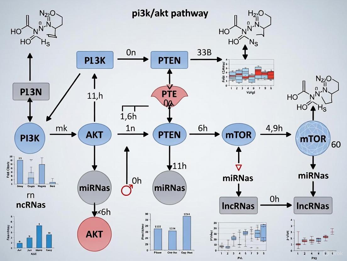
Decoding the Interaction: How ncRNAs Govern the PI3K/AKT Oncogenic Axis in HCC
The Central Role of the PI3K/AKT/mTOR Pathway in HCC Pathogenesis and Progression
Hepatocellular carcinoma (HCC) is a major global health challenge, ranking as the sixth most common cancer and the third leading cause of cancer-related mortality worldwide [1]. The phosphatidylinositol-3 kinase (PI3K)/protein kinase B (AKT)/mammalian target of rapamycin (mTOR) signaling pathway has emerged as a central regulator of hepatocarcinogenesis, with dysregulation observed in approximately 50% of HCC cases [2] [3]. This pathway integrates signals from growth factors, nutrients, and cellular energy status to control critical biological processes including cell survival, proliferation, metabolism, and angiogenesis [4] [2]. In the context of a broader thesis on PI3K/AKT pathway regulation by non-coding RNAs (ncRNAs) in hepatocellular carcinoma research, this review comprehensively examines the pathway's architecture, deregulation in HCC, intricate control by ncRNAs, and the therapeutic implications of targeting this axis.
Pathway Architecture and Core Components
The PI3K/AKT/mTOR signaling pathway represents a highly conserved signal transduction network in eukaryotic cells that promotes cell survival, growth, and proliferation in response to external stimuli [2]. The pathway is initiated when ligand binding to receptor tyrosine kinases (RTKs), such as the epidermal growth factor receptor (EGFR), induces their dimerization and autophosphorylation [3]. The phosphorylated tyrosine residues on activated RTKs serve as docking sites for the Src homology 2 (SH2) domains of PI3K regulatory subunits (p85), bringing the PI3K catalytic subunit (p110) close to the plasma membrane [4] [3].
Once recruited and activated, class I PI3K phosphorylates phosphatidylinositol-4,5-bisphosphate (PIP2) to generate phosphatidylinositol-3,4,5-trisphosphate (PIP3) [4] [3]. This lipid second messenger recruits proteins containing pleckstrin homology (PH) domains, including the serine/threonine kinase AKT and phosphoinositide-dependent kinase 1 (PDK1), to the plasma membrane [3]. AKT subsequently undergoes phosphorylation at two critical residues: Thr308 by PDK1 and Ser473 by the mTOR complex 2 (mTORC2), leading to its full activation [3].
The mammalian target of rapamycin (mTOR) functions as a master regulator of cell growth and proliferation and exists in two distinct multi-protein complexes: mTORC1 and mTORC2 [4]. mTORC1 consists of mTOR, regulatory-associated protein of mTOR (Raptor), mammalian lethal with SEC13 protein 8 (mLST8), and the DEP domain-containing mTOR-interacting protein (DEPTOR) [3]. It is activated by the small GTPase RHEB, which is itself regulated by the tuberous sclerosis complex (TSC1/TSC2) [3]. Active mTORC1 promotes protein synthesis through phosphorylation of downstream effectors including ribosomal protein S6 kinase (S6K) and eukaryotic translation initiation factor 4E-binding protein 1 (4E-BP1) [4]. mTORC2 contains mTOR, rapamycin-insensitive companion of mTOR (Rictor), mLST8, DEPTOR, and mammalian stress-activated protein kinase-interacting protein 1 (mSin1) [3]. This complex regulates cytoskeleton reorganization and cell survival primarily through phosphorylation of AKT at Ser473 [4].
The pathway is negatively regulated by several tumor suppressors, most notably phosphatase and tensin homolog (PTEN), which dephosphorylates PIP3 back to PIP2, thereby terminating PI3K signaling [2] [3].
Figure 1: Core PI3K/AKT/mTOR Signaling Pathway Architecture. This diagram illustrates the sequential activation of pathway components from receptor ligand binding to downstream biological effects. Key regulatory nodes including PTEN and TSC1/TSC2 are highlighted.
Dysregulation of the PI3K/AKT/mTOR Pathway in HCC
The PI3K/AKT/mTOR pathway is frequently dysregulated in HCC through multiple mechanisms, including upstream receptor overexpression, oncogenic mutations in pathway components, and loss of tumor suppressor function.
Genetic and Epigenetic Alterations
EGFR overexpression occurs in approximately 68% of human HCC cases and significantly correlates with metastasis, poor patient survival, and aggressive tumor behavior [3]. The PI3K catalytic subunit p110α (encoded by PIK3CA) is highly expressed in HCC tumor tissue, with upregulation associated with increased proliferation, reduced apoptosis, and unfavorable prognosis [4]. HCC patients with early-stage recurrence demonstrate a higher mutation rate of PIK3CB [4]. mTOR pathway upregulation has been reported in 50% of HCC cases, while approximately 40% of HCC patients who underwent orthotopic liver transplantation exhibited elevated mTOR activity [4] [3].
The tumor suppressor PTEN is frequently downregulated or mutated in HCC. Loss of PTEN function occurs in the majority of HCC patients in early stages, with somatic mutations reported in HCC tissues and observed allelic losses in approximately 32% of patients (12 of 37) [3]. Additionally, loss-of-function mutations in TSC1/2 are found in approximately 20% of HCC patients, representing another mechanism for pathway hyperactivation [3].
Table 1: Key Genetic Alterations in the PI3K/AKT/mTOR Pathway in HCC
| Component | Alteration Type | Frequency in HCC | Functional Consequences |
|---|---|---|---|
| EGFR | Overexpression | ~68% | Correlates with metastasis and poor survival [3] |
| PIK3CA | Upregulation/Mutation | Common (exact % not specified) | Associated with proliferation and poor prognosis [4] |
| PIK3CB | Mutation | Higher in early-recurrence HCC | Promotes tumor recurrence [4] |
| mTOR | Pathway Upregulation | 50% | Potential therapeutic target [4] |
| PTEN | Loss/Loss-of-function | Majority in early stages | Leads to constitutive AKT activation [3] |
| TSC1/TSC2 | Loss-of-function mutations | ~20% | Contributes to mTORC1 hyperactivation [3] |
Functional Consequences in Hepatocarcinogenesis
Hyperactivation of the PI3K/AKT/mTOR pathway drives multiple hallmarks of cancer in HCC. Pathway dysregulation enhances cell proliferation and survival through mTORC1-mediated protein synthesis and inhibition of pro-apoptotic signals [3]. The pathway promotes metabolic reprogramming, enhancing glycolytic flux (Warburg effect) through upregulation of glucose transporter 1 (GLUT1) and hexokinase 2 (HK2) [4]. Additionally, pathway activation facilitates epithelial-mesenchymal transition (EMT), invasion, and metastasis through complex interactions with cytoskeletal components and cell adhesion molecules [2] [3]. The pathway also contributes to angiogenesis by regulating vascular endothelial growth factor (VEGF) expression and supports an immunosuppressive tumor microenvironment [1].
Regulation of the PI3K/AKT/mTOR Pathway by Non-Coding RNAs
Non-coding RNAs (ncRNAs) have emerged as critical regulators of the PI3K/AKT/mTOR pathway in HCC, functioning as both oncogenic drivers and tumor suppressors. These regulatory RNAs fine-tune pathway activity through sophisticated molecular interactions.
microRNAs (miRNAs) in Pathway Regulation
miRNAs are small non-coding RNAs approximately 21-25 nucleotides in length that regulate gene expression post-transcriptionally by binding to target mRNAs, leading to translational repression or mRNA degradation [5]. miR-142-3p represses HCC progression and promotes apoptosis by decreasing PIK3CG-mediated activation of the PI3K/AKT pathway [4]. miR-7 controls cell proliferation and metastasis through the PI3K/AKT/mTOR pathway by targeting PIK3C [4]. Several other miRNAs including miR-873 activate AKT/mTOR signaling via targeting Nedd4 family-interacting protein 1 (NDFIP1), initiating metabolic changes that drive hepatocellular carcinoma formation and metastasis [4].
Table 2: Non-Coding RNAs Regulating the PI3K/AKT/mTOR Pathway in HCC
| ncRNA Type | Representative Molecules | Expression in HCC | Target/Mechanism | Functional Outcome |
|---|---|---|---|---|
| miRNA | miR-142-3p | Downregulated | Decreases PIK3CG [4] | Represses progression, promotes apoptosis |
| miRNA | miR-7 | Downregulated | Targets PIK3C [4] | Controls proliferation and metastasis |
| miRNA | miR-873 | Upregulated | Targets NDFIP1 [4] | Activates AKT/mTOR, drives metastasis |
| lncRNA | Multiple (FTX, XIST) | Varied | Interact with pathway components [5] | Modulate hepatocarcinogenesis |
| circRNA | Various | Varied | Function as miRNA decoys [5] | Fine-tune pathway activity |
Long Non-Coding RNAs (lncRNAs) and Circular RNAs (circRNAs)
Long non-coding RNAs (lncRNAs) exceed 200 nucleotides in length and regulate gene expression through interactions with chromatin, RNA transcripts, or proteins [5]. Current research has identified 69 lncRNAs related to the PI3K/AKT/mTOR system in HCC, with 52 showing upregulation and 15 demonstrating downregulation [5]. Notable examples include lncRNA FTX and XIST, which modulate the PI3K/AKT/mTOR pathway and influence hepatocarcinogenesis [5].
Circular RNAs (circRNAs) are single-stranded ncRNAs with a closed-loop structure formed through covalent bonds [5]. They primarily function as miRNA decoys or regulate transcription factor relationships, thereby fine-tuning PI3K/AKT pathway activity [5]. The intricate regulatory network formed by these ncRNAs represents a critical layer of pathway control in HCC.
Figure 2: ncRNA Regulatory Network of the PI3K/AKT/mTOR Pathway in HCC. This diagram illustrates how different classes of non-coding RNAs (miRNAs, lncRNAs, and circRNAs) regulate pathway activity through distinct molecular mechanisms.
Experimental Models and Research Methodologies
In Vitro Models and Functional Assays
HCC cell lines including HepG2, Huh7, PLC/PRF/5, and MHCC97H are widely used to investigate PI3K/AKT/mTOR signaling [3]. These models enable researchers to dissect pathway dynamics and test therapeutic interventions through various experimental approaches:
Gene manipulation techniques include siRNA- or shRNA-mediated knockdown of pathway components (e.g., SNRPD1, SOCS5) to assess functional consequences [6]. For instance, SNRPD1 knockdown resulted in approximately 5-6 autophagosomes per cell compared to 1.8-2 in control cells, demonstrating enhanced autophagy [6]. Stable overexpression constructs are used to investigate oncogenic effects of pathway activation [3].
Functional assays comprehensively evaluate pathway effects:
- Proliferation assays: MTT, CCK-8, and colony formation assays measure growth kinetics
- Migration and invasion assays: Transwell and wound healing assays quantify metastatic potential
- Metabolic assays: Glucose uptake and lactate production measurements assess glycolytic flux
- Apoptosis assays: Annexin V staining and caspase activity measurements evaluate cell survival
Protein and gene expression analysis includes:
- Western blotting: Assesses phosphorylation status of AKT (T308, S473), S6K, and 4E-BP1
- Immunohistochemistry: Evaluates pathway activation in cell lines and tissue specimens
- qRT-PCR: Measures expression of pathway components and regulatory ncRNAs
In Vivo Models
Xenograft models involve implanting human HCC cells into immunodeficient mice (e.g., nude mice) to evaluate tumor growth and metastasis in response to pathway modulation [6]. For example, SNRPD1 knockdown in xenograft models demonstrated decreased tumor size, confirming the protein's functional importance [6].
Genetically engineered mouse models utilize tissue-specific knockout or knockin approaches to investigate the role of specific pathway components (e.g., PTEN, TSC1) in hepatocarcinogenesis [3].
Chemical carcinogenesis models employ diethylnitrosamine (DEN) administration to induce HCC development in contexts of pathway dysregulation [3].
Table 3: Essential Research Reagents for Investigating PI3K/AKT/mTOR Signaling in HCC
| Reagent Category | Specific Examples | Research Application | Key Findings |
|---|---|---|---|
| siRNA/shRNA | SNRPD1, SOCS5, PDK1 siRNA | Target validation and functional studies | SNRPD1 knockdown reduces PI3K phosphorylation and tumor growth [6] |
| Small Molecule Inhibitors | AKT inhibitors, mTOR inhibitors (everolimus) | Pathway inhibition studies | Everolimus most frequently utilized in clinical trials [7] |
| Antibodies | p-AKT (T308, S473), p-S6K, p-4EBP1 | Protein detection and pathway activity assessment | Used to confirm pathway activation status [6] [3] |
| Expression Vectors | NT5DC2, TMOD3, 14-3-3σ overexpression constructs | Mechanistic studies of pathway activation | NT5DC2 stabilizes EGFR, activating PI3K/AKT/mTOR signaling [3] |
| Chemical Modulators | 3-MA (autophagy inhibitor), Piezo-CAP | Investigation of autophagy and alternative therapies | SOCS5 inhibition effects reversed by 3-MA [6] |
Therapeutic Targeting and Clinical Perspectives
Current Status of Targeted Therapies
Therapeutic targeting of the PI3K/AKT/mTOR pathway in HCC has faced significant challenges despite strong biological rationale. mTOR inhibitors constitute the majority of clinical investigations, with everolimus being the most frequently utilized drug [7]. However, only approximately 10% of studies have advanced to phase III or IV, predominantly involving mTOR inhibitors [7]. Challenges including adverse events (hyperglycemia, bone marrow suppression) and treatment resistance have limited success [7].
Combination therapies have shown promise in overcoming these limitations. Co-administration with MEK inhibitors, chemotherapy, immune checkpoint inhibitors, and VEGF inhibitors represents promising strategies to enhance efficacy and circumvent resistance [7]. For instance, the combination of anti-PD-L1 immunotherapy (atezolizumab) with anti-angiogenic therapy (bevacizumab) has demonstrated superiority over sorafenib in terms of overall survival, progression-free survival, and objective response rate [1].
ncRNA-Based Therapeutic Approaches
Emerging strategies targeting ncRNAs represent a promising frontier in HCC therapeutics:
Antisense oligonucleotides (ASOs) are synthetic analogues of natural nucleic acids designed to selectively bind to complementary RNA sequences in both the nucleus and cytosol [5]. These molecules can modulate lncRNA expression and function, potentially restoring normal pathway regulation.
RNA interference approaches utilizing siRNA or shRNA technologies can specifically downregulate oncogenic ncRNAs contributing to pathway hyperactivation [5] [8].
Small molecule inhibitors targeting the interactions between ncRNAs and pathway components offer another potential therapeutic strategy [8].
Despite promising preclinical data, significant challenges remain in translating ncRNA-targeting approaches to clinical applications, including delivery efficiency, tissue specificity, and potential off-target effects [5].
The PI3K/AKT/mTOR signaling pathway represents a critical hub in hepatocellular carcinoma pathogenesis, integrating signals from multiple upstream regulators and coordinating diverse downstream effects that drive tumor progression. The sophisticated regulation of this pathway by ncRNAs—including miRNAs, lncRNAs, and circRNAs—adds complex layers of control that both promote and suppress hepatocarcinogenesis depending on context. While therapeutic targeting of this pathway has proven challenging, combination approaches and novel strategies focusing on ncRNA-mediated regulation offer promising avenues for future investigation. A comprehensive understanding of the PI3K/AKT/mTOR pathway and its regulatory networks will be essential for developing more effective precision medicine approaches for HCC patients.
Hepatocellular carcinoma (HCC) remains a major global health challenge, ranking as the sixth most frequently diagnosed cancer and the third-leading cause of cancer death worldwide [9]. The poor prognosis of HCC, with a dismal 5-year survival rate of only 15–18%, is largely attributable to late diagnosis, high recurrence rates, and limited therapeutic options [9]. In recent years, non-coding RNAs (ncRNAs)—including microRNAs (miRNAs), long non-coding RNAs (lncRNAs), and circular RNAs (circRNAs)—have emerged as critical regulators of gene expression and central players in HCC pathogenesis. These molecules uniquely bridge bench-to-bedside translation in HCC, owing to their ability to regulate multiple oncogenic pathways while being stably detectable in circulation [9].
The phosphoinositide 3-kinase/protein kinase B (PI3K/AKT) signaling pathway represents one of the most important intracellular signal transduction pathways, playing a critical role in regulating cellular processes such as cell proliferation, survival, metabolism, and angiogenesis [10]. This pathway is frequently dysregulated in HCC, and emerging evidence indicates that ncRNAs serve as master regulators of the PI3K/AKT axis, thereby influencing tumorigenesis, metastasis, and therapeutic response [11]. This primer provides a comprehensive overview of the roles, mechanisms, and research methodologies for studying miRNAs, lncRNAs, and circRNAs in HCC, with special emphasis on their regulation of the PI3K/AKT pathway.
miRNA: Master Regulators of Gene Expression in HCC
Biogenesis and Functional Mechanisms
MicroRNAs (miRNAs) are small, non-coding RNA molecules approximately 19–24 nucleotides in length that function as post-transcriptional repressors of gene expression [9]. They are transcribed as primary miRNAs (pri-miRNAs) and subsequently processed by the Drosha/DGCR8 complex to form precursor miRNAs (pre-miRNAs). After export from the nucleus, pre-miRNAs are diced by Dicer to generate mature miRNA strands that are loaded into the RNA-induced silencing complex (RISC) [9]. Through partial complementarity with target mRNAs, miRNAs repress gene expression by either promoting mRNA degradation or inhibiting translation.
Key miRNAs Regulating PI3K/AKT in HCC
Table 1: Key miRNAs Regulating the PI3K/AKT Pathway in HCC
| miRNA | Expression in HCC | Validated Target | Effect on PI3K/AKT | Functional Outcome |
|---|---|---|---|---|
| miR-101-3p | Downregulated | Birc5 | Downregulates PI3K/AKT signaling | Inhibits proliferation, invasion, and EMT [12] |
| miR-338-3p | Downregulated | SOX4 | Suppresses PI3K/AKT activation | Reduces stemness and lenvatinib resistance [13] |
| miR-199a-5p | Downregulated | HIF1A, HK2 | Counters glycolytic shift | Restores mitochondrial oxidation [9] |
| miR-122 | Downregulated | PKM2, G6PD | Balances glycolysis with PPP | Induces metabolic switch to oxidative phosphorylation [9] |
| miR-21 | Upregulated | Multiple tumor suppressors | Promotes sorafenib resistance | Correlates with therapy resistance [9] |
The tumor-suppressive miRNA miR-101-3p plays crucial roles in inhibiting the proliferation, invasion, and epithelial-mesenchymal transition (EMT) of HCC cells by targeting Birc5 and downregulating the PI3K-AKT signaling pathway [12]. Similarly, miR-338-3p deficiency increases self-renewal and tumor malignancy in hepatic cancer stem cells (CSCs), while its overexpression suppresses tumorigenesis and self-renewal by specifically targeting SOX4 [13]. The liver-specific miR-122 serves as a key metabolic regulator, with its downregulation in HCC correlating with increased PKM2 expression, elevated FDG-PET uptake, and poor survival [9].
Research Methodologies for miRNA Studies
Dual-Luciferase Reporter Assay: This technique validates direct molecular interactions between miRNAs and their putative targets. The conserved miRNA-interacting regions from the 3'UTR of the target gene (e.g., a 500-bp segment for SOX4) are incorporated into a luciferase reporter plasmid [13]. A mutant construct with altered miRNA-binding sites serves as a negative control. HCC cells are co-transfected with the pRL-CMV vector (internal control), the luciferase UTR-report vector, and miRNA precursor or control. After 24 hours, relative luciferase activity is measured using a dual-luciferase reporter assay system [13].
Functional Assays: Gain-of-function and loss-of-function approaches are essential for establishing miRNA roles. For overexpression, eukaryotic expression vectors with enhanced miRNA expression or miRNA mimics are transfected into HCC cell lines using lipid-based transfection reagents [12]. For knockdown, lentivirus-based systems delivering anti-miRNA sequences are employed. Subsequent functional assessments include:
- Cell cloning experiments and CCK-8 assays for proliferation
- Transwell migration experiments for invasion capability
- Flow cytometric analysis with Annexin V/7-AAD staining for apoptosis
- In vitro spheroid formation assays for stemness properties [13]
lncRNAs: Architectural Regulators of Cellular Function
Characteristics and Classification
Long non-coding RNAs (lncRNAs) are defined as RNA transcripts exceeding 200 nucleotides without protein-coding capacity [10]. Compared with small RNA molecules, lncRNAs have longer sequences, more complicated spatial structures, and participate in more diverse and complex mechanisms [10]. They localize to both the nucleus and cytoplasm and regulate gene expression through multiple mechanisms: pre-transcriptionally by recruiting chromatin-modifying complexes; transcriptionally by interacting with transcription factors and RNA polymerase; and post-transcriptionally by acting as competing endogenous RNAs (ceRNAs) or affecting mRNA stability [10].
Key lncRNAs in HCC Pathogenesis
Table 2: Oncogenic and Tumor-Suppressive lncRNAs in HCC
| lncRNA | Expression | Molecular Mechanism | Functional Role | Therapeutic Relevance |
|---|---|---|---|---|
| RAB30-DT | Upregulated | Binds/stabilizes SRPK1; promotes splicing reprogramming | Drives stemness and progression; poor prognosis [14] | CREB1–RAB30-DT–SRPK1 axis sensitizes HCC to targeted therapies |
| MALAT1 | Upregulated | Recruits chromatin-modifying complexes; modulates PI3K/AKT | Promotes proliferation and metastasis [10] | Potential biomarker and therapeutic target |
| HOTAIR | Upregulated | Interacts with PI3K/AKT components; epigenetic regulation | Enhances tumor growth and invasion [10] | Associated with therapy resistance |
| TINCR | Upregulated | Physically interacts with AKT1 | Promotes PI3K/AKT signaling and proliferation [10] | Possible target for intervention |
The lncRNA RAB30-DT represents a recently characterized oncogenic lncRNA that is significantly overexpressed in malignant epithelial cells and associated with advanced tumor stage, stemness features, genomic instability, and poor patient prognosis [14]. Mechanistically, RAB30-DT is transcriptionally activated by CREB1 and directly binds and stabilizes the splicing kinase SRPK1, facilitating its nuclear localization and driving widespread alternative splicing reprogramming, including splicing of the cell cycle regulator CDCA7, to promote tumor stemness and malignancy [14].
Research Techniques for lncRNA Investigation
Integrated Multi-Omics Analysis: This approach identifies lncRNAs with clinical and functional significance. The TCGA-LIHC dataset provides gene expression data from HCC and adjacent normal tissues. Splicing regulatory factors are curated from databases like IARA, and a global splicing score is calculated for each sample [14]. Stemness is quantified using the mRNA stemness index (mRNAsi) algorithm. Differential expression analysis identifies significantly dysregulated lncRNAs, while correlation analysis links lncRNA expression with stemness and splicing scores. Kaplan-Meier survival analysis and ROC analysis evaluate prognostic significance [14].
Single-Cell RNA Sequencing (scRNA-Seq): This technology resolves lncRNA expression at cellular resolution. Public scRNA-Seq datasets (e.g., GSE202642 from NCBI GEO) are analyzed using Seurat package for quality control, normalization, and clustering [14]. The Harmony algorithm corrects for batch effects across samples. CytoTRACE computational tool assesses differentiation status and stemness level of individual tumor cells based on transcriptomic data, allowing identification of lncRNAs enriched in CSCs versus non-CSCs [14].
circRNAs: Emerging Circular Regulators
Unique Features and Biogenesis
Circular RNAs (circRNAs) represent a special class of endogenous RNA molecules characterized by a covalently closed continuous loop structure without 5' or 3' ends or polyadenosine tails [15]. This circular configuration confers unusual stability and resistance to RNA exonuclease-mediated degradation. circRNAs can be derived from exons or introns, or composed of both exons and introns, through back-splicing events where a downstream 5' splice site joins with an upstream 3' splice site [15]. Although circRNAs have been known since the 1970s, they have only recently gained significant attention due to advances in RNA sequencing and bioinformatics.
Functional circRNAs in HCC
circACVR2A (hsacirc0001073): This circRNA is highly expressed in hepatocellular carcinoma cell lines and promotes proliferation, migration, and invasion of HCC both in vitro and in vivo [15]. Mechanistically, circACVR2A functions as a miRNA sponge, directly interacting with miR-511-5p to regulate expression of related proteins in the PI3K-Akt signaling pathway. In HCC, circACVR2A mediates a miR-511-5p/mRNA network to activate PI3K signaling, establishing this molecular regulatory network as a potential target for diagnosis and treatment of hepatocellular carcinoma [15].
Other circRNAs contribute to HCC progression by regulating cytokine signaling and immunity, often functioning as miRNA sponges or protein scaffolds that influence gene networks [16]. Their unique stability, diverse mechanisms of action, and aberrant expression in various cancers make them highly relevant for oncology research and therapeutic development.
Experimental Approaches for circRNA Research
RNase R Treatment: To distinguish circRNAs from linear transcripts, total RNA is treated with 3 U/µg RNase R (an RNA exonuclease that degrades linear RNAs but not circular RNAs) for 30 minutes [15]. Subsequent qRT-PCR analysis comparing treated and untreated samples confirms circularity through RNase R resistance.
Nuclear-Cytoplasmic Fractionation: RNA from nuclear and cytoplasmic compartments is isolated using specialized kits (e.g., Cytoplasmic & Nuclear RNA Purification Kit) to determine subcellular localization of circRNAs, which provides clues about their potential mechanisms of action [15].
In Vivo Functional Studies: For assessing tumorigenic potential, HCC cells are diluted to relevant concentrations (e.g., 1×10³, 5×10³, 1×10â´, 5×10â´) and mixed with Matrigel gel (1:1 ratio) for in vivo limiting dilution experiments [13]. Cells are subcutaneously injected into immunocompromised mice (e.g., NOD-SCID mice), with tumor formation monitored over 6-8 weeks to assess cancer stem cell frequency and tumor-initiating potential.
The ncRNA-PI3K/AKT Signaling Axis in HCC
Integrated Regulatory Networks
The PI3K/AKT signaling pathway serves as a critical hub through which multiple ncRNAs coordinate HCC development and progression. PI3K activation generally begins with growth factor binding to receptor tyrosine kinases, leading to PI3K activation and phosphorylation of phosphatidylinositol (4,5)-bisphosphate (PIP2) to generate phosphatidylinositol (3,4,5)-trisphosphate (PIP3) [10]. PIP3 then recruits AKT to the plasma membrane, where it undergoes phosphorylation and activation. Once activated, AKT modulates downstream targets involved in cell cycle progression, apoptosis inhibition, and protein synthesis via the mTOR pathway.
ncRNAs regulate this pathway through multiple mechanisms. miRNAs can directly target PI3K or AKT components, while lncRNAs and circRNAs can function as ceRNAs that sequester miRNAs, thereby derepressing PI3K/AKT signaling [11]. Additionally, lncRNAs can directly bind to proteins or mRNAs involved in the PI3K/AKT pathway or recruit chromatin-modifying complexes to specific genomic loci to modify gene expression that impacts this pathway [10].
Schematic of ncRNA Regulation of PI3K/AKT Signaling in HCC
Therapeutic Implications and Resistance Mechanisms
The ncRNA-PI3K/AKT axis represents a promising therapeutic target in HCC. For instance, miR-338-3p overexpression in HCC cells increases sensitivity to lenvatinib-induced apoptosis and inhibits cell progression [13]. Patients with high miR-338-3p expression may experience greater benefits from lenvatinib treatment, suggesting its potential as a predictive biomarker. Similarly, pharmacological disruption of the CREB1–RAB30-DT–SRPK1 axis sensitizes HCC cells to targeted therapies [14].
Cancer stem cells (CSCs), a subpopulation within tumors with self-renewal and pluripotency capabilities, have emerged as crucial drivers of HCC recurrence, metastasis, and therapeutic resistance [14]. Their persistence following therapy often leads to relapse and resistance to conventional treatments. ncRNAs modulate CSC properties through the PI3K/AKT pathway, influencing self-renewal, tumor initiation, and drug resistance [13] [14].
The Scientist's Toolkit: Essential Research Reagents and Methods
Table 3: Essential Research Reagents and Experimental Resources
| Category | Specific Reagents/Tools | Application | Key Considerations |
|---|---|---|---|
| Cell Models | Huh-7, HepG2, Hep3B, MHCC97L, QGY7701, LO2 (normal liver) | In vitro functional studies | Select based on genetic background; include normal control [15] [12] |
| Culture Media | DMEM, RPMI-1640 with 10% FBS, 1% penicillin/streptomycin | Cell maintenance | Use serum-free conditions for spheroid formation assays [13] |
| Transfection Reagents | Lipofectamine 2000, lentiviral vectors (miR-338-3p knockdown) | Gain/loss-of-function studies | Optimize efficiency for each cell line; use stable infections for long-term studies [13] [12] |
| qRT-PCR Reagents | TRIzol RNA extraction, SYBR Green kits, specific primers for circACVR2A, miR-511-5p | Expression quantification | Use RNase R treatment to verify circRNAs; U6/GAPDH as internal controls [15] |
| Functional Assays CCK-8, Transwell migration, Annexin V/7-AAD apoptosis kit | Proliferation, invasion, apoptosis assessment | Standardize cell numbers and treatment durations across experiments [13] [12] | |
| Animal Models | NOD-SCID mice (4-6 weeks) | In vivo tumorigenesis studies | Follow ethical guidelines; monitor tumor size (<2000 mm³) [13] |
| Pathway Inhibitors | Miltefosine (PI3K/AKT inhibitor), Lenvatinib | Mechanism validation | Use at IC50 concentrations; establish resistant cell lines by gradual dose increase [13] [12] |
| N-(tert-butyl)-2-nitrobenzamide | N-(tert-butyl)-2-nitrobenzamide, CAS:41225-78-9, MF:C11H14N2O3, MW:222.244 | Chemical Reagent | Bench Chemicals |
| Methyl 2-(benzofuran-5-YL)acetate | Methyl 2-(benzofuran-5-YL)acetate|CAS 121638-36-6 | Methyl 2-(benzofuran-5-YL)acetate (CAS 121638-36-6) is a key synthetic intermediate for bioactive compound research. This product is For Research Use Only. Not for human or veterinary use. | Bench Chemicals |
Experimental Workflow for ncRNA Functional Characterization
The intricate regulatory networks formed by miRNAs, lncRNAs, and circRNAs represent a critical layer of control over the PI3K/AKT signaling pathway in hepatocellular carcinoma. These ncRNAs function as dynamic regulators of tumor initiation, progression, metastasis, and therapeutic response, offering promising avenues for biomarker development and targeted therapies. Future research directions should focus on validating the clinical utility of ncRNA biomarkers in large patient cohorts, developing efficient delivery systems for ncRNA-based therapeutics, and exploring combination strategies that target ncRNA networks alongside conventional therapies. As our understanding of ncRNA biology deepens, these molecules are poised to revolutionize precision oncology approaches for HCC management, potentially overcoming the limitations of current treatment paradigms and improving patient outcomes.
Oncogenic and Tumor-Suppressive miRNAs as Direct Tuners of PI3K/AKT Signaling
The phosphatidylinositol 3-kinase (PI3K)/AKT signaling pathway represents a crucial intracellular regulatory axis frequently dysregulated in hepatocellular carcinoma (HCC), driving fundamental oncogenic processes including cell survival, proliferation, metabolism, and therapeutic resistance. Non-coding RNAs, particularly microRNAs (miRNAs), have emerged as master regulators of this pathway, functioning as either oncogenic drivers or tumor suppressive elements through direct targeting of core pathway components. This technical review synthesizes current evidence elucidating how specific miRNAs directly tune PI3K/AKT signaling in HCC, presenting structured quantitative data, experimental methodologies for miRNA-pathway validation, visualization of regulatory networks, and essential research tools for investigating this critical axis in liver oncogenesis. The comprehensive analysis underscores the therapeutic potential of targeting miRNA-PI3K/AKT interactions for innovative HCC treatment strategies.
Hepatocellular carcinoma (HCC) constitutes approximately 90% of primary liver cancers and ranks as the third leading cause of cancer-related deaths globally, with projections indicating over one million annual fatalities by 2030 [17]. The PI3K/AKT signaling pathway is one of the most frequently dysregulated intracellular pathways in HCC, serving as a central regulator of cell survival, proliferation, metabolism, and therapeutic resistance [5] [8]. This pathway transduces signals from receptor tyrosine kinases (RTKs) through a cascade involving PI3K-mediated generation of phosphatidylinositol (3,4,5)-trisphosphate (PIP3), which recruits AKT to the plasma membrane for activation via phosphorylation. Activated AKT subsequently phosphorylates numerous downstream substrates, including mTOR, GSK-3β, and FOXO transcription factors, orchestrating diverse cellular processes critical to oncogenesis [5] [1]. The tumor suppressor PTEN (phosphatase and tensin homolog) serves as the primary negative regulator of this pathway by dephosphorylating PIP3 to PIP2 [5].
The PI3K/AKT pathway exhibits aberrant activation in HCC through multiple mechanisms, including genetic mutations, epigenetic alterations, and viral protein interactions [5]. This pathway activation promotes HCC development and progression by stimulating cell cycle progression, inhibiting apoptosis, enhancing angiogenesis, and facilitating metabolic reprogramming toward glycolysis [17] [18]. Importantly, emerging evidence positions microRNAs (miRNAs) as critical post-transcriptional regulators of PI3K/AKT signaling, offering novel insights into HCC pathogenesis and revealing potential therapeutic vulnerabilities [5] [8].
miRNA Biogenesis and Mechanisms of Pathway Regulation
MicroRNAs are small non-coding RNA molecules approximately 21-25 nucleotides in length that function as post-transcriptional regulators of gene expression [19]. miRNA biogenesis begins with RNA polymerase II/III transcription of primary miRNA transcripts (pri-miRNAs), which undergo sequential processing by the Drosha-DGCR8 complex in the nucleus to produce precursor miRNAs (pre-miRNAs) [20]. Following export to the cytoplasm, pre-miRNAs are cleaved by Dicer to generate mature miRNA duplexes. One strand of this duplex is incorporated into the RNA-induced silencing complex (RISC), where it guides target recognition through partial complementarity to sequences primarily within the 3'-untranslated regions (3'-UTRs) of target mRNAs, resulting in translational repression or mRNA degradation [19] [20].
In the context of the PI3K/AKT pathway, miRNAs function as precise tuners of signaling intensity and duration by directly targeting core pathway components, including receptor tyrosine kinases, PI3K subunits, AKT isoforms, PTEN, and downstream effectors [5]. The balance between oncogenic miRNAs (oncomiRs) that repress negative regulators like PTEN and tumor-suppressive miRNAs that target positive pathway components determines the overall signaling output, profoundly influencing HCC development, progression, and therapeutic response [17] [5] [8].
Oncogenic miRNAs Enhancing PI3K/AKT Signaling
Oncogenic miRNAs promote PI3K/AKT signaling primarily by targeting negative pathway regulators, with PTEN representing the most frequently suppressed tumor suppressor. The following table summarizes key oncogenic miRNAs, their validated targets, and experimental evidence in HCC models.
Table 1: Oncogenic miRNAs Enhancing PI3K/AKT Signaling in HCC
| miRNA | Expression in HCC | Validated Target | Experimental Model | Functional Outcome | Reference |
|---|---|---|---|---|---|
| miR-21 | Upregulated | PTEN | Transgenic zebrafish, human HCC tissues, sorafenib-resistant cell lines | Induced NAFLD-HCC progression, sorafenib resistance via suppressed autophagy | [18] [21] [20] |
| miR-17-92 cluster | Upregulated | PTEN | Mouse B-cell lymphoma models, human HCC cell lines | Enhanced tumorigenesis, promoted cell survival | [22] [23] |
| miR-221 | Upregulated | PTEN, p27 | Human HCC tissues, xenograft models | Accelerated tumor growth, enhanced proliferation | [5] |
| miR-25 | Upregulated | PTEN | Human HCC tissues, sorafenib-resistant cells | Contributed to therapy resistance | [19] |
| miR-424-3p | Upregulated | PTEN | Human HCC tissues, cell line experiments | Promoted cell proliferation, migration | [19] |
| miR-19a/b | Upregulated | PTEN | Eμ-Myc transgenic mouse model | Identified as primary oncogenic component of miR-17-92 cluster | [22] |
Case Study: miR-21 as a Master Regulator of PTEN/PI3K/AKT Axis
miR-21 represents one of the most extensively characterized oncomiRs in HCC, demonstrating consistent overexpression across multiple HCC etiologies, particularly in non-alcoholic fatty liver disease (NAFLD)-related HCC [18]. Mechanistically, miR-21 directly binds to the 3'-UTR of PTEN mRNA, repressing its translation and reducing PTEN protein levels without affecting mRNA abundance [21] [20]. This suppression relieves PI3K inhibition, leading to enhanced PIP3 production, AKT phosphorylation, and downstream signaling activation.
In a doxycycline-inducible transgenic zebrafish model (LmiR21), miR-21 overexpression induced the full spectrum of NAFLD progression to HCC, including steatosis, inflammation, fibrosis, and ultimately hepatocellular carcinoma [18]. This oncogenic progression occurred through coordinated activation of multiple signaling networks: (1) hepatic steatosis via decreased ptenb and pparaa with concomitant PI3K/AKT activation; (2) inflammation and fibrosis through STAT3 and TGF-β/Smad signaling; and (3) oncogenic transformation via Smad3/Stat3 activation [18]. Immunoblotting analyses confirmed consistent PTEN downregulation and concomitant AKT phosphorylation in both zebrafish models and human HCC tissues, validating the conserved nature of this regulatory axis [18].
Furthermore, miR-21 contributes substantially to sorafenib resistance in HCC through the PTEN/AKT/autophagy axis. Sorafenib-resistant HCC cells (HepG2-SR and Huh7-SR) exhibit significantly elevated miR-21 expression alongside reduced PTEN protein and enhanced AKT phosphorylation [21]. These cells demonstrate reduced autophagic flux, as evidenced by decreased acridine orange-stained acidic vesicular organelles and lower LC3-II and Beclin-1 protein levels compared to parental cells [21]. Functional experiments established that anti-miR-21 oligonucleotides restored sorafenib sensitivity by promoting autophagy, while miR-21 mimics conferred resistance, positioning miR-21 as both a biomarker and therapeutic target for overcoming sorafenib resistance [21].
The miR-17-92 Cluster: A Polycistronic OncomiR
The miR-17-92 cluster, located on chromosome 13q31.3, encodes six mature miRNAs (miR-17, miR-18a, miR-19a, miR-20a, miR-19b, and miR-92a) processed from a single polycistronic transcript [22] [23]. This cluster is frequently amplified and overexpressed in HCC and other malignancies, functioning as a potent oncogene. Among its components, miR-19a and miR-19b have been identified as the primary oncogenic effectors responsible for PTEN targeting and subsequent PI3K/AKT activation [22]. Mouse models of c-Myc-induced B-cell lymphoma demonstrated that while deletion of the entire miR-17-92 cluster slowed oncogenesis, reintroduction of miR-19a/19b alone largely rescued tumorigenicity, establishing these miRNAs as the critical oncogenic components [22]. Subsequent validation experiments confirmed PTEN as a direct and functionally significant target of miR-19, with miR-19 overexpression sufficient to reduce PTEN protein levels and activate AKT signaling [22].
Tumor-Suppressive miRNAs Attenuating PI3K/AKT Signaling
Tumor-suppressive miRNAs inhibit PI3K/AKT signaling by targeting positive pathway components, including receptor tyrosine kinases, PI3K subunits, and AKT itself. The following table summarizes key tumor-suppressive miRNAs, their targets, and experimental evidence in HCC.
Table 2: Tumor-Suppressive miRNAs Inhibiting PI3K/AKT Signaling in HCC
| miRNA | Expression in HCC | Validated Target | Experimental Model | Functional Outcome | Reference |
|---|---|---|---|---|---|
| miR-34a-5p | Downregulated | MET, YY1 | Human HCC tissues, bioinformatics analysis | Suppressed stemness, angiogenesis, glycolysis, autophagy, EMT, metastasis | [17] |
| miR-199a-5p | Downregulated | mTOR, c-Met | Human HCC tissues, mouse xenograft models | Inhibited proliferation, enhanced apoptosis | [5] |
| miR-195-5p | Downregulated | PI3K subunits | Human HCC tissues, cell line experiments | Suppressed cell cycle progression, migration | [17] [5] |
| miR-122 | Downregulated | IGF1R, PI3K | Human HCC tissues, transgenic mouse models | Restored sensitivity to sorafenib, inhibited tumor growth | [5] |
| miR-139-5p | Downregulated | PI3K, AKT | Human HCC tissues, bioinformatics analysis | Suppressed multiple cancer hallmarks | [17] |
| miR-1 | Downregulated | PIK3CA | Lung cancer models (conserved mechanism) | Inhibited tumor growth | [19] |
Case Study: miR-34a as a Multipathway Tumor Suppressor
miR-34a-5p has been identified as a key tumor-suppressive miRNA simultaneously targeting multiple oncogenic pathways in HCC, including PI3K/AKT signaling [17]. Through comprehensive bioinformatics analysis of HCC datasets and literature review, miR-34a-5p was recognized among a select group of miRNAs that coordinately regulate critical cancer hallmarks, including stemness, angiogenesis, glycolysis, autophagy, epithelial-mesenchymal transition (EMT), and metastasis [17]. Experimentally, miR-34a directly targets MET receptor tyrosine kinase and the transcription factor YY1, both of which positively regulate PI3K/AKT signaling [17]. The downregulation of miR-34a observed in HCC consequently unleashes these positive regulators, enhancing pathway activity and driving tumor progression across multiple biological dimensions.
miR-199a-5p: Dual Targeting of mTOR and c-Met
miR-199a-5p represents another significant tumor-suppressive miRNA frequently downregulated in HCC, with demonstrated activity against two key nodes in PI3K/AKT signaling: the upstream receptor c-Met and the downstream effector mTOR [5]. This dual targeting positions miR-199a-5p as a particularly potent inhibitor of the pathway, capable of simultaneous suppression at multiple levels. Restoration of miR-199a-5p expression in HCC models significantly impairs tumor growth and enhances apoptosis, highlighting its therapeutic potential [5]. The consistent downregulation of miR-199a-5p across HCC cohorts suggests its loss may be a critical event in hepatocarcinogenesis, permitting uncontrolled signaling through both PI3K/AKT and c-Met pathways.
Experimental Approaches for Validating miRNA-PI3K/AKT Interactions
Luciferase Reporter Assays for Direct Target Validation
Luciferase reporter assays represent the gold standard for experimentally validating direct miRNA-mRNA interactions. The following protocol details the methodology for confirming miRNA targeting of PI3K/AKT pathway components:
Vector Construction: Clone the 3'-UTR region of the putative target gene (e.g., PTEN, PIK3CA) into a luciferase reporter vector downstream of the luciferase coding sequence. Alternatively, clone mutated versions with altered seed sequence binding sites as controls [21] [20].
Cell Transfection: Co-transfect HEK293T or relevant HCC cell lines (Huh7, HepG2) with:
- Luciferase reporter vector (wild-type or mutated)
- miRNA mimic (for overexpression) or miRNA inhibitor (for knockdown)
- Renilla luciferase vector for normalization
Luciferase Measurement: After 24-48 hours, harvest cells and measure firefly and Renilla luciferase activities using dual-luciferase reporter assay systems.
Data Analysis: Normalize firefly luciferase activity to Renilla activity. Significant reduction in luciferase activity for wild-type but not mutated 3'-UTR constructs confirms direct miRNA targeting [21].
This approach has been successfully employed to validate numerous miRNA-PI3K/AKT interactions, including miR-21 targeting of PTEN [21] [20], miR-19 targeting of PTEN [22], and miR-199a-5p targeting of mTOR [5].
Functional Validation in Vitro and In Vivo
Following target validation, functional assessment of miRNA-mediated PI3K/AKT regulation requires integrated experimental approaches:
In Vitro Techniques:
- miRNA Modulation: Transfect HCC cells with miRNA mimics (for tumor suppressors) or inhibitors (for oncomiRs) using lipid-based transfection reagents [21].
- Western Blot Analysis: Evaluate protein levels of pathway components (PTEN, p-AKT, total AKT, PI3K subunits) 48-72 hours post-transfection [18] [21].
- Proliferation and Viability Assays: Assess functional outcomes using MTT, CCK-8, or colony formation assays following miRNA modulation [21].
- Apoptosis Detection: Measure caspase activation and annexin V staining to quantify apoptosis after miRNA manipulation [21].
In Vivo Models:
- Xenograft Studies: Implant miRNA-modified HCC cells into immunodeficient mice and monitor tumor growth, followed by immunohistochemical analysis of pathway activation [18].
- Transgenic Models: Utilize tissue-specific miRNA overexpression or knockout mice, such as the doxycycline-inducible miR-21 zebrafish model that recapitulates NAFLD-HCC progression [18].
- Therapeutic Testing: Administer nanoparticle-formulated miRNA mimics or inhibitors to pre-established tumors and assess therapeutic efficacy and pathway modulation [17].
Visualization of miRNA Regulatory Networks
The complex interplay between miRNAs and PI3K/AKT signaling components can be visualized through the following regulatory network:
Diagram: miRNA Regulation of PI3K/AKT Signaling in HCC. This network visualization illustrates how oncogenic miRNAs (red) and tumor-suppressive miRNAs (blue) differentially regulate core components of the PI3K/AKT pathway, ultimately influencing key oncogenic processes in hepatocellular carcinoma.
The Scientist's Toolkit: Essential Research Reagents
Table 3: Essential Research Reagents for Investigating miRNA-PI3K/AKT Interactions
| Reagent Category | Specific Examples | Research Application | Key Considerations |
|---|---|---|---|
| miRNA Modulators | miR-21 mimics/inhibitors, miR-34a mimics, anti-miR-17-92 oligonucleotides | Gain- and loss-of-function studies | Chemical modifications (2'-O-methyl, LNA) enhance stability and binding affinity |
| Delivery Systems | Lipid nanoparticles (LNP), viral vectors (lentivirus, AAV), extracellular vesicles | In vivo miRNA therapeutic delivery | LNPs used in clinical trials (MRX34, INT-1B3); optimize for liver tropism |
| Pathway Inhibitors | LY294002 (PI3K inhibitor), MK-2206 (AKT inhibitor), rapamycin (mTOR inhibitor) | Pathway inhibition controls | Use alongside miRNA modulation to establish specificity |
| Detection Antibodies | Anti-PTEN, anti-p-AKT (Ser473), anti-total AKT, anti-PI3K p85 | Western blot, IHC analysis | Phospho-specific antibodies require careful validation |
| Reporter Systems | Luciferase vectors with 3'-UTRs (PTEN, PIK3CA), mutated controls | Direct target validation | Include seed sequence mutations as critical controls |
| Animal Models | Diethylnitrosamine (DEN)-induced HCC, MYC-driven models, miR-21 transgenic zebrafish | In vivo validation | Transgenic zebrafish permit live imaging of NAFLD-HCC progression |
| N-[4-(phenylamino)phenyl]acetamide | N-[4-(Phenylamino)phenyl]acetamide Supplier | Bench Chemicals | |
| 2-(4-Fluorobenzyl)cyclohexanone | 2-(4-Fluorobenzyl)cyclohexanone | 2-(4-Fluorobenzyl)cyclohexanone is a fluorinated ketone for research. This product is For Research Use Only. Not for human or veterinary use. | Bench Chemicals |
The intricate regulation of PI3K/AKT signaling by oncogenic and tumor-suppressive miRNAs represents a fundamental layer of control in hepatocellular carcinoma pathogenesis. The evidence synthesized in this review demonstrates that miRNAs function as precise tuners of this critical pathway, with their dysregulation contributing substantially to HCC development, progression, and therapeutic resistance. The coordinated activity of miRNA networks—such as the simultaneous regulation of multiple cancer hallmarks by miR-34a-5p and related miRNAs—highlights the potential of targeting these regulatory nodes for therapeutic benefit [17].
Several challenges remain in translating these findings into clinical applications, including optimizing delivery systems for miRNA-based therapeutics, minimizing off-target effects, and understanding contextual dependencies in miRNA function [5] [8]. However, ongoing clinical trials with miRNA-based formulations (e.g., MRX34, a liposomal miR-34a mimic) provide promising proof-of-concept for this approach [17]. Future research directions should prioritize comprehensive mapping of miRNA-PI3K/AKT interactions across diverse HCC etiologies, development of combination therapies targeting both miRNAs and conventional pathway inhibitors, and exploitation of miRNA signatures as biomarkers for patient stratification and treatment response monitoring.
The integration of miRNA-based approaches with existing targeted therapies and immunotherapies represents a promising frontier in HCC management, potentially offering enhanced efficacy and overcome resistance mechanisms. As our understanding of miRNA-mediated PI3K/AKT regulation deepens, these insights will undoubtedly catalyze the development of more effective, personalized therapeutic strategies for hepatocellular carcinoma.
Long non-coding RNAs (lncRNAs) have emerged as pivotal regulators in hepatocellular carcinoma (HCC), functioning as molecular scaffolds and sponges to fine-tune the PI3K/AKT signaling pathway. This whitepaper delineates the sophisticated mechanisms through which lncRNAs modulate oncogenic processes by assembling protein complexes and sequestering microRNAs. Through an analysis of current research, we detail how specific lncRNAs such as HOTAIR, MALAT1, and NONHSAT192404.1 regulate key components of the PI3K/AKT pathway, providing a technical foundation for drug development targeting these RNA-based mechanisms. The integration of experimental protocols and research reagents outlined herein offers scientists a comprehensive toolkit for advancing therapeutic strategies in HCC.
The PI3K/AKT signaling pathway represents one of the most frequently dysregulated oncogenic pathways in hepatocellular carcinoma, governing critical cellular processes including proliferation, survival, metabolism, and apoptosis [5] [24]. While protein-centric regulators of this pathway have been extensively characterized, recent evidence has illuminated the indispensable role of long non-coding RNAs as precise modulators of pathway activity. LncRNAs, defined as transcripts longer than 200 nucleotides with limited or no protein-coding capacity, exert regulatory functions through their ability to form complex secondary and tertiary structures [10] [25].
In HCC, lncRNAs have been identified as master regulators of gene expression, operating through two primary mechanisms: as molecular scaffolds that assemble multi-protein complexes and as competitive endogenous RNAs (ceRNAs) that sequester miRNAs [26] [27]. The dysregulation of lncRNA expression contributes significantly to HCC pathogenesis, with implications for diagnosis, prognosis, and therapeutic intervention [5] [8]. This technical guide comprehensively details the mechanisms, experimental approaches, and reagent solutions for investigating lncRNA-mediated regulation of the PI3K/AKT pathway in HCC, providing researchers with the foundational knowledge necessary to advance targeted therapeutic development.
Molecular Mechanisms of LncRNA Action
LncRNAs as Molecular Scaffolds
As molecular scaffolds, lncRNAs provide structural platforms that facilitate the assembly of multiple protein partners into functional complexes, thereby enabling precise spatial and temporal regulation of the PI3K/AKT pathway.
Chromatin Modification Complexes: LncRNAs such as HOTAIR and MALAT1 recruit chromatin-modifying complexes to specific genomic loci, resulting in epigenetic alterations that impact the expression of PI3K/AKT pathway components [10] [28]. HOTAIR interacts with the Polycomb Repressive Complex 2 (PRC2), which contains the catalytic subunit EZH2 that trimethylates histone H3 at lysine 27 (H3K27me3), leading to transcriptional repression of tumor suppressor genes that would otherwise constrain PI3K/AKT signaling [26] [27]. Similarly, MALAT1 has been reported to influence AKT1 expression by altering histone methylation at the AKT1 promoter [10].
Direct Protein Interactions: Certain lncRNAs physically interact with key signaling proteins to modulate their activity. For instance, TINCR can directly bind to AKT1 and promote its activation, leading to enhanced PI3K/AKT signaling and cancer cell proliferation [10]. This direct binding facilitates the precise subcellular localization and activation state of pathway components.
Post-translational Regulation: LncRNAs can regulate the stability and degradation of PI3K/AKT pathway elements. For example, ANCR has been shown to regulate the stability of EZH2, leading to suppression of invasion and metastasis in cancer models [27].
LncRNAs as Molecular Sponges
The molecular sponging function, also known as the ceRNA mechanism, involves lncRNAs sequestering miRNAs to prevent them from interacting with their target mRNAs, many of which encode critical components of the PI3K/AKT pathway.
miRNA Sequestration: LncRNAs including UCA1 and NONHSAT192404.1 contain complementary binding sites for specific miRNAs. UCA1 sequesters miR-143, a tumor-suppressive miRNA that inhibits AKT, thereby promoting PI3K/AKT activation and enhancing tumor growth and metastasis [10]. Similarly, NONHSAT192404.1 acts as a sponge for miR-518a-3p, which regulates the PI3K/AKT pathway in triple-negative breast cancer, a mechanism with direct relevance to HCC [29].
Pathway Derepression: By binding to and inhibiting miRNAs that normally suppress positive regulators of the PI3K/AKT pathway, lncRNAs effectively derepress the pathway, leading to enhanced oncogenic signaling. This mechanism creates a robust regulatory network that fine-tunes pathway activity in response to cellular conditions [5] [29].
Table 1: LncRNA Regulatory Mechanisms in the PI3K/AKT Pathway
| LncRNA | Mechanism | Molecular Target | Effect on PI3K/AKT | Role in HCC |
|---|---|---|---|---|
| HOTAIR | Scaffold | PRC2/EZH2 complex | Indirect activation via epigenetic silencing | Oncogenic [5] [27] |
| MALAT1 | Scaffold | AKT1 promoter region | Direct activation via histone modification | Oncogenic [10] |
| TINCR | Scaffold | AKT1 protein | Direct binding and activation | Oncogenic [10] |
| UCA1 | Sponge | miR-143 | Derepression of AKT | Oncogenic [10] |
| NONHSAT192404.1 | Sponge | miR-518a-3p | Inhibition of PI3K/AKT signaling | Tumor suppressive [29] |
| FTX | Sponge/Scaffold | Multiple miRNAs & proteins | Pathway suppression | Tumor suppressive [5] |
The following diagram illustrates the core mechanisms by which lncRNAs regulate the PI3K/AKT pathway through scaffolding and sponging functions:
Experimental Protocols for Investigating LncRNA Mechanisms
Functional Validation of LncRNA-Protein Interactions
Objective: To confirm direct physical interactions between lncRNAs and protein components of the PI3K/AKT pathway, and to assess the functional consequences of these interactions.
RNA Immunoprecipitation (RIP) Protocol:
- Cell Lysis: Harvest HCC cells (e.g., HepG2, Huh-7) and lyse using RIP lysis buffer containing RNase inhibitors.
- Antibody Incubation: Incubate cell lysate with antibody against the target protein (e.g., anti-EZH2 for PRC2 complex studies, anti-AKT1 for direct interactors). Use species-matched IgG as negative control.
- Bead Capture: Add protein A/G magnetic beads to capture antibody-protein-RNA complexes.
- Washing: Wash beads extensively with RIP wash buffer to remove non-specifically bound RNAs.
- RNA Extraction: Digest proteins with proteinase K and recover bound RNAs using phenol-chloroform extraction.
- Analysis: Analyze co-precipitated lncRNAs by quantitative RT-PCR or RNA sequencing.
Functional Follow-up:
- Perform western blotting to assess changes in AKT phosphorylation following lncRNA modulation.
- Conduct chromatin immunoprecipitation (ChIP) to evaluate changes in histone modifications at PI3K/AKT pathway gene promoters.
- Assess phenotypic outcomes including cell proliferation, apoptosis, and invasion following lncRNA perturbation.
Validating LncRNA-miRNA Sponging Interactions
Objective: To demonstrate that a lncRNA functions as a ceRNA by directly binding to specific miRNAs and regulating their availability to target PI3K/AKT pathway components.
Dual-Luciferase Reporter Assay Protocol:
- Vector Construction: Clone the wild-type lncRNA sequence or fragments containing predicted miRNA binding sites into a psiCHECK-2 vector downstream of the Renilla luciferase gene. Generate mutant constructs with deleted or disrupted miRNA binding sites as controls.
- Cell Transfection: Co-transfect HCC cells with:
- The psiCHECK-2 construct (wild-type or mutant)
- miRNA mimic (to overexpress the miRNA) or miRNA inhibitor (to knock down the miRNA)
- Incubation: Culture transfected cells for 24-48 hours.
- Luciferase Measurement: Lyse cells and measure Firefly and Renilla luciferase activities using a dual-luciferase reporter assay system.
- Data Analysis: Normalize Renilla luciferase activity to Firefly luciferase activity (internal control). A significant decrease in relative luciferase activity with the wild-type construct upon miRNA mimic transfection indicates direct interaction.
RNA Pull-Down Assay Protocol:
- Biotinylated Probe Preparation: In vitro transcribe and label the lncRNA of interest with biotin. Use a scrambled sequence as negative control.
- Cell Lysate Preparation: Lyse HCC cells and pre-clear lysate with streptavidin beads.
- Incubation: Incubate biotinylated RNA with cell lysate to allow formation of RNA-protein complexes.
- Capture: Add streptavidin-coated magnetic beads to capture biotinylated RNA and associated proteins.
- Washing: Wash beads extensively to remove non-specific binders.
- Analysis: Elute and identify associated miRNAs by qRT-PCR or western blotting for specific miRNAs or AGO2 protein (a core component of the RISC complex).
Table 2: Key Experimental Approaches for LncRNA Functional Characterization
| Method | Application | Key Readouts | Advantages | Limitations |
|---|---|---|---|---|
| RNA Immunoprecipitation (RIP) | Identifying lncRNA-protein interactions | Enrichment of specific lncRNAs in IP fraction | Preserves native interactions; Can be combined with sequencing | Does not prove direct binding; Antibody specificity critical |
| Dual-Luciferase Reporter | Validating direct lncRNA-miRNA binding | Change in luciferase activity with wild-type vs mutant constructs | Highly specific for direct interactions; Quantitative | Artificial system may not reflect native conditions |
| RNA Pull-Down | Confirming lncRNA interactions with miRNAs or proteins | Recovery of associated molecules with biotinylated lncRNA | Can test direct interactions; Compatible with multiple downstream analyses | In vitro system may lack cellular context |
| qRT-PCR | Measuring lncRNA expression | Relative expression levels (2-ΔΔCt method) | Highly sensitive and quantitative; High-throughput | Requires specific primers; Does not inform function |
| Functional Assays (CCK-8, Transwell) | Assessing phenotypic impact | Cell proliferation, invasion, apoptosis | Measures biologically relevant outcomes | Indirect measure of mechanism |
The following diagram outlines a comprehensive workflow integrating these key experimental approaches to characterize lncRNA functions:
The Scientist's Toolkit: Research Reagent Solutions
Table 3: Essential Research Reagents for Investigating LncRNA-PI3K/AKT Interactions
| Reagent Category | Specific Examples | Application | Key Considerations |
|---|---|---|---|
| Cell Lines | HepG2, Huh-7, PLC/PRF/5, MHCC97H | In vitro modeling of HCC | Select based on genetic background and metastatic potential; Verify lncRNA expression profiles |
| LncRNA Modulation | siRNA, shRNA, ASOs (e.g., for NONHSAT192404.1), pcDNA3.1 overexpression vectors | Gain/loss-of-function studies | Include appropriate scrambled controls; Optimize transfection efficiency; Use multiple constructs to rule off-target effects |
| Antibodies | Anti-EZH2, Anti-AKT, Anti-p-AKT (Ser473), Anti-AUF1, Anti-hnRNPs | Protein detection, RIP, Western blotting | Validate specificity for intended applications; Check species reactivity |
| Molecular Cloning | psiCHECK-2 vectors, pcDNA3.1(+), pLKO.1 shRNA vectors | Reporter assays, stable cell line generation | Verify inserts by sequencing; Include mutagenesis controls for binding sites |
| Detection Kits | Dual-Luciferase Reporter Assay, CCK-8, Annexin V Apoptosis Kit | Functional validation, phenotypic assays | Follow manufacturer protocols for optimal performance; Include appropriate controls |
| RNA Analysis | TRIzol, mirVana miRNA Isolation Kit, PrimeScript RT Reagent Kit, SYBR Green Master Mix | RNA extraction, qRT-PCR | Use RNase-free techniques; Include no-reverse-transcription controls |
| 2-Amino-4-chloro-5-fluorophenol | 2-Amino-4-chloro-5-fluorophenol | Bench Chemicals | |
| (1S)-1-(1,4-Dioxan-2-yl)ethanol | (1S)-1-(1,4-Dioxan-2-yl)ethanol|CAS 1372875-59-6 | High-purity (1S)-1-(1,4-Dioxan-2-yl)ethanol for research. CAS 1372875-59-6. For Research Use Only. Not for human or veterinary use. | Bench Chemicals |
The intricate regulation of the PI3K/AKT pathway by lncRNAs through scaffolding and sponging mechanisms represents a sophisticated layer of control in hepatocellular carcinoma pathogenesis. The experimental methodologies and reagent solutions detailed in this technical guide provide researchers with a comprehensive framework for investigating these interactions and developing targeted therapeutic strategies. As our understanding of lncRNA biology continues to evolve, the translation of these findings into clinical applications holds significant promise for advancing HCC treatment. Future research directions should focus on elucidating the structural basis of lncRNA-protein interactions, developing specific lncRNA-targeting therapeutics, and validating these approaches in preclinical models to ultimately improve outcomes for HCC patients.
CircRNAs and Their Sponging Mechanisms in PI3K/AKT Pathway Regulation
Circular RNAs (circRNAs) represent a class of covalently closed noncoding RNA molecules that have emerged as pivotal regulators of gene expression in carcinogenesis. Within hepatocellular carcinoma (HCC), circRNAs frequently function as competitive endogenous RNAs (ceRNAs) by sponging microRNAs (miRNAs), thereby modulating the activity of the phosphoinositide 3-kinase (PI3K)/AKT signaling pathway. This oncogenic pathway plays crucial roles in cell survival, proliferation, invasion, and metabolism, with its aberrant activation being a hallmark of HCC. This technical review comprehensively examines the molecular mechanisms whereby circRNA-mediated sponging regulates PI3K/AKT signaling in HCC, summarizes key experimental methodologies for investigating these interactions, and discusses the translational potential of targeting circRNA-PI3K/AKT axes in HCC therapeutics.
Circular RNAs are generated through a distinctive back-splicing mechanism where a downstream 5' splice site joins with an upstream 3' splice site, resulting in a covalently closed loop structure that confers exceptional stability against RNA exonucleases [30] [31]. Initially considered splicing artifacts, circRNAs are now recognized as functionally significant molecules with tissue-specific expression patterns [32]. In hepatocellular carcinoma, numerous circRNAs demonstrate dysregulated expression and contribute to hepatocarcinogenesis through diverse mechanisms, with miRNA sponging being the most extensively characterized [5] [8].
The PI3K/AKT signaling pathway constitutes a critical regulatory axis in HCC progression, governing fundamental cellular processes including proliferation, apoptosis, metabolism, and drug resistance [5] [1]. Upon activation by growth factors or cytokine receptors, PI3K phosphorylates phosphatidylinositol lipids, recruiting AKT to the plasma membrane where it undergoes phosphorylation and activation. Activated AKT subsequently phosphorylates numerous downstream effectors, including mTOR, GSK-3β, and FOXO transcription factors, driving oncogenic processes [30] [31]. The pathway is frequently hyperactivated in HCC through multiple mechanisms, including PTEN inactivation and receptor tyrosine kinase overexpression [1].
The intersection of circRNA biology and PI3K/AKT signaling has emerged as a focal point in HCC research, revealing complex regulatory networks that offer new diagnostic, prognostic, and therapeutic opportunities [30] [5] [8].
Molecular Mechanisms of circRNA-Mediated PI3K/AKT Regulation
The ceRNA Hypothesis: Sponging miRNAs to Derepress PI3K/AKT Signaling
The competitive endogenous RNA (ceRNA) hypothesis posits that circRNAs can sequester miRNAs through complementary binding sites, preventing these miRNAs from interacting with their target mRNAs. This sponging mechanism effectively derepresses miRNA targets, including components of the PI3K/AKT pathway [30] [31]. The following diagram illustrates this sponging mechanism:
Exemplary circRNA/miRNA/Axis Regulatory Circuits in HCC
circACVR2A (hsacirc0001073): This circRNA is significantly upregulated in HCC tissues and cell lines (Huh-7, HepG2, Hep3B). It functions as an efficient sponge for miR-511-5p, which normally represses components of the PI3K/AKT pathway. Through this interaction, circACVR2A activates PI3K/AKT signaling, promoting HCC cell proliferation, migration, and invasion while inhibiting apoptosis [33] [34]. The tumor-promoting effects of circACVR2A have been validated in both in vitro and in vivo models, establishing its significance in HCC progression.
circNRIP1: Although initially characterized in gastric cancer, this circRNA exemplifies a conserved sponging mechanism relevant to HCC biology. circNRIP1 acts as a sponge for miR-149-5p, which targets AKT1 mRNA. By sequestering miR-149-5p, circNRIP1 enhances AKT1/mTOR signaling, thereby reprogramming cellular metabolism toward the Warburg effect and promoting tumor growth [35]. This metabolic reprogramming provides energy and biosynthetic precursors for rapidly proliferating HCC cells.
Additional circRNAs: Multiple other circRNAs participate in analogous regulatory circuits. For instance, circ-ASAP1 promotes HCC proliferation and invasion through sponging miR-326 and miR-532-5p, leading to MAPK1 activation within the broader PI3K/AKT signaling network [32]. The diversity of these circRNA-miRNA interactions highlights the complexity of the regulatory network controlling PI3K/AKT signaling in HCC.
Table 1: Key circRNA/miRNA/Axis Regulatory Circuits in HCC
| circRNA | Expression in HCC | Sponged miRNA | Derepressed Gene/Pathway | Functional Outcome | Experimental Validation |
|---|---|---|---|---|---|
| circACVR2A | Upregulated | miR-511-5p | PI3K/AKT signaling | ↑ Proliferation, ↑ Migration, ↑ Invasion, ↓ Apoptosis | qRT-PCR, Western blot, CCK-8, Transwell, TUNEL assay [33] [34] |
| circNRIP1 | Upregulated | miR-149-5p | AKT1/mTOR axis | ↑ Warburg effect, ↑ Proliferation, ↑ Metastasis | RNA-seq, pull-down assay, dual-luciferase reporter, Western blot [35] |
| circ-ASAP1 | Upregulated | miR-326/miR-532-5p | MAPK1 (PI3K/AKT network) | ↑ Proliferation, ↑ Invasion, ↑ Macrophage infiltration | qRT-PCR, Western blot, functional assays [32] |
Experimental Approaches for Investigating circRNA Sponging Mechanisms
Core Methodological Framework
The investigation of circRNA-mediated PI3K/AKT regulation requires an integrated methodological approach encompassing molecular biology, functional genomics, and biochemical techniques. The following workflow outlines a standardized pipeline for validating circRNA sponging mechanisms:
Detailed Experimental Protocols
circRNA Expression Profiling and Validation
RNA Sequencing and Differential Expression: Isolate total RNA from HCC tissues and matched adjacent non-tumor tissues using TRIzol reagent. Remove ribosomal RNA using RiboMinus Eukaryote Kit prior to library preparation with NEBNext Ultra Directional RNA Library Prep Kit. Sequence libraries on Illumina HiSeq platform (100-bp paired-end). Analyze back-splicing junctions using specialized algorithms (CIRCexplorer, CIRI2) [35].
RNase R Treatment: Treat total RNA (3 µg) with 3 U/µg RNase R (Geneseed) for 30 minutes at 37°C to degrade linear RNAs. Subsequently, perform reverse transcription and qRT-PCR to quantify circRNA resistance compared to linear transcripts [34].
Nuclear-Cytoplasmic Fractionation: Separate nuclear and cytoplasmic RNA fractions using Cytoplasmic & Nuclear RNA Purification Kit. Determine circRNA subcellular localization through qRT-PCR analysis of both fractions, with GAPDH (cytoplasmic) and U6 (nuclear) as controls [34].
Functional Validation of Sponging Mechanisms
Dual-Luciferase Reporter Assay: Clone wild-type and mutant circRNA sequences into pmirGLO vector. Co-transfect HEK-293T or HCC cells with reporter constructs and miRNA mimics. Measure firefly and Renilla luciferase activities 48 hours post-transfection using dual-luciferase assay system. Normalize firefly luciferase activity to Renilla to determine miRNA-mediated repression [33] [34].
RNA Immunoprecipitation (RIP): Crosslink cells with formaldehyde and lyse in RIP buffer. Incubate lysates with anti-Ago2 antibody-coated magnetic beads. After washing, extract bound RNAs and analyze circRNA and miRNA enrichment through qRT-PCR [35].
Functional Rescue Experiments: Perform sequential transfection of circRNA overexpression vectors followed by miRNA mimics. Assess PI3K/AKT pathway activity through Western blot analysis of phosphorylated AKT (Ser473) and total AKT. Evaluate functional phenotypes using proliferation, apoptosis, and invasion assays [33] [35].
Table 2: Key Research Reagent Solutions for circRNA-PI3K/AKT Studies
| Reagent/Category | Specific Examples | Function/Application | Experimental Context |
|---|---|---|---|
| RNA Isolation & Analysis | TRIzol Reagent, RiboMinus Eukaryote Kit, RNase R | RNA extraction, ribosomal RNA depletion, linear RNA degradation | circRNA identification and validation [35] [34] |
| Vector Systems | pCD3.1-CMV-ciR, pCDNA3.1-U6-CMV-ZsGreen, pmirGLO | circRNA overexpression, shRNA knockdown, luciferase reporter assays | Functional studies and sponging validation [33] [34] |
| Transfection Reagents | Lipofectamine RNAiMax, Lipofectamine 3000 | Nucleic acid delivery into cell lines | Knockdown/overexpression studies [35] [34] |
| Cell Function Assays | CCK-8, Transwell chambers, TUNEL assay | Proliferation, migration/invasion, apoptosis measurement | Phenotypic characterization [33] [34] |
| Pathway Analysis | Phospho-AKT (Ser473) antibodies, PI3K activity assays | Detection of PI3K/AKT pathway activation | Mechanistic studies [33] [35] |
Pathophysiological Implications in HCC
circRNA-Mediated PI3K/AKT Activation in HCC Progression
The circRNA/PI3K/AKT axis contributes to multiple hallmarks of hepatocellular carcinoma through distinct pathophysiological mechanisms:
Metabolic Reprogramming: circNRIP1-mediated activation of the AKT1/mTOR axis promotes aerobic glycolysis (Warburg effect) in HCC cells, enhancing glucose uptake and lactate production while suppressing mitochondrial oxidative phosphorylation. This metabolic shift provides necessary biosynthetic precursors for rapid proliferation [35].
Invasion and Metastasis: circACVR2A upregulation activates PI3K/AKT signaling, leading to epithelial-mesenchymal transition (EMT) characterized by downregulation of E-cadherin and upregulation of N-cadherin and vimentin. This phenotypic transition enhances migratory and invasive capabilities, facilitating intrahepatic spread and extrahepatic metastasis [33] [34].
Apoptosis Resistance: PI3K/AKT activation by various circRNAs phosphorylates and inactivates pro-apoptotic factors including Bad, Caspase-9, and FOXO transcription factors. This anti-apoptotic effect enhances tumor cell survival and confers resistance to conventional therapeutics [30] [31].
Therapeutic Resistance: circRNA-mediated PI3K/AKT activation represents a mechanism of adaptive resistance to targeted therapies in HCC. For instance, this pathway activation can compensate for MAPK pathway inhibition, maintaining survival signals under therapeutic pressure [1].
Clinical Correlations and Diagnostic Potential
Dysregulated expression of PI3K/AKT-related circRNAs demonstrates significant associations with clinicopathological features in HCC patients. High circACVR2A expression correlates with advanced tumor stage, vascular invasion, and reduced overall survival, suggesting its utility as a prognostic biomarker [34]. Similarly, other circRNAs in this regulatory network show distinct expression patterns between early and advanced HCC, indicating their potential as diagnostic markers and therapeutic targets [5] [8].
The stability and detectability of circRNAs in liquid biopsies enhance their clinical applicability. circRNAs are resistant to RNase activity due to their closed circular structure, making them promising non-invasive biomarkers for HCC detection, monitoring, and prognosis prediction [30] [8].
Therapeutic Perspectives and Future Directions
Targeting circRNA-PI3K/AKT Axes in HCC Treatment
The strategic targeting of oncogenic circRNAs presents novel therapeutic opportunities for hepatocellular carcinoma:
Antisense Oligonucleotides (ASOs): ASOs can be designed to specifically target and degrade oncogenic circRNAs through RNase H-mediated mechanisms. This approach offers high specificity with reduced off-target effects compared to conventional kinase inhibitors [5].
Small Molecule Inhibitors: Identification of compounds that disrupt specific circRNA-miRNA interactions could provide an alternative therapeutic strategy. High-throughput screening approaches may identify molecules that bind to structural motifs in oncogenic circRNAs, preventing their sponging function [8].
RNA Interference Approaches: Although conventional siRNAs primarily target linear transcripts, optimized siRNA designs can effectively reduce circRNA levels by targeting back-splice junctions, specifically suppressing oncogenic circRNAs without affecting their parental linear genes [5].
Combination Therapies: circRNA-directed therapies may synergize with existing PI3K/AKT inhibitors or immune checkpoint inhibitors by overcoming resistance mechanisms. For instance, circACVR2A inhibition could enhance the efficacy of AKT inhibitors in advanced HCC [1].
Challenges and Future Research Directions
Despite promising advances, several challenges remain in translating circRNA-PI3K/AKT research into clinical applications. Off-target effects of circRNA modulation require careful evaluation, as many circRNAs share miRNA response elements with functionally important protein-coding transcripts. Efficient and specific in vivo delivery of circRNA-targeting agents represents another significant hurdle, necessitating advances in nucleic acid delivery technologies [5].
Future research should prioritize comprehensive mapping of the circRNA-miRNA-PI3K/AKT network in HCC, establishing robust biomarkers for patient stratification, and developing circRNA-based combination therapies that address the heterogeneity and adaptability of hepatocellular carcinoma [30] [1].
The intricate interplay between circRNAs and the PI3K/AKT signaling pathway represents a crucial layer of regulation in hepatocellular carcinoma pathogenesis. Through their miRNA sponging activity, circRNAs such as circACVR2A and circNRIP1 fine-tune PI3K/AKT signaling, driving metabolic reprogramming, proliferation, invasion, and therapeutic resistance in HCC. A comprehensive understanding of these mechanisms, coupled with robust experimental methodologies for their investigation, provides a foundation for developing novel diagnostic and therapeutic approaches. As research in this field advances, targeting specific circRNA-PI3K/AKT axes holds significant promise for precision medicine in hepatocellular carcinoma, potentially overcoming limitations of current targeted therapies and improving outcomes for patients with this lethal malignancy.
The molecular pathogenesis of hepatocellular carcinoma (HCC) involves sophisticated cross-talk between non-coding RNA (ncRNA) species that collectively regulate critical signaling pathways. This technical review examines the integrated networks through which long non-coding RNAs (lncRNAs), microRNAs (miRNAs), and circular RNAs (circRNAs) interact to modulate the PI3K/Akt pathway in HCC. We synthesize current understanding of ncRNA-mediated regulatory mechanisms, present structured experimental methodologies for network analysis, and discuss therapeutic implications of targeting these networks. The complex interplay between ncRNA species creates precise regulatory circuits that either promote or suppress hepatocarcinogenesis, offering new avenues for biomarker discovery and targeted intervention in HCC management.
Hepatocellular carcinoma represents a major global health challenge, ranking as the sixth most common cancer worldwide and the third leading cause of cancer-related mortality [1]. The phosphatidylinositol 3-kinase/protein kinase B (PI3K/Akt) signaling pathway has been identified as a crucial regulator in HCC, controlling essential cellular processes including proliferation, survival, metabolism, and motility [5]. Aberrant activation of this pathway occurs frequently in HCC, making it an attractive therapeutic target.
Non-coding RNAs have emerged as master regulators of gene expression in HCC, with growing evidence revealing extensive interactions between different ncRNA categories. Rather than functioning in isolation, lncRNAs, miRNAs, and circRNAs form intricate networks that precisely control oncogenic signaling pathways [36]. The competing endogenous RNA (ceRNA) hypothesis provides a framework for understanding these interactions, whereby different RNA transcripts communicate through shared miRNA response elements (MREs) [37]. This cross-talk creates a sophisticated regulatory layer that fine-tunes gene expression and pathway activity in HCC.
Table 1: Major ncRNA Categories in HCC Pathway Regulation
| ncRNA Type | Size Range | Primary Functions | Role in PI3K/Akt Pathway |
|---|---|---|---|
| miRNA | 18-24 nt | Post-transcriptional repression of target mRNAs | Directly targets pathway components (PI3K, Akt, PTEN) |
| lncRNA | >200 nt | Chromatin remodeling, miRNA sponging, protein interaction | ceRNA activity, transcriptional regulation of pathway genes |
| circRNA | Variable | miRNA sponging, protein sequestration | Modulates pathway activity through miRNA competition |
Computational Identification of ncRNA Networks
Bioinformatic Pipeline Development
The systematic identification of ncRNA networks regulating the PI3K/Akt pathway begins with comprehensive data acquisition from public repositories. As demonstrated in recent studies, the Gene Expression Omnibus (GEO) database provides high-quality datasets for differential expression analysis [37] [38]. The standard workflow involves retrieving datasets containing both lncRNA and mRNA expression profiles from HCC and matched normal tissues (e.g., GSE98269, GSE60502, GSE14520).
Differential expression analysis is performed using the Linear Models for Microarray Data (limma) package in R, with stringent thresholds to identify significantly dysregulated transcripts. For lncRNAs, a logâ‚‚ fold change of >2 or <-2 with adjusted p-value <0.05 is typically applied, while for miRNAs and mRNAs, a logâ‚‚ fold change of >1 or <-1 with p-value <0.05 is used [37]. This initial filtering identifies hundreds to thousands of differentially expressed ncRNAs in HCC compared to normal liver tissue.
ceRNA Network Construction
The core of ncRNA cross-talk analysis involves constructing ceRNA networks through sequential bioinformatic predictions:
miRNA-mRNA Interaction Prediction: Potential miRNA targets are identified using miRWalk 3.0, which aggregates data from multiple databases including miRTarBase, TarBase, and miRecords [37]. The analysis focuses on 3'UTR and 5'UTR regions of PI3K/Akt pathway genes.
lncRNA-miRNA Interaction Mapping: The miRNet database identifies interactions between differentially expressed lncRNAs and miRNAs, integrating data from curated databases of miRNA binding sites [37].
Physical Interaction Validation: The Long non-coding RNA-Target Analysis Resource (LncTAR) tool predicts physical interactions between ncRNAs and mRNAs based on complementary base pairing and thermodynamic stability, with a minimum free energy threshold of -15 kcal/mol [38].
Network Integration and Visualization: Cytoscape software integrates all predicted interactions into a unified ceRNA network, revealing the complex regulatory landscape [37].
Diagram 1: ceRNA Network Mechanism. LncRNAs and circRNAs compete for miRNA binding, relieving repression of target mRNAs and modulating PI3K/Akt pathway activity.
Functional and Pathway Enrichment Analysis
Identified networks require functional characterization to establish biological relevance. Metascape provides comprehensive enrichment analysis of biological processes, cellular components, molecular functions, and KEGG pathways [37]. For HCC-focused studies, special attention is given to pathways directly implicated in hepatocarcinogenesis, including the PI3K/Akt pathway, cell cycle regulation, and metabolic processes.
Gene set enrichment analysis (GSEA) further evaluates whether predefined sets of genes show statistically significant concordant differences between HCC and normal tissues, using clusterProfile packages in R with p-value <0.05 considered significant [37]. This analysis helps validate the functional importance of identified ncRNA networks in PI3K/Akt pathway regulation.
Key ncRNA Networks Regulating PI3K/Akt in HCC
Oncogenic ncRNA Networks
Multiple studies have identified specific ncRNA networks that promote HCC progression through PI3K/Akt pathway activation. A comprehensive analysis revealed 69 lncRNAs linked to the PI3K/Akt/mTOR pathway in HCC, with 52 showing upregulation and 15 demonstrating downregulation [5]. Among these, lncRNAs such as FTX and XIST function as miRNA sponges, sequestering miRNAs that would otherwise repress positive regulators of the PI3K/Akt pathway.
The lncRNA NEAT1 exemplifies oncogenic network activity, promoting proliferation, metastasis, and sorafenib resistance in HCC by activating the PI3K/Akt pathway through miRNA sponging [5]. Similarly, lncRNA DSCR8 interacts with miR-485-5p to upregulate FSCN1, thereby activating the PI3K/Akt cascade and driving HCC progression [39]. These networks create positive feedback loops that sustain PI3K/Akt signaling and promote aggressive tumor behavior.
Table 2: Experimentally Validated Oncogenic ncRNA Networks in HCC
| Core ncRNA | Interacting Molecules | Regulatory Mechanism | Experimental Validation |
|---|---|---|---|
| LncRNA NEAT1 | miR-485-5p, FSCN1 | Sponges miR-485-5p to enhance FSCN1 expression | qPCR, western blot, luciferase reporter assay |
| LncRNA DSCR8 | miR-485-5p, FSCN1 | Competes with FSCN1 for miR-485-5p binding | Immunohistochemistry, proliferation assays |
| LncRNA HULC | miR-15b, CDC42/PAK1 | Downregulates miR-15b to activate CDC42/PAK1 axis | RNA immunoprecipitation, knockdown studies |
| LncRNA RP11-85G21.1 | miR-324-5p, target genes | Promotes proliferation via miR-324-5p sponging | Expression correlation analysis, functional assays |
| CircRNA_0001380 | miR-605-3p, TP53 | Sponges miR-605-3p to upregulate TP53 expression | qPCR, siRNA knockdown, flow cytometry |
Tumor-Suppressive ncRNA Networks
In contrast to oncogenic networks, tumor-suppressive ncRNA circuits inhibit PI3K/Akt signaling and restrain HCC progression. The lncRNA MIR31HG acts as a tumor suppressor in HCC by modulating the PI3K/Akt pathway, while lncRNA CASC2c similarly constrains pathway activity through miRNA regulation [39]. These networks typically function by sequestering oncogenic miRNAs or directly interacting with pathway components to dampen signaling activity.
Network analysis has revealed clusters of down-regulated lncRNAs significantly correlated with metallothionein family genes, which are associated with tumor invasion and poor prognosis in HCC [40]. These networks exhibit coordinated downregulation in advanced HCC, suggesting their collective function as tumor suppressors. Restoration of these networks represents a promising therapeutic approach for HCC treatment.
Experimental Validation Methodologies
In Vitro Functional Assays
Computational predictions of ncRNA networks require experimental validation to establish biological significance. Standard approaches include:
Gene Expression Quantification: Total RNA is extracted from HCC cell lines (e.g., HepG2) and normal controls using TRIzol reagent, followed by cDNA synthesis and quantitative RT-PCR. Specific primer sequences are designed using Beacon Designer software and validated for specificity [38]. Expression levels are normalized to reference genes (e.g., β-actin), and statistical analysis performed using appropriate methods (e.g., Student's t-test, ANOVA).
Functional Network Validation: Luciferase reporter assays confirm direct interactions between ncRNAs and their targets. Vectors containing wild-type and mutant binding sites are co-transfected with miRNA mimics or inhibitors into HCC cells. Significant changes in luciferase activity indicate direct binding relationships [39]. Additional validation methods include RNA immunoprecipitation (RIP) to assess RNA-protein interactions and chromatin isolation by RNA purification (ChIRP) to identify genomic binding sites.
Pathway Activity Assessment
The functional consequences of ncRNA network modulation on PI3K/Akt pathway activity are evaluated through multiple approaches:
Western Blot Analysis: Protein extracts from ncRNA-modulated cells are analyzed for phosphorylation status of PI3K/Akt pathway components (e.g., p-Akt, p-PI3K, p-mTOR) compared to total protein levels. Densitometric quantification determines fold changes in pathway activation [5].
Phenotypic Assays: Functional outcomes are assessed through proliferation assays (CCK-8, MTT), apoptosis measurement (annexin V staining), migration/invasion assays (Transwell, wound healing), and drug sensitivity tests. Correlation between ncRNA expression and phenotypic changes establishes the network's functional relevance [39] [41].
Diagram 2: Experimental Workflow for ncRNA Network Analysis. Sequential approach from computational prediction to therapeutic application.
The Scientist's Toolkit: Research Reagent Solutions
Table 3: Essential Research Reagents for ncRNA Network Studies
| Reagent Category | Specific Examples | Application Notes | Technical Considerations |
|---|---|---|---|
| Bioinformatic Tools | GEO2R, limma R package, miRWalk, LncTAR, Cytoscape | Differential expression analysis, interaction prediction, network visualization | Set appropriate statistical thresholds; validate predictions experimentally |
| Cell Line Models | HepG2, Huh7, PLC/PRF/5, Hep3B, SNU-398 | Functional validation of ncRNA networks | Select lines based on genetic background and pathway activation status |
| qPCR Reagents | TRIzol RNA extraction, cDNA synthesis kits, SYBR Green Master Mix, specific primers | Expression validation of network components | Normalize to appropriate reference genes; verify primer specificity |
| Luciferase Reporters | psiCHECK-2, pMIR-REPORT, custom vectors with wild-type/mutant 3'UTRs | Direct interaction validation between ncRNAs and targets | Include appropriate controls; confirm binding specificity with mutants |
| Functional Assay Kits | CCK-8/MTT proliferation, annexxin V apoptosis, Transwell migration | Phenotypic consequence assessment | Establish optimal timepoints and conditions for each assay type |
| Western Blot Antibodies | Phospho-specific Akt (Ser473), total Akt, PI3K subunits, mTOR pathway markers | Pathway activity measurement | Optimize antibody concentrations; include loading and phosphorylation controls |
| 2-(Furan-2-yl)-1-tosylpyrrolidine | 2-(Furan-2-yl)-1-tosylpyrrolidine Research Chemical | Explore the research applications of 2-(Furan-2-yl)-1-tosylpyrrolidine, a chemical scaffold for drug discovery. This product is for Research Use Only (RUO). Not for human or veterinary use. | Bench Chemicals |
| 2-Methoxy-4,4-dimethylpentan-1-ol | 2-Methoxy-4,4-dimethylpentan-1-ol|C8H18O2|RUO | Bench Chemicals |
Therapeutic Implications and Translational Potential
ncRNA-Targeted Therapeutic Strategies
The elucidation of ncRNA networks regulating PI3K/Akt signaling has opened new avenues for therapeutic intervention in HCC. Several approaches show promise for clinical translation:
Antisense Oligonucleotides (ASOs): Synthetic single-stranded ASOs designed to complementarily bind to specific ncRNAs can effectively modulate their activity. ASOs targeting oncogenic lncRNAs promote their degradation or block interactions with miRNAs and proteins [5]. Chemical modifications (e.g., 2'-O-methyl, 2'-O-methoxyethyl, phosphorothioate backbone) enhance stability and cellular uptake while reducing immunogenicity.
RNA Interference Approaches: Small interfering RNAs (siRNAs) and short hairpin RNAs (shRNAs) effectively silence oncogenic ncRNAs. Lipid nanoparticles (LNPs) represent the leading delivery platform for siRNA-based therapeutics, with several formulations in clinical trials for HCC [36]. The recent approval of GalNAc-conjugated siRNAs for other liver diseases demonstrates the feasibility of this approach.
ncRNA Replacement Therapy: For tumor-suppressive ncRNAs, replacement strategies using synthetic RNA molecules or viral vector-mediated expression restore lost functions. Lentiviral and adeno-associated virus (AAV) vectors show promise for stable expression of therapeutic ncRNAs in hepatocytes [36].
Delivery System Considerations
Effective delivery remains the primary challenge for ncRNA-based therapeutics. The liver's natural tropism for nucleic acid uptake makes it particularly amenable to such approaches. Current delivery strategies include:
Lipid Nanoparticles (LNPs): Cationic or ionizable LNPs protect ncRNA therapeutics from degradation and facilitate cellular uptake through endocytosis. Recent advances in LNP design enable targeted delivery to hepatocytes and HCC cells [36].
GalNAc Conjugation: N-acetylgalactosamine (GalNAc) ligands target the asialoglycoprotein receptor highly expressed on hepatocytes. GalNAc-conjugated siRNAs demonstrate excellent liver tropism and have entered clinical development for HCC [36].
Viral Vectors: AAV vectors provide long-term expression of therapeutic ncRNAs but face challenges with immunogenicity and payload capacity. Lentiviral vectors offer higher capacity but raise safety concerns for clinical use [5].
Challenges and Future Perspectives
Despite significant progress in understanding ncRNA networks in HCC, several challenges remain. The complexity of ncRNA interactions creates potential for off-target effects, requiring sophisticated delivery systems for precise targeting. The dynamic nature of ncRNA networks across different HCC stages and subtypes necessitates context-specific therapeutic approaches.
Future research directions should focus on:
Multi-omics Integration: Combining transcriptomic, proteomic, and epigenomic data to build comprehensive network models of PI3K/Akt regulation in HCC.
Single-Cell Analysis: Resolving ncRNA networks at single-cell resolution to understand cellular heterogeneity in HCC and identify cell-type-specific therapeutic targets.
Advanced Delivery Platforms: Developing novel nanocarriers with improved specificity for HCC cells to minimize off-target effects.
Combination Therapies: Integrating ncRNA-targeting approaches with existing treatments (e.g., tyrosine kinase inhibitors, immunotherapies) to overcome resistance and improve outcomes.
The systematic mapping of ncRNA networks regulating the PI3K/Akt pathway represents a paradigm shift in understanding HCC pathogenesis. As computational and experimental methodologies continue to advance, these networks offer unprecedented opportunities for developing precision medicine approaches for HCC diagnosis, prognosis, and treatment.
From Bench to Bedside: Research Tools and Therapeutic Strategies Targeting the ncRNA-PI3K/AKT Axis
Experimental Models for Studying ncRNA-Mediated PI3K/AKT Regulation in HCC
Hepatocellular carcinoma (HCC) represents a major global health challenge, ranking as the third leading cause of cancer-related deaths worldwide [1]. The phosphatidylinositol 3-kinase and protein kinase B (PI3K/AKT) signaling pathway has emerged as a central regulator in hepatocarcinogenesis, influencing critical cellular processes including proliferation, survival, metabolism, and apoptosis [8] [28]. Non-coding RNAs (ncRNAs), particularly long non-coding RNAs (lncRNAs) and microRNAs (miRNAs), have been identified as crucial epigenetic modulators of this pathway in HCC [8] [39]. These ncRNAs can either promote or suppress tumorigenesis by directly or indirectly targeting components of the PI3K/AKT cascade, creating a complex regulatory network that contributes to HCC progression, metastasis, and therapeutic resistance [8] [42]. Understanding these regulatory mechanisms requires a sophisticated array of experimental models that can recapitulate the complexity of HCC pathogenesis while allowing for precise manipulation and observation of ncRNA-PI3K/AKT interactions. This technical guide provides an in-depth overview of established and emerging experimental models for investigating ncRNA-mediated PI3K/AKT regulation in HCC, with detailed methodologies and practical applications for researchers in the field.
Experimental Model Systems
In Vitro Models
Table 1: In Vitro Models for Studying ncRNA-Mediated PI3K/AKT Regulation in HCC
| Model Type | Specific Examples | Key Applications | Advantages | Limitations |
|---|---|---|---|---|
| Immortalized HCC Cell Lines | HepG2, Huh7, Hep3B, PLC/PRF/5 | - Initial functional screening of ncRNAs- Mechanistic studies of PI3K/AKT pathway interactions- High-throughput drug screening | - Well-characterized- Easily manipulable- High reproducibility- Cost-effective | - Limited genetic diversity- Adapted to 2D culture- May not fully recapitulate tumor microenvironment |
| Primary HCC Cells | Patient-derived hepatocytes | - Patient-specific ncRNA profiling- Personalized therapy testing- Correlation with clinical parameters | - Maintains native genetic background- Better represents tumor heterogeneity- Preserves patient-specific ncRNA signatures | - Limited availability |
| Co-culture Systems | HCC cells with stromal cells (hepatic stellate cells, immune cells) | - Studying ncRNA transfer via exosomes- Tumor-stroma crosstalk via PI3K/AKT pathway- Tumor microenvironment influences on ncRNA expression | - Recapitulates cell-cell interactions- Models tumor microenvironment influence- Can study paracrine signaling | - Increased complexity- Challenging to isolate specific interactions- Standardization difficulties |
| 3D Culture Models | Spheroids, organoids | - Studying ncRNA regulation in tissue-like structures- Hypoxia-induced PI3K/AKT signaling- Drug penetration studies | - Better mimics in vivo architecture- Develops oxygen and nutrient gradients- Enhanced cell differentiation | - Technically demanding- Higher cost- Limited scalability for high-throughput applications |
Cell Culture Methodologies and Applications
Immortalized HCC cell lines remain the workhorse for initial investigations into ncRNA-mediated PI3K/AKT regulation. The standard protocol involves maintaining cells in appropriate media (DMEM or RPMI-1640 with 10% FBS) under humidified conditions at 37°C with 5% CO₂. For PI3K/AKT pathway studies, researchers should carefully control serum concentrations and growth factor supplementation, as these can significantly activate the pathway and confound results [8].
For functional studies, ncRNA modulation is achieved through:
- Overexpression: Synthetic ncRNA mimics (for miRNAs) or full-length lncRNA constructs are delivered via lipid-based transfection (Lipofectamine 3000) or viral transduction (lentivirus, adenovirus).
- Knockdown: siRNA/shRNA against specific lncRNAs or antisense oligonucleotides (ASOs) are employed, with lentiviral shRNA providing stable knockdown for longer-term experiments [36] [42].
Validation of successful modulation should include qRT-PCR for ncRNA expression, Western blot for PI3K/AKT pathway components (p-AKT, total AKT, PI3K subunits), and downstream targets (mTOR, GSK-3β). Functional assays including MTT, colony formation, transwell migration/invasion, and flow cytometric analysis of apoptosis should follow to establish phenotypic consequences [8] [42].
Primary HCC cells offer a more physiologically relevant model, though their isolation requires collagenase perfusion of tumor tissue obtained from surgical resection or biopsy, followed by density gradient centrifugation for purification. These cells maintain patient-specific ncRNA expression patterns and allow for correlation with clinical features such as tumor stage, metastasis, and treatment response [43].
Advanced 3D culture systems, particularly patient-derived organoids, have emerged as powerful tools that preserve the genetic and phenotypic heterogeneity of original tumors. The established protocol involves embedding dissociated tumor cells in Matrigel with specialized media containing growth factors (EGF, FGF10, HGF, R-spondin) that support the expansion of epithelial cells while maintaining their organizational structure [39]. These models are particularly valuable for studying how spatial organization and microenvironmental cues influence ncRNA regulation of PI3K/AKT signaling.
In Vivo Models
Table 2: In Vivo Models for Studying ncRNA-Mediated PI3K/AKT Regulation in HCC
| Model Type | Induction Method | ncRNA/PI3K/AKT Research Applications | Advantages | Limitations |
|---|---|---|---|---|
| Xenograft Models | Subcutaneous or orthotopic implantation of HCC cell lines | - Therapeutic efficacy of ncRNA-targeting approaches (ASOs, siRNA)- Metastasis and angiogenesis studies- PI3K/AKT inhibitor testing in ncRNA-modified contexts | - Rapid tumor formation- Easy monitoring (subcutaneous)- Genetically manipulable | - Immunocompromised environment- Does not recapitulate native liver microenvironment |
| Genetically Engineered Mouse Models (GEMMs) | - CRISPR/Cas9-mediated gene editing- Transgenic expression of oncogenes- Tissue-specific knockout of tumor suppressors | - Studying ncRNA function in immunocompetent hosts- Investigating temporal regulation of PI3K/AKT by ncRNAs during tumorigenesis- Evaluating ncRNAs in different etiological contexts (NASH, HBV) | - Intact immune system- Progressive tumor development- Etiology-specific contexts possible | - Time-consuming- Technically challenging- High cost |
| Carcinogen-Induced Models | - Diethylnitrosamine (DEN) injection- Combined with high-fat diet (for NASH-HCC) | - Investigating ncRNA changes during inflammation-driven hepatocarcinogenesis- Studying PI3K/AKT activation in fibrosis-cirrhosis-HCC sequence | - Recapitulates inflammation-fibrosis-carcinoma sequence- Immunocompetent | - Variable tumor onset- Multifocal tumors- Not genetically defined |
| Hydrodynamic Tail Vein Injection | - Plasmid delivery into hepatocytes- Can include ncRNA constructs with transposon system | - Rapid in vivo validation of ncRNA function- Testing combinatorial effects of multiple genetic alterations | - Rapid model generation- Flexible genetic manipulation- Focused on hepatocytes | - Does not fully recapitulate spontaneous tumor development- Acute injury response |
Methodologies for In Vivo Studies
Subcutaneous Xenograft Model: Establish by injecting 1-5×10ⶠHCC cells (unmodified or with ncRNA modulation) suspended in 100μL PBS/Matrigel (1:1 ratio) into the flanks of immunodeficient mice (e.g., NOD/SCID or NSG). Monitor tumor growth regularly using caliper measurements, calculating volume as (length × width²)/2. This model is optimal for initial in vivo validation of ncRNA effects on tumor growth and preliminary assessment of ncRNA-targeting therapeutics [42].
Orthotopic Liver Implantation: For more physiologically relevant modeling, implant HCC cells directly into the liver of anesthetized mice. This approach allows studies on how the liver microenvironment influences ncRNA-mediated PI3K/AKT regulation and more accurately models metastatic behavior. Utilize in vivo imaging systems (IVIS) for luciferase-expressing cells to monitor tumor growth and metastasis [39].
Genetically Engineered Mouse Models (GEMMs): For investigating ncRNA regulation in specific etiological contexts, utilize models such as:
- STAM model: Sequential injection of streptozotocin and high-fat diet for NASH-HCC progression
- HBV/HCV models: Transgenic expression of viral proteins
- MYC-driven or CTNNB1-mutant models: For specific molecular HCC subtypes These models allow investigation of how different etiologies influence ncRNA regulation of PI3K/AKT signaling and provide platforms for testing context-specific therapeutic approaches [1] [43].
Hydrodynamic Tail Vein Injection: Rapidly inject plasmid DNA (in volume equivalent to 10% of body weight) containing ncRNA constructs coupled with transposon systems (Sleeping Beauty or PiggyBac) for genomic integration into the tail vein of mice. This method enables efficient hepatocyte transduction and rapid model generation for functional validation of ncRNAs in vivo [39].
Endpoint analyses for all in vivo models should include tumor weight/volume measurement, histopathological examination (H&E staining), immunohistochemistry for PI3K/AKT pathway activation (p-AKT, Ki-67), and TUNEL assay for apoptosis. Additionally, analyze ncRNA expression in tumor tissues using qRT-PCR or RNA sequencing to confirm maintained modulation and correlate with molecular and phenotypic changes [8] [42].
Methodologies for Mechanistic Studies
Identifying ncRNA-PI3K/AKT Interactions
RNA Immunoprecipitation (RIP): To investigate direct interactions between lncRNAs and PI3K/AKT pathway components, perform RIP using antibodies against specific proteins (e.g., AKT, PI3K subunits) followed by qRT-PCR or RNA-seq for associated RNAs. The standard protocol involves crosslinking cells with 1% formaldehyde, lysis, immunoprecipitation with protein-specific antibodies or control IgG, reversal of crosslinks, and RNA extraction [28].
Chromatin Isolation by RNA Purification (ChIRP): For lncRNAs that regulate PI3K/AKT pathway components at the transcriptional level, ChIRP enables mapping of genomic binding sites. Use tiling oligonucleotides complementary to the target lncRNA to pull down chromatin complexes, followed by sequencing to identify direct DNA targets. This method can reveal how lncRNAs like MALAT1 or HOTAIR regulate the expression of PI3K/AKT pathway genes [28] [44].
Dual-Luciferase Reporter Assays: To validate direct targeting of PI3K/AKT pathway components by miRNAs or the regulatory elements of these components by lncRNAs, clone wild-type and mutant 3'UTR sequences of genes of interest into luciferase reporter vectors. Co-transfect with ncRNA mimics or inhibitors and measure luciferase activity after 48 hours. Significant reduction in luciferase activity with wild-type but not mutant constructs indicates direct targeting [43] [42].
Competitive Endogenous RNA (ceRNA) Network Validation: For lncRNAs that function as miRNA sponges, employ dual-approach validation: (1) demonstrate reciprocal expression between lncRNA and potential target mRNAs of shared miRNAs, and (2) show that lncRNA modulation affects miRNA activity on reporter constructs containing target gene 3'UTRs. Rescue experiments with miRNA inhibitors can confirm the specificity of these interactions within the PI3K/AKT pathway [28] [44].
Pathway Analysis Techniques
Western Blot for PI3K/AKT Pathway Components: Analyze key pathway components including total and phosphorylated forms of AKT (Ser473, Thr308), PDK1, mTOR, p70S6K, 4E-BP1, and GSK-3β. Always include loading controls (β-actin, GAPDH) and total protein levels to distinguish between expression changes and phosphorylation-mediated activation [8] [42].
Immunofluorescence and Immunohistochemistry: Visualize spatial localization and activation of PI3K/AKT pathway components in cultured cells (IF) and tissue sections (IHC). Use antibodies against p-AKT with appropriate fluorophore-conjugated secondary antibodies (IF) or enzymatic detection systems (IHC). Quantify staining intensity using image analysis software (e.g., ImageJ) across multiple fields for statistical analysis [28].
RNA Sequencing and Bioinformatics Analysis: For unbiased discovery of ncRNAs regulating PI3K/AKT signaling, perform RNA-seq on HCC samples or models with varying PI3K/AKT activity. Bioinformatics analyses should include differential expression analysis, gene set enrichment analysis (GSEA) for PI3K/AKT signaling pathway, and correlation between ncRNA expression and PI3K/AKT pathway activity scores [39] [43].
Phospho-Proteomic Profiling: Utilize mass spectrometry-based phospho-proteomics to comprehensively identify phosphorylation changes downstream of ncRNA modulation. This global approach can reveal novel nodes within the PI3K/AKT network affected by specific ncRNAs and identify potential feedback mechanisms [8].
Visualization of Experimental Approaches
Experimental Workflow for ncRNA-Mediated PI3K/AKT Regulation Studies in HCC
Research Reagent Solutions
Table 3: Essential Research Reagents for Studying ncRNA-Mediated PI3K/AKT Regulation in HCC
| Reagent Category | Specific Examples | Key Applications | Technical Notes |
|---|---|---|---|
| ncRNA Modulation Tools | - siRNA/shRNA against lncRNAs- miRNA mimics and inhibitors- Antisense oligonucleotides (ASOs)- CRISPRa/i systems | - Functional loss/gain-of-function studies- Therapeutic target validation | - Use lentiviral shRNA for stable knockdown- Include appropriate scramble controls- Validate efficiency with qRT-PCR |
| PI3K/AKT Pathway Modulators | - LY294002 (PI3K inhibitor)- MK-2206 (AKT inhibitor)- IGF-1 (pathway activator)- SC79 (AKT activator) | - Pathway inhibition/activation studies- Rescue experiments- Combination therapies with ncRNA targeting | - Determine optimal concentrations through dose-response curves- Consider compensatory mechanisms with prolonged inhibition |
| Detection Antibodies | - p-AKT (Ser473, Thr308)- Total AKT- p-PDK1, p-mTOR- PI3K subunits | - Western blot, IHC, IF- Pathway activation assessment | - Always include total protein controls- Validate phospho-specificity with inhibition experiments- Optimize conditions for different applications |
| Cell Culture Reagents | - Advanced DMEM/F12- Matrigel for 3D cultures- Growth factor-reduced media- Exosome-depleted FBS | - Primary cell culture- 3D model establishment- Exosome studies | - Use low-passage cells for consistency- Validate 3D structure formation microscopically- Test batch-to-batch variability of Matrigel |
| Delivery Systems | - Lipid nanoparticles (LNPs)- GalNAc-conjugated siRNAs- Viral vectors (lentivirus, AAV) | - In vivo ncRNA delivery- Tissue-specific targeting- Stable cell line generation | - GalNAc enables hepatocyte-specific delivery- Monitor immune responses to viral vectors- Include empty vector controls |
| Analysis Kits | - RNA extraction kits (including small RNAs)- cDNA synthesis kits- qPCR master mixes- Western blot detection | - ncRNA expression analysis- Pathway component detection | - Use kits that preserve small RNAs for miRNA studies- Include RNA integrity assessment- Normalize qPCR data using multiple reference genes |
The experimental models outlined in this technical guide provide a comprehensive toolkit for investigating the complex regulatory relationships between ncRNAs and the PI3K/AKT pathway in HCC. The integration of in vitro and in vivo approaches, coupled with sophisticated mechanistic studies, enables researchers to unravel the nuanced functions of specific ncRNAs in hepatocarcinogenesis. As the field advances, several emerging technologies promise to enhance these investigative capabilities. Single-cell RNA sequencing will enable the dissection of ncRNA expression patterns and PI3K/AKT activity at cellular resolution within the complex tumor microenvironment. CRISPR-based screening approaches allow for systematic identification of functional ncRNAs regulating PI3K/AKT signaling. Advanced delivery systems, particularly GalNAc-conjugated siRNAs and lipid nanoparticles, show promise for translating ncRNA-targeting strategies into clinically relevant therapies [36]. The continued refinement of these experimental models, combined with multi-omics integration and sophisticated computational approaches, will accelerate our understanding of ncRNA-mediated PI3K/AKT regulation in HCC and facilitate the development of novel diagnostic and therapeutic strategies for this devastating malignancy.
Antisense Oligonucleotides (ASOs) and RNA Interference for ncRNA Inhibition
Hepatocellular carcinoma (HCC) is a leading cause of cancer-related mortality worldwide, characterized by poor prognosis, high recurrence rates, and limited therapeutic options [8] [45]. The phosphoinositide 3-kinase/protein kinase B (PI3K/Akt) signaling pathway is a crucial regulator of cell survival, proliferation, metabolism, and therapy resistance, with its aberrant activation being a hallmark of HCC [8] [11] [5]. Non-coding RNAs (ncRNAs), including long non-coding RNAs (lncRNAs), microRNAs (miRNAs), and circular RNAs (circRNAs), have emerged as master regulators of oncogenic pathways, particularly the PI3K/Akt axis, in hepatocarcinogenesis [8] [11] [46].
The therapeutic targeting of oncogenic ncRNAs or restoration of tumor-suppressive ncRNAs represents a promising frontier in precision oncology for HCC. Among the most advanced approaches are antisense oligonucleotides (ASOs) and RNA interference (RNAi), which enable sequence-specific inhibition of target ncRNAs [47] [48]. This technical guide provides an in-depth examination of ASO and RNAi mechanisms, experimental methodologies, and their application for modulating the ncRNA-PI3K/Akt axis in HCC research and drug development.
Molecular Mechanisms of ncRNA Regulation in PI3K/Akt Signaling
The PI3K/Akt pathway is extensively regulated by diverse ncRNA species through both direct and indirect mechanisms. Understanding these regulatory networks is essential for designing effective targeting strategies.
Classification and Functions of Regulatory ncRNAs
Table: Major ncRNA Classes Regulating PI3K/Akt Signaling in HCC
| ncRNA Class | Size Range | Representative Examples | Mechanism of Action on PI3K/Akt |
|---|---|---|---|
| microRNAs (miRNAs) | 21-25 nucleotides | miR-221, miR-101, miR-497 [43] | Direct targeting of PI3K/Akt pathway components or regulators; can function as oncogenes or tumor suppressors |
| Long non-coding RNAs (lncRNAs) | >200 nucleotides | FTX, XIST [5] | miRNA sponging, chromatin remodeling, protein interactions; 69 identified to regulate PI3K/Akt in HCC |
| Circular RNAs (circRNAs) | Variable | Not specified in results | Function as miRNA decoys, regulate transcription factor activity |
LncRNAs demonstrate particularly complex regulation of the PI3K/Akt pathway, with current evidence identifying 67 dysregulated lncRNAs in HCC that interface with this signaling axis [46]. Among these, 52 lncRNAs are upregulated and 15 are downregulated, creating an imbalance that drives hepatocarcinogenesis through diverse mechanisms including epigenetic modification, transcriptional regulation, and post-transcriptional control [46] [5].
The following diagram illustrates the complex regulatory network between different ncRNA classes and the PI3K/Akt pathway in hepatocellular carcinoma:
Key Regulatory Axes in Hepatocellular Carcinoma
Specific ncRNA-PI3K/Akt regulatory axes have been identified as critical drivers of HCC pathogenesis:
Oncogenic miRNAs: miR-221 represents one of the most investigated oncomiRs in HCC, promoting proliferation and metastasis through downstream targeting of cell cycle regulators and PI3K/Akt components [43]. Similarly, other miRNAs function as tumor suppressors; for instance, miR-101 targets ROCK to impede metastasis, while miR-497 modulates the Rictor/AKT pathway to counteract proliferation and invasion [43].
LncRNA Networks: Dysregulated lncRNAs such as FTX and XIST interact with PI3K/Akt pathway genes, exerting prominent roles in hepatocarcinogenesis [5]. These lncRNAs can function as miRNA sponges, sequestering tumor-suppressive miRNAs and preventing their interaction with target mRNAs, thereby relieving inhibition of oncogenic signaling.
Cross-regulatory Mechanisms: ncRNAs operate within complex competing endogenous RNA (ceRNA) networks where different RNA species compete for shared miRNA response elements. For example, circRNAs and lncRNAs can sponge miRNAs that would otherwise target and suppress PI3K or Akt expression, leading to pathway hyperactivation [11].
Core Therapeutic Technologies: Mechanisms and Applications
Antisense Oligonucleotides (ASOs)
ASOs are single-stranded, synthetic nucleic acid analogs designed to bind complementary RNA sequences through Watson-Crick base pairing [47] [48]. In the context of ncRNA inhibition in HCC, ASOs primarily function through:
RNase H-Mediated Degradation: ASOs form DNA-RNA heteroduplexes that recruit RNase H1, which cleaves the target RNA strand [47]. This mechanism is particularly effective for targeting nuclear-retained lncRNAs and pre-mRNAs.
Steric Blockade: Chemically modified ASOs can physically obstruct access to RNA without degradation, preventing miRNA processing or lncRNA-protein interactions [48]. This approach is valuable for targeting functional domains within structured ncRNAs.
ASOs can be extensively chemically modified to enhance their drug-like properties, as they do not necessarily require cellular enzymatic machinery for their activity [48]. Modifications including phosphorothioate backbones, 2'-O-methyl (2'-OMe), and 2'-O-methoxyethyl (2'-MOE) groups improve nuclease resistance, binding affinity, and pharmacokinetic profiles.
RNA Interference (RNAi)
RNAi is an evolutionarily conserved mechanism that uses small double-stranded RNAs to mediate sequence-specific gene silencing [47]. The two primary RNAi platforms for ncRNA inhibition are:
Small Interfering RNAs (siRNAs): Exogenously delivered synthetic duplexes typically 21-25 nucleotides in length with 2-nucleotide 3' overhangs [47]. The mechanism involves:
MicroRNA Mimics and Inhibitors: Synthetic molecules designed to either restore tumor-suppressive miRNA function or sequester oncogenic miRNAs. These tools exploit endogenous miRNA processing machinery but face challenges with off-target effects due to imperfect complementarity [47].
The following diagram illustrates the comparative mechanisms of ASO and RNAi technologies for targeting ncRNAs:
Experimental Protocols for ncRNA Targeting in HCC Research
ASO Design and Validation Workflow
Step 1: Target Selection and Sequence Analysis
- Identify specific regions within target ncRNAs (e.g., functional domains, miRNA binding sites)
- Analyze secondary structure using mFold or RNAfold software
- Perform BLAST analysis to ensure target sequence specificity
- Avoid polymorphic sites and regions with high homology to other transcripts
Step 2: ASO Design and Chemical Modification
- Design 16-20 nucleotide ASOs with 40-60% GC content
- Incorporate phosphorothioate (PS) backbone modifications for nuclease resistance
- Apply 2'-O-methoxyethyl (2'-MOE) or 2'-O-methyl (2'-OMe) ribose modifications to improve binding affinity and reduce immune stimulation
- Include 5-methylcytosine modifications to minimize immune activation
- Design at least 3-5 ASOs targeting different regions for comparative evaluation
Step 3: In Vitro Validation
- Transfert ASOs into HCC cell lines (e.g., HepG2, Huh-7, PLC/PRF/5) using lipofection
- Use concentrations ranging from 10-100 nM with 48-72 hour incubation
- Assess target knockdown efficiency via qRT-PCR
- Evaluate functional effects on PI3K/Akt signaling through Western blotting for phospho-Akt (Ser473)
- Measure phenotypic outcomes using proliferation, apoptosis, and invasion assays
Step 4: In Vivo Evaluation
- Formulate ASOs in saline or with delivery agents (e.g., lipid nanoparticles)
- Administer via systemic or local injection in orthotopic or subcutaneous HCC mouse models
- Typical dosage: 10-50 mg/kg, 2-3 times per week for 3-6 weeks
- Monitor tumor growth by caliper measurement or bioluminescent imaging
- Analyze tissue distribution and target engagement post-sacrifice
RNAi Experimental Implementation
Step 1: siRNA Design and Selection
- Follow standard siRNA design rules: 19-21 bp duplex with 2-nt 3' overhangs
- Target sequences starting with AA, with 30-50% GC content
- Avoid off-target regions by performing genome-wide specificity analysis
- Synthesize with chemical modifications: PS linkages at 3'-end, 2'-OMe in internal positions
Step 2: Cell-Based Screening
- Screen multiple siRNAs targeting the same ncRNA for efficacy comparison
- Transfert using lipid-based reagents optimized for siRNA delivery
- Include negative control siRNAs with scrambled sequences
- Assess off-target effects by transcriptomic analysis or using control siRNAs with seed region mutations
Step 3: Functional Characterization
- Evaluate effects on HCC cell phenotypes: proliferation (MTS, colony formation), apoptosis (Annexin V), migration (wound healing, Transwell)
- Analyze PI3K/Akt pathway modulation via Western blot for pathway components (PI3K, Akt, mTOR) and phosphorylation status
- Validate specificity using rescue experiments with modified target sequences
Research Reagent Solutions
Table: Essential Research Reagents for ncRNA Targeting Studies
| Reagent Category | Specific Examples | Applications | Key Considerations |
|---|---|---|---|
| ASO Chemistry Platforms | Phosphorothioate backbone, 2'-MOE, 2'-OMe, Locked Nucleic Acids (LNAs) [47] | Enhance nuclease resistance, binding affinity, cellular uptake | LNA modifications significantly improve affinity but may increase toxicity |
| siRNA Delivery Systems | Lipid nanoparticles (LNPs), GalNAc conjugates, cyclodextrin-based particles [47] [48] | Improve cellular uptake, target tissue delivery | GalNAc conjugation enables hepatocyte-specific delivery for HCC applications |
| Control Oligonucleotides | Scrambled sequences, mismatch controls, sense strand controls | Establish specificity of observed effects | Should have same length and chemical modifications as active oligonucleotides |
| Transfection Reagents | Lipofectamine RNAiMAX, DharmaFECT, polyethylenimine (PEI) | Facilitate cellular uptake of oligonucleotides | Optimization required for different HCC cell lines and oligonucleotide types |
| Detection Assays | qRT-PCR assays, branched DNA signal amplification, Northern blot | Measure target ncRNA expression and knockdown efficiency | Account for different RNA species (lncRNA, circRNA) in assay design |
Quantitative Data Analysis and Interpretation
Table: Efficacy Metrics for ncRNA-Targeting Oligonucleotides in HCC Models
| Parameter | ASO Performance Range | RNAi Performance Range | Measurement Techniques |
|---|---|---|---|
| In Vitro IC50 | 1-10 nM (optimized ASOs) [48] | 0.1-5 nM (effective siRNAs) [47] | Dose-response curves with qRT-PCR readout |
| Target Knockdown | 70-90% (highly accessible sites) | 80-95% (well-designed siRNAs) | Normalized to housekeeping genes |
| Duration of Effect | 3-7 days (single transfection) | 5-10 days (single transfection) | Time-course measurements post-transfection |
| In Vivo Efficacy | 40-70% tumor growth inhibition [5] | 50-80% tumor growth inhibition | Tumor volume measurement in xenograft models |
| PI3K/Akt Pathway Modulation | 40-80% reduction in p-Akt levels | 50-85% reduction in p-Akt levels | Western blot densitometry, phospho-specific antibodies |
Technical Challenges and Optimization Strategies
Delivery and Specificity Considerations
Effective implementation of ASO and RNAi technologies faces several technical challenges:
Cellular Delivery: The polyanionic nature of oligonucleotides impedes cellular uptake. Optimization strategies include:
- Chemical modifications to enhance membrane permeability
- Formulation with lipid-based or nanoparticle delivery systems
- Conjugation with targeting ligands (e.g., GalNAc for hepatocyte-specific delivery)
Off-Target Effects: Sequence similarity can lead to unintended targeting. Mitigation approaches include:
- Rigorous bioinformatic analysis during design phase
- Modified bases to reduce miRNA-like off-target effects
- Using multiple distinct oligonucleotides targeting the same ncRNA to confirm phenotype
Immune Activation: Oligonucleotides can trigger innate immune responses through Toll-like receptors. Prevention strategies include:
- Incorporation of 2'-OMe modifications to reduce immune stimulation
- Avoiding specific immune-stimulatory sequences (e.g., CpG motifs)
- Using purified preparations free of contaminants
Validation and Reproducibility
Control Experiments: Include appropriate controls such as:
- Scrambled sequence controls with identical chemical modifications
- Target mismatch controls with 3-5 base pair mismatches
- Rescue experiments with modified target sequences
Orthogonal Validation: Confirm findings using multiple approaches:
- Multiple independent oligonucleotides targeting the same ncRNA
- Genetic approaches (CRISPR-based) where feasible
- Different technology platforms (ASO vs. RNAi)
ASO and RNAi technologies represent powerful tools for dissecting ncRNA functions in PI3K/Akt pathway regulation and developing novel therapeutic strategies for hepatocellular carcinoma. The successful implementation of these approaches requires careful consideration of design parameters, chemical modifications, delivery strategies, and validation methodologies. As these technologies continue to evolve with improvements in delivery, specificity, and durability of effect, they hold significant promise for advancing both basic research and clinical applications in HCC precision medicine. The integration of ncRNA-targeting strategies with conventional therapeutics may ultimately provide synergistic benefits for overcoming therapy resistance and improving outcomes in this challenging malignancy.
Small Molecule Inhibitors Targeting the PI3K/AKT Pathway and Upstream Regulators
The phosphoinositide 3-kinase (PI3K)/AKT signaling pathway represents one of the most crucial intracellular signaling networks governing cellular processes such as survival, proliferation, differentiation, and metabolism. In hepatocellular carcinoma (HCC), this pathway undergoes frequent dysregulation, with the PI3K/AKT axis being hyperactivated in a significant proportion of cases, contributing to tumor progression, therapy resistance, and poor prognosis [8] [11]. The pathway begins with PI3K activation, which converts phosphatidylinositol-4,5-bisphosphate (PIP2) to phosphatidylinositol-3,4,5-trisphosphate (PIP3), recruiting AKT to the cell membrane where it undergoes phosphorylation and activation [49]. Activated AKT then phosphorylates numerous downstream effectors, including mTORC1, GSK3β, and FOXO transcription factors, driving oncogenic processes.
Recent research has illuminated the critical regulatory role of non-coding RNAs (ncRNAs) in modulating the PI3K/AKT pathway in HCC. These ncRNAs, including long non-coding RNAs (lncRNAs), microRNAs (miRNAs), and circular RNAs (circRNAs), serve as key epigenetic modifiers that either promote or suppress PI3K/AKT signaling through diverse mechanisms [8] [11]. The intricate interplay between ncRNAs and the PI3K/AKT pathway opens new therapeutic avenues for HCC treatment, positioning small molecule inhibitors targeting this axis as promising candidates for targeted therapy.
Molecular Architecture and Regulatory Mechanisms of PI3K/AKT Signaling
Pathway Components and Activation Dynamics
The PI3K/AKT pathway exemplifies a sophisticated signaling network with precise regulatory mechanisms. PI3Ks are classified into three primary classes (I, II, and III), with Class I PI3Ks being most directly implicated in oncogenesis [50]. Class I PI3Ks function as heterodimeric enzymes composed of catalytic (p110) and regulatory subunits. The p110 catalytic subunit exists in four isoforms (p110α, p110β, p110δ, and p110γ), each encoded by distinct genes (PIK3CA, PIK3CB, PIK3CD, and PIK3CG, respectively) [50]. These isoforms typically associate with various regulatory subunits: class IA isoforms (p110α, p110β, p110δ) combine with p85 regulatory subunits, while class IB (p110γ) associates with p101 or p84/p87 regulatory subunits [50].
Activation of Class I PI3Ks occurs through a sophisticated molecular mechanism. Under basal conditions, the iSH2 domain of the p85 regulatory subunit binds to the p110 catalytic subunit, maintaining it in an inhibited state. Upon stimulation of membrane receptors such as receptor tyrosine kinases (RTKs), class I PI3K is recruited to the plasma membrane, where the nSH2 and cSH2 structural domains of p85 bind to phosphorylated tyrosines (YXXM motifs) in activated receptors and adaptor proteins [50]. This binding induces a conformational change that displaces the SH2 domains from the p110 catalytic subunit, thereby relieving p85-mediated inhibition and activating PI3K signaling.
Once activated, PI3K phosphorylates PIP2 to generate PIP3, which serves as a docking site for pleckstrin homology (PH) domain-containing proteins including AKT and phosphoinositide-dependent kinase 1 (PDK1). AKT is subsequently phosphorylated at two critical residues: Thr308 in the activation loop by PDK1 and Ser473 in the hydrophobic motif by mTOR complex 2 (mTORC2) [49]. This dual phosphorylation results in full AKT activation, enabling it to phosphorylate numerous downstream substrates that regulate cell survival, growth, proliferation, and metabolism.
Upstream Coactivators and Regulatory Nodes
Beyond the core pathway components, several upstream coactivators significantly influence PI3K/AKT signaling in cancer contexts. These include diverse molecular classes such as transmembrane proteins, transcription factors, oncoprotein coactivators, and cytokine ligands [49]. Key upstream regulators include:
- TMEPAI (PMEPA1): A transmembrane adaptor protein that induces degradation of negative regulators PTEN and PHLPP1 via recruitment of NEDD4 E3 ubiquitin ligase, thereby relieving feedback inhibition on AKT [49].
- SALL4: A zinc-finger transcription factor that suppresses expression of PTEN and elevates Bmi-1, leading to downregulation of PHLPP1 and removal of AKT inhibitory checks [49].
- TCL1B: An oncoprotein coactivator that binds directly to the PH domain of AKT, promoting AKT membrane localization and facilitating AKT phosphorylation by PDK1 [49].
- TGF-β: A cytokine ligand that activates PI3K via a noncanonical pathway through TRAF6 recruitment and polyubiquitination of PI3K's p85α subunit [49].
Table 1: Upstream Coactivators of the PI3K/AKT Pathway in Cancer
| Activator | Molecular Class | Mechanism of PI3K/AKT Activation | Associated Cancer Types |
|---|---|---|---|
| TMEPAI | Transmembrane adaptor protein | Induces degradation of PTEN and PHLPP1 via NEDD4 E3 ubiquitin ligase | Breast, lung, colorectal, ovarian, renal, prostate cancers |
| SALL4 | Zinc-finger transcription factor | Suppresses expression of PTEN and PHLPP1 | Leukemias (AML), hepatocellular carcinoma, colorectal, breast, endometrial, lung, and brain cancers |
| TCL1B | Oncoprotein coactivator | Binds directly to AKT PH domain, promoting membrane localization | T-cell and B-cell malignancies |
| TGF-β | Cytokine ligand | Activates PI3K via noncanonical pathway through TRAF6 recruitment | Advanced solid tumors (breast, lung, pancreas) |
The following diagram illustrates the core PI3K/AKT signaling pathway and its key upstream regulators:
Non-Coding RNA Regulation of PI3K/AKT in Hepatocellular Carcinoma
Regulatory Networks and Mechanisms
In hepatocellular carcinoma, non-coding RNAs constitute a sophisticated regulatory network that fine-tunes PI3K/AKT signaling through multiple mechanisms. These ncRNAs function as either oncogenic drivers or tumor suppressors, modulating pathway activity through direct and indirect interactions with pathway components [8] [11].
The regulatory functions of ncRNAs are compartmentalized within specific cellular locations. In the nucleus, ncRNAs can bind to the promoters of PI3K or AKT genes, either reducing or increasing their expression. In the cytoplasm, they participate in post-transcriptional regulation through RNA-protein interactions, RNA-RNA interactions, and competitive endogenous RNA (ceRNA) networks [11]. The ncRNA/PI3K/AKT axis plays a crucial role in determining critical oncogenic processes in HCC, including cell proliferation, metastasis, epithelial-mesenchymal transition (EMT), and development of chemoresistance and radioresistance [11].
Key mechanisms of ncRNA-mediated regulation include:
- miRNAs directly target PI3K or AKT mRNA for degradation or translational repression, or indirectly influence the pathway by targeting positive or negative regulators.
- lncRNAs function as molecular scaffolds, protein decoys, or competitive endogenous RNAs that sequester miRNAs, thereby modulating the expression of miRNA target genes within the PI3K/AKT pathway.
- circRNAs act as efficient miRNA sponges, titrating miRNAs away from their natural targets and indirectly enhancing PI3K/AKT signaling.
Table 2: Non-Coding RNAs Regulating PI3K/AKT Signaling in HCC
| Non-Coding RNA | Type | Expression in HCC | Mechanism of Action | Effect on PI3K/AKT |
|---|---|---|---|---|
| SLC7A11-AS1 | lncRNA | Upregulated | Downregulates KLF9, influencing STUB1-mediated ubiquitination degradation, leading to PHLPP2 downregulation and AKT activation | Activation |
| HOMER3-AS1 | lncRNA | Upregulated | Promotes HCC growth, migration, invasion, and M2 macrophage polarization | Activation |
| SNHG6 | lncRNA | Upregulated | Functions as ceRNA, binding miR-204-5p to increase E2F1 expression | Activation |
| CCAT2 | lncRNA | Upregulated | Inhibits miR-145 maturation, affecting HCC progression | Activation |
| HOTAIR | lncRNA | Upregulated | Decreases miR-122 expression through DNMTs-induced DNA methylation | Activation |
| miR-204-5p | miRNA | Downregulated | Targeted by SNHG6; normally suppresses E2F1 | Suppression |
| miR-145 | miRNA | Downregulated | Inhibited by CCAT2; functions as tumor suppressor | Suppression |
| miR-122 | miRNA | Downregulated | Epigenetically silenced by HOTAIR; regulates Cyclin G1 | Suppression |
Visualization of ncRNA Regulatory Networks
The following diagram illustrates the complex regulatory networks through which non-coding RNAs modulate the PI3K/AKT pathway in hepatocellular carcinoma:
Small Molecule Inhibitors: Clinical Status and Emerging Agents
Approved Inhibitors and Clinical Pipeline
The development of small molecule inhibitors targeting the PI3K/AKT pathway has produced several clinically approved agents and an extensive pipeline of investigational drugs. Currently, the U.S. Food and Drug Administration (FDA) has approved five class I PI3K inhibitors for cancer treatment, primarily for breast cancer and hematologic malignancies [50]. These include copanlisib for relapsed follicular lymphoma and alpelisib for advanced or metastatic breast cancer [51] [50].
The pharmaceutical landscape features over 25 pipeline PI3K inhibitors in various stages of clinical development, with more than 20 active pharmaceutical companies advancing these candidates [52]. Key players include TG Therapeutics, Pfizer, Relay Therapeutics, Curis, AUM Biosciences, Onconova Therapeutics, and Kazia Therapeutics, among others [52]. Promising pipeline agents include umbralisib (Phase III for chronic lymphocytic leukemia), gedatolisib (Phase III for HR+/HER2- advanced breast cancer), rigosertib (Phase III for chronic myelomonocytic leukemia and myelodysplastic syndromes), and fimepinostat (Phase II for MYC-altered cancers) [52].
Recent regulatory milestones highlight the dynamic nature of this field. In October 2025, Ensem Therapeutics received FDA Fast Track designation for ETX-636, a pan mutant-specific allosteric PI3Kα inhibitor and degrader, for PIK3CA-mutant, HR+/HER2- advanced breast cancer [52]. Similarly, in August 2025, Celcuity Inc. announced FDA agreement to accept its New Drug Application for gedatolisib in HR+/HER2- advanced breast cancer for review under the Real-Time Oncology Review program [52].
Table 3: Selected Small Molecule Inhibitors Targeting PI3K/AKT Pathway in Clinical Development
| Drug Name | Company | Phase | Target | Indication | Key Features |
|---|---|---|---|---|---|
| Alpelisib | Novartis | Approved | PI3Kα | Advanced breast cancer with PIK3CA mutations | First PI3Kα-specific inhibitor |
| Copanlisib | Bayer | Approved | Pan-PI3K | Relapsed follicular lymphoma | Intravenous administration |
| Gedatolisib | Celcuity | Phase III | PI3K/mTOR | HR+/HER2- advanced breast cancer | Dual PI3K/mTOR inhibitor |
| Umbralisib | TG Therapeutics | Phase III | PI3Kδ | Chronic lymphocytic leukemia | Oral administration |
| Capivasertib | AstraZeneca | Approved | AKT | Advanced breast cancer | AKT inhibitor |
| Ipatasertib | Roche | Phase III | AKT | Various cancers | AKT inhibitor |
| BKM120 | Novartis | Phase II | Pan-PI3K | Various solid tumors | Also known as buparlisib |
| MK2206 | Merck | Phase II | AKT | Various cancers | Allosteric AKT inhibitor |
| RLY-2608 | Relay Therapeutics | Phase I/II | PI3Kα | PIK3CA-mutant solid tumors | First-in-class mutant-selective inhibitor |
| Inavolisib | Roche | Phase III | PI3Kα | PIK3CA-mutant breast cancer | In combination with palbociclib and fulvestrant |
Emerging Approaches and Novel Therapeutic Strategies
Recent advances in drug discovery technologies have enabled the development of novel therapeutic strategies targeting the PI3K/AKT pathway. These include:
- Allosteric inhibitors that target regulatory sites rather than the conserved ATP-binding pocket, offering improved selectivity and reduced off-target effects. Examples include STX-478 (Scorpion Therapeutics) and roginolisib (iOnctura) [52].
- PROTAC-based degraders that facilitate ubiquitination and proteasomal degradation of target proteins, such as ETX-636 (Ensem Therapeutics) [52].
- Dual inhibitors that simultaneously target multiple pathway components, such as ATR/mTOR inhibitors (Rakovina Therapeutics) and PI3K/mTOR inhibitors [53].
- Mutant-selective inhibitors that specifically target oncogenic mutant forms of PI3K while sparing wild-type proteins, potentially reducing toxicity [52] [54].
- CNS-penetrant inhibitors designed to cross the blood-brain barrier, addressing primary brain tumors and brain metastases [53].
The application of artificial intelligence in drug discovery has accelerated the identification and optimization of novel inhibitors. For instance, Rakovina Therapeutics utilized Variational AI's Enki generative AI platform to design novel CNS-penetrating ATR/mTOR dual inhibitors for PTEN-deficient tumors [53]. This approach enabled the generation of a virtual library of 138 predicted compounds, from which 43 priority molecules were synthesized and evaluated, significantly streamlining the drug discovery process [53].
Experimental Models and Methodological Approaches
In Vitro Assessment of Inhibitor Efficacy
Robust experimental protocols are essential for evaluating the efficacy of small molecule inhibitors targeting the PI3K/AKT pathway. Standardized in vitro approaches include:
Cell Viability Assays: Dose-response analyses determine the optimal concentrations of investigational inhibitors alone and in combination with conventional chemotherapeutics. The MTT assay, CellTiter-Glo luminescent cell viability assay, or similar methods are employed to quantify cell viability following treatment. Typically, cells are seeded in 96-well plates and treated with a concentration gradient of inhibitors for 48-72 hours before viability assessment. IC50 values are calculated using nonlinear regression analysis of dose-response curves [55].
Apoptosis Analysis: Mechanistic studies of apoptosis induction examine activation of both intrinsic and extrinsic apoptotic pathways. This includes assessment of caspase activation (caspase-3, caspase-6) using fluorogenic substrates or Western blotting for cleaved caspases, evaluation of Bcl-2 family protein expression (Bax, Bak, Bcl-2, Bcl-xL), and measurement of phosphatidylserine externalization using Annexin V/propidium iodide staining followed by flow cytometry [55].
Pathway Inhibition Validation: Confirmation of target engagement and pathway modulation involves Western blot analysis of phosphorylation status of key pathway components, including p-AKT (Ser473 and Thr308), p-S6K (Thr389), p-4E-BP1 (Thr37/46), and downstream substrates. Cells are treated with inhibitors for predetermined timepoints (typically 2-24 hours) followed by protein extraction and immunoblotting [55].
Research Reagent Solutions
Table 4: Essential Research Reagents for PI3K/AKT Pathway Investigation
| Reagent/Category | Specific Examples | Research Application | Experimental Function |
|---|---|---|---|
| Cell Lines | H460 (large cell lung carcinoma), A549 (adenocarcinoma), MRC-5 (normal lung fibroblast) | In vitro efficacy screening | Models with constitutive PI3K/AKT activation for compound testing; normal cell controls for selectivity assessment |
| Small Molecule Inhibitors | BKM120 (PI3K inhibitor), MK2206 (AKT inhibitor), Cisplatin, 5-Fluorouracil | Mechanism of action studies | Tool compounds for pathway inhibition; standard chemotherapeutics for combination studies |
| Antibodies | Anti-p-AKT (Ser473), Anti-p-AKT (Thr308), Anti-AKT (pan), Anti-cleaved caspase-3, Anti-PARP | Pathway activation assessment | Detection of pathway phosphorylation and apoptosis markers via Western blot, immunofluorescence |
| Apoptosis Assays | Annexin V/Propidium iodide kit, Caspase-3/7 activation assays, MTT cell viability assay | Cell death quantification | Measurement of apoptotic populations and viability in response to inhibitor treatment |
| siRNA/ncRNA Tools | SLC7A11-AS1 siRNA, HOTAIR siRNA, miRNA mimics/inhibitors | Functional genomics | Genetic validation of ncRNA roles in PI3K/AKT regulation and inhibitor response |
Combination Therapy Experimental Workflow
The following diagram outlines a standardized experimental workflow for evaluating small molecule inhibitors in combination therapies:
Current Challenges and Future Perspectives
Therapeutic Limitations and Resistance Mechanisms
Despite significant progress in developing small molecule inhibitors targeting the PI3K/AKT pathway, several challenges persist in clinical application:
Toxicity and Selectivity Issues: PI3K/AKT signaling is essential for normal cellular functions, resulting in on-target toxicities when inhibited systemically. These include hyperglycemia, rash, diarrhea, hepatotoxicity, and mood alterations [49] [50]. First-generation PI3K inhibitors suffered from non-selectivity, poor pharmacokinetic profiles, and intolerable side effects, prompting the development of more selective agents [50].
Compensatory Resistance Mechanisms: The complex PI3K signaling network features numerous feedback loops and extensive crosstalk with compensatory pathways. Inhibition often leads to upregulation of parallel signaling pathways or mutations in regulatory genes, resulting in acquired resistance [50]. For example, PTEN loss can confer resistance to PI3Kα inhibitors, while RTK upregulation can reactivate the pathway despite ongoing inhibition [50].
Pharmacological Limitations: Many inhibitors exhibit poor blood-brain barrier penetration, limiting their efficacy against primary brain tumors and brain metastases [53]. Additionally, the development of isoform-selective inhibitors has been challenging due to structural conservation among catalytic domains.
Strategic Directions for Future Development
Future therapeutic development should focus on several strategic approaches to overcome current limitations:
Novel Modalities and Targeting Strategies: Emerging technologies offer promising avenues for improved therapeutics. Proteolysis-targeting chimeras (PROTACs) enable targeted protein degradation rather than mere inhibition, potentially overcoming resistance mechanisms [52]. Allosteric inhibitors targeting unique regulatory sites provide enhanced selectivity, while mutant-selective inhibitors specifically target oncogenic mutants while sparing wild-type proteins [54].
Rational Combination Therapies: Strategic combination approaches represent the most promising near-term strategy. These include vertical pathway inhibition (e.g., combining PI3K and mTOR inhibitors), horizontal pathway inhibition (targeting parallel pathways such as MEK/ERK), and immuno-oncology combinations (PI3K inhibitors with immune checkpoint inhibitors) [55]. Combination with conventional chemotherapy also shows promise for overcoming resistance, as demonstrated by the synergistic combination of 5-FU and BKM120 in NSCLC models [55].
ncRNA-Targeted Approaches: The burgeoning understanding of ncRNA regulation of the PI3K/AKT pathway opens new therapeutic possibilities. Strategies include antisense oligonucleotides targeting oncogenic lncRNAs, miRNA mimics to restore tumor suppressor function, and small molecules that modulate ncRNA expression or function [8] [11]. The lncRNA-miRNA-mRNA axis represents a particularly promising avenue for developing novel biomarkers and therapeutic interventions in HCC [44].
Biomarker-Driven Patient Selection: Advancements in molecular profiling enable more precise patient stratification. Future development should emphasize biomarker-guided approaches, focusing on patients with specific genetic alterations (PIK3CA mutations, PTEN loss, AKT amplifications) or ncRNA expression profiles that predict response to targeted therapies [50].
In conclusion, small molecule inhibitors targeting the PI3K/AKT pathway and its upstream regulators represent a promising therapeutic strategy for hepatocellular carcinoma, particularly when considered within the context of ncRNA-mediated pathway regulation. While challenges remain regarding toxicity, resistance, and pharmacological limitations, emerging approaches including novel modalities, rational combinations, and ncRNA-targeted therapies offer potential solutions. The continued integration of advanced technologies such as AI-driven drug discovery and biomarker-guided patient selection will likely accelerate the development of more effective and tolerable inhibitors for HCC treatment.
The therapeutic landscape for hepatocellular carcinoma (HCC) is rapidly evolving beyond monotherapies toward integrated combination strategies. While immune checkpoint inhibitors (ICIs) and tyrosine kinase inhibitors (TKIs) have established efficacy benchmarks in advanced HCC, response rates remain limited by the complex tumor microenvironment and development of resistance. Emerging research reveals that non-coding RNAs (ncRNAs) function as master regulators of key oncogenic pathways, including PI3K/AKT signaling, which intersects critically with mechanisms of therapy response. This whitepaper examines the therapeutic potential of strategically combining ncRNA-targeting approaches with established TKIs and immunotherapy regimens. We synthesize current evidence on ncRNA-mediated regulation of therapy resistance pathways, detail experimental methodologies for target validation, and present a framework for developing ncRNA-integrated combination therapies that may overcome current limitations in HCC management.
Hepatocellular carcinoma (HCC) represents a major global health challenge, ranking as the third leading cause of cancer-related mortality worldwide [56]. Its pathogenesis typically arises from diverse chronic liver injuries including hepatitis B/C infections, metabolic dysfunction-associated steatotic liver disease, and alcohol-related liver disease, creating a complex tumor microenvironment characterized by immunosuppression and molecular heterogeneity [56] [1]. The treatment paradigm has progressively expanded from single-agent sorafenib – the first multikinase inhibitor approved for advanced HCC – to include multiple tyrosine kinase inhibitors and immune checkpoint inhibitors across various lines of therapy [56].
Table 1: Approved Combination Therapies for Advanced HCC
| Agents/Regimens | Brand Name | Approval | Line of Therapy | Type | Supporting Study |
|---|---|---|---|---|---|
| Atezolizumab + Bevacizumab | Tecentriq + Avastin | FDA (2020) | First-line | PD-L1 inhibitor + VEGF inhibitor | IMbrave150 |
| Durvalumab + Tremelimumab | Imfinzi + Imjudo | FDA (2022) | First-line | PD-L1 inhibitor + CTLA-4 inhibitor | HIMALAYA |
| Camrelizumab + Rivoceranib | AiRuiKa + Apatinib | NMPA (2023) | First-line | PD-1 inhibitor + VEGFR-2 inhibitor | CARES-310 |
| Nivolumab + Ipilimumab | Opdivo + Yervoy | FDA (2020) | Second-line | PD-1 inhibitor + CTLA-4 inhibitor | CHECKMATE-040 |
Despite these advancements, significant challenges persist. Single ICI monotherapy achieves modest response rates of approximately 15-20%, while combination regimens still leave a substantial proportion of patients without durable clinical benefit [56] [57]. The limited efficacy is largely attributed to HCC's unique immunosuppressive tumor microenvironment, where upregulation of vascular endothelial growth factor contributes to abnormal vasculature and immune exclusion, restricting immune cell infiltration [56]. Furthermore, the PI3K/AKT signaling pathway emerges as a critical node in HCC pathogenesis, governing essential cellular processes including cell survival, proliferation, metabolism, and angiogenesis [5] [1]. Dysregulation of this pathway contributes significantly to therapy resistance, creating an compelling target for novel therapeutic approaches.
ncRNAs as Master Regulators of the PI3K/AKT Pathway in HCC
Non-coding RNAs represent a diverse category of RNA transcripts that do not encode proteins but exert profound regulatory influence on gene expression at both transcriptional and post-transcriptional levels. The three principal classes – microRNAs, long non-coding RNAs, and circular RNAs – collectively form intricate regulatory networks that fine-tune cellular signaling pathways, with the PI3K/AKT pathway being particularly prominent in HCC pathogenesis [5] [8].
miRNA-Mediated Regulation of PI3K/AKT Signaling
MicroRNAs function as post-transcriptional regulators by binding to complementary sequences on target messenger RNAs, leading to translational repression or mRNA degradation. In HCC, numerous miRNAs have been identified as direct regulators of PI3K/AKT pathway components [5]. For instance, miR-451a demonstrates tumor-suppressive properties by directly targeting and downregulating CAB39, a critical component of the LKB1/AMPK pathway that intersects with PI3K/AKT signaling, thereby inhibiting HCC cell proliferation and metastasis [5]. Conversely, oncogenic miRNAs such as miR-21 are frequently overexpressed in HCC and promote PI3K/AKT activation by targeting PTEN, a key negative regulator of the pathway, thus facilitating uncontrolled growth and survival [5].
lncRNA and circRNA Networks in Pathway Modulation
Long non-coding RNAs and circular RNAs typically function as competitive endogenous RNAs that sequester miRNAs, preventing them from binding their natural mRNA targets. This "sponge" mechanism creates complex regulatory networks that indirectly modulate PI3K/AKT signaling [5] [8]. For example, the lncRNA FTX is upregulated in HCC and promotes tumor progression by sponging miR-545, which normally suppresses the expression of AKT3, resulting in enhanced PI3K/AKT pathway activation [5]. Similarly, circRNAs such as circASHL2 are overexpressed in HCC tissues and contribute to carcinogenesis by adsorbing miR-195-5p, thereby releasing its inhibitory effect on PI3K catalytic subunits [5].
Table 2: ncRNAs Regulating PI3K/AKT Signaling in HCC
| ncRNA | Type | Expression in HCC | Molecular Target | Effect on PI3K/AKT | Functional Outcome |
|---|---|---|---|---|---|
| miR-451a | miRNA | Downregulated | CAB39 | Inhibition | Suppresses proliferation and metastasis |
| miR-21 | miRNA | Upregulated | PTEN | Activation | Promotes growth and survival |
| FTX | lncRNA | Upregulated | miR-545 sponge | Activation | Enhances tumor progression |
| XIST | lncRNA | Upregulated | miR-194-5p sponge | Activation | Promotes proliferation and invasion |
| circASHL2 | circRNA | Upregulated | miR-195-5p sponge | Activation | Contributes to carcinogenesis |
| LINC00665 | lncRNA | Upregulated | miR-186-5p sponge | Activation | Enhances metastatic potential |
The diagram below illustrates the complex regulatory network through which different classes of ncRNAs modulate the PI3K/AKT pathway in HCC:
Mechanistic Synergies Between ncRNA-Targeting, TKIs, and Immunotherapy
The therapeutic potential of integrating ncRNA-targeting approaches with established TKI and immunotherapy regimens lies in their ability to simultaneously target complementary resistance mechanisms within the HCC tumor microenvironment. Understanding these mechanistic synergies provides the rationale for specific combination strategies.
Overcoming Immunosuppression in the TME
The HCC tumor microenvironment is characterized by multiple immunosuppressive elements that limit the efficacy of ICIs. Cytotoxic CD8⺠T cells frequently enter an exhausted state with upregulated immune checkpoints including PD-1, CTLA-4, LAG-3, and TIM-3 [56] [57]. Simultaneously, immunosuppressive populations such as regulatory T cells, myeloid-derived suppressor cells, and M2-like tumor-associated macrophages accumulate and reinforce immune tolerance through various mechanisms including secretion of IL-10, TGF-β, and VEGF, expression of immune checkpoint ligands, and metabolic disruption of T cell function [56].
Research indicates that ncRNAs play pivotal roles in shaping this immunosuppressive landscape. Specific miRNAs and lncRNAs regulate the expression of immune checkpoints, functional receptors on immune cells, and the production of inflammatory and anti-inflammatory cytokines [58]. For instance, the manipulation of certain ncRNAs can reprogram the polarization state of tumor-associated macrophages from the immunosuppressive M2 phenotype toward the inflammatory M1 phenotype, thereby restoring antitumor immunity and enhancing ICI efficacy [58].
Enhancing TKI Sensitivity Through Pathway Modulation
Tyrosine kinase inhibitors, particularly multi-targeted agents such as sorafenib, lenvatinib, and cabozantinib, face challenges related to both intrinsic and acquired resistance. The PI3K/AKT pathway serves as a key resistance mechanism that cancer cells utilize to bypass TKI-mediated inhibition [5] [1]. Preclinical studies demonstrate that targeted inhibition of specific oncogenic ncRNAs can resensitize HCC cells to TKIs by suppressing alternative survival pathways.
For example, the lncRNA XIST is frequently overexpressed in HCC and promotes resistance to sorafenib by activating the PI3K/AKT pathway through sequestration of miR-194-5p [5]. Similarly, circASHL2 contributes to TKI resistance by functioning as a sponge for miR-195-5p, thereby releasing its inhibition of PI3K signaling components [5]. Strategic targeting of these ncRNAs creates opportunities to restore TKI sensitivity and improve therapeutic outcomes.
Normalizing Tumor Vasculature and Improving Drug Delivery
Abnormal tumor vasculature represents another significant barrier to effective therapy in HCC. VEGF-mediated angiogenesis creates disorganized, leaky vessels that impede the infiltration of immune cells and limit drug delivery to tumor sites [56] [1]. While anti-angiogenic TKIs and antibodies can normalize tumor vasculature, their effects are often transient and incomplete.
Emerging evidence suggests that ncRNAs participate in the regulation of angiogenic signaling. Certain miRNAs directly target VEGF or its receptors, while lncRNAs such as LINC00665 enhance metastatic potential by modulating the PI3K/AKT pathway and subsequent VEGF production [5] [1]. Combining ncRNA-targeting approaches with anti-angiogenic therapy may produce more sustained vascular normalization, improving the delivery and efficacy of both TKIs and immunotherapeutic agents.
Experimental Approaches for Validating ncRNA-Based Combination Therapies
Translating the conceptual framework of ncRNA-integrated combination therapies into clinical applications requires rigorous preclinical validation through well-designed experimental workflows. The following section outlines key methodologies and reagent solutions essential for investigating these innovative treatment strategies.
Core Experimental Workflow
A systematic approach to validating ncRNA-based combination therapies typically follows a multi-stage process from target identification to in vivo efficacy assessment, as illustrated below:
Essential Research Reagent Solutions
Table 3: Key Research Reagents for ncRNA-Combination Therapy Studies
| Reagent Category | Specific Examples | Experimental Function | Application Notes |
|---|---|---|---|
| ncRNA Modulation | Antisense oligonucleotides, miRNA mimics, siRNA, CRISPR/Cas9 systems | Targeted inhibition or overexpression of specific ncRNAs | Chemical modifications enhance stability; viral/non-viral delivery systems required |
| In Vitro Models | HCC cell lines (HepG2, Huh7, PLC/PRF/5), primary hepatocytes | Screening therapeutic efficacy and mechanism | Co-culture systems with immune cells better mimic TME |
| In Vivo Models | Chemically-induced, xenograft, genetically-engineered, patient-derived xenograft murine models | Evaluating therapeutic efficacy and toxicity in physiologic context | Immunocompetent models essential for immunotherapy combinations |
| Pathway Analysis | Phospho-specific antibodies, PI3K/AKT activity assays, RNA-protein binding assays | Validating target engagement and pathway modulation | Multiplex approaches recommended due to pathway crosstalk |
| Immune Monitoring | Flow cytometry panels, cytokine arrays, multiplex immunohistochemistry | Characterizing immune cell populations and activation states | Focus on CD8+ T cells, Tregs, MDSCs, TAMs in TME |
Protocol for Evaluating Combination Therapy Efficacy
A standardized methodology for assessing the therapeutic potential of ncRNA-targeting agents in combination with TKIs and immunotherapy:
Target Identification and Validation:
- Perform ncRNA sequencing on HCC tissues versus normal adjacent tissues to identify dysregulated ncRNAs
- Correlate ncRNA expression with clinical parameters and treatment response data
- Validate ncRNA-mRNA interactions through dual-luciferase reporter assays and RNA immunoprecipitation
In Vitro Combination Screening:
- Transfert HCC cell lines with ncRNA-targeting constructs (inhibitors or mimics)
- Treat cells with titrated concentrations of TKIs (sorafenib, lenvatinib) and/or ICIs (anti-PD-1, anti-PD-L1)
- Assess combination effects through synergy analysis (Chou-Talalay method)
- Evaluate functional outcomes: proliferation, apoptosis, migration, invasion
Mechanistic Studies:
- Analyze PI3K/AKT pathway activity via Western blot for phospho-AKT, phospho-S6, and total proteins
- Examine immune checkpoint expression (PD-L1, PD-1, CTLA-4) by flow cytometry
- Assess cytokine secretion profiles using ELISA or Luminex arrays
- Evaluate immune cell-mediated cytotoxicity in co-culture systems
In Vivo Validation:
- Establish immunocompetent murine HCC models (e.g., hydrodynamic transfection models)
- Administer ncRNA-targeting agents (ASOs, nanoparticle formulations) systemically
- Combine with standard-dose TKIs and ICIs per established protocols
- Monitor tumor growth, survival, and metastasis formation
- Analyze tumor immune infiltration and pathway modulation at endpoint
Clinical Translation Challenges and Future Perspectives
Despite promising preclinical data, the translation of ncRNA-based combination therapies into clinical practice faces several substantial hurdles. Overcoming these challenges requires coordinated efforts across multiple disciplines from basic science to clinical trial design.
Delivery and Specificity Considerations
The successful clinical implementation of ncRNA-targeting approaches depends on developing safe and efficient delivery systems that achieve therapeutic concentrations in tumor tissue while minimizing off-target effects [59] [60]. The liver's inherent ability for rapid uptake of systemically administered nucleic acid-based therapies presents a unique advantage for HCC treatment [59]. Current delivery strategies under investigation include:
- Lipid nanoparticles optimized for hepatocyte uptake and endosomal escape
- GalNAc-conjugation technologies that leverage asialoglycoprotein receptor-mediated hepatocyte targeting
- Viral vectors including adeno-associated viruses with liver-specific promoters
- Exosome-based systems that exploit natural intercellular communication mechanisms
Safety considerations remain paramount, as therapeutic approaches involving double-stranded RNA or viral delivery mechanisms may trigger innate immune responses [5]. Additionally, modulating ncRNAs that regulate broad gene networks could lead to unforeseen off-target effects on gene expression, necessitating comprehensive toxicology assessments [5] [59].
Biomarker-Driven Patient Stratification
The substantial heterogeneity of HCC, spanning genetic, transcriptomic, and immunologic dimensions, results in widely variable treatment outcomes [56]. The development of predictive biomarkers is therefore essential for identifying patient populations most likely to benefit from specific ncRNA-integrated combination therapies. Promising biomarker approaches include:
- Tissue-based ncRNA signatures that reflect pathway activation status
- Liquid biopsy platforms detecting circulating ncRNAs for dynamic monitoring
- Multimodal biomarker integration combining ncRNA profiles with genetic alterations, immune phenotypes, and clinical parameters
Research indicates that prediction models based on tissue expression or serum levels of ncRNAs show potential for predicting response to immunotherapy in HCC [58]. Further validation of these biomarkers in prospective clinical cohorts could enable more precise patient selection and treatment personalization.
Clinical Trial Design Considerations
The development path for ncRNA-based combination therapies requires innovative clinical trial designs that account for their unique mechanistic properties. Key considerations include:
- Phase I trials should incorporate extensive pharmacodynamic assessments to confirm target engagement and pathway modulation
- Biomarker-enriched populations in early-phase trials to establish proof-of-concept in responsive subsets
- Adaptive trial designs that allow for modification of combination partners based on emerging efficacy signals
- Rational sequencing strategies given the potential for ncRNA-targeting agents to resensitize tumors to subsequent therapies
As the field advances, the integration of ncRNA-targeting approaches with established TKI and immunotherapy regimens holds promise for fundamentally improving outcomes in HCC. By simultaneously addressing multiple resistance mechanisms – including immunosuppressive TME, pathway redundancy, and vascular abnormalities – these innovative combinations may ultimately achieve the synergistic efficacy needed to make transformative advances against this challenging malignancy.
Nanotechnology-Based Delivery Systems for Precise ncRNA Therapeutics
The therapeutic manipulation of non-coding RNAs (ncRNAs) presents a transformative approach for treating hepatocellular carcinoma (HCC), particularly through the targeted regulation of the critically dysregulated PI3K/Akt signaling pathway. The clinical translation of ncRNA-based therapies, however, is severely hampered by inherent challenges such as rapid degradation, poor cellular uptake, and off-target effects. This whitepaper provides an in-depth technical analysis of advanced nanotechnology-based delivery systems engineered to overcome these barriers. We detail the core principles of lipid nanoparticles, polymeric nanoparticles, and bioinspired vectors, correlating their physicochemical properties with delivery efficacy for various ncRNA classes, including microRNAs, long non-coding RNAs, and small interfering RNAs. Furthermore, we present structured experimental protocols for developing and validating these nano-formulations, supported by quantitative data on key performance metrics. By integrating these precision delivery tools with the nuanced biology of ncRNA-mediated PI3K/Akt regulation, this guide aims to equip researchers and drug development professionals with the foundational knowledge to advance next-generation targeted therapies for HCC.
Hepatocellular carcinoma (HCC) is a leading cause of cancer-related mortality globally, with projections indicating over one million new cases annually by 2025 [1]. A significant molecular driver of HCC pathogenesis is the aberrant activation of the phosphatidylinositol 3-kinase/protein kinase B (PI3K/Akt) signaling pathway. This pathway is a central regulator of essential cellular processes, including survival, proliferation, metabolism, and motility [5]. Its hyperactivation in HCC promotes tumorigenesis, metastasis, and resistance to conventional therapies. Notably, the PI3K/Akt pathway is intricately regulated by non-coding RNAs (ncRNAs) [8]. These RNA transcripts, which do not code for proteins, function as master regulators of gene expression. In HCC, specific ncRNAs can act as either oncogenes or tumor suppressors by directly targeting components of the PI3K/Akt cascade. For instance, certain microRNAs (miRNAs) and long non-coding RNAs (lncRNAs) are dysregulated in HCC and can either suppress or activate PI3K/Akt signaling, thereby influencing tumor progression [5] [36].
Targeting these ncRNAs offers a promising therapeutic strategy for modulating the PI3K/Akt pathway in HCC. However, the systemic delivery of ncRNA-based therapeutics—such as miRNA mimics, antisense oligonucleotides, or small interfering RNAs (siRNAs)—faces formidable biological obstacles. Naked RNA molecules are inherently unstable in the bloodstream, susceptible to rapid degradation by nucleases, and exhibit poor cellular uptake and potential immunogenicity [61]. Moreover, achieving targeted delivery to hepatocellular carcinoma cells while minimizing exposure to healthy tissues is critical for both efficacy and safety.
Nanotechnology-based delivery systems provide a sophisticated solution to these challenges. These systems, which include lipid nanoparticles (LNPs), polymeric nanoparticles (PNPs), and bioinspired vectors, are engineered to protect therapeutic ncRNAs from degradation, enhance their bioavailability, and facilitate their selective accumulation in tumor tissue [61] [62]. The liver's inherent physiology, characterized by a fenestrated endothelium and robust blood supply, makes it particularly amenable to the accumulation of nanocarriers, further enhancing the potential of this approach [36]. This whitepaper delves into the technical specifications of these nanoplatforms, outlines detailed experimental methodologies for their development, and discusses their application in precisely delivering ncRNA therapeutics to modulate the PI3K/Akt pathway in HCC.
ncRNA Regulation of the PI3K/Akt Pathway in HCC
The PI3K/Akt/mTOR axis is a frequently dysregulated signaling network in HCC, promoting cell survival, proliferation, and metabolic reprogramming. Class I PI3K, activated by growth factors via receptor tyrosine kinases (RTKs), phosphorylates phosphatidylinositol (4,5)-bisphosphate (PIP2) to generate phosphatidylinositol (3,4,5)-trisphosphate (PIP3). PIP3 then recruits Akt to the plasma membrane, where it is phosphorylated and activated by PDK1 and mTORC2. Activated Akt phosphorylates numerous downstream substrates, including mTORC1, which drives protein synthesis and cell growth [5] [63]. This pathway is negatively regulated by the tumor suppressor PTEN, which dephosphorylates PIP3 back to PIP2.
Non-coding RNAs serve as critical upstream modulators of this pathway. Their dysregulation can lead to the oncogenic activation of PI3K/Akt signaling, and their therapeutic restoration or inhibition can reverse this phenotype. The following diagram illustrates the complex regulatory interactions between different classes of ncRNAs and key components of the PI3K/Akt pathway in the context of HCC.
Figure 1: ncRNA Regulation of the PI3K/Akt Pathway in HCC. This diagram illustrates how different ncRNA classes (miRNAs, lncRNAs, circRNAs) interact with key components of the PI3K/Akt pathway, either promoting (green arrows) or inhibiting (red arrows) its activity, thereby influencing HCC cell survival and proliferation.
The table below summarizes specific examples of ncRNAs that regulate the PI3K/Akt pathway in HCC, highlighting their targets and overall functional impact.
Table 1: Key ncRNAs Regulating the PI3K/Akt Pathway in HCC
| ncRNA Type | Example | Expression in HCC | Molecular Target in PI3K/Akt Pathway | Overall Function in HCC |
|---|---|---|---|---|
| miRNA | miR-21 | Upregulated | PTEN | Oncogenic: Inhibits PTEN, leading to PIP3 accumulation and Akt hyperactivation [5]. |
| miRNA | let-7 family | Downregulated | PI3K, AKT isoforms | Tumor Suppressive: Its loss contributes to pathway activation [36]. |
| lncRNA | HOTAIR | Upregulated | PTEN | Oncogenic: Sequesters or inhibits PTEN, enhancing Akt signaling [5]. |
| lncRNA | FTX | Downregulated | PI3K subunits | Tumor Suppressive: Its restoration can inhibit PI3K/Akt signaling [5]. |
| circRNA | cir-ITCH | Downregulated | miR-17-5p (which targets PTEN) | Tumor Suppressive: Acts as a miRNA sponge to prevent PTEN inhibition [5]. |
Nanocarrier Platforms for ncRNA Delivery
The effective delivery of ncRNA therapeutics requires sophisticated nanocarriers that ensure stability, target specificity, and controlled release. The following section details the primary nanoparticle platforms used for this purpose, along with a comparative analysis of their characteristics.
Lipid Nanoparticles (LNPs)
LNPs are the most clinically advanced non-viral delivery systems for nucleic acids. They are typically composed of four key components: (1) Ionizable cationic lipids, which bind to negatively charged RNA and facilitate endosomal escape; (2) Phospholipids, which contribute to bilayer structure; (3) Cholesterol, which enhances stability and membrane integrity; and (4) PEG-lipids, which shield the particle, reduce aggregation, and prolong circulation time [61]. The success of LNPs in mRNA COVID-19 vaccines and in the siRNA drug Patisiran has validated their utility for liver-targeted therapies. TKM- PLK1, an LNP-formulated siRNA targeting Polo-like kinase 1, has entered clinical trials for HCC, demonstrating the application of this platform in oncology [61].
Polymeric Nanoparticles (PNPs)
PNPs are formulated using biodegradable and biocompatible polymers, such as poly(lactic-co-glycolic acid) (PLGA) or polyethylenimine (PEI). These polymers can condense RNA into nanoparticles and offer versatile release kinetics. For example, PLGA nanoparticles degrade hydrolytically, allowing for sustained release of the encapsulated therapeutic payload. Our group has developed a PNP platform for delivering p53 mRNA, which not only directly inhibited liver cancer growth but also modulated the immune microenvironment, showing synergistic effects when combined with anti-PD-1 therapy [61]. Surface modification of PNPs with targeting ligands further enhances their specificity.
Bioinspired Vectors
This category includes engineered extracellular vesicles (EVs) and biomimetic nanoparticles. EVs are natural lipid bilayers secreted by cells that can transport nucleic acids, proteins, and lipids. They offer inherent biocompatibility and low immunogenicity. Bioinspired vectors can be functionalized with tumor-specific ligands, such as those binding to the asialoglycoprotein receptor (ASGPR) highly expressed on hepatocytes, or Glypican-3 (GPC3), a proteoglycan overexpressed in HCC [61] [62]. This enables highly specific targeting of HCC cells.
Table 2: Comparison of Major Nanocarrier Platforms for ncRNA Delivery
| Platform | Core Composition | Key Advantages | Key Limitations | Clinical Stage (for HCC) |
|---|---|---|---|---|
| Lipid Nanoparticles (LNPs) | Ionizable lipids, phospholipids, cholesterol, PEG-lipids | High encapsulation efficiency, proven clinical success, scalable production | Potential for reactogenicity, storage stability challenges | Phase II (TKM-PLK1) [61] |
| Polymeric Nanoparticles (PNPs) | PLGA, PEI, chitosan | Tunable degradation & release kinetics, excellent payload protection, surface functionalization | Complexity in reproducible synthesis, potential polymer-specific toxicity | Preclinical (e.g., p53 mRNA delivery) [61] |
| Bioinspired Vectors | Engineered extracellular vesicles (EVs), cell membranes | Innate biocompatibility, low immunogenicity, natural homing capabilities | Difficulties in scalable production and purification, batch-to-batch variability | Preclinical/Early Research [61] |
Experimental Protocols for Nanocarrier Development and Evaluation
The development of a nanotechnology-based ncRNA delivery system involves a multi-step process from formulation to functional validation. The workflow below outlines the key stages in this development pipeline.
Figure 2: Workflow for Developing and Evaluating ncRNA-Loaded Nanocarriers. This diagram outlines the critical stages from initial nanoparticle synthesis through to in vivo validation and iterative refinement.
Protocol 1: Formulation of LNPs Encapsulating siRNA
This protocol describes the microfluidic synthesis of LNPs for siRNA delivery against a key gene in the PI3K/Akt pathway, such as AKT1.
Materials:
- Ionizable cationic lipid (e.g., DLin-MC3-DMA)
- Helper phospholipid (e.g., DSPC)
- Cholesterol
- PEG-lipid (e.g., DMG-PEG2000)
- siRNA (e.g., targeting AKT1)
- Microfluidic device (e.g., NanoAssemblr)
- Syringes and tubing
- DPBS, pH 7.4
Method:
- Lipid Stock Solution Preparation: Prepare the lipid mixture by dissolving the ionizable lipid, DSPC, cholesterol, and PEG-lipid in ethanol at a molar ratio of 50:10:38.5:1.5. The total lipid concentration should be 10 mM.
- Aqueous Phase Preparation: Dissolve the siRNA in sodium acetate buffer (50 mM, pH 5.0) to a final concentration of 0.2 mg/mL.
- Microfluidic Mixing:
- Load the lipid solution and the siRNA solution into separate syringes.
- Set up the microfluidic device with a specified flow rate ratio (typically 3:1 aqueous:ethanol) and a total combined flow rate of 12 mL/min.
- Rapidly mix the two streams in the device's mixing chamber, leading to the instantaneous self-assembly of LNPs as the ethanol is diluted.
- Buffer Exchange and Purification: Collect the LNP suspension and dialyze it against a large volume of DPBS (pH 7.4) for 4 hours at 4°C to remove ethanol and exchange the buffer. Alternatively, use tangential flow filtration.
- Sterile Filtration: Filter the final LNP formulation through a 0.22 µm sterile filter. Store at 4°C for short-term use.
Protocol 2: In Vitro Functional Validation in HCC Cell Lines
This protocol assesses the efficacy of ncRNA-loaded nanoparticles in modulating the PI3K/Akt pathway in cultured HCC cells.
Materials:
- Human HCC cell line (e.g., HepG2, Huh-7)
- LNP-formulated siRNA (from Protocol 1) or miRNA mimic/inhibitor
- Transfection reagent (for controls)
- Cell culture reagents (DMEM, FBS, penicillin-streptomycin)
- MTT assay kit
- RIPA lysis buffer
- Antibodies for p-Akt (Ser473), total Akt, and GAPDH
Method:
- Cell Seeding and Transfection:
- Seed HCC cells in 6-well or 96-well plates at an appropriate density (e.g., 2 x 10^5 cells/well for a 6-well plate) and culture for 24 hours.
- Treat cells with the LNP-siRNA formulation at a range of siRNA concentrations (e.g., 10-100 nM). Include controls: untreated cells, cells treated with empty LNPs, and cells treated with non-targeting siRNA LNPs.
- Gene Silencing Efficiency (qRT-PCR):
- After 48 hours, extract total RNA from treated cells.
- Perform reverse transcription followed by qPCR using primers for AKT1 and a housekeeping gene (e.g., GAPDH).
- Calculate the percentage of AKT1 mRNA knockdown relative to the non-targeting control using the 2^(-ΔΔCt) method.
- Protein Level Analysis (Western Blot):
- After 72 hours, lyse cells in RIPA buffer.
- Separate proteins by SDS-PAGE, transfer to a PVDF membrane, and probe with primary antibodies against p-Akt (Ser473) and total Akt.
- Quantify band intensity to determine the reduction in phosphorylated (active) Akt, normalized to total Akt.
- Cell Viability Assay (MTT):
- Seed cells in a 96-well plate and treat with LNP formulations for 72-96 hours.
- Add MTT reagent and incubate for 4 hours. Solubilize the formed formazan crystals with DMSO.
- Measure the absorbance at 570 nm. Calculate the percentage cell viability relative to untreated controls.
Table 3: Key Research Reagent Solutions for ncRNA Nanotherapeutics Development
| Reagent / Material | Function / Application | Example Product / Component |
|---|---|---|
| Ionizable Cationic Lipids | Core component of LNPs; binds ncRNA and facilitates endosomal escape. | DLin-MC3-DMA, SM-102 [61] |
| Biodegradable Polymers | Forms the matrix of PNPs; encapsulates ncRNA for controlled release. | PLGA, PEI, Chitosan [61] |
| Targeting Ligands | Conjugated to nanocarrier surface for active targeting of HCC cells. | GalNAc (for ASGPR), Anti-GPC3 antibodies [61] [36] |
| Fluorescent Dyes | Labels nanocarriers or ncRNA for tracking cellular uptake and biodistribution. | Cy5, FITC, DiR (for in vivo imaging) |
| Characterization Instruments | Measures nanoparticle size, charge, and dispersion. | Dynamic Light Scattering (DLS) instrument, Zeta Potential Analyzer |
| In Vivo Imaging System | Non-invasive visualization of nanocarrier biodistribution and tumor targeting. | IVIS Spectrum Imaging System |
The convergence of ncRNA biology and advanced nanotechnology represents a paradigm shift in the targeted treatment of hepatocellular carcinoma. As detailed in this whitepaper, engineered delivery systems like LNPs, PNPs, and bioinspired vectors are overcoming the fundamental barriers that have historically impeded the clinical translation of ncRNA therapeutics. By enabling the precise and efficient delivery of miRNAs, siRNAs, and other ncRNAs to HCC tissue, these platforms unlock the potential to directly modulate core oncogenic pathways, most notably the PI3K/Akt cascade.
The future of this field lies in the development of increasingly sophisticated "smart" nanocarriers. These next-generation systems will feature enhanced targeting capabilities, often through multi-ligand surfaces, and will be responsive to the unique tumor microenvironment (e.g., low pH, specific enzyme activity) for triggered payload release [62]. Furthermore, the strategy of combining ncRNA therapeutics with established modalities like immune checkpoint inhibitors is demonstrating remarkable synergistic potential, paving the way for powerful combination regimens [61] [1]. Despite the challenges of ensuring long-term safety, mitigating off-target effects, and scaling up manufacturing, the progress in RNA nanotherapeutics is undeniable. Continued interdisciplinary collaboration among molecular biologists, material scientists, and clinical oncologists is essential to fully realize the promise of this precision medicine approach and to ultimately transform the outlook for patients with HCC.
Identifying Clinically Actionable Mutations and Dysregulated Pathways for Patient Stratification
Hepatocellular carcinoma (HCC) represents a major global health concern, ranking as the sixth most prevalent tumor and the fourth leading cause of cancer-related deaths worldwide [1] [64]. With projections indicating that new cases could surpass 1 million annually by 2025, understanding its molecular drivers has become increasingly critical for improving patient outcomes [1]. HCC exhibits remarkable heterogeneity, with varied molecular subtypes and immune responses across different etiologies including hepatitis B (HBV) and C (HCV) infections, metabolic dysfunction-associated steatotopic liver disease (MASLD), and hereditary predispositions [5] [1]. This heterogeneity manifests clinically as divergent responses to standardized treatments, highlighting the limitations of traditional clinical staging and underscoring the urgent need for molecular-driven stratification methods [64].
The integration of multi-omics technologies has revolutionized our understanding of HCC pathogenesis, revealing complex alterations across genomic, transcriptomic, and proteomic dimensions [64] [65]. Large-scale genomic studies have identified frequently dysregulated pathways including WNT/β-catenin (44%), p53 (31%), telomerase (TERT promoter mutations in 44%), and PI3K/Akt signaling [65]. Notably, the PI3K/Akt pathway has emerged as a central regulator of hepatocarcinogenesis, influencing cell survival, proliferation, metabolism, and therapeutic resistance [5]. Beyond protein-coding genes, non-coding RNAs (ncRNAs) including long ncRNAs (lncRNAs), microRNAs (miRNAs), and circular RNAs (circRNAs) have been identified as crucial epigenetic regulators of the PI3K/Akt cascade, offering new insights for therapeutic intervention [5] [8].
This technical guide provides a comprehensive framework for identifying clinically actionable mutations and dysregulated pathways in HCC, with particular emphasis on PI3K/Akt regulation by ncRNAs. We synthesize current evidence from genomic studies, explore experimental methodologies for pathway analysis, and present integrative approaches for patient stratification that can inform both clinical practice and drug development.
Genomic Alterations in HCC: Landscape and Clinical Implications
Frequently Mutated Genes and Pathways
Comprehensive genomic profiling of advanced HCC has revealed a distinct mutational landscape dominated by alterations in key regulatory genes. A recent real-world analysis of 370 U.S. patients with advanced HCC who underwent systemic therapy identified the most frequent genetic alterations (GAs) as TERT promoter (61.5%), CTNNB1 (34.0%), and TP53 (33.0%), followed by MYC (16.2%) and ARID1A (11.7%) [66] [67]. These alterations primarily affect pathways related to cell cycle and apoptosis (56%), DNA damage and control (43%), WNT (40.9%), and p53 (38.1%) [66]. The distribution of these mutations varies significantly based on etiology, with viral-related HCC showing enrichment in TERT promoter, CTNNB1, and WNT pathway alterations, while non-viral HCC associated with metabolic dysfunction more frequently displays RTK/RAS pathway mutations [66] [67].
Table 1: Frequently Altered Genes in Advanced Hepatocellular Carcinoma
| Gene | Alteration Frequency | Primary Pathway | Clinical Associations |
|---|---|---|---|
| TERT promoter | 61.5% | Telomerase maintenance | Viral etiology association |
| CTNNB1 | 34.0% | WNT/β-catenin | Co-occurs with TERT promoter mutations |
| TP53 | 33.0% | Cell cycle/DNA damage | Shorter TTP, especially p.V157F and p.R249S |
| MYC | 16.2% | Cell proliferation | Potential negative predictor for A+B therapy |
| ARID1A | 11.7% | Chromatin remodeling | Trend toward improved outcomes |
The co-occurrence and mutual exclusivity patterns of these mutations provide additional insights into HCC biology. Mutations in the TERT promoter often co-exist with CTNNB1 and MYC alterations, suggesting synergistic roles in hepatocarcinogenesis, while CTNNB1 and RB1 mutations demonstrate mutual exclusivity [67]. Furthermore, frequent amplifications in FGF3/FGF4/FGF19/CCND1 loci highlight converging mechanisms of cell-cycle activation in HCC progression [67].
Prognostic and Predictive Implications of Genomic Alterations
Specific genetic alterations carry notable clinical implications for treatment response and survival outcomes. Analysis of time to progression (TTP) has revealed that TP53 missense mutations—particularly p.V157F and p.R249S—are associated with significantly shorter TTP and poorer prognosis [66] [67]. Additionally, MYC amplification appears to be a potential negative predictor for atezolizumab plus bevacizumab (A+B) therapy, suggesting that MYC-driven tumors may exhibit immune-resistant phenotypes characterized by low PD-L1 expression and diminished T-cell infiltration [67]. Conversely, alterations in chromatin-modifying genes and NOTCH pathway components show trends toward improved outcomes [67].
Table 2: Prognostic and Predictive Value of Key Genetic Alterations in HCC
| Genetic Feature | Prognostic Impact | Predictive Value for Therapy | Proposed Mechanism |
|---|---|---|---|
| TP53 mutations | Shorter TTP | Poor response to multiple therapies | Genomic instability, disrupted cell cycle control |
| MYC amplification | Poor prognosis | Potential resistance to A+B | Immune-excluded phenotype |
| TERT promoter mutations | Limited standalone value | Requires combination with other markers | Telomere maintenance, cellular immortality |
| CTNNB1 mutations | Variable impact | Potential resistance to immunotherapies | Exclusion of immune cells from tumor microenvironment |
| Chromatin remodeling alterations | Trend toward improved outcomes | May sensitize to specific agents | Epigenetic reprogramming |
From a therapeutic perspective, while most recurrent alterations in HCC remain untargetable with current agents, their characterization lays the foundation for precision oncology approaches. The frequent involvement of DNA repair and chromatin remodeling pathways suggests new opportunities for investigating PARP inhibitors and synthetic lethality-based strategies in molecularly selected HCC populations [67].
The PI3K/Akt Pathway: Central Regulator of Hepatocarcinogenesis
Core Components and Oncogenic Activation
The PI3K/Akt/mTOR cascade represents a critical signaling pathway controlling essential cellular processes in both normal physiology and HCC, including cell division, viability, metabolism, movement, and angiogenesis [5]. The human genome encodes three classes of PI3K, with Class I PI3K (CIP) being primarily implicated in tumor promotion [5]. Upon activation by growth factors or cytokines, CIP generates phosphatidylinositol (3,4,5)-trisphosphate (PIP3), which recruits Akt to the plasma membrane where it undergoes phosphorylation and activation [5]. Once activated, Akt regulates numerous downstream effectors including mTOR, GSK-3β, and FOXO transcription factors, collectively driving tumor progression through enhanced survival, proliferation, and metabolic reprogramming [5].
In HCC, the PI3K/Akt pathway exhibits inappropriate activation through multiple mechanisms. Numerous studies have highlighted its involvement in HCC development and advancement, with examples including THBS4-mediated modulation of HCC progression through the FAK/PI3K/AKT pathway, HIF-2α upregulation promoting NAFLD-HCC development by stimulating lipid formation via the PI3K-AKT-mTOR cascade, and NAP1L5 suppression of the PI3K/AKT/mTOR system thereby impeding HCC cell proliferation and spread [5]. Additionally, ARHGAP20 has been linked to inhibition of HCC development through regulation of the PI3K-AKT cascade [5]. These findings collectively underscore the crucial importance of the PI3K/Akt network in driving HCC advancement and its promise as a therapeutic target.
Non-coding RNA Regulation of PI3K/Akt Signaling
Non-coding RNAs (ncRNAs) have emerged as crucial epigenetic regulators of the PI3K/Akt pathway in HCC, with different ncRNA classes exhibiting distinct regulatory mechanisms. MicroRNAs (miRNAs), small RNA fragments approximately 21-25 nucleotides long, primarily function by binding to messenger RNAs (mRNAs), affecting their translation or facilitating degradation [5]. In HCC, specific miRNAs have been identified as either promoters or suppressors of PI3K/Akt signaling; for instance, miR-21 directly targets PTEN, thereby relieving inhibition of the pathway, while miR-199a/b-3p suppresses HCC progression by targeting mTOR and c-Met [5].
Long non-coding RNAs (lncRNAs), exceeding 200 nucleotides in length, exhibit significant functional diversity in regulating the PI3K/Akt pathway. Current investigations have linked 69 lncRNAs to the PI3K/AKT/mTOR system in HCC, with 52 showing upregulation and 15 demonstrating downregulation [5]. Notably, lncRNA FTX and XIST promote HCC proliferation by activating the PI3K/AKT network, while MEG2 suppresses PI3K/AKT signaling through protein phosphatase activity [5]. These lncRNAs exert their effects through diverse mechanisms including chromatin modification, transcriptional regulation, and post-translational modifications.
Circular RNAs (circRNAs), single-stranded ncRNAs with closed-loop structures, have also been implicated in PI3K/Akt regulation, primarily functioning as miRNA decoys or modulating transcription factor activity [5]. Through these "sponge" mechanisms, circRNAs can sequester miRNAs that would otherwise target components of the PI3K/Akt pathway, thereby fine-tuning pathway activity in HCC.
Diagram 1: Non-coding RNA Regulation of PI3K/Akt Pathway in HCC. Multiple classes of ncRNAs, including miRNAs, lncRNAs, and circRNAs, converge to regulate the PI3K/Akt/mTOR signaling axis, ultimately influencing key oncogenic processes in hepatocellular carcinoma.
Methodologies for Pathway Analysis and Patient Stratification
Genomic Profiling and Mutation Perturbation Analysis
Comprehensive genomic profiling (CGP) utilizing next-generation sequencing (NGS) forms the foundation for identifying actionable mutations in HCC. The methodology employed by Salani et al. provides a robust framework for such analyses [66] [67]. This approach involves:
Sample Collection and Processing: Both tissue and liquid biopsy samples are collected, with DNA extraction performed using standardized kits. For the real-world analysis of 370 U.S. patients, both tissue (n=291) and liquid (n=86) samples were analyzed using Foundation Medicine's comprehensive genomic profiling tests [66].
Library Preparation and Sequencing: Libraries are prepared using hybrid capture-based methods targeting several hundred cancer-related genes. Sequencing is performed on Illumina platforms with average coverage depths exceeding 500x for tissue and 10,000x for liquid biopsies [66].
Variant Calling and Annotation: Sequencing data undergoes alignment to reference genomes, followed by variant calling using customized algorithms. Only known or likely pathogenic variants are considered, with single nucleotide variants, small insertions/deletions, copy number alterations, and rearrangements all included in the analysis [66].
For pathway-level analysis, the individual pathway mutation perturbation (iPMP) calculation provides a powerful approach to assess the cumulative effects of genetic mutations on signaling pathways [68]. This method involves:
Gene-Pathway Mapping: Genes are manually assigned to signaling pathways based on established databases and literature curation [68].
Mutation Impact Scoring: Each mutation is weighted based on predicted functional impact using algorithms like SIFT, PolyPhen-2, or CADD.
Pathway Perturbation Calculation: The iPMP score is computed by aggregating the impact scores of all mutations within a pathway, providing a quantitative measure of pathway dysregulation.
Survival Correlation: Pathway activity scores are correlated with progression-free survival (PFS) and overall survival (OS) using Cox regression models to identify pathways with significant prognostic impact [68].
Multi-omics Biomarker Development and Validation
The development of dual tissue and serum signatures represents a cutting-edge approach for HCC risk stratification. The methodology described by Huang et al. involves a multi-step process [64]:
Candidate Biomarker Identification: Analysis of transcriptomic data from TCGA-LIHC and ICGC cohorts to identify genes upregulated in HCC tissues compared to adjacent normal tissues, followed by intersection with serum proteomics data from HCC patients [64].
Multi-omics Validation: Integration of 24 human multi-omics datasets to validate expression consistency across cohorts, supplemented by single-cell sequencing analysis to determine cellular origins and animal model studies to track expression during HCC development [64].
Prognostic Value Assessment: Evaluation of prognostic significance through univariate and multivariate Cox regression analysis, with verification in independent validation cohorts to ensure robustness [64].
Signature Construction: Using LASSO-Cox regression to construct a predictive model based on selected biomarkers, followed by stratification of patients into risk groups based on optimal cutoff values [64].
Serum-Tissue Correlation Analysis: Verification of correlation between serum and tissue expression levels using immunohistochemistry and ELISA on paired samples, ensuring that circulating biomarkers accurately reflect tissue pathology [64].
Diagram 2: Experimental Workflow for Biomarker Development and Patient Stratification. The process begins with sample collection and progresses through multi-omics profiling, bioinformatics analysis, signature development, validation, and ultimately clinical implementation for risk stratification and treatment guidance.
The Scientist's Toolkit: Essential Research Reagents and Platforms
Table 3: Essential Research Reagents and Platforms for HCC Pathway Analysis
| Category | Specific Reagents/Platforms | Application in HCC Research | Key Considerations |
|---|---|---|---|
| Sequencing Technologies | Foundation Medicine CGP tests; Illumina NovaSeq 6000; 10x Genomics Chromium Controller | Comprehensive genomic profiling; Single-cell RNA sequencing | Coverage depth >500x for tissue; >10,000x for liquid biopsies; Cell viability >80% for scRNA-seq |
| Bioinformatics Tools | PMAPscore package; STRING database; CIBERSORT algorithm; MitoCarta3.0 | Pathway perturbation analysis; Protein-protein interaction mapping; Immune cell deconvolution; Mitochondrial gene identification | False discovery rate correction for multiple testing; Integration with clinical metadata |
| Cell Line Models | Huh-7; MHCC97H; Primary hepatocytes; Patient-derived organoids | Functional validation of biomarkers; Drug sensitivity testing | Authentication via STR profiling; Regular mycoplasma testing; Physiological relevance validation |
| Animal Models | NASH-related HCC models; Adenovirus-induced models; HBV-related models; VML injury models | Study of HCC development; Therapeutic efficacy testing | Etiology-specific model selection; Monitoring of metabolic parameters; Pathological confirmation |
| Protein Analysis | ELISA kits (AKR1B10, ANXA2, etc.); Aptamer-based proteomics; Immunohistochemistry | Biomarker quantification; Tissue localization studies | Sample collection standardization; Antibody validation; Normalization to housekeeping proteins |
| 6-Chloro-2-morpholinonicotinic acid | 6-Chloro-2-morpholinonicotinic acid, CAS:305863-07-4, MF:C10H11ClN2O3, MW:242.66 | Chemical Reagent | Bench Chemicals |
| 3-Chloro-4,5-diethoxybenzoic acid | 3-Chloro-4,5-diethoxybenzoic acid, CAS:766523-19-7, MF:C11H13ClO4, MW:244.67 | Chemical Reagent | Bench Chemicals |
Clinical Translation: From Molecular Alterations to Patient Stratification
Integrating Molecular Data with Clinical Variables
The transition from molecular discoveries to clinically applicable stratification systems requires careful integration of genomic data with established clinical variables. Recent research has demonstrated the superiority of integrated models that combine molecular signatures with clinical features such as albumin-bilirubin grade, Eastern Cooperative Oncology Group performance status, alpha-fetoprotein levels, and etiology [64] [66]. For instance, the development of a biological pathway score (BPS) model based on autophagy-animal, focal adhesion, and neuroactive ligand-receptor interaction pathways has shown significant prognostic value in glioma, with similar approaches being applied to HCC [68].
In HCC, the five-biomarker signature comprising AKR1B10, ANXA2, COL15A1, SPARCL1, and SPINK1 has been translated into both tissue mRNA and serum protein signatures for risk stratification [64]. The tissue mRNA signature effectively stratifies patients into distinct prognostic groups and reflects alterations in the tumor's genome, metabolism, and immune microenvironment [64]. Importantly, high-risk HCC identified by this signature responds poorly to sorafenib and transarterial chemoembolization (TACE) but shows sensitivity to agent ABT-263 in silico, in vitro, and in vivo experiments [64].
Therapeutic Implications and Treatment Selection
Molecular stratification holds particular promise for guiding treatment selection in advanced HCC. Current evidence suggests that specific mutational profiles may predict response to different therapeutic modalities:
Immune Checkpoint Inhibitors: MYC amplification has been identified as a potential negative predictor for atezolizumab plus bevacizumab (A+B) therapy, while alterations in chromatin-modifying genes show trends toward improved outcomes with immunotherapy [67].
Anti-angiogenic Therapies: The serum protein signature based on AKR1B10, ANXA2, COL15A1, SPARCL1, and SPINK1 outperforms clinical tumor staging systems in predicting 24-month disease-free survival, with an area under the curve (AUC) of 0.90 for predicting treatment benefit in a TACE-treated cohort [64].
Targeted Therapies: While most recurrent alterations in HCC currently lack targeted therapies, the frequent involvement of DNA damage response pathways suggests potential vulnerability to PARP inhibitors in selected populations [67]. Additionally, the PI3K/Akt pathway represents a promising therapeutic target, with numerous inhibitors in various stages of clinical development [5].
The successful implementation of these stratification approaches requires appropriate analytical frameworks. For time-to-progression analysis, Kaplan-Meier methods with log-rank tests are employed, while Cox proportional hazards models adjusted for age, sex, race/ethnicity, albumin-bilirubin grade, prior locoregional therapies, performance status, alpha-fetoprotein, and etiology provide hazard ratios for specific genomic features [66].
The integration of comprehensive molecular profiling with clinical practice represents the future of HCC management. While significant progress has been made in identifying clinically actionable mutations and dysregulated pathways, several challenges remain. First, the molecular heterogeneity of HCC necessitates large-scale collaborative efforts to validate stratification approaches across diverse populations and etiologies. Second, the development of targeted therapies for recurrent but currently "undruggable" alterations like TERT promoter mutations and CTNNB1 alterations requires innovative drug discovery approaches. Finally, the translation of ncRNA research into clinical applications faces hurdles related to delivery, stability, and off-target effects [5].
Despite these challenges, the field is rapidly advancing toward more personalized management of HCC. The ongoing development of multi-omics biomarkers, combined with improved computational methods for pathway analysis and risk stratification, promises to transform HCC care. Furthermore, the growing understanding of ncRNA-mediated regulation of the PI3K/Akt pathway opens new avenues for therapeutic intervention, with antisense oligonucleotides, RNA interference, and small molecule inhibitors offering potential strategies for targeting these epigenetic regulators [5] [8]. As these approaches mature, they will enable increasingly precise patient stratification and treatment selection, ultimately improving outcomes for individuals with this complex and heterogeneous malignancy.
Navigating the Hurdles: Challenges and Refinements in Therapeutic Development
Overcoming Drug Resistance and Compresatory Signaling Pathways
Hepatocellular carcinoma (HCC) represents a major global health challenge, ranking as the sixth most prevalent cancer worldwide and the third leading cause of cancer-related deaths [1]. Despite advancements in therapeutic strategies, the development of drug resistance remains a significant obstacle in HCC management, leading to poor long-term survival outcomes [69]. Resistance to systemic therapies, including tyrosine kinase inhibitors (TKIs) like sorafenib and lenvatinib, as well as immune checkpoint inhibitors (ICIs), manifests as either intrinsic resistance present before treatment initiation or acquired resistance that develops during therapy [70] [71]. The molecular pathogenesis of HCC is remarkably complex, involving genetic heterogeneity, compensatory signaling pathway activation, and dynamic alterations in the tumor microenvironment [69]. Within this intricate network, the PI3K/Akt pathway emerges as a critical signaling cascade frequently dysregulated in HCC, contributing substantially to therapeutic resistance [5] [72]. Contemporary research has revealed that non-coding RNAs (ncRNAs) serve as master regulators of this pathway, offering new insights into resistance mechanisms and potential therapeutic vulnerabilities [5] [73]. This technical review comprehensively examines the role of ncRNA-mediated PI3K/Akt pathway regulation in HCC drug resistance, providing detailed experimental methodologies and visualization frameworks to advance research in this critical area.
ncRNA Classes and Their Regulatory Functions in HCC
Non-coding RNAs constitute a diverse category of RNA transcripts that lack protein-coding potential but exert profound regulatory influence on cellular processes. The three primary ncRNA classes implicated in HCC drug resistance include microRNAs (miRNAs), long non-coding RNAs (lncRNAs), and circular RNAs (circRNAs), each with distinct biogenesis and functional mechanisms [70] [71].
MicroRNAs (miRNAs) are short RNA molecules approximately 21-25 nucleotides in length that primarily function in post-transcriptional gene regulation through binding to target mRNAs, leading to translational repression or mRNA degradation [5] [70]. In HCC, dysregulated miRNAs modulate drug resistance by targeting critical components of signaling pathways, including PI3K/Akt. For instance, miR-32-5p contributes to multidrug resistance through exosome-mediated transfer and subsequent activation of the PI3K/Akt pathway [70]. Similarly, miR-122 sensitizes HCC cells to chemotherapeutic agents by downregulating MDR-1, GST-pi, and MRP resistance genes [74].
Long non-coding RNAs (lncRNAs) exceed 200 nucleotides in length and exhibit complex secondary and tertiary structures that enable diverse regulatory functions [5] [71]. LncRNAs operate through multiple mechanisms, including chromatin modification, transcriptional regulation, and protein interactions. Current research has identified 69 lncRNAs associated with the PI3K/AKT/mTOR pathway in HCC, with 52 demonstrating upregulation and 15 showing downregulation in malignant tissue [5]. Notable examples include lncRNA FTX and XIST, which directly modulate PI3K/Akt signaling components to influence therapeutic response [5].
Circular RNAs (circRNAs) form covalently closed continuous loop structures that confer exceptional stability compared to linear RNAs [70] [71]. These molecules primarily function as competitive endogenous RNAs (ceRNAs) that "sponge" miRNAs, preventing their interaction with target mRNAs. In HCC, circRNAs such as circ-0072083 have been implicated in therapy resistance through exosome-mediated transfer between tumor cells [75]. The table below summarizes key ncRNA classes and their functional significance in HCC drug resistance.
Table 1: Major Non-Coding RNA Classes in HCC Drug Resistance
| ncRNA Class | Size Range | Primary Functions | Role in HCC Drug Resistance | Specific Examples |
|---|---|---|---|---|
| MicroRNAs (miRNAs) | 21-25 nucleotides | Post-transcriptional gene silencing via mRNA targeting | Regulate drug efflux, apoptosis resistance, and signaling pathway activation | miR-32-5p, miR-122, miR-223, miR-27a, miR-21 |
| Long Non-coding RNAs (lncRNAs) | >200 nucleotides | Chromatin remodeling, transcriptional regulation, protein interaction | Modulate key resistance pathways including PI3K/Akt; can act as miRNA sponges | FTX, XIST, HOTAIR, MALAT1, H19 |
| Circular RNAs (circRNAs) | Variable, typically hundreds of nucleotides | miRNA sponging, protein binding, occasional peptide coding | Transfer resistance traits via exosomes; regulate metabolic reprogramming | circ-0072083, circRNA_101237 |
PI3K/Akt Pathway Architecture and Dysregulation in HCC
The PI3K/Akt/mTOR signaling cascade represents a crucial intracellular pathway regulating fundamental cellular processes including proliferation, survival, metabolism, and apoptosis [73] [72]. This pathway initiates when extracellular ligands such as growth factors and cytokines activate receptor tyrosine kinases (RTKs), leading to recruitment and activation of Class I phosphatidylinositol 3-kinases (PI3Ks) [72]. These lipid kinases phosphorylate phosphatidylinositol-4,5-bisphosphate (PIP2) to generate phosphatidylinositol-3,4,5-triphosphate (PIP3), which serves as a secondary messenger recruiting downstream effectors including Akt to the plasma membrane [73] [72].
Akt (Protein Kinase B) undergoes phosphorylation at two critical residues for full activation: Thr308 by PDK1 and Ser473 by mTOR complex 2 (mTORC2) [73] [72]. Activated Akt phosphorylates numerous downstream targets, including mTOR, GSK-3β, CREB, FOXO transcription factors, and NF-κB, collectively promoting cell survival, proliferation, and metabolic reprogramming [72]. The tumor suppressor PTEN acts as the primary negative regulator of this pathway by dephosphorylating PIP3 back to PIP2 [73] [72]. In HCC, frequent genomic and epigenetic alterations lead to constitutive PI3K/Akt pathway activation, including PTEN loss-of-function mutations, PIK3CA gain-of-function mutations, and upstream RTK overexpression [5] [72]. This aberrant signaling not only drives hepatocarcinogenesis but also confers resistance to targeted therapies, chemotherapy, and immunotherapy [69] [72].
Table 2: Key Components of the PI3K/Akt Pathway and Their Alterations in HCC
| Pathway Component | Function | Alteration in HCC | Therapeutic Implications |
|---|---|---|---|
| Class I PI3K (p110α) | Catalytic subunit; phosphorylates PIP2 to PIP3 | PIK3CA mutations; overexpression | Target for selective PI3K inhibitors |
| PTEN | Lipid phosphatase; dephosphorylates PIP3 to PIP2 | Frequent loss-of-function mutations or epigenetic silencing | PTEN restoration strategies |
| Akt (PKB) | Serine/threonine kinase; central signaling node | Overexpression and hyperactivation | AKT inhibitors in clinical development |
| mTORC1 | Regulates protein synthesis and cell growth | Constitutively active in advanced HCC | Rapalogs (everolimus) as second-line therapy |
| mTORC2 | Phosphorylates Akt-Ser473; cytoskeletal organization | Upregulated in chemoresistant HCC | Dual mTORC1/mTORC2 inhibitors |
| PDK1 | Phosphorylates Akt-Thr308 | Overexpressed in aggressive HCC | Potential target to prevent Akt activation |
Diagram 1: ncRNA-Mediated Regulation of the PI3K/Akt Pathway in HCC Drug Resistance. This schematic illustrates how different ncRNA classes modulate key components of the PI3K/Akt signaling cascade, ultimately contributing to therapeutic resistance through enhanced cell survival, apoptosis evasion, and metabolic reprogramming.
Mechanisms of ncRNA-Mediated PI3K/Akt Regulation in Drug Resistance
Non-coding RNAs orchestrate PI3K/Akt pathway activity through multifaceted mechanisms that collectively promote drug resistance in HCC. These regulatory networks operate at transcriptional, post-transcriptional, and epigenetic levels to fine-tune signaling output and cellular adaptation to therapeutic pressure.
Direct Targeting of Pathway Components
MiRNAs predominantly regulate PI3K/Akt signaling through direct binding to mRNAs encoding critical pathway components, leading to translational repression or transcript degradation [5] [73]. For instance, miR-26a directly targets PI3K catalytic subunits, effectively dampening pathway activity and restoring chemosensitivity in HCC models [5]. Conversely, oncogenic miRNAs such as miR-21 suppress PTEN expression, resulting in constitutive PI3K/Akt activation and resistance to sorafenib [74]. The competitive endogenous RNA (ceRNA) network represents another crucial regulatory layer, wherein lncRNAs and circRNAs sequester miRNAs to prevent their interaction with target mRNAs [5] [75]. LncRNA HULC functions as a molecular sponge for miR-15b, relieving repression of PI3K and promoting multidrug resistance in HCC [5].
Epigenetic Modifications and Chromatin Remodeling
LncRNAs recruit chromatin-modifying complexes to genomic loci encoding PI3K/Akt pathway components, establishing stable epigenetic states that favor therapeutic resistance [76]. LncRNA HOTAIR facilitates repressive histone methylation at the PTEN promoter, effectively silencing its expression and enhancing Akt signaling [76]. Similarly, lncRNA UCA1 interacts with histone methyltransferases to maintain an active chromatin configuration at the PIK3CA locus, sustaining PI3K expression despite therapeutic intervention [76].
Metabolic Reprogramming and Microenvironment Adaptation
NcRNAs coordinate PI3K/Akt-driven metabolic alterations that confer survival advantages under treatment conditions [75]. MiR-143 enhances glycolytic flux by targeting PTEN and activating Akt, thereby promoting chemoresistance through enhanced energy production and redox homeostasis [75]. CircRNA_101237 stabilizes HK2 mRNA, increasing hexokinase 2 expression and strengthening the glycolytic phenotype in sorafenib-resistant HCC cells [75]. Additionally, ncRNAs regulate angiogenic signaling through the PI3K/Akt/VEGF axis, modifying tumor vascularization and drug delivery efficiency [1] [73].
Table 3: Experimentally Validated ncRNAs Regulating PI3K/Akt in HCC Drug Resistance
| ncRNA | Type | Expression in Resistant HCC | Molecular Target/Mechanism | Therapeutic Context |
|---|---|---|---|---|
| miR-32-5p | miRNA | Upregulated | PTEN suppression; activates PI3K/Akt via exosomal transfer | Multidrug resistance (doxorubicin, sorafenib) |
| miR-21 | miRNA | Upregulated | PTEN downregulation; enhanced Akt signaling | Sorafenib resistance, 5-FU resistance |
| miR-26a | miRNA | Downregulated | Direct targeting of PI3K catalytic subunits | Chemosensitization when restored |
| miR-122 | miRNA | Downregulated | Multiple targets including AKT3 | Sorafenib, doxorubicin resistance |
| LncRNA FTX | lncRNA | Upregulated | Sponges miR-29b; increases PI3K expression | Sorafenib resistance |
| LncRNA XIST | lncRNA | Upregulated | Binds to PRC2 complex; epigenetic regulation of PTEN | Platinum-based chemotherapy |
| LncRNA HULC | lncRNA | Upregulated | miRNA sponge for miR-15b; enhances PI3K translation | Multidrug resistance |
| CircRNA_101237 | circRNA | Upregulated | Stabilizes HK2 mRNA; enhances glycolysis | Sorafenib resistance |
| Circ-0072083 | circRNA | Upregulated | Transferred via exosomes; modulates Akt signaling | Temozolomide resistance |
Experimental Approaches for Investigating ncRNA-PI3K/Akt Interactions
Elucidating the functional relationships between ncRNAs and PI3K/Akt signaling requires integrated experimental approaches spanning molecular biology, functional genomics, and pharmacological validation. The following section details established methodologies for characterizing these regulatory networks in HCC drug resistance models.
In Vitro Functional Validation in HCC Cell Lines
Cell Culture Models: Establish isogenic drug-resistant HCC cell lines through continuous exposure to incrementally increasing concentrations of therapeutic agents (e.g., sorafenib, lenvatinib, doxorubicin) over 6-9 months [70] [74]. Maintain resistant lines in media containing maintenance doses of drugs to preserve resistance phenotypes. Include parental sensitive lines as controls in all experiments.
NcRNA Modulation: Employ synthetic miRNA mimics for gain-of-function studies and miRNA inhibitors (antagomiRs) for loss-of-function approaches [70] [74]. For lncRNAs and circRNAs, utilize siRNA- or shRNA-mediated knockdown and plasmid/viral vector-mediated overexpression. Optimize transfection efficiency using fluorescently-labeled negative controls and validate modulation efficiency via qRT-PCR 24-72 hours post-transfection.
Functional Assays:
- Viability and Proliferation: Perform MTT or CCK-8 assays at 24, 48, and 72 hours post-treatment with therapeutic agents. Calculate IC50 values using non-linear regression analysis [74].
- Apoptosis Assessment: Conduct Annexin V/propidium iodide staining followed by flow cytometry 48 hours after treatment. Include caspase-3/7 activity assays for complementary data [69].
- Migration and Invasion: Utilize Transwell chambers with Matrigel coating for invasion assays and without coating for migration studies. Quantify cells in 5 random microscopic fields after 24-48 hours [69].
Molecular Profiling and Pathway Analysis
Gene Expression Quantification: Extract total RNA using TRIzol reagent with DNase I treatment to remove genomic DNA contamination. For miRNA analysis, employ stem-loop reverse transcription primers specifically designed for mature miRNAs. Perform qRT-PCR using SYBR Green or TaqMan chemistry with U6 snRNA as endogenous control for miRNAs and GAPDH for mRNAs/lncRNAs [5] [74].
Western Blot Analysis: Resolve 20-40 μg of total protein lysate on 8-12% SDS-PAGE gels depending on target protein size. Transfer to PVDF membranes and probe with primary antibodies against key PI3K/Akt pathway components overnight at 4°C: phospho-Akt (Ser473), total Akt, phospho-PDK1 (Ser241), PTEN, phospho-mTOR (Ser2448), and corresponding total proteins [73] [72]. Include β-actin or GAPDH as loading controls. Perform densitometric analysis using ImageJ software.
Luciferase Reporter Assays: Clone wild-type and mutant 3'UTR sequences of putative target genes (e.g., PTEN, PIK3CA, AKT1) into psiCHECK-2 vectors downstream of Renilla luciferase coding sequence [5] [74]. Co-transfect HEK293T or HCC cells with reporter constructs and ncRNA mimics/inhibitors. Measure Firefly and Renilla luciferase activities 48 hours post-transfection using dual-luciferase assay system. Normalize Renilla luciferase activity to Firefly luciferase for transfection efficiency.
Diagram 2: Experimental Workflow for Investigating ncRNA-Mediated PI3K/Akt Regulation in HCC Drug Resistance. This flowchart outlines the systematic approach from initial candidate identification through in vitro characterization to in vivo validation and clinical correlation.
In Vivo Validation and Preclinical Models
Animal Xenograft Studies: Subcutaneously inject 5×10^6 drug-resistant HCC cells with ncRNA modulation into the flanks of 6-8 week old immunodeficient mice (BALB/c nude or NOD-scid IL2Rγnull mice) [70] [74]. Randomize animals into treatment groups (n=6-8 per group) when tumors reach 100-150 mm³. Administer therapeutics via oral gavage (sorafenib: 30 mg/kg/day; lenvatinib: 10 mg/kg/day) or intraperitoneal injection (doxorubicin: 5 mg/kg weekly). Monitor tumor dimensions twice weekly using digital calipers and calculate volume using the formula: V = (length × width²)/2. Euthanize mice when tumors reach 1,500 mm³ or at study endpoint (4-6 weeks).
Ex Vivo Analysis: Harvest tumors upon study completion and divide for multiple applications: snap-freezing in liquid nitrogen for RNA/protein extraction, formalin-fixation and paraffin-embedding for immunohistochemistry, and preservation in RNAlater for molecular analyses [70]. Perform IHC staining for Ki-67 (proliferation), cleaved caspase-3 (apoptosis), and phospho-Akt (Ser473) to correlate ncRNA modulation with pathway activity and therapeutic response.
Research Reagent Solutions for ncRNA-PI3K/Akt Studies
Table 4: Essential Research Reagents for Investigating ncRNA-Mediated PI3K/Akt Regulation
| Reagent Category | Specific Examples | Application/Function | Considerations for Experimental Design |
|---|---|---|---|
| Cell Line Models | HepG2, Huh7, PLC/PRF/5, MHCC97H, isogenic drug-resistant variants | In vitro screening and mechanism studies | Select lines based on genetic background and resistance profiles; authenticate regularly |
| NcRNA Modulation Tools | miRNA mimics/inhibitors, siRNA/shRNAs for lncRNAs, circRNA expression vectors | Gain- and loss-of-function studies | Include appropriate negative controls (scrambled sequences); optimize delivery efficiency |
| PI3K/Akt Pathway Inhibitors | LY294002 (PI3K inhibitor), MK-2206 (Akt inhibitor), rapamycin (mTOR inhibitor) | Pathway inhibition and rescue experiments | Use at validated concentrations; monitor cytotoxicity in combination studies |
| Antibodies for Western Blot/IHC | Phospho-Akt (Ser473), total Akt, PTEN, phospho-mTOR (Ser2448), PI3K p85/p110 | Protein expression and activation assessment | Validate antibody specificity; include both phospho and total protein antibodies |
| qRT-PCR Reagents | Stem-loop RT primers for miRNAs, SYBR Green/TaqMan master mixes, specific primers for lncRNAs/circRNAs | Expression quantification of ncRNAs and target genes | Normalize to appropriate endogenous controls; establish standard curves for absolute quantification |
| Luciferase Reporter Systems | psiCHECK-2 vectors, dual-luciferase assay kits | Validation of direct ncRNA-target interactions | Include mutation controls in 3'UTR binding sites; normalize for transfection efficiency |
| Animal Model Resources | Immunodeficient mice (BALB/c nude, NSG), in vivo imaging systems | Preclinical validation of ncRNA therapeutic potential | Monitor animal welfare closely during drug treatment; power studies appropriately |
Therapeutic Targeting Strategies and Clinical Perspectives
The intricate regulatory network between ncRNAs and the PI3K/Akt pathway presents promising therapeutic opportunities for overcoming drug resistance in HCC. Several strategic approaches are currently under investigation, ranging from direct ncRNA targeting to pathway inhibition in rational combination regimens.
NcRNA-Targeted Therapeutic Approaches
Antisense Oligonucleotides (ASOs) and locked nucleic acids (LNAs) represent promising modalities for selectively inhibiting oncogenic ncRNAs [5] [71]. These chemically-modified nucleic acids complementary bind to target ncRNAs, triggering RNase H-mediated degradation or sterically blocking functional interactions [5]. For instance, anti-miR-21 LNA administration sensitizes HCC tumors to sorafenib by relieving PTEN suppression and dampening PI3K/Akt signaling [74]. Similarly, ASOs targeting oncogenic lncRNAs like HULC and HOTAIR have demonstrated efficacy in preclinical HCC models [5] [76].
NcRNA Replacement Therapy involves restoring tumor-suppressive ncRNAs using synthetic mimics or expression vectors [71] [74]. Lipid nanoparticles encapsulating miR-26a effectively suppress PI3K signaling and enhance chemosensitivity in HCC models [5] [74]. Similarly, circRNA-based expression platforms offer innovative approaches for sustained miRNA sponging to counteract resistance mechanisms [75].
Combination Strategies with Existing Therapeutics
Rational combination therapies simultaneously target ncRNA-regulated pathways and conventional therapeutic targets to overcome resistance [1] [72]. The coexistence of ncRNA-mediated PTEN suppression and VEGF signaling activation suggests therapeutic synergy between miRNA inhibitors and anti-angiogenic agents [1] [70]. Preclinical data demonstrate that combining anti-miR-21 with sorafenib produces significantly enhanced antitumor activity compared to either agent alone [74]. Similarly, co-targeting oncogenic lncRNAs and immune checkpoints may reverse immunotherapy resistance in advanced HCC [70] [71].
Clinical Translation Challenges and Future Directions
Despite promising preclinical results, several challenges impede clinical translation of ncRNA-targeting approaches. Efficient and specific delivery to tumor tissue remains a primary obstacle, necessitating advanced nanoparticle systems and ligand-conjugated formulations [5] [71]. Additionally, comprehensive toxicity profiling of ncRNA therapeutics must address potential off-target effects and immune stimulation [5]. Future research directions should prioritize the development of biomarker-driven patient selection strategies, rational combination regimens based on resistance mechanisms, and innovative delivery platforms to maximize therapeutic index [5] [71].
The regulatory interplay between ncRNAs and the PI3K/Akt pathway represents a critical determinant of therapeutic resistance in hepatocellular carcinoma. Understanding these complex networks provides not only insight into resistance mechanisms but also unveils novel therapeutic vulnerabilities. As detailed in this technical review, experimental approaches spanning in vitro models to preclinical validation systems enable comprehensive characterization of ncRNA-mediated pathway regulation. The continuing development of ncRNA-targeting technologies, coupled with refined patient stratification biomarkers, promises to overcome compensatory signaling and transform the therapeutic landscape for HCC. Future research integrating multi-omics profiling with functional validation will further elucidate context-specific resistance mechanisms and guide the development of personalized combination therapies targeting the ncRNA-PI3K/Akt axis.
Addressing Off-Target Effects and Ensuring Specificity of ncRNA Therapeutics
The therapeutic targeting of non-coding RNAs (ncRNAs) represents a frontier in precision oncology, particularly for complex malignancies like hepatocellular carcinoma (HCC). The PI3K/AKT signaling pathway, frequently dysregulated in HCC, is intricately controlled by a network of ncRNAs, including microRNAs (miRNAs), long non-coding RNAs (lncRNAs), and circular RNAs (circRNAs) [5] [11]. Therapeutic strategies to manipulate this axis include antisense oligonucleotides (ASOs), small interfering RNAs (siRNAs), miRNA mimics, and inhibitors [77]. However, the clinical translation of these promising agents is significantly hampered by the persistent challenge of off-target effects, which can lead to unintended toxicity and diminished therapeutic efficacy [78] [77]. This technical guide details the mechanisms behind these off-target effects and outlines a systematic framework of experimental and computational strategies to ensure specificity when developing ncRNA therapeutics focused on the PI3K/AKT pathway in HCC.
Mechanisms of Off-Target Effects
Understanding the origins of off-target effects is foundational to designing specific therapeutics. These unintended consequences arise from several intrinsic properties of ncRNA biology and therapeutic design.
- Sequence-Based Off-Targeting: The most common mechanism involves partial complementarity, particularly through the "seed region" (nucleotides 2-8) of a miRNA or siRNA, leading to the unintended degradation or translational repression of mRNAs sharing limited homology with the intended target [78]. A single ncRNA therapeutic can potentially regulate hundreds of transcripts through this mechanism.
- Immunostimulation: Synthetic RNA oligonucleotides or their delivery vehicles (e.g., lipid nanoparticles) can activate the innate immune system by engaging Toll-like receptors (TLRs), resulting in cytokine release and potential liver toxicity [5] [77].
- Saturation of Endogenous RNAi Machinery: High intracellular concentrations of therapeutic ncRNAs can overwhelm the native RNA-induced silencing complex (RISC), potentially disrupting the physiological regulation of endogenous miRNAs and their target networks [77].
- Target Accessibility and Secondary Structure: The secondary and tertiary structures of both the therapeutic ncRNA and its target mRNA can obscure binding sites or create unintended ones, leading to unpredictable off-target interactions [78].
Strategies for Enhancing Therapeutic Specificity
A multi-pronged approach, combining advanced informatics, chemical modification, and precise delivery, is essential to mitigate off-target risks.
In Silico Design and Screening
Computational prediction serves as the first and most critical line of defense.
- Comprehensive Target Prediction: Utilize multiple, actively maintained algorithms to predict potential off-target interactions. These tools assess sequence complementarity, seed region matches, and evolutionary conservation across different species [78].
- Cross-Platform Validation: Never rely on a single algorithm. Compare outputs from tools like TargetScan (for miRNA targeting), miRWalk, and DIANA-microT-CDS to generate a high-confidence list of potential off-targets for experimental validation [78].
Table 1: Key In Silico Tools for Off-Target Prediction
| Tool Name | Primary Function | Key Features | Species |
|---|---|---|---|
| miRWalk [78] | miRNA target prediction | Aggregates results from multiple prediction tools; scans 3'UTR, promoter, 5'UTR, and CDS | Human, mouse, rat |
| TargetScan [78] | miRNA target prediction | Focus on 3'UTR binding; provides site conservation data | Mammals, fish, fly |
| DIANA-microT-CDS [78] | miRNA target prediction | Incorporates coding sequence (CDS) targets and site accessibility | Human, mouse, rat |
| DIANA-LncBase [78] | miRNA-lncRNA interactions | Manually curated experimentally supported interactions | Human, mouse |
Chemical Modifications and Thermodynamic Tuning
Strategic chemical modification of oligonucleotides enhances stability and specificity.
- Sugar and Backbone Modifications: Incorporate 2'-O-methyl (2'-O-Me), 2'-fluoro (2'-F), or Locked Nucleic Acid (LNA) nucleotides to increase binding affinity (ΔG) and nuclease resistance, allowing for the use of shorter, more specific sequences [77].
- Thermodynamic Profiling: Design duplex RNAs with asymmetric thermodynamic stability. A less stable 5' antisense end promotes correct RISC loading, while a centrally located, stable "seed" region can be fine-tuned to minimize off-targeting without losing on-target potency [77].
- Advanced Conjugation for Targeted Delivery: Conjugate therapeutics with N-Acetylgalactosamine (GalNAc), which promotes receptor-mediated uptake by hepatocytes, thereby increasing delivery to the liver and reducing exposure to other tissues [77].
Precision Delivery Systems
The delivery vehicle is paramount for directing the therapeutic to the desired site of action.
- Lipid Nanoparticles (LNPs): Modern LNPs can be engineered with specific lipid compositions and surface functionalization (e.g., with antibodies or targeting peptides) to enhance hepatocyte-specific delivery in HCC, minimizing uptake by Kupffer cells and other non-parenchymal cells [77].
- Novel Carrier Systems: Preclinical research explores exosome-based carriers and spherical nucleic acids (SNAs), which offer improved biocompatibility and more precise cellular targeting compared to traditional LNPs [77].
Experimental Validation Protocols
Rigorous experimental follow-up is non-negotiable for confirming computational predictions and establishing specificity.
In Vitro Specificity Profiling
Table 2: Essential Research Reagents for Specificity Validation
| Research Reagent | Function/Explanation | Application in Specificity Testing |
|---|---|---|
| Antisense Oligonucleotides (ASOs) [5] | Synthetic single-stranded nucleic acids that bind to target RNA via Watson-Crick base pairing. | Used to knock down specific oncogenic lncRNAs (e.g., HOTAIR) in HCC models to test functional necessity. |
| siRNA / shRNA Libraries [77] | Small interfering RNAs or short-hairpin RNAs that induce sequence-specific degradation of target mRNA. | For validating on-target efficacy and screening for phenotypic off-target effects via high-throughput assays. |
| GalNAc Conjugates [77] | Targeted delivery ligands that bind to the asialoglycoprotein receptor (ASGPR) on hepatocytes. | Used to achieve liver-specific delivery of ncRNA therapeutics, reducing off-target effects in other tissues. |
| Lipid Nanoparticles (LNPs) [77] | Biodegradable vesicles for encapsulating and protecting nucleic acid therapeutics. | The primary vehicle for in vivo delivery; composition can be tuned to optimize hepatocyte tropism. |
| Luciferase Reporter Vectors [78] | Plasmids containing the luciferase gene fused to the 3'UTR of a putative target mRNA. | The gold-standard assay for direct validation of ncRNA binding to a specific target sequence. |
Protocol 1: Luciferase Reporter Assay for Direct Target Validation
- Clone 3'UTRs: Clone the 3' untranslated regions (UTRs) of the primary target and the top in silico-predicted off-target genes into a dual-luciferase reporter plasmid (e.g., pmirGLO).
- Co-transfect: Co-transfect HEK-293T or HCC cell lines (e.g., HepG2, Huh7) with the reporter construct and the ncRNA therapeutic (e.g., miRNA mimic, siRNA, or ASO).
- Measure Activity: After 48 hours, measure Firefly and Renilla luciferase activity. A significant reduction in Firefly luciferase activity (normalized to Renilla) confirms direct interaction. This should be observed for the primary target but not for the off-targets if specificity is achieved [78].
Protocol 2: High-Throughput Transcriptomic Profiling
- Treat Cells: Treat relevant HCC cell models with the ncRNA therapeutic and a scrambled control using an optimal delivery method (e.g., lipofection).
- RNA Sequencing: After 48 hours, extract total RNA and perform RNA sequencing (RNA-Seq).
- Bioinformatic Analysis: Analyze the data to identify differentially expressed genes. Use pathway enrichment analysis (e.g., KEGG, GO) to determine if downregulated genes are enriched for the intended PI3K/AKT pathway versus other unexpected pathways, which would indicate off-target effects [78].
In Vivo Safety and Biodistribution
- Protocol 3: Tissue-Specific Biodistribution and Toxicity Profiling
- Animal Modeling: Administer the ncRNA therapeutic (e.g., GalNAc-conjugated siRNA) to a preclinical HCC mouse model (e.g., diethylnitrosamine-induced or patient-derived xenograft).
- Tissue Collection: After a predetermined period, collect major organs (liver, kidney, spleen, heart, lung).
- qPCR Analysis: Quantify the levels of the therapeutic ncRNA in each organ to confirm hepatic accumulation. Additionally, measure the expression of pre-identified off-target genes and standard liver toxicity markers (e.g., ALT, AST) in serum to assess functional toxicity [77].
Diagram 1: Specificity Optimization Workflow
Clinical Translation and Future Perspectives
The journey from bench to bedside requires careful consideration of clinical design to monitor and manage off-target effects. Early-phase clinical trials should incorporate pharmacogenomics to identify patient-specific factors that might predispose individuals to adverse events [78]. Monitoring liquid biopsy-derived ncRNAs (e.g., miR-21, HOTAIR) can serve as both pharmacodynamic biomarkers for on-target engagement and early warning signals for unintended pathway activation [79].
Future advancements hinge on the development of cell-type-specific delivery systems beyond GalNAc to target distinct cellular subsets within the HCC tumor microenvironment [80]. Furthermore, the integration of artificial intelligence and machine learning to model the complex interaction networks between ncRNAs and the PI3K/AKT pathway will enable the de novo design of highly specific therapeutics with minimized off-target potential [78] [77]. As the field progresses, a relentless focus on specificity will be the cornerstone of realizing the full potential of ncRNA therapeutics for HCC.
Diagram 2: Experimental Validation Pipeline
Within the development of novel therapeutics targeting the PI3K/Akt pathway in hepatocellular carcinoma (HCC), managing treatment-induced toxicities is a significant clinical challenge. The PI3K/Akt signaling cascade is not only a cornerstone of hepatocarcinogenesis but also a critical regulator of fundamental physiological processes, including glucose metabolism and hematopoiesis. Consequently, inhibition of this pathway can lead to two prominent adverse events: hyperglycemia and bone marrow suppression. This guide provides an in-depth technical examination of the mechanisms, monitoring parameters, and management strategies for these toxicities, framed within the context of PI3K/Akt pathway regulation by non-coding RNAs (ncRNAs) in HCC research. Understanding these adverse events is paramount for advancing clinical applications of PI3K/Akt-targeted therapies and combination regimens.
The PI3K/Akt Pathway in HCC and Toxicity Pathogenesis
Core Pathway Mechanics and Oncogenic Role
The PI3K/Akt/mTOR cascade is a fundamental signaling pathway that regulates essential cellular activities in both normal physiology and carcinoma, including cell division, viability, metabolism, and angiogenesis [5]. In HCC, this pathway is frequently dysregulated, contributing to uncontrolled tumor growth and survival.
- Pathway Activation: Under physiological conditions, PI3K is activated by extracellular stimuli such as growth factors and cytokines. It then phosphorylates phosphatidylinositol 4,5-bisphosphate (PIP2) to generate phosphatidylinositol (3,4,5)-trisphosphate (PIP3). This leads to the recruitment and activation of Akt, which is fully phosphorylated at T308 by PDK1 and at S473 by mTORC2 [81].
- Oncogenic Dysregulation: In HCC, common genetic alterations including PIK3CA mutations, PTEN deletion/loss of function, and amplification of upstream receptor tyrosine kinases (RTKs) lead to constitutive PI3K/Akt hyperactivation [81]. This results in a plethora of biological effects such as cell cycle dysregulation, apoptosis escape, and epithelial-to-mesenchymal transition (EMT), ultimately culminating in cancer growth, metastasis, and drug resistance.
Regulation by Non-Coding RNAs in HCC
Non-coding RNAs (ncRNAs) have emerged as critical modulators of the PI3K/Akt pathway in HCC, offering new perspectives on both tumor biology and therapeutic toxicity.
- microRNAs (miRNAs): These short RNA fragments (∼21–25 nucleotides) post-transcriptionally regulate gene expression by binding to messenger RNAs (mRNAs). In HCC, various miRNAs directly target core components of the PI3K/Akt cascade, either promoting or suppressing oncogenic signaling [5] [8].
- Long Non-coding RNAs (lncRNAs) and Circular RNAs (circRNAs): These ncRNAs exert their regulatory functions through complex mechanisms, including acting as miRNA decoys or sponges, thereby indirectly influencing PI3K/Akt activity. Current investigations have linked 69 lncRNAs to the PI3K/AKT/mTOR system in HCC, with 52 showing upregulation and 15 demonstrating downregulation [5].
The figure below illustrates the complex regulatory network of ncRNAs on the PI3K/Akt pathway in HCC, highlighting potential points where therapeutic intervention may trigger adverse metabolic and hematological events.
Diagram Title: ncRNA Regulation of PI3K/Akt Pathway and Toxicity Mechanisms
Hyperglycemia: Mechanisms and Management
Pathophysiological Basis in PI3K/Akt Inhibition
Hyperglycemia emerges as a direct consequence of disrupted glucose homeostasis following PI3K/Akt pathway inhibition. The PI3K/Akt axis is a crucial regulator of metabolic processes, particularly glycolysis [81]. Oncogenic activation of this cascade leads to widespread metabolic reprogramming in cancer cells, characterized by upregulation of glucose transporters and glycolytic enzymes.
- GLUT4 Translocation Impairment: Insulin signaling, which normally stimulates glucose uptake in peripheral tissues, operates largely through the PI3K/Akt pathway. Akt activation promotes the translocation of glucose transporter GLUT4 to the plasma membrane. Therapeutic inhibition of PI3K/Akt disrupts this process, resulting in reduced glucose uptake in insulin-sensitive tissues such as muscle and adipose tissue [81] [82].
- Hepatic Glucose Production: Under normal conditions, insulin suppresses hepatic gluconeogenesis via PI3K/Akt-mediated signaling. Pathway inhibition can lead to dysregulated gluconeogenesis, further contributing to elevated blood glucose levels [82].
- Counter-Regulatory Hormone Effects: Inhibition of PI3K/Akt signaling may amplify the effects of counter-regulatory hormones such as glucagon, epinephrine, and cortisol, which oppose insulin action and promote hyperglycemia [83].
Quantitative Assessment and Monitoring Parameters
Regular monitoring of glycemic parameters is essential for early detection and management of therapy-induced hyperglycemia. The following table summarizes key monitoring indicators and their clinical significance.
Table 1: Hyperglycemia Monitoring Parameters and Clinical Significance
| Parameter | Normal Range | Grade 1 (Mild) | Grade 2 (Moderate) | Grade 3 (Severe) | Clinical Implications |
|---|---|---|---|---|---|
| Fasting Blood Glucose (mg/dL) | 70-110 [82] | >110-160 | >160-250 | >250 | Fasting levels >250 mg/dL may require therapy interruption [83] |
| Random Blood Glucose (mg/dL) | <140 | >140-200 | >200-300 | >300 | Random levels >300 mg/dL indicate need for immediate intervention |
| HbA1c (%) | <5.7 | 5.7-6.4 | 6.5-8.0 | >8.0 | Reflects long-term (2-3 month) glycemic control; useful for baseline assessment |
| Fructosamine (μmol/L) | 200-285 | 286-330 | 331-400 | >400 | Alternative marker reflecting 2-3 week glycemic control |
| Ketones in Urine | Negative | Trace | Small | Moderate/Large | Indicates potential diabetic ketoacidosis; requires urgent management |
Experimental Models for Investigating Hyperglycemia Mechanisms
To elucidate the molecular mechanisms underlying PI3K/Akt inhibition-induced hyperglycemia and test potential interventions, researchers employ various experimental models. The following workflow illustrates a comprehensive approach combining in vivo and in vitro methodologies.
Diagram Title: Experimental Workflow for Hyperglycemia Research
Detailed Methodology:
- STZ-induced Diabetes Model: Mice receive intraperitoneal injection of 40 mg/kg streptozotocin (STZ) dissolved in chilled sodium citrate buffer (pH 4.2-4.5) for 5 consecutive days. Diabetic status is confirmed when fasting blood glucose exceeds 250 mg/dL for two consecutive measurements [84].
- Bone Marrow-Derived Macrophage (BMDM) Culture: Bone marrow cells are isolated from femurs and tibias of experimental animals, filtered through a 75-μm filter, and treated with lysis buffer for erythrocyte depletion. Cells are resuspended in RPMI-1640 with 10% FCS and appropriate antibiotics, then cultured under normal (5.5 mM) or high glucose (25-40 mM) conditions [85].
- Glucose Tolerance Test (GTT): After an overnight fast, mice receive an intraperitoneal glucose load (e.g., 2 g/kg body weight). Blood glucose is measured at baseline, 15, 30, 60, 90, and 120 minutes post-injection to assess glucose clearance capacity [84].
- Molecular Analysis: Pancreatic tissues are processed for hematoxylin and eosin (H&E) staining to assess islet morphology and inflammation. Insulin immunohistochemistry and TUNEL staining can be performed to evaluate β-cell mass and apoptosis [84].
Management Strategies and Research Reagents
Effective management of therapy-induced hyperglycemia requires a multi-faceted approach, including pharmacological interventions and lifestyle modifications. Research into novel management strategies utilizes specific reagent solutions as detailed below.
Table 2: Research Reagent Solutions for Hyperglycemia Investigation
| Reagent/Category | Specific Examples | Research Application | Mechanism of Action |
|---|---|---|---|
| PI3K/Akt Inhibitors | BKM120, LY294002, MK-2206 | Experimental induction of hyperglycemia for mechanism studies | Selective inhibition of PI3K/Akt signaling pathway components |
| GLUT1 Inhibitors | BAY-876, STF-31, WZB117 | Investigation of glucose transporter function in hyperglycemia | Direct interaction with GLUT1 to inhibit glucose uptake |
| Natural Compounds | Matrine, Apigenin, Genistein, Resveratrol, Curcumin | Evaluation of alternative glucose-lowering strategies | Suppress GLUT1 expression or modulate PI3K activity indirectly |
| Metabolic Assay Kits | Glucose Uptake Assay Kits, Glycolysis Stress Test Kits | Quantification of metabolic changes following PI3K inhibition | Fluorescent or colorimetric detection of glucose utilization |
| Insulin Signaling Antibodies | p-Akt (Ser473), p-Akt (Thr308), total Akt, p-GSK-3β | Assessment of insulin signaling pathway disruption | Western blot analysis of phosphorylation status in tissue samples |
Clinical Management Recommendations:
- Baseline Assessment: Obtain fasting blood glucose and HbA1c prior to initiating PI3K/Akt-targeted therapy. Assess other risk factors for hyperglycemia including age, body mass index, family history of diabetes, and concomitant medications [83] [82].
- Lifestyle Intervention: Implement medical nutrition therapy with controlled carbohydrate intake and regular physical activity to improve insulin sensitivity [82].
- Pharmacological Intervention: For persistent hyperglycemia, consider metformin as first-line therapy due to its insulin-sensitizing effects. Insulin therapy may be necessary for severe hyperglycemia, particularly if associated with ketosis [83].
- Therapy Modification: For Grade 3 hyperglycemia (fasting blood glucose >250 mg/dL), temporary interruption of PI3K/Akt inhibitor therapy may be necessary until glycemic control is achieved. Dose reduction or alternative dosing schedules should be considered for recurrent severe hyperglycemia [83].
Bone Marrow Suppression: Mechanisms and Management
Pathophysiological Basis in PI3K/Akt Inhibition
Bone marrow suppression (myelosuppression) represents a dose-limiting toxicity for many PI3K/Akt pathway inhibitors. The PI3K/Akt axis plays a crucial role in hematopoiesis by promoting the survival, proliferation, and differentiation of hematopoietic stem and progenitor cells.
- Hematopoietic Stem Cell Impact: The bone marrow is the manufacturing center of blood cells, producing billions of red blood cells, white blood cells, and platelets daily to maintain peripheral blood counts. Inhibition of PI3K/Akt signaling disrupts the survival signals necessary for hematopoietic precursor cells, leading to reduced production of mature blood elements [86] [87].
- Cell Lineage Specific Effects: Different blood cell lineages exhibit varying sensitivities to PI3K/Akt inhibition, influenced by their respective turnover rates. Neutrophils, with a half-life of 6-8 hours, are typically the first and most severely affected, followed by platelets (7-10 day lifespan) and red blood cells (120-day lifespan) [86].
- Cumulative Toxicity: Myelosuppression risk increases with certain combination therapies, particularly when PI3K/Akt inhibitors are combined with other myelosuppressive agents such as traditional chemotherapy [87].
Quantitative Assessment and Monitoring Parameters
Regular hematological monitoring is essential for detecting and grading bone marrow suppression in patients receiving PI3K/Akt pathway inhibitors. The following table outlines standard laboratory parameters and their clinical significance.
Table 3: Bone Marrow Suppression Monitoring Parameters and Grading
| Parameter | Normal Range | Grade 1 (Mild) | Grade 2 (Moderate) | Grade 3 (Severe) | Grade 4 (Life-Threatening) | Clinical Implications |
|---|---|---|---|---|---|---|
| Neutrophils (×10³/μL) | 2.0-7.0 | 1.5-2.0 | 1.0-1.5 | 0.5-1.0 | <0.5 | Febrile neutropenia risk increases significantly with Grade 3/4 |
| Platelets (×10³/μL) | 150-450 | 75-150 | 50-75 | 25-50 | <25 | Bleeding risk elevated at Grade 3/4; may require transfusion |
| Hemoglobin (g/dL) | 12-16 (F), 14-18 (M) | 10.0-12.0 | 8.0-10.0 | 6.5-8.0 | <6.5 | Symptomatic anemia may require transfusion at Grade 3/4 |
| Reticulocyte Count (%) | 0.5-2.0 | 0.4-0.5 | 0.3-0.4 | 0.1-0.3 | <0.1 | Indicator of bone marrow regenerative capacity |
Experimental Models for Investigating Bone Marrow Suppression
Research into the mechanisms and potential mitigation strategies for PI3K/Akt inhibitor-induced bone marrow suppression employs specialized experimental approaches, including in vitro colony-forming assays and in vivo models.
Detailed Methodology:
- Colony Forming Cell (CFC) Assays: Normal human bone marrow is cultured in semi-solid media such as ColonyGEL. Varying concentrations of PI3K/Akt inhibitors are added to assess their impact on the formation of colony-forming units (CFU) for granulocyte-macrophage (CFU-GM), erythroid (BFU-E), and megakaryocyte (CFU-Mk) lineages. These assays are predictive of clinical myelotoxicity and can inform drug scheduling [87].
- Syngeneic Bone Marrow Transplantation Models: Bone marrow cells are harvested from donor mice by flushing femurs and tibias with ice-cold PBS. Single-cell suspensions are obtained by filtration through a 75-μm filter, followed by erythrocyte depletion using lysis buffer. Recipient mice receive lethal irradiation (800 cGy from a 60Co source) followed by intravenous infusion of 10 million donor bone marrow cells. PI3K inhibitors such as BKM120 can be administered concurrently to study their effects on hematopoietic reconstitution [84].
- Flow Cytometry Analysis: Bone marrow cells are stained with fluorochrome-conjugated antibodies against CD34, CD45, CD71, and lineage-specific markers to quantify hematopoietic progenitor populations and assess differentiation blocks induced by PI3K/Akt inhibition.
- Complete Blood Count (CBC) Monitoring: Serial blood collection from experimental animals via retro-orbital or tail vein sampling provides longitudinal data on peripheral blood counts following PI3K/Akt inhibitor administration.
Management Strategies and Research Reagents
Proactive management of bone marrow suppression is crucial for maintaining treatment intensity and preventing life-threatening complications. Research in this area utilizes specific reagent solutions as detailed below.
Table 4: Research Reagent Solutions for Bone Marrow Suppression Studies
| Reagent/Category | Specific Examples | Research Application | Mechanism of Action |
|---|---|---|---|
| Growth Factors | G-CSF (filgrastim), GM-CSF (sargramostim), Erythropoietin | Mitigation of treatment-induced cytopenias in models | Stimulate proliferation and differentiation of specific hematopoietic lineages |
| CDK4/6 Inhibitors | Trilaciclib (COSELA) | Prevention of chemotherapy-induced myelosuppression in combination models | Temporary arrest of hematopoietic stem cells in G1 phase to protect from chemotherapy damage |
| Colony-Forming Assays | MethoCult, ColonyGEL | Predictive assessment of compound myelotoxicity | Support the growth and differentiation of hematopoietic progenitors in semi-solid media |
| Flow Cytometry Antibodies | CD11b, Gr-1, F4/80, CD41, CD71, Ter-119 | Phenotypic analysis of hematopoietic populations | Identification and quantification of specific bone marrow cell lineages |
| Cytokine ELISA Kits | SCF, TPO, IL-3, GM-CSF ELISA | Measurement of hematopoietic cytokine levels | Quantification of factors regulating hematopoiesis in serum and bone marrow supernatant |
Clinical Management Recommendations:
- Baseline Assessment: Obtain complete blood count with differential prior to initiating therapy. Assess underlying bone marrow function, particularly in patients with prior myelosuppressive therapy [86].
- Routine Monitoring: Check CBC with differential weekly during the first cycle of therapy and at least monthly thereafter, with increased frequency after dose modifications or with combination therapies [86] [87].
- Growth Factor Support: Consider primary prophylaxis with granulocyte colony-stimulating factor (G-CSF) in patients with high-risk features or previous severe neutropenia. Erythropoiesis-stimulating agents may be appropriate for anemia management, and thrombopoietin receptor agonists for persistent thrombocytopenia [87].
- Dose Modification Guidelines: For Grade 3 neutropenia with infection or Grade 4 neutropenia, temporarily interrupt therapy until recovery to Grade ≤1, then resume at a reduced dose. Similar modifications are recommended for severe thrombocytopenia or anemia [86] [87].
- Supportive Care: Implement infection control measures including hand hygiene and avoidance of sick contacts. Educate patients about signs of infection and bleeding that require immediate medical attention [86].
The management of hyperglycemia and bone marrow suppression in the context of PI3K/Akt-targeted therapies for HCC represents a significant clinical challenge that necessitates close collaboration between oncologists, endocrinologists, and hematologists. Understanding the fundamental mechanisms through which ncRNAs regulate the PI3K/Akt pathway provides valuable insights not only into hepatocarcinogenesis but also into the pathophysiological basis of these treatment-related toxicities.
Future research directions should focus on the identification of predictive biomarkers that can identify patients at highest risk for developing these adverse events, potentially through comprehensive analysis of ncRNA profiles. Additionally, novel therapeutic strategies that selectively target tumor-specific PI3K/Akt signaling while sparing metabolic and hematopoietic functions are needed. The continued investigation of ncRNA-mediated regulation of this pathway may yield such selective approaches, ultimately enhancing the therapeutic index of PI3K/Akt-directed therapies in HCC and other malignancies.
As the field advances, integrating toxicity management strategies into early drug development pipelines will be crucial for optimizing patient outcomes and maximizing the clinical potential of PI3K/Akt pathway inhibitors in hepatocellular carcinoma.
The advent of RNA-based therapeutics marks a revolutionary step in treating a wide array of diseases, including cancer. These therapeutics, encompassing messenger RNA (mRNA), small interfering RNA (siRNA), microRNA (miRNA), and antisense oligonucleotides (ASOs), function within the cell's own machinery to modulate gene expression. Their potential is particularly significant in complex diseases like hepatocellular carcinoma (HCC), where targeting dysregulated pathways such as PI3K/Akt with non-coding RNAs (ncRNAs) offers a novel therapeutic strategy [5] [73]. However, the inherent physicochemical properties of RNA molecules—including their negative charge, substantial size, and acute susceptibility to degradation by nucleases in the bloodstream—pose major barriers to their effective delivery [88] [89]. Overcoming these hurdles to ensure the RNA drug remains stable, reaches the specific diseased tissue (such as the liver), and efficiently enters the target cells is a paramount challenge that must be solved to realize the full clinical potential of these agents.
Key Hurdles in RNA Drug Delivery
The journey of an RNA drug from injection to its intracellular site of action is fraught with obstacles. The instability of naked RNA in biological fluids is a primary concern, as endonucleases and hydrolases in the blood can rapidly degrade it, leading to a short half-life and diminished therapeutic effect [88]. Furthermore, the hydrophilic nature and strong anionic (negative) charge of RNA molecules prevent passive diffusion across the similarly negatively charged lipid bilayer of cell membranes [88]. Even upon successful cellular uptake, most RNA therapeutics are sequestered within endosomes, failing to escape into the cytoplasm where they need to function; this often results in enzymatic degradation within lysosomes [88]. For HCC, these challenges are compounded by the need for specific targeting to tumor cells to minimize off-target effects and maximize the therapeutic impact on the PI3K/Akt pathway [5] [90].
Delivery System Design and Targeting Strategies
To address these challenges, sophisticated delivery systems have been engineered. The ideal vector must protect the RNA payload, facilitate cellular entry, and promote endosomal escape, all while exhibiting excellent biocompatibility and minimal off-target toxicity [88]. Targeting can be achieved through both passive and active mechanisms.
Vector Materials and Properties
The category and chemical structure of the vector material are fundamental factors influencing organ and tissue tropism—the natural propensity of a delivery system to accumulate in a particular organ [88]. The table below summarizes the main classes of non-viral delivery systems and their characteristics.
Table 1: Categories of Non-Viral Delivery Systems for RNA Drugs
| Vector Category | Examples | Key Characteristics | Considerations for HCC |
|---|---|---|---|
| Lipid-Based | Lipid Nanoparticles (LNPs), Liposomes | High encapsulation efficiency, excellent cellular uptake, facilitates endosomal escape [88]. | The liver's fenestrated endothelium promotes passive targeting; can be modified with GalNAc for active hepatocyte targeting [88] [89]. |
| Polymer-Based | Polyethyleneimine (PEI), Chitosan | Can condense RNA into stable polyplexes; tunable structure [88]. | Can be engineered for enhanced liver accumulation and reduced non-specific interaction [88]. |
| Bioconjugate-Based | N-Acetylgalactosamine (GalNAc) conjugates | Small molecules that bind specifically to the asialoglycoprotein receptor (ASGPR) on hepatocytes [89]. | Enables highly specific delivery to hepatocytes; used in approved siRNA drugs (e.g., Givosiran) [89]. |
Administration Routes and Physicochemical Properties
The route of administration significantly affects the distribution of RNA drugs. For HCC, systemic (intravenous) administration is most common, but it requires robust targeting strategies to ensure sufficient drug reaches the tumor site [88]. The physicochemical properties of the final RNA vector formulation, such as particle size, surface charge (zeta potential), and surface functionality (e.g., PEGylation), are critical design parameters that notably contribute to specific organ/tissue tropism [88] [90]. For instance, optimizing nanoparticle size is essential for particles to effectively cross the vascular wall, migrate through the extracellular matrix, and traverse the HCC cell membrane [90].
Application in HCC: Targeting the PI3K/Akt Pathway with ncRNAs
In HCC, the PI3K/Akt/mTOR signaling pathway is a critical driver of tumor progression, influencing cell survival, proliferation, and metabolism [5] [1] [73]. Non-coding RNAs—including microRNAs (miRNAs), long non-coding RNAs (lncRNAs), and circular RNAs (circRNAs)—have been identified as key regulators of this pathway, making them attractive therapeutic targets or agents [5] [73].
- miRNAs: These can act as tumor suppressors or oncogenes. For example, tumor-suppressor miRNAs can target and inhibit the expression of oncogenic components within the PI3K/Akt cascade. Therapeutically, miRNA mimics can be delivered to restore lost tumor-suppressive functions, while anti-miRNAs (antisense oligonucleotides) can be used to silence oncogenic miRNAs [5] [89].
- lncRNAs and circRNAs: These often function as competing endogenous RNAs (ceRNAs) or "miRNA sponges," sequestering miRNAs and preventing them from binding to their mRNA targets. This indirect regulation can lead to the activation of the PI3K/Akt pathway [5] [73]. Targeting these ncRNAs with ASOs or RNA interference (RNAi) strategies can disrupt this regulatory network and suppress tumor growth.
The diagram below illustrates the complex regulatory network of ncRNAs and the PI3K/Akt pathway in HCC, highlighting potential therapeutic intervention points.
Diagram: Targeting the PI3K/Akt Pathway in HCC with ncRNA Therapeutics. Delivery systems (e.g., LNPs, GalNAc) transport ncRNA drugs (miRNAs, lncRNA-ASOs, circRNA-siRNAs) to modulate the oncogenic PI3K/Akt pathway. miRNAs can directly upregulate tumor suppressors like PTEN or downregulate pathway components. lncRNAs and circRNAs often act as miRNA sponges, an effect that can be disrupted with ASOs/siRNAs.
Experimental Protocols for Evaluating RNA Delivery
Rigorous in vitro and in vivo experiments are essential to validate the efficiency and specificity of RNA delivery systems. The following protocols outline key methodologies.
Protocol: In Vitro Assessment of Delivery Efficiency and Gene Knockdown
This protocol is used to test and optimize siRNA or ASO delivery against a target in HCC cell lines (e.g., HepG2, Huh-7) [90].
- Cell Seeding: Plate HCC cells in a 96-well or 24-well plate at an appropriate density (e.g., 2.5 x 10â´ cells per well for a 96-well plate) in complete growth medium. Incubate for 24 hours to achieve 60-80% confluency at the time of transfection.
- Complex Formation:
- For lipid-based transfections: Dilute the siRNA (e.g., targeting an oncogenic lncRNA) in a serum-free medium to a concentration twice the desired final concentration. In a separate tube, dilute the lipid-based transfection reagent in the same serum-free medium. Incubate for 5 minutes at room temperature. Combine the diluted siRNA with the diluted transfection reagent, mix gently, and incubate for 15-20 minutes to allow complex formation.
- For polymer-based transfections: A similar process is followed, where the polymer is diluted and complexed with the RNA.
- Transfection: Add the RNA-vector complexes dropwise to the cells. Gently swirl the plate to ensure even distribution.
- Incubation: Incubate the cells for 4-6 hours at 37°C and 5% CO₂, then replace the transfection medium with fresh complete growth medium.
- Analysis (48-72 hours post-transfection):
- RNA Extraction and qRT-PCR: Harvest cells and extract total RNA. Perform quantitative reverse transcription PCR (qRT-PCR) to measure the knockdown efficiency of the target mRNA or ncRNA.
- Western Blotting: Analyze protein lysates by Western blot to assess downstream effects, such as reduced levels of phosphorylated Akt (p-Akt) in the PI3K/Akt pathway [5].
- Viability/Cytotoxicity Assays: Use assays like MTT or CellTiter-Glo to measure cell viability and potential cytotoxic effects of the treatment.
Protocol: In Vivo Evaluation of Biodistribution and Efficacy in HCC Models
This protocol evaluates the delivery system's performance in an animal model of HCC [88] [90].
- Animal Model: Use an appropriate murine model of HCC (e.g., xenograft model with subcutaneously or orthotopically implanted human HCC cells, or a genetically engineered model).
- Formulation and Administration: Formulate the RNA drug (e.g., an siRNA against a PI3K subunit) into the delivery vehicle (e.g., LNP optimized for liver tropism). Administer the formulation intravenously via the tail vein at a predetermined dose (e.g., 1-5 mg RNA per kg body weight). Include a control group receiving a non-targeting siRNA.
- Biodistribution Analysis (24-48 hours post-injection):
- Use an RNA probe labeled with a near-infrared dye (e.g., Cy5.5) to enable non-invasive live imaging.
- Image anesthetized animals using an in vivo imaging system (IVIS) to quantify fluorescence signals in the liver/tumor compared to other organs.
- Alternatively, after sacrifice, harvest major organs (liver, tumor, spleen, kidneys, lungs, heart), image them ex vivo, and process them for histological analysis (e.g., frozen sectioning and fluorescence microscopy) to confirm cellular uptake within the tumor.
- Efficacy Endpoint Analysis:
- Tumor Growth Monitoring: Treat tumor-bearing animals multiple times over 2-4 weeks. Regularly measure tumor volume using calipers.
- Biomarker Analysis: At the study endpoint, harvest tumor tissues. Analyze the expression of the target gene and key pathway proteins (e.g., p-Akt, total Akt) via qRT-PCR and Western blot.
- Histopathological Analysis: Fix tumor sections in formalin for H&E staining and immunohistochemical staining for markers of proliferation (Ki-67) and apoptosis (TUNEL assay).
The Scientist's Toolkit: Key Reagents and Materials
Successful development and testing of RNA therapeutics for HCC require a suite of specialized reagents and tools.
Table 2: Essential Research Reagent Solutions for RNA Drug Development in HCC
| Reagent/Material | Function/Application | Example Use Case |
|---|---|---|
| Cationic Lipids | Core component of LNPs; electrostatically binds RNA, promotes cellular uptake and endosomal escape [88]. | Formulating siRNA for in vivo delivery to mouse HCC models. |
| GalNAc Conjugation Kit | Chemically links GalNAc ligands to RNA, enabling active targeting of hepatocytes via ASGPR [89]. | Developing hepatocyte-specific siRNA for a gene involved in HCC progression. |
| Fluorogenic RNA Aptamers (e.g., Mango) | RNA sequences that bind and fluorescence small dyes; used as tags to visualize RNA localization and dynamics in live cells [91]. | Tagging a therapeutic lncRNA to track its delivery and intracellular trafficking in live HCC cells. |
| TO1-Biotin Fluorophore | The cell-permeable fluorogenic dye that becomes highly fluorescent upon binding to the Mango RNA aptamer [91]. | Visualizing Mango-tagged RNAs in fixed and live mammalian cells. |
| Peptide Nucleic Acid (PNA) FIT Probes | Single-stranded DNA probes containing a base surrogate that fluoresces upon hybridization with target RNA; used for RNA detection [92]. | Detecting the presence and localization of a specific miRNA in fixed HCC tissue sections. |
| MS2-MS2 Coat Protein (MCP) System | A two-component system using an RNA with MS2-binding sites and a GFP-fused MCP for tagging and tracking RNA in cells [92]. | Imaging single mRNA molecules encoding a PI3K/Akt component in live HCC cells. |
| (2S)-2-methylbutane-1,2,4-triol | (2S)-2-Methylbutane-1,2,4-triol|High-Purity Chiral Building Block |
Visualization and Detection of RNA Delivery
Confirming successful delivery and intracellular release of the RNA payload is critical. The following diagram outlines a standard workflow for testing and validating an LNP-based RNA delivery system.
Diagram: Workflow for Testing LNP-RNA Delivery. The process begins with formulating LNPs containing a traceable (fluorescent) RNA to optimize and confirm cellular uptake in vitro. Subsequently, LNPs loaded with a therapeutic RNA are administered in vivo to evaluate biodistribution, target engagement in the tumor, and ultimate therapeutic efficacy.
Strategies to Circumvent Immune System Activation by Nucleic Acid-Based Therapies
Nucleic acid therapeutics represent a revolutionary class of drugs capable of targeting disease at the genetic level, offering promising avenues for treating conditions ranging from genetic disorders to cancers such as hepatocellular carcinoma (HCC). These modalities—including messenger RNA (mRNA), small interfering RNA (siRNA), antisense oligonucleotides (ASOs), and systems like CRISPR-Cas9—can theoretically target any gene or pathway, including the critically important PI3K/Akt pathway in HCC [93]. However, their inherent biological activity and molecular structure also make them potent triggers of the innate immune system, creating a significant barrier to their therapeutic application. This unintended immune activation can lead to reduced efficacy, rapid clearance of the therapeutic, and potential toxicities, negating their therapeutic potential [94] [95].
The challenge is particularly acute in the context of HCC, where the PI3K/Akt signaling pathway is frequently dysregulated and represents a prime therapeutic target. Non-coding RNAs (ncRNAs)—including microRNAs (miRNAs), long non-coding RNAs (lncRNAs), and circular RNAs (circRNAs)—have been identified as key regulators of this pathway, making them attractive therapeutic tools or targets [5] [8] [96]. Successfully delivering ncRNA-targeting therapeutics to hepatocarcinoma cells without provoking a destructive immune response requires sophisticated engineering strategies. This technical guide details the mechanisms of nucleic acid-induced immunogenicity and presents advanced, practical strategies to circumvent them, with a specific focus on applications within HCC research targeting the PI3K/Akt pathway.
Understanding the Immune Response to Nucleic Acid Therapeutics
The innate immune system is equipped with pattern recognition receptors (PRRs) that vigilantly detect foreign nucleic acids as signatures of microbial invasion. Unfortunately, synthetic therapeutic nucleic acids can also be ligands for these receptors, triggering undesirable immune activation.
Key Immune Sensing Pathways
- Toll-like Receptor (TLR) Activation: Endosomal TLRs, particularly TLR3, TLR7, TLR8, and TLR9, are major sensors for exogenous nucleic acids. siRNA and RNA hybrids can activate TLR7 and TLR8, while unmethylated CpG motifs in plasmid DNA activate TLR9. This triggering leads to the production of pro-inflammatory cytokines and type I interferons (IFN-α and IFN-β) [95].
- cGAS-STING Pathway Cytosolic Sensing: The presence of double-stranded DNA (dsDNA) in the cytoplasm is detected by the enzyme cyclic GMP-AMP synthase (cGAS). This detection triggers a cascade that culminates in the production of type I interferons and other inflammatory cytokines. This pathway is a significant concern for DNA-based therapies and can even be activated by RNA therapies under certain conditions [95] [97]. The pathway's role is so critical that cancer cells often downregulate cGAS expression as an immune evasion mechanism, a factor that must be considered when designing therapies to reactivate this pathway [97].
Consequences of Immune Activation
Unwanted immune activation has several detrimental effects:
- Inflammation and Toxicity: The release of cytokines can cause flu-like symptoms, organ-specific inflammation, and in severe cases, cytokine release syndrome [95].
- Reduced Therapeutic Efficacy: An inflammatory state can lead to the nonspecific degradation of the nucleic acid therapeutic and the destruction of the transfected cells, thereby shutting down protein expression or gene editing [94].
- Inhibition of Target Gene Expression: Interferons can induce a global shutdown of protein synthesis, directly counteracting the goal of mRNA therapies intended to produce a specific therapeutic protein [94].
Table 1: Major Immune Sensing Pathways for Nucleic Acid Therapeutics
| Pathway | Receptor/Sensor | Therapeutic Ligand | Key Downstream Effector |
|---|---|---|---|
| Endosomal TLR | TLR3, TLR7/8, TLR9 | dsRNA, ssRNA, CpG DNA | NF-κB, IRF7 → Type I Interferons |
| Cytosolic Sensing | cGAS | dsDNA | STING → Type I Interferons |
| RIG-I-like Receptors | RIG-I, MDA5 | dsRNA, 5'-triphosphate RNA | MAVS → Type I Interferons |
Core Strategies for Minimizing Immune Activation
Overcoming the immunogenicity of nucleic acids requires a multi-faceted approach combining careful sequence design, chemical modification, and advanced delivery technologies.
Sequence Engineering and Bioinformatics
The primary structure of the nucleic acid itself is a key determinant of its immunogenicity.
- Avoiding Pathogen-Associated Molecular Patterns (PAMPs): Sophisticated algorithms are used to design sequences that avoid known immunostimulatory motifs, such as specific GU-rich sequences in RNA or CpG dinucleotides in DNA.
- UTR Optimization: For mRNA therapies, using untranslated regions (UTRs) from highly stable, low-immunogenicity human genes can enhance expression and reduce recognition by PRRs [94].
Chemical Modifications of the Nucleic Acid Backbone
The incorporation of chemically modified nucleotides is a foundational strategy to dampen immune recognition while also enhancing stability and longevity.
- Common Modifications: Incorporation of 2'-fluoro (2'-F), 2'-O-methyl (2'-O-Me), or N6-methyladenosine (m6A) into the RNA backbone can significantly reduce binding to TLRs and RIG-I-like receptors without impairing the desired activity of the molecule [94] [93].
- Phosphorothioate Backbones: Widely used in ASOs, this modification, where a sulfur atom replaces a non-bridging oxygen in the phosphate backbone, increases nuclease resistance and reduces immune stimulation [5] [8].
Table 2: Chemical Modifications to Reduce Immunogenicity
| Modification | Nucleic Acid Type | Primary Benefit | Effect on Immune Recognition |
|---|---|---|---|
| 2'-O-Methyl (2'-O-Me) | RNA | Nuclease resistance, reduced off-target effects | Significantly reduces TLR7/8 activation |
| 2'-Fluoro (2'-F) | RNA | Increased binding affinity, stability | Masks RNA from TLR and RIG-I recognition |
| Pseudouridine (Ψ) | mRNA | Enhances translation, reduces degradation | Abolishes TLR3/7/8 activation |
| N1-Methylpseudouridine (m1Ψ) | mRNA | Superior to Ψ for evading immune sensors | Effectively eliminates innate immune sensing |
| Phosphorothioate (PS) | DNA/ASO | Increased plasma protein binding, tissue half-life | Alters immune profile (can be pro- or anti-inflammatory) |
Advanced Delivery Systems: Lipid Nanoparticles
Lipid Nanoparticles (LNPs) are the leading platform for the systemic delivery of nucleic acids, playing a dual role in protecting the payload and facilitating cell-specific delivery while modulating immunogenicity [94].
- Composition and Function: Modern LNPs are complex, multi-component systems. A key innovation is the use of ionizable lipids, which are positively charged at low pH (enabling nanoparticle formation and endosomal escape) but neutral at physiological pH (reducing toxicity and nonspecific interactions) [94].
- Targeted Delivery to Hepatocytes: LNPs have a natural tropism for the liver, making them exceptionally suitable for HCC therapies. This tropism can be enhanced by incorporating targeting ligands, such as galactose or N-acetylgalactosamine (GalNAc), which bind specifically to the asialoglycoprotein receptor (ASGPR) highly expressed on hepatocytes [94]. This allows for lower therapeutic doses and minimizes exposure to immune cells, thereby reducing the risk of immune activation.
Diagram 1: LNP-mediated nucleic acid delivery to hepatocytes. The LNP's structure and targeting ability enable efficient payload delivery to liver cells while minimizing interactions with immune sensors.
Experimental Protocols for Assessing Immune Evasion
Robust experimental validation is required to confirm that the described strategies effectively mitigate immune activation. The following protocols provide a framework for in vitro and in vivo assessment.
In Vitro Immune Cell Activation Assay
This protocol tests the intrinsic immunostimulatory capacity of a nucleic acid therapeutic candidate.
- Human Peripheral Blood Mononuclear Cell (PBMC) Isolation: Islate PBMCs from healthy donor blood using density gradient centrifugation (e.g., Ficoll-Paque).
- Treatment: Seed PBMCs in 96-well plates and treat with:
- The experimental nucleic acid formulation (e.g., LNP-loaded, chemically modified).
- A positive control (e.g., unmodified RNA, commercial transfection reagent complexed with poly(I:C)).
- A negative control (e.g., PBS or empty LNPs).
- Incubation: Incubate cells for 18-24 hours at 37°C and 5% CO₂.
- Analysis:
- Supernatant Collection: Analyze culture supernatants for IFN-α and TNF-α using ELISA kits. A significant reduction in cytokine levels for the experimental group compared to the positive control indicates successful immune evasion.
- Cell-Based Assays: Use reporter cell lines (e.g., HEK-Blue hTLR7/8 cells) that secrete a detectable enzyme upon TLR activation for a more high-throughput screening approach.
In Vivo Biodistribution and Immune Profiling in an HCC Model
This protocol evaluates targeted delivery and immune responses in a physiologically relevant context.
- Animal Model: Establish an orthotopic or transgenic mouse model of HCC.
- Formulation Administration: Administer the nucleic acid therapeutic (e.g., an LNP-formulated siRNA targeting a PI3K/Akt pathway component) intravenously. Include control groups.
- Tissue Collection: At 6, 24, and 48 hours post-injection, euthanize animals and collect tissues: liver, tumor, spleen, lymph nodes, and blood.
- Analysis:
- Biodistribution: Quantify nucleic acid accumulation in tissues using qRT-PCR for the therapeutic sequence or via bioimaging if the LNP is labeled.
- Immune Profiling: Process tissues for flow cytometry. Analyze immune cell populations (e.g., CD8+ T cells, Tregs, MDSCs, NK cells) and their activation status (e.g., CD69, CD25) within the tumor microenvironment (TME) and spleen.
- Cytokine Measurement: Measure serum levels of interferons and pro-inflammatory cytokines by multiplex ELISA.
- Efficacy Assessment: Analyze tumor growth inhibition and quantify changes in the intended target (e.g., reduced phosphorylation of Akt in tumor lysates via western blot) to confirm functional delivery without immune interference.
The Scientist's Toolkit: Research Reagent Solutions
Table 3: Essential Reagents for Nucleic Acid Therapeutic Development
| Reagent / Material | Function / Application | Key Considerations |
|---|---|---|
| Ionizable Lipids (e.g., DLin-MC3-DMA, SM-102) | Core component of LNPs for encapsulation and endosomal escape. | pKa determines efficiency and toxicity; proprietary lipids often yield best results. |
| GalNAc Conjugation Reagents | Ligand for active targeting of hepatocytes via ASGPR. | Used to functionalize LNPs or for direct conjugation to siRNA/ASO. |
| Chemically Modified Nucleotide Phosphoramidites | Solid-phase synthesis of low-immunogenicity siRNA and ASOs. | 2'-O-Me, 2'-F, and pseudouridine phosphoramidites are commercially available. |
| In Vitro Transcription Kit (for mRNA) | Production of research-grade mRNA. | Allows for incorporation of modified nucleotides (e.g., N1-Methylpseudouridine). |
| HEK-Blue TLR Reporter Cell Lines | High-throughput screening of TLR activation by candidate therapeutics. | Available for specific TLRs (e.g., TLR7, TLR8, TLR9). |
| Human ASGPR-Expressing Cell Line (e.g., HepG2) | In vitro model for testing hepatocyte-specific uptake and gene silencing. | Confirms targeting moiety functionality. |
Integrating Immune Evasion with Therapeutic Efficacy in HCC
The ultimate goal is to apply these immune evasion strategies to develop effective treatments for HCC, particularly those targeting the PI3K/Akt pathway via ncRNAs.
Targeting the PI3K/Akt Pathway via ncRNAs
The PI3K/Akt pathway is a central driver of cell survival, proliferation, and metabolism in HCC, and its dysregulation is a common oncogenic event [5] [1] [96]. Non-coding RNAs are deeply implicated in its regulation:
- miRNAs: For instance, miR-21 is an oncomiR that promotes PI3K/Akt signaling by targeting PTEN, a key negative regulator of the pathway. Therapeutic inhibition of miR-21 with an ASO (antagomiR) could therefore suppress Akt activation [5] [8].
- lncRNAs and circRNAs: Many lncRNAs (e.g., HOTAIR) and circRNAs can act as molecular sponges for miRNAs, indirectly modulating the activity of the PI3K/Akt pathway. Targeting these ncRNAs offers a layer of indirect control over this critical signaling axis [5] [96].
A Workflow for Therapeutic Development
The journey from target identification to a viable therapeutic candidate involves a series of deliberate steps, integrating the principles of immune evasion throughout.
Diagram 2: An integrated workflow for developing immune-stealth ncRNA therapeutics. The process ensures that strategies to minimize immune activation are incorporated from the earliest design stages.
Case Study: Combining cGAS-STING Activation with Checkpoint Inhibition
Emerging strategies go beyond simple immune evasion and seek to redirect the immune system against the tumor. A pioneering approach used LNPs loaded with mRNA encoding the cGAS enzyme to force cancer cells to produce the immunostimulatory molecule cGAMP [97]. This "self-destruct" signal, when released, activates the STING pathway in neighboring immune cells, triggering a potent innate and adaptive anti-tumor response. In a mouse melanoma model, this strategy, when combined with an anti-PD-1 checkpoint inhibitor, led to complete tumor eradication in 30% of mice, a result not achievable with either treatment alone [97]. This demonstrates the power of nucleic acid platforms not just to avoid immune detection, but to intelligently reprogram the immunosuppressive tumor microenvironment characteristic of HCC.
The field of nucleic acid therapeutics is advancing beyond merely suppressing the immunogenicity of its molecules to intelligently engineering it. For HCC, this means the ability to design precise tools—such as chemically modified, LNP-delivered siRNAs or ASOs—that can selectively modulate the ncRNAs governing the PI3K/Akt pathway without triggering a counterproductive immune response. The convergence of bioinformatics-driven sequence design, sophisticated chemical modification, and targeted delivery systems like LNPs provides a robust toolkit for researchers and drug developers. As these technologies mature, the prospect of delivering highly effective, targeted, and safe genetic medicines for complex diseases like hepatocellular carcinoma becomes increasingly attainable.
Biomarker-Driven Approaches to Enhance Patient Selection and Treatment Efficacy
Hepatocellular carcinoma (HCC) represents a major global health challenge, ranking as the sixth most common cancer and the third leading cause of cancer-related deaths worldwide [1] [98]. The management of advanced HCC has undergone a significant transformation with the introduction of multiple therapeutic options, including tyrosine kinase inhibitors (TKIs) and immune checkpoint inhibitors (ICIs) [99] [98]. This expanding treatment landscape creates a critical challenge for clinicians: selecting the optimal therapy for individual patients amidst great heterogeneity in treatment response [99]. The efficacy of current systemic therapies exhibits significant variability among patients with advanced HCC, highlighting the urgent need for biomarkers for response prediction and patient stratification [99]. Currently, no single validated biomarker exists to effectively guide treatment selection, with the exception of alpha-fetoprotein (AFP) levels which inform the use of ramucirumab in second-line settings [99] [98]. Within this clinical context, the PI3K/Akt pathway emerges as a crucial signaling cascade frequently dysregulated in HCC, while non-coding RNAs (ncRNAs) present promising molecular tools that could refine patient selection and ultimately enhance treatment efficacy [5] [11].
Molecular Foundations: ncRNA Regulation of the PI3K/Akt Pathway in HCC
The Central Role of the PI3K/Akt Pathway in Hepatocarcinogenesis
The PI3K/Akt/mTOR axis is a potent regulator of both biological and pathological processes, governing essential cellular activities including cell division, viability, metabolism, movement, and angiogenesis [5] [11]. In HCC, aberrant activation of this pathway is a common occurrence, promoting tumor survival, proliferation, and metastasis [5]. The pathway can be activated by various mechanisms, including genetic alterations in PI3K genes and the loss of the tumor suppressor PTEN, a key negative regulator of the pathway [5]. The central role of PI3K/Akt signaling in HCC pathogenesis makes it an attractive therapeutic target and a source of potential biomarkers for treatment selection.
ncRNAs as Master Regulators of Oncogenic Signaling
Non-coding RNAs are functional RNA molecules transcribed from the genome that do not code for proteins but exert critical regulatory functions in gene expression and cellular processes [5] [100]. They are conventionally classified based on molecular size: small ncRNAs (sncRNAs, <200 nucleotides) including microRNAs (miRNAs), and long ncRNAs (lncRNAs, >200 nucleotides) [100] [43]. Circular RNAs (circRNAs) represent another important category of single-stranded ncRNAs with a closed-loop structure [5]. In cancer, including HCC, these ncRNAs can act as either tumor suppressors or oncogenes, with their dysregulation significantly contributing to disease progression and treatment resistance [5] [79].
The regulation of the PI3K/Akt pathway by ncRNAs occurs through complex and interconnected mechanisms, as illustrated below:
Diagram: ncRNA Regulatory Networks in the PI3K/Akt Pathway. This diagram illustrates how different classes of non-coding RNAs (miRNAs, lncRNAs, circRNAs) interact through various mechanisms to regulate key components of the PI3K/Akt pathway, ultimately influencing oncogenic outputs in hepatocellular carcinoma.
Key ncRNA Regulators of PI3K/Akt in HCC
Oncogenic ncRNAs that promote PI3K/Akt signaling include miR-21, which is overexpressed in approximately 82% of HCC tissues and promotes cell proliferation by targeting the tumor suppressor PTEN, thereby activating PI3K/Akt signaling [79]. Similarly, miR-221 is upregulated in metastatic HCC and enhances cell survival by targeting PTEN and TIMP3, leading to subsequent activation of the AKT pathway [100] [79]. The long non-coding RNA HOTAIR is overexpressed in advanced HCC (75% of TNM III/IV stages) and promotes metastasis through interaction with chromatin-modifying complexes, indirectly influencing PI3K/Akt signaling [79].
Tumor-suppressive ncRNAs that inhibit PI3K/Akt signaling include miR-122, a liver-specific miRNA downregulated in 65% of HCC cases, which represses oncogenes like c-Myc and enhances sensitivity to sorafenib [79] [43]. Low miR-122 expression predicts poor overall survival (median OS: 16 vs. 28 months, p<0.001) [79]. Similarly, miR-497 acts as a tumor suppressor by targeting the Rictor/AKT pathway in hepatoma cells, contrasting proliferation, invasion, and metastasis [100] [43]. The circular RNA circRNA_000828 is downregulated in HCC and functions by sequestering miR-214 to upregulate PTEN, thereby inhibiting AKT phosphorylation and tumor growth [79].
Clinically Applicable ncRNA Biomarkers: Diagnostic and Prognostic Performance
The translation of ncRNA research into clinically applicable biomarkers has shown significant promise for improving HCC management. The tables below summarize the diagnostic and prognostic performance of key ncRNAs with relevance to PI3K/Akt pathway regulation.
Table 1: Diagnostic Performance of ncRNA Biomarkers in HCC
| Biomarker | Sample Type | Sensitivity | Specificity | AUC-ROC | Clinical Utility |
|---|---|---|---|---|---|
| miR-21 | Serum | 78% | 85% | 0.85 | Detects PTEN loss & PI3K/Akt activation [79] |
| miR-155 | Plasma | 82% | 78% | 0.87 | Independent detection marker [79] |
| miR-21+miR-122 Panel | Tissue | 89% | 91% | 0.92 | Superior to AFP (AUC=0.72) [79] |
| HOTAIR | Serum | N/A | 82% | N/A | Early-stage detection [79] |
Table 2: Prognostic Significance of ncRNAs in HCC
| ncRNA Type | Molecule | High Expression (%) | Median OS (Months) | HR (95% CI) | p-value |
|---|---|---|---|---|---|
| miRNA | miR-221 | 65% (n=98) | 14 | 2.4 (1.5–3.8) | <0.001 |
| lncRNA | HOTAIR | 58% (n=112) | 18 | 1.9 (1.1–3.2) | 0.021 |
| circRNA | CDR1as | 45% (n=100) | 20 | 1.7 (1.0–2.8) | 0.045 |
The multi-ncRNA biomarker approach demonstrates particular clinical value. A panel of three miRNAs (miR-21, miR-155, miR-122) achieved an AUC-ROC of 0.89, significantly outperforming the traditional AFP biomarker (AUC=0.72) in distinguishing HCC from cirrhosis [79]. These ncRNA signatures not only provide diagnostic information but also enable prognostic stratification, which is crucial for treatment selection and patient management.
Biomarker-Driven Therapeutic Selection and Combination Strategies
Predictive Biomarkers for Current Treatment Modalities
The integration of ncRNA biomarkers with specific therapeutic mechanisms enables more precise patient selection. The following workflow illustrates how ncRNA profiling can guide treatment decisions in advanced HCC:
Diagram: ncRNA Biomarker-Guided Treatment Selection in HCC. This workflow illustrates how profiling specific non-coding RNA biomarkers can inform treatment selection and combination strategies to enhance therapeutic efficacy and overcome resistance mechanisms.
Predictive Biomarkers for TKI Response: Low miR-122 expression predicts sensitivity to sorafenib, and therapeutic delivery of miR-122 mimics in preclinical models suppressed tumor growth by 55% and sensitized HCC cells to chemotherapy [79] [43]. High miR-221/222 levels are associated with enhanced epithelial-mesenchymal transition (EMT) and may predict resistance to certain TKIs, suggesting the potential for combination approaches using anti-miR-221 agents [79].
Biomarkers for Immunotherapy Response: The lncRNA HOTAIR, overexpressed in advanced HCC, promotes an immunosuppressive microenvironment and may contribute to resistance to immune checkpoint inhibitors [79]. Similarly, alterations in the PI3K/Akt/mTOR pathway, regulated by various ncRNAs, have been associated with shorter overall survival in patients treated with sorafenib and may influence responses to immunotherapy [98].
ncRNA-Targeted Therapeutic Strategies
Several therapeutic approaches targeting ncRNAs are under investigation for HCC treatment. Antisense oligonucleotides (ASOs) are synthetic, single-stranded molecules designed to selectively bind to complementary RNA sequences in both the nucleus and cytosol of cells, effectively inhibiting oncogenic ncRNAs [5]. For example, anti-HOTAIR siRNA demonstrated significant anti-tumor effects in vitro, inhibiting proliferation by 60%, inducing apoptosis (25% vs. 5% in controls), and reducing migration by 70% [79]. miRNA mimics and antagomirs represent another promising approach. Lipid-nanoparticle delivery of miR-122 mimics suppressed tumor growth in nude mice and sensitized HCC cells to chemotherapy, while antagomir-21 reduced lung metastasis by 60% in orthotopic HCC models [79].
Table 3: Experimental Therapeutic Approaches Targeting ncRNAs
| Therapeutic Approach | Target | Mechanism | Preclinical Efficacy |
|---|---|---|---|
| Anti-HOTAIR siRNA | lncRNA HOTAIR | Inhibits oncogenic lncRNA | 60% proliferation inhibition, 25% apoptosis rate [79] |
| miR-122 mimic | miRNA replacement | Restores tumor suppressor | 55% tumor growth suppression [79] |
| Antagomir-21 | miR-21 inhibition | Blocks oncomiR function | 60% reduction in lung metastasis [79] |
| CDR1as sponge | circRNA inhibition | Prevents miR-7 sponging | 55% proliferation inhibition, 65% migration reduction [79] |
Experimental Protocols for ncRNA Biomarker Validation
Sample Processing and RNA Sequencing
The validation of ncRNA biomarkers requires standardized protocols for sample processing and analysis. RNA sequencing data should be gathered from both tissue samples and exosomes to capture the full spectrum of ncRNA expression [101]. The recommended protocol begins with RNA extraction and quality control using methods that preserve small RNA species. For exosomal RNA isolation, ultracentrifugation or commercial kit-based methods can be employed, with subsequent quality assessment using Bioanalyzer or TapeStation systems to ensure RNA Integrity Numbers (RIN) >7.0 [101]. For library preparation and sequencing, specific protocols should be selected based on ncRNA type: for miRNA sequencing, use 3' adaptor ligation followed by cDNA synthesis and amplification; for lncRNA/circRNA analysis, employ ribosomal RNA depletion followed by stranded RNA-seq library preparation. Sequencing should be performed on appropriate platforms (e.g., Illumina NextSeq) with sufficient depth (>50 million reads per sample for comprehensive ncRNA profiling) [101].
Bioinformatics Analysis Pipeline
A robust bioinformatics pipeline is essential for analyzing ncRNA sequencing data. The workflow should include data pre-processing and normalization with quality control using FastQC, adapter trimming with Trimmomatic or Cutadapt, and alignment to reference genomes (GRCh38) using STAR aligner. For circRNA detection, specific tools like CIRI2 or CIRCexplorer should be employed. Differential expression analysis should be performed using the limma package in R, with variance-based filtering to select the top 5,000 most variable genes for downstream analysis [101]. Co-expression network construction can be achieved using Weighted Gene Co-expression Network Analysis (WGCNA) in R to identify modules of highly interconnected genes. The adjacency matrix should be raised to the power of β (typically 5-12) to achieve scale-free topology, followed by generation of a Topological Overlap Matrix (TOM) and module identification using Dynamic Tree Cut algorithms [101].
Functional Validation Experiments
Candidate ncRNAs identified through sequencing and bioinformatics analysis require functional validation. In vitro functional assays should include gene silencing using siRNA or ASOs for lncRNAs/circRNAs, and miRNA inhibitors (antagomirs) or mimics for miRNAs. Transfection efficiency should be monitored using qRT-PCR, with functional assessments including cell proliferation (MTT assay), apoptosis (Annexin V staining), and migration (transwell assay) [79]. For in vivo validation, establish xenograft models using HCC cell lines with modulated ncRNA expression. Administer ncRNA-targeting therapeutics (e.g., 2-5 mg/kg anti-ncRNA ASOs via tail vein injection twice weekly) and monitor tumor growth using caliper measurements. Endpoint analyses should include tumor weight measurement, IHC staining for proliferation markers (Ki-67), and apoptosis detection (TUNEL assay) [79].
Table 4: Key Research Reagent Solutions for ncRNA-PI3K/Akt Studies
| Reagent/Resource | Function/Application | Example Sources/Platforms |
|---|---|---|
| ExoRBase database | Repository of exosomal RNA-seq data from HCC | http://www.exorbase.org [101] |
| LncTar tool | Investigation of interactions between mRNA, lncRNA, and circRNA | Standalone tool for RNA interaction prediction [101] |
| miRTarBase | Identification of validated miRNA-mRNA interactions | Database of validated miRNA targets [101] |
| WGCNA R package | Construction of co-expression networks from RNA-seq data | R package for weighted correlation network analysis [101] |
| Limma R package | Differential expression analysis of ncRNAs | R package for linear models for microarray and RNA-seq data [101] |
| UALCAN web resource | Analysis of LIHC OMICS data and expression patterns | https://ualcan.path.uab.edu [101] |
| LNCipedia | Reference database for lncRNA sequences | https://lncipedia.org/ [101] |
| CircBank | Comprehensive circRNA annotation database | http://www.circbank.cn/ [101] |
| Antisense Oligonucleotides (ASOs) | Experimental inhibition of oncogenic lncRNAs/circRNAs | Custom-designed sequences [5] |
| Lipid nanoparticles | Delivery vehicle for miRNA mimics/antagomirs | Preclinical formulation tool [79] |
The integration of ncRNA biomarkers into clinical practice for HCC management represents a promising frontier in precision oncology. The robust regulatory network between ncRNAs and the PI3K/Akt pathway provides a molecular foundation for understanding treatment response heterogeneity and developing biomarker-driven selection strategies. Current evidence supports the potential of ncRNA signatures to outperform traditional biomarkers like AFP in diagnostic accuracy, while also providing prognostic and predictive information. The future clinical implementation of these approaches will require standardized detection protocols, validated cutoff values for biomarker positivity, and integration with existing clinical parameters. As therapeutic strategies evolve to include direct ncRNA-targeting agents in combination with established TKIs and ICIs, the vision of truly personalized medicine for HCC patients becomes increasingly attainable. Future research should prioritize large-scale validation of ncRNA panels in prospective clinical trials, explore combination therapies with immune checkpoint inhibitors, and develop targeted delivery systems for ncRNA-based therapeutics to overcome current limitations in specificity and off-target effects.
Evidence and Evaluation: Clinical Trial Insights and Biomarker Potential
Hepatocellular carcinoma (HCC) represents a major global health challenge, ranking as the third leading cause of cancer-related deaths worldwide. [1] The phosphatidylinositol 3-kinase (PI3K)/AKT/mammalian target of rapamycin (mTOR) signaling pathway has emerged as a critical regulator in hepatocellular carcinoma (HCC) pathogenesis, influencing cell proliferation, survival, metabolism, and angiogenesis. [5] [10] This pathway can be activated by various mechanisms, including genomic alterations, upstream receptor tyrosine kinase signaling, and non-coding RNA (ncRNA)-mediated regulation. [5] [1] The intricate regulation of this pathway by ncRNAs provides both challenges and opportunities for therapeutic intervention. This review analyzes the current landscape of PI3K/AKT/mTOR pathway targeting in HCC clinical trials, with particular emphasis on resistance mechanisms, combination strategies, and the emerging role of ncRNA biology in patient stratification and treatment response.
Molecular Mechanisms of PI3K/AKT/mTOR Signaling in HCC
Core Pathway Architecture and Activation Mechanisms
The PI3K/AKT/mTOR pathway represents one of the most frequently dysregulated signaling networks in HCC. The canonical pathway activation begins with growth factors binding to receptor tyrosine kinases (RTKs), leading to PI3K activation. PI3K then phosphorylates phosphatidylinositol (4,5)-bisphosphate (PIP2) to generate phosphatidylinositol (3,4,5)-trisphosphate (PIP3), which recruits AKT to the plasma membrane where it undergoes phosphorylation and activation. [10] Activated AKT subsequently regulates numerous downstream effectors, most notably mTOR complex 1 (mTORC1), which controls protein synthesis, cell growth, and autophagy. [102]
In HCC, multiple mechanisms lead to constitutive pathway activation, including upstream RTK overexpression (e.g., EGFR, VEGFR, FGFR), PTEN loss-of-function mutations, PIK3CA mutations, and ncRNA-mediated regulation. [5] [1] The pathway integrates signals from the tumor microenvironment, including nutrient availability, oxidative stress, and immune cues, making it a master regulator of cancer cell adaptation.
Figure 1: PI3K/AKT/mTOR signaling pathway in HCC, showing core components and regulatory mechanisms including ncRNA-mediated regulation.
ncRNA-Mediated Regulation of PI3K/AKT/mTOR Signaling
Non-coding RNAs, particularly long non-coding RNAs (lncRNAs) and microRNAs (miRNAs), serve as sophisticated regulators of the PI3K/AKT/mTOR axis in HCC through multiple mechanisms:
- miRNAs directly target pathway components; for instance, tumor-suppressive miRNAs can inhibit PI3K or AKT expression, while oncogenic miRNAs may target negative regulators like PTEN. [5]
- LncRNAs function as competitive endogenous RNAs (ceRNAs), molecular scaffolds, or epigenetic regulators. For example, lncRNA UCA1 sequesters miR-143, a known AKT inhibitor, thereby promoting PI3K/AKT activation. [10]
- Circular RNAs (circRNAs) act as miRNA sponges, modulating the availability of miRNAs that target pathway components. [5]
Recent studies have identified at least 69 lncRNAs associated with PI3K/AKT/mTOR regulation in HCC, with 52 exhibiting oncogenic properties and 15 demonstrating tumor-suppressive functions. [5] This intricate regulatory network contributes to the heterogeneous activation patterns observed in HCC patients and influences therapeutic responses.
Current Landscape of PI3K/AKT/mTOR Targeted Therapies
Monotherapy Clinical Trials and Challenges
Despite strong biological rationale, PI3K/AKT/mTOR pathway inhibitors have demonstrated limited efficacy as monotherapies in HCC. The challenges include:
- Feedback Activation: Compensatory activation of parallel signaling pathways, particularly upon mTORC1 inhibition, leads to pathway reactivation and limited therapeutic efficacy. [103]
- Tumor Heterogeneity: Molecular heterogeneity among HCC patients results in variable treatment responses, with only subsets of patients benefiting from pathway inhibition. [1]
- Adaptive Resistance: Tumor cells rapidly develop resistance through multiple mechanisms, including upregulation of alternative RTKs and activation of survival pathways. [103]
Table 1: Selected Clinical Trials of PI3K/AKT/mTOR Pathway Inhibitors in HCC
| Therapeutic Agent | Target | Trial Phase | Key Findings | References |
|---|---|---|---|---|
| Everolimus | mTORC1 | Phase III | Limited survival benefit as monotherapy; improved efficacy in combinations | [103] |
| FGF401 (FGFR4 inhibitor) | FGFR4 | Preclinical/Phase I | Initial response followed by rapid resistance via PI3K/mTOR reactivation | [103] |
| FGF401 + Everolimus | FGFR4 + mTOR | Preclinical | Synergistic antitumor effect, delayed resistance, improved survival | [103] |
| LY294002 | PI3K | Preclinical | Reversed oncogenic effects of EMC3 overexpression in HCC models | [104] |
Novel Combination Strategies
Recent preclinical and clinical investigations have focused on rational combination therapies to overcome resistance mechanisms:
- Vertical Pathway Inhibition: Combining FGFR4 inhibitors with mTOR antagonists (e.g., FGF401 + everolimus) demonstrates enhanced efficacy by preventing feedback activation and resistance development. [103]
- Immunotherapy Combinations: PI3K/AKT/mTOR pathway inhibition modulates the tumor immune microenvironment, potentially enhancing response to immune checkpoint inhibitors. [1]
- Multi-Targeted Approaches: Natural compounds like Neoprzewaquinone A simultaneously target upstream receptors (EGFR) and downstream pathway effectors, providing multi-level pathway suppression. [105]
The combination of FGF401 and everolimus has shown particular promise in preclinical models, effectively suppressing tumor cell proliferation, promoting apoptosis, reducing tumor hypoxia via blood vessel normalization, and downregulating proteins involved in proliferation, survival, metastasis, and angiogenesis. [103]
Experimental Models and Methodologies
Preclinical Models for Therapeutic Evaluation
The evaluation of PI3K/AKT/mTOR pathway inhibitors utilizes diverse experimental systems that recapitulate different aspects of HCC pathogenesis:
Table 2: Experimental Models for Evaluating PI3K/AKT/mTOR Inhibitors in HCC
| Model System | Key Applications | Methodological Considerations | References |
|---|---|---|---|
| Patient-Derived Xenografts (PDX) | Study tumor heterogeneity, drug resistance mechanisms, and biomarker identification | Maintains original tumor architecture and molecular characteristics | [103] |
| Orthotopic Mouse Models | Evaluate tumor growth, metastasis, and tumor-microenvironment interactions | Tumors grow in physiologically relevant liver microenvironment | [103] |
| HCC Organoids | High-throughput drug screening, personalized medicine approaches | Preserves patient-specific genetic alterations and drug response patterns | [105] |
| Xenograft Models (e.g., HepG2-derived) | Initial efficacy assessment of single agents and combinations | Standardized, reproducible system for therapeutic screening | [105] |
Essential Methodologies for Pathway Analysis
Comprehensive evaluation of PI3K/AKT/mTOR pathway modulation requires integrated experimental approaches:
- Molecular Docking and Target Engagement: Techniques including molecular docking, drug affinity responsive target stability (DARTS), and cellular thermal shift assay (CETSA) validate direct target engagement, as demonstrated for Neoprzewaquinone A binding to EGFR. [105]
- Pathway Activity Assessment: Western blot analysis of phosphorylated AKT (Ser473), S6K, and 4E-BP1 monitors pathway inhibition; RNA sequencing identifies pathway-related gene expression changes. [106] [105]
- Functional Assays: Cell Counting Kit-8 (CCK-8), colony formation, EdU proliferation, Transwell migration and invasion assays quantify therapeutic effects on malignant phenotypes. [104] [106]
- In Vivo Efficacy Studies: Tumor volume measurement, immunohistochemical analysis of proliferation (Ki-67) and apoptosis (TUNEL), and assessment of metastasis evaluate therapeutic efficacy in preclinical models. [104] [105]
The Scientist's Toolkit: Essential Research Reagents
Table 3: Key Research Reagents for Investigating PI3K/AKT/mTOR Signaling in HCC
| Reagent/Category | Specific Examples | Research Applications | Key Considerations |
|---|---|---|---|
| Pathway Inhibitors | LY294002 (PI3K inhibitor), Everolimus (mTOR inhibitor), DCAC50 (ATOX1 inhibitor) | Mechanistic studies, target validation, combination therapy screening | Specificity, off-target effects, optimal concentration determination |
| Natural Compounds | Neoprzewaquinone A, Cryptotanshinone, Miltirone | Multi-targeted pathway inhibition, exploring traditional medicine approaches | Compound purity, bioavailability, mechanism elucidation |
| Molecular Biology Tools | siRNAs/shRNAs targeting lncRNAs, ATOX1, c-Myb; plasmids for gene overexpression | Functional validation of pathway components and regulators | Transfection efficiency, specificity of genetic manipulation |
| Antibodies | Phospho-AKT (Ser473), total AKT, Phospho-S6K, Cleaved Caspase-3 | Assessment of pathway activity and treatment response | Validation in specific experimental systems, species cross-reactivity |
| Cell Line Models | Huh7, HepG2, Hep3B, patient-derived primary cultures | In vitro screening, mechanism investigation | Authentication, mycoplasma testing, passage number consideration |
| Animal Models | Orthotopic implantation, patient-derived xenografts, transgenic models | In vivo efficacy evaluation, toxicity assessment, biomarker discovery | Ethical compliance, translational relevance, cost considerations |
Future Directions and Clinical Translation
Biomarker-Driven Patient Stratification
The development of predictive biomarkers represents a critical frontier for successful clinical translation of PI3K/AKT/mTOR-targeted therapies in HCC:
- ncRNA Signatures: Multi-ncRNA panels (e.g., combinations of oncogenic and tumor-suppressive lncRNAs) may identify patients with hyperactive PI3K/AKT/mTOR signaling who are most likely to respond to pathway inhibition. [5] [10]
- Protein Biomarkers: Elevated expression of ATOX1, EMC3, or FGF19 identifies HCC subsets with distinct pathway dependencies and potential susceptibility to specific targeted agents. [104] [106] [103]
- Pathway Activity Signatures: Gene expression signatures reflecting PI3K/AKT/mTOR pathway activity may stratify patients beyond single biomarker approaches. [103]
Therapeutic Innovation Beyond Direct Pathway Inhibition
Emerging strategies focus on indirect pathway modulation and novel therapeutic modalities:
- ncRNA-Targeted Approaches: Antisense oligonucleotides (ASOs), small interfering RNAs (siRNAs), and CRISPR/Cas systems selectively target oncogenic ncRNAs regulating the PI3K/AKT/mTOR axis. [5] [102]
- Copper Metabolism Targeting: DCAC50, a small molecule inhibiting the copper chaperone ATOX1, suppresses HCC progression by disrupting copper homeostasis and inhibiting PI3K/AKT signaling. [106]
- Epigenetic Modulators: Compounds targeting the epigenetic machinery that regulates ncRNA expression offer indirect means to normalize pathway activity. [5]
Figure 2: Future directions in PI3K/AKT/mTOR pathway targeting for HCC, showing the relationship between current challenges and developing solutions.
The PI3K/AKT/mTOR pathway remains a compelling therapeutic target in HCC, though its complexity and regulatory networks have necessitated evolution in therapeutic approaches. The recognition of ncRNA-mediated pathway regulation has provided both explanatory power for previous clinical challenges and novel avenues for therapeutic intervention. Future success will likely depend on biomarker-driven patient selection, rational combination therapies that prevent resistance, and innovative targeting strategies that address the pathway's complexity. As our understanding of ncRNA-PI3K/AKT/mTOR crosstalk deepens, increasingly sophisticated therapeutic approaches will emerge, potentially transforming the management of this challenging malignancy.
Hepatocellular carcinoma (HCC) represents a major global health concern, ranking as the sixth most common cancer globally and the third leading cause of cancer-related mortality [1]. The molecular pathogenesis of HCC involves the dysregulation of multiple signaling pathways, among which the phosphatidylinositol 3-kinase (PI3K)/protein kinase B (Akt)/mammalian target of rapamycin (mTOR) axis plays a central role [73]. This signaling cascade integrates environmental signals to regulate essential cellular processes including growth, proliferation, survival, and metabolism [73]. In HCC, as in many other cancers, this pathway is frequently hyperactivated, contributing to tumorigenesis, disease progression, and therapeutic resistance [1] [73].
The PI3K/Akt/mTOR pathway operates through a sophisticated signaling mechanism. Upon activation by growth factors or cytokines, PI3K phosphorylates phosphatidylinositol-4,5-bisphosphate (PIP2) to generate phosphatidylinositol-3,4,5-trisphosphate (PIP3). This leads to Akt recruitment to the plasma membrane where it undergoes phosphorylation at two critical sites: Thr308 by phosphoinositide-dependent kinase 1 (PDK1) and Ser473 by mTOR complex 2 (mTORC2). Fully activated Akt then phosphorylates numerous downstream substrates, including the tuberous sclerosis complex (TSC1/TSC2), which relieves inhibition of mTOR complex 1 (mTORC1). mTORC1 serves as a master regulator of protein synthesis, cell growth, and proliferation through its effectors S6 kinase 1 (S6K1) and eukaryotic initiation factor 4E-binding protein 1 (4E-BP1) [107] [73].
An emerging layer of regulation involves non-coding RNAs (ncRNAs), which have been shown to modulate the PI3K/Akt/mTOR pathway at multiple levels. These ncRNAs, including microRNAs (miRNAs), long non-coding RNAs (lncRNAs), and circular RNAs (circRNAs), can function as either oncogenes or tumor suppressors in HCC by directly or indirectly targeting components of this pathway [73]. The investigation of mTOR inhibition, particularly using everolimus, therefore represents a strategic therapeutic approach within this complex regulatory network.
Everolimus: Mechanism of Action and Molecular Targets
Everolimus is an oral mTOR inhibitor derived from rapamycin that has gained significant attention in oncology and transplant medicine [107]. As a selective mTORC1 inhibitor, everolimus forms a complex with FKBP12 which then binds to and allosterically inhibits mTORC1 activity. This inhibition leads to suppressed phosphorylation of downstream targets including S6K1 and 4E-BP1, ultimately resulting in reduced protein synthesis, cell cycle arrest, and inhibition of angiogenesis [107].
The antitumor effects of everolimus extend beyond direct mTORC1 inhibition. Preclinical studies demonstrate that everolimus suppresses protein synthesis, metabolic reprogramming, angiogenesis, and osteoclastogenesis [107]. In HCC models, everolimus has shown multifaceted effects on tumor biology, impacting not only cancer cell proliferation but also the tumor microenvironment [107] [108]. Importantly, everolimus exhibits robust bone matrix penetration, making it particularly suitable for treating malignancies with bone involvement [107].
Table 1: Key Properties of Everolimus
| Property | Description |
|---|---|
| Molecular Class | mTOR inhibitor (rapamycin derivative) |
| Primary Target | mTOR complex 1 (mTORC1) |
| Mechanism | Allosteric inhibition via FKBP12 binding |
| Key Downstream Effects | Reduced S6K1 and 4E-BP1 phosphorylation; inhibition of protein synthesis; cell cycle arrest; anti-angiogenesis |
| Pharmacokinetics | Oral administration; trough levels typically maintained at 3-8 ng/mL in combination therapy [108] |
| Biodistribution | Robust bone matrix penetration [107] |
Clinical Applications and Efficacy of Everolimus in HCC
Everolimus in Advanced HCC
The clinical development of everolimus in HCC has yielded mixed results. While everolimus demonstrated promising antitumor activity in preclinical models of HCC, its performance in clinical trials for advanced HCC has been limited. The EVOLVE-1 phase III clinical trial, which investigated everolimus versus placebo in patients with advanced HCC after progression on sorafenib, failed to meet its primary endpoint of improved overall survival [1]. This outcome highlighted the challenges of targeting the mTOR pathway as monotherapy in advanced HCC, potentially due to compensatory signaling mechanisms and pathway reactivation.
Everolimus in Post-Transplant HCC Recurrence Prevention
In contrast to its limited efficacy in advanced HCC, everolimus has shown more promising results in the prevention of HCC recurrence after liver transplantation (LT). A substantial retrospective analysis of 511 LT recipients conducted between 2013 and 2021 demonstrated that patients receiving everolimus-based immunosuppression had significantly reduced risk of HCC recurrence compared to those on tacrolimus-based regimens (7.7% versus 16.9%; RR = 0.45; p = 0.002) [108].
Multivariable analysis identified several factors associated with HCC recurrence. Microvascular infiltration (HR = 1.22; p < 0.04) and higher tumor grading (HR = 1.27; p < 0.04) were associated with increased recurrence risk, while being within Milan criteria at transplant (HR = 0.56; p < 0.001), successful pre-transplant downstaging (HR = 0.63; p = 0.01), and use of everolimus (HR = 0.46; p < 0.001) had protective effects [108].
The timing, duration, and exposure levels of everolimus treatment significantly influenced outcomes. Patients with earlier drug introduction (≤30 days post-transplant; p < 0.001), longer treatment duration (p < 0.001), and higher drug exposure (≥5.9 ng/mL; p < 0.001) showed the lowest recurrence rates [108]. These findings position everolimus as a valuable immunosuppressive alternative for LT recipients with HCC, particularly those with advanced disease or high-risk features.
Table 2: Clinical Applications of Everolimus in Hepatocellular Carcinoma
| Clinical Setting | Evidence Level | Key Findings | Outcomes |
|---|---|---|---|
| Advanced HCC (post-sorafenib) | Phase III Trial (EVOLVE-1) | Everolimus vs. placebo | No significant overall survival benefit [1] |
| HCC Recurrence Prevention Post-Liver Transplantation | Retrospective Cohort (n=511) | Everolimus-based vs. tacrolimus-based immunosuppression | 7.7% vs. 16.9% recurrence rate (RR=0.45; p=0.002) [108] |
| Combination Therapy | Preclinical Studies | With sorafenib, cisplatin, or immunotherapies | Synergistic effects; overcoming resistance mechanisms [107] [109] |
ncRNA Regulation of PI3K/Akt/mTOR in HCC: Implications for Everolimus Therapy
ncRNAs as Key Regulators of the PI3K/Akt/mTOR Pathway
Non-coding RNAs represent a critical layer of regulation for the PI3K/Akt/mTOR pathway in HCC. These regulatory RNAs can function as either agonists or antagonists of pathway activity, significantly influencing tumor behavior and therapeutic response [73]. MicroRNAs (miRNAs) can directly target components of the PI3K/Akt/mTOR cascade, either stimulating or suppressing its activity. For instance, certain tumor-suppressive miRNAs may target PI3K catalytic subunits or Akt, thereby dampening pathway signaling, while oncogenic miRNAs might inhibit negative regulators such as PTEN, leading to pathway hyperactivation [73].
Long non-coding RNAs (lncRNAs) and circular RNAs (circRNAs) add further complexity to this regulatory network. These ncRNAs can function as competitive endogenous RNAs (ceRNAs) that "sponge" miRNAs, preventing them from binding to their target mRNAs. Through this mechanism, a single lncRNA or circRNA can influence the expression of multiple pathway components simultaneously [73] [11]. The emerging understanding of this intricate regulatory network has important implications for everolimus therapy, as ncRNA expression patterns may predict drug sensitivity or resistance.
ncRNA-Mediated Resistance to mTOR Inhibition
The development of resistance to mTOR inhibitors like everolimus represents a significant clinical challenge in HCC treatment. ncRNAs contribute to this resistance through multiple mechanisms. Some ncRNAs promote the activation of compensatory pathways that bypass mTORC1 inhibition, while others enhance the stability of oncogenic proteins that maintain survival signals despite mTOR blockade [73].
Of particular relevance is the role of ncRNAs in regulating the switch between mTORC1 and mTORC2 signaling. Preclinical studies have shown that sorafenib monotherapy induces resistance via mTORC2 activation, while everolimus combination therapy can suppress this compensatory mechanism [107]. This finding suggests that specific ncRNAs regulating mTORC2 assembly or activity might serve as biomarkers for predicting everolimus efficacy.
Experimental Models and Methodologies for Evaluating Everolimus in HCC
In Vitro Assessment of Everolimus Efficacy
Standardized experimental protocols are essential for evaluating the antitumor effects of everolimus in HCC models. The following methodology outlines a comprehensive approach for in vitro assessment:
Cell Viability and Synergy Analysis:
- Cell Lines: Utilize established HCC cell lines (e.g., HepG2, Huh7, PLC/PRF/5) or patient-derived cells.
- Treatment: Expose cells to everolimus across a concentration gradient (typically 0-100 μM) for 48-72 hours.
- Viability Assay: Assess cell viability using Cell Counting Kit-8 (CCK-8) or MTT assay. Measure optical density at 450nm [109].
- Synergy Studies: Evaluate combination therapies using the SynergyFinder platform. Calculate synergy scores using the Zero Interaction Potency (ZIP) model, where scores >10 indicate synergistic effects [109].
- Colony Formation: After drug treatment, incubate cells in complete medium for 7-14 days, then fix with 4% paraformaldehyde and stain with 0.1% crystal violet. Quantify colonies using ImageJ software [109].
Apoptosis and Cell Cycle Analysis:
- Apoptosis Assay: Use Annexin V-APC/PI staining followed by flow cytometry. Analyze results with FlowJo software [109].
- Cell Cycle Analysis: Employ PI/RNase staining protocol followed by flow cytometry to determine cell cycle distribution [109].
In Vivo Models for Everolimus Evaluation
Animal models provide critical insights into the efficacy and safety of everolimus in HCC:
Xenograft Models:
- Inoculate immunodeficient mice (e.g., nude or SCID) with HCC cells subcutaneously.
- Administer everolimus via oral gavage at doses typically ranging from 5-10 mg/kg daily.
- Monitor tumor volume twice weekly and calculate using the formula: Volume = (Length × Width²)/2.
- Terminate study when control tumors reach approximately 2000 mm³ [109].
Orthotopic and Metastatic Models:
- For liver-specific implantation, inject HCC cells directly into the liver parenchyma.
- For metastasis studies, inject cells via tail vein or splenic injection.
- Treat with everolimus and assess tumor growth via bioluminescence imaging or MRI [107].
Pharmacodynamic Analysis:
- Collect tumor tissues at endpoint for immunohistochemical analysis of pathway inhibition (p-S6, p-4E-BP1) and proliferation markers (Ki-67) [109].
Diagram 1: ncRNA Regulation of PI3K/Akt/mTOR Pathway and Everolimus Mechanism. This diagram illustrates how non-coding RNAs modulate the PI3K/Akt/mTOR signaling cascade in hepatocellular carcinoma and the points of everolimus intervention. Critical regulatory nodes and resistance mechanisms are highlighted.
Table 3: Key Research Reagent Solutions for Investigating Everolimus in HCC Models
| Reagent/Resource | Function/Application | Example Specifications |
|---|---|---|
| Everolimus | mTORC1 inhibitor for in vitro and in vivo studies | Selleck Chemicals, Cat# S1102; dissolved in DMSO [109] |
| Cell Viability Assay Kits | Quantitative assessment of cell proliferation and drug sensitivity | CCK-8 Kit (NCM Biotech); MTT Assay Kit [109] |
| Apoptosis Detection Kits | Flow cytometry-based quantification of apoptotic cells | Annexin V-APC/PI Apoptosis Kit (Liankebio) [109] |
| Antibodies for Pathway Analysis | Western blot and IHC detection of pathway modulation | Anti-p-S6 (CST #4858), Anti-p-4E-BP1 (CST #9451), Anti-Ki67 (Servicebio GB121141) [109] [108] |
| Synergy Analysis Platform | Computational assessment of drug combination effects | SynergyFinder 3.0 (https://synergyfinder.fimm.fi) [109] |
| HCC Cell Lines | In vitro models for mechanistic studies | HepG2, Huh7, PLC/PRF/5, patient-derived cells [109] |
| Animal Models | In vivo efficacy and toxicity evaluation | Immunodeficient mice for xenografts; genetically engineered mouse models [109] [108] |
Limitations and Future Directions
Current Limitations of Everolimus in HCC Therapy
The therapeutic potential of everolimus in HCC is constrained by several significant limitations. The development of resistance remains a primary challenge, often mediated through mTORC2 activation and compensatory signaling pathways [107]. Additionally, everolimus exhibits limited intratumoral bioavailability, with studies indicating drug concentrations in tumor tissues reaching less than 20% of plasma levels [107]. The absence of reliable predictive biomarkers for patient selection further hampers clinical efficacy, as only subset of HCC patients likely benefit from mTOR inhibition [1] [108].
Adverse effects associated with everolimus, including stomatitis, rash, fatigue, metabolic abnormalities, and immunosuppression, also pose clinical challenges. These toxicities often necessitate dose reductions or treatment discontinuation, potentially limiting therapeutic efficacy [108]. In the post-liver transplantation setting, the balance between preventing rejection and minimizing the risk of HCC recurrence requires careful management of immunosuppressive regimens when incorporating everolimus [110] [108].
Emerging Strategies and Future Perspectives
Future research directions aim to overcome these limitations through several innovative approaches:
Combination Therapies: Rational drug combinations represent the most promising strategy to enhance everolimus efficacy. Preclinical data support combining everolimus with sorafenib to overcome compensatory mTORC2 activation [107]. Similarly, combinations with immunotherapeutic agents may leverage the potential synergy between mTOR inhibition and immune checkpoint blockade [1] [110].
Novel Formulation Strategies: Advanced drug delivery systems, including bone-targeted nanoparticles and other tissue-specific carriers, may improve everolimus bioavailability at tumor sites while reducing systemic exposure [107]. These approaches could potentially enhance efficacy while mitigating adverse effects.
Biomarker-Driven Patient Selection: The development of predictive biomarkers, particularly ncRNA signatures that reflect PI3K/Akt/mTOR pathway activity, could enable better patient stratification [73] [11]. The P3C biomarker score (CCL11, IFNα2, IL17A), combined with clinical parameters, has shown promise in predicting HCC recurrence risk after liver transplantation [110].
Next-Generation mTOR Inhibitors: Dual mTORC1/mTORC2 inhibitors and compounds like RapaLink-1, which overcome common resistance mechanisms, represent the next evolution in mTOR-targeted therapy [107]. These agents may provide more complete pathway inhibition and prevent the compensatory signaling that limits everolimus efficacy.
Diagram 2: Proposed Therapeutic Algorithm for Everolimus Application in HCC. This workflow outlines a biomarker-driven approach to patient selection and treatment strategy development for everolimus in hepatocellular carcinoma, emphasizing the importance of ncRNA profiling in therapeutic decision-making.
Everolimus represents a targeted therapeutic approach with demonstrated utility in specific clinical contexts of hepatocellular carcinoma, particularly in preventing post-transplant recurrence. Its promise, however, is tempered by significant limitations in advanced disease settings, where efficacy as monotherapy has been disappointing. The evolving understanding of ncRNA regulation of the PI3K/Akt/mTOR pathway offers new insights into both the molecular mechanisms of HCC pathogenesis and potential biomarkers for predicting everolimus response.
Future progress in leveraging mTOR inhibition for HCC treatment will likely depend on several critical factors: the development of rational combination therapies that address compensatory signaling, improved patient stratification through ncRNA and other molecular biomarkers, and advanced drug delivery systems that enhance intratumoral drug exposure. As our comprehension of the intricate regulatory networks involving ncRNAs and the PI3K/Akt/mTOR pathway deepens, so too will our ability to strategically deploy everolimus and related compounds within precision oncology paradigms for hepatocellular carcinoma.
Hepatocellular carcinoma (HCC) represents a major global health challenge, ranking as the third leading cause of cancer-related deaths worldwide [1]. The disease is particularly insidious as early-stage HCC often presents with no obvious symptoms, leading to frequent late-stage diagnoses when the cancer is advanced and incurable [1]. This diagnostic challenge is compounded by the limitations of current surveillance methods, including suboptimal sensitivity of imaging techniques and the invasive nature of tissue biopsy [111]. Within this clinical context, liquid biopsy has emerged as a transformative, minimally invasive approach for cancer detection and monitoring. This technique analyzes various tumor-derived components, including circulating non-coding RNAs (ncRNAs), which have shown tremendous promise as sensitive and specific biomarkers for HCC [111] [112].
The clinical utility of these circulating ncRNAs extends beyond mere detection; they function as critical regulatory molecules in the molecular pathogenesis of HCC. Particularly significant is their role in modulating the PI3K/Akt signaling pathway, a crucial driver of cellular proliferation, survival, and metabolism that is frequently dysregulated in hepatocellular carcinoma [5] [8]. The intricate interplay between ncRNAs and this pathway offers a rich landscape for biomarker development and therapeutic innovation. This review comprehensively examines the current landscape of circulating ncRNAs as diagnostic and prognostic biomarkers, with a specific focus on their regulatory functions within the PI3K/Akt pathway, technical considerations for their detection, and their emerging role in clinical management strategies for HCC.
Circulating ncRNAs as Biomarkers in HCC
Circulating non-coding RNAs have revolutionized the potential for non-invasive biomarker applications in hepatocellular carcinoma. These molecules, once released into bodily fluids like blood, urine, and saliva, offer a dynamic window into the tumor's molecular landscape [111]. Their stability in circulation—often protected within extracellular vesicles or complexed with proteins—makes them particularly suitable for clinical assay development [113]. The major classes of ncRNAs investigated in HCC liquid biopsy approaches include microRNAs (miRNAs), long non-coding RNAs (lncRNAs), and circular RNAs (circRNAs), each with distinct characteristics and functional roles.
MicroRNAs (miRNAs) are small RNA fragments approximately 21–25 nucleotides in length that function as post-transcriptional regulators of gene expression [5]. They typically bind to the 3' untranslated regions of target messenger RNAs (mRNAs), leading to translational repression or degradation of their targets [114]. In HCC, specific miRNA signatures demonstrate significant diagnostic potential. For instance, the differential expression of miR-21, miR-122, and miR-223 in serum can distinguish HCC patients from those with chronic liver disease or healthy controls [111]. These miRNA expression profiles often correlate with clinicopathological features such as tumor stage, vascular invasion, and recurrence risk, highlighting their prognostic utility.
Long non-coding RNAs (lncRNAs) exceed 200 nucleotides in length and exhibit considerable functional diversity, modulating gene activity through interactions with chromatin, RNA transcripts, or proteins [5]. Notable examples include HOTAIR and XIST, which are associated with gene suppression and cancer spread [5]. The expression patterns of lncRNAs show significant deviations in HCC compared to normal tissues, with current investigations identifying 69 lncRNAs linked to the PI3K/AKT/mTOR pathway—52 showing upregulation and 15 demonstrating downregulation in hepatocellular carcinoma [5]. This differential expression pattern underscores their potential as clinical biomarkers.
Circular RNAs (circRNAs) represent a widely recognized category of single-stranded ncRNAs characterized by a covalently closed-loop structure that confers remarkable stability [5] [114]. They primarily function as miRNA decoys or regulators of transcription factor activity [5]. Their stability and tissue-specific expression patterns make circRNAs particularly attractive as biomarker candidates [114]. In PDAC, another gastrointestinal malignancy, alterations in the circRNA_102049/miRNA-455-3p/CD80 axis have been implicated in immune modulation and tumor progression, illustrating the broader relevance of circRNAs in gastrointestinal cancers [114].
Table 1: Major Classes of Circulating Non-Coding RNAs in HCC Liquid Biopsy
| ncRNA Class | Size Range | Key Characteristics | Primary Functions | Example Biomarkers |
|---|---|---|---|---|
| miRNA | 21-25 nucleotides | Short, stable, sequence-specific binding | Post-transcriptional gene regulation, mRNA degradation | miR-21, miR-122, miR-223 |
| lncRNA | >200 nucleotides | High variability, complex secondary structures | Chromatin modification, transcriptional regulation, molecular scaffolding | HOTAIR, XIST, FTX |
| circRNA | Variable, often hundreds of nucleotides | Covalently closed loop, high stability, tissue-specific | miRNA sponging, protein decoys, transcription regulation | circRNA_102049, cir-ITCH |
Regulation of the PI3K/Akt Pathway by ncRNAs in HCC
The PI3K/Akt signaling pathway represents a critical regulatory axis in hepatocellular carcinoma, governing essential cellular processes including proliferation, survival, metabolism, and angiogenesis [5] [1]. Under normal physiological conditions, this pathway is tightly regulated, but in HCC, its balance is frequently disrupted, contributing to oncogenesis and disease progression. The pathway initiates when phosphatidylinositol 3-kinase (PI3K) is activated by receptor tyrosine kinases or other stimuli, leading to phosphorylation of phosphatidylinositol lipids in the cell membrane. This event recruits Akt to the membrane where it becomes fully activated through phosphorylation, subsequently modulating numerous downstream effectors including mTOR, which coordinates cell growth and metabolism [5].
Non-coding RNAs serve as master regulators of this pathway, with their dysregulated expression driving inappropriate activation or suppression of PI3K/Akt signaling in HCC. The following diagram illustrates the complex regulatory network through which different classes of ncRNAs modulate key components of the PI3K/Akt pathway:
Diagram 1: Regulatory Network of ncRNAs in the PI3K/Akt Pathway of HCC. This diagram illustrates how different classes of non-coding RNAs interact with key components of the PI3K/Akt signaling pathway, either promoting (oncogenic) or inhibiting (tumor suppressive) pathway activity in hepatocellular carcinoma.
miRNA-Mediated Regulation of PI3K/Akt
MicroRNAs demonstrate extensive regulatory influence over the PI3K/Akt pathway through direct targeting of pathway components. Tumor-suppressive miRNAs frequently act as molecular brakes on pathway activation. For example, miR-486-5p and miR-26a directly target PI3K, thereby inhibiting Akt phosphorylation and downstream signaling [5]. Similarly, other tumor-suppressive miRNAs such as miR-122 and miR-199a negatively regulate Akt expression or activation, constraining pathway activity. Conversely, oncogenic miRNAs promote PI3K/Akt signaling through indirect mechanisms. miR-21 exemplifies this category by suppressing PTEN, a critical phosphatase that normally counteracts PI3K activity by dephosphorylating PIP3 [5]. The loss of PTEN function removes this inhibitory constraint, resulting in sustained PI3K/Akt pathway activation and enhanced tumor cell survival and proliferation.
lncRNA-Mediated Regulation of PI3K/Akt
Long non-coding RNAs orchestrate complex regulatory programs that influence PI3K/Akt signaling through diverse mechanisms. Many oncogenic lncRNAs function as competitive endogenous RNAs that sequester tumor-suppressive miRNAs, effectively preventing them from binding to their target mRNAs. For instance, lncRNA XIST acts as a molecular sponge for miR-26a, a known inhibitor of PI3K [5]. By sequestering miR-26a, XIST indirectly enhances PI3K expression and activates the pathway. Other lncRNAs, including MALAT1 and H19, employ similar sponge mechanisms targeting different miRNAs to promote PI3K/Akt signaling. In contrast, tumor-suppressive lncRNAs such as FTX and ARAP1-AS1 directly inhibit pathway components. FTX suppresses PI3K expression, while ARAP1-AS1 directly interacts with Akt, impairing its activation and nuclear translocation [5]. This dual pattern of regulation—with some lncRNAs promoting and others restraining pathway activity—highlights the complex regulatory networks controlling PI3K/Akt signaling in HCC.
circRNA-Mediated Regulation of PI3K/Akt
Circular RNAs primarily influence PI3K/Akt signaling through their function as miRNA sponges, forming intricate ceRNA (competing endogenous RNA) networks that fine-tune pathway activity. For example, circ-ITCH functions as a tumor suppressor by sequestering oncogenic miR-21, which normally represses PTEN expression [5]. By binding miR-21, circ-ITCH prevents PTEN suppression, thereby maintaining PTEN's inhibitory influence on the PI3K/Akt axis. In opposition, certain oncogenic circRNAs such as circBIRC6 and circARSP91 sponge tumor-suppressive miRNAs that would otherwise target PI3K or Akt, thereby relieving this repression and enhancing pathway signaling [5]. The closed-loop structure of circRNAs confers exceptional stability, making them particularly valuable as persistent regulatory molecules and biomarker candidates that reflect pathway activity status in hepatocellular carcinoma.
Table 2: ncRNAs Regulating PI3K/Akt Pathway in HCC and Their Clinical Correlations
| ncRNA | Class | Expression in HCC | Target | Effect on PI3K/Akt | Clinical Correlation |
|---|---|---|---|---|---|
| miR-486-5p | miRNA | Downregulated | PI3K | Inhibitory | Associated with improved survival |
| miR-26a | miRNA | Downregulated | PI3K | Inhibitory | Correlates with differentiation status |
| miR-21 | miRNA | Upregulated | PTEN | Activating | Linked to metastasis and poor prognosis |
| LncRNA FTX | lncRNA | Downregulated | PI3K | Inhibitory | Higher expression predicts better response to sorafenib |
| LncRNA XIST | lncRNA | Upregulated | miR-26a | Activating | Associated with advanced tumor stage |
| LncRNA ARAP1-AS1 | lncRNA | Downregulated | Akt | Inhibitory | Correlates with reduced vascular invasion |
| circ-ITCH | circRNA | Downregulated | miR-21 | Inhibitory | Higher levels associated with longer recurrence-free survival |
| circBIRC6 | circRNA | Upregulated | miR-26a | Activating | Predicts early recurrence post-resection |
Technical Aspects and Experimental Protocols
The reliable detection and accurate quantification of circulating ncRNAs require carefully optimized methodologies and rigorous quality control throughout the experimental workflow. This section outlines standardized protocols for ncRNA analysis from liquid biopsy samples, emphasizing critical pre-analytical considerations that significantly impact assay performance.
Sample Collection and Processing
Proper sample collection and processing represent foundational steps that determine the success of subsequent analyses. For blood-based liquid biopsies, whole blood should be collected in specialized tubes containing cell-stabilizing agents or EDTA, with plasma separation ideally completed within 2 hours of collection to minimize ncRNA degradation and contamination by genomic DNA from lysed blood cells [113] [115]. The separation protocol involves initial centrifugation at 1,600-2,000 × g for 10 minutes at 4°C to obtain plasma, followed by a second, higher-speed centrifugation at 16,000 × g for 10 minutes to completely remove remaining cells and debris [114]. Processed plasma can be stored at -80°C for long-term preservation. For alternative biofluids like urine or saliva, similar rapid processing approaches are recommended to maintain RNA integrity.
RNA Extraction and Quality Control
The extraction of high-quality ncRNAs from biofluids presents technical challenges due to their low concentration and short fragment length. For comprehensive ncRNA recovery, commercial kits specifically designed for liquid biopsy samples are recommended, incorporating silica membrane-based columns or magnetic bead technologies [114]. The extraction process should be optimized to recover the full spectrum of ncRNAs, including small miRNAs and larger circRNAs. Following extraction, RNA quality and quantity should be assessed using appropriate methods such as the Agilent Bioanalyzer with the Small RNA Kit, which provides detailed size distribution profiles. For circRNA analysis, treatment with RNase R is recommended to degrade linear RNAs and enrich for circular forms, thereby enhancing detection sensitivity [114].
Detection and Quantification Methods
Multiple analytical platforms are available for ncRNA detection and quantification, each with distinct advantages and limitations. Quantitative reverse transcription PCR (qRT-PCR) offers high sensitivity and specificity for targeted analysis of specific ncRNAs and represents the gold standard for validation studies [114]. This method requires careful design of specific primers, with stem-loop primers particularly effective for miRNA quantification. For discovery-phase studies, next-generation sequencing (NGS) provides a comprehensive, hypothesis-free approach for profiling the entire ncRNA landscape [113]. NGS library preparation for ncRNAs typically employs adapter ligation protocols followed by size selection to enrich for specific RNA fractions. The sequencing depth must be sufficient to detect low-abundance transcripts, with recommended depths of 20-50 million reads per sample for plasma-derived RNA [114]. Microarray-based platforms offer an intermediate solution, providing broader profiling capacity than qRT-PCR with less computational burden than NGS, though with generally lower sensitivity [114].
Data Analysis and Normalization
Robust data analysis requires appropriate normalization strategies to account for technical variability. For qRT-PCR data, normalization to spiked-in synthetic oligonucleotides or consistently detected endogenous ncRNAs (e.g., miR-16-5p, U6 snRNA) is essential [114]. NGS data analysis involves multiple processing steps: adapter trimming, quality filtering, alignment to reference genomes, and quantification of ncRNA expression levels using tools such as Salmon or featureCounts [114]. Differential expression analysis can be performed using statistical methods including DESeq2 or edgeR, with false discovery rate correction for multiple hypothesis testing. For circRNA identification from RNA-seq data, specialized algorithms like CIRI2, find_circ, or CIRCexplorer are necessary to detect back-splice junctions that define circular RNAs [114].
The following workflow diagram illustrates the complete experimental process from sample collection to data analysis:
Diagram 2: Experimental Workflow for Circulating ncRNA Analysis. This diagram outlines the key steps in processing liquid biopsy samples for ncRNA biomarker discovery and validation, from initial sample collection through to final data analysis and verification.
The Scientist's Toolkit: Essential Research Reagents
Successful investigation of circulating ncRNAs as biomarkers in HCC requires carefully selected reagents and methodologies. The following table compiles essential research tools and their specific applications in this field:
Table 3: Essential Research Reagents and Platforms for Circulating ncRNA Studies
| Category | Specific Reagent/Platform | Application | Key Considerations |
|---|---|---|---|
| Sample Collection | Cell-free DNA BCT tubes (Streck) | Blood collection for ctDNA/ncRNA stabilization | Preserves sample integrity during transport |
| PAXgene Blood RNA tubes | RNA stabilization in whole blood | Maintains RNA profile for up to 7 days at room temp | |
| RNA Extraction | miRNeasy Serum/Plasma Kit (Qiagen) | Total RNA extraction from biofluids | Includes carrier RNA for improved miRNA yield |
| Norgen Plasma/Serum RNA Purification Kit | RNA isolation with protein removal | Suitable for low-abundance ncRNAs | |
| Library Prep | NEBNext Small RNA Library Prep Kit | NGS library construction for miRNAs | Optimized for Illumina platforms |
| QIAseq miRNA Library Kit | Unique molecular index-based miRNA profiling | Reduces PCR bias, enables absolute quantification | |
| Detection | TaqMan Advanced miRNA Assays (Thermo Fisher) | qRT-PCR for specific miRNA quantification | Excellent sensitivity for low-abundance targets |
| nCounter miRNA Expression Assay (NanoString) | Multiplexed miRNA profiling without amplification | Digital counting technology, no amplification bias | |
| Bioinformatics | CIRI2, find_circ | circRNA identification from RNA-seq data | Detects back-splice junctions |
| miRBase, TargetScan | miRNA annotation and target prediction | Regularly updated databases | |
| DESeq2, edgeR | Differential expression analysis | Statistical methods for count-based NGS data |
Clinical Applications and Therapeutic Perspectives
The translation of circulating ncRNA biomarkers into clinical practice offers significant potential to transform hepatocellular carcinoma management across the diagnostic, prognostic, and therapeutic spectrum. Current applications and emerging opportunities include:
Diagnostic and Prognostic Applications
Circulating ncRNAs demonstrate considerable utility for early detection of hepatocellular carcinoma, particularly in high-risk populations such as patients with cirrhosis, hepatitis B or C infection, or non-alcoholic fatty liver disease [111] [112]. Multi-analyte liquid biopsy panels that incorporate specific miRNA signatures (e.g., combinations of miR-21, miR-122, and miR-223) can detect HCC with superior sensitivity compared to conventional biomarkers like alpha-fetoprotein (AFP), especially for small or early-stage tumors that often evade detection by imaging modalities [111]. Beyond detection, quantitative changes in ncRNA levels provide prognostic information, with specific signatures correlating with tumor stage, metastatic potential, and recurrence risk following treatment [111] [5]. For example, elevated levels of oncogenic miR-21 in plasma associate with vascular invasion and reduced overall survival, while high expression of tumor-suppressive miR-26a predicts better response to sorafenib therapy [5].
Therapeutic Monitoring and Resistance Detection
Liquid biopsy approaches enable dynamic monitoring of treatment response through serial assessment of ncRNA profiles, offering a non-invasive alternative to repeated tissue biopsies [113] [115]. Decreasing levels of oncogenic ncRNAs (e.g., miR-21, lncRNA XIST) during therapy often correlate with treatment response, while their reappearance or increasing concentrations may indicate emerging resistance or early recurrence [5]. The exceptional stability of circRNAs makes them particularly valuable for longitudinal monitoring approaches [114]. Furthermore, the analysis of ncRNA-mediated regulatory networks provides insights into resistance mechanisms, such as the upregulation of oncogenic miRNAs that target tumor suppressor genes or modulate drug efflux transporters, enabling early intervention before clinical progression becomes apparent [5].
Therapeutic Targeting of ncRNAs
Beyond their biomarker applications, ncRNAs represent promising therapeutic targets themselves. Several strategies are under investigation for modulating ncRNA activity in hepatocellular carcinoma, including antisense oligonucleotides (ASOs) designed to inhibit oncogenic ncRNAs and miRNA mimics to restore tumor-suppressive functions [5]. For instance, antisense oligonucleotides targeting oncogenic miR-21 have shown promise in preclinical models of HCC by derepressing PTEN and enhancing apoptosis [5]. Similarly, synthetic mimics of tumor-suppressive miR-26a can inhibit PI3K signaling and reduce tumor growth in mouse models of hepatocellular carcinoma [5]. The development of efficient delivery systems, including lipid nanoparticles and viral vectors, represents a critical challenge for realizing the therapeutic potential of ncRNA-based approaches [5].
Integration with Clinical Practice
The successful integration of ncRNA biomarkers into routine clinical practice requires addressing several practical considerations. Current guidelines from organizations such as the National Comprehensive Cancer Network (NCCN) are increasingly incorporating liquid biopsy approaches for specific cancer types, reflecting growing clinical acceptance [116]. For HCC, the most immediate applications likely include complementing existing surveillance methods in high-risk patients, resolving diagnostic uncertainty in cases with inconclusive imaging, and monitoring treatment response in advanced disease [111] [112]. The development of standardized protocols, establishment of validated reference ranges, and demonstration of cost-effectiveness will be essential for widespread adoption. As evidence accumulates, circulating ncRNA profiles may eventually enable molecular subtyping of HCC that informs personalized therapeutic strategies tailored to the individual patient's tumor biology [5] [1].
Circulating non-coding RNAs represent a transformative class of biomarkers that are reshaping the diagnostic and therapeutic landscape for hepatocellular carcinoma. Their critical involvement in regulating the PI3K/Akt pathway, a central signaling axis frequently dysregulated in HCC, provides a molecular rationale for their clinical utility while simultaneously revealing new therapeutic targets. Ongoing technological advances in detection sensitivity, standardization of pre-analytical procedures, and validation in large prospective clinical cohorts will be essential to fully realize the potential of these biomarkers. As research continues to elucidate the complex regulatory networks governed by ncRNAs and their dynamic changes in response to therapy, the integration of multi-analyte liquid biopsy approaches into clinical practice promises to enable earlier detection, more accurate prognosis, and personalized treatment strategies for hepatocellular carcinoma patients, ultimately improving outcomes in this challenging malignancy.
Comparative Efficacy of Monotherapy vs. Combination Regimens in Advanced HCC
The treatment landscape for advanced hepatocellular carcinoma (HCC) has undergone a profound transformation, shifting from a era dominated by monotherapies to one increasingly characterized by multimodal combination regimens [117]. This evolution reflects growing recognition that targeting multiple oncogenic pathways simultaneously can yield superior clinical outcomes compared to single-agent approaches. The foundational role of the phosphatidylinositol-3-kinase/protein kinase B (PI3K/Akt) signaling pathway in hepatocarcinogenesis, along with its intricate regulation by non-coding RNAs (ncRNAs), provides a crucial mechanistic context for understanding these therapeutic advances [5] [1].
Historically, the multi-kinase inhibitors sorafenib and lenvatinib constituted the standard first-line therapies for unresectable or advanced HCC (u/aHCC), providing modest survival benefits with median overall survival (OS) ranging from 12 to 15 months [118]. However, prognosis remained poor and durable responses were uncommon. The advent of immune checkpoint inhibitor (ICI)-based combination therapies marked a paradigm shift in management of advanced HCC, catalyzing development of various therapeutic strategies that leverage synergistic anti-tumor effects [118] [119].
This review provides a comprehensive analysis of the comparative efficacy between monotherapy and combination regimens in advanced HCC, situating these clinical findings within the broader molecular context of PI3K/Akt pathway regulation by ncRNAs. By integrating quantitative clinical trial data with mechanistic insights into pathway regulation, we aim to provide researchers and drug development professionals with a sophisticated understanding of current therapeutic standards and future directions.
The PI3K/Akt Pathway and ncRNA Regulation in HCC: Mechanistic Foundations
PI3K/Akt Signaling in Hepatic Oncogenesis
The PI3K/Akt/mTOR cascade controls essential cellular activities in both normal physiological conditions and carcinoma, including cell division, viability, metabolism, movement, and angiogenesis [5]. In HCC, aberrant activation of this pathway represents a fundamental driver of tumor development and progression. The abnormal functioning of PI3K is a common occurrence in the development of tumors, with Class I PI3K (CIP) playing a primary role in promoting tumor development [5]. Upon activation, CIP generates phosphatidylinositol (3,4,5)-trisphosphate (PIP3), which recruits Akt to the plasma membrane where it becomes fully activated through phosphorylation.
Activated Akt subsequently regulates several downstream substrates, including mammalian target of rapamycin (mTOR), a critical regulator of cell growth and proliferation [5]. The PI3K/Akt system is essential in the evolution of HCC, as evidenced by numerous studies highlighting its involvement in the development and advancement of this condition. For example, THBS4 has been identified as a modulator of HCC progression by influencing the FAK/PI3K/AKT pathway [5]. Similarly, the upregulation of HIF-2α has been linked to the promotion of NAFLD-HCC development by stimulating lipid formation via the PI3K-AKT-mTOR cascade [5].
Table 1: Key Components of the PI3K/Akt Pathway in HCC
| Component | Function | Therapeutic Significance |
|---|---|---|
| Class I PI3K (CIP) | Generates PIP3 to activate downstream signaling | Primary driver of tumor development; frequently dysregulated |
| Akt | Serine/threonine kinase regulating cell survival, proliferation | Central signaling node; potential therapeutic target |
| mTOR | Regulates cell growth, proliferation, protein synthesis | Downstream effector; target of rapalogs |
| PTEN | Lipid phosphatase that antagonizes PI3K signaling | Frequently lost in HCC; tumor suppressor |
ncRNA-Mediated Regulation of the PI3K/Akt Pathway
Non-coding RNAs (ncRNAs), including microRNAs (miRNAs), long non-coding RNAs (lncRNAs), and circular RNAs (circRNAs), have emerged as critical regulators of the PI3K/Akt pathway in HCC [5] [8]. These RNA transcripts do not code for proteins but exert significant influence on gene regulation, playing important roles in cancer initiation, development, metastasis, and therapeutic resistance [5].
The intricate regulatory functions of ncRNAs influence the PI3K/Akt system in HCC, with dysregulated expression leading to inappropriate stimulation of the PI3K/Akt network, which promotes tumor growth, survival, and resistance to treatment [8]. When dysregulated, these molecules have been reported to markedly influence HCC-related progression mechanisms [43].
Table 2: ncRNAs Regulating PI3K/Akt Signaling in HCC
| ncRNA Type | Examples | Regulatory Role | Effect on PI3K/Akt |
|---|---|---|---|
| miRNAs | miR-29, miR-195, miR-497 | Target key pathway components | Mainly inhibitory (tumor-suppressive) |
| OncomiRs | miR-221, miR-21 | Suppress pathway inhibitors | Activating (oncogenic) |
| lncRNAs | FTX, XIST, HOTAIR | Complex regulation via various mechanisms | Both activating and inhibitory |
| circRNAs | Various closed-loop RNAs | miRNA sponges, protein decoys | Context-dependent regulation |
The following diagram illustrates the complex regulatory network of ncRNAs and their interactions with the PI3K/Akt pathway in HCC:
Diagram 1: ncRNA Regulation of the PI3K/Akt Pathway in HCC. Non-coding RNAs (miRNAs, lncRNAs, circRNAs) exert complex regulatory control over the PI3K/Akt/mTOR signaling cascade, influencing key cellular processes driving hepatocellular carcinoma progression.
Comparative Clinical Efficacy: Monotherapy Versus Combination Regimens
Historical Monotherapy Standards
The development of systemic therapies for advanced HCC began with the approval of sorafenib in 2007, which established a new standard of care as the first oral multi-tyrosine kinase inhibitor (TKI) approved for HCC [117]. The SHARP trial demonstrated that sorafenib significantly outperformed placebo in terms of OS and PFS for the treatment of advanced HCC, with a median OS of 10.7 months compared with 7.9 months for placebo [117]. This landmark study positioned sorafenib as the first systemic therapy to offer a survival benefit in this setting, though clinical benefits were modest with low objective response rates and eventual development of resistance [119].
In 2018, the REFLECT trial established lenvatinib as another first-line option, demonstrating non-inferiority to sorafenib with a median OS of 13.6 months compared with 12.3 months for sorafenib [117]. A key feature of lenvatinib was its high objective response rate of 40.6% (versus 12.4% with sorafenib), making it a key drug in subsequent combination trials [117]. Despite these advances, the limitations of monotherapy - including modest survival benefits, development of resistance, and low durable response rates - highlighted the need for more effective treatment approaches.
Current Combination Therapy Paradigms
The therapeutic landscape for advanced HCC changed dramatically with the introduction of combination regimens, particularly those pairing immune checkpoint inhibitors (ICIs) with anti-angiogenic agents [118] [117]. The landmark IMbrave150 trial established atezolizumab (anti-PD-L1 antibody) plus bevacizumab (anti-VEGF antibody) as a new first-line standard, demonstrating significantly improved survival outcomes compared to sorafenib monotherapy [118] [117].
Table 3: Efficacy Outcomes from Key Phase 3 Trials in Advanced HCC
| Trial/Regimen | Patient Population | Overall Survival (Months) | Progression-Free Survival (Months) | Objective Response Rate (%) |
|---|---|---|---|---|
| SHARP (Sorafenib) [117] | Advanced HCC | 10.7 | 2.8 | 2-12.4 |
| REFLECT (Lenvatinib) [117] | Advanced HCC | 13.6 | 7.3 | 40.6 |
| IMbrave150 (Atezo+Bev) [118] [117] | Unresectable HCC | 19.2 | 6.9 | 27-33 |
| HIMALAYA (Durva+Treme) [117] | Advanced HCC | 16.4 | 3.8 | 20.1 |
| CARES-310 (Came+Apa) [118] | u/aHCC | Significantly improved | Significantly improved | Significantly improved |
| Meta-Analysis (ICI+TT) [118] | 4,379 patients across 8 trials | HR 0.71 (0.62-0.82) | HR 0.62 (0.54-0.71) | OR 3.93 (2.64-5.85) |
A comprehensive meta-analysis synthesizing data from eight phase 3 trials involving 4,379 patients provided compelling evidence supporting the superiority of combination approaches [118]. Compared with sorafenib or lenvatinib (S/L) monotherapy, ICI plus targeted therapy (TT) combination therapy demonstrated significantly improved objective response rate (ORR) (OR 3.93; 95% CI 2.64–5.85), progression-free survival (PFS) (HR 0.62; 95% CI 0.54–0.71), and overall survival (OS) (HR 0.71; 95% CI 0.62–0.82) [118]. These findings establish combination therapy as a preferred first-line strategy for eligible patients with advanced HCC.
The following diagram illustrates the evolution of treatment paradigms from monotherapy to combination regimens in advanced HCC:
Diagram 2: Evolution of Systemic Therapy in Advanced HCC. The treatment landscape has evolved from limited options to tyrosine kinase inhibitor monotherapy and now to combination regimens incorporating immune checkpoint inhibitors, demonstrating progressively improving overall survival outcomes.
Safety and Tolerability Considerations
While combination regimens demonstrate superior efficacy, their safety profiles require careful consideration. The same meta-analysis that established the efficacy advantage of ICI-TT combinations found that the risk of grade 3–5 treatment-related adverse events (TRAEs) was not significantly increased with combination therapy (RR 1.13; 95% CI 0.96–1.33) [118]. However, combination therapy was associated with a significantly higher risk of serious TRAEs (RR 1.97; 95% CI 1.50–2.60) [118].
The most frequent adverse reactions reported for the atezolizumab plus bevacizumab regimen as grade 3 or higher were hypertension (12.2%), increased aspartate aminotransferase (5.1%), increased alanine aminotransferase (1.3%), thrombocytopenia (1.3%), and proteinuria (0.6%) [117]. For durvalumab plus tremelimumab, the frequency of immune-mediated adverse events is relatively high, with use of high-volume systemic steroids required in 20.1% of patients [117].
Experimental Approaches and Methodologies
Assessing ncRNA-PI3K/Akt Interactions in HCC Models
Understanding the mechanistic basis of therapeutic response requires sophisticated experimental approaches to elucidate ncRNA regulation of the PI3K/Akt pathway. The following methodology outlines key techniques for investigating these interactions in HCC models:
Cell Culture and Transfection:
- Utilize established HCC cell lines (HepG2, Huh7, Hep3B, PLC/PRF/5) representing different etiologies and molecular subtypes
- Perform gain-of-function and loss-of-function studies using synthetic miRNA mimics, inhibitors, or siRNA/shRNA for targeted ncRNA modulation
- Employ lentiviral or adenoviral vectors for stable overexpression or knockdown of specific ncRNAs
- Confirm transfection efficiency using quantitative PCR and Western blot analysis
Expression Analysis:
- Extract total RNA using TRIzol reagent with quality verification via bioanalyzer
- Conduct reverse transcription and quantitative real-time PCR (qRT-PCR) for ncRNA expression profiling
- Analyze protein expression of PI3K/Akt pathway components (PI3K, Akt, phospho-Akt, mTOR, etc.) by Western blot
- Perform immunohistochemistry or immunofluorescence for spatial localization of pathway activation
Functional Assays:
- Assess cell proliferation using MTT, CCK-8, or colony formation assays
- Evaluate cell migration and invasion through Transwell or wound healing assays
- Analyze apoptosis via flow cytometry with Annexin V/PI staining
- Examine cell cycle distribution by PI staining and flow cytometry
In Vivo Validation:
- Utilize xenograft models in immunodeficient mice or genetically engineered mouse models of HCC
- Implement ncRNA modulation through nanoparticle-based delivery or viral vectors
- Monitor tumor growth and metastatic potential in response to ncRNA manipulation
- Analyze tissue samples for pathway activity and correlative endpoints
Research Reagent Solutions
Table 4: Essential Research Reagents for Investigating ncRNA-PI3K/Akt Axis in HCC
| Reagent Category | Specific Examples | Research Application | Key Considerations |
|---|---|---|---|
| Cell Line Models | HepG2, Huh7, Hep3B, PLC/PRF/5 | In vitro mechanistic studies | Select lines based on genetic background; validate authentication |
| ncRNA Modulators | miRNA mimics/inhibitors, siRNA, shRNA vectors | Gain/loss-of-function studies | Include appropriate controls; verify specificity of effects |
| Antibodies | Anti-p-Akt (Ser473), total Akt, PI3K p85, mTOR | Protein expression analysis | Validate specificity; optimize conditions for phospho-epitopes |
| Animal Models | Xenograft models, genetically engineered mice (e.g., MYC, AKT) | In vivo validation | Consider immune competence; match to human HCC subtypes |
| qRT-PCR Assays | TaqMan miRNA assays, SYBR Green reagents | ncRNA expression quantification | Use appropriate normalization; verify amplification efficiency |
Future Directions and Therapeutic Integration
Emerging Therapeutic Combinations
The future of advanced HCC treatment lies in developing more effective and personalized combination strategies. Current research focuses on several promising approaches:
Novel ICI Combinations: Beyond established PD-1/PD-L1 and CTLA-4 inhibitors, emerging targets include LAG-3, TIM-3, TIGIT, and VISTA, which may overcome resistance to current immunotherapies [119]. The IMbrave152 trial is investigating atezolizumab and bevacizumab with or without tiragolumab (an anti-TIGIT monoclonal antibody) in patients with untreated locally advanced or metastatic HCC [120].
Locoregional-Systemic Combinations: Combining systemic therapies with locoregional treatments such as transarterial chemoembolization (TACE) represents a promising strategy for intermediate-stage HCC [117] [121]. Ongoing clinical trials (e.g., NCT04803994) are investigating the efficacy and safety of ICIs combined with TACE versus TACE alone in patients with intermediate HCC [121].
Targeted Therapy Refinements: Research continues to develop more selective inhibitors of key pathways in HCC, including the PI3K/Akt pathway. As our understanding of ncRNA regulation of this pathway improves, opportunities emerge for combining pathway-specific inhibitors with immunotherapeutic approaches.
Biomarker Development and Personalized Approaches
The substantial heterogeneity of HCC, spanning genetic, transcriptomic, and immunologic dimensions, means treatment outcomes vary widely [119]. Additional factors such as gut microbiota and epigenetic modifications further influence therapeutic response and resistance [119]. While PD-1, PD-L1, and CTLA-4 inhibitors are widely used, unresponsiveness is common, highlighting the need for predictive biomarkers.
Future research directions include:
- Developing integrated molecular classifications that incorporate ncRNA profiles
- Identifying biomarkers predictive of response to specific combination regimens
- Exploring ncRNAs as therapeutic targets themselves through antisense oligonucleotides or small molecule inhibitors
- Investigating modulation of the tumor immune microenvironment through ncRNA targeting
The evolution from monotherapy to combination regimens represents a paradigm shift in the management of advanced hepatocellular carcinoma, with demonstrated superior efficacy of immune checkpoint inhibitor-based combinations over historical single-agent standards. The accumulating evidence from multiple phase 3 trials and meta-analyses establishes combination therapy as the preferred first-line approach for eligible patients, notwithstanding the need for careful management of increased serious adverse events.
The PI3K/Akt signaling pathway and its regulation by non-coding RNAs provides a crucial mechanistic context for understanding both therapeutic responses and resistance mechanisms in HCC. As research continues to unravel the complex interactions between ncRNAs and key oncogenic pathways, opportunities emerge for developing more effective, targeted combination strategies and personalized treatment approaches. Future advances will likely integrate novel therapeutic targets, refined biomarker selection, and rational combinations of systemic and locoregional therapies to further improve outcomes for patients with this challenging malignancy.
Correlating ncRNA Expression with PI3K/AKT Activation and Patient Survival Outcomes
The PI3K/AKT signaling pathway is a central regulator of cell survival, proliferation, and metabolism, and its aberrant activation is a hallmark of hepatocellular carcinoma (HCC). Non-coding RNAs (ncRNAs), including long non-coding RNAs (lncRNAs) and microRNAs (miRNAs), have emerged as critical upstream regulators of this pathway. This whitepaper synthesizes current evidence demonstrating that the expression levels of specific ncRNAs strongly correlate with PI3K/AKT activation status and predict patient survival outcomes in HCC. We summarize quantitative clinical data, detail experimental methodologies for validating these relationships, and provide visualizations of the underlying molecular mechanisms. The findings underscore the potential of PI3K/AKT-associated ncRNAs as robust prognostic biomarkers and therapeutic targets, paving the way for their integration into personalized medicine approaches for HCC management.
Hepatocellular carcinoma (HCC) constitutes approximately 90% of primary liver cancers and ranks as the third leading cause of cancer-related deaths globally [122] [1]. A major signaling pathway frequently dysregulated in HCC is the phosphoinositide 3-kinase (PI3K)/AKT pathway, which acts as a hub governing tumor cell apoptosis, growth, nutrient production, angiogenesis, and metastasis [5] [123]. The PI3K/AKT pathway is normally activated by growth factors binding to receptor tyrosine kinases (RTKs), leading to a cascade where PI3K phosphorylates phosphatidylinositol (4,5)-bisphosphate (PIP2) to generate phosphatidylinositol (3,4,5)-trisphosphate (PIP3), which recruits AKT to the plasma membrane for activation [10]. Once activated, AKT modulates downstream effectors involved in cell cycle progression (e.g., Cyclin D1) and apoptosis inhibition (e.g., Bcl-2, MDM2) [10].
Non-coding RNAs (ncRNAs), particularly long non-coding RNAs (lncRNAs, >200 nucleotides) and microRNAs (miRNAs, 21-25 nucleotides), are now recognized as master regulators of oncogenic signaling pathways [5] [100]. They exert their effects through diverse mechanisms including chromatin remodeling, miRNA sponging, and direct interactions with proteins or mRNAs [10] [5]. In HCC, dysregulated expression of specific ncRNAs correlates strongly with PI3K/AKT pathway activity, influencing key tumor biological behaviors and ultimately patient prognosis [5] [100]. This whitepaper examines this critical correlation within the context of HCC, providing a technical guide for researchers and drug development professionals.
Quantitative Correlation of ncRNA Expression, PI3K/AKT Activation, and Survival
Comprehensive analyses, including from The Cancer Genome Atlas (TCGA) and Gene Expression Omnibus (GEO) databases, have identified numerous ncRNAs whose expression levels are significantly associated with PI3K/AKT activation and patient survival in HCC. The table below summarizes key ncRNAs with validated clinical correlations.
Table 1: Prognostic ncRNAs Regulating PI3K/AKT Pathway in HCC
| ncRNA Name | Expression in HCC | Correlation with PI3K/AKT | Impact on Overall Survival (OS) | Molecular Targets/Mechanisms |
|---|---|---|---|---|
| LncRNA FTX [122] [5] | Upregulated | Positive | High expression linked to poorer OS [122] | Sponges miR-374a-3p, targeting HMGB1; interacts with RBMX to enhance its own stability [122] |
| LncRNA AC099850.3 [124] | Upregulated | Positive | High expression predicts poor prognosis [124] | Upregulates PRR11, activating PI3K/AKT axis [124] |
| miR-374a-3p [122] | Downregulated (target of Lnc-FTX) | Negative | - (Tumor suppressor) | Targeted by Lnc-FTX; directly inhibits HMGB1 [122] |
| miR-497 [100] | Downregulated | Negative | - (Tumor suppressor) | Targets Rictor/AKT pathway in hepatoma cells [100] |
| miR-221 [100] | Upregulated | Positive | - (Oncogenic) | Targets PTEN and TIMP3, leading to AKT pathway activation [100] |
The association between ncRNA expression and patient survival is quantitatively demonstrated through Kaplan-Meier curve analyses. For instance, patients with high expression of Lnc-FTX exhibit significantly poorer overall survival rates compared to those with low expression [122]. Similarly, bioinformatic screening of TCGA data reveals that high levels of AC099850.3 predict unfavorable prognosis in HCC patients [124]. These data establish a direct link between ncRNA-driven PI3K/AKT signaling and clinical outcomes.
Molecular Mechanisms of ncRNA-Mediated PI3K/AKT Regulation
The PI3K/AKT pathway is regulated by ncRNAs through intricate molecular networks. The following diagram illustrates the primary mechanisms by which oncogenic and tumor-suppressive ncRNAs modulate this pathway in HCC.
The diagram above, titled "ncRNA Regulation of PI3K/AKT in HCC," shows two primary regulatory modes:
Oncogenic ncRNAs (Upregulated): These molecules promote PI3K/AKT signaling. For example:
- Lnc-FTX sponges and inhibits tumor-suppressive miR-374a-3p, relieving miR-374a-3p's repression of its target HMGB1, a known contributor to tumor progression [122].
- LncRNA AC099850.3 upregulates PRR11, which in turn activates the PI3K/AKT axis [124].
- miR-221 acts as an oncomiR by directly targeting and suppressing PTEN, a key negative regulator of the PI3K pathway, thereby leading to constitutive AKT activation [100].
Tumor-Suppressive ncRNAs (Downregulated): These molecules normally inhibit the PI3K/AKT pathway. Their loss in HCC leads to pathway overactivation. For instance, miR-497 targets Rictor, a core component of the mTORC2 complex which is involved in AKT activation, thereby impeding the pathway [100].
The convergence of these mechanisms on the core PI3K/AKT signaling module drives HCC cell proliferation, survival, and metastasis, ultimately resulting in poorer overall survival for patients.
Experimental Protocols for Validation
To establish a causal link between ncRNA expression, PI3K/AKT activation, and functional outcomes, a combination of in vitro and in vivo experiments is required. Below is a detailed methodology for key functional and mechanistic assays.
Functional Characterization of ncRNAs in HCC Cells
Objective: To determine the effect of a specific ncRNA on HCC cell proliferation, apoptosis, migration, and invasion.
Materials and Reagents:
- HCC Cell Lines: Hep3B, MHCC-LM3, Huh7, HepG2, and a normal hepatocyte line (e.g., LO2) as control [122] [124].
- Culture Media: Dulbecco's Modified Eagle Medium (DMEM) or RPMI 1640, supplemented with 10% Fetal Bovine Serum (FBS) and 1% penicillin-streptomycin [122].
- Transfection Reagents: Lipofectamine 2000 or similar [124].
- Nucleic Acids:
- For gain-of-function: Plasmid vectors overexpressing the target lncRNA [122].
- For loss-of-function: Small interfering RNAs (siRNAs) or short hairpin RNAs (shRNAs) targeting the lncRNA. Example sequences for AC099850.3 knockdown include:
CTGCTATGGACTTCAGAGA(siRNA1) andCCAGGCTGTATTACTGTCT(siRNA2) [124].
Protocol:
- Cell Transfection: Seed HCC cells (e.g., Hep3B, Huh7) in appropriate plates and transfect with overexpression plasmids, siRNAs, or negative controls using the transfection reagent according to the manufacturer's protocol [122] [124].
- Efficiency Verification: 24-48 hours post-transfection, harvest cells and extract total RNA using an RNA extraction kit. Verify knockdown or overexpression efficiency via quantitative RT-PCR (qRT-PCR) [124].
- Functional Assays:
- Proliferation Assay:
- Cell Counting Kit-8 (CCK-8): Resuspend transfected cells in serum-free medium and inoculate into 96-well plates (~2000 cells/well). Incubate with CCK-8 reagent for 2 hours and measure absorbance at 450 nm at 0, 24, 48, and 72 hours [124].
- Colony Formation: Seed ~500 transfected cells into a 6-well plate and culture for 2 weeks, changing medium every 4 days. Stain formed colonies with crystal violet and count [124].
- EdU (5-Ethynyl-2'-deoxyuridine) Assay: Detect proliferating cells by incorporating EdU, a thymidine analogue, into newly synthesized DNA [124].
- Apoptosis Assay: Use flow cytometry with Annexin V/propidium iodide (PI) staining to quantify apoptotic rates in transfected cells [122].
- Migration and Invasion Assays:
- Transwell Assay: For migration, place transfected cells in the upper chamber of a transwell insert. For invasion, coat the membrane with Matrigel. Add serum-containing medium to the lower chamber as a chemoattractant. After 24-48 hours, fix cells that migrated/invaded to the lower membrane and stain with crystal violet for counting [122] [124].
- Proliferation Assay:
Mechanistic Studies: Identifying Molecular Interactions
Objective: To elucidate the direct molecular targets and mechanisms by which the ncRNA regulates the PI3K/AKT pathway.
Protocol:
- RNA-Protein Interaction (RNA Pull-Down):
- In vitro transcribe and biotin-label the target lncRNA (e.g., using TranscriptAid T7 High Yield Transcription Kit) [122].
- Incubate the biotinylated lncRNA with streptavidin magnetic beads for 30 minutes at room temperature.
- Further incubate the bead-bound RNA with whole cell lysate from HCC cells for 1 hour at 4°C.
- Wash the beads extensively with a wash buffer to remove non-specifically bound proteins.
- Elute the bound proteins and identify them using mass spectrometry (nanoLC-MS/MS) or western blotting for specific candidates (e.g., RBMX was identified for Lnc-FTX) [122].
- miRNA Sponging (Dual-Luciferase Reporter Assay):
- Clone the wild-type (or mutant) sequence of the lncRNA containing the putative miRNA binding site into a luciferase reporter plasmid downstream of the luciferase gene.
- Co-transfect HEK-293T or HCC cells with the reporter plasmid and either the target miRNA mimics (e.g., miR-374a-3p mimics) or negative control mimics.
- 48 hours post-transfection, harvest cell lysates and measure both firefly and Renilla luciferase activities using a dual-luciferase assay kit. A decrease in firefly luciferase activity (normalized to Renilla) in the presence of the miRNA mimic confirms direct binding [122].
- Pathway Activation Analysis (Western Blotting):
- Extract total proteins from transfected HCC cells using a total protein extraction kit.
- Determine protein concentration with a BCA assay kit.
- Separate proteins by SDS-PAGE and transfer to a polyvinylidene fluoride (PVDF) membrane.
- Incubate membrane overnight at 4°C with primary antibodies against: p-AKT (Ser473), total AKT, p-PI3K, and a loading control (e.g., GAPDH). This is critical for assessing pathway activation [124] [123].
- Incubate with secondary antibodies for 30 minutes at room temperature and detect signals. Overexpression of an oncogenic lncRNA should increase levels of p-AKT and p-PI3K.
The Scientist's Toolkit: Key Research Reagents
Successful investigation of ncRNAs in the PI3K/AKT pathway requires a suite of specific reagents and tools. The following table details essential solutions for these studies.
Table 2: Essential Research Reagents for Investigating ncRNA in PI3K/AKT Pathway
| Reagent/Tool Category | Specific Examples | Function & Application |
|---|---|---|
| Gene Modulation | siRNAs / shRNAs (e.g., targeting AC099850.3, FTX) [124] | Loss-of-function studies to knock down target ncRNA expression. |
| Plasmid Overexpression Vectors [122] | Gain-of-function studies to overexpress target ncRNA. | |
| miRNA Mimics (e.g., miR-374a-3p) [122] | To restore the function of a tumor-suppressive miRNA. | |
| Functional Assays | Cell Counting Kit-8 (CCK-8) [124] | To quantitatively assess cell proliferation/viability. |
| Transwell Chambers (with/without Matrigel) [122] [124] | To evaluate cell migration and invasion capabilities. | |
| Annexin V Apoptosis Kit [122] | To detect and quantify apoptotic cell populations via flow cytometry. | |
| Mechanistic Studies | RNA-Protein PullDown Kit (e.g., Thermo Fisher #20164) [122] | To identify proteins that directly bind to the target lncRNA. |
| Dual-Luciferase Reporter Assay System [122] | To validate direct interaction between an ncRNA and a miRNA or mRNA target. | |
| Pathway Analysis | Antibodies: p-AKT (Ser473), total AKT, p-PI3K, GAPDH [124] [123] | To detect protein expression and phosphorylation status, key for confirming PI3K/AKT pathway activation via Western Blot. |
| Data Analysis | R software with 'limma', 'survival', 'survminer' packages [124] [123] | For bioinformatic analysis of differential expression, survival analysis, and generating Kaplan-Meier curves from public datasets (e.g., TCGA). |
The intricate correlation between ncRNA expression, PI3K/AKT pathway activation, and patient survival outcomes represents a pivotal area in HCC research. Quantitative evidence from clinical data and functional studies consistently shows that oncogenic ncRNAs like Lnc-FTX and AC099850.3 are upregulated in HCC, driving PI3K/AKT signaling to promote tumor progression and predict poorer survival, while tumor-suppressive ncRNAs like miR-374a-3p and miR-497 have the opposite effect. The experimental frameworks and reagents detailed herein provide a roadmap for researchers to validate these relationships further. Moving forward, the challenge lies in translating this knowledge into clinical utility. Future efforts should focus on developing ncRNA-based biomarkers for early detection and prognosis, and exploring targeted therapeutic strategies such as antisense oligonucleotides (ASOs) or small interfering RNAs (siRNAs) to selectively inhibit oncogenic ncRNAs, ultimately paving the way for more effective, personalized treatments for hepatocellular carcinoma.
The management of advanced hepatocellular carcinoma (HCC) has been transformed by molecularly targeted therapies and immune checkpoint inhibitors. However, the persistent challenges of drug resistance, tumor heterogeneity, and limited durable response rates underscore the critical need for novel therapeutic approaches. Non-coding RNAs (ncRNAs), including microRNAs (miRNAs), long non-coding RNAs (lncRNAs), and circular RNAs (circRNAs), are emerging as pivotal regulators of the PI3K/Akt pathway in HCC. This whitepaper provides a comprehensive technical analysis of current ncRNA-targeting strategies, benchmarking their potential against the established standard of care. We evaluate preclinical evidence, delivery platforms, and emerging clinical data, with a specific focus on mechanistic insights into PI3K/Akt pathway regulation. The integration of RNA nanotherapeutics with conventional treatments presents a promising frontier for overcoming current therapeutic limitations and achieving precision oncology in HCC management.
Hepatocellular carcinoma (HCC) constitutes approximately 90% of primary liver cancers and remains a leading cause of cancer-related mortality worldwide, with a five-year survival rate of approximately 18% [125] [43]. The current therapeutic landscape for advanced HCC includes tyrosine kinase inhibitors (TKIs) such as sorafenib and lenvatinib as first-line treatments, and immune checkpoint inhibitors (ICIs) such as the anti-PD-L1 antibody atezolizumab in combination with the anti-VEGF antibody bevacizumab [1]. Despite these advancements, the overall survival benefits remain modest, with a substantial proportion of patients exhibiting primary or acquired resistance [1] [126]. The PI3K/Akt signaling pathway is aberrantly activated in a significant subset of HCC cases, promoting cell survival, proliferation, and therapy resistance, making it an attractive therapeutic target [8] [1].
Non-coding RNAs, once considered "genomic junk," are now recognized as master regulators of gene expression in health and disease. In HCC, dysregulated ncRNAs form complex networks that critically influence oncogenesis, metastasis, and treatment response [43] [127]. The strategic targeting of these ncRNAs offers a novel approach to modulate key pathways like PI3K/Akt with high specificity. This review systematically benchmarks emerging ncRNA-targeting strategies against current standards of care, evaluating their potential to address unmet clinical needs in HCC management through precise pathway manipulation.
Current Standard of Care in HCC: A Benchmark for Evaluation
The established first-line systemic therapies for advanced HCC set the critical benchmark against which novel ncRNA-targeting strategies must be evaluated. The following table summarizes the key regimens, their mechanisms of action, and documented efficacy:
Table 1: Standard of Care Systemic Therapies for Advanced HCC
| Therapeutic Regimen | Class | Molecular Targets | Reported Efficacy (Overall Survival) | Key Limitations |
|---|---|---|---|---|
| Sorafenib | TKI | RAF, MEK, VEGFR, PDGFR | 10.7 months (vs. 7.9 months placebo) [1] | Low response rate, toxicity, resistance |
| Lenvatinib | TKI | VEGFR, FGFR, RET, KIT | Non-inferior to sorafenib [1] | Similar limitations as sorafenib |
| Atezolizumab + Bevacizumab | ICI + Anti-angiogenic | PD-L1, VEGFA | Superior to sorafenib [1] | Benefits only a subset of patients |
| Durvalumab + Tremelimumab | ICI + ICI | PD-L1, CTLA-4 | 4-year OS rate of 25.2% [1] | Immune-related adverse events |
The IMbrave150 trial established atezolizumab plus bevacizumab as the preferred first-line option for most patients with advanced HCC, demonstrating significantly improved overall and progression-free survival compared to sorafenib [1]. Despite this progress, primary and acquired resistance mechanisms limit long-term benefits for many patients. These limitations highlight the urgent need for novel therapeutic approaches that can overcome resistance, enhance efficacy, and provide more durable disease control.
ncRNA Regulation of the PI3K/Akt Pathway in HCC
The PI3K/Akt pathway represents a crucial signaling hub in HCC, integrating signals from receptor tyrosine kinases to regulate cell survival, proliferation, metabolism, and apoptosis. ncRNAs fine-tune this pathway at multiple levels, offering strategic intervention points for therapeutic development.
Mechanistic Insights into Pathway Regulation
Table 2: ncRNAs Regulating the PI3K/Akt Pathway in HCC
| ncRNA | Type | Expression in HCC | Molecular Targets/Mechanisms | Functional Outcome |
|---|---|---|---|---|
| circACVR2A | circRNA | Upregulated | Sponges miR-511-5p, activating PI3K signaling [15] | Promotes proliferation, invasion, inhibits apoptosis |
| miR-29 | miRNA | Downregulated | Targets IGF2BP1, VEGFA [43] | Suppresses tumor progression and angiogenesis |
| miR-497 | miRNA | Downregulated | Targets Rictor/AKT pathway [43] | Contrasts proliferation, invasion, and metastasis |
| Multiple lncRNAs | lncRNA | Variably dysregulated | Act as competing endogenous RNAs, sponge miRNAs [127] | Modulate PI3K/Akt activity indirectly |
The circACVR2A/miR-511-5p axis exemplifies the sophisticated regulatory networks involving ncRNAs in HCC. circACVR2A is significantly upregulated in hepatocellular carcinoma cell lines, where it functions as a molecular sponge for miR-511-5p [15]. This sequestration prevents miR-511-5p from inhibiting its target mRNAs, ultimately leading to activation of the PI3K signaling pathway and promoting malignant phenotypes including enhanced proliferation, invasion, and resistance to apoptosis [15].
Diagram 1: The circACVR2A/miR-511-5p/PI3K Axis in HCC. This diagram illustrates how upregulated circACVR2A in HCC sequesters miR-511-5p, relieving its inhibition of PI3K/Akt signaling and promoting oncogenic phenotypes.
Beyond this specific axis, multiple lncRNAs contribute to PI3K/Akt pathway regulation through various mechanisms. LncRNAs such as HOTAIR and MALAT1, which are frequently overexpressed in HCC, can function as competing endogenous RNAs (ceRNAs) that sponge miRNAs targeting components of the PI3K/Akt pathway [127]. This complex regulatory network highlights the potential of targeting key ncRNA nodes to achieve precise modulation of this critical signaling pathway.
Experimental Approaches for Investigating ncRNA-PI3K/Akt Interactions
Core Methodologies and Workflows
The investigation of ncRNA interactions with the PI3K/Akt pathway employs a multifaceted experimental approach combining molecular biology techniques, functional assays, and advanced delivery systems.
Diagram 2: Experimental Workflow for ncRNA-PI3K/Akt Investigation. This workflow outlines key methodological steps from initial identification of dysregulated ncRNAs to final therapeutic assessment, highlighting specialized techniques for circRNA validation and delivery optimization.
The Scientist's Toolkit: Essential Research Reagents
Table 3: Key Research Reagents for ncRNA-PI3K/Akt Studies
| Reagent/Category | Specific Examples | Experimental Function | Technical Notes |
|---|---|---|---|
| Cell Lines | Huh-7, HepG2, Hep3B, MIHA (normal control) [15] | In vitro modeling of HCC | Validate ncRNA expression across lines |
| Molecular Biology Kits | RNase R treatment kit [15] | Circular RNA validation | Confirms circRNA resistance to exonuclease |
| Cytoplasmic & Nuclear RNA Purification Kit [15] | Subcellular localization | Determines ncRNA compartmentalization | |
| Functional Assay Reagents | CCK-8 assay [15] | Cell proliferation/viability | High-throughput screening |
| Transwell invasion chambers [15] | Invasion capacity measurement | Matrix coating required | |
| TUNEL apoptosis assay kit [15] | Apoptosis detection | Combine with caspase assays | |
| Delivery Vehicles | Lipid Nanoparticles (LNPs) [125] | RNA therapeutic delivery | Liver-tropic formulations preferred |
| Polymer Nanoparticles (PNPs) [125] | Controlled RNA release | Tunable biodegradability | |
| Animal Models | Mouse xenograft models [15] | In vivo therapeutic validation | Orthotopic models preferred for metastasis |
The functional validation of ncRNAs involves critical gain-of-function and loss-of-function experiments. For circACVR2A, knockdown approaches using siRNA or shRNA targeting the back-splice junction have demonstrated reduced proliferation, invasion, and increased apoptosis in HCC cell lines [15]. Conversely, overexpression studies using plasmid vectors containing the circularized sequence confirm oncogenic activity. The PI3K/Akt pathway activation status is typically monitored through Western blot analysis of phosphorylated Akt (p-Akt) levels and downstream targets following ncRNA modulation [15].
ncRNA-Targeting Therapeutic Platforms and Delivery Strategies
The translation of ncRNA discoveries into clinical applications requires sophisticated delivery platforms to overcome inherent challenges of RNA instability, poor cellular uptake, and off-target effects.
Advanced Nanocarrier Systems
Nanotechnology has revolutionized RNA therapeutic delivery through the development of specialized carriers:
Lipid Nanoparticles (LNPs): These have emerged as the leading platform for RNA delivery, exemplified by their successful application in COVID-19 mRNA vaccines [125]. LNPs protect RNA payloads from enzymatic degradation and facilitate efficient cellular uptake through endocytic pathways. For HCC, LNPs can be functionalized with targeting ligands such as galactose to enhance hepatocyte-specific delivery via asialoglycoprotein receptor (ASGPR) recognition [125].
Polymer Nanoparticles (PNPs): These offer tunable properties for controlled release and enhanced biocompatibility. Our group has developed PNPs for delivering p53 mRNA that demonstrated direct inhibition of liver cancer growth and significant enhancement of anti-tumor immunity when combined with anti-PD-1 therapy [125].
Bioinspired Vectors: These include extracellular vesicles (EVs) and virus-like particles that leverage natural delivery mechanisms for improved targeting efficiency and reduced immunogenicity [125].
RNA Therapeutic Modalities
Multiple RNA-based therapeutic approaches are being investigated for HCC treatment:
miRNA Therapeutics: These include miRNA mimics to restore tumor-suppressor functions and antagomiRs to inhibit oncogenic miRNAs. MRX34, a liposomal miR-34 mimic, was the first miRNA-based therapy to enter clinical trials for liver cancer, though development was halted due to immune-related adverse events [125].
siRNA Approaches: Small interfering RNAs enable sequence-specific gene silencing. TKM-PLK1, an LNP-formulated siRNA targeting polo-like kinase 1, has entered clinical trials for HCC [125].
mRNA Therapeutics: In vitro transcribed mRNAs can encode tumor suppressor proteins or antigens for cancer vaccination. Our recent work demonstrates that p53 mRNA delivery directly inhibits liver cancer growth and modulates the immune microenvironment [125].
small Activating RNA (saRNA): MTL-CEBPA, the first saRNA drug to enter clinical trials, activates C/EBP-α expression and has shown promising safety and efficacy when combined with TKIs in HCC patients [125].
Benchmarking Analysis: ncRNA Therapies vs. Standard of Care
A critical comparative analysis reveals both the potential advantages and challenges of ncRNA-targeting strategies relative to established treatments:
Table 4: Benchmarking ncRNA Therapies Against Standard of Care
| Parameter | Standard of Care (TKI/ICI) | ncRNA-Targeting Strategies |
|---|---|---|
| Mechanism of Action | Broad kinase inhibition/Immune activation | Precise pathway modulation |
| Target Specificity | Moderate (multiple kinase targets) | High (sequence-specific) |
| Resistance Mechanisms | Well-characterized (e.g., alternative pathway activation) | Emerging (potential miRNA target site mutations) |
| Tumor Heterogeneity Addressing | Limited | Potential for multi-target approaches |
| Delivery Challenges | Small molecule/antibody distribution | Significant (requires advanced formulations) |
| Clinical Validation | Extensive Phase III data | Early-phase trials, mostly preclinical |
| Combination Potential | Established regimens (e.g., Atezo+Bev) | High (synergy with TKIs/ICIs demonstrated preclinically) |
| Manufacturing Complexity | Established pharmaceutical processes | Complex RNA synthesis and formulation |
The benchmarking analysis indicates that ncRNA-targeting approaches offer superior theoretical specificity and novel mechanisms of action compared to standard TKIs. Their ability to simultaneously modulate multiple genes within pathogenic networks provides a potential advantage against heterogeneous tumors like HCC [125] [126]. However, these novel strategies face significant challenges in delivery efficiency, long-term safety assessment, and manufacturing scalability that have already been addressed for established therapies.
Preclinical studies demonstrate promising synergy between ncRNA-targeting approaches and standard treatments. For instance, the combination of p53 mRNA nanotherapeutics with anti-PD-1 antibodies resulted in significantly enhanced anti-tumor effects compared to either treatment alone [125]. Similarly, the saRNA drug MTL-CEBPA has shown enhanced efficacy when combined with sorafenib in clinical trials [125], suggesting that ncRNA therapies may initially find application as sensitizers to existing treatments rather than standalone modalities.
The field of ncRNA-targeting therapeutics represents a promising frontier in HCC treatment, offering the potential for precise modulation of critical pathways like PI3K/Akt that drive tumor progression and therapy resistance. While current standard of care regimens provide established efficacy benchmarks, ncRNA-based approaches address fundamental limitations including tumor heterogeneity, drug resistance, and pathway redundancy.
Significant challenges remain in the clinical translation of these strategies, particularly regarding delivery efficiency, tissue specificity, long-term safety, and manufacturing scalability. The successful development of ncRNA therapeutics will require continued optimization of nanocarrier systems, comprehensive toxicological profiling, and innovative trial designs that incorporate biomarker-driven patient selection.
Future research directions should focus on leveraging artificial intelligence and multi-omics approaches to identify the most therapeutically vulnerable nodes within complex ncRNA regulatory networks [128]. Additionally, the development of personalized ncRNA therapy cocktails tailored to individual tumor molecular profiles represents an ambitious but potentially transformative goal. As the field advances, rational combination strategies that integrate ncRNA therapeutics with established targeted therapies and immunotherapies offer the most immediate path to improving outcomes for HCC patients.
The convergence of RNA biology and nanotechnology is poised to redefine the therapeutic landscape for hepatocellular carcinoma. With ongoing technological innovations and growing clinical validation, ncRNA-targeting strategies have the potential to transition from novel investigational approaches to integral components of precision medicine for HCC in the coming decade.
Conclusion
The intricate regulation of the PI3K/AKT pathway by ncRNAs represents a cornerstone of hepatocellular carcinoma biology. This review synthesizes evidence that miRNAs, lncRNAs, and circRNAs form a complex, interdependent network that can either promote or suppress tumorigenesis, offering a rich repertoire of novel therapeutic targets and biomarkers. While significant challenges remain—including delivery efficiency, toxicity, and therapeutic resistance—advances in oligonucleotide-based therapeutics, combination strategies with existing agents, and sophisticated nanoparticle delivery systems are paving the way for clinical translation. Future research must prioritize the functional validation of specific ncRNA-pathway interactions in diverse etiological contexts of HCC, the development of robust companion diagnostics, and the design of innovative clinical trials that leverage these insights to ultimately improve outcomes for HCC patients.
References
- 1. Hepatocellular carcinoma: signaling pathways and ... [https://www.nature.com/articles/s41392-024-02075-w]
- 2. PI3K/AKT/mTOR signaling transduction pathway and targeted ... [https://molecular-cancer.biomedcentral.com/articles/10.1186/s12943-023-01827-6]
- 3. Targeting EGFR/PI3K/AKT/mTOR Signaling in Hepatocellular ... [https://pmc.ncbi.nlm.nih.gov/articles/PMC10458925/]
- 4. The Role of PI3K/AKT/mTOR Signaling in Hepatocellular ... [https://pmc.ncbi.nlm.nih.gov/articles/PMC9916527/]
- 5. Non-coding RNA mediated regulation of PI3K/Akt pathway ... [https://www.sciencedirect.com/science/article/abs/pii/S0344033824002140]
- 6. Targeting the PI3K/AKT/mTOR Pathway and Autophagy in ... [https://jmsgr.tamhsc.edu/targeting-the-pi3k-akt-mtor-pathway-and-autophagy-in-hepatocellular-carcinoma/]
- 7. Evaluating Oncogenic Drivers and Therapeutic Potential of ... [https://www.sciencedirect.com/org/science/article/pii/S0327954525000313]
- 8. Non-coding RNA mediated regulation of PI3K/Akt pathway ... [https://pubmed.ncbi.nlm.nih.gov/38728793/]
- 9. MicroRNA Landscape in Hepatocellular Carcinoma: Metabolic ... [https://pmc.ncbi.nlm.nih.gov/articles/PMC12467513/]
- 10. Interplay between lncRNAs and the PI3K/AKT signaling ... [https://pmc.ncbi.nlm.nih.gov/articles/PMC11573320/]
- 11. Emerging roles of non-coding RNAs in modulating the ... [https://www.sciencedirect.com/science/article/pii/S2468054024001276]
- 12. MiR-101-3p targets the PI3K-AKT signaling pathway via ... [https://pmc.ncbi.nlm.nih.gov/articles/PMC11923034/]
- 13. miRNA-338-3p influences the liver cancer stem cells and ... [https://www.nature.com/articles/s41598-025-06805-0]
- 14. A novel lncRNA-mediated signaling axis governs cancer ... [https://jeccr.biomedcentral.com/articles/10.1186/s13046-025-03546-w]
- 15. Circular RNA ACVR2A promotes the progression of ... [https://pmc.ncbi.nlm.nih.gov/articles/PMC11302160/]
- 16. Nonâ€coding RNAs as key player in cancer diagnosis and ... [https://pmc.ncbi.nlm.nih.gov/articles/PMC12401177/]
- 17. Deciphering the multifaceted role of microRNAs in ... [https://pmc.ncbi.nlm.nih.gov/articles/PMC11532857/]
- 18. MicroRNA-21 Plays Multiple Oncometabolic Roles in the ... [https://pubmed.ncbi.nlm.nih.gov/33668153/]
- 19. Advances in the role of microRNAs associated with ... [https://pmc.ncbi.nlm.nih.gov/articles/PMC10758458/]
- 20. MicroRNA-21's role in PTEN suppression and PI3K/AKT ... [https://www.sciencedirect.com/science/article/abs/pii/S0344033824000025]
- 21. MiR-21 mediates sorafenib resistance of hepatocellular ... [https://pmc.ncbi.nlm.nih.gov/articles/PMC4745697/]
- 22. Tumorigenicity of the miR-17-92 cluster distilled - PMC [https://pmc.ncbi.nlm.nih.gov/articles/PMC2802185/]
- 23. Biology of MiR-17-92 Cluster and Its Progress in Lung ... [https://www.medsci.org/v15p1443.htm]
- 24. The emerging role of noncoding RNAs in the PI3K/AKT ... [https://www.sciencedirect.com/science/article/abs/pii/S0344033824000918]
- 25. lncRNAs: the unexpected link between protein synthesis and ... [https://molecular-cancer.biomedcentral.com/articles/10.1186/s12943-025-02236-7]
- 26. Regulatory RNAs: role as scaffolds assembling protein ... [https://www.explorationpub.com/Journals/etat/Article/1002252]
- 27. LncRNA-mediated regulation of cell signaling in cancer [https://pmc.ncbi.nlm.nih.gov/articles/PMC6450570/]
- 28. Interplay between lncRNAs and the PI3K/AKT signaling ... [https://www.spandidos-publications.com/10.3892/ijmm.2024.5456]
- 29. LncRNA-NONHSAT192404.1 Regulates miR-518a-3p to ... [https://bmrat.org/index.php/BMRAT/article/view/998]
- 30. Crosstalk between circRNAs and the PI3K/AKT signaling ... [https://pmc.ncbi.nlm.nih.gov/articles/PMC8611092/]
- 31. Crosstalk between circRNAs and the PI3K/AKT signaling ... [https://www.nature.com/articles/s41392-021-00788-w]
- 32. Circular RNA regulates the onset and progression of cancer ... [https://pmc.ncbi.nlm.nih.gov/articles/PMC8503804/]
- 33. Circular RNA ACVR2A promotes the progression of ... [https://pubmed.ncbi.nlm.nih.gov/39107843/]
- 34. Circular RNA ACVR2A promotes the progression of ... [https://molecular-cancer.biomedcentral.com/articles/10.1186/s12943-024-02074-z]
- 35. Circular RNA circNRIP1 acts as a microRNA-149-5p sponge ... [https://molecular-cancer.biomedcentral.com/articles/10.1186/s12943-018-0935-5]
- 36. Harnessing non-coding RNA therapeutics for ... [https://www.sciencedirect.com/science/article/abs/pii/S0163725825001688]
- 37. Integrated analysis of ceRNA network in hepatocellular ... [https://pmc.ncbi.nlm.nih.gov/articles/PMC8183720/]
- 38. Integrative Analysis of Non-coding RNAs and Hippo ... [https://brieflands.com/journals/ijcm/articles/157228]
- 39. Long non-coding RNA in hepatocellular carcinoma [https://link.springer.com/article/10.1007/s13402-025-01119-9]
- 40. Network of clinically-relevant lncRNAs-mRNAs associated ... [https://www.nature.com/articles/s41598-020-67742-8]
- 41. A novel disulfidptosis-related lncRNA signature for ... [https://pmc.ncbi.nlm.nih.gov/articles/PMC12582836/]
- 42. The Underlying Mechanisms of Noncoding RNAs in ... [https://pmc.ncbi.nlm.nih.gov/articles/PMC7270498/]
- 43. Role of Non-Coding RNAs in Hepatocellular Carcinoma ... [https://www.mdpi.com/2072-6694/15/21/5178]
- 44. Deciphering the nexus between long non-coding RNAs ... [https://www.nature.com/articles/s41420-024-02200-2]
- 45. Exploring non-coding RNA mechanisms in hepatocellular ... [https://pmc.ncbi.nlm.nih.gov/articles/PMC11116607/]
- 46. PI3K/AKT/mTOR pathway-related long non-coding RNAs [https://pubmed.ncbi.nlm.nih.gov/32916254/]
- 47. RNA therapeutics: RNAi and antisense mechanisms and ... [https://pmc.ncbi.nlm.nih.gov/articles/PMC4995773/]
- 48. RNA therapeutics: Beyond RNA interference and antisense ... [https://pmc.ncbi.nlm.nih.gov/articles/PMC4743652/]
- 49. Targeting Tumorigenic Coactivators in the PI3K/AKT ... [https://pmc.ncbi.nlm.nih.gov/articles/PMC12571992/]
- 50. Targeting PI3K family with small-molecule inhibitors in ... [https://molecular-cancer.biomedcentral.com/articles/10.1186/s12943-024-02072-1]
- 51. Developments in research and commercialization of PI3K and ... [https://pmc.ncbi.nlm.nih.gov/articles/PMC12367088/]
- 52. PI3K Inhibitors Clinical Trials Analysis 2025 [https://www.theglobeandmail.com/investing/markets/markets-news/GetNews/35264387/pi3k-inhibitors-clinical-trials-analysis-2025-pipeline-analysis-competitive-landscape-emerging-therapies-companies-and-future-outlook-by-delveinsight/]
- 53. Rakovina Therapeutics Showcases Compelling Preclinical ... [https://www.globenewswire.com/news-release/2025/11/24/3193233/0/en/Rakovina-Therapeutics-Showcases-Compelling-Preclinical-Data-on-AI-Discovered-CNS-Penetrant-ATR-mTOR-Inhibitors-at-the-2025-Society-for-Neuro-Oncology-Annual-Meeting.html]
- 54. Molecular mechanisms of PI3Kα activation by ... - Nature [https://www.nature.com/articles/s41421-025-00833-w]
- 55. Enhancing Chemotherapeutic Efficacy in Lung Cancer ... [https://pubmed.ncbi.nlm.nih.gov/40943298/]
- 56. Combination immunotherapy in hepatocellular carcinoma [https://pmc.ncbi.nlm.nih.gov/articles/PMC12465164/]
- 57. Targeting immune checkpoints in hepatocellular ... [https://ehoonline.biomedcentral.com/articles/10.1186/s40164-025-00636-5]
- 58. Non-coding RNA-based immunotherapy may improve the ... [https://www.sciencedirect.com/science/article/pii/S0753332223008958]
- 59. Harnessing non-coding RNA therapeutics for ... [https://pubmed.ncbi.nlm.nih.gov/41238082/]
- 60. RNA renaissance: Harnessing non-coding ... [https://read.qxmd.com/read/41238082/rna-renaissance-harnessing-non-coding-rna-therapeutics-for-hepatocellular-carcinoma]
- 61. RNA nanotherapeutics for hepatocellular carcinoma treatment [https://pmc.ncbi.nlm.nih.gov/articles/PMC11700867/]
- 62. Research progress in nano-drug delivery systems based ... [https://www.sciencedirect.com/science/article/pii/S0753332223018577]
- 63. Interaction of ncRNAs and the PI3K/AKT/mTOR pathway [https://pmc.ncbi.nlm.nih.gov/articles/PMC11306965/]
- 64. Dual tissue mRNA and serum protein signatures improve ... [https://www.nature.com/articles/s41698-025-01142-x]
- 65. New insights into biomarkers and risk stratification to ... [https://pmc.ncbi.nlm.nih.gov/articles/PMC12020275/]
- 66. Genomic profiling in hepatocellular carcinoma: a real-world ... [https://pmc.ncbi.nlm.nih.gov/articles/PMC12607002/]
- 67. Genomic Profiling in Hepatocellular Carcinoma [https://oncodaily.com/oncolibrary/hepatocellular-carcinoma-atezo-plus-bev]
- 68. Mutational accumulation drives pathway dysregulation to ... [https://www.nature.com/articles/s41598-025-12346-3]
- 69. Mechanisms of drug resistance in hepatocellular carcinoma [https://biologicalproceduresonline.biomedcentral.com/articles/10.1186/s12575-025-00281-6]
- 70. The interplay between noncoding RNAs and drug resistance ... [https://pmc.ncbi.nlm.nih.gov/articles/PMC10249219/]
- 71. Targeting non-coding RNAs to overcome cancer therapy ... [https://www.nature.com/articles/s41392-022-00975-3]
- 72. Targeting the PI3K/Akt/mTOR Pathway in Hepatocellular ... [https://www.mdpi.com/2227-9059/9/11/1639]
- 73. Emerging roles of non-coding RNAs in modulating the PI3K ... [https://pmc.ncbi.nlm.nih.gov/articles/PMC11406677/]
- 74. Non-coding RNA in drug resistance of hepatocellular ... [https://pmc.ncbi.nlm.nih.gov/articles/PMC6177555/]
- 75. International Journal of Oncology [https://www.spandidos-publications.com/10.3892/ijo.2025.5741]
- 76. Epigenetic Alterations in Hepatocellular Carcinoma [https://www.mdpi.com/1424-8247/18/9/1281]
- 77. A systematic review of non-coding RNA therapeutics in early clinical... [https://translational-medicine.biomedcentral.com/articles/10.1186/s12967-024-05554-4]
- 78. Integrative approaches for studying the role of noncoding RNAs in... [https://pmc.ncbi.nlm.nih.gov/articles/PMC9117541/]
- 79. Non-coding RNAs in Hepatocellular Carcinoma [https://www.genesispub.org/non-coding-rnas-in-hepatocellular-carcinoma]
- 80. Long non-coding RNAs and autophagy: dual drivers of ... [https://pmc.ncbi.nlm.nih.gov/articles/PMC12339970/]
- 81. The PI3K/Akt Pathway and Glucose Metabolism [https://pmc.ncbi.nlm.nih.gov/articles/PMC11186371/]
- 82. Chronic hyperglycemia mediated physiological alteration ... [https://www.sciencedirect.com/science/article/pii/S0753332218322406]
- 83. Problems associated with glucose toxicity - PubMed Central [https://pmc.ncbi.nlm.nih.gov/articles/PMC2738809/]
- 84. Syngeneic bone marrow transplantation in combination with ... [https://atm.amegroups.org/article/view/81583/html]
- 85. High Glucose Environments Interfere with Bone Marrow ... [https://www.nature.com/articles/s41598-019-47836-8]
- 86. Myelosuppression (Bone Marrow Suppression) [https://my.clevelandclinic.org/health/diseases/24788-myelosuppression]
- 87. Bone marrow suppression [https://en.wikipedia.org/wiki/Bone_marrow_suppression]
- 88. Targeting materials and strategies for RNA delivery - PMC [https://pmc.ncbi.nlm.nih.gov/articles/PMC10465230/]
- 89. Drug delivery systems for RNA therapeutics [https://www.nature.com/articles/s41576-021-00439-4]
- 90. Recent advances in optimizing siRNA delivery to ... [https://pubmed.ncbi.nlm.nih.gov/40126051/]
- 91. Fluorogenic RNA Mango aptamers for imaging small non- ... [https://www.nature.com/articles/s41467-018-02993-8]
- 92. Current techniques for visualizing RNA in cells - PMC [https://pmc.ncbi.nlm.nih.gov/articles/PMC4850879/]
- 93. Review Nucleic acid therapeutics: Past, present, and future [https://www.sciencedirect.com/science/article/pii/S2162253124003275]
- 94. Recent Advances in Lipid Nanoparticles: Nucleic Acid ... [https://pubmed.ncbi.nlm.nih.gov/40988620/]
- 95. Immune evasion in cancer: mechanisms and cutting-edge ... [https://www.nature.com/articles/s41392-025-02280-1]
- 96. PI3K/AKT/mTOR Axis in Cancer: From Pathogenesis to ... [https://pubmed.ncbi.nlm.nih.gov/40740483/]
- 97. Beating cancer cells at their own game by stepping on their ... [https://wyss.harvard.edu/news/beating-cancer-cells-at-their-own-game-by-stepping-on-their-cgas/]
- 98. Noncoding RNAs in Hepatocellular Carcinoma [https://www.mdpi.com/2072-6694/16/4/766]
- 99. The evolving landscape of biomarkers for systemic therapy in ... [https://pmc.ncbi.nlm.nih.gov/articles/PMC11993949/]
- 100. Role of Non-Coding RNAs in Hepatocellular Carcinoma ... [https://pmc.ncbi.nlm.nih.gov/articles/PMC10647270/]
- 101. Integrative analysis of exosomal ncRNAs and their regulatory ... [https://pmc.ncbi.nlm.nih.gov/articles/PMC11992429/]
- 102. Long non-coding RNAs and autophagy: dual drivers of ... [https://www.nature.com/articles/s41420-025-02667-7]
- 103. Reactivation of the PI3K/mTOR Signaling Pathway Confers ... [https://pubmed.ncbi.nlm.nih.gov/41097083/]
- 104. EMC3 as a Novel Biomarker and Driver of PI3K/AKT/mTOR ... [https://pubmed.ncbi.nlm.nih.gov/41193823/]
- 105. Neoprzewaquinone A Suppresses Hepatocellular ... [https://www.sciencedirect.com/science/article/abs/pii/S0014299925010283]
- 106. ATOX1 Promotes Hepatocellular Carcinoma ... [https://pmc.ncbi.nlm.nih.gov/articles/PMC12375821/]
- 107. The role of everolimus in malignant bone tumor therapy [https://pmc.ncbi.nlm.nih.gov/articles/PMC12443837/]
- 108. Everolimus Mitigates the Risk of Hepatocellular Carcinoma ... [https://www.mdpi.com/2072-6694/16/7/1243]
- 109. mTOR Inhibitor Everolimus Modulates Tumor Growth in ... [https://www.mdpi.com/2227-9059/13/1/1]
- 110. Managing hepatocellular carcinoma recurrence after liver ... [https://pmc.ncbi.nlm.nih.gov/articles/PMC12304362/]
- 111. Role of Liquid Biopsy for Early Detection, Prognosis, and ... [https://pmc.ncbi.nlm.nih.gov/articles/PMC12249386/]
- 112. Opportunities and challenges of liquid biopsy in liver cancer [https://www.sciencedirect.com/science/article/pii/S2773160X23000181]
- 113. Liquid biopsy in cancer: current status, challenges and ... [https://www.nature.com/articles/s41392-024-02021-w]
- 114. Combined bulk and single-cell transcriptomic analysis reveals ... [https://pmc.ncbi.nlm.nih.gov/articles/PMC12296822/]
- 115. The 'Liquid' Biopsy [https://www.cap.org/member-resources/articles/the-liquid-biopsy]
- 116. Liquid Biopsy | June Round-Up 2025 [https://www.decibio.com/insights/liquid-biopsy-june-round-up-2025]
- 117. Advances in Systemic Therapy for Hepatocellular ... [https://pmc.ncbi.nlm.nih.gov/articles/PMC12468478/]
- 118. First-line immune checkpoint inhibitors plus targeted ... [https://pmc.ncbi.nlm.nih.gov/articles/PMC12568680/]
- 119. Combination immunotherapy in hepatocellular carcinoma: ... [https://jhoonline.biomedcentral.com/articles/10.1186/s13045-025-01739-6]
- 120. UCSD Hepatocellular Cancer Clinical Trials for 2025 [https://clinicaltrials.ucsd.edu/hepatocellular-cancer]
- 121. Emerging Therapies for Hepatocellular Carcinoma – May 2025 [https://www.gastroenterologyandhepatology.net/archives/may-2025/looking-toward-the-future-emerging-therapies-for-hepatocellular-carcinoma/]
- 122. LncRNA FTX accelerates the progression of hepatocellular ... [https://pmc.ncbi.nlm.nih.gov/articles/PMC11898846/]
- 123. PI3K/Akt signalling pathway-associated long noncoding ... [https://www.nature.com/articles/s41598-023-41927-3]
- 124. LncRNA AC099850.3 promotes hepatocellular carcinoma ... [https://pmc.ncbi.nlm.nih.gov/articles/PMC8824888/]
- 125. RNA nanotherapeutics for hepatocellular carcinoma ... [https://www.thno.org/v15p0965.htm]
- 126. RNA-based therapies in hepatocellular carcinoma [https://www.oaepublish.com/articles/2394-5079.2024.25]
- 127. LincRNA-miR interactions in hepatocellular carcinoma [https://link.springer.com/article/10.1007/s00210-025-04285-7]
- 128. The role and targeting strategies of non-coding RNAs in ... [https://pmc.ncbi.nlm.nih.gov/articles/PMC12597961/]