Optimizing Lipid Extraction from Plasma and Serum: A Comprehensive Guide for Robust Lipidomics
Lipidomic analysis of plasma and serum is crucial for discovering biomarkers and understanding disease mechanisms in biomedical research.
Optimizing Lipid Extraction from Plasma and Serum: A Comprehensive Guide for Robust Lipidomics
Abstract
Lipidomic analysis of plasma and serum is crucial for discovering biomarkers and understanding disease mechanisms in biomedical research. However, the diverse chemical nature of lipids and the complexity of blood matrices make efficient and unbiased extraction a significant challenge. This article provides a comprehensive guide for researchers and drug development professionals, covering the foundational principles of lipid extraction, a detailed comparison of modern methodological protocols, strategies for troubleshooting and optimization, and rigorous approaches for method validation. By synthesizing current literature and comparative studies, this content aims to empower scientists to select and optimize lipid extraction methods that ensure high recovery, reproducibility, and biological relevance for their specific research applications.
The Critical Role of Lipid Extraction: Principles and Challenges in Plasma/Serum Analysis
Why Lipid Extraction is a Bottleneck in Plasma/Serum Lipidomics
In lipidomics, the comprehensive analysis of lipid molecules within biological systems, the sample preparation stage is frequently identified as a major critical point. Lipid extraction, in particular, presents significant challenges that can constrain the reliability, reproducibility, and scope of entire studies [1]. This is especially true for complex biofluids like plasma and serum, which contain a diverse array of lipid classes alongside potential interfering compounds such as proteins and salts [2]. The selection of an optimal extraction protocol is not merely a preliminary step but a fundamental determinant of data quality, influencing downstream analysis from chromatographic separation to mass spectrometric detection and biological interpretation [3]. This guide addresses the core challenges and provides actionable troubleshooting protocols to overcome the bottleneck of lipid extraction in plasma and serum lipidomics.
FAQ: Fundamental Challenges in Lipid Extraction
1. Why is lipid extraction from plasma/serum particularly challenging?
Plasma and serum present a unique set of challenges due to their complex composition. They contain a wide range of lipid classes with vastly different chemical properties, from polar phospholipids to non-polar cholesteryl esters and triglycerides [2] [3]. This structural diversity means no single solvent system can optimally extract all lipid classes simultaneously. Furthermore, the high abundance of proteins can bind lipids, leading to incomplete recovery, while salts and other water-soluble metabolites can cause ion suppression during mass spectrometric analysis, reducing sensitivity [2] [4]. The ideal extraction method must navigate these challenges to achieve maximum recovery of a broad range of lipids with minimal co-extraction of interfering compounds.
2. What are the key differences between monophasic and biphasic extraction methods, and how do I choose?
The choice between monophasic and biphasic methods is a central consideration in protocol design.
- Biphasic Methods (e.g., Folch, Matyash/MTBE): These methods use a mixture of water-immiscible organic solvents (like chloroform or MTBE) and water/methanol to create two separate phases after centrifugation. Lipids partition into the organic phase, while highly polar contaminants remain in the aqueous phase. This results in cleaner extracts, which is crucial for shotgun lipidomics approaches where samples are directly infused into the mass spectrometer [2]. A key advantage of methods like Matyash (MTBE) is that the lipid-containing organic phase is the top layer, making it easier to collect without contamination [1] [2].
- Monophasic Methods (e.g., IPA, MMC, EE): These methods use a single-phase solvent mixture to precipitate proteins and solubilize lipids. They are typically faster, simpler, and more amenable to automation. However, the resulting extract is "less clean" as it contains salts and other polar metabolites [2]. This is less of an issue for LC-MS-based lipidomics, where chromatographic separation removes these interferences before detection [2].
3. How does the choice of extraction solvent impact my final lipidomic data?
The solvent system directly dictates the efficiency and breadth of lipid recovery. Different solvent combinations have varying affinities for specific lipid classes based on their polarity. Consequently, the selected protocol can significantly skew the observed lipid profile.
- Recovery Bias: Studies show that while methods like Folch and Matyash perform well for many lipid classes, some protocols (e.g., the MTBE method) can yield significantly lower recoveries for specific polar lipids like lysophosphatidylcholines (LPC), lysophosphatidylethanolamines (LPE), and sphingomyelins (SM) [2].
- Reproducibility: Some monophasic methods, such as Isopropanol (IPA) and Ethyl Acetate/Ethanol (EE), have been reported to show poor reproducibility for lipids extracted from most tissues, which is a critical consideration for robust statistical analysis [2].
- Chemical Noise vs. Biological Signal: Beyond simply counting the number of detected features, the optimal method should maximize the recovery of biologically relevant lipids while minimizing chemical noise. The ability of a method to capture true biological variability between sample groups is a key metric for enhancing statistical power [1].
Troubleshooting Guide: Common Lipid Extraction Issues
| Problem | Potential Causes | Solutions |
|---|---|---|
| Low Lipid Recovery | - Inefficient protein denaturation/lipid release.- Solvent mixture not optimized for target lipid classes.- Incomplete phase separation (for biphasic methods). | - Ensure thorough homogenization/vortexing.- Increase solvent-to-sample ratio.- Consider adding a chelating agent (e.g., EDTA). |
| Poor Reproducibility (High %RSD) | - Inconsistent sample handling or vortexing time.- Human error in collecting the organic phase.- Variable solvent evaporation conditions. | - Strictly standardize all timing and volumes.- Automate steps where possible.- Use internal standards added before extraction. |
| Ion Suppression in MS | - Co-extraction of salts and polar metabolites.- Inefficient chromatographic separation. | - Use biphasic methods for cleaner extracts.- Ensure proper LC column conditioning and mobile phase preparation.- Dilute sample and re-inject if necessary. |
| Incomplete Protein Precipitation | - Insufficiently denaturing solvents.- Solvent-to-sample ratio too low. | - Use proven monophasic cocktails (e.g., MMC) or biphasic systems.- Increase the proportion of organic solvent. |
Optimized Experimental Protocols
Protocol 1: Biphasic MTBE (Matyash) Extraction for Broad Lipid Coverage
This method is widely used for its effectiveness and safety compared to chloroform-based protocols [2] [4].
- Materials: Methanol (MeOH), Methyl-tert-butyl ether (MTBE), Water (LC-MS grade), Stable Isotope-Labeled Internal Standard (SIL-ISTD) mixture.
- Procedure:
- Transfer a measured volume of plasma/serum (e.g., 10-50 µL) to a glass tube.
- Add SIL-ISTDs: Spike with a mixture of internal standards prior to extraction to correct for losses and matrix effects [2].
- Add MeOH: Add 225 µL of MeOH to the sample. Vortex vigorously for 10-30 seconds to denature proteins and initiate lipid solubilization.
- Add MTBE: Add 750 µL of MTBE. Vortex vigorously for 30-60 minutes at room temperature.
- Induce Phase Separation: Add 188 µL of water (LC-MS grade) to induce phase separation. Vortex again briefly and then centrifuge at ~1,000-2,000 RCF for 10-15 minutes.
- Collect Organic Layer: Two phases will form. The upper layer is the lipid-rich MTBE phase. Carefully collect ~80-90% of this upper phase into a new tube.
- Dry and Reconstitute: Evaporate the solvent under a gentle stream of nitrogen gas. Reconstitute the dried lipid extract in a suitable solvent blend (e.g., Isopropanol/Acetonitrile/Water, 65:30:5 v/v/v) for LC-MS analysis [4].
Protocol 2: Monophasic MMC Extraction for High-Throughput Processing
The MeOH/MTBE/CHCl3 (MMC) method is a monophasic protocol noted for its performance with tissues like liver and intestine, and is suitable for automated workflows [2].
- Materials: Methanol (MeOH), Methyl-tert-butyl ether (MTBE), Chloroform (CHCl3).
- Procedure:
- Aliquot plasma/serum into a microcentrifuge tube and add internal standards.
- Add Solvent Cocktail: Add a pre-mixed monophasic solvent blend of MeOH/MTBE/CHCl3 in a specified ratio (e.g., 4:3:1 v/v/v) [2].
- Vortex and Centrifuge: Vortex the mixture thoroughly for several minutes to ensure complete protein precipitation and lipid dissolution. Centrifuge at high speed (e.g., >14,000 RCF) for 10 minutes to pellet the precipitated proteins.
- Collect Supernatant: Transfer the clear supernatant, which contains the extracted lipids, to a new vial.
- The extract can be directly analyzed or dried and reconstituted as needed.
Quantitative Comparison of Lipid Extraction Methods
The following table summarizes performance data from a systematic evaluation of six common extraction methods across multiple mouse tissues, which serves as a relevant model for plasma/serum challenges [2].
Table 1: Performance Comparison of Common Lipid Extraction Methods
| Extraction Method | Type | Key Advantages | Key Limitations / Recovery Concerns | Recommended Use |
|---|---|---|---|---|
| Folch (CHCl3:MeOH 2:1) | Biphasic | High efficacy & reproducibility for many tissues; considered a "gold standard" [2]. | Uses hazardous chloroform; lower organic phase is hard to collect cleanly [2] [4]. | General purpose for pancreas, spleen, brain, plasma [2]. |
| Matyash (MTBE) (MTBE:MeOH) | Biphasic | Less toxic solvents; top-layer organic phase for easy collection [1] [2]. | Significantly lower recovery of LPC, LPE, AcCa, SM, and Sph [2]. | General purpose (with ISTD correction for polar lipids). |
| BUME (BuOH:MeOH) | Biphasic | Designed for automation; top-layer organic phase [2]. | High boiling point of BuOH may risk lipid hydrolysis [2]. | Recommended for liver and intestine [2]. |
| MMC (MeOH/MTBE/CHCl3) | Monophasic | Fast, high-throughput; good performance for liver [2]. | Less clean extract (co-extracts salts) [2]. | High-throughput LC-MS; liver/intestine studies [2]. |
| IPA (Isopropanol) | Monophasic | Rapid, simple protein precipitation [2]. | Poor reproducibility for most tissues [2]. | Use with caution; not recommended for robust studies. |
| mSAP-Spin Column | Solid-Phase | ~10x faster than Matyash; excellent recovery & reproducibility; low LOD [4]. | Requires specialized spin columns and SAP beads [4]. | Fast, sensitive analysis of low-volume plasma samples [4]. |
The Scientist's Toolkit: Essential Research Reagents & Materials
Table 2: Key Reagents and Materials for Lipid Extraction
| Item | Function | Example & Notes |
|---|---|---|
| Stable Isotope-Labeled Internal Standards (SIL-ISTDs) | Correct for variable recovery, matrix effects, and instrument variability [2]. | SPLASH Lipidomix (Avanti Polar Lipids). Must be added at the very beginning of extraction [2]. |
| Methyl-tert-butyl ether (MTBE) | Primary solvent in biphasic Matyash method; forms top lipid-containing layer [2]. | HPLC/MS grade. Less toxic alternative to chloroform [2] [4]. |
| Chloroform (CHCl3) | Primary solvent in Folch method; high extraction efficiency for many lipids [2]. | HPLC grade. Highly toxic—use in fume hood with proper PPE [2]. |
| Methanol (MeOH) & Isopropanol (IPA) | Denatures proteins and solubilizes lipids; used in most solvent systems [2]. | LC-MS grade to reduce background noise. |
| Superabsorbent Polymer (SAP) Beads | Solid-phase material for rapid, efficient lipid isolation from small sample volumes [4]. | Used in the mSAP spin-column method [4]. |
| Ammonium Formate / Formic Acid | Mobile phase additives in LC-MS to improve ionization efficiency and chromatographic separation [5] [6]. | Optima LC/MS grade. |
| TLR7-IN-1 | TLR7-IN-1, MF:C17H16N6O2, MW:336.3 g/mol | Chemical Reagent |
| Pan-RAS-IN-5 | Pan-RAS-IN-5, MF:C45H58N8O5S, MW:823.1 g/mol | Chemical Reagent |
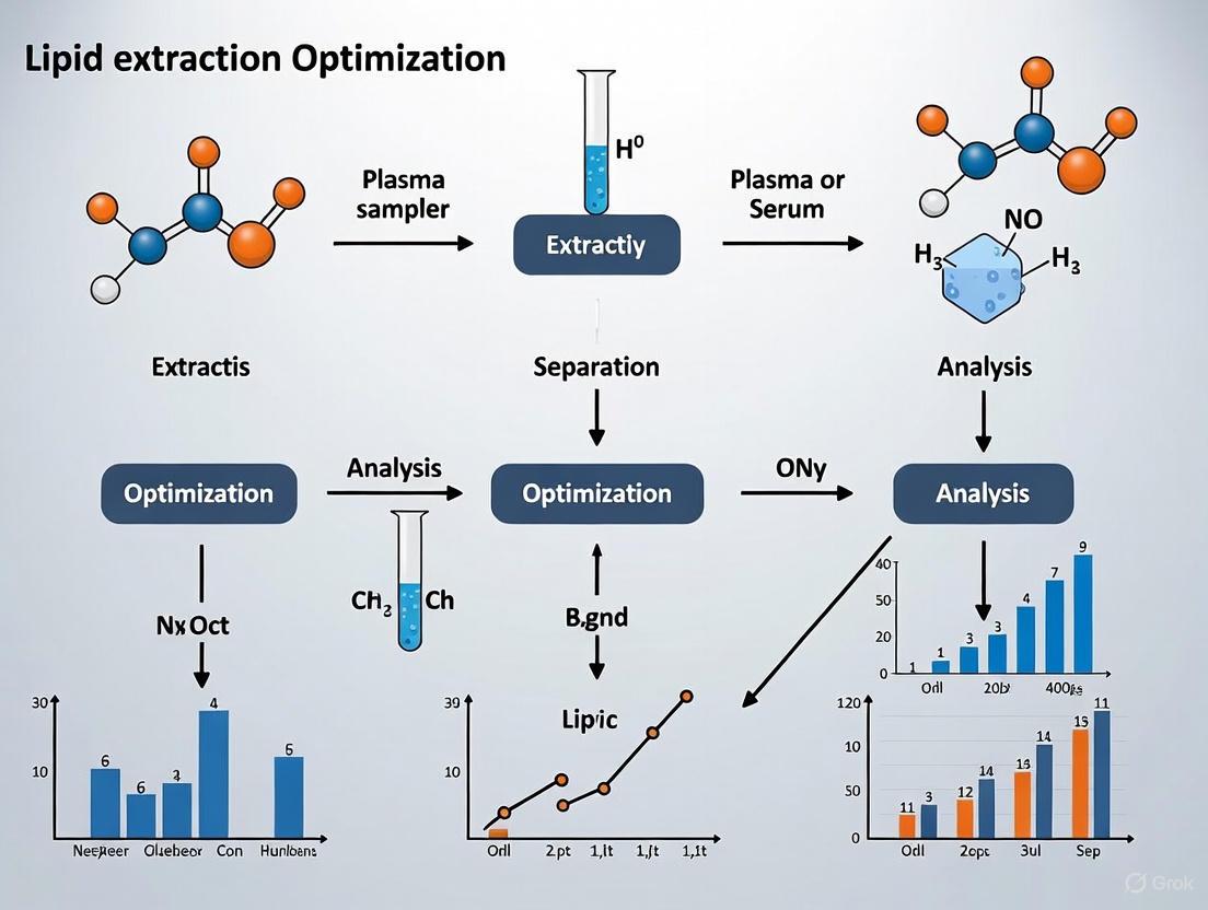
Workflow Visualization: The Lipid Extraction Bottleneck
The following diagram illustrates the lipidomics workflow, highlighting how challenges at the extraction stage create a bottleneck that affects all downstream data.
Overcoming the lipid extraction bottleneck requires a strategic shift from simply maximizing feature count to optimizing for biological relevance and data reliability [1]. This involves:
- Systematic Method Evaluation: Compare a few candidate methods (e.g., Folch, MTBE, MMC) using your specific plasma/serum samples and LC-MS platform. Evaluate them based on reproducibility, recovery of key lipid classes (using ISTDs), and their ability to reveal true biological variance [1].
- Rigorous Quality Control: Incorporate Extraction Quality Controls (EQCs)—pooled samples extracted with each batch—to monitor and correct for technical variability introduced during sample preparation [1].
- Embrace Automation: Where feasible, automate liquid handling steps to significantly improve reproducibility and throughput [2] [4].
- Context-Aware Protocol Selection: Base your choice on the research question. For high-throughput biomarker screening with LC-MS, a monophasic method like MMC may be optimal. For absolute quantification of specific polar lipids, a biphasic method like Folch with carefully chosen ISTDs might be necessary.
By adopting this framework, researchers can transform lipid extraction from a problematic bottleneck into a robust, reliable, and reproducible foundation for impactful lipidomics research.
FAQs: Fundamental Lipid Properties and Analysis
Q1: What are the key lipid classes found in human blood, and what are their primary functions? Blood lipids are broadly categorized into several classes, each with distinct functions. Triacylglycerols (Triglycerides) are the main form of energy storage and transport. They are carried in the core of lipoproteins like chylomicrons and VLDL to be delivered to adipose and muscle tissues [7] [8]. Phospholipids, which are amphipathic molecules with hydrophilic heads and hydrophobic tails, are the primary structural components of all cellular membranes and lipoprotein particles [7] [9]. Sterols, chiefly cholesterol, are another major class; they are essential for modulating membrane fluidity and serve as precursors for steroid hormones and bile acids [7]. Cholesterol is transported in the blood primarily by Low-Density Lipoproteins (LDL) and High-Density Lipoproteins (HDL) [10].
Q2: How does the chemical structure of a lipid influence its physical properties and biological role? The physical properties of lipids are largely dictated by their fatty acid components. Saturation is a key factor.
- Saturated fatty acids have no double bonds, allowing their straight chains to pack tightly. This results in higher melting points, making them solid at room temperature [11] [9].
- Unsaturated fatty acids contain one (monounsaturated) or more (polyunsaturated) double bonds, which are almost always in the cis configuration. This introduces kinks in the hydrocarbon chain, preventing tight packing and leading to lower melting points (e.g., liquid oils) [11] [9]. The degree of unsaturation directly affects membrane fluidity. Furthermore, the position of the double bonds (e.g., omega-3 or omega-6) is critical for their role as signaling molecule precursors [9].
Q3: Why is a biphasic solvent system like chloroform-methanol so effective for lipid extraction? Lipid extraction from biological matrices like plasma is a mass transfer process that must overcome lipid-protein and lipid-membrane associations. A biphasic system, such as the classic Folch method (Chloroform:MeOH, 2:1 v/v), is effective because the mixture serves two key roles [12]:
- The polar solvent (Methanol) disrupts hydrogen bonds and ion-dipole interactions between lipids and proteins, dissolving the more polar lipids and breaking up the matrix [13] [12].
- The non-polar solvent (Chloroform) then efficiently solubilizes the freed neutral lipids and facilitates the formation of a separate organic phase where all lipids partition, allowing for easy separation from water-soluble contaminants [13] [12].
Troubleshooting Guide: Lipid Extraction from Plasma/Serum
| Problem | Potential Cause | Solution |
|---|---|---|
| Low Lipid Yield | Inefficient cell membrane/protein disruption; incorrect solvent-to-sample ratio. | Incorporate a pretreatment step (e.g., bead beating, sonication) [12]. Ensure the Folch (2:1) or Bligh & Dyer (1:2:0.8, CHCl₃:MeOH:H₂O) ratios are meticulously followed [12]. |
| Poor Sample Cleanup (contamination with non-lipids) | Incomplete phase separation; inadequate washing of the organic phase. | Add a saline solution (e.g., 0.9% NaCl or KCl) to improve phase separation [7] [12]. Wash the collected organic (lower) phase with a theoretical upper phase (CHCl₃:MeOH:H₂O, 3:48:47) to remove water-soluble impurities [12]. |
| Oxidation of Unsaturated Lipids | Exposure to oxygen during extraction and storage. | Add an antioxidant, such as Butylated Hydroxytoluene (BHT), to the solvent mixtures [13]. Perform procedures under an inert nitrogen atmosphere and store lipid extracts at -80°C [14]. |
| Inconsistent LC-MS Results | Pre-analytical variables; solvent effects. | Standardize blood collection, processing time, and storage conditions (fast-freeze in liquid Nâ‚‚) [14]. Use mass spectrometry-grade solvents and ensure complete dryness and consistent reconstitution for LC-MS [14] [13]. |
Experimental Protocol: Chloroform-Free Total Lipid Extraction from Human Plasma
This protocol is adapted from modern sustainable lipidomics research [13] and provides an alternative to traditional chloroform-based methods.
Principle: This single-phase extraction uses Cyclopentyl Methyl Ether (CPME) as a greener alternative to chloroform, mixed with methanol and MTBE to efficiently extract a broad range of lipids from plasma by disrupting hydrophobic and electrostatic interactions.
Materials & Reagents:
- Reconstituted human plasma sample
- Ice-cold Methanol (MeOH)
- Ice-cold Methyl tert-butyl ether (MTBE)
- Cyclopentyl methyl ether (CPME)
- Butylated hydroxytoluene (BHT, 0.01% w/v in solvent mixtures)
- Isopropanol (i-PrOH)
- Water (Hâ‚‚O, ULC-MS grade)
- Centrifuge and microcentrifuge tubes
- Vortex mixer and rotary shaker
- Nitrogen evaporator
Procedure:
- Preparation: Spike all solvent mixtures with 0.01% BHT to prevent lipid oxidation.
- Protein Precipitation & Extraction: To 5 µL of human plasma in a microcentrifuge tube, add 180 µL of ice-cold MeOH. Vortex for 30 seconds.
- Solvent Addition: Add 600 µL of ice-cold MTBE and 300 µL of CPME. Vortex vigorously for 30 seconds.
- Incubation: Incubate the mixture on a rotary shaker for 60 minutes at 40 rpm and 4°C.
- Phase Separation: Add 150 µL of ice-cold H₂O to induce phase separation. Vortex for 30 seconds and shake for 10 minutes at 40 rpm and 4°C.
- Centrifugation: Centrifuge for 10 minutes at 1,000 × g and 4°C to achieve clear phase separation.
- Collection: Carefully collect the upper organic phase (approximately 540 µL) into a new tube.
- Concentration: Evaporate the organic solvent to dryness under a gentle stream of nitrogen.
- Reconstitution: Reconstitute the dried lipid extract in 50 µL of i-PrOH, shake for 15 minutes, and centrifuge briefly. The sample is now ready for downstream analysis (e.g., LC-MS).
Lipid Transport and Metabolism Pathways
Lipid Transport Pathway
Research Reagent Solutions
| Reagent / Material | Function in Lipid Analysis | Key Considerations |
|---|---|---|
| Chloroform (CHCl₃) | Classic non-polar solvent for biphasic extraction; dissolves neutral lipids [12]. | High toxicity and environmental hazard. Use in fume hood with proper PPE [13]. |
| Cyclopentyl Methyl Ether (CPME) | Greener alternative to chloroform; used in single-phase extraction [13]. | Lower health risk, good sustainability profile, and comparable extraction efficiency for many lipid classes [13]. |
| Methanol (MeOH) | Polar solvent that disrupts lipid-protein complexes and dissolves polar lipids [12]. | Essential component of most extraction mixtures. Miscible with water. |
| Butylated Hydroxytoluene (BHT) | Antioxidant added to solvent mixtures [13]. | Prevents oxidation of polyunsaturated fatty acids (PUFAs) during extraction and storage. |
| MTBE (Methyl tert-butyl ether) | Solvent used in single-phase and biphasic extraction protocols [13]. | Often combined with methanol. Forms the upper organic phase in the MTBE method [13]. |
| Ammonium Formate / Formic Acid | Mobile phase additives for LC-MS [13]. | Promotes protonation and improves ionization efficiency of lipids in mass spectrometry. |
Troubleshooting Guide: Common Lipid Extraction Issues in Plasma/Serum Research
Problem 1: Low Lipid Recovery from Plasma Samples
- Possible Cause: Inefficient disruption of lipoprotein complexes or incorrect solvent-to-sample ratio.
- Solution: Ensure adequate sample pre-treatment. For delipidation of plasma while preserving proteins, use a mixture of butanol and di-isopropyl ether (40/60, v/v). One volume of plasma is added to two volumes of this solvent mixture and rotated for 30 minutes [15].
- Prevention: Standardize the sample volume and always use the correct solvent proportions. For the classical Folch method, use a 2:1 (v/v) mixture of chloroform to methanol [16].
Problem 2: Persistent Lipemic Interference in Biochemical Assays
- Possible Cause: Incomplete removal of lipoproteins (chylomicrons) leads to turbidity, which affects spectrophotometric measurements [17].
- Solution: Employ high-speed centrifugation. Centrifuging serum/plasma samples at 10,000×g for 15 minutes effectively removes the lipid layer, and the infranatant (aqueous phase) can be collected for analysis [17].
- Prevention: For clinical samples, ensure patient fasting when required. Avoid using chemical agents like LipoClear or 1,1,2-trichlorotrifluoroethane for lipemia removal, as they can interfere with the measurement of certain parameters like total protein, albumin, and calcium [17].
Problem 3: Solvent Toxicity and Environmental Concerns
- Possible Cause: Use of hazardous solvents like chloroform.
- Solution: Investigate safer solvent alternatives. Some studies show that toluene can be a valid substitute for chloroform in specific extraction protocols for microbial lipids [18]. Ethanol is another potential, though sometimes less effective, substitute for methanol [18].
- Prevention: Transition to green solvent-based methods where possible, such as supercritical CO2 extraction (SCE), which is non-toxic and leaves no residue [16] [19].
Problem 4: Emulsification During Liquid-Liquid Extraction
- Possible Cause: Over-vigorous mixing or the nature of the biological sample can create stable emulsions, trapping lipids and preventing phase separation [20].
- Solution: Use controlled, gentle agitation during extraction. If an emulsion forms, low-speed centrifugation can often break it. Bead beating can also be optimized to disrupt cells without excessive emulsion formation [18].
- Prevention: Ensure the correct pH and ionic strength. Adding salts like NaCl or KCl, as in the Folch wash step, can facilitate cleaner phase separation [16].
Problem 5: Inconsistent Results Between Different Sample Types
- Possible Cause: The efficiency of a single extraction protocol can vary significantly based on the cell wall structure and lipid composition of the source material [16] [18].
- Solution: Optimize the extraction method for each specific biological matrix (e.g., plasma, yeast, microalgae). This may require testing different solvent polarities and cell disruption methods [21] [18].
- Prevention: Do not assume a universal method. For high-value applications, use a hybrid AI-powered simulation approach that combines mechanistic modeling and machine learning to predict the optimal extraction process for a specific sample and desired outcome [20].
Frequently Asked Questions (FAQs)
Q1: What is the fundamental principle behind selecting solvents for lipid extraction? The core principle is selectivity based on solubility, governed by the partition coefficient (K). A solvent mixture, typically comprising a polar (e.g., methanol) and a non-polar (e.g., chloroform) solvent, is used because of the chemical diversity of lipids. The polar solvent disrupts protein-lipid complexes and dissolves polar lipids, while the non-polar solvent dissolves neutral lipids [16] [22]. A higher partition coefficient for a lipid in the organic solvent phase leads to more efficient extraction.
Q2: Why are chloroform and methanol so commonly used together? This combination is effective because it covers a broad range of lipid polarities. Chloroform (non-polar) efficiently solubilizes neutral lipids like triacylglycerols, while methanol (polar) disrupts hydrogen bonding and electrostatic interactions between polar lipids (like phospholipids) and membranes or proteins. The classic Folch method uses a 2:1 chloroform-methanol ratio, and the Bligh and Dyer method is a modification of this for smaller sample sizes [16].
Q3: How does solvent polarity directly affect the yield and profile of extracted lipids? Solvent polarity directly determines which lipid species are solubilized. Non-polar solvents like hexane and chloroform yield higher total amounts of neutral lipids, which are rich in saturated fatty acids. In contrast, polar solvents like methanol are more effective at extracting polar lipids, which often contain more unsaturated fatty acids [21]. Therefore, the choice of solvent dictates the resulting fatty acid methyl esters (FAMEs) composition and subsequent biodiesel properties if used for biofuel [21].
Q4: What are the key considerations for delipidating plasma without denaturing proteins? The primary goal is to extract lipids while keeping proteins in a soluble, native state in the aqueous phase. A specific solvent system like butanol/di-isopropyl ether (40/60, v/v) is recommended for this purpose. Butanol helps to dissociate lipids from proteins without causing irreversible denaturation, allowing the delipidated proteins to be recovered from the aqueous phase [15].
Q5: Are there effective and safer alternatives to chlorinated solvents? Yes, research into greener alternatives is ongoing. Supercritical CO2 is a prominent non-toxic, non-flammable alternative, especially for neutral lipids [16] [19]. Other options include solvent substitution, such as using toluene instead of chloroform or ethanol instead of methanol, though their efficacy can be species-dependent and requires validation for each application [18].
Quantitative Data: Solvent Polarity and Extraction Performance
The following table summarizes quantitative findings on how solvent polarity influences lipid extraction efficiency and profile, based on experimental data.
Table 1: Impact of Solvent Polarity on Microalgal Lipid Yield and Biodiesel Properties [21]
| Solvent Type | Example Solvents | Total Lipid Yield (mg/g microalgae) | Total Saturated Fatty Acids (SFAs) | Total Unsaturated Fatty Acids (UFAs) | Key Biodiesel Property (Cetane Number) |
|---|---|---|---|---|---|
| Non-Polar | Chloroform, Hexane | 94.33 - 100.01 | 61.53% (Chloroform) | 38.47% (Chloroform) | Higher |
| Polar | Methanol, Acetone | 40.12 - 86.91 | 38.85% (Methanol) | 61.15% (Methanol) | Lower |
Table 2: Comparison of Common Lipid Extraction Methods for Biological Samples
| Extraction Method | Solvent System | Typical Application | Key Advantages | Key Limitations |
|---|---|---|---|---|
| Folch Method [16] | Chloroform/Methanol (2:1, v/v) | General lipidomics; animal tissues | High extraction efficiency; considered a gold standard | Uses toxic chloroform; requires a purification wash step |
| Bligh & Dyer Method [16] | Chloroform/Methanol/Water (1:2:0.8, v/v) | High-throughput screening; animal tissues with high water content | Rapid; adapted for smaller sample sizes | Less effective for samples with very high water content |
| Butanol/Diisopropyl Ether [15] | Butanol/Diisopropyl Ether (40:60, v/v) | Plasma/Serum delipidation | Preserves protein integrity; effective for clinical samples | May not extract all lipid classes with equal efficiency |
| High-Speed Centrifugation [17] | N/A (Physical separation) | Removing lipemia from serum/plasma for clinical assays | No chemical interference; simple and practicable | Only removes lipoproteins, does not extract lipids for analysis |
Experimental Protocols for Key Methods
Protocol 1: Folch Method for Total Lipid Extraction from Tissues [16]
- Homogenize the tissue sample in a 2:1 (v/v) mixture of chloroform-methanol (e.g., 20 mL solvent per 1 g of tissue).
- Filter the homogenate to remove solid debris.
- Add a salt solution (e.g., 0.05 N NaCl or KCl) at a ratio of 0.2 volumes of salt solution to 1 volume of the chloroform-methanol filtrate. This promotes phase separation.
- Mix thoroughly and let the phases separate. The lower organic phase (chloroform) contains the extracted lipids.
- Recover the lower chloroform phase carefully, avoiding the interface.
- Evaporate the chloroform under a stream of nitrogen or using a rotary evaporator to obtain the purified lipid extract.
Protocol 2: Delipidation of Plasma/Serum with Protein Preservation [15]
- To one volume of serum or plasma (containing 0.1 mg/ml EDTA as an anticoagulant/antioxidant), add two volumes of a butanol/di-isopropyl ether (40/60, v/v) mixture.
- Tightly close the vials and fasten them on a mechanical rotator. Provide end-over-end rotation at 30 rpm for 30 minutes.
- Centrifuge the mixture at low speed (approx. 2,000×g) for 2 minutes to achieve clear phase separation.
- The aqueous phase (lower phase) contains the delipidated proteins. Remove it carefully using a needle and syringe or pipette.
- To remove traces of butanol, the aqueous phase can be washed with two volumes of pure di-isopropyl ether.
- Residual solvent in the aqueous protein solution can be removed by aspiration under vacuum at 37°C for a few minutes.
Protocol 3: Removing Lipemia via High-Speed Centrifugation [17]
- Homogenize the lipemic serum or plasma sample using a vortex mixer.
- Pipette 1 mL of the sample into a microcentrifuge tube.
- Centrifuge at 10,000×g for 15 minutes.
- After centrifugation, the lipid layer will form a compact upper phase. Carefully collect the clarified infranatant (the aqueous phase at the bottom of the tube) using a fine-needle syringe, taking care not to aspirate the lipid layer.
- The infranatant is now suitable for downstream biochemical analysis.
Workflow and Relationship Diagrams
Decision Workflow for Lipid Handling in Plasma/Serum Research
How Solvent Polarity Targets Different Lipid Classes
The Scientist's Toolkit: Essential Research Reagents & Materials
Table 3: Key Reagents and Materials for Lipid Extraction from Plasma/Serum
| Item | Function/Application | Example Use Case |
|---|---|---|
| Chloroform | Non-polar solvent for dissolving neutral lipids [16] | Folch and Bligh & Dyer methods for total lipid extraction. |
| Methanol | Polar solvent for disrupting lipid-protein complexes and dissolving polar lipids [16] | Used in combination with chloroform in classical methods. |
| Butanol/Diisopropyl Ether Mix | Solvent system for delipidation with protein preservation [15] | Extracting lipids from plasma or serum without denaturing apolipoproteins. |
| Ethylenediamine Tetraacetate (EDTA) | Chelating agent that binds metal ions to prevent oxidation [15] | Added to plasma/serum samples before delipidation to preserve sample integrity. |
| Glass Beads (425-600 μm) | Mechanical means for cell disruption to enhance solvent access [18] | Bead beating for efficient lipid extraction from yeast or other tough cell walls. |
| High-Speed Centrifuge | Physical separation of lipid layers from aqueous samples [17] | Removing lipemia from clinical serum/plasma samples prior to analysis. |
| ML349 | ML349, MF:C23H22N2O4S2, MW:454.6 g/mol | Chemical Reagent |
| WR99210 | WR99210, CAS:30711-93-4; 30737-44-1; 47326-86-3, MF:C14H18Cl3N5O2, MW:394.7 g/mol | Chemical Reagent |
Troubleshooting Guide: FAQs for Lipid Extraction from Plasma Serum Samples
FAQ 1: How can I prevent the co-extraction of non-lipid contaminants that interfere with my LC-MS analysis?
Issue: Non-lipid contaminants, such as proteins and sugars, are co-extracted, leading to ion suppression in MS, column fouling in HPLC, and reduced analytical sensitivity [23] [12].
Solution & Troubleshooting Steps:
- Implement a Robust Wash Step: The classical Folch and Bligh & Dyer methods include a wash step with a salt solution (e.g., KCl, NaCl, or water) to partition non-lipid polar contaminants into the aqueous-methanol phase while lipids remain in the organic phase [12]. Ensure the extraction mixture reaches the correct final solvent-to-sample ratio for proper phase separation.
- Adopt the BUME Method: This automated, chloroform-free method is designed to minimize contamination. It uses a two-phase system with 1% acetic acid, which helps purify the lipid extract. The lipids are recovered from the upper heptane-ethyl acetate phase, reducing the risk of aspirating the protein-rich interphase [23].
- Verify Phase Separation: After adding the wash buffer, allow the mixture to separate completely. Carefully aspirate the upper aqueous phase without disturbing the interphase or the lower organic phase. If the interphase is thick and protein-rich, it may be better to sacrifice a small amount of the organic phase to avoid contamination [23].
FAQ 2: What is the most effective way to ensure complete cell wall disruption for maximal lipid yield from complex samples?
Issue: Incomplete disruption of cells, such as those with tough walls in microbial samples, results in low and inconsistent lipid yields because solvents cannot access intracellular lipids [12] [24].
Solution & Troubleshooting Steps:
- Choose a Physical Disruption Method: For samples with robust cell walls (e.g., plants, fungi, microalgae), mechanical methods are highly effective.
- Bead Milling: Agitate the sample at high speed with beads. Use 0.5 mm glass beads for yeast and 3–7 mm stainless steel or tungsten carbide beads for plant and fungal tissues [25].
- Rotor-Stator Homogenization: Effective for animal and plant tissues, using mechanical shearing for disruption in seconds [25].
- High-Pressure Homogenization (HPH): Forces a cell suspension through a narrow valve at high pressure (e.g., 10-300 MPa). Pressures above 100 MPa are often needed for robust microalgal cells [24].
- Sonication: Uses high-frequency sound waves to create cavitation bubbles, whose collapse generates shear forces that break open cells [24].
- Optimize Pretreatment for Your Sample: The optimal method depends on the sample's cell wall composition. A combination of treatments may be necessary for maximum yield [12].
- For Plasma/Serum: Note that mechanical disruption is typically not required for plasma or serum samples, as lipids are already compartmentalized within lipoproteins. The primary step here is the efficient disruption of these lipoprotein complexes using a solvent like methanol in the initial one-phase extraction [23].
FAQ 3: My lipid recoveries are low and variable. How can I improve the efficiency and reproducibility of my extraction?
Issue: Low recovery can stem from inefficient solvent systems, incomplete sample mixing, or failure to optimize the protocol for your specific sample matrix.
Solution & Troubleshooting Steps:
- Select the Appropriate Solvent System: The solvent must be capable of both disrupting lipid-protein complexes and dissolving a wide range of lipids. A mixture of polar and non-polar solvents is essential [12].
- Ensure Complete Homogenization: During the initial one-phase extraction, ensure the sample and solvents are mixed thoroughly to form a fine suspension. This is critical for efficient and reproducible lipid solubilization [23].
- Automate the Process: Manual liquid handling can be a significant source of variability. Implementing an automated extraction protocol on a liquid handling robot, as demonstrated with the BUME method, can greatly improve reproducibility and throughput [23].
Table 1: Comparison of Key Lipid Extraction Method Performance
| Method | Key Solvents | Extraction Efficiency (vs. Reference) | Key Advantages | Reported Challenges |
|---|---|---|---|---|
| BUME [23] | Butanol:MeOH, Heptane:Ethyl Acetate | Similar or better for major lipid classes [23] | Chloroform-free, automated, upper-phase lipid recovery, 96 samples/60 min [23] | Requires optimization for robot parameters [23] |
| Folch [12] | CHCl₃:MeOH (2:1) | Reference Method [12] | High efficiency for wide hydrophobicity range [12] | Chloroform use, lower phase recovery, manual, tedious [23] [12] |
| Bligh & Dyer [12] | CHCl₃:MeOH (1:2) | Reference Method [12] | Adapted for high water content samples [12] | Chloroform use, prone to contamination, manual [23] [12] |
| MTBE [23] | MTBE:MeOH | Similar lipid profiles [23] | Upper-phase lipid recovery [23] | High solvent-to-sample ratio, challenging automation [23] |
Table 2: Performance of Cell Disruption Methods for Intracellular Lipid Extraction
| Method | Mechanism of Action | Typical Application Scale | Reported Effectiveness | Key Limitations |
|---|---|---|---|---|
| High-Pressure Homogenization [24] | High shear force, turbulence, and cavitation from pressure drop [24] | Industrial / Large-scale [24] | Effective for tough-walled microalgae (e.g., Nannochloropsis); protein release from yeast: 50 µg/g [24] | High energy consumption; cell age and wall composition affect required pressure [24] |
| Ultrasonication [24] | Cavitation from high-frequency sound waves [24] | Lab / Small to medium-scale [24] | Effective for a wide range of cell types; suitable for heat-sensitive compounds [24] | Potential for local heating; scale-up can be challenging [24] |
| Bead Milling [25] | Shearing and crushing from high-speed agitation with beads [25] | Lab / Small-scale [25] | Thorough disruption for bacteria, yeast, and plant tissues [25] | Optimization of bead size and material is critical; can generate heat [25] |
| Rotor-Stator Homogenization [25] | Mechanical shearing from a high-speed rotor [25] | Lab / Small-scale [25] | Rapid disruption (5-90 sec) of animal and plant tissues [25] | Not ideal for very small sample volumes; foaming can occur [25] |
Detailed Experimental Protocols
Protocol 1: The Automated BUME Extraction Method for Plasma/Serum
This protocol is adapted from Löfgren et al. for a fully automated, high-throughput, and chloroform-free lipid extraction from plasma or serum samples [23].
1. Reagents and Materials:
- BUME Mixture (Solvent 1): Butanol:Methanol (3:1, v/v)
- Solvent 2: Heptane:Ethyl Acetate (3:1, v/v)
- Acetic Acid Solution: 1% (v/v) acetic acid in water
- Plasma/Serum Samples
- Automated Liquid Handling Robot (e.g., Velocity 11 Bravo) equipped with a 96-well head
- 96-well glass vial racks and 1.2 ml glass vials
2. Procedure:
- Sample Loading: Pipette 10–100 µl of plasma/serum into individual glass vials in a 96-well rack [23].
- Initial One-Phase Extraction: Add 300 µl of the BUME Mixture (Solvent 1) to each sample. The robot mixes the contents to form a fine suspension, ensuring efficient dissolution of lipoproteins and solubilization of lipids. This step is critical for complete initial extraction [23].
- Secondary Two-Phase Extraction: Add 300 µl of Solvent 2 (Heptane:Ethyl Acetate) to the mixture, followed by 300 µl of 1% Acetic Acid buffer. The robot mixes the contents. A two-phase system will form spontaneously without centrifugation. The lipids partition into the upper organic phase (heptane/ethyl acetate), while non-lipid contaminants partition into the lower aqueous phase [23].
- Recovery of Lipid Extract: The robot automatically aspirates the upper organic phase containing the purified lipids, ready for downstream analysis like LC-MS [23].
3. Critical Notes:
- The method has demonstrated linear lipid recoveries across a plasma volume of 10–100 µl [23].
- Robot parameters (aspiration/dispensing speed and position) must be optimized for robustness [23].
Protocol 2: High-Pressure Homogenization for Cell Disruption
This protocol describes the use of HPH for disrupting microbial cells to facilitate subsequent lipid solvent extraction [24].
1. Reagents and Materials:
- Microbial cell biomass (e.g., microalgal paste)
- Suitable aqueous buffer (e.g., phosphate buffer)
- High-Pressure Homogenizer (e.g., from Avestin, GEA Niro Soavi)
- Cooling system
2. Procedure:
- Sample Preparation: Prepare a concentrated suspension of microbial cells in an appropriate buffer.
- Homogenization: Feed the cell suspension into the homogenizer. The pressure and number of passes must be optimized for the specific cell type.
- For the microalgae Porphyridium cruentum, a pressure above 100 MPa was critical for effective disruption and release of the pigment B-Phycoerythrin [24].
- For the microalgae Parachlorella kessleri, increasing passes at 120 MPa released protein concentrations up to 3656 mg/L [24].
- For Saccharomyces cerevisiae yeast, homogenization at 80 MPa was effective for protein extraction [24].
- Collection: Collect the homogenate, which contains the disrupted cells and released intracellular components, including lipids. This homogenate is now ready for lipid extraction using a solvent-based method like BUME or Folch.
3. Critical Notes:
- The process can generate heat, so using a cooling jacket is advisable to protect heat-sensitive compounds.
- Cell wall composition and cell age significantly impact the required disruption pressure [24].
Experimental Workflow Visualization
Troubleshooting Workflow for Common Pitfalls
BUME Method Lipid Extraction Workflow
The Scientist's Toolkit: Key Research Reagent Solutions
Table 3: Essential Reagents and Materials for Optimized Lipid Extraction
| Reagent/Material | Function in Lipid Extraction | Example Use Case |
|---|---|---|
| Butanol:MeOH (BUME) Mixture [23] | Initial one-phase extraction solvent; disrupts protein-lipid complexes and solubilizes a wide range of lipids. | Primary solvent in the automated BUME method for plasma/serum [23]. |
| Heptane:Ethyl Acetate [23] | Secondary two-phase extraction solvent; forms the upper organic phase for easy lipid recovery and reduced contamination. | Used in the BUME method after the initial one-phase extraction [23]. |
| Chloroform:MeOH [12] | Classical solvent mixture for efficient total lipid extraction via Folch or Bligh & Dyer methods. | Reference method for lipid extraction efficiency [12]. |
| 1% Acetic Acid [23] | Aqueous buffer in two-phase systems; facilitates phase separation and purifies lipid extract by partitioning non-lipids into the aqueous phase. | Wash buffer in the BUME method [23]. |
| High-Pressure Homogenizer [24] | Physical disruption equipment for breaking tough cell walls to release intracellular lipids prior to solvent extraction. | Disrupting microalgae (e.g., Nannochloropsis) and yeast cells [24]. |
| Bead Mill [25] | Physical disruption equipment using beads for shearing and crushing cells. | Disruption of bacterial, yeast, and plant tissues in a laboratory setting [25]. |
| J30-8 | J30-8, MF:C17H9ClFN3O2S, MW:373.8 g/mol | Chemical Reagent |
| PF-06424439 | PF-06424439, MF:C22H26ClN7O, MW:439.9 g/mol | Chemical Reagent |
The accurate analysis of lipids from biological samples is a critical step in lipidomics and biomedical research. The methods established by Folch et al. and Bligh & Dyer remain the gold standards for lipid extraction over six decades after their development. These biphasic solvent systems, utilizing chloroform, methanol, and water, are designed to efficiently isolate a broad range of lipid classes while removing non-lipid contaminants. Within the context of optimizing lipid extraction from plasma and serum samples, understanding the specific parameters, advantages, and limitations of these foundational protocols is essential for obtaining reliable and reproducible data in drug development and clinical research.
Detailed Experimental Protocols
The Folch Method
The Folch method was originally developed for the extraction of lipids from brain tissue and uses a chloroform:methanol:water system in a ratio of 8:4:3 (v/v/v) [26] [12].
Detailed Protocol:
- Homogenization: Homogenize the plasma or tissue sample in a 2:1 (v/v) mixture of chloroform-methanol. The classical sample-to-solvent ratio is 1:20 [26] [27]. For instance, for 1 mL of plasma, add 20 mL of the chloroform-methanol (2:1) mixture.
- Partitioning: Add a volume of water or a salt solution (e.g., 0.9% NaCl or 0.003 N CaClâ‚‚) equal to one-quarter the volume of the chloroform-methanol mixture used. This shifts the solvent system to the biphasic state.
- Centrifugation: Centrifuge the mixture to separate the phases. The lower, dense chloroform-rich phase contains the extracted lipids. The upper phase is methanol-water rich and contains non-lipid contaminants. An interface of denatured proteins is often present.
- Recovery: Carefully aspirate and discard the upper phase. The lower organic phase can be recovered by siphoning or pipetting through the protein disk.
- Washing (Optional): To further purify the lipid extract, "wash" the lower phase by adding a fresh volume of theoretical upper phase (chloroform:methanol:water, 3:48:47) and repeating the centrifugation and separation [28].
The Bligh & Dyer Method
The Bligh & Dyer method was developed as a rapid, microscale extraction for fish muscle, using a chloroform:methanol:water system in a ratio of 2:2:1.8 (v/v/v) [26] [12]. It uses less solvent than the Folch method.
Detailed Protocol for Liquid Samples (e.g., Plasma):
- Initial Monophasic System: To 1 mL of plasma, add 3.75 mL of a chloroform/methanol mixture in a 1:2 ratio [29]. Vortex vigorously for 10-15 minutes.
- Inducing Biphasic System: Add 1.25 mL of chloroform and mix for 1 minute.
- Final Separation: Add 1.25 mL of water and mix for another minute.
- Centrifugation and Recovery: Centrifuge to separate the phases. The lower, dense chloroform-rich phase contains the lipids and is recovered as described in the Folch method [29].
Comparative Analysis and Optimization for Plasma/Serum
Quantitative Method Comparison
The table below summarizes the key characteristics of the Folch and Bligh & Dyer methods, particularly in the context of plasma-based lipidomics.
Table 1: Comparison of Folch and Bligh & Dyer Lipid Extraction Methods
| Feature | Folch Method | Bligh & Dyer Method |
|---|---|---|
| Original Solvent Ratio (CHCl₃:MeOH:H₂O) | 8:4:3 (v/v/v) [26] | 2:2:1.8 (v/v/v) [26] |
| Classical Sample-to-Solvent Ratio | 1:20 [26] [27] | 1:3 (does not account for tissue water) [26] |
| Organic Layer Position | Bottom (higher density) [26] | Bottom (higher density) [26] |
| Recommended Plasma Sample Ratio | 1:20 (v/v) [26] [27] | 1:20 (v/v) [26] [27] |
| Extraction Efficiency for High-Lipid Samples | Accurate for a broad range [30] | Underestimates lipid content in samples >2% lipid [29] [30] |
| Multi-Omic Capability (Plasma) | Suitable for lipidomics and metabolomics from organic and aqueous phases [26] [27] | Suitable for lipidomics and metabolomics from organic and aqueous phases [26] [27] |
Optimization for Plasma and Serum Research
Optimizing the sample-to-solvent ratio is vital for comprehensive lipid coverage in untargeted lipidomics.
- Sample-to-Solvent Ratio: A systematic evaluation for human plasma demonstrated that a 1:20 (v/v) plasma-to-total-solvent ratio yielded the highest peak areas for a diverse range of lipid and metabolite species for both Folch and Bligh & Dyer methods. Decreasing the ratio (e.g., to 1:4) or increasing it (e.g., to 1:100) resulted in lower recoveries [26] [27].
- Handling and Storage: To prevent artefact formation and lipid degradation, samples should be frozen immediately at -20°C or lower in an atmosphere of nitrogen. Tissues should be homogenized and extracted with solvent at the lowest temperature practicable without being allowed to thaw [28].
- Modifications for Specific Lipids: The recovery of acidic phospholipids can be improved by replacing water with 1M NaCl or adding 0.5% acetic acid (v/v) to the water phase during partitioning [29].
Troubleshooting Guides & FAQs
Frequently Asked Questions
Q1: Why does the Bligh & Dyer method underestimate lipid content in fatty samples? The Bligh & Dyer method was designed for tissues with low lipid content. In samples with more than 2% lipid, the solvent volumes become insufficient for complete extraction, leading to a significant underestimation of total lipid content that worsens with increasing lipid levels [29] [30]. For fatty tissues, the Folch method with its higher solvent ratio is more reliable [30].
Q2: My lipid extract contains a large amount of free fatty acids and lysophospholipids. What went wrong? This is a classic sign of lipolytic degradation. It indicates that lipids were exposed to active lipases, likely due to improper sample handling. This can occur if tissues were not frozen rapidly after collection, were subjected to slow thawing, or if extraction was not performed promptly on frozen tissue [28]. Ensure rapid freezing in liquid nitrogen and homogenize while the sample is still frozen.
Q3: What is a key practical difference when handling the organic phase between these methods and the newer Matyash (MTBE) method? In both Folch and Bligh & Dyer methods, the chloroform-rich organic phase is denser than water and forms the lower layer [26]. This is the opposite of the Matyash method, which uses methyl tert-butyl ether (MTBE), where the organic phase is less dense and forms the upper layer, making it easier to collect [26] [31].
Q4: Can I use these extraction methods for a multi-omics approach? Yes. A significant advantage of these biphasic extractions is the ability to analyze both the organic phase (for lipidomics) and the aqueous phase (for metabolomics) from a single sample preparation. This increases analyte coverage and provides a more comprehensive understanding of the biological system [26] [27].
Troubleshooting Common Issues
Table 2: Troubleshooting Common Problems in Lipid Extraction
| Problem | Potential Cause | Solution |
|---|---|---|
| Low Lipid Yield | Incorrect solvent ratios; insufficient sample disruption; sample-too-solvent ratio too high. | Precisely measure solvents; optimize homogenization; use a 1:20 (v/v) sample-to-solvent ratio for plasma [26]. |
| Lipid Degradation (High FFAs) | Poor sample storage/handling; lipase activity. | Freeze samples immediately in liquid nitrogen; store at -80°C; add antioxidants to solvents; homogenize frozen tissue [28]. |
| Contamination with Non-Lipid Material | Incomplete phase separation; aqueous phase carried over. | Ensure final solvent ratios are correct for biphasic system; be careful when collecting the organic layer [12] [28]. |
| Poor Recovery of Acidic Phospholipids | Ionic interactions with denatured proteins at the interface. | Acidify the water phase with dilute HCl or use 1M NaCl for partitioning [29]. |
Workflow and Decision Pathways
The following workflow diagram outlines the key decision points and steps for selecting and performing these lipid extraction methods.
Lipid Extraction Decision Workflow
The Scientist's Toolkit: Essential Research Reagents
Table 3: Essential Reagents and Materials for Lipid Extraction
| Item | Function / Purpose |
|---|---|
| Chloroform | Primary non-polar solvent; dissolves neutral lipids and forms the dense organic phase [12]. |
| Methanol | Polar solvent; disrupts lipid-protein complexes and hydrogen bonding; deactivates lipolytic enzymes [26] [12]. |
| Water (HPLC/MS Grade) | Used to induce biphasic phase separation and partition non-lipid contaminants into the upper aqueous phase [26]. |
| Salt Solutions (e.g., 1M NaCl, KCl) | Added during partitioning to improve recovery of specific lipid classes (e.g., acidic phospholipids) by altering ionic strength [29] [31]. |
| Glass Tubes with Teflon-lined Caps | Prevents solvent evaporation and leaching of contaminants from plastic [28]. |
| Antioxidants (e.g., BHT) | Added to solvents to prevent autoxidation of polyunsaturated fatty acids during extraction and storage [28]. |
| Inert Atmosphere (Nâ‚‚ Gas) | Used to store tissue samples and final lipid extracts to prevent oxidation [28]. |
| KU004 | KU004, MF:C29H27ClFN4O2P, MW:549.0 g/mol |
| P2X7-IN-2 | P2X7-IN-2, MF:C22H21F4N3O2, MW:435.4 g/mol |
A Practical Guide to Lipid Extraction Protocols for Blood Samples
Troubleshooting Guides
Guide 1: Poor Lipid Recovery or Low Extraction Yield
Problem: Inconsistent or lower-than-expected recovery of lipids from plasma samples.
Solutions:
- Cause 1: Incomplete Phase Separation
- Solution: Ensure precise solvent ratios. For the Matyash method, use MTBE/methanol/water in a ratio of 10/3/2.5 (v/v/v). For Folch, use chloroform/methanol 2:1 (v/v) with 0.2 volumes of water or saline. Centrifuge at 2000 × g for 10 minutes at a consistent temperature (20°C) to achieve complete separation [32] [33].
- Prevention: Allow the mixture to settle in a stable, vibration-free environment before and after centrifugation.
Cause 2: Improfficient Cell Disruption
- Solution: For complex samples, implement mechanical pretreatments such as bead beating or high-pressure homogenization to disrupt cell walls and facilitate solvent penetration. Osmotic shock has been shown to increase lipid yield by 2.8-fold in some microorganisms [16].
- Prevention: For tough tissues, consider a combination of pretreatments (e.g., enzymatic disruption followed by mechanical homogenization) [16].
Cause 3: Solvent Evaporation or Handling Errors
- Solution: Always use glass syringes or positive displacement pipettes to accurately transfer the organic phase. When using the Folch method, carefully collect the lower chloroform phase. For Matyash, take the upper MTBE phase [32] [28].
- Prevention: Perform extractions in a temperature-controlled environment to minimize solvent evaporation.
Guide 2: Formation of Persistent Emulsions
Problem: A stable emulsion forms at the interface, preventing clean phase separation.
Solutions:
- Cause 1: Over-Homogenization or Vigorous Mixing
- Solution: If an emulsion forms, increase centrifugation time to 15-20 minutes or briefly recentrifuge. Alternatively, add a small volume of saturated NaCl solution (5-10% of total volume) to break the emulsion [28].
- Prevention: Use gentle inversion mixing instead of vortexing, especially for protein-rich samples like plasma.
- Cause 2: Sample-Specific Interferences
- Solution: For plasma samples with high lipoprotein or protein content, consider a slight modification of the solvent-to-sample ratio. A "modified Matyash" system (MTBE/methanol/water, 2.6/2.0/2.4, v/v/v) has shown improved performance in complex samples [33].
- Prevention: Dilute viscous plasma samples with a minimal amount of saline before extraction.
Guide 3: Lipid Degradation or Artefact Formation
Problem: Detection of elevated levels of free fatty acids, lysophospholipids, or other lipid degradation products.
Solutions:
- Cause 1: Inadequate Inactivation of Lipolytic Enzymes
- Solution: Ensure plasma is processed and frozen rapidly after collection. Homogenize samples in the presence of extraction solvents at the lowest practical temperature to denature enzymes [28].
- Prevention: Add lipase inhibitors during plasma collection if profiling sensitive lipid classes. Store plasma at -80°C and limit freeze-thaw cycles [32] [28].
- Cause 2: Sample Oxidation
- Solution: Include antioxidants like butylated hydroxytoluene (BHT) (50-100 μM) in your extraction solvents, especially for polyunsaturated fatty acid analysis [28] [34].
- Prevention: Perform extractions under an inert nitrogen atmosphere, and store lipid extracts in apolar solvents like chloroform or hexane at -80°C under nitrogen [28].
Frequently Asked Questions (FAQs)
Q1: Which method, Folch or Matyash, provides superior lipid recovery for LC-MS-based lipidomics of plasma?
While both methods are effective for global lipidomics, comparative studies show nuanced differences. The single-phase methanol/1-butanol method demonstrated comparable effectiveness to Folch and Matyash for most lipid classes and was more effective in extracting polar lipids [32]. A "modified Matyash" method (MTBE/methanol/water, 2.6/2.0/2.4) showed a 4-20% higher number of detectable peaks and putatively annotated metabolites compared to the Bligh and Dyer (a Folch-derived method) and original Matyash methods in serum and other samples [33]. The choice depends on your target lipidome; for polar lipids, the single-phase method may be better, while the modified Matyash offers broad coverage.
Q2: Is it necessary to deactivate enzymes in plasma samples prior to lipid extraction?
Yes, this is a critical step. Lipolytic enzymes remain active even at low temperatures and can rapidly alter the lipid profile. Appreciable hydrolysis of phospholipids has been observed in tissues and plasma stored at -20°C and even during extraction if enzymes are not denatured [28]. The best practice is to freeze plasma rapidly at -80°C immediately after collection and to homogenize or mix the thawed plasma directly with the organic solvents, which themselves deactivate many enzymes [28].
Q3: What is the key practical advantage of the MTBE-based (Matyash) method over the chloroform-based (Folch) method?
The primary advantage is safety and convenience. MTBE is less toxic and less dense than chloroform. Its lower density means the lipid-containing organic phase forms the upper layer after phase separation, making it much easier and safer to collect without risk of disturbing the protein interphase or the lower aqueous phase [32] [33]. Furthermore, disposing of MTBE is considered more environmentally friendly.
Q4: How can I improve the reproducibility of my lipid extraction protocol?
Key strategies include:
- Automation: Using automated liquid handlers for solvent addition and phase collection can significantly reduce human error [35] [33].
- Internal Standards: Add a stable isotope-labeled internal standard (SILIS) mixture at the very beginning of extraction, before adding solvents, to correct for variations in extraction efficiency and ionization [32] [36].
- Standardized Washing: If washing the organic phase (e.g., with water or salt solution in the Folch method), keep the volume and ionic concentration consistent. The single-phase Alshehry method eliminates this step, which can reduce variability [32].
Q5: Can I use these methods for very small-volume plasma samples, such as from mice?
Yes, these methods can be successfully miniaturized. The Folch and Matyash methods have been reliably used with plasma volumes as low as 10-50 μL. A study successfully performed the single-phase Alshehry extraction using only 10 μL of pooled human plasma [32]. The key is to scale down the solvent volumes proportionally and use appropriate internal standards for accurate quantification in small sample analyses [35].
Experimental Data & Protocol Comparison
Table 1: Quantitative Comparison of Lipid Extraction Methods
| Parameter | Folch Method | Matyash Method | Single-Phase (Alshehry) Method |
|---|---|---|---|
| Primary Solvents | Chloroform/Methanol/Water (8:4:3 ratio after addition to sample) [32] | MTBE/Methanol/Water (10:3:2.5 ratio after addition to sample) [32] [33] | 1-Butanol/Methanol (1:1) + Water [32] |
| Solvent Toxicity | High (Chloroform is toxic) [32] | Moderate (MTBE is less toxic) [32] | Low (No chlorinated solvents) [32] |
| Organic Phase Location | Lower phase [32] | Upper phase [32] | Single phase (No separation) [32] |
| Extraction Efficiency (Relative Number of Metabolites) | Baseline (Conventional method) [33] | Comparable or 1-29% more than original Matyash [33] | Highly correlated with Folch (r² = 0.99) [32] |
| Reproducibility (Intra-assay CV%) | 15.1% (in positive ion mode) [32] | 21.8% (in positive ion mode) [32] | 14.1% (in positive ion mode) [32] |
| Key Advantage | Established benchmark; high efficiency for many lipids [32] [16] | Safer; lipid-rich top layer; good for sphingolipids [32] [16] | Simple, fast, no phase separation; good for polar lipids [32] |
Table 2: Detailed Protocol Steps for Folch and Matyash Methods
| Step | Folch (Chloroform-Based) Protocol | Matyash (MTBE-Based) Protocol |
|---|---|---|
| 1. Sample Preparation | Homogenize 100 μL plasma (or tissue equivalent) [32] [28]. | Homogenize 100 μL plasma (or tissue equivalent) [32] [33]. |
| 2. Solvent Addition | Add 20 volumes of Chloroform:Methanol (2:1, v/v). For 100 μL sample, add 2 mL of solvent mixture [32] [16]. | Add 1 volume of Methanol (e.g., 100 μL) to the sample, vortex. Then add 3.3 volumes of MTBE (e.g., 330 μL) [32] [33]. |
| 3. Mixing & Incubation | Vortex vigorously for 10-30 seconds. Incubate for 10-60 minutes with shaking at room temperature [32]. | Vortex vigorously for 10-30 seconds. Incubate for 10-60 minutes with shaking at room temperature [32] [33]. |
| 4. Phase Separation | Add 0.2 volumes of water or saline (e.g., 0.4 mL for a 2 mL extraction). Vortex. Centrifuge at 2000 × g for 10 minutes [32] [16]. | Add 1.25 volumes of water (e.g., 125 μL for a 100 μL sample). Vortex. Centrifuge at 2000 × g for 10 minutes [32] [33]. |
| 5. Lipid Collection | Carefully collect the lower organic (chloroform) phase using a glass syringe or pipette, avoiding the protein interphase [32] [28]. | Carefully collect the upper organic (MTBE) phase using a glass syringe or pipette [32] [33]. |
| 6. Post-Processing | Evaporate solvent under a stream of nitrogen gas. Reconstitute dried lipids in a suitable solvent for analysis (e.g., isopropanol) [32]. | Evaporate solvent under a stream of nitrogen gas. Reconstitute dried lipids in a suitable solvent for analysis (e.g., isopropanol) [32]. |
Workflow Visualization
Biphasic Lipid Extraction Workflow for Plasma Samples - This diagram outlines the parallel procedural paths for the Folch and Matyash methods, highlighting the key difference in the final collection step.
The Scientist's Toolkit: Essential Research Reagents
Table 3: Key Reagents and Materials for Biphasic Lipid Extraction
| Reagent/Material | Function/Purpose | Example from Literature |
|---|---|---|
| Internal Standards (SILIS) | Corrects for extraction and ionization variability; enables accurate quantification [32] [36]. | SPLASH Lipidomix (deuterated PC, PE, PS, TG, SM, etc.) added prior to solvent addition [32]. |
| Chloroform (HPLC grade) | Primary non-polar solvent in Folch method; dissolves neutral lipids efficiently [32] [16]. | Used in 2:1 (v/v) ratio with methanol [32]. |
| Methanol (HPLC grade) | Polar solvent that disrupts hydrogen bonds between lipids and proteins; used in both Folch and Matyash [32] [16]. | Used in Folch (2:1 with CHCl₃) and Matyash (with MTBE) [32] [33]. |
| MTBE (HPLC grade) | Less toxic alternative to chloroform; forms the less-dense upper phase in Matyash method [32] [33]. | Used in original Matyash (10/3/2.5, v/v/v, MTBE/MeOH/water) and modified versions [32] [33]. |
| Antioxidants (e.g., BHT) | Prevents autoxidation of polyunsaturated fatty acids during extraction and storage [28] [34]. | Butylated hydroxytoluene (BHT) at 50-100 μM concentration in solvents [34]. |
| Salt Solutions (e.g., KCl) | Used in washing steps (Folch) or to induce phase separation; adjusts ionic strength for cleaner partitioning [32] [16]. | 0.05 N NaCl or KCl used in Folch wash [32]. 0.9% saline used for phase induction [16]. |
| Glass Vials with Teflon-lined Caps | Prevents solvent evaporation and leaching of contaminants from plastic; essential for storage [28]. | Recommended for storage of tissues and lipid extracts to minimize contamination and oxidation [28]. |
| Simvastatin acid-d6 | Simvastatin acid-d6, MF:C25H40O6, MW:442.6 g/mol | Chemical Reagent |
| NDI-101150 | NDI-101150, CAS:2628486-22-4, MF:C27H27FN6O2, MW:486.5 g/mol | Chemical Reagent |
Troubleshooting Guides
Poor Extraction Recovery for Specific Lipid Classes
Problem: Low recovery of lysophospholipids (LPC, LPE), acyl carnitines (AcCa), sphingomyelins (SM), and sphingosines (Sph) from plasma/serum samples.
Explanation: Different monophasic solvents have varying affinities for specific lipid classes due to their physicochemical properties. The polarity and composition of the solvent mixture directly impact which lipids are efficiently solubilized and extracted [2].
Solutions:
- For IPA and EE methods: Add stable isotope-labeled internal standards (SIL-ISTDs) prior to lipid extraction to correct for recovery variations [2]. Consider switching to MMC protocol for more consistent broad-spectrum recovery.
- Optimize solvent-to-sample ratio: For plasma samples, ensure adequate solvent volume to completely precipitate proteins and solubilize lipids. Typical ratios range from 3:1 to 10:1 (solvent:sample) depending on method [37] [38].
- Implement cooling: Use cooled solvents and equipment to significantly improve lipid extraction efficiencies, as demonstrated in CSF lipid extraction studies [39].
Inconsistent Results and Poor Reproducibility
Problem: High variability in lipid quantification between technical replicates, particularly with IPA and EtOAc/EtOH (EE) methods.
Explanation: Monophasic extracts can contain salts and polar metabolites that may cause ion suppression in MS analysis. IPA and EE methods have demonstrated poor reproducibility for most tested tissues in comparative studies [2].
Solutions:
- Standardize mixing parameters: Optimize and strictly control vortexing or mixing times. Excessive mixing can cause lipid degradation, while insufficient mixing leads to incomplete extraction [39].
- Control temperature consistently: Perform extractions at controlled room temperature or on ice, depending on lipid stability. Document temperature conditions precisely.
- Add purification step: For particularly problematic samples, consider adding a phospholipid removal solid-phase extraction step after monophasic extraction, though this reduces throughput [38].
Emulsion Formation and Phase Separation Issues
Problem: Formation of stable emulsions during extraction, making it difficult to recover clean supernatant.
Explanation: While more common in biphasic systems, emulsions can occur in monophasic systems when samples contain high amounts of surfactant-like compounds (phospholipids, free fatty acids, triglycerides, proteins) [40].
Solutions:
- Gentle mixing: Swirl containers gently instead of vigorous shaking or vortexing to reduce emulsion formation while maintaining extraction efficiency [40].
- Centrifugation optimization: Increase centrifugation speed or duration. Typical parameters are 10,000-15,000 × g for 10-15 minutes at 4°C [41].
- Brine addition: For samples prone to emulsion, add small volumes of concentrated brine or salt water to increase ionic strength and break emulsions [40].
Matrix Effects and Ion Suppression in MS Analysis
Problem: Reduced MS signal for certain lipid classes due to co-eluting contaminants in crude extracts.
Explanation: Monophasic extracts are "less clean" compared to biphasic extracts as they contain salts, polar metabolites, and other water-soluble impurities that can cause ion suppression during ESI-MS analysis [2].
Solutions:
- Dilute-and-shoot approach: Dilute extracts 2-5 fold with reconstitution solvent to reduce matrix effects, though this may decrease sensitivity for low-abundance lipids.
- LC method adjustment: For RPLC-MS methods, ensure polar impurities elute at solvent front and do not interfere with lipid analysis by optimizing chromatographic conditions [2].
- SPE clean-up: For critical applications, add a rapid SPE clean-up step using phospholipid removal cartridges, accepting the trade-off in throughput [38].
Frequently Asked Questions (FAQs)
Q1: Which monophasic extraction method provides the best overall performance for plasma lipidomics?
A: Based on comparative studies, the MeOH/MTBE/CHCl3 (MMC) method generally provides better reproducibility and broader lipid coverage compared to IPA and EtOAc/EtOH (EE) for plasma samples [2]. However, method choice depends on your specific target lipid classes. For high-throughput applications where sample clean-up is less critical, simple methanol precipitation often shows broad specificity and outstanding accuracy [38].
Q2: How does sample matrix (plasma vs. serum) affect monophasic extraction efficiency?
A: Plasma is generally more suitable for metabolomics and lipidomics approaches combined with methanol-based methods [38]. Plasma shows different metabolite and lipid profiles compared to serum due to the coagulation process, which can release additional compounds and activate enzymes that modify the lipidome. The choice of matrix should be consistent throughout a study.
Q3: Can I use monophasic extraction for simultaneous metabolomics and lipidomics from a single sample?
A: While possible, it's challenging. Monophasic extracts contain both lipids and polar metabolites, but optimal analysis typically requires different LC-MS conditions for each domain. For integrated multi-omics, a single-step extraction with n-butanol:ACN (3:1, v:v) has been successfully used for the simultaneous extraction of metabolites and lipids, with subsequent on-bead protein digestion for proteomics [41].
Q4: What are the critical parameters to control for ensuring reproducibility in high-throughput applications?
A: Key parameters include: (1) consistent solvent-to-sample ratio, (2) controlled mixing time and intensity, (3) precise temperature control during extraction, (4) standardized centrifugation conditions, and (5) consistent evaporation and reconstitution procedures. Automated liquid handlers can significantly improve reproducibility for high-throughput applications [39] [37].
Q5: How do I choose between monophasic and biphasic extraction for my plasma lipidomics study?
A: Monophasic systems are preferred for high-throughput applications due to simpler handling and faster processing. They are particularly suitable when using LC-MS separation, as chromatographic steps can separate lipids from co-extracted contaminants. Biphasic methods (like Folch or MTBE) provide cleaner extracts and are better for shotgun lipidomics where samples are directly infused into the MS without chromatography [2].
Experimental Protocols & Workflows
Standardized MMC Extraction Protocol for Plasma/Serum
Principle: This monophasic extraction using methanol, methyl tert-butyl ether, and chloroform efficiently precipitates proteins while extracting a broad range of lipid classes with good reproducibility [2].
Procedure:
- Sample Preparation: Thaw plasma/serum samples on ice. Vortex briefly.
- Aliquoting: Transfer 50 μL of plasma/serum to a 1.5 mL microcentrifuge tube.
- Internal Standards: Add appropriate stable isotope-labeled internal standards (10-20 μL depending on concentration).
- Solvent Addition: Add 300 μL of MMC solvent (methanol:MTBE:chloroform, 8:5:2, v/v/v).
- Mixing: Vortex vigorously for 30 seconds, then shake on a platform shaker for 10 minutes at room temperature.
- Centrifugation: Centrifuge at 14,000 × g for 10 minutes at 4°C to pellet proteins.
- Supernatant Collection: Carefully transfer supernatant to a new tube without disturbing the pellet.
- Evaporation: Evaporate to dryness under a gentle stream of nitrogen or using a vacuum concentrator.
- Reconstitution: Reconstitute in 100 μL of appropriate LC-MS solvent (e.g., n-butanol:IPA:water, 8:23:69, v/v/v with 5 mM phosphoric acid for lipidomics) [41].
- Analysis: Vortex, centrifuge, and transfer to LC-MS vials for analysis.
High-Throughput 96-Well Format Adaptation
Procedure:
- Plate Preparation: Transfer 10-20 μL of plasma/serum to 96-well plate.
- Automated Liquid Handling: Use automated systems to add 200-300 μL of extraction solvent.
- Sealing and Mixing: Seal plate with silicone mat and mix on plate shaker for 15-20 minutes.
- Centrifugation: Centrifuge plate at 3,000-4,000 × g for 15 minutes.
- Automated Transfer: Use liquid handler to transfer supernatants to clean 96-well collection plate.
- Evaporation: Evaporate in centrifugal vacuum concentrator with 96-well capability.
- Reconstitution: Automatically reconstitute in 50-100 μL appropriate solvent.
- Analysis: Direct injection from 96-well plate to LC-MS system.
Comparative Performance Data
Table 1: Lipid Class Recovery Comparison Across Monophasic Extraction Methods [2]
| Lipid Class | IPA | MMC | EtOAc/EtOH | Notes |
|---|---|---|---|---|
| PC | +++ | +++ | +++ | Comparable recovery |
| PE | ++ | +++ | ++ | MMC superior |
| LPC | + | ++ | + | Challenging for all methods |
| LPE | + | ++ | + | Add SIL-ISTDs recommended |
| TG | +++ | +++ | +++ | Excellent for all |
| DG | ++ | +++ | ++ | MMC optimal |
| SM | + | ++ | + | Lower with IPA and EE |
| Acyl Carnitines | + | ++ | + | MMC shows better recovery |
| Reproducibility | Low | High | Low | IPA and EE show poor reproducibility |
Table 2: Method Characteristics for High-Throughput Applications
| Parameter | IPA | MMC | EtOAc/EtOH |
|---|---|---|---|
| Throughput Potential | High | High | High |
| Ease of Automation | Excellent | Good | Excellent |
| Sample Cleanliness | Low | Medium | Low |
| Reproducibility | Problematic | Good | Problematic |
| Recommended Internal Standards | SIL for LPC, LPE, AcCa, SM, Sph | SIL for quantification | SIL for LPC, LPE, AcCa, SM, Sph |
| Optimal Sample Type | Liver, Intestine [2] | Broad tissue compatibility | Limited application |
Workflow Visualization
Monophasic Extraction Workflow with Critical Control Points
Research Reagent Solutions
Table 3: Essential Reagents for Monophasic Lipid Extraction
| Reagent | Function | Application Notes | Quality Requirement |
|---|---|---|---|
| Isopropanol (IPA) | Primary extraction solvent, protein precipitation | Shows poor reproducibility for some tissues; suitable for high-throughput [2] | HPLC grade or higher |
| Methanol | Component of MMC, protein precipitation | Improves extraction of polar lipids; common in solvent precipitation [38] | LC-MS grade |
| Methyl tert-butyl ether (MTBE) | Less hazardous than chloroform, organic phase | Used in MMC mixture; less dense than water [2] | HPLC grade |
| Chloroform | Lipid solubilization, traditional extraction | Component of MMC; hazardous but effective [2] | HPLC grade, stabilize with amylene |
| Ethyl Acetate | Extraction solvent in EE method | Shows poor reproducibility for most tissues [2] | HPLC grade |
| Ethanol | Polar solvent in EE method | Less common than methanol for lipidomics [38] | LC-MS grade |
| Stable Isotope-Labeled Internal Standards | Quantification correction, recovery monitoring | Essential for normalizing extraction variability; add before extraction [2] | Mixture covering major lipid classes |
| Ammonium Formate/Formic Acid | Mobile phase additive, ionization control | Improves chromatographic separation and MS detection [37] | LC-MS grade |
| Phosphoric Acid | Additive in reconstitution solvent | Enhances negative ion mode detection for acidic lipids [41] | LC-MS grade |
Frequently Asked Questions (FAQs)
Q1: What is the core principle behind the BUME method? The BUME method is a chloroform-free total lipid extraction technique that uses a mixture of butanol and methanol (BUME). It involves an initial one-phase extraction to solubilize lipids, followed by a secondary two-phase extraction using heptane:ethyl acetate and an acetic acid buffer. [23] [42] This solvent system is designed to create a lipid-enriched upper organic phase, simplifying recovery and making it ideally suited for automation with standard 96-well pipetting robots. [43] [23]
Q2: How does the recovery of the BUME method compare to traditional chloroform-based methods? Validation studies have demonstrated that the BUME method delivers lipid recoveries that are similar or superior to the traditional Folch method for a wide range of lipid classes, including sterols, glycerolipids, glycerophospholipids, and sphingolipids. [43] [42] The method shows high reproducibility, with coefficients of variation (CV%) typically below 20%. [44]
Q3: Can the BUME method be used for tissue samples as well as plasma? Yes. The BUME method has been successfully validated for both biofluids (like plasma and serum) and tissue samples. [43] [45] For tissues, the protocol is designed for samples weighing between 15–150 mg and incorporates an automated homogenization step in the same tube used for extraction. [43] Research indicates that the optimal solvent ratio may be tissue-specific, with BUME (3:1) showing superior coverage for adipose tissue, while BUME (1:1) may be more effective for heart tissue. [46]
Q4: What are the main advantages of using the BUME method in a high-throughput lab? The primary advantages are:
- Automation & Throughput: The method is fully automatable, allowing for the extraction of 96 samples in approximately 60 minutes. [23] [45]
- Safety & Environmental Health: It eliminates the use of hazardous chloroform, a known carcinogen. [43]
- Simplicity: The lipid-containing upper phase is easier to collect without risk of contamination from the protein interphase, a common issue with the Folch method. [43] [23]
- Economy: Reduced solvent consumption and minimal manual handling save time and resources. [43]
Q5: Are there any limitations to using one-phase extraction methods like BUME? While the BUME method is a robust two-phase extraction, a 2022 benchmarking study on single-phase extractions provides a crucial caveat. It found that the efficiency of monophasic solvents is highly dependent on the polarity of both the solvent and the lipid class. [47] Highly nonpolar lipids like cholesteryl esters (CE) and triglycerides (TG) can show very low recovery (<5%) in polar single-phase solvents such as pure methanol or acetonitrile due to precipitation. [47] The BUME mixture, containing less polar butanol, performs significantly better for these nonpolar lipids, but researchers should validate recovery for their specific lipid targets. [47] [44]
Troubleshooting Guide
| Problem | Potential Cause | Solution |
|---|---|---|
| Poor Lipid Recovery | Inefficient tissue homogenization. | Ensure tissues are snap-frozen and homogenized using reinforced tubes with ceramic or zirconium oxide beads. Perform homogenization on pre-cooled blocks. [43] |
| Incomplete extraction of nonpolar lipids. | Verify the use of correct butanol:methanol (3:1) ratio. For very nonpolar lipids, confirm recovery with internal standards and consider a method-specific validation. [43] [47] | |
| Inconsistent or Irreproducible Results | Inaccurate liquid handling during automation. | Calibrate and optimize the pipetting robot's parameters (speed, position) for aspiration and dispensing to ensure volume accuracy and avoid cross-contamination. [23] |
| Incomplete protein precipitation or phase separation. | Ensure the correct volume and concentration of acetic acid buffer (e.g., 1%) is used to facilitate clean phase separation. [23] | |
| Clogged LC-MS/MS System or Ion Suppression | Contamination of the lipid extract with aqueous phase or protein debris. | During automated recovery of the upper organic phase, ensure the tips do not penetrate the lower aqueous phase. The BUME method's upper-phase design inherently reduces this risk. [43] [23] |
Experimental Protocol: BUME Extraction for Plasma/Serum
This protocol is adapted for a standard 96-well pipetting robot. [23]
Summary of the BUME Extraction Workflow
1. Materials & Reagents
- BUME Mixture: Butanol:Methanol (3:1, v/v)
- Solvent 2: Heptane:Ethyl Acetate (3:1, v/v)
- Extraction Buffer: 1% Acetic Acid in water
- Internal Standards: A mixture of stable isotope-labeled lipid standards appropriate for your target lipid classes.
- Equipment: 96-well pipetting robot (e.g., Agilent Bravo), vortex mixer, 96-well plate collection rack.
2. Step-by-Step Procedure
- Sample Preparation: Pipette 10-100 µL of plasma or serum into a 96-well plate. Add internal standards prior to extraction. [23] [44]
- One-Phase Extraction: Add 300 µL of cold BUME mixture to each sample. Seal the plate and vortex mix thoroughly to form a fine suspension and initiate lipid solubilization. [23]
- Two-Phase Extraction: Add 300 µL of heptane:ethyl acetate (3:1) mixture to each well, followed by 300 µL of 1% acetic acid buffer. Seal the plate and vortex mix vigorously. A two-phase system will form spontaneously without the need for centrifugation. [23] [42]
- Phase Separation: Allow the plate to sit undisturbed until clear phase separation is achieved. The lipids will partition into the upper organic phase.
- Automated Recovery: Program the pipetting robot to automatically aspirate the upper organic phase (approximately 300 µL) and transfer it to a clean collection plate or vial for analysis. [43] [23]
Key Performance Data
Table 1: Lipid Recovery Comparison (BUME vs. Folch Method) [43] [42]
| Lipid Class | BUME Recovery | Folch Recovery |
|---|---|---|
| Cholesteryl Esters (CE) | Similar or Better | Reference |
| Triacylglycerols (TAG) | Similar or Better | Reference |
| Phosphatidylcholines (PC) | Similar or Better | Reference |
| Sphingomyelins (SM) | Similar or Better | Reference |
| Free Cholesterol | Similar or Better | Reference |
Table 2: Impact of Solvent Polarity on Lipid Recovery in One-Phase Systems [47]
| Lipid Class | BuOH:MeOH (3:1) | Methanol (MeOH) | Isopropanol (IPA) | Acetonitrile (ACN) |
|---|---|---|---|---|
| Lysophospholipids (LPC) | High | High | High | High |
| Phospholipids (PC, SM) | High | Low (Precipitation) | High | Very Low |
| Ceramides (Cer) | High | Low | Medium-High | Very Low |
| Triacylglycerols (TG) | High | Very Low (<5%) | Medium-High | Very Low (<5%) |
| Cholesteryl Esters (CE) | High | Very Low (<5%) | Medium-High | Very Low (<5%) |
The Scientist's Toolkit: Research Reagent Solutions
Table 3: Essential Materials for BUME Method Implementation
| Item | Function | Example/Note |
|---|---|---|
| Butanol:MeOH (3:1) | Primary extraction solvent; disrupts lipoproteins and solubilizes a wide range of lipids. [23] | HPLC grade solvents from a reputable supplier (e.g., Rathburn Chemicals). [43] [23] |
| Heptane:Ethyl Acetate (3:1) | Secondary solvent for two-phase extraction; forms the low-density upper organic phase. [23] | HPLC grade. This mixture ensures lipids are enriched in the upper phase for easy collection. [43] |
| 1% Acetic Acid Buffer | Aqueous buffer for two-phase separation; helps purify lipids by retaining polar contaminants in the lower phase. [23] | Use high-purity water and acetic acid. Concentration is critical for clean phase separation. [23] |
| Stable Isotope-Labeled Internal Standards | Corrects for variability in extraction efficiency and MS analysis; essential for accurate quantification. [47] [44] | Should be added before extraction. Available from vendors like Avanti Polar Lipids and CDN Isotopes. [43] [44] |
| Automated Homogenization System | For tissue samples. Enables rapid, reproducible homogenization of frozen tissue in extraction tubes. [43] | Systems like Precellys 24 (Bertin Technologies) with zirconium oxide beads are used in the original protocol. [43] |
| 96-Well Pipetting Robot | Enables high-throughput, automated liquid handling for all steps, ensuring reproducibility and speed. [23] | Systems like the Velocity 11 Bravo (Agilent) are compatible. Parameters (speed, position) may require optimization. [23] |
| Bcl-2-IN-20 | Bcl-2-IN-20, MF:C22H14BrNO6S, MW:500.3 g/mol | Chemical Reagent |
| Rupatadine-d4fumarate | Rupatadine-d4fumarate, MF:C30H30ClN3O4, MW:536.1 g/mol | Chemical Reagent |
Experimental Protocols & Workflows
This section provides detailed methodologies for the key lipid extraction techniques cited in modern lipidomics research, enabling researchers to select and implement the most appropriate protocol for their analytical goals.
Isopropanol Protein Precipitation for High-Throughput Profiling
This method is recommended for untargeted ultra-high-throughput lipid profiling using UPLC-MS, as it provides a broad coverage of lipid species with excellent repeatability and protein removal efficiency [48].
- Step 1: Sample Preparation. Begin with 10-50 µL of plasma or serum. For optimal recovery, the sample should be homogenized via vortex mixing.
- Step 2: Protein Precipitation. Add a volume of ice-cold isopropanol to the sample to achieve a sample-to-solvent ratio of 1:10 (e.g., add 500 µL of isopropanol to 50 µL of plasma). Vortex vigorously for 1 minute.
- Step 3: Incubation and Centrifugation. Allow the mixture to incubate at room temperature for 10 minutes. Subsequently, centrifuge the sample at 14,000 × g for 10 minutes to form a compact protein pellet.
- Step 4: Lipid Extract Collection. Carefully collect the supernatant, which contains the extracted lipids. The supernatant can be directly analyzed or dried down under a gentle stream of nitrogen gas and reconstituted in a solvent compatible with your downstream analysis (e.g., a mixture of 2-propanol, acetonitrile, and water) [48] [38].
Modified Folch (Chloroform/Methanol) Method
The Folch method is a benchmark LLE technique for comprehensive lipidome extraction, often yielding the best results in terms of recovery rate, matrix effect, and precision, particularly when followed by UHPSFC/MS or HILIC-UHPLC/MS analysis [16] [49].
- Step 1: Create a Monophasic System. To 100 µL of plasma, add 375 µL of methanol and 750 µL of chloroform. Vortex the mixture thoroughly for 1-2 minutes to form a single, homogenous phase. Including an internal standard mixture at this stage is recommended for quantitative accuracy.
- Step 2: Induce Phase Separation. Add 250 µL of water or a saline solution (e.g., 0.9% NaCl) to the mixture. Vortex again. Upon centrifugation at 1,000 × g for 5 minutes, the solution will separate into two distinct phases: a lower organic phase (chloroform) containing the lipids, an interphase containing denatured proteins, and an upper aqueous phase.
- Step 3: Recover the Lipid-Containing Phase. Carefully aspirate and discard the upper aqueous phase without disturbing the interphase. The lower, organic phase can then be collected using a fine-needle syringe or a pipette.
- Step 4: Wash the Extract (Optional). For higher purity, the lipid extract can be washed by adding a fresh volume of upper phase solution (pre-mixed from chloroform, methanol, and water in the ratio 3:48:47) to the collected organic phase, vortexing, and re-centrifuging. The final lower phase is collected.
- Step 5: Prepare for Analysis. Evaporate the chloroform phase to dryness under nitrogen gas. Reconstitute the lipid extract in a suitable solvent for LC-MS analysis [16] [49] [50].
mSAP (Modified Superabsorbent Polymer) Spin-Column Method
This rapid, solid-phase-based method simplifies the extraction process, reduces solvent use, and demonstrates high recovery and reproducibility for mass spectrometry analysis [4].
- Step 1: Prepare the Spin Column. Place 12-15 superabsorbent polymer (SAP) beads (1 mm diameter) into the upper reservoir of a spin column fitted with a 0.2 µm PTFE membrane.
- Step 2: Load Sample and Hydrate Beads. Apply 10-50 µL of plasma directly onto the SAP beads. Let it stand for 1 minute to allow the beads to fully swell and absorb the aqueous sample.
- Step 3: Elute Lipids. Load 200 µL of an organic solvent mixture (methyl-tert-butyl ether (MTBE) and methanol in a 2:1 v/v ratio) onto the swelled SAP beads. Wait for 1 minute to allow lipid solubilization.
- Step 4: Collect Eluent. Centrifuge the column at 6,000 rpm for 1 minute. The lipid extracts will pass through the membrane into the collection tube.
- Step 5: Prepare Extract. Dry the eluent under a gentle stream of nitrogen gas. Redissolve the dried lipids in 200 µL of a solvent mixture suitable for UPLC-MS, such as 2-propanol/acetonitrile/water (65:30:5, v/v/v) [4].
High-Speed Centrifugation for Lipemia Removal
For clinical biochemistry assays where lipemia interferes with spectrophotometric measurements, this is a practical and effective pre-treatment method [17].
- Step 1: Homogenize Sample. Vortex the lipemic serum or plasma sample to ensure it is well-mixed.
- Step 2: Centrifuge. Transfer 1 mL of the sample to a microcentrifuge tube and centrifuge at 10,000 × g for 15 minutes.
- Step 3: Collect Clarified Infranatant. After centrifugation, the lipids will form a layer at the top of the tube. Carefully collect the clarified aqueous phase (infranatant) from the bottom of the tube using a fine-needle syringe, taking care not to aspirate the lipid layer.
- Step 4: Proceed with Analysis. The clarified infranatant is now suitable for analysis of various biochemical parameters [17].
The following workflow diagram illustrates the decision-making process for selecting the optimal lipid extraction method based on your research objectives.
The Scientist's Toolkit: Research Reagent Solutions
This table details essential materials and reagents used in the featured lipid extraction protocols, along with their critical functions.
Table 1: Key Reagents and Their Functions in Lipid Extraction
| Reagent / Material | Primary Function in Protocol | Key Considerations |
|---|---|---|
| Chloroform | Principal non-polar solvent in LLE (Folch) to dissolve neutral lipids [16]. | High toxicity and environmental concern; requires careful handling and disposal [4]. |
| Methanol | Polar solvent to disrupt protein-lipid complexes and dissolve polar lipids [16] [38]. | Used in Folch, Bligh & Dyer, and MTBE methods; highly effective for protein precipitation [48] [38]. |
| Methyl-tert-butyl ether (MTBE) | Less-toxic alternative to chloroform for LLE; forms lipid-rich upper phase [4] [50]. | Used in Matyash method; less dense than water simplifies lipid recovery [4]. |
| Isopropanol | Solvent for protein precipitation in high-throughput workflows [48]. | Provides broad lipid coverage, excellent recovery, and high repeatability [48]. |
| Superabsorbent Polymer (SAP) Beads | Solid-phase matrix to absorb aqueous phase, freeing lipids for elution [4]. | Enables rapid, column-based extraction; minimizes solvent use and improves reproducibility [4]. |
| Internal Standards (IS) | Spiked compounds for normalization and quantification in mass spectrometry [49]. | Critical for reliable quantitative workflows; should be added at the beginning of extraction [49]. |
| Pan-RAS-IN-4 | Pan-RAS-IN-4, MF:C38H44F2N8O3, MW:698.8 g/mol | Chemical Reagent |
| KS-58 | KS-58, MF:C64H89FN12O14S2, MW:1333.6 g/mol | Chemical Reagent |
Quantitative Data Comparison
The selection of a lipid extraction method involves trade-offs between lipid coverage, reproducibility, and practicality. The following table summarizes quantitative performance data from key studies to guide this decision.
Table 2: Quantitative Comparison of Lipid Extraction Method Performance
| Extraction Method | Reported Lipid Recovery & Coverage | Repeatability (Precision) | Processing Time & Throughput |
|---|---|---|---|
| Isopropanol Precipitation | Broad coverage of plasma lipid species [48]. | 61.1% of features with CV < 20% [48]. | High-throughput, simple workflow [48]. |
| Modified Folch | Excellent recovery rates; benchmark for lipidome coverage [16] [49]. | Yields best precision compared to other methods in validation studies [49]. | Time-consuming; requires phase separation [4]. |
| mSAP Spin Column | Excellent recovery for major lipid classes, outperforming Matyash method [4]. | RSD for inter-/intra-day variability significantly lower than other methods [4]. | ~10x faster than conventional LLE methods [4]. |
| MTBE (Matyash) | Comparable outcomes to Folch for lipid isolation in plasma [50]. | Performance is method-dependent. | Simplified handling due to lipid-rich upper phase [4]. |
Troubleshooting Guides
Low Lipid Recovery or Poor Yield
- Problem: Inconsistent phase separation during liquid-liquid extraction (LLE).
- Solution: Ensure the solvent ratios are precisely measured. The classical Folch ratio is 2:1 (v/v) chloroform:methanol for the initial monophasic system, shifting to 8:4:3 (chloroform:methanol:water) for the biphasic system after addition of aqueous sample or saline. Verify the final volumes [16].
- Problem: Inefficient recovery of the lipid-containing phase.
- Problem: Incomplete protein precipitation or cell disruption.
- Solution: Ensure adequate vortexing and incubation time with the precipitating solvent. For tough tissues or microalgae, incorporate mechanical pre-treatments like bead beating or ultrasonication to facilitate solvent penetration [16].
Poor Repeatability (High Coefficient of Variation)
- Problem: Inconsistent sample handling or solvent evaporation.
- Solution: Use an internal standard (IS) mixture spiked into the sample at the very beginning of extraction. This corrects for variations in recovery and analysis. Always dry samples under a controlled, gentle stream of inert gas (like Nâ‚‚) rather than air, and ensure complete and consistent reconstitution before analysis [49].
- Problem: Variable water content in samples affects solvent ratios.
- Solution: For LLE methods, the sample water content is part of the overall biphasic system calculation. For protocols like Bligh & Dyer, the ratios are critical and depend on the water content of the tissue. Adhere strictly to the defined protocol for your sample type [16].
- Problem: Clogging or inconsistent flow in solid-phase extraction (SPE or mSAP).
- Solution: For spin-column methods, do not overload the column with sample. Ensure the membrane is intact and use consistent centrifugation speed and time [4].
High Matrix Effect or Ion Suppression in MS
- Problem: Co-extraction of phospholipids, which are major contributors to ion suppression in electrospray ionization.
- Solution: Use hybrid-SPE methods designed to remove phospholipids (e.g., Phree plates) [38]. While this may reduce coverage of some lipid classes, it significantly improves data quality for targeted analysis of other metabolites and lipids.
- Problem: Residual salts or non-lipid contaminants in the final extract.
- Solution: Incorporate a washing step. In the Folch method, washing the collected organic phase with a synthetic upper phase (water/methanol/chloroform) can remove non-lipid impurities [16]. Ensure the final extract is fully evaporated and reconstituted in a pure, MS-compatible solvent.
Frequently Asked Questions (FAQs)
Q1: What is the most critical factor in choosing a lipid extraction method?
- A: The primary factor is your analytical goal. For untargeted lipidomics seeking the broadest possible coverage, the modified Folch method is the benchmark. For high-throughput targeted analysis, isopropanol precipitation offers a robust balance of coverage and speed. For maximum reproducibility and minimal matrix effects, the mSAP method shows superior performance [48] [4] [49].
Q2: Is it possible to replace toxic solvents like chloroform in lipid extraction?
- A: Yes, several less-toxic alternatives exist. MTBE is a popular and effective solvent for LLE, forming a lipid-rich upper phase for easier recovery [4] [50]. Isopropanol in precipitation protocols is another excellent alternative that avoids chlorinated solvents entirely and is highly effective for high-throughput profiling [48].
Q3: For clinical samples, should I use plasma or serum for lipidomics?
- A: Plasma is generally recommended. Comparative metabolomics studies have shown plasma to be the most suitable matrix, as the clotting process to produce serum can introduce variability and release metabolites from platelets. Using plasma with an appropriate anticoagulant (e.g., EDTA) provides a more consistent representation of the circulating lipidome [38].
Q4: How important are internal standards (IS) in lipidomics?
- A: They are absolutely critical for reliable quantification. The choice of appropriate IS for individual lipid classes is of key importance. Since no single IS can represent all lipids, a mixture of internal standards covering the major lipid classes should be spiked into the sample at the beginning of extraction to correct for losses during preparation and variations in MS analysis [49].
Q5: My sample is lipemic. How does this affect lipid extraction and analysis?
- A: High lipid content can interfere with both protein removal and downstream biochemical assays. For lipidomic profiling via MS, standard extraction methods like Folch, MTBE, or isopropanol are designed to handle the full range of lipid concentrations. However, if you are performing clinical biochemistry assays (e.g., measuring electrolytes or enzymes) on a lipemic sample, a pre-treatment step like high-speed centrifugation (10,000 × g for 15 minutes) is recommended to remove lipemic interference prior to the assay [17].
Frequently Asked Questions (FAQs)
What is the core purpose of using an internal standard in lipid analysis?
The internal standard (IS) serves two primary purposes for accurate lipid quantification. First, it corrects for variability in sample preparation and instrument analysis. By adding a known amount of IS at the beginning of extraction, you can track the recovery of your target lipids through the entire process. Second, it provides a correction factor for quantitative calculations, where the response of the target analyte is compared directly to the response of the IS. This is crucial because it compensates for losses during sample handling, variations in injection volume, and changes in detector response [51].
How do I choose a suitable internal standard for my lipid experiment?
Selecting an appropriate internal standard is critical for obtaining reliable data. An ideal internal standard should meet the following four criteria [51]:
- Absent in Sample: It must be a compound not naturally present in your biological sample.
- Similar Chemistry: Its chemical and physical properties should be similar to those of the target analytes, ensuring comparable extraction efficiency and chromatographic behavior.
- Stable and Inert: It should not react with the sample or degrade during analysis.
- Well-Separated Chromatographically: Its peak must be fully resolved from all other peaks in the sample, including those of other target lipids. For lipidomics, stability isotope-labeled versions of the target lipids (e.g., containing 13C or 2H) are considered the gold standard as they perfectly match the chemical behavior of the analyte. However, a more common and practical approach is to use a non-endogenous lipid from the same class as the target lipids. For instance, you might use a odd-chain or deuterated fatty acid, triglyceride, or phospholipid not found in your plasma/serum samples [52].
What is the fundamental difference between an internal standard and an external standard?
The key difference lies in the point of addition and the type of errors they can correct.
- External Standard: The standard is present only in separately run calibration solutions, not in the sample itself. It is used to create a calibration curve. This method is simpler but cannot account for losses or errors occurring during the sample preparation process or for variations in individual sample injections [51].
- Internal Standard: The standard is added directly to every individual sample before any processing steps. It directly corrects for losses during extraction, evaporation, and injection, because both the analyte and the IS undergo the same processes. This makes the results more accurate and precise, especially for complex sample matrices like plasma and serum [51].
The following table summarizes the comparison:
Table 1: Internal Standard vs. External Standard
| Feature | Internal Standard | External Standard |
|---|---|---|
| Addition Point | Added directly to each sample before processing | In separate calibration solutions |
| Compensates For | Sample prep losses, matrix effects, injection volume errors | Instrumental response drift |
| Complexity | Higher (requires finding a suitable IS) | Lower |
| Accuracy in Complex Matrices | High | Can be lower due to unaccounted losses |
My internal standard recovery is low. What could be the cause?
Low recovery of your internal standard indicates significant losses during the experimental workflow. Key areas to investigate are:
- Sample Preparation: The lipid extraction step is a common source of loss. Common methods like Folch or Bligh and Dyer use chloroform:methanol mixtures for extraction, followed by centrifugation to separate the organic (lipid-containing) layer from the aqueous phase. Incomplete phase separation or careless collection of the organic layer will lead to poor recovery [52].
- Solvent Evaporation: After extraction, the organic solvent is typically evaporated under a stream of nitrogen or using a vacuum concentrator. Overly aggressive or prolonged evaporation can lead to the loss of more volatile lipids or the adsorption of lipids to the tube walls [52].
- Improper Internal Standard Selection: If the internal standard's chemical properties (e.g., polarity) are too different from your target lipids, it may not accurately reflect the losses those specific lipids experience.
Troubleshooting Guide
This guide helps you diagnose and resolve common issues related to internal standards in lipid extraction from plasma and serum.
Problem: Inconsistent Internal Standard Peak Areas Across Samples
Possible Causes and Solutions:
- Cause 1: Inconsistent Addition. The volume of the internal standard solution added to each sample is not accurate.
- Solution: Regularly calibrate your pipettes. Use positive displacement pipettes for viscous solvents. Prepare a large, homogeneous stock solution of the IS to ensure consistency across all experiments.
- Cause 2: Incomplete Mixing. The internal standard is not fully integrated with the sample matrix before extraction begins.
- Solution: After adding the IS solution to the plasma/serum sample, vortex mix thoroughly for at least 30-60 seconds to ensure complete and homogeneous mixing.
- Cause 3: Instrumental Issues. Chromatographic or mass spectrometric instability is causing retention time shifts or signal fluctuation.
- Solution: Perform routine instrument maintenance (e.g., clean ion source, replace chromatographic liner). Use the internal standard itself to monitor instrument performance. Its retention time and peak shape should be consistent.
Problem: Poor Recovery of Both Target Lipids and Internal Standard
Possible Causes and Solutions:
- Cause 1: Inefficient Lipid Extraction. The extraction protocol is not optimal for your specific sample type or lipid classes.
- Solution: Re-optimize the extraction conditions. The classic Folch (chloroform:methanol 2:1 v/v) or Bligh and Dyer methods are common starting points. Ensure the correct volumetric ratios of sample, methanol, chloroform, and water are used to achieve a clean phase separation. The protocol should be as follows [52]:
- Add a chloroform-methanol mixture (e.g., 2:1 ratio) to your sample.
- Vortex mix thoroughly to form a single phase.
- Add water or aqueous salt solution to achieve a final ratio of approximately 1:1:0.9 (chloroform:methanol:water). This induces phase separation.
- Centrifuge to accelerate phase separation and form a clear interface.
- Carefully collect the lower, organic (chloroform) layer, which contains the lipids, without disturbing the upper aqueous layer or the protein disc at the interface.
- Solution: Re-optimize the extraction conditions. The classic Folch (chloroform:methanol 2:1 v/v) or Bligh and Dyer methods are common starting points. Ensure the correct volumetric ratios of sample, methanol, chloroform, and water are used to achieve a clean phase separation. The protocol should be as follows [52]:
- Cause 2: Losses During Solvent Transfer or Evaporation.
- Solution: When transferring the organic layer after extraction, use a fine-tip pipette and be meticulous. When evaporating the solvent under nitrogen, do not over-dry the sample. Re-dissolve the lipid film immediately in a compatible solvent (e.g., methanol or chloroform/methanol mixture) for LC-MS analysis [52].
Problem: Co-elution of Internal Standard with Endogenous Lipids
Possible Causes and Solutions:
- Cause: Unsuitable Internal Standard. The chosen IS is not chromatographically resolved from compounds naturally present in the plasma or serum.
- Solution: This is a fundamental selection error. You must change your internal standard. Select a different non-endogenous lipid or, ideally, a stable isotope-labeled standard. The stable isotope-labeled version will have an identical retention time but a different mass, allowing the mass spectrometer to distinguish it from the endogenous compound easily.
The Scientist's Toolkit: Essential Reagent Solutions
Table 2: Key Reagents for Lipid Extraction and Analysis
| Reagent | Function in the Protocol |
|---|---|
| Chloroform | Primary organic solvent for liquid-liquid extraction; dissolves non-polar and intermediate-polarity lipids into the organic phase [52]. |
| Methanol | Serves to denature proteins and solubilize more polar lipids; used in combination with chloroform for efficient extraction [52]. |
| Stable Isotope-Labeled Lipids | Ideal internal standards (e.g., d₇-cholesterol, 13C-labeled fatty acids); behave identically to analytes during extraction and chromatography but are distinguished by MS [51]. |
| Non-endogenous Lipid Analogs | Practical internal standards (e.g., odd-chain fatty acids, triheptadecanoin); used when isotope-labeled standards are unavailable or too costly [51]. |
| Water / Aqueous Salt Solutions | Used to induce phase separation after adding the chloroform-methanol mixture; the aqueous phase removes water-soluble contaminants [52]. |
| Dextran Sulfate / Metal Cations | Used in selective precipitation methods for specific lipoprotein classes (e.g., LDL, HDL) from serum prior to lipid extraction [53]. |
| Benz-AP | Benz-AP, MF:C20H13NO2, MW:299.3 g/mol |
| CaMdr1p-IN-1 | CaMdr1p-IN-1, MF:C22H20N2O2, MW:344.4 g/mol |
Workflow for Diagnosing Internal Standard Issues
The following diagram outlines a logical, step-by-step process to troubleshoot problems with your internal standard in lipid analysis:
Solving Common Problems and Enhancing Lipid Recovery and Reproducibility
Optimizing Solvent-to-Sample Ratios for Maximum Yield
Frequently Asked Questions (FAQs)
FAQ 1: Why is the solvent-to-sample ratio so critical in lipid extraction? The solvent-to-sample ratio is a fundamental parameter that directly determines the extraction efficiency and the solubility of different lipid classes. An optimal ratio ensures that the solvent volume is sufficient to disrupt molecular interactions between lipids and other matrix constituents (like proteins) and to dissolve the liberated lipids, preventing their precipitation. Using an incorrect ratio can lead to incomplete phase separation in biphasic methods or significant precipitation of non-polar lipids in monophasic methods, resulting in substantial quantitative errors [12] [47].
FAQ 2: Can I adjust the solvent ratio to use less organic solvent? While reducing solvent volume may seem cost-effective or more sustainable, it can severely compromise data quality. Research shows that increasing the solvent-to-sample ratio from 1:3 to 1:5 (v/v) enhances the recovery of several lipid classes, particularly those with intermediate polarity like phosphatidylcholines (PC) and sphingomyelins (SM). For non-polar lipids such as triglycerides (TG) and cholesteryl esters (CE), a higher ratio may not prevent precipitation in polar solvents, making solvent choice the primary factor [47].
FAQ 3: How does the solvent-to-sample ratio interact with the choice of solvent? The polarity of the solvent and the ratio work synergistically. No amount of a polar solvent like methanol (MeOH) will efficiently extract very non-polar lipids like TG and CE, as these lipids will precipitate. For a solvent with reasonable extraction efficiency for a broader lipid range, such as isopropanol (IPA), increasing the ratio can significantly improve recovery. Therefore, the optimal ratio is contingent on the selected solvent system [47].
Troubleshooting Guides
Problem: Low Recovery of Non-Polar Lipids (e.g., Triglycerides, Cholesteryl Esters)
- Potential Cause 1: Use of a highly polar solvent (e.g., Acetonitrile, ACN; Methanol, MeOH) in a monophasic system.
- Solution: Switch to a biphasic extraction protocol (e.g., Folch, Bligh & Dyer, or MTBE-based methods) which are inherently designed for comprehensive lipid recovery. Alternatively, for a monophasic system, use a less polar solvent mixture like isopropanol (IPA) or 1-butanol/methanol (BuMe, 3:1) [47].
Solution: If solvent change is not possible, significantly increase the solvent-to-sample ratio (e.g., to 1:5 or higher), though this may still be insufficient for complete recovery of non-polar lipids in very polar solvents [47].
Potential Cause 2: Insufficient solvent volume to dissolve all liberated lipids, leading to precipitation.
- Solution: Systematically test and optimize the solvent-to-sample ratio for your specific sample type. A higher ratio generally improves recovery until solubility limits are reached [47].
Problem: Inconsistent Results and High Variability Between Replicates
- Potential Cause: Incomplete retrieval of the supernatant, especially when using viscous solvents like 1-butanol (BuOH).
- Solution: Take care during the supernatant recovery step. For viscous solvents, consider using a syringe for more precise retrieval. Automating the extraction workflow can also enhance robustness, reduce variability, and minimize contamination risks [54].
Problem: Inadequate Recovery Despite Using Internal Standards
- Potential Cause: The stable isotope-labeled internal standards (added prior to extraction) are not dissolving properly in the selected solvent, preventing them from correcting for extraction losses accurately. This is particularly problematic for non-polar lipid classes.
- Solution: Verify the solubility of your internal standards in the chosen solvent system. For non-polar lipids, ensure the solvent is sufficiently non-polar (e.g., IPA, BuMe) to dissolve both the standards and the endogenous lipids. The application of internal standards cannot always compensate for fundamental solubility issues [47].
Quantitative Data on Solvent and Ratio Performance
The following table summarizes quantitative recovery data for key lipid classes from human plasma using different solvents at a 1:3 ratio, benchmarked against the classical Bligh & Dyer (B&D) method [47].
Table 1: Lipid Class Recovery (%) from Human Plasma with Different Solvents (Sample-to-Solvent Ratio 1:3)
| Lipid Class | MeOH | EtOH | IPA | BuOH | BuMe (3:1) | Me:ACN (1:1) | ACN |
|---|---|---|---|---|---|---|---|
| LPC/LPE | >80% | >80% | >80% | >80% | >80% | >80% | >80% |
| PC | <20% | ~80% | >80% | <50% | >80% | <20% | <20% |
| SM | <20% | ~80% | >80% | <50% | >80% | <20% | <20% |
| Cer | <20% | ~60% | >80% | <50% | >80% | <20% | <20% |
| TG | <5% | <5% | >80% | <50% | >80% | <5% | <5% |
| CE | <5% | <5% | >80% | <50% | >80% | <5% | <5% |
Abbreviations: LPC/LPE (lysophosphatidylcholine/lysophosphatidylethanolamine), PC (phosphatidylcholine), SM (sphingomyelin), Cer (ceramide), TG (triglyceride), CE (cholesteryl ester).
The data demonstrates that solvent polarity is the dominant factor. Polar solvents like MeOH and ACN are excellent for lysolipids but fail to extract non-polar lipids. Solvents like IPA and the BuMe mixture provide a much broader spectrum of recovery [47].
Table 2: Impact of Increasing Solvent-to-Sample Ratio on Lipid Recovery in Methanol (MeOH) [47]
| Lipid Class | Recovery at 1:3 Ratio | Recovery at 1:4 Ratio | Recovery at 1:5 Ratio |
|---|---|---|---|
| PC | <20% | ~50% | ~70% |
| SM | <20% | ~50% | ~70% |
| Cer | <20% | ~40% | ~60% |
| TG | <5% | <5% | <5% |
| CE | <5% | <5% | <5% |
This table shows that while increasing the ratio can improve the recovery of intermediate polarity lipids, it is completely ineffective for non-polar lipids (TG, CE) in a highly polar solvent like MeOH [47].
Detailed Experimental Protocol: Benchmarking Solvent Ratios
This protocol is adapted from a study that used quantitative flow injection analysis mass spectrometry to benchmark lipid recovery [47].
Objective: To determine the optimal solvent-to-sample ratio for maximum lipid yield from human plasma or serum using a chosen solvent.
Materials & Reagents:
- Human plasma or serum samples
- Organic solvents (e.g., MeOH, EtOH, IPA, ACN, BuOH, BuMe [3:1])
- Stable isotope-labeled internal standard mixture (covering key lipid classes)
- Ice-cold water bath with sonication
- Centrifuge
- Syringe or fine-tip pipette for supernatant recovery
Procedure:
- Sample Preparation: Aliquot a fixed volume of plasma (e.g., 10 µL) into a series of microcentrifuge tubes.
- Add Internal Standards: Add a mixture of stable isotope-labeled internal standards to each tube and dry under a gentle nitrogen stream or vacuum.
- Solvent Addition: Add the extraction solvent at different sample-to-solvent ratios (e.g., 1:3, 1:4, 1:5 v/v) to the respective tubes. For a 10 µL plasma sample, this would equate to 30 µL, 40 µL, and 50 µL of solvent.
- Extraction: Vortex the mixtures for 20-30 seconds to ensure homogeneity.
- Enhanced Extraction: Incubate the samples for 1 hour in an ice-cooled ultrasonic bath to facilitate lipid solubilization and disrupt lipid-matrix interactions.
- Phase Separation: Centrifuge the samples for 15 minutes at 16,000 RCF and 4°C. This pellets precipitated proteins and any insoluble lipids.
- Supernatant Recovery: Carefully recover the supernatant using a syringe or fine-tip pipette, making sure not to disturb the pellet.
- Analysis: The lipid-containing supernatant can be directly analyzed or processed further (e.g., dried down and reconstituted in a MS-compatible solvent) for liquid chromatography-mass spectrometry (LC-MS) analysis.
Data Interpretation: Compare the quantified amounts of each lipid class and individual lipid species across the different solvent ratios. The ratio that yields the highest concentration for the broadest range of lipids, without causing precipitation, is considered optimal for that specific solvent-sample system.
Workflow Visualization
The following diagram illustrates the decision-making pathway for optimizing the solvent-to-sample ratio, based on the experimental goal and the lipid classes of interest.
The Scientist's Toolkit: Essential Research Reagents
Table 3: Key Reagents for Lipid Extraction Optimization
| Reagent / Material | Function / Application |
|---|---|
| Isopropanol (IPA) | A relatively green solvent that provides good recovery for a wide range of lipid classes, including phospholipids and triglycerides, in monophasic extractions [47]. |
| Butanol:MeOH (BuMe, 3:1) | A solvent mixture effective for monophasic extraction, showing high recovery for lipid classes from lysolipids to cholesteryl esters [47]. |
| Methyl tert-butyl ether (MTBE) | A less hazardous alternative to chloroform for biphasic lipid extraction. It forms the upper organic phase, simplifying recovery and providing high lipid yields [54] [55]. |
| Cyclopentyl methyl ether (CPME) | A sustainable, greener solvent identified as a high-performing alternative to chloroform in biphasic systems, demonstrating comparable or superior extraction efficiency [55]. |
| Stable Isotope-Labeled Internal Standards | A mixture of non-endogenous lipid standards added prior to extraction to correct for losses during sample preparation and to enable accurate quantification [47]. |
| Ethyl Acetate | A green solvent that, in automated workflows, has shown quantitative recoveries (~80-90%) for most lipid classes from plasma, serum, and cell lines, comparable to established methods [54]. |
| Glucolipsin A | Glucolipsin A, MF:C50H92O14, MW:917.3 g/mol |
Addressing Low Recovery of Specific Lipid Classes (e.g., Lysophospholipids, Sphingolipids)
FAQs: Troubleshooting Low Lipid Recovery
Why is my recovery of lysophospholipids (LysoPLs) low in plasma/serum samples? Low recovery of LysoPLs is frequently due to suboptimal extraction conditions and their low abundance relative to other phospholipids. Key factors include extraction solvent pH and temperature control. A 2025 study identified that acidification during extraction significantly improved the recovery of acidic glycerophospholipids [39]. Furthermore, using cooled solvents and equipment throughout the extraction process has been shown to significantly improve lipid extraction efficiencies for a broad range of lipid classes, including LysoPLs [39].
What is the best biological matrix for comprehensive lipidomic analysis, and how does it affect recovery? The choice between plasma and serum is critical. A 2023 systematic comparison of five extraction methods concluded that plasma, combined with methanol-based protein precipitation, is generally the most suitable matrix for metabolomics and lipidomics approaches [38]. This combination provides broad metabolite coverage and outstanding accuracy, which is essential for detecting low-abundance lipid classes like sphingolipids and lysophospholipids.
How can I improve the extraction efficiency and coverage of sphingolipids? Sphingolipids are challenging due to their structural heterogeneity and broad polarity span. A validated method for 25 key sphingolipids in human plasma, including ceramides and sphingosine-1-phosphates, uses a modified Bligh and Dyer liquid-liquid extraction with a chloroform/methanol mixture [56]. Ensuring thorough mixing and a precise solvent ratio is critical for high recovery. The method also includes a second extraction step with chloroform to increase overall recovery, which is particularly important for comprehensive coverage [56].
My lipid extraction method is inconsistent. What steps can improve repeatability? Inconsistency often stems from variable sample handling and preparation. Key parameters to control and optimize include:
- Mixing Times: Should be thoroughly optimized for the lipid classes of interest to achieve high recoveries without causing degradation from unnecessarily long mixing [39].
- Temperature: Maintain consistency by using cooled solvents and equipment [39].
- Method Choice: Solvent precipitation methods (e.g., methanol) generally provide higher repeatability compared to more selective techniques like solid-phase extraction (SPE), which can suffer from poor reproducibility despite potentially reducing matrix effects [38].
Optimized Experimental Protocols
Protocol 1: Modified Folch Extraction for Broad Lipid Coverage
This protocol, optimized for cerebrospinal fluid but applicable to plasma/serum, is effective for glycerophospholipids, glycerolipids, and sphingolipids [39].
- Reagents: Chloroform, Methanol, Water (all HPLC-grade), 0.1% Formic Acid or other acidification agent.
- Procedure:
- Cooling: Pre-cool all solvents and lab equipment (e.g., centrifuge, vials) to 4°C.
- Homogenization: Add 500 µL of plasma/serum to 2 mL of a chilled 2:1 (v/v) chloroform-methanol mixture in a glass vial. Acidify the solvent if targeting acidic phospholipids.
- Vortexing: Vortex-mix thoroughly for a predefined, optimized time (e.g., 30-60 seconds).
- Phase Separation: Add 0.5 mL of chilled chloroform, vortex, then add 0.5 mL of chilled water, and vortex again.
- Centrifugation: Centrifuge at 3,500×g for 15 minutes at 4°C to separate phases.
- Collection: Carefully collect the lower organic phase.
- Re-extraction (Critical for Recovery): Re-extract the aqueous phase with an additional 1 mL of chloroform. Combine the organic phases [56].
- Drying: Dry the combined organic phases under a gentle stream of nitrogen.
- Reconstitution: Redissolve the dried lipid extract in a suitable solvent like methanol/chloroform (9:1, v/v) for LC-MS analysis [56].
Protocol 2: Targeted Sphingolipid Extraction and LC-MS/MS Analysis
This high-throughput method allows for the simultaneous quantification of 25 sphingolipids in a single 9-minute LC-MS/MS run [56].
- Reagents: Chloroform, Methanol, Trifluoroacetic Acid (TFA), Synthetic Sphingolipid Standards, Odd-Chain Lipid Internal Standards (e.g., Cer d18:1/17:0, SPH d17:1).
- Extraction Procedure:
- Spike and Extract: Transfer 50 µL of plasma to a glass vial. Add 2 mL of extraction solvent (methanol/chloroform 2:1, v/v, with 0.1% TFA) spiked with internal standards.
- Vortex: Mix for 30 seconds.
- Separate: Sequentially add 0.5 mL chloroform and 0.5 mL water, vortexing after each addition.
- Centrifuge: Centrifuge at 3,500×g for 15 minutes at room temperature.
- Re-extract: Transfer the organic (lower) phase. Re-extract the aqueous fraction with 1 mL of chloroform.
- Combine and Dry: Pool the organic phases and dry under Nâ‚‚.
- Reconstitute: Redissolve in 100 µL of methanol/chloroform (9:1, v/v) for analysis.
- LC-MS/MS Conditions:
- Column: BEH C18 (2.1 × 50 mm, 1.7 µm).
- Mobile Phase: A) 0.1% formic acid in acetonitrile/water (20:80); B) 0.1% formic acid in acetonitrile/2-propanol (20:80).
- Gradient: 30% B to 70% B (1.0-2.5 min), to 80% B (4.0 min), to 90% B (6.5 min), 100% B (7.5 min).
- Detection: Tandem mass spectrometry in multiple reaction monitoring (MRM) mode.
Table 1: Comparison of Lipid Extraction Method Performance
Performance characteristics of different extraction methods when applied to plasma or serum, based on untargeted and targeted approaches [38].
| Extraction Method | Metabolite/Coverage | Repeatability | Key Advantages | Key Limitations |
|---|---|---|---|---|
| Methanol Precipitation | Broadest coverage | High (outstanding accuracy) | Simple, fast, low cost | Complex samples can mask low-abundance lipids |
| Methanol/Acetonitrile | Broad coverage | High | Good protein precipitation | Slightly less coverage than methanol alone |
| Acetonitrile Precipitation | Good coverage | High | Effective protein removal | May miss some lipid classes |
| SPE-based Methods | Selective coverage | Variable (can be low) | Reduces phospholipids & matrix effects | Lower overall coverage, more time-consuming |
Table 2: Impact of Key Parameters on Specific Lipid Recovery
Summary of optimized parameters to improve recovery of challenging lipid classes, synthesized from multiple studies [39] [56] [38].
| Parameter | Optimal Condition | Impact on Recovery |
|---|---|---|
| Temperature | Use of cooled solvents and equipment (4°C) | Significantly improves extraction efficiency for a broad range of lipids [39]. |
| Solvent pH (Acidification) | Addition of 0.1% TFA or formic acid | Markedly improves recovery of acidic glycerophospholipids [39]. |
| Number of Extractions | Two-step extraction of the aqueous phase | Critical for high recovery, especially for sphingolipids; increases yield by >15% [56]. |
| Biological Matrix | Plasma over serum | Provides more reliable and comprehensive lipid profiles with higher accuracy [38]. |
Workflow and Pathway Visualizations
Lipid Extraction Optimization Workflow
Sphingolipid Analysis Pathway
The Scientist's Toolkit: Research Reagent Solutions
Table 3: Essential Reagents for Optimized Lipid Extraction
Key materials and their functions for reliable recovery of lysophospholipids and sphingolipids.
| Reagent / Material | Function / Purpose | Application Note |
|---|---|---|
| HPLC-grade Chloroform & Methanol | Primary extraction solvents for liquid-liquid separation. | Use cooled for higher efficiency. Form classic Folch (2:1) or Bligh & Dyer mixtures [39] [56]. |
| Trifluoroacetic Acid (TFA) / Formic Acid | Acidification agent to improve recovery of acidic phospholipids. | Add at 0.1% (v/v) to the extraction solvent [39] [56]. |
| Odd-Chain Synthetic Lipid Standards (e.g., Cer d18:1/17:0, SPH d17:1) | Internal standards for quantification and monitoring extraction efficiency. | Spiked into samples prior to extraction to correct for losses [56]. |
| Phospholipid Removal SPE Tubes | Selective removal of abundant phospholipids to reduce matrix effects. | Can improve data quality but may lower overall coverage of the lipidome [38]. |
| Glass Vials with PTFE-lined Caps | Sample containers for extraction. | Prevents leaching of contaminants and absorption of lipids by plastic [56]. |
Reproducibility is a cornerstone of reliable lipidomics research, yet it remains a significant challenge when working with plasma and serum samples. The inherent structural complexity and diversity of lipids, combined with their sensitivity to pre-analytical conditions, can dramatically impact experimental outcomes and cross-study comparisons [1]. This technical support guide addresses the critical role of sample handling, temperature control, and solvent evaporation in optimizing lipid extraction from plasma and serum. Within the broader context of lipid extraction optimization for clinical research, we provide targeted troubleshooting guidance to help researchers, scientists, and drug development professionals enhance the reliability and reproducibility of their lipid analyses, ultimately supporting more robust biomarker discovery and therapeutic development.
Frequently Asked Questions (FAQs)
Q1: How does sample handling during collection impact lipid extraction reproducibility from plasma and serum?
A1: Proper sample handling is fundamental to preserving lipid integrity. Using quantitative microsampling devices, such as the Capitainer B, significantly improves reproducibility by collecting an exact volume of blood (10 µL), minimizing volume variation artifacts. For traditional venipuncture, ensure consistent clotting times for serum and use appropriate anticoagulants for plasma. Studies demonstrate that samples collected on Whatman cards show significant metabolite variation after just 3 days at room temperature, whereas Capitainer devices maintain stability for up to 6 days [57].
Q2: What are the optimal temperature conditions for storing plasma and serum samples prior to lipid extraction?
A2: Temperature control must be maintained throughout the pre-analytical workflow. For short-term storage (up to 6 days), stable lipid profiles are maintained at room temperature only when using stabilized microsampling devices like Capitainer [57]. For conventional plasma/serum samples, immediate freezing at -80°C is recommended. During extraction, some protocols incorporate a controlled freezing step (e.g., -20°C for 25 hours) to separate phospholipids from triacylglycerols in ethanolic solutions, which improves selectivity [58]. Thawing should always be performed on ice to minimize degradation.
Q3: Why is complete solvent removal critical for gravimetric analysis, and what is the best evaporation technique?
A3: Incomplete solvent removal leads to overestimation of total lipid content in gravimetric analysis and can interfere with downstream mass spectrometry. Nitrogen blowdown evaporation is the preferred technique as it provides gentle, controlled solvent removal under an inert atmosphere, preventing lipid oxidation [59]. This method operates at ambient or slightly elevated temperatures (typically 2-3°C below the solvent's boiling point), preserving heat-sensitive lipids and ensuring complete solvent elimination, which is crucial for accurate weight measurement [59].
Q4: Which extraction method should I use for high-throughput lipidomics from cellular models relevant to lipid storage diseases?
A4: For high-throughput applications, such as screening compounds for diseases like Niemann-Pick Type C, a semi-automated protocol using methyl tert-butyl ether (MTBE) in a 384-well plate format is recommended. This method, integrated with a liquid handling platform, allows for seamless extraction and subsequent mass spectrometry analysis in less than 2 hours. It provides excellent linearity and reproducibility (R² > 0.99) and reduces the risk of contamination compared to chloroform-based methods, as lipids are extracted from the upper phase [60].
Q5: How can I improve the reproducibility of lipid extraction from challenging biological samples?
A5: Reproducibility is enhanced by focusing on these key aspects:
- Standardized Protocols: Adhere strictly to documented protocols (e.g., Folch, Bligh & Dyer) and avoid ad-hoc modifications [1] [18].
- Batch Effect Monitoring: Incorporate extraction quality controls (EQCs) to monitor and correct for variability introduced during sample preparation [1].
- Efficient Cell Disruption: For samples with tough cell walls (e.g., yeasts), optimize mechanical disruption methods like bead-beating. The optimal number of cycles and duration are species-specific and must be determined empirically [18].
Troubleshooting Guides
Problem: Low Lipid Yield and Poor Recovery
| Symptom | Possible Cause | Solution |
|---|---|---|
| Low gravimetric reading | Incomplete solvent removal | Use nitrogen blowdown evaporation instead of air drying. Optimize gas flow rate and bath temperature [59]. |
| Inconsistent yields between samples | Inefficient cell disruption | Implement and optimize a mechanical disruption step (e.g., bead-beating). For yeasts, 4-8 cycles of 30-60 seconds at high speed may be required [18]. |
| Low recovery of specific lipid classes (e.g., phospholipids) | Suboptimal solvent system | For polar lipids, ensure use of a chloroform-methanol or MTBE-methanol system. For high-throughput work, MTBE is preferred [60]. |
| High variation in microsampling | Inaccurate blood volume collection | Switch from traditional DBS cards to quantitative microsampling devices (e.g., Capitainer B) that collect a fixed volume [57]. |
Problem: Sample Degradation and Oxidation
| Symptom | Possible Cause | Solution |
|---|---|---|
| Increased lipid oxidation products | Sample exposure to oxygen during processing | Perform solvent evaporation under a stream of inert nitrogen gas [59]. |
| Degradation of labile polyunsaturated fatty acids (e.g., DHA/EPA) | Inappropriate storage temperature or repeated freeze-thaw cycles | Store samples at -80°C immediately after collection and processing. Avoid repeated freeze-thaw cycles. Use cold solvents during extraction [58]. |
| Unstable lipid profiles in dried samples | Poor short-term stability of the microsampling device | For room temperature storage of up to 5 days, use stabilized devices like Capitainer. Whatman and Telimmune devices may require cold-chain storage after 3 days [57]. |
Key Experimental Protocols
Protocol 1: Semi-Automated, High-Throughput Lipid Extraction from Cells in 384-Well Format
This protocol is optimized for cholesterol quantification from neural stem cells and is amenable to high-throughput screening [60].
- Cell Seeding and Treatment: Seed cells in a pre-coated, glass-coated 384-well plate. Treat with compounds and incubate.
- Methanol Addition: After removing culture media and washing with PBS, add 14.4 µL of cold methanol to each well using a liquid handling system.
- Internal Standard Spiking: Spike with a suitable internal standard (e.g., ¹³C-cholesterol) directly into the methanol.
- MTBE Extraction: Add 57.6 µL of MTBE to each well. Seal the plate and shake orbital shaking for 20 minutes.
- Phase Separation: Add 14.4 µL of LC-MS grade water to induce phase separation. Shake for an additional 10 minutes.
- Centrifugation: Centrifuge the plate to complete phase separation.
- Sample Collection: The upper layer (MTBE phase), which contains the lipids, is transferred to a new plate for direct MS analysis.
Protocol 2: Optimized Extraction from Dried Plasma/Blood Spots for Combined Lipidomics and Metabolomics
This protocol allows for the simultaneous extraction of lipids and polar metabolites from the same microsampling spot, optimizing sample usage [57].
- Punch/Sample Collection: Punch a disk from a dried blood/plasma spot device (e.g., Whatman, Capitainer).
- Single-Step Extraction: Add 400 µL of pure methanol and incubate on ice for 30 minutes.
- Mixing and Centrifugation: Stir the samples for 20 minutes at 4°C, then centrifuge for 15 minutes at 21,000 g.
- Filtration (if required): Filter the supernatant for devices without an integrated filter (e.g., Whatman, Capitainer) using a 3 kDa cut-off filter to remove hemoglobin.
- Split and Reconstitute: Divide the supernatant into two equal volumes.
- For Lipidomics: Dry under nitrogen and reconstitute in isopropanol for LC-MS analysis.
- For Metabolomics: Dry under nitrogen and reconstitute in acetonitrile:water (50:50 v/v) for LC-MS analysis.
Table 1: Comparison of Solvent Performance for Lipid Extraction from Different Matrices
| Solvent System | Sample Type | Key Performance Metric | Result | Reference |
|---|---|---|---|---|
| Chloroform:Methanol (Folch) | Coral tissue (model system) | Statistical power for capturing biological variance | Superior for dry tissue | [1] |
| MTBE:Methanol (Matyash) | Coral tissue (model system) | Statistical power for capturing biological variance | Superior for fresh tissue | [1] |
| 2-Methyloxolane (2-MeOx) | Camellia seed oil cake | Extraction ratio | 94.79% | [61] |
| n-Hexane | Camellia seed oil cake | Extraction ratio | 89.50% | [61] |
| Ethanol-based, Low-Temp Crystallization | Krill oil | PL-DHA/EPA content in final product | 39.40% | [58] |
Table 2: Impact of Microsampling Device on Short-Term Sample Stability at Room Temperature
| Device Type | Key Feature | Stable Duration (Room Temp) | Evidence of Variation After | Reference |
|---|---|---|---|---|
| Capitainer B | Quantitative DBS (10 µL) | Up to 6 days | 6 days | [57] |
| Whatman 903 | Traditional DBS | Less than 3 days | 3 days | [57] |
| Telimmune DUO | Plasma separation card | Less than 3 days | 3 days | [57] |
Workflow and Pathway Diagrams
Lipid Extraction Optimization Workflow
Nitrogen Blowdown Evaporation Process
The Scientist's Toolkit: Research Reagent Solutions
Table 3: Essential Reagents and Materials for Reproducible Lipid Extraction
| Item | Function in Lipid Extraction | Application Note |
|---|---|---|
| Methyl tert-butyl ether (MTBE) | Primary extraction solvent for lipids. Forms an upper phase in biphasic systems, reducing contamination risk during collection [60]. | Preferred for high-throughput and automated workflows. Less dense than chloroform. |
| Chloroform-Methanol (2:1 v/v) | Classic solvent system for comprehensive lipid extraction (Folch method) [1] [18]. | Considered a gold standard but poses health and environmental concerns. |
| Nitrogen Gas (High Purity) | Inert gas used for solvent evaporation (blowdown) and creating an oxygen-free environment to prevent lipid oxidation [59]. | Must be dry. Flow rates should be optimized to prevent sample splashing. |
| 13C-labeled Internal Standards | Isotopically labeled compounds (e.g., ¹³C-cholesterol) added to samples for accurate quantification via mass spectrometry [60]. | Corrects for losses during sample preparation and ion suppression in MS. |
| Quantitative Microsampling Devices | Devices like Capitainer B collect a fixed volume of blood (10 µL), minimizing pre-analytical variation [57]. | Crucial for normalizing results and improving inter-sample reproducibility. |
| Pure Methanol | Effective solvent for simultaneous extraction of lipids and polar metabolites from dried blood spots [57]. | Enables dual-omics (lipidomics and metabolomics) analysis from a single sample. |
| Glass Beads (425-600 µm) | Used for mechanical cell disruption (bead-beating) to break tough cell walls and improve lipid recovery from microbial samples [18]. | Optimization of bead mass and beating cycles is organism-specific. |
In lipidomics research, batch effects represent a significant challenge to data quality and reproducibility. These technical variations, introduced during sample processing and analysis, can obscure true biological signals and lead to misleading conclusions [62]. Within the context of optimizing lipid extraction from plasma and serum samples, Extraction Quality Controls (EQCs) are standardized samples used to monitor, evaluate, and correct for technical performance across experimental batches. This guide provides troubleshooting and FAQs for implementing EQCs to ensure the reliability of your lipidomics data.
Frequently Asked Questions (FAQs)
1. What exactly are Extraction Quality Controls (EQCs) and why are they critical for plasma lipidomics?
EQCs are pooled quality control samples made from a representative matrix (e.g., pooled human plasma) that are processed and analyzed alongside your experimental samples in every batch [63]. They are critical because they act as a technical replicate, allowing you to:
- Monitor Instrument Performance: Track signal drift, noise, and sensitivity of the mass spectrometer over time.
- Assess Extraction Efficiency: Verify that the lipid extraction protocol (e.g., Folch, Bligh & Dyer, MTBE, or mSAP) performs consistently from one batch to another [4] [50].
- Quantify Batch Effects: Provide a standardized measure of the technical variation introduced during sample preparation and analysis.
2. How do I create a robust EQC pool for my plasma/serum study?
A robust EQC pool should be a homogenous mixture that closely mirrors your study samples.
- Source: Combine a small aliquot of plasma or serum from a subset of the study samples or use a commercial pooled matrix [63].
- Preparation: Create a single, large volume of the pooled QC, then aliquot it into single-use vials to avoid freeze-thaw cycles.
- Storage: Store aliquots at -80°C under the same conditions as your study samples to maintain stability.
3. At what frequency should I inject EQCs during my LC-MS sequence?
The frequency of EQC analysis depends on the batch size and stability of your platform. A general guideline is:
- Start of Sequence: Analyze several EQC injections at the beginning of the batch to equilibrate the system.
- Throughout the Sequence: Inject an EQC after every 4-8 experimental samples to monitor technical performance continuously [63].
- End of Sequence: Include EQCs at the end to assess overall batch stability.
4. What are the key metrics to check from my EQCs to diagnose batch effects?
You should routinely monitor the following quantitative metrics from your EQC data [63]:
- Retention Time Drift: Significant shifts indicate chromatographic instability.
- Peak Area and Intensity: Large fluctuations in abundant lipids suggest issues with extraction efficiency or instrument sensitivity.
- Mass Accuracy: Deviations can reveal problems with mass spectrometer calibration.
- Signal-to-Noise Ratio: A decline can indicate increasing background interference or loss of sensitivity.
Consistent trends or sudden shifts in these metrics across a batch are clear indicators of a developing batch effect.
5. My EQCs show a clear batch effect. What are my correction options?
If EQCs reveal technical variation, you have several correction strategies:
- Preventive Action: Ensure consistent experimental conditions by using the same reagent lots, personnel, and equipment wherever possible [64].
- Statistical Correction: Apply batch effect correction algorithms (BECAs) such as ComBat, which can use the EQC data to model and remove unwanted variation [62] [65].
- Normalization: Use the data from stable lipid features in the EQCs to perform signal correction (e.g., using the
removeBatchEffectfunction from thelimmaR package) across the entire dataset [66].
Troubleshooting Guide: Common EQC Scenarios
| Problem Description | Potential Technical Cause | Corrective & Preventive Actions |
|---|---|---|
| Gradual signal drift in EQC peak intensities over a sequence. | LC column degradation, buildup on ion source, or reagent degradation. | Perform system maintenance; use a quality control measure (LISI, ASW) to confirm the drift is technical [65]. |
| Sudden, large shift in EQC metrics mid-batch. | Change in reagent lot, operator error, or instrument fault. | Document the event; if possible, halt and restart the batch after investigation; use batch correction algorithms post-acquisition [62] [66]. |
| High variability in EQC results for specific lipid classes (e.g., phospholipids). | Inefficient or inconsistent extraction for those specific lipid classes. | Re-optimize or switch extraction method (e.g., from Folch to mSAP method, which showed excellent recovery for major lipid classes) [4]. |
| EQCs cluster separately from study samples in PCA. | Fundamental matrix differences between EQC pool and study samples. | Ensure the EQC pool is representative of the study cohort; if using commercial QC, verify its suitability. |
The Scientist's Toolkit: Essential Research Reagents & Materials
The following table details key materials used in implementing EQCs and performing lipid extraction from plasma/serum.
| Item | Function & Application in Lipidomics |
|---|---|
| Pooled Human Plasma/Serum | The fundamental matrix for creating a representative EQC pool, used to monitor technical performance across batches [63]. |
| Internal Standards (IS) | Stable isotope-labeled or non-natural lipid analogs spiked into every sample prior to extraction to correct for losses during sample preparation and variations in MS ionization [4]. |
| Methyl-tert-butyl ether (MTBE) | A less toxic solvent alternative to chloroform, used in liquid-liquid extraction (LLE) protocols like the Matyash method to isolate a broad range of lipids [4] [50]. |
| Superabsorbent Polymer (SAP) Beads | Used in solid-phase-based lipid extraction methods (e.g., mSAP). The beads absorb water, allowing lipids to be efficiently eluted with an organic solvent, leading to faster processing and high recovery [4]. |
| Chloroform-Methanol Mixtures | The cornerstone of traditional LLE methods (Folch, Bligh & Dyer) for comprehensive lipidome extraction, though associated with toxicity concerns [50]. |
Experimental Workflow & Data Analysis
The following diagram illustrates the recommended workflow for incorporating EQCs into a plasma lipidomics study, from sample preparation to data correction.
Workflow for Integrating Extraction Quality Controls (EQCs) in Lipidomics.
When EQC analysis indicates a batch effect, statistical correction methods can be applied. The diagram below outlines the logical decision process for selecting an appropriate batch effect correction method based on your data characteristics.
Decision Workflow for Batch Effect Correction Methods.
Frequently Asked Questions (FAQs)
What is the core difference between targeted and untargeted lipidomics? Untargeted lipidomics is a hypothesis-generating approach that aims to comprehensively profile all detectable lipids in a sample without prior selection. In contrast, targeted lipidomics is a hypothesis-driven approach focused on the precise identification and absolute quantification of a predefined set of lipids [67].
When should I choose an untargeted approach for my plasma/serum study? Choose untargeted lipidomics when your goal is biomarker discovery, exploring novel metabolic pathways, or investigating the global lipidomic alterations in conditions like disease states or therapeutic interventions. It is ideal for initial, unbiased screening when you do not have specific lipid targets in mind [67].
When is a targeted approach more appropriate? Targeted lipidomics is best for validating candidate biomarkers, monitoring specific metabolic fluxes, conducting high-sample-throughput studies, or when you require absolute concentrations (e.g., for clinical diagnostics or drug pharmacokinetics monitoring) [67].
How does sample preparation differ between the two approaches? While both may use similar initial steps, targeted assays often require more rigorous optimization and the use of isotopically labeled internal standards for each target lipid to ensure accurate quantification. Untargeted workflows also use internal standards, but may employ them for broader class-based normalization and quality control [67] [39].
What are the key data analysis challenges for each method?
- Untargeted: Challenges include managing high-dimensional data complexity, lipid identification using databases, and handling semi-quantitative data. Sophisticated bioinformatics for peak alignment and statistical analysis are required [67] [68] [69].
- Targeted: The main challenge is the reliance on available standards. It is unable to detect novel lipids not included in the predefined panel, and can be affected by matrix interference requiring careful method validation [67].
How can I improve the reliability of my lipid extraction from plasma? Careful selection of the extraction method is crucial. The Folch (chloroform-based) and Matyash (MTBE-based) methods are widely used. Recent studies suggest evaluating methods not just on the total number of lipids detected, but on their ability to capture biologically relevant variability between sample groups. Incorporating extraction quality controls (EQCs) is also recommended to monitor and correct for batch effects [1].
Are there safer alternatives to chloroform for lipid extraction? Yes, research into green solvents is advancing. Computational and experimental studies have identified Cyclopentyl Methyl Ether (CPME) as a promising alternative. Single-phase extraction protocols using CPME have shown comparable, and sometimes superior, performance to traditional chloroform-based methods in extracting lipids from human plasma [55].
Troubleshooting Common Experimental Issues
Problem: Low Lipid Coverage or Yield in Untargeted Profiling
- Potential Cause: Suboptimal extraction method or solvent system for your sample type.
- Solution: Systematically evaluate different extraction protocols (e.g., Folch, MTBE, BUME). For plasma/serum, a modified Folch method has been shown to be highly efficient for a broad range of lipid classes. Ensure solvents and equipment are cooled, as this can significantly improve extraction efficiency. Optimize mixing times to balance recovery against potential degradation [39].
Problem: Poor Reproducibility or High Technical Variability
- Potential Cause: Inconsistent sample handling, insufficient use of quality controls, or unaccounted batch effects.
- Solution: Implement a robust system of Quality Control (QC) samples, such as a pooled sample from all biological specimens, injected at regular intervals throughout the analytical sequence. Use these QCs to monitor instrument performance and apply batch correction algorithms (e.g., LOESS, SERRF) during data processing to remove technical noise [1] [68] [69].
Problem: Inaccurate Quantification in Targeted Assays
- Potential Cause: Inadequate internal standardization or matrix effects.
- Solution: Use a comprehensive set of stable isotope-labeled internal standards that are chemically identical to the target analytes. These standards should be added as early as possible in the sample preparation workflow to correct for losses during extraction and matrix-induced ion suppression or enhancement [67] [70].
Problem: Managing and Interpreting Large, Complex Datasets from Untargeted Studies
- Potential Cause: Lack of standardized data processing workflows.
- Solution: Adopt established statistical and visualization tools available in R and Python. Use packages for peak alignment (e.g., XCMS), imputation of missing values (e.g., k-nearest neighbors), multivariate statistics (PCA, PLS-DA), and specialized visualizations like volcano plots, lipid maps, and acyl-chain plots to extract meaningful biological insights [68] [69].
Comparison of Targeted and Untargeted Lipidomics
Table: Technical and practical comparison of untargeted and targeted lipidomics approaches.
| Dimension | Untargeted Lipidomics | Targeted Lipidomics |
|---|---|---|
| Conceptual Goal | Hypothesis-generating, discovery | Hypothesis-driven, validation |
| Target Scope | Global coverage (>1,000 lipids) | Specific targets (typically < 100-300 lipids) |
| Quantification | Semi-quantitative (relative) | Absolute quantification |
| Instrumentation | Q-TOF, Orbitrap (High-Resolution MS) | Triple Quadrupole (QQQ) |
| Data Acquisition | Full Scan, Data-Dependent Acquisition (DDA) | Selective/Multiple Reaction Monitoring (SRM/MRM) |
| Key Strength | Unbiased, high discovery power | High sensitivity and precise quantification |
| Primary Limitation | Lower quantitative accuracy, complex data analysis | Limited scope, cannot detect novel lipids |
| Ideal Application | Biomarker discovery, pathway analysis | Clinical diagnostics, pharmacokinetics, biomarker validation |
Experimental Protocols for Plasma/Serum Lipid Extraction
Protocol 1: Folch Method (Chloroform-Based) This is a classic biphasic method widely used for its high efficiency across a broad range of lipid classes [39].
- Sample Preparation: Aliquot 100 µL of plasma or serum into a glass tube.
- Homogenization: Add a 20-fold volume of chloroform:methanol (2:1, v/v) mixture. For example, add 2 mL of the solvent to the 100 µL sample.
- Mixing: Vortex the mixture vigorously for 30-60 seconds. Then, agitate on a shaker for 15-20 minutes at room temperature.
- Phase Separation: Add 0.2 volumes of water or saline solution (e.g., 400 µL for a 2 mL extract). Vortex again and centrifuge at low speed (e.g., 1,000 × g) for 10 minutes to separate the phases.
- Collection: The lower, organic phase (chloroform-rich) contains the extracted lipids. Carefully collect this phase using a glass pipette, avoiding the protein disc at the interface.
- Evaporation: Evaporate the chloroform under a gentle stream of nitrogen gas.
- Reconstitution: Redissolve the dried lipid extract in a suitable solvent for your MS analysis, such as isopropanol/acetonitrile (e.g., 60/40, v/v).
Protocol 2: MTBE (Matyash) Method (Chloroform-Free Alternative) This method is less dense than chloroform and is considered less toxic [1] [55].
- Sample Preparation: Aliquot 100 µL of plasma/serum into a glass tube.
- Methanol Addition: Add 1.5 mL of methanol to the sample and vortex thoroughly.
- MTBE Addition: Add 5 mL of Methyl tert-butyl ether (MTBE). Vortex and shake for 1 hour at room temperature.
- Phase Induction: Add 1.25 mL of water to induce phase separation. Vortex and centrifuge at 1,000 × g for 10 minutes.
- Collection: The upper, organic phase (MTBE-rich) contains the lipids. Collect this phase.
- Evaporation and Reconstitution: Evaporate the MTBE under nitrogen and reconstitute the lipids as described in the Folch method.
Workflow and Decision Diagrams
Diagram: Method Selection Workflow. This flowchart guides the choice between untargeted and targeted lipidomics based on research objectives and resource availability.
Diagram: Core Lipidomics Workflow. This diagram outlines the shared and divergent steps in untargeted (red) and targeted (green) lipidomics pipelines after sample collection.
Research Reagent Solutions
Table: Key reagents, solvents, and materials essential for lipidomics workflows.
| Item | Function / Application | Example / Note |
|---|---|---|
| Internal Standards | Correct for extraction efficiency & matrix effects; enable absolute quantification. | Deuterated or 13C-labeled lipid standards (e.g., EquiSPLASH LIPIDOMIX Mix). Critical for both targeted and untargeted workflows [70] [55]. |
| Chloroform | Primary solvent in biphasic extractions; dissolves a wide polarity range of lipids. | Used in Folch & Bligh-Dyer methods. High efficiency but significant health and environmental hazards [39] [55]. |
| Methyl tert-butyl ether (MTBE) | Primary solvent in biphasic extractions; less dense & toxic than chloroform. | Used in the Matyash method. A common chloroform alternative [1] [55]. |
| Methanol | Disrupts lipid-protein interactions; component of all common extraction mixtures. | Used with chloroform or MTBE. Effective at breaking hydrogen bonds and ion-dipole interactions [55]. |
| Cyclopentyl Methyl Ether (CPME) | Greener alternative solvent for chloroform in extraction protocols. | Identified via computational screening. Shows comparable performance to chloroform in single-phase extractions [55]. |
| Butylated Hydroxytoluene (BHT) | Antioxidant to prevent lipid oxidation during extraction and storage. | Added to solvent mixtures, especially when working with polyunsaturated lipids [55]. |
| Ammonium Formate / Formic Acid | Mobile phase additives for LC-MS to control pH and improve ionization. | Essential for chromatographic separation in LC-MS workflows [55]. |
Benchmarking Extraction Performance: Metrics, Comparisons, and Data Quality
Troubleshooting Guide: FAQs on Lipid Extraction from Plasma and Serum
Q1: I am concerned about the toxicity of chloroform. Are there effective, greener alternatives for lipid extraction from plasma?
A: Yes, recent research has identified several greener solvents that can match or even surpass the performance of chloroform. When substituting chloroform in a standard monophasic extraction protocol (MeOH/MTBE/solvent, 1.33:1:1, v/v/v), cyclopentyl methyl ether (CPME) demonstrated comparable and sometimes superior recovery of a broad range of lipids from human plasma compared to the traditional Folch method [55]. Other promising alternatives include 2-methyltetrahydrofuran (2-MeTHF) and iso-butyl acetate (iBuAc) [55]. The selection of these solvents was guided by computational models evaluating their solubility parameters and sustainability profiles, making them less hazardous choices without compromising analytical performance.
Q2: My lipidomics data shows high variability between sample replicates. Which steps should I focus on to improve reproducibility?
A: High inter-sample variability often stems from the sample preparation phase. To improve reproducibility, focus on these key areas:
- Internal Standards: Use a ready-to-use mix of internal standards for robust normalization. This has been shown to improve analytical precision significantly, achieving relative standard deviations (RSD) as low as 5-6% for serum lipidomics [71].
- Automation and Simplification: Employ simplified, semi-automated protocols where possible. A method using a single-phase extraction (methanol/MTBE, 1:1, v/v) from just 10 µL of serum reduced operator variability and improved QC clustering [71].
- Consistent Protocol Adherence: Ensure all samples are processed identically. Use a single master mix of reagents for multiple samples to reduce pipetting errors and maintain consistent solvent ratios and incubation times across all replicates [55] [71].
Q3: I need a comprehensive lipid profile. How can I maximize the coverage of different lipid classes from a single, small-volume plasma sample?
A: Maximizing coverage from minimal sample volume is a key goal in modern lipidomics. A proven strategy involves using a monophasic extraction system. For example:
- A simplified methanol/MTBE (1:1, v/v) extraction requiring only 10 µL of serum has been shown to confidently identify over 440 lipid species across 23 classes while also providing broad coverage of metabolites [71].
- Alternative one-phase systems, such as n-butanol:acetonitrile (3:1, v/v), are also gaining popularity for their effectiveness in extracting a wide range of lipids and their compatibility with integrated multiomics workflows that also analyze proteins and metabolites from the same sample [41]. The choice of solvent system should be optimized for your specific analytical targets.
Q4: What is the best way to handle solid samples like tissue prior to lipid extraction to ensure good recovery?
A: Effective sample pretreatment is crucial for solid matrices. The key is to achieve thorough homogenization to ensure solvent penetration. Methods include:
- Physical and mechanical disruption using bead milling, ultrasonication, or hydrodynamic cavitation.
- Chemical procedures such as osmotic shock. For cells or tissues, a monophasic extraction using paramagnetic beads has been shown to be highly reproducible and efficient for concurrent lipid, metabolite, and protein extraction [41]. Always process samples quickly or flash-freeze them at -80 °C to prevent lipid degradation [72].
Quantitative Data on Lipid Extraction Performance
The following tables summarize key performance metrics from recent lipidomics studies to guide your experimental planning.
Table 1: Performance Metrics of Microscale Lipidomics Workflows
| Sample Volume | Extraction Protocol | Number of Lipid Species Identified | Reproducibility (RSD) | Citation |
|---|---|---|---|---|
| 10 µL Serum | Methanol/MTBE (1:1, v/v) | >440 (23 classes) | 5-6% | [71] |
| 5 µL Plasma | MMC protocol with CPME | Comparable to Folch method | Not specified | [55] |
| HepG2 Cells | n-Butanol:ACN (3:1, v/v) with beads | Integrated multiomics workflow | Highly reproducible | [41] |
Table 2: Comparison of Chloroform and Alternative Solvents in Lipid Extraction
| Extraction Method / Solvent | Key Advantages | Reported Performance | Citation |
|---|---|---|---|
| Chloroform-based (Folch) | High recovery of broad lipid range; well-established | Benchmark for performance | [55] |
| Cyclopentyl Methyl Ether (CPME) | Greener profile; lower health and environmental risk | Comparable/superior to Folch in monophasic extraction | [55] |
| 2-Methyltetrahydrofuran (2-MeTHF) | Sustainable solvent; derived from renewable resources | Effective candidate; may not form biphasic systems | [55] |
Detailed Experimental Protocols
Protocol 1: Simplified Microscale Lipid Extraction from Serum
This protocol is adapted from a workflow designed for high-throughput clinical applications using minimal sample volume [71].
- Preparation: Pipette 10 µL of serum into a microcentrifuge tube.
- Extraction: Add 100 µL of a cold methanol/MTBE mixture (1:1, v/v). Note: Butylated hydroxytoluene (BHT) can be added to prevent lipid oxidation.
- Mixing: Vortex the mixture vigorously for 30 seconds to ensure complete mixing.
- Incubation: Shake the sample for 60 minutes at 1000 rpm and 4 °C.
- Centrifugation: Centrifuge at 20,000 × g for 10 minutes at 4 °C to pellet insoluble debris.
- Collection: Carefully collect the supernatant containing the lipids.
- Analysis: The extract can be evaporated and reconstituted in a solvent compatible with your LC-MS system (e.g., i-PrOH/Hâ‚‚O) for analysis.
Protocol 2: Monophasic Multiomics Extraction from Cells
This protocol, suitable for cultured cells like HepG2, allows for the simultaneous extraction of lipids, metabolites, and proteins [41].
- Lysis and Extraction:
- Place the cell culture plate on ice.
- Add 420 µL of ice-cold n-butanol:acetonitrile (3:1, v/v) containing a cocktail of isotope-labeled internal standards.
- Lyse and detach cells by mechanical scraping and pipetting the suspension up and down 20 times.
- Bead Addition: Add 80 µL of a suspension of unmodified silica beads (e.g., 400 nm or 700 nm) to the lysate.
- Processing: Vortex the mixture, then sonicate it in a chilled water bath for 5 minutes.
- Separation: Incubate on ice for 5 minutes, then place the tube on a magnetic rack for 30 seconds to separate the beads.
- Aliquoting for Lipidomics/Metabolomics:
- Transfer 225 µL of the supernatant to a new vial.
- Dry under a vacuum centrifuge.
- For lipidomics analysis, reconstitute the dried extract in 30 µL of n-butanol:IPA:water (8:23:69, v/v/v) with 5 mM phosphoric acid.
- Proteomics: The bead pellet with bound proteins can be processed separately for proteomic analysis using an accelerated on-bead digestion protocol.
Workflow and Relationship Visualizations
Lipid Extraction Optimization Pathway
Solvent Selection Logic
The Scientist's Toolkit: Essential Research Reagents
Table 3: Key Reagents for Optimized Lipidomics Workflows
| Reagent / Material | Function / Application | Example Use Case |
|---|---|---|
| Cyclopentyl Methyl Ether (CPME) | Green alternative solvent for lipid extraction. | Replaces chloroform in monophasic or biphasic extraction protocols [55]. |
| Methyl tert-butyl ether (MTBE) | Extraction solvent for one-phase and two-phase systems. | Used in microscale serum extraction (with MeOH) and biphasic MTBE-based multiomics protocols [71] [41]. |
| Internal Standard Mix | Isotope-labeled lipids for normalization and QC. | Improves reproducibility and enables precise quantification; added at the start of extraction [71] [41]. |
| n-Butanol:ACN Mixture | Solvent for monophasic multiomics extraction. | Used for simultaneous extraction of lipids, metabolites, and proteins from cell cultures [41]. |
| Silica-coated Beads | Aid in cell lysis and protein aggregation. | Used in bead-based monophasic protocols to facilitate sample preparation and on-bead digestion [41]. |
| Butylated Hydroxytoluene (BHT) | Antioxidant to prevent lipid oxidation. | Added to solvent mixtures during extraction from plasma to preserve lipid integrity [55]. |
Lipid extraction is a critical first step in plasma lipidomics, influencing the reliability and reproducibility of downstream mass spectrometry analysis. The selection of an optimal extraction protocol is a common challenge faced by researchers and industry professionals in drug development. This technical support guide provides a comparative analysis of four major extraction methods—Folch, MTBE, BUME, and monophasic protocols—framed within the context of lipid extraction optimization for plasma and serum samples. We present troubleshooting guides, frequently asked questions, and structured data to help you select and optimize the most appropriate method for your research needs.
Core Principles of Each Method
- Folch Method: A classical biphasic system using chloroform:methanol:water in a 8:4:3 ratio, where lipids partition into the lower chloroform-rich phase [16].
- MTBE (Matyash) Method: A biphasic system where methyl-tert-butyl ether forms the upper lipid-containing phase when mixed with methanol and water, eliminating the need to collect the lower phase [73] [16].
- BUME Method: A biphasic, automatable system using heptane:ethyl acetate:butanol:methanol in a 3:1:0.1:2.5 ratio with water, where lipids partition into the upper organic phase [2].
- Monophasic Methods: Single-phase systems using miscible solvents such as 1-butanol:methanol (1:1, v/v) or isopropanol that precipitate proteins while keeping lipids in solution [73] [44].
Quantitative Method Comparison
The following table summarizes the key characteristics and performance metrics of the four extraction methods based on recent comparative studies:
Table 1: Comprehensive Comparison of Plasma Lipid Extraction Methods
| Extraction Method | Solvent System (Typical Ratios) | Phase Separation | Chloroform-Free | Key Advantages | Reported Limitations |
|---|---|---|---|---|---|
| Folch | CHCl₃:MeOH:H₂O (8:4:3) | Biphasic (Lipids in lower phase) | No | Considered a "gold standard"; broad lipid coverage [2] [74] | Toxic solvent; cumbersome collection; lower throughput [73] [44] |
| MTBE (Matyash) | MTBE:MeOH:Hâ‚‚O (10:3:2.5) | Biphasic (Lipids in upper phase) | Yes | Safer profile; easier collection; good for sphingolipids [73] | Lower recovery of LPC, LPE, AcCa, SM, and sphingosines [2] |
| BUME | Heptane:EtOAc:BuOH:MeOH:Hâ‚‚O | Biphasic (Lipids in upper phase) | Yes | Automatable; good for liver/intestinal lipids [2] | Butanol has high boiling point, risk of lipid hydrolysis during evaporation [2] |
| Monophasic (Butanol:MeOH) | BuOH:MeOH (1:1) | Single Phase | Yes | Fastest protocol; no phase separation; high recovery & reproducibility [73] [44] | Less clean extracts (contain salts/polar metabolites) [2] [44] |
Table 2: Performance Metrics for Lipid Extraction from Human Plasma
| Extraction Method | Reproducibility (Median CV%) | Correlation with Folch (R²) | Recovery of Polar Lipids | Throughput Potential |
|---|---|---|---|---|
| Folch | 15.1% [73] | 1.00 (Reference) | Good [2] | Low (Manual, complex collection) |
| MTBE (Matyash) | 21.8% [73] | 0.97 [73] | Variable; lower for some lysophospholipids [2] | Medium |
| BUME | Information Missing | Information Missing | Good [2] | High (Potentially automatable) |
| Monophasic (Butanol:MeOH) | 14.1% [73] | 0.98 [44] | Excellent [73] | Very High (Simple protocol) |
The Scientist's Toolkit: Essential Research Reagents
Table 3: Key Reagent Solutions for Plasma Lipid Extraction
| Reagent / Material | Function / Application | Example Use in Protocol |
|---|---|---|
| Deuterated SIL-ISTDs (e.g., SPLASH Lipidomix) | Quantification & normalization; corrects for extraction efficiency and matrix effects [2] [73] | Added to plasma sample prior to extraction to track and correct for losses [2]. |
| Chloroform (CHCl₃) | Primary non-polar solvent in classical methods [16] | Forms the lower lipid-rich phase in the Folch method (CHCl₃:MeOH, 2:1 v/v) [16]. |
| Methyl-tert-butyl ether (MTBE) | Less-toxic alternative to chloroform for biphasic extraction [73] [16] | Forms the upper lipid-rich phase in the Matyash method (MTBE:MeOH, 10:3 v/v, then add water) [73]. |
| 1-Butanol (BuOH) / Methanol (MeOH) | Solvent pair for single-phase and BUME extraction [2] [44] | Used in a 1:1 (v/v) ratio for simple, high-throughput monophasic extraction [44]. |
| Superabsorbent Polymer (SAP) Beads | Solid-phase support for rapid, miniaturized extraction [4] | Packed in spin columns; plasma is loaded, water absorbed, lipids eluted with organic solvent [4]. |
Troubleshooting Guides & FAQs
Frequently Asked Questions
Q1: Which method is truly the "best" for extracting lipids from plasma? There is no single "best" method; the choice is a trade-off. The Folch method is often the most comprehensive for broad lipid coverage [2] [74]. However, if you prioritize high throughput, safety, and reproducibility for a large clinical study, the monophasic butanol:methanol (Alshehry) method is an excellent choice, showing high correlation with Folch and superior reproducibility (CV% 14.1) [73] [44]. If you need to avoid chloroform but prefer a biphasic clean-up, the MTBE method is a solid alternative, though it may under-recover certain lipid classes like lysophosphatidylcholines (LPC) [2].
Q2: I am working with limited plasma volumes (e.g., < 50 µL). Which method should I use? Methods have been successfully adapted for volumes as low as 10 µL of plasma [73] [44]. The monophasic butanol:methanol method is particularly well-suited for small volumes due to its simplicity and minimal handling steps. Furthermore, the novel mSAP (modified Superabsorbent Polymer) spin-column method was specifically developed for miniaturized, efficient extraction from tiny sample volumes [4].
Q3: Why are my extracted lipid samples giving low signal intensity in LC-MS?
- Cause 1: Poor Recovery. Ensure you are adding stable isotope-labeled internal standards (SIL-ISTDs) before extraction to monitor and correct for recovery issues [2].
- Cause 2: Ion Suppression. This is more common in "shotgun" lipidomics. If using LC-MS, it is less critical, but if problematic, consider a biphasic method (Folch, MTBE) over a monophasic one to remove more non-lipid contaminants [2] [44].
- Cause 3: Incomplete Extraction. For tough samples, a second re-extraction of the pellet can check for unextracted lipids. One study showed a single monophasic extraction recovered >90% of lipids [44].
Q4: The monophasic extraction seems too good to be true. What are its drawbacks? The primary drawback is that the extract is less clean than from a biphasic method. It contains co-precipitated salts, proteins, and highly polar metabolites [2]. While this is less of an issue for LC-MS (where polar contaminants elute early), it can lead to more source contamination over time. Biphasic methods are generally preferred for "shotgun" lipidomics where a clean extract is crucial [44].
Troubleshooting Common Experimental Issues
Problem: Low Reproducibility (High CV%) Between Replicates
- Possible Cause: Inconsistent phase handling in biphasic methods.
- Solution:
- For Folch: Carefully pipetting the lower phase can be inconsistent. Switch to MTBE or BUME where the top phase is easier to collect without interface disturbance [73].
- For all methods: Automate the liquid handling steps where possible. The BUME and monophasic methods are more amenable to automation [2] [44].
- Standardize vortexing and centrifugation times across all samples.
Problem: Low Recovery of Specific Lipid Classes (e.g., Lysophospholipids, Sphingolipids)
- Possible Cause: The extraction solvent's polarity is not optimal for all lipid classes.
- Solution:
- Spike SIL-ISTDs early to quantify and correct for the low recovery [2].
- Consider method blending. A study on mouse tissues found that while Folch was optimal for most tissues, BUME or MMC (a monophasic mix) were better for liver and intestine [2]. You may need to validate your method for your specific plasma cohort (e.g., diseased vs. healthy).
Problem: Method is Too Slow for High-Throughput Analysis
- Possible Cause: Use of manual, multi-step biphasic protocols.
- Solution:
- Adopt a monophasic protocol. The butanol:methanol method is extremely rapid, does not require phase separation or solvent drying/reconstitution, and is ideal for processing hundreds of samples [44].
- Investigate the mSAP method. This solid-phase approach using spin columns is reported to be ~10x faster than the MTBE method and offers excellent reproducibility [4].
Experimental Protocols & Workflows
Detailed Protocol: Monophasic Butanol/Methanol Extraction
This protocol is adapted from Alshehry et al. (2015) for its speed and suitability for LC-MS analysis [44].
- Materials: 1-Butanol (HPLC grade), Methanol (HPLC grade), stable isotope-labeled internal standards (SIL-ISTDs), plasma sample, microcentrifuge tubes, centrifuge.
- Internal Standard Addition: Pipette 10 µL of plasma into a microcentrifuge tube. Add a known amount of SIL-ISTD mixture directly to the plasma and vortex briefly.
- Extraction: Add 100 µL of 1-butanol:methanol (1:1, v/v) solvent mixture to the tube.
- Mixing: Vortex the mixture vigorously for 1-2 minutes to ensure complete protein precipitation and lipid solubilization.
- Clarification: Centrifuge the tube at 14,000 x g for 10 minutes at 4°C to pellet the precipitated proteins.
- Collection: Transfer the clear supernatant (which contains the extracted lipids) directly to a LC-MS vial for analysis. No drying or reconstitution is required.
Detailed Protocol: MTBE (Matyash) Extraction
This protocol is adapted from Matyash et al. (2008) and offers a chloroform-free biphasic alternative [73] [16].
- Materials: MTBE (HPLC grade), Methanol (HPLC grade), water (LC-MS grade), SIL-ISTDs, plasma sample, microcentrifuge tubes, centrifuge.
- Internal Standard Addition: Pipette 10-50 µL of plasma into a tube and add SIL-ISTDs.
- Initial Solvent Addition: Add 300 µL of methanol to the plasma and vortex thoroughly.
- MTBE Addition: Add 1 mL of MTBE to the mixture.
- Mixing and Phase Separation: Vortex the mixture for 1 hour at room temperature on a shaker. Then, add 250 µL of water (LC-MS grade) to induce phase separation.
- Incubation: Incubate the tube for 10 minutes at room temperature.
- Clarification: Centrifuge at 1,000 x g for 10 minutes to separate the phases. The lipid-rich upper phase is MTBE, a lower middle phase contains proteins, and the bottom phase is aqueous.
- Collection: Carefully collect the upper MTBE (organic) layer without disturbing the interface, and transfer it to a new tube.
- Solvent Evaporation: Evaporate the MTBE under a gentle stream of nitrogen gas.
- Reconstitution: Reconstitute the dried lipid extract in a suitable solvent for LC-MS analysis (e.g., IPA/ACN/DW, 65:30:5, v/v/v).
Experimental Workflow Visualization
The following diagram illustrates the logical decision-making process for selecting and troubleshooting a lipid extraction method.
Diagram 1: Lipid extraction method selection and troubleshooting guide.
Advanced Topics & Emerging Methods
Solid-Phase Extraction with Superabsorbent Polymers (mSAP)
A novel, rapid method uses spin columns filled with superabsorbent polymer (SAP) beads [4]. The protocol involves loading plasma onto SAP beads, which absorb water. Lipids are then eluted with an organic solvent (e.g., MTBE/MeOH). This mSAP method is reported to be ~10x faster than the MTBE method, with excellent recovery and lower limits of detection, making it promising for high-throughput clinical applications [4].
Recovery Compensation with Internal Standards
A critical finding from recent evaluations is that the lower recovery of certain lipid classes (e.g., LPC, LPE, sphingomyelins) in methods like MTBE can be effectively compensated for by adding stable isotope-labeled internal standards (SIL-ISTDs) prior to extraction [2]. This practice is essential for accurate quantification, regardless of the chosen method.
Lipidomics, the large-scale study of lipid pathways and networks, is crucial for discovering biomarkers and understanding disease mechanisms [75]. For researchers working with plasma and serum samples, the initial lipid extraction step is a critical pre-analytical variable. The chosen extraction method directly influences which lipid classes are efficiently recovered, thereby shaping the perceived composition of the lipidome. This technical guide addresses the inherent biases of different extraction protocols, providing troubleshooting and optimized methodologies to ensure robust and reproducible results in drug development and clinical research.
Core Lipid Extraction Methodologies and Their Biases
Two primary liquid-liquid extraction (LLE) techniques are widely used in lipidomics, each with distinct advantages and specific recovery profiles for different lipid classes.
Methyl-tert-butyl ether (MTBE)-Based Extraction is frequently used for global, untargeted lipidomics profiling. This method partitions lipids into an organic MTBE phase, leaving other biomolecules in a water/methanol phase [75]. It is renowned for its high recovery of a broad lipid spectrum and is easily automated for high-throughput applications, as demonstrated in a 2025 study profiling ovarian cancer sera [76] and a 2023 study on human blood samples [77]. Automated MTBE extraction on a robotic platform has shown high reproducibility, with coefficients of variation (CVs) for normalized lipid peak areas below 10% for most lipid classes [77].
Chloroform/Methanol (Bligh & Dyer) Extraction is another common LLE method. Similar to MTBE, it separates lipids into a chloroform-rich organic phase. It was used in a stability study that monitored lipid species in plasma and serum under various pre-analytical conditions [78].
The bias of each method becomes apparent in their differential recovery of lipid classes. The table below summarizes key performance characteristics based on recent studies.
Table 1: Lipid Recovery Profiles of Common Extraction Methods
| Extraction Method | Strongly Recovered Lipid Classes | Poorly Recovered/Potential Bias | Reported Reproducibility (CV) |
|---|---|---|---|
| MTBE-based (Manual/Automated) | Phospholipids (PL), Sphingolipids, Glycerides [77] | Cholesterol, FAHFA [77] | Majority of lipids <10% [77] |
| Chloroform/Methanol (Bligh & Dyer) | Triacylglycerols (TAG), Phosphatidylcholines (PC) [78] | Free Fatty Acids (FFA), Diacylglycerols (DAG) - susceptible to degradation [78] | N/A |
Troubleshooting Common Lipid Extraction Issues
FAQ: Emulsion Formation During Liquid-Liquid Extraction
Q: Emulsions frequently form during LLE, preventing clean phase separation. How can this be resolved?
A: Emulsion formation is a common challenge, especially with samples rich in surfactant-like compounds such as phospholipids, free fatty acids, and proteins [40].
- Prevention is Key: Gently swirl the separatory funnel instead of shaking it vigorously. This provides sufficient contact between phases while minimizing agitation that causes emulsions [40].
- Break Existing Emulsions: Several techniques can disrupt a formed emulsion:
- Salting Out: Add brine (salt water) to increase the ionic strength of the aqueous layer, forcing surfactant-like molecules to separate into one phase [40].
- Filtration: Pass the emulsion through a glass wool plug or a highly silanized phase separation filter paper to isolate the desired phase [40].
- Centrifugation: Use centrifugation to isolate the emulsion material in the residue [40].
- Solvent Adjustment: Add a small amount of a different organic solvent to alter the solvent properties and break the emulsion [40].
- Alternative Method: If emulsions persist, consider Supported Liquid Extraction (SLE). In SLE, the aqueous sample is applied to a solid support (e.g., diatomaceous earth), creating an interface for extraction that is far less prone to emulsion formation [40].
FAQ: Lipid Degradation and Oxidation
Q: How can I prevent lipid degradation and oxidation during sample preparation and storage?
A: Lipid integrity is paramount for accurate profiling. Degradation can occur from enzymatic activity, exposure to oxygen, and improper storage.
- Inhibit Enzymatic Activity: During collection, use appropriate anticoagulants (e.g., EDTA, heparin) and immediately place samples on ice. Process samples rapidly after collection, with quick snap-freezing of tissues in liquid nitrogen and prompt separation of plasma/serum from cells [79].
- Prevent Oxidation: Perform lipid extraction under an inert atmosphere (e.g., nitrogen or argon) to displace oxygen. Incorporate antioxidants like butylated hydroxytoluene (BHT) or ascorbic acid into the extraction solvent to scavenge free radicals [79].
- Ensure Proper Storage: For long-term storage, maintain samples at -80°C to preserve the integrity of sensitive lipids and minimize enzymatic activity [79]. Systematic monitoring of low-abundance free fatty acids (FFA), diacylglycerols (DAG), and certain cholesteryl esters (CE) can help identify sample degradation, as these species are products of lipase activity and oxidation [78].
Advanced Workflows and Protocol for High-Throughput Lipidomics
Recent advancements have integrated automated extraction with sophisticated mass spectrometry to create robust, high-throughput workflows. The following diagram illustrates a streamlined pipeline for confident clinical lipidomic profiling.
High-Throughput Clinical Lipidomics Workflow
Detailed Experimental Protocol: Automated MTBE Extraction for Plasma/Serum
This protocol is adapted from a 2023 Nature Communications study that showcased high-throughput clinical profiling of human blood samples [77].
Step 1: Sample Preparation
- Use 20 µL of plasma or serum aliquot [80].
- Add 225 µL of ice-cold methanol containing internal standards (e.g., lysoPE(17:1) for positive mode, PE(17:0/17:0) for negative mode) [80]. Vortex for 10 seconds.
Step 2: Automated Lipid Extraction
- Add 750 µL of MTBE to the mixture using a robotic handling station [77] [80].
- Vortex and incubate at 4°C for 10 minutes to facilitate extraction.
- Add 188 µL of ultrapure water to induce phase separation [80]. Vortex for 20 seconds.
- Centrifuge at 18,000 rpm at 4°C for 2 minutes. This yields a lower aqueous phase and an upper organic (MTBE) phase containing the lipids.
Step 3: Organic Phase Recovery
- The robotic station removes a defined volume (e.g., 350 µL) of the upper organic phase. Optimization of pipetting depth and speed is critical to maximize solvent recovery without contamination from the polar phase [77].
- Transfer the organic phase to a new tube.
Step 4: Sample Reconstitution
- Add 120 µL of methanol:toluene (9:1) to the organic phase [80].
- Vortex the mixture for 10 minutes, followed by sonication for 10 minutes.
- Centrifuge at 18,000 rpm for 10 minutes. The supernatant is now ready for LC-MS analysis.
Key Advantages of this Protocol:
- Throughput: The automated processing time is reduced to approximately 3 hours per 96-well plate [77].
- Reproducibility: The method demonstrates excellent inter-day reproducibility, with median variability of 0.58 ppm for mass accuracy, and median CVs of 0.19% for retention time and 0.11% for collisional cross-section (CCS) values [77].
- Standardization: Automated extraction minimizes manual handling, reducing variability and increasing standardization across large-scale studies [77].
The Scientist's Toolkit: Essential Research Reagents
Table 2: Key Reagents for Lipid Extraction from Plasma and Serum
| Reagent/Item | Function in Workflow | Example Use-Case |
|---|---|---|
| Methyl-tert-butyl ether (MTBE) | Primary organic solvent for liquid-liquid extraction; partitions lipids into organic phase [77] [80]. | Global untargeted lipidomics for broad lipid class recovery [76]. |
| Internal Standards (e.g., lysoPE 17:1, PE 17:0/17:0) | Enables relative quantification and monitors extraction efficiency; corrects for technical variability [80]. | Added at the start of extraction to all samples for data normalization [77]. |
| Butylated Hydroxytoluene (BHT) | Antioxidant added to extraction solvents to prevent lipid oxidation, especially of polyunsaturated fatty acids [79]. | Included in chloroform or MTBE to maintain integrity of sensitive lipids. |
| Ammonium Formate/Acetate | Additive for LC mobile phases to promote consistent lipid ionization during mass spectrometry [80]. | Used in UHPLC solvent systems for positive and negative ion mode analysis, respectively. |
| Phase Separation Filter Paper | Highly silanized paper used to isolate a specific liquid phase when emulsions are problematic [40]. | A physical method to break emulsions and recover the organic phase during LLE. |
Frequently Asked Questions: Statistical Power & Biological Variability
Q1: What is statistical power, and why is it critical in lipidomics research? Statistical power is the probability that a test will correctly reject a false null hypothesis (i.e., detect a true effect). In lipidomics, high statistical power is crucial for reliably identifying the true biological differences in lipid profiles between sample groups, rather than findings that are mere artifacts of chance or methodological noise. Low power not only misses real discoveries but, when combined with publication bias, also floods the literature with false positive results and overestimated effect sizes [81].
Q2: How does biological variability affect the required sample size in my lipid extraction studies? Biological variability refers to the natural differences in lipid concentrations between individuals or sample groups. Higher variability "smears" the data, making it harder to detect a true signal. To reliably identify a effect of a given size amidst this noise, a larger sample size is required. Studies that fail to account for this variability by using small samples are often underpowered, leading to unreliable results [81].
Q3: What is the difference between fixed effects and random effects model selection, and why does it matter? This is a key decision that directly impacts how you account for biological variability.
- Fixed Effects: Assumes that a single model is the true underlying model for all subjects in the study. It ignores the possibility that different biological mechanisms (and thus, different models) might be expressed in different individuals. This method is now often considered inappropriate for biological data as it has high false positive rates and is highly sensitive to outliers [82].
- Random Effects: Acknowledges that the best-fitting model might vary across individuals in the population. Instead of seeking one "true" model for everyone, it estimates the probability or frequency with which each model is expressed across the sampled population. This method is generally recommended as it more realistically captures biological heterogeneity [82].
Q4: Beyond sample size, what other factors can reduce the statistical power of my experiment?
- Expanding the Model Space: The more plausible computational models you compare simultaneously, the more data you need to reliably distinguish the best one. Power decreases as the number of candidate models increases [82].
- Low-Efficiency Protocols: Using suboptimal sample preparation methods can introduce unnecessary technical noise and reduce the recovery of lipids, which obscures the biological signal you are trying to detect and effectively lowers your power [39] [48].
Q5: A power analysis suggests I need a very large sample size, which is costly. Are there alternatives? While a priori power analysis is the gold standard, other methods can help ensure robust findings. These include using internal pilot studies to estimate parameters for a full power analysis, adopting registered reports (where the study design is peer-reviewed before data collection), and employing sensitivity analyses to determine the smallest effect size your study can detect with its current sample [81].
Troubleshooting Guide: Common Experimental Issues
Problem: Inconsistent or low lipid recovery across sample batches.
- Potential Cause: Inefficient or variable extraction efficiency due to suboptimal solvent selection or protocol.
- Solution: Evaluate and optimize your lipid extraction method. For blood plasma, protein precipitation with isopropanol has been shown to be a simple, robust, and high-throughput method that provides broad lipid coverage and high recovery [48]. For CSF, a modified Folch method (liquid-liquid extraction with chloroform-methanol) was found to be most effective for a broad range of lipid classes [39].
- Investigation Steps:
- Benchmark your current method against a recommended protocol [39] [48].
- Systematically evaluate key parameters like solvent temperature and mixing times. Using cooled solvents and equipment can significantly improve efficiency [39].
- For acidic glycerophospholipids, consider whether acidification of the extraction solvent could improve recovery [39].
Problem: Low repeatability and high technical variance in lipid measurements.
- Potential Cause: Inconsistent sample handling, incomplete protein removal, or degradation during extraction.
- Solution: Standardize the protocol and control environmental factors. The isopropanol precipitation method is noted for its high repeatability [48]. Furthermore, for CSF samples, mixing times should be optimized to be long enough for high recovery but not so long as to cause lipid degradation [39].
- Investigation Steps:
- Ensure all steps are timed and performed at controlled temperatures.
- Check protein removal efficiency; precipitation methods can achieve >99% removal, reducing interference [48].
- Use an objective set of criteria (e.g., CV < 20% for a high percentage of features) to evaluate the repeatability of your sample preparation [48].
Problem: My study failed to replicate a previously published lipid biomarker finding.
- Potential Cause: The original study may have been underpowered or its effect size overestimated due to questionable research practices, a common issue in literature [81].
- Solution: Before conducting your replication study, perform a power analysis based on an independently estimated, biologically plausible effect size. Use random effects model selection to account for population heterogeneity [82].
- Investigation Steps:
- Critically appraise the power and sample size justification of the original study.
- For model-based findings, ensure your replication uses a random effects Bayesian model selection approach instead of fixed effects to avoid high false positive rates [82].
Problem: My computational model is not generalizing well to new data.
- Potential Cause: The model selection process during development may have had low power, potentially selecting an overly complex model that overfits the original data.
- Solution: Use a power analysis framework for model selection. Be aware that power decreases as the number of models you compare increases. Favor studies with larger sample sizes relative to the complexity of the model space [82].
- Investigation Steps:
- Review how many alternative models were considered in your initial analysis.
- If power was low, consider simplifying the model space or collecting more data to robustly distinguish between competing models.
Table 1: Comparison of Lipid Extraction Methods for Biofluids
| Method | Solvent System | Best For | Key Advantages | Key Disadvantages |
|---|---|---|---|---|
| Modified Folch [39] | Chlorform-Methanol | Cerebrospinal Fluid (CSF) | Broad coverage of glycerophospholipids, glycerolipids, and sphingolipids. | Requires careful handling of chlorinated solvents. |
| Isopropanol Precipitation [48] | Isopropanol | Blood Plasma (High-Throughput) | Excellent protein removal (>99%), simple, robust, and high repeatability. | May have different selectivity compared to liquid-liquid extraction. |
| MTBE [39] | Methyl-tert-butyl ether | CSF (Alternative) | Less dense than water, easier phase separation. | Lower efficiency than modified Folch for some lipid classes in CSF. |
| Bligh & Dyer [39] | Chlorform-Methanol-Water | General (Classic Method) | Well-established classic protocol. | Can be less efficient than optimized methods for specific biofluids like CSF. |
Detailed Protocol: Modified Folch Method for CSF [39]
- Preparation: Cool all solvents and equipment on ice.
- Homogenization: Add 400 µL of cold CSF sample to 800 µL of cold chloroform-methanol (2:1, v/v).
- Mixing: Vortex or mix vigorously for an optimized duration (e.g., 30-60 seconds; avoid excessively long times to prevent degradation).
- Phase Separation: Centrifuge to separate phases. The lower organic phase will contain the lipids.
- Recovery: Carefully collect the lower organic phase.
- Drying: Evaporate the solvent under a gentle stream of nitrogen.
- Reconstitution: Reconstitute the dried lipid extract in a suitable solvent for analysis (e.g., UPLC-MS compatible solvent).
Note: Acidification can be tested for improved recovery of acidic phospholipids.
The Scientist's Toolkit: Research Reagent Solutions
Table 2: Essential Materials for Optimized Lipid Extraction
| Item | Function / Rationale |
|---|---|
| Isopropanol (HPLC/MS grade) | High-efficiency solvent for protein precipitation in plasma; provides broad lipid coverage and high recovery [48]. |
| Chloroform-Methanol (2:1) | Classic solvent pair for liquid-liquid extraction (Folch); effective for a wide range of lipid classes from CSF [39]. |
| Methyl-tert-butyl ether (MTBE) | Alternative extraction solvent; forms less dense organic phase for easier retrieval [39]. |
| UPLC-MS System | Analytical platform for high-throughput, untargeted lipid profiling of the final extract [48]. |
| Nitrogen Evaporator | For gentle and rapid removal of organic solvents from the lipid extract without degrading heat-sensitive lipids. |
| Cooled Centrifuge | Essential for maintaining low temperature during preparation and for efficient phase separation. |
Workflow and Relationship Diagrams
This diagram illustrates the core concepts of how statistical power is influenced by sample size and model complexity, and the recommended analytical approach for capturing biological variability.
Diagram Title: Statistical Power and Model Selection
This diagram outlines a systematic troubleshooting workflow for resolving common lipid extraction problems, linking experimental issues to potential methodological solutions.
Diagram Title: Lipid Extraction Troubleshooting Path
This technical support guide provides troubleshooting and best practices for optimizing lipid extraction from plasma and serum to ensure full compatibility with downstream liquid chromatography-mass spectrometry (LC-MS) analysis, a cornerstone of reliable lipidomics research.
Core Principles of LC-MS-Compatible Lipid Extraction
A lipid extraction protocol that is optimized for LC-MS compatibility must achieve two primary goals: maximum lipid recovery and minimum ion suppression. The extraction method must efficiently isolate a broad range of lipid molecules while removing contaminants that can interfere with chromatographic separation and mass spectrometric detection.
The choice of extraction chemistry is foundational. The methyl-tert-butyl ether (MTBE) method is widely recommended for its excellent performance and environmental, safety, and health (ESH) profile compared to traditional chloroform-based methods like Bligh & Dyer [83]. It produces a solvent bilayer where the lipid-rich upper MTBE layer is easily collected, simplifying the process and reducing hands-on time [83].
Furthermore, the use of a comprehensive internal standard (IS) cocktail is non-negotiable for robust quantitative analysis. Adding a mixture of stable isotope-labeled lipid standards representative of different lipid classes at the very beginning of the extraction process corrects for variable lipid recovery and matrix effects during LC-MS analysis, significantly improving data accuracy and reproducibility [71].
Optimized Experimental Protocols
The following protocols are standardized for high-throughput clinical applications and have been validated for use with plasma and serum samples.
Protocol 1: Microscale MTBE Extraction for Serum/Plasma
This protocol is designed for situations where sample volume is limited, requiring only 10 µL of serum or plasma [71].
- Step 1: Preparation. Thaw samples on ice and centrifuge briefly to collect liquid at the bottom of the tube. Prepare an IS cocktail in methanol (MeOH). A typical cocktail may include Avanti LightSPLASHTM LIPIDOMIX or similar [84].
- Step 2: Protein Precipitation and Lipid Extraction.
- Pipette 10 µL of sample into a microcentrifuge tube.
- Add 125 µL of a prepared IS cocktail in MeOH. Vortex vigorously for 10-30 seconds.
- Add 400 µL of MTBE. Vortex for 30 minutes or agitate on a shaker to ensure complete extraction.
- Step 3: Phase Separation.
- Add 125 µL of MS-grade water to induce phase separation.
- Centrifuge at 14,000 RCF for 10 minutes at room temperature.
- The mixture will separate into a lower aqueous phase and an upper organic (MTBE) phase containing the lipids.
- Step 4: Collection.
- Carefully collect 300 µL of the upper MTBE layer.
- Transfer to a new LC-MS vial.
- Evaporate the solvent to dryness under a gentle stream of nitrogen gas.
- Step 5: Reconstitution.
- Reconstitute the dried lipid pellet in 100-200 µL of a suitable LC-MS solvent (e.g., a 2:1 or 1:1 mixture of isopropanol and methanol).
- Vortex thoroughly for 30-60 seconds and pulse-centrifuge before placing in the LC-MS autosampler.
Protocol 2: Modified MTBE Extraction for Larger Sample Volumes
This method is adapted for more robust lipid class coverage from larger starting volumes, such as 50-100 µL of plasma [83] [84].
- Step 1: Preparation. Thaw samples on ice. Prepare the IS cocktail in MeOH.
- Step 2: Homogenization.
- Transfer 50 µL of sample to a glass vial or tube.
- Add 150 µL of MeOH containing the IS. Vortex well.
- Step 3: Liquid-Liquid Extraction.
- Add 500 µL of MTBE.
- Vortex or shake the mixture for 30 minutes at room temperature.
- Step 4: Phase Separation and Washing.
- Add 125 µL of MS-grade water. Vortex briefly.
- Centrifuge at 10,000 RCF for 10 minutes.
- Collect the upper organic phase.
- (Optional) Re-extract the lower phase with an additional 300 µL of MTBE, combine the organic phases.
- The combined organic extract can be washed with a small volume of water to remove residual salts.
- Step 5: Sample Concentration.
- Dry the combined MTBE extract under nitrogen.
- Reconstitute in an appropriate volume (e.g., 100 µL) of isopropanol:methanol (1:1, v/v) for LC-MS analysis.
The logical workflow and decision points for sample preparation are summarized in the diagram below.
Performance Data of Optimized Workflows
Adherence to the above protocols yields quantitative data suitable for robust biomarker discovery. The following tables summarize key performance metrics from published studies using these approaches.
Table 1: Analytical Precision of Microscale Lipidomics Workflow [71]
| Metric | Positive Ion Mode | Negative Ion Mode |
|---|---|---|
| Analytical Precision (RSD) | 6% | 5% |
| Sample Volume | 10 µL serum | 10 µL serum |
| Lipid Species Identified | >440 across 23 classes | >440 across 23 classes |
Table 2: Quantitative Alterations in a Clinical Cohort [71]
| Lipid Metabolite | Observed Change | Remarks |
|---|---|---|
| TG (22:622:622:6) | 34-fold increase | Highly unsaturated triglyceride |
| Various Fatty Acids | Significant reduction | Fold changes 0.6-0.8 |
| Sphingomyelins | Significant reduction | Fold changes 0.6-0.8 |
Frequently Asked Questions (FAQs)
Q1: My calibration standards show inconsistent peak areas between runs, sometimes increasing by 20-30%. What could be causing this?
This is a classic symptom of instrumental instability, often observed after a system has been idle.
- Check Gas Supplies: Ensure a continuous supply of nitrogen for the nebulizer and desolvation gas. Running out of gas, even temporarily, introduces oxygen and moisture, destabilizing the ion source. A nitrogen generator is recommended over tanks to prevent interruptions [85].
- Condition the System: After long downtime, the LC-MS system may require conditioning. Perform 20-30 injections of a standard or blank to cap active sites in the flow path and allow the instrument to reach thermal and vacuum equilibrium. Sensitivity can increase as the vacuum improves [85].
- Verify Sample Stability: If your standard solutions are stored for days between runs, check for solvent evaporation, which concentrates the analyte and artificially increases the peak area. Ensure vials are properly sealed [85].
Q2: How should I handle missing values in my lipidomics dataset before statistical analysis?
Lipidomics data often contain missing values (NA), which can be categorized as Missing Completely at Random (MCAR), Missing at Random (MAR), or Missing Not at Random (MNAR). The handling strategy depends on the cause.
- MNAR (Most Common): Values are missing because the lipid concentration is below the instrument's limit of detection. The best practice is imputation with a small constant, such as a percentage (e.g., half) of the minimum concentration for that lipid across all samples [69].
- MCAR/MAR: Values are missing due to random technical errors. For these, k-Nearest Neighbors (kNN) or random forest-based imputation methods are generally recommended, as they use information from correlated lipids to estimate the missing value [69].
- Initial Filtering: Always filter out lipid species with a high rate of missing values (e.g., >35%) across the entire sample set before any imputation [69].
Q3: The vacuum on my MS system fails after running for about 30 minutes. What should I check?
A vacuum that fails after a short period of operation typically indicates a thermal or mechanical fault in the pumping system.
- Inspect Cooling Systems: Modern turbo pumps have overheat protection. Check the inlet air filters for the pump's cooling system; if they are dirty, cooling efficiency decreases, causing the pump to overheat and shut down. Clean or replace the filters [86].
- Review System Logs: The instrument's diagnostic logs will contain specific error codes. A message like "Qqq fault detected 1.23 vacuum system is not ready" confirms a vacuum system fault that requires technical investigation [86].
The Scientist's Toolkit: Essential Research Reagents
The following reagents are critical for ensuring high-quality, reproducible lipid extraction and analysis.
Table 3: Essential Reagents for LC-MS Compatible Lipid Extraction
| Reagent / Material | Function / Purpose | Example |
|---|---|---|
| Methyl-tert-butyl ether (MTBE) | Primary extraction solvent for liquid-liquid separation; favored for its low toxicity and high lipid recovery [83] [71]. | HPLC or LC-MS Grade |
| Methanol (MeOH) & Isopropanol | Used for protein precipitation, as a component of the extraction mixture, and for sample reconstitution for LC-MS injection [83]. | LC-MS Grade |
| Internal Standard Cocktail | A mixture of stable isotope-labeled lipids added before extraction to correct for recovery and matrix effects [71] [84]. | Avanti LIPIDOMIX |
| Formic Acid / Ammonium Formate | Common mobile phase additives that promote protonation and control pH for optimal electrospray ionization and chromatographic separation [83]. | LC-MS Grade |
| Stable Isotope Labeled Internal Standard (SILIS) | Crucial for quantifying specific, challenging analytes like lipid-conjugated siRNA, enabling high accuracy and precision [36]. | Analyte-specific |
Conclusion
Optimizing lipid extraction from plasma and serum is not a one-size-fits-all endeavor but a critical step that directly dictates the quality and reliability of lipidomic data. The choice of extraction method presents a trade-off, where traditional methods like Folch offer broad lipid class coverage, while modern alternatives like MTBE and monophasic systems provide enhanced safety and high-throughput potential. The key takeaway is that the optimal protocol must be selected based on the specific research question, the lipid classes of interest, and the required balance between comprehensiveness and practicality. Future directions point toward increased automation, the development of standardized, validated protocols for clinical applications, and a greater emphasis on green chemistry. By rigorously applying the principles of validation and optimization outlined here, researchers can generate robust, reproducible lipidomic data from blood samples, significantly advancing biomarker discovery and our understanding of disease pathophysiology.
References
- 1. Enhancing statistical power and data reliability in Lipidomics [https://www.sciencedirect.com/science/article/abs/pii/S0026265X25026943]
- 2. Evaluation of Lipid Protocols for Untargeted Analysis of... Extraction [https://pmc.ncbi.nlm.nih.gov/articles/PMC10535403/]
- 3. Frontiers | The strategic role of lipidomics in biomarker identification... [https://www.frontiersin.org/journals/endocrinology/articles/10.3389/fendo.2025.1546512/full]
- 4. A simple and rapid extraction of lipids in plasma using spin column... [https://jast-journal.springeropen.com/articles/10.1186/s40543-023-00388-z]
- 5. Integrated plasma and red blood cell membrane lipidomics ... [https://pmc.ncbi.nlm.nih.gov/articles/PMC12309142/]
- 6. Advanced lipidomics using UHPLC-ESI-QTOF-MS ... [https://www.sciencedirect.com/science/article/abs/pii/S002196732500041X]
- 7. Biochemistry, Lipids - StatPearls - NCBI Bookshelf - NIH [https://www.ncbi.nlm.nih.gov/books/NBK525952/]
- 8. Lipid - Fats, Oils, Phospholipids [https://www.britannica.com/science/lipid/Classification-and-formation]
- 9. 12. Lipids: fatty acids, triglycerides and phospholipids [https://ecampusontario.pressbooks.pub/bioc2580/chapter/lipids-fatty-acids-triglycerides-and-phospholipids/]
- 10. What Are Lipids? [https://my.clevelandclinic.org/health/body/24425-lipids]
- 11. 3.6: Lipids [https://bio.libretexts.org/Bookshelves/Human_Biology/Human_Biology_(Wakim_and_Grewal)/03%3A_Chemistry_of_Life/3.06%3A_Lipids]
- 12. Advances in Lipid Extraction Methods—A Review [https://www.mdpi.com/1422-0067/22/24/13643]
- 13. Towards sustainable lipidomics: computational screening ... [https://link.springer.com/article/10.1007/s00216-025-06136-z]
- 14. Recommendations for sample selection, collection and ... [https://pmc.ncbi.nlm.nih.gov/articles/PMC12065766/]
- 15. Delipidation of plasma | Cyberlipid [https://cyberlipid.gerli.com/techniques-of-analysis/extraction-handling-of-extracts/delipidation-of-plasma/]
- 16. Advances in Lipid Extraction Methods—A Review - PMC [https://pmc.ncbi.nlm.nih.gov/articles/PMC8704327/]
- 17. Removing Lipemia in Serum / Plasma Samples: A Multicenter Study... [https://pmc.ncbi.nlm.nih.gov/articles/PMC6056396/]
- 18. Optimization of lipid extraction from the oleaginous yeasts ... [https://amb-express.springeropen.com/articles/10.1186/s13568-018-0658-4]
- 19. Solvent Extraction Techniques [https://www.organomation.com/solvent-extraction-techniques]
- 20. New Wave Biotech and CPI collaboration unlocks “hybrid ... [https://www.foodingredientsfirst.com/news/new-wave-biotech-ai-lipid-extraction.html]
- 21. The effect of solvents polarity and extraction conditions on ... [https://www.sciencedirect.com/science/article/pii/S096085242101645X]
- 22. Solvent Extraction Method: Principles, Applications, and... [https://vinanhatrang.com/solvent-extraction-method-principles-applications-and-advantages/]
- 23. The BUME method: a novel automated chloroform-free 96 ... [https://pmc.ncbi.nlm.nih.gov/articles/PMC3540853/]
- 24. Physical Cell Disruption Technologies for Intracellular ... [https://www.mdpi.com/2227-9717/12/10/2059]
- 25. Sample disruption for extraction of genomic DNA [https://www.qiagen.com/us/knowledge-and-support/knowledge-hub/bench-guide/dna/handling-dna/sample-disruption-for-extraction-of-genomic-dna]
- 26. Optimization of Folch, Bligh-Dyer, and Matyash sample-to- ... [https://www.sciencedirect.com/science/article/abs/pii/S0003267018309413]
- 27. Optimization of Folch, Bligh-Dyer, and Matyash sample-to ... [https://pubmed.ncbi.nlm.nih.gov/30292311/]
- 28. Preparation of Lipid Extracts Tissues – AOCS [https://www.aocs.org/resource/preparation-of-lipid-extracts-tissues/]
- 29. Liquid samples (bligh and dyer) - Cyberlipid - gerli [http://cyberlipid.gerli.com/techniques-of-analysis/extraction-handling-of-extracts/liquid-samples-bligh-dyer/]
- 30. Comparison of the bligh and dyer and folch methods for ... [https://link.springer.com/article/10.1007/s11745-001-0843-0]
- 31. “Bligh and Dyer†and Folch Methods for Solid–Liquid– ... [https://pmc.ncbi.nlm.nih.gov/articles/PMC5412294/]
- 32. Comparison of Single Phase and Biphasic Extraction ... [https://pmc.ncbi.nlm.nih.gov/articles/PMC6712511/]
- 33. Comparison of modified Matyash method to conventional ... [https://www.sciencedirect.com/science/article/pii/S0003267018303982]
- 34. A comparison of five lipid extraction solvent systems for ... [https://www.sciencedirect.com/science/article/pii/S0022227520421303]
- 35. ADME Optimisation 2025 | Accelerate Drug Development [https://www.pharmaron.com/pharmaron-stories/adme-optimisation-2025/]
- 36. Development of an LC-MS/MS assay to analyze a lipid ... [https://pubmed.ncbi.nlm.nih.gov/40705657/]
- 37. High-Throughput Single-Step plasma sample extraction ... [https://www.sciencedirect.com/science/article/abs/pii/S0026265X22003538]
- 38. Comparison between 5 extractions methods in either plasma ... [https://cmbl.biomedcentral.com/articles/10.1186/s11658-023-00452-x]
- 39. Evaluation and optimization of common lipid extraction ... [https://pubmed.ncbi.nlm.nih.gov/32738523/]
- 40. Tips for Troubleshooting Liquid–Liquid Extractions [https://www.chromatographyonline.com/view/tips-troubleshooting-liquid-liquid-extractions]
- 41. comparative evaluation of extraction protocols for HepG2 ... [https://link.springer.com/article/10.1007/s00216-025-06235-x]
- 42. The BUME method: a novel automated chloroform-free 96- ... [https://pubmed.ncbi.nlm.nih.gov/22645248/]
- 43. The BUME method: a new rapid and simple chloroform ... [https://www.nature.com/articles/srep27688]
- 44. An Efficient Single Phase Method for the Extraction of ... [https://www.mdpi.com/2218-1989/5/2/389]
- 45. Total lipid extraction for lipidomics made easy - The BUME ... [https://www.hilarispublisher.com/proceedings/total-lipid-extraction-for-lipidomics-made-easy--the-bume-method-2991.html]
- 46. Evaluation of tissue-specific extraction protocols for ... [https://research-repository.uwa.edu.au/en/publications/evaluation-of-tissue-specific-extraction-protocols-for-comprehens]
- 47. Benchmarking One-Phase Lipid Extractions for Plasma ... [https://pmc.ncbi.nlm.nih.gov/articles/PMC9475500/]
- 48. Objective set of criteria for optimization of sample preparation... [https://pubmed.ncbi.nlm.nih.gov/24820162/]
- 49. Validation of lipidomic analysis of human plasma and serum by... [https://link.springer.com/article/10.1007/s00216-020-02473-3]
- 50. Exploring the Lipidome: Current Lipid Extraction ... [https://pmc.ncbi.nlm.nih.gov/articles/PMC7345237/]
- 51. åˆ†äº«ä¸¨å†…æ ‡æ³•ï¼Ÿå¤–æ ‡æ³•ï¼Ÿæ€Žä¹ˆé€‰ [http://www.aqsc.agri.cn/jyjc/202312/t20231228_436137.htm]
- 52. 脂质代谢组å¦ç ”究如何æå–è„‚è´¨ [http://www.biotech-pack.com/qa603.html]
- 53. 血清脂类ã€è„‚蛋白åŠè½½è„‚蛋白高效浓缩æå–方法 [https://patents.google.com/patent/CN104356227B/zh]
- 54. Automated green sample preparation for quantitative ... [https://www.sciencedirect.com/science/article/pii/S2772577424000375]
- 55. Towards sustainable lipidomics: computational screening and ... [https://pmc.ncbi.nlm.nih.gov/articles/PMC12596327/]
- 56. Rapid evaluation of 25 key sphingolipids and ... [https://pmc.ncbi.nlm.nih.gov/articles/PMC4471391/]
- 57. Pre-analytic assessment of dried blood and dried plasma ... [https://pmc.ncbi.nlm.nih.gov/articles/PMC11913995/]
- 58. Novel Environmentally-Friendly Process for Selective ... [https://www.mdpi.com/2304-8158/14/16/2841]
- 59. How Nitrogen Blowdown Evaporation Enhances ... [https://blog.organomation.com/blog/how-nitrogen-blowdown-evaporation-enhances-gravimetric-analysis-for-total-lipid-content-determination]
- 60. An Integrated Platform for High-Throughput Extraction and ... [https://pmc.ncbi.nlm.nih.gov/articles/PMC12268818/]
- 61. Exploring alternative solvents to n-hexane for green ... [https://pmc.ncbi.nlm.nih.gov/articles/PMC12005931/]
- 62. Assessing and mitigating batch effects in large-scale omics ... [https://pmc.ncbi.nlm.nih.gov/articles/PMC11447944/]
- 63. MBECS: Microbiome Batch Effects Correction Suite [https://bmcbioinformatics.biomedcentral.com/articles/10.1186/s12859-023-05252-w]
- 64. Batch Effect Correction [https://www.10xgenomics.com/analysis-guides/introduction-batch-effect-correction]
- 65. Why You Must Correct Batch Effects in Transcriptomics Data? [https://www.metwarebio.com/transcriptomics-batch-effect-correction/]
- 66. How to Analyze RNAseq Data for Absolute Beginners 21 [https://ngs101.com/how-to-analyze-rnaseq-data-for-absolute-beginners-21-a-comprehensive-guide-to-batch-effects-covariates-adjustment/]
- 67. Comparative Analysis of Untargeted and Targeted ... [https://www.creative-proteomics.com/resource/untargeted-targeted-lipidomics.htm]
- 68. Streamlining Lipidomics: Novel Statistical Workflows and ... [https://www.chromatographyonline.com/view/streamlining-lipidomics-novel-statistical-workflows-and-visualization-in-r-and-python]
- 69. Best practices and tools in R and Python for statistical ... [https://www.nature.com/articles/s41467-025-63751-1]
- 70. Cross-Platform Comparison of Untargeted and Targeted ... [https://www.nature.com/articles/s41598-018-35807-4]
- 71. Rapid and reproducible LC-HRMS for combined lipidomics ... [https://www.sciencedirect.com/science/article/pii/S0026265X25032254]
- 72. Recent Analytical Methodologies in Lipid Analysis [https://www.mdpi.com/1422-0067/25/4/2249]
- 73. Comparison of Single Phase and Biphasic Extraction ... [https://www.frontiersin.org/journals/neurology/articles/10.3389/fneur.2019.00879/full]
- 74. Comparison of one-phase and two-phase extraction ... [https://www.sciencedirect.com/science/article/abs/pii/S0039914021007700]
- 75. Lipidomics Offers Insights Into Human Health and Disease [https://www.the-scientist.com/lipidomics-offers-insights-into-human-health-and-disease-72591]
- 76. Utilizing Serum-Derived Lipidomics with Protein ... [https://pmc.ncbi.nlm.nih.gov/articles/PMC12409608/]
- 77. Four-dimensional trapped ion mobility spectrometry ... [https://www.nature.com/articles/s41467-023-36520-1]
- 78. Stability of lipids in plasma and serum: Effects ... [https://www.sciencedirect.com/science/article/pii/S2667145X21000225]
- 79. Lipidomics Sample Preparation FAQ [https://www.metwarebio.com/lipidomics-sample-preparation-faq/]
- 80. Lipidomic profiling reveals systemic serum lipid remodeling ... [https://www.frontiersin.org/journals/molecular-biosciences/articles/10.3389/fmolb.2025.1644637/full]
- 81. A Systematic Review on the Evolution of Power Analysis ... [https://psychologicabelgica.com/articles/10.5334/pb.1318]
- 82. Addressing low statistical power in computational ... [https://www.nature.com/articles/s41562-025-02348-6]
- 83. Enhanced lipidomics workflows for plasma and ... [https://www.sciencedirect.com/science/article/abs/pii/S0039914025003376]
- 84. Exploring lipid profiles and unveiling potential lipid markers ... [https://pmc.ncbi.nlm.nih.gov/articles/PMC12182773/]
- 85. LCMS Troubleshooting: Increasing Quantification on Standards - Chromatography Forum [https://www.chromforum.org/viewtopic.php?t=49114]
- 86. LC-MS/MS troubleshooting - Chromatography Forum [https://www.chromforum.org/viewtopic.php?t=84234]