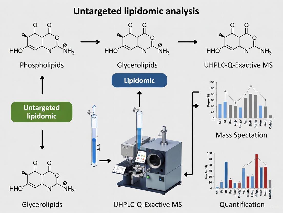
Untargeted Lipidomics with UHPLC-Q-Exactive MS: A Comprehensive Workflow for Diabetes Biomarker Discovery and Metabolic Pathway Analysis
Untargeted lipidomics utilizing UHPLC-Q-Exactive MS technology has emerged as a powerful strategy for uncovering the complex lipid dysregulation associated with diabetes mellitus and its complications.
Untargeted Lipidomics with UHPLC-Q-Exactive MS: A Comprehensive Workflow for Diabetes Biomarker Discovery and Metabolic Pathway Analysis
Abstract
Untargeted lipidomics utilizing UHPLC-Q-Exactive MS technology has emerged as a powerful strategy for uncovering the complex lipid dysregulation associated with diabetes mellitus and its complications. This article provides a comprehensive guide for researchers and drug development professionals, covering the foundational principles of lipid diversity and diabetes pathophysiology, detailed methodological workflows from sample preparation to data acquisition, critical troubleshooting for enhancing data reliability, and robust validation techniques. By integrating foundational exploration with practical application and validation, this resource aims to equip scientists with the knowledge to identify novel lipid biomarkers and elucidate disrupted metabolic pathways, thereby advancing our understanding of diabetes mechanisms and fostering the development of new diagnostic and therapeutic strategies.
Unveiling the Diabetic Lipidome: Foundations and Discovery with UHPLC-Q-Exactive MS
Lipid Classification: The LIPID MAPS System
Lipids are a diverse group of hydrophobic or amphipathic molecules that are insoluble in water but soluble in organic solvents. They perform many key biological functions, such as acting as structural components of cell membranes, serving as energy storage sources, and participating in signaling pathways [1]. The LIPID MAPS consortium has established a comprehensive, chemically-based classification system that categorizes lipids into eight major categories based on their distinct hydrophobic and hydrophilic elements and biosynthetic origins [1] [2]. This system uses a 12- or 14-character identifier (LIPID MAPS ID) to provide a unique, systematic identification for each lipid molecule [1].
Table 1: LIPID MAPS Lipid Classification System
| Category | Abbreviation | Core Structure/Biosynthetic Origin | Example Molecules |
|---|---|---|---|
| Fatty Acyls [1] [3] | FA | Carbanion-based condensations of ketoacyl thioesters [1]. | Fatty acids, eicosanoids, fatty alcohols [3]. |
| Glycerolipids [1] [3] | GL | Mono-, di-, and tri-substituted glycerols [3]. | Triacylglycerols (triglycerides) [3]. |
| Glycerophospholipids [1] [3] | GP | Glycerol with a phosphate group esterified to one of the glycerol hydroxyls [1]. | Phosphatidylcholine (PC), Phosphatidylethanolamine (PE) [3]. |
| Sphingolipids [1] [3] | SP | Long-chain nitrogenous base (sphingoid) backbone [1]. | Ceramides (Cer), Sphingomyelin (SM), Gangliosides [3]. |
| Sterol Lipids [1] [3] | ST | Carbocation-based condensations of isoprene units; distinct fused ring structure [1]. | Cholesterol, steroid hormones [3]. |
| Prenol Lipids [1] [3] | PR | Polymerization of isoprene units (dimethylallyl pyrophosphate/isopentenyl pyrophosphate) [1]. | Ubiquinones, vitamins E and K [3]. |
| Saccharolipids [1] [3] | SL | Fatty acyl groups linked directly to a sugar backbone [1]. | UDP-3-O-(3R-hydroxy-tetradecanoyl)-N-acetylglucosamine [3]. |
| Polyketides [1] [3] | PK | Condensation of ketoacyl subunits; often modified [1]. | Erythromycin, tetracycline, aflatoxins [3]. |
The Role of Lipids in Type 2 Diabetes Pathophysiology
Lipidomics, the large-scale study of pathways and networks of cellular lipids, has become a crucial tool for understanding the molecular mechanisms of Type 2 Diabetes (T2D) [4] [5]. Dysregulated lipid metabolism is a hallmark of T2D, and specific lipid species have been identified as key players in the development of insulin resistance and other diabetic complications [4] [6] [3].
Ceramides, a class of sphingolipids, are significantly associated with a higher risk of diabetes and insulin resistance [4] [6]. These lipids can inhibit the activity of the insulin-signaling phosphoinositide 3-kinase (PI3K) pathway, a mechanism also linked to elevated free fatty acids (FFA) [4] [3]. Furthermore, specific phosphatidylcholine (PC) and phosphatidylethanolamine (PE) species are involved in glucose homeostasis and are often altered in T2D patients [4] [7]. These phospholipids, along with sphingomyelin (SM), can also exert anti-inflammatory effects by antagonizing pro-inflammatory mediators like platelet-activating factor (PAF), linking them to the reduced cardiovascular risk associated with some dairy products [7].
Table 2: Select Lipid Classes and Their Implications in Type 2 Diabetes Research
| Lipid Class | Specific Example(s) | Association with T2D Pathophysiology | Supporting Evidence |
|---|---|---|---|
| Sphingolipids [4] [6] | Ceramides (e.g., Cer(d18:1/16:0)) | Promotes insulin resistance; correlates with severity of insulin resistance [6]. | Elevated in obese subjects with T2D; predictive of disease development [4] [6]. |
| Glycerophospholipids [4] [7] | Phosphatidylcholine (PC), Phosphatidylethanolamine (PE) | Altered levels affect membrane fluidity and signaling; some species have anti-inflammatory properties [7] [3]. | Dysregulated in serum of T2D patients; PC/PE ratio can impact insulin sensitivity [4]. |
| Fatty Acyls [4] [7] | Free Fatty Acids (FFA), Rumenic Acid | High FFA leads to insulin resistance; some bioactive fatty acids (e.g., in dairy) improve glucose homeostasis [4] [7]. | FFAs inhibit PI3K activity; rumenic acid agonizes peroxisome proliferator-activated receptors (PPARs) [4] [7]. |
| Glycerolipids [4] | Triacylglycerols (TAG) | Primary energy storage; excess accumulation is a risk factor for T2D and cardiovascular disease [4] [3]. | Standard clinical measure; lipidomics reveals specific TAG species as potential biomarkers [4]. |
Figure 1: Simplified Pathway of Lipid-Mediated Mechanisms in T2D. Abbreviations: T2D (Type 2 Diabetes), PI3K (Phosphoinositide 3-Kinase), PAF (Platelet-Activating Factor).
Experimental Protocol: Untargeted Lipidomics for Diabetes Research
The following section provides a detailed methodology for conducting untargeted lipidomic analysis of serum samples from T2D patients and healthy controls, based on validated protocols from recent literature [4] [8].
Sample Preparation and Lipid Extraction
Materials:
- Human Serum/Plasma Samples: Collected after informed consent and ethical approval [4].
- Methyl tert-butyl ether (MTBE), Methanol, Chloroform: HPLC or LC-MS grade solvents for lipid extraction [8].
- Internal Standards: A mixture of stable isotope-labeled or non-natural lipid standards (e.g., LPC 13:0, PC 14:0/14:0, PE 17:0/17:0) for quality control and normalization [8].
- Formic Acid: For acidification of the extraction mixture [8].
- Equipment: Microcentrifuges, vortex mixers, glass tubes, and a nitrogen evaporator.
Protocol: MTBE-based Lipid Extraction (Modified from Matyash et al.) [8]
- Aliquot: Transfer 100 µL of serum sample into a glass tube.
- Add Solvents and Standards: Add 750 µL of methanol containing internal standards and 20 µL of 1M formic acid. Vortex for 10 seconds.
- Extraction: Add 2.5 mL of MTBE. Mix vigorously with a multi-pulse vortexer for 5 minutes.
- Phase Separation: Add 625 µL of deionized water. Vortex for 3 minutes and centrifuge at 1000 g for 5 minutes. This will result in a two-phase system.
- Collection: Carefully collect the upper organic (MTBE-rich) phase, which contains the extracted lipids.
- Drying and Reconstitution: Evaporate the organic phase to dryness under a stream of nitrogen gas. Reconstitute the dried lipid extract in a suitable solvent (e.g., chloroform/methanol or isopropanol) for LC-MS analysis [4] [8].
UHPLC-Q-Exactive MS Analysis
Instrumentation:
- UHPLC System: e.g., Thermo Scientific Dionex UltiMate 3000.
- Analytical Column: Reversed-phase C18 column (e.g., Xselect CSH C18, 1.7 µm, 2.1 x 100 mm) [4].
- Mass Spectrometer: High-resolution Q-Exactive Orbitrap mass spectrometer equipped with an electrospray ionization (ESI) source [4] [7].
Chromatographic Conditions:
- Mobile Phase A: Water:acetonitrile (40:60, v/v) with 10 mM ammonium formate.
- Mobile Phase B: Isopropanol:acetonitrile (90:10, v/v) with 10 mM ammonium formate.
- Gradient: Use a non-linear gradient from 30% B to 100% B over a 20-30 minute runtime.
- Flow Rate: 0.3-0.4 mL/min.
- Column Temperature: 55°C.
- Injection Volume: 2-5 µL (plasma equivalent) [4] [8].
Mass Spectrometric Conditions:
- Ionization Mode: Positive and negative electrospray ionization (ESI+ and ESI-).
- Source Voltage: 3.3 kV for ESI+ and 2.8 kV for ESI- [4].
- Capillary Temperature: 350°C.
- Scan Mode: Full MS (m/z range 200-1200) at a resolution of 70,000, followed by data-dependent MS/MS (dd-MS2) at a resolution of 17,500.
- Collision Energy: Stepped normalized collision energy (e.g., 20, 30, 40 eV) [4] [8].
Figure 2: Untargeted Lipidomics Workflow for Diabetes Research.
Data Processing and Analysis
- Data Preprocessing: Use software such as MS-DIAL, LipidSearch, or XCMS for peak picking, alignment, and deisotoping [4] [9]. Perform strict quality control, including monitoring signal intensity, retention time alignment, and mass accuracy (< 5 ppm) [5].
- Lipid Identification: Identify lipids by matching the accurate mass (within 5 ppm) and MS/MS fragmentation spectra against databases such as LIPID MAPS [1], HMDB, or internal spectral libraries [4] [5].
- Statistical Analysis:
- Multivariate Analysis: Use Principal Component Analysis (PCA) for an unsupervised overview of data and Orthogonal Partial Least Squares-Discriminant Analysis (OPLS-DA) to maximize separation between T2D and control groups [4] [5].
- Univariate Analysis: Apply Student's t-test or ANOVA (with False Discovery Rate (FDR) correction for multiple testing) to identify lipids with significantly different abundances (e.g., p-adjusted < 0.05 and fold-change > 2) [4] [6].
- Pathway and Biological Interpretation: Utilize tools like MetaboAnalyst to map significantly altered lipids onto known metabolic pathways (e.g., sphingolipid metabolism, glycerophospholipid metabolism) to generate biologically testable hypotheses [5].
The Scientist's Toolkit: Essential Reagents and Software
Table 3: Key Research Reagent Solutions for Lipidomics in Diabetes Research
| Item | Function/Application | Example Usage in Protocol |
|---|---|---|
| MTBE (Methyl tert-butyl ether) [8] | Primary solvent for liquid-liquid lipid extraction. Provides high recovery of both polar and non-polar lipids. | Used in the MTBE-based extraction method for serum/plasma samples [8]. |
| Synthetic Lipid Standards [8] | A set of non-naturally occurring lipids for quality control, normalization, and semi-quantification. | Added at the beginning of extraction (e.g., PC 14:0/14:0) to monitor technical variability and aid quantification [8]. |
| UHPLC C18 Column [4] [8] | Reversed-phase chromatographic column for separating complex lipid mixtures prior to MS injection. | Used to resolve lipid species based on hydrophobicity over a 30-minute gradient [4]. |
| Ammonium Formate/Formic Acid [4] [8] | Mobile phase additives that enhance ionization efficiency and aid chromatographic separation in ESI-MS. | Added to mobile phases for UHPLC separation to improve peak shape and MS signal [4]. |
| Data Processing Software (e.g., MS-DIAL, LipidSearch) [4] [9] | Bioinformatics tools for automated peak picking, alignment, lipid identification, and statistical analysis. | Used to process raw LC-MS data files, identify lipids via database matching, and create a data matrix for statistical analysis [4] [9]. |
| Corynecin I | Corynecin I|CAS 4423-58-9|Antibacterial Agent | Corynecin I is a chloramphenicol-like antibiotic for RUO. It inhibits bacterial protein synthesis. This product is for Research Use Only. Not for human use. |
| Rizatriptan N-oxide | Rizatriptan N-oxide, CAS:260435-42-5, MF:C15H19N5O, MW:285.34 g/mol | Chemical Reagent |
The Role of Lipids in Diabetes Pathophysiology and Complication Development
Diabetes mellitus is a chronic metabolic disorder characterized by chronic hyperglycemia that leads to heterogenous disturbances of metabolism, with its continuing rise becoming a major concern globally [10]. Lipidomics, an important branch of metabolomics, aims to detect, quantify, and pinpoint all lipid species in a biological system, providing comprehensive insights into the lipid disruptions associated with diabetes pathophysiology and its complications [4]. The association between lipids and diabetes has been widely recognized, but the complexity of these relationships is underestimated in conventional lipid studies [10]. With advances in mass spectrometry platforms like UHPLC-Q-Exactive MS, researchers can now globally assess lipid species and their biological significance in diabetes, enabling the identification of novel lipid biomarkers and dysregulated metabolic pathways that offer new opportunities for disease prediction, detection, and therapeutic intervention [4] [11].
Lipid Alterations in Type 1 and Type 2 Diabetes
Comprehensive lipidomic profiling has revealed distinct and shared lipid disturbances between type 1 (T1D) and type 2 (T2D) diabetes. A recent study characterizing the lipidome of 360 subjects (91 T1D, 91 T2D, 74 with prediabetes, and 104 controls) identified 54 unique lipid subspecies from 15 unique lipid classes, with lysophosphatidylcholines (LPC) and ceramides (Cer) showing opposite effects in T1D and T2D [10]. LPCs were mainly up-regulated in T1D and down-regulated in T2D, while ceramides were up-regulated in T2D and down-regulated in T1D. Phosphatidylcholines (PC) were clearly down-regulated in subjects with T1D [10]. The study also found important sex-specific differences in diabetes-associated lipid disruptions, with ceramides and phosphatidylcholines exhibiting significant variations due to sex [10].
Table 1: Key Lipid Class Alterations in Diabetes Mellitus
| Lipid Class | Type 1 Diabetes | Type 2 Diabetes | Associated Complications |
|---|---|---|---|
| Ceramides (Cer) | Down-regulated [10] | Up-regulated [10] | Insulin resistance, DR [11] [12] |
| Lysophosphatidylcholines (LPC) | Up-regulated [10] | Down-regulated [10] | T2DM with dyslipidemia [12] |
| Phosphatidylcholines (PC) | Down-regulated [10] | Variable | DKD, DH [13] [14] |
| Sphingomyelins (SM) | Not specified | Down-regulated in DR [11] | DR, T2DM with dyslipidemia [11] [12] |
| Triglycerides (TG) | Not specified | Up-regulated [13] | Hyperuricemia, DKD [13] [14] |
| Phosphatidylethanolamines (PE) | Not specified | Up-regulated [13] | DH [13] |
Lipid Biomarkers of Diabetic Complications
Diabetic Retinopathy
Lipidomic studies have identified specific lipid signatures associated with diabetic retinopathy (DR). A 2024 investigation with 622 T2DM patients found that three ceramides and seven sphingomyelins were significantly lower in the DR group compared to diabetic patients without retinopathy (NDR group), while one phosphatidylcholine, two lysophosphatidylcholines, and two sphingomyelins were significantly higher [11]. Multifactorial logistic regression analysis revealed that lower abundance of two ceramides, Cer(d18:0/22:0) and Cer(d18:0/24:0), was an independent risk factor for DR occurrence in T2DM patients [11]. Another study published in 2024 identified a four-lipid combination diagnostic model including TAG58:2-FA18:1 that showed good predictive ability for distinguishing between NDR patients and those with non-proliferative DR (NPDR) [15].
Diabetes with Hyperuricemia
A 2025 study comparing lipid metabolites between patients with diabetes mellitus combined with hyperuricemia (DH) and diabetes mellitus (DM) alone identified 1,361 lipid molecules across 30 subclasses [13]. Researchers found 31 significantly altered lipid metabolites in the DH group compared to normal glucose tolerance (NGT) controls, with 13 triglycerides (e.g., TG(16:0/18:1/18:2)), 10 phosphatidylethanolamines (e.g., PE(18:0/20:4)), and 7 phosphatidylcholines (e.g., PC(36:1)) significantly upregulated, while one phosphatidylinositol was downregulated [13]. These differential lipids were predominantly enriched in glycerophospholipid metabolism and glycerolipid metabolism pathways.
Diabetic Kidney Disease
Emerging research has revealed lipidomic disruptions in diabetic kidney disease (DKD). Lysophosphatidylethanolamines (LPEs) have been identified as potential biomarkers and contributors to DKD pathophysiology, with specific lipid species showing significant alterations across different stages of kidney disease progression [14]. The dysregulated lipid species are involved in key pathological processes including inflammation, fibrosis, and oxidative stress in renal tissues [14].
Table 2: Specific Lipid Biomarkers of Diabetic Complications
| Complication | Specific Lipid Biomarkers | Direction of Change | AUC/Diagnostic Performance |
|---|---|---|---|
| Diabetic Retinopathy | Cer(d18:0/22:0), Cer(d18:0/24:0) | Decreased [11] | Independent risk factor [11] |
| Diabetic Retinopathy | SM(d18:1/24:1) | Decreased [11] | Significantly lower in DR [11] |
| Early DR Detection | TAG58:2-FA18:1 and 3 other lipids | Specific expression [15] | Good predictive ability [15] |
| T2DM with Dyslipidemia | Cer(d18:1/24:0), SM(d18:1/24:0) | Altered [12] | Essential potential biomarkers [12] |
| T2DM with Dyslipidemia | SM(d18:1/16:1), SM(d18:1/24:1), SM(d18:2/24:1) | Altered [12] | Closely linked to clinical parameters [12] |
Novel Lipid Indices for Diabetes and Insulin Resistance Monitoring
Recent investigations have examined the correlation between novel lipid indices and diabetes/insulin resistance (IR). A 2025 analysis of 19,780 NHANES participants found that the atherogenic index of plasma (AIP) and remnant cholesterol (RC) showed the strongest associations with diabetes and IR [16]. For Q4 versus Q1, AIP and RC showed significantly elevated diabetes risk (OR: 2.52 [2.07–3.07] and 2.13 [1.75–2.58], respectively). Regarding IR, all indices exhibited dose-dependent associations, with AIP (OR: 5.74 [5.00–6.59]) and RC (4.09 [3.58–4.67]) showing the strongest links [16]. For diabetes diagnosis, AIP (AUC: 0.824) and RC (0.822) outperformed other lipid indices but were less effective than fasting glucose and HbA1c. Subgroup analyses indicated stronger AIP/RC-diabetes/IR associations in females [16].
Experimental Protocols for Diabetes Lipidomics
Sample Preparation Protocol
Serum Collection and Processing:
- Collect fasting blood samples in appropriate tubes (EDTA tubes for plasma, procoagulation tubes for serum) [4] [10].
- Centrifuge at 1,500-3,000 rpm for 10-20 minutes at 4°C to separate serum/plasma [4] [11].
- Aliquot and store at -80°C until analysis [11] [13].
Lipid Extraction (Modified Folch Method):
- Use 100 μL serum sample [4] or 50-100 μL plasma [13] [10].
- Add 267 μL CHCl₃ and 133 μL methanol (for Folch) or 300-800 μL MTBE (for MTBE-based methods) [4] [13].
- Vortex thoroughly and centrifuge for phase separation [4].
- Collect organic phase and evaporate under nitrogen stream [15].
- Reconstitute in appropriate solvent (CHCl₃/methanol, isopropanol, or mobile phase B) for LC-MS analysis [4] [13] [15].
Quality Control:
- Prepare pooled quality control (QC) samples from all samples [4] [10].
- Inject QC samples every 10 runs throughout the sequence [4].
UHPLC-Q-Exactive MS Analysis Conditions
Chromatography Conditions:
- Column: Reversed-phase C18 column (e.g., Waters ACQUITY UPLC BEH C18, 2.1 × 100 mm, 1.7 μm; Xselect CSH C18; or Accucore C30 column) [4] [13] [17].
- Mobile Phase A: 10 mM ammonium formate in acetonitrile/water (60:40) or 10 mM ammonium formate with 0.1% formic acid in 60% acetonitrile/water [13] [10].
- Mobile Phase B: 10 mM ammonium formate in acetonitrile/isopropanol (10:90) or 10 mM ammonium formate with 0.1% formic acid in 90% propan-2-ol/water [13] [10].
- Gradient: Linear gradient from 20-30% B to 99-100% B over 9-16 minutes, followed by re-equilibration [10] [17].
- Flow Rate: 0.35-0.40 mL/min [10] [17].
- Column Temperature: 40°C [17].
- Injection Volume: 5-10 μL [15] [17].
Mass Spectrometry Conditions:
- Ionization: Electrospray ionization (ESI) in both positive and negative modes [4] [10].
- Source Voltage: 3.3 kV for positive mode, 2.8-4.5 kV for negative mode [4] [15].
- Ion Source Temperature: 350°C [15].
- Mass Range: m/z 50-500 or m/z 50-1500 [17].
- Resolution: High-resolution mode (typically 70,000-140,000 full width at half maximum) [4].
- Data Acquisition: Full MS and data-dependent MS/MS (dd-MS²) or data-independent acquisition (DIA) [17].
Data Processing and Statistical Analysis
- Raw Data Processing: Use software such as MS-DIAL for peak detection, alignment, and identification [4].
- Lipid Identification: Identify lipids based on precise molecular weights and MS/MS fragmentation patterns [4]. Use internal standards for quantification when possible [11].
- Multivariate Analysis: Employ principal component analysis (PCA) and orthogonal partial least squares-discriminant analysis (OPLS-DA) using platforms like MetaboAnalyst [4] [13].
- Differential Analysis: Apply t-tests or ANOVA with appropriate multiple testing corrections [4].
- Biomarker Evaluation: Use receiver operating characteristic (ROC) analysis, LASSO regression, and support vector machine recursive feature elimination (SVM-RFE) for biomarker selection [15] [14].
- Pathway Analysis: Conduct pathway enrichment analysis using MetaboAnalyst or similar platforms [13] [12].
Lipid Metabolic Pathways in Diabetes
The disrupted lipid species in diabetes and its complications are involved in several key metabolic pathways. Pathway enrichment analyses have identified glycerophospholipid metabolism and sphingolipid metabolism as the most significantly perturbed pathways in diabetes [18] [12]. Glycerolipid metabolism has also been identified as a core disrupted pathway in diabetes with hyperuricemia [13]. The following diagram illustrates the key lipid metabolic pathways disrupted in diabetes:
The Scientist's Toolkit: Essential Research Reagents and Materials
Table 3: Essential Research Reagents and Materials for Diabetes Lipidomics
| Category | Specific Items | Function/Application | Examples from Literature |
|---|---|---|---|
| Chromatography Columns | Reversed-phase C18 columns (e.g., Waters ACQUITY UPLC BEH C18, Xselect CSH C18) | Lipid separation by hydrophobicity | [4] [13] |
| Mass Spectrometry Standards | SPLASH LIPIDOMIX Mass Spec Standard | Internal standardization for quantification | [11] |
| Lipid Extraction Solvents | Methyl-tert-butyl ether (MTBE), Chloroform, Methanol, Isopropanol | Lipid extraction from biological samples | [13] [10] [17] |
| Mobile Phase Additives | Ammonium formate, Formic acid | Enhance ionization and chromatographic separation | [10] [17] |
| Quality Control Materials | Pooled quality control samples from all study samples | Monitor instrument stability and performance | [4] [10] |
| Data Processing Software | MS-DIAL, MetaboAnalyst, SCIEX OS | Peak detection, alignment, identification, and statistical analysis | [4] [15] |
| Fingolimod phosphate | Fingolimod Phosphate | High-purity Fingolimod phosphate for life science research. Explore its applications in immunology and neurobiology. This product isFor Research Use Only. Not for human or veterinary use. | Bench Chemicals |
| 10-Carboxylinalool | 10-Carboxylinalool, CAS:28420-25-9, MF:C10H16O3, MW:184.23 g/mol | Chemical Reagent | Bench Chemicals |
Untargeted lipidomics using UHPLC-Q-Exactive MS has revealed extensive disruptions in lipid metabolism in both type 1 and type 2 diabetes, with specific lipid signatures associated with different complications including retinopathy, kidney disease, and hyperuricemia. Ceramides, sphingomyelins, glycerophospholipids, and triglycerides represent key lipid classes involved in diabetes pathophysiology. The experimental protocols outlined provide comprehensive methodologies for conducting diabetes lipidomics research, from sample preparation to data analysis. These approaches enable researchers to identify novel lipid biomarkers and therapeutic targets, advancing our understanding of diabetes pathophysiology and contributing to improved prevention, diagnosis, and treatment strategies for diabetes and its complications.
Principles of UHPLC-Q-Exactive MS for Comprehensive Lipid Coverage
Ultra-High-Performance Liquid Chromatography coupled with Q-Exactive Mass Spectrometry (UHPLC-Q-Exactive MS) represents a powerful analytical platform for untargeted lipidomics, enabling comprehensive characterization of complex lipidomes in diabetes research. This technical note details the fundamental principles, optimized methodologies, and application protocols for leveraging the high mass accuracy and resolution of the Q-Exactive Orbitrap system to investigate lipid dysregulation in diabetes mellitus and its complications. We provide experimentally validated workflows for lipid extraction, chromatographic separation, mass spectrometric detection, and data processing specifically tailored for diabetes research, facilitating the discovery of novel lipid biomarkers and pathogenic mechanisms.
The UHPLC-Q-Exactive MS system combines advanced chromatographic separation with high-resolution accurate-mass (HRAM) detection, making it particularly suitable for untargeted lipidomic analysis. The platform's core components operate synergistically to address the challenges of lipid complexity. The UHPLC system provides rapid, high-efficiency separation of lipid molecules using sub-2μm particle columns, significantly reducing analytical time while improving peak capacity compared to conventional HPLC. This is crucial for resolving the numerous structural isomers present in biological lipidomes [19].
The Q-Exactive mass spectrometer incorporates a quadrupole precursor selection system with a high-resolution Orbitrap mass analyzer, enabling both data-dependent acquisition (DDA) and full-scan MS modes with mass accuracy typically below 3 ppm. This exceptional mass precision is fundamental for confident lipid identification, allowing distinction between isobaric species with minimal mass differences (e.g., different double bond equivalents or backbone structures) commonly encountered in diabetic lipidomes [20]. The system's high resolution (typically ≥70,000 at m/z 200) provides additional selectivity in complex biological matrices like plasma and tissue extracts from diabetic models.
Experimental Protocols for Lipidomics in Diabetes Research
Sample Preparation and Lipid Extraction
Protocol: MTBE-Based Lipid Extraction from Plasma/Serum Adapted from diabetes lipidomics studies [13] [20]
- Sample Preparation: Thaw frozen plasma/serum samples on ice. Aliquot 100 μL of sample into a glass centrifuge tube.
- Protein Precipitation: Add 300 μL of chilled methanol to the sample. Vortex vigorously for 30 seconds.
- Liquid-Liquid Extraction: Add 1 mL of methyl-tert-butyl ether (MTBE) to the mixture. Sonicate in a low-temperature water bath for 20 minutes.
- Phase Separation: Add 250 μL of LC-MS grade water to induce phase separation. Centrifuge at 14,000 × g for 15 minutes at 10°C.
- Collection: Carefully collect the upper organic phase (approximately 800 μL) containing the extracted lipids.
- Concentration: Evaporate the organic phase under a gentle stream of nitrogen gas.
- Reconstitution: Reconstitute the dried lipid extract in 100-200 μL of isopropanol for UHPLC-MS analysis.
- Quality Control: Prepare pooled quality control (QC) samples by combining equal aliquots from all samples to monitor system stability throughout the analysis sequence.
Note: This method has been successfully applied in studies investigating lipid alterations in type 2 diabetes patients, demonstrating excellent recovery of diverse lipid classes [13] [4].
UHPLC Conditions for Comprehensive Lipid Separation
Chromatographic Protocol for Lipidome Coverage
| Parameter | Specification |
|---|---|
| Column | Waters ACQUITY UPLC BEH C18 (2.1 × 100 mm, 1.7 μm) or Accucore C30 (2.1 × 150 mm, 2.6 μm) |
| Mobile Phase A | Acetonitrile:Water (60:40, v/v) with 10 mM ammonium formate |
| Mobile Phase B | Isopropanol:Acetonitrile (90:10, v/v) with 10 mM ammonium formate and 0.1% formic acid |
| Gradient Program | 30% B (0-2 min), 30-43% B (2-5 min), 55% B (5.1 min), 70% B (11 min), 99% B (16-18 min), 30% B (18.1-20 min) |
| Flow Rate | 0.35 mL/min |
| Column Temperature | 40°C |
| Injection Volume | 5 μL |
The above conditions enable separation of diverse lipid classes including glycerophospholipids, glycerolipids, and sphingolipids within a 20-minute run time, as validated in diabetes lipidomics research [13] [20].
Q-Exactive MS Instrument Configuration
Mass Spectrometric Parameters for Lipid Detection
| Parameter | Positive Ion Mode | Negative Ion Mode |
|---|---|---|
| Spray Voltage | 3.0-3.3 kV | 2.8-3.0 kV |
| Capillary Temperature | 350°C | 350°C |
| Aux Gas Temperature | 400°C | 400°C |
| S-lens RF Level | 50% | 50% |
| Full Scan Resolution | 70,000-140,000 | 70,000-140,000 |
| Scan Range | m/z 150-2000 | m/z 150-2000 |
| AGC Target | 1e6 | 1e6 |
| Maximum IT | 100 ms | 100 ms |
| dd-MS² Settings | Top 5-10 most intense ions | Top 5-10 most intense ions |
| Stepped NCE | 25, 30 eV | 20, 24, 28 eV |
Data acquisition should include both full scan MS and data-dependent MS/MS analyses in separate runs to maximize lipid identification and quantification, as employed in recent diabetes studies [20] [4].
Application in Diabetes Research: Key Findings and Data Analysis
Lipid Alterations in Diabetes and Hyperuricemia
Untargeted lipidomics using UHPLC-Q-Exactive MS has revealed profound lipid disruptions in diabetic conditions. A recent study comparing patients with diabetes mellitus (DM), diabetes combined with hyperuricemia (DH), and healthy controls identified 1,361 lipid molecules across 30 subclasses, with 31 significantly altered lipid metabolites in the DH group compared to controls [13].
Table 1: Significantly Altered Lipid Classes in Diabetes and Hyperuricemia
| Lipid Class | Trend in DH vs Control | Specific Examples | Potential Biological Significance |
|---|---|---|---|
| Triglycerides (TGs) | Significant upregulation (13 species) | TG(16:0/18:1/18:2) | Energy storage, insulin resistance |
| Phosphatidylethanolamines (PEs) | Significant upregulation (10 species) | PE(18:0/20:4) | Membrane fluidity, signaling |
| Phosphatidylcholines (PCs) | Significant upregulation (7 species) | PC(36:1) | Membrane integrity, lipoprotein metabolism |
| Phosphatidylinositols (PIs) | Downregulation | Not specified | Cell signaling, insulin signaling pathway |
Multivariate analyses including Principal Component Analysis (PCA) and Orthogonal Partial Least Squares-Discriminant Analysis (OPLS-DA) confirmed distinct lipidomic profiles between these clinical groups, with glycerophospholipid metabolism (impact value: 0.199) and glycerolipid metabolism (impact value: 0.014) identified as the most significantly perturbed pathways [13].
Lipidomics in Diabetic Complications
UHPLC-Q-Exactive MS has also been instrumental in elucidating lipid disruptions associated with diabetic complications. A recent lipidomic analysis of subclinical carotid atherosclerosis (SCA) in type 2 diabetes revealed 27 unique lipid species associated with SCA, with phosphatidylcholines and diacylglycerols as the main SCA-associated lipid classes [21]. Specifically, ten different species of phosphatidylcholines were upregulated, while four phosphatidylcholines containing polyunsaturated fatty acids were downregulated. These findings provide molecular insights into the accelerated atherosclerosis observed in diabetic populations [21].
Data Processing and Bioinformatics Pipeline
Workflow Visualization
Software and Tools for Data Analysis
Table 2: Essential Bioinformatics Tools for Lipidomics Data Analysis
| Software/Tool | Application | Key Features |
|---|---|---|
| MS-DIAL | Peak detection, alignment, identification | Open-source, comprehensive lipid database, supports DDA and DIA |
| LipidSearch | Lipid identification and quantification | Commercial, curated lipid database, automated identification |
| MetaboAnalyst 5.0 | Statistical and pathway analysis | Web-based, multivariate statistics, lipid pathway mapping |
| LIPID MAPS | Lipid structure database | Structural information, classification, MS/MS reference |
| XCMS Online | Peak picking and alignment | Cloud-based, statistical analysis, visualization |
The Scientist's Toolkit: Essential Research Reagents and Materials
Table 3: Critical Reagents and Materials for UHPLC-Q-Exactive Lipidomics
| Item | Function | Application Notes |
|---|---|---|
| Methyl-tert-butyl ether (MTBE) | Lipid extraction solvent | Less toxic alternative to chloroform, high extraction efficiency for diverse lipids |
| Ammonium formate | Mobile phase additive | Improves ionization efficiency, reduces sodium/potassium adduct formation |
| Deuterated lipid internal standards | Quantification reference | Correct for ionization suppression/enhancement, e.g., EquiSplash Lipidomix |
| C18 or C30 UHPLC columns | Lipid separation | C30 provides better resolution for complex lipid isomers |
| Quality control samples | System performance monitoring | Pooled sample from all specimens, injected regularly throughout sequence |
| Formic acid | Mobile phase modifier | Enhances positive ion formation in electrospray ionization |
| Tyvelose | Tyvelose, CAS:5658-12-8, MF:C6H12O4, MW:148.16 g/mol | Chemical Reagent |
| Taprostene | Taprostene | Stable PGI2 Analogue | For Research | Taprostene is a chemically stable, synthetic prostacyclin (PGI2) analogue and IP receptor agonist for cardiovascular and inflammation research. For Research Use Only. |
Methodological Considerations and Optimization Strategies
Enhancing Lipid Coverage and Identification
Comprehensive lipid coverage requires optimization at each workflow stage. For sample preparation, the MTBE method demonstrated superior extraction efficiency for polar and non-polar lipids compared to traditional Folch or Bligh-Dyer methods [20]. During LC-MS analysis, the use of C30 stationary phases provides enhanced separation of lipid isomers compared to conventional C18 columns, particularly for triglycerides and phospholipids with subtle structural differences [20].
For mass spectrometric detection, alternating positive and negative ion mode acquisitions within a single analytical sequence maximizes lipid class coverage, as different lipid classes ionize preferentially in different modes. Phosphatidylcholines and triglycerides ionize efficiently in positive mode, while phosphatidylinositols and fatty acids are better detected in negative mode [19] [4].
Quality Assurance in Lipidomics
Robust quality control measures are essential for generating reliable lipidomic data. Pooled QC samples should be analyzed at the beginning of the sequence for system equilibration, then regularly throughout the sequence (every 6-10 samples) to monitor instrument stability. Key performance indicators include retention time stability (RSD < 2%), peak intensity stability (RSD < 15-20% for abundant features), and mass accuracy drift (< 3 ppm) [19].
Lipid identification confidence should be reported according to established guidelines, with level 1 identifications requiring matching of MS/MS spectra to authentic standards, level 2 requiring matching to library spectra or diagnostic ions, and level 3 relying on accurate mass and retention time behavior alone [19] [4].
UHPLC-Q-Exactive MS represents a robust platform for comprehensive lipidomic analysis in diabetes research, capable of characterizing hundreds to thousands of lipid species from minimal sample volumes. The methodologies and protocols detailed in this application note provide a validated framework for investigating lipid dysregulation in diabetes and its complications, facilitating the discovery of novel biomarkers and therapeutic targets. As lipidomic technologies continue to advance, their integration with other omics platforms will further enhance our understanding of the molecular mechanisms underlying diabetes pathogenesis and progression.
Designing Exploratory Studies: Cohort Selection and Ethical Considerations is a foundational process in untargeted lipidomic research, which aims to discover novel lipid biomarkers and mechanistic pathways associated with complex metabolic diseases like diabetes. Untargeted lipidomics provides a comprehensive analysis of lipid species within a biological system, offering profound insights into the metabolic disruptions that precede and accompany disease states [4]. The UHPLC-Q-Exactive MS platform, with its high resolution and mass accuracy, is particularly well-suited for this discovery-phase research, enabling the identification of a wide array of lipid molecules without prior selection [4] [18].
The integrity and success of such studies are wholly dependent on rigorous initial planning, specifically in the selection of a well-defined participant cohort and the steadfast adherence to ethical principles. Proper cohort selection ensures the scientific validity and translational relevance of the lipidomic findings, while a strong ethical framework protects participant rights and welfare, thereby preserving the integrity of the research data [22]. This document outlines detailed protocols and considerations for these critical aspects within the context of a diabetes research thesis utilizing UHPLC-Q-Exactive MS-based untargeted lipidomics.
Core Concepts and Definitions
- Untargeted Lipidomics: A hypothesis-generating approach that aims to comprehensively detect and relatively quantify the full complement of lipids in a biological sample, crucial for discovering novel metabolic signatures in diabetes [4].
- UHPLC-Q-Exactive MS: An analytical platform combining Ultra-High-Performance Liquid Chromatography for superior separation of complex lipid mixtures with a high-resolution/accurate mass Q-Exactive Orbitrap mass spectrometer for precise lipid identification [4] [7].
- Cohort: A defined group of participants who are recruited based on specific eligibility criteria and followed over time to investigate the relationship between lipidomic profiles and health outcomes [13] [23].
- Ethical Principles: A set of guiding values—including social value, scientific validity, and informed consent—that ensure research is conducted responsibly and with respect for participant autonomy and well-being [22].
Cohort Selection: Design and Implementation
Defining Cohort Objectives and Inclusion Criteria
The primary objective in cohort selection for a diabetes lipidomic study is to assemble participant groups that enable clear differentiation of lipid signatures based on disease status, progression, or comorbidity.
Table 1: Example Cohort Structure for a Diabetes Lipidomics Study
| Cohort Group | Sample Size (Guideline) | Key Inclusion Criteria | Primary Comparative Aim |
|---|---|---|---|
| Healthy Control | ~17-47 participants [13] [4] | Normal glycemic status (HbA1c <5.7%), no history of diabetes [24]. | Provides a baseline lipidomic profile for comparison. |
| Prediabetes | ~40-6578 participants [24] | Intermediate hyperglycemia (HbA1c 5.7%-6.4%) [24]. | Identify lipid changes antecedent to overt diabetes. |
| Type 2 Diabetes (T2D) | ~40-250 participants [23] [4] | Meets ADA criteria (e.g., HbA1c ≥6.5%) [23] [24]. | Characterize the established diabetic lipidome. |
| T2D with Comorbidity | ~17 participants [13] | T2D diagnosis with a specific comorbidity (e.g., hyperuricemia) [13]. | Uncover lipid pathways linked to diabetic complications. |
Key Methodological Considerations for Cohort Selection
- Sample Size Justification: While practical constraints often play a role, the sample size should be justified through power analysis or based on previous successful studies. Pilot studies or published literature can guide this, with examples ranging from tightly matched pilot studies (n=17 per group) to large-scale cohorts (n>20,000) for epidemiological models [13] [25].
- Matching and Confounding Factors: To minimize bias, participants across cohorts should be matched for potential confounding variables such as age, sex, and body mass index (BMI) [4]. Detailed data on medication use (especially lipid-lowering and hypoglycemic drugs), lifestyle factors (diet, physical activity), and precise disease duration should be collected and accounted for in the statistical analysis [13] [23].
- Progression vs. Cross-Sectional Cohorts: For etiological studies, a prospective cohort tracks participants over time (e.g., 5 years) to identify lipidomic predictors of progression from prediabetes to T2D [24]. A case-control or cross-sectional design is efficient for comparing the lipidomes of established disease states versus healthy controls [13] [4].
The following workflow diagram illustrates the key decision points in the cohort selection process.
Ethical Considerations in Lipidomics Research
Ethical conduct is not an administrative hurdle but a scientific prerequisite that ensures the generation of reliable and socially valuable data [22]. The following principles are paramount.
Foundational Ethical Principles
- Social and Clinical Value: The study must be designed to answer a scientifically valid question that contributes to understanding diabetic lipid metabolism or improves disease prevention, treatment, or care. Exposing participants to any risk is only justifiable if the research has the potential to yield useful knowledge [22].
- Scientific Validity: The study must be methodologically robust to validly answer the research question. This includes using a fit-for-purpose platform (UHPLC-Q-Exactive MS), having a sound statistical plan for complex data, and ensuring the cohort is appropriately selected and sized [22].
- Favorable Risk-Benefit Ratio: Risks in lipidomic studies are typically minimal (discomfort from blood draw, potential breach of confidentiality) but must be systematically minimized. The potential benefits of new knowledge must outweigh these identified risks [22].
- Independent Review: The study protocol must be submitted for approval to an independent Institutional Review Board (IRB) or Ethics Committee before initiation. This review ensures ethical soundness and participant protection [13] [22] [4].
Participant-Centered Ethical Practices
- Informed Consent: The consent process is fundamental. Potential participants must be provided with clear information about the study's purpose, procedures, risks, benefits, and alternatives. They must understand that participation is voluntary and that they can withdraw at any time without penalty [22]. The consent form should explicitly cover the use of their biological samples (e.g., plasma) for lipidomic analysis and long-term storage [13] [4].
- Respect for Enrolled Subjects: This principle extends beyond initial consent. It encompasses:
- Privacy and Confidentiality: Protecting participant data is critical. All lipidomic and clinical data must be de-identified (coded or anonymized) [13] [22].
- Welfare Monitoring: Researchers must have plans to monitor participant well-being and manage any adverse events.
- Dissemination of Results: Participants should be informed of the aggregate findings of the research where appropriate [22].
Table 2: Essential Documentation for Ethical Research Conduct
| Document Type | Purpose and Key Components |
|---|---|
| Protocol Submission | Submission to Ethics Committee for independent review. Must include full study design, cohort details, informed consent form, and data management plan [13] [22]. |
| Informed Consent Form | To obtain voluntary participant agreement. Must include study purpose, procedures, risks/benefits, confidentiality terms, and rights to withdraw [22]. |
| Data Management Plan | To ensure data integrity and participant privacy. Must describe data anonymization procedures, secure storage solutions, and access controls. |
Experimental Protocol: UHPLC-Q-Exactive MS-Based Plasma Lipidomics
This section provides a detailed, citable protocol for a typical untargeted lipidomics workflow from sample preparation to data acquisition, as applied in diabetes research.
Sample Collection and Storage
- Collection: Draw fasting blood samples (e.g., 5 mL) from consented participants into appropriate anticoagulant tubes (e.g., EDTA plasma tubes) [13] [23].
- Processing: Centrifuge blood samples at 3,000 rpm for 10 minutes at room temperature to separate plasma [13].
- Storage: Aliquot 0.2 mL of plasma into sterile tubes and immediately store at -80°C until analysis to preserve lipid integrity [13] [23].
Lipid Extraction
The modified MTBE (Methyl tert-butyl ether) method is widely used for comprehensive lipid extraction [13]. 1. Thaw plasma samples on ice. 2. Pipette 100 μL of plasma into a 1.5 mL microcentrifuge tube. 3. Add 200 μL of cold HPLC-grade water and vortex mix. 4. Add 240 μL of ice-cold methanol and vortex mix thoroughly. 5. Add 800 μL of MTBE, vortex, and sonicate in a low-temperature water bath for 20 minutes. 6. Incubate the mixture at room temperature for 30 minutes to facilitate phase separation. 7. Centrifuge at 14,000 g at 10°C for 15 minutes. 8. Carefully collect the upper organic phase (which contains the lipids) into a new tube. 9. Evaporate the organic solvent to dryness under a gentle stream of nitrogen gas. 10. Reconstitute the dried lipid extract in a suitable solvent (e.g., 100 μL isopropanol) for LC-MS analysis [13].
UHPLC-MS Analysis Conditions
Table 3: UHPLC-Q-Exactive MS Instrumental Conditions for Untargeted Lipidomics
| Parameter | Specification | Notes |
|---|---|---|
| UHPLC Column | Waters ACQUITY UPLC BEH C18 (2.1x100 mm, 1.7 μm) [13] or equivalent (e.g., CSH column) [4]. | Provides high-resolution separation of complex lipid mixtures. |
| Mobile Phase A | 10 mM ammonium formate in acetonitrile/water (e.g., 95:5:0.1 v/v/v 10mM ammonium acetate/methanol/acetic acid) [13] [23]. | Aqueous phase with buffer additive. |
| Mobile Phase B | 10 mM ammonium formate in acetonitrile/isopropanol [13] or 99.9:0.1 v/v methanol/acetic acid [23]. | Organic phase for gradient elution. |
| Gradient Program | Non-linear gradient from 80% A to 100% B over 12+ minutes [13] [23]. | Optimized for gradual elution of diverse lipid classes. |
| Mass Spectrometer | Q-Exactive Orbitrap MS [4]. | High-resolution and accurate mass measurement. |
| Ionization Mode | Electrospray Ionization (ESI), both positive and negative ion modes [4]. | Essential for comprehensive coverage of different lipid classes. |
| Full Scan Resolution | 70,000 [23] to 140,000 [4]. | Enables precise determination of elemental composition. |
| Mass Range | m/z 200-1100 [23] or m/z 150-2000 [4]. | Covers the mass range of most lipid species. |
Quality Control (QC) and Data Preprocessing
- Quality Control (QC): Prepare a pooled QC sample by combining a small aliquot of every individual sample. Inject the QC sample repeatedly at the beginning of the run to equilibrate the system and then at regular intervals (e.g., every 10 injections) throughout the acquisition sequence to monitor instrument stability and performance [4].
- Data Preprocessing: Use specialized software (e.g., MS-DIAL, Progenesis QI) for raw data processing, which includes peak picking, alignment across samples, deconvolution, and lipid identification based on accurate mass and MS/MS spectral matching against databases [23] [4].
The following diagram summarizes the core experimental workflow.
The Scientist's Toolkit: Essential Research Reagents and Materials
Table 4: Key Reagent Solutions for UHPLC-MS-Based Lipidomics
| Item | Function/Application | Example Specification |
|---|---|---|
| UHPLC Solvents | Mobile phase preparation for chromatographic separation. | HPLC-MS grade Acetonitrile, Isopropanol, Methanol, Water [13] [4]. |
| Ammonium Formate/Acetate | Mobile phase additive to improve ionization efficiency and aid adduct formation. | 10 mM concentration in mobile phases [13] [23]. |
| Lipid Extraction Solvents | For liquid-liquid extraction of lipids from plasma. | Methyl tert-butyl ether (MTBE), Chloroform, Methanol [13] [4]. |
| Internal Standard | To monitor and correct for variability in extraction and ionization. | A known, non-endogenous lipid (e.g., 1,2-didodecanoyl-sn-glycero-3-phosphocholine) added at the start of extraction [23]. |
| Analytical Column | Separation of individual lipid species prior to MS detection. | Reversed-Phase C18 or C8 column (e.g., Waters BEH C18, 2.1x100 mm, 1.7 μm) [13] [23]. |
| 6-Bromoisoquinoline | 6-Bromoisoquinoline | High-Purity Building Block | High-purity 6-Bromoisoquinoline, a key heterocyclic building block for medicinal chemistry & material science. For Research Use Only. Not for human or veterinary use. |
| Ethylparaben-d5 | Ethyl-d5 Paraben | Stable Isotope Labeled | Ethyl-d5 Paraben, a deuterated internal standard for precise LC-MS/MS analysis. For Research Use Only. Not for human or veterinary use. |
Untargeted lipidomics, particularly utilizing UHPLC-Q-Exactive Mass Spectrometry, has unveiled complex lipid metabolism dysregulation in diabetes mellitus. This application note details the key alterations in triglycerides (TGs), glycerophospholipids, and sphingolipids identified in recent studies, provides validated experimental protocols for their detection, and visualizes the involved metabolic pathways. This resource is designed to support researchers and drug development professionals in elucidating novel metabolic pathways and biomarker candidates.
Key Lipid Alterations in Diabetes: Quantitative Data
Untargeted lipidomics reveals distinct lipid signatures in type 1 (T1DM) and type 2 diabetes (T2DM). The following tables summarize the most significant lipid alterations reported in recent clinical studies.
Table 1: Key Lipid Alterations in Type 1 Diabetes (T1DM) with Glycemic Control [26]
| Lipid Class | Specific Lipid Species | Alteration Trend | Statistical Significance (AUC) | Biological Sample |
|---|---|---|---|---|
| Diglycerides (DAGs) | DAG(14:0/20:0) | ↓ Decrease | 0.966 (Composite) | Plasma |
| Phosphatidylcholines (PCs) | PC(18:0/20:3) | ↓ Decrease | 0.966 (Composite) | Plasma |
| Triglycerides (TAGs) | Multiple Species | ↓ Decrease | Significant | Plasma |
| Phosphatidylethanolamines (PEs) | Multiple Species | ↓ Decrease | Significant | Plasma |
Table 2: Key Lipid Alterations in Type 2 Diabetes (T2DM) and Dyslipidemia [4] [27] [28]
| Lipid Class | Specific Lipid Species | Alteration Trend | Associated Condition | Biological Sample |
|---|---|---|---|---|
| Ceramides (Cers) | Cer(d18:1/24:0), Cer(d18:1/20:0) | ↑ Increase | T2DM, T2DM with Dyslipidemia | Serum, Plasma |
| Sphingomyelins (SMs) | SM(d18:1/24:0), SM(d18:1/16:1) | ↑ Increase | T2DM with Dyslipidemia | Plasma |
| Phosphatidylcholines (PCs) | PC(36:1), LysoPCs | ↓ Decrease / Varied | T2DM, Hyperuricemia Complication | Plasma, Serum |
| Phosphatidylethanolamines (PEs) | PE(18:0/20:4) | ↑ Increase | Hyperuricemia Complication | Plasma |
| Triglycerides (TGs) | TG(16:0/18:1/18:2) | ↑ Increase | Hyperuricemia Complication | Plasma |
Detailed Experimental Protocol: Untargeted Plasma/Serum Lipidomics
The following section outlines a standardized protocol for UHPLC-Q-Exactive-MS-based untargeted lipidomics, as adapted from recent studies [13] [4].
Sample Collection and Pre-processing
- Collection: Collect fasting blood into procoagulation tubes.
- Processing: Centrifuge at 3,000 rpm for 10 minutes at room temperature to isolate plasma/serum.
- Storage: Aliquot and store at -80°C until analysis. Avoid multiple freeze-thaw cycles.
Lipid Extraction using Modified Folch Method
- Add Internal Standards: Spike 100 µL of plasma/serum with a mixture of stable isotope-labeled lipid standards (e.g., PC(14:0)-d13, TG(17:0/17:0/17:0)) for quantification [26].
- Extraction: Add 267 µL of chloroform and 133 µL of methanol to the sample [4]. Alternatively, a methyl tert-butyl ether (MTBE)-based method can be used [13].
- Vortex and Centrifuge: Mix thoroughly, then centrifuge to achieve phase separation.
- Recovery: Carefully recover the lower organic phase containing the lipids.
- Drying: Evaporate the solvent under a gentle nitrogen stream.
- Reconstitution: Reconstitute the dried lipid extract in 100 µL of isopropanol for LC-MS analysis [13].
UHPLC-Q-Exactive MS Analysis
- Column: Waters ACQUITY UPLC BEH C18 column (2.1 mm × 100 mm, 1.7 µm particle size) [13] [4].
- Mobile Phase:
- Gradient Elution: Use a linear gradient from 30% B to 100% B over 20-30 minutes.
- MS Parameters:
- Ionization: Heated Electrospray Ionization (HESI) in positive and negative modes.
- Source Voltage: 3.3 kV (+mode), 2.8 kV (-mode) [4].
- Full Scan Resolution: 70,000 full width at half maximum (FWHM).
- Scan Range: m/z 200-2000.
- Data-Dependent MS/MS (dd-MS²): Top 10 most intense ions, resolution 17,500 FWHM.
Data Processing and Analysis
- Peak Picking & Alignment: Use software like MS-DIAL for peak detection, alignment, and lipid identification based on accurate mass and MS/MS spectral matching [4].
- Statistical Analysis:
- Perform multivariate statistical analysis (PCA, OPLS-DA) using platforms like MetaboAnalyst.
- Identify significant lipid features using t-tests and fold-change analysis.
Visualizing Lipid Metabolic Pathways in Diabetes
The identified lipid alterations are interconnected through key metabolic pathways. The diagram below illustrates the most significantly perturbed pathways in diabetes.
The Scientist's Toolkit: Essential Research Reagents and Materials
Table 3: Key Research Reagent Solutions for Diabetes Lipidomics
| Item | Function/Application | Specific Examples |
|---|---|---|
| Stable Isotope-Labeled Internal Standards | Quantification and quality control during lipid extraction and MS analysis. | PC(14:0)-d13, TG(17:0/17:0/17:0), Cer(1/17:0)-d18 [26] [27] |
| LC-MS Grade Solvents | Mobile phase preparation and lipid extraction to minimize background noise and ion suppression. | Acetonitrile, Isopropanol, Methanol, Chloroform, MTBE [26] [13] |
| UHPLC C18 Column | Reverse-phase chromatographic separation of complex lipid mixtures. | Waters ACQUITY UPLC BEH C18 (1.7 µm) [13] [4] |
| Mass Spectrometry Instrumentation | High-resolution accurate mass (HRAM) analysis for lipid identification and quantification. | Q-Exactive Orbitrap Mass Spectrometer [4] |
| Data Processing Software | Lipid identification, peak alignment, and statistical analysis. | MS-DIAL, MetaboAnalyst, LipidSearch [4] |
| Isoproturon | Isoproturon | Phenylurea Herbicide for Research | Isoproturon is a phenylurea herbicide for plant science research. It inhibits photosynthesis. For Research Use Only. Not for human or veterinary use. |
| Dimethyl sulfone-d6 | Dimethyl sulfone-d6 | Deuterated MSM | High Purity | Dimethyl sulfone-d6 (D6-MSM), a high-purity isotopic standard for research. For Research Use Only. Not for human or veterinary diagnostic or therapeutic use. |
The application of UHPLC-Q-Exactive-MS-based lipidomics robustly identifies triglycerides, glycerophospholipids, and sphingolipids as key players in diabetic dyslipidemia. The provided detailed protocol, quantitative landscape of alterations, and pathway visualization offer a foundational resource for advancing research into the metabolic pathology of diabetes and developing targeted diagnostic and therapeutic strategies.
Untargeted lipidomics, utilizing advanced platforms like UHPLC-Q-Exactive MS, generates complex, high-dimensional datasets containing thousands of lipid species from biological samples. In diabetes research, where subtle metabolic alterations precede clinical manifestations, extracting meaningful biological insights from this data deluge requires sophisticated chemometric tools. Multivariate data analysis (MVDA) provides powerful statistical frameworks for visualizing inherent data structures, classifying samples based on lipid profiles, and identifying discriminatory lipid species associated with diabetic states [4] [29].
These methods are particularly valuable for overcoming the "large p, small n" problem, where the number of measured variables (lipids, p) far exceeds the number of biological samples (n) [29]. Within the context of a diabetes research thesis, applying MVDA enables researchers to move beyond univariate comparisons to achieve a systems-level understanding of lipid metabolic disruptions in type 1 diabetes (T1D), type 2 diabetes (T2D), and associated complications.
Theoretical Foundations of PCA and OPLS-DA
Principal Component Analysis (PCA)
Principal Component Analysis is an unsupervised dimensionality reduction technique used to explore internal data structure without prior knowledge of sample class labels. It identifies principal components (PCs)—new, uncorrelated variables—that capture maximum variance in the data. The first PC (PC1) accounts for the largest possible variance, with each subsequent component capturing the remaining variance under orthogonality constraints [29].
In lipidomics, PCA simplifies complex data by projecting it into a lower-dimensional space defined by these PCs, allowing for visualization of natural clustering, outliers, and trends. Although biological systems exhibit complexity, systematic lipid changes in controlled experiments, such as comparing diabetic versus control groups, are often effectively captured by this linear model [30].
Orthogonal Partial Least Squares-Discriminant Analysis (OPLS-DA)
Orthogonal Partial Least Squares-Discriminant Analysis is a supervised method that separates predictive variation related to a specific factor (e.g., disease state) from non-correlated orthogonal variation (e.g., inter-individual differences). It enhances model interpretability by focusing on systematic variation that discriminates predefined sample classes [4] [30].
OPLS-DA is particularly suited for biomarker discovery in diabetes lipidomics, as it identifies lipid species whose changes are most predictive of a particular condition, such as T2D or hyperuricemia complication, by filtering out unrelated metabolic noise [4] [13].
Table 1: Comparison of PCA and OPLS-DA Characteristics in Lipidomic Analysis.
| Feature | PCA | OPLS-DA |
|---|---|---|
| Analysis Type | Unsupervised | Supervised |
| Primary Goal | Exploratory data analysis, outlier detection, trend visualization | Classification, biomarker identification, hypothesis testing |
| Use of Class Labels | No | Yes |
| Variance Handling | Captures maximum total variance | Separates predictive and orthogonal variance |
| Model Validation | Not applicable (descriptive) | Requires rigorous validation (e.g., permutation testing) |
| Key Output | Scores plot (sample patterns), Loadings plot (variable influence) | S-plot or VIP (identifying discriminatory variables) |
Workflow for Multivariate Analysis in Lipidomics
The application of PCA and OPLS-DA follows a structured pipeline from raw data to biological interpretation. The diagram below outlines the key stages in this process.
Application in Diabetes Lipidomics: A Detailed Experimental Protocol
This protocol details the application of UHPLC-Q-Exactive MS-based lipidomics and subsequent multivariate analysis for investigating serum samples from diabetic subjects, based on established methodologies [4] [13] [10].
Sample Preparation and Lipid Extraction
- Materials:
- Serum samples from T2D patients and matched healthy controls (e.g., n=40/group) [4].
- Pre-cooled HPLC-grade methanol, methyl tert-butyl ether (MTBE), and chloroform.
- Internal standard mixture (e.g., SPLASH LIPIDOMIX Mass Spec Standard).
- Procedure:
- Precipitation: Thaw serum samples on ice. Aliquot 100 µL of serum into a glass tube.
- Extraction: Add 267 µL of cold chloroform and 133 µL of cold methanol (a modified Folch method) [4]. Alternatively, a MTBE-based extraction can be used: add 200 µL of ice-cold water and 240 µL of cold methanol to 100 µL of serum, vortex, then add 800 µL of MTBE, and sonicate in a cold water bath [13].
- Phase Separation: Vortex mixture thoroughly and centrifuge at 14,000 g for 15 min at 4°C.
- Collection: Carefully recover the lower organic phase (chloroform phase for Folch) or the upper organic phase (MTBE phase).
- Drying: Evaporate the organic solvent under a gentle stream of nitrogen gas.
- Reconstitution: Reconstitute the dried lipid extract in a suitable solvent (e.g., chloroform/methanol or isopropanol) for LC-MS analysis.
UHPLC-Q-Exactive MS Analysis
- Chromatographic Conditions:
- Column: Reversed-phase C18 column (e.g., Waters ACQUITY UPLC BEH C18, 1.7 µm, 2.1 × 100 mm) [4] [13].
- Mobile Phase: A) 10 mM ammonium formate in acetonitrile/water (60:40, v/v) with 0.1% formic acid; B) 10 mM ammonium formate in isopropanol/acetonitrile (90:10, v/v) with 0.1% formic acid [10].
- Gradient: Start at 20% B, increase linearly to 100% B over 8.5 min, hold for 1 min, then re-equilibrate to initial conditions.
- Flow Rate: 0.4 mL/min.
- Column Temperature: 45-55°C.
- Injection Volume: 2-5 µL.
- Mass Spectrometric Conditions:
- Instrument: Q-Exactive or Q-Exactive Focus mass spectrometer.
- Ionization: Heated electrospray ionization (HESI) in both positive and negative ion modes.
- Source Parameters: Spray voltage: 3.3 kV (positive), 2.8 kV (negative); Capillary temperature: ~320°C.
- Full MS Scan: Resolution: 70,000; Scan range: m/z 200-1200.
- Data-Dependent MS/MS (dd-MS²): Top 10 most intense ions; Resolution: 17,500; Normalized Collision Energy (NCE): 25, 30, 35 eV.
Data Preprocessing for Multivariate Analysis
- Software: Use open-source (e.g., MS-DIAL [4]) or commercial software for peak picking, alignment, and annotation.
- Peak Annotation: Identify lipids based on accurate mass (MS1, typically < 5 ppm mass error) and MS/MS spectral matching against databases (e.g., LIPID MAPS).
- Data Matrix Construction: Generate a matrix where rows are samples, columns are lipid species, and values are peak intensities.
- Data Cleaning:
- Missing Value Imputation: Filter lipids with >20-35% missing values. Impute remaining missing values using methods like k-nearest neighbors (kNN) or a small constant (e.g., half the minimum value) [31].
- Normalization: Apply probabilistic quotient normalization or normalize to internal standards and median sample to correct for systematic errors.
- Scaling: Pareto or Unit Variance (UV) scaling is recommended to balance the influence of high and low-abundance lipids.
Multivariate Data Analysis Execution
- Software: Use statistical platforms like MetaboAnalyst [4] [13], SIMCA-P, or R/Python with packages such as
roplsandmixOmics[31]. - PCA:
- Input the preprocessed data matrix.
- Center the data (mean-centered).
- Generate scores plot to visualize sample clustering and identify outliers.
- Generate loadings plot to investigate which lipids contribute most to the observed separation.
- OPLS-DA:
- Provide the data matrix and a Y-variable vector specifying the class membership (e.g., T2D=1, Control=0).
- Build the model, typically allowing the software to automatically determine the number of predictive and orthogonal components.
- Validate the model using permutation tests (e.g., 200-1000 permutations) to ensure it is not overfit. Check R²Y (goodness-of-fit) and Q² (goodness-of-prediction) values.
- Use the S-plot or Variable Importance in Projection (VIP) scores to identify lipids most responsible for class separation. Lipids with high VIP values (e.g., > 1.5) are considered potential biomarkers.
Case Study: Identifying a Lipid Signature in T2D with Hyperuricemia
A 2025 study employed this exact workflow to investigate lipidomic disruptions in patients with Diabetes Mellitus and Hyperuricemia (DH) [13]. The OPLS-DA model showed a clear separation between DH, DM-only, and normal glucose tolerance (NGT) groups. The analysis identified 31 significantly altered lipid metabolites in DH vs NGT.
Table 2: Key Differential Lipids Identified in Diabetes Mellitus with Hyperuricemia (DH) vs. Controls [13].
| Lipid Class | Example Lipid Species | Trend in DH | Biological Relevance |
|---|---|---|---|
| Triglycerides (TGs) | TG(16:0/18:1/18:2) | ↑ Upregulated | Energy storage; associated with insulin resistance and cardiometabolic risk. |
| Phosphatidylethanolamines (PEs) | PE(18:0/20:4) | ↑ Upregulated | Membrane fluidity; precursors for signaling molecules. |
| Phosphatidylcholines (PCs) | PC(36:1) | ↑ Upregulated | Major membrane constituents; involved in lipoprotein metabolism. |
| Phosphatidylinositols (PIs) | Not specified | ↓ Downregulated | Key signaling lipids; precursors for secondary messengers. |
Pathway analysis of the differential lipids revealed that glycerophospholipid metabolism and glycerolipid metabolism were the most significantly perturbed pathways in DH patients, providing a mechanistic link between lipid dysregulation and this diabetic complication [13].
The Scientist's Toolkit: Essential Reagents and Software
Table 3: Key Research Reagent Solutions and Tools for Lipidomics and Multivariate Analysis.
| Item | Function / Purpose | Example Products / Software |
|---|---|---|
| Internal Standards | Correct for variability in extraction and ionization; enable semi-quantification. | SPLASH LIPIDOMIX, Avanti Polar Lipids stable isotope-labeled standards. |
| LC-MS Grade Solvents | Ensure high sensitivity, low background noise, and prevent system contamination. | Fisher Optima, Honeywell Chromasolv. |
| UHPLC C18 Column | Separate complex lipid mixtures prior to mass spectrometry analysis. | Waters ACQUITY UPLC BEH C18, Thermo Accucore C30. |
| Quality Control (QC) Pool | Monitor instrument stability, align features, and assess data quality. | Pooled sample from all study aliquots [31]. |
| Data Processing Software | Convert raw data into a peak intensity table for statistical analysis. | MS-DIAL, XCMS, Compound Discoverer. |
| Statistical Analysis Platform | Perform PCA, OPLS-DA, and other univariate/multivariate analyses. | MetaboAnalyst [4], SIMCA-P, R (ropls, mixOmics) [31]. |
| Bromonitromethane | Bromonitromethane | High-Purity Reagent | RUO | Bromonitromethane: A versatile synthon & alkylating agent for organic synthesis & medicinal chemistry research. For Research Use Only. Not for human use. |
| Fluo-3 | Fluo-3 AM | High-Affinity Calcium Indicator | Fluo-3 is a visible-light excitable calcium indicator for live-cell imaging & flow cytometry. For Research Use Only. Not for human or veterinary use. |
Critical Pathway and Biological Workflow
The journey from sample collection to biological insight involves a tightly integrated workflow of laboratory techniques and data science. The following diagram synthesizes the key steps and their relationships, culminating in the identification of perturbed metabolic pathways.
From Sample to Spectrum: A Practical UHPLC-Q-Exactive MS Lipidomics Workflow for Diabetes Research
Optimal Sample Collection and Pre-processing for Plasma, Serum, and Tissues
In untargeted lipidomic studies of diabetes using UHPLC-Q-Exactive MS technology, the reliability of research outcomes critically depends on sample quality during the preanalytical phase [32]. Lipid molecules exhibit varying ex vivo stability in blood, creating substantial risks for data misinterpretation if sample collection protocols are not rigorously standardized [32]. This application note provides detailed protocols for collecting and pre-processing plasma, serum, and tissue specimens specifically for diabetes lipidomics research, ensuring the integrity of lipid profiles from sample acquisition to MS analysis.
Blood Sample Collection and Processing
Critical Preanalytical Considerations
Whole blood before centrifugation represents a "liquid tissue" containing trillions of metabolically active cells that can rapidly alter lipid abundances ex vivo [32]. Consequently, handling of whole blood constitutes the most vulnerable preanalytical step for clinical lipidomics [32]. The table below summarizes the stability characteristics of different lipid classes in EDTA whole blood.
Table 1: Stability of Lipid Classes in EDTA Whole Blood Under Various Temperature Conditions
| Lipid Class | 24h at 21°C | 24h at 30°C | Stability Rating | Key Considerations |
|---|---|---|---|---|
| Phosphatidylcholines (PC) | Stable | Stable | High | Robust across conditions |
| Sphingomyelins (SM) | Stable | Stable | High | Consistently stable |
| Diacylglycerols (DG) | Stable | Stable | High | Reliable for analysis |
| Triacylglycerols (TG) | Stable | Stable | High | Maintain integrity |
| Lysophosphatidylcholines (LPC) | Significant changes | Significant changes | Low | High susceptibility to degradation |
| Lysophosphatidylethanolamines (LPE) | Significant changes | Significant changes | Low | Pronounced instability |
| Fatty Acids (FA) | Significant changes | Significant changes | Low | High ex vivo variability |
Recommended Protocol for Plasma Collection
Materials Required:
- EDTA vacuum blood collection tubes
- Cooled centrifuge capable of maintaining 4°C
- Timer
- Permanent ice bath or refrigerated cooling block
- Pre-labeled cryovials for plasma storage
- -80°C freezer for long-term storage
Step-by-Step Procedure:
Collection: Draw venous blood using EDTA vacuum collection tubes. Invert tubes gently 8-10 times immediately after collection to ensure proper mixing with anticoagulant.
Immediate Cooling: Place blood tubes immediately in a cooled environment at 4°C. Permanent cooling is recommended from this point forward [32].
Centrifugation: Centrifuge samples within 4 hours of collection at 4°C using these parameters:
- Speed: 3,100 × g
- Duration: 7 minutes
- Temperature: 4°C [32]
Plasma Separation: Carefully transfer the upper plasma layer to pre-labeled cryovials using disposable transfer pipettes, avoiding disturbance of the buffy coat or red blood cells.
Storage: Immediately freeze plasma aliquots at -80°C. Avoid multiple freeze-thaw cycles.
Diagram: Plasma Sample Collection Workflow
Stability Exceptions and Quality Control
For lipid classes demonstrating significant instability (LPC, LPE, FA), immediate processing within 30 minutes of collection is recommended. A potential quality control (QC) lipid triplet can be implemented to detect sampling artifacts during the preanalytical phase from blood collection until centrifugation [32]. When focusing exclusively on robust lipid species (PC, SM, DG, TG), the 4-hour processing window remains valid [32].
Tissue Sample Collection and Processing
Protocol for Tissue Specimen Handling
Materials Required:
- Biopsy tools or surgical instruments
- Aluminum foil or cryomolds
- Liquid nitrogen container
- Isopentane (pre-cooled)
- Cryostat
- Storage boxes for -80°C archives
Step-by-Step Procedure:
Collection: Obtain tissue specimens using standardized biopsy or dissection techniques.
Rinsing: Gently rinse tissues in ice-cold phosphate-buffered saline (PBS) to remove blood contaminants.
Snap-Freezing: For optimal lipid preservation:
- Embed tissue in cryomold with optimal cutting temperature (OCT) compound
- Submerge in pre-cooled isopentane (-150°C) for 60 seconds
- Alternatively, wrap in aluminum foil and directly freeze in liquid nitrogen
Storage: Transfer snap-frozen specimens to -80°C for long-term storage.
Sectioning: Cut tissue sections (5-10 μm thickness) in cryostat at -20°C and transfer to MS-compatible slides.
Diagram: Tissue Sample Processing Workflow
Urine Sample Collection and Processing
Protocol for Urine Specimens
Urine metabolomics provides complementary information to blood analyses in diabetes research, particularly for monitoring renal complications [33].
Materials Required:
- Sterile urine collection containers
- Centrifuge
- pH test strips
- -80°C freezer
Step-by-Step Procedure:
Collection: Collect mid-stream urine into sterile containers.
Centrifugation: Centrifuge at 3,000 × g for 10 minutes at 4°C to remove cellular debris.
Aliquoting: Transfer supernatant to cryovials.
Storage: Freeze immediately at -80°C. For untargeted analysis, maintain pH neutrality.
Quality Assurance in Untargeted Lipidomics
Internal Standards and QC Samples
Implement a robust quality assurance system throughout the analytical process:
Internal Standards: Add isotope-labeled internal standards as early as possible in the sample preparation process to normalize for experimental biases [34]. Recommended standards include:
- PC 15:0/15:0
- LPC 19:0
- PE 15:0/15:0
- SM d18:1/12:0
- Cer d18:1/17:0
- DG 15:0/18:1-d7
- TG 15:0/15:0/15:0
- FA 22:0-d4 [32]
Quality Control (QC) Samples: Prepare pooled QC samples by combining aliquots from each sample. Analyze QC samples:
- Multiple times before initiating the run to condition the column
- After every tenth sample during the sequence
- After each batch of samples [34]
Blank Samples: Include blank extraction samples (empty tubes without tissue) after every 23rd sample to establish baseline and filter out technical contamination peaks [34].
Batch Design Considerations
The main limitation of LC-MS experiments involves small batch sizes (typically 48-96 samples) compared to large study cohorts [34]. To minimize batch effects:
- Distribute samples among batches to enable comparisons between groups of interest within each batch
- Avoid confounding the factor of interest with batch covariates
- Balance confounding factors (sex, age, diabetes duration) between sample groups
- Implement stratified randomization [34]
Table 2: Research Reagent Solutions for Diabetes Lipidomics
| Reagent/Category | Specific Examples | Function & Application |
|---|---|---|
| Internal Standards | PC 15:0/15:0, LPC 19:0, PE 15:0/15:0, SM d18:1/12:0, Cer d18:1/17:0, DG 15:0/18:1-d7, TG 15:0/15:0/15:0, FA 22:0-d4 [32] | Normalization for extraction efficiency and MS performance |
| Lipid Extraction Solvents | HPLC-grade methanol, acetonitrile, isopropanol, MTBE, chloroform [32] | Lipid extraction and phase separation |
| Chromatography Materials | UHPLC system, C8 column (e.g., ACQUITY 1.7 μm BEH C8), mobile phases with ammonium acetate [32] [34] | Chromatographic separation of lipid species |
| Sample Collection | EDTA vacuum tubes, sterile urine containers, cryomolds, OCT compound [32] | Biological specimen collection and preservation |
Analytical Methodology for Diabetes Lipidomics
UHPLC-Q-Exactive MS Parameters
For comprehensive lipid coverage, implement the following analytical conditions:
Chromatography Conditions:
- Column: ACQUITY 1.7 μm BEH C8 (2.1 × 100 mm)
- Mobile Phase A: ACN/water (60:40, v/v) with 10 mM ammonium acetate
- Mobile Phase B: IPA/ACN (90:10, v/v) with 10 mM ammonium acetate
- Gradient: 50% B to 85% B over 9 min, 100% B for 1.9 min
- Flow Rate: 0.3 ml/min
- Column Temperature: 60°C [32]
Mass Spectrometry Conditions:
- Ionization: Heated electrospray ionization (HESI)
- Polarity: Positive and negative ion modes
- Scan Range: m/z 300-1,100 (positive), m/z 120-1,600 (negative)
- Resolution: 140,000 (full scan), 70,000 (MS/MS)
- TopN: 10 most abundant ions for fragmentation
- Collision Energies: 15, 30, and 45 eV [32]
Data Processing Workflow
Process LC-MS data using the following workflow:
- Conversion: Convert raw files to mzXML format using ProteoWizard
- Import: Import into R environment using xcms Bioconductor package
- Peak Alignment: Align peaks across samples using folder hierarchy
- Normalization: Apply internal standard normalization and batch correction
- Statistical Analysis: Implement multivariate statistical methods (PCA, PLS-DA) [34]
Standardized sample collection and pre-processing protocols are fundamental for generating reliable, reproducible lipidomics data in diabetes research. Strict adherence to the specified time windows, temperature conditions, and quality control measures detailed in this application note will significantly enhance the validity of translational findings in UHPLC-Q-Exactive MS-based untargeted lipidomics studies.
In untargeted lipidomics for diabetes research, the initial lipid extraction is a critical determinant for the quality and reliability of final results. The comprehensive profiling of lipid species, which is essential for understanding the metabolic perturbations in conditions like Type 2 Diabetes Mellitus (T2DM), relies heavily on the efficiency and coverage of the extraction method [35] [36]. Lipids are structurally diverse molecules, ranging from very polar phospholipids to non-polar triacylglycerols and sterol esters, making their simultaneous extraction challenging [35]. No single protocol is universally perfect; each offers distinct trade-offs between lipid coverage, selectivity, compatibility with downstream analysis, and environmental and health safety [36].
This article provides a detailed comparison of three cornerstone approaches: the classical Folch method, the methyl-tert-butyl ether (MTBE) method, and emerging modern techniques. Framed within the context of diabetes research using UHPLC-Q-Exactive MS, we will evaluate their applicability for uncovering lipidomic signatures associated with disease pathogenesis and progression [37] [38].
Core Lipid Extraction Methodologies
The Folch Method
Principles and Historical Context Developed in 1957, the Folch method is often considered the "gold standard" in lipid biochemistry [35] [36]. It is a two-phase liquid-liquid extraction system based on chloroform and methanol. The core principle involves using a chloroform/methanol mixture in a 2:1 (v/v) ratio to efficiently isolate lipids from biological matrices. The mixture is then partitioned into two phases by adding a salt solution, which helps separate the lipids from non-lipid contaminants [35].
Detailed Protocol
- Reagents: Chloroform, Methanol, Aqueous Salt Solution (e.g., 0.9% NaCl or 0.003 N CaClâ‚‚).
- Procedure:
- Homogenize the tissue or biological sample (e.g., ~1 g tissue or 1 mL plasma) with 20 volumes of a 2:1 (v/v) chloroform/methanol mixture. For diabetic serum samples, a starting volume of 100 µL is typical [37].
- Vortex the mixture vigorously and incubate for 30-60 minutes at room temperature to ensure complete protein precipitation and lipid dissolution.
- Add 0.2 volumes of the salt solution (e.g., 4 mL for every 20 mL of organic solvent) to induce phase separation.
- Centrifuge the mixture to achieve clear phase separation. The lower, denser chloroform phase contains the extracted lipids, while the upper aqueous phase contains non-lipid impurities. The protein precipitate forms an interfacial layer.
- Carefully collect the lower organic phase using a glass pipette or needle, avoiding the aqueous phase and the protein interphase.
- Evaporate the solvent under a stream of nitrogen or in a vacuum centrifuge. The dried lipid extract can be reconstituted in a suitable solvent for UHPLC-MS analysis, such as isopropanol/acetonitrile (1:1, v/v) [37].
Advantages and Limitations in Diabetes Research The Folch method provides high, reproducible recovery for a wide range of lipid classes, making it a robust benchmark [39]. However, its drawbacks are significant for modern high-throughput lipidomics. Chloroform is toxic and a suspected carcinogen, posing health risks [39]. The dense chloroform phase forms the lower layer, making its collection cumbersome and prone to contamination from the protein interphase, which can lead to ion suppression in mass spectrometry [39]. Furthermore, the protocol is time-consuming and less amenable to automation.
The MTBE Method
Principles and Rationale for Development The MTBE method was developed to address several limitations of the Folch protocol, particularly its toxicity and handling difficulties [39]. In this method, MTBE replaces chloroform as the primary non-polar solvent. A key physical property of MTBE—its lower density compared to the water/methanol mixture—results in the lipid-containing organic phase forming the upper layer after phase separation [39] [40]. This inversion drastically simplifies sample collection.
Detailed Protocol
- Reagents: MTBE, Methanol, Water (LC-MS grade).
- Procedure (as applied to serum/plasma in diabetes studies [37] [40]):
- To 100 µL of serum sample, add 300 µL of methanol. Vortex thoroughly to precipitate proteins.
- Add 1 mL of MTBE (a solvent-to-sample ratio of 10:1) to the mixture.
- Vortex or shake the mixture vigorously and incubate for 1 hour at room temperature on a shaker to complete the lipid extraction.
- Induce phase separation by adding 250 µL of LC-MS grade water.
- Incubate for 10 minutes at room temperature and then centrifuge (e.g., 1,000 g for 10 minutes). This results in a three-layer system: a upper MTBE layer (lipids), a lower aqueous layer, and a solid protein pellet at the bottom of the tube.
- Collect the upper organic phase easily and cleanly. Optionally, the lower phase can be re-extracted with a fresh solvent mixture for higher yields.
- Combine the organic phases and dry under nitrogen or in a vacuum centrifuge.
Advantages for UHPLC-MS Lipidomics The MTBE method is highly suited for shotgun lipidomics and LC-MS workflows [39]. The clean collection of the upper phase minimizes the carry-over of non-lipid contaminants and salts, reducing background noise and ion suppression in the mass spectrometer [39]. Multiple studies have demonstrated that the MTBE protocol delivers similar or better recoveries for most major lipid classes compared to the Folch method [39]. Its format is also more easily adapted for automated, high-throughput processing using robotic liquid handlers, which is crucial for large-scale diabetes cohort studies [40].
Modern and Emerging Techniques
Green Solvents and Automation The drive towards greener chemistry and higher throughput has spurred the development of new methods. Butanol-methanol (BUME) mixture is one such alternative proposed for automated lipid extraction [36]. Furthermore, one-phase extraction systems using solvents like isopropanol are gaining traction for their simplicity and compatibility with automated protein precipitation in 96-well plates, significantly increasing throughput for clinical lipidomics [36] [38].
Microwave-Assisted Extraction (MAE) MAE uses microwave energy to rapidly heat the solvent and sample, reducing extraction time and solvent consumption [41]. A recent lipidomic study on soft cheese optimized MAE conditions (65 °C for 18 min with an ethanol/ethyl acetate mixture), demonstrating its efficiency and the benefit of using less toxic solvents [41]. While application in clinical diabetes samples is emerging, MAE represents a powerful green alternative for complex matrices.
Supercritical Fluid Extraction (SFE) SFE, typically using supercritical COâ‚‚, is an entirely green technology that avoids organic solvents. It is highly efficient for extracting non-polar lipids and can be adapted for polar lipids with modifiers [35]. Its main limitations are high equipment cost and less suitability for high-throughput processing of multiple biological samples.
Quantitative Method Comparison
The table below provides a structured, quantitative comparison of the key characteristics of the discussed lipid extraction methods.
Table 1: Comprehensive Comparison of Lipid Extraction Methods for Lipidomics
| Characteristic | Folch Method | MTBE Method | Modern Methods (e.g., MAE, BUME) |
|---|---|---|---|
| Primary Solvents | Chlorform/Methanol/Water [35] | MTBE/Methanol/Water [39] [40] | Variable (e.g., Ethanol/Ethyl Acetate, Butanol/Methanol) [36] [41] |
| Phase Separation | Chlorform (lower phase) [39] | MTBE (upper phase) [39] | Varies; can be one-phase or two-phase |
| Lipid Recovery | High and broad for polar and non-polar lipids [35] | Comparable or better than Folch for most major classes [39] | Matrix and method-dependent; can be highly optimized [41] |
| Throughput | Low, manual | Medium, amenable to automation [40] | High (especially one-phase), easily automated |
| Toxicity & Safety | High (Chloroform is toxic) [39] | Lower (MTBE is less toxic) [39] | Generally lower (use of greener solvents) [41] |
| MS Compatibility | Good, but risk of salt/contaminant carry-over | Excellent, cleaner extracts with lower background [39] | Good, depends on solvent purity and protocol |
| Best Suited For | Benchmarking, applications requiring maximum lipid coverage | High-throughput lipidomics, shotgun lipidomics, automated workflows [39] [40] | Green chemistry applications, high-throughput targeted analysis, specific matrix types [36] |
Application in Diabetes Research: A Practical Workflow
Lipid extraction is the foundational step in the lipidomic pipeline for diabetes research. The following workflow diagram illustrates the integrated process from sample preparation to data acquisition, highlighting the role of extraction.
Figure 1. Integrated lipidomic workflow for diabetes research, from sample to discovery.
Diabetes-Focused Protocol: MTBE Extraction for Serum/Plasma
This protocol is adapted from methods successfully used in recent T2DM lipidomic studies [37] [13].
- Step 1: Sample Preparation. Thaw frozen serum or plasma samples slowly on ice. Centrifuge at a low speed (e.g., 3,000 g for 5 min at 4°C) to pellet any particulates.
- Step 2: Protein Precipitation and Lipid Extraction.
- Pipette 100 µL of serum into a glass vial or a 1.5 mL microcentrifuge tube.
- Add 300 µL of ice-cold methanol (LC-MS grade). Vortex vigorously for 1 minute.
- Add 1 mL of MTBE. Vortex again for 1 minute.
- Incubate the mixture for 1 hour at room temperature on a shaker or orbital mixer to ensure complete extraction.
- Step 3: Phase Separation. Add 250 µL of LC-MS grade water to induce phase separation. Vortex briefly. Centrifuge at 1,000 g for 10 minutes at room temperature. Three distinct layers will form.
- Step 4: Collection. Carefully collect the upper, clear MTBE layer (typically ~800-900 µL) using an automatic pipette, avoiding the protein pellet and the aqueous phase.
- Step 5: Solvent Evaporation and Reconstitution. Transfer the MTBE phase to a new vial. Evaporate to dryness under a gentle stream of nitrogen or in a vacuum centrifuge. Reconstitute the dried lipids in 100 µL of isopropyl alcohol/acetonitrile/water (2:1:1, v/v/v) or a mobile phase compatible with your UHPLC system. Vortex thoroughly and centrifuge before transferring to an LC-MS vial for analysis.
The Scientist's Toolkit
Table 2: Essential Research Reagent Solutions for Lipid Extraction and Analysis
| Item | Function & Importance | Example/Note |
|---|---|---|
| Methyl-tert-butyl ether (MTBE) | Primary non-polar solvent for lipid dissolution in the MTBE method; forms the upper phase for easy collection [39]. | LC-MS grade purity is critical to minimize background noise. |
| Chloroform | Primary non-polar solvent in the Folch method; highly efficient for lipid extraction [35]. | Handle with care due to toxicity; use in a fume hood. |
| Methanol | Polar solvent used in most methods to disrupt lipid-protein complexes and dissolve polar lipids [35]. | LC-MS grade. |
| Internal Standards (IS) | Crucial for quantifying lipid species and monitoring extraction efficiency; corrects for technical variability [38]. | Deuterated lipid mix (e.g., SPLASH LIPIDOMIX) added at the start of extraction. |
| UHPLC-MS/MS System | Core analytical platform for separating and detecting a vast number of lipids in complex extracts [37] [42]. | e.g., UHPLC system coupled to a Q-Exactive Orbitrap mass spectrometer. |
| C18 or C8 UHPLC Column | Stationary phase for reverse-phase chromatography, separating lipids based on hydrophobicity [37] [13]. | e.g., Waters ACQUITY BEH C8 (100 mm x 2.1 mm, 1.7 µm). |
| Ammonium Acetate/Formate | Mobile phase additive that promotes the formation of [M+H]+ or [M+NH4]+ adducts, improving ionization efficiency in positive ESI mode [37] [13]. | Typically used at 5-10 mM concentration. |
| 2-tert-Butyl-4-methoxyphenol | 2-tert-Butyl-4-methoxyphenol, CAS:1341-82-8, MF:C11H16O2, MW:180.24 g/mol | Chemical Reagent |
| Reactive Blue 4 | Reactive Blue 4 | Textile & Research Dye | Reactive Blue 4 is a chlorotriazine dye for textile research & biochemical studies. For Research Use Only. Not for human or veterinary use. |
The choice of lipid extraction method is a fundamental decision that shapes the entire lipidomic analysis. While the Folch method remains a robust benchmark, the MTBE method offers a safer, more practical, and highly efficient alternative that is particularly well-suited for high-throughput untargeted lipidomics in diabetes research using UHPLC-Q-Exactive MS technology [39] [37]. Modern trends are pushing the field towards greener solvents, faster extraction techniques like MAE, and full automation to handle the large sample cohorts required for robust biomarker discovery [36] [41]. By carefully selecting and optimizing the extraction protocol, researchers can ensure they capture the most comprehensive picture of the lipidome, thereby unlocking deeper insights into the complex metabolic dysregulation of diabetes and its complications.
Ultra-High-Performance Liquid Chromatography (UHPLC) has become an indispensable tool in modern analytical science, particularly for the analysis of complex biological samples. Its superior speed, resolution, and sensitivity compared to traditional HPLC make it especially valuable in metabolomics and lipidomics research [43]. In the context of diabetes research, untargeted lipidomic analysis using UHPLC-Q-Exactive Mass Spectrometry (MS) enables comprehensive characterization of lipid metabolic profiles, revealing insights into disease mechanisms and potential biomarkers [13]. The effectiveness of such analyses critically depends on proper method development, with column selection and mobile phase optimization representing two of the most crucial parameters. This application note provides detailed protocols and strategic guidance for developing robust UHPLC methods tailored specifically for untargeted lipidomic studies in diabetes research.
Column Selection for Lipidomic Analysis
Chromatographic column selection fundamentally determines the separation efficiency, peak capacity, and overall quality of lipidomic data. The choice of column chemistry and dimensions must align with the diverse physicochemical properties of lipid molecules.
Column Chemistry and Dimensions
For untargeted lipidomics, reversed-phase C18 columns are the workhorse for separating complex lipid mixtures based on their hydrophobicity. The specific column parameters significantly impact separation quality:
Table 1: Column Specifications for Lipidomic Analysis
| Parameter | Specification | Rationale |
|---|---|---|
| Stationary Phase | C18 (octadecylsilane) | Provides optimal hydrophobicity for lipid separation [13] |
| Particle Size | 1.7 µm | Maximizes efficiency and resolution while maintaining acceptable backpressure [43] |
| Column Dimensions | 2.1 × 100 mm | Balances separation efficiency with analysis time [13] |
| Pore Size | 130Ã… | Suitable for accommodating diverse lipid molecular sizes [44] |
The Waters ACQUITY UPLC BEH C18 column (2.1 mm × 100 mm, 1.7 µm) has been successfully implemented in lipidomic studies of diabetic patients, demonstrating excellent separation of phospholipids, triglycerides, and other lipid classes [13]. The sub-2µm particles provide high theoretical plate counts, which is essential for resolving isobaric lipid species that co-elute on conventional HPLC columns.
Alternative Column Chemistries
While C18 columns are most common, alternative stationary phases may be beneficial for specific applications:
- HILIC (Hydrophilic Interaction Liquid Chromatography): Useful for separating polar lipid classes and metabolites. A BEH HILIC column (100 × 2.1 mm, 1.7 µm) has been employed for polar compounds like metformin [45].
- C8 columns: Provide shorter retention times for very hydrophobic lipids while maintaining adequate resolution.
- Mixed-mode columns: Combine reversed-phase and ion-exchange mechanisms for challenging separations.
Mobile Phase Optimization
Mobile phase composition critically influences ionization efficiency, chromatographic resolution, and matrix effects in UHPLC-MS analyses. For lipidomics, the optimization must address the diverse chemical nature of lipid classes.
Mobile Phase Composition and Additives
The selection of mobile phase components should prioritize MS compatibility, while ensuring efficient chromatographic separation:
Table 2: Mobile Phase Components for Lipidomic Analysis
| Component | Role | Optimal Conditions |
|---|---|---|
| Aqueous Phase | Solvent for polar analytes | 10 mM ammonium formate in water [13] |
| Organic Phase | Solvent for hydrophobic analytes | Acetonitrile:isopropanol with 10 mM ammonium formate [13] |
| Buffer Salts | Modify selectivity and improve peak shape | Ammonium formate (5-20 mM) [46] |
| Acidic Modifiers | Enhance ionization in positive mode | 0.1% formic acid [45] |
A well-optimized mobile phase for lipidomics consists of:
- Mobile Phase A: 10 mM ammonium formate in water
- Mobile Phase B: 10 mM ammonium formate in acetonitrile:isopropanol (specific ratios may require optimization)
The ammonium formate serves as a volatile buffer that enhances ionization efficiency and helps control matrix effects without leaving residues in the mass spectrometer. For negative ion mode lipidomics, ammonium acetate may be preferred.
Gradient Elution Optimization
Effective separation of complex lipid mixtures requires carefully optimized gradient conditions. A typical gradient program for lipidomic analysis might include:
- Initial conditions: 40-60% organic phase
- Shallow gradient: To separate phospholipid classes
- Steeper gradient: To elute triglycerides and neutral lipids
- Column cleaning: 95-100% organic phase
- Re-equilibration: Back to initial conditions
Gradient time should be sufficient to provide adequate resolution (typically 15-30 minutes), while flow rates generally range from 0.2-0.4 mL/min for 2.1 mm ID columns. The use of quality-by-design (QbD) principles and design of experiments (DoE) can systematically optimize these parameters, as demonstrated in methods for antidiabetic drug analysis [47] [46].
Experimental Protocols
Protocol: Column Screening and Selection
Objective: Identify the optimal UHPLC column for untargeted lipidomic analysis.
Materials:
- UHPLC system coupled to Q-Exactive mass spectrometer
- Test columns: C18 (1.7 µm, 2.1 × 100 mm), HILIC, C8
- Standard lipid mixture (commercially available or prepared from biological extracts)
- Mobile phase components: water, acetonitrile, isopropanol, ammonium formate
Procedure:
- Prepare the standard lipid mixture at 1 µg/µL in chloroform:methanol (2:1, v/v)
- Dilute 10-fold in the initial mobile phase composition
- For each test column, perform the following:
- Condition the column with 10 column volumes of starting mobile phase
- Inject 2 µL of the standard mixture
- Apply a linear gradient from 40% to 100% organic phase over 20 minutes
- Maintain flow rate at 0.3 mL/min
- Column temperature: 45°C
- MS detection in both positive and negative modes
- Evaluate chromatographic performance based on:
- Number of resolved peaks
- Peak symmetry (asymmetry factor 0.8-1.2 ideal)
- Peak capacity
- Retention time stability
- Select the column providing the best overall performance for subsequent method optimization
Protocol: Mobile Phase Optimization Using DoE
Objective: Systematically optimize mobile phase composition for maximum lipid coverage and sensitivity.
Materials:
- UHPLC-Q-Exactive system
- Selected column from Protocol 4.1
- Ammonium formate, ammonium acetate, formic acid
- Acetonitrile, isopropanol, methanol
- Pooled quality control sample from study subjects
Procedure:
- Define factors and responses:
- Independent factors: buffer concentration (5-20 mM), organic solvent ratio (acetonitrile:isopropanol), acidic modifier concentration (0-0.1%)
- Responses: number of detected lipid features, average peak area, chromatographic resolution
- Design experiments using a Box-Behnken or central composite design
- Prepare mobile phases according to the experimental design
- Analyze QC samples in randomized order to avoid bias
- Acquire data using full MS scan (m/z 150-1500) with data-dependent MS/MS
- Process data using lipidomics software (e.g., LipidSearch, MS-DIAL)
- Build mathematical models describing the relationship between factors and responses
- Identify optimal conditions using response surface methodology
- Verify predictions by conducting confirmatory experiments at the predicted optimum
The Scientist's Toolkit: Essential Research Reagents
Table 3: Key Research Reagent Solutions for UHPLC Lipidomics
| Reagent/Category | Specific Examples | Function/Application |
|---|---|---|
| UHPLC Columns | Waters ACQUITY UPLC BEH C18 (1.7 µm, 2.1 × 100 mm) [13] | Primary separation of complex lipid mixtures |
| Mobile Phase Buffers | Ammonium formate, ammonium acetate [46] [13] | Volatile salts for pH adjustment and improved ionization |
| Organic Solvents | LC-MS grade acetonitrile, isopropanol, methanol [13] | Mobile phase components with minimal MS interference |
| Lipid Standards | SPLASH Lipidomix Mass Spec Standard | Internal standards for retention time alignment and quantification |
| Sample Preparation | Methyl tert-butyl ether (MTBE) [13] | Lipid extraction solvent for comprehensive recovery of lipid classes |
| D-Biopterin | D-Biopterin | High-quality D-Biopterin for research applications. This product is For Research Use Only and is not intended for diagnostic or personal use. |
| (E)-m-Coumaric acid | 3-Hydroxycinnamic Acid|High-Purity Research Compound | Explore 3-Hydroxycinnamic Acid (m-Coumaric acid), a key phenolic metabolite for nutritional and biochemical research. This product is for Research Use Only. Not for human or veterinary use. |
Workflow Visualization
Diagram 1: UHPLC Method Development Workflow for Lipidomics. This workflow outlines the systematic approach to developing and validating UHPLC methods for untargeted lipidomic analysis in diabetes research.
Proper UHPLC method development is fundamental to successful untargeted lipidomic analysis in diabetes research. The combination of appropriate column selection (typically sub-2µm C18 chemistry) with optimized mobile phase conditions (volatile buffers with acetonitrile/isopropanol systems) enables comprehensive lipid separation and detection. The experimental protocols provided herein, incorporating quality-by-design principles and systematic optimization approaches, offer researchers a robust framework for developing analytical methods capable of detecting subtle lipid alterations in diabetic populations. When properly validated, these methods provide the sensitivity, reproducibility, and comprehensiveness required to advance our understanding of lipid metabolism in diabetes and identify potential diagnostic or prognostic biomarkers.
Q-Exactive MS Instrument Tuning and Data Acquisition Modes (DDA, DIA)
Ultra-High-Performance Liquid Chromatography coupled to a Q-Exactive mass spectrometer (UHPLC-Q-Exactive MS) has become a cornerstone technology in modern untargeted lipidomics, particularly in diabetes research. This platform's high mass accuracy and resolution are crucial for deciphering complex lipid signatures associated with diabetic pathophysiology [13] [4]. The choice of data acquisition strategy—primarily Data-Dependent Acquisition (DDA) and Data-Independent Acquisition (DIA)—fundamentally shapes the depth, reproducibility, and comprehensiveness of lipidomic data. This application note provides detailed protocols and benchmarks for instrument tuning and acquisition modes on the Q-Exactive MS, specifically framed within diabetes lipidomics.
Q-Exactive MS Tuning and Calibration for Lipidomics
Optimal instrument performance is a prerequisite for reliable lipid identification and quantification. Key tuning parameters must be optimized for lipid analysis.
Source and Ion Transfer Tuning
For robust lipid ionization, the following source conditions are recommended, which can be adapted from methods used in serum lipidomics studies of Type 2 Diabetes Mellitus (T2DM) [4]:
- Sheath Gas Flow: 20-35 arbitrary units (arb)
- Auxiliary Gas Flow: 5-15 arb
- Sweep Gas Flow: 1-5 arb
- Spray Voltage: 3.3 kV (positive ion mode); 2.8 - 3.2 kV (negative ion mode)
- Capillary Temperature: 300-350 °C
- Heater Temperature: 300-400 °C
- S-Lens RF Level: 50-70%
Regular calibration with a standard calibration mixture (e.g., Pierce LTQ Velos ESI Positive Ion Calibration Solution or negative ion equivalent) is essential to maintain mass accuracy below 5 ppm, which is critical for distinguishing isobaric lipids in complex biological samples like plasma from diabetic patients [4] [20].
Data Acquisition Modes: A Comparative Analysis
The selection between DDA and DIA involves a trade-off between spectral quality, quantitative robustness, and coverage. The table below summarizes the performance characteristics of DDA and DIA modes on the Q-Exactive platform, based on comparative studies.
Table 1: Performance Comparison of DDA and DIA on Q-Exactive MS for Complex Mixture Analysis
| Feature | Data-Dependent Acquisition (DDA) | Data-Independent Acquisition (DIA) |
|---|---|---|
| Principle | Selects top N most intense precursor ions from MS1 for fragmentation [48] | Fragments all precursors within pre-defined, sequential isolation windows [48] [49] |
| Identification Confidence | High, due to direct precursor-fragment linkage and cleaner MS/MS spectra [50] | Can be high, but requires specialized software for deconvolution; spectra can have interference [49] [50] |
| Quantitative Reproducibility | Can be variable due to stochastic precursor selection [48] | Superior reproducibility and lower coefficients of variation (CVs) [48] [50] |
| Sensitivity for Low-Abundance Lipids | May miss low-intensity precursors due to dynamic range limitations [48] | Improved detection of low-abundance species due to unbiased fragmentation [48] [50] |
| Spectral Quality | Cleaner, less complex MS/MS spectra [50] | Multiplexed, complex MS/MS spectra requiring advanced software [49] [50] |
| Best Suited For | Lipid identification, library building, and discovery of novel lipids [20] | High-quality quantitative studies across large cohorts [48] [4] |
Detailed DDA Method for Lipidomics
The following method provides a foundation for DDA-based lipid discovery in diabetic samples [48] [20]:
- MS1 Scan:
- Resolution: 120,000
- Scan Range: m/z 300-1,500
- AGC Target: 1e6 to 3e6
- Maximum IT: 100 ms
- MS2 (MS/MS) Scan:
- Resolution: 30,000-60,000 (lower resolution increases speed and sensitivity) [48]
- Top N: 10-15 most intense ions
- Isolation Window: 1.2-2.0 m/z
- AGC Target: 1e5
- Maximum IT: 50-100 ms
- Normalized Collision Energy (NCE): Stepped, 20-30 eV or 25-35 eV [20]
- Dynamic Exclusion: 15-20 seconds to increase coverage.
Detailed DIA Method for Lipidomics
DIA methods are ideal for large-scale cohort studies in diabetes research, ensuring consistent quantification of lipids across all samples [48] [49]. A method using variable isolation windows is recommended:
- MS1 Scan:
- Resolution: 60,000-120,000
- Scan Range: m/z 300-900 (covers most lipid precursors)
- AGC Target: 3e6
- Maximum IT: 55 ms [48]
- MS2 (DIA) Scan:
Diagram 1: DDA vs DIA Workflow Comparison
Experimental Protocol: Untargeted Lipidomics of Diabetic Plasma
This protocol is adapted from studies investigating lipid disruptions in T2DM and diabetes with hyperuricemia (DH) [13] [4].
Sample Preparation and Lipid Extraction
- Materials:
- Methanol (MeOH), Methyl tert-butyl ether (MTBE), Chloroform (CHCl3), Isopropanol (IPA) - all LC-MS grade.
- Internal standards (e.g., SPLASH LipoMix).
- Ammonium formate, Formic Acid.
- Procedure (MTBE Method) [13] [20]:
- Thaw plasma/serum samples on ice.
- Aliquot 100 µL of sample into a glass tube.
- Add 225 µL of pre-cooled MeOH and vortex thoroughly.
- Add 750 µL of MTBE, vortex, and sonicate in a low-temperature water bath for 20 minutes.
- Incubate for 1 hour at room temperature with shaking.
- Add 188 µL of MS-grade water to induce phase separation.
- Centrifuge at 1,000 × g for 10 minutes.
- Collect the upper organic (MTBE) layer.
- Re-extract the lower phase with 400 µL of MTBE/MeOH/water (10:3:2.5, v/v/v).
- Combine the organic phases and dry under a gentle stream of nitrogen.
- Reconstitute the lipid extract in 100-200 µL of IPA for LC-MS analysis.
UHPLC-MS Analysis
- Chromatography:
- Column: Waters ACQUITY UPLC BEH C18 (2.1 × 100 mm, 1.7 µm) or Accucore C30 (2.1 × 150 mm, 2.6 µm) [13] [20].
- Mobile Phase: A: Acetonitrile/Water (60:40, v/v) with 10 mM ammonium formate; B: Isopropanol/Acetonitrile (90:10, v/v) with 10 mM ammonium formate and 0.1% formic acid [20].
- Gradient: Start at 30% B, ramp to 43% B at 5 min, 55% B at 5.1 min, 70% B at 11 min, 99% B at 16-18 min, then re-equilibrate [20].
- Flow Rate: 0.35 mL/min
- Column Temp: 40-45 °C
- Injection Volume: 2-5 µL
- Mass Spectrometry:
- Apply the DDA or DIA methods detailed in Sections 3.1 and 3.2.
- Acquire data in both positive and negative ion modes separately to capture the full lipidome.
Data Processing and Pathway Analysis
For DDA data, software like MS-DIAL or Compound Discoverer can be used for peak alignment, lipid identification using public databases (LMSD), and statistical analysis [4] [20]. For DIA data, dedicated software like DIA-NN or Spectronaut, which can handle the deconvolution of complex spectra, is highly recommended and has been shown to provide deep proteome (and by extension, lipidome) coverage [49].
Multivariate analysis (PCA, OPLS-DA) should be performed to differentiate patient groups (e.g., Diabetic vs. Control). Significant lipid alterations should be mapped to metabolic pathways. In diabetes and hyperuricemia research, glycerophospholipid metabolism and glycerolipid metabolism are consistently identified as the most significantly perturbed pathways [13]. Key lipid classes to monitor include:
- Upregulated: Triglycerides (TG), specific Phosphatidylcholines (PC), and Phosphatidylethanolamines (PE) [13] [4].
- Downregulated: Certain Phosphatidylinositols (PI) and other complex lipids [13].
Diagram 2: Key Perturbed Lipid Pathways in Diabetes
The Scientist's Toolkit: Essential Reagents and Materials
Table 2: Key Research Reagent Solutions for UHPLC-Q-Exactive Lipidomics
| Item | Function / Application | Example / Note |
|---|---|---|
| Methanol, MTBE, Chloroform | Lipid extraction solvents. | Use LC-MS grade to minimize background interference. MTBE is less toxic than chloroform [51]. |
| Ammonium Formate / Acetate | Mobile phase additive. | Promotes ionization and improves chromatographic separation of lipids [20]. |
| SPLASH LipoMix | Internal standard mix. | A set of stable isotope-labeled lipids for normalization and quality control. |
| C18 or C30 UPLC Column | Chromatographic separation. | C30 columns can offer superior separation for complex lipid isomers [20]. |
| DIA-NN / Spectronaut Software | Data processing for DIA. | Essential for deconvoluting complex DIA spectra and quantification [49]. |
| MS-DIAL / Compound Discoverer | Data processing for untargeted. | For peak picking, alignment, and identification in DDA and untargeted workflows [4] [20]. |
| Bleomycin | Bleomycin Research Grade|DNA Synthesis Inhibitor | Research-grade Bleomycin, a glycosylated peptide antibiotic and DNA synthesis inhibitor. For Research Use Only. Not for human or veterinary diagnostic or therapeutic use. |
| Chlorcyclamide | Chlorcyclamide, CAS:19523-45-6, MF:C13H15ClN2O3S, MW:314.79 g/mol | Chemical Reagent |
In untargeted lipidomic analysis, particularly in diabetes research using UHPLC-Q-Exactive MS technology, robust quality control (QC) strategies are essential for generating reliable and reproducible data. The inherent complexity of biological samples and the sensitivity of mass spectrometry systems necessitate comprehensive QC protocols to monitor technical variability and ensure analytical precision. Without proper QC measures, biological interpretations regarding diabetes pathogenesis, biomarker discovery, and metabolic pathway alterations may be compromised. This document outlines standardized QC methodologies specifically tailored for untargeted lipidomics investigations in diabetes research, with detailed protocols for implementation.
Two cornerstone techniques form the foundation of effective lipidomics QC: the use of pooled QC samples and internal standards. Pooled QC samples, created by combining aliquots from all biological samples in a study, serve as technical replicates throughout the analytical sequence to monitor instrument stability and perform data correction. Internal standards, typically stable isotope-labeled lipid analogs, enable compensation for extraction efficiency variations, ionization suppression effects, and instrument performance fluctuations. When implemented together within an untargeted lipidomics workflow for diabetes research, these strategies significantly enhance data quality and biological validity.
Table: Essential QC Components in Untargeted Lipidomics for Diabetes Research
| QC Component | Primary Function | Frequency/Usage | Key Performance Indicators |
|---|---|---|---|
| Pooled QC Samples | Monitor instrumental stability | Every 5-10 injections | Retention time drift < 0.1 min; intensity RSD < 30% |
| Internal Standards | Correct technical variability | Added to every sample | Consistent peak areas across samples |
| Process Blanks | Identify contamination | Start, middle, end of sequence | Absence of significant lipid signals |
| Solvent Blanks | Detect carryover | After high-concentration samples | Minimal signal in blank injections |
Pooled QC Samples: Preparation and Application
Preparation Protocol for Pooled QC Samples
The creation of pooled QC samples requires careful execution to ensure they accurately represent the entire sample set. Follow this detailed protocol:
Aliquot Collection: After individual sample preparation, take equal volumes (typically 10-20 µL) from each reconstituted sample and combine them in a separate vial [52]. For diabetes studies comparing multiple groups (e.g., healthy controls, prediabetes, T2DM), ensure proportional representation from all groups.
Homogenization: Vortex the pooled mixture thoroughly for at least 60 seconds to ensure complete homogenization [53]. Centrifuge briefly to collect any liquid from the cap and walls of the vial.
Aliquoting: Divide the homogenized pool into multiple low-volume vials (enough for single injections) to avoid repeated freeze-thaw cycles [10]. Store these aliquots at -80°C until analysis.
Quantity Preparation: Prepare sufficient pooled QC aliquots to accommodate the entire analytical sequence, including method development and testing phases. A typical study with 100 samples would require approximately 20-30 QC injections distributed throughout the sequence.
Implementation in Analytical Sequence
Strategic placement of pooled QC samples within the analytical sequence is critical for effective monitoring:
Conditioning Injections: Perform 5-10 initial injections of pooled QC samples to condition the chromatography system before acquiring actual data [4]. These data should not be included in final analyses but serve to stabilize the system.
Regular Interval Placement: Insert pooled QC samples at regular intervals throughout the sequence, typically every 5-10 experimental samples [4] [52]. This frequency provides sufficient data points to monitor temporal changes in instrument performance.
Balanced Distribution: Ensure even distribution of QC injections across the entire sequence, with additional QC samples at the beginning and end of the sequence to assess overall drift.
Data Quality Assessment Using Pooled QCs
Pooled QC samples enable comprehensive monitoring of analytical performance through multiple parameters:
Retention Time Stability: Monitor the retention time of representative lipids across all QC injections. Acceptable stability is typically defined as < 0.1-minute drift for most lipids [13].
Signal Intensity Stability: Track peak areas and heights for key lipid species across QC injections. For untargeted analysis, typically 70-80% of detected features should show < 30% RSD in pooled QCs after robust data correction.
Mass Accuracy: In high-resolution MS platforms like the Q-Exactive, monitor mass accuracy drift in pooled QCs, which should generally remain < 3 ppm throughout the sequence.
Multivariate Assessment: Use principal component analysis (PCA) of QC samples to assess clustering; tight clustering indicates good system stability [4] [13].
Internal Standards: Selection and Implementation
Internal Standard Selection Strategy
The selection of appropriate internal standards is critical for accurate lipid quantification and data normalization in diabetes lipidomics research. Implement this structured approach:
Coverage Principle: Select internal standards that represent major lipid classes relevant to diabetes pathophysiology. Key classes include glycerophospholipids (PC, PE, PI), sphingolipids (ceramides, SM), and glycerolipids (DG, TG) [53] [52] [10].
Non-Endogenous Properties: Choose lipid species with atypical fatty acid chains not naturally occurring in biological samples to avoid interference with endogenous lipids. Common choices include lipids with odd-numbered carbon chains (17:0, 19:0) or deuterated isotopes [53].
Concentration Optimization: Add internal standards at concentrations that approximate the mid-range of expected endogenous lipids. Typically, 0.1-10 µg/mL working solutions are appropriate for most lipid classes in serum/plasma samples from diabetes studies.
Table: Internal Standard Recommendations for Diabetes Lipidomics
| Lipid Category | Recommended Internal Standards | Key Diabetes Relevance | Reference |
|---|---|---|---|
| Glycerophospholipids | LysoPC (17:0), PC (17:0/17:0) | Insulin resistance pathways | [53] |
| Glycerolipids | TG (17:0/17:0/17:0) | Energy metabolism, obesity link | [53] |
| Sphingolipids | Ceramide (d18:1/17:0) | Insulin signaling, complications | [10] |
| Fatty Acyls | FA (19:0) | Inflammation markers | [54] |
Internal Standard Implementation Protocol
Follow this detailed protocol for internal standard application:
Standard Solution Preparation: Prepare stock solutions of each internal standard in appropriate solvents (typically chloroform:methanol 1:1 or 2:1). Combine to create a master mix containing all selected internal standards at predetermined ratios.
Sample Addition: Add a fixed volume of the internal standard master mix to each sample prior to lipid extraction. For typical serum/plasma samples from diabetes studies, add 10-20 µL of internal standard mix to 100 µL of sample [53].
Extraction Compensation: Include internal standards before the extraction step to account for variations in extraction efficiency across samples [55]. This is particularly important when comparing different disease states in diabetes research that may have different matrix effects.
Data Normalization: Use internal standard peak areas to normalize corresponding lipid class signals. Calculate normalized response as (Endogenous Lipid Peak Area) / (Internal Standard Peak Area) for relative quantification.
Integrated QC Workflow for Diabetes Lipidomics
Comprehensive Sample Preparation Protocol
Implement this standardized protocol for diabetes lipidomics studies:
Sample Collection: Collect blood samples after an overnight fast from diabetes patients and matched controls. Process within 1-2 hours of collection [4] [52].
Serum/Plasma Separation: Centrifuge blood samples at 3,000-4,000 rpm for 10 minutes at 4°C. Aliquot and store at -80°C until analysis [4] [13].
Lipid Extraction: Employ modified Folch or MTBE-based extraction:
Sample Reconstitution: Evaporate organic extracts under nitrogen stream and reconstitute in 600 µL acetonitrile/isopropanol/water (65:30:5, v/v/v) [53]. Centrifuge at 15,000 rpm for 10 minutes before LC-MS analysis.
LC-MS Analysis with Integrated QC
Implement this analytical protocol on UHPLC-Q-Exactive MS systems:
Chromatographic Conditions:
- Column: Waters ACQUITY UPLC BEH C18 (2.1 × 100 mm, 1.7 µm) or equivalent
- Mobile Phase A: 10 mM ammonium formate in acetonitrile/water (60:40)
- Mobile Phase B: 10 mM ammonium formate in isopropanol/acetonitrile (90:10)
- Gradient: 20% B to 100% B over 8.5 minutes, hold 1 minute, re-equilibrate
- Flow Rate: 0.4 mL/min; Column Temperature: 45-55°C [4] [10]
Mass Spectrometry Parameters:
QC-Integrated Sequence: Design analytical sequences with balanced block randomization of samples from different groups (control, T2DM, etc.), interspersed with pooled QC samples every 5-10 injections, and process blanks to monitor contamination.
Data Processing and QC Evaluation
Quality Assessment Metrics
Establish rigorous QC metrics for data acceptance:
Retention Time Stability: For pooled QCs, >90% of lipids should show retention time drift < 0.1 minutes [13].
Signal Intensity Stability: >70% of detected features in pooled QCs should demonstrate RSD < 30% after data correction.
Internal Standard Performance: Internal standards should show RSD < 20% across all samples after normalization.
Mass Accuracy: Maintain mass accuracy < 3 ppm throughout the analytical sequence using real-time calibration.
Multivariate QC: Pooled QC samples should cluster tightly in PCA space, with no systematic drift trends.
Data Correction Techniques
Implement these data correction approaches when QC metrics indicate technical variability:
Drift Correction: Apply quality control-based robust LOESS signal correction (QC-RLSC) or similar algorithms to correct intensity drift across the sequence.
Batch Correction: When samples are analyzed in multiple batches, apply cross-batch normalization using shared pooled QC samples.
Internal Standard Normalization: Use class-specific internal standards to normalize lipid abundances within their respective classes.
Data Filtering: Remove features with poor reproducibility (RSD > 30% in pooled QCs) or those not demonstrating consistent detection across biological replicates.
The Scientist's Toolkit: Essential Research Reagents
Table: Essential Research Reagents for Diabetes Lipidomics QC
| Reagent/Material | Function | Application Notes | Reference |
|---|---|---|---|
| LysoPC (17:0) | Internal standard for lysophosphatidylcholines | Concentration: 1 µg/mL in methanol; add 10 µL to 100 µL serum | [53] |
| PC (17:0/17:0) | Internal standard for phosphatidylcholines | Critical for diabetes studies as PCs are frequently dysregulated | [53] |
| TG (17:0/17:0/17:0) | Internal standard for triglycerides | Important for monitoring lipid metabolism in insulin resistance | [53] |
| Ceramide (d18:1/17:0) | Internal standard for sphingolipids | Relevant for ceramide-mediated insulin resistance pathways | [10] |
| Methyl tert-butyl ether (MTBE) | Lipid extraction solvent | HPLC grade; less toxic alternative to chloroform | [53] [13] |
| Ammonium formate | Mobile phase additive | Enhances ionization in negative mode; 10 mM concentration | [13] [10] |
Implementing rigorous quality control strategies centered on pooled QC samples and internal standards is fundamental to generating reliable untargeted lipidomics data in diabetes research. The protocols outlined here provide a comprehensive framework for maintaining analytical stability across extensive sample sequences, correcting for technical variability, and ensuring that observed lipid alterations genuinely reflect diabetes pathophysiology rather than analytical artifacts. As lipidomics continues to reveal novel mechanistic insights into diabetes development and progression, robust QC practices will remain essential for translating lipidomic discoveries into clinically relevant applications.
Lipidomics, a specialized branch of metabolomics, involves the comprehensive analysis of lipid molecules within a biological system. In the context of diabetes research, untargeted lipidomics using UHPLC-Q-Exactive MS technology has revealed significant alterations in lipid metabolism, providing insights into disease mechanisms and potential biomarkers [13] [4] [56]. The data processing pipeline is a critical component that transforms raw mass spectrometry data into biologically meaningful information. This pipeline typically consists of three fundamental stages: peak picking (or feature detection), alignment, and lipid identification [51]. Each stage employs specific algorithms and software tools to handle the complexity and volume of data generated by high-resolution mass spectrometers, ultimately enabling researchers to identify lipid species that are differentially abundant between healthy and diabetic states. The accuracy of this process is paramount, as it forms the basis for subsequent biological interpretation and pathway analysis in diabetes research.
The following diagram illustrates the comprehensive workflow for processing untargeted lipidomics data in diabetes research, from raw data acquisition to biological interpretation:
Experimental Protocols in Diabetes Lipidomics
Sample Preparation and LC-MS Analysis
The foundation of reliable lipidomics data processing begins with proper sample preparation and instrumental analysis. In diabetes lipidomics studies, plasma or serum samples are typically collected from patients and healthy controls following standardized protocols.
Sample Preparation Protocol:
- Extraction: Add 300 μL of methanol and 200 μL of dichloromethane to 100 μL of serum/plasma sample [4] [57]. Vortex thoroughly for 30 seconds.
- Partitioning: Add 300 μL of water and 200 μL of dichloromethane, then vortex for 1 minute [57].
- Centrifugation: Centrifuge at 13,000 rpm for 10 minutes at 4°C to achieve phase separation [57].
- Collection: Carefully collect the lower organic phase containing lipids [51].
- Drying: Evaporate organic solvent under a gentle nitrogen stream [13] [4].
- Reconstitution: Reconstitute dried lipids in 100 μL of isopropanol for LC-MS analysis [13] [56].
UHPLC-Q-Exactive MS Analysis Conditions:
- Column: Waters ACQUITY UPLC BEH C18 column (2.1 mm × 100 mm, 1.7 μm) [13] or Ascentis Express C18 (10 cm × 2.1 mm, 1.7 μm) [56]
- Mobile Phase:
- Gradient: Multi-step gradient from 30-32% B to 97-100% B over 18-25 minutes [13] [56]
- MS Parameters: ESI spray voltage 3.3-3.5 kV (positive) and 2.8-3.5 kV (negative); capillary temperature 350-450°C; full MS scan range m/z 114-1700 [13] [56]
Data Processing Software and Tools
Table 1: Software Tools for Lipidomics Data Processing
| Software Tool | Primary Function | Key Features | Application in Diabetes Research |
|---|---|---|---|
| MS-DIAL [4] | Peak picking, alignment, identification | Retention time alignment, MS/MS spectral decomposition | Identification of 222 lipids in positive mode and 145 in negative mode in T2DM studies [4] |
| LipidMatch [58] | Lipid identification | Rule-based identification, extensive in silico libraries (>250,000 species) | Customizable identification rules for diabetes-specific lipid panels |
| MetaboAnalyst [4] | Statistical analysis | PCA, OPLS-DA, pathway enrichment | Differentiating T2DM patients from healthy controls [4] |
| Xcalibur [56] | Data acquisition and processing | Instrument control, data visualization | Operating Q-Exactive mass spectrometers in diabetes lipidomics [56] |
Detailed Data Processing Protocols
Peak Picking and Feature Detection
Peak picking, also known as feature detection, is the first computational step in processing raw LC-MS data. This process converts continuous mass spectral data into discrete features characterized by m/z, retention time, and intensity.
Protocol Parameters:
- Mass Accuracy: Set to 5-10 ppm for Q-Exactive data [56]
- Retention Time Tolerance: 0.1-0.3 minutes for chromatographic alignment [4]
- Signal-to-Noise Threshold: Typically 3:1 to 5:1 for feature detection [51]
- Minimum Peak Width: 0.1-0.3 minutes based on chromatographic performance [51]
Quality Control Measures:
- Pooled QC Samples: Analyze every 10-12 injections to monitor system stability [4] [56]
- Internal Standards: Use labeled internal standards (e.g., LysoPC(17:0), PC(17:0/17:0), TG(17:0/17:0/17:0)) to assess extraction efficiency [56]
- Background Subtraction: Apply blank subtraction to remove environmental contaminants [51]
Peak Alignment Across Samples
After feature detection, peak alignment corrects for retention time shifts across multiple samples, ensuring that the same lipid feature is correctly matched across all samples in the study.
Alignment Algorithm Workflow:
- Reference Selection: Designate a high-quality sample or create a pooled reference from all samples
- Landmark Detection: Identify robust features present in most samples as alignment landmarks
- Retention Time Correction: Apply warping algorithms to align all samples to the reference
- Gap Filling: Re-integrate peaks in regions where features were detected in some but not all samples
Table 2: Quantitative Parameters for Lipid Identification in Diabetes Studies
| Parameter | Typical Values | Diabetes Research Application | Impact on Data Quality |
|---|---|---|---|
| Mass Accuracy | < 5-10 ppm [56] | Confident identification of lipid species | Reduces false positive identifications |
| Retention Time Tolerance | 0.1-0.3 min [4] | Alignment of lipid features across patient cohorts | Ensures consistent matching across samples |
| MS/MS Spectral Matching | > 80% similarity [58] | Structural confirmation of potential biomarkers | Increases confidence in lipid identifications |
| Isotopic Pattern Matching | < 5% deviation [51] | Distinguishing lipid classes with similar m/z | Helps resolve co-eluting isobaric species |
| Signal Intensity Variance in QCs | < 20-30% RSD [51] | Monitoring analytical precision in patient samples | Ensures detection of biological vs. technical variation |
Lipid Identification Approaches
Lipid identification represents the most complex step in the processing pipeline, transforming m/z and retention time data into specific lipid identities.
Rule-Based Identification Protocol:
- Database Searching: Compare accurate mass against lipid databases (LIPID MAPS) with 5-10 ppm tolerance [58]
- MS/MS Spectral Interpretation: Apply class-specific fragmentation rules for confirmation [58]
- Isomer Differentiation: Use retention time and diagnostic fragments to resolve isobaric species [59]
Confidence Levels:
- Level 1: Identified by accurate mass, MS/MS, and retention time matching authentic standard
- Level 2: Putatively annotated by MS/MS spectral similarity to databases
- Level 3: Putatively characterized by class-specific fragmentation patterns [58]
Lipid Identification Logic Diagram
The lipid identification process follows a structured decision tree to achieve confident annotations:
Research Reagent Solutions
Table 3: Essential Research Reagents for Diabetes Lipidomics
| Reagent/Chemical | Function in Workflow | Specific Application in Diabetes Research |
|---|---|---|
| Methyl tert-butyl ether (MTBE) [13] [56] | Lipid extraction solvent | Efficient extraction of polar and non-polar lipids from patient plasma/serum |
| Ammonium formate [13] [56] | Mobile phase additive | Enhances ionization and adduct formation in LC-MS analysis |
| Internal Standards:• LysoPC(17:0)• PC(17:0/17:0)• TG(17:0/17:0/17:0) [56] | Quantification reference | Normalization of lipid abundances across patient samples |
| Formic acid [56] | Mobile phase modifier | Improves chromatographic separation and ionization efficiency |
| Chloroform-Methanol [4] | Alternative extraction solvents | Traditional Folch extraction for comprehensive lipid coverage |
| Isopropanol [13] [56] | Reconstitution solvent | Optimal solubility for diverse lipid classes prior to LC-MS analysis |
Application in Diabetes Research
In diabetes lipidomics studies, the data processing pipeline has enabled identification of clinically relevant lipid alterations. For instance, research has revealed significant upregulation of specific triglycerides (TGs) and phosphatidylethanolamines (PEs) in patients with diabetes combined with hyperuricemia compared to healthy controls [13]. Multivariate statistical methods like Principal Component Analysis (PCA) and Orthogonal Partial Least Squares Discriminant Analysis (OPLS-DA) applied to processed lipidomic data can effectively differentiate T2DM patients from healthy controls [4]. These approaches have identified disruptions in key metabolic pathways including glycerophospholipid metabolism and glycerolipid metabolism, which are now recognized as central to diabetes pathophysiology [13] [56]. The rigorous application of the data processing protocols outlined in this document ensures that such findings are based on confident lipid identifications and quantitative accuracy, providing a reliable foundation for advancing our understanding of lipid metabolism in diabetes.
Optimizing Reliability and Power in Diabetic Lipidomics: Troubleshooting Common Challenges
Addressing Batch Effects and Enhancing Statistical Power in Large Studies
In untargeted lipidomic analysis for diabetes research, batch effects represent a significant challenge, often introducing non-biological variations that can confound results and lead to misleading conclusions [60]. These technical variations arise from multiple sources, including differences in sample preparation, reagent lots, instrument calibration, and running conditions over time [60] [61]. For diabetes research utilizing UHPLC-Q-Exactive MS technology, the complexity of lipid metabolism and the subtle nature of lipid alterations in conditions like type 2 diabetes mellitus (T2DM) make the mitigation of batch effects particularly crucial for maintaining data integrity [4].
Statistical power in lipidomics is equally critical, as it determines the ability to detect true biological signals amidst technical noise and biological variability. Enhanced statistical power enables more reliable detection of differentially abundant lipids, which is essential for identifying valid biomarkers and understanding the pathogenesis of complex diseases like diabetic cardiomyopathy [62]. This application note provides comprehensive protocols and solutions for addressing these interconnected challenges in large-scale lipidomic studies focused on diabetes research.
Understanding Batch Effects in Lipidomics
Batch effects in lipidomics originate from various technical sources throughout the experimental workflow. These include differences in sample collection, preparation, and storage conditions; variations in reagent lots and protocols; instrument variability between runs or across different machines; and data processing inconsistencies [60]. In LC-MS-based lipidomics, retention time drift and intensity variations are particularly common between batches [63].
The negative impacts of unaddressed batch effects are profound. They can increase variability, dilute true biological signals, reduce statistical power, and potentially lead to false conclusions [60]. In severe cases, batch effects have resulted in incorrect classification of patients and retraction of scientific publications [60]. For diabetes lipidomics research, where studies often involve large sample sizes processed across multiple batches over extended periods, effective batch effect management becomes essential for generating reproducible and biologically meaningful results [4] [60].
Assessment and Detection Methods
Proper detection and assessment of batch effects are prerequisite for effective correction. Several visualization and quantitative methods are available for this purpose:
Principal Component Analysis (PCA): This unsupervised method is widely used to visualize batch-associated clustering. When samples group by batch rather than biological factors in PCA plots, batch effects are likely present [64] [65].
Quality Control (QC) Samples: Pooled QC samples analyzed throughout the batch sequence provide critical information about technical variability. Increasing coefficient of variation (%CV) in QC samples across a batch indicates deteriorating instrument performance [65].
Statistical Tests: The k-nearest neighbor batch effect test (kBET) measures how well batches are mixed at the local level, while other metrics like the average silhouette width assess batch separation [66].
Table 1: Methods for Batch Effect Detection in Lipidomics
| Method | Purpose | Interpretation |
|---|---|---|
| Principal Component Analysis (PCA) | Visualize overall data structure | Samples clustering by batch indicates batch effects |
| QC Sample CV Monitoring | Track technical performance | Increasing CV over time suggests drift |
| kBET | Quantify local batch mixing | Low p-values indicate significant batch effects |
| Intensity Distribution Boxplots | Compare distributions across batches | Different medians/ranges suggest batch effects |
Experimental Design for Batch Effect Minimization
Strategic Study Planning
Proper experimental design represents the most effective approach for minimizing batch effects. For large-scale lipidomics studies in diabetes research, several key considerations should be incorporated during study planning:
Randomization: Biological samples should be randomly distributed across batches to avoid confounding between biological groups and batch groups. For case-control studies of T2DM, ensure each batch contains comparable numbers of case and control samples [60].
Balanced Design: When complete randomization is impossible, implement balanced designs where known confounding factors (e.g., age, sex, BMI) are distributed evenly across batches [4].
Batch Size Consistency: Maintain consistent batch sizes whenever possible, as varying sample numbers per batch can introduce additional technical variability [63].
QC Sample Integration: Include pooled quality control samples in each batch at regular intervals (e.g., every 10 samples) to monitor technical performance and facilitate later batch effect correction [63].
Sample Preparation Protocols
Standardized sample preparation is critical for minimizing technical variability in lipidomics. The following protocol, adapted from T2DM lipidomics research [4], provides a robust foundation:
Lipid Extraction Protocol (Modified Folch Method):
Reagents:
- Chloroform
- Methanol
- HPLC-grade water
- Internal standards mixture
Procedure:
- Transfer 100 μL of serum sample to a glass tube
- Add 267 μL chloroform and 133 μL methanol
- Vortex vigorously for 60 seconds
- Centrifuge at 14,000 × g for 10 minutes at 4°C
- Recover the lower organic phase
- Evaporate under nitrogen stream or lyophilization
- Reconstitute in chloroform:methanol (1:1) for LC-MS analysis
Quality Controls:
- Include extraction quality controls (EQCs) with each batch [67]
- Use pooled samples representing the study matrix
- Process EQCs alongside experimental samples
Computational Approaches for Batch Effect Correction
Data Preprocessing Strategies
Effective data preprocessing can address batch effects before statistical analysis. For LC-MS-based lipidomics data, specialized preprocessing approaches have been developed:
Two-Stage Preprocessing: This approach, implemented in tools like apLCMS, processes data in two stages. First, each batch is processed individually with within-batch retention time correction and alignment. Then, batch-level feature matrices are aligned across batches with between-batch retention time correction [63]. This method improves peak detection, alignment, and quantification across multiple batches.
Retention Time Correction: Nonlinear curve fitting is applied to correct retention time drift both within and between batches. The correction function is typically derived using kernel smoothing on uniquely matched features between samples and a reference [63].
Signal Recovery: Weak signal recovery across batches is enabled through accurate retention time correction, allowing detection of low-abundance lipids that might otherwise be missed [63].
The following diagram illustrates the comprehensive workflow for batch effect management in lipidomics studies:
Batch Effect Correction Algorithms
Several computational algorithms have been developed specifically for batch effect correction in omics data:
ComBat: This empirical Bayes method is widely used for batch correction and can handle both additive and multiplicative batch effects. It performs well even with imbalanced data designs [61].
Harmonization Methods: Algorithms like Harman use principal component analysis to identify and remove variance associated with batch effects while preserving biological variance [61].
Surrogate Variable Analysis (SVA): This approach estimates hidden factors (surrogate variables) that capture unmodeled technical variation without requiring explicit batch information [61].
Domain-Specific Methods: For single-cell lipidomics data, methods like MMD-ResNet and BERMUDA have been developed to handle the unique characteristics of single-cell data, including high dropout rates and greater technical variability [66].
Table 2: Batch Effect Correction Algorithms for Lipidomics Data
| Algorithm | Method Type | Requirements | Strengths |
|---|---|---|---|
| ComBat | Empirical Bayes | Known batch factors | Handles unbalanced designs; robust performance |
| Harman | PCA-based | Known batch factors | Preserves biological variance; intuitive |
| SVA | Surrogate variable | No batch factors needed | Discovers unknown batch factors; flexible |
| MMD-ResNet | Deep learning | Large datasets | Handles complex nonlinear batch effects |
| Two-Stage Preprocessing | Preprocessing-focused | Batch information | Addresses misalignment; improves quantification |
Enhancing Statistical Power in Lipidomics
Biosynthetic Network-Based Approaches
Novel approaches that leverage the inherent structure of lipid biosynthetic networks can significantly enhance statistical power in lipidomics studies. The iLipidome method analyzes lipidomics data in the context of the lipid biosynthetic network, accounting for the interdependence of measured lipids [68]. This approach:
- Enhances statistical power by incorporating biological structure into analyses
- Enables reliable clustering and lipid enrichment analysis
- Links lipidomic changes to their genetic origins
- Helps disclose enzyme-substrate specificity [68]
For diabetes research, this method can identify coordinated changes in lipid metabolic pathways, providing deeper insights into the molecular mechanisms underlying diabetic cardiomyopathy and other complications [68] [62].
Lipid Set Enrichment and Chain Length Analysis
Specialized statistical approaches for lipidomics can increase sensitivity for detecting biologically relevant changes:
Lipid Set Enrichment Analysis: This method, implemented in LipidSuite, evaluates whether specific lipid classes or categories show coordinated changes in abundance. Instead of relying on individual lipid significance, it assesses enrichment patterns across predefined lipid sets [65].
Chain Length Trend Analysis: This approach detects systematic changes in fatty acid chain length or saturation patterns across lipid classes, which often reflect alterations in biosynthetic or remodeling pathways [65].
Confounding Factor Adjustment: LipidSuite enables adjustment for clinical covariates such as age, BMI, and medication use, which is particularly important in clinical lipidomics studies of diabetes where multiple confounding factors are present [4] [65].
Integrated Data Analysis Workflow
Comprehensive Lipidomics Data Processing
An integrated workflow for lipidomics data analysis incorporates both batch effect correction and statistical power enhancement. LipidSuite provides an end-to-end solution with the following steps [65]:
Data Input and Parsing: Upload lipidomics data in multiple formats (Skyline export, numerical matrix, or mwTab format). Lipid names are automatically parsed to extract class and chain length information.
Data Quality Control: Interactive QC plots enable visualization of sample quality and lipid variability. Data subsetting allows focus on specific sample types or lipid classes.
Preprocessing: This includes summarization (for targeted lipidomics), imputation of missing values using methods like QRILC or KNN, and normalization using Probabilistic Quotient Normalization or internal standards.
Data Exploration: PCA and OPLS-DA enable unsupervised and supervised exploration of data structure and group separation.
Differential Analysis: Moderated t-tests (for two-group comparisons) or ANOVA (for multi-group comparisons) identify significantly altered lipids, with optional adjustment for confounding factors.
Enrichment and Interpretation: Lipid set enrichment and chain length analysis facilitate biological interpretation of results.
Visualization and Interpretation
Effective visualization is critical for interpreting complex lipidomics data:
Lipid Class Composition: Pie charts and bar plots display the relative abundance of different lipid classes across sample groups, highlighting global compositional changes [64].
Differential Analysis Visualization: Volcano plots simultaneously display statistical significance (p-values) and magnitude of change (fold-change) for all detected lipids [65].
Heatmaps: Clustered heatmaps visualize abundance patterns across samples and lipids, revealing co-regulation patterns and sample groupings [64].
PCA Plots: Both 2D and 3D PCA plots provide quality assessment and visualization of group separations in multivariate space [64].
The Scientist's Toolkit
Table 3: Essential Research Reagents and Computational Tools
| Item | Function | Application Notes |
|---|---|---|
| UHPLC-Q-Exactive MS System | Lipid separation and detection | High-resolution accurate mass measurement for untargeted lipidomics [4] |
| Folch Reagents (CHCl₃:MeOH) | Lipid extraction | 2:1 chloroform:methanol ratio for comprehensive lipid extraction [4] |
| Internal Standard Mixture | Quantitation normalization | Include stable isotope-labeled lipids across multiple classes [62] |
| Quality Control Pooled Sample | Batch performance monitoring | Prepare from study samples; analyze throughout sequence [63] |
| LipidSuite Web Server | Differential analysis | End-to-end workflow with lipid-specific statistics [65] |
| apLCMS with Two-Stage Processing | Data preprocessing | Handles multi-batch data with improved alignment [63] |
| iLipidome | Network-based analysis | Enhances power using biosynthetic relationships [68] |
| ComBat | Batch effect correction | Empirical Bayes method for known batch factors [61] |
Application in Diabetes Research
Case Study: Lipidomics in Type 2 Diabetes Mellitus
In a UHPLC-Q-Exactive MS study of serum samples from T2DM patients and healthy controls, the implementation of comprehensive batch effect management and statistical power enhancement enabled robust identification of dysregulated lipid species [4]. Key findings included:
- 222 lipid species identified in positive ion mode and 145 in negative ion mode
- Clear separation between T2DM and control groups using OPLS-DA after batch correction
- Identification of several new lipids and pathways involved in T2DM pathogenesis
- Significant correlations between specific lipids and clinical variables [4]
Case Study: Diabetic Cardiomyopathy Model
In a study of diabetic cardiomyopathy mice using UHPLC-high resolution tandem MS, proper batch effect management facilitated the identification of 89 significantly altered lipids out of 244 detected lipids [62]. The lipid metabolic disturbances were characterized by:
- Accumulation of triacylglycerol, glycerophospholipid, cholesterol-sulfate, ceramide, and sphingomyelin
- Specific losses of certain glycerophospholipid species
- Alterations in lipid metabolism that contribute to cardiac dysfunction [62]
The following diagram illustrates the experimental workflow for diabetes lipidomics studies with integrated batch effect management:
Effective management of batch effects and enhancement of statistical power are essential components of robust lipidomics research in diabetes. Through strategic experimental design, appropriate sample preparation protocols, computational batch effect correction, and specialized lipidomics data analysis methods, researchers can significantly improve the reliability and biological relevance of their findings. The integrated workflow presented here provides a comprehensive approach for large-scale lipidomic studies, enabling more confident identification of lipid metabolic alterations in diabetes and its complications. As lipidomics technologies continue to advance, maintaining rigor in addressing technical variability and maximizing statistical power will remain critical for translating lipidomic findings into meaningful biological insights and clinical applications.
Mitigating Ion Suppression and Improving Signal-to-Noise Ratio
In untargeted lipidomic analysis for diabetes research, data quality is paramount for discovering novel biomarkers and understanding pathological mechanisms. Ion suppression and poor signal-to-noise ratio present significant challenges in UHPLC-Q-Exactive MS analysis, particularly when analyzing complex biological samples from diabetic patients. Ion suppression occurs when co-eluting compounds interfere with the ionization of target analytes, leading to reduced sensitivity and inaccurate quantification [69]. This phenomenon is especially problematic in lipidomics due to the vast concentration range of lipid species and the complexity of biological matrices such as serum and tissue [70]. In diabetes research, where subtle lipid alterations may signify crucial metabolic shifts, mitigating these analytical challenges is essential for generating reliable data capable of distinguishing pathological states such as type 2 diabetes mellitus (T2DM) from metabolically healthy conditions [71].
Systematic Strategies for Mitigation
Comprehensive Sample Preparation
Proper sample preparation represents the first critical defense against ion suppression. For lipidomics analysis in diabetes studies, specialized extraction protocols have been developed to efficiently isolate lipids while removing interfering compounds.
Optimized MTBE Extraction Protocol: Research on lipidomics in morbidly obese women with and without T2DM employed a methyl-tert-butyl ether (MTBE)-based extraction method [20] [71]. The detailed protocol involves:
- Homogenizing 100 mg of tissue or 100 μL of serum with 0.75 mL of pre-cooled methanol
- Adding 2.5 mL of pre-cooled MTBE and incubating at room temperature with shaking for 1 hour
- Inducing phase separation with 0.625 mL of ultrapure water
- Centrifuging at 1,000× g for 10 minutes and collecting the upper organic phase
- Re-extracting the lower phase with MTBE/MeOH/H2O (10:3:2.5, v/v/v)
- Combining and evaporating organic phases under nitrogen flow
- Reconstituting dried lipids in 100 μL isopropanol for analysis [20]
This method effectively removes water-soluble contaminants that contribute to ion suppression while maintaining the integrity of diverse lipid classes relevant to diabetes pathology, including glycerophospholipids, sphingolipids, and glycerolipids [71].
Solid-Phase Extraction (SPE): For targeted analysis of specific lipid classes, SPE provides additional refinement by separating lipid classes prior to MS analysis, further reducing ion suppression caused by co-eluting compounds [69]. This approach is particularly valuable when focusing on low-abundance lipid mediators of insulin resistance.
Advanced Chromatographic Separation
Chromatographic separation significantly reduces ion suppression by temporally separating lipid species, thereby decreasing the number of compounds simultaneously entering the mass spectrometer.
C30 Column Chemistry: Untargeted lipidomics studies utilizing UHPLC-Q-Exactive systems have successfully employed Accucore C30 columns (2.1 mm × 150 mm, 2.6 μm ID) for superior separation of lipid isomers compared to traditional C18 columns [20]. The C30 stationary phase provides enhanced shape selectivity for resolving structurally similar lipids that commonly occur in biological systems.
Optimized Mobile Phase and Gradient: An effective chromatographic method for comprehensive lipid separation uses:
- Mobile Phase A: Acetonitrile:water (60:40, v/v) with 10 mM ammonium formate
- Mobile Phase B: Isopropanol:acetonitrile (90:10, v/v) with 10 mM ammonium formate [71]
The addition of ammonium formate enhances ionization efficiency and improves chromatographic resolution in both positive and negative ionization modes [71]. A carefully designed gradient program is essential for optimal separation:
Table 1: Optimized UHPLC Gradient for Lipid Separation
| Time (min) | % Solvent B | Separation Goal |
|---|---|---|
| 0-2 | 15-30% | Hydrophilic lipids |
| 2-2.5 | 30-48% | Early elution |
| 2.5-11 | 48-82% | Main lipid classes |
| 11-11.5 | 82-99% | Hydrophobic lipids |
| 11.5-12 | 99% | Column cleaning |
| 12-12.1 | 99-15% | Rapid re-equilibration |
| 12.1-15 | 15% | Column equilibration |
This gradient achieves comprehensive separation of lipid classes from hydrophilic lysophospholipids to hydrophobic triacylglycerols within a 15-minute run time [71].
Mass Spectrometric Optimization
Source Parameter Optimization: Specific ionization parameters must be optimized for different sample types. For lipidomics analysis in diabetes research, the following settings have proven effective:
Table 2: Optimized MS Source Parameters for Lipidomics
| Parameter | Positive Mode | Negative Mode | Function |
|---|---|---|---|
| Spray Voltage | 3.5 kV [71] | 2.8 kV [71] | Electrospray formation |
| Ion Transfer Tube Temperature | 300°C [71] | 300°C [71] | Desolvation |
| Vaporizer Temperature | 400°C [20] | 400°C [20] | Solvent evaporation |
| Sheath Gas Flow | 50 arb [71] | 50 arb [71] | Spray stabilization |
| Auxiliary Gas Flow | 10 arb [71] | 10 arb [71] | Desolvation assistance |
Data Acquisition Modes: Combining full scan MS with data-dependent MS/MS (dd-MS²) enables comprehensive lipid profiling and structural identification. Stepped normalized collision energies (NCE) of 25 eV and 30 eV in positive mode and 20 eV, 24 eV, and 28 eV in negative mode improve fragmentation efficiency across diverse lipid classes [20].
Quality Control Practices
Implementing rigorous quality control procedures is essential for maintaining system stability and data reliability throughout large-scale diabetes studies:
- Pooled QC Samples: Create a representative QC sample by combining equal aliquots of all study samples [20]
- System Suitability Tests: Inject QC samples at regular intervals (every 6-10 samples) to monitor system performance
- Blank Injections: Regular solvent blanks identify carry-over contamination [69]
Diagram 1: Integrated workflow for mitigating ion suppression in lipidomics. Quality control procedures are maintained throughout the analytical process.
Quantitative Performance Assessment
Implementing the described strategies yields measurable improvements in analytical performance for diabetes lipidomics research:
Table 3: Performance Metrics in Untargeted Lipidomics of Diabetic Samples
| Performance Metric | Before Optimization | After Optimization | Improvement Factor |
|---|---|---|---|
| Lipids Identified | ~500 [71] | ~1000 [18] | 2.0× |
| Signal-to-Noise Ratio | Baseline | >1000:1 [20] | Significant |
| Ion Suppression | >30% for key lipids [69] | <15% [70] | >50% reduction |
| Quantitative Precision | 20-30% RSD [70] | <15% RSD [20] | ~2× improvement |
| Linear Dynamic Range | 2-3 orders [70] | >4 orders [70] | Significant expansion |
The enhanced performance enables detection of subtle lipid alterations in diabetic cohorts, such as the increased levels of ceramides, diacylglycerols, and specific phosphatidylcholines observed in morbidly obese women with T2DM compared to metabolically healthy obese subjects [71].
Diabetes Research Applications
The optimized methods have direct applications in diabetes research for uncovering lipid-based biomarkers and pathological mechanisms. In a study of morbidly obese women with and without T2DM, comprehensive lipidomics analysis revealed:
- Elevated ceramides and sphingomyelins in morbid obesity [71]
- Increased diacylglycerols (DG 36:2) and specific triacylglycerols in T2DM subjects [71]
- Altered phosphatidylcholine and phosphatidylethanolamine profiles associated with insulin resistance [71]
- Distinct bile acid patterns differentiating metabolic phenotypes [71]
These findings demonstrate how robust lipidomics methodologies can identify potential lipid biomarkers for early detection and monitoring of T2DM progression.
Diagram 2: Key lipid pathways in diabetes pathogenesis revealed by optimized lipidomics. Ceramides, diacylglycerols (DAG), and phosphatidylcholine/phosphatidylethanolamine (PC/PE) alterations contribute to insulin resistance.
The Scientist's Toolkit
Table 4: Essential Research Reagent Solutions for Lipidomics
| Reagent/Material | Function | Application Note |
|---|---|---|
| Methyl-tert-butyl ether (MTBE) | Lipid extraction solvent | Superior recovery of diverse lipid classes with minimal protein co-precipitation [20] |
| Ammonium formate | Mobile phase additive | Enhances ionization efficiency and improves chromatographic resolution [71] |
| C30 UHPLC Column | Chromatographic separation | Enhanced shape selectivity for isomer separation compared to C18 columns [20] |
| Internal Standard Mix | Quantification calibration | Corrects for variation in extraction efficiency and ionization suppression [69] |
| Isopropanol (IPA) | Sample reconstitution | Excellent solubility for diverse lipid classes with compatible UHPLC backpressure [20] |
Implementing a systematic approach to mitigate ion suppression and improve signal-to-noise ratio in UHPLC-Q-Exactive MS-based lipidomics is essential for generating high-quality data in diabetes research. Through optimized sample preparation, chromatographic separation, mass spectrometric detection, and rigorous quality control, researchers can achieve comprehensive lipid coverage with enhanced sensitivity and quantitative accuracy. These methodological advancements enable the detection of subtle lipid alterations associated with diabetes pathogenesis, facilitating the discovery of novel biomarkers and expanding our understanding of metabolic dysregulation in T2DM.
Strategies for Resolving Isobaric and Isomeric Lipid Species
In untargeted lipidomic analyses for diabetes research, the comprehensive and accurate characterization of the lipidome is paramount. A significant analytical challenge in this endeavor is the presence of isobaric and isomeric lipid species—molecules sharing the same nominal mass or molecular formula but differing in their atomic connectivity or spatial orientation. These subtle structural differences can profoundly influence their biological roles in metabolic pathways and signaling networks pertinent to diabetes pathology [72] [73]. The UHPLC-Q-Exactive Orbitrap MS platform, with its high mass resolution and accuracy, provides a powerful tool to address these challenges. This application note details practical strategies and protocols for resolving these tricky lipid species, framed within the context of diabetes-focused research.
The Analytical Challenge: Complexity of the Lipidome in Metabolic Diseases
Cellular lipidomes are estimated to contain tens to hundreds of thousands of individual lipid species, the composition of which is highly dynamic and can be perturbed under pathological conditions such as type 2 diabetes (T2D) [72] [74] [69]. The complexity arises not only from the vast number of species but also from the extensive structural diversity, which includes:
- Classical Isomers: Lipid species sharing the same molecular formula and lipid class but differing in factors such as the position of fatty acyl chains on the glycerol backbone (sn-1/sn-2 positional isomers) or the location and configuration (cis/trans) of double bonds within the acyl chains [73].
- Ether Lipid Isomers: A particularly challenging subgroup, including plasmanyl (alkyl ether) and plasmenyl (vinyl ether) lipids, which differ only by a degree of unsaturation in the ether linkage but have distinct physicochemical properties and biosynthetic pathways [75]. For instance, plasmanyl PE(O-16:0/22:6) is isobaric with plasmenyl PE(P-16:0/22:6) [73].
- Isobaric Overlap: The phenomenon where the monoisotopic peak of one lipid species overlaps with a heavier isotopic peak (e.g., the M+2 isotope) of another species with the same nominal mass but a different number of double bonds, known as the Type-II isotopic effect [76] [77]. This can severely compromise accurate quantification.
Misannotation of these species can lead to erroneous biological interpretation, hampering the discovery of genuine biomarkers and the understanding of pathogenic mechanisms in diabetes [73].
Strategic Approaches for Enhanced Lipid Identification
Modern strategies to overcome these limitations combine advanced instrumentation, chemical derivatization, and sophisticated data processing.
Chromatographic and Mass Spectrometric Foundations
Liquid chromatography coupled to high-resolution tandem mass spectrometry (HR-MS/MS) is the cornerstone of modern lipidomics. Ultra-high-performance liquid chromatography (UHPLC) effectively separates lipid classes and some isomeric species prior to mass analysis, reducing ion suppression and simplifying subsequent mass spectra [69] [78]. The Q-Exactive Orbitrap mass spectrometer provides the high mass resolution and mass accuracy (<5 ppm) necessary to distinguish between ions with minute mass differences, such as those arising from isobaric overlaps and Type-II isotopic effects [74] [76] [77].
Key Protocol: UHPLC-Q-Exactive Method for Broad Lipid Coverage
- Chromatography:
- Column: C18 reversed-phase column (e.g., 1.7 µm, 2.1 × 100 mm).
- Mobile Phase A: Water:Acetonitrile (4:6, v/v) with 10 mM Ammonium Formate.
- Mobile Phase B: Isopropanol:Acetonitrile (9:1, v/v) with 10 mM Ammonium Formate.
- Gradient: Start at 30% B, increase to 55% B over 4 min, ramp to 85% B at 12 min, then to 100% B at 14 min, hold for 4 min [78].
- Flow Rate: 0.3 mL/min.
- Column Temperature: 50°C.
- Mass Spectrometry (Q-Exactive):
- Ionization Mode: Positive and negative electrospray ionization (ESI) with switching.
- Spray Voltage: ±3.5 kV.
- Capillary Temperature: 320°C.
- Sheath Gas: 40 arb units.
- Aux Gas: 15 arb units.
- Full Scan Parameters: Resolution: 70,000; Scan Range: m/z 200-1200; AGC Target: 1e6.
- Data-Dependent MS/MS (dd-MS²): Top 10 precursors; Resolution: 17,500; NCE: 25, 30, 35; Isolation Window: 1.0 m/z.
Advanced Tandem MS and Data Acquisition Strategies
Beyond simple dd-MS², more targeted MS/MS scans are crucial for specific isomeric challenges.
- Precursor-Ion Scanning (PIS) and Neutral-Loss Scanning (NLS): These techniques on tandem mass spectrometers can "isolate" all species yielding a characteristic fragment, such as a head group. This is the foundation of classical shotgun lipidomics and is highly specific for certain lipid classes [72] [74]. However, it often cannot distinguish isomers within a class.
- Multi-Dimensional MS (MDMS): This shotgun lipidomics approach maximally exploits the chemical and physical properties of lipids by performing multiple, sequential tandem MS analyses to achieve deeper structural characterization, even for low-abundance species [72].
Chemical Derivatization for Enhanced Detection
Chemical derivatization is a powerful strategy to improve the ionization efficiency and provide characteristic fragments for lipids that are poorly ionized or yield non-specific fragmentation.
- Targeting Low-Abundance/Poorly Ionizable Lipids: Derivatization can make certain lipid classes more amenable to detection and quantification by shifting their mass or introducing easily ionizable moieties [74].
- Pinpointing Double Bond Positions: Techniques like OzID (ozone-induced dissociation) or Paterno-Büchi reaction coupled with MS/MS can directly determine double bond locations within fatty acyl chains, a level of structural detail not available from conventional CID [73].
The following workflow integrates these core strategies into a coherent analytical process for diabetes lipidomics research.
Diagram 1: Integrated analytical workflow for resolving isobaric and isomeric lipid species on the UHPLC-Q-Exactive platform, highlighting decision points for advanced techniques.
Detailed Experimental Protocols
Protocol 1: Resolving Ether Lipid Isomers (Plasmanyl vs. Plasmenyl)
Ether lipids are increasingly implicated in metabolic diseases. Distinguishing plasmanyl (alkyl) from plasmenyl (alkenyl) species requires specific fragmentation cues [75] [73].
Sample Preparation:
- Extract lipids from diabetic plasma or liver tissue using the methyl-tert-butyl ether (MTBE) method [69] [78].
- Reconstitute the dried lipid extract in a suitable solvent for infusion or LC-MS, such as chloroform/methanol/2-propanol (1:2:4, v/v/v) [77].
MS Analysis:
- Infuse the sample directly or introduce via LC flow.
- In negative ion mode, select the [M-H]⻠or [M+CH₃COO]⻠ion of the ether lipid of interest.
- Acquire MS/MS spectra using stepped normalized collision energy (e.g., 20-35 eV). The key diagnostic is the ratio of the fragment resulting from the loss of the alkyl/alkenyl chain (as an alcohol or aldehyde, respectively) to the fragment representing the loss of the entire phosphoethanol head group.
- Diagnostic Ions:
- Plasmanyl (alkyl): Predominant loss of the alkyl chain as a neutral alcohol (e.g., loss of Câ‚₆H₃₄O for a 16:0 alkyl chain).
- Plasmenyl (alkenyl): Predominant loss of the alkenyl chain as a fatty aldehyde (e.g., loss of Câ‚₆H₃₂O for a 16:0 alkenyl chain) and a characteristic low-abundance ion at m/z 124.0003 for ethanolamine plasmalogens in negative mode [75].
Protocol 2: Managing Isobaric Overlap for Accurate Quantification
The Type-II isotopic overlap can be managed through resolution-dependent data processing [76] [77].
Experimental Setup:
- Analyze a calibration mixture containing pairs of lipid species from the same class differing by one double bond (e.g., PC 34:1 and PC 34:2) in known ratios using the UHPLC-Q-Exactive method.
- Acquire data at the maximum practical resolution (e.g., 140,000 at m/z 200) to physically separate the monoisotopic peak of the less unsaturated species (Mi+0) from the M+2 isotopic peak of the more unsaturated species (Mi+1+2), which differ by 8.94 mDa [77].
Data Processing Workflow:
- If the peaks are baseline-resolved, integrate each monoisotopic peak directly. No isotopic correction is needed.
- If the peaks are partially resolved, avoid standard isotopic correction algorithms as they can overcorrect due to FTMS peak interference. Instead, quantify the less abundant species by using its first isotopic peak (M+1), which is less affected by overlap [77].
- Validate the chosen method by comparing the calculated ratios to the known prepared ratios.
Table 1: Data Processing Strategy for Type-II Isobaric Overlap in FTMS
| Resolution Status | Recommended Quantification Method | Key Consideration |
|---|---|---|
| Baseline Resolved | Direct integration of monoisotopic peak (M+0) | Most accurate; no correction required [77] |
| Partially Resolved | Quantification using the first isotopic peak (M+1) | Avoids overcorrection from algorithms [76] [77] |
| Completely Unresolved | Use of M+1 peak or application of careful, validated algorithmic correction | Standard correction leads to substantial error; M+1 is preferred [77] |
The Scientist's Toolkit: Essential Reagents and Materials
Table 2: Key Research Reagents and Materials for Lipid Isomer Analysis
| Item | Function / Application | Examples / Notes |
|---|---|---|
| Chloroform & Methanol | Primary solvents for lipid extraction via Folch or MTBE methods [69] [78] | HPLC grade; use in a fume hood. |
| MTBE (Methyl-tert-butyl ether) | Alternative extraction solvent; forms upper lipid-rich phase, is less toxic than chloroform [78] | Preferred for high-throughput workflows. |
| Ammonium Formate/Acetate | Mobile phase additive to promote stable adduct formation ([M+NHâ‚„]âº, [M+CH₃COO]â») in ESI-MS [78] [77] | Use 5-10 mM concentration. |
| Internal Standards (IS) | Correct for extraction efficiency, ionization suppression, and instrumental variance; essential for quantification [69] [77] | Use stable isotope-labeled IS for each lipid class (e.g., PC(28:0), SM(30:1), TG(51:0)) [77]. |
| Solid Phase Extraction (SPE) Cartridges | Pre-fractionation of lipid classes to reduce sample complexity and ion suppression [69] | Si columns for class separation; C18 for general clean-up. |
| Chemical Derivatization Reagents | To enhance ionization or fragment yield for specific lipid classes (e.g., to fix double bond positions) [74] | Paterno-Büchi reagents, ozone. |
Annotation and Data Reporting Guidelines
Proper annotation is critical to avoid over-reporting structural detail and to ensure data reproducibility [73].
- Sum Composition: Use this level when only class-specific fragments are observed (e.g., PC(34:2)).
- Fatty Acyl Constituents: Only report specific chains (e.g., PC(16:0_18:2)) when corresponding acyl fragment ions are present in the MS/MS spectrum.
- Positional Isomers: Use a slash "/" only if the sn-position of the acyl chains is experimentally confirmed. Otherwise, use an underscore "" (e.g., PC(16:018:2)) [73].
- Ether Lipids: Annotate plasmanyl lipids with "O-" and plasmenyl lipids (plasmalogens) with "P-" (e.g., PE(O-16:0/18:1) and PE(P-16:0/18:1)) per LIPID MAPS convention [73].
The successful resolution of isobaric and isomeric lipid species is achievable on the UHPLC-Q-Exactive platform by integrating robust chromatographic separation, high-resolution mass analysis, targeted tandem MS experiments, and informed data processing. The protocols and strategies outlined herein provide a concrete framework for diabetes researchers to deepen their lipidomic analyses, moving beyond simple lipid identification towards a precise understanding of lipid structure-function relationships in metabolic disease.
Evaluating Extraction Efficiency and Reproducibility Across Sample Types
In untargeted lipidomics, the accuracy and breadth of biological insight are fundamentally dependent on the initial sample preparation. The choice of extraction protocol directly determines which lipid classes are isolated, their subsequent detectability by Ultra-High-Performance Liquid Chromatography coupled to Q-Exactive Mass Spectrometry (UHPLC-Q-Exactive MS), and the ultimate reproducibility of the results. This is particularly critical in diabetes research, where subtle alterations in lipid species across classes like glycerophospholipids and glycerolipids are key to understanding disease mechanisms [13] [53]. This application note provides a standardized evaluation of common lipid extraction methods, focusing on their efficiency and reproducibility across biologically relevant sample types for diabetes-focused lipidomic investigations.
Comparative Analysis of Lipid Extraction Methods
The core challenge in lipidomics is the immense structural diversity of lipids, which imposes a constraint on the type and amount of lipids any single extraction method can recover. Differences in extraction yield across lipid classes can introduce a significant bias in downstream analyses and biological interpretations [36]. The selection of an extraction method is therefore a critical balance between extraction efficiency, lipidome coverage, and compatibility with the sample matrix.
Table 1: Characteristics of Common Lipid Extraction Methods in Lipidomics
| Extraction Method | Key Solvents | Mechanism | Best For | Limitations |
|---|---|---|---|---|
| MTBE-Based [79] [80] | Methyl tert-butyl ether, Methanol, Water | Liquid-Liquid Extraction (LLE); organic phase (upper) contains lipids | Glycerophospholipids, Ceramides, Unsaturated Fatty Acids [80] | Less efficient for saturated Fatty Acids and Plasmalogens vs. Chloroform [79] |
| Chloroform-Based (Folch, Bligh & Dyer) [36] [79] | Chloroform, Methanol, Water | LLE; organic phase (lower) contains lipids | Saturated Fatty Acids, Plasmalogens [79]; considered a benchmark | Chloroform is toxic; pipetting lower layer is less convenient [79] |
| BUME [36] [79] | Butanol, Methanol, Heptane/Ethyl Acetate | LLE; organic phase (upper) contains lipids; amenable to automation | High-throughput analysis in 96-well plates [79] | --- |
| Protein Precipitation (One-Phase) [79] | Methanol, Isopropanol, Acetonitrile | Protein denaturation and lipid solubilization in a single phase | Polar lipids (e.g., Lysophospholipids, Gangliosides, Acylcarnitines) [79] | Co-precipitation of non-lipid compounds can cause ion suppression [79] |
The MTBE method has gained popularity due to its ease of handling (the lipid-containing organic phase is on top) and safer profile compared to chloroform-based methods [79]. A study on adipose tissue found MTBE superior to chloroform for extracting unsaturated fatty acids and glycerophospholipids [80]. However, the optimal method can vary significantly with the sample matrix.
Table 2: Impact of Sample Type on Extraction Protocol Selection
| Sample Type | Recommended Protocol | Key Considerations |
|---|---|---|
| Plasma/Serum | MTBE or BUME [36] [79] | High reproducibility; amenable to automation for large cohorts. |
| Adipose Tissue | Sequential extraction (e.g., non-polar followed by polar solvents) [80] | High triacylglycerol content causes ion suppression; polar lipids require clean-up. |
| Cells | MTBE or Chloroform-based [79] | Requires efficient cell disruption (e.g., bead beating, nitrogen cavitation) [79]. |
| Milk / Fermented Foods | MTBE [7] | Complex matrix with diverse lipid classes; validated in dairy lipidomics studies. |
Experimental Protocols for Diabetes Lipidomics Research
Protocol 1: MTBE-Based Extraction for Plasma/Serum
This protocol is widely used and has been applied in diabetes lipidomics studies [13] [53].
- Step 1: Sample Preparation. Thaw plasma/serum samples on ice. Vortex thoroughly. Pipette a precise volume (e.g., 30-100 µL) into a glass vial [53].
- Step 2: Internal Standard Addition. Add a mixture of synthetic lipid internal standards. This step is crucial for monitoring extraction efficiency and for later quantification [36] [53].
- Step 3: Liquid-Liquid Extraction.
- Add 200 µL of methanol and vortex for 20 seconds to precipitate proteins [53].
- Add 660 µL of MTBE, then vortex vigorously for 5-10 minutes to form a homogenous mixture [53].
- Add 150-165 µL of water to induce phase separation, vortex again, and let stand at room temperature for 5-10 minutes [13] [53].
- Centrifuge at 10,000 rpm for 5-10 minutes at 4-8°C to achieve clear phase separation [53].
- Step 4: Collection and Evaporation. Carefully collect the upper organic phase (MTBE, contains lipids) without disturbing the interface. Transfer to a new vial and evaporate to dryness under a gentle nitrogen stream or in a vacuum concentrator at approximately 50°C [13] [53].
- Step 5: Reconstitution. Reconstitute the dried lipid extract in a solvent compatible with UHPLC-MS, typically a mixture of acetonitrile and isopropanol (e.g., 65:30:5, v/v/v, with water). Vortex and centrifuge before injection [53].
Protocol 2: Modified Folch Extraction for Tissues
For complex matrices like adipose tissue, a more rigorous protocol is needed.
- Step 1: Homogenization. Homogenize the frozen tissue sample (20-100 mg) in a solvent using a Potter-Elvehjem homogenizer or a bead beater. This step is vital for equal lipid accessibility [79].
- Step 2: Extraction. Add a 20-fold excess of a chloroform:methanol (2:1, v/v) mixture relative to the sample weight. Vortex or shake vigorously for 1 hour at room temperature [79] [80].
- Step 3: Washing and Phase Separation. Add a 0.2-volume of water or saline solution (0.9% NaCl). Vortex and centrifuge to separate the phases. The lower, dense chloroform phase contains the lipids [79].
- Step 4: Collection and Evaporation. Collect the lower chloroform phase carefully. The extract can be washed with a pure solvents upper phase to remove non-lipid contaminants. Evaporate the chloroform phase under nitrogen [79].
- Step 5: Reconstitution. Reconstitute the lipid extract in a suitable UHPLC-MS solvent.
The Scientist's Toolkit: Essential Reagents and Materials
Table 3: Key Research Reagent Solutions for Lipid Extraction
| Reagent/Material | Function/Purpose | Application Notes |
|---|---|---|
| Methyl tert-butyl ether (MTBE) | Primary extraction solvent for LLE; forms upper organic phase. | Preferred for its safety profile and efficiency for many lipid classes [79]. |
| Chloroform | Primary extraction solvent for traditional LLE; forms lower organic phase. | Toxic; requires careful handling and disposal; efficient for saturated lipids [79]. |
| HPLC-MS Grade Methanol | Protein precipitation and modifier in LLE; reconstitution solvent. | High purity is essential to minimize background noise and ion suppression. |
| Synthetic Lipid Internal Standards | Monitoring extraction efficiency, correcting for ion suppression, and quantification. | Should be added at the very beginning of extraction; cover multiple lipid classes [36]. |
| Antioxidants (e.g., BHT) | Inhibits oxidation of unsaturated lipids during extraction. | Critical for the analysis of oxidized lipids and polyunsaturated fatty acids (PUFAs) [81]. |
| Protein LoBind Tubes | Sample storage and processing; minimize nonspecific adsorption of lipids. | Particularly important for low-abundance lipid species. |
Data Quality Assurance and Analytical Considerations
Monitoring Extraction Efficiency and Reproducibility
The relative standard deviation (RSD) of peak areas for internal standards and endogenous lipids in quality control (QC) samples is the primary metric for assessing reproducibility. A rigorous QC protocol involves:
- Pooled QC Samples: Creating a pooled sample from all study samples and injecting it repeatedly throughout the analytical sequence [82].
- Internal Standard Monitoring: Tracking the peak area and RSD of added internal standards across all samples. High RSDs can indicate poor extraction reproducibility [36].
- Sample Randomization: Analyzing samples in a randomized order to avoid batch effects [82].
Pre-analytical Variables
Reproducibility can be severely compromised before extraction even begins. Key considerations include:
- Sample Stability: Lipids are susceptible to enzymatic and chemical degradation. Samples should be processed quickly or flash-frozen and stored at -80°C. Freeze-thaw cycles should be minimized [81] [79].
- Anticoagulant Choice: In plasma studies, the use of calcium-chelating anticoagulants like EDTA or citrate can affect the calcium-dependent formation or degradation of certain lipids ex vivo [81].
- Standardization: Consistent collection, processing, and storage protocols across all samples in a study are non-negotiable for generating comparable and reproducible data.
In untargeted lipidomics for diabetes research, there is no universal "best" extraction method. The MTBE method offers an excellent balance of safety, convenience, and broad coverage for biofluids like plasma and serum. However, for lipid-rich tissues like adipose tissue, specialized protocols with clean-up steps are necessary to achieve comprehensive and reproducible results. The reliability of the entire lipidomics pipeline, from biological insight to potential biomarker discovery, is fundamentally anchored in the careful evaluation and consistent application of a fit-for-purpose sample preparation protocol.
In untargeted lipidomic analysis for diabetes research, the quality of data is paramount. The UHPLC-Q-Exactive MS platform provides high-resolution data capable of identifying thousands of lipid species, yet the analytical process is susceptible to background noise and contamination that can compromise data integrity [4]. Effectively managing these factors is critical for generating reliable, reproducible results that accurately reflect the pathophysiological state in diabetes mellitus (T2DM) and related metabolic disorders [13]. This document outlines structured protocols and application notes to identify, mitigate, and correct for these analytical challenges within the context of diabetes lipidomics research.
Background interference in lipidomics originates from multiple sources throughout the analytical workflow. Understanding these sources enables researchers to implement targeted strategies for their mitigation.
Table 1: Common Sources of Background Noise and Contamination in Lipidomics
| Source Category | Specific Examples | Impact on Data Quality |
|---|---|---|
| Sample Preparation | Lipid extraction contaminants, plasticizers, column bleed | Chemical noise, ion suppression, false peaks |
| Instrumentation | Solvent impurities, mobile phase additives, capillary contamination | Elevated baseline, reduced sensitivity, signal drift |
| Sample-Derived | Non-lipid biomolecules, isobaric interferences, in-source fragmentation | Misidentification, reduced dynamic range, spectral overlap |
| Data Processing | Peak misalignment, incorrect baseline correction, poor peak detection | Inaccurate quantification, missing values, statistical artifacts |
The most prevalent issues include chemical noise from solvents and reagents, carryover contamination from previous samples, and ion suppression effects from co-eluting compounds [34]. In diabetes research, where samples may exhibit extreme metabolic dysregulation, these effects can be pronounced, potentially obscuring crucial lipid biomarkers [4].
Experimental Protocols for Contamination Control
Rigorous Sample Preparation and QC
Materials:
- Pre-cooled methanol and methyl tert-butyl ether (MTBE) for lipid extraction [13]
- Isotope-labeled internal standards (added prior to extraction) [34]
- High-purity water and solvents (LC-MS grade)
- Low-binding microcentrifuge tubes
Procedure:
- Sample Extraction: Use a modified Folch or MTBE method. For serum/plasma, add 100 μL sample to 200 μL 4°C water, then add 240 μL pre-cooled methanol, mix, then add 800 μL MTBE [13].
- Internal Standards: Add isotope-labeled lipid standards to the extraction buffer before homogenization to normalize for technical variability [34].
- Phase Separation: Sonicate in a low-temperature water bath for 20 minutes, stand at room temperature for 30 minutes, then centrifuge at 14,000 g at 10°C for 15 minutes [13].
- Quality Controls: Prepare pooled quality control (QC) samples by combining aliquots from all samples. Inject QC samples throughout the analytical sequence (every 10 samples) to monitor instrument stability [4] [34].
- Blank Samples: Include procedural blanks (extraction without sample) to identify contamination from solvents, tubes, or handling.
LC-MS Analysis with Optimal Chromatography
Chromatographic Conditions:
- Column: Waters ACQUITY UPLC BEH C18 column (2.1 mm × 100 mm, 1.7 μm) or equivalent [13]
- Mobile Phase: A: 10 mM ammonium formate in water; B: 10 mM ammonium formate in acetonitrile-isopropanol solution [13]
- Gradient: Optimized for comprehensive lipid separation over 15-30 minutes
- Temperature: Maintain column at 40-50°C
- Injection Volume: 1-10 μL (depending on concentration)
Mass Spectrometry Parameters:
- Ionization: Heated electrospray ionization (HESI)
- Source Voltage: 3.3 kV (+ion mode), 2.8 kV (-ion mode) [4]
- Mass Range: 150-1500 m/z
- Resolution: >70,000 at 200 m/z
- Data Acquisition: Data-independent acquisition (DIA/SWATH) recommended for comprehensive digital mapping of all precursors and fragments [83]
Systematic Quality Control Implementation
A robust QC protocol is essential for distinguishing true biological signal from analytical noise:
- Column Conditioning: Inject QC samples multiple times before initiating the run sequence [34].
- Randomization: Distribute sample groups across acquisition batches to avoid confounding batch effects with biological factors [34].
- Blank Injection: Inject pure solvent blanks at the beginning and end of sequences to monitor carryover.
- QC Monitoring: Track retention time drift, peak intensity, and mass accuracy in QC samples throughout the run.
Data Processing Strategies for Noise Reduction
Advanced data processing techniques are required to extract meaningful biological information from complex lipidomic datasets.
Pre-processing and Peak Alignment
Software Tools: Utilize specialized software such as MS-DIAL [4] or XCMS [34] for peak detection, alignment, and annotation.
Key Parameters:
- Peak Width: Set appropriate for your chromatographic system
- Noise Threshold: Adjust to minimize false positives while retaining true low-abundance signals
- Retention Time Tolerance: 0.1-0.3 minutes for alignment
- Mass Accuracy Tolerance: <5 ppm for high-resolution instruments
Multivariate Analysis for Outlier Detection
Multivariate statistical methods including Principal Component Analysis (PCA) and Orthogonal Partial Least Squares-Discriminant Analysis (OPLS-DA) effectively identify analytical outliers and distinguish true group separation from noise-driven variation [4] [13].
Table 2: Multivariate Model Parameters for Quality Assessment
| Model Type | Primary Function | Quality Indicators | Application in Diabetes Research |
|---|---|---|---|
| PCA | Unsupervised clustering | Tight QC clustering indicates stability | Reveals inherent group separation between T2DM vs. controls [4] |
| OPLS-DA | Supervised discrimination | High R²Y and Q² values validate model | Identifies lipids most responsible for group separation [13] |
| S-plot | Biomarker identification | Combines covariance and correlation | Pinpoints significantly altered lipids in diabetes cohorts [4] |
Workflow Visualization
Workflow for Managing Noise and Contamination in Lipidomics
The Scientist's Toolkit: Essential Research Reagents
Table 3: Key Research Reagent Solutions for Lipidomics
| Reagent/Material | Function | Application Notes |
|---|---|---|
| Isotope-labeled internal standards | Normalization for extraction efficiency and ion suppression | Add before extraction; cover major lipid classes [34] |
| LC-MS grade solvents | Minimize chemical noise and background interference | Use fresh lots; avoid plasticizer contamination |
| UHPLC C18 columns | Chromatographic separation of lipid species | 1.7-1.8 μm particle size; 100-150 mm length [4] [13] |
| Ammonium formate/acetate | Mobile phase additive for improved ionization | 5-10 mM concentration in both aqueous and organic phases [13] |
| Quality control pool | Monitoring instrument performance throughout run | Create from equal aliquots of all study samples [34] |
Results and Data Interpretation
Quantitative Assessment of Lipid Alterations
Application of these protocols in diabetes research has revealed significant lipid alterations. In a study comparing T2DM patients and controls, 222 lipid species in positive ion mode and 145 in negative ion mode were reliably identified, with multivariate analysis clearly separating the groups [4]. Another study on diabetes with hyperuricemia identified 1,361 lipid molecules across 30 subclasses, with 31 significantly altered lipids between disease states [13].
Table 4: Significantly Altered Lipid Classes in Diabetes Research
| Lipid Class | Direction in T2DM | Biological Significance | Identification Method |
|---|---|---|---|
| Triglycerides (TG) | Upregulated [13] | Energy storage, insulin resistance | Accurate m/z, MS/MS fragmentation [4] |
| Phosphatidylethanolamines (PE) | Upregulated [13] | Membrane fluidity, signaling | Precursor ion scanning, retention time |
| Phosphatidylcholines (PC) | Upregulated [13] | Membrane composition, lipid signaling | MS/MS patterns, database matching [4] |
| Ceramides | Upregulated [4] | Insulin resistance, apoptosis | High-resolution accurate mass, fragmentation |
Pathway Analysis Visualization
Lipid Pathways Perturbed in Diabetes
Effective management of background noise and contamination is foundational to successful untargeted lipidomics in diabetes research. Through implementation of rigorous sample preparation protocols, optimized chromatographic separation, systematic quality control, and advanced data processing techniques, researchers can overcome these analytical challenges. The resulting high-quality data enables confident identification of lipid biomarkers and metabolic pathways relevant to diabetes pathophysiology, providing novel insights into disease mechanisms and potential therapeutic targets.
In the field of untargeted lipidomics, the comprehensive analysis of complex biological samples presents a significant challenge due to the vast structural diversity of lipids and the presence of numerous isobaric interferences. Within diabetes research, where understanding subtle alterations in lipid metabolism can reveal novel pathophysiological mechanisms and biomarkers, this challenge is particularly acute. The integration of Ion Mobility Spectrometry (IM) with Ultra-High-Performance Liquid Chromatography coupled to Q-Exactive Mass Spectrometry (UHPLC-Q-Exactive MS) introduces a powerful, orthogonal separation dimension that significantly enhances the resolution and confidence of lipid identification. This technical note details the application of IM-enhanced lipidomic workflows within the specific context of diabetes research, providing validated protocols and data analysis strategies to uncover novel lipid pathways involved in Type 2 Diabetes Mellitus (T2DM).
Ion Mobility Spectrometry: Principles and Value Proposition
Ion Mobility Spectrometry (IM) is a gas-phase electrophoretic technique that separates ions based on their collision cross section (CCS)—a physicochemical property related to the ion's size, shape, and charge as it travels through a buffer gas under the influence of an electric field [84] [85]. The CCS value serves as a stable molecular descriptor, akin to a retention time in chromatography or a mass-to-charge ratio (m/z) in mass spectrometry, but with the distinct advantage of providing insights into molecular structure.
When integrated into a standard UHPLC-MS workflow, IM operates on a millisecond timescale, inserting a rapid, orthogonal separation between the LC and MS stages. This integration provides three key benefits for untargeted lipidomics in diabetes research:
- Enhanced Peak Capacity and Signal-to-Noise Ratio: IM separates complex lipid extracts from interfering isobaric and isomeric species, reducing spectral complexity and improving the detection of low-abundance lipids [84] [86].
- Improved Confidence in Lipid Identification: The addition of a experimentally derived CCS value to the identifying parameters (m/z, RT, MS/MS spectrum) provides an additional qualitative filter, increasing confidence in lipid annotations [85] [87].
- Structural Elucidation and Isomer Differentiation: CCS values are sensitive to subtle differences in lipid structure, such as double bond position or acyl chain branching, enabling the separation and identification of lipid isomers that are co-eluting and isobaric [84] [87].
Table 1: Key IM Techniques and Their Relevance to Lipidomics
| IM Technique | Principle of Separation | Key Advantages for Lipidomics |
|---|---|---|
| Traveling Wave IM (TWIMS) | Ions are propelled through a gas by a moving potential wave [87]. | Widely used; provides CCS values via calibration; suitable for complex lipid mixtures. |
| Trapped IM (TIMS) | Ions are held in place by an electric field against a gas flow; eluted by scanning the field [86]. | High resolution and sensitivity; enables parallel accumulation for increased throughput (e.g., PASEF). |
| Field-Asymmetric IM (FAIMS) | Separation in an oscillating asymmetric electric field at atmospheric pressure [86]. | Effective at filtering chemical noise; can select for specific ion classes (e.g., multiply charged lipids). |
The following diagram illustrates the logical workflow for incorporating IM into a lipidomics study, from experimental design to biological insight.
Diagram 1: Ion Mobility Lipidomics Workflow. This flowchart outlines the key stages of an integrated LC-IM-MS analysis for diabetes research.
Application in Diabetes Lipidomics: Experimental Evidence
The power of an IM-enhanced lipidomics workflow is demonstrated by its ability to uncover specific lipid disturbances in T2DM. A foundational study using UHPLC-Q-Exactive MS (without IM) on serum from 40 T2DM patients and 47 healthy controls successfully identified 367 lipid species and established a clear separation between groups using multivariate statistics like OPLS-DA [4]. This study highlighted the dysregulation of key lipid classes, including ceramides (linked to insulin resistance), free fatty acids, and phosphatidylethanolamines [4]. The integration of IM into such a workflow would further deconvolute this complex lipid signature, particularly by resolving isomeric species within these critical lipid classes.
More recent research reinforces the centrality of these pathways. A UHPLC-MS/MS-based plasma untargeted lipidomic analysis of patients with diabetes and hyperuricemia identified 1,361 lipid molecules and found 31 significantly altered lipid metabolites in the comorbid group compared to healthy controls [13]. The most significantly perturbated pathways were glycerophospholipid metabolism and glycerolipid metabolism [13], pathways that are rich in structural isomers where IM separation provides a distinct advantage.
Table 2: Key Lipid Classes and Pathways Implicated in Diabetes Research
| Lipid Class | Observed Change in T2DM Studies | Postulated Biological Role in T2DM |
|---|---|---|
| Triglycerides (TGs) | Multiple TGs significantly upregulated [13]. | Energy storage; specific TGs associated with insulin resistance and disease progression. |
| Phosphatidylcholines (PCs) | Both up- and down-regulation of specific species reported [4] [13]. | Membrane integrity; precursors to signaling molecules; modulation of insulin sensitivity. |
| Phosphatidylethanolamines (PEs) | Specific species upregulated [4] [13]. | Membrane curvature and fusion; mitochondrial function. |
| Ceramides | Associated with higher risk of diabetes and insulin resistance [4]. | Inhibition of insulin signaling pathways (e.g., AKT); induction of lipotoxicity and apoptosis. |
| Glycerophospholipids | Metabolism pathway significantly perturbed [13]. | Central hub for membrane lipid synthesis and inflammatory lipid mediator production. |
Detailed Experimental Protocol: LC-IM-MS for Serum/Plasma Lipidomics
This protocol is optimized for the analysis of human serum or plasma lipids in a diabetes study cohort, incorporating IM separation.
Materials and Reagents
Table 3: Research Reagent Solutions for Lipid Extraction and Analysis
| Reagent/Material | Function/Application | Notes for Protocol Implementation |
|---|---|---|
| Methyl tert-butyl ether (MTBE) | Primary solvent for liquid-liquid lipid extraction. | Preferred for high recovery of polar and non-polar lipids; less dense than water [13]. |
| Methanol (HPLC grade) | Co-solvent in lipid extraction; mobile phase component. | Used with MTBE in a modified extraction protocol [13]. |
| Isopropanol (HPLC grade) | Solvent for reconstituting dried lipid extracts; mobile phase component. | Effective at solubilizing a broad range of lipids [13]. |
| Ammonium Formate (or Acetate) | Mobile phase additive. | Promotes adduct formation (e.g., [M+HCOO]-) in negative ion mode, improving sensitivity and consistency. |
| Acetonitrile (HPLC grade) | Mobile phase component for reversed-phase UHPLC. | Provides strong eluting strength in reversed-phase separations. |
| Water (UHPLC/MS grade) | Mobile phase component. | Essential for maintaining low background and preventing ion suppression. |
| UHPLC Column (C18, 1.7µm) | Reversed-phase chromatographic separation. | e.g., Waters ACQUITY UPLC BEH C18 (100 x 2.1 mm); provides high-resolution separation of lipids [13]. |
Sample Preparation Protocol
- Thawing and Aliquoting: Thaw frozen serum/plasma samples on ice. Vortex thoroughly for 30 seconds.
- Protein Precipitation and Lipid Extraction: Precisely aliquot 100 µL of sample into a glass vial. Add 200 µL of ice-cold water and vortex. Add 240 µL of ice-cold methanol and vortex vigorously for 60 seconds.
- Liquid-Liquid Extraction: Add 800 µL of MTBE to the mixture. Sonicate the mixture in a cold water bath for 20 minutes. Allow the mixture to stand at room temperature for 30 minutes to facilitate phase separation.
- Phase Separation: Centrifuge at 14,000 g at 10°C for 15 minutes. The upper organic layer (MTBE, containing lipids) will be clearly separated from the lower aqueous layer.
- Collection and Drying: Carefully collect the upper organic layer without disturbing the interface. Transfer to a new glass vial and evaporate to dryness under a gentle stream of nitrogen gas.
- Reconstitution: Reconstitute the dried lipid extract in 100 µL of isopropanol. Vortex for 60 seconds and sonicate for 10 minutes to ensure complete solubilization. Centrifuge briefly before transferring to an LC vial for analysis [4] [13].
Instrumental Analysis: UHPLC-IM-Q-TOF Conditions
This section outlines generic but robust conditions. Parameters should be optimized for specific instrument models.
Chromatographic Conditions:
- Column: Waters ACQUITY UPLC BEH C18 (100 x 2.1 mm, 1.7 µm) or equivalent.
- Mobile Phase A: Acetonitrile:Water (60:40, v/v) with 10 mM ammonium formate.
- Mobile Phase B: Isopropanol:Acetonitrile (90:10, v/v) with 10 mM ammonium formate.
- Gradient Program:
- 0-2 min: 40% B
- 2-25 min: 40-100% B (linear gradient)
- 25-30 min: 100% B (hold for column cleaning)
- 30-32 min: 100-40% B (re-equilibration)
- 32-35 min: 40% B (hold for equilibration)
- Flow Rate: 0.4 mL/min
- Column Temperature: 55°C
- Injection Volume: 5 µL (full loop) [4] [13].
Ion Mobility Conditions (TWIMS):
- Drift Gas: Nitrogen or Helium.
- Wave Velocity: Ramp from low to high velocity (e.g., 650 m/s to 250 m/s) depending on m/z range.
- Wave Height: Optimized for separation (e.g., 40 V).
- CCS Calibration: Perform using a calibrant mixture of known CCS values (e.g., polyalanine, Tris clusters) in both positive and negative ion modes [85] [87].
Mass Spectrometric Conditions (Q-Exactive or Q-TOF):
- Ionization: Heated Electrospray Ionization (HESI), positive and negative ion modes.
- Spray Voltage: +3.5 kV (positive), -3.0 kV (negative).
- Capillary Temperature: 320°C
- Probe Heater Temp: 350°C
- Sheath Gas Flow: 50 arb
- Aux Gas Flow: 15 arb
- MS1 Acquisition: Resolution > 70,000 (at m/z 200), mass range 150-2000 m/z.
- Data-Dependent Acquisition (DDA): Top 10-20 most intense ions subjected to MS/MS with stepped normalized collision energy (e.g., 20, 40, 60 eV) [4] [86].
The instrumental configuration and the flow of data acquisition are depicted in the following workflow.
Diagram 2: LC-IM-MS Instrumental Workflow. This diagram visualizes the sequential steps of the analytical process, from sample injection to the generation of a rich, four-dimensional dataset.
Data Processing, Analysis, and Reporting Standards
Data Processing Workflow
- Raw Data Conversion: Convert raw data files to an open format (e.g., mzML).
- Feature Detection and Alignment: Use software platforms (e.g., MS-DIAL, Progenesis QI) to perform peak picking, retention time alignment, and isotope and adduct deconvolution across all samples.
- CCS Extraction and Alignment: Extract experimental CCS values for each detected feature. Align CCS values across samples using internal or external standards to ensure reproducibility.
- Lipid Identification:
- Level 1 (Confirmed): Match using accurate mass (ppm error < 5), RT (aligned with authentic standard), MS/MS spectrum (library match), and CCS value (deviation < 2% from standard).
- Level 2 (Putative): Match using accurate mass, predictive RT, and MS/MS or CCS from in-silico libraries (e.g., LipidBlast, Mordred).
- Level 3 (Candidate): Accurate mass and CCS match only, for unknown lipids [85] [87].
- Multivariate Statistical Analysis: Import the finalized data matrix (samples x features with intensities) into software such as MetaboAnalyst [4]. Perform unsupervised analysis (Principal Component Analysis, PCA) to observe natural clustering and outliers. Follow with supervised analysis (Orthogonal Projections to Latent Structures-Discriminant Analysis, OPLS-DA) to maximize the separation between groups (e.g., T2DM vs. Control) and identify the most significant lipid contributors to the model. Validate models with cross-validation and permutation tests.
Critical Reporting Standards for IM-MS Data
To ensure reproducibility and data quality, particularly in peer-reviewed publication, the following parameters must be explicitly reported as per community guidelines [85]:
- Mobility Value: Report as either the reduced mobility value (Kâ‚€) or the experimentally derived Collision Cross Section (CCS).
- CCS Reporting: Clearly state the method of CCS determination (e.g., calibrated with polyalanine) and the buffer gas used (Nâ‚‚ is common). The CCS value itself should be reported with its associated measurement uncertainty if available.
- Instrumental Conditions: Detail the type of IM separator used (TWIMS, TIMS, FAIMS), electric field strengths (E/N), and gas temperature.
- Data Deposition: Where possible, deposit raw CCS data in public databases to contribute to community resources.
The integration of Ion Mobility Spectrometry with UHPLC-Q-Exactive MS platforms represents a significant advancement in the toolkit for diabetes lipidomics researchers. This combination delivers a tangible increase in separation power, which directly translates to higher confidence in lipid identification, the resolution of challenging isomeric species, and the discovery of more robust and specific lipid biomarkers. By applying the detailed protocols and data analysis frameworks outlined in this document, research scientists can deepen their investigation into the dysregulated lipid pathways underlying Type 2 Diabetes, ultimately accelerating the discovery of novel therapeutic targets and diagnostic strategies.
Beyond Discovery: Validation, Pathway Mapping, and Clinical Translation of Lipid Biomarkers
Transitioning from Untargeted to Targeted Lipidomics for Validation
Lipidomics has emerged as a powerful tool for elucidating the complex lipid metabolic disruptions characteristic of Type 2 Diabetes Mellitus (T2DM). Within this field, a synergistic approach that combines untargeted discovery with targeted validation has proven particularly effective for generating robust, biologically significant findings [53]. The untargeted phase serves as a hypothesis-generating engine, comprehensively profiling lipid landscapes to identify potential biomarkers without bias. The subsequent targeted phase then provides rigorous, quantitative validation of these candidate lipids, confirming their biological relevance and establishing their potential diagnostic or mechanistic significance [88] [53]. This application note details a standardized protocol for executing this critical transition within the context of T2DM research, utilizing the high-resolution capabilities of UHPLC-Q-Exactive MS technology. The workflow is designed to move seamlessly from broad lipidome profiling to the precise quantification of key lipid species involved in diabetic pathophysiology, such as sphingomyelins, phosphatidylcholines, and sterol esters [53].
Experimental Design and Workflow
The transition from untargeted to targeted lipidomics is a linear, logical process that minimizes bias and maximizes the robustness of the resulting data. The overarching strategy involves using untargeted analysis on a subset of samples to discover lipids of interest, which are then subjected to a rigorously validated targeted method for quantitative analysis across the entire sample set.
The following diagram illustrates this integrated workflow:
Phase 1: Untargeted Lipidomic Profiling
Sample Preparation Protocol
A standardized sample preparation method is critical for ensuring reproducibility and accuracy in lipidomics. The following protocol, suitable for serum or plasma, is adapted from methods used in recent diabetes lipidomics studies [53].
- Thawing and Aliquoting: Thaw frozen serum samples on ice and vortex for 30 seconds to ensure homogeneity.
- Protein Precipitation and Lipid Extraction: Pipette 30 µL of serum into a 1.5 mL Eppendorf tube. Add 200 µL of ice-cold methanol containing a suite of stable isotope-labeled internal standards (SIL-IS). Vortex for 20 seconds.
- Liquid-Liquid Extraction: Add 660 µL of methyl tert-butyl ether (MTBE) and 150 µL of water to the mixture. Vortex vigorously for 5 minutes to facilitate phase separation.
- Centrifugation and Collection: Centrifuge the mixture at 10,000 rpm for 5 minutes at 8°C. Carefully collect 600 µL of the upper organic phase without disturbing the protein pellet.
- Drying and Reconstitution: Transfer the organic layer to a new vial and evaporate to complete dryness under a gentle stream of nitrogen or in a vacuum concentrator at 50°C. Reconstitute the dried lipid extract in 600 µL of a chilled acetonitrile/isopropanol/water (65:30:5, v/v/v) mixture.
- Final Preparation: Centrifuge the reconstituted solution at 15,000 rpm for 10 minutes at 8°C. Transfer the supernatant to an LC-MS vial for analysis.
- Quality Control (QC): Prepare a pooled QC sample by combining equal aliquots (e.g., 5 µL) from every sample in the study. This QC is analyzed at regular intervals throughout the acquisition sequence to monitor instrument stability and data quality [20] [53].
UHPLC-Q-Exactive MS Analysis for Untargeted Workflow
The liquid chromatography and mass spectrometry conditions outlined below are optimized for broad lipid coverage.
Liquid Chromatography Conditions [20] [89]:
- Column: Accucore C30 or HSS T3 (2.1 mm x 150 mm, 2.6 µm or 1.8 µm)
- Mobile Phase A: Acetonitrile:Water (60:40, v/v) with 10 mM ammonium formate and 0.1% formic acid
- Mobile Phase B: Isopropanol:Acetonitrile (90:10, v/v) with 10 mM ammonium formate and 0.1% formic acid
- Flow Rate: 0.35 mL/min
- Column Temperature: 40-50°C
- Injection Volume: 5-10 µL
- Gradient Program:
Time (min) % B 0.0 30 2.0 30 5.0 43 5.1 55 11.0 70 16.0 99 18.0 99 18.1 30 20.0 30
Mass Spectrometry Conditions [20] [53]:
- Instrument: Q-Exactive series Orbitrap MS
- Ionization Mode: Positive and negative electrospray ionization (ESI)
- Spray Voltage: +3.5 kV / -3.5 kV
- Capillary Temperature: 320-380°C
- Sheath Gas Flow: 50-60 arb
- Auxiliary Gas Flow: 10-30 arb
- S-lens RF Level: 50%
- Scan Range: m/z 100-1200
- MS1 Resolution: 70,000
- Data Acquisition: Data-Dependent Acquisition (DDA) mode
- MS2 Resolution: 17,500
- Stepped NCE: 20, 35, 50 eV (positive mode) and -20, -30, -40 eV (negative mode)
Data Processing and Candidate Biomarker Selection
Raw data files are processed using software suites (e.g., Compound Discoverer, LipidSearch, or open-source tools) for peak picking, alignment, and lipid identification against databases such as LIPID MAPS and HMDB [90].
Statistical analysis is then performed to identify significant lipids:
- Multivariate Analysis: Principal Component Analysis (PCA) to observe natural clustering and identify outliers.
- Univariate Analysis: Student's t-test or Mann-Whitney U test with a significance threshold of p < 0.05, and Fold Change (FC) analysis (e.g., FC > 1.5 or < 0.67) [89] [53].
- False Discovery Rate (FDR): Correction for multiple comparisons (e.g., Benjamini-Hochberg) to reduce false positives.
Lipids that are statistically significant and exhibit a substantial fold change are selected as candidate biomarkers for the targeted validation phase. In a T2DM study, this process might identify 40-50 significantly altered lipids in newly diagnosed patients [53].
Phase 2: Targeted Lipidomic Validation
Development of the Targeted Method
The targeted method shifts the focus from breadth to precision, optimizing the system for the specific, pre-defined list of candidate lipids and their corresponding SIL-IS.
Liquid Chromatography Conditions: The LC conditions can be shortened to improve throughput, as the number of analytes is reduced. A dedicated, fast gradient (e.g., 10-15 minutes) can be developed [88].
Mass Spectrometry Conditions:
- Instrument: Q-Exactive MS (or a triple quadrupole mass spectrometer for ultimate sensitivity).
- Acquisition Mode: Parallel Reaction Monitoring (PRM).
- MS1 Resolution: 70,000.
- MS2 Resolution: 17,500.
- Isolation Window: 1.0-2.0 m/z.
- Targeted m/z List: A pre-defined list including the precise m/z values for the precursor ions of all candidate lipids and their respective SIL-IS.
- Collision Energy: Optimized for each lipid class or individual lipid.
Quantification and Analytical Validation
In the targeted phase, quantification is achieved by interpolating the peak area ratio (analyte/SIL-IS) against a calibration curve constructed for each lipid [88].
The targeted method must be subjected to a rigorous analytical validation to ensure the data is reliable and reproducible. Key parameters are summarized in the table below.
Table 1: Analytical Validation Parameters for Targeted Lipidomics [88]
| Validation Parameter | Description & Target Criteria |
|---|---|
| Calibration Curve & Linearity | A minimum of 6 concentration levels. Linearity with R² > 0.99 is typically required. |
| Accuracy | Measured using QC samples; should be within ±15% of the nominal value (±20% at LLOQ). |
| Precision | Both intra-day and inter-day precision (RSD) should be ≤15% (≤20% at LLOQ). |
| Limit of Quantification (LLOQ) | The lowest concentration on the calibration curve that can be measured with acceptable accuracy and precision. |
| Carry-over | Should be minimal (e.g., <20% of LLOQ) in blank samples injected after high-concentration standards. |
The Scientist's Toolkit: Essential Research Reagents and Materials
The following table lists key reagents, solvents, and materials crucial for implementing the described lipidomics workflow.
Table 2: Essential Research Reagent Solutions for Lipidomics
| Item | Function & Application |
|---|---|
| Methyl tert-butyl ether (MTBE) | Primary solvent for liquid-liquid extraction of lipids from biological matrices [53]. |
| Stable Isotope-Labeled Internal Standards (SIL-IS) | Correction for matrix effects and losses during sample preparation; essential for accurate quantification [88] [53]. |
| UHPLC-Q-Exactive Mass Spectrometer | High-resolution accurate mass platform for both untargeted and targeted (PRM) lipidomic analysis [20] [53]. |
| C30 or C18 Reversed-Phase UHPLC Column | Chromatographic separation of complex lipid mixtures, providing resolution of isomeric and isobaric species [20] [90]. |
| Lipidomics Data Analysis Software | Platforms like LipidSig 2.0 or commercial software for automated lipid identification, characterization, and statistical analysis [91]. |
| National Institute of Standards and Technology (NIST) Plasma | Standard reference material (e.g., SRM 1950) for quality control and inter-laboratory method benchmarking [88]. |
The structured transition from untargeted discovery to targeted validation provides a powerful framework for generating high-quality, biologically relevant lipidomic data in diabetes research. By first leveraging the comprehensive profiling power of UHPLC-Q-Exactive MS in untargeted mode, researchers can pinpoint a specific panel of lipid biomarkers associated with T2DM. The subsequent development and rigorous validation of a targeted PRM method ensures that these candidate biomarkers are quantified with high precision, accuracy, and reliability across a large cohort. This two-phase strategy effectively bridges the gap between hypothesis generation and clinical validation, accelerating the discovery of diagnostic biomarkers and the elucidation of pathological mechanisms in metabolic disease.
Integrating Lipidomic Data with Bioinformatics and Pathway Analysis (e.g., MetaboAnalyst)
In the field of diabetes research, untargeted lipidomics using UHPLC-Q-Exactive-MS has emerged as a powerful discovery tool for identifying lipid biomarkers and understanding pathological mechanisms. The integration of these rich lipidomic datasets with bioinformatics and pathway analysis tools is crucial for transforming raw spectral data into biologically meaningful insights. This protocol details a comprehensive workflow for processing, analyzing, and interpreting lipidomic data within the context of diabetes research, with specific application to investigating the therapeutic effects of interventions such as exenatide, a GLP-1 receptor agonist used in type 2 diabetes treatment [92]. The workflow leverages established tools including LipidSig for lipid-centric analysis and MetaboAnalyst for statistical and functional interpretation, enabling researchers to uncover novel lipid pathways involved in diabetes pathophysiology and treatment response.
Experimental Protocols
Sample Preparation and Lipid Extraction
Proper sample preparation is critical for reliable lipidomic results. The following protocol, adapted from recent lipidomics studies, ensures comprehensive lipid extraction from plasma/serum samples while maintaining compatibility with subsequent UHPLC-Q-Exactive-MS analysis [13] [93].
Materials:
- Methanol (LC-MS grade)
- Methyl tert-butyl ether (MTBE, LC-MS grade)
- Water (LC-MS grade)
- Internal standard mixture (e.g., SPLASH LIPIDOMIX Mass Spec Standard)
- Nitrogen evaporator
- Refrigerated centrifuge
- Sonicator with cooling capability
Protocol Steps:
- Sample Thawing: Thaw frozen plasma/serum samples on ice and vortex thoroughly to ensure homogeneity.
- Aliquoting: Transfer 100 μL of sample to a clean 1.5 mL microcentrifuge tube.
- Protein Precipitation: Add 200 μL of ice-cold water to the sample, followed by 240 μL of pre-cooled methanol. Vortex for 30 seconds after each addition.
- Lipid Extraction: Add 800 μL of MTBE to the mixture, vortex for 1 minute, and sonicate in a cooled water bath (4°C) for 20 minutes.
- Phase Separation: Allow the mixture to stand at room temperature for 30 minutes to facilitate phase separation.
- Centrifugation: Centrifuge at 14,000 × g for 15 minutes at 10°C to complete phase separation.
- Organic Phase Collection: Carefully collect the upper organic phase (approximately 700-750 μL) containing the lipids without disturbing the protein interphase.
- Solvent Evaporation: Evaporate the organic phase to dryness under a gentle stream of nitrogen.
- Reconstitution: Reconstitute the lipid extract in 100 μL of isopropanol for UHPLC-MS analysis.
- Quality Control: Prepare pooled quality control (QC) samples by combining equal aliquots from all experimental samples for instrument performance monitoring.
Table 1: Troubleshooting Guide for Lipid Extraction
| Issue | Potential Cause | Solution |
|---|---|---|
| Poor recovery of polar lipids | Inefficient extraction | Adjust MTBE/methanol/water ratio |
| Incomplete phase separation | Sample matrix effects | Increase centrifugation time or speed |
| Low signal intensity | Incomplete reconstitution | Use different solvent (e.g., chloroform:methanol) |
| High background noise | Solvent impurities | Use higher purity solvents |
UHPLC-Q-Exactive-MS Analysis
The following method provides comprehensive lipid separation and detection suitable for diabetes research applications, optimized for the Thermo Scientific Q-Exactive mass spectrometer [92] [13].
Chromatographic Conditions:
- Column: Waters ACQUITY UPLC BEH C18 (2.1 × 100 mm, 1.7 μm)
- Mobile Phase A: 10 mM ammonium formate in acetonitrile:water (60:40, v/v)
- Mobile Phase B: 10 mM ammonium formate in acetonitrile:isopropanol (10:90, v/v)
- Flow Rate: 0.4 mL/min
- Column Temperature: 55°C
- Injection Volume: 5 μL
- Gradient Program:
- 0-2 min: 40% B
- 2-25 min: 40-100% B (linear gradient)
- 25-30 min: 100% B (hold)
- 30-31 min: 100-40% B (re-equilibration)
- 31-35 min: 40% B (equilibration)
Mass Spectrometry Conditions:
- Ionization Mode: Electrospray ionization (ESI) positive and negative modes
- Spray Voltage: 3.5 kV (positive), 3.0 kV (negative)
- Capillary Temperature: 320°C
- Sheath Gas Flow: 45 arb
- Aux Gas Flow: 15 arb
- S-Lens RF Level: 55%
- Scan Range: m/z 200-1200
- Resolution: 70,000 (MS1), 17,500 (MS2)
- Data Acquisition: Data-dependent acquisition (DDA) with top-10 most intense ions selected for MS/MS fragmentation
- Normalized Collision Energy: Stepped 20, 30, 40 eV
Data Processing and Statistical Analysis
Raw Data Preprocessing
Process raw UHPLC-MS data using software tools such as MS-DIAL, XCMS, or MetaboAnalystR 4.0 to perform peak detection, alignment, and normalization [94]. MetaboAnalystR 4.0 offers an auto-optimized pipeline for feature detection and quantification that is particularly suitable for researchers without extensive computational background [94].
Key Preprocessing Steps:
- Peak Picking: Identify lipid features with signal-to-noise ratio > 5
- Retention Time Alignment: Correct for minor retention time shifts across samples
- Missing Value Imputation: Replace missing values with 1/5 of the minimum positive value for each variable
- Normalization: Apply probabilistic quotient normalization or internal standard normalization
- Data Scaling: Use pareto scaling to reduce high-abundance bias while preserving data structure
Statistical Analysis Workflow
Implement a comprehensive statistical workflow to identify significantly altered lipids in diabetes research applications:
Univariate Analysis:
- Perform Student's t-test or Mann-Whitney test for group comparisons
- Apply false discovery rate (FDR) correction for multiple testing
- Set significance threshold at p < 0.05 and FDR < 0.1
Multivariate Analysis:
- Principal Component Analysis (PCA): Assess overall data quality and group separation trends
- Orthogonal Partial Least Squares-Discriminant Analysis (OPLS-DA): Identify lipids contributing most to group separation
- Model Validation: Validate OPLS-DA models with permutation testing (n = 200)
Table 2: Differential Lipids in Type 2 Diabetes and Exenatide Response [92]
| Lipid Class | Lipid Species | Change in T2DM vs Control | Change After Exenatide | Potential Role |
|---|---|---|---|---|
| Sphingomyelins | SM (d18:1/18:0) | Increased | Decreased | Insulin resistance |
| Sphingomyelins | SM (d18:1/18:1) | Increased | Decreased | Insulin resistance |
| Ceramides | Cer (d18:1/18:0) | Increased | No significant change | Lipotoxicity |
| Ceramides | Cer (d18:1/16:0) | Increased | No significant change | Lipotoxicity |
| Lysophosphatidylcholines | LPC (16:0) | Increased | Decreased | Inflammation |
| Lysophosphatidylethanolamines | LPE (18:0) | Increased | Decreased | Membrane integrity |
| Phosphatidylcholines | PC (18:1/18:0) | Decreased | No significant change | Membrane fluidity |
Bioinformatics Integration and Pathway Analysis
LipidSig for Lipid-Centric Analysis
LipidSig is a web-based platform specifically designed for lipidomic data analysis that automatically assigns lipid characteristics and provides specialized analytical functions [95].
LipidSig Workflow:
- Data Upload: Upload processed lipid abundance data in supported formats
- ID Conversion: Map lipid identifiers to standardized nomenclature across 9 resource databases
- Characteristic Assignment: Automatically assign 29 lipid characteristics including class, chain length, unsaturation, and hydroxyl groups
- Differential Expression Analysis: Identify significantly altered lipids between experimental groups
- Enrichment Analysis: Perform lipid class and subclass enrichment analysis
- Correlation and Network Analysis: Explore lipid-lipid correlation networks and their biological implications
MetaboAnalyst for Functional Interpretation
MetaboAnalyst provides comprehensive statistical and functional analysis capabilities for lipidomic data [94]. The recently released MetaboAnalystR 4.0 offers a unified workflow for LC-MS data processing, compound identification, and functional interpretation [94].
Key Analysis Modules:
- Pathway Analysis:
- Upload significantly altered lipid lists with fold changes and p-values
- Select the "Lipidomics" pathway library for appropriate background
- Perform over-representation analysis and pathway topology analysis
- Identify impacted pathways with p < 0.05 and impact value > 0.1
Enrichment Analysis:
- Conduct lipid class/subclass enrichment analysis
- Perform lipid structural enrichment (chain length, unsaturation degree)
- Visualize results using bubble charts and bar plots
Biomarker Analysis:
- Apply machine learning methods (ROC curve, Random Forest)
- Identify potential lipid biomarker panels for diabetes diagnosis or treatment response
- Evaluate biomarker performance through cross-validation
Figure 1: Bioinformatics Integration Workflow for Lipidomic Data
Diabetes Research Application: Exenatide Case Study
In a clinical study investigating exenatide effects in type 2 diabetes patients, lipidomic analysis revealed significant alterations in 45 lipid species, including sphingomyelins, ceramides, and lysophospholipids [92]. The integration of these findings with pathway analysis revealed connections to glycerophospholipid and sphingolipid metabolism pathways, providing mechanistic insights into the drug's therapeutic effects beyond glycemic control [92] [13].
Key Findings from Exenatide Study:
- 13 lipid species were significantly elevated in T2DM patients compared to healthy controls
- Exenatide treatment specifically reversed increases in SM(d18:1/18:0), SM(d18:1/18:1), LPC(16:0), and LPE(18:0)
- These lipid changes correlated with improvements in HbA1c, LDL-C, and ApoA-I levels
- Pathway analysis implicated sphingolipid and glycerophospholipid metabolism in exenatide's mechanism of action
Figure 2: Lipid Pathways in Diabetes and Exenatide Mechanism
The Scientist's Toolkit
Table 3: Essential Research Reagents and Resources
| Category | Item | Specification | Application/Function |
|---|---|---|---|
| Internal Standards | SPLASH LIPIDOMIX | 14 SIL-ISs in DCM:MeOH (50:50) | Quantification normalization |
| Solvents | Methanol | LC-MS grade | Lipid extraction & mobile phase |
| Solvents | Methyl tert-butyl ether (MTBE) | LC-MS grade | Lipid extraction |
| Solvents | Isopropanol | LC-MS grade | Sample reconstitution |
| Additives | Ammonium formate | LC-MS grade, 10 mM | Mobile phase additive |
| Columns | UPLC BEH C18 | 2.1 × 100 mm, 1.7 μm | Lipid separation |
| Software | LipidSig | Web platform v2.0 | Lipid-centric analysis |
| Software | MetaboAnalystR | R package v4.0 | Statistical & pathway analysis |
| Databases | LIPID MAPS | Structure database | Lipid identification |
| Databases | HMDB | Metabolite database | Compound verification |
This application note provides a detailed protocol for integrating UHPLC-Q-Exactive-MS-based lipidomic data with bioinformatics and pathway analysis tools, with specific application to diabetes research. The workflow enables comprehensive characterization of lipid alterations in type 2 diabetes and provides mechanistic insights into therapeutic interventions such as exenatide. The combination of LipidSig for lipid-characteristic insights and MetaboAnalyst for statistical and functional analysis creates a powerful framework for extracting biological meaning from complex lipidomic datasets. This integrated approach facilitates the identification of novel lipid biomarkers and pathways in diabetes research, potentially contributing to improved diagnosis, treatment monitoring, and therapeutic development.
Correlating Lipid Signatures with Clinical Variables and Disease Progression
Untargeted lipidomics, particularly using UHPLC-Q-Exactive MS technology, has emerged as a powerful tool for discovering novel lipid biomarkers and understanding their role in disease pathogenesis. In the context of diabetes research, integrating lipidomic data with clinical variables provides a systems-level view of metabolic dysregulation, offering insights for early diagnosis, prognosis, and therapeutic development [13] [53]. This application note details standardized protocols and analytical frameworks for correlating lipid signatures with clinical progression in metabolic disorders, with specific examples from diabetes and related conditions.
Experimental Workflow & Analytical Framework
The following diagram illustrates the integrated workflow for conducting lipidomic studies and correlating findings with clinical variables.
Detailed Methodologies
Sample Collection and Preparation Protocol
Patient Selection and Ethical Considerations:
- Recruit study participants with clearly defined clinical phenotypes (e.g., healthy controls, diabetic patients, those with comorbidities) using matched case-control designs [13].
- Obtain informed consent and ethical approval from institutional review boards.
- Collect fasting blood samples in appropriate anticoagulant tubes [13].
Plasma/Serum Processing:
- Centrifuge blood samples at 3,000-4,000 rpm for 10 minutes at room temperature [13] [53].
- Aliquot supernatant (plasma/serum) into cryovials.
- Store immediately at -80°C until analysis [13] [53].
Lipid Extraction (MTBE/Methanol Method):
- Thaw samples on ice and vortex thoroughly.
- Combine 100 μL plasma with 200 μL chilled water and 240 μL pre-cooled methanol [13].
- Add 800 μL methyl tert-butyl ether (MTBE) and mix thoroughly.
- Sonicate in a low-temperature water bath for 20 minutes.
- Centrifuge at 14,000 g for 15 minutes at 10°C [13].
- Collect upper organic phase and dry under nitrogen stream.
- Reconstitute in appropriate solvent for LC-MS analysis [13] [53].
Table 1: Key Reagents for Lipid Extraction
| Reagent | Function | Specifications |
|---|---|---|
| Methyl tert-butyl ether (MTBE) | Primary extraction solvent | HPLC grade |
| Methanol | Protein precipitation & solvent | HPLC grade |
| Acetonitrile | Mobile phase component | HPLC grade |
| Isopropanol | Solvent for lipid reconstitution | HPLC grade |
| Ammonium formate | Mobile phase additive | LC-MS grade |
| Internal Standards | Quantification control | e.g., LysoPC(17:0), PC(17:0/17:0), TG(17:0/17:0/17:0) |
UHPLC-Q-Exactive MS Analysis Conditions
Chromatographic Conditions:
- Column: Waters ACQUITY UPLC BEH C18 (2.1 × 100 mm, 1.7 μm) [13]
- Mobile Phase A: 10 mM ammonium formate in acetonitrile:water [13]
- Mobile Phase B: 10 mM ammonium formate in acetonitrile:isopropanol [13]
- Gradient Elution: Optimized for comprehensive lipid separation
- Temperature: Column compartment maintained at 45-55°C
- Injection Volume: 10 μL [53]
Mass Spectrometry Parameters:
- Instrument: Q-Exactive mass spectrometer with ESI source [53]
- Ionization Mode: Positive and negative switching
- Spray Voltage: 3.5 kV (positive), -3.5 kV (negative) [53]
- Capillary Temperature: 320°C
- Sheath Gas Flow: 45-60 arbitrary units
- Aux Gas Flow: 10-15 arbitrary units
- Scan Range: m/z 150-1500
- Resolution: 70,000 for full scan, 17,500 for MS/MS
- Collision Energies: Stepped (20, 35, 50 eV) [53]
Lipid Signatures in Disease Progression
Key Lipid Alterations in Diabetes and Comorbidities
Table 2: Clinically Significant Lipid Alterations in Metabolic Diseases
| Disease State | Lipid Classes Elevated | Lipid Classes Reduced | Associated Clinical Variables | Citation |
|---|---|---|---|---|
| Diabetes Mellitus with Hyperuricemia (DH) | Triglycerides (TG), Phosphatidylethanolamines (PE), Phosphatidylcholines (PC) | Phosphatidylinositol (PI) | Uric acid levels, Glycemic markers | [13] |
| Type 2 Diabetes (Newly Diagnosed) | Specific sphingomyelins, sterol esters, phospholipids | Plasmalogens, Polyunsaturated lipids | HbA1c, Fasting glucose, Insulin resistance | [53] |
| Critical Illness (Trauma/COVID-19) | Phosphatidylethanolamines (PE), Triacylglycerols (TAG), Acylcarnitines | Plasmalogens, Sphingosine-1-phosphate, Coenzyme Q10 | Inflammation (CRP), Hypoxia (P/F ratio), Coagulation (D-dimer) | [96] [97] |
| COVID-19 Severity | Ceramides, Sulfatides, Acylcarnitines | Vitamin E, Sterols, Sphingosine-1-phosphate | Kidney function (creatinine), Age, Oxygenation | [97] |
Statistical Analysis and Data Integration
Multivariate Analysis:
- Apply Principal Component Analysis (PCA) for quality control and outlier detection [64] [98]
- Use Orthogonal Partial Least Squares-Discriminant Analysis (OPLS-DA) to maximize separation between clinical groups [13] [97]
- Calculate Variable Importance in Projection (VIP) scores to identify lipids most responsible for group separation [97]
Differential Analysis:
- Perform Limma t-test or ANOVA to identify significantly altered lipids between clinical groups [64] [98]
- Apply false discovery rate (FDR) correction for multiple testing (Benjamini-Hochberg method) [98]
- Set significance thresholds (e.g., p < 0.05, FDR < 0.05) and fold-change cutoffs (e.g., >1.5 or <-1.5) [64]
Pathway and Correlation Analysis:
- Utilize MetaboAnalyst 5.0 or similar platforms for pathway enrichment analysis [13]
- Calculate pathway impact values using topological importance
- Perform Spearman correlation between lipid abundances and clinical variables (e.g., HbA1c, creatinine, inflammatory markers) [97]
Metabolic Pathway Analysis
The following diagram illustrates the key lipid pathways frequently dysregulated in diabetes and related metabolic disorders, based on lipidomic findings.
The Scientist's Toolkit: Essential Research Reagents and Materials
Table 3: Essential Research Reagents for UHPLC-MS Lipidomics
| Category | Specific Items | Function/Application | Notes |
|---|---|---|---|
| Chromatography | Waters ACQUITY UPLC BEH C18 Column (1.7 μm, 2.1 × 100 mm) | Lipid separation | Provides high-resolution separation of complex lipid mixtures [13] |
| Mobile Phase Additives | Ammonium formate, Formic acid | Ion pairing, Improving ionization | LC-MS grade for optimal sensitivity [13] [53] |
| Extraction Solvents | Methyl tert-butyl ether (MTBE), Methanol, Chloroform | Lipid extraction from biological matrices | HPLC grade; MTBE method shows high recovery [13] [53] |
| Internal Standards | LysoPC(17:0), PC(17:0/17:0), TG(17:0/17:0/17:0) | Quantification normalization | Stable isotope-labeled or odd-chain lipids not typically found in samples [53] |
| Quality Control | Pooled quality control (QC) samples | Monitoring instrument performance | Prepared from aliquots of all study samples [53] |
| Data Analysis Software | Lipidomics Visualization Dashboard, MetaboAnalyst 5.0 | Data processing, statistical analysis, visualization | Supports SCIEX Lipidyzer output; enables PCA, Limma t-test, ANOVA [64] [98] |
The integration of UHPLC-Q-Exactive MS-based lipidomics with clinical variables provides a powerful framework for understanding metabolic dysregulation in diabetes and related conditions. The standardized protocols outlined in this application note enable robust identification of lipid signatures correlated with disease progression, offering potential biomarkers for early detection, patient stratification, and therapeutic monitoring. The consistent observation of glycerophospholipid and glycerolipid metabolism disruptions across multiple studies highlights their fundamental role in metabolic pathophysiology and their value as targets for further investigation.
This application note details the use of untargeted lipidomics via UHPLC-Q-Exactive MS to characterize plasma lipid metabolic profiles in patients with type 2 diabetes mellitus (T2DM) and co-occurring hyperuricemia (HU). Compared to T2DM alone or healthy controls, the DH cohort exhibits distinct lipidomic signatures, with 31 significantly altered lipid metabolites identified. Multivariate analyses confirmed clear separation between groups. Pathway enrichment analysis revealed glycerophospholipid and glycerolipid metabolism as the most significantly perturbed pathways. This protocol provides a comprehensive workflow for sample preparation, chromatographic separation, mass spectrometric analysis, and data processing to investigate lipid dysregulation in complex metabolic disorders.
Diabetes mellitus (DM) is a group of chronic metabolic diseases characterized by hyperglycemia resulting from impaired insulin secretion, insulin resistance, or both [13]. The global prevalence of diabetes in adults aged 20–71 years is approximately 10.5%, affecting over 536 million individuals [13]. Hyperuricemia (HU), characterized by elevated serum uric acid levels, is a common comorbidity in diabetic populations, with studies showing higher incidence of HU in diabetic than in non-diabetic populations [13]. The risk of diabetes increases by 17% for every 1 mg/dL increase in serum uric acid [13].
Both conditions are associated with significant lipid metabolism abnormalities that conventional clinical biomarkers cannot fully capture [13]. Lipidomics, a branch of metabolomics, provides an effective tool for studying changes in lipid metabolism and characterizing lipid perturbations that precede and accompany disease states [13] [12]. This application note establishes a standardized protocol for comparative lipidomic analysis of diabetes with hyperuricemia versus other comorbidities using UHPLC-Q-Exactive MS technology, framed within a broader thesis on untargeted lipidomics in diabetes research.
Experimental Design
Sample Cohort Design
For a robust comparative lipidomics study, careful cohort selection is essential:
- DH Group: Patients with confirmed T2DM and hyperuricemia (serum uric acid >420 μmol/L in men, >360 μmol/L in women) [13].
- DM Group: Patients with T2DM but normal uric acid levels, matched for age and sex to the DH group [13].
- Healthy Controls: Normoglycemic individuals with normal uric acid levels and no lipid-lowering medication [13] [12].
Exclusion Criteria: Patients using hypoglycemic agents, drugs affecting uric acid metabolism (diuretics, lipid-lowering drugs, aspirin, benzbromarone, allopurinol), or with gout, primary kidney disease, renal insufficiency, leukemia, tumors, psychiatric conditions, or pregnancy/lactation [13].
Sample Collection and Storage
- Collection: Collect 5 mL of fasting venous blood in appropriate vacutainers.
- Processing: Centrifuge at 3,000 rpm for 10 minutes at room temperature to separate plasma.
- Storage: Aliquot 0.2 mL of plasma into 1.5 mL centrifuge tubes and store at -80°C until analysis [13].
- Quality Control: Prepare pooled quality control (QC) samples by combining equal volumes from all experimental samples.
Materials and Reagents
Research Reagent Solutions
| Item | Function | Specification |
|---|---|---|
| Methanol | Lipid extraction, protein precipitation | HPLC grade, pre-cooled to 4°C |
| Methyl tert-butyl ether (MTBE) | Organic solvent for lipid extraction | HPLC grade |
| Chloroform | Organic solvent for liquid-liquid extraction | HPLC grade |
| Isopropanol | Solvent for reconstituting dried lipid extracts | HPLC grade |
| Ammonium formate | Mobile phase additive for improved ionization | LC-MS grade |
| Acetonitrile | Mobile phase component | LC-MS grade |
| Internal Standard Mix | Quality control and quantification | Contains labeled lipids: PC(16:1/0:0-D3), PC(16:1/16:1-D6), TG(16:0/16:0/16:0-13C3) [99] |
| Reserpine | Lock mass calibration for accurate mass measurement | - |
Equipment: UHPLC system coupled to Q-Exactive mass spectrometer; Waters ACQUITY UPLC BEH C18 column (2.1 × 100 mm, 1.7 μm); centrifuge; nitrogen evaporator; vortex mixer; ultrasonic bath [13] [99].
Protocol
Lipid Extraction Protocol
The following workflow details the lipid extraction process from plasma samples:
UHPLC-Q-Exactive MS Analysis
Chromatographic Conditions
- Column: Waters ACQUITY UPLC BEH C18 (2.1 mm × 100 mm, 1.7 μm)
- Temperature: 50°C
- Mobile Phase A: 10 mM ammonium formate in water:acetonitrile (90:10, v/v)
- Mobile Phase B: 10 mM ammonium formate in acetonitrile:isopropanol (10:90, v/v)
- Flow Rate: 0.4 mL/min
- Injection Volume: 2.0 μL [13] [99]
Gradient Program:
| Time (min) | %A | %B |
|---|---|---|
| 0 | 65 | 35 |
| 2 | 20 | 80 |
| 7 | 0 | 100 |
| 14 | 0 | 100 |
| 15 | 65 | 35 |
| 17 | 65 | 35 |
Mass Spectrometry Parameters
- Ionization Mode: Electrospray ionization (ESI) positive and negative
- Mass Range: m/z 300-1200
- Resolution: 70,000 (at m/z 200)
- Spray Voltage: 3.5 kV (positive), 3.2 kV (negative)
- Capillary Temperature: 320°C
- Sheath Gas Flow: 40 arb
- Aux Gas Flow: 10 arb
- S-Lens RF Level: 50
- Data Acquisition: Data-dependent MS/MS (dd-MS2) with stepped normalized collision energies (20, 30, 40 eV)
Data Processing and Statistical Analysis
- Raw Data Conversion: Use MSConvert (ProteoWizard) to convert .raw files to .mzML format.
- Peak Detection and Alignment: Process using MZmine 2 software or similar for peak picking, alignment, and normalization [99].
- Lipid Identification: Match against internal spectral libraries (LIPID MAPS, HMDB) with mass accuracy <5 ppm.
- Statistical Analysis:
- Multivariate: Principal Component Analysis (PCA) and Orthogonal Partial Least Squares-Discriminant Analysis (OPLS-DA)
- Univariate: Student's t-test with multiple testing correction (FDR <0.05)
- Pathway Analysis: MetaboAnalyst 5.0 for metabolic pathway enrichment [13]
Key Findings in Diabetes with Hyperuricemia
Differential Lipid Signatures
Application of this protocol to 17 DH patients, 17 DM patients, and 17 healthy controls identified 1,361 lipid molecules across 30 subclasses [13]. Multivariate analyses revealed significant separation trends among groups. The table below summarizes significantly altered lipid classes in DH compared to healthy controls:
| Lipid Class | Trend in DH | Representative Lipids | Potential Biological Significance |
|---|---|---|---|
| Triglycerides (TGs) | Significant upregulation (13 species) | TG(16:0/18:1/18:2) | Energy storage, cardiovascular risk [13] |
| Phosphatidylethanolamines (PEs) | Significant upregulation (10 species) | PE(18:0/20:4) | Membrane fluidity, signaling precursors [13] |
| Phosphatidylcholines (PCs) | Significant upregulation (7 species) | PC(36:1) | Membrane integrity, lipoprotein assembly [13] |
| Phosphatidylinositol (PI) | Downregulation (1 species) | - | Signaling pathway disruption [13] |
| Sphingomyelins (SMs) | Upregulation in T2DM with dyslipidemia | SM(d18:1/24:0), SM(d18:1/16:1) | Insulin resistance, ceramide precursors [12] |
| Ceramides (Cer) | Upregulation in T2DM with dyslipidemia | Cer(d18:1/24:0) | Insulin resistance, apoptosis [12] |
Compared to DM alone, DH patients show 12 differential lipids, also predominantly enriched in glycerophospholipid and glycerolipid metabolism pathways [13]. In T2DM with dyslipidemia, significant changes occur in lysophosphatidylcholine (LysoPC), PC, PE, SM, and Cer [12].
Perturbed Metabolic Pathways
Pathway analysis using MetaboAnalyst 5.0 reveals the most significantly perturbed metabolic pathways in diabetes with hyperuricemia:
The pathway diagram illustrates how lipid class alterations map to specific metabolic pathways, with glycerophospholipid metabolism showing the highest impact value (0.199) [13]. In T2DM with dyslipidemia, sphingolipid metabolism and glycerophospholipid metabolism are most relevant to glucose and lipid metabolism changes [12].
Discussion
The optimized protocol presented here enables comprehensive lipid profiling for distinguishing specific lipidomic signatures in diabetes with different comorbidities. The differential regulation of glycerophospholipids is particularly significant as these lipids are fundamental membrane components that influence fluidity, membrane protein function, and serve as precursors for signaling molecules [100].
The upregulation of specific triglycerides in DH patients aligns with clinical observations of dyslipidemia in metabolic syndrome and may contribute to increased cardiovascular risk [13] [101]. The distinct lipid profile of DH patients compared to those with DM alone suggests that hyperuricemia exacerbates lipid metabolism dysregulation in diabetes, potentially through shared mechanisms involving insulin resistance, oxidative stress, and chronic inflammation [102].
The sphingolipid and glycerophospholipid pathway disruptions identified in T2DM with dyslipidemia highlight the central role of these metabolites in glucose and lipid metabolism integration [12]. Ceramides and sphingomyelins have been directly implicated in insulin resistance pathogenesis through interference with insulin signaling cascades [12] [103].
This application note provides a validated, detailed protocol for comparative lipidomic analysis of diabetes with hyperuricemia using UHPLC-Q-Exactive MS. The methodology enables reliable identification of distinct lipid signatures that differentiate metabolic comorbidities, offering insights into underlying pathological mechanisms. Lipid biomarkers such as specific triglycerides, phosphatidylethanolamines, and ceramides show promise for early detection, risk stratification, and monitoring of metabolic disorders. The standardized workflow supports applications in both basic research and clinical investigations of complex metabolic diseases.
Untargeted lipidomics, particularly when leveraging the high resolution and sensitivity of UHPLC-Q-Exactive MS technology, has become an indispensable tool for discovering novel biomarkers in diabetes research. By comprehensively profiling lipid species in biological samples, this approach can identify specific lipid metabolites and panels whose alterations are associated with the onset and progression of diabetes and its complications. The diagnostic performance of these discovered biomarkers is most rigorously evaluated using Receiver Operating Characteristic (ROC) curves, which quantify their ability to distinguish disease states from healthy conditions. This application note details the experimental protocols, data analysis pipelines, and key considerations for developing and validating lipid biomarker panels with high diagnostic performance for diabetes-related conditions, providing researchers and drug development professionals with a framework for implementing these methodologies in their own laboratories.
Performance of Lipid Biomarkers in Diabetes Research
The diagnostic capability of lipid biomarkers discovered through untargeted lipidomics is primarily quantified by the Area Under the ROC Curve (AUC), with values approaching 1.0 indicating perfect classification and 0.5 representing no discriminative power. The following table summarizes performance metrics reported in recent diabetes and related metabolic disease studies utilizing UHPLC-Q-Exactive MS platforms.
Table 1: Diagnostic Performance of Lipid Biomarker Panels in Metabolic Disease Studies
| Condition Studied | Biomarker Panel | Sample Size (Case/Control) | AUC | Specificity | Sensitivity | Citation |
|---|---|---|---|---|---|---|
| Hyperlipidemic Acute Pancreatitis | 5 Lipid Molecules | 24/24 | 1.00 | 100% | 100% | [104] |
| Diabetic Retinopathy in T2DM | Cer(d18:0/22:0) & Cer(d18:0/24:0) | 42/42 (Discovery) 95/95 (Validation) | 0.87* | 81.1% | 81.3% | [38] |
| Diabetes Mellitus with Hyperuricemia | 31 Lipid Metabolites | 17/17 | N/R | N/R | N/R | [13] |
| Pancreatic Cancer | Sphingomyelins, Ceramides, (Lyso)PCs | 262/102 (Discovery) 554 (Validation) | >0.90 | >90% | >90% | [105] |
*Average AUC for the two ceramides; N/R = Not Reported
These studies demonstrate that lipid biomarker panels can achieve exceptional diagnostic performance, often surpassing conventional clinical markers. The panel of five lipid molecules for hyperlipidemic acute pancreatitis achieved perfect separation (AUC = 1.0) in the study cohort [104]. For diabetic retinopathy, two specific ceramides—Cer(d18:0/22:0) and Cer(d18:0/24:0)—were identified as independent diagnostic biomarkers after rigorous validation in an expanded cohort, maintaining strong performance (AUC = 0.87) [38]. The exceptional performance of these lipid panels highlights the clinical potential of lipidomics-driven biomarker discovery.
Experimental Protocol for Untargeted Lipidomics Biomarker Discovery
Sample Preparation and Lipid Extraction
Proper sample preparation is critical for reproducible lipidomic analysis. The following protocol has been optimized for serum/plasma samples in diabetes research:
Table 2: Key Research Reagent Solutions for Lipid Extraction
| Reagent/Material | Function | Specifications |
|---|---|---|
| Methanol | Protein precipitation and lipid solvation | HPLC-grade, pre-chilled to -20°C |
| Methyl-tert-butyl ether (MTBE) | Primary extraction solvent | HPLC-grade |
| Acetonitrile and Isopropanol | Mobile phase components | HPLC-grade with 10 mM ammonium formate |
| Internal Standard Mixture | Quantification normalization | SPLASH LIPIDOMIX or equivalent |
| Ammonium Formate | Mobile phase additive | MS-grade, 10 mM concentration |
Protocol:
- Sample Collection: Collect venous blood into EDTA vacuum tubes from fasting subjects. Centrifuge at 3,000-4,000 × g for 10 minutes at 4°C to separate plasma/serum. Aliquot and store at -80°C until analysis [13] [104].
Lipid Extraction: Employ a modified MTBE/methanol extraction. Precisely aliquot 100 μL of plasma/serum into a 1.5 mL Eppendorf tube. Add 300 μL of cold methanol containing internal standards (e.g., SPLASH LIPIDOMIX). Vortex for 30 seconds. Add 660 μL of MTBE, vortex for 5 minutes, then add 150 μL of water. Centrifuge at 10,000 × g for 5-10 minutes at 4°C. Collect 600 μL of the upper organic phase and evaporate to dryness under nitrogen or in a vacuum concentrator [104] [53].
Reconstitution: Reconstitute the dried lipid extract in 200 μL of acetonitrile:isopropanol:water (65:30:5, v/v/v). Centrifuge at 15,000 × g for 10 minutes at 4°C, and transfer the supernatant to LC-MS vials for analysis [53].
UHPLC-Q-Exactive MS Analysis
Chromatographic Conditions:
- Column: Waters ACQUITY UPLC BEH C18 (2.1 × 100 mm, 1.7 μm) or CSH C18 column [13] [38]
- Mobile Phase: A: 10 mM ammonium formate in acetonitrile:water (60:40); B: 10 mM ammonium formate in acetonitrile:isopropanol (10:90) [13]
- Gradient: 90% A to 50% A over 5 min, to 0% A at 23 min, hold for 7 min, re-equilibrate [104]
- Flow Rate: 0.3 mL/min [104]
- Column Temperature: 50°C [104]
- Injection Volume: 10 μL [53]
Mass Spectrometric Conditions:
- Ionization: Electrospray ionization (ESI) in positive and negative modes
- Spray Voltage: 3.5 kV (positive), 2.8 kV (negative) [4]
- Capillary Temperature: 320°C [104]
- Sheath Gas Flow: 45 arb [104]
- Aux Gas Flow: 10-15 arb [104]
- Scan Range: m/z 300-2000 [104]
- Resolution: 70,000 FWHM [104]
- Data Acquisition: Data-dependent MS/MS (ddMS2) with stepped normalized collision energies (20, 35, 50 eV) [53]
Data Processing and Biomarker Validation
Raw Data Processing: Use software such as LipidSearch, MS-DIAL, or Compound Discoverer for peak picking, alignment, and identification. Match accurate mass (typically <5 ppm error) and MS/MS spectra against databases like LIPID MAPS, mzCloud [4] [106].
Statistical Analysis:
- Multivariate Analysis: Employ Principal Component Analysis (PCA) and Orthogonal Partial Least Squares-Discriminant Analysis (OPLS-DA) to identify lipid species contributing most to group separation [13] [4].
- Univariate Analysis: Apply Student's t-test or ANOVA with false discovery rate (FDR) correction to identify significantly altered lipids (typically p < 0.05 and VIP > 1 from OPLS-DA) [4].
Biomarker Panel Development:
ROC Analysis: Calculate ROC curves and AUC values for individual lipids and panels using statistical software (R, SPSS). Determine optimal cutoff values that maximize both sensitivity and specificity [104] [38].
The following diagram illustrates the complete workflow from sample preparation to biomarker validation:
Key Signaling Pathways in Diabetes Revealed by Lipidomics
Untargeted lipidomics studies in diabetes have consistently identified several key metabolic pathways that are perturbed in disease states. The following diagram illustrates the primary lipid pathways and their interconnections identified in diabetes research:
The diagram illustrates how lipidomics studies have identified four central metabolic pathways that are consistently dysregulated in diabetes. Glycerophospholipid metabolism shows alterations in phosphatidylcholines (PC), phosphatidylethanolamines (PE), and lysophosphatidylcholines (LPC), which have been associated with insulin resistance and oxidative stress in diabetic patients [13] [53]. Glycerolipid metabolism, particularly triglycerides (TG), has been strongly linked to insulin resistance and serves as a core marker of metabolic dysregulation [13]. Sphingolipid metabolism disturbances, especially in ceramides (Cer) and sphingomyelins (SM), have been specifically correlated with insulin resistance and microvascular complications like diabetic retinopathy [38]. Additionally, fatty acid metabolism abnormalities contribute significantly to the elevated triglyceride levels observed in diabetic patients [53].
Critical Factors for Successful Biomarker Panel Development
Cohort Selection and Matching
The exceptional diagnostic performance (AUC = 1.00) reported for hyperlipidemic acute pancreatitis was achieved through careful cohort design and appropriate matching of cases and controls [104]. Similarly, the diabetic retinopathy study meticulously matched T2DM patients with and without retinopathy for age, diabetes duration, HbA1c levels, and hypertension status to isolate the effect of retinopathy on the lipidome independent of these known confounders [38]. This rigorous matching strategy ensures that identified biomarkers are specifically associated with the condition of interest rather than underlying demographic or clinical variables.
Validation Strategies
Robust biomarker development requires validation through multiple approaches:
Technical Validation: Assess analytical performance through quality control samples, evaluation of precision (typically <15-20% CV), and linearity [104].
Independent Cohort Validation: Validate candidate biomarkers in a completely separate cohort that was not used for biomarker discovery. The diabetic retinopathy study initially discovered ceramide biomarkers in 42 matched pairs, then validated them in an independent set of 95 matched pairs [38].
Multivariate Adjustment: Apply multifactorial logistic regression to confirm that identified lipid biomarkers remain independent predictors after adjusting for potential confounders such as sex, BMI, and lipid-lowering therapy [38].
UHPLC-Q-Exactive MS-based untargeted lipidomics provides a powerful platform for discovering lipid biomarker panels with outstanding diagnostic performance for diabetes and its complications. The experimental protocols outlined in this application note—from standardized sample preparation to rigorous statistical validation—enable researchers to identify lipid signatures capable of distinguishing disease states with high sensitivity and specificity. The consistent implication of glycerophospholipid, glycerolipid, and sphingolipid metabolism pathways across multiple diabetes studies underscores their fundamental role in disease pathophysiology and highlights their potential as targets for both diagnostic and therapeutic development. As lipidomics technologies continue to advance and standardization improves, lipid biomarker panels are poised to play an increasingly important role in the early detection, risk stratification, and personalized management of diabetes and its associated complications.
Best Practices for Data Reporting and Reproducibility in Clinical Lipidomics
Lipidomics has emerged as one of the fastest-expanding scientific disciplines in biomedical research, particularly in the investigation of complex metabolic diseases such as diabetes [107]. The comprehensive analysis of lipid species using untargeted approaches like UHPLC-Q-Exactive MS provides unprecedented insights into disease pathophysiology. However, with an increasing number of research groups entering the field, the implementation of guidelines assuring high standards of data quality and reproducibility has become paramount [107]. The Lipidomics Standards Initiative (LSI), embedded within the International Lipidomics Society (ILS), represents a community-based endeavor to coordinate the development of these best practice guidelines [107]. This application note outlines validated protocols and reporting standards specifically framed within diabetes research using UHPLC-Q-Exactive MS technology, providing researchers with a framework for generating robust, reproducible lipidomic data.
Preanalytical Phase: Sample Collection and Preparation
The preanalytical phase represents the most vulnerable stage for introducing variability in lipidomic studies. Implementing standardized protocols during sample acquisition and processing is fundamental to preserving in vivo lipid concentrations and preventing artificial degradation.
Critical Preanalytical Considerations
Sample Acquisition and Storage: Tissue samples should be immediately frozen in liquid nitrogen, while biofluids like plasma must be processed immediately or frozen at -80°C [107]. Prolonged exposure to room temperature facilitates enzymatic and chemical degradation processes, including lipid peroxidation or hydrolysis [107]. Special precautions are required for lysophospholipids such as lysophosphatidic acid (LPA) and sphingosine-1-phosphate (S1P), which are generated instantly after drawing blood samples and require specific stabilization protocols to preserve in vivo concentrations [107].
Lipid Extraction Protocols: Liquid-liquid extraction remains the gold standard in lipidomic sample preparation [107]. The classical methods of Folch (chloroform-methanol-water 8:4:3, v/v/v) and Bligh & Dyer (chloroform-methanol-water 1:2:0.8, v/v/v) provide robust extraction efficiency for most lipid classes [107]. For polar anionic lipids, an acidified Bligh and Dyer protocol is recommended, though strict adherence to HCl concentration and extraction time is essential to prevent acid-sensitive hydrolysis [107]. Methyl-tert-butyl ether-based methods offer reduced toxicity and improved handling as an alternative [107]. For large-scale studies, monophasic methods based on protein precipitation facilitate workflow automation but may compromise recovery of nonpolar lipids like triglycerides or cholesteryl esters [107].
Internal Standard Implementation: Internal standards (IS) should be added prior to extraction to control for variability in extraction efficiency, matrix effects, and instrument performance [107] [34]. The selection of IS should cover the lipid classes of interest, with isotopically-labeled standards representing each lipid category providing optimal quantification accuracy [34].
Optimized Protocol for Plasma/Serum Sample Preparation
Table 1: Detailed Protocol for Plasma/Serum Lipid Extraction Using Modified Bligh & Dyer Method
| Step | Parameter | Specification | Purpose | Critical Notes |
|---|---|---|---|---|
| 1 | Sample Volume | 100 μL plasma/serum | Consistency | Use calibrated pipettes; maintain uniform sample volume |
| 2 | IS Addition | 10 μL IS mixture in methanol | Normalization & QC | Add prior to extraction; cover lipid classes of interest |
| 3 | Extraction Solvent | Chloroform:MeOH (1:2, v/v) | Lipid solubilization | 800 μL added to sample; vortex immediately for 30 sec |
| 4 | Vortexing | 10 min at room temperature | Complete extraction | Use multi-tube vortexer for batch consistency |
| 5 | Phase Separation | Add 200 μL chloroform + 200 μL H₂O | Biphasic separation | Vortex 1 min after each addition; centrifuge 10 min at 3,500×g |
| 6 | Collection | Lower organic phase | Lipid recovery | Use glass pipettes; avoid aqueous phase contamination |
| 7 | Drying | Under nitrogen stream | Sample concentration | Maintain temperature ≤30°C; reconstitute immediately after drying |
| 8 | Reconstitution | 200 μL isopropanol:acetonitrile (9:1, v/v) | MS compatibility | Vortex 5 min; sonicate 5 min in ice-water bath |
Analytical Methodology: UHPLC-Q-Exactive MS Setup
The UHPLC-Q-Exactive MS platform provides high-resolution separation and accurate mass measurement essential for comprehensive untargeted lipidomics. The following methodology has been optimized specifically for diabetes research applications.
Chromatographic Separation Conditions
Table 2: UHPLC Parameters for Comprehensive Lipid Separation
| Component | Specification | Settings | Rationale |
|---|---|---|---|
| Column | Waters CSH C18 (2.1 × 100 mm, 1.7 μm) | Temperature: 55°C | Enhanced separation of lipid classes by hydrophobicity |
| Mobile Phase A | Acetonitrile:water (60:40, v/v) + 10 mM ammonium formate + 0.1% formic acid | - | Aqueous component for gradient elution |
| Mobile Phase B | Isopropanol:acetonitrile (90:10, v/v) + 10 mM ammonium formate + 0.1% formic acid | - | Organic component for elution of nonpolar lipids |
| Gradient Program | 0 min: 40% B; 2 min: 40% B; 2.1 min: 50% B; 12 min: 60% B; 12.1 min: 80% B; 18 min: 99% B; 18.1 min: 40% B; 20 min: 40% B | Flow rate: 0.4 mL/min | Optimal resolution of phospholipids to triglycerides |
| Injection Volume | 5 μL (partial loop with needle wash) | - | Compromise between sensitivity and column longevity |
Mass Spectrometry Parameters
Table 3: Q-Exactive MS Configuration for Untargeted Lipidomics
| Ionization Mode | Parameter | Setting | Purpose |
|---|---|---|---|
| ESI Positive | Spray Voltage | 3.5 kV | Optimal positive ion formation |
| Capillary Temp | 320°C | Efficient desolvation | |
| Sheath Gas | 45 arb | Spray stabilization | |
| Aux Gas | 15 arb | Enhanced desolvation | |
| S-Lens RF | 55% | Efficient ion transfer | |
| ESI Negative | Spray Voltage | 3.0 kV | Optimal negative ion formation |
| Capillary Temp | 320°C | Consistent with positive mode | |
| Sheath Gas | 45 arb | Method consistency | |
| Aux Gas | 15 arb | Method consistency | |
| S-Lens RF | 55% | Method consistency | |
| Mass Analyzer | Resolution | 70,000 @ m/z 200 | Sufficient for lipid identification |
| Scan Range | m/z 200-1200 | Coverage of relevant lipid species | |
| AGC Target | 1e6 | Optimal sensitivity and dynamic range | |
| Maximum IT | 100 ms | Balance sensitivity and cycle time |
Quality Control Implementation
Robust quality control (QC) measures are essential for monitoring instrument stability and data quality throughout analytical sequences:
- Pooled QC Samples: Create a pooled sample from aliquots of all study samples and inject at regular intervals (every 6-10 samples) to monitor system stability [34].
- Blank Injections: Inject solvent blanks at the beginning and end of sequences to monitor carryover and background contamination [34].
- Standard Reference Materials: Include commercially available or in-house reference standards to verify retention time stability and mass accuracy throughout sequences.
Data Processing and Analysis Workflow
The transformation of raw MS data into biologically meaningful information requires a structured bioinformatics pipeline with appropriate statistical frameworks.
Data Processing Pipeline
Key Data Processing Steps
- Raw Data Conversion: Convert vendor-specific raw files to open formats (mzXML/mzML) using tools like ProteoWizard for platform-independent processing [34].
- Feature Detection and Alignment: Utilize software packages (e.g., XCMS, MS-DIAL) for peak picking, retention time alignment, and feature grouping across samples [34].
- Lipid Annotation: Annotate lipid species using accurate mass (±5 ppm), MS/MS spectral matching, and retention time information when available. Implement the shorthand nomenclature established by Liebisch et al. to clearly indicate the level of structural validation [107].
- Data Normalization: Apply appropriate normalization strategies to correct for technical variance. Probabilistic Quotient Normalization (PQN) effectively corrects for dilution factors, while internal standard-based normalization corrects for extraction and ionization efficiency [65].
Statistical Analysis Framework
- Univariate Statistics: Implement moderated t-tests (for two-group comparisons) or ANOVA (for multi-group comparisons) with appropriate multiple testing correction (Benjamini-Hochberg FDR) [65].
- Multivariate Analysis: Employ Principal Component Analysis (PCA) for unsupervised data exploration and quality assessment, and Orthogonal Projections to Latent Structures-Discriminant Analysis (OPLS-DA) for supervised model building and biomarker identification [65].
- Lipid Set Enrichment Analysis: Move beyond individual lipid significance to evaluate enrichment of specific lipid classes or chain length modifications that may be biologically relevant in diabetes pathophysiology [65].
Essential Research Reagents and Materials
Table 4: Critical Research Reagents for Clinical Lipidomics
| Reagent/Material | Specification | Application | Quality Considerations |
|---|---|---|---|
| Internal Standards | SPLASH LIPIDOMIX or equivalent | Quantification normalization | Isotopic purity >99%; cover major lipid classes |
| Solvents | LC-MS grade (Optima) | Sample preparation & mobile phases | Low UV absorbance; minimal particle contamination |
| Additives | Ammonium formate, formic acid (>99% purity) | Mobile phase modification | LC-MS compatibility; minimal sodium/potassium salts |
| Solid Phase Extraction | Bond Elut SPE cartridges (C8, C18, Si) | Lipid class fractionation | High lot-to-lot reproducibility; minimal bleed |
| Reference Materials | NIST SRM 1950 (Metabolites in Plasma) | Method validation | Certified values for method benchmarking |
Data Reporting and Reproducibility Framework
Comprehensive reporting of experimental details and analytical parameters is fundamental to ensuring research reproducibility and facilitating data comparison across studies.
Minimum Reporting Requirements
- Sample Information: Report complete sample collection, processing, and storage conditions, including time-to-freezing, freeze-thaw cycles, and any deviations from standard protocols [107].
- Extraction Methodology: Document the specific extraction protocol with all modifications, including solvent volumes, incubation times, and equipment used [107].
- Instrument Configuration: Detail all MS and chromatography parameters, including instrument model, column specifications, mobile phase composition, and gradient program [107].
- Data Processing Parameters: Report software tools and algorithms used for data processing, including peak picking, alignment, and identification criteria [34].
- Quality Control Metrics: Include system suitability test results, QC sample performance, and batch-to-batch variability assessments [34].
Data Visualization and Interpretation
Effective visualization strategies enhance the interpretability and communication of lipidomics data:
- Lipid Class Distribution: Utilize pie charts and bar plots to visualize relative abundances of lipid classes across experimental groups [64] [98].
- Differential Analysis Results: Employ volcano plots to display fold-changes versus statistical significance for group comparisons [64] [65].
- Multivariate Analysis: Present PCA scores plots to illustrate sample grouping and identify potential outliers [64] [98].
- Heatmaps: Implement clustered heatmaps to visualize patterns in lipid abundance across sample groups and conditions [64] [65].
Implementation of standardized protocols for sample preparation, chromatographic separation, mass spectrometric analysis, and data processing is essential for generating robust, reproducible lipidomics data in diabetes research. The methodologies outlined herein provide a framework for clinical lipidomics studies using UHPLC-Q-Exactive MS technology that aligns with initiatives from the Lipidomics Standards Initiative and International Lipidomics Society. Adherence to these best practices in preanalytical protocols, analytical methodologies, and data reporting standards will enhance data quality, facilitate cross-study comparisons, and accelerate the translation of lipidomic discoveries into clinical insights for diabetes management and therapeutic development.
Conclusion
Untargeted lipidomics with UHPLC-Q-Exactive MS provides an unparalleled, systems-level view of the metabolic disruptions in diabetes, revealing specific lipid signatures and pathways—such as glycerophospholipid and glycerolipid metabolism—that are central to the disease's pathophysiology. The integration of robust methodological workflows, rigorous troubleshooting, and systematic validation is paramount for translating these discoveries into clinically actionable insights. Future directions should focus on standardizing protocols to enhance cross-study comparability, expanding multi-omics integrations to build comprehensive metabolic networks, and advancing towards large-scale clinical validation studies. This progression will solidify lipidomics as an indispensable tool for pioneering early diagnostic strategies, personalized risk assessment, and novel therapeutic interventions for diabetes and its associated complications.
References
- 1. Lipid classification, structures and tools - PMC - PubMed Central [https://pmc.ncbi.nlm.nih.gov/articles/PMC3995129/]
- 2. A comprehensive classification system for lipids [https://www.sciencedirect.com/science/article/pii/S0022227520339687]
- 3. Exploring Lipid Classification, Structures, Functions, and ... [https://www.metwarebio.com/lipidomics-guide-lipid-classification-structure-functions/]
- 4. LC-MS-Based Lipidomic Analysis of Serum Samples from ... [https://pmc.ncbi.nlm.nih.gov/articles/PMC8858047/]
- 5. How to Interpret Lipidomics Data: A Step-by-Step Guide [https://lipidomics.creative-proteomics.com/resource/how-to-interpret-lipidomics-data-a-step-by-step-guide.htm]
- 6. Inclusion of Plasma Lipid Species Improves Classification of ... [https://journals.plos.org/plosone/article?id=10.1371/journal.pone.0076577]
- 7. UHPLC-Q-Exactive Orbitrap mass spectrometry reveals the ... [https://www.sciencedirect.com/science/article/abs/pii/S0308814622012298]
- 8. A Comprehensive and Reproducible Untargeted Lipidomic ... [https://pmc.ncbi.nlm.nih.gov/articles/PMC6572761/]
- 9. ADViSELipidomics: a workflow for analyzing lipidomics data [https://pmc.ncbi.nlm.nih.gov/articles/PMC9750127/]
- 10. Lipidome characterisation and sex-specific differences in type ... [https://cardiab.biomedcentral.com/articles/10.1186/s12933-024-02202-5]
- 11. Lipidomic studies revealing serological markers associated ... [https://pmc.ncbi.nlm.nih.gov/articles/PMC11089707/]
- 12. Lipidomics based on UHPLC/Q-TOF-MS to characterize ... [https://pubmed.ncbi.nlm.nih.gov/38404868/]
- 13. UHPLC-MS/MS-based plasma untargeted lipidomic ... [https://www.frontiersin.org/journals/molecular-biosciences/articles/10.3389/fmolb.2025.1656458/full]
- 14. Lipidomics reveals potential biomarkers and ... [https://www.sciencedirect.com/science/article/pii/S2589936825000106]
- 15. Clinical serum lipidomic profiling revealed potential lipid ... [https://www.nature.com/articles/s41598-024-66157-z]
- 16. Association of non-traditional lipid indices with diabetes ... [https://pmc.ncbi.nlm.nih.gov/articles/PMC12331762/]
- 17. UPLC-Q-Exactive Orbitrap-MS-Based Untargeted ... [https://www.mdpi.com/2304-8158/12/19/3634]
- 18. UHPLC-Q-Exactive Orbitrap MS/MS-based untargeted ... [https://pubmed.ncbi.nlm.nih.gov/35868287/]
- 19. Recent advances in analytical strategies for mass ... [https://pmc.ncbi.nlm.nih.gov/articles/PMC7525665/]
- 20. UPLC-Q-Exactive Orbitrap-MS-Based Untargeted ... [https://pmc.ncbi.nlm.nih.gov/articles/PMC10572684/]
- 21. Lipidomic analysis reveals metabolism alteration associated ... [https://pmc.ncbi.nlm.nih.gov/articles/PMC11967040/]
- 22. Guiding Principles for Ethical Research [https://www.nih.gov/health-information/nih-clinical-research-trials-you/guiding-principles-ethical-research]
- 23. Plasma Lipidomic Profiling and Risk of Type 2 Diabetes in ... [https://pmc.ncbi.nlm.nih.gov/articles/PMC6245212/]
- 24. Development of a 5-Year Risk Prediction Model for ... [https://www.jmir.org/2025/1/e73190]
- 25. Developing risk prediction models for type 2 diabetes and ... [https://pubmed.ncbi.nlm.nih.gov/40975243/]
- 26. Lipid profile alterations and biomarker identification in type ... [https://pmc.ncbi.nlm.nih.gov/articles/PMC11318335/]
- 27. Serum Sphingolipid and Glycerophospholipid Synthesis ... [https://www.mdpi.com/2673-4540/6/10/112]
- 28. Lipidomics based on UHPLC/Q-TOF-MS to characterize ... [https://www.sciencedirect.com/science/article/pii/S2405844024023570]
- 29. From big data to big insights: statistical and bioinformatic ... [https://pmc.ncbi.nlm.nih.gov/articles/PMC10954412/]
- 30. Multivariate Data Analysis Methods and Their Application in ... [https://pmc.ncbi.nlm.nih.gov/articles/PMC12280249/]
- 31. Best practices and tools in R and Python for statistical ... [https://www.nature.com/articles/s41467-025-63751-1]
- 32. Ex vivo instability of lipids in whole blood: preanalytical ... [https://pmc.ncbi.nlm.nih.gov/articles/PMC10208886/]
- 33. UHPLC-MS-Based Serum and Urine Metabolomics ... [https://www.mdpi.com/1420-3049/26/21/6657]
- 34. The Hitchhiker's Guide to Untargeted Lipidomics Analysis [https://pmc.ncbi.nlm.nih.gov/articles/PMC8624948/]
- 35. Advances in Lipid Extraction Methods—A Review - PMC [https://pmc.ncbi.nlm.nih.gov/articles/PMC8704327/]
- 36. Exploring the Lipidome: Current Lipid Extraction ... [https://pmc.ncbi.nlm.nih.gov/articles/PMC7345237/]
- 37. Serum Lipidomic Analysis of T2DM Patients: A Potential ... [https://pmc.ncbi.nlm.nih.gov/articles/PMC11847430/]
- 38. Lipidomic studies revealing serological markers associated ... [https://translational-medicine.biomedcentral.com/articles/10.1186/s12967-024-05274-9]
- 39. Lipid extraction by methyl-tert-butyl ether for high- ... [https://pmc.ncbi.nlm.nih.gov/articles/PMC2311442/]
- 40. MTBE Lipid Extraction - Protocol - OneLab [https://onelab.andrewalliance.com/library/mtbe-lipid-extraction-Zo0j8wAO]
- 41. Microwave-Assisted Extraction/UHPLC-Q-Orbitrap-MS ... [https://www.mdpi.com/2304-8158/13/7/1033]
- 42. UPLC-Q-Exactive Orbitrap-MS-Based Untargeted ... [https://pubmed.ncbi.nlm.nih.gov/37835287]
- 43. Optimizing Ultra-High-Performance Liquid ... [https://www.intechopen.com/chapters/1188571]
- 44. Development of a Multi-Product SE-UHPLC Method for the ... [https://www.chromatographyonline.com/view/development-multi-product-se-uhplc-method-determination-size-variants-bispecific-antibody-formats-0]
- 45. Bioanalytical Method Using Ultra-High-Performance Liquid ... [https://pmc.ncbi.nlm.nih.gov/articles/PMC7587192/]
- 46. Optimization and Validation of the UPLC Method for Rapid, ... [https://pmc.ncbi.nlm.nih.gov/articles/PMC11840761/]
- 47. Analytical quality-by-design approach for development and ... [https://pmc.ncbi.nlm.nih.gov/articles/PMC10163694/]
- 48. Comparison of dataâ€acquisition methods for the ... [https://pmc.ncbi.nlm.nih.gov/articles/PMC6519233/]
- 49. Benchmarking commonly used software suites and ... [https://www.nature.com/articles/s41467-022-35740-1]
- 50. Comparative Study of DDA and DIA Acquisition Modes ... [https://app.jove.com/t/69227/comparative-study-dda-dia-acquisition-modes-on-uplc-q-orbitrap-mass]
- 51. Lipidomics from sample preparation to data analysis [https://pmc.ncbi.nlm.nih.gov/articles/PMC7118050/]
- 52. Lipidomic analysis reveals metabolism alteration associated ... [https://cardiab.biomedcentral.com/articles/10.1186/s12933-025-02701-z]
- 53. Untargeted and Targeted Lipidomics Unveil Dynamic Lipid ... [https://pmc.ncbi.nlm.nih.gov/articles/PMC11596168/]
- 54. LipidCreator workbench to probe the lipidomic landscape [https://www.nature.com/articles/s41467-020-15960-z]
- 55. Untargeted metabolomics based on ultra-high performance ... [https://pmc.ncbi.nlm.nih.gov/articles/PMC11066166/]
- 56. Untargeted and Targeted Lipidomics Unveil Dynamic Lipid ... [https://www.mdpi.com/2218-1989/14/11/610]
- 57. UHPLC-HRMS-based metabolomic and lipidomic ... [https://www.nature.com/articles/s41598-023-34902-5]
- 58. LipidMatch: an automated workflow for rule-based lipid ... [https://bmcbioinformatics.biomedcentral.com/articles/10.1186/s12859-017-1744-3]
- 59. A Critical Overview of HPLC-MS-Based Lipidomics in ... [https://www.mdpi.com/2304-8158/12/17/3177]
- 60. Assessing and mitigating batch effects in large-scale omics ... [https://genomebiology.biomedcentral.com/articles/10.1186/s13059-024-03401-9]
- 61. Perspectives for better batch effect correction in mass- ... [https://pmc.ncbi.nlm.nih.gov/articles/PMC9411064/]
- 62. Application of untargeted lipidomics based on UHPLC-high resolution tandem MS analysis to profile the lipid metabolic disturbances in the heart of diabetic cardiomyopathy mice - ScienceDirect [https://www.sciencedirect.com/science/article/abs/pii/S0731708520314114]
- 63. Addressing the batch effect issue for LC/MS metabolomics ... [https://www.nature.com/articles/s41598-020-70850-0]
- 64. Lipidomics Visualization Dashboard [https://docs.polly.elucidata.io/Apps/Lipidomics%20Data/Lipidomics%20Visualization%20Dashboard.html]
- 65. LipidSuite: interactive web server for lipidomics differential ... [https://pmc.ncbi.nlm.nih.gov/articles/PMC8262688/]
- 66. Are batch effects still relevant in the age of big data? [https://www.sciencedirect.com/science/article/pii/S0167779922000361]
- 67. Enhancing statistical power and data reliability in Lipidomics [https://www.sciencedirect.com/science/article/abs/pii/S0026265X25026943]
- 68. iLipidome: enhancing statistical power and interpretability ... [https://pubmed.ncbi.nlm.nih.gov/38826229/]
- 69. Lipidomics—Reshaping the Analysis and Perception of ... [https://pmc.ncbi.nlm.nih.gov/articles/PMC5133841]
- 70. Unveiling Lipidomics Quantitative Analysis: What, Why, ... [https://dmpkservice.wuxiapptec.com/articles/479-unveiling-lipidomics-quantitative-analysis-what-why-and-how/]
- 71. Untargeted lipidomics analysis in women with morbid obesity ... [https://journals.plos.org/plosone/article?id=10.1371/journal.pone.0303569]
- 72. Strategies to improve/eliminate the limitations in shotgun lipidomics - PMC [https://pmc.ncbi.nlm.nih.gov/articles/PMC7394605/]
- 73. Common cases of improper lipid annotation using high ... [https://pmc.ncbi.nlm.nih.gov/articles/PMC5584053/]
- 74. Novel strategies for enhancing shotgun lipidomics for ... - PMC [https://pmc.ncbi.nlm.nih.gov/articles/PMC7344273/]
- 75. Tricky Isomers—The Evolution of Analytical Strategies to ... [https://www.frontiersin.org/journals/cell-and-developmental-biology/articles/10.3389/fcell.2022.864716/full]
- 76. Accurate quantification of lipid species affected by isobaric overlap in Fourier-transform mass spectrometry - PubMed [https://pubmed.ncbi.nlm.nih.gov/33600775/]
- 77. Accurate quantification of lipid species affected by isobaric overlap in Fourier-transform mass spectrometry - PMC [https://pmc.ncbi.nlm.nih.gov/articles/PMC8010702/]
- 78. Improving lipidomic coverage for animal matrices by ... [https://www.sciencedirect.com/science/article/abs/pii/S0308814625016188]
- 79. Lipidomics from sample preparation to data analysis [https://link.springer.com/article/10.1007/s00216-019-02241-y]
- 80. Influence of sample preparation on lipidomics analysis ... [https://www.sciencedirect.com/science/article/abs/pii/S0039914017309608]
- 81. Sample Preparation Methods for Lipidomics Approaches ... [https://pmc.ncbi.nlm.nih.gov/articles/PMC7696154/]
- 82. Ultraâ€high performance liquid chromatography coupled to ... [https://pmc.ncbi.nlm.nih.gov/articles/PMC10445193/]
- 83. Untargeted LC–MS Lipidomics with Data Independent ... [https://www.chromatographyonline.com/view/untargeted-lc-ms-lipidomics-with-data-independent-acquisition-using-sequential-window-acquisition-of-all-theoretical-fragment-ion-spectra]
- 84. Applications of ion-mobility mass spectrometry for lipid ... [https://pubmed.ncbi.nlm.nih.gov/25893801/]
- 85. Recommendations for reporting ion mobility Mass ... [https://pmc.ncbi.nlm.nih.gov/articles/PMC6618043/]
- 86. Liquid-Phase Separations Coupled with Ion Mobility-Mass ... [https://pmc.ncbi.nlm.nih.gov/articles/PMC11299228/]
- 87. Application of Ion Mobility Mass Spectrometry in Lipidomics [https://pubmed.ncbi.nlm.nih.gov/31347056/]
- 88. Validation of a multiplexed and targeted lipidomics assay ... [https://pmc.ncbi.nlm.nih.gov/articles/PMC9168725/]
- 89. Serum untargeted lipidomics by UHPLC-ESI-HRMS aids the ... [https://bmccancer.biomedcentral.com/articles/10.1186/s12885-022-09427-1]
- 90. Lipidomics Workflows [https://www.thermofisher.com/us/en/home/industrial/mass-spectrometry/mass-spectrometry-learning-center/mass-spectrometry-applications-area/metabolomics-mass-spectrometry/lipidomics-workflows.html]
- 91. LipidSig 2.0: integrating lipid characteristic insights into ... [https://pmc.ncbi.nlm.nih.gov/articles/PMC11223864/]
- 92. The Changes of Lipidomic Profiles Reveal Therapeutic ... [https://www.frontiersin.org/journals/endocrinology/articles/10.3389/fendo.2022.677202/full]
- 93. Lipidomics: Techniques, applications, and outcomes ... [https://pmc.ncbi.nlm.nih.gov/articles/PMC5085849/]
- 94. MetaboAnalystR 4.0: a unified LC-MS workflow for global ... [https://www.nature.com/articles/s41467-024-48009-6]
- 95. LipidSig 2.0: Integrating Lipid Characteristic Insights into ... [https://lipidsig.bioinfomics.org/]
- 96. Lipidomic signatures align with inflammatory patterns and ... [https://pmc.ncbi.nlm.nih.gov/articles/PMC9647252/]
- 97. Link between serum lipid signature and prognostic factors ... [https://www.nature.com/articles/s41598-021-00755-z]
- 98. Lipidomics Visualization Dashboard - Polly Documentation [https://polly-docs.readthedocs.io/en/stable/Apps/Lipidomics%20Data/Lipidomics%20Visualization%20Dashboard.html]
- 99. Ultrahigh-Performance Liquid Chromatography–Mass ... [https://www.spectroscopyonline.com/view/ultrahigh-performance-liquid-chromatography-mass-spectrometry-lipidomics]
- 100. UHPLC-Q-Exactive Orbitrap MS/MS based untargeted ... [https://www.sciencedirect.com/science/article/abs/pii/S0889157522007141]
- 101. Correlation between remnant cholesterol and hyperuricemia ... [https://lipidworld.biomedcentral.com/articles/10.1186/s12944-024-02148-3]
- 102. Interrelationship Between Dyslipidemia and Hyperuricemia ... [https://pmc.ncbi.nlm.nih.gov/articles/PMC12562356/]
- 103. Integration of Proteomic and Lipidomic Analysis Reveals ... [https://pmc.ncbi.nlm.nih.gov/articles/PMC12534162/]
- 104. Lipid metabolic profiling and diagnostic model development ... [https://pmc.ncbi.nlm.nih.gov/articles/PMC11540618/]
- 105. Lipidomic profiling of human serum enables detection ... [https://www.nature.com/articles/s41467-021-27765-9]
- 106. Metabolomics and lipidomics of plasma biomarkers for ... [https://www.frontiersin.org/journals/cellular-and-infection-microbiology/articles/10.3389/fcimb.2025.1526740/full]
- 107. Recommendations for good practice in MS-based lipidomics [https://pmc.ncbi.nlm.nih.gov/articles/PMC8585648/]