A Comprehensive Guide to Index Adapter Pooling for the AmpliSeq Childhood Cancer Panel
This guide provides researchers and scientists with a complete framework for implementing index adapter pooling with the AmpliSeq for Illumina Childhood Cancer Panel.
A Comprehensive Guide to Index Adapter Pooling for the AmpliSeq Childhood Cancer Panel
Abstract
This guide provides researchers and scientists with a complete framework for implementing index adapter pooling with the AmpliSeq for Illumina Childhood Cancer Panel. It covers foundational principles of dual-indexed library preparation, step-by-step methodological protocols for pooling up to 8-plex libraries, troubleshooting strategies for common sequencing artifacts, and validation data demonstrating the panel's clinical utility in pediatric leukemia research. The content is tailored to support robust, high-throughput targeted sequencing for somatic variant detection in childhood cancers, enabling efficient sample multiplexing without compromising data quality.
Understanding Index Adapter Pooling: Core Principles for the Childhood Cancer Panel
In the context of AmpliSeq Childhood Cancer Panel research, efficient sample multiplexing is fundamental for high-throughput genomic analysis. Sample multiplexing, or multiplex sequencing, enables large numbers of DNA libraries to be pooled and sequenced simultaneously during a single NGS run [1]. This approach exponentially increases the number of samples analyzed without proportionally increasing cost or time, which is particularly advantageous in research settings involving large patient cohorts [1]. The process relies on the incorporation of unique "barcode" sequences (index adapters) to each DNA fragment during library preparation, allowing bioinformatic tools to identify and sort reads back to their original samples after sequencing [1].
Dual index sequencing represents the gold standard for multiplexing applications. This approach utilizes two unique barcode sequences—one on each end of the DNA fragment—significantly improving demultiplexing accuracy compared to single-indexed methods [2]. For sensitive applications like childhood cancer research, where accurate variant calling is paramount, unique dual indexes (UDIs) are strongly recommended over combinatorial dual indexes [2] [3]. UDIs employ completely unique identifier sequences on both ends of each sample, providing the highest level of protection against index hopping and sample misassignment, which are critical concerns on patterned flow cell instruments [3].
AmpliSeq CD Indexes: Product Specifications and Configurations
The AmpliSeq CD Indexes for Illumina are specifically designed to support targeted sequencing workflows, including the AmpliSeq Childhood Cancer Panel. These indexes facilitate robust sample multiplexing with configurations tailored to different experimental scales [4] [5].
Table 1: AmpliSeq CD Indexes Product Specifications
| Product Name | Catalog Number | Configuration | Number of Indexes | Storage Conditions |
|---|---|---|---|---|
| AmpliSeq CD Indexes Set A | 20019105 | Set A | 96 indexes (96 samples) | -25°C to -15°C |
| AmpliSeq CD Indexes Set B | 20019106 | Set B | 96 indexes (96 samples) | -25°C to -15°C |
| AmpliSeq CD Indexes Set C | 20019107 | Set C | 96 indexes (96 samples) | -25°C to -15°C |
| AmpliSeq CD Indexes Set D | 20019167 | Set D | 96 indexes (96 samples) | -25°C to -15°C |
| AmpliSeq CD Indexes Large Volume | Not Specified | Large Volume | 96 indexes (96 samples) | -25°C to -15°C |
| AmpliSeq CD Indexes Set A-D | 20031676 | Bundle (A-D) | 384 indexes (384 samples) | -25°C to -15°C |
These products are shipped at room temperature but require storage at -25°C to -15°C for long-term preservation [5]. The complete set of four index plates (Sets A-D) enables researchers to pool up to 384 unique samples in a single sequencing run, dramatically reducing per-sample costs for large-scale childhood cancer studies [2].
Comparative Analysis of Indexing Strategies
Selecting the appropriate indexing strategy involves careful consideration of experimental requirements, sequencing platform, and desired throughput. The table below provides a systematic comparison of different indexing approaches relevant to AmpliSeq Childhood Cancer Panel research.
Table 2: Performance Comparison of Indexing Strategies
| Parameter | Single Indexing | Combinatorial Dual Indexing | Unique Dual Indexing (UDI) |
|---|---|---|---|
| Demultiplexing Accuracy | Lower | Moderate | Highest |
| Index Hopping Mitigation | Limited | Partial | Effective filtering of misassigned reads |
| Multiplexing Capacity | Limited by number of unique indexes | Limited to combinations of 8 i7 and 8 i5 adapters | 96 unique combinations per plate; expandable |
| Cost-Per-Sample | Higher for large studies | Moderate | Lowest for high-plex studies |
| Recommended Applications | Low-plex studies, instruments without dual-index support | Moderate-plex studies with budget constraints | High-plex studies, clinical research, patterned flow cells |
| Compatibility with Childhood Cancer Panel | Compatible but not recommended | Compatible | Strongly recommended |
Unique dual indexes provide significant advantages for cancer panel research, including improved detection of low-frequency somatic variants by minimizing sample cross-talk [3]. The AmpliSeq UD Indexes for Illumina (Catalog #20019104), which provides 24 indexes for 24 samples, offers an alternative for smaller-scale studies [3].
Experimental Protocol: Library Preparation with AmpliSeq CD Indexes for Childhood Cancer Panel
Materials and Equipment
Table 3: Research Reagent Solutions for AmpliSeq Workflow
| Reagent/Labware | Function/Application | Specific Example/Catalog Number |
|---|---|---|
| AmpliSeq Childhood Cancer Panel | Targeted resequencing of 203 genes associated with pediatric cancers | 20028446 [6] |
| AmpliSeq Library PLUS | Library preparation reagents | 20019101 (24 reactions), 20019102 (96 reactions), 20019103 (384 reactions) [6] |
| AmpliSeq CD Indexes | Sample barcoding for multiplexing | Various sets (A-D) as listed in Table 1 [4] |
| AmpliSeq Library Equalizer | Library normalization for balanced sequencing | 20019171 [6] |
| AmpliSeq for Illumina Direct FFPE DNA | DNA preparation from FFPE tissues without deparaffinization | 20023378 [6] |
| AmpliSeq cDNA Synthesis for Illumina | RNA-to-cDNA conversion for RNA panels | 20022654 [6] |
Step-by-Step Workflow Protocol
Procedure Details:
Library Amplification: Amplify 10 ng of high-quality DNA using the AmpliSeq Childhood Cancer Panel according to manufacturer's specifications. The panel targets 203 genes associated with pediatric cancers including leukemias, brain tumors, and sarcomas [6].
Primer Digestion: Treat amplification products with the provided enzyme blend to partially digest the PCR primers. This step is specific to the AmpliSeq library preparation method compared to other approaches that may use different enzymatic treatments [7].
Index Ligation: Ligate AmpliSeq CD Index adapters to the digested amplicons. For UDI applications, ensure each sample receives a unique combination of i5 and i7 indexes. Follow the Index Adapters Pooling Guide for optimal color balance across Illumina systems [8].
Library Purification: Purify the indexed libraries using Agencourt AMPure XP beads or equivalent purification system to remove unincorporated adapters and enzymatic reaction components.
Library Normalization: Employ the AmpliSeq Library Equalizer for efficient normalization of library concentrations. This ensures balanced representation of all samples in the final pool [6].
Library Pooling: Combine equal volumes of normalized libraries into a single tube. Refer to the pooling calculator to determine appropriate dilution factors for optimal cluster density on your specific Illumina sequencing platform [1].
Quality Control: Assess library quality and concentration using appropriate methods such as Agilent Bioanalyzer, TapeStation, or fragment analyzer. For qPCR-based quantification, use the KAPA Library Quantification Kit according to Illumina recommendations.
Sequencing: Load the pooled library onto compatible Illumina sequencing platforms (MiSeq, NextSeq 500/1000/2000, or MiniSeq systems) following standard protocols for amplicon sequencing [6].
Data Analysis and Demultiplexing Workflow
The data analysis pipeline begins with automatic demultiplexing by Illumina sequencing software, which utilizes the dual index information to sort reads into sample-specific files [1]. For AmpliSeq CD Indexes, the unique dual index design ensures that index-hopped reads are flagged as "undetermined" and can be excluded from downstream analysis, preserving data integrity [3]. This is particularly crucial for childhood cancer research where detecting low-frequency somatic variants requires exceptional accuracy.
Following demultiplexing, standard variant calling pipelines for amplicon sequencing should be employed, with special attention to the AmpliSeq panel design. The Childhood Cancer Panel enables detection of multiple variant types including single nucleotide polymorphisms (SNPs), insertions-deletions (indels), copy number variants (CNVs), and gene fusions across the 203 targeted genes [6].
Troubleshooting and Best Practices
Optimizing Index Balance
When pooling libraries for sequencing, follow the Index Adapters Pooling Guide to ensure optimal color balance [8]. This document provides specific recommendations for combining different index combinations to minimize phasing and pre-phasing errors during sequencing, which is particularly important for maintaining read quality across amplicon panels.
Mitigating Index Hopping
While unique dual indexes provide the primary defense against index hopping, additional best practices include:
- Avoiding excessive cycle numbers during library amplification
- Using reduced PCR cycles when possible
- Following recommended storage conditions for index adapters (-25°C to -15°C) [5]
- Employing Illumina's recommended purification methods between enzymatic steps
Addressing Low-Diversity Libraries
Amplicon panels naturally produce lower sequence diversity than whole genome approaches. To overcome clustering challenges:
- Include sufficient PhiX control (typically 5-10%) to improve cluster detection
- Use the Library Equalizer for consistent representation across samples [6]
- Follow Illumina's recommended loading concentrations for amplicon libraries
For researchers implementing the AmpliSeq Childhood Cancer Panel with CD Indexes, the integrated workflow from library preparation through data analysis provides a robust solution for comprehensive genomic profiling in pediatric oncology research. The unique dual index strategy ensures data integrity while maximizing throughput and minimizing per-sample costs in accordance with the principles of effective sample multiplexing [1] [2] [3].
The AmpliSeq Childhood Cancer Panel for Illumina is a targeted next-generation sequencing (NGS) solution specifically designed for the comprehensive evaluation of somatic variants associated with childhood and young adult cancers [6]. This ready-to-use panel enables researchers to simultaneously investigate 203 genes linked to various pediatric cancer types, including leukemias, brain tumors, and sarcomas [6] [9]. By consolidating multiple genetic analyses into a single assay, the panel significantly reduces the time and effort researchers would otherwise spend identifying targets, designing primers, and optimizing panels independently [6].
The panel utilizes a PCR-based amplicon sequencing approach, generating thousands of targeted amplicons from both DNA and RNA inputs to detect diverse variant classes including single nucleotide variants (SNVs), insertions-deletions (indels), copy number variants (CNVs), and gene fusions [6] [9]. This technical overview examines the target genes and amplicon structure of the AmpliSeq Childhood Cancer Panel, providing researchers with detailed information for implementing this technology in pediatric cancer research.
Technical Specifications and Performance
Key Panel Specifications
The AmpliSeq Childhood Cancer Panel operates as part of an integrated workflow that includes PCR-based library preparation, Illumina sequencing by synthesis (SBS) technology, and automated analysis [6]. The panel demonstrates particular utility in pediatric acute leukemia research, where it has shown high sensitivity (98.5% for DNA variants with 5% variant allele frequency) and specificity (100%) in validation studies [9].
Table 1: Technical Specifications of the AmpliSeq Childhood Cancer Panel
| Parameter | Specification |
|---|---|
| Target Genes | 203 genes associated with childhood cancers [6] |
| Variant Types Detected | Single nucleotide polymorphisms (SNPs), gene fusions, somatic variants, insertions-deletions (indels), copy number variants (CNVs) [6] |
| Input Requirements | 10 ng high-quality DNA or RNA [6] |
| Assay Time | 5-6 hours (library preparation only) [6] |
| Hands-on Time | < 1.5 hours [6] |
| Amplicon Count | 3,069 DNA amplicons; 1,701 RNA amplicons [9] |
| Average Amplicon Size | 114 bp (DNA); 122 bp (RNA) [9] |
| Compatible Systems | MiSeq, NextSeq 550, NextSeq 2000, NextSeq 1000, MiniSeq [6] |
Performance Metrics
Validation studies demonstrate the panel's robust performance characteristics. In pediatric acute leukemia applications, the panel achieved 98.5% sensitivity for DNA variants at 5% variant allele frequency (VAF) and 94.4% sensitivity for RNA fusions [9]. The method also showed excellent reproducibility (100% for DNA, 89% for RNA) and generated a mean read depth greater than 1000×, ensuring reliable variant detection [9]. The panel's design enables detection of variants occurring at allele frequencies as low as 10% in DNA, though it does not detect variants below this threshold or exon deletions [10].
Target Genes and Amplicon Architecture
Gene Coverage and Selection
The panel targets 203 genes carefully selected for their association with pediatric malignancies [6] [9]. The content includes coverage for 97 gene fusions, 82 DNA variants, 44 genes with full exon coverage, and 24 CNV targets [9]. This comprehensive design allows researchers to identify clinically relevant mutations and fusion events simultaneously, with studies reporting that 49% of mutations and 97% of fusions detected have clinical impact in acute leukemia [9].
The target selection encompasses genes relevant to various pediatric cancer types, with particular emphasis on genes significant in leukemias, brain tumors, and sarcomas [6]. For leukemia research specifically, the panel covers crucial genes including FLT3, NPM1, GATA1, KMT2A, and fusion partners such as ETV6::RUNX1, BCR::ABL1, TCF3::PBX1, and RUNX1::RUNX1T1 [9]. The panel's design addresses the distinctive genetic landscape of pediatric cancers, which typically have lower mutational burden than adult cancers but often harbor clinically relevant alterations [9].
Amplicon Design Characteristics
The AmpliSeq Childhood Cancer Panel employs a highly multiplexed amplicon sequencing approach with optimized design characteristics for comprehensive genomic profiling. The DNA component generates 3,069 amplicons with an average size of 114 base pairs, while the RNA component produces 1,701 amplicons averaging 122 base pairs [9]. This compact amplicon size strategy enhances sequencing efficiency and enables successful analysis of degraded samples, such as those extracted from formalin-fixed paraffin-embedded (FFPE) tissues [6].
Table 2: Amplicon Structure and Distribution
| Component | Amplicon Count | Average Size | Coverage |
|---|---|---|---|
| DNA Library | 3,069 amplicons [9] | 114 bp [9] | 82 DNA variants, 44 full exon coverage, 24 CNVs [9] |
| RNA Library | 1,701 amplicons [9] | 122 bp [9] | 97 gene fusions [9] |
| Total Coverage | 4,770 amplicons | 114-122 bp average | 203 genes [6] |
The panel's amplicon structure employs a targeted approach focusing on specific regions of cancer-associated genes [11]. The DNA amplicons cover coding regions of multiple genes, while the RNA amplicons specifically target fusion breakpoints [9]. This design strategy ensures efficient coverage of clinically relevant regions while maintaining manageable library complexity and sequencing requirements.
Experimental Protocol and Workflow
Library Preparation Process
The library preparation protocol for the AmpliSeq Childhood Cancer Panel follows a standardized workflow with specific requirements for input material and processing steps. The procedure begins with quality assessment of input nucleic acids, requiring 100 ng each of DNA and RNA per sample [9]. For FFPE samples, the panel offers compatibility with the AmpliSeq for Illumina Direct FFPE DNA protocol, which enables DNA preparation without requiring deparaffinization or DNA purification [6].
Index Adapter Pooling Strategy
The AmpliSeq Childhood Cancer Panel supports flexible indexing options to accommodate various study designs and sample throughput requirements. The system employs CD Indexes available in Sets A, B, C, and D, with each set containing 96 unique 8-base pair indexes sufficient for labeling 96 samples [6]. For large-scale studies, the panel offers a bundled option (Set A-D) containing 384 unique indexes [6].
The indexing system employs unique dual indexing strategies to minimize index hopping and cross-contamination between samples [12]. This approach enables efficient multiplexing of libraries during sequencing, significantly reducing per-sample costs while maintaining data integrity. Following library preparation with index adapter ligation, DNA and RNA libraries are pooled at an optimized 5:1 ratio before sequencing on Illumina platforms [9].
Research Reagent Solutions
Successful implementation of the AmpliSeq Childhood Cancer Panel requires specific reagent components that form an integrated research system. The core panel focuses on target capture, while additional specialized reagents address specific sample types and workflow requirements.
Table 3: Essential Research Reagents for AmpliSeq Childhood Cancer Panel Implementation
| Reagent Solution | Function | Specifications |
|---|---|---|
| AmpliSeq Childhood Cancer Panel | Core panel for targeting 203 childhood cancer genes [6] | 24 reactions; detects SNVs, indels, CNVs, fusions [6] |
| AmpliSeq Library PLUS | Library preparation reagents [6] | Available in 24, 96, or 384 reactions [6] |
| AmpliSeq CD Indexes | Unique sample barcodes for multiplexing [6] | Sets A-D with 96 indexes each; 8 bp indexes [6] |
| AmpliSeq cDNA Synthesis | Converts total RNA to cDNA for RNA panels [6] | Required for RNA input; number of reactions varies by panel [6] |
| AmpliSeq Direct FFPE DNA | DNA preparation from FFPE tissues [6] | 24 reactions; no deparaffinization or DNA purification needed [6] |
| AmpliSeq Library Equalizer | Normalizes libraries for sequencing [6] | Bead-based normalization solution [6] |
Application Considerations
Sample Requirements and Quality Control
The AmpliSeq Childhood Cancer Panel requires careful attention to sample quality and preparation for optimal performance. The standard input requirement is 10 ng of high-quality DNA or RNA, though the protocol has been validated using 100 ng of each nucleic acid type [6] [9]. For solid tumor samples, particularly FFPE tissues, the panel requires tumor content greater than 50% to ensure reliable variant detection [10].
Nucleic acid quality assessment is critical for successful implementation. Recommended quality control measures include spectrophotometric analysis (OD260/280 ratio >1.8), fluorometric quantification, and integrity assessment using systems such as Labchip or TapeStation [9]. For FFPE-derived samples, the panel offers the AmpliSeq for Illumina Direct FFPE DNA solution, which enables library construction without requiring deparaffinization or DNA purification [6].
Analytical Sensitivity and Limitations
Researchers should consider the technical limitations of the AmpliSeq Childhood Cancer Panel when interpreting results. The DNA component does not detect variants occurring at allele frequencies below 10%, and the panel may miss exon deletions or variants in regions with pseudogene interference [10]. The RNA component specifically detects 1,706 predefined gene fusion variants and does not identify splice variants or novel fusion events outside the targeted regions [10].
The panel is validated for somatic variant detection but may identify germline variants even though it is not specifically designed for this purpose [10]. This necessitates appropriate patient counseling and confirmation of potentially heritable findings through orthogonal methods. Despite these limitations, the panel demonstrates strong clinical utility, with studies reporting clinically relevant findings in 43% of pediatric acute leukemia patients tested [9].
The AmpliSeq Childhood Cancer Panel represents a comprehensive targeted sequencing solution specifically optimized for pediatric cancer research. Its carefully designed target genes and optimized amplicon structure enable efficient detection of diverse variant types across 203 cancer-associated genes. The panel's integrated workflow, flexible indexing options, and specialized reagent solutions provide researchers with a powerful tool for advancing precision medicine in childhood cancers. When implemented with appropriate quality controls and awareness of its technical limitations, this technology offers significant potential for refining diagnosis, prognosis, and treatment strategies for pediatric oncology patients.
Compatible Illumina Sequencing Systems and Reagent Kits for Childhood Cancer Profiling
The AmpliSeq for Illumina Childhood Cancer Panel provides a targeted resequencing solution for the comprehensive evaluation of somatic variants associated with childhood and young adult cancers [13] [6]. This ready-to-use panel is designed to detect variants across multiple pediatric cancer types, including leukemias, brain tumors, and sarcomas, by analyzing 203 genes associated with these malignancies [9] [6]. The panel utilizes a PCR-based amplicon sequencing approach that simultaneously investigates multiple variant types—including single nucleotide variants (SNVs), insertions-deletions (indels), copy number variants (CNVs), and gene fusions—from both DNA and RNA inputs as low as 10ng [6].
The integrated workflow encompasses AmpliSeq for Illumina PCR-based library preparation, Illumina sequencing by synthesis (SBS) technology, and automated analysis, providing researchers with a standardized method that saves the time and effort typically associated with identifying targets, designing primers, and optimizing panels [6]. The panel's design is particularly relevant for pediatric cancers, which are characterized by distinctive genetic features including a relatively low mutational burden but generally clinically relevant alterations [9].
Panel Specifications and Technical Profile
Panel Composition and Design
The Childhood Cancer Panel employs a meticulously designed target capture strategy with separate DNA and RNA components. The DNA panel generates 3,069 amplicons with an average length of 114 base pairs, while the RNA panel targets 1,701 amplicons with an average length of 122 base pairs [13]. This comprehensive coverage includes 97 gene fusions, 82 DNA variants, 44 full exon coverage regions, and 24 CNV targets, providing extensive genomic surveillance for pediatric oncology research [9].
The panel's design focuses on genes with established diagnostic, prognostic, and therapeutic relevance to childhood cancers. A 2022 validation study demonstrated that the panel covers genes relevant for refining pediatric acute leukemia diagnosis, prognosis, and treatment, with 49% of identified mutations and 97% of detected fusions showing clinical impact [9]. This highlights the panel's utility in generating clinically actionable genomic information.
Technical Performance Characteristics
Rigorous technical validation of the Childhood Cancer Panel has demonstrated robust performance characteristics. The assay achieves a mean read depth greater than 1000×, providing sufficient coverage for reliable variant detection [9]. Analytical validation studies have reported a high sensitivity for DNA variants (98.5% for variants with 5% variant allele frequency) and RNA fusions (94.4%), with 100% specificity and reproducibility for DNA and 89% reproducibility for RNA components [9].
The panel's performance remains consistent across various sample types, including blood, bone marrow, and FFPE tissue, making it suitable for diverse research scenarios [6]. The ability to work with low-input amounts (10ng) of nucleic acids enables researchers to utilize precious pediatric tumor samples efficiently, particularly important when dealing with limited biopsy material.
Compatible Sequencing Systems and Configuration
System Compatibility and Specifications
The AmpliSeq for Illumina Childhood Cancer Panel is compatible with multiple Illumina sequencing systems, providing flexibility for different throughput needs and experimental scales [13] [6]. The compatible systems include:
- MiniSeq System
- MiSeq System
- NextSeq 550 System
- NextSeq 1000 System
- NextSeq 2000 System
- MiSeqDx (in Research Mode)
This broad compatibility allows researchers to implement the panel across various laboratory settings, from smaller-scale research projects to higher-throughput studies.
Performance Metrics Across Platforms
The table below summarizes the sequencing performance and configuration guidelines for the Childhood Cancer Panel across compatible Illumina systems:
Table 1: Sequencing System Performance for Childhood Cancer Panel
| System | Reagent Kit | Max DNA Samples Per Run | Max RNA Samples Per Run | Max Combined Samples Per Run | Recommended DNA:RNA Pooling Ratio | Run Time |
|---|---|---|---|---|---|---|
| MiniSeq System | MiniSeq Mid Output | 1 | 8 | 1 | 5:1 | 17 hours |
| MiniSeq High Output | 5 | 25 | 4 | 5:1 | 24 hours | |
| MiSeq System | MiSeq Reagent Kit v2 | 3 | 15 | 2 | 5:1 | 24 hours |
| MiSeq Reagent Kit v3 | 5 | 25 | 4 | 5:1 | 32 hours | |
| NextSeq System | NextSeq Mid Output v2 | 27 | 96 | 22 | 5:1 | 26 hours |
| NextSeq High Output v2 | 83 | 96 | 48 | 5:1 | 29 hours |
Note: Combined samples refer to paired DNA and RNA from the same sample that generates two libraries, one from each nucleic acid and separately indexed [13].
The 5:1 DNA:RNA pooling ratio is based on recommended read coverage requirements for optimal performance [13]. This balanced approach ensures sufficient coverage for both variant types while maximizing sample throughput.
Library Preparation and Indexing Strategy
Library Preparation Workflow
The library preparation process for the Childhood Cancer Panel follows a streamlined PCR-based protocol with minimal hands-on time of less than 1.5 hours [6]. The complete assay time for library preparation only is 5-6 hours, not including library quantification, normalization, or pooling time [6].
The process begins with 100ng of DNA and 100ng of RNA per sample [9]. RNA is first reverse transcribed to cDNA using the required AmpliSeq cDNA Synthesis kit [6]. The protocol then generates amplicon libraries through consecutive PCRs, with specific barcodes added for each sample to enable multiplexing. After cleanup and quality control steps, libraries are diluted to 2nM, and DNA and RNA libraries are pooled at the recommended 5:1 ratio before sequencing [13] [9].
Index Adapter Selection and Configuration
Proper indexing is critical for multiplexed sequencing experiments. The Childhood Cancer Panel requires the use of AmpliSeq CD Index Adapters, which are available in multiple sets to accommodate different scaling needs and sample throughput:
Table 2: Index Adapter Solutions for Childhood Cancer Panel
| Product Name | Catalog ID | Number of Indexes | Samples Capacity |
|---|---|---|---|
| AmpliSeq CD Indexes Set A | 20019105 | 96 | 96 samples |
| AmpliSeq CD Indexes Set B | 20019106 | 96 | 96 samples |
| AmpliSeq CD Indexes Set C | 20019107 | 96 | 96 samples |
| AmpliSeq CD Indexes Set D | 20019167 | 96 | 96 samples |
| AmpliSeq CD Indexes Set A-D | 20031676 | 384 | 384 samples |
For researchers planning large-scale studies, the AmpliSeq CD Indexes Set A-D provides a complete set of 384 indexes, sufficient for labeling 384 samples in a single purchase [6]. This comprehensive indexing solution supports high-throughput sequencing initiatives while maintaining sample identification integrity.
Library Preparation Workflow Visualization
The following diagram illustrates the complete library preparation and sequencing workflow for the Childhood Cancer Panel:
Childhood Cancer Panel Library Prep Workflow
Required Reagents and Research Solutions
Essential Research Reagent Solutions
Successful implementation of the Childhood Cancer Panel requires several specialized reagents and kits that work in concert to deliver high-quality sequencing results. The following table details the essential components:
Table 3: Research Reagent Solutions for Childhood Cancer Panel
| Product Category | Product Name | Function | Key Specifications |
|---|---|---|---|
| Core Panel | AmpliSeq for Illumina Childhood Cancer Panel | Target enrichment for 203 pediatric cancer genes | 24 reactions; detects SNVs, indels, CNVs, fusions [6] |
| Library Prep | AmpliSeq Library PLUS for Illumina | Library construction reagents | Available in 24-, 96-, 384-reaction configurations [13] |
| Index Adapters | AmpliSeq CD Indexes (Sets A-D) | Sample multiplexing and identification | 96 indexes per set; 8bp indexes [6] |
| RNA Conversion | AmpliSeq cDNA Synthesis for Illumina | RNA to cDNA conversion for RNA panels | Required for RNA input; number of reactions varies by panel [6] |
| Library Normalization | AmpliSeq Library Equalizer for Illumina | Library normalization | Beads and reagents for library normalization [6] |
| Sample Tracking | AmpliSeq for Illumina Sample ID Panel | Sample identification and tracking | 8 SNP-targeting primer pairs + gender determination [6] |
| FFPE Support | AmpliSeq for Illumina Direct FFPE DNA | FFPE DNA preparation | 24 reactions; no deparaffinization or DNA purification needed [6] |
Kit Configuration for Different Sample Throughputs
The Childhood Cancer Panel can be scaled to accommodate various project sizes through strategic kit selection. The table below illustrates the recommended kit combinations for different sample throughputs:
Table 4: Kit Configuration Guide for Various Sample Throughputs
| Number of Samples | Number of Libraries | Childhood Cancer Panel | Library PLUS Kit | AmpliSeq CD Set A | cDNA Synthesis |
|---|---|---|---|---|---|
| 24 Samples | 48 Libraries (24 DNA, 24 RNA) | 1 | 2 × 24-reaction kits | 1 | 1 |
| 96 Samples | 192 Libraries (96 DNA, 96 RNA) | 4 | 2 × 96-reaction kits | 2 | 1 |
| 384 Samples | 768 Libraries (384 DNA, 384 RNA) | 16 | 2 × 384-reaction kits | 8 | 4 |
This configuration guide ensures researchers can accurately plan and budget for their specific project needs, from smaller pilot studies to larger cohort analyses.
Quality Control and Performance Metrics
Sequencing Quality Standards
Illumina sequencing systems employ a Phred-like algorithm to assign quality scores to each base call, where the quality score (Q) is defined as Q = -10log₁₀(e), with 'e' representing the estimated probability of an incorrect base call [14]. For clinical research applications, Q30 is considered the benchmark for quality, representing an error rate of 1 in 1000 and a base call accuracy of 99.9% [14].
The relationship between quality scores and accuracy follows these critical thresholds:
- Q20: 1 in 100 error rate (99% accuracy)
- Q30: 1 in 1000 error rate (99.9% accuracy)
- Q40: 1 in 10,000 error rate (99.99% accuracy)
The Childhood Cancer Panel, when sequenced on Illumina platforms, typically delivers a vast majority of bases at Q30 and above, providing the accuracy required for reliable variant detection in pediatric cancer research [14].
Data Analysis and Interpretation
Following sequencing, data analysis proceeds through a structured pipeline to ensure accurate variant identification and interpretation. The process typically includes:
- Base Calling and Demultiplexing: Generation of FASTQ files with quality scores and sample separation using index sequences.
- Alignment to Reference Genome: Mapping of reads to the appropriate reference genome (e.g., GRCh38).
- Variant Calling: Identification of SNVs, indels, CNVs, and gene fusions using specialized algorithms.
- Annotation and Interpretation: Functional annotation of variants and assessment of potential clinical significance.
For the Childhood Cancer Panel specifically, a 2022 validation study demonstrated that the panel identifies clinically relevant results in 43% of pediatric acute leukemia patients, with 41% of mutations refining diagnosis and 49% considered targetable [9]. This highlights the panel's utility in generating actionable genomic information for pediatric oncology research.
Applications in Pediatric Cancer Research
Clinical Utility and Research Impact
The AmpliSeq for Illumina Childhood Cancer Panel has demonstrated significant utility in pediatric oncology research, particularly in refining diagnoses and identifying targetable alterations. Research involving 888 pediatric tumors has revealed that 33% of patients harbor at least one genomic variant matching precision oncology trial protocols, highlighting the panel's potential to inform targeted therapy approaches [15].
The most frequently altered genes detected in pediatric cancers include BRAF (10%), NF1 (4%), CDKN2A (4%), and PIK3CA (2.4%), with match rates to targeted therapy protocols varying by diagnosis [15]. Glioneuronal tumors, high-grade gliomas, and pilocytic astrocytomas show the highest match rates (89%, 70%, and 64% respectively), driven predominantly by BRAF alterations [15].
Integration with Precision Medicine Initiatives
The comprehensive genomic profiling provided by the Childhood Cancer Panel supports various precision medicine initiatives in pediatric oncology. The panel's design facilitates identification of alterations matching eligibility criteria for major basket trials, including:
- NCI-Children's Oncology Group (COG) Pediatric MATCH Trial
- NCI-MATCH Trial
- ASCO TAPUR Study
This compatibility enables researchers to identify potential trial opportunities and contributes to the growing understanding of the molecular landscape of pediatric cancers, particularly for rare and understudied diagnoses that constitute nearly half of all pediatric cancer cases [15].
Key Components and Kit Requirements for DNA and RNA Library Construction
Next-generation sequencing (NGS) has revolutionized genomic research, enabling comprehensive analysis of genomes, transcriptomes, and epigenomes. Library construction represents the pivotal first step in the NGS workflow, transforming raw nucleic acids into sequences ready for high-throughput sequencing. This process is particularly crucial in clinical research applications such as cancer genomics, where the accuracy and sensitivity of results directly impact diagnostic and therapeutic decisions. Within the context of pediatric cancer research using the AmpliSeq for Illumina Childhood Cancer Panel, proper library construction and index adapter pooling are fundamental to generating reliable, multiplexed sequencing data that can reveal clinically actionable variants [9].
This application note details the key components, kit requirements, and methodological protocols for constructing high-quality DNA and RNA libraries, with specific emphasis on their application in targeted sequencing for childhood cancer research.
Key Components of NGS Library Construction
Fundamental Steps in Library Preparation
Library construction involves a series of molecular biology steps that convert fragmented DNA or RNA into a population of molecules suitable for sequencing platform requirements [16]. The core steps are universal, though specific implementations vary by sequencing application:
- Fragmentation: DNA or cDNA is physically or enzymatically fragmented to sizes compatible with the sequencing platform (typically 50-600 bp) [16] [17].
- End Repair and A-Tailing: Fragment ends are repaired to create blunt ends, followed by addition of a single 'A' nucleotide to the 3' end to facilitate ligation with adapters containing a complementary 'T' overhang [16].
- Adapter Ligation: Platform-specific adapters are ligated to fragment ends. These adapters contain sequencing primer binding sites and, crucially, indexing sequences (barcodes) that allow sample multiplexing [16] [18].
- PCR Amplification: Libraries are amplified to generate sufficient material for sequencing, simultaneously incorporating full adapter sequences and unique dual indexes to distinguish different samples [16] [6].
- Purification and Quality Control: Final libraries are purified to remove contaminants and reaction components, then quantified and assessed for quality to ensure optimal sequencing performance [18] [17].
Core Chemical Components and Their Functions
Table 1: Essential Reagents in NGS Library Construction
| Component | Function | Example Kits/Formats |
|---|---|---|
| Fragmentation Enzymes | Shears DNA/cDNA to desired length; Tn5 transposase simultaneously fragments and tags DNA (tagmentation) [16] [19]. | Tn5 Transposase, Ultrasonic Shearer |
| End-Repair Enzymes | Converts sticky ends to blunt ends; T4 DNA Polymerase, T4 Polynucleotide Kinase [16]. | T4 DNA Polymerase, T4 PNK |
| Adapter Sequences (Y-shaped) | Contains P5/P7 flow cell binding sites, index sequences, and sequencing primer binding sites (Rd1/Rd2 SP) [16]. | Illumina P5/P7 Adapters |
| DNA Ligase | Catalyzes the joining of adapters to fragmented DNA [16]. | T4 DNA Ligase |
| Indexes (Barcodes) | Short, unique DNA sequences (e.g., 8 bp) added to each sample during PCR enabling sample multiplexing and pooling [6]. | AmpliSeq CD Indexes Sets A-D |
| High-Fidelity Polymerase | Amplifies the final library with minimal bias and high fidelity [6]. | AmpliSeq Library PLUS |
Special Considerations for RNA Library Construction
RNA library construction requires an initial conversion of RNA to complementary DNA (cDNA) before proceeding with standard library preparation steps, as DNA is more stable and allows for amplification using DNA polymerase [18]. The xGen RNA Library Prep Kit, for instance, follows three main steps: (1) Fragmentation & Reverse Transcription, where RNA is fragmented and converted to cDNA using a tailed random primer that incorporates the Read 1 Stubby Adapter; (2) Adaptase, where the Read 2 Stubby Adapter is added to the 3’ end of the first-strand cDNA; and (3) Indexing PCR, where fully indexed adapter sequences are added and the library is amplified [20].
Different RNA sequencing applications require specialized approaches:
- Whole Transcriptome Analysis: Uses ribosomal RNA (rRNA) depletion to focus on high-value coding and noncoding RNA [19].
- mRNA Sequencing: Employs poly(A) capture to enrich for polyadenylated RNA molecules [19].
- Targeted RNA Sequencing: Uses hybridization probes or amplicon sequencing to focus on specific genes or transcripts of interest [19].
Figure 1: RNA Library Construction Workflow. The process begins with RNA extraction, followed by conversion to cDNA, adapter ligation, and PCR amplification to create sequencing-ready libraries.
Application Focus: AmpliSeq for Illumina Childhood Cancer Panel
The AmpliSeq for Illumina Childhood Cancer Panel is a targeted resequencing solution for comprehensive evaluation of somatic variants in childhood and young adult cancers, including leukemias, brain tumors, and sarcomas [6]. This panel uses a PCR-based amplicon sequencing method to target 203 genes associated with pediatric cancer, detecting single nucleotide polymorphisms (SNPs), insertions-deletions (indels), copy number variants (CNVs), and gene fusions from both DNA and RNA inputs [6].
Table 2: AmpliSeq Childhood Cancer Panel Specifications
| Parameter | Specification |
|---|---|
| Input Quantity | 10 ng high-quality DNA or RNA [6] |
| Assay Time | 5-6 hours (library prep only) [6] |
| Hands-on Time | < 1.5 hours [6] |
| Nucleic Acid Type | DNA, RNA [6] |
| Species Category | Human [6] |
| Number of Reactions | 24 reactions per kit [6] |
| Compatible Instruments | MiSeq, NextSeq 550, NextSeq 1000/2000, MiniSeq Systems [6] |
Required Reagents and Kits
A complete workflow for the Childhood Cancer Panel requires several specialized reagents, which must be purchased separately [6]:
- Library Preparation: AmpliSeq Library PLUS (available in 24, 96, or 384 reactions)
- Index Adapters: AmpliSeq CD Indexes (Sets A, B, C, D, each containing 96 unique 8 bp indexes)
- RNA-Specific Reagents: AmpliSeq cDNA Synthesis for Illumina kit (required to convert total RNA to cDNA)
- Specialized Sample Types: AmpliSeq for Illumina Direct FFPE DNA for tissue samples without DNA purification
- Normalization: AmpliSeq Library Equalizer for Illumina for library normalization
- Sample Tracking: AmpliSeq for Illumina Sample ID Panel for SNP-based sample identification
Index Adapter Pooling Strategy
Index adapter pooling is critical for multiplexed sequencing, allowing multiple libraries to be sequenced simultaneously on the same flow cell. The AmpliSeq CD Indexes provide 384 unique dual indexes (Sets A-D) that enable sample multiplexing and prevent index hopping [6]. Proper index balancing and color balance across the pooled libraries are essential for optimal sequencing performance and data quality on Illumina platforms [21] [22].
Figure 2: Index Adapter Pooling Strategy. Unique dual indexes are added to individual libraries during PCR, enabling multiplexing of multiple samples into a single sequencing run.
Experimental Protocol for Childhood Cancer Panel
Library Preparation Methodology
The following protocol is adapted from the manufacturer's instructions and validated clinical studies [6] [9]:
DNA and RNA QC: Assess DNA/RNA purity by spectrophotometry (OD260/280 ratio of 1.8-2.0 for DNA; 1.8-2.1 for RNA) and quantify by fluorometric methods (e.g., Qubit). Verify integrity by TapeStation or Labchip [9].
cDNA Synthesis (for RNA): Convert 100 ng total RNA to cDNA using the AmpliSeq cDNA Synthesis for Illumina kit according to manufacturer specifications [9].
Ampliseq PCR:
- Combine 100 ng DNA or cDNA with the Childhood Cancer Panel primer pool.
- Perform PCR amplification to generate 3,069 DNA amplicons (average size 114 bp) or 1,701 RNA amplicons (average size 122 bp) covering targeted regions [9].
Partial Digest: Digest primer sequences from amplicons using the provided enzyme blend.
Adapter Ligation and Indexing:
- Ligate Illumina P5/P5 adapters containing sample-specific barcodes using DNA ligase.
- Amplify the ligated library using primers that incorporate full-length adapter sequences, including P5/P7 flow cell binding sites and unique dual indexes [6].
Library Purification and Normalization:
- Purify libraries using Agencourt AMPure XP beads or equivalent.
- Normalize libraries using the AmpliSeq Library Equalizer for Illumina to ensure equimolar representation [6].
Library QC and Pooling:
- Assess library quality and quantity using TapeStation, Labchip, or qPCR.
- Pool DNA and RNA libraries at a 5:1 ratio (DNA:RNA) for simultaneous sequencing [9].
Sequencing: Dilute the final pool to 17-20 pM and load onto compatible Illumina sequencers (MiSeq, NextSeq series) [9].
Technical Validation and Performance Metrics
Technical validation of the AmpliSeq Childhood Cancer Panel demonstrates excellent performance characteristics for clinical research applications [9]:
- Sensitivity: 98.5% for DNA variants with 5% variant allele frequency (VAF); 94.4% for RNA fusions
- Specificity: 100% for both DNA and RNA analyses
- Reproducibility: 100% for DNA; 89% for RNA
- Sequencing Depth: Mean read depth >1000×, ensuring reliable variant detection
- Clinical Utility: 49% of mutations and 97% of fusions identified had clinical impact, with 41% of mutations refining diagnosis and 49% considered targetable [9]
The Scientist's Toolkit: Essential Research Reagent Solutions
Table 3: Essential Research Reagent Solutions for NGS Library Construction
| Item | Function | Application Notes |
|---|---|---|
| AmpliSeq for Illumina Childhood Cancer Panel | Targeted primer pool for 203 pediatric cancer genes [6]. | Includes primers for DNA variants, fusions, and CNVs; sufficient for 24 samples. |
| AmpliSeq Library PLUS | Core library preparation reagents including enzymes and buffers [6]. | Available in 24, 96, and 384 reaction sizes; purchase panels and indexes separately. |
| AmpliSeq CD Indexes (Sets A-D) | Unique 8 bp dual indexes for sample multiplexing [6]. | Each set contains 96 indexes; combine sets for 384-plexing. Essential for pooling. |
| AmpliSeq cDNA Synthesis for Illumina | Converts total RNA to cDNA for RNA panels [6]. | Required for RNA fusion detection; number of reactions varies by panel. |
| AmpliSeq for Illumina Direct FFPE DNA | Prepares DNA from FFPE tissues without purification [6]. | Enables analysis of archived clinical samples; 24 reactions per kit. |
| AmpliSeq Library Equalizer | Normalizes libraries for balanced sequencing representation [6]. | Uses bead-based technology; critical for multiplexed sequencing. |
| Agencourt AMPure XP Beads | Magnetic beads for nucleic acid purification and size selection [9]. | Used for cleanup between library prep steps and final purification. |
| Qubit dsDNA HS Assay | Fluorometric quantification of double-stranded DNA libraries [9]. | More specific than spectrophotometry for accurate library quantification. |
Proper library construction is the foundational step in generating high-quality NGS data, particularly for clinical research applications like pediatric cancer genomics. The AmpliSeq for Illumina Childhood Cancer Panel provides an optimized, targeted approach for detecting clinically relevant variants in childhood leukemias and other cancers. Success depends on careful attention to each component of the library preparation process—from nucleic acid quality control and appropriate input quantities to proper index adapter pooling and library normalization. When implemented according to the detailed protocols outlined herein, this workflow delivers highly sensitive, specific, and reproducible results that can refine diagnosis, inform prognosis, and identify targetable alterations in pediatric acute leukemia, ultimately supporting advances in precision medicine for childhood cancer patients.
The Importance of Balanced Index Combinations for Optimal Sequencing Performance
Within next-generation sequencing (NGS) workflows for cancer research, the strategic combination of index adapters is a critical pre-sequencing step that directly dictates the success and quality of the resulting data. This document details application notes and protocols for achieving optimal sequencing performance through balanced index combinations, specifically within the context of the AmpliSeq for Illumina Childhood Cancer Panel. This panel provides a targeted resequencing solution for the comprehensive evaluation of somatic variants across 203 genes associated with pediatric and young adult cancers [6]. Proper index adapter pooling is not merely an operational step; it is a fundamental prerequisite for maximizing data quality, enabling accurate sample multiplexing, and ensuring the cost-effectiveness of sequencing runs. The following sections provide a detailed guide on the reagents, methodologies, and principles essential for researchers and drug development professionals to implement this technique successfully.
Research Reagent Solutions
The following table catalogs the essential materials required for library preparation and indexing using the AmpliSeq for Illumina Childhood Cancer Panel.
Table 1: Key Research Reagents for AmpliSeq Childhood Cancer Panel Workflow
| Item Name | Function | Key Specifications |
|---|---|---|
| AmpliSeq for Illumina Childhood Cancer Panel [6] | Ready-to-use primer pool for targeted amplification of 203 cancer-associated genes. | 24 reactions per kit; targets SNVs, indels, CNVs, and fusions; input: 10 ng DNA or RNA. |
| AmpliSeq Library PLUS for Illumina [6] | Reagents for preparing sequencing libraries from amplicons generated by the panel. | Available in 24-, 96-, and 384-reaction configurations. |
| AmpliSeq CD Indexes for Illumina [6] | Unique nucleotide sequences (indexes) ligated to amplicons for sample multiplexing. | Sold in sets (A, B, C, D); each set contains 96 unique 8-base pair indexes. |
| AmpliSeq cDNA Synthesis for Illumina [6] | Converts total RNA to cDNA for use with the RNA component of the panel. | Required when analyzing RNA targets; number of reactions varies. |
Principles of Balanced Index Combinations
The practice of balanced index combination, or index adapter pooling, is grounded in two core principles: the prevention of index hopping and the assurance of balanced base representation.
- Prevention of Index Hopping: Index hopping is a phenomenon where index sequences are incorrectly assigned between samples during sequencing, leading to misattribution of reads. The use of dual, unique combinations of i5 and i7 indexes for each sample, often referred to as dual-indexing, dramatically reduces this risk [23]. A balanced pool, where all indexes are present in equimolar amounts, prevents any single index from being overrepresented and thus minimizes the chance of misassignment.
- Balanced Base Representation: Sequencing instruments rely on signal detection from each cluster on the flow cell. If the nucleotide diversity at any given sequencing position is low (e.g., if the same base is present in the index for a large number of samples), the signal can become weak, leading to poor cluster recognition and higher error rates. A balanced combination of indexes ensures that all four nucleotides (A, C, G, T) are represented as evenly as possible at each cycle of the index read, which is crucial for the instrument's software to accurately calibrate and call bases [23].
Experimental Protocol for Library Preparation and Index Pooling
This protocol outlines the steps for processing samples with the AmpliSeq Childhood Cancer Panel, from nucleic acid input to a pooled library ready for sequencing.
The diagram below illustrates the complete experimental workflow from sample to sequenced pool.
Step-by-Step Procedure
Nucleic Acid Isolation and QC
- Extract high-quality DNA and/or RNA from patient samples, which can include blood, bone marrow, or FFPE tissue [6].
- Quantify nucleic acids using a fluorometric method. Ensure input meets the requirement of 10 ng per nucleic acid type.
cDNA Synthesis (For RNA Samples)
- If analyzing RNA targets, convert total RNA to cDNA using the AmpliSeq cDNA Synthesis for Illumina kit, following the manufacturer's instructions [6].
- Use the synthesized cDNA as input for the subsequent amplification step.
Target Amplification with Childhood Cancer Panel
- Perform a multiplex PCR using the AmpliSeq Childhood Cancer Panel to amplify the 203 target genes.
- The panel is configured in two pools for DNA (3,069 amplicons) and two pools for RNA (1,701 amplicons) [13].
- The total hands-on time for library preparation is less than 1.5 hours, with a total assay time of 5-6 hours [6].
Library Preparation and Dual Index Adapter Ligation
- Partially digest the amplicon primers and ligate the AmpliSeq CD Index adapters to the ends of the DNA fragments using the AmpliSeq Library PLUS kit [6].
- Critical Step: Assign a unique combination of i5 and i7 indexes from the AmpliSeq CD Indexes sets to each individual sample. This dual-indexing strategy is paramount for accurate sample multiplexing [23].
Library Purification and Quantification
- Purify the indexed libraries to remove enzymes, salts, and unused adapters.
- Quantify the final yield of each library using a fluorometric method suitable for dsDNA.
Normalization and Equimolar Pooling
- Normalize all individual libraries to the same concentration (e.g., 4 nM).
- Critical Step: Combine equal volumes of each normalized library to create a single, balanced pool where each sample's index combination is represented equimolarly.
Sequencing
- Denature and dilute the final pooled library according to the Illumina sequencing system specifications.
- Load the pool onto a compatible sequencer (e.g., MiSeq, NextSeq 500/1000/2000) [13].
Index and Sequencing Configuration Guide
The following tables provide the quantitative data necessary for planning sequencing runs with the Childhood Cancer Panel.
Table 2: Kit Configuration for Scaling Library Preparation
| Number of Samples | Childhood Cancer Panels Needed | Library PLUS Kits Needed | CD Index Set A-D Kits Needed | Total Libraries Generated |
|---|---|---|---|---|
| 24 | 1 | Two 24-reaction kits | 1 Set A | 48 (24 DNA + 24 RNA) |
| 96 | 4 | Two 96-reaction kits | 2 Sets (e.g., A & B) | 192 (96 DNA + 96 RNA) |
| 384 | 16 | Two 384-reaction kits | 8 Sets (A-D, 2 of each) | 768 (384 DNA + 384 RNA) |
Table 3: Recommended Sequencing Parameters for Illumina Systems [13]
| Sequencing System | Reagent Kit | Maximum Combined* Samples per Run | Recommended DNA:RNA Pooling Ratio |
|---|---|---|---|
| MiniSeq System | MiniSeq High Output Kit | 4 | 5:1 |
| MiSeq System | MiSeq Reagent Kit v3 | 4 | 5:1 |
| NextSeq 550/1000/2000 System | NextSeq High Output v2 Kit | 48 | 5:1 |
| *Combined runs sequence paired DNA and RNA libraries from the same samples. |
Troubleshooting and Quality Control
A well-balanced index pool should generate sequencing data with several key characteristics. The per-cycle base composition during the index reads should show nearly equal representation of all four nucleotides. The quality scores (Q-scores) for the index reads should be high (e.g., >30), and the demultiplexing results should show roughly equivalent numbers of reads assigned to each sample, with a low percentage of reads failing the barcode check or being assigned to an unknown index.
- Symptom: A high percentage of reads are unassigned or misassigned during demultiplexing.
- Potential Cause & Solution: Index hopping or cross-talk. Verify that a dual-indexing strategy was used with unique combinations for each sample. Ensure that the index pool was balanced equimolarly before loading. Check for any potential sample-to-sample contamination during library preparation.
- Symptom: Low quality scores for the index reads.
- Potential Cause & Solution: Low nucleotide diversity during index sequencing. Confirm that the index pool is balanced and contains a diverse set of index sequences. Avoid using indexes from the same set that may have high sequence similarity for samples within the same run.
Step-by-Step Protocol: Pooling Strategies and Sequencing Setup
This application note details a standardized protocol for preparing sequencing libraries using the AmpliSeq for Illumina Childhood Cancer Panel, a targeted resequencing solution for the comprehensive evaluation of somatic variants in childhood and young adult cancers [6]. The workflow is designed to efficiently process low-input samples, starting with only 10 ng of high-quality DNA or RNA, and culminate in pooled libraries ready for sequencing [6]. Proper library construction is the foundation of a successful next-generation sequencing (NGS) run, as poorly prepared libraries can result in low-quality sequences, inaccurate data, or complete sequencing failure [24]. This protocol is framed within the broader context of optimizing index adapter pooling strategies to ensure high-quality, multiplexed sequencing data for cancer research.
The AmpliSeq for Illumina Childhood Cancer Panel enables the creation of one DNA and one RNA library per sample. The table below summarizes the key specifications and time requirements for the library preparation process [6] [13].
Table 1: Library Preparation Workflow Specifications
| Specification | Details |
|---|---|
| Input Quantity | 10 ng of high-quality DNA or RNA [6] |
| Assay Time | 5-6 hours (library preparation only) [6] |
| Hands-on Time | < 1.5 hours [6] |
| Number of Reactions | 24 reactions per panel [6] |
| Nucleic Acid Type | DNA, RNA [6] |
| Specialized Sample Types | Blood, Bone Marrow, FFPE Tissue, Low-input samples [6] |
| Panel Components | 2 pools for DNA (3,069 amplicons) and 2 pools for RNA (1,701 amplicons) [13] |
| Average Library Length | 254 bp for DNA libraries; 262 bp for RNA libraries [13] |
The following diagram illustrates the complete library preparation and pooling workflow, from nucleic acid input to a normalized library pool ready for sequencing.
Detailed Experimental Protocol
cDNA Synthesis (For RNA Samples)
Function: This step is required only for RNA samples to convert total RNA into complementary DNA (cDNA) before amplicon generation [6]. Procedure:
- Use the AmpliSeq cDNA Synthesis for Illumina kit [6].
- Combine 10 ng of total RNA with the reaction mix and enzyme blend.
- Incubate according to the manufacturer's protocol to synthesize first-strand cDNA.
- The resulting cDNA is used as input for the subsequent amplicon generation step.
Amplicon Generation and Pooling
Function: The Childhood Cancer Panel uses a targeted, PCR-based approach to amplify regions of interest from 203 genes associated with pediatric cancers [6]. Procedure:
- Combine the DNA or synthesized cDNA with the AmpliSeq Childhood Cancer Panel primer pools [13].
- The panel consists of two primer pools for DNA (3,069 amplicons) and two pools for RNA (1,701 amplicons) to ensure specific and efficient amplification [13].
- Perform PCR amplification using the following parameters (general guidelines):
- Denaturation: 99°C for 2 minutes.
- Cycling: Repeat 21-25 cycles of:
- 99°C for 15 seconds (denature)
- 60°C for 4-8 minutes (anneal/extend)
- Hold: 10°C.
- After amplification, combine the two DNA pools or the two RNA pools into a single tube per sample.
End Repair and A-Tailing
Function: This step prepares the fragmented amplicons for adapter ligation by creating blunt ends and adding a single 'A' nucleotide overhang, which facilitates ligation to adapters with a complementary 'T' overhang [24]. Procedure:
- To the pooled amplicons, add a proprietary enzyme blend (e.g., FuPa reagent).
- Incubate to simultaneously digest remaining primers and phosphorylate the amplicon ends.
- The enzyme blend also performs A-tailing in the same reaction step.
- Purify the reaction products using magnetic beads to remove enzymes and reaction buffers.
Adapter Ligation with Indexes
Function: Ligation of Illumina P5 and P7 flow cell adapters containing unique dual indexes (UDIs). These indexes allow for sample multiplexing and are essential for the sample to bind to the flow cell during sequencing [24] [13]. Procedure:
- To the purified, A-tailed amplicons, add the AmpliSeq CD Indexes (e.g., Set A, B, C, or D) and DNA ligase [6].
- Incubate to allow ligation of the adapters to both ends of the amplicon. Each adapter contains a unique i5 or i7 index sequence.
- Using Unique Dual Indexes (UDIs) is critical for highly multiplexed experiments as it minimizes the impact of index hopping, a phenomenon where reads are misassigned between samples [24].
- Purify the ligation products with magnetic beads to remove excess adapters and enzymes.
Library Amplification
Function: A limited-cycle PCR enriches for library fragments that have adapters successfully ligated to both ends and adds the full-length sequences required for cluster formation on the flow cell [24]. Procedure:
- Add a PCR master mix and primers complementary to the adapter ends.
- Amplify using the following typical conditions:
- Initial Denaturation: 98°C for 1 minute.
- Cycling: 6-12 cycles of:
- 98°C for 15 seconds (denature)
- 60°C for 1 minute (anneal)
- 72°C for 1 minute (extend)
- Final Extension: 72°C for 1 minute.
- Hold: 10°C.
- Purify the final amplified library using magnetic beads.
Library Quantification, Normalization, and Pooling
The final and crucial stage before sequencing is the creation of a balanced pool of libraries. Two primary methods can be employed.
Traditional Quantification and Pooling
This method relies on physical quantification of each individual library [24].
- Quantification: Measure library concentration using a fluorometric method (e.g., Qubit). For greater accuracy in quantifying only adapter-ligated fragments, use qPCR.
- Fragment Analysis: Determine the average library fragment size using a microfluidic electrophoresis system (e.g., TapeStation or Bioanalyzer).
- Normalization: Calculate the molarity of each library based on its concentration and fragment size.
- Pooling: Combine equal molar amounts of each individually normalized library into a single tube.
Enzymatic Normalization (Streamlined Workflow)
As an alternative, enzymatic normalization simplifies the process, saving time and reducing hands-on steps, which is especially valuable for high-throughput laboratories [24] [25].
- Amplification Condition: Ensure libraries are amplified with specific normalization primers (e.g., xGen Normalase Primers) to a yield that is at least 3x the target molarity (e.g., ≥6 nM) [25].
- Normalase I Incubation: Individually incubate each library with the Normalase I Master Mix for 15 minutes. This enzymatically selects a specified molarity of each library [25].
- Pooling: Combine equal volumes of each treated library into a single tube.
- Normalase II Incubation: Incubate the pooled libraries with the Normalase II Master Mix for 15 minutes. This step enzymatically normalizes each library in the pool to the same specified molarity, resulting in a balanced, multiplexed library pool ready for sequencing [25].
The following diagram contrasts these two pooling strategies.
The Scientist's Toolkit: Essential Research Reagents
Successful execution of the library preparation workflow requires specific reagents and kits. The following table lists the essential components.
Table 2: Essential Research Reagent Solutions
| Item | Function | Example Product |
|---|---|---|
| Targeted Panel | Contains primer pools to amplify 203 genes associated with childhood cancers. | AmpliSeq for Illumina Childhood Cancer Panel [6] |
| Library Prep Kit | Provides core reagents for amplification, end-repair, ligation, and purification. | AmpliSeq Library PLUS for Illumina [6] |
| cDNA Synthesis Kit | Converts total RNA to cDNA for RNA input samples. | AmpliSeq cDNA Synthesis for Illumina [6] |
| Index Adapters | Contains unique i5 and i7 index sequences for sample multiplexing. | AmpliSeq CD Indexes (Set A-D) [6] [13] |
| Library Normalization | Enzymatic module for normalizing and pooling libraries without individual quantification. | xGen Normalase Module [25] |
| Direct FFPE DNA Kit | Prepares DNA from FFPE tissues without deparaffinization or DNA purification. | AmpliSeq for Illumina Direct FFPE DNA [6] |
| Library Equalizer | Provides beads and reagents for normalizing libraries using traditional methods. | AmpliSeq Library Equalizer for Illumina [6] |
Sequencing Guidelines for Pooled Libraries
After pooling, libraries are ready for sequencing. The table below provides the recommended sequencing configuration for the AmpliSeq Childhood Cancer Panel on various Illumina systems, including the maximum number of samples per run and the recommended DNA-to-RNA pooling ratio [13].
Table 3: Sequencing System Guidelines for Combined DNA and RNA Samples
| Sequencing System | Reagent Kit | Maximum Combined* Samples per Run | Recommended DNA:RNA Pooling Ratio | Run Time |
|---|---|---|---|---|
| MiniSeq System | Mid Output Kit | 1 | 5:1 | 17 hours |
| High Output Kit | 4 | 5:1 | 24 hours | |
| MiSeq System | MiSeq Reagent Kit v3 | 4 | 5:1 | 32 hours |
| NextSeq 550 System | Mid Output v2 Kit | 22 | 5:1 | 26 hours |
| High Output v2 Kit | 48 | 5:1 | 29 hours |
*Combined means paired DNA and RNA from the same sample, generating two separately indexed libraries [13].
Guidelines for Pooling 2 to 8 Libraries with AmpliSeq CD Indexes
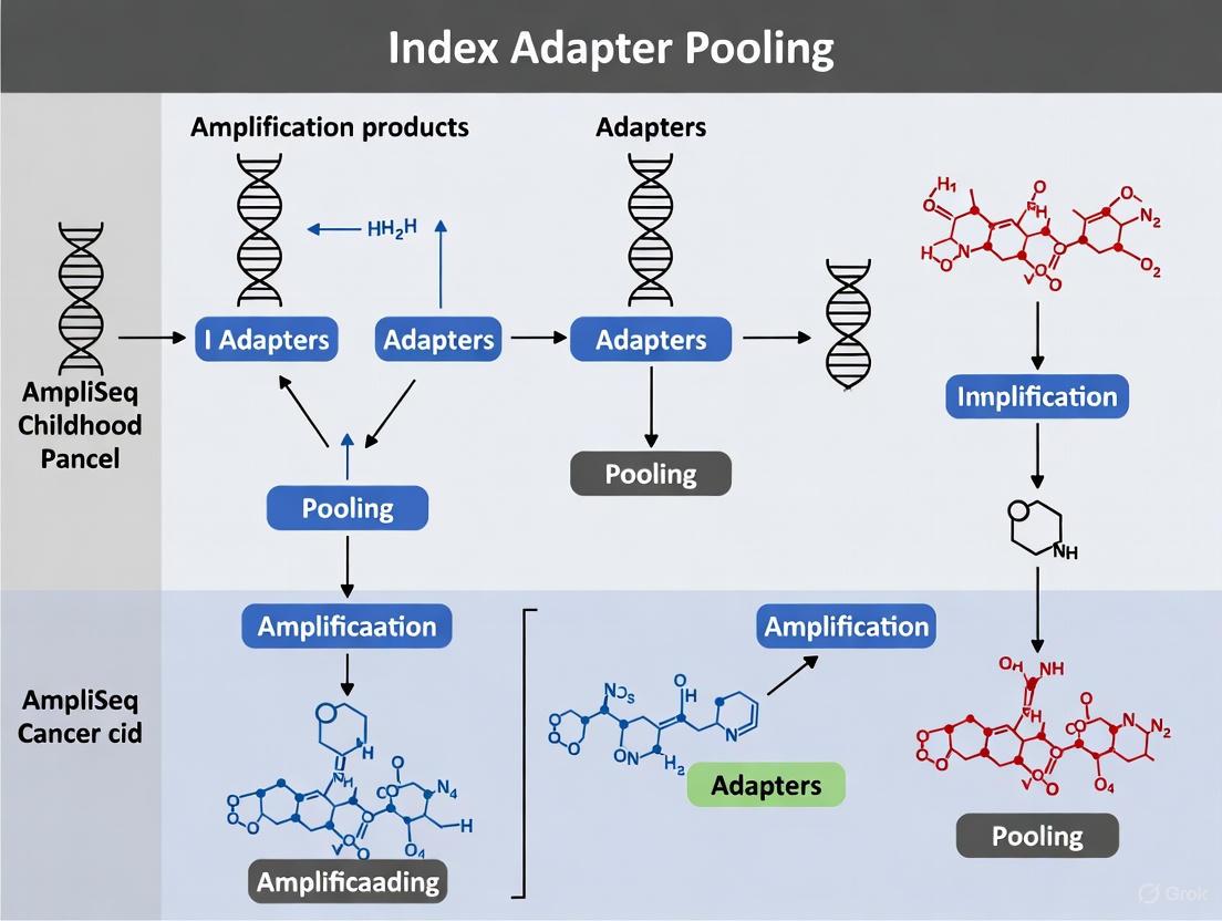
Within the context of AmpliSeq Childhood Cancer Panel research, efficient and accurate sequencing of multiple samples is paramount for investigating the 203 genes associated with pediatric and young adult cancers. Index adapter pooling is a critical methodological step that enables the multiplexing of libraries, allowing several libraries to be sequenced simultaneously in a single sequencing run. This guide details the specific guidelines for creating low-plexity pools of two to eight libraries using AmpliSeq Combinatorial Dual (CD) Indexes for Illumina. Adhering to these protocols ensures optimal color balance on Illumina sequencing systems, which is a prerequisite for high-quality base calling and reliable data output essential for somatic variant detection in cancer research [26] [27].
The AmpliSeq for Illumina Childhood Cancer Panel provides a targeted resequencing solution for comprehensive evaluation of somatic variants, including single nucleotide polymorphisms (SNPs), insertions-deletions (indels), copy number variants (CNVs), and gene fusions [6]. The integrated workflow, which includes PCR-based library preparation and Illumina Sequencing by Synthesis (SBS) technology, is designed to work seamlessly with the recommended pooling strategies outlined in the official Index Adapters Pooling Guide [26] [6] [28]. For researchers focusing on childhood cancers, mastering these pooling techniques translates to more efficient use of sequencing capacity, reduced per-sample costs, and accelerated data generation for drug development pipelines.
Key Concepts and Definitions
Understanding Plexity and Color Balance
- Plexity: Plexity is defined as the number of individual libraries combined into a single pool for sequencing. For example, combining twelve libraries results in a plexity of 12. This application note focuses specifically on low-plex pools, defined as pools containing between two and eight libraries [27].
- Color Balance: Illumina's SBS technology utilizes two types of fluorescent dyes. "Color balance" refers to achieving a nearly equal representation of all four bases (A, C, G, T) during each sequencing cycle, particularly within the index reads. Proper color balance is critical for minimizing phasing and pre-phasing errors and ensuring high-quality index demultiplexing [27].
- Combinatorial Dual (CD) Indexes: AmpliSeq CD Indexes are 8-base oligonucleotides used for dual indexing. In a CD indexing system, the unique identity of a sample is defined by the specific combination of an i7 (Index 1) and an i5 (Index 2) adapter. Unlike Unique Dual (UD) indexes, where each index sequence is entirely distinct, CD indexes share some common sequences across different adapters. This design means that most libraries in a pool will share a common index on either the i5 or i7 end, making careful pooling strategy more important for low-plexity experiments to maintain color balance [27].
The Scientist's Toolkit: Essential Research Reagent Solutions
Successful library preparation and pooling for childhood cancer research requires a suite of specialized reagents. The table below catalogs the essential materials and their specific functions within the AmpliSeq for Illumina workflow.
Table 1: Essential Research Reagent Solutions for AmpliSeq Childhood Cancer Panel Workflow
| Product Name | Catalog ID | Function and Key Features |
|---|---|---|
| AmpliSeq for Illumina Childhood Cancer Panel | 20028446 | Ready-to-use targeted panel for investigating 203 genes associated with childhood and young adult cancers. Sufficient for 24 samples [6]. |
| AmpliSeq Library PLUS for Illumina | 20019101 (24rxn) | Contains core reagents for library preparation. Panel and index adapters must be purchased separately [6]. |
| AmpliSeq CD Indexes Set A for Illumina | 20019105 | Includes 96 unique 8-base indexes, sufficient for labeling 96 samples. Compatible with all AmpliSeq for Illumina panels [6] [29]. |
| AmpliSeq CD Indexes Set B for Illumina | 20019106 | Includes 96 indexes, enabling larger studies or higher plexity pooling with a broader array of index combinations [6] [30]. |
| AmpliSeq CD Indexes Set C for Illumina | 20019107 | Includes 96 indexes, expanding the available combinatorial options for complex study designs [6] [30]. |
| AmpliSeq CD Indexes Set D for Illumina | 20019167 | Includes 96 indexes, completing the full set of 384 available indexes for large-scale projects [6] [30]. |
| AmpliSeq cDNA Synthesis for Illumina | 20022654 | Required to convert total RNA to cDNA when using the Childhood Cancer Panel with RNA input [6]. |
| AmpliSeq Library Equalizer for Illumina | 20019171 | An easy-to-use solution based on bead-based normalization, used to equalize the concentration of AmpliSeq libraries before pooling [6]. |
Experimental Protocol for Library Pooling
The following diagram illustrates the end-to-end workflow for preparing and pooling libraries using the AmpliSeq Childhood Cancer Panel, from nucleic acid input to a pooled library ready for sequencing.
Detailed Methodologies
Library Preparation and Index Ligation
- Input Material: Begin with 10 ng of high-quality DNA or total RNA. For RNA samples, you must first convert it to cDNA using the AmpliSeq cDNA Synthesis for Illumina kit [6].
- Library Construction: Perform the AmpliSeq library preparation protocol using the AmpliSeq Childhood Cancer Panel and the AmpliSeq Library PLUS reagents. This targeted, PCR-based assay generates amplicon libraries covering the 203 genes of interest [6].
- Index Ligation: Ligate the AmpliSeq CD Indexes to the prepared libraries. The indexes are available in Sets A, B, C, and D, each containing 96 unique 8-base indexes. For low-plex pooling, you can use any column- or row-based pooling strategy from any set [26] [6] [30]. Record the specific i7 and i5 index combination used for each sample on a sample sheet, as this is required for downstream demultiplexing [29].
Library Normalization and Pooling
- Purification and Quantification: Purify the final indexed libraries. Precisely quantify the concentration of each library using a fluorometry-based method appropriate for NGS libraries.
- Library Equalization: Use AmpliSeq Library Equalizer for Illumina to normalize all libraries to the same concentration. This bead-based normalization step is crucial for ensuring that each library is represented equally in the final pool [6].
- Low-Plex Pooling Strategy: To create a pool of 2 to 8 libraries, combine equal volumes of each normalized library. The official Index Adapters Pooling Guide provides specific, pre-validated index combinations that ensure color balance for these low-plexity pools. It is strongly recommended to consult this guide for your specific pooling setup [26] [28].
- Final Pool Validation: Quantify the final pooled library to confirm its concentration and validate its quality, for example, by using an electrophoretic assay, to ensure it is suitable for sequencing.
Data Presentation and Analysis
Pooling Strategy Specifications
Adherence to the following specifications is critical for generating high-quality sequencing data from low-plex pools. The table below summarizes the core parameters for the recommended pooling strategy.
Table 2: Quantitative Specifications for 2- to 8-Plex Library Pooling with AmpliSeq CD Indexes
| Parameter | Specification | Technical Rationale |
|---|---|---|
| Plexity Range | 2 to 8 libraries per pool | Optimized for color balance on Illumina sequencing systems [26]. |
| Index Type | Combinatorial Dual (CD) Indexes | 8-base indices designed for dual indexing; unique sample ID is from the i7/i5 combination [27]. |
| Index Strategy | Use any column- or row-based strategy from Sets A, B, C, or D | Provides flexibility in experimental design while maintaining compatibility [26] [30]. |
| Input Quantity | 10 ng of high-quality DNA or RNA (requires cDNA synthesis) | Standardized input ensures uniform library preparation efficiency and coverage [6]. |
| Library Prep Time | 5-6 hours (excludes quantification & pooling) | Informs experimental planning and throughput expectations [6]. |
| Recommended Guide | Index Adapters Pooling Guide (Illumina) | Contains validated low-plex index combinations to ensure color balance [26] [8] [28]. |
Logical Framework for Pooling Strategy
The logic of selecting an appropriate pooling strategy is primarily driven by the plexity of the intended pool, as this determines the requirement for a pre-balanced index combination. The following diagram outlines the decision-making workflow.
Discussion
Technical Advantages in Childhood Cancer Research
Implementing the prescribed guidelines for pooling 2 to 8 libraries with AmpliSeq CD Indexes provides several material benefits for cancer research. The use of pre-balanced index combinations directly mitigates the risk of sequencing failures due to poor color balance, a common pitfall in low-plexity runs. This is especially critical when working with precious samples, such as FFPE tissue or bone marrow aspirates, where sample quantity is limited and reproducibility is paramount [6] [27]. The combinatorial dual indexing design itself provides an additional layer of data integrity by reducing the probability of index hopping errors misassigning reads, which in turn ensures that somatic variant calls are accurately associated with the correct patient sample [27].
Integration with the Broader Workflow
The pooling protocol is not a standalone procedure but a key link in the integrated AmpliSeq for Illumina workflow. The normalized and pooled library is compatible with a range of Illumina sequencing systems, including the MiSeq, NextSeq 550, NextSeq 1000, and NextSeq 2000 series, allowing labs to select the platform that best matches their required scale and throughput [6]. Furthermore, the availability of accessory products like the AmpliSeq for Illumina Sample ID Panel allows researchers to generate unique genetic fingerprints for each sample, adding a layer of sample identity tracking that complements the indexing strategy [6]. For automated, high-throughput drug discovery environments, the pooling guidelines are compatible with liquid handling robots, facilitating seamless scale-up [6].
Within pediatric cancer genomics, efficient and accurate sequencing of both DNA and RNA from a single sample is paramount for comprehensive molecular profiling. The AmpliSeq for Illumina Childhood Cancer Panel provides a targeted resequencing solution for the evaluation of somatic variants in young adult and childhood cancers. A critical, yet often optimized, step in this integrated workflow is determining the correct pooling ratio of DNA and RNA libraries prior to sequencing. This application note provides a detailed, evidence-based protocol for determining the optimal DNA:RNA pooling ratio for combined analysis, ensuring cost-effective and reliable detection of variants, including single nucleotide polymorphisms (SNPs), insertions-deletions (indels), copy number variants (CNVs), and gene fusions [6] [9].
Technical Specifications and Sequencing Guidelines
The AmpliSeq Childhood Cancer Panel generates separate DNA and RNA (via cDNA) libraries from a single sample. The DNA component targets 3,069 amplicons across 203 genes, while the RNA component targets 1,701 amplicons to detect fusion genes [13]. The differing number of targets and desired coverage for each library type necessitate a specific pooling ratio to balance data output.
Illumina provides clear sequencing guidelines for combining these libraries, with the recommended ratio based on achieving sufficient read coverage for both data types [13].
Table 1: Sequencing System Specifications and Pooling Guidelines
| Sequencing System | Reagent Kit | Maximum Combined Samples per Run* | Recommended DNA:RNA Pooling Volume Ratio |
|---|---|---|---|
| MiniSeq System | MiniSeq High Output Kit | 4 | 5:1 |
| MiSeq System | MiSeq Reagent Kit v3 | 4 | 5:1 |
| NextSeq 550/1000/2000 Systems | NextSeq Mid Output v2 Kit | 22 | 5:1 |
| NextSeq 550/1000/2000 Systems | NextSeq High Output v2 Kit | 48 | 5:1 |
*Combined samples refer to paired DNA and RNA from the same source, generating two separately indexed libraries that are pooled for a single run [13].
Research Reagent Solutions
The following reagents are essential for implementing the combined DNA:RNA workflow with the AmpliSeq Childhood Cancer Panel.
Table 2: Essential Research Reagents for the Combined Workflow
| Item Name | Function in Workflow | Key Specifications |
|---|---|---|
| AmpliSeq for Illumina Childhood Cancer Panel | Ready-to-use primer pool | Targets 203 genes; generates 3069 DNA and 1701 RNA amplicons [6] [13]. |
| AmpliSeq Library PLUS for Illumina | Library preparation master mix | Contains reagents for PCR-based library construction; available in 24-, 96-, and 384-reaction configurations [6]. |
| AmpliSeq CD Indexes | Sample multiplexing | Unique 8-base indexes for sample identification; sold in sets of 96 (e.g., Set A-D) [6]. |
| AmpliSeq cDNA Synthesis for Illumina | RNA-to-cDNA conversion | Required to convert total RNA to cDNA for the RNA side of the panel [6] [13]. |
| AmpliSeq Library Equalizer for Illumina | Library normalization | Bead-based reagent for normalizing libraries before pooling, crucial for achieving the desired ratio [6]. |
Validated Experimental Protocol
This protocol is adapted from the manufacturer's guidelines and independent clinical validation studies [13] [9].
Nucleic Acid Input and Quality Control
- Input Quantity: Use 10-100 ng of high-quality DNA and 10-100 ng of high-quality total RNA per sample [6] [9]. The validated clinical study used 100 ng of each [9].
- Quality Control: Assess DNA and RNA purity via spectrophotometry (e.g., OD260/280 ratio >1.8) [9]. Assess RNA integrity using an instrument such as a TapeStation (Agilent) or Labchip (PerkinElmer) [9].
Library Preparation Workflow
Separate Library Construction:
- DNA Library: Amplify 100 ng of DNA using the Childhood Cancer Panel and the AmpliSeq Library PLUS kit. Incorporate a unique CD Index for each sample during this PCR step [6] [9].
- RNA (cDNA) Library: Convert 100 ng of total RNA to cDNA using the AmpliSeq cDNA Synthesis kit. Then, amplify the cDNA using the Childhood Cancer Panel and the AmpliSeq Library PLUS kit, incorporating a unique CD Index [6] [13] [9].
Library Normalization: Precisely normalize the concentration of each individual DNA and RNA library using the AmpliSeq Library Equalizer [6]. This step is critical for the success of the subsequent pooling step.
Combined Library Pooling:
- Pool the normalized DNA and RNA libraries from the same sample at a 5:1 volume ratio (DNA:RNA) [13]. For example, combine 5 µL of the normalized DNA library with 1 µL of the normalized RNA library.
- This ratio is designed to balance the output from the larger number of DNA amplicons with the RNA targets, ensuring adequate coverage for both.
Sequencing: Dilute the final pool to the appropriate loading concentration (e.g., 17-20 pM) and sequence on a supported Illumina platform, such as the MiSeq or NextSeq series, using the recommended reagent kit [13] [9].
The following diagram illustrates the key steps and the decisive pooling stage in this workflow.
Performance and Clinical Validation
Independent technical validation of the AmpliSeq Childhood Cancer Panel confirms that the recommended workflow, including the pooling strategy, delivers high-performance results suitable for clinical research.
- Sensitivity and Specificity: The panel demonstrated a sensitivity of 98.5% for DNA variants (at 5% variant allele frequency) and 94.4% for RNA fusions, with 100% specificity for DNA [9].
- Sequencing Performance: The validation study obtained a mean read depth greater than 1000x across targets, ensuring reliable variant calling [9].
- Clinical Impact: In a cohort of pediatric acute leukemia patients, the panel provided clinically relevant results in 43% of patients, refining diagnosis and identifying targetable mutations [9].
The 5:1 DNA:RNA pooling ratio is a key, empirically validated parameter for the successful combined analysis of the AmpliSeq for Illumina Childhood Cancer Panel. Adherence to this ratio, integrated with a robust library preparation and normalization protocol, ensures balanced sequencing coverage, maximizes the utility of sequencing capacity, and yields highly sensitive and specific data. This standardized approach empowers researchers to reliably detect the spectrum of genomic alterations driving childhood cancers, thereby accelerating precision medicine in pediatric oncology.
Calculating Sample Throughput and Reagent Requirements for 24 to 384 Samples
Within the framework of a comprehensive index adapter pooling guide for AmpliSeq Childhood Cancer Panel research, precise calculation of sample throughput and reagent requirements is a critical step in experimental design. The AmpliSeq for Illumina Childhood Cancer Panel is a targeted resequencing solution for the comprehensive evaluation of somatic variants associated with childhood and young adult cancers [6]. This application note provides detailed methodologies and structured data tables to enable researchers, scientists, and drug development professionals to accurately scale their experiments from 24 to 384 samples while maintaining protocol integrity and sequencing quality.
The AmpliSeq Childhood Cancer Panel for Illumina is a PCR-based targeted sequencing panel designed to analyze 203 genes associated with pediatric and young adult cancers [9]. The panel simultaneously detects multiple variant types including single nucleotide variants (SNVs), insertions-deletions (indels), copy number variants (CNVs), and gene fusions [6] [9]. The integrated workflow includes AmpliSeq for Illumina library preparation, Illumina sequencing by synthesis (SBS) technology, and automated analysis, providing a complete solution from library preparation to data generation [6].
Key technical specifications of the panel include an assay time of 5-6 hours for library preparation only (excluding library quantification, normalization, or pooling time), with less than 1.5 hours of hands-on time [6]. The protocol requires only 10 ng of high-quality DNA or RNA input and is compatible with various specialized sample types including blood, bone marrow, and FFPE tissue [6]. The panel generates both DNA and RNA libraries for each sample, enabling comprehensive genomic profiling [13].
Research Reagent Solutions
Successful implementation of the AmpliSeq Childhood Cancer Panel requires several specialized reagents and kits. The table below details the essential materials and their specific functions within the experimental workflow.
Table 1: Essential Research Reagent Solutions for AmpliSeq Childhood Cancer Panel
| Component Category | Product Name | Function | Key Specifications |
|---|---|---|---|
| Library Prep | AmpliSeq Library PLUS for Illumina [6] | Provides core reagents for preparing sequencing libraries | Available in 24-, 96-, and 384-reaction configurations |
| Index Adapters | AmpliSeq CD Indexes Sets A-D [6] | Labels individual samples with unique barcodes for multiplexing | Each set contains 96 unique 8 bp indexes; Set A sufficient for 96 samples |
| RNA Conversion | AmpliSeq cDNA Synthesis for Illumina [6] | Converts total RNA to cDNA for RNA library preparation | Required for working with RNA samples and panels |
| Library Normalization | AmpliSeq Library Equalizer for Illumina [6] | Normalizes libraries for sequencing | Uses beads and reagents for consistent library representation |
| Sample Tracking | AmpliSeq for Illumina Sample ID Panel [6] | Generates unique IDs for sample identification via SNP genotyping | Includes 8 SNP-targeting primer pairs + 1 gender-determining pair |
| FFPE Processing | AmpliSeq for Illumina Direct FFPE DNA [6] | Prepares DNA from FFPE tissues without deparaffinization | 24 reactions per kit; no DNA purification required |
Kit Calculation and Sample Scaling
Precise calculation of required kits is essential for experimental planning and budgeting. The AmpliSeq Childhood Cancer Panel generates one DNA and one RNA library per sample, effectively doubling the number of libraries relative to sample count [13]. The following table provides exact kit requirements for standard sample throughputs.
Table 2: Kit Requirements for Different Sample Throughputs
| Number of Samples | Number of Libraries | AmpliSeq Childhood Cancer Panel | AmpliSeq Library PLUS for Illumina | AmpliSeq CD Set A | cDNA Synthesis |
|---|---|---|---|---|---|
| 24 Samples | 48 Libraries (24 DNA, 24 RNA) | 1 | 2 × 24-reaction kits | 1 | 1 |
| 96 Samples | 192 Libraries (96 DNA, 96 RNA) | 4 | 2 × 96-reaction kits | 2 | 1 |
| 384 Samples | 768 Libraries (384 DNA, 384 RNA) | 16 | 2 × 384-reaction kits | 8 | 4 |
Library Preparation and Pooling Protocol
Nucleic Acid Input and Quality Control
The protocol requires 100 ng each of DNA and RNA per sample [9]. DNA and RNA purity should be determined by spectrophotometry with all samples exhibiting an OD260/280 ratio >1.8 [9]. Integrity must be assessed using systems such as Labchip or TapeStation, and concentration should be determined by fluorometric quantification using instruments like the Qubit 4.0 Fluorimeter with appropriate assay kits [9].
Library Preparation Workflow
Figure 1: Library preparation workflow for DNA and RNA samples.
The library preparation process begins with simultaneous processing of DNA and RNA samples. For RNA, the first step involves reverse transcription to cDNA using the AmpliSeq cDNA Synthesis kit [9]. Following this, both DNA and cDNA undergo PCR amplification using the Childhood Cancer Panel to generate amplicons. The panel creates 3069 amplicons per DNA sample with an average size of 114 bp, and 1701 amplicons per RNA sample with an average size of 122 bp [13]. After amplification, specific barcodes are ligated to each sample using the AmpliSeq CD Indexes [9]. Quality controls are performed after library cleanup, and libraries are diluted to 2 nM concentration [9].
Library Pooling and Normalization
For combined DNA and RNA sequencing, libraries should be pooled at a 5:1 DNA:RNA ratio based on recommended read coverage requirements [13]. This ratio ensures optimal coverage for both variant detection and fusion identification. The final pool is diluted to 17-20 pM for sequencing on Illumina platforms [9]. The AmpliSeq Library Equalizer for Illumina can be used to normalize libraries before pooling, ensuring even representation across samples [6].
Sequencing System Selection and Throughput
Selection of appropriate sequencing systems is crucial for achieving optimal coverage while maximizing cost-efficiency. The following table provides detailed specifications for compatible Illumina sequencing systems.
Table 3: Sequencing System Specifications and Throughput
| System | Reagent Kit | Max # DNA Samples Only per Run | Max # RNA Samples Only per Run | Max # Combined* Samples per Run | Recommended DNA:RNA Pooling Ratio | Run Time |
|---|---|---|---|---|---|---|
| MiniSeq System | MiniSeq Mid Output | 1 | 8 | 1 | 5:1 | 17 hours |
| MiniSeq System | MiniSeq High Output | 5 | 25 | 4 | 5:1 | 24 hours |
| MiSeq System | MiSeq Reagent Kit v2 | 3 | 15 | 2 | 5:1 | 24 hours |
| MiSeq System | MiSeq Reagent Kit v3 | 5 | 25 | 4 | 5:1 | 32 hours |
| NextSeq System | NextSeq Mid Output v2 | 27 | 96 | 22 | 5:1 | 26 hours |
| NextSeq System | NextSeq High Output v2 | 83 | 96 | 48 | 5:1 | 29 hours |
*Combined means paired DNA and RNA from the same sample that generates two libraries, one from each nucleic acid and separately indexed.
Sequencing Configuration and Data Analysis
Figure 2: Decision workflow for sequencing configuration.
The panel demonstrates robust performance characteristics with a mean read depth greater than 1000× and high sensitivity for both DNA (98.5% for variants with 5% variant allele frequency) and RNA (94.4%) [9]. The assay shows 100% specificity and reproducibility for DNA and 89% reproducibility for RNA [9]. This performance enables reliable detection of clinically relevant variants, with studies showing that 49% of mutations and 97% of fusions identified have clinical impact, refining diagnosis in 41% of mutations and providing targetable findings in 49% of them [9].
Technical Validation and Quality Control
Sensitivity and Reproducibility Assessment
Technical validation of the AmpliSeq Childhood Cancer Panel should include sensitivity, specificity, and reproducibility assessments using appropriate controls [9]. For DNA analyses, commercial controls such as SeraSeq Tumor Mutation DNA Mix can be used, containing clinically relevant DNA variants at an average variant allele frequency of 10% [9]. For RNA analyses, SeraSeq Myeloid Fusion RNA Mix provides synthetic RNA fusions combined with RNA from reference lines [9]. Negative controls should include reference materials such as NA12878 for DNA and IVS-0035 for RNA [9].
Limit of Detection Establishment
The limit of detection (LOD) should be established for both DNA and RNA components. Validation studies have demonstrated that the panel can detect variants at 5% variant allele frequency for DNA with 98.5% sensitivity [9]. For RNA fusions, the LOD should be determined using dilution series of positive control materials to establish the minimum detectable concentration while maintaining assay specificity and reproducibility.
This application note provides comprehensive guidance for calculating sample throughput and reagent requirements when implementing the AmpliSeq for Illumina Childhood Cancer Panel across scales from 24 to 384 samples. By following the detailed protocols, kit calculations, and sequencing guidelines outlined herein, researchers can effectively plan and execute robust genomic profiling studies for childhood cancer research. The structured approach to library preparation, indexing, pooling, and sequencing system selection ensures optimal performance of this validated method for refining pediatric acute leukemia diagnosis, prognosis, and treatment strategies.
Within the framework of a broader thesis on index adapter pooling strategies for AmpliSeq Childhood Cancer Panel research, this application note provides detailed sequencing configuration guidelines. Targeted sequencing panels, such as the 203-gene AmpliSeq Childhood Cancer Panel, require precise calibration of sequencing parameters to achieve optimal coverage and detect somatic variants with high confidence [6]. This document is designed to assist researchers, scientists, and drug development professionals in selecting the appropriate Illumina sequencing system and configuring run parameters to ensure data quality and cost-effectiveness for their pediatric cancer studies. The recommendations herein are based on manufacturer specifications and established genomic practices.
System Comparison and Selection
Choosing between the MiSeq and NextSeq systems depends on the project's scale, required throughput, and desired run time. The table below summarizes key specifications to guide this decision.
Table 1: Sequencing System Comparison for Targeted Panel Sequencing
| Parameter | MiSeq System | NextSeq 550 System | NextSeq 1000/2000 System (P2 Flow Cell) |
|---|---|---|---|
| Recommended Output Range | 300 Mb - 15 Gb [31] | 16.25 - 120 Gb [32] | 40 - 240 Gb [33] |
| Max Paired-End Reads | 44-50M (v3 chemistry) [31] | ≤ 800M (High-Output Kit) [32] | 800M (P2 flow cell) [33] |
| Typical Run Time (2x150 bp) | ~24 hours (v2 kit) [31] | ~29 hours (High-Output Kit) [32] | ~22 hours [33] |
| Quality Scores (Q30) | >80% (2x150 bp, v2) [31] | >75% (2x150 bp) [32] | ≥90% (2x150 bp) [33] |
| Ideal Use Case | Low-plexity panels, small sample batches, method development | High-plexity panels, exomes, transcriptomes | Ultra-high multiplexing, large-scale studies |
For the AmpliSeq Childhood Cancer Panel, which is compatible with both MiSeq and NextSeq systems, the choice hinges on the number of samples being pooled in a single run [6]. The MiSeq is optimal for labs processing smaller batches or requiring rapid turnaround, while the NextSeq platforms are better suited for core facilities that multiplex dozens of samples to achieve a higher throughput and lower cost per sample.
Recommended Coverage and Run Parameters
Coverage Requirements for the Childhood Cancer Panel
The AmpliSeq Childhood Cancer Panel is a targeted resequencing solution for evaluating somatic variants in 203 genes associated with pediatric and young adult cancers [6]. For targeted DNA sequencing panels like this one, the primary goal is to achieve sufficient depth of coverage to confidently call heterozygous single nucleotide variants (SNPs), insertions-deletions (indels), and copy number variants (CNVs). While the panel's specific recommended coverage is not explicitly stated in the search results, general guidelines for somatic variant detection suggest a minimum coverage of 200x to 500x is necessary to reliably identify low-frequency subclonal populations. This ensures a high probability of sequencing both alleles and detecting variants present in a fraction of the cells.
Configuring Run Parameters
To achieve the required coverage, the key parameters are the read length, read depth, and indexing strategy.
- Read Length: A 2x150 bp paired-end run is the recommended configuration for amplicon-based panels like the AmpliSeq Childhood Cancer Panel. This length is sufficient to cover the entire amplicon, ensure high-quality base calls across the read, and provide positional information to resolve structural rearrangements [34].
- Read Depth and Multiplexing: The total number of reads required per sample depends on the target coverage and the total size of the genomic regions covered by the panel. The number of samples that can be pooled in a single run is then determined by the total reads generated by the sequencing instrument and the reads needed per sample.
Table 2: Sample Multiplexing Capacity on Different Systems (2x150 bp)
| Sequencing System | Reagent Kit / Flow Cell | Total Paired-End Reads | Estimated Samples per Run (at 3M reads/sample) | Estimated Samples per Run (at 5M reads/sample) |
|---|---|---|---|---|
| MiSeq | V2 Kit | 24-30M [31] | 8 | 4-5 |
| MiSeq | V3 Kit | 44-50M [31] | 14-16 | 8-9 |
| NextSeq 550 | High-Output Kit | ≤ 800M [32] | ~260 | ~160 |
| NextSeq 1000/2000 | P2 Flow Cell | 800M [33] | ~260 | ~160 |
Index Adapter Pooling Strategy
A critical step for multiplexing samples is the use of dual index adapters, which incorporate unique molecular barcodes (i5 and i7) for each sample [35]. This allows for the pooling of multiple libraries into a single sequencing run and subsequent bioinformatic deconvolution. To ensure data quality:
- Use Balanced Index Combinations: Follow the Index Adapters Pooling Guide to select index combinations that are balanced in base composition, which minimizes phasing and prephasing errors during the sequencing run [21] [28].
- Avoid Index Hopping: Employ unique dual indexes (UDIs) to mitigate the risk of index hopping, a phenomenon where reads are assigned to the wrong sample due to errors in index sequencing.
Experimental Protocol: End-to-End Workflow
The following diagram illustrates the complete experimental workflow from sample preparation to data analysis for the AmpliSeq Childhood Cancer Panel.
Detailed Step-by-Step Methodology
Library Preparation
The AmpliSeq for Illumina Childhood Cancer Panel uses a PCR-based amplicon sequencing method [6].
- Input Material: Begin with 10 ng of high-quality DNA. For RNA samples, first convert to cDNA using the AmpliSeq cDNA Synthesis for Illumina kit [6].
- Library Amplification: Use the AmpliSeq Library PLUS reagent kit to generate amplicon libraries from the panel's primer pool, which targets the 203 cancer-associated genes. The total assay time for library preparation is approximately 5-6 hours with less than 1.5 hours of hands-on time [6].
- Indexing and Adapter Ligation: Ligate unique dual index adapters (e.g., from AmpliSeq CD Indexes sets) to each sample's library. This step is crucial for multiplexing and requires careful planning to use balanced index combinations as per the Index Adapters Pooling Guide [21] [28].
Library Quality Control and Pooling
- Normalization: Use AmpliSeq Library Equalizer for Illumina to normalize libraries, ensuring an equimolar representation of each sample in the final pool [6].
- Pooling: Combine the normalized, indexed libraries into a single pool. The number of libraries pooled depends on the desired coverage per sample and the output capacity of the chosen sequencing instrument (see Table 2).
Sequencing Run Setup
- System Configuration: On the MiSeq or NextSeq instrument, select the appropriate reagent kit and input the cycle configuration for a 2x150 bp paired-end run [31] [33] [32].
- Cluster Generation and Sequencing: Load the pooled library onto the flow cell. The instrument will perform cluster generation, followed by sequencing by synthesis (SBS). Monitor key run metrics in real-time using software like the Sequencing Analysis Viewer (SAV), paying close attention to cluster density, % bases ≥ Q30, and phasing/prephasing [35].
The Scientist's Toolkit: Research Reagent Solutions
The following table lists the essential materials and kits required to perform a sequencing study using the AmpliSeq Childhood Cancer Panel.
Table 3: Essential Research Reagents and Kits for the AmpliSeq Workflow
| Item Name | Catalog ID (Example) | Function in the Workflow |
|---|---|---|
| AmpliSeq for Illumina Childhood Cancer Panel | 20028446 [6] | Ready-to-use primer pool for amplifying 203 target genes associated with childhood cancers. |
| AmpliSeq Library PLUS for Illumina | 20019101 (24 rxns) [6] | Core reagents for PCR-based library preparation from amplicons. |
| AmpliSeq CD Indexes for Illumina | 20019105 (Set A) [6] | Unique dual index adapters for sample multiplexing and identification. |
| AmpliSeq cDNA Synthesis for Illumina | 20022654 [6] | Converts input RNA to cDNA for use with the panel when analyzing RNA targets. |
| AmpliSeq Library Equalizer for Illumina | 20019171 [6] | Beads and reagents for normalizing libraries prior to pooling, ensuring balanced representation. |
| AmpliSeq for Illumina Direct FFPE DNA | 20023378 [6] | Prepares DNA directly from FFPE tissues without the need for deparaffinization or purification. |
| MiSeq Reagent Kit v2 (300-cycle) | N/A [31] | Flow cell and reagents for sequencing on the MiSeq platform (e.g., for 2x150 bp runs). |
| NextSeq P2 Cartridge (300-cycle) | N/A [33] | Flow cell and reagents for sequencing on the NextSeq 1000/2000 platform. |
Troubleshooting Index Balance and Data Quality in Multiplexed Runs
Addressing Common Issues in Library Quantification and Normalization
Next-generation sequencing (NGS) library preparation is a critical step in genomic workflows, with library quantification and normalization being significant sources of inefficiency that can waste resources and increase costs [36]. For targeted panels like the AmpliSeq for Illumina Childhood Cancer Panel, which enables comprehensive evaluation of 203 genes associated with pediatric and young adult cancers, precise quantification is essential for generating reliable variant data [6]. In the context of index adapter pooling, inaccurate library quantification directly compromises pooling efficiency, leading to unbalanced sequencing representation and potentially misleading research conclusions. This application note details common pitfalls in library quantification and normalization and provides optimized protocols to ensure data quality and reproducibility for childhood cancer research.
Common Quantification and Normalization Challenges
Limitations of Standard QC Methods
Each common library QC method has specific limitations that can introduce variability if not properly addressed. The table below summarizes the primary constraints of these standard techniques:
Table 1: Limitations of Common NGS Library QC Methods
| Method | Key Limitations | Impact on Library Preparation |
|---|---|---|
| Fluorometry | Measures total nucleic acid concentration (ng/µL) rather than functional molarity; cannot distinguish between sequenceable molecules and adapter dimers [37]. | Leads to inaccurate normalization and imbalanced representation in pooled libraries [37]. |
| qPCR | Labor-intensive and time-consuming; requires multiple sample dilutions and prior fragment size analysis; introduces user-user variability through manual steps; higher reagent costs [37]. | Reduces workflow efficiency and consistency; potential for batch effects across large sample sets [36]. |
| Microfluidic Electrophoresis | Costly and slow for individual sample analysis; provides size distribution but indirect concentration estimation [37]. | Impractical for high-throughput applications; increases project timelines and costs. |
Consequences of Improper Normalization
Inaccurate library quantification directly impacts the efficiency of index adapter pooling and subsequent sequencing outcomes:
- Pooling Imbalance: Under-represented libraries in multiplexed runs yield insufficient data, while over-represented libraries consume disproportionate sequencing resources [37] [38].
- Suboptimal Flow Cell Loading: Improperly quantified pools cause over- or under-clustering on the flow cell, leading to run failures, reduced data quality, and increased sequencing costs [37] [38].
- Compromised Data Integrity: Imbalanced sequencing depth across samples can skew variant detection, particularly for low-frequency somatic mutations relevant in childhood cancer profiling [38].
Optimized Methodologies for Library QC
Method Comparison and Selection
Researchers should select quantification methods based on throughput requirements, available resources, and necessary accuracy. The following table compares standard and advanced methods:
Table 2: Comparison of Library Quantification and Normalization Methods
| Method | Accuracy | Hands-on Time | Cost Considerations | Best Use Cases |
|---|---|---|---|---|
| Fluorometry | Low (total nucleic acid) | Low | Low | Initial quality check; not recommended for final pooling [37]. |
| qPCR | High (functional molecules only) | High (1-4 hours) | High (reagents, consumables) | Gold standard for applications requiring high accuracy [37]. |
| Microfluidic Electrophoresis | Medium (size information) | Medium | High (per-sample cost) | Assessing library fragment size distribution [37]. |
| NuQuant | High (direct molarity) | Very Low (~6 minutes) | Medium | High-throughput workflows; ideal for large sample batches [37]. |
| Bead-Based Normalization | High (post-cleaning) | Low with automation | Low | Automated workflows using systems like G.STATION [36]. |
Detailed Experimental Protocols
qPCR Quantification Protocol for AmpliSeq Libraries
This protocol is optimized for libraries generated from the AmpliSeq Childhood Cancer Panel [6].
Materials Required:
- qPCR instrument and compatible SYBR Green or TaqMan master mix
- Library standards of known concentration
- Nuclease-free water and PCR plates/tubes
- Primers compatible with Illumina adapter sequences
Procedure:
- Perform Serial Dilutions: Dilute library samples 1:10,000 and 1:100,000 in nuclease-free water [37].
- Prepare Standards: Create a standard curve using library standards diluted across a range of 0.01-10 pM.
- Set Up Reactions: Combine 5 µL of diluted library or standard with 15 µL of master mix containing primers in a 96-well plate. Include no-template controls.
- Run qPCR Program:
- Step 1: 95°C for 2 minutes (initial denaturation)
- Step 2: 95°C for 15 seconds (denaturation)
- Step 3: 60°C for 1 minute (annealing/extension)
- Repeat Steps 2-3 for 40 cycles
- Data Analysis: Calculate library concentrations based on the standard curve. Adjust concentrations to 4 nM in preparation for pooling.
Normalization Using Bead-Based Cleanup
This method utilizes the AmpliSeq Library Equalizer for Illumina or similar products [6].
Materials Required:
- Magnetic beads (e.g., SPRIselect)
- Freshly prepared 80% ethanol
- Tris-HCl buffer (10 mM, pH 8.5)
- Magnetic stand compatible with sample format
Procedure:
- Combine Samples and Beads: Add magnetic beads to purified library at a sample:bead ratio of 1:1 in a 96-well plate.
- Incubate and Separate: Incubate for 5 minutes at room temperature. Place on magnetic stand until supernatant clears.
- Wash: Remove supernatant. Wash beads twice with 80% ethanol while plate is on magnetic stand.
- Elute: Air dry beads for 5-10 minutes. Remove from magnetic stand and elute DNA in Tris-HCl buffer.
- Quantify and Pool: Measure concentration via fluorometry and pool equal masses of each library.
Automated Normalization Workflow
Automation significantly improves reproducibility in high-throughput settings. The following diagram illustrates an automated workflow for library normalization:
The Scientist's Toolkit: Essential Research Reagents
Successful library preparation and quantification for the AmpliSeq Childhood Cancer Panel requires several specialized reagents and tools. The following table details essential components:
Table 3: Essential Research Reagent Solutions for AmpliSeq Workflows
| Item | Function | Example Product |
|---|---|---|
| Library Prep Kit | Provides reagents for preparing sequencing libraries from DNA/RNA. | AmpliSeq Library PLUS for Illumina [6]. |
| Target Enrichment Panel | Contains primers for amplifying genes of interest. | AmpliSeq for Illumina Childhood Cancer Panel [6]. |
| Index Adapters | Enable sample multiplexing through unique barcodes. | AmpliSeq CD Indexes for Illumina [6]. |
| Library Normalization Beads | Streamline library cleanup and normalization. | AmpliSeq Library Equalizer for Illumina [6]. |
| cDNA Synthesis Kit | Converts RNA to cDNA for RNA-based panels. | AmpliSeq cDNA Synthesis for Illumina [6]. |
| FFPE DNA Preparation | Enables library construction from FFPE tissues without DNA purification. | AmpliSeq for Illumina Direct FFPE DNA [6]. |
| Automated Liquid Handler | Precisely dispenses reagents in nanoliter volumes, reducing pipetting errors. | I.DOT Liquid Handler [36]. |
| Library QC Solution | Provides accurate molar concentration measurement without fragment analysis. | NuQuant Technology [37]. |
Integrated Workflow for Reliable Library Pooling
The complete workflow from library preparation to sequencing-ready pool incorporates multiple quality control checkpoints to ensure balanced index adapter pooling. The following diagram illustrates this integrated approach:
Robust library quantification and normalization are foundational to generating reliable sequencing data with the AmpliSeq Childhood Cancer Panel. By understanding the limitations of common QC methods, implementing appropriate protocols, and leveraging automation where possible, researchers can significantly improve the quality of their index adapter pooling experiments. These optimized approaches ensure balanced representation across samples, maximize sequencing efficiency, and ultimately contribute to more meaningful insights in childhood cancer research.
Optimizing Pooling Volumes to Prevent Read Depth Disparities Between Samples
Within next-generation sequencing (NGS) workflows for the AmpliSeq for Illumina Childhood Cancer Panel, effective library pooling is a critical step to ensure uniform sequencing coverage and prevent read depth disparities between samples. This protocol outlines a standardized methodology for optimizing pooling volumes when using this panel, directly supporting the broader objective of generating reproducible and reliable genetic data for pediatric cancer research [13] [9]. By adhering to the prescribed pooling ratios and quality control measures detailed herein, researchers can achieve balanced sequencing results, which is fundamental for the accurate detection of somatic variants, including single nucleotide polymorphisms (SNPs), insertions-deletions (indels), gene fusions, and copy number variants (CNVs) [6].
Key Concepts and Definitions
- Plexity: The number of individual libraries combined into a single sequencing pool. Higher plexity pools are inherently more color-balanced [27].
- Color Balance: The optimization of nucleotide diversity across all sequencing cycles during a run. This is crucial for accurate base calling on Illumina sequencing systems [27].
- Index Adapters: Short DNA sequences, specifically the Index 1 (i7) and Index 2 (i5) indexes, that are attached to sample libraries during preparation to allow for multiplexing. The AmpliSeq for Illumina Childhood Cancer Panel typically uses Combinatorial Dual (CD) Indexes, which have a limit of eight unique pairs [27] [13].
- Dual Indexing: A strategy that adds both an i7 and an i5 index sequence to each library, enabling more complex multiplexing and improved demultiplexing accuracy [27].
Recommended Pooling Ratios and Sequencing Specifications
For the AmpliSeq Childhood Cancer Panel, which generates one DNA and one RNA library per sample, a consistent DNA-to-RNA pooling ratio is recommended across Illumina sequencing platforms to ensure balanced coverage [13] [9]. This ratio is designed to accommodate the different coverages required for DNA and RNA analysis.
Table 1: Sequencing System Specifications and Pooling Guidance
| Sequencing System | Reagent Kit | Maximum Combined* Samples per Run | Recommended Combined* DNA:RNA Pooling Volume Ratio | Run Time |
|---|---|---|---|---|
| MiniSeq System | MiniSeq Mid Output Kit | 1 | 5:1 | 17 hours |
| MiniSeq System | MiniSeq High Output Kit | 4 | 5:1 | 24 hours |
| MiSeq System | MiSeq Reagent Kit v2 | 2 | 5:1 | 24 hours |
| MiSeq System | MiSeq Reagent Kit v3 | 4 | 5:1 | 32 hours |
| NextSeq System | NextSeq Mid Output v2 Kit | 22 | 5:1 | 26 hours |
| NextSeq System | NextSeq High Output v2 Kit | 48 | 5:1 | 29 hours |
*Combined refers to paired DNA and RNA from the same sample, resulting in two separately indexed libraries. [13]
The following workflow diagram illustrates the key stages from library preparation to sequencing, highlighting the crucial pooling step.
Experimental Protocol for Library Preparation and Pooling
Nucleic Acid Extraction and QC
- Input Quantity: Use a minimum of 10 ng of high-quality DNA or RNA. For formalin-fixed, paraffin-embedded (FFPE) tissues, the AmpliSeq for Illumina Direct FFPE DNA kit can be used without prior DNA purification [6].
- Purity and Integrity: Assess nucleic acid purity by spectrophotometry (OD260/280 ratio >1.8). Evaluate DNA and RNA integrity using systems such as Labchip or TapeStation [9].
Library Preparation
The protocol below is based on the manufacturer's instructions and validated literature [9] [6].
- * cDNA Synthesis (for RNA):* Convert 100 ng of total RNA to cDNA using the AmpliSeq cDNA Synthesis for Illumina kit [9] [6].
- * Amplicon Generation:*
- Use 100 ng of DNA per sample to generate 3,069 amplicons (average size: 114 bp).
- Use the synthesized cDNA from 100 ng of RNA input to generate 1,701 amplicons (average size: 122 bp) targeting fusion genes [9].
- * Indexing and PCR:* Generate amplicon libraries by performing consecutive PCRs with specific barcodes (e.g., AmpliSeq CD Indexes) for each sample [13] [9].
Library QC, Normalization, and Pooling
- * Library Quantification:* Perform quality controls after library cleanup. Quantify libraries fluorometrically using a system like Qubit 4.0 Fluorimeter [9].
- * Library Normalization:* Normalize all DNA and RNA libraries to the same concentration (e.g., 2 nM) using a system such as the AmpliSeq Library Equalizer for Illumina [6].
- * Volumetric Pooling:* Combine the normalized DNA and RNA libraries in a 5:1 volume ratio (DNA:RNA) into a final pool [13] [9].
- * Final Dilution:* Dilute the final pool to the appropriate loading concentration for your sequencer (e.g., 17-20 pM for a MiSeq system) [9].
The Scientist's Toolkit: Essential Research Reagent Solutions
Table 2: Key Reagents and Materials for the Workflow
| Item Name | Function | Catalog Number Example (for 24 samples) |
|---|---|---|
| AmpliSeq for Illumina Childhood Cancer Panel | Ready-to-use primer panel for targeting 203 genes associated with pediatric cancers. | 20028446 [6] |
| AmpliSeq Library PLUS for Illumina | Provides reagents for preparing sequencing libraries. Includes enzymes and master mix. | 20019101 (24 rxns) [6] |
| AmpliSeq CD Indexes | Contains unique nucleotide sequences (barcodes) for multiplexing samples. | Set A (20019105) [13] [6] |
| AmpliSeq cDNA Synthesis for Illumina | Converts total RNA to cDNA for subsequent library preparation from RNA targets. | 20022654 [6] |
| AmpliSeq Library Equalizer | Beads and reagents for normalizing library concentrations prior to pooling, saving hands-on time. | 20019171 [6] |
| AmpliSeq for Illumina Direct FFPE DNA | Prepares DNA directly from FFPE tissues without deparaffinization or DNA purification. | 20023378 [6] |
Troubleshooting and Best Practices
- Preventing Read Depth Disparities: The primary cause of read depth disparity is inaccurate library quantification or failure to follow the 5:1 pooling ratio. Always use fluorometric quantification (e.g., Qubit) over spectrophotometric methods for accurate library concentration measurement [9].
- Index Selection and Color Balance: When using Combinatorial Dual (CD) Indexes, which have a limited set of unique pairs, careful planning is required to ensure color balance across sequencing cycles. Refer to the Illumina Adapter Sequences document for sequence information to verify balance [27].
- Low Signal or Failed Runs: If the sequencer reports low cluster density, verify the final pool concentration with a highly sensitive method such as qPCR. Ensure all purification steps during library prep were performed correctly and that amplicons are of the expected size.
- Addressing Sample Cross-Talk: If demultiplexing errors occur, confirm that unique index combinations were used for each sample in the pool and that indexes are from the same designed set to ensure orthogonality.
Strategies for Mitigating Index Hopping and Cross-Contamination
Index hopping, also known as index switching, is a phenomenon in next-generation sequencing (NGS) where sequencing reads are misassigned from their expected sample index to a different index within a multiplexed pool [39]. This occurs when sample-specific DNA barcodes, or indexes, become erroneously associated with DNA fragments from different libraries during the sequencing process [40]. Although index hopping typically affects only 0.1% to 2% of total reads, this misassignment can significantly impact data interpretation in sensitive applications such as low-frequency somatic variant detection, single-cell RNA sequencing, and circulating tumor DNA analysis [39] [41] [42].
The emergence of patterned flow cell technologies and exclusion amplification (ExAmp) chemistry on platforms such as the Illumina NovaSeq 6000, NextSeq 2000, and HiSeq 4000 has exacerbated index hopping rates compared to non-patterned flow cell instruments [39] [40]. In these systems, DNA fragments and amplification primers coexist in solution rather than being surface-bound, increasing the likelihood that free-floating adapters can anneal to and amplify unintended fragments [40]. For research using the AmpliSeq Childhood Cancer Panel, which targets 203 genes associated with pediatric and young adult cancers, preventing index hopping is particularly crucial as even low levels of cross-contamination can lead to false positive variant calls and compromised data integrity [6].
Mechanisms and Impact of Index Hopping
Primary Causes of Index Misassignment
Index hopping results from multiple interconnected mechanisms throughout the NGS workflow. Understanding these sources is essential for developing effective mitigation strategies:
Free Adapter Contamination: Incomplete removal of unbound indexing adapters and adapter dimers after library preparation creates a pool of free-floating barcodes that can participate in cross-sample annealing during cluster generation [39] [40]. This residual adapter contamination represents a significant contributor to index hopping.
Jumping PCR during Multiplexed Capture: Also known as "template switching," this occurs during post-capture PCR amplification when an incompletely extended PCR product from one sample dissociates and acts as a primer on a different template molecule, incorporating incorrect index sequences [42] [40].
Patterned Flow Cell Chemistry: The ExAmp clustering chemistry used on patterned flow cells increases the likelihood of index hopping compared to non-patterned flow cells [39]. The solution-based amplification in nanowells allows more opportunity for index sequences to transfer between molecules before solid-phase attachment.
Oligonucleotide Synthesis and Handling Errors: Contamination during commercial adapter synthesis, purification, dilution, or aliquoting can introduce index misassignment from the earliest stages of library preparation [42].
Sequencing and Demultiplexing Artifacts: Sequencing errors (including base substitutions, insertions, or deletions during bridge amplification), improper cluster resolution (mixed clusters), and bioinformatic errors during demultiplexing can all contribute to apparent index hopping [42].
Impact on Sensitive Genomic Applications
The consequences of index hopping are particularly severe in precision oncology applications, including those utilizing the AmpliSeq Childhood Cancer Panel:
Table 1: Impact of Index Hopping Across NGS Applications
| Application | Potential Impact | Vulnerability Level |
|---|---|---|
| Low-Frequency Somatic Variant Detection | False positive variant calls; inaccurate allele frequency quantification | High |
| Single-Cell and Spatial Transcriptomics | Altered cluster definitions; increased apparent doublet rates; reduced interpretability | High |
| Microbiome and Metagenomic Studies | Introduction of non-native taxa; skewed diversity and abundance analyses | Medium-High |
| Circulating Tumor DNA (ctDNA) Analysis | Compromised detection limit for rare variants; false positive/negative results | Very High |
| Bulk RNA-seq and Exome Sequencing | Minor impact on overall data quality; potential for misinterpretation of rare events | Medium |
In childhood cancer research, where the AmpliSeq Panel is used to identify somatic variants including single nucleotide polymorphisms, insertions-deletions, copy number variants, and gene fusions, index hopping can generate "phantom molecules" that complicate variant calling and validation [41] [6]. This is especially problematic when working with limited or challenging sample types such as formalin-fixed paraffin-embedded (FFPE) tissues, bone marrow, or low-input samples common in pediatric oncology [6].
Experimental Strategies for Mitigation
Unique Dual Indexing (UDI) Implementation
Unique dual indexing represents the most effective experimental approach for mitigating index hopping. Unlike combinatorial indexing which reuses individual i5 and i7 indexes across multiple samples, UDI assigns a completely unique combination of i5 and i7 index sequences to each sample in a pool [39] [40].
Mechanism of Action: When index hopping occurs with UDI adapters, the resulting read contains an i5-i7 index pair not present in the experimental sample sheet. During demultiplexing, these misassigned reads are automatically filtered into "undetermined" files and excluded from downstream analysis [39] [40]. The effectiveness of this approach is mathematical: if one index experiences 1% contamination, the probability of both indexes being misassigned to a valid but incorrect combination is approximately 0.01% (1% × 1%) [42] [43].
Implementation for AmpliSeq Childhood Cancer Panel:
- Select UDI adapters specifically designed for AmpliSeq workflows, such as the AmpliSeq CD Indexes Sets A-D, which provide 384 unique 8bp index combinations [6].
- Ensure compatibility with automated library preparation systems to maintain reproducibility.
- Verify index uniqueness across all samples pooled within a single sequencing run.
Table 2: Comparison of Indexing Strategies
| Parameter | Combinatorial Dual Indexing | Unique Dual Indexing (UDI) |
|---|---|---|
| Index Structure | Reuses i5 and i7 indexes across samples | Unique i5-i7 pairs for each sample |
| Misassignment Rate | 0.1% - 2.0% [40] | <0.01% [42] [43] |
| Data Loss | Minimal, but misassigned reads included in analysis | Slightly higher, but misassigned reads filtered out |
| Multiplexing Scalability | Limited by index combination constraints | High, with 384+ unique combinations available |
| Bioinformatic Filtering | Limited ability to identify hopped reads | Robust filtering of unexpected index pairs |
| Cost Considerations | Lower reagent cost | Higher reagent cost, but improved data fidelity |
Library Preparation Best Practices
Optimized library preparation techniques significantly reduce index hopping by addressing its root causes:
Free Adapter Removal:
- Implement rigorous cleanup procedures following adapter ligation, using magnetic bead-based purification with optimized sample-to-bead ratios [39].
- Validate adapter removal efficiency using fragment analyzers or bioanalyzers to detect residual adapter dimers.
- Avoid overamplification during library PCR, which can generate excess free adapters.
Library Storage and Handling:
- Store individual libraries at -20°C before pooling to maintain stability [39].
- Pool libraries immediately before sequencing rather than storing pre-pooled libraries.
- Use low-binding tubes and pipette tips to minimize adapter adherence to surfaces.
PCR Optimization:
- Optimize PCR conditions to maximize specificity and efficiency, particularly for samples with high homology or low diversity [41].
- Consider using polymerase enzymes with proofreading capability to reduce amplification errors.
- Implement limited cycle numbers to sufficient library yield while minimizing jumping PCR.
Unique Molecular Identifiers (UMIs)
UMIs provide an additional layer of protection against index hopping and other amplification artifacts:
Technical Implementation:
- UMIs are short (typically 6-10 nucleotide) random sequences appended to each DNA molecule before amplification [39] [41].
- Each original molecule receives a unique UMI, allowing bioinformatic identification of PCR duplicates.
- In the context of index hopping, UMIs enable discrimination between true biological molecules and artifacts arising from misassignment.
Integration with AmpliSeq Workflow:
- For the AmpliSeq Childhood Cancer Panel, consider using the AmpliSeq for Illumina Sample ID Panel, which includes SNP-targeting primer pairs for sample identification [6].
- UMI information can be combined with dual-index filtering to provide two layers of protection against false positives.
Computational and Bioinformatic Approaches
Demultiplexing with UDI Recognition
Proper bioinformatic processing is essential for leveraging the benefits of UDI in index hopping mitigation:
Demultiplexing Parameters:
- Use demultiplexing software (e.g., Illumina's BCLConvert or DRAGEN) capable of recognizing UDI configurations [40].
- Enable strict filtering for perfect index matches to exclude reads with any index errors.
- Configure output to separate reads with unexpected index combinations into "undetermined" files for monitoring.
Cross-Talk Monitoring:
- Implement routine monitoring of the undetermined read fraction across sequencing runs.
- Investigate elevated levels of undetermined reads which may indicate increased index hopping or adapter contamination.
- Track index hopping rates as a quality control metric for library preparation and sequencing core performance.
UMI-Enhanced Error Correction
For the most sensitive applications, combining UDI with UMI-based error correction provides maximum protection:
Consensus Sequence Generation:
- Group reads by their UMI sequences and molecular coordinates.
- Generate consensus sequences from read families to correct for amplification and sequencing errors.
- Filter out singleton reads that may represent index hopping artifacts or other technical noise.
Variant Calling Improvements:
- Use UMI-aware variant callers that incorporate molecular barcode information.
- Implement duplicate marking based on UMI and mapping position rather than just mapping coordinates.
- The integration of UMIs with the AmpliSeq Childhood Cancer Panel has been shown to improve positive predictive value from 69.6% to 98.6% while reducing false positive calls from 136 to 4 in validation studies [43].
Research Reagent Solutions
Table 3: Essential Reagents for Index Hopping Mitigation in AmpliSeq Workflows
| Reagent / Product | Function | Application Notes |
|---|---|---|
| AmpliSeq CD Indexes Sets A-D | Provides 384 unique dual index combinations | Specifically validated for AmpliSeq workflows; sufficient for 96 samples per set [6] |
| AmpliSeq Library PLUS | Library preparation reagents | Compatible with AmpliSeq Childhood Cancer Panel; available in 24, 96, and 384 reaction sizes [6] |
| xGen UDI-UMI Adapters | Unique dual indexes with molecular barcodes | Reduces index cross-talk; improves variant calling in FFPE and low-input samples [43] |
| AmpliSeq Library Equalizer | Normalizes library concentrations | Enables balanced representation in pooled libraries; critical for multiplexed sequencing [6] |
| AmpliSeq for Illumina Direct FFPE DNA | DNA preparation from FFPE tissue | Maintains compatibility with AmpliSeq workflow without need for deparaffinization [6] |
Index hopping presents a significant challenge for multiplexed NGS applications, particularly in sensitive areas such as childhood cancer genomics. Through the combined implementation of unique dual indexing, optimized library preparation techniques, and bioinformatic filtering, researchers can effectively reduce index misassignment to negligible levels (<0.01%). For laboratories utilizing the AmpliSeq Childhood Cancer Panel, adopting these mitigation strategies ensures the highest data quality and reliability for detecting somatic variants in pediatric and young adult cancer samples. The integration of UDI with UMIs represents the current gold standard for sensitive variant detection, enabling confident identification of low-frequency mutations while minimizing false positives from index hopping artifacts.
Experimental Protocols
UDI Adapter Ligation for AmpliSeq Childhood Cancer Panel
Materials:
- AmpliSeq Library PLUS for Illumina (24, 96, or 384 reactions)
- AmpliSeq CD Indexes (Set A, B, C, or D)
- AmpliSeq Childhood Cancer Panel
- Magnetic bead-based purification kit
- Thermal cycler
Procedure:
- Library Amplification: Amplify 10 ng of input DNA or cDNA using the Childhood Cancer Panel according to manufacturer's specifications [6].
- Partial Digest: Treat amplicons with FuPa reagent to partially digest primer sequences.
- Adapter Ligation:
- Dilute UDI adapters to working concentration (typically 1-2 μM).
- Combine digested amplicons with UDI adapters, ligation buffer, and DNA ligase.
- Incubate at 22°C for 30 minutes followed by 72°C for 10 minutes.
- Cleanup: Purify ligated libraries using magnetic beads with double-sided cleanup (0.9X and 1.0X ratios) to completely remove free adapters [39].
- Library Amplification:
- Amplify purified ligation products with limited-cycle PCR (typically 6-12 cycles).
- Use P5 and P7 primers compatible with Illumina sequencing platforms.
- Final Purification: Clean up amplified libraries with magnetic beads (0.9X ratio) and elute in appropriate buffer.
- Quality Control: Assess library quality using fragment analyzer and quantify via qPCR.
Library Pooling and Normalization Protocol
Materials:
- AmpliSeq Library Equalizer for Illumina
- Quantified individual libraries
- qPCR quantification kit
Procedure:
- Library Quantification: Quantify each UDI-labeled library using qPCR with Illumina-compatible primers [6].
- Normalization: Normalize all libraries to equal concentration (typically 2-10 nM) using the AmpliSeq Library Equalizer or manual dilution.
- Pooling: Combine equal volumes of normalized libraries into a single pool.
- Pool Quantification: Precisely quantify the final pool concentration via qPCR.
- Sequencing Preparation: Dilute library pool to appropriate loading concentration for the specific Illumina sequencing platform.
Workflow Diagrams
Diagram 1: UDI Workflow for Index Hopping Mitigation. This workflow illustrates the complete process from library preparation with unique dual indexes to bioinformatic filtering of index-hopped reads.
Diagram 2: UDI Filtering Mechanism. This diagram shows how unique dual indexes enable bioinformatic identification and filtering of index-hopped reads during demultiplexing.
Utilizing the AmpliSeq Library Equalizer for Consistent Library Performance
Within targeted sequencing research, particularly for sensitive applications like childhood cancer genomics, consistent library performance is a critical determinant of data quality and reliability. The AmpliSeq Library Equalizer for Illumina addresses this fundamental need by providing an easy-to-use, bead-based normalization solution specifically engineered for AmpliSeq for Illumina libraries [44] [6]. This application note details the methodology and strategic implementation of the Library Equalizer within the context of the AmpliSeq Childhood Cancer Panel, a targeted resequencing solution for comprehensive evaluation of 203 somatic variants associated with pediatric and young adult cancers [6]. When integrated into a workflow that includes careful index adapter pooling, this system enables researchers to achieve highly uniform library representation, thereby maximizing the utility of sequencing capacity and enhancing the detection confidence for variant classes crucial to childhood cancer research, including single nucleotide polymorphisms (SNPs), insertions-deletions (indels), copy number variants (CNVs), and gene fusions [6].
Key Research Reagent Solutions
Successful execution of the library normalization and pooling workflow requires several key components. The following table details the essential materials and their specific functions within the experimental context.
Table 1: Essential Research Reagents and Materials
| Component Name | Function/Description |
|---|---|
| AmpliSeq for Illumina Childhood Cancer Panel [6] | A ready-to-use panel targeting 203 genes associated with childhood and young adult cancers, sufficient for 24 samples. |
| AmpliSeq Library PLUS for Illumina [44] [6] | Core library preparation reagents containing the enzyme blend and master mix for the multiplex PCR-based workflow. |
| AmpliSeq CD Indexes for Illumina [44] [6] | Unique 8 bp index sequences (available in Sets A-D) used to label individual samples for multiplexing, enabling pooling and downstream deconvolution. |
| AmpliSeq Library Equalizer for Illumina [44] [6] [45] | A specialized reagent kit containing beads and solutions for the normalization of AmpliSeq libraries prior to pooling and sequencing, ensuring consistent library representation. |
| Agencourt AMPure XP Beads [45] | Magnetic beads used in the clean-up steps of the library preparation and equalization workflow to purify nucleic acids. |
Integrated Workflow for Library Preparation and Equalization
The complete process, from sample to pooled libraries ready for sequencing, integrates library construction, indexing, and normalization into a streamlined workflow. The diagram below illustrates the logical relationships and sequence of these key stages.
Workflow Stage Details
- Library Preparation with Indexing: The process begins with the AmpliSeq Childhood Cancer Panel and AmpliSeq Library PLUS kit. In this stage, a multiplexed PCR simultaneously amplifies the targeted 203 gene regions and incorporates fully degenerate primer tails. In a subsequent PCR, the AmpliSeq CD Indexes are attached, which uniquely labels each sample with a unique combination of i5 and i7 index sequences. The total hands-on time for library prep is less than 1.5 hours, with a total assay time of approximately 5-6 hours [6].
- Library Equalization: Following index ligation and a post-amplification clean-up step using AMPure XP Beads [45], the uniquely indexed libraries are normalized using the AmpliSeq Library Equalizer. This critical step ensures that each library is present at a consistent concentration, which is vital for achieving balanced sequencing representation in the final pool [44] [6].
- Index Adapter Pooling: After normalization, the individually normalized libraries are combined into a single pool. The prior equalization step guarantees that each sample contributes equally to the final pool, which prevents over-representation of some samples and under-representation of others, thereby optimizing sequencing data quality and yield [44].
Detailed Experimental Protocol
This section provides a step-by-step methodology for integrating the AmpliSeq Library Equalizer into the workflow for the Childhood Cancer Panel.
Protocol 1: Equalizer Workflow for Library Normalization
This protocol is adapted from the established AmpliSeq for Illumina Immune Response Panel workflow [45] and is directly applicable to libraries prepared with the Childhood Cancer Panel.
Table 2: Key Protocol Steps and Reagent Calculations
| Step | Description | Key Reagents & Calculations |
|---|---|---|
| 1. Clean Up Library | Purify the amplified and indexed library using magnetic beads. | Reagent: Agencourt AMPure XP Beads [45]. This step removes excess primers, enzymes, and salts. |
| 2. Amplify Library | Perform a final amplification of the purified library. | Master Mix Calculation (per sample, with 10% overage):- 1X Lib AMP Mix = 49.5 µL- 10X Library Amp Primers = 5.5 µL [45].Thermal Cycler Program: EQUAL [45]. |
| 3. Perform Capture and Clean Up | This is the core normalization step using the Library Equalizer reagents. | Reagent: AmpliSeq Library Equalizer for Illumina [45]. The bead-based system selectively binds libraries to bring them to a uniform concentration. |
| 4. Elute Library | Elute the normalized library in a low-volume elution buffer. | The final, normalized library is eluted and is now ready for quantification and pooling [45]. |
Quantification and Final Pooling Strategy
Following the equalization protocol, the concentration of the normalized libraries should be verified using a sensitive fluorescence-based quantification method (e.g., Qubit dsDNA HS Assay). While the Equalizer ensures high consistency, verification is a recommended best practice. Based on the quantified values, calculate the volume required from each library to achieve an equimolar pool. For the AmpliSeq Childhood Cancer Panel, this final pool can then be sequenced on supported Illumina platforms such as the MiSeq, NextSeq 500/1000/2000, or MiniSeq Systems [6].
The integration of the AmpliSeq Library Equalizer into the workflow for the AmpliSeq Childhood Cancer Panel provides a robust and reliable method for achieving consistent library performance. By ensuring that each indexed library is normalized prior to pooling, researchers can significantly improve data uniformity, which in turn enhances the sensitivity and reliability of detecting somatic variants in childhood cancer research. This streamlined, bead-based normalization process, with less than 1.5 hours of hands-on time, fits seamlessly into the fast and simple AmpliSeq workflow, enabling researchers to generate highly accurate data with confidence [44] [6] [46].
Within the context of an Index Adapter Pooling Guide for AmpliSeq Childhood Cancer Panel research, rigorous quality control (QC) is the cornerstone of success. The AmpliSeq for Illumina Childhood Cancer Panel enables targeted resequencing of 203 genes associated with pediatric and young adult cancers [6]. This PCR-based library preparation has a hands-on time of less than 1.5 hours and requires 10 ng of high-quality DNA or RNA input [6]. To ensure the reliability of somatic variant detection—including SNPs, indels, copy number variants, and gene fusions—the constructed libraries must be accurately quantified and characterized before pooling and sequencing. Effective QC checkpoints throughout the workflow are indispensable for preventing costly sequencing errors, ensuring balanced sample representation in multiplexed pools, and generating high-quality, publication-ready data [47].
Critical QC Checkpoints in the Library Preparation Workflow
Quality control is not a single step but a continuous process integrated at key stages of library preparation. The following checkpoints are critical for monitoring library integrity and preventing the carry-over of issues into the final sequencing run [47]:
Checkpoint 1: Starting Material QC The quality of the final NGS library is fundamentally dependent on the quality of the input nucleic acids. For the AmpliSeq Childhood Cancer Panel, which accepts both DNA and RNA, this is a crucial first step [6]. For RNA samples, conversion to cDNA using the AmpliSeq cDNA Synthesis for Illumina kit is required [6]. Critical parameters to assess include:
- Quantity: Precise quantification is essential to use the recommended 10 ng input, ensuring optimal performance of the panel [6] [47].
- Purity: Absorbance ratios (A260/A280 and A260/A230) should be evaluated. An A260/A280 ratio close to 1.8 and an A260/A230 ratio around 2.0 indicate minimal contamination from proteins or organic compounds, which can inhibit enzymatic reactions during library prep [47].
- Integrity: For RNA, the RNA Integrity Number (RIN) or RNA Quality Number (RQN) should be determined via capillary electrophoresis. Intact RNA is vital for generating a representative and unbiased library [47].
Checkpoint 2: Post-Fragmentation & Ligation QC While the AmpliSeq method is based on amplicon generation rather than physical fragmentation, QC after adapter ligation remains vital. This stage verifies the success of the ligation process, ensuring that adapters are efficiently incorporated into the library fragments [48] [47].
Checkpoint 3: Final Library QC This is the most critical validation step before sequencing. The final amplified library must be assessed for several key parameters [48] [49] [47]:
- Successful Library Construction: Confirming the presence of a clean, appropriately-sized library peak.
- Absence of Contaminants: Detecting and minimizing adapter dimers or residual primers, which can compete for sequencing resources and drastically reduce usable data output.
- Amplification Efficiency: Ensuring the library has been adequately amplified without overcycling, which can lead to high duplication rates, reduced library complexity, and the formation of aberrant "bubble products" visible on electropherogram traces [49].
- Accurate Quantification: Determining the molar concentration for precise normalization and equitable pooling of multiple libraries, a prerequisite for the Index Adapter Pooling Guide.
Core Methodologies for Library Assessment
A combination of techniques is required to fully characterize an NGS library, as no single method provides all necessary information regarding size, concentration, and purity [49].
Assessment of Library Size and Purity
Microfluidics-based capillary electrophoresis has become the standard method for analyzing the size distribution and purity of NGS libraries, replacing traditional agarose and PAGE gel electrophoresis due to higher throughput, sensitivity, and automation [48] [49].
- Experimental Protocol: Library Analysis using Microfluidics-based Electrophoresis (e.g., Bioanalyzer, Fragment Analyzer, TapeStation)
- Prepare the system according to the manufacturer's instructions, using the appropriate sensitivity DNA kit or screen tape.
- Dilute the library, typically 1 µL of library into 5 µL of water or the supplied buffer. The required concentration is instrument-dependent.
- Load the samples onto the chip or well plate alongside the provided DNA ladder and gel-dye mix.
- Run the analysis. The instrument automatically performs electrophoresis, data acquisition, and digital output.
- Interpret the results. The software generates an electropherogram trace and a virtual gel image. A successful library shows a single, tight peak at the expected size (e.g., ~200-500 bp for amplicon libraries). The presence of a small peak around ~50-150 bp typically indicates adapter dimers, while a high molecular weight "bump" may suggest overamplification and bubble products [49]. It is recommended to re-purify the library if substantial by-products account for >3% of the total trace [49].
Determination of Library Concentration and Purity
Accurate quantification is critical for loading the sequencer and, more importantly, for the equitable pooling of libraries in multiplexed runs. Different quantification methods provide different types of information, as summarized in the table below.
Table 1: Comparison of Library Quantification and QC Methods
| Method | Principle | Information Provided | Key Advantage | Key Limitation |
|---|---|---|---|---|
| Spectrophotometry (e.g., NanoDrop) | Absorbance of UV light | Total nucleic acid concentration; Purity (A260/A280, A260/A230) | Fast; requires small volume; assesses purity | Does not distinguish between DNA, RNA, or free nucleotides; cannot detect adapter dimers [48] |
| Fluorometry (e.g., Qubit) | Fluorescence of dsDNA-binding dyes | Concentration of double-stranded DNA | Specific for dsDNA; more accurate than spectrophotometry for concentration | Measures total dsDNA, including adapter dimers and by-products [49] [47] |
| Microfluidics Electrophoresis | Separation by size and fluorescence detection | Size distribution, approximate concentration, visualizes contaminants | Integrates size and quantitation; identifies adapter dimers and other by-products [48] [49] | Measures total nucleic acid, not just functional library [48] |
| qPCR (Quantitative PCR) | Amplification of adapter sequences | Concentration of amplifiable library fragments | Quantifies only molecules with intact adapters; essential for accurate clustering on sequencer [48] [49] | Does not provide size information; by-products are also amplified if they contain adapters [49] |
| Digital PCR (ddPCR) | Endpoint PCR across thousands of partitions | Absolute quantification of amplifiable library fragments | No standard curve needed; single-molecule sensitivity; resistant to PCR efficiency variations [48] | Requires specialized, expensive equipment; not yet widely adopted [48] |
- Experimental Protocol: Accurate Quantification using qPCR
- Serially dilute a library of known concentration (standard) and your unknown libraries. A typical dilution factor is 1:10,000 or 1:100,000.
- Prepare the reaction mix containing a DNA intercalating dye (e.g., SYBR Green I) and primers specific to the adapter sequences present in all fully functional library molecules.
- Run the qPCR assay according to the kit protocol, with the standards, unknowns, and a no-template control all in duplicate or triplicate.
- Analyze the data. The software generates a standard curve from the known standards and uses it to calculate the concentration of the unknown libraries in moles/µL (molarity).
- Normalize for size. Since the signal from intercalating dyes is proportional to fragment length, the molar concentration derived from qPCR must be normalized based on the average library size determined from the electrophoresis trace [49]. This normalized molarity is used for pooling libraries.
The Scientist's Toolkit: Essential Reagents and Materials
For researchers utilizing the AmpliSeq Childhood Cancer Panel, the following products are essential for the complete workflow, from library preparation to QC and pooling.
Table 2: Research Reagent Solutions for the AmpliSeq Childhood Cancer Panel Workflow
| Product Name | Function | Usage Note |
|---|---|---|
| AmpliSeq for Illumina Childhood Cancer Panel [6] | Target Enrichment: Primer pool for amplifying 203 target genes. | The core panel; sufficient for 24 samples. |
| AmpliSeq Library PLUS for Illumina [6] | Library Construction: Reagents for preparing amplification-ready libraries. | Purchase separately in 24, 96, or 384 reactions. |
| AmpliSeq CD Indexes for Illumina [6] | Sample Multiplexing: Unique dual indexes for labeling individual samples. | Sold in sets (A, B, C, D); required for pooling. |
| AmpliSeq cDNA Synthesis for Illumina [6] | RNA Input Preparation: Converts total RNA to cDNA for use with the panel. | Required when starting with RNA samples. |
| AmpliSeq Library Equalizer for Illumina [6] | Library Normalization: Bead-based solution for normalizing library concentrations. | Simplifies the pooling process before sequencing. |
| AmpliSeq for Illumina Direct FFPE DNA [6] | Challenging Samples: Prepares DNA from FFPE tissues for library construction. | Bypasses need for deparaffinization and DNA purification. |
| Qubit dsDNA HS Assay Kit [49] | Library Quantification: Fluorometric measurement of dsDNA concentration. | More accurate than spectrophotometry for DNA quant. |
| Bioanalyzer/Fragment Analyzer HS DNA Kit [49] | Library QC: Microfluidics-based analysis of library size and purity. | Essential for detecting adapter dimers and size shifts. |
| Library Quantification Kit for Illumina (qPCR) [49] | Functional Quantification: qPCR-based assay for amplifiable library concentration. | Critical for accurate molarity determination for pooling. |
Decision Pathway for Library QC and Troubleshooting
The following decision diagram outlines a logical workflow for quality control, based on the results obtained from the various assessment methods. This pathway helps in determining whether a library is ready for sequencing, requires cleanup, or needs to be repeated.
Assessing Performance: Sensitivity, Reproducibility, and Clinical Validation
Within next-generation sequencing (NGS) workflows for pediatric cancer research, robust analytical validation ensures that detected DNA and RNA alterations are true positives, directly informing patient diagnosis and treatment strategies. The AmpliSeq for Illumina Childhood Cancer Panel provides a targeted resequencing solution for comprehensive evaluation of somatic variants associated with childhood and young adult cancers [6]. This application note details the experimental protocols and performance metrics for establishing analytical sensitivity and specificity for this panel, specifically framed within a research workflow utilizing index adapter pooling.
A key consideration in this validation is the panel's design, which simultaneously Interrogates 203 genes associated with pediatric cancers, detecting single nucleotide variants (SNVs), insertions-deletions (indels), copy number variants (CNVs), and gene fusions from both DNA and RNA inputs [6] [9]. The integration of index adapter pooling is critical for efficient sample multiplexing, enabling high-throughput analysis while maintaining data integrity and preventing index misassignment across samples.
Performance Metrics and Validation Data
Comprehensive technical validation of the AmpliSeq Childhood Cancer Panel demonstrates its high performance in detecting clinically relevant alterations. The following tables summarize key analytical performance metrics established using commercial controls and patient samples.
Table 1: Overall Analytical Performance of the Childhood Cancer Panel
| Performance Characteristic | DNA Alterations | RNA Fusion Genes |
|---|---|---|
| Sensitivity | 98.5% (at 5% VAF) | 94.4% |
| Specificity | 100% | 100% |
| Reproducibility | 100% | 89% |
| Limit of Detection (LoD) | 5% Variant Allele Frequency (VAF) | Not Specified |
Table 2: Clinical Utility in a Pediatric Acute Leukemia Cohort (n=76)
| Alteration Type | Detection Rate with Clinical Impact | Primary Clinical Utility |
|---|---|---|
| Mutations (DNA) | 49% of identified mutations | Refined diagnosis (41%); Identified targetable alterations (49%) |
| Fusion Genes (RNA) | 97% of identified fusions | Refined diagnostic classification (97%) |
| Overall | 43% of patients had clinically relevant findings | Diagnosis, prognosis, and treatment refinement |
The validation data confirm the panel's high sensitivity and specificity for DNA variants, with a 98.5% detection rate for mutations at a 5% variant allele frequency (VAF) and 100% specificity [9]. For RNA-based fusion gene detection, the panel demonstrated 94.4% sensitivity and 100% specificity [9]. The panel successfully identified clinically impactful results in 43% of patients in a validation cohort, refining diagnosis and revealing targetable mutations [9].
Experimental Protocol for Library Preparation and Sequencing
This section provides a detailed methodology for library construction using the AmpliSeq for Illumina Childhood Cancer Panel, incorporating steps for index adapter pooling.
Sample Requirements and Input
- Input Quantity: 10 ng of high-quality DNA or RNA [6].
- Sample Types: Compatible with blood, low-input samples, bone marrow, and FFPE tissue [6].
- Quality Control: Assess DNA/RNA purity via spectrophotometry (OD260/280 ratio >1.8) and integrity via automated electrophoresis systems (e.g., Labchip, TapeStation) [9].
Library Preparation Workflow
The entire library preparation process requires 5-6 hours, with less than 1.5 hours of hands-on time [6].
- cDNA Synthesis (for RNA samples): Convert 100 ng of total RNA to cDNA using the AmpliSeq cDNA Synthesis for Illumina kit [9].
- Target Amplification: Generate 3,069 amplicons from DNA and 1,701 amplicons from RNA (average sizes 114 bp and 122 bp, respectively) via a PCR-based protocol using the Childhood Cancer Panel primer pool [9].
- Partial Digest: Treat amplicons with FuPa reagent to partially digest primers and phosphorylate amplicon ends.
- Index Adapter Ligation:
- Ligate Index Adapters: Attach unique, sample-specific Illumina CD Indexes (e.g., from Sets A-D) to the digested amplicons. This step is critical for sample multiplexing.
- Index Pooling Strategy: Follow Illumina's Index Adapters Pooling Guide to use balanced index combinations, preventing misassignment during sequencing [21] [22].
- Library Amplification: Perform a final PCR to enrich for adapter-ligated fragments.
- Library Clean-up: Purify the final libraries using Agencourt AMPure XP beads to remove unwanted reagents and short fragments.
- Library Normalization: Normalize libraries using the AmpliSeq Library Equalizer for Illumina to ensure equimolar pooling [6].
- Library Pooling:
- Pool DNA and RNA Libraries: Combine normalized DNA and RNA libraries from individual samples at a 5:1 ratio (DNA:RNA) into a single sequencing pool [9].
- Quality Control: Assess library concentration and size distribution.
Sequencing and Analysis
- Sequencing Systems: Load the final pooled library onto Illumina platforms (e.g., MiSeq, NextSeq 550/1000/2000) at a loading concentration of 17-20 pM [9].
- Data Analysis: Use Illumina sequencing by synthesis (SBS) technology and appropriate software for base calling, demultiplexing based on index sequences, and variant annotation [6].
The Scientist's Toolkit: Research Reagent Solutions
Successful implementation of the validated workflow requires specific reagents and kits. The following table details essential components.
Table 3: Essential Research Reagents for AmpliSeq Childhood Cancer Panel Workflow
| Item | Function | Specific Example |
|---|---|---|
| Childhood Cancer Panel | Primer pool for targeting 203 pediatric cancer genes | AmpliSeq for Illumina Childhood Cancer Panel (20028446) [6] |
| Library Prep Kit | Reagents for PCR-based library construction | AmpliSeq Library PLUS for Illumina (20019101, 20019102, 20019103) [6] |
| Index Adapters | Unique barcodes for sample multiplexing | AmpliSeq CD Indexes for Illumina (Sets A, B, C, D) [6] |
| cDNA Synthesis Kit | Converts total RNA to cDNA for RNA fusion detection | AmpliSeq cDNA Synthesis for Illumina (20022654) [6] |
| Library Normalization | Beads and reagents for library normalization | AmpliSeq Library Equalizer for Illumina (20019171) [6] |
| Direct FFPE DNA Kit | Prepares DNA from FFPE tissue without deparaffinization | AmpliSeq for Illumina Direct FFPE DNA (20023378) [6] |
| Sample ID Panel | SNP genotyping panel for sample tracking | AmpliSeq for Illumina Sample ID Panel (20019162) [6] |
Index Adapter Pooling Strategy
A critical aspect of the analytical validation is the implementation of a robust index adapter pooling strategy to enable efficient, high-throughput sequencing while safeguarding data quality.
The strategy involves:
- Balanced Index Combinations: Using Illumina's Index Adapters Pooling Guide to select unique, balanced index adapter sets (e.g., Sets A-D) for each sample during library preparation [21] [22]. This prevents misassignment of reads during sequencing data analysis.
- Optimal Pooling Ratio: Combining DNA and RNA libraries from individual samples at a optimized 5:1 ratio (DNA:RNA) to ensure sufficient coverage for both alteration types [9].
- Quality Control: Normalizing all libraries to equimolar concentrations using the AmpliSeq Library Equalizer before final pool construction ensures uniform sequencing coverage across all samples [6].
This structured approach to index pooling is integral to the panel's validation, as it directly supports the accuracy and reliability of high-sensitivity detection in a multiplexed environment. Proper execution minimizes index hopping and ensures that the high sensitivity and specificity metrics are maintained when processing multiple samples simultaneously.
Reproducibility Assessment Across Multiple Runs and Operators
Within the framework of developing an index adapter pooling guide for AmpliSeq Childhood Cancer Panel research, assessing reproducibility is a critical component of assay validation. Reproducibility, which measures the consistency of results across different runs, days, and operators, is essential for establishing the reliability of next-generation sequencing (NGS) data in both research and clinical settings [50] [51]. The AmpliSeq for Illumina Childhood Cancer Panel is a targeted sequencing panel designed to investigate 203 genes associated with cancer in children and young adults, capable of detecting single nucleotide variants (SNVs), insertions-deletions (indels), copy number variants (CNVs), and gene fusions from minimal input DNA or RNA (10 ng) [6]. This application note details standardized protocols and presents experimental data for rigorously evaluating the reproducibility of this panel, providing a model for robust quality assessment in genomic studies.
Experimental Protocol for Reproducibility Assessment
A comprehensive reproducibility assessment evaluates the consistency of the entire NGS workflow, from library preparation to final variant calling. The following protocol is adapted from established validation practices for the Childhood Cancer Panel [51].
Sample and Control Selection
- Reference Materials: Utilize commercially available reference control samples with known variants. For DNA, the SeraSeq Tumor Mutation DNA Mix is recommended. For RNA, the SeraSeq Myeloid Fusion RNA Mix is suitable [51].
- Patient Samples: Include a subset of well-characterized patient samples (e.g., from patients with B-cell precursor Acute Lymphoblastic Leukemia, T-ALL, or AML) to assess performance across real-world, heterogeneous samples [51].
- Experimental Replication: Sequence the same set of samples across multiple sequencing runs, on different days, and with different operators performing the library preparations. A minimum of eight replicates for a reference sample is advisable to ensure statistical rigor [51] [52].
Library Preparation and Sequencing
The library preparation should follow the manufacturer's instructions for the AmpliSeq for Illumina Childhood Cancer Panel, with careful attention to factors that can introduce variability.
Library Preparation:
- Input: Use 100 ng of DNA and RNA per sample as starting material [51].
- Automation: To minimize operator-associated variability, employ liquid handling robots for library preparation steps where possible [6].
- Index Adapter Pooling: Use a unique combination of index adapters (e.g., from AmpliSeq CD Index Sets A-D) for each sample to enable multiplexing. Accurate normalization and pooling of libraries are critical for achieving balanced sequencing coverage [6].
Sequencing: Sequence the replicated libraries on the recommended Illumina platforms, such as the MiSeq, NextSeq 550, or NextSeq 1000/2000 systems [6].
Data Analysis and Variant Calling
- Bioinformatics Pipeline: Employ a standardized, version-controlled bioinformatics pipeline for all analyses to ensure consistency [53]. The pipeline should include:
- Alignment to the hg38 reference genome.
- Variant calling for SNVs, indels, CNVs, and fusions.
- Use of multiple tools for structural variant calling is recommended [53].
- Reproducibility Metrics: For each variant detected, calculate the following:
- Variant Allele Frequency (VAF) Concordance: The coefficient of variation (CV%) of the VAF across all replicates.
- Detection Consistency: The percentage of replicates in which a known variant is successfully called.
- Quality Control: Monitor key performance indicators such as mean read depth (>1000x is achievable [51]), uniformity of coverage, and quality scores (e.g., Q30) across all replicates [50].
Key Findings and Quantitative Reproducibility Data
A validation study by Hospital Sant Joan de Déu demonstrated the high reproducibility of the AmpliSeq Childhood Cancer Panel. The table below summarizes the key quantitative metrics from their assessment.
Table 1: Key Reproducibility Metrics from a Technical Validation Study
| Metric | DNA Variants | RNA Fusion Genes |
|---|---|---|
| Sensitivity | 98.5% (at 5% VAF) | 94.4% |
| Specificity | 100% | 100% |
| Reproducibility | 100% | 89% |
| Mean Read Depth | >1000x | >1000x |
Source: Adapted from Frontiers in Molecular Biosciences (2022) [51].
The study found 100% reproducibility for DNA variant calling across replicates, indicating excellent robustness for SNV and indel detection. Reproducibility for RNA-based fusion gene detection was also high at 89% [51]. These results confirm that the panel can yield consistent results across multiple experimental runs, a prerequisite for its use in both research and clinical diagnostics.
The Scientist's Toolkit: Research Reagent Solutions
The following reagents are essential for executing the reproducibility assessment as described.
Table 2: Essential Research Reagents for AmpliSeq Childhood Cancer Panel Reproducibility Studies
| Item | Function | Example/Catalog ID |
|---|---|---|
| AmpliSeq Childhood Cancer Panel | Core primer pool for targeting 203 cancer-associated genes. | 20028446 [6] |
| AmpliSeq Library PLUS | Reagents for preparing sequencing libraries. | 20019101 (24 rxns) [6] |
| AmpliSeq CD Indexes | Unique barcodes for multiplexing samples in a single run. | Set A (20019105) [6] |
| Positive Control (DNA) | Assess sensitivity/specificity for DNA variants. | SeraSeq Tumor Mutation DNA Mix [51] |
| Positive Control (RNA) | Assess sensitivity/specificity for RNA fusions. | SeraSeq Myeloid Fusion RNA Mix [51] |
| Negative Control | Monitor for contamination. | NA12878 (DNA), IVS-0035 (RNA) [51] |
| AmpliSeq cDNA Synthesis | Converts total RNA to cDNA for RNA input panels. | 20022654 [6] |
| AmpliSeq Library Equalizer | Normalizes libraries to ensure balanced representation. | 20019171 [6] |
Workflow and Data Analysis Diagrams
The following diagram illustrates the logical flow of the experimental design for assessing reproducibility, integrating multiple operators and sequencing runs.
Figure 1: Experimental Workflow for Reproducibility Assessment
The data analysis pipeline for processing sequencing data and calculating reproducibility metrics is outlined below.
Figure 2: Data Analysis Pipeline for Reproducibility Metrics
Limit of Detection (LOD) for Low-Frequency Somatic Variants
The reliable detection of low-frequency somatic variants is a cornerstone of precision oncology, enabling the identification of subclonal populations, emerging therapy resistance, and minimal residual disease. The Limit of Detection (LOD) defines the lowest variant allele frequency (VAF) at which a mutation can be reliably detected with stated probability, representing a critical performance parameter for any genomic assay [54]. In clinical and research settings, establishing a robust LOD is particularly challenging for somatic variants due to their mosaic nature and the presence in samples with high wild-type DNA background [55] [56].
The growing implementation of targeted sequencing panels, such as the AmpliSeq Childhood Cancer Panel, demands careful consideration of LOD to ensure accurate variant calling while managing sequencing costs and efficiency. This application note provides a comprehensive framework for determining, improving, and validating LOD for low-frequency somatic variants within the context of childhood cancer research, focusing on practical methodologies and analytical considerations.
Defining Limit of Detection and Key Concepts
LOD and LOQ Definitions
For quantitative molecular diagnostics, precise definitions guide assay validation and implementation:
- Limit of Detection (LOD): "The lowest amount of analyte in a sample that can be detected with stated probability" [54]. For somatic variants, this is typically expressed as the lowest VAF detectable with 95% confidence.
- Limit of Quantification (LOQ): "The lowest amount of measurand in a sample that can be quantitatively determined with stated acceptable precision and accuracy" [54].
Challenges in Low-Frequency Variant Detection
Multiple technical challenges complicate low VAF detection:
- Background wild-type DNA: Somatic mutations exist as minor populations against abundant wild-type sequences [55]
- Sequencing errors: Intrinsic NGS error rates create false positives that mimic true low-frequency variants [57]
- Sampling noise: At low DNA inputs, stochastic sampling affects variant detection reliability [55]
- Biomolecular artifacts: Formalin-fixed paraffin-embedded (FFPE) tissue processing can induce DNA damage that mimics mutations [57]
Quantitative LOD Benchmarks Across Technologies
Different genomic approaches offer varying sensitivities for somatic variant detection, with a clear trade-off between sensitivity and analytical scope.
Table 1: LOD Comparison Across Genomic Detection Methods
| Technology | Theoretical LOD (VAF) | Practical LOD (VAF) | Key Applications | Considerations |
|---|---|---|---|---|
| Sanger Sequencing | N/A | 5-20% [57] | Orthogonal confirmation | Gold standard but limited sensitivity |
| Standard WES (100×) | N/A | 5-10% [57] | Comprehensive mutation discovery | Limited by depth and coverage uniformity |
| Deep WES (1000×) | N/A | ~0.5% [57] | Research applications | Higher cost (~$2,000/sample) |
| Ultra-Deep Targeted NGS (35,000×) | 0.1% [57] | 0.1-0.5% [57] | Liquid biopsy, resistance mutations | Very high cost (~$50,000/sample) |
| Digital PCR | 0.001%-0.01% [55] | 0.01-0.1% | Validation, specific variant monitoring | Limited to known mutations |
| Enhancement Methods (Surveyor, BDA) | 0.001-0.1% [55] [57] | 0.1-1% | Increasing sensitivity of existing methods | Requires additional processing steps |
| Whole-Exome Sequencing (15-40 Gbp) | N/A | 5-10% [56] | Comprehensive analysis | LOD improves with sequencing depth |
Table 2: LOD Performance of Validated Commercial Assays
| Assay/Technology | Variant Type | Validated LOD | Sample Type | Key Features |
|---|---|---|---|---|
| Northstar Select CGP Assay [58] | SNV/Indels | 0.15% VAF | Plasma (Liquid Biopsy) | 84-gene panel |
| CNV (Amplification) | 2.11 copies | Plasma (Liquid Biopsy) | Tumor-naive approach | |
| CNV (Loss) | 1.80 copies | Plasma (Liquid Biopsy) | QCT technology | |
| Fusions | 0.30% Tumor Fraction | Plasma (Liquid Biopsy) | High sensitivity | |
| Surveyor Nuclease Method [55] | EGFR/KRAS mutations | 0.001% MAF | Plasma, Tissue | Cost-effective enrichment |
| WES with 15 Gbp data [56] | SNVs | 8.7% VAF | Genomic DNA | 15 Gbp sequencing data |
| WES with 30 Gbp data [56] | SNVs | 6.6% VAF | Genomic DNA | 30 Gbp sequencing data |
| WES with 40 Gbp data [56] | SNVs | 7.0% VAF | Genomic DNA | 40 Gbp sequencing data |
Methodologies for LOD Determination and Enhancement
Statistical Determination of LOD for qPCR-based Methods
For qPCR-based detection methods, LOD determination requires specialized statistical approaches due to the logarithmic nature of Cq values and the absence of signal in negative samples [54]. The recommended procedure involves:
- Preparation of dilution series: Create a 2-fold dilution series covering the expected detection range (e.g., 1 to 2048 molecules per reaction)
- Multiple replicates: Analyze each concentration with sufficient replicates (e.g., 64-128 replicates)
- Logistic regression modeling: Fit a binomial model to detection rates at each concentration
- LOD calculation: Determine the concentration where detection probability reaches 95% [54]
The fundamental formulas for LOD determination in linear measurement systems are:
- Limit of Blank (LoB): ( LoB = mean{blank} + 1.645 × σ{blank} )
- Limit of Detection (LOD): ( LoD = LoB + 1.645 × σ_{low concentration sample} ) [54]
However, these conventional approaches require modification for qPCR data, which exhibits logarithmic response and non-normal distribution in linear scale [54].
LOD Estimation for Next-Generation Sequencing
For NGS-based methods like the AmpliSeq Childhood Cancer Panel, LOD can be established using a moving average approach:
- Reference material preparation: Utilize genomic DNA with mutations pre-validated by digital droplet PCR (ddPCR) across a range of allele frequencies (1.0-33.5%) [56]
- Technical replication: Perform independent quadruplicate experiments including entire workflow from library preparation
- Data analysis: Calculate % relative standard deviation (%RSD) for each mutation
- LOD determination: Define LOD as the allele frequency with RSD value of 30% [56]
The relationship between sequencing depth and LOD follows predictable patterns, with larger sequencing data sizes (15 Gbp or more) achieving LOD between 5-10% for whole-exome sequencing [56].
Enzymatic Enrichment Methods for Enhanced Sensitivity
Surveyor Nuclease Enrichment Method
The Surveyor nuclease method enables detection of mutant alleles at frequencies as low as 0.001% through wild-type sequence depletion [55]:
Workflow Description:
- Hybridization: Create heteroduplex DNA by denaturing and reannealing the DNA population [55]
- Mismatch digestion: Use Surveyor endonuclease to cleave at base-substitution mismatch sites, selectively digesting wild-type sequences [55]
- Biotin labeling: Extend newly generated 3' ends with biotin-labeled dCTP using terminal transferase [55]
- Magnetic enrichment: Capture biotinylated fragments (enriched for mutations) using streptavidin magnetic beads [55]
This method effectively removes wild-type sequences and enriches mutant DNA, with demonstrated application for EGFR and KRAS mutations in lung cancer [55]. The approach increases detectable copies of mutant genes, transforming one copy of mutant gene into four copies for subsequent screening [55].
Blocker Displacement Amplification (BDA)
BDA technology provides an orthogonal method for confirming low VAF mutations identified by NGS:
Workflow Description:
- Assay design: Algorithmically design primer and blocker sequences using specialized software platforms [57]
- Enrichment PCR: Perform amplification with mutation-specific blockers that inhibit wild-type amplification while permitting mutant sequence amplification [57]
- Sanger confirmation: Sequence enriched products using conventional Sanger sequencing [57]
BDA enables confirmation of variants at ≤5% VAF, addressing the high false-positive rates of WES (up to 78% for SNVs and 44% for indels) without requiring ultra-deep sequencing [57].
The Scientist's Toolkit: Essential Research Reagents
Table 3: Key Research Reagent Solutions for LOD Studies
| Reagent/Kit | Primary Function | Application Context | Specifications |
|---|---|---|---|
| AmpliSeq Childhood Cancer Panel [6] | Targeted resequencing of 203 cancer-associated genes | Childhood cancer somatic variant detection | 10 ng DNA/RNA input; 5-6 hr library prep |
| Surveyor Nuclease [55] | Mismatch-specific cleavage for mutant enrichment | Enhancing sensitivity for low-frequency variants | Cleaves at base-substitution mismatches |
| NGSure Custom Assay [57] | BDA-based variant confirmation | Orthogonal validation of low-VAF mutations | Includes blocker design and validation |
| AmpliSeq Library PLUS [6] | Library preparation reagents | AmpliSeq panel implementation | 24-384 reactions |
| AmpliSeq CD Indexes [6] | Sample multiplexing | NGS library indexing | 8 bp indexes; 96 indexes per set |
| QIAamp DNA Mini Kit [57] | DNA extraction from fresh-frozen tissue | Sample preparation for genomic analysis | Suitable for various sample types |
| GeneRead DNA FFPE Kit [57] | DNA extraction from FFPE tissue | Clinical sample processing | Repairs FFPE-induced DNA damage |
| PowerUp SYBR Green Master Mix [57] | qPCR detection | BDA and enrichment quantification | Compatible with blocker assays |
Experimental Protocol: LOD Validation for Low-Frequency Variants
Sample Preparation and DNA Extraction
FFPE Tissue Processing:
- Cut serial sections at 10 μm from FFPE tissue blocks [57]
- Extract DNA using dedicated FFPE kits (e.g., GeneRead DNA FFPE Kit) [57]
- Repair DNA using FFPE repair mixes (e.g., NEBNext FFPE DNA Repair Mix) [57]
- Quantify DNA yield using fluorometric methods (e.g., Qubit Fluorometer) [57]
- Assess DNA quality using automated electrophoresis systems (e.g., Bioanalyzer) [57]
Fresh-Frozen Tissue Processing:
- Process frozen sections using standard DNA extraction kits (e.g., QIAamp DNA Mini Kit) [57]
- Quantify and quality-control DNA using same methods as FFPE samples [57]
Library Preparation with AmpliSeq Childhood Cancer Panel
Standard Protocol:
- Input DNA: Utilize 10 ng high-quality DNA as input material [6]
- Library Preparation: Follow AmpliSeq for Illumina protocol (5-6 hours hands-on time) [6]
- Indexing: Incorporate CD Indexes for sample multiplexing [6]
- Normalization: Use AmpliSeq Library Equalizer for library normalization [6]
- Sequencing: Process on Illumina platforms (MiSeq, NextSeq series) [6]
Data Analysis and Variant Calling
Variant Calling Parameters:
- Establish a "panel of normal" from >50 unrelated normal samples to remove recurrent technical artifacts [57]
- Implement somatic variant calling pipelines (e.g., Dragen somatic pipeline) [57]
- Filter variants marked as common in population databases (e.g., dbSNP) [57]
- Annotate variants using established annotation tools (e.g., Ensembl VEP) [57]
Orthogonal Validation of Low-Frequency Variants
BDA-Enhanced Sanger Confirmation:
- Assay Design: Design custom BDA assays for each selected variant using specialized software [57]
- Validation: Test each assay on negative control (wild-type genomic DNA) and positive control (synthetic gBlocks with variant) [57]
- qPCR Conditions: Use PowerUp SYBR Green Master Mix with 400 nM primer, 4 μM blocker, and 10 ng DNA per well [57]
- Thermal Cycling: 95°C for 180s followed by 45 cycles of 95°C for 15s and 60°C for 60s [57]
- Analysis: Confirm variants by comparing Cq values between blocker and no-blocker reactions [57]
Establishing robust LOD for low-frequency somatic variants is essential for advancing childhood cancer research using targeted panels like the AmpliSeq Childhood Cancer Panel. By implementing rigorous statistical approaches, employing enzymatic enrichment methods where enhanced sensitivity is required, and conducting orthogonal validation of putative low-VAF variants, researchers can significantly improve the reliability of their molecular findings. The methodologies outlined provide a framework for optimizing variant detection capabilities while maintaining practical considerations for implementation in research and clinical settings.
The genetic landscape of pediatric acute leukemia is characterized by significant molecular heterogeneity, which complicates diagnosis and treatment. Acute myeloid leukemia (AML) accounts for 15–20% of childhood leukemia cases and has the highest mortality rate among leukemias, with relapse rates ranging from 34% to 38% [59]. Similarly, acute lymphoblastic leukemia (ALL), while exhibiting improved survival rates overall, remains a leading cause of cancer-related death in children, with relapse occurring in 15–20% of cases [60]. The molecular characterization of these malignancies has become essential for accurate diagnosis, risk stratification, and identification of targetable mutations. Next-generation sequencing (NGS) technologies have transformed diagnostic approaches, enabling comprehensive profiling of genetic alterations that drive leukemogenesis. This application note examines the clinical utility of targeted NGS panels, with specific focus on the AmpliSeq Childhood Cancer Panel, in refining diagnosis and guiding therapeutic decisions for pediatric acute leukemia.
Molecular Landscape of Pediatric Acute Leukemia
Genetic Alterations in Pediatric AML
The molecular profile of pediatric AML differs significantly from adult AML, with distinct age-specific genetic signatures observed across the pediatric population [59]. Key recurrent alterations in pediatric AML include:
- KMT2A rearrangements: Particularly common in infant AML, associated with aggressive disease
- Core-binding factor (CBF) alterations: Including RUNX1-RUNX1T1 (t(8;21)) and CBFB-MYH11 (inv(16)), generally associated with favorable prognosis
- FLT3 mutations: Especially internal tandem duplications (ITD), associated with poor prognosis
- NPM1 mutations: Often co-occur with FLT3-ITD, with prognostic implications
- CEBPA mutations: Both single and double mutations have prognostic significance
The frequency of specific molecular alterations varies with age within the pediatric population, underscoring the need for age-specific molecular profiling to guide therapeutic interventions [59].
Genetic Alterations in Pediatric ALL
Pediatric ALL demonstrates a diverse array of structural variants and single-nucleotide variations that define distinct molecular subtypes with clinical implications [61]:
- ETV6-RUNX1 fusion: Associated with favorable prognosis
- BCR-ABL1 fusion (Philadelphia chromosome): Historically poor prognosis, now targetable with tyrosine kinase inhibitors
- KMT2A rearrangements: Common in infant ALL, associated with high-risk disease
- TCF3-PBX1 fusion: Intermediate risk profile
- IKZF1 alterations: Associated with poor outcomes in B-ALL
The latest World Health Organization (WHO) and International Consensus Classification (ICC) guidelines increasingly emphasize the role of molecular alterations in defining leukemia subtypes, including entities defined by single-nucleotide variants such as IKZF1 N159Y and PAX5 P80R [61].
Performance Validation of the AmpliSeq Childhood Cancer Panel
Technical Specifications and Performance Metrics
The AmpliSeq for Illumina Childhood Cancer Panel is a pediatric pan-cancer NGS targeted panel designed specifically for studying common variants associated with childhood and young adult cancers. The panel analyzes 203 genes simultaneously, covering 97 gene fusions, 82 DNA variants, 44 genes with full exon coverage, and 24 copy number variants [51].
A comprehensive validation study assessed the panel's performance using commercial controls and patient samples, with key metrics summarized in the table below.
Table 1: Performance Validation Metrics of AmpliSeq Childhood Cancer Panel
| Parameter | DNA Analysis | RNA Analysis | Experimental Details |
|---|---|---|---|
| Mean Read Depth | >1000× | N/A | Across all targeted regions |
| Sensitivity | 98.5% (at 5% VAF) | 94.4% | Using SeraSeq controls |
| Specificity | 100% | 100% | Against known negative controls |
| Reproducibility | 100% | 89% | Inter-run consistency |
| Limit of Detection (LOD) | 5% VAF | N/A | For single nucleotide variants |
Clinical Utility and Impact
In a cohort of 76 pediatric patients with acute leukemia, the AmpliSeq Childhood Cancer Panel demonstrated significant clinical utility [51]:
- 49% of identified mutations had clinical impact for diagnosis, prognosis, or treatment
- 97% of detected fusion genes refined diagnostic classification
- 41% of mutations contributed to refined diagnosis
- 49% of mutations were considered potentially targetable with specific therapies
- Overall, the panel provided clinically relevant results for 43% of patients tested
The panel was particularly valuable for resolving non-informative cases where standard diagnostic methods had failed to identify driving genetic alterations.
Experimental Protocol: Library Preparation and Sequencing
Nucleic Acid Extraction and Quality Control
Materials:
- Gentra Puregene kit (Qiagen) or QIAamp DNA Mini/Micro Kit (Qiagen)
- TriPure Isolation Reagent (Roche) or Direct-zol RNA MiniPrep (Zymo Research)
- Qubit 4.0 Fluorometer (ThermoFisher Scientific)
- Qubit dsDNA BR Assay Kit and RNA BR Assay Kit (ThermoFisher Scientific)
- TapeStation or Labchip system (Agilent/PerkinElmer)
Procedure:
- Extract DNA and RNA from bone marrow or peripheral blood samples according to manufacturer protocols
- Assess nucleic acid purity using spectrophotometry (OD260/280 ratio >1.8 required)
- Determine concentration by fluorometric quantification using Qubit system
- Evaluate integrity via TapeStation or Labchip system
- Proceed only with samples meeting quality thresholds (RIN >7 for RNA)
Library Preparation Using AmpliSeq Childhood Cancer Panel
Materials:
- AmpliSeq for Illumina Childhood Cancer Panel (Illumina)
- AmpliSeq Library Kit 2.0 (Illumina)
- Magnetic bead-based purification system
Procedure:
- DNA Library Preparation:
- Use 100 ng input DNA per sample
- Generate 3,069 amplicons covering coding regions of targeted genes
- Perform PCR amplification according to manufacturer's protocol
- Average amplicon size: 114 bp
RNA Library Preparation:
- Use 100 ng input RNA per sample
- Generate 1,701 amplicons targeting fusion genes
- Average amplicon size: 122 bp
Library Purification and Normalization:
- Purify amplified libraries using magnetic beads
- Quantify final libraries using fluorometric methods
- Normalize libraries to appropriate concentration for sequencing
Sequencing and Data Analysis
Materials:
- Illumina sequencing platform (MiSeq, NextSeq, or NovaSeq)
- Ion Torrent sequencing system (alternative option)
- Ion AmpliSeq Comprehensive Cancer Panel (Thermo Fisher) - 409 genes [62]
Procedure:
- Pool normalized libraries appropriately for multiplexed sequencing
- Sequence using Illumina chemistry with minimum recommended coverage of 1000×
- For alternative protocol using Ion Torrent system:
- Use 40 ng input DNA with Ion AmpliSeq Comprehensive Cancer Panel [62]
- Prepare libraries using Ion AmpliSeq Library Kit 2.0
- Sequence on Ion PGM or Ion GeneStudio S5 systems
- Process raw data through bioinformatics pipeline:
- Align sequences to reference genome (GRCh38)
- Call variants using platform-specific software (Ion Reporter or Illumina DRAGEN)
- Annotate variants with population frequency and clinical databases
- Interpret variants according to AMP/ACMG guidelines
Comparative Analysis of Genomic Approaches
Benchmarking Standard-of-Care vs. Emerging Technologies
A comprehensive study comparing standard-of-care (SoC) methods with emerging genomic technologies in 60 pediatric ALL patients revealed significant differences in detection capabilities [61]:
Table 2: Detection Rates of Genetic Alterations by Methodology in Pediatric ALL
| Methodology | Gains/Losses Detection | Gene Fusions Detection | Clinically Relevant Alterations | Non-Informative Cases Resolved |
|---|---|---|---|---|
| Standard-of-Care (CBA+FISH) | 35% | 30% | 46.7% | Baseline |
| Optical Genome Mapping (OGM) | 51.7% | 56.7% | 90% | 15% |
| dMLPA + RNA-seq Combination | Superior to SoC | Superior to SoC | 95% | Higher than OGM |
| Integrated WGS + WTS | Comprehensive | Comprehensive | Near-complete | Most comprehensive |
SoC = chromosome banding analysis and fluorescence in situ hybridization; dMLPA = digital multiplex ligation-dependent probe amplification; WGS = whole genome sequencing; WTS = whole transcriptome sequencing
Integrated Sequencing Approach for Comprehensive Diagnosis
Research from St. Jude Children's Research Hospital demonstrates that combining whole genome sequencing (WGS) with whole transcriptome sequencing (WTS) provides the most comprehensive genetic characterization of pediatric AML [63]. This integrated approach:
- Enables confident determination of genetic drivers and molecular subtypes
- Provides important cross-validation for novel findings
- Facilitates identification of rare AML subtypes missed by targeted approaches
- Supports issuance of robust clinical reports with comprehensive genetic information
While this approach currently requires specialized infrastructure, decreasing sequencing costs and improved data processing pipelines are making it more accessible to institutions worldwide [63].
Signaling Pathways and Therapeutic Implications
Targetable Pathways in Pediatric Acute Leukemia
Molecular profiling has identified several targetable pathways in pediatric acute leukemia, enabling precision medicine approaches:
Research Reagent Solutions for Pediatric Leukemia Profiling
Table 3: Essential Research Reagents for Comprehensive Molecular Profiling
| Reagent/Kit | Manufacturer | Primary Function | Key Specifications |
|---|---|---|---|
| AmpliSeq Childhood Cancer Panel | Illumina | Targeted NGS of pediatric cancers | 203 genes, 97 fusions, DNA/RNA input 100 ng each |
| Ion AmpliSeq Comprehensive Cancer Panel | Thermo Fisher | Broad cancer gene profiling | 409 genes, 40 ng DNA input, 16,000 primer pairs |
| SALSA MLPA P335 Probemix | MRC-Holland | Copy number analysis in ALL | Targets BTG1, CDKN2A/B, EBF1, ETV6, IKZF1, PAX5 |
| SALSA digitalMLPA D007 ALL | MRC-Holland | Digital CNV detection | Detects microdeletions/amplifications and gross abnormalities |
| SeraSeq Tumor Mutation DNA Mix | SeraCare | NGS validation control | 22 genes with variants at 10% VAF |
| SeraSeq Myeloid Fusion RNA Mix | SeraCare | RNA fusion validation | ETV6::ABL1, TCF3::PBX1, BCR::ABL1, RUNX1::RUNX1T1 fusions |
The integration of comprehensive molecular profiling using targeted NGS panels such as the AmpliSeq Childhood Cancer Panel represents a significant advancement in the diagnosis and management of pediatric acute leukemia. The demonstrated clinical utility—with clinically relevant findings in 43% of patients and targetable mutations identified in nearly half of those with mutations—supports the incorporation of these technologies into standard diagnostic workflows. As the molecular landscape of pediatric leukemia continues to be elucidated, with distinct genetic signatures identified across different age groups, the implementation of robust, validated NGS approaches becomes increasingly essential for delivering precision medicine to pediatric patients. Future directions will likely see greater integration of whole genome and transcriptome sequencing as costs decrease and analytical pipelines become more accessible, further enhancing our ability to characterize the genetic complexity of pediatric acute leukemia.
The accurate detection of gene fusions and copy number variations (CNVs) is a critical component in the molecular profiling of childhood cancers, directly influencing diagnosis, prognosis, and treatment selection. Traditional methods, while established, present significant limitations in throughput, resolution, and multiplexing capability. This application note frames the comparative analysis of conventional versus next-generation sequencing (NGS)-based methods within the context of optimizing workflows for the AmpliSeq for Illumina Childhood Cancer Panel. We provide a structured quantitative comparison and detailed experimental protocols to guide researchers and scientists in implementing robust, high-performance detection assays.
Performance Comparison Tables
Fusion Detection Technologies
Table 1: Comparison of Methodologies for Gene Fusion Detection in Cancer
| Methodology | Principle | Sensitivity | Specificity | Key Advantages | Key Limitations |
|---|---|---|---|---|---|
| Whole Transcriptome Sequencing (WTS) [64] | Sequencing of entire transcriptome; unbiased fusion detection. | 98.4% | 100% | Detects known and novel fusions; identifies MET exon 14 skipping. | Requires high-quality RNA (DV200 ≥30%); computationally intensive; potential false positives. |
| Hybridization-Capture-Based RNA-Seq [65] | Target enrichment via probe capture prior to sequencing. | Identifies fusions missed by amplicon-based assays [65] | High (as a reflex test) | Detects rare and novel fusions; high specificity. | Longer workflow; typically used as a reflex test after initial screening. |
| Amplicon-Based RNA-Seq (e.g., AmpliSeq) [65] | PCR amplification of targeted transcript regions. | Detects ~82.6% of known fusions in NSCLC [65] | High | Fast; simple workflow; integrated into targeted panels like the Childhood Cancer Panel. | Limited to predefined fusion targets; may miss novel partners or complex rearrangements. |
| Fluorescence In Situ Hybridization (FISH) [64] | Fluorescent DNA probes bind to specific chromosomal loci. | Varies by probe | High | Single-cell resolution; does not require high-quality nucleic acids. | Low multiplexing capacity; only detects known fusions; labor-intensive. |
| Reverse Transcription PCR (RT-PCR) [64] | PCR amplification of cDNA from fusion transcripts. | Varies by assay design | High | Highly sensitive for known fusions with known breakpoints. | Cannot detect novel fusion partners; limited multiplexing. |
CNV Calling Technologies
Table 2: Comparison of Methodologies for CNV Detection in Cancer Genomics
| Methodology | Principle | Key Performance Metrics | Key Advantages | Key Limitations |
|---|---|---|---|---|
| Optical Genome Mapping (OGM) [66] | Single-molecule imaging of fluorescently labeled ultra-high molecular weight DNA. | 76% overall concordance with conventional methods; 83% for aneuploidies, 81% for deletions [66]. | Detects balanced and unbalanced SVs; high resolution; identifies novel variants. | Challenging in centromeric/telomeric regions; requires specialized instrumentation. |
| scRNA-seq CNV Callers [67] | Infers CNVs from gene expression patterns in single-cell data. | Performance varies by tool and dataset; methods using allelic information (e.g., Numbat) are more robust for large datasets [67]. | Reveals intra-tumor heterogeneity; uses widely available scRNA-seq data. | Indirect inference; performance depends on reference dataset and data quality. |
| Short-Read WGS Callers [68] | Detects CNVs from depth of coverage and read-pair information in WGS data. | Sensitivity: 7-83%; Precision: 1-76%; Better for deletions (up to 88% sens) than duplications <5 kb (up to 47% sens) [68]. | Comprehensive genome coverage; precise breakpoint identification. | Performance varies widely; duplications are challenging; requires orthogonal confirmation for clinical use. |
| Chromosomal Microarray (CMA) [69] | Hybridization of sample DNA to arrayed probes for relative copy number. | N/A (Traditional standard) | Genome-wide; established clinical standard. | Cannot detect balanced rearrangements; limited resolution compared to WGS. |
| VS-CNV (from NGS data) [69] | Analyzes coverage depth from existing NGS BAM files. | 100% concordance with MLPA in a study of 388 samples for LDLR CNVs [69]. | Uses existing NGS data; cost-effective; on-site analysis. | Performance dependent on underlying NGS data quality and coverage. |
Experimental Protocols
Detailed Protocol: Whole Transcriptome Sequencing for Fusion Detection
This protocol is adapted from the validation of a novel WTS assay [64].
I. Sample Preparation and Quality Control (QC)
- RNA Extraction: Extract total RNA from FFPE tissue sections (recommended: 10 sections of 5x5 mm², tumor content >20%) using a commercial kit (e.g., RNeasy FFPE Kit, Qiagen). For bone marrow or blood aspirates, use a dedicated RNA extraction kit.
- RNA QC: Assess RNA quality and quantity using multiple systems:
- Quantification: NanoDrop 8000 and Qubit 3.0.
- Integrity: Agilent 2100 Bioanalyzer. Critical: Define a DV200 value ≥ 30% as the threshold for sufficient RNA quality for library preparation [64].
II. Library Preparation and Sequencing
- rRNA Depletion: Remove ribosomal RNA using a depletion kit (e.g., NEBNext rRNA Depletion Kit).
- cDNA Synthesis and Library Prep: Use a directional RNA library prep kit (e.g., NEBNext Ultra II Directional RNA Library Prep Kit). For samples with DV200 ≤ 50%, omit the fragmentation step.
- Indexing: Use unique dual indices (UDIs) to multiplex libraries. Adhere to the Index Adapters Pooling Guide to ensure balanced index combinations and prevent index hopping [21].
- Library QC and Quantification: Assess the final library using a system like LabChip GX Touch and quantify via Qubit.
- Sequencing: Sequence on a platform such as the Illumina NovaSeq or Gene+ seq 2000. Target: >80 million mapped reads per sample with 100 bp paired-end reads [64].
III. Data Analysis and Fusion Calling
- Alignment and Preprocessing: Map sequencing reads to the human reference genome (e.g., GRCh38) using a splice-aware aligner (e.g., STAR).
- Fusion Calling: Use a fusion detection algorithm (e.g., Arriba, STAR-Fusion). To minimize false positives, implement a filtering strategy based on:
- Read support (minimum number of spanning and junction reads).
- A curated list of 553 reportable genes associated with diagnosis, prognosis, or therapy [64].
- Validation: Confirm novel or unexpected fusions using an orthogonal method such as FISH or RT-PCR.
Detailed Protocol: Optical Genome Mapping for CNV/Structural Variant Detection
This protocol is based on the application of OGM in hematologic malignancies [66].
I. Ultra-High Molecular Weight (UHMW) DNA Isolation
- Sample Source: Use residual bone marrow aspirate or peripheral blood samples.
- DNA Isolation: Isolate UHMW gDNA from white blood cells using a dedicated isolation kit (e.g., Bionano Genomics BMA DNA Isolation Kit). The integrity and length of the gDNA are critical for success.
II. DNA Labeling, Staining, and Imaging
- Fluorescent Labeling: Label the UHMW gDNA at a specific 6-base motif (e.g., CTTAAG) using a direct label and stain kit (e.g., Bionano DLS DNA Labeling Kit).
- Data Generation: Load the labeled DNA into the Saphyr system (Bionano Genomics) for linearization and imaging through nanochannel arrays. The instrument collects hundreds of gigabases of data per flow cell.
III. Data Analysis and Variant Calling
- De Novo Assembly and Variant Calling: Process the raw image data using the vendor's software (e.g., Bionano Tools). The pipeline performs de novo assembly of genome maps and compares them to a reference genome (hg19/hg38) to identify SVs and CNVs.
- Variant Filtering: Apply a multi-step filtering strategy to remove artifacts and common polymorphisms:
- Manufacturer Defaults: Include CNVs ≥ 500 kb with a confidence score ≥ 0.99; exclude SVs with a self-molecule count < 5.
- User Filtering: Eliminate SVs with population frequency > 0.01 in control databases; filter out insertions and deletions with a confidence score < 0.9 [66].
- Concordance Assessment: Compare OGM-called variants with those identified by conventional methods (karyotyping, FISH, RNA fusion panels). Slight breakpoint differences within the same chromosome arm are considered concordant [66].
The Scientist's Toolkit: Research Reagent Solutions
Table 3: Essential Reagents and Kits for AmpliSeq Childhood Cancer Research
| Item | Function/Description | Example Product (Illumina) | Specifications |
|---|---|---|---|
| Targeted Cancer Panel | A predefined set of primers to amplify genes associated with childhood cancers. | AmpliSeq for Illumina Childhood Cancer Panel [6] | Investigates 203 genes; detects SNVs, indels, fusions, CNVs. |
| Library Prep Kit | Reagents for preparing sequencing libraries from amplified targets. | AmpliSeq Library PLUS [6] | Used with the Cancer Panel; available in 24, 96, and 384 reactions. |
| Index Adapters | Unique oligonucleotides used to tag individual samples for multiplexing. | AmpliSeq CD Indexes [6] | 8 bp indexes; available in sets (A-D) for 384 total unique indexes. |
| cDNA Synthesis Kit | Converts total RNA to cDNA for RNA-based panels (e.g., fusion detection). | AmpliSeq cDNA Synthesis for Illumina [6] | Required when using the Childhood Cancer Panel with RNA input. |
| Library Normalization Kit | Normalizes library concentrations to ensure balanced sequencing representation. | AmpliSeq Library Equalizer for Illumina [6] | Bead-based normalization for consistent results. |
| Direct FFPE DNA Kit | Prepares DNA from FFPE tissues without needing deparaffinization or purification. | AmpliSeq for Illumina Direct FFPE DNA [6] | Simplifies workflow for challenging but common sample types. |
Conclusion
Effective index adapter pooling is fundamental to leveraging the full potential of the AmpliSeq Childhood Cancer Panel for high-throughput genomic profiling. By integrating robust pooling methodologies with the panel's validated performance, researchers can achieve comprehensive detection of SNVs, indels, fusions, and CNVs across 203 cancer-associated genes with high sensitivity and reproducibility. The demonstrated clinical utility in refining pediatric acute leukemia diagnosis and identifying therapeutically actionable variants underscores the panel's value in advancing precision oncology. Future directions should focus on expanding validation across diverse pediatric cancer types and integrating automated liquid handling solutions to further standardize and scale the workflow for multi-institutional research and clinical applications.
References
- 1. Sample Multiplexing | Multiplex sequencing with indexes [https://www.illumina.com/techniques/sequencing/ngs-library-prep/multiplexing.html]
- 2. Dual Index Sequencing | Reduce Per-Sample Costs ... [https://www.illumina.com/techniques/sequencing/ngs-library-prep/multiplexing/unique-dual-indexes.html]
- 3. Understanding unique dual indexes (UDI) and associated ... [https://knowledge.illumina.com/library-preparation/general/library-preparation-general-reference_material-list/000002344]
- 4. AmpliSeq CD Indexes for Illumina [https://support.illumina.com/sequencing/sequencing_kits/ampliseq-cd-indexes.html]
- 5. AmpliSeq CD Indexes for Illumina Contents & Storage [https://support.illumina.com/sequencing/sequencing_kits/ampliseq-cd-indexes/contents-storage.html]
- 6. AmpliSeq for Illumina Childhood Cancer Panel [https://www.illumina.com/products/by-type/sequencing-kits/library-prep-kits/ampliseq-childhood-cancer-panel.html]
- 7. Comparison of commercial targeted amplicon sequencing ... [https://pmc.ncbi.nlm.nih.gov/articles/PMC11732929/]
- 8. AmpliSeq CD Indexes for Illumina Documentation [https://support.illumina.com/sequencing/sequencing_kits/ampliseq-cd-indexes/documentation.html]
- 9. Technical Validation and Clinical Utility of an NGS ... [https://pmc.ncbi.nlm.nih.gov/articles/PMC9021638/]
- 10. AmpliSeq Childhood Cancer Panel [https://www.kkh.com.sg/our-specialties/elab-book/ampliseq-childhood-cancer-panel.html]
- 11. Performance of the AmpliSeq NGS panel in thyroid ... [https://pmc.ncbi.nlm.nih.gov/articles/PMC11816033/]
- 12. Comparison of commercial targeted amplicon sequencing ... [https://link.springer.com/article/10.1007/s00414-024-03335-8]
- 13. AmpliSeq for Illumina Childhood Cancer Panel [https://support.illumina.com/sequencing/sequencing_kits/ampliseq-for-illumina-childhood-cancer/product-compatibility.html]
- 14. Sequencing Quality Scores [https://www.illumina.com/science/technology/next-generation-sequencing/plan-experiments/quality-scores.html]
- 15. Molecular profiling of 888 pediatric tumors informs future ... [https://www.nature.com/articles/s41467-024-49944-0]
- 16. Library Construction for Next-Generation Sequencing (NGS) [https://www.cd-genomics.com/resource-library-construction.html]
- 17. NGS Library Construction | DNA Technologies Core - UC Davis [https://dnatech.ucdavis.edu/ngs-library-construction]
- 18. Sample Preparation for NGS – A Comprehensive Guide [https://frontlinegenomics.com/sample-preparation-for-ngs-a-comprehensive-guide/]
- 19. RNA Library Preparation [https://www.illumina.com/techniques/sequencing/ngs-library-prep/rna.html]
- 20. What are the 3 main steps for library construction? | IDT [https://www.idtdna.com/pages/support/faqs/what-are-the-main-steps-for-library-construction-using-the-xgen-rna-library-prep-kit]
- 21. Index Adapters Pooling Guide [https://support.illumina.com/downloads/index-adapters-pooling-guide-1000000041074.html]
- 22. Index Adapters Pooling Guide [https://support-docs.illumina.com/SHARE/IndexAdaptersPooling/Content/SHARE/FrontPages/IndexAdapterPooling.htm]
- 23. An improved and extended dual-index multiplexed 16S rRNA ... [https://bmcgenomdata.biomedcentral.com/articles/10.1186/s12863-024-01192-3]
- 24. Why library prep is essential to sequencing [https://www.idtdna.com/page/support-and-education/decoded-plus/why-library-prep-is-essential-to-sequencing/]
- 25. xGen Normalase Module | IDT [https://www.idtdna.com/pages/products/next-generation-sequencing/workflow/library-normalization/xgen-ngs-normalase]
- 26. AmpliSeq for Illumina Panels [https://support-docs.illumina.com/SHARE/IndexAdaptersPooling/Content/SHARE/IndexAdaptersPooling/AmpliSeq/PoolingGuidelines.htm]
- 27. Overview [https://support-docs.illumina.com/SHARE/IndexAdaptersPooling/Content/SHARE/IndexAdaptersPooling/Overview.htm]
- 28. AmpliSeq for Illumina Childhood Cancer Panel [https://support.illumina.com/sequencing/sequencing_kits/ampliseq-for-illumina-childhood-cancer/documentation.html]
- 29. AmpliSeq CD Indexes for Illumina Compatible Products [https://support.illumina.com/sequencing/sequencing_kits/ampliseq-cd-indexes/compatibility.html]
- 30. Plate layouts for Ampliseq Combinatorial Dual (CD) ... [https://knowledge.illumina.com/library-preparation/ampliseq/library-preparation-ampliseq-reference_material-list/000002333]
- 31. MiSeq Sequencing System specifications [https://www.illumina.com/systems/sequencing-platforms/miseq/specifications.html]
- 32. NextSeq 550 Sequencing System specifications [https://www.illumina.com/systems/sequencing-platforms/nextseq/specifications.html]
- 33. NextSeq 1000 & 2000 System Specifications [https://www.illumina.com/systems/sequencing-platforms/nextseq-1000-2000/specifications.html]
- 34. Designing Your Next Generation Sequencing Run [https://genohub.com/next-generation-sequencing-guide/]
- 35. In‐depth comparative analysis of Illumina® MiSeq run ... [https://pmc.ncbi.nlm.nih.gov/articles/PMC6487961/]
- 36. 5 Best Practices for NGS Library Prep Success [https://dispendix.com/blog/5-best-practices-for-ngs-library-prep-success]
- 37. How to optimize NGS library QC to improve your ... [https://www.tecan.com/blog/the-benefits-of-optimizing-ngs-library-qc]
- 38. The Importance of Doing NGS Library QC Right [https://seqwell.com/the-importance-of-doing-ngs-library-qc-right/]
- 39. Index Hopping | Intro & how to minimize it [https://www.illumina.com/techniques/sequencing/ngs-library-prep/multiplexing/index-hopping.html]
- 40. Index hopping in Illumina NGS: what it is, why it matters ... [https://www.revvity.com/blog/index-hopping-illumina-ngs-what-it-why-it-matters-and-how-unique-dual-indexing-solves-problem]
- 41. Decoding the Mystery of Index Hopping in NGS [https://seqwell.com/decoding-the-mystery-of-index-hopping-in-ngs/]
- 42. Unique, dual-indexed sequencing adapters with UMIs ... [https://pmc.ncbi.nlm.nih.gov/articles/PMC5759201/]
- 43. Unique dual index adapters reduce index hopping | IDT [https://www.idtdna.com/pages/technology/next-generation-sequencing/dual-index-adapters-reduce-index-hopping]
- 44. AmpliSeq for Illumina Library Prep, Indexes, and Accessories [https://www.illumina.com/products/by-type/sequencing-kits/library-prep-kits/ampliseq-library-plus-adapters-accessories.html]
- 45. Equalizer v1.1 | Clarity LIMS - Illumina [https://help.claritylims.illumina.com/library-prep/ampliseq/immune-response-panel/ampliseq-response-equ-v1.1]
- 46. AmpliSeq for Illumina Sequencing Solution [https://support1.dev-web.illumina.com.cn/content/illumina-marketing/apac/en/products/by-brand/ampliseq.html]
- 47. Quality Control in NGS Library Preparation Workflow [https://www.cd-genomics.com/resource-ngs-library-preparation-quality-control.html]
- 48. Library QC for NGS [https://www.qiagen.com/us/knowledge-and-support/knowledge-hub/bench-guide/ngs/dna-sequencing/library-qc-for-ngs]
- 49. Library Preparation Quality Control and Quantification [https://www.lexogen.com/blog/rna-lexicon-library-preparation-quality-control-and-quantification/]
- 50. The Next-Generation Sequencing Quality Initiative and ... [https://wwwnc.cdc.gov/eid/article/31/13/24-1175_article]
- 51. Technical Validation and Clinical Utility of an NGS ... [https://www.frontiersin.org/journals/molecular-biosciences/articles/10.3389/fmolb.2022.854098/full]
- 52. Clinical Assessment of Next-Generation Sequencing Probe ... [https://www.sciencedirect.com/science/article/abs/pii/S1525157825001667]
- 53. Recommendations for bioinformatics in clinical practice [https://genomemedicine.biomedcentral.com/articles/10.1186/s13073-025-01543-4]
- 54. Methods to determine limit of detection and ... [https://pmc.ncbi.nlm.nih.gov/articles/PMC5496743/]
- 55. A Sensitive PCR-Based Method for Somatic Mutations ... [https://pmc.ncbi.nlm.nih.gov/articles/PMC8558320/]
- 56. A simple method to estimate the in-house limit of detection for ... [https://bmcgenomdata.biomedcentral.com/articles/10.1186/s12863-020-00956-x]
- 57. Confirming putative variants at ≤ 5% allele frequency ... [https://www.nature.com/articles/s41598-021-91142-1]
- 58. Validation of a liquid biopsy assay with increased sensitivity ... [https://pmc.ncbi.nlm.nih.gov/articles/PMC12447423/]
- 59. Molecular Landscape of Acute Myeloid Leukemia in Pediatric ... [https://pmc.ncbi.nlm.nih.gov/articles/PMC12562608/]
- 60. Early detection and better outcomes: molecular approaches in ... [https://pmc.ncbi.nlm.nih.gov/articles/PMC12577831/]
- 61. Benchmarking standard-of-care and emerging genomic ... [https://www.nature.com/articles/s41416-025-03204-0]
- 62. Ion AmpliSeq ™ Comprehensive Cancer | thermofisher.com Panel [https://www.thermofisher.com/order/catalog/product/4477685]
- 63. Integrated sequencing approach provides gold standard ... [https://www.stjude.org/research/progress/2025/integrated-sequencing-approach-provides-gold-standard-pediatric-aml-diagnosis.html]
- 64. Development and application of a whole transcriptome ... [https://pmc.ncbi.nlm.nih.gov/articles/PMC12063385/]
- 65. Comparison of Targeted RNA-Sequencing Platforms for ... [https://www.sciencedirect.com/science/article/pii/S1525157825000649]
- 66. Comparison of Optical Genome Mapping With ... [https://pmc.ncbi.nlm.nih.gov/articles/PMC10961627/]
- 67. Benchmarking scRNA-seq copy number variation callers [https://www.nature.com/articles/s41467-025-62359-9]
- 68. Benchmarking of germline copy number variant callers ... [https://pmc.ncbi.nlm.nih.gov/articles/PMC12005901/]
- 69. Golden Helix Replaces Traditional Methods With VS-CNV [https://www.goldenhelix.com/blog/vs-cnv-the-solution-to-replace-traditional-methods/]