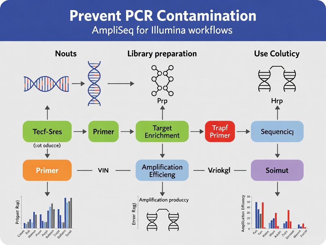
A Comprehensive Guide to Preventing PCR Contamination in AmpliSeq for Illumina Workflows
This article provides a complete framework for researchers and drug development professionals to understand, prevent, and troubleshoot PCR contamination in AmpliSeq for Illumina next-generation sequencing workflows.
A Comprehensive Guide to Preventing PCR Contamination in AmpliSeq for Illumina Workflows
Abstract
This article provides a complete framework for researchers and drug development professionals to understand, prevent, and troubleshoot PCR contamination in AmpliSeq for Illumina next-generation sequencing workflows. Covering foundational principles to advanced validation strategies, it details the unique contamination risks in amplicon-based NGS, outlines best practices for laboratory setup and technique, offers systematic troubleshooting for common issues like low yield and nonspecific amplification, and emphasizes the critical role of controls and new technologies for data validation. Adopting these integrated practices is essential for ensuring the integrity and reproducibility of sequencing data in sensitive applications, from clinical diagnostics to biomedical research.
Understanding PCR Contamination: Risks and Sources in AmpliSeq Workflows
Why Contamination is a Critical Concern in Amplicon-Based NGS
Frequently Asked Questions
What makes amplicon-based NGS so susceptible to contamination? The method relies on PCR amplification to enrich target sequences. A single contaminant DNA molecule introduced at the setup stage can be amplified millions of times, leading to false-positive results. The high number of amplification products generated in a lab creates a significant contamination risk for subsequent experiments [1] [2].
What are the most common sources of contamination? The primary source is carryover contamination from previous amplification reactions (amplicons) [2]. When you open a tube containing PCR products, aerosols can form, dispersing millions of amplicons into the lab environment, which can then contaminate reagents, equipment, and new reaction setups [3] [2]. Cross-contamination between samples during pipetting is another common risk.
How can I monitor for contamination in my experiments? Always include a No-Template Control (NTC) in every run. This reaction contains all reagents except the DNA template. If amplification occurs in the NTC, it signals that one or more of your reagents or the master mix is contaminated with target DNA [3].
Besides the NTC, what is the first line of defense against contamination? Physical separation of laboratory areas is the most critical practice. You should establish dedicated, separate spaces for:
- Reagent preparation and PCR setup (pre-amplification)
- Amplification (where the thermal cycler is placed)
- Post-PCR analysis (e.g., gel electrophoresis) [3] [2] [4] Maintain a unidirectional workflow from clean (pre-PCR) to dirty (post-PCR) areas, with dedicated equipment, lab coats, and consumables for each [3].
Are there enzymatic methods to prevent carryover contamination? Yes, the Uracil-N-Glycosylase (UNG) system is widely used. In this method, dUTP is substituted for dTTP during PCR, so all new amplification products contain uracil. Before the next PCR run, the UNG enzyme degrades any uracil-containing contaminants that may be present. The enzyme is then inactivated during the initial heating step of the new PCR, allowing the amplification of the natural, thymine-containing template to proceed [3] [2].
My NGS data shows high levels of index hopping or adapter dimers. Is this contamination? While not "contamination" in the traditional sense, these are critical preparation errors. A sharp peak around 70-90 bp in an electropherogram indicates adapter dimers, often caused by an imbalance in the adapter-to-insert ratio or overly aggressive purification [5]. Using unique dual indexes (UDIs) and optimizing cleanup steps can mitigate these issues.
Troubleshooting Guide: Identifying and Resolving Contamination
Use the table below to diagnose and address common contamination-related problems in your amplicon-based NGS workflow.
| Problem & Symptoms | Potential Root Cause | Corrective & Preventive Actions |
|---|---|---|
| Amplification in No-Template Control (NTC)• NTC wells show amplification curves (qPCR) or bands (gel).• Consistent Ct values across NTC wells suggest reagent contamination. | • Contaminated master mix, primers, or water.• Aerosol contamination from high-concentration amplicons or plasmids in the lab environment. | • Replace all reagents with fresh, aliquoted stocks [3].• Decontaminate surfaces and equipment with 10% bleach, followed by 70% ethanol [3] [2].• Use aerosol-filter pipette tips and maintain separate pre- and post-PCR pipette sets [4]. |
| Inaccurate Variant Calls or False Positives• Detection of unexpected variants or sequences.• High duplicate reads in sequencing data. | • Carryover contamination from previous runs [1].• Cross-contamination between samples during library prep. | • Implement the UNG enzymatic system to degrade carryover amplicons [3] [2].• Adopt a one-way workflow and use dedicated lab coats/equipment for pre-PCR work [4].• Use physical barriers like PCR workstations or UV hoods [2]. |
| High Adapter Dimer Peaks• Bioanalyzer trace shows a dominant sharp peak at ~70-90 bp.• Low library yield and complexity. | • Adapter-dimer formation due to suboptimal ligation or purification.• Over-amplification from too many PCR cycles. | • Optimize adapter concentration and ligation conditions [5].• Adjust bead-based cleanup ratios to better remove short fragments [5].• Minimize PCR cycles and use high-fidelity polymerases [5] [4]. |
Experimental Protocol: The K-Box Method for Contamination Protection
For applications demanding high sensitivity and accuracy, such as two-step PCR for NGS library generation, consider implementing the K-box method [1]. This integrated approach both prevents and identifies carryover contamination.
1. Principle The K-box is a series of short, synergistically acting sequence elements added to the primers of the first amplification round. It prevents amplification of contaminants in the second PCR and provides a molecular barcode to identify any residual contaminants during data analysis [1].
2. K-Box Architecture The system uses three key elements in the first-stage primers [1]:
- K1 (Suppression element): A sample-specific sequence (e.g., 7 nucleotides) that must exactly match the second-stage primer for amplification to occur.
- K2 (Detection element): A sample-specific sequence (e.g., 3 nucleotides) only present in first-stage amplicons, allowing bioinformatic identification of contaminants.
- S (Separator element): A short mismatch sequence that prevents the template-binding part of the primer from being influenced by the K-box tail, avoiding PCR bias.
3. Step-by-Step Procedure
- Step 1: Primer Design
- Design first-stage primers with the architecture: 5' - [K1] - [K2] - [S] - Template-specific sequence - 3' [1].
- Design second-stage primers with the architecture: 5' - [Adapter / Barcode] - [K1] - 3'. The K1 sequence must exactly match the K1 in the corresponding first-stage primer [1].
- A unique "set" is defined by a specific combination of forward (k) and reverse (k') K-boxes. With a small number of distinct k- and k'-boxes, you can generate an exponentially larger number of unique sets for multiplexing [1].
Step 2: Two-Step PCR and NGS Library Prep
- Perform the first PCR using your target-specific primers that contain the full K-box.
- Perform the second PCR using the K1-containing primers to add adapters and barcodes.
- Critical Note: Only amplicons from the first PCR that have the correct K1 sequences will be efficiently amplified in the second PCR. Contaminating amplicons from previous runs with non-matching K1/K2 sequences will be suppressed [1].
Step 3: Bioinformatics Analysis
- During NGS data analysis, the K2 sequence is used to track the sample origin of every read.
- Any read that contains a K2 sequence not matching its sample's barcode is flagged as a carryover contamination event [1].
The following diagram illustrates the structure of the primers and the protective mechanism of the K-box system.
Research Reagent Solutions
This table lists key reagents and materials essential for implementing effective contamination control.
| Item | Function in Contamination Control |
|---|---|
| Aerosol-Resistant Filter Pipette Tips | Prevents aerosols from contaminating the pipette shaft and subsequent samples [3] [4]. |
| Uracil-N-Glycosylase (UNG) | Enzyme used to degrade uracil-containing DNA from previous amplifications before a new PCR begins [3] [2]. |
| dUTP Mix | Used in place of dTTP during PCR to generate amplicons that are susceptible to degradation by UNG, enabling the UNG system to function [2]. |
| 10% Sodium Hypochlorite (Bleach) | Effective surface decontaminant that causes oxidative damage to nucleic acids, rendering them unamplifiable [3] [2]. |
| Dedicated Pre-PCR Reagents | Aliquoting reagents for exclusive use in clean pre-PCR areas prevents contamination from shared stocks [4]. |
In AmpliSeq for Illumina workflows, Polymerase Chain Reaction (PCR) is a fundamental process for target amplification prior to sequencing. However, the extreme sensitivity of PCR makes it highly susceptible to contamination, which can compromise data integrity and lead to false positive results [6] [3]. Contamination occurs when unwanted DNA sequences are introduced into a PCR reaction, most commonly through cross-contamination between samples or carry-over contamination from amplified products of previous experiments [6]. Understanding and identifying the sources of this contamination is the first critical step in ensuring the validity of your research.
Contamination in a laboratory setting can be broadly categorized. The table below outlines the primary types, their common origins, and how they typically manifest in experiments.
| Contamination Type | Common Sources | Typical Manifestation in Experiments |
|---|---|---|
| Reagent Contamination | Contaminated water, master mixes, primers, or enzymes [7] [8] [3]. | Systematic amplification in all samples, including negative controls, often with similar Ct values [3]. |
| Environmental Contamination | Aerosolized amplicons from post-PCR areas, contaminated lab coats, skin, or hair [6] [3]. | Random amplification in some samples and controls, with varying Ct values [3]. |
| Sample Cross-Contamination | Improper pipetting technique, shared equipment (centrifuges, vortexers), or reusable utensils [6] [9] [8]. | Unexpected amplification or signal in samples, potentially with patterns linked to sample handling order. |
| Template DNA Carry-over | Introduction of previously amplified PCR products into new reactions [6] [7]. | False positive amplification in subsequent experiments. |
Experimental Protocols for Contamination Detection
Rigorous experimental design includes controls that are essential for detecting contamination.
Utilizing Negative Controls
Protocol: No Template Control (NTC)
- Purpose: To detect contamination in the PCR reagents or master mix.
- Methodology: The NTC well contains all components of the PCR reaction—master mix, primers, and water—but no template DNA is added [6] [3].
- Interpretation: Under contamination-free conditions, the NTC should yield no amplification. Amplification in the NTC indicates that one or more reagents have been contaminated with a DNA template [6] [3]. If the contamination is from a reagent, all NTC wells containing that reagent will typically show amplification at a similar Ct value. If the contamination is random (e.g., from an environmental aerosol), only some NTC wells will amplify, with varying Ct values [3].
Protocol: No Reverse Transcription Control (–RT Control) for RNA Workflows
- Purpose: To identify genomic DNA (gDNA) contamination in RNA samples during gene expression studies.
- Methodology: This control is set up by omitting the reverse transcriptase enzyme during the cDNA synthesis step [7].
- Interpretation: Amplification in the –RT control indicates that the RNA sample is contaminated with gDNA, as the PCR is amplifying the genomic target rather than the cDNA [7].
UNG Decontamination Protocol
Protocol: Using Uracil-N-Glycosylase (UNG) to Prevent Carry-over Contamination
- Purpose: To enzymatically destroy carry-over contamination from previous PCR amplifications [3].
- Methodology:
- In your PCR setup, use a dNTP mix where dTTP is replaced by dUTP. This results in all newly synthesized PCR products containing uracil instead of thymine [3].
- In subsequent PCR reactions, use a master mix that contains the UNG enzyme [3].
- Prior to the thermal cycling, incubate the reaction at room temperature. During this step, UNG will selectively degrade any uracil-containing DNA (i.e., previous amplicons) that may have contaminated the reaction [3].
- The initial denaturation step of the PCR thermal cycler inactivates the UNG enzyme, allowing the new, uracil-free template to amplify without interference [3].
- Limitations: UNG is most effective for thymine-rich amplicons and is only effective against uracil-containing DNA, not other sources of DNA contamination [3].
Contamination Prevention Workflow
The following diagram illustrates the core principle of a unidirectional workflow, which is critical for preventing contamination in PCR and AmpliSeq for Illumina workflows.
Key Workflow Practices:
- Physical Separation: Maintain separate, dedicated rooms or spaces for pre-amplification (reagent and sample preparation) and post-amplification (PCR product analysis) activities [6] [7] [3].
- Unidirectional Workflow: Researchers and materials should move from "clean" pre-amplification areas to "dirty" post-amplification areas, but not in the reverse direction [7] [3]. Personnel should not enter pre-amplification areas after working in post-amplification areas on the same day without thorough decontamination [3].
- Dedicated Equipment: Each area must have its own set of pipettes, centrifuges, vortexers, lab coats, gloves, and consumables [6] [3].
The Scientist's Toolkit: Essential Reagent Solutions
The table below lists key reagents and materials essential for preventing contamination in your experiments.
| Item | Function in Contamination Prevention |
|---|---|
| Aerosol-Resistant Filter Tips | Create a barrier between the pipette and the liquid, preventing aerosols from contaminating the pipette shaft and subsequent samples [6] [7]. |
| UNG (Uracil-N-Glycosylase) | An enzymatic system used in qPCR master mixes to degrade carry-over contamination from previous PCR products, as detailed in the protocol above [3]. |
| Molecular Biology Grade Water | High-purity, DNase/RNase-free water ensures that the water used in master mixes and reagents is not a source of contamination [8]. |
| Bleach Solution (5-10%) | A potent DNA-degrading agent used for decontaminating work surfaces and non-porous equipment [6] [3]. |
| Aliquoted Reagents | Storing primers, enzymes, and master mixes in single-use aliquots prevents the contamination of an entire stock solution through repeated use [6] [7]. |
| Disposable Labware | Using disposable tubes, punches, and blades for sample collection and handling prevents the transfer of material between samples [6]. |
| Hot-Start DNA Polymerase | Reduces non-specific amplification and primer-dimer formation by remaining inactive until a high-temperature activation step, improving assay specificity [10]. |
Frequently Asked Questions (FAQs)
Q1: My No Template Control (NTC) shows amplification. What should I do? First, determine if the contamination is systematic or random. If all NTCs show similar amplification, the source is likely a contaminated reagent. You should discard all opened reagents (master mixes, water, primers) and repeat the experiment with fresh, aliquoted stocks [7] [3]. If the amplification is random and sporadic, the contamination is likely environmental. In this case, you should decontaminate your workspace and equipment with a fresh 5-10% bleach solution and review your lab's workflow to ensure physical separation is being maintained [6] [3].
Q2: How can I prevent genomic DNA contamination in RNA-based AmpliSeq workflows? There are several key strategies:
- DNase Treatment: Treat your RNA sample with DNase before performing reverse transcription, followed by heat inactivation of the enzyme [7].
- Exon-Exon Junction Primers: Design your assays to span an exon-exon junction so that amplification will only occur from cDNA, not from genomic DNA [7].
- Include a –RT Control: Always run a no-reverse-transcription control to monitor for gDNA contamination [7].
Q3: What is the most effective way to decontaminate my pipettes and work surfaces? For non-porous surfaces and equipment, a freshly prepared 5-10% bleach solution is highly effective at degrading DNA [6] [3]. Spray or wipe the surface and allow it to sit for 10-15 minutes before wiping down with deionized water to prevent corrosion [3]. Regular decontamination should be performed before and after experimental setup.
Q4: Can laboratory automation help reduce contamination? Yes, automated liquid handling systems can significantly reduce the risk of human error and cross-contamination. These systems minimize physical touches and often operate within an enclosed hood that provides a HEPA-filtered, contamination-free workspace [8].
In low-biomass microbiome studies, the inherent sensitivity of PCR-based methods like the AmpliSeq for Illumina workflow becomes a double-edged sword. These environments—such as certain human tissues, treated drinking water, or hyper-arid soils—harbor microbial biomass near the limits of detection [11]. Here, the DNA "signal" from the actual sample can be easily overwhelmed by contaminant "noise," making these samples uniquely vulnerable to contamination that can distort ecological patterns, cause false pathogen detection, and lead to incorrect conclusions [11]. This guide provides targeted troubleshooting and FAQs to help researchers safeguard their low-biomass experiments.
FAQs: Understanding Vulnerability and Contamination
What makes low-biomass samples so susceptible to contamination? The issue is proportional. In a high-biomass sample (like human stool), the contaminant DNA is a tiny fraction of the total. In a low-biomass sample, the same amount of contaminant can constitute a large portion, or even the majority, of the final sequenced DNA, making the true signal difficult or impossible to distinguish from the noise [11].
My negative controls show amplification. What does this mean? Amplification in your No Template Control (NTC)—a well containing all PCR components except the template DNA—is a clear indicator of contamination [6] [3]. If the contamination is uniform across all NTCs, a reagent is likely contaminated. If it's random and at varying levels, the cause is more likely aerosolized amplicons or environmental DNA drifting into the reactions [3].
What are the most common sources of contamination? The primary sources are:
- Carryover Contamination: Aerosolized droplets containing PCR amplicons from previous reactions [6] [2].
- Cross-Contamination: Transfer of DNA between samples during handling [11].
- Reagents/Kits: Microbial DNA present in the reagents and kits used for DNA extraction and PCR [11].
- Laboratory Environment: Human operators, sampling equipment, and lab surfaces [11].
Beyond standard lab practice, what extra steps are critical for low-biomass work? Standard practices are necessary but insufficient. You must:
- Implement Rigorous Physical Separation: Establish dedicated, physically separated pre- and post-amplification areas with unidirectional workflow [3] [2].
- Use Extensive Controls: Include multiple negative controls at the sample collection and DNA extraction stages (e.g., swabs of the air, empty collection vessels, aliquots of preservation solution) to identify the nature and extent of contamination [11].
- Decontaminate with DNA-Removing Agents: Use sodium hypochlorite (bleach) to destroy contaminating DNA on surfaces, as standard decontamination like ethanol or autoclaving may kill cells but leave DNA intact [11] [3].
Troubleshooting Guide: Identifying and Resolving Contamination
| Observation | Possible Causes | Recommended Solutions |
|---|---|---|
| Amplification in No Template Control (NTC) | Reagent contamination, carryover from amplified products, or environmental DNA [6] [3] | Discard all suspect reagents. Decontaminate workspaces with 10% bleach. Use aerosol-resistant filter tips and UNG treatment [6] [3] [2] |
| High Background or Smearing | Non-specific amplification, overcycling, or primer-dimer formation [10] [12] | Increase annealing temperature, use hot-start polymerase, reduce number of cycles, optimize Mg2+ concentration, and redesign primers if necessary [10] [13] [12] |
| Inconsistent Results Between Replicates | Sample-to-sample cross-contamination or sporadic environmental contamination [11] [3] | Use disposable equipment, change gloves between samples, include sample-processing controls, and adopt a linear workflow [11] [6] |
| Unexpected Microbial Taxa in Data | Contamination from reagents, kits, or the laboratory environment [11] | Sequence negative controls (extraction blanks) and use bioinformatic tools to subtract contaminants found in these controls from your sample data [11] |
Best Practices for a Contamination-Aware Workflow
Preventing contamination requires a proactive, multi-layered strategy. The diagram below illustrates the core principles of a contamination-aware workflow for low-biomass research.
Laboratory Design and Workflow
Maintain strict physical separation of pre- and post-amplification areas, ideally in different rooms with separate equipment, lab coats, and consumables [6] [3] [2]. The workflow must be unidirectional; personnel and equipment should not move from post-PCR areas back to pre-PCR areas [3] [12].
Sample Collection and Handling
During sampling, use personal protective equipment (PPE) and DNA-free, single-use collection vessels whenever possible [11]. Decontaminate reusable equipment with 80% ethanol followed by a nucleic acid degrading solution like bleach or UV-C light [11]. Crucially, collect sampling controls (e.g., swabs of air, empty vessels) to identify contaminants introduced during collection [11].
Reaction Setup and Amplification
- Pipetting: Use aerosol-resistant filter tips and positive-displacement pipettes to minimize cross-contamination [3] [12].
- Reagents: Aliquot all reagents into single-use amounts to avoid contaminating entire stocks [6] [3].
- Enzymatic Prevention: Incorporate uracil-N-glycosylase (UNG) into your qPCR master mix. UNG selectively degrades carryover contamination from previous PCRs (which contain dUTP) before the new amplification begins, while leaving the native template (with dTTP) intact [3] [2].
The Scientist's Toolkit: Essential Research Reagent Solutions
| Item | Function in Low-Biomass Research |
|---|---|
| UNG (Uracil-N-Glycosylase) | Enzymatically destroys carryover PCR amplicons from previous reactions, preventing false positives [3] [2] |
| Aerosol-Resistant Filter Tips | Creates a barrier within the pipette tip, preventing aerosols from contaminating the pipette shaft and subsequent samples [6] [3] |
| Hot-Start DNA Polymerase | Remains inactive at room temperature, preventing non-specific amplification and primer-dimer formation that can complicate low-biomass analysis [10] [13] |
| Sodium Hypochlorite (Bleach) | Effectively degrades contaminating DNA on work surfaces and equipment; unlike ethanol, it destroys the DNA itself [11] [3] |
| DNA-Free Water & Reagents | Certified to contain minimal microbial DNA, reducing background contamination from the reagents themselves [11] |
| No Template Control (NTC) | Critical control containing all reaction components except template DNA; used to monitor for reagent or environmental contamination [6] [3] |
FAQ: Understanding Contamination in the Laboratory
What is the fundamental difference between contamination and cross-contamination?
In a laboratory context, particularly in PCR and AmpliSeq for Illumina workflows, contamination refers to the general introduction of any foreign, unwanted substance into a reaction or sample [14]. In contrast, cross-contamination is a specific type of contamination where the contaminant is a specific, known material from a different part of the experimental process, most notably amplicons from previous PCR reactions [15] [1].
The table below outlines the core differences:
| Aspect | Contamination | Cross-Contamination |
|---|---|---|
| Definition | Introduction of any foreign, unwanted substance (e.g., DNA, microbes, chemicals) [14]. | Transfer of a specific, known material from one process or sample to another [14]. |
| Common Sources | - Contaminated reagents (e.g., water, enzymes) [15] [16]- Aerosols [15]- Personnel [17]- Environment [15] | - Carry-over of amplicons from a previous PCR into a new reaction [15] [1]- Using the same pipette for different samples without decontamination [15]. |
| Typical Contaminants | - Genomic DNA in RNA samples [7]- Bacterial DNA in reagents [16]- Microbial organisms [14] | - PCR products (amplicons) [15] [1]- Plasmid DNA from a different experiment. |
Why is cross-contamination a particularly serious problem in amplicon sequencing workflows?
Cross-contamination is especially critical in amplicon sequencing because it directly compromises the accuracy and sensitivity of your results.
- False Positives: The most significant risk is obtaining false positive results. Even tiny amounts of carry-over amplicons can be amplified in subsequent runs, leading to the false detection of pathogens or targets that are not actually present in the original sample [15].
- False Negatives: In rare cases, high levels of contamination can compete with the target DNA during amplification, potentially leading to false negatives or inaccurate quantification [15].
- Impact on Data Integrity: For sensitive applications like pathogen detection or minimal residual disease monitoring, this can lead to severe misinterpretation of results with direct consequences for research conclusions or clinical diagnostics [15] [1].
The following table summarizes the key sources and specific examples of contamination identified in experimental studies:
| Source | Specific Examples from Experimental Data |
|---|---|
| Reagents | - PCR Master Mix: Contamination of original mix showed a mean T-value (contamination level) of 9.18% compared to 0.01% with a new mix [15].- Water and Enzymes: Taq polymerase can contain copurified bacterial DNA, a major problem for highly sensitive 16S rRNA PCR [16]. |
| Aerosols | - Nuclease-free water left open in lab rooms (prep and analysis) showed measurable contamination (T-values of 0.36% and 0.32%) [15]. |
| Equipment (Pipettes) | - Using pipettes without filter tips significantly increased contamination levels (mean T-value of 1.12%) compared to using filter tips (0.43%) in standardized labs [15]. |
| Laboratory Layout | - Performing experiments in a non-physically isolated lab (general lab) resulted in higher contamination levels (mean T-value up to 1.28%) compared to labs with physical separation of pre- and post-PCR areas [15]. |
Experimental Protocols for Identifying and Quantifying Contamination
Protocol: Using No Template Controls (NTCs) to Monitor Contamination
1. Purpose: The No Template Control (NTC) is a critical quality control experiment designed to detect the presence of contamination in your AmpliSeq for Illumina workflow [18] [15].
2. Methodology:
- Sample Preparation: Instead of adding sample DNA or RNA, use nuclease-free sterile water (NFS water) as the "template" in your library preparation reaction [15].
- Placement: Include the NTC at the point of reaction setup, alongside your actual test samples.
- Downstream Processing: Process the NTC through the entire workflow simultaneously with your samples, including PCR amplification and sequencing.
3. Data Analysis:
- After sequencing, analyze the NTC data for the presence of any sequencing reads.
- The presence of a significant number of reads in the NTC, especially those mapping to your target (e.g., SARS-CoV-2, immune response genes), indicates that contamination is present in your workflow [15].
- In a contamination-controlled workflow (ccAMP-Seq), the NTC should have a very low level of detectable reads (e.g., 0.05% T-value) [15].
This protocol is based on a study that successfully identified specific contamination sources in an amplicon sequencing workflow [15].
1. Experimental Setup:
- Prepare multiple samples of NFS water exposed to different potential contamination sources.
- Aerosol Test: Place NFS water open in different laboratory rooms (e.g., PCR prep room, analysis room) for varying durations (e.g., 1 day, 1 week) and use it as a template [15].
- Reagent Test: Test newly purchased NFS water with both newly purchased and original (potentially contaminated) PCR master mix reagents [15].
- Equipment Test: Test NFS water in labs with and without physical separation of workflow steps, using pipettes with and without filter tips [15].
2. Data Analysis:
- Process all samples through your amplicon sequencing workflow.
- Use a quantitative measure like the T-value (the ratio of reads mapped to target loci to the total qualifying reads) to compare contamination levels [15].
- Statistical Analysis: Use non-parametric tests like the Wilcoxon rank-sum test to determine if differences in T-values between groups (e.g., with vs. without filter tips) are statistically significant [15].
The Scientist's Toolkit: Key Reagent Solutions
The following table lists essential reagents and materials used in featured experiments to prevent and control contamination.
| Research Reagent / Material | Function in Contamination Control |
|---|---|
| Filter Tips or Positive Displacement Tips | Creates a physical barrier to prevent aerosols from contaminating the pipette shaft and subsequent samples. Proven to significantly lower contamination levels [15] [7]. |
| dUTP / Uracil DNA Glycosylase (UDG) System | A biochemical method to degrade carry-over contamination. dUTP is incorporated into PCR products instead of dTTP. In subsequent reactions, UDG enzyme cleaves these uracil-containing contaminants before amplification starts, preventing their replication [15]. |
| Synthetic DNA Spike-Ins | Engineered DNA fragments added to samples. They compete with any contaminating DNA during amplification, reducing its impact. They also help identify contamination in data analysis and ensure sufficient library concentration for sequencing, even in low-target samples [15]. |
| Low-DNA AmpliTaq Polymerase LD | A specially purified form of Taq polymerase that contains very low levels of contaminating bacterial DNA, crucial for highly sensitive applications like 16S rRNA PCR [16]. |
| DNase I | An enzyme used to degrade contaminating genomic DNA in RNA samples before performing reverse transcription, ensuring that subsequent PCR amplifies only cDNA from RNA [7]. |
Workflow Diagram: Contamination Control in AmpliSeq
The diagram below illustrates a logical workflow for preventing contamination in a typical amplicon sequencing experiment, integrating key concepts from the troubleshooting guide.
Building a Contamination-Free Lab: Practical Workflow and Best Practices
Core Principles of Spatial Separation
Why is spatial separation critical for AmpliSeq for Illumina workflows?
Spatial separation is a foundational principle for preventing PCR contamination in sensitive next-generation sequencing (NGS) applications like AmpliSeq for Illumina. The primary goal is to create a unidirectional workflow that physically separates pre-amplification processes from post-amplification activities, ensuring that amplified PCR products (amplicons) cannot contaminate your initial reaction setups [19].
Contamination occurs when aerosolized amplicons—tiny droplets created when opening PCR tubes or pipetting amplified DNA—settle on equipment, surfaces, or into reagents [20]. These contaminants can then serve as templates in subsequent reactions, leading to false-positive results and compromised data integrity. In highly sensitive AmpliSeq workflows, even minute levels of contamination can significantly impact results [21].
Laboratory Design & Workflow Implementation
What is the ideal laboratory layout for spatial separation?
The optimal layout establishes three distinct physical areas arranged to enforce a unidirectional workflow from "clean" to "dirty" areas [19]. The following diagram illustrates this fundamental concept:
Ideal Laboratory Workflow
- Reagent Preparation Room/Area: This is the cleanest zone, dedicated to preparing and aliquoting master mixes (water, buffer, nucleotides, primers, polymerase) [19]. No biological samples or amplified DNA should enter this space.
- Pre-PCR/Sample Preparation Room/Area: This area is for handling and processing raw DNA samples and adding template to master mixes [19]. It should be physically separated from post-PCR areas.
- Amplification/Post-PCR Room/Area: This designated "dirty" area houses thermal cyclers and is used for all post-amplification processes like purifying PCR-amplified DNA, running agarose gels, and analyzing results [19] [21]. Equipment and consumables from this area must never be brought back to pre-PCR areas.
How can I implement spatial separation in a limited space?
Many laboratories operate in open-concept or limited spaces. The following table outlines practical mitigation strategies:
| Constraint | Practical Solutions | Key Considerations |
|---|---|---|
| Open-concept lab | Use separate, dedicated benches spaced as far apart as possible [19]. | Assign specific cabinets and equipment to pre- and post-PCR work. Clear labeling is essential. |
| No separate rooms | Implement dead air boxes (DABs) or laminar flow cabinets with HEPA filters for reagent prep and PCR setup [19]. | A DAB provides a contained, low-turbulence environment to protect reactions from airborne contaminants. |
| Shared equipment | Never share equipment like pipettes, centrifuges, or vortexers between pre- and post-PCR workflows [20] [21]. | Use dedicated, color-coded equipment for each zone. Pipettes with aerosol-filter tips are mandatory for pre-PCR work [19] [21]. |
Contamination Prevention & Control Protocols
What are the essential practices for maintaining a contamination-free workflow?
Beyond physical separation, rigorous procedural controls are necessary to prevent contamination.
- Unidirectional Workflow: Strictly enforce a one-way movement of personnel and materials. Once you enter or handle items from a post-PCR area, you must not re-enter a pre-PCR area without decontamination [19] [21].
- Dedicated Consumables and Equipment: Maintain separate sets of pipettes, tip boxes, lab coats, and waste containers for pre- and post-PCR areas. Label all items clearly [20] [21].
- Physical Barriers and Decontamination: Use 10% bleach solution or commercial DNA decontaminants (e.g., DNA-away) to regularly wipe down benches, pipettes, and equipment [20].
- Aliquoting Reagents: Aliquot all PCR reagents (polymerase, buffers, water) into single-use volumes upon receipt. This minimizes repeated exposure to the environment and prevents the loss of entire reagent stocks if contamination occurs [20] [21].
- Technique: Always use filter pipette tips for handing DNA samples and setting up reactions. Open PCR tubes carefully with two hands to avoid aerosol generation, and always add template DNA last to the master mix [20] [21].
The Scientist's Toolkit: Essential Reagents and Materials
The following table details key items required for implementing a contamination-controlled PCR laboratory.
| Item | Function | Application Notes |
|---|---|---|
| Aerosol-filter Pipette Tips | Prevents aerosol contamination from entering and contaminating pipette shafts [20] [21]. | Mandatory for all pre-PCR liquid handling. |
| 10% Bleach Solution / DNA Decontaminant | Degrades contaminating DNA on surfaces and equipment [20]. | Use for daily cleaning of benches, pipettes, and equipment in pre-PCR areas. |
| Dedicated Lab Coats | Prevents transfer of amplicons on clothing [20]. | Store coats in their respective areas. Never wear a post-PCR coat in a pre-PCR area. |
| UDG/dUTP System | Enzymatically destroys carryover contaminates from previous PCR reactions (not a substitute for spatial separation) [10]. | A biochemical method used within the reaction setup. |
| Hot-Start DNA Polymerase | Reduces non-specific amplification and primer-dimer formation by requiring heat activation, improving assay specificity [10]. | Standard for most modern PCR and NGS library amplification protocols. |
| Molecular Grade Water | Ultrapure, nuclease-free water for preparing reagents and negative controls [10] [20]. | Essential for ensuring reagent quality and accurate negative controls. |
Troubleshooting Guide & FAQs
How do I monitor for and confirm PCR contamination?
Protocol: Monitoring for Contamination Always include a negative control in every run. This reaction contains the entire master mix but substitutes the DNA template with molecular-grade water [20] [21].
Interpretation: A clear negative control (no bands or Cq values) indicates a contamination-free setup. Any amplification in the negative control signals contamination.
What should I do if I detect contamination?
Follow this systematic troubleshooting protocol to identify and eliminate the source:
Systematic Contamination Response
Frequently Asked Questions (FAQs)
Q: Can I use a biological safety cabinet (BSC) instead of separate rooms? A: Yes, a BSC or laminar flow hood can serve as an excellent dedicated pre-PCR area, especially in space-constrained labs, as it provides a HEPA-filtered, clean air environment for reaction setup [19].
Q: My lab is small, and I cannot afford separate pipettes. What can I do? A: This is a significant risk. At a minimum, thoroughly decontaminate pipettes with 10% bleach and use aerosol-filter tips exclusively. However, investing in a dedicated set of pre-PCR pipettes is strongly recommended for reliable results [20] [21].
Q: How should I store plasmids to avoid contamination? A: Plasmid stocks, which are high-copy number templates, should be diluted and handled only in the Pre-PCR/Sample Preparation area. Never handle them in the Reagent Preparation room [19].
Troubleshooting Guides and FAQs
Frequently Asked Questions
Q1: Why is my amplicon sequencing workflow still showing contamination even after I use filter tips and UDG treatment?
Carryover contamination in amplicon sequencing is complex and can originate from multiple sources. Even with standard precautions like filter tips and uracil DNA glycosylase (UDG) treatment, contamination can persist if other sources are not controlled. Key contamination sources include:
- Aerosols: Airborne amplicons in laboratory environments can contaminate reagents and samples [15].
- Contaminated Reagents: Master mixes and other reagents can be pre-contaminated, leading to random, high-level contamination in non-template controls (NTCs) [15].
- Equipment: Pipettes can harbor contaminants if not properly decontaminated, especially if used without filter tips [15].
A comprehensive approach is required. The ccAMP-Seq (carryover contamination-controlled Amplicon Sequencing) workflow recommends:
- Using filter tips and physical isolation of experimental steps to avoid cross-contamination [15].
- Adding synthetic DNA spike-ins to compete with contaminants during amplification [15].
- Implementing a dUTP/UDG system to enzymatically digest carryover amplicons from previous reactions [15].
- Applying a dedicated data analysis procedure to bioinformatically remove sequencing reads originating from contaminants [15].
Q2: I need to decontaminate healing abutments for reuse. Which protocol is more effective: ultrasonic cleaning with autoclaving, or a protocol that includes sodium hypochlorite?
A comparative in-vitro study demonstrates that a protocol incorporating sodium hypochlorite is significantly more effective [22].
Table: Efficacy of Two Decontamination Protocols for Healing Abutments
| Decontamination Protocol | Residual Contamination (Evidence of Staining) | Statistical Significance |
|---|---|---|
| Group 2: Ultrasonic cleaning + Autoclaving | 100% of samples (40/40) showed biological remnants [22] | |
| Group 3: NaOCl + Ultrasonic cleaning + Autoclaving | 0% of samples (0/40) showed residual contamination [22] | The difference between groups was statistically significant (p < 0.001) [22] |
The study concluded that cleaning with 3% sodium hypochlorite for 1 minute prior to ultrasonic cleaning and autoclaving ensures complete decontamination, whereas ultrasonic cleaning and autoclaving alone are insufficient [22].
Q3: What are the critical safety considerations when working with sodium hypochlorite (bleach) in the lab?
Sodium hypochlorite is a highly reactive chemical. Adhering to safety guidelines is critical to prevent harmful reactions [23] [24].
- Incompatible Materials: Never mix bleach with incompatible chemicals. Key reactions include:
- Proper Concentration and Stability: For disinfection, a working dilution of 0.5% to 2% sodium hypochlorite is effective. Diluted solutions are not stable and should be prepared fresh regularly (e.g., weekly) [23].
- Personal Protective Equipment (PPE): Always wear appropriate PPE, including safety goggles, nitrile gloves, and a lab coat [23].
- Ventilation: Use bleach solutions in a well-ventilated area or a chemical fume hood for larger volumes [23].
Q4: My UV sterilization unit doesn't seem to be working effectively. What are the most common points of failure?
UV sterilization effectiveness can be compromised by several common issues [25] [26]:
- Lamp Aging and Degradation: UV bulbs naturally degrade over time. The output intensity decreases, even if the bulb still produces visible blue light. Bulbs should be replaced every 6 months for optimum performance or according to the manufacturer's schedule [25].
- Devitrification: This is the process where the quartz glass of the bulb develops white, cloudy spots due to overheating or surface contamination. This cloudiness blocks UV light from passing through [26].
- Filament Failure: Rough handling, electrical shorts, or simply a defective bulb can break the filament, preventing the lamp from lighting [25].
- Ballast or Starter Failure: The ballast provides the surge voltage needed to start the bulb. A weak or failed ballast may not be able to ignite a new bulb, even if it can still light an older, "broken-in" one [25].
- External Contamination: Fingerprints, dust, ink, or other contaminants on the quartz sleeve or bulb surface can absorb UV radiation, preventing it from reaching the target area. Always handle bulbs with gloves and clean them regularly with isopropanol [26].
Experimental Protocols and Data
Detailed Methodology: Decontamination of Healing Abutments [22]
This protocol compares the efficacy of two decontamination methods for reused healing abutments.
- Materials: 85 healing abutments (80 used, 5 unused controls), ultrasonic bath, steam autoclave, 3% sodium hypochlorite solution, Phloxine B stain, 10X stereomicroscope.
- Group Allocation:
- Group 1 (Control): 5 unused healing abutments.
- Group 2: 40 used healing abutments subjected to ultrasonic cleaning (20-minute cycle) followed by autoclaving (45 minutes at 121°C).
- Group 3: 40 used healing abutments subjected to scrubbing with 3% NaOCl for 1 minute, followed by the same ultrasonic cleaning and autoclaving as Group 2.
- Evaluation of Residual Contamination: All abutments were stained with Phloxine B for 1 minute and observed under a microscope. The presence of pink/red staining indicated residual proteinaceous contamination.
Detailed Methodology: Establishing a Contamination-Controlled Amplicon Sequencing (ccAMP-Seq) Workflow [15]
This protocol was developed to identify and eliminate carryover contamination in SARS-CoV-2 detection but is applicable to other amplicon sequencing workflows.
- Identifying Contamination Sources:
- Aerosols: Nuclease-free water was placed open in different lab areas for varying durations and then tested.
- Reagents: Newly purchased nuclease-free water and newly synthesized primers were tested with original and new master mix reagents.
- Equipment/Pipettes: Tests were conducted in physically isolated vs. non-isolated labs, with and without filter tips.
- The ccAMP-Seq Workflow:
- Physical Isolation: Use filter tips and work in physically separated pre- and post-PCR areas.
- Synthetic DNA Spike-ins: Add 10,000 copies/reaction of synthetically designed competitive DNA fragments before library prep to outcompete low-level contaminants and allow for sequencing of very low-biomass samples.
- dUTP/UDG System: Incorporate dUTP in the first PCR. In subsequent reactions, the UDG enzyme digests any carryover dUTP-containing amplicons before amplification.
- Bioinformatic Filtering: Implement a data analysis pipeline to remove reads matching the spike-in sequences or other identified contaminants.
Table: Quantitative Improvement with ccAMP-Seq Workflow [15]
| Workflow Metric | Standard AMP-Seq | ccAMP-Seq | Improvement |
|---|---|---|---|
| Contamination Level (T value in NTC) | Varies, can be high | At least 22-fold lower | Significant reduction in false positives |
| Detection Limit | ~10 copies/reaction | 1 copy/reaction | ~1 order of magnitude more sensitive |
| Sensitivity & Specificity | Compromised by contamination | 100% (with SARS-CoV-2 standard) | High accuracy for qualitative and quantitative detection |
The Scientist's Toolkit: Research Reagent Solutions
Table: Essential Materials for Decontamination and Contamination Control
| Item | Function/Brief Explanation | Example Protocol Use |
|---|---|---|
| Sodium Hypochlorite (Bleach) | Oxidizing agent that disrupts bacterial cell walls and denatures proteins and nucleic acids [22] [27]. | 3% solution for 1 min surface decontamination [22]; 150 ppm for eggshell sanitization [27]. |
| Phloxine B Stain | A protein-binding dye used as an indicator for residual biological contamination on surfaces [22]. | Stain for 1 min, then observe under microscope for pink staining [22]. |
| Synthetic DNA Spike-ins | Non-naturally occurring DNA sequences that compete with contaminants during amplification, enabling their detection and quantification [15]. | Add 10,000 copies/reaction prior to library preparation for amplicon sequencing [15]. |
| dUTP / Uracil DNA Glycosylase (UDG) | Enzyme system that digests carryover amplicons from previous PCRs, preventing their re-amplification [15]. | Incorporate dUTP in first PCR; add UDG to all subsequent reaction mixes [15]. |
| Ultraviolet (UV-C) Light | Non-ionizing radiation that inactivates microorganisms by damaging their DNA; leaves no chemical residue [27]. | Used alone or in combination with NaOCl for surface decontamination (e.g., eggshells) [27]. |
Workflow Diagrams
Contamination Control in Amplicon Sequencing
Effective Healing Abutment Decontamination
Proper Pipetting Technique and the Use of Uracil-DNA Glycosylase (UDG)
Your Pipetting Troubleshooting Guide
| Common Pipetting Error | Impact on Results | How to Correct It |
|---|---|---|
| Using the wrong pipette type [28] | Inaccuracy with viscous or non-aqueous liquids. | Use air-displacement for aqueous liquids; use positive-displacement for viscous, volatile, or dense liquids [28]. |
| Careless tip handling [28] | Leaking tips can reduce accuracy by 0.5% to 50% [28]. | Always use original or manufacturer-recommended tips to ensure a proper seal [28]. |
| Inconsistent rhythm & speed [28] [29] | Evaporation within the tip or incomplete aspiration leads to volume variation. | Aspirate and dispense with a smooth, consistent force and rhythm. Pause for one second after aspiration for liquid to fully enter the tip [28] [29]. |
| Improper tip immersion [29] | Aspirating air (too shallow) or liquid clinging to tip exterior (too deep). | Immerse the tip adequately below the meniscus but avoid contacting the bottom of the container [29]. |
| Holding the pipette at an angle [29] | Alters the hydrostatic pressure, changing the aspirated volume. | Always hold the pipette vertically when aspirating liquid [29]. |
| Skipping the pre-wetting step [29] | Increases evaporation within the tip air space, causing lower delivery volumes. | Pre-wet the tip by aspirating and fully expelling the liquid at least three times before the actual delivery [29]. |
| Ignoring temperature equilibrium [29] | Thermal expansion/shrinking of the air space causes volume variation. | Allow liquids, tips, and pipettes to equilibrate to the ambient temperature of the room before pipetting [29]. |
Proper Forward Mode Pipetting Protocol
For accurate and precise delivery of most aqueous solutions, follow these steps for forward mode pipetting with an air-displacement pipette [28] [29]:
- Set the volume on the pipette.
- Pre-wet the tip by aspirating and expelling the liquid several times [29].
- Depress the plunger smoothly to the first stop.
- Immerse the tip vertically to the proper depth in the liquid.
- Slowly release the plunger to its resting position to aspirate the liquid. Pause for one second [29].
- Withdraw the pipette vertically from the liquid.
- Place the tip against the wall of the receiving vessel at a 10-45 degree angle.
- Smoothly depress the plunger to the first stop, then pause for one second.
- Depress the plunger to the second stop to expel any residual liquid.
- Slide the tip up the vessel wall while removing the pipette.
- Allow the plunger to return to its rest position.
UDG/UNG Frequently Asked Questions
Q1: What is UNG/UDG, and what is its function in qPCR?
A: Uracil-DNA Glycosylase (UDG) is a DNA-repair enzyme. The term UNG (Uracil-N-Glycosylase) refers to a specific family of these enzymes, but the names UDG and UNG are often used interchangeably in qPCR as they perform the same critical function: preventing carry-over contamination [30]. Its biological role is to remove uracil bases from DNA, and in qPCR, it is used to degrade DNA from previous amplification reactions (which contain dUTP), thereby preventing false positives [30].
Q2: How exactly does UDG prevent carry-over contamination?
A: UDG prevents contamination through a two-step method [31]:
- dUTP Incorporation: dUTP is incorporated into all PCR products during amplification in place of dTTP.
- Enzymatic Digestion: In subsequent PCR setups, the pre-assembled reaction mixture is treated with UDG before amplification begins. The enzyme cleaves the uracil base from the sugar-phosphate backbone of any contaminating dU-containing DNA. This creates an apyrimidinic site that blocks replication by DNA polymerases, preventing re-amplification. The UDG is then thermally inactivated before the new PCR cycle starts [30] [31].
Q3: Does UDG/UNG affect my target DNA, primers, or dUTP?
A: No. UDG is specific for uracil in DNA. It does not affect:
- Native, thymine-containing DNA (your original sample template) [30].
- dUTP, which is not a substrate for the enzyme [30].
- Taq polymerase or other standard PCR reaction components [30].
Q4: When should I NOT use a master mix containing UDG/UNG?
A: UDG is not suitable for all applications. Avoid using it in the following scenarios [30]:
- One-Step RT-PCR with E. coli UDG, as the enzyme will degrade cDNA synthesized with dUTP. (Heat-labile UDG from Atlantic cod is a suitable alternative for one-step protocols).
- Genotyping experiments with an end-point read that will be performed at a later date, as residual UDG activity can degrade PCR products over time.
- Amplifying dU-containing templates, such as in nested PCR.
- Bisulfite-converted DNA, as the conversion process turns unmethylated cytosines into uracils, which UDG will degrade.
- Any experiment where you need to store the amplicon for post-PCR analysis.
Q5: My no-template control (NTC) is still showing contamination even though I use UDG. Why?
A: UDG only degrades PCR products that contain uracil (dUTP). If your NTC is contaminated, the source may be pre-existing contamination from standard dTTP-containing PCR products, contaminated primers, or other sources in the lab environment that UDG cannot remove [30]. This highlights that UDG is one part of a comprehensive contamination control strategy that must also include good laboratory practices, such as physical separation of pre- and post-PCR areas and using filtered tips [30].
Research Reagent Solutions
| Reagent | Function in Contamination Control |
|---|---|
| dUTP | A nucleotide that is incorporated into PCR products during amplification in place of dTTP. This uracil tag makes the amplicons susceptible to degradation by UDG/UNG in subsequent reactions, preventing their re-amplification [30] [31]. |
| Uracil-DNA Glycosylase (UDG/UNG) | An enzyme that catalyzes the removal of uracil bases from the DNA backbone. It is added to a PCR master mix to enzymatically cleave any uracil-containing contaminating DNA from previous reactions before a new PCR cycle begins [30]. |
| Heat-Labile UDG | A version of UDG (e.g., cloned from Atlantic cod) that is fully inactivated during the initial heating steps of PCR. This is essential for one-step RT-PCR protocols, as it prevents degradation of newly synthesized cDNA that contains dU nucleotides [30]. |
UDG-Controlled PCR Contamination Prevention Workflow
Implementing Rigorous Personal Protective Equipment (PPE) Protocols
PPE Fundamentals for Contamination Control
What is the primary purpose of PPE in preventing PCR contamination?
Personal protective equipment (PPE) serves as a crucial barrier to minimize exposure to workplace hazards, including biological contaminants that can compromise PCR experiments. In the context of AmpliSeq for Illumina workflows, PPE provides the last line of defense against introducing exogenous DNA, nucleases, or cross-contaminating samples through personnel. Proper PPE use is essential given the sensitivity of next-generation sequencing applications that use Polymerase Chain Reaction (PCR) for DNA amplification [18] [32].
How does PPE fit into a comprehensive contamination control strategy?
PPE represents one component of a holistic Contamination Control Strategy (CCS) that integrates facility design, equipment, processes, and personnel behavior. While engineering and administrative controls should be prioritized, PPE remains essential for protecting both the experiment and the researcher. Effective PPE implementation requires a risk-based approach aligned with standard precautions for every patient or sample encounter in healthcare and research settings [33] [34].
Proper PPE Selection and Usage
What specific PPE components are necessary for AmpliSeq workflows?
The table below outlines essential PPE components for preventing PCR contamination:
| PPE Component | Specification/Standard | Contamination Risk Mitigated |
|---|---|---|
| Gloves | Minimum AQL 2.5 for exam gloves; sterile for sensitive applications [33] | Direct sample contact, skin-derived nucleases, cross-contamination |
| Masks/Respirators | NIOSH-approved N95 or higher for airborne protection; ASTM F5302-21 standard for barrier face coverings [33] | Salivary contamination, respiratory droplets |
| Eye Protection | Goggles or face shields [33] | Accidental splashes of amplicons or reagents |
| Protective Gowns | ANSI/AAMI PB70 standards with appropriate fluid barrier protection [33] | Particulate shedding from clothing, cross-contamination between workspaces |
What is the correct sequence for donning and doffing PPE?
Donning Sequence (outside the PCR workspace):
- Gown - Cover body from neck to knees, arms to wrists, ensuring the gown wraps around the back. Tie neck and waist straps if available [33].
- Mask or Respirator - Place straps or ties around head/neck. Adjust the flexible band to the nose bridge and perform a fit-check for respirators [33].
- Eye Protection - Position goggles or face shield to ensure adequate protection of face and eyes [33].
- Gloves - Don gloves, ensuring they extend to cover the wrists of the gown [33].
Doffing Sequence:
- Gloves - Remove using proper technique without touching the outer surface with bare skin [33].
- Eye Protection - Handle by straps or earpieces only, without touching the contaminated front surface [33].
- Gown - Release straps or ties, touching only the inside of the gown. Pull away from neck and shoulders, turning it inside out [33].
- Mask/Respirator - Remove after exiting the contaminated area [33].
PPE Protocol Workflow for PCR Laboratories
Troubleshooting Common PPE Issues
What are solutions to frequent PPE-related contamination problems?
| Problem | Potential Cause | Solution | Prevention Tip |
|---|---|---|---|
| False positives in No Template Controls (NTC) | Improper glove changing between samples; contaminated gloves [18] | Change gloves between processing different samples and after touching potentially contaminated surfaces | Implement double-gloving for procedures with high contamination risk [33] |
| Cross-contamination between samples | Inadequate gown protection; re-use of disposable gowns | Use single-use, low-lint gowns with appropriate barrier protection levels | Select gowns meeting ANSI/AAMI PB70 standards for the specific procedure [33] |
| Airborne contamination affecting reactions | Inadequate respiratory protection during sample preparation | Use properly fitted N95 respirators or higher-level protection during aerosol-generating steps | Ensure fit-testing for all personnel requiring respirators [33] |
| Environmental surface contamination | PPE contamination transfer to workspaces | Establish clear separation between clean and contaminated areas for PPE donning/doffing | Follow strict doffing sequence to minimize transfer of amplicons [33] |
How can researchers verify their PPE practices are effective?
Regular competency assessments should include:
- Media-fill testing to simulate aseptic techniques while wearing PPE
- Gloved fingertip sampling to assess hand contamination after proper donning
- Surface monitoring of PPE donning and doffing areas
- Positive and negative controls in experimental designs to detect contamination breaches [35]
Laboratory Materials and Reagents
What essential materials support effective PPE protocols?
| Material/Reagent | Function in Contamination Control |
|---|---|
| Nitrile exam gloves (AQL ≤2.5) | Primary hand barrier with minimal perforations [33] |
| ANSI/AAMI PB70 Level 2-4 gowns | Fluid-resistant protection for upper body and arms [33] |
| NIOSH-approved N95 respirators | Filtration of airborne particles and respiratory droplets [33] |
| Safety goggles or face shields | Eye and facial mucosa protection from splashes [33] |
| Disposable shoe covers | Prevention of tracking contaminants between lab areas |
| Surface decontamination reagents | Elimination of amplicons from PPE contact surfaces [18] |
| No Template Controls (NTC) | Critical PCR controls to detect contamination despite PPE use [18] |
Advanced Contamination Prevention
What specialized methods complement PPE for two-step PCR workflows?
For highly sensitive applications like two-step PCR in AmpliSeq library preparation, technical controls such as the K-box method provide additional protection against carry-over contamination. This approach uses:
- K1 sequences for suppression of contaminations
- K2 sequences for detection of residual contaminations
- S sequences as separators to prevent amplification bias [1]
Even with optimal PPE, these molecular safeguards are recommended for research and diagnostic applications demanding high sensitivity and accuracy, particularly when analyzing rare events or minimal residual disease [1].
How should PPE be adapted for different contamination types?
- Microbial contamination: Focus on sterile gloves, high-level masks, and gowns with appropriate barrier levels [34]
- Particulate contamination: Use low-lint PPE and ensure proper donning to minimize shedding [34]
- Chemical contamination: Select chemical-resistant PPE based on Safety Data Sheets [33]
- Cross-contamination: Implement sample-specific PPE changes and spatial separation [34]
In molecular biology research, particularly within AmpliSeq for Illumina workflows, the master mix strategy is a fundamental technique for efficient and reliable experimental setup. This approach involves preparing a single, homogeneous mixture of common PCR components—such as enzymes, dNTPs, buffers, and primers—which is then aliquoted into individual reaction tubes before adding the template DNA. This method significantly reduces both hands-on time and the risk of contamination, two critical factors in achieving reproducible results in sensitive amplification-based applications. For researchers and drug development professionals working with high-throughput sequencing panels, implementing a robust master mix protocol is essential for maintaining data integrity while streamlining laboratory workflows.
Frequently Asked Questions (FAQs)
1. What is a master mix and how does it specifically reduce contamination risk in AmpliSeq workflows?
A master mix is a unified solution containing all common components required for a PCR reaction, such as polymerase enzyme, dNTPs, reaction buffer, magnesium ions, and primers. This strategy reduces contamination risk by minimizing the number of individual pipetting steps and tube openings required during reaction setup. Each time a reagent tube is opened or a pipetting step is performed, the risk of introducing contaminating DNA aerosols or cross-contaminating samples increases [3] [7]. By consolidating most components into a single mix, the master mix approach significantly reduces these manipulation points. Furthermore, it allows researchers to maintain a strict unidirectional workflow, where the master mix can be prepared in a clean, pre-amplification area before being transported to the sample addition station, effectively preventing the introduction of amplification products from previous experiments into fresh reactions [6].
2. How can I tell if my master mix or PCR reagents have become contaminated?
The most reliable method to detect contamination in your master mix or reagents is through the consistent use of No Template Controls (NTCs), also called negative controls [3] [6] [36]. These control reactions contain all PCR components—including your master mix—but instead of template DNA, you add nuclease-free water or an appropriate buffer. Following amplification, if you observe amplification signals (such as fluorescence curves in qPCR or bands on an agarose gel) in your NTC wells, this indicates that one or more of your reagents has been contaminated with amplifiable DNA [3]. The pattern of contamination can provide clues about its source: consistent amplification across all NTCs at similar threshold cycle (Ct) values suggests a contaminated reagent, while sporadic amplification with varying Ct values typically indicates environmental contamination from aerosols [3].
3. What immediate steps should I take if I confirm contamination in my experiments?
When contamination is confirmed through positive NTCs, immediate and comprehensive action is required:
Discard all potentially contaminated reagents: This includes any opened aliquots of master mix components, primers, buffers, and water [6] [7]. Do not attempt to use these reagents for further experiments.
Thoroughly decontaminate your workspace: Clean all work surfaces, equipment, and pipettes with a 10% bleach (sodium hypochlorite) solution, followed by ethanol to remove bleach residue [3] [2] [36]. Bleach chemically degrades DNA through oxidation, rendering it unamplifiable.
Replace consumables: Use fresh filter tips, tubes, and gloves before setting up new reactions [6].
Implement stricter workflow separation: Re-evaluate your laboratory layout to ensure strict physical separation of pre-and post-amplification areas, and confirm that personnel follow a unidirectional workflow without backtracking [3] [2].
4. Beyond physical separation, what procedural techniques can further minimize contamination risk?
Several procedural techniques can significantly enhance contamination control:
Use of uracil-N-glycosylase (UNG): This enzymatic system can be incorporated into your master mix to target and destroy carryover contamination from previous PCR amplifications [3] [2]. UNG works by degrading DNA containing uracil (which you incorporate into your amplification products instead of thymine), while leaving natural template DNA (containing thymine) unaffected. The UNG is active during reaction setup but is inactivated during the initial high-temperature step of PCR.
Aliquoting reagents: Upon receipt, immediately divide all reagents—including master mix components, primers, and water—into single-use aliquots [6] [7]. This prevents the entire stock from becoming contaminated through repeated freeze-thaw cycles or frequent opening.
Using aerosol-resistant filter tips: These tips prevent aerosols—tiny liquid droplets that may contain DNA—from entering and contaminating the barrel of your pipettes, which are difficult to decontaminate [3] [7].
Troubleshooting Guides
Problem: Amplification in No Template Controls (NTCs)
Observation: Consistent amplification signals detected in NTC wells across multiple experiments.
Potential Causes and Solutions:
Table: Troubleshooting Contamination in NTCs
| Cause | Evidence | Corrective Action |
|---|---|---|
| Contaminated Water | NTCs show amplification even with different master mixes | Replace with fresh, aliquoted nuclease-free water; use dedicated water stock for master mix preparation only [6] [7] |
| Contaminated Primer/Probe Stock | NTCs amplify only with specific primer sets; new primers resolve issue | Centrifuge primer tubes before opening; prepare fresh primer dilutions; aliquot into single-use volumes [7] |
| Carryover Contamination | Sporadic NTC amplification with varying Ct values; occurs after analyzing previous PCR products | Implement strict unidirectional workflow; use UNG enzyme in master mix; decontaminate surfaces with 10% bleach [3] [2] |
| Aerosol Contamination of Pipettes | Random NTC positivity; contamination traced to specific pipette used for master mix assembly | Use aerosol-resistant filter tips; regularly decontaminate pipette exteriors with bleach and ethanol; dedicate pipettes for master mix prep [3] [36] |
Problem: Inconsistent Results Across Replicates
Observation: High well-to-well variability in amplification efficiency despite using the same master mix.
Potential Causes and Solutions:
Inadequate Mixing: After preparing the master mix, ensure it is vortexed thoroughly and centrifuged briefly to collect all liquid at the bottom of the tube. Inconsistent mixing can lead to uneven distribution of components, causing variation between replicates.
Poor Pipetting Technique: Use calibrated pipettes and proper technique to ensure accurate and precise dispensing of the master mix into individual wells. Variation in volume delivery will directly impact reaction consistency.
Partial Master Mix Thawing: If using a commercial frozen master mix, ensure it is completely thawed and mixed uniformly before aliquoting. Incomplete thawing creates concentration gradients within the tube.
Experimental Protocols
Protocol 1: Establishing a Contamination-Resistant Master Mix Preparation Workstation
Objective: To create a dedicated, clean area for master mix preparation that minimizes the risk of introducing DNA contamination.
Materials:
- Laminar flow hood or dedicated clean bench (recommended)
- Dedicated micropipettes (positive-displacement or with aerosol-resistant filter tips)
- Microcentrifuge tube rack
- Fresh, aliquoted PCR-grade water, buffer, enzyme mix, and primers
- 70% ethanol and 10% fresh bleach solution in spray bottles
- UV light box (optional, but recommended)
Methodology:
- Physical Setup: Designate a specific laboratory area or hood as the "Master Mix Preparation Zone." This area should be physically separated from locations where template DNA, PCR products, or agarose gels are handled [3] [2]. Clearly label this area and restrict access to trained personnel only.
Pre-Session Decontamination: Before beginning work, thoroughly wipe down all surfaces, tube racks, and pipette exteriors with 10% bleach solution. Allow the bleach to remain in contact with surfaces for 10-15 minutes for effective DNA degradation, then wipe with ethanol to remove residue [3] [36]. If available, expose all consumables (tips, tubes) and equipment to UV light for 15-20 minutes [2].
Reagent Handling: Always centrifuge all reagent tubes briefly before opening to collect contents at the bottom and prevent aerosol formation upon opening [36]. Work with tubes closed whenever possible, opening only one tube at a time.
Unidirectional Workflow: Personnel should don fresh gloves and a dedicated lab coat upon entering this area. Once they leave the master mix preparation area to handle templates, they should not re-enter on the same day without complete decontamination [3].
Protocol 2: Implementing a UNG Carryover Prevention System
Objective: To incorporate uracil-N-glycosylase (UNG) into the master mix to selectively destroy contaminating amplification products from previous experiments.
Materials:
- UNG enzyme (often included in commercial master mixes)
- dUTP nucleotide mix (replaces dTTP in the reaction)
- Standard master mix components
Methodology:
- Master Mix Formulation: Prepare your master mix according to your standard protocol, but ensure you substitute the standard dNTP mix with one containing dUTP instead of dTTP [3] [2]. This ensures all newly synthesized PCR products will contain uracil bases.
UNG Addition: Add the appropriate concentration of UNG enzyme to your master mix formulation. Many commercial master mixes already contain this enzyme.
Incubation and Inactivation: After aliquoting the master mix and adding template DNA to the reaction plates, incubate the complete reactions at room temperature (or 25-50°C, depending on the enzyme) for 2-10 minutes before starting the thermal cycling program [2]. During this time, UNG will actively seek out and fragment any uracil-containing DNA (i.e., carryover contaminants) that may have entered the reaction.
Enzyme Inactivation: Program your thermal cycler to include an extended initial denaturation step at 95°C for 2-10 minutes. This high temperature will completely inactivate the UNG enzyme, preventing it from degrading the new uracil-containing products that will be synthesized during the subsequent PCR cycles [3] [2].
Table: Comparison of Common Laboratory Decontamination Methods
| Method | Mechanism of Action | Effective Against | Limitations | Protocol |
|---|---|---|---|---|
| Bleach (10% Sodium Hypochlorite) | Oxidative damage to nucleic acids [2] | Amplification products, genomic DNA | Corrosive to metals; requires fresh preparation; must be rinsed with water or ethanol after use [3] [36] | Apply for 10-15 minutes, then wipe with ethanol/water [3] [36] |
| UV Irradiation | Induction of thymine dimers and other covalent DNA modifications [2] | Surface and air contamination; equipment in UV boxes | Reduced efficacy on short or GC-rich templates; shadow effects; may damage plastics/ enzymes over time [2] | 15-30 minute exposure at 254-300 nm [2] |
| UNG Enzyme | Enzymatic hydrolysis of uracil-containing DNA [3] [2] | Carryover contamination from previous PCRs (only if dUTP used) | Only effective against uracil-containing DNA; requires dUTP in PCR mix; less effective for GC-rich targets [3] [2] | Add to master mix; incubate reactions at room temp before thermal cycling [3] |
Workflow Visualization
Diagram 1: Unidirectional PCR Workflow to Prevent Contamination
The Scientist's Toolkit: Research Reagent Solutions
Table: Essential Materials for Contamination-Free Master Mix Preparation
| Item | Function | Best Practice for Contamination Control |
|---|---|---|
| Aerosol-Resistant Filter Tips | Creates a barrier preventing aerosols from entering and contaminating pipette barrels [3] [7] | Use for all liquid handling, especially when pipetting master mix components |
| Nuclease-Free Water | Solvent for reactions; must be free of nucleases and contaminating DNA | Purchase certified nuclease-free; aliquot upon receipt; use dedicated aliquots for master mix prep [6] |
| UNG Enzyme | Enzymatically destroys carryover contamination from previous uracil-containing PCR products [3] [2] | Incorporate into master mix; use with dUTP instead of dTTP; incubate reactions pre-cycling |
| Bleach (Sodium Hypochlorite) | Chemically degrades DNA through oxidation, rendering it unamplifiable [3] [2] | Prepare fresh 10% dilution weekly; use for surface decontamination before and after work |
| Single-Use Reagent Aliquots | Prevents contamination of entire stock through repeated freeze-thaw and handling [6] [7] | Aliquot all reagents (primers, enzymes, water, buffers) upon receipt into single-experiment volumes |
| Positive-Displacement Pipettes | Eliminates air interface, reducing aerosol formation compared to air-displacement pipettes [3] | Use for pipetting critical reagents if available; otherwise, use filter tips with standard pipettes |
Troubleshooting PCR Contamination: From Detection to Resolution
The No-Template Control (NTC) is a critical quality control sample used in PCR and next-generation sequencing (NGS) workflows, including AmpliSeq for Illumina. It contains all reaction components—primers, reagents, and master mix—but intentionally omits the nucleic acid template [37] [3]. Its primary purpose is to detect contamination or non-specific amplification, such as primer-dimer formation, which could lead to false-positive results and compromise experimental integrity [37] [38]. For researchers working with sensitive AmpliSeq for Illumina panels, proper interpretation of NTCs is fundamental to preventing PCR contamination and ensuring data reliability.
Frequently Asked Questions (FAQs)
1. What does an amplified NTC signal mean? Amplification in your NTC indicates contamination or non-specific amplification. This can result from two primary issues:
- Contamination of your reactions by DNA: This includes contamination of reagents, cross-contamination from samples, or carryover contamination from amplified PCR products in your lab environment [38].
- Primer-dimer formation: This is a common issue with SYBR Green chemistry, where primers anneal to each other or to themselves, creating a detectable amplification product [38].
2. How can I distinguish between different types of contamination? You can distinguish the source by examining the amplification pattern of your NTC replicates [38]:
| Observation | Likely Cause | Description |
|---|---|---|
| Random CT values in NTC replicates | Random Contamination | Aerosolized DNA contaminated wells during plate loading [38]. |
| Consistent CT values across NTC replicates | Systematic Reagent Contamination | One or more PCR reagents (master mix, water, primers) are contaminated with template [38]. |
| Low Tm peak in melt curve | Primer-Dimer Formation | A low melting temperature peak in the dissociation curve indicates primer-dimers, common with intercalating dyes [38]. |
3. Can a contaminated NTC affect my sequencing run results? Yes. Significant contamination can consume sequencing reads and potentially impact the success of your analysis. For instance, in Oncomine assays, defining samples as NTCs in the Torrent Suite software is crucial, as using an incorrect sample type might cause analyses to fail due to insufficient reads [37].
4. What is the difference between an NTC and a no-RT control?
- An NTC detects DNA contamination in PCR reagents and setup [39].
- A no-RT control (used in reverse transcription PCR) detects contaminating DNA in an RNA sample. It contains all components, including RNA, but omits the reverse transcriptase enzyme. Amplification in this control indicates the presence of genomic DNA that could be mistaken for cDNA [39].
Troubleshooting Guide: NTC Amplification
Problem: Amplification in NTC
Follow the logical workflow below to diagnose and resolve the issue.
Step 1: Identify the Pattern and Source
- For SYBR Green Assays, perform a dissociation/melt curve analysis. This will help you distinguish between specific amplification and primer-dimer. Primer-dimers typically appear as a peak at a lower melting temperature (Tm) than your specific amplicon [38].
- Analyze the CT values of your NTC replicates. As shown in the table above, the pattern of amplification across replicates points to the likely source [38].
- Systematically rule out contamination sources:
- Laboratory Environment: Use a 10-15% bleach solution or a commercial DNA decontaminant to thoroughly wipe down your bench top, pipettes, centrifuge, vortex, and tube racks [3] [20]. UV-irradiate your biosafety cabinet for at least 30 minutes before use [40].
- Reagents: Substitute each of your current reagents one-by-one with a new, unopened aliquot and re-run the NTC. The replacement that eliminates the contamination identifies the contaminated reagent [20].
Step 2: Implement Corrective Actions
Based on the identified source, apply the following solutions:
For Primer-Dimer Formation:
- Optimize primer concentrations. Test a matrix of forward and reverse primer concentrations (e.g., from 100nM to 400nM) to find a combination that minimizes dimerization while maintaining efficient amplification [38].
For Environmental Contamination:
- Enforce strict laboratory practices. Establish physically separated pre- and post-PCR areas with unidirectional workflow (from reagent prep to amplification) [3] [40]. Use dedicated lab coats, equipment, and aerosol-resistant filter tips in each area [40] [20]. Never bring amplified products back into the pre-PCR areas.
- Improve pipetting technique. Carefully open and close tubes to avoid splashing and aerosol creation [40].
For Contaminated Reagents:
- Discard contaminated aliquotes. Replace any identified contaminated reagents. To prevent future issues, aliquot all reagents upon arrival to avoid repeated freeze-thaw cycles and exposure to the lab environment [20].
Step 3: Employ Proactive Prevention
- Use Enzymatic Controls: Incorporate Uracil-N-Glycosylase (UNG) into your master mix. During PCR setup, UNG enzymatically degrades any uracil-containing DNA carryover contaminants from previous amplifications. It is inactivated once the thermocycling begins. For this to work, you must use a dNTP mix where dTTP is replaced with dUTP in your PCR reactions [3] [40].
- Include Appropriate Controls: Always include an NTC in every run to continuously monitor for contamination [39] [20].
Research Reagent Solutions
The following table lists key materials and reagents essential for implementing effective contamination control in your AmpliSeq for Illumina workflow.
| Item | Function in Contamination Control |
|---|---|
| Aerosol-Resistant Filter Tips | Prevents aerosols from contaminating pipette shafts and subsequent samples, a primary defense against cross-contamination [3] [20]. |
| UNG (Uracil-N-Glycosylase) | Enzyme added to the master mix to destroy carryover contamination from previous uracil-containing PCR amplicons [3] [38]. |
| Bleach (10-15%) or DNA Decontaminants | Freshly diluted sodium hypochlorite solution is highly effective for decontaminating work surfaces and equipment by degrading DNA [3] [40]. |
| Dedicated Pre-PCR Lab Coat & Gloves | Personal protective equipment worn only in the clean reagent preparation area to prevent introducing contaminants on clothing [40] [20]. |
| Aliquoted, Nuclease-Free Water | Sterile water used in NTCs and reagent preparation; aliquoting prevents contaminating the entire stock [20]. |
Laboratory Workflow for Contamination Prevention
A properly designed laboratory workflow is your most effective strategy for preventing contamination. The following diagram illustrates the recommended unidirectional workflow and physical separation of areas.
Workflow Description:
- Room 1: Reagent Preparation (Cleanest Area): This area, ideally under positive pressure to keep contaminants out, is dedicated to preparing and aliquoting master mixes. No template nucleic acids should ever be introduced here [40].
- Room 2: Sample Preparation: Here, nucleic acid extraction and the addition of DNA template to the master mix occur. This area should be under negative pressure to contain the template. Always add the positive control last [40].
- Room 3: Amplification and Product Analysis: This room houses the thermocyclers and sequencers. It must be under negative pressure to contain the vast quantities of PCR amplicons. Manipulating open tubes after amplification should be done in a hood within this room [40].
Critical Rule: Maintain a strict unidirectional workflow. Personnel, equipment, and consumables must not move from post-PCR areas back to pre-PCR areas [3] [40].
In AmpliSeq for Illumina workflows, achieving specific and efficient amplification is critical for generating high-quality next-generation sequencing (NGS) data. The Polymerase Chain Reaction (PCR) is a fundamental step in these protocols, and its failure can compromise entire experiments [18] [41]. This guide addresses the common issue of low yield or no amplification, providing a systematic troubleshooting approach focused on template DNA, reagents, and thermal cycling conditions, all within the essential context of preventing PCR contamination.
FAQ: Diagnosing Amplification Failure
1. What are the first things to check when I get no PCR product? Begin by verifying the integrity and quantity of your template DNA. Degraded DNA or insufficient input are leading causes of failure [10]. Next, confirm that all reaction components were added correctly, including the DNA polymerase, and check that the thermal cycler block is calibrated correctly [42].
2. How can I determine if my template DNA is the problem? Evaluate DNA quality by gel electrophoresis, which can reveal degradation (smearing) or poor purity [10]. Assess concentration using a fluorometric method for accuracy. If the DNA is degraded, re-isolate it using a method that minimizes shearing and store it in molecular-grade water or TE buffer (pH 8.0) to prevent nuclease degradation [10].
3. My positive control amplifies, but my sample does not. What does this indicate? This typically points to an issue with the sample itself. The most likely culprits are PCR inhibitors co-purified with your template DNA or poor template quality [10] [42]. Re-purifying your DNA sample, for instance by alcohol precipitation, can often resolve this [10].
4. What reagent-related mistakes most commonly cause low yield? Key reagent issues include:
- Insufficient DNA polymerase: The enzyme amount may be too low for the specific reaction conditions or template complexity [10].
- Suboptimal Mg²⁺ concentration: Mg²⁺ is a cofactor for DNA polymerase; its concentration must be optimized for each primer-template system [10] [42].
- Old or miscalculated primers: Primers can degrade over time, and miscalibrated concentrations are a common source of failure [10] [43].
5. How do I know if my thermal cycling conditions are wrong? Suboptimal annealing temperature is the most frequent cycling-related problem. An annealing temperature that is too high can prevent primer binding, while one that is too low can lead to non-specific binding [10]. Using a thermal cycler with a gradient function to test a range of annealing temperatures is the most effective way to optimize this [42].
Troubleshooting Guide: Key Checks and Solutions
Template DNA
A thorough investigation of amplification failure should always start with the template DNA. The table below summarizes common issues and their solutions.
Table 1: Template DNA Troubleshooting Guide
| Problem | Possible Cause | Recommended Solution |
|---|---|---|
| No Product | Insufficient quantity [10] | Examine input DNA amount and increase it if necessary; increase PCR cycles for low-copy targets (<10 copies) [10]. |
| Poor integrity (degraded) [10] [42] | Minimize shearing during isolation; evaluate via gel electrophoresis; store in molecular-grade water or TE buffer [10]. | |
| Low purity / PCR inhibitors [10] [42] | Re-purify template to remove inhibitors like phenol, EDTA, or salts; use polymerases with high inhibitor tolerance [10]. | |
| Low Yield | Complex targets (GC-rich, secondary structures) [10] | Use a PCR additive (e.g., DMSO, GC Enhancer); increase denaturation time/temperature [10]. |
| Long amplicons [10] | Use a DNA polymerase designed for long-range PCR; prolong extension time [10]. |
Reagents and Reaction Components
After verifying the template, scrutinize the reaction components. Proper primer design and reagent concentrations are fundamental.
Table 2: Reagent-Related Troubleshooting Guide
| Component | Problem | Recommended Solution |
|---|---|---|
| Primers | Problematic design [10] [42] | Verify specificity and avoid complementary sequences at 3' ends; use online design tools [10]. |
| Insufficient quantity [10] | Optimize concentration, typically between 0.1–1 µM; for long PCR, use at least 0.5 µM [10]. | |
| Old primers [10] | Aliquot primers after resuspension; reconstitute fresh aliquots for critical experiments [10]. | |
| DNA Polymerase | Inappropriate type [10] | Use hot-start DNA polymerases to prevent non-specific amplification and primer degradation [10] [42]. |
| Insufficient quantity [10] | Increase amount if additives (e.g., DMSO) are used or for complex templates [10]. | |
| Mg²⁺ Concentration | Insufficient Mg²⁺ [10] | Optimize Mg²⁺ concentration; note that EDTA or high dNTPs can chelate Mg²⁺, requiring more [10]. |
| dNTPs | Unbalanced concentrations [10] [42] | Use equimolar concentrations of dATP, dCTP, dGTP, and dTTP to prevent increased error rates [10]. |
Thermal Cycling Conditions
Finally, the thermal cycling protocol must be appropriate for the primer-template system and the DNA polymerase in use.
Table 3: Thermal Cycling Troubleshooting Guide
| Step | Problem | Recommended Solution |
|---|---|---|
| Denaturation | Suboptimal (too low/short) [10] | Increase denaturation time and/or temperature, especially for GC-rich templates [10]. |
| Annealing | Temperature too high [10] [42] | Lower temperature; optimal is typically 3–5°C below the lowest primer Tm [10]. |
| Temperature too low [10] | Increase temperature to improve specificity; use a gradient cycler for optimization [10]. | |
| Extension | Time too short [10] | Prolong extension time for long targets; general guideline is 1 minute per kb [10]. |
| Temperature too high [10] | Reduce extension temperature (e.g., to 68°C) for long amplicons to maintain enzyme stability [10]. | |
| Cycle Number | Insufficient cycles [10] | Increase number of cycles (generally to 25–35; up to 40 for very low copy numbers) [10]. |
Experimental Protocols for Troubleshooting
Protocol 1: Systematic Optimization of Annealing Temperature
Purpose: To determine the ideal annealing temperature (Ta) for a specific primer pair, maximizing yield and specificity [10] [42].
Materials:
- Thermal cycler with gradient functionality
- Optimized PCR master mix (with hot-start polymerase)
- Template DNA (of known, good quality)
- Primer pair
Method:
- Prepare a master mix containing all reaction components except template, and aliquot it.
- Add a constant, optimal amount of template to each aliquot.
- Program the thermal cycler with a gradient across the block, spanning a range of at least 10°C (e.g., from 5°C below to 5°C above the calculated Tm of your primers).
- Run the PCR.
- Analyze the results by gel electrophoresis. The well with the brightest band of the correct size and the least non-specific product indicates the optimal Ta.
Protocol 2: Testing for PCR Inhibitors
Purpose: To determine if a sample contains substances that inhibit the PCR reaction [10] [42].
Materials:
- Successful PCR sample (positive control)
- Test sample DNA
- Purification kit (e.g., alcohol precipitation or spin-column based)
Method:
- Set up a standard PCR reaction with your positive control template. This is your control reaction.
- Set up a second reaction with the test sample template.
- Set up a third reaction spiked with the test sample template AND a known quantity of the positive control template.
- Run the PCR and analyze the products by gel electrophoresis. Interpretation: If the control reaction (1) works, the test reaction (2) fails, and the spiked reaction (3) also fails or shows dramatically reduced yield for the control amplicon, then the test sample contains PCR inhibitors. If the spiked reaction works, the issue is more likely with the template itself (e.g., degradation or insufficient quantity).
Visual Guide: Troubleshooting Workflow for Low/No Amplification
The following diagram outlines the logical decision-making process for diagnosing and addressing amplification failure.
The Scientist's Toolkit: Essential Research Reagent Solutions
The following table details key reagents and their critical functions in optimizing and troubleshooting PCR amplification within sensitive NGS workflows.
Table 4: Essential Reagents for PCR Troubleshooting
| Reagent / Material | Function / Purpose in Troubleshooting |
|---|---|
| Hot-Start DNA Polymerase | Essential for preventing non-specific amplification and primer-dimer formation by remaining inactive until the high-temperature denaturation step [10] [42]. |
| MgCl₂ or MgSO₄ Solution | A crucial cofactor for DNA polymerase activity; its concentration must be optimized for each reaction as it profoundly impacts yield and specificity [10] [42]. |
| PCR Additives (e.g., DMSO, GC Enhancer) | Helps denature difficult templates with high GC-content or secondary structures, improving yield [10] [42]. |
| dNTP Mix (Equimolar) | Provides the building blocks for DNA synthesis; an unbalanced mix can drastically increase PCR error rates [10] [42]. |
| Nuclease-Free Water | The solvent for all reactions; ensures no external nucleases or contaminants degrade reagents or template [10]. |
| Gel Electrophoresis System | A fundamental tool for visualizing DNA integrity, PCR product size, yield, and specificity [10] [42]. |
| Gradient Thermal Cycler | Allows for the efficient testing of a range of annealing temperatures in a single run, which is critical for primer optimization [10] [42]. |
Successfully troubleshooting low yield or no amplification in AmpliSeq workflows requires a systematic approach. Begin with the template DNA, move to reagents and their concentrations, and finally, optimize thermal cycling parameters. By adhering to these best practices and incorporating rigorous contamination control measures, researchers can achieve robust and reliable amplification, ensuring the generation of high-quality data for their NGS projects.
Eliminating Nonspecific Amplification and Primer-Dimers
In AmpliSeq for Illumina workflows, achieving specific amplification is fundamental to generating high-quality, reliable sequencing data. Nonspecific amplification and primer-dimer formation are common challenges that can compromise assay sensitivity, reduce library complexity, and lead to misinterpretation of results. This guide addresses the root causes of these issues within the context of preventing PCR contamination and provides targeted troubleshooting methodologies for researchers, scientists, and drug development professionals.
FAQ: Understanding the Basics
What is nonspecific amplification and how do I recognize it?
Nonspecific amplification occurs when PCR primers bind to and amplify regions of the genome other than the intended target sequence [44]. This is distinct from amplification of external contamination. You can recognize it during gel electrophoresis analysis by several key signs [44]:
- Multiple unexpected bands appearing at sizes different from your target amplicon
- Smears or ladder-like patterns at the bottom of the gel
- Primer dimers appearing as bright bands between 20-60 bp
- DNA stuck in the wells which can sometimes be associated with extremely large or complex amplicons
What are primer-dimers and why are they problematic?
Primer dimers are small, unintended DNA fragments that form when PCR primers anneal to each other rather than to the target template DNA [45]. They typically range from 20-100 bp in size [44] [45]. There are two main types [46]:
- Homodimers: Formed when two identical primers bind together
- Heterodimers: Formed when forward and reverse primers with complementary sequences bind together
Primer dimers compete with your target amplicons for PCR reagents and polymerase, potentially reducing amplification efficiency and yield [44] [45]. In severe cases, they can outcompete target amplification entirely, leading to PCR failure.
How can I distinguish primer-dimers from specific amplification products?
Primer dimers have distinctive characteristics that help differentiate them from specific amplicons [45]:
- Short length: Typically below 100 bp, often appearing below the smallest band of your DNA ladder
- Smeary appearance: Often appear as fuzzy bands rather than sharp, well-defined bands
- Presence in no-template controls: Will appear in your NTC lane, while specific amplicons will not
Running your gel for a longer time can help separate primer dimers from your target products, as the smaller dimers will migrate further down the gel [45].
Troubleshooting Guide: Identification and Resolution
Problem 1: Nonspecific Bands or Smears on Gel
Potential Causes and Solutions:
Table: Troubleshooting Nonspecific Amplification
| Observed Issue | Potential Cause | Recommended Solution |
|---|---|---|
| Multiple unexpected bands | Annealing temperature too low | Increase annealing temperature in 2°C increments |
| Smears across various sizes | Primer concentration too high | Reduce primer concentration; optimize primer:template ratio |
| Smears with high template DNA | Excessive template DNA | Dilute DNA template 10-100x prior to PCR |
| Long smears | Degraded primers or DNA template | Replace primers; re-extract DNA to minimize fragmentation |
Experimental Protocol for Optimization:
- Perform a temperature gradient PCR: Test annealing temperatures from 55-72°C to determine the optimal specificity [47]
- Titrate primer concentrations: Test concentrations from 0.1-0.5 µM to find the minimum concentration that provides robust amplification
- Use a hot-start polymerase: Select a polymerase that remains inactive until the initial denaturation step to prevent mispriming during reaction setup [45]
- Verify template quality: Run an aliquot of your template DNA on a gel to check for degradation before proceeding with PCR
Problem 2: Prominent Primer-Dimer Formation
Potential Causes and Solutions:
Table: Troubleshooting Primer-Dimer Formation
| Observed Issue | Potential Cause | Recommended Solution |
|---|---|---|
| Bright band ~50 bp | Primer complementarity at 3' ends | Redesign primers with minimized 3' complementarity |
| Primer dimers in all reactions including NTC | Non-optimal reaction setup | Set up reactions on ice; use quick pipetting techniques |
| Primer multimers (ladder pattern) | Extended primer interactions | Implement touchdown PCR or use hot-start polymerase |
| Persistent dimers despite optimization | Fundamental primer design issues | Redesign primers using specialized software tools |
Experimental Protocol for Primer-Dimer Reduction:
- Analyze primer sequences: Use primer design tools to check for self-complementarity and heterodimer formation potential [45]
- Optimize reaction setup:
- Prepare master mixes on ice
- Add template DNA last
- Use thin-wall PCR tubes for rapid thermal transfer
- Incorporate a hot-start activation step: Include a 2-5 minute initialization step at 95°C before cycling to ensure complete polymerase activation [45]
- Validate with rigorous controls: Always include a no-template control to identify primer-dimer sources
Research Reagent Solutions
Table: Essential Reagents for Preventing Amplification Issues
| Reagent/Tool | Function | Application Notes |
|---|---|---|
| Hot-start DNA polymerase | Reduces non-specific amplification during reaction setup | Essential for high-sensitivity applications; prevents pre-PCR extension |
| UNG (Uracil-N-Glycosylase) | Prevents carryover contamination from previous PCRs | Requires dUTP in PCR mix; effective for amplicon sterilization [2] |
| Filter pipette tips | Prevents aerosol contamination | Use in all pre-PCR areas; essential for master mix preparation [48] |
| DNase I treatment reagents | Degrades contaminating genomic DNA | Critical for RT-PCR and RNA workflows [7] |
| Design software | Identifies potential primer interactions | Evaluates self-dimers, heterodimers, and secondary structures [46] |
Workflow and Process Diagrams
Troubleshooting workflow for amplification issues
Spatial separation to prevent amplicon contamination
Advanced Methodologies
Primer Design Optimization for AmpliSeq Panels
Proper primer design is the most effective strategy for preventing nonspecific amplification and primer-dimer formation. For AmpliSeq panels, consider these specific approaches [46] [7]:
Exon-Junction Spanning Designs: When targeting RNA sequences, design primers to span exon-exon junctions where possible. This prevents amplification of contaminating genomic DNA [7]
3' End Stability Analysis: Ensure primers have appropriate stability at the 3' end (typically with a G or C residue) but avoid excessive complementarity between forward and reverse primer 3' ends
Secondary Structure Prediction: Use analysis tools to evaluate potential hairpin formation and self-dimerization, particularly important for the longer primers used in some isothermal amplification methods [46]
Laboratory Practice for Contamination Prevention
Implementing strict laboratory protocols is essential for maintaining AmpliSeq workflow integrity [2] [48] [20]:
Physical Separation of Work Areas:
- Dedicated pre-PCR area for reagent preparation and reaction setup
- Separate post-PCR area for amplification product analysis
- Unidirectional workflow from clean to contaminated areas
Decontamination Procedures:
- Regular surface cleaning with 10% sodium hypochlorite (bleach) which causes oxidative damage to nucleic acids [2]
- UV irradiation of equipment and workstations to induce thymidine dimers in contaminating DNA [2]
- Use of UNG (uracil-N-glycosylase) systems for enzymatic destruction of carryover contaminants [2]
Personal Practice:
By implementing these targeted strategies and troubleshooting methods within your AmpliSeq for Illumina workflows, you can significantly reduce nonspecific amplification and primer-dimer formation, thereby enhancing the quality and reliability of your sequencing results while maintaining the integrity of your research findings.
Systematic Decontamination Procedures for Reagents and Equipment
Troubleshooting Guides
FAQ: General Decontamination
What are the different levels of decontamination? Decontamination encompasses several levels, ranging from simple cleaning to high-level sterilization [49]:
- Cleaning: Uses water, detergent, and mechanical action to remove soil, organic material, and reduce microorganisms. It is often a required step before sterilization or disinfection [49].
- Disinfection: Uses a liquid chemical to eliminate virtually all pathogenic microorganisms on work surfaces and equipment, with the exception of bacterial spores [49].
- Sterilization: Uses a physical or chemical procedure to destroy all microbial life, including highly resistant bacterial endospores. Autoclaving (steam sterilization) is the most dependable method [49].
What is the most effective disinfectant for general lab surfaces? A freshly prepared 1:10 dilution of household bleach in water (10% bleach) is a common and effective disinfectant, requiring a minimum contact time of 30 minutes. However, be aware that a 10% bleach solution can corrode or damage some surfaces like stainless steel and rubber. A final rinse with water or soapy water is recommended to remove disinfectant residue. Always review manufacturer information for specific equipment [50].
Why should I avoid using alcohol (e.g., ethanol, isopropanol) for surface decontamination? Alcohols like ethanol and isopropanol evaporate too quickly to maintain the necessary contact time for effective decontamination of surfaces [50].
FAQ: Decontamination in PCR and Sequencing Workflows
What are the common sources of PCR contamination? Common sources include contaminated reagents, aerosolized amplicons from previous reactions, and cross-contamination from samples. Proper lab design, dedicated workspaces for pre- and post-amplification steps, and meticulous pipetting techniques are critical to minimize this risk [18].
How can I decontaminate equipment that contains hazardous chemicals? Lab equipment that contained hazardous chemicals must have all contents removed and surfaces decontaminated prior to transport. This includes draining any chemical reservoirs and flushing lines with water or an appropriate buffer solution according to manufacturer guidelines to prevent damage. Any surface contamination must be cleaned with a damp rag and mild soapy water [50].
What are the best practices for preventing contamination in AmpliSeq for Illumina workflows? Illumina emphasizes best practices to minimize the potential for PCR contamination, which is critical for amplicon-based methods like AmpliSeq. This includes understanding common contamination sources, implementing an optimal lab setup with separate pre- and post-PCR areas, using proper pipetting techniques, and effectively utilizing no-template controls (NTCs) to monitor for contamination [18] [41].
Troubleshooting Common Decontamination and PCR Issues
| Problem | Possible Cause | Recommended Solution |
|---|---|---|
| Persistent PCR Contamination | Contaminated reagents or equipment; carryover amplicons | Use UV radiation in workstations to degrade nucleic acids; use dedicated equipment and workspaces; employ uracil-DNA glycosylase (UDG) treatment; ensure rigorous cleaning with appropriate disinfectants [18] [49]. |
| Nonspecific PCR Products | Contaminated DNA template; low primer specificity; excess Mg2+ | Re-purify template DNA to remove inhibitors; redesign primers for better specificity; optimize Mg2+ concentration and use hot-start DNA polymerases [10]. |
| Low PCR Yield | Poor template quality or quantity; insufficient decontamination leading to enzyme inhibitors | Re-isolate template DNA to minimize shearing; increase input DNA amount or cycle number; ensure reagents are free from contaminating nucleases and inhibitors [10]. |
| Corrosion of Equipment after Decontamination | Use of corrosive disinfectants like bleach on sensitive materials | Use alternative, manufacturer-approved disinfectants; always perform a final rinse with water after bleach decontamination [50]. |
Experimental Protocols for Key Procedures
Standard Surface Decontamination Protocol
This protocol outlines the decontamination of general laboratory equipment (e.g., centrifuges, incubators, vortexers) potentially contaminated with biohazardous material [50].
Methodology:
- Personal Protective Equipment (PPE): Don appropriate street clothing (long pants, closed-toe shoes), a lab coat, safety glasses or goggles, and gloves [50].
- Disinfectant Application: Apply a freshly prepared 1:10 bleach-water solution to all exposed surfaces. Ensure the surface remains wet for a minimum contact time of 30 minutes [50].
- Rinsing: After contact time, perform a final rinse with sterile water or soapy water to remove any residual disinfectant that could corrode equipment [50].
- Verification: Complete and attach a Laboratory Equipment Decontamination Form to the equipment to verify the process is complete [50].
Decontamination of Optical Equipment
Based on a systematic review of endoscopic lens cleaning, the following methods are effective for maintaining optical clarity, which can be analogous to caring for sensitive optics in lab equipment [51].
Methodology: A systematic review identified several effective methods for cleaning and defogging optical lenses. The most effective conventional approaches included:
- Using heated sterile water.
- Pre-warming the laparoscope lenses before insertion.
- Applying surfactant solutions (e.g., FRED, Ultra-Stop). The review concluded that the choice of method can be based on operator preference, as no method showed a significant difference in clinical complication rates [51].
Workflow Diagram
The following diagram illustrates a systematic decontamination workflow for laboratory reagents and equipment within a PCR workflow, integrating key decision points to ensure effective contamination control.
Research Reagent Solutions
The following table details key reagents and materials essential for implementing effective decontamination procedures in a molecular biology laboratory.
| Item | Function/Explanation |
|---|---|
| Sodium Hypochlorite (Bleach) | A common and effective chemical disinfectant. A 1:10 dilution is used for surface decontamination with a 30-minute contact time. It is critical for inactivating biological contaminants on equipment and surfaces [50] [49]. |
| 70% Ethanol | Used for routine cleaning and disinfection of surfaces and equipment, particularly for wiping down UV lamps and other non-corrosive sensitive areas [49]. |
| Ultraviolet (UV-C) Lamp | Used to reduce levels of airborne microorganisms and surface contamination in air locks, animal holding areas, and biological safety cabinets. Its effectiveness depends on direct exposure and proper maintenance [49]. |
| Autoclave | Provides sterilization through wet heat (saturated steam under pressure). It is the most dependable method for sterilizing lab equipment and decontaminating biohazardous waste [49]. |
| Hot-Start DNA Polymerase | A key reagent for preventing non-specific amplification in PCR. It remains inactive at room temperature, preventing primer-dimer formation and mispriming until a high-temperature activation step, thereby reducing background and improving yield [10]. |
| No-Template Control (NTC) | A critical quality control reagent used in PCR to monitor for contamination. The NTC contains all reaction components except the template DNA. Amplification in an NTC indicates contaminating DNA is present in the reagents or environment [18]. |
| PCR Additives (e.g., DMSO, GC Enhancers) | Used to improve the amplification of difficult templates, such as GC-rich sequences. They help denature DNA and prevent secondary structures, which can be a source of amplification failure and troubleshooting complexity [10]. |
In sensitive molecular workflows like AmpliSeq for Illumina, achieving optimal polymerase chain reaction (PCR) performance is paramount. The specificity and yield of amplification are critically dependent on the precise optimization of reaction components. Key factors include magnesium ion (Mg²⁺) concentration, the use of specialized additives, and the selection of appropriate DNA polymerases, such as hot-start enzymes. Missteps in these areas not only reduce amplification efficiency but also significantly increase the risk of PCR contamination, which can compromise the integrity of entire experiments. This guide provides targeted troubleshooting and FAQs to help researchers systematically optimize these components, enhancing the reliability of their results within a contamination-aware framework.
FAQs and Troubleshooting Guides
How does magnesium chloride (MgCl₂) concentration affect my PCR, and how do I optimize it?
Magnesium chloride is an essential cofactor for DNA polymerase activity, and its concentration is one of the most crucial parameters to optimize for a successful PCR.
- Role of Mg²⁺: Mg²⁺ ions facilitate DNA polymerase activity and influence the thermodynamics of DNA denaturation and annealing. They are also involved in primer-template binding [52].
- Concentration Effects: The optimal MgCl₂ concentration typically falls between 1.5 mM and 2.0 mM for Taq DNA Polymerase [53].
- If the concentration is too low, you may see no PCR product at all because the DNA polymerase lacks sufficient cofactor [53] [52].
- If the concentration is too high, non-specific binding can occur, leading to spurious bands or undesired products [53] [52]. A recent meta-analysis quantified that for every 0.5 mM increment in MgCl₂ within the 1.5–3.0 mM range, the DNA melting temperature (Tm) consistently rises, which can affect reaction efficiency [52].
- Optimization Strategy: The optimal concentration depends on your specific template, primers, buffer, and dNTPs, as these components can chelate magnesium ions [53]. It is recommended to optimize by testing a range of concentrations, supplementing in 0.5 mM increments up to 4 mM [53].
Table 1: Troubleshooting MgCl₂ Concentration in PCR
| Observed Problem | Potential Cause Related to Mg²⁺ | Recommended Optimization Action |
|---|---|---|
| No PCR product | Mg²⁺ concentration too low | Increase MgCl₂ concentration in 0.5 mM steps, starting from 1.5 mM [53]. |
| Multiple non-specific bands or smearing | Mg²⁺ concentration too high | Decrease MgCl₂ concentration. Test a range from 1.0 mM to 2.5 mM [53] [52]. |
| Inconsistent results between assays | Variable chelation of Mg²⁺ by different templates/dNTPs | Systematically optimize MgCl₂ for each new primer pair or template type [53]. |
What are hot-start polymerases, and how do they help prevent contamination?
Hot-start polymerases are engineered versions of DNA polymerase that remain inactive until a high-temperature activation step is applied, typically the initial denaturation at 95°C.
- Principle of Operation: This technology prevents the polymerase from extending primers at room temperature or during reaction setup, where non-specific primer binding and the formation of primer-dimers are most likely to occur [54] [55].
- Contamination Control Benefit: By suppressing non-specific amplification early in the process, hot-start PCR increases the specificity and sensitivity of the desired amplification. This is crucial for preventing the amplification of low-level contaminants and for reducing the generation of non-target amplicons that could become contaminants in future experiments [55].
- Activation Methods: Several methods are used to achieve the hot-start effect, including:
- Antibody Inhibition: The polymerase is bound by a specific antibody that denatures at high temperature, releasing the active enzyme [55].
- Chemical Modification: The polymerase is chemically modified to be inactive until heated [55].
- Physical Separation: A physical barrier, like a wax bead, separates the polymerase from other reaction components until the first denaturation step melts the barrier [55].
- Modified Primers: Primers are synthesized with thermolabile modifications that block extension until the modification is cleaved at high temperature [54].
What additives can I use to enhance PCR specificity and efficiency?
Beyond Mg²⁺ and polymerase choice, various additives can be incorporated into the PCR buffer to improve the amplification of difficult templates.
- DMSO (Dimethyl Sulfoxide): DMSO is a common additive that can help reduce secondary structure in GC-rich templates by interfering with hydrogen bonding, leading to more efficient denaturation and primer annealing [52].
- Betaine: Similar to DMSO, betaine can help with GC-rich templates. It is thought to equalize the contribution of GC and AT base pairs to DNA stability, promoting more uniform melting [52].
- BSA (Bovine Serum Albumin): BSA can bind inhibitors that may be present in nucleic acid preparations, thereby stabilizing the polymerase and improving reaction efficiency, particularly with challenging sample types [52].
- Commercial Enhancer Solutions: Many manufacturers offer proprietary PCR enhancer solutions that may contain a blend of components designed to increase yield, specificity, and consistency.
Table 2: Common PCR Additives and Their Functions
| Additive | Primary Function | Common Use Case | Considerations |
|---|---|---|---|
| DMSO | Disrupts secondary structure; reduces DNA melting temperature. | Amplification of GC-rich templates (>60% GC) [52]. | Can inhibit Taq polymerase at high concentrations (>10%). Requires concentration optimization. |
| Betaine | Homogenizes the base-pairing stability; reduces DNA strand separation temperature. | Amplification of GC-rich templates and long amplicons [52]. | Generally used at a concentration of 1-1.5 M. |
| BSA | Binds to inhibitors; stabilizes enzymes. | Reactions with potentially inhibitory samples (e.g., from blood, plants). | Helps counteract the effects of phenol, heparin, or humic acids. |
| Formamide | Denaturant that lowers DNA melting temperature. | Amplification of highly structured regions. | Can be inhibitory; requires careful titration. |
How can I design my experiment to minimize the risk of PCR contamination?
Preventing contamination is a multi-faceted effort that involves laboratory design, workflow discipline, and specific biochemical techniques.
- Physical Separation of Work Areas: The most effective strategy is to physically separate your laboratory into distinct, dedicated areas for pre- and post-PCR processes [20] [56] [2]. This includes having separate rooms or, at a minimum, separate benches for:
- Reagent Preparation and PCR Setup (Pre-PCR): This area should contain all your master mix components, primers, and purified templates.
- Amplification and Analysis (Post-PCR): The thermal cycler and equipment for gel electrophoresis or other product analysis should be located here.
- Unidirectional Workflow: Personnel and equipment should move from the "clean" pre-PCR area to the "dirty" post-PCR area, but never in reverse. Lab coats, gloves, and equipment (pipettes, centrifuges) must be dedicated to each area [20] [56].
- Biochemical Contamination Control:
- dUTP/UNG System: This is a powerful method to degrade carryover contamination from previous PCRs. In this system, dTTP is replaced with dUTP in the PCR master mix. All newly synthesized amplicons will then contain uracil. Before the next PCR is run, the enzyme Uracil-N-Glycosylase (UNG) is added and will cleave any contaminating uracil-containing DNA from prior reactions. The UNG is then inactivated during the first high-temperature denaturation step of the new PCR cycle [2] [3] [15].
- UV Irradiation: Exposing the reaction setup (without template) to UV light can help degrade nucleic acid contaminants on surfaces and in open tubes by forming thymine dimers [2].
- Good Laboratory Practices:
- Always Include Negative Controls: A no-template control (NTC), where water is substituted for DNA, is essential for detecting contamination [20] [3].
- Use Filter Pipette Tips: These prevent aerosols from contaminating the shaft of the pipette [15].
- Aliquot Reagents: Divide bulk reagents into smaller, single-use aliquots to prevent a single contamination event from spoiling your entire stock [20] [56].
- Decontaminate Surfaces: Regularly clean benches and equipment with a 10% bleach solution or commercial DNA-decontaminating solutions, as bleach causes oxidative damage to DNA [20] [2] [3].
Diagram 1: Contamination controlled PCR workflow.
Detailed Experimental Protocols
Protocol: Optimizing MgCl₂ Concentration for a New Assay
This protocol provides a step-by-step method for empirically determining the optimal MgCl₂ concentration for a specific primer-template combination.
Objective: To identify the MgCl₂ concentration that yields the highest amount of specific product with the least background.
Materials:
- DNA template
- Forward and reverse primers
- 10X PCR buffer (without MgCl₂)
- 50 mM MgCl₂ stock solution
- dNTP mix
- Hot-start DNA polymerase
- Nuclease-free water
- Thermal cycler
Procedure:
- Prepare a master mix containing all reaction components except the MgCl₂ stock solution and the DNA template. Calculate for n+1 reactions, where n is the number of MgCl₂ concentrations you will test.
- Aliquot the master mix into 8 separate PCR tubes.
- Add the 50 mM MgCl₂ stock solution to each tube to create a final concentration series. A typical range is 1.0 mM to 4.5 mM in 0.5 mM increments.
- Tube 1: 1.0 mM MgCl₂
- Tube 2: 1.5 mM MgCl₂
- Tube 3: 2.0 mM MgCl₂
- ... continue to Tube 8: 4.5 mM MgCl₂
- Add the DNA template to each tube and mix gently.
- Place the tubes in a thermal cycler and run the standard cycling program for your assay.
- Analyze the results using agarose gel electrophoresis. Identify the tube with the strongest band of the correct size and the cleanest background (least smearing or extra bands).
Protocol: Implementing the dUTP/UNG System for Carryover Prevention
This protocol outlines how to incorporate the dUTP/UNG system into a standard PCR to destroy contaminating amplicons from previous reactions.
Objective: To enzymatically degrade PCR products from prior amplifications to prevent false-positive results.
Materials:
- Standard PCR components (template, primers, buffer, hot-start polymerase)
- dUTP mix (replacing dTTP)
- Uracil-N-Glycosylase (UNG)
Procedure:
- Prepare the Master Mix: Create your PCR master mix as usual, but substitute the standard dNTP mix with a dNTP mix where dTTP is fully replaced by dUTP [2] [15].
- Add UNG: Supplement the master mix with UNG enzyme (typically 0.2-1.0 units per reaction) [2].
- Incubate for Contamination Degradation: After assembling the complete reaction (including template), incubate the tubes at 25-37°C for 5-10 minutes before starting the thermal cycling [2] [3]. During this step, UNG will actively seek out and cleave any uracil-containing DNA (i.e., contaminants from previous PCRs).
- Inactivate UNG and Begin PCR: Transfer the tubes to the thermal cycler and start the program with an extended denaturation step (e.g., 95°C for 2-5 minutes). This high temperature will permanently inactivate the UNG enzyme, preventing it from degrading the new, uracil-containing products you are about to amplify [2] [3].
- Proceed with standard PCR cycling.
The Scientist's Toolkit: Research Reagent Solutions
Table 3: Essential Reagents for Optimized, Contamination-Aware PCR
| Reagent / Material | Critical Function | Key Considerations for AmpliSeq/Illumina Workflows |
|---|---|---|
| Hot-Start DNA Polymerase | Suppresses non-specific amplification and primer-dimer formation during reaction setup, enhancing specificity and yield [54] [55]. | Choose a high-fidelity, Illumina-compatible enzyme. Antibody-mediated hot-start is common. |
| MgCl₂ Stock Solution | Serves as an essential cofactor for DNA polymerase. Optimal concentration is template- and primer-specific [53] [52]. | Always optimize concentration. Use a dedicated, sterile stock to avoid contamination. |
| PCR Additives (e.g., DMSO, Betaine) | Assist in amplifying difficult templates (e.g., GC-rich) by reducing secondary structure and homogenizing melting temperatures [52]. | Require empirical testing. Start with low concentrations (e.g., 3-5% DMSO). |
| dUTP Mix & UNG Enzyme | Biochemical system for preventing carryover contamination from previous PCR amplicons [2] [3] [15]. | Fully replace dTTP with dUTP. Ensure compatibility with downstream sequencing applications. |
| Aerosol-Resistant Filter Tips | Prevent pipette contamination from aerosols, a major source of cross-contamination between samples [56] [15]. | Non-negotiable for setting up amplification reactions. Use in all pre-PCR areas. |
| Molecular Biology Grade Water | Serves as the solvent for all reactions and the component for no-template controls (NTCs). | Must be nuclease-free. Aliquot to avoid contamination of the main stock. |
Ensuring Data Integrity: Validation, Controls, and Technology Comparisons
In AmpliSeq for Illumina workflows, the extreme sensitivity of PCR-based next-generation sequencing (NGS) makes these protocols particularly vulnerable to contamination, which can lead to false-positive results and erroneous conclusions. A well-designed control strategy is not merely a quality check—it is a fundamental component of rigorous experimental design. This guide provides detailed protocols and troubleshooting advice for implementing the control systems essential for generating reliable, reproducible NGS data in AmpliSeq experiments, directly supporting the broader thesis on comprehensive PCR contamination prevention.
Core Concepts: The Hierarchy of Controls
Table 1: Types of Essential Controls and Their Functions
| Control Type | Purpose | When to Include | What a Positive Result Indicates |
|---|---|---|---|
| No Template Control (NTC) | Detects amplicon or reagent contamination [18] [57]. | In every PCR amplification run. | Contamination from amplicon carryover, laboratory environment, or contaminated reagents [57] [58]. |
| Positive Control | Verifies the entire workflow is functioning correctly [57]. | With each new reagent batch, protocol, or assay. | The experiment is technically successful; samples can be processed. |
| Extraction Control | Identifies contamination introduced during DNA extraction [11]. | Whenever a DNA extraction step is performed. | Contamination from the extraction kit reagents, consumables, or the extraction environment [11] [58]. |
| Sample Collection Control | Identifies contamination introduced during sample collection [11]. | For low-biomass samples or sterile site studies. | Contamination from the sampling equipment, preservatives, or ambient environment [11]. |
Figure 1: Integration of control samples within a typical AmpliSeq workflow. Controls should be included at every critical stage to pinpoint the source of potential contamination.
Frequently Asked Questions (FAQs) and Troubleshooting
FAQ 1: My No Template Control (NTC) shows amplification. What does this mean and how should I proceed?
A positive signal in your NTC indicates that contamination has been introduced into your reaction. The appropriate corrective action depends on the type of signal observed.
Troubleshooting Steps:
Determine the Contamination Source:
- Specific Bands/Sequences in NTC: This suggests amplicon carryover contamination, where PCR products from previous runs have contaminated your current setup. Decontaminate workspaces and equipment with a DNA degradation solution like sodium hypochlorite (bleach) [11].
- Low-level/Mixed Sequences in NTC: This often points to contaminated reagents. Bacterial DNA in PCR enzymes and master mixes is a common culprit [58]. Test your water, enzymes, and buffers individually by setting up NTCs for each component.
Implement Procedural Corrections:
- Physical Separation: Maintain separate, dedicated areas for pre- and post-PCR work, with unidirectional workflow [57].
- Use of UV Light: Irradiate PCR plates and workspaces with UV-C light to degrade contaminating DNA before setting up reactions [11].
- Reagent Aliquoting: Aliquot all reagents upon receipt to minimize repeated freeze-thaw cycles and cross-contamination.
FAQ 2: How many negative controls are sufficient for a low-biomass AmpliSeq study?
For low-biomass samples (e.g., tissue, blood, or environmental samples near detection limits), a single negative control is insufficient. We recommend a minimum of three negative controls per extraction batch to accurately assess the nature and extent of the "kitome" (background contaminant DNA) [11]. This replication allows for more robust bioinformatic filtering, as contaminants can be distinguished from true signal by their higher prevalence in controls than in true low-biomass samples.
FAQ 3: My positive control failed. What are the likely causes?
A failed positive control indicates a failure in one or more steps of your workflow.
Systematic Troubleshooting:
Check the Integrity of Control DNA:
- Verify the concentration and quality of your positive control DNA. Ensure it has not degraded from improper storage or repeated freeze-thaw cycles.
Review Reagent Preparation:
- Confirm that all PCR components were added in the correct volumes.
- Check that master mixes are thoroughly mixed before aliquoting.
- Ensure enzymes have not lost activity.
Verify Thermal Cycler Conditions:
- Confirm that the thermal cycler block is calibrated and reaching the correct temperatures, especially the denaturation and extension steps.
FAQ 4: Are there specific controls for highly multiplexed PCR, like large AmpliSeq panels?
Yes, highly multiplexed assays require additional vigilance. Consider these advanced strategies:
- The K-box Method: For two-step PCR protocols, implement a "K-box" in your primer design. This involves adding short, sample-specific sequences (K1) to first-round PCR primers. The corresponding second-round primers must match these K1 sequences to amplify, effectively blocking the amplification of any contaminating amplicons that lack the exact match [1].
- Internal Positive Controls (IPCs): Spike a synthetic, non-target DNA sequence into each reaction. Successful amplification of the IPC confirms that PCR inhibition has not occurred in that particular sample.
Experimental Protocol: Testing Commercial Reagents for Bacterial DNA Contamination
Background
Commercial PCR enzymes and reagents can be a significant source of bacterial DNA contamination, which is critical when working with low-biomass samples [58]. This protocol allows you to systematically screen your reagents.
Materials Required
Table 2: Research Reagent Solutions for Contamination Testing
| Item | Function in this Protocol |
|---|---|
| Multiple commercial PCR enzymes | Test article for contaminating DNA. |
| PCR-grade water | Negative control and reagent diluent; must be DNA-free. |
| Broad-range 16S rRNA gene primers (e.g., V3-V4 region) | To amplify a wide spectrum of bacterial DNA, if present. |
| Agarose gel electrophoresis system | To visualize amplified PCR products. |
| Sanger sequencing capabilities | To identify the species of contaminating DNA. |
| Laminar flow hood (dedicated to PCR setup) | Provides a clean, controlled environment to prevent contamination during setup [58]. |
Step-by-Step Methodology
Reagent Preparation:
- Perform all reaction setup in a PCR-dedicated laminar flow hood using aerosol-barrier tips [58].
- For each PCR enzyme to be tested, prepare two reactions:
- Test Reaction:
PCR mix + enzyme + water + 16S primers - Positive Control:
PCR mix + enzyme + *E. coli* DNA + 16S primers
- Test Reaction:
PCR Amplification:
- Use the cycling conditions recommended by the enzyme manufacturer for amplification of a ~500 bp fragment (typical for the 16S V3-V4 region). An example profile is: 95°C for 2 min; 45 cycles of (95°C for 30s, 55°C for 30s, 72°C for 1 min); 72°C for 5 min [58].
Analysis:
- Gel Electrophoresis: Run 5 µL of each PCR product on a 1% agarose gel. A band in the "Test Reaction" at the expected size (~500 bp) indicates bacterial DNA contamination in that enzyme or its associated buffers [58].
- Sequencing and Identification: Excise the band from the gel, purify it, and submit for Sanger sequencing. Analyze the resulting sequence using the NCBI BLAST tool to identify the contaminating bacterial genus/species [58].
A meticulously designed and consistently applied control strategy is the cornerstone of data integrity in AmpliSeq for Illumina research. By integrating negative, positive, and extraction controls at every stage of the workflow, researchers can confidently distinguish true biological signal from technical artifact. Adopting these practices, along with proactive troubleshooting, is essential for producing robust, reliable, and reproducible NGS data that advances scientific discovery and drug development.
Leveraging BioAnalyzer and Fragment Analyzer for Library QC
Within the framework of preventing PCR contamination in AmpliSeq for Illumina workflows, library quality control (QC) is a critical defense line. Proper use of the Agilent BioAnalyzer and Fragment Analyzer ensures that only high-quality libraries proceed to sequencing, minimizing wasted resources and potential false results. This guide provides targeted troubleshooting and FAQs to address specific QC challenges encountered during AmpliSeq library preparation.
Frequently Asked Questions (FAQs)
1. What is the primary purpose of using the BioAnalyzer/Fragment Analyzer in my AmpliSeq workflow? These instruments are essential for checking library quality prior to sequencing. They help you confirm the expected average library size, verify that a clear library peak is present, and, crucially, ensure the absence of additional small and large peaks that indicate issues like adapter dimers or contamination [59] [60] [61]. This is a key quality control step to prevent sequencing failures.
2. What does an ideal final library trace look like? An ideal trace for an AmpliSeq library should have a single, sharp peak within the expected size range for your specific panel. The trace should return to the baseline quickly after the peak, indicating a tight size distribution and the absence of significant primer dimers or other contaminants [60] [61].
3. Can I use the BioAnalyzer to quantify my libraries for pooling? For AmpliSeq libraries, which typically have a narrow size distribution, the BioAnalyzer or Fragment Analyzer can be used for quantification [62]. However, for library types with broad fragment size distributions, this method is not optimal and can lead to inaccurate quantification. For the most accurate quantification of amplifiable, full-length libraries, qPCR is generally recommended [62].
4. What are common signs of contamination in my library trace? The presence of a large peak around 50-150 bp often indicates adapter dimers, which are a form of contamination from undigested adapters or primer interactions [63]. Other unexpected peaks at larger sizes could indicate genomic DNA contamination or carryover amplicons from previous experiments.
Troubleshooting Guide: Common Library QC Issues
The following table summarizes frequent problems observed on BioAnalyzer/Fragment Analyzer traces, their potential causes, and solutions.
Table 1: Troubleshooting Library QC Issues
| Observed Issue | Potential Cause | Corrective & Preventive Actions |
|---|---|---|
| Large peak ~50-150 bp (Adapter dimer) [63] | • Incomplete cleanup after library preparation• Over-amplification during PCR | • Optimize post-PCR purification (e.g., use a different bead-to-sample ratio)• Reduce PCR cycle number• Re-cleanup the library if dimer is present |
| Multiple peaks or smearing | • Non-specific PCR amplification• DNA sample degradation | • Verify PCR primer specificity and annealing temperature• Check the quality of the input DNA/RNA• Ensure reagents are fresh and not contaminated |
| No peak or very low signal | • Failed library preparation• Incorrect sample loading on the instrument• Quantification error in previous steps | • Repeat the library preparation protocol• Confirm the sample was loaded correctly on the BioAnalyzer/Fragment Analyzer• Use a fluorometric method (e.g., Qubit) for accurate pre-library prep DNA quantification [62] |
| Carryover contamination (e.g., false-positive peaks) [15] | • Aerosols from high-concentration amplicons• Contaminated reagents or pipettes | • Use filter tips and physically separate pre- and post-PCR areas [15]• Use a dUTP/UDG (Uracil-DNA Glycosylase) system to enzymatically digest carryover contaminants [15]• Employ synthetic DNA spike-ins to competitively inhibit amplification of contaminants [15] |
Experimental Protocols for Contamination Control
The following workflow and protocols are adapted from best practices and recent research to minimize contamination in sensitive amplicon sequencing workflows like AmpliSeq.
Workflow for Contamination-Controlled Amplicon Sequencing
This diagram outlines a comprehensive experimental workflow designed to prevent and control carryover contamination at every stage.
Detailed Methodologies
Physical and Mechanical Separation
- Procedure: Establish physically isolated laboratories or dedicated workstations for pre-PCR (reagent preparation, sample handling), PCR amplification, and post-PCR analysis. This prevents aerosolized amplicons from contaminating fresh reactions [15].
- Application: Use this separation for all steps of the AmpliSeq library preparation workflow.
dUTP/UDG System for Carryover Digestion
- Principle: Incorporate dUTP instead of dTTP during the PCR amplification step of library construction. This generates amplicons that contain uracil. In subsequent PCR setups, a pre-PCR incubation with Uracil-DNA Glycosylase (UDG) enzymatically cleaves any uracil-containing carryover contamination, preventing its re-amplification [15].
- Protocol: Follow kit instructions for optimal dUTP and UDG concentrations. Typically, a brief incubation with UDG at 37°C is performed before the initial PCR activation step.
Use of Synthetic DNA Spike-Ins
- Principle: Add a known quantity of synthetically designed DNA sequences (spike-ins) to each sample at the start of library prep. These spike-ins use the same primer binding sites as the target amplicons but contain a different internal sequence. They competitively inhibit the amplification of low-level contaminants and can also serve as internal controls for quantification and monitoring contamination levels [15].
- Protocol: Design spike-ins homologous to your target regions. A concentration of 10,000 copies per reaction has been shown to be effective for competitive inhibition while allowing library concentrations to reach the required threshold for sequencing [15].
Research Reagent Solutions
The following table lists key reagents and materials essential for implementing a robust, contamination-controlled AmpliSeq QC workflow.
Table 2: Essential Research Reagent Solutions for Library QC and Contamination Control
| Item | Function/Application |
|---|---|
| Agilent BioAnalyzer | Microfluidics-based system for automated electrophoresis, used for assessing library fragment size distribution and identifying adapter dimers or other contaminants [60] [62] [61]. |
| Fragment Analyzer | Capillary electrophoresis instrument providing functionality similar to the BioAnalyzer for library QC, offering high sensitivity and accuracy for size analysis [60] [61]. |
| DNA Binding Dyes (e.g., Qubit dsDNA HS Assay) | Fluorometric quantification of double-stranded DNA library concentration. Selective for dsDNA and more accurate than UV spectrophotometry, though it may overestimate concentration by measuring all dsDNA fragments, including incomplete products [62]. |
| qPCR Quantification Kit (e.g., KAPA) | Precisely quantifies only full-length, amplifiable library fragments (with both adapters) via qPCR. This is the gold standard for achieving optimal cluster density on the sequencer [62]. |
| Filter Pipette Tips | Physical barrier within pipettes to prevent aerosol contamination, a known source of carryover amplicons [15]. |
| Uracil-DNA Glycosylase (UDG) / dUTP | Enzymatic system to digest carryover amplicons from previous PCRs, preventing false-positive results [15]. |
| Synthetic DNA Spike-Ins | Custom-designed non-target DNA sequences used to compete with contaminants during amplification and to monitor contamination levels in the workflow [15]. |
Frequently Asked Questions (FAQs)
Q1: How does Illumina sequencing accuracy specifically compare to emerging platforms like Oxford Nanopore and Ultima Genomics?
Illumina platforms are characterized by very high per-base accuracy, which is a key differentiator when compared to emerging sequencing technologies. The table below summarizes a direct comparative analysis.
Table 1: Sequencing Platform Performance Comparison
| Feature | Illumina NovaSeq X | Oxford Nanopore (ONT) | Ultima Genomics UG 100 |
|---|---|---|---|
| Typical Read Length | Short reads (~300 bp) [64] | Long reads (~1,500 bp for 16S) [64] | Not specified in results |
| Per-Base Error Rate | < 0.1% [64] | 5-15% [64] | Higher than Illumina [65] |
| Variant Calling Accuracy (vs. NIST benchmark) | 6× fewer SNV errors and 22× fewer indel errors than UG 100 [65] | Not fully quantified vs. same benchmark | Masks 4.2% of the genome in a "High Confidence Region" [65] |
| Key Strength | High accuracy, ideal for broad microbial surveys and detecting a wide range of taxa [64] | Species-level resolution, real-time sequencing [64] | Lower cost per genome [65] |
Q2: What are the primary sources of contamination in amplicon sequencing workflows like AmpliSeq for Illumina?
The primary sources of contamination are:
- Carryover Contamination: Amplified DNA products (amplicons) from previous PCR reactions are a major source. These can aerosolize when tubes are opened and contaminate reagents, master mixes, or subsequent reactions [3] [6].
- Cross-Contamination: Physical transfer of DNA between samples during handling [6].
- Contaminated Reagents: Enzymes, primers, or water that have been exposed to amplified DNA or other environmental DNA sources [3].
Q3: What is a No Template Control (NTC), and why is it critical for my experiments?
An NTC is a control reaction that contains all PCR components—master mix, primers, water—except for the DNA template [3]. It is essential for detecting DNA contamination in your reagents or workflow. If amplification occurs in the NTC, it signals that contamination is present, and the results of the entire experiment are compromised [3] [6].
Troubleshooting Guides
Issue: Suspected PCR Contamination in AmpliSeq Workflow
Problem: Your No Template Control (NTC) shows amplification, or you are getting inconsistent, unreproducible results between runs.
Solution: Implement a comprehensive contamination prevention strategy.
Table 2: Contamination Prevention Checklist and Protocols
| Step | Protocol / Best Practice | Rationale |
|---|---|---|
| 1. Physical Lab Separation | Establish separate, dedicated pre-amplification and post-amplification areas [3]. Use different rooms with independent equipment (pipettes, centrifuges) if possible. | Prevents aerosolized amplicons from post-PCR areas from contaminating pre-PCR setups [3]. |
| 2. Personal & Workflow Practices | Use filtered pipette tips [6]. Wear dedicated lab coats and gloves in each area. Maintain a one-way workflow (pre- to post-amplification only) [3]. | Prevents contamination transfer via equipment and personnel [3] [6]. |
| 3. Surface Decontamination | Regularly clean work surfaces, centrifuges, and equipment with 10% bleach solution, followed by 70% ethanol [3] [6]. | Bleach degrades DNA, eliminating contaminating sequences [3]. |
| 4. Reagent & Sample Handling | Aliquot all reagents into single-use volumes [6]. Open tubes carefully and keep them capped as much as possible. Store PCR products only in the post-amplification area [3]. | Preents contamination of entire reagent stocks and reduces freeze-thaw cycles [3] [6]. |
| 5. Enzymatic Contamination Control | Use a master mix containing Uracil-N-Glycosylase (UNG) and incorporate dUTP in place of dTTP during amplification [3]. | UNG selectively degrades uracil-containing carryover amplicons from previous reactions before the new PCR begins [3]. |
Issue: Choosing the Right Sequencing Platform for Your Study
Problem: You are unsure whether to use Illumina or an emerging platform for your specific research question.
Solution: Base your decision on the specific goals of your study, as each platform has distinct strengths.
Table 3: Platform Selection Guide Based on Research Objectives
| Research Objective | Recommended Platform | Justification |
|---|---|---|
| Broad microbial discovery & high-throughput population studies | Illumina | Captures greater species richness and a broader range of taxa, making it ideal for comprehensive surveys [64]. |
| Species-level or strain-level resolution in a microbiome | Oxford Nanopore | Full-length 16S rRNA sequencing enables higher taxonomic resolution for dominant species [64]. |
| Clinical applications requiring the highest variant-calling accuracy | Illumina | Provides superior accuracy across the entire genome, including challenging homopolymer and GC-rich regions, without needing to mask difficult areas [65]. |
| Rapid, real-time sequencing in the field | Oxford Nanopore | The portable MinION device is designed for real-time, in-field applications [64]. |
The Scientist's Toolkit: Research Reagent Solutions
Table 4: Essential Reagents for Contamination Prevention
| Item | Function |
|---|---|
| Uracil-N-Glycosylase (UNG) | An enzyme used in certain master mixes to destroy carryover contamination from previous uracil-containing amplification products [3]. |
| Aerosol-Resistant Filtered Pipette Tips | Act as a barrier to prevent aerosols from entering and contaminating the pipette shaft [3] [6]. |
| Bleach (Sodium Hypochlorite) Solution | A critical decontaminant for work surfaces and equipment; effectively degrades DNA [3]. Prepare fresh 10% dilutions regularly [3]. |
| No Template Control (NTC) Reagents | The same master mix, primers, and water used for sample reactions, but without template DNA, to monitor for contamination [3]. |
| K-box Primers | A novel primer design for two-step PCR that uses unique sample-specific sequence elements to suppress and detect carry-over contamination between first and second PCR rounds [1]. |
Experimental Workflow: Contamination Prevention
The diagram below illustrates the recommended physical workflow and key steps to prevent PCR contamination in your laboratory.
Contamination Prevention Laboratory Workflow
Establishing Minimal Reporting Standards for Contamination Management
Core Concepts: Understanding Contamination
PCR contamination primarily originates from previously amplified PCR products (amplicons) and cross-contamination between samples. Specific sources include:
- Aerosolized Amplicons: The most significant source is the creation of aerosols when opening tubes containing amplified PCR product. These tiny, airborne droplets can travel throughout the lab and contaminate reagents, equipment, and future reactions [2] [20]. A single PCR tube can contain up to 10^9 copies of the target sequence, and a small aerosol droplet can contain as many as 10^6 amplification products [2].
- Laboratory Environment: Contamination can be present on lab coats, gloves, hair, jewelry, bench tops, and equipment such as pipettes, centrifuges, and vortexers [2] [20] [3].
- Plasmid Clones: For labs working with cloned organisms, high-copy-number plasmid clones from previous analyses can be a source of contamination [2].
- Contaminated Reagents: Water, polymerase, buffers, or nucleotides can become contaminated with amplicons or target DNA if handled improperly [20].
Why is contamination a critical concern for AmpliSeq for Illumina workflows?
AmpliSeq for Illumina relies on multiplexed PCR to amplify specific target regions from low-input DNA or RNA samples [66]. Contamination in this context is critical because:
- False Positives: Contaminating DNA can be amplified and sequenced, leading to incorrect identification of variants or genes that are not actually present in the original sample. This compromises the integrity of research data, particularly in sensitive areas like cancer or rare disease research [2] [66].
- Wasted Resources: Contamination can lead to failed sequencing runs, wasted expensive reagents, and significant time lost on troubleshooting and repeating experiments [20].
- Data Integrity: For research that informs drug development, false positives can derail research directions and undermine the validity of scientific findings [2].
Troubleshooting Guides
How do I identify and confirm a contamination event in my experiment?
To systematically identify contamination, follow these steps and use the table below to interpret your No Template Control (NTC) results.
- Always Include Controls: Run a No Template Control (NTC) in every experiment. This well contains all PCR components—water, buffer, primers, polymerase—except for the DNA template [20] [3].
- Analyze the NTC: After amplification and sequencing, check for any amplification in the NTC. The presence of amplification products indicates contamination [20] [3].
- Identify the Source: The pattern of amplification in your NTCs can help pinpoint the source.
Table: Interpreting No Template Control (NTC) Results
| Observation in NTC | Likely Contamination Source | Corrective Action |
|---|---|---|
| Amplification in all NTC replicates at a similar level (e.g., Ct value) [3]. | A contaminated reagent (e.g., water, master mix, primers). | Systematically replace each reagent with a new, unopened aliquot and re-test [20]. |
| Amplification in only some NTC replicates, with varying levels of product [3]. | Random environmental aerosol contamination (e.g., from opening amplified product tubes nearby). | Review and reinforce physical separation of pre- and post-PCR areas and improve technique [3]. |
| Specific, non-target bands or sequences in the NTC. | Carryover amplicon contamination from a previous, specific experiment. | Decontaminate surfaces with 10% bleach and implement enzymatic sterilization (e.g., UNG) [2] [20]. |
My lab is experiencing widespread contamination. What is a step-by-step protocol to decontaminate our workspace and reagents?
Follow this detailed protocol to resolve established contamination.
Objective: To eradicate PCR amplicon contamination from the laboratory environment and reagents. Principle: Use mechanical removal, chemical degradation of DNA, and systematic reagent testing [2] [20] [3].
Protocol Steps:
- Cease All PCR Setup: Stop setting up new amplification reactions in the contaminated area immediately.
- Decontaminate Surfaces and Equipment:
- Prepare a fresh 10% (v/v) solution of sodium hypochlorite (bleach) weekly [3].
- Wipe down all surfaces, pipettes, centrifuges, vortexers, tube racks, and thermocycler exteriors with the 10% bleach solution [2] [20].
- Allow the bleach to remain in contact with surfaces for 10-15 minutes to ensure it breaks down DNA through oxidative damage [2] [3].
- Wipe down with 70% ethanol or water to remove the bleach residue [3].
- Replace Consumables and Reagents:
- Rule Out Reagent Contamination:
- Prepare a new master mix using all new, unopened reagents and run an NTC.
- If the NTC is clean, your reagents were the source. If not, proceed to step 5.
- Test Laboratory Equipment:
- If contamination persists, systematically test equipment like water baths or centrifuges by placing a small volume of PCR-grade water in/on them, then using that water as the template in an NTC PCR.
Preventive Strategies and Best Practices
What are the essential components of a contamination management plan for a lab using AmpliSeq?
A robust contamination management plan is built on three pillars: physical separation, workflow discipline, and proactive techniques. The following diagram illustrates the logical workflow and strict unidirectional flow that must be maintained to prevent contamination.
Contamination Control Workflow
Table: Essential Components of a Contamination Management Plan
| Component | Description | Key Actions |
|---|---|---|
| Physical Separation | Establishing dedicated, separate areas for different stages of the workflow [2] [3]. | - Dedicated rooms for pre-PCR (reagent prep), amplification, and post-PCR (analysis).- Unidirectional workflow: personnel and materials move from clean to contaminated areas, not back.- Dedicated equipment, lab coats, and supplies for each area. |
| Workflow Discipline | Adhering to strict techniques during experimental setup [20] [3]. | - Use aerosol-resistant filter pipette tips.- Open tubes carefully without "flicking" to minimize aerosols.- Change gloves frequently.- Prepare master mixes and add template last, in a dedicated clean hood or bench. |
| Proactive Techniques | Implementing methods to destroy contaminants before they can amplify [2] [3]. | - Use Uracil-N-Glycosylase (UNG) to enzymatically destroy carryover contamination.- Regularly decontaminate surfaces with 10% bleach.- Aliquot all reagents to limit exposure. |
What specific preventive techniques can be built into the AmpliSeq library prep protocol?
Several techniques can be integrated directly into your workflow to actively prevent contamination:
- Enzymatic Sterilization with UNG: This is the most widely used contamination control technique. It involves incorporating dUTP in place of dTTP during PCR. Any contaminating amplicons from previous runs will contain uracil. Adding the UNG enzyme to the new PCR mix will hydrolyze these uracil-containing contaminants before thermal cycling begins. The UNG is then inactivated at high temperatures during the first PCR cycle, allowing the new amplification to proceed [2] [3].
- Ultraviolet (UV) Light Irradiation: Exposing the pre-assembled master mix (without template) to UV light (254-300 nm) can induce thymidine dimers in any contaminating double-stranded DNA, rendering it unamplifiable. This is best used as a supplementary measure, as its efficacy can vary with amplicon length and GC-content [2].
- Chemical Inactivation with Psoralens: For post-amplification sterilization, compounds like psoralen can be added to the PCR product. Upon UV irradiation, they intercalate and covalently bind to the DNA, blocking it from being used as a template in future reactions [2].
Research Reagent Solutions
Table: Essential Reagents and Materials for Contamination Management
| Item | Function in Contamination Control |
|---|---|
| Sodium Hypochlorite (Bleach), 10% | The primary chemical for surface and equipment decontamination. Causes oxidative damage to nucleic acids, destroying their ability to be amplified [2] [20]. |
| Uracil-N-Glycosylase (UNG) | An enzyme added to the PCR master mix to selectively hydrolyze and destroy any contaminating uracil-containing amplicons from previous PCRs [2] [3]. |
| dUTP | A nucleotide used in place of dTTP during PCR amplification. This ensures all new amplicons contain uracil, making them susceptible to future degradation by UNG [2]. |
| Aerosol-Resistant Filter Pipette Tips | Prevent aerosols and liquids from entering the pipette shaft, thereby protecting the instrument from becoming a source of contamination [20] [3]. |
| Ethanol, 70% | Used for general surface cleaning and for wiping down surfaces after bleach treatment to remove residue [3]. |
| Dedicated Lab Coats and Gloves | Worn only in the pre-PCR area and never taken into post-PCR areas. Prevents transfer of amplicons on clothing [2] [20]. |
Frequently Asked Questions (FAQs)
Q: My negative control shows amplification, but my sample results look fine. Should I be concerned?
A: Yes, you should be very concerned. A positive signal in your No Template Control (NTC) is a clear red flag that indicates your reagents or workspace are contaminated. While your current sample data might "look fine," you can no longer trust the validity of any negative results. Any sample that does not show a signal might truly be negative, or it might be a false negative due to a failed reaction. The contamination must be identified and resolved before any experimental conclusions can be drawn [3].
Q: Can I use the same pipettes for setting up PCR and for analyzing PCR products on a gel?
A: Absolutely not. Pipettes are a major vector for contamination. You must use dedicated pipettes for pre-PCR and post-PCR work. Pipettes used to handle amplified products will become contaminated and must never be brought back into the clean pre-PCR area. If an item must be moved from a post-PCR to a pre-PCR area, it must be decontaminated with 10% bleach and thoroughly cleaned first [2] [20].
Q: How often should I decontaminate my work surfaces with bleach?
A: Surfaces should be decontaminated before and after every PCR setup session [3]. Furthermore, a thorough weekly decontamination of the entire pre-PCR area, including equipment, is recommended as a preventive measure. Always prepare a fresh bleach solution at least every two weeks, as it degrades and loses efficacy over time [3].
Q: The UNG method seems complex. Is it really necessary for my AmpliSeq workflow?
A: While not absolutely mandatory, the use of UNG is considered a best practice and is a highly effective, proactive measure to prevent the most common form of contamination—carryover amplicons. It is incorporated into many commercial PCR kits for this reason [2]. Given the sensitivity and value of AmpliSeq experiments, implementing UNG is a simple step that significantly reduces the risk of false positives and is strongly recommended [3].
Implementing Post-Hoc Bioinformatic Filters for Contaminant Removal
Frequently Asked Questions
What is the primary purpose of implementing post-hoc bioinformatic filters? Post-hoc bioinformatic filters are used to identify and remove contaminating sequence reads from a dataset after sequencing is complete. This is crucial for ensuring the accuracy of your results, reducing computational load during downstream analysis, and protecting host privacy in human studies [67].
My AmpliSeq data shows unexpected microbial reads in a host-derived sample. Could this be contamination? Yes, this is a common concern, especially in low-biomass samples. Contaminants can be introduced from various sources, including reagents, sampling equipment, or the laboratory environment [11]. Using bioinformatic filters to remove sequences that align to known contaminant genomes (e.g., human, common reagent contaminants) is a standard practice to address this [67].
Which tool should I choose for removing host contamination from my Illumina data? The optimal tool depends on your priorities regarding speed and accuracy. Based on performance evaluations [67]:
- For an optimal balance of speed and accuracy with short reads, consider BioBloom, Bowtie2 in end-to-end mode, or HISAT2.
- For the fastest processing, Kraken2 is recommended, though it may involve a trade-off in accuracy.
- For a user-friendly and dedicated solution, HoCoRT, which integrates multiple methods like Bowtie2, provides a slightly more accurate and considerably faster alternative to older tools like DeconSeq [67].
How can I handle contaminant removal for long-read sequencing data (e.g., Nanopore)? Classifying host-derived long reads is more challenging. Current evidence suggests that a combination of Kraken2 followed by Minimap2 achieves the highest accuracy, though it may still detect only a portion of the human reads (e.g., 59% in one synthetic benchmark) [67].
What are the minimal reporting standards when using contaminant removal filters? When publishing, you should report the specific tools and software versions used for decontamination (e.g., HoCoRT v1.0.0), the reference databases used (e.g., GRCh38.p13 for human), and all key parameters for the classification method [11]. This ensures the analysis is reproducible.
Troubleshooting Guides
Issue 1: Poor Specificity - Over-removal of Non-Contaminant Sequences
| Possible Cause | Recommendations |
|---|---|
| Overly sensitive classifier parameters | Use more stringent mapping parameters (e.g., higher score thresholds). For Kraken2, consider using a confidence threshold to filter out weak classifications [67]. |
| Poor quality or overly complex reference database | Curate the host/contaminant database to ensure it only contains relevant sequences. Avoid including closely related non-target organisms. |
| Low sequence quality or short read length | Perform standard QC and trimming of reads before contamination screening. Tools like Fastp or Trimmomatic can improve read mapping accuracy. |
Issue 2: Poor Sensitivity - Failure to Remove Contaminant Sequences
| Possible Cause | Recommendations |
|---|---|
| Misclassified reads in complex regions | For short reads, consider using Bowtie2 in local alignment mode, which can improve sensitivity for reads that only partially map to the host genome [67]. |
| Divergent contaminant strains | Ensure your reference database is comprehensive and includes common strain variants relevant to your study. |
| High proportion of host reads | For samples with very high host content (e.g., 50%), most high-accuracy tools (BioBloom, Bowtie2, HISAT2) perform well in detection [67]. Verify you are not using a speed-optimized method that sacrifices sensitivity. |
Issue 3: Excessive Computational Time or Memory Usage
| Possible Cause | Recommendations |
|---|---|
| Inefficient tool for the data type | For maximum speed with a small accuracy trade-off, use Kraken2 [67]. |
| Large, unsorted reference database | Ensure all reference genomes are properly indexed for the tool you are using. HoCoRT can manage this indexing automatically [67]. |
| Running with suboptimal parameters | For Bowtie2, the --end-to-end mode is generally faster than --local mode [67]. Adjust the number of parallel threads based on your available compute resources. |
Performance of Bioinformatic Filtering Tools
The following table summarizes the performance characteristics of various classification methods integrated into the HoCoRT tool, as evaluated on synthetic human gut microbiome datasets containing 1% host sequences [67].
| Classification Method | Key Characteristics / Mode | Recommended Use Case |
|---|---|---|
| BioBloom | Fast and accurate for short reads [67]. | Optimal balance for short-read data. |
| Bowtie2 | End-to-end mode: High accuracy and speed [67]. | General-purpose, high-accuracy short-read filtering. |
| Local mode: Potentially higher sensitivity, slower [67]. | Complex short reads that fail end-to-end mapping. | |
| HISAT2 | High accuracy and speed for short reads [67]. | Optimal balance for short-read data. |
| Kraken2 | Fastest tool, but with lower accuracy [67]. | Rapid initial screening of large short-read datasets. |
| Kraken2 + Minimap2 | Two-pass pipeline for long reads [67]. | Most accurate current method for long-read data. |
| Minimap2 | Standalone mapper for long reads [67]. | Long-read alignment; less accurate for host detection alone. |
Experimental Protocol: Contaminant Removal with HoCoRT
This protocol details how to use the HoCoRT tool to remove host sequences from metagenomic sequencing data [67].
1. Tool Installation HoCoRT can be easily installed via Bioconda, which also handles dependencies.
2. Database Indexing Build an index of your host genome (e.g., human GRCh38) for your chosen classifier. HoCoRT simplifies this process.
3. Running the Decontamination Execute HoCoRT with your input reads, the index, and specify the output files for both classified (host) and unclassified (non-host) reads.
4. Output Interpretation The output will be two FASTQ files:
host_reads.fastq.gz: Sequences identified as deriving from the host genome.non_host_reads.fastq.gz: The filtered dataset, ready for downstream microbial analysis.
The following workflow diagram illustrates the key steps in the bioinformatic contaminant removal process, from raw data to a cleaned dataset.
Bioinformatic Contaminant Removal Workflow
Research Reagent Solutions
The following table lists key bioinformatic tools and resources essential for implementing effective post-hoc contaminant removal.
| Item | Function in Contaminant Removal |
|---|---|
| HoCoRT (Host Contamination Removal Tool) | A dedicated, user-friendly command-line tool that integrates multiple classification methods (Bowtie2, Kraken2, etc.) into a unified pipeline for optimized host sequence removal [67]. |
| Bowtie2 | A widely-used, fast and memory-efficient alignment tool ideal for mapping short sequencing reads against a large host reference genome [67]. |
| Kraken2 | An ultra-fast k-mer based taxonomic classification system that can quickly screen reads for host origin, useful for rapid pre-filtering [67]. |
| Minimap2 | A versatile aligner that is particularly effective for long-read sequencing technologies (e.g., Nanopore, PacBio) [67]. |
| GRCh38.p13 (Human Genome) | The standard human reference genome (Genome Reference Consortium Build 38) used for identifying and removing human-derived contaminant reads [67]. |
| BBTools Suite (BBMap, BBSplit) | A suite of tools that includes various utilities for read mapping and splitting, which can be used for decontamination within broader pipelines [67]. |
| Samtools | A critical utility for manipulating alignments in SAM/BAM format, which is often a dependency or used in post-processing for many contamination removal tools [67]. |
Conclusion
Preventing PCR contamination in AmpliSeq for Illumina workflows is not a single step but an end-to-end commitment, integral to generating reliable and reproducible data. A successful strategy seamlessly integrates foundational knowledge of contamination sources, stringent methodological practices in the lab, proactive troubleshooting, and rigorous validation through controls. As sequencing technologies evolve and are applied to increasingly sensitive clinical and low-biomass samples, the principles outlined here will become even more critical. Future directions will likely involve the wider adoption of automated liquid handlers to minimize human error, the development of even more robust bioinformatic decontamination tools, and the establishment of universal reporting standards. By embedding these comprehensive contamination prevention protocols into their daily work, researchers and drug development professionals can safeguard the quality of their genomic data, thereby accelerating confident discoveries and diagnostic applications.
References
- 1. A new method to prevent carry-over contaminations in two ... [https://pmc.ncbi.nlm.nih.gov/articles/PMC4787772/]
- 2. Preventing PCR Amplification Carryover Contamination in ... [https://www.annclinlabsci.org/content/34/4/389.long]
- 3. Avoiding Contamination | Thermo Fisher Scientific - ES [https://www.thermofisher.com/us/en/home/life-science/pcr/real-time-pcr/real-time-pcr-learning-center/real-time-pcr-basics/good-laboratory-practice-avoid-contamination-qpcr-experiments.html]
- 4. Seven tips for avoiding DNA contamination in your PCR [https://www.takarabio.com/about/bioview-blog/tips-and-troubleshooting/avoid-dna-contamination-in-pcr?srsltid=AfmBOoq4aWS2UjIEq3DJIG1Y8Ei9RB_o2s_nA8Zkvw1y1cFqM66fHdfu]
- 5. How to Troubleshoot Sequencing Preparation Errors (NGS ... [https://www.cd-genomics.com/resource-sequencing-preparation-troubleshooting.html]
- 6. How to prevent PCR contamination [https://www.minipcr.com/how-to-prevent-pcr-contamination/]
- 7. Could your PCR be affected by contamination? | IDT [https://www.idtdna.com/page/support-and-education/decoded-plus/could-your-pcr-be-affected-by-contamination/]
- 8. How to Avoid Contamination in Lab Samples [https://www.aurorabiomed.com/how-to-avoid-contamination-in-lab-samples/?srsltid=AfmBOopOX_CVqkYOYPVy_dBHSSWhIM68Beb5DIQMbxdXLKnh13MXrI2l]
- 9. How To Avoid Contamination in Lab [https://techsafety.com/blog/avoid-contamination-in-lab]
- 10. PCR Troubleshooting Guide | Thermo Fisher Scientific - US [https://www.thermofisher.com/us/en/home/life-science/cloning/cloning-learning-center/invitrogen-school-of-molecular-biology/pcr-education/pcr-reagents-enzymes/pcr-troubleshooting.html]
- 11. Guidelines for preventing and reporting contamination in ... [https://www.nature.com/articles/s41564-025-02035-2]
- 12. Troubleshooting your PCR [https://www.takarabio.com/learning-centers/pcr/faq/troubleshooting?srsltid=AfmBOor3Oew518QeE4zrzmDkUCzTTY7A-x3v924-VQ1VbEsB6AiITtIi]
- 13. PCR Troubleshooting Guide [https://www.neb.com/en-us/tools-and-resources/troubleshooting-guides/pcr-troubleshooting-guide?srsltid=AfmBOorOheicqR5lL9DnsjcO2D9-CnpZSor7ZKSZwRv2WnwSBgN0Bc28]
- 14. Contamination, Cross-Contamination, And Mix-Ups In GMP [https://gmpinsiders.com/contamination-cross-contamination-and-mix-ups-in-pharmaceutical-manufacturing/]
- 15. Carryover Contamination-Controlled Amplicon Sequencing ... [https://pmc.ncbi.nlm.nih.gov/articles/PMC10269707/]
- 16. Contamination and Sensitivity Issues with a Real-Time ... [https://pmc.ncbi.nlm.nih.gov/articles/PMC86577/]
- 17. Cross-Contact VS Cross-Contamination: What's the ... [https://www.fooddocs.com/post/cross-contact-vs-cross-contamination]
- 18. Preventing Contamination Expert Video Tip [https://www.illumina.com/company/video-hub/u4yuFlouwXs.html]
- 19. Organizing and Optimizing Laboratory Space for Molecular ... [https://www.aphl.org/programs/newborn_screening/Pages/Molecular-Workflow.aspx]
- 20. Clean Up Your Act! How To Clean Up PCR Contamination [https://bitesizebio.com/20773/clean-up-your-act-how-to-clean-up-pcr-contamination/]
- 21. Seven tips for avoiding DNA contamination in your PCR [https://www.takarabio.com/about/bioview-blog/tips-and-troubleshooting/avoid-dna-contamination-in-pcr?srsltid=AfmBOorRv5ba8a6ErhEm2KHIpXQn34SoPsZlLLX9n9mUR_32BN5cLE0L]
- 22. An in vitro study - PMC [https://pmc.ncbi.nlm.nih.gov/articles/PMC12523072/]
- 23. Sodium Hypochlorite (Bleach) [https://ehs.stanford.edu/reference/sodium-hypochlorite-bleach]
- 24. Cleaning and Disinfecting with Bleach [https://www.cdc.gov/hygiene/about/cleaning-and-disinfecting-with-bleach.html]
- 25. Troubleshooting UV Bulb & Sterilizer [https://www.americanaquariumproducts.com/uv-troubleshooting.html?srsltid=AfmBOoqWHg11Vwk-KLb9wfy0BRHLMN4D_aSO8J6jSlmcdIBdKdfA-1oj]
- 26. UV Lamp Troubleshooting [https://www.alpha-cure.com/knowledge-centre/uv-lamp-troubleshooting]
- 27. Evaluation of Washing with Sodium Hypochlorite ... [https://www.mdpi.com/2304-8158/14/13/2156]
- 28. A Guide to Proper Pipetting [https://www.labmanager.com/a-guide-to-proper-pipetting-2235]
- 29. 10 Steps to Improve Pipetting Accuracy [https://www.thermofisher.com/us/en/home/life-science/lab-plasticware-supplies/lab-plasticware-supplies-learning-center/lab-plasticware-supplies-resource-library/fundamentals-of-pipetting/proper-pipetting-techniques/10-steps-to-improve-pipetting-accuracy.html]
- 30. What is UNG/UDG? [https://www.thermofisher.com/us/en/home/life-science/pcr/real-time-pcr/real-time-pcr-learning-center/real-time-pcr-basics/what-is-ung-udg.html]
- 31. Use of uracil DNA glycosylase to control carry-over ... [https://pubmed.ncbi.nlm.nih.gov/2227421/]
- 32. Personal Protective Equipment - Overview [http://www.osha.gov/personal-protective-equipment]
- 33. Personal Protective Equipment - StatPearls - NCBI Bookshelf [https://www.ncbi.nlm.nih.gov/books/NBK589639/]
- 34. FAQs on Revised EU GMP Annex 1 Contamination Control ... [https://www.westpharma.com/blog/2025/september/eu-gmp-annex-1-contamination-control-strategies-faqs?srsltid=AfmBOoqgxWnzhcCOrMk0WsqTsan9J8KlqyvwD3Hku2J68mEfKyTAY9NV]
- 35. USP 797 FAQs: Positive and Negative Controls [https://blog.pacelabs.com/keeping-pace-with-analytical-services/usp-797-faqs-positive-and-negative-controls]
- 36. PCR product contamination and how to deal with it [https://bento.bio/resources/bento-lab-advice/pcr-product-contamination-what-it-is-how-it-occurs-and-how-to-deal-with-it/]
- 37. Define samples as no-template controls [https://ionreporter.thermofisher.com/ir/help/GUID-27D272DE-6804-4CC9-B1FA-1F63D2A84DE0.html]
- 38. Amplification of the No Template Control (NTC) [https://www.thermofisher.com/us/en/home/life-science/pcr/real-time-pcr/real-time-pcr-learning-center/real-time-pcr-basics/real-time-pcr-troubleshooting-tool/gene-expression-quantitation-troubleshooting/amplification-no-template-control.html]
- 39. PCR controls [https://www.qiagen.com/us/knowledge-and-support/knowledge-hub/bench-guide/pcr/reference-genes-and-controls/pcr-controls]
- 40. The Devil in the Details: Contamination in a PCR Lab [https://www.rapidmicrobiology.com/news/the-devil-in-the-details-contamination-in-a-pcr-lab]
- 41. AmpliSeq for Illumina Immune Response Panel Training [https://support.illumina.com/sequencing/sequencing_kits/ampliseq-for-illumina-immune-response-panel/training.html]
- 42. PCR Troubleshooting Guide [https://www.neb.com/en-us/tools-and-resources/troubleshooting-guides/pcr-troubleshooting-guide?srsltid=AfmBOoqZCYZkqmuKMqwooKleQkVXpq2FPxJNPDcllbSSwZEuK0XtkdCJ]
- 43. PCR Troubleshooting Tips [https://www.sigmaaldrich.com/US/en/technical-documents/technical-article/genomics/pcr/pcr-troubleshooting-tips?srsltid=AfmBOop4y89oQjRiTpNOndRVtALclGbhxsJZBGtvFgjw-hrg12vf3VTo]
- 44. Troubleshooting Non-Specific Amplification [https://bento.bio/resources/bento-lab-advice/troubleshooting-non-specific-amplification-with-bento-lab/]
- 45. Troubleshooting primer dimer in PCR [https://www.minipcr.com/primer-dimer-pcr/]
- 46. Primer Dimer - an overview [https://www.sciencedirect.com/topics/biochemistry-genetics-and-molecular-biology/primer-dimer]
- 47. Polymerase Chain Reaction (PCR) - StatPearls - NCBI - NIH [https://www.ncbi.nlm.nih.gov/books/NBK589663/]
- 48. Seven tips for avoiding DNA contamination in your PCR [https://www.takarabio.com/about/bioview-blog/tips-and-troubleshooting/avoid-dna-contamination-in-pcr?srsltid=AfmBOopPDBSb90VHhHdcT3BRkYYolkVg5aeGiHjoRSYmlYoFWz3N6K1t]
- 49. Biosafety: Decontamination Methods for Laboratory Use [https://blink.ucsd.edu/safety/research-lab/biosafety/decontamination/index.html]
- 50. Laboratory Equipment Decontamination Procedures [https://research.wayne.edu/oehs/hazardous/equipment-decon-procedures]
- 51. Effective cleaning of endoscopic lenses to achieve visual ... [https://pmc.ncbi.nlm.nih.gov/articles/PMC8921162/]
- 52. Investigating concentration effects on reaction efficiency ... [https://www.sciencedirect.com/science/article/pii/S0003269725001472]
- 53. Guidelines for PCR Optimization with Taq DNA Polymerase [https://www.neb.com/en-us/tools-and-resources/usage-guidelines/guidelines-for-pcr-optimization-with-taq-dna-polymerase?srsltid=AfmBOopkFNAioc7guBvQ0MY7XWLavwSb419Xa8P6iej1pVUE4qTB7YqH]
- 54. Hot Start PCR with heat-activatable primers [https://pmc.ncbi.nlm.nih.gov/articles/PMC2582603/]
- 55. Hot start PCR [https://en.wikipedia.org/wiki/Hot_start_PCR]
- 56. 7 tips for avoiding DNA contamination in your PCR [https://www.takarabio.com/about/bioview-blog/tips-and-troubleshooting/avoid-dna-contamination-in-pcr]
- 57. Best practices for molecular microbiology testing [https://www.sciencedirect.com/science/article/abs/pii/S0196439924000278]
- 58. Bacterial DNA Contamination of Commercial PCR Enzymes [https://pmc.ncbi.nlm.nih.gov/articles/PMC12029200/]
- 59. AmpliSeq for Illumina Illumina Repertoire Panel Training [https://support.illumina.com/sequencing/sequencing_kits/ampliseq-for-illumina-immune-repertoire-panel/training.html]
- 60. Library QC and Troubleshooting with the BioAnalyzer ... [https://www.illumina.com/company/video-hub/uie_UYNjFSk.html]
- 61. Library QC and Troubleshooting with the Bioanalyzer ... [https://knowledge.illumina.com/library-preparation/general/library-preparation-general-troubleshooting-list/000008041]
- 62. Best practices for library quantification [https://knowledge.illumina.com/library-preparation/general/library-preparation-general-reference_material-list/000003750]
- 63. Troubleshooting [https://knowledge.illumina.com/library-preparation/general/library-preparation-general-troubleshooting-list]
- 64. Comparative analysis of illumina and oxford nanopore ... [https://www.nature.com/articles/s41598-025-18768-3]
- 65. Ultima Genomics UG 100 platform vs. Illumina NovaSeq X ... [https://www.illumina.com/science/genomics-research/articles/ultima-ug-100-vs-illumina-novaseq-x-wgs-data.html]
- 66. AmpliSeq for Illumina Sequencing Solution [https://www.illumina.com/products/by-brand/ampliseq.html]
- 67. HoCoRT: host contamination removal tool | BMC Bioinformatics [https://bmcbioinformatics.biomedcentral.com/articles/10.1186/s12859-023-05492-w]