Absolute Quantification of CCR5 Gene Editing: A GEF-dPCR Guide for Therapeutic Development
This article provides a comprehensive guide to Gene Editing Frequency droplet digital PCR (GEF-dPCR) for the precise analysis of CCR5 gene editing, a critical therapeutic strategy for HIV.
Absolute Quantification of CCR5 Gene Editing: A GEF-dPCR Guide for Therapeutic Development
Abstract
This article provides a comprehensive guide to Gene Editing Frequency droplet digital PCR (GEF-dPCR) for the precise analysis of CCR5 gene editing, a critical therapeutic strategy for HIV. Tailored for researchers and drug development professionals, we explore the foundational principles of CCR5 knockout and the limitations of conventional genotyping methods. The content details the GEF-dPCR workflow for absolute quantification of editing efficiency, biallelic disruption, and unintended on-target effects. We further address troubleshooting for assay optimization and present a comparative analysis validating dPCR against other techniques like NGS and flow cytometry. This resource aims to equip scientists with the knowledge to robustly quantify gene editing outcomes, accelerating the translation of CCR5-targeted therapies from bench to bedside.
CCR5 Gene Editing and the Need for Precise Quantification
The C-C chemokine receptor type 5 (CCR5) serves as a critical co-receptor for human immunodeficiency virus (HIV-1) entry into CD4+ T-cells. The discovery that a natural 32-base-pair deletion in the CCR5 gene (CCR5-Δ32) confers profound resistance to HIV-1 infection in homozygous individuals launched a new therapeutic paradigm [1]. This observation, solidified by the cases of the "Berlin," "London," and "Düsseldorf" patients who were functionally cured of HIV after receiving hematopoietic stem cell transplants from CCR5-Δ32 homozygous donors, established CCR5 disruption as a validated strategy for achieving an HIV cure [2] [3]. This application note traces the evolution of CCR5 targeting from understanding the natural Δ32 mutation to modern engineered knockouts, framing the discussion within the context of research utilizing Gene Editing Frequency digital PCR (GEF-dPCR) for precise quantification of editing efficiency.
The CCR5-Δ32 Mutation: A Natural Blueprint for Resistance
Genetics and Protective Mechanism
The CCR5-Δ32 variant is characterized by a 32-base-pair deletion in the CCR5 gene's coding region. This deletion introduces a premature stop codon, resulting in a truncated and non-functional receptor protein that fails to localize to the cell surface [1]. Without the CCR5 co-receptor, R5-tropic HIV-1 strains cannot effectively enter and infect host immune cells.
- Homozygous Individuals (Δ32/Δ32): Possess two copies of the mutant allele and are highly resistant to HIV-1 infection, despite multiple high-risk exposures [1].
- Heterozygous Individuals (+/Δ32): Have a greater than 50% reduction of functional CCR5 receptors on their cell surfaces and exhibit slower disease progression and better virological responses to antiretroviral therapy compared to wild-type individuals [1].
The allele has a heterozygote frequency of approximately 9% in European populations, with a homozygote frequency of about 1%, suggesting historical positive selection pressure, potentially from pathogens like smallpox [1].
Clinical Proof-of-Concept: Allogeneic Hematopoietic Stem Cell Transplantation (HSCT)
The therapeutic potential of CCR5 ablation was unequivocally demonstrated when HIV-positive patients with hematological malignancies received allogeneic HSCTs from CCR5-Δ32 homozygous donors [3] [4]. Following transplant, these patients experienced hematopoietic reconstitution with an immune system dominated by HIV-resistant CD4+ T cells, enabling them to discontinue antiretroviral therapy (ART) without viral rebound, achieving a functional cure [2]. However, the rarity of compatible CCR5-Δ32 homozygous donors and the significant morbidity and mortality associated with allogeneic HSCT prevent the widespread application of this approach [2].
Engineered CCR5 Knockouts: Recapitulating Natural Resistance
Gene editing technologies now allow scientists to recapitulate the CCR5-Δ32 protective phenotype in a patient's own cells, enabling autologous transplantation and bypassing the need for allogeneic donors.
Gene Editing Platforms for CCR5 Disruption
Several programmable nuclease platforms have been successfully employed for CCR5 knockout, each with distinct characteristics summarized in Table 1.
Table 1: Comparison of Major Gene Editing Technologies for CCR5 Knockout
| Technology | Mechanism of Action | Key Advantages | Primary Limitations |
|---|---|---|---|
| Zinc Finger Nucleases (ZFNs) | Custom zinc finger proteins fused to FokI nuclease dimerize to create a double-strand break (DSB) at a specific DNA sequence [5]. | Early clinical trial data available (e.g., SB-728-T) [3]. | Complex design; higher risk of off-target effects; potential immunogenicity [3]. |
| TALENs | Transcription activator-like effector proteins fused to FokI nuclease dimerize to induce a DSB [3] [6]. | More modular design and improved specificity over ZFNs [3]. | Large molecular size complicates delivery; technically demanding construction [3]. |
| CRISPR/Cas9 | A single guide RNA (sgRNA) directs the Cas9 nuclease to a specific genomic locus for cleavage [7] [2]. | Easy design; high editing efficiency; enables multiplexed editing [3]. | Off-target effects; PAM sequence dependency; potential immune response to prolonged Cas9 expression [3]. |
Quantitative Threshold for a Functional Cure
A critical question for therapeutic development is the minimum frequency of CCR5 disruption required to confer a clinical benefit. Recent research using CRISPR/Cas9 in human hematopoietic stem and progenitor cells (HSPCs) has provided a quantitative answer. Titration studies demonstrated that >90% CCR5 editing in the HSPC transplant is necessary to achieve a protective effect that renders xenograft mice refractory to HIV infection. The benefit decreases with lower editing frequencies, becoming negligible between 54% and 26% editing [2]. This finding underscores the necessity of high-efficiency editing for a successful outcome and highlights the critical role of GEF-dPCR in precisely quantifying editing rates during therapy development and manufacturing.
Application Notes & Experimental Protocols
Protocol 1: High-Frequency CCR5 Editing in HSPCs using CRISPR/Cas9
This protocol is adapted from a 2025 Nature Communications study that achieved >90% CCR5 editing in human HSPCs, leading to HIV resistance in a mouse xenograft model [2].
1. Isolation and Culture of Human CD34+ HSPCs:
- Isolate CD34+ cells from mobilized peripheral blood or cord blood using clinical-grade magnetic-activated cell sorting (MACS).
- Culture cells in serum-free stem cell expansion medium supplemented with human cytokines (SCF, TPO, FLT3-L).
2. Pre-treatment and Electroporation:
- Pre-stimulate cells for 24-48 hours in the cytokine-supplemented medium.
- Complex chemically synthesized sgRNA (e.g., TB48 or TB50 [2]) with SpCas9 protein to form ribonucleoproteins (RNPs).
- Wash cells and resuspend in electroporation buffer.
- Electroporate the RNP complex into the pre-stimulated CD34+ HSPCs using a clinical-scale electroporation system.
3. Post-Editing Analysis and Culture:
- After 48 hours, analyze cell viability and recovery.
- Harvest a sample for GEF-dPCR analysis to quantify the frequency of CCR5 indels.
- Perform colony-forming unit (CFU) assays to assess the pluripotency and lineage potential of the edited HSPCs.
- The edited HSPCs can be cryopreserved or immediately transplanted into immunodeficient mice for in vivo engraftment and HIV challenge studies.
GEF-dPCR Analysis: Design probes and primers to flank the on-target CCR5 editing site. The digital PCR platform will partition the sample into thousands of individual reactions, allowing for absolute quantification of the edited vs. wild-type alleles to calculate the precise editing frequency—a critical quality control metric.
Protocol 2: Engineering HIV-Resistant γδ CAR-T Cells with CCR5-Targeted CAR Insertion
This advanced protocol, based on a 2025 Frontiers in Pharmacology article, combines allogeneic CAR-T therapy with inherent HIV resistance by targeting the CAR transgene to the CCR5 locus [7].
1. Expansion of γδ T Cells:
- Isolate peripheral blood mononuclear cells (PBMCs) from healthy donors.
- Expand γδ T cells (e.g., Vγ9Vδ2 subset) using artificial antigen-presenting cells (aAPCs) or specific phosphoantigens in the presence of IL-2 to maintain a central/effective memory phenotype.
2. CRISPR/Cas9-Mediated Gene Editing:
- Design a CRISPR/Cas9 system with a sgRNA targeting the CCR5 locus and a recombinant adeno-associated virus (rAAV) donor template containing the CD19-CAR expression cassette flanked by homology arms.
- Electroporate expanded γδ T cells with the Cas9 RNP complex.
- Transduce the cells with the rAAV6 donor template to facilitate homology-directed repair (HDR).
3. Validation of Editing and Function:
- Confirm precise CAR integration at the CCR5 locus via PCR and sequencing. GEF-dPCR can be used to quantify the percentage of successful HDR events.
- Evaluate surface expression of the CAR and loss of CCR5 via flow cytometry.
- Assess in vitro cytotoxicity against CD19+ target cells and resistance to HIV infection using CCR5-tropic HIV strains.
Table 2: Key Reagent Solutions for CCR5 Gene Editing & Validation
| Research Reagent / Tool | Function / Application | Example/Notes |
|---|---|---|
| CRISPR/Cas9 RNP | Induces a double-strand break at the CCR5 locus for gene disruption or HDR. | Use chemically synthesized sgRNAs (e.g., TB48, TB50 [2]) complexed with high-fidelity SpCas9 protein. |
| rAAV6 Donor Template | Delivers the homology-directed repair template for precise CAR transgene insertion. | Contains the CAR expression cassette flanked by CCR5 homology arms (~1 kb) [7]. |
| Cytokine Cocktail | Expands and maintains T-cell or HSPC fitness during ex vivo culture. | For HSPCs: SCF, TPO, FLT3-L. For T cells: IL-2, IL-15 [7] [2]. |
| GEF-dPCR Assay | Absolutely quantifies the frequency of gene editing events (indels or HDR). | Critical for measuring editing efficiency in heterogeneous cell populations pre-transplant [2]. |
| Artificial APCs (aAPCs) | Provides the necessary stimulation for robust expansion of γδ T cells. | Used to prevent terminal differentiation and exhaustion during culture [7]. |
Advanced Therapeutic Strategies and Future Directions
Multi-Targeted and Combinatorial Approaches
To overcome limitations such as viral tropism switching to CXCR4, the field is moving toward multi-layered defense strategies.
- Multiplexed Gene Editing: Simultaneously disrupting CCR5 and CXCR4, or targeting the HIV long terminal repeat (LTR) to prevent viral reactivation, can create a more comprehensive viral barrier [3].
- Combined Knockout and Knock-in: A 2025 study engineered HSPCs with a dual functionality: CCR5 knockout combined with the knock-in of expression cassettes for potent, broadly neutralizing anti-HIV antibodies (e.g., Ibalizumab, 10-1074). Upon transplantation, these HSPCs reconstitute an immune system that is both intrinsically resistant (CCR5-) and capable of secreting antibodies for extrinsic protection of unedited cells, targeting both R5-tropic and X4-tropic viruses [4].
The Critical Role of GEF-dPCR in Therapeutic Development
The transition of CCR5-targeted therapies from research to clinical application hinges on robust analytical methods. GEF-dPCR is indispensable for:
- Potency Assays: Precisely quantifying the percentage of CCR5-disrupted alleles in a final cellular product, directly correlating with therapeutic potential [2].
- Process Optimization: Screening different sgRNAs, delivery methods, and culture conditions to maximize editing efficiency while preserving cell fitness.
- Quality Control and Release Testing: Providing a sensitive, absolute, and reproducible measurement of editing frequency for clinical-grade batch release, ensuring that products meet the >90% editing threshold associated with efficacy [2].
The journey of CCR5 from a fundamental HIV co-receptor to a well-validated therapeutic target exemplifies the power of translating natural genetic insights into advanced engineered therapies. The CCR5-Δ32 mutation provided the blueprint, and modern gene editing tools like CRISPR/Cas9 now enable the precise recapitulation of this protective phenotype in autologous cell therapies. As strategies evolve to include multiplexed editing and combinatorial approaches, the demand for precise, quantitative tools like GEF-dPCR will only intensify. This technology is foundational for ensuring that next-generation therapies achieve the high editing frequencies required for a functional cure for HIV and other diseases.
The advancement of gene editing technologies, particularly in clinical applications such as CCR5 ablation for HIV resistance, demands precise and comprehensive analytical methods to quantify editing outcomes [8] [9]. Conventional methods for assessing gene editing efficiency, including amplicon sequencing and T7 endonuclease I (T7EI) assays, predominantly detect small insertions and deletions (indels) but systematically fail to capture the full spectrum of editing-induced genetic alterations [10]. These techniques rely on polymerase chain reaction (PCR) amplification of the target locus, which inherently biases against large deletions, complex structural variations, and unresolved double-strand breaks (DSBs) because these alterations prevent efficient primer binding or produce amplicons too large for amplification [10]. This fundamental limitation leads to a significant underestimation of genotoxic events and an overestimation of functional editing efficiency, posing substantial risks for clinical translation.
Recent studies utilizing more comprehensive assessment strategies have revealed that conventional methods may miss a large proportion of on-target aberrations. The CLEAR-time dPCR platform, for instance, demonstrated that in clinically relevant edited cells—including hematopoietic stem and progenitor cells (HSPCs), induced pluripotent stem cells (iPSCs), and T-cells—up to 90% of loci can harbor unresolved DSBs that are not detected by standard sequencing-based methods [10]. This observational gap is critical in therapeutic contexts like CCR5 gene editing for HIV immunotherapy, where the accurate quantification of all mutation types is essential for evaluating both efficacy and safety [2] [9].
Quantitative Comparison of Mutation Detection Capabilities
The following table summarizes the detection capabilities of conventional methods versus advanced digital PCR (dPCR) approaches for key genetic alterations in gene-edited samples.
Table 1: Detection Capabilities of Gene Editing Analysis Methods
| Genetic Alteration Type | Conventional Methods (e.g., Amplicon-Seq, T7EI) | Advanced dPCR Methods (e.g., CLEAR-time dPCR) |
|---|---|---|
| Small Indels | Effectively detected [10] | Effectively detected [10] |
| Large Deletions (> few hundred bp) | Poorly detected due to PCR amplification bias [10] | Specifically quantified via linkage assays [10] |
| Unresolved DSBs | Not detected [10] | Directly quantified, revealing up to 90% of loci with unresolved breaks [10] |
| Translocations | Require specialized, low-sensitivity methods (e.g., CAST-seq) [10] | Detected via loss of linkage in flanking assays [10] |
| Complex Structural Variations | Largely undetected [10] | Systematically quantified [10] |
| Percentage of Modified Alleles Detected | 35-60% [10] | 85-90% [10] |
The data reveals that conventional screening assays fail to capture approximately 40-65% of modified alleles, providing a dangerously incomplete picture of the editing outcome [10]. This is particularly problematic for therapies involving hematopoietic stem and progenitor cells (HSPCs), where long-term engraftment and potential for malignant transformation are primary concerns [2]. For example, in CCR5-edited HSPCs intended for transplantation, undetected large deletions or complex rearrangements could compromise both therapeutic efficacy and patient safety.
Advanced Methodologies for Comprehensive Mutation Analysis
The CLEAR-time dPCR Platform
The CLEAR-time dPCR (Cleavage and Lesion Evaluation via Absolute Real-time dPCR) platform represents a significant advancement in gene editing analysis. This ensemble of multiplexed dPCR assays quantifies genome integrity at targeted sites in absolute terms, tracking active DSBs, small indels, large deletions, and other aberrations simultaneously [10]. The method's key innovation lies in its ability to normalize data dually, providing an absolute assessment of the frequency at which all undesired aberrations occur at on-target sites.
The platform comprises several modular assays:
- The "Edge" Assay: Quantifies wildtype sequences, indels, and total non-indel aberrations using primers flanking the target site and two probes—one directly over the cleavage site (FAM-labeled) and another distal (HEX-labeled) [10]. Loss of FAM signal indicates indels, while loss of both signals indicates large deletions or unresolved DSBs.
- The "Flanking" Assay: Quantifies DSBs, large deletions, and structural mutations using two amplicons flanking the cleavage site, each with a nested probe [10]. The linkage between these sequences is measured by double-positive signals within the same PCR droplet.
- The "Aneuploidy" Assay: Detects full or partial chromosome loss/gain using primers and probes placed in sub-telomeric regions of the p and q arms of the edited chromosome [10].
Gene Editing Frequency-dPCR (GEF-dPCR) for CCR5
Specifically for CCR5 gene editing analysis, the Gene Editing Frequency-droplet digital PCR (GEF-dPCR) method has been developed and validated [11]. This method utilizes specific primers and probes to distinguish between wild-type and edited CCR5 alleles in a quantitative manner without the biases of PCR amplification efficiency that plague conventional methods.
Table 2: Research Reagent Solutions for CCR5 Editing Analysis
| Reagent/Assay | Function/Application | Key Features |
|---|---|---|
| CCR5 GEF-dPCR Assay | Quantifies CCR5 gene editing rates [11] | Uses CCR5fw, CCR5rv, CCR5ref, CCR5mut primers/probes; absolute quantification without reference standards |
| Edge Assay (CLEAR-time dPCR) | Quantifies wildtype, indels, and total non-indel aberrations [10] | Employ FAM probe at cleavage site, HEX probe distal; identifies mutations via signal attenuation |
| Flanking Assay (CLEAR-time dPCR) | Detects DSBs, large deletions, and structural mutations [10] | Uses two amplicons flanking cleavage site; measures linkage between sequences |
| CCR2 Off-Target Assay | Assesses editing at homologous CCR2 locus [11] | Employs CCR2fw, CCR2rv, CCR5ref, CCR2mut primers/probes; critical for specificity validation |
| Dual-Guide CRISPR System | Enhances functional knockout efficiency [2] | Uses TB48 + TB50 gRNAs; creates small deletions approximating CCR5Δ32 mutation |
Experimental Protocol for Comprehensive CCR5 Editing Analysis
The following protocol provides a detailed methodology for using the CLEAR-time dPCR platform to analyze CCR5 editing outcomes in hematopoietic stem and progenitor cells (HSPCs), as adapted from the literature [10] [2] [11].
Sample Preparation:
- Cell Editing: Perform CCR5 editing on mobilized human CD34+ HSPCs using CRISPR/Cas9 ribonucleoprotein (RNP) complexes with validated guide RNAs (e.g., TB48, TB50) via electroporation [2].
- Genomic DNA Extraction: Isolate genomic DNA from edited cells 48 hours post-electroporation using a commercial kit (e.g., QIAamp DNA Blood Mini Kit). Quantify DNA using a fluorometer (e.g., Qubit dsDNA BR Assay Kit) [11].
- DNA Quality Control: Ensure DNA integrity via agarose gel electrophoresis; avoid fragmented DNA samples which can confound aberration analysis.
CLEAR-time dPCR Setup:
- Assay Selection: Based on the information need, prepare reaction mixes for:
- Edge Assay: To quantify total editing efficiency and indels.
- Flanking Assay: To quantify DSBs and large deletions.
- Aneuploidy Assay: If chromosomal stability assessment is required.
- Reaction Assembly:
- For each assay, prepare a 20-22 μL reaction mixture containing:
- 1X dPCR Supermix (compatible with probe-based detection)
- Custom primer/probe mixes (final concentration: 900 nM primers, 250 nM probes)
- Approximately 50-100 ng of genomic DNA
- Nuclease-free water to volume
- Include control samples: unedited cells (wild-type control) and mock-electroporated cells.
- For each assay, prepare a 20-22 μL reaction mixture containing:
- Droplet Generation: Transfer 20 μL of each reaction mixture to a droplet generator cartridge. Generate droplets using a commercial droplet generator (e.g., Bio-Rad QX200 Droplet Generator) following manufacturer's instructions.
- PCR Amplification: Transfer generated droplets to a 96-well PCR plate. Seal the plate and perform amplification on a thermal cycler with the following conditions:
- Initial denaturation: 95°C for 10 minutes
- 40 cycles of:
- Denaturation: 95°C for 30 seconds
- Annealing/Extension: 60°C for 60 seconds
- Final hold: 98°C for 10 minutes
- Infinite hold at 4°C
- Droplet Reading: Place the amplified plate in a droplet reader (e.g., Bio-Rad QX200 Droplet Reader) to quantify fluorescent signals in each droplet.
Data Analysis:
- Quality Control: Assess droplet count per sample; exclude samples with <10,000 accepted droplets.
- Fluorescence Analysis: Using the instrument software, set appropriate fluorescence thresholds to distinguish positive and negative droplets for each channel (FAM and HEX).
- Absolute Quantification:
- For the Edge Assay: Calculate the ratio of FAM-negative droplets (indicating indels) and double-negative droplets (indicating large deletions) to total droplets.
- For the Flanking Assay: Quantify the loss of linkage between the 5' and 3' amplicons by comparing the observed frequency of double-positive droplets to the expected frequency in unedited controls.
- Statistical Analysis: Perform replicate measurements (minimum n=3) and calculate mean values with standard deviations. Use Poisson statistics to determine confidence intervals for absolute copy number concentrations.
Experimental Workflow for Comprehensive CCR5 Editing Analysis
Implications for CCR5 Gene Editing and HIV Immunotherapy
The underestimation of complex mutations by conventional analysis methods has profound implications for developing CCR5-based HIV therapies. Clinical success requires not only high editing efficiency but also preservation of genomic integrity in transplanted cells [2] [9]. Recent studies demonstrate that high-frequency CCR5 editing (>90%) in human HSPCs is necessary to confer protection from HIV infection in xenograft models, with diminishing protective benefit at lower editing frequencies [2]. If conventional methods are used to assess editing efficiency, they may fail to detect significant detrimental alterations that compromise both safety and efficacy.
Furthermore, the integration of multi-target editing strategies—including simultaneous targeting of CCR5, CXCR4, and HIV LTR regions—increases the potential for complex structural variations that conventional methods cannot adequately characterize [9] [3]. Advanced dPCR methodologies like CLEAR-time dPCR and GEF-dPCR provide the comprehensive analysis necessary to advance these sophisticated approaches toward clinical application while ensuring rigorous safety standards.
Mutation Detection Capabilities: Conventional vs. Advanced Methods
GEF-dPCR (Gene Editing Frequency digital PCR) is a powerful absolute quantification method used to measure the efficiency and outcomes of genome editing experiments. Unlike relative quantification methods, dPCR provides an absolute count of target DNA molecules without the need for standard curves, by partitioning a sample into thousands of individual reactions and applying Poisson statistics to count positive partitions [12] [13]. This technique has become particularly valuable in the field of CCR5 gene editing, where precise measurement of editing frequencies is crucial for developing HIV therapeutic strategies [12].
The fundamental principle of dPCR involves dividing a PCR reaction into numerous nanoliter-sized partitions, effectively creating an endpoint PCR reaction in each one. After amplification, the number of partitions containing the target sequence (positive) and those without (negative) are counted. The absolute quantity of the target molecule in the original sample is then calculated using Poisson distribution statistics, which accounts for the probability of multiple target molecules being present in a single partition [13]. This approach enables GEF-dPCR to deliver highly precise, absolute quantification of gene editing events, including the detection of rare mutations and complex structural variations that occur during CRISPR-Cas9 or TALEN-mediated editing [14] [12].
Key Principles of Absolute Quantification in Digital PCR
Partitioning and Poisson Statistics
The absolute quantification capability of GEF-dPCR stems from its partitioning approach and subsequent statistical analysis. Following sample partitioning and amplification, the fraction of negative partitions is used to calculate the initial target concentration according to the Poisson distribution formula:
λ = -ln(1 - p)
Where λ represents the average number of target molecules per partition, and p is the ratio of positive partitions to the total number of partitions [13]. This mathematical foundation allows GEF-dPCR to provide absolute quantification without external standards, a significant advantage over relative quantification methods used in conventional qPCR.
Comparison with Other Quantification Methods
Table 1: Comparison of Gene Editing Quantification Techniques
| Method | Quantification Type | Detection Capabilities | Advantages | Limitations |
|---|---|---|---|---|
| GEF-dPCR | Absolute | Small indels, large deletions, DSBs | No standard curve needed; high precision; absolute counts | Limited multiplexing; specialized equipment |
| qPCR (ΔΔCt Method) | Relative | Gene expression changes; limited indel detection | High throughput; widely available | Requires reference genes; assumes 100% efficiency [15] |
| qPCR (Pfaffl Method) | Relative | Gene expression with efficiency correction | Accounts for primer efficiency differences | Requires efficiency determination; relative quantification only [15] |
| Next-Generation Sequencing | Semi-quantitative | Comprehensive mutation spectrum | Detects all mutation types; high resolution | High cost; complex data analysis; relative quantification [12] [16] |
| T7 Endonuclease 1 (T7E1) | Semi-quantitative | Indels at target site | Low cost; simple protocol | Low sensitivity; indirect quantification [16] |
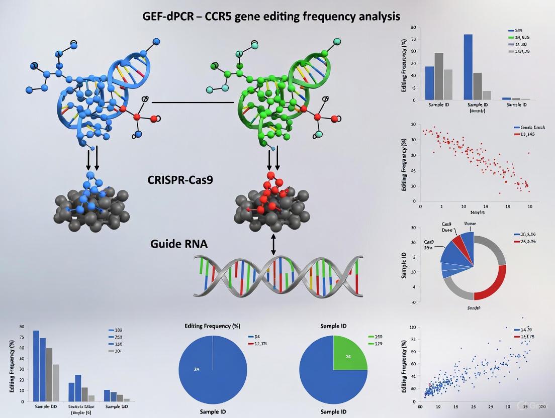
GEF-dPCR Workflow for CCR5 Gene Editing Analysis
The following diagram illustrates the complete GEF-dPCR workflow for analyzing CCR5 gene editing frequency:
Detailed Protocol for CCR5 Editing Frequency Analysis
Step 1: Sample Preparation and DNA Isolation
- Isolate genomic DNA from CCR5-edited cells using commercial kits (e.g., QIAamp DNA Blood Mini Kit)
- Quantify DNA concentration using fluorometric methods (e.g., Qubit dsDNA BR Assay)
- Dilute DNA to optimal concentration for dPCR (typically 1-10 ng/μL) [12]
Step 2: GEF-dPCR Reaction Setup
- Prepare reaction mix containing:
- 1X ddPCR Supermix
- CCR5-specific primers (final concentration: 900 nM each)
- Wild-type probe (HEX-labeled, final concentration: 250 nM)
- Mutant probe (FAM-labeled, final concentration: 250 nM)
- 20-100 ng genomic DNA template
- Nuclease-free water to final volume [12]
Step 3: Droplet Generation and PCR Amplification
- Generate droplets using automated droplet generator
- Transfer droplets to 96-well PCR plate and seal
- Perform PCR amplification with optimized cycling conditions:
- 95°C for 10 minutes (enzyme activation)
- 40 cycles of: 94°C for 30 seconds, 57°C for 60 seconds
- 98°C for 10 minutes (enzyme deactivation)
- 4°C hold [12]
Step 4: Droplet Reading and Data Analysis
- Read droplets using droplet reader (e.g., Bio-Rad QX100/QX200)
- Analyze data using manufacturer's software (e.g., QuantaSoft)
- Apply Poisson correction to calculate absolute copy numbers of wild-type and edited CCR5 alleles [12]
Essential Research Reagents and Solutions
Table 2: Key Research Reagent Solutions for GEF-dPCR
| Reagent/Equipment | Function | Example Products/Specifications |
|---|---|---|
| dPCR System | Partition generation, thermal cycling, and fluorescence reading | Bio-Rad QX200, QuantStudio Absolute Q System |
| dPCR Master Mix | Provides optimized buffer, enzymes, and nucleotides for amplification | ddPCR Supermix for Probes, Absolute Q Digital PCR Master Mix |
| CCR5-specific Primers | Amplify target region surrounding edit site | Custom-designed oligonucleotides (e.g., CCR5fw, CCR5rv) [12] |
| Hydrolysis Probes | Sequence-specific detection with fluorescent reporters | TaqMan probes with FAM/HEX labels and quenchers [13] |
| DNA Isolation Kit | High-quality genomic DNA extraction | QIAamp DNA Blood Mini Kit, QIAamp DNA Micro Kit [12] |
| DNA Quantification Kit | Accurate nucleic acid concentration measurement | Qubit dsDNA BR Assay Kit [12] |
Advanced Applications and Technical Considerations
Multiplexed GEF-dPCR Assays for Comprehensive Editing Analysis
The CLEAR-time dPCR method represents an advanced implementation of GEF-dPCR principles, employing multiple assay types to characterize different aspects of gene editing outcomes [14]:
- Edge Assay: Quantifies wild-type sequences, indels, and total non-indel aberrations using primers flanking the RNP target site with cleavage (FAM) and distal (HEX) probes
- Flanking and Linkage Assay: Detects double-strand breaks, large deletions, and structural mutations using two amplicons flanking the cleavage site
- Aneuploidy Assay: Identifies chromosomal gains or losses using primers and probes in sub-telomeric regions
- Target-integrated and Episomal Donor Assessment: Measures HDR efficiency when using donor templates [14]
Critical Technical Considerations for GEF-dPCR
Probe Design and Optimization:
- Probes should be less than 30 nucleotides between fluorophore and quencher
- Avoid guanine (G) at the 5′ end of probes to prevent fluorescence quenching
- Ensure probes have higher melting temperature (Tm) than primers when possible
- Test specificity using synthetic substrates to determine false positive rates [13]
Data Interpretation and Quality Control:
- Establish clear thresholds for positive/negative partition calling
- Ensure adequate partition numbers for statistical significance (typically >10,000)
- Include appropriate controls (wild-type, edited, and no-template controls)
- Account for potential cross-reactivity between highly homologous targets (e.g., CCR5 and CCR2) [12] [17]
Performance Benchmarking and Data Analysis
Quantitative Performance Metrics
Table 3: GEF-dPCR Performance in CCR5 Gene Editing Studies
| Parameter | Performance Metric | Experimental Context |
|---|---|---|
| Editing Efficiency | 30%–56% gene editing rates | Primary CD4+ T cells edited with CCR5-Uco-hetTALEN [17] |
| Biallelic Editing | ~40% of large-scale produced cells | Clinical-scale production of CCR5-edited CD4+ T cells [12] |
| Large Deletion Detection | Up to 2% of T cells with 15-kb deletions | Simultaneous cutting at CCR5 and CCR2 [17] |
| Sensitivity | Detection of rare RNA editing events | APOBEC3A-mediated RNA editing quantification [13] |
| Accuracy Benchmark | High correlation with AmpSeq | Systematic comparison of editing quantification methods [16] |
Data Analysis Workflow
The following diagram illustrates the logical flow of data analysis in GEF-dPCR experiments:
The GEF-dPCR methodology provides unparalleled accuracy for quantifying CCR5 gene editing frequencies, enabling robust assessment of therapeutic cell products. This absolute quantification approach has proven essential for clinical translation of CCR5-edited T cells for HIV therapy, where precise measurement of editing efficiency directly correlates with therapeutic efficacy and safety [12] [17].
The development of programmable nucleases, including TAL effector nucleases (TALENs) and CRISPR-Cas systems, has revolutionized genetic engineering approaches for research and therapeutic applications. Within the context of CCR5 gene editing for HIV resistance, accurately quantifying three fundamental metrics—editing efficiency, biallelic disruption, and large deletions—is paramount for evaluating experimental success and therapeutic potential. Editing efficiency determines the proportion of cells with modified target sequences, while biallelic disruption indicates complete knockout of both alleles, which is essential for conferring HIV resistance. Large deletions represent unintended, extensive genetic alterations that may have functional consequences. This application note details the definitions, quantification methods, and protocols for these key metrics, with a specific focus on Gene Editing Frequency digital PCR (GEF-dPCR) for robust analysis of CCR5 editing outcomes.
Defining and Quantifying Key Metrics
Editing Efficiency
Definition: Editing efficiency refers to the percentage of alleles in a cell population that contain any form of modification—including insertions, deletions (INDELs), or other sequence alterations—at the intended nuclease target site. This metric reflects the overall activity of the gene editing system.
Quantification Methods:
- Next-Generation Amplicon Sequencing (Amplicon NGS): Provides a comprehensive, base-pair-resolution profile of all sequence variations at the target locus. Reads containing insertions or deletions at the TALEN binding sites or Cas9 cut site are classified as edited [6] [18] [11].
- Gene Editing Frequency digital PCR (GEF-dPCR): A highly precise method for absolute quantification of edited alleles without a standard curve. It utilizes sequence-specific probes to distinguish wild-type from edited alleles and calculates editing efficiency based on Poisson statistics of positive and negative partitions [11] [19].
Table 1: Representative Editing Efficiencies for CCR5-Targeting Nucleases
| Nuclease System | Target Gene | Cell Type | Editing Efficiency | Citation |
|---|---|---|---|---|
| CCR5-Uco-hetTALEN | CCR5 | Primary CD4+ T cells | 30% - 56% | [6] |
| HiFi SpCas9 RNP | HBB | Hematopoietic Stem and Progenitor Cells (HSPCs) | 11.7% - 35.4% (on-target) | [18] |
| HiFi SpCas9 RNP | PD-1 | Primary T cells | 15.2% (on-target) | [18] |
Biallelic Disruption
Definition: Biallelic disruption occurs when both copies of a target gene in a diploid cell are successfully modified, resulting in complete loss of function. This is a critical goal for CCR5 knockout strategies to achieve maximum resistance to CCR5-tropic HIV infection.
Quantification Methods:
- Single-Cell High-Resolution Melting Curve Analysis (scHRMCA): Individual cells are sorted into PCR plates, and the target locus is amplified and analyzed via high-resolution melting. Differences in melting profiles between wild-type and edited alleles allow for genotyping of single cells to determine if edits are monoallelic or biallelic [6] [11].
- Clonal Genotyping: Single-cell-derived clones are expanded and screened via PCR and sequencing. Clones are defined as having biallelic deletion if PCR amplification of the "deletion band" is present and the "non-deletion band" is absent [20].
- Droplet Digital PCR (ddPCR): A bioanalytical method that can be used to quantify the frequency of biallelically edited cells in a population [11].
Key Finding: The frequency of cells with biallelic deletion can exceed probabilistic expectation, suggesting that the CRISPR/Cas9 system may be highly efficient or that cells with biallelic edits may have a growth advantage in certain contexts [20].
Large Deletions
Definition: Large deletions (LDs) are unintended genomic modifications exceeding 100 base pairs (bp) that occur at the on-target nuclease cut site. In CRISPR-Cas9 editing, these can range from hundreds to over a million base pairs [20] [18] [21]. CRISPR-Cas3 systems induce even broader, unidirectional deletions of several thousand bp upstream of the PAM site [22].
Quantification Methods:
- Long-Range PCR and Sequencing: Amplification of a large genomic region (e.g., 5-15 kb) surrounding the cut site, followed by gel electrophoresis (for size shift detection) or next-generation sequencing (for precise characterization) [18] [21].
- ddPCR-based Allelic Drop-Off Assay: A quantitative method that uses a probe binding distal to the cut site. A large deletion that removes the probe-binding sequence will result in a "drop-off" of the fluorescence signal, allowing for quantification of the deletion frequency [18].
- Optimized Long-Amplicon Sequencing: An Illumina-based method combining long-range PCR with DNA fragmentation and high-accuracy short-read sequencing. This approach allows for the simultaneous detection of small INDELs and large deletions with high precision [21].
Table 2: Occurrence of Large Deletions Across Cell Types and Editors
| Editing System | Cell Type | Large Deletion Frequency | Deletion Size Range | Citation |
|---|---|---|---|---|
| Cas9 Nuclease (HBB target) | HSPCs | 11.7% - 35.4% | Up to several thousand bp | [18] |
| Cas9 Nuclease (multiple targets) | Cancer Cell Lines (HeLa, HEK293T) | 4.4% - 6.4% | >100 bp | [21] |
| Base Editors / Prime Editors | Various Human Cell Lines | ~20-fold lower than Cas9 nuclease | >100 bp | [21] |
| CRISPR-Cas3 | Human Cells (e.g., 293T) | Induces prominent large deletions | Several thousand bp | [22] |
Experimental Protocols
Protocol 1: GEF-dPCR for CCR5 Editing Frequency
This protocol is adapted for quantifying CCR5 editing efficiency using the Bio-Rad QX100/QX200 ddPCR system [6] [11] [19].
Workflow Overview:
Materials:
- Primers/Probes: FAM-labeled probe for wild-type CCR5 sequence, HEX/VIC-labeled probe for edited CCR5 sequence (e.g., detecting a common deletion), and a reference gene assay.
- ddPCR Supermix: (e.g., Bio-Rad ddPCR Supermix for Probes).
- Droplet Generator and Reader.
Procedure:
- Genomic DNA Extraction: Extract high-quality gDNA from edited and control cell populations using a commercial kit (e.g., QIAamp DNA Blood Mini Kit). Quantify DNA using a fluorometer.
- Reaction Setup: Prepare a 20-22 µL reaction mix containing:
- 1x ddPCR Supermix.
- Primers and probes at optimized concentrations (e.g., 900 nM primers, 250 nM probes each).
- Approximately 20-100 ng of gDNA.
- Droplet Generation: Transfer the reaction mix to a DG8 cartridge. Generate approximately 20,000 droplets using the Droplet Generator.
- PCR Amplification: Transfer droplets to a 96-well PCR plate. Seal the plate and run the PCR with the following optimized cycling conditions:
- 95°C for 10 min (enzyme activation).
- 40 cycles of: 94°C for 30 s, 60°C for 60 s (annealing/extension).
- 98°C for 10 min (enzyme deactivation).
- 4°C hold.
- Droplet Reading: Place the plate in the Droplet Reader, which measures the fluorescence in each droplet.
- Data Analysis: Use the manufacturer's software (e.g., QuantaSoft) to analyze the data. The software clusters droplets as FAM+ (wild-type), HEX/VIC+ (edited), double-positive, or negative. Editing efficiency is calculated as:
(Concentration of edited alleles / (Concentration of edited alleles + Concentration of wild-type alleles)) * 100.
Protocol 2: Long-Amplicon Sequencing for Large Deletions
This protocol is designed to detect and quantify large deletions (>100 bp) using Illumina sequencing [18] [21].
Workflow Overview:
Materials:
- DNA Polymerase: KOD (Multi & Epi) DNA polymerase is recommended for its low amplification bias during long-range PCR [21].
- Primers: Designed to amplify a 5-15 kb region flanking the nuclease cut site.
- Library Prep Kit: Illumina-compatible library preparation kit (e.g., Nextera XT).
Procedure:
- gDNA Extraction: Extract high-quality, high-molecular-weight gDNA.
- Long-Range PCR: Set up a 50 µL PCR reaction with:
- 1x buffer for KOD (Multi & Epi).
- 200 µM dNTPs.
- 0.3 µM each forward and reverse primer.
- 1.0 U/µL KOD (Multi & Epi) polymerase.
- 50-100 ng gDNA.
- Cycling conditions: 94°C for 2 min; 30 cycles of 98°C for 10 s, 60°C for 30 s, 68°C for 10-15 min (depending on amplicon size); final extension at 68°C for 20 min.
- Amplicon Fragmentation and Library Prep: Fragment the long-range PCR products to ~300 bp using a standardized library preparation protocol (end repair, dA-tailing, adaptor ligation, and PCR enrichment).
- Sequencing: Sequence the library on an Illumina platform (e.g., MiSeq) to generate high-accuracy short reads.
- Bioinformatic Analysis: Analyze the sequencing data using a specialized tool like ExCas-Analyzer. This k-mer alignment-based algorithm is designed to simultaneously detect small INDELs and large deletions from the long-amplicon sequencing data with high speed and accuracy [21].
The Scientist's Toolkit: Research Reagent Solutions
Table 3: Essential Reagents for Gene Editing Analysis
| Item | Function/Description | Example Use Case |
|---|---|---|
| CCR5-Uco-hetTALEN | TALEN with heterodimeric FokI domain for high-efficiency, specific CCR5 knockout. | CCR5 gene disruption in primary T cells [6] [11]. |
| HiFi SpCas9 | High-fidelity version of Cas9 nuclease with reduced off-target activity. | On-target editing with minimized off-target effects in HSPCs [18]. |
| KOD (Multi & Epi) DNA Polymerase | High-fidelity DNA polymerase with low bias in long-range PCR amplification. | Accurate amplification of large genomic regions for deletion detection [21]. |
| ddPCR System (e.g., Bio-Rad QX100) | Instrumentation for absolute quantification of nucleic acids via droplet partitioning. | Absolute quantification of CCR5 editing frequency and biallelic disruption [11] [19]. |
| Unique Molecular Identifiers (UMIs) | Short random nucleotide sequences used to tag individual DNA molecules prior to PCR. | Reducing PCR amplification artifacts and biases in long-amplicon sequencing [18]. |
| ExCas-Analyzer Software | A dedicated k-mer alignment algorithm for analyzing CRISPR-edited samples. | Simultaneous detection and quantification of small INDELs and large deletions from sequencing data [21]. |
Implementing GEF-dPCR for CCR5 Analysis: A Step-by-Step Protocol
The C-C chemokine receptor type 5 (CCR5) serves as a crucial co-receptor for human immunodeficiency virus (HIV) entry into T-cells [23] [24]. A naturally occurring 32-base pair deletion in the CCR5 gene, known as CCR5Δ32, results in a non-functional receptor that confers resistance to HIV infection in homozygous individuals [23] [2] [24]. This discovery has spurred the development of novel therapeutic strategies, including allogeneic hematopoietic stem cell transplantation from CCR5Δ32 donors and CRISPR/Cas9-mediated gene editing to create the mutation in autologous cells [23] [2].
Accurately quantifying the frequency of this genetic modification is essential for evaluating the efficacy of gene-editing approaches and monitoring transplanted cell populations. Droplet Digital PCR (ddPCR) has emerged as a powerful tool for precise quantification of gene-editing frequencies, enabling sensitive detection of mutant alleles within heterogeneous cell mixtures [23] [24] [25]. This application note details the strategic design of primers and probes for discriminating between wild-type and CCR5Δ32 mutant alleles using ddPCR, framed within the broader research context of Gene Editing Frequency digital PCR (GEF-dPCR) [25].
Background and Principle of GEF-dPCR
The GEF-dPCR method exploits the capabilities of digital PCR to provide absolute quantification of nucleic acid targets without the need for standard curves [25]. In the context of CCR5 gene editing, this technique utilizes two differentially labeled probes placed within a single amplicon spanning the target site to simultaneously detect wild-type and mutation-containing alleles [25].
The fundamental principle involves partitioning a PCR reaction into thousands of nanoliter-sized droplets, effectively creating individual reaction chambers. Each droplet undergoes PCR amplification and is analyzed for fluorescence signals indicating the presence of wild-type alleles, mutant alleles, or both [25]. This approach allows for concurrent quantification of edited and wild-type alleles in a given sample, providing a direct measurement of gene-editing efficiency that is critical for clinical applications [25].
Table 1: Key Genetic Elements in CCR5-Targeted HIV Therapies
| Element | Characteristics | Therapeutic Relevance |
|---|---|---|
| CCR5 (Wild-Type) | G-protein coupled receptor; HIV-1 co-receptor [23] [24] | Primary pathway for R5-tropic HIV entry; target for inhibition or disruption |
| CCR5Δ32 Mutation | 32-bp deletion in coding sequence; causes frameshift and premature stop codon [23] [24] | Confers HIV resistance in homozygous carriers; target for gene editing therapies [2] |
| CRISPR/Cas9 gRNAs | Guide RNAs targeting CCR5 exon 3 (e.g., TB48, TB50) [2] | Tools for creating artificial CCR5Δ32 mutations via non-homologous end joining [23] [2] |
Primer and Probe Design Strategy
Target Region Selection and Amplicon Design
Effective allele discrimination requires careful selection of the target region and strategic placement of primers and probes:
- Amplicon Localization: Design primers to flank the 32-base pair deletion region in exon 3 of the CCR5 gene, ensuring the amplicon is sufficiently small (typically 80-200 bp) for efficient amplification in ddPCR [24].
- Probe Placement: Position one probe to span the exact deletion junction for specific detection of the CCR5Δ32 mutant allele. Design another probe to bind within the wild-type sequence at the same genomic location, ensuring both probes have similar melting temperatures (Tm) for equivalent amplification efficiency [26].
- Dual Probe System: Implement a multiplex assay using two hydrolysis probes (e.g., FAM-labeled for mutant allele, HEX/VIC-labeled for wild-type allele) that compete for binding within the same genomic region, enabling precise allele discrimination and quantification [25].
The following diagram illustrates the core principle of the GEF-dPCR assay for simultaneous detection of wild-type and mutant alleles:
Specific Design Parameters
- Allele-Specific Oligonucleotides: For TaqMan probe-based assays, design probes with the nucleotide that distinguishes the alleles (the deletion junction for Δ32) located at or near the center of the probe sequence [26]. For SYBR Green-based approaches using allele-specific primers, place the discriminating nucleotide at the 3' terminal base of one primer [26].
- Probe Characteristics: Utilize minor groove binder (MGB) probes to increase binding specificity and Tm, which is particularly advantageous for short probes necessary for detecting small deletions [26].
- Specificity Validation: Perform BLAST analysis against the human genome to ensure minimal homology with other regions, particularly the highly homologous CCR2 gene [2] [26].
Table 2: Primer and Probe Design Specifications for CCR5 Genotyping
| Component | Sequence (5' to 3') | Modification | Genome Position | Function |
|---|---|---|---|---|
| Forward Primer | CCCAGGAATCATCTTTACCA [24] | Standard desalting | Upstream of Δ32 | Forward amplification primer |
| Reverse Primer | GACACCGAAGCAGAGTTT [24] | Standard desalting | Downstream of Δ32 | Reverse amplification primer |
| WT-specific Probe | (Sequence spanning WT region) | HEX/MGB [26] | Within deleted region | Binds only to wild-type allele |
| Δ32-specific Probe | (Sequence spanning Δ32 junction) | FAM/MGB [26] | Across deletion junction | Binds only to Δ32 mutant allele |
Experimental Protocol: CCR5Δ32 Frequency Quantification
Sample Preparation and DNA Extraction
- Cell Culture and Editing: Culture target cells (e.g., MT-4 T-cell line or human CD34+ HSPCs) following standard protocols [23] [2]. Perform CCR5 gene editing using CRISPR/Cas9 system with optimized gRNAs (e.g., TB48 and TB50 combination) [2].
- Genomic DNA Extraction: Harvest cells and extract genomic DNA using phenol-chloroform method or commercial kits (e.g., ExtractDNA Blood and Cells Kit) [23] [24]. Measure DNA concentration and purity using spectrophotometry (A260/A280 ratio of ~1.8-2.0) [23].
ddPCR Reaction Setup and Thermal Cycling
The following workflow outlines the complete experimental procedure from sample preparation to data analysis:
Table 3: ddPCR Reaction Setup and Thermal Cycling Conditions
| Component/Step | Specification | Notes |
|---|---|---|
| Template DNA | 10-100 ng per reaction | Adjust based on DNA quality and target abundance [23] |
| Primer Concentration | 0.2 µM each | Optimize to minimize nonspecific amplification [24] |
| Probe Concentration | 0.1-0.2 µM each | FAM for Δ32, HEX/VIC for wild-type [25] |
| ddPCR Supermix | 1X | Use ddPCR supermix for probes |
| Final Reaction Volume | 20-22 µL | Adjust based on droplet generator requirements |
| Initial Denaturation | 95°C for 10 min | Enzyme activation |
| Amplification (40 cycles) | 94°C for 30 sec, 58-60°C for 60 sec | Annealing/extension temperature probe-specific |
| Enzyme Deactivation | 98°C for 10 min | Final deactivation |
| Droplet Reading | Use ddPCR droplet reader | Follow manufacturer's instructions |
Data Analysis and Interpretation
- Droplet Classification: Use the droplet reader software to classify droplets as FAM-positive (mutant Δ32), HEX-positive (wild-type), double-positive (heterozygous), or negative (no template) [23] [25].
- Concentration Calculation: Apply Poisson correction to calculate the absolute concentration (copies/μL) of wild-type and mutant alleles in the original sample [25].
- Editing Frequency Determination: Calculate the percentage of CCR5Δ32 alleles using the formula: % CCR5Δ32 = [Δ32 concentration / (Δ32 concentration + WT concentration)] × 100
- Sensitivity Assessment: The developed system can accurately measure CCR5Δ32 content down to 0.8% in cell mixtures, providing sensitive detection of low-frequency editing events [23].
Research Reagent Solutions
Table 4: Essential Reagents and Tools for CCR5 Genotyping Assays
| Reagent/Tool | Function | Example Products/Specifications |
|---|---|---|
| ddPCR System | Partitioning, amplification, and droplet reading | Bio-Rad QX200 Droplet Digital PCR System [23] [25] |
| DNA Extraction Kit | High-quality genomic DNA isolation | ExtractDNA Blood and Cells Kit, NucleoSpin Kit [23] [27] |
| CRISPR/Cas9 Reagents | Generation of CCR5Δ32 mutation | Cas9 protein, gRNAs (e.g., TB48, TB50) [2] |
| ddPCR Supermix | Optimized reaction mix for ddPCR | ddPCR Supermix for Probes [23] |
| Allele Discrimination Software | Probe and primer design for SNP detection | AlleleID [26] |
| Cell Culture Reagents | Maintenance of target cell lines | RPMI-1640 medium, Fetal Bovine Serum [23] |
Applications and Discussion
The ddPCR assay for CCR5 wild-type and Δ32 allele quantification provides critical applications in both basic research and clinical development:
- Therapeutic Monitoring: Accurately measure the percentage of CCR5-disrupted cells in autologous hematopoietic stem cell transplant products, with studies indicating that >90% editing frequency may be necessary for therapeutic efficacy [2].
- HIV Cure Research: Monitor engraftment and expansion of CCR5-edited cells in patients undergoing novel HIV cure strategies, analogous to the monitoring performed in the "Berlin" and "London" HIV cure cases [23] [2].
- Preclinical Development: Evaluate the efficiency of different gene-editing approaches (CRISPR/Cas9, TALEN, ZFNs) in various cell types by providing precise quantification of editing frequencies [23] [25].
This GEF-dPCR approach offers significant advantages over traditional methods such as endpoint PCR or flow cytometry, including absolute quantification without standard curves, high sensitivity for detecting rare mutations, and excellent reproducibility [23] [25]. The duplex nature of the assay allows for internal control of DNA quality and quantity through simultaneous detection of both alleles in each reaction.
When implementing this assay, researchers should validate assay performance using appropriate controls, including confirmed wild-type, heterozygous, and homozygous Δ32 samples when available [27]. Additionally, the analytical sensitivity and specificity should be established for the specific application, particularly when assessing low-frequency editing events in heterogeneous samples.
Within the framework of developing a functional cure for HIV, the precise quantification of CCR5 gene editing frequency using droplet digital PCR (GEF-dPCR) has emerged as a critical analytical method. This application note provides a detailed protocol for the preparation of high-quality genomic DNA (gDNA) from two primary cell types: edited T-cells and hematopoietic stem and progenitor cells (HSPCs). The integrity and purity of the isolated gDNA are foundational to the reliability of subsequent dPCR analyses, which in turn are essential for evaluating the efficacy of CCR5-editing therapies prior to clinical application [11].
The success of this workflow is demonstrated in recent pre-clinical studies, where high-frequency CCR5 editing (>90%) in human HSPCs translated into complete protection from HIV infection in xenograft mouse models [2]. This underscores the necessity of robust sample preparation for accurate editing assessment.
Critical Parameters for High-Quality gDNA
The table below summarizes the essential quality control metrics that gDNA samples must meet to be considered suitable for GEF-dPCR analysis.
Table 1: Quality Control Metrics for gDNA Intended for GEF-dPCR
| Parameter | Target Value for gDNA | Assessment Method | Significance for Downstream Analysis |
|---|---|---|---|
| Purity (A260/A280) | 1.8 – 1.9 [28] | Spectrophotometry (NanoDrop) | Ratios outside this range suggest protein or chemical contamination that can inhibit enzymatic reactions [29]. |
| Purity (A260/A230) | 1.8 – 2.5 [28] | Spectrophotometry (NanoDrop) | Low values indicate contamination with chaotropic salts or phenol, which are common inhibitors [29]. |
| Integrity | DIN > 8.5 or GQN > 8.0 [28] [30] | Automated Electrophoresis (TapeStation, Fragment Analyzer) | High molecular weight smears indicate intact DNA, ensuring accurate amplification of the target locus [30]. |
| Concentration | > 10 ng/μL (QC assay-dependent) [30] | Fluorometry (Qubit) | Fluorometry provides a more accurate quantification of double-stranded DNA than spectrophotometry [11]. |
Step-by-Step Protocols
gDNA Extraction from Edited T-Cells and HSPCs
This protocol is adapted from established methods for primary human cells [11].
Cell Lysis:
- Pellet approximately 1–5 × 10^6 edited T-cells or HSPCs by centrifugation.
- Resuspend the cell pellet thoroughly in 200 μL of PBS. Add 20 μL of Proteinase K and 200 μL of Buffer AL (from the kit). Mix immediately by pulse-vortexing for 15 seconds to ensure a homogeneous solution.
- Incubate at 56°C for 10 minutes. Brief centrifugation can be used to remove droplets from the lid.
Ethanol Precipitation:
- Add 200 μL of 96–100% ethanol to the lysed sample and mix again by pulse-vortexing.
Column Binding and Wash:
- Apply the entire mixture to the QIAamp Mini spin column and centrifuge at ≥6000 x g for 1 minute. Discard the flow-through.
- Place the column in a clean 2 mL collection tube. Add 500 μL of Buffer AW1, centrifuge at ≥6000 x g for 1 minute, and discard the flow-through.
- Place the column in a new 2 mL collection tube. Add 500 μL of Buffer AW2, centrifuge at full speed (20,000 x g) for 3 minutes, and discard the flow-through.
gDNA Elution:
- Transfer the column to a clean 1.5 mL microcentrifuge tube. To maximize concentration, apply 50–100 μL of Buffer AE or nuclease-free water directly to the center of the column membrane.
- Allow it to incubate at room temperature for 1–5 minutes, then centrifuge at ≥6000 x g for 1 minute. Store the eluted gDNA at -20°C or -80°C.
Assessment of gDNA Integrity
While agarose gel electrophoresis (0.75%) can provide a basic assessment, automated electrophoresis systems offer superior resolution and objective quantification [28] [30].
Using the Agilent TapeStation System:
- Sample Preparation: Dilute 1 μL of gDNA sample with 4 μL of nuclease-free water and add 5 μL of the Genomic DNA ScreenTape Sample Buffer.
- Loading: Load the mixture into the Genomic DNA ScreenTape.
- Analysis: Run the system and use the accompanying software to determine the DNA Integrity Number (DIN). A DIN > 8.5 indicates high-quality, intact gDNA suitable for GEF-dPCR [28].
Using Agilent Fragment Analyzer Systems:
- Follow the kit protocol for the Genomic DNA 50 kb kit, which typically involves a 200-fold dilution.
- The ProSize software will calculate a Genomic Quality Number (GQN). A GQN close to 10 indicates a sample where the majority of DNA is of high molecular weight [30].
The Scientist's Toolkit: Essential Reagents and Kits
Table 2: Key Research Reagent Solutions for gDNA Isolation and QC
| Item | Function/Application | Example Product (Supplier) |
|---|---|---|
| gDNA Purification Kit | Silica-membrane-based isolation of high-purity, high-integrity gDNA from cell pellets. | QIAamp DNA Blood Mini Kit (QIAGEN) [11] |
| Fluorometric DNA Quantification Assay | Highly specific, accurate quantification of double-stranded DNA concentration, superior to UV absorbance. | Qubit dsDNA BR Assay Kit (Thermo Fisher Scientific) [11] |
| Automated Electrophoresis System | Objective and precise assessment of gDNA integrity and size distribution, providing metrics like DIN or GQN. | Agilent TapeStation Systems [28] [30] |
| gDNA Integrity Assay | Reagent kit for use with automated electrophoresis systems to quantify gDNA integrity. | Genomic DNA ScreenTape Assay (Agilent) [30] |
| Droplet Digital PCR (ddPCR) System | Absolute quantification of CCR5 gene editing frequency with high precision, without the need for standard curves. | QX100/QX200 Droplet Digital PCR System (Bio-Rad) [11] |
Workflow Visualization
The following diagram illustrates the complete pathway from cell processing to data analysis, highlighting how gDNA quality directly influences GEF-dPCR outcomes and therapeutic development.
Droplet Digital PCR (ddPCR) is a powerful method for the absolute quantification of nucleic acids, providing a level of precision essential for assessing the success of gene-editing experiments. Unlike quantitative PCR (qPCR), which relies on standard curves and relative quantification, ddPCR uses a water-in-oil emulsion system to partition a sample into thousands of nanoliter-sized droplets, functioning as individual PCR reactions. This allows for absolute quantification of target DNA molecules without the need for external calibrators [31]. Within the context of CCR5 gene-editing research—a promising therapeutic strategy for achieving an HIV cure [2]—accurately determining the frequency of gene-editing events is a critical bottleneck. The Gene-Editing Frequency digital PCR (GEF-dPCR) method is specifically designed to address this need by enabling the concurrent quantification of edited and wild-type alleles in a given sample, making it optimal for monitoring gene-edited cells in clinical settings [25].
The ddPCR Workflow: A Step-by-Step Protocol
The ddPCR workflow, from sample preparation to data analysis, involves several key stages. The following diagram illustrates this complete process.
Diagram Title: The Complete ddPCR Workflow
Sample Preparation
Every ddPCR analysis begins with sample preparation. The required reaction mix is similar to a probe-based qPCR assay and includes:
- ddPCR Supermix: Provides the optimized buffer and polymerase for the reaction [32].
- Primers and Fluorescent Probes: Designed for the specific target. For GEF-dPCR, this involves two differently labeled probes placed within one amplicon at the gene-editing target site to simultaneously detect wild-type and NHEJ-affected alleles [25].
- Template DNA: The nucleic acid sample, which should be properly extracted and free of significant degradation or PCR inhibitors. If inhibitors are suspected, a 1:10 dilution of the sample is recommended [31].
A typical reaction volume of 20 µL is loaded into the individual wells of a droplet generator cartridge [31].
Droplet Generation
The loaded cartridge is placed into a droplet generator, which uses microfluidics and specific reagents to partition each sample into 20,000 nanoliter-sized water-in-oil droplets [31]. A well-functioning system creates droplets that are uniform in size and volume [33]. The distribution of target DNA molecules among these droplets is random, with some droplets containing zero, one, or a few template molecules. This randomness is the foundation for the subsequent statistical analysis [31].
PCR Amplification
After generation, the droplets are transferred to a 96-well PCR plate. The plate is sealed and placed in a standard thermal cycler. PCR amplification is then run to the endpoint, typically for 40 cycles, to amplify the target sequence within each droplet [31]. The specific thermal cycling profile may vary by assay; for instance, some protocols include a 10-minute step at 98°C after cycling to stabilize the droplets [33].
Droplet Reading
Following amplification, the PCR plate is transferred to a droplet reader. This instrument serially reads each well, guiding the droplets in a single file through a detection system. A two-color optical detector counts the droplets, measuring fluorescence to identify each one as positive (contains the target sequence, fluorescent) or negative (does not contain the target, non-fluorescent) [31].
Data Analysis
The ratio of positive to negative droplets is analyzed using Poisson statistics to determine the absolute concentration of the target nucleic acid in the original sample, expressed in copies per microliter (copies/µL) [31]. The fundamental calculation is based on the following formula [33]: [ \text{Concentration (copies/µL)} = \frac{-\ln(1 - \frac{P}{N})}{V_p} \times D ] Where:
- ( P ) = Number of PCR-positive partitions
- ( N ) = Total number of partitions
- ( V_p ) = Volume of each partition (nL)
- ( D ) = Dilution factor
Specialized software, such as QuantaSoft, is used to visualize the data and perform this calculation [32].
Quantitative Comparison of Digital PCR Platforms
Multiple commercial dPCR platforms exist, each with distinct characteristics. The following table summarizes a comparison of four different platforms for accurately quantifying the copy number of a certified plasmid DNA reference material [33].
| dPCR Platform | Partitioning Method | Typical Partitions per Reaction | Partition Volume | Relative Uncertainty of Partition Volume |
|---|---|---|---|---|
| BioMark (Fluidigm) | Microfluidic-chip | 765 per panel | 6 nL | 0.7% |
| QX100 (Bio-Rad) | Droplet-based | ~20,000 | 0.78 nL (per droplet) | 0.8% |
| QuantStudio 12k (Life Tech) | Micro-well chip | 3,072 per array | 33 nL (pre-set) | 2.3% |
| RainDrop (RainDance) | Droplet-based | Up to 10,000,000 | Not Specified | 2.9% |
The study demonstrated that after correcting for partition volume, all four platforms produced measurements that were consistent with the certified value of the reference material, confirming their comparability for DNA copy number quantification [33].
Application Protocol: GEF-dPCR for CCR5 Editing Analysis
This protocol outlines the specific application of ddPCR to assess the frequency of CCR5 gene editing in human hematopoietic stem and progenitor cells (HSPCs), a critical step in developing an autologous stem cell transplant for HIV cure [2].
- Objective: To absolutely quantify the frequency of CRISPR/Cas9-induced indels in the CCR5 gene locus.
- Principle: The GEF-dPCR assay uses two differentially labeled hydrolysis probes (e.g., FAM and HEX/VIC) that bind within the same amplicon spanning the Cas9 cut site. One probe is specific for the wild-type CCR5 sequence, while the other binds a sequence common to both wild-type and NHEJ-disrupted alleles. The ratio of droplets positive for both probes versus those positive only for the common probe allows for the calculation of the precise gene-editing frequency [25].
Experimental Workflow for CCR5 Editing
The specific steps for applying the ddPCR workflow to CCR5 research are detailed below.
Diagram Title: GEF-dPCR Protocol for CCR5 Analysis
- Sample Input: Begin with human mobilized CD34+ HSPCs that have been electroporated with CRISPR/Cas9 ribonucleoproteins (RNPs) targeting CCR5 [2].
- gDNA Extraction: Extract high-quality genomic DNA from the edited cells using a standard silica-membrane column or phenol-chloroform method. DNA purity and concentration should be measured spectrophotometrically.
- GEF-dPCR Assay Setup:
- Primers and Probes: Design and synthesize primers that amplify a 70-150 bp region encompassing the CCR5 target site. Use two probes:
- FAM-labeled probe: Specific to the wild-type CCR5 sequence.
- HEX-labeled probe: Binds a reference sequence present in both wild-type and successfully edited alleles [25].
- Reaction Mix: Prepare the 20 µL reaction mixture on ice according to the following table.
- Primers and Probes: Design and synthesize primers that amplify a 70-150 bp region encompassing the CCR5 target site. Use two probes:
| Reagent | Final Concentration | Function |
|---|---|---|
| ddPCR Supermix for Probes (2X) | 1X | Provides optimized buffer, dNTPs, and hot-start polymerase |
| Forward Primer | 900 nM | Amplifies the target region |
| Reverse Primer | 900 nM | Amplifies the target region |
| FAM-labeled Wild-Type Probe | 250 nM | Detects unedited CCR5 alleles |
| HEX-labeled Common Probe | 250 nM | Detects total (edited + unedited) alleles |
| Template gDNA | 10-100 ng | Contains the target CCR5 sequence |
| Nuclease-Free Water | To volume | Adjusts final reaction volume |
- Execute ddPCR: Follow the standard workflow of droplet generation, PCR amplification (40-50 cycles), and droplet reading as described in the general protocol above [2] [31].
- Data Analysis and Interpretation: Use the instrument's software (e.g., QuantaSoft) to analyze the data. The gene-editing frequency (GEF) is calculated as: [ \text{GEF} = \frac{\text{Number of HEX-positive, FAM-negative droplets}}{\text{Total number of HEX-positive droplets}} ] This represents the proportion of alleles that have been modified and no longer bind the wild-type probe. High-frequency editing (>90%) has been shown to be critical for conferring protective benefit against HIV infection in pre-clinical models [2].
The Scientist's Toolkit: Essential Research Reagents
The following table details key reagents and materials required for setting up a ddPCR experiment for gene-editing analysis.
| Item | Function / Explanation |
|---|---|
| ddPCR Supermix | A ready-to-use buffer solution containing DNA polymerase, dNTPs, and stabilizers optimized for the droplet environment. Available in formulations for probes or EvaGreen dye [32]. |
| Sequence-Specific Primers | Oligonucleotides designed to amplify the target region of interest (e.g., the CCR5 locus). Must be highly specific and yield a short amplicon. |
| Hydrolysis Probes (TaqMan) | Fluorescently labeled probes (e.g., FAM, HEX) that increase specificity and enable multiplexing. Crucial for GEF-dPCR to distinguish between wild-type and edited alleles [25] [31]. |
| Droplet Generator Cartridge & Oil | The consumable cartridge and corresponding droplet generation oil are essential for creating the water-in-oil emulsion [32]. |
| 96-Well PCR Plates & Seals | Plates compatible with the thermal cycler and droplet reader, and foil or pierceable seals to prevent cross-contamination and evaporation during cycling [32]. |
| QX200 Droplet Reader Oil | Specific oil required for the droplet reader to properly orient and read the droplets as they pass the detector [32]. |
The quantification of gene editing efficiency is a critical step in developing therapies for HIV. Gene-Editing Frequency digital PCR (GEF-dPCR) represents a significant methodological advancement, enabling researchers to accurately quantify the success of CRISPR/Cas9-mediated modifications at the CCR5 gene locus. This technique uses a novel approach with two differently labeled probes placed within a single amplicon at the target site to simultaneously detect both wild-type and modified alleles. For researchers and drug development professionals working on CCR5-based HIV therapies, mastering QuantaSoft data analysis is paramount for translating raw dPCR data into meaningful, publication-ready metrics on editing efficiency [25].
The clinical relevance of this analysis is underscored by recent research demonstrating that high-frequency CCR5 editing (>90%) in human hematopoietic stem progenitor cells (HSPCs) is necessary to confer protective benefit against HIV infection in xenograft models. Achieving this level of editing efficiency requires robust quantification methods to monitor experimental outcomes and optimize protocols [2]. This application note provides a comprehensive framework for interpreting 1D and 2D plots in QuantaSoft to calculate gene-editing frequencies, with specific application to CCR5 editing research.
Key Principles of GEF-dPCR Analysis
Core Concepts in Multiplexed dPCR Analysis
The GEF-dPCR methodology leverages the absolute quantification capabilities of digital PCR with multiplexing strategies to deconvolute complex editing outcomes. The fundamental principle involves discriminating alleles through probe-based detection where:
- A FAM-labeled probe binds specifically to the wild-type CCR5 sequence at the nuclease target site
- A HEX-labeled probe binds to a distal, conserved region of the amplicon to serve as an internal control for total DNA content
- The ratios of fluorescence signals across thousands of individual partitions are analyzed to determine the genotype status of each template molecule [25]
This approach has been refined in recently developed techniques like CLEAR-time dPCR, which provides an ensemble of multiplexed dPCR assays that quantify genome integrity at targeted sites. This method can track active double-strand breaks (DSBs), small indels, large deletions, and other genetic aberrations in absolute terms in clinically relevant edited cells, including HSPCs, induced pluripotent stem cells (iPSCs), and T-cells [14].
Experimental Workflow for CCR5 Editing Assessment
The complete process from sample preparation to data analysis follows a structured workflow that ensures reliable quantification of gene-editing outcomes.
Figure 1: Complete workflow for GEF-dPCR analysis of CCR5 edited samples, from initial sample preparation to final quality assessment of the calculated editing frequencies.
Experimental Protocols
Sample Preparation and gDNA Extraction
Protocol: Isolation of Genomic DNA from CCR5-Edited Hematopoietic Cells
- Cell Collection: Harvest CCR5-edited cells (e.g., HSPCs, T-cells) 48-72 hours post-electroporation with CRISPR/Cas9 ribonucleoprotein (RNP) complexes. Include mock-edited controls [2].
- Cell Lysis: Use proteinase K-based lysis buffer to digest cellular proteins. Incubate at 56°C for 2 hours with gentle agitation.
- DNA Purification: Employ silica membrane-based columns or magnetic beads for DNA binding and purification.
- DNA Quantification: Measure DNA concentration using fluorometric methods (e.g., Qubit dsDNA HS Assay). Ensure 260/280 ratios between 1.8-2.0.
- DNA Quality Verification: Confirm high molecular weight DNA using agarose gel electrophoresis or fragment analyzer systems.
Critical Step: For editing efficiency calculations, ensure input gDNA quality is sufficient for amplification of target regions. DNA fragmentation can lead to underestimation of editing frequencies [14].
GEF-dPCR Assay Setup and Configuration
Protocol: Reaction Setup for CCR5 Editing Frequency Analysis
Reagent Preparation:
- Prepare 1× ddPCR Supermix for Probes (no dUTP)
- Dilute gDNA to working concentration (10-100 ng/μL)
- Prepare primer-probe mix with final concentrations:
- CCR5 wild-type specific FAM-labeled probe: 250 nM
- CCR5 distal HEX-labeled probe: 250 nM
- Forward and reverse primers: 900 nM each [25]
Reaction Assembly:
- Combine 11 μL ddPCR Supermix, 2 μL primer-probe mix, and 9 μL diluted gDNA (total volume = 22 μL)
- Include no-template control (NTC) and wild-type gDNA controls
Droplet Generation:
- Transfer 20 μL of reaction mix to DG8 Cartridge
- Generate droplets using Droplet Generator
- Transfer emulsified samples to 96-well PCR plate
PCR Amplification:
- Seal plate with foil heat seal
- Run thermal cycling protocol:
- 95°C for 10 minutes (1 cycle)
- 94°C for 30 seconds, 60°C for 60 seconds (40 cycles)
- 98°C for 10 minutes (1 cycle)
- 4°C hold [25]
Data Acquisition:
- Transfer plate to Droplet Reader
- Analyze droplets from each well using QuantaSoft software
Data Analysis in QuantaSoft
Interpretation of 1D Amplification Plots
One-dimensional plots in QuantaSoft display fluorescence amplitude for a single channel (FAM or HEX) across all droplets. These plots provide initial quality control and preliminary data on assay performance.
Analysis Procedure:
FAM Channel Analysis (Wild-Type CCR5):
- Identify positive and negative droplet populations
- Assess separation between clusters (ΔRFU > 5000 recommended)
- Note any intermediate populations indicating probe binding issues
HEX Channel Analysis (Reference Assay):
- Verify strong positive/negative separation
- Ensure >10,000 total accepted droplets for statistical reliability
- Check for reduced HEX-positive droplets suggesting large deletions [14]
Table 1: Interpretation guidelines for 1D amplitude plots in GEF-dPCR analysis
| Observation | Interpretation | Required Action |
|---|---|---|
| Broad spread of intermediate amplitudes | Probable probe binding issues due to unexpected mutations | Redesign probe or verify target specificity |
| Low fluorescence amplitude in both channels | PCR inhibition or suboptimal reaction conditions | Optimize DNA input concentration or purify DNA |
| Reduced total droplet count in HEX channel | Potential large deletions affecting distal binding site | Perform additional flanking assay to confirm [14] |
| Clear bimodal distribution with high ΔRFU | Optimal assay performance | Proceed with 2D analysis for frequency calculation |
Interpretation of 2D Density Plots
Two-dimensional plots display fluorescence data from both FAM and HEX channels simultaneously, enabling classification of droplets into four distinct populations that correspond to different genetic outcomes.
Population Classification:
- Double-Positive (FAM+HEX+): Wild-type CCR5 alleles
- FAM-Negative/HEX-Positive (FAM-HEX+): Edited alleles with indels or mutations at target site
- FAM-Positive/HEX-Negative (FAM+HEX-): Rare population indicating potential probe competition or artifacts
- Double-Negative (FAM-HEX-): Non-amplifying droplets or empty partitions [25]
Figure 2: Classification of droplet populations in 2D density plots for CCR5 editing analysis. The key population for editing frequency calculation is the FAM-HEX+ group in quadrant 2.
Advanced CLEAR-time dPCR Analysis
For more comprehensive analysis of editing outcomes, the CLEAR-time dPCR method employs multiple assay configurations to quantify different types of genetic alterations:
Edge Assay:
- Primers flank the cleavage site with two probes (FAM at cleavage site, HEX distal)
- FAM signal loss indicates indels; complete signal loss indicates large deletions [14]
Flanking and Linkage Assay:
- Uses two separate amplicons flanking the cleavage site
- Loss of linkage indicates DSBs or large deletions
- Enables quantification of unresolved breaks [14]
Table 2: Quantification of different mutation types using CLEAR-time dPCR assays
| Assay Type | Measured Outcome | Calculation Method | Typical Range in HSPCs |
|---|---|---|---|
| Edge Assay | Total editing frequency (indels + large deletions) | (FAM-HEX+ + complete dropouts) / Total templates | 60-97% [2] |
| Flanking Assay | Large deletions and unresolved DSBs | Loss of linkage between flanking amplicons | 5-15% [14] |
| Aneuploidy Assay | Chromosomal abnormalities | Signal ratio variation in sub-telomeric regions | <2% [14] |
| Reference Assay | Sample quality and loading control | Normalization against non-targeted chromosomes | Used for normalization |
Frequency Calculation and Data Interpretation
Calculation of Gene-Editing Frequencies
The primary metric for success in CCR5 editing experiments is the gene-editing frequency, which represents the percentage of alleles successfully modified by the CRISPR/Cas9 system.
Fundamental Calculation:
In QuantaSoft analysis, this translates to:
This calculation specifically quantifies alleles with successful gene editing that resulted in disruption of the FAM probe binding site while maintaining the distal reference region [25].
Advanced Calculations: For the CLEAR-time dPCR method, more sophisticated calculations are employed:
These calculations provide a more comprehensive view of the editing outcomes, particularly important for assessing safety profiles in therapeutic applications [14].
Threshold Determination and Gating Strategies
Proper gating is essential for accurate frequency calculations. Follow these steps for robust threshold setting:
Use Control Samples:
- Apply identical gating to mock-edited and wild-type controls
- Establish baseline fluorescence for wild-type populations
- Set thresholds to minimize false positives in negative controls
Iterative Gating Approach:
- Begin with conservative gates based on control samples
- Adjust to ensure <0.1% background in negative controls
- Apply final gates uniformly across all experimental samples
Validation:
- Confirm expected clustering patterns are present
- Verify that control samples show <1% editing frequency
- Ensure experimental samples show clear population separation
The Scientist's Toolkit: Research Reagent Solutions
Table 3: Essential research reagents and materials for GEF-dPCR analysis of CCR5 gene editing
| Reagent/Material | Function | Example Specifications |
|---|---|---|
| CCR5-specific gRNAs (e.g., TB48, TB50) | Guide Cas9 to CCR5 target site | Chemically synthesized, >90% editing efficiency in HSPCs [2] |
| SpCas9 Nuclease | Creates double-strand breaks at CCR5 locus | High-purity, recombinant, complexed with gRNA as RNP [2] |
| ddPCR Supermix for Probes | PCR amplification in droplets | Contains DNA polymerase, dNTPs, optimized buffers |
| FAM-labeled CCR5 WT probe | Binds wild-type CCR5 sequence at cut site | 5' FAM, 3' BHQ-1, 20-25 bp |
| HEX-labeled reference probe | Binds conserved region distal to cut site | 5' HEX, 3' BHQ-1, 20-25 bp [25] |
| Droplet Generation Cartridges | Creates water-in-oil emulsion partitions | Compatible with QX200 system |
| gDNA Extraction Kits | Purifies high-quality genomic DNA | Column-based or magnetic bead purification |
| Human HSPCs or T-cells | Primary cells for CCR5 editing studies | Mobilized CD34+ cells or activated T-cells [2] |
Proficiency in interpreting QuantaSoft 1D and 2D plots is essential for accurate assessment of CCR5 gene editing frequencies in therapeutic development. The GEF-dPCR method provides a robust framework for this analysis, enabling precise quantification of editing outcomes that correlate with functional protection against HIV infection. The recent development of CLEAR-time dPCR further enhances this capability by providing a more comprehensive assessment of on-target editing, including the quantification of large deletions and unresolved double-strand breaks that may have safety implications. As research advances toward clinical applications, these dPCR methods will continue to play a critical role in optimizing editing strategies and ensuring the efficacy and safety of CCR5-based HIV therapies.
The functional cure of HIV achieved in patients following hematopoietic stem cell transplantation from donors with a homozygous CCR5-Δ32 mutation underscores the critical role of the C-C chemokine receptor 5 (CCR5) coreceptor in HIV pathogenesis [2] [3] [9]. This natural resistance to R5-tropic HIV strains has catalyzed the development of gene-editing strategies aimed at recapitulating this phenotype in a patient's own cells. While antiretroviral therapy (ART) effectively controls viral replication, it cannot eradicate latent viral reservoirs and requires lifelong adherence, creating a compelling need for curative strategies [3] [9].
This application note details an automated, Good Manufacturing Practice (GMP)-compatible process for generating CCR5-edited CD4+ T-cells using the CliniMACS Prodigy system [11]. The protocol leverages TALE nuclease (TALEN) technology to disrupt the CCR5 gene and incorporates Gene Editing Frequency digital PCR (GEF-dPCR) as a robust analytical method for quantifying editing efficiency [11] [25]. The resulting cell product has demonstrated potential in enabling post-rebound control of HIV replication, representing a significant advancement in HIV immunotherapy [34].
Key Quantitative Outcomes
The automated production process consistently yields clinically relevant numbers of gene-edited cells with high efficiency. The table below summarizes the key quantitative outcomes from the large-scale manufacturing process.
Table 1: Summary of Key Production and Editing Metrics
| Parameter | Result | Measurement Technique |
|---|---|---|
| Total Cell Production | >1.5 × 10^9 cells | Cell counting [11] |
| CCR5 Editing Efficiency | >60% | Droplet Digital PCR (ddPCR) [11] |
| Biallelic Editing Frequency | ~40% of total cells | Gene Editing Frequency digital PCR (GEF-dPCR) [11] |
| Central Memory T-Cell Phenotype | 25% - 42% | Flow cytometry [11] |
| Process Duration | 12 days | - [11] |
Table 2: Comparison of Gene-Editing Technologies for CCR5 Disruption
| Technology | Mechanism | Advantages | Limitations |
|---|---|---|---|
| ZFN | Zinc finger proteins fused to FokI nuclease cleave DNA. | Early clinical trial data available [3] [9]. | Complex design; higher risk of off-target effects [3] [9]. |
| TALEN | TALE proteins fused to FokI nuclease cleave DNA. | High specificity; modular design [11] [3]. | Large size can complicate delivery [3] [9]. |
| CRISPR/Cas9 | gRNA directs Cas9 nuclease to target DNA. | Easy design; high efficiency; enables multiplexing [2] [3]. | Potential for off-target effects; PAM sequence dependency [2] [3]. |
Experimental Protocols
Automated Production of CCR5-Edited CD4+ T-Cells
This protocol describes an automated, GMP-compatible workflow for producing CCR5-negative CD4+ T-cells using the CliniMACS Prodigy system, as established by Schwarze et al. (2021) [11].
Key Reagents and Materials:
- CCR5-Uco-hetTALEN mRNA: A highly active TALEN pair targeting the CCR5 gene, produced under GMP-like conditions using ARCA-capped in vitro transcription [11].
- CliniMACS Prodigy System: A closed, automated system for cell processing and culture [11].
- CD4+ T-Cells: Isolated from leukapheresis material of HIV-positive patients.
- Cell Culture Reagents: GMP-grade TexMACS medium and cytokines (e.g., IL-2) for T-cell activation and expansion [11].
Procedure:
- Cell Isolation and Activation: Isolate CD4+ T-cells from starting leukapheresis material using clinical-grade separation techniques. Transfer the cell suspension to the CliniMACS Prodigy system and initiate the automated process. Activate T-cells using GMP-compatible CD3/CD28 transduction [11].
- mRNA Electroporation: On day 3 following activation, automatically electroporate the cells with CCR5-Uco-hetTALEN mRNA using the integrated electroporation unit of the CliniMACS Prodigy. The system handles the washing and resuspension of cells in electroporation buffer [11].
- Post-Transfection Culture: After electroporation, cells are automatically transferred to a new bag and returned to the culture compartment for continued expansion. Culture the cells in TexMACS medium supplemented with IL-2 (100 U/mL) to support growth and viability [11].
- Harvest and Formulation: On day 12 of the process, the system automatically harvests the cell product. The final formulation is collected in a transfer bag for cryopreservation or infusion. Samples are taken for quality control, including sterility, viability, and potency (e.g., editing frequency) [11].
Quantifying Editing Efficiency using GEF-dPCR
Accurate quantification of gene-editing frequency is critical for product characterization. The GEF-dPCR protocol enables simultaneous detection of wild-type and edited alleles [25].
Key Reagents and Materials:
- ddPCR Supermix: Bio-Rad ddPCR Supermix for probes (no dUTP).
- Primers and Probes: Two differentially labeled probes (e.g., FAM and HEX/VIC) designed to bind within the same amplicon spanning the TALEN cut site. The FAM-labeled probe is designed to bind only to the wild-type sequence, while the HEX/VIC-labeled probe binds to a sequence common to both wild-type and NHEJ-edited alleles, or vice-versa [25].
- Droplet Generator and Reader: QX200 Droplet Generator and QX200 Droplet Reader (Bio-Rad) [25].
Procedure:
- Genomic DNA Isolation: Isolate genomic DNA from sampled cells using a commercial kit (e.g., QIAamp DNA Blood Mini Kit). Quantify DNA concentration using a fluorometer [11].
- ddPCR Reaction Setup: Prepare a 20 µL reaction mixture containing ddPCR Supermix, primers, and the two fluorescent probes. Add approximately 20-50 ng of genomic DNA as template [25].
- Droplet Generation: Transfer the reaction mixture to a DG8 cartridge for droplet generation. The generator partitions each sample into approximately 20,000 nanoliter-sized droplets [25].
- Endpoint PCR Amplification: Perform PCR amplification on the droplets using a thermal cycler with standard cycling conditions [25].
- Droplet Reading and Analysis: Load the amplified droplets into the QX200 Droplet Reader. The reader counts the number of fluorescence-positive and negative droplets for each channel (FAM and HEX) in each sample. Use QuantaSoft software to analyze the data. The editing frequency is calculated based on the ratio of droplets positive for the edited allele to the total number of analyzed droplets [25].
Workflow and Data Analysis
Integrated Experimental Workflow
The following diagram illustrates the complete integrated workflow for the automated production and quality control of CCR5-edited CD4+ T-cells.
GEF-dPCR Principle and Analysis
The GEF-dPCR method is a cornerstone for precise quantification of gene-editing outcomes. The diagram below details its underlying principle.
The Scientist's Toolkit
Table 3: Essential Research Reagents and Solutions
| Item | Function/Description | Example/Reference |
|---|---|---|
| CCR5-Targeting Nucleases | Engineered proteins (TALEN, CRISPR/Cas9) that induce double-strand breaks in the CCR5 gene. | CCR5-Uco-hetTALEN [11]; CRISPR gRNAs TB48, TB50 [2]. |
| GMP-Grade mRNA | In vitro transcribed mRNA encoding the nuclease, used for transient expression via electroporation. | ARCA-capped, silica bead-purified mRNA [11]. |
| Automated Cell Processing System | Closed system for automated cell culture, activation, transfection, and harvest under GMP conditions. | CliniMACS Prodigy [11]. |
| T-Cell Culture Media & Cytokines | GMP-grade media and recombinant cytokines (e.g., IL-2) essential for T-cell activation and expansion. | TexMACS medium [11]. |
| GEF-dPCR Reagents | Primers, dual-labeled probes (FAM/HEX), and supermix for absolute quantification of editing frequency. | Bio-Rad QX200 system reagents [25]. |
| Off-Target Prediction Software | In silico tools to predict potential off-target cleavage sites for guide RNA design. | PROGNOS, TALEN Targeter [11]. |
| Next-Generation Sequencing (NGS) | High-sensitivity method for comprehensive profiling of on-target edits and off-target assessment. | Targeted amplicon sequencing (AmpSeq) [11] [35]. |
Optimizing GEF-dPCR Assay Performance and Overcoming Common Challenges
In the field of gene editing frequency digital PCR (GEF-dPCR), the accurate quantification of editing outcomes, such as indels at the CCR5 locus, is paramount for developing therapies like HIV treatments. However, the specificity of these assays is often compromised by the inherent limitations of probe-based detection systems, which can generate false-positive signals and lead to an inaccurate quantification of gene editing efficiency. This article details critical validation protocols for probes and primers to overcome these challenges, ensuring data reliability for researchers and drug development professionals.
A primary challenge with conventional GEF-dPCR is the "raindrop" effect in scatter plots, where droplets containing indel sequences produce intermediate fluorescence signals. This occurs because the mismatch-tolerant NHEJ-sensitive probe imperfectly distinguishes mutated alleles from wild-type sequences, making threshold setting subjective and quantification inaccurate [36]. Advanced methods like CLEAR-time dPCR employ multiplexed assays and dual normalization to provide a more absolute assessment of genome integrity, while novel approaches like get-dPCR utilize highly specific Taq polymerase and specialized "watching primers" to achieve superior discrimination [10] [36].
Critical Analysis of Conventional GEF-dPCR Limitations
The standard GEF-dPCR method uses two probes within a single amplicon: an NHEJ-sensitive probe (designed to bind only the wild-type sequence) and an NHEJ-insensitive probe (binds both wild-type and mutated alleles). The fundamental flaw in this system is the probe's inability to completely prevent binding to sequences with small insertions or deletions (indels). This results in false-positive fluorescent signals and the formation of heavy "raindrops" on the scatter plot, which obscures the clear separation between positive and negative droplets [36].
Table 1: Key Limitations of Probe-Based GEF-dPCR
| Limitation | Impact on Specificity and Quantification |
|---|---|
| Mismatch Tolerance of Probes | NHEJ-sensitive probe binds to indel sequences, generating false-positive fluorescence and overestimating wild-type frequency [36]. |
| "Raindrop" Formation | Indel templates produce intermediate fluorescence signals, creating a continuum between positive and negative droplet clusters [36]. |
| Subjective Threshold Setting | The lack of clear separation forces manual, subjective threshold setting, leading to inconsistent and inaccurate indel frequency calculations [36]. |
| Inability to Detect Large Aberrations | Standard target-site PCR cannot amplify sequences with large deletions or unresolved double-strand breaks, leading to an underestimation of total editing-induced aberrations [10]. |
Enhanced Methodologies for Superior Specificity
CLEAR-time dPCR: A Multiplexed Assay for Absolute Quantification
The CLEAR-time dPCR system is an ensemble of multiplexed dPCR assays designed to comprehensively quantify genome integrity at edited loci, including CCR5. It overcomes the amplification bias of conventional methods by using multiple assay modules to profile different types of genetic alterations [10].
Key Assay Modules:
- Edge Assay: Uses a single primer pair with two probes: a "cleavage" probe (FAM) directly over the cut site and a "distal" probe (HEX) ~25 bp away. Loss of FAM signal indicates indels, while loss of both signals indicates large deletions or unresolved DSBs [10].
- Flanking and Linkage Assay: Employs two separate amplicons flanking the cleavage site. A decrease in linked (double-positive) droplets indicates DSBs, while a loss of single-fluorescent droplets indicates end processing or large deletions [10].
- Aneuploidy Assay: Detects large-scale chromosomal changes by placing probes on the p and q arms of the edited chromosome [10].
get-dPCR: Primer-Based Sensing with Enhanced Taq Polymerase
The get-dPCR (genome editing test dPCR) method fundamentally shifts the sensing element from a probe to a primer, thereby eliminating probe-derived false positives. This method uses a "watching primer" whose 3' end spans 3–5 bases across the NHEJ site. This design makes the primer exquisitely sensitive to mismatches caused by indels [36].
The success of this technique hinges on the use of an enhanced Taq DNA polymerase (Taq388), which has three amino acid substitutions (S577A, W645R, I707V) that confer improved sensitivity to primer/template mismatches at the 3' end. When combined with the watching primer, this enzyme completely suppresses the amplification of indel sequences, resulting in clear, binary fluorescence signals without raindrops [36].
Table 2: Comparative Performance of GEF-dPCR and get-dPCR
| Parameter | Conventional GEF-dPCR | get-dPCR with Taq388 |
|---|---|---|
| Detection Element | Fluorescent probe (e.g., TaqMan) | "Watching" primer with 3' end spanning cut site [36]. |
| Signal for Wild-Type | FAM/HEX double-positive droplets [36]. | FAM/HEX double-positive droplets [36]. |
| Signal for Indel Alleles | FAM-negative, HEX-positive droplets (with raindrops) [36]. | No FAM signal; only HEX-positive droplets (no raindrops) [36]. |
| Key Differentiator | Mismatch-tolerant probe causes false positives [36]. | Highly specific Taq polymerase prevents primer extension on indel templates [36]. |
| Accuracy (1% Indel Sample) | ~0.97% observed (threshold-dependent) [36]. | ~1.04% observed (closely matches expected) [36]. |
| Subjectivity | High (requires manual threshold setting) [36]. | Low (clear separation of droplet populations) [36]. |
The following workflow illustrates the critical steps in the get-dPCR method and the root cause of false positives in conventional GEF-dPCR:
Experimental Protocols for Validation
Protocol 1: Probe Specificity Validation for GEF-dPCR
This protocol is designed to empirically test the mismatch tolerance of NHEJ-sensitive probes.
Template Preparation:
dPCR Setup and Run:
- Prepare dPCR reactions according to manufacturer's instructions for your platform (e.g., Bio-Rad QX200, Qiagen QIAcuity).
- Use the standard GEF-dPCR assay design: a single amplicon with an NHEJ-sensitive probe (e.g., FAM-labeled) and an NHEJ-insensitive probe (e.g., HEX-labeled) [36].
- Run the dPCR with a thermal cycling protocol optimized for the assay.
Data Analysis and Threshold Setting:
- Analyze the data using the instrument's software. Observe the scatter plot for the presence of "raindrop" droplets between the positive and negative clusters for the NHEJ-sensitive probe (FAM) channel [36].
- Vary the fluorescence threshold for the FAM channel and document the observed indel frequency at each threshold level. The high variability of results with different thresholds indicates poor probe specificity [36].
Protocol 2: get-dPCR Assay for High-Specificity CCR5 Editing Analysis
This protocol leverages a primer-based approach and enhanced enzyme to eliminate false positives.
Assay Design:
- Watching Primer: Design a primer so that its 3' terminal 3-5 bases span the CRISPR-Cas9 cut site in the CCR5 gene. Label this primer with a fluorescent dye (e.g., FAM) [36].
- Reference Primer/Probe: Design a second primer pair and probe (e.g., HEX-labeled) for a genetically stable reference target within the same amplicon or a separate, linked amplicon to control for DNA copy number [36].
Reagent Preparation:
- Use the enhanced specificity Taq388 DNA polymerase instead of standard Taq polymerase [36].
- Prepare the dPCR master mix according to the specifications for the Taq388 enzyme.
dPCR Execution:
- Load the samples and run the dPCR with a standard thermal cycling protocol.
- The get-dPCR method allows for a clear, binary readout: droplets containing wild-type DNA are double-positive (FAM+/HEX+), while droplets containing indel mutations are single-positive (HEX+ only) with no intermediate "raindrop" droplets [36].
Absolute Quantification:
- Use Poisson correction applied to the raw droplet counts to calculate the absolute concentration and frequency of wild-type and indel alleles [37].
The Scientist's Toolkit: Research Reagent Solutions
Table 3: Essential Reagents for High-Specificity GEF-dPCR
| Reagent / Material | Function and Importance in Specificity |
|---|---|
| Enhanced Taq Polymerase (e.g., Taq388) | Critical for get-dPCR. High sensitivity to 3' primer mismatches prevents amplification of indel sequences, virtually eliminating false positives [36]. |
| "Watching Primers" | Sense gene variation by having a 3' end that spans the nuclease cut site. Their design is pivotal for distinguishing mutated from wild-type sequences [36]. |
| Plasmid Controls (Wild-type & Indel) | Essential for validating assay specificity and establishing a standard curve. Used to mimic known editing frequencies and test probe/primer performance [36]. |
| Multiplexed dPCR Assays (CLEAR-time) | A suite of assays (Edge, Flanking, Aneuploidy) that together provide a comprehensive view of genome integrity, overcoming biases of single-amplicon tests [10]. |
| NHEJ-Sensitive & Insensitive Probes | The standard probes used in conventional GEF-dPCR. Their performance must be rigorously validated against plasmid controls to quantify mismatch tolerance [36]. |
Accurate determination of CCR5 gene editing frequency is a critical step in developing advanced therapies. Relying on conventional GEF-dPCR without rigorous validation of probes can lead to significant inaccuracies due to false positives. The adoption of advanced methods like CLEAR-time dPCR for comprehensive aberration profiling or the get-dPCR system, which utilizes highly specific watching primers and engineered Taq polymerase, provides a path to superior specificity and reliable quantification. Implementing the detailed validation protocols and reagent solutions outlined here will empower researchers to minimize false positives, thereby generating robust and trustworthy data for preclinical and clinical drug development.
Resolving Poor Droplet Resolution or Amplification Efficiency Issues
In the field of gene therapy research, particularly for HIV treatment involving CCR5 gene editing, the accurate quantification of editing frequency is paramount. Droplet Digital PCR (ddPCR) has emerged as a critical tool for this purpose, enabling precise, absolute quantification of editing events without the need for standard curves [37] [38]. The Gene Editing Frequency ddPCR (GEF-dPCR) method is specifically designed to quantify the efficiency of nucleases like TALEN or CRISPR/Cas9 in disrupting the CCR5 gene, a co-receptor essential for HIV entry into CD4+ T-cells [11] [39]. However, the reliability of this data is heavily dependent on achieving optimal droplet resolution and amplification efficiency. Poor droplet separation or suboptimal amplification can lead to inaccurate quantification of editing frequencies, potentially compromising the assessment of therapeutic cell products [11] [38]. This application note provides a systematic troubleshooting guide to identify and resolve these common issues, specifically within the context of GEF-dPCR for CCR5 gene editing analysis.
Troubleshooting Poor Droplet Resolution
Poor droplet resolution manifests as inadequate separation between positive and negative droplet populations, increasing the number of intermediate-intensity "rain" droplets and making clear classification difficult. The table below outlines common causes and their solutions.
Table 1: Troubleshooting Guide for Poor Droplet Resolution
| Issue | Potential Cause | Recommended Solution |
|---|---|---|
| High background fluorescence/Noise | Fluorescent contaminants in sample or reagents | Use high-quality, nucleic-acid-free water and filter-tipped pipettes. Include a no-template control (NTC) to identify contaminant source [40]. |
| Poor separation between positive/negative clusters | Suboptimal probe concentration or degradation | Titrate probe concentration (e.g., 50-500 nM). Aliquot and store probes in the dark to avoid freeze-thaw cycles and photobleaching [40]. |
| Droplet degradation or coalescence | Inadequate droplet stabilisation; improper thermal cycling | Ensure correct oil-to-sample ratio and use fresh, validated droplet generation oil. Follow manufacturer's protocol for droplet generation and thermal cycling precisely [37]. |
Protocol: Optimizing Probe Concentration for CCR5 GEF-dPCR
The following protocol is adapted from a validated GEF-dPCR assay for CCR5 editing [11] [41].
- Reaction Setup: Prepare a master mix containing 1× ddPCR Supermix for Probes (No dUTP), primers at a final concentration of 500 nM each, and genomic DNA (e.g., 20 ng/µL) from CCR5-edited T-cells.
- Probe Titration: Aliquot the master mix and add the FAM-labeled CCR5 drop-off probe at final concentrations of 50, 100, 250, and 500 nM.
- Droplet Generation and PCR: Generate droplets using a QX200 Droplet Generator. Perform PCR amplification with the following cycling conditions:
- 95°C for 10 minutes (enzyme activation)
- 45 cycles of:
- 94°C for 30 seconds (denaturation)
- 58°C for 1 minute (annealing/extension)
- 98°C for 10 minutes (enzyme deactivation)
- 4°C hold
- Analysis: Read the plate on a QX200 Droplet Reader. Use QuantaSoft software to assess the separation between positive and negative droplet clusters. The probe concentration that yields the clearest separation and highest amplitude for positive droplets should be selected for future assays.
Troubleshooting Suboptimal Amplification Efficiency
Amplification efficiency is critical for the accurate quantification of low-abundance targets, such as rare editing events. Inhibitors present in the sample can significantly suppress amplification, leading to artificially low copy number estimates [38].
Table 2: Common PCR Inhibitors and Mitigation Strategies in GEF-dPCR
| Inhibitor Source | Impact on Assay | Mitigation Strategy |
|---|---|---|
| Residual RT components (for cDNA) | Partial inhibition of Taq polymerase, altering reaction efficiency and Cq values [38]. | Dilute the input sample to dilute out contaminants. Purify nucleic acid samples using silica-column-based kits or magnetic beads [40]. |
| Cellular contaminants (proteins, lipids) | Interfere with polymerase activity and primer annealing. | Increase the amount of surfactant in the reaction mix to improve tolerance to inhibitors [37]. |
| High salt concentration | Disrupts polymerase function. | Ensure DNA extraction is performed correctly and the final eluate is free of ethanol carryover. |
Protocol: Assessing and Improving Sample Quality
This protocol helps diagnose and address inhibition issues.
- Test for Inhibition: Perform a dilution series of the sample (e.g., 1:2, 1:5). If the measured concentration (copies/μL) does not decrease proportionally with dilution, inhibition is likely present.
- DNA Clean-up: Repurify the genomic DNA sample using a column-based kit (e.g., QIAamp DNA Blood Mini Kit) according to the manufacturer's instructions. Elute in a low-EDTA TE buffer or nuclease-free water.
- Assay with Internal Control: Spike a known quantity of a synthetic control (e.g., a plasmid containing the wild-type CCR5 target sequence) into the reaction. A significant drop in the recovery of the control compared to its measurement in water indicates persistent inhibition.
- Adjust Input: If inhibition cannot be fully eliminated, use the minimum amount of sample input that still provides a reliable signal, as determined by the dilution experiment.
Integrated Workflow for GEF-dPCR Analysis of CCR5 Editing
The diagram below illustrates the complete workflow for analyzing CCR5 gene editing frequency, integrating the optimization and troubleshooting steps detailed in this note.
Diagram 1: GEF-dPCR workflow with quality control checkpoints.
The Scientist's Toolkit: Key Reagents and Equipment
The following table lists essential materials and their functions for establishing a robust GEF-dPCR assay for CCR5 gene editing analysis, as cited in the literature.
Table 3: Research Reagent Solutions for CCR5 GEF-dPCR
| Item | Function/Application | Example from Literature |
|---|---|---|
| QX200 Droplet Digital PCR System | Platform for partitioning samples, PCR amplification, and absolute quantification of target molecules. | Used for GEF-dPCR to quantify CCR5 knockout efficiency [11]. |
| ddPCR Supermix for Probes (No dUTP) | Optimized reaction mix for probe-based assays; absence of dUTP prevents carryover contamination. | Standard reaction mix for ddPCR assays [40]. |
| CCR5-specific Primer/Probe Assay | A "drop-off" assay with a reference probe (distal to cut site) and a NHEJ probe (over cut site) to detect indels. | Critical for specific detection of CCR5 editing events [11] [41]. |
| Droplet Generation Oil | Immiscible oil used to generate stable, monodisperse water-in-oil droplets for partitioning. | Essential for microfluidic compartmentalization [37]. |
| Nucleic Acid Purification Kits | Isolation of high-quality, inhibitor-free genomic DNA from edited T-cells. | QIAamp DNA Blood Mini Kit used for gDNA isolation [11]. |
| TALE or CRISPR-Cas9 Nucleases | Designer nucleases to induce double-strand breaks in the CCR5 gene. | CCR5-Uco-hetTALEN used for clinical-scale editing [11]. |
Accurate Quantification of Large Deletions and Structural Variants
The development of designer nucleases, such as CRISPR-Cas9 and TALENs, has revolutionized genetic engineering, offering unprecedented potential for therapeutic applications. The C-C chemokine receptor type 5 (CCR5) gene represents a critical therapeutic target, as its disruption can confer resistance to CCR5-tropic HIV strains [6]. However, the process of inducing double-strand breaks (DSBs) with these nucleases and subsequent cellular repair can lead to unintended, complex genomic alterations. These include large deletions, chromosomal rearrangements, and other structural variants (SVs) that pose significant genotoxic risks and can compromise the safety and efficacy of gene therapies [14] [42].
Conventional methods for assessing gene editing efficiency, including Sanger sequencing, T7 endonuclease 1 (T7E1) assays, and next-generation sequencing (NGS)-based approaches, are often biased toward detecting small insertions and deletions (indels). They frequently fail to amplify and quantify large deletions, unresolved DSBs, and other complex SVs, leading to an overestimation of editing precision and an incomplete risk profile [14]. Within the context of CCR5 gene editing frequency analysis, it is therefore imperative to employ robust and absolute quantification methods that capture the full spectrum of nuclease-induced genetic alterations.
Digital PCR (dPCR) has emerged as a powerful technique for absolute nucleic acid quantification without the need for a standard curve. Its application has been extended through advanced multiplexing strategies that can simultaneously probe multiple aspects of genome integrity. This Application Note details protocols for using these advanced dPCR methods to accurately quantify large deletions and structural variants, providing a critical toolkit for the comprehensive safety assessment of CCR5-targeted gene editing therapies.
Key Quantification Strategies and Experimental Findings
The accurate analysis of gene editing outcomes requires a multi-faceted approach. The table below summarizes the key types of dPCR assays developed to address the limitations of conventional methods.
Table 1: Overview of Digital PCR Assays for Quantifying Gene Editing Outcomes
| Assay Name | Primary Targets | Key Principle | Advantages in SV Quantification |
|---|---|---|---|
| CLEAR-time dPCR (Edge Assay) [14] | Wildtype sequences, small indels, total non-indel aberrations | Uses a "cleavage" probe over the cut site and a "distal" probe. Loss of FAM signal indicates indels; loss of both signals indicates large SVs. | Quantifies the proportion of alleles with large deletions or complex rearrangements that preclude amplification. |
| CLEAR-time dPCR (Flanking & Linkage Assay) [14] | Double-strand breaks (DSBs), large deletions, structural mutations | Uses two separate amplicons flanking the cut site. Loss of linkage between them indicates a DSB or large deletion. | Directly measures the frequency of unresolved DSBs and large deletions (>20-30 bp) based on physical linkage disruption. |
| GEF-dPCR [43] | Wild-type vs. NHEJ-affected alleles | Uses two differentially labeled probes within a single amplicon at the target site to simultaneously detect wild-type and edited alleles. | Optimal for monitoring edited cells in vivo; enables concurrent quantification of edited and wild-type alleles. |
| Multiplexed Reference Gene dPCR [44] | Total DNA quantification, Copy Number Variation (CNV) | Simultaneously measures five reference gene targets to accurately quantify total genome equivalents. | Mitigates bias from genomic instability in cancer samples; provides a more reliable baseline for CNV analysis. |
Recent studies applying these methods have revealed a higher prevalence of significant structural variants than previously recognized. Research in human primary cells, including hematopoietic stem and progenitor cells (HSPCs) and T cells—key targets for CCR5 therapy—has demonstrated that conventional mutation screening assays can be significantly biased. CLEAR-time dPCR, for example, quantified that up to 90% of loci can harbor unresolved DSBs after editing, and accurately revealed prevalent scarless repair that leads to recurrent nuclease cleavage [14]. Furthermore, a TALE nuclease study targeting CCR5 found that simultaneous cutting at the highly homologous CCR2 off-target site induced rearrangements, including 15-kb deletions between the cut sites, in up to 2% of primary T cells [6]. These findings underscore the necessity of employing these comprehensive quantification strategies.
Experimental Protocols
Comprehensive Workflow for SV Quantification in CCR5-Edited Cells
The following diagram outlines the core experimental workflow, from cell preparation to data analysis, for quantifying structural variants using dPCR.
Protocol 1: CLEAR-time dPCR for On-Target Aberration Analysis
This protocol is designed to comprehensively quantify wildtype sequences, small indels, and large structural variants at the CCR5 on-target site [14].
Genomic DNA (gDNA) Isolation and Preparation:
- Extract gDNA from CCR5-edited cells (e.g., primary T cells or HSPCs) using a method that minimizes DNA shearing, such as a column-based kit. An automated system like the Maxwell RSC can be used for consistency.
- Quantify gDNA using a fluorometer (e.g., Qubit Flex with dsDNA HS assay) to ensure accurate concentration measurement. Dilute gDNA to a working concentration of 10–50 ng/µL in Tris-EDTA buffer.
- Restriction Digest (Optional but Recommended): For complex genomes, digest 1 µg of gDNA with a restriction enzyme (e.g., HindIII) that does not cut within the target amplicons. This reduces DNA complexity and viscosity, improving partition formation in dPCR. Incubate at 37°C for 1 hour before dilution [44].
CLEAR-time dPCR "Edge Assay" Setup:
- Primer and Probe Design:
- Design one pair of primers that flanks the CCR5 TALEN or Cas9 cut site, generating an amplicon of optimal length for dPCR (e.g., 100–200 bp).
- Design two hydrolysis probes:
- FAM-labeled "Cleavage" probe: Place this probe directly over the nuclease cut site.
- HEX-labeled "Distal" probe: Place this probe ~25 bp upstream or downstream of the FAM probe.
- Reaction Assembly: Prepare a 20–40 µL dPCR reaction mix containing:
- 1X dPCR Supermix (compatible with your instrument).
- Primers and probes at optimized final concentrations (typically 0.9 µM and 0.25 µM, respectively [44]).
- 10–100 ng of digested or undigested gDNA.
- Partitioning and Amplification: Load the reaction mix into a dPCR chip or cartridge to generate thousands of individual partitions. Perform PCR amplification on a thermal cycler using standard cycling conditions for TaqMan-based assays.
- Primer and Probe Design:
Data Acquisition and Analysis:
- After amplification, read the fluorescence in each partition using a dPCR droplet reader.
- Gating Strategy and Quantification:
- Wildtype Alleles: Partitions that are double-positive for both FAM and HEX.
- Indel Alleles: Partitions that are positive for HEX but negative for FAM (loss of cleavage probe binding due to small mutations).
- Total Non-Indel Aberrations (Large Deletions, DSBs, etc.): Calculated based on the total loss of expected copies relative to a reference assay. The number of molecules with large aberrations = (Copies in Reference Assay) - (Total copies from Edge Assay, using the HEX channel count).
Protocol 2: Flanking and Linkage Assay for DSBs and Large Deletions
This protocol specifically detects the loss of physical linkage indicative of DSBs and large deletions that separate the two flanking amplicons [14].
Primer and Probe Design:
- Design two independent amplicons that flank the CCR5 cut site—one 5' and one 3'—with the cut site located between them. The amplicons should be short enough for robust amplification.
- Design one probe nested within each amplicon. Use distinct fluorophores (e.g., FAM for the 5' amplicon, HEX for the 3' amplicon).
Reaction Assembly and Run:
- Prepare the dPCR reaction mix as in Protocol 1, but include both sets of primers and probes in a single multiplex reaction.
- Partition and amplify the DNA as described previously.
Linkage Analysis:
- The fundamental measurement is the co-localization of FAM and HEX signals within the same partition.
- In unedited control cells, the majority of droplets will be double-positive, indicating intact, linked DNA molecules.
- In edited samples, an increase in droplets that are positive for only FAM or only HEX indicates a break in the DNA molecule between the two amplicons.
- The frequency of DSBs/large deletions is calculated by normalizing the observed frequency of single-positive droplets against the frequency expected by chance (which is very low for a linked locus), as previously described [14].
Probe Design Strategy for dPCR Assays
The diagram below illustrates the critical probe placement strategy for the CLEAR-time dPCR assays.
The Scientist's Toolkit: Research Reagent Solutions
A successful dPCR-based quantification experiment relies on key reagents and instruments. The following table lists essential components for setting up the described protocols.
Table 2: Essential Research Reagents and Tools for dPCR-based SV Quantification
| Item | Function / Description | Example Use Case |
|---|---|---|
| Obligatory Heterodimeric TALEN [6] | CRISPR-Cas9 alternative; engineered FokI domain to minimize off-target activity. | CCR5-Uco-hetTALEN for specific CCR5 targeting with reduced off-target effects at CCR2. |
| dPCR Instrument | Partitions samples, performs amplification, and reads fluorescence of each partition. | Running CLEAR-time dPCR assays on platforms like Bio-Rad QX200 or QuantStudio Absolute Bio. |
| Hydrolysis Probes (TaqMan) [44] | Sequence-specific fluorescent probes (FAM, HEX) for target detection in multiplex dPCR. | "Cleavage" and "Distal" probes in the Edge assay; amplicon-specific probes in the Flanking assay. |
| High-Sensitivity DNA Assay Kits | Fluorometric quantification of low-concentration and low-quality DNA samples. | Accurately measuring gDNA concentration from precious primary T cell samples before dPCR. |
| Restriction Endonucleases (e.g., HindIII) [44] | Cuts gDNA into smaller fragments to reduce viscosity and improve partitioning efficiency. | Pre-digestion of human gDNA prior to setting up the dPCR reaction. |
| Multiplexed Reference Gene Assay Panel [44] | Simultaneously quantifies multiple stable reference genes for precise DNA input normalization. | Pentaplex panel (DCK, HBB, etc.) to control for genomic instability in cancer cell line models. |
The accurate quantification of large deletions and structural variants is a non-negotiable component of the safety assessment for gene therapies, particularly for clinical targets like CCR5. The multiplexed dPCR protocols detailed herein—specifically the CLEAR-time dPCR Edge and Flanking assays—provide researchers with a robust, precise, and accessible means to move beyond the limitations of conventional genotyping methods. By offering an absolute quantification of all major editing outcomes, from small indels to large chromosomal aberrations, these methods enable a more complete and truthful understanding of the genomic consequences of nuclease activity. This rigorous approach is fundamental for de-risking therapeutic development, optimizing editing conditions, and ensuring the successful clinical translation of CCR5 gene editing strategies.
Assessing and Controlling for Off-Target Editing at Highly Homologous Loci (e.g., CCR2)
The clinical application of CRISPR-Cas systems for therapeutic gene editing represents a paradigm shift in treating genetic diseases. However, a significant challenge complicating their translational pathway is the prevalence of off-target effects, particularly at genomic loci with high sequence homology to the intended target [45] [46]. These unintended editing events occur when the Cas nuclease, complexed with its guide RNA (gRNA), recognizes and cleaves DNA sequences similar to the target site, potentially leading to genotoxic consequences [46].
The CCR5-CCR2 locus presents a canonical example of this challenge. The CCR5 gene is a therapeutic target for HIV resistance, but its high sequence similarity with CCR2 (73% at the amino acid level) creates a significant risk profile [47] [48]. Specifically, a therapeutically relevant gRNA targeting CCR5 differs by only a single nucleotide from a homologous sequence within the CCR2 gene [48]. This minimal divergence can result in approximately equal mutation frequencies at both the target (CCR5) and off-target (CCR2) sites when using wild-type SpCas9, highlighting the critical need for robust assessment and control methodologies [48].
This Application Note details a comprehensive framework for assessing and controlling off-target editing at highly homologous loci, with a specific focus on the CCR5-CCR2 model system. We place particular emphasis on the application of Gene-Editing Frequency digital PCR (GEF-dPCR) [25] and complementary strategies within a rigorous safety profiling workflow.
Detection and Quantification of Off-Target Editing
Accurate detection and quantification are foundational to assessing the safety of gene-editing therapeutics. The chosen methods must be sensitive enough to detect low-frequency editing events and accurate in complex genomic contexts.
Methodological Comparison for Off-Target Assessment
A wide array of techniques has been developed to identify and quantify off-target effects, each with distinct advantages, limitations, and suitable applications [46] [35].
Table 1: Comparison of Methods for Detecting and Quantifying CRISPR-Cas9 Off-Target Effects
| Method | Principle | Advantages | Disadvantages | Best Suited For |
|---|---|---|---|---|
| GEF-dPCR [25] [49] | Duplexed probe-based detection of wild-type and NHEJ-affected alleles via droplet partitioning | Absolute quantification without standards; high sensitivity for low-frequency events (<0.1%); applicable to processed samples [49] | Requires prior knowledge of off-target loci; limited to detecting known/ suspected sites | Validation and frequent monitoring of known off-targets (e.g., CCR2); clinical sample quality control |
| In silico Prediction [46] | Computational algorithms nominate potential off-target sites based on sgRNA sequence similarity | Fast, inexpensive, and accessible; provides an initial risk assessment | Biased toward sgRNA-dependent sites; does not consider chromatin or epigenetic states; requires experimental validation [46] | Primary screening and gRNA selection during the design phase |
| GUIDE-seq [46] | Integration of double-stranded oligodeoxynucleotides (dsODNs) into DSBs for genome-wide profiling | Highly sensitive, genome-wide, and low false-positive rate [46] | Limited by transfection efficiency of the dsODN [46] | Comprehensive, unbiased discovery of unknown off-target sites in cultured cells |
| Digenome-seq [46] | In vitro digestion of purified genomic DNA with Cas9-gRNA RNP followed by whole-genome sequencing (WGS) | Highly sensitive; does not require living cells | Expensive; requires high sequencing coverage; uses purified DNA lacking chromatin structure [46] | In vitro safety assessment under controlled conditions |
| CIRCLE-seq [46] [50] | In vitro screening using circularized sheared genomic DNA incubated with Cas9-gRNA RNP | Highly sensitive; genome-wide; low background; does not require a reference genome [46] | Cell-free system; may not fully recapitulate the nuclear environment | Highly sensitive in vitro off-target nomination |
| Targeted Amplicon Sequencing (AmpSeq) [35] | High-throughput sequencing of PCR amplicons spanning the target site | Considered the "gold standard"; highly sensitive and accurate; provides sequence-level detail [35] | Relatively high cost and longer turnaround time; requires specialized facilities [35] | High-resolution validation and final safety verification of nominated sites |
Protocol: Gene-Editing Frequency Digital PCR (GEF-dPCR) for CCR2 Off-Target Quantification
GEF-dPCR is a powerful method for the absolute quantification of gene-editing frequencies, ideal for validating and monitoring known off-target sites like CCR2 with high precision [25] [49]. The following protocol is adapted for detecting CCR2 edits resulting from a CCR5-targeting therapy.
Experimental Workflow
The following diagram outlines the complete GEF-dPCR experimental workflow.
Materials and Reagents
Table 2: Essential Research Reagent Solutions for GEF-dPCR
| Item | Function/Description | Example/Comment |
|---|---|---|
| ddPCR Supermix for Probes | Provides optimized reagents for probe-based digital PCR reactions. | Use a formulation without dUTP if subsequent enzymatic processing is planned. |
| Primer Pair (CCR2 Locus) | Amplifies a ~100-200 bp region spanning the potential off-target cut site. | Must be designed to avoid co-amplifying the homologous CCR5 locus. |
| FAM-labeled Probe | Binds perfectly to the wild-type CCR2 sequence. | Quencher: BHQ-1 or MGB. Place over the predicted cut site or PAM region [49]. |
| HEX/VIC-labeled Probe | Binds to a stable reference gene not affected by editing. | Used for normalization and DNA quality control. |
| Droplet Generator Oil | Creates the water-in-oil emulsion for partitioning the PCR reaction. | - |
| Nuclease-Free Water | Solvent for diluting primers, probes, and DNA. | - |
Step-by-Step Procedure
- Genomic DNA Extraction: Extract high-quality genomic DNA from edited cells (e.g., patient-derived CD4+ T cells or HEK293 models) using a commercial kit (e.g., QIAGEN DNeasy Plant Mini Kit). Quantify DNA using a fluorometer (e.g., Qubit) for accuracy. Dilate to a working concentration of 10-50 ng/µL [49].
- Primer and Probe Design:
- Design a primer pair that flanks the predicted off-target site within the CCR2 gene. Amplicon size should be 100-200 bp.
- The mutation-detection probe (FAM-labeled) should be designed to span the genomic region complementary to the seed sequence of the CCR5-targeting gRNA and its PAM. This ensures the probe will fail to bind if an indel is present, leading to a FAM-negative, HEX-positive droplet [49].
- A reference gene probe (HEX-labeled) is used as an internal control for total DNA quantity.
- Validate primers and probes via conventional PCR and gel electrophoresis to ensure a single, specific product of the expected size is amplified.
- dPCR Reaction Setup and Droplet Generation:
- Prepare a 20 µL reaction mixture containing:
- 10 µL of 2x ddPCR Supermix for Probes
- 450 nM of each CCR2 primer and reference gene primer
- 250 nM of each probe (FAM-CCR2 and HEX-Reference)
- 1 µL of template genomic DNA
- Nuclease-free water to 20 µL.
- Transfer the reaction mix to a DG8 cartridge. Add 70 µL of droplet generation oil to the appropriate well.
- Place the cartridge in the droplet generator. The instrument will partition the 20 µL reaction into approximately 20,000 nanoliter-sized droplets.
- Prepare a 20 µL reaction mixture containing:
- Endpoint PCR Amplification:
- Carefully transfer the generated droplets to a 96-well PCR plate.
- Seal the plate with a foil heat seal.
- Perform PCR amplification on a thermal cycler using the following protocol:
- Enzyme Activation: 95°C for 10 minutes.
- Amplification (40 cycles): Denature at 94°C for 30 seconds; Anneal/Extend at a designated temperature (58-68°C, primer-specific) for 60 seconds.
- Enzyme Deactivation: 98°C for 10 minutes.
- Hold: 4°C.
- Droplet Reading and Data Analysis:
- Place the PCR plate in the droplet reader.
- The reader will count the number of positive and negative droplets for each fluorescence channel.
- Using the instrument's software, set appropriate thresholds to distinguish positive from negative droplets. A 2D-plot will typically show four clusters: double-negative (empty droplets), FAM-positive only (edited alleles), HEX-positive only (reference gene), and double-positive (wild-type CCR2 alleles).
- The gene-editing frequency is calculated automatically by the software using the ratio of mutant droplets to total wild-type droplets. The formula is: Off-target Editing Frequency (%) = [FAM-negative, HEX-positive droplets] / [Total HEX-positive droplets] × 100 [49].
Strategies for Controlling and Mitigating Off-Target Editing
Once off-target sites are identified, implementing strategies to minimize editing at these loci is crucial for therapeutic development. The following diagram illustrates a combined strategy integrating several mitigation approaches.
Orthogonal Mitigation Approaches
High-Fidelity Cas Variants: Engineered Cas9 enzymes like eSpCas9(1.1) and SpCas9-HF1 exhibit reduced tolerance for gRNA:DNA mismatches by reintroducing mutations that destabilize non-specific interactions [46] [48]. While effective for many gRNAs, they can sometimes severely reduce on-target activity, making them unsuitable for some targets. For the CCR5-gRNA, eSpCas9(1.1) reduced off-target editing at CCR2 but also significantly diminished on-target editing at CCR5 [48].
The PROTECTOR Strategy: This novel approach employs an orthogonal, nuclease-dead Cas (dCas) protein from a different species (e.g., Staphylococcus aureus dSaCas9) complexed with a specific gRNA to bind physically and shield the known off-target site [48]. The PROTECTOR gRNA is designed to bind the off-target site (CCR2), sterically blocking the active Cas9 (SpCas9) from accessing it. This method can be combined with high-fidelity variants for additive effects and is uniquely effective even when the off-target sequence is fully identical to the target, provided the flanking sequences differ [48].
Optimized gRNA Design and In Silico Tools: Careful gRNA selection is the first line of defense. Tools like DeepCRISPR incorporate machine learning to consider both sequence context and epigenetic features to predict and rank gRNAs with high on-target and low off-target activity [46] [51]. Selecting a gRNA with maximal sequence divergence from homologous genomic regions is critical.
Table 3: Comparison of Off-Target Mitigation Strategies for the CCR5-CCR2 Scenario
| Strategy | Mechanism | Impact on CCR5 (On-Target) Editing | Impact on CCR2 (Off-Target) Editing | Key Consideration |
|---|---|---|---|---|
| Wild-Type SpCas9 | N/A (Baseline) | High (Baseline) | High (Baseline) | Unsuitable for therapy due to high off-target risk [48] |
| High-Fidelity eSpCas9(1.1) [48] | Reduced gRNA:DNA mismatch tolerance | Significantly Reduced | Reduced | May compromise therapeutic efficacy by reducing on-target activity [48] |
| PROTECTOR Strategy [48] | Steric blocking of CCR2 locus by dSaCas9 | Unaffected | Significantly Reduced | Requires a priori knowledge of the off-target site and careful PROTECTOR gRNA design |
| Combined (eSpCas9(1.1) + PROTECTOR) [48] | Synergy of both mismatch intolerance and steric blocking | Similar to eSpCas9(1.1) alone | Lowest | Can achieve maximal off-target reduction where HiFi Cas alone is insufficient |
The path to clinical translation of CRISPR-based therapies demands a rigorous, multi-layered approach to safety. The challenge of off-target editing at highly homologous loci, exemplified by the CCR5/CCR2 system, can be effectively managed through a combination of comprehensive detection and strategic mitigation.
This Note establishes that a safety workflow should begin with unbiased genome-wide screening (e.g., GUIDE-seq) to nominate potential off-target sites, followed by highly sensitive validation and quantification using methods like GEF-dPCR. GEF-dPCR, in particular, offers a precise, reproducible, and clinically applicable means to monitor known risky sites like CCR2 throughout therapeutic development and manufacturing.
Finally, mitigation strategies such as the PROTECTOR approach provide a powerful and specific tool to suppress unwanted editing without compromising on-target efficacy, a critical advancement for therapies where target and off-target sequences are highly similar. By integrating sensitive detection, accurate quantification, and robust mitigation, researchers can advance gene-editing therapeutics with enhanced safety profiles and a clear path toward clinical success.
Establishing a Robust Reference Gene System for Copy Number Normalization
In the field of gene therapy, precise quantification of editing events is critical for assessing the efficacy and safety of novel treatments. For HIV gene therapy approaches involving the disruption of the CCR5 gene, Droplet Digital PCR (dPCR) has emerged as a powerful tool for quantifying gene-editing frequencies (GEF) [25] [11]. However, the accuracy of these dPCR-based measurements is fundamentally dependent on the implementation of a robust reference gene system for copy number normalization [52] [53]. This Application Note details the establishment and validation of such a system within the context of GEF-dPCR for CCR5 gene editing frequency analysis, providing validated protocols for researchers and drug development professionals.
The necessity for a carefully selected reference gene is underscored by the discovery that substantial genomic copy number variations (CNVs) can exist between individuals, and within the same individual in the context of cancer and other proliferative disorders [53]. Utilizing an inappropriate reference gene that exhibits CNVs in the cell type of interest can lead to systematic inaccuracies in vector copy number (VCN) determination or editing frequency calculations, ultimately compromising data reliability and potentially jeopardizing the development of clinical cell therapies.
Reference Gene Selection Criteria
Core Principles for Selection
Selecting an appropriate reference gene requires adherence to several core principles to ensure data integrity. The ideal reference gene should be characterized by:
- Stable Copy Number: The gene must exhibit a consistent diploid copy number (two copies per genome) across different individuals and, crucially, within the specific cell types used for therapy [53].
- Location: It is recommended that the reference gene is located on a chromosome different from that of the target gene (e.g., CCR5 is on chromosome 3) to avoid linked aberrations [52].
- Absence of Homology: The reference sequence should not share significant homology with other genomic regions to prevent non-specific amplification [52].
- Robust Assay Performance: The dPCR assay designed for the reference gene must demonstrate high efficiency, specificity, and a clear negative/positive droplet separation [52].
Validated Reference Genes for Clinical T-Cell Products
Extensive validation studies have identified specific reference genes suitable for clinical chimeric antigen receptor (CAR) T-cell products, which are directly relevant to CCR5-edited T-cell therapies. Research analyzing cells from healthy donors and patients with various hematologic malignancies has demonstrated that certain genes maintain stable copy numbers [53].
Table 1: Validated Reference Genes for Copy Number Assays in Clinical T-Cell Products
| Gene Name | Genomic Context | Performance in Healthy Donors | Performance in Patient-Derived Cancer Cells | Suitability for CAR T-cell/VCN Assays |
|---|---|---|---|---|
| AP3B1 | Low copy number variance in cancer [53] | Stable, diploid copy number [53] | Stable copy number in Acute Leukemia, Lymphoma, Multiple Myeloma, and HPV-associated cancers [53] | Suitable [53] |
| MKL2 | Low copy number variance in cancer [53] | Stable, diploid copy number [53] | Stable copy number in Acute Leukemia, Lymphoma, Multiple Myeloma, and HPV-associated cancers [53] | Suitable [53] |
| rPP30 | Low copy number variance in cancer [53] | Stable, diploid copy number [53] | Stable copy number in Acute Leukemia, Lymphoma, Multiple Myeloma, and HPV-associated cancers [53] | Suitable [53] |
| AGO1 | Low copy number variance in cancer [53] | Stable, diploid copy number [53] | Copy number alteration observed in some clinical samples [53] | Requires further evaluation [53] |
Experimental Protocol for Reference Gene Validation
This protocol outlines the steps to validate a candidate reference gene for ddPCR-based copy number assays in human primary T-cells, ensuring reliable normalization for CCR5 GEF-dPCR.
In Silico Analysis of Candidate Genes
Purpose: To bioinformatically pre-screen candidate genes for low genomic instability in relevant cell types. Procedure:
- Identify Candidates: Select initial candidate genes (e.g.,
AP3B1,MKL2,rPP30) from scientific literature [53]. - Database Interrogation: Utilize public databases like The Cancer Genome Atlas (TCGA) to evaluate the frequency of amplifications or deletions in each candidate gene across cancer types relevant to your study (e.g., lymphomas, leukemias) [53].
- Primer/Probe Design: Design ddPCR primers and a hydrolysis probe (e.g., HEX-labeled) for the candidate gene. Ensure the amplicon is within a stable genomic region, devoid of known single nucleotide polymorphisms (SNPs).
Sample Preparation and DNA Extraction
Purpose: To obtain high-quality genomic DNA (gDNA) from test samples. Procedure:
- Cell Sources: Collect gDNA from a minimum of the following sources:
- Healthy donor peripheral blood mononuclear cells (PBMCs).
- Target cell populations (e.g., primary human CD4+ T-cells) from multiple healthy donors.
- Relevant patient-derived cells (e.g., from patients with Acute Lymphocytic Leukemia, if applicable) [53].
- DNA Extraction: Isolate gDNA using a commercial kit (e.g., QIAamp DNA Blood Mini Kit, Qiagen). Prefer spin-column-based methods for consistent yield and purity [11] [52].
- Quality Assessment: Quantify DNA using a fluorometric method (e.g., Qubit dsDNA BR Assay) and assess purity via spectrophotometry (A260/A280 ratio ~1.8). Evaluate DNA integrity by agarose gel electrophoresis or equivalent [52].
Droplet Digital PCR (ddPCR) Assay
Purpose: To absolutely quantify the copy number of the candidate reference gene in the test samples. Procedure:
- Reaction Setup: Prepare ddPCR reactions according to the QX200 ddPCR system (Bio-Rad) or equivalent. A typical 20 µL reaction contains:
- Droplet Generation: Transfer the reaction mix to a DG8 cartridge for droplet generation. Follow the manufacturer's instructions to generate approximately 20,000 droplets per sample.
- PCR Amplification: Transfer the generated droplets to a 96-well plate. Seal the plate and perform PCR amplification using the following thermal cycling conditions:
- Step 1: 95°C for 10 minutes (enzyme activation).
- Step 2: 40 cycles of:
- 94°C for 30 seconds (denaturation).
- 55-60°C (assay-specific) for 60 seconds (annealing/extension).
- Step 3: 98°C for 10 minutes (enzyme deactivation).
- Hold: 4°C ∞ [11].
- Droplet Reading and Analysis: Read the plate using a droplet reader. Analyze the data with the instrument's software (e.g., QuantaSoft, Bio-Rad). Set thresholds between positive and negative droplet clusters manually based on the clear separation observed in the 1D amplitude plot [11] [52].
- Copy Number Calculation: The software calculates the concentration (copies/µL) of the target gene in the original reaction. The copies per nanogram of genomic DNA can be derived and is expected to be ~1.5 for a single-copy gene in a human diploid genome. Calculate the mean and standard deviation across replicates and donors.
Data Analysis and Validation
Purpose: To statistically confirm the stability of the candidate reference gene's copy number. Procedure:
- Assess Variability: Calculate the inter-sample and inter-donor coefficient of variation (CV) for the calculated copy number. A low CV (<10%) indicates high stability [53].
- Compare to Expected Value: Use a one-sample t-test to compare the measured mean copy number to the theoretical value of a diploid gene. A non-significant p-value (>0.05) suggests the gene is not amplified or deleted in the test population.
- Final Selection: A gene is considered validated if it shows low variability across samples and its mean copy number is not statistically different from the diploid state in both healthy and relevant patient-derived cells.
Integration with GEF-dPCR for CCR5 Editing Analysis
The validated reference gene system is integral to the GEF-dPCR workflow for quantifying CCR5 gene editing. The GEF-dPCR method uses two differentially labeled probes within a single amplicon spanning the nuclease target site: a FAM-labeled probe specific for the wild-type sequence and a HEX-labeled probe that binds to a distal, conserved site, which also serves as the internal reference [25] [11].
Table 2: Research Reagent Solutions for GEF-dPCR and Reference Gene Assays
| Reagent / Material | Function / Description | Example / Note |
|---|---|---|
| ddPCR System | Instrumentation for partitioning samples, thermocycling, and absolute quantification of nucleic acids. | Bio-Rad QX200 Droplet Digital PCR system [11]. |
| Reference Gene Assay | Primer and probe set for a validated reference gene; used for copy number normalization. | HEX-labeled assay for AP3B1, MKL2, or rPP30 [53]. |
| Target Gene Assay | Primer and probe set for the gene of interest; used to quantify the target locus. | FAM-labeled probe for wild-type CCR5 sequence and a distal HEX-labeled reference probe [25] [11]. |
| SuperMix | PCR master mix optimized for droplet formation and stability. | ddPCR Supermix for Probes (Bio-Rad) [11]. |
| gDNA Extraction Kit | For purification of high-quality, inhibitor-free genomic DNA. | QIAamp DNA Blood Mini Kit (Qiagen) [11]. |
| Nuclease-Free Water | Solvent to ensure no enzymatic degradation of the reaction components. | - |
The following workflow diagram illustrates how the reference gene system is integrated into the complete GEF-dPCR process for CCR5 editing analysis.
The analysis of the two-color droplet plot allows for the absolute quantification of different allele types:
- FAM+HEX+ Droplets: Contain wild-type CCR5 alleles.
- FAM-HEX+ Droplets: Contain NHEJ-induced mutated CCR5 alleles (the reference amplicon is intact, but the wild-type probe binding site is disrupted).
- Total DNA Molecules: Represented by the total number of HEX-positive droplets (reference amplicons), which is used to normalize the counts of wild-type and mutated alleles to the input DNA copy number [25]. The gene editing frequency is calculated as:
(Number of mutated alleles / Total number of HEX-positive droplets) × 100.
Establishing a robust reference gene system is not a mere technical step but a foundational requirement for generating reliable and clinically relevant data in gene editing research. The protocols and validated genes outlined herein provide a clear roadmap for scientists to implement this critical system. By selecting a stable reference gene such as AP3B1, MKL2, or rPP30 and following the detailed validation protocol, researchers can ensure the accuracy of their GEF-dPCR assays for CCR5 gene editing. This rigor is essential for the accurate characterization of novel gene therapies, from early research and development through to clinical application, ultimately ensuring that these powerful therapies are both effective and safe for patients.
Benchmarking GEF-dPCR: Validation Against Gold Standard Methods
Within the field of gene therapy, the precise quantification of gene editing efficiency is a critical pillar of preclinical research and drug development. For HIV therapies based on CCR5 gene disruption, two primary analytical techniques are employed: Gene Editing Frequency digital PCR (GEF-dPCR) and Next-Generation Amplicon Sequencing (Amplicon NGS). While Amplicon NGS offers broad characterization of editing outcomes, GEF-dPCR provides highly accurate, absolute quantification of editing rates. This application note details the distinct advantages, limitations, and synergistic applications of both methods, providing a structured protocol for researchers developing CCR5-targeted therapies. The data and protocols herein are framed within the context of a broader thesis on advancing GEF-dPCR for CCR5 gene editing frequency analysis.
Comparative Performance Analysis
The choice between GEF-dPCR and Amplicon NGS involves significant trade-offs in sensitivity, throughput, and informational content. The table below summarizes a direct comparison of their core performance characteristics based on data from CCR5 editing studies.
Table 1: Performance Comparison of GEF-dPCR and Amplicon NGS in CCR5 Gene Editing Analysis
| Characteristic | GEF-dPCR | Next-Generation Amplicon Sequencing |
|---|---|---|
| Primary Application | Absolute quantification of known indel frequencies [11] | Comprehensive profiling of heterogeneous editing outcomes [17] |
| Theoretical Detection Limit | Can detect as few as 3 mutant molecules in a background of wild-type genomes [54] | Limited to ~0.1%–1% variant allele frequency due to sequencing errors [55] |
| Quantification Nature | Absolute, without need for standard curves [11] | Relative, based on read depth and requires careful bioinformatic normalization [17] |
| Ability to Detect Large Deletions | Limited to pre-designed assays; can miss unexpected large structural variants [10] [56] | Capable of identifying large, unexpected deletions and complex rearrangements [56] [17] |
| Typical Workflow Time | Several hours from sample to result [54] | Several days, including library preparation, sequencing, and bioinformatic analysis [55] |
| Key Discrepancy | May overestimate functional knockout if in-frame edits occur [17] | Can identify all mutation types, providing a more nuanced view of functional consequences [17] |
A key discrepancy identified in CCR5 editing research is that GEF-dPCR and Amplicon NGS can yield different interpretations of editing success. GEF-dPCR might report a high percentage of edited alleles, but Amplicon NGS reveals the specific composition of these edits. For instance, in TALEN-edited cells, prevailing 18-bp and 10-bp deletions were identified via NGS. The resulting CCR5Δ55–60 protein from the 18-bp deletion was found to be mostly retained in the cytosol, a functional outcome that would be indistinguishable from a complete knockout in a standard GEF-dPCR assay [17].
Furthermore, Amplicon NGS is superior in detecting unintended on-target consequences. While GEF-dPCR is highly efficient at quantifying intended indels, it can miss large-scale deletions. Whole Genome Sequencing (WGS) of CCR5-targeted non-human primate embryos uncovered large deletions that were not previously detected using standard PCR-based methods, highlighting a potential blind spot in PCR-centric analyses [56].
Experimental Protocols
Protocol 1: GEF-dPCR for CCR5 Editing Frequency
This protocol is adapted from the GMP-compatible production of CCR5-negative CD4+ T cells [11].
1. Reagents and Equipment
- Primers and Probes:
- CCR5fw & CCR5rv: Flank the TALEN or CRISPR-Cas9 cut site.
- CCR5ref Probe: Binds to the wild-type sequence at the cut site (e.g., HEX dye).
- CCR5mut Probe: Binds to a common deletion allele or the disrupted cut site (e.g., FAM dye).
- dPCR Supermix: Suitable for probe-based assays.
- Droplet Generator and Droplet Reader (e.g., Bio-Rad QX100/QX200 systems).
- Thermal Cycler.
2. Experimental Procedure
- Step 1: DNA Isolation. Isolate genomic DNA (gDNA) from edited cells (e.g., using QIAamp DNA Blood Mini Kit). Quantify DNA using a fluorometer (e.g., Qubit with dsDNA BR Assay Kit).
- Step 2: Assay Preparation. Prepare a 20 µL dPCR reaction mix containing:
- 1x dPCR Supermix
- CCR5fw and CCR5rv primers (final concentration as optimized, typically 0.4-0.9 µM)
- CCR5ref and CCR5mut probes (final concentration as optimized, typically 0.2-0.25 µM)
- Approximately 20-100 ng of gDNA template
- Step 3: Droplet Generation. Transfer the reaction mix to a droplet generation cartridge. Generate droplets according to the manufacturer's instructions.
- Step 4: PCR Amplification. Transfer the emulsified droplets to a 96-well plate and run the following thermocycling protocol:
- 95°C for 10 minutes (enzyme activation)
- 40 cycles of:
- 94°C for 30 seconds (denaturation)
- 55-60°C for 60 seconds (annealing/extension; optimize temperature based on assay)
- 98°C for 10 minutes (enzyme deactivation)
- Hold at 4°C.
- Step 5: Droplet Reading and Analysis. Read the plate on a droplet reader. Use the instrument's software (e.g., QuantaSoft) to analyze the fluorescence amplitude of each droplet. The software will provide absolute counts for wild-type (HEX+/FAM-), mutant (FAM+/HEX-), and mixed/heterozygous (FAM+/HEX+) droplets.
- Step 6: Data Calculation. Calculate the gene editing frequency using the formula:
Editing Frequency (%) = [N_mut / (N_wt + N_mut)] × 100
where
N_mutis the number of mutant molecules andN_wtis the number of wild-type molecules.
Protocol 2: Amplicon NGS for CCR5 Editing Outcomes
This protocol is derived from the optimization and off-target assessment of TALE nucleases [17].
1. Reagents and Equipment
- Primers: Designed to amplify a 300-500 bp region surrounding the CCR5 on-target site.
- High-Fidelity PCR Master Mix (e.g., Q5 Hot Start High-Fidelity DNA Polymerase).
- NGS Library Preparation Kit (e.g., Illumina Nextera XT).
- Illumina MiSeq/NextSeq Platform or equivalent.
2. Experimental Procedure
- Step 1: Target Amplification. Perform the first PCR to amplify the CCR5 target site from gDNA. Use a high-fidelity polymerase to minimize PCR errors.
- Step 2: Library Preparation. In a second, indexing PCR, add Illumina adapter sequences and sample-specific barcodes (dual indexing) to the amplicons from Step 1. This allows for multiplexing of multiple samples in a single sequencing run.
- Step 3: Library Quality Control and Pooling. Purify the amplified libraries and quantify them using a method suitable for fragmented DNA (e.g., Agilent Bioanalyzer or Fragment Analyzer). Normalize and pool the libraries in equimolar amounts.
- Step 4: Sequencing. Dilute the pooled library to the appropriate concentration for clustering and sequence on an Illumina platform (e.g., 2x250 bp paired-end runs on a MiSeq).
- Step 5: Bioinformatic Analysis.
- Demultiplexing: Assign reads to samples based on their unique barcodes.
- Alignment/Processing: Map reads to the reference CCR5 sequence or use an amplicon-aware tool.
- Variant Calling: Use specialized software (e.g., CRISPResso2) to quantify the percentage of reads containing insertions, deletions (indels), or other complex mutations relative to the wild-type sequence at the target site.
The Scientist's Toolkit: Research Reagent Solutions
The following table outlines essential reagents and their functions for implementing the described protocols.
Table 2: Key Research Reagents for CCR5 Gene Editing Analysis
| Reagent / Kit | Function / Application | Example Use-Case |
|---|---|---|
| CCR5-Uco-hetTALEN [17] [11] | A TALE nuclease with heterodimeric FokI domain targeting the CCR5 gene; reduces off-target activity. | Inducing DSBs at the CCR5 locus for gene knockout in primary T cells. |
| QIAamp DNA Blood Mini Kit [11] | For the isolation of high-quality genomic DNA from edited cell populations. | Preparing template DNA for both GEF-dPCR and Amplicon NGS workflows. |
| QX100/QX200 Droplet Digital PCR System [54] [11] | Partitions samples into nanoliter droplets for absolute nucleic acid quantification. | Performing GEF-dPCR to calculate the absolute frequency of CCR5 gene editing. |
| Q5 Hot Start High-Fidelity DNA Polymerase [56] [17] | Reduces PCR amplification errors during the creation of amplicons for NGS. | Generating accurate and unbiased sequencing libraries for variant analysis. |
| Illumina MiSeq/NextSeq Platform [17] | A short-read sequencing system used for high-throughput amplicon sequencing. | Sequencing CCR5 target amplicons to characterize the spectrum of indel mutations. |
| CRISPResso2 [17] | A bioinformatic tool specifically designed to quantify genome editing outcomes from NGS data. | Analyzing Amplicon NGS data to quantify the percentage and types of indels at the CCR5 locus. |
GEF-dPCR and Amplicon NGS are not mutually exclusive but are complementary tools in the gene editing analytical pipeline. GEF-dPCR is the superior choice for rapid, absolute quantification of editing efficiency during process development and for lot-release testing of clinical-grade cell products. In contrast, Amplicon NGS is indispensable for the comprehensive characterization of on-target editing profiles during nuclease optimization and preclinical safety assessment, as it can reveal complex mutations like large deletions that dPCR assays may miss. For a robust analysis of CCR5 gene editing, a combined approach is recommended: using GEF-dPCR for its sensitivity and precision in quantifying known edits, and employing Amplicon NGS to validate the spectrum of induced mutations and uncover potential discrepancies that could impact therapeutic efficacy and safety.
This application note establishes a critical framework for validating the functional success of CCR5 gene-editing experiments. Within the broader thesis research utilizing Gene Editing Frequency digital PCR (GEF-dPCR) for precise quantification of knockout efficiency, we detail the essential subsequent step: confirming that genetic alterations result in the intended loss of CCR5 protein on the cell surface. We provide a validated protocol using flow cytometry to directly correlate the indel frequency measured by GEF-dPCR with the percentage of CCR5-negative cells, thereby bridging genetic analysis with functional phenotypic validation. This correlation is vital for developing autologous cell therapies aimed at conferring resistance to CCR5-tropic HIV strains [8] [2] [11].
The CCR5 co-receptor is a prime therapeutic target for achieving HIV-1 functional cure, as evidenced by patients cured following transplantation with CCR5Δ32/Δ32 hematopoietic stem cells [5] [2]. Modern gene-editing approaches using CRISPR/Cas9 or TALENs seek to replicate this phenotype by disrupting the CCR5 gene in a patient's own cells [8] [11]. While quantifying the genetic disruption is a crucial first step, it is not a direct measure of functional protein loss.
This document outlines a standardized methodology for:
- Quantifying CCR5 gene editing frequency (GEF) via a dPCR-based method.
- Validating the knockout by measuring the loss of CCR5 surface expression using flow cytometry.
- Correlating genetic and protein data to conclusively demonstrate successful CCR5 ablation.
The following workflow visualizes the integrated experimental process from cell preparation to final analysis:
Key Quantitative Relationships Between Genetic and Protein Knockout
The efficacy of a gene-editing protocol is ultimately determined by its success in eliminating the target protein. The data below summarizes the expected relationships between genetic editing efficiency and the observed phenotypic outcome, which is critical for setting success criteria.
Table 1: Correlation of CCR5 Editing Efficiency with Functional Outcomes
| Editing Frequency (GEF-dPCR) | CCR5-Negative Cells (Flow Cytometry) | Resistance to HIV Infection | Research Context |
|---|---|---|---|
| >90% | >90% reduction | Refractory to high-dose challenge | Gold standard for curative HSPC transplant [2] |
| ~60% - 70% | Significant reduction (exact % donor-dependent) | Strong reduction in viral replication | Typical high-efficiency editing in primary T cells [2] [11] |
| 54% | Decreasing protective benefit | Negligible protective effect | Titration study threshold [2] |
| <26% | Minimal reduction | No significant protection | Insufficient for therapeutic effect [2] |
Detailed Experimental Protocols
Protocol 1: GEF-dPCR for CCR5 Editing Frequency
This protocol is adapted from established methods for absolute quantification of gene-editing events [43] [25] [36].
1. Principle: Two differentially labeled probes within a single amplicon simultaneously detect wild-type and NHEJ-affected alleles. An "NHEJ-insensitive" probe binds regardless of indels, while an "NHEJ-sensitive" probe binds only to the wild-type sequence.
2. Sample Preparation: Extract genomic DNA from the gene-edited cell population using a commercial kit (e.g., QIAamp DNA Blood Mini Kit). Quantify DNA using a fluorometer [11].
3. Reaction Setup:
* Master Mix: 10 µL ddPCR SuperMix for Probes (no dUTP).
* Primers/Probes:
* CCR5 Reference (NHEJ-insensitive) Probe: HEX-labeled, final concentration 250 nM.
* CCR5 Mutant (NHEJ-sensitive) Probe: FAM-labeled, final concentration 250 nM.
* CCR5 forward and reverse primers: 450 nM each.
* Template: 1 µL (approximately 10-50 ng) of gDNA.
* Adjust total volume to 20 µL with nuclease-free water.
4. Droplet Generation and PCR:
* Generate droplets using a droplet generator (e.g., Bio-Rad QX200).
* Transfer droplets to a 96-well PCR plate and seal.
* Amplify with the following cycling conditions: 95°C for 10 min; 40 cycles of 94°C for 30 s and 58-60°C for 60 s; 98°C for 10 min; 4°C hold [49].
5. Data Analysis:
* Read the plate on a droplet reader.
* Using analysis software (e.g., QuantaSoft), quantify four droplet populations: FAM+/HEX+ (wild-type), FAM-/HEX+ (edited), FAM+/HEX- (rain), and FAM-/HEX- (negative).
* Calculation: Editing Frequency (%) = [Number of FAM-/HEX+ (Edited) Droplets / (Number of FAM-/HEX+ + Number of FAM+/HEX+ (Wild-type))] * 100
Protocol 2: Flow Cytometry for CCR5 Surface Expression
1. Cell Preparation:
* Harvest gene-edited and control (untreated) cells. For T cells, restimulation may enhance CCR5 detection [2].
* Wash cells twice in cold FACS buffer (e.g., PBS + 2% FBS).
* Count cells and aliquot 0.5-1 x 10^6 cells per staining tube.
2. Staining Procedure:
* Viability Stain (optional but recommended): Add a viability dye to exclude dead cells.
* Fc Block: Incubate with human Fc block for 10-15 minutes on ice to reduce non-specific binding.
* Surface Staining:
* Test Sample: Add anti-CCR5 antibody (e.g., clone 2D7) conjugated to a fluorophore like FITC or PE [57].
* Isotype Control: Add matching isotype control antibody to a separate tube.
* Optional - Additional Phenotyping: Include antibodies for CD3, CD4, CD8, etc., to gate on specific lymphocyte populations.
* Incubate for 30 minutes in the dark on ice.
* Wash cells twice with cold FACS buffer.
* Resuspend in fixation buffer (e.g., 1-4% PFA) or analyze immediately.
3. Flow Cytometry Acquisition and Analysis:
* Acquire data on a flow cytometer, collecting a minimum of 10,000 events in the lymphocyte/live cell gate.
* Gating Strategy:
1. FSC-A vs. SSC-A to gate on lymphocytes.
2. FSC-H vs. FSC-A to exclude doublets.
3. Viability dye to gate on live cells.
4. (If stained) CD3+ and/or CD4+ to gate on T helper cells.
* Analysis:
* Plot fluorescence for the CCR5 channel (e.g., FITC-A) on a histogram.
* Overlay the isotype control and the test sample.
* Set a marker (M1) based on the isotype control (typically encompassing 99% of its events).
* The percentage of cells outside this marker (CCR5-negative) in the test sample is reported.
* Calculation: % CCR5-Negative Cells = Percentage of cells in CCR5-dim/negative region on histogram.
The Scientist's Toolkit: Essential Research Reagents
Table 2: Key Reagent Solutions for CCR5 Knockout Validation
| Item | Specific Example/Clone | Function in Protocol | Critical Parameters |
|---|---|---|---|
| Anti-CCR5 Antibody | Clone 2D7 [57] | Detection of CCR5 surface expression for flow cytometry | Must bind to an epitope (e.g., extracellular loop 2) disrupted by common indels or the Δ32 mutation. |
| GEF-dPCR Probe Set | FAM-labeled "NHEJ-sensitive" probe; HEX-labeled "NHEJ-insensitive" probe [43] [25] | Simultaneous detection of wild-type and edited CCR5 alleles | Probes must be placed within one amplicon spanning the nuclease cut site; specificity is critical. |
| Enhanced Taq Polymerase | e.g., Taq388 [36] | Amplification in get-dPCR (a GEF-dPCR variant) | High sensitivity to primer/template mismatches; reduces false-positive "raindrop" signals. |
| Droplet Digital PCR System | e.g., Bio-Rad QX200 [49] | Partitioning and absolute quantification of target DNA molecules | Enables precise measurement of editing frequency without a standard curve. |
| Cell Separation Media | e.g., Ficoll-Paque PLUS | Isolation of PBMCs or lymphocytes from whole blood | Required for obtaining pure cell populations for editing or analysis. |
Data Correlation and Interpretation
The final, critical step is to directly compare the results from the GEF-dPCR and flow cytometry assays. A strong, positive correlation confirms that genetic edits are successfully disrupting protein expression.
Interpretation Guidance:
- Strong Correlation: A linear relationship where the % CCR5-negative cells approaches or equals the % indel frequency suggests highly efficient biallelic knockout. Discrepancies can indicate:
- Inefficient editing: A significant gap where protein loss is lower than genetic editing may suggest a high proportion of mono-allelic edits, which can still result in functional protein expression [11].
- Compensatory mechanisms: In rare cases, edited genes might produce truncated but still detectable protein fragments.
- Threshold for Success: For a therapeutic effect akin to the "Berlin patient," research indicates that >90% CCR5 editing in hematopoietic stem/progenitor cells (HSPCs) is required to reconstitute an immune system refractory to HIV infection [2]. Lower frequencies may delay viral rebound but are insufficient for a cure.
This application note demonstrates that Flow Cytometry validation of CCR5 negativity is an indispensable companion to GEF-dPCR analysis. By implementing these correlated protocols, researchers can move beyond mere genetic quantification to confidently report on the functional efficacy of their gene-editing strategies, thereby accelerating the development of advanced cell therapies for HIV.
The pursuit of an HIV cure through CCR5 gene editing in hematopoietic stem and progenitor cells (HSPCs) represents a frontier in therapeutic genome engineering. Recent clinical-scale studies have demonstrated the feasibility of achieving >90% CCR5 editing frequencies using CRISPR/Cas9, with edited transplants conferring protection against HIV infection in xenograft models [2]. However, the full therapeutic potential of this approach depends not only on achieving high editing frequencies but also on comprehensively characterizing the spectrum of editing outcomes, particularly unresolved double-strand breaks (DSBs) and large deletions that conventional analysis methods often miss [10]. The Gene Editing Frequency digital PCR (GEF-dPCR) method, and its recent evolution into CLEAR-time dPCR, addresses this critical gap by providing absolute quantification of genome integrity at targeted loci, enabling researchers to move beyond simple indel quantification to fully understand the safety and efficacy of their gene editing systems [25] [10] [58].
Conventional methods for assessing gene editing outcomes, including Sanger sequencing, T7E1 assays, and next-generation sequencing, suffer from significant limitations in detecting large deletions and unresolved DSBs due to their reliance on PCR amplification of the target site [10]. These techniques systematically underestimate genotoxic risks because sequences with large deletions or broken ends fail to amplify efficiently, creating observational biases that can obscure important safety concerns [10] [58]. In contrast, GEF-dPCR utilizes a duplex probe system within a single amplicon to simultaneously detect wild-type and non-homologous end joining (NHEJ)-affected alleles, while CLEAR-time dPCR expands this capability through a modular ensemble of multiplexed dPCR assays that collectively quantify wild-type sequences, indels, large deletions, DSBs, and other structural variations in absolute terms [25] [10].
Quantitative Analysis of Editing Outcomes
Comparative Performance of Genome Editing Assessment Methods
Table 1: Capabilities of different genome editing analysis methods for detecting various mutation types
| Method | Small Indels | Large Deletions | Unresolved DSBs | Absolute Quantification | Time to Results |
|---|---|---|---|---|---|
| Sanger Sequencing | Yes | Limited | No | No | 1-2 days |
| NGS Amplicon Sequencing | Yes | Limited (<100 bp) | No | No | 3-7 days |
| T7E1 Assay | Yes | No | No | Semi-quantitative | 1-2 days |
| GEF-dPCR | Yes | Indirect | Indirect | Yes | <1 day |
| CLEAR-time dPCR | Yes | Yes | Yes | Yes | 1-2 days |
Quantitative Outcomes in CCR5-edited Hematopoietic Cells
Table 2: Editing outcomes in human primary cells using dPCR-based assessment methods
| Cell Type | Editing System | Total Editing Efficiency | Large Deletions/DSBs | Functional CCR5 Knockout | Reference |
|---|---|---|---|---|---|
| CD34+ HSPCs | CRISPR-Cas9 (CCR5) | 91-97% | Up to 90% of loci with unresolved DSBs* | >90% reduction in CCR5+ T cells | [10] [2] |
| Primary T cells | CRISPR-Cas9 RNP (CCR5) | 52-70% | Not specified | 60-80% reduction in CCR5+ cells | [2] |
| iPSCs | TALENs | 30-45% | 5-15% | Not specified | [25] |
*CLEAR-time dPCR revealed that conventional mutation screening assays underestimate unresolved DSBs, with biases inherent to PCR-based methods accounting for these inaccuracies [10].
Experimental Protocols
GEF-dPCR Protocol for Assessing Gene-editing Frequencies
Principle: GEF-dPCR uses two differently labeled probes placed within one amplicon at the gene-editing target site to simultaneously detect wild-type and NHEJ-affected alleles [25]. The cleavage probe (FAM-labeled) is positioned directly over the prospective cleavage site, while the distal probe (HEX-labeled) is placed approximately 25 bp upstream or downstream [25] [10].
Procedure:
- Primer and Probe Design:
- Design primers flanking the nuclease target site (amplicon size: 70-150 bp)
- Design FAM-labeled probe to span the cleavage site
- Design HEX-labeled probe 25 bp from cleavage site
- Validate specificity and efficiency using genomic DNA controls [25]
Genomic DNA Preparation:
- Extract genomic DNA from edited cells (≥48 hours post-editing)
- Quantify DNA using fluorometric methods
- Dilute to working concentration (10-100 ng/μL)
- Restrict DNA with appropriate enzymes if necessary [25]
dPCR Reaction Setup:
- Prepare reaction mix containing:
- 1X dPCR master mix
- 900 nM forward and reverse primers
- 250 nM FAM and HEX-labeled probes
- 10-100 ng genomic DNA
- Nuclease-free water to final volume [25]
- Load samples into dPCR cartridge or plate
- Run partitioning according to manufacturer's protocol
- Prepare reaction mix containing:
Data Analysis:
- Analyze using dPCR software with two-color detection
- Identify four droplet populations: FAM+HEX+ (wild-type), FAM-HEX+ (indels), FAM+HEX- (rare), FAM-HEX- (other aberrations)
- Calculate editing frequency: (FAM-HEX+ + FAM-HEX-) / (total droplets) × 100 [25]
The entire GEF-dPCR protocol requires up to 2 weeks to establish initially, but subsequent sample analysis can be completed in less than 1 day [25].
CLEAR-time dPCR for Comprehensive Genome Integrity Assessment
Principle: CLEAR-time dPCR employs a modular ensemble of multiplexed dPCR assays to quantify the integrity status of DNA and its repair outcomes following genome editing [10] [58]. The system includes four specialized assays that collectively provide a complete picture of editing outcomes.
Edge Assay Protocol:
- Design: Single primer pair placed on either side of RNP target site with FAM probe at cleavage site and HEX probe ~25 bp distal [10]
- Execution:
- Prepare dPCR reaction as in GEF-dPCR
- Quantify FAM+HEX+ (wild-type), FAM-HEX+ (indels), and loss of both signals (non-indel aberrations) [10]
- Analysis: The sum of wild-type, indel, and other aberration populations encapsulates the entire edited cell population [10]
Flanking and Linkage Assay Protocol:
- Design: Two amplicons flanking cleavage site (5' and 3'), each with probe nested within primer pairs [10]
- Execution:
- Set up dPCR reaction with both amplicons
- Measure linkage between probed sequences by presence of double-positive signals in same droplet [10]
- Analysis:
- Increased single-positive droplets indicate DSBs
- Loss of copies indicates end processing preventing primer/probe binding
- Large deletions classified by distance between primer and probe relative to cleavage site [10]
Aneuploidy Assay Protocol:
- Design: Primers and probes placed in sub-telomeric regions of p and q arms of edited chromosome [10]
- Execution: Standard dPCR conditions with two-color detection
- Analysis: Numerical variation of p arm, q arm, or whole chromosome alters FAM, HEX, or both signals [10]
Target-Integrated and Episomal Donor Assessment Protocol:
- Design:
- Primer bound to genomic sequence outside donor homology arms + donor-specific primer with probe between them
- Second primer set within donor-specific sequence to detect all donor templates [10]
- Execution: Multiplex dPCR reaction
- Analysis: Distinguish integrated versus non-integrated donor templates [10]
Visualization of GEF-dPCR and CLEAR-time dPCR Workflows
Figure 1: GEF-dPCR and CLEAR-time dPCR workflow for comprehensive analysis of genome editing outcomes. The integrated approach enables absolute quantification of diverse editing products, from small indels to large structural variations.
Figure 2: Comparative detection capabilities of conventional methods versus CLEAR-time dPCR for identifying genotoxic editing outcomes. CLEAR-time dPCR reveals that conventional methods miss up to 90% of unresolved DSBs due to PCR amplification biases.
The Scientist's Toolkit: Essential Research Reagents and Materials
Table 3: Key research reagents and materials for GEF-dPCR and CLEAR-time dPCR applications
| Reagent/Material | Specification | Function in Protocol | Example Application |
|---|---|---|---|
| Digital PCR System | Partitioning-based (droplet or chip) | Absolute nucleic acid quantification without standard curves | All dPCR applications [25] [10] |
| Dual-Labeled Probes | FAM and HEX-labeled TaqMan probes | Simultaneous detection of wild-type and edited alleles | GEF-dPCR for editing frequency [25] |
| High-Quality gDNA Kit | Column-based or magnetic bead purification | Intact genomic DNA template for reliable amplification | All genomic DNA applications [25] |
| CRISPR-Cas9 RNP | Ribonucleoprotein complex with synthetic gRNA | Efficient delivery of editing machinery with reduced off-target effects | CCR5 editing in HSPCs and T cells [2] |
| Multiplex dPCR Master Mix | Optimized for multi-probe assays | Enable multiple detection channels in single reaction | CLEAR-time dPCR modules [10] |
| Primary Human Cells | CD34+ HSPCs, T cells, iPSCs | Clinically relevant models for therapeutic editing | CCR5 editing for HIV resistance [10] [2] |
| Reference Assay Primers | Stable genomic locus unaffected by editing | Normalization control for copy number variation | Aneuploidy detection in CLEAR-time dPCR [10] |
| Nuclease-Free Water | Molecular biology grade | Reaction preparation without enzymatic degradation | All molecular biology applications [25] |
The implementation of GEF-dPCR and its advanced iteration, CLEAR-time dPCR, represents a transformative approach for quantifying gene editing outcomes in therapeutically relevant contexts such as CCR5 ablation for HIV resistance. These methods provide absolute quantification of editing products that conventional approaches systematically miss, particularly unresolved DSBs and large deletions that constitute up to 90% of the aberrant editing outcomes in some systems [10]. The multi-assay architecture of CLEAR-time dPCR enables researchers to move beyond simple efficiency metrics toward comprehensive genome integrity assessment, providing the critical safety data necessary for clinical translation of gene editing therapies [10] [58]. As the field advances toward therapeutic applications, these dPCR-based methods will play an indispensable role in ensuring that gene editing products are both efficacious and safe, ultimately supporting the development of autologous HSCT with CRISPR-edited CCR5 null cells as a viable HIV cure strategy [2].
Within the field of gene therapy, precise measurement of editing outcomes is not just a technical step but a critical determinant of therapeutic success. This application note details a comparative analysis of CCR5 gene editing frequencies across three clinically relevant primary cell types: T-Cells, Hematopoietic Stem and Progenitor Cells (HSPCs), and Induced Pluripotent Stem Cells (iPSCs). The data and methodologies presented herein are framed within the broader research context of utilizing Gene Editing Frequency digital PCR (GEF-dPCR) for robust and absolute quantification of editing outcomes. The ability to accurately compare editing efficiencies across different cell types is paramount for developing effective cell and gene therapies, particularly for HIV, where CCR5 disruption has proven to be a viable curative strategy [2] [3]. We demonstrate that GEF-dPCR provides a rapid, sensitive, and reproducible framework for this cross-cell type comparison, enabling researchers to optimize editing protocols and assess their therapeutic potential reliably.
Comparative Analysis of Editing Outcomes
Editing efficiency and the resulting phenotypic outcomes vary significantly between cell types due to differences in their biology, transfection methods, and repair mechanisms. The following section provides a quantitative and functional comparison.
Quantitative Editing Efficiencies
Table 1: Summary of Editing Efficiencies and Key Outcomes Across Cell Types
| Cell Type | Editing Technology | Editing Efficiency | Key Functional Outcome | Reference |
|---|---|---|---|---|
| T-Cells | TALEN (CCR5-Uco-hetTALEN) via mRNA EP | 30% - 60% (biallelic editing ~40%) | Resistance to CCR5-tropic HIVenv-pseudotyped vectors [17] [11] | |
| HSPCs | CRISPR/Cas9 RNP (dual gRNA) | 91% - 97% (total editing) | Refractory to HIV infection in xenograft mice; normal hematopoiesis [2] | |
| HSPCs | TALEN with CssDNA donor template | Up to 49% gene knock-in | High engraftment potential and maintenance of edits in vivo [59] | |
| iPSCs, HSPCs, T-Cells | CRISPR-Cas9 RNP | Quantified via CLEAR-time dPCR | Prevalent scarless repair leading to recurrent nuclease cleavage [10] |
Functional and Phenotypic Consequences
The quantitative differences in editing efficiency translate directly into distinct functional outcomes:
- In T-Cells: Editing efficiencies of 30-60% are sufficient to generate a substantial population of CCR5-negative CD4+ T cells resistant to HIV infection. The presence of a central memory T-cell phenotype (25-42%) in large-scale productions is a critical indicator of long-term persistence and therapeutic efficacy [11].
- In HSPCs: High-frequency editing (>90%) is crucial for a protective effect against HIV. Titration studies demonstrated that decreasing the frequency of edited HSPCs to between 54% and 26% confers negligible protective benefit in vivo [2]. This highlights the existence of a threshold effect and underscores the necessity of high-efficiency editing in stem cells for a durable cure.
- Across All Cell Types: The repair profile varies. Techniques like CLEAR-time dPCR have revealed that a significant portion of DSBs in primary cells undergoes precise, "scarless" repair. This can lead to recurrent cleavage cycles by nucleases, a dynamic that is not captured by conventional sequencing-based assays and has profound implications for understanding editing kinetics and genotoxic risk [10].
Experimental Protocols for GEF-dPCR Analysis
This section provides a detailed methodology for using GEF-dPCR to quantify gene editing frequency, specifically for CCR5, in edited cell populations. The protocol is adapted for scalability from research to clinical manufacturing settings [11].
Sample Preparation and DNA Isolation
- Cell Source: Obtain edited cell populations. For T-cells and HSPCs, this typically involves electroporation with CRISPR/Cas9 ribonucleoprotein (RNP) complexes or TALEN mRNA [2] [11].
- Genomic DNA (gDNA) Isolation: Isolate high-quality gDNA using a commercial kit (e.g., QIAamp DNA Blood Mini Kit, QIAGEN).
- DNA Quantification: Precisely measure the concentration of isolated gDNA using a fluorometer (e.g., Qubit 2.0 Fluorometer with dsDNA BR Assay Kit). Accurate quantification is critical for the absolute copy number analysis in subsequent dPCR steps.
Droplet Digital PCR (ddPCR) Setup and Workflow
The GEF-dPCR assay relies on probes that distinguish between wild-type and edited CCR5 alleles.
Reagent Preparation: Prepare the ddPCR reaction mix using a supermix such as Bio-Rad's QX200 ddPCR EvaGreen Supermix. The reaction includes:
- Primers: Design primers flanking the CCR5 target site.
- Probes: Use two differently labeled probes:
- Reference Probe (HEX/VIC): Binds to a sequence outside the edited region, serving as an internal control for the presence of the locus.
- Mutation Probe (FAM): Binds specifically to the edited sequence or is designed to be disrupted by successful editing (e.g., spanning the Cas9 cut site).
- Template DNA: Add the quantified gDNA. A typical reaction uses 20-100 ng of gDNA [11].
Droplet Generation: Transfer the reaction mix to a droplet generator cartridge. This instrument partitions the sample into approximately 20,000 nanoliter-sized oil-emulsion droplets, effectively creating individual reaction chambers.
PCR Amplification: Perform endpoint PCR on the droplet emulsion in a thermal cycler using a standard protocol optimized for the primer-probe set.
Droplet Reading and Analysis: Place the post-PCR sample into a droplet reader, which flows droplets one by past a fluorescence detector. The reader quantifies the fluorescence (FAM and HEX) for each droplet.
Data Interpretation: Use the instrument's software (e.g., QuantaSoft from Bio-Rad) to analyze the results. Droplets are classified as:
- FAM-positive and HEX-positive: Contain the wild-type CCR5 allele.
- FAM-negative and HEX-positive: Contain an edited CCR5 allele (the mutation probe binding site is disrupted).
- HEX-negative: Do not contain the target locus and are excluded from analysis.
The software calculates the absolute concentration (copies/μL) of edited and wild-type alleles, from which the editing frequency is directly derived as:
[Edited Alleles] / ([Edited Alleles] + [Wild-type Alleles]) * 100[11].
The Scientist's Toolkit: Research Reagent Solutions
Table 2: Essential Reagents and Tools for Editing Frequency Analysis
| Item | Function / Description | Example Use Case |
|---|---|---|
| CRISPR/Cas9 RNP | Pre-complexed Cas9 protein and guide RNA for high-efficiency editing with reduced off-target effects and transient activity. | High-frequency editing in HSPCs [2]. |
| TALEN mRNA | In-vitro transcribed mRNA encoding TAL effector nucleases for transient expression and high-specificity editing. | Clinical-scale production of CCR5-edited CD4+ T-cells [11]. |
| CssDNA Donor Template | Kilo-base long circular single-stranded DNA for efficient homology-directed repair (HDR), minimizing cellular toxicity. | High-frequency gene insertion in HSPCs [59]. |
| GEF-dPCR Assay Kits | Pre-optimized primer-probe sets for absolute quantification of specific edits (e.g., CCR5 knockout). | Direct, absolute measurement of CCR5 editing frequency without standard curves [11]. |
| CLEAR-time dPCR | A multiplexed dPCR ensemble for absolute quantification of DSBs, indels, large deletions, and other aberrations. | Comprehensive on-target genotoxicity assessment in iPSCs, HSPCs, and T-cells [10]. |
Discussion and Concluding Remarks
The comparative data unequivocally demonstrates that editing frequencies and their functional consequences are highly cell-type dependent. While HSPCs can achieve remarkably high editing rates (>90%) with CRISPR/Cas9 RNP, which is necessary for a functional cure in the HIV model, T-cell therapies can achieve clinically beneficial outcomes with moderate efficiencies. The choice of editing tool (e.g., CRISPR vs. TALEN) and delivery method (e.g., RNP vs. mRNA) also significantly impacts the outcome.
The implementation of GEF-dPCR and related advanced dPCR techniques like CLEAR-time dPCR provides the field with a critical tool for making these cross-cell-type comparisons reliably. These methods move beyond relative quantification to provide an absolute measure of editing success, capable of detecting a wide spectrum of editing outcomes—from small indels to large deletions and unresolved DSBs [10]. This level of precision is indispensable for optimizing editing conditions for each cell type, validating the safety of edited drug products, and ultimately, ensuring the successful clinical translation of gene therapies targeting CCR5 and other therapeutic loci.
The Role of Single-Cell Analyses (scHRMCA) in Validating Biallelic Editing Events
Within the field of therapeutic genome editing, particularly for applications such as CCR5 disruption to confer HIV resistance, achieving and accurately quantifying biallelic editing is a critical determinant of therapeutic success. Biallelic editing refers to the introduction of insertions or deletions (indels) into both alleles of a target gene, which is often necessary for complete loss-of-function phenotypes [60]. While bulk analyses like gene editing frequency digital PCR (GEF-dPCR) provide population-level editing statistics, they lack the resolution to confirm whether editing events occur in combination on the same cell. Single-cell High-Resolution Melting Curve Analysis (scHRMCA) addresses this fundamental limitation by enabling the genotyping of individual cells, thereby directly validating biallelic editing events [6] [11]. This application note details the integration of scHRMCA within a comprehensive analytical workflow, highlighting its pivotal role in confirming the biallelic CCR5 editing necessary for robust HIV resistance.
scHRMCA Workflow and Principle
The scHRMCA method operates on the principle that the DNA sequence composition of a PCR amplicon determines its melting behavior in the presence of intercalating dyes. Even single-base changes can alter the melting profile, allowing for the discrimination of wild-type from edited sequences without the need for sequencing [6].
Experimental Workflow
The end-to-end workflow, from single-cell isolation to genotype assignment, is designed for seamless integration with upstream cell culture and editing processes.
Detailed Protocol
The following protocol is adapted from established methods for analyzing CCR5-edited cells [6] [11].
Step 1: Single-Cell Sorting
- Isolate single cells from the edited population (e.g., CCR5-Uco-hetTALEN transfected cells) using a fluorescence-activated cell sorter (FACS), such as the FACSAria III.
- Sort individual cells directly into a 96-well PCR plate containing 10 µl of lysis buffer (50 mM Tris base pH 8.0, 10 mM EDTA, 100 mM NaCl, 0.1 µl proteinase K).
- Include control wells with non-edited cells to establish wild-type melting profiles.
Step 2: Cell Lysis and DNA Preparation
- Seal the PCR plate and incubate it as follows:
- 37°C for 60 minutes (to lyse cells and digest proteins).
- 95°C for 10 minutes (to inactivate proteinase K).
- The resulting lysate contains the genomic DNA template for subsequent PCR amplification.
Step 3: Nested PCR Amplification
- Prepare the first PCR mix. A sample 25 µl reaction may contain:
- 2.5 µl DreamTaq Buffer
- 2.5 U DreamTaq DNA Polymerase
- 5 µM each of forward and reverse primers flanking the edited locus (e.g.,
nesPCR_fwandnesPCR_rvfor CCR5) - 10 µM MgCl₂
- 5 µM dNTPs
- Run the first PCR with the following program:
- Initial Denaturation: 95°C for 3 minutes
- Amplification (25 cycles): 94°C for 30 s, 57°C for 30 s, 72°C for 30 s
- Final Extension: 72°C for 3 minutes
- Dilute the first PCR product 1:80 with nuclease-free water.
- Use 1.5 µl of the diluted product as the template for the second, real-time PCR, which is compatible with melting curve analysis. This reaction uses internal primers (e.g.,
HRM_fwandHRM_rv) and a standardized master mix like the LightCycler 480 High-Resolution Melting Master.
Step 4: High-Resolution Melting and Data Analysis
- Run the second PCR and melting program on a suitable instrument (e.g., LightCycler 480 Instrument II):
- PCR (40 cycles): 95°C for 15 s, 60°C for 30 s (ramp rate 2.2 °C/s), 72°C for 30 s with single acquisition.
- Melting Analysis: Heat from 65°C to 95°C with a slow ramp rate of 0.06 °C/s and continuous fluorescence acquisition.
- Analyze the resulting melting curves using the instrument's software. Normalize and shift the curves for clear visualization. Genotype assignment is performed by comparing the melting profile of each single-cell sample to the control wild-type profile.
- A single peak matching the wild-type profile indicates an unedited, wild-type genotype.
- Two distinct peaks indicate a monoallelic edit (one wild-type and one edited allele).
- A single peak with a shifted melting temperature (Tm) indicates a biallelic edit (either homozygous or compound heterozygous mutations).
Key Applications and Data Interpretation
scHRMCA is indispensable for definitively characterizing the output of gene editing experiments, moving beyond bulk efficiency metrics to understand the distribution of edits within a cell population.
Quantifying Biallelic Editing Efficiency
The primary application of scHRMCA in the context of CCR5 editing is to determine the fraction of cells that have been successfully modified at both alleles. This is a critical quality attribute, as research shows that high-frequency biallelic disruption is necessary to confer robust resistance to HIV infection [2]. One study demonstrated that a CCR5 editing frequency of >90% in hematopoietic stem and progenitor cells (HSPCs) was required to render xenograft mice refractory to HIV infection, with lower frequencies providing diminishing protective benefit [2].
Table 1: Interpretation of scHRMCA Melting Curve Profiles
| Genotype | Melting Curve Profile | Number of Peaks | Peak Tm Relation | Functional Outcome |
|---|---|---|---|---|
| Wild-Type | Matches control profile | Single | Reference Tm | CCR5 expressed, susceptible to HIV |
| Monoallelic Edit | Two distinct curves | Two | One matches WT, one is shifted | Partial CCR5 disruption, may delay HIV progression [6] |
| Biallelic Edit | Single, shifted curve | One | Clearly deviated from WT Tm | Complete CCR5 knockout, confers HIV resistance [2] |
Correlation with Phenotypic Resistance
The genotypic data provided by scHRMCA directly correlates with phenotypic resistance to HIV. In studies where T cells were edited with CCR5-targeting nucleases, cells with biallelic edits showed a drastic reduction or complete absence of CCR5 surface expression and were highly resistant to infection by CCR5-tropic HIV strains [6] [11] [61]. This functional validation is crucial for linking the molecular outcome of gene editing to its intended therapeutic effect. Furthermore, scHRMCA can be used to isolate clonal populations with specific genotypes (e.g., biallelic knockout clones) for downstream expansion and functional assays, thereby strengthening the pipeline for developing cell therapies.
The Scientist's Toolkit: Research Reagent Solutions
The successful implementation of scHRMCA and related editing workflows relies on a suite of specialized reagents and tools.
Table 2: Essential Reagents and Tools for scHRMCA and Gene Editing Analysis
| Item | Function/Description | Example Use Case |
|---|---|---|
| CCR5-Targeting Nuclease | Engineered nuclease (e.g., TALEN, CRISPR-Cas9) to create DSB in CCR5 locus. | CCR5-Uco-hetTALEN [6] or CRISPR/Cas9 with gRNAs (TB48, TB50) [2]. |
| Single-Cell Sorter | Instrument for depositing individual cells into multi-well plates. | FACSAria III for sorting single edited T-cells [6]. |
| Cell Lysis Buffer | Buffer with proteinase K to lyse single cells and release gDNA for PCR. | Lysis buffer (Tris, EDTA, NaCl, proteinase K) for scHRMCA sample prep [6]. |
| HRM-capable qPCR System | Real-time PCR instrument with high-resolution melting acquisition capabilities. | LightCycler 480 Instrument II for running scHRMCA [6]. |
| Guide-it Genotype Confirmation Kit | Commercial kit providing optimized reagents for in vitro cleavage-based genotyping. | Alternative method for identifying monoallelic and biallelic mutants post-editing [60]. |
| Droplet Digital PCR (ddPCR) | Technology for absolute quantification of editing frequency and large deletion analysis. | GEF-dPCR for bulk editing efficiency; detection of large deletions at on-target site [11]. |
Integration with GEF-dPCR in an Analytical Workflow
scHRMCA does not operate in isolation but is a complementary component within a hierarchical analytical strategy for characterizing edited cell products. The relationship between bulk and single-cell techniques provides a complete picture of editing outcomes.
The workflow typically begins with GEF-dPCR, which robustly quantifies the total percentage of edited alleles in a bulk cell population [11]. This is a crucial first-pass quality control measure. However, GEF-dPCR cannot determine if these edited alleles are distributed as monoallelic edits in many cells or biallelic edits in a smaller subset. This is where scHRMCA provides the critical second layer of information, directly determining the proportion of cells that are wild-type, monoallelically edited, or biallelically edited. This combined approach was used effectively in a GMP-compatible production of CCR5-edited CD4+ T cells, where the process yielded ">60% CCR5 editing" at the bulk level, and scHRMCA (and related methods) refined this by showing that "about 40% of total large-scale produced cells showed a biallelic CCR5 editing" [11].
Single-cell High-Resolution Melting Curve Analysis (scHRMCA) is an indispensable tool for the rigorous validation of biallelic editing events in therapeutic genome editing programs. Its capacity to genotype individual cells provides a depth of analysis that bulk methods like GEF-dPCR cannot offer, directly quantifying the fraction of cells with the desired biallelic knockout. In the context of developing a cure for HIV via CCR5 disruption, where high-frequency biallelic editing is a prerequisite for therapeutic efficacy [2], scHRMCA serves as a critical release assay for cell products. Its integration into a broader analytical workflow, complementing other powerful techniques like GEF-dPCR, ensures a comprehensive understanding of gene-edited cell products, thereby de-risking their path to clinical application.
Conclusion
GEF-dPCR has established itself as an indispensable tool for the precise and absolute quantification of CCR5 gene editing, moving beyond the limitations of traditional methods by reliably detecting a full spectrum of on-target outcomes—from small indels to large, complex structural variations. Its demonstrated utility in clinically relevant, GMP-compatible production workflows underscores its critical role in translating CCR5-based therapies from research into clinical practice. Future directions will involve further multiplexing capabilities to simultaneously monitor multiple genomic outcomes, integration with single-cell omics technologies for deeper mechanistic insights, and the application of these rigorous standards to the safety assessment of next-generation genome editors. The adoption of robust, quantitative methods like GEF-dPCR is paramount for ensuring the efficacy and safety of next-generation genetic therapies.
References
- 1. CCR5-Δ32 [https://en.wikipedia.org/wiki/CCR5-%CE%9432]
- 2. High frequency CCR5 editing in human hematopoietic ... [https://www.nature.com/articles/s41467-025-55873-3]
- 3. CCR5 gene editing and HIV immunotherapy - PubMed Central [https://pmc.ncbi.nlm.nih.gov/articles/PMC12213807/]
- 4. Multilayered HIV-1 resistance in HSPCs through CCR5 ... [https://www.nature.com/articles/s41467-025-58371-8]
- 5. CCR5 Knockout Strategies - PMC [https://pmc.ncbi.nlm.nih.gov/articles/PMC3066441/]
- 6. Optimisation of a TALE nuclease targeting the HIV co ... [https://pmc.ncbi.nlm.nih.gov/articles/PMC8455333/]
- 7. CCR5-Targeted γδ CAR-T Cells: A Rational Step Toward ... [https://www.frontiersin.org/journals/pharmacology/articles/10.3389/fphar.2025.1734423/full]
- 8. Closing the Door with CRISPR: Genome Editing of CCR5 and ... [https://pmc.ncbi.nlm.nih.gov/articles/PMC9326690/]
- 9. CCR5 gene editing and HIV immunotherapy [https://www.frontiersin.org/journals/immunology/articles/10.3389/fimmu.2025.1590690/full]
- 10. Unveiling the cut-and-repair cycle of designer nucleases in ... [https://pmc.ncbi.nlm.nih.gov/articles/PMC12583642/]
- 11. Automated production of CCR5-negative CD4 + -T cells in ... [https://www.nature.com/articles/s41434-021-00259-5]
- 12. Automated production of CCR5-negative CD4+-T cells in a ... [https://pmc.ncbi.nlm.nih.gov/articles/PMC8455337/]
- 13. A digital PCR-based protocol to detect and quantify RNA ... [https://pmc.ncbi.nlm.nih.gov/articles/PMC8915000/]
- 14. Unveiling the cut-and-repair cycle of designer nucleases in ... [https://www.nature.com/articles/s41467-025-65182-4]
- 15. Relative Quantification of Gene Expression Using qPCR [https://bitesizebio.com/29322/methods-relative-quantification-qpcr-data/]
- 16. Comprehensive benchmarking of genome editing ... [http://www.sciencedirect.com/science/article/pii/S258900422500611X]
- 17. Optimisation of a TALE nuclease targeting the HIV co ... [https://www.nature.com/articles/s41434-021-00271-9]
- 18. Comprehensive analysis and accurate quantification of ... [https://pmc.ncbi.nlm.nih.gov/articles/PMC9586483/]
- 19. Guideline for the verification of digital PCR methods in ... [https://link.springer.com/article/10.1007/s00003-024-01516-6]
- 20. Characterization of Genomic Deletion Efficiency Mediated ... [https://pmc.ncbi.nlm.nih.gov/articles/PMC4118095/]
- 21. Large DNA deletions occur during DNA repair at 20-fold ... [https://www.nature.com/articles/s41551-024-01277-5]
- 22. CRISPR-Cas3 induces broad and unidirectional genome ... [https://www.nature.com/articles/s41467-019-13226-x]
- 23. Frontiers | Detection of CCR5Δ32 Mutant Alleles in Heterogeneous Cell Mixtures Using Droplet Digital PCR [https://www.frontiersin.org/journals/molecular-biosciences/articles/10.3389/fmolb.2022.805931/full]
- 24. Detection of CCR5Δ32 Mutant Alleles in Heterogeneous ... [https://pmc.ncbi.nlm.nih.gov/articles/PMC8898955/]
- 25. Digital PCR to assess gene-editing frequencies (GEF- ... [https://pubmed.ncbi.nlm.nih.gov/26914317/]
- 26. Allele specific oligonucleotide primers for allele specific amplification [http://www.premierbiosoft.com/bacterial-identification/glossary/allele-specific.html]
- 27. Distribution of CCR5-Δ32 and HLA-B*57:01 alleles in HIV ... [https://www.nature.com/articles/s41439-025-00321-3]
- 28. FAQ: How can I assess gDNA integrity and purity? [https://www.neb.com/en-us/faqs/2019/02/12/how-can-i-assess-gdna-integrity-and-purity?srsltid=AfmBOoo6ziEqfB59xxXqZTFNj0u7VggCK58ugkGV3f_MhHmfdwP-peJP]
- 29. Genomic DNA Purification and Assessment [https://www.sigmaaldrich.com/US/en/technical-documents/technical-article/genomics/dna-and-rna-purification/sample-purification-and-quality-assessment?srsltid=AfmBOop0-kpdG3SiJJbNQaMr4dEwpb10c_61IQz6kr43NaYS1CRETXRV]
- 30. Genomic DNA Quality Control Analysis [https://www.agilent.com/en/product/automated-electrophoresis/gdna-analysis?srsltid=AfmBOoqlPmdcYNNRoqr9FXOI3seWQ7JsHX0cC7Xyxly49NEf428AKaWt]
- 31. How does ddPCR work? [https://mogene.com/how-does-ddpcr-work/]
- 32. Bio-Rad Droplet Digital PCR [https://genomics.umn.edu/service/bio-rad-droplet-digital-pcr]
- 33. Comparison of four digital PCR platforms for accurate ... [https://www.nature.com/articles/srep13174]
- 34. CCR5-edited CD4+ T cells augment HIV-specific immunity ... [https://www.jci.org/articles/view/144486]
- 35. Comprehensive benchmarking of genome editing ... [https://pmc.ncbi.nlm.nih.gov/articles/PMC12144422/]
- 36. A Digital PCR Method Based on Highly Specific Taq for ... [https://pmc.ncbi.nlm.nih.gov/articles/PMC10488114/]
- 37. Digital PCR: from early developments to its future application ... [https://pubs.rsc.org/en/content/articlehtml/2025/lc/d5lc00055f]
- 38. Droplet Digital PCR versus qPCR for gene expression ... [https://www.nature.com/articles/s41598-017-02217-x]
- 39. LATE–a novel sensitive cell-based assay for the study of ... [https://pmc.ncbi.nlm.nih.gov/articles/PMC8399046/]
- 40. Development and validation of a droplet digital PCR assay ... [https://www.frontiersin.org/journals/plant-science/articles/10.3389/fpls.2025.1573949/full]
- 41. Screening for Successful Genome Editing with Digital PCR [https://blog.addgene.org/screening-for-successful-genome-editing-with-digital-pcr]
- 42. Advanced Gene Editing Technologies, GMO 2.0 [https://www.gefree.org.nz/articles-and-journal-publications/advanced-gene-editing-technologies-gmo-2-0/]
- 43. Digital PCR to assess gene-editing frequencies (GEF ... [https://www.nature.com/articles/nprot.2016.027]
- 44. Multiplexed Digital PCR Reference Gene Measurement for ... [https://pmc.ncbi.nlm.nih.gov/articles/PMC12523864/]
- 45. Off-target effects in CRISPR-Cas genome editing for ... [https://www.sciencedirect.com/science/article/pii/S2162253125001908]
- 46. Off-target effects in CRISPR/Cas9 gene editing - PMC [https://pmc.ncbi.nlm.nih.gov/articles/PMC10034092/]
- 47. The role of the 3′-UTR of the chemokine receptor CCR2 and ... [https://pmc.ncbi.nlm.nih.gov/articles/PMC12405378/]
- 48. The PROTECTOR strategy employs dCas orthologs to ... [https://www.nature.com/articles/s41598-023-29332-2]
- 49. Accurate Detection and Evaluation of the Gene-Editing ... [https://www.frontiersin.org/journals/plant-science/articles/10.3389/fpls.2020.610790/full]
- 50. Prediction-based highly sensitive CRISPR off-target ... [https://www.nature.com/articles/s41467-020-17418-8]
- 51. Review Advancing CRISPR with deep learning [https://www.sciencedirect.com/science/article/pii/S2162253125002458]
- 52. Why your dPCR assay needs dMIQE guidelines [https://www.qiagen.com/us/knowledge-and-support/knowledge-hub/science-matters/pcr-solutions/why-your-dpcr-assay-needs-dmiqe-guidelines]
- 53. Reference gene selection for clinical chimeric antigen ... [https://www.sciencedirect.com/science/article/pii/S1465324923000622]
- 54. Single-Color Digital PCR Provides High-Performance ... [https://pmc.ncbi.nlm.nih.gov/articles/PMC6593258/]
- 55. Enhancing the accuracy of next-generation sequencing for ... [https://pmc.ncbi.nlm.nih.gov/articles/PMC6485430/]
- 56. Whole genome sequencing of CCR5 CRISPR-Cas9-edited ... [https://pmc.ncbi.nlm.nih.gov/articles/PMC9877282/]
- 57. Cell surface expression of CCR5 and other host factors ... [https://pmc.ncbi.nlm.nih.gov/articles/PMC2151978/]
- 58. Unveiling the cut-and-repair cycle of designer nucleases in ... [https://pubmed.ncbi.nlm.nih.gov/41184307/]
- 59. Circular single stranded DNA potentiates non-viral gene ... [https://pmc.ncbi.nlm.nih.gov/articles/PMC12630883/]
- 60. Successful identification of monoallelic and biallelic ... [https://www.takarabio.com/learning-centers/gene-function/gene-editing/crispr/cas9-knockouts/monoallelic-versus-biallelic-mutants?srsltid=AfmBOooKgm2aL4sAQm-hCqS4VfW-K28vyXYKyesjEoZJrZXW_WVi5SKw]
- 61. Biallelic, Selectable, Knock-in Targeting of CCR5 via ... [https://www.frontiersin.org/journals/immunology/articles/10.3389/fimmu.2022.821190/full]