Aerosol Contamination in PCR: Complete Guide to Prevention, Detection, and Troubleshooting for Reliable Results
This comprehensive article addresses the critical challenge of aerosol contamination in Polymerase Chain Reaction (PCR) and quantitative PCR (qPCR) workflows.
Aerosol Contamination in PCR: Complete Guide to Prevention, Detection, and Troubleshooting for Reliable Results
Abstract
This comprehensive article addresses the critical challenge of aerosol contamination in Polymerase Chain Reaction (PCR) and quantitative PCR (qPCR) workflows. Designed for researchers, scientists, and drug development professionals, it provides a systematic framework spanning foundational knowledge, practical prevention methodologies, advanced troubleshooting protocols, and modern validation techniques. The content synthesizes current best practices for laboratory setup, reagent handling, and contamination control, empowering laboratories to safeguard experimental integrity, ensure accurate diagnostics, and enhance the reliability of molecular data in both research and clinical applications.
What is Aerosol Contamination? Understanding the Invisible Threat to PCR Integrity
Aerosol contamination represents one of the most pervasive and challenging problems in molecular biology, particularly in polymerase chain reaction (PCR) research and diagnostic applications. These invisible particles, typically ranging from 80 to 500 base pairs in size, can compromise assay integrity by introducing false-positive results and erroneous data [1]. The exquisite sensitivity of amplification techniques, while their greatest strength, also renders them profoundly vulnerable to contamination from previously amplified DNA sequences [2]. A typical PCR generates as many as 10⁹ copies of a target sequence, and if aerosolized, even the smallest droplet can contain up to 10⁶ amplification products [2]. Without systematic control measures, aerosolized amplicons rapidly accumulate in laboratory environments, contaminating reagents, equipment, and ventilation systems, ultimately jeopardizing experimental validity and diagnostic accuracy [2].
This technical guide examines the mechanisms, detection methods, and prevention strategies for aerosol contamination within the broader context of maintaining assay integrity in pharmaceutical research and molecular diagnostics. By defining the scope of the problem and presenting evidence-based solutions, we provide researchers with a comprehensive framework for safeguarding their experiments against this invisible adversary.
Fundamental Concepts and Definitions
Aerosol contamination in PCR amplification refers specifically to DNA/RNA aerosol contamination, a form of airborne pollution that occurs when minute droplets of nucleic acids escape into the laboratory atmosphere during routine procedures [1]. These droplets collide and form stable aerosols that can persist in the environment and settle into open reaction tubes, contaminating subsequent experiments with genetic material from previous amplifications [1].
The physical processes generating these contaminants occur during nearly every routine laboratory procedure. Common sources include centrifugation, rapid shaking of reaction tubes, repetitive opening and closing of tube caps, and pipetting operations [1]. Inadequate laboratory conditions, such as facilities lacking proper positive or negative pressure systems, significantly exacerbate these issues by allowing contaminated air to circulate freely between pre-and post-amplification areas [1].
High-Risk Laboratory Procedures
- Centrifugation: The high-speed rotation of tubes creates significant aerosolization forces, especially when tubes are opened immediately after spinning without adequate cooling periods.
- Vortex Mixing: Violent shaking of samples, particularly with loose caps or parafilm seals, generates substantial aerosol particles containing nucleic acids.
- Pipetting Operations: Both manual and automated pipetting create aerosols through the repetitive aspiration and dispensing of liquids, especially when techniques are rushed or improper tips are used.
- Tube Opening: The simple act of opening reaction tubes after thermal cycling releases billions of amplicons into the immediate environment, creating a persistent contamination reservoir.
- Sample Extraction: Procedures involving chemical treatments, heating, or mechanical disruption during nucleic acid extraction represent significant aerosol generation points.
Detection and Identification of Aerosol Contamination
Diagnostic Approaches for Contamination Detection
Identifying aerosol contamination requires systematic diagnostic approaches. The most straightforward method involves using no template controls (NTCs) containing all reaction components except the DNA template [3]. If amplification occurs in these NTC wells, contamination is likely present. The pattern of contamination can reveal its source: consistent Ct values across multiple NTCs suggest reagent contamination, while random amplification with varying Ct values indicates environmental aerosol contamination [3].
A more specific identification protocol involves ruling out other contamination sources first. If system contamination and sample cross-contamination have been eliminated, yet target bands still amplify when using deionized water as a template, aerosol contamination is the probable culprit [1]. Aerosol contaminants typically produce amplification bands between 80-500bp, providing a characteristic signature for identification [1].
Advanced detection methodologies include visual assessment techniques using fluorescent resins under UV or black light illumination, which provide immediate, potentially quantitative results for monitoring aerosol containment in equipment like flow sorters [4]. For environmental surveillance, filter-based sampling methods have proven effective for capturing airborne SARS-CoV-2 RNA, demonstrating the broader application of aerosol detection principles beyond conventional PCR laboratories [5].
Table 1: Methods for Detecting Aerosol Contamination in Molecular Assays
| Method | Principle | Application | Limitations |
|---|---|---|---|
| No Template Controls (NTCs) | Amplification in template-free controls indicates contamination | Routine monitoring of PCR reagents and environment | Does not distinguish between contamination types |
| Water Blank Amplification | Deionized water as template reveals environmental contaminants | Systematic troubleshooting | Requires elimination of other contamination sources |
| Band Size Analysis | Aerosol contaminants typically amplify 80-500bp fragments | Post-amplification characterization | Not preventive; identifies contamination after occurrence |
| Fluorescent Tracer Detection | UV-visible resins visualize aerosol dispersion | Equipment validation and workflow monitoring | Requires specialized reagents and equipment |
| Air Sampling with Filtration | Capture of airborne particles on filters for molecular analysis | Environmental surveillance in laboratory settings | Complex setup and analysis |
Consequences of Unchecked Aerosol Contamination
Impact on Research Integrity and Diagnostic Accuracy
The consequences of aerosol contamination extend beyond mere inconvenience, potentially invalidating research findings or leading to misdiagnosis in clinical settings. Documented cases exist where false-positive PCR findings for Lyme disease, including one with fatal outcome, were attributed to contamination [2]. The scientific literature has witnessed formal retractions of published manuscripts due to false-positive PCR reactions, highlighting the severe academic consequences [2].
In pharmaceutical research and drug development, contamination can derail screening programs, lead to mischaracterization of compound effects, and generate misleading data regarding gene expression or mutation profiles. The financial and temporal costs of such contamination events can be substantial, potentially requiring complete cessation of laboratory operations for decontamination and validation of all reagents and equipment.
Prevention Strategies and Best Practices
Physical and Chemical Barriers
Establishing robust physical barriers represents the foundational approach to contamination control. This includes the strict separation of laboratory areas for different procedures, with unidirectional workflow from reagent preparation to sample preparation, amplification, and finally product analysis [2] [3]. Each area should contain dedicated instruments, disposable devices, laboratory coats, gloves, and ideally separate ventilation systems [2].
Chemical barriers complement physical separation. Regular decontamination of work surfaces with 10% sodium hypochlorite (bleach) followed by ethanol rinse effectively degrades nucleic acids through oxidative damage [2]. Fresh bleach solutions must be prepared regularly due to instability, with surfaces remaining in contact for 10-15 minutes before wiping [3]. Equipment such as centrifuges and vortex mixers, which are prone to contamination, require particular attention in cleaning protocols [3].
Procedural Controls and Technical Solutions
Adherence to meticulous technique significantly reduces aerosol generation. Key practices include using aerosol-resistant filtered pipette tips, avoiding violent shaking of tubes, opening reaction tubes carefully, and keeping samples capped whenever possible [1] [3]. Proper personal protective equipment management is crucial, as contamination can transfer via hair, glasses, jewelry, and clothing from contaminated to clean areas [2].
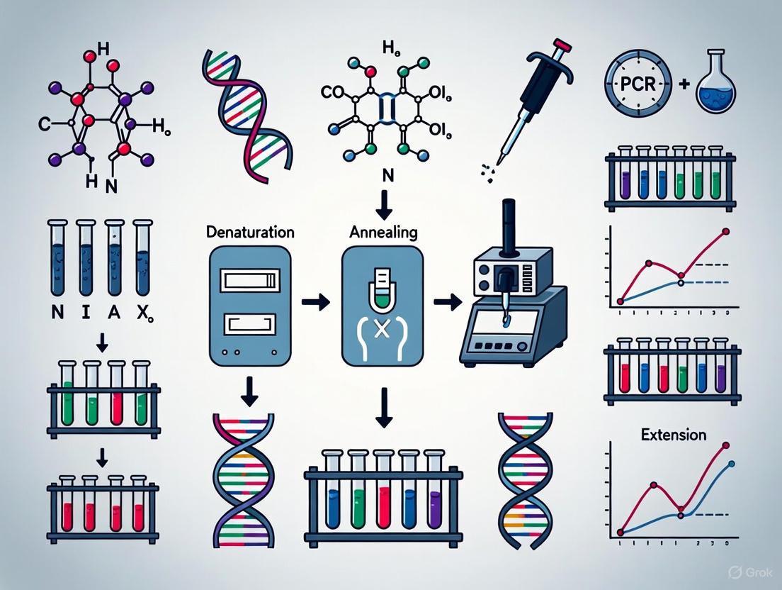
The uracil-N-glycosylase (UNG) system represents the most widely implemented technical solution for carryover contamination prevention [2] [3]. This enzymatic approach incorporates uracil (dUTP) instead of thymine (dTTP) during PCR amplification, generating products distinguishable from natural DNA. The UNG enzyme, included in the reaction mix, hydrolyzes any uracil-containing contaminants from previous amplifications during room temperature incubation before thermal cycling. The enzyme is subsequently inactivated at high temperatures during the initial PCR denaturation step, allowing amplification of the current target to proceed uncontested [2].
Table 2: Comparison of Primary Aerosol Contamination Prevention Methods
| Method | Mechanism of Action | Advantages | Limitations |
|---|---|---|---|
| Laboratory Zoning | Physical separation of pre-and post-amplification activities | Fundamentally prevents cross-contamination | Requires significant space and organizational discipline |
| UNG Treatment | Enzymatic degradation of uracil-containing prior amplicons | Highly effective for carryover prevention; integrated into kits | Less effective for GC-rich targets; requires dUTP incorporation |
| UV Irradiation | Induction of thymidine dimers in contaminating DNA | Simple, inexpensive; does not require protocol modification | Reduced efficacy for short or GC-rich templates; damages reagents |
| Bleach Decontamination | Oxidative damage to nucleic acids | Highly effective surface decontamination | Corrosive; cannot be used on reagents or samples |
| Aerosol-Resistant Tips | Physical barrier to aerosol entry during pipetting | Directly addresses pipetting-generated aerosols | Increased cost compared to standard tips |
Experimental Protocols for Contamination Control
Comprehensive Workflow for Aerosol-Free PCR
The following integrated protocol combines multiple contamination control strategies for maximum protection:
Laboratory Setup and Preparation
- Designate physically separated areas for: (1) reagent preparation, (2) sample preparation, (3) amplification, and (4) product analysis
- Equip each area with dedicated equipment, supplies, and laboratory coats
- Install UV light boxes in reagent and sample preparation areas for decontaminating opened packages and devices
- Establish unidirectional workflow policy with strict prohibition of reverse movement
Pre-Amplification Procedures
- Clean all work surfaces with 10% fresh bleach solution, followed by 70% ethanol
- Aliquot all reagents to avoid repeated freeze-thaw cycles and minimize contamination of stock solutions
- Prepare master mixes in the dedicated reagent preparation area using aerosol-resistant tips
- Include UNG in reaction mixes when using dUTP-based systems
- Perform reaction assembly in a designated UV-equipped workstation
Amplification and Post-Amplification
- Transfer closed reaction tubes to amplification area
- After cycling, open tubes only in the post-amplification area
- Analyze products in the dedicated analysis area
- Dispose of amplification products properly in sealed containers
- Never return materials from post-amplification to pre-amplification areas
Diagram 1: Unidirectional PCR Workflow. This workflow prevents amplicon contamination by maintaining physical separation between processes.
Decontamination Protocol for Equipment and Surfaces
Materials Needed:
- Freshly prepared 10% sodium hypochlorite solution
- 70% ethanol solution
- RNase/DNase-free water
- Dedicated cleaning cloths or wipes
- Personal protective equipment (gloves, lab coat, eye protection)
Procedure:
- Apply 10% bleach solution thoroughly to all work surfaces and equipment
- Allow surfaces to remain wet for 10-15 minutes to ensure nucleic acid degradation
- Wipe surfaces with ethanol to remove residual bleach
- For equipment that cannot tolerate bleach (e.g., pipette interiors), use 70% ethanol exclusively
- For critical applications, follow with RNase/DNase-free water rinse
- Perform this decontamination before and after each use of work areas
- Regularly decontamate refrigerators, freezers, and other shared equipment
The Scientist's Toolkit: Essential Reagent Solutions
Table 3: Key Research Reagents for Aerosol Contamination Control
| Reagent/Equipment | Function | Application Notes |
|---|---|---|
| Uracil-N-Glycosylase (UNG) | Enzymatic degradation of carryover contaminants | Most effective with thymine-rich targets; requires dUTP incorporation |
| dUTP Nucleotides | Substitute for dTTP to create distinguishable amplicons | Must be completely substituted for dTTP in reaction mix |
| Aerosol-Resistant Pipette Tips | Physical barrier to aerosol contamination | Essential for all pipetting operations; color-coded by volume |
| Sodium Hypochlorite (10%) | Surface decontamination through nucleic acid oxidation | Must be prepared fresh weekly; corrosive to some equipment |
| 70% Ethanol | Surface decontamination and bleach residue removal | Less effective than bleach for nucleic acid degradation |
| UV Light Source | Induction of thymidine dimers in contaminating DNA | Effective for stationary surfaces; limited penetration |
| DNA/RNA Shield Solutions | Stabilization and protection of nucleic acids in collection | Inactivates nucleases and protects target integrity |
Aerosol contamination represents a persistent challenge in molecular biology that demands systematic, multi-layered control strategies. The combination of physical laboratory organization, chemical decontamination, procedural discipline, and enzymatic prevention methods provides a robust defense against this invisible threat to assay integrity. As molecular techniques continue to evolve toward greater sensitivity and throughput, maintaining vigilance against contamination remains fundamental to generating reliable, reproducible scientific data and clinically valid diagnostic results. By implementing the comprehensive approaches outlined in this guide, researchers can effectively safeguard their experiments against compromise by minute particulate contamination, ensuring the validity of their findings in both basic research and drug development contexts.
In the realm of molecular biology, the polymerase chain reaction (PCR) has revolutionized diagnostics and research. However, its exquisite sensitivity, enabling the amplification of a few DNA copies into millions, also renders it exceptionally vulnerable to contamination, potentially compromising the integrity of results [2]. In a high-risk, high-burden mycobacterial reference laboratory, contamination events can lead to false positives, ultimately affecting patient diagnosis and treatment [6]. This technical guide examines the primary sources of contamination—amplicons, pipetting practices, and general laboratory procedures—within the broader context of understanding aerosol contamination in PCR research. We will detail specific protocols and present quantitative data on the efficacy of various mitigation strategies, providing a comprehensive resource for researchers, scientists, and drug development professionals dedicated to upholding the highest standards of data quality.
Understanding the Contaminants: Amplicons and Aerosols
The most significant source of contamination in a PCR laboratory is the amplification product, or amplicon [2]. A typical PCR reaction can generate as many as 10⁹ copies of the target sequence, creating a substantial reservoir for potential contamination [2]. When aerosolized, these amplicons pose the greatest threat; even the smallest aerosol droplet can contain up to 10⁶ amplification products [2]. These contaminated aerosols can build up quickly, contaminating laboratory reagents, equipment, and even ventilation systems if not properly controlled [2].
Aerosols are tiny liquid or solid particles suspended in the air. In the context of a PCR lab, they are primarily generated during routine laboratory practices. The act of pipetting is a major generator of aerosols, which can lead to cross-contamination from sample to sample if not properly managed [7]. Other activities, such as the careless opening of sample tubes or reaction plates, can cause liquid to splash and create aerosols [7]. These aerosols can contain amplicons from previous reactions, positive control plasmids, or even the target nucleic acids from clinical specimens [2] [7].
Table 1: Primary Sources and Characteristics of PCR Contamination
| Source Type | Description | Key Characteristics & Risks |
|---|---|---|
| Amplicons | The DNA products from previous PCR amplifications. | • Can reach concentrations of 10⁹ copies per reaction [2].• A single aerosol can contain 10⁶ copies [2].• The most problematic source of false positives. |
| Aerosols from Pipetting | Tiny droplets created during liquid handling. | • Major cause of sample-to-sample cross-contamination [7].• Can be prevented using aerosol barrier tips. |
| Aerosols from Tube Opening | Splashes created when carelessly opening sample tubes. | • Can spread contamination to gloves, surfaces, and adjacent samples [7].• Pre-spinning tubes before opening can mitigate this risk. |
Laboratory Design and Workflow as a Primary Defense
A robust first line of defense against contamination is a properly designed laboratory with a strict unidirectional workflow. The fundamental principle is the physical separation of the amplification and analysis steps from the reagent preparation and sample handling areas [7].
Physical Separation of Work Areas
A laboratory performing PCR diagnostics should be divided into at least three physically separate rooms [7]:
- Reagent Preparation Area: This is the "cleanest" area, dedicated to master-mix preparation and reagent aliquoting. No extracted nucleic acids or amplified products should ever be introduced here [7].
- Sample Preparation Area: This area is for nucleic acid extraction and the addition of DNA template to reactions. It is a "dirty" area with a high risk of sample contamination and should be maintained under negative air pressure to keep template nucleic acids contained [7].
- Amplification and Product Detection Area: This room houses the thermocyclers and is for the analysis of amplified products. It must be under negative pressure to prevent amplicons from escaping into other lab areas [7].
Unidirectional Workflow and Dedicated Equipment
The movement of personnel and materials must follow a unidirectional path: from the Reagent Preparation Area → to the Sample Preparation Area → and finally to the Amplification Area [7]. Crucially, one must never return tubes or equipment from a downstream area to an upstream one. All equipment, including pipettors, centrifuges, lab coats, and consumables, must be dedicated to each area and never interchanged [7]. The following workflow diagram illustrates this critical spatial and procedural organization:
Critical Laboratory Practices to Minimize Contamination
Beyond laboratory design, stringent daily practices are essential for contamination control. These protocols target the primary sources of contamination directly.
Decontamination Protocols
Regular and thorough decontamination of work surfaces and equipment is non-negotiable. Effective methods include [7]:
- Chemical Decontamination: Surfaces should be cleaned with a freshly made 10% sodium hypochlorite (bleach) solution, which causes oxidative damage to nucleic acids, rendering them unamplifiable. After 10-15 minutes, the surface should be wiped down with de-ionized water to remove the bleach [2] [7]. For metallic equipment where bleach is corrosive, commercial DNA-destroying decontaminants can be used.
- Ultraviolet (UV) Irradiation: UV light (254/300 nm) induces thymidine dimers in DNA, sterilizing contaminated surfaces by making nucleic acids inactive as PCR templates [2]. Workstation laminar flow cabinets should be irradiated with UV light for at least 30 minutes before use, and rooms should be exposed to UV overnight [7]. It is important to note that UV efficacy can be suboptimal for short or G+C-rich templates and may be shielded by nucleotides in PCR mixes [2].
Pipetting and Sample Handling Techniques
Proper technique is critical to minimizing aerosol generation.
- Aerosol Barrier Tips: Always use aerosol-barrier (filter) pipette tips to prevent the introduction of aerosolized contaminants into the pipette shaft and subsequent samples [7].
- Careful Tube Handling: All sample tubes and reaction plates should be opened and closed carefully to prevent splashing. Spinning down tubes in a centrifuge before opening them can significantly reduce the risk of creating aerosols [7].
Experimental and Control Design
- Use of Controls: Every PCR experiment must include a no-template control (NTC), which contains all reaction components except the DNA template. The NTC is critical for detecting contamination in reagents, consumables, or the environment [7]. Well-characterized positive and negative controls should also be included.
- Enzymatic Decontamination (UNG/dUTP): Incorporating the enzyme uracil-N-glycosylase (UNG) into the PCR master mix, along with substituting dUTP for dTTP in the reaction, is a highly effective pre-amplification sterilization technique [2]. UNG recognizes and hydrolyzes uracil-containing DNA from previous PCRs, preventing its amplification. Before the new PCR begins, the reaction is incubated at room temperature, allowing UNG to destroy any contaminating amplicons. The enzyme is then inactivated during the initial denaturation step at 95°C [2]. This method works best with T-rich amplicons [2].
Table 2: Efficacy of Combined Contamination Control Interventions [6]
| Intervention Implemented | Reduction in Mean Contamination Percentage |
|---|---|
| Pre- and post-cleaning of working surfaces | 36.5% |
| Combined cleaning of work surfaces, automated pipetting devices, and AC machines | 53.5% (94.6% reduction from baseline) |
Experimental Protocols for Contamination Monitoring
To validate the effectiveness of contamination control measures, laboratories should implement routine monitoring experiments.
Protocol: Monitoring Surface Contamination with No-Template Controls (NTCs)
- Objective: To detect the presence of amplifiable DNA contamination on laboratory work surfaces.
- Methodology:
- Swab Sampling: Moisten a sterile swab with a molecular-grade buffer. Wipe a defined area (e.g., 10 cm²) of the work surface to be tested (e.g., inside the biosafety cabinet, next to pipettors).
- Elution: Place the swab in a tube with a small volume of buffer and vortex to elute any collected material.
- PCR Setup: Use an aliquot of this eluate as the "template" in a standard PCR reaction alongside appropriate controls (positive control, negative control, and a clean swab NTC).
- Analysis: Analyze the PCR products. Amplification in the surface swab sample, but not the clean swab NTC, indicates contamination on that surface.
Protocol: Assessing Aerosol Contamination via Settle Plates
- Objective: To qualitatively assess the presence of airborne contaminating amplicons in different laboratory zones.
- Methodology:
- Plate Placement: In the sample preparation and amplification areas, place open PCR tubes or plates containing a standard PCR master mix (lacking template but including UNG if part of the protocol) in various locations. Leave them exposed for a set period (e.g., 1-2 hours).
- Amplification: Carefully close the tubes/plates and transfer them to the amplification area. Run the PCR program.
- Detection: Analyze the results. Amplification in any of the exposed tubes indicates that airborne contaminants settled into the reaction mix, identifying a problem with aerosol control in that area. This method has been used in studies to detect the presence of contaminating amplicons, where the genotype MTBDRplus V2 kits based on DNA strip technology were used for detection [6].
The Scientist's Toolkit: Essential Reagents and Materials
The following table details key reagents and materials essential for establishing and maintaining a contamination-controlled PCR laboratory.
Table 3: Essential Research Reagent Solutions for PCR Contamination Control
| Item | Function | Key Considerations |
|---|---|---|
| Sodium Hypochlorite (Bleach) | Surface decontaminant that oxidizes and destroys nucleic acids [2] [7]. | Must be freshly prepared (10-15%) for maximum efficacy. Requires rinsing with water after use. |
| Molecular-Grade Ethanol (70%) | General surface disinfectant. Used for cleaning cabinets and equipment before UV irradiation [7]. | Does not destroy DNA on its own; must be followed by UV light for decontamination [7]. |
| Uracil-N-Glycosylase (UNG) | Enzyme that hydrolyzes contaminating uracil-containing DNA from previous PCRs [2]. | Most effective with T-rich amplicons. Requires dUTP in the PCR mix. Inactivated at 95°C. |
| Aerosol Barrier Pipette Tips | Prevent aerosols from entering and contaminating the pipette shaft during pipetting [7]. | Critical for all liquid handling, especially in the sample preparation area. |
| Ultraviolet (UV) Light Cabinet | Provides nucleic acid sterilization via thymidine dimer formation for small equipment and workspaces [2] [7]. | Effectiveness depends on distance and can be reduced for short, G+C-rich templates [2]. |
| No-Template Control (NTC) | A critical quality control reaction to detect contamination in reagents and the environment [7]. | Should be included in every PCR run. Amplification in the NTC invalidates the run. |
In the realm of polymerase chain reaction (PCR) research, the integrity of data is paramount. This technical guide examines a critical triad of challenges—false positive results, erroneous cycle threshold (Ct) values, and broader data integrity loss—within the specific context of aerosol contamination. For researchers, scientists, and drug development professionals, understanding the interconnectedness of these issues is crucial for developing robust, reproducible assays. Aerosol contamination, often an overlooked consequence of routine laboratory procedures, serves as a silent catalyst that can compromise experimental outcomes from the pre-analytical stage through to data analysis. This guide provides a detailed examination of the causes and consequences, supported by structured data and experimental protocols, to equip laboratories with the knowledge to safeguard their research findings.
Core Concepts and Definitions
Understanding False Positives and Ct Values
False Positive PCR Results are incorrect outcomes that indicate the presence of a target sequence when it is genuinely absent. The primary cause is cross-contamination, frequently via aerosols—microscopic droplets of fluid introduced into the air through spillages, overly energetic pipetting, or careless vortexing [8]. These aerosols can carry amplicons from previous PCR reactions or target nucleic acids, which are then inadvertently introduced into new reaction mixtures.
The Cycle Threshold (Ct) Value is a critical quantitative data point in real-time PCR. It represents the PCR cycle number at which the amplification signal first exceeds a predefined threshold, indicating a positive reaction [9]. The Ct value is inversely correlated with the starting quantity of the target nucleic acid; a lower Ct value indicates a higher initial target concentration.
Erroneous Ct values arise when the amplification plot or its interpretation is compromised. This can occur due to:
- Background noise in qPCR, which can be misinterpreted as a positive signal, particularly when true target signals are weak [10].
- Non-specific amplification, where primers bind to non-target sequences, generating amplification products that do not represent the true target [8] [10].
- Carry-over contamination, where amplicons from previous reactions contaminate new setups, leading to unexpectedly low Ct values that falsely suggest high target concentration [8].
The Mechanism of Aerosol-Induced Data Integrity Loss
Aerosol contamination fundamentally undermines data integrity by introducing uncontrolled variables that skew results. The relationship between aerosols, false positives, and erroneous Ct values creates a cascade of data corruption, as shown in the following workflow.
This degradation of data integrity has tangible consequences. The downstream impacts of false positives and erroneous data in research and clinical settings are severe.
Table 1: Consequences of False Positive Results and Data Integrity Loss
| Domain | Consequence | Impact Description |
|---|---|---|
| Research & Development | Resource Waste | Unnecessary additional tests, wasted reagents, and wasted time [8]. |
| Compromised Studies | Invalid experimental data leading to incorrect conclusions and retractions [11]. | |
| Delayed Timelines | Time spent troubleshooting contamination and repeating experiments [8]. | |
| Clinical & Diagnostic | Patient Misdiagnosis | Unnecessary treatments, psychological distress, and false sense of security [8] [12]. |
| Public Health Impact | Overestimation of disease prevalence and ineffective policy decisions [12]. | |
| Erosion of Trust | Loss of confidence in testing systems and scientific institutions [11]. |
Quantitative Data and Statistical Impact
The Critical Role of Prevalence and PPV
The impact of false positives is not constant; it is profoundly influenced by the prevalence of the target in the population being tested. This relationship is captured by the Positive Predictive Value (PPV)—the proportion of positive test results that are true positives.
Table 2: Positive Predictive Value (PPV) at Different Disease Prevalences (Assumes 95% Sensitivity, 98% Specificity)
| Prevalence | True Positives | False Positives | PPV |
|---|---|---|---|
| 10% (Diagnostic Setting) | 950 | 180 | 84.0% |
| 1% (Screening Setting) | 95 | 198 | 32.4% |
| 0.1% | 9.5 | 199.8 | 4.5% |
As shown in Table 2, in a low-prevalence screening scenario (1%), a test with 98% specificity will yield a PPV of only 32.4%, meaning nearly two-thirds of all positive results are false positives [12]. This demonstrates that even a test with high specificity can produce misleading data when deployed in a low-prevalence population, a critical consideration for research screening projects or asymptomatic testing programs.
The Problem of Non-Detects and Ct Value Bias
In qPCR, reactions that fail to produce a detectable signal are known as non-detects. A common but flawed practice is to assign these non-detects an arbitrary high Ct value, such as 40. This approach introduces significant bias into data analysis [13]. Non-detects are not random events; they often occur systematically when target concentration is very low or inhibitors are present. Setting them to a fixed value distorts the distribution of ΔCt and ΔΔCt values, leading to biased estimates of gene expression and differential expression [13]. Statistical methods that model the missing data mechanism, rather than applying a fixed value, are required to reduce this bias and maintain data integrity.
Experimental Protocols for Mitigation
Comprehensive Workflow for Contamination Prevention
Implementing a rigorous, multi-layered protocol is essential to prevent aerosol contamination and its downstream effects on data.
Protocol: Aerosol Contamination Prevention and Control
1. Physical Laboratory Design and Workflow:
- Spatial Separation: Maintain physically separated rooms for pre-PCR (template preparation) and post-PCR (amplification and analysis) activities [8].
- Unidirectional Workflow: Implement and enforce a unidirectional flow of personnel and materials from pre-PCR to post-PCR areas. Do not allow reagents or equipment to move from post-PCR to pre-PCR areas [8].
- Dedicated Equipment: Assign dedicated pipettes, tip boxes, lab coats, and other equipment to each area. Equipment in the pre-PCR area should never be used in the post-PCR area [8].
2. Aseptic Techniques and Laboratory Practices:
- Glove Changing: Change gloves frequently, especially when moving between workstations or after handling potentially contaminated materials [8].
- Pipetting Technique: Use slow, controlled pipetting actions. Avoid rapid dispensing or mixing that can generate aerosols. Use filter-containing pipette tips to prevent aerosol contamination of the pipette shaft [8].
- Tube Handling: Open tubes carefully with one hand, avoiding contact between the cap and bench surfaces. Keep all sample and reagent vessels sealed as long as possible [8].
- Workspace Decontamination: Before and after use, sterilize benches and equipment with 70% ethanol, 10% sodium hypochlorite (with a minimum 10-minute contact time), and/or UV irradiation [8].
3. Biochemical and Procedural Safeguards:
- Reagent Aliquoting: Prepare single-use aliquots of all critical reagents (water, master mix, primers) to minimize repeated exposure to potential contaminants [8].
- UNG Treatment: Incorporate uracil-DNA-glycosylase (UNG) into the PCR master mix. This enzyme degrades uracil-containing DNA (e.g., from previous PCR products), preventing their amplification, while the current reaction uses thymine [8].
- Controls: Always include the appropriate controls in every run.
- No-Template Control (NTC): Contains all reaction components except the template nucleic acid. A positive signal in the NTC indicates contamination of reagents or the environment [8] [10].
- Inhibition Control (IPC): Used to detect the presence of PCR inhibitors in the sample that could cause false negatives [8].
Protocol for Investigating Suspected Contamination
When contamination is suspected (e.g., a positive NTC, or an unexpected cluster of high Ct value positives), a systematic investigation is required.
- Cease Testing: Temporarily halt all routine testing using the affected assay or workstation.
- Decontaminate: Discard all open reagents. Autoclave tubes and pipette tips. Meticulously clean all equipment and surfaces (pipettes, centrifuges, vortexers, hoods, benches) with sodium hypochlorite and ethanol [8].
- Service Equipment: Open and clean the interior of pipette guns, which can be a hidden source of contamination. Send pipettes for professional servicing and recalibration [8].
- Quality Control Testing: Test new reagent aliquots with a fresh set of NTCs. Only resume testing when NTCs consistently return negative results.
The Scientist's Toolkit: Essential Research Reagent Solutions
The following reagents and materials are critical for implementing the protocols described and for maintaining overall data integrity.
Table 3: Research Reagent Solutions for Contamination Control
| Item | Function | Technical Specification & Use |
|---|---|---|
| UNG Enzyme | Prevents carry-over contamination by degrading uracil-containing PCR amplicons from previous reactions. | Add to the PCR master mix. The current PCR reaction must be set up with dUTP instead of dTTP for this system to work effectively [8]. |
| Hot-Start Taq Polymerase | Increases specificity by remaining inactive until the initial denaturation step, preventing non-specific amplification and primer-dimer formation during reaction setup at room temperature [8]. | Choose chemically modified or antibody-bound enzymes. Ensures primer binding only occurs at the stringent, elevated temperature of the thermal cycler [8]. |
| Aerosol-Resistant Filter Tips | Prevents aerosols and liquids from entering the pipette shaft, thereby protecting the pipette from becoming a source of cross-contamination. | Essential for all liquid handling in pre-PCR and PCR setup areas. Use for all reagents and samples [8]. |
| Molecular Biology Grade Water | Nuclease-free, sterile water for preparing PCR reagents and reactions. | Prevents degradation of nucleic acids and enzymes by nucleases. Must be certified nuclease-free [8]. |
| Positive Control | A known source of the target sequence used to verify the assay is functioning correctly and to detect false negatives. | Should be a non-cross-reactive synthetic template or a plasmid control. Store in single-use aliquots to avoid becoming a source of contamination [8] [10]. |
| No-Template Control (NTC) | The primary diagnostic tool for detecting contamination in reagents or the laboratory environment. | Contains all PCR components except the template nucleic acid, which is replaced with water or buffer. A signal in the NTC confirms contamination [8] [10]. |
The integrity of PCR data is inextricably linked to the meticulous management of aerosol contamination. False positives and erroneous Ct values are not merely isolated technical failures; they are often symptoms of a compromised workflow that can lead to a cascade of data integrity loss. As demonstrated, the statistical impact of false positives is particularly severe in low-prevalence settings, and the mishandling of non-detect data can systematically bias results. By adopting the rigorous experimental protocols and essential reagent strategies outlined in this guide—from physical laboratory design and unidirectional workflows to the strategic use of UNG and stringent controls—research and diagnostic laboratories can fortify their operations. Ultimately, safeguarding data integrity requires a proactive, systematic, and unwavering commitment to contamination control at every stage of the PCR process.
Aerosol contamination represents one of the most significant and persistent challenges in molecular diagnostics, particularly for highly sensitive techniques like polymerase chain reaction (PCR). The exquisite sensitivity that makes PCR an invaluable tool for pathogen detection also renders it extraordinarily vulnerable to contamination from amplified products, potentially leading to false-positive results, diagnostic errors, and compromised patient care [2] [7]. This case study examines the real-world impact of aerosol contamination through the lens of a notable diagnostic failure, analyzes quantitative data on contamination dynamics, and presents evidence-based strategies for safeguarding diagnostic integrity.
The critical importance of contamination control was starkly illustrated during the COVID-19 pandemic when the Centers for Disease Control and Prevention (CDC) distributed flawed SARS-CoV-2 test kits to 26 public health laboratories in early 2020. Of these, 24 laboratories reported false-positive results, creating significant delays in national testing capabilities during a critical phase of the emerging pandemic [14]. This incident underscores how aerosol contamination can transcend individual laboratory failures to become a substantial public health concern.
Mechanisms and Dynamics of Aerosol Contamination
Generation and Transmission of Aerosols
Aerosol contamination in diagnostic laboratories occurs through the generation of microscopic liquid or solid particles carrying nucleic acids, which become suspended in the air and settle on surfaces, equipment, or directly into reaction mixtures. These particles are typically generated during routine laboratory procedures including pipetting, tube opening, centrifugation, and sample mixing [14]. A single PCR reaction can generate up to 10⁹ copies of the target sequence, and when aerosolized, even minute droplets can contain as many as 10⁶ amplification products [2].
The physical behavior of contaminating aerosols depends on multiple factors including particle size, density, shape, ambient temperature, humidity, and air circulation patterns [15]. Smaller particles (<5 µm) can remain airborne for extended periods and penetrate deep into the respiratory tract, while larger particles tend to settle more quickly on surfaces [15] [16]. This size-dependent behavior directly influences contamination spread patterns within the laboratory environment.
Quantitative Assessment of Aerosol Contamination
Understanding the magnitude of aerosol contamination requires quantitative assessment across different environments and procedures. The following table summarizes key findings from aerosol monitoring studies in various settings:
Table 1: Quantitative Aerosol Contamination Across Environments
| Environment/Procedure | Aerosol Metric | Levels Detected | Key Factors Influencing Contamination |
|---|---|---|---|
| Dental tooth grinding [16] | Particle concentration | Significantly elevated during procedures | Handpiece speed, distance from source, time |
| University dormitories [17] | Bioaerosol contamination | 511–9960 CFU/m³ (bacteria), 531–6568 CFU/m³ (fungi) | Human occupancy, ventilation, cleaning practices |
| Exhaled breath [15] | Particle count | Median: 79.55 particles/liter | Age, BMI, COVID-19 vaccination status |
| SARS-CoV-2 in air [18] | Viral RNA copies | 7-35 copies/m³ detection limit | Airflow, sampling time, room occupancy |
The data demonstrate that human activity directly influences aerosol generation, with procedures like dental grinding producing significant particulate matter and human occupancy markedly increasing bacterial contamination levels in indoor environments [17] [16]. Notably, a study of 250 children and adolescents found that SARS-CoV-2 infection itself did not increase exhaled aerosol particles, but age and COVID-19 vaccination status were significant predictors of emission levels [15].
Laboratory Protocols for Contamination Prevention
Physical Separation and Workflow Design
Effective contamination control begins with appropriate laboratory design implementing strict physical separation of pre- and post-amplification activities. The recommended configuration includes three distinct areas: (1) a reagent preparation area, (2) a sample preparation area, and (3) an amplification and product analysis area [7]. Each area should be equipped with dedicated instruments, consumables, laboratory coats, and personal protective equipment to prevent cross-contamination [7].
The unidirectional workflow must flow from the cleanest area (reagent preparation) to the dirtiest (amplification and analysis), with personnel and materials never moving backward from post-amplification to pre-amplification areas [14] [7]. Air pressure control is critical, with positive air pressure in the reagent preparation area to prevent contamination entry, and negative pressure in sample preparation and amplification areas to contain nucleic acids within those spaces [7].
Decontamination Procedures and Techniques
Rigorous decontamination protocols are essential for maintaining contamination-free work environments. The following procedures should be implemented systematically:
- Surface Decontamination: Regular cleaning of work surfaces and equipment with 10% sodium hypochlorite (bleach) followed by ethanol to remove residual bleach [2] [7]. Bleach causes oxidative damage to nucleic acids, rendering them unamplifiable in subsequent PCR reactions [2].
- UV Irradiation: Exposure of work areas, equipment, and reagents to UV light (254-300 nm) for 15-30 minutes before use induces thymidine dimers and other covalent modifications in DNA that render contaminating nucleic acids inactive as amplification templates [2].
- Enzymatic Inactivation: Incorporation of uracil-N-glycosylase (UNG) into PCR master mixes provides a powerful biochemical barrier against contamination. This method utilizes dUTP instead of dTTP during amplification, making amplicons susceptible to degradation by UNG enzyme, which is added to subsequent reaction mixtures to destroy any contaminating amplicons from previous reactions [2] [7].
Table 2: Decontamination Methods and Their Applications
| Decontamination Method | Mechanism of Action | Application | Limitations |
|---|---|---|---|
| Sodium Hypochlorite (Bleach) [2] | Oxidative nucleic acid damage | Work surfaces, equipment, instruments | Cannot be used on reagents or samples |
| UV Irradiation [2] | Thymidine dimer formation | Workstations, laminar flow cabinets, reagents | Reduced efficacy on short or GC-rich templates |
| Uracil-N-Glycosylase (UNG) [2] [7] | Hydrolysis of dUTP-containing DNA | PCR reaction mixtures | Less effective with GC-rich targets |
| Furocoumarins [2] | Intercalation and cross-linking | Post-amplification sterilization | Requires UV activation |
Procedural Controls and Quality Measures
Technical personnel must implement stringent procedural controls throughout the diagnostic testing process. Key measures include:
- Use of aerosol barrier pipette tips to prevent sample carryover during pipetting [7].
- Careful opening and closing of sample tubes to minimize aerosol generation, with brief centrifugation before opening to deposit liquid from tube walls and caps [7].
- Aliquoting reagents into single-use volumes to minimize repeated exposure to potential contaminants [14].
- Implementation of comprehensive control reactions including no-template controls (NTC) to monitor for contamination in reagents and environmental sources [7].
The following diagram illustrates the integrated approach to contamination prevention, encompassing laboratory design, workflow, and procedural controls:
The Scientist's Toolkit: Essential Research Reagent Solutions
Implementation of effective contamination control requires specific reagents, equipment, and methodologies. The following table catalogues essential solutions for maintaining diagnostic integrity:
Table 3: Essential Research Reagents and Solutions for Contamination Control
| Tool/Reagent | Function | Application Context |
|---|---|---|
| UDG/UNG Contamination Prevention Reagents [14] | Enzymatic degradation of contaminating amplicons | PCR and qPCR reaction mixes |
| dUTP [2] | Substrate for UNG-mediated degradation | Incorporated during amplification instead of dTTP |
| Aerosol Barrier Pipette Tips [7] | Prevent aerosol cross-contamination | All liquid handling procedures |
| Sodium Hypochlorite (10%) [2] [7] | Nucleic acid oxidation | Surface and equipment decontamination |
| UV Light Cabinet [2] | Nucleic acid damage through thymidine dimer formation | Reagent and workstation decontamination |
| High-Efficiency Particulate Air (HEPA) Filtration [18] | Removal of airborne particulates | Laboratory ventilation systems |
| Dedicated Outdoor Air System (DOAS) [16] | Particle filtration and air exchange | Dental operatories and procedure rooms |
| Wet Cyclone Sampler [18] | High-flow bioaerosol collection | Environmental surveillance |
Advanced detection systems are emerging to provide real-time monitoring of pathogen-laden aerosols. The pathogen Air Quality (pAQ) monitor represents one such innovation, combining a high-flow wet cyclone aerosol sampler with a nanobody-based micro-immunoelectrode biosensor to detect SARS-CoV-2 aerosols with 5-minute time resolution and a detection limit of 7-35 viral RNA copies/m³ of air [18]. Such technologies could provide early warning systems for contamination events or pathogen exposure in diagnostic and healthcare settings.
Aerosol contamination remains a formidable challenge in diagnostic testing, with demonstrated potential to cause significant diagnostic errors and public health consequences. The multifaceted approach presented in this case study—encompassing proper laboratory design, rigorous decontamination protocols, procedural controls, and innovative reagents—provides a comprehensive framework for mitigating contamination risk. As molecular diagnostics continue to evolve toward greater sensitivity and automation, maintaining vigilance against contamination through evidence-based practices and emerging technologies will remain essential for ensuring diagnostic accuracy and patient safety.
Building Your Defense: Proactive Strategies and Best Practices to Prevent Aerosol Contamination
In the context of polymerase chain reaction (PCR) research, the exquisite sensitivity that makes this technique powerful also renders it profoundly vulnerable to contamination, particularly from aerosolized amplification products. A single PCR reaction can generate as many as 10^9 copies of a target sequence, and even minimal aerosolization can release droplets containing up to 10^6 amplification products into the laboratory environment [2]. These aerosols, often invisible to the naked eye, can permeate laboratory spaces, contaminating reagents, equipment, and ventilation systems, ultimately leading to false-positive results that compromise research integrity and diagnostic accuracy [3] [2].
Physical separation of pre- and post-amplification areas represents the most fundamental and effective strategy for containing this risk. This whitepaper provides an in-depth technical guide for researchers, scientists, and drug development professionals on designing and implementing rigorous laboratory separation protocols. By framing this guidance within a comprehensive understanding of aerosol contamination dynamics, we aim to empower laboratories to establish physical barriers and unidirectional workflows that are essential for maintaining the validity of molecular research and diagnostic testing.
Understanding the Contamination Challenge: Amplification Products and Aerosol Dynamics
The Nature of the Contaminant
The primary contamination threat in PCR laboratories stems from previously amplified DNA sequences, commonly referred to as amplicons. When tubes or plates are opened after amplification, these products can become aerosolized, forming particles that remain suspended in air and settle on surfaces [3]. These contaminants are particularly problematic because they are identical to the target sequences being amplified, making them perfect templates for subsequent reactions. Contamination is not reducible once it has occurred; therefore, prevention through physical separation is paramount [3].
Aerosol Behavior and Transmission Dynamics
Understanding aerosol behavior is crucial for effective laboratory design. Aerosolized amplification products can behave similarly to infectious droplet nuclei described in healthcare settings, with particles ranging from 1–5 μm in size that can remain suspended indefinitely in air and be transported over long distances [19]. Their buoyancy and resistance to desiccation allows them to travel far from their source via air currents [19]. This transmission model underscores why simple spatial separation within a single room is insufficient and why dedicated, isolated areas are necessary for different stages of the PCR workflow.
Core Principles of Physical Separation
Implementing effective physical separation is grounded in three core principles: spatial segregation, unidirectional workflow, and dedicated equipment.
Spatial Segregation
The foundation of contamination control lies in establishing separate, dedicated areas for distinct processes in the PCR workflow. At a minimum, this requires implementing separate pre- and post-amplification areas [3]. Ideally, these areas should be located in different rooms with completely independent laboratory equipment and ventilation systems [3] [20].
Pre-amplification areas must remain pristine and should be dedicated to:
Post-amplification areas contain the high concentrations of amplicons and are used for:
The most critical separation is between the reagent/sample preparation areas and the areas where amplified products are handled. If possible, these rooms should not be supplied by the same ventilation system to prevent airborne cross-contamination [3].
Unidirectional Workflow
Maintaining a strict unidirectional workflow is essential for preventing the backward flow of contamination. Personnel and materials must move from clean pre-amplification areas to post-amplification areas, but never in reverse [20] [2]. Researchers who have entered post-amplification areas should not re-enter pre-amplification areas on the same day without rigorous decontamination procedures [3].
Dedicated Equipment and Consumables
Each separated area must have its own dedicated set of equipment, including pipettes, centrifuges, vortexers, and protective equipment [3]. Supplies and consumables should be delivered directly to their respective areas and never shared between pre- and post-amplification zones [3] [2]. This prevents the transfer of contaminants via equipment surfaces.
Diagram 1: Unidirectional laboratory workflow for PCR.
Practical Implementation Strategies
Laboratory Layout Design Options
The implementation of physical separation must be adapted to available space and resources. Several design approaches can be effective:
Ideal Scenario: Separate Rooms – Dedicated, physically separated rooms with independent ventilation systems for pre-amplification, amplification, and post-amplification processes [3]. This represents the gold standard for contamination control.
Compromise Solution: Contained Areas – In open-concept laboratories, physical separation can be achieved using rigid wall partitions, dead air boxes, or laminar flow cabinets to create distinct zones [20].
Minimum Requirement: Temporal Separation – When spatial separation is impossible, strict temporal separation can be implemented, where pre-PCR activities are completed first in a dedicated space, followed by thorough decontamination before any post-PCR work begins [3].
For laboratories performing real-time qPCR (which combines amplification and detection in a closed system), the workflow can be slightly modified, but the fundamental separation between sample/reagent preparation and amplified product handling remains critical [20].
Dead Air Boxes and Biological Safety Cabinets
In situations where dedicated rooms are not feasible, dead air boxes or Biological Safety Cabinets (BSCs) can provide localized contamination control.
Dead Air Boxes are enclosed workstations that provide a static, undisturbed environment for sensitive procedures like reagent preparation or PCR setup [20]. By eliminating air currents, they minimize the opportunity for aerosol contamination during critical pre-amplification steps.
Biological Safety Cabinets offer both product and environmental protection through HEPA-filtered laminar airflow and air barriers [21]. For PCR setup, Class II BSCs are particularly valuable as they provide a HEPA-filtered clean work environment to protect the reaction mix from contamination while also protecting the user [21].
Airflow and Pressure Control
Proper air pressure differentials are crucial for containment. Pre-amplification areas should be maintained at a higher pressure relative to adjacent spaces and corridors to prevent the influx of contaminated air [20]. Conversely, post-amplification areas should be kept at a lower pressure to contain amplicons within that space [20]. HVAC systems should be designed to ensure that air flows from clean to contaminated areas, not vice versa.
Diagram 2: Laboratory pressure cascade design.
Complementary Contamination Control Measures
While physical separation forms the foundation of contamination control, it must be supported by additional procedural and chemical measures.
Personal Protective Equipment (PPE) and Practices
Laboratory personnel can inadvertently transfer contaminants on clothing, skin, or hair [3] [2]. Each separated area must have dedicated lab coats and protective equipment [3]. Gloves should be changed frequently, particularly when moving between different areas or after potential exposure to contaminants [3].
Surface Decontamination
Regular decontamination of work surfaces and equipment is essential. For nucleic acid contamination, a 10-15% bleach solution (sodium hypochlorite) is most effective, causing oxidative damage that renders DNA unamplifiable [3] [2]. Surfaces should be treated with bleach for 10-15 minutes before wiping with de-ionized water [3]. Bleach solutions should be prepared fresh frequently as they degrade over time [3]. When bleach is incompatible with equipment, 70% ethanol can be used as an alternative, though it is less effective against nucleic acids [3].
Enzymatic Control with UNG
The use of uracil-N-glycosylase (UNG) provides a powerful chemical barrier against carryover contamination [3] [2]. This method involves incorporating dUTP instead of dTTP during PCR, causing amplification products to contain uracil. UNG enzyme added to the PCR master mix degrades any uracil-containing contaminants from previous reactions before thermal cycling begins [2]. The enzyme is then inactivated during the initial denaturation step, allowing amplification of the new target template to proceed unimpeded [3]. This method works best with thymine-rich amplification products and is less effective for guanine/cytosine-rich targets [3].
Monitoring and Validation
No Template Controls (NTCs)
The primary method for monitoring contamination is the inclusion of No Template Controls in every qPCR run [3]. These wells contain all reaction components except the DNA template. If amplification occurs in NTC wells, it indicates contamination is present. The pattern of amplification (consistent Ct values across NTCs versus random amplification) can help identify the source of contamination [3].
Environmental Monitoring
Regular monitoring of laboratory surfaces and equipment using surface sampling techniques (swabbing followed by PCR analysis) can identify accumulating contamination before it affects experimental results [22].
The table below summarizes key contamination monitoring methods:
Table 1: Contamination Monitoring and Decontamination Methods
| Method | Procedure | Application/Interpretation |
|---|---|---|
| No Template Controls (NTCs) | Include wells containing all qPCR components except DNA template [3]. | Consistent amplification across NTCs suggests reagent contamination; random amplification suggests environmental aerosol contamination [3]. |
| Surface Monitoring | Swab surfaces and test for presence of amplicons using PCR [22]. | Identifies specific contaminated surfaces or equipment for targeted decontamination. |
| Bleach Decontamination | Apply 10-15% sodium hypochlorite for 10-15 minutes, then wipe with de-ionized water [3]. | Causes oxidative damage to DNA, rendering it unamplifiable. Effective on surfaces and some equipment [2]. |
| UV Irradiation | Expose work areas and equipment to UV light (254-300 nm) for 5-20 minutes [2]. | Creates thymidine dimers in DNA, preventing amplification. Less effective for short or GC-rich templates [2]. |
The Scientist's Toolkit: Essential Reagents and Materials
Table 2: Essential Research Reagent Solutions for PCR Contamination Control
| Item | Function | Application Notes |
|---|---|---|
| Aerosol-Resistant Filter Pipette Tips | Prevent aerosol contamination of pipette shafts and cross-contamination between samples [3] [20]. | Essential for all liquid handling in both pre- and post-amplification areas. |
| Uracil-N-Glycosylase (UNG) | Enzymatically degrades uracil-containing DNA from previous amplifications [3] [2]. | Requires use of dUTP in place of dTTP in PCR mixes. Most effective against thymine-rich amplicons. |
| Sodium Hypochlorite (Bleach) | Oxidizes nucleic acids, rendering them unamplifiable [3] [2]. | Use 10-15% solution for surface decontamination. Prepare fresh frequently. |
| Dedicated Laboratory Equipment | Prevents cross-contamination between laboratory areas [3]. | Each separated area requires dedicated pipettes, centrifuges, vortexers, and PPE. |
| Aliquoted Reagents | Prevents repeated exposure of stock solutions to potential contamination [3]. | Divide master mixes, primers, and enzymes into single-use aliquots. |
Implementing rigorous physical separation of pre- and post-amplification areas is not merely an optional enhancement but a fundamental requirement for any laboratory committed to producing reliable PCR results. As the sensitivity of molecular techniques continues to increase and their applications expand into critical areas like drug development and clinical diagnostics, the consequences of contamination become increasingly severe. By adopting the comprehensive approach outlined in this whitepaper—integrating spatial segregation, unidirectional workflow, dedicated equipment, and complementary control measures—research facilities can establish a robust defense against the persistent challenge of aerosol contamination. This systematic implementation of contamination controls ultimately protects not just individual experiments, but the integrity of the scientific enterprise itself.
This whitepaper details essential personal protective equipment (PPE) and workflow controls to mitigate aerosol contamination in laboratories conducting PCR research. Aerosol contamination, the unintended introduction of aerosolized nucleic acids into samples or reagents, represents a significant threat to data integrity and experimental validity. This document provides a structured framework for establishing robust laboratory protocols, framed within the broader context of understanding and controlling aerosol contamination in PCR research. The guidance synthesizes established biosafety principles with specific procedural controls to create a defensive strategy for safeguarding molecular biology workflows.
Foundational Biosafety Principles
Laboratories handling SARS-CoV-2 specimens, which serve as a relevant model for procedures with aerosol-generating potential, are advised to operate at a minimum of Biosafety Level 2 (BSL-2) [23] [24]. The core principle governing all laboratory activities is the site-specific and activity-specific risk assessment [23]. This assessment, developed in collaboration with biosafety professionals and laboratory management, must evaluate the laboratory facilities, personnel competency, specific techniques, safety equipment, and engineering controls to identify and mitigate risks associated with aerosol-generating procedures [23]. All clinical specimens should be treated as potentially infectious, and Standard Precautions must be followed, which include hand hygiene and the use of PPE based on exposure potential [23] [24].
Table 1: Core Elements of a Laboratory Biosafety Risk Assessment
| Element | Description | Application to PCR Workflows |
|---|---|---|
| Facility & Engineering Controls | Laboratory design, ventilation, and containment equipment. | Use of certified Class II Biosafety Cabinets (BSCs) for aerosol-generating procedures [23] [24]. |
| Personnel & Training | Competency of staff in biosafety practices and specific procedures. | Training on proper PPE use, unidirectional workflow, and techniques to minimize aerosols [24]. |
| Practices & Techniques | Standard Operating Procedures (SOPs) for safe work practices. | Implementation of unidirectional workflow and controlled sample manipulation [24]. |
| Safety Equipment | Personal protective equipment and other protective devices. | Use of dedicated lab coats, gloves, and eye protection [23] [24]. |
Personal Protective Equipment (PPE) for Aerosol Control
PPE serves as a critical barrier between the researcher and potential contaminants, protecting both the individual and the integrity of the experiments.
Dedicated Lab Coats
A dedicated lab coat or gown is a fundamental requirement for BSL-2 containment [23]. For work with SARS-CoV-2, and by extension for procedures with aerosol risk, the lab coat or solid-front gown should have a knit or grip cuff to prevent sleeves from riding up [24]. These lab coats must be donned upon entering the laboratory and removed before exiting. They should not be worn outside the laboratory area (e.g., in offices or break rooms) to prevent the transfer of contaminants.
Gloves
Gloving is a mandatory component of PPE for handling potentially infectious materials [23]. For procedures involving manipulation of untreated specimens in a BSC, the use of double gloves is recommended, with the outer pair extending over the sleeve of the lab coat or gown [24]. The outer gloves should be removed before exiting the BSC, and a new pair should be donned upon re-entering [24]. This practice contains contamination within the cabinet. Gloves must be removed carefully to avoid self-contamination and followed by hand hygiene [24].
Workflow and Procedural Controls
Engineering and administrative controls are the primary defenses for minimizing aerosol contamination, with PPE serving as personal protection.
Unidirectional Workflow
Unidirectional movement of personnel, samples, and materials is a key administrative control. This workflow is designed to move from "clean" areas to "potentially contaminated" areas, preventing backtracking and cross-contamination. The following diagram illustrates the logical sequence and physical segregation of activities.
Management of Aerosol-Generating Procedures
Many routine laboratory procedures can generate infectious aerosols and droplets [23]. These include:
- Pipetting
- Vortexing
- Centrifuging
- Grinding/Blending
- Opening containers of infectious materials [23]
Procedures with a high likelihood of generating aerosols must be conducted within a certified Class II Biological Safety Cabinet (BSC) [23] [24]. The BSC must be properly maintained and certified. Technical procedures should be chosen to minimize the formation of aerosols, and any necessary aerosol-generating procedures must be performed within the BSC [24].
Experimental Evidence on Aerosol Generation and Control
Understanding the behavior of aerosols is critical for developing effective controls. Recent research provides quantitative data on aerosol generation, which reinforces the necessity of the PPE and workflow controls described previously.
A 2022 prospective cohort study demonstrated a highly significant difference in respiratory aerosol concentrations between SARS-CoV-2 PCR-positive and negative subjects (median of 1490.5 particles per liter vs. 252.0 particles per liter; p < 0.0001) [25]. This study highlighted that aerosol emission can occur during normal breathing and is not necessarily dependent on age, sex, or smoking status [25].
A 2024 study in Nature Communications provided further crucial evidence, successfully isolating culturable SARS-CoV-2 from size-fractionated aerosols (<5 μm and <10 μm) generated by ambulatory COVID-19 patients [26]. This study found that 50-61% of participants emitted these variant-specific culture-positive aerosols, with the likelihood of infectiousness highest within the first 8 days of symptom onset [26]. The following table summarizes key experimental findings related to aerosol viability.
Table 2: Experimental Findings on Viable Aerosol Generation
| Experimental Finding | Methodology / Measurement | Implication for Lab Control |
|---|---|---|
| Heterogeneity in Aerosol Emission [26] | Only ~29% of participants were "probably highly infectious" (emitting culture-positive aerosols <5μm at ~6 days post-symptom onset). | Risk is not uniform, but controls must protect against high-emission scenarios. |
| Association with Host Immunity [26] | Aerosol culturability was significantly associated with lower variant-specific serum neutralizing antibody levels. | Reinforces that asymptomatic individuals can pose a transmission risk. |
| Particle Size & Ventilation [27] | Sub-5μm particles can remain suspended for hours, travel farther, and penetrate the lower respiratory tract. | HEPA filtration and increased air changes per hour (ACH) are critical engineering controls [27]. |
The experimental workflow for establishing such evidence, and by extension for validating containment effectiveness, can be complex, as shown in the following diagram.
The Scientist's Toolkit: Research Reagent Solutions
The following table details essential materials and their functions for establishing a controlled PCR workflow, based on the cited guidelines and research.
Table 3: Key Research Reagents and Materials for Aerosol Control
| Item | Function / Specification | Rationale |
|---|---|---|
| Class II Biosafety Cabinet (BSC) | Engineering control for aerosol containment; must be certified. | Primary barrier for performing aerosol-generating procedures; protects user and sample [23] [24]. |
| HEPA-Filtered Centrifuge Rotors | Physical containment device with gaskets. | Prevents release of aerosols during centrifugation, a known aerosol-generating procedure [24]. |
| Synthetic Tipped Swabs | For specimen collection; thin plastic or wire shafts. | Calcium alginate or wooden-shaft swabs may inhibit molecular tests [28]. |
| Viral Transport Media | Sterile medium for specimen transport and storage. | Maintains specimen integrity and viral RNA stability for testing [28]. |
| EPA-Registered Disinfectants | Disinfectants from EPA List N, effective against SARS-CoV-2. | For decontamination of work surfaces and equipment; follow manufacturer's dilution and contact time [23]. |
The exquisite sensitivity of the Polymerase Chain Reaction (PCR), which enables the amplification of millions of copies from a few initial DNA sequences, is also its greatest vulnerability. This very sensitivity makes the technique exceptionally prone to contamination, where even minute aerosolized particles can lead to false-positive results and compromise scientific integrity [3] [7]. A single PCR reaction can generate as many as 10⁹ copies of the target amplicon, and the smallest aerosol droplet can contain up to 10⁶ of these amplification products [2]. Uncontrolled amplification product carryover can lead to the rapid contamination of laboratory reagents, equipment, and ventilation systems, posing a significant challenge to diagnostic accuracy and research reliability [2].
Within this context, a robust strategy for managing techniques and reagents forms the first and most critical line of defense. This guide details three foundational pillars of contamination prevention: the use of aerosol barrier pipette tips, the strategic aliquoting of reagents, and the practice of careful tube handling. When implemented within a framework of proper laboratory organization, these practices are indispensable for maintaining the validity of PCR results and ensuring the success of sensitive molecular research and diagnostic applications [29] [30] [3].
Understanding the Contamination Threat in PCR
The primary sources of contamination in a PCR laboratory can be categorized as follows:
- Amplicon Carryover Contamination: This is the most significant and challenging source. It involves the contamination of new reactions with amplification products from previous PCR assays [2]. When a tube containing amplified DNA is opened, these products can become aerosolized and spread throughout the lab environment.
- Cross-Contamination from Samples: Clinical specimens with high target organism loads can be a source of cross-contamination during sample preparation [2].
- Contaminated Reagents or Consumables: Enzymes, water, buffers, and plasticware can become contaminated with nucleases, amplicons, or other DNA/RNA, leading to widespread experimental failure [30].
- Environmental Contamination via Personnel: Contaminants can be transferred on gloves, lab coats, hair, or jewelry from post-amplification areas to pre-amplification areas [3] [7].
Consequences of Contamination
The impact of contamination is not merely an academic inconvenience; it has real-world consequences. Documented cases exist where false-positive PCR results for Lyme disease led to misdiagnosis, with one case having a fatal outcome [2]. Contamination has also necessitated the formal retraction of published scientific manuscripts, undermining scientific progress and credibility [2]. In a diagnostic setting, false positives can lead to unnecessary treatments and patient anxiety, while in research, they can invalidate months of work and lead to erroneous conclusions.
Core Technique 1: Aerosol Barrier Pipette Tips
Design and Functionality
Aerosol barrier tips, also known as filter tips, are specialized pipette tips fitted with a hydrophobic filter seated in the proximal end (closest to the pipette barrel) [29] [31]. This filter acts as a one-way physical barrier that serves two critical functions:
- Protecting the Pipette: It prevents volatile, corrosive, or viscous liquids from being accidentally aspirated into the pipette shaft, which can damage the instrument's internal mechanisms and lead to inaccurate pipetting [29].
- Protecting the Sample and Reagents: It blocks aerosols and micro-droplets generated during pipetting from traveling up the tip and into the pipette barrel. When the pipette is used for a subsequent sample, these contaminants could then be introduced, leading to cross-contamination [29] [31]. This is paramount for preventing sample-to-sample carryover and protecting master mixes from becoming contaminated.
It is important to note that not all filter tips are created equal. The quality of the filter and its seal is critical. High-quality tips provide a true barrier, while lower-quality versions may only slow the progression of liquid [29].
Application and Selection Guidelines
Filter tips are considered essential for all sensitive molecular biology applications, especially qPCR, RT-PCR, and any experiment involving precious or low-copy-number templates [29] [30]. They are also highly recommended as "training wheels" for new laboratory personnel to prevent accidental pipette contamination [29].
Table 1: Comparison of Pipette Tip Types
| Feature | Aerosol Barrier (Filter) Tips | Standard (Non-Barrier) Tips |
|---|---|---|
| Primary Function | Prevent aerosol-mediated cross-contamination; protect pipette | General liquid transfer for non-sensitive applications |
| Internal Filter | Yes, hydrophobic barrier | No |
| Sterility | Typically pre-sterilized and DNase/RNase-free [31] | May be sterile or non-sterile; often requires autoclaving |
| Ideal Applications | qPCR, PCR, clinical diagnostics, handling volatile/corrosive liquids [29] | Gel loading, plasmid DNA prep, practice protocols |
| Cost | Higher | Lower |
| Impact on Accuracy | High; preserves sample integrity | Lower; risk of contamination in sensitive workflows |
Core Technique 2: Strategic Aliquoting of Reagents
Principles and Rationale
Aliquoting is the process of dividing a bulk volume of a reagent into smaller, single-use or limited-use portions. This practice is a cornerstone of contamination control and reagent management for several reasons:
- Limits Cross-Contamination: Repeatedly opening a master stock reagent tube increases the risk of introducing contaminants via aerosols, pipettes, or airborne particles. Using a single-use aliquot prevents the entire stock from being compromised [30].
- Preserves Reagent Integrity: Multiple freeze-thaw cycles can degrade enzymes, primers, and other labile reagents. Aliquoting into single-experiment volumes minimizes freeze-thaw cycles and maintains reagent stability [3] [32].
- Enhances Experimental Reproducibility: By ensuring consistency between experiments and reducing batch-to-batch variability, aliquoting contributes to more reliable and reproducible results [33] [32].
Best Practices for Aliquoting
A rigorous aliquoting protocol is essential for maximizing its benefits. The following workflow outlines a systematic approach:
Figure 1: A standardized workflow for aliquoting reagents to ensure integrity.
- Sample Preparation and Environment: Perform all aliquoting in a dedicated, clean pre-PCR area, ideally within a laminar flow hood or biosafety cabinet that has been decontaminated with UV light and ethanol [32] [7]. Ensure the bulk reagent is thoroughly mixed (homogenized) before partitioning.
- Equipment and Materials: Use calibrated pipettes and high-quality, low-retention tips to ensure volumetric accuracy [32]. Choose appropriate, sterile vials for storage.
- Labeling: Assign a unique identifier (UID) to each aliquot. Labels must be durable and resistant to extreme temperatures (e.g., -80°C, liquid nitrogen), chemicals, and moisture to remain legible throughout storage [33].
- Validation and Quality Control: For critical applications, implement quality control checks. This can include gravimetric verification (weighing dispensed water) to confirm pipetting accuracy and running control samples to check for contamination [32].
Table 2: Aliquoting Validation and Quality Control Metrics
| Validation Metric | Typical Threshold | Purpose |
|---|---|---|
| Coefficient of Variation (CV) | < 2% for microvolumes [32] | Measures precision and consistency between aliquots |
| Accuracy | Within 99-101% of target volume [32] | Verifies that the dispensed volume matches the intended volume |
| Contamination Checks | No detectable traces in control samples (e.g., NTC) [32] | Confirms the sterility and purity of the aliquots |
Core Technique 3: Careful Tube Handling
Minimizing Aerosol Generation
The simple act of opening and closing reaction tubes is a major, yet often overlooked, source of aerosol generation. Implementing careful handling techniques is a low-cost but highly effective defensive measure.
- Centrifugation Before Opening: Always pulse-centrifuge PCR tubes or strips briefly (a "mini-spin") before opening. This collects all liquid at the bottom of the tube, preventing it from being present on the rim or cap where it can easily be aerosolized when cracked open [7].
- Slow and Deliberate Motions: Open and close tube caps carefully and slowly. Rapid movements can create turbulent airflows that disrupt the liquid surface and generate aerosols [30].
- Avoiding Splashing: When pipetting into a tube, ensure the tip is immersed in the liquid already present (if any) or is touching the inner wall of the tube just above the liquid meniscus. Dispense the liquid smoothly and avoid blowing out the last drop aggressively, which can create bubbles and aerosols [30].
- Proper Glove Use: Wear properly fitting gloves and change them frequently, especially if they become contaminated or after handling post-amplification tubes [30] [7]. Ill-fitting gloves are more likely to touch tube rims and spread contamination.
Strategic Workflow and Tube Holding
- Holding Technique: Hold PCR tubes or strips at the bottom or middle, avoiding any contact with the cap or top portion of the tube. This prevents contaminants from your gloves from being transferred to the area where the pipette tip will enter [30].
- Order of Addition: When setting up reactions, add the patient sample or DNA template last, after all other master mix components are in the tube. This minimizes the time the most potent source of contamination (the template) is exposed in an open tube. Furthermore, add negative controls before positive controls to reduce the risk of contaminating the negatives with the high-titer positive [7].
- Keep Tubes Capped: Keep reaction tubes capped or covered as much as possible during setup to limit exposure to the environment [3].
Integrating Techniques into a Coherent Contamination Control Strategy
No single technique is sufficient to guarantee a contamination-free workflow. The true power of aerosol barrier tips, aliquoting, and careful tube handling is realized when they are integrated into a comprehensive laboratory strategy that includes physical separation and enzymatic controls.
Physical Laboratory Organization
The gold standard for PCR laboratory setup is physical separation of the workflow into distinct, dedicated rooms or areas with a unidirectional workflow [3] [2] [7].
- Reagent Preparation Area (Pre-PCR): This should be the cleanest area, reserved for preparing and aliquoting master mixes and reagents. It must be physically isolated from amplified DNA and sample processing. This area should be maintained under positive air pressure to prevent the ingress of contaminants [7].
- Sample Preparation Area: This area is used for nucleic acid extraction from clinical or research samples. It should operate under negative air pressure to contain any template DNA that is released during processing [7].
- Amplification and Analysis Area (Post-PCR): This area houses the thermal cyclers and equipment for analyzing PCR products. It must be physically separated from pre-PCR areas and also maintained under negative pressure to contain the massive quantity of amplicons generated [7].
A strict unidirectional workflow must be enforced: personnel and materials should move from pre-PCR to post-PCR areas, but never in reverse without thorough decontamination [3] [7].
Complementary Chemical and Enzymatic Controls
- Surface Decontamination: Regularly clean all work surfaces, equipment, and pipettes with a 10% sodium hypochlorite (bleach) solution, which acts by oxidizing nucleic acids, rendering them unamplifiable. After a 10-15 minute contact time, wipe down with de-ionized water to remove residual bleach, which can corrode equipment. Follow with 70% ethanol [3] [2] [7].
- Ultraviolet (UV) Irradiation: UV light induces thymidine dimers in DNA, sterilizing exposed surfaces. Use UV lamps in biosafety cabinets and workstations to decontaminate surfaces and plasticware before and after use [2] [7].
- Uracil-N-Glycosylase (UNG): This enzymatic method is a highly effective pre-amplification sterilization technique. In this system, dTTP in the PCR master mix is replaced with dUTP. Consequently, all newly synthesized amplicons contain uracil. In subsequent PCR setups, the UNG enzyme is added to the master mix. It selectively degrades any uracil-containing contaminating amplicons from previous runs before the thermal cycling begins. The UNG is then inactivated during the first high-temperature denaturation step of the new PCR, allowing the amplification of the native, thymine-containing template to proceed unimpeded [3] [2]. UNG is most effective for thymine-rich targets and is a standard component of many commercial qPCR kits.
The Scientist's Toolkit: Essential Reagent Solutions
Table 3: Key Research Reagent Solutions for Contamination Control
| Item | Function in Contamination Control |
|---|---|
| Aerosol Barrier Pipette Tips | Creates a physical barrier to prevent cross-contamination via aerosols and protects pipette shafts from damage [29] [31]. |
| High-Quality, Nuclease-Free Water | The foundation for all reagent preparation; ensures no exogenous nucleases degrade primers, templates, or products. |
| Pre-Aliquoted Master Mixes | Commercial or lab-made mixes aliquoted into single-use volumes to limit freeze-thaw cycles and exposure to contaminants. |
| Uracil-N-Glycosylase (UNG) | Enzyme used with dUTP for enzymatic degradation of carryover contamination from previous PCRs [3] [2]. |
| Molecular Grade Ethanol (70%) | Used for routine decontamination of surfaces, gloves, and equipment to reduce particulate and microbial contamination [30] [7]. |
| Sodium Hypochlorite (10% Bleach) | Potent nucleic acid-destroying agent for surface decontamination; essential for post-amplification cleanup [3] [2]. |
| DNase/Rnase Decontamination Sprays | Commercial solutions specifically formulated to rapidly degrade DNA and RNA on surfaces and equipment. |
| Durable Cryo-Labels | Temperature- and chemical-resistant labels for clear sample tracking and prevention of sample mix-ups during long-term storage [33]. |
Mastering the techniques of aerosol barrier tip use, strategic aliquoting, and meticulous tube handling is non-negotiable for any serious PCR-based research or diagnostic operation. These practices form an integrated defense system that directly targets the most common pathways of contamination. When combined with a physically separated laboratory layout and robust enzymatic and chemical decontamination protocols, they create a powerful barrier against false positives and unreliable data. Adherence to this comprehensive approach safeguards not only the integrity of individual experiments but also the credibility of scientific findings and the efficacy of clinical diagnostics.
In polymerase chain reaction (PCR) and microbiome research, the profound sensitivity of amplification techniques makes them exceptionally vulnerable to contamination, which can compromise experimental integrity and lead to erroneous conclusions. Within the context of aerosol contamination in PCR research, controlling DNA contamination becomes paramount, as aerosolized amplicons from previous reactions can permeate laboratory environments, contaminating reagents, equipment, and ventilation systems [2]. A single microliter of a typical PCR product can contain as many as 10^9 copies of the target sequence, meaning even the smallest aerosol droplet may contain millions of amplifiable molecules [2]. This background of contaminating DNA competes with target DNA during amplification, potentially causing false positives and misrepresenting the true microbial community in a sample, especially in low-biomass studies [34] [35].
Effective decontamination protocols are not merely a supplementary best practice but a fundamental requirement for generating reliable, reproducible data. This is particularly crucial when researching environments with low microbial biomass, such as certain human tissues, the atmosphere, or treated drinking water, where the contaminant signal can easily overwhelm the native signal [34]. This guide provides an in-depth examination of evidence-based decontamination strategies, focusing on the practical application of bleach solutions and other DNA-deactivating reagents to safeguard your research against contamination.
Contamination can be introduced at virtually any stage of the research workflow, from sample collection to data analysis. Major sources include:
- Human Operators: Skin, hair, and aerosol droplets generated from breathing or talking [34].
- Laboratory Environments: Airborne particles and dust settled on surfaces, benches, and equipment [34] [2].
- Reagents and Consumables: Commercial PCR enzymes, DNA extraction kits, and water have all been documented to contain trace amounts of bacterial DNA [35].
- Cross-Contamination Between Samples: Well-to-well leakage during plate setup or improper pipetting techniques can transfer DNA between samples [34].
- Amplicon Carryover: The most significant source in established labs, where amplification products from previous PCRs contaminate new reactions [2].
Acknowledging these vectors is the first step in designing a robust decontamination strategy. The following workflow diagram outlines the primary contamination sources throughout a typical molecular biology process and key intervention points.
Chemical Decontamination Agents: Mechanisms and Efficacy
A variety of chemical agents are available for decontamination, each with distinct mechanisms of action and optimal use cases.
Sodium Hypochlorite (Bleach)
Sodium hypochlorite, the active component in household bleach, is one of the most effective and widely used DNA decontaminants. Its mechanism of action involves oxidative damage, causing base modifications, strand breaks, and the production of chlorinated base products [36]. With increasing concentration and exposure time, bleach cleaves DNA into progressively smaller fragments, eventually reducing it to individual bases, thereby rendering it unamplifiable [36].
The efficacy of bleach is highly dependent on concentration, exposure time, and the surface material. A comprehensive study evaluating cleaning strategies found that sodium hypochlorite solutions were among the most effective, recovering a maximum of only 0.3% of initially deposited cell-free DNA after treatment across plastic, metal, and wood surfaces [37].
Table 1: Efficacy of Bleach Solutions Against Cell-Free DNA on Different Surfaces
| Surface Type | Bleach Solution Concentration | DNA Recovery Post-Treatment | Key Finding |
|---|---|---|---|
| Plastic | Sodium Hypochlorite-based solutions | ≤ 0.3% | Highly effective on non-porous surfaces |
| Metal | Sodium Hypochlorite-based solutions | ≤ 0.3% | Highly effective on non-porous surfaces |
| Wood | Sodium Hypochlorite-based solutions | ≤ 0.3% | Effective, but higher inherent DNA retention |
For cell-contained DNA, such as in whole blood, alternative agents like 1% Virkon proved most effective, with a maximum of 0.8% DNA recovery post-decontamination [37]. This highlights the importance of matching the decontaminant to the type of contaminant expected.
Alternative and Complementary Chemical Agents
While bleach is highly effective, other reagents play important roles in a comprehensive decontamination strategy:
- Ethanol (70-80%): Commonly used to kill contaminating microorganisms. However, it is less effective at removing trace DNA, as it may fix biological material to surfaces rather than degrade it. It is often used in a two-step process with a DNA-degrading agent [34].
- UV Radiation: Ultraviolet light, particularly at 254 nm, induces thymidine dimers and other covalent modifications in DNA, preventing it from being used as a PCR template [2]. Its efficacy can be reduced for short or G+C-rich templates and is dependent on distance from the source and shadowing effects [2].
- Trigene: A commercial disinfectant cleaner shown to be highly effective, achieving similar low DNA recovery rates (≤0.3%) as sodium hypochlorite on various surfaces [37].
- DNA Remover: A specialized commercial product formulated specifically for degrading DNA contaminants.
Enzymatic Inactivation with Uracil-N-Glycosylase (UNG)
UNG is a powerful pre-amplification sterilization technique incorporated directly into the PCR mix. It involves substituting dUTP for dTTP during PCR, generating uracil-containing amplicons. The UNG enzyme is then added to subsequent PCR mixes; it hydrolyzes any contaminating uracil-containing amplicons from previous reactions before the new thermal cycling begins. The enzyme is subsequently inactivated during the initial denaturation step (95°C), allowing the new PCR to proceed normally [2]. This method is now widely adopted in commercial PCR kits.
Recommended Decontamination Protocols
General Laboratory Surface Decontamination
For routine decontamination of benchtops, equipment, and tools, the following protocol is recommended:
- Mechanical Cleaning: Physically remove gross debris and dust using a clean cloth.
- Chemical Application: Thoroughly apply a 10% (v/v) sodium hypochlorite solution (equivalent to ~0.5-0.6% sodium hypochlorite final concentration). Ensure the surface remains wet for a minimum contact time of 10-15 minutes to ensure complete DNA degradation [2] [36].
- Rinsing (if necessary): Wipe the surface with ethanol (70-80%) to remove residual bleach, which can corrode metal and damage other surfaces over time [2].
- Frequency: High-touch surfaces and areas in PCR rooms should be decontaminated daily or prior to setting up sensitive reactions.
Decontamination of Specific Materials and Samples
Different materials and sample types require tailored approaches:
- Ancient Bones and Teeth: A systematic study found that immersion in 2.5% (w/v) sodium hypochlorite (approximately equivalent to 50% v/v commercial bleach) for 15 minutes was effective at removing surface contamination without completely destroying endogenous ancient DNA within the bone [36]. This demonstrates that concentrated bleach can penetrate and decontaminate porous surfaces effectively.
- Microfluidic Devices: For complex equipment, a combination of methods is often best. One effective protocol involves a combination of ethanol rinses followed by ultraviolet-C (UV-C) light radiation, which was shown to reduce RNase contamination tenfold [38].
- Plasticware and Glassware: Autoclaving is standard for sterilizing against viable cells, but it does not remove pre-existing DNA. Autoclaved items should be treated with UV irradiation or chemical DNA-degrading solutions if used for ultra-sensitive work [34].
Integrated Experimental Design for Contamination Control
Beyond cleaning surfaces, a holistic approach is critical [34]:
- Include Negative Controls: Always process extraction blanks (containing only water or buffer) and no-template PCR controls alongside your samples. These are essential for detecting reagent contamination [34] [35].
- Validate Reagents: Test new lots of PCR enzymes and water for bacterial DNA contamination using a sensitive method like endpoint PCR with broad-range 16S rRNA gene primers [35].
- Physical Separation: Maintain strict unidirectional workflow by physically separating pre-amplification (reagent preparation, sample extraction) and post-amplification (product analysis) areas. Dedicated equipment, lab coats, and supplies for each area are mandatory [2].
- Use Personal Protective Equipment (PPE): Wear gloves, lab coats, and, in low-biomass settings, face masks and hair covers to minimize human-derived contamination [34].
Table 2: The Scientist's Toolkit: Essential Reagents for Decontamination and Control
| Reagent / Tool | Primary Function | Key Considerations |
|---|---|---|
| Sodium Hypochlorite (Bleach) | Degrades DNA via oxidation on surfaces and equipment. | Concentration and contact time are critical; can be corrosive. |
| Uracil-N-Glycosylase (UNG) | Enzymatically degrades carryover amplicons within the PCR tube. | Requires dUTP in PCR mix; standard in many commercial kits. |
| Ethanol (70-80%) | Disinfects surfaces by killing microbial cells. | Less effective for DNA removal; often used as a rinse after bleach. |
| UV-C Light Box | Sterilizes surfaces of pipettes, tips, and plasticware by cross-linking DNA. | Efficacy depends on exposure, distance, and template sequence. |
| Trigene / Virkon | Commercial disinfectants effective against cells and DNA. | Follow manufacturer's instructions for dilution and contact time. |
| DNA-Free Water | Serves as a negative control and for preparing master mixes. | Essential for identifying reagent-derived contamination. |
Validation and Troubleshooting
Implementing a protocol is insufficient without validation. The flowchart below outlines a systematic approach to validate decontamination effectiveness and troubleshoot persistent contamination issues in the PCR workflow.
Key Troubleshooting Actions:
- If contamination is detected in negative controls: Follow the troubleshooting path. Based on the pattern of contamination, specific actions are required.
- For suspected contaminated reagents: Replace critical reagents like water, enzymes, and buffers one at a time while retesting controls to identify the source. Consider using duplex-specific nucleases (DSN) to treat master mixes [35].
- For amplicon carryover: Ensure the UNG system is being used correctly and that the initial activation step is not skipped. Decontaminate the entire PCR setup area aggressively.
- Always document any contamination event and the corrective actions taken. This creates a valuable record for improving lab practices and is crucial for the transparency of published research [34].
Controlling contamination in PCR research is an ongoing challenge that demands a systematic, multi-faceted approach. The effective use of sodium hypochlorite solutions at appropriate concentrations and contact times provides a powerful and economical means of decontaminating surfaces and equipment. However, this must be integrated with other strategies, including enzymatic pre-treatment with UNG, rigorous physical separation of workflows, and the consistent use of comprehensive negative controls. By adopting and validating these protocols, researchers can significantly reduce the risk of false positives, ensure the integrity of their data, and uphold the highest standards of scientific rigor, particularly in fields like aerosol PCR research and low-biomass microbiome studies where the stakes of contamination are exceptionally high.
In polymerase chain reaction (PCR) research, the exquisite sensitivity that makes this technique powerful also renders it vulnerable to contamination, particularly from aerosolized amplification products. A typical PCR can generate as many as 10⁹ copies of a target sequence, and when aerosolized, even microscopic droplets can contain up to 10⁶ amplification products [2]. This "carry-over contamination" poses a significant problem for research integrity and diagnostic accuracy, as contaminating amplicons from previous reactions can serve as ideal templates for re-amplification, leading to false-positive results [39]. This whitepaper details two pivotal biochemical strategies—Uracil-N-Glycosylase (UNG) and Hot-Start polymerases—that, when implemented together, provide a robust defense system against these contamination threats, thereby safeguarding the validity of PCR-based research and diagnostics.
Uracil-N-Glycosylase (UNG): Combating Carry-Over Contamination
Fundamental Mechanism and Application
Uracil-N-Glycosylase (UNG), also known as UDG, is a DNA repair enzyme that initiates the base excision repair pathway by specifically recognizing and removing uracil bases from DNA molecules [40] [41]. The strategic application of UNG in PCR contamination control involves a two-step process:
- Incorporation: In all PCR reactions, dTTP is partially or completely replaced with dUTP. Consequently, all newly synthesized amplification products (amplicons) will contain uracil in place of thymine [39] [2].
- Sterilization: Prior to the start of a new PCR, the fully assembled reaction mixture is treated with UNG. The enzyme hydrolytically cleaves the N-glycosidic bond, releasing uracil bases from any contaminating, uracil-containing DNA and creating abasic sites (apurinic/apyrimidinic sites) in the DNA backbone [39] [41]. These sites block the progression of DNA polymerases and are highly labile to hydrolysis under subsequent thermal cycling conditions, effectively destroying the contaminant's ability to be amplified [39]. The UNG enzyme is then thermally inactivated during the initial high-temperature denaturation step of the PCR, preventing any degradation of the new, natural (thymine-containing) DNA templates [2].
This mechanism is highly effective because it distinguishes between "old" contaminating DNA (which contains uracil) and "new" target DNA (which contains thymine).
Experimental Protocol for UNG Incorporation
The following protocol outlines the standard method for integrating UNG into a PCR workflow to control carry-over contamination [39] [2]:
- Reaction Assembly: On ice, prepare a PCR master mix containing:
- 1X PCR Buffer
- Primers (0.25–0.5 µM each)
- dATP, dCTP, dGTP (standard concentrations)
- dUTP (substituted for dTTP at an equivalent concentration)
- Uracil-N-Glycosylase (UNG) (typically 0.5–1 unit per reaction)
- DNA Polymerase (standard amount)
- Target DNA (natural, thymine-containing template)
- Contaminant Digestion: Incubate the assembled reaction tubes at 25–37°C for 10–30 minutes. This critical step allows UNG to actively cleave uracil bases from any contaminating amplicons present in the reaction mix [2] [42].
- Enzyme Inactivation and PCR Amplification: Transfer the tubes to a thermal cycler and initiate the PCR program with a prolonged 95°C step for 5–10 minutes. This achieves two objectives: it completely inactivates UNG to protect newly synthesized uracil-containing products in the upcoming PCR, and it denatures the template DNA. Standard thermal cycling then proceeds.
- Post-Amplification Handling: To prevent degradation of PCR products during storage, they should be held at 72°C or quickly frozen at -20°C if analysis is not immediate, as residual trace UNG activity could degrade products over time [2].
Quantitative Efficacy of UNG Treatment
Research utilizing Next-Generation Sequencing (NGS) has quantitatively demonstrated the power of UNG pretreatment in reducing sequencing artifacts, particularly those derived from formalin-fixed paraffin-embedded (FFPE) tissues where cytosine deamination is common.
Table 1: Quantitative Reduction of Sequencing Artifacts with UNG Pretreatment [42]
| Metric | Untreated Samples (Mean) | UNG-Treated Samples (Mean) | Percentage Reduction |
|---|---|---|---|
| Number of Transitions (C:G > T:A artifacts) | 157.7 | 18.8 | 80% |
| Transition/Transversion (Ts/Tv) Ratio | 12.5 | 3.1 | 75% |
| Mean Variant Allele Frequency (VAF) | 10.5% | 36.0% | Not Applicable |
The data shows that UNG treatment effectively eliminates a majority of low-frequency artifactual variants, thereby increasing the confidence and accuracy of true mutation detection by enriching the mean VAF for real variants [42].
Hot-Start Polymerases: Preventing Non-Specific Amplification
Fundamental Mechanism and Application
While UNG tackles inter-experiment contamination, Hot-Start polymerases address intra-reaction non-specific amplification that occurs during reaction setup. At room temperature, standard DNA polymerases can exhibit low-level activity, leading to the extension of misprimed sequences or the formation of primer-dimers [43]. These non-specific products compete for reagents and can drastically reduce the yield, sensitivity, and specificity of the target amplicon [43].
Hot-Start technology employs various mechanisms to inhibit polymerase activity at ambient temperatures. The enzyme remains inactive during the reaction setup, and its full activity is restored only after the initial denaturation step at high temperature (typically >90°C), ensuring that primer binding and extension begin under stringent conditions [43] [44].
Comparative Hot-Start Technologies
Different methods are used to achieve the Hot-Start effect, each with distinct benefits and considerations.
Table 2: Comparison of Common Hot-Start Polymerase Technologies [43] [44] [45]
| Technology | Mechanism | Benefits | Considerations |
|---|---|---|---|
| Antibody-Based | An antibody binds the polymerase's active site, blocking activity. | Rapid activation; full enzyme activity restored; does not alter polymerase. | May contain animal-origin components; higher exogenous protein in reaction. |
| Aptamer-Based | An engineered oligonucleotide binds reversibly to the polymerase. | Rapid activation; free of animal-origin components; less exogenous protein. | May be less stringent; reversible inhibition requires careful handling at RT. |
| Chemical Modification | Polymerase is covalently modified with chemical groups. | Highly stringent inhibition; free of animal-origin components. | Requires longer activation time (often 10-15 min); may not fully reactivate. |
Experimental Protocol for Hot-Start PCR
The following protocol is generalized for a typical antibody- or aptamer-based Hot-Start polymerase:
- Reaction Assembly: At room temperature, prepare a PCR master mix containing:
- 1X PCR Buffer
- Primers
- dNTPs (including dUTP if combined with UNG)
- Hot-Start DNA Polymerase
- Template DNA
- Initial Denaturation/Activation: Place tubes in the thermal cycler and program an extended initial denaturation at 95°C for 2–5 minutes. This high-temperature step dissociates the inhibitory antibody or aptamer, fully activating the polymerase [43].
- PCR Amplification: Proceed with standard thermal cycling (e.g., 30–40 cycles of denaturation, annealing, and extension). The polymerase is now active and will function with high specificity.
- Final Extension and Hold: Perform a final extension at 72°C for 5–10 minutes, then hold the reaction at 4–12°C.
Synergistic Integration for Maximum Contamination Control
For laboratories facing significant challenges from both aerosol contamination and non-specific amplification, the combination of UNG and Hot-Start polymerase in a single reaction provides a powerful, multi-layered defense. The safeguards act at different stages of the experimental workflow to provide comprehensive protection.
This workflow demonstrates the sequential and complementary nature of these safeguards. UNG is active during the initial incubation, sterilizing the reaction mix. The subsequent high-temperature step simultaneously inactivates UNG and activates the Hot-Start polymerase, ensuring that the amplification process itself begins with a clean template and proceeds with high specificity.
The Scientist's Toolkit: Essential Research Reagents
Table 3: Key Reagents for Implementing Biochemical Safeguards in PCR
| Reagent | Function in Contamination Control | Key Considerations |
|---|---|---|
| dUTP | A direct substitute for dTTP; labels all newly synthesized PCR products with uracil, making them susceptible to future UNG digestion. | Must be optimized for concentration with each assay; may require a mix of dUTP/dTTP for G+C-rich targets [2]. |
| Uracil-N-Glycosylase (UNG) | The sterilizing agent; hydrolyzes uracil bases from contaminating amplicons in new reactions before PCR begins. | Active at room temperature; requires a pre-incubation step; must be thoroughly inactivated by heat prior to cycling [39] [41]. |
| Hot-Start DNA Polymerase | The specificity agent; remains inactive during reaction setup to prevent non-specific amplification and primer-dimer formation. | Choice of technology (antibody, aptamer, chemical) affects activation time, stringency, and cost [43] [44]. |
| PCR Buffers with Enhancers | Supports the activity of both UNG and Hot-Start polymerases while mitigating common PCR inhibitors. | Additives like trehalose (0.4 M) and Tween 20 (2%) can be critical for direct PCR from crude samples like whole blood [45]. |
In the context of modern PCR research, where the consequences of false positives can range from flawed scientific conclusions to incorrect clinical diagnoses, implementing robust biochemical safeguards is non-negotiable. Aerosolized amplicons represent a persistent and pervasive threat. The strategic integration of Uracil-N-Glycosylase and Hot-Start polymerases creates a synergistic defense system that is greater than the sum of its parts. UNG effectively addresses the problem of inter-run carry-over contamination by chemically tagging and destroying "old" amplicons, while Hot-Start polymerases ensure that each reaction begins with maximum specificity, preventing intra-run artifacts. By adopting this dual approach, researchers and drug development professionals can significantly enhance the reliability, reproducibility, and accuracy of their PCR-based assays, thereby solidifying the foundation of their molecular data.
Detecting and Eradicating Contamination: A Systematic Troubleshooting Guide for Laboratories
In the highly sensitive world of polymerase chain reaction (PCR) research, the integrity of experimental results is paramount. The No Template Control (NTC) serves as a critical sentinel, guarding against the pervasive challenge of contamination that can compromise data validity. This technical guide examines the role of NTCs within the specific context of aerosol contamination, a ubiquitous yet often overlooked threat in molecular biology laboratories. The NTC is a reaction mixture containing all components required for PCR amplification—including master mix, primers, probes, and water—specifically omitting the DNA or RNA template [46] [47]. When properly implemented and interpreted, this seemingly simple control provides researchers with their first line of defense against false positives, reagent contamination, and the insidious effects of aerosolized amplicons. For drug development professionals and research scientists, understanding NTC interpretation is not merely good laboratory practice but a fundamental requirement for generating reliable, reproducible data that can withstand regulatory scrutiny and advance scientific knowledge.
The Theoretical Foundation of NTCs
Fundamental Purpose and Principle
The No Template Control operates on a straightforward principle: in the absence of any intentional template, no amplification signal should be detected. The presence of an amplification curve in the NTC well unequivocally indicates that one or more components of the PCR reaction have been contaminated with exogenous nucleic acids [46] [3]. This contamination can originate from various sources, including previous PCR products (amplicons), plasmid DNA, genomic DNA, or cross-contamination between samples during reaction setup. In the context of aerosol contamination, NTCs are particularly valuable for detecting the presence of airborne amplicons which can settle into reaction mixtures when proper laboratory protocols are not followed [48]. The extreme sensitivity of PCR, while being its greatest strength, also represents its most significant vulnerability; even a few contaminating molecules can be amplified to detectable levels, generating false-positive results that can lead to erroneous conclusions in both basic research and diagnostic applications [49].
Placement within the PCR Control Hierarchy
The NTC represents just one component of a comprehensive quality control strategy in molecular diagnostics and research. While positive controls verify that the amplification chemistry is functioning correctly, and internal controls detect the presence of PCR inhibitors, the NTC specifically monitors the purity of the reaction components and the laboratory environment [47]. In reverse transcription PCR (RT-PCR) workflows, a "no-RT control" plays an additional crucial role in detecting contaminating DNA in RNA preparations [47]. The strategic implementation of NTCs at multiple points in large-scale experiments can even help researchers identify specific sources of contamination within their workflow, whether related to a particular reagent batch, a specific pipette, or a compromised work surface [50].
Interpreting NTC Results: A Diagnostic Framework
Patterns of Amplification and Their Significance
The interpretation of NTC results extends beyond a simple binary assessment of amplification presence or absence. The pattern and timing of amplification provide critical diagnostic information about the potential source and nature of contamination. The table below summarizes the key interpretation parameters for NTC results.
Table 1: Interpretation of NTC Amplification Patterns
| Amplification Pattern | Ct Value Range | Likely Contamination Source | Characteristics |
|---|---|---|---|
| Consistent Amplification | Low Ct (<30), consistent across replicates | Reagent contamination | All NTC replicates show similar Ct values; indicates systemic contamination of master mix, water, primers, or probes [46] [3] |
| Random Amplification | Variable Ct (often >30), inconsistent across replicates | Random environmental contamination or aerosol exposure during plate setup | Some but not all NTCs show amplification with varying Ct values; suggests airborne contaminants or pipetting errors [46] [3] |
| Late Amplification | High Ct (>35) | Low-level contamination or primer-dimer formation | Gradual amplification curve appearing after many cycles; may indicate minimal contaminant levels or non-specific primer interactions [46] |
| No Amplification | Undetermined | No significant contamination | Flat amplification curve; indicates clean reagents and proper technique [47] |
Distinguishing Contamination from Primer-Dimer
In SYBR Green-based qPCR assays, amplification in NTC wells requires additional investigation to distinguish between true contamination and non-specific primer-dimer formation [46]. Primer-dimers are short, double-stranded artifacts resulting from primer self-annealing that can generate amplification signals, particularly in later cycles. The dissociation (melting) curve analysis following amplification is essential for differentiating these phenomena. True amplification products typically display a distinct, higher temperature melting peak corresponding to the expected amplicon, while primer-dimers exhibit a broader, lower temperature peak [46]. When primer-dimer is identified as the cause of NTC amplification, researchers should consider re-optimizing primer concentrations, annealing temperatures, or switching to probe-based chemistry if specificity cannot be achieved through optimization alone [46].
NTCs as Indicators of Aerosol Contamination
The Aerosol Contamination Challenge
Aerosol contamination represents one of the most persistent challenges in PCR laboratories. These microscopic droplets, containing amplified DNA fragments from previous reactions, can become suspended in the air and settle into open reaction tubes or contaminate laboratory surfaces [48]. The consequences can be severe, as evidenced by a 2020 incident where contamination at CDC laboratories led to delayed deployment of COVID-19 testing kits due to false-positive results [48]. Aerosols are typically generated during routine laboratory procedures including pipetting, centrifugation, tube opening, and vortexing [48] [7]. The sensitivity of qPCR means that even minimal aerosol exposure—carrying as few as 10⁴ to 10⁶ copies of amplified DNA—can generate detectable signals in NTC wells, with Ct values ranging from approximately 24 (heavy contamination) to 33 (light contamination) [48].
NTC Interpretation for Aerosol Detection
When NTC amplification signals appear randomly across a plate with variable Ct values, this pattern strongly suggests aerosol contamination introduced during plate setup [3]. Unlike systematic reagent contamination, which affects all NTC replicates consistently, aerosol exposure tends to be stochastic, affecting some wells but not others, depending on random settlement patterns or intermittent exposure during specific pipetting steps. This random pattern is a key diagnostic indicator that should prompt investigators to examine their physical workflow, air handling systems, and pipetting techniques [3] [48]. The following diagram illustrates the decision pathway for investigating aerosol contamination based on NTC results:
Prevention and Remediation Strategies
Laboratory Design and Workflow
Preventing aerosol contamination begins with appropriate laboratory design. The most effective approach involves establishing physically separated areas for different stages of the PCR workflow [3] [7]. Ideally, laboratories should implement three distinct zones: (1) a reagent preparation area (maintained under positive air pressure), (2) a sample preparation area, and (3) an amplification and product analysis area (both maintained under negative air pressure) [7]. These areas should have dedicated equipment, consumables, and personal protective equipment to prevent cross-contamination [49] [7]. Critically, workflow should proceed in a unidirectional manner from pre-amplification to post-amplification areas, with personnel never returning to clean areas after working in amplification areas [49] [7]. The following workflow diagram illustrates this essential segregation:
Practical Laboratory Techniques
Beyond physical separation, specific laboratory practices significantly reduce aerosol contamination risks. Aerosol-barrier pipette tips should be used throughout reaction setup to prevent sample carryover [7]. Proper pipetting technique—aspirating and dispensing slowly—minimizes aerosol generation [48] [49]. Tubes should be centrifuged briefly before opening to collect liquid from the walls and cap, then opened carefully to avoid splashing [49] [7]. Regular surface decontamination with freshly prepared 10-15% sodium hypochlorite (bleach) solution followed by 70% ethanol is highly effective at destroying contaminating DNA on work surfaces and equipment [3] [7]. Additionally, reagent aliquoting into single-use volumes prevents repeated exposure of stock solutions to potential contaminants [3] [48].
Enzymatic Contamination Control
For persistent contamination issues, enzymatic methods can provide an additional layer of protection. The UNG (Uracil-N-Glycosylase) carryover prevention system is particularly effective against contamination from previous PCR amplifications [46] [3]. This approach involves substituting dTTP with dUTP in PCR reactions, incorporating uracil into all amplification products. In subsequent reactions, UNG enzyme excises uracil bases from any contaminating DNA before amplification, rendering it unamplifiable [3]. The enzyme is then inactivated during the initial high-temperature denaturation step of PCR, preserving the new template DNA. It's important to note that UNG only targets uracil-containing contaminants from previous reactions and does not protect against other sources of DNA contamination [3].
Research Reagent Solutions
Implementing a robust NTC monitoring system requires specific reagents and consumables designed to prevent and detect contamination. The table below summarizes essential solutions for maintaining PCR integrity.
Table 2: Essential Research Reagents for Contamination Control
| Reagent/Consumable | Function | Application Notes |
|---|---|---|
| Aerosol-barrier pipette tips | Prevents aerosol carryover during pipetting | Essential for all liquid handling in pre-PCR areas; should be used with proper pipetting technique [7] |
| UNG/Uracil N-Glycosylase | Enzymatic degradation of carryover contamination | Requires dUTP incorporation in PCR mixes; effective against uracil-containing amplicons [46] [3] |
| Nucleic acid decontamination reagents | Surface decontamination | Fresh 10-15% bleach solution (15min contact), followed by 70% ethanol; commercial DNA-degrading solutions available [3] [7] |
| Aliquoted master mix components | Reduces repeated exposure to contaminants | Primer/probe stocks, water, and master mix aliquoted for single-use; prevents bulk reagent contamination [3] [48] |
| DNA/RNA-free water | Template-free reaction component | Certified nuclease-free; aliquoted in small volumes to prevent contamination [46] |
Experimental Protocols for Contamination Investigation
Systematic Troubleshooting Protocol
When NTC amplification is detected, a systematic investigation should be implemented to identify the contamination source:
- Replicate Testing: Repeat the assay with fresh NTCs to confirm contamination pattern.
- Reagent Evaluation: Test individual reaction components by preparing master mix omitting one component at a time.
- Environmental Monitoring: Place open tubes containing water or master mix in different laboratory locations for 15-30 minutes, then close and amplify.
- Equipment Testing: Swab critical equipment (centrifuges, pipettes, vortexers) and test for contaminating DNA.
- Personnel Testing: Have different technologists prepare replicate reactions to identify technique-related contamination.
Decontamination Protocol
Upon identifying contamination sources, implement this comprehensive decontamination procedure:
- Surface Decontamination: Clean all work surfaces with freshly prepared 10-15% bleach solution (sodium hypochlorite). Allow 15 minutes contact time before wiping with deionized water [3] [7].
- Equipment Decontamination: Wipe down pipettes, centrifuges, vortexers, and other equipment with bleach solution followed by 70% ethanol. For sensitive equipment, use UV irradiation or commercial DNA-decontaminating solutions [7].
- Reagent Replacement: Discard all potentially contaminated reagents, including primers, probes, master mix, and water. Implement new aliquots from frozen stocks [46] [3].
- UV Irradiation: Expose work areas and consumables to UV light for at least 30 minutes (or overnight for severe contamination) [7].
- Validation: Test the decontaminated system with fresh NTCs before resuming experimental work.
The No Template Control represents an indispensable component of quality assurance in PCR-based research and diagnostics. Its proper implementation and interpretation provide critical insights into the presence of aerosol and other forms of contamination that could otherwise compromise experimental validity. Within the context of a broader thesis on aerosol contamination, NTCs serve as both early warning systems and diagnostic tools, enabling researchers to identify breakdowns in laboratory practices before they invalidate experimental results. As PCR technologies continue to evolve toward even greater sensitivity, the role of NTCs in monitoring and controlling contamination will only grow in importance. By integrating the systematic approaches outlined in this guide—including appropriate laboratory design, rigorous technique, and thoughtful interpretation of control results—research scientists and drug development professionals can safeguard the integrity of their molecular analyses against the persistent challenge of aerosol contamination.
The exquisite sensitivity of Polymerase Chain Reaction (PCR) makes it uniquely vulnerable to contamination, with aerosolized amplification products representing the most significant threat to assay integrity [2] [51]. These aerosols, created when opening tubes containing amplified PCR product or during pipetting, generate microscopic droplets that can travel throughout the laboratory environment, contaminating reagents, equipment, and ventilation systems [2]. A single typical PCR reaction can generate as many as 10^9 copies of the target sequence, and the smallest aerosolized droplet may contain up to 10^6 amplification products [2]. This contamination risk is particularly acute in the context of aerosol contamination research, where distinguishing true environmental signal from laboratory-introduced contamination is paramount. Without systematic approaches to identify contamination sources, researchers risk false-positive results that can distort experimental findings and lead to incorrect conclusions [34].
This technical guide provides methodical approaches for identifying contamination sources in laboratory reagents and environments, with specific consideration for research focused on aerosol contamination. By implementing these protocols, researchers can enhance the reliability of their molecular analyses and ensure the validity of their findings in aerosol studies.
Diagnostic Workflow for Contamination Source Identification
A systematic, step-by-step approach is essential for accurately identifying the source of PCR contamination. The following diagnostic workflow guides researchers through the process of isolating and confirming contamination sources, beginning with the crucial confirmation of contamination and progressing through the testing of laboratory environments and reagents.
Figure 1. Diagnostic workflow for systematic identification of PCR contamination sources. This flowchart outlines the methodical process for confirming contamination and isolating its origin in either the laboratory environment or reagents. NTC: No-Template Control.
Initial Confirmation of Contamination
The diagnostic process begins with confirmation that contamination exists through proper control procedures:
- No-Template Control (NTC) Implementation: Always include NTCs in every PCR run, consisting of the complete master mix (polymerase, primers, buffer, nucleotides) with molecular-grade water substituted for template DNA [51]. Amplification in the NTC indicates contamination is present.
- Control Interpretation: A positive NTC (showing amplification) confirms that contaminating nucleic acids have entered the reaction mixture, necessitating further investigation. Without this control, contamination can remain undetected, compromising experimental results [51].
Testing the Laboratory Environment
Before investigating reagents, researchers must first rule out environmental sources of contamination through systematic decontamination and workflow adjustments.
Environmental Decontamination Protocols
Surface decontamination is essential for eliminating aerosolized amplicons that have settled on laboratory equipment:
- Chemical Decontamination: Thoroughly wipe down all work surfaces and equipment with 10% sodium hypochlorite (bleach) solution, followed by ethanol to remove residual bleach [2] [51]. Bleach causes oxidative damage to nucleic acids, rendering them unamplifiable [2].
- UV Irradiation: Expose reaction tubes containing all amplification reagents except template DNA to UV light (254-300 nm) for 5-20 minutes before adding the template [2]. This method induces thymidine dimers and other covalent modifications that render contaminating DNA inactive as an amplification template [2].
- Equipment-Specific Cleaning: Focus decontamination efforts on pipettes, centrifuges, vortex mixers, tube racks, and thermocycler lids and buttons [51]. Use dedicated equipment for PCR setup that never comes into contact with amplified products [51].
Workflow and Procedural Controls
Implement physical barriers and procedural adjustments to minimize contamination introduction:
- Spatial Separation: Establish physically separated areas for reagent preparation, sample preparation, amplification, and product analysis [2]. Maintain unidirectional workflow from clean to contaminated areas [2].
- Personal Protective Equipment (PPE): Wear dedicated lab coats for PCR setup that never encounter amplified products [34] [51]. Change gloves frequently, especially when moving between different laboratory areas [51].
- Aerosol-Reduction Practices: Avoid "flicking" PCR tubes open; instead, carefully open tubes with two hands to minimize aerosol generation [51].
Table 1: Laboratory Environment Contamination Sources and Diagnostic Approaches
| Contamination Source | Diagnostic Method | Interpretation of Results | Remediation Action |
|---|---|---|---|
| Work Surfaces | Swab test with subsequent PCR amplification | Positive amplification indicates surface contamination | Decontaminate with 10% bleach solution [2] [51] |
| Pipettes | NTC prepared using suspected pipettes | Positive NTC implicates pipette contamination | Internal cleaning with 10% bleach or DNA-degrading solutions [51] |
| Centrifuges/Vortexers | NTC prepared after equipment use | Positive NTC suggests equipment contamination | Thorough wiping with decontamination solutions [51] |
| Laboratory Air | Open NTC tube exposed during setup | Positive NTC indicates airborne contaminants | Improve ventilation, use PCR hood, implement UV irradiation [2] |
| Thermocycler | NTC run in suspected thermocycler | Positive NTC suggests well contamination | Decontaminate with 10% bleach, use tube caps [51] |
Testing Laboratory Reagents
If environmental decontamination does not resolve contamination, focus shifts to systematic testing of laboratory reagents.
Systematic Reagent Evaluation Protocol
A methodical approach to reagent testing ensures accurate identification of contaminated components:
- Prepare Master Mix Aliquots: Create fresh master mixes using new, unopened reagent aliquots [51].
- Component Substitution: Systematically substitute individual reagents (polymerase, buffer, nucleotides, primers, water) with new lots in successive NTC reactions [51].
- Contamination Identification: When substitution of a specific reagent resolves NTC amplification, the replaced reagent is identified as the contamination source [51].
- Documentation: Record all tested reagents, including lot numbers and substitution outcomes, for quality tracking.
Reagent Management Best Practices
Implement preventive measures to minimize future reagent contamination:
- Aliquoting Practices: Divide reagents into single-use aliquots to minimize repeated freeze-thaw cycles and reduce potential contamination spread [51].
- Proper Storage: Store PCR reagents and amplified products in separate locations to prevent cross-contamination [51].
- Water Quality Verification: Ensure molecular-grade water is nuclease-free and DNA-free, as water is a common contamination source [51].
Table 2: Reagent Contamination Testing and Quality Control Measures
| Reagent Component | Testing Methodology | Acceptance Criteria | Quality Control Practice |
|---|---|---|---|
| Polymerase Enzyme | Individual substitution with new aliquot | NTC shows no amplification | Aliquot upon receipt; avoid repeated freeze-thaw cycles [51] |
| Primer Stocks | Test old vs. new primer lots in NTC | No amplification in NTC with new lot | Dilute to working concentration; store in small aliquots [51] |
| Nucleotide Mixes | Substitute with new nucleotide aliquot | NTC remains negative with new aliquot | Use uracil-containing dUTP mixes for UNG control systems [2] |
| Reaction Buffers | Comparative testing of buffer lots | Consistent performance across lots | Aliquot upon receipt; avoid contamination during use [51] |
| Molecular Grade Water | Test water alone as template | No amplification in water-only control | Purchase from certified sources; use dedicated aliquots [51] |
Advanced Technical Approaches for Contamination Control
Beyond basic environmental and reagent testing, several advanced methods provide enhanced contamination control for aerosol research.
Enzymatic Decontamination Methods
The Uracil-N-Glycosylase (UNG) system represents the most widely used contamination control technique in molecular diagnostics [2]:
- Mechanism of Action: Incorporate dUTP instead of dTTP during PCR amplification, generating uracil-containing amplicons. In subsequent reactions, bacterial UNG enzyme hydrolyzes uracil-containing contaminants before amplification [2].
- Protocol Implementation: Add UNG to the master mix and incubate reactions at room temperature for 10 minutes before PCR cycling. This incubation allows degradation of contaminating amplicons from previous reactions [2].
- Thermal Inactivation: The initial denaturation step at 95°C inactivates UNG, preventing degradation of newly synthesized products during amplification [2].
- Application Considerations: UNG works best with thymine-rich amplification products and has reduced activity with G+C-rich targets. Optimal UNG and dUTP concentrations must be determined for each assay [2].
Process Controls for Aerosol Research
Spike-and-recovery controls provide critical validation of nucleic acid extraction efficiency and inhibition detection:
- Control Design: Use synthetic or non-pathogenic sequences not found in environmental samples as spike-in controls [52] [53].
- Extraction Validation: Spike controls into samples before nucleic acid extraction to monitor extraction efficiency and detect inhibitors [52] [53].
- Gram-Positive/Negative Considerations: For intracellular DNA (iDNA) controls, include both Gram-positive and Gram-negative model organisms (e.g., Bacillus subtilis and Escherichia coli) to account for differential lysis efficiency due to cell wall structural differences [53].
The Researcher's Toolkit: Essential Reagents and Solutions
Table 3: Key Research Reagent Solutions for Contamination Control
| Reagent/Solution | Function | Application Protocol |
|---|---|---|
| 10% Sodium Hypochlorite (Bleach) | Nucleic acid degradation through oxidation | Surface decontamination; equipment cleaning [2] [51] |
| Uracil-N-Glycosylase (UNG) | Enzymatic degradation of uracil-containing DNA | Pre-amplification contamination control; add to master mix [2] |
| dUTP Nucleotides | Substrate for UNG-based control systems | Replace dTTP in PCR mixes to generate susceptible amplicons [2] |
| Molecular Grade Water | Contamination-free reaction component | Negative controls; reagent preparation [51] |
| DNA-Away/Similar Commercial Products | Surface decontamination | Alternative to bleach for equipment decontamination [51] |
| Spike-In Control Sequences | Process efficiency monitoring | Add to samples before extraction to validate recovery [52] [53] |
Methodical approaches to testing reagents and laboratory environments provide essential safeguards against PCR contamination, particularly critical in aerosol contamination research where distinguishing environmental signal from laboratory contamination is paramount. Through systematic implementation of environmental decontamination, rigorous reagent testing, and advanced technical approaches like UNG systems, researchers can significantly reduce false-positive results and enhance data reliability. Maintaining meticulous documentation of contamination events and their resolution creates an institutional knowledge base that strengthens long-term laboratory quality assurance. By integrating these practices into routine laboratory workflows, research teams can produce more robust, reproducible molecular data that advances our understanding of aerosol contamination dynamics.
In the context of PCR research laboratories, the accidental release of aerosolized biological materials, including recombinant nucleic acids or viral vectors such as SARS-CoV-2, represents a significant biocontainment breach. Such events necessitate immediate and methodical emergency decontamination to ensure personnel safety, protect the integrity of research, and prevent environmental release. This guide provides a detailed, step-by-step plan for responding to an aerosol contamination event, framed within a broader thesis on understanding and mitigating aerosol contamination in molecular research. The procedures are aligned with biosafety level 2 (BSL-2) and higher containment principles, as recommended for work involving pathogens like SARS-CoV-2 and other potentially hazardous biological agents [23]. The audience for this whitepaper includes researchers, scientists, and drug development professionals who require technically precise and actionable protocols for managing laboratory emergencies.
Immediate Response and Initial Containment
The initial moments following a suspected or confirmed aerosol-generating incident are critical. The primary goals are to secure the area and prevent further spread of contamination.
1. Alert and Evacuate: Immediately upon recognizing a spill or aerosol release (e.g., a dropped culture tube, centrifuge accident, or failed containment during a procedure), the involved personnel should verbally alert all others in the laboratory. All personnel, except those necessary to execute the initial containment, must evacuate the area immediately. The individual responsible for the initial response should don appropriate Personal Protective Equipment (PPE), which, at a minimum, includes a laboratory coat or gown, gloves, and eye protection. For respiratory hazards, a NIOSH-approved N95 respirator or higher should be used [54] [23].
2. Secure and Identify the Hazard: The responder should secure the area by closing laboratory doors and containing the spill if it is liquid-based. Using absorbent materials, they should cover the spill to minimize further aerosolization. It is crucial to identify the nature of the spilled material (e.g., SARS-CoV-2 culture, recombinant virus, clinical specimen) to assess the risk level and determine the appropriate disinfectant.
3. Initiate Source Control and Exposure Timeout: The area should be marked with signage to prevent inadvertent re-entry. A preliminary "exposure timeout" should be called to account for all personnel who were in the lab and assess potential exposures. The laboratory supervisor and the Institutional Biosafety Officer must be notified immediately [55].
The following diagram illustrates the critical first steps in the emergency response workflow.
Systematic Decontamination Procedures
Once the immediate area is contained, a systematic decontamination process must begin. This involves selecting and applying validated disinfectants with a defined contact time to ensure complete inactivation of the biological hazard.
Disinfectant Selection and Application
For decontamination after a spill of blood or other potentially infectious materials, the CDC recommends using an EPA-registered tuberculocidal agent, a germicide on the EPA List N with claims against SARS-CoV-2, or a freshly diluted sodium hypochlorite (bleach) solution [56]. The selection depends on the surface material and the nature of the contaminant.
- Sodium Hypochlorite (Bleach): For small spills (<10 mL) on non-porous surfaces, a 1:100 dilution of household bleach (5.25-6.15% sodium hypochlorite) providing 525-615 ppm available chlorine is effective. For large spills (>10 mL) or those involving organic matter, a 1:10 dilution (providing 5250-6150 ppm) is recommended. The solution must be applied with a cloth or spray, ensuring the surface remains wet for the required contact time (typically 10+ minutes, but at least 1 minute is supported by studies for many pathogens) [56].
- EPA-Registered Disinfectants: Use a disinfectant from EPA List N that is qualified for use against SARS-CoV-2, following the manufacturer's instructions for dilution, application, and contact time precisely [23].
- Alcohol: If hypochlorite or a List N disinfectant is not suitable for the equipment (e.g., due to corrosiveness), a 60%-90% solution of ethyl or isopropyl alcohol can be used. Note that alcohol is not sporicidal and may not penetrate organic matter effectively. The optimum bactericidal and virucidal concentration is 60–90% solutions in water [57].
All disinfectant solutions should be prepared fresh and replaced frequently. After the required contact time, the area should be wiped clean with water and detergent to remove any disinfectant residue [56].
Waste Management and Final Verification
All materials used in the decontamination process, including gloves, absorbent pads, and wipes, must be managed as biohazardous waste. This waste must be placed in autoclave bags or other appropriate containers and decontaminated on-site via autoclaving or incineration, in compliance with all local, regional, state, and national regulations [23]. Final verification of decontamination efficacy can involve visual inspection and, if necessary and available, environmental sampling using swabs for culture or PCR analysis to confirm the absence of the target agent, though this is not always required for routine spills [58].
Table 1: Chemical Disinfectants for Emergency Decontamination
| Disinfectant | Recommended Concentration | Contact Time | Key Advantages | Key Limitations |
|---|---|---|---|---|
| Sodium Hypochlorite (Bleach) | 1:10 dilution (large spills), 1:100 dilution (small spills) [56] | ≥1 minute [56] | Broad spectrum, inexpensive, fast-acting [57] | Corrosive to metals, inactivated by organic matter, releases toxic gas if mixed with ammonia [57] |
| Ethyl / Isopropyl Alcohol | 60% - 90% solutions in water (v/v) [57] | As per manufacturer; typically until dry | Rapidly bactericidal, tuberculocidal, fungicidal, and virucidal [57] | Not sporicidal, evaporates quickly, cannot penetrate protein-rich materials [57] |
| EPA-List N Hospital Disinfectants | As specified on manufacturer's label [23] [56] | As specified on manufacturer's label (often 10 minutes) [56] | EPA-registered for specific pathogens, often with material compatibility | Must be used exactly as per label; user liable for off-label use [56] |
Experimental Validation and Airborne Pathogen Detection
Validating the success of decontamination and understanding the extent of an aerosol release requires sensitive detection methodologies. The following protocol outlines a procedure for quantifying airborne SARS-CoV-2, which can be adapted for other airborne pathogens in a research setting [59].
Protocol: Airborne SARS-CoV-2 Detection via ddPCR
This protocol is designed to collect and quantify SARS-CoV-2 in indoor air, providing a method to assess contamination levels before and after decontamination.
1. Bioaerosol Sample Collection:
- Equipment: BioSampler liquid impinger (SKC Inc.), portable pump, Viatrap mineral oil or other suitable collection media.
- Procedure: Place the BioSampler at a height of approximately 1.3 meters in the area of interest. Add 1 mL of sterile collection media to the vessel. Connect the sampler to a calibrated pump and collect air at a specified flow rate (e.g., 12.5 L/min) for a determined period (e.g., 60 minutes). This method is highly efficient for collecting aerosols of 1–3 µm [59]. After collection, immediately place the sample on ice and transport it to the laboratory for processing.
2. RNA Isolation and Reverse Transcription:
- RNA Isolation: Use an automated isolation system or phenol-based extraction method (e.g., TRItidy G) to isolate total RNA from the collection media. Unify the RNA aliquots and quantify the total RNA using a spectrophotometer [59].
- cDNA Synthesis: Synthesize cDNA using a reverse transcriptase enzyme (e.g., Superscript II). For each reaction, combine 5 µL of total RNA with random nonamers and dNTPs. After incubation, add the reaction buffer, DTT, RNase inhibitor, and the enzyme. Incubate the complete reaction at 25°C for 10 minutes, followed by 42°C for 50 minutes, and 70°C for 15 minutes to inactivate the enzyme [59].
3. Pathogen Quantification via Droplet Digital PCR (ddPCR):
- Reaction Setup: Prepare reactions using 6 µL of non-diluted cDNA, ddPCR Supermix for Probes, and forward/reverse primers and FAM/HEX-labeled probes specific to the target pathogen (e.g., SARS-CoV-2 ORF1ab gene). Generate oil droplets using a droplet generator [59].
- PCR Amplification: Transfer the emulsion to a 96-well PCR plate and run the PCR protocol. The limit of detection for this method has been estimated to be as low as 1.1 copies [59].
- Data Analysis: Read the plate on a droplet reader and analyze the results to determine the absolute number of target gene copies per liter of air sampled.
Table 2: Key Research Reagents for Airborne Pathogen Detection
| Reagent / Material | Function in Protocol | Technical Specification / Example |
|---|---|---|
| Liquid Impinger (BioSampler) | Collects bioaerosols from air into a liquid medium with high efficiency for 1–3 µm particles [59] | SKC BioSampler; used with collection media like Viatrap mineral oil |
| Collection Media (Viatrap) | Retains viral-laden bioaerosols and preserves pathogen integrity for downstream analysis [59] | Sterile mineral oil; other suitable liquid media can be used |
| RNA Isolation Kit | Extracts and purifies total RNA, including pathogen RNA, from the collection media [59] | Phenol-based reagent (e.g., TRItidy G) or column-based kits on automated systems |
| Reverse Transcriptase | Synthesizes complementary DNA (cDNA) from the isolated RNA template for PCR amplification [59] | High-efficiency enzyme (e.g., Superscript II) |
| ddPCR Supermix & Probes | Enables absolute quantification of target pathogen genes without a standard curve via water-oil emulsion droplet technology [59] | Bio-Rad ddPCR Supermix for Probes; pathogen-specific primers/probes (e.g., for SARS-CoV-2 ORF1ab) |
Post-Event Documentation and Biosafety Review
Following the containment, decontamination, and validation phases, a thorough review must be conducted.
- Incident Reporting: Significant incidents, violations, and research-related accidents must be reported to the relevant institutional office (e.g., Office of Scientific Policy) within 30 days, or immediately for serious events, as per NIH Guidelines [55]. The report should detail the cause of the event, the response actions, personnel exposures, and the final outcome.
- Comprehensive Review: The Institutional Biosafety Committee (IBC) should review the incident to identify root causes and recommend policy changes or additional training to prevent recurrence [55]. This review is a cornerstone of a continuous improvement cycle in laboratory safety.
- De-briefing and Training: All laboratory personnel should participate in a de-briefing session. Lessons learned should be incorporated into future biosafety training to reinforce standard practices and emergency preparedness.
Primer Re-design and Assay Optimization as a Solution to Persistent Contamination
Aerosol contamination represents one of the most persistent and challenging problems in polymerase chain reaction (PCR) laboratories, potentially compromising experimental results and leading to false-positive diagnoses. This contamination occurs when minute droplets containing nucleic acids escape into the air during routine laboratory procedures such as centrifugation, pipetting, and the opening of reaction tubes [1]. These aerosols, often carrying amplicons from previous PCR reactions, can then settle into new reaction mixtures, serving as unintended templates for amplification. The problem is particularly acute because PCR's exponential amplification capability can generate millions of copies from just a few initial molecules, making even minimal contamination events statistically significant [7].
Within the context of a broader thesis on understanding aerosol contamination in PCR research, this technical guide addresses the critical need for robust preventive strategies that extend beyond basic laboratory hygiene. While proper laboratory setup with physically separated work areas and unidirectional workflow provides a foundational defense [7], these measures alone cannot completely eliminate the risk of contamination from aerosolized amplicons. Primer re-design and comprehensive assay optimization offer a more sophisticated approach by addressing the problem at the molecular level, creating systems that are inherently more resistant to contamination and its effects. By implementing the strategies outlined in this guide, researchers can significantly reduce the frequency and impact of aerosol contamination in molecular diagnostics and research applications.
Understanding Aerosol Contamination in PCR
Aerosol contamination in PCR laboratories originates from multiple sources throughout the experimental workflow. The most significant generators of nucleic acid-containing aerosols include repetitive pipetting, rapid shaking or vortexing of reaction tubes, centrifugation, and the repetitive opening and closing of tube caps [1]. These activities create minute droplets of nucleic acids that escape into the air and collide to form what are commonly referred to as nucleic acid aerosols. The problem is particularly insidious because these aerosolized particles, often ranging between 80 to 500 base pairs in size, can remain suspended in the air for extended periods and travel significant distances throughout the laboratory environment [1].
The risk of aerosol contamination is further amplified by inadequate laboratory conditions, particularly facilities lacking proper pressure differentials between work areas. Ideally, PCR reagent preparation areas should maintain positive air pressure to prevent the introduction of contamination, while sample preparation and amplification areas should utilize negative pressure to contain template nucleic acids and amplified products, respectively [7]. Without these engineering controls, aerosols can freely circulate between laboratory sections, creating persistent contamination issues that are difficult to eradicate once established.
Detection and Identification of Contamination
Identifying aerosol contamination requires systematic diagnostic approaches. A primary indicator of aerosol contamination emerges when deionized water used as a template produces amplification of the target band, particularly after system contamination and sample cross-contamination have been ruled out [1]. This simple diagnostic test should be incorporated into regular quality control procedures to monitor for contamination events.
Advanced detection methodologies have been developed for researching and monitoring aerosols in laboratory environments. Single particle aerosol mass spectrometry (SPAMS) has emerged as a powerful tool for real-time detection and characterization of respiratory aerosols, providing chemically resolved information at the single-particle level [60]. While originally developed for environmental monitoring, this technology principle can be adapted for laboratory aerosol surveillance. Additionally, quantitative PCR (qPCR) methods utilizing double-stranded DNA-binding dyes like SYBR Green I offer sensitive detection and quantification of aerosol contaminants [61]. These methods enable researchers to identify contamination sources and evaluate the effectiveness of contamination control measures through direct measurement rather than inference.
Table 1: Quantitative Data on Aerosol Generation in Different Contexts
| Scenario | Aerosol Concentration | Particle Size Range | Measurement Technique | Reference |
|---|---|---|---|---|
| SARS-CoV-2 PCR-positive individuals | Median: 1490.5 particles/L | 0.15–5.0 μm | Aerosol spectrometer (Resp-Aer-Meter) | [25] |
| SARS-CoV-2 PCR-negative individuals (controls) | Median: 252.0 particles/L | 0.15–5.0 μm | Aerosol spectrometer (Resp-Aer-Meter) | [25] |
| Vocal activities vs. quiet breathing | 10-100 times higher during vocalization | <5 μm (dominant for pathogen carriage) | SPAMS | [60] |
| PCR aerosol contaminants | N/A | 80-500 bp (amplified bands) | Gel electrophoresis | [1] |
Primer Re-design Strategies to Minimize Contamination Effects
Principles of Contamination-Resistant Primer Design
Effective primer design represents the first line of defense against the effects of aerosol contamination in PCR assays. The primary objective is to create primer sets with maximal specificity for the intended target while minimizing interactions that could lead to non-specific amplification or primer-dimer formation. Primer dimers are short, double-stranded DNA fragments that form when primers anneal to each other instead of the target DNA template, consuming reaction components and potentially generating false-positive signals in subsequent reactions if aerosolized [62]. These artifacts typically arise due to complementary regions within the primers themselves, particularly at the 3'-ends where extension occurs.
Several key principles should guide contamination-resistant primer design. First, primers should be carefully analyzed for self-complementarity and cross-complementarity using specialized software tools. Regions of complementarity longer than 3-4 bases, particularly at the 3'-ends, should be avoided as they promote primer-dimer formation. Second, primer melting temperatures (Tm) should be optimized to fall within a narrow range (typically 55-65°C) to ensure specific annealing under standardized PCR conditions. Third, primer length should be sufficient to guarantee specificity (generally 18-30 nucleotides) while avoiding unnecessary length that increases the probability of non-specific interactions. Finally, GC content should be maintained between 40-60% to ensure appropriate binding stability without promoting non-specific annealing.
Advanced Primer Modifications
Beyond conventional design principles, several advanced modifications can further enhance primer resistance to contamination effects. Incorporating modified bases such as locked nucleic acids (LNAs) or peptide nucleic acids (PNAs) into primer sequences significantly enhances binding specificity and reduces the likelihood of primer-dimer formation [62]. These modified chemistries increase the thermal stability of primer-template hybrids, allowing for the use of shorter primers or higher annealing temperatures that discourage non-specific interactions.
The implementation of uracil-DNA-glycosylase (UNG) carryover prevention systems represents another powerful strategy for combating amplicon contamination. This approach involves substituting dUTP for dTTP during PCR amplification, effectively incorporating uracil into all newly synthesized amplicons [7]. In subsequent reactions, UNG enzyme treatment selectively degrades any uracil-containing contaminants from previous amplifications while leaving native thymine-containing templates intact. Though particularly effective for T-rich amplicons, this method may show reduced efficacy with GC-rich targets [7]. For laboratories handling diverse targets, designing primers that generate amplicons with balanced nucleotide composition can maximize the effectiveness of UNG-based containment systems.
Diagram 1: Primer design and validation workflow for contamination resistance
Experimental Protocols for Assay Optimization
Systematic Optimization of PCR Conditions
Comprehensive optimization of PCR conditions is essential for developing robust assays resistant to aerosol contamination effects. The process should begin with the preparation of a master mix containing all reaction components except templates, using fresh reagents and consumables dedicated to the pre-amplification area [1]. A systematic approach to thermal cycling parameters should then be implemented, focusing on three key variables: annealing temperature, primer concentration, and cycling protocol.
The annealing temperature represents the most critical parameter for maximizing specificity while minimizing primer-dimer formation. Initial reactions should test a temperature gradient spanning at least 5°C above and below the calculated Tm of the primers. The optimal temperature will produce a single strong band of the expected size with minimal non-specific products. For further refinement, touchdown PCR protocols can be employed where the annealing temperature is incrementally decreased during initial cycles, favoring specific amplification during early rounds while maintaining stringency [1]. This approach is particularly effective for combating contamination, non-specific amplification, and primer dimerization.
Primer concentration represents another crucial optimization parameter. While standard protocols often recommend 0.2-1.0 μM final concentration, testing a range from 0.1-0.5 μM can identify concentrations that minimize primer-dimer formation without compromising target amplification efficiency. Additionally, implementing hot-start activation through specialized polymerases or chemical modifications prevents enzymatic activity during reaction setup, thereby reducing opportunities for primer-dimer formation during the initial stages of PCR before thermal cycling begins [62].
Contamination Prevention and Decontamination Procedures
Rigorous contamination prevention protocols must complement biochemical optimization to effectively address aerosol contamination. Laboratory workflow should follow a strict unidirectional path from reagent preparation through sample preparation to amplification and analysis, with no retrograde movement of materials or personnel between areas [7]. Each area should have dedicated equipment, reagents, lab coats, and consumables, with color-coding systems providing visual cues to prevent accidental cross-contamination.
Surface decontamination should employ multiple complementary approaches. Routine wiping of work surfaces and equipment with 70% ethanol should be performed before and after all procedures [1] [7]. However, since ethanol alone is insufficient for complete nucleic acid degradation, regular decontamination with freshly prepared 10-15% sodium hypochlorite (waiting 10-15 minutes before wiping with de-ionized water) is recommended for comprehensive elimination of contaminating DNA [7]. For surfaces incompatible with sodium hypochlorite, commercially available DNA-destroying decontaminants can be substituted. Ultraviolet irradiation represents another essential decontamination method, with laminar flow cabinets and biosafety cabinets undergoing UV treatment for at least 30 minutes before use and work areas being irradiated overnight when possible [7].
The implementation of appropriate controls represents a critical component of contamination monitoring. Each experiment should include no-template controls (NTCs) to detect reagent contamination, positive controls to confirm reaction efficiency, and for RT-PCR applications, no-reverse-transcription controls to identify genomic DNA contamination [47]. These controls provide essential diagnostic capability for identifying contamination sources when they occur and should be strategically placed throughout amplification plates to detect spatial contamination patterns.
Table 2: Research Reagent Solutions for Contamination Control
| Reagent/Category | Function in Contamination Control | Implementation Notes | Effectiveness |
|---|---|---|---|
| Aerosol-barrier pipette tips | Prevents aerosol cross-contamination during pipetting | Use in all liquid handling steps; change between samples | High for preventing sample-to-sample contamination |
| Uracil-DNA-glycosylase (UNG) | Degrades carryover contamination from previous PCRs | Requires dUTP substitution in PCR mix; effective for T-rich amplicons | Moderate to high (amplicon-dependent) |
| Hot-start polymerases | Prevents non-specific amplification during reaction setup | Choose antibody-mediated or chemical modification formats | High for reducing primer-dimer formation |
| DNase/RNase reagents | Eliminates nucleic acid contamination from surfaces and equipment | Use during cleaning procedures; requires inactivation before PCR | High for surface decontamination |
| Sodium hypochlorite (10-15%) | DNA-destroying surface decontaminant | Freshly prepared; contact time 10-15 minutes before rinsing | High for surface decontamination |
| Absolute standards (positive controls) | Verification of assay performance without excessive contamination risk | Use at minimal detectable concentrations; avoid high-copy templates | Essential for validation |
Validation and Quality Control Measures
Control Systems for Contamination Monitoring
Implementing a comprehensive system of controls is essential for validating contamination-resistant assays and maintaining ongoing quality control. The foundation of this system begins with the no-template control (NTC), which contains all PCR components except the nucleic acid template [47]. Detection of amplification in the NTC indicates contamination of reagents, consumables, or the laboratory environment, invalidating the entire experiment. For reverse transcription PCR applications, inclusion of no-reverse-transcription controls (no-RT controls) is equally critical, as these reactions lack reverse transcriptase enzyme and thus detect the presence of contaminating genomic DNA that might otherwise be misinterpreted as authentic cDNA amplification [47].
Positive controls require careful consideration in contamination-prone environments. While necessary for verifying assay performance, traditional positive controls consisting of high-copy number target sequences pose significant contamination risks. Alternative approaches include using absolute standards at concentrations near the assay's limit of detection, utilizing non-homologous synthetic templates with the same primer binding regions, or implementing heterologous internal positive controls that are amplified with different primer-probe sets [47]. These exogenous heterologous controls offer particular advantages as their template quantities are defined and consistent, their design prevents competition for PCR components, and they serve as universal controls across multiple assays.
Statistical Considerations and Experimental Design
Robust validation of contamination-resistant assays requires appropriate experimental design and statistical consideration. Technical replicates (repeat measurements of the same sample) should be implemented with a minimum of three replicates to effectively estimate and reduce technical variability through averaging [63]. The most significant reduction in confidence interval width occurs when moving from two to three replicates, with diminishing returns at higher replication levels. For quantitative applications, standard curves spanning the expected dynamic range of the assay should be included, with efficiency values between 90-110% and R² values >0.985 generally indicating acceptable performance.
Biological replication must be distinguished from technical replication in experimental design. While technical replicates address variability in measurement processes, biological replicates (samples from different biological sources) are essential for drawing meaningful conclusions about populations [63]. The number of biological replicates directly determines the statistical power to detect significant effects, with power analysis from pilot studies informing appropriate sample sizes for definitive experiments. Randomization of sample processing order helps prevent systematic biases that could confound results, such as temporal patterns in contamination or reagent degradation.
Regular monitoring of quality control metrics enables the detection of developing contamination issues before they compromise experimental results. Statistical process control methods can be applied to parameters such as NTC fluorescence values, standard curve efficiencies, and internal control cycle threshold (Ct) values to identify trends indicating emerging contamination problems. This data-driven approach facilitates proactive intervention rather than reactive troubleshooting after experiments have been compromised.
Diagram 2: Comprehensive aerosol contamination control strategy
Primer re-design and systematic assay optimization provide powerful molecular-level solutions to the persistent challenge of aerosol contamination in PCR laboratories. By implementing the strategies outlined in this technical guide—including careful primer design with advanced modifications, comprehensive thermal cycling optimization, rigorous contamination prevention protocols, and robust validation with appropriate controls—researchers can develop assays that are inherently resistant to contamination effects. These approaches complement traditional physical containment methods to create a multi-layered defense system against aerosolized contaminants. As molecular diagnostics continue to evolve toward greater sensitivity and automation, these contamination control principles will become increasingly essential for maintaining assay reliability and producing valid, reproducible results in both research and clinical settings.
Beyond Conventional PCR: Leveraging Digital PCR and Advanced Platforms for Contamination-Resistant Validation
Digital PCR (dPCR) represents the third generation of polymerase chain reaction technology, succeeding conventional PCR and real-time quantitative PCR (qPCR) [64]. This powerful technique is founded on the principle of sample partitioning, which enables the absolute quantification of nucleic acid molecules without the need for a standard curve [65]. The core innovation of dPCR lies in its ability to dilute and distribute a DNA sample across thousands to millions of individual partitions, such that each contains zero, one, or a few target molecules [64]. Following end-point PCR amplification, the proportion of positive partitions is counted and used to calculate the absolute initial concentration of the target sequence using Poisson statistics [64] [66].
The technology has emerged as particularly valuable in scenarios requiring high sensitivity and precision, such as detecting rare genetic mutations, monitoring minimal residual disease in oncology, quantifying viral loads, and analyzing copy number variations [64] [65]. Unlike qPCR, which relies on the exponential amplification curve and requires calibration against reference standards, dPCR provides direct, absolute quantification by counting individual molecules, making it less susceptible to amplification efficiency variations and inhibitor effects [67] [66]. This technical overview explores the fundamental principles, methodologies, and applications of dPCR, with particular emphasis on its advantages for minimizing aerosol contamination concerns in molecular diagnostics.
Historical Development and Technological Evolution
The conceptual foundation for dPCR was established in the early 1990s when researchers combined limiting dilution PCR with Poisson statistics to isolate, detect, and quantify single nucleic acid molecules [64]. The term "digital PCR" was formally coined in 1999 by Bert Vogelstein and colleagues, who developed a method using 96-well plates to detect RAS oncogene mutations in colorectal cancer patients [64]. This pioneering work demonstrated the potential for detecting rare mutations against a background of wild-type sequences, a challenging application for traditional qPCR.
Significant technological advances have propelled dPCR from a specialized research tool to a mainstream diagnostic platform. The transition from microtiter plates to microfluidic-based partitioning systems marked a critical evolution in the field [64]. In 2003, the introduction of BEAMing technology (beads, emulsion, amplification, and magnetics) simplified compartmentalization through water-in-oil droplet emulsions, dramatically increasing partition numbers while reducing reagent costs [64]. Subsequent commercial platforms have further refined these approaches, leading to the automated, high-throughput dPCR systems available today [64].
Table: Evolution of Digital PCR Technologies
| Development Phase | Key Innovation | Impact |
|---|---|---|
| Early Concepts (1992) | Limiting dilution + Poisson statistics | Enabled single molecule quantification |
| Plate-based Systems (1999) | Microtiter plate partitioning | First practical implementation of dPCR |
| Microfluidics (2003) | Water-in-oil emulsion droplets | Increased partition numbers, reduced costs |
| Commercial Platforms (2006-present) | Automated partitioning and reading | Mainstream adoption in clinical and research settings |
Fundamental Principles of dPCR
The Partitioning Principle
The operational core of dPCR involves dividing a PCR reaction mixture into a large number of separate partitions, effectively creating nanoliter-scale individual reaction vessels [64]. This partitioning step randomly distributes target nucleic acid molecules throughout the compartments according to Poisson distribution principles, where some partitions contain no targets, others contain single targets, and some may contain multiple targets [66]. The number of partitions generated varies by platform, ranging from hundreds in early chip-based systems to millions in droplet-based systems [64].
Following partition creation, thermal cycling proceeds similarly to conventional PCR, with each partition functioning as an independent amplification reactor [65]. Partitions containing at least one target molecule generate amplified products, while those without targets remain negative. The massive parallelization of reactions enables unprecedented sensitivity for detecting rare targets and provides robust statistical power for precise quantification [64].
Absolute Quantification via Poisson Statistics
The mathematical foundation of dPCR quantification relies on Poisson distribution statistics, which model the random distribution of target molecules across partitions [66]. After amplification, the fraction of negative partitions (those showing no amplification) is determined, allowing calculation of the average number of target molecules per partition (λ) using the formula:
λ = -ln(1-p)
Where p represents the proportion of positive partitions [66]. The absolute concentration of the target in the original sample is then calculated using the partition volume and the degree of sample dilution. This approach eliminates the need for standard curves, a significant advantage over qPCR that reduces potential quantification errors associated with reference material inconsistencies [64] [65].
Comparison with Other PCR Technologies
dPCR offers distinct advantages and disadvantages compared to earlier PCR generations. While conventional PCR provides qualitative or semi-quantitative results and qPCR enables relative quantification, dPCR delivers absolute quantification with superior sensitivity and precision, particularly at low target concentrations [67] [64]. The partitioning approach also confers greater resistance to PCR inhibitors, as the dilution effect during partitioning reduces inhibitor concentration in individual reaction chambers [67]. However, dPCR typically has a more limited dynamic range compared to qPCR and requires specialized instrumentation, which may represent barriers to adoption in some settings [64].
dPCR Workflow and Methodologies
Standard Operational Protocol
The typical dPCR workflow consists of four key steps: reaction mixture preparation, partitioning, amplification, and data analysis [64]. First, a standard PCR master mix is prepared containing DNA template, primers, probes, polymerase, dNTPs, and buffer components. This mixture is then loaded into a partitioning device that generates either droplet-based or chamber-based partitions [65]. Following partition creation, thermal cycling proceeds using optimized amplification protocols specific to the target sequence. Finally, partitions are analyzed for fluorescence signals, with positive and negative counts used to calculate target concentration via Poisson statistics [66].
For droplet-based systems (ddPCR), the partitioning process involves creating a water-in-oil emulsion where each aqueous droplet functions as an individual microreactor [64]. These systems typically generate 20,000-100,000 droplets per sample, with volumes in the nanoliter range. Chamber-based systems utilize microfabricated chips containing fixed arrays of microwells, offering more consistent partition sizes but generally lower total partition counts [64]. Both approaches effectively dilute the sample to single-molecule sensitivity while providing the statistical power needed for precise quantification.
Experimental Optimization Parameters
Successful dPCR implementation requires careful optimization of several technical parameters. Template DNA quality and quantity significantly impact data quality, with excessive DNA concentration leading to saturation effects where too many partitions contain multiple targets, violating Poisson distribution assumptions [66]. Optimal reaction conditions must be established through empirical testing of annealing temperatures, primer concentrations, and cycle numbers to ensure efficient amplification while minimizing non-specific products [66].
Partition quality represents another critical consideration, as imperfect emulsification or well loading can create merged partitions or inconsistent volumes that introduce quantification errors [66]. Fluorescence threshold setting between positive and negative populations requires particular attention, with intermediate signals (termed "rain") presenting analytical challenges that may necessitate specialized algorithms for accurate classification [66]. Proper optimization of these parameters ensures robust assay performance and reliable quantification across sample types.
Table: Key Performance Parameters in Digital PCR
| Parameter | Optimal Range | Impact on Results |
|---|---|---|
| Partition Number | 10,000-100,000+ | Higher numbers increase precision and dynamic range |
| Target Concentration | 0.1-100,000 copies/μL | Very high concentrations cause saturation; very low reduce precision |
| Template Quality | High molecular weight, minimal degradation | Affects amplification efficiency and partition occupancy |
| Amplification Efficiency | >90% | Reduced efficiency decreases sensitivity and precision |
| False Positive Rate | <0.01% | Critical for rare event detection applications |
Contamination Control in dPCR
Aerosol Contamination Challenges
The exceptional sensitivity of dPCR, while analytically advantageous, renders the technique particularly vulnerable to contamination from amplified products (amplicons) from previous reactions [2]. A single PCR reaction can generate as many as 10^9 copies of target sequence, and aerosolized droplets containing these amplicons can contaminate laboratory surfaces, equipment, and ventilation systems [2]. Without proper controls, these contaminants can serve as templates in subsequent reactions, leading to false positive results that compromise data integrity [51].
The dPCR workflow presents multiple opportunities for contamination introduction, particularly during tube opening, pipetting, and partition analysis [51]. The enormous number of partitions created in ddPCR systems further compounds this challenge, as even minimal contamination can generate false positive signals. Historical examples demonstrate the serious consequences of contamination, including documented cases of Lyme disease misdiagnosis with fatal outcomes attributed to false-positive PCR findings [2]. These concerns necessitate rigorous contamination control protocols in laboratories implementing dPCR technologies.
Strategic Contamination Prevention
Effective contamination management requires integrated approaches combining physical separation, procedural controls, and biochemical interventions. The foundation of contamination control involves establishing physically separated laboratory areas for pre- and post-amplification activities, with strict unidirectional workflow from clean (pre-PCR) to potentially contaminated (post-PCR) areas [2] [49]. Each area should contain dedicated equipment, reagents, and personal protective equipment to prevent cross-contamination [49].
Procedural controls include meticulous pipetting techniques, careful tube handling to minimize aerosol generation, frequent glove changes, and comprehensive surface decontamination [51] [49]. Environmental decontamination typically employs 10% sodium hypochlorite (bleach) solutions, which cause oxidative damage to nucleic acids, followed by ethanol removal to prevent equipment corrosion [2]. Regular cleaning of workstations, equipment, and touch points establishes a critical barrier against amplicon accumulation in the laboratory environment [49].
Biochemical Contamination Control Methods
Several biochemical methods provide additional protection against carryover contamination. The most widely implemented approach incorporates uracil-N-glycosylase (UNG) into reaction mixtures, coupled with dUTP substitution for dTTP during amplification [2]. This system generates amplicons containing uracil rather than thymine, allowing UNG enzyme treatment of subsequent reaction setups to selectively degrade contaminating amplicons before amplification of native DNA templates [2]. UNG is most effective against thymine-rich targets and requires optimization of dUTP and enzyme concentrations for different applications [2].
Alternative approaches include ultraviolet (UV) light irradiation to induce thymidine dimers in contaminating DNA, psoralen compounds that form covalent crosslinks with nucleic acids upon photoactivation, and restriction enzyme digestion of specific sequences in potential contaminants [2]. Each method offers distinct advantages and limitations, with UNG representing the current gold standard for most clinical applications due to its comprehensive coverage and compatibility with standard protocols [2].
Research Reagent Solutions for dPCR
Table: Essential Reagents and Materials for Digital PCR
| Reagent/Material | Function | Application Notes |
|---|---|---|
| Specific Primers/Probes | Target recognition and amplification | FAM, VIC, CY5 channels for multiplex detection [67] |
| DNA Polymerase | Enzymatic DNA amplification | Thermostable enzymes with high processivity [66] |
| dNTPs (including dUTP) | Nucleotide substrates for DNA synthesis | dUTP substitution enables UNG contamination control [2] |
| Partitioning Oil/Surfactant | Emulsion stabilization for ddPCR | Prevents droplet coalescence during thermal cycling [64] |
| Uracil-N-Glycosylase (UNG) | Contaminating amplicon degradation | Pre-incubation step degrades uracil-containing DNA [2] |
| Buffer Components | Optimal enzymatic reaction conditions | Mg²⁺ concentration critically impacts efficiency [66] |
| Microfluidic Chips/Cartridges | Partition creation and containment | Platform-specific designs (wells, channels, chambers) [64] |
Applications and Future Perspectives
dPCR has demonstrated particular utility in applications requiring exceptional sensitivity and precise quantification. In clinical oncology, the technology enables liquid biopsy approaches by detecting rare tumor-derived DNA sequences in patient blood, facilitating non-invasive cancer monitoring and treatment response assessment [64]. Infectious disease diagnostics benefit from dPCR's absolute quantification capabilities for viral load monitoring and pathogen detection in complex sample matrices [67] [64].
The partitioning principle inherent to dPCR provides natural resistance to PCR inhibitors, making the technology valuable for analyzing challenging sample types such as sputum, bronchoalveolar lavage, and forensic specimens [67]. Recent research has confirmed that dPCR exhibits less inhibition from substances present in respiratory specimens compared to qPCR, with clinical sensitivity for major respiratory pathogens improving from 94.7-97.4% with qPCR to 100% with ddPCR [67]. This advantage stems from the dilution effect during partitioning, which reduces inhibitor concentration in individual reaction chambers.
Future dPCR developments will likely focus on increasing multiplexing capabilities, improving throughput, reducing costs, and enhancing automation [64] [68]. Emerging platforms already offer 5-7 color detection systems, while integrated point-of-care devices demonstrate potential for rapid molecular testing in decentralized settings [68]. The ongoing maturation of dPCR technology promises to expand its role in clinical diagnostics, environmental monitoring, food safety testing, and basic research applications where precise nucleic acid quantification is paramount.
The pervasive challenge of amplification inhibitors and aerosolized amplicon contamination significantly compromises the reliability of polymerase chain reaction (PCR) in molecular diagnostics and research. This in-depth technical analysis examines the inherent robustness of digital PCR (dPCR) compared to quantitative real-time PCR (qPCR) in tolerating common contaminants. We demonstrate that the fundamental principle of sample partitioning in dPCR confers superior resilience to both enzymatic inhibitors and the pervasive problem of carryover contamination, a critical consideration for laboratories managing aerosol contaminants. Data synthesized from recent studies consistently reveal that dPCR maintains precise quantification and achieves a lower limit of detection in the presence of inhibitors that severely impair qPCR efficiency. This whitepaper provides a detailed comparison of the technologies, supported by quantitative data and experimental protocols, concluding that dPCR offers a more robust solution for applications involving complex sample matrices or requiring the utmost sensitivity and precision.
The exquisite sensitivity of PCR-based techniques makes them exceptionally vulnerable to contamination, which can be categorized into two primary types: pre-amplification contaminants (inhibitors co-extracted with the target nucleic acid) and post-amplification contaminants (aerosolized amplicons from previous reactions) [2]. In a clinical or research laboratory, the buildup of aerosolized amplification products can contaminate reagents, equipment, and ventilation systems, leading to false-positive results [2]. A single PCR reaction can generate up to 10^9 copies of the target sequence, and even a minute aerosol droplet may contain as many as 10^6 amplification products, creating a significant challenge for subsequent assays [2].
Traditional countermeasures include physical separation of laboratory areas, ultraviolet (UV) light irradiation, and enzymatic inactivation using uracil-N-glycosylase (UNG) [2]. While these are partially effective, they add complexity and are not infallible. The emergence of digital PCR (dPCR), the third generation of PCR technology, presents a paradigm shift. Its core mechanics of partitioning the reaction mixture into thousands of individual compartments inherently bolster its defenses against both forms of contamination, offering a pathway to more reliable and robust nucleic acid quantification [64] [69].
## 2 Fundamental Principles: qPCR vs. dPCR
To understand the differential impact of contaminants, one must first appreciate the fundamental operational differences between qPCR and dPCR.
Quantitative Real-Time PCR (qPCR): This method is a bulk reaction. The entire sample is amplified in a single tube, and the amplification is monitored in real-time via fluorescence. The critical output, the quantification cycle (Cq), is the cycle number at which the fluorescence crosses a predefined threshold. This Cq value is relative and must be compared to a standard curve of known concentrations to determine the target quantity in the sample. The accuracy of qPCR is highly dependent on reaction efficiency; any factor that inhibits the polymerase enzyme or primer annealing will alter the Cq value, leading to inaccurate quantification [70] [71].
Digital PCR (dPCR): This technology is based on the partitioning of a PCR reaction mixture into tens of thousands of nanoliter-sized individual reactions. After end-point amplification, each partition is analyzed as positive (containing the target) or negative (not containing the target). The absolute concentration of the target nucleic acid is then calculated directly from the ratio of positive to negative partitions using Poisson statistics, without the need for a standard curve [64] [69] [72]. This partitioning is the key to its robustness.
The following diagram illustrates the core workflow and logical relationship between these two technologies, highlighting the step where partitioning confers dPCR its advantage.
## 3 Mechanisms of Enhanced Robustness in dPCR
### 3.1 Tolerance to PCR Inhibitors
The partitioning of the reaction in dPCR fundamentally changes its interaction with PCR inhibitors, which are substances that co-purify with nucleic acids from complex samples (e.g., humic acids from soil, hemoglobin from blood, heparin from tissues, or components from reverse transcription reactions).
- Dilution of Inhibitors: When a sample containing inhibitors is partitioned into thousands of individual reactions, the inhibitor molecules are also randomly distributed. Consequently, the effective concentration of the inhibitor in any single partition is drastically reduced [73] [74]. While an inhibitor might be present at a concentration that would partially inhibit a bulk qPCR reaction, many dPCR partitions will contain no inhibitor molecules or a concentration too low to have a meaningful effect.
- "All-or-Nothing" Detection: dPCR relies on an end-point, binary (positive/negative) readout. A partition containing a target molecule will generally amplify to a detectable level even in the presence of a moderate inhibitor that merely reduces the amplification efficiency, not prevents it entirely. In contrast, in qPCR, a uniform reduction in amplification efficiency across the entire bulk reaction directly delays the Cq value, leading to an underestimation of the true target concentration [71] [74]. Studies have shown that dPCR can produce precise and reproducible data from samples with variable levels of contaminants that render qPCR data artifactual and non-reproducible [71].
### 3.2 Resilience to Aerosol Contamination
The threat of false positives from amplicon carryover is a major impediment in laboratories performing repetitive amplification of the same target [2]. dPCR offers a distinct advantage here as well.
- Discrimination by Signal Intensity: In probe-based dPCR assays, the fluorescence amplitude of a partition is proportional to the amount of amplicon generated. A partition containing a single target molecule from the original sample will generate a similar high fluorescence amplitude as a partition containing a pre-amplified contaminant amplicon. While this doesn't prevent contamination, the Poisson analysis used in dPCR is based on the presence or absence of target, not its quantity. More importantly, the high number of partitions provides a statistical advantage. A low-level contaminant will create a small number of positive partitions, which may be distinguishable from a true positive sample with a high copy number based on the calculated concentration and confidence intervals.
- The UNG Advantage for Both Platforms: The use of uracil-N-glycosylase (UNG) is a well-established pre-amplification sterilization technique effective for both qPCR and dPCR. It involves substituting dUTP for dTTP in the PCR master mix. Any contaminating amplicons from previous reactions, which contain uracil, will be selectively hydrolyzed by UNG added to the new reaction mix before amplification begins. This method is highly effective when properly optimized and is incorporated into many commercial kits [2].
## 4 Comparative Experimental Data and Performance Metrics
Recent studies across diverse fields have consistently demonstrated the superior performance of dPCR in the presence of complex sample matrices. The following table summarizes key quantitative findings from the literature.
Table 1: Comparative Performance of dPCR and qPCR in Challenging Samples
| Application / Sample Type | Key Inhibitor(s) | qPCR Performance | dPCR Performance | Citation |
|---|---|---|---|---|
| Plague Detection (Yersinia pestis) / Soil & Mouse Liver | Humic acid, heparin, hemoglobin | LoD: 10^3-10^4 CFU/sample. Significant sensitivity reduction in tissues. | LoD: 10^2 CFU/100 mg soil; 10^3 CFU/20 mg liver. Better sensitivity and quantitative linearity (R²=0.99). | [74] |
| Gene Expression Analysis / Synthetic DNA | Reverse transcription (RT) reaction components | 5μL RT mix reduced efficiency to 67.1%, causing a 2 Cq shift and ~4-fold underestimation. | Average absolute concentration remained accurate despite RT contamination; higher precision. | [71] |
| Citrus Bacterial Canker (Xanthomonas citri) / Plant Tissue | Plant-derived compounds | Broader dynamic range but higher CV at low target concentration; more susceptible to inhibition. | Higher sensitivity (10-fold lower LoD); lower CV; better tolerance to inhibitors. | [73] |
| Antibiotic Resistance Genes (sul) / Feces, Sewage | Environmental and sample matrix inhibitors | N/A | Achieved high sensitivity with LOD of 3.98-6.16 copies/reaction in complex samples. | [75] |
| Lactic Acid Bacteria / Fermented Food | Food matrix components | Good linearity (R²≥0.996) but higher limit of detection. | Good linearity (R²≥0.996); 10-fold lower limit of detection than qPCR. | [76] |
### 4.1 Detailed Experimental Protocol: Evaluating Inhibitor Tolerance
The following protocol is adapted from studies that directly compared qPCR and ddPCR performance under inhibitory conditions [71] [74].
Objective: To assess and compare the tolerance of qPCR and dPCR to a common inhibitor (e.g., heparin, humic acid, or reverse transcription mix components).
Materials:
- Purified genomic DNA or synthetic oligonucleotide of the target gene.
- Validated primer and probe set.
- qPCR and dPCR instrumentation and corresponding reaction supermixes.
- The selected inhibitor (e.g., Heparin Sodium Salt from porcine intestinal mucosa).
Method:
- Sample Preparation: Prepare a dilution series of the target DNA (e.g., 10^0 to 10^5 copies/μL). For each concentration, create two sets of reactions: one with a consistent, sub-optimal concentration of the inhibitor spiked in, and a control set without the inhibitor.
- Reaction Setup:
- For qPCR: Assemble 20μL reactions according to the manufacturer's instructions. Run reactions in triplicate. Use a standard curve of known concentration for quantification.
- For dPCR: Use the same primer/probe concentration and reaction chemistry. Partition the reaction according to the platform's protocol (e.g., droplet generation or nanoplate loading).
- Amplification:
- For qPCR: Run on a real-time cycler with standard cycling conditions.
- For dPCR: Perform end-point PCR amplification on a thermal cycler.
- Data Analysis:
- For qPCR: Record the Cq values and calculate the measured concentration from the standard curve. Compare the measured concentration to the expected concentration for both inhibited and non-inhibited samples. Calculate the reaction efficiency.
- For dPCR: Use the system software to calculate the absolute concentration (copies/μL) based on the fraction of positive partitions. Compare results from inhibited and non-inhibited samples.
Expected Outcome: The qPCR assay will show a significant drop in reaction efficiency and an increase in Cq values for the inhibited samples, leading to a substantial underestimation of the target concentration. The dPCR assay will show minimal change in the calculated absolute concentration between inhibited and non-inhibited samples, demonstrating its higher tolerance [71] [74].
## 5 The Scientist's Toolkit: Essential Reagents for Contamination Control
Table 2: Key Research Reagent Solutions for Managing PCR Contamination
| Reagent / Tool | Function/Benefit | Application Context |
|---|---|---|
| Uracil-N-Glycosylase (UNG) | Enzymatic pre-amplification sterilization; degrades uracil-containing contaminating amplicons from previous runs. | Pre-emptive contamination control in both qPCR and dPCR workflows. |
| dUTP | Substrate for UNG; used in place of dTTP during PCR to generate amplicons susceptible to UNG degradation. | Used in conjunction with UNG for carryover prevention. |
| TaqMan Probes (FAM/HEX) | Sequence-specific fluorescent probes for multiplexed detection in dPCR and qPCR. | Enables multiplexed, target-specific detection. Critical for probe-based dPCR assays [75] [74]. |
| Restriction Enzymes (e.g., HaeIII) | Digest DNA to break up complex structures or tandem repeats, improving target accessibility and assay precision. | Can enhance dPCR precision, especially for targets with high copy numbers or complex genomes [72]. |
| Surfactants / Stabilizers | Critical for stabilizing water-in-oil emulsions in droplet-based dPCR (ddPCR), preventing droplet coalescence during thermal cycling. | Essential for robust and reproducible ddPCR assays [64] [69]. |
| Sodium Hypochlorite (Bleach) | Chemical decontamination; causes oxidative damage to nucleic acids, rendering them unamplifiable. | For surface decontamination (benches, equipment) in the lab. Not for use with samples. |
The detection of specific nucleic acid sequences in challenging biological samples is a cornerstone of molecular diagnostics and life science research. However, samples such as formalin-fixed paraffin-embedded (FFPE) tissues and blood samples containing low-abundance pathogens present significant obstacles for conventional PCR methods due to factors like nucleic acid degradation, the presence of inhibitors, and low target abundance. These challenges are compounded by the ever-present risk of aerosol contamination in the laboratory, where amplified DNA from previous experiments can lead to false-positive results. This technical guide explores the application of digital PCR (dPCR) as a superior solution for ultrasensitive and absolute quantification of nucleic acids in these difficult contexts, while also framing the discussion within the critical need for robust contamination control protocols.
dPCR Technology: Principle and Advantages Over qPCR
Fundamental Principle of dPCR
Digital PCR operates on a simple yet powerful principle: sample partitioning. The reaction mixture is divided into thousands to millions of individual partitions, such as droplets in droplet digital PCR (ddPCR). Each partition effectively acts as a separate, miniature PCR reaction. Following amplification, each partition is analyzed and scored as positive (containing the target sequence) or negative (not containing the target). This binary readout enables the absolute quantification of the target nucleic acid without the need for a standard curve, using Poisson statistics to calculate the initial copy number.
Comparative Advantages in Challenging Applications
The unique workflow of dPCR confers several critical advantages over quantitative PCR (qPCR) when working with challenging samples:
- Absolute Quantification Without Standard Curves: dPCR provides a direct count of target molecules, eliminating variability introduced by standard curve construction and improving reproducibility [77].
- Enhanced Resistance to Inhibitors: By partitioning the sample, inhibitors present in the sample are diluted into individual reactions, reducing their effective concentration and mitigating their impact on amplification efficiency in a significant number of partitions.
- Superior Sensitivity for Rare Targets: The partitioning process allows for the reliable detection of target sequences present at very low frequencies, making it ideal for detecting rare mutations or low-level pathogens.
Application Spotlight 1: DNA Methylation Analysis in FFPE Tissues
The Challenge of FFPE Samples
Archival FFPE tissues are an invaluable resource for retrospective clinical research. However, the formalin fixation process and long-term storage lead to extensive DNA fragmentation and cross-linking, severely compromising DNA quality [77] [78]. Subsequent bisulfite conversion, required for most DNA methylation analyses, causes further DNA degradation, making accurate quantification a formidable challenge [77].
dPCR Protocol for Methylation Detection
An optimized ddPCR assay for FFPE samples involves the following critical steps and design considerations [77]:
Primer and Probe Design:
- Use methylation-independent primers that amplify only the bisulfite-converted DNA sequence.
- Design primers to target converted cytosine residues, ensuring no CpG sites are included within the primer sequences. This prevents amplification bias between methylated and unmethylated templates and makes the assay independent of bisulfite conversion efficiency.
- Design two fluorescent probes: a FAM-labeled probe to target the methylated sequence and a HEX-labeled probe to target the unmethylated sequence.
Experimental Workflow: The diagram below illustrates the core workflow for detecting DNA methylation in an FFPE sample using ddPCR.
- Performance Data: A comparative study of qPCR and ddPCR for assessing the methylation status of two CpG sites demonstrated the superior accuracy of ddPCR, particularly in low-input samples [77]. The table below summarizes the key quantitative findings.
Table 1: Comparison of qPCR and ddPCR Accuracy in Methylation Detection [77]
| Assay Target | Method | Median Difference (Expected vs. Measured Methylation %) |
|---|---|---|
| cg07164631 | qPCR | 5.52% |
| cg07164631 | ddPCR | 1.04% |
| cg25249613 | qPCR | 4.71% |
| cg25249613 | ddPCR | 1.16% |
Furthermore, the optimized primer/probe design was shown to be robust against variations in bisulfite conversion efficiency, maintaining a stable methylation reading even with suboptimal conversion [77].
Application Spotlight 2: Pathogen Detection in Blood and Tissues
Overcoming Sensitivity Limitations
The detection of low-level bacteremia or latent viral infections requires a method with a very low limit of detection. Traditional culture methods can be slow, and qPCR may lack the sensitivity to detect very few copies of a pathogen's genome in a large background of human DNA.
dPCR vs. Metagenomics for Unbiased Screening
While species-specific dPCR assays offer exceptional sensitivity for known pathogens, metagenomic whole-genome sequencing provides a powerful complementary approach for unbiased screening. This is particularly useful for identifying unknown pathogens or polymicrobial infections that would be missed by targeted assays [78]. A study on FFPE lung autopsy tissues demonstrated that while metagenomics was less sensitive than species-specific PCR (confirming only 62% of PCR-positive samples), it successfully identified pathogens in samples that had tested negative for a range of common microorganisms, highlighting its unique discovery potential [78].
Table 2: Comparison of Pathogen Detection Methods for FFPE Tissues
| Method | Principle | Key Advantage | Key Limitation |
|---|---|---|---|
| Digital PCR (dPCR) | Absolute quantification of a specific, known DNA/RNA target after partitioning. | Ultra-sensitive and precise for detecting known, low-abundance targets. | Requires prior knowledge of the target sequence. |
| Metagenomic Sequencing | Shotgun sequencing of all nucleic acids; human reads are computationally subtracted. | Unbiased screening; can discover novel or unexpected pathogens. | Less sensitive than targeted PCR; higher cost; complex data analysis [78]. |
| Species-Specific PCR | Amplification and detection using primers for a single, known pathogen. | High sensitivity and specificity for the targeted organism; gold standard. | Limited to a finite number of pre-selected pathogens [78]. |
The Critical Framework of Aerosol Contamination Control
The extreme sensitivity of dPCR and other amplification techniques makes them highly vulnerable to false-positive results caused by carryover contamination from amplified products (amplicons) [2] [3]. A single aerosolized droplet can contain millions of amplifiable molecules, which can easily contaminate reagents, equipment, and laboratory ventilation systems [2]. Therefore, integrating rigorous contamination control practices is non-negotiable.
Comprehensive Contamination Prevention Strategies
A multi-layered approach is required to effectively minimize the risk of contamination.
Physical and Workflow Barriers:
- Spatial Separation: Establish physically separated, dedicated areas for pre-amplification (reagent preparation, sample setup) and post-amplification (product analysis) activities [2] [3] [79]. Traffic must be unidirectional, moving from clean pre-amplification areas to post-amplification areas [2].
- Dedicated Equipment and Consumables: Each area must have its own set of pipettes, centrifuges, lab coats, gloves, and consumables. Aerosol-resistant filter pipette tips are essential [3] [79].
Decontamination Protocols:
- Surface Decontamination: Regularly clean work surfaces, equipment, and instruments with a 10% sodium hypochlorite (bleach) solution, which causes oxidative damage to nucleic acids, followed by ethanol to remove the bleach [2] [3]. Fresh bleach dilutions should be prepared frequently as it degrades over time [3].
- Ultraviolet (UV) Irradiation: Exposing reaction setups and equipment to UV light in a UV cross-linker or cabinet can induce thymidine dimers in contaminating DNA, rendering it unamplifiable [2]. This method is most effective for clear solutions and exposed surfaces.
Enzymatic Control with Uracil-N-Glycosylase (UNG): This is one of the most effective methods for preventing carryover contamination [2] [3]. The workflow involves:
- Incorporating dUTP instead of dTTP in the PCR master mix. All newly synthesized amplicons will then contain uracil.
- Adding the UNG enzyme to the master mix of subsequent reactions.
- Before thermal cycling, an incubation step at room temperature allows UNG to selectively hydrolyze any uracil-containing contaminating amplicons from previous runs.
- The initial denaturation step of the PCR cycle (typically 95°C) irreversibly inactivates the UNG, allowing the amplification of the new, uracil-free target templates to proceed unimpeded.
The following diagram summarizes the key strategies and their placement in the experimental workflow.
The Scientist's Toolkit: Essential Reagents and Materials
The following table details key reagents and materials critical for successfully implementing dPCR assays for challenging samples while maintaining contamination control.
Table 3: Essential Research Reagent Solutions for dPCR Applications
| Item | Function and Importance |
|---|---|
| Methylation-Independent Primers | Designed to target only bisulfite-converted DNA without CpG sites in their sequence, ensuring methylation-independent amplification and accurate quantification [77]. |
| FAM/HEX Dual-Labeled Probes | Allow for simultaneous detection of methylated (FAM) and unmethylated (HEX) sequences in a single reaction well, enabling precise calculation of the methylation ratio [77]. |
| dUTP-Containing Master Mix | Enables the use of the UNG carryover prevention system. Amplification products incorporate uracil, making them susceptible to degradation in future UNG-treated assays [2] [3]. |
| Uracil-N-Glycosylase (UNG) | An enzyme added to the PCR mix to hydrolyze any uracil-containing DNA contaminants from previous amplification reactions before the start of the new thermal cycling [2] [3]. |
| Aerosol-Resistant Filter Tips | Prevent aerosolized contaminants from entering pipette shafts, protecting both samples and reagents during liquid handling [3] [79]. |
| Nucleic Acid Isolation Kits (FFPE-Optimized) | Designed to recover short, fragmented nucleic acids from FFPE tissues, often including a heating step to reverse formaldehyde-induced cross-links for improved yield [80]. |
Digital PCR represents a significant advancement in nucleic acid detection, offering the robustness, sensitivity, and precision required to overcome the inherent challenges of FFPE and blood pathogen samples. Its ability to provide absolute quantification independently of amplification efficiency makes it particularly suited for degraded DNA and rare target detection. However, this immense power must be managed with disciplined laboratory practices. A comprehensive strategy that integrates spatial separation, rigorous decontamination, and enzymatic amplicon sterilization using UNG is essential to safeguard the integrity of results against aerosol contamination. By combining the technical superiority of dPCR with an unwavering commitment to contamination control, researchers can reliably unlock the vast potential held within the most challenging and valuable clinical samples.
The exquisite sensitivity of Polymerase Chain Reaction (PCR) makes it an indispensable tool in modern molecular biology and clinical diagnostics. However, this very sensitivity also renders it profoundly vulnerable to contamination, particularly from aerosolized amplification products (amplicons). A single PCR reaction can generate as many as 10^9 copies of a target sequence, and the smallest aerosol droplet can contain up to 10^6 amplification products [2]. If uncontrolled, the buildup of these aerosolized amplicons will inevitably contaminate laboratory reagents, equipment, and ventilation systems, leading to false-positive results that compromise diagnostic accuracy and research integrity [2]. Documented cases even exist where false-positive PCR findings for Lyme disease had fatal outcomes, underscoring the critical nature of robust contamination control [2].
This technical guide frames the evaluation of molecular diagnostic platforms within the critical context of managing aerosol contamination. The choice of a platform—encompassing its workflow, degree of automation, and detection technology—is not merely a matter of throughput or cost. It is a fundamental decision that determines a laboratory's resilience against the pervasive threat of amplicon contamination. We will explore how platform architecture, from traditional benchtop systems to fully integrated walk-away solutions, directly influences contamination risk and operational reliability.
Understanding Aerosol Contamination in PCR Workflows
PCR product contamination occurs when concentrated DNA from a completed amplification reaction "escapes" into the laboratory environment. This most commonly happens during post-amplification steps, such as when opening PCR tubes for analysis or pipetting the product for downstream applications [81]. These actions create tiny, airborne droplets (aerosols) that are invisible to the naked eye but can carry hundreds of thousands of DNA copies [81] [51]. Once aerosolized, these amplicons can settle on lab surfaces, equipment, gloves, and even into open reagent tubes, where they become potent templates for subsequent amplification runs, leading to false positives.
The Contamination Control Mindset: Pre- and Post-Amplification
Effective contamination control requires a paradigm of strict spatial and procedural separation, often conceptualized as the "pre-amplification" and "post-amplification" zones [2] [82].
- Pre-amplification Zone: This area is dedicated to preparing the PCR master mix and loading patient samples or purified nucleic acids. It must be a pristine environment, guarded against the incursion of amplification products. All reagents, equipment (pipettes, centrifuges), and consumables in this zone should be dedicated and never exposed to post-amplification materials.
- Post-amplification Zone: This is where amplified PCR products are handled, such as during gel electrophoresis or sequencing preparation. This area is considered contaminated, and nothing should travel from it back to the pre-amplification zone.
All laboratory workflow must be unidirectional, moving from the clean pre-amplification area to the contaminated post-amplification area, never in reverse [82]. Personnel must be vigilant, as amplification products can be transferred on hair, glasses, jewelry, and clothing from contaminated rooms to clean rooms [2].
Evaluating Platform Architectures and Their Contamination Risks
The degree to which a laboratory workflow is susceptible to contamination is heavily influenced by the architecture of its molecular diagnostic platform. The following diagram illustrates the key differences in workflow and contamination risk between open and fully automated systems.
Open Platforms: Modular but High-Risk
Open platforms require technicians to transfer samples between discrete, often vendor-agnostic instruments for each step of the process: extraction, PCR setup, amplification, and detection [83]. This modularity offers flexibility but creates multiple opportunities for aerosol contamination.
- Contamination Risks: Each tube opening, reagent transfer, and pipetting step is a potential event for creating amplicon-containing aerosols. The workflow inherently involves moving samples through the open laboratory environment, increasing exposure risk [81] [51].
- Mitigation Demands: Using such systems necessitates rigorous discipline. Labs must implement strict unidirectional workflow, physical separation of pre- and post-PCR areas, and meticulous decontamination protocols for work surfaces and equipment using 10% sodium hypochlorite (bleach) or DNA-degrading solutions [2] [82] [81].
Fully Automated, Integrated Systems: The Closed-Tube Standard
Fully automated "walk-away" systems, such as the BD MAX system, integrate sample lysis, nucleic acid extraction, purification, amplification, and detection within a single, closed instrument [83]. This architecture represents the gold standard for contamination control.
- Contamination Control: The entire process from sample input to result occurs within a sealed reagent strip or cartridge. This closed-tube approach fundamentally eliminates the risk of amplicon aerosol release into the laboratory environment during the analytical process [83].
- Workflow Efficiency: These systems significantly reduce hands-on time and the potential for human error. A study comparing extraction methods found that a fully automated system exhibited higher potential sensitivity and more robust efficiency compared to both manual and automated-but-modular extraction methods, particularly at low target concentrations [83].
Quantitative Platform Performance: Sensitivity, Throughput, and Reproducibility
Beyond contamination risk, platform choice is guided by performance metrics critical for diagnostic and research accuracy. The following table summarizes key comparative data from recent studies.
Table 1: Performance Comparison of Molecular Diagnostic Platforms
| Platform Feature | Real-Time RT-PCR | Digital PCR (dPCR) | Fully Automated Integrated Systems |
|---|---|---|---|
| Quantification Method | Relative (based on standard curve) | Absolute (direct molecule counting) | Varies (typically relative) |
| Sensitivity Performance | Effective for medium to high viral loads [84] | Superior accuracy for high viral loads (Influenza A/B, SARS-CoV-2) and medium loads of RSV; greater consistency for low targets [84] | High sensitivity; can detect low-concentration targets more reliably than modular automated extraction [83] |
| Precision & Reproducibility | Subject to variability from amplification efficiency and inhibitors [84] | High precision and consistency, less susceptible to inhibitors [84] | High reproducibility due to walk-away automation [83] |
| Throughput (Examples) | Varies by instrument; high-throughput systems available | Varies; QIAcuity nanoplate technology allows high-throughput processing [84] | Moderate (e.g., 24 samples per run for BD MAX) [83] |
| Contamination Risk | High in open systems | High in open systems | Very Low (closed system) [83] |
The Emerging Role of Digital PCR
Digital PCR (dPCR) represents a significant advancement in quantification technology. By partitioning a PCR reaction into thousands of individual nanodroplets or nanowells and using endpoint detection, it allows for the absolute quantification of nucleic acid targets without a standard curve [84]. A 2025 study comparing dPCR and Real-Time RT-PCR during the 2023-2024 respiratory virus "tripledemic" found that dPCR demonstrated superior accuracy and greater consistency, particularly for intermediate viral levels [84]. This makes dPCR a powerful tool for applications requiring high precision, such as viral load monitoring and minimal residual disease detection. However, its routine implementation is currently limited by higher costs and reduced automation compared to Real-Time RT-PCR [84].
Essential Protocols for Contamination Prevention and Monitoring
Regardless of the platform chosen, a robust framework of routine protocols is non-negotiable for controlling aerosol contamination.
Procedural and Physical Controls
- Spatial Separation: Maintain strict physical separation of pre-amplification and post-amplification areas. These areas should be equipped with dedicated instruments, laboratory coats, gloves, and aerosol-barrier pipette tips [2] [82].
- Unidirectional Workflow: Enforce a workflow that moves linearly from reagent preparation → specimen preparation → amplification → product analysis. There should be no backtracking [82].
- Chemical Decontamination: Regularly clean work surfaces and equipment with a 10% sodium hypochlorite (bleach) solution, followed by ethanol or water to remove residue. Bleach causes oxidative damage to DNA, rendering it unamplifiable [2] [81] [51].
- Ultraviolet (UV) Irradiation: Use UV light boxes to irradiate work surfaces, pipettes, and disposable devices before use. UV light induces thymidine dimers in DNA, preventing its amplification. This method is less effective for short, GC-rich amplicons [2].
Enzymatic and Experimental Controls
- Uracil-N-Glycosylase (UNG) System: This is the most widely used enzymatic sterilization technique. In this method, dUTP is substituted for dTTP in the PCR master mix. Newly synthesized amplicons thus contain uracil. The UNG enzyme, added to the master mix, will hydrolyze any contaminating uracil-containing amplicons present in the reaction setup before PCR begins. The enzyme is then inactivated during the initial denaturation step, allowing the new PCR to proceed with the natural (dTTP-containing) target DNA [2].
- Rigorous Use of Controls:
- Negative Control: A reaction tube containing all PCR components except the template DNA (replaced with water). This is the primary indicator of contamination [82] [81] [51].
- Positive Control: A reaction containing a known, low-copy number of the target sequence to verify the assay is working correctly [82].
Table 2: The Scientist's Toolkit for Aerosol Contamination Control
| Tool or Reagent | Function in Contamination Control |
|---|---|
| Aerosol-Barrier Pipette Tips | Prevent aerosols from entering and contaminating the pipette shaft, a common source of cross-contamination [2]. |
| Sodium Hypochlorite (Bleach), 10% | Primary chemical decontaminant for work surfaces and equipment; oxidizes DNA, making it unamplifiable [2] [81]. |
| Uracil-N-Glycosylase (UNG) | Enzymatic pre-treatment of PCR mix to destroy carryover contamination from previous uracil-containing amplicons [2]. |
| UV Light Chamber | Used to sterilize surfaces, pipettes, and open labware by inducing DNA cross-links before beginning pre-PCR work [2]. |
| Dedicated Lab Coats & Gloves | Pre-amplification and post-amplification areas must have dedicated protective clothing to prevent particulate transfer [82] [51]. |
The Future of Lab Automation and Contamination Control
The laboratory automation market is evolving rapidly to address the dual needs of efficiency and precision. The global laboratory benchtop automation market is projected to grow from USD 1.5 billion in 2025 to USD 2.4 billion in 2035, driven by the need for higher throughput and reproducibility in molecular workflows [85]. Key trends include:
- Compact, Flexible Systems: There is a growing demand for scalable, entry-level benchtop automation systems that make contamination-controlled workflows accessible to smaller laboratories [85] [86]. These systems automate liquid handling, pipetting, and sample preparation, minimizing human intervention.
- Integration of AI and Robotics: The convergence of robotics (for physical sample handling) and artificial intelligence (for data analysis and process optimization) is creating smarter, more connected lab environments. Robotics enable 24/7 operational capability with minimal manual intervention, thereby reducing contamination opportunities [85] [86].
- Total Laboratory Automation (TLA): For high-volume labs, the trend is moving towards TLA, where pre-analytical, analytical, and post-analytical steps are connected by a track system, effectively creating a closed environment that drastically reduces human-induced errors and contamination [86].
Evaluating a molecular diagnostic platform for your lab requires a holistic view that prioritizes contamination control as a cornerstone of data integrity. While open systems offer flexibility, they demand a high level of discipline and rigorous environmental management to mitigate the ever-present risk of aerosol contamination. In contrast, fully automated, integrated systems provide a fundamentally more secure architecture by enclosing the entire process. The emerging data on digital PCR highlights its superior precision for absolute quantification, albeit at a higher cost and with less automation.
The decision is not solely technical; it is also strategic. As laboratory automation advances, the choice of platform will increasingly define a lab's capacity for precision, efficiency, and resilience against the persistent challenge of aerosol contamination. Investing in the right architecture and supporting it with unwavering adherence to contamination control protocols is an investment in the very reliability of your scientific and diagnostic output.
Conclusion
A proactive, multi-layered strategy is paramount for effective management of aerosol contamination in PCR. This synthesis underscores that success hinges on integrating foundational knowledge, stringent methodological practices, vigilant troubleshooting, and the adoption of robust technologies like digital PCR. By implementing physical separation, disciplined techniques, and biochemical safeguards, laboratories can significantly reduce false results and protect the integrity of molecular data. Future directions point toward the increased automation of workflows to minimize human error, the development of even more resilient enzyme systems, and the broader adoption of digital PCR for its superior robustness in sensitive clinical diagnostics and complex sample analysis, ultimately advancing reliability across biomedical research and drug development.
References
- 1. PCR Aerosol Contamination: Causes, Consequences and ... [https://en.gempharmatech.com/content/details100648_3812859.html]
- 2. Preventing PCR Amplification Carryover Contamination in ... [https://www.annclinlabsci.org/content/34/4/389.long]
- 3. Avoiding Contamination | Thermo Fisher Scientific - ES [https://www.thermofisher.com/us/en/home/life-science/pcr/real-time-pcr/real-time-pcr-learning-center/real-time-pcr-basics/good-laboratory-practice-avoid-contamination-qpcr-experiments.html]
- 4. Method for visualizing aerosol contamination in flow sorters [https://pubmed.ncbi.nlm.nih.gov/18770766/]
- 5. Systematic review and meta-analysis of methodological ... [https://www.nature.com/articles/s41612-025-01180-z]
- 6. Mitigating polymerase / chain in... reaction amplicon contamination [https://pubmed.ncbi.nlm.nih.gov/30531030/]
- 7. The Devil in the Details: Contamination in a PCR Lab [https://www.rapidmicrobiology.com/news/the-devil-in-the-details-contamination-in-a-pcr-lab]
- 8. Preventing False Positive and False Negative PCR Results [https://www.clinicallab.com/preventing-false-positive-and-false-negative-pcr-results-26261]
- 9. Understanding Ct Values in Real-Time PCR [https://www.thermofisher.com/blog/behindthebench/understanding-ct-values/]
- 10. Understanding False Positive and False Negative PCR ... [https://geneticeducation.co.in/false-positive-and-false-negative-pcr-results/]
- 11. Guidelines for Research Data Integrity (GRDI) [https://www.nature.com/articles/s41597-024-04312-x]
- 12. False Positive Results With SARS-CoV-2 RT-PCR Tests ... [https://pmc.ncbi.nlm.nih.gov/articles/PMC7934325/]
- 13. On non-detects in qPCR data - PMC [https://pmc.ncbi.nlm.nih.gov/articles/PMC4133581/]
- 14. Unmasking Aerosol Contamination: Safeguarding PCR ... [https://www.sbsgenetech.com/blog/unmasking-aerosol-contamination-safeguarding-pcr-experiment-integrity?srsltid=AfmBOorHp1SWcQDAKlMkR3w-ZOSAB_aSS-6YRPRU6L20rXVYRJbquVwx]
- 15. Exhaled aerosols among PCR-confirmed SARS-CoV-2- ... [https://pmc.ncbi.nlm.nih.gov/articles/PMC10160682/]
- 16. Quantitative assessment of aerosol contamination ... [https://www.sciencedirect.com/science/article/abs/pii/S0300571223002178]
- 17. Quantitative assessment of bio-aerosols contamination in ... [https://pmc.ncbi.nlm.nih.gov/articles/PMC4633188/]
- 18. Real-time environmental surveillance of SARS-CoV-2 ... [https://www.nature.com/articles/s41467-023-39419-z]
- 19. C. Air | Infection Control [https://www.cdc.gov/infection-control/hcp/environmental-control/air.html]
- 20. Organizing and Optimizing Laboratory Space for Molecular ... [https://www.aphl.org/programs/newborn_screening/Pages/Molecular-Workflow.aspx]
- 21. Laboratory Safety Manual - Chapter 16 - UNC Policies [https://policies.unc.edu/TDClient/2833/Portal/KB/ArticleDet?ID=132028]
- 22. Laboratory Contamination: Identifying and Mitigating Sources [https://www.kewaunee.in/blog/laboratory-contamination-identifying-and-mitigating-sources/]
- 23. Laboratory Biosafety Guidelines for working with SARS- ... [https://www.cdc.gov/covid/php/lab/index.html]
- 24. Control and Prevention - Laboratory Workers and Employers [http://www.osha.gov/coronavirus/control-prevention/laboratory]
- 25. Aerosol measurement identifies SARS-CoV 2 PCR positive ... [https://pmc.ncbi.nlm.nih.gov/articles/PMC9507996/]
- 26. Frequency, kinetics and determinants of viable SARS-CoV ... [https://www.nature.com/articles/s41467-024-45400-1]
- 27. Ventilation and Infection Control in Healthcare Facilities [https://www.mdpi.com/2813-4168/3/4/30]
- 28. Interim Guidelines for Collecting and Handling of Clinical ... [https://www.cdc.gov/covid/hcp/clinical-care/clinical-specimen-guidelines.html]
- 29. , PCR Positive Control, And Filter Pipette Contamination PCR Tips [https://bitesizebio.com/4074/the-pcr-controls-you-must-use/]
- 30. 10 Handy Tips to Help Keep Your PCRs Contamination Free [https://bitesizebio.com/23625/10-handy-tips-to-help-keep-your-pcrs-contamination-free/]
- 31. Pipette Filter Tips and Their Impact on Laboratory Accuracy - ANTITECK [https://antiteck.com/pipette-filter-tips-and-their-impact-on-laboratory-accuracy/]
- 32. Mastering Aliquoting Techniques for Precise Laboratory ... [https://dev.housing.arizona.edu/aliquoting]
- 33. Aliquoting Best Practices [https://tubewriter.com/aliquot-labeling/]
- 34. Guidelines for preventing and reporting contamination in ... [https://www.nature.com/articles/s41564-025-02035-2]
- 35. Bacterial DNA Contamination of Commercial PCR Enzymes [https://www.mdpi.com/2076-2607/13/4/732]
- 36. Use of bleach to eliminate contaminating DNA from ... [https://www.sciencedirect.com/science/article/abs/pii/S0379073804008229]
- 37. Evaluation of Different Cleaning Strategies for Removal ... [https://pmc.ncbi.nlm.nih.gov/articles/PMC8775027/]
- 38. Design, fabrication, and decontamination of low-cost ... [https://www.sciencedirect.com/science/article/pii/S2666053925000724]
- 39. Use of uracil DNA glycosylase to control carry-over ... [https://pubmed.ncbi.nlm.nih.gov/2227421/]
- 40. Uracil-DNA glycosylases—Structural and functional ... [https://pmc.ncbi.nlm.nih.gov/articles/PMC4253808/]
- 41. Uracil-DNA glycosylase [https://en.wikipedia.org/wiki/Uracil-DNA_glycosylase]
- 42. Use of uracil-DNA glycosylase enzyme to reduce DNA-related ... [https://appliedcr.biomedcentral.com/articles/10.1186/s41241-019-0075-2]
- 43. How is Hot-Start Technology Beneficial For Your PCR [https://www.thermofisher.com/us/en/home/brands/thermo-scientific/molecular-biology/molecular-biology-learning-center/molecular-biology-resource-library/spotlight-articles/hot-start-technology-benefits-PCR.html]
- 44. What are Hot Start and WarmStart® polymerases ... [https://www.neb.com/en-us/faqs/2023/08/02/what-are-hot-start-and-warmstart-polymerases-and-when-would-i-use-them?srsltid=AfmBOor9jTog8__uqRWs-5MzAeQuFZSAhXqo2QWRHZYGblIxIQ5v_1_u]
- 45. Development of a Simple Direct and Hot-Start PCR Using ... [https://pmc.ncbi.nlm.nih.gov/articles/PMC10379934/]
- 46. Amplification of the No Template Control (NTC) [https://www.thermofisher.com/us/en/home/life-science/pcr/real-time-pcr/real-time-pcr-learning-center/real-time-pcr-basics/real-time-pcr-troubleshooting-tool/gene-expression-quantitation-troubleshooting/amplification-no-template-control.html]
- 47. PCR controls [https://www.qiagen.com/us/knowledge-and-support/knowledge-hub/bench-guide/pcr/reference-genes-and-controls/pcr-controls]
- 48. Unmasking Aerosol Contamination: Safeguarding PCR ... [https://www.sbsgenetech.com/blog/unmasking-aerosol-contamination-safeguarding-pcr-experiment-integrity?srsltid=AfmBOoonkE0-SEop1PfdaKSjVyPuXYDpnoZxQ9iT95riQTFXlurQqSoq]
- 49. 6 Ways to Minimize Contamination during PCR [https://www.labclinics.com/2019/07/22/six-ways-to-minimize-contamination-during-pcr/?lang=en]
- 50. Eight controls in detailed quantitative PCR [https://www.linkedin.com/pulse/eight-controls-detailed-quantitative-pcr-matt-hou]
- 51. Clean Up Your Act! How To Clean Up PCR Contamination [https://bitesizebio.com/20773/clean-up-your-act-how-to-clean-up-pcr-contamination/]
- 52. IST PCR Quality Control: Full Technical Guide with ... - ISCA [https://www.iscaconsortium.org/ist-pcr-quality-control-full-technical-guide-with-best-practices-and-protocols/]
- 53. Tracking Different States of Spiked Environmental DNA ... [https://pmc.ncbi.nlm.nih.gov/articles/PMC11950903/]
- 54. Infection Control Guidance: SARS-CoV-2 | COVID-19 [https://www.cdc.gov/covid/hcp/infection-control/index.html]
- 55. NIEHS Institutional Biosafety Committee Charter [https://www.niehs.nih.gov/about/boards/ibc/charter]
- 56. Recommendations for Disinfection and Sterilization in ... [https://www.cdc.gov/infection-control/hcp/disinfection-sterilization/summary-recommendations.html]
- 57. Chemical Disinfectants | Infection Control [https://www.cdc.gov/infection-control/hcp/disinfection-sterilization/chemical-disinfectants.html]
- 58. Investigation of SARS-CoV-2 in various environments and ... [https://www.nature.com/articles/s41598-025-20831-y]
- 59. Airborne SARS-CoV-2 Detection by ddPCR in Adequately ... [https://pmc.ncbi.nlm.nih.gov/articles/PMC12299698/]
- 60. Real-Time Direct Measurement of Indoor Respiratory ... [https://www.sciencedirect.com/science/article/abs/pii/S0360132325013617]
- 61. Detection of viral aerosols by use of real-time quantitative ... [https://pmc.ncbi.nlm.nih.gov/articles/PMC7087933/]
- 62. PCR Optimization: Strategies To Minimize Primer Dimer [https://genemod.net/blog/pcr-optimization]
- 63. Basic PCR /qPCR/dPCR protocols for assay quality control and... [https://www.sigmaaldrich.com/PL/pl/technical-documents/technical-article/genomics/pcr/pcr-technologies-protocols-introduction]
- 64. Digital PCR: from early developments to its future application ... [https://pmc.ncbi.nlm.nih.gov/articles/PMC12278255/]
- 65. Digital PCR Principle, techniques and applications | Stilla [https://www.stillatechnologies.com/digital-pcr/principle/]
- 66. Measuring Digital PCR Quality: Performance Parameters and Their Optimization - PMC [https://pmc.ncbi.nlm.nih.gov/articles/PMC4858304/]
- 67. Multiplex droplet digital PCR for the detection and ... [https://www.frontiersin.org/journals/cellular-and-infection-microbiology/articles/10.3389/fcimb.2025.1548492/full]
- 68. An open source, PCR based, point-of-care testing platform [https://www.nature.com/articles/s41598-025-95639-x]
- 69. Digital PCR: from early developments to its future application ... [https://pubs.rsc.org/en/content/articlehtml/2025/lc/d5lc00055f]
- 70. dPCR vs qPCR [https://www.qiagen.com/us/applications/digital-pcr/beginners/dpcr-vs-qpcr]
- 71. Droplet Digital PCR versus qPCR for gene expression ... [https://www.nature.com/articles/s41598-017-02217-x]
- 72. Comparing the precision of two digital PCR applications for ... [https://www.nature.com/articles/s41598-025-13143-8]
- 73. Comparison of Droplet Digital PCR and Quantitative PCR ... [https://pmc.ncbi.nlm.nih.gov/articles/PMC4948846/]
- 74. Development and evaluation of a multi-target droplet digital ... [https://pmc.ncbi.nlm.nih.gov/articles/PMC11095742/]
- 75. Development and application of a highly sensitive ... [https://www.frontiersin.org/journals/microbiology/articles/10.3389/fmicb.2025.1612740/full]
- 76. Comparison of Real-Time PCR and Droplet Digital PCR for ... [https://pmc.ncbi.nlm.nih.gov/articles/PMC9105557/]
- 77. Droplet digital PCR is an accurate method to assess ... [https://pmc.ncbi.nlm.nih.gov/articles/PMC5997148/]
- 78. Unbiased screen for pathogens in human paraffin-embedded ... [https://pmc.ncbi.nlm.nih.gov/articles/PMC9530700/]
- 79. How to prevent PCR contamination [https://www.minipcr.com/how-to-prevent-pcr-contamination/]
- 80. Optimized Gene Expression Analysis from FFPE Samples [https://www.thermofisher.com/us/en/home/references/ambion-tech-support/rna-isolation/tech-notes/optimized-gene-expression-analysis-from-ffpe-samples.html]
- 81. PCR product contamination and how to deal with it [https://bento.bio/resources/bento-lab-advice/pcr-product-contamination-what-it-is-how-it-occurs-and-how-to-deal-with-it/]
- 82. How to Prevent Aerosol Contamination in PCR Labs [https://www.chenyanglobal.com/prevent-aerosol-contamination.html]
- 83. Performance Comparison of Three Molecular Diagnostic ... [https://www.mddionline.com/ivd/performance-comparison-of-three-molecular-diagnostic-test-systems]
- 84. Comparative Performance of Digital PCR and Real-Time RT ... [https://pmc.ncbi.nlm.nih.gov/articles/PMC12474457/]
- 85. Laboratory Benchtop Automation Market Analysis 2025– ... [https://www.futuremarketinsights.com/reports/laboratory-benchtop-automation-market]
- 86. The IVD Market in 2025: $113 Billion and Growing [https://kaloramainformation.com/the-in-vitro-diagnostics-market/]