Beyond the Swab: How Material, Design, and Workflow Dictate Sample Collection Efficiency
This article provides a comprehensive analysis for researchers and drug development professionals on the critical, yet often overlooked, role of swab design in diagnostic and research outcomes.
Beyond the Swab: How Material, Design, and Workflow Dictate Sample Collection Efficiency
Abstract
This article provides a comprehensive analysis for researchers and drug development professionals on the critical, yet often overlooked, role of swab design in diagnostic and research outcomes. It explores the foundational science behind swab materials, their application in different methodological contexts, common challenges with optimization strategies, and the latest validation data for both traditional and emerging sampling technologies. By synthesizing evidence from recent studies, this review aims to equip scientists with the knowledge to select optimal sampling tools, minimize pre-analytical errors, and enhance the reliability of downstream analyses in biomedical research.
The Science of the Swab: Material Chemistry and Its Impact on Sample Uptake and Release
Swab efficiency is a critical, multi-faceted parameter that directly influences the sensitivity and reliability of downstream diagnostic and forensic analyses. This technical guide deconstructs swab efficiency into its core components—recovery efficiency, extraction efficiency, and absorption capacity—and examines how swab design and material fundamentally impact these metrics. Aimed at researchers and drug development professionals, this whitepaper synthesizes current experimental data and methodologies, providing a framework for evaluating and selecting swab technologies to optimize pre-analytical processes in scientific research. The evidence underscores that swab design is not merely a collection step but a determinant variable in sample quality and analytical success.
The Core Components of Swab Efficiency
The overall performance of a sample collection swab is governed by three distinct yet interrelated efficiency concepts. A precise understanding of these terms is essential for rigorous experimental design and interpreting sample collection outcomes.
- Recovery Efficiency: This is defined as the effectiveness of transferring biological material from the sampled surface (e.g., skin, mucosa, or an object) into the swab head itself [1]. It represents the initial collection capability.
- Extraction Efficiency: Following collection, this metric measures the effectiveness of releasing the collected material from the swab into an extraction or transport solution during the elution process [1]. A swab with high recovery but low extraction efficiency will trap material, making it unavailable for analysis.
- Absorption Capacity: This is the maximum volume of liquid a swab can retain, which is primarily a function of the swab head's physical dimensions and the morphology of its fibers [1] [2]. It sets the upper limit for how much sample can be collected. The absorption capacity influences efficiency calculations, as the distribution of DNA across the liquid phases on the swab and in the extraction vial must be considered to determine a maximum theoretical efficiency [1].
The Overall Efficiency (sometimes termed recovery efficiency in a broader sense) is the composite effectiveness of transferring material from the original surface to the final extraction solution, and is the product of the recovery and extraction processes [1].
The Impact of Swab Material and Design on Efficiency
The chemical composition and physical architecture of the swab tip are the most significant factors determining its performance. Different materials interact with biological samples in unique ways, leading to substantial variation in efficiency metrics.
Swab Material Chemistry and Morphology
- Cotton and Rayon: These traditional materials consist of cellulose, which contains hydroxyl (O–H) groups that form strong hydrogen bonds with nucleic acids and carbohydrates in cell membranes [1]. While this is beneficial for initial sample collection (recovery), it severely hampers the release of the material during extraction [1]. Their tightly wound fiber structure leads to sample entrapment.
- Nylon-Flocked Swabs: These swabs feature short nylon fibers attached perpendicularly to the shaft. This creates a hydrophilic, open-fiber structure designed to enhance both collection and release [1]. However, nylon is a polyamide containing N–H groups, which also form hydrogen bonds with nucleic acids, meaning some inhibition of extraction remains, though it is generally superior to cotton [1].
- Polyester (Dacron): As a synthetic polymer, polyester possesses polar ester (C=O) groups that form only weak dipole-dipole interactions with biological samples [1]. This results in relatively high collection and release characteristics, as the bonds are more easily broken during elution.
- Foam (Polyurethane): Foam swabs have a spongey, open structure. Like polyester, polyurethane has polar C=O groups, leading to weak interactions and a lesser negative effect on extraction efficiency [1]. Their flexibility can be advantageous for sampling uneven or porous surfaces.
Quantitative Comparison of Swab Performance
The following tables consolidate empirical data from published studies, quantifying the performance differences across swab types.
Table 1: Microbial DNA Recovery from Different Swab Types A study pipetting a controlled volume of Proteus mirabilis bacteria onto swabs found significant differences in absolute microbial DNA yield after extraction and qPCR quantitation. [3]
| Swab Type | Average Bacterial DNA Yield (ng) |
|---|---|
| Nylon Flocked | ~1240 |
| Dental Applicators | ~533 |
| Dissolvable Swabs | ~430 |
| Cotton | ~184 |
Table 2: Pure DNA Recovery Efficiency A study investigating the recovery of pure DNA from different swab types reported that efficiencies are generally low, with nylon flocked swabs performing the best. [4]
| Swab Type | DNA Recovery Efficiency |
|---|---|
| Nylon Flocked 4N6FLOQSwab | <50% (Best performing) |
| Cotton | <50% |
| Foam | <50% |
| Polyester | <50% |
| Rayon | <50% |
Table 3: Fluid Uptake and Release by Swab Type A comparative study measured the volume uptake and release of bacterial suspensions by different swabs in an unrestricted volume (1000 µl) setting. [2]
| Swab Type | Tip Material | Mean Volume Uptake (mg) | Mean Volume Release (mg) |
|---|---|---|---|
| MWE Dryswab | Rayon | 239.6 | 95.4 |
| MWE Σ-Swab | Cellular Foam | 131.3 | 84.4 |
| Copan FLOQSwabs | Nylon Flocked | 89.7 | 68.3 |
| Sarstedt Neutral Swab | Rayon | 88.7 | 57.7 |
| Mast Mastaswab | Not Specified | 89.4 | 54.6 |
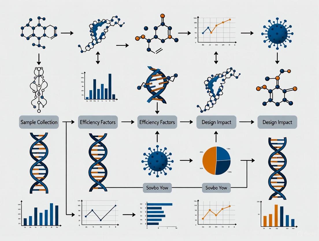
Visualizing the Efficiency Pathway
The following diagram illustrates the journey of a sample from collection to analysis and how swab design impacts key efficiency metrics.
Swab Efficiency Pathway
Key Experimental Protocols for Assessing Swab Efficiency
To ensure reproducible and comparable results, rigorous standardized protocols are essential for evaluating swab performance. The following sections detail methodologies from key studies.
Protocol for Quantifying Microbial DNA Recovery
Objective: To determine the optimal swab type for the collection and analysis of microbiome samples by comparing absolute microbial DNA recovery. [3]
Materials:
- Test Organism: Proteus mirabilis or other representative bacteria.
- Swab Types: e.g., Cotton, Nylon Flocked, Dissolvable, Dental Applicators.
- Equipment: Real-time PCR System, Microcentrifuge, NanoDrop or similar spectrophotometer.
- Reagents: DNA Extraction Kit (e.g., MagMAX DNA Multi-Sample Ultra 2.0 Kit), iTaq Universal SYBR Green Supermix, primers for 16S rRNA gene.
Methodology:
- Sample Deposition: Culture, wash, and pellet the bacteria. Deposit a controlled volume (e.g., 10 µL) of a uniformly mixed bacterial stock onto eight replicates of each swab type.
- DNA Extraction: Extract bacterial DNA from each swab using a commercial kit, following the manufacturer's protocol. Include positive controls (bacterial stock added directly to a tube) and negative controls.
- DNA Quantitation: Quantify the purified DNA using real-time PCR with primers targeting a conserved gene like the 16S rRNA gene. Use a standard curve created from a pooled and quantified sample of the bacterial stock for absolute quantitation.
- Data Analysis: Compare the absolute DNA yields (in ng) across the different swab types using statistical analysis (e.g., ANOVA) to identify significant differences.
Protocol for Comparing Swab Uptake and Release of Fluids
Objective: To characterize the volume uptake and release rates of different swab types. [2]
Materials:
- Swab Types: e.g., Rayon, Flocked Nylon, Foam.
- Equipment: Analytical balance (0.1 mg accuracy), vortex mixer, sterile tubes.
- Reagents: Phosphate-buffered saline (PBS) or a synthetic nasal fluid.
Methodology:
- Volume Uptake Measurement:
- Fill a pre-weighed tube with a known volume of liquid (e.g., 1000 µL for unrestricted supply, 10 µL for restricted supply).
- Weigh the tube again to get the precise initial mass.
- Immerse the swab tip into the liquid for a standardized time (e.g., 15 seconds).
- Remove the swab and re-weigh the tube. The difference in mass corresponds to the volume absorbed by the swab (assuming 1 µL = 1 mg).
- Volume Release Measurement:
- Place the inoculated swab from step 1 into a new, pre-weighed empty tube.
- Agitate on a vortex mixer for a standardized time (e.g., 10 seconds) while applying gentle pressure to the tube wall.
- Remove the swab and weigh the tube. The mass of the liquid in the tube represents the volume released by the swab.
- Data Analysis: Calculate mean uptake and release for each swab type. Release can also be expressed as a percentage of the original uptake.
The Scientist's Toolkit: Essential Research Reagents & Materials
This table catalogs key materials and reagents used in the featured experiments, providing a reference for replicating these efficiency studies.
Table 4: Essential Materials for Swab Efficiency Research
| Item | Function/Description | Example Use Case |
|---|---|---|
| COPAN FLOQSwabs | Nylon-flocked swabs with perpendicular fibers for enhanced collection and release. [3] [5] | Used as a high-performance benchmark in comparative studies. |
| Puritan Cotton Swabs | Traditional cotton-tipped swabs; often used as a baseline for comparison. [3] | Representing a widely used, though less efficient, swab technology. |
| MagMAX DNA Extraction Kit | For automated or manual purification of DNA from a variety of sample types. [3] | Extraction of microbial or human DNA from collected swabs. |
| iTaq Universal SYBR Green | A ready-to-use master mix for real-time PCR (qPCR) quantitation. [3] | Quantifying DNA yield via qPCR against a standard curve. |
| 16S rRNA Primers | Target conserved bacterial genes for universal microbial detection and quantitation. [3] | Absolute quantitation of bacterial DNA in swab eluates. |
| Phosphate-Buffered Saline (PBS) | A balanced salt solution used to suspend cells and maintain pH. [2] | Creating bacterial suspensions or synthetic biological fluids for testing. |
| eSwab Liquid Amies | A preservation medium that maintains sample viability for cultural and molecular tests. [2] | Elution and transport medium for swabs, per manufacturer protocols. |
Swab efficiency is a critical, multi-stage process where design dictates data quality. The evidence is clear: swab material and construction directly govern recovery and extraction efficiency, with modern synthetic materials like nylon flocking consistently outperforming traditional cotton. The scientific community must treat swab selection not as a generic pre-analytical step, but as a deliberate, protocol-driven choice. Future research and development should continue to refine material science and swab architecture, with a focus on maximizing both collection and release to fully leverage the increasing sensitivity of modern analytical platforms like PCR and next-generation sequencing.
The efficacy of any diagnostic test is fundamentally dependent on the quality of the sample collected, making the swab a critical component in the data generation pipeline. For researchers and drug development professionals, selecting an appropriate swab is not a mere procedural formality but a key variable that can significantly impact experimental outcomes, assay sensitivity, and ultimately, the reliability of scientific data. While cotton swabs have been the traditional mainstay, the landscape of sample collection has evolved with the introduction of synthetic materials including rayon, nylon-flocked, polyester, and foam, each offering distinct physicochemical properties.
This evolution is driven by a core principle of swab design: the optimal swab must achieve two often competing goals simultaneously – efficient uptake of the sample from the collection surface and subsequent maximal release of that sample into the transport media or extraction buffer [6] [7]. The material and physical architecture of the swab tip directly influence these processes. The ongoing research in this field is squarely focused on how these design elements impact collection efficiency, a relationship that forms the thesis of this deep dive. Understanding these material characteristics is essential for making evidence-based decisions that enhance diagnostic accuracy and research reproducibility.
Material Characteristics and Performance Metrics
The performance of a swab is dictated by the intrinsic properties of its tip material and its manufacturing design. A comparative analysis of these characteristics reveals significant differences in their functional behavior.
Table 1: Swab Material Characteristics and Performance Comparison
| Material | Primary Structure | Key Advantages | Key Disadvantages | Reported Sample Release Efficiency |
|---|---|---|---|---|
| Cotton | Twisted, woven natural fibers | High absorbency, low cost, widely available [8] | Contains natural oils that can inhibit PCR, low release efficiency, sample trapped in internal fiber core [9] [8] | 25.2% - 35.0% [7] |
| Rayon | Twisted, synthetic fibers from cellulose | Highly absorbent, soft, contains no natural oils or DNA, making it ideal for microbiological testing [10] [8] | Sample can be trapped in inner core, leading to slow and weak elution [8] | ~41% recovery efficiency for spores from nonporous surfaces [11] |
| Polyester (Dacron) | Tightly wound synthetic fibers | Does not produce PCR inhibitors, good for large-area sampling [8] | Low sample release rate (20-30%) due to sample entrapment in inner core [8] | Information missing |
| Nylon-Flocked | Short nylon fibers attached vertically to handle | No internal core; rapid sample uptake and >90% elution; superior for small sample collection [8] [12] | Requires regulated use by trained professionals to prevent mishandling [8] | 45.4% - 49.0% for spores (vs. 13.2% for cotton) [9]; 54.7% - 80.4% overall efficiency [7] |
| Polyurethane (Foam) | Porous, sponge-like structure | Fiber-free, dust-free, excellent for cleaning and absorbing fluids, good wear resistance [8] | Low release rate when used for biological sample collection [8] | 57.9% uptake efficiency (lowest among synthetics) [7] |
A critical finding from recent systematic reviews is that swabs made of the same material but from different manufacturers do not perform equally [6]. For instance, one study noted that while a rayon swab from one manufacturer performed best with a diluted blood sample, a rayon swab from a different supplier performed poorly with neat blood [6]. This underscores that material is only one factor, and the specific product design and manufacturing quality are equally important for performance.
Experimental Protocols for Swab Evaluation
To generate comparable and reliable data on swab performance, researchers employ standardized protocols that quantitatively assess uptake and release efficiency. The following methodologies are commonly cited in the literature.
Protocol for Evaluating Bacterial Spore Recovery
This protocol, adapted from a study validating nylon-flocked swabs for planetary protection, provides a robust framework for comparing swab efficiency in microbiology [9].
- Sample Preparation: Spore stocks of specific Bacillus species (e.g., B. atrophaeus) are prepared, quantified, and applied as a dry aerosol onto standardized surface coupons (e.g., stainless steel, painted wallboard) of a defined area (e.g., 25 cm²). Surface concentrations are pre-determined.
- Sample Collection: The swabbing protocol is rigorously defined. Typically, a swab is moistened with a sterile buffer (e.g., PBST or water) and used to scrub the surface in a systematic pattern, such as horizontally, vertically, and diagonally, while rotating the swab to use all sides.
- Sample Elution: The swab head is placed in a sterile tube containing a precise volume of elution buffer (e.g., 2.5 ml of PBST). Extraction is performed via vortexing for 5-6 seconds or a combination of sonication (120 seconds) and vortexing.
- Analysis: The eluent is serially diluted and pour-plated or spread-plated on appropriate culture media (e.g., R2A agar). After incubation, colony-forming units (CFU) are enumerated. Recovery efficiency is calculated as: (CFU recovered from swab eluent / CFU initially present on the surface) × 100.
Protocol for Evaluating Forensic DNA Recovery
This protocol assesses swab performance for human DNA collection, relevant for forensic science and diagnostic testing [13].
- Sample Preparation: A known volume and concentration of a biological source (e.g., 50 µL of human saliva) is deposited onto different fabric substrates (e.g., denim, cotton, polyester) and allowed to dry for 24 hours at room temperature.
- Sample Collection: Different swab types are used to recover the dried sample from the fabric. A consistent, controlled swabbing technique and pressure should be applied across all tests.
- Immunochromatographic Test: The swab is first placed in a specific buffer to elute the sample, and an aliquot is used for a lateral flow immunochromatography (LFI) test to detect the presence of human salivary α-amylase. Band intensity can be semi-quantitatively ranked.
- DNA Analysis: Total DNA is then extracted from the remaining sample buffer. DNA yield and quality are quantified using fluorescent assays (e.g., Qubit) and real-time PCR systems (e.g., PowerQuant System). The success of short tandem repeat (STR) profiling and mitochondrial DNA analysis can also be evaluated.
Signaling Pathways and Workflow Visualization
The journey from sample collection to analysis is a critical pathway where swab design imposes significant influence. The following diagram maps this workflow and highlights the key points where material choice dictates efficiency.
Figure 1: Impact of Swab Design on Diagnostic Workflow. This workflow illustrates the two-phase journey of a sample, where swab design critically influences outcomes. The Uptake Phase is governed by the swab's fiber structure and absorbency, directly determining how much sample is collected from the source. The Transfer & Elution Phase is affected by the swab's release efficiency and potential for introducing inhibitors, determining the quantity and quality of the analyte delivered for downstream analysis. Key decision points related to material properties are shown in red, fundamentally shaping the final result.
The Scientist's Toolkit: Essential Research Reagents and Materials
The following table details key materials and reagents required for conducting rigorous swab performance evaluations, as derived from the cited experimental protocols.
Table 2: Key Reagents and Materials for Swab Performance Studies
| Item Name | Function in Protocol | Specific Examples / Notes |
|---|---|---|
| Surface Coupons | Provides a standardized, reproducible surface for sample deposition and recovery. | Stainless steel, painted wallboard, graphite composite, fabric substrates (denim, cotton, polyester) [9] [13]. |
| Test Microorganism / Analyte | The target of recovery used to quantify efficiency. | Bacillus atrophaeus spores (surrogate for B. anthracis), human saliva (for α-amylase and DNA), whole blood [9] [13]. |
| Elution/Storage Buffer | Liquid medium to release collected material from the swab and preserve it. | Phosphate-Buffered Saline with Tween (PBST), Tris-based buffers (Tris HEPES, Tris MOPS, Tris TAPS), molecular-grade water [9] [7]. |
| Culture Media | Supports the growth of recovered microorganisms for quantification. | Trypticase Soy Agar (TSA) for standard cultivation; R2A agar for microbes from low-nutrient environments [9]. |
| DNA Quantification Kits | Precisely measures the concentration and quality of recovered human DNA. | Fluorescent assays (e.g., Qubit dsDNA HS Assay), real-time PCR kits (e.g., PowerQuant System) [13]. |
| Lateral Flow Immunochromatography (LFI) Tests | Rapidly detects the presence of a specific protein analyte, such as salivary α-amylase. | Rapid stain identification tests for forensics; principle is the same as COVID-19 antigen tests [13]. |
The body of research clearly demonstrates that there is no universal "best" swab material. The optimal choice is contingent on the specific application, the nature of the target analyte (e.g., spores, epithelial cells, viral particles), and the substrate from which it is being collected. However, a dominant trend emerges: the design of nylon-flocked swabs, which facilitates both high uptake and superior release, consistently delivers higher overall efficiency in both microbiological and molecular forensic studies [9] [13] [7].
Future research must continue to address existing gaps. As noted in a recent systematic review, there are over 40 potential substrate-DNA source combinations, yet an optimal swab type has been identified for only 13 of them [6]. Furthermore, the interaction between DNA extraction chemistry and swab material requires more investigation. The future of swab design is likely to see increased integration of advanced manufacturing, such as 3D printing and the use of novel bio-resins, which promise to create swabs that are not only more efficient but also rapidly produced and environmentally sustainable [14]. For scientists, a critical and evidence-based approach to swab selection is paramount, as this seemingly simple tool holds significant power over the integrity of diagnostic and research data.
This technical guide explores the fundamental molecular forces governing DNA interactions, with a specific focus on implications for sample collection swab design. Hydrogen bonding and dipole forces critically influence the binding affinity and release efficiency of DNA molecules to and from collection surfaces. Understanding these interactions enables the rational design of swab materials and processing protocols that maximize sample recovery, directly impacting the accuracy and reliability of downstream diagnostic applications in pharmaceutical development and clinical research.
The efficiency of DNA sample collection is governed by a complex interplay of non-covalent molecular interactions at the interface between biological material and the collection substrate. Among these, hydrogen bonding and dipole-related forces are paramount, as they determine the adhesion strength and subsequent release of DNA during processing.
- Hydrogen Bonding: A hydrogen bond is an attractive interaction between a hydrogen atom from a molecule or a molecular fragment X−H (where X is more electronegative than H) and an atom or group of atoms in the same or another molecule [15]. In the context of DNA, these bonds are not purely electrostatic but exhibit partial covalent character and involve charge transfer, making them highly directional and specific [15]. The strength of hydrogen bonds can vary significantly, from weak (1–2 kJ/mol) to very strong (over 40 kcal/mol), as seen in the bifluoride ion (HF⁻₂) [15]. In biological systems, typical hydrogen bonds, such as those in protein-DNA complexes or between water and DNA bases, often have enthalpies in the range of 5-29 kJ/mol [15].
- Dipole-Dipole Forces: These are electrostatic interactions between the permanent dipoles of polar molecules. In DNA, the polar sugar-phosphate backbone and the heterocyclic bases create a significant electrostatic potential landscape [16]. This landscape is not uniform; it depends on the local DNA sequence and conformation, providing a pattern for long-range recognition and interaction [16]. While generally weaker than hydrogen bonds, these forces are crucial for initial attraction and positioning.
The combined effect of these forces dictates the thermodynamic balance of DNA binding and release, a principle that directly informs the engineering of swab surfaces and the composition of wetting and transport solutions.
Hydrogen Bonding in DNA Recognition and Binding
Fundamental Principles and Energetics
Hydrogen bonds are a primary mechanism for specific molecular recognition. In DNA-protein complexes, a dense network of hydrogen bonds forms between amino acid side chains and the edges of DNA bases in the major and minor grooves [16]. This network is highly cooperative; the geometry of each hydrogen bond is constrained, and the rearrangement of one bond can trigger a cascade of changes in the interface [16]. The strength of the interaction depends on the donor-acceptor pair and their geometric arrangement, with optimal linear (180°) alignments providing the strongest bonds [15].
Role in Specificity and Affinity
There are differing views on the precise role of hydrogen bonds in DNA recognition. One perspective holds that the specific pattern of hydrogen bonds between a protein and DNA is the primary determinant of binding specificity, allowing the discrimination between different nucleotide sequences [16]. An alternative view is that the hydrogen bond network, while contributing significantly to binding affinity (the enthalpy of the interaction), primarily serves to fix the complex in place after the initial recognition has occurred through other means, such as long-range electrostatic forces [16]. In practice, both mechanisms are likely at play; the network satisfies the hydrogen-bonding requirements of both the DNA backbone and bases, precisely positioning the interacting molecules for specific contact [17].
Table 1: Hydrogen Bond Strengths in Biological Systems
| Donor-Acceptor Pair | Typical Enthalpy (kJ/mol) | Example Context |
|---|---|---|
| F−H···:F− | ≈ 161.5 | Bifluoride ion (HF⁻₂) |
| O−H···:N | ≈ 29 | Water-ammonia interaction |
| O−H···:O | ≈ 21 | Water-water, alcohol-alcohol |
| N−H···:N | ≈ 13 | Ammonia-ammonia |
| N−H···:O | ≈ 8 | Water-amide interaction |
Dipole and Electrostatic Forces in DNA Interactions
The DNA double helix presents a highly charged and polar surface. The phosphate groups of the backbone each carry a negative charge, creating a strong, long-range electrostatic field that attracts positively charged ions and protein patches [16]. This "electrostatic pre-screening" is a critical first step in DNA recognition, guiding proteins to the DNA molecule before specific, short-range interactions like hydrogen bonding can occur.
Beyond simple charge-charge interactions, the arrangement of atoms in the DNA bases creates permanent molecular dipoles. The spatial pattern of these dipoles, and the resulting electrostatic potential, varies with the nucleotide sequence [16]. This means that the local conformation of the DNA backbone provides a unique spatial pattern for recognition, allowing proteins and synthetic materials to distinguish between different sequences based on their electrostatic "signature" even before direct contact is made. Factors such as ionic strength, counterion concentration, and pH can significantly modulate these electrostatic interactions by shielding charges and altering the effective potential [16].
Implications for Swab Design and Sample Collection
The principles of DNA molecular interactions directly translate to the performance of specimen collection swabs. The goal is to design a system that initially binds DNA effectively from a surface but allows for its efficient release into the transport medium for subsequent analysis.
Swab Material and Surface Chemistry
The chemical nature of the swab fiber surface dictates the type and strength of molecular interactions with DNA.
- Flocked Swabs: These swabs, which feature fibers oriented perpendicular to the tip, are trending in the market due to superior sample collection efficiency. Their brush-like structure is designed to create a large surface area for interaction, effectively trapping cells and DNA via a combination of capillary action and molecular adhesion [18].
- Material Composition: The shift from traditional cotton to synthetic materials like rayon and polyester is driven by the need to minimize unwanted interactions. Cotton can have hydrophilic surfaces that may form strong hydrogen-bonding networks with DNA, potentially trapping it and reducing elution efficiency. Synthetic materials offer more controlled surface chemistry, potentially reducing non-specific binding [18].
The Critical Role of Swabbing Solutions
The liquid used to moisten the swab is not merely a wetting agent; it is an active component that modulates molecular forces. A study evaluating eleven different swab-wetting solutions found that their performance was heavily influenced by the type of biological evidence being collected [19].
- Tonicity and Cell Integrity: Hypotonic solutions like pure water can cause cell lysis. The released DNA is then free to form strong hydrogen bonds with the swab fibers, leading to entrapment and poor recovery. Isotonic buffers, such as phosphate-buffered saline (PBS), help maintain cell integrity, resulting in the collection of intact cells that are less likely to adhere strongly to the swab, thereby improving DNA yield [19].
- Detergent-Based Solutions: Amphiphilic detergents like Tween20, Triton X-100, and SDS are more efficient at solubilizing cellular components and disrupting hydrophobic interactions. They work by reducing the surface tension and competing with DNA for binding sites on the swab, which helps in the release of DNA during extraction [19].
- Chelating Agents: Solutions containing EDTA or EGTA were identified as particularly effective for recovering DNA from saliva and blood samples [19]. These agents chelate divalent cations like Mg²⁺, which are essential for stabilizing the structure of DNA and facilitating its binding to surfaces. By sequestering these ions, chelating agents weaken the DNA's structural cohesion and its interaction with the swab, thereby promoting release.
Table 2: Impact of Swab-Wetting Solutions on DNA Recovery from Different Biological Sources
| Wetting Solution Type | Mechanism of Action | Optimal Application / Performance |
|---|---|---|
| Deionized Water | Hypotonic, causes cell lysis | Less efficient; released DNA trapped in swab fibers [19] |
| Isotonic Buffers (e.g., PBS) | Maintains cell integrity | Higher DNA recovery by collecting intact cells [19] |
| Detergent-Based (e.g., Tween 20, SDS) | Solubilizes cellular components; disrupts hydrophobic bonds | Efficient for trace DNA and cellular DNA; competes for binding sites [19] |
| Chelating Agents (e.g., EDTA, EGTA) | Chelates Mg²⁺ and other divalent cations | Most suitable for saliva and blood samples; weakens DNA structure and adhesion [19] |
Experimental Protocols for Evaluating Interactions
Protocol: Evaluating Swab-Wetting Solutions
This protocol is adapted from a study that investigated the impact of various solutions on DNA recovery [19].
- Sample Preparation: Deposit standardized quantities (e.g., 50 ng) of different DNA sources (cell-free DNA, cellular DNA, saliva, blood) in triplicate onto non-porous surfaces (plastic, glass, metal). Allow deposits to dry.
- Swab Collection: Use a standardized swab (e.g., cotton) moistened with a fixed volume (e.g., 50 µL) of the test wetting solution. Employ a consistent swabbing technique across all samples.
- DNA Extraction: Process swabs using a standardized extraction method, such as an in-house protocol involving a lysis buffer (e.g., containing PVP, Tween 20, Tris-HCl, and proteinase K) followed by incubation and purification using solid-phase reversible immobilization (SPRI) magnetic beads [19].
- Quantification and Analysis: Quantify the recovered DNA using a fluorometric method (e.g., Qubit). Compare the mean recovery yields across the different wetting solutions and biological sources to determine optimal conditions.
Protocol: Molecular Dynamics (MD) Simulation of DNA-Surface Interactions
Computational approaches like MD simulation provide atomistic insight into interaction forces, as demonstrated in studies of CRISPR-Cas systems [20].
- System Setup: Construct an all-atom model of the DNA sequence of interest and the swab material surface (e.g., a polymer fragment). Solvate the system in a water box and add ions to achieve physiological ionic strength.
- Simulation Run: Perform the MD simulation using high-performance computing. Calculate key molecular interaction features over the simulation trajectory, including:
- Hydrogen Bond Counts: The number of stable hydrogen bonds between DNA and the surface.
- Binding Free Energies: The total energy of interaction, decomposed into components like van der Waals forces, electrostatic energies, and polar solvation energies [20].
- Root Mean Square Fluctuation (RMSF): Measures the flexibility of DNA atoms when near the surface.
- Data Integration: Use the calculated molecular features as inputs for a neural network model to predict the binding affinity and release propensity of the DNA from that specific surface. This predictive model can then guide the rational design of new swab materials.
Diagram 1: MD simulation workflow for swab design.
The Scientist's Toolkit: Research Reagent Solutions
The following table details key reagents and materials used in the featured experiments for studying and optimizing DNA-swab interactions.
Table 3: Essential Research Reagents and Materials for DNA Collection Studies
| Item | Function/Application | Specific Examples |
|---|---|---|
| Swab Types | Physical collection device; material chemistry affects DNA binding/release. | Flocked Swabs [18], Cotton Swabs [19], Polyester Swabs [18] |
| Wetting & Transport Solutions | Modulate molecular forces to maintain sample integrity and promote DNA release. | Viral Transport Medium (VTM) [21], Phosphate-Buffered Saline (PBS) [19], EDTA/EGTA Solutions [19], Detergent Solutions (Tween 20, Triton X-100) [19] |
| Lysis Buffers | Break down cells and release nucleic acids from the swab matrix and biological material. | Buffers containing Proteinase K, Tween 20, Tris-HCl [19] |
| Purification Reagents | Isolate and concentrate DNA from the complex lysate for downstream analysis. | Solid-Phase Reversible Immobilization (SPRI) Beads [19] |
| Quantification Kits | Precisely measure the amount of DNA recovered, a key metric for protocol efficiency. | Fluorometer-based assays (e.g., Qubit kits) [19] |
The efficiency of DNA sample collection via swabs is fundamentally governed by the subtle balance of hydrogen bonding and dipole forces. The strategic design of swab materials—prioritizing synthetic fibers like flocked nylon—coupled with the use of optimized wetting solutions such as isotonic buffers or chelating agents, allows researchers to actively manage these molecular interactions. By shifting from a purely empirical to a rational design approach, informed by molecular dynamics simulations and rigorous experimental validation, it is possible to significantly enhance DNA recovery. This advancement ensures the highest quality of input material for subsequent diagnostic assays, directly impacting the reliability of data in drug development and clinical research.
The efficiency of forensic DNA analysis and molecular diagnostics is fundamentally dictated by the initial sample collection process. The choice of swab design and collection methodology presents distinct challenges that are profoundly influenced by the nature of the sample itself—whether it is a visible biological fluid or latent touch DNA. This whitepaper examines the variable performance of different swab types and materials across these sample categories, synthesizing recent research to provide an evidence-based framework for selecting optimal collection tools. By integrating quantitative data on recovery efficiencies with detailed experimental protocols, this guide aims to inform researchers and forensic professionals in refining sample collection strategies to maximize DNA yield and integrity, thereby enhancing the reliability of downstream genetic analyses.
The fidelity of DNA analysis—encompassing forensic investigations, diagnostic testing, and biomedical research—is contingent upon the initial recovery of biological material from substrates. The swab serves as the critical interface between the evidence and the analytical pipeline, yet its design and material composition are often overlooked as variables in experimental and operational outcomes. The core challenge lies in the divergent physical and biological properties of different sample types, which demand specific and often competing optimizations from the collection tool.
Biological fluids like blood and saliva contain abundant cellular material and free DNA, suspended in a liquid matrix that can be absorbed by a swab. In contrast, touch DNA samples are composed of scant epithelial cells and cell-free DNA transferred from skin to a surface via contact [22]. These traces are characterized by inherently low levels of DNA and are susceptible to degradation, making their collection exceptionally challenging [22]. The efficacy of a swab is measured by its dual capability: to efficiently release collected material during the extraction process. This review systematically evaluates how these performance metrics vary with sample type and how swab design can be tailored to address these fundamental challenges within the context of sample collection efficiency research.
Comparative Analysis of Sample Types
Fundamental Characteristics and Collection Challenges
The table below summarizes the defining characteristics and inherent challenges of collecting biological fluids versus touch DNA.
| Characteristic | Biological Fluids (e.g., Blood, Saliva) | Touch DNA |
|---|---|---|
| DNA Source | Nucleated cells, cell-free DNA from body fluids [22]. | Shed keratinocytes, epithelial cells, cell-free DNA from skin [22]. |
| Typical DNA Yield | High (visible stain) | Low (invisible trace), often Low Template DNA (LT-DNA) [22]. |
| Primary Challenge | Efficient elution from swab fibers; inhibition from substrate. | Maximizing recovery of minute, often degraded, material from a surface [22]. |
| Influencing Factors | Substrate porosity, fluid viscosity. | Substrate roughness, "shedder status," contact duration/pressure, environmental exposure [22]. |
Impact of Substrate and Environmental Factors
The substrate from which DNA is collected introduces another layer of complexity. Porous substrates like fabric or paper can trap biological material, potentially protecting it but also making recovery more difficult. Non-porous substrates like plastic or glass generally allow for better recovery, though metal surfaces have proven particularly challenging for touch DNA collection [22] [23]. Furthermore, environmental factors such as heat, humidity, and exposure to sunlight can accelerate DNA degradation, while contaminants like dirt or chemicals can inhibit subsequent PCR amplification [24].
Swab Material and Design Performance
The evolution of swab design is driven by the need to overcome the challenges outlined above. A shift from traditional cotton swabs to newer materials and configurations has been observed, with performance being highly dependent on the sample type and substrate.
Quantitative Recovery Efficiencies
Recent studies have quantified the performance of different swab materials. The following table summarizes key findings for touch DNA and biological fluids.
| Swab Material / Technique | Performance for Touch DNA | Performance for Biological Fluids | Key Findings |
|---|---|---|---|
| Nylon Flocked | Superior recovery and DNA release from non-porous surfaces [23]. | Effective for fluid collection and release. | Open structure minimizes sample entrapment; ~85% DNA extraction efficiency from the swab itself [23]. |
| Traditional Cotton | Lower efficiency; sample tends to be trapped in dense fibers [23] [6]. | Variable performance; can be effective for neat blood [6]. | DNA extraction efficiency from seeded swab was ~56% [23]. Performance varies by manufacturer [6]. |
| Single-Swab Technique | Higher efficiency in recovering STR alleles across varied settings [22]. | Not specifically reported. | Outperformed double-swab and other methods (cutting, adhesive tape) in recent systematic review [22]. |
| Double-Swab Technique | Does not consistently improve recovery rates over single swab [22]. | Not specifically reported. | A wet swab followed by a dry swab; historically used to maximize cellular material recovery [22]. |
A 2025 systematic review of swab materials in forensic testing underscores that swabs made of the same material by different manufacturers do not perform identically, highlighting the importance of manufacturer-specific validation [6]. For instance, while one study may find a specific cotton swab brand performs well with neat blood, another brand of rayon swab may perform poorly with the same sample [6].
The Scientist's Toolkit: Essential Research Reagents and Materials
Selecting the appropriate tools is critical for ensuring the integrity of DNA collection and analysis. The following table details key reagents and materials used in this field.
| Item | Function/Application | Examples & Key Considerations |
|---|---|---|
| Nylon Flocked Swabs | Sample collection for trace DNA; designed for high elution efficiency. | FLOQSwabs (COPAN); various OEM suppliers. Tips have short, perpendicular nylon fibers for optimal sample release [25] [23]. |
| Cotton Swabs | Traditional, cost-effective collection tool for biological fluids. | Performance varies by brand. Prone to retaining sample within dense fibers [6]. |
| DNA Extraction Kit | Purifies DNA from the swab tip for downstream analysis. | QIAamp DNA Investigator Kit. Efficiency is kit- and swab-dependent [23]. |
| Quantification Kits (qPCR) | Precisely measures DNA concentration. | Detects human-specific DNA; critical for determining input into STR amplification [6]. |
| STR Amplification Kits | Generates genetic profile from recovered DNA. | Highly sensitive commercial kits are essential for low-template touch DNA [22]. |
| Absorbance Spectrophotometer | Assesses DNA concentration and purity (A260/A280, A260/A230). | NanoDrop. Rapid assessment of nucleic acid concentration and contaminant detection [26] [27] [28]. |
| Fluorometer with dsDNA Assay | Highly sensitive and specific quantification of double-stranded DNA. | Qubit (Invitrogen); PicoGreen assay. More accurate for low-concentration samples than absorbance methods [26] [27]. |
Experimental Protocols for Evaluating Swab Efficiency
To generate comparable data on swab performance, standardized experimental protocols are essential. The following sections detail common methodologies for quantifying DNA recovery.
Protocol: Controlled Sample Deposition and Recovery
This protocol is designed to test swab efficiency from non-porous surfaces [23].
- Surface Preparation: Clean substrates (e.g., plastic, glass, metal) with 20% bleach and UV irradiation to remove background DNA.
- Sample Application:
- For touch DNA simulations: Have donors handle substrates under controlled conditions (e.g., after hand washing, with defined pressure and duration).
- For biological fluids: Deposit standardized volumes (e.g., 5-10 µL) of synthetic blood or saliva.
- For acellular DNA: Apply aliquots of a known quantity of purified human DNA (e.g., ~10 ng) directly to the surface or swab [23].
- Swabbing Procedure: Moisten the swab with a recommended buffer (e.g., sterile water or the buffer provided with the extraction kit). Swab the entire surface area using a consistent pattern and pressure, rotating the swab to use all sides. For the double-swab technique, follow the wet swab immediately with a dry one [22].
- Drying and Storage: Air-dry swabs thoroughly before storage. Rapid drying in specialized tubes has been shown to preserve DNA, with one study showing 95% recoverable DNA with fast-drying tubes versus only 12% with slower-drying alternatives [24].
- DNA Extraction: Process swabs using a standardized DNA extraction kit (e.g., QIAamp DNA Investigator Kit), including negative controls (clean swabs) to monitor contamination.
- DNA Quantification: Quantify the recovered DNA using a fluorescence-based method (e.g., Qubit, PicoGreen) for sensitivity and specificity [26] [27].
Protocol: DNA Quantification and Purity Assessment
Accurate DNA quantification is crucial for interpreting recovery data and downstream success.
Fluorometry (for accurate concentration):
- Principle: Fluorescent dyes (e.g., PicoGreen) bind specifically to dsDNA. The fluorescence intensity is measured and compared to a standard curve of known concentrations [26] [27].
- Procedure: Prepare DNA standards and unknown samples according to the assay kit's instructions. Load them into a fluorometer or microplate reader and measure fluorescence. The instrument calculates concentration based on the standard curve. This method is highly sensitive and specific for dsDNA but does not assess purity [27].
UV Spectrophotometry (for concentration and purity):
- Principle: Nucleic acids absorb UV light maximally at 260 nm. The ratio of absorbance at 260 nm and 280 nm (A260/A280) indicates protein contamination, while the A260/A230 ratio indicates contamination from salts or organic compounds [26] [28].
- Procedure: Blank the instrument with the suspension buffer. Measure 1-2 µL of the DNA sample using a microvolume spectrophotometer. The concentration is calculated as: Concentration (µg/mL) = A260 × Dilution Factor × 50 µg/mL (for dsDNA). Pure DNA has an A260/A280 ratio of ~1.8-2.0 and an A260/A230 ratio of ~2.0-2.4 [26] [27] [28].
Diagram 1: Experimental workflow for evaluating swab efficiency, covering sample deposition, collection with different materials, and downstream DNA analysis.
The influence of sample type on the challenges of DNA collection is undeniable. Biological fluids and touch DNA represent two ends of a spectrum, necessitating tailored approaches for optimal recovery. The evidence strongly indicates that nylon flocked swabs consistently outperform traditional cotton in the critical ability to release DNA during extraction, making them particularly advantageous for low-yield touch DNA evidence [23] [6]. Furthermore, the single-swab technique has shown superior efficiency in recent studies, challenging previous assumptions about the double-swab method [22].
Future research should focus on standardizing testing protocols to reduce heterogeneity in study results and allow for robust meta-analyses. There is also a pressing need to explore novel swab materials and designs, including dissolvable polymer swabs and miniaturized tips for targeted collection. As DNA profiling technologies continue to increase in sensitivity, the bottleneck of sample collection will only become more pronounced. Therefore, an evidence-based, sample-type-driven selection of collection tools is not merely a best practice but a fundamental requirement for ensuring the integrity and success of modern genetic analysis in both forensic and diagnostic contexts.
From Theory to Practice: Selecting and Applying Swabs in Research and Diagnostic Workflows
The efficacy of any diagnostic or research protocol that relies on surface sampling is fundamentally determined at the initial point of contact: the interface between the swab and the substrate. The physical and chemical characteristics of the surface—its porosity, roughness, and hydrophobicity—directly govern the adhesion and retention of target analytes. Sample collection efficiency, a key metric in analytical science, is therefore not a property of the swab alone, but of the swab-substrate system. A poorly matched system can lead to inadequate sample recovery, false negatives, and compromised data integrity, ultimately undermining research validity and diagnostic outcomes.
Framed within a broader thesis on how swab design impacts sample collection efficiency research, this guide posits that a deliberate, evidence-based strategy for pairing swab properties with substrate characteristics is a critical methodological variable. The prevailing trend in swab design is a move away from traditional materials like cotton, which can entrap samples and inhibit elution, toward advanced synthetic alternatives. [18] [29] Flocked swabs, characterized by their brush-like tips of upright nylon fibers, have demonstrated superior performance by creating a capillary effect that enhances both absorption and release of liquid samples, thereby maximizing the yield of genetic material for PCR and other sensitive analytical techniques. [25] Furthermore, the global market for sample collection is evolving rapidly, with a growing emphasis on self-collection kits and automation-compatible designs, placing a premium on swabs that deliver consistent performance across diverse and unpredictable user-applied substrates. [18] [29] This technical guide provides researchers and drug development professionals with the framework and experimental tools to systematically evaluate and optimize swab-substrate pairings, transforming sample collection from an art into a reproducible science.
Swab and Substrate Characteristics: A Foundation for Strategic Matching
A Taxonomy of Swab Design and Material Science
The design and composition of a collection swab are engineered to influence its core functions: sample acquisition, retention, and release. Understanding these components is a prerequisite for strategic selection.
- Tip Material: The tip material is the primary factor in sample interaction.
- Flocked Nylon: These swabs feature nylon fibers perpendicularly projected onto the shaft, creating a porous, brush-like tip. This structure maximizes surface area for sample capture and uses capillary action to absorb and release >90% of the sample, making it the gold standard for molecular applications. [25] [29] They are ideal for recovering samples from a wide range of surfaces, particularly irregular ones.
- Polyester: Synthetic polyester tips offer good absorption and are often chosen for their chemical compatibility, as they are less likely to inhibit PCR reactions compared to some natural materials. [18]
- Cotton: Traditional cotton fibers can entrap biological samples and are known to contain natural impurities that can inhibit enzymatic reactions in PCR, potentially reducing test accuracy. [29] Their use is declining in favor of synthetic, PCR-compatible alternatives.
- Shaft Material: The shaft (e.g., ABS plastic or polystyrene) must provide sufficient rigidity for controlled application but include a breakpoint for safe and easy transfer into a transport vial. [25]
- Sterility: Individually wrapped, sterile swabs are non-negotiable in clinical and research settings to prevent contamination of the collected sample and the surface being sampled. [25]
Defining Substrate Properties for Sampling
Surfaces can be categorized by key physical properties that dictate sampling strategy:
- Porous Surfaces: These materials allow liquids to penetrate beneath the surface layer. Examples include untreated wood, fabric, drywall, and cardboard. The challenge is that analytes can wick away from the immediate surface, making recovery difficult.
- Non-Porous Surfaces: These materials have a solid, impermeable surface that prevents liquid penetration. Examples include stainless steel, glass, plastic, and glazed tile. Samples remain on the surface, but can be prone to spreading thinly or evaporating.
- Rough Surfaces: Defined by their complex topography with peaks and valleys, rough surfaces include concrete, textured plastic, or machined metal. The primary challenge is that swab fibers cannot make contact with analytes trapped in microscopic crevices.
Table 1: Swab Material Properties and Their Impact on Collection
| Swab Material | Mechanism of Collection | Sample Release Efficiency | Primary Best-Use Context |
|---|---|---|---|
| Flocked Nylon | Capillary action, high surface-area absorption | High (typically >90%) [25] | Universal; excellent for porous surfaces, rough surfaces, and molecular diagnostics |
| Polyester | General absorption | Moderate to High | Non-porous and slightly porous surfaces; PCR-sensitive applications |
| Cotton | Entrapment within fibrous mesh | Low to Moderate (potential for inhibition) [29] | General use where high-sensitivity analysis is not required; being phased out |
Strategic Matching and Experimental Protocols
A Framework for Matching Swab to Substrate
Selecting the optimal swab requires a systematic approach based on the substrate's properties. The following workflow provides a logical decision-making pathway for researchers.
Experimental Protocol for Quantifying Collection Efficiency
To generate empirical data supporting the selection of a swab for a specific application, researchers can employ the following controlled protocol to quantify collection efficiency.
Objective: To determine the sample collection efficiency of different swab types from defined porous, non-porous, and rough surfaces.
Materials:
- Surfaces of interest (e.g., stainless steel coupon, pine wood, textured plastic).
- Test swabs (e.g., Flocked Nylon, Polyester, Cotton).
- Standardized sample solution (e.g., PBS spiked with a known concentration of a detectable analyte like yeast RNA or a fluorescent dye).
- Elution buffer (compatible with downstream detection method).
- Micropipettes and sterile tips.
- Vortex mixer.
- Analytical instrument for quantification (e.g., qPCR machine for RNA, fluorimeter, or spectrophotometer).
Methodology:
- Surface Preparation: Clean all test surfaces thoroughly to remove contaminants. Sterilize if necessary.
- Sample Application: Apply a precise volume (e.g., 10 µL) of the standardized sample solution onto the center of each test surface. Allow it to air dry under controlled conditions (e.g., in a biosafety cabinet for 15-30 minutes) to simulate an environmental deposit.
- Swab Sampling: Pre-moisten swabs with a consistent volume of elution buffer if the protocol dictates. Swab the entire contaminated area using a standardized pattern (e.g., rotating the swab while making five horizontal and five vertical passes).
- Sample Elution: Immediately place the swab tip into a known volume of elution buffer (e.g., 1 mL) in a microcentrifuge tube. Vortex vigorously for a set time (e.g., 1-2 minutes) to release the collected sample.
- Quantification: Analyze the eluent using the chosen analytical method (e.g., qPCR for RNA concentration, fluorimetry for fluorescence) and compare the measured value to a standard curve.
Data Analysis: Calculate the Collection Efficiency (%) for each swab-substrate combination using the formula: Collection Efficiency (%) = (Quantity of Analyte Eluted / Quantity of Analyte Applied) × 100
Table 2: Experimental Data Table for Collection Efficiency Analysis
| Swab Type | Substrate Type | Analyte Used | Quantity Applied (ng or RFU) | Quantity Eluted (ng or RFU) | Collection Efficiency (%) |
|---|---|---|---|---|---|
| Flocked Nylon | Porous (Wood) | Yeast RNA | 100 ng | 85 ng | 85.0 |
| Cotton | Porous (Wood) | Yeast RNA | 100 ng | 52 ng | 52.0 |
| Flocked Nylon | Non-Porous (Steel) | Yeast RNA | 100 ng | 92 ng | 92.0 |
| Polyester | Non-Porous (Steel) | Yeast RNA | 100 ng | 88 ng | 88.0 |
| Flocked Nylon | Rough (Textured Plastic) | Fluorescent Dye | 100 RFU | 78 RFU | 78.0 |
| [Researcher to fill with experimental data] |
The Scientist's Toolkit: Essential Research Reagent Solutions
The following table details key materials and reagents essential for conducting rigorous swab collection efficiency research.
Table 3: Essential Materials for Swab Efficiency Research
| Item | Function in Research | Key Considerations |
|---|---|---|
| Flocked Swabs | The primary test device for sample collection from surfaces. | Select swabs validated for PCR to avoid inhibition. Ensure consistent tip size and material across experiment replicates. [25] |
| PCR-Compatible Transport Media | Liquid medium to preserve and transport collected genetic material. | Essential for maintaining RNA/DNA integrity between collection and qPCR analysis. Use viral transport media for pathogen studies. [29] |
| Synthetic Analogue (e.g., Yeast RNA) | A safe, stable, and quantifiable surrogate for hazardous human pathogens in method development. | Allows for safe, reproducible, and highly quantitative benchmarking of collection efficiency using qPCR. |
| Fluorescent Tracers (e.g., FITC-Dextran) | A visual and quantifiable marker for recovery studies. | Enables rapid, low-cost assessment of physical recovery efficiency using a fluorimeter, independent of biological variables. |
| qPCR Instrument | The gold-standard analytical tool for quantifying specific nucleic acid sequences recovered from a surface. | Provides high sensitivity and specificity for determining the efficiency of collecting and eluting genetic material. |
In the pursuit of robust and reproducible research, the method of sample collection must be scrutinized with the same rigor as the analytical technique itself. The interaction between swab and substrate is a critical, yet often underspecified, variable in the chain of custody for any sample. As this guide has detailed, a one-size-fits-all approach is inadequate. The strategic selection of a swab—prioritizing advanced materials like flocked nylon for their superior sample release and ability to handle complex surfaces—is a direct contributor to data quality and experimental power.
The future of this field points toward greater precision and integration. Ongoing innovation in swab design, including the exploration of new materials and tip geometries, will continue to push the boundaries of recovery efficiency. [29] Furthermore, the rise of automation and digital tracking in laboratories necessitates the use of swabs that are not only effective but also compatible with automated systems, ensuring traceability and reducing human error. [29] By adopting a principled, evidence-based framework for matching the swab to the substrate, as outlined in this whitepaper, researchers and drug developers can significantly enhance the validity of their findings and contribute to the advancement of reliable scientific discovery.
In molecular diagnostics and microbiological research, the journey of a sample from collection to analysis is fraught with potential pitfalls. While swab design is a critical first step for maximizing sample collection efficiency, it is only the beginning. The integrity of the biological specimen and the accuracy of the final analytical result are profoundly influenced by the subsequent workflow considerations: the volume in which the sample is collected, the transport medium used to preserve it, and the protocols employed for sample pooling. This guide details these critical parameters, providing researchers and drug development professionals with the experimental data and methodologies necessary to optimize the entire sample lifecycle, thereby ensuring that the initial investment in high-quality swab design is not undermined by downstream processing errors.
Collection Volume and Efficiency
The volume of fluid used in collection and elution processes is a critical determinant of sample yield. It directly influences the concentration of the target analyte and the efficiency of its release from the swab.
The Elution Volume Trade-off
The relationship between elution volume and analyte concentration is a key consideration. When a swab is eluted into a larger volume of transport media, the absolute amount of collected analyte may be higher, but the resulting concentration is lower due to dilution. This can push low-abundance targets below the limit of detection for subsequent assays like PCR. Conversely, a very small elution volume might yield a high concentration but risks incomplete release of the analyte from the swab material, potentially leaving a significant portion of the sample trapped. The optimal elution volume is therefore a balance, ensuring complete sample elution while maintaining a concentration that is analytically detectable.
Impact on Pooling Strategies
Collection volume becomes even more critical in pooling strategies. As outlined in [30], when samples are pooled, the viral RNA from a single positive sample is diluted by the number of negative samples in the pool. The effective concentration in the pooled sample is calculated based on the original sample volume and the pool size. If the initial collection volume is insufficient or the elution efficiency from the swab is poor, the starting concentration may be too low to withstand the dilution effect of pooling, leading to false negatives. Therefore, validating the collection and elution process to ensure consistent and high yield is a prerequisite for implementing a reliable pooling protocol.
Transport Media and Sample Integrity
Transport media are designed to preserve the viability and molecular integrity of a sample from the moment of collection until laboratory analysis. Their composition and the storage conditions are paramount to preventing sample degradation.
The Pivotal Role of Temperature
Temperature is one of the most critical factors influencing transport media performance. A study evaluating the preservation of biological threat agents demonstrated that lower temperatures generally promote better sample preservation. Samples containing agents like Bacillus anthracis, Yersinia pestis, and Venezuelan equine encephalitis virus showed superior stability when stored at -70°C or 4°C compared to 25°C or 45°C over a 60-day period [31]. Similarly, a study on Listeria monocytogenes found no pathogen growth at 4°C through 72 hours of storage, whereas storage at a suboptimal 15°C led to variable growth dependent on the transport media and food matrix [32]. These findings underscore the necessity of maintaining a cold chain for viability testing.
Viability vs. Molecular Detection
A critical distinction must be made between preserving organism viability and preserving molecular targets (DNA/RNA/protein). Research has shown that commercial transport media, while sufficient for stabilizing nucleic acids for PCR detection, are not always capable of maintaining an accurate representation of live biothreat agents at the time of collection [33]. Even when microorganisms lose viability, their nucleic acids often remain detectable by PCR. Furthermore, the immunodetection of protein toxins like ricin was not coupled to viability, with the protein remaining detectable even after bacterial and viral inactivation [31]. This disconnect is crucial for assay interpretation: a positive PCR result does not necessarily indicate the presence of an infectious agent.
Media Formulation Specificity
No single transport medium is universally optimal. The same study on Listeria monocytogenes revealed that pathogen recovery during enrichment was highly dependent on the transport media used. Letheen broth and neutralizing buffer resulted in more successful detections than Dey-Engley broth when swabs were stored at 15°C [32]. This highlights that the choice of medium must be tailored to the target organism, the intended analysis (cultural vs. molecular), and the expected environmental challenges, such as the presence of disinfectant residues.
Table 1: Experimental Findings on Transport Media Performance
| Study Focus | Key Experimental Variables | Major Findings | Citation |
|---|---|---|---|
| Biological Threat Agent Preservation | Agents: B. anthracis, Y. pestis, VEE virus, Ricin.Media: COTS (e.g., Amies, Stuart), PBST.Temperatures: -70°C, 4°C, 25°C, 45°C.Duration: Up to 60 days. | Lower temperatures (-70°C, 4°C) generally promoted better preservation. COTS media preserved nucleic acids but not necessarily viability. Ricin protein detection was uncoupled from microbial viability. | [31] [33] |
| Listeria monocytogenes Detection | Media: Butterfield's buffer, Neutralizing Buffer, Letheen Broth, Dey-Engley Broth.Temperatures: 4°C, 15°C.Duration: Up to 72 h.Matrix: None, cheese whey, ice cream. | No growth at 4°C for 72 h. Growth at 15°C depended on media and food matrix. Letheen broth and neutralizing buffer yielded the most detections. | [32] |
Experimental Protocol: Evaluating Transport Media
Objective: To evaluate the performance of different transport media in preserving sample viability and nucleic acid integrity for a specific target organism under simulated shipping conditions.
Methodology:
- Preparation: Inoculate sterile swabs with a calibrated concentration of the target organism (e.g., a non-pathogenic strain of B. anthracis or L. monocytogenes). Use a standardized volume and concentration for each swab [33].
- Media and Storage: Place each inoculated swab into a tube containing a different test transport medium (e.g., Liquid Amies, Stuart, Letheen Broth, Dey-Engley Broth, PBST). Include a control group with no medium (dry) [33] [32].
- Incubation: Store the swabs at a range of temperatures (e.g., 4°C, 15°C, 25°C) to simulate optimal and suboptimal cold chain conditions [31] [32].
- Time-Course Analysis: At predetermined time points (e.g., 0, 24, 48, 72 hours), remove swabs from each test condition in duplicate.
- Viability Assessment: Elute swabs in a neutral solution and perform serial dilutions. Plate on appropriate agar media and incubate to determine colony-forming units (CFU) per mL, quantifying viable cells [33].
- Molecular Assessment: Extract nucleic acids from a separate aliquot of the eluent. Perform RT-qPCR or qPCR to determine the cycle quantification (Cq) value, quantifying the stability of the genetic target [33] [30].
- Data Analysis: Plot CFU/mL and Cq values over time for each media-temperature combination. Statistical analysis (e.g., logistic regression) can determine the significance of the effects of storage time, temperature, and transport media on detection [32].
Pooling Protocols: Efficiency and Sensitivity
Pooling samples is a powerful strategy to increase testing throughput and conserve resources, particularly during large-scale screening programs. However, it introduces a fundamental trade-off between efficiency and analytical sensitivity.
The Dilution Effect and Sensitivity
The primary limitation of pooling is the dilution of positive samples. When one positive sample is mixed with several negative ones, the concentration of the target analyte (e.g., viral RNA) in the pooled extract is reduced. The pooled Cq value can be estimated from the original Cq value of the positive sample(s) and the pool size, as shown in the formula derived from SARS-CoV-2 RT-qPCR data [30]:
Cq_pool = log2(P) - log2( sum_of_(2^-Cq_individual) )
where P is the pool size. This dilution can cause samples with a high initial Cq value (low viral load) to drop below the clinical detection limit of the assay, resulting in false negatives [30].
1D vs. 2D Pooling Strategies
The two most common pooling strategies are one-dimensional (1D) and two-dimensional (2D) pooling.
- 1D Pooling: Samples are combined into a single pool. If the pool tests negative, all constituent samples are considered negative. If positive, each sample in the pool is retested individually. This method is simple but can be inefficient at high prevalence rates as many secondary tests are needed [30].
- 2D Pooling: Samples are arranged in a matrix (e.g., 8x12). Pools are created from each row and each column. The intersection of a positive row and a positive column identifies the individual positive sample, often without the need for individual retesting. This can be more efficient than 1D pooling for certain pool sizes and prevalence rates [30].
Table 2: Comparison of 1D and 2D Pooling Strategies from a Simulation Study
| Pooling Strategy | Key Findings from SARS-CoV-2 Simulations | Optimal Use Case | Citation |
|---|---|---|---|
| 1D Pooling (e.g., 1x8, 1x12) | Efficiency is highly dependent on disease prevalence. Larger pool sizes (e.g., 1x24) save resources but significantly increase the risk of false negatives due to dilution. A Boolean (positive/negative) approach is less accurate than using Cq values for modeling. | Large-scale population screening when prevalence is low (<1-2%). | [30] |
| 2D Pooling (e.g., 8x12, 12x16) | Can provide greater efficiency than 1D pooling for specific pool sizes and prevalence rates. Requires more complex sample management and tracking. | Situations where reagent cost savings outweigh operational complexity, at moderate prevalence. | [30] |
Experimental Protocol: Implementing a 1D Pooling Strategy
Objective: To establish and validate a 1D pooling protocol for RT-qPCR-based screening that maximizes throughput while maintaining acceptable sensitivity.
Methodology:
- Determine Prevalence and Pool Size: Use historical data or preliminary testing to estimate the prevalence of the target in the population. Use modeling software or published data (e.g., [30]) to select an optimal pool size that balances efficiency with an acceptable risk of dilution-induced false negatives.
- Sample Preparation: Collect samples using validated swabs and transport media. For the pooling simulation, use a set of characterized samples, including known positives with a range of Cq values (high, medium, low viral loads) and known negatives.
- Virtual Pooling & Simulation: Create a digital cohort of patients, assigning Cq values to positive samples based on the observed distribution in your population [30]. Randomly separate these virtual patients into pools of the selected size.
- Calculate Pooled Cq: For each virtual pool, calculate the expected Cq value using the formula:
Cq_pool = log2(P) - log2( sum_of_(2^-Cq_individual) )[30]. - Define Positivity Threshold: Set a Cq cutoff value for the pooled test. A pool is considered positive if its calculated Cq is less than this cutoff.
- Assay Validation: In the lab, create the pools physically by combining equal volume aliquots from individual sample eluates. Perform RNA extraction and RT-qPCR on the pooled samples.
- Data Analysis: Compare the experimentally obtained Cq values with the predicted values. Calculate the efficiency (number of tests saved) and the sensitivity (ability to detect true positives, particularly those with high Cq values). Adjust the pool size based on the validated performance.
Integrated Workflow and Swab Design
The entire sample workflow—from collection to analysis—is a chain of interdependent steps. The choice of swab directly influences the effectiveness of the transport medium and the pooling protocol.
A high-yield swab with a flocked nylon tip designed for maximum sample elution [25] ensures that the initial collection volume contains a representative and concentrated amount of the target analyte. This robust starting point makes the sample more resilient to the stresses of transport and the inherent dilution of pooling. Conversely, a poor-quality swab that retains the sample or inhibits PCR will compromise the entire process, regardless of the quality of the transport media or the elegance of the pooling algorithm. Therefore, swab design cannot be researched in isolation; its impact on collection efficiency must be evaluated within the context of the entire downstream workflow to ensure diagnostic accuracy and research validity.
The Scientist's Toolkit
Table 3: Essential Research Reagent Solutions for Sample Workflow Optimization
| Item | Function | Example Use Case |
|---|---|---|
| Flocked Nylon Swabs | Maximize specimen collection and release via high absorbency and elution characteristics. | High-sensitivity PCR testing for respiratory viruses [25]. |
| Liquid-Based Transport Media | Maintain sample viability and molecular integrity during storage/transport. Includes neutralizers for disinfectants. | Environmental monitoring for pathogens (e.g., Listeria) in food processing facilities [32]. |
| Molecular Grade Reagents | Ensure compatibility with enzymatic amplification, preventing inhibition in RT-qPCR/dPCR. | Accurate quantification of viral load in patient samples [30]. |
| Positive Control RNA/DNA | Calibrate and validate extraction and amplification processes; essential for pooling simulations. | Determining the limit of detection for a pooled RT-qPCR assay [30]. |
| Automated Nucleic Acid Extractors | Provide high-throughput, reproducible purification of nucleic acids from pooled samples. | Processing hundreds of swab samples per day for population screening [30]. |
Workflow Diagrams
Sample Journey from Collection to Result
Pooling Strategy Decision Logic
The reliability of any diagnostic or research assay is fundamentally contingent on the initial quality of the sample collected. For decades, traditional twisted fiber swabs, typically made of cotton, were the standard for biological specimen collection. However, their inherent design limitations often compromised the sensitivity of downstream analytical techniques. The advent of flocked swab technology represents a paradigm shift in sample collection methodology. Characterized by a unique structure of short, perpendicular nylon fibers attached to the swab shaft, flocked swabs have demonstrated superior sample release capabilities, making them the preferred choice for sensitive molecular assays such as PCR. This technical guide analyzes the structural, functional, and performance characteristics of flocked swabs, providing researchers and drug development professionals with a comprehensive evidence-based resource on how swab design critically impacts sample collection efficiency.
Core Technology: Deconstructing the Flocked Swab Design
Structural Anatomy and Material Composition
A flocked swab is a high-tech medical sampling device engineered specifically to overcome the limitations of traditional fiber-wrapped swabs. Its design is defined by several key components [34]:
- Flocked Tip: The tip contains millions of short, soft nylon fibers vertically attached to the swab stem using an electrostatic flocking process. This creates a dense, brush-like, fuzzy tip that is both soft and abrasive enough for effective cell collection.
- Shaft: Typically made from rigid yet breakable plastic such as polystyrene (PS) or polypropylene (PP). Polypropylene is often preferred for molecular diagnostics due to its chemical stability. The shaft usually features a breakpoint—a pre-scored notch that allows the tip to be easily snapped off into a collection tube, minimizing contamination risk and facilitating easier transport [34].
- Packaging: Flocked swabs are routinely sterilized using gamma radiation or ethylene oxide (EO) and are individually packaged in a sterile, lint-free environment to preserve sample integrity [25].
The Capillary Action Principle
The superior performance of flocked swabs is governed by a core physical principle: capillary action. Unlike traditional cotton swabs that act like sponges—absorbing and trapping the sample deep within their wound fibers—the vertical nylon fibers on a flocked swab create a dense network of microscopic capillaries [34]. This structure enables the swab to:
- Wick up and hold the sample on the surface of the fibers.
- Rapidly release nearly the entire collected sample when placed in liquid transport media and agitated [34].
This efficient sample transfer is the fundamental reason for their enhanced performance in sensitive applications.
Comparison of Swab Technologies
Table 1: Performance Comparison of Flocked Swabs vs. Traditional Cotton Swabs
| Feature | Flocked Swab | Traditional Cotton Swab |
|---|---|---|
| Sample Release Efficiency | >90% released into liquid [34] | ~20-40% released; sample remains trapped inside [34] |
| Collection Efficiency | Excellent for collecting cells from mucosal surfaces [34] | Less efficient at exfoliating and holding cells [34] |
| Speed of Release | Very fast (seconds) [34] | Slow and often incomplete [34] |
| PCR Compatibility | Lint-free; synthetic materials lack natural PCR inhibitors (e.g., fatty acids) [34] | Cotton fibers can contain inhibitors that cause false-negative results [34] |
| Patient Comfort | Soft, flexible, brush-like tip minimizes discomfort [34] | Stiffer and can be more uncomfortable, especially for nasopharyngeal sampling [34] |
Quantitative Performance Data in Assays
Sample Release and Viral Detection Efficacy
Empirical studies consistently validate the quantitative superiority of flocked swabs. In a landmark study comparing swab performance for COVID-19 surveillance testing, significant differences in cycle threshold (Ct) values—a key metric in PCR testing where a lower value indicates higher viral load—were observed based on swab type [35]. The study highlighted that swab types with lower volume retention, such as certain nylon flocked and injection-molded designs, resulted in a reduced occurrence of false negatives in pooled sample scenarios [35].
A 2025 study developed a novel 3D-printed nasopharyngeal cavity model lined with a mucus-mimicking SISMA hydrogel to generate a physiologically relevant assessment of swab efficiency. The study reported that nylon flocked swabs collected 1.8 times more hydrogel than an injection-molded design in the anatomical model. Furthermore, when comparing release percentages in a simplified tube model, the nylon flocked swabs achieved a 25.89% release, whereas the injection-molded Heicon swab released 68.77% of its collected sample [36]. This indicates that while flocked swabs may have a high collection capacity, their release efficiency can be variable and must be considered in the context of the specific assay and elution protocol.
Performance in Forensic and Environmental DNA Recovery
The advantage of flocked swabs extends beyond clinical virology into forensic science. A systematic review in 2025 concluded that the optimal swab type for forensic DNA recovery depends on the DNA source and substrate [6]. However, the review noted that for specific applications, such as recovering blood from porous substrates, certain cotton swabs demonstrated the most consistent performance, highlighting that material alone is not the sole determinant of success [6]. This underscores the necessity for context-specific swab validation.
In environmental testing for bacterial DNA, flocked swabs demonstrated a clear advantage over sponge-sticks. A 2025 study found that the recovery of bacterial DNA via quantitative 16S PCR was greater with flocked swabs by an average of 3 log copies per specimen [37]. Conversely, the same study found that sponge-sticks were superior for culture-based recovery of multidrug-resistant organisms (MDROs), with a sensitivity of 80% compared to 58% for flocked swabs [37]. This critical distinction confirms that the optimal swab differs by the method of subsequent analysis—flocked for molecular and sponge for culture-based techniques.
Comparative Swab Release Efficiencies
Table 2: Summary of Swab Performance Across Different Experimental Conditions
| Swab Type / Material | Application/Context | Key Performance Metric | Result |
|---|---|---|---|
| Nylon Flocked Swab | Viral Detection (Model) | Sample Release Percentage (Tube) | 25.89% [36] |
| Nylon Flocked Swab | Bacterial DNA Recovery (Environmental) | 16S rRNA Gene Copies | +3 log copies vs. sponge [37] |
| Injection Molded (Heicon) | Viral Detection (Model) | Sample Release Percentage (Tube) | 68.77% [36] |
| Sponge-Stick | Culture-based MDRO Recovery | Sensitivity | 80% (vs. 58% for flocked) [37] |
| Polyester Flocked | Pooled COVID-19 Testing | Volume Retention (CCW Workflow) | Highest retention, impacting Ct value [35] |
Experimental Protocols for Swab Evaluation
To ensure the validity and reproducibility of research involving sample collection, standardized evaluation protocols are essential. Below are detailed methodologies from recent seminal studies.
Protocol 1: In-Vitro Pre-Clinical Swab Evaluation Using a 3D Nasopharyngeal Model
This protocol, developed to overcome the limitations of simplistic immersion tests, uses an anatomically accurate model for swab validation [36].
1. Model Fabrication:
- Reconstruct the nasopharyngeal cavity from patient CT scans using 3D modeling software.
- Utilize dual-material 3D printing: a rigid resin (e.g., VeroBlue) to mimic bone and a flexible resin (e.g., Agilus30) to mimic soft tissue.
- Line the cavity with a mucus-mimicking hydrogel (e.g., SISMA), which demonstrates shear-thinning behavior and viscosity parameters nearly identical to actual mucosa.
2. Swab Testing - Collection Capacity:
- Load the model with a known volume and concentration of a test substance (e.g., SISMA hydrogel, virus-spiked artificial nasal fluid).
- Execute a standardized swabbing procedure via the nasal passage.
- Weigh the swab before and after collection to determine the mass of the collected sample gravimetrically.
3. Swab Testing - Release Efficiency:
- Place the loaded swab into a defined volume of transport media or elution buffer.
- Agitate the swab using a vortex mixer for a standardized duration.
- Quantify the amount of material released using an appropriate method:
- Gravimetric Analysis: Measure the weight of the elution medium before and after swab release.
- Molecular Detection: Use RT-qPCR to measure the cycle threshold (Ct) for viral RNA.
- Fluorescent Tagging: Use fluorescein isothiocyanate (FITC)-labeled microparticles as a cellular surrogate and measure fluorescence in the eluent [35].
Protocol 2: Comparative Swab Performance in Environmental Surface Sampling
This protocol is designed for evaluating swabs for bacterial recovery from surfaces, differentiating between culture and molecular endpoints [37].
1. Surface Preparation and Sampling:
- In-Situ Hospital Sampling: Identify standardized surface areas (e.g., bed railings, keyboard) in a healthcare setting. Sample the same boundary using two different swab types (e.g., flocked and sponge-stick) in a crossed pattern.
- Laboratory-Prepared Surfaces: Inoculate a sterile, non-porous surface (e.g., polystyrene) with a standardized concentration of a control organism (e.g., Staphylococcus aureus). Allow the inoculum to dry completely.
2. Swab Processing:
- For Culture-Based Analysis: Transfer each swab to a container with a neutralizing buffer and homogenize using a mechanical homogenizer (e.g., Stomacher). Plate the homogenate or concentrate onto selective agar media. Incubate and count colony-forming units (CFUs).
- For Molecular Analysis: Transfer each swab to a DNA extraction buffer or freeze the swab at -80°C until processing. Perform DNA extraction using a commercial kit (e.g., MoBio PowerSoil DNA isolation kit). Quantify the bacterial DNA yield via quantitative PCR (qPCR) targeting a conserved gene like 16S rRNA.
3. Data Analysis:
- Calculate sensitivity and CFU recovery for culture-based analysis.
- For qPCR data, compare the log copies of the target gene recovered by different swab types using statistical tests like generalized linear regression.
Essential Research Reagent Solutions
Table 3: Key Materials and Reagents for Swab Performance Research
| Item | Function/Description | Example Use Case |
|---|---|---|
| SISMA Hydrogel | A synthetic mucus mimic with shear-thinning properties and viscosity nearly identical to human nasal mucus [36]. | Validating swab performance in anatomically accurate nasopharyngeal models. |
| Artificial Nasal Fluid | Physiologically relevant fluid simulating the chemical composition and viscosity of nasal secretions [35]. | Bench-top swab testing for respiratory virus collection. |
| FITC-Labeled Microparticles | Fluorescent particles acting as a surrogate for cellular material to quantify swab release efficiency without molecular assays [35]. | Gravimetrically quantifying sample pick-up and release. |
| Viral Transport Media (VTM) | A liquid medium designed to preserve virus viability and genetic material during transport. | Eluting and preserving samples collected for viral PCR. |
| Heat-Inactivated SARS-CoV-2 | A non-infectious form of the virus, allowing for safe laboratory handling while maintaining biochemical properties for testing [35]. | Spiking experiments to validate viral recovery via RT-qPCR. |
| Universal Transport Media (UTM) | A liquid medium that preserves a wide range of pathogens, including viruses and bacteria. | A versatile medium for multi-pathogen sample collection studies. |
Signaling Pathways and Experimental Workflows
Swab Performance Evaluation Workflow
The following diagram illustrates the logical flow of a comprehensive swab evaluation experiment, integrating protocols from the cited research.
Flocked vs. Traditional Swab Mechanism
This diagram contrasts the fundamental mechanisms of sample collection and release between flocked and traditional swabs.
The rise of flocked swabs is firmly rooted in demonstrable technological superiority over traditional designs, particularly for sensitive molecular assays. Their unique structure, which leverages capillary action for superior sample release (>90%), directly translates to enhanced analytical sensitivity in applications from viral PCR to forensic DNA analysis. However, this guide underscores that "superiority" is context-dependent. While flocked swabs are unparalleled for molecular recovery, alternative swab types like sponge-sticks may be optimal for culture-based methods [37], and specific cotton swabs may still be best for certain forensic substrates [6]. Therefore, the selection of a sample collection swab is a critical pre-analytical variable that must be intentionally validated against the specific assay and sample type to ensure data integrity and maximize research and diagnostic outcomes.
This case study investigates the critical, yet often overlooked, role of swab design in the efficacy of COVID-19 surveillance testing. Through the analysis of comparative laboratory data and clinical validation studies, we demonstrate that the physical and material properties of sampling swabs—including their collection efficiency, release performance, and compatibility with testing workflows—directly influence the sensitivity of viral detection and the success of public health surveillance strategies. Key findings indicate that injection-molded swabs consistently outperformed traditional flocked and foam designs in pooled testing scenarios, and that anatomically accurate in vitro models provide a more reliable assessment of swab performance than standard tube-based tests. The insights presented herein underscore the necessity of integrating swab selection as a fundamental component in the design of diagnostic and surveillance protocols for respiratory pathogens.
The global response to the SARS-CoV-2 pandemic has hinged on the rapid and accurate identification of infected individuals. While much attention has been focused on assay development and laboratory throughput, the initial step of sample collection—specifically, the choice of swab—proved to be a critical variable influencing surveillance outcomes. Effective surveillance, particularly in high-risk settings like healthcare facilities, requires tests that are not only analytically sensitive but also logistically feasible for large-scale, repeated testing [38].
The diagnostic sensitivity of any test is contingent upon the quality of the input sample. For SARS-CoV-2, this begins with the effective collection and release of viral particles from the patient's respiratory tract. Suboptimal swab performance can lead to false-negative results, which in turn compromise patient care, undermine public health containment measures, and distort the accuracy of epidemiological surveillance data [36]. The pandemic-induced shortages in the supply chain forced the rapid development and validation of alternative swab designs, including those produced via 3D printing and injection molding [39] [36]. This scenario created a natural experiment to evaluate how different swab characteristics—such as material, structure, and head design—impact sample collection efficiency and, ultimately, the reliability of surveillance testing programs.
Comparative analysis of swab design and performance
The performance of a swab is governed by its ability to both absorb and subsequently release biological material. These characteristics are determined by the swab's geometric design and the material from which it is constructed.
Quantitative performance across swab types
A comparative bench-top study utilizing an anterior nasal cavity tissue model revealed significant differences in performance across several commercially available swab types. The following table summarizes the key quantitative findings from gravimetric and release analyses [35].
Table 1: Performance Metrics of Different Swab Types in a Preclinical Nasal Cavity Model
| Swab Type | Material/Design | Average Mass Uptake (mg) | Relative Cellular-Mimicking Release (Fluorescence) | Volume Retention in Pooling Workflows |
|---|---|---|---|---|
| ClearTip (Yukon) | Injection-Molded | Lowest | High | Low |
| Puritan Flocked | Nylon Flocked | Highest | Low | High |
| Steripack | Polyester Flocked | Intermediate | Low | Highest |
| Puritan Foam | Foam | Intermediate | Highest | Low |
The data indicates that injection-molded swabs (e.g., ClearTip) and foam swabs exhibit lower volume retention, which correlates with more efficient sample release into transport media. Conversely, flocked swabs, while demonstrating high mass uptake, retained a significant portion of the collected sample, thereby reducing the amount of material available for downstream analysis [35].
The impact of anatomical fidelity in swab testing
Traditional pre-clinical testing often involves simple "dip tests" in tubes, a method that fails to account for the complex anatomy and mucus properties of the nasopharyngeal cavity. A 2025 study addressed this limitation by developing a 3D-printed nasopharyngeal cavity lined with a mucus-mimicking SISMA hydrogel [36].
When swabs were evaluated in this anatomically accurate model, their performance differed markedly from the tube standard. For instance, both commercial nylon flocked and novel Heicon (injection-molded) swabs collected 4.8 to 8.4 times less hydrogel in the cavity model compared to the tube [36]. Furthermore, the cycle threshold (Ct) values for viral RNA detection were significantly higher (indicating lower recovered viral load) in the cavity model, with a difference of approximately 4-5 cycles, equating to a 20-25 fold decrease in detected RNA [36]. This underscores that simplified tube models may overestimate swab performance and that anatomically relevant models are crucial for valid pre-clinical assessment.
Swab choice in action: A surveillance testing case study
The practical implications of swab design were starkly revealed in the context of pooled surveillance testing, a strategy employed to expand testing capacity and reduce costs.
Surveillance context and methodology
A large-scale longitudinal surveillance program was established for medical and dental students at a university hospital. The program's objective was to regularly screen a large, asymptomatic cohort to preemptively identify and contain COVID-19 outbreaks. The workflow relied on a cost-effective pooled PCR testing method, where self-sampled tongue swabs from multiple individuals were combined into a single initial test [38]. This approach reduced PCR testing costs by more than 10-fold without compromising time-to-result, screening 3,693 participants who contributed 52,993 samples over more than two years [38]. The success of this entire enterprise, however, was fundamentally dependent on the consistency and reliability of the front-end sample collection device—the swab.
Impact of workflow and swab choice on pooling sensitivity
Research has demonstrated that the interaction between swab type and pooling workflow is a critical determinant of test sensitivity. Two primary workflows were analyzed:
- Dip and Discard Workflow (DDW): Each swab is sequentially dipped in a shared transport media vial and then immediately discarded.
- Combine and Cap Workflow (CCW): All swabs are collected and stored together in a single vial, which is transported to the lab with the swabs still submerged.
The following diagram illustrates the logical sequence of these workflows and their interaction with swab retention properties.
The study found that the order in which a positive sample was added to the pool (first vs. last) had a significant impact when using certain swabs. For example, with Puritan flocked swabs (high retention), no viral material was detected if the positive sample was first in the DDW, whereas a signal was detected if it was last [35]. This is likely because the first swab retains a substantial volume of media, effectively concentrating the viral particles trapped within its structure rather than releasing them. In contrast, injection-molded swabs (e.g., ClearTip) with low retention performed more consistently across different workflows and positive sample orders, minimizing the risk of false negatives in pooled surveillance [35].
The scientist's toolkit: Essential reagents and materials
The following table details key reagents and materials used in the featured experiments for evaluating swab performance, providing a resource for researchers seeking to replicate or build upon these findings.
Table 2: Research Reagent Solutions for Swab Performance Evaluation
| Item | Function/Description | Example Use Case |
|---|---|---|
| SISMA Hydrogel | A synthetic mucus mimic with shear-thinning behavior and viscosity parameters nearly identical to human nasopharyngeal mucus. | Used as a physiologically relevant collection medium in anatomically accurate 3D nasal cavity models [36]. |
| Silk-Glycerol Sponge | A material used to line silicone tubing, mimicking the soft tissue mechanics of the nasal cavity for bench-top swab testing. | Serves as the substrate in a preclinical anterior nasal cavity tissue model for quantifying swab uptake and release [35]. |
| FITC-Labeled Microparticles | Fluorescent particles acting as a surrogate for cellular material to quantitatively measure a swab's sample release efficiency. | Used to indirectly quantify and compare the release capacity of different swab types after sampling from a tissue model [35]. |
| Heat-Inactivated SARS-CoV-2 | A non-infectious version of the virus used to safely spike samples for evaluating viral recovery and detection via RT-qPCR. | Employed in pooling workflow studies to measure the impact of swab type and workflow on the resulting Ct value [35]. |
| VeroE6-AT Cells | A cell line (VeroE6) engineered to express human ACE2 and TMPRSS2, used for viral culture to quantify infectious viral titre. | Utilized in TCID₅₀ assays to measure the presence of replication-competent virus in clinical specimens, correlating it with RNA viral load [40]. |
Experimental protocols for key cited studies
Protocol 1: Evaluating swab collection and release using an in vitro nasal cavity model
This protocol is adapted from studies that quantified swab performance using a bench-top tissue model [35].
- Model Preparation: Fabricate a nasal cavity model by lining silicone tubing with a silk-glycerol sponge. Saturate the sponge with a synthetic nasal fluid (e.g., 2% w/v polyethylene oxide solution or SISMA hydrogel).
- Sample Collection: For each swab type (N≥5), perform a standardized swabbing procedure by inserting the swab into the model, rubbing and rotating it against the sponge surface, and withdrawing.
- Gravimetric Uptake Analysis: Weigh each swab before and immediately after sample collection using an analytical balance. Calculate the mass of biological material collected as the difference.
- Release Quantification (Indirect): Saturate the model with a solution containing FITC-labeled microparticles. After swabbing, place each swab into a known volume of buffer (e.g., 1X PBS) and vortex thoroughly. Measure the fluorescence of the eluate to quantify the release of cellular-mimicking material.
- Viral Release Quantification: Spike the synthetic nasal fluid with heat-inactivated SARS-CoV-2. After swabbing, elute the swab in viral transport media. Extract RNA and perform RT-qPCR to determine the Cycle Threshold (Ct) value as a measure of viral recovery.
Protocol 2: Validation of swab efficacy using a 3D-printed anatomical model
This protocol is based on a 2025 study that used a high-fidelity anatomical model for swab testing [36].
- Model Fabrication: Reconstruct a 3D model of the nasopharyngeal cavity from patient CT scans. Use a dual-material 3D printer with a rigid resin (e.g., VeroBlue) to mimic bone and a flexible resin (e.g., Agilus30) to mimic soft tissue.
- Mucosa Simulation: Line the printed cavity with SISMA hydrogel to simulate the viscoelastic properties of nasopharyngeal mucus.
- Swab Testing: Compare test and control swabs by having a trained operator perform a standardized nasopharyngeal sampling procedure on the model.
- Sample Processing: After collection, place the swab head into transport media and vortex to release the collected hydrogel.
- Analysis:
- Volume Collection/Release: Quantify the amount of hydrogel collected and released using gravimetric or volumetric methods.
- Functional Viral Detection: Spike the SISMA hydrogel with a virus (e.g., Yellow Fever Virus vaccine strain as a surrogate) at a known concentration (e.g., 5000 copies/mL). Perform RNA extraction and RT-qPCR on the eluate to determine the Ct value and compare the efficiency of viral recovery between swab types.
Discussion and future directions
The body of evidence confirms that swab choice is not a mere logistical consideration but a decisive factor in diagnostic accuracy. The material and design of a swab directly influence its sample collection efficiency and release performance, which in turn affects the sensitivity of downstream molecular tests [36] [35]. This is especially critical in surveillance contexts involving sample pooling, where the dilution of viral material and the interaction between multiple swabs in one tube can amplify the negative effects of a suboptimal swab, leading to false negatives and compromised outbreak detection [38] [35].
Future research and development in swab technology should focus on several key areas. First, the adoption of anatomically accurate in vitro models should become standard in pre-clinical validation to ensure swab performance translates to clinical settings [36]. Second, there is a need for swabs optimized for specific use cases, such as self-collection and pooled testing, where factors like patient comfort and minimal volume retention are paramount. Finally, as demonstrated by surveillance programs that successfully monitored viral variants, integrating robust sample collection with viral sequencing capabilities is essential for comprehensive public health response [38].
In conclusion, this case study unequivocally demonstrates that the humble swab is a cornerstone of effective disease surveillance. A deliberate, evidence-based approach to swab selection, grounded in an understanding of design principles and performance characteristics, is fundamental to the success of any testing program aimed at containing the spread of respiratory pathogens like SARS-CoV-2.
Overcoming Common Pitfalls: A Guide to Troubleshooting Swab-Based Sampling
Addressing Sample Entrapment and Incomplete Elution in Traditional Swab Materials
The efficiency of downstream biological analysis, whether for forensic investigation or diagnostic testing, is fundamentally determined by the initial sample collection step. Within this workflow, the swab serves as the primary interface between the source material and the analytical platform. Sample entrapment and incomplete elution represent two major technological hurdles associated with traditional swab materials, potentially causing significant sample loss, reduced sensitivity, and even analytical failure [1]. These limitations directly compromise the effectiveness of DNA typing technologies that have otherwise seen remarkable advances in sensitivity [6].
Understanding these challenges is not merely a matter of material science but is essential for interpreting research outcomes and guiding the development of next-generation collection tools. The design and material composition of a swab influence its core performance metrics: the recovery efficiency (the effectiveness of transferring a sample from a surface to the swab) and the extraction efficiency (the effectiveness of releasing the collected sample from the swab into a solution for analysis) [1]. An ideal swab must balance strong sample collection with effortless sample release—a duality that traditional materials often fail to achieve. This guide examines the mechanistic causes of entrapment and incomplete elution, provides a quantitative comparison of swab performance, and outlines standardized experimental protocols for evaluation, thereby providing a foundation for improving sample collection efficiency research.
Material Science of Swabs: Mechanisms of Entrapment and Elution
The physical and chemical architecture of a swab tip dictates its interaction with biological samples. The primary mechanisms behind sample loss are physical entrapment within a dense fiber network and chemical binding between sample macromolecules and the swab's functional groups.
Physical Structure and Entrapment
- Wound Fiber Swabs (Cotton, Rayon): These traditional swabs are composed of long fibers tightly wound around a shaft. This creates a dense, labyrinthine network. While effective for absorption, this structure mechanically traps cellular material and DNA deep within the swab head. During the elution phase, fluid agitation often fails to dislodge these trapped particles, leading to their permanent retention and a lower overall recovery efficiency [1] [6].
- Flocked Swabs (Nylon): Flocked swabs are manufactured by attaching short, straight fibers perpendicularly onto the shaft. This creates an open, brush-like structure with capillary action that enhances sample absorption. More importantly, this design minimizes deep physical entrapment. The sample is held near the surface of the fibers, facilitating much more efficient release during elution [1] [41].
- Foam Swabs (Polyurethane): Foam tips function like a sponge with a porous, open structure. Their flexibility can be advantageous for penetrating uneven surfaces. However, their hydrophobic nature can sometimes prevent aqueous samples from fully penetrating the foam matrix, potentially limiting collection from certain surfaces [1].
Chemical Composition and Molecular Binding
The chemical properties of the swab material determine the strength of its interaction with biological molecules like DNA, directly impacting extraction efficiency.
- Cellulose-Based Swabs (Cotton and Rayon): These materials contain abundant hydroxyl (O–H) groups on their cellulose chains. These groups form strong hydrogen bonds with the carbohydrate structures in cell membranes and the phosphate-sugar backbone of DNA [1]. While beneficial for initial sample pick-up, these strong bonds make it difficult to release the DNA during the extraction step, leading to incomplete elution.
- Nylon-Flocked Swabs: Nylon is a polyamide that contains N–H groups, which are also capable of forming hydrogen bonds with nucleic acids. Although this aids collection, it can still hinder release, albeit often to a lesser extent than cotton [1].
- Polyester and Foam Swabs: Polyester contains polar ester (C=O) groups, while foam (polyurethane) also contains carbonyl groups. These groups engage in weaker dipole-dipole interactions with biological samples compared to the strong hydrogen bonds of cellulose [1]. Consequently, these materials generally exhibit superior sample release, even if their collection efficiency might vary.
Diagram 1: Mechanisms of sample entrapment and incomplete elution in swab materials, highlighting the relationship between physical/chemical properties and performance outcomes.
Quantitative Performance Comparison of Swab Materials
A growing body of research quantitatively compares the performance of different swab types. A systematic review from 2025 analyzed 39 forensic studies to identify the best-performing swab for specific substrate and DNA source combinations [6]. The findings demonstrate that performance is context-dependent, but clear trends emerge.
Table 1: Best-Performing Swab Types by Substrate and DNA Source (Based on Systematic Review Data) [6]
| DNA Source | Substrate Type | Number of Studies | Best-Performing Swab Type(s) | Key Findings |
|---|---|---|---|---|
| Blood | Porous (e.g., drywall, paper) | 4 | Cotton (in 3 studies) | Cotton swabs (e.g., Bode SecurSwab) showed most consistent performance with neat blood [6]. |
| Touch DNA | Non-porous (e.g., glass, plastic) | 5 | Nylon Flocked, Cotton | Performance varied by study; some found nylon flocked superior, others found no significant difference [6]. |
| Semen | Various | 3 | Nylon Flocked | Nylon flocked swabs improved cell release and male DNA yield in vaginal samples [41]. |
| Saliva | Non-porous | 2 | Nylon Flocked | Flocked swabs are efficient for sampling saliva from various objects [1] [41]. |
Furthermore, collaborative studies have highlighted that the choice of swab can lead to significant differences in recovered DNA yield. For instance, one study found that the "ForensiX Evidence Collection Kit" and "ForensiX SafeDry" swabs yielded significantly higher DNA concentrations from touch DNA, blood, saliva, and semen on various surfaces compared to "Copan 4N6FLOQSwabs" [5]. This underscores that swabs made of the same broad material category (e.g., nylon flocked) can perform differently based on brand and specific design [6].
Table 2: Quantitative Comparison of Swab Material Characteristics and Performance
| Swab Material | Physical Structure | Key Chemical Group | DNA Binding Mechanism | Extraction Efficiency | Recovery Efficiency | Key Limitations |
|---|---|---|---|---|---|---|
| Cotton | Tightly wound fibers | Hydroxyl (-OH) | Strong Hydrogen Bonding | Low [1] | Variable | Sample entrapment in fibers; potential for PCR inhibitors [1]. |
| Rayon | Tightly wound fibers | Hydroxyl (-OH) | Strong Hydrogen Bonding | Low [1] | Variable | Similar to cotton, but softer and less prone to shedding fibers [1]. |
| Nylon Flocked | Short, upright fibers | Amide (-N-H) | Hydrogen Bonding | Medium-High [1] [41] | High | May leave material on rough surfaces; performance varies by brand [1] [5]. |
| Polyester | Knitted/irregular fibers | Ester (-C=O) | Weak Dipole-Dipole | High [1] | Medium | Often used for cleaning, less for forensics; less efficient extraction than flocked [1]. |
| Foam | Spongy, open structure | Carbamate/C=O | Weak Dipole-Dipole | High [1] | Medium | Hydrophobic nature may limit absorption of aqueous samples [1]. |
Experimental Protocols for Evaluating Swab Efficiency
To objectively compare swab performance and validate new designs, researchers employ standardized experimental protocols. These protocols typically measure recovery and extraction efficiency using quantitative PCR (qPCR) to determine DNA concentration and Short Tandem Repeat (STR) profiling to assess the quality of the resulting DNA profile [6].
Protocol for Measuring Recovery Efficiency from a Substrate
This protocol evaluates a swab's ability to collect biological material from a given surface.
- Sample Preparation: Apply a known quantity of biological material (e.g., 10 µL of whole blood or a standardized touch DNA deposit) onto a defined area of a target substrate (e.g., glass, plastic, fabric). Allow the sample to dry completely under controlled conditions [5].
- Swabbing Procedure: Using a standardized swabbing protocol, sample the deposited material. A typical protocol involves:
- Moistening the swab with a defined volume of a suitable buffer (e.g., 10-20 µL of sterile water or a swabbing solution) [5].
- Rotating the swab while applying consistent pressure, covering the entire stained area.
- Repeating the process with a second, dry swab if necessary to maximize recovery.
- DNA Extraction and Quantification: Place the swab tip directly into a tube for DNA extraction. Use an automated or manual extraction system optimized for the swab type. Quantify the amount of recovered DNA using qPCR [6].
- Calculation: Recovery Efficiency is calculated as:
(Amount of DNA recovered / Amount of DNA originally deposited) * 100.
Protocol for Measuring Extraction (Elution) Efficiency
This protocol assesses how effectively a swab releases the collected sample into the extraction solution. It often involves depositing the sample directly onto the swab to bypass the collection variable.
- Direct Application: Apply a known quantity of purified DNA or a standardized liquid biological sample (e.g., 10 µL of blood) directly onto the swab tip. Allow it to air-dry [6].
- Elution/Extraction: Subject the swab to the standard DNA extraction process. This may involve vortexing in a lysis buffer or using a specialized protocol.
- Quantification: Quantify the amount of DNA eluted into the final extract using qPCR [6].
- Calculation: Extraction Efficiency is calculated as:
(Amount of DNA in the eluate / Amount of DNA applied to the swab) * 100.
It is critical to note that a higher DNA yield does not automatically translate to a better DNA profile. The final step in any swab evaluation should include STR amplification and profiling to assess profile quality, completeness, and the presence of any artifacts or inhibition [1] [6].
Diagram 2: Standardized experimental workflow for evaluating swab performance, incorporating both quantitative and qualitative analysis.
The Scientist's Toolkit: Research Reagent Solutions
Evaluating swab performance requires specific materials and reagents to ensure consistency and accuracy. The following table details key components used in the experiments cited throughout this guide.
Table 3: Essential Research Reagents and Materials for Swab Performance Studies
| Item | Function & Description | Example Use Case in Research |
|---|---|---|
| Nylon Flocked Swabs | Sample collection with open fiber structure for high absorption and release. | Used for comparative studies on touch DNA, saliva, and semen, showing improved cell elution and DNA yield [41] [5]. |
| Cotton Swabs | Traditional wound fiber swab; baseline for performance comparison. | Served as the standard for comparison in multiple studies; performs consistently with neat blood on porous substrates [6]. |
| Quantitative PCR (qPCR) | Technique for precisely measuring DNA concentration in a sample. | Primary method for quantifying DNA recovery and extraction efficiency in systematic reviews and comparison studies [6] [5]. |
| STR Amplification Kits | Kits for amplifying specific polymorphic loci for DNA profiling. | Used to determine the ultimate success of collection by generating DNA profiles from swabbed samples [6]. |
| Standardized Swabbing Solution | Liquid (e.g., sterile water, buffer) used to moisten swab tip before sampling. | Volume of swabbing solution was a controlled variable, shown to significantly impact DNA yield in comparative studies [5]. |
The limitations of traditional swab materials, specifically sample entrapment and incomplete elution, are well-documented and have a tangible impact on the sensitivity and reliability of both forensic and diagnostic analyses. The move towards advanced materials like nylon flocked swabs represents a significant step forward, offering a physical structure and chemical interface that better balances collection and release. However, the research landscape reveals that there is no universal "best" swab; performance is highly dependent on the substrate, the DNA source, and even the specific brand and model [6].
Future advancements in swab design will likely focus on material engineering to further optimize this balance. This could include the development of smart materials with tunable surface properties or dissolvable swabs that completely release their contents, eliminating the elution step entirely [6]. Furthermore, the integration of transport media designed to preserve sample integrity and the standardization of swabbing protocols across the industry are critical areas for development. For researchers and practitioners, an evidence-based approach to swab selection, grounded in empirical data from systematic studies and internal validation, is paramount to maximizing sample collection efficiency and ensuring the success of downstream genetic analysis.
In diagnostic testing and pharmaceutical development, the quality of a sample is the foundation upon which all subsequent analyses are built. The initial step of sample collection, often performed with a swab, is paradoxically both critically important and highly vulnerable to error. Inconsistencies in the application of pressure, technique, and surface coverage during swabbing introduce significant human factor variability that can compromise sample integrity, leading to false-negative results, reduced test sensitivity, and unreliable data [39]. This variability presents a major challenge in both clinical diagnostics and research settings, where precision and reproducibility are paramount.
Framed within the broader context of swab design efficiency research, this guide explores how innovative swab design and standardized protocols can actively mitigate these human factors. The goal is to transition from a reliance on operator skill to a system where the swab device itself guides correct usage and minimizes inherent variability, thereby ensuring that the collected sample is a true and efficient representation of the source material.
Core Challenges in Human Swabbing Techniques
The manual nature of swab sampling introduces three primary areas of variability that directly impact the quantity and quality of the collected specimen.
Pressure Inconsistency: The pressure applied during swabbing is difficult to standardize across different users. Excessive pressure can cause patient discomfort and may not linearly increase cell yield, while insufficient pressure can fail to dislodge and collect an adequate number of target cells or organisms [39]. This is particularly problematic in self-administered tests, where user experience varies widely.
Technique and Motion Variability: The specific action used to collect a sample—whether a simple rotation, a vigorous rubbing, or a specific rolling motion—lacks standardization. The absence of a uniform technique leads to high inter-operator variability, making it difficult to correlate results across different collectors or studies [39]. The optimal technique for one swab design may not be effective for another, further complicating protocols.
Incomplete or Non-Uniform Surface Coverage: The area covered by the swab tip during collection can be inconsistent, potentially missing contaminated or infected zones on a surface or within a nasal passage. This is especially true for surfaces that are irregular or difficult to access, where standard swabs may not make sufficient contact [42]. The consequence is a non-representative sample that does not accurately reflect the state of the environment or patient.
Swab Design Solutions for Mitigating Variability
Strategic swab design can directly address and mitigate the challenges of human factor variability. The following table summarizes key design parameters and their role in combating inconsistency.
Table 1: Swab Design Features for Mitigating Human Factor Variability
| Design Feature | Targeted Variability | Mechanism of Action | Example/Format |
|---|---|---|---|
| Optimized Tip Geometry & Texture | Pressure, Technique | A flat-headed swab tip provides a larger, more consistent contact area, standardizing sample collection regardless of rotation and reducing the need for precise user technique [42]. | Head flat design for optimized sampling [42]. |
| Pre-defined Breakpoints | Technique, Contamination | A break-point notch on the shaft allows for easy, one-handed insertion into a transport vial, minimizing the risk of contamination from handling post-sample collection [43]. | Break-off shaft designs to facilitate easy sample release [43]. |
| Specialized Materials for Superior Elution | Technique, Sample Yield | Flocked fibers with a brush-like structure trap more biological material. Their superior elution characteristics release a higher percentage of the collected sample into the transport medium, compensating for variations in initial collection technique [18] [43]. | Flocked nylon or polyester swabs for enhanced cell and viral particle collection [43]. |
| Ergonomic Shaft Design | Pressure, Technique | Shaft flexibility and length can be engineered to guide the user towards applying appropriate pressure and reaching the target anatomy consistently, improving comfort and yield [39]. | 3D printed designs optimized for flexibility and reach [39]. |
| All-in-One Integrated Systems | Contamination, Technique | Integrated systems that do not require opening after sampling (e.g., where a medium ampoule is crushed within the device) drastically reduce the risk of secondary contamination during handling, a significant human error point [42]. | Devices with internal glass ampoules of growth medium [42]. |
Experimental Protocols for Quantifying Collection Efficacy
To validate that swab design changes effectively reduce variability, robust and quantitative experimental protocols are essential. The following methodologies provide concrete data on swab performance.
CFD-Based Analysis of Collection Efficiency
Objective: To numerically investigate and quantify the sample collection and retention efficiency of different swab designs using Computational Fluid Dynamics (CFD), providing a rapid, low-cost screening method before clinical trials [39].
Methodology:
- Geometric Modeling: Create accurate 3D computational models (e.g., using STEP files from CAD software) of the swab tip geometries to be tested [39].
- Define Computational Domain: Model a virtual container filled with a liquid that mimics human mucus secretions [39].
- Simulate Dip Test: Use the Volume of Fluid (VOF) method within CFD software (e.g., ANSYS Fluent) to simulate the process of immersing the swab into the liquid and withdrawing it. This tracks the liquid-air interface and calculates the volume of fluid retained on the swab [39].
- Parametric Analysis: Employ a Design of Experiments (DOE) approach, such as the Taguchi method, to systematically evaluate the influence of specific geometric parameters (e.g., fiber density, tip shape, surface texture) on collection efficiency. Subsequent Analysis of Variance (ANOVA) can rank the contribution of each parameter [39].
Visualization of Workflow: The following diagram illustrates the sequential workflow for the CFD-based analysis protocol.
Pre-Clinical Dip Testing and RNA Recovery
Objective: To experimentally validate swab performance through bench-top tests that measure fluid uptake/release and the recovery of genetic material, which is a critical metric for molecular diagnostics like PCR [39].
Methodology:
- Dip Test Procedure:
- Immerse the swab head into a centrifuge tube containing a known volume and weight of a standardized fluid (e.g., water, saline, or a synthetic mucus analog) [39].
- Withdraw the swab at a controlled speed and angle.
- Weigh the tube again to determine the weight of the absorbed fluid. The collection efficiency is calculated as the difference in weight [39].
- Viral RNA Recovery Analysis:
- Spike the collection fluid with a known quantity of inactivated virus or viral RNA.
- Use the swab to collect the spiked sample and elute into a transport medium.
- Use quantitative PCR (qPCR) to measure the cycle threshold (Ct) value and calculate the percentage of viral RNA recovered compared to the initial known quantity. A lower Ct value indicates higher RNA yield [39].
The Researcher's Toolkit: Essential Reagents and Materials
The following table details key reagents and materials used in the experimental protocols for evaluating swab performance and mitigating variability.
Table 2: Essential Research Reagents and Materials for Swab Efficacy Studies
| Item Name | Function/Benefit | Application in Protocols |
|---|---|---|
| HiCap Neutralizing Broth | A collection medium that maintains the viability of stressed cells for up to 72 hours, allowing for flexibility in testing schedules and reducing the time-sensitive pressure on technicians, thereby mitigating a procedural human factor [44]. | Sample preservation and transport prior to enrichment and culture. |
| Tryptic Soy Broth (TSB) with Neutralizers | A general-purpose growth medium supplemented with agents to neutralize common disinfectants (e.g., Quaternary Ammonium Compounds), reducing the risk of false-negative results in environmental monitoring [42]. | Culturing microorganisms collected from sanitized surfaces. |
| Synthetic Mucus Analog | A standardized fluid that mimics the rheological properties (viscosity, surface tension) of human mucus, enabling consistent and reproducible in vitro testing of swab collection efficiency across different labs [39]. | CFD model validation and experimental dip tests. |
| Gamma-Irradiated Sterile Swabs | Swabs sterilized using gamma radiation (>25 kGy) ensure no biological contamination from the swab itself interferes with the sample, which is critical for sensitive microbiological and molecular assays [42]. | All sterile sample collection procedures. |
| Pre-Labeled/Barcoded Swabs | Swabs with unique identifiers (barcodes) improve traceability and drastically reduce sample identification errors introduced during manual labeling and handling [43]. | High-throughput studies and clinical trials requiring strict chain of custody. |
The inconsistencies introduced by human factors in swab sampling are a significant, yet solvable, problem in diagnostic and pharmaceutical research. By moving from passive collection tools to actively engineered solutions, swab design can effectively mitigate variability in pressure, technique, and coverage. The integration of optimized tip geometries, advanced materials like flocked fibers, and ergonomic considerations creates a more forgiving and standardized sampling system.
The path forward relies on a closed-loop process of computational modeling, rigorous experimental validation, and iterative design. Utilizing the protocols and tools outlined in this guide, researchers and manufacturers can quantitatively assess swab performance, leading to designs that are not only more efficient but also less dependent on operator skill. This advancement is crucial for improving the accuracy of diagnostic tests, the reliability of research data, and ultimately, the efficacy of public health responses and drug development pipelines.
In molecular diagnostics, the integrity of the final result is contingent upon every step of the testing process, beginning with sample collection. Specimen collection swabs, as the initial point of contact with the patient, play a disproportionately significant role in determining the success or failure of downstream molecular assays. While often treated as simple collection devices, swabs constitute a complex biointerface whose material composition and manufacturing residuals can introduce substantial interference in sensitive amplification-based detection systems.
The pre-analytical phase, encompassing specimen collection, transportation, and storage, contributes to 60-70% of all laboratory errors in molecular diagnostics [45]. These errors primarily affect nucleic acid integrity and stability, potentially leading to false negatives, false positives, or quantification inaccuracies that directly impact clinical decision-making [45]. Within this context, swab design and material selection emerge as critical variables that can either preserve sample quality or introduce potent amplification inhibitors that compromise assay performance.
This technical guide examines the mechanisms by which swab materials and residuals interfere with molecular assays, provides experimental frameworks for evaluating these effects, and recommends evidence-based mitigation strategies positioned within the broader research on sample collection efficiency.
Mechanisms of Interference: How Swabs Inhibit Molecular Assays
Direct Chemical Inhibition from Swab Materials
The molecular composition of swab tips and shafts can leach chemicals that directly inhibit the enzymes essential for nucleic acid amplification. These inhibitors operate through several distinct mechanisms:
Enzyme Denaturation: Residual aldehydes from certain manufacturing processes can cross-link with DNA polymerase and reverse transcriptase enzymes, irreversibly damaging their catalytic sites and reducing amplification efficiency [45]. This mechanism is particularly problematic for PCR-based assays requiring thermal cycling with thermostable enzymes.
Cofactor Chelation: Calcium and magnesium ions serve as essential cofactors for numerous DNA polymerases and nucleases. Swab materials containing ethylenediaminetetraacetic acid (EDTA) or other chelating agents can sequester these metal ions, rendering amplification enzymes non-functional [45]. The impact is especially pronounced in point-of-care assays using recombinase polymerase amplification (RPA) and loop-mediated isothermal amplification (LAMP), which have stringent divalent cation requirements.
Nucleic Acid Binding: Certain synthetic polymers and treated cotton fibers possess electrostatic properties that promote adsorption of nucleic acids, effectively reducing the template available for amplification. This nonspecific binding disproportionately affects low-copy number targets, potentially pushing them below the assay's limit of detection [45].
Interference from Manufacturing Residuals
During manufacturing, swabs may acquire chemical residuals that persist through sterilization processes and subsequently elute into collection media:
Endotoxins and Nucleases: Bacterial endotoxins from manufacturing environments can co-purify with nucleic acids and inhibit PCR. Similarly, ribonucleases (RNases) and deoxyribonucleases (DNases) may remain active despite sterilization, degrading target molecules before analysis [46]. RNA targets are particularly vulnerable to these enzymatic degradation processes.
Organic Solvents and Plasticizers: Manufacturing molds and packaging may introduce dimethyl sulfoxide (DMSO), phthalates, or other organic compounds that disrupt the hydrophobic interactions essential for enzyme folding and function [46]. These compounds frequently exhibit concentration-dependent inhibition, with subtle effects becoming pronounced when sample volumes are small or when pre-analytical concentration steps are employed.
Ethylene Oxide Sterilization Byproducts: Swabs sterilized with ethylene oxide may retain ethylene chlorohydrin and ethylene glycol, both documented inhibitors of DNA polymerases [45]. The porous structure of flocked swabs particularly retains these gaseous sterilization agents without proper aeration protocols.
Physical and Design-Related Interference
Beyond chemical interference, physical swab characteristics significantly influence sample collection and release efficiency:
Sample Retention: Traditional twisted fiber cotton swabs trap a substantial proportion of collected cells within their dense matrix, typically releasing 20-40% less biological material compared to advanced flocked designs [18]. This inefficient release directly reduces the absolute number of target molecules available for detection.
Automation Incompatibility: Swab shafts that break or splinter during sample processing can clog automated extraction systems, while magnetic particle-based extraction methods suffer when fiber fragments co-precipitate with nucleic acids, physically blocking binding sites [47].
The table below summarizes the primary interference mechanisms and their impacts on molecular assays:
Table 1: Mechanisms of Swab-Induced Interference in Molecular Assays
| Interference Type | Specific Compounds/Features | Primary Mechanism | Impact on Molecular Assays |
|---|---|---|---|
| Chemical Inhibition | Aldehydes, EDTA, Denaturants | Enzyme inactivation, Cofactor chelation | Reduced amplification efficiency, false negatives |
| Manufacturing Residuals | Ethylene oxide, Organic solvents, Endonucleases | Protein denaturation, Nucleic acid degradation | Complete assay failure, target degradation |
| Physical Interference | Dense fiber matrix, Fragile shafts | Sample retention, Particle shedding | Reduced sample yield, automated system failure |
| Nucleic Acid Binding | Charged polymers, Cotton fibers | Nucleic acid adsorption | Decreased effective template concentration |
Experimental Approaches for Evaluating Swab Interference
Systematic Evaluation Framework
Rigorous assessment of swab interference requires a standardized methodological framework that evaluates both sample collection efficiency and amplification compatibility. The following protocol provides a comprehensive characterization approach:
Sample Collection and Elution Protocol
- Swab Conditioning: Pre-moisturize test swabs with 200μL of sterile phosphate-buffered saline (PBS) or appropriate viral transport media to simulate clinical collection conditions [45].
- Sample Application: Apply 100μL of standardized sample containing known quantities of target organism (e.g., 10^5 copies/mL of cultured virus or bacterial cells) to each swab tip using calibrated pipettes.
- Elution: Place swabs in 2mL cryovials containing 1mL of elution buffer (commercial viral transport media or laboratory-formulated recipe). Vortex for 30 seconds, then incubate for 30 minutes at room temperature with periodic agitation [45].
- Storage Simulation: Subject eluates to defined storage conditions (24 hours at 2-8°C or room temperature) reflecting typical clinical transport timelines before nucleic acid extraction [45].
Nucleic Acid Extraction and Amplification
- Extraction Method: Extract nucleic acids from 200μL of eluate using both automated (e.g., Kingfisher Flex System with MagMAX Viral/Pathogen Nucleic Acid Isolation Kit) and manual (silica-column based) methods to identify method-specific interactions [48].
- Inhibition Testing: Divide each extract for parallel amplification with and without an internal amplification control (IAC). The IAC should be a non-competitive synthetic nucleic acid sequence with primer binding sites distinct from the target, enabling differentiation between true target-negative results and amplification failure due to inhibition [46].
- Quantification: Use digital PCR for absolute quantification when possible, as it provides more robust inhibition detection compared to quantitative PCR (qPCR) which relies on standard curves [48].
The following diagram illustrates the complete experimental workflow for evaluating swab interference:
Key Performance Metrics and Acceptance Criteria
When evaluating swab performance, establish the following quantitative metrics and acceptance criteria:
Table 2: Key Performance Metrics for Swab Evaluation
| Performance Metric | Calculation Method | Acceptance Criterion |
|---|---|---|
| Nucleic Acid Yield | Quantification via spectrophotometry (A260/A280) or fluorometry | ≥90% of positive control (direct extraction) |
| Inhibition Rate | ΔCq value between test sample and positive control with IAC | ΔCq ≤ 1.5 cycles |
| Sample Release Efficiency | Comparison of target concentration in eluate vs. initial sample | ≥80% release efficiency |
| Inter-Assay Reproducibility | Coefficient of variation across multiple operators and lots | CV ≤ 15% for quantitative assays |
| Limit of Detection Impact | Lowest detectable concentration with test swab vs. control | ≤2-fold reduction in sensitivity |
Material-Specific Interference Profiles
Comparative Analysis of Swab Materials
Different swab materials exhibit distinct interference profiles based on their composition and manufacturing processes:
Flocked Swabs: These swabs feature perpendicularly oriented fibers that create a brush-like structure. While they demonstrate superior sample release efficiency (typically 85-95% compared to 60-70% for cotton) [18], some flocking adhesives can inhibit amplification. Ultraviolet polymerization adhesives generally show less interference than chemical bonding agents. The nylon fibers used in most flocked swabs have minimal nucleic acid binding affinity, making them particularly suitable for low-copy number targets.
Cotton Swabs: Traditional twisted fiber cotton swabs frequently contain punicalagin and other polyphenolic compounds that are potent PCR inhibitors difficult to remove through standard washing procedures [45]. The dense fiber structure further entraps biological material, reducing elution efficiency. While cost-effective, cotton presents significant challenges for quantitative applications.
Polyester and Rayon Swabs: Synthetic fiber swabs generally exhibit lower inhibition profiles than natural materials, though they vary based on manufacturing methods. Electrostatically charged polyester specifically designed for molecular applications demonstrates optimized nucleic acid binding and release characteristics, making it suitable for automated extraction platforms [18].
Calcium Alginate Swabs: These should be strictly avoided for molecular applications as they chelates divalent cations essential for polymerase activity and can reduce PCR efficiency by up to 90% even at minimal concentrations [45].
The table below provides a comparative analysis of common swab materials:
Table 3: Comparison of Swab Material Properties and Interference Potential
| Swab Material | Sample Recovery Efficiency | Inhibition Potential | Key Interfering Substances | Recommended Applications |
|---|---|---|---|---|
| Flocked Nylon | High (85-95%) | Low to Moderate | Fiber adhesives, Molding compounds | Viral detection, Low-copy targets |
| Cotton | Low to Moderate (60-75%) | High | Polyphenols, Polysaccharides, Dyes | Non-critical applications with inhibitor removal steps |
| Polyester | Moderate to High (75-90%) | Low | Manufacturing solvents, Spin finishes | Bacterial culture, Antigen testing |
| Rayon | Moderate (70-85%) | Moderate | Bleaching agents, Processing chemicals | Routine diagnostics with validated extraction |
| Calcium Alginate | Variable | Very High | Alginate polymers, Calcium ions | Not recommended for molecular assays |
Mitigation Strategies and Best Practices
Technical Solutions for Inhibition Removal
When swab-based inhibition is unavoidable, several technical approaches can mitigate interference:
Additive-Based Solutions: Include bovine serum albumin (BSA) at 0.1-0.5 mg/μL in amplification reactions to bind and neutralize phenolic compounds [46]. Similarly, T4 gene 32 protein can stabilize DNA polymerases against denaturation by residual chemicals. For swabs with nuclease concerns, add recombinant RNase or DNase inhibitors to collection and storage media.
Dilution Approaches: Simple dilution of extracted nucleic acids (typically 1:5 to 1:10) can reduce inhibitor concentrations below functional thresholds while retaining sufficient target for detection [46]. This approach is most effective for samples with high target concentrations but becomes problematic with low-copy targets near the assay's limit of detection.
Alternative Extraction Chemistry: Magnetic bead-based extraction methods with additional wash steps (e.g., incorporating 70% ethanol washes) effectively remove many organic inhibitors [48]. Silica-membrane columns with specialized wash buffers containing guanidine thiocyanate show particular efficacy against polysaccharide-based inhibition common with natural fiber swabs.
Quality Control and Validation Framework
Implement a comprehensive quality control system to monitor swab-related interference:
Lot-to-Lot Validation: Test each swab manufacturing lot using a standardized inhibition assay before implementation in clinical or research settings. Maintain reference samples with known inhibition profiles for comparative testing.
Process Controls: Incorporate internal amplification controls (IAC) in every reaction to distinguish true target-negative results from amplification failure [46]. For quantitative assays, include external standards in every run to monitor inhibition-induced shifts in amplification efficiency.
Environmental Monitoring: Implement regular surface decontamination protocols using 10-15% bleach solutions (freshly prepared) to prevent amplicon contamination that could be mistaken for swab-related false positives [46]. Establish separate pre- and post-amplification areas with dedicated equipment to minimize cross-contamination risks.
Emerging Technologies and Future Directions
Advanced Materials and Design Innovations
The next generation of specimen collection swabs addresses interference challenges through material science and design innovations:
Inhibitor-Resistant Polymers: Development of specialty polymers with low protein binding characteristics and minimal leachables specifically engineered for molecular applications. These materials undergo extensive extraction testing to confirm compatibility with enzymatic amplification.
Integrated Purification Systems: Swabs with functionalized tips that bind inhibitors while releasing intact nucleic acids, effectively performing initial purification during the elution step. These designs incorporate immobilized chelating agents or inhibitor-binding compounds that sequester interference molecules while allowing target elution.
Point-of-Care Compatibility: Materials optimized for direct amplification protocols that bypass extraction steps, requiring exceptionally clean swab compositions that don't inhibit polymerase activity in minimal-reagent reaction environments [49].
CRISPR-Based Detection and Material Requirements
Novel CRISPR diagnostic platforms like the Cas12a cis-cleavage mediated lateral flow assay (cc-LFA) present unique material compatibility challenges and opportunities [49]. These systems combine nucleic acid amplification with Cas enzyme detection, creating multiple potential interference points:
The Cas12a enzyme exhibits different susceptibility to chemical inhibitors compared to traditional polymerases, potentially making some swab materials compatible with CRISPR detection that failed with PCR.
The lateral flow detection component introduces additional interference considerations, as certain swab materials release particulates that clog the nitrocellulose membrane or create background signal.
The following diagram illustrates the double-key recognition mechanism used in advanced CRISPR detection systems:
Essential Research Reagent Solutions
The table below outlines key reagents and materials essential for investigating swab-related interference in molecular assays:
Table 4: Research Reagent Solutions for Swab Interference Studies
| Reagent/Material | Function | Application Notes |
|---|---|---|
| Internal Amplification Controls (IAC) | Distinguish true negatives from inhibition | Non-competitive synthetic targets with distinct detection channels |
| Inhibitor-Resistant Polymerases | Amplification in challenging samples | Engineered enzymes with enhanced tolerance to polyphenolics and blood derivatives |
| Nucleic Acid Preservation Media | Maintain target stability during transport | Guanidine thiocyanate-based formulations inhibit nucleases and stabilize targets |
| Magnetic Bead Extraction Kits | Nucleic acid purification with inhibitor removal | Silica-coated beads with optimized wash buffers for specific swab types |
| Digital PCR Platforms | Absolute quantification despite inhibition | Endpoint detection less affected by amplification efficiency variations |
| Reference Swab Panels | Standardized comparison across studies | Characterized swabs with known inhibition profiles for method validation |
Swab-induced interference in molecular assays represents a significant yet often overlooked pre-analytical variable that directly impacts diagnostic accuracy and research reproducibility. The material composition, manufacturing processes, and design characteristics of specimen collection swabs introduce chemical inhibitors and physical barriers that compromise nucleic acid recovery and amplification efficiency.
A comprehensive understanding of these interference mechanisms enables researchers to select appropriate collection devices, implement effective mitigation strategies, and interpret results within the context of swab-specific limitations. As molecular diagnostics continue evolving toward higher sensitivity and point-of-care applications, parallel innovation in swab design and material science will be essential to ensure that collection devices keep pace with analytical technologies.
Through systematic evaluation using the frameworks outlined in this guide and adherence to standardized validation protocols, researchers can effectively combat swab-related inhibition, thereby enhancing the reliability of molecular assays across clinical, research, and public health applications.
Diagnostic Stewardship (DS) is a systematic approach aimed at optimizing the use of laboratory testing to improve patient outcomes while reducing unnecessary tests [50]. This methodology involves modifying test ordering, processing, and reporting processes to ensure the right test is ordered for the right patient at the right time. Within a broader research context, diagnostic stewardship provides a critical framework for evaluating how swab design and sample collection efficiency directly impact diagnostic accuracy, resource utilization, and environmental footprint in laboratory settings. For researchers and scientists investigating sample collection methodologies, understanding these principles is fundamental to developing more efficient diagnostic systems that minimize waste without compromising clinical utility.
The relevance of diagnostic stewardship has intensified in an era of increasing healthcare costs and environmental concerns. Inappropriate testing not only strains healthcare systems but also generates significant biological and plastic waste, particularly from single-use collection devices. A core principle of diagnostic stewardship is that sometimes the best outcome for a patient is no treatment at all, which begins with avoiding unnecessary testing [51]. This philosophy aligns with the research priorities of improving swab design to maximize first-attempt diagnostic yield, thereby reducing repeat testing and its associated resource consumption.
The Impact of Inappropriate Testing: A Quantitative Perspective
Unnecessary diagnostic testing creates multifactorial burdens across healthcare systems, affecting clinical workloads, operational costs, and environmental sustainability. Research specifically focused on Clostridium difficile testing reveals the magnitude of this problem: chart reviews at Johns Hopkins Hospital found that approximately 42% of reported hospital-onset C. difficile infection cases did not meet clinical criteria, with inappropriate testing and treatment occurring in many instances [51]. This indicates a substantial volume of avoidable tests, treatments, and associated specimen collection materials.
The downstream effects of inappropriate testing extend beyond immediate clinical consequences. Each unnecessary test consumes:
- Specimen collection kits (swabs, containers)
- Transport media and packaging
- Laboratory reagents and consumables
- Energy for processing and analysis
- Biohazard waste disposal resources
Diagnostic stewardship interventions have demonstrated significant potential to reverse these trends. A multicenter study from the Duke Convalescent Network implemented reflex toxin testing for polymerase chain reaction (PCR)-positive C. difficile samples, which dramatically decreased reported infection rates from 7.05 to 2.28 cases per 10,000 patient days and cut antibiotic therapy by nearly half without increasing adverse outcomes [51]. This exemplifies how optimized testing algorithms can substantially reduce both clinical interventions and material consumption.
Core Strategies in Diagnostic Stewardship
Intervention Methodologies
Successful diagnostic stewardship implementation employs three key intervention points throughout the testing process, each offering opportunities to improve efficiency and reduce unnecessary resource utilization [50] [51].
| Intervention Phase | Implementation Strategies | Impact on Workload & Environment |
|---|---|---|
| Test Ordering | Clinical decision support tools, education programs, hard/soft stops in EHRs, removal of tests from standard order sets [50] [51] | Reduces inappropriate test requests before specimen collection, minimizing swab and supply usage |
| Test Processing | Reflex testing algorithms, preferential test selection, batch analysis [51] | Optimizes reagent use and laboratory workload through conditional processing logic |
| Result Reporting | Selective reporting, interpretive comments, integrated results [51] | Prevents clinical overaction on insignificant findings, reducing subsequent interventions |
Multidisciplinary Implementation Framework
Diagnostic stewardship programs require collaborative engagement across multiple specialties to achieve sustainable success [50] [51]. The multidisciplinary nature of these initiatives ensures that testing protocols balance clinical needs with operational efficiency. Key stakeholders include:
- Laboratory personnel who understand test capabilities and limitations
- Infectious disease specialists who provide clinical context
- Medical staff and nursing who implement ordering protocols
- Information technology teams who build decision support systems
- Hospital leadership who support policy implementation and education initiatives
Engaging all stakeholders early in the process is critical for developing effective diagnostic pathways that reduce unnecessary testing while maintaining diagnostic accuracy. Research indicates that successful programs often leverage institutional guidelines, clinical pathways, and algorithms implemented through clinical decision support systems integrated into electronic health records [51].
Swab Design as a Fundamental Component of Diagnostic Stewardship
The Link Between Collection Efficiency and Testing Outcomes
Sample collection efficiency represents a critical yet often overlooked component of diagnostic stewardship. Inefficient collection methods directly contribute to unnecessary repeat testing, increased reagent consumption, and elevated waste generation. Research demonstrates that swab design significantly impacts diagnostic yield, with specialized flocked swabs showing superior sample collection efficiency compared to traditional cotton swabs due to their unique brush-like structure that traps more cells and biological material [18]. This enhanced efficacy translates to more reliable diagnostic results, reducing the need for repeat testing and associated resource utilization.
The relationship between swab design and diagnostic stewardship extends beyond collection efficiency to encompass material compatibility with downstream analytical processes. Swabs manufactured with materials that inhibit PCR amplification, for example, can yield false negative results necessitating retesting [25]. Advanced swab designs incorporate PCR-compatible materials that avoid enzyme inhibition, ensuring accurate amplification and preventing unnecessary repeat testing cycles [25]. This alignment between collection device design and analytical methodology represents a crucial integration point for comprehensive diagnostic stewardship programs.
Quantitative Analysis of Swab Performance Characteristics
The following table summarizes key performance metrics for different swab types based on current market research and manufacturer specifications:
| Swab Type | Sample Recovery Efficiency | PCR Compatibility | Primary Applications | Environmental Impact Considerations |
|---|---|---|---|---|
| Flocked Nylon | High (superior cell elution) [18] [25] | Excellent (specifically validated) [25] | Virology, molecular diagnostics [25] | Reduced repeat testing; specialized material disposal |
| Cotton | Moderate (variable release) [18] | Variable (potential inhibition) [25] | General microbiology, basic cultures [18] | Traditional material; higher repeat test rate |
| Polyester | Moderate to High | Good (with validation) [18] | Specialty laboratory protocols [18] | Balance of performance and cost |
Swab Evaluation Experimental Protocol
For researchers investigating sample collection efficiency, the following experimental methodology provides a standardized approach to evaluate swab performance:
Objective: To quantitatively compare the sample collection and release efficiency of different swab types for molecular diagnostic applications.
Materials Required:
- Test swabs (minimum 3 types, e.g., flocked nylon, cotton, polyester)
- Standardized sample material (e.g., viral transport medium with quantified viral RNA/DNA)
- Collection apparatus (mimicking nasal/oropharyngeal anatomy)
- Elution buffer (compatible with downstream extraction)
- Quantitative PCR system with appropriate target assays
- Nucleic acid extraction kit
- Sterile containers for sample processing
Procedure:
- Sample Standardization: Prepare identical aliquots of sample material containing precisely quantified target nucleic acid (e.g., 10^5 copies/mL).
- Collection Simulation: Using a validated collection apparatus, immerse each swab type into the standardized sample for consistent duration and rotation.
- Elution: Place each swab into identical volumes of elution buffer, agitate using standardized methodology (vortex speed and duration).
- Nucleic Acid Extraction: Process eluates using identical extraction protocols and kits.
- Quantitative Analysis: Perform qPCR analysis in triplicate for each sample using validated assays.
- Data Analysis: Compare cycle threshold (Ct) values and calculated copy numbers across swab types.
Statistical Analysis:
- Perform ANOVA with post-hoc testing to compare recovery efficiency across swab types
- Calculate coefficient of variation for replicate measurements
- Establish correlation between swab design characteristics and nucleic acid yield
This protocol enables direct comparison of swab performance characteristics, providing evidence-based data to inform diagnostic stewardship decisions regarding specimen collection devices.
Diagnostic Stewardship Workflow Integration
The following diagram illustrates how swab design evaluation integrates within a comprehensive diagnostic stewardship framework:
Diagnostic Stewardship and Swab Selection Workflow
Essential Research Reagents and Materials
For researchers conducting swab efficiency studies within diagnostic stewardship frameworks, the following reagents and materials are essential for standardized experimental protocols:
| Research Reagent/Material | Specification Requirements | Experimental Function |
|---|---|---|
| Quantified Reference Material | Certified viral RNA/DNA with precise copy number quantification [25] | Serves as standardized sample for recovery efficiency comparisons |
| Viral Transport Medium | Validated for nucleic acid preservation without PCR inhibition [25] | Maintains sample integrity during collection simulation studies |
| Nucleic Acid Extraction Kit | High-efficiency RNA/DNA purification | Standardizes extraction efficiency across test conditions |
| qPCR Master Mix | Validated sensitivity and amplification efficiency | Enables precise quantification of recovered nucleic acid |
| Elution Buffer | Compatible with downstream molecular applications | Standardizes sample release from swab materials |
| Positive Control Swabs | Manufacturer-validated high-yield flocked swabs [25] | Serves as benchmark for experimental swab performance |
Diagnostic stewardship represents a systematic methodology for optimizing testing processes to improve patient outcomes while reducing unnecessary resource utilization. The integration of swab design evaluation into this framework provides a critical research pathway for minimizing environmental impact and workload burden without compromising diagnostic accuracy. Future research should focus on developing standardized metrics for evaluating the environmental lifecycle of specimen collection devices, establishing validated correlation between in vitro efficiency studies and clinical performance, and creating integrated diagnostic pathways that incorporate device selection as a fundamental stewardship component. As diagnostic technologies evolve, the principles of diagnostic stewardship will remain essential for balancing clinical utility with operational efficiency and environmental responsibility.
Validating Performance: Comparative Data on Swab Types and Emerging Technologies
The global demand for reliable diagnostic testing, underscored by the COVID-19 pandemic, has highlighted the critical role of sample collection devices. The efficiency of diagnostic testing is inherently linked to the initial sample collection step, where the design and material of the collection swab play a pivotal role. This whitepaper provides an in-depth technical guide on the core bench-top performance metrics—uptake, release, and volume retention—used to evaluate nasopharyngeal swabs. Framed within a broader thesis on how swab design impacts sample collection efficiency, this document details standardized in vitro methodologies that enable quantitative, preclinical comparison of different swabs, saving valuable time and clinical resources [52] [53]. The objective is to equip researchers with the protocols and analytical frameworks necessary to systematically evaluate how variables such as swab head material, structural design, and workflow integration influence the final diagnostic result, thereby informing the development of superior collection devices.
Core Performance Metrics and Experimental Protocols
To ensure consistent and comparable results, performance testing of swabs should be conducted using standardized bench-top models and precise experimental protocols. The following section details the key metrics and their corresponding methodologies.
Sample Uptake and Release
Gravimetric Analysis for Uptake: The swab's ability to absorb biological material, or its uptake, is fundamentally quantified through gravimetric analysis [52]. The protocol involves first weighing a dry swab. The swab head is then fully immersed and rotated in a solution that mimics nasal fluid, often a mucus mimic like 1% wt/v locust bean gum or a 2% w/v poly(ethylene oxide) (PEO) solution, which provides physiologically relevant viscosity [52] [53]. The swab is weighed again immediately after collection, and the mass of the absorbed fluid is calculated as the difference between the wet and dry weights. This mass is a direct measure of the swab's uptake capacity.
Fluorescent Tracer Release: The release characteristic, arguably more critical than uptake, is the swab's ability to relinquish the collected sample into the transport medium. This is often quantified indirectly using fluorescent tracers [52]. In this protocol, the synthetic nasal fluid is spiked with a known concentration of a tracer, such as FITC-labeled microparticles or 150 kDa FITC-labeled dextran [52] [53]. After the swab collects the spiked fluid, it is placed in a defined volume of a release medium like Hank's Buffered Salt Solution (HBSS). The solution is vortexed vigorously to simulate standard laboratory handling. The amount of tracer released into the solution is then measured using a fluorescence spectrometer (e.g., a Synergy H1 spectrometer). A higher fluorescence signal indicates superior release properties [52].
Viral RNA Recovery: For a direct functional assessment, viral RNA recovery can be tested. Swabs are used to collect a mucus mimic spiked with a known titer of a model virus, such as heat-inactivated SARS-CoV-2 or murine coronavirus (MHV) [52] [53]. After collection and release into a transport medium, RNA is extracted and quantified using Reverse Transcription Quantitative Polymerase Chain Reaction (RT-qPCR). The resulting Cycle Threshold (Ct) value provides a sensitive measure of how much viral genetic material was successfully recovered, with lower Ct values indicating higher RNA recovery [52]. Alternatively, the recovery of human cellular material can be assessed by measuring a ubiquitous housekeeping gene like GAPDH from swabs used on human volunteers [54].
Volume Retention
Volume Retention in Pooling Workflows: Volume retention is a key metric in pooled testing strategies, where multiple swabs are placed sequentially into a single tube of transport media. It refers to the liquid volume retained by the swab after it is removed from the solution, which is consequently lost from the pooled sample [52]. This is critical because significant retention can dilute the final pool, potentially leading to false negatives.
The protocol involves immersing and vortexing different swab types in a known volume of liquid (e.g., 10 mL of 1x PBS). After all swabs are removed, the remaining volume in the tube is measured. The volume retention for each swab type is calculated as a percentage of the initial volume [52]. Studies show that swabs with lower volume retention, such as injection-molded and foam swabs, contribute less to sample dilution and therefore reduce the occurrence of false negatives in pooled testing environments [52].
Table 1: Summary of Key Bench-Top Performance Metrics and Protocols
| Performance Metric | Experimental Protocol | Measurement Method | Key Outcome |
|---|---|---|---|
| Sample Uptake | Gravimetric analysis before/after immersion in mucus mimic [52] [53]. | Mass difference (mg). | Quantifies fluid absorption capacity. |
| Sample Release | Immersion in FITC-spiked mucus mimic, release into HBSS, vortexing [52] [53]. | Fluorescence intensity of solution. | Measures efficiency of sample elution. |
| Viral RNA Recovery | Collection of virus-spiked sample (e.g., heat-inactivated SARS-CoV-2) [52] [53]. | RT-qPCR Cycle Threshold (Ct) value. | Functional measure of nucleic acid yield. |
| Volume Retention | Swabs vortexed in known liquid volume, remaining volume measured post-removal [52]. | Percentage of initial volume retained. | Critical for dilution risk in pooled testing. |
Impact of Swab Design and Workflow on Performance
Quantitative data reveals that the material and structural design of a swab, as well as the collection workflow, have a statistically significant impact on its performance.
Swab Type and Material
Research demonstrates significant performance differences across common swab types:
- Injection-Molded (IM) Swabs: Consistently perform well due to low volume retention and high release capacity, making them robust across various testing workflows [52].
- Polyester and Nylon Flocked Swabs: Exhibit higher volume retention, which can lead to increased sample dilution in pools. Puritan nylon flocked swabs showed maximum mass uptake but potential issues with viral material detection in certain pooling scenarios [52].
- Foam Swabs: Demonstrate consistently high release of cellular-mimicking material and performance less affected by workflow variables, though with a higher average Ct value in viral detection [52].
Table 2: Comparative Performance of Different Swab Types
| Swab Type | Mass Uptake | Release Performance | Volume Retention | Key Characteristics |
|---|---|---|---|---|
| Injection-Molded | Moderate [52] | High (cellular mimic) [52] | Low [52] | Consistent performance across workflows [52]. |
| Nylon Flocked | High (Maximum) [52] | Lower (cellular mimic) [52] | High [52] | Potential for viral signal loss in pools [52]. |
| Polyester Flocked | Moderate [52] | Lower (cellular mimic) [52] | High [52] | Performance varies with workflow [52]. |
| Foam | Low (Statistically different) [52] | High (Maximum) [52] | Low [52] | Consistent across workflows, less variable [52]. |
Collection Workflow and Pooling Order
The methodology used for sample collection and pooling is a critical variable. Two distinct workflows have been analyzed:
- Dip and Discard Workflow (DDW): Each swab is sequentially dipped into the transport media vial and immediately discarded at the point of collection [52].
- Combine and Cap Workflow (CCW): All collected swabs are stored together in the same media vial and are only discarded upon arrival at the testing laboratory [52].
The order in which a positive sample is added to a pool also significantly impacts the result. Adding a positive swab last (positive-last) generally results in a lower Ct value (stronger signal) compared to adding it first (positive-first), especially for swabs with high volume retention like flocked types. This is attributed to the limited incubation time for the first positive swab to release its contents in the DDW workflow, and continuous retention and re-absorption of viral material in the CCW workflow [52]. Therefore, swabs with lower retention (e.g., IM and foam) are less affected by positive sample order [52].
Essential Research Reagent Solutions
The following reagents and models are fundamental for conducting standardized bench-top evaluations of swab performance.
Table 3: Research Reagent Solutions for Swab Performance Testing
| Reagent / Model | Function in Experimental Protocol |
|---|---|
| Silk-Glycerol Sponge Tissue Model | Bench-top model that mimics the soft tissue mechanics of the nasal cavity for clinically relevant swabbing [52]. |
| Synthetic Nasal Fluid | Mimics the viscosity and physiological conditions of real nasal secretions during swab collection [52]. |
| Mucus Mimic (e.g., 1% Locust Bean Gum) | Provides a viscous solution to simulate the uptake of mucosal secretions [53]. |
| FITC-Labeled Microparticles / Dextran | Fluorescent tracers that act as surrogates for cellular material to quantitatively measure sample release [52] [53]. |
| Heat-Inactivated SARS-CoV-2 / Murine Coronavirus (MHV) | Model viruses spiked into samples to functionally assess the recovery of viral genetic material via RT-qPCR [52] [53]. |
| Universal Testing Machine (e.g., Instron) | Standard equipment for performing quantitative mechanical tests on swabs (tension, torsion, flexure) [53]. |
Workflow Diagram for Performance Evaluation
Swab Performance Workflow
The systematic bench-top evaluation of swab performance through metrics of uptake, release, and volume retention provides critical, quantitative data linking swab design to diagnostic efficacy. This analysis demonstrates that swab type is not a neutral variable; factors such as material composition and structural design directly influence the concentration of analyte available for testing. The consistent outperformance of swabs with low volume retention and high release capacity across different workflows underscores a key design principle for sample collection devices. As diagnostic technologies advance, integrating these standardized preclinical evaluations into the development pipeline is essential for creating robust, efficient, and reliable diagnostic systems from the point of sample collection onward.
The efficiency of sample collection and recovery is a foundational pillar in diagnostic testing, forensic science, and pharmaceutical development. The physical design and material composition of the collection swab itself are often-overlooked variables that can significantly impact analyte concentration, test sensitivity, and ultimately, the accuracy of results. This whitepaper synthesizes current research to provide a statistical, head-to-head comparison of three predominant swab technologies: traditional cotton, advanced flocked, and emerging injection-molded or 3D-printed variants. Within the context of a broader thesis on how swab design dictates collection efficiency, the data presented herein demonstrate that substrate choice is not a mere procedural detail but a critical parameter that directly influences the sensitivity of downstream analytical processes, from point-of-care rapid tests to complex molecular assays [3] [55] [56].
Comparative Performance Metrics: A Quantitative Analysis
Evaluations across multiple studies and application fields consistently reveal significant performance differences between swab types, primarily in sample absorption, cellular/DNA recovery, and subsequent release efficiency.
Microbial and Cellular Recovery Efficiency
The performance of flocked swabs in microbial and cellular collection is notably superior, as evidenced by direct comparisons.
Table 1: Microbial DNA and Cellular Recovery from Various Swab Types
| Swab Type | Average Bacterial DNA Yield (ng) | Average Cell Count (Cytofluorometry) | Average Cell Count (Cytology) | Key Findings |
|---|---|---|---|---|
| Flocked | ~1240 [3] | 425,544.3 cells/mL [57] | 17,503.6 [57] | Superior sample release and recovery from most surfaces [3]. |
| Cotton | ~184 [3] | 96,726.6 cells/mL [57] | 13,130.4 [57] | Failed to recover P. mirabilis DNA from non-porous surfaces [3]. |
| Dental Applicator | ~533 [3] | N/A | N/A | Moderate performance, better than cotton but less than flocked. |
| Dissolvable | ~430 [3] | N/A | N/A | Requires manual modification, performance impacted by fiber mass. |
A study on vaginal self-sampling for HPV detection further underscored the advantage of flocked swabs, showing a significantly higher HPV prevalence detection rate (38.1% vs. 29.7%) compared to cotton swabs, directly correlating with the greater number of cells collected [57].
Sample Release Concentration and Volume
The method of sample release is a critical differentiator. Traditional swabs often use Diluted Release (DR), where the swab is immersed in an elution buffer, inevitably diluting the analyte. In contrast, innovative Controlled Release (CR) methods, enabled by advanced designs, can preserve original sample concentration [55].
Table 2: Sample Release Characteristics and Physical Properties
| Swab Type | Release Concentration vs. Traditional | Customizable Release Volume | Flexibility vs. Commercial | Key Release Characteristics |
|---|---|---|---|---|
| 3D-Printed Microlattice | Dozens to thousands of times higher (CR mode) [55] | Yes (~2.3x larger) [55] | Up to ~11x better [55] | Near 100% recovery efficiency with controlled centrifugal release [55]. |
| Flocked | Subject to dilution (DR mode) | Limited | ~7x better (reactive force) [55] | Open-fiber structure minimizes entrapment, improves release over cotton [57] [56]. |
| Cotton | Subject to dilution (DR mode) | Limited | Baseline | Fibrous matrix traps biological material, leading to poor release [3] [56]. |
| Injection-Molded (CRN_01) | "Give back" more collected sample [58] | Yes (via grooves/cavities) [58] | Information Missing | Biomimetic design; efficient collection and deposition without flocks [58]. |
Virus Release Efficiency and Stability
In virology, the swab's ability to not only collect but also release infectious virus particles is paramount. Experimental data comparing virus release efficiency under both wet and dry conditions highlight the robustness of flocked swabs.
Table 3: Virus Release Efficiency and Stability Across Swab Types
| Swab Type | Virus Release (Wet Swab) | Virus Release (Dry Swab) | Stability at 37°C (No Media) |
|---|---|---|---|
| Flocked | High, but slightly less than cotton [56] | Best [56] | Least degradation [56] |
| Cotton | Slightly higher than flocked [56] | Less than flocked [56] | Greatest degradation [56] |
| Rayon | Substantially lower than cotton/flocked [56] | Least [56] | Information Missing |
The "dry swab" scenario is particularly relevant to point-of-care testing or transport without immediate access to media. Flocked swabs demonstrate superior performance in this critical context. Furthermore, without transport media, flocked swabs best maintain virus integrity at elevated temperatures, whereas cotton shows the greatest degradation, potentially due to PCR inhibitors or other inherent material properties [56].
Experimental Protocols and Methodologies
The quantitative comparisons above are derived from rigorous, reproducible experimental protocols. Key methodologies are detailed below to facilitate research replication and validation.
Protocol: Comparison of Swab Types for Microbial DNA Recovery
- Sample Preparation: A bacterium (Proteus mirabilis) was cultured, and a standardized volume (10 µL) of a washed stock was deposited via pipette onto replicate swabs (cotton, flocked, dental applicators, and dissolvable) [3].
- Surface Collection: For surface efficiency, the bacterial stock was spotted onto defined sections (3x3 cm²) of non-porous surfaces (wood, glass, plastic, tile). Swabs (cotton and flocked) were pre-moistened with ~100 µL sterile water and used to collect the sample with firm, constant pressure and rotation for 30 seconds [3].
- DNA Extraction & Quantitation: Bacterial DNA was extracted using a commercial kit (MagMAX DNA Multi-Sample Ultra 2.0). Quantification was performed via real-time PCR targeting the 16S rRNA gene, allowing for absolute quantitation of microbial DNA recovery [3].
Protocol: Evaluation of Vaginal Self-Sampling for HPV Detection
- Study Population & Randomization: Women (aged 21-65) undergoing colposcopy were recruited. A 1:1 randomization determined the order in which each participant self-collected two vaginal samples: one with a cotton swab and one with a flocked swab [57].
- Sample Collection: Participants inserted the swab into the vagina until meeting resistance, then performed five full rotations. The swab was placed into a vial containing 20 mL of PreservCyt solution [57].
- HPV DNA & Cellular Analysis: DNA was extracted and analyzed using the Anyplex II HPV HR Detection assay for 14 high-risk HPV genotypes. Cellular retrieval was assessed by both cytofluorometric analysis and cytological cell counting on Papanicolaou-stained slides [57].
Protocol: Controlled Release from 3D-Printed Microlattice Swabs
- Swab Design & Manufacturing: Open-cell microlattice swabs (e.g., Auxetic, Dodecahedron, BCC structures) were fabricated using 3D printing, creating a porous, high-surface-area structure without internal fiber entanglement [55].
- Sample Loading & Release: Swabs were loaded with a standardized food dye solution as a specimen simulant. For Controlled Release (CR), the swab was placed in a centrifuge tube and subjected to centrifugal force, mechanically separating liquid into the tube's bottom without dilution. This was compared to traditional Diluted Release (DR) in an elution buffer [55].
- Measurement: The concentration of the released dye in the CR method was measured spectrophotometrically and compared to the DR method. Release volume and recovery efficiency were also calculated [55].
Swab Selection Decision Pathway
The Scientist's Toolkit: Essential Research Reagent Solutions
The following reagents and materials are fundamental to conducting rigorous swab performance evaluations, as evidenced by the cited studies.
Table 4: Key Reagents and Materials for Swab Performance Research
| Item | Function/Application | Example Use Case & Notes |
|---|---|---|
| MagMAX DNA Multi-Sample Ultra 2.0 Kit | Nucleic acid extraction from complex samples. | Used for bacterial DNA extraction from swabs in microbial recovery studies [3]. |
| Anyplex II HPV HR Detection Assay | Multiplex real-time PCR for detection and genotyping of high-risk HPV. | Enabled comparison of HPV prevalence between cotton and flocked swabs in self-samples [57]. |
| ThinPrep PreservCyt Solution | Liquid-based cytology medium for cell preservation and preparation. | Used as transport and preservation medium for vaginal self-samples for cellular and HPV analysis [57]. |
| iTaq Universal SYBR Green Supermix | Master mix for quantitative real-time PCR (qPCR). | Used for absolute quantitation of bacterial 16S rRNA gene DNA to determine microbial load on swabs [3]. |
| 16S rRNA ReadyMade Primers | Target for universal bacterial detection and quantitation via qPCR. | Provides a standardized amplicon for comparing total bacterial DNA recovery across swab types [3]. |
| Anti-cytokeratin antibody & MACS Inside Stain kit | Cell staining reagents for flow cytometric analysis. | Used for cytofluorometric quantification of human epithelial cells collected by swabs [57]. |
The body of evidence unequivocally demonstrates that swab design is a decisive factor in sample collection efficiency. Flocked swabs consistently outperform traditional cotton swabs in critical metrics, including DNA yield, cellular retrieval, and virus release efficiency, particularly in dry conditions. While cotton swabs remain a low-cost option, their tendency to trap material and potential for PCR inhibition present significant drawbacks for sensitive applications [3] [57] [56].
The emergence of 3D-printed microlattice and injection-molded swabs represents a paradigm shift, moving beyond fiber-based collection to engineered metamaterials. Their ability to implement Controlled Release methods bypasses the dilution inherent in standard protocols, offering a revolutionary path to dramatically increasing detection sensitivity without altering the core assay chemistry [55] [58]. For researchers and drug development professionals, the selection of a sample collection swab must be elevated from a simple consumable to a strategically chosen component of the analytical workflow, directly aligned with the goals of maximizing recovery, preserving analyte integrity, and ensuring the highest possible accuracy in diagnostic and research outcomes.
In fields ranging from biomedical diagnostics to food safety and forensic science, the integrity of any subsequent analysis is entirely dependent on the initial sample collection. For decades, this critical first step has been dominated by manual swabbing techniques, which are inherently variable due to differences in human operator pressure, technique, and angle. This variability directly impacts sample collection efficiency, a parameter crucial for accurate diagnostic results and safety validation. The emergence of automated swabbing systems represents a paradigm shift, offering the potential to overcome these limitations through precise, programmable, and consistent operation. This whitepaper evaluates the performance of robotic swabbing systems against traditional manual methods, examining the technical frameworks that enable unprecedented consistency. Furthermore, it situates this technological advancement within the critical context of swab design, acknowledging that the efficiency of sample collection is a function of both the robotic system's operation and the physical and material properties of the swab itself.
Technical Frameworks of Automated Swabbing Systems
Automated swabbing systems leverage advanced robotics and control systems to standardize the sample collection process. The core components and their functions are summarized in the table below.
Table 1: Core Components of a Robotic Swabbing System
| Component | Function | Example Technologies/Models |
|---|---|---|
| Robotic Manipulator | Provides the physical movement and positioning of the swab. | Universal Robots UR5e arm [59], Remote Center of Motion (RCM) mechanism [60] |
| End-Effector/Gripper | Holds and manipulates the swab. | Custom-designed grippers [59] |
| Tactile Sensing System | Provides real-time feedback on applied force and pressure distribution. | Waterproof Force-Sensing Resistors (FSR), 16x16 FSR grid tactile pads [59] |
| Control System & Algorithms | Processes sensor data and executes controlled swabbing patterns. | State-Adaptive Koopman Linear Quadratic Regulator (SA-KLQR) [59] |
| Vision/Positioning System | Guides the swab to the correct location and orientation, particularly in anatomical applications. | Facial recognition technology for nasopharyngeal sampling [61] |
The operational principles of these systems are designed to address the specific shortcomings of manual methods. For instance, in a food safety application, a robotic system integrated with a UR5e arm uses a dual-sensor setup. Embedded force-sensing resistors within the swab provide micro-level force tracking, while a larger tactile pad records macro-level surface coverage. This system employs a sophisticated State-Adaptive Koopman Linear Quadratic Regulator (SA-KLQR) control framework to manage the nonlinear dynamics of the deformable swab interacting with the surface. This controller uses an inner loop to regulate contact force to a target band and an outer loop to track a predefined zigzag trajectory for uniform area coverage [59].
In healthcare, robotic systems for nasopharyngeal sampling have been developed with a focus on safety and patient-specific anatomy. Some systems utilize a Remote Center of Motion (RCM) mechanism that constrains the robot's movement to a fixed point at the patient's nostril, preventing unintended insertion depth and minimizing collision risk. These systems often incorporate vision or facial recognition to customize the swab's insertion path based on the individual's facial structure [61] [60].
The following diagram illustrates the core feedback and control logic of a typical advanced robotic swabbing system.
Swab Robot Control Logic
Experimental Protocols for Performance Validation
Rigorous experimental protocols are essential for quantifying the performance advantages of robotic swabbing. The following section details key methodologies from recent studies.
Protocol: Robotic vs. Manual Swabbing for Surface Hygiene
This protocol, derived from food safety research, evaluates consistency in pressure, coverage, and sample pickup [59].
- Objective: To compare the consistency of a robotic swabbing system against trained and untrained human operators in sampling food contact surfaces.
- Materials:
- Robotic System: Universal Robots UR5e arm with custom gripper and integrated force-sensing resistors (FSR).
- Manual Swabbing: Trained and untrained human technicians.
- Surfaces: Standardized food contact surfaces.
- Swabs: Deformable sponge swabs (e.g., 3M).
- Measurement Tools: 10x10 cm tactile pad with 16x16 FSR grid, precision scale for water pickup measurement.
- Procedure:
- Apply precisely 1.00 mL of water to the tactile detector pad.
- Program the robot to execute a predefined zigzag swabbing pattern across the target area.
- Human operators perform swabbing on an identical setup using their standard technique.
- The tactile feedback system records the contact location and pressure distribution throughout the robot's and humans' trajectories.
- Measure the amount of water picked up by the swab (a proxy for sample collection) post-sampling.
- Data Analysis: Statistically compare the groups for consistency in applied force (from FSR data), percentage of surface coverage (from tactile grid data), and volume of water pickup.
Protocol: Clinical Evaluation of Nasopharyngeal Swabbing Robot
This clinical trial protocol assesses the effectiveness and safety of robotic swabbing in a medical context [61] [62].
- Objective: To compare the specimen quality, success rate, and patient experience of robot-collected versus manually collected nasopharyngeal swabs.
- Study Design: Prospective controlled trial.
- Participants: 80 adult subjects split into robotic (n=40) and manual (n=40) groups.
- Materials:
- Robotic System: Nasopharyngeal Swab Robot (NSR) with facial recognition and a flexible swab (e.g., FLOQSwabs).
- Manual Method: Standard technique performed by medical staff.
- Analysis Tool: Real-time RT-PCR for SARS-CoV-2 RNA detection.
- Questionnaire: For pain (0-10 scale) and psychological stress (5-point Likert scale).
- Procedure:
- For the robotic group, position the patient's head in a chin holder. The robot uses facial recognition to map the insertion path.
- The robot inserts the swab, following a clinically defined trajectory.
- For the manual group, a healthcare professional performs the swabbing using standard practice.
- Record the time taken for sample collection. For the manual group, also record the time taken to don Personal Protective Equipment (PPE).
- Analyze all specimens via RT-PCR to determine Cycle Threshold (Ct) values, where a lower Ct value indicates a higher viral load and better specimen quality.
- Participants and staff complete post-collection questionnaires.
- Data Analysis: Compare the two groups for Ct values, first-attempt success rate, sampling time, pain scores, and questionnaire responses using appropriate statistical tests.
Performance Data and Comparative Analysis
Quantitative data from controlled studies demonstrates the clear advantages of robotic swabbing systems in achieving consistency and effectiveness.
Table 2: Performance Comparison: Robotic vs. Manual Swabbing
| Performance Metric | Robotic Swabbing Performance | Manual Swabbing Performance | Context & Implications |
|---|---|---|---|
| Force & Coverage Consistency | Statistically superior performance with consistent force and near-complete surface coverage [59]. | Prone to significant variability due to differences in operator pressure, angle, and technique [59]. | Directly addresses a key limitation of manual methods, ensuring reproducible sample collection. |
| Specimen Quality (Ct Value) | Ct = 30.9 (indicates good quality) [61] [62]. | Ct = 28.0 (indicates good, but significantly different quality) [61] [62]. | Robotic specimens were of high quality, though the statistical difference warrants investigation into collection efficiency. |
| First-Attempt Success Rate | 92.5% (37/40 subjects) [61] [62]. | 100% (40/40 subjects) [61]. | Highlights an area for technical refinement in robotics, though the success rate remains high. |
| Sampling Time (Procedure Only) | 201 seconds [61] [62]. | 29 seconds [61] [62]. | Manual sampling is faster when considering only the procedure time. |
| Total Sampling Time (with PPE) | 201 seconds [61] [62]. | 251 seconds [61] [62]. | Robotic "zero-contact" sampling eliminates PPE donning time, making the overall process more efficient. |
| Pain Index (0-10 scale) | 2.8 [61] [62]. | 3.6 [61] [62]. | Suggests a trend towards improved patient comfort with robotics, though not statistically significant in this study. |
| Disinfection Efficacy (Log Reduction) | Log 5.8 reduction in bacterial load [63]. | Log 3.95 reduction in bacterial load [63]. | In disinfection applications, robotics demonstrate significantly superior efficacy. |
The following workflow diagram synthesizes the experimental steps used to generate the comparative data in the tables above.
Performance Evaluation Workflow
The Critical Interplay: Swab Design and Collection Efficiency
The revolution in automated swabbing is not solely about the robot; the physical swab itself is a critical determinant of collection efficiency. Research indicates that the geometric and material design of a swab profoundly impacts its performance, a factor that becomes measurably more significant when variability in the swabbing technique is eliminated by automation.
Computational Fluid Dynamics (CFD) analyses of 3D-printed nasopharyngeal swabs have shown that even minor alterations in geometric design parameters can lead to significant changes in the amount of fluid captured. Studies report a huge variability in the collection efficiency of different 3D-printed swab designs, ranging from 2 µl to 120 µl [64]. This highlights that swab design is a major variable in the sample collection equation.
Furthermore, in forensic science, the choice of collection method and swab type directly impacts DNA recovery. Studies comparing single wet swabbing against the direct cutting-out of material (destructive sampling) have found that the cutting-out method generally results in higher DNA recovery from porous materials like cotton and paper [65]. This suggests that even the most consistent robotic swabbing may be limited by the fundamental efficiency of the swab's interaction with the surface and its subsequent release of the sample. Collaborative studies have also confirmed that different swab types (e.g., cotton vs. flocked nylon) yield significantly different DNA concentrations from identical samples, and that the inter-individual performance of operators remains a significant variable in manual contexts [5]. The move to automation nullifies this human variable, thereby shifting the focus to optimizing the remaining factors: swab design and the elution protocol.
The Scientist's Toolkit: Essential Research Reagents and Materials
Table 3: Key Materials and Reagents for Swabbing Efficiency Research
| Item | Function/Application | Example Use Case |
|---|---|---|
| Force-Sensing Resistors (FSR) | Embedded in swabs or grippers to provide real-time, quantitative feedback on applied pressure during sampling. | Validating consistent force application in robotic food surface monitoring [59]. |
| Tactile Pressure Grid Mats | A matrix of FSRs used to visually map pressure distribution and surface coverage of a swabbing pass. | Measuring macro-scale coverage completeness in robotic vs. manual swabbing studies [59]. |
| Flocked Swabs (e.g., FLOQSwabs) | Swabs with perpendicular nylon fibers designed for superior sample collection and release, minimizing retention. | Used as the standard in clinical trials for nasopharyngeal robotic sampling [61] [62]. |
| Cell-Free DNA & Cellular Suspensions | Standardized, quantifiable biological materials used to spike surfaces for controlled recovery experiments. | Investigating DNA collection efficiency from various surfaces and with different swab types [65]. |
| Real-Time RT-PCR Assays | The gold standard for quantifying nucleic acid yield from a collected sample, providing a Cycle Threshold (Ct) value. | Determining the quality and viral load of nasopharyngeal specimens collected by robot vs. human [61] [62]. |
| Synthetic Sebum Solution | A chemical mixture mimicking the composition of skin oils. Used to resuspend touch DNA for more realistic experiments. | Creating standardized and reproducible touch DNA deposits on fabric and other surfaces for forensic research [65]. |
The global swabbing robot market, valued at approximately USD 936 million in 2024 and projected to grow at a CAGR of 9.3% to USD 2.5 billion by 2035, reflects the expanding recognition of this technology's value [66]. This growth is driven by increasing demand for automation in healthcare, food safety, and other industries, with key players like Medtronic, Olympus, and Stryker actively developing and acquiring robotic swabbing technologies [66].
The integration of robotic swabbing systems marks a fundamental advance in quantitative biosampling. By decoupling sample collection from human variability, these systems provide a foundation of consistency that enhances the reliability of downstream analytical results, whether for diagnosing disease, ensuring food safety, or processing forensic evidence. The research clearly demonstrates that robots achieve superior consistency in applied force and surface coverage, and can operate with an efficacy that matches or exceeds manual methods in key areas such as disinfection [63].
However, this whitepaper has underscored a critical parallel theme: the efficiency of the entire sample collection process is a product of both the collection mechanism (the robot) and the collection interface (the swab). The demonstrated impact of swab geometry and material on fluid and DNA recovery [65] [64] means that the full potential of automated swabbing can only be realized through a co-optimization strategy. Future research must focus on the synergistic design of robotic manipulation and swab architecture. As automation handles the macro-scale variables of path and pressure, scientific inquiry can now focus more precisely on the micro-scale interactions at the swab-surface interface. This dual-front revolution—in both robotics and swab design—promises to unlock unprecedented levels of consistency, sensitivity, and reliability in sample collection, ultimately strengthening the integrity of scientific and diagnostic conclusions across countless applications.
The efficiency of any diagnostic or research protocol is fundamentally constrained by its initial step: sample collection. For decades, the swab has been the cornerstone of biological and chemical sampling across clinical, industrial, and research settings. The core thesis of this whitepaper is that swab design characteristics directly dictate sample collection efficiency, influencing everything from cellular yield to analytical sensitivity. Traditional swab designs often create a significant bottleneck, retaining a substantial portion of the collected sample and thereby introducing variability and potential quantitative inaccuracy downstream [67]. This document assesses the limitations of conventional contact-based swabbing and explores the frontier of non-contact and integrated alternative sampling modalities that promise to enhance data fidelity, enable new applications, and redefine sampling efficiency research.
The Baseline: Swab Design and Its Direct Impact on Collection Efficiency
The physical and material properties of a swab are not merely operational details; they are critical variables that directly determine the success of subsequent analysis.
Material and Structural Considerations
The choice of swab material and architecture governs its fundamental interaction with the sample.
- Traditional Swabs (Cotton, Viscose): These typically feature a compacted, woven tip. This structure, while absorbent, acts as a trap, retaining a high percentage of the collected material. Studies indicate that traditional swabs release only 30%-50% of the collected sample into the analysis workflow, creating a significant quantitative bottleneck and undermining the validity of trend analysis and spot checks [67].
- Flocked Swabs: These are manufactured by coating a plastic shaft with perpendicularly oriented nylon fibers, creating a brush-like structure with microscopic capillaries. This design does not rely on absorption but on capillary action and surface tension to capture samples. The result is a dramatic improvement in sample release, with reported efficiencies exceeding 90%. This high release capacity transforms swab sampling into a truly quantitative technique [67] [68].
- Sponge/Foam Swabs: Composed of porous polyurethane, these swabs are soft and suitable for wound cleaning or sampling irregular surfaces. However, their large, irregular pores can cause samples to be trapped deep within the matrix, leading to incomplete release and potentially affecting the sensitivity of detection results [68].
Quantitative Performance Metrics
The following table summarizes the key performance characteristics of different swab types, highlighting the direct impact of design on analytical outcomes.
Table 1: Performance Comparison of Common Swab Designs
| Swab Type | Sample Collection Efficiency | Sample Release Efficiency | Primary Application Contexts |
|---|---|---|---|
| Traditional (e.g., Cotton) | Moderate | Low (30-50%) [67] | General purpose, low-cost hygiene testing |
| Flocked Nylon | High (large surface area) [68] | Very High (>90%) [67] | Nucleic acid testing, viral sampling, forensic DNA collection [68] |
| Polyurethane Sponge | Moderate for micro-samples [68] | Low to Moderate (sample retention) [68] | Wound cleaning, secretion collection, environmental sampling [68] |
| Specialized Polyester (e.g., Texwipe TX714A) | High for surface exudates [69] | Validated for mass spectrometry [69] | Heritage science, surface analysis, pharmaceutical cleanrooms [69] |
Advanced and Integrated Contact Modalities
Moving beyond conventional designs, several advanced swab-based systems integrate the collection device with transport media or specialized chemistry to stabilize samples and mitigate the limitations of traditional setups.
The All-in-One Swab System: PMM-Swab
A significant innovation in integrated swab technology is the PMM-Swab, launched in 2025. This device represents a paradigm shift from a simple collection tool to a self-contained micro-laboratory.
Experimental Protocol for Environmental Monitoring: The PMM-Swab is designed for presence/absence testing in critical environments like cleanrooms and isolators. The protocol involves:
- Sampling: The pre-moistened, double-layered polyester swab pad is used to swab a defined surface area (e.g., dry surfaces, filling needles).
- Incubation: The swab is returned to its container without opening the device. A sealed glass ampoule within the container, filled with Tryptic Soy Broth (TSB) supplemented with neutralizers, is crushed to release the medium onto the swab pad.
- Analysis: The entire unit is incubated, and growth is monitored. The integrated medium, prepared according to Eur. Phar. 2.6.1 and USP <71>, supports microbial growth while neutralizers counteract disinfectant residues to reduce false negatives [42].
Key Advantages: This design minimizes the risk of secondary contamination during transfer, enhances traceability via unique barcodes, and improves reliability through the use of neutralizers [42].
Non-Destructive Swabbing for Cultural Heritage
A novel application of swabbing that prioritizes non-destructiveness over liquid transport has been developed for analyzing historical PVC objects. This method provides a blueprint for sensitive surface analysis where sample preservation is paramount.
Experimental Protocol for Surface Exudate Analysis:
- Sampling: A dry, knitted polyester swab (Texwipe TX714A) is wiped systematically across a defined 5x5 cm area of an object's surface using a paper stencil.
- Storage: The swab is stored suspended in a 50 mL centrifuge tube to prevent loss of analyte.
- Analysis via NPDPI-MS: The swab head is positioned non-proximately (<1 mm) below a heated nitrogen jet (200°C) in a Non-Proximate Desorption Photoionization (NPDPI) system. Analytes are desorbed into the gas phase, pulled into a heated transfer line, mixed with a gas-phase ionization dopant (anisole), and ionized by a krypton vacuum ultraviolet lamp before being analyzed by a high-resolution Orbitrap mass spectrometer [69].
Key Advantages: This protocol is explicitly non-destructive for the sampled object and requires no advanced equipment for the sampling step itself. It allows for comprehensive, untargeted chemical analysis of surface exudates, identifying plasticizers, flame retardants, and lubricants directly from the swab [69].
Non-Contact and Emerging Sampling Modalities
The ultimate evolution in sampling moves beyond physical contact, leveraging ambient ionization and airborne particle analysis to collect data with minimal invasiveness.
Non-Proximate Desorption Photoionization Mass Spectrometry (NPDPI-MS)
As utilized in the cultural heritage protocol, NPDPI-MS is a powerful non-contact sampling modality in its own right.
- Experimental Protocol for Direct Object Analysis: For suitable objects, NPDPI-MS can be performed without any swabbing. The museum object itself is positioned ~1 mm below the stationary gas jet probe. It is then exposed to a 2.0-second pulse of nitrogen heated to 140°C. The desorbed analytes are then transported, ionized, and analyzed as described in the swab-based protocol [69]. This represents a true non-contact sampling method for volatile and semi-volatile surface compounds.
The workflow below illustrates the direct object analysis via NPDPI-MS, a true non-contact sampling method.
Diagram: NPDPI-MS Direct Object Analysis Workflow. This non-contact method thermally desorbs analytes directly from an object's surface for mass spectrometry.
Broader Trends in Non-Swab-Based Sampling
The push for innovation in sampling extends beyond mass spectrometry into other life science fields, driven by the need for richer data and greater efficiency.
- Liquid Biopsies for Molecular Diagnostics: While not detailed in the search results, the trend towards precision medicine relies on non-invasive sampling of circulating tumor DNA (ctDNA) from blood, representing a paradigm shift from tissue biopsies.
- Bioprinting for Drug Development: Advanced 3D bioprinting creates functional tissue samples that mimic human organs. These tissues serve as sophisticated models for studying organ function and evaluating drug safety and efficacy, representing an in-situ sampling surrogate that reduces reliance on animal testing [70].
- AI-Powered Virtual Sampling: In clinical trial design, AI is now used to create "virtual patient" platforms and digital twins. These systems simulate thousands of individual disease trajectories, effectively "sampling" a virtual population to test dosing regimens and refine inclusion criteria before a single patient is dosed [71].
The Scientist's Toolkit: Essential Reagents and Materials
Successful implementation of the described protocols requires specific reagents and materials, as detailed below.
Table 2: Key Research Reagent Solutions for Advanced Sampling
| Item | Function / Application | Key Characteristics / Rationale |
|---|---|---|
| Flocked Nylon Swab | High-efficiency sample collection and release for molecular assays [67] [68]. | Vertical nylon fibers create capillaries for >90% sample release; ideal for nucleic acid tests. |
| PMM-Swab | All-in-one device for environmental monitoring in cleanrooms [42]. | Integrated TSB medium with neutralizers; enables incubation without opening device to prevent contamination. |
| Texwipe TX714A Swab | Non-destructive surface sampling for heritage science and MS analysis [69]. | Knitted polyester, lint-free, low organic content; does not interfere with sensitive chemical analysis. |
| PrimerStore MTM | Viral Transport Medium for RNA/DNA stabilization [67]. | Inactivates pathogens at collection; stabilizes nucleic acids; allows processing in BSL-1/2 labs. |
| Tryptic Soy Broth (TSB) | Microbial growth medium in PMM-Swab [42]. | Formulated with neutralizers (e.g., for QACs); prepared per Eur. Phar. 2.6.1 and USP <71>. |
| Anisole Dopant | Ionization enhancer in NPDPI-MS [69]. | Gas-phase dopant mixed with desorbed analytes to facilitate efficient photoionization. |
The landscape of sample collection is undergoing a profound transformation. The research community is moving decisively beyond the traditional swab, guided by a clear understanding that sample collection efficiency is a direct function of collection device design. The innovations highlighted—from high-release flocked fibers and integrated devices like the PMM-Swab to the truly non-contact nature of NPDPI-MS—demonstrate a unified trajectory towards methodologies that maximize analytical fidelity while minimizing invasiveness and contamination risk. For researchers and drug development professionals, embracing these advanced and alternative modalities is not merely an operational improvement but a strategic necessity. It enables more reliable data, unlocks new analytical possibilities in sensitive environments, and ultimately accelerates the pace of discovery and diagnostics. The future of sampling is precise, integrated, and increasingly non-contact.
Conclusion
The evidence is clear: swab design is not a minor detail but a fundamental variable that directly impacts the accuracy, reproducibility, and efficiency of sample collection. Key takeaways indicate that material composition dictates biochemical interactions with the sample, the physical structure influences absorption and release, and the sampling protocol introduces significant variability. The future of sample collection lies in moving beyond one-size-fits-all solutions. For biomedical and clinical research, this means a greater emphasis on validating swab choices for specific applications, adopting automated systems to eliminate human error and integrating sample collection more seamlessly with downstream analytical chemistry. The ongoing innovation in swab technology, including advanced materials and robotic automation, promises to enhance the sensitivity of diagnostic assays and the reliability of research data, ultimately accelerating drug development and improving patient outcomes.
References
- 1. What Are the Limitations and Challenges of Swab-Based ... [https://www.mdpi.com/2673-6756/4/1/6]
- 2. Some Are More Equal - A Comparative Study on Swab Uptake ... [https://journals.plos.org/plosone/article?id=10.1371/journal.pone.0102215]
- 3. Comparison of swab types for collection and analysis ... [https://pmc.ncbi.nlm.nih.gov/articles/PMC8591448/]
- 4. The Extraction and Recovery Efficiency of Pure DNA for ... [https://pubmed.ncbi.nlm.nih.gov/29890011/]
- 5. Collaborative swab performance comparison and the ... [https://www.sciencedirect.com/science/article/pii/S1872497322000576]
- 6. Evaluation of swab materials in forensic DNA testing [https://ejfs.springeropen.com/articles/10.1186/s41935-025-00444-2]
- 7. Performance evaluation of commercially available swabs ... [https://pubmed.ncbi.nlm.nih.gov/38040293/]
- 8. Comparison: Nylon Flocked Swabs vs Dacron Swab vs Foam ... [https://www.chenyanglobal.com/flocking-swab-vs-other-swab.html]
- 9. Validation of a Nylon-Flocked-Swab Protocol for Efficient ... [https://pmc.ncbi.nlm.nih.gov/articles/PMC2916499/]
- 10. The Complete Guide to Rayon Swabs [https://blog.puritanmedproducts.com/everything-you-want-to-know-about-rayon-swabs]
- 11. Evaluation of rayon swab surface sample collection ... [https://pubmed.ncbi.nlm.nih.gov/17897212/]
- 12. FLOQSwabs® Flocked Swabs - Copan Diagnostics [https://www.copanusa.com/products/flocked-swabs-traditional-swabs/floqswabs-flocked-swabs/]
- 13. The Perfect Match: Assessment of Sample Collection ... [https://pmc.ncbi.nlm.nih.gov/articles/PMC9502974/]
- 14. A systematic review on materials, design, and ... [https://www.sciencedirect.com/science/article/pii/S2666964122000467]
- 15. Hydrogen bond [https://en.wikipedia.org/wiki/Hydrogen_bond]
- 16. Protein-DNA recognition mechanisms and specificity - PMC [https://pmc.ncbi.nlm.nih.gov/articles/PMC10643805/]
- 17. Computational design of sequence-specific DNA-binding ... [https://www.nature.com/articles/s41594-025-01669-4]
- 18. Sample Collection Swabs Market Analysis and Forecasts [https://www.datainsightsmarket.com/reports/sample-collection-swabs-991558]
- 19. Impact of swabbing solutions on the recovery of biological ... [https://pmc.ncbi.nlm.nih.gov/articles/PMC11399651/]
- 20. Facilitating crRNA Design by Integrating DNA Interaction ... [https://pmc.ncbi.nlm.nih.gov/articles/PMC12224974/]
- 21. USA Swab & Viral Transport Medium Market Size 2025-2035 [https://www.futuremarketinsights.com/reports/united-states-swab-and-viral-transport-medium-market]
- 22. Touch DNA Sampling Methods: Efficacy Evaluation and ... [https://pmc.ncbi.nlm.nih.gov/articles/PMC9779423/]
- 23. Efficiencies of recovery and extraction of trace DNA from ... [https://www.sciencedirect.com/science/article/abs/pii/S1875176817302573]
- 24. The forensiX evidence collection tube and its impact on ... [https://pubmed.ncbi.nlm.nih.gov/24288659/]
- 25. Best High-Yield Swab Manufacturers for PCR Testing 2025 [https://hanheng-medical.com/high-yield-swab20250422/]
- 26. Quantifying DNA? Here are Five DNA Quantification ... [https://blog.addgene.org/five-methods-for-quantifying-dna]
- 27. DNA quantification methods - INTEGRA Biosciences [https://www.integra-biosciences.com/united-states/en/blog/article/dna-quantification]
- 28. Quantification of DNA through the NanoDrop Spectrophotometer [https://pmc.ncbi.nlm.nih.gov/articles/PMC7719535/]
- 29. Swab Collection Kit Market: Advancements in Sample ... [https://www.pharmiweb.com/press-release/2025-02-13/swab-collection-kit-market-advancements-in-sample-collection-technologies]
- 30. Evaluation of efficiency and sensitivity of 1D and 2D ... [https://www.nature.com/articles/s41598-022-10581-6]
- 31. The Crucial Role of Transport Media [https://www.mantacc.com/blog/maintaining-biological-integrity-the-crucial-role-of-transport-media?srsltid=AfmBOorXbvCyAdIxqDOZLXtqRF7QRY88W-ptYvCQoBQ3bfO1EhhYGk8l]
- 32. Effect of Time, Temperature, and Transport Media on the ... [https://www.sciencedirect.com/science/article/pii/S0362028X22055090]
- 33. Suitability of Commercial Transport Media for Biological ... [https://pmc.ncbi.nlm.nih.gov/articles/PMC3206327/]
- 34. About Flocked Swabs This article explains everything [https://www.chenyanglobal.com/about-flocked-swabs-this-article-explains-everything.html]
- 35. Underscoring the effect of swab type, workflow, and ... [https://www.nature.com/articles/s41598-023-34337-y]
- 36. A new in vitro pre-clinical method for testing SARS-CoV-2 ... [https://www.nature.com/articles/s41598-025-21107-1]
- 37. Comparative performance of sponge versus flocked swabs ... [https://pmc.ncbi.nlm.nih.gov/articles/PMC12483624/]
- 38. Effective surveillance of acute COVID-19 using a cost [https://pmc.ncbi.nlm.nih.gov/articles/PMC12460580/]
- 39. CFD based analysis of 3D printed nasopharyngeal swabs for ... [https://pmc.ncbi.nlm.nih.gov/articles/PMC9233993/]
- 40. The ratio between SARS-CoV-2 RNA viral load and ... [https://pmc.ncbi.nlm.nih.gov/articles/PMC11833051/]
- 41. Post-coital vaginal sampling with nylon flocked swabs ... [https://www.sciencedirect.com/science/article/abs/pii/S1872497309001215]
- 42. PMM-Swab: the Safest Solution for Swab Sampling [https://www.rapidmicrobiology.com/news/pmm-swab-the-safest-solution-for-swab-sampling]
- 43. Sample Collection Swabs Market Direction 2025 [https://www.linkedin.com/pulse/sample-collection-swabs-market-direction-2025-emerging-avxec]
- 44. How to keep environmental swab samples for up to 72 hours [https://www.rapidmicrobiology.com/news/improve-your-environmental-monitoring-program]
- 45. Pre-analytical Practices in the Molecular Diagnostic Tests ... [https://pmc.ncbi.nlm.nih.gov/articles/PMC7691716/]
- 46. Avoiding Contamination | Thermo Fisher Scientific - ES [https://www.thermofisher.com/us/en/home/life-science/pcr/real-time-pcr/real-time-pcr-learning-center/real-time-pcr-basics/good-laboratory-practice-avoid-contamination-qpcr-experiments.html]
- 47. ISO 17025 Compliance Guide 2026: Requirements, ... [https://www.scispot.com/blog/iso-17025-compliance-guide-requirements-software-best-practices]
- 48. An integrated methodological framework for the validation ... [https://pmc.ncbi.nlm.nih.gov/articles/PMC11721250/]
- 49. Cas12a Cis-cleavage mediated lateral flow assay enables ... [https://www.nature.com/articles/s41467-025-60917-9]
- 50. Doing more with less: a narrative review of recent evidence in... [https://pubmed.ncbi.nlm.nih.gov/40920459/]
- 51. Optimizing Diagnostic : The... | Conexiant Stewardship [https://conexiant.com/pathology/articles/amp33optimizing-diagnostic-stewardship-the-clinicians-perspective/]
- 52. Underscoring the effect of swab type, workflow, and ... [https://pmc.ncbi.nlm.nih.gov/articles/PMC10155136/]
- 53. Performance of three-dimensional printed nasopharyngeal ... [https://link.springer.com/article/10.1557/s43577-021-00170-9]
- 54. The sampling efficiencies of commercial nasopharyngeal swabs [https://pmc.ncbi.nlm.nih.gov/articles/PMC8912975/]
- 55. Enhancing detection accuracy via controlled release of 3D ... [https://www.nature.com/articles/s44172-024-00185-5]
- 56. How to Choose the Right Type of Sample Collection Swabs [https://www.mantacc.com/blog/how-to-choose-the-right-type-of-sample-collection-swabs?srsltid=AfmBOoowVJy2Vpy8K0_6ZCORn_2cc7XONP0zz5M8FgT3eF8_0kZS1Y3g]
- 57. A comparison of cotton and flocked swabs for vaginal self- ... [https://pmc.ncbi.nlm.nih.gov/articles/PMC5960235/]
- 58. Single-Shot Injection Molded Cotton Swab Alternative [https://www.core77.com/posts/115754/Single-Shot-Injection-Molded-Cotton-Swab-Alternative]
- 59. Evaluation of Robotic Swabbing and Fluorescent Sensing ... [https://www.mdpi.com/2304-8158/14/19/3311]
- 60. Robotic system for nasopharyngeal swab sampling based ... [https://pubmed.ncbi.nlm.nih.gov/37985641/]
- 61. Comparison of the safety, effectiveness, and usability of ... [https://pmc.ncbi.nlm.nih.gov/articles/PMC10597818/]
- 62. Comparison of the safety, effectiveness, and usability of ... [https://www.sciencedirect.com/science/article/pii/S2405844023079653]
- 63. Robotic versus manual disinfection of global priority ... [https://www.sciencedirect.com/science/article/pii/S0196655325000410]
- 64. CFD based analysis of 3D printed nasopharyngeal swabs ... [https://www.sciencedirect.com/science/article/abs/pii/S0169260722003595]
- 65. Comparison of swabbing and cutting-out DNA collection ... [https://pmc.ncbi.nlm.nih.gov/articles/PMC10821607/]
- 66. Swabbing Robot Market: Trends & Opportunities 2035 [https://www.wiseguyreports.com/reports/swabbing-robot-market]
- 67. rapidmicrobiology.com/test-method/microbiological- swabs -capture... [https://www.rapidmicrobiology.com/test-method/microbiological-swabs-capture-maintain-and-release]
- 68. Differences Between Flocked Swabs and Sponge Swabs ... [https://www.chenyanglobal.com/differences-between-flocked-swabs-and-sponge-swabs-a-comprehensive-analysis-on-material-performance-and-applications.html]
- 69. Non-destructive mass spectrometry of historical poly(vinyl ... [https://www.nature.com/articles/s40494-025-01969-4]
- 70. What's next? 5 life science trends for 2025 | CFC [https://www.cfc.com/en-us/knowledge/resources/articles/2025/02/what-s-next-5-life-science-trends-for-2025/]
- 71. The latest trends in drug discovery [https://www.cas.org/resources/cas-insights/2025-drug-discovery-trends]