Capillary Electrophoresis-Electrospray Ionization: A Powerful Tool for Modern Separations in Biomedicine and Pharma
This article provides a comprehensive overview of Capillary Electrophoresis-Electrospray Ionization-Mass Spectrometry (CE-ESI-MS) as a powerful analytical technique for researchers, scientists, and drug development professionals.
Capillary Electrophoresis-Electrospray Ionization: A Powerful Tool for Modern Separations in Biomedicine and Pharma
Abstract
This article provides a comprehensive overview of Capillary Electrophoresis-Electrospray Ionization-Mass Spectrometry (CE-ESI-MS) as a powerful analytical technique for researchers, scientists, and drug development professionals. It covers the foundational principles of CE-ESI-MS, detailing the electrospray process and interface designs. The scope extends to methodological advances and diverse applications in pharmaceutical, lipidomic, and biomolecular analysis. Practical guidance on troubleshooting and optimizing critical parameters for robust performance is included. Finally, the article presents a comparative analysis with other separation techniques like LC-MS, validating CE-ESI-MS as a complementary and often superior method for specific analytical challenges, particularly for charged molecules and complex biological samples.
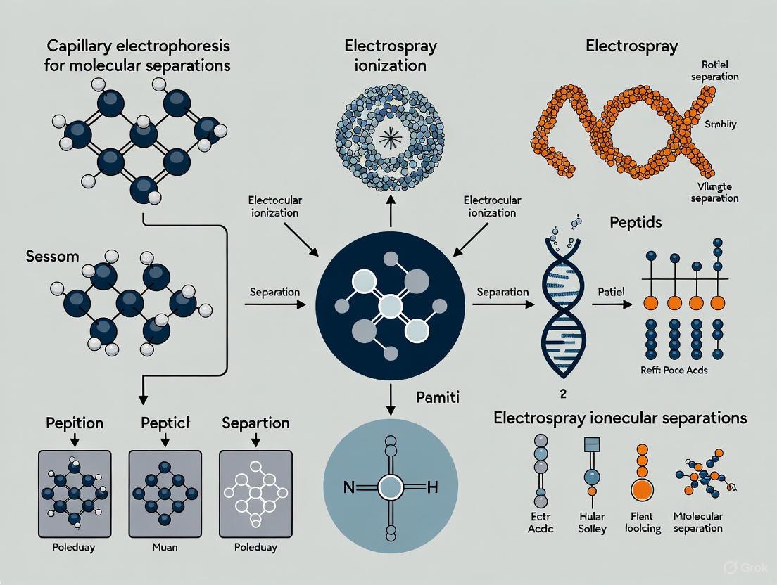
Understanding CE-ESI-MS: Core Principles and Evolving Interface Technology
The Fundamentals of Capillary Electrophoresis and Electrospray Ionization
Capillary Electrophoresis (CE) is a powerful liquid-phase separation technique that separates ions based on their electrophoretic mobility under the influence of an applied electric field [1] [2]. The fundamental principle involves the movement of charged analytes through a capillary tube filled with an electrolyte buffer, with separation achieved due to differences in their charge-to-size ratios [3]. The key components of a CE system include a fused silica capillary, electrodes (anode and cathode), buffer reservoirs, a high-voltage power supply, and a detector [1]. The technique is characterized by its high separation efficiency, small sample volume requirements, and rapid analysis times [4].
Electrospray Ionization (ESI) is a soft ionization technique that enables the transfer of ions from solution to the gas phase for mass spectrometric analysis [5] [6]. The ESI process involves three fundamental stages: the formation of a fine spray of charged droplets at the capillary tip, solvent evaporation from these droplets, and the subsequent release of gas-phase ions from the highly charged droplets [7] [8]. This technique is particularly valuable for the analysis of biomacromolecules because it overcomes their propensity to fragment when ionized and can produce multiply charged ions, effectively extending the mass range of the mass analyzer [5] [6].
The hyphenation of CE with ESI-MS creates a powerful analytical platform that combines high-efficiency separation with sensitive and selective detection, making it particularly suitable for the analysis of complex biological samples in separations research [9].
Fundamental Principles
Theory of Capillary Electrophoresis
The separation mechanism in CE is governed by the electrophoretic mobility (μₑₚ) of analytes, which represents the balance between the electric field force acting on a charged particle and the retarding frictional force in the separation medium [2]. The electrophoretic velocity (uₑₚ) of an analyte can be expressed as uₑₚ = μₑₚE, where E is the electric field strength [2]. The electrophoretic mobility is directly proportional to the charge of the ion and inversely proportional to the friction coefficient, which is related to the size and shape of the analyte [1].
An essential phenomenon in CE is electroosmotic flow (EOF), which originates from the charged capillary wall [2]. In a fused silica capillary, silanol groups (Si-OH) ionize to form silanoate ions (Si-O⁻) at pH values above approximately 3, creating a negatively charged surface [2]. This charged surface attracts cations from the buffer solution, forming an electrical double layer. When voltage is applied, these hydrated cations migrate toward the cathode, dragging the bulk solution with them and creating a flat, plug-like flow profile that contributes to the high separation efficiency of CE [3] [2].
The overall velocity of an analyte (u) is the vector sum of its electrophoretic velocity and the electroosmotic flow velocity (uₑₒf): u = (μₑₚ + μₑₒf)E, where μₑₒf is the electroosmotic mobility [2].
Theory of Electrospray Ionization
The ESI process begins when a high voltage (typically 2.5-6 kV) is applied to a liquid flowing through a capillary, dispersing it into a fine aerosol of charged droplets [5] [7]. As these droplets travel toward the mass spectrometer inlet, the solvent evaporates, reducing droplet size and increasing charge density [6]. When the Rayleigh limit is reached (where Coulombic repulsion equals surface tension), the droplets undergo Coulombic fission, disintegrating into smaller droplets [5] [6]. This process repeats until gas-phase ions are produced.
Two primary models explain the final stage of gas-phase ion formation:
Charge Residue Model (CRM): Proposes that successive droplet fission events eventually produce droplets containing a single analyte molecule. After solvent evaporation, the analyte retains some of the droplet's charge, becoming a gas-phase ion [6]. This model is thought to dominate for large biomolecules like proteins [6].
Ion Evaporation Model (IEM): Suggests that when droplets reach a sufficiently small size (∼10-20 nm), the electric field strength at the droplet surface becomes high enough to directly desorb solvated ions into the gas phase [6] [8]. This mechanism is considered more likely for smaller ionic species [6].
ESI is particularly renowned for its ability to generate multiply charged ions [5] [6]. For proteins and other macromolecules, this multiple charging effect effectively extends the mass range of mass analyzers, allowing the measurement of molecular weights into the kDa-MDa range with conventional mass analyzers [5].
Modes of Capillary Electrophoresis
CE encompasses several separation modes, each with distinct mechanisms and applications:
Table 1: Modes of Capillary Electrophoresis
| Mode | Acronym | Separation Mechanism | Primary Applications |
|---|---|---|---|
| Capillary Zone Electrophoresis | CZE | Differential electrophoretic mobility in free solution [1] | Separation of ions, proteins, peptides [1] |
| Capillary Gel Electrophoresis | CGE | Molecular sieving through gel or polymer network [1] | DNA fragment analysis, protein separations [1] [4] |
| Micellar Electrokinetic Chromatography | MEKC | Partitioning between aqueous phase and micellar pseudo-stationary phase [1] [3] | Separation of neutral and charged molecules [1] |
| Capillary Isoelectric Focusing | CIEF | Migration in a pH gradient until isoelectric point is reached [1] | Protein separations, determination of isoelectric points [1] |
| Capillary Electrochromatography | CEC | Combination of electrophoretic mobility and chromatographic partitioning [1] | Pharmaceutical analysis, complex mixture separations [1] |
| Capillary Isotachophoresis | CITP | Focusing between leading and terminating electrolytes [2] | Sample preconcentration, analysis of ionic compounds [1] |
Instrumentation and Experimental Protocols
CE-ESI-MS Interface Configuration
The hyphenation of CE with ESI-MS requires careful interface design to maintain electrical continuity for both systems while ensuring efficient ionization. A common approach uses a coaxial sheath-flow interface [9].
Protocol: CE-ESI-MS System Setup
Capillary Preparation: Use a fused silica capillary (typically 50-100 cm length, 50-100 μm internal diameter) [9] [2]. For UV detection, create a detection window by carefully removing a small section (2-5 mm) of the polyimide coating.
Interface Assembly:
- Secure the capillary outlet into a PEEK tee fitting using an FEP sleeve and fitting [9].
- Attach a stainless steel hypodermic tubing (e.g., 0.0083″ OD, 0.0065″ ID) to the opposite port to serve as the ESI needle [9].
- Connect sheath liquid delivery tubing (e.g., PEEK, 1/16″ OD, 0.005″ ID) to the remaining port using appropriate fittings [9].
- Polish the ESI needle tip with fine lapping paper to ensure a smooth finish [9].
Sheath Liquid Delivery: Use a syringe pump to deliver sheath liquid (typically 50/50 methanol/water with 0.1% formic acid) at a flow rate of 1-5 μL/min [9].
Electrical Connection: The electrical circuit for ESI is completed through the conductive sheath liquid, while the CE electrical circuit is maintained through the buffer at the capillary inlet [9].
Diagram 1: CE-ESI-MS interface setup with sheath-flow configuration
Typical CE-ESI-MS Analytical Protocol
Protocol: Metabolomic Analysis of Single Cells
This protocol is adapted from single neuron analysis methodologies [9].
Sample Preparation:
- Isolate individual cells manually using microdissection techniques under visual control [9].
- Transfer isolated cells into microvials containing extraction solvent (e.g., 50% methanol with 0.1% formic acid).
- Lyse cells by freeze-thaw cycling or sonication.
- Centrifuge at high speed (e.g., 10,000 × g) to remove particulate matter.
Capillary Electrophoresis Conditions:
- Capillary: 100 cm × 40 μm ID fused silica [9]
- Background electrolyte: 1% formic acid in water [9]
- Injection: Hydrodynamic, 15 cm height difference for 60 seconds (approximately 6 nL) [9]
- Separation voltage: 20 kV, ramped gradually over 30 seconds [9]
- Capillary temperature: Maintained constant using thermostatic control [1]
ESI-MS Parameters:
Data Acquisition and Analysis:
Performance Characteristics and Optimization
Key Performance Metrics
Table 2: Typical Performance Characteristics of CE-ESI-MS
| Parameter | Typical Range/Value | Influencing Factors |
|---|---|---|
| Separation Efficiency | 100,000 - 600,000 theoretical plates [9] | Field strength, capillary dimensions, sample stacking [2] |
| Detection Limits | Low nM range (e.g., <50 nM for neurotransmitters) [9] | Injection volume, ionization efficiency, matrix effects [9] |
| Migration Time RSD | <2% with internal standards [9] | Buffer composition, temperature control, capillary conditioning [1] |
| Analysis Time | 5-30 minutes [4] | Capillary length, applied voltage, separation mode [1] |
| Sample Volume | 1-10 nL injection volumes [9] [2] | Capillary dimensions, injection method and duration [2] |
Critical Optimization Parameters
Capillary Selection: Fused silica capillaries with internal diameters of 25-100 μm and outer diameters of 150-360 μm are standard [9] [2]. Smaller diameters provide better heat dissipation, enabling higher field strengths and improved efficiency [2].
Buffer Composition: Volatile buffers (e.g., formate, acetate, ammonium bicarbonate) are essential for ESI compatibility [2]. Typical concentrations range from 10-100 mM. Buffer pH significantly affects analyte charge state and EOF magnitude [1] [2].
Field Strength: Typical applied voltages range from 15-30 kV, with field strengths of 300-600 V/cm [9] [2]. Higher field strengths decrease analysis time but may generate excessive Joule heating if not properly controlled [2].
Sheath Liquid Composition: Methanol/water or acetonitrile/water mixtures with 0.1-1% acid or base (depending on ionization mode) are commonly used [9]. The modifier enhances droplet formation and desolvation while providing protons for ionization [5] [7].
Research Reagent Solutions
Table 3: Essential Materials for CE-ESI-MS Experiments
| Reagent/Material | Function/Purpose | Examples/Typical Specifications |
|---|---|---|
| Fused Silica Capillary | Separation channel | 25-100 μm ID, 150-360 μm OD, polyimide coated [9] [2] |
| Background Electrolyte | Separation medium, current carrier | Volatile buffers: formate, acetate, ammonium bicarbonate (10-100 mM) [2] |
| Sheath Liquid | ESI stability, electrical contact | Methanol/water or ACN/water (50/50 to 90/10) with 0.1% acid/base [9] |
| Ion-Pairing Reagents | Modify selectivity, enhance separation | HFBA, TFA for basic analytes; alkylamines for acidic analytes |
| Capillary Coatings | Control EOF, prevent adsorption | Dynamic coatings (e.g., ionic polymers); covalent coatings (e.g., polyacrylamide) |
| Mass Calibration Standards | Instrument calibration | ESI-tuning mix for low mass range; protein standards for high mass range |
| Internal Standards | Quantitation, signal normalization | Stable isotope-labeled analogs of target analytes |
Applications in Separations Research
CE-ESI-MS has found diverse applications across multiple scientific disciplines:
Pharmaceutical Analysis: CE-ESI-MS is employed for the analysis of small molecule drugs, chiral separations, impurity profiling, and biopharmaceutical characterization [1] [10]. The technique provides critical information on drug purity, structural identity, and degradation products during drug development [10].
Metabolomics and Single-Cell Analysis: The high separation efficiency and sensitivity of CE-ESI-MS make it ideal for metabolomic profiling, particularly in volume-limited samples [9]. Researchers have successfully detected over 100 compounds from the injection of only 0.1% of the total content from a single neuron, demonstrating the remarkable sensitivity of the technique for single-cell metabolomics [9].
Proteomics and Biomarker Discovery: CE-ESI-MS enables the analysis of complex protein and peptide mixtures [8]. The multiple charging phenomenon in ESI facilitates the analysis of high molecular weight proteins, while CE provides efficient separation of proteolytic digests for bottom-up proteomics [5] [8].
Forensic Science: CE-ESI-MS is utilized in forensic laboratories for the analysis of explosives, gunshot residues, ink characterization, and DNA sequencing [10]. The technique offers fast, sensitive, and reliable detection while requiring minimal sample amounts, which is crucial for preserving evidence integrity [10].
Clinical Diagnostics: Clinical applications include screening for inborn errors of metabolism, hemoglobin variant analysis, therapeutic drug monitoring, and quantification of disease biomarkers [7]. The technique's high specificity and sensitivity enable rapid and accurate diagnosis of various metabolic disorders [7].
Troubleshooting and Technical Considerations
Signal Instability: Common causes include electrical connection issues, bubble formation, or capillary surface irregularities. Ensure proper grounding, degas buffers, and check capillary window quality.
Poor Separation Efficiency: May result from sample overload, inappropriate buffer conditions, or capillary fouling. Optimize injection parameters, adjust buffer pH and concentration, and implement effective capillary rinsing protocols.
Low Sensitivity: Can be addressed through sample preconcentration techniques such as field-amplified sample stacking, solid-phase extraction, or using alternative detection configurations (e.g., bubble cell capillaries for UV detection) [2].
ESI-Related Issues: Spray instability can often be resolved by optimizing sheath liquid composition and flow rate, adjusting ESI needle position relative to the MS inlet, and ensuring proper gas flows for nebulization and desolvation [9] [7].
Within the field of modern analytical chemistry, Capillary Electrophoresis-Electrospray Ionization-Mass Spectrometry (CE-ESI-MS) stands as a powerful hyphenated technique that combines the high separation efficiency of CE with the exceptional detection capabilities of MS [11] [12]. The critical link between these two components—the ESI interface—serves as the bridge that transforms separated analytes in liquid phase into gas-phase ions suitable for mass analysis. Understanding the fundamental processes occurring within this interface is paramount for researchers and drug development professionals seeking to optimize their analytical methods. The ESI process, which seamlessly converts solution-phase ions into gas-phase ions, enables the sensitive and specific detection of a wide range of biomolecules, from small metabolites to intact proteins [13] [14]. This application note deconstructs the intricate journey from charged droplets to gas-phase ions, providing detailed protocols and practical insights for implementing robust CE-ESI-MS methodologies in separations research.
The Three-Step ESI Process: Fundamental Mechanisms
The electrospray ionization process operates through three distinct yet interconnected stages, each governed by specific physical and chemical principles that collectively enable the soft ionization of analytes for mass spectrometric detection.
Spray Formation
The initial stage begins with the formation of a Taylor cone and subsequent emission of a fine aerosol from the CE capillary terminus. When a high voltage (typically 3-5 kV) is applied to the liquid emerging from the capillary, electrostatic forces overcome surface tension, resulting in the formation of a conical meniscus [14] [11]. From the apex of this Taylor cone, a thin jet emerges that breaks up into a fine spray of charged droplets. The stability of this process is influenced by several parameters including the electric field strength, liquid flow rate, and the physical properties of the solvent such as surface tension and conductivity. In CE-ESI-MS applications, this stage is particularly critical as the electroosmotic flow from CE (typically 20-200 nL/min) is significantly lower than the optimal flow rates for conventional ESI (μL/min range), necessitating specialized interface designs to maintain spray stability [14].
Droplet Evolution
Following spray formation, the charged droplets undergo a series of transformations during their trajectory toward the mass spectrometer inlet. The primary mechanism of droplet shrinkage is solvent evaporation, often assisted by a co-axial flow of heated drying gas (typically nitrogen) that accelerates the desolvation process [14]. As droplets decrease in size, their charge density increases significantly until they reach the Rayleigh stability limit, at which point Coulombic repulsion overcomes surface tension. This leads to droplet fission through Coulomb explosion, producing smaller offspring droplets [11]. This iterative process of evaporation and fission continues until nanometer-sized droplets are formed, setting the stage for the final ion emission. The efficiency of this desolvation process directly impacts the sensitivity of the CE-ESI-MS analysis, as incomplete desolvation can result in increased chemical noise and reduced ion signal.
Production of Gas-Phase Ions
The final stage involves the liberation of gas-phase ions from the highly charged nanodroplets. Two predominant mechanisms have been proposed for this process:
Charge Residue Model (CRM): Postulates that continued solvent evaporation from nanodroplets containing a single analyte molecule eventually leads to the formation of a gas-phase ion as the solvent molecule is completely removed, leaving the charge on the analyte residue [11].
Ion Evaporation Model (IEM): Suggests that when the droplet radius becomes sufficiently small (typically <10 nm), the electric field at the droplet surface becomes strong enough to directly desorb analyte ions into the gas phase [11].
In practice, both mechanisms likely contribute to ion formation, with their relative importance depending on factors such as analyte size, charge state, and surface activity. The resulting gas-phase ions then enter the mass analyzer for separation based on their mass-to-charge ratio, completing the transition from solution-phase separation to gas-phase detection.
Table 1: Critical Parameters in the Three-Stage ESI Process
| Process Stage | Key Parameters | Optimal Conditions for CE-ESI-MS | Impact on Analysis |
|---|---|---|---|
| Spray Formation | Applied voltage (kV), Flow rate, Solvent properties | 3-5 kV, Nanoflow rates (<1 μL/min), Low conductivity buffers | Determines spray stability and initial droplet size distribution |
| Droplet Evolution | Nebulizing gas pressure, Drying gas temperature and flow rate, Solvent volatility | Minimal nebulization for CE flows, 150-300°C drying gas | Affects desolvation efficiency and background noise |
| Ion Production | Capillary temperature, Interface design, Solution chemistry | 200-350°C capillary, Clean interface geometry, Volatile buffers | Impacts ionization efficiency and detection sensitivity |
CE-ESI-MS Interface Designs and Technical Considerations
The coupling of CE with ESI-MS presents unique technical challenges, primarily due to the inherent flow rate mismatch between the two techniques and the necessity of maintaining electrical continuity for both CE separation and ESI processes. Three principal interface designs have been developed to address these challenges, each with distinct advantages and limitations for separations research.
Sheath-Flow Interface
The sheath-flow interface represents the most common and robust design for commercial CE-ESI-MS systems. This configuration employs a three-tube coaxial arrangement where the separation capillary is centered within a second tube that delivers a sheath liquid, which is itself surrounded by a third outer tube for nebulizing gas [12] [14]. The sheath liquid (typically a 1:1 mixture of water-methanol with 0.1% acetic acid or formic acid) serves multiple critical functions: it establishes electrical contact at the capillary terminus, provides sufficient volume for stable electrospray operation at conventional flow rates (1-10 μL/min), and can enhance ionization efficiency through adjustment of pH and solvent composition [12]. While this design offers excellent reliability and broad compatibility with various separation electrolytes, the dilution of analyte with sheath liquid can reduce sensitivity, particularly for low-abundance species. Additionally, the sheath gas flow can induce a parabolic flow profile within the separation capillary, potentially compromising separation efficiency [12].
Sheathless Interface
Sheathless interfaces address the sensitivity limitations of sheath-flow designs by establishing direct electrical contact with the separation effluent without dilution. This is typically achieved through use of conductively coated capillaries or porous emitters where voltage is applied directly to the separation buffer [12] [14]. The most advanced designs incorporate porous tips created through chemical etching, allowing electrical contact via a conductive liquid that surrounds but does not mix with the separation effluent [12]. This approach enables operation at nanoelectrospray regimes (flow rates <1000 nL/min), providing significantly enhanced sensitivity due to the absence of sample dilution and higher ionization efficiency at low flow rates. However, sheathless interfaces historically suffered from challenges with mechanical robustness and reproducibility, though recent developments in porous emitter technology have substantially addressed these limitations [12]. The transient capillary isotachophoresis (CITP)/capillary zone electrophoresis (CZE) coupling with sheathless interfaces has demonstrated remarkable sensitivity improvements for trace analysis, making this design particularly valuable for applications with limited sample availability [12].
Liquid Junction Interface
The liquid junction interface employs a different approach, using a stainless steel tee-piece to create a connection between the separation capillary and the ESI emitter. A narrow gap (typically 10-50 μm) is maintained between the two capillaries, across which electrical contact is established via a makeup solution [14]. This design offers ease of operation and capillary replacement compared to other interfaces. However, the potential for analyte dilution and band broadening at the junction can degrade separation efficiency and detection sensitivity. A modified approach, the pressurized liquid junction interface, operates at lower flow rates (<200 nL/min) and applies additional pressure to prevent defocusing of the CE effluent, thereby improving resolution while minimizing dilution effects [12].
Table 2: Comparison of CE-ESI-MS Interface Technologies
| Interface Type | Flow Rate Regime | Sensitivity | Robustness | Best Applications |
|---|---|---|---|---|
| Sheath-Flow | Conventional ESI (1-10 μL/min) | Moderate (sample dilution) | High | Routine analysis, method development |
| Sheathless | NanoESI (<1 μL/min) | High (no dilution) | Moderate (improved with new designs) | Trace analysis, sample-limited applications |
| Liquid Junction | Variable | Moderate to Low | Moderate | Research applications, flexible setups |
Experimental Protocols for CE-ESI-MS Analysis
Protocol 1: Sheath-Flow CE-ESI-MS for Metabolite Profiling
This protocol describes a robust method for comprehensive metabolite analysis using sheath-flow CE-ESI-MS, suitable for both targeted and untargeted metabolomic studies in biological samples [13].
Materials and Reagents:
- Fused silica capillary (50-100 μm i.d., 80-100 cm length)
- Sheath liquid: 1:1 (v/v) water-methanol with 0.1% formic acid
- Background electrolyte: 1 M formic acid (pH ~1.8) for cationic analysis or 50 mM ammonium acetate (pH ~7.5) for anionic analysis
- CE-ESI-MS interface: Coaxial sheath-flow nebulizer
Procedure:
- Capillary Conditioning: Flush new capillaries sequentially with 1 M NaOH (30 min), water (15 min), and background electrolyte (30 min) using approximately 1 bar pressure.
- Sample Preparation: Precipitate proteins from biological samples (e.g., plasma, urine) with cold methanol (2:1 ratio), centrifuge at 14,000 × g for 15 min, and evaporate supernatant under nitrogen. Reconstitute in water or background electrolyte for analysis.
- Sample Injection: Perform hydrodynamic injection using 50 mbar for 5-30 s (approximately 1-10 nL injected volume).
- CE Separation: Apply separation voltage of 20-30 kV with the inlet as anode for positive mode MS detection. Maintain capillary temperature at 20-25°C.
- ESI-MS Parameters:
- Sheath liquid flow rate: 1-5 μL/min
- Nebulizing gas pressure: 3-7 psi
- Drying gas temperature: 150-300°C
- Drying gas flow rate: 5-10 L/min
- Capillary voltage: 3-4 kV (positive mode)
- Scan range: m/z 50-1000
- System Optimization: Continuously infuse reference compound (e.g., purine, HP-921) during method development to optimize interface parameters and mass calibration.
Troubleshooting Tips:
- If spray stability is problematic, adjust sheath liquid composition to include more organic modifier (up to 80% methanol) or modify gas flow rates.
- For sensitivity issues, ensure electrical connection is stable at the liquid junction and consider using a smaller i.d. capillary to reduce flow rate.
- If migration time reproducibility is suboptimal, implement conditioning steps between runs and monitor capillary pH.
Protocol 2: Sheathless CE-ESI-MS for Sensitive Protein Characterization
This protocol leverages the enhanced sensitivity of sheathless interfaces for the analysis of proteins and peptides, particularly valuable for low-abundance species or sample-limited applications.
Materials and Reagents:
- Porous tip emitter capillary (30 μm i.d., 100 cm length)
- Background electrolyte: 10% acetic acid (v/v) in water for positive mode analysis
- Conductive liquid: 5% acetic acid in 50% methanol
- Make-up solution: 0.1% formic acid in water
Procedure:
- Emitter Preparation: For chemically etched porous tips, ensure the conductive liquid reservoir is filled and electrical contact is established prior to separation.
- Capillary Conditioning: Flush with 0.1 M HCl (10 min), water (10 min), and background electrolyte (20 min) at 1000 mbar.
- Sample Injection: For limited samples, use electrokinetic injection (5-10 kV for 10-30 s) to enhance loading of charged species. For quantitative applications, hydrodynamic injection (0.5-5 psi for 10-30 s) provides better reproducibility.
- CE Separation: Apply voltage of 20-25 kV with the inlet as anode. Note that current may be lower than with sheath-flow interfaces due to smaller capillary dimensions.
- ESI-MS Parameters:
- No sheath liquid or gas flows required
- Emitter voltage: 1.5-2.5 kV
- Capillary temperature: 200-250°C
- Ion transfer tube temperature: 150-200°C
- Data-dependent acquisition for peptide identification
- System Performance Verification: Analyze standard protein digest (e.g., bovine serum albumin) to verify separation efficiency and sensitivity. Expect detection limits in the low attomole range for peptides.
Critical Considerations:
- Sheathless interfaces are more susceptible to capillary clogging; always filter samples and buffers through 0.2 μm filters.
- Electrical connection stability is crucial; monitor current throughout separation to identify potential interruptions.
- For CITP/CZE-MS applications, prepare leading and terminating electrolytes appropriate for your analyte set to achieve on-line focusing [12].
The Scientist's Toolkit: Essential Research Reagent Solutions
Successful implementation of CE-ESI-MS methodologies requires careful selection of reagents and materials optimized for both separation and ionization processes. The following table details essential components for establishing robust CE-ESI-MS analyses.
Table 3: Essential Research Reagents and Materials for CE-ESI-MS
| Item | Specification | Function/Purpose | Application Notes |
|---|---|---|---|
| Separation Capillary | Fused silica, 25-100 μm i.d., various lengths | Containment for electrophoretic separation | Smaller diameters provide better heat dissipation; coated capillaries control EOF |
| Background Electrolyte | Volatile buffers (formate, acetate, ammonium salts) | Medium for electrophoretic separation | Must be volatile for ESI compatibility; concentration affects current and Joule heating |
| Sheath Liquid | Water-methanol/isopropanol with 0.1-1% acid | Establish electrical contact, stabilize spray | Isopropanol reduces electrical current; acid modifiers enhance positive ion formation |
| ESI Nebulizer Gas | Nitrogen (99.99% purity) | Aid droplet formation and desolvation | Higher flows improve spray stability; lower flows preserve separation efficiency |
| Capillary Conditioning Solutions | 1 M NaOH, 0.1 M HCl, methanol, water | Maintain capillary surface properties | Regular conditioning essential for migration time reproducibility |
| Ionization Assistants | Formic acid, acetic acid, ammonium hydroxide | Enhance analyte ionization efficiency | Concentration optimization critical for sensitivity; typically 0.1-1% in sheath liquid |
| Mass Calibration Standards | Sodium formate clusters, proprietary mixes | Calibrate mass accuracy across range | Infuse separately or add to sheath liquid for continuous calibration |
Applications in Separations Research
The unique capabilities of CE-ESI-MS have enabled its application across diverse areas of separations research, particularly where high-resolution separation of complex mixtures is required.
In metabolomics, CE-ESI-MS has emerged as a particularly powerful platform due to its exceptional efficiency in separating highly polar and charged metabolites that often challenge reverse-phase LC methods [13]. The technique enables comprehensive profiling of cationic, anionic, and zwitterionic metabolites without complicated sample handling, making it invaluable for functional genomics research [13]. When coupled with sophisticated data analysis approaches, CE-ESI-MS facilitates both targeted quantification of specific metabolic pathways and untargeted discovery of novel biomarkers in various biological systems.
For pharmaceutical analysis, CE-ESI-MS provides robust solutions for drug impurity profiling, chiral separations, and biopharmaceutical characterization [14]. The technique's ability to separate isomeric compounds—such as distinguishing between glucose-6-phosphate and fructose-6-phosphate which have identical chemical formulas and molecular weights but different electrophoretic mobilities—makes it particularly valuable for pharmaceutical applications where isomeric impurities can have significant therapeutic consequences [14].
In the expanding field of single-cell analysis, CE-ESI-MS enables the comprehensive profiling of metabolites, neurotransmitters, and proteins from individual cells, leveraging its minimal sample volume requirements [12] [3]. Recent applications have demonstrated successful analysis of neurons, frog embryos, and various mammalian cells, providing unprecedented insights into cellular heterogeneity without the averaging effects associated with bulk analyses [12]. The development of surface sampling CE-MS (SS-CE-MS) further extends these capabilities by enabling direct analysis of tissue sections without extensive sample preparation [12].
Workflow and Signaling Pathway Visualization
The following diagrams illustrate key processes and relationships in CE-ESI-MS analysis, providing visual representations of the complex mechanisms involved.
CE-ESI-MS Analytical Workflow
CE-ESI-MS Analytical Workflow
ESI Mechanism Pathway
ESI Mechanism Pathway
Capillary Electrophoresis coupled to Electrospray Ionization Mass Spectrometry (CE-ESI-MS) represents a powerful analytical technique that combines the high separation efficiency of CE with the exceptional detection capabilities of MS. The successful hyphenation of these two platforms hinges critically on the interface design, which must simultaneously maintain electrical continuity for both the CE separation and ESI process while effectively transferring analytes from the capillary to the mass spectrometer. The development of robust interfacing methodologies has been an active area of research since the first CE-MS interface was introduced in 1987, with two predominant configurations emerging: sheath-flow and sheathless interfaces. This application note provides a comprehensive technical comparison of these interface designs, complete with experimental protocols and performance data to guide researchers in selecting and implementing the appropriate configuration for their separations research, particularly in the context of biotherapeutic characterization and drug development.
Interface Design Fundamentals
Core Principles of CE-ESI-MS Coupling
The fundamental challenge in coupling CE to ESI-MS lies in establishing and maintaining stable electrical contacts for both systems. CE requires a complete electrical circuit along the separation capillary to sustain the electroosmotic flow and electrophoretic migration of analytes, while ESI requires a high voltage at the spray tip to generate a stable Taylor cone and produce charged droplets. The CE current is typically more than a hundred times larger than the electrospray current, necessitating careful design of electrical circuits to protect the sensitive MS instrumentation. Additionally, the minimal flow rates in CE (on the order of nanoliters per minute) create challenges for establishing stable electrospray, and many CE background electrolytes demonstrate poor compatibility with MS detection due to their non-volatile nature [15].
Classification of Interface Designs
Two primary interface architectures have been developed to address these coupling challenges:
- Sheath-Flow Interfaces: Utilize a coaxial flow of sheath liquid to establish electrical contact and facilitate spray stability
- Sheathless Interfaces: Establish electrical contact directly through the capillary or via alternative conductive pathways without dilution from sheath liquids
Table 1: Fundamental Characteristics of CE-ESI-MS Interface Types
| Characteristic | Sheath-Flow Interface | Sheathless Interface |
|---|---|---|
| Electrical Contact | Established via sheath liquid surrounding capillary terminus | Established directly through conductive capillary wall or porous junction |
| Flow Rate Regime | Microliter per minute (1-10 μL/min) | Nanoliter per minute (<100 nL/min) |
| Dilution Effects | Significant (sample dilution 1:10 to 1:100) | Minimal to none |
| Fabrication Complexity | Moderate | High |
| Mechanical Robustness | High | Variable (depending on design) |
| Commercial Availability | Widely available | Limited options |
Sheath-Flow Interface Designs
Operational Principles and Configurations
Sheath-flow interfaces, first introduced by Smith and coworkers in 1988, represent the most common and commercially available approach for CE-ESI-MS coupling. In this design, the separation capillary is positioned coaxially within a metal tube (typically stainless steel) that serves both as the CE outlet electrode and the ESI emitter. A sheath liquid, delivered by a syringe pump at flow rates of 1-10 μL/min, flows between the separation capillary and the metal tube, establishing electrical contact with the CE effluent and facilitating stable electrospray formation [12] [16]. Most commercial systems employ a three-tube coaxial design that includes an additional outer tube for nebulizing gas, which improves spray stability and solvent evaporation, though this can sometimes introduce parabolic flow profiles that reduce separation efficiency [12].
The composition of the sheath liquid can be optimized independently of the CE background electrolyte, typically consisting of a mixture of water and organic solvent (e.g., methanol or isopropanol) with 0.1% acetic acid or formic acid to promote ionization in positive ion mode [12] [16]. This flexibility allows researchers to optimize MS detection conditions without compromising CE separation efficiency.
Experimental Protocol: Sheath-Flow CE-ESI-MS
Materials and Reagents:
- Fused silica capillary (40-75 μm i.d., 360 μm o.d.)
- Sheath-flow interface (commercial or custom-built)
- Syringe pump for sheath liquid delivery
- ESI-MS compatible background electrolyte (e.g., 1% formic acid)
- Sheath liquid (80:20 methanol:water with 0.1% formic acid for positive ion mode)
- HPLC-grade water and solvents
Procedure:
Capillary Preparation:
- If using bare fused silica capillary, condition with 1 M NaOH for 30 minutes, followed by background electrolyte for 20 minutes
- For coated capillaries, follow manufacturer's conditioning recommendations
- Cut the outlet end cleanly and ensure proper alignment within the interface
Interface Assembly:
- Position the separation capillary within the sheath tube, leaving a minimal gap (<1 mm) between the capillary tip and the ESI emitter end
- Connect the sheath liquid delivery line to the interface body
- Secure the assembly to ensure no movement during analysis
System Operation:
- Set sheath liquid flow rate to 0.5-1.0 μL/min using the syringe pump [16]
- Apply nebulizing gas pressure (if available) at 2-5 psi
- Set ESI voltage typically between 2-4 kV, depending on the specific interface and MS instrument
- Begin CE separation with typical voltages of 15-30 kV
- Monitor current stability throughout the separation
Optimization Guidelines:
- Adjust sheath liquid composition to maximize ionization efficiency for target analytes
- Minimize sheath flow rate to reduce dilution while maintaining stable spray
- Ensure the CE instrument platform is at the same height as the MS sprayer to prevent siphoning effects [15]
Diagram 1: Sheath-Flow CE-ESI-MS Interface Workflow
Performance Characteristics and Applications
Sheath-flow interfaces provide robust performance for a wide range of applications. The detection limits for model peptides and protein mixtures typically fall in the attomole-to-low femtomole range, with reproducibility of peak areas and migration times generally below 5% RSD when properly optimized [16] [9]. The dilution factor caused by the sheath liquid (typically 10-100 fold) represents the primary limitation in sensitivity, though this can be partially mitigated by using lower sheath flow rates and appropriate sample stacking techniques during injection [15].
Sheath-flow interfaces have been successfully applied to the analysis of pharmaceutical compounds in biological matrices, characterization of biotherapeutics including monoclonal antibodies, metabolomic profiling, and single-cell analysis. The ability to use a wide range of capillary types (including various coated capillaries) and background electrolytes makes this interface design particularly versatile for method development [15].
Sheathless Interface Designs
Operational Principles and Configurations
Sheathless interfaces were developed to address the primary limitation of sheath-flow designs: sample dilution. By eliminating the sheath liquid, these interfaces potentially offer significant improvements in sensitivity, making them particularly valuable for applications with limited sample amounts or low analyte concentrations. Sheathless designs establish electrical contact through alternative pathways, with several configurations reported in the literature:
- Porous Tip Emitters: The capillary outlet is etched with hydrofluoric acid to create a porous segment, through which ions can diffuse to establish electrical contact with an external electrode [17]
- Conductive Coatings: The capillary is coated with a conductive material (e.g., metal or conductive polymer) that serves as the electrode for both CE and ESI [12]
- Laser-Ablated Openings: A CO2 laser is used to create an opening in the capillary wall near the outlet, allowing a minimal volume of makeup fluid to be added for electrical contact [17]
- Split-Flow Techniques: Part of the CE buffer is diverted through an opening near the capillary outlet, establishing electrical contact through a metal tube surrounding the opening [18]
These designs typically operate at nanoliter per minute flow rates, taking full advantage of the concentration-sensitive nature of ESI and potentially offering up to 100-fold improvement in sensitivity compared to sheath-flow interfaces [18] [17].
Experimental Protocol: Sheathless CE-ESI-MS with Porous Tip
Materials and Reagents:
- Fused silica capillary (30-50 μm i.d.)
- Hydrofluoric acid (48%) for etching
- Conductive coating solution (if using coated capillaries)
- Conductive liquid (e.g., background electrolyte) for external electrode
- ESI-MS compatible background electrolyte
Procedure:
Capillary Etching (Porous Tip Design):
- Carefully immerse the capillary outlet (~2 cm) in hydrofluoric acid (48%) for 30-90 minutes
- Monitor the etching process to achieve a wall thickness of 5-15 μm [17]
- Thoroughly rinse with HPLC-grade water to remove all acid residues
Interface Assembly:
- Position the etched capillary segment within an electrode chamber filled with conductive liquid
- Ensure the porous section is fully immersed in conductive liquid
- Align the capillary tip with the MS inlet, maintaining minimal distance (1-3 mm)
System Operation:
- Apply the ESI voltage (1-2 kV) to the external electrode chamber
- Begin CE separation at 15-25 kV
- Monitor current stability - fluctuations may indicate improper electrical contact
Alternative Laser-Ablated Interface:
- Use a CO2 laser engraver to create an opening approximately 5 mm from the capillary tip [17]
- Position the opening within a silicon tube reservoir containing electrode and minimal makeup fluid
- Apply ESI voltage directly to the electrode in the reservoir
Diagram 2: Sheathless CE-ESI-MS Interface Workflow
Performance Characteristics and Applications
Sheathless interfaces provide exceptional sensitivity, with detection limits frequently in the low nanomolar range (≤50 nM) for various metabolites and signaling molecules, corresponding to attomole-level mass detection limits [9]. The absence of sheath liquid dilution and operation at optimal nanoESI flow rates significantly enhances ionization efficiency, making these interfaces particularly valuable for challenging applications such as single-cell metabolomics, proteomic analysis of limited samples, and detection of low-abundance biomarkers [17] [9].
The primary challenges with sheathless interfaces include lower mechanical robustness, more complex fabrication procedures, and potential instability when using certain background electrolytes or capillary coatings. However, recent advances such as the CO2 laser-ablated interface have demonstrated improved reproducibility and stability while maintaining the sensitivity advantages of sheathless designs [17].
Comparative Performance Analysis
Table 2: Quantitative Comparison of Sheath-Flow vs. Sheathless Interface Performance
| Performance Parameter | Sheath-Flow Interface | Sheathless Interface | References |
|---|---|---|---|
| Typical Detection Limits | Attomole-to-low femtomole | Low attomole-to-zeptomole | [16] [9] |
| Concentration Detection Limits | 10-100 nM | 0.5-5 nM | [9] [19] |
| Migration Time RSD | < 2-5% | 3-7% | [12] [17] |
| Peak Area RSD | < 5-10% | 5-15% | [12] [17] |
| Analysis Time | 10-30 minutes | 10-30 minutes | [15] [9] |
| Suitable Flow Rates | 1-10 μL/min (sheath) 20-100 nL/min (CE) | < 100 nL/min (total) | [12] [17] |
| Minimum Sample Volume | 5-20 μL | < 1 μL | [15] [9] |
The selection between sheath-flow and sheathless interfaces involves careful consideration of application requirements and practical constraints. Sheath-flow interfaces offer superior robustness and ease of use, making them appropriate for routine analyses and method development where maximum sensitivity is not critical. The independent optimization of separation and ionization conditions provides additional flexibility for challenging separations. In contrast, sheathless interfaces demonstrate clear advantages for applications requiring maximum sensitivity, such as single-cell analysis, detection of low-abundance metabolites, and characterization of limited samples in proteomics and metabolomics [15] [9].
The Scientist's Toolkit: Essential Research Reagents and Materials
Table 3: Key Research Reagent Solutions for CE-ESI-MS
| Reagent/Material | Function/Purpose | Example Formulations | Considerations |
|---|---|---|---|
| Background Electrolyte (BGE) | Separation medium for CE | 1% formic acid, Ammonium acetate (10-50 mM, pH 3-9), Acetic acid (0.5-2%) | Must be volatile and MS-compatible; Choice affects separation and ionization |
| Sheath Liquid | Establishes electrical contact and facilitates spray stability in sheath-flow interfaces | 80:20 methanol:water + 0.1% formic acid (positive mode), 50:50 methanol:water + 1 mM ammonium acetate (negative mode) | Lower flow rates reduce dilution; Composition affects ionization efficiency |
| Capillary Conditioning Solutions | Prepares capillary surface for analysis | 1 M NaOH for fused silica, Manufacturer-specific protocols for coated capillaries | Proper conditioning essential for reproducibility |
| Coated Capillaries | Reduces analyte adsorption to capillary wall | PVA (polyvinyl alcohol), LPA (linear polyacrylamide), PEI (polyethylenimine) | Minimizes protein/peptide adsorption; Stability varies with pH and conditions |
| Calibration Standards | System performance verification | Peptide standards, Metabolite mixtures, Drug compounds | Should represent analyte chemistry; Used for system suitability testing |
Troubleshooting and Optimization Guidelines
Successful implementation of CE-ESI-MS methodologies requires attention to several critical parameters that impact system stability and data quality:
Electrical Stability Issues:
- Problem: Fluctuating currents or arcing
- Solution: Verify all electrical connections, ensure proper grounding, check for conductive liquid leaks in sheathless designs, reduce voltage differences between interfaces
Spray Instability:
- Problem: Unstable total ion current or sporadic signal dropouts
- Solution: For sheath-flow interfaces, optimize sheath liquid composition and flow rate; check capillary alignment; verify nebulizing gas pressure (if applicable). For sheathless interfaces, ensure proper electrical contact at porous junction or conductive coating; check for clogged emitter tips
Poor Sensitivity:
- Problem: Higher than expected detection limits
- Solution: For sheath-flow interfaces, minimize sheath liquid flow rate and consider composition adjustments. For sheathless interfaces, verify electrical contact quality and emitter condition. For both interfaces, implement appropriate sample stacking techniques and check capillary surface for adsorptive losses
Irreproducible Migration Times:
- Problem: High RSD for analyte migration times
- Solution: Ensure consistent capillary conditioning between runs, maintain stable temperature, verify buffer composition consistency, check for siphoning effects due to height mismatches between CE and MS platforms [15]
The field of CE-ESI-MS continues to evolve with emerging technologies such as microfluidic devices (e.g., ZipChip) that integrate sample preparation with separation and interfacing, promising improved reproducibility and ease of use while maintaining the sensitivity advantages of sheathless designs [17]. Additionally, hybrid approaches that combine elements of both sheath-flow and sheathless designs offer promising directions for future development, potentially delivering both robustness and sensitivity for challenging analytical applications in pharmaceutical development and biomedical research.
Capillary Electrophoresis-Electrospray Ionization-Mass Spectrometry (CE-ESI-MS) has undergone a remarkable transformation since its initial development in 1987. This analytical technique has evolved from a specialized method with significant interfacing challenges to a robust, mainstream platform capable of analyzing complex biological mixtures with exceptional sensitivity and resolution. Its journey from niche to essential tool represents one of the significant success stories in analytical chemistry, particularly in the fields of proteomics, metabolomics, and pharmaceutical analysis. This application note details the technical evolution, current methodologies, and practical protocols that have enabled CE-ESI-MS to become an indispensable technique for researchers and drug development professionals.
The original interface between capillary zone electrophoresis and mass spectrometry was developed in 1987 by Richard D. Smith and coworkers at Pacific Northwest National Laboratory [12]. This pioneering work demonstrated the fundamental compatibility of these techniques but also revealed significant challenges, particularly regarding stable electrospray conditions and interface design. Early researchers noted that commercially available interfaces were "hardly able to produce stable electrospray conditions over an extended period of time," primarily due to "insufficient positioning of the CE capillary inside the ESI stainless steel tip" [20]. These technical hurdles initially limited widespread adoption and confined the technique to specialized laboratories.
The evolution of CE-ESI-MS can be characterized by three interconnected developments: interface refinement, application diversification, and sensitivity enhancement. As interface technology improved, applications expanded from basic metal speciation studies to complex biological analyses including proteomics, metabolomics, and single-cell analysis [20] [21] [12]. Each application breakthrough further drove technical innovations, creating a virtuous cycle of improvement that has positioned CE-ESI-MS as a powerful tool for modern analytical challenges.
Technical Foundations: Interface Evolution and Separation Principles
Interface Design Developments
The coupling of CE with ESI-MS presented unique challenges due to fundamental differences in operational principles and flow rate requirements. Three primary interface designs have emerged, each with distinct advantages and limitations:
Table 1: Comparison of CE-ESI-MS Interface Types
| Interface Type | Flow Characteristics | Sensitivity | Robustness | Typical Applications |
|---|---|---|---|---|
| Sheathless | Low flow rates (nl/min) | High (minimal dilution) | Lower | High-sensitivity proteomics |
| Sheath-flow | Mixed flows (μl/min sheath) | Moderate (sample dilution) | High | Routine analysis |
| Liquid junction | Makeup flow added | Moderate | Moderate | Specialized applications |
Sheath-flow interfaces represent the most common commercial design, utilizing a three-tube coaxial system where separation liquid mixes with sheath liquid flowing coaxially in metal capillary tubing [12]. An additional outer tube provides sheath gas to improve electrospray stability and solvent evaporation. While this design offers reliability and wide electrolyte selection, the typical sheath flow rates of 1-10 μL/min can reduce sensitivity through sample dilution. Commonly used sheath liquids include 1:1 mixtures of water-methanol or water-isopropanol with 0.1% acetic or formic acid [12].
Sheathless interfaces provide direct coupling of the CE capillary to an electrospray ionization source, often using capillaries coated with conductive metal [12]. This design offers superior sensitivity due to minimal background and low flow rates, but early versions suffered from mechanical fragility. Recent innovations include porous ESI emitters created through chemical etching, which provide more robust interfacing while maintaining sensitivity advantages [12].
Liquid junction interfaces employ a stainless steel tee to mix separation electrolyte with makeup liquid, maintaining a narrow gap between the CE capillary and ESI needle [12]. While operationally simpler, this approach can reduce sensitivity and potentially degrade separation through mixing effects. The pressurized liquid junction variant applies pressure to the makeup liquid reservoir, reducing dilution through lower flow rates (<200 nl/min) and preventing defocusing of the CE effluent [12].
Fundamental Separation Principles
CE-ESI-MS combines the exceptional separation efficiency of capillary electrophoresis with the molecular identification capabilities of mass spectrometry. In CE, molecules are separated based on their electrophoretic mobility under the influence of a high electric field, which is dependent on their charge, size, and the buffer viscosity [12]. This separation occurs within a fused silica capillary typically filled with a background electrolyte.
The electrospray ionization process transfers separated analytes from the liquid phase to gas-phase ions suitable for mass analysis. In ESI, the application of a high voltage to the liquid emerging from the capillary tip creates a Taylor cone, which disperses into a fine spray of charged droplets [22] [23]. Through solvent evaporation and droplet fission, these charged droplets produce gas-phase ions that retain the solution-phase charge state information of the analytes.
The following diagram illustrates the core workflow of a CE-ESI-MS analysis:
Application Expansion: From Metal Speciation to Single-Cell Analysis
Early Applications: Metal Speciation and Small Molecules
The initial applications of CE-ESI-MS focused on metal speciation, leveraging its ability to differentiate between various metal-containing compounds. Early research demonstrated that the technique could analyze free metal ions [Cu(II)], metal ion-containing complexes [CuEDTA, (CH)₃SbCl₂], and covalent organometallic compounds (selenocystamine, selenomethionine) [20]. These studies revealed important fundamental insights about the technique, including that "inorganic species (i.e., metal ions) alter their composition when being electrosprayed" as "parts of the weakly complexing ligands will be exchanged by solvent molecules" [20]. This work established that ESI-MS was "best suited for the speciation of covalent organometallic compounds" whose structure remains intact during ionization [20].
Critical technical challenges emerged during these early applications, particularly regarding buffer compatibility. Researchers found that "non-volatile electrolytes affect the ESI process dramatically," necessitating method adaptations such as switching from alkaline buffer systems (Na₂CO₃-NaOH) to volatile acidic background electrolytes (2% acetic acid) [20]. These methodological refinements were essential for achieving stable electrospray conditions and acceptable detection limits, which were calculated as 1-6 mg/L for organic Se species in selenium speciation studies [20].
Biomolecular Analysis: Proteomics and Glycomics
As interface technology matured, CE-ESI-MS applications expanded significantly into biomolecular analysis. The technique proved particularly valuable for proteomics, where it became a component of both top-down and bottom-up approaches [12]. Similarly, glycoform analysis benefited from CE-ESI-MS capabilities, with researchers developing "reliable off- and on-line CE-based methods for the analysis of glycoforms with ESI MS/MS" [24]. These applications leveraged the technique's ability to separate complex carbohydrate mixtures with "fast, sensitive, and efficient separations for the accurate identification of a large variety of glycoform mixture types" [24].
Table 2: Evolution of CE-ESI-MS Applications and Performance Metrics
| Application Area | Key Analytes | Typical Performance | Technical Requirements |
|---|---|---|---|
| Metal Speciation | Organometallic compounds, metal complexes | Detection limits: 1-6 mg/L (Se species) | Volatile buffers, optimized capillary position |
| Proteomics | Peptides, proteins | High separation efficiency, mass accuracy <1 ppm | Sheathless interfaces, high-resolution MS |
| Metabolomics | Small polar metabolites | ~70 molecular features per single cell | High sensitivity, minimal sample volume |
| Glycomics | Glycopeptides, glycosaminoglycans | Efficient separation of heterogeneous mixtures | MS/MS capability, specialized buffers |
| Pharmaceutical Analysis | Drugs, metabolites | High reproducibility, quantitative accuracy | Robust interfaces, validated methods |
Cutting-Edge Applications: Single-Cell Metabolomics
Perhaps the most remarkable demonstration of CE-ESI-MS evolution is its application to single-cell metabolomics, which leverages the technique's minimal sample volume requirements and high sensitivity. Researchers have utilized "microprobe single-cell CE-ESI-MS" to analyze metabolism directly in live embryos, quantifying "~70 molecular features, including 52 identified metabolites" from individual cells in Xenopus laevis embryos [21]. This approach enabled the discovery of metabolic differences between dorsal and ventral cells at the 8-cell stage, with statistical analysis revealing that "asparagine, glycine betaine, and a yet-unidentified molecule were statistically significantly enriched in the D1L cell compared to V1L" [21].
The experimental workflow for single-cell analysis demonstrates the sophisticated capabilities of modern CE-ESI-MS:
This application exemplifies how CE-ESI-MS has evolved to address extraordinarily challenging analytical problems, requiring minimal volume (several nanoliters) while providing both separation efficiency and molecular mass information in a single analysis [12].
Detailed Experimental Protocols
Protocol 1: Basic CE-ESI-MS Setup for Metabolite Analysis
Background Electrolyte Preparation:
- Prepare 1% (v/v) formic acid in LC-MS grade water
- Filter through 0.2 μm membrane and degas by sonication for 10 minutes
- Store at 4°C for up to one week
Sheath Solution Preparation:
- Combine 50% (v/v) methanol and 0.1% (v/v) formic acid in LC-MS grade water
- Deliver at 1 μL/min using a syringe pump or equivalent system
Capillary Conditioning:
- Flush new capillaries sequentially with:
- Methanol (10 column volumes)
- LC-MS grade water (10 column volumes)
- Background electrolyte (10 column volumes)
- Between runs, flush with background electrolyte for 2 minutes
Instrument Parameters:
- Separation voltage: 22.5 kV (positive polarity)
- Capillary dimensions: 100 cm × 40 μm ID (bare fused silica)
- ESI voltage: -1.7 kV (positive ion mode)
- Mass spectrometer survey scan rate: 2 Hz
- Mass calibration: <1 ppm accuracy using standard calibration solution
Sample Injection:
- Utilize hydrodynamic injection at 0.5 psi for 10 seconds
- Alternative: electrokinetic injection at 5 kV for 10 seconds (note potential mobility bias)
Protocol 2: Single-Cell Metabolomics Using Microprobe CE-ESI-MS
Cell Sampling:
- Prepare borosilicate micropipettes (0.75/1 mm inner/outer diameter) pulled to ~20 μm diameter
- Mount micropipette on three-axis micromanipulator
- Position tip into identified cell and apply negative pressure to withdraw portion of cell content
- Expel aspirate into 4 μL of chilled metabolite extraction solution (40% acetonitrile, 40% methanol, 4°C)
- Vortex-mix for ~1 minute and centrifuge at 8,000 × g for 5 minutes at 4°C
- Store supernatant at -80°C until analysis
Metabolite Analysis:
- Inject 10 nL of cell extract using hydrodynamic injection
- Separate molecules electrophoretically by applying 22.5 kV across a 100 cm capillary
- Employ sheath-flow CE-ESI interface with 50% methanol containing 0.1% formic acid at 1 μL/min as sheath solution
- Apply -1,700 V spray voltage to the mass spectrometer's orifice plate
- Detect generated ions in a quadrupole orthogonal-acceleration time-of-flight mass spectrometer
- For metabolite identification, perform MS/MS experiments using 12-18 eV energy for collision-induced dissociation with nitrogen as collision gas
Quality Control:
- Validate CE-ESI-MS performance daily using 300 amol of standard metabolite mixture
- Monitor migration time stability (RSD < 2%)
- Verify mass accuracy (<5 ppm) and sensitivity (signal-to-noise > 10 for 100 nM standard)
Essential Research Reagents and Materials
Table 3: Essential Research Reagent Solutions for CE-ESI-MS
| Reagent/Material | Specifications | Function | Application Notes |
|---|---|---|---|
| Background Electrolyte | 1% (v/v) formic acid in LC-MS grade water | Separation medium | Volatile, ESI-compatible; alternative: acetic acid |
| Sheath Liquid | 50% methanol, 0.1% formic acid | ESI stability and charge transfer | Isopropanol alternative for less polar analytes |
| Metabolite Extraction Solution | 40% ACN, 40% MeOH in water | Protein precipitation, metabolite stabilization | Maintain at 4°C during extraction |
| Capillary | Fused silica, 40/105 μm ID/OD, 100 cm length | Separation channel | Various lengths and coatings available |
| Calibration Solution | ESI-L Low Concentration Tuning Mix (or equivalent) | Mass accuracy calibration | Required for <1 ppm mass accuracy |
| Separation Capillary | Bare fused silica, 40/105 μm inner/outer diameter | Electrophoretic separation | 100 cm length typical |
Methodological Considerations and Troubleshooting
Critical Technical Parameters
Successful CE-ESI-MS analysis requires careful optimization of several key parameters. The capillary position relative to the ESI tip significantly impacts electrospray stability, with research indicating that "the optimum position for stable electrospray conditions was set to 0.4-0.7 mm outside the ESI tip" [20]. This precise positioning ensures consistent ionization efficiency and signal stability.
Buffer selection represents another critical consideration. Early researchers recognized that "non-volatile electrolytes affect the ESI process dramatically," necessitating the use of volatile buffers such as formic acid, acetic acid, or ammonium acetate/formate [20]. These volatile additives facilitate the electrospray process without accumulating in the interface or causing signal suppression.
Troubleshooting Common Issues
Spray Instability:
- Verify capillary position (0.4-0.7 mm outside ESI tip)
- Check sheath liquid composition and flow rate
- Inspect for capillary tip damage or contamination
- Ensure proper electrical connections
Poor Separation Efficiency:
- Confirm capillary conditioning protocol
- Verify background electrolyte pH and composition
- Check for sample overloading or matrix effects
- Inspect for bubble formation in capillary
Low Sensitivity:
- Optimize injection technique and volume
- Evaluate sheath liquid composition and flow rate
- Check mass spectrometer calibration and detector performance
- Consider sample preconcentration techniques
The evolution of CE-ESI-MS from a niche technique to a mainstream analytical tool represents a significant achievement in separation science. Through continuous refinement of interface technology, expansion of application areas, and enhancement of sensitivity, CE-ESI-MS has established itself as an indispensable platform for researchers and drug development professionals. Its unique capabilities for analyzing minute sample volumes with high separation efficiency and molecular specificity make it particularly valuable for contemporary challenges in proteomics, metabolomics, and pharmaceutical analysis. As the technique continues to evolve, further applications and methodological refinements will undoubtedly solidify its position as a cornerstone of modern analytical chemistry.
CE-ESI-MS in Action: Method Development and Cutting-Edge Applications
Capillary Electrophoresis (CE) is a family of powerful separation techniques where ions migrate through narrow capillaries under a strong electric field, offering high efficiency and resolution for analytical challenges in pharmaceutical and biotechnological development [25]. The fundamental principle of CE relies on the differential electrophoretic mobility of analytes, which is dependent on the molecule's charge, viscosity, and atomic radius [26]. The actual velocity of ion migration is directly proportional to the applied electric field, enabling rapid and high-resolution separations [26]. While all CE techniques share this basic instrumental setup, different modes have been developed to address specific separation challenges, particularly for complex biological and pharmaceutical samples [27] [25].
The choice of CE mode is critical for successful method development and depends heavily on the physicochemical properties of the target analytes. This application note provides a detailed comparison of four principal CE modes—Capillary Zone Electrophoresis (CZE), Micellar Electrokinetic Capillary Chromatography (MEKC), Capillary Isoelectric Focusing (CIEF), and Capillary Gel Electrophoresis (CGE)—with specific guidance on their application for different analyte classes. Within the broader context of capillary electrophoresis electrospray ionization for separations research, understanding these complementary techniques enables researchers to select the optimal approach for their specific analytical needs, from small ions to complex macromolecular assemblies [28].
Comparative Analysis of CE Separation Modes
Table 1: Characteristics and Applications of Major CE Separation Modes
| CE Mode | Separation Mechanism | Primary Applications | Key Advantages | Critical Parameters |
|---|---|---|---|---|
| CZE | Charge-to-mass ratio in free solution [25] [29] | Small ions, drugs, metabolites, peptides [25] [30] | Simple setup, high efficiency for charged species [25] | Buffer pH and composition, applied voltage, capillary type [25] |
| MEKC | Partitioning between aqueous phase and micellar pseudostationary phase [27] | Neutral molecules, chiral separations, small pharmaceuticals [27] [25] | Extends CE to neutral analytes [27] | Surfactant type/CMC, buffer pH, organic modifiers [27] |
| CIEF | Isoelectric point (pI) in pH gradient [25] [29] | Proteins, peptides, antibody variants [25] [29] | High resolution for protein separations, pI determination [25] | Ampholyte selection, focusing voltage/time, chemical mobilization [25] |
| CGE | Molecular size sieving through gel/polymer network [25] [29] | DNA, RNA, proteins, SDS-protein complexes [25] [29] | Size-based separation, excellent for macromolecules [25] | Gel matrix composition, pore size, applied field strength [25] |
Table 2: Optimal CE Mode Selection Guide Based on Analyte Properties
| Analyte Class | Recommended CE Mode | Specific Applications | Typical Separation Conditions |
|---|---|---|---|
| Small Ionic Molecules | CZE | Pharmaceutical compounds, inorganic ions, amino acids [25] [29] | Alkaline borate or phosphate buffer (pH 8-9), 15-30 kV [25] |
| Neutral Molecules | MEKC | Pharmaceutical substances, hydrophobic compounds [27] [25] | SDS micelles (10-50 mM above CMC), pH 7-9 buffer [27] |
| Proteins (by pI) | CIEF | Protein isoforms, monoclonal antibodies, biomarker discovery [25] [28] | 1-2% ampholytes (pH 3-10 gradient), slow chemical mobilization [25] |
| Macromolecules (by size) | CGE | DNA sequencing, protein oligomers, SDS-protein complexes [25] [29] | Cross-linked polyacrylamide or linear polymer matrix [25] |
| Chiral Compounds | MEKC with chiral selector | Enantiomeric separations of pharmaceuticals [31] | Chiral surfactants or cyclodextrin additives in buffer [31] |
Detailed Operational Protocols
Protocol for Capillary Zone Electrophoresis (CZE)
Principle: CZE separates analytes based on their intrinsic electrophoretic mobility in a free solution, governed by their charge-to-mass ratio under an applied electric field [25] [29]. This is the simplest and most widely used CE form, ideal for separating charged species including small ions, drugs, and metabolites [25].
Procedure:
- Capillary Preparation: Install a bare fused-silica capillary (40-100 cm length, 50 μm internal diameter) into the CE instrument. Condition new capillaries with 1 M NaOH for 30 minutes, followed by deionized water for 10 minutes, and finally with running buffer for 20 minutes [25] [26].
- Background Electrolyte (BGE) Preparation: Prepare appropriate buffer system based on analyte properties. Common systems include:
- Sample Preparation: Dissolve samples in BGE or deionized water at concentrations of 0.1-1.0 mg/mL. Filter through 0.2 μm membrane if necessary [25].
- Instrumental Parameters: Set separation voltage to 15-30 kV (positive polarity for anionic analytes, negative for cationic). Maintain constant capillary temperature (20-25°C). Use hydrodynamic injection (0.5-5.0 psi for 1-10 seconds) or electrokinetic injection (5-10 kV for 5-20 seconds) [25] [26].
- Detection: Typically UV detection at appropriate wavelength (200-214 nm for proteins, higher wavelengths for pharmaceuticals). For CE-ESI-MS coupling, use volatile buffers (ammonium acetate/formate) [28].
- Capillary Regeneration: Between runs, flush capillary with BGE for 1-2 minutes. For storage, flush with deionized water and air dry [25].
Applications in Pharmaceutical Analysis: CZE has been successfully applied for analysis of highly polar charged analytes, determination of drugs and excipients in pharmaceutical preparations, and analysis of pharmaceuticals in biological fluids [31] [30]. Its simplicity and compatibility with various detection methods make it particularly valuable for quality control applications.
Protocol for Micellar Electrokinetic Capillary Chromatography (MEKC)
Principle: MEKC extends CE to neutral analytes by incorporating surfactant micelles (above critical micellar concentration) as a pseudostationary phase. Separation occurs through differential partitioning of analytes between the mobile aqueous phase and the hydrophobic micellar phase [27]. The anionic SDS micelles are electrostatically attracted toward the anode, but are carried toward the cathode by the stronger electroosmotic flow, creating a migration window between the EOF marker (t0) and the micellar marker (tmc) [27].
Procedure:
- Micellar Solution Preparation: Prepare BGE containing surfactant above its critical micellar concentration (CMC). For SDS, use concentration of 20-100 mM in appropriate buffer (e.g., 10-50 mM borate or phosphate, pH 7-9). Other surfactants include cationic (CTAB), bile salts (sodium cholate), or chiral selectors [27].
- Capillary Conditioning: Use bare fused-silica capillary (50-75 cm effective length, 50 μm ID). Condition with 0.1 M NaOH (10 minutes), water (10 minutes), and micellar BGE (15 minutes) [27].
- Sample Preparation: Dissolve samples in water or dilute BGE. For very hydrophobic compounds, add small amounts of organic modifiers (methanol, acetonitrile) to improve solubility [27].
- Separation Conditions: Apply voltage of 15-30 kV with positive polarity. Capillary temperature 25-40°C. Injection parameters similar to CZE [27].
- Migration Markers: Use methanol (EOF marker, t0) and Sudan III (micellar marker, tmc) to define the migration window and calculate retention factors [27].
- Method Optimization: Adjust surfactant concentration, buffer pH, and organic modifier content (acetonitrile, methanol) to improve resolution of neutral compounds [27].
Applications: MEKC is particularly valuable for pharmaceutical analysis as most pharmaceutical substances are neutral from an electrophoretic viewpoint [27]. It has been applied to separations of amino acids, chiral pharmaceuticals, and other small-molecule drugs through the addition of chiral selectors like cyclodextrins to the running buffer [31]. The technique provides an important complement to HPLC for neutral compound separations.
CE Mode Selection Decision Tree
Protocol for Capillary Gel Electrophoresis (CGE)
Principle: CGE separates molecules based on their size using a gel or polymer network-filled capillary, functioning as a molecular sieve. This technique is particularly effective for macromolecules such as DNA, RNA, and proteins [25] [29]. The gel matrix reduces solute diffusion and provides a size-based separation mechanism similar to traditional slab gel electrophoresis but with enhanced resolution and quantification capabilities [30].
Procedure:
- Gel Matrix Preparation: Prepare appropriate separation matrix:
- For DNA sequencing: Cross-linked polyacrylamide (3-10%) in TBE buffer with urea
- For protein analysis: Linear polymer matrix (dextran, polyethylene oxide) or SDS-containing buffers for SDS-protein complexes [25]
- Capillary Filling: Carefully fill capillary with gel matrix using pressure injection (avoid introducing air bubbles). For commercially coated capillaries, follow manufacturer's instructions [25].
- Sample Preparation: Denature samples if necessary (heat at 95°C for DNA, or in SDS-containing buffer for proteins). For protein analysis, incubate with SDS and reducing agents [29].
- Electrophoresis Conditions: Apply constant voltage (5-15 kV) or constant current. Higher fields may cause heating effects in viscous gels. Maintain precise temperature control (20-25°C) [25].
- Detection: Typically UV detection at 214 nm (proteins) or 260 nm (nucleic acids). For fluorescent detection, pre-label samples with appropriate dyes [29].
- Capillary Life Management: Monitor performance degradation. Replace capillary when resolution decreases significantly. Some commercial capillaries allow regeneration according to manufacturer protocols [25].
Applications: CGE is the standard method for DNA sequencing and fragment analysis in genetic analysis, forensics, and quality control of biopharmaceuticals [29]. It provides excellent resolution for proteins and their aggregates, making it invaluable for biopharmaceutical characterization [25].
Protocol for Capillary Isoelectric Focusing (CIEF)
Principle: CIEF separates amphoteric molecules such as proteins and peptides according to their isoelectric point (pI) within a stable pH gradient created using carrier ampholytes [25] [28]. Analytes migrate through the gradient until they reach the pH position where their net charge is zero (pI), resulting in extremely high resolution for protein separations [25].
Procedure:
- Ampholyte Solution Preparation: Prepare sample mixture containing 1-2% (v/v) carrier ampholytes (appropriate pH range), 0.1-1.0% methylcellulose (to suppress EOF), and protein sample (10-100 μg/mL) in deionized water [25].
- Capillary Selection: Use coated capillaries (e.g., fluorocarbon, polyacrylamide) with suppressed EOF, or dynamically coated capillaries with additives [25].
- Capillary Filling: Fill entire capillary with sample-ampholyte mixture using pressure injection (20-50 psi for 1-2 minutes) [25].
- Focusing Step: Apply constant voltage (5-15 kV) for 5-15 minutes until current drops to a stable minimum (typically 1-10% of initial current), indicating completed focusing [25].
- Mobilization Step: After focusing, mobilize separated zones past detector using:
- Chemical mobilization: Replace cathode or anode reservoir with salt solution (e.g., 80 mM NaCl)
- Pressure mobilization: Apply low pressure (0.5-1.0 psi) while maintaining voltage
- Gravity mobilization: Elevate one capillary end [25].
- Detection: UV detection at 280 nm for proteins. Whole-column imaging detection is also available in some instruments [25].
Applications: CIEF is extensively used for characterization of protein isoforms, monoclonal antibodies, and biomarker discovery in proteomics research [28]. It provides exceptional resolution for separating proteoforms that differ by minor charge variations, such as deamidated or glycosylated species, making it invaluable for biopharmaceutical analysis and quality control [28].
Essential Research Reagent Solutions
Table 3: Key Reagents and Materials for CE Method Development
| Reagent/Material | Function/Purpose | Application Notes |
|---|---|---|
| Fused-Silica Capillaries | Standard separation channel providing strong electroosmotic flow [26] | Internal diameters: 25-100 μm; lengths: 30-100 cm; pH range: 2-10 [25] |
| Coated Capillaries | Suppress EOF and analyte adsorption to capillary wall [25] | PEG, polyacrylamide, or polydopamine coatings for improved reproducibility [25] |
| SDS (Sodium Dodecyl Sulfate) | Anionic surfactant for MEKC; forms micellar pseudostationary phase [27] | CMC: 8.1 mM; use at 20-100 mM concentrations above CMC [27] |
| Carrier Ampholytes | Create pH gradient for CIEF separations [25] | Available in various pH ranges (narrow/broad); use at 1-2% concentration [25] |
| Polymer Matrices | Form sieving network for size-based separations in CGE [25] | Linear polymers (cellulose derivatives) or cross-linked gels (polyacrylamide) [25] |
| Cyclodextrins | Chiral selectors for enantiomeric separations [31] | Native or derivatized (hydroxypropyl, sulfated); add to BGE in MEKC or CZE [31] |
| Volatile Buffers | CE-MS compatibility; ammonium acetate/formate [28] | Concentration 10-50 mM; pH adjustment with acetic or formic acid [28] |
CE-ESI-MS Integration in Separations Research
The integration of CE with electrospray ionization mass spectrometry (CE-ESI-MS) represents a powerful advancement in separation science, particularly for proteomics and pharmaceutical research [28]. This hyphenated technique combines the high separation efficiency of CE with the structural identification capabilities of MS, creating an invaluable tool for complex mixture analysis [30] [28].
Two primary interface designs dominate CE-ESI-MS applications: the sheath-liquid interface and the sheathless interface [28]. The sheath-liquid interface employs a coaxial liquid to establish electrical contact and is valued for its robustness and versatility, though it can cause some analyte dilution [28]. In proteomics research, the electrokinetically pumped low sheath-flow nanospray interface (commercially available as EMASS-II ion source) has demonstrated excellent performance for both bottom-up and top-down analyses [28]. Conversely, sheathless interfaces provide superior sensitivity by applying voltage directly to the background electrolyte, achieving ultra-low flow rates (10-20 nL/min) that enhance ionization efficiency [28]. The porous tip sheathless interface has been particularly successful for highly sensitive top-down analysis and characterization of protein complexes under native conditions [28].
For CE-ESI-MS applications, method optimization must consider several critical parameters. Buffer selection is restricted to volatile compounds such as ammonium acetate or ammonium formate to prevent MS source contamination and signal suppression [28]. Capillary coatings that suppress electroosmotic flow often improve resolution and sensitivity for protein analyses [25] [28]. The low sample loading capacity of CE can be mitigated by employing preconcentration techniques such as field-amplified sample stacking, transient isotachophoresis, or SPE-CE integration [25]. These considerations make CE-ESI-MS particularly valuable for proteomics research, biopharmaceutical characterization, and the analysis of complex biological samples where its high separation efficiency complements the detection power of modern mass spectrometers [28].
CE-ESI-MS Integration Workflow
The selection of an appropriate CE separation mode is fundamental to successful method development in pharmaceutical and biotechnology research. Each major mode—CZE, MEKC, CIEF, and CGE—offers distinct advantages for specific analyte classes, from small molecules to complex macromolecular assemblies. CZE provides exceptional efficiency for charged species, while MEKC extends CE applicability to neutral compounds through micellar partitioning. CIEF offers unparalleled resolution for protein charge variants, and CGE excels at size-based separations of biomacromolecules. When coupled with ESI-MS detection through robust interface technologies, CE becomes an exceptionally powerful platform for comprehensive sample characterization. By understanding the principles, applications, and practical protocols for each CE mode detailed in this application note, researchers can make informed decisions to select the optimal separation strategy for their specific analytical challenges.
Sample Preparation and Preconcentration Strategies for Complex Matrices
The effectiveness of Capillary Electrophoresis-Electrospray Ionization-Mass Spectrometry (CE-ESI-MS) is fundamentally dependent on the preparatory steps taken before analysis. Complex biological and environmental matrices contain numerous interfering substances that can compromise separation efficiency, suppress ionization, and ultimately reduce analytical sensitivity and accuracy. Sample preparation and preconcentration are therefore not merely preliminary steps but are integral components of a successful CE-ESI-MS analytical workflow. This document provides detailed application notes and protocols for preparing complex samples, framed within separations research, to enable reliable and high-sensitivity analyses for researchers, scientists, and drug development professionals.
The primary challenges when dealing with complex matrices such as plasma, tissue homogenates, or wastewater include the vast dynamic range of analyte concentrations, the presence of ionization-suppressing compounds like salts and detergents, and the potential for capillary fouling [32] [33] [34]. Efficient sample preparation strategies are designed to mitigate these issues by removing interferents, enriching target analytes, and presenting the sample in a format compatible with both CE separation and ESI-MS detection.
Core Principles and Challenges
The Impact of Matrix Effects on CE-ESI-MS
Matrix effects represent a significant challenge in analytical separations, particularly when using sensitive detection techniques like ESI-MS. These effects occur when co-eluting compounds from the sample matrix alter the ionization efficiency of the target analytes, leading to either ion suppression or enhancement [34]. In CE-ESI-MS, this can manifest as reduced signal intensity, poor reproducibility, and inaccurate quantification.
The multifaceted nature of matrix effects is influenced by factors including the physicochemical properties of the target analyte, the sample preparation protocol, and the overall sample composition [34]. Non-volatile buffers, such as sodium phosphate, can suppress ionization and clog the electrospray source, while detergents like Tween, Triton-X, and SDS are particularly detrimental to ESI performance due to their tendency to form abundant polymeric ions and cause persistent contamination [35] [36]. Similarly, high salt concentrations can disrupt electrophoretic separation and interfere with stable electrospray formation.
Preconcentration Necessity in Trace Analysis
The inherently low sample loading capacity of CE, coupled with the demand for detecting trace-level analytes in complex mixtures, makes preconcentration a critical step. Preconcentration serves to lower detection limits by increasing the amount of analyte introduced into the system without proportionally increasing the matrix components. This is especially vital in applications like biomarker discovery from plasma, where proteins of interest may exist at concentrations twelve orders of magnitude lower than abundant proteins like albumin [33], and in environmental analysis, where pharmaceuticals are present in wastewater at nanogram per liter levels [37].
Preconcentration and Clean-up Techniques
Several techniques have been established to effectively clean and concentrate samples for CE-ESI-MS analysis. The choice of technique depends on the sample matrix, the nature of the target analytes, and the required level of purification.
Table 1: Comparison of Common Preconcentration and Clean-up Techniques
| Technique | Principle | Best Suited For | Advantages | Limitations |
|---|---|---|---|---|
| Solid-Phase Extraction (SPE) | Analyte adsorption onto a solid sorbent, washing, and subsequent elution. | Removal of salts and general matrix clean-up; peptide pre-concentration [32]. | High selectivity and clean-up efficiency; can be automated. | Can be time-consuming and require organic solvents [32]. |
| Filter-Aided Sample Prep (FASP) | Size-based separation using membrane filters. | Surfactant removal (e.g., SDS) from protein digests [32]. | Effective for detergent removal. | Can be laborious; potential for analyte loss [32]. |
| Solid-Phase Microextraction (SPME) | Equilibrium-based extraction using a coated fiber. | Passive sampling of organic pollutants from water [37]. | Solvent-less; combines sampling and concentration. | Limited fiber capacity; possible fiber degradation. |
| Immunodepletion | Affinity-based removal of high-abundance proteins. | Pre-fractionation of plasma/serum to widen dynamic range [33]. | Highly specific removal of top 6-20 abundant proteins. | Costly; does not remove low-abundance protein interferences. |
For field sampling or continuous monitoring, passive sampling techniques offer a powerful alternative. Devices like the Polar Organic Chemical Integrative Sampler (POCIS) are based on the adsorption of pollutants onto a receiving phase and are designed for the accumulation of polar, hydrophilic compounds in water [37]. These samplers integrate sampling and preconcentration into a single step, simplifying the analytical procedure and providing a time-weighted average concentration of analytes, which can be more representative than discrete grab samples [37].
Detailed Experimental Protocols
Protocol 1: Bottom-Up Proteomics from Plasma/Serum
This protocol describes a comprehensive intact-protein-based workflow for quantitative plasma proteome analysis, combining immunodepletion, protein labeling, and multidimensional separation prior to LC-ESI-MS/MS, as applied in cancer biomarker discovery studies [33]. The following workflow visualizes the key stages of this protocol:
Materials
- Samples: Human plasma or serum (e.g., 600 µL per condition) [33].
- Immunodepletion: Hu-6 or Ms-3 HC column (Agilent) [33].
- Labeling: iTRAQ 4-plex or acrylamide isotope tags (light/heavy) [33].
- Buffers: Exchange Buffer (6.6 M Urea, 0.5 M TEAB, 0.2% OG, pH 8.3); Labeling Buffer (0.5 M TEAB, 0.2% OG, pH 8.3) [33].
- Reduction/Alkylation: TCEP (50 mM); Iodoacetamide (200 mM, fresh) [33].
- Chromatography: 2D-HPLC system with anion-exchange (Poros HQ) and reversed-phase (Poros R2) columns [33].
- Digestion: Sequence-grade modified trypsin [33].
Step-by-Step Procedure
- High-Abundance Protein Depletion: Process plasma samples through the immunodepletion column according to manufacturer's instructions to remove top abundant proteins (e.g., albumin, IgG) [33].
- Protein Concentration and Buffer Exchange: Concentrate the immunodepleted sample and perform buffer exchange into the Exchange Buffer using a centrifugal device (e.g., Centricon YM-3) [33].
- Reduction and Alkylation: Reduce disulfide bonds with TCEP (final conc. ~5 mM) and alkylate cysteine residues with iodoacetamide (final conc. ~10-20 mM) in the dark [33].
- Isotopic Labeling: Label proteins from different conditions with different isobaric (iTRAQ) or isotopic (acrylamide) tags according to the kit or standard protocol [38].
- Sample Pooling and 2D-HPLC: Combine the labeled samples. First, separate proteins by charge via anion-exchange chromatography. In the second dimension, separate each AEX fraction by hydrophobicity using reversed-phase chromatography. Collect all fractions [33].
- Digestion: Lyophilize the protein fractions. Reconstitute in a digestion-compatible buffer (e.g., 0.25 M urea, 50 mM ammonium bicarbonate) and digest with trypsin (e.g., 1:20-50 enzyme-to-protein ratio) overnight at 37°C [33].
- CE-ESI-MS/MS Analysis: The resulting peptide digests are now ready for analysis. Desalt if necessary using a StageTip or micro-SPE column before CE or nano-LC-ESI-MS/MS analysis for peptide identification and quantification [39] [33].
Protocol 2: Metabolite Analysis from Cells and Tissues
This protocol focuses on the extraction and analysis of water-soluble metabolites from cultured cells or tissue samples for targeted metabolomics, a process requiring rapid quenching of metabolism to preserve the in vivo state [38].
Materials
- Quenching Solvent: Cold (-20°C to -48°C) methanol:water (e.g., 80:20, v/v) or acetonitrile:methanol:water for specific analytes [38].
- Extraction Buffers: Methanol:water or acetonitrile:methanol:water mixtures, kept ice-cold [38].
- Equipment: Liquid nitrogen for tissue snap-freezing; a cooled centrifuge; a cryogenic mill or homogenizer for tissues [38].
Step-by-Step Procedure
- Rapid Metabolic Quenching:
- For Adherent Cells: Rapidly aspirate culture medium and immediately add cold quenching solvent (-20°C or colder) [38].
- For Suspension Cells: Transfer culture directly into a larger volume of cold quenching solvent, or pass through a filter and wash with cold buffer before immersing in solvent [38].
- For Tissues: Snap-freeze the tissue sample in situ using liquid nitrogen-cooled tongs or clamps. Grind the frozen tissue to a fine powder under liquid nitrogen using a cryogenic mill [38].
- Metabolite Extraction: Add an appropriate volume of ice-cold extraction solvent (e.g., 40:40:20 acetonitrile:methanol:water or 80:20 methanol:water) to the quenched cells or tissue powder. Vortex vigorously [38].
- Phase Separation: Centrifuge the extract at high speed (e.g., >14,000 g) at 4°C for 10-15 minutes to pellet precipitated protein and cellular debris [38].
- Supernatant Collection and Storage: Carefully transfer the clarified supernatant (containing the metabolites) to a new vial. Store extracts at 4°C and analyze within 24 hours to minimize degradation, especially for labile species like nucleotides and folates [38].
- Pre-CE/LC-MS Clean-up (Optional): For CE-ESI-MS, a desalting step using a micro-SPE cartridge (e.g., C18 or a mixed-mode phase) may be necessary to ensure compatibility. The extract can be directly injected for LC-ESI-MS if the chromatographic method is robust [38].
Protocol 3: Desalting and Surfactant Removal for Peptide/Protein Digests
This is a critical clean-up protocol for bottom-up proteomics where salts, urea, and detergents like SDS from digestion buffers must be removed prior to ESI-MS [32] [35].
Materials
- SPE Micro-Columns: C18 StageTips or commercial micro-columns/cartridges [32].
- Buffers and Solvents: Equilibration/Wash buffer (0.1% Formic Acid, 2-5% Acetonitrile in water); Elution buffer (0.1% Formic Acid, 30-80% Acetonitrile) [36].
Step-by-Step Procedure
- Conditioning: Activate the C18 sorbent by passing 100% acetonitrile through the micro-column, followed by equilibration with wash buffer (0.1% FA, 2% ACN) [32].
- Sample Loading: Acidify the peptide digest to pH < 3 with formic acid to protonate carboxylic groups. Load the sample onto the conditioned column slowly [32].
- Washing: Pass at least 3-5 column volumes of wash buffer (0.1% FA, 2% ACN) through the column to remove salts, residual urea, and other polar contaminants [32].
- Elution: Elute the purified peptides with a small volume (e.g., 20-50 µL) of elution buffer (0.1% FA, 30-80% ACN) into a low-volume autosampler vial. The sample is now ready for CE-ESI-MS or LC-ESI-MS analysis [32] [36].
The Scientist's Toolkit: Essential Reagent Solutions
Successful sample preparation requires careful selection of reagents and consumables. The following table details key materials and their functions.
Table 2: Key Research Reagent Solutions for Sample Preparation
| Item/Category | Specific Examples | Function & Application Notes |
|---|---|---|
| Volatile Buffers | Ammonium acetate, Ammonium bicarbonate, Formic acid, Acetic acid, TFA [36]. | Compatible with ESI-MS; do not cause ion suppression or source contamination. Use at ≤ 1% concentration [36]. |
| Protein Precipitation Reagents | Cold Methanol, Acetonitrile, Acetonitrile:MeOH:Water mixtures [38]. | To precipitate and remove proteins from biological fluids (serum, plasma) or cell extracts prior to metabolite/protein analysis. |
| Digestion & Denaturants | Urea, Trypsin, OG (octyl-β-d-glucopyranoside) [33]. | Urea denatures proteins for digestion; trypsin cleaves proteins into peptides; OG is a MS-compatible detergent. |
| Alkylating & Reducing Agents | Iodoacetamide, DTT, TCEP [33]. | To reduce and alkylate cysteine disulfide bonds in proteins, preventing reformation and aiding digestion. |
| SPE Sorbents | C18, C8, Mixed-Mode, Graphitized Carbon [32]. | For desalting and preconcentration of peptides, metabolites, and other small molecules. Choice depends on analyte hydrophobicity. |
| Immunoaffinity Columns | Hu-6/Ms-3 HC Columns (Agilent) [33]. | For specific removal of the 6 or 3 most abundant proteins from human or mouse serum/plasma to expand dynamic range. |
| Proper Vials | Glass 2 mL vials (PTFE/silicone septa), Polypropylene limited volume vials [36]. | To prevent leaching of contaminants and septa coring. Do not reuse vials or use limited volume inserts to avoid instrument damage [36]. |
Concluding Remarks
Robust sample preparation and preconcentration are non-negotiable prerequisites for achieving high-quality, reproducible data with CE-ESI-MS. The strategies outlined here—from immunodepletion and SPE to rapid metabolic quenching and passive sampling—provide a framework for tackling the analytical challenges posed by complex matrices. While the protocols are detailed, method optimization for specific applications is always encouraged. An integrated approach that thoughtfully combines sample preparation, analytical separation, and detection remains the most effective strategy for unlocking the full potential of CE-ESI-MS in separations research.
Capillary electrophoresis coupled with electrospray ionization mass spectrometry (CE-ESI-MS) has emerged as a powerful analytical technique in pharmaceutical and biomedical analysis. This hyphenated technique combines the high separation efficiency of CE with the exceptional detection selectivity and sensitivity of MS [40] [30]. The fundamental principle of CE involves the separation of ions based on their electrophoretic mobility under the influence of an applied electric field, while ESI-MS provides sensitive detection and structural elucidation capabilities [41] [30]. The versatility of CE-ESI-MS allows for its application across various aspects of pharmaceutical analysis, including drug impurity profiling, therapeutic drug monitoring, metabolomic studies, and quality control of active pharmaceutical ingredients [41] [40]. This article explores the practical applications and detailed methodologies of CE-ESI-MS within the context of separations research, providing researchers with structured protocols and analytical frameworks for implementation in drug development and biomedical analysis.
CE-ESI-MS Fundamentals and Instrumentation
Principles of Separation and Detection
In capillary electrophoresis, separation occurs within a narrow-bore fused silica capillary filled with background electrolyte (BGE). When a high voltage is applied, analytes migrate based on their electrophoretic mobility, which is determined by their charge-to-size ratio [41] [30]. The electroosmotic flow (EOF) generated by the ionization of silanol groups on the capillary wall contributes to the overall movement of analytes toward the detection end [41]. The coupling with ESI-MS enables sensitive detection by converting separated analytes into gas-phase ions through an electrospray process, making them amenable to mass analysis [9] [40]. This combination provides orthogonality in separation mechanisms, where CE separates based on charge and size, while MS adds a dimension of mass-based identification [40].
Interface Design and Technical Considerations
The hyphenation of CE with ESI-MS requires specialized interfaces to overcome technical challenges related to electrical circuit completion and stable electrospray formation. Two primary interface designs are commonly employed: sheath-flow interfaces and sheathless interfaces [40]. The sheath-flow interface uses a coaxial liquid to establish electrical contact and stabilize the electrospray, providing robustness for routine analysis [9]. In contrast, sheathless interfaces offer improved sensitivity by eliminating sample dilution but require more specialized fabrication [40]. Key considerations for successful CE-ESI-MS analysis include the use of volatile buffers compatible with MS detection (e.g., ammonium acetate, ammonium formate), appropriate capillary coatings to suppress analyte adsorption, and optimization of ESI parameters for efficient ionization [40].
Figure 1: CE-ESI-MS Workflow. The analytical process encompasses sample preparation, CE separation, interface ionization, MS detection, and data analysis stages.
Applications in Pharmaceutical Analysis
Drug Impurity Profiling
Impurity profiling is crucial in pharmaceutical development to ensure drug safety and quality. CE-ESI-MS enables the detection and identification of impurities at trace levels (0.1% w/w or lower) due to its high separation efficiency and selective detection [41] [42]. The technique has been successfully applied to various drug compounds, including carbachol, lidocaine, and proguanil, facilitating the separation and identification of their potential impurities [42]. Compared to liquid chromatography (LC), CE offers complementary separation selectivity, particularly for polar and charged molecules, making it valuable for comprehensive impurity assessment [41] [40].
Table 1: CE-ESI-MS Analytical Performance in Drug Impurity Profiling
| Drug Compound | Impurity Types | LOD (ng/mL) | Background Electrolyte | Separation Mode |
|---|---|---|---|---|
| Carbachol | Quaternary ammonium compounds | 100 | 100 mM acetic acid (pH 4.5) | CZE |
| Lidocaine | 2,6-dimethylaniline | 500 | 100 mM acetic acid (pH 4.5) | CZE |
| Proguanil | Degradation products | 100 | 100 mM acetic acid (pH 4.5) | CZE |
| Sulfonylureas | By-products, intermediates | <100 | Ammonium acetate/formate | CZE |
| β-lactam antibiotics | Degradation products | ~100 | Volatile buffers | CZE |
Bioanalysis and Therapeutic Drug Monitoring
CE-ESI-MS provides significant advantages for analyzing drugs and their metabolites in complex biological matrices. The technique's high separation efficiency enables the resolution of analytes from matrix interferences, while MS detection offers specificity for unambiguous identification [40]. Applications include the determination of quinolone antibiotics in milk and biological tissues, analysis of neurotransmitters in neuronal cells, and monitoring of chemotherapeutic agents in plasma [9] [40]. The minimal sample volume requirement (nanoliters) makes CE-ESI-MS particularly suitable for analyzing limited sample amounts, such as in single-cell metabolomics or pediatric pharmacokinetic studies [9].
Table 2: CE-ESI-MS Applications in Bioanalysis and Biomarker Detection
| Analytes | Biological Matrix | Key Findings | Preconcentration Method |
|---|---|---|---|
| Neurotransmitters (ACh, dopamine, serotonin) | Single neurons (Aplysia californica) | >100 metabolites detected from 0.1% of single cell content | Field-amplified sample stacking |
| Quinolone antibiotics | Milk, prostate tissue | Detection at MRL levels with confirmation by MS/MS | Solid-phase extraction |
| Glycoforms (O-glycopeptides, glycosaminoglycans) | Biological fluids | Identification of glycoform heterogeneity | Off-line fractionation |
| Monoclonal antibodies | Formulations, biological fluids | Characterization of charge variants | CIEF mode |
Metabolomics and Biomarker Discovery
The high resolution and sensitivity of CE-ESI-MS make it particularly suitable for metabolomic profiling, especially for polar and charged metabolites that are challenging to retain and separate by reversed-phase LC [9] [40]. In single-cell metabolomics, CE-ESI-MS has enabled the detection of over 100 metabolites from individual identified neurons, revealing cell-specific metabolic signatures [9]. The technique has been applied to various biomedical investigations, including the discovery of disease biomarkers in biological fluids, monitoring metabolic pathway alterations, and understanding cell-to-cell communication mechanisms [9] [40].
Experimental Protocols
Protocol 1: Impurity Profiling of Drug Substances
Objective: To separate, detect, and identify impurities in active pharmaceutical ingredients using CE-ESI-MS.
Materials and Reagents:
- Fused silica capillary (40-100 cm length, 50 μm internal diameter)
- Volatile background electrolyte (e.g., 100 mM acetic acid, pH 4.5, or ammonium acetate/formate)
- Standard solutions of drug substance and potential impurities
- Methanol and water (MS grade) for sheath liquid (if using sheath-flow interface)
Instrumentation:
- CE system with temperature control
- ESI-MS system (ion trap, Q-TOF, or triple quadrupole)
- CE-ESI-MS interface (sheath-flow or sheathless)
Procedure:
- Capillary Conditioning: Rinse new capillaries with 1 M NaOH for 30 min, followed by water for 10 min, and background electrolyte (BGE) for 20 min.
- Sample Preparation: Dissolve drug substance in appropriate solvent (e.g., water, methanol, or BGE) at concentration of 1 mg/mL. Filter through 0.22 μm membrane.
- Injection: Hydrodynamic injection (50 mbar for 5-30 s) or electrokinetic injection (5-10 kV for 5-20 s).
- Separation: Apply voltage of 15-30 kV with BGE temperature maintained at 20-25°C.
- MS Detection: Use positive ion mode for most applications. Optimize ESI parameters (drying gas temperature and flow rate, nebulizer pressure, capillary voltage) for maximum sensitivity.
- Data Analysis: Identify impurities by comparing migration times and mass spectra with reference standards. Use tandem MS for structural elucidation of unknown impurities.
Troubleshooting Tips:
- If sensitivity is insufficient, apply on-line preconcentration techniques such as field-amplified sample stacking or transient isotachophoresis.
- If peak broadening occurs, check capillary coating integrity and adjust BGE composition and pH.
- For unstable electrospray, ensure proper electrical connection at the interface and use appropriate sheath liquid composition.
Protocol 2: Single-Cell Metabolite Profiling
Objective: To extract and analyze metabolites from individual cells using CE-ESI-MS.
Materials and Reagents:
- Micromanipulation system for single-cell isolation
- Nanovials for sample collection (100-500 nL volume)
- Extraction solvent (50:50 methanol:water with 0.1% formic acid)
- Background electrolyte (1% formic acid in water)
- Sheath liquid (50:50 methanol:water with 0.1% formic acid)
Instrumentation:
- CE system with safety interlocks
- High-sensitivity ESI-TOF or Q-TOF mass spectrometer
- Nebulizer-free coaxial sheath-flow interface
Procedure:
- Cell Isolation: Identify and isolate individual cells using sharpened tungsten needles under stereomicroscope guidance.
- Cell Extraction: Transfer individual cells to nanovials containing 100 nL extraction solvent. Sonicate briefly or apply freeze-thaw cycles to disrupt cells.
- Sample Injection: Inject approximately 1% of total extract volume (approximately 1 nL) hydrodynamically by maintaining height difference of 15 cm between capillary inlet and outlet for 60 s.
- CE Separation: Use 100 cm length, 40 μm ID fused silica capillary. Apply voltage of 20 kV (ramped gradually over 30 s) with BGE of 1% formic acid.
- MS Detection: Operate MS in full scan mode (m/z 50-1000) for metabolite profiling. Use tandem MS for identification of specific metabolites.
- Data Processing: Use specialized software for peak picking, alignment, and metabolite identification against standard databases.
Technical Notes:
- Maintain consistent extraction conditions to minimize metabolite degradation.
- Include blank extracts to identify background signals.
- Use internal standards to correct for injection and ionization variability.
Figure 2: Single-Cell Metabolomics Workflow. The process involves single-cell isolation, metabolite extraction, CE separation, MS detection, and comprehensive data analysis.
The Scientist's Toolkit: Essential Research Reagents and Materials
Table 3: Key Research Reagent Solutions for CE-ESI-MS
| Item | Function | Application Examples |
|---|---|---|
| Fused silica capillaries (25-100 μm ID) | Separation channel providing high surface area-to-volume ratio for efficient heat dissipation | All CE separation modes |
| Volatile electrolytes (ammonium acetate/formate, acetic acid) | Background electrolytes compatible with ESI-MS detection | Drug impurity profiling, metabolomics |
| Capillary coatings (PEG, polydopamine/polyethylenimine) | Suppress analyte adsorption to capillary wall, control EOF | Protein analysis, biomarker discovery |
| Sheath liquid (methanol/water with modifiers) | Establish electrical contact, stabilize electrospray in sheath-flow interfaces | Routine pharmaceutical analysis |
| Solid-phase extraction cartridges | Sample clean-up and preconcentration | Bioanalysis of drugs in biological fluids |
| Ionic and zwitterionic surfactants | Form pseudostationary phases for MEKC | Separation of neutral compounds |
| Cyclodextrins and derivatives | Chiral selectors for enantiomer separation | Chiral impurity profiling |
| Ampholytes | Create pH gradient for CIEF | Protein charge variant analysis |
CE-ESI-MS represents a powerful analytical platform that continues to evolve and expand its applications in pharmaceutical analysis. The technique offers unique advantages for impurity profiling, bioanalysis, and metabolomics studies, complementing traditional chromatographic methods. As interface technology advances and sensitivity improvements continue, CE-ESI-MS is poised to play an increasingly important role in pharmaceutical research and development. The protocols and applications detailed in this article provide researchers with practical guidance for implementing this versatile technique in their separations research, ultimately contributing to enhanced drug quality and safety assessment.
Within the framework of separations research, the hyphenation of capillary electrophoresis with electrospray ionization mass spectrometry (CE-ESI-MS) represents a powerful analytical platform for resolving complex biological mixtures. Its unique separation mechanism, based on charge-to-size ratio, provides a complementary approach to traditional chromatographic methods, making it exceptionally well-suited for the comprehensive analysis of polar and ionic metabolites and lipids [43] [44]. This application note details how CE-ESI-MS methodologies can be leveraged to address key challenges in lipidomic and metabolomic studies, with a focus on practical protocols for researchers in drug development and biomedical science.
The fundamental strength of CE-ESI-MS lies in its ability to profile a wide array of charged metabolites using only two analytical modes: cation and anion [45]. Furthermore, the advent of sheathless interfacing designs, such as the porous tip emitter, has significantly enhanced detection sensitivity by leveraging the intrinsically low-flow property of CE with nano-electrospray ionization, thereby lowering detection limits and expanding metabolome coverage [44].
Principles of Separation and Detection
Capillary Electrophoresis separates analytes based on their electrophoretic mobility in a buffer-filled capillary under the influence of a high-voltage electric field. When coupled to ESI-MS, this enables the highly efficient separation and sensitive detection of a broad range of biomolecules. The electrospray ionization process creates charged droplets from the liquid effluent, leading to gas-phase ions that are analyzed by the mass spectrometer [43].
Two primary interface designs exist for CE-MS coupling:
- Sheath-Liquid Interface: The CE capillary outlet is surrounded by a coaxial flow of sheath liquid that provides electrical contact and stabilizes the electrospray [43].
- Sheathless Porous Tip Interface: The electrical contact is made directly through a porous section of the capillary near the spray tip, eliminating flow dilution and significantly enhancing sensitivity [44]. This design allows for nanomolar detection limits for a broad range of polar metabolite classes.
Comparative Analytical Techniques
Table 1: Comparison of Analytical Platforms for Omics Studies
| Analytical Technique | Separation Mechanism | Key Advantages | Ideal Application in Omics |
|---|---|---|---|
| CE-ESI-MS | Charge-to-size ratio | Excellent for polar/charged metabolites; low sample consumption; high efficiency | Targeted and untargeted analysis of ionic metabolites (organic acids, nucleotides, amino acids) |
| LC-ESI-MS | Hydrophobicity (RP); Polarity (HILIC) | Broad lipid coverage; well-established methods; high robustness | Global lipidomics profiling; analysis of non-polar and semi-polar metabolites |
| GC-MS | Volatility and polarity | High resolution; reproducible; robust compound identification | Volatile metabolites; metabolomics after chemical derivatization |
Experimental Protocols
Protocol 1: Sheathless CE-MS for Metabolic Profiling of Cell Cultures
This protocol describes metabolic profiling of adherent mammalian cancer cells (e.g., glioblastoma U-87 MG cell line) using sheathless CE-MS with a porous tip interface [44].
A. Sample Preparation
- Washing: Wash adherent cells three times with 1 mL ice-cold 0.9% sodium chloride solution.
- Metabolite Extraction: Add 2 mL ice-cold methanol/water (8/2, v/v) to adherent cells and scrape using a rubber-tipped cell scraper.
- Collection and Sonication: Collect the methanol/water solution in a tube and ultrasonicate for 2 minutes.
- Phase Separation: Add chloroform to achieve a final ratio of methanol/water/chloroform at 8/8/2 (v/v/v). Centrifuge for 10 minutes at 16,100 × g and 4°C.
- Reconstitution: Collect the upper methanol/water layer and evaporate using a vacuum concentrator. Reconstitute the dried material in 50 μL water for CE-MS analysis. Store at -80°C if not used immediately.
B. Sheathless CE-MS Analysis
- Capillary Conditioning: For a new bare fused-silica cartridge (30 μm ID × 90 cm total length) with a porous tip emitter, rinse with 100% methanol at 50 psi for 15 min, followed by sequential rinses with water (10 min), 0.1 M NaOH (10 min), water (10 min), and background electrolyte (BGE; 10% acetic acid, pH 2.2) at 50 psi.
- System Coupling: Install the porous tip emitter in the nanospray source adapter, positioning it 2-3 mm from the MS inlet.
- MS Acquisition: Apply a separation voltage of 30 kV with a 1-min ramp time. Acquire MS data in the m/z range 65-1000. The ESI voltage is initially set at 0 V and increased in 200 V increments until a stable spray is achieved (typically 1.0-1.5 kV).
- Metabolite Profiling: The same capillary and BGE can be used for both cationic and anionic metabolites by switching the MS detection and separation voltage polarity [44].
Protocol 2: Lipid Extraction from Adherent Cancer Cells
This protocol optimizes lipid extraction for UHPLC-ESI-HRMS-based lipidomic analysis of adherent mammalian cancer cells (e.g., PANC-1 pancreatic cancer cell line) [46].
A. Extraction Method Comparison Four extraction methods were compared: methanol/MTBE/H₂O, methanol/chloroform, methanol/MTBE/chloroform, and hexane/isopropanol. Results demonstrated that methanol/MTBE/H₂O provided superior extraction efficiency across different lipid classes and was selected as the optimized solvent system [46].
B. Optimized Methanol/MTBE/H₂O Extraction Procedure
- Cell Homogenization: Homogenize cells in solvent using shear-force methods (e.g., ULTRA-TURRAX) or nitrogen cavitation to ensure complete cell disruption and lipid accessibility.
- Lipid Extraction: Add a mixture of methanol and MTBE to the homogenate.
- Phase Separation: Add water to induce phase separation. The ratio of MTBE/methanol/water is 5:1.5:1.25 (v/v/v). The upper organic phase contains the lipids, while the lower aqueous phase contains salts and hydrophilic compounds.
- Collection: Collect the upper organic phase (MTBE) containing lipids. This method is easier to handle than chloroform-based protocols and less harmful [46] [47].
Protocol 3: Salivary Metabolite Quantification using CE-TOFMS
This protocol describes the quantification of charged salivary metabolites for biomarker discovery, applicable to various cancers [45].
A. Saliva Collection and Pre-processing
- Subject Preparation: Subjects should fast overnight (except water), refrain from smoking, drinking water, tooth-brushing, and intense exercise 1 hour before collection. Gargle with water immediately before collection.
- Collection: Collect approximately 400 μL of unstimulated saliva.
- Sample Preparation: Deproteinize saliva samples using centrifugal filtration devices (e.g., Nanosep Centrifugal Devices with 3K membrane).
B. CE-TOFMS Analysis
- CE Conditions: Use a fused silica capillary under low-pH conditions or a COSMO(+) capillary for cation analysis.
- MS Detection: Use time-of-flight mass spectrometry for high-mass accuracy detection.
- Data Processing: Utilize software such as MasterHands for peak picking and alignment, and other bioinformatics tools (e.g., R, JMP) for statistical analysis and biomarker identification.
Key Research Reagent Solutions
Table 2: Essential Reagents for CE-MS and LC-MS Based Omics Studies
| Reagent / Material | Function / Application | Example from Protocols |
|---|---|---|
| Methanol/MTBE/H₂O | Lipid extraction from cellular samples; superior efficiency for diverse lipid classes [46] | Optimized extraction solvent for PANC-1 cells |
| Methanol/Chloroform | Traditional lipid extraction (Folch, Bligh & Dyer); two-phase system [46] [47] | Total lipid extraction from animal tissues |
| Bare Fused-Silica Capillary with Porous Tip | Sheathless CE-MS interface; enables high-sensitivity detection [44] | Metabolic profiling of glioblastoma cells |
| 10% Acetic Acid (pH 2.2) | Background electrolyte (BGE) for CE-MS separations [44] | Standard BGE for cationic and anionic metabolite analysis |
| Internal Standards (e.g., L-Methionine sulfone) | Migration time and peak area normalization in CE-MS [45] | Quality control for salivary metabolomics |
| Sheath Liquid (e.g., with IPA/Water) | Provides electrical contact and stable spray in sheath-liquid CE-MS interfaces [43] | Not used in sheathless interface, crucial for sheath-liquid designs |
Workflow and Data Interpretation
Integrated Workflow for Lipidomics and Metabolomics
The following diagram illustrates the comprehensive workflow for MS-based lipidomic and metabolomic analysis, from sample collection to data interpretation, highlighting the role of CE-ESI-MS within the broader analytical strategy.
Sheathless CE-ESI-MS Interface Mechanism
The sheathless interface is critical for achieving high sensitivity in CE-MS analyses. The following diagram details the operational principle of the porous tip emitter interface.
Applications in Biological Research
The described protocols have demonstrated significant utility across various biomedical research applications. In cancer research, CE-TOFMS analysis of salivary metabolites has enabled the identification of polyamine-based biomarkers for detecting breast and pancreatic cancers, providing a non-invasive diagnostic approach [45]. Lipidomic analysis of pancreatic cancer PANC-1 cell lines using the optimized methanol/MTBE/H₂O extraction protocol has revealed alterations in cellular lipid composition relevant to understanding cancer metabolism [46].
For metabolomic profiling, sheathless CE-MS has been successfully applied to diverse biological samples including urine, cerebrospinal fluid, and glioblastoma cell extracts, providing highly information-rich metabolic profiles in less than one hour of analysis [44]. The technology is particularly powerful for monitoring cellular redox status and detecting inborn errors of metabolism through its high sensitivity for polar, charged metabolites [43].
CE-ESI-MS represents a powerful and complementary analytical platform within the separations research landscape for resolving complex biological mixtures in lipidomics and metabolomics. Its unique separation mechanism based on charge-to-size ratio, particularly when enhanced by sheathless interface technology, provides unparalleled capability for profiling highly polar and charged metabolites that are challenging for chromatographic methods. The protocols detailed herein for metabolic profiling and lipid extraction offer researchers robust methodologies for advancing biomedical discovery. When integrated with LC-MS based lipidomic approaches, CE-ESI-MS enables a more comprehensive understanding of biological systems, supporting advancements in biomarker discovery, drug development, and personalized medicine.
Capillary electrophoresis electrospray ionization mass spectrometry (CE-ESI-MS) represents a powerful analytical platform for the comprehensive characterization of biomolecules, including proteins, glycans, and post-translational modifications (PTMs). This technique leverages the high separation efficiency of CE with the sensitive detection and identification capabilities of MS, creating a synergistic tool for separations research. CE achieves separation based on differences in the charge and size of analytes under the influence of a high-voltage electric field within micrometer-scaled capillaries [48]. When coupled with MS through a sheathless nano-electrospray interface, CE-ESI-MS enables highly sensitive analysis of volume-limited samples (as little as 5 µL) with minimal consumption of organic solvents, offering significant advantages over traditional liquid chromatography-mass spectrometry (LC-MS) approaches [49] [50].
The integration of CE with ESI-MS has opened new avenues for analyzing complex biological samples, particularly for characterizing the biomolecular corona acquired by nanomaterials [49], profiling protein glycosylation patterns [50], and identifying diverse PTMs [51]. The orthogonality of CE separation to MS detection makes this combination especially valuable for resolving isomeric structures that are challenging to distinguish by LC-MS alone [50]. For researchers in drug development, CE-ESI-MS provides critical insights into charge variants, glycoform distributions, and modification sites that influence the safety, efficacy, and stability of biopharmaceuticals [48].
CE-ESI-MS Analysis of Proteins and Post-Translational Modifications
Strategic Considerations for PTM Analysis
The analysis of PTMs by mass spectrometry presents unique challenges that require careful methodological consideration. Key factors affecting PTM analysis include: (1) the mass shift in peptide molecular weight, (2) the overall abundance of the modified peptide, (3) the stability of the modification during MS and MS/MS analysis, and (4) the effect of the modification on the peptide's ionization efficiency [52]. Database-dependent searches for PTM identification must balance comprehensiveness with specificity, as including too many variable modifications exponentially increases search time and decreases probability scores [51] [52]. For samples not enriched for specific PTMs, it is recommended to include only methionine oxidation, protein N-terminal acetylation, and peptide N-terminal glutamine to pyroglutamic acid as variable modifications to minimize false discovery rates [51].
Table 1: Common Post-Translational Modifications and Their Mass Shifts
| Modification Type | Mass Shift (Da) | Amino Acid Specificity | Stability in MS/MS |
|---|---|---|---|
| Methionine Oxidation | +16 | Methionine | Moderate |
| N-terminal Acetylation | +42 | Protein N-terminus | High |
| Cysteine Carbamidomethylation | +57 | Cysteine | High |
| Phosphorylation | +80 | Serine, Threonine, Tyrosine | Low (labile) |
| Deamidation | +1 | Asparagine, Glutamine | High |
| N-terminal Pyroglutamic Acid | -17 | N-terminal Glutamine | High |
Experimental Protocol for Protein and Metabolite Corona Characterization
The following protocol outlines a standardized approach for characterizing the complete biomolecular corona (proteins and metabolites) acquired by nanomaterials from biofluids using CE-ESI-MS [49]:
1. Background Electrolyte (BGE) Preparation:
- For metabolomics: Prepare 10% acetic acid (v/v), pH 2.2, daily by adding 1 mL acetic acid to a 10 mL volumetric flask and diluting with deionized water.
- For proteomics: Prepare 100 mM acetic acid, pH 2.9, daily by adding 57 µL acetic acid to a 10 mL volumetric flask and diluting with deionized water.
- Use low-binding plastic vials for all solutions to minimize analyte loss and contamination.
2. Nanomaterial Preparation and Characterization:
- Disperse nanomaterials vigorously in water according to published guidelines [49].
- Characterize particle size using dynamic light scattering, nanoparticle tracking analysis, or transmission electron microscopy.
- Measure surface charge as zeta potential using a zeta sizer.
3. Biomolecular Corona Formation:
- Incubate 1 mg/mL of nanomaterials in human plasma (or other biofluids) for 1 hour at 37°C with gentle mixing at 500 rpm.
- Centrifuge at 4,000 × g for 15 minutes at 4°C to pellet the nanomaterials with their acquired corona.
- Collect the plasma supernatant for metabolite corona analysis.
- Retain the nanomaterial-protein corona complex for protein characterization.
4. Protein Corona Isolation and Digestion:
- Resuspend the nanomaterial-protein complex in 1 mL of 10× PBS buffer and vortex vigorously for 2 minutes to remove unbound proteins.
- Centrifuge at 4,000 × g for 15 minutes at 4°C and carefully remove supernatant.
- Resuspend the pellet in 1 mL of ammonium bicarbonate buffer (100 mM, pH 8.0) and vortex for 2 minutes.
- Centrifuge at 4,000 × g for 15 minutes at 4°C and remove supernatant.
- Perform on-particle digest of the protein corona using a surfactant to increase protein recovery.
5. CE-ESI-MS Analysis:
- For protein analysis: Use neutral coated CESI capillaries with 100 mM acetic acid (pH 2.9) as BGE.
- For metabolite analysis: Use bare fused silica capillaries with 10% acetic acid (pH 2.2) as BGE for both cationic and anionic metabolites.
- Employ sheathless CESI-MS interface for direct connection of ultra-low flow CE (<20 nL/min) to high-resolution mass spectrometer.
Figure 1: Workflow for Nanomaterial Biomolecular Corona Characterization
CE-ESI-MS for Glycoform Analysis
Glycosylation Analysis Approaches
Protein glycosylation represents one of the most frequent and important PTMs, serving as a critical quality attribute for recombinant biotherapeutics [50]. CE-ESI-MS offers distinctive advantages for glycoform analysis, including high sensitivity, excellent separation efficiency for structural isomers, and minimal sample requirements. Glycosylation analysis can be performed at multiple levels:
- Intact Analysis: Glycans are identified while attached to the entire protein backbone, revealing glycosylation profiles and providing information on macro-heterogeneity and site occupancy.
- Middle-up and Bottom-up Analysis: Proteins are digested into subunits or smaller peptide fragments, revealing specific glycoforms and elucidating micro-heterogeneity in a site-specific approach.
- Released Glycans Analysis: Glycans are completely released from proteins through chemical or enzymatic means, achieving excellent sensitivity in identifying glycan microheterogeneity without site-specific information [50].
Table 2: Comparison of Glycosylation Analysis Methods by CE-ESI-MS
| Analysis Level | Site-Specific Information | Sensitivity | Sample Preparation Complexity | Analysis Time |
|---|---|---|---|---|
| Intact Analysis | Limited (macro-heterogeneity) | Moderate | Low | 10-20 minutes |
| Middle-up/Bottom-up | High (micro-heterogeneity) | High | High | 20-40 minutes |
| Released Glycans | None | Very High | Moderate | 3-20 minutes |
Experimental Protocol for Serum N-Glycan Analysis
The following protocol details the structural identification of serum N-glycans using CE-ESI-MS with a sheathless interface [53] [50]:
1. Release of N-glycans from Serum Proteins:
- Use enzymatic cleavage (e.g., PNGase F) to release N-glycans from serum glycoproteins.
- Purify released glycans using solid-phase extraction or precipitation methods.
2. Derivatization for Stabilization and Detection:
- Neutralize sialic acids via methylamidation to stabilize labile sialic acids and prevent decomposition during analysis.
- For linkage-specific sialic acid analysis: React with 4-(4,6-dimethoxy-1,3,5-triazin-2-yl)-4-methylmorpholinium in the presence of ammonium chloride to amidate α2,6-linked sialic acids and form lactones with α2,3-linked sialic acids.
- Label glycans with 8-aminopyrene-1,3,6-trisulfonic acid (APTS) to render them fluorescent and provide a -3 charge for electrophoresis and negative-mode MS detection.
3. CE-ESI-MS Analysis:
- Use sheathless CE-ESI-MS interface with appropriate capillary coating to minimize analyte adsorption.
- Apply separation voltage of 20-30 kV for efficient resolution of glycan isomers.
- Employ high-resolution mass spectrometer (e.g., Q-TOF) for accurate mass determination.
- Use collision-induced dissociation (CID) at low energies for structural characterization of glycoforms.
4. Data Analysis and Structural Assignment:
- Identify potential N-glycan structures based on accurate mass and migration time.
- Confirm structures through MS/MS fragmentation patterns.
- Differentiate co-migrating structures and specific linkages on isomers featuring sialic acids.
- Utilize internal standards and glucose unit (GU) system to standardize migration time between different analyses.
This approach has enabled the identification of 77 potential N-glycan structures derived from human serum, including 31 new structures not previously identified by indirect methods [53]. The method successfully differentiates sialic acid linkages, with α2,6-linked sialic acids amidated and α2,3-linked sialic acids forming lactones, each displaying unique masses for specific identification.
Figure 2: Multi-level Glycoform Analysis Workflow by CE-ESI-MS
The Scientist's Toolkit: Essential Research Reagents and Materials
Successful implementation of CE-ESI-MS for biomolecule characterization requires specific reagents and materials optimized for this analytical platform. The following table details essential components for protein, glycan, and PTM analysis:
Table 3: Essential Research Reagent Solutions for CE-ESI-MS Biomolecule Characterization
| Reagent/Material | Function/Purpose | Application Examples | Key Considerations |
|---|---|---|---|
| Acetic Acid (various concentrations) | Background electrolyte for separation | Protein and metabolite corona analysis [49] | Prepare fresh daily; use low-binding vials |
| Ammonium Bicarbonate Buffer | Washing buffer for protein cleanup | Removal of unbound proteins from NM corona [49] | Maintain pH 8.0 for optimal performance |
| APTS (8-aminopyrene-1,3,6-trisulfonic acid) | Fluorescent labeling of glycans | N-glycan analysis by CE-LIF and CE-MS [53] | Imparts -3 charge for electrophoresis |
| Methylamidation Reagents | Neutralization of sialic acids | Stabilization for sialylated glycan analysis [53] | Prevents decomposition during analysis |
| PNGase F | Enzymatic release of N-glycans | Glycoprotein analysis [50] | Maintain activity with appropriate buffers |
| Sheathless CE-ESI Interface | Coupling CE to MS without flow incompatibility | Sensitive analysis of volume-limited samples [49] [50] | Enables nano-flow rates (<20 nL/min) |
| Coated Capillaries | Minimize analyte adsorption | Protein and glycan separations [49] | Neutral coating for proteins; bare silica for metabolites |
| IMAC/TiO₂ Beads | Enrichment of phosphopeptides | Phosphoproteomics [52] | Different selectivity for mono vs. polyphosphorylated peptides |
Analytical Performance and Applications
Quantitative Performance Metrics
CE-ESI-MS demonstrates exceptional performance for biomolecule characterization, with specific advantages for different analyte classes. The technique achieves separation times ranging from 3-20 minutes using femtoliters to nanoliters of sample, significantly faster than LC-based methods requiring 20-60 minutes for analysis with microliter sample volumes [50]. For glycan analysis, CE-ESI-MS has enabled the identification of linkage-specific sialylation at the attomole level [50], while for proteomic applications, the technique provides reproducible analysis with minimal carry-over between runs [49].
Table 4: Analytical Performance of CE-ESI-MS for Biomolecule Characterization
| Performance Parameter | Typical Range | Comparative Advantage | Application Example |
|---|---|---|---|
| Sample Volume | 5-10 µL (multiple injections) | Ideal for volume-limited samples | Nanomaterial corona analysis [49] |
| Separation Efficiency | >100,000 theoretical plates | Superior resolution of isomers | Sialic acid linkage differentiation [53] |
| Analysis Time | 3-40 minutes | Faster than LC methods | Released glycan analysis in 3-20 minutes [50] |
| Detection Sensitivity | Attomole to femtomole | Femtomolar LOD with LIF detection | Glycan analysis at attomole level [50] |
| Solvent Consumption | <20 nL/min | Environmentally friendly | Aqueous electrolytes replace organic solvents [49] |
Applications in Biopharmaceutical Development
In biopharmaceutical development, CE-ESI-MS has become indispensable for characterizing critical quality attributes of therapeutic proteins, including monoclonal antibodies, fusion proteins, and other glycoprotein-based drugs [48] [50]. The technology enables comprehensive analysis of charge heterogeneity through imaged capillary isoelectric focusing (icIEF), which has recently been included in pharmacopeias [48]. For glycosylation analysis, CE-ESI-MS provides detailed characterization of batch-to-batch variations in glycan distributions that significantly impact drug efficacy and immunogenicity [50]. The move to CESI-MS with sheathless interfaces has further enhanced sensitivity for characterizing low-abundance proteoforms and glycoforms that may influence therapeutic protein function and stability.
CE-ESI-MS represents a versatile and powerful platform for comprehensive biomolecule characterization, offering unique advantages for analyzing proteins, glycans, and post-translational modifications. The techniques and protocols outlined in this application note provide researchers with robust methodologies for addressing complex analytical challenges in separations science. As the field continues to evolve, ongoing developments in CE-ESI-MS interfaces, capillary coatings, and derivatization strategies will further enhance the sensitivity, throughput, and application scope of this technology. For drug development professionals, CE-ESI-MS offers critical capabilities for characterizing the complex heterogeneity of biotherapeutic proteins, ensuring product quality, and understanding structure-function relationships that dictate therapeutic efficacy and safety.
Maximizing Performance: A Practical Guide to Troubleshooting and Optimization
In the realm of analytical chemistry, the coupling of capillary electrophoresis (CE) with electrospray ionization mass spectrometry (ESI-MS) represents a powerful tool for the analysis of complex mixtures, particularly for mass-limited and volume-limited samples such as those encountered in single-cell metabolomics [9]. The performance of CE-ESI-MS is profoundly influenced by the configuration and optimization of the electrospray ionization source. Achieving maximum sensitivity and robustness requires careful attention to three critical parameters: sprayer voltage, sprayer position, and gas settings (nebulizing and desolvation gas flows). These parameters are interdependent and their optimal settings can vary significantly depending on the analyte properties, solvent composition, and specific interface design [54] [55] [56]. This application note provides detailed protocols and structured data to guide researchers in systematically optimizing these key ESI parameters for separations research, with a specific focus on CE-ESI-MS applications.
Background and Fundamental Concepts
Electrospray ionization operates through the formation of a fine aerosol of charged droplets at the emitter tip, facilitated by a strong electric field. The process involves three fundamental steps: spray formation, droplet evolution through desolvation and fission, and the eventual production of gas-phase ions [11]. In CE-ESI-MS, the interface must simultaneously maintain the electrical circuit required for CE separation and provide stable electrospray conditions [9]. The use of narrower inner diameter (i.d.) emitters has been shown to enhance signal intensity by producing smaller droplets with higher charge density, thereby increasing ionization efficiency [57]. Furthermore, operation at lower flow rates, often employed in nano-ESI, reduces the background electrolyte, leading to improved ionization efficiency and lower limits of detection, which is particularly crucial for mass-limited samples like single-cell extracts [9] [57].
Parameter Optimization Strategies
Sprayer Voltage Optimization
The sprayer (or capillary) voltage is the high potential applied between the ESI emitter and the mass spectrometer inlet, which is responsible for charging the liquid surface and initiating electrospray. Its optimization is critical for stable ion generation.
The "Less is More" Principle: A recurring theme in ESI optimization is that "if a little bit works, a little bit less will probably work better" [54] [55] [56]. Using excessively high voltages can lead to unstable spray modes, electrical discharge (particularly problematic in negative ion mode), and unwanted redox side reactions that degrade signal quality [55] [56].
Optimal Voltage Ranges and Effects: The ideal voltage depends on solvent composition. Solvents with lower surface tension (e.g., methanol, acetonitrile) require lower onset voltages for stable Taylor cone formation compared to aqueous solvents [56]. As shown in Table 1, the threshold voltage varies significantly with solvent properties.
Table 1: Threshold Electrospray Voltages for Different Solvents
| Solvent | Surface Tension (N/m) | Typical Capillary Voltage (kV) |
|---|---|---|
| Water | 0.073 | 4.0 |
| Acetonitrile | 0.030 | 2.5 |
| Methanol | 0.0226 | 2.2 |
| Isopropanol | 0.0214 | 2.0 |
Source: Adapted from [56]
Practical Protocol for Voltage Optimization:
- Preparation: Prepare a standard solution of your target analyte in the eluent composition at which it elutes during the CE separation.
- Infusion: Infuse the standard directly into the ESI source, bypassing the CE separation, at a flow rate representative of your method.
- Systematic Variation: Use the mass spectrometer's automatic optimization function, if available, to systematically vary the sprayer voltage while monitoring the signal intensity of the protonated or deprotonated molecule ([M+H]+ or [M-H]-).
- Manual Verification: If performing manually, adjust the voltage in increments of 0.2-0.5 kV over a range of approximately 1.5 to 4.0 kV for positive mode, and 1.0 to 3.0 kV for negative mode (to minimize discharge risk) [54] [56].
- Assessment: Identify the voltage that provides the maximum stable signal for the target ion. Confirm that the signal remains stable over time (e.g., 2-3 minutes) at the chosen voltage.
Sprayer Position Optimization
The spatial position of the ESI emitter relative to the mass spectrometer's sampling cone (or inlet) significantly affects ion transmission efficiency and sensitivity, as different analytes are released into the gas phase at different points within the spray plume [54].
Analyte-Dependent Response: The optimal sprayer position is influenced by an analyte's surface activity and its propensity to migrate to the droplet surface. In general, smaller, more polar analytes benefit from the sprayer being positioned farther from the sampling cone, while larger, more hydrophobic species often yield a better response with the sprayer closer to the cone [56].
Optimization Protocol:
- Initial Setup: Begin with the manufacturer's recommended default position.
- Standard Infusion: Infuse a standard solution containing the key analytes of interest.
- Positional Adjustment: Carefully adjust the sprayer position in the X, Y, and Z axes while monitoring the real-time signal intensity of the target ions. Many interfaces allow for micrometer-controlled adjustments.
- Signal Maximization: Find the position that provides the highest signal intensity without compromising spray stability. This "sweet spot" is often a compromise position that provides high signal quality across a range of analytes in a mixture [54] [55].
- Documentation: Precisely document the final coordinates for future method reproducibility.
Gas Settings Optimization
The nebulizing (or sheath) gas and desolvation (drying) gas are crucial for assisting spray formation and efficiently removing solvent from charged droplets, respectively.
Nebulizing Gas: This gas, typically nitrogen, flows concentrically around the ESI emitter to pneumatically assist in the formation of a stable spray and to help restrict droplet size, especially at higher flow rates [56] [57]. Its optimization is key for stabilizing the electrospray.
Desolvation Gas and Temperature: This warm flow of nitrogen (or air) facilitates the rapid evaporation of solvent from the charged droplets, promoting droplet fission and the eventual release of gas-phase ions. The temperature of this gas stream must be optimized to ensure complete desolvation without thermally degrading the analytes [9] [56].
Optimization Protocol for Gas Settings:
- Initial Conditions: Start with the instrument manufacturer's recommended gas flow and temperature settings.
- Nebulizing Gas Sweep: While infusing a standard, incrementally increase the nebulizing gas pressure (e.g., from 0 to 15 psi) and observe its effect on total ion current (TIC) stability and signal intensity of the analyte. The goal is to find the lowest pressure that provides a stable signal.
- Desolvation Gas and Temperature: Similarly, optimize the desolvation gas flow rate and temperature. Increase the temperature gradually from a low setting (e.g., 50°C) to a moderate level (typically 100-300°C, depending on the instrument), monitoring for signal improvement and the absence of thermal degradation.
- Interdependence Check: Re-check the optimized sprayer voltage after establishing the gas settings, as these parameters can be interdependent.
Table 2: Typical Optimization Ranges for Critical ESI Parameters
| Parameter | Typical Optimization Range | Primary Effect | CE-ESI-MS Consideration |
|---|---|---|---|
| Sprayer Voltage | 1.5 - 4.0 kV (Positive Mode)1.0 - 3.0 kV (Negative Mode) | Governs spray stability and ionization efficiency; high voltage can cause discharge. | Lower voltages often preferred for stability; critical for junction interfaces [55] [56]. |
| Sprayer Position | 1 - 10 mm from inlet (axis-dependent) | Impacts ion transmission efficiency into the MS inlet. | A compromise position is often needed for multiple analytes [54] [56]. |
| Nebulizing Gas Pressure | 0 - 15 psi | Stabilizes the Taylor cone and spray, especially with higher aqueous content. | May not be used in pure nano-ESI setups; essential for sheath-flow interfaces [9] [56]. |
| Desolvation Gas Temperature | 100 - 300 °C | Aids droplet desolvation and ion release. | Lower temperatures may be sufficient for low-flow regimes [9] [56]. |
Integrated Workflow and Experimental Design
The following diagram illustrates the logical workflow and interrelationships for systematically optimizing the critical ESI parameters discussed in this note.
Research Reagent Solutions
The following table lists key reagents, solvents, and materials essential for establishing and optimizing a CE-ESI-MS method, based on protocols from the literature.
Table 3: Essential Research Reagents and Materials for CE-ESI-MS
| Item | Typical Specification / Grade | Function in CE-ESI-MS |
|---|---|---|
| Fused Silica Capillaries | 20 - 100 µm i.d., various coatings (e.g., PVA) | Separation channel; coating controls electroosmotic flow and analyte adsorption [58]. |
| Formic Acid | LC-MS Grade, >99% | Common volatile acidic buffer modifier for positive ion mode; promotes protonation [9]. |
| Ammonium Acetate / Formate | LC-MS Grade, >99% | Volatile buffer salts for stable CE current and efficient ionization; used at low mM concentrations [58]. |
| Methanol / Acetonitrile | LC-MS Grade, low metal ions | Sheath liquid components and/or mobile phase modifiers; low metal content reduces adduct formation [54] [56]. |
| Polypropylene Vials | Low adsorption, certified for LC-MS | Sample storage and introduction; prevents leaching of metal ions that cause sodium/potassium adducts [54] [56]. |
| Sheath Liquid | 50:50 MeOH/Water with 0.1% Formic Acid | Completes electrical circuit in sheath-flow interfaces and stabilizes the electrospray [9]. |
The meticulous optimization of sprayer voltage, position, and gas settings is not a one-time task but a fundamental requirement for achieving the full potential of CE-ESI-MS in demanding separations research. By adhering to the principle that "less is more," particularly for sprayer voltage, and by following the systematic, iterative protocols and utilizing the structured data tables provided herein, researchers can significantly enhance the sensitivity, robustness, and reproducibility of their analytical methods. This is especially critical in advanced applications such as single-cell metabolomics and the analysis of highly polar metabolites, where maximum ion signal and stability are paramount.
In the field of capillary electrophoresis coupled with electrospray ionization mass spectrometry (CE-ESI-MS), analysts consistently face three pervasive challenges that compromise data quality: the formation of salt adducts, signal suppression from matrix effects, and analyte adsorption to capillary surfaces. These issues collectively reduce sensitivity, obscure spectral interpretation, and diminish analytical reproducibility. Salt adduction distributes analyte signal across multiple species rather than a single protonated molecule, ion suppression reduces overall signal intensity, and capillary adsorption leads to peak broadening and sample loss. Within the context of drug development, where characterization of therapeutic oligonucleotides, proteins, and small molecules is paramount, overcoming these hurdles is essential for obtaining accurate, reliable results. This application note delineates validated protocols and innovative technologies to mitigate these challenges, enabling robust CE-ESI-MS analyses for pharmaceutical applications.
Background and Significance
Fundamental Challenges in CE-ESI-MS
The operational principles of CE-ESI-MS make it particularly susceptible to certain analytical interferences. The polyanionic backbone of oligonucleotides, for example, has a high propensity for metal cation adduction (e.g., Na+, K+), which results in complex mass spectra and decreased sensitivity for the target protonated ion [59]. During electrospray, processes occurring at the capillary tip and within the generated droplets are critical. The Taylor cone formation involves complex hydrodynamic flows where "momentum in the constrained geometry of the cone results in some liquid flow in backward and circular directions," creating a "whirlpool mixer" environment that influences ionization efficiency [60]. Furthermore, stainless steel surfaces in standard HPLC and CE systems can adsorb analytes, particularly those with phosphate or carboxylate groups, leading to poor peak shape, low recovery, and the release of metal ions that exacerbate adduct formation [61].
Impact on Pharmaceutical Analysis
For researchers and drug development professionals, these challenges directly impact critical quality attributes of biopharmaceuticals. Signal suppression caused by surfactants or high salt concentrations complicates quantitative analysis, while metal adduction hinders the precise molecular weight determination of oligonucleotide-based therapeutics. The need to mitigate these effects is especially pressing given the growing pipeline of oligonucleotide drugs, proteins, and other complex molecules that require stringent quality control.
Experimental Protocols
Protocol 1: CE-ESI-QTOF-MS for Oligonucleotide Characterization
This protocol describes a method for analyzing oligonucleotides (5000-9200 Da) with minimal metal adduction [59].
1. Capillary Electrophoresis Conditions:
- Separation Buffer: 25-50 mM ammonium carbonate, pH 9.0. The ammonium ions exchange with more tightly bound metal cations during separation.
- Additive: 1-2 mM trans-1,2-diaminocyclohexane-N,N,N',N'-tetraacetic acid (CDTA) to chelate interfering metal ions. Piperidine and imidazole were also investigated, but CDTA yielded superior results.
- Voltage: Optimize between 15-30 kV for short analysis times and maximum signal response.
- Sample Stacking: Employ on-line sample stacking as a vital step for preconcentration.
2. ESI-QTOF-MS Conditions:
- Ionization Mode: Negative electrospray ionization.
- Sheath Liquid: Composition must be optimized for signal improvement; typically consists of 50:50 isopropanol:water with 0.1% triethylamine.
- Desolvation Gas: Temperature and flow rate should be optimized for stable spray and efficient desolvation.
3. Data Analysis:
- Use deconvolution software to process mass spectra.
- The method yields deconvoluted spectra with very little adduct formation, achieving mass accuracy with relative errors of 3-35 ppm.
Protocol 2: Laser Electrospray Mass Spectrometry (LEMS) for Salty Protein Solutions
This protocol is for direct analysis of proteins from solutions with high salt concentration, comparing LEMS with conventional ESI-MS [62].
1. Sample Preparation:
- Prepare lysozyme, cytochrome c, and/or myoglobin in water at a final concentration of 2.0 × 10⁻⁴ M.
- Add NaCl to achieve desired concentration (2.5 to 250 mM).
- Spot 10 μL of the protein solution onto a stainless steel plate.
2. Laser Vaporization:
- Use a Ti:sapphire laser (75 fs, 0.6 mJ, 10 Hz) centered at 800 nm.
- Focus the laser to a spot size of ~250 μm diameter at a 45° incident angle.
- Set laser intensity to approximately 1 × 10¹³ W/cm².
- Bias the steel sample plate to -2.0 kV to compensate for electric field distortion.
3. Electrospray and Mass Spectrometry:
- Generate an ESI plume perpendicular to the vaporized material using a sheath liquid of aqueous ammonium acetate.
- Set ES solvent flow rate to 2 μL/min.
- Operate the mass spectrometer in positive ion mode.
- LEMS displays approximately two orders of magnitude higher salt tolerance compared to conventional ESI-MS, with protonated protein peaks observable from solutions containing up to 250 mM NaCl.
Results and Data Presentation
Quantitative Comparison of Salt Tolerance
The table below summarizes the performance of different ionization techniques in the presence of sodium chloride.
Table 1: Comparison of Salt Tolerance in Various ESI-Based Techniques
| Technique | Maximum [NaCl] for Protein Detection | Average Na+ Adducts ( |
Key Observation |
|---|---|---|---|
| Conventional ESI [62] | 0.5 mM | Severe ion suppression; protein identification hampered above 0.5 mM NaCl. | |
| Nano-ESI [62] | 50 mM | Data Not Provided | Higher salt tolerance attributed to formation of higher charge states. |
| PESI [62] | 250 mM | Data Not Provided | Selective sampling of analytes rather than salt enhances detection. |
| LEMS [62] | 250 mM | Protonated protein peaks observed; mixture constituents identifiable at 250 mM NaCl. |
Efficacy of Surface Modification Technologies
The implementation of Hybrid Surface Technology (HST) demonstrates significant improvement in the analysis of metal-sensitive analytes.
Table 2: Impact of Hybrid Surface Technology (HST) on Analytical Performance
| Analyte Class | Conventional System Performance | HST System Performance | Application Reference |
|---|---|---|---|
| Nucleotides (AMP, ATP) | Peak tailing, low recovery due to metal interaction | Improved peak shape and area; more accurate quantification | HST UHPLC System with BEH Amide Column [61] |
| Phosphorylated Peptides | Adsorption and peak broadening | Enhanced peak symmetry and injection-to-injection reproducibility | Reversed-phase chromatography with HST [61] |
| Oligonucleotides | Significant adsorption to metal surfaces | High recovery and efficient separation | RPLC and Mixed-Mode separations with HST [61] |
Visualization of Workflows and Mechanisms
CE-ESI-MS Workflow with Mitigation Strategies
The following diagram illustrates the integrated CE-ESI-MS workflow, highlighting key points where the described mitigation strategies are implemented to address specific challenges.
Mechanism of Reduced Salt Adduction in LEMS
This diagram contrasts the proposed mechanisms of conventional ESI and LEMS, explaining the superior salt tolerance of the LEMS technique.
The Scientist's Toolkit: Research Reagent Solutions
The following table catalogs key reagents and materials essential for implementing the protocols described in this note and for mitigating the discussed challenges in CE-ESI-MS.
Table 3: Essential Reagents and Materials for Mitigating CE-ESI-MS Challenges
| Reagent/Material | Function/Purpose | Application Example |
|---|---|---|
| Ammonium Carbonate Buffer | Volatile buffer that exchanges Na+/K+ for NH4+ ions, reducing metal adduction. | Running buffer for CE-ESI-MS of oligonucleotides [59]. |
| CDTA (Chelator) | Strong chelator that binds metal ions in solution, preventing adduction to analytes. | Additive to CE running buffer for clean, adduct-free spectra [59]. |
| High-Purity Solvents (LC-MS Grade) | Minimizes introduction of metal ions and contaminants that cause adducts and background noise. | Preparation of mobile phases and samples for all LC/CE-MS applications [63]. |
| Hybrid Surface Technology (HST) | Creates an inert, organic/inorganic barrier on metal surfaces to prevent analyte adsorption. | HST-coated autosamplers, tubing, and columns for analyzing nucleotides and phosphorylated peptides [61]. |
| Volatile Ion-Pairing Reagents | Enables reversed-phase separation of oligonucleotides without persistent ion suppression in MS. | Alternative to non-volatile ion-pairing agents for oligonucleotide separation prior to ESI-MS. |
| Aqueous Ammonium Acetate | Volatile buffer and electrolyte source for electrospray, compatible with mass spectrometry. | Sheath liquid in LEMS; component of mobile phases [62]. |
Capillary electrophoresis electrospray ionization-mass spectrometry (CE-ESI-MS) represents a powerful microscale analytical technique for addressing complex biological questions, particularly those dealing with minimal sample material [64]. The performance of this hyphenated technique is critically dependent on two fundamental components: the background electrolyte (BGE) and the capillary surface. Appropriate selection and optimization of these components directly control separation efficiency, ionization efficacy, and ultimately, detection sensitivity [25] [58].
This application note provides structured protocols and data for researchers developing CE-ESI-MS methods, with a specific focus on analyzing challenging analytes such as highly polar metabolites and biotherapeutics. The guidance is framed within a separations research context aimed at enhancing analytical performance for drug development applications.
Core Principles and Component Functions
The Role of the Capillary Surface
The inner wall of the fused-silica capillary, typically covered with ionizable silanol (Si-OH) groups, generates an electroosmotic flow (EOF) when an electric field is applied. The magnitude and direction of the EOF significantly impact separation efficiency and resolution [2]. For reproducible analyses and to prevent analyte adsorption, particularly for proteins and peptides, capillary coatings are essential [25] [65].
The Role of the Background Electrolyte
The BGE composition (buffer type, pH, ionic strength, and additives) governs analyte charge state, electrophoretic mobility, and the stability of the electrospray process. Volatile buffers are mandatory for compatibility with ESI-MS detection [66].
Research Reagent Solutions: Essential Materials
The following table catalogues key reagents and materials critical for successful CE-ESI-MS method development.
Table 1: Essential Research Reagents and Materials for CE-ESI-MS
| Item Name | Function/Application | Key Considerations |
|---|---|---|
| Polyvinyl Alcohol (PVA) Coated Capillary | Suppresses EOF and analyte-wall interactions for anionic metabolite analysis [58]. | Ideal for analyzing small, highly polar, negatively charged analytes without derivatization. |
| Polyethylene Glycol (PEG) Coated Capillary | Provides a stable, covalently bound hydrophilic surface to suppress protein adsorption [25]. | Offers high stability across multiple injections. |
| PolyE-323 Coated Capillary | Minimizes analyte-wall interactions in proteomic profiling [65]. | Suitable for multidimensional approaches combining IEF and CE. |
| Ammonium Acetate Buffer | A volatile buffer suitable for MS detection; can be used under native conditions [64]. | Useful for maintaining protein structure and protein-ligand interactions. |
| Ammonium Formate Buffer | A volatile buffer for MS compatibility; often used in metabolomics [58]. | Optimized concentration and pH are critical for separation and ionization. |
| Formic Acid | Common acidic additive to the BGE and sheath liquid to promote positive ion mode ESI [25]. | Enhances protonation of analytes for [M+H]+ ion formation. |
| Ammonium Hydroxide | Basic additive to the BGE or sheath liquid to promote negative ion mode ESI [58]. | Enhances deprotonation of analytes for [M-H]- ion formation. |
| Methionine Sulfone / MES | Used as an internal standard (IS) for calculating relative migration times (RMT) in metabolomics [58]. | Corrects for minor fluctuations in migration time, improving data quality. |
Quantitative Data for Method Optimization
Capillary Coating Selection Guide
The choice of capillary coating is application-dependent. The following table summarizes the performance characteristics of common coatings.
Table 2: Performance Characteristics of Capillary Coatings for CE-ESI-MS
| Capillary Coating Type | EOF Manipulation | Primary Application Area | Key Advantage | Cited Reference |
|---|---|---|---|---|
| Bare Fused Silica | High, pH-dependent | Simple ion analysis, method scouting | No pretreatment required | [2] |
| Covalently Bound PEG | Suppresses / Adjusts EOF | Protein and peptide analysis | High run-to-run stability | [25] |
| Polyvinyl Alcohol (PVA) | Effectively suppresses EOF | Anionic metabolomics (negative mode) | Excellent for polar anions | [58] |
| PolyE-323 | Minimizes interactions | Proteomic profiling of complex fluids | Reduced protein adsorption | [65] |
| Polydopamine/PEI | Precisely adjusts EOF | Tunable separations | Multilayer coating for fine control | [25] |
Buffer System Optimization
Buffer composition is a critical lever for optimizing selectivity and sensitivity.
Table 3: Buffer System Optimization for Various Analytic Classes
| Analyte Class | Recommended Buffer | Typical pH Range | MS Compatibility | Notes |
|---|---|---|---|---|
| Anionic Metabolites | Ammonium acetate/ formate with NH₄OH | 9.0 - 11.0 | High (Volatile) | PVA capillary is recommended; enables negative mode MS [58] |
| Intact Proteins / mAbs | Ammonium acetate | 5.0 - 8.5 (Native) | High (Volatile) | Preserves native structure; allows native MS [64] |
| Tryptic Peptides | Formic acid in water/ low ACN | 2.0 - 3.0 | High (Volatile) | Enhances positive ionization; compatible with bare silica or coated capillaries [25] |
| Oligonucleotides | Triethylammonium acetate (TEAA) / Hexylamine | 7.0 - 9.0 | Moderate | Ion-pairing agent required; can cause MS contamination |
Experimental Protocols
Protocol: CE-ESI-MS Method for Anionic Metabolomics Using PVA Capillaries
This protocol is adapted from a study focused on non-small cell lung cancer biomarker discovery [58].
Workflow Overview:
Step-by-Step Procedure:
- Capillary Conditioning: Install a new PVA-coated capillary (e.g., 90 cm total length, 50 µm i.d.). For a used capillary, flush with methanol (5 min), water (5 min), and then BGE (10 min) at 100 psi. Note: PVA capillaries are stable but avoid flushing with strong acids or bases.
- BGE Preparation: Prepare a 50 mM ammonium acetate buffer at pH 9.5. Adjust the pH using ammonium hydroxide. Filter through a 0.2 µm membrane and degas by sonication for 5 minutes.
- Sample Preparation: Precipitate proteins from blood plasma by adding 300 µL of cold methanol to 100 µL of plasma. Vortex, centrifuge at 14,000 x g for 10 min, and collect the supernatant. Dry the supernatant under a nitrogen stream and reconstitute in 50 µL of 10% BGE in water. Include internal standards (e.g., Methionine Sulfone and MES) at a final concentration of 10 µM each.
- Instrument Setup: Use a CESI 8000 Plus system or equivalent coupled to a high-resolution mass spectrometer. Set the capillary cartridge temperature to 25°C.
- Electrokinetic Injection: Inject the sample at 5 kV for 10 seconds. This method leverages field-amplified sample stacking for sensitivity enhancement.
- CE Separation: Apply a separation voltage of 25 kV (negative polarity at the injection side) for approximately 30 minutes. Maintain a constant pressure of 5 psi on the inlet vial during separation to stabilize the flow.
- ESI-MS Analysis: Use a sheath liquid interface. Sheath liquid: 50% methanol with 0.1% ammonium hydroxide, flow rate: 3 µL/min. Operate the MS in negative ionization mode with a capillary voltage of 2500 V. Set the drying gas temperature to 200°C and the fragmentor voltage between 100-175 V for optimal sensitivity and fragmentation data.
- Data Processing: Use the relative migration times (RMT) of the internal standards to normalize analyte migration times. Compare against a database of RMT values for metabolite identification.
Protocol: CIEF-ESI-MS for Protein Charge Variant Analysis
This protocol outlines the off-line coupling of capillary isoelectric focusing (CIEF) with MS for high-resolution separation of protein isoforms, such as monoclonal antibody charge variants [67].
Workflow Overview:
Step-by-Step Procedure:
- Sample and Catholyte/Anolyte Preparation: Mix the protein sample (e.g., 1 mg/mL mAb) with 2% (v/v) pharmalyte (e.g., pH 3-10) and 0.1% methyl cellulose. Use 100 mM phosphoric acid as the anolyte (in the inlet vial) and 50 mM ammonium hydroxide as the catholyte (in the outlet vial).
- Capillary Focusing: Pressure-load the sample mixture into a coated capillary (e.g., fluorocarbon-coated) for 5 min. Focus the proteins by applying 20 kV for 10 minutes. Monitor the current to ensure it stabilizes at a minimum, indicating completion of focusing.
- Fraction Elution to Target: To elute focused zones, apply pressure to the inlet vial while maintaining a low voltage (e.g., 5 kV) across the capillary. This "voltage parking" minimizes band broadening during elution. Sequentially elute fractions directly onto a MALDI target plate in discrete spots. The entire raw sample volume analyzed can be as low as 100 nL [67].
- MALDI Matrix Application: After the fractions are deposited and dried, overlay each spot with a saturated solution of sinapinic acid (SA) in 50% acetonitrile and 0.1% trifluoroacetic acid (TFA). Allow to co-crystallize at room temperature.
- MALDI-TOF MS Analysis: Acquire mass spectra for each fraction. The pI information is derived from the CIEF elution order, and the mass information is obtained from the MALDI-TOF MS analysis, providing a 2-D-like dataset (pI vs m/z) [65] [67].
Troubleshooting and Best Practices
- Poor Migration Time Reproducibility: Ensure consistent capillary conditioning between runs. Use internal standards for migration time correction (RMT). Implement voltage pre-conditioning steps as described in the literature [66].
- Low Sensitivity (Signal Intensity): Employ sample preconcentration techniques such as field-amplified sample stacking or transient isotachophoresis, which can provide over a thousand-fold sensitivity improvements [25]. Verify that the sheath liquid composition and flow rate are optimized for the analyte and ionization mode.
- Analyte Adsorption/Peak Tailing: Confirm that the capillary coating is appropriate and intact. For basic proteins, a dynamic coating using high-ionic-strength buffers or additives can be beneficial [25].
- Unstable Electrospray: Check for electrical connectivity at the interface. Ensure the sheath liquid is free of particulates and is being delivered at a constant flow rate. For nano-ESI interfaces (e.g., sheathless), verify that the electrode is properly positioned and intact.
Capillary Electrophoresis coupled to Electrospray Ionization Mass Spectrometry (CE-ESI-MS) represents a powerful analytical platform for the separation and detection of compounds in complex mixtures, particularly in pharmaceutical and biological research. Despite its superior separation efficiency and minimal sample consumption, CE-ESI-MS faces inherent sensitivity limitations due to the nanoliter-scale injection volumes and the low flow rates associated with CE separations [68] [15]. These constraints often result in limits of detection (LOD) that are inadequate for tracing low-abundance analytes in demanding applications such as therapeutic drug monitoring, metabolomics, and proteomics. The fundamental challenge lies in the fact that the electrospray ionization (ESI) source in mass spectrometry can function as either a concentration-sensitive or mass flow-sensitive detector, depending on the operational regime and interface design [69]. Consequently, without effective preconcentration, the extremely low absolute amounts of analyte introduced into the CE capillary yield correspondingly weak MS signals. To overcome these barriers, researchers have developed sophisticated on-line stacking and preconcentration techniques that focus dilute analyte bands into sharp zones within the capillary immediately prior to separation and detection, thereby dramatically improving signal intensity without requiring extensive off-line sample preparation.
Stacking and Preconcentration Techniques: Principles and Applications
Fundamental Principles of On-Line Preconcentration
Almost all on-column concentration techniques in CE operate by manipulating the electrophoretic mobility of analytes at the boundary between buffer zones of differing resistivities [68]. The local electrophoretic velocity of an ion is directly proportional to the electric field strength (v = qE/f, where q is the charge, E is the field strength, and f is the frictional coefficient) [68]. When a analyte migrates from a low-conductivity zone (high electric field) into a high-conductivity zone (low electric field), its velocity decreases sharply, causing stacking at the buffer interface. This phenomenon compresses diffuse analyte bands into narrow zones, simultaneously enhancing peak height and improving mass loading capacity. The two primary factors governing detection enhancement are the degree of analyte band narrowing and the increased sample volume that can be loaded without sacrificing separation efficiency [68]. While the underlying principle relies on creating discontinuous buffer systems, its practical implementation has spawned multiple specialized techniques tailored for different sample types and matrices.
Field-Amplified Sample Stacking (FASS)
Field-Amplified Sample Stacking represents the simplest and most widely implemented on-line preconcentration approach. In FASS, samples are prepared in a low-conductivity matrix (such as deionized water) while the capillary is filled with a high-conductivity background electrolyte (BGE) [68]. When voltage is applied, the electric field strength is significantly higher in the low-conductivity sample zone, causing analytes to migrate rapidly until they reach the interface with the BGE, where they slow down and stack into a narrow band. The efficiency of FASS can be substantially improved by injecting a short plug of low-conductivity solvent (e.g., water or ethylene glycol) immediately before the sample, which acts as a trap to further slow electrophoretic velocity and enhance focusing [68]. Researchers have successfully applied FASS to the analysis of various compounds, including β2-agonists in human urine, achieving remarkable concentration enhancement factors of 115 to 332-fold and resulting in limits of detection as low as 0.08-0.5 ng/mL [70]. Similar approaches have demonstrated sensitivity improvements up to 1000-fold for certain applications [68].
Additional Stacking Methodologies
Beyond basic FASS, several advanced stacking techniques have been developed to address specific analytical challenges:
pH-Mediated Stacking: This technique exploits differences in analyte ionization states across pH boundaries. By preparing the sample in a buffer at a pH where analytes have low electrophoretic mobility and migrating into a BGE where they become highly charged, significant stacking occurs at the pH junction [68].
Transient Isotachophoresis (t-ITP): In t-ITP, a discontinuous electrolyte system is created with leading and terminating ions that have higher and lower electrophoretic mobilities, respectively, than the analytes of interest. The analytes stack into sharp zones between the leading and terminating electrolytes, often providing greater concentration factors than FASS for samples with high salt content [68].
Sweeping: Combined with micellar electrokinetic chromatography (MEKC), sweeping utilizes a pseudostationary phase to pick up and concentrate analytes as the micellar zone migrates through the sample region. This technique is particularly effective for hydrophobic compounds and neutral molecules that cannot be concentrated by field-based techniques alone [68].
Quantitative Comparison of Techniques
Table 1: Performance Comparison of CE-ESI-MS Sensitivity Enhancement Techniques
| Technique | Enhancement Factor | Achieved LOD | Analyte Class | Key Applications |
|---|---|---|---|---|
| Field-Amplified Sample Stacking (FASS) | 115-332x [70] | 0.08-0.5 ng/mL [70] | β2-agonists, amino acids, peptides | Pharmaceutical analysis in biological matrices [70] |
| Small-Diameter Capillaries | 25-50x [69] | ~150 attomoles (melittin) [69] | Peptides, proteins | High-sensitivity proteomics, low-abundance protein analysis [69] |
| Sheathless CE-ESI-MS Interface | Not quantified | Sub-attomole range projected [69] | Peptides, proteins | High-sensitivity analysis when sample volume is limited [15] [69] |
| Derivatization for ESI Enhancement | Varies by analyte | Compound-dependent | Small molecules, metabolites | LC/API/MS for compounds with poor native ionization [71] |
Table 2: Impact of Capillary Diameter on Detection Sensitivity in CE-ESI-MS
| Capillary Inner Diameter (μm) | Sensitivity Relative to 50-100 μm | Detection Level Demonstrated | Analyte | Key Advantage |
|---|---|---|---|---|
| 5-10 μm | 25-50x increase [69] | 150 attomoles (selected ion monitoring) [69] | Melittin (peptide) | Reduced background flow to ESI source |
| 5 μm | Not applicable | 600 attomoles (full scan) [69] | Carbonic anhydrase (protein) | Molecular weight determination ≤0.05% accuracy |
| 50-100 μm (conventional) | Reference | Nanomole range | Typical peptides/proteins | Robust operation, standard method |
Detailed Experimental Protocols
Protocol 1: Field-Amplified Sample Stacking for β2-Agonists in Urine
This protocol outlines a validated method for determining β2-agonists (Clenbuterol, Salbutamol, Terbutaline, and Formoterol) in human urine using online FASS with CE-ESI/MS detection [70].
Background: The method achieves significant preconcentration without manual sample pretreatment steps, making it suitable for high-throughput analysis of pharmaceutical compounds in biological matrices.
Materials and Reagents:
- Background Electrolyte: 20 mmol/L ammonium acetate, adjusted to pH 9.0 with ammonia
- Sheath Liquid: 7.5 mmol/L acetic acid in isopropanol/water (50:50, v/v)
- Reference Standards: Clenbuterol, Salbutamol, Terbutaline, Formoterol
- Sample Solvent: Deionized water for sample reconstitution
- Capillary: Fused silica, dimensions specific to instrument
Procedure:
- Sample Preparation: Dilute urine samples in deionized water to create a low-conductivity matrix.
- Capillary Conditioning: Rinse the capillary with background electrolyte for 5 minutes before initial use.
- Electrokinetic Injection: Perform sample injection at 10 kV for 50 seconds to achieve field-amplified stacking.
- Separation: Conduct electrophoresis at 22 kV using ammonium acetate/ammonia buffer (20 mmol/L, pH 9.0).
- MS Detection: Operate ESI in positive ion mode with the specified sheath liquid composition.
Critical Parameters:
- Sample must be prepared in low-ionic-strength solution (deionized water) to establish conductivity difference
- Maintain precise control of injection parameters (voltage and duration) for reproducible stacking
- Use volatile buffers compatible with ESI-MS detection
Performance Characteristics:
- Linearity: Excellent linear response (R² > 0.99)
- Precision: RSD < 1.3% for migration times, < 6.7% for peak areas (n=5)
- Recovery: 82.7-101% from spiked urine samples with RSD < 9.8%
Protocol 2: Sensitivity Enhancement via Small-Diameter Capillaries
This protocol describes the implementation of small-inner-diameter capillaries to significantly enhance sensitivity in CE-ESI-MS analyses of peptides and proteins [69].
Background: Using capillaries with inner diameters of 5-10 μm instead of conventional 50-100 μm diameters reduces the mass flow rate of buffer constituents into the electrospray source, thereby improving sample ionization efficiency.
Materials and Reagents:
- Capillaries: 5-10 μm inner diameter, chemically modified with aminopropylsilane
- Buffer Systems: MS-compatible volatile buffers (e.g., ammonium formate, ammonium acetate)
- Analyte Standards: Peptide (melittin) and protein (carbonic anhydrase) test mixtures
Procedure:
- Capillary Preparation: Condition aminopropylsilane-modified capillaries according to manufacturer specifications.
- System Alignment: Precisely align the capillary outlet with the MS inlet to minimize dead volume.
- Sample Injection: Use hydrodynamic or electrokinetic injection appropriate for capillary dimensions.
- Separation Optimization: Adjust voltage and buffer conditions to achieve optimal separation efficiency.
- MS Interface Optimization: Fine-tune ESI parameters for stable spray with low flow rates.
Critical Parameters:
- Capillary coating is essential to minimize analyte adsorption on walls
- Precise alignment is crucial with small-diameter capillaries to maintain performance
- Lower flow rates require adjustment of ESI voltage for stable spray formation
Performance Characteristics:
- Sensitivity Gain: 25-50x increase compared to conventional capillaries
- Detection Limits: ~150 attomoles for melittin with selected ion monitoring
- Mass Accuracy: ≤0.05% for protein molecular weight determination with 600 attomole injections
Visualization of Method Workflows
FASS-CE-ESI-MS Workflow
Diagram 1: Field-Amplified Sample Stacking Workflow for CE-ESI-MS
CE-ESI-MS System Configuration
Diagram 2: CE-ESI-MS System Configuration with Sheath Flow Interface
Research Reagent Solutions
Table 3: Essential Reagents for Sensitivity Enhancement in CE-ESI-MS
| Reagent/Category | Specific Examples | Function in Sensitivity Enhancement | Application Notes |
|---|---|---|---|
| Volatile BGE | Ammonium acetate, Ammonium formate, Formic acid [70] [15] | MS-compatible separation buffer; enables stacking | Concentration typically 10-50 mM; pH critical for separation |
| Stacking Additives | Low-conductivity water plugs, Ethylene glycol [68] | Enhance field amplification; trap analytes during stacking | Injected prior to sample in FASS |
| Sheath Liquid Components | Isopropanol/water with acetic acid [70] | Stabilize electrospray; improve ionization efficiency | 50:50 ratio common; 7.5 mM acetic acid typical |
| Capillary Coatings | Polyvinyl alcohol (PVA), Linear polyacrylamide (LPA) [15] | Reduce analyte adsorption; improve peak shape | Essential for protein analysis; improves reproducibility |
| Ionization Enhancers | Ammonium fluoride [72] | Increase ion abundance in negative ion mode | 70-350 µM in ESI solvent; enhances lipids and glycans |
| Derivatization Reagents | FITC (for amino acids) [73] | Introduce readily ionizable moieties; improve ESI response | Enables detection of non-UV absorbing compounds |
Concluding Remarks
The implementation of advanced stacking and preconcentration techniques has dramatically expanded the utility of CE-ESI-MS for trace analysis in pharmaceutical research and development. Field-amplified sample stacking, when properly optimized with appropriate buffer systems and injection parameters, can yield over 100-fold sensitivity improvements, enabling the detection of therapeutic compounds at sub-ng/mL levels in complex biological matrices [70]. The complementary approach of using small-diameter capillaries further enhances sensitivity by improving ionization efficiency through reduced solvent flow into the ESI source [69]. These techniques, combined with optimized interface designs and MS-compatible volatile buffer systems, transform CE-ESI-MS into a highly sensitive analytical platform capable of addressing the increasing demands of modern bioanalysis. As research continues, further innovations in stacking methodologies, capillary coatings, and interface designs promise to push detection limits even lower, opening new possibilities for characterizing low-abundance analytes in challenging sample matrices.
Maintaining Robustness and Ensuring Reproducible Results
Capillary electrophoresis coupled with electrospray ionization mass spectrometry (CE-ESI-MS) represents a powerful analytical technique for the separation and identification of complex biological mixtures, particularly in pharmaceutical and proteomics research. The technique combines the high separation efficiency of CE with the sensitive detection and identification capabilities of MS. However, maintaining robustness and ensuring reproducible results present significant challenges that require carefully optimized protocols and systematic validation approaches. The fundamental goal of reproducibility ensures that different laboratories can generate equivalent results using the same method, which is particularly critical for regulatory submissions and quality control in drug development [74].
Critical Factors Affecting Robustness in CE-ESI-MS
Interface Design and Configuration
The interface between the CE capillary and the ESI source is a critical determinant of overall system robustness. Sheathless interfaces have demonstrated superior performance for sensitive analyses by eliminating flow splitting and improving ionization efficiency. Specific interface designs, such as those employing a copper-coated microsprayer, have shown significant improvements in signal stability for carbohydrate and glycoconjugate analysis [75]. Recent innovations in in-line cIEF-ESI interfaces demonstrate improved MS characteristics by effectively desalting amino acid ampholytes after isoelectric focusing but prior to electrospray ionization. This configuration has shown >90% increase in area under the curve of electropherograms compared to interfaces without this desalting capability while maintaining separation linearity (R² = 0.99) [76].
Buffer Composition and Capillary Conditioning
The electrolyte system and capillary conditioning protocol significantly impact migration time reproducibility and separation efficiency. For glycoform analysis, specific buffer systems have been optimized for different analyte classes:
- O-glycopeptides: Ammonium acetate/formate buffers (10-50 mM, pH 3.0-5.0)
- Glycosaminoglycans: Volatile ammonium acetate buffers with organic modifiers
- Proteins/Peptides: Ampholyte-containing buffers for cIEF separations
Proper capillary conditioning between runs is essential for maintaining consistent electroosmotic flow and preventing analyte adsorption. A standardized protocol of rinsing with 0.1 M NaOH, water, and background electrolyte between runs significantly improves run-to-run reproducibility [75].
Sample Preparation Consistency
Variability in sample preparation represents a major source of irreproducibility in inter-laboratory studies. For method validation, it is essential that each laboratory performs independent sample preparations including weighing, dilution, and extraction procedures rather than sharing prepared samples. This approach validates the entire analytical method rather than just the instrumental analysis component [74].
Table 1: Key Parameters Affecting CE-ESI-MS Reproducibility
| Parameter | Impact on Reproducibility | Optimization Strategy |
|---|---|---|
| Interface Stability | Signal intensity & detection sensitivity | Sheathless design; stable electrical contact |
| Capillary Conditioning | Migration time & peak area RSD | Standardized between-run rinsing protocol |
| Buffer Composition | Separation efficiency & ionization | Optimized pH & ionic strength; volatile additives |
| Temperature Control | Run-to-run migration time variance | Active capillary cooling (±0.1°C) |
| Sample Matrix | Ion suppression & migration behavior | Consistent sample dissolution & cleanup |
Experimental Protocols for Reproducible CE-ESI-MS
Protocol: Sheathless CE-ESI-MS Interface Implementation
Application: Analysis of O-glycopeptides and glycosaminoglycans [75]
Materials and Equipment:
- Fused silica capillary (50-100 μm i.d., 90-110 cm length)
- High-voltage power supply (0-30 kV)
- CE system with UV detection
- MS system with ESI source
- Microsprayer interface with copper coating
Procedure:
- Capillary Preparation: Cut capillary to desired length, remove polyimide coating at detection window (2-3 mm) and ESI tip (1-2 cm).
- Interface Assembly: Position the capillary through the microsprayer assembly ensuring the conductive coating makes contact with the capillary tip.
- Electrical Connection: Apply ESI voltage (1.5-2.5 kV) through the conductive coating of the microsprayer.
- System Conditioning: Rinse capillary sequentially with 0.1 M NaOH (10 min), water (5 min), and background electrolyte (10 min) at 1 bar.
- Method Implementation: Apply separation voltage (15-25 kV) with MS detection in positive ion mode.
Validation Parameters:
- Migration time RSD: <1.5% (run-to-run), <3.0% (day-to-day)
- Peak area RSD: <5.0% for major components
- Signal intensity stability: <10% drift over 6 hours
Protocol: Inter-laboratory Reproducibility Assessment
Application: Method validation for regulatory submissions [74]
Experimental Design:
- Sample Preparation: Each participating laboratory independently prepares samples from separate weighings of reference standards.
- Concentration Levels: Include samples at specification limit (100%), 80% and 120% of target concentration.
- Replication: Minimum of six independent preparations per laboratory across three concentration levels.
- Analysis: Each preparation analyzed with minimum of three injections.
Acceptance Criteria:
- Inter-laboratory precision (RSD): <5.0% for assay, <15.0% for impurities at specification limit
- No significant difference in mean results between laboratories (p>0.05 by ANOVA)
- Consistent chromatographic profiles across laboratories
Method Validation Approaches
Validation Parameters and Acceptance Criteria
Comprehensive method validation is essential for establishing CE-ESI-MS robustness. Key validation parameters with corresponding acceptance criteria are summarized in Table 2.
Table 2: Method Validation Parameters for CE-ESI-MS Methods
| Validation Parameter | Methodology | Acceptance Criteria |
|---|---|---|
| Specificity | Resolution of critical analyte pairs | Baseline separation (R ≥ 1.5) |
| Accuracy | Recovery of spiked analytes | 98-102% for assay; 90-110% for impurities |
| Precision (Repeatability) | Multiple injections of same preparation | RSD ≤ 2.0% for assay; ≤ 5.0% for impurities |
| Intermediate Precision | Different days, analysts, instruments | RSD ≤ 3.0% for assay; ≤ 10.0% for impurities |
| Linearity | 5 concentration levels across range | R² ≥ 0.998 |
| Range | From LOQ to 120% of specification | Meets accuracy & precision requirements |
System Suitability Testing
Implementing rigorous system suitability testing (SST) is critical for ongoing method robustness. A typical SST protocol for CE-ESI-MS includes:
- Migration Time Stability: RSD < 1.5% for six replicate injections
- Theoretical Plates: >50,000 for main component
- Tailing Factor: < 2.0 for symmetric peaks
- Signal-to-Noise: > 10 for quantitation limit
For complex separations, inclusion of a retention time marker solution in SST procedures reduces the risk of peak misidentification due to retention time shifts [77].
Research Reagent Solutions and Essential Materials
Table 3: Essential Research Reagents for CE-ESI-MS
| Reagent/Material | Function/Application | Critical Specifications |
|---|---|---|
| Fused Silica Capillaries | Separation channel | 50-100 μm i.d.; various lengths; coated/uncoated |
| Ampholytes | cIEF separation of proteins/peptides | pH range appropriate to analyte; MS-compatible |
| Volatile Buffers | Background electrolytes | Ammonium acetate/formate; pH 3.0-5.0; MS-compatible |
| Capillary Coating Reagents | EOF modification; reduce adsorption | Polymeric coatings; covalent modifications |
| ESI Stabilization Solutions | Maintain spray stability | Glycerol; PEG additives (0.1-1.0%) |
| Reference Standards | System qualification; quantification | Certified purity; well-characterized |
Visualization of CE-ESI-MS Workflow
CE-ESI-MS Robust Workflow
CE-ESI Interface Design
Troubleshooting Common Reproducibility Issues
Migration Time Instability
Causes: Inconsistent capillary conditioning, buffer depletion, temperature fluctuations Solutions: Implement standardized conditioning protocol between runs; refresh background electrolyte regularly; ensure active temperature control
Signal Intensity Drift
Causes: ESI tip contamination, capillary coating degradation, electrical contact instability Solutions: Regular ESI tip cleaning/trimming; monitor capillary performance; verify electrical connections
Inter-laboratory Variability
Causes: Differences in sample preparation, water quality, instrument calibration Solutions: Detailed method documentation; specify reagent grades and water purity; implement cross-laboratory calibration protocols
Achieving and maintaining robustness in CE-ESI-MS analyses requires systematic attention to multiple technical factors, from interface design to method validation protocols. The implementation of sheathless interfaces, consistent capillary conditioning procedures, and comprehensive validation approaches significantly enhances reproducibility. For regulatory applications, inter-laboratory validation with independent sample preparations remains essential for demonstrating method robustness. By adhering to these detailed protocols and validation strategies, researchers can ensure reproducible and reliable CE-ESI-MS results across different laboratories and instrument platforms.
CE-ESI-MS in the Analytical Landscape: Validation, Comparison, and Orthogonality
In the field of modern separations research, Capillary Electrophoresis and Liquid Chromatography represent two powerful analytical techniques, especially when coupled with mass spectrometric detection. For researchers employing capillary electrophoresis electrospray ionization, a clear, data-driven understanding of how CE-MS benchmarks against the more established LC-MS is crucial for method selection and development. This application note provides a structured comparison based on sensitivity, efficiency, and speed, delivering specific experimental protocols to facilitate this assessment within a biopharmaceutical and proteomic context.
Technical Comparison: CE-MS vs. LC-MS
The choice between CE-MS and LC-MS is not a matter of one technique being universally superior, but rather of selecting the right tool for a specific analytical question. The fundamental differences in their separation mechanisms—electrophoretic mobility versus chromatographic partitioning—lead to distinct performance profiles [15].
Table 1: Overall Performance Benchmarking of CE-MS vs. LC-MS
| Parameter | CE-MS | LC-MS | Context and Implications |
|---|---|---|---|
| Separation Mechanism | Electrophoretic mobility in an electric field [15] | Partitioning between mobile & stationary phases [78] | CE offers an orthogonal separation mode, excellent for complementary analysis. |
| Separation Efficiency | Very high (theoretical plates often > 100,000) [15] | Moderate to High | CE's flat flow profile yields narrow peaks and superior resolution for charged analytes. |
| Analysis Speed | Fast (typically several minutes) [15] | Moderate (often 10-60 minutes) | CE provides rapid separations, increasing throughput. |
| Sample Consumption | Minimal (nanoliter injections) [15] [79] | Moderate (microliter injections) | CE is ideal for volume-limited samples (e.g., single-cell analysis). |
| Ideal Analyte Profile | Charged, polar, and ionic compounds [79] | Medium to non-polar compounds | CE-MS excels for analytes poorly retained by LC. |
| Flow Rates | Very low (nL/min range) [15] | Moderate (µL/min to mL/min) | CE's low flow is ideal for efficient ionization but requires specialized interfaces. |
| Solvent Consumption | Low | High | CE is a "greener" technique, reducing buffer purchase and waste disposal costs. |
Table 2: Performance in Specific Application Areas
| Application | CE-MS Performance | LC-MS Performance | Key Comparative Findings |
|---|---|---|---|
| Peptide Mapping | Excellent for small, hydrophilic, and acidic peptides [15] [80]. | Can miss short, hydrophilic peptides [15] [80]. | Highly complementary; one study found 50% of peptides only with LC, 20% only with CE, and 30% with both [80]. |
| Post-Translational Modification (PTM) Analysis | Superior for resolving positional isomers (e.g., phosphopeptides) and deamidation products [81]. | Struggles with isomers that have identical masses and similar fragmentation patterns [81]. | CE-MS can detect additional PTMs missed by nano LC-MS [81]. |
| Intact Protein Analysis | Limited for proteins > 20 kDa, especially in acidic buffers [79]. | Well-established and widely used. | LC-MS is generally more robust for intact mass analysis of larger proteins. |
| Quantitative Performance | Good, but can be affected by injection bias. | Excellent, highly robust for bioanalysis. | LC-MS is generally the default for regulated quantitative bioanalysis. |
The Scientist's Toolkit: Key Reagent Solutions
Successful implementation of CE-MS methods requires specific consumables and reagents. The following table details essential items and their functions.
Table 3: Essential Research Reagents and Consumables for CE-MS
| Item | Function/Description | Key Considerations |
|---|---|---|
| Fused Silica Capillaries | The standard separation channel for Capillary Zone Electrophoresis (CZE). | Prone to analyte adsorption; often requires specific conditioning [15]. |
| Coated Capillaries | Capillaries with coatings (e.g., PVA, LPA, PEI) to minimize solute-wall interactions and improve reproducibility [15]. | Crucial for protein and peptide analysis to prevent adsorption and peak broadening. |
| Background Electrolyte | The running buffer that supports the electric current and defines separation conditions. | Must be MS-compatible and volatile (e.g., formic acid, ammonium acetate/formate) [15]. |
| Sheath Liquid | Used in sheath-flow interfaces to make electrical contact and stabilize the electrospray [15]. | Composition (e.g., pH) can be optimized to manipulate analyte charge state without affecting CE separation [15]. |
| Nebulizing Gas | A concentric gas flow in pneumatically-assisted ESI interfaces that aids in stable spray formation. | Requires optimization for a stable current and sensitive detection [15] [63]. |
Experimental Protocols
Protocol 1: CE-MS for Peptide Mapping and PTM Analysis
This protocol is designed for the characterization of proteolytic digests, such as from monoclonal antibodies, to achieve high sequence coverage and identify post-translational modifications.
Materials and Reagents:
- CE-MS system with a sheath-flow interface and a mass spectrometer capable of MS/MS sequencing.
- PVA-coated capillary (e.g., 90 cm length, 50 µm inner diameter) [15].
- Background Electrolyte (BGE): 1 M formic acid in water [15].
- Sheath Liquid: 50:50 (v/v) water:isopropanol with 0.1% formic acid, delivered at 3-5 µL/min [15].
- Sample Solvent: 1% formic acid in water.
Method:
- Capillary Conditioning: For a new PVA-coated capillary, flush with water for 10 minutes. Before each analysis, flush sequentially with 0.1 M sodium hydroxide (5 min), water (5 min), and BGE (10 min) [15].
- Sample Preparation: Reconstitute the tryptic digest in sample solvent to a final concentration of ~0.1-0.5 µg/µL. Ensure the sample vial is tightly capped to prevent evaporation if the volume is less than 20 µL [15].
- Instrument Parameters:
- Injection: Hydrodynamic injection at 1-2 psi for 10-30 seconds.
- Separation Voltage: +30 kV applied at the inlet.
- MS Detection: ESI voltage: +1.8 kV; Drying gas temperature and flow: optimized per system; Data-dependent MS/MS acquisition.
- Analysis: The method provides excellent resolution of small, hydrophilic, and acidic peptides, which are often challenging for LC-MS. It is particularly effective for identifying deamidation sites and phosphorylated peptides [15] [81].
Protocol 2: Comparative LC-MS/MS Analysis for Complementarity Assessment
This protocol is designed to be run in parallel with Protocol 1 to empirically demonstrate the complementary nature of the two techniques.
Materials and Reagents:
- Nano-liquid chromatography system coupled to a high-resolution mass spectrometer.
- Reversed-phase LC column (e.g., C18, 75 µm x 150 mm, 2.5 µm particle size).
- Mobile Phase A: 0.1% formic acid in water.
- Mobile Phase B: 0.1% formic acid in acetonitrile.
Method:
- Column Equilibration: Equilibrate the column with 95% Mobile Phase A at a flow rate of 0.3 µL/min.
- Sample Loading: Load 1-2 µg of the same tryptic digest used in Protocol 1 onto the column.
- Chromatographic Separation:
- Gradient: 5% to 35% Mobile Phase B over 60 minutes.
- Column Temperature: 40°C.
- MS Detection: ESI voltage: +2.0 kV; Data-dependent MS/MS acquisition using the same collision energy settings as the CE-MS experiment for direct comparison.
- Data Analysis: Process both CE-MS and LC-MS data with the same database search software. Compare the identified peptides to reveal the unique coverage provided by each technique, expecting a significant portion (e.g., 20%) to be uniquely identified by CE-MS [80].
Critical Factors for Optimal CE-MS Performance
Achieving stable and sensitive performance with CE-MS requires careful attention to several parameters that are less critical in LC-MS.
- Interface Configuration: The coupling of CE to MS is a primary technical challenge. Sheath-flow interfaces are more common and robust, but dilute the analyte. Sheathless interfaces offer superior sensitivity but can be less stable [15]. The position of the CE capillary outlet relative to the MS inlet must be optimized, typically with both at the same height to avoid siphoning [15].
- Buffer Compatibility: The BGE must be volatile to prevent contamination and signal suppression in the MS. Non-volatile salts and surfactants commonly used in standalone CE are not suitable for CE-MS [15].
- Capillary Surface Chemistry: The inner wall of the fused-silica capillary can interact with proteins and peptides, causing adsorption, peak broadening, and poor reproducibility. The use of appropriately coated capillaries (e.g., PVA) is essential for reliable analysis of biomolecules [15].
- Source Parameter Optimization: ESI parameters for CE-MS differ from LC-MS due to the low flow rates. The sprayer voltage often needs to be lower to prevent electrical discharge, and the nebulizing gas must be tuned for a stable spray [15] [63].
The benchmarking data and protocols presented herein confirm that CE-MS and LC-MS are highly complementary techniques. LC-MS remains the gold standard for robust, quantitative analysis and intact protein characterization. However, CE-MS demonstrates distinct advantages in separation efficiency, analysis speed, and minimal sample consumption. Its true power is revealed in applications where LC-MS struggles, particularly the analysis of highly polar and charged metabolites, small hydrophilic peptides, and post-translational modification isomers. For separations scientists, incorporating CE-MS into their analytical arsenal provides an orthogonal approach that can significantly expand metabolite coverage and resolve critical analytes that would otherwise be missed.
The comprehensive analysis of complex biological and chemical samples requires a multifaceted analytical strategy. This application note details how the synergistic use of Capillary Electrophoresis-Electrospray Ionization-Mass Spectrometry (CE-ESI-MS) and Liquid Chromatography-Mass Spectrometry (LC-MS) provides an orthogonal analytical platform. By leveraging their complementary separation mechanisms—based on electrophoretic mobility and chromatographic partitioning, respectively—this combined approach significantly expands metabolite coverage, improves confidence in compound identification, and offers solutions for challenging analytes. We present detailed protocols, key applications, and practical considerations for integrating these powerful techniques into separations research, particularly in the fields of metabolomics, biotherapeutic characterization, and natural product analysis.
The core strength of combining CE-ESI-MS and LC-MS lies in their orthogonal separation principles. LC-MS separates compounds based on their differential partitioning between a mobile liquid phase and a stationary phase, a process largely governed by hydrophobicity [82]. In contrast, CE-ESI-MS separates analytes based on their charge-to-size ratio under the influence of an electric field within a capillary, making it exceptionally suited for polar and ionic compounds [15] [43]. This fundamental difference means that co-eluting compounds in one system can often be baseline separated in the other.
The integration of these techniques is particularly powerful in separations research. LC-MS is a well-established, robust workhorse, while CE-ESI-MS offers unique advantages for specific application niches, including minimal sample consumption, high separation efficiency, and the ability to analyze compounds that are poorly retained by reversed-phase chromatography [15]. The following sections provide a detailed experimental framework for leveraging this orthogonality.
Comparative Technique Profiles
The table below summarizes the core characteristics of each technique, highlighting their complementary nature.
Table 1: Comparative Analysis of CE-ESI-MS and LC-MS Techniques
| Parameter | CE-ESI-MS | LC-MS |
|---|---|---|
| Separation Mechanism | Charge-to-size ratio (Electrophoretic mobility) | Hydrophobicity, polarity (Partitioning) [82] [15] |
| Typical Flow Rates | Nanoliter per minute [15] | Microliter to milliliter per minute |
| Sample Volume | Nanoliter injection volumes [15] | Microliter injection volumes |
| Theoretical Plates | High (>100,000) due to flat flow profile [15] | Lower in comparison |
| Ideal Analyte Class | Polar, ionic metabolites, peptides, glycans [43] | Non-polar to mid-polar molecules [83] |
| Key Strength | Analysis of hydrophilic, charged, and low-MW species; high efficiency [15] [43] | Broad applicability, robustness, high peak capacity for complex mixtures [82] |
Experimental Protocols
Protocol for CE-ESI-MS Analysis of Metabolites and Peptides
This protocol is adapted for the analysis of polar metabolites and peptide digests, which are often challenging for standard RPLC-MS methods [15] [43].
A. Sample Preparation
- Volume: Prepare a minimum of 5 µL of sample, though only nanoliters are injected. Ensure the vial is tightly capped to prevent evaporation.
- Pre-concentration: For low-abundance analytes, employ online sample preconcentration techniques such as pH junction or transient isotachophoresis to enhance sensitivity and improve detection limits [15] [43].
- Solution: Dissolve or dilute samples in a volatile buffer compatible with ESI-MS, such as 10-50 mM ammonium formate or ammonium acetate, at a pH appropriate for the analysis.
B. Capillary and Buffer Selection
- Capillary Type: Use a fused silica capillary (50-75 µm inner diameter, ~90 cm length). To minimize analyte adsorption and improve reproducibility, polyvinyl alcohol (PVA)-coated capillaries are highly recommended for protein and peptide analyses [15].
- Background Electrolyte (BGE): Prepare a volatile buffer. A common and robust BGE is 10-50 mM ammonium acetate/ammonium hydroxide buffer at pH 10.0 [84]. Ensure the BGE is filtered and degassed before use.
C. Instrumental Configuration and MS Coupling
- Interface: A sheath-flow interface is recommended for stable operation. It uses a makeup liquid to establish electrical contact and assist in nebulization [15].
- Sheath Liquid: A typical sheath liquid is a mixture of water and organic solvent (e.g., 50:50 water:isopropanol) with 0.1% formic acid, delivered at a flow rate of 1-10 µL/min.
- Positioning: The CE capillary outlet and the ESI-MS sprayer must be at the same height (within 1 cm) to prevent siphoning effects, which can destabilize the electrospray and introduce air bubbles [15].
- MS Parameters: Optimize key MS parameters for the interface. For a sheath-flow configuration, this typically involves lowering the sheath gas flow rate, capillary voltage, and drying gas temperature compared to standard LC-ESI-MS methods to accommodate the lower flow rates from CE [84].
D. Execution
- Conditioning: Flush the capillary with the BGE for several minutes before the first analysis.
- Injection: Perform hydrodynamic or electrokinetic injection. For a 50 mbar pressure injection, 1 minute corresponds to approximately 10 nL of injected volume.
- Separation: Apply a voltage of 25-30 kV for separation. Monitor the current to ensure stability.
- Data Acquisition: Acquire data in full-scan MS mode for untargeted profiling or in targeted MS/MS mode for specific compounds.
The workflow for this protocol is outlined in the diagram below.
Protocol for LC-ESI-MS Analysis in Profiling Studies
This protocol outlines a general approach for LC-ESI-MS, which can be tailored for reversed-phase (RP) or hydrophilic interaction liquid chromatography (HILIC) to maximize metabolite coverage [83].
A. Sample Preparation
- Extraction: Use appropriate solvent systems (e.g., methanol/acetonitrile/water mixtures) to extract a broad range of metabolites or lipids.
- Reconstitution: Reconstitute the dried extract in a solvent compatible with the initial mobile phase conditions of the LC gradient.
B. Chromatographic Separation
- Column Selection: For a comprehensive analysis, use two complementary separation modes:
- Reversed-Phase (RP) LC: Ideal for non-polar to mid-polar lipids and metabolites. Use a C18 column (e.g., 2.1 x 100 mm, 1.7-1.8 µm particle size).
- HILIC: Ideal for polar and ionic metabolites. Use a bare silica or amide-based column.
- Mobile Phase: For RP-LC, use water (A) and acetonitrile or methanol (B), both modified with 0.1% formic acid. For HILIC, use acetonitrile (A) and aqueous buffer (B), e.g., ammonium acetate or formate.
- Gradient: Employ a linear gradient from 5% B to 95-100% B over 10-20 minutes, followed by re-equilibration.
C. Mass Spectrometry
- Ionization: Use an ESI source with both positive and negative ionization modes to broaden the scope of detectable compounds.
- Data Acquisition: Acquire data in high-resolution full-scan mode (e.g., TOF or Orbitrap) for untargeted metabolomics. For targeted analysis, use tandem MS (MRM) for higher sensitivity and specificity.
Key Applications and Data
The orthogonal application of CE-ESI-MS and LC-ESI-MS is demonstrated in the following key research areas.
Metabolomics and Biosimilar Characterization
In metabolomics, the combination of RPLC-MS, HILIC-MS, and CE-MS is often necessary to achieve sufficient coverage of the metabolome due to the vast chemical diversity of metabolites [83]. A pivotal application is in the characterization of biotherapeutics and biosimilars. A study demonstrated that CE-ESI-MS achieved 100% sequence coverage for a monoclonal antibody (mAb) digest, whereas LC-MS of the same sample missed several short and hydrophilic peptides [15]. This highlights CE-ESI-MS's critical role as an orthogonal technique for confirming the identity and structure of complex biologics.
Natural Product and Food Contaminant Analysis
The determination of phenolic compounds in virgin olive oil showcases a direct comparison. A study found that using a modified orthogonal sampling ESI interface for CE-ESI-MS provided better repeatability (migration time RSD <1% and peak area RSD <7%) and lower limits of detection (reaching 0.003-0.024 mg/L for some phenolics) compared to a standard coaxial interface [84]. Furthermore, LC-ESI-MS/MS has been successfully applied to identify and quantify antioxidant phenolic compounds in complex natural matrices like mistletoe berry extracts from different host trees [85]. For quantification in the absence of pure standards, a machine learning approach using random forest regression has been developed to predict ESI response, allowing concentration estimation with an average error of 5.4-fold, which is suitable for screening purposes [86].
Table 2: Quantitative Performance in Food and Natural Product Analysis
| Analyte / Matrix | Technique | Key Performance Metric | Value |
|---|---|---|---|
| Phenolic acids in olive oil [84] | CE-ESI-MS (Orthogonal Interface) | Limit of Detection (LOD) | 0.003 - 0.024 mg/L |
| Phenolic acids in olive oil [84] | CE-ESI-MS (Orthogonal Interface) | Repeatability (Peak Area, n=15) | < 7% RSD |
| Pesticides/Mycotoxins in cereal [86] | LC-ESI-MS (Quantification without standards) | Mean Concentration Prediction Error | 5.4-fold |
The Scientist's Toolkit: Essential Research Reagents and Materials
The following table lists critical reagents and materials required for implementing the described orthogonal CE-ESI-MS and LC-MS workflows.
Table 3: Essential Research Reagents and Materials for Orthogonal Separations
| Item | Function / Application | Technical Notes |
|---|---|---|
| Fused Silica Capillaries | The primary separation channel for CE. | Typically 50-75 µm ID. Length is application-dependent (e.g., ~90 cm for MS coupling). |
| PVA-coated Capillaries | Minimizes adsorption of proteins and peptides to the capillary wall, improving peak shape and reproducibility [15]. | Essential for the analysis of biotherapeutics like mAbs. |
| Volatile Salts (Ammonium Acetate, Ammonium Formate) | Component of the Background Electrolyte (BGE) in CE and mobile phase buffer in LC. | Critical for MS compatibility; non-volatile salts can cause ion suppression and source contamination. |
| Sheath Liquid (e.g., Isopropanol/Water with 0.1% Formic Acid) | Provides electrical contact and aids nebulization in sheath-flow CE-ESI-MS interfaces [15]. | Composition and flow rate are key optimization parameters for stable spray and sensitivity. |
| Reference Compounds (e.g., Tetraethylammonium, Benzoic Acid) | Used as anchor compounds to measure relative ionization efficiency (RIE) and calibrate the logIE scale [86]. | Enables semi-quantitative estimation of unknown concentrations. |
| HILIC and RPLC Columns | Complementary stationary phases for expanding metabolite coverage in LC-MS [83]. | HILIC for polar metabolites; RPLC (e.g., C18) for lipids and non-polar metabolites. |
Visualizing the Orthogonal Workflow
The strategic integration of CE-ESI-MS and LC-MS into a single analytical workflow maximizes the information gained from a single sample. The pathway below illustrates this complementary approach.
Capillary electrophoresis coupled with electrospray ionization mass spectrometry (CE-ESI-MS) has evolved as a powerful analytical tool for the enantioselective determination of drugs and metabolites in biological samples due to its high selectivity and sensitivity [87]. Within pharmaceutical development, the validation of these bioanalytical methods is a regulatory requirement to ensure the generation of reliable, reproducible data that supports drug safety and efficacy profiles [88] [89]. This application note delineates validated protocols for the analysis of pharmaceutical compounds in plasma using CE-ESI-MS, providing a framework for meeting international regulatory standards, such as those outlined by the International Conference on Harmonisation (ICH) and the Société Française des Sciences et Techniques Pharmaceutiques (SFSTP) [87].
Principles of CE-ESI-MS in Regulated Bioanalysis
CE-ESI-MS combines the high separation efficiency of capillary electrophoresis with the selective detection and structural elucidation capabilities of mass spectrometry. The versatility of CE separation modes, including free zone electrophoresis and micellar electrokinetic chromatography (MEKC), makes it particularly suitable for complex bioanalytical tasks such as chiral separations and metabolite profiling [90] [91]. A principal challenge in quantitative bioanalysis is the adsorption of biomolecules to the fused silica capillary wall, which can be mitigated through various capillary coating strategies, including covalent modification, physical adsorption, or dynamic coatings [90]. The coupling of CE to ESI-MS requires careful optimization of interface parameters, including sheath-liquid composition, nebulizing gas pressure, and capillary positioning, to achieve robust quantitative performance with intermediate precision of approximately 5% [88].
Experimental Protocols
Generic CE-ESI-MS Method for Basic Compounds in Plasma
3.1.1 Equipment and Reagents
- CE System: Any commercial CE system with temperature control and adjustable voltage.
- Mass Spectrometer: MS detector with ESI source and triple quadrupole or high-resolution mass analyzer.
- Capillary: Fused silica capillary, 50-100 µm i.d., with effective length optimized for separation (e.g., 50-100 cm). For protein analyses, coated capillaries (e.g., phospholipid bilayer, polyvinyl alcohol) are recommended to minimize adsorption [90].
- Background Electrolyte (BGE): For chiral separations, a BGE containing a chiral selector (e.g., cyclodextrins, macrocyclic antibiotics) is required. A typical BGE might consist of 50-100 mM ammonium formate or acetate, pH-adjusted with formic or acetic acid, with 1-5% (v/v) of a chiral selector [87] [91].
- Sheath Liquid: For ESI interface, typically methanol or isopropanol/water mixtures (e.g., 90:10, v/v) with 0.1% formic acid, delivered at a flow rate of 1-10 µL/min [88].
- Internal Standards: Stable isotope-labeled internal standards (e.g., deuterated analogs) are required for optimal quantitative accuracy [87].
3.1.2 Sample Preparation: Two Validated Approaches
Two sample preparation procedures have been validated, selected based on the required sensitivity and sample throughput [87].
Table 1: Validated Sample Preparation Procedures for CE-ESI-MS Bioanalysis
| Parameter | Protein Precipitation (PP) | Liquid-Liquid Extraction (LLE) |
|---|---|---|
| Procedure | Mix plasma sample with 3 volumes of organic solvent (e.g., acetonitrile). Vortex, then centrifuge. Collect supernatant. | Mix plasma sample with organic extraction solvent (e.g., ethyl acetate). Vortex, then centrifuge. Transfer organic layer and evaporate to dryness. Reconstitute in compatible solvent. |
| Compatibility | Hydrodynamic Injection (HD) | Electrokinetic Injection (EK) |
| Throughput | High | Time-consuming |
| Linear Range | 0.25 - 5 µg/mL [87] | 0.50 - 175 ng/mL [87] |
| Best For | High-concentration samples (>1 ppm) | Trace-level analysis (ppb level) |
3.1.3 Instrumental Parameters
- CE Separation:
- Injection: Hydrodynamic (e.g., 50 mbar for 10-30 s) for PP-HD; Electrokinetic (e.g., 10 kV for 10-30 s) for LLE-EK [87].
- Separation Voltage: 15-30 kV.
- Capillary Temperature: 20-25°C.
- ESI-MS Detection:
- Sheath Liquid Flow: 1-10 µL/min.
- Nebulizing Gas Pressure: 3-10 psi (optimize for stable spray).
- Drying Gas Temperature/Flow: 200-350°C, 5-10 L/min.
- Capillary Voltage: 3-4 kV (positive mode).
- Detection Mode: Selected Reaction Monitoring (SRM) for highest sensitivity.
Method Validation Protocol
The following validation experiments must be performed, as demonstrated for ecstasy and methadone in plasma, in accordance with ICH guidelines [87] [88].
Table 2: Core Validation Experiments and Acceptance Criteria
| Validation Parameter | Experimental Procedure | Acceptance Criteria |
|---|---|---|
| Linearity & Range | Analyze minimum of 6 non-zero calibrators across the range. | R² > 0.99. Accuracy within ±15% (±20% at LLOQ) [87]. |
| Accuracy & Precision | Analyze QC samples at LLOQ, Low, Mid, High concentrations (n ≥ 5) over multiple runs. | Intra-day & Inter-day Precision: RSD ≤ 15% (≤20% at LLOQ). Accuracy: 85-115% (80-120% at LLOQ) [87]. |
| Specificity | Analyze blank plasma from at least 6 different sources. | No significant interference (<20% of LLOQ area for analyte, <5% for IS) at retention times. |
| Limit of Quantification (LLOQ) | Determine signal-to-noise ratio (S/N) and analyze replicates (n=5). | S/N ≥ 10. Precision (RSD) ≤ 20%, Accuracy 80-120% [87]. |
| Carry-over | Inject blank solvent after a high-concentration calibrator. | Peak area in blank ≤ 20% of LLOQ area for analyte, ≤ 5% for IS. |
Workflow and Data Analysis
The following workflow diagrams the complete process from sample to validated result.
Integrated CE-ESI-MS Bioanalysis Workflow
Method Validation and Regulatory Pathway
Essential Research Reagent Solutions
The following table details key reagents and materials critical for the success of CE-ESI-MS bioanalytical methods.
Table 3: Key Reagent Solutions for CE-ESI-MS Bioanalysis
| Reagent/Material | Function | Application Notes |
|---|---|---|
| Chiral Selectors | Enables enantiomer separation by forming transient diastereomeric complexes. | Cyclodextrins (various derivatives) are most common. Concentrations typically 1-5% in BGE [91]. |
| Stable Isotope-Labeled Internal Standards | Corrects for variability in sample prep, injection, and ionization. | Deuterated analogs of the analyte are ideal. Use at a fixed concentration throughout validation [87]. |
| Capillary Coating Reagents | Suppresses protein/biomolecule adsorption to fused silica surface. | Phospholipid bilayers, polyelectrolyte multilayers, or covalent coatings extend capillary life and improve peak shape [90]. |
| ESI Sheath Liquid | Provides stable electrical contact and aids spray formation at the CE-MS interface. | Methanol/Isopropanol-Water mixtures with 0.1% acid or volatile buffer (e.g., ammonium acetate) [88]. |
| Ionization Enhancers | Modifies droplet surface tension to increase analyte charging. | Additives like m-nitrobenzyl alcohol (m-NBA) or dimethylsulfoxide can boost signal for proteins [92]. |
The protocols and validation data presented herein demonstrate that CE-ESI-MS is a mature, robust technology for the quantitative bioanalysis of pharmaceutical compounds, capable of meeting rigorous regulatory standards. The availability of complementary sample preparation strategies allows methods to be tailored to specific sensitivity and throughput requirements. By adhering to the described experimental workflows and validation criteria, researchers can confidently employ CE-ESI-MS to generate high-quality data supporting preclinical and clinical drug development, ensuring the safety and efficacy of new pharmaceutical entities.
The shift toward personalized medicine has placed biomarkers at the forefront of pharmaceutical drug development and clinical diagnostics [93]. Protein biomarkers are of particular interest given their direct role in biochemical pathways, creating a pressing need for analytical capabilities that can quantify multiple protein biomarkers simultaneously within a single assay [93]. Multi-protein signatures provide more comprehensive biological system information than single biomarkers, leading to improved insights into disease mechanisms, diagnostics, and personalized treatment efficacy [93].
This case study examines the integration of capillary electrophoresis electrospray ionization mass spectrometry (CE-ESI-MS) with other analytical platforms to create a powerful framework for comprehensive biomarker discovery. We demonstrate how this multi-technique approach enables robust, high-throughput, standardized, and affordable analysis of protein biomarkers in a multiplex format, with particular emphasis on the unique separation capabilities offered by CE-ESI-MS [94].
Multi-Technique Platform Integration
The Role of CE-ESI-MS in Biomarker Analysis
CE-ESI-MS combines the exceptional separation efficiency of capillary electrophoresis with the sensitivity and selectivity of mass spectrometric detection [94]. This coupling creates a powerful platform for analyzing a wide range of biomolecules from complex samples, making it particularly valuable for biomarker discovery applications where sample complexity often challenges conventional separation techniques [94].
Recent technological advances in CE and chip-based CE coupled with ESI and MALDI-MS detection have significantly enhanced their application in metabolomics, peptidomics, and proteomics [94]. The development of novel CE-ESI-MS interfaces, such as the flow-through microvial design with bevelled sprayer tip geometry, has improved robustness, usability, and sensitivity while extending the optimal flow rate range for electrospray ionization [95]. These interface improvements reduce dilution effects from chemical modifier solutions and enhance detection sensitivity, critical factors for identifying low-abundance biomarkers in complex biological matrices [95].
Complementary Analytical Techniques
While CE-ESI-MS provides exceptional separation capabilities, a comprehensive biomarker discovery platform integrates complementary techniques to address diverse analytical challenges:
Immunoassays remain the vanguard method in clinical laboratories for protein biomarker analysis, with innovations enabling multiplexed measurements [93]. Luminescence-based biosensors and electrochemiluminescence platforms offer enhanced sensitivity for detecting DNA, bioactive molecules, and cancer biomarkers [93]. Aptamer-based proteomic profiling technologies enable multiplexed biomarker discovery and have identified novel candidate biomarkers and pathways in cardiovascular disease [93].
Machine learning frameworks incorporating explainable AI (XAI) techniques have emerged as powerful tools for biomarker discovery from complex datasets. These approaches can identify novel blood-based biomarkers such as cystatin C and glycated hemoglobin as significant indicators in aging and frailty studies [96]. The "black box" nature of many ML models is addressed through XAI methods like SHapley Additive exPlanations (SHAP), making ML models more transparent and interpretable for clinical applications [96].
Table 1: Comparison of Key Techniques in Biomarker Discovery Platforms
| Technique | Key Advantages | Typical Applications | Limitations |
|---|---|---|---|
| CE-ESI-MS | High separation efficiency, small sample requirements, compatibility with complex samples | Metabolites, peptides, proteins, glycoforms, glycosaminoglycans [94] [97] | Technical complexity, potential interface instability |
| Immunoassays | High specificity, well-established protocols, clinical acceptance | Protein quantification, clinical diagnostics, therapeutic monitoring [93] | Limited multiplexing capabilities, antibody cross-reactivity |
| Aptamer-based Technologies | Multiplexing capability, wide dynamic range, discovery applications | Biomarker discovery, pathway analysis, proteomic profiling [93] | Limited availability for some targets, specificity challenges |
| Machine Learning/XAI | Pattern recognition in complex data, identification of novel biomarkers | Predictive model development, biomarker prioritization, aging clocks [96] | Data quality dependency, computational resources required |
Experimental Design and Workflow
Integrated Biomarker Discovery Pipeline
A comprehensive biomarker discovery pipeline integrates multiple analytical techniques and validation steps to ensure identified biomarkers are clinically relevant and analytically robust. The workflow begins with clear definition of the biomarker's intended use and context of use (COU), which drives all subsequent methodological choices [98].
Table 2: Key Considerations for Biomarker Discovery Study Design
| Consideration | Impact on Study Design | Best Practices |
|---|---|---|
| Intended Use | Defines analytical requirements and validation level | Pre-specify use context (diagnostic, prognostic, predictive) early in development [99] |
| Target Population | Determines sample selection and inclusion criteria | Ensure specimens directly reflect target population and intended use [99] |
| Pre-analytical Variables | Affects biomarker stability and measurability | Control matrix, collection methods, processing procedures; document uncontrollable variables (age, gender) [98] |
| Power and Sample Size | Influences study reliability and statistical significance | Perform a priori power calculation ensuring sufficient samples and events [99] |
| Analytical Validity | Determines measurement reliability | Establish precision, accuracy, specificity, and stability during method development [98] |
Fit-for-Purpose Method Validation
Biomarker assay validation should follow a "fit-for-purpose" approach, where the extent of validation is appropriate for the intended use of the data and associated regulatory requirements [98]. The 2018 Guidance for Industry recognizes this concept, emphasizing that assays should produce accurate, reliable, and robust data to support regulatory decision-making [98]. Key validation parameters include precision, accuracy, parallelism, stability, and specificity, with 75% of practitioners agreeing there should be minimum standards for validation [98].
CE-ESI-MS Protocols for Biomarker Analysis
Interface Configuration and Operation
The development of robust CE-ESI-MS interfaces has been critical for successful biomarker applications. The flow-through microvial interface design uses a bevelled sprayer tip geometry that extends the optimal flow rate range for ESI and requires lower flow rates compared to conventional blunt or symmetrically tapered sprayer tips [95]. This design reduces dilution effects from chemical modifier solutions and improves detection sensitivity, crucial for detecting low-abundance biomarkers [95].
Protocol: CE-ESI-MS Interface Operation
- Capillary Preparation: Condition new capillaries with 1M NaOH (30 min), followed by water (10 min), and background electrolyte (10 min) using pressure injection (50-100 mbar) [75].
- Interface Alignment: Precisely align the capillary terminus within the flow-through microvial to ensure electrical continuity and stable electrospray.
- Chemical Modifier Selection: Choose appropriate chemical modifier solution based on sample composition and CE separation mode. Common modifiers include 50% methanol with 0.1% formic acid for positive ion mode or 50% methanol with 1mM ammonium acetate for negative ion mode [95].
- Flow Rate Optimization: Adjust modifier flow rate (typically 1-5 μL/min) to maintain stable electrospray without significant peak broadening.
- Voltage Application: Apply separation voltage (15-30 kV) and ESI voltage (2-4 kV) with careful monitoring of current stability.
Glycoform Analysis for Biomarker Discovery
Protein glycosylation patterns serve as valuable biomarkers for various diseases. CE-ESI-MS enables efficient separation and identification of glycoforms from complex mixtures [75].
Protocol: Glycoform Analysis by CE-ESI-MS
- Sample Preparation:
- Digest glycoproteins with specific proteases (trypsin, chymotrypsin) appropriate for the target glycoprotein.
- Purify glycopeptides using C18 solid-phase extraction or hydrophilic interaction liquid chromatography (HILIC).
- Optional: Label glycans with fluorescent tags (APTS) for enhanced detection [75].
CE Separation Conditions:
- Capillary: Fused silica, 50-100 cm length, 50 μm internal diameter
- Background electrolyte: 50 mM formic acid (pH ~2.5) or 20 mM ammonium acetate (pH ~5.0)
- Separation voltage: 20-30 kV
- Temperature: 20-25°C
- Injection: Pressure injection (50 mbar for 30-60 s) or electrokinetic injection (5-10 kV for 10-30 s)
MS Detection Parameters:
- Ionization mode: ESI positive or negative ion mode, depending on glycan composition
- Mass analyzer: Q-TOF or ion trap for structural characterization
- Scan range: m/z 200-2000 for most glycoforms
- Collision energy: 20-60 eV for CID fragmentation
Data Analysis:
- Identify glycoforms based on accurate mass and migration time
- Confirm structures by MS/MS fragmentation patterns
- Quantify relative abundances using extracted ion electropherograms
This protocol has been successfully applied to analyze O-glycopeptides and proteoglycan-derived glycosaminoglycans such as depolymerized hybrid chains of chondroitin sulfate and dermatan sulfate [75].
Simultaneous Analysis of Cationic and Anionic Compounds
The ability to analyze both cationic and anionic compounds in a single run significantly enhances throughput in biomarker discovery. A developed method divides the CE run into two time segments in normal polarity mode, switching ESI polarity once during the run to increase sensitivity and data quality [100].
Protocol: Single-Run Analysis of Cations and Anions
- Capillary Conditioning: Rinse with 0.1M NaOH (10 min), water (5 min), and running buffer (10 min).
- Background Electrolyte: 50 mM formic acid (pH 2.5) or 20 mM ammonium acetate (pH 5.0)
- Separation Parameters:
- Voltage: 25 kV
- Capillary temperature: 25°C
- Injection: 50 mbar for 30 s
- Polarity Switching:
- Positive ion mode: 0-15 min
- Negative ion mode: 15-30 min
- Detection:
- MS scan range: m/z 50-1000
- Drying gas temperature: 200°C
- Nebulizer pressure: 5-10 psi
This method has been successfully applied to simultaneously analyze melamine and its analogs (ammeline, ammelide, and cyanuric acid) in powdered milk, demonstrating its utility for complex sample matrices [100].
Data Integration and Computational Analysis
Machine Learning for Biomarker Prioritization
Machine learning algorithms integrated with explainable AI (XAI) techniques enable effective prioritization of candidate biomarkers from high-dimensional data. Tree-based ML algorithms including Random Forest, Gradient Boosting, CatBoost, and eXtreme Gradient Boosting have demonstrated particular utility for biomarker discovery due to their inherent interpretability and high predictive accuracy [96].
Protocol: ML-Based Biomarker Discovery
- Data Preprocessing:
- Impute missing data with mean values for low missing rates (<0.15%)
- Normalize data using min-max scaling
- Address class imbalance using Synthetic Minority Over-sampling Technique (SMOTE) when necessary [96]
Model Training:
- Split data into training (80%) and test (20%) sets
- Employ tenfold cross-validation on training set
- Utilize grid search for hyperparameter optimization
- Select best model based on R-squared values and mean absolute error for continuous variables, or accuracy and AUC for classification
Feature Importance Analysis:
- Apply SHapley Additive exPlanations (SHAP) to interpret model predictions
- Identify primary biomarker contributors through SHAP analysis
- Compare biomarker importance across different models (e.g., biological age vs. frailty predictors) [96]
This approach has identified cystatin C as a primary contributor in both biological age and frailty prediction models, highlighting its potential as a significant biomarker in aging research [96].
Proteomic Data Analysis with Top-N Quantification
The Top-N quantification method (also known as Hi3 approach) determines protein abundance using intensities of the three most abundant tryptic peptides per protein, providing a robust proxy for protein concentration in label-free quantitative proteomics [101].
Protocol: Top-N Quantification Workflow
- Database Search:
- Perform Byonic database search from MS/MS spectra
- Apply PSM filtering criteria (minimum peptide length: 5, maximum: 32, maximum missed cleavages: 2)
- Set minimum peptide score threshold (typically 80)
- Require minimum peptide replicate count (e.g., 2 replicates within a condition) [101]
Match Between Runs:
- Create extracted ion chromatograms (XICs) for peptides detected in at least one sample but not all
- Enhance quantification completeness across sample sets
Reported Peptide Filtering:
- Retain only primary M2 peptide identification (highest scoring spectrum)
- Use only top charge state for quantification
- Apply Top-N filter (typically N=3) to select most intense peptides
- Implement RSD limit for replicates (e.g., <20%) to filter variable peptides [101]
Data Visualization:
- Export significantly changing proteins for external analysis
- Perform 2D hierarchical clustering using platforms like GraphPad Prism
- Identify sample-level groupings and protein co-regulation patterns [101]
This workflow has successfully revealed significant diet-induced proteome changes in a rat model of diet-induced obesity, separating control and western diet groups through clustering analysis [101].
Experimental Visualization
Diagram 1: CE-ESI-MS with polarity switching workflow
Research Reagent Solutions
Table 3: Essential Research Reagents and Materials for CE-ESI-MS Biomarker Analysis
| Reagent/Material | Function/Purpose | Application Notes |
|---|---|---|
| Fused Silica Capillaries | Separation channel for CE | Various internal diameters (25-100 μm); length optimized for separation (50-100 cm) [75] |
| Background Electrolytes | Conduct current and define separation environment | Formic acid (pH 2.5) for positive mode; ammonium acetate (pH 5.0-9.0) for various applications [100] |
| Chemical Modifiers | Maintain electrical continuity, support stable electrospray | Methanol/water mixtures with 0.1% formic acid or volatile salts; flow rate 1-5 μL/min [95] |
| Proteolytic Enzymes | Protein digestion for bottom-up proteomics | Trypsin, chymotrypsin; specific to target proteins and glycoproteins [75] |
| Solid-Phase Extraction Cartridges | Sample cleanup and concentration | C18 for peptides; HILIC for glycans; specific sorbents for target analytes [75] |
| Reference Standards | Calibration and quality control | Recombinant proteins for protein assays; endogenous QCs for stability determination [98] |
| Ionization Assistants | Enhance ionization efficiency | 0.1% formic acid for positive mode; ammonium hydroxide for negative mode [95] |
This case study demonstrates that comprehensive biomarker discovery requires integration of multiple analytical techniques, with CE-ESI-MS serving as a powerful separation and detection platform. The combination of high-efficiency CE separations with selective MS detection enables analysis of diverse biomolecules from complex samples, while complementary techniques address specific analytical challenges.
Successful implementation requires careful attention to experimental design, fit-for-purpose method validation, and appropriate data analysis strategies. Machine learning approaches enhanced by explainable AI techniques further strengthen biomarker discovery by enabling pattern recognition in complex datasets and providing mechanistic insights into biomarker contributions.
The continued development of robust CE-ESI-MS interfaces and standardized protocols will expand applications in biomarker discovery, particularly for challenging analytes such as glycoforms and closely related compounds. As multi-technique platforms evolve, they will provide increasingly comprehensive insights into biological systems, accelerating the development of personalized medicine approaches.
Within separations research, capillary electrophoresis-electrospray ionization (CE-ESI) represents a powerful technique renowned for its high separation efficiency and minimal sample consumption [102] [103]. Its separation mechanism, based on analyte charge-to-size ratios, provides excellent complementarity to chromatographic techniques [102]. However, despite its significant advantages for specific applications such as the analysis of highly charged, hydrophilic, or volume-limited samples, CE-ESI is not a universal solution for all analytical challenges. This application note details specific scenarios—supported by quantitative data and experimental protocols—where liquid chromatography-mass spectrometry (LC-MS) demonstrates distinct advantages and is therefore the preferred analytical technique. The content is framed within a broader research thesis on CE-ESI, aiming to guide researchers and drug development professionals in selecting the most appropriate separation method for their specific needs.
Comparative Performance Data: LC-MS vs. CE-MS
The decision between LC-MS and CE-MS is often guided by their inherent technical capabilities. The following tables summarize key performance differences and application strengths, providing a data-driven foundation for method selection.
Table 1: Technical Comparison of LC-MS and CE-MS
| Technical Parameter | LC-MS | CE-MS |
|---|---|---|
| Separation Mechanism | Hydrophobicity/Retention | Charge-to-Size Ratio (Mobility) [102] |
| Typical Flow Rates | µL/min to mL/min | Nanoliter per minute scale [102] [15] |
| Sample Loading Capacity | High (Micrograms) [15] | Low (Nanoliters) [15] [12] |
| Interface Design | Commercially mature and robust | Can be challenging; requires sheathless or sheath-flow interfaces [102] [15] |
| Buffer Compatibility | Wide range of solvents and additives | Requires MS-compatible, volatile electrolytes [15] |
Table 2: Performance Comparison in Peptide Sequencing of Human Urine
| Performance Metric | LC-MS/MS | CE-MS/MS | Combined Techniques |
|---|---|---|---|
| Unique Peptides Identified (n) | 724 (80% of total) | 452 (50% of total) | 905 |
| Exclusive Identifications | 50% of total | 20% of total | - |
| Overlap | 30% identified by both techniques | - | - |
| Strength Profile | Superior coverage for most peptides [80] | Ideal for small, highly charged peptides [80] | Maximum sequence coverage [80] |
The data in Table 2, derived from a study on human urine, quantitatively demonstrates the significant complementarity of these techniques. While LC-MS provides broader coverage, CE-MS excels at identifying a specific, unique subset of peptides that LC-MS cannot easily detect [80].
Preferred Scenarios for LC-MS
High Sample Loading Requirements
Limitation of CE-MS: The capillary dimensions in CE-MS severely limit the volume of sample that can be injected, typically to the nanoliter range [15] [12]. This can be a critical drawback when analyzing trace-level analytes in complex matrices, as the absolute amount of analyte reaching the detector is low.
Advantage of LC-MS: LC-MS systems can handle significantly larger sample volumes (micrograms) and are compatible with pre-concentration techniques like solid-phase extraction (SPE) [15] [104]. This higher loading capacity directly translates to lower limits of detection for trace analyses, making LC-MS preferable for applications like environmental contaminant screening, biomarker verification in low-concentration biofluids, and analysis of impurities in pharmaceuticals.
Established, High-Throughput Routine Analysis
Limitation of CE-MS: Coupling CE with MS can present challenges in robustness. Interfaces, particularly sheathless designs, can suffer from stability and longevity issues, making them less suitable for 24/7 operation in quality control (QC) environments [102] [15]. Method robustness is critical for regulatory compliance.
Advantage of LC-MS: LC-MS is a mature technology with highly robust and automated commercial systems. Its operation is well-understood, and methods are easily transferred and validated. This makes LC-MS the undisputed "gold standard" for routine characterization in the biopharmaceutical industry and for high-throughput clinical and forensics applications where reliability and regulatory acceptance are paramount [105] [15].
Analysis of Hydrophobic or Non-Polar Compounds
Limitation of CE-MS: CE separation is most effective for charged, polar, or ionic analytes. Highly hydrophobic molecules, such as triacylglycerols (TAGs) and carotenoids, have low solubility in aqueous background electrolytes and may not be amenable to separation by CE.
Advantage of LC-MS: LC-MS, particularly with reversed-phase (C18) columns, is exceptionally well-suited for separating analytes based on hydrophobicity [106]. Techniques like non-aqueous reversed-phase LC (NARP-LC) are specifically designed for such applications. When coupled with atmospheric pressure chemical ionization (APCI)—an ionization source highly effective for non-polar molecules—LC-MS becomes the premier technique for lipidomics and the analysis of non-polar metabolites [107] [106].
Detailed LC-MS Experimental Protocols
To illustrate the practical application of LC-MS in a scenario where it is preferred, the following protocols for targeted and untargeted analysis are provided.
Protocol: Targeted Peptide Quantification using LC-MS/MS (QQQ)
This protocol is designed for the precise, sensitive quantification of a predefined panel of peptides, such as in pharmacokinetic studies or biomarker validation [104].
1. Sample Preparation
- Protein Precipitation: Add 300 µL of ice-cold acetonitrile to 100 µL of plasma. Vortex for 30 seconds and centrifuge at 15,000 × g for 10 minutes. Transfer the supernatant to a new vial [104].
- Solid-Phase Extraction (SPE): Condition a C18 SPE cartridge with 1 mL methanol followed by 1 mL water. Load the cleared supernatant, wash with 1 mL 5% methanol, and elute peptides with 500 µL of 80% acetonitrile/0.1% formic acid. Evaporate the eluent to dryness and reconstitute in 50 µL of 2% acetonitrile/0.1% formic acid for LC-MS analysis [104].
- Internal Standards: Spike a stable isotope-labeled analog of each target peptide into the sample prior to protein precipitation to correct for matrix effects and losses during preparation [107] [104].
2. LC-MS/MS Method Configuration
- Liquid Chromatography:
- Column: Reversed-phase C18 column (e.g., 2.1 x 100 mm, 1.7 µm particle size).
- Mobile Phase: A: 0.1% Formic acid in water; B: 0.1% Formic acid in acetonitrile.
- Gradient: 2% B to 35% B over 10 min, then to 95% B in 2 min, hold for 2 min.
- Flow Rate: 0.4 mL/min.
- Column Temperature: 45°C.
- Injection Volume: 5-10 µL [104].
- Mass Spectrometry (Triple Quadrupole - QQQ):
- Ionization: Positive ESI mode.
- Data Acquisition: Multiple Reaction Monitoring (MRM). For each target peptide, optimize the MS parameters to define the precursor ion, product ion, and optimal collision energy (CE).
- Source Parameters: Capillary voltage: 3.5 kV; Source temperature: 150°C; Desolvation temperature: 500°C [104].
3. Data Processing
- Use the internal standard for each analyte to generate a calibration curve from serially diluted standards.
- Integrate peak areas for each MRM transition and calculate the analyte concentration in unknown samples using the calibration curve. Acceptable accuracy and precision should be within ±15% [104].
Protocol: Untargeted Metabolite/Lipid Profiling using LC-HRMS
This protocol is used for discovery-phase applications, such as identifying novel biomarkers or mapping metabolic pathways, using high-resolution mass spectrometry (HRMS) [107].
1. Sample Preparation
- Lipid Extraction: Use a methyl-tert-butyl ether (MTBE) extraction. Add 300 µL of methanol to 50 µL of plasma, vortex, then add 1 mL MTBE, and sonicate for 30 min. Add 250 µL of water to induce phase separation. Centrifuge at 15,000 × g for 10 min. Collect the upper (organic) layer and dry under nitrogen. Reconstitute in 100 µL of isopropanol/acetonitrile (1:1) for LC-MS analysis [107].
- Quality Control (QC): Generate a pooled QC sample by combining equal aliquots of all study samples. Inject the QC repeatedly at the beginning of the run for system conditioning and throughout the sequence to monitor instrument stability [107] [104].
2. LC-HRMS Method Configuration
- Liquid Chromatography:
- Mass Spectrometry (Q-TOF or Orbitrap):
- Ionization: ESI positive and negative polarity switching.
- Data Acquisition: Data-Dependent Acquisition (DDA). A full MS1 scan (e.g., m/z 50-1200) is performed at high resolution (>30,000), followed by MS/MS fragmentation on the most intense ions from the MS1 scan.
- Source Parameters: Capillary voltage: 3.0 kV; Sheath gas flow: 40 arb; Aux gas temperature: 350°C [107] [104].
3. Data Processing
- Use software (e.g., XCMS, Progenesis QI) for peak picking, alignment, and deconvolution.
- Annotate metabolites by matching acquired MS/MS spectra against online databases (e.g., LipidMaps, HMDB) with a mass tolerance of < 5 ppm [107].
- Perform multivariate statistical analysis (PCA, PLS-DA) on the processed data to identify significant features.
The Scientist's Toolkit: Key Research Reagent Solutions
Table 3: Essential Materials for LC-MS Experiments
| Item | Function/Benefit |
|---|---|
| Stable Isotope-Labeled Internal Standards | Corrects for matrix effects and procedural losses; enables absolute quantification in targeted assays [107] [104]. |
| C18 Reversed-Phase LC Columns | Provides high-resolution separation of a wide range of semi-polar to non-polar analytes; workhorse of LC-MS [104]. |
| Formic Acid & Ammonium Acetate/Formate | Common volatile buffer additives for mobile phases; ensure MS compatibility and efficient ionization [15] [104]. |
| Solid-Phase Extraction (SPE) Cartridges | Pre-concentrates target analytes and removes interfering matrix components, improving sensitivity and robustness [107] [104]. |
| Quality Control (QC) Materials | Pooled study samples or commercial serums monitor instrument performance and data quality throughout an analytical batch [104]. |
LC-MS and CE-MS are powerful, complementary techniques. While CE-MS offers unparalleled efficiency for specific analyte classes and minimal sample volumes, LC-MS remains the preferred choice for applications demanding high sample loading capacity, robust and high-throughput operation, and the analysis of hydrophobic compounds. A sophisticated separations research strategy involves understanding the limitations and strengths of both platforms, enabling the selection of the optimal tool—or their synergistic combination—to address complex analytical challenges effectively.
Conclusion
Capillary Electrophoresis-Electrospray Ionization-Mass Spectrometry has matured into an indispensable and orthogonal technique in the analytical scientist's toolkit. Its unparalleled separation efficiency, minimal sample consumption, and compatibility with a wide range of biomolecules make it particularly valuable for pharmaceutical and biomedical research. As method robustness continues to improve with next-generation interfaces and streamlined workflows, CE-ESI-MS is poised for expanded use in clinical diagnostics, personalized medicine, and the characterization of complex biotherapeutics. The future will see its deeper integration into multi-omics platforms and its role grow in addressing critical challenges in drug development and biomarker discovery, solidifying its position as a cornerstone of modern separation science.
References
- 1. : Overview & Applications Capillary Electrophoresis [https://www.excedr.com/resources/capillary-electrophoresis-overview]
- 2. - Wikipedia Capillary electrophoresis [https://en.wikipedia.org/wiki/Capillary_electrophoresis]
- 3. sciencedirect.com/topics/neuroscience/ capillary - electrophoresis [https://www.sciencedirect.com/topics/neuroscience/capillary-electrophoresis]
- 4. What are the applications of capillary ? electrophoresis [https://www.thermofisher.com/us/en/home/life-science/sequencing/sequencing-learning-center/capillary-electrophoresis-information/capillary-electrophoresis-applications.html]
- 5. - Wikipedia Electrospray ionization [https://en.wikipedia.org/wiki/Electrospray_ionization]
- 6. Principles of Electrospray Ionization - PMC [https://pmc.ncbi.nlm.nih.gov/articles/PMC3134074/]
- 7. Electrospray Ionisation Mass Spectrometry: Principles and Clinical Applications - PMC [https://pmc.ncbi.nlm.nih.gov/articles/PMC1853331/]
- 8. - Creative Proteomics Electrospray Ionization [https://www.creative-proteomics.com/support/electrospray-ionization.htm]
- 9. Capillary Electrophoresis with Electrospray Ionization Mass Spectrometric Detection for Single Cell Metabolomics - PMC [https://pmc.ncbi.nlm.nih.gov/articles/PMC2810313/]
- 10. of Applications Capillary Electrophoresis [https://www.azolifesciences.com/article/Applications-of-Capillary-Electrophoresis.aspx]
- 11. -electrospray ionization-mass spectrometry... Capillary electrophoresis [https://pubmed.ncbi.nlm.nih.gov/22846629/]
- 12. Capillary electrophoresis–mass spectrometry - Wikipedia [https://en.wikipedia.org/wiki/Capillary_electrophoresis%E2%80%93mass_spectrometry]
- 13. Capillary electrophoresis-electrospray ionization-mass spectrometry (CE-ESI-MS)-based metabolomics - PubMed [https://pubmed.ncbi.nlm.nih.gov/21207294/]
- 14. -Mass Spectrometry | PPTX Capillary Electrophoresis [https://www.slideshare.net/slideshow/capillary-electrophoresismass-spectrometry/149052738]
- 15. Practical Considerations for Capillary Electrophoresis–Mass... [https://www.chromatographyonline.com/view/practical-considerations-capillary-electrophoresis-mass-spectrometry-analysis-biotherapeutics-1]
- 16. (PDF) Design , optimisation, and evaluation of a sheath interface... flow [https://www.academia.edu/10517722/Design_optimisation_and_evaluation_of_a_sheath_flow_interface_for_automated_capillary_electrophoresis-electrospray-mass_spectrometry]
- 17. Development of a novel sheathless CE-ESI-MS interface via a CO2 laser ablated opening - ScienceDirect [https://www.sciencedirect.com/science/article/abs/pii/S0039914020301442]
- 18. and Design of a universal sheathless capillary... performance [https://pubmed.ncbi.nlm.nih.gov/11476254/]
- 19. A promising capillary electrophoresis-electrospray ionization-mass spectrometry method for carbohydrate analysis - PubMed [https://pubmed.ncbi.nlm.nih.gov/21792989/]
- 20. (PDF) Analysis of metal species by using electrospray ionization mass... [https://www.academia.edu/59487370/Analysis_of_metal_species_by_using_electrospray_ionization_mass_spectrometry_and_capillary_electrophoresis_electrospray_ionization_mass_spectrometry]
- 21. Metabolic Comparison of Dorsal versus Ventral Cells Directly in the... [https://pmc.ncbi.nlm.nih.gov/articles/PMC5650250/]
- 22. A tutorial in small molecule identification via electrospray ionization‐mass spectrometry: The practical art of structural elucidation - PMC [https://pmc.ncbi.nlm.nih.gov/articles/PMC6099382/]
- 23. An Organic Chemist’s Guide to Electrospray Mass Spectrometric Structure Elucidation - PMC [https://pmc.ncbi.nlm.nih.gov/articles/PMC6384780/]
- 24. and identification of glycoforms by Separation ... capillary [https://pubmed.ncbi.nlm.nih.gov/23296530/]
- 25. Capillary Electrophoresis: Modern Variants and Core Method Strategies [https://www.sepscience.com/capillary-electrophoresis-modern-variants-and-core-method-strategies-12146]
- 26. Capillary Electrophoresis - Chemistry LibreTexts [https://chem.libretexts.org/Bookshelves/Analytical_Chemistry/Supplemental_Modules_(Analytical_Chemistry)/Instrumentation_and_Analysis/Capillary_Electrophoresis]
- 27. Principles of Micellar Electrokinetic Capillary Chromatography Applied... [https://pmc.ncbi.nlm.nih.gov/articles/PMC3846027/]
- 28. Recent Trends of Capillary -Mass Spectrometry in... Electrophoresis [https://pmc.ncbi.nlm.nih.gov/articles/PMC6800771/]
- 29. Clinical Applications of Capillary Electrophoresis... | Today's Clinical Lab [https://www.clinicallab.com/clinical-applications-of-capillary-electrophoresis-methods-16263]
- 30. of ce-ms in pharmaceutical field | international journal of... Applications [https://ijpsr.com/bft-article/applications-of-ce-ms-in-pharmaceutical-field/?view=fulltext]
- 31. of Applications in Pharmaceutical Analysis Capillary Electrophoresis [https://link.springer.com/article/10.1023/A:1018918306877]
- 32. Sample Clean-up Strategies for ESI Mass Spectrometry Applications in Bottom-up Proteomics: Trends from 2012 to 2016 - PubMed [https://pubmed.ncbi.nlm.nih.gov/28271630/]
- 33. Electrospray Mass Spectrometry for Quantitative Plasma Proteome Analysis - PMC [https://pmc.ncbi.nlm.nih.gov/articles/PMC4678002/]
- 34. effects demystified: Matrix for resolving challenges in... Strategies [https://pubmed.ncbi.nlm.nih.gov/37897324/]
- 35. Open Access Sample Prep | Harvard Center for Mass Spectrometry [https://massspec.fas.harvard.edu/pages/open-access-sample-prep]
- 36. Sample Preparation & Autosampler Vials for ESI-MS | Stanford University Mass Spectrometry [https://mass-spec.stanford.edu/open-access/sample-preparation-autosampler-vials-esi-ms]
- 37. Analysis of trace pharmaceuticals and related... :: BioResources [https://bioresources.cnr.ncsu.edu/resources/analysis-of-trace-pharmaceuticals-and-related-compounds-in-municipal-wastewaters-by-preconcentration-chromatography-derivatization-and-separation-methods/]
- 38. Analytical strategies for LC-MS-based targeted metabolomics - PMC [https://pmc.ncbi.nlm.nih.gov/articles/PMC2607197/]
- 39. - CE -MS for bottom-up proteomics: Advances in ESI ... separation [https://pubmed.ncbi.nlm.nih.gov/24852088/]
- 40. Capillary electrophoresis-mass spectrometry in pharmaceutical and biomedical analyses - ScienceDirect [https://www.sciencedirect.com/science/article/abs/pii/S0731708522004800]
- 41. Capillary electrophoresis methods for impurity profiling of drugs: A review of the past decade - PMC [https://pmc.ncbi.nlm.nih.gov/articles/PMC9073252/]
- 42. Drug impurity profiling by capillary electrophoresis/mass spectrometry using various ionization techniques - PubMed [https://pubmed.ncbi.nlm.nih.gov/19670338/]
- 43. Capillary Electrophoresis–Electrospray Ionization-Mass... | SpringerLink [https://link.springer.com/protocol/10.1007/978-1-61737-985-7_14]
- 44. Sheathless Capillary Electrophoresis–Mass Spectrometry for Metabolic... [https://www.jove.com/t/54535/sheathless-capillary-electrophoresismass-spectrometry-for-metabolic]
- 45. Quantification of Salivary Charged Metabolites Using Capillary... [https://bio-protocol.org/en/bpdetail?id=3797&type=0]
- 46. Optimization of lipid extraction and analytical protocols for... [https://link.springer.com/article/10.1007/s00216-017-0483-7]
- 47. Lipidomics from sample preparation to data analysis: a primer | Analytical and Bioanalytical Chemistry [https://link.springer.com/article/10.1007/s00216-019-02241-y]
- 48. Drug Protein Methods - icIEF... - Creative Proteomics Characterization [https://www.creative-proteomics.com/pronalyse/resource-protein-drug-characterization-methods-icief-technology.html]
- 49. Mass Spectrometry Approaches for... Capillary Electrophoresis [https://www.jove.com/t/61760/capillary-electrophoresis-mass-spectrometry-approaches-for]
- 50. Hitting the sweet spot with capillary electrophoresis: advances in N-glycomics and glycoproteomics - ScienceDirect [https://www.sciencedirect.com/science/article/abs/pii/S0958166921001257]
- 51. A Cautionary Tale on the Inclusion of Variable Posttranslational Modifications in Database-Dependent Searches of Mass Spectrometry Data - PubMed [https://pubmed.ncbi.nlm.nih.gov/28137575/]
- 52. Mass Spectrometry for Post-Translational Modifications - Neuroproteomics - NCBI Bookshelf [https://www.ncbi.nlm.nih.gov/books/NBK56012/]
- 53. Capillary electrophoresis-mass spectrometry for direct structural identification of serum N-glycans - PubMed [https://pubmed.ncbi.nlm.nih.gov/28989033/]
- 54. Tips for Electrospray LC–MS | LCGC International Ionization [https://www.chromatographyonline.com/view/tips-for-electrospray-ionization-lc-ms]
- 55. The Secrets of Electrospray Ionization: Why Less is More | LCGC International [https://www.chromatographyonline.com/view/secrets-electrospray-ionization-why-less-more-0]
- 56. 10 Tips for Electrospray LC-MS Ionisation [https://www.elementlabsolutions.com/uk/chromatography-blog/post/10-tips-for-electrospray-ionisation-lc-ms]
- 57. A Study of Electrospray Emitters with Differing Geometries... Ionization [https://link.springer.com/article/10.1007/s13361-011-0251-4]
- 58. Optimization of capillary electrophoresis coupled to negative mode electrospray ionization-mass spectrometry using polyvinyl alcohol coated capillaries. Application to a study on non-small cell lung cancer - PubMed [https://pubmed.ncbi.nlm.nih.gov/36068059/]
- 59. Development of a quality control method for the characterization of... [https://pubmed.ncbi.nlm.nih.gov/15765471/]
- 60. Perspective on Electrospray Ionization and Its Relation to Electrochemistry | Journal of The American Society for Mass Spectrometry [https://link.springer.com/article/10.1007/s13361-014-1066-x]
- 61. Modifying the Metal Surfaces in HPLC Systems and Columns to Prevent Analyte Adsorption and Other Deleterious Effects | LCGC International [https://www.chromatographyonline.com/view/modifying-the-metal-surfaces-in-hplc-systems-and-columns-to-prevent-analyte-adsorption-and-other-deleterious-effects]
- 62. Direct Analysis of Proteins from Solutions with High Salt Concentration Using Laser Electrospray Mass Spectrometry | Journal of The American Society for Mass Spectrometry [https://link.springer.com/article/10.1007/s13361-018-1893-2]
- 63. The LCGC Blog: 10 Great Tips for Electrospray LC–MS Ionization [https://www.chromatographyonline.com/view/the-lcgc-blog-10-great-tips-for-electrospray-ionization-lc-ms]
- 64. CESI 8000 Plus ESI-MS High Performance System [https://sciex.com/products/capillary-electrophoresis/cesi-8000-plus-esi-ms-high-performance-system]
- 65. Surface modified capillary electrophoresis combined with in solution... [https://pubmed.ncbi.nlm.nih.gov/19155017/]
- 66. Precision and quantitation in capillary electrophoresis | SpringerLink [https://link.springer.com/chapter/10.1007/978-94-009-1561-9_4]
- 67. Analysis of human blood serum using the off-line coupling of capillary ... [https://asu.elsevierpure.com/en/publications/analysis-of-human-blood-serum-using-the-off-line-coupling-of-capi/]
- 68. On-line preconcentration methods for capillary electrophoresis - PMC [https://pmc.ncbi.nlm.nih.gov/articles/PMC2519813/]
- 69. Use of small‐diameter capillaries for increasing peptide and... | Scilit [https://www.scilit.com/publications/cc48d495a48fb3b41d076438f953dcc3]
- 70. An online field-amplification sample stacking method for the... [https://pubmed.ncbi.nlm.nih.gov/23597894/]
- 71. Sensitivity enhancement in liquid chromatography/atmospheric pressure ionization mass spectrometry using derivatization and mobile phase additives - PubMed [https://pubmed.ncbi.nlm.nih.gov/15897015/]
- 72. Signal enhancement of lipids and glycans using ammonium fluoride for IR-MALDESI mass spectrometry imaging | Analytical and Bioanalytical Chemistry [https://link.springer.com/article/10.1007/s00216-025-06197-0]
- 73. Development of a CE - ESI -ITMS method for the enantiomeric... | CoLab [https://colab.ws/articles/10.1002%2Felps.200800679]
- 74. HPLC Validation Reproducibility Sample Prep - Chromatography Forum [https://www.chromforum.org/viewtopic.php?t=80414]
- 75. and Identification of Glycoforms by Separation ... | SpringerLink Capillary [https://link.springer.com/protocol/10.1007/978-1-62703-146-2_11]
- 76. Isoelectric Focusing of Proteins and Peptides Using an... Capillary [https://pubmed.ncbi.nlm.nih.gov/39742429/]
- 77. Validation of Stability-Indicating HPLC Methods for Pharmaceuticals: Overview, Methodologies, and Case Studies | LCGC International [https://www.chromatographyonline.com/view/validation-of-stability-indicating-hplc-methods-for-pharmaceuticals-overview-methodologies-and-case-studies]
- 78. of selectivity and Comparison : two key... - Bioanalysis Zone efficiency [https://www.bioanalysis-zone.com/comparison-of-selectivity-and-efficiency-two-key-factors-in-determining-chromatographic-resolution-of-components-of-a-mixture/]
- 79. Capillary Electrophoresis-Mass Spectrometry - an overview | ScienceDirect Topics [https://www.sciencedirect.com/topics/chemistry/capillary-electrophoresis-mass-spectrometry]
- 80. Comparison of CE-MS/MS and LC-MS/MS sequencing demonstrates significant complementarity in natural peptide identification in human urine - PubMed [https://pubmed.ncbi.nlm.nih.gov/24254231/]
- 81. How Does CE - MS with Compare - LC in Peptide and PTM Analysis MS [https://sciex.com/events/how-does-ce-ms-compare-with-lc-ms-in-peptide-and-ptm-analysis]
- 82. Liquid chromatography–mass spectrometry - Wikipedia [https://en.wikipedia.org/wiki/Liquid_chromatography%E2%80%93mass_spectrometry]
- 83. Current state-of-the-art of separation methods used in LC-MS based metabolomics and lipidomics - PubMed [https://pubmed.ncbi.nlm.nih.gov/34879285/]
- 84. Advantages of using a modified orthogonal sampling configuration... [https://pubmed.ncbi.nlm.nih.gov/20602934/]
- 85. LC-ESI-MS/MS Identification of Biologically Active Phenolic Compounds in Mistletoe Berry Extracts from Different Host Trees - PMC [https://pmc.ncbi.nlm.nih.gov/articles/PMC6154606/]
- 86. Quantification for non-targeted LC/MS screening without standard substances | Scientific Reports [https://www.nature.com/articles/s41598-020-62573-z]
- 87. of chiral Validation - capillary ... electrophoresis electrospray [https://pubmed.ncbi.nlm.nih.gov/18409162/]
- 88. (PDF) Validation of capillary – mass spectrometry... electrophoresis [https://www.academia.edu/14435285/Validation_of_capillary_electrophoresis_mass_spectrometry_methods_for_the_analysis_of_a_pharmaceutical_formulation]
- 89. Bioanalytical Services [https://www.intertek.com/pharmaceutical/bioanalysis/]
- 90. Challenging Bioanalyses with Capillary Electrophoresis - PMC [https://pmc.ncbi.nlm.nih.gov/articles/PMC6995690/]
- 91. The impact of capillary electrophoresis in drug analysis and bioanalysis - PubMed [https://pubmed.ncbi.nlm.nih.gov/10597655/]
- 92. New reagents for enhanced liquid chromatographic separation and charging of intact protein ions for electrospray ionization mass spectrometry - PubMed [https://pubmed.ncbi.nlm.nih.gov/20704305/]
- 93. Analytical techniques for multiplex analysis of protein biomarkers [https://www.academia.edu/59789314/Analytical_techniques_for_multiplex_analysis_of_protein_biomarkers]
- 94. Recent advances in coupling capillary electrophoresis-based... [https://pubmed.ncbi.nlm.nih.gov/24170529/]
- 95. Design and applications of an improved capillary ... electrophoresis [https://open.library.ubc.ca/soa/cIRcle/collections/ubctheses/24/items/1.0062357]
- 96. Explainable machine learning framework for biomarker discovery by combining biological age and frailty prediction | Scientific Reports [https://www.nature.com/articles/s41598-025-98948-3]
- 97. of Applications ... capillary electrophoresis electrospray ionization [https://pubmed.ncbi.nlm.nih.gov/26701317/]
- 98. Best practices for the development and fit-for-purpose validation of biomarker methods: a conference report | AAPS Open | Full Text [https://aapsopen.springeropen.com/articles/10.1186/s41120-021-00050-1]
- 99. Biomarker Discovery and Validation: Statistical Considerations - PMC [https://pmc.ncbi.nlm.nih.gov/articles/PMC8012218/]
- 100. Single-run separation of closely related cationic and anionic... [https://research.vu.nl/en/publications/single-run-separation-of-closely-related-cationic-and-anionic-com/]
- 101. Advancing Proteomic Analysis with Top-N Quantification : Combining... [https://support.proteinmetrics.com/hc/en-us/articles/33334083117972-Advancing-Proteomic-Analysis-with-Top-N-Quantification-Combining-Byos-Muti-Protein-Quantitation-Module-and-GraphPad-Prism]
- 102. Highly Sensitive and Robust Capillary - Electrophoresis ... Electrospray [https://www.degruyterbrill.com/document/doi/10.1515/revac-2020-0112/html]
- 103. - Capillary ... | SpringerLink Electrophoresis Electrospray Ionization [https://link.springer.com/protocol/10.1385/1-59259-798-x:253]
- 104. LC-MS Explained: Principles, Platforms, and a Reproducible Workflow | MetwareBio [https://www.metwarebio.com/understanding-lc-ms-workflow-types-applications/]
- 105. Recent Advances in Liquid Chromatography–Mass Spectrometry (LC–MS) Applications in Biological and Applied Sciences - PMC [https://pmc.ncbi.nlm.nih.gov/articles/PMC12204688/]
- 106. Advances in LC–MS for Food Analysis | LCGC International [https://www.chromatographyonline.com/view/advances-lc-ms-food-analysis]
- 107. Analysis of Untargeted and... - Creative Proteomics Comparative [https://www.creative-proteomics.com/resource/untargeted-targeted-lipidomics.htm]