Cas9 Nickase Variants: Engineering Single-Strand Breaks for Precision Genome Editing
This article provides a comprehensive resource for researchers and drug development professionals on the application of Cas9 nickase (Cas9n) variants for precision genome engineering.
Cas9 Nickase Variants: Engineering Single-Strand Breaks for Precision Genome Editing
Abstract
This article provides a comprehensive resource for researchers and drug development professionals on the application of Cas9 nickase (Cas9n) variants for precision genome engineering. We explore the foundational mechanisms of single-strand break generation, contrasting the distinct activities of D10A and H840A nickase variants. The scope extends to advanced methodological applications in base editing and prime editing systems, alongside practical strategies for optimizing editing efficiency and specificity. The content critically evaluates validation techniques for assessing on-target performance and off-target effects, synthesizing recent advances to guide the selection and implementation of Cas9n tools for therapeutic development and functional genomics.
The Molecular Mechanics of Cas9 Nickases: From Double-Strand Breaks to Single-Strand Precision
FAQ: Core Concepts and Applications
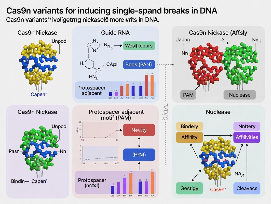
Q1: What are Cas9 nickases (Cas9n), and how do they differ from wild-type Cas9? Cas9 nickases are engineered variants of the wild-type Streptococcus pyogenes Cas9 (SpCas9) that generate single-strand breaks (nicks) in DNA instead of double-strand breaks (DSBs). Wild-type Cas9 utilizes two nuclease domains, RuvC and HNH, to cleave both DNA strands, creating a DSB. By introducing a point mutation into one of these domains, a nickase is created [1] [2] [3].
- D10A Nickase: A mutation in the RuvC domain (aspartate to alanine at position 10) inactivates it. This variant cleaves only the target strand (the strand complementary to the guide RNA) via the intact HNH domain [1] [2].
- H840A Nickase: A mutation in the HNH domain (histidine to alanine at position 840) inactivates it. This variant cleaves only the non-target strand via the intact RuvC domain [1] [2].
Q2: Why would a researcher choose to use a nickase over the standard Cas9 nuclease? The primary reasons are enhanced specificity and applications requiring precision.
- Reduced Off-Target Effects: A single nick is typically repaired with high fidelity by the cellular base excision repair pathway and does not frequently lead to mutations. To create a functional DSB, two nickases must bind in close proximity on opposite strands (a "double-nicking" strategy). This paired requirement dramatically lowers the probability of off-target DSBs and subsequent indels elsewhere in the genome [2] [3].
- Improved Homology-Directed Repair (HDR): Nickases can facilitate HDR for precise gene insertions. Unlike standard Cas9, which has a narrow window of high-efficiency repair near the cut site, a paired nickase system creates a staggered DSB. This allows for efficient HDR across the entire region between the two nick sites, expanding the targetable range for precise edits, especially when no good guide RNA is available close to the desired mutation [1] [2].
Q3: What is a key recent finding regarding the H840A nickase variant? Recent research has revealed that the canonical H840A nickase can sometimes retain low-level activity on the target strand, leading to unintended DSBs both on-target and off-target [4]. To address this, an improved variant, nCas9 (H840A + N863A), was developed. The additional N863A mutation in the HNH domain further stabilizes its inactivation. This genuine nickase minimizes the generation of unwanted DSBs and reduces error-prone repair outcomes, making it particularly valuable for high-precision applications like prime editing [4].
Experimental Protocols
Protocol 1: Site-Directed Mutagenesis to Create Nickase Plasmids
This protocol outlines the method to engineer D10A or H840A mutations into a plasmid containing the wild-type SpCas9 gene.
Objective: To introduce specific point mutations into the Cas9 gene to create D10A or H840A nickase expression plasmids. Principle: Site-directed mutagenesis uses custom primers containing the desired mutation to amplify the entire plasmid, followed by ligation to create a circular mutant plasmid.
Materials:
- Wild-type SpCas9 expression plasmid (e.g., from Addgene [3])
- High-fidelity DNA polymerase (e.g., Q5 or PfuUltra)
- DpnI restriction enzyme
- T4 Polynucleotide Kinase
- T4 DNA Ligase
- Competent E. coli cells
Procedure:
- Primer Design: Design forward and reverse primers that are complementary to the target site but contain the desired single nucleotide change (e.g., GAC→GCC for D10A; CAC→GCC for H840A).
- PCR Amplification: Set up a PCR reaction using the wild-type Cas9 plasmid as a template and the mutagenic primers. The polymerase will replicate the entire plasmid, incorporating the mutation.
- Template Digestion: Add DpnI enzyme to the PCR product. DpnI specifically cleaves the methylated parental (wild-type) DNA template, leaving the amplified, unmethylated mutant DNA intact.
- Ligation and Transformation: Ligate the PCR product and transform it into competent E. coli cells.
- Sequence Verification: Pick several colonies, prepare plasmid DNA, and sequence the Cas9 gene across the mutated region to confirm the introduction of the correct mutation without any other errors.
Protocol 2: Validating Nickase Activity Using a Plasmid Cleavage Assay
This protocol describes an in vitro method to confirm that your engineered nickase creates single-strand breaks, unlike the wild-type Cas9.
Objective: To biochemically validate the single-strand nicking activity of purified D10A and H840A proteins compared to wild-type Cas9. Principle: A supercoiled plasmid DNA is incubated with the Cas9 protein and a target-specific guide RNA. Reaction products are analyzed by agarose gel electrophoresis: nicking converts supercoiled DNA to a relaxed open-circular form (slower migration), while DSBs create a linear form (distinct migration) [4].
Materials:
- Purified wild-type Cas9, D10A, and H840A proteins
- In vitro transcribed sgRNA targeting a site within the plasmid
- Supercoiled plasmid substrate
- Reaction buffer (e.g., NEBuffer 3.1)
- Agarose gel electrophoresis equipment
Procedure:
- Reaction Setup: In separate tubes, incubate the supercoiled plasmid with:
- Tube 1: Wild-type Cas9 + sgRNA
- Tube 2: D10A Nickase + sgRNA
- Tube 3: H840A Nickase + sgRNA
- Tube 4: No protein control
- Incubation: Incubate reactions at 37°C for 1 hour.
- Analysis: Stop the reactions and load the products onto an agarose gel.
- Interpretation:
- Wild-type Cas9: Will produce a predominant band corresponding to the linearized plasmid.
- Functional Nickase (D10A or H840A): Will produce a predominant band corresponding to the nicked, open-circular form.
- Control: Will show only the supercoiled plasmid band.
Table 1: Expected Results from Plasmid Cleavage Assay
| Protein | Supercoiled DNA | Open-Circular (Nicked) DNA | Linear (DSB) DNA |
|---|---|---|---|
| No Protein Control | ++++ | - | - |
| Wild-type Cas9 | - | - | ++++ |
| D10A Nickase | - | ++++ | - |
| H840A Nickase | - | ++++ | -/+* |
*The canonical H840A may show a faint linear band, indicating residual DSB activity [4].
Troubleshooting Guides
Problem: Low Editing Efficiency with Paired Nickases
- Cause 1: Incorrect gRNA Orientation. The two gRNAs must be in a PAM-out configuration, meaning the PAM sequences face away from each other, toward the outside of the targeted region [1] [2].
- Solution: Redesign gRNA pairs to ensure a PAM-out orientation.
- Cause 2: Suboptimal Spacing Between Nick Sites.
- Solution: Adhere to validated spacing rules. For the D10A nickase, optimal nick separation is 40-68 bp. For the H840A nickase, optimal spacing is 51-68 bp [1] [2].
- Cause 3: Inefficient gRNA or Nickase Delivery.
- Solution: Use recombinant ribonucleoprotein (RNP) complexes for delivery, which can increase efficiency and reduce off-target effects. Ensure your transfection method is optimized for your specific cell type [5] [6].
Problem: High Unwanted Indel Background with H840A Nickase
- Cause: The canonical H840A (H840A) nickase can exhibit residual double-strand break activity, leading to error-prone NHEJ repair [4].
- Solution: Use the improved H840A+N863A nickase variant. The additional N863A mutation more completely inactivates the HNH domain, minimizing DSB formation and resulting in cleaner editing outcomes with fewer unwanted indels [4].
Data Presentation Tables
Table 2: Comparative Summary of Cas9 Nickase Variants
| Parameter | Wild-Type Cas9 | D10A Nickase | H840A Nickase | H840A+N863A Nickase |
|---|---|---|---|---|
| Catalytic Domains | RuvC (active), HNH (active) | RuvC (inactive), HNH (active) | RuvC (active), HNH (inactive) | RuvC (active), HNH (inactive) |
| DNA Cleavage | Double-strand break (DSB) | Single-strand break (nick) on target strand | Single-strand break (nick) on non-target strand | Single-strand break (nick) on non-target strand |
| Primary Application | Gene knockouts, NHEJ-dominated editing | Paired nicking for specific DSBs, Base Editing | Prime Editing, Paired nicking | High-fidelity Prime Editing |
| Key Design Rule | Single gRNA | Two gRNAs, PAM-out, 40-68 bp spacing | Two gRNAs, PAM-out, 51-68 bp spacing | As for H840A |
| Specificity | Standard (potential for off-target DSBs) | High (requires cooperative nicking) | High (requires cooperative nicking) | Very High (minimized residual DSBs) |
| HDR Efficiency | Limited to narrow window near DSB | High across entire region between nicks | High across entire region between nicks | High, with reduced competing NHEJ |
Table 3: Donor Template Design for Nickase-Mediated HDR [1] [2]
| Edit Type | Donor Type | Recommended Homology Arm Length | Strand Preference |
|---|---|---|---|
| Small insertion/tag (e.g., EcoRI site) | ssODN | 30 - 60 bases | Test both top and bottom strand donors; preference can be unpredictable. |
| Large insertion (e.g., mCherry) | Long ssDNA (e.g., IDT Megamer) | 100 bases | Information not specified, but testing both strands is recommended. |
Signaling Pathways and Workflows
Diagram: Engineering Workflow for Cas9 Nickase Variants
The Scientist's Toolkit
Table 4: Essential Research Reagent Solutions for Nickase Engineering and Application
| Reagent / Tool | Function / Description | Example Source / Note |
|---|---|---|
| Wild-Type SpCas9 Plasmid | The starting template for engineering nickase mutations. | Addgene [3] |
| Site-Directed Mutagenesis Kit | A commercial kit that simplifies the introduction of point mutations. | Various suppliers (NEB, Agilent) |
| In Vitro Transcription Kit | To synthesize sgRNA for validation assays. | |
| Agarose Gel Electrophoresis System | To analyze the results of the plasmid cleavage assay and confirm nickase activity. | Standard lab equipment |
| Alt-R CRISPR-Cas9 System | Pre-designed, synthetic gRNAs and recombinant Cas9 nickase proteins. | Integrated DNA Technologies (IDT) [2] |
| ssODN Donor Oligos | Single-stranded oligodeoxynucleotides used as repair templates for HDR with nickases. | Ultramer Oligonucleotides [1] |
| Long ssDNA Donor Fragments | For inserting large sequences (e.g., fluorescent tags) via HDR. | Megamer ssDNA Fragments [1] |
Core Mechanism and Strand Selection
What is the fundamental mechanistic difference between the D10A and H840A Cas9 nickase variants?
The fundamental difference lies in which nuclease domain is inactivated and, consequently, which DNA strand is cut. Both variants are derived from the wild-type Streptococcus pyogenes Cas9 (SpCas9), which uses two nuclease domains to cleave both strands of target DNA [7].
- Cas9n D10A: This variant contains a point mutation (D10A) that inactivates the RuvC nuclease domain. This domain is responsible for cleaving the non-target strand (the strand that contains the Protospacer Adjacent Motif or PAM sequence). When inactivated, the Cas9 complex can only cleave the target strand (the strand to which the guide RNA binds) [8] [9].
- Cas9n H840A: This variant contains a point mutation (H840A) that inactivates the HNH nuclease domain. This domain is responsible for cleaving the target strand. When inactivated, the Cas9 complex can only cleave the non-target strand [8] [9].
The table below summarizes the core mechanistic differences.
Table 1: Fundamental Characteristics of Cas9 Nickase Variants
| Feature | Cas9n (D10A) | Cas9n (H840A) |
|---|---|---|
| Inactivated Domain | RuvC | HNH |
| Inactivated Domain Function | Cleaves non-target strand | Cleaves target strand |
| Strand Cleaved | Target strand (guide RNA-bound) | Non-target strand (PAM-containing) |
| Active Domain | HNH | RuvC |
The following diagram illustrates the strand-specific nicking mechanism of each variant.
Experimental Performance and Practical Application
How do the editing efficiency and repair outcomes differ between D10A and H840A?
Quantitative data reveals significant differences in the performance of the two nickases, influencing their suitability for specific applications.
Table 2: Comparative Performance and Repair Outcomes
| Parameter | Cas9n (D10A) | Cas9n (H840A) | Notes and Citations |
|---|---|---|---|
| HDR Efficiency | High [1] | Can be higher than D10A [10] | In a direct comparison, H840A elicited 51% HTR (Homology-Targeted Repair), significantly more efficient than D10A (41%) and Cas9 nuclease (27%) [10]. |
| Indel Profile (PAM-out) | Predominantly small deletions [1] | Biased towards large insertions [1] | The distinct overhang patterns (5' for D10A, 3' for H840A) influence repair [1]. |
| Mutagenic Efficiency for Gene Disruption | High (9x higher than H840A in one study) [11] | Lower [11] | When used as a pair to create a double-strand break, D10A demonstrates higher disruption efficiency [11]. |
| Interaction with Replication Forks | Predominantly generates double-ended DSBs when on lagging strand template [12] | Can generate single-ended DSBs when on leading strand template [12] | Important for studies of DNA repair and genome instability [12]. |
| Recommended Application | Homology-Directed Repair (HDR) [1] | Prime Editing (as part of PE2 system) [11] | The H840A mutation is part of the standard Prime Editor 2 (PE2) construct [11]. |
What are the optimal design rules for a paired-nicking experiment with Cas9n?
For paired nicking, where two sgRNAs are used to create a staggered double-strand break, the orientation and spacing of the sgRNAs are critical.
- Orientation: Use a PAM-out configuration, where the PAM sequences for both sgRNAs are on the outer extremes of the targeted sequence. This configuration yields significantly higher editing efficiency than PAM-in [1].
- Spacing: The distance between the two nick sites is crucial.
Troubleshooting Common Experimental Issues
My paired-nicking experiment is yielding low HDR efficiency. What could be wrong?
- Problem: Incorrect sgRNA spacing.
- Solution: Verify that the distance between the two nicks on opposite strands falls within the optimal range of 37–68 bp for D10A. For distances over 200 bp, a "spacer-nick" approach can minimize NHEJ while preserving HDR, but efficiency may decrease as the distance increases [13].
- Problem: Suboptimal donor template design.
- Solution: For small insertions (e.g., using ssODN), use homology arms of 40 bp. For larger insertions (e.g., with long ssDNA), use 100 bp homology arms. Test donors for both the top and bottom strands [1].
- Problem: Using the less efficient nickase for HDR.
- Solution: Prefer the D10A variant for standard HDR experiments, as it is consistently reported as the superior choice for this application [1].
I am concerned about off-target effects. How do the nickase variants compare?
The primary safety advantage of nickases is a significant reduction in off-target mutations compared to wild-type Cas9 nuclease. Because a single nick is typically repaired faithfully, off-target activity requires two sgRNAs to bind in close proximity at an off-target site, which is statistically far less likely.
- Evidence: The "spacer-nick" approach (paired nicks at 200–350 bp) was shown to achieve gene correction with minimal NHEJ-mediated on-target mutations. Furthermore, frequent off-target genetic alterations induced by classical CRISPR-Cas9 were "significantly reduced or absent" in cells treated with this nickase-based approach [13].
- Comparison to Nuclease: The paired-nickase approach can reduce off-target activity by up to 50–1500 fold compared to Cas9 nuclease [11] [7].
Essential Experimental Protocols
Protocol: Spacer-Nick Gene Correction in Hematopoietic Stem and Progenitor Cells (HSPCs)
This protocol, adapted from [13], is designed for high-precision gene repair with minimal NHEJ.
Key Reagents:
- Ribonucleoprotein (RNP) Complex: Cas9D10A nickase protein complexed with a pair of synthetic, chemically modified sgRNAs in a "PAM-out" orientation with a spacer distance of 200–350 bp.
- Donor Template: Recombinant Adeno-Associated Virus serotype 6 (AAV6) containing the single-stranded DNA donor template with homologous arms.
Methodology:
- Electroporation: Deliver the preassembled RNP complexes into activated HSPCs or T cells via electroporation.
- Viral Transduction: Subsequently transduce the cells with the AAV6 donor template.
- Analysis: Assess gene correction efficiency 3 days post-targeting using flow cytometry (if using a fluorescent reporter) or by sequencing the targeted genomic locus to quantify HDR and NHEJ events.
Critical Step: The large spacer distance (200–350 bp) is key to shifting the repair balance overwhelmingly towards HDR and away from indels [13].
Protocol: Assessing Nickase-Induced Recombination in Fission Yeast
This protocol, based on [12], is used to study DNA repair dynamics following strand-specific nicking.
Key Reagents:
- Strand-Specific Nickases: Cas9n D10A (for lagging strand template nicks) and Cas9n H840A (for leading strand template nicks).
- Reporter Strain: S. pombe strain with a direct repeat recombination reporter (e.g., ade6- alleles) integrated at a characterized locus.
Methodology:
- Transformation: Introduce plasmids expressing a specific Cas9n variant (D10A or H840A) and its corresponding gRNA into the reporter strain.
- Culture and Selection: Grow transformed cells asynchronously and select for recombinants.
- DSB Detection: Harvest genomic DNA and analyze for double-stranded breaks (DSBs) using Southern blotting of restriction fragments encompassing the gRNA binding site.
- Recombination Frequency: Quantify the frequency of Ade+ recombinants arising from gene conversion or deletion events.
Research Reagent Solutions
Table 3: Essential Reagents for Cas9 Nickase Research
| Reagent / Tool | Function / Description | Example Sources / References |
|---|---|---|
| SpCas9 Nickase Plasmids | Mammalian expression vectors for D10A (e.g., pX335) and H840A mutants. | Addgene (#42335 for pX335) [14] |
| High-Specificity sgRNAs | Chemically modified synthetic sgRNAs for improved stability and RNP complex formation. | Integrated DNA Technologies (IDT) [1] |
| AAV6 Donor Template | High-efficiency single-stranded DNA donor delivery vehicle for HDR in primary cells. | Packaged via standard AAV production methods [13] |
| Cas9-NG Nickase | Engineered nickase variant that recognizes relaxed NG PAM, expanding targetable sites. | Addgene; research labs [11] |
| T7 Endonuclease I (T7EI) | Enzyme for detecting and quantifying nuclease-induced indels via mismatch cleavage. | New England Biolabs (NEB) [14] |
| ssODN Donor | Single-stranded oligodeoxynucleotide donor template for introducing point mutations or small tags. | IDT Ultramer Oligonucleotides [1] |
| Long ssDNA Donor | Long single-stranded DNA donor template for inserting large sequences (e.g., fluorescent proteins). | IDT Megamer ssDNA Fragments [1] |
Frequently Asked Questions (FAQs)
Q1: What is the fundamental mechanistic difference between Cas9 nuclease and Cas9 nickase (Cas9n) that leads to improved safety?
Cas9 nuclease creates double-strand breaks (DSBs), which are highly genotoxic lesions that can lead to genomic instability if misrepaired. In contrast, Cas9 nickase is a mutated form of Cas9 that cuts only one DNA strand, creating a single-strand break or "nick" [15] [16]. DSBs activate a strong DNA damage response (DDR) and can trigger p53-mediated cellular toxicity, apoptosis, or select for cells with potentially unstable genomes [17] [18]. Single-strand breaks are inherently less genotoxic and are repaired more faithfully by high-fidelity cellular repair pathways, leading to a significant reduction in both unwanted mutations and cellular stress [16].
Q2: How does using Cas9 nickase specifically reduce off-target editing in experiments?
Wild-type Cas9 can tolerate several mismatches between the gRNA and off-target DNA sequences, leading to cleavage at incorrect sites [19] [20]. Cas9 nickase reduces this risk because a single nick in an off-target location is often repaired without introducing mutations [15] [16]. For effective genome editing, the Cas9 nickase system typically uses a pair of nickases with two guide RNAs that target opposite strands of the DNA at adjacent sites. This creates a "staggered" double-strand break. The requirement for two guide RNAs to bind in close proximity for a productive edit dramatically increases the system's specificity, as the probability of both guides binding incorrectly at an off-target site is exceedingly low [15] [21].
Q3: My research requires high-efficiency editing. What is the trade-off in efficiency when using Cas9 nickase, and how can I mitigate it?
The primary trade-off for the increased specificity of Cas9 nickase is that editing efficiency can be lower than with wild-type Cas9, as it requires the simultaneous action of two guide RNAs for a full DSB [16]. To mitigate this, you can:
- Optimize guide RNA design: Ensure both gRNAs have high predicted on-target activity. Use computational tools to select gRNAs with optimal GC content and minimal off-target potential [15] [20].
- Test multiple gRNA pairs: Design and empirically test 3-4 different gRNA pairs to identify the most efficient combination for your target [16].
- Optimize delivery: Use efficient delivery methods such as electroporation for ribonucleoprotein (RNP) complexes of Cas9n protein and synthetic gRNAs, which provides rapid activity and can reduce cell toxicity [20] [22].
Q4: Can Cas9 nickase be used to reduce p53-mediated cellular toxicity in sensitive cell types?
Yes. Research has shown that DSBs generated by wild-type Cas9 can activate the p53 pathway, leading to cell cycle arrest or apoptosis, particularly in certain cell types like stem cells or those with intact p53 signaling [17]. This can confound experimental results by selecting for edited cells with compromised p53 function. Since Cas9 nickase creates less severe DNA damage (single-strand breaks), it presents a lower activation signal for the p53 pathway, thereby reducing associated cellular toxicity and providing a more accurate representation of gene function in your experimental model [17] [16].
Troubleshooting Guides
Problem: Low On-Target Editing Efficiency with Cas9 Nickase
Potential Causes and Solutions:
- Cause: Suboptimal guide RNA (gRNA) pair design.
- Cause: Inefficient delivery or expression of CRISPR components.
- Solution: For transient expression, deliver Cas9 nickase as an RNP complex with the two in vitro-transcribed or synthetic gRNAs via electroporation. If using plasmids, confirm promoter compatibility with your cell type and ensure both gRNAs are expressed at high levels. Validate delivery efficiency with a fluorescent reporter system [20] [22].
- Cause: Inherently lower efficiency of paired nickase systems.
Problem: Suspected Residual Off-Target Activity
Potential Causes and Solutions:
- Cause: One of the gRNAs in the pair has low specificity.
- Cause: High concentrations of Cas9n and gRNAs can lead to promiscuous cutting.
Problem: High Cell Toxicity or Low Cell Survival Post-Editing
Potential Causes and Solutions:
- Cause: Transfection-related toxicity.
- Cause: Excessive nicking activity.
- Cause: Toxicity from the plasmid backbone or other vector components.
Data Presentation
Table 1: Quantitative Comparison of Wild-Type Cas9 vs. Cas9 Nickase
| Feature | Wild-Type Cas9 (DSB) | Cas9 Nickase (SSB) | Key Implication |
|---|---|---|---|
| DNA Lesion | Double-Strand Break (DSB) | Single-Strand Break (Nick) | SSBs are less genotoxic and trigger a weaker DNA Damage Response [18] [16] |
| Typical Off-Target Mutation Rate | Higher (variable, can be >50%) | Significantly Lower | Nickase greatly reduces confounding off-target mutations in experimental data [15] [16] |
| Cellular Toxicity (p53 activation) | Higher, can lead to cell death or selection for p53-deficient cells | Lower, improved cell viability | Nickase is better suited for editing sensitive cell types like stem cells [17] |
| Theoretical Targetable Genomic Loci | Limited by NGG PAM (SpCas9) | Limited by NGG PAM (SpCas9) | Both are limited by the same PAM when using the same Cas9 variant [21] |
| Editing Efficiency (Knock-in) | Low HDR efficiency, competes with error-prone NHEJ | Can be enhanced with paired nicking and optimized donor design | Paired nickase can improve the fidelity of HDR-based precise edits [22] |
Experimental Protocols
Protocol 1: Designing and Validating a Paired Nickase Experiment
Objective: To create a defined genomic deletion or knock-in using a Cas9 nickase pair.
Materials:
- Cas9 nickase expression plasmid or purified protein (e.g., D10A mutant for SpCas9)
- Two in vitro-synthesized or plasmid-derived gRNAs targeting opposite strands
- Target cells
- Delivery reagents (e.g., electroporation kit, lipofectamine)
- PCR reagents and sequencing primers
- Surveyor or T7 Endonuclease I assay kit, or resources for NGS
Methodology:
- gRNA Design: Using a computational tool (e.g., from [20]), select two gRNAs with high on-target scores that bind to opposite DNA strands, spaced 30-50 bp apart. Verify minimal off-target potential for each guide individually.
- Component Delivery:
- Plasmid Method: Co-transfect the target cells with the Cas9 nickase plasmid and the two gRNA expression plasmids.
- RNP Method: Complex the purified Cas9 nickase protein with the two synthetic gRNAs to form RNPs. Deliver the RNP complex into the cells via electroporation.
- Validation and Analysis:
- Harvest Genomic DNA: 48-72 hours post-delivery, harvest genomic DNA from the cell population.
- Initial Screening: Amplify the target region by PCR. Use the T7 Endonuclease I or Surveyor assay to detect the presence of indels, which indicate successful dual nicking and DSB formation.
- Confirmation: Clone the PCR products and perform Sanger sequencing, or use next-generation sequencing (NGS) to determine the exact sequence of the edited alleles and confirm the precise deletion or insertion [20] [22].
Protocol 2: Assessing Cell Viability and p53 Activation
Objective: To quantify and compare the cellular toxicity and DNA damage response induced by wild-type Cas9 versus Cas9 nickase.
Materials:
- Wild-type Cas9 and Cas9 nickase (plasmid or RNP)
- Validated gRNAs
- Cell viability assay kit (e.g., MTT, CellTiter-Glo)
- Antibodies for immunoblotting (anti-p53, anti-p21, anti-γH2AX)
- qPCR reagents
Methodology:
- Cell Treatment: Divide your cell culture into three groups: (1) Untreated control, (2) Wild-type Cas9 + gRNA, (3) Cas9 nickase + two gRNAs. Use the same delivery method and equimolar amounts of total nuclease/gRNA.
- Viability Assay: 24-96 hours post-editing, perform a cell viability assay according to the manufacturer's instructions. Normalize readings to the untreated control.
- DNA Damage Response Analysis:
- Protein Level: Harvest cell lysates 24-48 hours post-editing. Perform a western blot to detect the levels of phosphorylated p53 (S15), its downstream target p21, and the DSB marker γH2AX. Expect to see a stronger signal in the wild-type Cas9 group.
- Gene Expression Level: Isolve RNA and perform qPCR to measure the transcript levels of p53 target genes like
CDKN1A (p21)andBAX[17] [18].
Signaling Pathways and Workflows
Diagram 1: DNA Damage Signaling: DSB vs. SSB
Diagram 2: Paired Nickase Editing Workflow
The Scientist's Toolkit: Research Reagent Solutions
Table 2: Essential Reagents for Cas9 Nickase Research
| Reagent | Function | Key Considerations |
|---|---|---|
| High-Fidelity Cas9 Nickase | Engineered Cas9 variant (e.g., D10A mutation for SpCas9) that cuts only one DNA strand. | Source from reputable suppliers. Consider high-fidelity base variants (e.g., eSpCas9(1.1)) for the nickase backbone to further reduce off-target binding [15] [21]. |
| Chemically Modified sgRNA | Synthetic guide RNA with chemical modifications (e.g., 2'-O-methyl-3'-phosphonoacetate) to improve stability and specificity. | Modified sgRNAs show increased resistance to nucleases and can reduce off-target effects while maintaining on-target activity [19] [15]. |
| Ribonucleoprotein (RNP) Complex | Pre-complexed Cas9 nickase protein and sgRNA. | Direct delivery of RNPs leads to rapid activity and degradation, shortening exposure time and reducing off-target effects and immune responses [20] [22]. |
| Off-Target Prediction Software | Computational tools (e.g., from MIT Broad Institute, E-CRISP) to analyze gRNA designs for potential off-target sites. | An essential in-silico step for gRNA selection. Use tools that allow input of your specific cell line's genome sequence for improved accuracy [20] [22]. |
| Next-Generation Sequencing (NGS) Assays | High-depth sequencing methods (e.g., GUIDE-seq, CIRCLE-seq) for comprehensive profiling of editing outcomes. | Critical for empirically validating the off-target profile of your chosen gRNA pairs in your specific experimental system [20]. |
Advanced genome editing technologies, particularly prime editing and base editing, represent a significant evolution from traditional CRISPR-Cas9 systems by leveraging Cas9 nickase (Cas9n) variants to achieve precise genetic modifications without inducing double-strand DNA breaks (DSBs). This technical resource center examines the architectural composition of these editors, focusing on their core components and functional mechanisms to support researchers in troubleshooting experimental challenges.
System Architectures and Mechanisms
Core Components of Base Editing Systems
Base editors utilize catalytically impaired Cas9 variants fused to deaminase enzymes to achieve single-nucleotide conversions without DSBs [24].
Adenine Base Editors (ABEs) perform A•T to G•C conversions using an engineered tRNA adenosine deaminase (TadA) that converts adenine to inosine, which is read as guanine during DNA replication [24]. Cytosine Base Editors (CBEs) achieve C•G to T•A conversions using a cytidine deaminase (typically APOBEC1) that converts cytosine to uracil, which is then read as thymine [24]. Both systems incorporate uracil glycosylase inhibitors (UGI) in CBEs to prevent repair of the edited base back to cytosine [24].
Base Editing System Architectures
Core Components of Prime Editing Systems
Prime editors employ a more complex architecture consisting of a Cas9 nickase fused to a reverse transcriptase (RT) enzyme, programmed with a specialized prime editing guide RNA (pegRNA) [25] [26]. The pegRNA contains both a spacer sequence for target recognition and an extended RT template (RTT) encoding the desired edit [25].
The editing process involves: (1) Cas9n nicking the target DNA strand, (2) the exposed 3' end serving as a primer for reverse transcription using the RTT, and (3) cellular resolution of the resulting DNA flap structure to incorporate the edit [25] [26].
Prime Editing Mechanism Workflow
Troubleshooting Guide: Frequently Asked Questions
Q1: Why is my base editing efficiency low despite high transfection rates?
Low editing efficiency often results from suboptimal positioning of the target base within the editing window [24]. The editing window varies by base editor design but typically spans nucleotides 4-8 in the protospacer region [24]. Additionally, high GC content in the sgRNA can reduce efficiency, with optimal GC content between 40-60% [5].
Troubleshooting protocol:
- Verify target base positioning using base editor-specific prediction tools
- Design and test 3-5 alternative sgRNAs with varying GC content
- Include positive control targets with known high efficiency
- Use high-purity, HPLC-grade sgRNAs to minimize truncated guides [27]
- Consider cell line-specific variations in DNA repair pathways that may affect outcomes [5]
Q2: How can I reduce unwanted bystander edits in base editing?
Bystander edits occur when additional bases within the editing window are unintentionally modified [25]. To minimize this:
Experimental approach:
- Position target base optimally: Place the desired edit at the most efficient position within the editing window while ensuring surrounding bases have lower susceptibility to deamination [24]
- Use engineered editors: Newer base editor variants with narrowed editing windows can reduce bystander activity [25]
- Validate with sequencing: Perform deep sequencing to quantify bystander edit frequencies and adjust sgRNA design accordingly
Q3: What causes low prime editing efficiency and how can it be improved?
Prime editing efficiency depends on multiple factors including pegRNA stability, cellular resolution of flap intermediates, and reverse transcription efficiency [25] [26].
Optimization strategies:
- Implement engineered pegRNAs (epegRNAs): Incorporate evopreQ or mpknot RNA motifs at the 3' terminus to protect against degradation [25]
- Use dual-nicking systems: Employ PE3 systems with an additional sgRNA to nick the non-edited strand, enhancing edit incorporation [25] [26]
- Optimize RTT length: Keep reverse transcription template between 10-15 nucleotides when possible [25]
- Modify cellular repair pathways: Consider transient inhibition of mismatch repair proteins (e.g., MLH1) to improve efficiency [26]
Q4: How can I minimize structural variations and off-target effects?
While nickase systems reduce DSB-related risks, they can still generate structural variations [28].
Risk mitigation protocol:
- Use high-fidelity Cas9 nickase variants: Engineered Cas9n with additional mutations (e.g., N863A) reduce off-target nicking [25]
- Avoid DNA-PKcs inhibitors: These can dramatically increase kilobase-scale deletions and chromosomal translocations [28]
- Employ modified delivery methods: Synthetic sgRNAs show reduced immunogenicity and off-target effects compared to plasmid-based expression [27]
- Comprehensive analysis: Utilize long-read sequencing and CAST-Seq to detect structural variations missed by short-read approaches [28]
Q5: What are the key considerations for selecting between base editing and prime editing?
The choice depends on the specific genetic modification required [25] [24].
Selection framework:
- Use base editing for: Straightforward transition mutations (C→T, A→G), projects requiring higher efficiency, and applications where small editing windows are acceptable [24]
- Choose prime editing for: Transversions, small insertions/deletions, combinations of edits, or when targeting sequences with limited PAM options [25] [26]
- Consider delivery constraints: Prime editors are larger, posing challenges for viral delivery, while compact base editors are more deliverable [25]
Performance Comparison Tables
Table 1: Evolution of Prime Editor Systems and Their Efficiencies
| Editor Version | Key Components | Editing Frequency | Improvements Over Previous Versions |
|---|---|---|---|
| PE1 | nCas9 (H840A) + M-MLV RT | ~10-20% | Initial proof-of-concept system [26] |
| PE2 | nCas9 + engineered RT | ~20-40% | Optimized reverse transcriptase for enhanced stability and processivity [25] [26] |
| PE3 | PE2 + additional sgRNA | ~30-50% | Additional nick on non-edited strand to bias repair toward edited sequence [25] [26] |
| PE4 | PE2 + MLH1dn | ~50-70% | Mismatch repair inhibition reduces repair of edits [26] |
| PE5 | PE3 + MLH1dn | ~60-80% | Combines dual nicking with mismatch repair inhibition [26] |
| PE6 | Engineered RT + epegRNAs | ~70-90% | Compact RT variants and stabilized pegRNAs [26] |
| PE7 | PE6 + La protein fusion | ~80-95% | Enhanced pegRNA stability and editing in challenging cell types [26] |
Table 2: Base Editing Comparison by Type and Capabilities
| Editor Type | Base Conversion | Key Components | Editing Window | Common Applications |
|---|---|---|---|---|
| Cytosine Base Editor (CBE) | C•G → T•A | nCas9 + APOBEC1 + UGI | ~ nucleotides 4-8 | Creating stop codons, correcting C→T mutations [24] |
| Adenine Base Editor (ABE) | A•T → G•C | nCas9 + engineered TadA | ~ nucleotides 4-8 | Correcting A→G mutations, splice site modulation [24] |
Research Reagent Solutions
Table 3: Essential Reagents for Advanced Editing Experiments
| Reagent Type | Specific Examples | Function | Considerations |
|---|---|---|---|
| Cas9 Nickase Variants | nCas9 (H840A), HiFi Cas9 nickase | DNA nicking for primer generation or strand-specific nicking | H840A mutation inactivates RuvC domain; N863A further reduces DSB formation [25] |
| Guide RNA Formats | epegRNA, xr-pegRNA, sgRNA | Target recognition and template provision | epegRNAs with 3' RNA motifs improve stability and editing efficiency by 3-4 fold [25] |
| Synthetic Guide RNAs | HPLC-purified sgRNAs [27] | High-purity guides for reduced off-target effects | >90% purity minimizes truncated guides and cell toxicity [27] |
| Delivery Systems | AAV vectors, lipid nanoparticles [25] | Editor delivery to cells | Prime editor size challenges AAV packaging; split systems may be required [25] |
| Detection Tools | NGS-based assays, CAST-Seq [28] | Identification of on/off-target edits and structural variations | Essential for comprehensive safety assessment [28] |
Experimental Protocols
Protocol 1: Prime Editing Workflow for Mammalian Cells
This protocol outlines a standard workflow for prime editing in mammalian cell lines, incorporating optimization strategies for enhanced efficiency [25] [26].
Materials:
- Prime editor plasmid (PE2, PE3, or updated versions)
- Engineered pegRNA expression vector or synthetic epegRNA
- Appropriate delivery reagents (lipofection, electroporation)
- Target cells (HEK293T recommended for initial testing)
Method:
- pegRNA Design: Design pegRNA with 5' spacer (20 nt), RTT (10-15 nt encoding desired edit), and primer binding site (8-15 nt)
- Stability Enhancement: Incorporate evopreQ or mpknot RNA motifs at 3' end for epegRNAs [25]
- Delivery: Transfect prime editor and pegRNA at optimal ratio (start with 1:3 mass ratio)
- Efficiency Assessment: Harvest cells 72-96 hours post-transfection; extract genomic DNA for sequencing
- Analysis: Use targeted amplicon sequencing with minimum 10,000x coverage; quantify editing and indel rates
Troubleshooting Notes:
- If efficiency remains low, test PE3 system with additional sgRNA
- For difficult-to-edit loci, consider PE4/5 systems with MLH1dn
- Validate edits with functional assays where possible
Protocol 2: Base Editing Optimization for Therapeutic Development
This protocol focuses on base editing optimization with emphasis on specificity and safety profiling [24].
Materials:
- Base editor expression system (ABE or CBE)
- HPLC-purified sgRNAs [27]
- Target cells (including relevant primary cells if applicable)
- Next-generation sequencing resources
Method:
- sgRNA Design: Identify optimal targets within editing window; avoid sequences with multiple identical bases in editing window
- Delivery: Use ribonucleoprotein (RNP) delivery when possible for reduced off-target effects [27]
- Editing Assessment: Quantitative sequencing at 48-72 hours post-editing
- Specificity Profiling: Perform whole-genome sequencing or CAST-Seq to detect structural variations [28]
- Functional Validation: Western blot for protein knockout or functional assays for specific corrections
Troubleshooting Notes:
- If bystander edits are problematic, reposition target base or try different base editor variants
- For low efficiency in primary cells, optimize delivery methods and consider cell cycle synchronization
- Always include multiple negative controls to distinguish background mutation rates
Applied Cas9n Systems: From Base Editing to Therapeutic Genome Correction
Implementing Paired Nickase Systems for Enhanced Specificity with Dual gRNAs
Core Concepts and Key Advantages
How does a paired nickase system improve specificity over wild-type Cas9? The paired nickase system enhances specificity by requiring two independent recognition events for a double-strand break (DSB) to occur. Wild-type Cas9, guided by a single gRNA, can tolerate several mismatches between the gRNA and the target DNA, leading to cuts at unintended, off-target sites [29]. In contrast, the paired nickase system employs a Cas9 nickase variant (such as the D10A mutant) and two gRNAs that target opposite strands of the DNA near the target site [2]. A functional DSB is only created when both gRNAs correctly bind and generate offset nicks. Even if one gRNA binds an off-target site, the absence of a complementary nick from the second gRNA typically results in a harmless single-strand break that is efficiently repaired by high-fidelity cellular mechanisms, thereby reducing off-target mutations by 50 to 1,000-fold [29].
What are the primary applications of this technology? This system is particularly valuable for applications demanding high precision, including:
- Generation of Isogenic Cell Lines: For precise disease modeling, as it minimizes confounding off-target mutations [30] [29].
- Functional Genomic Screens: Enables scalable knockout libraries using paired gRNAs for enhanced specificity [31].
- Gene Knockout and Large Deletions: Two offset nicks create a DSB for efficient gene disruption, while two distal gRNAs can excise large genomic regions [31].
Experimental Design and Optimization
The following diagram illustrates the fundamental mechanism of the paired nickase system for creating a double-strand break, which requires two guide RNAs binding to opposite DNA strands in a "PAM-out" orientation.
What are the critical design parameters for gRNA pairs? Successful experimental outcomes hinge on several key design factors, which are summarized in the table below.
| Design Parameter | Optimal Configuration | Rationale and Experimental Support |
|---|---|---|
| Nickase Variant | Cas9 D10A (RuvC mutant) | The D10A variant is more potent than the H840A variant in mediating homology-directed repair (HDR) and shows higher mutagenic targeting efficiency in human cells [11] [2]. |
| gRNA Orientation | PAMs facing outward from the target site | This "PAM-out" configuration is crucial for efficient cooperative nicking and generation of a DSB with overhangs. PAM-in configurations show significantly reduced efficiency [2]. |
| gRNA Spacing | 40–70 base pairs apart for Cas9 D10A | Systematic assessments show that high-efficiency editing is achieved within this distance range, likely due to reduced steric hindrance between the two Cas9n complexes [2]. Offsets from -4 to 20 bp can also work, but efficiency drops with increased distance [29]. |
| Promoter Selection | Use of heterologous promoters (e.g., human U6 and murine U6) in a single vector | Vectors with two identical U6 promoters are prone to recombination, leading to the loss of one gRNA sequence. Using two different promoters ensures stable expression of both gRNAs [31]. |
Troubleshooting Common Experimental Problems
What should I do if I observe low editing efficiency? Low editing efficiency can be addressed by systematically checking the following:
- Verify gRNA Pair Design: Confirm that your gRNAs are in the "PAM-out" orientation and are spaced within the optimal range (40-70 bp for D10A) [2]. Re-screening alternative gRNA pairs targeting the same locus can often identify a more efficient combination [32].
- Optimize Delivery and Expression: Ensure your delivery method (e.g., electroporation, lipofection) is efficient for your cell type. Confirm that both Cas9 nickase and the two gRNAs are expressed at high levels. Using a vector with heterologous promoters (e.g., human U6 and mouse U6) prevents recombination and ensures both gRNAs are expressed [31].
- Enhance Cell Survival: For sensitive cells like iPSCs, transient overexpression of the anti-apoptotic gene BCL-XL can significantly improve survival post-electroporation, thereby increasing the recovery of correctly edited cells [30].
How can I minimize off-target effects even further? While the paired nickase system inherently reduces off-targets, these strategies can enhance specificity further:
- Employ High-Fidelity Nickase Variants: Consider using advanced nickase variants like Cas9-NG, which recognizes a simpler NG PAM, allowing for more flexible and specific gRNA design across the genome [11].
- Use Truncated sgRNAs: Shortening the guide sequence by a few nucleotides at the 5' end can increase specificity by making the system more intolerant to mismatches [11].
- Validate with Robust Assays: Always use sensitive genotyping methods, such as amplicon sequencing, to thoroughly assess on-target efficiency and screen for potential off-target sites predicted by in silico tools [23].
Why are my edited cells not surviving puromycin selection after HDR? This could indicate low HDR efficiency or high cytotoxicity.
- Check Donor Template Design: For HDR using single-stranded oligonucleotide (ssODN) donors, ensure homology arm lengths are sufficient (e.g., 30-60 bases). Test donor templates complementary to both DNA strands, as the optimal strand can be unpredictable [2].
- Mitigate Cellular Toxicity: High concentrations of CRISPR components can be toxic. Titrate the amounts of Cas9n mRNA/protein and gRNAs to find a balance between editing and cell viability [23]. Using Cas9 protein with a nuclear localization signal (NLS) can improve efficiency and reduce toxicity.
- Confirm Selection Marker Integration: If using a double-selection system (e.g., Puromycin and Fialuridine) with a PiggyBac transposon for seamless excision, verify the correct integration of the donor cassette via PCR before selection [30].
Protocols and Workflows
The following chart outlines a general workflow for a typical paired nickase experiment, from design to validation.
Detailed Protocol for HDR in Human iPSCs This protocol, adapted from a study that achieved ~15% precise editing efficiency without indels, outlines key steps for precise genome editing in human iPSCs [30].
Design and Cloning:
- Design a donor construct containing your desired mutation (e.g., an expanded CAG repeat) flanked by homology arms (1.5-2.0 kb is effective).
- Incorporate a PiggyBac transposon-based dual-selection cassette (e.g., EGFP-Puro-DTK) near the edit for positive and negative selection.
- Clone the selected gRNA pair (in "PAM-out" orientation) into an expression vector with heterologous promoters.
Electroporation:
- Co-electroporate the iPSCs with the following components: the Cas9 D10A nickase plasmid, the paired gRNA vector, the donor construct, and a plasmid for transient BCL-XL overexpression to enhance survival.
Selection and Picking:
- At 48 hours post-electroporation, begin puromycin selection to eliminate non-transfected cells.
- After 7-10 days, manually pick individual puromycin-resistant clones and expand them in culture.
Genotypic Screening:
- Extract genomic DNA from expanded clones.
- Use junction PCR with primers outside the homology arms and within the selection cassette to identify correctly targeted clones.
- For precise sequence verification, perform Sanger sequencing of the edited region on both alleles. A key advantage of the nickase system is that clones generated with a single gRNA (single nick) often show the desired edit without "on-target" indels on either allele.
Excision of Selection Markers:
- Transiently transfert correctly edited clones with a PiggyBac transposase plasmid to excise the selection cassette.
- Use negative selection with Fialuridine (FIAU) to enrich for cells that have lost the EGFP-Puro-DTK cassette, resulting in a seamless, marker-free edit.
Essential Research Reagent Solutions
The table below lists key reagents and their functions for implementing paired nickase experiments.
| Reagent / Tool | Function in the Experiment |
|---|---|
| Cas9 D10A Nickase | The engineered core enzyme that creates single-strand breaks (nicks) at DNA sites specified by the gRNAs [2]. |
| Paired gRNA Expression Vector | A plasmid designed to co-express two gRNAs, ideally from heterologous promoters (e.g., human U6 and mouse U6) to prevent recombination [31]. |
| HDR Donor Template | A DNA template (e.g., ssODN or dsDNA with long homology arms) containing the desired edit, used by the cell's repair machinery to incorporate the new sequence [30] [2]. |
| PiggyBac Transposon System | A tool for seamless integration and subsequent removal of selection cassettes from the edited genome, ensuring no exogenous sequences remain [30]. |
| BCL-XL Expression Plasmid | A vector for transiently expressing this anti-apoptotic protein to improve the survival of difficult-to-transfect cells (e.g., iPSCs) after electroporation [30]. |
Base editing is a revolutionary genome editing technology that enables precise, efficient, and predictable nucleotide substitutions without creating double-strand breaks (DSBs) in DNA or requiring donor DNA templates [33]. This technology represents a significant advancement over conventional CRISPR-Cas9 editing, which relies on DSBs that can lead to unintended insertions, deletions, or chromosomal rearrangements.
The core innovation of base editors involves fusing catalytically impaired Cas9 nickase (Cas9n), typically the D10A variant, with specific deaminase enzymes [33]. Cas9n(D10A) contains a single amino acid mutation that disrupts its RuvC nuclease domain, converting it from a double-strand break inducer to a single-strand nicking enzyme [29] [34]. When coupled with deaminase enzymes, this system enables precise chemical conversion of one DNA base to another.
There are two primary classes of base editors: Cytosine Base Editors (CBEs) which convert C•G to T•A base pairs, and Adenine Base Editors (ABEs) which convert A•T to G•C base pairs [33]. Both systems maintain the key advantage of not inducing DSBs while achieving highly predictable editing outcomes, making them particularly valuable for therapeutic applications and functional genetic studies where precision is critical.
Figure 1: Architecture of Base Editor Systems. Base editors consist of Cas9n(D10A) fused to deaminase enzymes via linkers, with nuclear localization signals (NLS) for proper cellular targeting. The specific deaminase determines whether cytosine or adenine conversions occur.
Troubleshooting Guide: Common Base Editing Challenges
Q1: Why is my base editing efficiency low, and how can I improve it?
Low editing efficiency commonly stems from suboptimal editor expression, sgRNA design, or target sequence context. To address this:
Optimize nuclear localization: Use three nuclear localization signals (NLS) at the C-terminus of nCas9, which has been shown to increase editing efficiency by approximately 1.1-fold compared to single NLS configurations [33]. Enhanced nuclear targeting ensures sufficient editor concentration at genomic DNA.
Implement enhanced sgRNA (esgRNA) designs: Replace standard sgRNAs with esgRNA architectures, which demonstrate approximately two-fold higher editing efficiency than native sgRNAs and three-fold higher efficiency than tRNA-sgRNA systems [33]. The improved stability and Cas9 protein complex formation with esgRNAs significantly boost performance.
Verify spacer length: Use the canonical 20-nucleotide spacer length in your sgRNAs. Studies have shown that spacers shorter than 20 nucleotides (14-19 nt) result in substantially reduced or undetectable base editing activity [33].
Position target base appropriately: Ensure the target base falls within the optimal editing window. For ABE systems, the most efficient editing occurs when the target adenine is at positions 4-8 within the protospacer (counting from the distal end to the PAM) [33].
Q2: How can I minimize off-target editing in base editor experiments?
While base editors inherently have reduced off-target effects compared to wild-type Cas9, these strategies further enhance specificity:
Employ double-nicking strategies: When possible, use paired Cas9n(D10A) systems with two sgRNAs targeting opposite strands. This approach reduces off-target activity by 50-1,000 fold while maintaining on-target efficiency, as simultaneous nicking at off-target sites is statistically improbable [29] [34].
Utilize truncated sgRNAs: Shortened sgRNAs with 17-18 nucleotide spacers can improve specificity while maintaining sufficient on-target activity, particularly when combined with Cas9-NG nickase variants that recognize relaxed PAM requirements [35].
Select unique target sequences: Carefully design sgRNAs with minimal similarity to other genomic regions, paying particular attention to the seed region adjacent to the PAM sequence where mismatches are less tolerated [29].
Q3: What causes bystander edits, and how can they be reduced?
Bystander edits occur when non-target bases within the editing window are unintentionally modified:
Understand the editing window: Base editors typically have an active window of 4-5 nucleotides where deamination can occur [33]. Bystander edits are more likely when multiple targetable bases (C's for CBEs, A's for ABEs) are present in this window.
Use narrow-window editors: Newer base editor variants with engineered deaminase domains exhibit narrower editing windows. For example, fusing truncated CDA1 deaminase to Cas9(D10A) nickase constrains the editing window, reducing bystander mutations [35].
Optimize sgRNA positioning: When possible, design sgRNAs that position the desired edit such that potential bystander bases fall outside the optimal editing positions (typically edges of the window).
Q4: Why is my base editor causing indels despite using nickase?
Although base editors are designed to avoid DSBs, indels can still occur through several mechanisms:
Minimize nicking at the edited strand: The Cas9n(D10A) component nicks the non-edited strand to initiate repair and bias incorporation of the edited base. However, excessive nicking activity or prolonged expression can lead to low-frequency DSB formation if cellular repair mechanisms convert nicks to breaks.
Limit expression duration: Use transient delivery methods (mRNA, protein, or non-integrating vectors) rather than stable expression to reduce the timeframe where nicking can occur [30].
Monitor cellular repair pathways: Variations in DNA repair machinery across cell types can influence indel formation. Some cell lines may be more prone to process nicks into indels through alternative repair pathways.
Base Editor Performance Data
Table 1: Adenine Base Editor Performance in Plant Systems. Data demonstrates editing efficiency across different configurations and target loci [33].
| Editor Construct | sgRNA Type | Target Locus | Editing Efficiency | Optimal Editing Window |
|---|---|---|---|---|
| PABE-7 (3x C-term NLS) | esgRNA | OsACC-T1 | 15.8-59.1% (in plants) | Positions 4-8 |
| PABE-7 (3x C-term NLS) | esgRNA | OsDEP1-T1 | 15.8-59.1% (in plants) | Positions 4-8 |
| PABE-7 (3x C-term NLS) | esgRNA | Multiple loci | 0.1-7.5% (in protoplasts) | Positions 4-8 |
| PABE-2 (original) | esgRNA | Multiple loci | ~1.1x lower than PABE-7 | Positions 4-8 |
| PABE-7 | Native sgRNA | Multiple loci | ~2x lower than esgRNA | Positions 4-8 |
Table 2: Comparison of Nickase Systems for Genome Editing Applications. Different Cas9 nickase configurations offer distinct advantages for various research needs [29] [30] [34].
| Nickase System | Editing Type | Key Features | Indel Formation | Best Applications |
|---|---|---|---|---|
| Single guided Cas9n(D10A) | SSB, HDR | Minimal indels, lower efficiency | Undetectable | Precise point mutations, disease modeling |
| Double nickase (paired Cas9n) | DSB via paired nicks | High specificity, reduced off-target | 5-40% | Gene knockouts, large insertions |
| All-in-One Cas9n(D10A) + dual sgRNAs | DSB via paired nicks | Simplified delivery, high efficiency | Up to 34.7% | High-throughput screening |
| Base editors (Cas9n + deaminase) | Chemical conversion | No DSBs, high precision | <0.1% | Single-base substitutions, therapeutic applications |
Experimental Protocols
Protocol 1: Installing Point Mutations with ABE in Mammalian Cells
This protocol describes the use of adenine base editors for precise A•T to G•C conversions in human induced pluripotent stem cells (iPSCs) and other mammalian systems:
Materials:
- ABE construct (e.g., PABE-7 with 3x C-terminal NLS configuration)
- Enhanced sgRNA (esgRNA) expression vector
- Appropriate delivery system (electroporation, lipofection)
- Validation reagents (PCR primers, sequencing reagents)
Procedure:
- Design sgRNA: Identify target site with NGG PAM and desired adenine within positions 4-8 of the protospacer.
- Clone esgRNA: Incorporate target sequence into enhanced sgRNA backbone using appropriate cloning strategy (BbsI sites for U6 promoter systems).
- Deliver editors: Co-transfect ABE and esgRNA plasmids using optimized method for your cell type. For difficult-to-transfect cells, consider mRNA or ribonucleoprotein delivery.
- Express transiently: Allow 48-72 hours for editor expression and base conversion.
- Validate editing: Harvest genomic DNA and amplify target region by PCR. Sequence amplicons to detect A to G conversions using Sanger or next-generation sequencing.
- Quantify efficiency: Calculate percentage of sequencing reads containing desired edit, noting any bystander edits within the activity window.
Troubleshooting notes:
- If efficiency is low, verify editor nuclear localization and try alternative sgRNA designs.
- If bystander edits are problematic, reposition sgRNA or use narrow-window editor variants.
- For iPSCs, transient BCL-XL overexpression can improve survival post-electroporation without increasing indel formation [30].
Protocol 2: Targeted Base Editing in Plant Systems
This protocol adapts base editing technology for rice and wheat, based on established plant ABE systems:
Materials:
- Plant-optimized ABE (PABE-7 with cereal codon optimization)
- esgRNA expression cassette
- Plant transformation materials (protoplast isolation reagents, Agrobacterium strains)
Procedure:
- Vector assembly: Clone target-specific esgRNA into plant expression vector with Ubi-1 promoter driving PABE-7 expression.
- Deliver to plant cells: For protoplast assays, use PEG-mediated transformation. For stable lines, use Agrobacterium-mediated transformation.
- Regenerate plants: Select transformed cells on appropriate antibiotics and regenerate whole plants through tissue culture.
- Screen edits: Genotype T0 seedlings by sequencing target loci to identify homozygous and heterozygous edits.
- Confirm absence of indels: Verify that target sites contain only the desired base changes without insertions or deletions.
Key optimization parameters:
- Spacer length must be 20 nucleotides for optimal activity
- esgRNA provides 1.7-fold higher efficiency than native sgRNA in regenerated plants
- Effective editing window spans positions 4-8 within the protospacer
Research Reagent Solutions
Table 3: Essential Reagents for Base Editing Research. Core components required for implementing base editing technologies [35] [30] [33].
| Reagent Category | Specific Examples | Function | Notes |
|---|---|---|---|
| Nickase Backbones | pX335 (Cas9n D10A), PABE vectors | Provides catalytically impaired Cas9 for targeted nicking | D10A mutation in RuvC domain creates 5' overhangs |
| Deaminase Enzymes | ecTadA-ecTadA* (ABE), rAPOBEC1 (CBE) | Catalyzes targeted base conversion | Evolved tRNA adenosine deaminase for ABE; cytidine deaminase for CBE |
| sgRNA Expression Systems | U6-driven esgRNA, tRNA-sgRNA | Targets editor to specific genomic loci | esgRNA shows highest efficiency in plant and mammalian systems |
| Delivery Tools | Electroporation, lipofection, viral vectors | Introduces editing components into cells | Transient delivery preferred to reduce off-target effects |
| Selection Systems | PiggyBac transposon, antibiotic resistance | Enriches for successfully edited cells | Allows seamless removal of selection markers post-editing |
Frequently Asked Questions
Q5: What is the difference between Cas9n(D10A) and Cas9n(H840A), and which should I use for base editing?
Cas9n(D10A) contains a mutation in the RuvC domain that cleaves the non-target DNA strand, while Cas9n(H840A) has a mutation in the HNH domain that cleaves the target strand complementary to the guide RNA [29] [34]. For base editing applications, Cas9n(D10A) is predominantly used because it creates nicks in the non-edited strand, which cellular repair machinery then uses as a template to incorporate the edited base [33]. Studies have shown that Cas9n(D10A) generates approximately nine-fold higher editing efficiency than Cas9n(H840A) in multiple genomic contexts [34].
Q6: Can I use base editing for multiplexed applications targeting several genomic sites simultaneously?
Yes, base editing can be multiplexed by expressing multiple sgRNAs alongside the base editor protein. However, careful optimization is required:
- Co-deliver multiple sgRNAs: Express several sgRNAs from individual U6 promoters or as tRNA-gRNA arrays to target multiple loci simultaneously.
- Monitor efficiency trade-offs: Editing efficiency may decrease as the number of targets increases due to competition for the base editor complex.
- Assess bystander effects: When targeting multiple adjacent sites, consider potential interactions between editing events and possible synergistic bystander mutations.
Q7: How long should base editor components be expressed in cells, and what are the risks of prolonged expression?
Base editor expression should be limited to the shortest effective duration, typically 48-96 hours:
- Minimize off-target effects: Transient expression reduces the opportunity for off-target editing [30].
- Reduce cellular toxicity: Prolonged editor expression can activate DNA damage responses and impair cell viability.
- Avoid excessive bystander editing: Extended activity windows increase the probability of modifying non-target bases within the editing window.
For most applications, transient delivery via plasmid transfection, mRNA, or ribonucleoprotein complexes provides sufficient editing with minimal risks.
Figure 2: Base Editor Troubleshooting Decision Tree. Common experimental challenges with base editors and recommended solutions to optimize editing efficiency and specificity.
Frequently Asked Questions (FAQs) and Troubleshooting
FAQ 1: What are the core components of the prime editing system, and what is the specific function of the Cas9n(H840A) variant?
The prime editing system consists of two core components:
- The Prime Editor (PE) Protein: This is a fusion protein comprising a Cas9 nickase (nCas9) and an engineered Moloney Murine Leukemia Virus Reverse Transcriptase (M-MLV RT) [36] [37]. The Cas9 component used is specifically the H840A nickase variant, which has a single active active site and only nicks the non-target DNA strand [38]. Its primary function is to bind to the target genomic DNA specified by the pegRNA and create a single-strand break (a "nick"), which provides a 3'-OH group to initiate the reverse transcription reaction [36] [38].
- The Prime Editing Guide RNA (pegRNA): This RNA molecule not only guides the PE protein to the target DNA sequence but also contains a 3' extension that serves as a Primer Binding Site (PBS) and a Reverse Transcription Template (RTT) that encodes the desired edit [36] [39].
The mechanism can be visualized as follows:
FAQ 2: I am observing low prime editing efficiency in my experiments. What are the primary factors I should optimize?
Low editing efficiency is a common challenge. Optimization should focus on the following key areas:
- pegRNA Design: This is often the most critical variable. You must systematically test the lengths of the Primer Binding Site (PBS) and Reverse Transcription Template (RTT) [39].
- Cellular DNA Repair Pathways: The cellular mismatch repair (MMR) system can disfavor the incorporation of your desired edit. Using advanced PE systems like PE4 or PE5, which temporarily inhibit MMR by expressing a dominant-negative MLH1 protein, can significantly improve efficiency [36].
- pegRNA Stability: The 3' extension of the pegRNA is vulnerable to degradation. Using engineered pegRNAs (epegRNAs) that incorporate RNA pseudoknots at their 3' end can protect them and enhance editing efficiency [36] [39].
Table 1: Key Optimization Parameters for pegRNA Design
| Parameter | Recommended Starting Point | Function & Consideration |
|---|---|---|
| PBS Length | 13 nucleotides [39] | Provides the initial binding site for the nicked DNA strand. Its length and stability are crucial for initiating reverse transcription [38]. |
| PBS GC Content | 40–60% [39] | Extreme GC content can affect binding affinity and efficiency. |
| RTT Length | 10–16 nucleotides [39] | Encodes the desired edit. Avoid long, structured templates that may hinder reverse transcription. |
| 3' Extension First Base | Not Cytosine (C) [39] | A 'C' at the start of the 3' extension can base-pair with the gRNA scaffold (G81), disrupting Cas9 binding. |
FAQ 3: What causes undesired byproducts like indels or scaffold-derived incorporations, and how can I prevent them?
- Indels: These are primarily caused by the PE creating concurrent nicks on both DNA strands, mimicking a double-strand break. This can happen in the PE3/PE5 systems when the nicking sgRNA acts at the same time as the initial pegRNA nick.
- Scaffold-Derived Incorporations: Recent structural studies (2024) revealed that the M-MLV RT can occasionally reverse transcribe a few nucleotides beyond the end of the RTT, copying part of the pegRNA's scaffold sequence into the genome [37].
- Solution: While this is a more recently characterized challenge, using optimized pegRNA designs and being aware of this limitation during the analysis of editing outcomes is crucial. Future engineered RT variants may solve this issue.
FAQ 4: The large size of the prime editor is a problem for delivery via AAV vectors. What are the potential solutions?
The ~6.4 kb PE gene exceeds the packaging capacity of a single adeno-associated virus (AAV) vector. Two primary solutions have been developed:
- Engineered, Truncated Prime Editors: Research has shown that the RNase H domain of the M-MLV RT is dispensable for prime editing activity. Deleting this domain, along with further truncations of the polymerase domain, can create a minimized PE (e.g., PECO-Mini) that is over 600 bp shorter while retaining full editing activity [40].
- Dual AAV Systems: The PE gene can be split into two parts and packaged into separate AAV vectors. The split sites are typically within the Cas9 protein, and the fragments are reconstituted in the target cell using split intein systems, which cataly a protein-splicing reaction. The Rma intein split at Cas9 residues 573-574 or 674-675 has been shown to be highly effective, especially when combined with a truncated PE [40].
The workflow for developing a deliverable prime editor is summarized below:
Experimental Protocols for Key Workflows
Protocol 1: A Standard Workflow for Optimizing pegRNA Design
- Target Selection and pegRNA Design:
- Identify your target genomic locus and ensure an NGG PAM sequence is present on the target strand.
- Using design software (e.g., PE-Designer), generate a set of pegRNAs with varying PBS lengths (e.g., 10-15 nt) and RTT lengths (e.g., 10-20 nt) for the same edit [39].
- Ensure the first base of the 3' extension is not a 'C' [39].
- Delivery:
- Co-transfect your cells (e.g., HEK293T) with plasmids expressing your chosen prime editor (e.g., PE2) and the panel of pegRNAs. Include a non-targeting pegRNA as a negative control.
- Analysis:
- Harvest cells 48-72 hours post-transfection.
- Extract genomic DNA from the transfected cell population.
- Amplify the target locus by PCR and analyze editing efficiency using next-generation sequencing (NGS) to quantify the percentage of reads containing the precise edit.
- Validation:
- Select the most effective pegRNA from the initial screen for further experiments and validation in clonal cell lines.
Protocol 2: Employing the PE4/PE5 System to Bypass MMR
- System Selection:
- Choose the PE4 (uses PE2 + MMR inhibition) or PE5 (uses PE3 + MMR inhibition) system if you are working in a cell type with high MMR activity or if you are installing single-base substitutions, which are efficiently corrected by MMR [36].
- Delivery:
- Deliver the prime editing components (PE2 or PE3 system) along with a plasmid expressing the dominant-negative MLH1 (dnMLH1) protein. Advanced systems like PEmax often have the dnMLH1 expression cassette optimized within the system architecture [36].
- Considerations:
- Be aware that inhibiting MMR can increase the incorporation of other undesired mutations. Carefully design your pegRNA to avoid homology between its scaffold and the genomic target, as this mismatch may not be repaired [39].
The Scientist's Toolkit: Research Reagent Solutions
Table 2: Essential Reagents and Tools for Prime Editing Research
| Reagent / Tool | Function / Description | Example or Note |
|---|---|---|
| Prime Editor Plasmids | Express the fusion protein (Cas9n-RT). | PE2 (basic editor) [36]. PEmax (optimized version with better expression and nuclear localization) [36]. PE4/PE5 (include MMR inhibition) [36]. |
| pegRNA Expression Vectors | Plasmids for cloning and expressing pegRNAs. | Often designed for multiplexing or high-throughput cloning [3]. |
| pegRNA Design Software | In-silico tools for designing pegRNA sequences. | PE-Designer [41], PRIDICT [39]. Essential for optimizing PBS and RTT. |
| Nicking sgRNA | For PE3/PE5 systems; nicks the non-edited strand to increase efficiency. | Must be designed to target a site ~40-100 bp from the pegRNA nick site [39]. |
| MMR Inhibitor | Protein (e.g., dnMLH1) to temporarily suppress mismatch repair. | Key component of the PE4 and PE5 systems [36]. |
| epegRNA Modifications | Structured RNA motifs (e.g., mpknot) added to the 3' end of pegRNAs. | Protects pegRNA from exonucleases, increasing its stability and half-life [36] [39]. |
| AAV Delivery System | For in vivo delivery of prime editing components. | Requires use of a split-intein system and/or a truncated PE (e.g., PECO-Mini) to fit within the viral packaging limit [40]. |
CRISPR-Cas9 nickases (Cas9n) represent a refined genome-editing tool that creates single-strand breaks (SSBs) instead of the double-strand breaks (DSBs) generated by wild-type Cas9. This technical support center provides a comprehensive guide for researchers aiming to utilize Cas9 nickases to selectively target and exploit genomic amplifications, a common hallmark in many cancers. The content is structured to address specific experimental challenges through detailed protocols, troubleshooting guides, and FAQs, framed within the context of precision cancer research.
Mechanism of Action: How do Cas9 nickases selectively target amplified genomic regions?
Core Principle
Cas9 nickases are engineered variants of the Cas9 nuclease where one of its two catalytic domains is mutated, rendering it capable of cutting only one DNA strand. The most common variants are:
- D10A: Inactivates the RuvC domain, resulting in a nick on the target DNA strand [1].
- H840A: Inactivates the HNH domain, resulting in a nick on the non-target DNA strand [1].
In proliferating cancer cells with highly amplified genomic regions (e.g., MYCN in neuroblastoma), the introduction of multiple single-strand breaks via Cas9 nickase leads to the generation of toxic double-ended double-strand breaks (deDSBs) during DNA replication. Normal, non-amplified cells experience significantly fewer nicks and can effectively repair the damage, leading to a favorable therapeutic index [42].
Visualizing the Mechanism
The following diagram illustrates the core mechanism by which Cas9 nickases induce selective toxicity in cells with genomic amplifications.
Key Experimental Protocols
Protocol: Selective Killing of Cancer Cells with Gene Amplifications
This protocol is adapted from a 2025 Nature Communications study demonstrating the efficacy of Cas9D10A in MYCN-amplified neuroblastoma cells [42].
1. Guide RNA (gRNA) Design and Cloning:
- Target Selection: Design gRNAs to target non-coding regions within the amplified oncogene locus (e.g., a region ~700 bp downstream of the MYCN coding sequence). This avoids altering the oncogene's expression and leverages the amplification itself.
- Cloning: Clone the gRNA sequence into a stable expression vector under a U6 promoter.
2. Cell Line Engineering and Validation:
- Stable gRNA Expression: Generate cell lines stably expressing the gRNA (e.g., sgMYCN) and a control gRNA (e.g., sgAAVS1 targeting a safe-harbor locus) via viral transduction or transfection followed by selection.
- Validation: Validate gRNA integration and expression using PCR and RT-qPCR.
3. Delivery of Cas9 Nickase:
- Method: Use electroporation to deliver in vitro transcribed Cas9D10A mRNA into the engineered cells.
- Dosage: A dose of 30 nM Cas9D10A mRNA is effective for initial screening, with a range of 7.5-60 nM used for dose-response assessment [42].
4. Assessment of Cell Viability and Toxicity:
- Timeframe: Assess cell viability 3 days post-treatment.
- Assays:
- Cell Viability Assays: Use assays like MTT or CellTiter-Glo.
- Quantitative Image-Based Cytometry (QIBC): Monitor population dynamics and collapse over 72 hours.
- Functional Validation: Perform Western blotting to confirm that target protein levels (e.g., N-Myc) are unchanged at early time points (e.g., 48 hours), confirming that cell death is not due to oncogene knockdown but replication-mediated toxicity [42].
Protocol: HDR using Paired Nickases ("Double Nicking")
Using two gRNAs with a Cas9 nickase can create a staggered double-strand break, which is highly conducive to Homology-Directed Repair (HDR) and can reduce off-target effects [1].
1. gRNA Pair Design:
- Orientation: Use a PAM-out configuration, where the Protospacer Adjacent Motifs (PAMs) face outwards on the genomic DNA [1] [43].
- Spacing: The optimal distance between the cleavage sites of the two gRNAs depends on the nickase variant [1] [43].
| Nickase Variant | Optimal Nick Distance | Preferred Donor Type & Homology Arm Length |
|---|---|---|
| Cas9 D10A | 40 - 70 bp [43] | ssODN with 40 bp arms; long ssDNA (e.g., for mCherry) with 100 bp arms [1]. |
| Cas9 H840A | 50 - 70 bp [43] | Similar to D10A, but D10A is generally recommended for HDR [1]. |
- Donor Template: For small insertions (e.g., EcoRI site), use a single-stranded oligodeoxynucleotide (ssODN) with 40 nt homology arms. For larger insertions, use long single-stranded DNA (e.g., IDT Megamer) with 100 nt homology arms. Place the intended edit between the two nick sites [1].
2. Experimental Workflow: The following diagram outlines the key steps for a double-nicking HDR experiment.
Troubleshooting Guides
Low Cell Killing Efficiency in Amplified Cell Lines
| Problem | Possible Cause | Solution |
|---|---|---|
| Low selective toxicity | Inefficient gRNA design or delivery. | Design multiple gRNAs (3-5) against the amplified locus and test for the most effective one. Optimize delivery method (e.g., electroporation parameters for mRNA) [5] [42]. |
| Low Cas9 nickase expression or activity. | Use a stably expressing Cas9 cell line to ensure consistent expression. Validate Cas9 activity using a reporter assay [5]. | |
| High background cell survival. | Combine Cas9 nickase with small molecule inhibitors targeting DNA damage response (DDR) pathways to enhance synthetic lethality [42]. |
High Off-Target Effects
| Problem | Possible Cause | Solution |
|---|---|---|
| Unexpected mutations or toxicity | gRNA lacks specificity. | Use bioinformatics tools (e.g., CRISPR Design Tool, Benchling) to predict and minimize off-target sites. Use paired nickases (double nicking) which significantly reduce off-target mutations compared to wild-type Cas9 [23] [1]. |
| RuvC domain in D10A can sometimes cause DSBs. | Use a Cas9 nickase with an additional N863A mutation (H840A + N863A) to further reduce spurious DSB formation and indel generation [25]. |
Inefficient Homology-Directed Repair (HDR)
| Problem | Possible Cause | Solution |
|---|---|---|
| Low HDR efficiency | Suboptimal gRNA pair design. | Ensure gRNAs are in a PAM-out orientation and spaced within the recommended range (40-70 bp for D10A). Test both top and bottom strand ssDNA donors [1] [43]. |
| Donor template issues. | Increase the length of homology arms (use 100 nt for large insertions). Verify the quality and concentration of the donor template [1]. | |
| Low HDR rate in cell type. | Synchronize cells to enrich for S/G2 phases where HDR is more active. Use small molecule modulators of DNA repair pathways [23]. |
Frequently Asked Questions (FAQs)
Q1: What are the main advantages of using Cas9 nickases over wild-type Cas9 for targeting cancer amplifications? A1: Cas9 nickases induce single-strand breaks (SSBs), which are less toxic per se than the DSBs caused by wild-type Cas9. However, in the context of genomic amplifications, the high density of SSBs is converted into lethal DSBs specifically during replication in fast-dividing cancer cells. This provides a superior therapeutic index by sparing normal cells. Furthermore, nickases significantly reduce off-target editing compared to wild-type Cas9 [42] [1].
Q2: Can this strategy be applied to cancers without gene amplifications? A2: The primary selective leverage of this strategy is the high copy number of the target sequence. While it is most effective in cancers with focal amplifications (e.g., MYCN, ERBB2, MYC), it can also be applied to target highly repetitive genomic elements, such as LINE-1 repeats, which are abundant in many cancer genomes [42].
Q3: Which Cas9 nickase variant should I use for my experiment? A3: The D10A variant is generally preferred for most applications, including selective cell killing and HDR. It has been shown to achieve higher editing efficiency in mammalian cells compared to the H840A variant, especially with smaller nick distances [1]. For HDR experiments, D10A is explicitly recommended [1].
Q4: How can I validate that cell death is due to the amplification-specific toxicity and not other factors? A4: Include critical controls:
- Isogenic non-amplified cells: Treat MYCN non-amplified cells (e.g., SH-SY5Y) with the same sgMYCN/Cas9n system. No significant cell death should occur [42].
- Non-targeting gRNA control: Use a gRNA targeting a neutral locus (e.g., AAVS1) in your amplified cell line.
- On-target validation: Verify via qPCR that the copy number of the target gene is high and that early cell death is not accompanied by a significant drop in the oncoprotein level (e.g., N-Myc at 48 hours) [42].
The Scientist's Toolkit: Essential Research Reagents
| Item | Function & Application | Example/Notes |
|---|---|---|
| Cas9 D10A Nickase | The core enzyme for creating targeted single-strand breaks. | Available as plasmid, mRNA, or recombinant protein. Alt-R Cas9 D10A Nickase V3 is a commercial option [43]. |
| Stable Cell Lines | Ensures consistent Cas9/gRNA expression, improving reproducibility. | Engineered to stably express Cas9 nickase or specific gRNAs [5] [42]. |
| Long ssDNA Donor | Template for large insertions via HDR. | IDT Megamer ssDNA Fragments with 100 nt homology arms are effective [1]. |
| Electroporation System | Efficient delivery method for Cas9 mRNA/gRNA complexes. | Critical for hard-to-transfect cells like primary cultures [5] [42]. |
| gRNA Design Tools | Bioinformatics platforms for predicting specific and efficient gRNAs. | CRISPR Design Tool, Benchling. IDT's HDR design tool incorporates nickase-specific rules [5] [43]. |
| QIBC Platform | For quantitative, high-throughput analysis of cell population dynamics and death. | Used to monitor population collapse over time post-treatment [42]. |
Expanding Targetable Genomic Space with PAM-Relaxed Nickases like Cas9-NG
Troubleshooting Guide: Common Experimental Issues and Solutions
Researchers often encounter specific challenges when working with PAM-relaxed nickases like Cas9-NG. The table below outlines common problems and their evidence-based solutions.
| Problem | Possible Cause | Solution | Key References |
|---|---|---|---|
| Low editing efficiency | Chromatin inaccessibility; suboptimal guide RNA design; inefficient delivery. | - Design 3-4 different gRNAs for testing.- Use modified, chemically synthesized gRNAs to improve stability.- Employ RNP delivery to increase efficiency and reduce off-targets.- Consider chromatin state and use activators if target is in closed region. | [44] [45] |
| High off-target activity | Mismatch tolerance of Cas9; relaxed PAM recognition. | - Use high-fidelity Cas9 variants.- Titrate sgRNA and Cas9 concentrations to optimize on-to-off-target ratio.- Utilize the Cas9 nickase (Cas9n) system, requiring two adjacent guides for a DSB.- Design gRNAs with maximal mismatches in potential off-target sequences. | [46] [23] [16] |
| PAM sequence constraint | Strict NGG PAM requirement of wild-type SpCas9 limits targetable sites. | - Use engineered Cas9 variants (e.g., SpCas9-NG, xCas9) that recognize relaxed NG PAMs.- For targets with no NG PAM, consider alternative editors (e.g., TALENs). | [47] [48] [16] |
| High indel byproducts in prime editing | Inefficient strand displacement and repair. | - Use engineered prime editors (e.g., pPE, xPE, vPE) with Cas9 mutations (e.g., K848A, H982A) that promote nicked end degradation to suppress indels. | [49] [50] |
| Cell toxicity | High concentrations of CRISPR components. | - Optimize delivery component concentrations; start with lower doses.- Use RNP delivery with a nuclear localization signal. | [23] [45] |
Frequently Asked Questions (FAQs)
How do PAM-relaxed nickases like Cas9-NG fundamentally expand the targetable genome?
PAM-relaxed nickases overcome a primary limitation of the native CRISPR-Cas9 system: its strict dependence on a specific Protospacer Adjacent Motif (PAM) sequence immediately following the target site. While the wild-type Streptococcus pyogenes Cas9 (SpCas9) requires an NGG PAM, engineered variants like SpCas9-NG and xCas9 can recognize the more relaxed NG PAM [47]. This single-nucleotide relaxation significantly expands the number of potential target sites in any genome. For researchers, this means greater flexibility in designing gRNAs to target genes previously inaccessible due to PAM constraints, which is crucial for both basic research and therapeutic development focused on precise single-strand breaks [46] [47].
What is the evidence for the enhanced specificity of Cas9-NG compared to SpCas9?
Studies in model systems like rice have demonstrated that Cas9-NG not only maintains robust editing activity but can also exhibit higher specificity than SpCas9. In one evaluation, both xCas9 and SpCas9-NG showed higher specificity than SpCas9 at a site with a CGG PAM [47]. This enhanced specificity is attributed to the engineered nature of these variants, which may have stricter requirements for target binding beyond the PAM sequence, thereby reducing the likelihood of off-target binding and cleavage at non-canonical sites.
How does chromatin accessibility impact the efficiency of Cas9-NG, and how can this be mitigated?
Chromatin dynamics are a critical factor for all CRISPR-based editing tools, including Cas9-NG. Closed, gene-silencing-associated chromatin directly inhibits Cas9 binding and editing efficiency [44]. Experimental evidence shows that editing efficiency can be significantly reduced at target sites within fully silenced chromatin compared to unsilenced regions [44]. To mitigate this:
- Consider chromatin state during gRNA design: Utilize bioinformatics tools to predict open chromatin regions (e.g., DNase hypersensitivity sites) when selecting target sites.
- Employ chromatin-modulating agents: In some experimental contexts, it may be possible to use small molecules to temporarily open the chromatin structure and increase accessibility to the nuclease [44].
Can Cas9-NG be used for advanced editing techniques like base and prime editing?
Yes, the PAM flexibility of Cas9-NG makes it highly valuable for advanced, precision editing applications. Research has successfully demonstrated that different forms of cytosine or adenine base editors containing SpCas9-NG worked efficiently in rice [47]. Furthermore, the core Cas9 nickase is a fundamental component of prime editing systems. Recent engineering efforts have focused on introducing mutations into the Cas9 nickase (e.g., R976A, H982A) to relax its nick positioning and promote degradation of the competing 5' DNA strand. This innovation, incorporated into editors like the "very-precise prime editor" (vPE), dramatically reduces indel byproducts, achieving edit-to-indel ratios as high as 543:1 [49] [50].
Experimental Protocol: Evaluating SpCas9-NG Activity with NG PAMs
This protocol details a methodology for assessing the editing efficiency of SpCas9-NG at genomic targets with various NG PAMs, based on a study conducted in rice [47].
Principle
The experiment leverages stable transgenic lines to express SpCas9-NG and guide RNAs targeting specific genomic loci with different NG PAM sequences. Editing efficiency is quantified by detecting mutations at these target sites.
Materials and Reagents
- Plasmids: Expression constructs for SpCas9-NG and target-specific sgRNAs.
- Cells/Organisms: Rice callus (or your relevant cell/organism model).
- Transformation Reagents: Agrobacterium tumefaciens strain for plant transformation.
- Culture Media: Selection media containing appropriate antibiotics (e.g., Hygromycin).
- Lysis Buffer: For genomic DNA extraction.
- PCR Reagents: Primers flanking the target genomic locus.
- Gel Electrophoresis Equipment.
Step-by-Step Procedure
Step 1: sgRNA Design and Vector Construction
- Design sgRNAs targeting genomic loci of interest that are followed by various NG PAM sequences (e.g., NG, NGG, NGA, NGC).
- Clone the sgRNA expression cassettes into a plant binary vector alongside the SpCas9-NG expression cassette.
Step 2: Plant Transformation and Selection
- Introduce the constructed vector into rice calli via Agrobacterium-mediated transformation.
- Culture the transformed calli on selection media to generate stable transgenic plant lines.
Step 3: Genomic DNA Extraction
- Harvest transgenic plant tissue.
- Extract genomic DNA using a standard lysis and purification protocol.
Step 4: Amplification of Target Loci
- Design PCR primers to amplify a 300-500 bp region surrounding the target site.
- Perform PCR using the extracted genomic DNA as a template.
Step 5: Mutation Detection and Analysis
- Option A (Sequencing): Sanger sequence the PCR products. Use decomposition software (e.g., TIDE) or clone the PCR products and sequence multiple clones to calculate the mutation frequency.
- Option B (Enzymatic Assay): Use a mismatch-specific endonuclease (e.g., T7 Endonuclease I) to digest heteroduplexed PCR products formed from a mix of wild-type and mutated sequences. Analyze the cleavage fragments by gel electrophoresis and quantify the band intensities to estimate mutation efficiency [47].
The Scientist's Toolkit: Essential Research Reagents
| Item | Function in Research with PAM-Relaxed Nickases |
|---|---|
| SpCas9-NG/xCas9 Plasmids | Engineered Cas9 variants that recognize NG PAMs, fundamental for expanding target scope [47]. |
| Chemically Modified sgRNAs | Synthetic guide RNAs with modifications (e.g., 2'-O-methyl) that enhance stability and editing efficiency while reducing immune stimulation [45]. |
| Ribonucleoproteins (RNPs) | Pre-complexed Cas9-NG protein and sgRNA. Delivery as RNP increases editing speed, reduces off-target effects, and is ideal for "DNA-free" editing [45]. |
| High-Fidelity Cas9 Variants | e.g., eSpCas9 or SpCas9-HF1. Used for comparisons or in applications where maximum specificity is required, even with NGG PAMs [23]. |
| Prime Editor Constructs | Plasmids encoding PE systems (e.g., PEmax, PE7) incorporating error-suppressing Cas9n mutations (e.g., for vPE) for precise edits with minimal indels [49] [50]. |
| Mismatch Detection Assay Kits | e.g., T7 Endonuclease I or Surveyor Assay kits. Enable rapid, initial quantification of editing efficiency at the target locus [48] [47]. |
Workflow Diagram: From PAM Relaxation to Precision Editing
Optimizing Cas9n Performance: Tackling Efficiency, Specificity, and Delivery Challenges
Addressing Unwanted DSB Formation by Cas9n(H840A) and Engineering Improved Variants (e.g., H840A+N863A)
Troubleshooting Guides & FAQs
FAQ: General Nickase Concepts
Q1: What is the fundamental mechanism of Cas9 nickase (H840A) and why is it preferred for single-strand break research?
A1: The Cas9 nickase variant H840A contains a single point mutation in the HNH nuclease domain. This mutation (Histine-840 to Alanine) inactivates the domain, rendering it unable to cleave the target DNA strand. The RuvC domain remains active, allowing it to cleave only the non-target (complementary) DNA strand. This results in a single-strand break, or "nick," which is highly repairable by the high-fidelity Base Excision Repair (BER) pathway without introducing indels. This precision is preferred for applications requiring minimal off-target effects, such as single-nucleotide polymorphism (SNP) correction or high-fidelity gene editing when used with a pair of offset guides.
Q2: Under what experimental conditions does Cas9n(H840A) lead to unwanted Double-Strand Breaks (DSBs)?
A2: Unwanted DSB formation primarily occurs under two conditions:
- High Cellular Concentration: When a single nickase and its sgRNA are expressed at high levels, leading to the formation of two closely spaced nicks on opposite strands from a single sgRNA binding event. This is a rare but possible event.
- Dual Nickase System Miscalibration: When two nickases with offset guide RNAs are used, but the guides are designed with too small of a spacing (typically <20 bp) or are expressed with significant stoichiometric imbalance, increasing the probability of a DSB from two proximal nicks.
Troubleshooting Guide: Mitigating Unwanted DSB Events
Issue: High levels of indels are detected in my negative control (single sgRNA with Cas9n(H840A)).
- Potential Cause 1: Overexpression of the nickase construct.
- Solution:
- Titrate the amount of plasmid DNA or mRNA transfected.
- Use a weaker promoter (e.g., substitution of CMV with EF1α or U6) to drive Cas9n expression.
- Switch to a ribonucleoprotein (RNP) delivery method for more precise control over concentration and short activity window.
- Potential Cause 2: The target site has a nearby, homologous off-target site on the opposite strand.
- Solution:
- Perform thorough in silico off-target prediction analysis using tools like Cas-OFFinder.
- Re-design the sgRNA to ensure uniqueness in the genomic context.
Issue: My paired nickase system (e.g., for a large deletion) is producing a complex mixture of products, including small indels at the cut sites instead of a clean deletion.
- Potential Cause: The two nicks are being interpreted by the cell as a DSB, engaging the error-prone NHEJ pathway instead of the intended pathway for large deletions.
- Solution:
- Re-design the sgRNA pair to increase the offset distance. A spacing of 50-200 bp is often optimal for clean deletions.
- Ensure both nickases and their respective sgRNAs are delivered at equimolar concentrations to avoid dominant nicking from one side.
FAQ: Improved Nickase Variants
Q3: What is the structural rationale behind the H840A+N863A double mutant as an improved nickase?
A3: The N863A mutation targets the RuvC domain. While H840A inactivates the HNH domain, the RuvC domain (cleaving the non-target strand) retains full activity. The N863A mutation is designed to partially impair RuvC activity. The combined H840A+N863A variant is theorized to produce a "weaker" nickase with reduced catalytic efficiency. This reduced activity shortens the functional window and lowers the probability of creating two concurrent nicks on opposite strands from a single sgRNA, thereby minimizing the source of unwanted DSBs while retaining sufficient on-target nicking for desired applications.
Q4: What quantitative data supports the superiority of the H840A+N863A variant over the classic H840A?
A4: Key metrics from recent studies are summarized in the table below.
Table 1: Comparative Performance of Cas9 Nickase Variants
| Metric | Cas9n (H840A) | Cas9n (H840A+N863A) | Measurement Method |
|---|---|---|---|
| On-target Nicking Efficiency | 100% (Baseline) | 75% - 90% | T7 Endonuclease I (T7EI) on paired-nick systems; HDR efficiency |
| Unwanted DSB from single sgRNA | 0.5% - 2.5% | < 0.2% | NGS-based indel frequency at a target site with a single sgRNA |
| RuvC Domain Catalytic Rate (k~cat~) | ~1.0 min⁻¹ | ~0.15 min⁻¹ | In vitro cleavage assays with purified protein |
| Specificity Index (On-target vs Off-target nicking) | High | Very High | Ratio of on-target to off-target nicking activity measured by NGS |
Experimental Protocol: Quantifying Unwanted DSB Formation
Title: Protocol for Assessing DSB Formation by a Single Nickase-sgRNA Complex.
Objective: To measure the indel frequency resulting from the delivery of a single nickase and a single sgRNA, which serves as a proxy for unwanted DSB formation.
Materials:
- HEK293T cells (or other relevant cell line)
- Plasmid encoding Cas9n(H840A) or Cas9n(H840A+N863A)
- Plasmid encoding a target-specific sgRNA
- Lipofectamine 3000 transfection reagent
- Genomic DNA extraction kit
- PCR primers flanking the target site
- NGS library preparation kit
- Bioinformatics pipeline for indel analysis (e.g., CRISPResso2)
Methodology:
- Transfection: Seed HEK293T cells in a 24-well plate. At 70-80% confluency, co-transfect 250 ng of nickase plasmid and 250 ng of sgRNA plasmid using Lipofectamine 3000 according to the manufacturer's protocol. Include a negative control (cells only) and a positive control (wild-type Cas9 with the same sgRNA).
- Harvest: Incubate cells for 72 hours to allow for editing and repair.
- Genomic DNA Extraction: Harvest cells and extract genomic DNA using a commercial kit. Elute in 50 µL of nuclease-free water.
- PCR Amplification: Design primers to amplify a ~300-400 bp region surrounding the target site. Perform PCR using a high-fidelity DNA polymerase.
- Next-Generation Sequencing (NGS): Purify the PCR amplicon and prepare NGS libraries using a dual-indexing strategy. Sequence on an Illumina MiSeq or similar platform to achieve >10,000x coverage per sample.
- Data Analysis: Use CRISPResso2 to align sequencing reads to the reference amplicon and quantify the percentage of reads containing insertions or deletions (indels). The indel percentage in the nickase samples, compared to the near-zero background, directly indicates the level of unwanted DSB formation.
Visualizations
Diagram 1: Cas9n(H840A) Mechanism vs. DSB Formation
Diagram 2: Improved Variant Engineering Logic
Diagram 3: Experimental Workflow for DSB Quantification
The Scientist's Toolkit
Table 2: Essential Research Reagents for Nickase Studies
| Reagent | Function/Benefit |
|---|---|
| Plasmids: pCas9n(H840A) | Standard nickase backbone for cloning and expression. |
| Plasmids: pCas9n(H840A+N863A) | Engineered high-fidelity nickase variant for reduced DSB risk. |
| sgRNA Expression Vectors (e.g., pU6) | For PCR-based or cloning-based sgRNA insertion. |
| Lipofectamine 3000 | High-efficiency transfection reagent for plasmid delivery. |
| Recombinant Cas9 Nickase Protein (RNP) | For precise, transient delivery via nucleofection. |
| T7 Endonuclease I (T7EI) | Quick, cost-effective assay for initial editing efficiency screening. |
| KAPA HiFi HotStart ReadyMix | High-fidelity PCR for accurate amplicon generation for NGS. |
| Illumina MiSeq System | Gold-standard for NGS-based editing analysis and indel quantification. |
| CRISPResso2 Software | Bioinformatic tool for precise quantification of NGS data from editing experiments. |
Strategies to Minimize Bystander Editing in Base Editing Platforms
Frequently Asked Questions (FAQs)
1. What is bystander editing in CRISPR base editing? Bystander editing occurs when a base editor makes unwanted nucleotide conversions at multiple sites within its activity window, instead of only at the intended target base. This happens because base editors have an editing window—typically 4-10 nucleotides—and can modify all editable bases (e.g., all adenines for ABEs or all cytosines for CBEs) within that window, reducing editing precision [51] [52].
2. Why is minimizing bystander editing critical for therapeutic applications? Approximately 82.3% of human disease-associated mutations correctable by Adenine Base Editors (ABEs) are located in genomic regions containing multiple adenines. Unwanted bystander edits can disrupt the function of the corrected gene, potentially leading to adverse outcomes in therapeutic contexts [51].
3. What are the main strategies to reduce bystander editing? The primary strategies involve engineering the deaminase component of the base editor to refine its editing window and enhance specificity. Key approaches include:
- Incorporating oligonucleotide-binding modules: Engineering the deaminase active site to increase binding affinity and specificity for the target base [51].
- Introducing point mutations: Specific mutations can narrow the editing window and reduce deamination of bystander bases [51] [53] [54].
- Leveraging motif-specific deaminases: Using engineered deaminases that preferentially edit bases within specific sequence contexts [53].
4. Can I use Cas9 nickase (Cas9n) variants with these engineered editors? Yes, engineered deaminases like TadA-NW1 and eA3A are conjugated with Cas9 nickase variants to create precise base editors (e.g., ABE-NW1). These combinations consistently achieve robust editing within a narrowed window while maintaining high on-target efficiency [51] [53].
Troubleshooting Guide: Addressing Bystander Editing
Problem: High Rate of Unwanted Bystander Edits
Potential Causes and Solutions:
Cause: Overly broad editing window of the base editor.
- Solution: Switch to a high-specificity base editor variant.
- For Adenine Base Editing (ABE), use ABE-NW1 (based on TadA-NW1), which restricts the A-to-G editing window to protospacer positions 4-7, substantially narrower than the 10-bp window of ABE8e [51].
- For Cytosine Base Editing (CBE), use eA3A-BE3 (e.g., the N57G variant), which preferentially deaminates cytosines in TCR motifs and greatly reduces editing in other sequence contexts [53].
- Solution: Switch to a high-specificity base editor variant.
Cause: The target site contains multiple editable bases within the editor's activity window.
Quantitative Comparison of Engineered Base Editors
The following table summarizes key metrics for base editors designed to minimize bystander effects, based on recent studies.
| Base Editor | Base Conversion | Key Mutation/Feature | Editing Window | Performance Summary |
|---|---|---|---|---|
| ABE-NW1 [51] | A-to-G | TadA-NW1 deaminase | Positions 4-7 (narrowed) | Comparable peak efficiency to ABE8e; up to 97.1-fold higher peak-to-bystander ratio; significantly reduced off-target activity. |
| eA3A-BE3 (N57G) [53] | C-to-T | A3A-N57G deaminase | ~5 nucleotides (positions 5-9) | 5- to 264-fold higher editing of cognate (TCR) vs. bystander motifs; corrects a β-thalassemia mutation with >40-fold higher precision than BE3. |
| eA3A-BE3 (QF) [53] | C-to-T | A3A-N57Q/Y130F deaminase | ~5 nucleotides (positions 5-9) | Increased sequence specificity for TCR motifs over the wild-type A3A-BE3. |
| A3G-BE (T218 mutants) [54] | C-to-T | T218 point mutations | Varies by mutant | Theoretical framework-guided design; new T218 mutations provide tunable stringency for reducing bystander editing at different loci. |
Experimental Protocol: Validating Bystander Editing Reduction
This protocol outlines how to assess the specificity of a base editor at a given genomic locus in human cells.
1. Material and Cell Line Preparation
- Cell Line: HEK293T cells (or your cell line of interest).
- Plasmids: Express your base editor of interest (e.g., ABE-NW1, eA3A-BE3) and a non-specific base editor control (e.g., ABE8e, BE3) alongside the target-specific gRNA.
- gRNA Design: Design a gRNA targeting a genomic site known to contain multiple editable bases (adenines for ABE, cytosines for CBE) within the standard editing window.
2. Cell Transfection and Editing
- Transfect HEK293T cells with the base editor and gRNA plasmids using a standard transfection method (e.g., lipofection). Include controls transfected with non-targeting gRNAs.
- Culture the cells for 48-72 hours post-transfection to allow for editing to occur.
3. Genomic DNA Extraction and Amplification
- Harvest the cells and extract genomic DNA using a commercial kit.
- Design PCR primers flanking the target site. Perform PCR to amplify the target region from the extracted genomic DNA.
4. Analysis by High-Throughput Sequencing (HTS)
- Purify the PCR amplicons and prepare them for targeted amplicon sequencing on a platform like Illumina.
- Submit the libraries for high-throughput sequencing.
5. Data Analysis and Calculation of Editing Precision
- Alignment and Variant Calling: Process the sequencing data to align reads to the reference genome and call variants at the target site.
- Calculate Editing Efficiencies: Determine the percentage of reads with an edit at each potential base within the activity window.
- On-target efficiency: Editing percentage at the desired base.
- Bystander efficiency: Editing percentage at other editable bases.
- Determine Editing Ratio: Calculate the ratio of on-target editing to bystander editing. A higher ratio indicates greater precision. As demonstrated in research, editors like ABE-NW1 can achieve peak-to-bystander ratios over 20:1, a significant improvement over standard editors [51].
The Scientist's Toolkit: Essential Reagents for Precision Base Editing
| Research Reagent | Function / Explanation |
|---|---|
| High-Specificity Base Editors (e.g., ABE-NW1, eA3A-BE3) | Engineered editors with narrowed activity windows are the core tool for minimizing bystander edits. They incorporate specialized deaminases for precision [51] [53]. |
| Cas9 Nickase (Cas9n) | A catalytically impaired Cas9 that cuts only one DNA strand. It is fused to deaminases in base editors to facilitate the editing process without causing double-strand breaks, forming the foundation of base editing systems [52] [24]. |
| Optimized gRNAs | Guide RNAs must be designed with the editor's narrowed activity window in mind. The target base should be positioned within the high-specificity zone (e.g., positions 4-7 for ABE-NW1) [51] [24]. |
| Targeted Amplicon Sequencing Kit | Essential for robust, quantitative assessment of editing outcomes (both on-target and bystander) at base resolution, providing the data needed to calculate editing precision [51] [53]. |
Mechanism and Workflow of Precision Base Editing
The following diagram illustrates the core strategy for minimizing bystander edits by engineering the deaminase domain to achieve a narrower, more specific editing window.
Enhancing Prime Editing Efficiency with Engineered pegRNAs (epegRNAs) and MMLH1 Suppression
Troubleshooting Guides
FAQ 1: My prime editing efficiency is low. What are the primary strategies to enhance it?
Low editing efficiency is a common challenge. The most effective strategies involve optimizing the pegRNA design and modulating cellular DNA repair pathways to favor the desired edit.
- Solution 1: Utilize Engineered pegRNAs (epegRNAs). Standard pegRNAs can be degraded by cellular exonucleases, reducing their effectiveness. epegRNAs incorporate a structured RNA motif at the 3' end, which enhances pegRNA stability and protection, leading to a consistent increase in editing efficiency [55]. When testing a new target, always design both a standard pegRNA and an epegRNA for comparison.
- Solution 2: Suppress the DNA Mismatch Repair (MMR) Pathway. The MMR system often recognizes and reverts prime editing intermediates, reducing efficiency. Transient inhibition of MMR, achieved by co-expressing a dominant-negative version of the MLH1 protein (MLH1dn), significantly boosts editing outcomes. This is the foundation of the enhanced PE4 (PE2 + MLH1dn) and PE5 (PE3 + MLH1dn) systems [56] [57].
- Solution 3: Exploit the La Protein Pathway. Recent genome-wide screens identified the La protein as a key positive regulator of prime editing. La binds to the 3' end of pegRNAs to stabilize them. Using a prime editor fused to the RNA-binding domain of La (the PE7 system) has been shown to substantially improve efficiency with both expressed and synthetic pegRNAs [55].
The table below summarizes the performance of different prime editing systems you can employ.
Table 1: Comparison of Prime Editing Systems for Efficiency Optimization
| PE System | Components | Key Mechanism | Best Use Cases |
|---|---|---|---|
| PE2 | Cas9(H840A) nickase + engineered RT | Base system for installing edits | Initial testing; when minimal components are desired [57]. |
| PE3 | PE2 + nicking sgRNA | Nicks non-edited strand to bias repair | When high efficiency is needed and some indel byproducts are acceptable [57]. |
| PE4 | PE2 + MLH1dn | Suppresses MMR to enhance editing | Optimal choice when indels must be minimized and nicking sgRNAs are not used [56] [57]. |
| PE5 | PE3 + MLH1dn | Combines non-edited strand nicking & MMR suppression | Optimal choice for maximal efficiency while minimizing MMR reversal [56] [57]. |
| PE7 | PE2 + La homology domain | Stabilizes pegRNAs to enhance efficiency | Improving efficiency with both expressed and synthetic pegRNAs [55]. |
FAQ 2: How do I choose the right prime editing system for my application?
Selecting the appropriate system depends on your requirements for efficiency, precision, and experimental simplicity.
- For simplicity and baseline editing: Start with PE2. It requires only the pegRNA and is the simplest system, though it may yield lower efficiency [57].
- For high efficiency and can tolerate some indels: Use PE3. The additional nicking sgRNA can boost efficiency but often increases the rate of indel byproducts [57].
- For high efficiency with minimal indels: The PE4 and PE5 systems are recommended. They are particularly useful in cell types with high MMR activity and for edits that are highly susceptible to MMR-mediated reversal [56] [57].
- For challenging targets or synthetic delivery: The PE7 system represents a recent advance that can offer superior performance, especially when pegRNA stability is a limiting factor [55].
Figure 1: A workflow to guide the selection of the optimal prime editing system for your experiment.
FAQ 3: I am getting a high number of indels and byproducts. How can I improve editing purity?
A high rate of indels often occurs because the prime editing process can create DNA intermediates that are repaired by error-prone pathways.
- Solution 1: Switch from PE3 to PE4/PE5. While the PE3 system (which uses a second nicking sgRNA) increases efficiency, it can also lead to a higher incidence of indels. The PE4 and PE5 systems, which include MMR suppression via MLH1dn, are specifically designed to reduce these unwanted byproducts while maintaining high editing efficiency [56] [57].
- Solution 2: Optimize the nicking sgRNA position in PE3/PE5 systems. If using PE3 or PE5, test several nicking sgRNAs at different positions relative to the edit. A nicking sgRNA whose protospacer overlaps with the edit (a PE3b configuration) often provides the best balance of high efficiency and low indels [57].
- Solution 3: Use the PEmax architecture. PEmax is an optimized version of the prime editor protein with improved expression and nuclear localization in human cells, which can contribute to cleaner editing outcomes [57].
Experimental Protocols
Protocol 1: Designing and Cloning pegRNAs and epegRNAs
This protocol outlines the steps for designing and constructing effective pegRNAs for your prime editing experiments [58] [57].
Step 1: pegRNA Design
- Spacer Sequence: Design the 5' spacer (approximately 20-nt) to be complementary to your target genomic DNA site, ensuring it is adjacent to a compatible PAM sequence (e.g., 5'-NGG-3' for SpCas9).
- Primer Binding Site (PBS): Design a PBS (typically 10-15 nucleotides) that is complementary to the 3' end of the nicked DNA strand. The length and melting temperature (Tm) are critical; a Tm of around 30°C is often optimal.
- Reverse Transcription Template (RTT): Design the RTT to encode your desired edit (substitution, insertion, or deletion). It must be long enough to include the edit and any necessary homology for flap equilibration (typically 10-16 nt beyond the edit).
Step 2: epegRNA Design
- To convert a pegRNA to an epegRNA, engineer the 3' extension to include a structured RNA motif, such as an MS2 RNA hairpin. This structure protects the pegRNA from exonucleases and enhances its stability and lifetime in the cell [55].
Step 3: Cloning into Expression Vectors
- Clone the final pegRNA or epegRNA sequence into a suitable expression plasmid under the control of a U6 promoter (for Pol III transcription).
- The prime editor protein (PE2, PEmax, etc.) should be cloned into a separate expression vector under a Polymerase II promoter (e.g., CMV, CAG, or EF1α).
Protocol 2: Delivering Prime Editing Components into Mammalian Cells
This protocol describes a standard method for introducing prime editing components into cells via electroporation, suitable for cell lines like HEK293T and K562 [58] [57].
Step 1: Plasmid Preparation
- Prepare high-purity, endotoxin-free plasmid DNA for the prime editor expression vector and the pegRNA/epegRNA expression vector.
- For a PE4/PE5 experiment, also prepare the plasmid expressing the dominant-negative MLH1 (MLH1dn).
Step 2: Cell Culture and Electroporation
- Culture your mammalian cells to approximately 70-80% confluency, ensuring they are in a healthy, logarithmic growth phase.
- Harvest the cells and resuspend them in an electroporation-compatible buffer.
- For each reaction, mix 1-5 million cells with the following plasmids:
- Prime editor plasmid (e.g., PEmax): 1-2 µg
- pegRNA/epegRNA plasmid: 1-2 µg
- MLH1dn plasmid (for PE4/PE5): 0.5-1 µg
- Nicking sgRNA plasmid (for PE3/PE5): 1-2 µg
- Electroporate the cells using a pre-optimized program (e.g., Neon or Nucleofector system).
Step 3: Post-Transfection Processing
- Immediately transfer the electroporated cells to pre-warmed culture medium.
- Allow the cells to recover and express the editing machinery for 48-72 hours before analysis.
Protocol 3: Evaluating Prime Editing Efficiency
Accurate measurement of editing outcomes is crucial. This protocol uses targeted next-generation sequencing (NGS) [58].
Step 1: Genomic DNA Extraction
- Harvest transfected cells 48-72 hours post-transfection.
- Extract genomic DNA using a commercial kit.
Step 2: PCR Amplification
- Design primers to amplify a 300-500 bp region surrounding the target site.
- Perform PCR to create amplicons from the extracted genomic DNA.
Step 3: Next-Generation Sequencing and Analysis
- Purify the PCR amplicons and prepare an NGS library.
- Sequence the library on a platform such as Illumina MiSeq.
- Analyze the sequencing data using specialized software (e.g., CRISPResso2) to quantify the percentage of reads containing the precise intended edit, as well as any indel byproducts.
The Scientist's Toolkit: Research Reagent Solutions
Table 2: Essential Reagents for Optimized Prime Editing Experiments
| Reagent / Tool | Function | Example / Note |
|---|---|---|
| Optimized Prime Editor | The core protein component that nicks DNA and reverse transcribes the edit. | PEmax: An enhanced PE2 protein with improved nuclear localization and expression [57]. |
| epegRNA | An engineered pegRNA resistant to exonuclease degradation for higher stability and efficiency. | Contains a 3' MS2 RNA hairpin or similar structure [55]. |
| MMR Inhibitor (MLH1dn) | A dominant-negative protein that transiently suppresses MMR to boost editing efficiency. | Key component of the PE4 and PE5 systems [56] [57]. |
| La Fusion System (PE7) | A prime editor fused to a protein domain that binds and stabilizes pegRNAs. | PE7: Fuses the N-terminal domain of La protein to the prime editor [55]. |
| Nicking sgRNA | A standard sgRNA that directs the editor to nick the non-edited DNA strand. | Used in PE3 and PE5 systems to increase efficiency [57]. |
| Delivery Vector | A method to introduce editing components into cells. | Plasmids for research; AAV or LNPs for therapeutic applications [59]. |
Figure 2: Mechanism of action for key efficiency-enhancing strategies. The La protein stabilizes pegRNAs, while MLH1dn inhibits the MMR pathway that would otherwise reverse the edit.
Troubleshooting Guides
FAQ 1: My Cas9n construct is too large for efficient AAV packaging. What strategies can I use?
Answer: The packaging capacity of Adeno-Associated Viruses (AAVs) is approximately 4.7 kb, which can be challenging for larger Cas9n-based editors. Several strategies can overcome this limitation:
- Utilize Smaller Cas9 Orthologs: Replace the commonly used Streptococcus pyogenes Cas9 (SpCas9) with smaller, naturally occurring variants. For example:
- Employ Engineered Compact Effectors: Newly engineered nucleases are designed to be both small and highly efficient.
- Adopt a Dual-Vector System: Split your large editor across two separate AAV vectors. This approach is commonly used for prime editors or other large constructs, where the Cas9n and the reverse transcriptase/effector domain are delivered separately [59].
FAQ 2: I am experiencing low editing efficiency with my delivered Cas9n system. How can I improve this?
Answer: Low editing efficiency can stem from multiple factors. Beyond optimizing delivery, you can enhance the Cas9n protein itself.
- Fuse DNA-Binding Domains: Research shows that fusing non-sequence-specific double-strand DNA binding domains (dsDBDs) to Cas9 can significantly boost activity. For instance, fusing the HMG-D domain from Drosophila melanogaster to the N-terminus of Cas9 creates an "efficiency-enhanced" Cas9 (eeCas9) that shows an average 1.4-fold increase in editing efficiency across many target sites [60]. This enhancement also applies to base editors and epigenetic regulators built on the Cas9 scaffold.
- Optimize gRNA Design and Delivery:
- Use 5′-end-truncated sgRNAs to improve specificity and, in some cases, efficiency for nucleotide-level editing [11].
- Deliver the system as a pre-assembled Ribonucleoprotein (RNP) complex. RNP delivery rapidly introduces the editing machinery into cells, reducing the time for off-target effects and often increasing efficiency, especially in primary cells [61].
- Improve Nuclear Localization: Ensure your Cas9n construct contains effective nuclear localization signals (NLSs) to facilitate entry into the nucleus, which is critical for genome editing.
FAQ 3: How can I reduce off-target effects when using Cas9n systems for precise editing?
Answer: While Cas9 nickases are inherently more specific than nucleases, further optimization is possible.
- Use High-Fidelity Nickase Variants: Standard nCas9(H840A) can sometimes create unexpected double-strand breaks [4]. An improved variant, nCas9(H840A + N863A), contains a second mutation that fully inactivates the HNH domain, ensuring clean single-strand nicks and minimizing unwanted indel formation [4].
- Leverage the Paired Nickase Approach: Using two sgRNAs to guide Cas9n to create nicks on opposite strands at adjacent sites dramatically increases specificity. This approach requires both sgRNAs to bind in close proximity for a double-strand break to occur, reducing off-target activity by up to 1,500-fold [11].
- Choose the Right Delivery Method: RNP delivery is highly recommended for reducing off-target effects. Because the Cas9n protein degrades relatively quickly inside cells, the window for off-target editing is minimized [61].
FAQ 4: What are the key considerations for choosing a delivery vector for in vivo applications?
Answer: The choice of vector depends on the target tissue, required payload, and desired duration of expression.
- Adeno-Associated Viruses (AAVs):
- Lentiviral Vectors (LVs):
- Pros: Larger payload capacity than AAVs, can infect dividing and non-dividing cells.
- Cons: Integrates into the host genome (raising safety concerns), more complex production [61].
- Non-Viral Vectors (e.g., Lipid Nanoparticles - LNPs):
- Virus-Like Particles (VLPs) and Exosomes:
Experimental Protocols
Protocol 1: RNP Complex Formation and Delivery via Electroporation
This protocol is ideal for ex vivo editing of primary cells, offering high efficiency and reduced off-target effects [61].
RNP Complex Assembly:
- Combine the following in a nuclease-free tube:
- Purified Cas9n protein (e.g., 10 µM)
- Synthetic sgRNA (e.g., 30 µM)
- Optional: Single-stranded oligodeoxynucleotide (ssODN) donor template for HDR.
- Incubate at room temperature for 10-20 minutes to allow RNP complex formation.
- Combine the following in a nuclease-free tube:
Cell Preparation:
- Harvest and wash the target cells (e.g., HEK293T, primary T-cells) with an appropriate electroporation buffer.
Electroporation:
- Resuspend the cell pellet in the electroporation buffer.
- Mix the cell suspension with the pre-assembled RNP complexes.
- Transfer the mixture to an electroporation cuvette.
- Electroporate using a device-specific program (e.g., for human hematopoietic stem cells, a protocol using the Lonza 4D-Nucleofector is common [60]).
Post-Transfection Recovery:
- Immediately after electroporation, add pre-warmed culture medium to the cells.
- Transfer the cells to a culture plate and incubate at 37°C, 5% CO₂.
- Analyze editing efficiency after 48-72 hours using T7 Endonuclease I assay, Sanger sequencing, or next-generation sequencing.
Protocol 2: Evaluating Nickase-Induced DSBs and Editing Purity via Digenome-seq
This in vitro method assesses the genome-wide specificity of your nickase and quantifies unwanted DSBs [4].
Genomic DNA Isolation:
- Extract high-quality, high-molecular-weight genomic DNA from your target cell line (e.g., HEK293T).
In Vitro Cleavage Reaction:
- Set up a reaction containing:
- Purified Cas9n protein (WT Cas9, nCas9(H840A), nCas9(H840A+N863A), etc.)
- In vitro transcribed sgRNA targeting a specific locus (e.g., HEK4, EMX1).
- Isolated genomic DNA.
- Incubate to allow cleavage.
- Set up a reaction containing:
Whole-Genome Sequencing (WGS):
- Purify the DNA from the reaction.
- Prepare a WGS library and perform high-coverage sequencing.
Data Analysis:
- Map the sequencing reads to the reference genome.
- Use bioinformatics tools to identify cleavage sites. DSBs are identified as sites where a cluster of reads begins.
- Compare the number and location of DSB sites generated by your nickase variant to those generated by wild-type Cas9 to confirm reduced off-target activity [4].
Data Presentation
Table 1: Comparison of Cas Nickase Variants and Their Properties
| Nickase Variant | Key Mutations | PAM Requirement | Primary Cleavage Strand | Key Features / Applications | Relative Size (aa) | Evidence of Reduced DSBs |
|---|---|---|---|---|---|---|
| nCas9 (D10A) | D10A (RuvC) | NGG (SpCas9) | Target strand | Base editing (e.g., BE3), paired nicking [4] | ~1368 | Yes [4] |
| nCas9 (H840A) | H840A (HNH) | NGG (SpCas9) | Non-target strand | Prime editing (PE2), can induce some DSBs [4] | ~1368 | No (can induce DSBs) [4] |
| nCas9 (H840A+N863A) | H840A, N863A (HNH) | NGG (SpCas9) | Non-target strand | Improved prime editing, minimizes unwanted indels by eliminating DSBs [4] | ~1368 | Yes (validated by Digenome-seq) [4] |
| SaCas9 Nickase | D10A or N580A | NNGRRT | Target or non-target | Smaller size for AAV packaging; nickase version available [21] | ~1053 | Information Missing |
| Cas9-NG Nickase | D10A + PAM recognition mutations | NG | Target strand | Reduced PAM constraint, expands targetable genome space [11] | ~1368 | Information Missing |
| Delivery Method | Cargo Format | Max Payload | Key Advantages | Key Limitations | Best Suited For |
|---|---|---|---|---|---|
| Adeno-Associated Virus (AAV) | DNA, Dual AAVs | ~4.7 kb (single) | High in vivo transduction efficiency, long-term expression, good safety profile [59] [21] | Limited packaging capacity, potential immunogenicity [59] | In vivo delivery to specific tissues (e.g., liver, eye, CNS) |
| Lentivirus (LV) | DNA | ~8 kb | High efficiency in hard-to-transfect cells, stable genomic integration (for long-term expression) [61] | Insertional mutagenesis risk, more complex regulatory path [61] | Ex vivo editing of immune cells, creating stable cell lines |
| Electroporation (RNP) | RNP Complex | N/A (protein) | High efficiency, rapid action, reduced off-target effects, low toxicity [61] | Primarily for ex vivo use, requires specialized equipment [61] | Ex vivo editing of primary cells (T-cells, HSCs) |
| Lipid Nanoparticles (LNPs) | RNP, mRNA/gRNA | Flexible | Clinically validated, can target various tissues, avoids viral vector concerns [59] [21] | Can be transient, optimization for specific tissues needed | Both ex vivo and in vivo delivery, particularly for RNA or RNP cargo |
Mandatory Visualization
Diagram 1: Nickase Variant Engineering for Improved Specificity
Diagram 2: Strategies to Overcome Packaging Constraints
The Scientist's Toolkit: Research Reagent Solutions
| Reagent / Tool | Function in Experiment | Key Notes |
|---|---|---|
| Cas9-NG Nickase [11] | A Cas9 nickase variant that recognizes a relaxed NG PAM sequence, greatly expanding the number of targetable sites in the genome. | Essential for targeting genomic regions lacking traditional NGG PAM sites. |
| Truncated sgRNAs [11] | Guide RNAs with shortened 5' ends. They can improve mismatch intolerance and enhance the precision of single-nucleotide editing. | Useful for increasing specificity and reducing off-target nicking. |
| Efficiency-enhanced Cas9 (eeCas9) [60] | A Cas9 variant fused to an HMG-D domain, which enhances DNA binding and increases editing efficiency, particularly at refractory sites. | Can be applied to Cas9n variants, base editors, and epigenetic regulators. |
| High-Fidelity nCas9 (H840A+N863A) [4] | An improved nickase variant with a second mutation that prevents unintended DSB formation, leading to cleaner editing outcomes with fewer indels. | Critical for prime editing applications where high product purity is required. |
| Pre-assembled RNP Complexes [61] | Cas9n protein pre-complexed with sgRNA. This format allows for direct delivery, resulting in fast activity, high efficiency, and reduced off-target effects. | The preferred method for ex vivo editing of sensitive primary cells. |
| Digenome-seq Kit [4] | A comprehensive in vitro method for profiling genome-wide off-target effects of nucleases and nickases by sequencing. | Provides a robust, unbiased assessment of editor specificity. |
Frequently Asked Questions
What is the primary advantage of using truncated sgRNAs? Truncated sgRNAs, which are shorter at their 5' end compared to standard 20-nucleotide guides, exhibit significantly reduced tolerance for mismatches between the sgRNA and the target DNA sequence. This mismatch intolerance allows them to discriminate against off-target sites with single-base differences, thereby enhancing editing precision for applications like single-nucleotide editing [11] [62].
How does this method integrate with Cas9 nickase (Cas9n) variants? The strategy is highly compatible with Cas9 nickase variants, which create single-strand breaks instead of double-strand breaks. The paired nickase approach already improves specificity by requiring two adjacent sgRNAs to generate a double-strand break. Combining this with truncated sgRNAs adds a further layer of precision, making the system ideal for single-nucleotide editing in precision research [11]. Cas9-NG, a nickase variant with relaxed PAM requirements (recognizing NG PAMs), is particularly useful for this approach as it increases the number of targetable sites in the genome [11] [35].
What is a typical editing efficiency I can expect? Efficiency depends on the level of truncation. The table below summarizes data from a single-base editing experiment in E. coli aiming to introduce a premature stop codon, where efficiency was measured by the percentage of successfully edited (white) colonies [62].
| Number of 5' Nucleotides Truncated | Editing Efficiency for Target galKT504A | Editing Efficiency for Target galKC510A |
|---|---|---|
| 0 (untruncated) | < 5% | 4% |
| 1 | 31% | 24% |
| 2 | 80% | 83% |
Why is my editing efficiency low even when using truncated sgRNAs? Low efficiency can result from several factors. Over-truncation (e.g., removing 3 or more 5' nucleotides) can completely ablate sgRNA activity [62]. Other common issues include suboptimal sgRNA design (e.g., low on-target activity), inefficient delivery of CRISPR components into your cells, or low activity of the Cas9 nickase variant itself [23] [5].
Troubleshooting Guides
Problem: Low On-Target Editing Efficiency
Potential Causes and Solutions:
- Cause: Over-truncation of sgRNA.
- Solution: Systematically test different truncation lengths. A 2-nucleotide truncation often provides an optimal balance of high specificity and robust activity, while a 3-nucleotide truncation may lead to a severe drop in efficiency [62]. Refer to the efficiency table for guidance.
- Cause: Inefficient delivery of CRISPR components.
- Solution: Optimize your transfection method (e.g., electroporation, lipofection) and confirm the delivery of both the Cas9 nickase and the dual truncated sgRNAs into your target cells [5].
- Cause: Low intrinsic activity of the chosen sgRNA.
- Solution: Even with truncation, the underlying sgRNA must be capable of binding its target. Use bioinformatics tools to design and select sgRNAs with high predicted on-target scores before applying truncation designs [5].
Problem: High Off-Target Effects Persist
Potential Causes and Solutions:
- Cause: The chosen Cas9 nickase variant may have residual double-strand break activity.
- Solution: Note that the commonly used nCas9(H840A) has been shown to sometimes create unwanted double-strand breaks. Consider using an engineered double-mutant variant like nCas9(H840A+N863A), which demonstrates cleaner single-strand nicking and minimizes indel formation [4].
- Cause: The truncated sgRNAs are not sufficiently specific for your genomic context.
- Solution: Use in silico off-target prediction tools to scan the genome for sites with high homology to your truncated sgRNA sequences. Avoid guides with potential off-target sites that have minimal mismatches, especially within the seed region [23].
Experimental Protocol: Single-Nucleotide Editing with Truncated sgRNAs and Cas9-NG Nickase
The following protocol, adapted from recent research, outlines a method for precise single-nucleotide editing in a microbial model system using paired Cas9-NG nickase and truncated sgRNAs [11] [35].
1. Design of Truncated Dual sgRNAs
- Identify two target sites on opposite DNA strands surrounding the nucleotide to be edited. The PAM sequences for the Cas9-NG nickase must be in opposing orientations (e.g., one PAM facing upstream, the other downstream) [11].
- Design two sgRNAs, each 18-20 nucleotides in length, targeting these sites.
- Systematically truncate 1-2 nucleotides from the 5' end of each sgRNA sequence. A 2-nucleotide truncation is often optimal [62].
- Spacing is critical: The distance between the two nicks on opposite strands (the "offset") should be carefully chosen. Test offsets between 0 and 30 base pairs to find the optimal for your system [11].
2. Design of Donor DNA
- Design a single-stranded oligodeoxynucleotide (ssODN) donor template to introduce the desired single-nucleotide change.
- The donor DNA should contain homologous arms flanking the edit. Ensure the edit is located asymmetrically relative to the double-nick sites to favor correction of the non-nicked strand during repair [11].
3. Experimental Workflow The diagram below illustrates the key steps in the experimental workflow.
4. Validation and Analysis
- Genotypic Validation: Use Sanger sequencing or next-generation sequencing (NGS) of the target locus to confirm the introduction of the precise single-nucleotide change and to check for unwanted indels [11] [4].
- Functional Validation: If the edit introduces a stop codon or alters enzyme function, employ phenotypic assays (e.g., fermentation tests on indicator plates) to confirm the loss or change of function [62].
The Scientist's Toolkit: Essential Research Reagents
The table below lists key materials used in the featured experiments for implementing this editing strategy.
| Research Reagent | Function in the Protocol |
|---|---|
| Cas9-NG (D10A) Nickase | An engineered Cas9 variant that nicks the target DNA strand and recognizes the relaxed 5'-NG PAM, greatly expanding the number of targetable sites in the genome [11] [35]. |
| 5'-Truncated sgRNAs | Guide RNAs shortened at the 5' end to reduce mismatch tolerance. Typically, 18-19 nucleotides in length are used to confer single-base discrimination [11] [62]. |
| Single-Stranded Oligodeoxynucleotide (ssODN) | A donor DNA template that carries the desired single-nucleotide change and homologous flanking sequences to guide the repair of the nicked DNA [11] [62]. |
| Lambda Red Bet Protein | Expressed in some bacterial editing systems to promote recombineering and enhance the incorporation of the donor DNA sequence during repair [11] [35]. |
| Electroporation Apparatus | A common method for delivering plasmid DNA encoding the CRISPR components and the donor oligonucleotide into microbial cells with high efficiency [11] [5]. |
Mechanism of Mismatch Intolerance in Truncated sgRNAs
The following diagram illustrates the molecular mechanism by which a truncated sgRNA achieves single-base discrimination. When a single-base mismatch occurs, the truncated sgRNA/Cas9 complex fails to form a stable interaction, preventing cleavage of the off-target site.
Benchmarking Cas9n Tools: A Rigorous Framework for Performance Validation
CRISPR-Cas9 nickases represent a precision-focused evolution in genome editing technology. By generating single-strand breaks (nicks) instead of double-strand breaks (DSBs), these engineered enzymes, particularly the D10A and H840A variants of Streptococcus pyogenes Cas9, aim to reduce off-target effects while maintaining editing capability. This technical resource centers on the comparative performance of these two primary nickase variants, providing researchers with actionable protocols and data to guide experimental design and troubleshooting within the broader context of single-strand break precision research.
Nickase Mechanism and Key Differences
Fundamental Biochemistry
The wild-type Cas9 enzyme utilizes two nuclease domains, RuvC and HNH, to create a blunt-ended DSB. Nickases are created by inactivating one of these domains through a single amino acid substitution:
- Cas9 D10A Nickase: An alanine substitution in the RuvC domain inactivates it, leaving the HNH domain active. This results in a nick on the target strand (the strand complementary to the guide RNA) [2] [63].
- Cas9 H840A Nickase: An alanine substitution in the HNH domain inactivates it, leaving the RuvC domain active. This results in a nick on the non-target strand (the strand containing the PAM sequence) [2] [63].
For a DSB to occur using nickases, a pair of gRNAs targeting opposite strands in close proximity is required. This "double-nicking" strategy generates a DSB with overhangs, which significantly reduces off-target effects compared to wild-type Cas9 [34].
A Critical Design Flaw and an Improved Variant
Recent research has revealed a critical consideration for the H840A variant. Despite its intended design, nCas9 (H840A) can sometimes create DSBs because the H840A mutation may not completely abolish HNH domain activity [4]. This leads to unwanted indels and reduces editing purity.
To address this, an improved version, nCas9 (H840A + N863A), was developed. The additional N863A mutation further disables the HNH domain. Digenome-seq validation confirmed that this double mutant does not cleave the target strand and generates clean single-strand breaks only in the non-target strand, thereby minimizing unwanted indel formation [4].
Table: Comparative Characteristics of Cas9 Nickase Variants
| Feature | Cas9 D10A Nickase | Cas9 H840A Nickase | Cas9 H840A+N863A Nickase |
|---|---|---|---|
| Mutated Domain | RuvC (D10A) | HNH (H840A) | HNH (H840A + N863A) |
| Active Domain | HNH | RuvC | RuvC (with enhanced inactivation) |
| Strand Cleaved | Target Strand | Non-Target Strand | Non-Target Strand |
| DSB Formation | Only via paired nicking | Can sometimes cause DSBs due to residual HNH activity | Clean nickase; minimal DSB formation |
| Primary Application | Paired nicking, HDR, Base Editing | Prime Editing | Improved Prime Editing |
Diagram 1: Biochemical pathways for generating key Cas9 nickase variants and their primary cleavage outcomes.
Quantitative Comparison: Efficiency and Indel Formation
On-Target Editing Efficiency
A direct comparison of the two nickases in an "All-in-One" vector system targeting the MDC1 locus revealed a stark difference: Cas9 D10A nickase produced nine-fold higher levels of mutagenesis than Cas9 H840A [34]. This suggests that the 5' overhangs created by D10A (through nicking the target strand) may be processed more efficiently by the cellular DNA repair machinery than the 3' overhangs created by H840A.
Furthermore, a systematic assessment of paired gRNA designs found that while both nickases can achieve efficient editing, their optimal spacing differs [2]:
- Cas9 D10A: Highest efficiency with cleavage sites 40–70 bp apart.
- Cas9 H840A: Highest efficiency with cleavage sites 50–70 bp apart.
Unwanted Indel Formation
The propensity for indel formation is a key differentiator, primarily due to the residual activity of the H840A variant.
- D10A Nickase: When used as a single nickase, it generates single-strand breaks that are typically repaired with high fidelity without inducing indels [34].
- H840A Nickase: Purified nCas9 (H840A) protein can lead to partial cleavage of the target strand, resulting in unexpected DSBs. In vitro plasmid cleavage assays showed that H840A treatment produced 43.4% linearized plasmid (indicative of DSBs), compared to 84.0% open circular form (indicative of nicking) for D10A [4].
- Improved H840A+N863A: This double mutant dramatically increases the purity of editing outcomes. When incorporated into prime editors, it significantly reduces unwanted indel frequencies, yielding edit-to-indel ratios of up to 543:1 in advanced systems [4] [64].
Table: Experimental Performance Metrics of Nickase Variants
| Performance Metric | Cas9 D10A Nickase | Cas9 H840A Nickase | Improved H840A+N863A |
|---|---|---|---|
| Relative Mutagenesis Efficiency | High (Benchmark) | 9x lower than D10A in one study [34] | Varies by application |
| Optimal Paired Nick Spacing | 40-70 bp [2] | 50-70 bp [2] | Not specified |
| Indel Formation from Single Nick | Very Low [34] | Moderate (due to DSB formation) [4] | Very Low [4] |
| HDR Efficiency | More Potent [2] [63] | Less Potent [2] [63] | Not specified |
| Prime Editing Purity (edit:indel) | Not Applicable (uses H840A) | Standard Purity | Up to 543:1 (vPE system) [64] |
Essential Protocols and Workflows
Protocol: Paired Nickase Mediated Gene Disruption
This protocol is optimized for creating gene knockouts via non-homologous end joining (NHEJ) following a double nick.
gRNA Design and Cloning:
- Identify two target sites on opposite DNA strands with a PAM-out orientation [2].
- Ensure the distance between the two cleavage sites is within the optimal range (40-70 bp for D10A, 50-70 bp for H840A) [2].
- Clone the two gRNA sequences into an appropriate expression vector, such as an All-in-One plasmid containing the Cas9 nickase gene and dual gRNA expression cassettes [34].
Delivery and Transfection:
- Transfect your target cells (e.g., HEK293FT, RPE-1) with the constructed plasmid. For hard-to-transfect cells, consider ribonucleoprotein (RNP) delivery.
- Critical Note: When using RNP complexes, form the RNP for each gRNA separately before co-delivery, rather than mixing all components in a single reaction, to maximize efficiency [63].
Analysis and Validation:
- Harvest cells 2-3 days post-transfection.
- Isolate genomic DNA and amplify the target region by PCR.
- Assess editing efficiency using the T7 Endonuclease I (T7E1) assay or by sequencing [14] [34].
- Troubleshooting Tip: If efficiency is low, use flow cytometry to sort for cells expressing fluorescent markers (e.g., GFP) linked to the nickase, then expand clonal populations for genotyping [34].
Diagram 2: A generalized workflow for paired nickase-mediated gene disruption, from gRNA design to knockout validation.
Protocol: Nickase-Mediated Homology-Directed Repair (HDR)
This protocol is for precise gene insertion or correction using a donor template.
System Setup:
Donor Template Design:
- For small insertions (<120 bp), use single-stranded oligodeoxynucleotides (ssODNs) with homology arm lengths of 30-60 bases [63].
- For larger insertions (>120 bp), use double-stranded DNA (dsDNA) templates with homology arms of 200-300 bp [63].
- Design and test donor templates complementary to both strands, as strand preference is unpredictable [2].
Execution and Enhancement:
- Co-deliver the nickase vector/gRNA complex and the donor template into your cells.
- To increase HDR efficiency, use an HDR enhancer molecule like the Alt-R HDR Enhancer V2 [63].
The Scientist's Toolkit: Research Reagent Solutions
Table: Essential Reagents for Nickase-Based Genome Editing
| Reagent / Tool | Function / Description | Example / Source |
|---|---|---|
| Alt-R Cas9 Nickase Proteins | Recombinant, cell-free Cas9 nickase proteins (D10A & H840A) for RNP delivery. | IDT [2] [63] |
| All-in-One Nickase Vectors | Single plasmids expressing Cas9 nickase and dual gRNAs; simplify delivery. | Addgene (#42335 for pX335-D10A) [14] |
| HDR Donor Oligos (ssODN) | Single-stranded DNA templates for precise edits via HDR. | Alt-R HDR Donor Oligos [63] |
| HDR Donor Blocks (dsDNA) | Double-stranded DNA templates for larger knock-in events (>120 bp). | Alt-R HDR Donor Blocks [63] |
| HDR Enhancer V2 | A small molecule that improves the efficiency of HDR without increasing off-target effects. | IDT [63] |
| T7 Endonuclease I | Enzyme for detecting indels via mismatch cleavage in PCR-amplified target sites. | Various Suppliers [14] [34] |
Frequently Asked Questions (FAQs)
Q1: When should I choose the D10A nickase over the H840A nickase? Choose D10A for: a) Paired nicking strategies for gene disruption, as it generally shows higher on-target efficiency [34]. b) Homology-directed repair (HDR) experiments, as it is more potent than H840A in mediating HDR [63]. Choose the improved H840A+N863A for prime editing applications to minimize unwanted indels [4]. The standard H840A variant should be used with caution due to its potential for DSB formation.
Q2: My paired nickase system is showing low editing efficiency. How can I improve it?
- Verify gRNA Design: Ensure your gRNA pairs are in a PAM-out orientation and the distance between cleavage sites is optimal (40-70 bp for D10A) [2].
- Check gRNA Activity: Test the activity of each crRNA in the pair independently with wild-type Cas9 before using them with nickases [63].
- Optimize Delivery: If using RNPs, form the RNP complexes for each gRNA separately before co-delivering them into cells. Avoid forming a single RNP mixture with both gRNAs [63].
- Enrich Transfected Cells: Use fluorescence-activated cell sorting (FACS) to isolate cells successfully expressing the nickase and gRNAs, then proceed with clonal expansion [34].
Q3: I am concerned about off-target effects with nickases. How specific are they? Paired nickase systems are significantly more specific than wild-type Cas9 nuclease. Research has demonstrated that while wild-type Cas9 caused significant off-target mutagenesis at multiple sites, an All-in-One D10A nickase vector with paired gRNAs produced no detectable off-target cleavage at the same sites, despite having the highest on-target efficiency [34]. The requirement for two independent nicking events in close proximity dramatically reduces the probability of off-target DSBs.
Q4: Why does my H840A-based prime editing experiment result in high indel byproducts? This is a known issue with the standard H840A variant. Its residual ability to sometimes cleave the target strand leads to DSBs and subsequent error-prone repair, generating indels [4]. To resolve this, transition to the double-mutant nCas9 (H840A + N863A). This genuine nickase variant has been shown to dramatically increase the purity of prime editing outcomes by nearly eliminating this DSB-inducing behavior [4].
Frequently Asked Questions (FAQs)
Q1: Why should I use genome-wide methods like Digenome-seq or BLESS instead of computationally predicted off-target screening for my Cas9 nickase experiments?
Computational prediction tools rely on sequence homology and can miss off-target sites with structural variations or those in unique genomic contexts. Genome-wide methods are unbiased and empirically profile nuclease activity across the entire genome. For Cas9 nickase, which is designed for high precision, confirming a minimal off-target profile requires the comprehensive coverage that techniques like Digenome-seq (a biochemical method) and BLESS (a cellular method) provide. The FDA now recommends using multiple methods, including genome-wide analysis, for off-target assessment of therapeutic genome editing products [65].
Q2: What is the core difference between Digenome-seq and BLESS in the context of nickase profiling?
The core difference lies in the presence of cellular context. Digenome-seq is performed in vitro on purified genomic DNA, which allows for ultra-sensitive, comprehensive mapping of all potential cleavage sites without the influence of chromatin structure or cellular repair mechanisms [66] [65]. In contrast, BLESS captures double-strand breaks (DSBs) directly in fixed cells, preserving the native nuclear architecture and providing a snapshot of nuclease activity within a relevant biological context, including the impact of chromatin accessibility [66] [65].
Q3: Our research aims to use paired Cas9 nickases for single-nucleotide editing. Can Digenome-seq detect off-target activity for a double-nicking system?
Yes. Digenome-seq can profile the specificity of multiple nucleases simultaneously. A study demonstrated "multiplex Digenome-seq," which successfully profiled the genome-wide specificities of up to 11 CRISPR-Cas9 nucleases at once by digesting genomic DNA with a pool of guide RNAs and Cas9 protein [67]. This same principle can be applied to profile the two guide RNAs required for a paired nickase experiment in a single reaction.
Q4: We obtained a list of potential off-target sites from Digenome-seq. What is the critical next step before drawing conclusions?
Any potential off-target site identified by an in vitro method like Digenome-seq must be validated in a cellular context. This is typically done via targeted deep sequencing of the candidate loci in cells that have undergone the actual genome editing experiment. This step confirms which predicted sites are genuinely cleaved and mutated under physiological conditions, filtering out sites that are technically cleavable in vitro but not accessible in a cellular environment [67] [68].
Q5: For BLESS, how does the timing of cell fixation impact the results?
Timing is a critical parameter for BLESS. The method captures DSBs at a specific moment in time when cells are fixed [66]. Fixing cells too early or too late after nuclease expression may result in missing the peak of nuclease activity or capturing mainly repaired DNA, respectively. Optimization of the fixation timepoint relative to the onset of nuclease expression is essential for sensitive detection.
Troubleshooting Guides
Issue 1: Low Signal-to-Noise Ratio in Digenome-seq
- Problem: The Digenome-seq analysis reveals an overwhelming number of potential cleavage sites, many of which are likely false positives.
- Potential Causes and Solutions:
- Cause: Use of sgRNAs transcribed from oligonucleotide duplexes. These can be heterogeneous and contain truncated molecules, leading to promiscuous cleavage.
- Solution: Transcribe sgRNAs from a plasmid template to ensure full-length, homogeneous guide RNA populations, which has been shown to eliminate false-positive, bulge-type off-target sites [67].
- Cause: Inadequate bioinformatic filtering. Naturally occurring indels in the genomic DNA can produce false-positive cleavage signals.
- Solution: Use the improved DNA cleavage scoring system that accounts for 1- or 2-nucleotide overhangs and filter sites by comparing with a mock-treated (undigested) genomic DNA control to remove background noise [67] [68].
Issue 2: Failure to Detect Validated Off-Target Sites with BLESS
- Problem: A known off-target site, later confirmed by targeted sequencing, was not detected in the initial BLESS experiment.
- Potential Causes and Solutions:
- Cause: Cell fixation was performed at a suboptimal time. DSBs were not captured at their peak abundance.
- Solution: Perform a time-course experiment where cells are fixed at different time points after nuclease delivery to determine the optimal window for capturing Cas9 nickase-induced DSBs [66].
- Cause: Low labeling or enrichment efficiency of the DSB ends.
- Solution: Ensure fresh reagents are used for the in situ biotinylation and streptavidin enrichment steps. Include a positive control, such as a sample treated with a well-characterized Cas9 nuclease, to validate the entire BLESS workflow [66] [65].
Issue 3: High Background in BLESS Data
- Problem: The BLESS sequencing data shows a high level of random DSBs across the genome, obscuring the specific signal from the nickase.
- Potential Causes and Solutions:
- Cause: The cells are undergoing excessive apoptosis or necrosis, leading to widespread DNA fragmentation.
- Solution: Optimize the delivery of the Cas9 nickase and guide RNAs to minimize cellular toxicity. Check cell viability before fixation.
- Cause: The computational pipeline is not effectively distinguishing Cas9-mediated DSBs (which occur near a PAM site) from naturally occurring random breaks.
- Solution: Apply stringent computational filtering to focus on DSB sites that are aligned near a valid PAM sequence (e.g., 5'-NG for Cas9-NG nickase) [11]. Comparing the BLESS data from nickase-treated cells to untreated control cells is essential for background subtraction [66].
Experimental Protocols
Detailed Protocol 1: Digenome-seq for Cas9 Nickase
This protocol outlines the steps for performing Digenome-seq to profile the genome-wide off-target activity of a Cas9 nickase [67] [68].
- Step 1: Prepare Nucleases. Purify or procure recombinant Cas9 nickase protein (e.g., D10A mutant). Synthesize sgRNAs via in vitro transcription using a plasmid template to ensure homogeneity [67].
- Step 2: In Vitro Cleavage of Genomic DNA. Isolate high-quality genomic DNA from the relevant cell type. Pre-incubate Cas9 nickase protein (e.g., 0.004–40 μg) with each sgRNA (or a pair for double-nicking) (0.003–30 μg) at room temperature for 10 minutes to form ribonucleoprotein (RNP) complexes. Mix the RNP complex with genomic DNA (8 μg) in a reaction buffer (e.g., 100 mM NaCl, 50 mM Tris-HCl, 10 mM MgCl₂, 100 μg/ml BSA) and incubate at 37°C for 8 hours. Include a mock-treated control without the RNP complex [68].
- Step 3: Whole-Genome Sequencing. Purify the digested genomic DNA using a commercial kit (e.g., DNeasy from Qiagen), treating with RNase A to remove sgRNA. Prepare sequencing libraries from the fragmented DNA (e.g., using an Illumina platform). Perform whole-genome sequencing on both the nuclease-treated and mock-treated DNA samples to a sufficient depth [67] [68].
- Step 4: Computational Analysis. Map the sequencing reads to the reference genome to generate BAM files. Use a dedicated Digenome-seq analysis program to assign a DNA cleavage score to each nucleotide position. The algorithm identifies sites with unusual patterns of straight alignments, which indicate cleavage. Potential off-target sites are ranked based on their cleavage scores [67] [69] [68].
- Step 5: Validation. Design PCR primers to amplify the top candidate off-target sites and the on-target site from cells edited with your Cas9 nickase system. Perform targeted deep sequencing (e.g., on an Illumina MiSeq) and use tools like Cas-Analyzer to calculate indel frequencies at each site [68].
Detailed Protocol 2: BLESS for Cas9 Nickase in Cells
This protocol captures Cas9 nickase-induced DSBs within the native cellular environment [66] [65].
- Step 1: Cell Culture and Transfection. Culture the relevant cell line and transfert with plasmids or RNP complexes encoding the Cas9 nickase and its guide RNA(s).
- Step 2: Cell Fixation. At a predetermined optimal time post-transfection (e.g., 24-72 hours), fix the cells with formaldehyde. This cross-links the DNA and proteins, freezing the DSBs in place.
- Step 3: In Situ Biotinylation of DSBs. Isolate nuclei and permeabilize the cells. The DSB ends are then labeled in situ with biotinylated linkers via a biochemical ligation reaction.
- Step 4: Capture and Sequencing. Shear the genomic DNA and capture the biotinylated fragments (representing DSB ends) using streptavidin beads. After washing, construct sequencing libraries directly from the captured DNA.
- Step 5: Data Analysis. Sequence the libraries and map the reads to the reference genome. The DSB sites will be enriched in the dataset. Computational filtering is applied to distinguish Cas9-mediated breaks (which occur at sites with homology to the guide RNA and a PAM) from background breaks.
Comparative Data Tables
Table 1: Core Characteristics of Digenome-seq and BLESS
| Feature | Digenome-seq | BLESS |
|---|---|---|
| Approach | Biochemical ( in vitro ) | Cellular ( in situ ) |
| Input Material | Purified genomic DNA [65] | Fixed cells or nuclei [65] |
| Chromatin Context | No (naked DNA) [65] | Yes (native chromatin) [65] |
| Key Strength | Ultra-sensitive, comprehensive discovery; standardized conditions [65] | Reflects true cellular activity; preserves nuclear architecture [65] |
| Key Limitation | May overestimate biologically relevant cleavage [65] | Less sensitive; may miss rare off-target sites [65] |
| Detects Translocations | No | No [65] |
| Throughput | High (can be multiplexed) [67] | Moderate to Low [65] |
Table 2: Key Reagents and Kits for Off-Target Profiling
| Reagent / Solution | Function | Example Source / Note |
|---|---|---|
| Recombinant Cas9 Nickase Protein | The engineered nuclease for in vitro digestion (Digenome-seq) or cellular delivery. | ToolGen; or purify from E. coli [68] |
| Plasmid Template for sgRNA | Ensures production of homogeneous, full-length sgRNA to reduce false positives. | Custom cloning [67] |
| DNeasy Tissue Kit (Qiagen) | For purification of genomic DNA after in vitro cleavage in Digenome-seq. | Commercial kit [68] |
| Phusion Polymerase (NEB) | For high-fidelity amplification of candidate off-target loci during validation. | Commercial enzyme [68] |
| Biotinylated Linkers | For in situ labeling of DSB ends in the BLESS method. | Custom synthesis [66] |
Workflow and Pathway Diagrams
Cas9 Nickase Off-target Profiling Workflow
Precision Editing with Paired Cas9 Nickase
Evaluating the Purity of Editing Outcomes in Prime Editing Systems
Troubleshooting Guide: Improving Editing Purity and Reducing Errors
FAQ 1: What causes indel errors in prime editing experiments and how can I minimize them?
Indel errors are primarily caused by the cell's DNA repair mechanisms acting on the editing intermediate. During prime editing, the edited 3' DNA strand must successfully displace the original 5' strand. When this process is inefficient, the edited strand can be integrated at unintended positions, leading to insertions or deletions (indels) [49] [70].
Solutions:
- Use precision-optimized editors: Deploy engineered prime editors like pPE (precision Prime Editor) or vPE (very-precise Prime Editor) which incorporate Cas9-nickase mutations (e.g., K848A-H982A) that relax nick positioning and promote degradation of the competing 5' strand. This can reduce indel errors by up to 118-fold compared to earlier systems [49].
- Modify editing strategy: Design pegRNAs to edit the PAM sequence, which prevents the Cas9 nickase from re-binding and re-nicking the newly synthesized strand, thereby reducing opportunities for indel formation [39].
- Employ PE3b/PE5b systems: Use nicking sgRNAs designed to bind only after the edit is installed. This reduces concurrent nicks that can lead to double-strand breaks and subsequent indel errors [39].
FAQ 2: My prime editing efficiency is low. How can I boost it without compromising purity?
Low editing efficiency often stems from suboptimal pegRNA design, rapid pegRNA degradation, or inefficient strand displacement.
Solutions:
- Optimize pegRNA design:
- Enhance pegRNA stability: Utilize epegRNAs with structured 3' motifs or PE7 systems with fused La protein to protect pegRNAs from degradation [39] [26].
- Implement MMR inhibition: Use PE4/PE5 systems incorporating dominant-negative MLH1 to suppress mismatch repair, which can otherwise hinder editing efficiency [39] [26].
FAQ 3: How do I handle unwanted byproducts like large deletions or tandem duplications?
These errors typically result from flawed resolution of the branched DNA intermediate or end joining at unintended positions [49].
Solutions:
- Use high-purity editors: vPE systems demonstrate up to 60-fold lower indel errors and edit:indel ratios as high as 543:1, significantly reducing large deletion events [49].
- Avoid scaffold homology: Ensure pegRNA scaffold sequences lack homology to the target genomic region, especially when using MMR inhibition, to prevent incorrect incorporation of scaffold sequences [39].
- Apply strategic mutations: Introduce silent mutations near point mutations to create "bubbles" of 3 or more mismatched bases, making them less recognizable to DNA mismatch repair systems [39].
Performance Comparison of Prime Editing Systems
The table below summarizes key performance metrics for various prime editing systems, highlighting the trade-offs between editing efficiency and error rates.
Table 1: Performance Characteristics of Prime Editor Systems
| System | Key Features | Typical Editing Efficiency* | Indel Error Reduction | Edit:Indel Ratio |
|---|---|---|---|---|
| PE2 | Optimized reverse transcriptase mutations [26] [71] | ~20-40% [26] | Baseline | Baseline [49] |
| PE3/PE5 | Additional nicking sgRNA [26] [71]; PE5 includes MMR inhibition [26] | ~30-50% (PE3) [26]; ~60-80% (PE5) [26] | -- | -- |
| PEmax | Common benchmark system with R221K/N394K mutations [49] | Comparable to PE2/PE3 | Baseline | Baseline [49] |
| pPE | Precision Prime Editor; K848A-H982A Cas9n mutations [49] | Slightly reduced vs. PEmax [49] | 7.7 to 36-fold vs. PEmax [49] | Up to 276:1 [49] |
| vPE | Very-precise Prime Editor; Combines pPE mutations with La protein for pegRNA stability [49] [70] | High (3.2-fold boost vs. xPE) [49] | Up to 60-fold vs. previous editors [49] [70] | Up to 543:1 [49] |
*Efficiency can vary significantly based on target locus, cell type, and edit type.
Experimental Protocol: Assessing Editing Purity with High-Fidelity Systems
This protocol outlines steps to evaluate the purity of editing outcomes when testing novel Cas9n variants, using the vPE system as a benchmark.
Objective: To quantify intended editing efficiency and indel error rates at a target genomic locus.
Materials:
- Plasmids: Prime editor expression plasmid (e.g., vPE, pPE, PEmax for comparison) [49].
- pegRNA & ngRNA: Designed for your target locus (e.g., AAVS1, EMX1, TGFB1) [49] [39].
- Cells: HEK293T cells (or your relevant cell line) [49].
- Transfection Reagent: Suitable for your cell type.
- Lysis & Genotyping Reagents: For genomic DNA extraction and PCR amplification of the target locus.
- Analysis Method: Next-generation sequencing (NGS) platform for deep sequencing of amplicons.
Procedure:
- pegRNA Design:
- Design pegRNA with a 13 nt Primer Binding Site (PBS) and ~10-16 nt Reverse Transcriptase Template (RTT) containing your desired edit [39].
- Include a PAM-disrupting mutation in the RTT to prevent re-nicking [39].
- For the ngRNA in PE3 mode, design it to nick the non-edited strand approximately 50 bp from the pegRNA nick site [39].
Cell Transfection:
- Culture HEK293T cells according to standard protocols.
- Co-transfect cells with the prime editor plasmid and the pegRNA/ngRNA constructs.
- Include appropriate controls (e.g., untreated cells, editor-only transfection).
Harvest and Genotype:
- Harvest cells 72-96 hours post-transfection.
- Extract genomic DNA and perform PCR to amplify the target genomic region.
Sequencing and Analysis:
- Prepare an NGS library from the purified PCR amplicons.
- Sequence the library to a sufficient depth (e.g., >10,000x coverage).
- Analyze the sequencing data using bioinformatic tools to quantify:
- Intended Edit Efficiency: Percentage of reads containing the precise desired edit.
- Indel Frequency: Percentage of reads containing insertions or deletions at the target site.
- Calculate the Edit:Indel Ratio as a key metric for editing purity [49].
Mechanism of High-Purity Prime Editing
The following diagram illustrates the core mechanism by which engineered high-fidelity prime editors like pPE and vPE minimize indel errors.
Research Reagent Solutions
Table 2: Essential Reagents for High-Purity Prime Editing Experiments
| Reagent | Function | Key Characteristics & Notes |
|---|---|---|
| High-Fidelity PE Plasmid (e.g., vPE, pPE) | Expresses the core editor protein | Contains Cas9n (H840A) fused to engineered reverse transcriptase with precision-enhancing mutations (e.g., K848A-H982A) [49]. |
| pegRNA Expression System | Encodes the targeting and editing instructions | Plasmid or synthesized RNA. Must include spacer, PBS, RTT, and scaffold. epegRNA backbones improve stability [39] [26]. |
| Nicking sgRNA (ngRNA) | Guides nicking of the non-edited strand | Used in PE3/PE5 modes. Designed to cut ~50 bp from pegRNA nick site. PE3b design targets edited sequence [39]. |
| MMR Inhibitor (e.g., dnMLH1) | Suppresses mismatch repair to boost efficiency | Co-expressed in PE4/PE5 systems. Can increase unintended edits if pegRNA scaffold has genomic homology [39] [26]. |
| La Protein / RNA Binder | Protects pegRNA from degradation | Fused in PE7/vPE systems. Can be combined with 3' polyU tracts on pegRNAs for enhanced stability and efficiency [49] [39]. |
FAQ: Core Concepts and Applications
What are the fundamental mechanistic differences between Cas9 nuclease, nickase, and dCas9?
The core difference lies in their catalytic activity and the type of DNA break they induce, stemming from engineered mutations in the Cas9 protein's nuclease domains.
- Cas9 Nuclease (Wild-Type): Contains two active nuclease domains: RuvC and HNH. The RuvC domain cleaves the non-target DNA strand, while the HNH domain cleaves the target strand (the one complementary to the guide RNA), resulting in a double-strand break (DSB) [72] [4].
- Cas9 Nickase (Cas9n): Created by inactivating one of the two nuclease domains. The common variants are:
- D10A: Mutates the RuvC domain, resulting in a nickase that cleaves only the target strand via the intact HNH domain [11] [4].
- H840A: Mutates the HNH domain, resulting in a nickase that cleaves only the non-target strand via the intact RuvC domain [4]. Recent studies show that the H840A variant can sometimes retain low levels of target-strand cleavage activity, leading to unwanted DSBs. An improved double-mutant nCas9 (H840A + N863A) has been developed to ensure genuine single-strand break activity [4].
- Dead Cas9 (dCas9): Created by mutating both catalytic domains (D10A and H840A), completely abolishing all endonuclease activity. dCas9 retains the ability to bind DNA based on the guide RNA sequence but does not cut it, serving as a programmable DNA-binding platform [72].
When should I choose a nickase system over a standard nuclease for my gene editing experiment?
Nickases are the preferred choice when your primary goal is to maximize precision and minimize off-target effects [11]. You should consider nickases in these scenarios:
- For High-Fidelity Genome Editing: Using a paired nickase strategy (two adjacent guide RNAs targeting opposite strands) creates a DSB that requires two independent binding events. This doubles the sequence specificity required for a cut, reducing off-target activity by up to 1,500-fold compared to the wild-type nuclease [11].
- For Advanced Editing Tools: Nickases are the foundation for base editing (typically using nCas9-D10A) and prime editing (typically using nCas9-H840A), which enable precise nucleotide changes without creating a conventional DSB, thus minimizing indels [4] [30].
- When Off-Target Effects are a Major Concern: If you are working with a sensitive cell type, modeling a disease where genomic integrity is critical, or your initial nuclease experiment shows high off-target activity, switching to a nickase system can provide a solution.
In what applications is dCas9 uniquely advantageous?
dCas9 is unmatched for applications that require targeting DNA without causing permanent sequence alterations. Its key applications include:
- Transcriptional Regulation (CRISPRi/a): By fusing dCas9 to repressor domains like KRAB (CRISPR interference, or CRISPRi) or activator domains like VP64 (CRISPR activation, or CRISPRa), you can precisely turn genes off or on without altering the underlying DNA sequence [72] [73].
- Epigenetic Modification: dCas9 can be fused to enzymes that add or remove epigenetic marks (e.g., methylases, acetylases) to study the role of chromatin status in gene regulation [72].
- Genome Imaging: Tagging dCas9 with fluorescent proteins allows for the visualization of specific genomic loci in live cells [73].
Table: Functional Comparison of Cas9 Variants
| Feature | Cas9 Nuclease | Cas9 Nickase (nCas9) | Dead Cas9 (dCas9) |
|---|---|---|---|
| Catalytic Activity | Full | Partial (One active domain) | Inactive |
| DNA Lesion | Double-Strand Break (DSB) | Single-Strand Break ("Nick") | No break; programmable binding |
| Primary Repair Pathway | NHEJ (error-prone), HDR | HDR, BER (More precise) | Not applicable |
| Key Advantage | Highly efficient gene knockout | High precision, reduced off-target effects | Reversible modulation (e.g., gene expression) |
| Common Applications | Gene knockouts, large deletions | Paired nicking, base editing, prime editing | CRISPRi/a, epigenetic editing, live imaging |
FAQ: Experimental Design and Troubleshooting
I am concerned about off-target effects. How can I design an effective paired nickase experiment?
A successful paired nickase experiment relies on the strategic design of two guide RNAs.
- Guide RNA Spacing: The two sgRNAs should target adjacent sites on opposite strands. Optimal spacing can vary by system, but a common range is between 0-100 base pairs, with some studies indicating high efficiency when the nicks are 10-100 bp apart, creating a 5' overhang [11].
- PAM Orientation: The Protospacer Adjacent Motifs (PAMs) for the two sgRNAs must face outward from the intervening sequence. This ensures that the nicks occur on different strands [11].
- Specificity: The requirement for two independent binding events to generate a DSB dramatically increases specificity. Even if one sgRNA binds an off-target site, the absence of a second nearby nick from its partner guide will prevent a full DSB and mutagenesis [11].
The following diagram illustrates the key design principles for a paired nickase experiment to minimize off-target effects:
I am getting low editing efficiency with my nickase system. What can I optimize?
Low efficiency in nickase experiments can be addressed by troubleshooting several key parameters:
- Titrate Reagent Amounts: The ratio of Cas9 nickase protein to guide RNA is critical. High concentrations can increase off-target effects, while too little can reduce on-target efficiency. Use chemically synthesized, modified sgRNAs to enhance stability and editing efficiency [45] [74].
- Delivery Method: Consider using Ribonucleoprotein (RNP) complexes—pre-complexing the Cas9 nickase protein with the sgRNA before delivery. RNP delivery offers a brief, potent pulse of activity, often resulting in higher editing efficiency and reduced off-target effects compared to plasmid-based delivery in many cell types, especially primary cells [74] [45].
- Test Multiple Guides: Not all guide RNAs are equally efficient. Bioinformatics tools can predict good candidates, but empirical validation is essential. Design and test 3-5 different sgRNA pairs for your target to identify the most effective one [45] [74].
- Enhance Cell Survival: For demanding applications like precise editing in human induced Pluripotent Stem Cells (iPSCs), transient overexpression of anti-apoptotic genes like BCL-XL can improve cell survival post-electroporation, thereby increasing the recovery of correctly edited clones [30].
My prime editing experiment is resulting in a high rate of unwanted indels. What could be the cause?
A high frequency of unwanted indels in prime editing is a known challenge, and it is often linked to the inherent activity of the nickase component.
- Root Cause: The nCas9 (H840A) traditionally used in prime editors can sometimes exhibit residual off-target nicking or even create low-level DSBs, which are then repaired via the error-prone NHEJ pathway, leading to indels [4].
- Solution: Utilize genuine Cas9 nickase variants. Recent research has engineered improved nickases, such as the double-mutant nCas9 (H840A + N863A), which shows a cleaner nicking profile with minimal DSB formation. When incorporated into prime editors (creating PE2* or PE3*), these variants significantly reduce unwanted indels while maintaining or even improving the frequency of correct edits [4].
Table: Troubleshooting Common Issues with Cas9 Systems
| Problem | Possible Cause | Suggested Solution |
|---|---|---|
| High Off-Target Effects (Nuclease) | High concentration of reagents; guide RNA with low specificity. | Switch to a paired nickase system; titrate down RNP/sgRNA amounts; use a high-fidelity Cas9 variant [11] [45]. |
| Low Editing Efficiency (Nickase) | Inefficient sgRNA; poor delivery; low cell viability. | Test multiple sgRNAs; use RNP delivery and modified sgRNAs; for iPSCs, use transient BCL-XL overexpression [45] [30]. |
| Unwanted Indels in Prime Editing | Residual DSB activity from nCas9 (H840A). | Use an improved prime editor with a genuine nickase like nCas9 (H840A + N863A) [4]. |
| Toxicity in Primary Cells | DNA cleavage-induced apoptosis; prolonged Cas9 expression. | Use RNP delivery for a short pulse of activity; employ nickase systems to reduce DSB burden [74] [4]. |
The Scientist's Toolkit: Essential Reagents and Methods
This table outlines key reagents and methodologies critical for successfully implementing nickase-based research.
Table: Research Reagent Solutions for Nickase Experiments
| Reagent / Method | Function/Description | Key Consideration |
|---|---|---|
| Cas9 Nickase Protein (D10A or H840A) | Engineered protein for creating single-strand breaks. | The D10A variant is standard for base editing; H840A is for prime editing. For highest precision in PE, use the H840A+N863A double mutant [4] [11]. |
| Chemically Modified Synthetic sgRNA | Enhances stability and reduces innate immune response compared to in vitro transcribed (IVT) guides. | Improves editing efficiency and reduces cellular toxicity [45] [74]. |
| Ribonucleoprotein (RNP) Complex | Pre-complexed Cas9 nickase and sgRNA delivered directly into cells. | Minimizes off-target effects, provides a short editing window, and enables DNA-free editing. Ideal for primary cells [74] [45]. |
| Digenome-seq | An unbiased, genome-wide method for identifying off-target cleavage sites by sequencing nuclease-treated genomic DNA in vitro [66]. | Critical for profiling the specificity of novel nickase variants and confirming reduced off-target activity [4]. |
| PiggyBac Transposon System | A method for seamless removal of selection cassettes from the genome after successful HDR. | Allows for the creation of "footprint-free" edited cell lines, which is essential for precise disease modeling [30]. |
Fundamental Concepts: Cas9 Nickase and Precision Editing
What is the primary advantage of using Cas9 nickase (Cas9n) over nuclease-active Cas9 for precision research?
The primary advantage is the significant reduction of off-target effects. Cas9n creates a single-strand break (nick) in DNA, rather than a double-strand break (DSB). DSBs can lead to unintended insertions, deletions (indels), and chromosomal rearrangements through error-prone repair pathways. Since single-strand breaks are repaired with higher fidelity and a nickase requires two adjacent guide RNAs to create a DSB, the system's specificity is greatly enhanced, making it ideal for precision applications where minimizing genotoxicity is critical [16].
How do advanced editors like Prime Editors build upon the Cas9 nickase scaffold?
Prime Editors represent a major evolution of the Cas9n concept. A Prime Editor is a fusion protein consisting of a Cas9 nickase (H840A) and an engineered reverse transcriptase. It is programmed with a specialized prime editing guide RNA (pegRNA) that both specifies the target site and encodes the desired edit. The system directs a nick to the target strand, and the reverse transcriptase uses the pegRNA's template to write new genetic information directly into the nicked site. This allows for all 12 possible base-to-base conversions, as well as small insertions and deletions, without requiring donor DNA templates or inducing DSBs, thereby offering unparalleled precision [26].
Troubleshooting Common Experimental Challenges
We are observing low editing efficiency with our Prime Editing system. What are the key strategies for improvement?
Low editing efficiency is a common hurdle. The following strategies, derived from recent literature, can significantly enhance performance:
- Optimize the pegRNA Design: Use engineered pegRNAs (epegrRNAs) which incorporate structural motifs to protect the RNA from degradation, thereby increasing its stability and half-life in cells [26].
- Utilize Advanced Editor Versions: Employ the latest Prime Editor architectures, such as PE2, PE3, PE4, and PE5. These versions feature improvements like optimized reverse transcriptase enzymes and the co-expression of dominant-negative mismatch repair (MMR) proteins (e.g., MLH1dn) to prevent the cell from rejecting the newly edited strand, which can boost efficiency from ~20% to over 80% in some cases [26].
- Validate sgRNA Functionality: Always design and test 3-4 different guide RNAs for your target. Efficiency is highly dependent on the specific target sequence and local chromatin environment [75].
- Modulate DNA Repair Pathways: Co-expressing a dominant-negative MLH1 (MLH1dn) to temporarily inhibit the MMR pathway has been shown to increase prime editing efficiency by several-fold, as MMR often disfavors the incorporation of the edited strand [26] [49].
What are the main sources of indel byproducts in Prime Editing, and how can they be minimized?
Indel formation during prime editing is often a consequence of the edited 3' DNA flap failing to properly integrate or of errant DNA repair. A key mechanism involves the cellular bias towards retaining the original, non-edited 5' DNA strand over the newly synthesized, edited 3' strand.
A groundbreaking 2025 study identified that engineering the Cas9 nickase to relax its binding to the nicked DNA end can promote degradation of the competing 5' strand. This approach was used to create a "precise Prime Editor" (pPE) and a next-generation editor (vPE). These engineered editors demonstrated a dramatic reduction in indel errors—up to 60-fold lower than previous systems—achieving edit-to-indel ratios as high as 543:1 [49]. Therefore, adopting these latest editor variants is the most effective strategy to minimize unwanted indels.
Our target genomic site lacks a canonical NGG PAM sequence. What are our options?
The requirement for a Protospacer Adjacent Motif (PAM) is a key limitation. Your options include:
- Alternative PAM Recognition: For S. pyogenes Cas9 (SpCas9), a NAG PAM can sometimes be used, albeit with reduced efficiency (approximately one-fifth of NGG) [16].
- Alternative Cas9 Variants: Utilize naturally occurring or engineered Cas9 variants with different PAM requirements.
- AI-Designed Editors: Emerging editors designed with artificial intelligence, such as OpenCRISPR-1, offer novel PAM specificities and can be explored for targeting challenging sites [76].
Performance Data and Editor Selection
The evolution of prime editors has led to successive generations with improved efficiency and reduced errors. The table below summarizes key versions.
Table 1: Evolution and Performance of Prime Editor Systems
| Editor | Key Components | Editing Frequency (in HEK293T) | Key Features and Improvements |
|---|---|---|---|
| PE1 | nCas9(H840A), Wild-type M-MLV RT | ~10-20% | Initial proof-of-concept system [26]. |
| PE2 | nCas9(H840A), Engineered M-MLV RT | ~20-40% | Optimized reverse transcriptase for higher stability and processivity [26]. |
| PE3 | PE2 + additional sgRNA | ~30-50% | Second sgRNA nicks the non-edited strand to bias repair towards the edited strand, boosting efficiency [26]. |
| PE4/PE5 | PE2/PE3 + MLH1dn | ~50-80% | Dominant-negative MLH1 suppresses MMR, greatly enhancing efficiency and reducing indels [26]. |
| pPE/vPE | Engineered nCas9 with relaxed nick positioning (e.g., K848A-H982A) | Comparable to PEmax | Up to 60-fold lower indel errors; achieved by promoting degradation of the non-edited 5' DNA strand [49]. |
Table 2: Comparison of Common Nickase-Based Editors for Precision Applications
| Editor Type | Mechanism of Action | Best For | Limitations |
|---|---|---|---|
| Cas9 Nickase (D10A) | Creates a single-strand break. Requires two guides for a DSB. | High-fidelity gene knockout via small deletions; reducing off-target effects [77] [16]. | Limited precision; cannot directly install specific point mutations. |
| Base Editors (BE) | Cas9n fused to a deaminase. Converts C-to-T or A-to-G without DSBs. | Efficient point mutations in a narrow window (typically 4-5 nucleotides); high efficiency [26] [78]. | Restricted to specific transition mutations; potential for bystander editing of nearby bases. |
| Prime Editors (PE) | Cas9n (H840A) fused to reverse transcriptase. Writes new DNA from a pegRNA template. | All 12 possible base substitutions, small insertions, deletions; no donor DNA required; no DSBs [26] [78]. | Complexity of pegRNA design; efficiency can be variable across loci. |
Experimental Protocols and Workflows
Protocol: A Standard Workflow for Functional Validation Using Prime Editing
In Silico Design and Validation:
- Target Confirmation: Obtain the genomic DNA, mRNA, and coding sequence (CDS) of your target gene from a reliable database. Manually confirm the gene structure (start codon, exon/intron boundaries, splicing variants) using multiple sequence alignment tools [75].
- pegRNA Design: Input your target genomic sequence into multiple online sgRNA design tools (e.g., CRISPR-P 2.0, CHOPCHOP). Select common sgRNAs predicted to have high efficiency and low off-target effects. For knockouts, target an early exon to maximize the chance of generating frameshifts and premature stop codons [75].
- Sequence Validation: A critical and often overlooked step. Design primers to flank the target site and sequence it in your specific cell line or model organism. This confirms there are no polymorphisms that would hinder pegRNA binding [75].
In Vitro Validation (Highly Recommended):
- Ribonucleoprotein (RNP) Assay: Pre-complex the purified Prime Editor protein (or Cas9n) with the synthesized pegRNA (or sgRNA) to form an RNP complex. Incubate this complex with a PCR-amplified DNA fragment of your target site. Analyze the products via gel electrophoresis or next-generation sequencing to confirm precise cleavage or editing before proceeding to cell culture, saving significant time and resources [75].
Delivery and Transformation:
Mutation Detection and Analysis:
- Screening: Use PCR to amplify the target region from treated cell populations.
- Analysis: Perform Sanger sequencing of the amplicons, followed by decomposition trace analysis software, or use next-generation sequencing for a comprehensive, quantitative view of editing efficiency and indel profiles [75].
Essential Research Reagent Solutions
Table 3: The Scientist's Toolkit: Key Reagents for Nickase-Based Precision Editing
| Reagent / Tool | Function | Example & Notes |
|---|---|---|
| Cas9 Nickase (D10A) | Creates targeted single-strand breaks for paired nicking or base editing. | Foundational component for early precision tools; available as plasmid, mRNA, or purified protein. |
| Engineered Reverse Transcriptase | Synthesizes DNA from an RNA template within the Prime Editor complex. | A key improvement in PE2; engineered versions in PEmax offer higher thermostability and processivity [26]. |
| pegRNA | Specifies target locus and encodes the desired edit via its template sequence. | Use epegRNA designs with RNA stability motifs to increase efficiency [26]. |
| Nicking sgRNA (ngRNA) | Used in PE3/PE5 systems to nick the non-edited strand, encouraging its replacement. | Critical for boosting editing efficiency in many genomic contexts [26]. |
| MMR Inhibitor (MLH1dn) | Suppresses the mismatch repair pathway to prevent rejection of the edited DNA strand. | Co-expression is a hallmark of the high-efficiency PE4 and PE5 systems [26] [49]. |
| AI-Designed Editor (OpenCRISPR-1) | Novel, highly functional editor generated by machine learning, with potential for novel PAM recognition. | Represents the next frontier in editor design; offers high activity and specificity distinct from SpCas9 [76]. |
Conclusion
Cas9 nickase variants represent a pivotal advancement in the pursuit of precise and safe genome editing, moving the field beyond the inherent risks of double-strand breaks. The distinct functionalities of D10A and H840A nickases have enabled a new generation of tools, including highly specific base editors and versatile prime editors, which are already demonstrating significant therapeutic potential. While challenges in editing efficiency, delivery, and the complete elimination of unintended edits remain, ongoing innovations—such as engineered high-fidelity nickase variants and optimized guide RNA designs—are rapidly addressing these limitations. The future of Cas9n technology lies in the continued refinement of these systems for in vivo therapeutic applications, particularly for correcting genetic disorders and developing targeted cancer therapies, ultimately paving the way for their successful translation into clinical practice.
References
- 1. Cas9 Nickase Design and Homology Directed Repair [https://blog.addgene.org/crispr-101-cas9-nickase-design-and-homology-directed-repair]
- 2. Using Cas9 nickases for genome editing | IDT [https://www.idtdna.com/page/support-and-education/decoded-plus/when-and-how-to-use-nickases-for-efficient-genome-editing/]
- 3. CRISPR Guide [https://www.addgene.org/guides/crispr/]
- 4. Prime editing with genuine Cas9 nickases minimizes ... [https://www.nature.com/articles/s41467-023-37507-8]
- 5. Troubleshooting Low Knockout Efficiency in CRISPR ... [https://www.biosynsis.com/blog/troubleshooting-low-knockout-efficiency-in-crispr-experiments/]
- 6. Guidelines for Performing CRISPR/Cas9 Genome Editing ... [https://pmc.ncbi.nlm.nih.gov/articles/PMC10610170/]
- 7. Assessing and advancing the safety of CRISPR-Cas tools [https://www.nature.com/articles/s41467-023-35886-6]
- 8. Should I use the Alt-R™ Cas9 D10A or H840A nickase? [https://eu.idtdna.com/pages/support/faqs/should-i-use-the-alt-r-sup-sup-cas9-d10a-or-h840a-nickase]
- 9. CRISPR Nucleases: The Ultimate Guide To Selection [https://bitesizebio.com/46146/crispr-nucleases-the-ultimate-guide/]
- 10. File:Comparison of Cas9-, D10A-, and H840A-induced ... [https://commons.wikimedia.org/wiki/File:Comparison_of_Cas9-,_D10A-,_and_H840A-induced_repair_phenotypes.jpg]
- 11. Use of paired Cas9-NG nickase and truncated sgRNAs for ... [https://pmc.ncbi.nlm.nih.gov/articles/PMC11464485/]
- 12. DNA nicks in both leading and lagging strand templates ... [https://pmc.ncbi.nlm.nih.gov/articles/PMC12095120/]
- 13. Precise CRISPR-Cas–mediated gene repair with minimal ... [https://pmc.ncbi.nlm.nih.gov/articles/PMC9166625/]
- 14. Paired D10A Cas9 nickases are sometimes more efficient ... [https://pmc.ncbi.nlm.nih.gov/articles/PMC6158698/]
- 15. Recent Advancements in Reducing the Off-Target Effect of ... [https://pmc.ncbi.nlm.nih.gov/articles/PMC10802171/]
- 16. CRISPR-Cas9 Troubleshooting | Blog [https://go.zageno.com/blog/crispr-cas9-troubleshooting]
- 17. Gene editing via CRISPR/Cas9 can lead to cell toxicity and ... [https://www.irbbarcelona.org/en/news/scientific/gene-editing-crisprcas9-can-lead-cell-toxicity-and-genome-instability]
- 18. Weaponizing CRISPR/Cas9 for selective elimination of ... [https://www.sciencedirect.com/science/article/pii/S1568786425000369]
- 19. DNA repair pathway choices in CRISPR-Cas9 mediated ... [https://pmc.ncbi.nlm.nih.gov/articles/PMC8187289/]
- 20. Computational Tools and Resources Supporting CRISPR ... [https://pmc.ncbi.nlm.nih.gov/articles/PMC7290941/]
- 21. How to Choose the Right Cas Variant for Every CRISPR ... [https://www.synthego.com/guide/how-to-use-crispr/cas-nuclease-variants/]
- 22. Resources for the design of CRISPR gene editing experiments [https://genomebiology.biomedcentral.com/articles/10.1186/s13059-015-0823-x]
- 23. Troubleshooting Guide for Common CRISPR-Cas9 Editing ... [https://synapse.patsnap.com/article/troubleshooting-guide-for-common-crispr-cas9-editing-problems]
- 24. CRISPR Base Editing Explained | ABE & CBE in Gene ... [https://www.synthego.com/crispr-base-editing-guide/]
- 25. Emerging trends in prime editing for precision genome ... [https://www.nature.com/articles/s12276-025-01463-8]
- 26. Emerging trends in prime editing for precision genome ... [https://pmc.ncbi.nlm.nih.gov/articles/PMC12322275/]
- 27. CRISPR Synthetic Cas9 sgRNA Solutions [https://www.genscript.com/crispr-synthetic-sgrna.html]
- 28. The hidden risks of CRISPR/Cas: structural variations and ... [https://www.nature.com/articles/s41467-025-62606-z]
- 29. Double nicking by RNA-guided CRISPR Cas9 for ... [https://pmc.ncbi.nlm.nih.gov/articles/PMC3856256/]
- 30. Use of single guided Cas9 nickase to facilitate precise and ... [https://www.nature.com/articles/s41598-021-89312-2]
- 31. Rapid and efficient one-step generation of paired gRNA ... [https://www.nature.com/articles/ncomms9083]
- 32. Troubleshooting CRISPR [https://hms.harvard.edu/news/troubleshooting-crispr]
- 33. Expanded base editing in rice and wheat using a Cas9 ... [https://genomebiology.biomedcentral.com/articles/10.1186/s13059-018-1443-z]
- 34. CRISPR-Cas9D10A nickase-based genotypic and ... [https://www.nature.com/articles/srep24356]
- 35. Use of paired Cas9-NG nickase and truncated sgRNAs for ... [https://www.frontiersin.org/journals/genome-editing/articles/10.3389/fgeed.2024.1471720/full]
- 36. Prime Editing: Adding Precision and Flexibility to CRISPR ... [https://blog.addgene.org/prime-editing-crisp-cas-reverse-transcriptase]
- 37. Structural basis for pegRNA-guided reverse transcription ... [https://www.nature.com/articles/s41586-024-07497-8]
- 38. Prime editing: Mechanism insight and recent applications in ... [https://pmc.ncbi.nlm.nih.gov/articles/PMC10754014/]
- 39. Design Tips for Prime Editing [https://blog.addgene.org/design-tips-for-prime-editing]
- 40. A truncated reverse transcriptase enhances prime editing ... [https://pmc.ncbi.nlm.nih.gov/articles/PMC9481986/]
- 41. PE-Designer [http://www.rgenome.net/pe-designer/]
- 42. Selective targeting of genome amplifications and repeat ... [https://www.nature.com/articles/s41467-025-60160-2]
- 43. Guide Design when Using the Alt-R Nickases | IDT [https://www.idtdna.com/pages/support/faqs/how-far-apart-should-i-design-my-two-guides-when-using-the-alt-r-sub-sub-nickases]
- 44. The Impact of Chromatin Dynamics on Cas9-Mediated ... [https://pmc.ncbi.nlm.nih.gov/articles/PMC5357160/]
- 45. 5 tips when starting a CRISPR-Cas9 experiment | IDT [https://www.idtdna.com/page/support-and-education/decoded-plus/quick-tips-when-starting-a-crispr-experiment/]
- 46. Enhancing Specificity, Precision, Accessibility, Flexibility ... [https://pmc.ncbi.nlm.nih.gov/articles/PMC12302564/]
- 47. Genome Engineering in Rice Using Cas9 Variants that ... [https://www.sciencedirect.com/science/article/pii/S1674205219301224]
- 48. CRISPR-Based Genome Editing Support—Troubleshooting [https://www.thermofisher.com/us/en/home/technical-resources/technical-reference-library/genome-editing-support-center/crispr-based-genome-editing-support/crispr-based-genome-editing-support-troubleshooting.html]
- 49. Engineered prime editors with minimal genomic errors [https://www.nature.com/articles/s41586-025-09537-3]
- 50. Engineering the Cas9 nuclease to enhance prime editing ... [https://www.nature.com/articles/s41587-025-02876-6.pdf]
- 51. A streamlined base editor engineering strategy to reduce ... [https://www.nature.com/articles/s41467-025-63609-6]
- 52. Current Status and Challenges of DNA Base Editing Tools [https://pmc.ncbi.nlm.nih.gov/articles/PMC7474268/]
- 53. An APOBEC3A-Cas9 base editor with minimized bystander ... [https://pmc.ncbi.nlm.nih.gov/articles/PMC6181770/]
- 54. A general theoretical framework to design base editors with ... [https://pmc.ncbi.nlm.nih.gov/articles/PMC8586357/]
- 55. Improving prime editing with an endogenous small RNA- ... [https://www.nature.com/articles/s41586-024-07259-6]
- 56. Enhancing prime editing via inhibition of mismatch repair ... [https://pmc.ncbi.nlm.nih.gov/articles/PMC8864037/]
- 57. Designing and executing prime editing experiments in ... [https://pmc.ncbi.nlm.nih.gov/articles/PMC9799714/]
- 58. Protocol for the design, conduct, and evaluation of prime ... [https://www.sciencedirect.com/science/article/pii/S2666166723005506]
- 59. CRISPR/Cas9 therapeutics: progress and prospects [https://www.nature.com/articles/s41392-023-01309-7]
- 60. Engineering of efficiency-enhanced Cas9 and base editors ... [https://pmc.ncbi.nlm.nih.gov/articles/PMC10014233/]
- 61. Packaging and Uncoating of CRISPR/Cas ... [https://pmc.ncbi.nlm.nih.gov/articles/PMC10056905/]
- 62. Mismatch Intolerance of 5′-Truncated sgRNAs in CRISPR ... [https://pmc.ncbi.nlm.nih.gov/articles/PMC8235755/]
- 63. HDR and Cas9 nickase design: What you should know [https://eu.idtdna.com/page/support-and-education/decoded-plus/hdr-and-cas9-nickase-design-what-you-should-know/]
- 64. Engineering the Cas9 nuclease to enhance prime editing ... [https://www.nature.com/articles/s41587-025-02876-6]
- 65. BLOG: Selecting the Right Gene Editing Off-Target Assay [https://seqwell.com/guide-to-selecting-right-gene-editing-off-target-assay/]
- 66. Methods for Optimizing CRISPR-Cas9 Genome Editing ... [https://pmc.ncbi.nlm.nih.gov/articles/PMC4976696/]
- 67. Genome-wide target specificities of CRISPR-Cas9 ... [https://pmc.ncbi.nlm.nih.gov/articles/PMC4772022/]
- 68. Example protocol of Digenome-seq [http://www.rgenome.net/digenome-js/help]
- 69. Profiling Genome-Wide Specificity of CRISPR-Cas9 Using ... [https://pubmed.ncbi.nlm.nih.gov/32926386/]
- 70. Engineered Prime Editors with Improved Precision Could ... [https://www.the-scientist.com/engineered-prime-editors-with-improved-precision-could-provide-safer-cell-and-gene-therapies-73486]
- 71. with pegRNA | IDT Prime editing [https://eu.idtdna.com/page/support-and-education/decoded-plus/prime-editing-with-pegrna-a-novel-and-precise-crispr-genome-editing-system/]
- 72. CRISPR/dCas9 Tools: Epigenetic Mechanism and ... [https://www.mdpi.com/1422-0067/24/19/14865]
- 73. CRISPR/dCas9-Based Systems: Mechanisms and ... [https://pmc.ncbi.nlm.nih.gov/articles/PMC8540305/]
- 74. Technical Considerations for the Use of CRISPR/Cas9 in ... [https://pmc.ncbi.nlm.nih.gov/articles/PMC5603407/]
- 75. Guidelines for Performing CRISPR/Cas9 Genome Editing ... [https://www.mdpi.com/2223-7747/12/20/3564]
- 76. Design of highly functional genome editors by modelling ... [https://www.nature.com/articles/s41586-025-09298-z]
- 77. Modeling disease in vivo with CRISPR/Cas9 - PMC [https://pmc.ncbi.nlm.nih.gov/articles/PMC4592741/]
- 78. Applications of CRISPR-Cas9 for advancing precision ... [https://pmc.ncbi.nlm.nih.gov/articles/PMC10613999/]