Digital PCR as a Precision Tool for HIV Reservoir Quantification and Cure Monitoring Post-CCR5Δ32/Δ32 HSCT
This article examines the critical role of digital PCR (dPCR) in quantifying the HIV reservoir and validating cure strategies, with a specific focus on patients undergoing CCR5Δ32/Δ32 allogeneic hematopoietic stem...
Digital PCR as a Precision Tool for HIV Reservoir Quantification and Cure Monitoring Post-CCR5Δ32/Δ32 HSCT
Abstract
This article examines the critical role of digital PCR (dPCR) in quantifying the HIV reservoir and validating cure strategies, with a specific focus on patients undergoing CCR5Δ32/Δ32 allogeneic hematopoietic stem cell transplantation (HSCT). We explore the foundational science of HIV persistence and the breakthrough of HSCT-mediated cure. The piece provides a methodological deep-dive into dPCR assay development and optimization for total HIV DNA quantification, contrasting its superior accuracy and reproducibility with traditional qPCR. Furthermore, we analyze validation data from cured patients, where dPCR detected sporadic viral traces but confirmed the absence of replication-competent virus, cementing its role as an essential tool for therapeutic monitoring and endpoint assessment in HIV cure research.
The HIV Reservoir and the Path to Cure: From CCR5Δ32/Δ32 HSCT to Sustained Remission
Despite the success of antiretroviral therapy (ART) in suppressing HIV replication, the virus persists in latently infected CD4+ T cells, forming a stable latent reservoir that is the primary barrier to a cure [1] [2]. This reservoir, established early in infection, consists of integrated proviral genomes that can remain transcriptionally silent, evading immune detection and the effects of ART [1]. Upon interruption of therapy, this reservoir can lead to viral rebound, preventing eradication of the infection.
The CCR5Δ32/Δ32 allogeneic hematopoietic stem cell transplantation (HSCT) has been validated as a viable cure strategy for HIV-1, as demonstrated in the "Berlin," "London," and "Düsseldorf" patients [3] [4]. This approach aims to replace the susceptible host immune system with one that is genetically resistant to HIV-1 infection. The critical element for achieving a lasting HIV cure through HSCT is the transplantation of hematopoietic stem and progenitor cells (HSPCs) harboring the CCR5Δ32/Δ32 mutation and the subsequent reconstitution of an immune system dominated by HIV-resistant CD4+ T cells [3]. Accurate measurement of the latent reservoir using sensitive molecular tools like droplet digital PCR (ddPCR) is therefore crucial for evaluating the efficacy of curative interventions such as CCR5Δ32/Δ32 HSCT [5] [6].
Application Notes: ddPCR for HIV Reservoir Quantification in Cure Research
The Role of ddPCR in Evaluating HSCT Outcomes
Digital PCR platforms, particularly ddPCR, have become indispensable in HIV cure research due to their ability to provide absolute quantification of nucleic acids without relying on standard curves, their high sensitivity, and their improved tolerance to sequence variations and PCR inhibitors compared to qPCR [5] [6]. These attributes are critical for accurately measuring the often low-abundance HIV reservoir in individuals undergoing experimental curative interventions.
In the context of CCR5Δ32/Δ32 HSCT, ddPCR is applied to:
- Quantity total HIV DNA in peripheral blood mononuclear cells (PBMCs), CD4+ T cells, and tissues post-transplant [4] [7].
- Detect trace levels of residual HIV in diverse anatomical compartments (e.g., lymph nodes, gut) to assess the extent of reservoir reduction [4] [7].
- Measure donor chimerism and CCR5Δ32 allele frequency in heterogeneous cell mixtures, which is vital for monitoring engraftment success [8].
Table 1: Key Metrics of ddPCR in HIV Reservoir Quantification
| Metric | Performance/Value | Context & Significance |
|---|---|---|
| Lower Limit of Detection (LLOD) | 79.7 HIV DNA copies/10⁶ cells [6] | Essential for detecting minimal residual disease in deeply suppressed individuals. |
| Precision (CV%) | 8.7% - 26.9% (intra-assay) [6] | Higher variability at lower target concentrations (150 vs. 1250 copies/10⁶ cells). |
| Reproducibility (CV%) | 10.9% - 19.9% (inter-assay) [6] | Demonstrates reliability across different runs and operators. |
| Assay Targets | Total HIV DNA, 2-LTR circles, unspliced/multiple-spliced RNA [5] | Allows for comprehensive profiling of different viral forms and activity. |
| Advantage over qPCR | Better accuracy, precision, and mismatch tolerance [5] | More robust for quantifying highly variable viruses like HIV. |
Quantitative Findings in HSCT Patients
Post-CCR5Δ32/Δ32 HSCT, patients who have achieved long-term remission show dramatically reduced or undetectable levels of replication-competent HIV, even when highly sensitive assays are employed.
Table 2: HIV Reservoir Measurements in CCR5Δ32/Δ32 HSCT Patients
| Patient / Study | Timepoint | Sample Type | Assay | Result |
|---|---|---|---|---|
| IciStem No. 19 ("Düsseldorf") [4] | 48 months post-ATI | PBMCs & Tissues | In vivo outgrowth assay | No replication-competent virus detected |
| Various timepoints | T cells & Tissues | ddPCR / ISH | Sporadic traces of HIV DNA/RNA, but at levels near assay limit of detection | |
| London Patient [7] | 30 months post-ATI | Plasma | Ultrasensitive VL assay | <1 copy/mL |
| 22 months post-ATI | Gut tissue | ddPCR | HIV DNA negative | |
| 27 months post-ATI | Lymph node | ddPCR | LTR+: 33 copies/10⁶ cells; Intact provirus: Negative | |
| CRISPR/Cas9-Edited HSPCs [3] | Pre-clinical (Mice) | Human T cells | Flow Cytometry / Challenge | >90% CCR5 editing conferred refractory state to HIV infection |
A critical finding from recent research is the threshold of CCR5 disruption required for a functional cure. Titration studies in a pre-clinical model demonstrated that high-frequency CCR5 editing (>90%) in human HSPCs was necessary to confer a protective benefit against HIV challenge, with lower levels of editing (e.g., between 54% and 26%) providing negligible protection [3]. This underscores the importance of achieving high editing efficiency in autologous therapies and using sensitive methods like ddPCR to quantify it.
Experimental Protocols
Protocol 1: Duplex ddPCR for Total HIV DNA Quantification
This protocol is adapted for a microfluidic chamber array-based dPCR system (e.g., Absolute Q) to simultaneously quantify total HIV DNA and a reference human gene [6].
1. Sample Preparation and DNA Extraction
- Isolate PBMCs or CD4+ T cells from whole blood using standard Ficoll density gradient centrifugation.
- Extract genomic DNA using a commercial kit. Assess DNA concentration and purity (A260/A280 ratio of ~1.8-2.0 is ideal).
2. ddPCR Reaction Setup
- Prepare a duplex reaction mix as detailed below. The target is the HIV-1 LTR-RU5 region, and the reference is the single-copy human RPP30 gene.
Table 3: Research Reagent Solutions for Duplex ddPCR
| Reagent | Final Concentration | Function |
|---|---|---|
| Absolute Q ddPCR Master Mix | 1X | Provides optimized buffer, dNTPs, and polymerase for digital PCR. |
| HIV LTR-RU5 Forward/Reverse Primer | 900 nM each | Amplifies a conserved region of the HIV-1 LTR. |
| HIV LTR-RU5 Probe (FAM-labeled) | 250 nM | Generates FAM signal for HIV-1 DNA quantification. |
| RPP30 Forward/Reverse Primer | 900 nM each | Amplifies the human RPP30 gene as a cell count control. |
| RPP30 Probe (VIC-labeled) | 250 nM | Generates VIC signal for cellular DNA quantification. |
| Nuclease-Free Water | Variable | Adjusts reaction volume. |
| Template DNA | ~50-100 ng/reaction | Sample containing potential HIV proviral DNA. |
- Gently mix and briefly centrifuge the reaction mix. Load the sample into the designated ddPCR instrument plate or cartridge according to the manufacturer's instructions.
3. Instrument Running and Thermal Cycling
- Use the automated instrument to partition the sample into thousands of nanoscale chambers.
- Run the following thermal cycling protocol:
- Denaturation: 96°C for 10 s
- Annealing/Extension: 60°C for 50 s
- Number of cycles: 40
- Signal stabilization and imaging: As per instrument default.
4. Data Analysis
- The instrument software will automatically analyze each chamber and generate 2D plots (FAM vs. VIC fluorescence).
- Manually review and adjust thresholds if necessary to distinguish positive from negative partitions clearly.
- The software will apply Poisson statistics to calculate the absolute concentration of HIV DNA and RPP30 copies in the original sample (copies/μL).
- Normalize the HIV DNA concentration to the RPP30 concentration and report as HIV DNA copies per million cells or per μg of DNA.
Protocol 2: CCR5Δ32 Genotyping and Editing Frequency Analysis
This protocol uses a multiplex ddPCR assay to quantify the frequency of the CCR5Δ32 allele in heterogeneous cell populations, which is critical for monitoring donor chimerism post-HSCT or the efficiency of gene editing approaches [8].
1. gRNA Design and Cloning (For Editing Applications)
- Design gRNAs targeting exon 3 of the CCR5 gene (e.g., sequences CCR5-7: CAGAATTGATACTGACTGTATGG and CCR5-8: AGATGACTATCTTTAATGTCTGG) [8].
- Anneal, phosphorylate, and clone the gRNA oligonucleotides into an appropriate plasmid vector (e.g., pU6-gRNA).
- Co-transfect target cells (e.g., HSPCs) with the gRNA plasmid(s) and a Cas9 expression plasmid via electroporation.
2. DNA Extraction and ddPCR Assay
- Extract genomic DNA from the cell mixture of interest (e.g., post-edited HSPCs or patient PBMCs post-HSCT).
- Design two probe assays: one specific for the wild-type CCR5 allele and another for the CCR5Δ32 allele.
- Set up a multiplex ddPCR reaction similar to Protocol 1, but with probes for WT and Δ32 alleles, labeled with different fluorophores (e.g., FAM and HEX).
- Include appropriate controls: DNA from a known CCR5Δ32/Δ32 homozygous individual, a wild-type homozygous individual, and a heterozygous individual.
3. Data Analysis and Interpretation
- After running the ddPCR, the software will classify partitions as WT-positive, Δ32-positive, double-positive (for heterozygous DNA), or negative.
- The concentration of each allele is calculated independently.
- Calculate the CCR5Δ32 allele frequency using the formula: [Δ32 copies / (WT copies + Δ32 copies)] × 100%.
- This system can accurately quantify the content of cells with the CCR5Δ32 mutation down to 0.8% [8].
The Scientist's Toolkit: Essential Reagents and Materials
Table 4: Key Research Reagent Solutions for HIV Cure & Reservoir Studies
| Category / Reagent | Specific Example | Function / Application |
|---|---|---|
| Digital PCR Systems | Droplet Digital PCR (Bio-Rad), Absolute Q (Thermo Fisher) | Partitioning samples for absolute quantification of HIV DNA/RNA and host genes. |
| Primers & Probes | HIV LTR-RU5, RPP30, CCR5 WT/Δ32 | Target-specific amplification and detection in PCR/ddPCR assays. |
| Cell Separation | CD34+ HSPCs, CD4+ T Cells | Isolation of specific cell populations for analysis or engineering. |
| Genome Editing | CRISPR/Cas9 (SpCas9 protein, gRNAs TB48, TB50) [3] | Introduction of CCR5Δ32 mutation into autologous HSPCs. |
| Cell Culture & Assay | Viral Outgrowth Assay (QVOA), In vivo humanized mouse models [4] | Detecting and quantifying replication-competent latent virus. |
| Reference Materials | 8E5 Cell Line (contains 1 copy of HIV DNA/cell) [6] | Standard for assay validation and calibration. |
The latent HIV reservoir remains the definitive obstacle to a cure. The CCR5Δ32/Δ32 HSCT approach has proven that a cure is scientifically possible, setting a benchmark for all other strategies. The critical role of advanced molecular diagnostics, particularly ddPCR, cannot be overstated. It provides the sensitive, precise, and reproducible quantification necessary to monitor the dramatic reduction of the reservoir post-HSCT, to validate the high-frequency CCR5 editing required for success in autologous settings, and to ultimately define a patient's path to long-term remission or cure. Future work will focus on combining these powerful measurement tools with safer and more scalable curative interventions to make HIV cure accessible beyond a handful of exceptional cases.
{#ccr5δ32-δ32-hsct-a-paradigm-for-sterilizing-cure}
Application Notes and Protocols: CCR5Δ32/Δ32 HSCT for HIV Cure
Allogeneic hematopoietic stem cell transplantation from CCR5Δ32/Δ32 donors (CCR5Δ32/Δ32 HSCT) has emerged as the only intervention to date to consistently produce a sterilizing cure for HIV-1 infection. This paradigm challenges the long-held belief that HIV-1 infection is invariably lifelong. To date, this outcome has been documented in multiple individuals, often known in the literature as the "Berlin," "London," and "Düsseldorf" patients, who have achieved long-term HIV-1 remission without antiretroviral therapy (ART) following such a transplant for treating co-existing hematological malignancies [4] [9] [10].
The core mechanism hinges on two synergistic effects:
- Replacement of Susceptible Host Immune System: The transplantation procedure replaces the patient's HIV-susceptible, CCR5-expressing immune system with one derived from donor cells that naturally lack the CCR5 co-receptor.
- Graft-versus-Reservoir Effect: The conditioning regimen and the donor's allogeneic immune response (graft-versus-host disease) actively eliminate recipient cells, including those harboring the latent HIV reservoir [9].
Within this research framework, droplet digital PCR (ddPCR) has become an indispensable tool for the ultra-sensitive quantification of the HIV reservoir pre- and post-transplant, providing critical biomarkers for assessing the efficacy of the intervention and guiding decisions on ART interruption.
The following tables consolidate key quantitative findings from pivotal case reports and studies, highlighting the role of ddPCR in measuring success.
Table 1: Summary of Key Clinical Cases of HIV Cure via CCR5Δ32/Δ32 HSCT
| Patient Identifier | Underlying Condition | ART Interruption (Months Post-ATI) | Key ddPCR/Reservoir Findings Post-ATI | Intact Provirus Assay | Reference |
|---|---|---|---|---|---|
| Berlin Patient | Acute Myeloid Leukemia | >144 (Cured) | No replication-competent virus detected | Not performed | [11] [10] |
| London Patient | Hodgkin's Lymphoma | >30 (Cured) | HIV DNA positive in lymph node; negative in blood, CSF, gut, semen | Negative | [11] |
| Düsseldorf Patient | Acute Myeloid Leukemia | 48 (Cured) | Sporadic HIV DNA traces; no replication-competent virus | Negative | [4] |
| New York Patient | Acute Myeloid Leukemia | >24 (Remission) | Not specified in detail | Not specified | [9] [10] |
| City of Hope Patient | Acute Myeloid Leukemia | >24 (Remission) | Not specified in detail | Not specified | [9] [10] |
| Esperanza Patient | N/A (Elite Controller) | >96 (Potential Cure) | No intact proviruses in ~1.19 billion PBMCs | Negative | [12] |
| Geneva Patient | Myeloid Sarcoma | 32 (Remission) | Sporadic low-level defective HIV DNA; no intact virus | Negative | [13] |
Table 2: ddPCR-Based HIV Reservoir Quantification in Tissues (London Patient Example)
| Tissue Sample | Time Post-ATI | Target | Result (copies/10^6 cells) | Interpretation |
|---|---|---|---|---|
| Rectum, Caecum, Sigmoid Colon | 22 months | HIV DNA | Undetectable | No reservoir detected in gut-associated lymphoid tissue (GALT) |
| Terminal Ileum | 22 months | HIV DNA | Undetectable | No reservoir detected in GALT |
| Axillary Lymph Node | 27 months | HIV LTR | 33 | Detection of defective viral fossils |
| Axillary Lymph Node | 27 months | HIV env | 26.1 | Detection of defective viral fossils |
| Axillary Lymph Node | 27 months | IPDA (ψ and env) | Negative for intact provirus | No genome-intact HIV provirus present |
Experimental Protocols for Reservoir Quantification
The following protocols detail core methodologies used in the cited research to evaluate the HIV reservoir post-CCR5Δ32/Δ32 HSCT.
Protocol: Ultrasensitive Viral Load Testing in Plasma and Cerebrospinal Fluid (CSF)
Application: Detecting extremely low levels of cell-free HIV RNA to rule out ongoing viral replication. Principle: Centrifugation-concentration of virions followed by reverse-transcription quantitative PCR (RT-qPCR). Workflow:
- Sample Collection: Collect 4-8 mL of plasma or CSF.
- Virion Concentration: Centrifuge at 21,000 × g for 2 hours at 4°C. Carefully remove the supernatant.
- Pellet Resuspension: Resuspend the pellet in 700 µL of residual plasma.
- Nucleic Acid Testing: Analyze the suspension using the Hologic Aptima HIV-1 Quant Dx assay or an equivalent ultrasensitive RT-qPCR platform [11]. Interpretation: A result below the lower limit of detection (LLD, e.g., <1 copy/mL) is a strong indicator of the absence of active replication.
Protocol: Droplet Digital PCR (ddPCR) for Total HIV DNA
Application: Absolute quantification of total HIV DNA (intact and defective) in cell samples from blood and tissue biopsies. Principle: Partitioning of a DNA sample into thousands of nanodroplets, with endpoint PCR in each droplet and counting of positive/negative droplets for absolute quantification without a standard curve. Workflow:
- Cell Isolation & DNA Extraction: Isolate mononuclear cells from blood (PBMCs) or tissue biopsies. Extract high-molecular-weight DNA using a kit (e.g., Qiagen DNeasy Blood and Tissue Kit).
- Target Selection: Design primers/probes for conserved regions in the HIV LTR, gag, or integrase genes. Always co-amplify a reference host gene (e.g., RPP30) to normalize cell count.
- Droplet Generation & PCR: Mix DNA with primers/probes and ddPCR supermix. Generate droplets using a droplet generator (e.g., Bio-Rad QX200). Perform PCR amplification.
- Droplet Reading & Analysis: Read droplets on a droplet reader. Use vendor software to quantify the concentration (copies/µL) of the HIV target and the reference gene.
- Data Normalization: Calculate the final result as copies of HIV DNA per million cells [11] [4].
Protocol: Intact Proviral DNA Assay (IPDA)
Application: Specifically quantify the fraction of proviruses that are genetically intact and potentially replication-competent. Principle: A duplex ddPCR assay simultaneously targeting two regions of the HIV genome that are frequently mutated in defective provinces. Workflow:
- Sample Preparation: As per Protocol 3.2.
- Multiplex ddPCR: Set up a duplex ddPCR reaction with:
- PSI Probe Set: Targets the packaging signal (Ψ), a region critical for virus production.
- RRE Probe Set: Targets the Rev Response Element (RRE) within the env gene, critical for regulatory function.
- Analysis: Intact proviruses are scored as double-positive (Ψ+RRE+). Proviruses with large deletions or hypermutations will be single-positive or negative. A DNA shearing index is calculated to correct for technical artifacts [11].
- Interpretation: The absence of double-positive droplets, as seen in the London and Düsseldorf patients, is a powerful predictor of sterilizing cure [11] [4].
Diagram 1: IPDA Workflow for Intact Provirus Quantification. The duplex ddPCR assay discriminates between intact and defective proviruses based on the co-localization of two viral genome signals.
The Scientist's Toolkit: Essential Research Reagents
Table 3: Key Research Reagent Solutions for HIV Cure Studies Post-HSCT
| Reagent / Kit | Function / Application | Specific Example(s) from Literature |
|---|---|---|
| ddPCR Supermix & Systems | Absolute quantification of HIV DNA and RNA targets without a standard curve. | Bio-Rad QX200 ddPCR System [11] [4] |
| Intact Proviral DNA Assay (IPDA) | Duplex ddPCR assay to specifically quantify genome-intact HIV proviruses. | Custom primers/probes for HIV Ψ and env/RRE [11] [4] |
| Nucleic Acid Extraction Kits | High-quality DNA/RNA isolation from PBMCs and tissue homogenates. | Qiagen DNeasy Blood & Tissue Kit, Qiagen AllPrep DNA/RNA Mini Kit [11] |
| Cell Separation Kits | Isolation of specific immune cell subsets (e.g., naive/memory CD4+ T cells) for subset-specific reservoir analysis. | Miltenyi Biotec CD4+ T-Cell Isolation Kit (Magnetic Activated Cell Sorting) [11] |
| Ultra-sensitive Viral Load Assay | Detection of very low-level viremia (<1 copy/mL) in plasma and CSF. | Hologic Aptima HIV-1 Quant Dx Assay [11] |
| Humanized Mouse Models | In vivo viral outgrowth assay to confirm the absence of replication-competent virus. | NSG or BLT mice for in vivo outgrowth assays [4] |
Analysis Workflow and Data Interpretation
A comprehensive post-HSCT reservoir analysis requires a multi-assay approach, as no single test can definitively prove eradication. The following diagram and logic path outline the recommended strategy.
Diagram 2: Reservoir Analysis and ATI Decision Logic. A multi-step protocol for assessing HIV cure candidacy post-HSCT, integrating ddPCR data with clinical monitoring.
The convergence of evidence from these protocols is critical. For instance, the Düsseldorf patient exhibited sporadic traces of HIV DNA but was consistently negative for intact provirus by IPDA and for replication-competent virus in outgrowth assays. This, coupled with the absence of viral rebound and waning HIV-specific immune responses for over 4 years, provided the multi-faceted proof needed to declare a cure [4]. Similarly, the successful outcome in cases using wild-type CCR5 donor cells, such as the Geneva patient, underscores the pivotal role of the "graft-versus-reservoir" effect in eliminating the viral reservoir, even without the protection of CCR5Δ32 [13] [9].
Achieving a functional cure for Human Immunodeficiency Virus (HIV), defined as sustained viral suppression without antiretroviral therapy (ART), is a primary goal of contemporary research. Two distinct natural models provide invaluable insights for this pursuit: Elite Controllers (ECs) and Post-Treatment Controllers (PTCs). ECs are rare individuals (<1% of people with HIV) who maintain undetectable viral loads (<50 copies/mL) without ever initiating ART [14]. In contrast, PTCs achieve and sustain viral suppression after the discontinuation of ART [14]. The existence of these cohorts demonstrates the biological feasibility of ART-free remission and provides a template for developing curative interventions [14].
The study of these individuals is particularly relevant within the context of CCR5Δ32 hematopoietic stem cell transplantation (HSCT) research. Allogeneic HSCT using cells from donors with a homozygous CCR5Δ32 mutation has led to the only documented cases of sterilizing HIV cure [15] [7]. However, this approach is prohibitively risky and complex for widespread use. Understanding the immune mechanisms of ECs and PTCs can inform safer, more scalable strategies, including those employing CRISPR/Cas9-mediated CCR5 editing in autologous stem cells [3] and immunotherapies designed to emulate natural control. This document details the application of droplet digital PCR (ddPCR) for precise HIV reservoir quantification, a critical metric for evaluating cure strategies inspired by these unique models.
Comparative Analysis of Controller Phenotypes
The mechanisms of viral control in ECs and PTCs involve a complex interplay of immune responses, host genetics, and viral reservoir characteristics. Table 1 summarizes the key comparative features of these two models.
Table 1: Comparative Features of Elite Controllers and Post-Treatment Controllers
| Feature | Elite Controllers (ECs) | Post-Treatment Controllers (PTCs) |
|---|---|---|
| Definition | Maintain viral load <50 copies/mL without ART [14] | Sustain viral suppression after stopping ART [14] |
| Prevalence | <1% of people with HIV [14] | 5-15% in some studies [14] |
| Primary Immune Correlate | Potent, multifunctional HIV-specific CD8+ T-cell responses [14] | Heterogeneous and often attenuated immune responses [14] |
| Key Genetic Factors | Strong association with protective HLA alleles (e.g., B57, B27) [14] | Less dependent on protective HLA alleles [14] |
| HIV Reservoir | Reservoir smaller and enriched for viruses in "gene deserts" [16] | Reservoir size is significantly reduced, often due to early ART initiation [14] |
| Innate Immunity | Enhanced activity of Natural Killer (NK) cells and dendritic cells [17] [14] | Role is less defined but likely contributes to control |
A critical insight from reservoir studies is the role of viral integration sites. In ECs, the intact HIV reservoir is found in deep lymph nodes and is characterized by a high proportion of provinces integrated into gene deserts or heterochromatic regions of the genome, making viral reactivation more difficult [16]. Furthermore, the reservoir in ECs appears to be under active, immune-mediated control, as evidenced by a decreasing reservoir size over years of follow-up [16]. PTCs, often identified after early ART initiation, typically possess a smaller and less active reservoir, which may allow their immune system to prevent viral rebound even without the exceptionally potent responses seen in ECs [14] [18].
The following diagram illustrates the shared and distinct immune mechanisms that contribute to viral control in these individuals.
The CCR5Δ32 Paradigm and ddPCR in Cure Research
The cases of HIV cure following CCR5Δ32/Δ32 allogeneic HSCT provide a critical link between natural models of control and therapeutic intervention. The "Berlin," "London," and other patients demonstrate that replacing a susceptible immune system with one resistant to CCR5-tropic HIV can lead to cure [15] [7]. Recent updates on the "second Berlin patient" reveal that a heterozygous CCR5Δ32 transplant (from a donor with one mutated allele) can also be successful, especially when coupled with an unusual and potent natural killer (NK) cell response that eliminated residual HIV-infected cells [17]. This highlights that cure can be achieved through a combination of CCR5 disruption and immune effector mechanisms, the latter being a hallmark of ECs.
To evaluate the efficacy of such curative strategies, precise measurement of the persistent HIV reservoir is paramount. Droplet digital PCR (ddPCR) has emerged as a superior technology for this task, offering direct, absolute quantification of nucleic acids without a standard curve and with greater precision and reproducibility at low copy numbers compared to quantitative real-time PCR (qPCR) [5]. As summarized in Table 2, ddPCR is extensively applied in HIV reservoir studies to track the decline of viral persistence, a key indicator of treatment success.
Table 2: Applications of ddPCR in HIV Cure and Reservoir Research
| Application | Measured Target | Significance in Cure Research | Reference Context |
|---|---|---|---|
| Reservoir Quantification | Total HIV DNA, Integrated DNA | Gold-standard for measuring reservoir size and decay post-intervention. | [5] |
| Transplant Monitoring | Donor vs. Recipient HIV DNA | Detect residual virus from recipient cells after CCR5Δ32 HSCT. | [8] [7] |
| Viral Fitness | 2-LTR Circles (Episomal DNA) | Marker of recent infection and viral replication dynamics. | [5] |
| Viral Transcription | Unspliced & Multiple-spliced RNA | Assess residual viral activity and "block and lock" strategies. | [5] |
| Intervention Efficacy | CCR5Δ32 mutant alleles | Quantify editing efficiency in heterogeneous cell mixtures (e.g., post-CRISPR). | [8] |
Experimental Protocols for HIV Reservoir Quantification
This section provides a detailed methodology for applying ddPCR to quantify the HIV reservoir in the context of cure-related research, such as monitoring patients after experimental interventions.
Protocol: Quantification of Total HIV DNA via ddPCR
Principle: This protocol uses ddPCR to absolutely quantify total HIV DNA copies in genomic DNA extracted from peripheral blood mononuclear cells (PBMCs) or specific cell subsets (e.g., CD4+ T-cells). This serves as a key metric for the size of the persistent viral reservoir [5].
Workflow:
Materials & Reagents:
- Source Material: PBMCs or purified CD4+ T-cells from patient blood.
- DNA Extraction Kit: High-yield kit for genomic DNA (e.g., QIAamp DNA Blood Maxi Kit).
- Restriction Enzyme: HindIII or similar, to digest genomic DNA and reduce viscosity.
- ddPCR Supermix: For probes (no dUTP) (Bio-Rad, #186-3024).
- Primers & Probes:
- HIV Target Assay: Primers/probe set targeting a conserved region of the HIV genome (e.g., LTR or gag).
- Reference Gene Assay: Primers/probe for a single-copy human gene (e.g., RPP30) for cellular input normalization.
- ddPCR System: QX200 Droplet Digital PCR System (Bio-Rad) or equivalent, including droplet generator, thermal cycler, and droplet reader.
Step-by-Step Procedure:
- DNA Extraction & Quantification: Extract high-molecular-weight genomic DNA from 1-5 million PBMCs or CD4+ T-cells using a standardized kit. Accurately quantify DNA using a fluorometer (e.g., Qubit).
- DNA Digestion: Digest 1-2 μg of genomic DNA with a restriction enzyme (e.g., HindIII) for 1 hour to facilitate droplet formation. Heat-inactivate the enzyme.
- Prepare ddPCR Reaction Mixture: For each sample, prepare a 20-22 μL reaction containing:
- 10 μL of 2x ddPCR Supermix for Probes.
- 1 μL of HIV target assay (900 nM primers, 250 nM probe).
- 1 μL of reference gene assay (900 nM primers, 250 nM probe).
- 50-200 ng of digested genomic DNA.
- Nuclease-free water to the final volume.
- Droplet Generation: Transfer the reaction mixture to a DG8 cartridge. Add 70 μL of Droplet Generation Oil and generate droplets using the QX200 Droplet Generator.
- PCR Amplification: Carefully transfer 40 μL of the generated droplets to a 96-well PCR plate. Seal the plate and run the following thermocycling protocol:
- Enzyme activation: 95°C for 10 minutes.
- 40 cycles of: Denaturation at 94°C for 30 seconds, Annealing/Extension at 60°C for 1 minute.
- Enzyme deactivation: 98°C for 10 minutes.
- Hold at 4°C.
- Droplet Reading: Place the plate in the QX200 Droplet Reader. The reader will measure the fluorescence (FAM and HEX/VIC channels) of each droplet.
- Data Analysis: Use the associated software (QuantaSoft) to analyze the data. Set thresholds to distinguish positive and negative droplets for each channel. The software will apply Poisson statistics to calculate the concentration (copies/μL) of the HIV target and the reference gene in the original reaction. Normalize the HIV DNA copies to the reference gene and input mass to report results as HIV copies per microgram of DNA or HIV copies per million cells.
Protocol: Detection and Quantification of CCR5Δ32 Editing Efficiency
Principle: Following CRISPR/Cas9-mediated gene editing of hematopoietic stem/progenitor cells (HSPCs) to disrupt CCR5, it is crucial to quantify the frequency of successful editing. This ddPCR protocol uses a duplex assay to distinguish wild-type and Δ32 alleles in a heterogeneous cell population [8].
Materials & Reagents:
- Source Material: Genomic DNA from in vitro edited cells or patient cells post-therapy.
- ddPCR Mutation Assay: A custom ddPCR assay with two probe sets:
- FAM-labeled probe: Specific to the wild-type CCR5 sequence.
- HEX-labeled probe: Specific to the CCR5Δ32 deletion sequence.
- ddPCR Supermix & System: As in Protocol 4.1.
Step-by-Step Procedure:
- DNA Preparation: Extract and quantify genomic DNA as described in 4.1.
- Reaction Setup: Prepare a 20-22 μL reaction containing:
- 10 μL of 2x ddPCR Supermix.
- 1 μL of the custom CCR5 mutation assay (containing both probes).
- 50-100 ng of genomic DNA.
- Nuclease-free water to volume.
- Droplet Generation and PCR: Follow steps 4 and 5 from Protocol 4.1.
- Analysis and Interpretation: After reading the droplets, the software will display four populations: double-negative (no allele), FAM-positive (wild-type), HEX-positive (Δ32), and double-positive (heterozygous). The editing efficiency is calculated as: (Number of HEX-positive droplets) / (Number of HEX-positive + Number of FAM-positive droplets) x 100. This allows for sensitive detection of Δ32 alleles down to 0.8% in a mixed population [8].
Table 3: Key Research Reagent Solutions for HIV Cure and Reservoir Studies
| Reagent / Resource | Function / Application | Example & Notes |
|---|---|---|
| CRISPR/Cas9 gRNAs | Induction of CCR5Δ32 mutation via genome editing. | gRNAs targeting CCR5 exon 3 (e.g., sequences TB48: AAGTAGCAATCTCACAGCT and TB50: CCAAGTGCTTCTCACAGCC) show high efficiency [3]. |
| ddPCR Assays | Absolute quantification of HIV targets and host genes. | Commercially available or custom-designed primer-probe sets for Total HIV DNA (LTR/gag), 2-LTR circles, and human reference genes (RPP30, CCR5) [8] [5]. |
| Cell Separation Kits | Isolation of specific cell populations for reservoir analysis. | Immunomagnetic kits for CD4+ T-cell isolation from PBMCs; essential for measuring the primary reservoir. |
| Nucleic Acid Extraction Kits | High-quality DNA/RNA isolation from cells and tissues. | Kits optimized for blood and tissue (lymph nodes, gut) to analyze viral reservoirs in multiple compartments [7]. |
| Viral Outgrowth Assay (VOA) | Gold-standard for quantifying replication-competent virus. | Requires in vitro co-culture of patient CD4+ T-cells with donor cells; complex but critical for defining cure [5]. |
The Critical Need for Sensitive Reservoir Monitoring in Cure Trials
The quantification of persistent HIV reservoirs represents a fundamental challenge in cure research, particularly in the context of innovative therapeutic interventions like allogeneic hematopoietic stem cell transplantation (allo-HSCT) with CCR5Δ32/Δ32 donor cells. Recent evidence confirms that HIV remission can be achieved post-transplantation, even with wild-type CCR5 donor cells, highlighting the critical importance of sophisticated monitoring strategies to accurately assess reservoir dynamics [13]. The extremely low abundance of replication-competent virus in individuals on long-term ART necessitates quantification technologies with exceptional precision and sensitivity. Droplet digital PCR (ddPCR) has emerged as a vital tool in this context, enabling direct, absolute quantification of viral nucleic acids without standard curves and providing the robustness required to monitor subtle changes in reservoir size during cure interventions [5].
Quantitative HIV Reservoir Metrics: Technology Performance Comparison
The accurate measurement of HIV persistence is complicated by the predominance of defective proviruses, which vastly outnumber replication-competent virus but are clinically irrelevant to rebound. Total HIV DNA assays serve as a practical surrogate for overall reservoir size, though they cannot distinguish intact provinces. Data from recent studies and technical evaluations reveal key performance characteristics of modern quantification platforms.
Table 1: Performance Characteristics of HIV Reservoir Quantification Assays
| Assay Type | Target | Technology | Limit of Detection | Key Advantage | Reported Precision (CV%) |
|---|---|---|---|---|---|
| Total HIV DNA | LTR region | ddPCR | ~80 copies/10^6 cells [6] | Absolute quantification | 8.7-26.9% [6] |
| Total HIV DNA | LTR region | qPCR | Varies with standards | Established methodology | Typically higher than ddPCR [5] |
| Intact Proviral DNA | Multiplex regions | ddPCR | Requires validation | Discriminates intact/defective virus | Improved accuracy [19] |
| 2-LTR circles | Junction region | ddPCR | Improved with partitioning | Marker of recent infection | Better accuracy vs qPCR [5] |
Recent applications in clinical settings demonstrate the critical utility of ddPCR. In the notable IciS-34 case, a patient receiving allo-HSCT with wild-type CCR5 cells achieved sustained HIV remission for 32 months after ART interruption. ddPCR-based monitoring detected only sporadic, low levels of proviral DNA after transplantation, exclusively comprising defective HIV sequences without intact provirus [13]. This precision in distinguishing viral forms is essential for accurately interpreting intervention outcomes.
Experimental Protocols for HIV Reservoir Quantification
Total HIV DNA Quantification via Duplex Digital PCR
This protocol details a robust method for quantifying total HIV DNA in parallel with a reference gene using microfluidic chamber array digital PCR, adapted for Thermo Fisher Scientific's Absolute Q platform [6].
Reagents and Equipment:
- Primers and probes for HIV-1 LTR-RU5 region
- Primers and probes for human RPP30 reference gene
- DNA extraction kit (e.g., QIAamp DNA Mini Kit)
- Absolute Q Digital PCR Instrument
- Absolute Q Assay Plates
- Thermal cycler compatible with Absolute Q system
Procedure:
- Nucleic Acid Extraction: Extract genomic DNA from PBMCs or CD4+ T cells using a standardized extraction method. Quantify DNA concentration using spectrophotometry.
- Assay Preparation:
- Prepare PCR reaction mix containing:
- 1× Absolute Q Master Mix
- 900 nM each forward and reverse primer for LTR-RU5 and RPP30
- 250 nM each probe for LTR-RU5 (FAM-labeled) and RPP30 (VIC/HEX-labeled)
- 50-100 ng/μL DNA template
- Adjust total reaction volume to 15-25 μL according to manufacturer specifications.
- Prepare PCR reaction mix containing:
- Partitioning and Amplification:
- Load samples into Absolute Q assay plates.
- Perform partitioning using the Absolute Q instrument.
- Execute thermal cycling: 10 seconds at 96°C for denaturation, 50 seconds at 60°C for annealing/extension, for 40 cycles.
- Data Analysis:
- Use instrument software to count positive and negative partitions for both targets.
- Apply Poisson correction to calculate absolute copy numbers.
- Normalize HIV copies to cell number using RPP30 results (copies/10^6 cells).
Specimen Collection and Processing for Reservoir Analysis
Proper specimen handling is critical for accurate reservoir quantification, particularly in multi-center trials where standardization is essential.
Blood Collection and PBMC Isolation:
- Collect peripheral blood in EDTA or CPT tubes.
- Isolate PBMCs within 24 hours using density gradient centrifugation (e.g., Ficoll-Paque).
- Wash cells twice with PBS and count using automated or manual methods.
- Aliquot cells for immediate DNA extraction or freeze in freezing medium (90% FBS, 10% DMSO) at -80°C or liquid nitrogen.
CD4+ T Cell Isolation (Optional):
- Isulate CD4+ T cells from PBMCs using magnetic bead-based negative selection kits.
- Determine purity via flow cytometry (typically >95% CD3+CD4+).
- Proceed with DNA extraction or cryopreservation.
DNA Extraction and Quality Control:
- Extract genomic DNA using column-based or automated systems.
- Quantify DNA using fluorometric methods (e.g., Qubit) for improved accuracy over spectrophotometry.
- Assess DNA quality via A260/A280 ratio (acceptable range: 1.8-2.0) and by PCR amplification of a single-copy human gene.
Research Reagent Solutions for HIV Reservoir Studies
Table 2: Essential Reagents for HIV Reservoir Quantification
| Reagent/Catalog | Function | Application Note |
|---|---|---|
| LTR-RU5 Primers/Probes | Amplifies conserved HIV LTR region | FAM-labeled; essential for total HIV DNA quantification [6] |
| RPP30 Primers/Probes | Amplifies single-copy human reference gene | VIC/HEX-labeled; enables cell number normalization [6] |
| Magnetic CD4+ Isolation Kits | Negative selection for CD4+ T cells | Improves assay sensitivity by enriching target cells [13] |
| Digital PCR Master Mix | Optimized for partition-based amplification | Must withstand partitioning process; contain dUTP/UNG for contamination control [5] |
| 8E5/ACH2 Cell Lines | Standards containing known HIV copies | Critical for assay validation and standardization across laboratories [6] |
Workflow Visualization for HIV Reservoir Monitoring
HIV Reservoir Monitoring Workflow
The implementation of sensitive molecular monitoring technologies represents a cornerstone in HIV cure research. As demonstrated in recent transplantation cases, the ability to detect and characterize extremely rare HIV DNA species and distinguish defective from intact provinces provides critical insights into intervention mechanisms and success [13]. The standardized protocols and reagent systems outlined here enable reliable cross-study comparisons and facilitate the development of validated biomarkers for HIV remission. As cure strategy trials grow in complexity and scope, these precision monitoring approaches will be indispensable for evaluating therapeutic efficacy and guiding the path toward sustainable HIV remission.
Implementing Digital PCR for Absolute Quantification of the HIV Reservoir
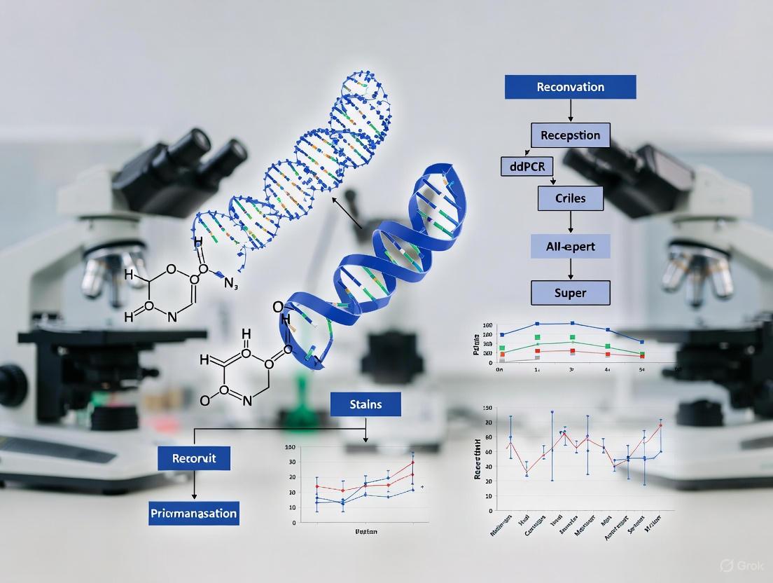
Digital PCR (dPCR) represents a paradigm shift in nucleic acid quantification by enabling absolute measurement of target DNA or RNA without reliance on external standard curves. This method is particularly transformative for monitoring Human Immunodeficiency Virus (HIV) reservoirs in research involving CCR5Δ32/Δ32 allogeneic Hematopoietic Stem Cell Transplantation (HSCT), a promising pathway for achieving HIV cure [8] [4]. Unlike quantitative real-time PCR (qPCR), which provides relative quantification based on a standard curve, dPCR partitions a sample into thousands of individual reactions, counts the positive and negative partitions, and uses Poisson statistics to calculate the absolute copy number of the target molecule in the original sample [20] [21]. This core principle underpins a more accurate, reproducible, and sensitive method for quantifying the size of the persistent viral reservoir, a critical parameter in evaluating the success of curative interventions [6].
Core Principles and Comparative Advantages
The workflow of dPCR is fundamentally different from that of qPCR. In the context of HIV reservoir quantification, the sample (typically genomic DNA from peripheral blood mononuclear cells - PBMCs or CD4+ T cells) is partitioned into numerous nanoliter-sized droplets or microchambers [21]. This partitioning results in a binary outcome for each partition after PCR amplification: positive (fluorescent) for the presence of the HIV target or negative (non-fluorescent). The fraction of negative partitions is used in a Poisson correction formula to determine the absolute concentration of the target, expressed as copies per microliter of input or, more commonly, copies per million cells [6] [20].
The following table summarizes the key methodological differences and advantages of dPCR over qPCR for HIV reservoir quantification.
Table 1: Comparison of qPCR and dPCR for HIV Reservoir Quantification
| Feature | Quantitative PCR (qPCR) | Digital PCR (dPCR) |
|---|---|---|
| Quantification Basis | Relative to a standard curve [21] | Absolute, via Poisson statistics [20] [21] |
| Standard Curve | Required, prone to variability [21] | Not required [6] [21] |
| Sensitivity | High | Superior, especially for low-abundance targets [6] |
| Precision & Reproducibility | Subject to standard curve quality | High, with lower inter-assay variability [6] |
| Tolerance to Inhibitors | Moderate | High, due to sample partitioning [21] |
| Key Application in HIV Research | Total HIV DNA quantification | High-precision reservoir sizing post-therapy [6] [4] |
The advantages of dPCR are critical for HIV reservoir studies after CCR5Δ32 HSCT. The method's high sensitivity allows for the detection of rare HIV-DNA positive cells in patients where the reservoir has been dramatically reduced [4]. Furthermore, its absolute quantification eliminates inaccuracies introduced by unstable calibrators, such as the 8E5 cell line, which has been shown to lose HIV DNA over time, leading to qPCR overestimation [21].
Application in HIV Reservoir Quantification Post-CCR5Δ32 HSCT
Following CCR5Δ32/Δ32 HSCT, patients require meticulous monitoring to assess the size and dynamics of the remaining HIV reservoir. Droplet digital PCR (ddPCR) has been instrumental in this endeavor, providing the sensitivity needed to detect trace levels of viral DNA. A 2023 study of a patient with long-term HIV-1 remission after CCR5Δ32/Δ32 HSCT utilized ddPCR to sporadically detect traces of HIV DNA in T cell subsets and tissue-derived samples over a nine-year period [4]. Despite these trace signals, the absence of replication-competent virus confirmed by other assays provided strong evidence for a cure, highlighting the need for ultra-sensitive detection methods [4].
A 2025 study developed a duplex dPCR assay on a microfluidic chamber array platform (Absolute Q) to quantify total HIV DNA targeting the LTR region and the human RPP30 gene as a reference [6]. The performance characteristics of this assay, as detailed below, demonstrate its suitability for clinical research.
Table 2: Performance Metrics of a Representative dPCR Assay for HIV DNA Quantification
| Performance Parameter | Result |
|---|---|
| Linearity (R²) | 0.977 [6] |
| 95% Lower Limit of Detection (LLOD) | 79.7 HIV DNA copies/10⁶ cells [6] |
| Limit of Quantification (LOQ) | 5 HIV copies/reaction [6] |
| Repeatability (CV% intra-assay) | 8.7% at 1,250 copies/10⁶ cells [6] |
| Reproducibility (CV% inter-assay) | 10.9% at 1,250 copies/10⁶ cells [6] |
| Median HIV DNA in ART-treated PWH | 995.3 copies/10⁶ CD4+ T cells [6] |
The workflow for such an assay, from sample processing to data analysis, is visualized in the following diagram.
Detailed Experimental Protocol: Duplex ddPCR for Total HIV DNA
This protocol outlines the steps for absolute quantification of total HIV DNA in patient PBMCs using a duplex ddPCR assay targeting HIV-LTR and the reference gene RPP30 [6].
I. Sample Preparation and DNA Extraction
- Isolate PBMCs from whole blood using standard Ficoll density gradient centrifugation.
- Extract genomic DNA from the PBMC pellet using a commercial kit (e.g., ExtractDNA Blood and Cells Kit, Evrogen) or the phenol-chloroform method.
- Quantify DNA using a spectrophotometer (e.g., NanoPhotometer P-Class). Ensure the A260/A280 ratio is between 1.8 and 2.0 for purity. Dilute DNA to a working concentration of 50-100 ng/μL in nuclease-free water or TE buffer.
II. ddPCR Reaction Setup
- Prepare the master mix on ice. A single 20-22 μL reaction may contain:
- 10 μL of 2x ddPCR Supermix for Probes (No dUTP).
- 900 nM each of forward and reverse primers for HIV-LTR.
- 900 nM each of forward and reverse primers for RPP30.
- 250 nM of FAM-labeled probe for HIV-LTR.
- 250 nM of VIC/HEX-labeled probe for RPP30.
- Nuclease-free water.
- Add DNA template (2-5 μL, typically 200-500 ng total) to the master mix. Gently pipette to mix. Include a no-template control (NTC) with water.
- Generate droplets using an automated droplet generator (e.g., QX200 Droplet Generator, Bio-Rad). Transfer the entire reaction mixture and droplet generation oil into the designated cartridge. The generator will produce ~20,000 nanoliter-sized droplets per sample.
III. PCR Amplification
- Carefully transfer the generated emulsion to a 96-well PCR plate. Seal the plate with a foil heat seal.
- Place the plate in a thermal cycler and run the following protocol:
- Enzyme Activation: 10 min at 95°C.
- Amplification (40 cycles): 30 s at 94°C (denaturation), 60 s at 60°C (annealing/extension). Note: Use a ramp rate of 2 °C/s.
- Enzyme Deactivation: 10 min at 98°C.
- Hold: 4°C ∞.
IV. Data Acquisition and Analysis
- Read the plate in a droplet reader (e.g., QX200 Droplet Reader, Bio-Rad). The reader will flow droplets one-by-one and measure the fluorescence in the FAM and VIC/HEX channels.
- Analyze the data using the associated software (e.g., QuantaSoft, Bio-Rad).
- Set appropriate fluorescence amplitude thresholds to clearly distinguish positive and negative droplet populations for each channel.
- The software will automatically apply Poisson statistics to calculate the absolute concentration (copies/μL) of HIV-LTR and RPP30 in the original reaction.
- Normalize the results: Calculate the HIV DNA copies per million cells using the RPP30 concentration (assuming two copies of RPP30 per diploid cell).
- HIV DNA copies/10⁶ cells = ( [HIV-LTR] / [RPP30] ) x 1,000,000
The Scientist's Toolkit: Essential Research Reagents
The following table lists key reagents and materials required for implementing the ddPCR assay for HIV reservoir quantification.
Table 3: Essential Research Reagents for HIV DNA ddPCR Assay
| Reagent/Material | Function | Example |
|---|---|---|
| Primers & Probes (HIV LTR) | Amplify and detect a conserved region of the HIV genome [6] | Custom TaqMan Assays [6] |
| Primers & Probes (Reference Gene) | Amplify and detect a single-copy human gene for normalization [6] | RPP30 Assay [6] |
| ddPCR Supermix | Optimized buffer, enzymes, and dNTPs for droplet-based digital PCR | ddPCR Supermix for Probes (Bio-Rad) |
| Droplet Generation Oil | Immiscible oil for generating stable, monodisperse water-in-oil emulsions | Droplet Generation Oil for Probes (Bio-Rad) |
| dgDNA Digestion Enzyme | Optional enzyme to reduce background from high molecular weight genomic DNA | |
| Droplet Generator & Reader | Instrumentation for automated droplet generation and fluorescence reading | QX200 system (Bio-Rad) or equivalent [21] |
| Commercial dPCR System | Integrated microfluidic chamber array system for automated workflow | Absolute Q (Thermo Fisher) [6] |
The principle of absolute quantification without standard curves establishes dPCR as a superior technology for precise and reliable measurement of the HIV reservoir. In the advanced research context of CCR5Δ32/Δ32 HSCT, where the viral reservoir is minimal, the high sensitivity and absolute accuracy of dPCR are indispensable for distinguishing between true cure and long-term remission. As therapeutic strategies evolve, dPCR will remain a cornerstone analytical tool for validating the efficacy of next-generation HIV cure interventions.
The quantification of persistent HIV-1 reservoirs remains a significant challenge in the development of curative interventions, particularly following innovative therapeutic approaches such as hematopoietic stem cell transplantation (HSCT) with CCR5Δ32/Δ32 cells. Droplet digital PCR (ddPCR) has emerged as a critical technology in this field due to its ability to provide absolute quantification of viral DNA molecules without requiring a standard curve, offering superior accuracy, precision, and reproducibility compared to quantitative PCR (qPCR) [22]. This application note details the design and validation of ddPCR assays targeting HIV Long Terminal Repeat (LTR) regions alongside the human reference gene RPP30, specifically framed within the context of monitoring HIV-1 persistence in patients who have undergone CCR5Δ32 HSCT—a therapeutic intervention that has led to documented cases of HIV cure [7] [3].
The clinical relevance of these assays is underscored by studies of patients who have received CCR5Δ32/Δ32 allogeneic hematopoietic stem cell transplants, where highly sensitive reservoir quantification methods are essential for confirming cure. Research indicates that high-frequency CCR5 editing (>90%) in hematopoietic stem progenitor cells (HSPCs) is likely necessary to confer protective benefit against HIV replication, emphasizing the need for precise monitoring tools [3]. The assays described herein enable researchers to accurately measure the size and dynamics of residual HIV reservoirs across diverse anatomical sites, providing critical insights into the efficacy of CCR5-targeted curative interventions.
Assay Design and Theoretical Framework
HIV LTR Target Selection
The HIV-1 LTR region plays a pivotal role in viral integration and gene expression, making it a suitable target for reservoir quantification. When designing ddPCR assays for HIV reservoir studies, multiple regions of the viral genome should be targeted to distinguish between intact and defective proviruses:
- Total HIV DNA assays typically target conserved regions such as LTR-gag or RU5 sequences to provide an overall measure of viral persistence [22] [23].
- Integrated HIV DNA assays utilize a nested-ddPCR approach with Alu-forward primers and HIV Gag-reverse primers to specifically quantify proviral DNA that has integrated into the host genome [22].
- Multiplex ddPCR assays can simultaneously target the HIV packaging signal (ψ) and envelope (env) genes to discriminate intact versus defective proviruses, as demonstrated in studies of the "London patient" where such approaches confirmed the absence of replication-competent virus in various tissues [7].
Reference Gene Selection: RPP30 as an Optimal Control
The RPP30 (ribonuclease P protein subunit p30) gene located on human chromosome 10 (10q23.31) has emerged as an superior reference gene for ddPCR-based HIV reservoir studies due to its highly conserved sequence, stable expression across human tissues, and presence as a single-copy gene in the diploid human genome [24] [25]. Unlike traditional reference genes such as β-actin and GAPDH, whose expression can vary under different experimental conditions, RPP30 demonstrates minimal expression fluctuations, making it particularly suitable for normalizing cell-associated HIV DNA measurements [24].
The RPP30 ddPCR assay enables precise determination of cell counts by quantifying the number of haploid genome equivalents in a sample, as each nucleated cell contains two copies of the RPP30 gene. This approach provides a more accurate normalization method compared to conventional cell counting techniques, especially when working with complex tissue samples or archived specimens where cell viability may be compromised [25].
Table 1: Key Characteristics of RPP30 as a Reference Gene
| Characteristic | Description | Significance for HIV Reservoir Studies |
|---|---|---|
| Genomic Location | Chromosome 10 (10q23.31) | Single locus reduces copy number variation |
| Copy Number | Two copies per diploid cell | Enables precise cell quantification |
| Sequence Conservation | Highly conserved across species | Useful for non-human primate studies |
| Expression Stability | Maintains stable expression across tissues | Reliable normalization across reservoir sites |
| Assay Performance | Compatible with ddPCR technology | Accurate absolute quantification |
Experimental Protocols
Sample Processing and DNA Extraction
Proper sample processing is critical for accurate HIV DNA quantification. The following protocol applies to various sample types, including peripheral blood mononuclear cells (PBMCs), lymph node tissue, and gastrointestinal tissue.
Protocol:
- Cell Isolation: Isolate PBMCs using Ficoll density gradient centrifugation. For tissue samples, process using mechanical dissociation and enzymatic digestion (e.g., collagenase) to generate single-cell suspensions [22].
- Cell Counting: Perform initial cell counting using automated or manual methods to approximate cell input.
- DNA Extraction: Extract genomic DNA using silica-membrane based kits or phenol-chloroform methods. The "ExtractDNA Blood and Cells Kit" (Evrogen) has been successfully used in prior HIV reservoir studies [8].
- DNA Quantification and Quality Assessment: Measure DNA concentration and purity using spectrophotometry (e.g., NanoPhotometer). Acceptable 260/280 ratios typically range from 1.8-2.0 [8].
- DNA Storage: Store extracted DNA at -20°C to -80°C until ddPCR analysis.
ddPCR Assay for Total HIV DNA with RPP30 Normalization
This duplex ddPCR protocol allows for simultaneous quantification of HIV DNA and the RPP30 reference gene in a single reaction.
Reagent Setup:
- ddPCR Supermix for Probes (no dUTP): 10-11 μL per reaction
- HIV Primer/Probe Mix: Final concentration 250 nM primers, 500 nM probe [22]
- RPP30 Primer/Probe Mix: Final concentration 250 nM primers, 500 nM probe [25]
- DNA Template: 5-100 ng per reaction (optimal input: 5 ng/μL) [22]
- Nuclease-free Water: to adjust total volume to 20-22 μL
Table 2: Primer and Probe Sequences for HIV LTR and RPP30 Assays
| Target | Primer/Probe | Sequence (5' to 3') | Final Concentration |
|---|---|---|---|
| HIV LTR | Forward Primer | Custom sequence targeting conserved LTR region | 250 nM |
| Reverse Primer | Custom sequence targeting conserved LTR region | 250 nM | |
| Probe | FAM-labeled, e.g., FAM-5'-[sequence]-3'-BHQ1 | 500 nM | |
| RPP30 | Forward Primer | e.g., AGATTTGGACCTGCGAGCG [25] | 250 nM |
| Reverse Primer | e.g., GAGCGGCTGTCTCCACAAGT [25] | 250 nM | |
| Probe | HEX-labeled, e.g., HEX-5'-[sequence]-3'-BHQ1 | 500 nM |
Thermal Cycling Conditions:
- Step 1: Enzyme activation at 95°C for 10 minutes
- Step 2: 40 cycles of:
- Denaturation: 95°C for 30 seconds
- Annealing/Extension: 58.1°C for 60 seconds [22]
- Step 3: Enzyme deactivation at 98°C for 10 minutes
- Step 4: Signal stabilization at 4°C hold
Droplet Reading and Analysis:
- Read droplets using a droplet reader (e.g., QX200 Droplet Reader, Bio-Rad)
- Analyze data using associated software (e.g., QuantaSoft, Bio-Rad)
- Apply Poisson correction to determine absolute copy numbers of HIV DNA and RPP30
- Calculate HIV DNA copies per million cells using the formula: (HIV copies / RPP30 copies) × 1,000,000 × 2 [25]
Integrated HIV DNA Assay
The quantification of integrated HIV DNA requires a different approach to distinguish it from unintegrated forms.
Protocol:
- First-round PCR (Nested Pre-amplification):
- Use Alu-forward primer and HIV Gag-reverse primer with standard PCR reagents
- Thermal cycling conditions: 95°C for 2 min, followed by 25 cycles of 95°C for 30s, 60°C for 30s, 72°C for 3-4 min [22]
- Second-round ddPCR:
- Use 2-5 μL of first-round product as template
- Employ HIV-specific primers and probes (as in Table 2)
- Follow standard ddPCR conditions as described in section 3.2
Assay Validation and Quality Control
Rigorous validation is essential for generating reliable data in HIV reservoir studies.
Key Validation Parameters:
- Limit of Detection (LoD): Determine the lowest concentration of target detectable with 95% confidence. For HIV DNA assays, LoD of 29 copies per million PBMCs has been reported [23].
- Limit of Quantification (LoQ): Establish the lowest concentration that can be accurately quantified. For HIV DNA, LoQ of 1 copy/μL has been demonstrated [22].
- Linearity: Assess over a range of DNA inputs (0.2-100 ng/μL) [22].
- Precision: Evaluate intra-assay and inter-assay variability, with coefficients of variation <10% considered acceptable.
- Specificity: Verify absence of signal in HIV-negative controls.
Data Analysis and Interpretation
Calculation Methods
For HIV DNA copies per million cells: [ \text{HIV copies per million cells} = \frac{\text{HIV copies}}{\text{RPP30 copies}} \times 1,000,000 \times 2 ]
For percentage of HIV-infected cells: [ \% \text{ infected cells} = \frac{\text{HIV copies}}{\text{RPP30 copies}} \times 100\% ]
For cell counts based on RPP30 quantification: [ \text{Cell count} = \frac{\text{RPP30 copies}}{2} ]
Expected Results and Performance Characteristics
Based on validated assays, researchers can expect the following performance metrics:
Table 3: Performance Characteristics of HIV Reservoir ddPCR Assays
| Parameter | Total HIV DNA Assay | Integrated HIV DNA Assay | RPP30 Reference Assay |
|---|---|---|---|
| Limit of Detection | 1 copy/μL [22] | Not specified | 100 pg gDNA [25] |
| Limit of Quantification | 29 copies/million PBMCs [23] | Not specified | 50 cells [25] |
| Linear Range | 0.2-100 ng/μL DNA input [22] | Not specified | 100-100,000 pg gDNA [25] |
| Precision (CV) | <10% | <10% | <7.6% at 100 pg [25] |
| Clinical Sensitivity | Detects 4 copies/10^6 cells [22] | Detects 10^3-10^4 copies/10^6 cells [22] | Highly sensitive |
Applications in CCR5Δ32 HSCT Research
The exceptional sensitivity of these ddPCR assays makes them particularly valuable for monitoring HIV reservoir dynamics in patients who have undergone CCR5Δ32 HSCT. In the landmark "London patient" case, multiplex ddPCR targeting HIV ψ and env sequences demonstrated the absence of intact proviral DNA in various tissues, contributing to the declaration of cure [7]. The ability to detect HIV DNA at frequencies as low as 4 copies per million cells enables researchers to document profound reservoir reduction following CCR5-targeted interventions [22].
When applied to clinical samples from CCR5Δ32 HSCT recipients, these assays typically reveal:
- Substantial reduction in total HIV DNA levels across multiple anatomical compartments
- Absence of detectable intact provirus in successful cases
- Very low-level positive signals in some tissues (e.g., 33 LTR copies/10^6 cells in lymph nodes) without detection of replication-competent virus [7]
- Correlation between high donor chimerism (>90%) and undetectable reservoir size [7]
The Scientist's Toolkit
Table 4: Essential Research Reagents and Materials
| Reagent/Material | Function | Example Product/Specification |
|---|---|---|
| ddPCR Supermix for Probes | Partitioning and amplification of target sequences | Bio-Rad ddPCR Supermix for Probes (no dUTP) |
| HIV LTR Primer/Probe Set | Detection and quantification of HIV sequences | Custom-designed primers/probes targeting conserved LTR regions |
| RPP30 Primer/Probe Set | Reference gene for normalization | Pre-validated assays [25] |
| Droplet Generation Oil | Creation of nanoliter-sized droplets | Droplet Generation Oil for Probes (Bio-Rad) |
| DG8 Cartridges and Gaskets | Droplet generation hardware | DG8 Cartridges for QX200 system |
| DNA Extraction Kits | High-quality genomic DNA isolation | ExtractDNA Blood and Cells Kit (Evrogen) [8] |
| Nuclease-free Water | Reaction preparation without contamination | Molecular biology grade nuclease-free water |
| Positive Control DNA | Assay validation and quality control | DNA from HIV-infected cell lines (e.g., Molt3-IIIB) |
Visual Workflows
Diagram 1: Comprehensive workflow for HIV reservoir quantification using ddPCR, highlighting the integration of RPP30 normalization throughout the process.
Diagram 2: Stepwise assay validation process ensuring reliability for HIV reservoir quantification in CCR5Δ32 HSCT research.
The quantification of persistent HIV reservoirs is a critical challenge in cure research, particularly in the context of CCR5Δ32/Δ32 allogeneic hematopoietic stem cell transplantation (HSCT). This intervention, which has led to documented cases of HIV cure, aims to replace the patient's immune system with donor cells that lack the primary CCR5 co-receptor essential for viral entry [11] [4]. Digital PCR (dPCR) has emerged as a vital tool for precisely measuring the dramatic reductions in viral reservoir size following such interventions, enabling researchers to distinguish between true cure and long-term remission [26] [5].
This technical note provides a comparative analysis of two primary dPCR platforms—microfluidic chamber arrays and droplet-based systems—for HIV DNA quantification in the specialized context of post-CCR5Δ32 HSCT research. We present performance data, detailed protocols, and analytical considerations to guide platform selection for monitoring HIV reservoir dynamics in cure studies.
Platform Comparison: Technical Specifications and Performance
dPCR platforms partition samples into thousands of individual reactions, enabling absolute nucleic acid quantification without standard curves. The method of partitioning represents the key distinction between systems.
Table 1: Technical Comparison of dPCR Platforms for HIV Reservoir Quantification
| Feature | Microfluidic Chamber Array (e.g., Absolute Q) | Droplet-Based Systems (e.g., QX200 ddPCR) |
|---|---|---|
| Partition Type | Pre-fabricated microchambers on a chip [6] | Nanosized water-in-oil droplets [26] |
| Partition Number | ~ 25,000-30,000 per sample [6] [26] | ~ 20,000 per sample (QX200) [26] |
| Throughput | Fully automated partitioning, thermocycling, and imaging [6] | Requires separate droplet generation and reading steps [26] |
| HIV DNA Assay Linear Range | 78 - 5,000 copies/10⁶ cells (R² = 0.977) [6] | Effectively quantifies from >100 to 3,000 copies/10⁶ cells in ART-suppressed individuals [5] |
| Limit of Detection (95%) | 79.7 HIV DNA copies/10⁶ cells [6] | Similar sensitivity to qPCR, but with higher precision at low levels [5] |
| Precision (Coefficient of Variation) | 8.7% at 1,250 copies/10⁶ cells; 26.9% at 150 copies/10⁶ cells [6] | Improved precision over qPCR, especially for HIV DNA and 2-LTR circles [5] |
| Key Advantage for HIV Research | Automated workflow minimizes hands-on time and variability [6] | Extensive published validation for HIV reservoir studies and better tolerance of sequence mismatches [5] |
Application in HIV-1 Cure and CCR5Δ32 HSCT Research
Both platforms are instrumental in validating HIV cure, as demonstrated by their use in landmark studies of patients who received CCR5Δ32/Δ32 stem cell transplants.
Table 2: Representative dPCR Findings in Documented Cases of HIV Cure
| Research Case (Patient) | Key dPCR Findings | Platform Used |
|---|---|---|
| The London Patient [11] | No replication-competent virus in blood, CSF, semen, intestinal, or lymphoid tissue at 30 months post-ATI. A very low-level positive signal for HIV DNA was recorded in peripheral CD4 memory cells at 28 months, deemed a "fossil" trace. | Droplet Digital PCR (ddPCR) |
| The Düsseldorf Patient [4] | Sporadic traces of HIV DNA detected in T cell subsets and tissue samples. Repeated viral outgrowth assays in humanized mice did not reveal replication-competent virus. The patient remained in remission 48 months after treatment interruption. | Droplet Digital PCR (ddPCR) |
| General HIV Reservoir Profiling [6] | Total HIV DNA was successfully quantified in 50 ART-treated individuals, with a median of 995.3 copies/10⁶ CD4+ T cells. The assay demonstrated high specificity with no false positives in HIV-negative controls. | Microfluidic Chamber Array (Absolute Q) |
Experimental Protocols
Total HIV DNA Quantification using a Microfluidic Chamber Array System
This protocol is adapted from a 2025 study that developed a duplex assay for total HIV DNA on the Absolute Q platform [6].
Workflow Overview:
Step-by-Step Procedure:
Sample Input and DNA Extraction:
- Input: Process peripheral blood mononuclear cells (PBMCs) or purified CD4+ T cells from patients. For post-HSCT patients, ensure high donor chimerism is confirmed.
- Extraction: Isolate genomic DNA using a commercial kit (e.g., DNeasy Blood and Tissue Kit, Qiagen). Determine DNA concentration and purity using a spectrophotometer.
Reaction Mixture Preparation:
- Prepare a duplex PCR reaction mix containing:
- 1X Absolute Q ddPCR Supermix.
- 900 nM each of forward and reverse primers targeting the HIV-1 LTR-RU5 region.
- 250 nM each of the FAM-labeled probe for HIV-1 LTR and the VIC-labeled probe for the reference gene RPP30.
- Approximately 50-100 ng of sample DNA per reaction.
- Adjust the final volume with nuclease-free water.
- Prepare a duplex PCR reaction mix containing:
Automated Partitioning and PCR Amplification:
- Load the reaction mixture into an Absolute Q dPCR chip.
- Place the chip into the Absolute Q instrument. The system will automatically:
- Partition the sample into tens of thousands of nanoliter-scale microchambers.
- Perform endpoint PCR with the following cycling conditions, optimized for the duplex assay [6]:
- Denaturation: 96°C for 10 seconds
- Annealing/Extension: 60°C for 50 seconds
- Number of cycles: 40
Image Acquisition and Analysis:
- The integrated imager scans the chip to detect fluorescence in each microchamber.
- Use the instrument's software to analyze the 2D plot, setting thresholds to distinguish positive (FAM+ for HIV, VIC+ for RPP30) and negative partitions.
Data Calculation and Normalization:
- The software uses Poisson statistics to calculate the absolute concentration of HIV DNA targets in copies/μL of the input.
- Normalize the result to the number of human diploid cells using the RPP30 reference gene and express the final result as HIV DNA copies per million cells.
Protocol for HIV Reservoir Analysis in Post-HSCT Patients via ddPCR
This protocol reflects the methodologies used in key studies of the London and Düsseldorf patients to comprehensively evaluate viral reservoirs after transplant [11] [4].
Workflow Overview:
Step-by-Step Procedure:
Sample Collection from Multiple Compartments:
- Collect longitudinal samples from the patient to assess systemic viral elimination. Key compartments include:
Nucleic Acid Extraction from Various Samples:
- PBMCs and Tissue Biopsies: Homogenize tissue samples using a benchtop homogenizer with ceramic beads. Extract total DNA from PBMCs and homogenized tissue using a validated kit (e.g., QIAamp DNA Mini Kit). The quality of the extracted DNA is critical for assay performance [11].
- Plasma and CSF: Use an ultracentrifugation step (e.g., 21,000 g for 2 hours) to concentrate viral particles before RNA extraction for viral load testing [11].
Droplet Digital PCR Assays:
- Target multiple regions of the viral genome to distinguish between intact and defective proviruses. Common targets include:
- Prepare the ddPCR reaction mix according to manufacturer specifications (Bio-Rad) and load it into a DG8 cartridge for droplet generation.
Droplet Generation and PCR Amplification:
- Generate droplets using the QX200 Droplet Generator.
- Transfer the emulsified sample to a 96-well PCR plate.
- Seal the plate and perform PCR amplification on a conventional thermal cycler using assay-specific cycling conditions.
Droplet Reading and Threshold Determination:
- Read the droplets using the QX200 Droplet Reader.
- Analyze data with QuantaSoft software. Critical Note: Carefully set fluorescence amplitude thresholds to distinguish positive from negative droplets. The presence of "rain" (droplets with intermediate fluorescence) and rare false-positive droplets in negative controls are known challenges that require data-driven thresholding methods for accurate quantification [26] [5].
Data Integration and Interpretation:
The Scientist's Toolkit: Essential Reagents and Materials
Table 3: Key Research Reagent Solutions for HIV Reservoir dPCR
| Item | Function/Application | Example Products/Assays |
|---|---|---|
| DNA Extraction Kit | Isolation of high-quality genomic DNA from PBMCs and tissue homogenates. Critical for assay sensitivity. | QIAamp DNA Blood & Tissue Kits, DNeasy Blood and Tissue Kit [11] [8] |
| dPCR Supermix | Optimized master mix for digital PCR, containing DNA polymerase, dNTPs, and buffer. | Absolute Q ddPCR Supermix, ddPCR Supermix for Probes (Bio-Rad) [6] |
| Primers/Probes for HIV Targets | For amplification and detection of specific HIV sequences (LTR, gag, env, ψ). | Custom or published assays for total HIV DNA, 2-LTR circles, and the Intact Proviral DNA Assay (IPDA) [11] [6] [5] |
| Reference Gene Assay | For normalization of cell number in the sample. | RNase P (RPP30) assay, labeled with a different fluorophore (e.g., VIC/HEX) [11] [6] |
| Droplet Generation Oil | Creates the water-in-oil emulsion for partitioning in ddPCR systems. | DG8 Cartridges and Droplet Generation Oil for Probes (Bio-Rad) |
| No-Template Control (NTC) | Essential control to monitor for contamination and set baseline for false-positive signals. | Nuclease-free water [5] |
The choice between microfluidic chamber array and droplet-based dPCR systems depends on the specific needs of the HIV cure research project.
- Choose a Microfluidic Chamber Array system when prioritizing a fully automated, streamlined workflow with minimal hands-on time. Its integrated design reduces operator-dependent variability, making it suitable for processing larger sample batches in a standardized manner [6].
- Choose a Droplet-Based system when the research requires deep, multi-target characterization of the reservoir and needs to be directly benchmarked against a vast body of existing literature. Its extensive validation in landmark cure studies [11] [4], combined with its higher tolerance for sequence mismatches [5], makes it the current gold-standard for the most critical analyses in the field.
For definitive proof of cure in CCR5Δ32 HSCT patients, data from dPCR should be integrated with other sophisticated assays, such as quantitative viral outgrowth assays (QVOA) and in vivo testing in humanized mouse models, to confirm the absence of any replication-competent virus [4].
This application note details a droplet digital PCR (ddPCR) protocol optimized for the precise quantification of the HIV reservoir and the detection of the CCR5Δ32 mutation in the context of allogeneic hematopoietic stem cell transplantation (HSCT) research.
The quantification of persistent HIV proviral reservoirs is a central challenge in the pursuit of a cure. The existence of latently infected cells, which harbor replication-competent virus, necessitates lifelong antiretroviral therapy (ART), as these reservoirs can reactivate if treatment is interrupted [27]. Allogeneic hematopoietic stem cell transplantation from a donor with a homozygous CCR5Δ32 mutation (CCR5Δ32/Δ32 HSCT) has been established as a viable, albeit complex, path to HIV remission and cure [4]. This mutation confers resistance to the most common strain of HIV by eliminating a crucial co-receptor required for viral entry.
A critical component of this research is the accurate quantification of two key metrics: the size of the residual HIV reservoir and the level of donor chimerism, particularly the frequency of the CCR5Δ32 allele. ddPCR is uniquely suited for this task due to its ability to provide absolute quantification of nucleic acids without a standard curve, its high sensitivity, and its superior precision for detecting low-abundance targets compared to quantitative real-time PCR (qPCR) [5] [28]. This protocol describes a validated ddPCR workflow for these applications.
Key Reagent Solutions
Table 1: Essential research reagents for ddPCR-based HIV and CCR5Δ32 analysis.
| Item | Function/Description | Example |
|---|---|---|
| ddPCR Supermix | Provides optimized reagents for PCR amplification in a droplet format. | ddPCR Supermix for Probes (No dUTP) [28] |
| Primer/Probe Sets | Target-specific assays for HIV DNA (e.g., LTR, gag, pol) and the CCR5Δ32 deletion. | Commercially validated or custom-designed hydrolysis (TaqMan) probes [8] [5] |
| Droplet Generation Oil | Creates a stable water-in-oil emulsion for partitioning the PCR reaction. | DG Droplet Generation Oil [28] |
| Droplet Generator | Microfluidic device for partitioning samples into thousands of nanoliter-sized droplets. | QX200 Droplet Generator [28] |
| Droplet Reader | Instrument for flowing droplets and detecting end-point fluorescence in each droplet. | QX200 Droplet Reader [5] |
| Nucleic Acid Extraction Kit | For isolation of high-quality genomic DNA from patient samples (e.g., PBMCs, tissues). | Phenol-chloroform or commercial kits (e.g., ExtractDNA Blood and Cells Kit) [8] |
Comparative Performance Data
Table 2: Quantitative comparison of ddPCR and qPCR for HIV reservoir quantification.
| Parameter | Droplet Digital PCR (ddPCR) | Quantitative PCR (qPCR) |
|---|---|---|
| Quantification Method | Absolute, without a standard curve [28] | Relative, requires a standard curve [5] |
| Precision & Reproducibility | High precision and improved reproducibility for low-level targets [5] | Lower precision, especially at low target concentrations [5] |
| Sensitivity (Limit of Detection) | Can detect down to 0.8% mutant alleles in a wild-type background [8]; suitable for single-copy detection [29] | Similar sensitivity in some studies, but can be affected by PCR inhibitors [5] |
| Robustness to Inhibitors | More tolerant to PCR inhibitors due to sample partitioning [28] | Susceptible to inhibition, which reduces amplification efficiency [28] |
| Tolerance to Sequence Variation | Better tolerates primer/probe mismatches, advantageous for highly variable viruses like HIV [5] | Mismatches can significantly impair amplification efficiency and quantification accuracy [5] |
Detailed Experimental Protocol
Sample Preparation and DNA Extraction
- Source: Collect peripheral blood mononuclear cells (PBMCs) from patients pre- and post-CCR5Δ32/Δ32 HSCT. Tissue biopsies (e.g., lymph node, gut) can also be analyzed for reservoir distribution [4].
- Extraction: Isate genomic DNA using a standardized method, such as the phenol-chloroform protocol or a commercial kit (e.g., ExtractDNA Blood and Cells Kit) [8].
- Quantification: Precisely measure DNA concentration and purity using a spectrophotometer (e.g., NanoPhotometer). Ensure 260/280 ratios are ~1.8-2.0 [8].
ddPCR Reaction Setup
- Prepare Reaction Mix: On ice, assemble the following for each sample in a 22 µL total volume [28]:
- 11 µL of 2x ddPCR Supermix for Probes (No dUTP)
- Forward and reverse primers (optimized concentration, typically 0.9 µM each)
- Hydrolysis (TaqMan) probe (optimized concentration, typically 0.25 µM)
- 2 µL of extracted genomic DNA template (optimize amount based on expected target concentration)
- Nuclease-free water to 22 µL
- Include Controls: Always run negative template controls (NTCs, with water instead of DNA) and positive controls (e.g., DNA with known copies of the target) in duplicate to monitor for contamination and assay performance [28].
Droplet Generation and PCR Amplification
- Generate Droplets: Transfer 20 µL of the reaction mix into a DG8 cartridge. Add 70 µL of Droplet Generation Oil to the appropriate well. Place the cartridge in the QX200 Droplet Generator. This instrument partitions each sample into approximately 20,000 nanoliter-sized droplets [30] [28].
- Transfer Emulsion: Carefully transfer the generated water-in-oil emulsion from the cartridge to a 96-well PCR plate. Seal the plate with a foil heat seal.
- PCR Amplification: Place the sealed plate in a thermal cycler and run the following profile:
- Enzyme Activation: 95°C for 10 minutes.
- Amplification: 40 cycles of:
- Denaturation: 94°C for 30 seconds.
- Annealing/Extension: 55-60°C (assay-specific) for 60 seconds.
- Enzyme Deactivation: 98°C for 10 minutes.
- Hold: 4°C ∞.
- Ramp rate: 2°C/second.
Data Acquisition and Poisson Statistical Analysis
- Read Droplets: Place the PCR plate in the QX200 Droplet Reader. This instrument aspirates droplets from each well and flows them single-file past a two-color optical detection system. Each droplet is classified as positive or negative for the target based on its fluorescence amplitude [29] [5].
- Analyze Data: Use the instrument's accompanying software (e.g., QuantaSoft) to analyze the results. The software applies Poisson statistics to the fraction of positive droplets (λ) to calculate the absolute concentration of the target DNA in copies per microliter of the original reaction mix, using the formula: ( \text{Concentration (copies/µL)} = -\ln(1 - p) / V ) where ( p ) is the ratio of positive droplets to total droplets, and ( V ) is the volume of each droplet in microliters [29] [20]. This calculation accounts for the fact that some droplets may contain more than one target molecule.
The quantification of total HIV DNA is a cornerstone technique in HIV cure research, particularly for evaluating the impact of therapeutic interventions like hematopoietic stem cell transplantation (HSCT). This application note provides a detailed protocol and a consolidated summary of key quantitative data for researchers measuring the HIV reservoir using droplet digital PCR (ddPCR) in the context of CCR5Δ32/Δ32 allogeneic HSCT and other curative strategies.
Accurate measurement of the viral reservoir is essential, as a significant reduction in its size is a primary indicator of therapeutic success. The data and methods outlined here are critical for monitoring patients post-treatment and advancing the development of a functional HIV cure.
HIV Reservoir Quantification in Key Clinical Cases
The following table summarizes the total HIV DNA measurements from seminal case studies and cohort studies, highlighting the profound reservoir reduction achievable through interventions like HSCT.
Table 1: Total HIV DNA in Patient Samples from Clinical Studies
| Study Participant / Cohort | Clinical Context | Sample Type | Total HIV DNA (copies per million cells) | Key Finding |
|---|---|---|---|---|
| IciS-34 (Nature Medicine, 2024) [13] | Sustained remission after wild-type CCR5 allo-HSCT | Peripheral Blood Mononuclear Cells (PBMCs) | 202 (pre-transplant) | Durable HIV remission for 32+ months post-ART interruption, despite wild-type CCR5 donor. |
| Bone Marrow Cells | 1,096 (pre-transplant) | |||
| Purified Blood CD4+ T cells | 457 (pre-transplant) | |||
| The London Patient (The Lancet HIV, 2020) [7] | HIV cure after CCR5Δ32/Δ32 allo-HSCT | Lymph Node Tissue | 26.1 (env) | No replication-competent virus detected in multiple tissues, suggesting cure. |
| Memory CD4+ T Cells | Very low-level positive signal | |||
| Intestinal Tissue | Undetectable | |||
| Children with Perinatal HIV (JCI, 2025) [31] | Long-term ART initiated in infancy | Naive CD4+ T Cells | Median 33 (corrected for contamination) | Naive CD4+ T cells are a significant and distinct reservoir in children, contributing a median of 13.5% to the total infected cell pool. |
| Memory CD4+ T Cells | Median 975 | |||
| PWH on Stable ART (Scientific Reports, 2025) [6] | Cross-sectional cohort study on ART | CD4+ T Cells | Median 995.3 (IQR: 646.9 - 1,572) | The duplex dPCR assay robustly quantifies the reservoir in ART-treated individuals, showing levels significantly lower than in ART-naive individuals. |
| ART-naive PWH | PBMCs | Median 16,565 (IQR: 6,560 - 35,465) |
Detailed Experimental Protocol for Total HIV DNA Quantification via ddPCR
This section provides a step-by-step protocol for quantifying total HIV DNA from patient blood samples, adapted from established methodologies [32] [5] [33].
The following diagram illustrates the complete experimental workflow for total HIV DNA quantification, from sample collection to data analysis.
Sample Preparation and DNA Extraction
- PBMC Isolation: Collect whole blood in EDTA or heparin tubes. Isolate Peripheral Blood Mononuclear Cells (PBMCs) using standard Ficoll-Hypaque density gradient centrifugation. Isolate CD4+ T cells from PBMCs using immunomagnetic beads or Fluorescence-Activated Cell Sorting (FACS) for a more specific reservoir measurement [31] [6].
- gDNA Extraction: Extract genomic DNA (gDNA) from up to 10^7 PBMCs or CD4+ T cells using a commercial gDNA extraction kit, following the manufacturer's instructions. Elute DNA in nuclease-free water or TE buffer.
- DNA Quantification and Quality Control: Accurately measure the DNA concentration using a fluorometer. Assess purity by measuring the A260/A280 ratio (ideal range: 1.8-2.0). DNA should be stored at -20°C or -80°C.
Restriction Enzyme Digestion
Digesting gDNA is critical for reliable ddPCR results, as it disrupts DNA topology and ensures efficient amplification by exposing target sequences [33].
- Direct Digestion in ddPCR Mix (Recommended for simplicity) [33]:
- Assemble the ddPCR reaction mix at room temperature. Add 0.5–1 µL of a restriction enzyme (e.g., HaeIII or EcoRI-HF, providing 5–20 units) directly to the reaction mixture.
- The enzyme will be active during reaction setup and will be irreversibly inactivated during the first denaturation step of the PCR cycle.
- Digestion Prior to ddPCR (Alternative method) [33]:
- Set up a separate digestion reaction with 1 µg of gDNA, 1x appropriate restriction enzyme buffer, and 10 units of enzyme per µg of DNA.
- Incubate for 5–60 minutes at the enzyme's optimal temperature.
- Heat inactivation is optional. A cleanup step is not required; the digest can be directly added to the ddPCR reaction, ensuring it constitutes less than 1/10 of the final reaction volume.
ddPCR Reaction Setup and Amplification
This protocol is optimized for the Bio-Rad QX200 ddPCR system [32].
Table 2: ddPCR Reaction Setup
| Component | Final Concentration | Volume per 20 µL Reaction |
|---|---|---|
| 2x ddPCR Supermix for Probes (No dUTP) | 1x | 10 µL |
| 20x Primer/Probe Assay (FAM) | 1x | 1 µL |
| Target: HIV LTR or other conserved region | ||
| 20x Primer/Probe Assay (HEX/VIC) | 1x | 1 µL |
| Reference: Human single-copy gene (e.g., RPP30) | ||
| Restriction Enzyme (e.g., HaeIII) | 5–20 units | 0.5–1 µL |
| Template gDNA | 100 ng (or optimized amount) | X µL |
| Nuclease-free Water | - | To 20 µL |
- Prepare Reaction Mix: Combine all components in the order listed in a master mix, then aliquot 20 µL into the designated wells of a DG8 cartridge.
- Generate Droplets: Add 70 µL of Droplet Generation Oil to the oil wells of the cartridge. Place a DG8 gasket on the cartridge and load it into the QX200 Droplet Generator.
- Transfer Droplets: After droplet generation, carefully transfer 40 µL of the emulsified sample to a semi-skirted 96-well PCR plate. Seal the plate with a pierceable foil heat seal.
- PCR Amplification: Place the plate in a thermal cycler and run the following protocol:
- Enzyme Activation: 10 minutes at 95°C (if using direct digestion method).
- Amplification (40 cycles):
- Denature: 30 seconds at 94°C
- Anneal/Extend: 60 seconds at 60°C (Optimize temperature for primers/probes)
- Enzyme Deactivation: 10 minutes at 98°C.
- Hold: 4°C ∞.
- Use a ramp rate of 2°C/second.
Data Acquisition and Analysis
- Read Droplets: Place the PCR plate in the QX200 Droplet Reader. The instrument will automatically aspirate each sample, count the droplets, and detect the fluorescence (FAM and HEX) in each droplet.
- Analyze Data: Use the QuantaSoft software to analyze the results.
- Set appropriate fluorescence amplitude thresholds to clearly distinguish positive and negative droplets for each channel (FAM for HIV, HEX/VIC for the reference gene).
- The software applies Poisson statistics to calculate the absolute concentration of the target and reference gene in copies/µL of the reaction mix.
- Calculate Final Result:
- HIV DNA copies per million cells = ( [HIV copies/µL] / [RPP30 copies/µL] / 2 ) * 10^6
- Note: The division by 2 accounts for the two copies of the RPP30 gene per diploid human cell.
The Scientist's Toolkit: Research Reagent Solutions
Table 3: Essential Materials for HIV DNA ddPCR Quantification
| Item | Function/Description | Example |
|---|---|---|
| ddPCR System | Partitions samples into nanodroplets for absolute quantification. | Bio-Rad QX200 Droplet Generator & Reader [32] |
| Thermal Cycler | Executes the PCR amplification protocol. | C1000 Touch (Bio-Rad) with 96-well module [32] |
| ddPCR Supermix | Optimized reaction buffer for droplet-based digital PCR. | Bio-Rad 2x ddPCR Supermix for Probes [32] |
| Primers & Probes | Target-specific assays for HIV and a reference human gene. | HIV Assay: LTR-gag or similar [5] [6]Reference Assay: RPP30 (single-copy gene) [6] |
| Restriction Enzymes | Digest gDNA to improve target accessibility and assay accuracy. | HaeIII (recognition site: GG/CC) or EcoRI-HF (G/AATTC) [33] |
| Droplet Generation Consumables | Essential disposables for creating uniform droplets. | DG8 Cartridges, Gaskets, and Droplet Generation Oil [32] |
| DNA Standard | Control for assay validation and determining copy number per cell. | 8E5/ACH2 Cell Line DNA (contains one integrated HIV provirus per cell) [6] |
This application note consolidates critical quantitative data and a standardized protocol for HIV reservoir monitoring. The dramatic reduction of total HIV DNA to near-undetectable levels in HSCT case studies, such as the London patient and IciS-34, provides a key benchmark for success in HIV cure research. The detailed ddPCR methodology outlined here offers the precision, sensitivity, and robustness required to accurately measure these low reservoir levels, making it an indispensable tool for evaluating next-generation curative interventions.
Optimizing ddPCR Assay Performance for Maximum Sensitivity and Precision
The precise quantification of the human immunodeficiency virus (HIV) reservoir is a critical component in evaluating the success of curative strategies, particularly in the context of CCR5Δ32 hematopoietic stem cell transplantation (HSCT). This protocol details the application of droplet digital PCR (ddPCR) for the absolute quantification of intact HIV-1 proviral DNA, focusing on the critical optimization parameters of annealing temperature, oligonucleotide concentration, and cycle number. The methodologies outlined herein are derived from established, highly multiplexed ddPCR assays that provide a specific, sensitive, and reproducible measure of the replication-competent reservoir, enabling reliable monitoring of patients post-HSCT [34].
Background
Droplet digital PCR represents a significant advancement over traditional quantitative PCR (qPCR) by enabling the absolute quantification of nucleic acids without the need for a standard calibration curve [21]. In ddPCR, a sample is partitioned into thousands of nanoliter-sized droplets, with each droplet acting as an individual PCR reactor. Following amplification, droplets are analyzed to determine the fraction that contains the target sequence, allowing for absolute quantification via Poisson statistics [21] [34]. This technology is particularly suited for HIV reservoir quantification due to its high sensitivity, tolerance to PCR inhibitors, and ability to multiplex—probing several regions of the HIV genome simultaneously to distinguish intact proviruses from defective ones [34].
Key Parameters and Optimization
The accuracy and sensitivity of ddPCR are highly dependent on the meticulous optimization of several reaction parameters. The following table summarizes the core parameters and their optimized ranges for the multiplexed HIV-1 provirus assay.
Table 1: Key Optimized Parameters for Multiplexed HIV-1 ddPCR Assay
| Parameter | Recommended Range / Value | Function and Impact |
|---|---|---|
| Annealing Temperature | Optimized for each triplex assay (e.g., 60°C) | Ensures specific binding of multiple primer sets; critical for multiplexing efficiency and assay specificity [34]. |
| Oligonucleotide Concentration | Variable by target (e.g., FAM-low, FAM-high, HEX-low, HEX-high) | Allows discrimination of different targets using the same fluorophore; optimized to prevent signal overlap and ensure accurate droplet classification [34]. |
| Cycle Number | Standard ~40 cycles | Provides sufficient amplification for robust fluorescence signal in positive droplets while maintaining the digital nature of the assay. |
| Primer/Probe Specificity | Locked Nucleic Acids (LNAs) incorporated into probes | Enhances binding affinity and specificity, especially for conserved or hypermutated regions like env [34]. |
| Target Region Selection | 5 regions across HIV genome (e.g., LTR/gag, pol, tat, env) | Enables identification of proviruses likely to be intact; spaced over several kilobases to detect large deletions [34]. |
Annealing Temperature
The annealing temperature is a critical parameter, especially in a multiplexed assay format. An optimal, unified annealing temperature must be established for each triplex assay to ensure specific and efficient amplification of all targets simultaneously. For the HIV triplex assays described, this temperature was optimized to 60°C. This temperature provides a stringent enough environment to minimize non-specific binding and primer-dimer formation, which is paramount when probing for rare targets like intact HIV proviruses in a vast background of human genomic and defective viral DNA [34].
Oligonucleotide Concentration
In multiplex ddPCR, probe concentration is strategically used to differentiate between targets labeled with the same fluorophore. The described protocol uses a combination of "high" and "low" concentrations of FAM and HEX dyes for different targets within the same assay. For instance, in Assay 1, the pol target may be labeled with FAM-low and the tat target with FAM-high. This concentration differential creates distinct clusters of fluorescence amplitude on the ddPCR plot, allowing software to distinguish and count droplets positive for one or both FAM-labeled targets. This innovative approach effectively increases the multiplexing capacity without requiring additional fluorescent channels [34].
Cycle Number
The cycle number in ddPCR must be sufficient to amplify single-copy targets to a detectable level of fluorescence. The standard cycle number of ~40 is typically used. It is crucial that the reaction remains within the exponential phase for accurate end-point quantification. Over-cycling can lead to increased background fluorescence and false-positive calls, while under-cycling might fail to detect true positive droplets containing a single DNA molecule, thus reducing the assay's sensitivity.
Experimental Protocol: Multiplexed ddPCR for HIV-1 Reservoir Quantification
Research Reagent Solutions
The following table lists the essential materials and reagents required to perform the multiplexed ddPCR assay for intact HIV-1 provirus quantification.
Table 2: Essential Research Reagents and Materials
| Reagent / Material | Function / Application | Specifications / Notes |
|---|---|---|
| ddPCR Supermix | Provides the core components for the PCR reaction in a droplet-stable formulation. | Use a commercial supermix for probe-based assays without dUTP. |
| Primer/Probe Sets | Specifically amplifies and detects target sequences in the HIV-1 genome. | Five sets targeting LTR/gag, 5'pol, 3'pol, tat, env; probes incorporate LNAs for specificity [34]. |
| DNA Template | Contains the HIV-1 proviral DNA target for quantification. | Typically genomic DNA extracted from patient PBMCs or tissue CD4+ T cells. |
| Droplet Generator | Partitions the PCR reaction mix into thousands of uniform nanoliter droplets. | Use a system such as the Bio-Rad QX200 Droplet Digital system. |
| Droplet Reader | Performs end-point fluorescence detection for each individual droplet. | Must be compatible with the droplet generator and capable of detecting FAM and HEX. |
| Nuclease-Free Water | Serves as a solvent and diluent for the reaction mix. | Ensures the reaction is not degraded by contaminants. |
| Reference Assay Reagents | Quantifies a human genomic target (e.g., RPP30) for DNA quality and cell counting normalization [34]. | Enables normalization of HIV copies per million T cells and corrects for DNA shearing. |
Step-by-Step Workflow
The following diagram illustrates the complete experimental workflow for the HIV-1 intact provirus quantification, from sample preparation to data analysis.
Workflow for HIV-1 Provirus Quantification
Sample Preparation and DNA Extraction
- Source: Obtain peripheral blood mononuclear cells (PBMCs) or mucosal tissue biopsies from HIV-1-infected patients on antiretroviral therapy.
- Extraction: Isolate high-molecular-weight genomic DNA using a standardized column-based or magnetic bead-based kit. Accurate quantification and assessment of DNA purity (e.g., via A260/A280 ratio) are crucial.
- Quality Control: Analyze DNA integrity by running a subset on an agarose gel. Sheared DNA can lead to an underestimation of intact proviruses, as separated target regions may be misclassified as defective.
ddPCR Reaction Setup
- Assay Design: The protocol employs two parallel triplex ddPCR assays that together probe five regions of the HIV-1 genome (LTR/gag, 5'pol, 3'pol, tat, env) [34].
- Master Mix Preparation: For each triplex assay, prepare a reaction mix on ice. A sample composition is below. Optimize primer and probe concentrations based on empirical validation.
Table 3: Example Reaction Mix for One Triplex Assay
| Component | Final Concentration/Amount |
|---|---|
| ddPCR Supermix | 1X |
| Primer/Probe Set 1 (e.g., FAM-low) | Optimized concentration (nM) |
| Primer/Probe Set 2 (e.g., FAM-high) | Optimized concentration (nM) |
| Primer/Probe Set 3 (e.g., HEX-high) | Optimized concentration (nM) |
| DNA Template | 50-200 ng |
| Nuclease-Free Water | To final volume (22 μL) |
- Droplet Generation: Transfer the 20 μL reaction mix to a droplet generation cartridge. Following manufacturer's instructions, generate droplets in an oil emulsion. Typically, this yields ~20,000 droplets per sample. Transfer the generated droplets to a 96-well PCR plate and seal it firmly.
PCR Amplification
- Place the sealed plate in a thermal cycler and run the following optimized protocol, using the unified annealing temperature for the multiplex assay:
- Enzyme Activation: 95°C for 10 minutes.
- Amplification (40 cycles):
- Denature: 94°C for 30 seconds.
- Anneal/Extend: 60°C for 60 seconds.
- Enzyme Deactivation: 98°C for 10 minutes.
- Hold: 4°C ∞.
- A ramp rate of 2°C/second is standard. After amplification, the plate can be stored at 4°C for several hours before reading.
Data Acquisition and Analysis
- Droplet Reading: Load the plate into the droplet reader. The instrument will stream each sample and measure the fluorescence intensity in the FAM and HEX channels for each droplet.
- Threshold Setting: Use the manufacturer's software to analyze the data. Set fluorescence thresholds to distinguish positive and negative droplets for each target. The use of high/low dye concentrations will manifest as distinct clusters for targets sharing a fluorophore.
- Intact Provirus Calculation: The number of intact proviruses is derived from the concentration of droplets that are positive for all three targets in both triplex assays. This corresponds to proviruses containing all five probed genomic regions. The formula for calculating the copy number (in copies per microliter of reaction) is based on the fraction of positive droplets and the Poisson distribution:
Copies/μL = -ln(1 - p) * (Total Droplets / Reaction Volume in μL), wherepis the fraction of positive droplets. - Normalization to T Cell Count: Simultaneously, run a reference ddPCR assay (e.g., targeting the RPP30 gene) to quantify the number of human diploid genomes (equivalent to the number of cells) in the DNA sample [34]. Normalize the intact provirus count to the cell count to express the final result as intact HIV-1 provirus copies per million T cells.
Application in HIV Reservoir Quantification Post-CCR5Δ32 HSCT
For patients undergoing CCR5Δ32 HSCT, this multiplexed ddPCR protocol serves as a powerful tool to monitor the size and dynamics of the HIV reservoir with high precision. The assay's ability to distinguish intact from defective proviruses provides a more accurate measure of the residual, potentially replication-competent virus than methods that quantify total HIV DNA. Longitudinal tracking of intact provirus levels in blood and tissue compartments can reveal the decay kinetics of the reservoir post-transplantation, offering critical insights into the efficacy of the intervention and informing decisions on potential analytical treatment interruptions.
The quantification of persistent HIV reservoirs in patients undergoing CCR5Δ32/Δ32 allogeneic hematopoietic stem cell transplantation (HSCT) represents a critical frontier in HIV cure research. [4] droplet digital PCR (ddPCR) has emerged as a powerful tool for this application, enabling absolute quantification of residual HIV DNA with precision down to 0.8% mutant allele frequency in heterogeneous cell mixtures. [8] However, the technique presents specific technical challenges—particularly "rain" and false-positive signals—that can compromise data accuracy and interpretation. [5] This application note details optimized protocols to address these challenges, providing researchers with standardized methodologies for reliable HIV reservoir quantification in the context of CCR5Δ32 HSCT studies.
Understanding the Challenges in HIV Reservoir Quantification
The "Rain" Phenomenon
In ddPCR, "rain" refers to the population of droplets that display intermediate fluorescence intensity, falling between the clear negative and positive clusters. [5] This phenomenon complicates threshold determination and can lead to inaccurate quantification of target molecules. In HIV reservoir studies, where detecting rare events is crucial, even minor misclassification can significantly impact results interpretation and clinical conclusions.
False-Positive Signals
False-positive signals in ddPCR manifest as positive droplets in negative template controls (NTCs), potentially leading to overestimation of target concentrations. [5] Multiple studies have reported low numbers of false-positive droplets in NTCs during HIV quantification assays. [5] These false signals can originate from various sources, including:
- Contamination from amplicon carryover or environmental sources
- Non-specific amplification
- Fluorescent background noise
- Cross-reactivity with non-target sequences
- Presence of cell-free nucleic acids in single-cell suspensions [35]
Quantitative Comparison of ddPCR Performance
Table 1: Comparative Performance of ddPCR vs. qPCR for HIV Reservoir Quantification
| Technical Aspect | ddPCR Performance | qPCR Performance | Implication for HIV Reservoir Studies |
|---|---|---|---|
| Accuracy/Bias | Higher accuracy for HIV DNA and 2-LTR circles [5] | Potential overestimation due to standard curve dependence [5] | More reliable baseline assessment pre- and post-HSCT |
| Precision | Improved precision for total HIV DNA quantification [5] | Lower precision compared to ddPCR [5] | Better detection of subtle changes in reservoir size |
| Sensitivity | Equal or superior sensitivity [5] | Sufficient for high viral loads | Critical for detecting minimal residual disease |
| Limit of Detection | Enhanced detection of rare mutants [8] | Limited by standard curve and efficiency | Enables detection of CCR5Δ32 mutations down to 0.8% [8] |
| Reproducibility | High inter-assay reproducibility [5] | Variable between runs and operators | Essential for multi-center clinical trials |
Table 2: Platform Comparison for ddPCR Applications
| Parameter | QX200 ddPCR (Bio-Rad) | QIAcuity One ndPCR (QIAGEN) | Significance for HIV Research |
|---|---|---|---|
| Partitioning Method | Water-in-oil droplets [20] | Nanoplate-based microchambers [20] | Different susceptibility to inhibition |
| Reaction Volume | 20μL [36] | 40μL [36] | Affects sample input requirements |
| LOD (copies/μL) | 0.17 [36] | 0.39 [36] | Sensitivity for low-abundance targets |
| LOQ (copies/μL) | 4.26 [36] | 1.35 [36] | Quantitative range for reservoir monitoring |
| Precision (CV) | <5% with optimized restriction enzymes [36] | 1.6-14.6% [36] | Consistency in longitudinal studies |
| Throughput | Moderate | High with automation [37] | Practicality for large sample batches |
Workflow for HIV Reservoir Quantification with Critical Optimization Steps
Optimized Protocols for HIV Reservoir Studies
Protocol 1: Droplet Generation and Threshold Optimization for CCR5Δ32 Detection
Purpose: To accurately quantify CCR5Δ32 mutant alleles in heterogeneous cell populations after HSCT. [8]
Materials:
- QX200 Droplet Digital PCR System (Bio-Rad)
- DG8 Cartridges and Gaskets
- Droplet Generation Oil
- TaqMan assays for CCR5 wild-type and Δ32 mutation
- Restriction enzymes (HaeIII or EcoRI)
Methodology:
- DNA Extraction: Extract genomic DNA using phenol-chloroform method or commercial kits (e.g., ExtractDNA Blood and Cells Kit). [8]
- DNA Quantification: Measure DNA concentration and purity using spectrophotometry (e.g., NanoPhotometer P-Class P360). [8]
- Restriction Digestion: Digest 1μg genomic DNA with HaeIII restriction enzyme to improve precision, especially for high copy number targets. [36]
- Reaction Setup:
- Prepare 20μL reaction mixture containing:
- 1X ddPCR Supermix for Probes
- 900nM each primer
- 250nM each probe (FAM for mutant, HEX for wild-type)
- 100-200ng digested DNA template
- Include negative controls (no-template) and positive controls (serial dilutions of plasmids with known mutations)
- Prepare 20μL reaction mixture containing:
- Droplet Generation:
- Transfer 20μL reaction mix to DG8 cartridge wells
- Add 70μL Droplet Generation Oil
- Place gasket and generate droplets in QX200 Droplet Generator
- PCR Amplification:
- Transfer 40μL emulsified sample to 96-well PCR plate
- Seal plate and run thermal cycling:
- 95°C for 10 min (enzyme activation)
- 40 cycles of: 94°C for 30s, 58-60°C for 60s (annealing/extension)
- 98°C for 10 min (enzyme deactivation)
- 4°C hold
- Droplet Reading:
- Load plate into QX200 Droplet Reader
- Analyze using QuantaSoft software with data-driven threshold determination
- Data Analysis:
- Apply Poisson statistics to calculate absolute copy numbers
- Calculate mutant allele frequency using the formula: (Mutant copies / Total copies) × 100
Protocol 2: Mitigation of False-Positive Signals in Single-Cell HIV DNA Detection
Purpose: To minimize false positives when quantifying HIV DNA in single-cell suspensions from patient tissues. [35]
Materials:
- DNase I (RNase-free)
- Laminar flow system for cell washing
- ddPCR reagents for β-actin and HIV targets
- Single-cell suspension from patient tissues
Methodology:
- Single-Cell Preparation:
- Prepare single-cell suspension from patient PBMCs or tissue biopsies
- Adjust concentration to 2,000-4,000 cells per 20μL ddPCR reaction to optimize single-cell encapsulation following Poisson distribution [35]
- DNase I Treatment:
- Resuspend cell pellet in DNase I solution (10U/mL in appropriate buffer)
- Incubate at room temperature for 15 minutes to digest cell-free nucleic acids
- Enzyme Removal:
- Wash cells twice using laminar flow washing method to minimize cell damage [35]
- Resuspend in appropriate buffer for immediate encapsulation
- Droplet Encapsulation and PCR:
- Encapsulate single cells into droplets following manufacturer's protocol
- Perform in-droplet TaqMan PCR targeting β-actin (cell encapsulation control) and HIV targets (cccDNA or integrated DNA)
- Data Interpretation:
- β-actin positive (β-actin+): confirms successful single-cell encapsulation
- Dual positive (β-actin+ & cccDNA+): indicates HIV-infected cell
- Exclude samples with high β-actin- & cccDNA+ droplets indicating residual cell-free nucleic acids
False Positive Mitigation Strategies
Research Reagent Solutions
Table 3: Essential Reagents for ddPCR-Based HIV Reservoir Quantification
| Reagent/Category | Specific Examples | Function & Application Note |
|---|---|---|
| Restriction Enzymes | HaeIII, EcoRI [36] | Digest genomic DNA to improve precision; HaeIII shows superior performance for ddPCR compared to EcoRI [36] |
| Nucleic Acid Extraction | ExtractDNA Blood and Cells Kit [8], MagMax Viral/Pathogen kit [37] | High-quality DNA/RNA extraction; critical for sensitivity and reproducibility |
| PCR Enhancers | KAPA Enhancer 1 [35] | Mitigates PCR inhibition from cell lysates; essential for single-cell applications |
| DNase Treatment | DNase I (RNase-free) [35] | Digests cell-free nucleic acids to reduce false positives in single-cell assays |
| Digital PCR Master Mix | ddPCR Supermix for Probes [8] | Optimized reaction chemistry for partitioning and amplification |
| Reference Assays | β-actin primers/probes [35] | Quality control for cell encapsulation and DNA quality in single-cell assays |
| Positive Controls | Plasmids with CCR5Δ32 mutation [8] | Standard curve generation and assay validation |
Addressing the technical challenges of "rain" and false-positive signals in ddPCR is essential for advancing HIV cure research following CCR5Δ32/Δ32 HSCT. The optimized protocols presented here provide researchers with standardized methods to improve the accuracy, precision, and reliability of HIV reservoir quantification. By implementing these strategies—including appropriate restriction enzyme selection, DNase treatment of single-cell suspensions, and data-driven threshold determination—research teams can generate more robust data on residual HIV reservoirs. These technical advances support the development of more effective HIV cure strategies and enhance our understanding of viral persistence in the context of CCR5-targeted interventions.
In molecular diagnostics and quantitative biology, defining the performance characteristics of an assay at low analyte concentrations is fundamental to generating reliable, interpretable data. The Lower Limit of Detection (LOD) and Lower Limit of Quantification (LOQ) are two critical performance parameters that describe the smallest amounts of an analyte an assay can reliably detect or quantify, respectively [38] [39]. Within the context of HIV cure research—particularly studies investigating the impact of allogeneic hematopoietic stem cell transplantation with CCR5Δ32 donor cells (CCR5Δ32 HSCT) on the viral reservoir—precise definition of these limits is paramount [13]. Droplet Digital PCR (ddPCR) has emerged as a key technology for HIV reservoir quantification due to its ability to provide absolute nucleic acid copy number without a standard curve, making the accurate determination of its LOD and LOQ essential for distinguishing true low-level persistent HIV DNA from background noise [40].
This document provides detailed application notes and protocols for defining LOD and LOQ, framed specifically for use in ddPCR assays aimed at quantifying the HIV reservoir in remission patients post-CCR5Δ32 HSCT.
Defining LOD and LOQ: Concepts and Calculations
The LOD and LOQ are distinct concepts that serve different purposes in data interpretation. Their relationship and the statistical principles underlying them are summarized in the following workflow.
Conceptual Definitions
- Limit of Blank (LoB): The highest apparent analyte concentration observed when replicates of a blank sample (containing no analyte) are tested [38]. It represents the background noise of the assay. Statistically, it is defined as the mean of the blank results plus 1.645 times their standard deviation (SD), which ensures that fewer than 5% of blank measurements exceed this value [38].
- Limit of Detection (LOD): The lowest analyte concentration that can be reliably distinguished from the LoB [38] [39]. Detection at the LOD is feasible, but precise quantification is not. The LOD is calculated using both the LoB and the variability of measurements from a sample with a low concentration of analyte: LOD = LoB + 1.645(SD_low concentration sample) [38]. A signal-to-noise ratio of 3:1 is also a commonly accepted proxy for the LOD [39].
- Limit of Quantification (LOQ): The lowest concentration at which the analyte can not only be detected but also quantified with acceptable precision and trueness (accuracy) [38] [39]. It is the level at which the assay meets predefined goals for bias and imprecision. The LOQ is calculated as LOQ = 10 * σ / S, where σ is the standard deviation of the response and S is the slope of the calibration curve [39]. A signal-to-noise ratio of 10:1 is a typical benchmark [39].
Comparison of LOD and LOQ Parameters
Table 1: Key Characteristics of LoB, LOD, and LOQ
| Parameter | Definition | Key Concern | Typical Calculation | Common Signal-to-Noise Ratio |
|---|---|---|---|---|
| Limit of Blank (LoB) | Highest measurement result likely from a blank sample [38] | Distinguishing signal from background noise | LoB = mean_blank + 1.645(SD_blank) [38] |
N/A |
| Limit of Detection (LOD) | Lowest concentration reliably distinguished from LoB [38] [39] | Detection feasibility | LOD = LoB + 1.645(SD_low_sample) or 3.3 * σ / S [38] [39] |
3:1 [39] |
| Limit of Quantification (LOQ) | Lowest concentration quantifiable with stated precision and accuracy [38] [39] | Reliability of the numerical result | LOQ = 10 * σ / S [39] |
10:1 [39] |
Experimental Protocols for Determining LOD and LOQ in ddPCR
Determining the LOD and LOQ for a ddPCR assay, such as one targeting HIV DNA, requires a rigorous experimental approach. The following protocol outlines the procedure for a hypothetical HIV pol DNA assay.
Protocol: Determination of LOD and LOQ for HIV DNA ddPCR
1. Principle This protocol describes the procedure for establishing the LOD and LOQ for a ddPCR assay designed to quantify HIV DNA. The method relies on the statistical analysis of results from replicate measurements of blank and low-concentration samples [38] [39].
2. Scope Applicable to the development and validation of any ddPCR assay for HIV reservoir quantification, specifically in the context of monitoring patients who have undergone CCR5Δ32 HSCT, where viral DNA levels may be extremely low [13].
3. Reagents and Equipment
- Template DNA: HIV DNA standard (e.g., plasmid with cloned HIV target sequence), Sheared human genomic DNA (HIV-negative) [40].
- Assay Reagents: ddPCR Supermix for Probes (no dUTP), Primers and FAM-labeled probe for HIV target (e.g., pol), HEX-labeled probe for a reference gene (e.g., RPP30) [40].
- Consumables: DG8 cartridges and gaskets, droplet generation oil.
- Equipment: Droplet generator, thermal cycler, droplet reader [40].
4. Procedure
- 4.1. Preparation of Sample Series
- Serially dilute the HIV DNA standard in a background of sheared human genomic DNA (e.g., 50 ng/µL) to create a matrix-matched dilution series. The series should encompass the expected LOD/LOQ range (e.g., 0, 1, 5, 10, 20 copies per reaction).
- The "0 copy" sample (human DNA only) serves as the blank for LoB determination.
4.2. Droplet Digital PCR
- Prepare the ddPCR reaction mix according to manufacturer's instructions, using optimized concentrations of primers and probes [40].
- For each concentration level in the dilution series (including the blank), prepare a minimum of n=20 technical replicates to ensure robust statistical power [38].
- Generate droplets, perform PCR amplification, and read the droplets according to the standard protocol for your system.
4.3. Data Analysis
- Threshold Setting: Set fluorescence thresholds for positive/negative droplet calling in both FAM (HIV target) and HEX (reference gene) channels. Use an objective algorithm to minimize "rain" (intermediate fluorescence droplets) and ensure consistent analysis [40].
- Data Collection: Record the calculated concentration (copies/µL or copies/20µL reaction) for each replicate.
5. Calculation
- Calculate LoB: Using the results from the n=20 blank replicates.
LoB = mean_blank + 1.645(SD_blank)[38]
- Calculate LOD: Using the results from a low-concentration sample (e.g., 1 or 5 copies/reaction).
LOD = LoB + 1.645(SD_low_concentration_sample)[38]
- Determine LOQ:
- The LOQ is the lowest concentration level where the %CV (Coefficient of Variation) is ≤ 25% (or another pre-defined acceptable imprecision threshold) and the measured mean concentration is within ±30% of the expected nominal concentration [39].
- The formula
LOQ = 10 * σ / Scan be used if a calibration curve is constructed from the dilution series data [39].
Essential Research Reagent Solutions
Table 2: Key Reagents for HIV Reservoir ddPCR Quantification
| Reagent / Material | Function / Description | Example / Note |
|---|---|---|
| ddPCR Supermix | Provides optimized reagents for PCR amplification in droplets. | Bio-Rad ddPCR Supermix for Probes [40]. |
| HIV DNA Standard | A calibrated reference material for creating a standard curve and determining LOD/LOQ. | Plasmid with full-length or sub-genomic HIV clone; critical for assay accuracy. |
| Human Genomic DNA | Serves as a biological matrix for dilution standards to mimic patient sample conditions. | DNA from HIV-negative donors; sheared to match fragment size of patient DNA. |
| Target-specific Primers/Probes | Enable specific amplification and detection of HIV targets and human reference genes. | FAM-labeled probe for HIV (e.g., pol, gag); HEX/VIC-labeled for reference gene (e.g., RPP30, CCR5) [40]. |
| Droplet Generation Oil & Cartridges | Consumables for partitioning the PCR reaction into nanoliter-sized droplets. | Specific to the ddPCR system used (e.g., Bio-Rad QX200) [40]. |
Application in HIV Reservoir Quantification Post-CCR5Δ32 HSCT
The critical importance of accurately defined LOD and LOQ is powerfully illustrated in recent research on HIV remission following CCR5Δ32 HSCT. In a landmark case study, a patient received a transplant from a wild-type CCR5 donor (not CCR5Δ32/Δ32) and subsequently interrupted antiretroviral therapy (ART) [13]. The researchers employed highly sensitive assays to monitor the patient for viral rebound.
Crucially, they reported that "plasma viral load has remained undetectable for 32 months after the interruption of antiretroviral treatment," using an assay with an ultrasensitive limit of detection of <1 HIV RNA copy per milliliter of plasma [13]. Furthermore, to characterize the cellular reservoir, they measured "low levels of proviral DNA" that were sporadically detected post-transplant, but these consisted of "defective but not intact HIV DNA" [13]. The ability to make this distinction—and to conclude that "no virus could be amplified in cultures of CD4+ T cells"—hinges on the validated sensitivity and specificity of the employed DNA and viral outgrowth assays [13]. Without a clearly defined and sufficiently low LOD/LOQ, the sporadic low-level signals could not be confidently interpreted, and the conclusion of sustained remission could not be robustly supported.
This case underscores that in HIV cure research, where the goal is to reduce the reservoir to undetectable or functionally irrelevant levels, the limits of our assays directly define the boundaries of what we can claim about the state of remission or cure. Properly validated LOD and LOQ are not mere technicalities; they are fundamental to drawing meaningful conclusions from negative or very low-positive results.
In the pursuit of an HIV cure, precision medicine approaches have taken center stage. Among the most promising developments is CCR5Δ32/Δ32 hematopoietic stem cell transplantation (HSCT), which has led to sustained HIV remission in several documented cases [41]. The HIV proviral reservoir—cells harboring integrated HIV DNA that persist despite antiretroviral therapy—represents the primary barrier to eradication [27]. Accurately quantifying changes in this reservoir following therapeutic interventions requires detection methods of exceptional sensitivity and precision.
Droplet digital PCR (ddPCR) has emerged as a powerful tool for absolute nucleic acid quantification, offering enhanced sensitivity and reproducibility compared to quantitative PCR (qPCR) [29]. This technology partitions samples into thousands of nanoliter-sized droplets, functioning as independent amplification reactors, enabling absolute quantification without standard curves through Poisson statistics [42]. In the context of HIV reservoir monitoring after CCR5Δ32 HSCT, establishing robust assay precision through coefficient of variation (CV) calculations is paramount for reliably measuring subtle changes in reservoir size that may indicate therapeutic efficacy.
This application note provides detailed protocols for determining intra- and inter-assay coefficients of variation for ddPCR assays, specifically contextualized within HIV reservoir quantification in CCR5Δ32 HSCT research.
Theoretical Framework: Precision Metrics in Digital PCR
Understanding Coefficients of Variation
The coefficient of variation (CV) represents the ratio of the standard deviation to the mean, expressed as a percentage. In ddPCR applications, two distinct precision metrics are essential:
- Intra-assay CV: Measures precision within a single assay run, encompassing technical variations from partitioning, amplification, and detection.
- Inter-assay CV: Measures precision across multiple independent assay runs performed on different days, incorporating additional variations from reagent lots, environmental conditions, and operator factors.
For HIV reservoir studies, where total HIV DNA levels in successfully treated individuals can range from fewer than 100 to 3,000 copies per million peripheral blood mononuclear cells (PBMCs) [6], maintaining low CV values is critical for detecting statistically significant changes in reservoir size.
The Impact of Target Concentration on Precision
In ddPCR, precision is intrinsically linked to the number of target molecules present in the reaction. According to Poisson statistics, higher target concentrations generate more positive partitions, reducing sampling error and resulting in lower CV values. This relationship is particularly relevant when quantifying the HIV latent reservoir, which persists at low frequencies despite long-term ART [27].
Table 1: Representative Precision Data from HIV ddPCR Studies
| Study Target | Sample Type | Concentration Level | Intra-Assay CV (%) | Inter-Assay CV (%) | Reference |
|---|---|---|---|---|---|
| Total HIV DNA | 8E5 Cell Line | 1,250 copies/10⁶ cells | 8.7 | 10.9 | [6] |
| Total HIV DNA | 8E5 Cell Line | 150 copies/10⁶ cells | 26.9 | 19.9 | [6] |
| FRS2 (CNV) | Bladder Cancer | 20 ng input DNA | 2.58 | 2.68 | [43] |
| FRS2 (CNV) | Bladder Cancer | 2 ng input DNA | 3.75 | 3.79 | [43] |
Experimental Protocols
Sample Preparation and DNA Extraction
Materials:
- Peripheral blood samples from persons with HIV (PWH) pre- and post-CCR5Δ32 HSCT
- CD4+ T cell isolation kit (e.g., magnetic bead-based negative selection)
- QIAamp DNA Blood Mini Kit (Qiagen) or similar
- NanoDrop spectrophotometer or Qubit fluorometer for DNA quantification
Procedure:
- PBMC Isolation: Isolate peripheral blood mononuclear cells (PBMCs) from whole blood using density gradient centrifugation (Ficoll-Paque PLUS).
- CD4+ T cell Enrichment: For more specific reservoir quantification, isolate CD4+ T cells using negative selection kits to avoid cell activation.
- DNA Extraction: Extract genomic DNA using validated kits according to manufacturer protocols. Ensure elution in low TE buffer or nuclease-free water.
- DNA Quantification: Precisely measure DNA concentration using fluorometric methods (preferred over spectrophotometry for accuracy).
- DNA Dilution: Adjust concentrations to working aliquots of 20-100 ng/μL. Store at -80°C until use.
Note: For HIV reservoir studies, process a minimum of 2-5 million CD4+ T cells to obtain sufficient DNA for replicate analyses, particularly given the low abundance of target molecules [6].
ddPCR Assay Setup for HIV DNA Quantification
Materials:
- Absolute Q ddPCR System (Thermo Fisher) or equivalent ddPCR platform
- 2× ddPCR Supermix for Probes (no dUTP)
- Primers and probes for HIV LTR-RU5 region and reference gene (RPP30)
- DG32 cartridges and droplet generation oil
- PCR plate seals
Primer and Probe Sequences (adapted from [6]):
- HIV LTR Forward: 5'-Custom sequence-3'
- HIV LTR Reverse: 5'-Custom sequence-3'
- HIV LTR Probe: 5'-FAM-ATCAAGCAGCCATGC-MGB-NFQ-3'
- RPP30 Forward: 5'-AGATTTGGACCTGCGAGCG-3'
- RPP30 Reverse: 5'-GAGCGGCTGTCTCCACAAGT-3'
- RPP30 Probe: 5'-ROX-CTGACCTGAAGGCTCT-BHQ1-3'
Reaction Setup:
- Prepare master mix on ice as follows:
- 10 μL 2× ddPCR Supermix
- 1.8 μL HIV LTR primer/probe mix (900 nM final each primer, 250 nM probe)
- 1.8 μL RPP30 primer/probe mix (900 nM final each primer, 250 nM probe)
- 2-4 μL DNA template (adjust based on concentration)
- Nuclease-free water to 20 μL total volume
Droplet Generation:
- Transfer 20 μL reaction mix to DG32 cartridge wells
- Add 70 μL droplet generation oil
- Place cartridge in droplet generator
- Transfer generated droplets to 96-well PCR plate
PCR Amplification:
- Seal plate with foil heat seal
- Run thermal cycling as follows:
- 95°C for 10 min (enzyme activation)
- 40 cycles of:
- 94°C for 30 s (denaturation)
- 60°C for 60 s (annealing/extension)
- 98°C for 10 min (enzyme inactivation)
- 4°C hold
Droplet Reading:
- Place plate in droplet reader
- Analyze using manufacturer's software with appropriate threshold settings
Figure 1: Experimental workflow for determining intra- and inter-assay coefficients of variation in ddPCR-based HIV reservoir quantification.
Protocol for Intra-Assay CV Determination
Objective: To measure precision within a single assay run by testing multiple replicates of the same sample.
Procedure:
- Sample Preparation: Select two clinical samples representing high and low HIV DNA concentrations (e.g., 1,000 and 150 copies/10⁶ cells, if available).
- Replicate Setup: Prepare a minimum of 8-10 technical replicates for each concentration level from the same DNA aliquot.
- Assay Execution: Process all replicates simultaneously using the same reagent master mix, following the protocol in Section 3.2.
- Data Collection: Record the absolute quantification values (copies/μL) for each replicate.
- Statistical Analysis:
- Calculate the mean and standard deviation (SD) for each concentration level
- Compute CV% using the formula: CV% = (SD / Mean) × 100
Acceptance Criteria: For HIV DNA quantification, intra-assay CV should be <15% for samples >100 copies/10⁶ cells, though higher CVs are expected at very low concentrations near the limit of detection [6].
Protocol for Inter-Assay CV Determination
Objective: To measure precision across multiple independent assay runs performed on different days.
Procedure:
- Sample Preparation: Aliquot sufficient DNA from 2-3 control samples (e.g., 8E5 cell line DNA and clinical samples with high/medium HIV DNA levels) for multiple runs.
- Experimental Design: Include each control sample in 3-5 separate assay runs conducted on different days by different operators if possible.
- Assay Execution: Process samples following the standard protocol in Section 3.2, preparing fresh reagents and master mixes for each run.
- Data Collection: Record absolute quantification values for control samples across all runs.
- Statistical Analysis:
- Calculate the mean and SD for each control sample across all runs
- Compute CV% using the formula: CV% = (SD / Mean) × 100
Acceptance Criteria: For HIV DNA quantification, inter-assay CV should be <20% for samples >100 copies/10⁶ cells, with higher values acceptable near the limit of detection [6].
Application in CCR5Δ32 HSCT Research
Monitoring HIV Reservoir Dynamics
The application of ddPCR with established precision metrics is particularly valuable in CCR5Δ32 HSCT research, where patients typically demonstrate dramatic reductions in viral reservoirs following transplantation [41]. Accurate quantification of these changes requires methods capable of reliably detecting low-abundance targets.
In the context of HSCT, the graft-versus-reservoir effect—where donor-derived immunity helps eliminate residual HIV-infected cells—contributes to reservoir reduction [41]. Precise monitoring of this reduction provides critical insights into the mechanisms of HIV remission and helps identify predictors of successful outcomes.
Table 2: Essential Research Reagents for HIV Reservoir ddPCR Quantification
| Reagent/Equipment | Specification | Application in HIV Research |
|---|---|---|
| ddPCR System | Absolute Q or equivalent | Partitioning, amplification, and detection of HIV DNA targets |
| Reference Gene Assay | RPP30 primers/probes | Quality control and normalization of input DNA [6] |
| HIV Target Assay | LTR-gag or LTR-RU5 primers/probes | Specific detection of conserved HIV regions [6] |
| DNA Extraction Kit | QIAamp DNA Blood Mini Kit | High-quality DNA extraction from PBMCs/CD4+ T cells |
| Positive Control | 8E5 cell line DNA (contains 1 HIV copy/cell) | Assay validation and precision monitoring [6] |
| Droplet Generation Oil | Manufacturer-specific oil | Stable droplet formation for partitioning |
Data Interpretation and Quality Control
When applying these precision protocols in CCR5Δ32 HSCT studies, several factors require special consideration:
Input DNA Quality: The integrity of DNA extracted from patient samples significantly impacts assay precision. Implement quality control measures such as RPP30 quantification to ensure consistent input DNA [6].
Inhibition Testing: Complex clinical samples may contain PCR inhibitors. The partitioning nature of ddPCR provides inherent tolerance to inhibitors, but extreme cases can affect precision.
Threshold Setting: Consistent fluorescence amplitude thresholds across runs are essential for comparable results. Implement standardized thresholding protocols, preferably using the same software version.
Sample Stability: When conducting inter-assay precision studies, ensure consistent DNA storage conditions (-80°C) to prevent degradation over time.
Figure 2: Relationship between precision metrics and clinical assessment in CCR5Δ32 HSCT HIV cure research.
Establishing robust precision metrics through intra- and inter-assay CV calculations is fundamental to generating reliable data in HIV reservoir studies. The protocols outlined herein provide a standardized approach for validating ddPCR assays used to monitor HIV DNA dynamics in CCR5Δ32 HSCT research. As these curative approaches evolve, maintaining strict quality control through precision monitoring will remain essential for accurately assessing intervention efficacy and advancing toward a scalable HIV cure.
The sensitivity of ddPCR makes it particularly suited for tracking the low-level residual reservoirs that persist after interventions like CCR5Δ32 HSCT, where high precision at low target concentrations provides the statistical power needed to confirm true biological changes rather than analytical variation [3] [6] [41].
Superior Accuracy and Clinical Validation of dPCR in HIV Cure Research
Quantifying the persistent HIV reservoir is the foremost challenge in cure research. Following interventions such as CCR5Δ32/Δ32 allogeneic hematopoietic stem cell transplantation (HSCT)—the procedure responsible for the documented cures of the "Berlin," "London," and "Geneva" patients—the viral reservoir is dramatically reduced to near-undetectable levels [11] [4]. At this frontier, traditional quantitative PCR (qPCR) struggles with the imprecision and inaccuracy of measuring very low target copies, creating a bottleneck in reliably assessing therapeutic efficacy. Droplet Digital PCR (dPCR) has emerged as a powerful alternative, offering direct, absolute quantification of nucleic acids without the need for standard curves. This application note provides a detailed, evidence-based comparison of the two technologies and delivers validated protocols for implementing dPCR in the critical context of HIV reservoir quantification post-CCR5Δ32 HSCT.
Technology Showdown: dPCR vs. qPCR
Fundamental Principles
Quantitative PCR (qPCR): This method relies on the indirect detection and quantification of a target nucleic acid during the polymerase chain reaction. The cycle at which the fluorescence signal crosses a predefined threshold (Cq) is proportional to the starting quantity of the target. This measurement is inherently relative and must be compared to a standard curve of known concentrations to infer the target amount in a sample [44] [5].
Droplet Digital PCR (dPCR): dPCR is a third-generation PCR technology that provides absolute quantification [45]. The reaction mixture is partitioned into thousands of nanoliter-sized droplets, so that each droplet contains either zero, one, or a few target molecules. Following end-point PCR amplification, each droplet is analyzed for fluorescence. The fraction of negative droplets is applied to a Poisson distribution to calculate the absolute concentration of the target molecule in the original sample, without the need for a standard curve [46] [44].
Comparative Performance Data
Multiple studies have systematically compared the performance of dPCR and qPCR, particularly for low-abundance targets relevant to the HIV reservoir. The table below summarizes key findings.
Table 1: Performance Comparison of dPCR vs. qPCR for HIV Reservoir Quantification
| Technical Aspect | dPCR Performance | qPCR Performance | Key Findings from Literature |
|---|---|---|---|
| Accuracy & Bias | Superior | Variable | dPCR shows better accuracy for 2-LTR circles; qPCR can overestimate HIV DNA copies due to standard curve reliance [5]. |
| Precision | Superior | Lower | dPCR demonstrates improved precision for total HIV DNA and 2-LTR circles, with lower inter-assay variation [5]. |
| Reproducibility | Superior | Lower | dPCR shows high reproducibility across different instruments and operators [5]. |
| Sensitivity (LoD) | Comparable/ Superior in context | Good | Sensitivities are often similar; however, dPCR's ability to detect rare mutations and its tolerance to inhibitors can offer a practical sensitivity advantage [45] [44] [5]. |
| Tolerance to Inhibitors | High | Low | dPCR is more robust in the presence of contaminants (e.g., from reverse transcription reactions) that can inhibit Taq polymerase and skew qPCR Cq values [44]. |
| Sequence Variability | More Robust | Less Robust | dPCR better tolerates primer/probe mismatches common in HIV, leading to more accurate quantification across diverse viral quasispecies [5]. |
Application in HIV Reservoir Research: Evidence from CCR5Δ32/Δ32 HSCT
The superior technical performance of dPCR is not merely theoretical but has been instrumental in validating HIV cure after CCR5Δ32/Δ32 HSCT.
- The London Patient: Researchers used droplet digital PCR (ddPCR) to quantify total HIV-1 DNA in peripheral CD4 memory cells and tissue samples from diverse reservoir sites (lymph node, gut). The ability of ddPCR to reliably report negative or very low-positive signals (e.g., a very low-level signal in peripheral cells at 28 months, but negative in gut tissue) was critical evidence for declaring a cure [11].
- The Geneva Patient (IciStem no. 19): In this case, "sporadic traces of HIV-1 DNA" were detected by ddPCR and in situ hybridization in some tissue samples. However, the absence of replication-competent virus was confirmed through repeated ex vivo quantitative and in vivo outgrowth assays in humanized mice. The precision of ddPCR allowed researchers to contextualize these traces as likely "fossil" DNA fragments, not indicative of a rebounding, replication-competent reservoir, supporting the conclusion of cure 48 months after treatment interruption [4].
Detailed Experimental Protocols
Protocol: Quantifying Total HIV DNA from Patient PBMCs using ddPCR
This protocol is adapted from methodologies used in HIV cure research [11] [4] [5].
I. Sample Preparation and DNA Extraction
- Isolate PBMCs from patient whole blood using standard Ficoll density gradient centrifugation.
- Extract genomic DNA from PBMCs or sorted CD4+ T-cell subsets (e.g., naive, central memory, effector memory) using a commercial kit (e.g., Qiagen DNeasy Blood & Tissue Kit). Ensure accurate quantification of DNA concentration and purity (A260/A280 ~1.8).
- Dilute DNA to a working concentration, typically 25-50 ng/µL. The total input per ddPCR reaction should be optimized to avoid droplet saturation; a common input is 100-200 ng of DNA (e.g., 4 µL of 50 ng/µL DNA) [11].
II. ddPCR Reaction Setup
- Prepare Reaction Mix (per 20-22 µL reaction, volumes may vary by ddPCR system):
- 10.0 µL of 2x ddPCR Supermix for Probes (no dUTP)
- 1.0 µL of 20x Primer/Probe Assay (FAM-labeled for HIV target, e.g., LTR or gag)
- 1.0 µL of 20x Primer/Probe Assay (HEX-labeled for reference gene, e.g., RPP30)
- X µL Nuclease-free water (to bring volume to 18 µL before adding sample)
- 4.0 µL of DNA sample (200 ng total)
- Partitioning: Load the reaction mix into the droplet generator cartridge according to the manufacturer's instructions (e.g., Bio-Rad QX200 system) to create ~20,000 nanoliter-sized droplets.
- PCR Amplification: Transfer the emulsified sample to a 96-well PCR plate. Seal the plate and run on a thermal cycler using optimized cycling conditions. A typical program:
- Step 1: Enzyme activation at 95°C for 10 minutes.
- Step 2: 40 cycles of:
- Denaturation: 94°C for 30 seconds
- Annealing/Extension: 55-60°C (assay-specific) for 60 seconds
- Step 3: Enzyme deactivation at 98°C for 10 minutes.
- Hold: 4°C ∞ (ramp rate should be 2°C/second).
III. Droplet Reading and Data Analysis
- Read Plate: Place the plate in the droplet reader, which automatically reads the fluorescence (FAM and HEX) of each droplet.
- Analyze Data: Use the manufacturer's software or open-source tools like the
ddpcrR package [46].- Set appropriate thresholds to distinguish positive and negative droplets for each channel based on control wells.
- The software will apply Poisson statistics to calculate the absolute concentration (copies/µL) of the HIV target and the reference gene in the final reaction.
- Normalize Results: Normalize the HIV DNA concentration to the reference gene and input DNA to report results as copies per million cells or copies per µg DNA.
- Formula: ( [HIV copies/µL] / [RPP30 copies/µL] ) * (2 * 1,000,000) = HIV copies per million cells.
Protocol: Analytical Treatment Interruption (ATI) Monitoring with dPCR
Following ART interruption in study participants, frequent monitoring is essential.
- Sample Collection: Collect plasma and PBMCs at baseline and at regular intervals (e.g., weekly, then bi-weekly) post-ATI.
- Plasma Viral Load: Use an ultrasensitive HIV-1 RNA assay (e.g., LLD of 1 copy/mL) to monitor for viral rebound [11] [4].
- Cellular Reservoir Tracking: In parallel, use the ddPCR protocol above to quantify total HIV DNA in PBMCs at each time point. A stable or declining HIV DNA level in the absence of plasma viral rebound strengthens the evidence for sustained remission or cure.
Visualizing the Workflow and Technology
The following diagram illustrates the core ddPCR workflow and its fundamental advantage over qPCR.
Diagram 1: ddPCR workflow provides absolute quantification directly from the sample.
The Scientist's Toolkit: Essential Reagents and Materials
Table 2: Key Research Reagent Solutions for HIV Reservoir ddPCR
| Item | Function/Description | Example Product/Catalog |
|---|---|---|
| ddPCR System | Instrumentation for droplet generation, thermal cycling, and droplet reading. | Bio-Rad QX200 Droplet Digital PCR System |
| ddPCR Supermix | Optimized master mix for digital PCR applications. | Bio-Rad ddPCR Supermix for Probes (no dUTP) |
| HIV Assay | Primers and FAM-labeled probe targeting a conserved HIV region (e.g., LTR, gag). | Custom or commercially available assays [5] |
| Reference Gene Assay | Primers and HEX-labeled probe for a single-copy human gene (e.g., RPP30, CCR5) for normalization. | Bio-Rad ddPCR Copy Number Assay |
| DNA Extraction Kit | For high-quality, inhibitor-free genomic DNA from PBMCs or tissues. | Qiagen DNeasy Blood & Tissue Kit |
| Droplet Generator Cartridges | Consumable for partitioning samples into droplets. | Bio-Rad DG32 Cartridges |
| Analysis Software | Software for droplet classification, threshold setting, and concentration calculation. | Bio-Rad QuantaSoft, ddpcr R package [46] |
In the demanding field of HIV cure research, where measuring the "last copy" of the virus is paramount, Droplet Digital PCR demonstrates clear and decisive advantages over qPCR. Its superior reproducibility, precision, and accuracy at low target concentrations, combined with its robustness to inhibitors and sequence variation, make it the gold-standard technology for quantifying the residual HIV reservoir following transformative treatments like CCR5Δ32/Δ32 HSCT. The protocols and data presented herein provide a framework for laboratories to robustly implement this critical technology.
Allogeneic hematopoietic stem cell transplantation (allo-HSCT) from donors with a homozygous CCR5Δ32 mutation has emerged as the only intervention to date to result in a sustained cure for HIV-1 infection [11] [4] [47]. For researchers and drug development professionals, accurately quantifying the decay of the viral reservoir following such interventions is paramount to evaluating their success. This application note details how droplet digital PCR (ddPCR) assays were utilized to track reservoir decay in the "Geneva Patient" and other landmark CCR5Δ32/Δ32 HSCT recipients, providing a framework for reservoir monitoring in cure trials.
The critical challenge in HIV cure research lies in distinguishing between intact, replication-competent proviruses and the vast majority of defective proviruses, which vastly outnumber intact ones but are clinically irrelevant [48]. This document provides detailed protocols and data analysis from seminal cases, including the Geneva patient—a unique case of sustained remission after HSCT from a wild-type CCR5 donor—and contrasts these findings with established cures like the London and Düsseldorf patients [49] [11] [4].
Key Clinical Cases and Virological Outcomes
Table 1: Summary of Key HIV-1 Remission Cases Post Allo-HSCT
| Case Identifier | Transplant Type & Donor CCR5 | Conditioning Regimen | ART Interruption & Follow-up | Key Reservoir Findings | Immune Correlates |
|---|---|---|---|---|---|
| Geneva Patient (IciS-34) [49] | Allo-HSCT (Wild-type) | Clofarabine, Cyclophosphamide, Fludarabine, TBI (8 Gy) | 32 months post-ART; No rebound | Sporadic detection of defective HIV DNA; No replication-competent virus in qVOA/CD4+ cultures | Waning HIV-specific Ab and T-cell responses |
| London Patient (IciS-36) [11] | Allo-HSCT (CCR5Δ32/Δ32) | Reduced-intensity | 30 months post-ATI; No rebound | No detectable replication-competent virus in blood, CSF, semen, lymph node, or gut tissue | Absent HIV-1 T-cell responses; Declining Env antibodies |
| Düsseldorf Patient (IciS-19) [4] | Allo-HSCT (CCR5Δ32/Δ32) | Fludarabine, Treosulfan, ATG | 48 months post-ATI; No rebound | Sporadic HIV DNA/RNA traces, but no replication-competent virus in qVOA/mouse assays | Loss of HIV-specific T-cell responses and antibodies |
Experimental Protocols for Reservoir Quantification
Protocol 1: Intact Proviral DNA Assay (IPDA) via ddPCR
The IPDA is a duplex ddPCR assay that simultaneously probes two essential regions of the HIV-1 genome to distinguish intact from defective proviruses [48].
Workflow Overview
Detailed Procedure
- Sample Preparation: Isolate Peripheral Blood Mononuclear Cells (PBMCs) from patient whole blood using Ficoll density gradient centrifugation. Extract high-molecular-weight genomic DNA using a commercial kit (e.g., DNeasy Blood & Tissue Kit, Qiagen). Assess DNA purity and concentration [11].
- DNA Digestion: Digest 1-5 µg of genomic DNA with a restriction enzyme (e.g., HindIII or BstEII) to reduce DNA shearing and prevent topological linking of proviruses that can lead to overcounting [48] [50].
- ddPCR Reaction Setup: Prepare a reaction mix containing:
- Droplet Generation: Transfer the reaction mix to a DG8 cartridge and generate droplets using the QX200 Droplet Generator (Bio-Rad). According to [50], each sample is partitioned into approximately 20,000 nanodroplets.
- PCR Amplification: Transfer the emulsified samples to a 96-well plate and run the PCR with the following cycling conditions:
- 95°C for 10 minutes (enzyme activation)
- 40 cycles of: 94°C for 30 seconds and 60°C for 60 seconds
- 98°C for 10 minutes (enzyme deactivation)
- 4°C hold [48].
- Droplet Reading and Analysis: Read the plate using the QX200 Droplet Reader. Analyze the data using QuantaSoft software. Apply a data-driven threshold to distinguish positive and negative droplets, accounting for "rain" (droplets with intermediate fluorescence) [50].
- Intact Provirus: Droplets positive for both FAM (ψ) and HEX (RRE).
- 5' Defective Provirus: Droplets positive for FAM (ψ) only.
- 3' Defective Provirus: Droplets positive for HEX (RRE) only.
Protocol 2: Quantitative Viral Outgrowth Assay (qVOA)
The qVOA is considered a gold standard for measuring the frequency of cells harboring replication-competent virus, though it is labor-intensive and slow [48] [4].
Workflow Overview
Detailed Procedure
- CD4+ T-cell Isolation: Isolate resting CD4+ T-cells from patient PBMCs using negative selection magnetic bead kits (e.g., Miltenyi Biotec) to avoid cell activation [4].
- Limit Dilution and Co-culture: Serially dilute the purified resting CD4+ T-cells. In replicate wells, co-culture the diluted cells with a large number of CD3/CD28-activated PBMCs from healthy HIV-negative donors (to provide targets for new rounds of infection). As performed in [4], this process often uses millions of input cells across multiple replicates.
- Culture Expansion: Maintain co-cultures for 14-21 days, periodically adding fresh stimulated donor cells to support viral spread [4].
- Viral Detection: Harvest culture supernatant at the end of the expansion period. Test for the presence of HIV-1 p24 antigen using a sensitive ELISA.
- IUPM Calculation: Using the number of p24-positive wells at each dilution, calculate the frequency of cells harboring replication-competent virus, expressed as Infectious Units Per Million (IUPM) cells, using statistical software like IUPMStats.
The Scientist's Toolkit: Essential Research Reagents
Table 2: Key Reagent Solutions for HIV Reservoir Quantification
| Reagent / Kit | Manufacturer | Function in Protocol |
|---|---|---|
| DNeasy Blood & Tissue Kit | Qiagen | Isolation of high-quality genomic DNA from PBMCs or tissues [11] [51]. |
| CD4+ T-Cell Isolation Kit | Miltenyi Biotec | Negative selection for isolating untouched resting CD4+ T cells for qVOA [11] [4]. |
| QX200 Droplet Digital PCR System | Bio-Rad | Platform for absolute quantification of HIV DNA via IPDA and other assays [48] [5] [50]. |
| HIV-1 IPDA Assay Primers/Probes | Custom (e.g., IDT) | Primers and dual-labeled probes for specific detection of HIV-1 ψ and RRE regions [48]. |
| Human CCR5Δ32 Genotyping Primers | Custom (e.g., IDT) | Primers for PCR-based screening of donors for the CCR5Δ32 polymorphism [51]. |
| HIV-1 p24 Antigen ELISA Kit | Multiple (e.g., ZeptoMetrix) | Detection of viral replication in qVOA culture supernatants [4]. |
Data Analysis and Interpretation
Table 3: Representative ddPCR and qVOA Data from a Cured Patient (e.g., Düsseldorf Patient)
| Time Point (Months Post-HSCT) | Assay | Target | Result | Interpretation |
|---|---|---|---|---|
| Baseline (Pre-HSCT) | ddPCR (IPDA) | Intact Proviruses | Detected | Baseline reservoir established |
| qVOA | Replication-competent virus | Positive | ||
| +12 Months | ddPCR (IPDA) | Intact Proviruses | Not Detected | Drastic reduction of intact reservoir |
| qVOA | Replication-competent virus | Negative | ||
| +24 Months (Post-ATI) | ddPCR (IPDA) | Intact Proviruses | Not Detected | Sustained absence of intact proviruses |
| qVOA | Replication-competent virus | Negative | Absence of replication-competent virus |
The data from cured patients, such as the Düsseldorf patient, consistently show a dissociation between the presence of sporadic, often defective, viral fragments and the absence of replication-competent virus [4]. A critical component of declaring a cure is the concomitant decline in HIV-specific immune responses, indicating a lack of antigenic stimulation. As seen in the London and Düsseldorf patients, the loss of HIV-1-specific T-cell responses and the decline in antibody levels and avidity provide strong corroborating evidence for cure [11] [4].
Tracking HIV-1 reservoir decay using ddPCR-based methods like the IPDA provides a sensitive and specific means of evaluating the success of curative interventions like CCR5Δ32/Δ32 HSCT. The protocols and data outlined herein, drawn from landmark clinical cases, offer a validated roadmap for researchers in the field. While HSCT is not a scalable cure for most people living with HIV, the insights gained from these patients are invaluable, confirming that HIV-1 cure is achievable and providing the benchmarks and tools necessary to evaluate broader cure strategies.
The quantification of the latent HIV-1 reservoir is a central challenge in the field of cure research, particularly in the context of curative interventions like CCR5Δ32/Δ32 hematopoietic stem cell transplantation (HSCT). This reservoir, composed of latently infected cells harboring replication-competent virus, persists during antiretroviral therapy (ART) and is the primary cause of viral rebound after treatment interruption [52] [4]. Accurate measurement of this reservoir is essential for evaluating the efficacy of any curative strategy. The quantitative viral outgrowth assay (QVOA) is widely considered the gold standard for quantifying the replication-competent reservoir, as it measures virus that can be induced and cultured ex vivo [53]. However, QVOA is resource-intensive, has a long turnaround time, and can underestimate the true size of the reservoir because not all intact proviruses are induced by a single round of T-cell activation [54] [53].
The development of the Intact Proviral DNA Assay (IPDA) using droplet digital PCR (dPCR) technology represents a major methodological advance. This high-throughput assay simultaneously targets two HIV-1 regions to distinguish genomically intact proviruses from a large background of defective ones, which constitute the majority of the persistent viral DNA in individuals on ART [54] [53]. For researchers monitoring the dramatic reservoir changes in patients undergoing CCR5Δ32/Δ32 HSCT—a procedure that has led to several documented cures of HIV-1—understanding the correlation and comparative utility of dPCR-based methods against gold-standard assays is paramount [52] [4] [47]. This application note details the correlation between these assays and provides protocols for their application in HIV-1 cure research.
Quantitative Comparison of Reservoir Assays
Cross-sectional and longitudinal studies have systematically compared the quantitative outputs of dPCR-based assays with QVOA to define their relationship and respective interpretations.
Table 1: Key Characteristics of HIV Reservoir Quantification Assays
| Assay | Target | Throughput | Time to Result | Reported Output | Main Advantage | Main Limitation |
|---|---|---|---|---|---|---|
| QVOA | Replication-competent virus | Low | 2-3 weeks | Infectious Units per Million (IUPM) | Functional measure of inducible, replication-competent virus | Underestimates reservoir size; labor-intensive; large cell input |
| IPDA | Genomically intact provirus | High | 1-2 days | Intact Proviruses per Million Cells | High-throughput; specific for intact proviruses; small cell input | Can miss intact proviruses with sequence polymorphisms |
| Total HIV DNA PCR | Total HIV DNA (intact & defective) | High | 1-2 days | Total DNA Copies per Million Cells | Simple; high sensitivity | Vastly overestimates replication-competent reservoir |
The frequencies of intact HIV genomes detected by IPDA are intermediate between total HIV DNA measurements and QVOA. A head-to-head comparison of IPDA and QVOA on samples from ART-suppressed individuals showed that while the measurements correlated with one another (Spearman r = 0.49), the median intact proviral frequency measured by IPDA (65 copies/million CD4+ T cells) was substantially higher than the median QVOA measurement (0.60 IUPM) [54]. This discrepancy is expected, as QVOA only measures the fraction of intact proviruses that are inducible in a single round of activation, whereas IPDA quantifies all proviruses with an intact genomic structure [53].
Longitudinal data provides stronger evidence for the utility of IPDA as a surrogate for the replication-competent reservoir. A study tracking participants over time found that decreases in intact proviral frequencies measured by IPDA were strikingly similar to the decay of replication-competent virus measured by QVOA in most individuals. In contrast, the frequencies of defective proviral DNA appeared relatively stable over time [53]. This suggests that IPDA can effectively track meaningful changes in the reservoir size in intervention studies.
Table 2: Representative Quantitative Comparison of Assay Outputs from a Cohort Study [54]
| Measurement Type | Median Frequency per Million CD4+ T Cells | Approximate Fold Difference from QVOA |
|---|---|---|
| QVOA (IUPM) | 0.60 | (Reference) |
| IPDA (Intact Proviruses) | 65 | 108-fold |
| Total HIV DNA (gag) | 387 | 645-fold |
| IPDA (Total Proviruses) | 652 | 1087-fold |
Experimental Protocols for Key Assays
Protocol: Intact Proviral DNA Assay (IPDA)
The IPDA is a duplexed droplet digital PCR (ddPCR) assay that simultaneously interrogates the HIV-1 packaging signal (Ψ) and the envelope (env) region to discriminate intact from defective proviruses [54] [55].
Key Reagents and Materials:
- Primers and Probes: Specifically designed for conserved regions in HIV-1 Ψ (FAM-labeled probe) and env (HEX/VIC-labeled probe). A reference assay (e.g., RPP30) for cellular input quantification is run in parallel.
- ddPCR System: Bio-Rad QX200 Droplet Digital PCR system or equivalent.
- DNA Extraction Kit: For high-quality genomic DNA from patient cells (e.g., CD4+ T cells).
- Restriction Enzyme: To reduce DNA viscosity and improve droplet generation (e.g., HindIII or BstEII).
Detailed Workflow:
Nucleic Acid Extraction: Extract genomic DNA from purified CD4+ T cells or peripheral blood mononuclear cells (PBMCs). Preferentially use magnetic bead-based kits for high purity and minimal fragmentation. Quantify DNA using a fluorometer.
Restriction Digest (Optional but Recommended): Digest 1-5 µg of genomic DNA with a restriction enzyme to shear the DNA, which improves the efficiency of droplet generation and subsequent PCR amplification.
ddPCR Reaction Setup:
- Prepare a reaction mix containing:
- ddPCR Supermix for Probes (no dUTP)
- Ψ-specific primers and FAM-labeled probe
- env-specific primers and HEX/VIC-labeled probe
- Nuclease-free water
- Approximately 1 µg of digested genomic DNA
- The total reaction volume is typically 20-22 µL.
- Prepare a reaction mix containing:
Droplet Generation: Transfer the reaction mix to a DG8 cartridge for droplet generation. The generator partitions the sample into approximately 20,000 nanoliter-sized droplets.
PCR Amplification: Transfer the emulsified samples to a 96-well PCR plate and run the following thermocycling protocol on a conventional thermal cycler:
- Enzyme activation: 95°C for 10 minutes
- 40-45 cycles of:
- Denaturation: 94°C for 30 seconds
- Annealing/Extension: 60°C for 60 seconds (Note: This temperature is critical; some protocols optimize this to 53-58°C to accommodate sequence polymorphisms [55])
- Enzyme deactivation: 98°C for 10 minutes
- Hold at 4-12°C.
Droplet Reading and Analysis: Place the plate in a droplet reader, which counts the fluorescent positive and negative droplets for each channel (FAM and HEX) in every sample.
- Intact Proviruses: Droplets positive for both FAM (Ψ) and HEX (env).
- 5´-Defective Proviruses: Droplets positive for FAM (Ψ) only.
- 3´-Defective/Hypermutated Proviruses: Droplets positive for HEX (env) only.
Data Analysis and Normalization: Use the Poisson distribution to calculate the concentration (copies/µL) of each proviral type in the original reaction. Normalize these values to the cellular input (determined by the reference gene assay) and report as copies per million cells.
Figure 1: IPDA Workflow. The assay partitions a sample into thousands of droplets, performs end-point PCR with two probes, and uses Poisson statistics to quantify intact and defective proviruses.
Protocol: Quantitative Viral Outgrowth Assay (QVOA)
QVOA is a limiting dilution culture assay that directly measures the frequency of resting CD4+ T cells that harbor inducible, replication-competent HIV-1 [53].
Key Reagents and Materials:
- Virus-permissive Cells: CD8-depleted PBMCs from HIV-seronegative donors, activated with PHA and IL-2.
- Mitogens and Cytokines: Phytohemagglutinin (PHA) and Interleukin-2 (IL-2).
- ELISA Kit: For detection of HIV-1 p24 antigen in culture supernatants.
Detailed Workflow:
CD4+ T Cell Isolation: Isulate large numbers of resting CD4+ T cells (typically > 100 million) from patient PBMCs via leukapheresis. Use negative selection kits to purify resting CD4+ T cells (CD25⁻ HLA-DR⁻ CD69⁻).
Limiting Dilution Culture:
- Serially dilute the purified resting CD4+ T cells (e.g., from 1 million to 0.125 million cells per well) in a 96-well plate. Each dilution is typically replicated in 12-24 wells.
- To each well, add a large excess of PHA-activated CD8-depleted PBMCs from healthy donors (feeder cells) to support T-cell activation and viral spread.
Co-culture and Viral Outgrowth:
- Culture the cells in RPMI-1640 medium supplemented with IL-2 for 1-2 weeks.
- Perform a half-medium change with fresh medium and feeder cells every 3-4 days to maintain the culture.
Detection of Viral Replication:
- After 1-2 weeks, collect culture supernatants.
- Use an HIV-1 p24 antigen capture ELISA to test each well for the presence of viral protein, indicating successful viral outgrowth.
Calculation of IUPM:
- The number of p24-positive wells at each dilution is recorded.
- Use a statistical model (e.g., maximum likelihood method or the online IUPM calculator) to estimate the frequency of infectious units per million (IUPM) resting CD4+ T cells.
Figure 2: QVOA Workflow. The assay involves limiting dilution of patient cells, co-culture with feeder cells to induce virus, and statistical estimation of the frequency of cells carrying replication-competent HIV.
The Scientist's Toolkit: Essential Research Reagents
Table 3: Key Research Reagent Solutions for HIV Reservoir Assays
| Reagent/Material | Function | Example Application |
|---|---|---|
| Primer/Probe Sets for Ψ and env | Targets for IPDA to specifically amplify and detect intact HIV-1 proviral sequences. | IPDA [54] [55] |
| ddPCR System (e.g., Bio-Rad QX200) | Partitions samples into droplets for absolute nucleic acid quantification without a standard curve. | IPDA [54] [5] |
| Magnetic Bead-based Cell Separation Kits | Isolation of specific cell populations (e.g., resting CD4+ T cells) with high purity and viability. | QVOA, cell sorting for IPDA [53] |
| PHA and IL-2 | T-cell mitogen and growth factor used to activate feeder cells and support robust viral outgrowth. | QVOA [53] |
| p24 Antigen Capture ELISA | Sensitive detection of HIV-1 replication in culture supernatants as a readout for viral outgrowth. | QVOA [53] |
Analysis of Discordant Results and Limitations in HSCT Context
While IPDA and QVOA show a positive correlation, discordant results are common and must be interpreted correctly, especially in the context of HSCT. A key limitation of the IPDA is its susceptibility to sequence polymorphisms. Natural variations in the HIV-1 genome within primer or probe binding sites can lead to false-negative results or underestimation of the intact reservoir. One study reported an IPDA failure rate of 28% in a cohort with HIV-1 subtype B due to such polymorphisms [55]. This is a critical consideration for global cure studies involving non-B subtypes.
In patients undergoing CCR5Δ32/Δ32 HSCT, highly sensitive dPCR and in situ hybridization assays may continue to detect sporadic traces of HIV-1 DNA in tissues and T-cell subsets long after transplantation [52] [4]. However, these traces often represent defective proviruses. The definitive evidence for cure comes from the consistent failure to recover replication-competent virus using QVOA and similar in vivo outgrowth assays in humanized mouse models, coupled with the absence of viral rebound for years after ART interruption [52] [4] [13]. Therefore, in the post-HSCT setting, a combination of assays—IPDA to demonstrate the drastic reduction of intact proviruses and QVOA to confirm the absence of replication-competent virus—provides the most compelling evidence for a cure.
The quantification of persistent HIV reservoirs is a fundamental challenge in the quest for a cure. For individuals undergoing antiretroviral therapy (ART)-interruption studies, particularly after transformative interventions like CCR5Δ32/Δ32 hematopoietic stem cell transplantation (HSCT), precise measurement of the residual viral reservoir is critical for evaluating therapeutic success and guiding clinical decisions. Droplet Digital PCR (ddPCR) has emerged as an indispensable tool in this context, offering the sensitivity, accuracy, and reproducibility required to detect and quantify trace levels of viral nucleic acids when other methods fail. This application note details the deployment of ddPCR in monitoring patients for ART-free remission, framing its utility within the specific context of HIV cure research following CCR5Δ32 HSCT.
The success of CCR5Δ32/Δ32 HSCT in achieving HIV remission, as documented in several landmark cases, hinges on the near-elimination of replication-competent virus [52] [56]. Post-transplantation, the critical question is whether the residual viral reservoir is sufficient to cause rebound after ART is stopped. Conventional qPCR often lacks the precision to quantify these extremely low levels reliably. ddPCR, with its ability to provide absolute quantification without a standard curve and its superior tolerance to PCR inhibitors, is uniquely positioned to assess the depth of viral reservoir reduction and help determine the safety of an Analytical Treatment Interruption (ATI) [21] [5].
Key Clinical Cases and Measured Parameters
Research into HIV remission following CCR5Δ32/Δ32 HSCT provides a framework for understanding which virological markers are crucial for monitoring. The following table summarizes key parameters tracked in documented cases of sustained remission.
Table 1: Key Virological and Immunological Parameters in Documented HIV Remission Cases Post-CCR5Δ32/Δ32 HSCT
| Parameter | Measurement Technique | Findings in Sustained Remission | Clinical Significance |
|---|---|---|---|
| Plasma HIV-1 RNA | RT-ddPCR; Ultrasensitive assays | Undetectable (<1 copy/mL) post-ATI for up to 48+ months [52] | Indicates absence of active viral replication |
| Total HIV-1 DNA | ddPCR (LTR, gag) | Sporadic, very low-level detection; often below conventional qPCR detection [52] | Measures total proviral reservoir size; defective vs. intact assays are critical |
| Replication-Competent Virus | Quantitative Viral Outgrowth Assay (qVOA) | Not detected in millions of rested CD4+ T cells [52] [56] | Gold-standard functional measure of the latent, inducible reservoir |
| HIV-Specific Antibodies | Western Blot, Avidity Assays | Declining titers and avidity over time [52] [56] | Serological evidence for lack of antigenic stimulation |
| HIV-Specific T-Cell Responses | IFN-γ ELISpot, Intracellular Cytokine Staining | Loss of detectable responses post-transplant [56] | Corroborates absence of ongoing antigen presentation |
| Immune Reconstitution | Flow Cytometry (CD4/CD8 counts, CCR5 expression) | Stable CD4+ counts; absence of CCR5 on CD4+ T cells [52] | Confirms successful engraftment with resistant cells |
A more recent case series has also reported sustained HIV remission after allo-HSCT with wild-type CCR5 donor cells, accompanied by immunosuppression with ruxolitinib [13]. In this patient, ddPCR and other assays similarly confirmed the absence of intact provirus and no viral rebound 32 months after ATI, highlighting that the transplant procedure itself can profoundly reduce the reservoir, and ddPCR is vital for monitoring these effects regardless of the donor's CCR5 genotype.
Experimental Protocols for Reservoir Quantification
This section provides a detailed methodology for using ddPCR to quantify the HIV reservoir in the context of remission studies, from sample collection to data analysis.
Sample Collection and Processing
- Blood Collection: Collect peripheral blood in EDTA or acid-citrate-dextrose (ACD) tubes. Process within 8 hours of collection.
- Peripheral Blood Mononuclear Cell (PBMC) Isolation: Isolate PBMCs using density gradient centrifugation (e.g., Ficoll-Paque). Wash cells and perform a total cell count with viability assessment (e.g., Trypan Blue exclusion).
- CD4+ T Cell Isolation: Isolate CD4+ T cells from PBMCs using negative selection magnetic bead kits to avoid T-cell activation. The purity of the isolated cells should be verified by flow cytometry (>95% is ideal).
- Genomic DNA Extraction: Extract genomic DNA from up to 1-5 million CD4+ T cells using a silica-membrane or magnetic bead-based kit. Elute DNA in a low-EDTA TE buffer or nuclease-free water. DNA concentration and purity (A260/A280 ratio of ~1.8-2.0) should be measured using a spectrophotometer.
- DNA Restriction (Optional but Recommended): Digest 1-2 µg of genomic DNA with a restriction enzyme (e.g., MseI, HindIII) that does not cut within the HIV target amplicon. This step reduces DNA viscosity and can improve droplet generation and PCR efficiency [5]. Inactivate the enzyme according to the manufacturer's instructions.
ddPCR Assay Setup and Run
The following protocol is adapted from methods used in multiple studies to quantify total HIV DNA [52] [5].
Table 2: Key Research Reagent Solutions for HIV DNA ddPCR
| Reagent/Material | Function | Specifications & Notes |
|---|---|---|
| Bio-Rad QX200 ddPCR System | Platform for droplet generation and reading | Includes droplet generator and droplet reader |
| DG8 Cartridges and Gaskets | Consumables for generating droplets | Compatible with the QX200 droplet generator |
| HIV LTR or gag Assay | Primers/Probe for target amplification | FAM-labeled probe; validated for ddPCR efficiency and specificity |
| RNase P/RPP30 Assay | Reference gene for DNA input normalization | HEX/VIC-labeled probe |
| ddPCR Supermix for Probes (no dUTP) | PCR reaction mix | Optimized for droplet stability and PCR efficiency |
| Droplet Generation Oil | Immiscible oil for water-in-oil emulsions | Essential for forming stable, monodisperse droplets |
Reaction Mixture Preparation: For each sample, prepare a 20-22 µL reaction mixture on ice:
- ddPCR Supermix for Probes (2X): 10 µL
- HIV LTR/gag Forward Primer (18-25 µM): 0.9 µL (final 900 nM)
- HIV LTR/gag Reverse Primer (18-25 µM): 0.9 µL (final 900 nM)
- HIV LTR/gag FAM-labeled Probe (10 µM): 0.25 µL (final 250 nM)
- Restriction Enzyme (if not pre-digested): 1 µL (e.g., HindIII)
- Nuclease-Free Water: to 19 µL
- DNA Template (up to 1 µg): 1 µL
- Total Volume: 20 µL
Include a no-template control (NTC) with nuclease-free water and a positive control (e.g., diluted 8E5/LAV cell DNA) in each run.
Droplet Generation:
- Load 20 µL of the reaction mixture into the middle well of a DG8 cartridge.
- Load 70 µL of Droplet Generation Oil into the bottom well.
- Place a DG8 gasket over the cartridge and transfer to the QX200 Droplet Generator.
- After generation, carefully transfer approximately 40 µL of the droplet emulsion to a semi-skirted 96-well PCR plate. Seal the plate with a foil heat seal.
PCR Amplification: Perform endpoint PCR on a thermal cycler with the following sample ramp rate set to 2°C/sec:
- Enzyme activation: 95°C for 10 minutes
- 40 cycles of:
- Denaturation: 94°C for 30 seconds
- Annealing/Extension: 60°C for 60 seconds (optimize temperature based on assay)
- Enzyme deactivation: 98°C for 10 minutes
- Hold: 4°C ∞
Droplet Reading and Analysis:
- Place the PCR plate in the QX200 Droplet Reader.
- Set up the plate protocol in the accompanying software (QuantaSoft), defining the wells and assays (FAM for HIV, HEX for reference gene).
- The reader will automatically count the positive and negative droplets for each channel.
- Analyze the data in QuantaSoft or QuantaSoft Analysis Pro. Set thresholds between positive and negative droplet populations clearly based on the NTC and positive control. The software will apply Poisson statistics to calculate the absolute concentration of the target in copies/µL of the reaction.
Data Normalization and Reporting:
- The HIV DNA concentration is normalized to the diploid reference gene (e.g., RPP30) and the input cell number.
- Calculation: (HIV copies/µL ÷ RPP30 copies/µL) × (2 copies of RPP30 per cell) × (DNA elution volume in µL) = HIV copies/million cells.
- Report results as total HIV DNA copies per million CD4+ T cells.
The experimental workflow for this protocol is summarized in the following diagram:
Interpretation of Results and Decision-Making
Interpreting ddPCR data in the context of treatment interruption requires a multifaceted approach. A single negative ddPCR result for total HIV DNA is not sufficient to declare a cure, as the assay may detect defective provinces and the sample may not represent the entire body reservoir [52] [5].
Correlating with Functional and Serological Assays
Decision-making for ATI must integrate ddPCR findings with other lines of evidence:
- Functional Assays: A negative Quantitative Viral Outgrowth Assay (qVOA) is a stronger indicator of the absence of replication-competent virus. ddPCR (measuring total DNA) and qVOA (measuring inducible virus) provide complementary data [52].
- Serological Decline: A progressive decline in HIV-specific antibodies, particularly a loss of p24 and p31 bands on Western Blot and reduced antibody avidity, provides indirect evidence for a lack of ongoing antigen production [52] [56].
- Low-level Positives: Sporadic positive droplets for HIV DNA by ddPCR have been observed in patients who nevertheless did not rebound post-ATI [52]. These signals, especially if very low and not corroborated by RNA or viral outgrowth assays, may represent defective viruses and should not necessarily preclude a carefully monitored ATI.
Integrated Analysis Framework
The logical relationship between different assay results and the ultimate ATI decision is summarized below. This framework was instrumental in cases of sustained remission.
Performance Comparison and Limitations
Understanding the technical advantages of ddPCR over qPCR is key to appreciating its clinical utility in low-reservoir scenarios.
Table 3: Technical Comparison of ddPCR vs. qPCR for HIV Reservoir Quantification
| Characteristic | Droplet Digital PCR (ddPCR) | Quantitative PCR (qPCR) |
|---|---|---|
| Quantification Method | Absolute, via Poisson statistics | Relative, requires a standard curve |
| Precision & Reproducibility | Higher, due to endpoint measurement [5] | Lower, subject to calibration curve variability |
| Sensitivity (LoD) | Superior for low target copy numbers [21] [5] | Can be impaired at the limit of detection |
| Tolerance to PCR Inhibitors | High (inhibitors are diluted into partitions) [21] | Lower, can significantly affect efficiency |
| Robustness to Sequence Variation | More tolerant of primer/probe mismatches [5] | Highly sensitive to mismatches |
| Throughput and Cost | Moderate throughput, higher cost per sample | High throughput, established low cost |
Despite its advantages, ddPCR has limitations. It cannot distinguish between intact and defective provinces, a critical distinction that assays like the Intact Proviral DNA Assay (IPDA) are designed to address. Furthermore, the presence of false-positive droplets in no-template controls, potentially due to environmental contamination or probe degradation, necessitates careful threshold setting and assay validation [5]. Finally, the high sensitivity of ddPCR means that detecting trace HIV signals requires careful clinical interpretation, as they may not be clinically relevant if non-replication competent.
In the pioneering field of HIV cure research, particularly following CCR5Δ32/Δ32 HSCT, ddPCR has established itself as a cornerstone technology for evaluating the success of intervention strategies. Its ability to provide precise and absolute quantification of minute viral reservoirs offers researchers and clinicians the confidence to assess the potential for ART-free remission and make informed decisions regarding treatment interruption. When integrated with functional assays like qVOA and serological profiling, ddPCR data forms a robust evidence base for determining whether a patient has achieved a state of sustained HIV remission or cure.
The quantification and characterization of persistent HIV reservoirs are central to cure research, particularly in the context of innovative interventions like CCR5Δ32/Δ32 allogeneic hematopoietic stem cell transplantation (HSCT). Standard clinical assays lack the sensitivity to detect the extremely low levels of viral material that persist after such interventions. This application note details how ultrasensitive techniques, specifically droplet digital PCR (ddPCR), are employed to detect cell-associated HIV RNA and minor drug-resistant variants in this unique setting, providing critical insights into the state of viral remission or cure.
The successful implementation of these assays is exemplified by studies of patients post-CCR5Δ32/Δ32 HSCT. For instance, in a long-term follow-up of a patient who underwent this procedure, sporadic traces of HIV-1 DNA were detected in peripheral T cell subsets and tissue-derived samples using ddPCR and in situ hybridization. However, repeated assays, including in vivo outgrowth assays in humanized mice, failed to reveal replication-competent virus. The absence of viral rebound for 48 months after treatment interruption, coupled with these virological findings, provides strong evidence for HIV-1 cure [4]. This underscores the necessity of highly sensitive tools to distinguish between residual viral fragments and genuine, replication-competent reservoirs.
Ultrasensitive Detection of Minor Drug-Resistant Variants
The emergence of drug-resistant viruses, even at low frequencies, poses a significant threat to the success of subsequent antiretroviral regimens. Standard genotyping platforms typically have a limit of detection of approximately 20% of the viral population, allowing minor resistant variants to go undetected until they expand under selective drug pressure [57].
Clinical Impact of Minor Variants
Research on patients who received single-dose nevirapine (sdNVP) for prevention of mother-to-child transmission has demonstrated that minor variant drug-resistant viruses can be detected using highly sensitive methods like allele-specific PCR (ASPCR). One study found NVP resistance mutations in 65% of patients (17 of 26) where standard genotyping was negative. The frequency of these resistant viruses ranged from 0.1% to 4.11%. Critically, a receiver operating characteristics (ROC) analysis established a clinical threshold frequency of 0.19% for the ASPCR assay. The presence of minor variants above this threshold was significantly associated with virologic failure on subsequent NVP-containing ART (OR = 13; 95% CI 1.27–133) [57]. This highlights a direct clinical role for ultrasensitive assays in predicting treatment outcomes.
Table 1: Key Findings on Minor Drug-Resistant Variants and Treatment Outcomes
| Parameter | Patients with Virologic Failure (n=7) | Patients with Virologic Success (n=19) |
|---|---|---|
| Minor Variant Resistance Detected by ASPCR | 6 of 7 (86%) | 6 of 19 (32%) |
| Frequency Range of NVP-Resistant Viruses | 0.1% - 4.11% | 0.1% - 4.11% |
| Odds Ratio for Virologic Failure | 13 (95% CI 1.27–133) | - |
Protocol: Allele-Specific PCR (ASPCR) for Minor Variants
ASPCR is a nested PCR assay that combines a standard first-round PCR with a quantitative second-round PCR using allele-specific primers [57].
Detailed Methodology:
- HIV-1 RNA Isolation and cDNA Synthesis: Isolate HIV RNA from plasma (e.g., 140 μL) using a commercial kit (e.g., QIAamp Viral RNA Mini Kit). Generate viral cDNA using a reverse transcriptase (e.g., Superscript III) [57].
- First-Round PCR: Amplify the target region (e.g., a 957 bp amplicon in pol) using nested primers. Perform duplicate first and second-round PCR amplifications [57].
- Second-Round Quantitative ASPCR:
- Use the first-round amplicon as a template.
- Design a suite of reverse primers that account for binding site polymorphisms and include each of the four possible nucleotides at the 3' terminus of the drug resistance codon (e.g., 68 different primers for K103N and Y181C).
- The percentage of a specific allele is calculated as the quantity of that allele divided by the sum of all four alleles [57].
Application in HIV-1 Cure Research Post-CCR5Δ32/Δ32 HSCT
The "Berlin," "London," and "Düsseldorf" patients have demonstrated that CCR5Δ32/Δ32 HSCT can lead to HIV-1 cure. Ultrasensitive detection methods are indispensable for the comprehensive virological and immunological characterization of these individuals.
Multi-Assay Approach for Reservoir Quantification
A single assay is insufficient to claim a cure. A combination of techniques is required to probe the reservoir for different forms of viral material and replication competence.
Table 2: Virological and Immunological Characterization Post-HSCT
| Assay Type | Target | Key Finding in a Cured Case | Interpretation |
|---|---|---|---|
| ddPCR / In Situ Hybridization | HIV-1 DNA & RNA | Sporadic traces in T cells & tissues | Defective viral fragments, not indicative of replication-competent reservoir [4]. |
| Quantitative Viral Outgrowth Assay (qVOA) | Replication-competent virus | Not detected | No inducible, infectious virus present [4]. |
| In Vivo Outgrowth Assay (humanized mice) | Replication-competent virus | Not detected | Confirmation of absent replication-competent virus in an in vivo model [4]. |
| HIV-1 Specific Antibody Response | Humoral immunity | Waning over time | Suggests lack of ongoing antigenic stimulation [4]. |
| HIV-1 Specific T-Cell Response | Cellular immunity | Substantially low & declining | Further evidence for absence of active HIV-1 replication [4]. |
Protocol: Reservoir Quantification Workflow Post-Intervention
The following workflow integrates multiple assays to thoroughly evaluate the HIV reservoir in a cure research context.
The Scientist's Toolkit: Key Research Reagent Solutions
Successful implementation of these ultrasensitive assays relies on a suite of specific reagents and tools.
Table 3: Essential Reagents for Ultrasensitive HIV Detection
| Reagent / Kit | Function / Application | Example Product (Supplier) |
|---|---|---|
| High-Sensitivity Nucleic Acid Extraction Kits | Isolation of high-quality viral RNA and proviral DNA from low-input samples (plasma, PBMCs, tissues). | QIAamp Viral RNA Mini Kit (Qiagen) [57] |
| Reverse Transcriptase for cDNA Synthesis | Generation of stable cDNA from isolated HIV RNA for subsequent PCR amplification. | Superscript III (Invitrogen) [57] |
| ddPCR Supermix & Assays | Partitioning of PCR reactions into nanoliter droplets for absolute quantification of HIV DNA/RNA targets without a standard curve. | ddPCR Supermix for Probes (Bio-Rad) |
| In Situ Hybridization Assays | Visualization and spatial localization of HIV RNA and DNA within tissue sections at single-cell resolution. | RNAscope/DNAscope Assays (ACD Bio-Techne) [4] |
| Cell Culture Media for qVOA | Ex vivo expansion of latently infected cells to induce and quantify replication-competent virus. | RPMI with cytokines (IL-2) |
| HIV-1 Specific Antibodies (bNAbs) | Used in CAR-T cell constructs or directly for targeting and identifying cells expressing HIV envelope. | Various bNAbs (e.g., for scFv in CAR-T) [58] |
Technical Challenges and Considerations
The application of these sensitive techniques, particularly in the context of novel therapies, presents specific challenges:
- Sequence Homology in Gene Therapy: Lentiviral vectors used for chimeric antigen receptor (CAR)-T cell therapy share sequence homology with HIV-1. This can confound standard HIV-1 DNA/RNA PCR assays, leading to false-positive results. Assays must be carefully designed to target unique regions of the HIV-1 genome not present in the vector [58].
- Target Antigen Identification: Identifying cells expressing the hypervariable HIV-1 envelope protein gp120 on the surface of infected cells, especially under ART, remains a significant hurdle for immune-based clearance strategies like CAR-T cells [58].
- Assay Validation: The extreme sensitivity of ddPCR and ASPCR requires rigorous validation with appropriate controls to ensure specificity and avoid amplification artifacts, especially when detecting targets at the limit of detection [57] [4].
Ultrasensitive detection technologies like ddPCR and ASPCR are no longer niche research tools but are critical for advancing HIV cure strategies. They enable the precise quantification of viral reservoirs and the identification of minor drug-resistant populations that have clear clinical significance. In the context of transformative interventions like CCR5Δ32/Δ32 HSCT, these assays provide the necessary resolution to distinguish between a deep viral suppression and a true cure, guiding future therapeutic development.
Conclusion
Digital PCR has firmly established itself as an indispensable, precise, and reliable technology for quantifying the HIV reservoir in the pursuit of a cure. Its superior accuracy and reproducibility over qPCR provide the sensitivity required to monitor the profound reservoir reduction achieved by interventions like CCR5Δ32/Δ32 HSCT and to validate therapeutic outcomes. In cured patients, dPCR plays a dual role: confirming the absence of replication-competent virus while detecting trace non-functional elements, thus offering a nuanced view of virological status. As research advances towards combinatorial immunotherapies and functional cure strategies, dPCR will be paramount for evaluating efficacy, guiding clinical decisions, and ultimately certifying the success of the next generation of HIV curative therapies.
References
- 1. The Biology of the HIV-1 Latent Reservoir and Implications for ... [https://pmc.ncbi.nlm.nih.gov/articles/PMC7219958/]
- 2. The reservoir of latent HIV [https://www.frontiersin.org/journals/cellular-and-infection-microbiology/articles/10.3389/fcimb.2022.945956/full]
- 3. High frequency CCR5 editing in human hematopoietic ... [https://www.nature.com/articles/s41467-025-55873-3]
- 4. In-depth virological and immunological characterization of ... [https://www.nature.com/articles/s41591-023-02213-x]
- 5. Diagnostic utility of droplet digital PCR for HIV reservoir ... [https://pmc.ncbi.nlm.nih.gov/articles/PMC4967968/]
- 6. Duplex digital PCR assay on microfluidic chamber arrays ... [https://www.nature.com/articles/s41598-025-17392-5]
- 7. Evidence for HIV-1 cure after CCR5Δ32/Δ32 allogeneic ... [https://www.sciencedirect.com/science/article/pii/S2352301820300692]
- 8. Detection of CCR5Δ32 Mutant Alleles in Heterogeneous ... [https://pmc.ncbi.nlm.nih.gov/articles/PMC8898955/]
- 9. The power of ten in HIV remission: insights from hematopoietic ... [https://pmc.ncbi.nlm.nih.gov/articles/PMC12337928/]
- 10. In pursuit of an HIV cure: from stem cell transplants to gene ... [https://www.frontiersin.org/journals/genome-editing/articles/10.3389/fgeed.2025.1634193/full]
- 11. Evidence for HIV-1 cure after CCR5Δ32/Δ32 allogeneic ... [https://pmc.ncbi.nlm.nih.gov/articles/PMC7606918/]
- 12. A Possible Sterilizing Cure of HIV-1 Infection Without Stem ... [https://pubmed.ncbi.nlm.nih.gov/34781719/]
- 13. Sustained HIV remission after allogeneic hematopoietic ... [https://www.nature.com/articles/s41591-024-03277-z]
- 14. HIV controllers: hope for a functional cure [https://www.frontiersin.org/journals/immunology/articles/10.3389/fimmu.2025.1540932/full]
- 15. Two more people with HIV may be cured after stem cell ... [https://www.aidsmap.com/news/mar-2025/two-more-people-hiv-may-be-cured-after-stem-cell-transplants]
- 16. “Elite controllers” provide clues for an HIV cure strategy [https://www.eatg.org/hiv-news/elite-controllers-provide-clues-for-an-hiv-cure-strategy/]
- 17. EACS 2025: Second Berlin patient has unusual immune ... [https://www.eatg.org/hiv-news/eacs-2025-second-berlin-patient-has-unusual-immune-response-that-seems-to-have-removed-his-hiv/]
- 18. Time to HIV viral rebound and frequency of post-treatment ... [https://www.nature.com/articles/s41467-025-56116-1]
- 19. Research priorities for an HIV cure: International AIDS ... [https://www.nature.com/articles/s41591-021-01590-5]
- 20. Digital PCR: from early developments to its future application ... [https://pubs.rsc.org/en/content/articlehtml/2025/lc/d5lc00055f]
- 21. Droplet digital PCR of viral DNA/RNA, current progress ... [https://pmc.ncbi.nlm.nih.gov/articles/PMC8013307/]
- 22. Development of Droplet Digital PCR-Based Assays to ... [https://pmc.ncbi.nlm.nih.gov/articles/PMC8754137/]
- 23. Validation of digital droplet PCR assays for cell-associated ... [https://www.sciencedirect.com/science/article/pii/S138665322300255X]
- 24. New insights into the role of ribonuclease P protein subunit ... [https://pmc.ncbi.nlm.nih.gov/articles/PMC9606464/]
- 25. Normalization of cell associated antiretroviral drug ... [https://www.nature.com/articles/s41598-018-21882-0]
- 26. Digital PCR as a tool to measure HIV persistence - PMC [https://pmc.ncbi.nlm.nih.gov/articles/PMC5789538/]
- 27. The Proviral Reservoirs of Human Immunodeficiency Virus ... [https://www.mdpi.com/2076-0817/14/1/15]
- 28. Comparative Study Between Droplet Digital PCR and Real ... [https://www.mdpi.com/2076-2607/13/11/2426]
- 29. Digital PCR: from early developments to its future application ... [https://pmc.ncbi.nlm.nih.gov/articles/PMC12278255/]
- 30. dPCR vs. ddPCR: Why Precision Matters in Cell and Gene ... [https://www.roslinct.com/insights/dpcr-vs-ddpcr/]
- 31. Divergent populations of HIV-infected naive and memory ... [https://pmc.ncbi.nlm.nih.gov/articles/PMC12043081/]
- 32. dPCR Protocol [https://www.sigmaaldrich.com/US/en/technical-documents/protocol/genomics/qpcr/dpcr?srsltid=AfmBOopReL3y2njFJ2wUESY2ff_Iw3b6kpqimahdLDSj_cdxZGLVTw88]
- 33. Restriction Enzymes for Droplet Digital PCR (ddPCR) [https://www.neb.com/en-us/tools-and-resources/usage-guidelines/restriction-enzymes-for-droplet-digital-pcr?srsltid=AfmBOoqtg4GeXCpEEX8-aTt24WanKFsXAYAebp8YlZ6fa5e1RxCIImjj]
- 34. A highly multiplexed droplet digital PCR assay to measure the ... [https://pmc.ncbi.nlm.nih.gov/articles/PMC8080125/]
- 35. A single-cell digital PCR method tailored for quantification of ... [https://pmc.ncbi.nlm.nih.gov/articles/PMC12512710/]
- 36. Comparing the precision of two digital PCR applications for ... [https://www.nature.com/articles/s41598-025-13143-8]
- 37. Comparative Performance of Digital PCR and Real-Time RT ... [https://pmc.ncbi.nlm.nih.gov/articles/PMC12474457/]
- 38. Limit of Blank, Limit of Detection and Limit of Quantitation [https://pmc.ncbi.nlm.nih.gov/articles/PMC2556583/]
- 39. What is meant by the limit of detection and quantification (LOD ... [https://mpl.loesungsfabrik.de/en/english-blog/method-validation/limit-of-detection-quantifcation]
- 40. Optimization of digital droplet polymerase chain reaction for ... [https://pmc.ncbi.nlm.nih.gov/articles/PMC4827695/]
- 41. The power of ten in HIV remission: insights from ... [https://www.ovid.com/journals/cohiv/pdf/10.1097/coh.0000000000000954~the-power-of-ten-in-hiv-remission-insights-from]
- 42. Development and Validation of a Droplet Digital PCR ... [https://www.mdpi.com/2306-7381/12/11/1107]
- 43. Droplet digital PCR assay for precise determination of ... [https://pmc.ncbi.nlm.nih.gov/articles/PMC12291395/]
- 44. Droplet Digital PCR versus qPCR for gene expression ... [https://www.nature.com/articles/s41598-017-02217-x]
- 45. Digital PCR: from early developments to its future ... [https://pubs.rsc.org/en/content/articlelanding/2025/lc/d5lc00055f]
- 46. ddpcr: Analysis and visualization of Droplet Digital PCR ... [https://cran.r-project.org/web/packages/ddpcr/readme/README.html]
- 47. HIV-1 remission and possible cure in a woman after haplo ... [https://www.sciencedirect.com/science/article/pii/S0092867423001733]
- 48. Measuring the size and decay dynamics of the HIV-1 latent ... [https://pmc.ncbi.nlm.nih.gov/articles/PMC8080242/]
- 49. Sustained HIV remission after allogeneic hematopoietic stem ... [https://pmc.ncbi.nlm.nih.gov/articles/PMC11645271/]
- 50. Digital PCR as a tool to measure HIV persistence - Retrovirology [https://retrovirology.biomedcentral.com/articles/10.1186/s12977-018-0399-0]
- 51. Protocol to identify the sex and CCR5 genotype of the human ... [https://pmc.ncbi.nlm.nih.gov/articles/PMC11889956/]
- 52. In-depth virological and immunological characterization of HIV ... [https://pmc.ncbi.nlm.nih.gov/articles/PMC10033413/]
- 53. Longitudinal Dynamics of Intact HIV Proviral DNA and ... [https://pmc.ncbi.nlm.nih.gov/articles/PMC8253129/]
- 54. Sequence Evaluation and Comparative Analysis of Novel ... [https://pmc.ncbi.nlm.nih.gov/articles/PMC8094944/]
- 55. HIV-1 diversity considerations in the application of ... [https://www.nature.com/articles/s41467-020-20442-3]
- 56. SUSTAINED HIV-1 REMISSION FOLLOWING ... [https://www.natap.org/2019/CROI/croi_32.htm]
- 57. Ultrasensitive Detection of Minor Drug-Resistant Variants ... [https://pmc.ncbi.nlm.nih.gov/articles/PMC2864062/]
- 58. Challenges in HIV-1 Latent Reservoir and Target Cell ... [https://pmc.ncbi.nlm.nih.gov/articles/PMC10222761/]