Digital PCR Platform Comparison for CCR5 Mutation Analysis: A Guide for Precision Medicine Research
This article provides a comprehensive comparison of digital PCR (dPCR) platforms for analyzing CCR5 mutations, a critical target in HIV research, immunology, and drug development.
Digital PCR Platform Comparison for CCR5 Mutation Analysis: A Guide for Precision Medicine Research
Abstract
This article provides a comprehensive comparison of digital PCR (dPCR) platforms for analyzing CCR5 mutations, a critical target in HIV research, immunology, and drug development. We explore the foundational principles of dPCR technology and its superiority for detecting low-frequency variants. The content delivers a practical methodology for assay design and application across clinical and research settings, offers troubleshooting and optimization strategies for enhanced sensitivity, and presents a direct validation and performance comparison of leading droplet-based and chip-based dPCR systems. Aimed at researchers, scientists, and drug development professionals, this guide synthesizes current data to inform platform selection and advance CCR5-related biomedical research.
CCR5 Mutations and Digital PCR Fundamentals: Enabling Precision Detection
The Role of CCR5 Mutations in Human Disease and Therapy
The C-C chemokine receptor type 5 (CCR5) is a transmembrane protein that serves as a critical co-receptor for human immunodeficiency virus (HIV-1) entry, particularly for the R5-tropic strains responsible for the majority of transmissions and early-stage infections [1] [2] [3]. Its pivotal role in disease pathogenesis was thrown into sharp relief by the discovery of a natural 32-base pair deletion in the CCR5 gene, known as the CCR5-Δ32 mutation, which confers profound resistance to HIV-1 infection in homozygous carriers [1] [2]. This discovery, validated by the functional cures of the "Berlin," "London," and "Düsseldorf" HIV patients following hematopoietic stem cell transplantation (HSCT) from CCR5-Δ32/Δ32 donors, established CCR5 as a cornerstone for therapeutic intervention [1] [4] [5]. Beyond HIV, CCR5 modulation influences outcomes in other diseases, such as hepatitis C, where heterozygosity for the Δ32 mutation is associated with significantly higher rates of spontaneous viral clearance [6]. This article compares the key experimental methodologies driving CCR5 research, with a specific focus on evaluating digital PCR platforms for mutation analysis, providing a vital resource for researchers and drug development professionals.
Comparative Analysis of CCR5-Targeted Technologies
Therapeutic strategies targeting CCR5 are diverse, spanning from small molecule antagonists to advanced gene-editing techniques. The table below provides a structured comparison of the core technological approaches.
Table 1: Comparison of Major CCR5-Targeted Therapeutic and Research Technologies
| Technology | Mechanism of Action | Key Advantages | Key Limitations & Challenges | Representative Applications |
|---|---|---|---|---|
| Small Molecule Antagonists | Blocks CCR5 coreceptor to prevent HIV viral entry [3]. | Well-established drug class; oral administration [3]. | Does not eliminate viral reservoir; potential for selective pressure and resistance [3]. | Maraviroc for multidrug-resistant HIV [3]. |
| Monoclonal Antibodies | Binds CCR5 extracellularly; can engage immune effector functions [3]. | High specificity and potency; long-acting formulation potential [3]. | Intravenous or subcutaneous delivery; high production cost [3]. | Leronlimab (PRO 140) in clinical trials [3]. |
| Zinc Finger Nucleases (ZFNs) | Engineered proteins cause double-strand breaks in CCR5 DNA to disrupt gene function [1]. | Early clinical data on safety and efficacy (e.g., SB-728-T trial) [1]. | Complex design; higher risk of off-target effects; potential immunogenicity [1]. | Autologous T-cell therapy for HIV [1]. |
| CRISPR/Cas9 | RNA-guided nuclease (Cas9) induces precise DNA breaks at CCR5 locus [1] [5]. | Easier design; high editing efficiency; enables multiplexed gene editing [1] [5]. | Off-target effects; PAM sequence dependency; potential immune response to Cas9 [1]. | >90% editing in HSPCs conferring HIV resistance in xenograft models [5]. |
| Base Editors | Fusion protein enables direct conversion of single DNA bases without double-strand breaks [1]. | Avoids risks of indels and chromosomal translocations [1]. | Potential for off-target DNA/RNA editing; constrained editing window [1]. | Preclinical development for precise genome modification [1]. |
A critical insight from recent studies is the threshold effect of CCR5 editing. Research demonstrates that while high-frequency CCR5 editing (>90%) in hematopoietic stem and progenitor cells (HSPCs) can render xenograft mice refractory to HIV infection, the protective benefit diminishes significantly with lower editing frequencies, becoming negligible between 54% and 26% [5]. This underscores the necessity for highly efficient editing protocols and robust analytical methods like digital PCR to accurately quantify editing success.
Experimental Workflows and Protocols
Driving these technological advances are standardized, yet highly specialized, experimental protocols. The following diagrams and descriptions outline the core workflows for gene editing and analysis.
Core Experimental Workflow for CCR5 Gene Editing and Validation
The following diagram illustrates the generalized multi-stage pipeline for creating and validating CCR5-modified cells.
Diagram 1: CCR5 Gene Editing and Validation Workflow. This flowchart outlines the key steps from guide RNA design to functional validation of CCR5-edited cells.
Detailed Methodological Breakdown
CRISPR/Cas9-Mediated CCR5 Gene Editing in Hematopoietic Stem/Progenitor Cells (HSPCs)
This protocol is adapted from a 2025 Nature Communications study that achieved >90% CCR5 editing, sufficient to confer HIV resistance in a xenograft model [5].
Step 1: Guide RNA (gRNA) Selection and RNP Complex Formation
- gRNA Design: Identify gRNAs targeting exon 3 of the human CCR5 gene using in silico prediction software (e.g., CRISPOR, ChopChop). Select guides with high on-target efficiency and minimal predicted off-target activity. Example: gRNAs TB48 and TB50 used in combination [5].
- Ribonucleoprotein (RNP) Complex Formation: Chemically synthesize gRNAs and complex them with purified SpCas9 protein. The RNP formulation is preferred as it reduces off-target effects and limits Cas9 exposure time [5].
Step 2: Electroporation of Mobilized CD34+ HSPCs
- Isolate CD34+ HSPCs from healthy donors via leukapheresis and magnetic-activated cell sorting (MACS).
- Electroporate the cells using a specialized system (e.g., Gene Pulser Xcell) with pre-assembled Cas9/gRNA RNP complexes. Typical settings: 1,600 V, 3 pulses, 10 ms pulse length [5].
- After electroporation, culture cells in serum-free medium supplemented with cytokines (SCF, TPO, FLT3L) to maintain viability and stemness [5].
Step 3: Assessment of Editing Efficiency and Cell Viability
- Genomic DNA Extraction: Harvest cells 48 hours post-electroporation and extract genomic DNA.
- Editing Analysis: Amplify the CCR5 target region by PCR and analyze editing efficiency via next-generation sequencing (NGS) or tracking of indels by decomposition (TIDE). Total CCR5 editing frequencies of >90% are achievable [5].
- Viability Check: Use flow cytometry or automated cell counting to confirm post-electroporation cell viability and recovery >95% [5].
Droplet Digital PCR (ddPCR) for CCR5-Δ32 Mutation Quantification
This protocol, based on a 2022 Frontiers in Molecular Biosciences study, enables absolute quantification of CCR5-Δ32 alleles in heterogeneous cell mixtures with high precision [2].
Step 1: DNA Sample Preparation
- Extract high-quality genomic DNA from target cells (e.g., peripheral blood mononuclear cells - PBMCs) using a standard phenol-chloroform method or commercial kits. Measure DNA concentration and purity (A260/A280 ratio of ~1.8) using a spectrophotometer [2].
Step 2: Multiplex ddPCR Assay Setup
- Reaction Mix: Prepare a duplex ddPCR reaction containing:
- DNA template (approximately 50-100 ng).
- Two TaqMan Probe Master Mix: One probe labeled with FAM to target the wild-type CCR5 allele, and a second probe labeled with HEX/VIC to target the CCR5-Δ32 mutant allele [2].
- Primers flanking the Δ32 deletion site in the CCR5 gene.
- Droplet Generation: Transfer the reaction mix to a droplet generator to create thousands of nanoliter-sized water-in-oil droplets, effectively partitioning the DNA sample [2].
- Reaction Mix: Prepare a duplex ddPCR reaction containing:
Step 3: Endpoint PCR and Droplet Reading
- Amplify the target DNA within the droplets using a standard thermal cycler with a optimized protocol (e.g., 95°C for 10 min, followed by 40 cycles of 94°C for 30 sec and 60°C for 60 sec).
- After PCR, read the plate using a droplet reader. This instrument counts each droplet and classifies it as FAM-positive (wild-type), HEX-positive (Δ32 mutant), double-positive (heterozygous), or negative [2].
Step 4: Data Analysis and Quantification
- The software calculates the concentration (copies/μL) of each target based on the number of positive droplets using Poisson statistics.
- The mutant allele frequency is calculated as: [Δ32 concentration / (Δ32 concentration + Wild-type concentration)].
- This method demonstrates high accuracy, capable of detecting mutant alleles at frequencies as low as 0.8% [2].
The Scientist's Toolkit: Essential Research Reagents
Successful execution of these protocols relies on a suite of specialized reagents and tools.
Table 2: Key Research Reagent Solutions for CCR5 Mutation Analysis and Editing
| Reagent/Tool Category | Specific Example | Function in Research |
|---|---|---|
| Cell Lines | MT-4 Human T-cell Line [2] | A model system for developing and optimizing CCR5 gene editing protocols and viral challenge assays. |
| Plasmids & gRNAs | pU6-gRNA Vector; pCas9-IRES2-EGFP [2] | For the stable or transient expression of Cas9 and guide RNAs in mammalian cells. |
| Electroporation Systems | Gene Pulser Xcell (Bio-Rad) [2] | Enables efficient delivery of CRISPR RNP complexes into sensitive primary cells like HSPCs. |
| ddPCR Systems | QX200 Droplet Digital PCR (Bio-Rad) | Provides absolute quantification of CCR5 wild-type and Δ32 alleles with high sensitivity and precision [2]. |
| Flow Cytometry Antibodies | Anti-CCR5 Monoclonal Antibodies | Critical for validating the success of gene editing by quantifying the loss of CCR5 protein on the surface of CD4+ T cells [5]. |
| Cytokines & Growth Factors | SCF, TPO, FLT3L [5] | Essential for the ex vivo culture and maintenance of hematopoietic stem cells post-genetic manipulation. |
Quantitative Data Comparison: Platform Performance Metrics
A critical step in advancing therapies is the rigorous, data-driven comparison of technological performance. The following table consolidates key experimental findings from the literature to facilitate this comparison.
Table 3: Comparison of Quantitative Performance Data from CCR5 Studies
| Technology / Method | Key Performance Metric | Reported Result | Experimental Context / Model | Source |
|---|---|---|---|---|
| CRISPR/Cas9 (gRNAs TB48+TB50) | CCR5 Editing Frequency in HSPCs | 91% - 97% | In vitro editing of mobilized human CD34+ cells from 3 donors. | [5] |
| CRISPR/Cas9 (gRNAs TB48+TB50) | Reduction of CCR5+ CD4+ T cells | ~90% reduction (inferred from AUC) | In vitro edited human PBMCs, measured by flow cytometry. | [5] |
| Droplet Digital PCR (ddPCR) | Detection Sensitivity for CCR5-Δ32 | Down to 0.8% mutant allele frequency | In vitro mixtures of wild-type and CRISPR-generated Δ32 MT-4 cells. | [2] |
| Allogeneic HSCT (CCR5-Δ32/Δ32) | HIV Remission Duration | >18 months to "cure" | Clinical cases (Berlin, London patients) after ART interruption. | [1] [4] |
| Allogeneic HSCT (Wild-type CCR5) | HIV Remission Duration | 32 months (and ongoing) | "IciS-34" case study after ART interruption. | [4] |
| High-Resolution Melting (HRM) | Tm Differentiation for Δ32 Heterozygotes | 0.4°C (deemed ineffective) | Clinical sample screening; method was not straightforward. | [7] |
The strategic inhibition or disruption of CCR5 has evolved from a compelling genetic observation into a validated therapeutic pathway for achieving HIV remission and cure [1] [4] [5]. The quantitative data clearly demonstrates that high-frequency CCR5 editing, exceeding 90%, is achievable with optimized CRISPR/Cas9 protocols and is necessary for a robust protective effect against viral challenge [5]. The parallel development of sensitive analytical techniques, particularly droplet digital PCR, provides the essential toolkit for accurately measuring these editing outcomes, even in complex heterogeneous samples [2]. Future research is increasingly focused on multiplexed strategies that combine CCR5 disruption with other targets, such as the CXCR4 coreceptor or the HIV proviral LTR, to construct comprehensive viral barriers and prevent escape mechanisms [1]. As these sophisticated therapies move toward the clinic, the precise and reliable comparison of editing platforms and analytical methods, as detailed in this guide, will be indispensable for researchers and drug developers in the ongoing pursuit of effective disease interventions.
The analysis of the CCR5-Δ32 mutation is a critical focus in HIV research, cancer therapy, and genetic epidemiology. This 32-base-pair deletion in the CC-type chemokine receptor 5 (CCR5) gene leads to a non-functional protein and confers resistance to HIV-1 infection in homozygous individuals [2] [8]. Accurate detection and quantification of this mutation—particularly at low frequencies in heterogeneous cell mixtures—is essential for developing curative HIV therapies through hematopoietic stem cell transplantation and CRISPR/Cas9 genome editing approaches [2] [9]. However, researchers face significant technical challenges in reliably detecting rare CCR5 variants present in minor subpopulations of cells. This comparison guide examines three powerful molecular technologies—digital PCR (dPCR), quantitative real-time PCR (qPCR), and next-generation sequencing (NGS)—for CCR5 variant analysis, with emphasis on their performance characteristics for identifying low-abundance mutations.
Digital PCR (dPCR)
Digital PCR represents the third generation of PCR technology, employing a sample partitioning strategy that divides the PCR reaction into thousands to millions of separate nanoliter-scale reactions [10] [11]. This partitioning process allows the absolute quantification of nucleic acid targets without requiring standard curves, as the fraction of positive partitions enables calculation of target concentration using Poisson statistics [10]. The technology is particularly powerful for detecting rare genetic variants due to its exceptional sensitivity and precision at low target concentrations [12].
Table 1: Digital PCR Platform Comparison
| Platform Type | Partitioning Method | Throughput | Key Features |
|---|---|---|---|
| Droplet-based (ddPCR) | Water-in-oil emulsion | High | ~20,000 droplets per sample; cost-effective for large partitions |
| Nanoplate-based (QIAcuity) | Microchamber array | Medium-High | Automated workflow; ~2-hour processing time [10] |
| Chip-based (Absolute Q) | Microfluidic array | Medium | Integrated sample processing; high reproducibility [12] |
Quantitative PCR (qPCR)
Quantitative PCR, also known as real-time PCR, monitors PCR amplification kinetics during the exponential phase of the reaction using fluorescence-based detection [10] [13]. The technique requires standard curves for relative quantification and is widely established in research laboratories with familiar protocols and accessible equipment [10] [14]. While excellent for high-abundance targets, qPCR faces limitations in sensitivity and precision for rare variant detection [10].
Next-Generation Sequencing (NGS)
Next-generation sequencing provides a hypothesis-free approach that enables comprehensive analysis of genetic variation across thousands of target regions simultaneously [14]. Unlike PCR-based methods, NGS can detect both known and novel variants without prior sequence knowledge, offering exceptional discovery power [14]. However, its effectiveness in detecting low-frequency variants depends heavily on sequencing depth and may require specialized error-reduction bioinformatics approaches [15].
Head-to-Head Comparison: Performance Metrics for CCR5 Analysis
Sensitivity and Detection Limits
The detection of low-frequency CCR5 variants presents one of the most significant differentiators between these technologies. dPCR demonstrates superior sensitivity for rare mutation detection, reliably identifying mutant allele frequencies as low as 0.1% (1 mutant in 1,000 wild-type alleles) [12]. This exceptional sensitivity stems from the partitioning process that effectively enriches rare targets by separating them from abundant wild-type sequences [12].
In a landmark study applying droplet digital PCR (ddPCR) to quantify CRISPR/Cas9-generated CCR5-Δ32 mutations in heterogeneous cell mixtures, researchers achieved accurate measurement down to 0.8% mutation frequency [2]. The system enabled precise monitoring of mutant cell content expansion—a critical parameter for tracking therapeutic efficacy in HIV treatment approaches [2].
In comparison, qPCR typically detects mutation rates only at >1%, making it insufficient for applications requiring identification of rare variants in mixed cell populations [10]. While NGS can theoretically detect low-frequency variants, standard coverages (~85x) often miss variants present at frequencies below 5-10%, as demonstrated in whole-exome sequencing of normal colonic mucosa [15]. Specialized NGS approaches with ultra-deep sequencing can improve sensitivity but at substantially increased cost and computational complexity.
Quantification Approach and Precision
dPCR provides absolute quantification without requiring standard curves or reference samples, instead using Poisson statistical analysis of positive and negative partitions to calculate target concentration [10]. This approach demonstrates higher tolerance to PCR inhibitors and is less affected by amplification efficiency variations compared to qPCR [10]. The technology also offers higher precision for improved reproducibility across laboratories, making it ideal for tracking subtle changes in CCR5-Δ32 frequencies over time or between treatment groups [10].
qPCR relies on relative quantification using standard curves or reference samples, making it susceptible to variations in amplification efficiency and inhibitor effects [10]. While appropriate for measuring high-abundance targets, the method's precision diminishes significantly when analyzing targets near its detection limit [13].
NGS provides relative quantification based on read counts, which can be converted to absolute values with appropriate standards. However, quantification accuracy depends on sequencing depth, library preparation efficiency, and bioinformatic processing, introducing multiple potential variables that can affect precision [14] [16].
Table 2: Performance Comparison for CCR5 Variant Detection
| Parameter | Digital PCR | Quantitative PCR | Next-Generation Sequencing |
|---|---|---|---|
| Detection Limit | 0.1% mutation rate [12] | >1% mutation rate [10] | 1-5% (varies with coverage) [15] |
| Quantification | Absolute (no standards) [10] | Relative (requires standards) [10] | Relative (read count-based) [14] |
| Precision | High (Poisson statistics) [10] | Moderate (efficiency-dependent) [10] | Variable (depth-dependent) [14] |
| Inhibitor Tolerance | High (partitioning reduces effects) [10] | Low to moderate (prone to effects) [10] | Moderate (library prep affects) |
| Throughput | Medium (multiplexing improving) | High (well-established) | Very high (massively parallel) [14] |
Practical Workflow Considerations
The practical workflow for dPCR has significantly improved with recent technological advances. Nanoplate-based systems like the QIAcuity now offer processing times under 2 hours with front-end automation and qPCR-like plate setup [10]. These systems integrate partitioning, thermocycling, and imaging into a single instrument, streamlining the path from sample to results [10].
qPCR maintains advantages in protocol familiarity and equipment accessibility, with most molecular biology laboratories already equipped with real-time PCR instruments [14]. The established workflows and extensive validated assay databases make it attractive for routine analysis of abundant targets.
NGS involves the most complex workflow, requiring library preparation, sequencing, and sophisticated bioinformatic analysis [14]. While offering unparalleled discovery power, the technical expertise, time investment (often several days), and computational resources required present significant barriers for laboratories focused on quantifying specific known variants like CCR5-Δ32.
Application in CCR5 Research: Experimental Evidence
Tracking Engineered Mutations in Heterogeneous Cell Populations
In groundbreaking research combining CRISPR/Cas9 genome editing with ddPCR quantification, scientists developed a robust system for measuring CCR5-Δ32 mutation content in mixed cell populations [2]. The experimental protocol involved:
- CRISPR/Cas9 editing of MT-4 human T-cell line using CCR5-targeting gRNAs
- Fluorescence-activated cell sorting to isolate successfully transfected cells
- Monoclonal cell line generation through limiting dilution cloning
- Genomic DNA extraction using phenol-chloroform method
- Multiplex droplet digital PCR analysis to quantify CCR5-Δ32 allele frequency [2]
This approach enabled researchers to track the expansion of CCR5-Δ32 mutant cells with high accuracy, achieving detection sensitivity to 0.8%—performance unattainable with qPCR and challenging with standard NGS at reasonable coverages [2]. The precision of ddPCR allowed reliable monitoring of mutant cell proportions, essential for evaluating therapeutic efficacy in emerging HIV treatments.
Population Frequency Analysis and Clinical Translation
The exceptional sensitivity of dPCR makes it invaluable for determining CCR5-Δ32 allele frequencies across diverse populations. A comprehensive study of over 1.3 million potential hematopoietic stem cell donors revealed striking geographical variation in CCR5-Δ32 distribution, with allele frequencies ranging from 16.4% in Norwegian populations to 0% in Ethiopian cohorts [8]. Such large-scale epidemiological studies benefit from dPCR's reproducibility across laboratories, enabling reliable comparison of data generated at different sites [10].
The Scientist's Toolkit: Essential Research Reagent Solutions
Table 3: Key Reagent Solutions for CCR5 dPCR Analysis
| Reagent/Consumable | Function | Application Notes |
|---|---|---|
| TaqMan Probe Assays | Sequence-specific detection | Can be adapted from existing qPCR assays; target wild-type and Δ32 alleles [12] |
| Digital PCR Master Mix | Optimized enzyme/buffer system | Formulated for partition stability and efficient amplification [10] |
| Nanoplates/Microchambers | Sample partitioning | Varying well densities available depending on sensitivity requirements [10] |
| Reference DNA Controls | Assay validation | Wild-type and CCR5-Δ32 homozygous controls essential for quantification accuracy [2] |
| Bioinformatic Software | Data analysis | Poisson statistical analysis; quality control metrics (positive samples, NTC) [10] |
The comparative analysis of dPCR, qPCR, and NGS reveals a clear technological hierarchy for detecting low-abundance CCR5 variants. Digital PCR emerges as the superior choice for applications requiring sensitive quantification of CCR5-Δ32 mutations in heterogeneous samples, offering unmatched detection sensitivity, absolute quantification without standards, and robust performance across experimental conditions.
qPCR remains appropriate for applications where the target is relatively abundant (e.g., genotyping homozygous individuals) or when equipment access and budget constraints are primary considerations [10]. NGS provides unparalleled value for discovery-phase research aiming to identify novel CCR5 mutations or when comprehensive genomic context is required beyond specific variant quantification [14].
For researchers focused on quantifying low-frequency CCR5 variants in mixed cell populations—particularly in the context of HIV therapy development, transplantation monitoring, or genome editing validation—digital PCR represents the optimal analytical platform, combining the necessary sensitivity, precision, and practical workflow required for reliable experimental outcomes.
Digital PCR (dPCR) represents a significant advancement in nucleic acid quantification by enabling absolute measurement of DNA or RNA targets without the need for a standard curve [11]. This technology operates by distributing a PCR reaction mixture across thousands to millions of individual partitions, each acting as a discrete micro-reactor. Following end-point PCR amplification, the proportion of positive partitions is counted, and the absolute concentration of the target molecule is calculated using Poisson statistics [11] [17]. The two predominant methodologies for creating these partitions are Droplet Digital PCR (ddPCR) and Chip-Based Digital PCR (cdPCR), which differ fundamentally in their approach to partitioning and analysis [18] [19].
Droplet Digital PCR (ddPCR) utilizes microfluidic technology to encapsulate the PCR reaction within an oil-water emulsion, generating tens of thousands of nanoliter-sized droplets [18] [11]. In a typical workflow, the reaction mix is partitioned into droplets using a droplet generator. This emulsion is then transferred to a vial for endpoint PCR amplification. Finally, a flow cytometer reads the droplets individually as they pass in front of a laser, detecting fluorescence to determine which droplets contained the target template [18].
Chip-Based Digital PCR (cdPCR), also referred to as nanoplate-based dPCR, employs a microfluidic chip containing a fixed array of nanoliter-volume microchambers [18] [17]. The PCR mixture is loaded into these pre-formed chambers, often via capillary action or automated fluidic controls. The entire chip is then subjected to thermal cycling. Unlike the serial reading of droplets, the fluorescence from all chambers on the chip is typically detected simultaneously in a single imaging step using a high-powered camera or scanner [18] [19].
Technical Comparison of Partitioning Mechanisms
The core difference between these platforms lies in their partitioning mechanisms, which directly impacts parameters such as partition number, volume, and workflow. The table below summarizes the key technical characteristics of standard ddPCR and cdPCR systems.
Table 1: Technical Characteristics of Standard ddPCR and cdPCR Platforms
| Parameter | Droplet Digital PCR (ddPCR) | Chip-Based Digital PCR (cdPCR) |
|---|---|---|
| Partitioning Mechanism | Water-in-oil emulsion droplets [18] [11] | Fixed microchambers on a chip [18] [17] |
| Number of Partitions | Typically 20,000; can range up to millions (e.g., RainDrop system: up to 80 million) [18] [20] | Typically 10,000 to 30,000 (e.g., QIAcuity: 8,500 or 26,000; common systems: ~20,000) [18] [21] |
| Partition Volume | Picoliter to nanoliter scale (e.g., 10-100 pL) [18] | Nanoliterscale (e.g., ~0.71 nL, 10 nL) [18] [19] |
| Workflow | Multiple instruments: droplet generator, thermocycler, droplet reader [18] [22] | Integrated instrument for partitioning, thermocycling, and imaging [18] [23] |
| Primary Readout Method | In-line detection: droplets flowed past a laser [11] | Planar imaging: simultaneous fluorescence capture of all chambers [19] [11] |
Performance and Experimental Data
Independent studies have directly compared the performance of ddPCR and cdPCR platforms, revealing highly correlated but not identical results. The choice between them often involves trade-offs between sensitivity, precision, and practical workflow considerations.
Quantitative Correlation and Precision
A 2025 study compared the Bio-Rad QX200 ddPCR system with the QIAGEN QIAcuity One cdPCR system for quantifying gene copy numbers in the ciliate Paramecium tetraurelia and synthetic oligonucleotides [17]. The research found a strong linear relationship between the expected and measured gene copy numbers for both platforms (ndPCR: R²adj = 0.98; ddPCR: R²adj = 0.99). However, measured values were consistently lower than expected for both, with ddPCR showing slightly better agreement [17]. Precision, measured by the Coefficient of Variation (CV), was high for both platforms above the Limit of Quantification (LOQ), with CVs ranging from 6% to 13% for ddPCR and 7% to 11% for cdPCR, depending on the target concentration [17].
Another 2025 study focusing on DNA methylation analysis of the CDH13 gene in breast cancer samples also reported a very strong correlation between the QIAcuity (cdPCR) and QX200 (ddPCR) platforms (r = 0.954). The specificity and sensitivity for cdPCR were 99.62% and 99.08%, respectively, compared to 100% and 98.03% for ddPCR, demonstrating comparable analytical performance for this application [21].
Sensitivity: Limit of Detection and Quantification
The same 2025 study established the Limits of Detection (LOD) and Quantification (LOQ) for both platforms using synthetic DNA [17]. The results are summarized in the table below.
Table 2: Comparison of Sensitivity Metrics from a 2025 Platform Study [17]
| Metric | QIAcuity One (cdPCR) | QX200 (ddPCR) |
|---|---|---|
| Limit of Detection (LOD) | 0.39 copies/µL input | 0.17 copies/µL input |
| Limit of Quantification (LOQ) | 1.35 copies/µL input | 4.26 copies/µL input |
This data indicates that while the ddPCR system had a slightly more sensitive LOD, the cdPCR system achieved a lower LOQ, meaning it could provide precise quantitative results at a lower concentration [17].
Workflow and Practical Considerations for the Laboratory
Beyond pure performance metrics, practical aspects of the workflow significantly influence platform selection, especially in regulated environments or for high-throughput applications.
Table 3: Practical Workflow and Usability Comparison
| Consideration | Droplet Digital PCR (ddPCR) | Chip-Based Digital PCR (cdPCR) |
|---|---|---|
| Workflow Integration | Multiple, separate instruments (generator, cycler, reader); more manual transfer steps [18] [23] | Single, integrated instrument for all steps [18] [23] |
| Hands-on Time & Contamination | Higher risk of contamination and human error due to multiple transfer steps [18] | Lower risk; streamlined "sample-to-result" process minimizes manual intervention [18] [23] |
| Turnaround Time | Can be lengthy (e.g., 6-8 hours total for some systems) [23] | Generally faster (e.g., ~2 hours for a complete run) [18] |
| Ease of Use | Requires trained personnel; workflow is more complex and time-consuming [18] [22] | qPCR-like workflow; easier to implement with minimal training [18] |
| Suitability for QC/Regulated Environments | Powerful for research, but workflow complexity can be a drawback for routine QC [23] | Ideal for QC and GMP environments due to automation, compliance features, and audit trails [23] |
A key challenge specific to ddPCR is the occurrence of "rain," which are droplets with intermediate fluorescence that are difficult to classify as positive or negative. This can result from damaged droplets, non-specific amplification, or irregular droplet size, complicating data interpretation [18]. Chip-based systems generally present clearer thresholding due to more uniform partition sizes and the absence of emulsion-related instability [18] [22]. Furthermore, the integrated nature of cdPCR instruments saves valuable laboratory space [18].
The Scientist's Toolkit: Essential Reagents and Materials
The following table details key reagents and materials required for performing digital PCR experiments, drawing from the methodologies cited in the comparison studies.
Table 4: Essential Research Reagent Solutions for Digital PCR
| Reagent / Material | Function / Description | Example Use in Cited Studies |
|---|---|---|
| Digital PCR Master Mix | Optimized buffer containing DNA polymerase, dNTPs, and other components for efficient amplification in partitioned volumes. | - QIAcuity 4x Probe PCR Master Mix [21]- QuantStudio 3D Digital PCR Master Mix v2 [24]- Bio-Rad ddPCR Supermix for Probes [21] |
| Hydrolysis Probes (e.g., TaqMan) | Sequence-specific fluorescent probes (FAM, HEX/VIC) that cleave during amplification, providing target-specific signal. | Used for methylation-specific detection of CDH13 gene [21] and EGFR mutations [24]. |
| Primers | Forward and reverse oligonucleotides designed to flank the target sequence of interest (e.g., CCR5 mutation). | Designed for CDH13 promoter region [21] and EGFR mutations [24]. |
| Restriction Enzymes | Used to digest genomic DNA, improving access to target sequences and breaking up complex DNA, which can enhance precision. | HaeIII and EcoRI were tested; HaeIII significantly improved precision for ddPCR in one study [17]. |
| DNA Extraction & Bisulfite Modification Kits | For isolating DNA from complex samples (e.g., FFPE tissue) and converting unmethylated cytosines to uracils for methylation analysis. | DNeasy Blood and Tissue Kit (Qiagen) and EpiTect Bisulfite Kit (Qiagen) were used [21]. |
| dPCR Plates/Chips & Cartridges | The physical consumables that form the partitions. Platform-specific (e.g., nanoplates, DG8 cartridges for droplet generation). | 24-well QIAcuity nanoplate [21]; Bio-Rad DG8 cartridge [21]; Thermo Fisher dPCR chips [24]. |
Application in CCR5 Mutation Analysis Research
The comparative data provides a clear framework for selecting a dPCR platform for CCR5 mutation analysis. Both ddPCR and cdPCR are capable of absolute quantification and rare allele detection, making them suitable for applications such as determining mutation carrier frequency, studying heterogeneous cell populations, or monitoring gene editing outcomes like CCR5 knockouts.
- For maximum sensitivity and the highest number of partitions, a high-end ddPCR system may be preferable, provided the laboratory can manage the more complex workflow and potential data analysis challenges like "rain" [18].
- For a streamlined, automated workflow with high reproducibility, a cdPCR system is advantageous. Its faster turnaround time and integrated design are beneficial for screening larger sample sets and for labs requiring high throughput and ease of use [18] [23].
- For optimal precision, researchers should consider the impact of sample quality. The use of restriction enzymes (e.g., HaeIII) during sample preparation can significantly improve data precision, an effect that was more pronounced for the ddPCR platform in one study [17].
In conclusion, both ddPCR and cdPCR offer highly accurate and precise quantification for molecular analysis. The decision for a CCR5 research project should be based on a balanced consideration of the required sensitivity, sample throughput, available laboratory expertise, and operational convenience.
Digital PCR (dPCR) has emerged as a powerful technology for the absolute quantification of nucleic acids, offering significant advantages in precision and sensitivity for applications like CCR5 mutation analysis in HIV research [11] [25]. This guide provides an objective comparison of leading dPCR platforms, supported by experimental data, to inform researchers and drug development professionals.
Why Digital PCR? Core Principles and Advantages
Digital PCR operates by partitioning a single PCR reaction into thousands of nanoscale reactions. Each partition acts as an individual microreactor, resulting in a binary "yes/no" endpoint for target detection. The absolute quantity of the target nucleic acid is then calculated using Poisson statistics, without the need for a standard curve [26] [27].
This fundamental principle underpins several key performance advantages over quantitative PCR (qPCR):
- Absolute Quantification: dPCR provides a direct count of target molecules, eliminating the variability introduced by standard curves in qPCR and improving reproducibility across laboratories [26] [27].
- Superior Sensitivity and Precision: Partitioning the reaction increases the effective concentration of rare targets and results in thousands of data points, enabling the detection of small fold-change differences and rare mutations with high precision [26] [11].
- High Tolerance to Inhibitors: The sample partitioning step separates the target from PCR-inhibiting compounds, making dPCR more robust when analyzing complex samples [26].
Direct Platform Comparison: Performance Metrics and Experimental Data
The following tables summarize key performance metrics from published studies comparing two common dPCR platform types: droplet-based (exemplified by the Bio-Rad QX200) and nanoplate-based (exemplified by the Qiagen QIAcuity).
Table 1: General Platform Performance Characteristics
| Performance Metric | Bio-Rad QX200 (Droplet ddPCR) | Qiagen QIAcuity (Nanoplate dPCR) |
|---|---|---|
| Partitioning Mechanism | Water-oil emulsion droplets [28] | Microfluidic nanoplates [28] |
| Approximate Partitions | Up to 20,000 droplets per sample [28] | 26,000 partitions per well (Nanoplate 26k) [28] |
| Absolute Quantification | Yes, via Poisson statistics [28] | Yes, via Poisson statistics [28] |
| Typical Workflow | Requires separate droplet generator and reader [28] | Fully integrated partitioning, thermocycling, and imaging [28] |
| Multiplexing Capacity | 2 colors (plexes) [28] | 5 colors (plexes) [28] |
Table 2: Comparative Experimental Data from Cross-Platform Studies
| Study Focus & Parameter | Bio-Rad QX200 (Droplet ddPCR) | Qiagen QIAcuity (Nanoplate dPCR) | Citation |
|---|---|---|---|
| GMO Quantification (Soybean) | All validation parameters (specificity, dynamic range, linearity) met acceptance criteria [28]. | All validation parameters (specificity, dynamic range, linearity) met acceptance criteria. Performance equivalent to QX200 [28]. | [28] |
| Gene Copy Number (Protists) | LOD: ~0.17 copies/µL input; LOQ: ~4.26 copies/µL input [17] | LOD: ~0.39 copies/µL input; LOQ: ~1.35 copies/µL input [17] | [17] |
| Gene Copy Number Precision | CV range: 6% - 13% (using synthetic oligos) [17] | CV range: 7% - 11% (using synthetic oligos) [17] | [17] |
| DNA Methylation Analysis | Specificity: 100%; Sensitivity: 98.03%; Strong correlation with QIAcuity (r=0.954) [29] | Specificity: 99.62%; Sensitivity: 99.08%; Strong correlation with QX200 (r=0.954) [29] | [29] |
Experimental Protocol: CCR5Δ32 Mutation Detection by ddPCR
The following detailed methodology is adapted from a study that developed a multiplex droplet digital PCR (ddPCR) assay to detect and quantify the CCR5Δ32 mutant allele in heterogeneous cell mixtures, achieving a detection sensitivity down to 0.8% [2]. This protocol exemplifies a typical dPCR workflow for a key application in HIV research.
Diagram: ddPCR Workflow for CCR5Δ32 Detection
- DNA Extraction: Genomic DNA is extracted from cell lines or patient samples using a standard phenol-chloroform method or commercial kits. DNA concentration and purity are measured via spectrophotometry.
- Inhibition Test: The extracted DNA is serially diluted and measured in duplicate to ensure the absence of PCR inhibitors. The average copy number in diluted samples should not differ by more than 25% from the highest concentration.
- Reaction Setup: A duplex ddPCR reaction is prepared. The assay uses two probe-based detection systems:
- FAM-labeled probe: Targets the mutant CCR5Δ32 allele.
- HEX-labeled probe: Targets the wild-type CCR5 allele or a reference gene.
- Droplet Generation: The reaction mix is loaded into a DG8 cartridge together with droplet generation oil. The cartridge is placed in the QX200 Droplet Generator, which creates approximately 20,000 nanoliter-sized water-in-oil droplets per sample.
- PCR Amplification: The emulsion is carefully transferred to a 96-well PCR plate. The plate is sealed and placed in a thermal cycler for endpoint PCR amplification using the following profile:
- Enzyme activation: 95°C for 10 minutes.
- 40 cycles of: Denaturation at 94°C for 30 seconds; Annealing/Extension at 55-60°C (optimized for the assay) for 60 seconds.
- Enzyme deactivation: 98°C for 10 minutes.
- Hold at 4°C.
- Droplet Reading: The PCR plate is placed in the QX200 Droplet Reader. Droplets are streamed single file past a two-color (FAM/HEX) optical detection system.
- Quantification: The reader software counts the number of fluorescence-positive and negative droplets for each channel. The concentration of the target (copies/μL) is absolutely quantified based on the fraction of positive droplets using Poisson statistics.
The Scientist's Toolkit: Essential Research Reagents
For reliable dPCR experiments, especially in critical applications like CCR5 analysis, the following reagents are essential.
Table 3: Key Research Reagent Solutions
| Reagent / Material | Function / Description | Application in CCR5 Analysis |
|---|---|---|
| dPCR/ddPCR Supermix | A ready-to-use mastermix containing DNA polymerase, dNTPs, buffers, and stabilizers optimized for partitioning. | Provides the core biochemical environment for amplification within droplets or nanoplate partitions [2]. |
| FAM/HEX Labeled Probes | Sequence-specific TaqMan hydrolysis probes labeled with different fluorescent dyes. | Enable multiplex detection of wild-type (HEX) and mutant CCR5Δ32 (FAM) alleles in a single well [2]. |
| Droplet Generation Oil | A specialized oil formulation used to generate stable water-in-oil emulsions. | Critical for creating the thousands of individual partitions in droplet-based systems like the QX200 [2]. |
| Certified Reference Materials (CRMs) | Standards with known concentrations of target molecules, such as GMOs or specific genetic variants. | Used for method validation, determining accuracy, and controlling for measurement uncertainty [28]. |
| Restriction Enzymes (e.g., HaeIII) | Enzymes that cut DNA at specific sequences. | Can be added to the reaction mix to digest large DNA fragments, improving access to the target and boosting precision [17]. |
Key Insights for Platform Selection
The choice between dPCR platforms should be driven by the specific needs of the research project. The data indicates that both leading platforms deliver excellent and comparable performance in sensitivity, specificity, and precision for nucleic acid quantification [28] [17] [29]. The decision often hinges on practical workflow considerations:
- Choose a Droplet-based System (e.g., Bio-Rad QX200) if your lab prioritizes a well-established technology with a proven track record in rare mutation detection, such as the CCR5Δ32 analysis demonstrated here [2].
- Choose a Nanoplate-based System (e.g., Qiagen QIAcuity) if your lab values a fully integrated, automated workflow to reduce hands-on time and potential for error, and requires higher-order multiplexing (more than 2 targets) [28].
For CCR5 mutation analysis research, where detecting low-frequency mutant alleles is critical, both platforms offer the requisite sensitivity and absolute quantification capabilities to generate robust, publication-quality data.
Designing and Implementing a Robust CCR5 dPCR Assay
The C-C chemokine receptor type 5 (CCR5) serves as a critical co-receptor for human immunodeficiency virus (HIV) entry into T-cells, making its genetic analysis particularly valuable for both basic research and therapeutic development [2]. The CCR5Δ32 mutation, a 32-base pair deletion resulting in a non-functional receptor, confers natural resistance to R5-tropic HIV strains and represents a cornerstone for cure strategies using hematopoietic stem cell transplantation (HSCT) or CRISPR-Cas9 genome editing [30] [2]. Accurate detection and quantification of this mutation is therefore essential for advancing HIV cure research and cell-based therapies. Digital PCR (dPCR) has emerged as a powerful technology for this application, enabling absolute quantification of nucleic acids without standard curves by partitioning samples into thousands of individual reactions [11]. This guide provides a comprehensive comparison of dPCR platforms and methodologies for CCR5 analysis, offering best practices for researchers and drug development professionals conducting CCR5 mutation analysis.
Digital PCR Platform Comparison for CCR5 Analysis
The two primary dPCR architectures available are droplet-based (ddPCR) and nanoplate-based (ndPCR) systems. Studies directly comparing these platforms for genetic analyses demonstrate comparable performance in sensitivity and precision, though key differences in workflow and operational characteristics exist [17] [29]. The following table summarizes the comparative performance of the two leading platforms based on published studies:
Table 1: Comparative Performance of Digital PCR Platforms
| Parameter | QX200 Droplet Digital PCR (Bio-Rad) | QIAcuity Nanoplate Digital PCR (QIAGEN) |
|---|---|---|
| Technology Foundation | Water-in-oil droplet emulsification [11] | Microchambers in a solid chip [11] |
| Partitioning Mechanism | Generates ~20,000 nL-sized droplets [31] | Creates ~26,000 fixed nanowells [31] |
| Limit of Detection (LOD) | 0.17 copies/μL input [17] | 0.39 copies/μL input [17] |
| Limit of Quantification (LOQ) | 4.26 copies/μL input [17] | 1.35 copies/μL input [17] |
| Precision (CV with EcoRI) | 2.5%-62.1% (varies by cell number) [17] | 0.6%-27.7% (varies by cell number) [17] |
| Precision (CV with HaeIII) | <5% (all cell numbers) [17] | 1.6%-14.6% (varies by cell number) [17] |
| Restriction Enzyme Impact | Significant - HaeIII dramatically improves precision [17] | Moderate - HaeIII improves precision [17] |
| Accuracy (R² vs. expected copies) | R²adj = 0.99 [17] | R²adj = 0.98 [17] |
| Methylation Analysis Specificity | 100% [29] | 99.62% [29] |
| Methylation Analysis Sensitivity | 98.03% [29] | 99.08% [29] |
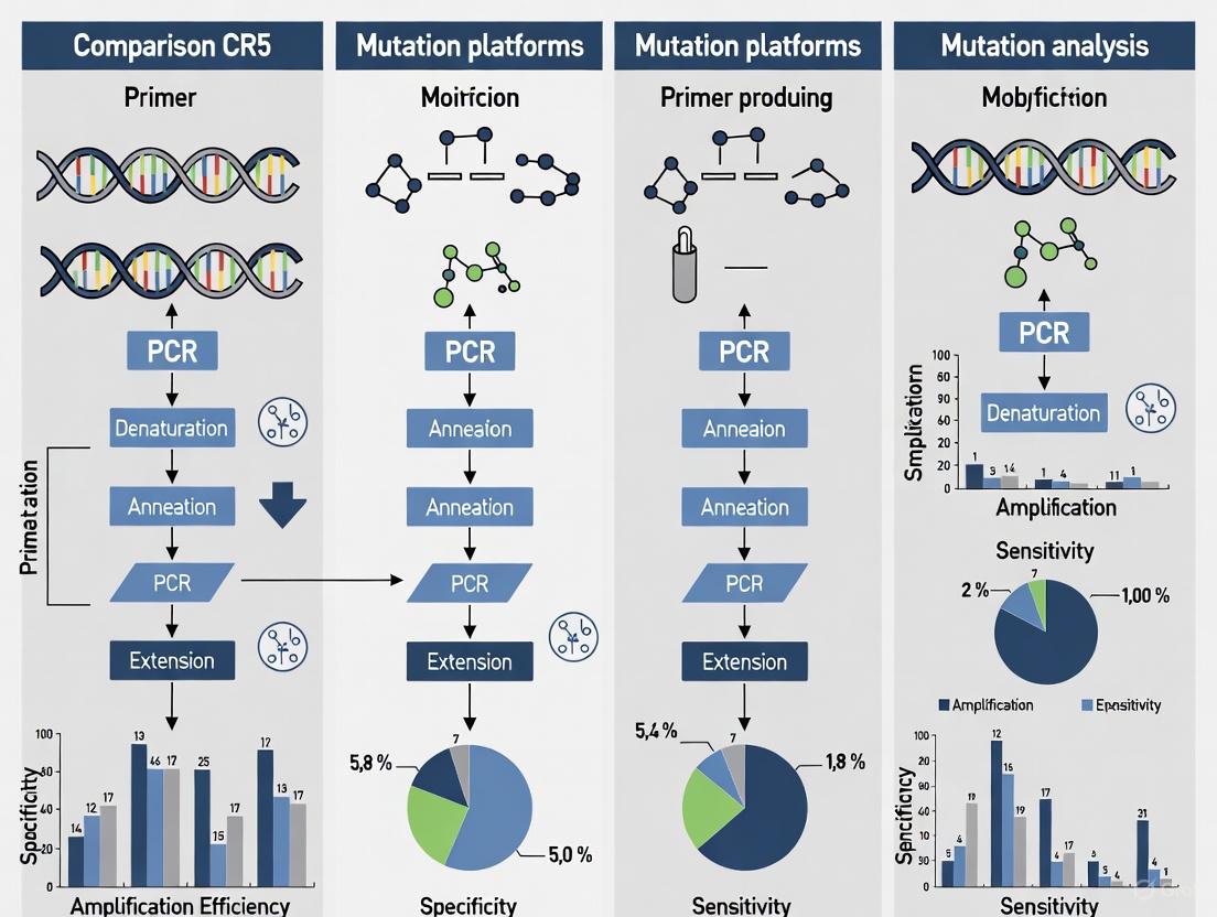
The following diagram illustrates the core workflow and technological differences between these two dPCR platforms:
Digital PCR Platform Workflows
Both platforms demonstrate strong correlation in quantitative measurements (r = 0.954) according to methylation studies, suggesting either platform provides reliable results for CCR5 genotyping [29]. The choice between systems often depends on practical laboratory considerations: ddPCR offers established protocols and extensive validation literature, while ndPCR provides faster setup with reduced hands-on time and integrated workflow automation [31].
CCR5-Specific Assay Design and Optimization
Primer and Probe Design Considerations
Effective CCR5 assay design requires careful attention to several critical factors. For CCR5Δ32 mutation analysis, specific gRNA sequences have been successfully implemented: CCR5-7 (CAGAATTGATACTGACTGTATGG) and CCR5-8 (AGATGACTATCTTTAATGTCTGG) [2]. These sequences facilitate precise targeting of the CCR5 locus for both knockout and detection strategies. Research demonstrates that applying statistical design of experiments (DOE) methodology for probe optimization can significantly enhance assay performance while reducing the number of required optimization experiments [32]. This approach systematically evaluates multiple input factors simultaneously, such as primer-probe distance and dimer stability, leading to efficiency improvements of up to 10% compared to traditional one-factor-at-a-time optimization [32].
Restriction enzyme selection profoundly impacts assay performance, particularly for targets with potential secondary structures or tandem repeats. Comparative studies show that HaeIII consistently outperforms EcoRI in CCR5 analysis, reducing coefficient of variation (CV) values to below 5% in ddPCR applications compared to much higher variability with EcoRI (up to 62.1% CV) [17]. This enhancement is attributed to HaeIII's superior ability to cleave accessibility barriers in complex genomic regions, ensuring more consistent amplification across partitions.
Experimental Protocol for CCR5Δ32 Mutation Detection
The following protocol provides a validated methodology for CCR5Δ32 detection using digital PCR:
Table 2: Step-by-Step CCR5Δ32 Detection Protocol
| Step | Procedure | Parameters | Quality Control |
|---|---|---|---|
| 1. DNA Extraction | Extract genomic DNA from PBMCs or cell lines using phenol-chloroform or commercial kits [2] | Measure concentration and purity (A260/A280) [2] | NanoPhotometer verification [2] |
| 2. Restriction Digest | Digest DNA with HaeIII restriction enzyme [17] | 2-4 hours at 37°C | Include undigested control |
| 3. PCR Mix Preparation | Prepare reaction mix with target-specific primers and probes [2] | Optimized primer/probe concentrations [32] | Include no-template control |
| 4. Partitioning | Load samples onto chosen dPCR platform | QX200: 20μL reaction [17]QIAcuity: 40μL reaction [17] | Verify partition quality |
| 5. Amplification | Perform endpoint PCR | Platform-specific cycle conditions [31] | Monitor amplification efficiency |
| 6. Analysis | Calculate target concentration using Poisson statistics [11] | Platform-specific software [31] | Threshold setting optimization |
This protocol has demonstrated sensitivity for detecting CCR5Δ32 mutant alleles in heterogeneous cell mixtures down to 0.8% variant allele frequency, enabling precise quantification of edited cells in therapeutic applications [2].
Advanced Applications in HIV Cure Research
CCR5 genotyping assays support critical applications in developing HIV curative strategies. The successful allogeneic transplantation of CCR5 null hematopoietic stem and progenitor cells (HSPCs) in the Berlin and London patients represents the only known cure for HIV-1 infection, establishing CCR5 disruption as a validated therapeutic approach [30]. Current research focuses on improving this strategy through multilayered HIV-1 resistance combining CCR5 knockout with B-cell secretion of HIV-inhibiting antibodies [30]. Accurate dPCR-based quantification of CCR5 editing efficiency is essential for evaluating these novel therapeutic candidates.
Advanced applications include CRISPR-Cas9-engineered HSPCs that engraft and reconstitute multiple hematopoietic lineages in vivo, creating durable HIV-resistant immune systems [30]. These approaches require precise monitoring of CCR5 modification levels, as low editing rates have previously resulted in therapeutic failure due to viral rebound in unedited cells [30]. The sensitivity of dPCR enables researchers to track these critical quality attributes throughout therapeutic development and manufacturing.
Research Reagent Solutions
The following essential materials represent key components for implementing robust CCR5 dPCR assays:
Table 3: Essential Research Reagents for CCR5 dPCR Analysis
| Reagent Category | Specific Examples | Function | Application Notes |
|---|---|---|---|
| Restriction Enzymes | HaeIII, EcoRI [17] | Improve DNA accessibility and precision | HaeIII provides superior precision for CCR5 assays [17] |
| Digital PCR Systems | QX200 (Bio-Rad), QIAcuity (QIAGEN) [17] [29] | Partitioned amplification and absolute quantification | Platform choice depends on throughput and workflow needs [31] |
| PCR Master Mixes | TaqPath ProAmp Master Mix [33] | Provides optimized reaction components | Compatible with multiple detection chemistries |
| Nucleic Acid Extraction Kits | MagMax Viral/Pathogen Kit [31] | Isolation of high-quality DNA | Maintains nucleic acid integrity for sensitive detection |
| Primer/Probe Sets | CCR5-7, CCR5-8 gRNAs [2] | Target-specific amplification | DOE optimization recommended [32] |
| Quantification Standards | Synthetic oligonucleotides [17] | Assay validation and standardization | Verify accuracy and dynamic range |
Digital PCR platforms provide robust, sensitive solutions for CCR5 genotyping applications in HIV cure research. Both droplet-based and nanoplate-based systems demonstrate excellent performance characteristics, with platform selection often depending on specific laboratory workflows and throughput requirements. Critical success factors include careful primer and probe design using DOE principles, selection of appropriate restriction enzymes such as HaeIII, and implementation of validated experimental protocols. As CCR5-directed therapies continue to evolve toward multiplexed approaches combining knockout with antibody delivery systems, precise dPCR-based quantification will remain essential for evaluating editing efficiency and advancing these promising therapeutic strategies toward clinical application.
Digital PCR (dPCR) represents a transformative technology in molecular diagnostics, enabling the absolute quantification of nucleic acids without the need for standard curves [11]. This third-generation PCR technology operates by partitioning a PCR mixture into thousands of individual reactions, allowing for the detection and quantification of rare genetic mutations with exceptional sensitivity [34] [11]. The application of dPCR has become particularly valuable in oncology and genetic disease research, where it facilitates the detection of minority targets amid complicated backgrounds, such as rare mutations in cancer or heterogeneous cell populations [12] [35].
The analysis of CCR5 mutations, specifically the CCR5Δ32 variant with its 32-base pair deletion, represents a critical application in HIV research and potential cure strategies [2]. Transplantations of hematopoietic stem cells with the CCR5Δ32 knockout mutation have demonstrated potential for complete HIV cure, creating an urgent need for accurate quantification methods to monitor mutant allele fractions in heterogeneous cell mixtures [2]. This guide provides a comprehensive comparison of digital PCR platforms specifically applied to CCR5 mutation analysis, offering researchers detailed methodologies and performance data to inform their experimental design.
Digital PCR Platform Technologies
Partitioning Methodologies
Digital PCR platforms primarily utilize two fundamental partitioning approaches: droplet-based systems and chip/nanoplate-based systems. Droplet digital PCR (ddPCR) systems, such as the Bio-Rad QX200, employ an immiscible fluid in oil to generate tens of thousands of submicroliter droplets that serve as individual reaction chambers [18]. The sample is randomly distributed within these droplets, which are then amplified and analyzed individually. Alternatively, nanoplate-based systems like the Qiagen QIAcuity use microfluidic digital PCR plates with predefined wells, integrating partitioning, thermocycling, and imaging into a single instrument [21] [18].
Each partitioning method presents distinct advantages and limitations. Droplet systems typically generate higher partition numbers (up to 20,000 per reaction for standard systems, and up to millions for specialized systems like RainDrop), potentially enhancing detection sensitivity for rare targets [18] [35]. However, they can suffer from droplet variability in size and shape, potentially affecting robustness and reproducibility [18]. Nanoplates offer a more streamlined workflow similar to qPCR, with reduced risk of contamination and less variability in partition size, but generally provide fewer partitions per reaction (typically 8,500-26,000) [21] [18].
Platform Comparison and Selection Criteria
When selecting a dPCR platform for CCR5 mutation analysis, researchers must consider several technical and practical factors. The table below summarizes key performance characteristics of major dPCR platforms based on comparative studies:
dot code for platform comparison diagram
Platform Selection Workflow - This diagram outlines the decision process for selecting between droplet-based and plate-based dPCR systems, highlighting key technological differentiators that impact CCR5 mutation detection performance.
Table 1: Comparison of Digital PCR Platforms for Mutation Analysis
| Platform | Partitioning Method | Number of Partitions | Throughput (samples/run) | Turnaround Time | Sensitivity (VAF) | Multiplexing Capacity |
|---|---|---|---|---|---|---|
| QIAcuity (Qiagen) | Nanoplate | 8,500-26,000 | 312-1,248 | ~2 hours for full plate | 0.1% | Up to 5-plex |
| QX200/QX One (Bio-Rad) | Droplet | ~20,000 | 96-480 | Several hours | 0.01%-0.1% | 4-plex |
| QuantStudio Absolute Q | Microfluidic array | ~20,000 | 16 | ~2.5 hours | 0.1% | 4-plex |
| Naica System (Stilla) | Droplet crystal | ~20,000-30,000 | 24 | 2-3 hours | 0.1%-0.2% | 3-plex |
| Biomark (Fluidigm) | Microfluidic chamber | 765-10,000 | Variable | Several hours | 0.1% | Variable |
Data compiled from [21] [18] [35]
The sensitivity of dPCR platforms for rare mutation detection is a critical consideration. Studies have demonstrated that dPCR can detect rare targets with mutation allele frequencies (MAFs) as low as 0.1%, with some platforms achieving even greater sensitivity down to 0.01% under optimal conditions [12]. This exceptional sensitivity makes dPCR particularly suitable for detecting CCR5Δ32 mutations in heterogeneous cell mixtures, where accurate quantification of low-frequency variants is essential for monitoring transplanted cell populations [2].
Sample Preparation Workflow
DNA Extraction and Qualification
The sample preparation workflow begins with DNA extraction from the source material, which may include whole blood, isolated cells, or cell-free DNA (cfDNA) from liquid biopsy samples. For CCR5 mutation analysis in heterogeneous cell mixtures, genomic DNA is typically extracted using commercial kits designed for blood or cell cultures [2]. The phenol-chloroform method or specialized kits such as the ExtractDNA Blood and Cells Kit have been successfully employed in CCR5 research applications [2].
DNA quantification and quality assessment are critical steps that significantly impact dPCR performance. Spectrophotometric methods (e.g., NanoPhotometer) provide concentration measurements and purity assessments through A260/A280 and A260/A230 ratios [2]. For cfDNA samples from liquid biopsies, fluorometric methods (e.g., Qubit with dsDNA BR Assay) are preferred due to their superior sensitivity and specificity for double-stranded DNA quantification [21] [36]. DNA integrity should be verified, particularly for samples from formalin-fixed, paraffin-embedded (FFPE) tissues or liquid biopsies, where DNA fragmentation can affect amplification efficiency [21].
Assay Design for CCR5 Mutation Detection
The CCR5Δ32 mutation detection requires careful assay design to distinguish between wild-type and mutant alleles. A proven approach utilizes two different hydrolysis probes (TaqMan-style) with a single set of primers that amplify a region spanning the deletion [34] [2]. One probe targets the wild-type sequence, while the other specifically binds to the mutant allele containing the 32-bp deletion, with each probe labeled with distinct fluorophores to enable multiplex detection.
Primer and probe sequences should be designed to have similar melting temperatures and optimal binding characteristics. For the CCR5Δ32 mutation, previously validated primers include forward: CCCAGGAATCATCTTTACCA and reverse: GACACCGAAGCAGAGTTT, which generate an amplicon that encompasses the deletion region [2]. Probe design should account for the specific deletion and utilize appropriate quencher-fluorophore combinations compatible with the selected dPCR platform's optical system.
dPCR Reaction Setup and Optimization
The dPCR reaction mixture preparation varies by platform but generally includes a master mix, primers, probes, DNA template, and nuclease-free water. The table below outlines essential reagents and their functions in the dPCR workflow for CCR5 mutation analysis:
Table 2: Essential Research Reagents for CCR5 dPCR Analysis
| Reagent | Function | Considerations for CCR5 Analysis |
|---|---|---|
| dPCR Master Mix | Provides DNA polymerase, dNTPs, buffer, and MgCl₂ | Choose probe-based mixes for hydrolysis probe assays |
| CCR5 Primers | Amplify target region spanning Δ32 deletion | Optimize concentration (typically 400-900 nM) |
| Wild-Type Probe | Detects non-mutated CCR5 sequence | Label with FAM or HEX; optimize concentration (150-300 nM) |
| Mutant Probe (Δ32) | Specifically detects 32-bp deletion | Label with distinct fluorophore (VIC, Cy3, etc.) |
| Reference Dye | Normalization for data analysis | Platform-specific requirements |
| Nuclease-Free Water | Reaction volume adjustment | Use high-purity, DNAse-free water |
| DNA Template | Sample containing CCR5 alleles | Input amount critical for sensitivity calculations |
Reagent information synthesized from [21] [34] [2]
The optimal DNA input amount must be carefully determined based on the desired sensitivity and the specific dPCR platform's characteristics. For rare mutation detection, the required DNA input can be calculated using the formula: Number of copies in reaction volume = mass of DNA in reaction volume (in ng)/0.003 (for human genomic DNA) [34]. This calculation ensures sufficient genome equivalents are analyzed to detect the target mutation at the desired frequency with statistical confidence.
Experimental Protocol for CCR5Δ32 Analysis
Step-by-Step Workflow
The following comprehensive protocol for CCR5Δ32 mutation analysis using droplet digital PCR has been adapted from established methodologies [34] [2]:
DNA Extraction and Quantification: Extract genomic DNA from cell mixtures using a standardized method (e.g., phenol-chloroform or commercial kit). Quantify DNA using fluorometric methods and assess purity spectrophotometrically. Adjust DNA concentration to working stocks in nuclease-free water or TE buffer.
Reaction Mixture Preparation: Prepare the dPCR master mix according to platform-specific requirements. For a 20μL ddPCR reaction (Bio-Rad QX200 system), combine:
- 10μL of 2× ddPCR Supermix for Probes (no dUTP)
- 900nM forward primer (1.8μL of 10μM stock)
- 900nM reverse primer (1.8μL of 10μM stock)
- 250nM wild-type probe (0.5μL of 10μM stock)
- 250nM mutant probe (0.5μL of 10μM stock)
- DNA template (adjust volume based on desired input of 10-100ng)
- Nuclease-free water to 20μL total volume
Droplet Generation: Load the reaction mixture into a DG8 cartridge along with 70μL of droplet generation oil. Process in the QX200 Droplet Generator to create approximately 20,000 droplets per sample. Transfer the resulting emulsion (40μL) to a 96-well PCR plate and seal with a foil heat seal.
PCR Amplification: Perform endpoint PCR amplification using the following thermal cycling conditions:
- 95°C for 10 minutes (enzyme activation)
- 40-45 cycles of:
- 94°C for 30 seconds (denaturation)
- 57-62°C for 60 seconds (annealing/extension)
- 98°C for 10 minutes (enzyme deactivation)
- 4°C hold
Droplet Reading and Analysis: Transfer the plate to the QX200 Droplet Reader, which measures the fluorescence in each droplet sequentially. Analyze the data using platform-specific software (QuantaSoft for Bio-Rad systems) to determine the concentration of wild-type and mutant alleles in copies/μL.
dot code for experimental workflow diagram
Experimental Workflow - This diagram illustrates the complete dPCR workflow for CCR5 mutation analysis, from sample preparation through final quantification, with color coding indicating different process phases.
Data Analysis and Interpretation
Data analysis in dPCR relies on Poisson statistics to determine the absolute concentration of target molecules in the original sample. The fundamental equation for calculating target concentration is:
Target concentration (copies/μL) = -ln(1 - P/N) / Vp × D
Where P is the number of positive partitions, N is the total number of partitions, Vp is the partition volume, and D is the dilution factor [35].
For CCR5Δ32 mutation analysis, the mutant allele frequency is calculated as:
Mutant Allele Frequency = [Mutant copies/μL] / ([Mutant copies/μL] + [Wild-type copies/μL])
This calculation enables precise quantification of the CCR5Δ32 mutation in heterogeneous cell mixtures, with demonstrated sensitivity down to 0.8% mutant alleles in wild-type backgrounds [2]. The development of such assays has enabled researchers to monitor the expansion of CCR5Δ32 mutant cells in experimental models, providing critical insights for developing HIV cure strategies [2].
Performance Comparison and Applications
Platform Performance Metrics
Comparative studies of dPCR platforms have revealed important performance characteristics relevant to CCR5 mutation analysis. A comprehensive comparison of four dPCR platforms demonstrated that all could accurately quantify DNA copy numbers when partition volume corrections were applied, with relative uncertainties of partition volume ranging from 0.7% to 2.9% across platforms [35]. This highlights the fundamental robustness of dPCR technology while acknowledging platform-specific variations.
In DNA methylation analysis studies comparing nanoplate-based and droplet-based systems, both technologies showed strong correlation (r = 0.954) and comparable sensitivity despite their different partitioning mechanisms [21]. The specificity and sensitivity of the nanoplate-based system were 99.62% and 99.08% respectively, while the droplet-based system achieved 100% specificity and 98.03% sensitivity in detecting methylated DNA [21]. These performance metrics demonstrate the reliability of both platform types for precise nucleic acid quantification.
Application to Liquid Biopsy and Clinical Research
The application of dPCR for CCR5 mutation analysis extends naturally to liquid biopsy approaches and clinical monitoring. In cancer research, dPCR has demonstrated superior sensitivity for circulating tumor DNA (ctDNA) detection compared to next-generation sequencing (NGS) in some applications, with ddPCR detecting ctDNA in 58.5% of baseline plasma samples versus 36.6% for NGS in rectal cancer patients [36]. This enhanced sensitivity for rare allele detection directly translates to HIV research, where monitoring CCR5Δ32 mutant cell populations following transplantation requires robust, sensitive detection methods.
The operational costs of dPCR compare favorably with NGS, with studies reporting 5-8.5-fold lower costs for dPCR-based ctDNA detection [36]. This cost-effectiveness, combined with rapid turnaround times (approximately 2 hours for full plates on nanoplate systems), positions dPCR as an accessible technology for both research and potential clinical applications [18].
Digital PCR platforms provide robust, sensitive solutions for CCR5 mutation analysis, enabling researchers to accurately quantify mutant allele frequencies in heterogeneous cell mixtures. Both droplet-based and nanoplate-based systems offer distinct advantages, with the choice depending on specific research requirements including throughput, sensitivity needs, and workflow preferences. The detailed protocols and performance metrics provided in this guide serve as a foundation for implementing dPCR-based CCR5 analysis in HIV research, hematopoietic stem cell monitoring, and related applications. As dPCR technology continues to evolve, its applications in clinical research and diagnostic development will expand, potentially encompassing routine monitoring of CCR5-directed therapies and transplantation outcomes.
The precise quantification of genetic variants, such as the CCR5Δ32 mutation, is pivotal in advanced biomedical research, particularly in the study of HIV resistance and the development of cure strategies [37] [2]. The CCR5 co-receptor serves as a primary binding site for the human immunodeficiency virus (HIV), and a naturally occurring 32-base pair deletion (CCR5Δ32) results in a non-functional receptor, conferring resistance to R5-tropic HIV infection [38]. This mutation is present in approximately 10% and 1% of the Northern European population in heterozygous and homozygous states, respectively [2]. The ability to accurately detect and quantify this mutation in heterogeneous cell mixtures is not only crucial for understanding natural HIV control but also for monitoring transplanted hematopoietic stem cells and therapies involving CRISPR/Cas9-generated CCR5Δ32 mutations [37].
Digital PCR (dPCR) has emerged as a powerful technology for this application, enabling the absolute quantification of nucleic acids without the need for a standard curve. dPCR achieves this by partitioning a sample into thousands of individual reactions, allowing for the precise counting of target DNA molecules [39]. Two primary dPCR platforms dominate this field: droplet digital PCR (ddPCR) and chip-based digital PCR (cdPCR). This guide provides an objective, step-by-step comparison of these platforms for CCR5 analysis, equipping researchers with the data and protocols necessary to select the optimal system for their experimental needs.
Platform Comparison: ddPCR vs. Chip-based dPCR
Fundamental Principles and Technical Specifications
Droplet Digital PCR (ddPCR) utilizes an immiscible fluid to partition a sample into thousands to millions of nanoliter-sized droplets, each serving as an individual PCR reactor [18]. In contrast, Chip-based dPCR (cdPCR) employs a microfluidic chip to divide the sample into a fixed array of nanoliter-sized wells [18] [40]. This fundamental difference in partitioning strategy drives variations in workflow, performance, and practical application.
Table 1: Core Technical Specifications of dPCR Platforms
| Feature | Droplet Digital PCR (ddPCR) | Chip-based dPCR (cdPCR) |
|---|---|---|
| Partitioning Method | Water-in-oil emulsion droplets [18] | Microfluidic chip with etched wells [40] |
| Number of Partitions | 10,000 - 80,000 (standard); up to millions possible [18] | ~20,000 (e.g., QuantStudio 3D) to ~26,000 (nanoplate) [18] |
| Partition Volume | Picoliter (pL) scale (e.g., 10-100 pL) [18] | Nanoliter (nL) scale (e.g., 10 nL) [18] |
| Typical Workflow | Multi-step: droplet generation, PCR thermocycling, droplet reading [18] | Integrated: partitioning, thermocycling, and imaging often in a single instrument [18] |
| Key Instruments | Bio-Rad QX200/One, Stilla Naica System [18] [39] | Thermo Fisher QuantStudio 3D, QIAGEN QIAcuity [18] [40] |
Performance Data in CCR5 and HIV Research
Studies have successfully adapted CCR5 mutation assays to both platforms. Research has demonstrated that a multiplex ddPCR assay can accurately quantify the content of cells with the CCR5Δ32 mutation in artificial mixtures down to a detection limit of 0.8% [37] [2]. Furthermore, the Intact Proviral DNA Assay (IPDA), a gold standard for quantifying the HIV-1 reservoir, has been successfully validated on a chip-based dPCR system. This adaptation performed with high accuracy on clinical samples from people with HIV, showing no false positives in negative controls and a strong correlation between intact HIV-1 DNA, total HIV-1 DNA, and input concentration for HIV-1 subtype B [41].
Table 2: Performance Comparison for Key Applications
| Performance Metric | Droplet Digital PCR (ddPCR) | Chip-based dPCR (cdPCR) |
|---|---|---|
| Detection Sensitivity | Can detect rare mutant prevalence as low as 0.1% [40] | Capable of absolute quantification for HIV reservoir studies [41] |
| Precision in Mutation Quantification | Accurately measures 0.8% CCR5Δ32 mutant cells in a wild-type background [2] | High precision for intact HIV DNA quantification; performance varies for non-B subtypes [41] |
| Sample Throughput (per run) | High (e.g., 480 samples across 5 plates on Bio-Rad QX One) [18] | Moderate to High (e.g., 312 to 1,248 samples on QIAcuity nanoplate systems) [18] |
| Hands-on Time | Time-consuming and cumbersome due to multiple transfer steps [18] | Less hands-on time with a more streamlined, qPCR-like workflow [18] [41] |
Experimental Protocols
Sample Preparation and DNA Isolation
This initial stage is critical for the success of any downstream dPCR application.
- Cell Culture: Grow your target cells (e.g., MT-4 human T-cell line, primary CD4+ T-cells) under appropriate conditions. For MT-4 cells, use RPMI-1640 medium supplemented with 10% fetal bovine serum (FBS) and maintain at 37°C with 5% CO₂ [37] [2].
- Genomic DNA Extraction: Isolate genomic DNA using a commercial kit, such as the ExtractDNA Blood and Cells Kit. The phenol-chloroform method is also a viable option [37].
- DNA Quantification and Quality Control: Precisely measure the concentration and purity (A260/A280 and A260/A230 ratios) of the extracted DNA using a spectrophotometer (e.g., NanoPhotometer) [37] [42]. High-purity DNA is essential for efficient amplification.
Step-by-Step ddPCR Protocol for CCR5Δ32 Detection
This protocol is adapted from the methodology described by Frontiers in Molecular Biosciences for detecting CRISPR/Cas9-generated CCR5Δ32 mutations [37] [2].
- Reaction Mix Preparation: Prepare the ddPCR reaction mixture on ice. A typical 20 µL reaction for the Bio-Rad QX200 system may contain:
- 10 µL of 2x ddPCR Supermix for Probes (no dUTP).
- 1 µL of CCR5 Wild-Type Probe (e.g., HEX-labeled, 250 nM final concentration).
- 1 µL of CCR5Δ32 Mutation Probe (e.g., FAM-labeled, 250 nM final concentration).
- 450 nM each of forward and reverse primers targeting the CCR5 locus.
- 50-100 ng of genomic DNA template.
- Nuclease-free water to 20 µL.
- Droplet Generation: Load the reaction mixture into a DG8 cartridge alongside droplet generation oil. Place the cartridge into the droplet generator. This instrument will create thousands of nanoliter-sized, water-in-oil droplets, effectively partitioning the sample.
- PCR Amplification: Carefully transfer the generated emulsion from the cartridge into a 96-well PCR plate. Seal the plate and perform endpoint PCR amplification in a thermal cycler using the following cycling conditions, optimized for the CCR5 target:
- Enzyme activation: 95°C for 10 minutes.
- 40 cycles of:
- Denaturation: 94°C for 30 seconds.
- Annealing/Extension: 60°C for 60 seconds.
- Enzyme deactivation: 98°C for 10 minutes.
- (Optional) Final hold: 4°C.
- Droplet Reading and Analysis: Place the PCR plate into the droplet reader. This instrument will aspirate each sample, passing droplets in a single file past a laser. Fluorescence (FAM and HEX) is measured for each droplet. Use the manufacturer's software (e.g., QuantaSoft) to analyze the data. The software will apply a threshold to distinguish positive (mutant or wild-type) droplets from negative droplets and provide an absolute count of target molecules based on Poisson statistics.
Step-by-Step Chip-Based dPCR Protocol
This protocol outlines a generic workflow for systems like the QIAcuity or QuantStudio 3D.
- Reaction Mix Preparation: Prepare the dPCR master mix. This is typically simpler than for ddPCR. For a 25 µL reaction on a system like the Stilla Naica:
- 5 µL of Naica Multiplex PCR Mix Buffer A (5x).
- 1 µL of Naica Multiplex PCR Mix Buffer B (4%).
- 1 µL of FAM-labeled probe/primer mix (25x).
- 1 µL of VIC/HEX-labeled probe/primer mix (25x).
- 50 ng of DNA template.
- Nuclease-free water to 25 µL [39].
- Loading the Chip/Nanoplate: Pipette the prepared reaction mixture directly into the wells of the dPCR chip or nanoplate.
- Partitioning and Amplification: Load the plate into the integrated dPCR instrument. The instrument automatically performs partitioning (creating the nanoliter wells), PCR thermocycling, and imaging in a single, closed system. The cycling conditions will need to be optimized for the CCR5 assay but are generally similar to the ddPCR protocol.
- Imaging and Data Analysis: After the run is complete, the integrated software (e.g., AnalysisSuite for QuantStudio 3D) will analyze the fluorescence images of each partition on the chip. It will provide the concentration of the target sequence in copies per microliter, and for multiplex assays, it will discriminate between wild-type and mutant alleles [40].
The following diagram illustrates the core procedural differences between the two workflows.
The Scientist's Toolkit: Essential Reagents and Materials
Table 3: Key Research Reagent Solutions for CCR5 dPCR Analysis
| Item | Function / Application | Example Products / Components |
|---|---|---|
| Cell Culture Media | Maintenance and expansion of target cells (e.g., T-cell lines) for DNA extraction. | Roswell Park Memorial Institute medium (RPMI-1640) supplemented with Fetal Bovine Serum (FBS) [37]. |
| DNA Extraction Kit | Isolation of high-quality, high-purity genomic DNA from cell samples. | ExtractDNA Blood and Cells Kit, QIAamp DNA Mini Kit [37] [42]. |
| dPCR Supermix | Provides the optimal buffer, enzymes, and dNTPs for efficient amplification in partitioned reactions. | ddPCR Supermix for Probes (no dUTP), Naica Multiplex PCR Mix [37] [39]. |
| TaqMan Assays | Fluorogenic probes and primers for specific, sensitive detection of wild-type CCR5 and the Δ32 mutant allele. | Custom or wet lab-validated TaqMan SNP Genotyping Assays [40]. |
| Primers & Probes | Oligonucleotides designed to specifically target and amplify the wild-type and mutant CCR5 sequences. | gRNA sequences for CRISPR: CCR5-7 (CAGAATTGATACTGACTGTATGG), CCR5-8 (AGATGACTATCTTTAATGTCTGG). Sequencing primers: Forward (CCCAGGAATCATCTTTACCA), Reverse (GACACCGAAGCAGAGTTT) [37] [2]. |
The choice between droplet-based and chip-based dPCR for CCR5 analysis hinges on the specific requirements of the research project.
Choose Droplet Digital PCR (ddPCR) if: Your priority is achieving the highest possible sensitivity for detecting very rare mutations (down to 0.1%) and you require the highest number of partitions for ultimate precision. It is also a suitable choice if you already have access to droplet generators and readers [18] [40]. Be prepared for a more complex workflow with potential for pipetting errors [18].
Choose Chip-Based dPCR (cdPCR) if: Your priority is a streamlined, efficient workflow with minimal hands-on time and a lower risk of contamination. This platform is ideal for labs seeking a user-friendly system with a qPCR-like workflow that integrates partitioning, amplification, and reading [18] [41]. It is an excellent choice for applications like the IPDA for HIV reservoir studies and for labs where bench space is limited [18] [41].
Both platforms offer superior absolute quantification for CCR5 mutation analysis compared to traditional qPCR. The decision matrix ultimately balances the need for ultra-high sensitivity against operational simplicity and throughput in the context of your specific research on HIV, genetic epidemiology, or therapeutic development.
The accurate quantification of mutation frequency is a cornerstone of modern genetic research, particularly in fields like HIV therapy development where the precise measurement of CCR5Δ32 mutant alleles can determine therapeutic efficacy [2]. Digital PCR (dPCR) has emerged as a powerful technology for this application, providing absolute quantification of nucleic acids without the need for standard curves [43]. This guide objectively compares the performance of leading dPCR platforms—droplet-based (ddPCR) and nanoplate-based (ndPCR) systems—for CCR5 mutation analysis, drawing on experimental data from peer-reviewed studies to highlight the strengths and considerations of each platform.
dPCR operates by partitioning a PCR reaction into thousands of nanoliter-scale reactions, enabling the detection and quantification of target sequences through Poisson statistical analysis [18] [44]. This partitioning confers significant advantages for mutation detection, including enhanced sensitivity for rare variants and greater robustness to PCR inhibitors compared to quantitative PCR (qPCR) [28] [43]. These characteristics make dPCR particularly suitable for applications requiring precise measurement of allele frequencies in complex biological samples.
Comparison of Digital PCR Platforms
The two primary dPCR platforms utilize different approaches to achieve reaction partitioning. The QX200 Droplet Digital PCR (ddPCR) System from Bio-Rad employs a water-in-oil emulsion technology, where samples are partitioned into approximately 20,000 nanoliter-sized droplets [28] [18]. In contrast, the QIAcuity Digital PCR (ndPCR) System from QIAGEN uses microfluidic nanoplates containing either 8,500 or 26,000 partitions per well, integrating partitioning, thermocycling, and imaging into a single instrument [28] [18].
A critical distinction between these platforms lies in their workflow integration. The ddPCR system requires multiple instruments—a droplet generator, thermal cycler, and droplet reader—while the ndPCR system performs all steps within a single integrated device [18]. This difference significantly impacts operational efficiency, with the nanoplate-based system offering a more streamlined, qPCR-like workflow that reduces manual handling steps and associated contamination risks [28] [18].
Performance Comparison for Mutation Analysis
Table 1: Platform Performance in Mutation Detection Studies
| Performance Metric | QX200 ddPCR (Bio-Rad) | QIAcuity ndPCR (QIAGEN) | Application Context |
|---|---|---|---|
| Detection Sensitivity | 0.8% mutant alleles in mixed samples [2] | Comparable sensitivity to ddPCR [21] | CCR5Δ32 mutation detection [2] |
| Specificity | 100% [21] | 99.62% [21] | DNA methylation analysis [21] |
| Precision (CV) | 6-13% (synthetic DNA) [17] | 7-11% (synthetic DNA) [17] | Gene copy number quantification [17] |
| Limit of Detection (LOD) | 0.17 copies/μL [17] | 0.39 copies/μL [17] | Sensitivity assessment [17] |
| Limit of Quantification (LOQ) | 4.26 copies/μL [17] | 1.35 copies/μL [17] | Sensitivity assessment [17] |
| Partition Number | ~20,000 [17] [18] | 8,500-26,000 [28] [18] | Technical specification |
| Sample Throughput | 96 samples in 3-4 hours [45] | 96 samples in ~2 hours [18] | Workflow efficiency |
Table 2: Platform Characteristics and Practical Considerations
| Characteristic | QX200 ddPCR (Bio-Rad) | QIAcuity ndPCR (QIAGEN) |
|---|---|---|
| Partitioning Method | Water-oil emulsion droplets [28] | Microfluidic nanoplate [28] |
| Workflow | Multiple instruments required [18] | Fully integrated system [18] |
| Throughput Time | 21 hours for 480 samples [18] | 8 hours for 1,248 samples [18] |
| Potential Issues | "Rain" droplets, droplet variability [18] | Fewer partition-related artifacts [18] |
| Multiplexing Capacity | 2-plex standard [18] | Up to 5-plex capability [18] |
| Sample Volume | 20 μL reaction [17] | 12-40 μL reaction [21] [18] |
Multiple studies have demonstrated that both platforms deliver highly comparable results for mutation frequency analysis. Research on CDH13 gene methylation in breast cancer samples revealed a strong correlation (r = 0.954) between methylation levels measured by both systems [21]. Similarly, both platforms showed excellent precision in copy number variation analysis, with either platform being suitable for reliable FCGR3B gene copy number quantification [46].
For CCR5Δ32 mutation analysis specifically, ddPCR has proven particularly effective. A 2022 study demonstrated that a properly optimized ddPCR assay could accurately quantify CCR5Δ32 mutant alleles in heterogeneous cell mixtures with a sensitivity down to 0.8%, making it suitable for monitoring the expansion of edited cells in therapeutic contexts [2].
Experimental Protocols for CCR5 Mutation Analysis
Sample Preparation and DNA Extraction
For CCR5 mutation analysis, the DNA source and extraction method significantly impact data quality. The referenced CCR5Δ32 study utilized the MT-4 human T-cell line cultured in RPMI-1640 medium supplemented with 10% fetal bovine serum, maintained at 37°C with 5% CO₂ [2]. Genomic DNA was extracted using a phenol-chloroform protocol or commercial kits (Evrogen "ExtractDNA Blood and Cells Kit"), with DNA concentration and purity measured spectrophotometrically [2].
For DNA extracted from formalin-fixed, paraffin-embedded (FFPE) samples—common in clinical research—studies recommend using a DNeasy Blood and Tissue Kit (QIAGEN) with an initial deparaffinization step using xylene [21]. DNA concentration is best determined using fluorescence-based methods (e.g., Qubit dsDNA BR Assay) rather than spectrophotometry for improved accuracy with degraded samples [21].
Digital PCR Assay Design and Optimization
The core principle of dPCR mutation analysis involves designing assays that can discriminate between wild-type and mutant alleles. For CCR5Δ32 analysis, this typically involves a multiplexed assay with two probe sets: one specific for the wild-type CCR5 allele and another for the Δ32 deletion variant [2].
Table 3: Essential Research Reagents for dPCR Mutation Analysis
| Reagent Category | Specific Examples | Function in Experiment |
|---|---|---|
| DNA Extraction Kits | DNeasy Blood & Tissue Kit (Qiagen), ExtractDNA Blood & Cells Kit (Evrogen) [21] [2] | High-quality genomic DNA isolation from cells or tissues |
| dPCR Master Mixes | QIAcuity Probe PCR Master Mix (Qiagen), ddPCR Supermix for Probes (Bio-Rad) [21] [2] | Provides optimized buffer, enzymes, and dNTPs for amplification |
| Restriction Enzymes | EcoRI, HaeIII [17] | Improve DNA accessibility, especially for tandem repeats |
| Fluorogenic Probes | FAM, HEX/VIC-labeled TaqMan probes [21] [2] | Target-specific detection with different fluorescent reporters |
| Reference Assays | Lectin gene for GMO studies [28] | Internal control for DNA quality and quantity |
| Digital PCR Plates | QIAcuity Nanoplate (Qiagen), DG8 Cartridges (Bio-Rad) [28] [21] | Reaction partitioning and thermal cycling |
Assay optimization should include thermal gradient experiments to establish optimal annealing temperatures and template concentration titration to ensure optimal partition occupancy [44]. The optimal DNA input per reaction typically ranges from 2.5-30 ng/μL, with excessive DNA leading to saturated partitions and underestimated concentrations [17]. For challenging genomic regions, the addition of restriction enzymes (e.g., HaeIII) can significantly improve data quality by digesting long DNA fragments and ensuring better access to the target sequence [17].
Workflow Visualization
The following diagram illustrates the complete experimental workflow for dPCR-based mutation frequency analysis, from sample preparation to data interpretation:
Data Analysis and Interpretation
Calculating Mutation Frequency
The fundamental principle of dPCR quantification relies on Poisson statistics to calculate the initial concentration of target molecules in a sample [44]. The calculation requires knowing the number of positive and negative partitions, along with the total number of partitions and the reaction volume [44].
The concentration of target DNA (in copies/μL) is calculated as: [ \lambda = -\ln(1 - p) / v ] Where (\lambda) is the target concentration, (p) is the fraction of positive partitions, and (v) is the volume of each partition [44].
For mutation frequency analysis, this calculation is performed separately for both the mutant and wild-type alleles. The mutation frequency is then expressed as the ratio of mutant allele concentration to the total allele concentration: [ \text{Mutation Frequency} = \frac{[\text{Mutant Allele}]}{[\text{Mutant Allele}] + [\text{Wild-type Allele}]} \times 100\% ]
In the context of CCR5Δ32 analysis, this approach enabled researchers to accurately quantify the proportion of mutant alleles in mixed cell populations, achieving precise quantification down to 0.8% mutation frequency [2].
Determining Confidence Intervals
The precision of dPCR measurements is intrinsically linked to the number of partitions analyzed, with more partitions generally yielding tighter confidence intervals [44]. The 95% confidence intervals for the target concentration can be calculated using the formula: [ CI = \lambda \pm 1.96 \times \sqrt{\lambda / (n \times v)} ] Where (n) is the total number of partitions [44].
For mutation frequency ratios, Fieller's theorem provides a more appropriate method for calculating confidence intervals [44]. This approach accounts for the covariance between the mutant and wild-type measurements and generally provides more accurate interval estimates than simple propagation of error methods.
Studies have shown that proper experimental design with adequate replication is essential for obtaining reliable confidence intervals. For rare mutation detection (frequencies <1%), increasing the number of partitions and replicates significantly improves the reliability of frequency estimates and associated confidence intervals [43] [44].
Quality Control and Validation
Implementing robust quality control measures is essential for generating reliable mutation frequency data. Key parameters to monitor include:
- Partition Number: Both platforms should maintain a minimum of 7,000-8,000 valid partitions per reaction, with significant deviations suggesting technical issues [21].
- Threshold Setting: Proper discrimination between positive and negative partitions is critical, with manual threshold adjustment sometimes necessary to account for "rain" (partitions with intermediate fluorescence) [21] [44].
- Inhibition Testing: Serial dilution of samples can identify PCR inhibition, with measured concentrations expected to decrease proportionally with dilution factor [28].
- Reference Genes: Inclusion of reference gene assays (e.g., lectin for GMO studies) controls for DNA quality and extraction efficiency [28].
The dynamic range of dPCR must also be considered, as both platforms can become saturated at high DNA concentrations, leading to underestimation of target copies. Optimal performance is typically achieved with 50-100 positive partitions per μL for the majority of targets [17].
Both digital PCR platforms offer excellent performance for mutation frequency analysis and confidence interval calculation in CCR5 research applications. The choice between systems should be guided by specific research requirements and practical laboratory considerations.
The Bio-Rad QX200 ddPCR system demonstrates marginally higher sensitivity in some studies [17] and has a proven track record in CCR5Δ32 mutation analysis [2]. Its droplet-based partitioning generates a higher number of partitions, potentially providing slightly better precision for low-frequency mutation detection.
The QIAGEN QIAcuity ndPCR system offers significant workflow advantages with its integrated design, reduced hands-on time, and higher throughput capacity [18]. The system's reduced susceptibility to partition artifacts (e.g., "rain" droplets) can simplify data analysis and interpretation [18].
For laboratories prioritizing maximal sensitivity for rare mutant detection and requiring flexibility in reaction setup, the QX200 ddPCR system represents an excellent choice. For facilities focused on higher throughput applications and seeking to minimize manual processing steps, the QIAcuity ndPCR system offers compelling advantages. Both platforms provide the precision and accuracy necessary for reliable mutation frequency calculation and confidence interval estimation in CCR5 mutation research.
Maximizing Sensitivity and Precision in CCR5 dPCR Analysis
Optimizing Partition Number and Template Input for Rare Mutation Detection
The detection and quantification of rare genetic mutations present significant challenges in molecular diagnostics and therapeutic monitoring. Digital PCR (dPCR) has emerged as a powerful technology that addresses these challenges through sample partitioning and single-molecule amplification [11]. This guide provides a comprehensive comparison of dPCR platforms and optimization strategies for detecting rare mutations, with a specific focus on CCR5Δ32 mutation analysis in HIV research. The CCR5 co-receptor serves as a critical binding site for HIV, and the 32-base pair deletion (CCR5Δ32) confers natural resistance to HIV infection, making its accurate quantification essential for developing curative therapies using hematopoietic stem cell transplantation or CRISPR/Cas9 genome editing [2].
Fundamental Principles of Digital PCR for Rare Mutation Detection
Core Technology and Partitioning
Digital PCR operates by partitioning a PCR reaction mixture into thousands to millions of individual reactions, allowing for the amplification of single DNA molecules [11]. Following amplification, the fraction of positive partitions is counted, and the target concentration is calculated using Poisson statistics, enabling absolute quantification without standard curves [11]. This partitioning effect effectively enriches low-level targets, making dPCR uniquely suited for detecting rare mutations present at frequencies as low as 0.1% or less [12].
The fundamental relationship between partition number and detection sensitivity is governed by Poisson statistics. As the number of partitions increases, the probability of detecting rare mutant alleles present at very low frequencies significantly improves. This statistical foundation forms the basis for optimizing dPCR assays for rare mutation detection, where maximizing partition count enhances both the limit of detection and the precision of quantification [34].
Comparison with Alternative Technologies
When compared to other molecular detection methods, dPCR offers distinct advantages for rare mutation detection. Next-generation sequencing (NGS) typically has a reliable limit of quantification of approximately 1% variant allele frequency, whereas dPCR can detect mutations at frequencies as low as 0.01-0.1% [36] [47]. This superior sensitivity makes dPCR particularly valuable for applications requiring ultra-sensitive detection, such as monitoring minimal residual disease or detecting early treatment resistance mutations.
However, each technology has its appropriate application context. NGS provides comprehensive profiling of multiple mutations across larger genomic regions, while dPCR offers superior sensitivity and quantification precision for monitoring specific known mutations [36]. The choice between these technologies depends on the specific research requirements, with dPCR being the preferred method when ultra-sensitive quantification of predetermined mutations is needed.
Critical Parameters for Optimization
Partition Number and Its Impact on Sensitivity
The number of partitions in a dPCR assay directly influences its sensitivity and precision for rare mutation detection. Higher partition counts increase the probability of detecting low-frequency mutations and improve the confidence of quantification [34]. Different dPCR platforms offer varying partition capacities, which represents a key consideration in platform selection.
The relationship between partition number and detection limit can be calculated using Poisson statistics. For a given mutation allele frequency, the probability of a partition containing at least one mutant target depends on the total number of partitions and the template input. This statistical foundation enables researchers to determine the minimum partition number required to detect mutations at specific frequency thresholds with 95% confidence [34].
Figure 1: Digital PCR Workflow and Optimization Points for Rare Mutation Detection
Template Input Considerations
The amount of template DNA input significantly affects the sensitivity of rare mutation detection. Sufficient template must be added to ensure adequate representation of both wild-type and mutant sequences, while avoiding overloading that can cause multiple targets per partition and reduce quantification accuracy [34].
For human genomic DNA applications, the conversion between mass and copy number follows a standard calculation: approximately 3 pg (0.003 ng) represents one haploid genome equivalent. Therefore, 10 ng of human genomic DNA contains approximately 3,333 copies of a single-copy gene [34]. This relationship enables researchers to calculate the theoretical detection sensitivity based on their specific template input and the system's limit of detection.
The theoretical limit of detection (LOD) can be calculated using the formula: Sensitivity = Theoretical LOD of the system / Total concentration of target copies in the sample. For example, with 10 ng of human genomic DNA input and a system with a theoretical LOD of 0.2 copies/μL, the detectable mutant allelic fraction would be approximately 0.15% with 95% confidence [34].
Platform-Specific Performance Characteristics
Different dPCR platforms demonstrate varying performance characteristics that impact their utility for rare mutation detection. The following table summarizes key technical specifications across major platforms:
Table 1: Digital PCR Platform Comparison for Rare Mutation Detection
| Platform | Partitioning Technology | Typical Partition Number | Partition Volume | Theoretical Sensitivity | Key Applications |
|---|---|---|---|---|---|
| QX200 ddPCR (Bio-Rad) | Droplet-based | 20,000 | 1 nL | 0.01% VAF [47] | KRAS mutations, ctDNA analysis [36] [47] |
| QIAcuity (Qiagen) | Nanoplate-based | 26,000 [31] | Not specified | 0.2 copies/μL [34] | Respiratory virus quantification, gene copy number analysis [31] [17] |
| Naica System (Stilla) | Droplet-based | 20,000-30,000 [34] | Not specified | 0.2 copies/μL [34] | EGFR T790M, rare mutation detection [34] |
| QuantStudio Absolute Q | Microfluidic array | Not specified | Not specified | 0.1% VAF [12] | Liquid biopsy, predefined cancer mutations [12] |
The selection of an appropriate dPCR platform depends on the specific research requirements, including needed sensitivity, throughput, and available resources. Droplet-based systems generally offer greater scalability and cost-effectiveness, while nanowell/nanochamber systems provide higher reproducibility and ease of automation [11].
Experimental Protocols for CCR5Δ32 Mutation Analysis
Sample Preparation and DNA Extraction
For CCR5Δ32 mutation analysis, genomic DNA is typically extracted from cell lines or patient samples using standard methods. The phenol-chloroform method or commercial kits such as the ExtractDNA Blood and Cells Kit have been successfully employed [2]. DNA quality assessment should include measurement of concentration and purity using spectrophotometry (A260/A280 ratio between 1.8-2.0 and A260/A230 ratio over 2.0) [47].
Restriction enzyme digestion may be employed to improve PCR amplification efficiency. EcoRI has been successfully used for KRAS mutation analysis [47], while studies comparing restriction enzymes for dPCR have demonstrated that enzyme selection can significantly impact precision, with HaeIII showing improved performance over EcoRI in some systems [17].
ddPCR Assay Design and Optimization
For CCR5Δ32 mutation detection, a multiplex ddPCR approach should be designed with two hydrolysis probes:
- Wild-type probe: Targets the intact CCR5 sequence
- Mutant probe: Specifically targets the 32-bp deletion region
Probes should be labeled with different fluorophores (e.g., FAM and VIC/HEX) with confirmed compatibility with the dPCR system being used [34]. Final primer concentrations are typically optimized around 500 nM, with probe concentrations around 250 nM each [34].
PCR reaction mixtures should include:
- 1X dPCR Mastermix
- Reference dye (if required by the platform)
- Optimized primer and probe concentrations
- Template DNA (amount optimized based on desired sensitivity)
- Nuclease-free water to final volume [34]
Thermal Cycling and Data Acquisition
Thermal cycling conditions must be optimized for the specific assay and mastermix used. For example, the EGFR T790M assay employs the following protocol [34]:
- Initial denaturation: 95°C for 10 minutes
- 45 cycles of:
- Denaturation: 95°C for 30 seconds
- Annealing/Extension: 62°C for 15 seconds
- Final extension: 98°C for 10 minutes
Following amplification, data acquisition varies by platform. Droplet-based systems typically read partitions sequentially in a process analogous to flow cytometry, while chip-based systems image the entire chip using fluorescence microscopy or scanning [34].
Data Analysis and Quality Control
Data analysis for rare mutation detection requires careful attention to several quality control parameters:
- Partition number: Higher numbers (preferably >20,000) improve detection confidence [34]
- Negative controls: Should show few or no positive partitions [34]
- Fluorescence compensation: Essential for multiplex assays to correct for spillover between channels [34]
- Cluster identification: Automated or manual classification of positive and negative partitions [48]
Accurate classification of partitions based on endpoint fluorescence intensities is crucial to avoid biased concentration estimates. Both general-purpose clustering methods (k-means, DBSCAN) and specialized dPCR methods (dpcp, calico) have been evaluated for this purpose [48].
Table 2: Key Reagent Solutions for CCR5Δ32 dPCR Analysis
| Reagent/Category | Specific Examples | Function | Considerations |
|---|---|---|---|
| dPCR Mastermix | ddPCR Supermix for Probes, PerfeCTa Multiplex Mastermix | Provides essential components for PCR: DNA polymerase, dNTPs, buffer, MgCl₂ | Platform-specific recommendations should be followed [34] |
| Hydrolysis Probes | TaqMan MGB probes, FAM/VIC/Cy3-labeled probes | Sequence-specific detection of wild-type and mutant alleles | Fluorophores must match system optics; typically 250 nM final concentration [34] [2] |
| Restriction Enzymes | EcoRI, HaeIII | Improve amplification efficiency by digesting genomic DNA | Enzyme selection affects precision; HaeIII may outperform EcoRI [17] [47] |
| DNA Extraction Kits | Phenol-chloroform, ExtractDNA Blood and Cells Kit | High-quality genomic DNA isolation | A260/A280 ratio of 1.8-2.0 indicates acceptable purity [2] [47] |
Platform Performance and Experimental Data
Sensitivity and Precision Comparisons
Studies directly comparing dPCR platforms demonstrate variations in performance characteristics. In a comparison of the QX200 ddPCR system and QIAcuity ndPCR system, the limit of detection (LOD) for ndPCR was approximately 0.39 copies/μL input, while ddPCR showed an LOD of approximately 0.17 copies/μL input [17]. However, the limit of quantification (LOQ) showed the opposite trend, with ndPCR at 1.35 copies/μL input and ddPCR at 4.26 copies/μL input [17].
For CCR5Δ32 mutation detection specifically, ddPCR has demonstrated the ability to accurately quantify mutant alleles down to 0.8% in heterogeneous cell mixtures [2]. This level of sensitivity is sufficient for monitoring the expansion of CCR5Δ32-modified cells in therapeutic contexts.
Comparison with Alternative Technologies
When compared to next-generation sequencing for mutation detection, ddPCR demonstrates superior sensitivity. In rectal cancer ctDNA analysis, ddPCR detected ctDNA in 58.5% of baseline plasma samples compared to 36.6% detected by NGS panel sequencing [36]. The reliable limit of quantification for ddPCR was 0.1% for KRAS mutations, compared to 1% for NGS [47].
The cost profile also differs significantly between these technologies. The operational costs of ctDNA detection with ddPCR are 5-8.5-fold lower than NGS [36], making ddPCR more accessible for high-throughput monitoring of specific mutations in clinical research settings.
Figure 2: Optimization Relationships for Rare Mutation Detection Sensitivity
The optimization of partition number and template input represents a critical factor in successful rare mutation detection using digital PCR. Based on current evidence and technological capabilities, the following recommendations can be made for CCR5Δ32 mutation analysis:
Partition Number: Aim for systems generating at least 20,000 partitions, with higher numbers (26,000+) providing improved sensitivity for mutations present at frequencies below 0.5%.
Template Input: For human genomic DNA, 10-20 ng input typically provides optimal sensitivity, balancing the need for sufficient target copies while avoiding partition overcrowding.
Platform Selection: Choose platforms based on required sensitivity thresholds, with droplet-based systems generally offering superior limits of detection and nanowell systems providing enhanced reproducibility.
Experimental Design: Include appropriate controls (no-template controls, monocolor controls for fluorescence compensation) and optimize restriction enzyme selection based on the specific target sequence.
As dPCR technology continues to evolve, future developments in partition density, multiplexing capabilities, and analytical algorithms will further enhance rare mutation detection, strengthening its role in clinical research applications including CCR5-directed HIV therapies.
Digital PCR (dPCR) has emerged as a powerful tool for absolute quantification of nucleic acids, offering superior sensitivity and precision for applications like CCR5 mutation analysis in HIV research. Unlike quantitative real-time PCR (qPCR), dPCR achieves absolute quantification by partitioning a sample into thousands of individual reactions, without the need for a standard curve [28]. This partitioning step, whether achieved through water-oil emulsion droplets or microfluidic nanoplates, is fundamental to the technology's advantages but also introduces specific technical challenges [28]. For researchers tracking rare CCR5Δ32 mutant alleles in heterogeneous cell mixtures—a critical application in developing HIV cure strategies—understanding and mitigating these pitfalls is paramount for generating reliable, reproducible data [2]. This guide objectively compares how different dPCR platforms address the common challenges of PCR inhibition, partition uniformity, and false-positive signals, providing a framework for selecting the optimal technology for sensitive genetic mutation research.
Platform Comparison: Addressing Key Technical Challenges
The performance of dPCR platforms can vary significantly based on their underlying partitioning technology. The following section compares the two predominant types—droplet-based dPCR (ddPCR) and chip-based dPCR—focusing on their inherent approaches to common technical pitfalls.
Table 1: Digital PCR Platform Comparison on Key Pitfalls
| Technical Challenge | Droplet dPCR (e.g., Bio-Rad QX200) | Chip-Based dPCR (e.g., Qiagen QIAcuity, Applied Biosystems QuantStudio 3D) |
|---|---|---|
| PCR Inhibition Resistance | Less sensitive to inhibitors than qPCR; sample partitioning dilutes inhibitors [28]. | Also exhibits reduced sensitivity to inhibitors due to sample partitioning [28]. |
| Partition Uniformity | Relies on uniform droplet size; DNA fragmentation is often required to prevent viscosity changes from altering droplet volume, impacting quantification accuracy [49]. | Fixed partition size in etched chips; eliminates variability from partitioning process. No fragmentation needed for most applications, ensuring consistent volume [49] [40]. |
| False-Positive Rate | Heat fragmentation, a common pre-step, can cause cytosine deamination, leading to false-positive mutation calls [49]. | Workflow avoids heat fragmentation, reducing this specific risk of false positives [49]. Assay optimization (e.g., probe design, thermal cycling) is still critical to prevent cross-talk [50]. |
Experimental Data and Validation
Performance in GMO Quantification
A direct comparative study of the Bio-Rad QX200 and Qiagen QIAcuity platforms for quantifying genetically modified organisms (GMOs) demonstrated that both systems can perform robustly after optimization. The in-house validation showed that duplex PCR methods on both platforms met all acceptance criteria for validation performance parameters according to JRC Guidance documents [28]. This underscores that with proper assay validation, different dPCR technologies are capable of producing reliable, quantitative data suitable for regulated environments.
Sensitivity in Rare Mutation Detection
Research utilizing the Bio-Rad QX200 ddPCR system for detecting CCR5Δ32 mutant alleles demonstrated its capability to accurately quantify the mutation content in artificial cell mixtures down to a level of 0.8% [2]. This high sensitivity is crucial for applications like monitoring the engraftment of edited cells in HIV patients. Furthermore, studies on minimal residual disease (MRD) in leukemia have successfully adapted ddPCR to monitor rare fusion transcripts and mutations, highlighting the platform's utility in detecting low-abundance targets with high sensitivity [50].
For oncology research, the Applied Biosystems QuantStudio 3D system, a chip-based platform, has been shown to detect and quantify rare mutant prevalence as low as 0.1% using validated TaqMan SNP Genotyping Assays [40]. This demonstrates the potential of optimized chip-based dPCR for extreme sensitivity in mutation detection.
Essential Protocols for Robust dPCR Assays
Optimizing Assay Specificity and Reducing Cross-Talk
Multiplex dPCR assays, which probe for multiple targets (e.g., wild-type and mutant CCR5 alleles), are prone to signal cross-talk between fluorescence channels. Key optimization strategies include:
- Thermal Cycling Optimization: Systematically testing annealing/extension temperatures is critical. For instance, one study found that reducing the temperature from 64°C to 58°C improved PCR efficiency and cluster separation for a PICALM::MLLT10 fusion transcript assay [50].
- Probe and Primer Design: Following manufacturer guidelines for amplicon size (e.g., keeping it under 200 bp as recommended by Bio-Rad) is essential [50]. If cross-talk persists, running FAM- and HEX-labeled probes in separate wells can resolve the issue, though this consumes more sample [50].
- Template Quality: For rare mutation detection, a chip-based workflow that does not require DNA fragmentation can avoid heat-induced deamination, a known source of false positives [49].
Determining Limit of Detection and Quantification
Establishing the analytical sensitivity of a dPCR assay is a multi-step process, as detailed in a study on MRD monitoring [50]:
- Limit of Blanks (LoB): Analyze multiple replicates of a negative control (e.g., wild-type DNA) to determine the background signal.
- Limit of Detection (LoD): Define the lowest mutant allele frequency that can be reliably detected, typically with a high probability (e.g., 95%).
- Limit of Quantification (LoQ): Establish the lowest level at which the mutant allele can be quantified with acceptable precision and accuracy, often through linear regression analysis across a dilution series.
Table 2: Key Research Reagent Solutions for dPCR Assays
| Reagent / Material | Function in dPCR Workflow | Application Context |
|---|---|---|
| Certified Reference Materials (CRMs) | Provides a standardized, truth-set template for assay validation and determining accuracy [28]. | GMO quantification [28]; creating standard curves for mutant allele frequencies. |
| TaqMan SNP Genotyping Assays | Fluorogenic 5' nuclease chemistry for specific allele discrimination in a single-tube format [40]. | Rare mutation detection in oncology [40]; CCR5Δ32 genotyping. |
| Restriction Enzymes (e.g., EcoRI-HF) | Digests genomic DNA to prevent physical linking of tandem genes, ensuring accurate copy number variation analysis [49] [50]. | Copy number variation analysis; fragmentation for droplet-based dPCR to ensure uniform partitioning [49]. |
| QXDx BCR-ABL %IS Kit | A commercially optimized and validated assay kit that serves as a protocol and design reference for in-house assay development [50]. | Designing primers/probes for fusion transcripts; optimizing thermal cycling conditions. |
Workflow for dPCR Assay Development and Troubleshooting
The following diagram illustrates a generalized workflow for developing and optimizing a dPCR assay, integrating key steps to mitigate common pitfalls.
Diagram 1: dPCR Assay Optimization Workflow.
The choice between droplet-based and chip-based dPCR platforms involves critical trade-offs concerning partition uniformity, false-positive rates, and workflow simplicity. Droplet dPCR systems are well-established and highly sensitive but may require DNA fragmentation, introducing a risk of heat-induced false positives [49]. In contrast, chip-based dPCR systems offer fixed partition sizes that eliminate variability from partitioning and do not require fragmentation for most applications, providing a streamlined workflow that minimizes a key source of false positives [49] [40]. For sensitive mutation detection assays like CCR5Δ32 quantification, researchers must prioritize rigorous assay optimization—including thermal cycling, probe design, and comprehensive determination of LOD and LOQ—regardless of the platform chosen. By systematically addressing the pitfalls of inhibition, partition uniformity, and false positives, scientists can fully leverage the power of dPCR to advance their research in drug development and therapeutic monitoring.
In the field of molecular biology, digital PCR (dPCR) has established itself as a powerful tool for the absolute quantification of nucleic acids, offering a level of precision beyond traditional real-time PCR (qPCR) [21]. For high-throughput laboratories focused on critical areas like CCR5 mutation analysis, maximizing throughput without sacrificing data quality is paramount. This is achieved through two key strategic approaches: multiplexing, which allows for the simultaneous detection of multiple targets in a single reaction, and workflow automation, which integrates robotic systems to create streamlined, walk-away processes [23] [51].
This guide objectively compares the performance of leading dPCR platforms, focusing on their multiplexing capabilities and compatibility with automated workflows, providing a framework for selecting the optimal system for high-volume genetic analysis.
Comparative Analysis of Digital PCR Platforms
The core dPCR technologies for high-throughput applications are droplet digital PCR (ddPCR) and nanoplate-based dPCR. While both partition a sample into thousands of individual reactions to achieve absolute quantification, their methodologies and operational workflows differ significantly [18] [23].
Key Technological Differences and Performance Metrics
The following table summarizes the critical specifications and performance characteristics of common dPCR platforms, based on data from manufacturer specifications and independent studies [18] [23].
Table 1: Technical Comparison of Common dPCR Platforms for High-Throughput Applications
| Parameter | Nanoplate-based dPCR (e.g., QIAGEN QIAcuity) | Droplet Digital PCR (e.g., Bio-Rad QX One) | Microfluidic Array Plate (e.g., Thermo Fisher Absolute Q) |
|---|---|---|---|
| Partitioning Method | Microfluidic digital PCR plate | Water-oil emulsion droplets | Microfluidic array plate (MAP) |
| Number of Partitions | 8,500 - 26,000 | ~20,000 | Not Specified |
| Multiplexing Capability | High (up to 5-plex) [18] | Limited to moderate (newer models up to 12-plex) [23] | Moderate |
| Typical Workflow Time | ~2 hours [18] | 6-8 hours [23] | Not Specified |
| Throughput (samples/run) | Up to 1,248 (96-well plate) [18] | 480 (across 5 plates) [18] | 16 (per MAP16 plate) |
| Automation Compatibility | Integrated instrument; high compatibility | Multiple instruments; lower compatibility | Integrated system with walk-away automation suites [52] |
| Key Workflow Advantage | Single instrument, qPCR-like workflow [18] | Established method, high partition count | Automated, hands-off batch processing [52] |
Experimental Data from Platform Comparison Studies
Independent studies consistently show that while the underlying technology differs, the quantitative results from various dPCR platforms are highly comparable, shifting the selection criteria towards workflow efficiency.
A 2025 study directly compared the nanoplate-based QIAGEN QIAcuity and the droplet-based Bio-Rad QX200 for DNA methylation analysis, a application analogous to precise mutation detection [21]. The study found a very strong correlation (r = 0.954) between the methylation levels measured by both methods. The specificity and sensitivity for both platforms were also nearly equivalent, exceeding 98% [21]. The authors concluded that the main differentiators for platform selection are workflow time and complexity, instrument requirements, and the potential for automation [21].
Similarly, a performance evaluation in a CDMO (Contract Development and Manufacturing Organization) context confirmed that platforms like the Absolute Q and QIAcuity can be used for key molecular release assays interchangeably with minor workflow modifications [23]. The study highlighted that for quality control (QC) release assays, factors like a streamlined "sample-in, results-out" workflow, reduced contamination risk, and enhanced multiplexing efficiency are paramount, making integrated dPCR platforms particularly compelling for regulated environments [23].
Workflow Automation in High-Throughput Labs
Laboratory automation integrates robotic systems and scheduling software to execute multi-step protocols with minimal human intervention. For dPCR workflows, this can encompass everything from automated liquid handling and plate sealing to integrated thermocycling and imaging [53] [51].
Automated Workflow Diagram
The following diagram illustrates the logical flow of an automated dPCR process, from sample preparation to data analysis, as enabled by integrated systems.
Automation Configurations and Their Impact
Automation solutions range from starter systems to fully customized workcells, allowing labs to scale their operations according to need.
- Starter & Scalable Automation: Systems like the ImageXpress HCS.ai can process 40 microtiter plates in just 2 hours with complete hands-off operation, boosting throughput and reproducibility for labs beginning their automation journey [51].
- Walk-Away Automation Suites: The QuantStudio Absolute Q AutoRun dPCR Suite is designed for extended multi-day, hands-free operation. A Spinnaker microplate robot handles plate loading and unloading, allowing the system to run multiple dPCR plates without manual intervention, thereby freeing staff for other tasks and ensuring run-to-run consistency [52].
- Fully Integrated Workcells: For maximum throughput, standardized workcells seamlessly integrate advanced instrumentation—such as automated liquid handlers (e.g., Beckman Coulter Biomek), robotic arms (e.g., PreciseFlex), automated incubators (e.g., LiCONiC), and plate readers—into a unified workflow managed by scheduling software like Biosero Green Button Go [51]. This end-to-end integration is key for applications like large-scale CCR5 mutation screening.
Essential Research Reagent Solutions
Successful multiplexed dPCR, especially in an automated environment, relies on a set of core reagent components. The following table details these essential materials and their functions.
Table 2: Key Reagent Solutions for Multiplexed dPCR Workflows
| Reagent/Material | Function | Key Considerations for Multiplexing & Automation |
|---|---|---|
| dPCR Master Mix | Provides enzymes, dNTPs, and optimal buffer conditions for amplification. | Must be robust enough to support simultaneous amplification of multiple targets without competition [54]. |
| Hydrolysis (TaqMan) Probes | Sequence-specific fluorescent probes for detecting each target. | Each probe must be labeled with a dye that has distinct, non-overlapping emission spectra [54] [55]. |
| Primer Pairs | Forward and reverse primers for each target sequence. | Primers must be specific and not form dimers with each other; concentrations may need optimization to balance amplification [54]. |
| dPCR Plates/Cartridges | The consumable that houses the reaction and is partitioned. | Must be compatible with both the dPCR instrument and any upstream automated liquid handlers [18] [52]. |
| Liquid Handling Tips | Disposable tips for automated pipetting. | Availability in sizes appropriate for reaction assembly and compatibility with the automated liquid handler are critical. |
The choice of a dPCR platform for high-throughput applications like CCR5 mutation analysis is no longer based solely on analytical performance, as modern platforms from leading manufacturers all provide highly precise and correlated data [21]. The decision should be strategically aligned with the lab's operational goals.
For laboratories prioritizing maximal throughput, streamlined workflow, and compliance in a QC environment, nanoplate-based dPCR systems offer significant advantages due to their integrated nature, faster turnaround times, and simpler path to full automation [18] [23]. Conversely, droplet-based systems remain a powerful and well-established technology, particularly suitable for research applications requiring an exceptionally high number of partitions.
Ultimately, by leveraging advanced multiplexing and implementing a level of automation that matches their throughput needs, research and drug development professionals can significantly enhance the efficiency, reproducibility, and scalability of their molecular analyses.
For researchers analyzing CCR5 mutations, the quality of digital PCR (dPCR) data is paramount. Poor resolution and data quality can compromise the detection of rare mutations, skew copy number variations, and ultimately lead to erroneous conclusions in critical drug development research. dPCR achieves its high sensitivity by partitioning a PCR reaction into thousands of nanoreactors, enabling the absolute quantification of nucleic acid targets without the need for standard curves [11]. However, achieving the theoretical precision of this technology requires careful attention to experimental design, platform selection, and troubleshooting. This guide provides a structured approach to diagnosing and resolving data quality issues across major dPCR platforms, with a specific focus on applications in CCR5 research.
Understanding and Comparing dPCR Platform Performance
The core of dPCR's precision lies in its partitioning process. Two main technologies dominate the market: droplet-based dPCR (ddPCR) and nanoplate-based dPCR. In ddPCR, the reaction mix is partitioned into thousands of nanoliter-sized water-in-oil droplets [11] [56]. In nanoplate-based systems, such as the QIAcuity, samples are partitioned into fixed nanowells on a microfluidic chip [10] [31]. While both methods enable absolute quantification, their technical differences can influence data quality, sensitivity, and ease of use.
A 2025 comparative study of the Bio-Rad QX200 (ddPCR) and QIAGEN QIAcuity One (nanoplate dPCR) platforms provides critical performance data. The study evaluated key parameters including the Limit of Detection (LOD), Limit of Quantification (LOQ), precision, and accuracy using synthetic oligonucleotides and complex biological samples [17].
Table 1: Performance Comparison of ddPCR and Nanoplate dPCR Platforms
| Performance Parameter | QX200 ddPCR (Bio-Rad) | QIAcuity ndPCR (QIAGEN) |
|---|---|---|
| Limit of Detection (LOD) | 0.17 copies/µL input [17] | 0.39 copies/µL input [17] |
| Limit of Quantification (LOQ) | 4.26 copies/µL input [17] | 1.35 copies/µL input [17] |
| Typical Partition Number | Up to 20,000 droplets [56] | ~26,000 nanowells [31] |
| Precision (CV) with High GC-Rich Targets | Higher CV (e.g., 62.1%) with EcoRI; <5% with HaeIII [17] | Lower impact from enzyme choice; CVs 0.6%-27.7% with EcoRI [17] |
| Tolerance to PCR Inhibitors | High (due to sample partitioning) [10] | High (due to sample partitioning) [10] |
| Detection of Rare Mutations | ≤0.1% mutation rate [10] [56] | ≥0.1% mutation rate [10] |
This data indicates that platform choice involves trade-offs. The QX200 system demonstrated a marginally better LOD, while the QIAcuity showed a superior LOQ and generally higher precision, particularly with challenging samples [17]. For CCR5 mutation analysis, which may involve detecting rare alleles, a lower LOD can be a deciding factor.
Key Experimental Protocols for Quality Assessment
Adhering to validated protocols is essential for generating high-quality, reproducible data. The following methodologies, drawn from recent comparative studies, provide a framework for ensuring data integrity.
Protocol for Determining Limit of Detection and Quantification
Aim: To empirically establish the sensitivity and reliable quantification range of your dPCR assay for CCR5 targets.
- Sample Preparation: Serially dilute a synthetic oligonucleotide or a quantified plasmid containing the wild-type and mutant CCR5 sequence. Create a dilution series spanning a wide dynamic range (e.g., from <0.5 to >3000 copies/µL) [17].
- dPCR Run: Analyze each dilution level in replicate (at least n=3) on the dPCR platform of choice.
- Data Analysis:
- LOD: The lowest concentration at which the target can be reliably detected, defined as a concentration significantly higher than the background noise [17].
- LOQ: The lowest concentration that can be quantitatively measured with acceptable precision and accuracy. This can be determined statistically as the lowest concentration for which the Coefficient of Variation (CV) falls below a predefined threshold (e.g., 25%) or by using a model-based fit (e.g., a 3rd-degree polynomial model) [17].
Protocol for Assessing the Impact of Restriction Enzymes
Aim: To optimize the precision of copy number quantification, particularly for targets with complex structures like tandem repeats or high GC-content, which may be relevant for CCR5.
- Enzyme Selection: Select two or more restriction enzymes with different cut sites. A 2025 study highlighted the significant impact of this choice, comparing HaeIII and EcoRI [17].
- Parallel Digestion: Treat aliquots of the same sample DNA (e.g., extracted from cell lines) with the different restriction enzymes.
- dPCR Analysis: Run the digested samples on the dPCR platform and quantify the target copy number.
- Precision Calculation: Calculate the Coefficient of Variation (CV) for replicates within each enzyme treatment group. The enzyme yielding the lowest CV indicates superior digestion efficiency and more precise quantification for that specific assay [17].
Troubleshooting Common Data Quality Issues
Table 2: Troubleshooting Poor Resolution and Data Quality in dPCR
| Problem | Potential Causes | Solutions and Optimization Strategies |
|---|---|---|
| Poor Resolution (indistinct positive/negative clusters) | 1. Suboptimal probe/primer design2. PCR inhibitors in sample3. Inefficient partitioning (droplet coalescence, well failure)4. Suboptimal thermal cycling conditions | 1. Re-optimize assay: Redesign primers/probes or use commercially validated assays for CCR5.2. Purify sample: Use high-quality DNA extraction kits, increase dilution factor, or use restriction enzymes (e.g., HaeIII) to improve precision [17].3. Check platform: Ensure proper droplet generation and stability with correct surfactants (ddPCR) or inspect chip integrity (nanoplate) [11]. |
| High Variation Between Replicates (Poor Precision) | 1. Low template concentration (near LOQ)2. Inconsistent partitioning3. Pipetting errors4. Inhibitors in the sample matrix | 1. Increase input DNA: Where sample is not limiting, use a higher DNA concentration to improve Poisson statistics.2. Use a restriction enzyme: Enzymatic digestion can significantly improve precision, as shown with HaeIII reducing CV from >60% to <5% in ddPCR [17].3. Automate pipetting: Use automated liquid handlers to improve reproducibility.4. Validate method: Follow a rigorous validation protocol assessing precision, accuracy, and robustness, even with microvolume samples [57]. |
| Inaccurate Absolute Quantification | 1. Improper application of Poisson correction2. Very high target concentration leading to partition saturation3. Assay nonlinearity | 1. Verify software settings: Ensure the correct partition volume and reaction setup are entered for Poisson correction.2. Dilute sample: Ensure the number of target molecules per partition is within the dynamic range (ideally, the fraction of positive partitions should be <70% for ddPCR and nanoplate systems to avoid saturation effects) [11].3. Check dynamic range: Use a standard curve with known concentrations to validate assay linearity. |
| Failure to Detect Rare Mutations | 1. Insufficient number of partitions2. Mutation rate below LOD3. Non-specific amplification | 1. Maximize partitions: Use platforms that generate a higher number of partitions (e.g., >20,000) to increase the probability of capturing rare targets [31].2. Confirm LOD: Validate that your assay's LOD is sufficient for the expected mutation frequency. ddPCR has been proven to detect EGFR T790M mutations at a 0.1% rate, demonstrating its capability for rare allele detection [56].3. Increase template: Use more total input DNA to increase the absolute number of mutant molecules analyzed. |
Essential Research Reagent Solutions
A successful dPCR experiment relies on a suite of high-quality reagents. The following table details key materials and their functions for a robust CCR5 mutation assay.
Table 3: Key Research Reagent Solutions for dPCR
| Reagent / Material | Function | Considerations for CCR5 Assays |
|---|---|---|
| dPCR Master Mix | Provides DNA polymerase, dNTPs, and optimized buffers for amplification. | Use a supermix compatible with your platform (e.g., ddPCR Supermix for Bio-Rad, QIAcuity Probe PCR Master Mix for QIAGEN). |
| Sequence-Specific Probes & Primers | Enables specific amplification and detection of wild-type and mutant CCR5 alleles. | Design TaqMan-style assays with distinct fluorophores (e.g., FAM, HEX/VIC) for multiplex detection. Validate specificity. |
| Restriction Enzymes (e.g., HaeIII) | Digests genomic DNA to improve access to the target sequence and reduce molecular crowding. | Crucial for improving precision and accuracy in copy number analysis, especially for GC-rich targets [17]. |
| DNA Extraction Kit | Ishes high-quality, inhibitor-free nucleic acid template from samples. | Select kits designed for your sample type (e.g., blood, FFPE, cells). Purity is critical for data quality [57] [56]. |
| Microvolume Blood Collection Tubes | Enables frequent sampling from preclinical models without adverse effects. | Allows for high-resolution pharmacokinetic studies in mouse models, providing more data points for robust analysis [57]. |
Workflow and Decision-Making Diagrams
The following diagram illustrates the logical workflow for diagnosing and resolving common data quality issues in dPCR experiments.
Diagram 1: Data Quality Troubleshooting Workflow
When selecting a dPCR platform for a specific application like CCR5 research, the decision process involves weighing key performance characteristics, as shown below.
Diagram 2: dPCR Platform Selection Logic
Resolving poor resolution and data quality in dPCR requires a systematic approach that encompasses platform selection, rigorous assay validation, and targeted troubleshooting. For CCR5 mutation analysis, where detecting low-frequency mutations can be critical, understanding the nuanced performance differences between droplet-based and nanoplate-based systems is the first step toward robust data generation. The experimental protocols and troubleshooting strategies outlined here provide a actionable framework for researchers to optimize their dPCR workflows. The future of dPCR is marked by trends toward automation, miniaturization, and the integration of AI-driven data analysis, which will further enhance the reproducibility and accessibility of this powerful technology across research and clinical diagnostics [58] [59]. By adhering to best practices and leveraging comparative performance data, scientists can fully harness the precision of dPCR to advance drug development and genetic research.
Direct Performance Comparison of Leading dPCR Platforms for CCR5
Digital PCR (dPCR) represents a transformative advancement in nucleic acid quantification, enabling absolute target measurement without the need for a standard curve. This technology operates by partitioning a PCR reaction into thousands of individual reactions, with each partition containing either 0, 1, or a few target molecules. After endpoint amplification, the fraction of positive partitions is counted, and the target concentration is calculated using Poisson statistics [60] [11]. This approach provides exceptional precision, sensitivity, and robustness to PCR inhibitors compared to quantitative real-time PCR (qPCR) [28] [60]. The QIAcuity (Qiagen), QX200 (Bio-Rad), and Digital LightCycler (Roche) represent three prominent dPCR systems employing different partitioning technologies. The QX200 utilizes droplet-based digital PCR (ddPCR) through water-in-oil emulsion, while the QIAcuity and Digital LightCycler are nanoplate-based systems that use microchambers embedded in a solid chip [28] [11]. This analysis provides a comprehensive, data-driven comparison of these platforms within the specific context of CCR5 mutation analysis research, a critical area for HIV cure studies and therapeutic development [2] [41].
Technical Specifications Comparison
The core technical differences between these platforms significantly influence their application in research settings. The partitioning method fundamentally affects workflow, partition uniformity, and potential for automation. The QX200's droplet-based system generates approximately 20,000 droplets per sample manually, requiring a droplet generator and transfer steps [2] [21]. In contrast, the QIAcuity and Digital LightCycler systems offer fully integrated partitioning, thermocycling, and imaging within a single instrument, streamlining the workflow and reducing hands-on time [28] [41]. The Digital LightCycler distinguishes itself with multiple nanoplate configurations offering vastly different partition numbers, from 20,000 to 100,000, providing exceptional flexibility to match sensitivity requirements with cost considerations [61].
Table 1: Core Technical Specifications of Major dPCR Platforms
| Feature | QIAGEN QIAcuity | Bio-Rad QX200 | Roche Digital LightCycler |
|---|---|---|---|
| Partitioning Technology | Nanoplate-based [28] | Droplet-based (ddPCR) [28] | Nanoplate-based [61] |
| Typical Partitions per Sample | 8,500 - 26,000 [28] [21] | ~20,000 [2] [21] | 20,000 - 100,000 [61] |
| Optical Channels | 5-color [28] | 2-color [28] | 6-color [61] |
| Master Mix Concentration | 4x [21] | Information Missing | 5x [61] |
| Reaction Volume | 12-40 µL [28] | 20 µL [21] | 15-45 µL [61] |
| Throughput per Run | 24, 96, 192 samples [11] | 96 samples [11] | 8-96 samples [61] |
| Workflow | Integrated partitioning, cycling, and imaging [28] | Manual droplet generation requiring separate instruments [28] | Integrated system [61] |
The number of optical channels is a critical differentiator for multiplexing applications. The Digital LightCycler's six channels and the QIAcuity's five channels offer superior capability for detecting multiple targets in a single reaction compared to the QX200's two channels [28] [61]. This is particularly advantageous for complex assays, such as simultaneously detecting the wild-type CCR5 allele, the CCR5Δ32 mutant allele, and a reference gene [2]. Furthermore, the higher concentration of master mixes (5x on the Digital LightCycler and 4x on the QIAcuity) allows for greater sample input, which can enhance sensitivity for low-abundance targets—a key consideration when analyzing rare mutant alleles in heterogeneous cell mixtures [61] [21].
Performance Data from Comparative Studies
Independent studies directly comparing dPCR platforms provide critical insights into their real-world performance. Research comparing the QIAcuity and QX200 for DNA methylation analysis of the CDH13 gene in breast cancer tissue samples demonstrated that both platforms deliver highly concordant and reliable data. The correlation of methylation levels measured between the two systems was exceptionally strong (r = 0.954), indicating that the underlying technology (nanoplate vs. droplet) does not necessarily compromise result accuracy [21].
Table 2: Experimental Performance Metrics from Peer-Reviewed Studies
| Performance Parameter | QIAGEN QIAcuity | Bio-Rad QX200 | Roche Digital LightCycler |
|---|---|---|---|
| Specificity (CDH13 methylation) | 99.62% [21] | 100% [21] | Information Missing |
| Sensitivity (CDH13 methylation) | 99.08% [21] | 98.03% [21] | Information Missing |
| Quantitative Correlation (vs. other platforms) | r = 0.954 with QX200 [21] | r = 0.954 with QIAcuity [21] | Information Missing |
| Resistance to PCR Inhibitors | More robust than qPCR [28] | More robust than qPCR [28] [60] | Information Missing |
| Data Quality (Cluster Separation) | Information Missing | Information Missing | Reported as "clear cluster separation" with "very little rain" [61] |
In the context of GMO quantification, a study transferring validated qPCR methods to dPCR found that both the QIAcuity and QX200 platforms met all acceptance criteria for validation parameters, including dynamic range, linearity, and accuracy. This confirms that assays can be successfully transferred between these systems with careful optimization [28]. While direct performance data for the Digital LightCycler from independent peer-reviewed studies is less abundant in the current search results, its technical specifications and manufacturer data indicate a design focused on high-sensitivity detection and unambiguous data analysis, with features to minimize "rain" (indeterminate partitions) [61].
Experimental Protocols for Platform Comparison
To ensure reliable and reproducible data when comparing dPCR platforms, a standardized experimental approach is essential. The following methodology, adapted from comparative studies, outlines a robust framework for evaluating performance in CCR5 mutation analysis [2] [21].
Sample Preparation and DNA Isolation
- Cell Lines and Models: For CCR5 research, utilize wild-type MT-4 T-cell lines and models with engineered CCR5Δ32 mutations created via CRISPR/Cas9 genome editing to generate controlled heterogeneous cell mixtures [2].
- DNA Extraction: Isolate genomic DNA using a phenol-chloroform protocol or commercial kits (e.g., ExtractDNA Blood and Cells Kit, Evrogen; DNeasy Blood and Tissue Kit, Qiagen). Quantify DNA concentration and assess purity using a spectrophotometer (e.g., NanoPhotometer) [2] [21].
- Sample Material: To test platform robustness, use DNA from formalin-fixed, paraffin-embedded (FFPE) tissues, which presents challenges like fragmentation, mimicking real-world clinical samples [21].
Assay Design for CCR5 Genotyping
A duplex assay is required to simultaneously detect the wild-type and mutant CCR5Δ32 alleles.
- Primers and Probes: Design TaqMan assays targeting the wild-type CCR5 sequence and the specific 32-base pair deletion. Fluorophores (e.g., FAM, HEX/VIC) must be selected to match the optical channels of the platform under test [2].
- Bisulfite Conversion (if applicable): For methylation analysis, treat 1 µg of DNA with bisulfite conversion kits (e.g., EpiTect Bisulfite Kit, Qiagen) according to the manufacturer's protocol [21].
dPCR Reaction Setup and Thermocycling
- QIAGEN QIAcuity: Prepare a reaction mix in a 12-40 µL volume containing 1x QIAcuity Probe PCR Master Mix, 900 nM primers, 250 nM probes, and DNA template. Load into a nanoplate (e.g., 26k partitions/well). Cycling: 95°C for 2 min, then 40 cycles of 95°C for 15 s and 57-60°C for 1 min. The instrument performs integrated partitioning, thermocycling, and imaging [28] [21].
- Bio-Rad QX200: Prepare a 20-22 µL reaction containing 1x ddPCR Supermix for Probes (no dUTP), 900 nM primers, 250 nM probes, and DNA. Generate droplets using a DG8 cartridge and Droplet Generation Oil. Transfer droplets to a 96-well PCR plate. Thermocycle: 95°C for 10 min, 40 cycles of 94°C for 30 s and 57-60°C for 1 min, then 98°C for 10 min. Read droplets using the QX200 Droplet Reader [2] [21].
- Roche Digital LightCycler: Protocols follow a similar principle. Use the provided 5x concentrated master mixes. The integrated system handles partitioning and thermocycling on nanoplates with varying partition densities [61].
Data Analysis and Validation
- Thresholding: Manually set fluorescence amplitude thresholds based on positive and negative controls to distinguish positive and negative partitions. The Digital LightCycler is reported to facilitate "clear cluster separation" [61].
- Concentration Calculation: The instrument software automatically calculates the absolute copy concentration (copies/µL) in the original reaction based on Poisson statistics.
- Result Validation: Include no-template controls (NTC) to check for contamination and positive controls for both wild-type and mutant alleles. Calculate the mutant allele frequency as: [copies mutant allele] / ([copies mutant allele] + [copies wild-type allele]) [2].
Figure 1: Digital PCR Experimental Workflow. The key difference lies in the partitioning step, where the QX200 requires manual droplet generation, and the QIAcuity/Digital LightCycler systems use integrated nanoplates.
The Scientist's Toolkit: Essential Research Reagents
Successful dPCR analysis requires a suite of reliable reagents and materials. The following table details key components for establishing a CCR5 genotyping assay.
Table 3: Essential Reagents and Materials for dPCR Research
| Item | Function/Description | Example Products/Details |
|---|---|---|
| DNA Extraction Kit | Isolation of high-quality genomic DNA from cells or tissues. Critical for accurate copy number determination. | DNeasy Blood & Tissue Kit (Qiagen), ExtractDNA Blood and Cells Kit (Evrogen) [2] [21]. |
| dPCR Master Mix | Provides enzymes, dNTPs, and buffer optimized for digital PCR. Platform-specific concentrations are used. | QIAcuity Probe PCR Master Mix (Qiagen), ddPCR Supermix for Probes (Bio-Rad), Digital LightCycler Master Mix (Roche) [61] [21]. |
| Primers & TaqMan Probes | Specifically amplify and detect the wild-type CCR5 and CCR5Δ32 mutant sequences. Fluorophores must match instrument channels. | Custom-designed oligonucleotides with FAM/HEX labels [2]. Validated kits like GT-Plex are also available [62]. |
| Droplet Generation Oil | Specific to droplet-based systems like the QX200. Creates stable water-in-oil emulsion for partitioning. | Droplet Generation Oil for Probes (Bio-Rad) [21]. |
| dPCR Nanoplates/Cartridges | The consumable for creating partitions in integrated systems. | QIAcuity Nanoplate (Qiagen), Digital LightCycler NanoPlates (Roche), DG8 Cartridges (Bio-Rad) [28] [61]. |
| Reference DNA Standards | Controls with known genotype (wild-type, heterozygous, homozygous mutant) for assay validation and quality control. | Commercially available genomic DNA or cell lines with characterized CCR5 status [2]. |
Application in CCR5 Mutation Research
The analysis of the CCR5Δ32 mutation is a paradigmatic application for dPCR, requiring precise quantification of mutant alleles in mixed cell populations. This is highly relevant for monitoring the engraftment of CCR5-modified hematopoietic stem cells in HIV cure strategies [2] [41]. Droplet digital PCR has been successfully implemented to quantify artificially created CCR5Δ32 mutations in MT-4 cell lines, demonstrating a capability to detect mutant alleles down to 0.8% in a background of wild-type sequences [2]. This level of sensitivity is crucial for assessing the efficiency of gene editing protocols and for tracking minimal residual disease or engraftment levels in clinical settings.
Furthermore, dPCR has been adapted for HIV reservoir research using the Intact Proviral DNA Assay (IPDA), where CCR5 is utilized as a reference gene. This application highlights the utility of dPCR for multi-target, rare event detection and its implementation on chip-based systems, which offer less hands-on time compared to droplet-based systems [41]. The multiplexing capability of platforms like the QIAcuity (5-plex) and Digital LightCycler (6-plex) provides a significant advantage for such complex assays, allowing researchers to incorporate internal controls and multiple targets simultaneously, thereby saving precious sample and reducing experimental variability [28] [61].
Figure 2: CCR5 Research Applications and dPCR Value. dPCR addresses key challenges in CCR5 research by enabling ultra-sensitive detection and absolute quantification of the Δ32 mutant allele for various therapeutic and diagnostic applications.
The choice between the QIAcuity, QX200, and Digital LightCycler depends heavily on the specific needs of the research laboratory and the application.
- Choose the Bio-Rad QX200 if: Your priority is accessing a well-established, widely published ddPCR system with a proven track record for a variety of applications, including CCR5 mutation detection [2]. It is a robust choice for labs already performing droplet-based workflows.
- Choose the QIAGEN QIAcuity if: You value an integrated, streamlined workflow that reduces hands-on time and potential for error. Its 5-color multiplexing capability is a significant advantage for developing complex assays beyond simple duplexes [28] [21].
- Choose the Roche Digital LightCycler if: You require maximum flexibility in partition density (up to 100,000 partitions) for potentially superior sensitivity and dynamic range, or need high-order multiplexing (6-plex). Its reported high data quality with minimal "rain" is also a key benefit [61].
Ultimately, performance validation in your own laboratory context is essential. However, current evidence indicates that all three platforms are capable of producing highly accurate and reproducible data for sensitive applications like CCR5 mutation analysis, with the decision often boiling down to workflow preferences, multiplexing needs, and infrastructure considerations [28] [21].
Digital PCR (dPCR) has emerged as a powerful third-generation nucleic acid quantification technology, offering absolute quantification without the need for standard curves and demonstrating exceptional sensitivity for detecting rare genetic events [11]. For researchers investigating CCR5 mutations—a critical area in HIV research and therapeutic development—selecting the appropriate dPCR platform is paramount. This guide provides an objective comparison of current dPCR platforms based on key performance metrics relevant to mutation analysis: sensitivity, cost, throughput, and ease-of-use. We synthesize data from recent peer-reviewed studies and market analyses to equip researchers, scientists, and drug development professionals with the evidence needed to inform their platform selection for precise genetic analysis.
Digital PCR operates by partitioning a PCR reaction mixture into thousands to millions of discrete reactions, so that a single nucleic acid molecule can be amplified and detected individually. After end-point amplification, the fraction of positive partitions is counted, and the absolute concentration of the target sequence is calculated using Poisson statistics [11] [17]. The two primary partitioning technologies dominating the market are droplet-based dPCR (ddPCR) and nanoplate-based dPCR.
- Droplet-based dPCR (ddPCR): This method, exemplified by Bio-Rad's QX200/QX600 systems, uses a water-in-oil emulsion to generate thousands of nanoliter-sized droplets that act as individual reaction chambers [28] [11]. The workflow typically involves separate steps for droplet generation, thermocycling, and droplet reading.
- Nanoplate-based dPCR: This method, used by systems like Qiagen's QIAcuity, relies on microfluidic chips with fixed arrays of nanoliter-sized wells [28] [11]. These systems often integrate partitioning, thermocycling, and imaging into a single, automated instrument.
Emerging competitors from Asia, such as Optolane's "lab-on-an-array" and Sniper's DQ24 systems, are introducing innovations like real-time monitoring and vibration-based droplet generation, potentially offering greater workflow flexibility and dynamic range [63].
The table below summarizes the core characteristics of these technologies and representative platforms.
Table 1: Fundamental Characteristics of Major dPCR Platform Types
| Feature | Droplet-based dPCR (ddPCR) | Nanoplate-based dPCR |
|---|---|---|
| Representative Platforms | Bio-Rad QX200, QX600; Sniper DQ24 [63] | Qiagen QIAcuity; Roche Digital LightCycler [11] |
| Partitioning Mechanism | Water-in-oil emulsion [11] | Microfluidic nanoplate with fixed wells [28] |
| Typical Partition Count | ~20,000 droplets (QX200) [64] | ~26,000 wells (QIAcuity 26k plate) [28] |
| Workflow Integration | Often requires separate droplet generator and reader [28] | Fully integrated partitioning, cycling, and imaging [28] [63] |
| Key Technological Innovations | VibroJect droplet generation (Sniper) eliminates microfluidic chips [63] | All-in-one automation; some systems offer real-time quantification (Optolane) [63] |
The following diagram illustrates the core workflow shared by all dPCR technologies, highlighting the fundamental principle of partitioning.
dPCR Core Workflow
Comparative Performance Metrics
Objective performance data is critical for platform selection. The following table synthesizes quantitative metrics from recent comparative studies and market analyses.
Table 2: Comparative Performance Metrics of Leading dPCR Platforms
| Performance Metric | Bio-Rad QX200 (ddPCR) | Qiagen QIAcuity (Nanoplate) | Experimental Context & Notes |
|---|---|---|---|
| Sensitivity (LOD/LOQ) | LOD: ~0.17 copies/µLLOQ: ~4.26 copies/µL [17] | LOD: ~0.39 copies/µLLOQ: ~1.35 copies/µL [17] | LOD=Limit of Detection; LOQ=Limit of Quantification. Measured using synthetic oligonucleotides [17]. |
| Precision (CV) | 6% to 13% (with oligos) [17]CVs < 5% (with HaeIII enzyme) [17] | 7% to 11% (with oligos) [17]Generally lower CV impact from enzyme choice [17] | Coefficient of Variation (CV) measured across dilution series. Enzyme choice can significantly impact precision in complex samples [17]. |
| Absolute Quantification | Yes, core capability of all dPCR platforms [11] [64] | Yes, core capability of all dPCR platforms [28] [11] | Does not require a standard curve, a key advantage over qPCR. |
| Multiplexing Capacity | Up to 6-plex (QX600) [63] | Up to 12-plex per reaction reported [63] | Higher multiplexing increases data richness per run. |
| Theoretical Throughput | ~96 samples per run (platform-dependent) | ~96 samples per run (platform-dependent) | Throughput is highly dependent on specific instrument model and configuration. |
| Per-Sample Cost | 2-3x higher than qPCR [65] | Consumables dominate market revenue [65] [66] | dPCR reagent and consumable costs are a significant operational factor. |
| Ease-of-Use / Workflow | Requires separate droplet generation and reading steps [28] | Fully integrated, automated "load-and-go" system [28] [63] | Nanoplate systems minimize hands-on time and reduce potential for user error. |
Key Insights from Comparative Data:
- Sensitivity and Precision: Both platform types offer exceptionally high sensitivity and precision suitable for detecting low-frequency mutations [17]. While their Limit of Detection (LOD) and Limit of Quantification (LOQ) values differ slightly in controlled tests, both are capable of detecting rare targets. The choice of restriction enzyme during sample preparation can significantly impact the precision of copy number estimation, particularly for droplet-based systems, highlighting the importance of optimized experimental protocols [17].
- Cost Considerations: The dPCR market is characterized by high capital instrument costs and recurring expenses for proprietary consumables, which accounted for over 57% of market revenue in 2024 [65] [66]. Per-test costs for dPCR remain 2-3 times higher than those for quantitative PCR (qPCR) in community hospital settings, though innovations in reagent bundling are reducing these costs [65].
- Throughput and Workflow: A critical differentiator lies in workflow integration. Nanoplate-based systems like the QIAcuity offer a consolidated, automated workflow that simplifies operation and reduces hands-on time [28] [63]. In a direct comparison study, both the QX200 and QIAcuity platforms demonstrated equivalent performance in validating GMO detection assays, meeting all acceptance criteria, indicating that the choice may often come down to workflow preference and throughput needs for the specific laboratory [28].
Experimental Data and Protocol Analysis
Detailed Methodology from a Platform Comparison Study
A 2025 study by provided a rigorous, head-to-head comparison of the Bio-Rad QX200 and Qiagen QIAcuity One systems, offering a template for a comparative validation protocol [17].
Objective: To evaluate the Limit of Detection (LOD), Limit of Quantification (LOQ), accuracy, and precision of two dPCR platforms using both synthetic oligonucleotides and DNA extracted from the ciliate Paramecium tetraurelia [17].
Key Experimental Protocols:
Sample Preparation:
- Synthetic Standards: A serial dilution of synthetic oligonucleotides was created, covering a range from <0.5 to >3000 copies/µL input.
- Biological DNA: DNA was extracted from varying numbers of P. tetraurelia cells. To assess the impact on precision and accessibility of tandemly repeated genes, the DNA was treated with two different restriction enzymes, EcoRI and HaeIII [17].
dPCR Setup and Execution:
- QX200 ddPCR: Reaction mixtures were partitioned into ~20,000 droplets using a droplet generator. After endpoint PCR amplification, droplets were read using a QX200 Droplet Reader. Data were analyzed with QX Manager software [17].
- QIAcuity ndPCR: Reaction mixtures were loaded into a 26k Nanoplate, which was then sealed. The integrated QIAcuity One instrument performed partitioning, thermocycling, and imaging. Data analysis was performed using the QIAcuity Software Suite [17].
Data Analysis:
- LOD/LOQ Calculation: The LOD was determined as the lowest concentration distinguishable from background. The LOQ was determined using a 3rd-degree polynomial model fit to the data, defined as the lowest concentration at which quantification meets specified precision criteria [17].
- Precision and Accuracy: Precision was reported as the Coefficient of Variation (CV%) across replicates. Accuracy was assessed by comparing measured gene copy numbers against expected values [17].
Application in Liquid Biopsy and Rare Mutation Detection
The high sensitivity of dPCR is particularly valuable in clinical research applications like liquid biopsy, where it must detect minute amounts of circulating tumor DNA (ctDNA) against a high background of wild-type DNA.
- Superior Sensitivity in Oncology: A 2025 study on rectal cancer demonstrated that a tumor-informed ddPCR assay detected ctDNA in 58.5% (24/41) of baseline plasma samples, significantly outperforming a tumor-uninformed NGS panel, which detected ctDNA in only 36.6% (15/41) of the same samples (p=0.00075) [36]. This highlights ddPCR's power for monitoring metastatic disease and residual disease when a specific mutation target is known.
- Cost-Effectiveness for Targeted Detection: The same study noted that the operational costs of ctDNA detection with ddPCR are 5–8.5-fold lower than with NGS [36]. This makes ddPCR an economically viable option for monitoring specific mutations in a clinical research setting, though the cost remains higher than qPCR.
Essential Research Reagent Solutions
The following table details key reagents and consumables critical for successful dPCR experiments in mutation analysis, as referenced in the studies.
Table 3: Essential Research Reagent Solutions for dPCR-Based Mutation Analysis
| Reagent / Consumable | Function | Examples & Notes |
|---|---|---|
| Probe-Based dPCR Supermix | Provides optimized buffer, polymerase, and dNTPs for probe-based assays. | ddPCR Supermix for Probes (No dUTP) (Bio-Rad) [64]; QIAcuity Probe PCR Kit (Qiagen). Essential for high-efficiency amplification in partitions. |
| Primers & Probes | Target-specific oligonucleotides for amplification and detection. | FAM/HEX-labeled TaqMan probes for single-plex or duplex assays [28] [64]. Must be designed for high specificity and efficiency. |
| DNA Restriction Enzymes | Digest high-molecular-weight DNA to prevent shearing and improve access to target sequences. | HaeIII or EcoRI [17]. Choice of enzyme can significantly impact precision of gene copy number estimation [17]. |
| Nucleic Acid Purification Kits | Isolate high-purity DNA from various sample types (e.g., plasma, tissue). | QIAamp Circulating Nucleic Acid Kit (for plasma cfDNA) [64]; Maxwell RSC Instrument with PureFood GMO kit (for tissue) [28]. Purity is critical for robust amplification. |
| Microfluidic Plates/Cartridges | Platform-specific consumables that create the nanoliter-scale partitions. | QIAcuity Nanoplate (26k, 96k wells) [28]; DG8 Cartridges for QX200 Droplet Generator [28]. A major recurring cost. |
The choice between droplet-based and nanoplate-based dPCR platforms for CCR5 mutation analysis involves careful consideration of core performance metrics. Both technologies provide the exceptional sensitivity and absolute quantification required for detecting low-frequency mutations. The decision often hinges on secondary but crucial factors: workflow integration and operational costs.
Nanoplate systems generally offer a streamlined, automated "load-and-go" experience that minimizes hands-on time and reduces the potential for user error [28] [63]. In contrast, droplet systems may involve more discrete steps but have a long history of peer-reviewed validation, giving many researchers confidence in their performance [65] [36]. Ultimately, for research focused on a defined target like CCR5, where high-sensitivity detection is the priority and multiplexing needs may be limited, both platform types are highly capable. Researchers should base their final selection on a thorough assessment of their specific budget, throughput requirements, and desired laboratory workflow.
Digital PCR (dPCR) represents a third generation of polymerase chain reaction technology, following conventional PCR and real-time quantitative PCR (qPCR) [11]. Its fundamental principle involves partitioning a PCR mixture into a large number of parallel reactions so that each partition contains either zero, one, or a few nucleic acid targets [11]. Following PCR amplification, the fraction of positive partitions is counted via end-point measurement, allowing absolute quantification of target concentration without the need for standard curves [11]. This calibration-free technology presents powerful advantages including high sensitivity, absolute quantification, high accuracy and reproducibility, and rapid turnaround time [11].
In HIV research, CCR5 genotyping has emerged as a critical application for dPCR due to the clinical significance of the CCR5Δ32 mutation. This 32-base pair deletion in the CCR5 gene, a co-receptor for HIV entry, confers natural resistance to R5-tropic HIV strains when present in a homozygous state [2] [1]. The "Berlin" and "London" patients, who achieved functional cure from HIV after transplantation with CCR5Δ32 homozygous stem cells, demonstrated the therapeutic potential of targeting this pathway [30] [1]. Accurate quantification of CCR5Δ32 mutant alleles in heterogeneous cell mixtures is therefore essential for developing next-generation HIV therapies, including CRISPR/Cas9-edited hematopoietic stem cells [2] [30]. dPCR's ability to precisely quantify low-abundance mutations makes it uniquely suited for monitoring editing efficiency and tracking mutant allele frequency in preclinical and clinical settings [2].
Comparative Analysis of Major dPCR Platforms
The evolution of dPCR began with foundational work in limiting dilution PCR, with the term "digital PCR" first coined by Bert Vogelstein and collaborators in 1999 [11]. Modern dPCR systems employ two primary partitioning methods: water-in-oil droplet emulsification (ddPCR) and microchamber-based arrays [11]. Droplet systems generate thousands to millions of monodisperse droplets at high speed using microfluidic chips, while chip-based systems use fixed arrays of microscopic wells or chambers [11]. Each approach presents distinct advantages: ddPCR offers greater scalability and cost-effectiveness, while microchamber dPCR provides higher reproducibility and ease of automation [11].
The first commercial nanofluidic dPCR platform was introduced by Fluidigm in 2006, followed by Applied Biosystem's Quantstudio 3D in 2013 [11]. More recent entrants include Formulatrix's Constellation system (acquired by Qiagen and renamed QIAcuity) and Roche's Digital LightCycler in 2022 [11]. These commercial platforms have progressively improved throughput, multiplexing capability, and workflow integration, making dPCR increasingly accessible for research and clinical applications.
Performance Characteristics Comparison
dTable: Key Characteristics of Commercial dPCR Platforms
| Platform | Partitioning Method | Partition Number | Multiplexing Capacity | Throughput | Optimal Use Cases |
|---|---|---|---|---|---|
| Droplet Digital PCR (Bio-Rad) | Water-in-oil droplets | 20,000+ | 4-6 plex | Medium | Rare mutation detection, liquid biopsy |
| QIAcuity (Qiagen) | Nanowell chip | 26,000-84,000 | 4-5 plex | High | High-throughput screening, viral quantification |
| QuantStudio Absolute Q (Thermo Fisher) | Microchamber array | 20,000-24,000 | 3-plex | Medium | Routine absolute quantification |
| Digital Light Cycler (Roche) | Microchamber array | 30,000+ | 3-plex | Medium | Clinical diagnostics, gene expression analysis |
The performance characteristics of each platform directly influence their suitability for CCR5 genotyping applications. Systems with higher partition numbers, such as the QIAcuity with up to 84,000 nanowells, provide enhanced sensitivity for detecting rare CCR5Δ32 alleles in mixed cell populations [31]. Similarly, multiplexing capacity is critical for simultaneous detection of wild-type and mutant CCR5 alleles, potentially with additional reference or control targets [67]. Throughput considerations become particularly important in therapeutic development contexts where numerous samples require screening.
Experimental Protocol for CCR5Δ32 Genotyping
Sample Preparation and DNA Extraction
The experimental workflow for CCR5 genotyping begins with sample preparation. In a recently published methodology for detecting CCR5Δ32 mutant alleles, researchers used the MT-4 human T-cell line as a model system [2]. Cells were cultured in Roswell Park Memorial Institute medium (RPMI-1640) supplemented with 10% fetal bovine serum and maintained in a humidified incubator with 5% CO2 at 37°C [2]. Genomic DNA was extracted using the phenol-chloroform method or commercial kits such as the "ExtractDNA Blood and Cells Kit" [2]. DNA concentration and purity were measured spectrophotometrically, with optimal purity indicated by A260/A280 ratios between 1.8-2.0 [2].
For therapeutic applications involving edited cells, such as CRISPR-Cas9-modified hematopoietic stem and progenitor cells (HSPCs), DNA extraction follows similar principles but may require additional quality control steps to ensure integrity for accurate genotyping [30] [68]. In these contexts, sampling timepoints post-editing and transplantation become critical variables for tracking allele frequency over time.
Primer and Probe Design for CCR5Δ32 Detection
Effective CCR5Δ32 genotyping requires careful primer and probe design to distinguish between wild-type and mutant alleles. The fundamental challenge lies in detecting a 32-bp deletion within the coding sequence of the CCR5 gene. The system developed by researchers utilizes a multiplex approach with distinct probes for wild-type and Δ32 alleles [2].
The primer sequences employed were: forward: 5'-CCCAGGAATCATCTTTACCA-3' and reverse: 5'-GACACCGAAGCAGAGTTT-3', generating an amplicon that spans the deletion region [2]. For probe design, allele-specific hydrolysis probes (such as TaqMan probes) are typically used, with different fluorophores (e.g., FAM for mutant and HEX/VIC for wild-type) to enable multiplex detection. The Δ32-specific probe must be designed to span the deletion junction, ensuring it only binds to and amplifies the mutant allele.
Diagram: dPCR Workflow for CCR5 Genotyping
dPCR Reaction Setup and Thermal Cycling
The dPCR reaction mixture follows standard PCR components with optimization for partitioning stability. A typical 20-40 μL reaction volume contains 1× dPCR supermix, 900 nM of each primer, 250 nM of each probe, and approximately 10-100 ng of genomic DNA template [2]. For droplet-based systems, this mixture is combined with droplet generation oil in appropriate cartridges or plates according to manufacturer specifications.
Thermal cycling conditions generally follow a standard amplification protocol: initial denaturation at 95°C for 10 minutes, followed by 40 cycles of denaturation at 95°C for 30 seconds and annealing/extension at 58-60°C for 60 seconds, with a final enzyme deactivation step at 98°C for 10 minutes [2]. Ramp rates should follow manufacturer recommendations for the specific dPCR platform employed. Optimal annealing temperature may require empirical determination based on specific primer-probe combinations.
Data Acquisition and Analysis
Following thermal cycling, partitions are analyzed using platform-specific readers. For droplet systems, droplets flow through a microfluidic channel past a fluorescence detector, while chip-based systems image the entire array simultaneously [11]. Fluorescence data is collected for each channel, and thresholding algorithms distinguish positive from negative partitions.
For CCR5Δ32 genotyping, four distinct populations are typically observed: double-negative (empty partitions), FAM-positive (Δ32 mutant only), HEX/VIC-positive (wild-type only), and double-positive (heterozygous or potential artifacts) [2]. The concentration (copies/μL) of each target is calculated using Poisson statistics: λ = -ln(1-p), where λ represents the average number of target molecules per partition and p is the fraction of positive partitions [11]. The mutant allele frequency is then derived as: [Δ32] / ([Δ32] + [WT]) × 100%.
Concordance Data Across dPCR Platforms
Quantitative Performance Metrics
Recent studies directly comparing dPCR platforms for sensitive detection applications provide insights into expected performance characteristics for CCR5 genotyping. In a comparative analysis of dPCR and RT-qPCR for respiratory virus detection, dPCR demonstrated superior accuracy, particularly for medium and high viral loads [31]. The technology showed greater consistency and precision than RT-qPCR, with improved quantification of intermediate target levels [31].
Specific to CCR5Δ32 detection, researchers have developed a multiplex ddPCR assay capable of accurately quantifying mutant alleles in heterogeneous cell mixtures down to 0.8% [2]. This level of sensitivity is critical for monitoring the efficacy of CRISPR-Cas9 editing approaches, where even small populations of successfully modified cells may have therapeutic significance [2] [30].
dTable: Performance Metrics for dPCR in Mutation Detection
| Performance Parameter | dPCR Performance | qPCR Performance | Significance for CCR5 Genotyping |
|---|---|---|---|
| Detection Sensitivity | 0.1%-0.8% mutant allele frequency [2] | 5%-10% mutant allele frequency [69] | Essential for detecting low-frequency edited cells |
| Quantification Precision | CV < 5% for low abundance targets [67] | CV 10%-20% for low abundance targets [67] | Critical for tracking allele frequency over time |
| Dynamic Range | 1-100,000 copies/μL [70] | 5-10 logs with standard curves [69] | Suitable for varied sample types and input amounts |
| Impact of Inhibitors | Minimal due to end-point analysis [67] | Significant impact on amplification efficiency [69] | Important for complex clinical samples |
Platform-Specific Performance Characteristics
Studies evaluating different dPCR systems have identified platform-specific strengths. The QIAcuity system (Qiagen), utilizing nanowell technology, demonstrated excellent performance in multiplex respiratory virus detection, with precise quantification across a wide dynamic range [31]. Similarly, the QX600 system (Bio-Rad) showed enhanced sensitivity for low-abundance targets in gene expression studies, detecting statistically significant fold changes that qPCR could not reliably resolve [67].
For CCR5 genotyping specifically, factors such as partition number, fluorescence detection sensitivity, and multiplexing capability significantly influence performance. Systems with higher partition numbers provide improved detection limits and quantitative precision for rare alleles, while robust fluorescence detection is essential for accurately distinguishing wild-type and mutant signals in multiplex assays [68].
Advanced Applications: dPCR in Gene Editing Assessment
CLEAR-time dPCR for Comprehensive Editing Analysis
Recent advances in dPCR technology have enabled increasingly sophisticated applications in gene editing assessment. The CLEAR-time dPCR (Cleavage and Lesion Evaluation via Absolute Real-time dPCR) method represents a cutting-edge approach for quantifying genome integrity at targeted sites [68]. This ensemble of multiplexed dPCR assays tracks active double-strand breaks, small indels, large deletions, and other aberrations in absolute terms in clinically relevant edited cells, including HSPCs [68].
For CCR5 editing assessment, CLEAR-time dPCR provides a comprehensive view of editing outcomes beyond simple mutation detection. The method includes several specialized assay types: "Edge assays" quantify wildtype sequences, indels, and total non-indel aberrations; "Flanking and linkage assays" detect double-strand breaks, large deletions, and structural mutations; and "Aneuploidy assays" identify chromosomal abnormalities [68]. This comprehensive analysis is particularly valuable for therapeutic development, where complete characterization of editing outcomes is essential for safety and efficacy assessment.
Diagram: CLEAR-time dPCR Editing Assessment
Multilayered HIV Resistance Strategies
dPCR plays an essential role in developing advanced HIV therapeutic strategies that combine CCR5 knockout with other antiviral approaches. Recent work has demonstrated the feasibility of engineering hematopoietic stem and progenitor cells (HSPCs) for multilayered HIV resistance through simultaneous CCR5 knockout and knock-in of expression cassettes for HIV-inhibiting antibodies [30]. In these sophisticated editing approaches, dPCR provides critical quality assessment at multiple stages: measuring CCR5 editing efficiency, quantifying successful knock-in events, and monitoring the persistence of edited cells in vitro and in vivo [30].
This integrated strategy addresses a key limitation of CCR5-focused approaches: the potential for viral escape via CXCR4-tropic strains [30] [1]. By combining cell-intrinsic resistance (CCR5 knockout) with cell-extrinsic protection (secreted broadly neutralizing antibodies), this approach provides a more comprehensive barrier against HIV infection [30]. dPCR enables precise quantification of each genetic modification, facilitating optimization of editing protocols and quality control of therapeutic cell products.
Essential Reagents and Research Tools
dTable: Key Research Reagent Solutions for CCR5 dPCR Analysis
| Reagent Category | Specific Products | Application Notes |
|---|---|---|
| DNA Extraction Kits | ExtractDNA Blood and Cells Kit, MagMax Viral/Pathogen Kit, phenol-chloroform method [2] [31] | Consistent yield and purity critical for reproducible partitioning |
| dPCR Master Mixes | ddPCR Supermix, QIAcuity Probe PCR Kit | Optimized for partition stability and amplification efficiency |
| Primer/Probe Sets | Custom TaqMan assays for CCR5 WT and Δ32 alleles [2] | Meticulous validation required for specific discrimination |
| Droplet Generation Oil | DG8 Cartridges, Droplet Generation Oil for Probes | Platform-specific reagents essential for proper partitioning |
| Reference Assays | ACTB, PGK1, RNase P [67] | Quality control and normalization for sample input variation |
| Quantification Standards | Synthetic gBlocks, Certified Reference Materials | Assay validation and inter-laboratory standardization |
Concordance analysis of dPCR platforms for CCR5 genotyping reveals a technology landscape with multiple robust options, each offering exceptional sensitivity and precision for quantifying the clinically significant CCR5Δ32 mutation. The fundamental advantages of dPCR—including absolute quantification without standard curves, high sensitivity for rare alleles, and resilience to PCR inhibitors—make it uniquely suited for therapeutic development applications [11] [69] [70]. As CCR5-targeted gene editing approaches advance toward clinical application, dPCR will continue to provide essential analytical capabilities for quantifying editing efficiency, tracking mutant allele frequency, and ensuring product quality and safety [2] [30] [68].
While platform selection depends on specific research needs, throughput requirements, and available resources, all major commercial dPCR systems demonstrate capabilities adequate for sensitive CCR5 genotyping applications. The continuing evolution of dPCR technologies, including advanced methods like CLEAR-time dPCR, promises even greater analytical power for comprehensive characterization of gene editing outcomes [68]. These technological advances will support the development of increasingly sophisticated HIV immunotherapy approaches based on CCR5 modification.
Digital PCR (dPCR) represents a transformative technology in molecular diagnostics, enabling absolute quantification of nucleic acids without the need for external standard curves. This approach offers greater robustness to PCR efficiency variations and enhanced precision compared to real-time PCR, making it particularly valuable for applications requiring high sensitivity and accuracy [21]. For researchers investigating CCR5 mutations, which play critical roles in HIV resistance, cancer metastasis, and disease pathogenesis, dPCR provides an ideal platform for detecting low-frequency mutations, validating gene editing outcomes, and precisely quantifying gene expression levels [71].
The dPCR market is experiencing robust expansion, projected to reach approximately USD 3,500 million in 2025 and growing at a Compound Annual Growth Rate (CAGR) of around 12.5% through 2033 [72]. This growth is fueled by increasing demand for highly sensitive and precise nucleic acid quantification across various applications, particularly in medical diagnostics and life sciences research. The advent of advanced dPCR technologies like microfluidic digital PCR and droplet digital PCR has revolutionized the field, offering superior accuracy and reproducibility compared to traditional PCR methods [72].
For CCR5 research, where detecting minor mutant populations or accurately quantifying gene expression changes can significantly impact research conclusions and therapeutic development, selecting the appropriate dPCR platform requires careful consideration of multiple technical and operational factors. This guide provides a comprehensive comparison of dPCR platforms to inform this critical decision-making process.
Comparative Analysis of Major dPCR Platforms
Digital PCR platforms primarily utilize two fundamental partitioning technologies: droplet-based systems and nanoplate/microfluidic-chip-based systems. Droplet-based dPCR (ddPCR) creates thousands to millions of nanoliter-sized water-in-oil droplets that function as individual reaction chambers, while nanoplate-based systems use microfabricated chips with fixed wells to partition samples [21] [72]. Each approach presents distinct advantages for specific research scenarios, particularly in CCR5 mutation analysis where detection sensitivity and quantification accuracy are paramount.
The table below summarizes the key characteristics of major dPCR platforms available:
Table 1: Performance Comparison of Digital PCR Platforms
| Platform | Technology Type | Partitions | Partition Volume | Throughput | Multiplexing Capacity | Sample Input |
|---|---|---|---|---|---|---|
| QIAcuity Digital PCR System | Nanoplate-based | 8,500-24,000 per well | Not specified | High | 2-plex (standard) | 12 μL reaction volume [21] |
| QX200 Droplet Digital PCR | Droplet-based | ~20,000 droplets per sample | Not specified | Medium | 2-plex (standard) | 20 μL reaction volume [21] |
| QuantStudio 12K Flex | Chip-based | 3,072 partitions per array | 33 nL (measured) | Very High | Up to 6-plex | 5 μL reaction volume [35] |
| RainDrop Digital PCR | Droplet-based | Up to 10,000,000 per reaction | Not specified | High | Limited information | Not specified [35] |
| BioMark dPCR | Chip-based | 765 partitions per panel | 6 nL | Medium | Limited information | Not specified [35] |
Quantitative Performance Data
Independent studies have validated the performance of various dPCR platforms using certified reference materials. One comprehensive study comparing four platforms demonstrated their comparability in quantifying DNA copy number when accounting for partition volume variations [35]. The relative uncertainty of partition volume was determined to be 0.7% for BioMark, 0.8% for QX100, 2.3% for QuantStudio 12k, and 2.9% for RainDrop [35], indicating important differences in measurement precision that could impact CCR5 quantification accuracy.
A direct comparison between nanoplate-based and droplet-based dPCR systems for DNA methylation analysis revealed comparable sensitivity and specificity between platforms. The QIAcuity dPCR system demonstrated 99.62% specificity and 99.08% sensitivity, while the QX200 ddPCR system reached 100% specificity and 98.03% sensitivity in detecting CDH13 gene methylation in breast cancer tissue samples [21]. Importantly, the methylation levels measured by both methods showed a strong correlation (r = 0.954), suggesting that both technologies can generate highly reliable data for quantitative applications [21].
Table 2: Analytical Performance Metrics from Comparative Studies
| Performance Metric | QIAcuity dPCR | QX200 ddPCR | QuantStudio 12K Flex | RainDrop |
|---|---|---|---|---|
| Specificity | 99.62% | 100% | Not specified | Not specified |
| Sensitivity | 99.08% | 98.03% | Not specified | Not specified |
| Dynamic Range | Not specified | Not specified | 9 logs | Not specified |
| Partition Volume Uncertainty | Not specified | 0.8% | 2.3% | 2.9% |
| Correlation with Other Methods | r = 0.954 with ddPCR | r = 0.954 with dPCR | Consistent with certified values | Consistent with certified values |
For CCR5 research, these performance characteristics translate into practical considerations. The high sensitivity of both droplet-based and nanoplate-based systems enables detection of rare CCR5 mutations or minor changes in gene expression. The strong correlation between platforms suggests that method choice may depend more on practical considerations than on fundamental performance differences for most applications.
Experimental Design and Methodologies for dPCR Analysis
Standard Workflow for dPCR Analysis
The following diagram illustrates the generalized workflow for digital PCR analysis, which applies across platforms with technology-specific variations:
Platform-Specific Protocols
QIAcuity Digital PCR Protocol
For the nanoplate-based QIAcuity system, reaction mixtures are prepared in a 12 μL volume per well containing 3 μL of QIAcuity 4× Probe PCR master mix, 0.96 μL of forward/reverse primer, 0.48 μL of each probe, 2.5 μL of DNA template, and RNase-free water [21]. Reaction mixtures are pipetted into 24-well nanoplates, with 8,500 partitions per well generated automatically by the instrument. Thermal cycling conditions consist of an initial heat activation at 95°C for 2 minutes, followed by 40 cycles of denaturation at 95°C for 15 seconds and a combined annealing/extension at 57°C for 1 minute [21]. The instrument automatically performs partitioning, PCR amplification, and fluorescence detection across all partitions.
QX200 Droplet Digital PCR Protocol
For the droplet-based QX200 system, reaction mixtures contain 10 μL of Supermix for Probes (No dUTP), 0.45 μL of forward/reverse primer, 0.45 μL of each probe, 2.5 μL of DNA template adjusted with RNase-free water to a final volume of 20 μL [21]. After transfer to a DG8 cartridge and addition of 70 μL of Droplet Generation Oil for Probes, approximately 20,000 droplets per sample are generated in the QX200 Droplet Generator. The optimized PCR protocol consists of an initial denaturation at 95°C for 10 minutes, 40 cycles of denaturation at 94°C for 30 seconds and annealing/extension, followed by a signal stabilization step [21].
QuantStudio 12K Flex Protocol
The chip-based QuantStudio 12K Flex system utilizes a 5 μL reaction volume comprising 2.5 μL of 2× TaqMan OpenArray Master Mix, 0.25 μL of 20× primers and probe mixture, and 1 μL of template DNA [35]. The mixture is dispensed into a 384-well plate, then loaded into an OpenArray chip containing 3,072 partitions via capillary action. Thermal cycling consists of a 10-minute activation at 95°C followed by 40 cycles of 15 seconds at 95°C and 60 seconds at 60°C [35].
Advanced Multiplexing Strategies
Recent advances in dPCR multiplexing capabilities enable simultaneous detection of multiple targets, which is particularly valuable for CCR5 research applications requiring parallel analysis of mutations, expression levels, or reference genes. The following diagram illustrates strategic approaches for multiplexing in two-color dPCR systems:
Two primary multiplexing strategies have been developed for common two-color dPCR systems. The probe-mixing approach involves detecting a single target with two different fluorescent probes (e.g., FAM and HEX), creating a combined fluorescent signal that can be distinguished from single-color signals [73]. The amplitude-based method utilizes probes with the same fluorophore but at significantly different concentrations, creating distinct fluorescence amplitudes that enable target discrimination [73]. By combining these approaches, researchers have developed 5-plex ddPCR assays capable of simultaneously detecting multiple targets with a standard two-color system [73].
For CCR5 research, these multiplexing strategies enable sophisticated experimental designs, such as simultaneously detecting wild-type and mutant CCR5 alleles while including internal controls, all within a single reaction. This approach conserves precious samples, reduces processing time, and minimizes technical variability.
Essential Reagents and Research Solutions
The table below outlines key reagents and materials required for implementing dPCR in CCR5 research:
Table 3: Essential Research Reagent Solutions for dPCR Analysis
| Reagent/Material | Function | Examples & Specifications |
|---|---|---|
| dPCR Master Mix | Provides enzymes, nucleotides, and buffers for amplification | QIAcuity 4× Probe PCR Master Mix [21], ddPCR Supermix for Probes [21], TaqMan OpenArray Master Mix [35] |
| Primers & Probes | Target-specific amplification and detection | Hydrolysis probes (FAM/HEX-labeled) [21], optimized for CCR5 targets, 900 nM primer concentration, 250 nM probe concentration in final reaction [73] |
| DNA Extraction Kits | Nucleic acid purification from various sample types | QIAamp DNA Mini Kit [73], DNeasy Blood and Tissue Kit [21] |
| Bisulfite Conversion Kits | DNA modification for methylation analysis | EpiTect Bisulfite Kit [21] (relevant for epigenetic studies of CCR5 regulation) |
| Partitioning Consumables | Platform-specific reaction partitioning | QIAcuity Nanoplates [21], DG8 Cartridges & Droplet Generation Oil [21], OpenArray Plates [35] |
| Quantification Standards | Instrument calibration and verification | Certified plasmid reference materials (e.g., pNIM-001) [35] |
Application to CCR5 Mutation Analysis Research
Special Considerations for CCR5 Research
The selection of an appropriate dPCR platform for CCR5 research depends on several application-specific factors. For detecting rare CCR5 mutations (such as the Δ32 mutation associated with HIV resistance), platforms with higher partition numbers (like the RainDrop system with up to 10 million partitions) may offer advantages in detecting very low mutation frequencies [35]. However, for most applications, systems generating 20,000 partitions (such as the QX200) provide sufficient sensitivity while offering greater practical convenience.
When analyzing CCR5 expression levels or validating CRISPR/Cas9 gene editing outcomes, researchers must consider the potential for off-target effects. Studies have shown that CRISPR/Cas9 systems can exhibit off-target activity with genomic sites cleaved when DNA sequences contain insertions ("DNA bulge") or deletions ("RNA bulge") compared to the guide RNA strand [74]. dPCR provides a highly sensitive method for quantifying both on-target and off-target editing efficiencies in CCR5 modification experiments.
For research involving formalin-fixed, paraffin-embedded (FFPE) samples – common in clinical retrospective studies – both droplet-based and nanoplate-based dPCR systems have demonstrated efficacy with degraded DNA samples [21]. The choice between systems may depend on the required throughput, with nanoplate-based systems potentially offering workflow advantages for larger sample numbers.
Decision Framework for Platform Selection
The following diagram outlines a systematic approach for selecting the appropriate dPCR platform based on research requirements:
Emerging Trends and Future Developments
The dPCR landscape continues to evolve with several trends particularly relevant to CCR5 research. Increased multiplexing capabilities are expanding, with recent developments enabling simultaneous detection of up to eight DNA methylation targets in a single reaction [75]. This advancement could benefit comprehensive CCR5 regulation studies involving multiple epigenetic markers.
Workflow automation and integration represent another significant trend, with manufacturers developing systems that minimize manual intervention and reduce hands-on time [72]. This improvement is particularly valuable for clinical applications or high-throughput drug development studies involving CCR5-targeted therapies.
The convergence with next-generation sequencing (NGS) is creating complementary workflows where dPCR validates NGS findings or quantifies low-frequency variants identified by sequencing [72]. For CCR5 research, this synergy enables comprehensive mutation screening followed by precise quantification of clinically relevant variants.
Selecting the appropriate digital PCR platform for CCR5 research requires careful consideration of multiple factors, including required sensitivity, throughput needs, multiplexing capabilities, sample types, and budget constraints. Both droplet-based and nanoplate-based systems demonstrate comparable performance in quantitative applications, with strong correlations between measurements generated on different platforms [21] [35]. The decision often hinges on practical considerations such as workflow time and complexity, instrument requirements, and specific application needs rather than fundamental performance differences [21].
For rare CCR5 mutation detection, systems with higher partition numbers may offer advantages, while for high-throughput screening applications, nanoplate-based systems with automation compatibility provide significant workflow benefits. The expanding multiplexing capabilities of dPCR platforms enable increasingly sophisticated experimental designs for comprehensive CCR5 analysis. By aligning platform characteristics with specific research requirements through the decision framework presented herein, researchers can optimize their experimental approach and generate robust, reproducible data advancing our understanding of CCR5 in health and disease.
Conclusion
Digital PCR platforms provide the exceptional sensitivity and absolute quantification required for accurate CCR5 mutation analysis, which is pivotal for advancing HIV research, drug development, and precision medicine. The choice between droplet-based and chip-based systems involves balancing factors such as sensitivity, workflow simplicity, throughput, and cost. As the technology evolves, trends like increased multiplexing, automation, and integration with AI-driven data analysis will further solidify dPCR's role. Embracing these advanced dPCR applications will accelerate the translation of CCR5 research into impactful clinical diagnostics and therapeutic strategies, ultimately enabling more personalized patient care.
References
- 1. CCR5 gene editing and HIV immunotherapy - PubMed Central [https://pmc.ncbi.nlm.nih.gov/articles/PMC12213807/]
- 2. Detection of CCR5Δ32 Mutant Alleles in Heterogeneous ... [https://pmc.ncbi.nlm.nih.gov/articles/PMC8898955/]
- 3. Advancements in CCR5 Inhibitors: A Comprehensive ... [https://www.academia.edu/129812611/Advancements_in_CCR5_Inhibitors_A_Comprehensive_Review_of_Pharmacophore_Models_and_Virtual_Screening_Approaches]
- 4. Sustained HIV remission after allogeneic hematopoietic ... [https://www.nature.com/articles/s41591-024-03277-z]
- 5. High frequency CCR5 editing in human hematopoietic ... [https://www.nature.com/articles/s41467-025-55873-3]
- 6. The CCR5-delta32 Mutation: Impact on Disease Outcome ... [https://pubmed.ncbi.nlm.nih.gov/15863470/]
- 7. Detection Of CCR5 Delta-32 Mutation Using High- ... [https://www.sciencedirect.com/org/science/article/abs/pii/S1570162X24000044]
- 8. Frequencies of gene variant CCR5-Δ32 in 87 countries ... [https://www.sciencedirect.com/science/article/pii/S0198885917305104]
- 9. Optimisation of a TALE nuclease targeting the HIV co ... [https://www.nature.com/articles/s41434-021-00271-9]
- 10. dPCR vs qPCR [https://www.qiagen.com/us/applications/digital-pcr/beginners/dpcr-vs-qpcr]
- 11. Digital PCR: from early developments to its future application ... [https://pubs.rsc.org/en/content/articlehtml/2025/lc/d5lc00055f]
- 12. Digital PCR for Rare Mutation Detection [https://www.thermofisher.com/us/en/home/life-science/pcr/digital-pcr/mutation-detection.html]
- 13. qPCR, dPCR, NGS – A journey - PMC - PubMed Central - NIH [https://pmc.ncbi.nlm.nih.gov/articles/PMC4822211/]
- 14. NGS vs qPCR [https://www.illumina.com/science/technology/next-generation-sequencing/beginners/advantages/ngs-vs-qpcr.html]
- 15. Quantification of rare somatic single nucleotide variants by ... [https://www.nature.com/articles/s41598-023-39874-0]
- 16. Comparison of DNA Quantification Methods for Next ... [https://www.nature.com/articles/srep24067]
- 17. Comparing the precision of two digital PCR applications for ... [https://www.nature.com/articles/s41598-025-13143-8]
- 18. of digital PCR methods Comparison [https://www.qiagen.com/us/knowledge-and-support/knowledge-hub/bench-guide/pcr/digital-pcr/comparison-of-digital-pcr-methods]
- 19. Chip-based digital PCR with large-field one-shot imaging ... [https://www.sciencedirect.com/science/article/abs/pii/S0956566325002556]
- 20. of four digital PCR platforms for accurate quantification of... Comparison [https://www.nature.com/articles/srep13174?error=cookies_not_supported&code=f922f657-a556-4f94-8fb8-a7fd04eb7f32]
- 21. Comparative Analysis of Two Digital PCR Platforms for ... [https://pmc.ncbi.nlm.nih.gov/articles/PMC12361875/]
- 22. : In-Depth ddPCR and... - BiologyInsights vs dPCR Comparison [https://biologyinsights.com/ddpcr-vs-dpcr-in-depth-comparison-and-accuracy-insights/]
- 23. dPCR vs. ddPCR: Why Precision Matters in Cell and Gene ... [https://www.roslinct.com/insights/dpcr-vs-ddpcr/]
- 24. Real-time digital polymerase chain reaction (PCR) as a novel... [https://pmc.ncbi.nlm.nih.gov/articles/PMC8743530/]
- 25. Medical diagnostic value of digital PCR (dPCR) [https://www.sciencedirect.com/science/article/pii/S2667099223000221]
- 26. Fundamentals of digital PCR [https://www.qiagen.com/us/knowledge-and-support/knowledge-hub/bench-guide/pcr/digital-pcr/what-is-digital-pcr]
- 27. Viral diagnostics in the era of digital PCR - PMC [https://pmc.ncbi.nlm.nih.gov/articles/PMC3519953/]
- 28. Comparison of two digital PCR platforms for quantification ... [https://pmc.ncbi.nlm.nih.gov/articles/PMC12622311/]
- 29. Comparative Analysis of Two Digital PCR Platforms for ... [https://pubmed.ncbi.nlm.nih.gov/40826981/]
- 30. Multilayered HIV-1 resistance in HSPCs through CCR5 ... [https://www.nature.com/articles/s41467-025-58371-8]
- 31. Comparative Performance of Digital PCR and Real-Time RT ... [https://pmc.ncbi.nlm.nih.gov/articles/PMC12474457/]
- 32. Real-time PCR probe optimization using design of ... [https://www.sciencedirect.com/science/article/pii/S2214753515300139]
- 33. Advancing quantitative PCR with color cycle multiplex ... [https://pmc.ncbi.nlm.nih.gov/articles/PMC11417387/]
- 34. Rare Mutation Detection Digital PCR Tutorial [https://www.stillatechnologies.com/digital-pcr/primer-design/rare-mutation-detection/]
- 35. Comparison of four digital PCR platforms for accurate ... [https://www.nature.com/articles/srep13174]
- 36. Performance Comparison of Droplet Digital PCR and Next ... [https://pmc.ncbi.nlm.nih.gov/articles/PMC12062871/]
- 37. Detection of CCR5Δ32 Mutant Alleles in Heterogeneous ... [https://www.frontiersin.org/journals/molecular-biosciences/articles/10.3389/fmolb.2022.805931/full]
- 38. Transcriptional down-regulation of ccr5 in a subset of HIV+ ... [https://pmc.ncbi.nlm.nih.gov/articles/PMC6456299/]
- 39. Comparison of digital PCR platforms using the molecular ... [https://pmc.ncbi.nlm.nih.gov/articles/PMC10326530/]
- 40. Go Digital PCR: Pt 5 – Rare Mutations - Behind the Bench [https://www.thermofisher.com/blog/behindthebench/go-digital-pcr-pt-5-rare-mutations/]
- 41. Considerations and limitations for establishing an Intact ... [https://www.sciencedirect.com/science/article/pii/S0166093425000989]
- 42. Next-generation sequencing of CCR5, CXCR4, and ... [https://pmc.ncbi.nlm.nih.gov/articles/PMC12280100/]
- 43. Digital PCR: A Reliable Tool for Analyzing and Monitoring ... [https://www.mdpi.com/1422-0067/21/9/3141]
- 44. Measuring Digital PCR Quality: Performance Parameters and ... [https://journals.plos.org/plosone/article?id=10.1371/journal.pone.0153317]
- 45. Screening for Successful Genome Editing with Digital PCR [https://blog.addgene.org/screening-for-successful-genome-editing-with-digital-pcr]
- 46. Comparison of real-time quantitative PCR and two digital ... [https://www.sciencedirect.com/science/article/pii/S0022175924000139]
- 47. Evaluation of droplet digital PCR and next generation ... [https://www.nature.com/articles/s41598-018-27368-3]
- 48. Benchmarking digital PCR partition classification methods ... [https://pmc.ncbi.nlm.nih.gov/articles/PMC10981767/]
- 49. Reducing False Positive Mutation Detection in Digital PCR with a Chip Based Workflow - Behind the Bench [https://www.thermofisher.com/blog/behindthebench/reducing-false-positive-mutation-detection-in-digital-pcr-with-a-chip-based-workflow/]
- 50. Application of droplet digital PCR in minimal residual ... [https://www.nature.com/articles/s41598-024-57016-y]
- 51. Lab Automation for High Content Screening (HCS) [https://www.moleculardevices.com/service-support/lab-automation-solutions/lab-automation-for-high-content-screening]
- 52. QuantStudio Absolute Q AutoRun Digital PCR Suite [https://www.thermofisher.com/us/en/home/life-science/pcr/digital-pcr/quantstudio-absolute-q-autorun-suite.html]
- 53. High Throughput Screening [https://highresbio.com/applications/ready-work-cells/high-throughput-automation]
- 54. What is multiplex qPCR? [https://www.thermofisher.com/us/en/home/life-science/pcr/real-time-pcr/real-time-pcr-learning-center/real-time-pcr-basics/what-is-multiplex-qpcr.html]
- 55. Multiplex qPCR [https://azurebiosystems.com/qpcr-applications/multiplexing/]
- 56. Comparison of droplet digital PCR and conventional ... [https://pmc.ncbi.nlm.nih.gov/articles/PMC4353752/]
- 57. Development and validation of methods that enable high- ... [https://pubmed.ncbi.nlm.nih.gov/36331037/]
- 58. Digital PCR (dPCR) and Real-Time PCR (qPCR) Market ... [https://www.pharmiweb.com/press-release/2025-07-08/digital-pcr-dpcr-and-real-time-pcr-qpcr-market-share-growth-demand-statistics-2025-2035]
- 59. dPCR Platforms: Competitive Landscape and Growth ... [https://www.datainsightsmarket.com/reports/dpcr-platforms-1027803]
- 60. The Role of Digital PCR in Enhancing Diagnostic Power ... [https://pmc.ncbi.nlm.nih.gov/articles/PMC11311727/]
- 61. Digital LightCycler® System [https://diagnostics.roche.com/us/en/products/systems/digital-lightcycler-system-sys-362.html]
- 62. Foundational & Translational Research [https://www.gtmolecular.com/foundational-and-translational-research-testing/]
- 63. Digital PCR Market Opportunities Growing for Legacy ... [https://www.genomeweb.com/pcr/digital-pcr-market-opportunities-growing-legacy-developers-amid-emerging-competition-asia]
- 64. Rapid and Sensitive Digital Droplet PCR Assays for ... [https://pmc.ncbi.nlm.nih.gov/articles/PMC11683180/]
- 65. Digital PCR Market Size, Forecast Report [https://www.mordorintelligence.com/industry-reports/digital-pcr-market]
- 66. Comprehensive Overview of Digital PCR Assays and Kits ... [https://www.datainsightsmarket.com/reports/digital-pcr-assays-and-kits-202628]
- 67. qPCR vs. ddPCR: Which Is Best for You? [https://www.bioradiations.com/qpcr-vs-ddpcr-assays-select-the-best-gene-expression-analysis-method-for-your-application1125/]
- 68. Unveiling the cut-and-repair cycle of designer nucleases in ... [https://www.nature.com/articles/s41467-025-65182-4]
- 69. Quantitative or digital PCR? A comparative analysis for ... [https://www.sciencedirect.com/science/article/abs/pii/S0165993624001584]
- 70. Digital PCR (dPCR) vs Real-Time PCR (qPCR) [https://firalismolecularprecision.com/2025/04/09/dpcr-qpcr-differences/]
- 71. CCL5 and CCR5 Interaction Promotes Cell Motility in ... [https://pmc.ncbi.nlm.nih.gov/articles/PMC3323659/]
- 72. Digital PCR Platform Strategic Roadmap: Analysis and ... [https://www.archivemarketresearch.com/reports/digital-pcr-platform-330356]
- 73. Development and evaluation of a multiplex droplet digital ... [https://www.frontiersin.org/journals/microbiology/articles/10.3389/fmicb.2022.970973/full]
- 74. CRISPR/Cas9 systems have off-target activity with ... [https://pmc.ncbi.nlm.nih.gov/articles/PMC4066799/]
- 75. A high-throughput droplet digital PCR system aiming eight ... [https://pubmed.ncbi.nlm.nih.gov/38181558/]