Digital PCR vs. Real-Time PCR: A Strategic Guide for Precise Mutation Quantification in Research and Diagnostics
This article provides a comprehensive comparison of Digital PCR (dPCR) and Real-Time Quantitative PCR (qPCR) for the quantification of genetic mutations, a critical task in oncology, liquid biopsy, and drug...
Digital PCR vs. Real-Time PCR: A Strategic Guide for Precise Mutation Quantification in Research and Diagnostics
Abstract
This article provides a comprehensive comparison of Digital PCR (dPCR) and Real-Time Quantitative PCR (qPCR) for the quantification of genetic mutations, a critical task in oncology, liquid biopsy, and drug development. Tailored for researchers and drug development professionals, it explores the fundamental principles of both technologies, detailing their optimal applications in detecting rare mutations and copy number variations. The content delivers practical guidance on assay optimization, troubleshooting common limitations, and presents validated, data-driven performance comparisons to empower informed methodological selection for precise and reliable genetic analysis.
Core Principles: How dPCR and qPCR Work for Nucleic Acid Quantification
The polymerase chain reaction (PCR) stands as one of the most transformative technological innovations in molecular biology, having revolutionized biological research, clinical diagnostics, and forensic science over the past four decades [1]. From its conceptual origins in the early 1980s, PCR has evolved through several revolutionary phases—from conventional end-point detection to quantitative real-time monitoring and, most recently, to digital absolute quantification [2]. This evolution has been driven by the persistent need for greater precision, sensitivity, and reproducibility in nucleic acid analysis, particularly in demanding applications such as mutation quantification research [3].
The fundamental principle underlying all PCR methods remains the enzymatic amplification of specific DNA sequences through repeated cycles of thermal denaturation, primer annealing, and polymerase-driven extension [4]. However, the implementation of this core principle has diversified significantly, with each technological advancement addressing specific limitations of its predecessors. Conventional PCR provided the foundational capability to amplify target sequences but lacked reliable quantification. Real-time PCR (qPCR) introduced fluorescence-based monitoring during the amplification process, enabling accurate quantification of gene expression, pathogen load, and nucleic acid targets [5]. Digital PCR (dPCR) has further refined this field by implementing a limiting dilution approach that allows absolute quantification without reference standards [1] [6].
Within the specific context of mutation quantification research, the precision and sensitivity of nucleic acid detection become paramount. The ability to detect rare somatic mutations in complex biological samples—such as tumor DNA in a background of wild-type sequences—demands technologies capable of distinguishing minute differences in target concentration [3]. This technical guide explores the evolutionary trajectory of PCR technologies, with particular emphasis on their relative merits for mutation detection and quantification in research settings.
Historical Development and Technological Evolution
The Origins of PCR and Key Milestones
The invention of PCR in 1983 by Kary Mullis at Cetus Corporation marked a paradigm shift in molecular biology [7] [8]. Mullis recognized that using a pair of primers to bracket a desired DNA sequence and cycling through denaturation, annealing, and extension steps could exponentially amplify the target region [8]. The first publication describing PCR appeared in 1985, demonstrating enzymatic amplification of β-globin genomic sequences for diagnosis of sickle cell anemia [8]. This groundbreaking work earned Mullis the Nobel Prize in Chemistry in 1993 [7].
Early PCR implementation faced significant practical challenges, primarily because the DNA polymerase had to be replenished after each denaturation cycle due to heat-induced inactivation [1]. This limitation was overcome in 1988 with the introduction of Taq polymerase from the thermophilic bacterium Thermus aquaticus, which could withstand the repeated high-temperature denaturation steps without significant loss of activity [1] [7] [8]. This discovery, coupled with the development of automated thermal cyclers, transformed PCR from a laborious manual technique to a robust, automated process [8].
Table 1: Major Milestones in PCR Technology Development
| Year | Milestone | Significance | Reference |
|---|---|---|---|
| 1983 | PCR invented by Kary Mullis | Conceptual foundation for all subsequent PCR technologies | [8] |
| 1985 | First PCR publication | Demonstrated application for sickle cell anemia diagnosis | [8] |
| 1987 | First thermal cycler introduced | Automated and standardized the thermal cycling process | [8] |
| 1988 | Taq polymerase introduced | Eliminated need to add fresh polymerase each cycle | [1] [8] |
| 1988 | Multiplex PCR demonstrated | Enabled simultaneous amplification of multiple targets | [8] |
| 1996 | Real-time PCR invented | Enabled quantification during amplification process | [8] |
| 1999 | Digital PCR concept published | Introduced principle of absolute quantification by partitioning | [8] |
| 2011 | First commercial dPCR system | Made digital PCR accessible to research laboratories | [8] |
| 2020 | PCR in COVID-19 pandemic | Brought PCR to forefront of global diagnostics | [1] [8] |
From Conventional to Real-Time PCR
Conventional PCR, often referred to as end-point PCR, relies on the detection of amplified DNA fragments after the completion of thermal cycling, typically using gel electrophoresis with ethidium bromide staining [5]. While this method is effective for qualitative applications such as presence/absence detection, it suffers from significant limitations for quantification. The measurement occurs during the plateau phase of amplification, where reaction components have become limited and the relationship between initial template concentration and final product amount is no longer linear [5]. This results in poor precision, low sensitivity, and a short dynamic range typically spanning less than two orders of magnitude [5].
The development of real-time PCR (also known as quantitative PCR or qPCR) addressed these limitations by monitoring the accumulation of PCR products in real time during the exponential phase of amplification [5] [4]. This approach utilizes fluorescent reporter molecules that increase in signal intensity proportional to the amount of amplified DNA. Two primary detection chemistries have emerged: DNA-binding dyes (e.g., SYBR Green I) that fluoresce when intercalated with double-stranded DNA, and sequence-specific probes (e.g., TaqMan probes) that rely on fluorescence resonance energy transfer (FRET) [4]. The critical measurement in real-time PCR is the cycle threshold (Ct), which represents the PCR cycle number at which the fluorescence signal exceeds a predetermined threshold above background [5] [4]. The Ct value is inversely proportional to the starting quantity of the target nucleic acid, enabling precise quantification through comparison with standard curves [4].
The Digital PCR Revolution
Digital PCR (dPCR) represents the most significant evolution in PCR technology, fundamentally changing the approach to nucleic acid quantification [1]. Rather than relying on amplification kinetics and standard curves, dPCR partitions the sample into thousands to millions of individual reactions, such that some partitions contain one or more target molecules while others contain none [6]. Following end-point PCR amplification, the ratio of positive to negative partitions is analyzed using Poisson statistics to determine the absolute copy number of the target sequence in the original sample [6].
This partitioning approach provides several key advantages. First, it enables absolute quantification without reference to standards or calibration curves, eliminating a major source of variability in quantitative measurements [5]. Second, dPCR demonstrates greater tolerance to PCR inhibitors, as the dilution effect during partitioning reduces the effective concentration of inhibitors in positive partitions [5]. Third, dPCR offers enhanced precision and sensitivity for detecting rare mutations and small fold-changes in target abundance [5] [6].
Two main dPCR platforms have emerged: droplet-based digital PCR (ddPCR), which partitions samples into nanoliter-sized droplets [1], and chip-based digital PCR (cdPCR), which distributes samples into microfabricated wells on a silicon chip [1]. Both approaches have been successfully implemented in research and clinical applications, with each offering distinct advantages in throughput, ease of use, and partitioning density.
Technical Principles and Methodologies
Fundamental Working Principles of Each PCR Format
Conventional PCR Methodology
Conventional PCR follows a three-step cyclic process: (1) denaturation of double-stranded DNA templates at high temperature (typically 94-95°C), (2) annealing of sequence-specific primers at a temperature optimized for primer-template binding (typically 50-65°C), and (3) extension of the primers by DNA polymerase at the optimal temperature for enzyme activity (typically 72°C for Taq polymerase) [2]. These steps are repeated for 25-40 cycles, theoretically amplifying the target sequence exponentially [2]. The final amplified products are then separated by size using agarose or polyacrylamide gel electrophoresis and visualized using intercalating dyes such as ethidium bromide [2]. While this method is straightforward and cost-effective for qualitative applications, its quantitative utility is severely limited by measurement during the plateau phase, where the relationship between initial template amount and final product concentration becomes nonlinear and unpredictable [5].
Real-Time PCR Quantification Approach
Real-time PCR builds upon the fundamental amplification process of conventional PCR but incorporates fluorescence detection capabilities that allow monitoring of product accumulation during the early, exponential phases of amplification [4]. The process begins with reverse transcription when working with RNA templates (RT-PCR), followed by thermal cycling with continuous fluorescence monitoring [4]. The fluorescence emission increases proportionally to the amount of amplified DNA, generating characteristic amplification curves when fluorescence is plotted against cycle number [4].
The quantitative capability of real-time PCR derives from the relationship between the Ct value and the starting quantity of the target nucleic acid [5] [4]. To determine the concentration of an unknown sample, a standard curve is generated using samples of known concentration, and the Ct values of unknowns are interpolated from this curve [4]. For gene expression analysis, relative quantification is often performed using the comparative Ct method (2^(-ΔΔCt)), which normalizes target gene expression to reference genes and compares it to a calibrator sample [4].
The two primary detection chemistries offer different advantages. SYBR Green is cost-effective and convenient but lacks sequence specificity, as it binds to any double-stranded DNA [5]. TaqMan probes provide enhanced specificity through their requirement for hybridization to the target sequence but are more expensive to design and implement [5] [4].
Digital PCR Absolute Quantification
Digital PCR represents a paradigm shift in quantification approach by implementing a "divide and conquer" strategy [6]. The technical process involves three fundamental steps: (1) partitioning of the PCR reaction mixture into numerous individual reactions, (2) end-point PCR amplification of each partition, and (3) counting of positive versus negative partitions to determine the concentration of target molecules in the original sample [6].
Partitioning is achieved through either droplet-based or chip-based systems. Droplet-based systems use microfluidics to generate thousands to millions of nanoliter-sized water-in-oil droplets [1]. Chip-based systems employ microfabricated arrays with thousands to millions of wells that are filled by capillary action or active loading [1]. Following partitioning, thermal cycling proceeds similarly to conventional PCR, but fluorescence is measured only at the end of the amplification process [6].
The absolute quantification is calculated using Poisson statistics, which accounts for the probability of multiple target molecules being present in a single partition. The formula for calculating copy concentration is:
λ = -ln(1 - p)
Where λ is the average number of target molecules per partition, and p is the ratio of positive partitions to total partitions [6]. This approach provides direct absolute quantification without reference to standards, eliminating variations associated with calibration curves and amplification efficiency differences [6].
Comparative Performance Characteristics
Table 2: Technical Comparison of Conventional, Real-Time, and Digital PCR
| Parameter | Conventional PCR | Real-Time PCR | Digital PCR |
|---|---|---|---|
| Quantification Capability | Semi-quantitative at best | Relative quantification | Absolute quantification |
| Dynamic Range | < 2 logs | 5-7 logs | 3-5 logs |
| Detection Sensitivity | Low | Moderate to High (detects down to 2-fold changes) | Very High (capable of rare allele detection) |
| Precision | Poor | Moderate | High (low coefficient of variation) |
| Tolerance to Inhibitors | Low | Moderate | High |
| Throughput | Low to Moderate | High | Moderate to High |
| Standard Curve Requirement | No | Yes | No |
| Primary Applications | Presence/absence detection, cloning, sequencing | Gene expression, viral load, SNP genotyping | Rare mutation detection, copy number variation, liquid biopsy |
| Data Output | Band intensity on gel | Cycle threshold (Ct) | Copies/μl |
Application in Mutation Quantification Research
Technical Requirements for Mutation Detection
Mutation detection and quantification present particular challenges for PCR-based technologies, especially in the context of somatic mutations in cancer and other genetic disorders. The key technical requirements include sensitivity to detect mutant alleles present at low frequencies in a background of wild-type sequences, specificity to distinguish closely related sequences, and precision to provide reliable quantitative data for monitoring disease progression or treatment response [3].
The limitation of detection (LOD) for mutant alleles is particularly critical in cancer research, where tumor heterogeneity and circulating tumor DNA analysis may require detection of mutations present at frequencies below 1% [3]. Similarly, in infectious disease monitoring, the detection of drug-resistant mutants often necessitates identification of variants present in minor viral populations [6].
Real-Time PCR Approaches to Mutation Detection
Real-time PCR has been adapted for mutation detection through several specialized techniques. Allele-specific PCR uses primers designed with their 3' ends complementary to the mutation of interest, thereby preferentially amplifying the mutant sequence [3]. Hydrolysis probe-based approaches (e.g., TaqMan assays) employ mutation-specific probes that hybridize only to the mutant sequence, with the 5' nuclease activity of DNA polymerase cleaving the probe and generating a fluorescent signal only when the target mutation is present [3].
A sophisticated implementation of this approach is the competitive allele-specific TaqMan PCR (castPCR) technology, which uses allele-specific primers and specialized MGB (Minor Groove Binder) oligonucleotide blockers to suppress amplification of the wild-type allele [3]. This technology enables highly specific detection of somatic mutations with sensitivity down to 0.1% mutant allele frequency in a background of wild-type genomic DNA [3].
The typical workflow for castPCR mutation detection includes: (1) DNA isolation from patient samples (commonly from FFPE tissues, fresh frozen tissues, or cell lines), (2) real-time PCR amplification using mutant allele assays and gene reference assays, (3) data collection and Ct determination, and (4) data analysis using the ΔCt method (difference between mutant allele assay Ct and gene reference assay Ct) to determine the quantity of mutant allele present [3].
Digital PCR for Rare Mutation Detection
Digital PCR has emerged as a particularly powerful technology for rare mutation detection due to its ability to detect and quantify minority alleles present at frequencies as low as 0.001% under optimal conditions [5]. The partitioning process effectively enriches rare targets by segregating them into individual reactions where they can be amplified without competition from the abundant wild-type sequences [6].
In mutation detection applications, dPCR assays are typically designed with two differently labeled probes: one specific for the wild-type sequence and another specific for the mutant sequence [6]. Following partitioning and amplification, each partition is classified as wild-type-positive, mutant-positive, double-positive, or negative. The ratio of mutant-positive partitions to total partitions provides a direct measurement of the mutant allele frequency in the original sample [6].
This approach has been successfully applied to numerous clinical and research scenarios, including cancer biomarker detection in liquid biopsies, monitoring of minimal residual disease, analysis of tumor heterogeneity, and detection of drug-resistant mutations in infectious pathogens [6]. The high precision and absolute quantification capability of dPCR make it particularly valuable for longitudinal monitoring of mutation levels in response to therapy [6].
Experimental Design for Mutation Quantification Studies
For researchers designing mutation quantification studies, several factors must be considered in selecting the appropriate PCR technology. The required sensitivity and specificity, the available sample material, the need for multiplexing, and the required throughput all influence technology selection [3] [6].
When designing real-time PCR experiments for mutation detection, careful assay validation is essential. This includes determination of amplification efficiency, linear dynamic range, limit of detection, and limit of quantification using well-characterized reference materials [3]. For dPCR experiments, optimization of partitioning efficiency, template loading concentration, and analysis thresholds is critical for obtaining accurate results [6].
Table 3: Research Reagent Solutions for Mutation Detection Studies
| Reagent/Equipment | Function | Example Products |
|---|---|---|
| TaqMan Mutation Detection Assays | Detect specific mutant alleles using allele-specific primers and blockers | Thermo Fisher Scientific TaqMan Mutation Detection Assays |
| Genotyping Master Mix | Optimized PCR reagents for allele discrimination | TaqMan Genotyping Master Mix |
| Internal Positive Control Reagents | Distinguish true negatives from PCR failure | TaqMan Mutation Detection IPC Reagent Kit |
| Digital PCR Systems | Partition samples for absolute quantification | Bio-Rad QX200 Droplet Digital PCR, Thermo Fisher QuantStudio 3D, Qiagen QIAcuity |
| Nucleic Acid Isolation Kits | Extract high-quality DNA from various sample types | MagMax Viral/Pathogen Kit, EZ1 DNA Tissue Kit |
| Real-Time PCR Instruments | Perform thermal cycling with fluorescence detection | Applied Biosystems QuantStudio systems, Bio-Rad CFX96 |
| Mutation Analysis Software | Analyze and interpret mutation detection data | Mutation Detector Software, QIAcuity Suite Software |
Comparative Performance Data
Analytical Performance in Controlled Studies
Recent comparative studies have provided valuable insights into the relative performance of real-time PCR and dPCR for mutation detection and viral quantification. A 2025 study comparing dPCR and real-time RT-PCR for respiratory virus detection during the 2023-2024 "tripledemic" demonstrated that dPCR provided superior accuracy, particularly for high viral loads of influenza A, influenza B, and SARS-CoV-2, and for medium loads of RSV [6]. The study, which analyzed 123 respiratory samples stratified by Ct value, found that dPCR showed greater consistency and precision than real-time RT-PCR, especially in quantifying intermediate viral levels [6].
An earlier study comparing ddPCR and real-time PCR for cytomegalovirus (CMV) load testing found that both methods showed a high degree of linearity and quantitative correlation for standards and clinical samples across their detectable ranges [9]. However, for higher concentrations, ddPCR showed less variability than real-time PCR for international standards [9]. Real-time PCR demonstrated somewhat higher sensitivity than ddPCR in clinical samples, with limits of detection of 3 log10 versus 4 log10 copies/mL for NIST and WHO standards, respectively [9].
Practical Considerations for Research Implementation
While dPCR offers compelling advantages for mutation quantification, several practical factors influence its implementation in research settings. The current higher cost per sample for dPCR compared to real-time PCR may be prohibitive for some applications, particularly those requiring high throughput [6]. Additionally, dPCR systems generally have lower degrees of automation compared to established real-time PCR platforms, potentially increasing hands-on time [6].
The selection between real-time PCR and dPCR should be guided by the specific research question and requirements. Real-time PCR remains the preferred choice for high-throughput applications where relative quantification is sufficient, when working with well-characterized systems where standard curves are readily available, and when analyzing samples with moderate to high target abundance [5] [4]. Digital PCR offers significant advantages for applications requiring absolute quantification without standards, detection of rare mutations or targets, analysis of samples with PCR inhibitors, and when highest precision is required for small fold-change differences [5] [6].
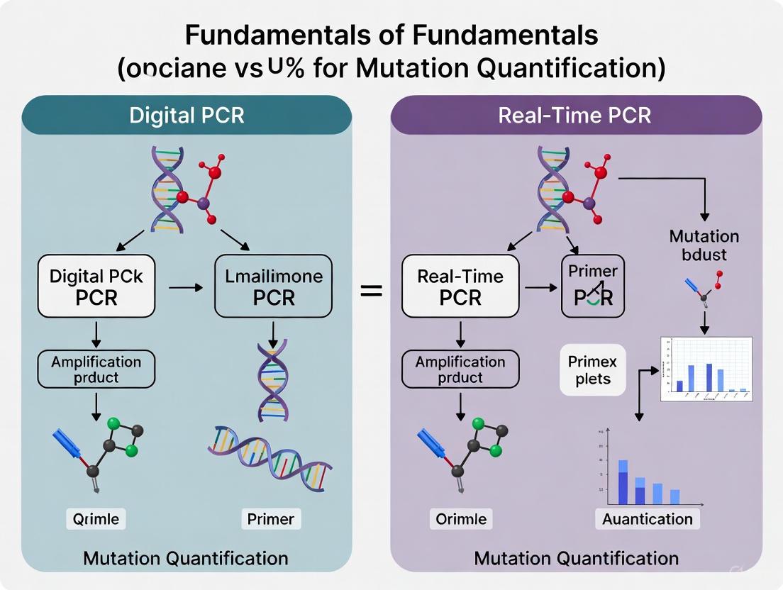
Visualization of PCR Evolution and Workflows
Diagram 1: Evolution of PCR Technologies and Methodological Workflows. The diagram illustrates the historical progression from Conventional to Real-Time to Digital PCR, along with their respective experimental workflows.
Diagram 2: Mutation Detection and Quantification Workflow. The diagram outlines the key experimental steps for mutation detection using both Real-Time and Digital PCR methodologies, highlighting their convergence in providing quantitative mutation data.
The evolution of PCR from a simple DNA amplification technique to sophisticated quantitative and digital platforms has fundamentally transformed mutation detection research. Each technological advancement has addressed specific limitations of its predecessors while introducing new capabilities that have expanded the possible applications in biomedical research.
Conventional PCR established the fundamental principle of enzymatic DNA amplification but provided limited quantitative capability. Real-time PCR introduced reliable quantification through fluorescence monitoring during the exponential amplification phase, becoming the gold standard for gene expression analysis, viral load monitoring, and many mutation detection applications [4]. Digital PCR has further advanced the field by enabling absolute quantification through sample partitioning and Poisson statistical analysis, providing exceptional precision and sensitivity for rare mutation detection [6].
For mutation quantification research, the choice between real-time PCR and dPCR depends on multiple factors including required sensitivity, precision, throughput, and available resources. Real-time PCR remains a robust, cost-effective solution for many applications, while dPCR offers superior performance for challenging scenarios such as liquid biopsies, rare mutation detection, and absolute quantification without standards [3] [6].
As PCR technologies continue to evolve, emerging trends including further miniaturization, integration with microfluidics, and development of isothermal amplification methods suggest that the future will bring even more powerful tools for mutation research [1]. The ongoing refinement of these technologies promises to further enhance our understanding of genetic heterogeneity, disease mechanisms, and therapeutic responses across diverse fields of biomedical research.
Quantitative PCR (qPCR) serves as a cornerstone technique in molecular biology, with two primary approaches for data analysis: absolute and relative quantification. This guide delves into the fundamentals of relative quantification, a method that analyzes changes in gene expression relative to a reference sample. We will explore the mathematical principles, detailed experimental protocols, and the critical, though indirect, role that standard curves play in validating these assays. Furthermore, this discussion is framed within a broader comparison with digital PCR (dPCR), highlighting how the choice of technology impacts mutation quantification and other precision research applications.
In the evolving field of nucleic acid quantification, researchers can choose between two powerful PCR technologies: real-time PCR (qPCR) and digital PCR (dPCR). The core difference lies in their approach to quantification. qPCR relies on a standard curve to determine the initial amount of target nucleic acid, providing either relative or inferred absolute quantification [10]. In contrast, dPCR uses a limiting dilution and partitioning method to directly count individual molecules, providing absolute quantification without the need for a standard curve [11] [12].
Relative quantification in qPCR is particularly valuable for assessing changes in gene expression—for example, comparing the expression level of a specific gene in a treated sample to an untreated control [11]. While this method does not require a standard curve for final calculation, the standard curve is indispensable during the assay development and validation phase to confirm optimal reaction efficiency. This technical guide provides an in-depth examination of relative quantification methodologies, placing them in the context of a researcher's toolkit that may also include the absolute quantification capabilities of dPCR for applications like rare mutation detection [13].
Core Concepts of Relative Quantification
Relative quantification is used to analyze changes in gene expression in a given sample relative to another reference sample, such as an untreated control or a calibrator [11]. The core principle involves normalizing the amount of the target gene of interest to the amount of one or more stably expressed endogenous control genes (often called housekeeping genes). This normalization controls for variations in RNA quantity and quality across different samples, allowing for meaningful comparison.
The final data output is typically expressed as a fold-change or fold-difference in expression levels [14]. For instance, a result of 5.0 indicates a five-fold increase in gene expression in the test sample relative to the control, while a result of 0.2 indicates a five-fold decrease.
Key Nomenclature
To understand relative quantification, a clear grasp of the key terms is essential. The following table defines the critical components involved in the calculations.
Table 1: Key Nomenclature for Relative Quantification of qPCR Data [15]
| Term | Symbol | Description |
|---|---|---|
| Threshold Cycle | Ct | The PCR cycle at which the sample's fluorescence crosses a threshold set above the background level. It is inversely proportional to the starting quantity of the target. |
| Target Gene | The gene of interest whose expression is being studied. | |
| Reference Gene | An endogenous control gene (e.g., GAPDH, actin) with stable expression across the sample set, used for normalization. | |
| Calibrator Sample | The reference sample (e.g., untreated control) to which all test samples are compared. | |
| Delta Ct | ΔCt | The difference in Ct values between the target gene and the reference gene within a single sample. |
| Double Delta Ct | ΔΔCt | The difference between the ΔCt of a test sample and the ΔCt of the calibrator sample. |
Mathematical Foundations and Calculation Methods
There are two main mathematical approaches for calculating relative quantification, chosen based on the amplification efficiencies of the target and reference gene assays.
The Comparative CT (ΔΔCT) Method
The ΔΔC_T method is the most common calculation, used when the amplification efficiencies of the target and reference gene are approximately equal and close to 100% (meaning the product doubles every cycle during the exponential phase) [15] [14].
The formula for the ΔΔC_T method is: RQ = 2^–ΔΔCT [15]
Where:
- ΔCt (test) = Ct (target, test) – Ct (reference, test)
- ΔCt (calibrator) = Ct (target, calibrator) – Ct (reference, calibrator)
- ΔΔCt = ΔCt (test) – ΔCt (calibrator)
This method is straightforward because it eliminates the need for a standard curve in the final analysis, increasing throughput and reducing potential pipetting errors [11].
The Pfaffl (Efficiency-Corrected) Method
When the amplification efficiencies of the target and reference gene assays are not equal (differing by more than 5%), the Pfaffl method, also known as the standard curve method for relative quantification, must be used [15]. This method incorporates the actual efficiency of each primer set into the calculation.
The formula for the Pfaffl method is: RQ = (Etarget)^ΔCt(target) / (Ereference)^ΔCt(reference) [15]
Where:
- E_target is the amplification efficiency of the target gene assay.
- E_reference is the amplification efficiency of the reference gene assay.
- ΔCt (target) = Ct (target, calibrator) – Ct (target, test)
- ΔCt (reference) = Ct (reference, calibrator) – Ct (reference, test)
The ΔΔC_T method is actually a special case of the Pfaffl method where both efficiencies are 2 (100%) [15].
Experimental Protocols for Relative Quantification
Determining Amplification Efficiency
Before performing a relative quantification experiment, the amplification efficiency (E) for each primer/probe set must be determined. This is a critical validation step.
Protocol:
- Prepare Dilutions: Make a minimum of five serial dilutions (e.g., 10-fold or 5-fold) of a cDNA template known to express the gene of interest in high abundance [16].
- Run qPCR: Use each dilution in separate qPCR reactions with the primer set to be validated.
- Generate Standard Curve: Plot the Ct values obtained against the logarithm of the dilution factor or the known concentration.
- Calculate Efficiency: Determine the slope of the standard curve and calculate the efficiency using the formula: E = 10^(–1/slope) [15]. The efficiency is often expressed as a percentage: % Efficiency = (E - 1) × 100. An ideal reaction with 100% efficiency has a slope of -3.32 and E=2. Efficiencies between 90% and 110% are generally considered acceptable [15].
Step-by-Step Workflow for Relative Quantification
The following diagram illustrates the core workflow for a relative quantification experiment using qPCR, from sample preparation to data analysis.
The Scientist's Toolkit: Essential Reagents and Materials
Successful relative quantification relies on a suite of specialized reagents and materials. The following table details the key components and their functions.
Table 2: Key Research Reagent Solutions for qPCR Relative Quantification [11] [14]
| Item | Function & Importance |
|---|---|
| High-Quality RNA/DNA Template | The starting material must be pure and intact. Contamination with RNases, DNA, or inhibitors (e.g., from plasmid prep) can inflate quantification and skew results [11]. |
| Reverse Transcription Kit | For gene expression (RT-qPCR), this converts RNA to cDNA. Kits may use oligo(dT), random primers, or gene-specific primers. The choice affects cDNA representation and efficiency [14]. |
| Sequence-Specific Primers | Designed to amplify the target and reference genes with high specificity and optimal efficiency (90-110%). Poor design leads to primer-dimer, off-target amplification, and unreliable data [14]. |
| Detection Chemistry | SYBR Green: Binds double-stranded DNA; cost-effective but less specific. TaqMan Probes: Fluorogenic probes provide higher specificity, enabling multiplexing. The choice depends on application needs [14]. |
| Validated Reference Gene Assay | Pre-designed, pre-validated assays for housekeeping genes (e.g., GAPDH, β-actin) reduce optimization time and ensure reliable normalization [14]. |
| qPCR Plates & Seals | Optically clear plates and secure seals are essential for efficient heat transfer and preventing well-to-well contamination and evaporation during cycling. |
| Commercial Pre-designed Assays | For single genes or pathway-focused PCR arrays, these provide a standardized, highly optimized solution that maximizes reproducibility and saves time [14]. |
qPCR vs. dPCR: A Strategic Comparison for Mutation Quantification Research
The choice between qPCR and dPCR is critical and depends on the research goals. The following table provides a direct comparison of the two technologies.
Table 3: Key Differences Between Real-Time PCR (qPCR) and Digital PCR (dPCR) [10] [12]
| Factor | Real-Time PCR (qPCR) | Digital PCR (dPCR) |
|---|---|---|
| Quantification Principle | Relative (or absolute via standard curve). | Absolute, via direct molecule counting. |
| Requires Standard Curve | Yes, for absolute quantification and assay validation. | No. |
| Precision & Sensitivity | High, but limited for detecting very rare targets (<1%) in a background of wild-type sequences. | Superior for rare targets and small fold changes (e.g., can detect rare mutations in liquid biopsies) [13]. |
| Dynamic Range | Wide (6-7 orders of magnitude). | Narrower. |
| Tolerance to Inhibitors | Sensitive; inhibitors in the sample can reduce amplification efficiency. | Highly resistant; partitioning dilutes inhibitors, making it robust for complex samples [12]. |
| Cost & Throughput | Lower cost per sample, high throughput (96/384-well plates). | Higher cost per sample, lower throughput. |
| Ideal Application in Cancer Research | Gene expression profiling, validating RNA-seq data, pathogen detection [17]. | Detecting rare mutations, copy number variation (CNV) analysis, liquid biopsies, validating NGS findings [13]. |
Application Context: Minimal Residual Disease (MRD) Monitoring
The comparison between qPCR and dPCR is actively debated in clinical research applications like Minimal Residual Disease (MRD) monitoring, where the goal is to detect extremely low levels of cancer cells after treatment. Research indicates that while well-validated qPCR assays are robust and reliable for known targets, dPCR is often more effective for detecting rarer targets due to its superior sensitivity and precision [17]. This makes dPCR a powerful tool for quantifying mutations, such as in EGFR, to track cancer evolution and guide therapy [17].
Relative quantification by qPCR is a powerful, well-established method for analyzing changes in gene expression. Its reliability hinges on careful experimental design, including the validation of amplification efficiencies and the selection of stable reference genes. The use of standard curves in the validation phase is fundamental to this process. However, for applications at the frontiers of research and molecular diagnostics—particularly the quantification of rare mutations, copy number variations, and biomarkers in complex samples—digital PCR offers a compelling alternative. Its ability to provide absolute quantification without standard curves, coupled with enhanced precision and resilience to inhibitors, makes dPCR an indispensable technology in the modern scientist's arsenal, perfectly complementing the high-throughput strengths of qPCR.
Digital PCR (dPCR) represents a transformative approach in molecular diagnostics, enabling absolute quantification of nucleic acids without requiring standard curves. This whitepaper examines the core principles of dPCR technology, focusing on its partitioning methodology and statistical foundation for precise mutation quantification research. Compared to real-time PCR (qPCR), dPCR demonstrates superior precision and sensitivity, particularly for detecting rare genetic mutations and quantifying low-abundance targets—critical requirements in oncology and drug development. We provide a comprehensive technical analysis of dPCR implementation, experimental protocols, and performance metrics relevant to research scientists and pharmaceutical professionals.
Digital PCR (dPCR) constitutes the third generation of PCR technology, evolving from conventional PCR and real-time quantitative PCR (qPCR) to address growing needs for precise nucleic acid quantification [18]. The fundamental innovation of dPCR lies in its partitioning approach, where a PCR reaction mixture is divided into thousands to millions of separate reactions, enabling individual amplification events that transform continuous analog signals into discrete digital measurements [19] [20]. This partitioning strategy allows dPCR to achieve absolute quantification through binary detection and Poisson statistical analysis, circumventing the reliance on external standards that introduces variability in qPCR results [21] [20].
The clinical and research significance of dPCR has substantially increased with the growing demand for detecting minor genetic variations within complex biological samples. Unlike qPCR, which quantifies nucleic acids based on their amplification kinetics relative to standard curves, dPCR provides direct absolute quantification by counting individual molecules, making it particularly valuable for applications requiring high precision [10]. This capability proves especially crucial in mutation quantification research, where accurately identifying rare mutations against a background of wild-type sequences can inform diagnostic and therapeutic decisions in oncology, infectious disease monitoring, and genetic disorder screening [18].
The technological evolution of dPCR platforms has progressed significantly since the conceptual foundation was laid in the 1990s, with commercial systems now employing either droplet-based or microchip-based partitioning methodologies [18]. Current systems partition samples into water-in-oil droplet emulsions (droplet digital PCR or ddPCR) or nanoscale wells on microchips (nanoplate dPCR), with each approach offering distinct advantages in throughput, partitioning efficiency, and integration with laboratory workflows [6] [22]. These technological advancements have positioned dPCR as an indispensable tool for researchers and drug development professionals requiring uncompromising accuracy in nucleic acid quantification.
Fundamental Principles of dPCR
The Partitioning Process and Absolute Quantification
The operational principle of digital PCR centers on sample partitioning, where a conventional PCR reaction mixture—containing template nucleic acids, primers, probes, nucleotides, enzymes, and buffers—is physically divided into numerous individual reactions [19]. This partitioning occurs through either droplet-based systems that generate thousands of nanoliter-sized water-in-oil droplets or chip-based systems that distribute the sample into fixed nanoscale wells [18]. Each partition effectively functions as an isolated PCR microreactor, with the random distribution of nucleic acid molecules ensuring that partitions contain zero, one, or several target molecules based on their initial concentration in the sample [20].
Following partitioning, the reactions undergo standard PCR amplification with fluorescence detection probes. Critically, dPCR utilizes end-point detection rather than monitoring the reaction in real-time [20]. After amplification, each partition is analyzed for fluorescence presence, creating a binary readout where partitions either fluoresce (positive, indicating target presence) or remain dark (negative, indicating target absence) [19]. This binary detection system simplifies signal interpretation compared to the continuous fluorescence measurements required in qPCR, thereby reducing potential analytical variability [10].
The absolute quantification capability of dPCR emerges from directly counting positive partitions rather than inferring concentration from amplification kinetics [21]. Since the partioning process is random, the proportion of negative partitions follows predictable statistical distributions, enabling back-calculation of the original target concentration without reference to standards [20]. This approach eliminates uncertainties associated with amplification efficiency variations that affect qPCR accuracy, particularly for difficult targets or suboptimal reaction conditions [23].
Poisson Statistics in Quantification
The mathematical foundation of dPCR quantification relies on Poisson statistics, which model the random distribution of molecules across partitions [19] [20]. The Poisson distribution accurately describes the probability of a partition containing a specific number of target molecules, with the parameter λ (lambda) representing the average number of target molecules per partition [20]. The probability of a partition being negative (containing zero target molecules) is given by P(0) = e^(-λ), while the probability of a partition containing at least one target molecule is P(≥1) = 1 - e^(-λ) [20].
From the experimental data, the fraction of negative partitions (k/n, where k is the number of negative partitions and n is the total number of partitions) provides the basis for calculating λ using the equation λ = -ln(1 - k/n) [20]. The absolute concentration of the target in the original sample is then determined by accounting for the partition volume and sample dilution factors [19]. This statistical approach enables direct calculation of target concentration without standard curves, providing true absolute quantification [21].
Table 1: Poisson Distribution Probabilities for Different Target Concentrations
| Copies/Partition (λ) | % Negative Partitions | % Partitions with 1 Copy | % Partitions with ≥2 Copies | Optimal Use Case |
|---|---|---|---|---|
| 0.1 | 90.5% | 9.0% | 0.5% | Rare target detection |
| 0.5 | 60.7% | 30.3% | 9.0% | Low abundance targets |
| 1.0 | 36.8% | 36.8% | 26.4% | Standard quantification |
| 1.6 | 20.2% | 32.3% | 47.5% | Optimal precision |
| 3.0 | 5.0% | 14.9% | 80.1% | High concentration targets |
| 5.0 | 0.7% | 3.4% | 95.9% | Limited utility for dPCR |
The precision of dPCR quantification depends significantly on the number of partitions analyzed and the value of λ [20]. Maximum precision occurs when approximately 20% of partitions are negative (λ ≈ 1.6), with precision improving as the total number of partitions increases [20]. This statistical understanding informs experimental design, guiding researchers to adjust sample concentrations to achieve optimal λ values for their specific applications [22].
dPCR versus qPCR: Comparative Analysis
Technical and Performance Comparisons
The fundamental distinction between dPCR and qPCR lies in their quantification methodologies. While qPCR relies on relative quantification against standard curves during the exponential amplification phase, dPCR utilizes absolute quantification through end-point measurement of binary partition outcomes [20]. This core difference translates into distinct performance characteristics that determine their suitability for specific applications, particularly in mutation quantification research.
Recent comparative studies demonstrate dPCR's superior accuracy and precision, especially for complex samples and low-abundance targets. In respiratory virus detection during the 2023-2024 tripledemic, dPCR showed significantly improved accuracy for high viral loads of influenza A, influenza B, and SARS-CoV-2, along with better consistency in quantifying intermediate viral levels [6]. Similarly, in copy number variation (CNV) analysis, dPCR achieved 95% concordance with pulsed field gel electrophoresis (considered a gold standard), while qPCR showed only 60% concordance, with qPCR particularly underestimating higher copy numbers [23].
Table 2: Performance Comparison of dPCR versus qPCR
| Parameter | Digital PCR (dPCR) | Real-Time PCR (qPCR) |
|---|---|---|
| Quantification Method | Absolute (without standards) | Relative (requires standard curves) |
| Precision | Higher (CV 6-13%) [22] | Lower (variable based on standards) |
| Sensitivity | Superior for rare targets [23] | Moderate to high |
| Dynamic Range | Limited by partition number [19] | Broader dynamic range |
| Tolerance to Inhibitors | Higher (due to partitioning) [19] | Lower |
| Throughput | Moderate | High |
| Cost per Sample | Higher | Lower |
| Mutation Detection | Excellent for rare mutations [18] | Limited by background signal |
The partitioning process in dPCR provides inherent advantages for analyzing challenging samples. By separating target sequences from potential inhibitors and background DNA, dPCR demonstrates greater resilience to PCR inhibitors present in complex matrices [19]. This capability proves particularly valuable for clinical samples that may contain substances inhibiting amplification, such as hemoglobin in blood samples or polysaccharides in tissue biopsies, where qPCR performance typically deteriorates [6].
Application-Based Selection Criteria
The choice between dPCR and qPCR depends heavily on specific research requirements, sample characteristics, and practical considerations. qPCR remains the preferred method for high-throughput applications where relative quantification suffices, such as gene expression analysis in well-characterized systems, pathogen detection with abundant targets, and routine diagnostic screening where cost-effectiveness is paramount [21] [10]. The established infrastructure, standardized protocols, and lower per-sample cost of qPCR maintain its position as the workhorse of molecular diagnostics for many applications.
dPCR excels in scenarios requiring absolute quantification and exceptional sensitivity. Its capabilities prove indispensable for detecting rare mutations in oncology research, monitoring minimal residual disease, quantifying viral reservoirs, validating reference materials, and analyzing copy number variations with high precision [18] [23]. Additionally, dPCR provides superior performance for analyzing samples with inherent variability in amplification efficiency, as its digital nature eliminates quantification biases introduced by such variations [20].
Recent advances in both technologies continue to reshape their respective applications. For qPCR, improvements in multiplexing capabilities and integration with automated systems maintain its relevance in clinical diagnostics. Meanwhile, dPCR platforms are addressing previous limitations in throughput and cost while expanding multiplexing capabilities [24]. The emerging trend of combining both technologies—using qPCR for initial screening and dPCR for confirmation of critical samples—represents a powerful approach leveraging the strengths of both methodologies [21].
Experimental Protocols and Implementation
Standard dPCR Workflow
The typical dPCR workflow comprises three main stages: sample preparation, partitioning with amplification, and data analysis. Initial sample preparation follows standard nucleic acid extraction protocols similar to those used in qPCR, though dPCR's partitioning provides greater tolerance to minor impurities [6]. For optimal performance, DNA or RNA quality should be verified, with special consideration for factors that might affect partitioning efficiency, such as viscosity or detergent concentration [22].
Following sample preparation, the reaction mixture—containing template, primers, probes, and PCR master mix—is loaded into the dPCR instrument for partitioning and amplification. The specific partitioning method varies by platform: droplet-based systems (ddPCR) generate thousands of nanoliter-sized droplets in an oil emulsion [18], while chip-based systems (ndPCR) distribute the sample into nanoscale wells on a solid substrate [6]. Following partitioning, endpoint PCR amplification occurs with fluorescence detection, during which partitions containing the target sequence generate positive signals while those without remain negative [19].
Data analysis involves counting positive and negative partitions, applying Poisson correction for multiple targets per partition, and calculating the absolute target concentration [20]. Modern dPCR platforms include sophisticated software that automates these calculations while providing visualization tools such as heat maps, scatter plots, and histograms to facilitate data interpretation and quality assessment [19]. Proper threshold setting between positive and negative partitions remains critical for accurate quantification, though dPCR's binary nature makes threshold determination more robust than in qPCR [22].
Key Reagents and Materials
Successful dPCR implementation requires careful selection of reagents and materials optimized for digital applications. The core components include specialized master mixes formulated for partition stability and consistent amplification, sequence-specific primers and probes with appropriate fluorophore-quencher combinations, and partitioning media (oils or chips) designed for the specific platform [22]. Additionally, restriction enzymes may be incorporated to improve access to target sequences in complex genomic regions, with enzyme selection significantly impacting quantification precision [22].
Table 3: Essential Research Reagents for dPCR Experiments
| Reagent/Material | Function | Implementation Considerations |
|---|---|---|
| dPCR Master Mix | Provides enzymes, nucleotides, buffers for amplification | Optimized for partition stability; different formulations for probe vs. dye-based detection |
| Assay Primers/Probes | Target-specific amplification and detection | Similar to qPCR but requires validation for dPCR; fluorophore selection depends on instrument channels |
| Partitioning Oil/Chip | Creates isolated reaction environments | Platform-specific; critical for consistent partition generation |
| Restriction Enzymes | Enhances target accessibility in complex DNA | Improves precision, especially for high copy numbers; enzyme selection important [22] |
| Positive/Negative Controls | Validates assay performance | Essential for establishing thresholds and confirming reaction efficiency |
| Reference Assays | Normalization for sample quality | Particularly important for copy number variation studies |
Platform selection significantly influences reagent choices and experimental design. Droplet-based systems (e.g., Bio-Rad QX200) typically require specific oils and surfactants for stable droplet formation [22], while chip-based systems (e.g., QIAGEN QIAcuity) utilize specialized nanoplates with predefined well structures [6]. Recent comparisons demonstrate that both platforms achieve similar detection and quantification limits, though precision may vary based on target concentration and sample treatment [22]. This underscores the importance of platform-specific optimization while confirming the general robustness of dPCR methodology across different implementations.
Digital PCR represents a significant advancement in nucleic acid quantification technology, with its partitioning approach and Poisson statistical foundation enabling absolute quantification without standard curves. The core strength of dPCR lies in its exceptional precision, sensitivity for rare targets, and resilience to amplification inhibitors—attributes particularly valuable for mutation quantification research in oncology, infectious disease monitoring, and biomarker validation. While qPCR maintains advantages in throughput and cost-effectiveness for many routine applications, dPCR provides unequivocal benefits for applications demanding the highest quantification accuracy.
The continuing evolution of dPCR technology addresses initial limitations in dynamic range and throughput while expanding multiplexing capabilities [24]. These advancements, coupled with growing recognition of its utility in clinical research and diagnostic development, position dPCR as an increasingly essential tool for researchers and drug development professionals. As molecular diagnostics continues toward more precise and personalized applications, the fundamental principles of partitioning and digital detection make dPCR uniquely suited to meet the emerging challenges in mutation quantification and rare sequence detection.
The evolution of polymerase chain reaction (PCR) technologies has progressively aimed for greater precision in nucleic acid quantification. While traditional bulk reaction analysis methods like quantitative real-time PCR (qPCR) provide valuable quantitative data, the emergence of microfluidic partitioning in digital PCR (dPCR) represents a paradigm shift in molecular quantification. This shift is particularly crucial for applications demanding absolute quantification and high sensitivity, such as mutation quantification research in cancer genomics and liquid biopsy development [25] [26].
The core distinction between these methodologies lies in their fundamental approach to sample analysis. Bulk reaction analysis processes the entire sample in a single, unified reaction volume, measuring amplification throughout the thermal cycling process. In contrast, microfluidic partitioning technologies physically divide the sample into thousands to millions of nanoliter-sized reactions, enabling a binary, digital readout of individual molecular presence [18]. This technical whitpaper examines the key technological differences between these approaches, focusing on their implications for research and diagnostic applications, particularly in the context of mutation detection and quantification.
Core Technological Principles
Bulk Reaction Analysis in Real-Time PCR
Real-time quantitative PCR (qPCR) operates on the principle of monitoring amplification in real-time within a single, bulk reaction vessel. This technology relies on detecting fluorescent signals that increase proportionally to the amount of amplified DNA during each PCR cycle. The fundamental metrics in qPCR are the cycle threshold (Ct) values, which represent the PCR cycle number at which the fluorescence signal crosses a predefined threshold [26].
The quantification process in qPCR is relative, requiring comparison to standard curves generated from samples of known concentration. This introduces several technical considerations:
- Amplification Efficiency Dependency: qPCR results are highly dependent on consistent amplification efficiency, which can be affected by PCR inhibitors present in the sample [26].
- Exponential Phase Measurement: Data collection occurs during the exponential amplification phase, where slight variations can significantly impact quantification accuracy [26].
- Standard Curve Requirement: The need for standard curves introduces potential variability and necessitates careful validation of reference materials [26].
Despite these limitations, qPCR remains widely adopted due to its established protocols, broad dynamic range, and extensive validation in clinical and research settings [24].
Microfluidic Partitioning in Digital PCR
Digital PCR (dPCR) employs a fundamentally different approach based on sample partitioning. The core principle involves distributing a PCR reaction across thousands to millions of individual partitions, each functioning as an isolated micro-reactor. Following endpoint amplification, each partition is analyzed as positive or negative for the target sequence, creating a binary (digital) readout [18].
The technological implementation of partitioning occurs primarily through two methods:
- Droplet-based Partitioning (ddPCR): Utilizes water-in-oil emulsion technology to create nanoliter-sized droplets, typically generating 20,000 or more partitions per sample [27].
- Chip-based Partitioning: Employs microfabricated chips with fixed nanowells or microchambers to physically isolate reactions, as seen in systems like the QIAcuity [18] [6].
The absolute quantification in dPCR is derived statistically using Poisson distribution to account for the random distribution of molecules across partitions, eliminating the need for standard curves and providing direct measurement of target concentration [18] [26].
Digital PCR Workflow via Microfluidic Partitioning
Comparative Performance Analysis
Technical Comparison Table
| Parameter | Bulk Reaction (qPCR) | Microfluidic Partitioning (dPCR) |
|---|---|---|
| Quantification Method | Relative (requires standard curve) | Absolute (no standard curve) [26] |
| Detection Principle | Real-time fluorescence during exponential phase | Endpoint fluorescence after partitioning [26] |
| Sensitivity for Rare Mutations | ≥1% mutant allele frequency [26] | ≤0.1% mutant allele frequency [26] |
| Impact of PCR Inhibitors | High susceptibility [26] | High tolerance due to partitioning [26] |
| Dynamic Range | Broad [26] | Limited by partition count [18] |
| Multiplexing Capability | Well-established | Advanced in nanoplate systems (4-12 targets) [27] |
| Workflow Duration | ~2 hours [27] | <2 hours for nanoplate; 6-8 hours for ddPCR [27] |
| Data Reproducibility | Moderate, lab-dependent | High precision across laboratories [26] |
Performance in Mutation Detection
The superior sensitivity of dPCR for rare mutation detection represents one of its most significant advantages. Where qPCR typically detects mutations present at frequencies above 1%, dPCR can reliably identify mutations at frequencies as low as 0.1% or less [26]. This enhanced sensitivity stems from the partitioning effect, which effectively enriches rare targets by separating them from the abundant wild-type background, thereby improving the signal-to-noise ratio [18].
This capability is particularly valuable in liquid biopsy applications, where tumor-derived DNA fragments circulate in blood at low concentrations amidst a background of wild-type DNA. A 2025 study comparing dPCR and Real-Time RT-PCR for respiratory virus detection confirmed dPCR's superior accuracy, particularly for samples with medium to high viral loads, demonstrating greater consistency and precision in quantification [6].
Experimental Protocols for Mutation Quantification
dPCR Protocol for Rare Mutation Detection
Objective: Absolute quantification of a point mutation (e.g., KRAS G12D) in a background of wild-type genomic DNA using nanoplate-based dPCR.
Materials and Reagents:
- QIAcuity Nanoplate 26k 5-plex (QIAGEN) [6]
- dPCR Supermix suitable for probe-based detection
- Target-specific FAM-labeled mutation probe and HEX-labeled wild-type probe
- Reference assay for total DNA quantification
- DNA sample (10-100 ng total input)
- Nuclease-free water
Methodology:
- Reaction Setup: Prepare 40 μL master mix containing 1X dPCR supermix, 20-100 nM each probe, 10-100 ng fragmented genomic DNA, and nuclease-free water to volume [6].
- Nanoplate Loading: Pipette the master mix into the designated well of the nanoplate. Seal the plate with provided foil [27].
- Partitioning and Amplification: Place the sealed nanoplate into the QIAcuity instrument. The system automatically performs partitioning, PCR amplification with the following protocol [6]:
- Enzyme activation: 95°C for 2 minutes
- Denaturation: 95°C for 15 seconds
- Annealing/Extension: 60°C for 30 seconds (40 cycles)
- Endpoint signal stabilization: 40°C for 5 minutes
- Image Acquisition and Analysis: The integrated imager captures fluorescence data from all partitions simultaneously. Analyze using instrument software with the following gating strategy [6]:
- Identify positive control (reference assay) partitions
- Gate mutation-positive (FAM+) partitions
- Gate wild-type-positive (HEX+) partitions
- Calculate mutation allele frequency using Poisson correction
Validation: Include no-template controls, wild-type-only controls, and mutation-positive controls at expected frequencies (0.1%, 1%, 5%) to validate assay performance.
Comparative qPCR Protocol
Objective: Relative quantification of the same point mutation using qPCR.
Materials and Reagents:
- qPCR master mix with hot-start DNA polymerase
- The same mutation-specific and wild-type-specific probes
- Standard curve samples with known mutation frequencies (0%, 1%, 5%, 10%, 25%, 50%, 100%)
- 96-well qPCR plate and sealing film
- Real-time PCR instrument
Methodology:
- Reaction Setup: Prepare 20 μL reactions containing 1X qPCR master mix, 20-100 nM each probe, and 10-100 ng DNA template per well [6].
- Plate Setup: Include standard curve samples in duplicate and unknown samples in triplicate.
- Amplification: Run on real-time PCR instrument with matching cycling conditions:
- Enzyme activation: 95°C for 2 minutes
- Denaturation: 95°C for 15 seconds
- Annealing/Extension: 60°C for 30 seconds (40 cycles)
- Fluorescence acquisition during annealing/extension step
- Data Analysis:
- Determine Ct values for each reaction
- Generate standard curves for both mutation and wild-type assays
- Calculate mutation frequency relative to standard curve
The Scientist's Toolkit: Essential Research Reagents and Materials
| Item | Function | Application Notes |
|---|---|---|
| Partitioning Plates/Cartridges | Physical separation of reactions into nanoliter volumes | Nanoplates (QIAcuity) or droplet cartridges (Bio-Rad) depending on platform [27] |
| dPCR Supermix | Optimized reaction mix for endpoint amplification | Contains polymerase, dNTPs, buffers; formulated for partition stability [6] |
| Hydrolysis Probes (TaqMan) | Sequence-specific detection with fluorescent reporters | FAM/HEX/CY5 labels for multiplexing; designed with stringent specificity requirements [6] |
| Nuclease-Free Water | Reaction preparation without enzymatic degradation | Essential for maintaining reaction integrity and preventing false negatives |
| Positive/Negative Controls | Assay validation and quality assurance | Synthetic oligonucleotides with known mutation status; wild-type genomic DNA [6] |
| Microfluidic Instrumentation | Partitioning, thermal cycling, and imaging | Integrated systems (e.g., QIAcuity, AbsoluteQ) or droplet generators (QX200) [27] |
Application-Specific Implementation
Mutation Quantification in Cancer Research
In oncology research, the precise quantification of tumor-specific mutations has profound implications for understanding tumor heterogeneity, monitoring minimal residual disease, and assessing treatment response. The high sensitivity of dPCR enables researchers to detect rare mutant alleles in liquid biopsies, providing a non-invasive method for tracking tumor evolution [18].
A compelling application comes from a 2024 Cancer Cell study where researchers leveraged both bulk and single-cell RNA-seq to identify developmental states driving chemoresistance in B-cell acute lymphoblastic leukemia (B-ALL). This integrated approach revealed how cellular heterogeneity influences treatment response, demonstrating the complementary value of high-sensitivity techniques in uncovering mechanisms that bulk analyses alone might miss [28].
Regulatory and Quality Control Considerations
For drug development professionals, the choice between dPCR and qPCR extends beyond technical performance to include regulatory compliance and quality control requirements. In Good Manufacturing Practice (GMP) environments, dPCR platforms offer advantages with their streamlined workflows, reduced contamination risk, and built-in compliance features supporting 21 CFR Part 11 requirements [27].
The absolute quantification capability of dPCR eliminates inter-laboratory variability associated with standard curve generation, making it particularly valuable for multi-center clinical trials where consistent measurement of biomarkers across sites is critical [27].
The technological divergence between microfluidic partitioning and bulk reaction analysis represents more than just methodological differences—it reflects a fundamental shift in the precision possible in molecular quantification. While bulk reaction analysis using qPCR remains a robust, well-established technology suitable for many applications, microfluidic partitioning in dPCR provides unambiguous advantages for mutation quantification research requiring absolute quantification, exceptional sensitivity, and high precision.
The choice between these technologies should be guided by specific research objectives, with dPCR particularly suited for applications involving rare mutation detection, liquid biopsy analysis, and situations where exact copy number quantification is essential. As microfluidic technologies continue to evolve, with improvements in multiplexing capabilities, workflow efficiency, and integration with automated systems, their implementation in both basic research and drug development is poised to expand significantly.
Technology Selection Guide
The emergence of digital PCR (dPCR) represents a paradigm shift in nucleic acid quantification, transitioning from relative measurements to absolute, single-molecule counting. This evolution began with limiting dilution techniques and has culminated in sophisticated, fully integrated instrumentation that defines modern dPCR. For researchers in mutation quantification and drug development, understanding this historical and technical progression is essential for selecting the appropriate methodology for precise molecular analyses. This whitepaper traces the critical developmental milestones, compares the fundamental principles of dPCR and real-time quantitative PCR (qPCR), and provides a detailed examination of contemporary dPCR platforms and their experimental applications.
From Concept to Reality: The Foundations of dPCR
The conceptual foundation of dPCR was established through limiting dilution PCR, a technique first used in 1989 by Peter Simmonds to quantify HIV provirus in infected cells by serially diluting samples until target molecules were separated [18]. This method demonstrated that disease stage correlated with the proportion of infected cells, laying the groundwork for single-molecule detection.
In 1992, Morley and Sykes formally combined limiting dilution with Poisson statistics to isolate, detect, and quantify single nucleic acid molecules, successfully detecting mutated genes in leukemia patients at a sensitivity of 2 targets in 160,000 wild-type sequences [18]. The term "digital PCR" was officially coined in 1999 by Bert Vogelstein and his team, who developed a workflow using limiting dilution on 96-well plates combined with fluorescence readout to detect RAS oncogene mutations in the stools of colorectal cancer patients [18].
Key innovations addressed the practical limitations of early approaches:
- Volume miniaturization (1997): Kalinina et al. introduced microcapillaries (~10 nL) for partitioning, reducing reagent costs and improving efficiency [18].
- BEAMing technology (2003): Vogelstein's group developed "Beads, Emulsion, Amplification, and Magnetics," which encapsulated individual DNA molecules with primer-coated magnetic beads in water-in-oil droplets [18].
These foundational developments established the core dPCR principle: partitioning a sample, amplifying individual molecules, and applying Poisson statistics to calculate absolute target concentration without standard curves [18] [22].
Table 1: Key Historical Developments in Digital PCR
| Year | Development | Key Innovator(s) | Significance |
|---|---|---|---|
| 1989 | Limiting Dilution PCR | Simmonds et al. | Enabled single-copy detection of HIV provirus, correlating with disease stage [18]. |
| 1992 | Limiting Dilution + Poisson Statistics | Morley and Sykes | Allowed accurate counting of single nucleic acid molecules for rare mutation detection [18]. |
| 1999 | Term "Digital PCR" Coined | Vogelstein et al. | Introduced fluorescence readout on 96-well plates to detect cancer mutations in clinical samples [18]. |
| 2003 | BEAMing Technology | Vogelstein et al. | Utilized water-in-oil droplet emulsion for efficient compartmentalization, simplifying the process [18]. |
| 2006 | First Commercial dPCR | Fluidigm | Launched the first commercial nanofluidic dPCR platform, making the technology accessible [18]. |
Diagram 1: The historical development path of digital PCR from its conceptual beginnings to commercialization.
dPCR vs. qPCR: Fundamental Principles and Technical Comparison
Core Principles of qPCR and dPCR
Quantitative PCR (qPCR) monitors the amplification of DNA in real-time during the exponential phase, using fluorescent dyes or probes. Quantification relies on the cycle threshold (Ct), the cycle number at which fluorescence crosses a predetermined threshold, and requires a standard curve derived from samples of known concentration to determine the initial amount of target DNA [10] [29]. This provides relative or, with a standard curve, absolute quantification, but introduces variability based on the accuracy of the standard curve and assumes consistent amplification efficiency across samples [10] [30].
Digital PCR (dPCR) eliminates the need for standard curves. The sample is partitioned into thousands of individual reactions, each acting as a separate PCR micro-reactor. Following end-point amplification, each partition is analyzed as positive (containing the target) or negative (lacking the target). The absolute concentration of the target nucleic acid is then statistically calculated using Poisson distribution based on the ratio of positive to negative partitions [18] [10] [22]. This allows for absolute quantification and is less susceptible to inhibitors and amplification efficiency variations [6] [22].
Partitioning Methods in Modern dPCR
Two major partitioning methods have emerged in modern dPCR platforms [18] [27]:
- Droplet Digital PCR (ddPCR): Employs a water-oil emulsion to create thousands of nanoliter-sized droplets (e.g., Bio-Rad's QX200/QX600 systems) [27] [22].
- Chip/Nanoplate-based dPCR: Distributes the sample across a plate containing fixed micro-wells or nanowells (e.g., Applied Biosystems' Absolute Q and QIAGEN's QIAcuity) [6] [27].
Table 2: Comparative Analysis: qPCR vs. dPCR Fundamentals
| Parameter | Quantitative PCR (qPCR) | Digital PCR (dPCR) |
|---|---|---|
| Quantification Basis | Relative to a standard curve [10] | Absolute, via Poisson statistics on positive/negative partitions [18] [22] |
| Standard Curve | Required [10] [29] | Not required [10] [22] |
| Precision & Sensitivity | High, but limited by standard curve [21] | Superior for rare targets and low-abundance sequences [6] [10] |
| Tolerance to Inhibitors | Moderate; can affect Ct values [6] | High; partitioning reduces inhibitor effect [6] [22] |
| Dynamic Range | Broad (~7-8 logarithms) [21] | Broad, but can be limited at very high concentrations by partition count [22] |
| Primary Application | Gene expression, pathogen detection (high throughput) [10] [31] | Rare mutation detection, copy number variation, liquid biopsy, viral load quantification [6] [10] |
| Throughput & Cost | High throughput, lower cost per sample [10] [31] | Lower throughput, higher cost per sample, but evolving [27] [31] |
Diagram 2: A comparison of the fundamental workflows for qPCR and dPCR, highlighting the key differentiators of real-time monitoring versus partitioning and absolute counting.
Modern dPCR Platforms and Experimental Protocol
The commercial dPCR landscape offers platforms based on different partitioning technologies. The QIAGEN QIAcuity system uses nanoplate-based technology, integrating partitioning, thermal cycling, and imaging into a single, automated instrument, facilitating high-throughput processing [6] [27]. The Applied Biosystems Absolute Q is a fully integrated, automated chip-based system designed to provide a "sample-in, results-out" workflow, which is particularly advantageous for quality control (QC) environments due to features supporting 21 CFR Part 11 compliance [27]. In contrast, Bio-Rad's ddPCR systems (e.g., QX200, QX600, QX700) utilize droplet technology, which is widely adopted in research settings for its high sensitivity and flexibility [27] [22].
Representative Experimental Protocol for Mutation Quantification
The following protocol, adapted from a 2025 study comparing dPCR platforms, outlines a typical workflow for absolute quantification of a genetic target, such as in mutation analysis [22].
1. Sample Preparation and Nucleic Acid Extraction
- Extract DNA or RNA from patient samples (e.g., blood, tissue, cell culture) using standardized kits. For RNA targets, include a reverse transcription step to generate cDNA.
- Critical Step: Pre-digest complex genomic DNA with restriction enzymes (e.g., HaeIII or EcoRI) to break up tandem repeats and improve access to the target sequence, which enhances precision and accuracy [22].
2. Assay Design
- Design and validate primer pairs and fluorescent probe(s) (e.g., TaqMan) specific to the target mutation and wild-type sequence for multiplex detection.
3. Reaction Mix Preparation
- Prepare the dPCR master mix according to the platform manufacturer's instructions. This typically includes:
- dPCR Supermix
- Primer/Probe sets for target and reference (if multiplexing)
- Nuclease-free water
- Template DNA (optimized amount, e.g., 1-100 ng)
4. Partitioning and Amplification
- Nanoplate-based (QIAcuity): Pipette the reaction mix into the designated wells of a nanoplate. The instrument automatically performs partitioning, PCR amplification, and imaging [6].
- Droplet-based (ddPCR): Use a droplet generator to create an emulsion of thousands of droplets from the reaction mix. Transfer the emulsion to a PCR plate for traditional thermal cycling. After cycling, load the plate into a droplet reader for analysis [22].
5. Data Analysis
- The instrument's software automatically analyzes each partition and classifies it as positive or negative based on fluorescence amplitude.
- The software calculates the absolute copy number concentration (copies/µL) of the target in the original sample using Poisson statistics. For mutation quantification, the ratio of mutant to wild-type alleles can be precisely determined.
Performance Comparison of dPCR Platforms
A 2025 study directly compared the QIAcuity One (nanoplate dPCR) and the QX200 (droplet ddPCR) platforms, providing key performance metrics [22].
Table 3: Performance Metrics of dPCR Platforms from a Comparative Study [22]
| Performance Metric | QIAcuity One (npdPCR) | QX200 (ddPCR) |
|---|---|---|
| Limit of Detection (LOD) | ~0.39 copies/µL input | ~0.17 copies/µL input |
| Limit of Quantification (LOQ) | ~1.35 copies/µL input | ~4.26 copies/µL input |
| Precision (with HaeIII enzyme) | Coefficient of Variation (CV) 1.6% - 14.6% | Coefficient of Variation (CV) < 5% |
| Accuracy (R² vs. expected copies) | R²adj = 0.98 | R²adj = 0.99 |
| Key Finding | High precision across a wide concentration range [22]. | Precision significantly improved with optimal restriction enzyme (HaeIII) [22]. |
The Scientist's Toolkit: Essential Reagents and Materials
Table 4: Key Research Reagent Solutions for dPCR Experiments
| Reagent/Material | Function | Example Use Case |
|---|---|---|
| dPCR Master Mix | Contains DNA polymerase, dNTPs, buffer, and MgCl₂ optimized for the partitioning and endpoint detection of dPCR. | The core chemical environment for all amplification reactions; formulation can vary by platform [22]. |
| TaqMan Probes | Sequence-specific, fluorescently labelled hydrolysis probes that provide high specificity for target detection. | Essential for multiplexed detection of wild-type and mutant alleles in a single reaction [6] [29]. |
| Restriction Enzymes (e.g., HaeIII) | Enzymes that cut DNA at specific recognition sites, fragmenting the genome. | Critical for pre-digestion to improve target accessibility in complex genomes, enhancing precision [22]. |
| Nanoplates or Cartridges | Platform-specific consumables containing microfluidic channels or pre-formed nanowells for partitioning. | Used in systems like QIAcuity and Absolute Q; the physical substrate for creating partitions [6] [27]. |
| Droplet Generation Oil & Cartridges | Reagents and consumables for creating a stable water-in-oil emulsion for droplet-based systems. | Used in Bio-Rad's ddPCR systems to generate tens of thousands of droplets per sample [18] [22]. |
The journey of dPCR from a theoretical concept based on limiting dilution to a suite of robust, commercially available platforms has fundamentally expanded the toolbox for molecular researchers. For applications demanding absolute quantification and high precision—such as rare mutation detection in cancer research, liquid biopsy analysis, and rigorous QC in cell and gene therapy manufacturing—dPCR offers distinct advantages over qPCR. The choice between nanoplate-based and droplet-based dPCR systems, or indeed between dPCR and qPCR, remains guided by the specific experimental needs, balancing throughput, cost, and the requisite level of sensitivity and precision. As the technology continues to evolve, its integration into both discovery research and regulated clinical applications is poised to grow, further solidifying its role in the advancement of personalized medicine and molecular diagnostics.
Choosing Your Tool: Application-Based Selection for Mutation Detection
Quantitative PCR (qPCR), also known as real-time PCR, has established itself as a cornerstone technology in molecular biology, particularly excelling in applications requiring high-throughput capability and precise gene expression measurement. Within the context of mutation quantification research, understanding the specific strengths and optimal applications of qPCR, especially when compared to digital PCR (dPCR), is fundamental for effective experimental design. While dPCR offers absolute quantification without standard curves and demonstrates superior sensitivity for detecting rare mutations [6] [10] [21], qPCR remains the gold standard for processing large numbers of samples quickly and cost-effectively, making it indispensable for gene expression analysis and primary screening [32] [10]. This technical guide delineates the core use cases where qPCR provides maximum scientific and operational value, with a focus on high-throughput screening (HTS) and gene expression analysis.
High-Throughput Screening with qPCR
Core Principles and Strategic Advantages
Phenotypic small-molecule screening, which probes a biological system for a change of interest independent of a known target, is a valuable strategy for generating hits in drug development [32]. Quantitative RT-PCR (qRT-PCR) has emerged as a powerful detection method for such screens because it directly measures the levels of native mRNA expression. This offers significant advantages over engineered reporter systems (e.g., luciferase assays), including faster development time and greater biological relevance by avoiding potential artifacts from reporter constructs [32]. The method is highly compatible with HTS, as it can be miniaturized to 384- or 1536-well formats, enabling the screening of tens to hundreds of thousands of compounds in a robust and cost-effective manner [32] [33].
Essential Instrumentation and Workflow Automation
The successful implementation of a qRT-PCR HTS assay relies on specialized instrumentation and careful workflow planning. The essential instrument is a real-time thermal cycler capable of reading 384 or 1536 wells simultaneously. Automation is critical, typically involving a bulk reagent dispenser (e.g., Thermo Multidrop) for mastermix and a multichannel pipetting station or acoustic dispenser (e.g., Labcyte Echo) for sample transfer [32]. Sample management and plate tracking are paramount, ideally using a Laboratory Information Management System (LIMS) to maintain data integrity across multiple plate transfers [32].
Table: Key Instrumentation for High-Throughput qPCR
| Instrument Type | Function | Example Models |
|---|---|---|
| Real-time Thermal Cycler | Fluorescence detection during PCR cycling in multi-well plates | Applied Biosystems ViiA 7, Roche LightCycler 480, Bio-Rad CFX [32] |
| Bulk Reagent Dispenser | Rapid distribution of master mixes to assay plates | Thermo Combi Multidrop [32] |
| Automated Liquid Handler | Accurate transfer of cDNA or sample lysates | Beckman Multimek, CyBio Vario [32] |
| Acoustic Dispenser | Contact-less transfer of nanoliter-volume samples | Labcyte Echo, EDC ATS [32] |
Experimental Protocol: Two-Step cDNA Generation and qPCR Analysis
The following protocol is optimized for high-throughput screening using adherent cells [32].
Materials:
- Cells and cell culture components
- Multiwell sterile cell-culture treated plates
- Small molecule compounds to be screened
- RNA isolation & cDNA preparation kit (e.g., Applied Biosystems Cells-to-Ct, Qiagen Fastlane)
- Real-time qPCR mastermix (with or without SYBR Green)
- Oligonucleotide primers or primer-probe sets
- RNAse-free tips and plates
Method:
- Cell Seeding and Compound Treatment: Plate cells in multiwell tissue culture-treated plates (e.g., 5,000-10,000 cells in 200 µL for a 96-well plate) and incubate overnight. The following day, treat cells with the small-molecule compound library.
- Cell Lysis and mRNA Isolation: After the desired treatment period, remove media, wash cells with PBS, and lyse cells using a commercial kit. Isolate mRNA and generate cDNA according to the kit protocol.
- qPCR Setup and Run: Prepare a qPCR master mix containing buffer, dNTPs, polymerase, and fluorescence detection chemistry (SYBR Green or hydrolysis probes). Transfer the cDNA samples into a qPCR plate and add the master mix using automated liquid handling. Seal the plate and run it on a real-time thermal cycler.
- Data Analysis: Normalize the expression (Cq values) of the gene(s) of interest to a housekeeping gene (e.g., GAPDH, TBP) to control for variation. Calculate the effect of each compound on target gene expression [32] [34].
Diagram 1: High-Throughput qPCR Screening Workflow
Gene Expression Analysis with qPCR
Quantitative Strategies and Data Processing
qPCR is the gold standard for precise quantification of gene expression [35]. The accuracy of this quantification hinges on proper data processing, which begins with determining the quantification cycle (Cq). Two critical steps in Cq determination are:
- Baseline Correction: Correcting for background fluorescence variations by defining a baseline from early cycles (e.g., cycles 5-15) to establish a stable fluorescence baseline [34].
- Threshold Setting: Setting the threshold at a fluorescence intensity within the exponential phase of all amplifications, where the amplification plots are parallel. This ensures that ∆Cq values between samples accurately reflect concentration differences [34].
The two primary quantitative strategies are:
- Standard Curve Quantification: An absolute or relative quantification method that involves running a dilution series of a known standard alongside unknown samples. The Cq values of the standards are plotted against the logarithm of their concentration to create a standard curve, which is then used to determine the concentration of unknowns [34] [35].
- Comparative Quantification (∆∆Cq Method): A relative method used to determine the fold change in gene expression between samples. It uses the formula: Fold Change = E^(∆∆Cq), where E is the amplification efficiency. This method requires stable expression of a reference gene for normalization and assumes near-optimal and equal amplification efficiencies for the target and reference genes [34] [35].
Experimental Protocol: Calculating Gene Expression with the 2^–∆∆Cq Method
This protocol assumes primer amplification efficiency has been validated and is close to 100% [35].
Materials:
- cDNA samples from experimental and control groups
- qPCR mastermix
- Validated primer sets for target gene(s) and reference gene(s)
- Real-time PCR instrument
Method:
- Plate Setup: Set up qPCR reactions for all samples, including experimental and control groups, for both the target gene(s) and the reference gene(s). All reactions should be performed in technical replicates.
- qPCR Run: Execute the qPCR protocol according to optimized cycling conditions.
- Data Analysis:
- Calculate the average Cq for replicates of the target gene (GOI) and reference gene (Ref) for each sample.
- For each sample, calculate ∆Cq = Cq(GOI) – Cq(Ref).
- Calculate ∆∆Cq = ∆Cq(Test Sample) – ∆Cq(Control Sample).
- Calculate the fold change in gene expression = 2^(–∆∆Cq) [35].
For cases where amplification efficiency is not ideal, efficiency-corrected models, such as the Pfaffl method, should be applied [34] [35]. The availability of R packages like qPCRtools has streamlined this process, providing tools for calculating amplification efficiency and gene expression levels using multiple methods [35].
Diagram 2: The 2^–∆∆Cq Calculation Workflow
Comparative Analysis and Research Applications
qPCR vs. dPCR: A Functional Comparison
The choice between qPCR and dPCR is application-dependent. The following table summarizes their key operational characteristics, underscoring the optimal use cases for qPCR.
Table: Functional Comparison of qPCR and dPCR for Research Applications
| Parameter | Quantitative PCR (qPCR) | Digital PCR (dPCR) |
|---|---|---|
| Quantification Method | Relative (requires standard curve); can estimate absolute [10] | Absolute (counts positive/negative partitions) [6] [10] |
| Throughput | High (96, 384, 1536 wells) [32] [21] | Lower (limited by partitioning) [21] |
| Cost per Sample | Lower, cost-effective for large screens [10] [21] | Higher, primarily due to consumables [6] [21] |
| Sensitivity | Moderate, suitable for most expression and pathogen detection [10] | High, superior for rare allele detection (e.g., cancer mutations) [6] [10] [21] |
| Ideal for Mutation Research | Initial high-throughput screening of many samples [10] | Quantifying low-frequency mutations (e.g., in liquid biopsies) [10] [21] |
| Tolerance to Inhibitors | Moderate, inhibitors can affect amplification efficiency [6] | High, partitioning reduces the effective concentration of inhibitors [6] [21] |
The Scientist's Toolkit: Essential Reagents and Materials
Table: Key Research Reagent Solutions for qPCR
| Reagent / Material | Function | Example Kits / Types |
|---|---|---|
| cDNA Synthesis Kits | Converts isolated mRNA into stable cDNA for qPCR amplification. | Applied Biosystems Cells-to-Ct, Qiagen Fastlane, Roche Transcriptor [32] |
| qPCR Master Mix | Provides optimized buffer, dNTPs, polymerase, and fluorescent dye for the reaction. | SYBR Green master mix, Hydrolysis probe (TaqMan) master mix [32] |
| Primer/Probe Sets | Sequence-specific oligonucleotides for target amplification and detection. | SYBR Green (primers only), Hydrolysis probes (e.g., TaqMan) for multiplexing [32] |
| RNA/DNA Purification Kits | Isolate high-quality nucleic acids from biological samples. | Roche High Pure PCR Template Kit, MagMax Viral/Pathogen Kit [6] [36] |
| Direct Lysis Reagents | Enable PCR directly from sample lysates, bypassing nucleic acid isolation. | Custom osmotic/heat lysis (GG-RT PCR) [36] |
qPCR remains a powerful and versatile technology that is optimally suited for high-throughput screening campaigns and robust gene expression analysis. Its strengths in speed, scalability, and cost-effectiveness make it an indispensable tool for initial phenotypic screens and for profiling gene expression patterns across large sample sets. In mutation quantification research, qPCR serves as an excellent primary tool for broad screening, while dPCR complements it by providing ultra-sensitive, absolute quantification for validating and deeply characterizing rare mutations identified in the initial screen. A strategic approach that leverages the respective strengths of both technologies will yield the most efficient and conclusive research outcomes.
Digital PCR (dPCR) represents a transformative technology for the detection of rare mutations, offering unparalleled sensitivity and absolute quantification crucial for advancing liquid biopsy applications in oncology. This technical guide delineates the fundamental principles of dPCR, contrasting them with real-time quantitative PCR (qPCR), and provides a comprehensive overview of its application in circulating tumor DNA (ctDNA) analysis. By enabling the detection of mutant alleles at frequencies as low as 0.1% against a background of wild-type DNA, dPCR facilitates non-invasive cancer detection, therapy monitoring, and residual disease assessment. Herein, we detail experimental protocols, data analysis methodologies, and essential reagent solutions to equip researchers and drug development professionals with the tools necessary to implement this cutting-edge technology in mutation quantification research.
Digital PCR (dPCR) is a method for the absolute quantification of nucleic acids through a combination of limiting dilution, end-point PCR, and Poisson statistics [19]. Unlike qPCR, which relies on standard curves and relative quantification, dPCR partitions a sample into thousands to millions of individual reactions, effectively creating a digital readout where each partition is scored positively or negatively for the target sequence [10] [19]. This binary outcome simplifies instrumentation and data interpretation while providing robust, reproducible quantification without the need for external calibrators [19].
The core principle hinges on sample partitioning. A PCR reaction mixture—containing template nucleic acids, primers, probes, nucleotides, enzymes, and buffers—is divided into numerous microreactions [19]. Each partition ideally contains zero, one, or several target molecules. Following end-point PCR amplification, fluorescent probes detect the presence (on) or absence (off) of amplification products in each partition [19]. The term "digital" is derived from this binary, computer-like signaling system [19].
The Role of Poisson Statistics
In dPCR analysis, the Poisson model calculates the absolute number of target molecules present in the original sample, accounting for the random distribution of molecules across partitions [19]. The formula P(k) = (λ^k * e^-λ) / k! calculates the probability (P) that a partition contains k target molecules, where λ is the average number of target molecules per partition [19].
For low concentrations (e.g., λ = 0.1), most partitions contain zero copies, and nearly all positive partitions contain a single copy [19]. At medium concentrations (e.g., λ = 0.5), some positive partitions contain more than one copy, while at high concentrations (e.g., λ = 5.0), most positive partitions contain multiple copies, and few partitions contain zero copies [19]. The absolute quantification is calculated based on the proportion of negative partitions and the total number of partitions, providing a concentration in copies per microliter without extrapolation [19].
dPCR vs. qPCR: A Technical Comparison for Mutation Quantification
The selection between dPCR and qPCR is guided by the specific requirements of the research application, particularly concerning sensitivity, precision, and the nature of quantification.
Table 1: Comparative Analysis of dPCR and qPCR Technologies
| Feature | Digital PCR (dPCR) | Quantitative PCR (qPCR) |
|---|---|---|
| Quantification Principle | Absolute, without standard curves [10] [19] | Relative, requires a standard curve [10] [37] |
| Sensitivity for Rare Mutations | High (detects down to 0.1% variant allele frequency) [38] [39] | Moderate (limited by background and efficiency) [10] |
| Precision & Accuracy | High precision for absolute quantification; improved accuracy by eliminating standard curve variability [10] [40] | High precision for relative quantification; accuracy contingent on standard curve quality [37] [40] |
| Tolerance to PCR Inhibitors | High (partitioning dilutes inhibitors) [19] [41] | Moderate to Low (inhibitors affect amplification efficiency) [41] |
| Dynamic Range | Narrower (limited by number of partitions) [19] | Broader (capable of quantifying over many logs in a single run) [41] |
| Throughput | Moderate (e.g., up to 96 samples per run) [41] | High (e.g., 384-well formats and automation) [10] [41] |
| Ideal Application | Rare mutation detection, absolute viral load, copy number variation, liquid biopsies [10] [38] [39] | Gene expression analysis, high-throughput screening, pathogen detection with moderate sensitivity [10] [41] |
qPCR measures DNA amplification in real-time using fluorescent reporters, with quantification based on the cycle threshold (Ct) compared to a standard curve [10] [37]. It excels in high-throughput applications and relative quantification, such as comparing gene expression levels across different treatment conditions [10]. However, for detecting rare mutations where the target sequence is minimal compared to the wild-type background, qPCR's sensitivity is often insufficient [10] [38].
dPCR overcomes this limitation by partitioning the sample, effectively enriching the rare target and enabling its detection and precise quantification even when it constitutes a very small fraction (0.1% or less) of the total DNA [38] [39]. This makes it the technology of choice for "needle in a haystack" applications like detecting cancer-associated mutations in liquid biopsies [39].
dPCR in Liquid Biopsy and Rare Mutation Detection
Liquid biopsy involves the non-invasive analysis of tumor-derived components, such as circulating tumor DNA (ctDNA), from blood samples [38]. ctDNA consists of short, fragmented DNA released into the bloodstream by apoptotic and necrotic tumor cells [38]. Characterizing ctDNA allows researchers to detect cancer early, measure therapeutic response, quantify residual tumor burden, and monitor emerging therapy resistance [38].
The central challenge is that ctDNA fragments are often short and present in very low concentrations in plasma, making them difficult to distinguish from the abundant wild-type cell-free DNA [38]. dPCR is ideally suited for this task due to its superior sensitivity and ability to precisely quantify known somatic mutations present at low variant allele frequencies [38] [39].
Key Benefits for Liquid Biopsy Analysis
- Absolute Quantification of ctDNA: dPCR provides a precise count of mutant DNA molecules per volume of plasma, enabling accurate tracking of tumor dynamics over time without a standard curve [38].
- Detection of Ultra-Rare Targets: By partitioning a sample into tens of thousands of reactions, dPCR increases the effective concentration of the rare target and separates it from the wild-type background, allowing detection of mutant allele frequencies as low as 0.1% [38] [39].
- Robustness with Limited Samples: dPCR is highly tolerant to PCR inhibitors commonly found in clinically derived samples, ensuring reliable results even with complex sample matrices like blood plasma [19] [41].
Experimental Protocol for Rare Mutation Detection via dPCR
The following protocol outlines a standardized workflow for detecting rare mutations using a nanoplate-based dPCR system, such as the QIAcuity or QuantStudio Absolute Q systems [38] [19].
Sample Preparation and Assay Design
- Nucleic Acid Extraction: Extract cell-free DNA (cfDNA) from blood plasma using a specialized kit for liquid biopsies. Pre-analytical steps are critical; use EDTA or cfDNA collection tubes to prevent genomic DNA contamination and process plasma within a few hours of collection.
- Assay Selection: Utilize predesigned, sequence-specific assays (e.g., TaqMan probe-based) validated for dPCR. For known somatic mutations, pre-formulated liquid biopsy dPCR assays are available [38]. For novel targets, design assays using vendor self-service tools or design services, ensuring high specificity to distinguish the mutant from wild-type sequences by a single nucleotide [38].
dPCR Reaction Setup and Partitioning
- Prepare Reaction Mix: In a pre-plate, combine the following components for a total reaction volume of 12-40 µL (volumes are system-dependent):
- Template DNA: 2-10 µL of extracted cfDNA (the input amount should be optimized for expected ctDNA concentration).
- dPCR Master Mix: Includes DNA polymerase, dNTPs, and buffer.
- Primers and Fluorescent Probes: For mutant allele, wild-type allele (if multiplexing), and potentially an internal control.
- Nuclease-Free Water: To the final volume.
- Load Nanoplate: Pipette the prepared reaction mix into the designated wells of the dPCR nanoplate.
- Seal Plate: Apply a sealing foil using a manual roller to ensure a tight, secure seal.
- Partitioning: Load the sealed plate into the dPCR instrument. The instrument automatically performs partitioning via microfluidic technology, dividing each sample well into up to 26,000 individual nanoliter-sized reactions [19] [39].
Thermal Cycling and End-Point Fluorescence Reading
- Amplification: The partitioned plate undergoes PCR thermal cycling in the instrument:
- Initial Denaturation: 95°C for 2-10 minutes.
- 40-50 Cycles:
- Denature: 95°C for 15-30 seconds.
- Anneal/Extend: 55-60°C for 30-60 seconds (assay-specific).
- Final Hold: 4-10°C.
- Imaging and Data Acquisition: Following amplification, the instrument performs an end-point fluorescence read for each partition across all fluorescent channels. Partitions containing the amplified target sequence will emit a fluorescent signal ("positive"), while those without will not ("negative") [19].
Data Analysis and Interpretation
- Threshold Setting: The dPCR software automatically analyzes the fluorescence data, generating 1D or 2D scatterplots. Manually review and adjust the fluorescence amplitude thresholds to clearly distinguish positive and negative partition clusters.
- Absolute Quantification: The software applies Poisson statistics to the count of positive and negative partitions, calculating the absolute concentration of the target (mutant) and control (wild-type) sequences in copies/µL of the input reaction [19].
- Variant Allele Frequency (VAF) Calculation:
VAF = [Concentration of Mutant Allele (copies/µL)] / [Concentration of Total Allele (Mutant + Wild-type, copies/µL)] * 100- Report the VAF as a percentage, with a limit of detection (LOD) typically reaching 0.1% [38].
Essential Research Reagent Solutions
Successful dPCR experiments require specific, high-quality reagents and consumables.
Table 2: Key Research Reagent Solutions for dPCR Workflows
| Item | Function | Application Notes |
|---|---|---|
| dPCR Master Mix | Provides optimized buffer, salts, dNTPs, and hot-start DNA polymerase for robust amplification in partitioned reactions. | Essential for consistent partitioning and amplification efficiency. Choose probe-based mixes for highest specificity [38]. |
| TaqMan Assays | Sequence-specific primers and fluorescently labeled probes for detecting mutant and wild-type alleles. | Predesigned, validated assays (e.g., Absolute Q Liquid Biopsy Assays) save time and ensure performance for known targets [38]. |
| dPCR Nanoplates/Chips | Microfluidic consumables that physically partition the PCR reaction into thousands of individual wells. | The foundation of the digital quantification; choice of plate determines partition number and volume [19]. |
| Sealing Foils | Provide a tight, secure seal for nanoplates to prevent cross-contamination and evaporation during thermal cycling. | Critical for maintaining partition integrity throughout the run. |
| Positive & Negative Controls | Synthetic oligonucleotides or characterized genomic DNA with and without the target mutation. | Mandatory for validating assay performance, setting thresholds, and confirming the absence of contamination in each run. |
Workflow and Data Analysis Visualization
dPCR Workflow for Liquid Biopsies
Principle of Absolute Quantification
Digital PCR has firmly established itself as an indispensable technology for rare mutation detection, particularly within the field of liquid biopsy. Its capacity for absolute quantification without standard curves, coupled with exceptional sensitivity to detect variant alleles down to 0.1%, provides researchers and clinicians with a powerful tool for non-invasive cancer monitoring and personalized medicine. While qPCR remains the gold standard for high-throughput gene expression analysis, dPCR's unique advantages in precision, inhibitor tolerance, and absolute quantification make it the superior choice for quantifying low-abundance targets in complex biological samples like ctDNA. As dPCR technology continues to evolve with improved multiplexing capabilities and streamlined workflows, its role in advancing cancer research and drug development is poised for significant growth.
Accurate Copy Number Variation (CNV) Analysis with dPCR
Copy Number Variations (CNVs) are unbalanced structural variants, conventionally defined as fragments of the genome with sizes ranging from a few kilobases to 5 megabases [42]. They alter gene dosage and genomic regulation, resulting in drastic changes in gene expression and contributing significantly to phenotypic variation, disease susceptibility, and cancer progression [43]. Accurate CNV detection is therefore critical in both research and clinical diagnostics, particularly in oncology for assessing biomarkers like the ERBB2 (HER2) gene in breast cancer [44].
For years, real-time quantitative PCR (qPCR) has been a widely used method for CNV analysis due to its accessibility and speed [45]. However, its quantification relies on standard curves and reference samples, which can introduce variability and limit precision [6] [26]. Digital PCR (dPCR), the third generation of PCR technology, has emerged as a robust alternative that provides absolute quantification without the need for standard curves [18]. By partitioning a PCR mixture into thousands of parallel reactions, dPCR enables precise counting of target molecules, offering superior sensitivity, accuracy, and reproducibility for CNV analysis [44] [45]. This technical guide details the principles and protocols for employing dPCR for accurate CNV analysis, positioning it within the broader comparison of dPCR and real-time PCR for mutation quantification research.
Core Principles: dPCR vs. qPCR for CNV Analysis
The fundamental difference between the two technologies lies in their approach to quantification. qPCR is a bulk reaction that monitors amplification in real-time during the exponential phase, with quantification based on the cycle threshold (Ct) value relative to a standard curve or reference gene [26] [45]. In contrast, dPCR partitions the sample into numerous individual reactions, performs endpoint amplification, and uses Poisson statistics on the ratio of positive to negative partitions to calculate the absolute copy number of the target sequence [18] [45].
Table 1: Fundamental Comparison of qPCR and dPCR in CNV Analysis
| Feature | Real-Time Quantitative PCR (qPCR) | Digital PCR (dPCR) |
|---|---|---|
| Quantification Method | Relative or absolute; requires standard curves or reference samples [26] | Absolute; no standards or references needed [26] |
| Reaction Format | Bulk PCR [26] | Partitioned PCR (thousands of individual reactions) [18] |
| Data Collection | Measures PCR amplification at each cycle (real-time) [45] | End-point measurement after amplification is complete [45] |
| Impact of PCR Efficiency | Data collection during exponential phase is impacted by changes in efficiency [26] | Unaffected by changes in amplification efficiency [26] |
| Tolerance to Inhibitors | Prone to inhibitors in the sample [26] | Higher inhibitor tolerance / increased robustness [26] |
| Detection Sensitivity | Detects mutation rates typically >1% [26] | Detects mutation rates as low as 0.1% or lower [46] [26] |
| Precision & Reproducibility | Well-established protocols [26] | Higher precision for greater reproducibility across laboratories [26] |
This partitioning confers key advantages for dPCR in CNV applications. It provides absolute quantification, which is inherently more reliable for determining discrete copy numbers [45]. It also demonstrates higher tolerance to PCR inhibitors present in complex sample matrices, as inhibitors are diluted into the partitions, preventing them from hampering the entire reaction [6] [26]. Furthermore, dPCR excels in detecting small fold changes and low-abundance alleles with high precision, a critical requirement for identifying minor copy number changes or heterogeneous cell populations in cancer [46] [26].
Experimental Protocol: A Guide to CNV Analysis via dPCR
The following section provides a detailed methodology for conducting CNV analysis using dPCR, based on established protocols from recent literature [44] [47].
Assay and Sample Preparation
- Assay Design: Select hydrolysis probe-based assays (e.g., TaqMan) for the target gene (e.g., ERBB2) and for multiple reference genes located on different chromosomes. Using a multiplex of several reference genes (e.g., a pentaplex panel) mitigates bias from potential genomic instability in the sample [44].
- DNA Extraction and Qualification: Extract genomic DNA from your sample source (e.g., cell lines, formalin-fixed paraffin-embedded (FFPE) tissue, or circulating tumor DNA (ctDNA)). Qualify the DNA using a fluorometer and check for fragmentation, especially for FFPE-derived DNA [44] [43].
- Restriction Digestion (Optional but Recommended): Digest 1 µg of genomic DNA with a restriction enzyme (e.g., HindIII) for 1 hour at 37°C. This step helps to fragment high-molecular-weight DNA, ensuring a more random distribution of target molecules during partitioning and improving the accuracy of Poisson statistics [44].
- dPCR Reaction Setup: Prepare the dPCR reaction mix containing the commercial master mix, optimized primer-probe mixes for the target and reference genes, and the template DNA. Studies have successfully used input DNA quantities ranging from 5 ng to 20 ng for CNV analysis [44] [43]. A no-template control (NTC) should be included to confirm the absence of contamination.
Partitioning, Amplification, and Data Analysis
- Partitioning and Amplification: Load the reaction mix into the dPCR instrument (e.g., a droplet-based system or a nanoplate-based system like the QIAcuity). The instrument will automatically partition the sample into thousands of nanoscale reactions. Subsequently, endpoint PCR amplification is carried out using a standard thermocycling protocol optimized for the assays [44] [18].
- Data Acquisition and Analysis: Following amplification, the instrument reads the fluorescence in each partition. The software then performs cluster analysis to distinguish positive partitions (containing the target sequence) from negative partitions (lacking the target). The absolute copy number per partition for both the target and reference genes is calculated using Poisson correction [18] [45].
- CNV Calculation: The copy number of the target gene is determined by calculating the ratio of the target gene concentration to the reference gene concentration, normalized to a diploid control sample. For example:
- Target Gene Concentration = (Copy number from target assay)
- Reference Gene Concentration = (Sum of copy numbers from multiplex reference assays / Number of reference genes) [44]
- Normalized Ratio = (Target Gene Concentration / Reference Gene Concentration) × 2
A normalized ratio of approximately 2 indicates two copies (neutral), a ratio significantly greater than 2 indicates amplification, and a ratio less than 2 indicates a deletion.
Performance Data: Quantitative Advantages of dPCR
Empirical studies consistently demonstrate the superior performance of dPCR for sensitive and precise nucleic acid quantification. A key study on respiratory viruses found that dPCR demonstrated superior accuracy, particularly for medium to high viral loads, and showed greater consistency and precision than Real-Time RT-PCR [6]. This precision is paramount for CNV analysis.
In a detailed study on mutation detection, the limits of detection (LoD) for dPCR assays were found to be exceptionally high. For an EGFR L858R assay, the LoD was one mutant in 180,000 wild-type molecules when analyzing 3.3 µg of genomic DNA. The measured false-positive rate for this assay was one in 14 million, indicating the theoretical LoD if an unlimited amount of DNA is evaluated [46].
Table 2: Key Performance Metrics of dPCR from Peer-Reviewed Studies
| Metric | Description | Experimental Context |
|---|---|---|
| Limit of Detection (LoD) | One mutant in 180,000 wild-type molecules (with 3.3 µg DNA input); theoretical LoD of 1 in 14 million [46] | Detection of EGFR L858R point mutation [46] |
| False Positive Rate | As low as one false positive event in 14 million wild-type molecules [46] | Characterization of EGFR L858R assay [46] |
| Accuracy & Precision | Superior accuracy for medium/high viral loads; greater consistency and precision vs. RT-qPCR [6] | Quantification of respiratory viruses (Influenza A/B, RSV, SARS-CoV-2) [6] |
| Measurement Uncertainty | Expanded relative measurement uncertainty of 9.2%–25.2% for cfDNA using a multiplex reference gene panel [44] | Total DNA quantification in healthy donor cfDNA and gDNA [44] |
| Concordance with Other Methods | Full concordance in FCGR3B copy number determination across qPCR, array-based dPCR, and droplet dPCR [47] | Copy number analysis of the FCGR3B gene in blood donors [47] |
Furthermore, a study comparing real-time qPCR with two dPCR platforms (droplet digital and array-based) for determining copy numbers of the FCGR3B gene found full concordance between the copy numbers obtained across all three platforms, validating dPCR as a highly reliable method for CNV quantification [47].
The Multiplex Advantage in dPCR CNV Analysis
A significant advancement in dPCR-based CNV analysis is the use of multiplexed reference gene panels. Traditionally, CNV measurement in dPCR is performed in a duplex format with a single reference gene. However, cancer genomes are unstable, and the chosen reference gene itself may be subject to copy number alterations, thereby introducing bias [44] [48].
To address this, researchers have developed multiplex dPCR panels that simultaneously quantify multiple reference genes (e.g., five genes) in a single reaction. This approach offers two major benefits:
- Mitigation of Bias: It avoids potential biases in CNV quantification of cancer samples where genome instability may affect single reference genes [44].
- Reduced Uncertainty: The multiplex approach affords lower measurement uncertainty compared to the use of a single reference gene for total DNA quantification, making it a more robust candidate for a calibration method [44].
This strategy provides a more reliable method for total DNA quantification, which is crucial for precision medicine applications such as Next-Generation Sequencing (NGS) library preparations and CNV analysis itself [44].
The Scientist's Toolkit: Essential Reagents and Materials
Table 3: Key Research Reagent Solutions for dPCR CNV Analysis
| Item | Function / Description | Example Use Case |
|---|---|---|
| Hydrolysis Probe Assays | Sequence-specific primers and probes (e.g., FAM/VIC-labeled) for target and reference genes. | Detecting specific CNV targets (e.g., ERBB2) and stable reference genes (e.g., RPPH1) [44] [43]. |
| Multiplex Reference Gene Panel | A pre-optimized panel of assays for several reference genes located on different chromosomes. | Provides a stable baseline for CNV calculation, mitigating errors from genomic instability [44]. |
| dPCR Master Mix | Optimized buffer containing DNA polymerase, dNTPs, and other reagents specific for dPCR chemistry. | Forms the base of the partition reaction; often optimized for probe-based assays [44]. |
| Restriction Endonuclease | Enzyme that digests DNA at specific recognition sites. | Fragments high-MW DNA (e.g., gDNA) for more random distribution during partitioning [44]. |
| Digital PCR System | Instrumentation for partitioning, thermocycling, and fluorescence reading (e.g., droplet- or nanoplate-based). | Platform for performing the entire dPCR workflow from sample to result [26] [18]. |
Digital PCR represents a significant evolution in nucleic acid quantification, offering a direct and precise method for CNV analysis that outperforms qPCR in key areas such as absolute quantification, sensitivity, and robustness. Its ability to reliably detect minor copy number changes without relying on external standards makes it an invaluable tool for cancer genomics, genetic disease research, and quality control in NGS workflows [44] [18] [43].
While qPCR remains a powerful and high-throughput tool for many applications, the evidence strongly supports the adoption of dPCR for research and clinical scenarios where the highest level of accuracy in CNV measurement is required. As the technology continues to evolve with increased automation, higher throughput, and more accessible platforms, dPCR is poised to become the gold standard for precise copy number variation analysis [26] [18].
Digital PCR (dPCR) represents the third generation of PCR technology, enabling the absolute quantification of nucleic acids by partitioning a sample into thousands of individual reactions [18]. This calibration-free technology provides powerful advantages for oncology applications, including exceptional sensitivity, absolute quantification without standard curves, high accuracy, and reproducibility [18] [49]. The fundamental principle involves statistically distributing DNA molecules across many partitions, performing end-point PCR amplification, and applying Poisson statistics to calculate target concentration based on the ratio of positive to negative partitions [18] [50]. This approach allows dPCR to detect rare genetic mutations within a background of wild-type sequences, making it particularly valuable for liquid biopsy applications, monitoring treatment response, and analyzing tumor heterogeneity [18].
Compared to quantitative real-time PCR (qPCR), dPCR demonstrates superior performance for specific clinical applications, especially those requiring detection of low-abundance targets. While qPCR remains suitable for gene expression analysis with its wide dynamic range, dPCR excels in sensitivity and precision for quantifying rare mutations [51]. This technical advantage has positioned dPCR as a transformative technology in precision oncology, enabling researchers to track molecular changes with unprecedented resolution [52].
Technical Comparison: dPCR versus qPCR for Mutation Quantification
Fundamental Technological Differences
The core distinction between dPCR and qPCR lies in their quantification methods and sample processing approaches. Traditional qPCR relies on relative quantification, where target concentration is determined by comparing amplification cycle thresholds (Ct values) to a standard curve run in parallel [18] [51]. In contrast, dPCR employs absolute quantification through sample partitioning and Poisson statistical analysis, eliminating the need for calibration curves [18] [49]. This partitioning approach also makes dPCR more tolerant to PCR inhibitors present in complex clinical samples [53].
dPCR partitions the reaction mixture into thousands of nanoliter-scale compartments, either through water-in-oil droplet emulsification (ddPCR) or microchamber arrays (cdPCR) [18] [50]. Each partition functions as an individual PCR microreactor, with endpoint fluorescence detection determining positive partitions containing the target sequence [49]. This fundamental architectural difference enables dPCR to achieve single-molecule detection sensitivity, a critical advantage for detecting rare oncogenic mutations in liquid biopsies where circulating tumor DNA (ctDNA) represents a minute fraction of total cell-free DNA [18] [52].
Performance Comparison in Oncology Applications
Table 1: Analytical Performance Comparison of dPCR versus qPCR in Oncology Settings
| Performance Parameter | Digital PCR (dPCR) | Quantitative PCR (qPCR) |
|---|---|---|
| Quantification Method | Absolute quantification | Relative quantification |
| Sensitivity | Can detect variant allele frequencies <0.2% [52] | Typically limited to 1-5% variant allele frequency |
| Precision | Lower intra-assay variability (median CV%: 4.5%) [53] | Higher variability, especially at low target concentrations |
| Dynamic Range | 5-6 orders of magnitude | 7-8 orders of magnitude |
| Effect of Inhibitors | Higher tolerance due to partitioning | More susceptible to inhibition |
| Multiplexing Capability | Rapidly advancing with multi-channel readouts [52] | Well-established with multiple fluorescence channels |
| Sample Throughput | Moderate, improving with newer systems | High, with 96- or 384-well formats |
| Cost Considerations | Higher cost per sample | More cost-effective for large sample batches |
For mutation detection in oncology, dPCR consistently demonstrates superior sensitivity compared to qPCR, particularly for low-abundance targets. A recent meta-analysis of the Idylla EGFR Mutation Test demonstrated a combined sensitivity of 0.92 and specificity of 1.00 for detecting EGFR mutations in non-small cell lung cancer compared to reference methods like next-generation sequencing (NGS) [54]. Similarly, in periodontal pathogen detection (a proxy for complex sample analysis), dPCR showed significantly improved detection of low bacterial loads that were missed by qPCR due to its higher sensitivity and precision [53].
Experimental Methodology for dPCR-Based Treatment Monitoring
Core dPCR Workflow for Mutation Detection
The standard dPCR protocol for oncology applications follows four key steps: (1) nucleic acid extraction from clinical samples (tissue, blood, etc.), (2) partition generation through emulsification or microchamber arrays, (3) endpoint PCR amplification, and (4) fluorescence reading and Poisson statistical analysis [18]. The partitioning step randomly distributes target molecules across thousands of discrete reactions, with each partition containing either 0, 1, or a few nucleic acid targets according to Poisson distribution principles [18] [50].
For optimal performance, several technical parameters require careful optimization. Reaction mixtures typically include 10-40 μL containing sample DNA, PCR master mix, target-specific primers (0.4 μM each), hydrolysis probes (0.2 μM each), and nuclease-free water [53]. Restriction enzymes may be incorporated to improve DNA accessibility, especially for targets with potential secondary structure [22]. Thermal cycling conditions generally follow: initial denaturation/enzyme activation (2 min at 95°C), followed by 40-45 amplification cycles of denaturation (15 sec at 95°C) and annealing/extension (60 sec at primer-specific temperatures) [53].
dPCR Experimental Protocol for Longitudinal ctDNA Monitoring
Sample Preparation:
- Collect peripheral blood (typically 10-20 mL) in cell-stabilization tubes
- Isolate plasma through two-step centrifugation (1600 × g for 10 min, then 16,000 × g for 10 min)
- Extract cell-free DNA using silica-membrane technology (QIAamp DNA Mini kit or similar)
- Elute DNA in 20-50 μL nuclease-free water
- Quantify DNA yield using fluorometry; typically 1-50 ng/μL expected
dPCR Reaction Setup:
- Prepare 20-40 μL reactions containing:
- 10 μL of sample DNA
- 1× dPCR master mix (including DNA polymerase, dNTPs, buffer)
- 0.4 μM forward and reverse primers (target-specific)
- 0.2 μM fluorescent probe(s) (FAM/HEX for multiplexing)
- Restriction enzyme (if required, e.g., 0.025 U/μL PvuII)
- Include no-template controls and positive controls for each run
Partitioning and Amplification:
- Load reaction mixture into dPCR plate or cartridge
- Generate partitions according to manufacturer specifications:
- Droplet-based systems: 20,000 droplets per sample
- Chip-based systems: 20,000-30,000 partitions per sample
- Perform thermal cycling:
- Enzyme activation: 95°C for 10 min
- 40-45 cycles of:
- Denaturation: 95°C for 15 sec
- Annealing/Extension: Primer-specific temperature for 60 sec
- Final hold: 4-10°C
Data Acquisition and Analysis:
- Read fluorescence signals using channel-specific thresholds:
- FAM channel: 500-700 RFU threshold
- HEX/VIC channel: 300-500 RFU threshold
- Cy5 channel: 400-600 RFU threshold
- Apply volume precision factor for partition volume correction
- Calculate absolute concentration using Poisson statistics:
- Concentration = −ln(1 − p) / v (where p = positive fraction, v = partition volume)
- For longitudinal monitoring, normalize results to reference genes or input volume
Case Study: Monitoring Treatment Response in Chronic Myeloid Leukemia
BCR-ABL1 Quantification for Molecular Relapse Detection
In chronic myeloid leukemia (CML), dPCR has demonstrated exceptional utility for monitoring BCR-ABL1 transcript levels during treatment [52]. The technology's precision enables reliable detection of transcript levels far below conventional qPCR thresholds, improving assessment of deep molecular response and informing decisions regarding treatment-free remission [52]. Research at Tata Medical Center has established that dPCR can identify upward trends in BCR-ABL1 transcripts months before clinical relapse, enabling earlier intervention [52].
A recent study (DEMONSTRATE) confirmed that dPCR can anticipate achievement of stable deep molecular response in adult CML patients, with the method reliably detecting transcript levels at variant allele frequencies below 0.01% [52]. This sensitivity threshold represents a significant advancement over traditional qPCR methods, allowing for more nuanced monitoring of treatment efficacy and minimal residual disease.
Table 2: dPCR Performance in Clinical Monitoring Applications
| Cancer Type | Target | Clinical Application | Performance Metrics |
|---|---|---|---|
| Chronic Myeloid Leukemia | BCR-ABL1 | Treatment response monitoring | Detects transcript levels below qPCR thresholds; predicts molecular relapse [52] |
| Non-Small Cell Lung Cancer | EGFR mutations | Therapy selection and resistance monitoring | 92% sensitivity, 100% specificity vs. NGS; detects emergent resistance mutations [54] |
| Breast Cancer | ESR1 mutations | Endocrine therapy resistance | Longitudinal monitoring detects mutations predicting relapse [52] |
| Pancreatic Cancer | KRAS mutations | Metastasis detection and monitoring | 82.3% detection in metastatic patients; <0.2% VAF sensitivity [52] |
| Colorectal Cancer | RAS mutations | Liquid biopsy applications | Early-stage detection in stool samples [18] |
Technical Validation and Quality Assurance
For reliable clinical monitoring, dPCR assays require rigorous validation. The dMIQE (Minimum Information for Publication of Quantitative Digital PCR Experiments) guidelines provide a framework for ensuring assay quality and reproducibility [22]. Key validation parameters include:
- Limit of Detection (LOD): Typically 0.17-0.39 copies/μL for optimized assays [22]
- Limit of Quantification (LOQ): Approximately 1.35-4.26 copies/μL, platform-dependent [22]
- Precision: Intra-assay coefficient of variation <5% for most applications [53] [22]
- Linearity: R² > 0.99 across the dynamic range [53]
Assay optimization must address technical challenges including:
- Partition volume consistency (improved with volume precision factors)
- False positives from cell-free nucleic acids (mitigated with DNase treatment) [55]
- Signal saturation at high target concentrations (addressed with sample dilution) [53]
- Restriction enzyme selection impacting precision (HaeIII superior to EcoRI in some systems) [22]
The Scientist's Toolkit: Essential Reagents and Materials
Table 3: Research Reagent Solutions for dPCR Oncology Applications
| Reagent/Material | Function | Application Notes |
|---|---|---|
| dPCR Master Mix | Provides polymerase, dNTPs, buffer essential for amplification | Optimized for partition stability; often includes surfactant for droplet systems |
| Hydrolysis Probes (TaqMan) | Sequence-specific detection with fluorescent reporter and quencher | Double-quenched probes improve signal-to-noise; multiplex with different colors [53] |
| Restriction Enzymes (e.g., PvuII, HaeIII) | Improve DNA accessibility for amplification | Critical for targets with secondary structure; enhances precision [22] |
| DNA Extraction Kits (e.g., QIAamp DNA Mini) | Isolation of high-quality nucleic acids from clinical samples | Specialized versions available for ctDNA (lower elution volumes) |
| Partitioning Oil/Stabilizer | Creates stable emulsion for droplet-based systems | Prevents coalescence during thermal cycling; critical for reproducibility [18] |
| PCR Enhancers (e.g., KAPA Enhancer) | Overcome inhibition in complex samples | Particularly valuable for single-cell applications [55] |
| DNase I | Digest cell-free nucleic acids in single-cell suspensions | Reduces false positives from damaged cells [55] |
Digital PCR has established itself as a powerful technology for monitoring treatment response in oncology, offering unparalleled sensitivity for detecting minimal residual disease and emerging resistance mutations. The capability to provide absolute quantification without standard curves, combined with exceptional precision at low target concentrations, positions dPCR as an essential tool in the precision oncology arsenal [52] [49].
Future developments in dPCR technology will likely focus on increased multiplexing capabilities, with emerging platforms incorporating melting-curve analysis for enhanced target discrimination [52]. The integration of artificial intelligence with dPCR data analysis shows promise for further improving the predictive value of molecular monitoring [56]. As these technologies mature and become more accessible, dPCR is poised to transition from a research tool to a central component of routine cancer management, enabling truly personalized treatment approaches based on dynamic molecular profiling.
The accurate quantification of genetic mutations is a cornerstone of modern molecular biology, playing a critical role in cancer research, infectious disease monitoring, and genetic disorder diagnosis [18]. For decades, Real-Time PCR (qPCR) has been the established standard for quantitative nucleic acid analysis, providing reliable data for gene expression, pathogen detection, and mutation screening [12]. Its versatility and high-throughput capabilities have made it a fundamental tool in research and applied sciences. The emergence of Digital PCR (dPCR), however, has introduced a paradigm shift by enabling absolute quantification of nucleic acids without the need for standard curves [12] [18]. This technical guide provides a detailed, step-by-step comparison of the workflows for both technologies, from initial sample preparation to final data analysis, specifically framed within the context of mutation quantification research. Understanding the distinct operational protocols, capabilities, and limitations of each method is essential for researchers and drug development professionals to select the optimal tool for precise mutation detection and quantification in their specific applications, such as detecting rare tumor DNA in liquid biopsies [12] [18].
Core Technological Principles and Workflows
The fundamental difference between the two technologies lies in their approach to quantification. qPCR relies on relative quantification based on the cycle threshold (Ct), while dPCR uses absolute quantification by direct molecule counting [12]. The following diagrams illustrate the core workflows for each technology.
Real-Time PCR (qPCR) Workflow
Real-Time PCR (qPCR) Process Flow
The qPCR workflow begins with the preparation of a reaction mixture containing the sample nucleic acids, primers, probes, nucleotides, and DNA polymerase [57]. This mixture is dispensed into the wells of a 96- or 384-well plate [12]. The plate is then placed in a thermocycler equipped with an optical detection system. During the thermal cycling process (denaturation, annealing, and extension), the instrument monitors the accumulation of fluorescent signal in real time [57]. The cycle at which the fluorescence crosses a predetermined threshold (the Ct value) is recorded and used for quantification [57]. Finally, the Ct values of the samples are compared to a standard curve generated from samples of known concentration, yielding a relative quantification result [12] [57].
Digital PCR (dPCR) Workflow
Digital PCR (dPCR) Process Flow
The dPCR workflow also starts with the preparation of a PCR mixture similar to that used in qPCR [18]. The critical differentiating step is sample partitioning, where the reaction mixture is divided into thousands or even millions of individual reactions [12] [18]. This can be achieved through droplet-based systems (ddPCR) that generate water-in-oil emulsions or chip-based systems (nanoplates) with fixed micro-wells [18] [58]. Following partitioning, the entire volume undergoes a standard PCR amplification, but unlike qPCR, the fluorescence is not monitored in real time. Instead, an endpoint fluorescence measurement is taken for each partition after the PCR is complete [18]. Partitions are scored as positive (containing the target mutation) or negative (lacking the target). Finally, the ratio of positive to total partitions is fed into a Poisson statistical analysis to calculate the absolute concentration of the target molecule in the original sample, expressed as copies per microliter, without any need for a standard curve [12] [18].
Detailed Workflow Comparison
Sample Preparation and Nucleic Acid Extraction
The initial steps for both qPCR and dPCR are largely similar and crucial for assay success. High-quality nucleic acid extraction is paramount.
- Common Protocol: For both technologies, sample types can include tissue, blood, plasma (for liquid biopsy), or cell cultures [6]. Nucleic acids (DNA or RNA) are extracted using commercial kits, often on automated platforms like the KingFisher Flex or STARlet systems [6]. When working with RNA (e.g., for viral load or gene expression), a reverse transcription step is required to generate complementary DNA (cDNA) before PCR amplification [57].
- Key Consideration for dPCR: While dPCR is more tolerant of inhibitors due to partitioning, the initial sample input quality and quantity remain critical, especially for rare mutation detection where the total number of target molecules is low [12] [6].
Reaction Setup and Partitioning
This stage is where the two workflows diverge significantly.
- Real-Time PCR (qPCR): The prepared master mix, containing fluorescent probes (e.g., TaqMan) or DNA-binding dyes (e.g., SYBR Green), is dispensed into the wells of a multi-well plate (96- or 384-well) [12] [57]. Each well represents a single, bulk reaction. Careful pipetting and plate sealing are essential to prevent evaporation and cross-contamination [59].
- Digital PCR (dPCR): The reaction mixture is loaded into a specialized consumable for partitioning. In droplet-based dPCR (ddPCR), a droplet generator microfluidically creates thousands of nanoliter-sized water-in-oil droplets from a single sample [18]. In nanoplate-based dPCR, the sample is loaded into a microfluidic chip containing a fixed array of microscopic wells (e.g., QIAcuity from Qiagen) that are pre-formed and sealed after loading [18] [6]. This partitioning step is unique to dPCR and is the foundation of its absolute quantification power.
Amplification and Detection
The process of DNA amplification also differs between the two technologies.
- Real-Time PCR (qPCR): The plate is placed in a thermal cycler integrated with a fluorescence detector. The instrument performs the temperature cycles while measuring the fluorescence intensity in each well at the end of every cycle [57]. This generates amplification curves for each reaction, allowing researchers to monitor the reaction in "real-time." The cycle threshold (Ct), the cycle number at which fluorescence exceeds a background threshold, is the key data point used for quantification [57].
- Digital PCR (dPCR): The partitioned sample (droplets or chip) undergoes a standard PCR amplification on a thermal cycler. However, no real-time detection occurs. The amplification is run to completion as an endpoint measurement [18]. After cycling, the partitions are analyzed one-by-one (in a flow stream for droplets) or imaged en masse (for chips) to determine the fluorescence status of each partition [18].
Data Analysis and Quantification
The final step involves translating raw data into a quantitative result, using fundamentally different mathematical approaches.
- Real-Time PCR (qPCR): Quantification is relative. The Ct values from unknown samples are compared to a standard curve created by running a dilution series of a sample with known concentration [12] [57]. The result is typically expressed as a relative quantity (e.g., fold-change) compared to a reference gene or control sample. This reliance on a standard curve can introduce variability [12].
- Digital PCR (dPCR): Quantification is absolute. The instrument's software counts the number of positive (fluorescent) and negative (non-fluorescent) partitions [12] [18]. Using Poisson statistics to account for the fact that some partitions may contain more than one target molecule, the software directly calculates the absolute concentration of the target in the original sample (e.g., copies/μL) [18]. This eliminates the need for a standard curve and provides a precise count of target molecules.
Table 1: Comparative Analysis of qPCR and dPCR Workflows and Performance
| Parameter | Real-Time PCR (qPCR) | Digital PCR (dPCR) |
|---|---|---|
| Quantification Method | Relative (requires standard curve) [12] | Absolute (direct molecule counting) [12] [18] |
| Key Data Output | Cycle Threshold (Ct) value [57] | Number of positive and negative partitions [18] |
| Statistical Basis | Linear regression from standard curve | Poisson distribution [18] |
| Sensitivity for Rare Mutations | Limited by background noise [12] | Excellent; can detect rare mutants down to <0.1% due to partitioning [12] [18] |
| Precision | Good for medium to high abundance targets [12] | Superior, especially at low target concentrations [12] [6] |
| Dynamic Range | Wide (6-7 orders of magnitude) [12] | Narrower, can saturate at very high target concentrations [12] |
| Tolerance to PCR Inhibitors | Sensitive; inhibitors affect amplification efficiency [12] | More resistant; partitioning dilutes inhibitors [12] |
| Throughput | High (96- or 384-well plates) [12] | Lower, though improving with newer systems [12] |
| Cost Per Reaction | Lower [12] | Higher [12] |
The Scientist's Toolkit: Essential Reagents and Materials
Successful mutation quantification requires a suite of reliable reagents and instruments. The following table details the core components for both qPCR and dPCR workflows.
Table 2: Essential Research Reagent Solutions for PCR-Based Mutation Quantification
| Item | Function | Example Technologies / Kits |
|---|---|---|
| Nucleic Acid Extraction Kits | Isolate high-quality DNA/RNA from complex samples (tissue, blood, plasma). | KingFisher Flex systems, STARMag kits, MagMax Viral/Pathogen Kit [6] |
| Reverse Transcriptase | Synthesizes cDNA from RNA templates for RNA virus detection or gene expression. | Included in one-step RT-qPCR or RT-dPCR kits [57] |
| PCR Primers & Probes | Target-specific oligonucleotides for amplification and detection. Hydrolysis probes (e.g., TaqMan) offer high specificity [57]. | Custom-designed assays; commercially validated primer-probe mixes [6] |
| dPCR Partitioning Consumables | Creates thousands of individual reactions for absolute quantification. | Droplet generator cartridges (ddPCR); nanoplate chips (e.g., QIAcuity) [18] [6] |
| Master Mix | Contains core PCR components: DNA polymerase, dNTPs, and optimized buffers. | TaqMan Master Mix (qPCR); QIAcuity Probe PCR Kit (dPCR) [6] [57] |
| Fluorescence Detection Instrument | Performs thermal cycling and detects fluorescent signals for quantification. | qPCR: CFX96 (Bio-Rad), QuantStudio (Thermo Fisher) [6]. dPCR: QIAcuity (Qiagen), Bio-Rad Droplet Digital systems [18] [6] |
| Data Analysis Software | Analyzes fluorescence data, calculates Ct values (qPCR) or applies Poisson statistics for absolute concentration (dPCR). | Bio-Rad CFX Maestro, QIAcuity Suite Software, instrument-integrated platforms [6] [57] |
Application in Mutation Quantification: Experimental Considerations
The choice between qPCR and dPCR is highly application-dependent. For mutation quantification research, particularly in oncology, the following experimental protocols are relevant.
- Detecting High-Abundance Mutations: For mutations with high allele frequency (e.g., >10%), qPCR is often sufficient and more cost-effective. Specific assays like TaqMan SNP Genotyping Assays can robustly distinguish and quantify known variants using different fluorescent dyes in a high-throughput format [57].
- Detecting Rare Mutations and Minimal Residual Disease (MRD): dPCR is the superior technology for detecting mutations present at very low frequencies (<1% to 0.01%) [12] [18]. This is critical for liquid biopsy applications, where the goal is to detect tiny amounts of circulating tumor DNA (ctDNA) against a vast background of wild-type DNA. The partitioning step effectively enriches the rare mutant target, allowing for its detection and precise quantification, which is challenging for qPCR [12] [18]. A 2025 study on respiratory viruses confirmed that dPCR demonstrates superior accuracy and precision, especially at low and medium target concentrations, underscoring its utility for low-abundance targets [6].
Both qPCR and dPCR are powerful technologies for mutation quantification, yet they offer distinct advantages and are suited for different research scenarios. Real-Time PCR (qPCR) remains the workhorse for high-throughput, cost-effective relative quantification where target abundance is not extremely low, and established standard curves are feasible. In contrast, Digital PCR (dPCR) provides unparalleled sensitivity and precision for absolute quantification, making it the technology of choice for challenging applications such as rare allele detection, liquid biopsy, and copy number variation analysis where the highest level of accuracy is required [12]. A strategic approach for many research laboratories is to employ a hybrid model: using qPCR for initial screening of large sample sets and following up with dPCR for targeted, ultra-sensitive quantification of key mutations or challenging samples [12]. As both technologies continue to evolve, their combined use will undoubtedly accelerate discoveries in mutation research and drug development.
Overcoming Challenges: Maximizing Accuracy and Precision in Your Assays
Addressing the Dynamic Range Constraint in dPCR
In the field of mutation quantification research, the choice between digital PCR (dPCR) and real-time quantitative PCR (qPCR) represents a critical methodological crossroad. While dPCR provides superior precision and absolute quantification without standard curves, it operates under a significant fundamental constraint: dynamic range [10] [60]. This technical limitation arises directly from the core operating principle of dPCR—sample partitioning. Unlike qPCR, which can quantify across a broader range through cycle threshold (Ct) measurements, dPCR's dynamic range is mathematically limited by the number of partitions into which a sample can be divided [60]. Understanding and addressing this constraint is paramount for researchers designing experiments for detecting rare mutations, quantifying copy number variations, or measuring viral load in drug development studies. This guide examines the technical foundations of this limitation and provides evidence-based strategies to optimize dPCR workflows for robust mutation quantification.
Core Principles: Dynamic Range in Partitioning Technologies
The Partitioning Paradox: Foundation and Limitation
Digital PCR achieves its precision by dividing a PCR reaction into thousands to millions of individual partitions, each functioning as a separate micro-reactor [10] [60]. Through endpoint PCR amplification, these partitions are scored as positive (containing at least one target molecule) or negative (containing no target molecules). The fundamental dynamic range constraint emerges from this binary detection system:
- Lower Limit: Determined by the need for sufficient positive partitions for statistical significance (typically >10-20 positive partitions)
- Upper Limit: Constrained by the saturation point where too many partitions contain multiple target molecules, compromising accurate quantification [60]
The absolute copy number concentration is calculated using Poisson statistics to account for the probability of multiple targets occupying a single partition [60]. This statistical correction enables accurate quantification, but only within the operational boundaries defined by the partition count.
Comparative Dynamics: dPCR versus qPCR
Table 1: Dynamic Range Comparison Between dPCR and qPCR
| Parameter | Digital PCR (dPCR) | Real-Time PCR (qPCR) |
|---|---|---|
| Quantification Basis | Absolute counting of molecules via partitioning | Relative quantification based on Ct values and standard curves [10] |
| Theoretical Dynamic Range | Limited by partition count (typically 4-5 logs) | Broader range (typically 7-8 logs) through dilution series [10] |
| Precision at Extreme Ends | Superior for low-abundance targets [6] [10] | Variable, efficiency-dependent at low concentrations [61] |
| Dependence on Amplification Efficiency | Minimal – endpoint detection [10] | Critical – relies on exponential phase efficiency [62] [61] |
| Impact on Rare Mutation Detection | Excellent for rare alleles amid wild-type background [60] | Challenging due to amplification bias and efficiency requirements [60] |
Quantitative Evidence: Measuring the Constraint
Platform-Specific Partitioning and Dynamic Range
Recent comparative studies have quantified the performance characteristics of dPCR across different platforms, revealing how partition numbers directly translate to dynamic range capabilities:
Table 2: Dynamic Range Characteristics by dPCR Platform
| Platform/Technology | Typical Partition Count | Effective Dynamic Range | Optimal Application Context |
|---|---|---|---|
| Bio-Rad Droplet Digital | 20,000 per sample [60] | ~4.5 logs | Copy number variation, viral load quantification [60] |
| RainDance Technologies | ~10,000,000 per sample [60] | ~6-7 logs | Detection of extremely rare mutations (1 in 200,000) [60] |
| Fluidigm BioMark | 9,180-36,960 per chip [60] | ~4-5 logs | High-plex target screening, gene expression |
| QIAGEN QIAcuity | ~26,000 nanowells [6] | ~4.5 logs | Routine absolute quantification, pathogen detection [6] |
A 2025 study comparing dPCR and Real-Time RT-PCR during the 2023-2024 "tripledemic" demonstrated dPCR's superior accuracy particularly for high viral loads of influenza A, influenza B, and SARS-CoV-2, and for medium loads of RSV [6]. This performance advantage was most pronounced in precisely the concentration ranges where dPCR's dynamic range is optimal, while qPCR showed greater variability in quantification, especially in complex sample matrices.
Impact on Mutation Quantification Accuracy
The dynamic range constraint directly impacts mutation detection sensitivity. Research indicates that dPCR can distinguish 1.2-fold changes in target concentration, compared to approximately 1.5-fold changes for optimally performed qPCR [60]. This enhanced sensitivity comes with a range limitation—whereas qPCR can maintain this discrimination across its broader dynamic range, dPCR achieves its superior precision only within its more confined operational window.
For rare mutation detection, studies have demonstrated dPCR's capability to identify mutations present at frequencies as low as 0.001% (1 in 100,000) in a wild-type background, with RainDance platforms pushing detection limits to 0.0005% (1 in 200,000) [60]. This exceptional sensitivity for minority variants is a key advantage in cancer research and liquid biopsy applications, where early detection of rare mutations can significantly impact therapeutic decisions.
Methodological Solutions: Optimizing dPCR Workflows
Experimental Design Strategies
The dynamic range constraint in dPCR can be effectively managed through strategic experimental design:
Sample Dilution Optimization: For samples with expected high target concentrations, perform preliminary dilution series to identify the optimal loading concentration that falls within the dPCR's quantifiable range [60]. Empirical testing of 1:10, 1:100, and 1:1000 dilutions is recommended for unknown samples.
Multiplexing Strategies: Utilize multiplex dPCR to simultaneously quantify high-abundance reference targets and low-abundance targets of interest within the same reaction, effectively expanding the useful information obtained from a single run [60].
Partition Number Maximization: Select platforms with higher partition counts (e.g., RainDance with 10 million partitions) for applications requiring exceptional dynamic range, such as rare mutation detection in complex backgrounds [60].
Data Analysis and Quality Control
Proper implementation of the dMIQE (Minimum Information for Publication of Quantitative Digital PCR Experiments) guidelines is essential for ensuring reliable results, particularly when working near the dynamic range limits [63] [64]. Key considerations include:
Poisson Statistical Validation: Confirm that the distribution of positive partitions follows Poisson statistics, indicating proper partitioning and absence of inhibition [60].
Threshold Setting Precision: Implement rigorous methods for setting fluorescence thresholds to distinguish positive from negative partitions, as this directly impacts concentration calculations, particularly for low-abundance targets [63].
Technical Replication: Incorporate sufficient replicates (typically 3-5) to account for technical variability, especially when working with target concentrations near the lower or upper quantifiable limits [64].
Figure 1: Optimized dPCR Workflow for Dynamic Range Management
The Scientist's Toolkit: Essential Research Reagent Solutions
Table 3: Key Reagents for dPCR Mutation Quantification Research
| Reagent/Category | Function in dPCR Workflow | Technical Considerations |
|---|---|---|
| Partitioning Oil/Reagents | Creates discrete reaction chambers for individual PCR amplifications | Platform-specific formulations critical for partition stability [60] |
| Probe-Based Detection Chemistry | Target-specific fluorescence signaling (e.g., TaqMan, Molecular Beacons) | Multiplexing requires non-overlapping emission spectra [60] |
| dPCR-Specific Master Mix | Optimized polymerase and buffer system for endpoint amplification | Enhanced resistance to inhibitors in complex matrices [6] |
| Nucleic Acid Stabilization Reagents | Preserve sample integrity from collection to analysis | Critical for maintaining original mutation ratios in clinical samples |
| Digital PCR Plates/Chips | Platform-specific consumables for partition creation | Nanowell (QIAGEN) vs. droplet (Bio-Rad) formats [6] [60] |
The dynamic range constraint in dPCR represents not a fundamental flaw, but a defined operational parameter that researchers can successfully navigate through thoughtful experimental design. While qPCR maintains its position for broad-range screening applications, dPCR offers unparalleled advantages for precise quantification of mutations, rare variants, and subtle fold-changes within its optimal detection window [6] [60]. By understanding the Poisson statistical foundations, implementing appropriate sample dilution strategies, selecting platforms with suitable partition densities, and adhering to dMIQE quality control standards, researchers can leverage dPCR's exceptional precision while effectively managing its dynamic range limitations. This approach enables drug development professionals to generate highly reliable, reproducible mutation quantification data that advances personalized medicine and targeted therapeutic development.
Polymersse Chain Reaction (PCR) inhibition represents a significant challenge in molecular biology, particularly when working with complex sample matrices that contain substances capable of suppressing or preventing nucleic acid amplification. These inhibitors, which include complex polysaccharides, lipids, proteins, metal ions, RNases, and various chemical compounds, interfere with PCR amplification through multiple mechanisms: inhibition of DNA polymerase activity, interference with fluorescent signaling, degradation or sequestration of target nucleic acids, or chelation of essential metal ions required for amplification [65]. The presence of these substances frequently leads to diminished PCR efficiency, false negative results, and substantial underestimation of target molecule concentrations, especially at low abundance levels [65].
The fundamental differences between real-time quantitative PCR (qPCR) and digital PCR (dPCR) architectures dictate their respective vulnerabilities to PCR inhibitors. Real-time PCR operates as a bulk reaction system where amplification is monitored during the exponential phase, with quantification relying on comparison to standard curves and reference samples [66] [67]. This dependence on amplification efficiency makes qPCR highly susceptible to even minor inhibitor-induced disruptions in reaction kinetics. In contrast, digital PCR employs a partitioning approach that distributes the PCR mixture into thousands to millions of individual microreactions, with each compartment functioning as an independent amplification event [50]. The system then performs endpoint measurement to simply count the positive versus negative partitions, using Poisson statistics to calculate absolute target concentration without requiring standard curves [26] [67]. This architectural distinction provides dPCR with inherent advantages when dealing with inhibitory substances present in challenging sample types.
Fundamental Mechanisms of dPCR's Inhibitor Tolerance
The Partitioning Advantage
The core mechanism behind dPCR's superior inhibitor tolerance lies in its sample partitioning approach. By dividing a single PCR reaction into numerous nanoliter-scale partitions, dPCR effectively dilutes inhibitor molecules across these individual reaction chambers [65]. Partitions that contain inhibitors may fail to amplify, but those without inhibitors will successfully amplify and be counted as positive. This binary readout system allows for accurate quantification despite the presence of inhibitory substances that would compromise bulk reaction measurements [26].
The partitioning process follows Poisson statistics, which enables precise calculation of target molecule concentration based on the ratio of positive to negative partitions [50]. Since this approach does not depend on reaction efficiency or the cycle threshold (Ct) values used in qPCR, it is inherently less affected by substances that interfere with amplification kinetics [26] [67]. The massive parallelization of reactions—typically ranging from 20,000 to several million partitions depending on the platform—provides statistical power that ensures accurate quantification even when a subset of reactions is inhibited [50].
Comparative Workflow and Inhibition Resistance
The diagram below illustrates the fundamental differences in how qPCR and dPCR handle inhibitory substances throughout their respective workflows:
Experimental Evidence: Direct Performance Comparison
Quantitative Data on Inhibition Tolerance
Substantial experimental evidence demonstrates dPCR's enhanced performance in the presence of PCR inhibitors. The following table summarizes key comparative findings from wastewater surveillance studies, where complex sample matrices present significant analytical challenges:
Table 1: Comparative Performance of dPCR vs. qPCR in Inhibitor-Rich Environments
| Sample Type | Inhibitor Challenge | qPCR Performance | dPCR Performance | Study Reference |
|---|---|---|---|---|
| Wastewater [65] | Complex polysaccharides, lipids, humic acids | Dramatic drop in performance at high inhibitor concentrations; requires sample dilution | Robust quantification despite increasing inhibitor concentrations; minimal effect from inhibitors | Zafeiriadou et al., 2024 |
| Environmental Samples [68] | Various PCR inhibitors | Efficiency significantly affected; leads to underestimation | Improved performance due to partitioning and endpoint detection | Thermo Fisher Applications |
| Clinical Samples [6] | Mucous, hemoglobin, immunoglobulins | Variable efficiency affects quantification accuracy | Greater consistency and precision across sample types | Respiratory Virus Study, 2025 |
A specific investigation into wastewater-based epidemiology demonstrated that "digital PCR is robust in the face of increasing inhibitor concentrations; whereas, the performance with real-time PCR dramatically drops off at high inhibitor concentrations" [66]. This study highlighted that while qPCR requires substantial sample dilution (typically 10-fold) to mitigate inhibition—a process that simultaneously reduces sensitivity and may still yield misleading viral load estimates—dPCR maintains accurate quantification without dilution, preserving detection capability for low-abundance targets [65].
Side-by-Side Methodology for Inhibition Testing
Experimental protocols designed to directly compare inhibitor tolerance typically employ standardized approaches. One comprehensive evaluation tested multiple PCR enhancement strategies alongside dPCR and qPCR performance [65]. The methodology included:
- Sample Preparation: 24-hour composite flow-proportional raw wastewater samples collected from a municipal treatment plant serving a large metropolitan area [65]
- Inhibitor Challenge: Natural inhibitors present in wastewater including humic acids, tannins, metals, and organic compounds [65]
- Nucleic Acid Extraction: Using automated platforms (KingFisher Flex system) with optimized viral/pathogen kits [6]
- Parallel Amplification: Same extracted nucleic acids tested with both qPCR and dPCR platforms [65]
- Enhancer Evaluation: Multiple PCR enhancers including BSA, T4 gp32 protein, DMSO, formamide, TWEEN-20, and glycerol tested at various concentrations [65]
This rigorous side-by-side comparison confirmed that dPCR consistently outperformed qPCR in inhibitor-rich environments, with the partitioning technology demonstrating superior resilience without requiring additional enhancement strategies [65].
Practical Research Implementation
Research Reagent Solutions Toolkit
For researchers working with challenging samples, the following reagent solutions have been experimentally validated to enhance PCR robustness:
Table 2: Essential Research Reagents for Mitigating PCR Inhibition
| Reagent/Category | Function/Mechanism | Application Notes |
|---|---|---|
| BSA (Bovine Serum Albumin) | Binds humic acids that prevent DNA polymerase action [65] | Effective in wastewater, soil, and plant extracts; reduces false negatives |
| T4 Gene 32 Protein (gp32) | Prevents inhibitor binding to nucleic acids and polymerase [65] | Enhances detection in inhibitor-rich clinical and environmental samples |
| TWEEN-20 | Counteracts inhibitory effects on Taq DNA polymerase [65] | Particularly effective for fecal samples and microbial diagnostics |
| Inhibitor-Tolerant Polymerases | Engineered enzymes resistant to common inhibitors [65] | Essential for blood, soil, and food samples with inherent inhibitors |
| DMSO | Lowers melting temperature (Tm), destabilizes DNA secondary structures [65] | Improves amplification efficiency across challenging templates |
| Commercial Inhibitor Removal Kits | Column-based removal of polyphenolics, humics, tannins [65] | Standardized approach for complex environmental and forensic samples |
Application-Specific Workflow Recommendations
Based on empirical evidence, the following workflow provides a systematic approach for managing PCR inhibition in mutation quantification research:
Implications for Mutation Quantification Research
The enhanced inhibitor tolerance of dPCR technology has profound implications for mutation quantification research, particularly in fields requiring high sensitivity and precision. In cancer research, dPCR enables detection of rare mutations in a background of wild-type DNA, with sensitivity to detect "extremely rare target sequences" that would be obscured by inhibitors in qPCR assays [66]. This capability is especially valuable for liquid biopsy applications, where circulating tumor DNA must be identified amid high levels of background DNA and potential PCR inhibitors [68].
For gene editing validation, dPCR provides precise quantification of editing efficiency even in challenging cell types. The recently developed CLEAR-time dPCR platform demonstrates how multiplexed dPCR assays can quantify "genome integrity at targeted sites" while maintaining robustness against cellular inhibitors that compromise conventional PCR methods [69]. This approach enables researchers to track "active DSBs, small indels, large deletions, and other aberrations in absolute terms" in clinically relevant primary cells including HSPCs, iPSCs, and T-cells [69].
In infectious disease diagnostics, dPCR's resilience to inhibitors present in respiratory samples (mucus, blood, inflammatory mediators) ensures accurate viral load quantification, which is critical for understanding disease progression and treatment efficacy [6]. Studies comparing dPCR and Real-Time RT-PCR during the 2023-2024 "tripledemic" found dPCR demonstrated "superior accuracy, particularly for high viral loads of influenza A, influenza B, and SARS-CoV-2, and for medium loads of RSV" along with "greater consistency and precision" across diverse sample types [6].
Digital PCR represents a significant advancement in molecular diagnostics by providing inherent resistance to PCR inhibitors through its fundamental partitioning architecture. The technology's ability to deliver absolute quantification without standard curves, combined with its tolerance to substances that typically compromise qPCR assays, makes it particularly valuable for mutation research in challenging sample matrices. While qPCR remains suitable for high-throughput applications with relatively clean samples, dPCR offers superior performance for inhibitor-rich environments, rare mutation detection, and applications requiring the highest levels of quantification accuracy. As molecular diagnostics continues to expand into increasingly complex sample types, dPCR's robust performance characteristics will be essential for generating reliable, reproducible data in both research and clinical settings.
The evolution of polymerase chain reaction (PCR) from conventional to real-time quantitative (qPCR) and now to digital PCR (dPCR) has fundamentally transformed molecular diagnostics and genetic research. dPCR, representing the third generation of PCR technology, provides absolute quantification of nucleic acids without requiring standard curves by partitioning a sample into thousands of individual reactions [18]. This technical guide examines two critical optimization strategies—restriction enzyme implementation and probe design—that enhance the performance of dPCR platforms for sensitive applications such as mutation quantification.
Within the context of mutation quantification research, dPCR offers significant advantages over qPCR, including higher sensitivity, superior precision, and greater resistance to inhibitors [53] [6]. These advantages are particularly valuable in oncology, liquid biopsy applications, and pathogen detection, where accurately quantifying rare mutations or low-abundance targets is critical [18]. However, realizing the full potential of dPCR requires meticulous protocol optimization, with restriction enzyme selection and probe design representing two fundamental pillars for achieving maximal accuracy and reproducibility.
The Fundamental Advantage of dPCR in Mutation Quantification
Digital PCR's partitioning-based methodology fundamentally differs from the bulk reaction analysis of qPCR. By dividing the PCR mixture into thousands of nanoscale reactions, dPCR enables binary detection (positive/negative) of individual target molecules, allowing for absolute quantification through Poisson statistics [18]. This approach provides several distinct advantages for mutation quantification research.
Table 1: Key Performance Advantages of dPCR vs. qPCR in Mutation Quantification
| Performance Parameter | Digital PCR (dPCR) | Real-Time PCR (qPCR) |
|---|---|---|
| Quantification Method | Absolute quantification without standard curves [6] | Relative quantification requiring standard curves [6] |
| Precision | Higher precision, especially for low-abundance targets [53] | Lower precision, particularly at low concentrations [53] |
| Sensitivity | Superior sensitivity for rare mutations and low viral loads [6] [53] | Limited sensitivity for targets in high background of wild-type sequences |
| Tolerance to Inhibitors | Higher tolerance to PCR inhibitors [53] | Susceptible to inhibition affecting amplification efficiency [6] |
| Dynamic Range | Wide dynamic range with high linearity (R² > 0.99) [53] | Limited dynamic range, dependent on standard curve quality |
| Application in Mutation Detection | Optimal for detecting rare mutations (e.g., oncogenes) and copy number variations [18] | Limited utility for rare mutation detection due to sensitivity constraints |
The clinical implications of these performance differences are substantial. In respiratory virus diagnostics, dPCR demonstrated superior accuracy for high viral loads of influenza A, influenza B, and SARS-CoV-2, and for medium loads of RSV, showing greater consistency and precision compared to qPCR [6]. Similarly, in periodontal microbiology, dPCR showed lower intra-assay variability (median CV%: 4.5%) than qPCR and detected lower bacterial loads, reducing false negatives for targets like Aggregatibacter actinomycetemcomitans [53].
Optimization Strategy I: Restriction Enzyme Selection and Implementation
Restriction enzymes play a crucial role in dPCR by enhancing the accessibility of target DNA sequences, particularly for organisms with complex genomes or high gene copy numbers. Their strategic implementation can significantly impact precision and accuracy.
Enzymatic Enhancement of Target Accessibility
Restriction enzymes function in dPCR workflows by cleaving DNA at specific recognition sites, thereby fragmenting long DNA strands and improving the accessibility of target sequences for primers and probes. This is especially important for organisms like ciliates and other protists that possess high gene copy numbers and potential tandem repeats, which can complicate accurate quantification [22]. By digesting these complex structures, restriction enzymes ensure more efficient amplification and more reliable quantification.
Experimental Evidence of Enzyme-Specific Performance
A comprehensive comparison of dPCR platforms using DNA from the ciliate Paramecium tetraurelia demonstrated that restriction enzyme choice significantly impacts precision. Researchers evaluated two restriction enzymes—HaeIII and EcoRI—across the QX200 droplet digital PCR (ddPCR) from Bio-Rad and the QIAcuity One nanoplate-based digital PCR from QIAGEN [22].
Table 2: Impact of Restriction Enzyme Selection on dPCR Precision (%CV)
| Cell Numbers | ddPCR with EcoRI | ddPCR with HaeIII | ndPCR with EcoRI | ndPCR with HaeIII |
|---|---|---|---|---|
| 10 Cells | 62.1% | 4.8% | 27.7% | 14.6% |
| 50 Cells | 19.3% | 3.3% | 3.1% | 1.6% |
| 100 Cells | 2.5% | 2.2% | 0.6% | 3.9% |
The data reveals a dramatic improvement in precision for ddPCR when using HaeIII instead of EcoRI, particularly at lower cell counts (62.1% vs. 4.8% CV for 10 cells) [22]. This suggests that HaeIII more effectively digested the target DNA, leading to more consistent partitioning and amplification. Interestingly, the nanoplate-based system (ndPCR) showed less variability between enzymes, though HaeIII still generally provided superior precision, especially at lower target concentrations [22].
Practical Implementation Protocol
Detailed Methodology: Restriction Enzyme Integration in dPCR
The following protocol is adapted from studies comparing restriction enzyme efficacy in dPCR applications [22] [53]:
Reaction Mixture Preparation:
- Combine 10 μL of sample DNA with 10 μL of 4× Probe PCR Master Mix.
- Add 0.4 μM of each specific primer and 0.2 μM of each specific probe.
- Include 0.025 U/μL of restriction enzyme (e.g., HaeIII or PvuII) [53].
- Adjust the final volume to 40 μL with nuclease-free water.
Partitioning and Amplification:
- Transfer the reaction mixture to a dPCR nanoplate (e.g., QIAcuity Nanoplate 26k).
- Seal the plate and load it into the dPCR instrument.
- Execute the thermal cycling protocol: initial denaturation at 95°C for 2 minutes, followed by 45 amplification cycles of 15 seconds at 95°C and 1 minute at the assay-specific annealing temperature (e.g., 58°C) [53].
Compatibility Considerations:
Figure 1: Restriction Enzyme Workflow in dPCR. Enzymes like HaeIII, EcoRI, or PvuII are selected to digest genomic DNA before partitioning, enhancing target accessibility for subsequent amplification and quantification [22] [53].
Optimization Strategy II: Probe Design and Assay Development
Probe design represents another critical factor in optimizing dPCR assays, directly influencing sensitivity, specificity, and overall assay performance. Strategic probe development is particularly crucial for multiplex applications and rare mutation detection.
Principles of Probe Design Optimization
Effective probe design extends beyond simple sequence complementarity. The thermodynamic properties of probes, including their dimer stability with both target sequences and universal reporters, significantly impact PCR efficiency [71]. One study investigating mediator probe (MP) PCR found that the dimer stability between the mediator and universal reporter (UR) had the greatest influence on assay performance, with optimal designs increasing RT-MP PCR efficiency by up to 10% [71].
Advanced optimization approaches utilize statistical design of experiments (DOE) to efficiently evaluate multiple input factors simultaneously. This method maximizes information yield while reducing the number of experiments required. Key factors in probe optimization include the distance between primer and mediator probe cleavage site, dimer stability of MP and target sequence, and dimer stability of the mediator and universal reporter [71].
Probe Design for Multiplex dPCR Applications
Multiplex dPCR assays, which simultaneously quantify multiple targets in a single reaction, require particularly careful probe design to minimize cross-reactivity and ensure specific detection. A study detecting periodontal pathobionts (Porphyromonas gingivalis, Aggregatibacter actinomycetemcomitans, and Fusobacterium nucleatum) demonstrated successful multiplexing using target-specific primers and probes labeled with different fluorescent dyes (e.g., FAM, HEX, CY5) [53].
Critical considerations for multiplex probe design include:
- Careful optimization of primer and probe concentrations to minimize cross-talk between channels
- Validation of probe specificity to prevent off-target amplification
- Selection of fluorophores with minimal spectral overlap compatible with the dPCR instrument's optical system
- Implementation of restriction enzymes to improve amplification efficiency of all targets simultaneously [53]
Implementation Protocol for Probe Optimization
Detailed Methodology: DOE-Based Probe Optimization
The following protocol outlines a systematic approach to probe optimization using design of experiments methodology [71]:
Define Optimization Goal:
- Establish specific performance targets based on application requirements (e.g., detection of 10-100 RNA copies per reaction for clinical viral load detection) [71].
Select Performance Characteristics and Target Value:
- Identify key performance metrics: PCR efficiency, R² value from standard curves, signal-to-background ratio, and Cq value at a specific concentration.
- Calculate a composite target value: Target value = (a×R²) + (b×PCR efficiency) + (c×signal increase) + (d×Cq value at 10⁴ copies/reaction), where coefficients a-d balance the influence of each parameter [71].
Screen Input Factors and Factor Levels:
- Evaluate critical input factors: primer-probe distance, dimer stability (ΔG) between probe and target, and dimer stability between mediator and universal reporter.
- Use a full factorial design to test all combinations of factor levels, requiring fewer experiments than one-factor-at-a-time approaches [71].
Experimental Validation:
- Test optimal probe designs against dilution series of target nucleic acids.
- Assess the limit of detection (3-14 target copies per reaction for optimized designs) [71].
- Validate assay specificity against closely related non-target sequences.
Figure 2: Probe Design Optimization Workflow. A systematic approach to probe optimization using Design of Experiments (DOE) methodology to efficiently identify optimal probe configurations [71].
Integrated Workflow for Mutation Quantification Research
Combining optimal restriction enzyme use with well-designed probes creates a powerful dPCR workflow for mutation quantification. This integrated approach is particularly valuable in clinical research applications such as liquid biopsy, minimal residual disease monitoring, and infectious disease quantification.
The Researcher's Toolkit: Essential Reagents and Materials
Table 3: Key Research Reagent Solutions for dPCR Optimization
| Reagent/Material | Function | Application Notes |
|---|---|---|
| Restriction Enzymes (HaeIII, PvuII) | Digest genomic DNA to improve target accessibility [22] [53] | HaeIII provides higher precision for complex genomes; verify buffer compatibility [70] |
| Double-Quenched Hydrolysis Probes | Specific target detection with reduced background [53] | Optimize concentration (typically 0.2 μM); label with compatible fluorophores for multiplexing |
| Nanoplate-Based dPCR Systems | Partition samples into ~26,000 reactions for absolute quantification [22] [53] | Provide high reproducibility and ease of automation; suitable for complex samples |
| Droplet-Based dPCR Systems | Partition samples into nanoliter droplets for target quantification [18] | Offer greater scalability; require precise emulsification and droplet stability |
| PCR Clean-Up Kits | Purify DNA templates to remove enzyme inhibitors [70] | Essential when restriction enzymes show reduced activity in PCR buffers |
The synergistic effect of these optimization strategies was demonstrated in a study comparing dPCR platforms for copy number analysis in protists. Using both optimal restriction enzymes (HaeIII) and well-designed probes, both the QX200 ddPCR system and QIAcuity ndPCR system showed high precision and generated reproducible gene copy number estimates across varying cell numbers of Paramecium tetraurelia [22]. The linear relationship between cell numbers and gene copy estimates for both platforms confirmed the reliability of this optimized approach [22].
Restriction enzyme selection and probe design represent two foundational optimization strategies that significantly enhance the performance of digital PCR in mutation quantification research. The strategic implementation of restriction enzymes such as HaeIII dramatically improves measurement precision, particularly for complex genomes with high copy numbers or repetitive elements. Similarly, methodical probe design using approaches like design of experiments maximizes assay sensitivity and specificity, enabling reliable detection of rare mutations and low-abundance targets.
When these optimization strategies are effectively integrated, dPCR demonstrates clear advantages over qPCR for absolute quantification, providing the precision, sensitivity, and robustness required for advanced applications in clinical research, oncology, and pathogen detection. As molecular diagnostics continue to evolve, these optimization principles will remain essential for researchers seeking to leverage the full potential of digital PCR technology in mutation quantification studies.
Digital PCR (dPCR) represents a significant advancement in nucleic acid quantification, offering absolute quantification without standard curves and superior precision for detecting rare mutations. However, its adoption in research and clinical diagnostics is tempered by significant limitations in throughput, cost, and automation compared to the established gold standard, real-time PCR (qPCR). This whitepaper provides a technical analysis of these constraints, presenting structured experimental data and comparative workflows to guide researchers and drug development professionals in selecting the appropriate PCR technology for mutation quantification research. Within the broader thesis on dPCR fundamentals, this analysis underscores that the choice between these technologies is not a matter of superiority but of strategic application-specific suitability.
The evolution from real-time PCR (qPCR) to digital PCR (dPCR) has empowered scientists with a tool for absolute quantification of nucleic acids, revolutionizing applications like rare mutation detection and copy number variation analysis [12]. dPCR achieves this by partitioning a sample into thousands of nanoscale reactions, allowing for the direct counting of target molecules via Poisson statistics, thereby eliminating the need for standard curves [19]. Despite its superior sensitivity and precision, dPCR faces fundamental challenges that hinder its widespread deployment in high-throughput environments. These limitations primarily revolve around throughput, operational cost, and the degree of automation [6] [12]. While qPCR systems efficiently process hundreds of samples in parallel using 96- or 384-well plates, dPCR instruments are inherently limited by their partitioning process, resulting in lower overall throughput [12] [72]. Furthermore, the specialized instrumentation and consumables required for dPCR contribute to a significantly higher cost per reaction, making large-scale screening projects economically challenging [12]. This guide delves into these practical constraints, providing a data-driven framework to navigate the trade-offs between analytical performance and operational practicality in mutation quantification research.
Comparative Performance and Cost Analysis
Quantitative Comparison of dPCR and qPCR
The decision between dPCR and qPCR requires a clear understanding of their performance and economic characteristics. The following table synthesizes key comparative data from recent studies and market analyses.
Table 1: Comprehensive comparison of dPCR and qPCR characteristics.
| Characteristic | Digital PCR (dPCR) | Real-Time PCR (qPCR) |
|---|---|---|
| Quantification Method | Absolute (direct counting) [19] [12] | Relative (requires standard curve) [12] [57] |
| Typical Instrument Cost | \$70,000 - \$150,000+ [12] | \$15,000 - \$50,000 [12] |
| Cost Per Reaction | \$5 - \$10 [12] | \$1 - \$3 [12] |
| Throughput | Lower (limited by partitioning) [6] [12] | High (96- or 384-well plates) [12] |
| Dynamic Range | Narrower [12] | Wide (6-7 orders of magnitude) [12] |
| Sensitivity for Rare Targets | Excellent (detection down to 0.1% mutant allele frequency or less) [73] [26] | Limited (detection typically >1% mutant allele frequency) [26] |
| Tolerance to PCR Inhibitors | High (partitioning dilutes inhibitors) [19] [12] | Sensitive (inhibitors affect amplification efficiency) [12] |
A pivotal study from the 2023-2024 respiratory virus "tripledemic" directly compared the performance of dPCR and qPCR, analyzing 123 clinical samples for influenza A, influenza B, RSV, and SARS-CoV-2 [6]. The research demonstrated dPCR's superior accuracy, particularly for high viral loads of influenza A, influenza B, and SARS-CoV-2, and for medium loads of RSV. It also showed greater consistency and precision than Real-Time RT-PCR, especially in quantifying intermediate viral levels [6]. However, the authors explicitly noted that the "routine implementation [of dPCR] is currently limited by higher costs and reduced automation compared to Real-Time RT-PCR" [6]. This finding perfectly encapsulates the core trade-off: enhanced analytical performance at the expense of operational efficiency and cost-effectiveness.
Experimental Protocol: Comparing dPCR and qPCR for EGFR T790M Mutation Detection
The following detailed methodology is adapted from a seminal 2015 study that compared droplet digital PCR (ddPCR) and amplification refractory mutation system-based qPCR (ARMS-qPCR) for detecting the EGFR T790M mutation in non-small cell lung cancer (NSCLC) [73]. This protocol serves as a model for conducting a rigorous, comparative performance analysis.
Objective: To compare the sensitivity and limit of detection (LOD) of ddPCR and ARMS-qPCR for the EGFR T790M resistance mutation in plasmid models and clinical FFPE samples [73].
Sample Preparation:
- Plasmid Model: Serially dilute a plasmid containing the EGFR T790M mutation with wild-type EGFR plasmid to create samples with known mutation rates (5%, 1%, 0.5%, and 0.1%). The number of mutant molecules was defined as 300, 60, 30, and 6 copies, respectively, within a background of 6,000 wild-type copies [73].
- Clinical Samples: Extract genomic DNA from 10 FFPE NSCLC tumor samples using the QIAamp DNA FFPE Tissue kit. Quantify DNA concentration and purity via spectrophotometry (OD260/OD280 ratio between 1.92-1.97) [73].
qPCR Workflow (ARMS-qPCR):
- Assay: Use the Human EGFR Gene Mutations Detection kit.
- Instrument: 7500 Real Time PCR System.
- Reaction Setup: Adjust all templates to a unified concentration (e.g., 10 ng/μL). Include a positive PCR control and no-template control (NTC) in each run.
- Thermocycling Conditions: 95°C for 5 min; 40 cycles of 95°C for 15 sec and 60°C for 1 min [73].
dPCR Workflow (Droplet Digital PCR):
- Assay: Use TaqMan primer-probe mixes specific for EGFR T790M and a reference gene.
- Instrument: QX200 Droplet Digital PCR System.
- Reaction Setup: Prepare a 25 μL reaction mixture containing 12.5 μL of ddPCR 2X Master Mix, 1.25 μL of 20X primer-probe mix, and 2.5 μL of template DNA.
- Droplet Generation: Load 20 μL of the reaction mix into a droplet generator cartridge with 70 μL of droplet generation oil to create ~20,000 nanoliter-sized droplets.
- Thermocycling: Transfer droplets to a 96-well PCR plate and run the following program: 95°C for 10 min; 40 cycles of 94°C for 30 sec and 58°C for 1 min; 98°C for 10 min; and a 4°C hold [73].
- Data Analysis: Read the plate on a droplet reader and analyze using Poisson statistics to determine the absolute copy number of mutant and wild-type alleles [19] [73].
Key Findings from this Study: The ARMS-qPCR method stably detected plasmid samples with 5% and 1% mutation rates but failed at lower concentrations. In contrast, ddPCR reliably detected mutation rates down to 0.1% (an average of 6 mutant copies) [73]. In clinical samples, one sample (N006) identified as wild-type by ARMS-qPCR was found to contain a clear T790M mutation (7 mutant copies in ~6,000 wild-type copies) using ddPCR, highlighting its superior sensitivity for rare mutation detection [73].
Workflow and System Architecture
The fundamental difference in how dPCR and qPCR process samples is the root cause of their throughput and automation disparities. The following diagram illustrates the core workflows.
Figure 1: Comparative workflow of qPCR and dPCR. The dPCR workflow involves additional, time-consuming steps for partitioning and imaging, which creates a throughput bottleneck compared to the more streamlined qPCR process [19] [12].
The Automation and Throughput Bottleneck
The dPCR workflow is inherently more complex than qPCR. While modern nanoplate-based systems (e.g., QIAcuity) have integrated partitioning, thermocycling, and imaging into a single instrument, the process remains sequential and limited by the physical number of partitions that can be created and analyzed [19] [26]. A typical dPCR run can take under two hours, but the number of samples processed per run is generally lower than a 96-well qPCR plate [26]. Furthermore, the step of partitioning the reaction mix, whether via droplets or nanowells, adds a layer of complexity that is absent in qPCR, where the simple act of pipetting into a multi-well plate is sufficient [12]. This makes qPCR far more amenable to full automation using standard liquid handling robots, a critical advantage in clinical diagnostics and high-throughput drug screening environments where processing thousands of samples per day is routine.
The Scientist's Toolkit: Essential Reagents and Materials
The following table lists key reagents and materials required for setting up and performing dPCR experiments, as derived from the cited protocols and technical guides.
Table 2: Essential research reagent solutions for digital PCR.
| Reagent / Material | Function | Example Product / Note |
|---|---|---|
| dPCR Master Mix | Contains DNA polymerase, dNTPs, buffer, and MgCl₂ optimized for the partitioning and endpoint detection of dPCR. | ddPCR Supermix for Probes (Bio-Rad) or QIAcuity Probe PCR Master Mix (QIAGEN) [73]. |
| Primer-Probe Mixes | Sequence-specific oligonucleotides for target amplification (primers) and fluorescent detection (hydrolysis probes). | TaqMan Assays; must be optimized for high specificity and efficiency [19] [74]. |
| dPCR Plates/Cartridges | Consumables designed for the specific dPCR instrument to facilitate the partitioning of the reaction. | QIAcuity Nanoplate (QIAGEN) or DG8 Cartridge (Bio-Rad) [19] [73]. |
| Partitioning Oil / Seal | For droplet-based systems, oil is used to generate the water-in-oil emulsion. For plate-based systems, a foil seal is used. | Droplet Generation Oil (Bio-Rad) [73]. |
| Nucleic Acid Extraction Kits | To purify high-quality DNA or RNA from complex biological samples (e.g., FFPE tissue, plasma). | QIAamp DNA FFPE Tissue Kit, MagMax Viral/Pathogen Kit [6] [73]. |
| Reference Materials | Controls with known concentrations of target sequences to validate assay performance and instrument calibration. | Seraseq ctDNA Mutation Mixes [73] [75]. |
Choosing the Right Tool for the Research Objective
The choice between dPCR and qPCR is not a binary one but a strategic decision based on the research question and operational constraints. The following diagram provides a logical framework for this decision-making process.
Figure 2: A strategic decision pathway for selecting between dPCR and qPCR based on application requirements [26] [12] [72].
Opt for Digital PCR (dPCR) when:
- The application demands absolute quantification without reference standards [19] [12].
- The primary goal is detecting rare events, such as somatic mutations in liquid biopsies, with allele frequencies below 1% [73] [12] [75].
- The sample is complex and contains PCR inhibitors that would compromise qPCR efficiency [12].
- The experiment requires high precision to measure small fold-changes (e.g., in copy number variation or gene expression) [26].
Opt for Real-Time PCR (qPCR) when:
- The project scope is large, requiring high-throughput processing of hundreds to thousands of samples [12] [72].
- Cost-effectiveness is a primary driver, and a standard curve is acceptable for quantification [12] [21].
- The target concentration spans a wide dynamic range [12].
- The application is well-established, with readily available and optimized qPCR assays (e.g., pathogen detection in routine diagnostics) [6] [72].
A hybrid strategy is increasingly adopted in many laboratories. This approach uses cost-effective, high-throughput qPCR for initial screening of large sample sets, followed by the powerful precision of dPCR for confirmatory analysis of borderline samples or in-depth investigation of key targets [12]. This leverages the strengths of both technologies to maximize both efficiency and analytical depth.
dPCR has firmly established itself as an indispensable tool for applications requiring the utmost sensitivity and absolute quantification in mutation research. Nevertheless, its limitations in throughput, cost, and automation relative to qPCR are real and significant. These constraints are not merely transient but are inherent to the core partitioning technology of dPCR. Therefore, the successful integration of dPCR into a research or development pipeline requires careful strategic planning. Scientists and drug developers must align the choice of technology with their specific analytical goals and operational realities. By understanding and navigating these limitations, as detailed in this whitepaper, research teams can make informed decisions that optimize both scientific rigor and resource utilization, effectively leveraging the right tool for the right job in the evolving landscape of molecular quantification.
Dead Volume Problem in Microfluidic dPCR and Sample Loss Mitigation
In the evolving landscape of molecular diagnostics and genetic research, digital PCR (dPCR) has emerged as a powerful technology for absolute nucleic acid quantification, offering superior sensitivity and precision compared to traditional real-time quantitative PCR (qPCR). While qPCR relies on standard curves and provides relative quantification during the exponential amplification phase, dPCR achieves absolute quantification by partitioning samples into thousands of individual reactions, enabling precise counting of target molecules through Poisson statistical analysis [10]. This fundamental difference makes dPCR particularly valuable for applications requiring exceptional accuracy, including rare mutation detection, liquid biopsy analysis, viral load quantification, and single-cell studies [6] [10] [69].
However, the full potential of dPCR, particularly in microfluidic formats, is often hampered by the "dead volume" problem—the portion of the precious sample that remains unused or lost during transfer between different processing stages. This issue is especially critical when working with volume-limited samples such as liquid biopsies, fine-needle aspirates, or single-cell analyses, where every microliter contains invaluable genetic material [76]. In conventional droplet-based digital PCR (ddPCR) systems, the requirement for separate instruments for droplet generation, amplification, and fluorescence detection necessitates sample transfer between devices, inevitably leading to sample loss and potential contamination [77]. This technical limitation not only reduces detection sensitivity but also compromises quantification accuracy, particularly for low-abundance targets that are central to mutation research and early disease detection.
This technical guide examines the dead volume problem in microfluidic dPCR systems, explores innovative engineering solutions for sample loss mitigation, and provides detailed experimental frameworks for implementing these advanced methodologies in mutation quantification research.
Understanding Sample Loss in Microfluidic dPCR Systems
In microfluidic dPCR platforms, dead volume and sample loss occur at multiple critical points in the workflow. During the initial sample loading and priming stages, traditional systems require excess sample to fill microchannels and establish stable flow conditions, with this surplus material ultimately being wasted [76]. The droplet generation process itself presents another significant source of inefficiency, as a portion of the aqueous phase is consumed to establish stable droplet formation, with any droplets outside the optimal size range typically being filtered out and discarded [76].
Perhaps most substantially, the transfer of droplets between separate instruments for generation, thermal cycling, and detection—a hallmark of non-integrated systems—results in considerable sample retention within transfer tubing, connectors, and interfaces [77]. This problem is particularly pronounced in droplet-based systems where sample loss and coalescence are frequently observed during the transfer process [77]. Additionally, the presence of bubbles within microfluidic architectures, often formed during the latter stages of droplet generation or during thermal cycling, can disrupt uniform sample distribution, promote droplet coalescence, and effectively create additional dead volume within the system [77].
The cumulative effect of these inefficiencies becomes especially consequential when analyzing clinical samples with limited target abundance, such as circulating tumor DNA in liquid biopsies or minimal residual disease specimens, where the loss of even a small number of target molecules can significantly impact diagnostic accuracy and quantitative precision [69].
Comparative Limitations of Conventional dPCR Platforms
The dead volume problem manifests differently across various dPCR platforms, each with distinct limitations:
Tube-based integrated ddPCR systems often require removal of the droplet generation module before thermal cycling, inevitably leading to droplet loss and coalescence during this transfer process [77]. These systems frequently employ large reaction tubes that demonstrate incompatibility with conventional thermal cyclers, further complicating workflows and increasing sample handling requirements [77].
Cavity-based integrated ddPCR platforms encounter significant challenges with bubble formation and removal, particularly during the final droplet generation stages and thermal cycling phases [77]. Once bubbles infiltrate the flat cavity architecture, they become exceptionally difficult to eliminate and can expand and contract during thermal cycling, potentially causing droplet coalescence and compromising compartment integrity [77].
Capillary-based integrated systems utilize lengthy HPLC capillaries to connect droplet generation and DNA amplification modules, requiring elevated pressure operation and introducing complex interconnections [77]. The serial flow configuration employed in these systems increases cross-contamination risks between samples and presents challenges for achieving high-throughput processing [77].
Engineering Solutions for Dead Volume Mitigation
Integrated Chip Architectures with Bubble Removal
Recent advancements in microfluidic engineering have yielded innovative chip designs that address dead volume challenges through enhanced integration and bubble elimination capabilities. One promising approach incorporates a fountain-like structure that leverages density differences to facilitate droplet transport and collection while simultaneously enabling effective bubble removal [77]. This architectural strategy integrates droplet generation, transport, and collection modules into a single unified system, eliminating the need to remove the droplet generation module after droplet formation is complete [77].
The operational principle involves a vertical channel design that capitalizes on density differentials to guide bubbles away from critical regions while ensuring smooth droplet transfer between functional modules [77]. This bubble removal mechanism has demonstrated particular effectiveness in preventing the droplet coalescence that commonly occurs when bubbles become trapped within conventional microfluidic architectures [77]. Importantly, the collection module in these integrated systems is designed with a PCR tube-compatible geometry and employs sealing methods that work with standard thermal cyclers, enhancing practical utility in research and clinical settings [77].
Table 1: Performance Comparison of Integrated dPCR Chip Technologies
| Chip Technology | Integration Approach | Bubble Management | Sample Retention | Thermal Cycler Compatibility |
|---|---|---|---|---|
| Tube-based Integrated ddPCR | Separate modules connected via tubing | Bubble formation likely due to air entry | Droplet loss during module removal | Limited, requires large reaction tubes |
| Cavity-based Integrated ddPCR | Droplets assemble in flat cavity | Bubbles difficult to remove once in cavity | Moderate, some loss during transfer | Good with specialized equipment |
| Capillary-based Integrated ddPCR | HPLC capillary connection | Minimal bubble issues | Low loss but risk of cross-contamination | Good, but complex connectivity |
| Fountain-like Structure Chip [77] | Fully integrated modules | Active bubble removal via vertical channels | Near-complete retention | Excellent, compatible with conventional cyclers |
Active Digitization Methods for Enhanced Efficiency
Beyond architectural innovations, active digitization methodologies have emerged as powerful approaches for maximizing sample utilization. The microfluidic Alternating-Pull-Push Active Digitization (μAPPAD) strategy employs pneumatic valve control to periodically manipulate air pressure within the chip, significantly enhancing vacuum-driven partition of solution into microwells [76]. This active manipulation facilitates efficient digitization of small-volume solutions while simultaneously reducing volume variability across partitions [76].
The μAPPAD method achieves remarkable digitization efficiencies of 99.5 ± 0.3% in tandem-channel configurations and 94.6 ± 0.9% in parallel-channel designs, substantially outperforming passive filling approaches that typically achieve only 28.7-81.6% efficiency depending on channel architecture [76]. This technology demonstrates particular strength in processing minute sample volumes with minimal loss, making it exceptionally suitable for applications involving scarce clinical material or single-cell analysis [76].
A key advantage of the μAPPAD approach lies in its reduced constraints on microwell geometries and channel designs compared to alternative methods such as self-digitization or self-priming vacuum pre-packaged chips [76]. This flexibility enhances the technology's adaptability across diverse experimental requirements and facilitates integration with automated sample processing modules.
Advanced Detection Systems for Maximized Information Yield
Complementing innovations in sample handling and digitization, advanced detection technologies have emerged that enhance data yield from available sample material. Large-field single-shot imaging systems enable comprehensive fluorescence detection across extensive chip areas without requiring multiple image capture and stitching procedures [78]. This approach eliminates potential errors and false positives/negatives that can arise from image misalignment during stitching processes [78].
Modern implementations of this technology utilize simplified Köhler illumination systems with compact, cost-effective lenses to achieve wide fields of view (>20 mm) and high numerical apertures (NA > 0.07) while reducing system weight to under 1 kg and cutting optical component costs by over 88% compared to earlier systems [78]. This imaging advancement enables rapid acquisition times of approximately 1 second, significantly enhancing throughput while maintaining high imaging performance (MTF >0.5 at 30 cycles/mm) [78].
Experimental Protocols for Dead Volume Assessment and Mitigation
Protocol: Evaluating Sample Digitization Efficiency Using Fluorescent Tracers
This protocol provides a standardized method for quantifying sample digitization efficiency in microfluidic dPCR systems, enabling objective comparison between different platforms and configurations.
Materials and Reagents:
- Fluorescent dye (e.g., 100 µM fluorescein sodium salt solution)
- Reference microbeads for quantification
- dPCR supermix (compatible with fluorescence detection)
- Partitioning oil or sealing reagent
- Test nucleic acid template (e.g., synthetic oligonucleotide target)
Procedure:
- Sample Preparation: Create a master mix containing dPCR supermix, primer-probe set, nucleic acid template (1,000 copies/µL), and fluorescent tracer (1:100 dilution from stock).
- System Priming: Load the sample into the microfluidic device according to manufacturer specifications, ensuring proper priming of all channels.
- Digitization Process: Execute the digitization protocol using either passive or active methods as required by the experimental design.
- Fluorescence Imaging: Capture images of the partitioned sample using appropriate fluorescence excitation/emission settings for the tracer dye.
- Data Analysis:
- Quantify total fluorescence intensity in the input reservoir before digitization
- Measure fluorescence intensity partitioned into individual reaction chambers
- Calculate digitization efficiency using the formula: Digitization Efficiency (%) = (Total chamber fluorescence / Initial input fluorescence) × 100
Validation:
- Compare results with theoretical expected values based on Poisson distribution
- Perform replicate measurements (n≥5) to establish statistical significance
- Correlate with quantitative dPCR results using reference materials
Protocol: Implementation of μAPPAD for Limited Sample Applications
This protocol details the application of Alternating-Pull-Push Active Digitization for volume-limited samples, enabling high-efficiency utilization of precious clinical specimens.
Materials and Reagents:
- Pneumatically controlled microfluidic chip with valve architecture
- Vacuum and pressure control system
- dPCR reaction mix with EvaGreen or probe-based chemistry
- Mineral oil or FC-40 fluorinated oil for sealing
- DNA samples (extracted from limited-source material)
Procedure:
- Chip Preparation:
- Program pneumatic controller with alternating pull-push parameters (typical cycle: 5-10 seconds vacuum followed by 2-5 seconds pressure)
- Pre-load oil into outlet channels to establish immiscible phase
- Sample Loading:
- Introduce 10-20 µL of dPCR reaction mixture into the input chamber
- Ensure no air bubbles are introduced during loading
- Active Digitization:
- Initiate μAPPAD cycling with optimized parameters:
- Vacuum pressure: -5 to -10 kPa
- Push pressure: +2 to +5 kPa
- Cycle count: 10-20 repetitions
- Monitor filling progress through transparent chip regions
- Initiate μAPPAD cycling with optimized parameters:
- Sealing and Amplification:
- Introduce oil to seal filled chambers after active digitization
- Transfer chip to thermal cycler for PCR amplification
- Analysis:
- Image partitions using fluorescence detection system
- Calculate filling efficiency and partition uniformity
Troubleshooting:
- Incomplete filling: Increase cycle count or adjust pressure differential
- Cross-contamination between chambers: Verify valve sealing and oil barrier integrity
- Bubble formation: Implement degassing step prior to sample loading
Table 2: Performance Metrics of Sample Loss Mitigation Technologies
| Technology | Digitization Efficiency | Handling Time | Sample Volume Range | Implementation Complexity |
|---|---|---|---|---|
| Conventional Passive Filling [76] | 28.7-81.6% | 0.8-2.1 minutes | 20-100 µL | Low |
| Self-Digitization Methods [76] | 95-100% | Several minutes | 10-50 µL | Medium |
| Self-Priming Vacuum Chips [76] | ~100% | 2-5 minutes | 5-20 µL | High |
| μAPPAD (Tandem) [76] | 99.5 ± 0.3% | 10.5 ± 1.3 minutes | 1-20 µL | High |
| μAPPAD (Parallel) [76] | 94.6 ± 0.9% | 1.86 ± 0.25 minutes | 5-50 µL | Medium |
| Fountain-Structure Chip [77] | >95% (estimated) | <5 minutes | 10-100 µL | Medium |
Research Reagent Solutions for Optimal dPCR Performance
Table 3: Essential Research Reagents for Advanced dPCR Applications
| Reagent / Material | Function | Application Notes |
|---|---|---|
| ddPCR Supermix for Probes (no dUTP) [77] | Provides optimized buffer, enzymes, and dNTPs for probe-based detection | Compatible with both droplet and chip-based systems; excludes dUTP for UDG carryover prevention |
| Droplet Generation Oil [77] | Creates stable water-in-oil emulsions for partitioning | Formulated with surfactants for emulsion stability during thermal cycling |
| Fluorinated Oils (FC-40) [76] | Sealing and immersion fluid for chip-based systems | Low volatility minimizes evaporation during extended thermal cycling |
| Bovine Serum Albumin (BSA) [76] | Stabilizes reaction components and reduces surface adsorption | Critical for maintaining efficiency in low-volume applications |
| EvaGreen ddPCR Supermix [76] | Enables intercalating dye-based detection | Alternative to probe-based chemistry; suitable for multiplexing with different amplicon melting temperatures |
| Restriction Enzymes (HaeIII, EcoRI) [22] | Enhances access to tightly packed DNA targets | Particularly important for organisms with high gene copy numbers or tandem repeats; HaeIII demonstrates superior performance in precision |
| Synthetic Oligonucleotides [22] | Standardized reference materials for quantification | Essential for system calibration and determining limits of detection/quantification |
Comparative Analysis of dPCR Platforms for Mutation Quantification
The selection of an appropriate dPCR platform represents a critical decision point in designing mutation quantification studies, with each technology offering distinct advantages and limitations. Recent comparative studies provide valuable insights into platform performance characteristics.
Nanoplate-based vs. Droplet-based Systems: A comprehensive comparison between the QIAcuity One nanoplate-based dPCR system and the QX200 droplet-based platform revealed similar limits of detection and quantification for synthetic oligonucleotides and Paramecium tetraurelia DNA templates [22]. Both platforms demonstrated high precision across most analyses, though the QIAcuity system generally exhibited slightly higher precision (CV 7-11%) compared to the QX200 system (CV 6-13%) across dilution series [22]. The study further identified that restriction enzyme selection significantly impacts measurement precision, with HaeIII demonstrating superior performance compared to EcoRI, particularly for the QX200 system [22].
Microfluidic Chamber Array Systems: The Absolute Q digital PCR platform, which utilizes microfluidic chamber array plates, has demonstrated excellent performance in demanding applications such as total HIV DNA quantification [79]. This system achieved good linearity (R² = 0.977) across a wide dynamic range (78-5,000 HIV DNA copies/10⁶ cells) with acceptable repeatability (CV = 8.7% at 1,250 copies/10⁶ cells) [79]. The fully automated workflow, which integrates partitioning, thermocycling, and imaging within a single instrument, significantly reduces hands-on time and contamination risk while eliminating variability associated with droplet size and number [79].
Performance Considerations for Mutation Research: When applying dPCR to mutation detection, particularly for rare variants in complex backgrounds, platform selection should prioritize sensitivity, precision at low target concentrations, and minimal sample loss. Technologies that integrate bubble removal mechanisms [77] or active digitization capabilities [76] offer distinct advantages for maximizing the yield of rare mutant templates from limited clinical samples.
The progressive minimization of dead volume in microfluidic dPCR systems represents a crucial frontier in enhancing the technology's applicability to mutation quantification research and clinical diagnostics. Through integrated chip architectures with innovative bubble removal capabilities [77], active digitization methodologies that achieve near-complete sample utilization [76], and advanced detection systems that maximize information yield [78], modern dPCR platforms are overcoming traditional limitations associated with sample loss.
These engineering advances assume particular importance when considering the fundamental advantages of dPCR compared to qPCR for mutation quantification. While qPCR remains valuable for high-throughput applications with abundant template [10], dPCR provides superior sensitivity, absolute quantification without standard curves, and enhanced resistance to inhibitors [6] [22]—advantages that are fully realized only when sample loss is minimized. The solutions detailed in this technical guide enable researchers to leverage the full potential of precious clinical samples, particularly in challenging scenarios such as liquid biopsy analysis, minimal residual disease detection, and single-cell genetic studies.
As microfluidic dPCR technologies continue to evolve, further integration of sample preparation steps, enhanced multiplexing capabilities, and simplified workflows will broaden their implementation in research and clinical settings. Through continued innovation in dead volume mitigation, dPCR platforms will increasingly become the technology of choice for precise mutation quantification across diverse applications in genetics, oncology, and infectious disease monitoring.
Data-Driven Decisions: Performance Validation and Head-to-Head Comparisons
Comparative Analysis of Sensitivity and Limit of Detection (LOD)
The selection of an appropriate polymerase chain reaction (PCR) technology is fundamental to the success of mutation quantification research. While real-time quantitative PCR (qPCR) has long been the established method, digital PCR (dPCR) has emerged as a powerful technique offering distinct advantages for detecting rare genetic events. This technical guide provides an in-depth comparative analysis of the sensitivity and Limit of Detection (LOD) of these two core technologies, providing researchers and drug development professionals with the data necessary to inform their experimental design. The fundamental difference lies in their approach to quantification: qPCR relies on relative quantification against a standard curve during the exponential phase of amplification, while dPCR uses absolute quantification by partitioning samples into thousands of individual reactions and applying Poisson statistics to count target molecules directly [18] [5].
Fundamental Principles and Technical Comparison
The core difference between these technologies stems from their underlying methodologies for nucleic acid quantification. The following diagram illustrates the basic workflow and principle of digital PCR.
Digital PCR Workflow and Principle
In real-time qPCR, amplification is monitored cycle-by-cycle, with the cycle threshold (Ct) at which fluorescence crosses a background level being proportional to the starting quantity of the target. This Ct value is compared to a standard curve to determine concentration, making the process relative and dependent on external calibrators [5]. In contrast, digital PCR partitions a sample into thousands of nanoreactions; following end-point amplification, the fraction of positive partitions is counted, and the absolute initial target concentration is calculated using Poisson statistics, eliminating the need for a standard curve [18] [5].
This methodological divergence leads to key performance differences, summarized in the table below.
Table 1: Core Technical Characteristics of qPCR and dPCR
| Feature | Real-Time qPCR | Digital PCR (dPCR) |
|---|---|---|
| Quantification Method | Relative, requires standard curve | Absolute, no standard curve needed [26] [5] |
| Data Collection | During exponential phase cycles | End-point analysis [26] [5] |
| Precision & Sensitivity | Detects mutation rates >1% [26] | Detects mutation rates ≥0.1% [26] |
| Tolerance to Inhibitors | Moderate, affected by amplification efficiency | High, due to sample partitioning [26] |
| Influence of Amplification Efficiency | Significant impact on Ct values and quantification | Minimal impact on absolute quantification [26] |
Direct Comparative Studies on Sensitivity and LOD
Empirical studies directly comparing these technologies reveal context-dependent performance. A study on cytomegalovirus (CMV) load testing found that qPCR demonstrated a lower LOD (3 log10 copies/mL) compared to ddPCR (4 log10 copies/mL) for clinical plasma samples. However, for higher target concentrations, ddPCR showed significantly less variability than qPCR, indicating superior precision at these levels [9]. This suggests that while qPCR might be more sensitive for very low-abundance targets in certain matrices, dPCR provides more reproducible quantification.
A more recent 2025 study on respiratory viruses (Influenza A/B, RSV, SARS-CoV-2) further clarifies this performance landscape. The research demonstrated that dPCR offered superior accuracy and consistency, particularly for samples with medium to high viral loads [6]. The following table summarizes key quantitative findings from these comparative studies.
Table 2: Summary of Performance Metrics from Comparative Studies
| Study & Target | qPCR Performance | dPCR Performance | Key Finding |
|---|---|---|---|
| CMV (2013) [9] | LOD: 3 log10 copies/mL | LOD: 4 log10 copies/mL | qPCR had better sensitivity; dPCR showed higher precision at higher concentrations. |
| Respiratory Viruses (2025) [6] | Variability in quantification, especially in medium Ct range. | Superior accuracy for medium/high viral loads; more consistent. | dPCR is less variable and more precise for quantification across a wide dynamic range. |
| Mutation Detection [26] | Capable of detecting mutations down to ~1% allele frequency. | Capable of detecting rare mutations down to ~0.1% allele frequency. | dPCR offers a 10-fold better sensitivity for rare allele detection. |
Experimental Protocols for LOD Determination
A critical step in validating any molecular assay is the rigorous determination of its Limit of Detection (LoD), defined as the lowest amount of target that can be detected in ≥95% of replicates [80] [81]. The following diagram outlines the generalized protocol for this process.
General Workflow for LOD Determination
The process involves two main phases of serial dilution. The primary series uses a large dilution factor (e.g., 1:10) to bracket the expected LoD, while the secondary series uses a finer dilution factor (e.g., 1:2) around the suspected LoD with a high number of replicates for statistical confidence [81].
A critical consideration for qPCR is that its data (Cq values) are proportional to the logarithm of the concentration, and negative samples do not yield a data point. This complicates standard statistical approaches for LoD determination. Therefore, probabilistic models like logistic regression are recommended. These models fit a curve to the binary outcome (detected/not detected) across a dilution series, and the LoD is derived as the concentration at which detection reaches a 95% probability [80].
For dPCR, the absolute quantification and high number of built-in replicates (partitions) can provide a direct and highly precise measure of LoD, often making it more straightforward to determine than for qPCR.
The Scientist's Toolkit: Essential Research Reagent Solutions
The successful implementation of qPCR and dPCR assays relies on a foundation of core reagents and instruments.
Table 3: Essential Reagents and Tools for PCR-Based Research
| Item | Function | Example Applications |
|---|---|---|
| TaqMan Probes | Sequence-specific fluorescent probes for target detection and quantification in both qPCR and dPCR. | Mutation detection, viral load quantification, gene expression [5] [82]. |
| Primer Sets | Target-specific oligonucleotides that define the genomic region to be amplified. | Allele-specific PCR, designing assays for specific mutations or pathogens [82]. |
| Digital PCR Platform | Instrument for partitioning, thermocycling, and imaging/reading partitions. | Absolute quantification for rare mutation detection, copy number variation analysis [6] [18]. |
| Nucleic Acid Standards | Reference materials with known concentrations of the target sequence. | Creating standard curves for qPCR, validating the performance of dPCR assays [9] [80]. |
| Master Mix | Optimized buffer containing DNA polymerase, dNTPs, and other components essential for PCR. | Providing a consistent reaction environment for robust and efficient amplification [82]. |
The superior ability of dPCR to detect and quantify rare mutations (<1% allele frequency) directly enables advanced research applications, most notably in the field of liquid biopsy for oncology [18]. By precisely counting circulating tumor DNA (ctDNA) molecules bearing specific mutations from a patient's blood sample, dPCR facilitates non-invasive monitoring of treatment response and tumor heterogeneity [18].
In conclusion, the choice between digital PCR and real-time PCR is dictated by the specific requirements of the research question. For applications demanding the highest possible quantitative precision, absolute quantification without standards, and superior sensitivity for rare mutations, digital PCR is the unequivocal choice. However, real-time qPCR remains a powerful, well-established, and broadly accessible technology, particularly suitable for applications with high analyte abundance where maximum sensitivity is not the primary concern. Researchers must weigh factors such as required LOD, need for absolute versus relative quantification, tolerance to inhibitors, and available budget when selecting the optimal tool for their mutation quantification research.
Digital PCR (dPCR) represents a fundamental advancement in nucleic acid quantification, operating on principles distinct from those of quantitative real-time PCR (qPCR). Unlike qPCR, which relies on standard curves and relative quantification based on cycle threshold (Ct) values observed during the exponential amplification phase, dPCR provides absolute quantification without the need for external calibrators [20] [26]. This methodological difference stems from dPCR's partitioning approach, where a sample is divided into thousands of individual reactions, each functioning as a separate PCR microreactor [20]. After end-point PCR amplification, the fraction of positive partitions is counted, and the absolute concentration of target nucleic acids is calculated using Poisson statistics [20]. This fundamental difference gives dPCR several theoretical advantages, including higher tolerance to PCR inhibitors and reduced susceptibility to variations in amplification efficiency, making it particularly valuable for applications requiring superior precision and accuracy, such as mutation quantification research [20] [26].
The growing importance of dPCR in research and clinical diagnostics has led to the development of multiple platforms employing different partitioning technologies. The two main categories are droplet-based digital PCR (ddPCR), which uses a water-oil emulsion to create thousands of nanoliter-sized droplets, and nanoplate-based dPCR (ndPCR), which distributes samples across fixed micro-wells [27]. As different platforms emerge with varying underlying technologies, cross-platform performance evaluations become essential for ensuring robust and reproducible results, particularly when comparing findings across studies [22]. This technical review synthesizes evidence from recent cross-platform comparisons to evaluate the precision and accuracy of leading dPCR systems, providing researchers with a framework for selecting appropriate technologies for mutation quantification and other precision-dependent applications.
Fundamental Principles: dPCR vs. qPCR
Statistical Foundations of Digital PCR
The absolute quantification capability of dPCR hinges on its statistical foundation, primarily based on binomial probability and Poisson distribution principles [20]. When a sample containing target DNA molecules is randomly partitioned into many individual reactions, some partitions contain one or more target molecules (positive), while others contain none (negative). The probability of a partition being positive follows a binomial distribution, which can be approximated using Poisson statistics when the number of partitions is large [20]. The concentration of target molecules (λ) is calculated using the formula λ = -ln(1-p), where p represents the proportion of positive partitions [20]. This statistical approach allows dPCR to achieve absolute quantification without standard curves, eliminating a major source of variability and potential bias inherent in qPCR methodologies [20] [26].
The precision of dPCR quantification is directly influenced by the number of partitions analyzed. Intuitively, confidence in estimating target concentration depends on the number of empty partitions, with optimal precision achieved when approximately 20% of partitions are positive (λ = 1.6) [20]. As the number of partitions increases, the precision of the concentration estimate improves, scaling as the inverse square root of the partition count [20]. This statistical framework provides dPCR with inherent advantages for applications requiring high precision, such as detecting small fold changes (as low as 10% precision) and low-frequency mutations (down to 0.1%) [26].
Comparative Methodological Frameworks
The fundamental differences between dPCR and qPCR extend beyond their quantification approaches to their core operational principles. The following table summarizes key distinctions between these technologies:
Table 1: Fundamental Differences Between dPCR and qPCR
| Parameter | Digital PCR (dPCR) | Quantitative Real-Time PCR (qPCR) |
|---|---|---|
| Quantification Method | Absolute, without standard curves | Relative or absolute, requires standard curves or reference samples |
| Reaction Format | Sample partitioning into thousands of nano-reactions | Bulk PCR in single reaction vessel |
| Data Collection | End-point fluorescence measurement | Real-time fluorescence measurement during exponential phase |
| Impact of PCR Efficiency | Less affected by changes in amplification efficiency | Highly impacted by changes in PCR efficiency |
| Tolerance to Inhibitors | Higher due to sample partitioning | Lower, more prone to inhibition effects |
| Mutation Detection Sensitivity | ≥ 0.1% (high signal-to-noise ratio) | >1% |
| Precision | Higher for reproducible results across laboratories | Well-established protocols but lower precision |
The partitioning of samples in dPCR effectively concentrates target molecules within isolated microreactors, reducing template competition and enabling more efficient amplification [20]. This concentration effect enhances the detection of rare mutations against a background of wild-type sequences and contributes to dPCR's superior tolerance to inhibitors commonly encountered in complex biological samples [20] [26].
Experimental Designs for Cross-Platform dPCR Evaluation
Platform Comparison Methodologies
Rigorous evaluation of dPCR platform performance requires carefully designed experimental approaches that assess multiple parameters across different systems. Recent studies have employed comprehensive methodologies to compare platforms such as the QX200 droplet digital PCR (ddPCR) from Bio-Rad and the QIAcuity One nanoplate-based digital PCR (ndPCR) from QIAGEN [22]. These comparisons typically utilize standardized reference materials, including synthetic oligonucleotides with precisely known concentrations and DNA extracted from well-characterized biological samples, such as the ciliate Paramecium tetraurelia with varying cell numbers [22]. This dual approach allows researchers to assess platform performance using both idealized controls and complex biological matrices more representative of real-world applications.
A critical aspect of cross-platform evaluation involves testing key performance parameters, including the Limit of Detection (LOD), Limit of Quantification (LOQ), precision, accuracy, and the level of agreement between copy number estimations [22]. Experimental designs typically incorporate dilution series to evaluate dynamic range and statistical measures such as coefficients of variation (CV) to assess precision across replicates. Additionally, some studies investigate the impact of methodological variables, such as restriction enzyme choice (e.g., HaeIII vs. EcoRI), on gene copy number quantification, particularly for targets with potential tandem repeats that might affect accessibility [22]. These comprehensive evaluations provide valuable insights into the practical tradeoffs associated with different dPCR platforms.
Workflow and Technical Considerations
The practical implementation of dPCR technologies involves distinct workflows that can significantly impact experimental design and throughput. The following diagram illustrates the core logical process common to all dPCR platforms, with platform-specific variations in partitioning mechanisms:
dPCR Core Workflow and Partitioning Methods
From a practical standpoint, dPCR platforms differ significantly in their operational workflows. Droplet-based systems (ddPCR) typically involve multiple instruments and manual processing steps, potentially extending total processing time to 6-8 hours [27]. In contrast, integrated nanoplate-based systems (ndPCR) combine partitioning, thermocycling, and imaging into a single automated instrument, reducing hands-on time and potentially completing runs in under 2 hours [26] [27]. These workflow differences have important implications for experimental throughput, reproducibility, and applicability to quality control environments where efficiency and standardization are paramount [27].
Key Performance Metrics in Cross-Platform Comparisons
Sensitivity and Dynamic Range
Comprehensive comparisons of dPCR platforms reveal important differences in their sensitivity and dynamic range capabilities. Studies evaluating the QX200 ddPCR and QIAcuity ndPCR systems using synthetic oligonucleotides have established that both platforms demonstrate similar detection and quantification limits, with the Limit of Detection (LOD) for ndPCR at approximately 0.39 copies/μL input (15.60 copies/reaction) compared to 0.17 copies/μL input (3.31 copies/reaction) for ddPCR [22]. However, the Limit of Quantification (LOQ) shows a different pattern, with ndPCR demonstrating a lower LOQ at 1.35 copies/μL input (54 copies/reaction) compared to ddPCR at 4.26 copies/μL input (85.2 copies/reaction) [22]. These findings suggest that while both platforms offer excellent sensitivity, their optimal quantification ranges may differ.
When assessing accuracy across concentration ranges, both platforms show high correlation between expected and measured gene copy numbers (ndPCR: R²adj = 0.98; ddPCR: R²adj = 0.99) [22]. However, consistent patterns emerge where measured gene copy numbers tend to be slightly lower than expected for both platforms, with this effect more pronounced for ddPCR at both ends of the dynamic range and for ndPCR with increasing concentrations [22]. These findings highlight the importance of understanding platform-specific performance characteristics when designing experiments, particularly for applications requiring precise quantification across diverse concentration ranges.
Precision and Reproducibility
Precision, typically measured by the coefficient of variation (CV), represents a critical parameter for assessing dPCR platform performance, especially for applications requiring reproducible results across multiple experiments or laboratories. Evaluations using synthetic oligonucleotides show that both ndPCR and ddPCR demonstrate precise results for dilution groups above their respective LOQ thresholds, with CVs ranging between 7-11% for ndPCR and 6-13% for ddPCR [22]. The highest precision for ddPCR occurs at concentrations of approximately 270 copies/μL input, while ndPCR maintains consistent precision across a broader concentration range (approximately 31-534 copies/μL input) with CVs around 8% [22].
The choice of experimental parameters can significantly impact precision, as demonstrated in studies using DNA from Paramecium tetraurelia. For ddPCR, restriction enzyme selection markedly affected precision, with CV values using EcoRI ranging between 2.5% and 62.1% depending on cell numbers, while HaeIII usage dramatically improved precision with all CVs below 5% [22]. In contrast, ndPCR showed less sensitivity to restriction enzyme choice, with CV values ranging between 0.6% and 27.7% for EcoRI and 1.6% to 14.6% for HaeIII [22]. These findings underscore the importance of both platform selection and assay optimization for achieving maximal precision in dPCR experiments.
Table 2: Comparative Performance Metrics of dPCR Platforms
| Performance Metric | QIAcuity ndPCR | QX200 ddPCR | Experimental Context |
|---|---|---|---|
| Limit of Detection (copies/μL) | 0.39 | 0.17 | Synthetic oligonucleotides [22] |
| Limit of Quantification (copies/μL) | 1.35 | 4.26 | Synthetic oligonucleotides [22] |
| Dynamic Range | 31-3000 copies/μL | 4.26-534 copies/μL | Based on optimal precision ranges [22] |
| Precision (CV Range) | 7-11% | 6-13% | Synthetic oligonucleotides above LOQ [22] |
| Optimal Precision Range | ~3000 copies/μL | ~270 copies/μL | Point of highest precision [22] |
| Restriction Enzyme Impact | Lower sensitivity to enzyme choice | High sensitivity to enzyme choice | DNA from Paramecium tetraurelia [22] |
| Typical Workflow Duration | <2 hours | 6-8 hours | Platform operation [26] [27] |
Application-Based Performance in Mutation Quantification Research
Detection of Viral Pathogens and Genetic Variants
The theoretical advantages of dPCR translate into practical benefits in specific application scenarios, particularly for detecting low-abundance targets and precise quantification in complex samples. A 2025 study comparing dPCR and Real-Time RT-PCR in detecting and quantifying respiratory viruses (influenza A, influenza B, RSV, and SARS-CoV-2) during the 2023-2024 tripledemic demonstrated dPCR's superior accuracy, especially for high viral loads of influenza A, influenza B, and SARS-CoV-2, and for medium loads of RSV [6]. The study, which analyzed 123 respiratory samples stratified by cycle threshold (Ct) values, found that dPCR showed greater consistency and precision than Real-Time RT-PCR, particularly in quantifying intermediate viral levels [6]. These findings highlight dPCR's potential to enhance diagnostic accuracy in complex co-circulation scenarios where precise quantification informs clinical and public health decisions.
The superior performance of dPCR in detecting minor sequence variants makes it particularly valuable for mutation quantification research. While qPCR typically detects mutation rates above 1%, dPCR can reliably detect mutations at frequencies as low as 0.1% due to its higher signal-to-noise ratio [26]. This enhanced sensitivity stems from dPCR's partitioning approach, which reduces template competition and enables more efficient amplification of minor variants [20] [26]. For research applications such as monitoring minimal residual disease, detecting emerging drug-resistant variants, or characterizing genome editing outcomes, this improved detection capability provides significant advantages over traditional qPCR methodologies.
Tolerance to Inhibitors and Complex Matrices
dPCR's partitioning methodology provides inherent advantages when working with challenging sample types that may contain PCR inhibitors. By dividing the reaction mixture into thousands of separate compartments, inhibitors become diluted in most partitions, reducing their impact on amplification efficiency [20] [26]. This property makes dPCR particularly valuable for analyzing samples derived from complex matrices, such as respiratory specimens with variable mucus content, epithelial cell debris, and potential PCR inhibitors [6]. The nanoliter or picoliter volumes in individual partitions also effectively concentrate target molecules, further enhancing detection sensitivity in suboptimal samples [20].
Clinical studies have demonstrated dPCR's robust performance with real-world sample types. In comparisons using respiratory samples, which are inherently heterogeneous due to variable mucus content and potential inhibitors, dPCR maintained consistent quantification while Real-Time RT-PCR showed greater variability [6]. This resilience to matrix effects positions dPCR as a valuable tool for applications requiring reliable quantification from diverse sample sources, including environmental samples, forensic specimens, and clinical samples with potential inhibitory substances [22] [6]. The following diagram illustrates the partitioning process that underlies dPCR's enhanced tolerance to inhibitors:
dPCR Enhanced Tolerance to PCR Inhibitors
Essential Research Reagent Solutions for dPCR Experiments
The successful implementation of dPCR protocols depends on carefully selected reagents and materials optimized for digital quantification. The following table details key research reagent solutions essential for conducting robust cross-platform dPCR studies:
Table 3: Essential Research Reagent Solutions for dPCR Experiments
| Reagent/Material | Function | Platform Considerations |
|---|---|---|
| Restriction Enzymes | Enhance accessibility to target sequences, particularly for tandemly repeated genes | Enzyme selection significantly impacts precision (e.g., HaeIII vs. EcoRI) [22] |
| Probe-Based Chemistry | Target-specific fluorescence detection with high specificity | Essential for multiplexing; requires optimization of primer-probe concentrations [6] |
| Synthetic Oligonucleotides | Reference standards for quantifying LOD, LOQ, and accuracy | Used for platform calibration and performance validation [22] |
| Digital PCR Master Mix | Optimized buffer system for efficient partitioning and amplification | Platform-specific formulations may enhance performance [22] |
| Reference DNA Materials | Biological controls from characterized cell lines (e.g., Paramecium tetraurelia) | Validate performance with complex biological samples [22] |
| Multiplex Primer-Probe Sets | Simultaneous quantification of multiple targets in single reaction | Commercially validated kits available; require optimization to minimize cross-reactivity [6] |
Cross-platform evaluations of dPCR technologies reveal a consistent pattern: while different platforms demonstrate generally comparable performance in sensitivity and accuracy, each exhibits distinct strengths optimal for specific research scenarios. The QX200 ddPCR and QIAcuity ndPCR systems both provide excellent precision and linearity across concentration ranges, but differ in their optimal quantification windows, susceptibility to methodological variables, and workflow efficiency [22]. These findings underscore the importance of platform selection based on specific application requirements rather than presumptions of universal superiority.
For mutation quantification research, dPCR platforms collectively offer significant advantages over qPCR methodologies, particularly for applications requiring absolute quantification, detection of rare variants, or analysis of complex sample matrices [6] [26]. The enhanced precision, superior sensitivity for low-frequency mutations, and greater tolerance to inhibitors position dPCR as an increasingly essential technology for advanced molecular diagnostics and research. As platform technologies continue to evolve, with improvements in multiplexing capability, workflow automation, and data analysis sophistication, dPCR's role in precision medicine and fundamental biological research is poised for substantial growth. Future developments will likely focus on expanding multiplexing capabilities, enhancing throughput, and reducing costs, making these powerful technologies accessible to broader research communities.
The accurate quantification of Copy Number Variations (CNVs) is fundamental in genomics research, with implications ranging from understanding cancer drivers to evaluating disease susceptibility. For years, real-time quantitative PCR (qPCR) has been a widely used method for CNV analysis due to its accessibility and throughput. However, the emergence of digital PCR (dPCR) represents a significant technological shift, offering absolute quantification without the need for standard curves. This technical guide evaluates the concordance of both qPCR and dPCR with established gold-standard methods for CNV quantification, providing researchers and drug development professionals with a critical assessment of their performance characteristics, limitations, and appropriate applications within a broader mutation quantification research framework.
Performance Comparison: dPCR vs. qPCR
Multiple studies have directly compared the quantitative performance of dPCR and qPCR against reference methods for CNV analysis. The consolidated findings from these investigations reveal distinct performance advantages for dPCR technology.
Table 1: Comparative Performance of dPCR and qPCR in CNV Studies
| Study Focus | Gold Standard | dPCR Concordance/Performance | qPCR Concordance/Performance | Key Findings |
|---|---|---|---|---|
| DEFA1A3 CNV [23] [83] | Pulsed Field Gel Electrophoresis (PFGE) | 95% concordance (38/40 samples); Strong correlation (r=0.90) | 60% concordance (24/40 samples); Moderate correlation (r=0.57) | dPCR showed high accuracy across low and high copy numbers; qPCR underestimated copy number and struggled at higher ranges. |
| FCGR3B CNV [47] | Previously established qPCR | Full concordance for copies 0-4 (32 samples) | Full concordance with itself (reference method) | Both dPCR (droplet and array) and qPCR were reliable for this specific gene with typical copy number range. |
| BRCA1/2 in Prostate Cancer [84] | Multiplex Ligation-dependent Probe Amplification (MLPA) | Effectively reclassified MLPA-ambiguous cases; High sensitivity in heterogeneous samples. | Not the primary focus | dPCR provided more reliable classification in samples with low tumor purity (<50%) where MLPA results were ambiguous. |
| General Resolution [85] [86] | Theoretical (Integer Copy Ratios) | Capable of distinguishing a 1.25-fold difference (e.g., 4 vs. 5 copies) with sufficient partitions. | Typically limited to 1.5 to 2-fold differences, requiring many replicates for finer resolution. | dPCR's absolute quantification and partitioning allow for finer resolution than the exponential nature of qPCR. |
A pivotal 2025 study investigating the DEFA1A3 locus provides a clear example of this performance gap. The study used pulsed-field gel electrophoresis (PFGE) as a gold standard and found that digital droplet PCR (ddPCR) copy numbers differed by only 5% on average from PFGE, while qPCR results differed by an average of 22% [23]. Furthermore, in clinical applications such as detecting BRCA1/2 CNVs in advanced prostate cancer, dPCR has demonstrated superior ability to clarify cases where the traditional method (MLPA) yielded ambiguous results due to tumor heterogeneity [84].
Experimental Protocols for CNV Quantification
Digital PCR Protocol for CNV (Based on DEFA1A3 Model)
The following protocol outlines a validated method for CNV quantification using ddPCR, as described in a 2025 study [23].
- 1. Assay Design: A TaqMan-based assay is designed for the target locus (e.g., DEFA1A3) and a reference gene known to be diploid (two copies per genome). The assays must be labeled with different fluorophores (e.g., FAM and HEX/VIC).
- 2. DNA Preparation: Extract high-quality genomic DNA. The protocol typically uses 20-50 ng of DNA per reaction. For complex or long genomic regions, a restriction enzyme digest (e.g., HaeIII) may be incorporated prior to partitioning to reduce viscosity and improve accuracy, especially for droplet-based systems [22].
- 3. Reaction Mix Preparation: Prepare a PCR mixture containing the DNA template, target and reference assays, dPCR supermix, and nuclease-free water.
- 4. Partitioning: Load the reaction mixture into a droplet generator. This instrument partitions the sample into thousands of nanoliter-sized water-in-oil droplets (e.g., >20,000 droplets), effectively creating individual PCR reactors [23] [18].
- 5. PCR Amplification: Transfer the droplets to a PCR plate and run endpoint PCR amplification on a thermal cycler using optimized cycling conditions for the assays.
- 6. Signal Reading and Analysis: After amplification, load the plate into a droplet reader. The reader flows the droplets one-by-one past a laser that detects the fluorescence in each channel. The software counts the number of droplets that are positive for the target, positive for the reference, positive for both, and negative for both.
- 7. CNV Calculation: The copy number of the target gene is calculated absolutely using Poisson statistics based on the ratio of the concentration of the target gene to the concentration of the reference gene [87] [86]. The formula is generally: Target Copy Number = 2 × (Concentration of Target / Concentration of Reference).
qPCR Protocol for CNV
The standard qPCR protocol for CNV relies on relative quantification [85].
- 1. Assay Design: Similar to dPCR, design specific assays for the target and a diploid reference gene.
- 2. DNA Preparation: Extract genomic DNA. A standard curve is often required.
- 3. Reaction Setup: Prepare separate reaction wells for the target and reference assays for each test sample. This requires running multiple replicates (often 3-6) for each assay to ensure statistical power [85]. A standard curve of known copy number (e.g., a serial dilution of a control DNA sample) must also be run on the same plate.
- 4. Real-Time PCR Amplification: Run the plate on a real-time PCR instrument. The instrument monitors fluorescence during each cycle and determines the Cycle Quantification (Cq) value for each reaction.
- 5. Data Analysis: Use the comparative Cq (ΔΔCq) method. First, normalize the Cq of the target gene to the Cq of the reference gene for each sample (ΔCq). Then, compare this ΔCq to the ΔCq of a calibrator sample (e.g., one with a known diploid copy number) to determine the relative copy number [85]. The final copy number is estimated as Copy Number = 2 × 2^(-ΔΔCq).
Workflow and Technology Visualization
The fundamental difference between the two technologies lies in their underlying workflow and principle of quantification, as illustrated below.
Digital PCR Workflow
qPCR vs. dPCR Principle Comparison
Research Reagent Solutions
A successful CNV quantification experiment relies on several key reagents and materials. The following table details essential components for setting up a dPCR assay for CNV.
Table 2: Essential Reagents and Materials for dPCR-based CNV Analysis
| Item | Function | Example Types / Notes |
|---|---|---|
| dPCR Instrument | Partitions the sample, performs thermocycling, and reads fluorescence. | Droplet-based (e.g., Bio-Rad QX200) or nanoplate-based (e.g., QIAGEN QIAcuity) [22]. |
| Target & Reference Assays | Specifically amplifies and detects the genomic regions of interest. | Hydrolysis probes (TaqMan) are most common for multiplexed, specific detection [23] [87]. |
| dPCR Supermix | Provides the core components for PCR (polymerase, dNTPs, buffers). | Must be optimized for the specific dPCR platform and assay chemistry. |
| Restriction Enzyme | Digests long genomic DNA to prevent entanglement and improve partitioning efficiency. | HaeIII or EcoRI; choice can impact precision [22]. |
| Microfluidic Cartridge/Plate | The consumable in which partitioning occurs. | Platform-specific (e.g., DG8 Cartridge for QX200, Nanoplates for QIAcuity). |
| Optical Seal / Oil | Prevents evaporation and cross-contamination during thermocycling. | Droplet generation oil and PCR plate seals are platform-specific. |
The body of evidence consistently demonstrates that digital PCR offers significant advantages over qPCR for CNV quantification, particularly when high accuracy, precision, and resolution across a wide copy number range are required. dPCR's high concordance with gold-standard methods like PFGE and its ability to resolve ambiguous cases from other techniques solidify its role as a powerful tool in modern genomics. While qPCR remains a viable and cost-effective option for detecting larger CNVs in well-characterized genes, dPCR is the superior choice for research and clinical applications involving subtle copy number differences, highly variable loci, or genetically heterogeneous samples. The integration of dPCR into mutation quantification workflows provides researchers and drug developers with a more robust and reliable platform for critical genetic analyses.
The evolution of Polymerase Chain Reaction (PCR) technology from conventional real-time quantitative PCR (qPCR) to digital PCR (dPCR) represents a significant advancement in molecular diagnostics and research. For researchers, scientists, and drug development professionals engaged in mutation quantification, the choice between these technologies involves carefully weighing tangible trade-offs between analytical performance and operational costs. While qPCR has established itself as a versatile workhorse with well-established protocols and lower per-sample costs, dPCR offers superior precision, absolute quantification without standard curves, and enhanced sensitivity for detecting rare genetic events [26] [66]. This technical guide provides a comprehensive cost-benefit framework to inform strategic technology selection within research environments, particularly focusing on applications requiring precise mutation quantification.
The fundamental distinction between these technologies lies in their approach to quantification. qPCR measures amplification during the exponential phase of PCR, relying on standard curves and reference samples to provide relative quantification across a broad dynamic range [26]. In contrast, dPCR employs a partitioning strategy where the sample is divided into thousands of individual reactions, enabling absolute quantification by counting positive partitions after endpoint amplification through Poisson statistics [53]. This methodological difference underlies their divergent performance characteristics and cost structures, creating a strategic decision point for research laboratories.
Technical Performance Comparison
Precision and Sensitivity Analysis
Digital PCR demonstrates markedly superior performance in applications requiring high precision and detection of low-abundance targets. In comparative studies, dPCR has shown significantly lower intra-assay variability (median CV%: 4.5%) compared to qPCR [53]. This enhanced precision is particularly valuable for quantifying small expression differences (2-fold or lower) in targets with low nucleic acid concentrations (Cq ≥ 29) [88]. The partitioning approach of dPCR enables more reliable detection of rare mutations below 1% allele frequency, whereas qPCR typically detects mutation rates only above 1% [26].
For mutation quantification research, this sensitivity advantage is crucial. dPCR can detect mutation rates as low as 0.1% with high signal-to-noise ratio, making it indispensable for cancer research where rare mutations need identification against a background of wild-type DNA [26] [66]. This capability extends to monitoring minimal residual disease, detecting circulating tumor DNA in liquid biopsies, and identifying emerging treatment-resistant clones during therapy. The technical foundation for this enhanced sensitivity lies in dPCR's ability to detect slight fractional abundance changes and its robust performance across low concentration ranges where qPCR results become highly variable and potentially artifactual [88].
Tolerance to Inhibitors and Reaction Efficiency
A significant technical advantage of dPCR is its higher tolerance to PCR inhibitors commonly present in complex biological samples. The massive partitioning of reactions in dPCR dilutes the effect of inhibitors across thousands of individual reactions, making the technology less susceptible to enzymatic inhibition [66]. Research demonstrates that while qPCR shows dramatically reduced performance at high inhibitor concentrations, dPCR maintains robust quantification under identical conditions [66]. This resilience to sample contaminants reduces the need for extensive nucleic acid purification, potentially streamlining workflows for challenging sample types.
Furthermore, dPCR's endpoint detection method makes it less affected by variations in amplification efficiency. Unlike qPCR, which depends on precise efficiency calculations during the exponential amplification phase, dPCR simply records positive or negative reactions after completion of all cycles [88]. This characteristic makes dPCR particularly valuable for analyzing samples where reaction efficiency may be compromised or variable, such as in formalin-fixed paraffin-embedded (FFPE) tissues or samples containing complex matrices that may interfere with amplification kinetics.
Table 1: Technical Performance Comparison between qPCR and dPCR
| Performance Parameter | Real-Time PCR (qPCR) | Digital PCR (dPCR) |
|---|---|---|
| Quantification Method | Relative (requires standard curve) | Absolute (no standard curve) |
| Precision (CV%) | Higher variability | Median 4.5% CV [53] |
| Mutation Detection Sensitivity | >1% allele frequency [26] | ≥0.1% allele frequency [26] |
| Tolerance to Inhibitors | Lower susceptibility | Higher tolerance [66] |
| Impact of Amplification Efficiency | Highly dependent | Minimal impact [88] |
| Dynamic Range | Broad | Limited by partition count |
| Multiplexing Capability | Well-established | Improved for low abundance targets [53] |
Operational Expenditure Analysis
Instrumentation and Consumable Costs
The implementation costs for dPCR and qPCR systems differ substantially, creating distinct financial considerations for research facilities. While specific pricing varies by manufacturer and configuration, qPCR systems generally represent a more accessible initial investment with a broader range of options from basic to high-throughput platforms. The global real-time PCR systems market, valued at USD 6.3 billion in 2025 and projected to reach USD 13.7 billion by 2035, reflects the established position and continuing evolution of this technology [89]. This mature market offers competitive pricing and numerous vendor options, potentially reducing capital expenditure.
In contrast, dPCR systems typically command higher initial instrument costs, though this landscape is evolving. Recent developments include more affordable dPCR systems, with one research group developing a customized system for under $8,000 [90]. Commercial dPCR platforms from leading manufacturers generally represent a more significant capital investment than comparable qPCR systems. However, the operational cost structure extends beyond initial purchase to include ongoing consumable expenses. qPCR generally offers lower per-sample costs for standard applications, while dPCR consumables involving specialized partitioning chips or cartridges typically cost more per reaction [26].
Throughput and Workflow Efficiency
Throughput considerations significantly impact operational expenditure calculations. qPCR systems traditionally support higher sample throughput with flexible reaction volumes and rapid cycling parameters, making them suitable for large-scale screening applications [26]. Modern qPCR platforms can process hundreds of samples per run with minimal hands-on time, particularly when integrated with automated liquid handling systems. This high-throughput capability makes qPCR cost-effective for applications requiring analysis of large sample cohorts.
dPCR workflows have historically involved more complex procedures, but recent advancements have substantially improved efficiency. Nanoplated-based dPCR systems, such as the QIAcuity, integrate partitioning, thermocycling, and imaging into a single automated instrument with a total processing time under two hours [26]. These systems facilitate a qPCR-like plate setup while maintaining the precision advantages of digital quantification. For mutation quantification studies requiring maximal precision rather than highest throughput, dPCR's workflow efficiency has improved significantly, though overall throughput remains lower than high-capacity qPCR systems.
Table 2: Operational Cost and Workflow Comparison
| Operational Factor | Real-Time PCR (qPCR) | Digital PCR (dPCR) |
|---|---|---|
| Initial Instrument Cost | Lower to moderate | Generally higher [6] |
| Cost Per Reaction | Lower | Higher [6] |
| Throughput Capacity | High (suitable for large-scale screening) | Moderate (improving with automation) |
| Workflow Automation | Well-established | Advancing with integrated systems [26] |
| Hands-on Time | Minimal with experience | Varies by platform |
| Assay Development Cost | Lower (established protocols) | Potentially higher (optimization required) |
| Required Technical Expertise | Moderate | Moderate to advanced |
Experimental Design and Methodologies
Protocol for Mutation Quantification Using dPCR
The following detailed protocol outlines a standardized approach for mutation quantification using nanoplated-based dPCR technology, as employed in recent studies comparing periodontal pathobiont detection [53]. This methodology can be adapted for various mutation quantification applications in cancer research and genetic analysis:
Reaction Mixture Preparation: Prepare 40 μL reaction mixtures containing:
- 10 μL of sample DNA (optimize concentration based on expected mutation frequency)
- 10 μL of 4× Probe PCR Master Mix
- 0.4 μM of each target-specific primer
- 0.2 μM of each target-specific hydrolysis probe (double-quenched designs recommended)
- 0.025 U/μL of restriction enzyme (e.g., Anza 52 PvuII) to reduce clumping of genomic DNA
- Nuclease-free water to volume
Partitioning and Thermocycling:
- Transfer reactions to dPCR nanoplates (e.g., QIAcuity Nanoplate 26k 24-well)
- Seal plates and load into automated dPCR instrument
- Execute partitioning phase (creating ~26,000 partitions per well)
- Perform thermocycling: Initial denaturation/enzyme activation at 95°C for 2 minutes, followed by 45 cycles of 95°C for 15 seconds and primer-specific annealing temperature (e.g., 58°C) for 1 minute
Data Acquisition and Analysis:
- Perform endpoint fluorescence imaging using appropriate channel settings for each probe
- Set optimal fluorescence thresholds for each channel (empirically determined)
- Analyze data using instrument software suite with automatic concentration calculation based on Poisson statistics
- Apply volume precision factor correction according to manufacturer recommendations
- Validate results with positive and negative controls, including wild-type-only and mutation-only samples
For samples with high target concentrations (>10⁵ copies/reaction), perform appropriate dilutions to avoid signal saturation, which can lead to template concentration underestimation [53].
Protocol for Mutation Quantification Using qPCR
The following protocol details a standardized qPCR approach for mutation quantification, adapted from methodologies used in comparative performance studies [88] [53]:
Reaction Setup:
- Prepare reaction mixtures in total volumes of 20-25 μL containing:
- 1× qPCR master mix (including DNA polymerase, dNTPs, buffer)
- Optimized primer concentrations (typically 0.2-0.5 μM each)
- Hydrolysis probes (e.g., TaqMan) at recommended concentrations
- Template DNA (amount optimized based on expected target frequency)
- Passive reference dye (if required by detection system)
- Nuclease-free water to volume
Standard Curve Preparation:
- Prepare serial dilutions of reference standards with known template concentrations
- Include at least 5 points across the expected concentration range
- Use appropriate negative controls (no-template controls)
- Utilize reference materials with known mutation status (wild-type, heterozygous, homozygous)
Amplification and Data Collection:
- Program thermocycler with appropriate protocol:
- Initial denaturation: 95°C for 2-10 minutes
- 40-45 cycles of:
- Denaturation: 95°C for 15-30 seconds
- Annealing/extension: Primer-specific temperature for 30-60 seconds
- Fluorescence data collection during annealing/extension phase
- Perform melt curve analysis if using intercalating dyes
- Program thermocycler with appropriate protocol:
Data Analysis:
- Determine Cq values for each reaction using instrument software
- Generate standard curve from reference dilutions
- Calculate reaction efficiency from standard curve slope
- Determine template quantity in unknown samples using standard curve
- Normalize results to reference genes if performing relative quantification
Research Reagent Solutions and Essential Materials
Successful implementation of mutation quantification studies requires careful selection of reagents and materials optimized for each technology platform. The following table details essential components and their specific functions in dPCR and qPCR workflows:
Table 3: Essential Research Reagents and Materials for PCR-Based Mutation Quantification
| Reagent/Material | Function in Workflow | Technology Application |
|---|---|---|
| Hydrolysis Probes (TaqMan-style) | Sequence-specific detection with fluorophore-quencher system | Both qPCR and dPCR [53] |
| Double-Quenched Probes | Reduced background fluorescence for improved signal clarity | Primarily dPCR for enhanced sensitivity [53] |
| Restriction Enzymes | Reduce genomic DNA clustering for accurate partitioning | dPCR-specific [53] |
| dPCR Master Mix | Optimized for partitioning efficiency and endpoint detection | dPCR-specific |
| qPCR Master Mix | Optimized for efficient amplification during exponential phase | qPCR-specific |
| Reference Standard Materials | Quantified templates for standard curve generation | Primarily qPCR [66] |
| Partitioning Oil/Cartridges | Create nanoscale reaction chambers | dPCR-specific [90] |
| Optical Plates/Seals | Enable fluorescence detection without signal distortion | Both technologies |
| Nuclease-Free Water | Prevent nucleic acid degradation during reaction setup | Both technologies |
| Positive Control Templates | Verify assay performance and detection limits | Both technologies |
Strategic Implementation Framework
Decision Matrix for Technology Selection
Research directors and principal investigators can utilize the following decision framework to guide technology selection based on specific application requirements:
Prioritize dPCR when:
- Detecting rare mutations (<1% allele frequency) is critical [26]
- Absolute quantification without reference standards is required [66]
- Analyzing samples with potential PCR inhibitors [66]
- Maximum precision for small fold-changes (<2×) is necessary [88]
- Budget allows for higher per-sample costs to achieve superior data quality
Select qPCR when:
- Analyzing high-abundance targets with minimal sensitivity concerns
- Budget constraints prioritize lower per-sample costs [6]
- High-throughput screening of large sample cohorts is required
- Established reference materials are available for standard curves
- Laboratory workflow is already optimized for qPCR applications
Consider hybrid approach:
- Use qPCR for initial screening followed by dPCR for confirmation
- Implement dPCR for critical low-abundance targets while maintaining qPCR for routine applications
- Phase in dPCR capabilities while maintaining qPCR infrastructure
Cost-Benefit Optimization Strategies
Strategic implementation can maximize return on investment regardless of technology selection:
Reagent Optimization: Validate assays with lower reagent concentrations without compromising data quality. For dPCR, explore different partitioning densities to balance cost and precision.
Workflow Integration: Leverage automation compatible with both platforms to reduce hands-on time and improve reproducibility. Several dPCR systems now offer qPCR-like workflows with minimal additional training requirements [26].
Shared Resource Model: Consider implementing core facility models where expensive dPCR instrumentation is shared across multiple research groups, distributing capital costs while maintaining access to cutting-edge capabilities.
Phased Technology Adoption: Begin with qPCR for initial assay development and validation, then transition to dPCR for applications requiring its superior performance characteristics, minimizing optimization costs.
The continuing evolution of both technologies suggests that current cost structures will likely change as dPCR achieves broader adoption and market competition increases. Meanwhile, ongoing innovations in qPCR reagents and instrumentation continue to enhance its performance for routine applications. A flexible strategy that acknowledges the complementary strengths of both platforms will best position research organizations to advance mutation quantification studies while responsibly managing operational expenditures.
The field of molecular diagnostics is undergoing a rapid transformation, driven by technological advancements that are pushing the boundaries of what's possible with polymerase chain reaction (PCR) technology. While real-time quantitative PCR (qPCR) remains the established workhorse for many applications, digital PCR (dPCR) has emerged as a powerful alternative that provides absolute quantification without standard curves, offering superior precision and sensitivity for challenging applications. The fundamental difference between these technologies lies in their approach to quantification: qPCR measures amplification in real-time using cycle threshold (Ct) values relative to standards, while dPCR partitions samples into thousands of individual reactions for absolute target counting using Poisson statistics [10] [49]. This technical whitepaper explores three critical frontiers—multiplexing, automation, and point-of-care applications—that are shaping the future of both dPCR and qPCR technologies within the context of mutation quantification research and clinical diagnostics. These advancements are particularly relevant for researchers and drug development professionals seeking to implement these technologies in precision medicine, cancer research, and infectious disease monitoring, where accurate mutation detection and quantification are paramount.
Market Context and Quantitative Landscape
The PCR technology market is experiencing significant growth, particularly in multiplexed solutions that enable simultaneous detection of multiple targets. Understanding the market dynamics and performance characteristics of these technologies provides crucial context for their application in research and diagnostic settings.
Table 1: Multiplex PCR Kit Market Projections and Segmentation (2024-2034)
| Parameter | 2024 Value | 2025 Projected Value | 2034 Projected Value | CAGR (2025-2034) |
|---|---|---|---|---|
| Total Market Size | US$ 1.25 billion | US$ 1.38 billion | US$ 3.43 billion | 10.64% |
| Technology Segment Share | ||||
| - Real-time (qPCR) Multiplex PCR | 45% | - | - | - |
| - Digital Multiplex PCR | - | - | - | Fastest Growing |
| Application Segment Share | ||||
| - Infectious Disease Diagnostics | 55% | - | - | - |
| - Oncology and Genetic Mutation Testing | - | - | - | Fastest Growing |
| Regional Share | ||||
| - North America | 35% | - | - | - |
| - Asia Pacific | - | - | - | Fastest Growing |
The market data reveals several key trends: digital multiplex PCR is the fastest-growing technology segment, while oncology and genetic mutation testing represent the fastest-growing application area [91]. This aligns with the increasing demand for precise mutation quantification in cancer research and personalized medicine. The dominance of infectious disease diagnostics (55% share) reflects the continued importance of multiplex PCR in pathogen detection, as evidenced by its application during the recent "tripledemic" of influenza, RSV, and SARS-CoV-2 [6].
Table 2: Performance Comparison of dPCR vs. RT-PCR in Respiratory Virus Detection
| Performance Metric | dPCR Superiority | Application Context |
|---|---|---|
| Quantification Accuracy | Superior for high viral loads (Influenza A, B, SARS-CoV-2) and medium loads (RSV) | 123 respiratory samples during 2023-2024 tripledemic [6] |
| Precision and Consistency | Greater consistency, particularly for intermediate viral levels | Stratified samples by Ct values: high (≤25), medium (25.1-30), low (>30) [6] |
| Sensitivity | Analytical sensitivity as low as 0.65-0.78 copies/μL | Automated high-throughput quadruplex RT-ddPCR assay [92] |
| Limit of Detection | Can detect variant allele frequencies below 0.2% | KRAS mutation detection in pancreatic cancer ctDNA [52] |
| Impact of Inhibitors | More robust against PCR inhibitors in complex matrices | Partitioning reduces effect of inhibitors [92] |
The superior performance characteristics of dPCR make it particularly valuable for mutation quantification research, especially when working with limited samples or low-abundance targets such as circulating tumor DNA (ctDNA) [52].
Advanced Multiplexing Strategies in dPCR
Multiplexing capabilities have expanded significantly with the advent of novel biochemical approaches and instrumentation upgrades, enabling researchers to detect multiple mutations simultaneously from precious limited samples.
Universal Signal Encoding PCR (USE-PCR)
A groundbreaking approach called Universal Signal Encoding PCR (USE-PCR) demonstrates the future of multiplexing by combining universal hydrolysis probes, amplitude modulation, and multispectral encoding [93]. This technology uses color-coded tags in primer tails that generate specific fluorescent signatures when amplified, decoupling analyte detection from signal generation. The system employs a standardized universal probe mixture containing eight unique probes coupled to one of four fluorophores, enabling encoding of up to 32 unique targets using ternary encoding strategies [93].
In validation studies, USE-PCR demonstrated a mean target identification accuracy of 92.6% ± 10.7% at high template copy and 97.6% ± 4.4% at low template copy, with linear correlation coefficients of 0.99 across four dPCR platforms [93]. This approach significantly streamlines assay development as new targets simply require primers with the appropriate appended tag sequences, while using the same optimized universal probe mixture.
High-Order Multiplexing in Infectious Disease Detection
Recent advances in platform capabilities have enabled sophisticated multiplex assays for infectious disease applications. The QX ONE Droplet Digital PCR System, with its four independent fluorescence channels, has facilitated the development of an automated high-throughput quadruplex RT-ddPCR assay (AHQR-ddPCR) that simultaneously detects influenza A, influenza B, respiratory syncytial virus, and SARS-CoV-2 in a single reaction [92].
This assay demonstrated analytical sensitivity as low as 0.65-0.78 copies/μL for all four respiratory viruses and exhibited excellent analytical specificity, with wide linear range for each viral target [92]. The availability of such multiplexed panels is particularly valuable for diagnosing co-infections during seasonal epidemics, enabling comprehensive pathogen detection while conserving valuable patient samples.
Diagram 1: USE-PCR workflow for multiplex mutation detection
Automation and High-Throughput Implementation
Automation represents a critical frontier in making dPCR technologies accessible and practical for both research and clinical settings. Recent developments have focused on integrating automated solutions to reduce manual handling, improve reproducibility, and increase throughput.
Integrated Automated dPCR Systems
The QX ONE Droplet Digital PCR System exemplifies the trend toward automation by combining droplet generation, thermal cycling, and imaging in a single integrated instrument [92]. This system eliminates the need for separate droplet generators and transfer steps, reducing hands-on time and potential contamination risks. For research laboratories processing large sample batches, this automation significantly enhances workflow efficiency while maintaining the precision and sensitivity required for mutation quantification studies.
Similarly, the QIAcuity platform from QIAGEN employs nanowell-based partitioning with fixed chips that facilitate high-throughput processing and seamless integration with automated workflows [6]. The system's design allows for faster setup and reduced sample handling compared to droplet-based systems, making it suitable for laboratories with high testing volumes.
Automated Nucleic Acid Extraction Integration
Complete automation solutions now combine upstream nucleic acid extraction with dPCR analysis. Platforms like the EX3600 Automatic Nucleic Acid Extraction and Purification System can be integrated with dPCR workflows to provide end-to-end automation from sample to result [92]. This is particularly valuable for clinical applications where standardized protocols and minimal manual intervention are essential for reproducibility and compliance.
Point-of-Care Applications and Miniaturization
The development of point-of-care PCR technologies represents perhaps the most transformative frontier in molecular diagnostics, with potential applications in remote settings, clinical point-of-care, and resource-limited environments.
The QUICK-PCR Paradigm
The concept of QUICK-PCR (Quick, Ubiquitous, Integrated, Cost-efficient Molecular Diagnostic Kit) embodies the future direction of point-care PCR systems [94]. This paradigm emphasizes six critical attributes: simplicity, speed, small size, sustainability, sensitivity, and specificity. Achieving these characteristics requires innovations across three core components: sample preparation methods, rapid thermal cycling technologies, and integrated result readout systems.
Recent advances in microfluidic technologies have enabled significant miniaturization of PCR systems. Plasmonic heating, Joule heating, and advanced thermoelectric controls have reduced thermal cycling times while maintaining amplification efficiency [94]. Similarly, integrated detection systems utilizing fluorescence, colorimetry, and electrochemical techniques have eliminated the need for bulky external detection instrumentation.
Direct PCR Approaches
A significant barrier to point-of-care PCR implementation has been the extensive sample preparation required. Direct PCR approaches that minimize or eliminate nucleic acid purification steps represent an active area of innovation [94]. These methods use specialized buffers and reagents that tolerate inhibitors present in crude samples, enabling rapid processing without sacrificing sensitivity or specificity.
Experimental Protocols for Mutation Quantification
Implementing dPCR for mutation quantification requires careful experimental design and validation. The following protocols outline key methodologies for leveraging dPCR in mutation detection research.
Protocol 1: USE-PCR for Multiplexed Mutation Detection
Principle: This protocol utilizes universal signal encoding with color-coded tags and amplitude modulation for highly multiplexed mutation detection [93].
Reagents and Equipment:
- USE-PCR universal probe mixture (8 probes with 4 fluorophores)
- Allele-specific primers with 5' synthetic tail regions
- Locus-specific primers
- One-Step RT-ddPCR Advanced Kit for Probes
- QX ONE Droplet Digital PCR System or equivalent 4-color system
Procedure:
- Assay Design: Design allele-specific primers with 3' analyte-targeting regions and 5' universal tail sequences containing color-coded tags.
- Reaction Setup: Prepare 20μL reactions containing:
- 5μL RNA/DNA template
- 5μL supermix
- 2μL reverse transcriptase (20 U/μL) for RNA targets
- 1μL DTT (15mM)
- 4.8μL primer mix
- 0.8μL universal probe mix
- 1.4μL nuclease-free water
- Partitioning and Amplification: Transfer reaction mixture to appropriate cartridge and run thermal cycling protocol:
- 25°C for 3 min (conditioning)
- 48°C for 20 min (reverse transcription)
- 95°C for 5 min (enzyme activation)
- 45 cycles of: 95°C for 15s (denaturation), 55°C for 30s (annealing/extension)
- 98°C for 10 min (enzyme inactivation)
- 25°C for 1 min (holding)
- Signal Acquisition and Analysis: Acquire fluorescence data in all four channels and decode amplitude patterns to identify specific targets using Poisson statistics for absolute quantification.
Validation: Test assay with synthetic templates containing known mutations across expected concentration range (0.0003 to 0.005 ratio) to establish linearity and detection limits [93].
Protocol 2: dPCR with Melting Curve Analysis for Rare Mutation Detection
Principle: This protocol combines dPCR partitioning with post-amplification melting curve analysis to enhance specificity and enable multiplexing beyond available color channels [52].
Reagents and Equipment:
- dPCR Supermix for Probes (without UNG)
- Mutation-specific TaqMan probes
- dPCR system with melting curve capability (e.g., Bio-Rad QX600)
- Thermal cycler with precise temperature control
Procedure:
- Reaction Setup: Prepare 20-40μL reactions according to manufacturer's recommendations with target-specific probes.
- Partitioning: Generate approximately 20,000 droplets or partitions using appropriate droplet generator.
- Amplification: Run standard dPCR thermal cycling protocol with 40-45 amplification cycles.
- Melting Curve Analysis: After amplification, gradually increase temperature from 55°C to 75°C at 0.2°C/step with fluorescence acquisition at each step.
- Variant Calling: Analyze melting curves to distinguish wild-type from mutant alleles based on melting temperature (Tm) differences.
Application: This method has demonstrated detection of KRAS mutations in pancreatic cancer ctDNA at variant allele frequencies below 0.2%, with 82.3% detection rate in patients with liver or lung metastases [52].
Diagram 2: Point-of-care QUICK-PCR system architecture
The Scientist's Toolkit: Essential Research Reagents and Platforms
Table 3: Essential Research Reagent Solutions for Advanced dPCR Applications
| Reagent/Platform | Function | Application Notes |
|---|---|---|
| USE-PCR Universal Probe Mix | Pre-optimized universal hydrolysis probes for signal encoding | Enables 32-plex detection; compatible with multiple dPCR platforms; reduces assay development time [93] |
| One-Step RT-ddPCR Advanced Kit | Integrated reverse transcription and digital PCR | Suitable for RNA targets; optimized for droplet stability; includes reverse transcriptase and DTT [92] |
| QX ONE Droplet Digital PCR System | Automated 4-color dPCR platform | Integrated droplet generation, thermal cycling, and imaging; minimal hands-on time [92] |
| QIAcuity Digital PCR System | Nanowell-based dPCR platform | ~26,000 partitions per sample; faster setup than droplet systems; compatible with multiplexing [6] |
| Color-coded Tag Primers | Allele-specific primers with universal tails | Enable universal probe binding; designed with amplitude-modulating sequences [93] |
| RNAse H-dependent PCR Reagents | Enhanced specificity for SNP detection | Improves allele discrimination; reduces false positives in variant calling [93] |
The frontiers of multiplexing, automation, and point-of-care applications are rapidly transforming the landscape of digital PCR technology and its relationship with traditional qPCR. For mutation quantification research, these advancements translate to increasingly precise, accessible, and comprehensive analytical capabilities. The development of highly multiplexed systems like USE-PCR enables researchers to interrogate complex mutation panels from limited samples, while automation solutions make these technologies practical for routine laboratory use. Perhaps most transformatively, the ongoing miniaturization and integration of PCR systems promises to eventually decentralize mutation testing, bringing powerful molecular diagnostic capabilities to point-of-care settings. As these technologies continue to evolve, they will undoubtedly expand our understanding of disease mechanisms and treatment responses, ultimately advancing the field of precision medicine and improving patient outcomes.
Conclusion
The choice between digital PCR and real-time PCR is not a matter of one being universally superior, but rather of selecting the right tool for the specific research or diagnostic question. qPCR remains the workhorse for high-throughput, cost-effective applications where relative quantification is sufficient. In contrast, dPCR is indispensable for applications demanding absolute quantification, superior precision, and high sensitivity, such as detecting rare mutations in liquid biopsies, accurately quantifying copy number variations, and validating biomarkers in drug development. As molecular diagnostics continue to advance toward personalized medicine, the integration of dPCR into clinical workflows is poised to grow, driven by its ability to provide the rigorous, reproducible data necessary for informed therapeutic decisions. Future developments will likely focus on increasing multiplexing capabilities, reducing costs, and enhancing automation to make this powerful technology more accessible.
References
- 1. PCR past, present and future - PMC - PubMed Central [https://pmc.ncbi.nlm.nih.gov/articles/PMC7439763/]
- 2. Conventional vs. Real-Time vs. Digital PCR [https://synapse.patsnap.com/article/pcr-machines-explained-conventional-vs-real-time-vs-digital-pcr]
- 3. Mutation Detection With Real-Time PCR [https://www.thermofisher.com/us/en/home/life-science/pcr/real-time-pcr/real-time-pcr-applications/mutation-detection-with-real-time-pcr.html]
- 4. Real-Time Polymerase Chain Reaction: Current Techniques ... [https://pmc.ncbi.nlm.nih.gov/articles/PMC9778061/]
- 5. Real-Time vs Digital vs Traditional PCR [https://www.thermofisher.com/lc/en/home/life-science/pcr/real-time-pcr/qpcr-education/qpcr-vs-digital-pcr-vs-traditional-pcr.html]
- 6. Comparative Performance of Digital PCR and Real-Time RT ... [https://pmc.ncbi.nlm.nih.gov/articles/PMC12474457/]
- 7. The History of PCR and importance for genomic research [https://www.idtdna.com/page/support-and-education/decoded-plus/pcr-its-history-and-importance-for-genomic-research/]
- 8. Major Milestones in PCR Technology [https://www.thermofisher.com/us/en/home/brands/thermo-scientific/molecular-biology/molecular-biology-learning-center/molecular-biology-resource-library/spotlight-articles/pcr-landmark-publications.html]
- 9. Comparison of Droplet Digital PCR to Real-Time ... [https://pmc.ncbi.nlm.nih.gov/articles/PMC3553899/]
- 10. Digital PCR (dPCR) vs Real-Time PCR (qPCR) [https://firalismolecularprecision.com/2025/04/09/dpcr-qpcr-differences/]
- 11. Absolute vs. Relative Quantification for qPCR [https://www.thermofisher.com/us/en/home/life-science/pcr/real-time-pcr/real-time-pcr-learning-center/real-time-pcr-basics/absolute-vs-relative-quantification-real-time-pcr.html]
- 12. Digital PCR vs. Real-Time PCR: Understanding the Key ... [https://www.labmanager.com/digital-pcr-vs-real-time-pcr-understanding-the-key-differences-33665]
- 13. Four applications of dPCR fundamental to cancer genomics [https://www.qiagen.com/us/knowledge-and-support/knowledge-hub/science-matters/pcr-solutions/four-applications-of-dpcr-fundamental-to-cancer-genomics]
- 14. Introduction to Quantitative PCR (qPCR) Gene Expression ... [https://www.thermofisher.com/us/en/home/life-science/pcr/real-time-pcr/real-time-pcr-learning-center/gene-expression-analysis-real-time-pcr-information/introduction-gene-expression.html]
- 15. Relative Quantification of Gene Expression Using qPCR [https://bitesizebio.com/29322/methods-relative-quantification-qpcr-data/]
- 16. What is the standard curve method for qPCR assay data ... [https://www.qiagen.com/us/resources/faq/2691]
- 17. Cancer Research to Microbes: Harnessing the Power of ... [https://www.thermofisher.com/blog/behindthebench/harnessing-the-power-of-qpcr/]
- 18. Digital PCR: from early developments to its future application ... [https://pubs.rsc.org/en/content/articlehtml/2025/lc/d5lc00055f]
- 19. Fundamentals of digital PCR [https://www.qiagen.com/us/knowledge-and-support/knowledge-hub/bench-guide/pcr/digital-pcr/what-is-digital-pcr]
- 20. dPCR: A Technology Review - PMC - PubMed Central [https://pmc.ncbi.nlm.nih.gov/articles/PMC5948698/]
- 21. Quantitative or digital PCR? A comparative analysis for ... [https://www.sciencedirect.com/science/article/abs/pii/S0165993624001584]
- 22. Comparing the precision of two digital PCR applications for ... [https://www.nature.com/articles/s41598-025-13143-8]
- 23. Digital droplet PCR is an accurate and precise method to ... [https://www.nature.com/articles/s41598-025-20944-4]
- 24. Digital PCR (dPCR) and Real-Time PCR (qPCR) Market ... [https://www.pharmiweb.com/press-release/2025-07-08/digital-pcr-dpcr-and-real-time-pcr-qpcr-market-share-growth-demand-statistics-2025-2035]
- 25. A comprehensive review of methodological and ... [https://www.sciencedirect.com/science/article/abs/pii/S0734975025002058]
- 26. dPCR vs qPCR [https://www.qiagen.com/us/applications/digital-pcr/beginners/dpcr-vs-qpcr]
- 27. dPCR vs. ddPCR: Why Precision Matters in Cell and Gene ... [https://www.roslinct.com/insights/dpcr-vs-ddpcr/]
- 28. Bulk vs. single cell RNA-seq: Experimental differences and ... [https://www.10xgenomics.com/blog/bulk-vs-single-cell-RNA-seq-experimental-differences-and-advantages-of-single-cell-resolution]
- 29. PCR/qPCR Data Analysis [https://www.sigmaaldrich.com/US/en/technical-documents/technical-article/genomics/qpcr/data-analysis?srsltid=AfmBOoo52EBnSV5b3dZIiXHImCZm0rcABNeK4uoAyc6J2ifmO-z-x9JJ]
- 30. Analyzing qPCR data: Better practices to facilitate rigor and ... [https://www.sciencedirect.com/science/article/pii/S2405580825004431]
- 31. Digital PCR (dPCR) and Real-time PCR (qPCR) Market to ... [https://www.towardshealthcare.com/insights/digital-pcr-dcpr-and-real-time-pcr-qpcr-market-sizing]
- 32. High-Throughput RT-PCR for small-molecule screening ... [https://pmc.ncbi.nlm.nih.gov/articles/PMC3593604/]
- 33. Automation and miniaturization of high-throughput qPCR ... [https://www.sciencedirect.com/science/article/pii/S2472630324001237]
- 34. PCR/qPCR Data Analysis [https://www.sigmaaldrich.com/US/en/technical-documents/technical-article/genomics/qpcr/data-analysis?srsltid=AfmBOoq0vZOCeWvmE6OBDX8hMJgTrt1Y6MuULQDzSVoDO3va2-FbwCca]
- 35. An R package for qPCR data processing and visualization [https://pmc.ncbi.nlm.nih.gov/articles/PMC9513427/]
- 36. A simple and cost-effective real-time PCR method using ... [https://www.nature.com/articles/s41598-024-78802-8]
- 37. Quantitative PCR (qPCR) Data Analysis [https://www.thermofisher.com/us/en/home/life-science/pcr/real-time-pcr/real-time-pcr-learning-center/gene-expression-analysis-real-time-pcr-information/precision-qpcr.html]
- 38. Digital PCR for Rare Mutation Detection [https://www.thermofisher.com/us/en/home/life-science/pcr/digital-pcr/mutation-detection.html]
- 39. Rare mutation detection [https://www.qiagen.com/us/applications/digital-pcr/applications/rare-mutation-detection]
- 40. Quick Guide to the Precision and Accuracy of results from ... [https://www.dnasoftware.com/resources/news/quick-guide-to-the-precision-and-accuracy-of-results-from-qpcr-copycount/]
- 41. : Choosing the Right qPCR ... | Technology Networks vs dPCR PCR [https://www.technologynetworks.com/analysis/articles/qpcr-vs-dpcr-choosing-the-right-pcr-technology-to-suit-your-experimental-needs-386507]
- 42. Read-depth based approach on whole genome resequencing ... [https://bmcgenomics.biomedcentral.com/articles/10.1186/s12864-023-09720-8]
- 43. Validation of quantitative PCR-based assays for detection ... [https://www.sciencedirect.com/science/article/abs/pii/S2210776217300984]
- 44. Multiplexed Digital PCR Reference Gene Measurement for ... [https://pmc.ncbi.nlm.nih.gov/articles/PMC12523864/]
- 45. Digital PCR Analysis Vs. Real-Time Quantiative PCR (qPCR) [https://www.nebiolab.com/digital-pcr-dpcr-vs-real-time-quantitative-pcr-for-multiplex-copy-number-analysis/]
- 46. Determining lower limits of detection of digital PCR assays ... [https://pmc.ncbi.nlm.nih.gov/articles/PMC5129438/]
- 47. Comparison of real-time quantitative PCR and two digital ... [https://www.sciencedirect.com/science/article/pii/S0022175924000139]
- 48. CNVnator: An approach to discover, genotype, and ... [https://pmc.ncbi.nlm.nih.gov/articles/PMC3106330/]
- 49. Medical diagnostic value of digital PCR (dPCR) [https://www.sciencedirect.com/science/article/pii/S2667099223000221]
- 50. Digital PCR: from early developments to its future application ... [https://pmc.ncbi.nlm.nih.gov/articles/PMC12278255/]
- 51. The Difference Between qPCR and dPCR, And Their Best ... [https://www.linkedin.com/pulse/difference-between-qpcr-dpcr-best-applications-allsheng-i7h0c]
- 52. How Digital PCR Enables Early Molecular Relapse Detection [https://www.thermofisher.com/blog/behindthebench/early-molecular-relapse-detection-dpcr/]
- 53. Digital PCR outperforms quantitative real-time PCR for the ... [https://pmc.ncbi.nlm.nih.gov/articles/PMC12288185/]
- 54. Diagnostic accuracy of the Idylla™ mutation test for ... [https://pubmed.ncbi.nlm.nih.gov/40554315/]
- 55. A single-cell digital PCR method tailored for quantification of ... [https://pmc.ncbi.nlm.nih.gov/articles/PMC12512710/]
- 56. Real-world deployment of a fine-tuned pathology ... [https://www.nature.com/articles/s41591-025-03780-x]
- 57. Real-Time PCR (qPCR) Basics [https://www.thermofisher.com/us/en/home/life-science/pcr/real-time-pcr/real-time-pcr-learning-center/real-time-pcr-basics.html]
- 58. Digital PCR Assays and Kits Analysis 2025 and Forecasts ... [https://www.archivemarketresearch.com/reports/digital-pcr-assays-and-kits-762372]
- 59. Your Ultimate Handbook for Real-Time PCR [https://www.technologynetworks.com/genomics/how-to-guides/your-ultimate-handbook-for-real-time-pcr-397235]
- 60. Digital PCR [https://www.sigmaaldrich.com/US/en/technical-documents/protocol/genomics/pcr/digital-pcr?srsltid=AfmBOoojGh5MhSGmIyyJyvNYvbfMee_SHo4o_2G63t-w4FaFuhTAZytF]
- 61. Relative quantification of mRNA: comparison of methods ... [https://bmcmolbiol.biomedcentral.com/articles/10.1186/1471-2199-8-113]
- 62. Statistical analysis of real-time PCR data - PMC [https://pmc.ncbi.nlm.nih.gov/articles/PMC1395339/]
- 63. The Digital MIQE Guidelines Update: Minimum Information ... [https://pubmed.ncbi.nlm.nih.gov/32746458/]
- 64. MIQE Guidelines for Digital PCR [https://bitesizebio.com/20356/miqe-guidelines-for-digital-pcr/]
- 65. Evaluation of PCR-enhancing approaches to reduce ... [https://www.sciencedirect.com/science/article/abs/pii/S0048969724069250]
- 66. When to Choose Digital PCR vs. Real-Time PCR? [https://www.thermofisher.com/blog/behindthebench/when-to-choose-digital-pcr-vs-real-time-pcr/]
- 67. Real-Time vs Digital vs Traditional PCR [https://www.thermofisher.com/us/en/home/life-science/pcr/real-time-pcr/real-time-pcr-learning-center/real-time-pcr-basics/real-time-vs-digital-vs-traditional-pcr.html]
- 68. Digital PCR Applications | Thermo Fisher Scientific - ES [https://www.thermofisher.com/us/en/home/life-science/pcr/digital-pcr/applications.html]
- 69. Unveiling the cut-and-repair cycle of designer nucleases in ... [https://www.nature.com/articles/s41467-025-65182-4]
- 70. Activity of Restriction Enzymes in PCR Buffers [https://www.neb.com/en-us/tools-and-resources/usage-guidelines/activity-of-restriction-enzymes-in-pcr-buffers?srsltid=AfmBOoptizLGMkQ7vYAWDVB_y1-I7OPseA2ztowx63FDU6gSd_Gzmpba]
- 71. Real-time PCR probe optimization using design of ... [https://pmc.ncbi.nlm.nih.gov/articles/PMC4827641/]
- 72. Digital PCR or Quantitative Real-Time PCR: Which Method ... [https://bitesizebio.com/19734/digital-pcr-or-quantitative-real-time-pcr-which-method-is-best-for-your-quantitative-pcr-application/]
- 73. Comparison of droplet digital PCR and conventional ... [https://pmc.ncbi.nlm.nih.gov/articles/PMC4353752/]
- 74. Fundamentals of multiplexing with digital PCR - PMC [https://pmc.ncbi.nlm.nih.gov/articles/PMC5154634/]
- 75. a comparison of polymerase chain reaction and sequencing ... [https://pcm.amegroups.org/article/view/5565/html]
- 76. A Microfluidic Alternating-Pull-Push Active Digitization ... [https://pmc.ncbi.nlm.nih.gov/articles/PMC6894176/]
- 77. An integrated droplet microfluidic chip for digital ... [https://www.sciencedirect.com/science/article/abs/pii/S0925400525003156]
- 78. Chip-based digital PCR with large-field one-shot imaging ... [https://www.sciencedirect.com/science/article/abs/pii/S0956566325002556]
- 79. Duplex digital PCR assay on microfluidic chamber arrays ... [https://www.nature.com/articles/s41598-025-17392-5]
- 80. Methods to determine limit of detection and ... [https://pmc.ncbi.nlm.nih.gov/articles/PMC5496743/]
- 81. The How and Why of Limit of Detection [https://bitesizebio.com/44092/how-to-calculate-the-limit-of-detection-for-an-assay-and-why-youd-want-to/]
- 82. Duplex nanoplate-based digital PCR assay for ... [https://www.sciencedirect.com/science/article/abs/pii/S0889157525000602]
- 83. Digital droplet PCR is an accurate and precise method to ... [https://pubmed.ncbi.nlm.nih.gov/41125744/]
- 84. Droplet Digital PCR Improves Detection of BRCA1/2 Copy ... [https://pmc.ncbi.nlm.nih.gov/articles/PMC12295428/]
- 85. Taking qPCR to a higher level: Analysis of CNV reveals ... [https://www.sciencedirect.com/science/article/abs/pii/S1046202310000162]
- 86. Entering the Digital Age for Quantitative PCR Analysis [https://bitesizebio.com/27227/entering-the-digital-age-for-quantitative-pcr-analysis-digital-pcr/]
- 87. Detection of CNV with digital PCR (dPCR) [https://sequencing.roche.com/us/en/article-listing/digital-pcr-dpcr-for-cnv.html]
- 88. Droplet Digital PCR versus qPCR for gene expression ... [https://www.nature.com/articles/s41598-017-02217-x]
- 89. Real-Time PCR Systems Market Size & Outlook 2025–2035 [https://www.futuremarketinsights.com/reports/real-time-pcr-systems-market]
- 90. An integrated digital PCR system with high universality and ... [https://pmc.ncbi.nlm.nih.gov/articles/PMC9437218/]
- 91. Multiplex PCR Kit Market Boosted by Genetic Diagnostics [https://www.towardshealthcare.com/insights/multiplex-pcr-kit-market-sizing]
- 92. Development and validation of an automated and high ... [https://www.frontiersin.org/journals/cellular-and-infection-microbiology/articles/10.3389/fcimb.2025.1529336/full]
- 93. Multiplexed, universal probe-based rare variant detection ... [https://www.nature.com/articles/s41598-025-08814-5]
- 94. Towards practical point-of-care quick, ubiquitous ... [https://www.nature.com/articles/s41378-025-01057-4]