Eliminating False Positives in Nested PCR: A Strategic Guide for Robust Molecular Diagnostics
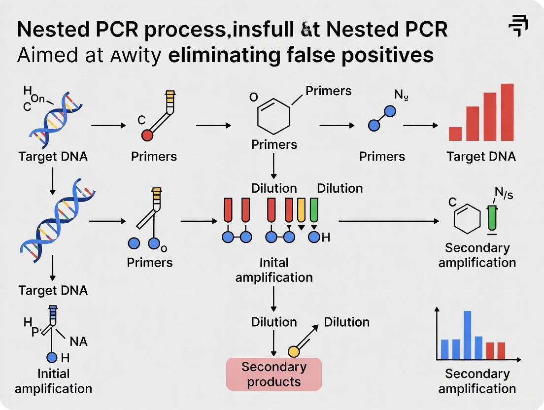
Nested PCR is a powerful tool for detecting low-abundance targets in complex samples, but its susceptibility to false positives remains a significant challenge in research and diagnostics.
Eliminating False Positives in Nested PCR: A Strategic Guide for Robust Molecular Diagnostics
Abstract
Nested PCR is a powerful tool for detecting low-abundance targets in complex samples, but its susceptibility to false positives remains a significant challenge in research and diagnostics. This article provides a comprehensive framework for overcoming this limitation, tailored for scientists and drug development professionals. We explore the fundamental causes of non-specific amplification, from primer design flaws to cross-contamination. The guide details advanced methodological strategies, including novel primer design and single-tube protocols, and offers a systematic troubleshooting regimen for assay optimization. Finally, we present rigorous validation and comparative data against other molecular techniques, empowering researchers to implement nested PCR with heightened specificity, reliability, and confidence in their results.
Understanding the Enemy: Deconstructing the Sources of False Positives in Nested PCR
Nested PCR FAQs
What is nested PCR and how does it work?
Nested PCR is a highly sensitive and specific molecular amplification method that uses two successive PCR reactions with two different sets of primers. The first round of amplification uses an outer set of primers that target a larger region of DNA. A very small amount of this initial PCR product is then used as the template for a second round of amplification using an inner set of primers that bind within the first amplified product. This two-step process acts like a "double check" system—it's statistically very unlikely that non-specific products from the first round will contain binding sites for the second set of primers, resulting in a much purer final product [1] [2].
Why would I choose nested PCR over conventional PCR?
Nested PCR provides significant advantages in situations where high specificity and sensitivity are critical. The method is particularly valuable when:
- Working with very low template concentrations (e.g., pathogen detection in early infection stages)
- Dealing with complex samples that may contain PCR inhibitors (e.g., environmental samples, feed, feces)
- False positive results are a concern due to cross-reactivity with similar sequences [3] [1] [4]
The enhanced sensitivity comes from the second round of amplification, which can detect target sequences present in very low copy numbers that would be undetectable with conventional PCR methods.
What are the most common causes of false positives in nested PCR?
The primary cause of false positives in nested PCR is cross-contamination between samples, particularly during the transfer of the first PCR product to the second reaction tube [5]. Other causes include:
- Carryover contamination from previous PCR products in the laboratory environment
- Primer cross-reactivity with non-target sequences that coincidentally have binding sites for both primer sets
- Aerosol formation during pipetting that transfers amplified DNA between tubes
How can I prevent contamination in nested PCR?
Implement these strict laboratory practices to minimize contamination risk:
- Physical separation: Perform pre-PCR (template preparation), first PCR, and second PCR setup in different dedicated areas or hoods
- Use aerosol-resistant pipette tips for all liquid transfers
- Prepare master mixes when setting up multiple reactions to reduce pipetting steps
- Include negative controls in both amplification rounds to monitor for contamination
- Use dedicated equipment and reagents for nested PCR work
- Clean workspaces frequently with DNA-decontaminating solutions [5]
Troubleshooting Guides
Problem: Persistent Non-Specific Bands or Smearing
| Possible Cause | Recommendations |
|---|---|
| Primer dimers or secondary structures | Review primer design using tools like Oligo Explorer or NCBI Primer-BLAST; avoid complementary sequences at 3' ends [6] [7] |
| Suboptimal annealing temperature | Optimize temperature in 1-2°C increments using a gradient cycler; increase temperature to improve specificity [8] |
| Excessive primer concentration | Optimize primer concentrations (typically 0.1-1 μM); high concentrations promote primer-dimer formation [8] |
| Too many amplification cycles | Reduce number of cycles (25-35 for first PCR, 20-30 for nested PCR) [8] [2] |
| Magnesium concentration too high | Optimize Mg2+ concentration; excessive concentrations stabilize non-specific binding [8] |
Problem: No Amplification Product or Weak Bands
| Possible Cause | Recommendations |
|---|---|
| Insufficient template in second round | Dilute first PCR product 1:100 to 1:1000; use 1-2 μL as template for nested PCR [2] |
| Poor primer design | Ensure nested primers are internal to first set; verify specificity with in silico PCR tools [7] |
| Inhibitors carried over from first PCR | Purify first PCR product before second round or increase dilution factor [8] |
| DNA polymerase incompatibility | Use hot-start DNA polymerases to prevent non-specific amplification at lower temperatures [8] |
| Suboptimal extension time/time | Prolong extension time for longer targets; ensure final extension step of 5-15 minutes [8] |
Problem: Inconsistent Results Between Replicates
| Possible Cause | Recommendations |
|---|---|
| Pipetting inaccuracies in small volumes | Use calibrated pipettes and practice accurate pipetting technique; master mix preparation [6] |
| Template degradation | Assess DNA integrity by gel electrophoresis; store DNA properly in TE buffer or molecular-grade water [8] |
| Enzyme activity variability | Ensure consistent thawing of reagents; avoid multiple freeze-thaw cycles [8] |
| Thermal cycler calibration issues | Verify temperature uniformity across the block; ensure lid temperature is optimal [8] |
| Reaction component evaporation | Use PCR tubes with secure seals; apply appropriate tube strip caps correctly [6] |
Research Reagent Solutions
| Reagent | Function | Optimization Tips |
|---|---|---|
| DNA Polymerase | Catalyzes DNA synthesis | Use hot-start versions to prevent non-specific amplification during setup [8] |
| MgCl₂/MgSO₄ | Cofactor for polymerase activity | Optimize concentration (0.5-5.0 mM); affects specificity and yield [8] [6] |
| PCR Additives (DMSO, BSA, Betaine) | Reduce secondary structures, enhance specificity | Use lowest effective concentration; DMSO at 1-10%, Betaine at 0.5-2.5 M [8] [6] |
| dNTPs | Building blocks for DNA synthesis | Use balanced equimolar concentrations (200 μM each); unbalanced increases error rate [8] |
| Primer Sets (Outer & Inner) | Target sequence recognition | Design with 40-60% GC content; Tm of 52-58°C; verify specificity with BLAST [6] [7] |
Case Study: Implementing a False-Positive Free Nested PCR System
Researchers developing a detection method for the microsporidian Enterocytozoon hepatopenaei (EHP) faced significant challenges with false positives when using conventional PCR targeting the small subunit ribosomal RNA (SSU rRNA) gene. Cross-reactivity with closely related microsporidia species led to inaccurate results, particularly problematic for environmental sampling in shrimp farms [3].
Solution Implemented: The team developed a nested PCR method targeting the spore wall protein (SWP) gene instead of the SSU rRNA gene. This approach provided:
- Greater specificity: No cross-reactivity with related microsporidia
- Enhanced sensitivity: 100-times more sensitive than the original method in the first PCR step
- Reliable environmental screening: Effective for feces, feed, and environmental samples where cross-reactivity was previously problematic [3]
Key Experimental Protocol:
- First amplification: 35 cycles with outer primers targeting SWP gene region
- Product dilution: 1:100 to 1:1000 dilution of first PCR product
- Second amplification: 25-30 cycles with internal nested primers
- Detection: Gel electrophoresis with appropriate controls [3]
This approach demonstrates how proper gene target selection combined with nested PCR methodology can effectively eliminate false positives while maintaining high sensitivity.
FAQs: Understanding Primer Artifacts
What are primer dimers and how do they form? A primer dimer is a small, unintended DNA fragment that forms when PCR primers anneal to each other instead of the target DNA template. This occurs through two main mechanisms [9]:
- Self-dimerization: A single primer contains regions that are complementary to each other, allowing it to fold and create a free 3' end for DNA polymerase.
- Cross-dimerization: Two separate primers have complementary regions that cause them to bind together, creating a structure that can be extended by DNA polymerase. These artifacts are often promoted by low annealing temperatures and high primer concentrations, which facilitate nonspecific interactions [10].
How does off-target binding (mispriming) differ from primer dimer formation? While primer dimers involve primer-primer interactions, off-target binding (or mispriming) occurs when primers partially anneal to non-target sequences on the template DNA itself. This can lead to the amplification of unexpected DNA fragments, which is a significant concern in multiplexed PCR assays where multiple primer pairs are used simultaneously [11]. In diagnostic and research applications, this can generate false-positive results by creating amplicons from nearly complementary, non-targeted sequences [11].
Why are primer artifacts a particular concern in nested PCR? Nested PCR is highly sensitive to primer artifacts due to its two-round amplification design. Artifacts formed in the first PCR round can be efficiently amplified in the second round, leading to false positives and obscuring the desired result [12] [13]. The high sensitivity of the method, while beneficial for detecting low-quantity targets, also makes it exceptionally vulnerable to amplifying any nonspecific products generated in the first round.
How can I identify primer dimers in my gel results? Primer dimers have distinct characteristics when visualized using gel electrophoresis [9] [14]:
- Short length: Typically appear below 100 base pairs (bp), often as a bright band between 20-60 bp.
- Smeary appearance: They often look like a fuzzy, diffuse smear rather than a sharp, well-defined band.
- Location: They run very far ahead of the expected target amplicon. For confirmation, you can run a No-Template Control (NTC); the presence of a band in the NTC confirms a primer-derived artifact [9].
Troubleshooting Guides
Guide 1: Preventing and Resolving Primer Dimers
Primer dimers compete for reagents, reduce amplification efficiency of your target, and can lead to false interpretations. The following table summarizes the primary causes and solutions.
| Problem Cause | Recommended Solution | Experimental Protocol / Notes |
|---|---|---|
| High Primer Concentration | Optimize primer concentration. Typically use 0.1–1 µM. Start with a lower concentration and increase if necessary [8]. | Set up a series of reactions with primer concentrations from 0.1 µM to 0.5 µM. A 0.2 µM final concentration is often a good starting point. |
| Low Annealing Temperature | Increase the annealing temperature incrementally. The optimal temperature is usually 3–5°C below the primer's Tm [9] [8]. | Use a thermal cycler with a gradient function. Test a range from 55°C to 65°C in 2°C increments to find the highest temperature that retains specific product yield. |
| Non-Hot-Start Polymerase | Use a hot-start DNA polymerase. This prevents enzyme activity during reaction setup, the stage when most primer dimers form [9] [8]. | Simply switch to a commercial hot-start polymerase. Follow the manufacturer's protocol for activation, which usually requires a preliminary high-temperature denaturation step. |
| Suboptimal Primer Design | Re-design primers using software tools to minimize 3'-end complementarity, especially in the last 3-5 bases [9] [6]. | Use primer design tools (e.g., NCBI Primer-BLAST, Primer3) to check for self-complementarity. Avoid G/C-rich 3' ends and long stretches of single nucleotides. |
Guide 2: Eliminating Off-Target Binding and Mispriming
Mispriming leads to nonspecific amplification, smearing on gels, and false positives in applications like diagnostic NGS panels. The strategies below are critical for robust assay design.
| Problem Cause | Recommended Solution | Experimental Protocol / Notes |
|---|---|---|
| Low Annealing Temperature | Increase annealing temperature and/or use Touchdown PCR [8]. | For gradient PCR, increase temperature in 1–2°C steps. For Touchdown PCR, start with an annealing temperature 10°C above the calculated Tm and decrease by 1°C per cycle for the first 10 cycles. |
| Poor Primer Specificity | BLAST primer sequences against the template genome to ensure uniqueness. Design longer primers (≥22 nt) for higher specificity [6] [8]. | Use NCBI BLAST with the "Somewhat similar sequences" option to check for unintended binding sites across the entire template. |
| Excess Mg²⁺ Concentration | Optimize Mg²⁺ concentration. High Mg²⁺ stabilizes nonspecific primer-template interactions [8]. | Set up a Mg²⁺ titration series (e.g., 1.0 mM, 1.5 mM, 2.0 mM, 2.5 mM, 3.0 mM) while keeping all other components constant. |
| Complex Template | Use PCR additives or co-solvents that reduce secondary structures and improve specificity [8]. | Test DMSO (1-3%), formamide (1.25-5%), or Betaine (0.5 M - 2.5 M). Note: Additives can lower the effective annealing temperature. |
Guide 3: Specific Considerations for Nested PCR
Nested PCR requires extra vigilance to prevent the carry-over and amplification of artifacts from the first round.
| Problem Cause | Recommended Solution | Experimental Protocol / Notes |
|---|---|---|
| Carry-over of Nonspecific Products | Use a minimal amount of the primary PCR product as the template for the nested reaction [2]. | Dilute the primary PCR product 1:100 to 1:1000. Use only 1 µL of the diluted product in the 50 µL nested PCR reaction [2]. |
| Suboptimal Inner Primer Design | Design inner (nested) primers with a higher Tm than the outer primers. This ensures the second round is more specific [12]. | When designing primers, set the melting temperature interval for the inner primer pair to be 2–5°C higher than that of the outer primer pair in your design software [12]. |
| High Cycle Number | Reduce the number of cycles in both the primary and nested PCR to prevent the accumulation of artifacts [8]. | For the primary PCR, use 20–25 cycles. For the nested PCR, use 25–30 cycles. Avoid exceeding 35 total cycles for the process [2] [8]. |
| Physical Contamination | Physically separate first and second-round PCR setups. Use dedicated equipment and reagent aliquots. | Perform pre- and post-PCR work in separate laminar flow hoods. Use aerosol-resistant pipette tips and uracil-DNA glycosylase (UDG) systems to degrade carry-over contamination. |
Essential Workflow: From Problem to Solution in Nested PCR
The following diagram outlines a systematic, decision-based workflow for troubleshooting primer artifacts in a nested PCR experiment.
Research Reagent Solutions
The following table lists key reagents and materials essential for preventing and diagnosing primer-related artifacts.
| Item | Function/Benefit | Application Note |
|---|---|---|
| Hot-Start DNA Polymerase | Inactive at room temperature; prevents primer dimer formation and nonspecific extension during reaction setup. Activated only at high temperatures (e.g., 95°C) [9] [8]. | Essential for all PCR types, especially critical in multiplex and nested PCR where primer interactions are more likely. |
| Gradient Thermal Cycler | Allows testing of a range of annealing temperatures in a single experiment for rapid optimization of specificity [8]. | Crucial for initial assay development and troubleshooting mispriming or primer dimers. |
| PCR Additives (DMSO, Betaine) | Co-solvents that help denature GC-rich templates and secondary structures, reducing mispriming and improving specificity [8]. | Concentration must be optimized (e.g., DMSO 1-10%, Betaine 0.5M-2.5M). Can sometimes inhibit polymerase if used at high levels. |
| No-Template Control (NTC) | A critical control reaction containing all PCR components except the DNA template. Used to detect contamination and primer-dimer formation [9]. | The presence of a band in the NTC unequivocally indicates an artifact derived from the primers or reagent contamination. |
| Primer Design Software | Tools (e.g., NCBI Primer-BLAST, Primer3) help design primers with optimal length, Tm, and minimal self-complementarity or dimerization potential [6] [15]. | The first and most important step in preventing artifacts. Always check proposed primers for off-target binding in the template. |
What is amplicon carryover contamination? Amplicon carryover contamination occurs when PCR products (amplicons) from previous reactions are accidentally introduced into new reaction setups. Given the exponential amplification power of PCR, even a single contaminating DNA molecule can lead to false-positive results. This risk is particularly acute in nested PCR, where a second round of amplification is performed, effectively providing two opportunities for contamination to occur. In diagnostic settings, this can lead to misdiagnosis and unnecessary treatments, while in research, it can compromise entire datasets [16] [17].
Frequently Asked Questions (FAQs)
FAQ 1: Why is nested PCR particularly vulnerable to carryover contamination? Nested PCR significantly increases sensitivity by performing two sequential amplification rounds. However, the process of transferring the first-round PCR product to the second reaction tube creates a major opportunity for aerosol formation and contamination. Furthermore, the laboratory environment becomes saturated with a high concentration of amplification products from the first round, increasing the risk of contaminating subsequent reactions [16] [18].
FAQ 2: What are the most common sources of contamination in a PCR laboratory? The primary sources include:
- Amplicons from previous reactions: The most significant source, creating a self-perpetuating problem [19] [17].
- Laboratory surfaces and equipment: Contaminants can be found on benchtops, pipettes, centrifuges, tube racks, and other equipment [17].
- Reagents and consumables: Contaminants can be present in water, enzyme stocks, buffers, and even on disposable pipette tips or tubes if not properly sterilized [17] [20].
- Personnel: Skin cells, hair, or aerosols from talking or coughing can introduce contaminating DNA [20].
FAQ 3: How can I confirm if my experiment has been compromised by carryover contamination? The most effective method is to routinely include No-Template Controls (NTCs) in your experimental setup. An NTC contains all reaction components—primers, master mix, water—except for the template DNA. Amplification of a product in the NTC is a clear indicator that one or more of your reagents or the environment is contaminated [19].
Troubleshooting Guide: Preventing and Managing Contamination
Physical and Workflow Strategies
Problem: Consistent false-positive results across multiple experiments, including NTCs.
| Strategy | Implementation | Rationale |
|---|---|---|
| Physical Separation of Work Areas | Establish dedicated, separate rooms or workstations for pre-PCR (reagent preparation, reaction setup) and post-PCR (product analysis) activities. | Creates a physical barrier to prevent high-concentration amplicons from entering clean reaction setups [19] [18] [17]. |
| Unidirectional Workflow | Enforce a strict one-way movement of personnel and materials from pre-PCR areas to post-PCR areas. Never return to a clean area after working in a post-PCR area without decontamination. | Prevents personnel from acting as vectors for carryover contamination [18] [17]. |
| Use of Dedicated Equipment and Supplies | Assign lab coats, gloves, pipettes, tip boxes, and other consumables exclusively to pre-PCR or post-PCR areas. Use aerosol-resistant filter tips in pre-PCR areas. | Eliminates equipment as a potential cross-contamination source [19] [18]. |
| Rigorous Decontamination | Regularly clean surfaces and equipment with a 10% bleach solution (followed by rinsing with nuclease-free water to prevent corrosion) or UV irradiation [19] [17]. UV light can cross-link DNA, rendering it unamplifiable. | Degrades contaminating DNA on laboratory surfaces and equipment [19] [17]. |
| Laminar Flow Hoods | Perform all reaction setup, especially the sensitive step of adding template DNA, inside a PCR workstation or laminar flow hood equipped with a HEPA/ULPA filter. | Provides a sterile, particulate-free workspace by supplying filtered air, protecting samples from environmental contamination [18]. |
Enzymatic and Reagent-Based Strategies
Problem: Contamination persists despite physical separation, or a high-throughput workflow makes physical separation challenging.
| Strategy | Implementation | Rationale |
|---|---|---|
| Uracil-DNA-Glycosylase (UNG) | Incorporate dUTP instead of dTTP in all PCR master mixes. Add UNG enzyme to the master mix, which will be active during reaction setup. It cleaves uracil-containing contaminants from previous runs. The initial denaturation step at 95°C permanently inactivates UNG, allowing new uracil-containing products to amplify without degradation [19] [17]. | Selectively degrades carryover contamination from past amplification reactions while preserving the native DNA template [19]. |
| Hot-Start DNA Polymerases | Use polymerases that are inactive at room temperature, requiring a high-temperature activation step (e.g., >90°C) to become active. | Prevents non-specific amplification and primer-dimer formation during reaction setup at room temperature, enhancing specificity and reducing background [21] [17]. |
PCR Protocol Optimization for Specificity
Problem: Non-specific amplification and primer-dimer formation complicate analysis and increase background noise.
| Strategy | Implementation | Rationale |
|---|---|---|
| Touchdown PCR | Start with an annealing temperature 5–10°C above the primer's calculated Tm. Gradually decrease the annealing temperature by 1–2°C per cycle over a series of cycles until the optimal temperature is reached. | The initial high-stringency cycles favor only the most specific primer-template binding, selectively amplifying the correct target before lower temperatures allow less specific binding [21] [17]. |
| Optimize Mg²⁺ Concentration | Perform a titration of MgCl₂ or MgSO₄ (typically 0.5-5.0 mM) to determine the optimal concentration for your specific primer-template system. | Mg²⁺ is a essential cofactor for DNA polymerase. Excess concentration can reduce specificity by stabilizing non-specific primer-template interactions [8] [6]. |
| Use of PCR Additives | Include additives like DMSO (1-10%), Betaine (0.5-2.5 M), or formamide (1.25-10%) in the reaction mix. | These compounds help denature GC-rich templates and disrupt secondary structures, improving amplification efficiency and specificity for difficult targets [8] [6]. |
Step-by-Step Experimental Protocols
Protocol for Decontaminating Laboratory Surfaces and Equipment
This protocol is essential for routine cleaning and after any known spill of PCR products.
- Prepare a fresh 10% (v/v) sodium hypochlorite (bleach) solution.
- Apply the bleach solution liberally to the surface (benchtops, pipette exteriors, tube racks, etc.).
- Allow a contact time of 10-15 minutes. This is critical for effective DNA degradation [19].
- Wipe the surface thoroughly with nuclease-free water to remove residual bleach, which can corrode equipment and inhibit PCR.
- For equipment that cannot tolerate bleach (e.g., inside centrifuges), decontaminate with 70% ethanol or irradiate with UV light for at least 15 minutes [19].
Protocol for Setting Up a Nested PCR with UNG Carryover Prevention
This protocol integrates physical and enzymatic strategies to minimize contamination risk.
Reagents:
- First-round PCR master mix (with dTTP)
- Second-round PCR master mix (formulated with dUTP)
- Uracil-DNA-Glycosylase (UNG)
- Outer and nested primer sets
- Template DNA
- Nuclease-free water
Procedure:
- Prepare First-Round Mix: In a pre-PCR laminar flow hood, assemble the first-round PCR reactions using a master mix containing standard dNTPs (dTTP). Cap tubes securely.
- Perform First-Round Amplification: Run the first-round PCR in a thermal cycler located in the post-PCR area.
- Prepare Second-Round Mix: Return to the pre-PCR hood. Prepare the second-round master mix. This mix must contain dUTP instead of dTTP and include UNG enzyme.
- Aliquot Second-Round Mix: Dispense the second-round master mix into new, clean PCR tubes.
- Transfer Template: In the post-PCR area, carefully open the first-round PCR tubes and transfer a small aliquot (e.g., 1-2 µl) of the product to the corresponding tubes containing the second-round master mix. Close the tubes immediately after transfer to minimize aerosol generation.
- UNG Incubation: Return the completed second-round reactions to the thermal cycler. Program an initial hold at 25°C for 10 minutes. During this step, UNG will enzymatically cleave any uracil-containing contaminating DNA.
- UNG Inactivation and Amplification: Program the cycler to hold at 95°C for 2-5 minutes to inactivate the UNG, followed by the standard nested PCR cycling protocol.
The Scientist's Toolkit: Essential Reagents for Contamination Control
| Item | Function/Benefit |
|---|---|
| Aerosol-Resistant Filter Pipette Tips | Prevent aerosols and liquids from entering the pipette shaft, a common source of cross-contamination [19] [17]. |
| Uracil-DNA-Glycosylase (UNG) | The core enzyme for enzymatic prevention of amplicon carryover, as described in Section 4.2 [19] [17]. |
| dUTP | Used in place of dTTP to generate uracil-containing amplicons that are susceptible to cleavage by UNG in subsequent reactions [19]. |
| Hot-Start DNA Polymerase | Reduces non-specific amplification and primer-dimer formation during reaction setup, improving assay robustness [8] [21]. |
| 10% Bleach Solution | Effective chemical decontaminant for degrading DNA on non-porous surfaces [19] [17]. |
| HEPA/ULPA Laminar Flow Hood | Provides an ISO Class 5 clean air environment for critical pre-PCR setup steps, protecting reactions from environmental contaminants [18]. |
| Dedicated Lab Coats and Gloves | Simple but critical barrier to prevent personnel from introducing contaminants into clean areas [18] [20]. |
Nested PCR is a powerful tool for detecting low-abundance targets, but its increased sensitivity makes it particularly vulnerable to false positives. These errors are primarily driven by three interconnected challenges: the presence of host DNA, co-pathogens in complex samples, and degraded template DNA. This guide provides troubleshooting protocols and FAQs to help researchers identify, mitigate, and eliminate these sources of error.
Troubleshooting Guides
Challenge 1: Host DNA Interference
Problem: Host genomic DNA acts as a complex background, causing nonspecific primer binding and false-positive amplification. This is a significant issue in host-associated microbiota studies [22].
Solutions:
- Primer Design: Design outer primers to bind to conserved regions of the target gene that are absent in the host genome. In silico analysis using BLAST is essential to verify specificity [23] [1].
- Two-Step PCR Optimization: A two-step nested approach is highly effective. The first PCR with outer primers enriches the initial target pool. The second PCR with inner primers, which bind within the first amplicon, selectively amplifies the specific target, minimizing host DNA background [22].
- Cycle Number: Optimize the number of cycles for each PCR step to maximize target yield without amplifying background. A configuration of 15 cycles for the first step and 25 for the second has been shown to be effective [23].
Challenge 2: Co-pathogens and Complex Samples
Problem: In samples with multiple potential pathogens (e.g., respiratory infections, gut microbiome), primers may cross-react with non-target sequences from co-pathogens, leading to misidentification [24] [25].
Solutions:
- Multiplex Nested PCR (MN-PCR): Implement a single-tube multiplex nested PCR system. This method uses universal primers in the first stage to broadly amplify a gene region (e.g., 16S rDNA) from various bacteria. The second stage uses multiple sets of specific, short primers at a lower annealing temperature to amplify species-specific fragments from the enriched template [23].
- Annealing Temperature Control: Design specific primers with annealing temperatures below 56°C and universal primers for temperatures above 65°C. This ensures that specific primers only function in the second, lower-temperature stage of the PCR, preventing early non-specific amplification [23].
- Probe-Based Verification: For real-time PCR applications, use specific fluorescent probes (e.g., TaqMan) with melting curve analysis (FMCA) to differentiate between pathogens based on their unique melting temperatures (Tm), confirming the identity of the amplicon [24].
Challenge 3: Degraded Template DNA
Problem: Template DNA is often fragmented due to degradation processes like hydrolysis and enzymatic activity during sample collection, storage, or extraction. This is prevalent in forensic, ancient DNA, and stool samples [26] [27]. Standard PCR for long amplicons fails, leading to false negatives or misleading results.
Solutions:
- Short Amplicon Strategy: Design your nested PCR to amplify shorter target fragments. Research on H. pylori detection in stool showed that a short 148 bp amplicon had a significantly higher detection rate (51.0%) compared to a long 454 bp amplicon (6.25%), as shorter fragments are more likely to survive degradation [26].
- Optimized Extraction and Preservation: Use specialized DNA extraction kits designed for difficult samples (e.g., stool, fixed tissue). Incorporate mechanical homogenization with controlled parameters (speed, cycle duration, temperature) to lyse cells without causing excessive DNA shearing. For preservation, flash-freeze samples in liquid nitrogen and store at -80°C to halt enzymatic degradation [27].
- Inhibition Prevention: Degraded samples often contain PCR inhibitors. During extraction, use buffers with chelating agents like EDTA to inactivate nucleases, but ensure it is thoroughly removed or diluted as it can also inhibit PCR. Adding Bovine Serum Albumin (BSA) to the PCR reaction can mitigate the effects of some inhibitors [17].
Frequently Asked Questions (FAQs)
Q1: What is the most critical step to prevent false positives in nested PCR? The single most critical step is preventing contamination [17]. Due to its high sensitivity, nested PCR is exceptionally vulnerable to carryover contamination from amplicons generated in previous reactions. Physical separation of pre- and post-PCR areas, dedicated equipment and lab coats, and using aerosol-barrier pipette tips are essential. The incorporation of uracil-DNA-glycosylase (UNG) into the reaction mix can also help degrade carryover amplicons from previous runs [17].
Q2: How can I confirm that my positive result is not a false positive? Sequencing the PCR product is the definitive method to confirm the specificity of the amplification and rule out false positives arising from non-specific binding or contamination [26]. Additionally, the use of appropriate controls is vital:
- No-Template Control (NTC): Contains all reaction components except the DNA template. A positive signal here indicates contamination of your reagents or environment [17].
- Positive Control: A sample with a known, low concentration of the target. This verifies that the assay is working correctly and is sensitive enough.
- Internal Control: A housekeeping gene (e.g., GAPDH) can be used to confirm that nucleic acid purification was successful and that the sample is not inhibited [17].
Q3: My template DNA is of low quality and concentration. How can I improve my nested PCR success?
- Use a robust DNA polymerase with high processivity and tolerance to inhibitors commonly found in complex samples [8].
- Increase the number of cycles in the first round of amplification to enrich the initial target pool, but be cautious not to go too high as it can promote background noise [8] [23].
- Target a shorter amplicon, as it is more likely to be intact in a degraded sample [26].
- Re-purify your DNA or use a precipitation step to remove salts, ions, and other contaminants that may inhibit the polymerase [8].
The following table summarizes a validated single-tube multiplex nested PCR (MN-PCR) protocol for detecting multiple bacterial pathogens in the presence of host DNA, demonstrating principles that can be adapted to other targets [23].
Table: Optimized Single-Tube Multiplex Nested PCR Protocol
| Parameter | Specification | Function and Rationale |
|---|---|---|
| Universal Primers | 0.01 µM each, Annealing Temp: 65°C | First-stage amplification; consumed after 15 cycles to prevent interference. |
| Specific Primers | 0.15 µM each, Annealing Temp: 55°C | Second-stage amplification; short length prevents activity in first stage. |
| Reaction Volume | 20 µL | Standard for compatibility with most thermal cyclers. |
| Thermal Cycling | Stage 1 (15 cycles): 94°C for 30s, 65°C for 30s, 72°C for 30s.Stage 2 (25 cycles): 94°C for 30s, 55°C for 30s, 72°C for 30s. | Two-stage protocol with distinct annealing temperatures ensures specificity. |
| Final Extension | 72°C for 5 minutes | Ensures complete extension of all amplicons. |
| Reported Sensitivity | 1 fg of target bacterial DNA | 1000x more sensitive than conventional multiplex PCR (1 pg). |
Workflow Visualization
The following diagram illustrates the logical workflow for diagnosing and addressing the core challenges discussed in this guide.
Research Reagent Solutions
Table: Essential Reagents for Reliable Nested PCR
| Reagent / Tool | Function | Application Note |
|---|---|---|
| Hot-Start DNA Polymerase | Enzyme inactive at room temperature; prevents non-specific amplification and primer-dimer formation during reaction setup. | Critical for enhancing specificity in both rounds of nested PCR [8] [17]. |
| UNG (Uracil-N-Glycosylase) | Enzymatically degrades carryover contaminant amplicons from previous PCRs by breaking down uracil-containing DNA. | Add to the PCR master mix to control one of the most common sources of false positives [17]. |
| BSA (Bovine Serum Albumin) | Binds to and neutralizes common PCR inhibitors found in complex biological samples (e.g., phenolic compounds). | Use 200-400 ng/µL in the reaction to improve efficiency from inhibited samples [17]. |
| Optimized Lysis Beads | Ceramic or stainless-steel beads for mechanical homogenization of tough samples (e.g., tissue, stool, bone). | Enables efficient DNA recovery while minimizing excessive shearing when used with controlled speed and time [27]. |
| Specific Probes (TaqMan/FMCA) | Fluorescently labeled probes that bind specifically to the target sequence, allowing detection and confirmation via melting temperature. | Used in multiplex real-time PCR to differentiate co-pathogens and verify amplicon identity [24] [25]. |
FAQ: Understanding and Troubleshooting False Positives in Nested PCR
Q1: What are the primary causes of false positives in nested PCR assays? False positives in nested PCR primarily arise from two sources:
- Carryover Contamination: The most common cause is the contamination of samples with amplicons (PCR products) from previous reactions [17]. Because nested PCR involves handling the product of the first reaction to perform the second, the risk of these products contaminating reagents, equipment, or new samples is significantly increased.
- Non-Specific Primer Binding: Universal primers, designed to target conserved regions across a broad group of organisms (like phytoplasmas), can sometimes bind to non-target sequences, including host plant DNA, bacterial DNA, or other contaminants present in the sample [28]. This can lead to the amplification of non-target fragments, which are then further amplified in the second round of PCR.
Q2: How can I confirm that my nested PCR result is a false positive? The most reliable method to confirm a false positive is DNA sequencing of the amplified nested PCR product [28]. If sequencing reveals the amplicon originates from a non-target organism (e.g., plant chloroplasts, endophytic bacteria) or is a non-specific artifact, the result is a false positive. Regular sequencing of a subset of positives is a good laboratory practice to validate your assay's specificity.
Q3: What are the consequences of false positives in diagnostic testing? False positives can lead to:
- Misdiagnosis: Incorrectly identifying a healthy plant as infected [28].
- Wasted Resources: Unnecessary implementation of disease management strategies, costing time and money [17].
- Psychological Distress: For human diagnostics, a false positive can cause significant anxiety and lead to unnecessary treatments [17].
Q4: Our lab follows strict protocols, but we still get sporadic false positives. What hidden sources of contamination should we check? Beyond obvious sources, investigate these potential hidden contamination vectors [17]:
- Laboratory Personnel: Contaminants on lab coats, skin, hair, or jewelry.
- Equipment Interiors: The inside of micropipettes can become contaminated from improper pipetting techniques. Consider sending them for professional servicing and calibration.
- Reagents: Even sterile reagents can become contaminated during repeated handling. Prepare single-use aliquots to mitigate this risk.
Q5: Are there alternatives to nested PCR that are less prone to false positives? Yes, several alternative methods exist, each with advantages and limitations. The table below compares nested PCR with other common techniques:
Table 1: Comparison of Molecular Detection Methods
| Method | Principle | Relative Specificity | Relative Sensitivity | Key Advantage | Key Disadvantage |
|---|---|---|---|---|---|
| Nested PCR | Two consecutive PCR rounds with two primer sets [1] | Moderate to High* | Very High | High sensitivity for low-pathogen titers [29] | High risk of carryover contamination [17] |
| Real-time PCR (qPCR) | Fluorescence-based detection during amplification [30] | High | High | Closed-tube system reduces contamination risk [30] | Limited multiplexing with standard instruments [30] |
| LAMP | Isothermal amplification with multiple primers [29] | High | High | Rapid, cost-effective, and suitable for field use [29] | Complex primer design, susceptibility to contamination [29] |
| Digital PCR (ddPCR) | Absolute quantification via sample partitioning [30] | High | Very High | Absolute quantification without a standard curve [30] | High cost, specialized equipment required [30] |
*Specificity is highly dependent on primer design. Assays with specific primers are significantly more specific than those using universal primers [28].
Experimental Case Study: Resolving False Positives in Areca Palm Phytoplasma Detection
Background and Problem Identification
A 2025 study aimed to detect phytoplasmas associated with Yellow Leaf Disease (YLD) in areca palms in Hainan, China. Initially, researchers used a universal nested PCR primer set (P1/P7 followed by R16mF2/R16mR1) to screen 335 samples [28]. While 50 samples showed amplification of the expected ~1400 bp band, sequencing revealed a critical problem: only 10 of the 50 amplicons were truly from phytoplasma. The rest were false positives, originating from areca palm chloroplast DNA (16 samples) or other bacterial sequences (20 samples) [28]. This high false-positive rate of 80% underscored the lack of specificity in the universal primer set for their specific samples.
Experimental Protocol: Developing a Specific Nested PCR System
Objective: To design a novel nested PCR primer set specific for the 16SrI and 16SrII group phytoplasmas affecting areca palms, thereby eliminating false positives.
Methodology:
Target Selection and Primer Design:
- Target Gene: The conserved phytoplasma 16S rDNA sequence was used [28].
- Bioinformatic Analysis: Researchers aligned 16S rDNA sequences from the target phytoplasmas (16SrI and 16SrII), areca palm chloroplasts, and common endophytic/pathogenic bacteria (e.g., Burkholderia andropogonis, Pantoea ananatis) to identify unique regions for the target phytoplasmas [28].
- Primer Design: Specific primer pairs were designed using standard principles. The final selected primers were:
- Outer Primers: HNP-1F / HNP-1R
- Inner Primers: HNP-2F / HNP-2R [28]
DNA Extraction:
- Genomic DNA was extracted from leaf midribs using a modified CTAB method. Key modifications included adding 2% polyvinyl pyrrolidone (PVP) to bind polyphenols and increasing the volume of isopropanol to improve DNA yield [31].
Nested PCR Amplification:
- First Round PCR:
- Reaction Mix: Template DNA, outer primers (HNP-1F/HNP-1R), dNTPs, PCR buffer, MgCl₂, and DNA polymerase.
- Cycling Conditions: Initial denaturation; followed by 35 cycles of denaturation, annealing (optimized at 53.6°C), and extension; final extension [28].
- Second Round PCR:
- Template: The product from the first PCR is diluted (e.g., 1:10) and used as the template.
- Reaction Mix: Diluted first-round product, inner primers (HNP-2F/HNP-2R), dNTPs, PCR buffer, MgCl₂, and DNA polymerase.
- Cycling Conditions: Similar to the first round but with an optimized annealing temperature of 57.2°C for the inner primers [28].
- First Round PCR:
Specificity and Sensitivity Validation:
- Specificity Test: The new primer set was tested against DNA from healthy areca palms, non-target phytoplasma groups (16SrXXXII), and other bacterial pathogens. The HNP-2F/2R primer pair amplified a specific 429 bp fragment only in samples infected with 16SrI or 16SrII phytoplasmas [28].
- Sensitivity Test: The detection limit was determined using serial dilutions of phytoplasma DNA. The new system achieved a sensitivity of 7.5 × 10⁻⁷ ng/µL for 16SrI and 4 × 10⁻⁷ ng/µL for 16SrII, which was superior to the conventional universal primers [28].
Results and Workflow Visualization
The following workflow diagram summarizes the experimental process and key findings that led to a specific and reliable detection assay.
The Scientist's Toolkit: Essential Reagents and Solutions
Table 2: Key Research Reagents for Specific Nested PCR Assay Development
| Reagent / Solution | Function / Purpose | Example / Note |
|---|---|---|
| Specific Primer Pairs | To selectively bind and amplify the DNA of the target organism, avoiding cross-reactivity with host or contaminant DNA [28]. | HNP-1F/1R (outer) and HNP-2F/2R (inner) for areca palm phytoplasma [28]. |
| Polyvinylpyrrolidone (PVP) | Added to DNA extraction buffers to bind and remove polyphenols and other secondary metabolites from plant tissues that can inhibit PCR [31]. | Used at 2% concentration in the modified CTAB protocol [31]. |
| Hot-Start DNA Polymerase | A modified enzyme that is inactive at room temperature, preventing non-specific amplification and primer-dimer formation during reaction setup [17] [8]. | Reduces false positives caused by mis-priming at low temperatures. |
| dNTPs | The building blocks (deoxynucleotide triphosphates) for DNA synthesis during PCR amplification. | Use balanced, equimolar concentrations to prevent incorporation errors [8]. |
| MgCl₂ | A co-factor essential for DNA polymerase activity. Its concentration can significantly impact primer specificity and PCR yield [8]. | Requires optimization; excess Mg²⁺ can promote non-specific binding. |
| Agarose Gel Electrophoresis Supplies | To visualize and confirm the size of the PCR amplicons, ensuring the correct target has been amplified. | Used to distinguish the specific 429 bp band from non-specific products [28]. |
| UV Sterilizer / 10% Sodium Hypochlorite | To decontaminate surfaces and equipment by degrading any contaminating DNA, such as amplicons from previous runs [17]. | Critical for maintaining a clean pre-PCR workspace. |
Building a Robust Assay: Proactive Strategies for Primer Design and Protocol Refinement
The power of nested Polymerase Chain Reaction (PCR) lies in its two-round amplification process, which dramatically enhances sensitivity and specificity for detecting target DNA sequences. This technique is particularly invaluable when working with challenging samples, such as those with minimal target copy numbers or substantial background DNA [13] [32] [33]. The first round of PCR uses an outer primer pair to amplify the target region broadly. The product from this reaction then serves as the template for a second round of amplification using an inner primer set that binds within the first amplicon [32]. This two-step process significantly reduces false positives by virtually eliminating nonspecific amplification products that might arise from primer binding to off-target sequences [32]. However, the full potential of this specificity is only realized through meticulous primer design, especially when targeting genes like the 16S rRNA, which contain both conserved and variable regions. This guide provides a focused framework for designing primers and troubleshooting experiments within the critical context of eliminating false positives in nested PCR research.
Core Principles of Specific Primer Design
Effective primer design is the cornerstone of a specific and robust nested PCR assay. The following principles are critical for minimizing off-target binding and false-positive results.
Fundamental Primer Properties
Primers should be engineered with the following characteristics to ensure efficient and specific binding [34] [6] [35]:
- Length: Aim for 18–30 nucleotides. Longer primers within this range can enhance specificity, particularly in complex samples like genomic DNA [35] [36].
- GC Content: Maintain a GC content of 40–60%, with an ideal of 50%. This provides sufficient sequence complexity while avoiding overly stable structures [34] [35] [36].
- Melting Temperature (Tm): Design primers to have a Tm between 52–65°C, with both primers in a pair differing by no more than 2–5°C [34] [6] [35]. The Tm must be calculated using a reliable tool that considers your specific reaction buffer conditions [34].
- 3' End Clamping: The 3' end of the primer must be terminated with a G or C residue (a "GC clamp"). This strengthens binding due to the three hydrogen bonds in G:C pairs, preventing "breathing" at the ends and drastically improving amplification efficiency and specificity [6] [36].
- Structural Pitfalls: Avoid sequences that lead to secondary structures like hairpin loops or primer-dimer formations with the other primer. The ΔG value for any such structures should be weaker (more positive) than –9.0 kcal/mol [34] [6].
Designing for Conserved and Variable Regions
Targeting the correct genomic landscape is essential for assays aimed at identifying organisms, such as in 16S rRNA sequencing.
- Conserved Regions for Binding: The outer primers in a nested PCR should ideally bind within the highly conserved regions of the gene. This ensures broad capture of the target sequence across different species or strains [37].
- Variable Regions for Differentiation: The inner primers should be designed to bind within the hypervariable regions (e.g., V1–V9 in the 16S rRNA gene) that flank the conserved areas. The sequence variations in these regions provide the specificity needed to distinguish between closely related taxa [37].
- Database-Informed Design: "Universal" primers designed from limited datasets often fail to capture true microbial diversity [37]. Always use comprehensive and updated databases (e.g., SILVA, NCBI) for in silico validation of primer coverage and specificity against your target organisms [37]. Run a BLAST analysis to ensure your primers are unique to the desired target [34].
The Critical Role of Annealing Temperature
The annealing temperature (Ta) is a critical experimental parameter that must be tightly controlled. Set the Ta no more than 5°C below the Tm of your primers [34] [35]. A Ta that is too low is a primary cause of false positives, as it permits primers to tolerate single-base mismatches and anneal to partially homologous, off-target sequences [34].
Table 1: Optimal Primer Design Parameters for Maximum Specificity
| Parameter | Optimal Range | Rationale |
|---|---|---|
| Primer Length | 18–30 nucleotides | Balances specificity with adequate binding stability [6] [36]. |
| GC Content | 40–60% (50% ideal) | Prevents overly stable (high GC) or unstable (low GC) duplexes [34] [35]. |
| Melting Temp (Tm) | 52–65°C | Ensures efficient priming at experimentally feasible Ta [34] [6]. |
| Tm Difference (Primer Pair) | ≤ 2–5°C | Allows both primers to bind simultaneously and efficiently [34] [6]. |
| 3' End Sequence | G or C (GC clamp) | Stabilizes the primer-template complex at the critical point of elongation [6] [36]. |
Troubleshooting Guide: Eliminating False Positives
This FAQ section addresses common experimental issues directly related to primer specificity and false positives in nested PCR.
Q1: My nested PCR produces nonspecific bands or a smear on the gel. What should I do?
- Increase Stringency: The most common solution is to increase the annealing temperature (Ta) in increments of 2°C [38]. This prevents weak, off-target binding.
- Check Primer Design: Use BLAST alignment to verify that the 3' ends of your primers are not complementary to non-target sites. Redesign if necessary [34] [38].
- Use Touchdown PCR: Start with a Ta above the calculated Tm and gradually reduce it to the optimal Ta in subsequent cycles. This enriches for the specific target early in the reaction [35] [38].
- Reduce Template Amount: Excess template can lead to nonspecific amplification. Reduce the template amount by 2–5 fold [38].
- Employ Hot-Start DNA Polymerases: These enzymes remain inactive until the high-temperature denaturation step, preventing primer-dimer formation and nonspecific priming during reaction setup [8] [38].
Q2: I am getting no amplification product at all. How can I troubleshoot this?
- Check Reaction Components: Always include a positive control to confirm all reagents are functional and present [38].
- Optimize PCR Conditions: If the positive control works, your conditions may be too stringent. Lower the Ta in 2°C increments or increase the number of cycles (up to 40) for low-abundance targets [38].
- Inspect Template Quality: PCR inhibitors from the sample or degraded template can cause failure. Dilute or re-purify the template, or use a polymerase tolerant to impurities [8] [38]. Ensure the template DNA has good integrity [8].
Q3: Despite my efforts, I still have background contamination or false positives. What are the best practices to prevent this?
- Physical Separation: Establish physically separated pre-PCR and post-PCR areas. Never bring reagents, equipment, or lab coats from the post-PCR area (where amplified DNA is handled) back into the pre-PCR area [38].
- Dedicated Equipment: Use separate sets of pipettes and filtered tips for pre-PCR setup [38].
- Include Negative Controls: Always run a negative control (no template DNA) to confirm the absence of contamination in your reagents [38].
- Decontaminate: If contamination occurs, decontaminate workstations and equipment with 10% bleach or UV irradiation [38].
Q4: How can I improve specificity when amplifying GC-rich templates?
- Use Specialized Polymerases: Choose a DNA polymerase specifically formulated for high-GC content templates [8] [38].
- Add Enhancers: Incorporate PCR additives like DMSO (1-10%), formamide (1.25-10%), or betaine (0.5 M to 2.5 M) to help denature stable secondary structures [8] [6].
- Design Primers Carefully: For GC-rich targets, avoid runs of Gs or Cs, especially at the 3' end, and ensure GC residues are spaced evenly within the primer [35].
Table 2: Troubleshooting Common Primer-Related Problems
| Problem | Possible Cause | Solution |
|---|---|---|
| Nonspecific Bands/Smearing | Ta too low; primer binding to off-target sites. | Increase Ta; use touchdown PCR; redesign primers; use hot-start polymerase [8] [38]. |
| No Product | Ta too high; degraded template; PCR inhibitors. | Lower Ta; check template quality/purity; increase cycle number [8] [38]. |
| Primer-Dimer Formation | Primer 3' ends are complementary; high primer concentration. | Redesign primers to avoid self-complementarity; optimize primer concentration (0.1–1 µM) [8] [35]. |
| False Positives (Contamination) | Carryover of amplicons from previous PCRs. | Strictly separate pre- and post-PCR workspaces; use dedicated equipment; include negative controls [38]. |
Experimental Protocol: A Specificity-Focused Nested PCR Workflow
The following workflow diagram outlines the key stages of a nested PCR experiment designed to maximize specificity and minimize false positives, from primer design to analysis.
Detailed Methodology
Step 1: Primer Design and In Silico Validation
- Design Outer and Inner Primers: Following the core principles in Section 2, design two sets of primers. The outer set should target conserved regions for broad capture, and the inner set should target flanking variable regions for high specificity [37].
- Validate In Silico: Use tools like the IDT OligoAnalyzer Tool or NCBI Primer-BLAST to analyze Tm, check for secondary structures, and ensure specificity against the relevant database (e.g., SILVA for 16S rRNA) [37] [34] [6].
Step 2: First-Round PCR Setup
- Prepare Reaction Mix (on ice): For a 50 µL reaction [6]:
- 5 µL of 10X PCR Buffer (with Mg2+ if not supplied separately)
- 1 µL of 10 mM dNTP mix (200 µM final)
- 1 µL of each outer forward and reverse primer (20 µM stock, 0.4 µM final)
- 0.5–2.5 Units of Hot-Start DNA Polymerase
- 1–1000 ng of Template DNA
- Nuclease-free water to 50 µL
- Thermal Cycling:
- Initial Denaturation: 95°C for 2–5 minutes (activates hot-start polymerase).
- Amplification (25–30 cycles): Denature at 95°C for 20–30 seconds, anneal at the optimized Ta for 20–30 seconds, extend at 72°C (1 min/kb).
- Final Extension: 72°C for 5–10 minutes.
Step 3: Second-Round (Nested) PCR Setup
- Template Dilution: Dilute the first-round PCR product 10-fold to 10,000-fold (e.g., 1:100 to 1:10,000) to minimize carryover of outer primers [38].
- Prepare Reaction Mix: Set up a new 50 µL reaction as in Step 2, but use 1–5 µL of the diluted first-round product as template and the inner primer set.
- Thermal Cycling: Use the same cycling conditions as the first round, but with an annealing temperature optimized for the inner primers.
Step 4: Analysis and Contamination Control
- Gel Electrophoresis: Analyze 5–10 µL of the second-round PCR product on an agarose gel alongside a appropriate DNA ladder and a negative control (a reaction where water was used as template in both rounds) [6] [38].
- Interpret Results: A single, sharp band of the expected size in the test sample, with a clean negative control, indicates a specific amplification free from contamination.
Research Reagent Solutions
The following table lists essential reagents and their specific functions in achieving high-specificity nested PCR.
Table 3: Essential Reagents for Specificity-Focused Nested PCR
| Reagent / Material | Function / Rationale for Specificity |
|---|---|
| Hot-Start DNA Polymerase | Prevents nonspecific priming and primer-dimer formation during reaction setup by requiring high-temperature activation [8] [38]. |
| Ultra-Pure dNTPs | Provide balanced nucleotide concentrations (200 µM each) to prevent misincorporation errors that can occur with unbalanced dNTP pools [8] [38]. |
| Optimized MgCl2 Solution | Mg2+ is a crucial cofactor for polymerase activity. Concentration must be optimized (typically 1.5–2.5 mM) as excess Mg2+ can reduce fidelity and promote nonspecific binding [8] [6]. |
| PCR Additives (DMSO, Betaine) | Assist in denaturing GC-rich templates and secondary structures, improving primer access to the target and increasing specificity and yield [8] [6]. |
| Aerosol-Barrier Pipette Tips | Critical for preventing cross-contamination between samples and from previous amplifications, a major source of false positives [38]. |
| Nuclease-Free Water | Ensures the reaction is not compromised by nucleases that could degrade primers or templates, or by contaminants that could inhibit the polymerase. |
In nested PCR research, false positives present a formidable challenge, potentially arising from non-specific amplification or contamination with fragmented DNA [39] [16]. These false signals can compromise experimental integrity, leading to inaccurate conclusions. Bioinformatics tools provide a powerful first line of defense. Performing in-silico specificity checks before any wet-lab experiment allows researchers to predict and eliminate primers likely to amplify unintended genomic targets, ensuring that the final amplified product originates from the intended genomic locus [40] [41]. This guide details how to integrate Primer-BLAST, a cornerstone tool from the NCBI, into your nested PCR workflow to enhance specificity and reliability [42] [43] [41].
Troubleshooting Guides
Primer Specificity and Design Issues
| Observation | Possible Cause | Solution |
|---|---|---|
| Multiple bands or smeared gel | Primers binding to non-target sites with sufficient complementarity to allow amplification [8]. | Use Primer-BLAST's specificity check with stringent parameters (e.g., require 3'-end mismatches) [42] [41]. |
| False positives in negative controls | Contamination from previously amplified DNA (amplicons) or fragmented DNA [39] [16]. | Implement a nested PCR design; use Primer-BLAST to ensure inner and outer primer pairs are unique and specific [16] [44]. |
| No product or weak amplification | Primers designed across splice variants or SNPs that disrupt binding [41]. | Utilize Primer-BLAST's options to place primers across exon-exon junctions and exclude SNP sites [42] [41]. |
| Inconsistent results between PCR rounds | Inner and outer primer pairs competing or forming primer-dimers [44]. | Design inner and outer primers with a significant difference in Tm; use Primer-BLAST to check for inter-primer complementarity [44]. |
Primer-BLAST Parameter and Output Interpretation
| Problem | Potential Reason | Resolution |
|---|---|---|
| No specific primer pairs found. | Specificity stringency is too high, or the template has highly similar paralogs/repeats [42]. | Lower the "maximum number of mismatches" value, or adjust the "specificity threshold" to be less stringent [42]. |
| Unexpected amplicon reported from a different organism. | The search database was too broad (e.g., 'nr') without an organism filter [42] [43]. | Restrict the search by specifying the target organism in the "Primer Pair Specificity Checking Parameters" [42] [43]. |
| Primer-BLAST results show amplicons on unintended transcripts of the same gene. | The tool is designed to be transcript-specific by default when using an RefSeq mRNA template [42]. | Check the "Ignore targets that have a mismatch to the PCR template" option, or enable the "Ignore splice variants" option for gene-level specificity [42]. |
| The program is very slow or times out. | Searching a large database (e.g., 'nr') with a low E-value or a highly complex template [42]. | Select a smaller, more specific database (e.g., Refseq mRNA or core_nt), and increase the "Expect value" under advanced parameters [42]. |
Frequently Asked Questions (FAQs)
Q1: What is the fundamental advantage of using Primer-BLAST over a standard BLAST search for checking primer specificity?
A standard BLAST search uses a local alignment algorithm, which may not return complete match information over the entire primer sequence, especially near the ends. This is critical because mismatches at the 3' end are most disruptive to amplification. Primer-BLAST combines BLAST with a global alignment algorithm, ensuring a full primer-target alignment and providing a complete view of mismatch numbers and locations. This allows for a more accurate prediction of whether a primer pair will actually amplify a given non-target sequence [41].
Q2: How can I use Primer-BLAST to design primers that avoid amplifying genomic DNA in RT-PCR experiments?
Primer-BLAST offers a direct solution. In the "Primer Parameters" section, you can set the option "Primer must span an exon-exon junction." This directs the tool to return primer pairs where at least one primer is located across the boundary between two exons. Such a design ensures that amplification will only occur from spliced mRNA, not from genomic DNA, as the intron in the genomic sequence would break the primer binding site [42] [41].
Q3: My template is GC-rich and difficult to amplify specifically. What strategies can I implement within Primer-BLAST?
For GC-rich templates, you can adjust both the primer design and specificity parameters. When designing new primers, you can instruct Primer-BLAST to adjust primer properties to suit difficult templates. Furthermore, during the specificity check, you can relax the "total number of mismatches" parameter slightly, as highly GC-rich primers might have more fortuitous matches. However, this should be balanced by simultaneously requiring a higher number of mismatches specifically at the 3' end to maintain specificity [42] [8].
Q4: What is the recommended workflow for verifying the specificity of pre-existing inner and outer nested PCR primers?
For pre-designed primers, use the "Check specificity for pre-designed primers" function. Enter the forward and reverse sequences for one pair at a time. A best practice is to concatenate the two primer sequences into a single query separated by 5-10 'N's and search using a sensitive BLAST algorithm, ensuring the low-complexity filter is turned off to get all potential hits [45]. Check each pair (outer and inner) independently against your target organism's genome to ensure neither pair has hits to unintended loci.
Q5: How does nested PCR itself help in reducing false positives, and how does bioinformatics complement this?
Nested PCR reduces false positives by requiring two separate, specific amplification events. The outer PCR amplifies a larger fragment, which is then used as the template for the inner primers. This two-step process exponentially enhances specificity. Bioinformatics complements this by ensuring in advance that all four primers (two outer, two inner) are highly specific to their intended target and do not bind to other regions in the genome, thereby preventing the amplification of non-target sequences in either the first or second round of PCR [16] [44].
Experimental Protocols
Protocol: Designing and Validating Nested PCR Primers with Primer-BLAST
This protocol provides a step-by-step methodology for designing robust nested PCR primers and verifying their specificity in-silico to minimize false positives.
I. Define Target and Primer Regions
- Identify Template: Obtain the NCBI Reference Sequence (RefSeq) accession number or FASTA format sequence of your target mRNA or genomic DNA [43].
- Map Primer Binding Regions: Determine the approximate binding sites for your outer and inner primer pairs. The inner primer pair should be located completely within the amplicon generated by the outer primer pair [44].
II. Design the Outer Primer Pair
- Access Tool: Navigate to the NCBI Primer-BLAST tool [42] [43].
- Input Template: Enter your template accession or sequence in the "PCR Template" section.
- Set Constraints for Outer Primers:
- In the "Primer Parameters," you can define the general product size range (e.g., 500-1000 bp for the outer pair).
- Under "Advanced Parameters," use the "Exon junction span" option to require primers to span an exon-exon junction if distinguishing mRNA from genomic DNA [42] [41].
- Similarly, use the "Exclude SNPs" option to avoid primer binding sites that contain known genetic variations [41].
- Set Specificity Parameters:
- In the "Primer Pair Specificity Checking Parameters," select the correct organism.
- Choose an appropriate database, such as RefSeq mRNA or Refseq representative genomes [42].
- Run and Select: Click "Get Primers." Review the results and select an outer primer pair with a high specificity score and no predicted off-target amplicons.
III. Design the Inner Primer Pair
- Refine Template: Use the amplicon sequence generated by the selected outer primer pair as the new PCR template for designing the inner primers. This ensures the inner primers are located within the outer product.
- Repeat Design Process: Repeat steps II.2 to II.5, but with a smaller product size range (e.g., 200-400 bp) for the inner pair.
- Ensure Distinct Melting Temperatures (Tm): Design the inner primers to have a Tm that is 3-5°C higher than that of the outer primers. This allows you to use a higher annealing temperature in the nested PCR round, increasing stringency and reducing mispriming from the outer amplicon [44].
IV. Validate All Four Primers for Comprehensive Specificity
- Check Each Pair Individually: Use the "pre-designed primers" function to run a final specificity check for both the outer and inner primer pairs against the entire genomic background of your target organism [42] [43].
- Check for Cross-Reactivity: Verify that none of the primers, especially between the inner and outer sets, show significant complementarity to each other, which could lead to primer-dimer formation [44].
Figure 1: Workflow for designing and validating nested PCR primers using Primer-BLAST.
Protocol: Using Primer-BLAST for Specificity Check of Pre-Designed Primers
This protocol is used when you have existing primers and need to verify their specificity.
- Access the Tool: Go to the NCBI Primer-BLAST submission form [43].
- Input Primer Sequences:
- In the "Primer Parameters" section, paste the forward primer sequence (5' to 3') into the "Forward primer" field and the reverse primer sequence (5' to 3') into the "Reverse primer" field [42].
- Optional but recommended: Enter the intended template accession or sequence to ensure it is listed as the primary target.
- Configure Specificity Parameters:
- Adjust Advanced Parameters (If Needed):
- To increase stringency, adjust "Require at least X total mismatches to unintended targets" and "Require at least X mismatches to unintended targets within the last Y bases at the 3' end" [42].
- Run and Analyze:
- Click "Get Primers." The results will show a list of all potential amplicons from the selected database.
- A specific primer pair will show only your intended template as an amplicon. Any additional amplicons indicate potential sources of non-specific amplification and false positives [41].
Research Reagent Solutions
The following table lists key reagents and materials essential for conducting robust nested PCR experiments supported by in-silico analysis.
| Item | Function/Application in Nested PCR |
|---|---|
| High-Fidelity DNA Polymerase | Enzyme with proofreading activity (3'→5' exonuclease) to reduce misincorporation errors during amplification, which is critical for multi-round PCR like nested PCR [8] [46]. |
| Hot-Start DNA Polymerase | Polymerase that requires heat activation, preventing primer-dimer formation and non-specific amplification at low temperatures during reaction setup [8] [46]. |
| PCR Additives (e.g., DMSO, GC Enhancer) | Co-solvents that help denature GC-rich templates and resolve secondary structures, improving the amplification efficiency of complex targets [8]. |
| dNTP Mix | Deoxynucleotide triphosphates (dATP, dCTP, dGTP, dTTP) must be provided in balanced, equimolar concentrations to prevent incorporation errors by the polymerase [8] [46]. |
| Magnesium Salt Solution (MgCl₂/MgSO₄) | Essential co-factor for DNA polymerase activity. Its concentration must be optimized, as excess Mg²⁺ can promote non-specific binding, while insufficient Mg²⁺ leads to low yield [8] [46]. |
| Nuclease-Free Water | Solvent for preparing all reaction mixes, ensuring no contaminating nucleases are present to degrade primers, templates, or enzymes [8]. |
| Template DNA (High Purity) | The DNA to be amplified. Must be of high integrity and free from inhibitors (e.g., phenol, EDTA, proteins) carried over from the isolation process [8]. |
| Primer-BLAST Web Tool | The primary bioinformatics tool for designing target-specific primers and checking for potential off-target amplification across genomic databases, thus preventing false positives at the design stage [42] [43] [41]. |
Figure 2: Relationship between common causes of false positives and their solutions.
Single-Tube Nested Polymerase Chain Reaction (STNPCR) represents a significant methodological advancement in molecular diagnostics, developed primarily to mitigate the high risk of cross-contamination inherent in conventional nested PCR protocols. Traditional nested PCR involves two sequential amplification rounds using two primer sets, requiring the transfer of first-round amplification products to a second reaction tube. This transfer step creates numerous opportunities for aerosol contamination, frequently leading to false-positive results that compromise diagnostic reliability and research integrity [47] [48].
The STNPCR innovation addresses this fundamental vulnerability by containing both amplification rounds within a single, sealed tube. This technical approach eliminates the need for reaction tube transfer between amplification steps, thereby substantially reducing contamination risks while preserving the exceptional sensitivity and specificity that make nested PCR so valuable for detecting low-abundance targets in complex samples [47] [48]. The methodology has demonstrated particular utility in diagnostic scenarios where rapid, reliable results are critical, such as clinical plague diagnosis [47], tuberculosis detection [49], and pathogen screening in agricultural settings [50] [51].
Technical Mechanisms and Workflows
Fundamental Principles and Procedural Workflow
STNPCR enhances target detection through two sequential amplification stages confined within a single tube. The first amplification utilizes an outer primer set to generate a primary amplicon, after which inner primers specifically anneal to this initial product to drive a second round of amplification, exponentially increasing target sequence quantity while ensuring specificity through dual primer recognition events [48].
The procedural workflow can be implemented through several technical approaches:
- Primer Immobilization: Inner primers are physically immobilized on the interior surface of the microtube cap prior to reaction setup. Following the first amplification round, a brief centrifugation step elutes these primers into the reaction mixture for the nested amplification, all without breaking tube containment [47].
- Balanced Primer Design: This approach replaces one outer primer with a chimeric primer containing the sequence of the opposite inner primer attached to its 5' end. This design ensures balanced amplification across both rounds, preventing asymmetric amplification that can reduce reaction efficiency [49].
- Thermodynamic Control: Sequential primer activation is achieved through carefully designed thermal cycling conditions that exploit differences in primer melting temperatures (Tm), with outer primers designed for higher annealing temperatures and inner primers for lower temperatures, enabling automated stage transitions without manual intervention [48] [51].
The diagram below illustrates the streamlined workflow of Single-Tube Nested PCR compared to the traditional method:
Research Reagent Solutions
The successful implementation of STNPCR relies on several critical reagents, each serving specific functions in the amplification process:
- Immobilized Primers: Inner primers chemically attached to tube interiors; function to enable second-round amplification without tube opening, preventing cross-contamination [47].
- Chimeric Primers: Single oligonucleotides containing both outer and inner primer sequences; function to eliminate asymmetric amplification, improving reaction efficiency and sensitivity [49].
- Hot-Start DNA Polymerase: Thermally activated polymerase enzymes; function to prevent non-specific amplification during reaction setup, enhancing specificity [8].
- PCR Additives (DMSO, BSA, Betaine): Chemical enhancers; function to mitigate secondary structures in GC-rich templates and counteract PCR inhibitors in complex samples [6] [8].
- dNTPs with dUTP: Modified nucleotide mixtures; function to enable enzymatic carryover prevention with uracil-N-glycosylase (UNG), destroying contaminants from previous amplifications [49].
- UNG Enzyme: DNA repair enzyme; function to cleave uracil-containing DNA before amplification, preventing false positives from amplicon contamination [49].
Comparative Performance Data
Sensitivity and Specificity Metrics
STNPCR demonstrates enhanced sensitivity compared to conventional PCR methods while maintaining the high specificity characteristic of nested approaches. The following table summarizes quantitative performance comparisons across multiple applications:
Table 1: Comparative Sensitivity of PCR Methodologies
| Application Context | Conventional PCR | STNPCR | Two-Step Nested PCR | Reference |
|---|---|---|---|---|
| Plague Diagnosis (caf1 gene) | Lower sensitivity | Intermediate sensitivity | Highest sensitivity | [47] |
| Tuberculosis Detection (smear-negative samples) | N/A | 75% sensitivity | 60% sensitivity | [49] |
| Porcine Cytomegalovirus Screening | 12.6% detection rate | 38.6% detection rate | 23.6% detection rate | [51] |
| False Positive Reduction (environmental samples) | High false positive rate | Effectively eliminates false positives | Reduced false positives | [50] |
The implementation of STNPCR for plague diagnosis demonstrated capability to detect Yersinia pestis F1 antigen gene in all samples from infected animals, despite contamination with other organisms, where conventional PCR failed in multiple cases [47]. Similarly, in tuberculosis diagnostics, balanced heminested PCR (a STNPCR variant) showed statistically significant superior sensitivity (75%) compared to standard heminested PCR (60%) when testing smear-negative, culture-positive sputum samples [49].
Contamination Reduction Efficacy
The primary advantage of STNPCR lies in its dramatic reduction of false positives resulting from amplicon contamination. Research has demonstrated that traditional nested PCR is particularly vulnerable to false positives caused by fragmented DNA contaminants, which can originate from collection sites, processing procedures, or the testing facility itself [16]. These contaminants can prime each other to form mosaic sequences that amplify efficiently, creating false positive signals that complicate result interpretation [16].
STNPCR methodologies effectively address this vulnerability through physical containment. One approach demonstrated for plague diagnosis utilized inner primers immobilized onto the inside of microtube caps, which were eluted by tube inversion after the first amplification without opening the tube [47]. These prepared tubes remained stable for months when properly stored, offering a practical contamination-resistant workflow for routine diagnostics [47].
Troubleshooting Guide and FAQs
Frequently Asked Questions
What is the primary advantage of STNPCR over traditional nested PCR? The fundamental advantage is dramatically reduced cross-contamination risk by performing both amplification rounds in a single, sealed tube. This eliminates the need to transfer first-round amplification products to a second tube, which is the step most vulnerable to aerosol contamination and false positives [47] [48].
Does the single-tube format compromise sensitivity compared to two-step nested PCR? In some implementations, STNPCR may show slightly lower sensitivity than two-step nested PCR, but this potential drawback is offset by superior contamination control. However, many STNPCR variants demonstrate equivalent or superior sensitivity, particularly when using balanced primer designs that prevent asymmetric amplification [47] [49].
What are the key considerations when designing STNPCR primers? Primer design requires careful attention to melting temperature differences between outer and inner primer sets (typically 5-10°C), avoidance of complementarity between primer pairs, and ensuring primers target unique genomic sequences to prevent false positives from related organisms [6] [50]. For balanced heminested designs, the chimeric primer must properly incorporate both outer and inner sequences without secondary structures [49].
Can STNPCR be adapted for real-time detection platforms? Yes, several studies have successfully developed single-tube nested real-time PCR assays that combine the contamination resistance of STNPCR with the quantitative capabilities of real-time PCR. These formats provide exceptional sensitivity with detection limits as low as 10 target copies while maintaining closed-tube containment throughout amplification [52] [51].
Troubleshooting Common Experimental Issues
Problem: Non-specific amplification or primer-dimer formation
- Potential Causes: Overlapping Tm between outer and inner primers; excessive primer concentrations; insufficient specificity of inner primers [6] [8].
- Solutions: Redesign inner primers with higher specificity; implement hot-start polymerase to prevent mispriming during reaction setup; optimize primer concentration ratios through systematic titration; increase annealing temperature for the second amplification stage [48] [8].
Problem: Low yield or failed second-round amplification
- Potential Causes: Inefficient elution of immobilized primers; insufficient initial template concentration; primer degradation; PCR inhibitors in sample [47] [8].
- Solutions: Verify elution procedure (adequate centrifugation time/speed); increase template concentration within optimal range (1-1000 ng); prepare fresh primer aliquots; include appropriate additives (BSA, betaine) to counteract inhibitors; verify primer integrity by gel electrophoresis [6] [8].
Problem: Inconsistent results between replicates
- Potential Causes: Non-homogeneous reagent mixtures; insufficient mixing after primer elution; variable template quality; thermal cycler hot spots [8].
- Solutions: Mix all reagent stocks thoroughly before use; ensure consistent pipetting and mixing techniques; assess template DNA quality (A260/A280 ratios, gel integrity); verify thermal cycler calibration and block temperature uniformity [6] [8].
Problem: Reduced sensitivity compared to published protocols
- Potential Causes: Suboptimal Mg2+ concentration; incorrect primer ratios; inefficient polymerase; inadequate cycle numbers for low-abundance targets [8].
- Solutions: Titrate Mg2+ concentration (0.5-5.0 mM range); optimize outer:inner primer ratios (typically between 10:1 and 100:1); use high-efficiency polymerase formulations; increase cycle numbers (up to 40 cycles) for rare targets while monitoring background amplification [48] [8].
Advanced Applications and Future Directions
The contamination-resistant properties of STNPCR have enabled its application across diverse fields requiring high-sensitivity detection. In clinical diagnostics, STNPCR has been successfully implemented for rapid plague diagnosis, producing reliable results in less than 24 hours for emergency situations where immediate control measures are essential [47]. In veterinary medicine, researchers have developed STNPCR assays to distinguish true Enterocytozoon hepatopenaei infections in shrimp from cross-reactions with related microsporidia, addressing a significant limitation of earlier detection methods that generated false positives in environmental samples [50].
Emerging applications continue to expand STNPCR's utility. Recent research has demonstrated its effectiveness for genotyping single cells and bovine embryos, showcasing sufficient sensitivity for analyzing minimal template quantities while avoiding contamination that could compromise results in reproductive studies [48]. The integration of STNPCR with real-time detection platforms represents another significant advancement, combining contamination resistance with quantitative capabilities [52] [51]. These one-tube nested real-time PCR formats have demonstrated superior detection rates for pathogens like porcine cytomegalovirus while completing analysis in approximately 1.5 hours, offering compelling advantages for high-throughput screening applications [51].
Future methodological developments will likely focus on further automation, multiplexing capabilities for simultaneous detection of multiple targets, and integration with point-of-care diagnostic platforms to make contamination-resistant, highly sensitive nucleic acid detection more accessible across diverse research and clinical settings.
FAQ: Troubleshooting Nested PCR for Emerging Pathogen Strains
My nested PCR works for known strains but fails for a new suspected strain. The outer primers amplify, but the inner ones do not. What is the core problem?
This specific symptom strongly indicates that the inner primer binding sites have mutated in the new strain. The outer primers successfully bind and amplify a larger fragment, confirming the pathogen's general presence. However, the failure of the inner primers suggests sequence divergence in their target regions, preventing annealing and amplification. This is a common issue when a diagnostic assay, designed for a specific strain, encounters a genetically distinct variant [53].
This problem was directly observed during the detection of a new Enterocytozoon hepatopenaei (EHP) strain, EHP~Mr~, found in Macrobrachium rosenbergii. While the established nested PCR targeting the spore wall protein (SWP) gene successfully detected the common strain from Litopenaeus vannamei (EHP~Lv~), the inner primers (SWP2F/SWP2R) failed to bind the EHP~Mr~ strain due to sequence differences, despite the SSU rDNA sequences being 99% identical [53].
How do I systematically diagnose and fix this primer mismatch issue?
A systematic approach involving sequencing, bioinformatics analysis, and primer redesign can resolve this problem.
Step 1: Sequence the Target Region
- Action: Amplify the initial product from the outer PCR reaction (e.g., the 514 bp fragment for EHP) from both the known and new strains [53].
- Method: Clone the purified PCR products into a sequencing vector and sequence them using standard methods like Sanger sequencing [53].
- Goal: Obtain the full nucleotide sequence of the target gene from the new strain for comparison.
Step 2: Analyze Sequences and Identify Conserved Regions
- Action: Perform a multiple sequence alignment of the gene sequences from the known and new strains.
- Method: Use tools like Clustal W and compare sequences against databases using NCBI BLAST. Identify regions of high conservation suitable for re-designing inner primers [53].
- Goal: Locate conserved regions within the outer primer-amplified fragment that can serve as binding sites for new, universal inner primers.
Step 3: Redesign and Validate New Primers
- Action: Design new inner primers that bind to these conserved regions.
- Method: Use primer design software (e.g., Primer Premier 6.0). Check specificity with tools like NCBI Primer-BLAST. The redesigned primers should amplify a product internal to the first PCR product for both strains [53].
- Validation: Test the new primers for specificity and sensitivity against both the old and new strains to ensure they function as a robust alternative for genotyping [53].
Table 1: Example of Primer Redesign for EHP Detection
| Primer Name | Sequence (5' → 3') | Target Site (bp) | Amplicon Size | Purpose |
|---|---|---|---|---|
| SWP1F (Outer) | TTGCAGAGTGTTGTTAAGGGTTT | 130 | 514 bp | Initial amplification for both EHP~Lv~ and EHP~Mr~ [53] |
| SWP1R (Outer) | CACGATGTGTCTTTGCAATTTTC | 643 | ||
| SWP2F (Original Inner) | TTGGCGGCACAATTCTCAAACA | 167 | 147 bp | Failed to detect EHP~Mr~ strain [53] |
| SWP2R (Original Inner) | GCTGTTTGTCTCCAACTGTATTTGA | 313 | ||
| SWP2F' (New Inner) | GCAGAGTGTTGTTAAGGGTTTAAG | 132 | 182 bp | Successful detection of both EHP~Lv~ and EHP~Mr~ [53] |
| SWP2R' (New Inner) | GCTGTTTGTCWCCAACTGTATT | 313 |
Beyond strain variation, what else can cause false positives or nonspecific amplification in nested PCR?
Nested PCR is highly sensitive but susceptible to several issues that can lead to false positives:
- Carryover Contamination: The most common source of false positives is the contamination of reagents or equipment with amplicons from previous PCR reactions [54]. These fragmented DNA molecules can be amplified even if they are not full-length, especially in the first PCR round [16].
- Suboptimal Primer Design: Primers with self-complementarity (leading to hairpin loops) or complementarity to each other (leading to primer-dimers) can cause nonspecific amplification and high background [6].
- Non-Stringent PCR Conditions: An annealing temperature that is too low can allow primers to bind to non-target sequences. Excessive cycle numbers, high Mg²⁺ concentration, or too much template DNA can also increase nonspecific binding [8] [54].
What are the best practices to prevent contamination and false positives?
Implementing strict laboratory procedures is crucial for eliminating false positives:
- Physically Separate Work Areas: Maintain distinct "pre-PCR" and "post-PCR" areas. Never bring reagents, equipment, or samples from the post-PCR area back into the pre-PCR area [54].
- Use Dedicated Equipment and Reagents: Use separate pipettes, tips, lab coats, and waste containers for each area. Aliquot reagents in small portions specifically for pre-PCR use [54].
- Employ Negative Controls: Always include a negative control (a reaction mix with no template DNA) to check for contamination [54].
- Utilize Hot-Start DNA Polymerases: These enzymes are inactive at room temperature, preventing nonspecific amplification and primer-dimer formation during reaction setup. They are only activated at high temperatures, improving specificity [8] [54].
- Adopt Nested PCR with Care: The use of two sets of primers itself acts as a double-check, making it statistically unlikely for non-target sequences to be amplified in both rounds, thereby increasing specificity [1]. Furthermore, the nested PCR process can help overcome false positives caused by small, fragmented contaminating DNA [16].
Experimental Protocol: Modified Nested PCR for Simultaneous Detection of Multiple EHP Strains
This protocol is adapted from the study that successfully addressed the EHP~Mr~ detection failure [53].
1. DNA Extraction
- Sample: Dissect the hepatopancreas from shrimp/prawn samples.
- Method: Use a commercial animal tissue DNA extraction kit according to the manufacturer's instructions.
- Storage: Store extracted DNA at -20°C.
2. First-Round PCR
- Reaction Mix (25 µL):
- 0.625 units of Ex Taq DNA Polymerase
- 1X PCR Buffer
- 0.2 µM of each outer primer (SWP1F and SWP1R)
- ~100 ng of template DNA
- Sterile water to volume.
- Cycling Conditions:
- Initial Denaturation: 95°C for 5 min
- 30 Cycles of:
- Denaturation: 95°C for 30 s
- Annealing: 58°C for 30 s
- Extension: 68°C for 45 s
- Final Extension: 68°C for 5 min
- Verification: Analyze 5 µL of the PCR product on a 1% agarose gel. A band of 514 bp should be visible for positive samples.
3. Second-Round (Nested) PCR
- Template: Use a 1:100 to 1:10,000 dilution of the first-round PCR product [54].
- Reaction Mix (25 µL):
- 0.625 units of Ex Taq DNA Polymerase
- 1X PCR Buffer
- 0.2 µM of each new inner primer (SWP2F' and SWP2R')
- 1 µL of diluted first-round product
- Sterile water to volume.
- Cycling Conditions: Use the same cycling profile as the first round.
- Verification: Analyze the product on a 1% agarose gel. A single, clear band of 182 bp indicates successful detection of the EHP strain.
Workflow Visualization: Solving Primer Mismatch
Research Reagent Solutions
Table 2: Essential Reagents for Nested PCR Assay Development and Troubleshooting
| Reagent / Material | Function / Application | Example / Note |
|---|---|---|
| DNA Polymerase | Catalyzes DNA synthesis. | Ex Taq DNA Polymerase was used in the EHP case study [53]. |
| Hot-Start DNA Polymerase | Reduces non-specific amplification by remaining inactive until high temperatures are reached. | Recommended for improving specificity in troubleshooting guides [8] [54]. |
| Primer Design Software | Aids in designing specific primers and checking for secondary structures. | Primer Premier 6.0 and NCBI Primer-BLAST were used for EHP primer redesign [53]. |
| dNTPs | Building blocks for new DNA strands. | Use balanced concentrations (200 µM of each dNTP is common) to prevent misincorporation [6] [54]. |
| Mg²⁺ Solution | Cofactor for DNA polymerase; concentration affects specificity and yield. | Requires optimization (typically 1.5-4.0 mM). Excess Mg²⁺ can cause non-specific bands [8] [54]. |
| PCR Additives | Helps amplify difficult templates (e.g., GC-rich). | DMSO, formamide, or Betaine can be used to destabilize secondary structures [8] [6]. |
| Gel Electrophoresis System | Visualizes and verifies PCR products. | 1% agarose gel used to confirm 514 bp (first round) and 182 bp (nested) EHP products [53]. |
| Cloning & Sequencing Kit | For cloning PCR products to obtain sequence data for primer redesign. | pMD18-T Vector and DH5α competent cells were used in the EHP study [53]. |
By following this troubleshooting guide and applying the detailed protocol, researchers can systematically overcome the challenge of false negatives caused by emerging genetic variants, ensuring their nested PCR assays remain accurate and reliable.
This guide outlines an optimized workflow for nested PCR, focusing specifically on the rigorous techniques required to eliminate false positives in sensitive molecular diagnostics. Nested PCR is a powerful method that enhances detection sensitivity and specificity by using two sets of amplification primers in sequential reactions [1]. The core principle involves an initial amplification round with outer primers, followed by a second round using inner primers that bind within the first amplicon, thereby providing a verification step that dramatically reduces false positives from non-specific amplification [21] [1]. Within the context of a broader thesis on eliminating false positives in nested PCR research, this technical framework establishes the foundational procedures necessary for reliable, reproducible results in diagnostic applications and drug development research.
FAQs: Addressing Critical Workflow Questions
FAQ 1: Why is a two-stage primer design critical for false positive reduction in nested PCR? The two-stage design with outer and inner primers acts as a "double-check" system. It is statistically very unlikely that non-specifically amplified products from the first round will contain binding sites for the second, nested primer set. This ensures that only the intended target sequence is amplified to a detectable level, significantly enhancing specificity [1]. Recent research on phytoplasma detection confirms that primers designed from conserved regions of a pathogen's genome can overcome false positives often associated with universal primers [55].
FAQ 2: What is the primary source of contamination in nested PCR, and how is it controlled? The primary source of contamination is carry-over contamination from amplicons generated in the first PCR round into the second round setup [17]. This is controlled through physical separation of pre- and post-amplification areas, the use of dedicated equipment and reagents for each stage, and employing good laboratory practices like frequent glove changes and using sterile, autoclaved labware [17].
FAQ 3: How does the "HotShot Vitis" DNA extraction method benefit nested PCR workflows? The "HotShot Vitis" (HSV) method is a rapid, simplified DNA extraction protocol optimized for difficult plant tissues rich in PCR inhibitors like polysaccharides and polyphenols [56]. Its key benefits are speed (approximately 30 minutes) and efficiency in removing inhibitors, which is crucial for the success of downstream nested PCR applications. It performs comparably to traditional CTAB and commercial kits while being faster and reducing chemical risks [56].
Troubleshooting Guide: Key Issues and Resolutions
Table 1: Troubleshooting Common Nested PCR Problems
| Observation | Possible Cause | Recommended Solution |
|---|---|---|
| No product in final nested round | Poor DNA template quality or quantity; suboptimal primer design; inhibitory carryover from first round. | Assess DNA integrity via gel electrophoresis [8]; re-purify template to remove inhibitors [57]; verify inner primer specificity using BLAST [6]. |
| Multiple non-specific bands | Annealing temperature too low; primer concentrations too high; Mg2+ concentration excessive. | Increase annealing temperature stepwise [8] [57]; optimize primer and Mg2+ concentrations [57]; use a hot-start DNA polymerase [21]. |
| False positives/Contamination | Carry-over of amplicons from first-round PCR; contaminated reagents or equipment. | Implement unidirectional workflow [17]; use separate rooms and dedicated equipment for reagent preparation and post-PCR analysis [17]; include multiple negative controls [17]. |
| Smearing or high background | Degraded DNA template; excessive cycle number; non-specific priming. | Reduce number of PCR cycles [8]; use higher quality, freshly extracted template [8]; consider touchdown PCR for initial cycles [21]. |
Optimized Procedural Workflow
The following diagram illustrates the physically separated workflow essential for preventing contamination in nested PCR.
Sample Collection and DNA Extraction
Begin with appropriate sample collection, using validated collection materials (e.g., avoid powdered gloves and wooden cotton swabs known to introduce inhibitors) [17]. For difficult plant tissues, the "HotShot Vitis" (HSV) protocol is recommended: homogenize 500 mg of tissue in an alkaline lysis buffer, incubate at 95°C for 10 minutes, cool on ice, neutralize with an equal volume of Tris-HCl buffer (pH 5), and centrate to collect the supernatant containing DNA [56]. This method efficiently removes polysaccharides and polyphenols that inhibit PCR, providing DNA suitable for amplification in about 30 minutes [56].
First-Round PCR with Outer Primers
- Reaction Setup: Prepare reactions on ice using a master mix. A typical 50 µL reaction contains: 1X PCR buffer, 200 µM of each dNTP, 1.5–2.5 mM MgCl₂ (optimize as needed), 20–50 pmol of each outer primer, 1–1000 ng of template DNA, and 0.5–2.5 units of DNA polymerase [6].
- Cycling Conditions: Initial denaturation: 94–95°C for 2–5 minutes; followed by 25–30 cycles of: Denaturation: 94–95°C for 30 seconds, Annealing: 50–60°C (5°C below the lowest primer Tm) for 30 seconds, Extension: 72°C for 1 minute per kb; Final extension: 72°C for 5–10 minutes [6] [8].
- Critical Control: Include a no-template control (NTC) with molecular grade water to detect reagent contamination [17].
Second-Round PCR with Nested Primers
- Template Preparation: Dilute the first-round PCR product 10- to 100-fold to minimize carry-over of outer primers into the second reaction [29].
- Reaction Setup: Use the same master mix formulation as the first round, but replace the outer primers with the nested (inner) primers.
- Cycling Conditions: Use a similar cycling profile to the first round, but typically 15–25 cycles are sufficient due to the high template concentration.
Quantitative Data from Optimized Protocols
Table 2: Performance Metrics of Optimized Nested PCR Protocols
| Application / Study | Target | Reported Sensitivity | Key Optimization for Specificity |
|---|---|---|---|
| Areca Palm Phytoplasma [55] | 16SrI/II groups | 4.0×10⁻⁷ ng/µL (16SrII) | Novel primers from conserved 16S rDNA regions to avoid false positives from universal primers. |
| Fusarium tricinctum [29] | CYP51C gene | 31 fg/µL (Comparable to qPCR) | Two-round amplification with specific inner primers; demonstrated exceptional stability and reliability. |
| SARS-CoV-2 [58] | Multiple conserved regions (E, N, ORF1ab) | 7.2 copies/reaction | Heptaplex (7-plex) semi-nested RT-PCR with melting analysis and AI-based curve interpretation. |
The Scientist's Toolkit: Essential Reagents and Materials
Table 3: Key Research Reagent Solutions for Nested PCR
| Reagent / Material | Function and Importance in Nested PCR |
|---|---|
| Hot-Start DNA Polymerase | Polymerase is inactive at room temperature, preventing non-specific amplification and primer-dimer formation during reaction setup, which is crucial before the first denaturation step [21]. |
| Dedicated PCR Workstations | Physical separation of pre- and post-amplification areas with dedicated pipettes, tips, and lab coats is fundamental to prevent amplicon carry-over contamination [17]. |
| Uracil-DNA-Glycosylase (UNG) | An enzyme that can be added to the PCR mix to degrade carry-over contamination from previous PCR reactions (amplicons), further reducing false positives [17]. |
| Nuclease-Free Water & Tubes | Guaranteed free of nucleases that could degrade primers, templates, and delicate amplicons, ensuring reaction integrity [17]. |
| PCR Additives (e.g., BSA, DMSO) | Bovine Serum Albumin (BSA) can bind inhibitors co-extracted with DNA [17]. DMSO helps denature GC-rich templates and secondary structures, improving amplification efficiency [8] [21]. |
| Validated Primers | Primers designed with tools like Primer-BLAST to ensure specificity, with a GC content of 40–60% and without self-complementarity, are essential for successful nested PCR [6] [1]. |
Fine-Tuning for Fidelity: A Systematic Approach to Troubleshooting and Optimization
In the context of nested PCR research, where false-positive results can be particularly problematic due to the increased sensitivity and multiple amplification rounds, precise determination of the optimal annealing temperature (Ta) is arguably the most critical parameter for achieving specific amplification. The annealing temperature governs the stringency of primer-template binding during the polymerase chain reaction. If the annealing temperature is too low, primers may bind non-specifically to similar, but not exact, complementary sequences, leading to amplification of non-target products and false positives. Conversely, if the annealing temperature is too high, primer binding may not occur efficiently, resulting in poor yield or complete amplification failure.
Gradient PCR provides a powerful methodological solution to this challenge by enabling researchers to empirically determine the optimal annealing temperature in a single, efficient experiment. This systematic approach is especially valuable in nested PCR applications where specificity is paramount, as it allows for the identification of temperature conditions that maximize target yield while minimizing non-specific amplification.
Understanding the Core Principles
FAQ: What is the relationship between melting temperature (Tm) and optimal annealing temperature?
The melting temperature (Tm) of a primer is defined as the temperature at which 50% of the primer-DNA duplex remains hybridized and 50% dissociates [59]. While Tm calculations provide a theoretical starting point, the optimal annealing temperature for a specific primer-template combination must be determined empirically. For most PCR applications, the optimal annealing temperature is typically 3–5°C below the calculated Tm of the primers [8]. However, this relationship can vary depending on buffer components, polymerase characteristics, and template complexity.
FAQ: Why is empirical determination via gradient PCR superior to theoretical calculation alone?
Theoretical Tm calculations provide an estimate, but actual optimal annealing temperatures are influenced by multiple reaction components that calculators may not fully account for [59]. Gradient PCR allows simultaneous testing of a temperature range, directly revealing the precise annealing temperature that balances yield and specificity for your specific reaction conditions. This empirical approach is particularly crucial for nested PCR, where non-specific amplification in early rounds can be dramatically amplified in subsequent rounds, leading to significant false-positive results.
Gradient PCR Experimental Protocol
Step-by-Step Methodology for Gradient PCR Optimization
Calculate Theoretical Tm and Define Gradient Range: Use a reliable Tm calculator that accounts for your specific polymerase and buffer system [60]. Set the gradient range approximately 5–10°C wide, centered on the calculated Tm or slightly above it. A typical initial gradient spans 5–7°C above and below the calculated Tm [61].
Prepare Reaction Mixture:
- Combine all standard PCR components: template DNA, primers, dNTPs, reaction buffer, and DNA polymerase
- Create a master mix to ensure reaction consistency across all temperature points
- Include appropriate positive and negative controls
Program Thermal Cycler:
- Set denaturation and extension steps to standard parameters
- Configure the annealing step to apply a linear temperature gradient across the block
- The cycler will automatically calculate precise temperatures for each column [61]
Execute PCR and Analyze Results:
- Run the complete PCR program with the gradient annealing step
- Analyze amplification products by gel electrophoresis or capillary electrophoresis
- Identify the optimal temperature that produces the strongest specific band with minimal non-specific products [61]
Workflow Diagram: Gradient PCR Optimization Process
Data Interpretation and Troubleshooting
FAQ: How do I interpret the results from a gradient PCR experiment?
Analysis of gradient PCR results focuses on identifying the annealing temperature that provides the optimal balance between product yield and specificity:
- Optimal Result: A single, bright band of the expected size at specific temperature points, with minimal or no background smearing or multiple bands
- Low Temperature Issues: Smears or multiple bands at lower temperatures indicate non-specific binding and primer dimers [61]
- High Temperature Issues: Faint or absent target bands at higher temperatures indicate insufficient primer binding
- Consistent Problems: Poor amplification across all temperatures suggests issues with template quality, primer design, or reaction components [62]
Troubleshooting Common Gradient PCR Issues
| Observation | Possible Cause | Recommended Solution |
|---|---|---|
| No product across entire gradient | Poor template quality or quantity; primer design issues; inhibitor presence | Verify template integrity and concentration; check primer specificity; include positive control [62] |
| Smear/multiple bands at low temperatures, no product at high temperatures | Non-specific priming at low Ta; insufficient primer binding at high Ta | Select annealing temperature from middle of gradient; optimize Mg2+ concentration [61] |
| Consistent non-specific amplification | Primer design problems; excess Mg2+; too many cycles | Redesign primers with stricter parameters; reduce Mg2+ concentration; decrease cycle number [8] [62] |
| Inconsistent amplification across replicates | Improper master mix preparation; thermal cycler calibration issues | Ensure thorough mixing of reagents; verify thermal cycler calibration [62] |
Research Reagent Solutions for PCR Optimization
| Reagent Category | Specific Examples | Function in PCR Optimization |
|---|---|---|
| High-Fidelity DNA Polymerases | Q5 High-Fidelity, Phusion DNA Polymerase | Provide superior specificity and reduce error rates in amplification [62] |
| Hot-Start Polymerases | OneTaq Hot Start DNA Polymerase | Minimize non-specific amplification during reaction setup by requiring heat activation [62] |
| PCR Enhancers/Additives | DMSO, formamide, BSA, betaine, GC enhancers | Improve amplification of difficult templates (GC-rich, secondary structures) [8] [6] |
| Magnesium Salts | MgCl2, MgSO4 | Cofactor for DNA polymerase; concentration significantly impacts specificity and yield [8] [62] |
| Buffer Systems | Manufacturer-specific optimized buffers | Provide optimal pH, ionic strength, and component compatibility for specific polymerases [59] |
Advanced Applications in Nested PCR
Enhancing Specificity in Multi-Round Amplification
In nested PCR protocols, where the products of one amplification reaction serve as template for a subsequent reaction with internal primers, temperature optimization becomes even more critical. The increased sensitivity of nested PCR makes it particularly vulnerable to false positives from non-specific amplification in early rounds, which then become templates for the second round of amplification.
Gradient PCR optimization should be performed for both the primary and secondary primer sets in nested PCR applications. Research demonstrates that primer sets targeting highly conserved regions, such as ribosomal RNA genes, may be particularly prone to non-specific amplification compared to primers targeting unique protein-coding genes [63]. Empirical temperature optimization helps mitigate this risk by establishing the most stringent conditions for each amplification round.
Contamination Control Strategies
Given the heightened sensitivity of nested PCR and its susceptibility to contamination-related false positives, incorporating specific detection strategies is essential. Recent approaches include designing chimeric plasmid controls with indicator probe systems that can distinguish between true target amplification and contamination from positive controls [64]. These systems utilize different fluorescent signals to differentiate specific products from control-derived amplicons, providing an internal quality check.
Frequently Asked Questions (FAQs)
FAQ: Can I use gradient PCR for optimization beyond annealing temperature?
While primarily used for annealing temperature optimization, gradient thermal cyclers can also be applied to optimize other temperature-sensitive steps:
- Extension Temperature: Testing polymerase processivity at different extension temperatures
- Denaturation Temperature: Optimizing denaturation for GC-rich templates without compromising enzyme activity
- Touchdown PCR: Establishing appropriate temperature ranges for progressive increases in stringency [61]
FAQ: How wide should my initial gradient range be?
For initial optimization of a new primer set, a gradient spanning approximately 10–12°C is recommended, typically from 5–7°C below to 5°C above the calculated Tm [61]. Once the approximate optimal range is identified, a second, narrower gradient (3–5°C range) can be used to precisely determine the ideal annealing temperature.
FAQ: What specific steps can reduce false positives in nested PCR?
- Temperature Optimization: Use gradient PCR to establish the highest possible annealing temperature that still provides robust yield for both primary and secondary reactions
- Hot-Start Enzymes: Employ hot-start polymerases to prevent primer-dimer formation and non-specific amplification during reaction setup [62]
- Physical Separation: Set up primary and secondary reactions in separate work areas to prevent amplicon contamination
- Dedicated Controls: Include multiple negative controls (template-free and primer-free) to detect contamination
- Probe-Based Detection: Implement dual-probe systems that can distinguish between specific products and control-derived amplicons [64]
FAQ: How do buffer components affect annealing temperature optimization?
Buffer composition significantly influences primer-template interactions and therefore optimal annealing temperature:
- Magnesium Ions: Higher Mg2+ concentrations stabilize DNA duplexes, effectively lowering the Tm and allowing lower annealing temperatures [59]
- Monovalent Cations: K+ and Na+ concentrations affect DNA duplex stability and compete with Mg2+ for DNA binding [59]
- Additives: DMSO, formamide, and other additives can lower the effective Tm by disrupting base pairing, requiring adjustment of annealing temperature [8]
Nested Polymerase Chain Reaction (PCR) is a powerful, two-stage amplification technique renowned for its high sensitivity and specificity. Its principle involves using two sets of primers for a single target. The first round of amplification uses an outer pair of primers, and a small aliquot of this product is transferred to a second reaction tube for amplification with an inner pair of primers that bind within the first amplicon [65]. This two-step process significantly reduces the possibility of amplifying non-target sequences, as it is statistically unlikely that a non-specific product from the first round would contain binding sites for the second, internal set of primers [65] [1].
However, this increased sensitivity also makes nested PCR particularly susceptible to a major pitfall: false positives, most often resulting from amplicon contamination between the first and second rounds of amplification [65] [66]. Furthermore, the reaction components themselves, specifically primer concentration and Mg2+ levels, must be meticulously optimized. Failure to do so can lead to either false negatives due to poor sensitivity or false positives due to a lack of specificity. This guide provides detailed troubleshooting protocols to help researchers fine-tune these critical parameters, effectively balancing sensitivity and specificity to ensure reliable results.
Core Principles: Primer and Mg2+ Roles
The Dual Role of Primers
Primers are short, single-stranded DNA sequences that are complementary to the 3' ends of the target DNA segment. They are the cornerstone of PCR specificity. In nested PCR, two sets of primers are used:
- External Primers: The first set, which generates the initial, larger amplicon.
- Internal (Nested) Primers: The second set, which binds within the first amplicon to generate a shorter, final product, thereby conferring an additional layer of specificity [65].
Proper primer design and concentration are paramount. Well-designed primers should be specific to the target, have a balanced GC content (40-60%), and possess similar melting temperatures (Tm) for both primers in a set, typically within a 5°C range [6]. Excess primer concentration promotes the formation of primer-dimers and non-specific amplification, while insufficient concentration results in low yield or amplification failure [8].
Magnesium Ions as a Critical Cofactor
Magnesium ions (Mg2+) are an essential cofactor for thermostable DNA polymerases. The free Mg2+ in the reaction mix stabilizes the DNA double helix, facilitates primer binding, and is directly involved in the catalytic function of the polymerase enzyme [67]. The optimization of Mg2+ concentration is therefore crucial:
- Insufficient Mg2+: Leads to low enzyme activity and poor PCR sensitivity, potentially causing false negatives.
- Excess Mg2+: Reduces enzyme fidelity and stabilizes non-specific binding between primers and template DNA, leading to false positives and an increased error rate [8] [67].
It is critical to note that the free Mg2+ concentration is what matters. This concentration is affected by other reaction components that chelate magnesium, such as dNTPs and EDTA, which may be present in the template DNA storage buffer [8] [67].
Optimization Strategies & Experimental Protocols
Optimizing Primer Concentration
The goal is to find a primer concentration that maximizes the yield of the specific product while minimizing non-specific artifacts.
Standard Optimization Protocol:
- Prepare a Master Mix: Create a master mix containing all reaction components except the primers and template DNA.
- Aliquot the Master Mix: Dispense equal volumes of the master mix into a series of PCR tubes.
- Vary Primer Concentration: Add forward and reverse primers to each tube to achieve a final concentration ranging from 0.1 μM to 1.0 μM. A typical series would be 0.1, 0.2, 0.4, 0.6, and 1.0 μM for both the outer and inner primer sets.
- Amplify: Run the nested PCR protocol under standard thermal cycling conditions.
- Analyze Results: Resolve the final PCR products on an agarose gel. The optimal concentration is the one that produces the brightest specific band with the least background smear or primer-dimer formation.
Nested PCR-Specific Considerations:
- For the first round of amplification, use the minimum effective primer amount to reduce the carryover of unused primers into the second reaction, which can cause non-specific amplification [65].
- For long amplicons or degenerate primers, start with a higher minimum concentration of 0.5 μM [8].
Table 1: Troubleshooting Guide for Primer-Related Issues
| Problem | Possible Cause | Recommended Solution |
|---|---|---|
| No Amplification | Insufficient primer concentration [8] | Increase concentration stepwise (e.g., to 0.6-1.0 μM). |
| High Background/Smear | Excessive primer concentration [8] | Decrease concentration stepwise (e.g., to 0.1-0.2 μM). |
| Primer-Dimer Formation | High primer concentration; 3' end complementarity [8] [6] | Lower concentration; re-design primers to avoid self-complementarity. |
| Non-specific Bands | Primers binding to non-target sites [8] | Increase annealing temperature; use hot-start polymerase; verify primer specificity with BLAST. |
Optimizing Mg2+ Concentration
Since Mg2+ requirement depends on the specific polymerase, buffer composition, and template, optimization is often necessary.
Standard Optimization Protocol:
- Identify Starting Point: Begin with the Mg2+ concentration recommended by the polymerase manufacturer. If the buffer already contains Mg2+, note its final concentration.
- Set Up a Gradient: Prepare a master mix without Mg2+. Aliquot it into a series of tubes.
- Add MgCl₂ or MgSO₄: Add a magnesium salt solution to the tubes to create a gradient of final concentrations. A typical range is 1.0 mM to 4.0 mM in 0.5 mM increments. Note that some polymerases (e.g., Pfu) perform better with MgSO₄ [8].
- Amplify and Analyze: Run the nested PCR and analyze the products by gel electrophoresis. The optimal concentration yields the strongest specific band with the cleanest background.
Table 2: Troubleshooting Guide for Mg2+-Related Issues
| Problem | Possible Cause | Recommended Solution |
|---|---|---|
| Weak or No Amplification | Insufficient free Mg2+ [8] [67] | Increase Mg2+ concentration; ensure calculation accounts for chelators like EDTA and dNTPs. |
| Non-specific Amplification | Excess Mg2+ [8] [67] | Decrease Mg2+ concentration. |
| High Error Rate (Low Fidelity) | Excess Mg2+ reduces polymerase fidelity [8] | Lower Mg2+ concentration to the minimum required for robust amplification. |
Workflow for Systematic Optimization
The following diagram illustrates the logical workflow for troubleshooting and optimizing a nested PCR assay to eliminate false positives.
Frequently Asked Questions (FAQs)
Q1: My negative template control (NTC) shows amplification in the nested PCR. What is the most likely cause? The most common cause is contamination from amplicons (products) from previous PCR runs. This is a significant risk in nested PCR because the reaction tube must be opened after the first round to add the template to the second reaction [65] [66]. To address this:
- Use separate physical areas for pre- and post-PCR work.
- Use dedicated pipettes and filter tips.
- Decontaminate work surfaces and equipment with 10% bleach and UV irradiation [66].
- Aliquot all reagents to avoid repeated freeze-thaw cycles that can lead to cross-contamination.
Q2: Besides primer and Mg2+ optimization, what else can I do to increase specificity?
- Use Hot-Start DNA Polymerases: These enzymes are inactive until a high-temperature activation step, preventing non-specific amplification during reaction setup [8].
- Optimize Annealing Temperature: Use a gradient thermal cycler to determine the optimal annealing temperature, which is typically 3–5°C below the primer Tm [8] [6].
- Employ PCR Additives: Additives like DMSO, formamide, or betaine can help denature templates with high GC content or secondary structures, improving specificity and yield [8] [6].
Q3: How can I prevent false positives when using universal or consensus primers? Universal primers targeting conserved regions (e.g., bacterial 16S rRNA) are prone to amplifying background DNA from reagents or the environment [66]. To mitigate this:
- Design primers targeting a hypervariable region unique to your organism of interest.
- Perform a BLAST search to check for cross-reactivity with non-target sequences.
- Use blocking oligonucleotides to suppress amplification of common contaminant sequences [66].
Q4: What is the function of each key reagent in a nested PCR? Table 3: Essential Research Reagent Solutions for Nested PCR
| Reagent | Function | Optimization Consideration |
|---|---|---|
| DNA Polymerase | Enzyme that synthesizes new DNA strands. | Hot-start versions are preferred to prevent non-specific amplification at low temperatures [8]. |
| Primers | Short sequences that define the start and end of the target amplicon. | Concentration must be optimized; two sets (outer and inner) are used sequentially [8] [65]. |
| MgCl₂ / MgSO₄ | Essential cofactor for DNA polymerase activity. | Critical parameter; concentration directly affects specificity, sensitivity, and fidelity [8] [67]. |
| dNTPs | Deoxynucleotides (dATP, dCTP, dGTP, dTTP) are the building blocks for new DNA. | Use balanced, equimolar concentrations; excess dNTPs can chelate Mg2+, requiring Mg2+ concentration adjustment [8]. |
| PCR Buffer | Provides optimal ionic conditions and pH for the reaction. | Often contains KCl and Tris-HCl; may or may not include Mg2+ [6]. |
| Template DNA | The sample containing the target sequence to be amplified. | Quality and quantity are vital; residual contaminants from purification (phenol, EDTA) can inhibit the reaction [8]. |
Advanced Applications & Case Studies
The principles of optimizing nested PCR are powerfully illustrated in advanced diagnostic applications. For instance, a 2023 study on Helicobacter pylori developed a nested-allele-specific primer PCR (nested-ASP-PCR) to detect a single-point mutation (A2143G) in the 23S rRNA gene that confers clarithromycin resistance. This assay was designed for use directly on biopsy samples. The researchers meticulously optimized primer concentrations and Mg2+ levels to achieve 100% sensitivity and specificity compared to the gold standard agar dilution method. This highlights how a rigorously optimized nested PCR protocol can provide a rapid, sensitive, and specific diagnostic tool crucial for initiating effective antibiotic treatment and combating antimicrobial resistance [68].
Similarly, a 2022 study developed a nested PCR assay for the specific detection of the yeast Metschnikowia bicuspidata in crabs. The researchers moved beyond traditional ribosomal DNA targets to the hyphally regulated cell wall protein (HYR) gene. They designed and optimized two sets of primers for a nested PCR, which demonstrated superior specificity (no cross-reaction with other pathogens) and a significantly higher sensitivity (detecting as low as 6.10 × 10¹ copies/μL) compared to conventional PCR methods. This work underscores that combining a well-chosen, specific target gene with an optimized nested PCR protocol is highly effective for accurate pathogen detection, even in early-stage infections [4].
Frequently Asked Questions
What is the most critical control for eliminating false positives in nested PCR?
The No-Template Control (NTC) is the most critical for identifying false positives. It contains all PCR reagents except the DNA/RNA template. A positive result in the NTC indicates amplicon contamination, one of the most significant challenges in nested PCR due to the high risk of carryover when transferring first-round products for the second amplification [33] [69] [70]. A clean NTC is non-negotiable for validating your results.
My No-Template Control is positive, but my positive control is also weak. What should I do?
This scenario suggests widespread contamination and potential PCR inhibition. You must first address the contamination [70]:
- Decontaminate your workspace and equipment with 10% bleach and UV irradiation [70].
- Discard all implicated reagents, especially water and reaction buffers.
- Physically separate your pre- and post-PCR areas [70]. Never bring reagents or equipment from the post-PCR area (where amplified products are handled) back to the pre-PCR area (where reaction mixtures are set up).
Simultaneously, the weak positive control signals potential inhibition. Re-purify your template DNA or dilute it to reduce the concentration of inhibitors [8] [70].
My Positive Control failed. Can I trust my experimental results?
No. A failed positive control invalidates the entire experiment. It indicates a fundamental problem with the PCR reagents or thermal cycling conditions [70]. Before troubleshooting your experimental samples, you must resolve the positive control failure. Refer to the troubleshooting guide below for systematic steps.
How do I know if my sample is inhibited?
Run an Inhibition Control (also known as a spiked control). This involves adding a known, amplifiable template (like your positive control material) to a separate aliquot of your sample DNA. If this control fails to amplify, while the same spike amplifies correctly in water, it confirms the presence of PCR inhibitors in your sample [8] [70].
Experimental Protocol: Implementing a Three-Control System for Nested PCR
This protocol provides a methodology for integrating essential controls into your nested PCR workflow to ensure result reliability.
1. Materials and Reagents
- Template DNA samples
- Two sets of primers (outer and inner primer pairs)
- Taq DNA Polymerase
- dNTP Mixture
- 10X PCR Buffer
- MgCl₂ solution
- Sterile, Nuclease-free Water
- Positive Control Template (a plasmid or genomic DNA with a known copy of the target sequence)
- PCR Tubes and Pipettes with aerosol-filter tips
2. Procedure
Step 1: First-Round PCR Setup Prepare the master mix on ice in a dedicated pre-PCR area. Include the following controls in the first round [6]:
- Experimental Samples: Add template DNA to respective tubes.
- No-Template Control (NTC): Use nuclease-free water instead of template DNA.
- Positive Control: Use the known positive control template.
- Inhibition Control: For each sample, you may choose to prepare a separate tube containing the sample DNA spiked with the positive control template. This is often run if initial results are suspicious.
Step 2: First-Round Thermal Cycling
- Initial Denaturation: 94°C for 2 minutes [71].
- Amplification Cycles (30-35 cycles):
- Final Extension: 72°C for 5 minutes [71].
Step 3: Second-Round PCR Setup Transfer a small, diluted aliquot (e.g., 1 µL of a 1:10 or 1:100 dilution) of the first-round products to new tubes containing the second-round master mix with the inner primers [33] [71]. It is critical to include the same controls (NTC, Positive Control) in the second round to detect contamination introduced during the transfer process.
Step 4: Second-Round Thermal Cycling and Analysis Use the same cycling conditions as the first round. After completion, analyze the products using agarose gel electrophoresis.
The following workflow diagram illustrates how to integrate these controls and interpret the results:
Troubleshooting Guide
Use this table to diagnose and resolve common issues identified by your controls.
| Problem | Possible Cause | Recommended Solution |
|---|---|---|
| No amplification in Positive Control & samples | Reagent failure (e.g., degraded polymerase, incorrect buffer) [70] | Prepare a fresh master mix with new aliquots of all reagents. Verify enzyme activity [70]. |
| Incorrect thermal cycler conditions [8] | Verify denaturation, annealing, and extension temperatures/times. Calibrate the thermal cycler. | |
| Positive signal in No-Template Control (NTC) | Amplicon contamination from previous runs [70] | Decontaminate workspaces with UV and bleach. Use dedicated pre- and post-PCR areas. Use new reagent aliquots [70]. |
| Contaminated reagents or water [70] | Test water and buffers individually in an NTC. Replace contaminated reagents. | |
| Positive Control works, but samples do not | PCR inhibitors in the sample (e.g., phenol, heparin, hemoglobin) [8] [70] | Re-purify the sample DNA using a silica-column kit. Dilute the template to reduce inhibitor concentration [8] [70]. |
| Insufficient template quantity or quality [8] | Quantify template DNA and check its integrity via gel electrophoresis. Increase template amount (within limits). | |
| Inhibition Control fails | High concentration of PCR inhibitors in the sample [70] | Dilute the sample template significantly (e.g., 1:10 to 1:100) or use a more rigorous DNA purification method [8] [70]. |
| Non-specific bands (smearing) | PCR conditions not stringent enough [8] [70] | Increase the annealing temperature in 2°C increments. Use touchdown PCR. Reduce the number of cycles [21] [70]. |
| Excessive template or primers [8] | Reduce the amount of template DNA by 2-5 fold. Optimize primer concentrations (typically 0.1–1 µM) [8]. |
Research Reagent Solutions
This table lists essential materials for implementing robust controls in nested PCR.
| Item | Function in Control Implementation |
|---|---|
| Aerosol-filter Pipette Tips | Prevents cross-contamination between samples and controls during reaction setup [70]. |
| Plasmid with Cloned Target | An ideal positive control template; its known concentration allows for determining the assay's limit of detection [24]. |
| Sterile Nuclease-free Water | Used in the No-Template Control and to dilute reagents; must be certified nucleic-acid free [6] [70]. |
| Hot-Start DNA Polymerase | Reduces non-specific amplification and primer-dimer formation by remaining inactive until the high-temperature initial denaturation, improving assay specificity [21]. |
| Commercial DNA Purification Kits | Removes PCR inhibitors (e.g., salts, polysaccharides, proteins) from complex samples, preventing false negatives [8] [70]. |
For researchers working with nested polymerase chain reaction (nested PCR), accurately interpreting gel electrophoresis results is a critical skill. The exceptional sensitivity of nested PCR, which involves two consecutive rounds of amplification with nested primer sets, makes it particularly vulnerable to false positives from contamination and amplification artifacts. This technical guide provides detailed methodologies for distinguishing true positive results from common artifacts like primer dimers and non-specific amplification, enabling researchers to validate their experimental outcomes with confidence.
FAQs & Troubleshooting Guides
How can I distinguish specific target bands from primer dimers on an agarose gel?
Primer dimers are short, unintended amplification artifacts that form when primers anneal to each other instead of the target template. The table below outlines the key differentiating characteristics:
| Characteristic | Specific Target Band | Primer Dimer |
|---|---|---|
| Band Size | Larger fragment, size corresponds to expected amplicon and DNA ladder [9] | Very short, typically below 100 bp [9] |
| Band Appearance | Sharp, well-defined band [72] | Fuzzy smear or diffuse band [9] |
| Band Location | Position consistent with expected amplicon size relative to ladder [73] | Runs far ahead of the target band, often at the very bottom of the gel [9] |
| Presence in Controls | Absent in a properly functioning No-Template Control (NTC) [9] | Often present in the NTC lane [9] |
What causes non-specific amplification and how can I prevent it?
Non-specific amplification appears as multiple unexpected bands or a smear on your gel and occurs when primers bind to non-target sequences. The following troubleshooting table addresses both causes and solutions:
| Possible Cause | Recommended Solution | Experimental Protocol |
|---|---|---|
| Annealing temperature too low | Increase annealing temperature in 1-2°C increments [8] [74] | Use a gradient thermal cycler to test a range of temperatures 3-5°C below the primer Tm [8] |
| Excessive primer concentration | Optimize primer concentration (typically 0.1-1 μM) [8] | Perform a primer titration series (e.g., 0.1, 0.5, 1.0 μM) to identify optimal concentration [74] |
| High Mg²⁺ concentration | Optimize Mg²⁺ concentration in 0.2-1 mM increments [74] | Prepare reaction mixtures with varying Mg²⁺ concentrations while keeping other components constant [8] |
| Non-hot-start DNA polymerase | Use hot-start DNA polymerase [8] [74] | Use polymerases that remain inactive until a high-temperature activation step [74] |
| Poor primer design | Redesign primers with better specificity [8] | Use primer design software to ensure specificity and avoid self-complementarity [74] |
How do I troubleshoot faint or absent target bands in nested PCR?
When your expected band is weak or missing, consider these troubleshooting approaches:
| Possible Cause | Recommended Solution | Experimental Verification |
|---|---|---|
| Insufficient template quality/quantity | Re-purify template DNA; evaluate integrity by gel electrophoresis [8] | Run pre-PCR gel to check for DNA degradation; use spectrophotometry to quantify DNA [8] |
| Suboptimal thermal cycling conditions | Increase cycle number (25-40 cycles); optimize denaturation/extension times [8] | Test different cycle numbers (e.g., 30, 35, 40) while monitoring for non-specific amplification [8] |
| Insufficient primer concentration | Increase primer concentration (typically 0.1-1 μM) [74] | Perform primer titration while keeping all other components constant [8] |
| Inhibitors in reaction mix | Precipitate and wash DNA with 70% ethanol to remove inhibitors [8] | Include an internal control to detect PCR inhibition; use DNA polymerases resistant to inhibitors [8] |
Why is my DNA ladder not running correctly, and how does this affect interpretation?
A properly running DNA ladder is essential for accurate size estimation of your amplicons. Common issues include:
- Smearing: Caused by overloading DNA, excessive voltage, or buffer issues [73]
- Crooked bands: Indicates an uneven gel or electrode issues [73]
- Poor separation: Results from running the gel for insufficient time or using suboptimal agarose concentration [73]
Protocol for correction: Ensure the gel is cast and run on a level surface; verify buffer ionic strength matches gel concentration; reduce voltage for longer runs; use appropriate agarose percentage for expected fragment size [73].
What specific considerations apply to interpreting gels for nested PCR?
Nested PCR involves two rounds of amplification using outer and inner primer sets, which introduces specific interpretation challenges:
- Contamination Risk: Transfer of first-round products can lead to false positives from fragmented DNA or aerosol contamination [16]
- Band Size Verification: Second-round products should be smaller than first-round amplicons [13]
- Background Elimination: Nested PCR helps overcome false positives from fragmented DNA contamination that may occur during sample processing [16]
Experimental Protocol: Include multiple negative controls (first-round only, second-round only, complete process); use physical separation methods (wax barriers) or single-tube nested approaches to minimize contamination; always verify that the final product size matches the expected amplicon from the inner primer set [16] [13].
Research Reagent Solutions
The following table details essential reagents and their functions for reliable nested PCR and gel electrophoresis:
| Reagent/Category | Function/Purpose | Specific Examples & Notes |
|---|---|---|
| Hot-Start DNA Polymerase | Reduces non-specific amplification by remaining inactive until heated [8] | OneTaq Hot Start DNA Polymerase; prevents primer dimer formation [74] |
| DNA Ladder | Provides size reference for amplified fragments [73] | 100 bp DNA ladder (100-3000 bp range); essential for size verification [73] |
| PCR Additives | Improves amplification of difficult templates [8] | GC Enhancer for GC-rich targets; DMSO for sequences with secondary structures [8] |
| Nested Primer Sets | Enhances sensitivity and specificity through two amplification rounds [13] | Outer primers (first round); inner primers (second round, internal to first set) [13] |
| Gel Staining Dyes | Visualizes DNA fragments after electrophoresis [73] | Ethidium bromide, SYBR Green; use long-wavelength UV to minimize DNA damage [74] |
Proper interpretation of gel electrophoresis results in nested PCR requires careful attention to band characteristics, appropriate controls, and systematic troubleshooting. By implementing the guidelines presented in this technical support document, researchers can confidently distinguish true positive results from artifacts, thereby enhancing the reliability of their molecular analyses and supporting robust scientific conclusions in drug development and diagnostic applications.
What are the primary causes of false positives in nested PCR, and how can I identify them?
False positives in nested PCR most commonly arise from amplification of non-specific targets or contamination with extraneous DNA. Contamination is a particularly significant issue because the high sensitivity of nested PCR and the need to transfer first-round amplification products make the technique susceptible to amplicon carryover and environmental contaminants [75] [76].
To identify the specific cause in your experiment, follow this diagnostic workflow:
How do I troubleshoot false positives caused by contamination?
Contamination is a critical issue in nested PCR. The following step-by-step guide outlines a comprehensive decontamination protocol.
Step-by-Step Decontamination Protocol
Physically Separate Workspaces: Establish three distinct, dedicated work areas [66]:
- Area A (Pre-PCR): For reaction setup (primer and master mix preparation). This should be a clean room or a PCR setup hood.
- Area B (Pre-PCR): For template DNA addition.
- Area C (Post-PCR): For analysis of amplified products. This area should be well away from Areas A and B.
Decontaminate Surfaces and Equipment: Thoroughly clean all work areas and micropipettes with a 10% bleach solution followed by UV irradiation [66]. UV light cross-links any residual DNA, rendering it unamplifiable.
Use Uracil-N-Glycosylase (UNG) System: Incorporate dUTP instead of dTTP in your PCR master mix. Before amplification, treat the reaction with the UNG enzyme, which will degrade any uracil-containing contaminants (like carryover amplicons from previous runs) but leave your natural thymine-containing template DNA intact [75].
Prepare Fresh Reagents: Discard all suspect reagents, especially primers, dNTPs, and water. Use fresh, sterilized aliquots for your experiments [66].
Employ Good Laboratory Practices:
How can I troubleshoot false positives caused by non-specific primer binding?
Non-specific amplification occurs when primers bind to non-target sequences. This is a common issue with universal primers, such as those targeting bacterial 16S rRNA or phytoplasma 16S rDNA [28] [66]. The following guide helps you resolve primer-specific issues.
Step-by-Step Guide to Optimize Primer Specificity
In Silico Primer Validation:
- Perform a BLAST search for all primer sequences against the GenBank database to check for cross-reactivity with non-target organisms present in your sample (e.g., host plant DNA, intestinal bacteria in animal samples) [50] [77].
- Use software like Primer-BLAST to ensure specificity during the design phase [29].
Re-design Internal Primers: If your current primers show non-specific binding, design new ones.
- Target a Unique Gene: Instead of a highly conserved gene like 16S rRNA, design primers for a more specific gene, such as a spore wall protein (SWP) for microsporidia [3] [50] or the CYP51C gene for Fusarium tricinctum [29].
- Target Hypervariable Regions: If you must use a conserved gene, design your primers within its hypervariable regions to enhance specificity [66].
- Verify Specificity: As demonstrated in phytoplasma detection, test new primers (e.g., HNP-2F/2R) against a panel of non-target DNA to confirm they do not produce amplification bands [28].
Optimize Thermal Cycling Conditions:
- Use a gradient thermocycler to determine the optimal annealing temperature for your primer set. Increase the temperature in 1-2°C increments; a higher temperature often improves specificity [8] [28].
- Shorten the annealing time to minimize the opportunity for primers to bind to non-target sequences [8].
Use a Hot-Start DNA Polymerase: Hot-start enzymes remain inactive until a high-temperature activation step, preventing non-specific amplification and primer-dimer formation that can occur during reaction setup at lower temperatures [8].
How do different solutions compare in resolving nested PCR false positives?
The table below summarizes the key characteristics of various solutions, helping you select the most appropriate one for your situation.
| Solution | Key Mechanism | Best For | Limitations |
|---|---|---|---|
| Primer Re-design [28] [50] | Targets unique gene sequence (e.g., SWP, CYP51C) | Overcoming cross-reactivity with host or microbial DNA | Requires sequence knowledge and validation |
| Spatial Separation [66] | Prevents amplicon carryover | All nested PCR workflows, especially for sensitive applications | Requires dedicated lab equipment and space |
| UNG System [75] | Enzymatically degrades contaminating amplicons | Labs performing high-throughput nested PCR | Adds cost and step to protocol |
| Hot-Start Polymerase [8] | Suppresses enzyme activity during setup | Reducing primer-dimers and non-specific bands | More expensive than standard polymerase |
| Optimized Annealing [8] [28] | Increases stringency of primer binding | Improving specificity of existing primer sets | May reduce yield of desired product |
The Scientist's Toolkit: Essential Reagents for Preventing False Positives
This table lists key reagents and materials crucial for implementing the troubleshooting strategies discussed above.
| Item | Function in Troubleshooting | Technical Note |
|---|---|---|
| Hot-Start DNA Polymerase | Reduces non-specific amplification during reaction setup by requiring heat activation [8]. | Essential for complex templates. Prefer enzymes with high processivity for difficult targets. |
| Uracil-N-Glycosylase (UNG) | Prevents carryover contamination by degrading PCR products from previous reactions that contain dUTP [75]. | Must use dUTP in place of dTTP in the master mix. |
| Primers Targeting Species-Specific Genes | Increases assay specificity by avoiding amplification of conserved genes found in non-target organisms [3] [50]. | More specific than universal primers (e.g., 16S rRNA). |
| Dedicated Pre-PCR Area | Physically separates PCR setup from areas where templates or amplicons are handled, preventing contamination [66]. | Should include a dedicated set of pipettes, tips, and lab coats. |
| Gradient Thermocycler | Empirically determines the optimal annealing temperature for a primer set, maximizing specificity and yield [8] [28]. | Critical for validating new primer designs. |
| Aerosol-Barrier Pipette Tips | Prevents aerosol-borne contaminants from entering pipette shafts and contaminating subsequent reactions [66]. | A simple but highly effective contamination control measure. |
Can you provide a real-world example of a successful nested PCR optimization?
A study on detecting phytoplasmas in areca palms provides an excellent case. Researchers found that the universal nested PCR primers (P1/P7 and R16mF2/R16mR1) resulted in a high rate of false positives. Upon sequencing the non-specific bands, they discovered the primers were amplifying areca palm chloroplast DNA and other bacterial sequences [28].
Solution and Workflow: The researchers designed a new, specific primer set (HNP-1F/HNP-1R for the outer round and HNP-2F/HNP-2R for the inner round) based on conserved regions unique to the areca palm yellow leaf phytoplasma. They systematically optimized the annealing temperatures for both rounds using a gradient thermocycler [28]. The workflow for this successful optimization is illustrated below.
This optimized assay successfully detected the phytoplasma with high sensitivity and eliminated the non-specific amplification, providing a reliable diagnostic tool [28].
Proving Assay Performance: Validation, Comparison with Gold Standards, and Clinical Utility
In molecular diagnostics, the reliability of a nested PCR assay is quantitatively assessed using three core metrics: sensitivity, specificity, and the limit of detection (LOD). Sensitivity is the proportion of true positive samples that are correctly identified by the assay. Specificity is the proportion of true negative samples correctly identified. The Limit of Detection (LOD) is the lowest concentration of the target that can be reliably detected by the assay [78] [79]. For nested PCR, which employs two rounds of amplification to enhance signal, establishing these metrics with rigor is fundamental to eliminating false positives and ensuring the data's validity, especially in a drug development context.
Performance of Nested PCR in Applied Research
The following table summarizes quantitative data on the performance of nested PCR assays from recent research, illustrating its application across different fields.
Table 1: Diagnostic Performance of Nested PCR Assays in Various Applications
| Pathogen/Target | Sample Type | Assay Type | Sensitivity & Specificity | Limit of Detection (LOD) | Key Finding |
|---|---|---|---|---|---|
| SARS-CoV-2 [78] | Animal & Human Samples | Nested PCR (N gene) | Sensitivity: ~95%Specificity: 100% | ~50 copies/µL (Ct ≤ 31.5) | Excellent agreement (Kappa=0.829) with RT-qPCR; cost-effective for large-scale surveillance. |
| Feline Calicivirus (FCV) [80] | Oropharyngeal Swabs | Nested PCR (ORF2 gene) | Not explicitly stated | More sensitive than conventional PCR | Positivity rate: 31.48% (nested PCR) vs 1.85% (conventional PCR). |
| Mucochytrium quahogii (QPX) [81] | Seawater & Sediment | Nested qPCR (rRNA genes) | Specificity confirmed vs. non-target DNA | 3 copies per reaction (theoretical) | Enabled quantification of the pathogen at its natural, low environmental abundance. |
| Enterocytozoon hepatopenaei (EHP) [3] | Environmental Samples | Nested SWP-PCR vs. SSU-PCR | Specificity: 100% (no cross-reactivity)SSU-PCR: Lower specificity | SWP-PCR: 10 copiesSSU-PCR: 104 copies (first step) | SWP-PCR eliminated false positives caused by cross-reacting environmental microsporidia. |
Experimental Protocol: Establishing Metrics for a Nested PCR Assay
This protocol outlines a general framework for determining the sensitivity, specificity, and LOD for a new nested PCR assay, following guidelines evident in the literature [3] [78] [81].
Step 1: Assay Design and Primer Selection
- Gene Target Selection: Choose a target gene with high specificity for the organism of interest. For example, targeting a spore wall protein (SWP) gene instead of a more conserved rRNA gene can eliminate cross-reactivity with related species [3].
- Primer Design: Design two sets of primers (outer and inner). The inner primers should be nested within the amplicon generated by the outer primers. Verify primer specificity using tools like BLAST [1].
Step 2: Preparation of Control Materials
- Positive Control: A synthetic plasmid containing the target sequence is ideal. Serially dilute it to known copy numbers (e.g., from 10^6 to 10^1 copies/µL) for sensitivity and LOD tests [81].
- Negative and Specificity Controls: Include:
Step 3: Determining Limit of Detection (LOD)
- Serially Dilute the positive control template across a range of concentrations, e.g., 10^6 to 10^0 copies/µL.
- Run the nested PCR assay on multiple replicates (at least 3-5) for each dilution.
- Identify the LOD as the lowest template concentration at which ≥95% of the replicates return a positive result [78]. For example, an LOD of 50 copies/µL was determined for a SARS-CoV-2 nested PCR [78].
Step 4: Determining Sensitivity and Specificity
- Use a panel of characterized samples with known status (positive or negative) as determined by a reference "gold standard" method.
- Run the nested PCR assay on this panel in a blinded manner.
- Calculate the metrics by comparing the nested PCR results to the known status:
- Sensitivity = [True Positives / (True Positives + False Negatives)] × 100
- Specificity = [True Negatives / (True Negatives + False Positives)] × 100 A well-designed nested PCR assay can achieve sensitivity near 95% and specificity of 100% [78].
Workflow for Establishing Diagnostic Metrics
The following diagram illustrates the logical workflow for establishing and validating the key diagnostic metrics for a nested PCR assay.
Troubleshooting Guide & FAQs
Frequently Asked Questions (FAQs)
Q1: Our nested PCR assay is showing false positives in our negative controls. What is the most likely cause and how can we fix it? A1: The most common cause is carryover contamination from the first-round PCR product. Mitigation strategies include:
- Physical Separation: Perform reagent preparation, first-round PCR setup, second-round PCR setup, and product analysis in separate, dedicated rooms or workstations [81].
- Aerosol-Reduced Pipetting: Use filter tips for all pipetting steps.
- Ultraviolet (UV) Irradiation: Treat the first-round PCR product with UV light before the second round to cross-link contaminating amplicons (requires optimization).
Q2: We are getting false-negative results in samples with a low viral load, even though our LOD seems sufficient. What could be wrong? A2: False negatives can arise from several factors:
- PCR Inhibition: Co-purified inhibitors from the sample (e.g., organics from soil, humic acids) can block the polymerase. Include an internal amplification control (IAC) in your reaction to detect inhibition [79].
- Suboptimal Outer PCR Efficiency: If the first round of PCR is inefficient, the inner PCR has insufficient template. Optimize the outer reaction's components (Mg2+, primer concentration) and cycling conditions. Ensure the outer reaction does not go to completion to remain in the exponential phase for quantitative work [81].
- Template Degradation: Ensure proper collection, storage, and extraction of samples to preserve nucleic acid integrity [8].
Q3: How does nested PCR improve specificity compared to conventional PCR? A3: Nested PCR adds a second layer of specificity. The first set of primers may amplify the target along with some non-specific products. The second set of primers, which bind within the first amplicon, will only amplify the correct target. It is statistically very unlikely for a non-specific product to contain binding sites for both the outer and inner primer sets, resulting in a much purer final product [1] [80].
Troubleshooting Common Problems
Table 2: Troubleshooting Common Issues in Nested PCR
| Problem | Possible Causes | Recommended Solutions |
|---|---|---|
| False Positives | Amplicon contamination; primer dimers. | Use separate workspaces and filter tips; include stringent negative controls; optimize primer design to avoid complementarity [8] [81]. |
| False Negatives | PCR inhibition; inefficient first-round amplification; degraded template. | Add an internal amplification control (IAC); re-optimize the outer PCR reaction conditions; check template quality and integrity [8] [79]. |
| Low Yield or No Product | Suboptimal primer design; insufficient template; incorrect annealing temperature. | Verify primer specificity and design (length, GC%); increase template amount (within reason); use a thermal cycler with a gradient function to optimize annealing temperature [8] [82]. |
| Non-specific Bands/Smearing | Excess primers or Mg2+; low annealing temperature; excess cycles. | Titrate primer and Mg2+ concentrations; increase the annealing temperature; reduce the number of cycles in the first-round PCR [8]. |
Research Reagent Solutions
Table 3: Essential Reagents for Robust Nested PCR Assay Development
| Reagent | Function | Key Considerations |
|---|---|---|
| Hot-Start DNA Polymerase | Enzyme for DNA synthesis. | Prevents non-specific amplification and primer-dimer formation during reaction setup by remaining inactive at room temperature. Essential for specificity [8] [82]. |
| Cloned Target Plasmid | Quantitative positive control. | Used for generating a standard curve to determine copy number and for LOD experiments. Provides a consistent, defined template [81] [80]. |
| PCR Additives (e.g., DMSO, BSA) | Reaction enhancers. | DMSO helps denature GC-rich templates with secondary structures. BSA can bind and neutralize PCR inhibitors common in complex samples like feces or soil [82]. |
| dNTPs | Nucleotide building blocks. | Use balanced, equimolar concentrations of dATP, dCTP, dGTP, and dTTP. Unbalanced concentrations can increase the error rate of the polymerase [8] [82]. |
| Magnesium Salts (MgCl₂/MgSO₄) | Essential cofactor for DNA polymerase. | Concentration must be optimized; too little leads to low yield, while too much promotes non-specific binding and increases error rate [8] [82]. |
Within molecular biology, the choice of polymerase chain reaction (PCR) methodology can significantly impact the accuracy, efficiency, and reliability of experimental results, particularly when the goal is the elimination of false positives. Nested PCR and Real-Time Quantitative PCR (qPCR) represent two powerful, yet fundamentally different, approaches to nucleic acid amplification. Nested PCR, through its two-stage amplification process, is renowned for its high sensitivity and specificity, making it a historical gold standard for detecting low-abundance targets [32] [83]. In contrast, Real-Time PCR offers a rapid, quantitative, and closed-tube system that minimizes the risk of carryover contamination [84] [85]. This technical support article provides a head-to-head comparison of these techniques, focusing on their sensitivity and practicality. It includes detailed troubleshooting guides and FAQs designed to help researchers, particularly in drug development, navigate the challenges associated with each method, with a special emphasis on mitigating false positives in nested PCR protocols.
Technical Breakdown: Mechanisms and Workflows
How Nested PCR Works
Nested PCR is a modified PCR technique designed to dramatically improve the sensitivity and specificity of amplification by using two successive sets of primers. The process involves two primary stages [32]:
- First PCR Round: The initial amplification is performed using an outer set of primers. These primers bind to sequences upstream from the actual target sequence, generating a primary amplicon that contains the target region plus additional flanking sequences.
- Second PCR Round: A small aliquot of the product from the first reaction is used as the template for a second round of amplification. This step uses an inner set of primers (nested primers) that bind within the first PCR product. This double amplification significantly enhances sensitivity by amplifying the target exponentially from an already pre-amplified template. More importantly, it increases specificity because any non-specific products amplified in the first round are unlikely to contain the binding sites for the second set of primers, and thus will not be amplified further [32] [1].
The following diagram illustrates this two-step workflow and its role in reducing false positives:
How Real-Time PCR (qPCR) Works
Real-Time PCR, or Quantitative PCR (qPCR), allows for the monitoring of amplification as it occurs—in "real time." This method eliminates the need for post-PCR gel electrophoresis by employing fluorescent markers to measure the accumulation of PCR product at the end of every cycle [84] [85]. There are two main detection chemistries:
- DNA-Binding Dyes: Non-specific dyes like SYBR Green that fluoresce when intercalated with double-stranded DNA. These are cost-effective but require careful optimization to ensure specificity, as they will bind to any double-stranded product, including primer-dimers [86].
- Sequence-Specific Probes: Probes such as TaqMan that generate fluorescence only upon hybridization to a specific sequence within the target amplicon. This provides a higher degree of specificity and enables multiplexing (detecting multiple targets in one reaction) but is more expensive [86].
The core output of a qPCR assay is the Cycle threshold (Ct), which is the cycle number at which the fluorescence crosses a predefined threshold. The Ct value is inversely proportional to the starting quantity of the target, enabling precise quantification [86].
Head-to-Head Comparison: Data and Sensitivity
Direct comparative studies reveal that the relative sensitivity of Nested PCR and qPCR can vary significantly depending on the specific application, target abundance, and sample type.
Quantitative Comparison Table
The table below summarizes key findings from recent studies that directly compared these two techniques.
Table 1: Experimental Sensitivity Comparison of Nested PCR and Real-Time PCR
| Disease / Target | Sample Type | Nested PCR Sensitivity | Real-Time PCR Sensitivity | Key Finding | Citation |
|---|---|---|---|---|---|
| Severe Fever with Thrombocytopenia Syndrome (SFTS) | Human Blood | 97.3% (37/38 initial samples) | 92.1% (35/38 initial samples) | Nested PCR showed the highest detection rate, especially in convalescent-phase samples (up to 40 days post-onset). [85] | |
| Acute Leukemias (Genetic fusions) | Patient Blood & Bone Marrow | Variable detection | Higher overall sensitivity at diagnosis | RT-qPCR was more effective in detecting a range of genetic alterations (e.g., BCR::ABL1, PML::RARA) at diagnosis. [83] | |
| Chronic Myeloid Leukemia (CML) | Patient Blood & Marrow | More sensitive for Minimal Residual Disease (MRD) | Less sensitive for MRD; required normalization | Nested PCR detected 44.6% of MRD samples that were negative by real-time PCR. Normalizing for total ABL transcripts reduced the discrepancy. [84] | |
| Vibrio vulnificus Sepsis | Human Blood | 86% | 100% (with Cp < 38 cutoff) | Real-time PCR was the most sensitive and specific method for rapid diagnosis from blood samples. [87] |
Analysis of Sensitivity and Specificity
- Nested PCR consistently demonstrates exceptional sensitivity, particularly for targets with very low copy numbers or in samples where the pathogen is in the late convalescent phase, as seen in the SFTS study [85]. Its two-stage process with two primer sets makes it exceptionally robust against non-specific amplification, leading to high specificity when performed correctly [32].
- Real-Time PCR generally offers high sensitivity suitable for most diagnostic and quantitative applications. Its major advantage is the closed-tube format, which virtually eliminates the risk of carryover contamination—a significant source of false positives in Nested PCR [84] [85]. Its specificity, when using probe-based chemistry, is very high.
The Scientist's Toolkit: Essential Research Reagents
Selecting the right reagents is critical for the success of either PCR method. The following table outlines key solutions and their functions.
Table 2: Key Research Reagent Solutions and Their Functions
| Reagent / Solution | Function | Key Considerations |
|---|---|---|
| Hot-Start DNA Polymerase | An engineered enzyme inactive at room temperature, preventing non-specific amplification and primer-dimer formation during reaction setup. | Critical for both methods. Dramatically improves specificity and yield in both Nested and Real-Time PCR. [8] |
| PCR Additives (e.g., DMSO, GC Enhancers) | Co-solvents that help denature complex DNA templates, such as those with high GC-content or secondary structures. | Improves amplification efficiency of difficult targets. Concentration must be optimized to avoid inhibiting the polymerase. [8] |
| SYBR Green Master Mix | An optimized ready-to-use solution containing a DNA-binding dye, polymerase, dNTPs, and buffer for SYBR Green-based qPCR. | Enables sensitive detection but requires post-amplification melt curve analysis to verify amplicon specificity. [86] |
| TaqMan Probe Master Mix | A ready-to-use solution formulated for probe-based qPCR assays, often containing a reference dye for normalization. | Provides superior specificity for multiplex assays. Probes must be meticulously designed for the target sequence. [86] |
| High-Capacity cDNA Synthesis Kit | For converting RNA to cDNA in reverse transcription (RT) steps, crucial for detecting RNA viruses or gene expression analysis. | Ensures high-efficiency reverse transcription, which is a foundational step for the sensitivity of subsequent RT-PCR or RT-qPCR. [83] |
Troubleshooting Guides & FAQs
Nested PCR Troubleshooting Guide
Table 3: Common Nested PCR Issues and Solutions
| Problem | Possible Causes | Recommended Solutions |
|---|---|---|
| False Positives / Contamination | Carryover of amplicons from first or previous PCR rounds. [16] | - Physically separate pre- and post-PCR areas. - Use dedicated equipment and pipettes. - Incorporate dUTP and UDG treatment into the protocol to degrade carryover amplicons. |
| No Amplification | - Poor template quality/quantity - Suboptimal primer design - Inhibitors in reaction | - Check DNA/RNA integrity and purity. - Verify primer specificity and annealing temperatures. - Re-purify template to remove inhibitors (e.g., salts, phenol). [8] |
| Non-Specific Bands/Smears | - Primer annealing to non-target sequences - Excess Mg2+ concentration - Too many cycles | - Optimize annealing temperature (increase incrementally). - Titrate Mg2+ concentration. - Use hot-start polymerase. - Reduce cycle number. [8] [1] |
| Weak or Low Yield | - Insfficient template or primers - Poor primer efficiency - Short extension time | - Increase amount of input template. - Re-design inner primers for better efficiency. - Optimize primer concentrations. [8] |
Real-Time PCR Troubleshooting Guide
Table 4: Common Real-Time PCR (qPCR) Issues and Solutions
| Problem | Possible Causes | Recommended Solutions |
|---|---|---|
| Amplification in No-Template Control (NTC) | - Contaminated reagents or primers - Primer-dimer formation | - Prepare fresh reagents in a clean environment. - Re-design primers to avoid self-complementarity. - Use a probe-based assay instead of SYBR Green for higher specificity. [86] |
| No Amplification | - PCR inhibitors in sample - Failed reverse transcription (for RT-qPCR) - Incorrect probe/primer design | - Re-purify the template nucleic acid. - Check RNA quality and include RT-positive controls. - Verify probe and primer sequences and concentrations. [86] |
| Poor Amplification Efficiency | - Suboptimal reaction conditions - Problematic primer/probe design - Inhibitors present | - Perform a standard curve to calculate actual efficiency. - Re-design assay to meet optimal criteria (amplicon length 70-150 bp). - Use a master mix with inhibitors resistance for complex samples (e.g., blood, soil). [86] |
| Multiple Peaks in Melt Curve | - Non-specific amplification (SYBR Green) - Primer-dimer formation - GDNA contamination | - Optimize annealing temperature and primer concentrations. - Design longer primers or use a hot-start polymerase. - Treat samples with DNase (if working with RNA). [86] |
Frequently Asked Questions (FAQs)
Q1: How can I definitively prevent false positives in my Nested PCR assays? A multi-pronged approach is essential. Key strategies include rigorous physical separation of pre- and post-PCR workspaces, using dedicated equipment and supplies (especially pipettes) for each stage, and incorporating UNG (uracil-N-glycosylase) treatment into your protocol. UNG degrades any PCR products from previous reactions that contain dUTP (which you incorporate in place of dTTP), preventing their re-amplification. [16]
Q2: My Real-Time PCR assay has low sensitivity for a low-abundance target. What can I do? To increase sensitivity:
- Increase sample input: Use the maximum recommended volume of cDNA or DNA in your reaction (up to 20% of the total reaction volume).
- Optimize RNA quality and RT efficiency: Start with high-quality, intact RNA and use a robust reverse transcription kit to ensure maximum cDNA yield. [86]
- Increase cycle number: Extending the number of cycles to 45-50 can help detect very low-copy targets, though this may also increase background noise.
- Switch to a nested qPCR protocol: For extremely challenging targets, a nested approach can be used where the first round is a conventional PCR, and a diluted product is then analyzed by qPCR. This combines the sensitivity of nested PCR with the quantification and closed-tube nature of qPCR.
Q3: Why is my Real-Time PCR baseline drifting or noisy? An unstable baseline is often due to fluctuations in reagent stability or pipetting errors. Ensure all reagents are thoroughly mixed and homogeneous before use. Check that your pipettes are calibrated and that you are pipetting accurately. Also, allow the reaction plate to equilibrate to room temperature before running to prevent condensation, which can scatter light. [86]
Q4: When should I choose Nested PCR over Real-Time PCR? Choose Nested PCR when:
- The highest possible sensitivity is required (e.g., detecting minimal residual disease in leukemia, or pathogens in the late convalescent phase). [84] [85]
- The target is present in extremely low copy numbers.
- You are working with suboptimal nucleic acid samples (e.g., partially degraded DNA from FFPE tissue). [32] Choose Real-Time PCR when:
- Quantification of the target is required.
- High-throughput and speed are priorities.
- Minimizing contamination risk is critical (due to the closed-tube system). [84] [85]
Experimental Protocols for Comparison
To objectively compare these techniques in your own lab, the following side-by-side protocol for detecting a specific genetic fusion (e.g., PML::RARA in Acute Promyelocytic Leukemia) is recommended.
Detailed Workflow for Side-by-Side Comparison
Sample Preparation:
- Extract total RNA from patient peripheral blood or bone marrow samples and quantify using a spectrophotometer (e.g., NanoDrop). Ensure 260/280 ratios are between 1.8-2.0. [83]
- Synthesize cDNA using a High-Capacity cDNA Reverse Transcription Kit according to the manufacturer's instructions. [83]
Path A: Nested PCR Protocol for PML::RARA
- First PCR Round (Outer Primers):
- Reaction Mix: Use a commercial master mix (e.g., Invitrogen Platinum SuperFi II PCR Master Mix), 0.2-0.5 µM of outer forward and reverse primers, and 2 µL of cDNA template in a 20 µL reaction.
- Cycling Conditions:
- Initial Denaturation: 95°C for 5 min
- 30 Cycles: 95°C for 30 sec, 55-60°C for 30 sec, 72°C for 1 min
- Final Extension: 72°C for 5 min
- Second PCR Round (Nested Primers):
- Reaction Mix: Use a fresh tube with the same master mix, 0.2-0.5 µM of inner (nested) forward and reverse primers, and 1-2 µL of the diluted (1:10 to 1:100) first-round PCR product as template.
- Cycling Conditions: Repeat the same cycling profile as the first round for 25-30 cycles.
- Analysis: Run the final PCR product on a 2% agarose gel stained with ethidium bromide. A band of the expected size confirms a positive result. [83]
Path B: Real-Time PCR Protocol for PML::RARA
- Reaction Setup:
- Reaction Mix: Use a TaqMan Gene Expression Master Mix, 1 µL of the cDNA template, and pre-designed, validated FAM-labeled TaqMan assay primers/probe for the
PML::RARAfusion. - Control: Include a primer/probe set for a housekeeping gene (e.g., ABL1) labeled with a different dye (e.g., VIC) as an internal control for normalization. [84]
- Reaction Mix: Use a TaqMan Gene Expression Master Mix, 1 µL of the cDNA template, and pre-designed, validated FAM-labeled TaqMan assay primers/probe for the
- Cycling and Analysis:
- Run the plate on a real-time PCR instrument using standard cycling conditions (e.g., 50°C for 2 min, 95°C for 10 min, followed by 40 cycles of 95°C for 15 sec and 60°C for 1 min).
- The software will generate a Ct value for the
PML::RARAtarget and the reference gene. The normalized result (e.g., ∆Ct or % BCR-ABL/ABL) can be used for precise quantification. [84] [83]
Frequently Asked Questions (FAQs)
FAQ 1: Why is amplicon sequencing considered the "gold standard" for verifying nested PCR products? While gel electrophoresis can suggest a product of the correct size, it cannot confirm the actual nucleotide sequence. Amplicon sequencing provides the ultimate verification by determining the exact DNA sequence of your amplified product. This definitively confirms that the amplicon is your specific target and not a non-specific amplification product or a contaminant, thereby eliminating false positives [88] [89] [90]. It is particularly powerful for confirming the identity of products in nested PCR, where the risk of amplifying minute contaminants is inherent.
FAQ 2: Our lab frequently gets false positives in nested PCR, even with negative controls. What could be the cause? Persistent false positives are most often due to amplicon contamination from previous PCR reactions (also known as "carry-over contamination") [17]. Nested PCR is especially vulnerable because it involves transferring the first-round product to a second reaction tube, increasing the risk of aerosol contamination. These contaminants can be fragmented DNA that is amplified by the second, highly specific set of primers [16]. Strict physical separation of pre- and post-PCR areas, using dedicated equipment and lab coats, and employing good pipetting techniques are critical to mitigate this [17].
FAQ 3: How does sequencing help distinguish between true positive results and contamination? Sequencing provides unambiguous data. A true positive will show a sequence that matches your expected target organism or gene with high fidelity. In contrast, sequencing results from common contaminants will either not match your target, match a known contaminant (such as a previously amplified product from a different experiment), or reveal a mosaic of sequences, indicating a non-specific amplification [16]. This moves your result from a presumptive "band of the right size" to a confirmed, sequence-verified identification.
FAQ 4: Can we use Sanger sequencing, or is next-generation sequencing (NGS) required for amplicon verification? For the primary goal of verifying the identity of a single, specific amplicon from a nested PCR, Sanger sequencing is typically sufficient, cost-effective, and rapid. NGS-based amplicon sequencing is more powerful when you need to analyze multiple amplicons simultaneously, sequence complex mixtures (like microbial communities via 16S rRNA sequencing), or detect rare variants within a population of amplicons [88] [89] [90]. The choice depends on the complexity and goal of your experiment.
Troubleshooting Guide: Eliminating False Positives in Nested PCR
This guide helps diagnose and resolve common issues leading to false positives and unreliable results in nested PCR, with sequencing as the definitive confirmation tool.
Table 1: Troubleshooting False Positives and Failed Amplification
| Problem & Symptoms | Possible Causes | Corrective Actions |
|---|---|---|
| No Amplification Product | • Degraded or insufficient DNA template [8]• PCR inhibitors present in sample [8]• Suboptimal primer design or annealing temperature [8] [91]• Faulty or inactive enzyme/reagents [8] | • Check template quality/quantity via gel electrophoresis or spectrophotometry [8] [91].• Re-purify template to remove inhibitors (e.g., phenol, salts) [8].• Re-optimize annealing temperature; use a gradient cycler [8] [91].• Include a positive control to test all reagents. |
| False Positives in Negative Controls | • Amplicon carry-over contamination [17] [16]• Contaminated reagents, pipettes, or labware [17]• Contaminants on gloves or lab coats [17] | • Implement strict unidirectional workflow: physically separate pre- and post-PCR areas [17].• Use dedicated equipment, lab coats, and filtered tips for pre-PCR work [17].• Decontaminate surfaces with 10% sodium hypochlorite (bleach) or UV irradiation [17] [88].• Use uracil-DNA-glycosylase (UNG) in the PCR mix to degrade carry-over contaminants from previous runs [17]. |
| Non-Specific Bands (Multiple Bands on Gel) | • Annealing temperature too low [8] [91]• Excess primers, Mg2+, or DNA polymerase [8] [91]• Poor primer design leading to mis-priming [8] | • Increase annealing temperature stepwise [8] [91].• Optimize reagent concentrations; use hot-start DNA polymerase to prevent activity at room temperature [8] [21].• Re-design primers to improve specificity and avoid secondary structures [8].• Employ Touchdown PCR to increase specificity in early cycles [17] [21]. |
| Incorrect Sequence Verified by Sequencing | • Mis-priming due to non-specific primer binding [8]• Contamination from an unexpected biological source• Poor primer specificity for the intended target | • Re-verify primer specificity using BLAST against the NCBI database [17].• Re-design primers to target longer, more unique sequences (≥100 bp recommended by FDA) [17].• Use sequencing to identify the contaminant and trace its source. |
Table 2: Advanced Strategies for Enhanced Specificity
| Technique | Principle | Application in Nested PCR |
|---|---|---|
| Hot-Start PCR | The DNA polymerase is inactive until a high-temperature activation step, preventing non-specific amplification and primer-dimer formation during reaction setup [17] [21]. | Use a hot-start polymerase for both the first and second rounds of nested PCR to maximize specificity from the beginning. |
| Touchdown PCR | The annealing temperature starts high (above primer Tm) and is gradually lowered in subsequent cycles. Early high-stringency cycles favor only the most specific primer binding [17] [21]. | Apply this cycling profile to the first round of nested PCR to selectively enrich the correct target before the second round of amplification. |
| One-Tube Nested PCR | Both sets of primers are included in a single tube, but they are designed with different lengths and annealing temperatures. The outer primers (high Tm) amplify first, followed by the inner primers (low Tm) [92]. | Significantly reduces contamination risk by eliminating the tube-opening step between rounds. Requires careful primer design and thermal cycler programming. |
Experimental Protocol: Verification of Nested PCR Amplicons via Sequencing
This protocol outlines the steps from nested PCR amplification to final sequence verification.
Workflow: From Sample to Sequence-Verified Amplicon
The following diagram illustrates the complete experimental workflow for verifying nested PCR amplicons through sequencing.
Detailed Step-by-Step Methodology
I. Optimized Two-Step Nested PCR Protocol [92] [7]
Materials:
- Template DNA (or cDNA for RT-nested PCR)
- Two pairs of primers: outer primers and nested (internal) primers
- Hot-Start DNA Polymerase and compatible buffer
- dNTP mix, MgCl₂ solution, Nuclease-free water
- Thermal cycler, PCR tubes
First Round PCR Amplification:
- Prepare Reaction Mix (25 µL final volume):
- Nuclease-free water: to 25 µL
- 10x PCR Buffer: 2.5 µL
- MgCl₂ (1.5-2.0 mM final): 1.5 µL
- dNTP mix (200 µM each final): 0.5 µL
- Outer Primer Forward (0.2 µM final): 0.5 µL
- Outer Primer Reverse (0.2 µM final): 0.5 µL
- Hot-Start DNA Polymerase (1.25 U): 0.25 µL
- Template DNA: 1-2 µL
- Thermal Cycling:
- Initial Denaturation: 94°C for 2 min
- 30-35 Cycles of:
- Denaturation: 94°C for 30 sec
- Annealing: 45-60°C (Tm-dependent) for 30 sec
- Extension: 72°C for 1 min/kb
- Final Extension: 72°C for 5 min
- Hold: 4°C
Second Round PCR Amplification:
- Prepare a New Reaction Mix (25 µL final volume) as above, but replace the outer primers with the nested primers.
- Use 1-2 µL of the first-round PCR product (often diluted 1:10 to 1:1000) as the template for this second round [92].
- Run the thermal cycling protocol using the same conditions as the first round.
II. Post-Amplification Analysis and Sequencing
- Gel Electrophoresis: Analyze 5 µL of the second-round PCR product on an agarose gel. A single, sharp band of the expected size should be present. Excise this band from the gel if non-specific products are also present [92].
- PCR Product Purification: Use a PCR clean-up kit to remove excess primers, dNTPs, and enzymes. This step is critical for high-quality sequencing. Elute the purified DNA in nuclease-free water or the provided buffer.
- Sanger Sequencing Submission: Submit the purified PCR product for sequencing. You will typically use one of the nested primers as the sequencing primer. For absolute confidence, sequence both strands using both the forward and reverse nested primers.
- Sequence Analysis and Verification:
- Assemble and Trim: Use software to assemble forward and reverse sequences and trim low-quality bases.
- BLAST Search: Perform a nucleotide BLAST (BLASTn) search against the NCBI database to confirm the amplified sequence matches your intended target with high identity and coverage [17].
The Scientist's Toolkit: Essential Reagents for Reliable Nested PCR & Sequencing
Table 3: Key Research Reagent Solutions
| Reagent / Solution | Function & Importance in False Positive Elimination |
|---|---|
| Hot-Start DNA Polymerase | Critical for both PCR rounds. Prevents non-specific amplification and primer-dimer formation during reaction setup by remaining inactive until the high-temperature denaturation step [17] [21]. |
| Uracil-DNA-Glycosylase (UNG) | An enzymatic safeguard against carry-over contamination. It degrades PCR products from previous reactions (if dUTP was incorporated), preventing their amplification in the current run [17]. |
| Dedicated Pre-PCR Reagents | Maintain separate, aliquoted stocks of water, buffers, and dNTPs for PCR setup only. This prevents contamination with amplicons from post-PCR analysis areas [17]. |
| PCR Purification Kit | Essential for cleaning up the final nested PCR product before sequencing. Removes residual primers and enzymes that could interfere with the sequencing reaction [91]. |
| No-Template Control (NTC) | A critical quality control. Contains all reaction components except the template DNA (replaced with water). A positive result in the NTC indicates contamination is present in your reagents or workflow [17] [88]. |
| Positive Control | Contains a known template and primers. Verifies that your entire PCR process is working correctly, helping to distinguish between true experimental failures and contamination issues [17] [88]. |
Visual Guide: The Nested PCR Process
The following diagram details the two-stage nested PCR process, highlighting the primer binding sites and the mechanism that improves specificity.
For researchers and drug development professionals working on sensitive detection of pathogens or genetic mutations, selecting the right molecular diagnostic technique is crucial. Nested PCR, Loop-Mediated Isothermal Amplification (LAMP), and digital PCR (dPCR), including droplet digital PCR (ddPCR), each offer distinct advantages and present unique challenges, particularly concerning the critical issue of false-positive results. This technical support center provides a comparative analysis, troubleshooting guides, and detailed protocols to help you optimize these techniques, with a special focus on eliminating false positives in nested PCR workflows.
Technique Comparison: Nested PCR vs. LAMP vs. ddPCR
The table below summarizes the core characteristics, advantages, and limitations of these three amplification methods to help guide your selection.
| Feature | Nested PCR | LAMP | ddPCR |
|---|---|---|---|
| Basic Principle | Two rounds of amplification with two primer sets for heightened specificity [93] | Isothermal amplification using 4-6 primers and a strand-displacing polymerase [94] | Partitioning of sample into thousands of individual reactions for absolute quantification [95] |
| Typical Sensitivity | ~31 fg/µL [96] | ~31 fg/µL [96] | Single-molecule detection [95] |
| Key Advantage | Exceptional stability and reliability; high specificity from two primer sets [96] [93] | Rapid, cost-effective, field-deployable with visual detection [96] [94] | Absolute quantification without a standard curve; high precision for rare alleles [95] |
| Primary False-Positive Concern | Carry-over contamination from first-round amplicons [93] | Primer-dimerization and nonspecific amplification [97] | False mutations from DNA fragmentation methods (e.g., heat-induced cytosine deamination) [95] |
| Instrumentation | Standard thermal cycler [6] | Heating block or water bath (single temperature) [94] | Specialized droplet generator and reader (ddPCR) or chip-based system (dPCR) [95] |
| Throughput Speed | Longer turnaround due to two successive reactions [93] | Very fast; results in 30-40 minutes [94] | Moderate; includes time for partitioning and analysis [95] |
Troubleshooting False Positives: A Guide for Researchers
Nested PCR Troubleshooting Guide
- Primary Cause of False Positives: Carry-over contamination from first-round PCR products [93].
- Solution: Rigorous Physical Separation
- Establish Separate Workstations: Maintain physically separated pre-PCR, first-round PCR, and second-round PCR areas [98].
- Dedicate Equipment: Use separate sets of pipettes, tips, lab coats, and waste containers for each area. Never bring post-PCR reagents or equipment back into a pre-PCR area [98].
- Use Aerosol-Barrier Tips: Always use pipette tips with aerosol filters to prevent cross-contamination [98].
LAMP Troubleshooting Guide
- Primary Causes of False Positives: Nonspecific amplification from primer-dimerization and carry-over contamination [97].
- Solution: Chemical and Enzymatic Inhibition
- Utilize Hot-Start Bst Polymerase: Reduces nonspecific amplification at low temperatures [97].
- Incorporate UDG/dUTP System: Use kits containing uracil-DNA-glycosylase (UDG) and dUTP. Any contaminating amplicons from previous reactions will be degraded by UDG before amplification [94] [97].
- Add Organic Additives: Use dimethyl sulfoxide (DMSO) or betaine to help suppress nonspecific amplification [97].
ddPCR Troubleshooting Guide
- Primary Cause of False Positives: Artifact mutations created during DNA fragmentation, such as heat-induced cytosine deamination to uracil [95].
- Solution: Alternative Sample Prep and Workflow
- Avoid Heat Fragmentation: Do not use high-temperature methods to fragment genomic DNA prior to ddPCR [95].
- Use Restriction Enzymes: For applications requiring DNA fragmentation (e.g., copy number variation), use restriction enzymes instead of heat [95].
- Consider Chip-Based dPCR: Technologies like the QuantStudio 3D Digital PCR System do not require DNA fragmentation for uniform partitioning, thereby avoiding this source of false positives [95].
Detailed Experimental Protocols
Protocol 1: Nested PCR forFusarium tricinctumDetection
This protocol, adapted from research on Zanthoxylum bungeanum gummosis, highlights steps to maximize specificity and minimize contamination [96].
- Primer Design: Target the CYP51C gene. Design two sets of primers (outer and inner) using software like Primer Premier 5.0. Verify specificity with NCBI's Primer-BLAST [96].
- First-Round PCR Mixture:
- 10X PCR Buffer: 5 µL
- dNTPs (10 mM): 1 µL
- MgCl2 (25 mM): Variable, optimize from 1.5-4.0 mM
- Outer Forward/Reverse Primers (20 µM): 1 µL each
- DNA Template: 1-1000 ng
- Taq DNA Polymerase: 0.5-2.5 units
- Sterile distilled water: to 50 µL
- First-Round Cycling Conditions:
- Initial Denaturation: 95°C for 5 min
- Denaturation: 95°C for 30 sec
- Annealing: Optimize temperature based on primer Tm (e.g., 55-65°C) for 30 sec
- Extension: 72°C for 1 min/kb
- Repeat steps 2-4 for 25-35 cycles
- Final Extension: 72°C for 5-10 min
- Second-Round PCR: Use a 1:100 to 1:10,000 dilution of the first-round product as the template. Use the inner primer set and repeat the reaction mixture and cycling conditions [93].
- Critical Step: Prepare the second-round reaction mix in a separate, clean area using dedicated equipment to prevent carry-over contamination.
Protocol 2: LAMP Assay with Contamination Control
This general LAMP protocol incorporates measures to reduce false positives [94] [97].
- Primer Design: Design two inner (FIP, BIP), two outer (F3, B3), and optionally two loop (LF, LB) primers targeting a conserved gene region using a dedicated tool like NEB's LAMP Primer Design tool [94].
- LAMP Reaction Mixture:
- Isothermal Amplification Buffer: 12.5 µL
- Primer Mix (FIP/BIP, F3/B3, LF/LB): 1-2 µL of each as optimized
- Bst DNA Polymerase (e.g., NEB #M0374): 1 µL
- DNA/RNA Template: 1-10 µL
- UDG (if using dUTP mix): 0.5-1 unit [94]
- Sterile distilled water: to 25 µL
- Amplification Conditions: Incubate at 60-65°C for 30-40 minutes in a heating block or thermocycler with a constant temperature [94].
- Detection: Analyze results via real-time fluorescence, colorimetric change (pH-sensitive dyes), or agarose gel electrophoresis to confirm the characteristic ladder pattern [94].
Research Reagent Solutions
The following table lists key reagents and their critical functions in these molecular assays, with a focus on mitigating false positives.
| Reagent | Function | Considerations for False-Positive Reduction |
|---|---|---|
| Hot-Start DNA Polymerase | Enzyme activated only at high temperatures, preventing activity during reaction setup. | Crucial for both nested PCR and LAMP to minimize primer-dimer and nonspecific amplification at low temperatures [8] [97]. |
| Uracil-DNA-Glycosylase (UDG) | Enzyme that cleaves uracil-containing DNA strands. | Used with dUTP in LAMP (and other methods) to degrade carry-over contamination from previous reactions [94] [97]. |
| dNTP Mix (including dUTP) | Nucleotides providing the building blocks for new DNA strands. | Substituting dTTP with dUTP allows subsequent UDG treatment to wipe out contaminating amplicons [94]. |
| Primers (High Purity) | Short DNA sequences that define the target region for amplification. | Order primers with purification to remove truncated sequences. Meticulous design is essential to avoid self-complementarity and mispriming [98] [6]. |
| PCR Additives (DMSO, Betaine) | Co-solvents that reduce secondary structure in DNA. | Can help improve specificity and yield for difficult templates (e.g., GC-rich), reducing spurious amplification [6] [97]. |
Frequently Asked Questions (FAQs)
Q1: My nested PCR shows a strong band in the negative control. What is the most likely cause and how can I fix it? The most likely cause is carry-over contamination from amplicons generated in previous PCR runs [93]. To fix this:
- Decontaminate: Clean your workstations and pipettes with 10% bleach and UV irradiation [98].
- Restrict Access: Do not open PCR tubes post-amplification in your sample preparation area. Use a separate room or laminar flow hood for post-PCR analysis [98].
- Fresh Start: Replace all stock reagents, particularly water and buffer, if you suspect they have been contaminated.
Q2: Are there ways to confirm a suspected false-positive result in a LAMP assay? Yes, several methods can confirm LAMP results post-amplification [97]:
- CRISPR/gRNA Systems: Use guide RNA designed for your specific amplicon. It will only bind and produce a signal if the correct sequence was amplified.
- Lateral Flow Immunoassay (LFD): Use hapten-labeled primers (e.g., FITC and biotin) during LAMP. The LFD strip will only show a positive line if it captures and detects the correctly hybridized amplicon.
- DNAzyme Detection: The formation of a G-quadruplex structure from the specific amplicon can create a DNAzyme that produces a colorimetric change, distinguishing it from nonspecific products.
Q3: Why does my ddPCR assay for rare mutations show false positives, and how can I prevent this? If you are using a droplet-based ddPCR system, a common source of false positives is heat-induced DNA fragmentation, which causes cytosine deamination (C→U), mimicking a true mutation [95]. To prevent this:
- Change Fragmentation Method: Avoid heat-based fragmentation. Instead, use restriction enzymes to cleave the DNA, ensuring they do not cut within your amplicon of interest [95].
- Alternative Workflow: Consider using a chip-based digital PCR system, which does not require DNA fragmentation for uniform partitioning, thus eliminating this source of error [95].
This technical support guide addresses a critical challenge in molecular diagnostics: the elimination of false positives in nested PCR. While nested PCR offers exceptional sensitivity and specificity for pathogen detection in complex sample matrices, its multi-step nature inherently increases contamination risks, potentially compromising results in clinical and field settings. The following FAQs and troubleshooting guides provide targeted solutions to maintain assay integrity from bench to bedside.
Frequently Asked Questions (FAQs)
1. Why does my nested PCR produce false positives even when my negative controls are clean? False positives in nested PCR most commonly result from amplicon contamination (also called "carryover contamination") from previous amplification reactions [99]. Even with clean negative controls, aerosolized amplicons can contaminate reagents, pipettes, or workstations during the tube transfer between the first and second amplification rounds [100]. Implementing strict physical separation of pre- and post-PCR areas and using UNG (uracil N-glycosylase) treatment can effectively degrade contaminants from earlier runs [101].
2. How can I improve the sensitivity of my nested PCR for low-pathogen-load samples? Nested PCR is inherently sensitive due to the dual amplification process. To maximize sensitivity for low-abundance targets:
- Ensure high template quality and purity by using purification kits to remove inhibitors like heparin, hemoglobin, or humic acids often found in clinical and environmental samples [8] [99].
- Re-amplify the primary PCR product using 10-fold dilutions with your nested primers, which can help overcome plateau effects and enhance signal from scarce templates [99].
- Consider a "one-tube" nested PCR approach, which uses primers with different annealing temperatures in a single tube, reducing dilution and transfer steps that can lead to template loss [100].
3. My nested PCR shows nonspecific bands or a smear on the gel. What is the cause? Nonspecific amplification is frequently due to suboptimal primer annealing or excessive template amount [8] [99].
- Verify primer specificity using BLAST alignment to ensure they are complementary only to the intended target [99].
- Increase the annealing temperature in 2°C increments to favor specific binding [8] [99].
- Reduce the number of PCR cycles and the amount of input template to minimize the accumulation of nonspecific products [99].
Troubleshooting Guides
Guide 1: Addressing False Positives and Contamination
Contamination is the primary source of false positives. The workflow below outlines its pathways and countermeasures.
Table 1: Contamination Source Identification and Resolution
| Observed Problem | Potential Cause | Corrective Action |
|---|---|---|
| Negative control shows a band of the correct size | Amplicon carryover from previous runs | Decontaminate workspaces and pipettes with 10% bleach; use UNG/dUTP system; aliquot all reagents [99] [101]. |
| False positives occur sporadically across plates | Cross-contamination during sample transfer | Use aerosol-filter pipette tips; establish unidirectional workflow (never bring post-PCR items to pre-PCR area) [99]. |
| Non-specific bands in all samples, including controls | Primer-dimer formation or mis-priming | Redesign primers to avoid self-complementarity; increase annealing temperature; use hot-start DNA polymerase [8] [99]. |
Guide 2: Optimizing Specificity and Sensitivity
Achieving the correct balance between sensitivity and specificity is crucial for validation. The following diagram illustrates the optimization logic.
Table 2: Nested PCR Performance in Validation Studies
This table summarizes real-world performance data from peer-reviewed studies comparing nested PCR with other common methods.
| Pathogen / Target | Sample Matrix | Method | Clinical Sensitivity | Clinical Specificity | Key Finding / Reference |
|---|---|---|---|---|---|
| Human Norovirus (GII.2) | Fecal suspension | One-step RT-qPCR | Consistent detection | High specificity | Nested real-time PCR detected 1 log10 lower virus, offering superior sensitivity for low-level contamination [102]. |
| Human Norovirus (GII.2) | Fecal suspension | Nested real-time PCR | Detected 1 log10 lower virus | High specificity | The double amplification enhances detection in samples with very low viral loads [102]. |
| Toxoplasma gondii | PBMCs from patients | Nested PCR (SAG-4, MAG-1) | 50% | 100% | Real-time PCR demonstrated higher sensitivity (100%) for the same patient samples, though nested PCR remained specific [103]. |
| Acute Leukemias (Genetic fusions) | Patient blood/bone marrow | Nested-PCR | Detected alterations | Confirmed specificity | RT-qPCR presented higher sensitivity at diagnosis, suggesting nested-PCR may be superseded by quantitative methods in some clinical applications [83]. |
Detailed Experimental Protocols
Protocol 1: Standard Two-Tube Nested PCR for DNA Targets
This is a generalized protocol adapted for high-specificity detection [100].
Materials & Reagents:
- Template DNA
- External and Internal (nested) primer sets
- Taq DNA Polymerase
- dNTP Mixture
- 10x PCR Buffer
- MgCl2 solution
- Sterile ultra-pure water
- Thermal cycler
Step-by-Step Method:
- First Round Amplification:
- Prepare a 25 µL reaction mix containing:
- 1-2 µL Template DNA
- 0.2 µM each external primer
- 200 µM of each dNTP
- 1.5-2.0 mM MgCl2
- 1.25 U Taq DNA Polymerase
- 1x PCR Buffer
- Use the following thermal cycling conditions:
- Initial Denaturation: 94°C for 2 minutes.
- 30-35 Cycles:
- Denaturation: 94°C for 30 seconds.
- Annealing: 45-60°C (primer-specific) for 30 seconds.
- Extension: 72°C for 1 minute per 1000 bp.
- Final Extension: 72°C for 5 minutes.
- Hold at 4°C.
- Prepare a 25 µL reaction mix containing:
Second Round Amplification:
- Dilute the first-round PCR product (e.g., 1:10 to 1:1000).
- Prepare a fresh 25 µL reaction mix identical to the first round, but use 1-2 µL of the diluted product as the template and the internal primer set.
- Use the same thermal cycling profile as the first round.
Analysis:
- Analyze both rounds of PCR products using agarose gel electrophoresis. The second round should yield a more intense and specific band of the expected size.
Protocol 2: One-Tube Nested Real-time PCR
This protocol reduces contamination risk by containing both amplifications in a single tube and allows for quantification [100] [102].
Method Overview:
- Primer Design: Design two pairs of primers. The outer primers are longer (~25 bp) with a high Tm (~68°C). The inner primers are shorter (~17 bp) with a lower Tm (~46°C).
- Reaction Setup: Add all four primers, template, and SYBR Green master mix to a single tube.
- Thermal Cycling:
- Stage 1: Perform 20-30 cycles with a high annealing temperature (e.g., 68°C) to favor amplification by the outer primers.
- Stage 2: Perform an additional 15-30 cycles with a low annealing temperature (e.g., 46°C) to allow the inner primers to amplify the product from the first stage.
- Detection: Monitor fluorescence in real-time. The signal will show a biphasic increase, with the second significant rise indicating the nested amplification.
The Scientist's Toolkit: Essential Research Reagents
Table 3: Key Reagent Solutions for Robust Nested PCR
| Reagent / Material | Function | Considerations for False Positive Reduction |
|---|---|---|
| Hot-Start DNA Polymerase | Enzyme activated only at high temperatures, preventing non-specific amplification during reaction setup. | Critical for improving specificity in both amplification rounds by reducing primer-dimer formation and mis-priming [8]. |
| UNG (Uracil N-Glycosylase) & dUTP | Enzymatic system to contaminate amplicons from previous reactions. dUTP is incorporated in place of dTTP; UNG degrades uracil-containing DNA before PCR. | Must be used with a pre-PCR heat step to degrade contaminants. Essential for high-throughput labs to prevent carryover [101]. |
| Aerosol-Filter Pipette Tips | Create a physical barrier to prevent the passage of aerosols and liquids into the pipette shaft. | A first line of defense against cross-contamination between samples. Mandatory for pre-PCR setup [99]. |
| Nucleic Acid Purification Kits | Isolate and purify DNA/RNA from complex matrices (e.g., blood, soil, food). | Removes potent PCR inhibitors (e.g., hemoglobin, humic acids, heparin) that can cause false negatives or aberrant results [8] [99]. |
| Primer Design Software | In silico tool for ensuring primer specificity, optimal Tm, and absence of secondary structures. | Well-designed primers are the foundation of a specific assay. Prevents non-specific binding and false positives [8]. |
Conclusion
Eliminating false positives in nested PCR is not a single fix but a holistic process rooted in meticulous primer design, optimized reaction conditions, and rigorous validation. As demonstrated across diverse fields—from plant pathology and aquaculture to human medicine [citation:1][citation:2][citation:3]—the strategic approaches outlined here significantly enhance assay reliability. The future of nested PCR lies in the continued integration of bioinformatics for smarter primer design, the adoption of closed-tube systems to eradicate contamination, and its use as a highly sensitive confirmatory tool alongside quantitative methods like ddPCR. For researchers and drug developers, mastering these techniques is paramount for generating trustworthy data, ensuring accurate diagnoses, and advancing robust molecular assays in biomedical and clinical research.
References
- 1. Time to Instigate Nested PCR [https://bitesizebio.com/24592/time-to-instigate-nested-pcr/]
- 2. What is nested PCR? [https://help.takarabio.com/36168/kb/article/110049/what-is-nested-pcr]
- 3. A Nested PCR Assay to Avoid False Positive Detection of ... [https://pubmed.ncbi.nlm.nih.gov/27832178/]
- 4. Development of a nested PCR assay for specific detection ... [https://www.frontiersin.org/journals/cellular-and-infection-microbiology/articles/10.3389/fcimb.2022.930585/full]
- 5. Use of a synthetic oligonucleotide to detect false positives ... [https://www.sciencedirect.com/science/article/abs/pii/S0167701224001520]
- 6. Polymerase Chain Reaction: Basic Protocol Plus ... [https://pmc.ncbi.nlm.nih.gov/articles/PMC4846334/]
- 7. A general method for nested RT-PCR amplification and ... [https://pmc.ncbi.nlm.nih.gov/articles/PMC1325262/]
- 8. PCR Troubleshooting Guide | Thermo Fisher Scientific - US [https://www.thermofisher.com/us/en/home/life-science/cloning/cloning-learning-center/invitrogen-school-of-molecular-biology/pcr-education/pcr-reagents-enzymes/pcr-troubleshooting.html]
- 9. Troubleshooting primer dimer in PCR [https://www.minipcr.com/primer-dimer-pcr/]
- 10. The Pain of Primer Dimer [https://kilobaser.com/post/the-pain-of-primer-dimer]
- 11. False Positives in Multiplex PCR-Based Next-Generation ... [https://pmc.ncbi.nlm.nih.gov/articles/PMC4188281/]
- 12. Primer design for nested PCR thus ... [https://resources.qiagenbioinformatics.com/manuals/clcgenomicsworkbench/650/Nested_PCR.html]
- 13. Nested Polymerase Chain Reaction - an overview [https://www.sciencedirect.com/topics/agricultural-and-biological-sciences/nested-polymerase-chain-reaction]
- 14. Troubleshooting Non-Specific Amplification [https://bento.bio/resources/bento-lab-advice/troubleshooting-non-specific-amplification-with-bento-lab/]
- 15. Prediction of PCR amplification from primer and template ... [https://www.nature.com/articles/s41598-021-86357-1]
- 16. Nested polymerase chain reaction assay for the detection of ... [https://pubmed.ncbi.nlm.nih.gov/2156009/]
- 17. Preventing False Positive and False Negative PCR Results [https://www.clinicallab.com/preventing-false-positive-and-false-negative-pcr-results-26261]
- 18. Help Prevent PCR Contamination with Laminar Flow Hoods [https://www.sentryair.com/blog/industry-applications/laboratories/help-prevent-pcr-contamination-with-laminar-flow-hoods/]
- 19. Avoiding Contamination | Thermo Fisher Scientific - ES [https://www.thermofisher.com/es/en/home/life-science/pcr/real-time-pcr/real-time-pcr-learning-center/real-time-pcr-basics/good-laboratory-practice-avoid-contamination-qpcr-experiments.html]
- 20. Guidelines for preventing and reporting contamination in ... [https://www.nature.com/articles/s41564-025-02035-2]
- 21. PCR Methods - Top Ten Strategies [https://www.thermofisher.com/us/en/home/life-science/cloning/cloning-learning-center/invitrogen-school-of-molecular-biology/pcr-education/pcr-reagents-enzymes/pcr-methods.html]
- 22. Nested PCR to optimize rpoB metabarcoding for low- ... [https://pubmed.ncbi.nlm.nih.gov/40792510/]
- 23. Single-tube Multiplex Nested PCR System for Efficient ... [https://pmc.ncbi.nlm.nih.gov/articles/PMC9536825/]
- 24. Development and clinical validation of a novel multiplex ... [https://www.nature.com/articles/s41598-025-16434-2]
- 25. Development of one-step multiplex real-time PCR for the ... [https://www.frontiersin.org/journals/veterinary-science/articles/10.3389/fvets.2025.1583769/full]
- 26. Peculiarities of Diagnostic Reliability—Nested PCR Versus ... [https://pmc.ncbi.nlm.nih.gov/articles/PMC12301005/]
- 27. How to Work with Challenging or Limited Genomic ... [https://blog.omni-inc.com/blog/how-to-work-with-challenging-or-limited-genomic-samples-updated-for-2025]
- 28. Specific Nested PCR for the Detection of 16SrI and 16SrII ... [https://www.mdpi.com/2223-7747/14/14/2144]
- 29. Development and comparative evaluation of LAMP, nested ... [https://bmcmicrobiol.biomedcentral.com/articles/10.1186/s12866-025-04295-8]
- 30. Advancing quantitative PCR with color cycle multiplex ... [https://pmc.ncbi.nlm.nih.gov/articles/PMC11417387/]
- 31. Improvement in molecular detection of phytoplasma ... [https://pmc.ncbi.nlm.nih.gov/articles/PMC7988127/]
- 32. Nested Polymerase Chain Reaction - an overview [https://www.sciencedirect.com/topics/immunology-and-microbiology/nested-polymerase-chain-reaction]
- 33. Detection of and Discrimination between Gram-Positive ... - PMC [https://pmc.ncbi.nlm.nih.gov/articles/PMC86579/]
- 34. Rules and Tips for PCR & qPCR Primer Design | IDT [https://www.idtdna.com/page/support-and-education/decoded-plus/how-to-design-primers-and-probes-for-pcr-and-qpcr/]
- 35. Proven tips for PCR primer design [https://www.neb.com/en-ca/nebinspired-blog/proven-tips-for-pcr-primer-design?srsltid=AfmBOoqOJbDw6ZV59Jd6IOl2jCfd6gOkdp9tU9zW-2kJSDfishL_EkNt]
- 36. Protocol - How to Design Primers [https://www.addgene.org/protocols/primer-design/]
- 37. Unveiling the impact of 16S rRNA gene intergenomic ... [https://pmc.ncbi.nlm.nih.gov/articles/PMC12081361/]
- 38. Troubleshooting your PCR [https://www.takarabio.com/learning-centers/pcr/faq/troubleshooting?srsltid=AfmBOooMGZp68NSN7ybLqlUXcGtfjWKveeXjapA87e6ZFhEoFNfFfmo7]
- 39. Use of a synthetic oligonucleotide to detect false positives ... [https://www.sciencedirect.com/science/article/pii/S0167701224001520]
- 40. In silico PCR analysis: a comprehensive bioinformatics tool ... [https://www.frontiersin.org/journals/bioinformatics/articles/10.3389/fbinf.2024.1464197/full]
- 41. Primer-BLAST: A tool to design target-specific primers for ... [https://bmcbioinformatics.biomedcentral.com/articles/10.1186/1471-2105-13-134]
- 42. Primer designing tool - NCBI - NIH [https://www.ncbi.nlm.nih.gov/tools/primer-blast/]
- 43. Design PCR primers and check them for specificity - NCBI - NIH [https://www.ncbi.nlm.nih.gov/guide/howto/design-pcr-primers]
- 44. Nested PCR [https://resources.qiagenbioinformatics.com/manuals/clcgenomicsworkbench/2106/index.php?manual=Nested_PCR.html]
- 45. Using Primer-BLAST to find PCR primers-tips from IDT [https://www.idtdna.com/page/support-and-education/decoded-plus/tips-for-using-blast-to-locate-pcr-primers/]
- 46. PCR Troubleshooting Guide [https://www.neb.com/en-us/tools-and-resources/troubleshooting-guides/pcr-troubleshooting-guide?srsltid=AfmBOor0VU7RWZj6pALstuVZ6ibQZ_GqQSs-AClRkBtmvnFz4WxIv_fi]
- 47. Development and Evaluation of a Single Tube Nested PCR ... [https://pubmed.ncbi.nlm.nih.gov/17966431/]
- 48. Optimizing single-tube nested PCR for enhanced ... [https://www.sciencedirect.com/science/article/abs/pii/S0378111924007194]
- 49. Single-Tube Balanced Heminested PCR for Detecting ... [https://pmc.ncbi.nlm.nih.gov/articles/PMC86365/]
- 50. A Nested PCR Assay to Avoid False Positive Detection of the ... [https://journals.plos.org/plosone/article?id=10.1371/journal.pone.0166320]
- 51. One-Tube Nested Real-Time PCR Assay for Rapid ... [https://www.frontiersin.org/journals/veterinary-science/articles/10.3389/fvets.2020.586045/full]
- 52. A novel single-tube nested real-time PCR method to ... [https://www.sciencedirect.com/science/article/pii/S0889157522006603]
- 53. A modification of nested PCR method for detection of ... [https://pmc.ncbi.nlm.nih.gov/articles/PMC9538563/]
- 54. Troubleshooting your PCR [https://www.takarabio.com/learning-centers/pcr/faq/troubleshooting?srsltid=AfmBOopki1Ml2Ia-ul5Ex75QqhOzA7RpSaxifxlradYlVduGb8HJFeh3]
- 55. Specific Nested PCR for the Detection of 16SrI and 16SrII ... [https://pubmed.ncbi.nlm.nih.gov/40733381/]
- 56. An optimized DNA extraction protocol for reliable PCR-based ... [https://pmc.ncbi.nlm.nih.gov/articles/PMC12532460/]
- 57. PCR Troubleshooting Guide [https://www.neb.com/en-us/tools-and-resources/troubleshooting-guides/pcr-troubleshooting-guide?srsltid=AfmBOoptxt3w_hYJpDyH5Ug_MPyN0sVvN7ySUZkVqloL87br5UamE99o]
- 58. Semi-nested RT-PCR enables sensitive and high-throughput ... [https://pmc.ncbi.nlm.nih.gov/articles/PMC9052777/]
- 59. FAQ: How should I determine the appropriate annealing ... [https://www.neb.com/en-us/faqs/2023/07/27/how-should-i-determine-the-appropriate-annealing-temperature-for-my-reaction?srsltid=AfmBOoogWdNIWxmkb2kXCPJH0eJFrWa91IadYwjguXhwg38GsFlH5YwU]
- 60. Tm Calculator for Primers [https://www.thermofisher.com/us/en/home/brands/thermo-scientific/molecular-biology/molecular-biology-learning-center/molecular-biology-resource-library/thermo-scientific-web-tools/tm-calculator.html]
- 61. Gradient Thermal Cyclers: When and How to Use Them [https://www.labx.com/resources/gradient-thermal-cyclers-when-and-how-to-use-them/5580]
- 62. PCR Troubleshooting Guide [https://www.neb.com/en-us/tools-and-resources/troubleshooting-guides/pcr-troubleshooting-guide?srsltid=AfmBOoq_hWWxL9_7h-FvrsIwqHyHGhzVGkYTIwGf0BKgJFO6riR9PcJY]
- 63. Evaluation of Two Nested PCR Assays for Detection ... [https://pmc.ncbi.nlm.nih.gov/articles/PMC130935/]
- 64. Development of a novel strategy to reduce diagnostic ... [https://www.nature.com/articles/s41598-024-78654-2]
- 65. Nested PCR: Principle, Protocol and Application [https://www.bocsci.com/resources/nested-pcr-principle-protocol-and-applications.html?srsltid=AfmBOopYUOvHpIxjwufycxW2rEGJ6cSXuPErb5ziJ2kqqsxO6G_cDklx]
- 66. Avoiding false positives with PCR | IDT [https://www.idtdna.com/page/support-and-education/decoded-plus/how-to-avoid-false-positives-in-pcr-and-what-to-do-if-you-get-them/]
- 67. What is the role of magnesium in PCR, and what ... [https://help.takarabio.com/36168/kb/article/110034/what-is-the-role-of-magnesium-in-pcr-and-what-is-the-optimal-concentration]
- 68. Development and evaluation of a simple PCR assay and ... [https://gutpathogens.biomedcentral.com/articles/10.1186/s13099-023-00530-7]
- 69. Nested Polymerase Chain Reaction - an overview [https://www.sciencedirect.com/topics/biochemistry-genetics-and-molecular-biology/nested-polymerase-chain-reaction]
- 70. Troubleshooting your PCR [https://www.takarabio.com/learning-centers/pcr/faq/troubleshooting?srsltid=AfmBOoo4n6a8p83VwJzPdcrPCX1cIOFuzdSTNQmdNJ9h5_HRMwIn6eNk]
- 71. Nested PCR: Principle, Protocol and Application [https://www.bocsci.com/resources/nested-pcr-principle-protocol-and-applications.html?srsltid=AfmBOoqA2ppEWyeg-G8csg2kLsfH7eq8oCG8Bty7gV3MHQdeHqp-cORZ]
- 72. How to Interpret DNA Gel Electrophoresis Results [https://www.goldbio.com/blogs/articles/interpreting-gel-electrophoresis-results?srsltid=AfmBOoo4Q03JHl5R41Ws277ck-iRkuWlx7CBzWi2fJoHKLX2Mam_IyHT]
- 73. Interpreting Electrophoresis Gels [https://bento.bio/resources/bento-lab-advice/interpreting-electrophoresis-gels-with-bento-lab/]
- 74. PCR Troubleshooting Guide [https://www.neb.com/en-us/tools-and-resources/troubleshooting-guides/pcr-troubleshooting-guide?srsltid=AfmBOorTGWHCR5PsFB2bqhoGwB435PI0a4EYa4dUBd_MQTdb90jOz9oi]
- 75. The Pitfalls of PCR Diagnosis - WSAVA 2014 Congress [https://www.vin.com/apputil/content/defaultadv1.aspx?pId=12886&id=7054710]
- 76. Nested PCR: Principle, Protocol and Application [https://www.bocsci.com/resources/nested-pcr-principle-protocol-and-applications.html?srsltid=AfmBOop5ALpjZV9balKD5qrK62q33O6ng0ddIlNBAXP3ZIOYOapSMayi]
- 77. Updating a New Semi-nested PCR Primer Pair for the ... [https://pmc.ncbi.nlm.nih.gov/articles/PMC8802746/]
- 78. Nested PCR effective to detect low viral loads of SARS ... [https://pubmed.ncbi.nlm.nih.gov/39128181/]
- 79. Minimizing errors in RT-PCR detection and quantification of ... [https://pmc.ncbi.nlm.nih.gov/articles/PMC8386095/]
- 80. Comparison of PCR, Nested PCR, and RT-LAMP for Rapid ... [https://www.mdpi.com/2076-2615/14/16/2432]
- 81. A nested quantitative PCR assay for detection of the hard ... [https://www.frontiersin.org/journals/marine-science/articles/10.3389/fmars.2022.988918/full]
- 82. Optimizing PCR: Proven Tips and Troubleshooting Tricks [https://www.the-scientist.com/optimizing-pcr-proven-tips-and-troubleshooting-tricks-71660]
- 83. Nested-PCR vs. RT-qPCR: A Sensitivity Comparison in the ... [https://www.mdpi.com/2673-8856/4/3/19]
- 84. Comparison of competitive-nested PCR and real-time PCR in ... [https://pubmed.ncbi.nlm.nih.gov/12454751/]
- 85. Comparison of RT-PCR, RT-nested PCRs, and real-time ... [https://www.nature.com/articles/s41598-021-96066-4]
- 86. Real-Time PCR Basics Support—Troubleshooting [https://www.thermofisher.com/us/en/home/technical-resources/technical-reference-library/real-time-digital-PCR-applications-support-center/real-time-pcr-basics-support/real-time-pcr-basics-support-troubleshooting.html]
- 87. Comparison of Conventional, Nested, and Real-Time PCR ... [https://pmc.ncbi.nlm.nih.gov/articles/PMC2546753/]
- 88. Amplicon sequencing: 5 tips for a successful experiment [https://www.idtdna.com/page/support-and-education/decoded-plus/amplicon-sequencing-5-tips-for-a-successful-experiment/]
- 89. Amplicon Sequencing Simplified Guide [https://www.paragongenomics.com/amplicon-sequencing-simplified-a-comprehensive-guide-to-targeted-genetic-analysis/]
- 90. Amplicon Sequencing Solutions [https://www.illumina.com/techniques/sequencing/dna-sequencing/targeted-resequencing/amplicon-sequencing.html]
- 91. PCR Troubleshooting Guide [https://www.neb.com/en-us/tools-and-resources/troubleshooting-guides/pcr-troubleshooting-guide?srsltid=AfmBOorTeeNPgByOYtMjmSc_xTvQm_j3iPqL-U1bjVQgDirlYOewVHnz]
- 92. Nested PCR: Principle, Protocol and Application [https://www.bocsci.com/resources/nested-pcr-principle-protocol-and-applications.html?srsltid=AfmBOoof1lSAGuk4opl_qzBQH8EtHEgop7AtzDwVkzUYfMp8cPX4VcE2]
- 93. a comparative analysis study of LAMP, conventional PCR ... [https://bmcbiotechnol.biomedcentral.com/articles/10.1186/s12896-020-00629-8]
- 94. Getting started with loop-mediated isothermal amplification ... [https://www.neb.com/en-us/nebinspired-blog/getting-started-with-loop-mediated-isothermal-amplification?srsltid=AfmBOooCSEnoUc7-4lPWch0KPcxFR0E4ALVnNOzmxfBwBo6fs1OqosOw]
- 95. Reducing False Positive Mutation Detection in Digital PCR ... [https://www.thermofisher.com/blog/behindthebench/reducing-false-positive-mutation-detection-in-digital-pcr-with-a-chip-based-workflow/]
- 96. Development and comparative evaluation of LAMP, nested ... [https://pmc.ncbi.nlm.nih.gov/articles/PMC12398972/]
- 97. Diverse methods of reducing and confirming false-positive ... [https://pubmed.ncbi.nlm.nih.gov/37858542/]
- 98. Troubleshooting your PCR [https://www.takarabio.com/learning-centers/pcr/faq/troubleshooting?srsltid=AfmBOoqpt_sa417VsnuXDCOl_ENTQxwD6OqU-6w3G1n7Z3awrO-DhxBF]
- 99. Troubleshooting your PCR [https://www.takarabio.com/learning-centers/pcr/faq/troubleshooting?srsltid=AfmBOooLD4DUwsjs2m2VAdxdXKGaQ9DyOe-rGk9qcY1wylkHKZLIUhDt]
- 100. Nested PCR: Principle, Protocol and Application [https://www.bocsci.com/resources/nested-pcr-principle-protocol-and-applications.html?srsltid=AfmBOoqUvIqpeABLSya2H64qdM8foQpa4wrFpvc2KJfKHHDAEYACmh6m]
- 101. Nested Polymerase Chain Reaction - an overview [https://www.sciencedirect.com/topics/neuroscience/nested-polymerase-chain-reaction]
- 102. Comparison of a one-step real-time RT-PCR and a nested ... [https://pmc.ncbi.nlm.nih.gov/articles/PMC8031402/]
- 103. Comparison of real-time PCR and nested PCR for ... [https://bmcinfectdis.biomedcentral.com/articles/10.1186/s12879-021-06873-3]