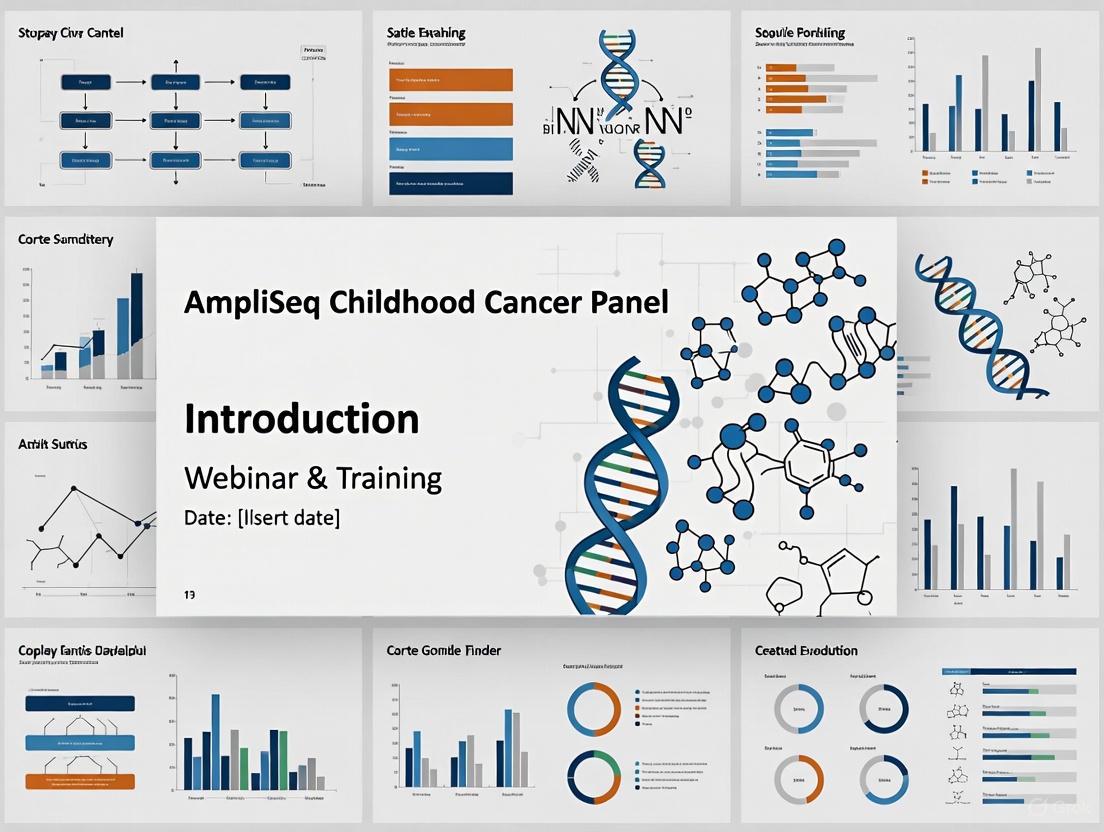
Implementing AmpliSeq for Illumina Childhood Cancer Panel: A Comprehensive Webinar for Research and Diagnostic Applications
This resource provides researchers, scientists, and drug development professionals with a comprehensive guide to the AmpliSeq for Illumina Childhood Cancer Panel.
Implementing AmpliSeq for Illumina Childhood Cancer Panel: A Comprehensive Webinar for Research and Diagnostic Applications
Abstract
This resource provides researchers, scientists, and drug development professionals with a comprehensive guide to the AmpliSeq for Illumina Childhood Cancer Panel. It covers foundational knowledge of this targeted NGS panel designed for pediatric and young adult cancers, detailing its 203-gene content for detecting SNVs, indels, CNVs, and gene fusions. The content delivers a step-by-step methodological workflow from nucleic acid extraction to sequencing on Illumina platforms, best practices for troubleshooting and data optimization, and critical analytical validation data including sensitivity, specificity, and reproducibility metrics. Finally, it explores the panel's clinical utility in refining diagnoses and informing targeted therapy, positioning it as a vital tool for advancing precision oncology in pediatric malignancies.
Understanding the AmpliSeq Childhood Cancer Panel: A Targeted Genomics Approach for Pediatric Malignancies
Next-generation sequencing (NGS) has emerged as a transformative tool in precision medicine for childhood cancers. Unlike adult malignancies, pediatric tumors are characterized by relatively low mutational burdens and distinctive genomic profiles, often originating from embryonic tissues [1]. Targeted NGS panels, such as the AmpliSeq for Illumina Childhood Cancer Panel, offer a cost-effective and rapid method for identifying actionable genomic alterations in this unique landscape. This technical guide explores the application, methodology, and clinical utility of targeted NGS, providing a framework for its implementation in pediatric oncology research and drug development.
The Distinct Molecular Landscape of Pediatric Cancers
The genomic architecture of pediatric solid tumors differs significantly from that of adult cancers, necessitating specialized diagnostic approaches. Pediatric malignancies often harbor fewer recurrent mutations and are frequently driven by structural variants, fusion genes, and copy number alterations rather than the single-nucleotide variants common in adult tumors [1] [2].
Key Genomic Alterations in Pediatric Solid Tumors
Table 1: Common Genomic Alterations in Pediatric Solid Tumors
| Alteration Type | Examples | Therapeutic Implications |
|---|---|---|
| Signal Pathway Mutations | RTK (EGFR), MAPK (KRAS), PI3K-mTOR (PTEN) | Often targetable with pathway-specific inhibitors |
| Transcriptional Regulators | MYC/MYCN amplification | Challenging to target directly |
| DNA Repair Genes | TP53 mutations | Impact treatment sensitivity and resistance |
| Epigenetic Modifiers | ATRX mutations | Emerging therapeutic targets |
| Germline Pathogenic Variants | TP53, BRCA1/2, NF1, RB1, WT1, APC | Cancer predisposition implications |
A meta-analysis of NGS applications in childhood and adolescent/young adult (AYA) solid tumors demonstrated that 57.9% of patients harbor actionable genomic alterations, with these findings influencing clinical decision-making in 22.8% of cases [1]. This highlights the substantial potential of precision oncology while underscoring the need for pediatric-focused genomic tools.
Targeted NGS Methodology for Pediatric Oncology
Library Preparation Approaches
Two primary methods are employed in targeted NGS library preparation, each with distinct advantages for pediatric cancer applications:
Amplicon-Based Sequencing (e.g., AmpliSeq) utilizes multiplex PCR to amplify specific genomic regions of interest. This approach offers several benefits for pediatric oncology, including:
- Rapid turnaround times (2-3 days)
- Robust performance with low tumor content (<25%)
- Cost-effectiveness for focused genomic interrogation [2]
The AmpliSeq for Illumina Childhood Cancer Panel exemplifies this approach, with available training resources covering library preparation protocols, pool planning for multiplexed runs, and best practices to optimize results [3].
Hybrid Capture-Based Sequencing uses biotinylated oligonucleotide probes to enrich target regions from fragmented DNA. This method:
- Circumvents issues of allele dropout
- Enables better coverage of large genomic regions
- Is more suitable for detecting copy number alterations and structural variants [4]
Analytical Validation Considerations
Robust validation of NGS panels is essential for clinical application. Professional guidelines recommend:
- Determining positive percentage agreement and positive predictive value for each variant type
- Establishing minimal depth of coverage requirements
- Using sufficient sample numbers to establish test performance characteristics
- Implementing an error-based approach that identifies potential sources of errors throughout the analytical process [4]
Table 2: Performance Metrics for Targeted NGS Panels in Pediatric Cancers
| Metric | Target Performance | Considerations for Pediatric Applications |
|---|---|---|
| Analytical Sensitivity | >95% for variant alleles at 5% allele frequency | Must account for low tumor purity in pediatric samples |
| Analytical Specificity | >99% for single-nucleotide variants | Critical for avoiding false positives in low-mutation-burden tumors |
| Coverage Uniformity | >95% of targets at ≥100x coverage | Essential for reliable copy number assessment |
| Concordance with Orthogonal Methods | >99% | Validation against WGS/WES for pediatric driver mutations |
Experimental Protocol: Pediatric-Focused NGS Panel Implementation
Sample Preparation and Quality Control
- Pathology Review: A certified pathologist must perform microscopic review of solid tumor samples to ensure sufficient viable tumor content and mark areas for macrodissection if needed.
- Tumor Fraction Estimation: Estimate tumor cell percentage through histopathological review, recognizing that inflammatory infiltrates may lead to underestimation.
- Nucleic Acid Extraction: Isulate DNA and/or RNA using quality-controlled methods appropriate for the sample type (FFPE, fresh frozen, etc.).
- Quality Assessment: Quantify nucleic acids and assess integrity using methods such as fluorometry and fragment analysis.
Library Preparation and Sequencing
For amplicon-based approaches such as the AmpliSeq Childhood Cancer Panel:
- Reverse Transcription: For RNA sequencing, convert RNA to cDNA using reverse transcriptase.
- Multiplex PCR Amplification: Amplify target regions using a single primer pool or multiple pools with barcoded adapters.
- Library Purification: Remove excess primers and enzymes using purification beads or columns.
- Library Quantification: Quantify final libraries using fluorometric methods and assess size distribution via fragment analyzers.
- Template Preparation and Sequencing: Dilute libraries to appropriate concentration, pool if multiplexing, and load onto the sequencing platform [3].
Bioinformatic Analysis
The data analysis pipeline typically includes:
- Base Calling and Demultiplexing: Generate sequence reads and assign to samples based on barcodes.
- Quality Control: Assess read quality, coverage uniformity, and other QC metrics.
- Alignment: Map reads to the reference genome.
- Variant Calling: Identify single-nucleotide variants, indels, copy number alterations, and structural variants.
- Annotation and Interpretation: Annotate variants with biological and clinical information to determine clinical actionability.
Comparative Performance of Pediatric-Specific Panels
Evidence suggests that pediatric-focused NGS panels outperform adult-oriented panels in detecting clinically relevant alterations in childhood cancers. A retrospective analysis comparing an adult-focused panel (OCAV3) with a pediatric-focused panel (OCCRA) demonstrated:
The OCCRA panel identified at least one target-agent pair for 19 of 28 samples (68%) compared to 16 of 28 samples (57%) for the OCAV3 panel [2]. The pediatric-focused panel also detected additional fusions and copy number alterations, including homozygous loss in CDKN2A, the most commonly identified target in the study.
Implementation Challenges and Solutions
Tumor Content and Purity
Pediatric tumor samples often have low tumor cellularity due to stromal contamination or inherent tumor characteristics. Solutions include:
- Microdissection: Enrich tumor cells by manually dissecting marked areas of interest.
- Bioinformatic Adjustment: Use computational methods to estimate and adjust for tumor purity in variant calling.
- Orthogonal Validation: Confirm key findings with complementary methods such as FISH or digital PCR.
Interpretation and Actionability
The relatively low prevalence of pediatric cancers creates challenges in establishing clinical actionability. Approaches to address this include:
- Utilizing Pediatric-Specific Knowledgebases: Leverage resources focused on pediatric cancer alterations.
- Implementing Actionability Frameworks: Use standardized classification systems such as the ESMO Scale for Clinical Actionability of Molecular Targets (ESCAT).
- Multidisciplinary Review: Incorporate input from molecular pathologists, pediatric oncologists, and genetic counselors in variant interpretation.
Physician Education and Engagement
A survey conducted at a comprehensive pediatric cancer center revealed that only 35% of physicians were confident in interpreting, utilizing, and discussing somatic genomic results, while just 27% expressed confidence with germline findings [5]. This highlights the critical need for educational initiatives alongside NGS implementation.
The Scientist's Toolkit: Essential Research Reagents
Table 3: Key Research Reagent Solutions for Targeted NGS in Pediatric Oncology
| Reagent/Framework | Function | Application in Pediatric NGS |
|---|---|---|
| AmpliSeq for Illumina Childhood Cancer Panel | Targeted amplicon-based NGS library preparation | Interrogation of pediatric cancer-relevant genes with optimized coverage |
| Hybrid Capture Probes | Solution-based enrichment of genomic regions | Comprehensive detection of SNVs, indels, CNAs, and fusions |
| Universal Blocking Oligos | Suppress unwanted hybridization of repetitive elements | Improve on-target rates and sequencing efficiency |
| Barcoded Adapters | Sample multiplexing and identification | Enable cost-effective sequencing of multiple samples in a single run |
| Automated Variant Interpretation Platforms | Streamline analysis and classification of genomic variants | Standardize variant calling and clinical actionability assessment |
Future Directions
The field of targeted NGS in pediatric oncology continues to evolve with several promising developments:
- Integration of RNA Sequencing: Enhanced detection of fusion transcripts and gene expression signatures.
- Liquid Biopsy Applications: Non-invasive monitoring of treatment response and minimal residual disease.
- Multi-omic Approaches: Combination of genomic, transcriptomic, and epigenetic profiling for comprehensive tumor characterization.
- Standardized Reporting Frameworks: Development of pediatric-specific guidelines for variant interpretation and clinical reporting.
Targeted NGS represents a powerful approach for unraveling the unique molecular landscape of pediatric cancers. By implementing pediatric-focused panels and optimized workflows, researchers and clinicians can enhance the identification of actionable alterations, ultimately guiding more precise therapeutic interventions for children with cancer.
The AmpliSeq for Illumina Childhood Cancer Panel is a targeted resequencing solution designed for the comprehensive evaluation of somatic variants associated with childhood and young adult cancers [6]. This ready-to-use panel addresses the distinctive genetic landscape of pediatric cancers, which, despite having a relatively low mutational burden, often contain genetic alterations with high clinical relevance [7]. The panel enables parallel study of numerous genes and patients with high sensitivity, providing vital information for redefining diagnostic, prognostic, and therapeutic strategies for managing acute leukemia (AL) and other pediatric cancers [7].
Panel Specifications and Technical Profile
The panel employs a robust PCR-based amplicon sequencing methodology, leveraging Illumina's next-generation sequencing (NGS) technology to interrogate a comprehensive set of genetic targets relevant to pediatric oncology [6]. The integrated workflow encompasses library preparation, sequencing, and automated analysis, creating a streamlined process from sample to result.
Table 1: Key Technical Specifications of the Childhood Cancer Panel
| Parameter | Specification | Details |
|---|---|---|
| Target Genes | 203 genes | Includes genes associated with leukemias, brain tumors, and sarcomas [6] |
| Variant Types | Multiple | SNPs, Indels, CNVs, Gene Fusions, Somatic variants [6] |
| Assay Time | 5-6 hours | Library preparation only; excludes quantification and pooling [6] |
| Hands-on Time | < 1.5 hours | Minimal manual intervention required [6] |
| Input Quantity | 10 ng | High-quality DNA or RNA [6] |
| Input Type | DNA, RNA | Compatible with various nucleic acid types [6] |
| Sample Types | Specialized | Blood, Bone Marrow, FFPE tissue, Low-input samples [6] |
The panel's design covers 203 genes, including 97 gene fusions, 82 DNA variants, 44 genes with full exon coverage, and 24 copy number variants (CNVs), making it a pan-cancer resource for pediatric oncology investigation [7]. Its wet-lab assay time is approximately 5-6 hours for library preparation, with a hands-on time of less than 1.5 hours, facilitating efficient laboratory workflows [6].
Experimental Protocol and Workflow
Library Preparation Methodology
The library preparation process follows a standardized protocol to ensure reproducibility and reliability [7]:
- Input Material: A total of 100 ng of DNA is used to generate 3,069 amplicons per sample, with an average size of 114 base pairs, covering the coding regions of the target genes. For RNA studies, 100 ng of RNA is reverse transcribed to cDNA using the AmpliSeq cDNA Synthesis kit, targeting 1,701 amplicons with an average size of 122 base pairs for fusion gene detection [7].
- Amplification and Barcoding: Amplicon libraries are generated through consecutive PCRs. Each sample is tagged with a specific barcode, enabling multiplexed sequencing runs [7].
- Library Pooling: After cleanup and quality control, the DNA and RNA libraries are pooled at an optimized 5:1 ratio (DNA:RNA) to balance coverage between variant types [7].
- Sequencing: The final pooled library is diluted to an appropriate concentration (17-20 pM) and sequenced on Illumina platforms, such as the MiSeq System [7].
Workflow Diagram
The following diagram illustrates the complete experimental workflow from sample to data analysis:
Essential Research Reagent Solutions
Successful implementation of the Childhood Cancer Panel requires several specialized reagents and accessory products, each serving a critical function in the workflow.
Table 2: Essential Research Reagents and Materials
| Product Name | Catalog ID (Example) | Function | Application Note |
|---|---|---|---|
| AmpliSeq Library PLUS | 20019101 | Provides core reagents for library construction (24 reactions) | Panel and index adapters sold separately [6] |
| AmpliSeq CD Indexes | 20019105 | Contains 96 unique indexes for sample multiplexing (Set A) | Enables pooling of up to 96 samples per run [6] |
| AmpliSeq cDNA Synthesis | 20022654 | Converts total RNA to cDNA for RNA fusion analysis | Required when working with RNA inputs [6] |
| AmpliSeq Library Equalizer | 20019171 | Provides beads and reagents for library normalization | Streamlines pre-sequencing workflow [6] |
| AmpliSeq for Illumina Direct FFPE DNA | 20023378 | Prepares DNA from FFPE tissues without deparaffinization | Simplifies processing of challenging sample types [6] |
Technical Validation and Performance Metrics
Rigorous validation studies demonstrate that the AmpliSeq Childhood Cancer Panel is a highly reliable and reproducible method for integrating targeted NGS into pediatric hematology practice [7]. The following performance characteristics were established using commercial controls and patient samples:
Table 3: Analytical Performance Metrics of the Panel
| Performance Metric | DNA (SNVs/Indels) | RNA (Fusions) |
|---|---|---|
| Mean Read Depth | > 1000x [7] | Not Specified |
| Sensitivity | 98.5% (at 5% VAF) [7] | 94.4% [7] |
| Specificity | 100% [7] | 100% [7] |
| Reproducibility | 100% [7] | 89% [7] |
| Limit of Detection (LOD) | Established for variants at 5% VAF [7] | Confirmed for key fusions [7] |
The panel achieves a mean read depth greater than 1000x, ensuring sufficient coverage for accurate variant calling [7]. It demonstrates high sensitivity (98.5% for DNA variants with 5% variant allele frequency (VAF) and 94.4% for RNA fusions) and perfect specificity (100%) for both DNA and RNA analyses [7]. Reproducibility is excellent for DNA (100%) and good for RNA (89%) [7].
Data Analysis and Clinical Utility
Data Analysis Pathway
The data generated by the panel undergoes a structured analysis pipeline to transform raw sequencing data into clinically actionable information, as illustrated below:
Demonstrated Clinical Impact
In a validation study focused on pediatric acute leukemia, the panel identified clinically relevant results in 43% of patients tested in the cohort [7]. The clinical utility of the findings can be broken down as follows:
- Mutation Impact: Among the mutations identified, 49% refined diagnosis, while 49% were considered targetable, offering potential therapeutic avenues [7].
- Fusion Gene Impact: Fusion genes identified by the panel were even more clinically impactful, with 97% of them refining diagnostic classification [7].
This high clinical impact demonstrates the feasibility and utility of incorporating this targeted NGS panel into the daily routine of pediatric molecular diagnostics, ultimately contributing to more precise diagnosis, prognosis, and treatment selection for pediatric cancer patients [7].
The AmpliSeq for Illumina Childhood Cancer Panel represents a significant advancement in the molecular profiling of pediatric and young adult cancers. This targeted next-generation sequencing (NGS) panel is specifically designed to address the unique genetic landscape of childhood malignancies, which often differ substantially from adult cancers in terms of mutation frequency, distribution, and variant types [7] [8]. Pediatric cancers typically display a lower mutational burden than their adult counterparts, yet the alterations present are frequently clinically relevant and often drive oncogenesis [7]. The panel provides a comprehensive resequencing solution for the simultaneous evaluation of multiple variant types across 203 genes associated with childhood and young adult cancers, including leukemias, brain tumors, and sarcomas [9].
The integration of this technology into research and clinical practice addresses a critical need in pediatric oncology. Traditional molecular diagnostic approaches often require multiple laborious tests performed separately for a single patient, consuming valuable time and limited sample material [7]. The AmpliSeq Childhood Cancer Panel consolidates this testing into a unified workflow that detects single nucleotide polymorphisms (SNPs), gene fusions, somatic variants, insertions-deletions (indels), and copy number variants (CNVs) from minimal input material [9]. This comprehensive approach enables researchers and clinicians to refine diagnostic classifications, identify prognostic markers, and discover potentially targetable alterations in a single assay, ultimately supporting the advancement of precision medicine for young cancer patients [7].
Technical Specifications and Panel Design
Panel Content and Coverage
The AmpliSeq Childhood Cancer Panel employs a sophisticated amplicon-based design that targets specific genomic regions of interest across the 203-gene panel. The technical architecture is optimized for comprehensive variant detection while maintaining efficiency in library preparation and sequencing. The panel is divided into DNA and RNA components, each targeting different variant types with optimized amplicon designs [10].
Table 1: Technical Specifications of the AmpliSeq Childhood Cancer Panel
| Parameter | DNA Component | RNA Component |
|---|---|---|
| Target Genes | 203 genes | 203 genes |
| Primary Variants Detected | SNPs, Indels, CNVs, Somatic variants | Gene fusions |
| Number of Amplicons | 3,069 | 1,701 |
| Average Amplicon Length | 114 bp | 122 bp |
| Average Library Length | 254 bp | 262 bp |
| Input Requirement | 10 ng DNA | 10 ng RNA |
The DNA component generates 3,069 amplicons covering coding regions of all 203 genes, with special emphasis on hotspot regions for point mutations and small indels, while also enabling copy number variant analysis through normalized coverage metrics [10] [9]. Simultaneously, the RNA component targets 1,701 amplicons specifically designed to capture known and novel fusion transcripts through carefully designed breakpoint regions [10]. This dual approach ensures comprehensive coverage of the major variant types relevant to pediatric cancer pathogenesis without requiring separate assays for DNA and RNA variants.
Supported Sample Types and Input Requirements
The panel demonstrates notable flexibility in terms of sample input requirements and compatibility with various sample types commonly encountered in pediatric cancer research. The core protocol requires only 10 ng of high-quality DNA or RNA, making it suitable for precious pediatric samples where material may be limited [9]. The panel supports multiple specialized sample types including blood, bone marrow, FFPE tissue, and low-input samples [9]. For challenging FFPE samples, a specialized product (AmpliSeq for Illumina Direct FFPE DNA) is available that allows for DNA preparation and library construction without the need for deparaffinization or DNA purification, potentially improving yields from archived clinical material [9].
The hands-on time for library preparation is remarkably efficient at less than 1.5 hours, with total assay time of approximately 5-6 hours (excluding library quantification, normalization, and pooling) [9]. This rapid turnaround time facilitates integration into research workflows where timely results are essential for experimental planning. The panel is compatible with various Illumina sequencing platforms including MiSeq, NextSeq, and MiniSeq systems, providing flexibility in sequencing scale and throughput based on project requirements [10].
Experimental Protocol and Workflow
Library Preparation Methodology
The library preparation process for the AmpliSeq Childhood Cancer Panel follows a PCR-based protocol that enables efficient target enrichment and library construction in a single day. The process begins with quality assessment of input nucleic acids, with recommended quantification using fluorometric methods (e.g., Qubit Fluorometer) and purity assessment via spectrophotometry (OD260/280 ratio >1.8) [7]. For RNA samples, an initial reverse transcription step is required using the AmpliSeq cDNA Synthesis for Illumina kit to convert total RNA to cDNA [9].
The core library preparation process involves several critical steps. First, DNA and RNA (converted to cDNA) inputs are amplified using the Childhood Cancer Panel primer pools in separate reactions. The panel utilizes a two-pool approach for both DNA and RNA components to minimize primer interference and maximize amplification efficiency [10]. Following amplification, enzymatic digestion is performed to partially digest primer sequences. Subsequently, index adapters are ligated to the amplicons to enable sample multiplexing. The final cleanup step removes residual enzymes and reagents prior to library quantification [7]. Throughout this process, quality control checkpoints are implemented to assess library quality, typically using capillary electrophoresis systems such as the Agilent BioAnalyzer or Fragment Analyzer [3].
Sequencing and Pooling Strategy
Optimal sequencing of the AmpliSeq Childhood Cancer Panel libraries requires careful planning of pooling ratios and sequencing parameters to ensure balanced coverage across all targets. Based on Illumina's recommendations, combined DNA and RNA libraries from the same sample should be pooled at a 5:1 ratio (DNA:RNA) to account for differences in amplicon numbers and ensure sufficient coverage for both variant types [10]. This ratio has been determined based on recommended read coverage requirements for robust variant detection.
Table 2: Recommended Sequencing Configuration by Platform
| Sequencing System | Reagent Kit | Max Combined Samples per Run | Recommended Pooling Ratio (DNA:RNA) | Run Time |
|---|---|---|---|---|
| MiniSeq System | Mid Output Kit | 4 | 5:1 | 24 hours |
| MiSeq System | v3 Kit | 4 | 5:1 | 32 hours |
| NextSeq System | High Output v2 Kit | 48 | 5:1 | 29 hours |
The sequencing output requirements are determined by the need for sufficient coverage to detect variants at low allele frequencies. Clinical validation studies have typically achieved mean read depths greater than 1000×, which enables reliable detection of somatic variants down to 5% variant allele frequency (VAF) [7]. For research applications requiring higher sensitivity for low-frequency variants, additional sequencing depth may be beneficial. The panel is compatible with a range of Illumina benchtop sequencers, allowing laboratories to select the appropriate throughput based on their sample volume [10].
Workflow Visualization
The following diagram illustrates the complete experimental workflow from sample preparation through data analysis:
Performance Validation and Analytical Metrics
Sensitivity and Specificity
Rigorous analytical validation studies have demonstrated the robust performance of the AmpliSeq Childhood Cancer Panel across multiple variant types. A comprehensive validation study focused on pediatric acute leukemia applications reported a sensitivity of 98.5% for DNA variants at 5% variant allele frequency (VAF), indicating excellent detection capabilities for low-frequency somatic mutations [7]. For fusion detection in RNA, the panel demonstrated 94.4% sensitivity, successfully identifying clinically relevant fusion transcripts including ETV6::ABL1, TCF3::PBX1, BCR::ABL1, RUNX1::RUNX1T1, and PML::RARA [7].
The panel maintains 100% specificity for DNA variants and 100% reproducibility for DNA detection, with slightly lower but still robust reproducibility for RNA at 89% [7]. These metrics indicate a very low false positive rate for DNA variant calling, which is critical for reliable identification of pathogenic mutations in clinical research settings. The high reproducibility ensures consistent results across repeated experiments and different operators, an essential requirement for standardized research protocols and multi-center studies.
Limit of Detection and Precision
The limit of detection (LOD) for the panel has been systematically evaluated using commercial reference standards with known mutation frequencies. For single nucleotide variants (SNVs) and small insertions/deletions (indels), the LOD has been established at 5% allele frequency with input quantities as low as 10 ng of DNA [7] [11]. This sensitivity threshold is appropriate for detecting somatic variants in heterogeneous tumor samples and minimal residual disease monitoring.
Validation of similar pediatric cancer panels (CANSeqTMKids) has demonstrated greater than 99% accuracy, sensitivity, repeatability, and reproducibility for SNVs, INDELs, and fusions when using inputs as low as 5 ng of nucleic acid with 20% neoplastic content [8]. The precision of variant calling has been confirmed through repeated measurements of reference standards, with minimal variation in variant allele frequency quantification and consistent detection of true positive variants across multiple runs [7] [8].
Variant Detection and Bioinformatics
Data Analysis Pipeline
The bioinformatics workflow for the AmpliSeq Childhood Cancer Panel involves multiple processing steps from raw sequencing data to final variant calls. Following sequencing, base calling and demultiplexing are performed using Illumina's instrument software. The resulting FASTQ files are then aligned to the human reference genome (hg19) using optimized aligners capable of handling amplicon-based data [8]. For the DNA component, variant calling encompasses multiple algorithms tailored to specific variant types: SNVs and indels are typically identified using amplicon-aware callers that account for PCR artifacts and sequencing errors, while CNVs are detected through normalized coverage ratios comparing target regions to reference controls [7].
The RNA sequencing data requires specialized analysis for fusion detection, typically employing tools that identify chimeric reads spanning breakpoint junctions. The sensitivity of fusion detection has been demonstrated down to approximately 1,100 supporting reads in validation studies of similar panels [8]. Following variant calling, comprehensive annotation is performed using established databases to prioritize potentially pathogenic variants based on population frequency, predicted functional impact, and known associations with pediatric cancers. The final output includes a curated list of variants with associated annotations supporting biological interpretation and potential clinical actionability.
Variant Classification and Interpretation
The interpretation of variants detected by the Childhood Cancer Panel follows a structured framework that considers multiple lines of evidence. Variants are typically classified based on their known or predicted functional consequences, including their presence in cancer hotspot databases, effect on protein function, and previous reports in pediatric malignancies [7]. In validation studies, a significant proportion of detected variants demonstrated clinical impact, with 49% of mutations and 97% of fusions identified as having potential clinical relevance in pediatric acute leukemia [7].
The clinical utility of the panel is evidenced by its ability to refine diagnostic classifications and identify targetable alterations. In one study, 41% of mutations refined diagnosis, while 49% were considered potentially targetable with existing therapeutic approaches [7]. The comprehensive genetic profiling enabled by the panel thus provides valuable insights for strategic treatment decisions and reveals opportunities for targeted therapeutic interventions in pediatric cancer patients.
The Scientist's Toolkit: Essential Research Reagents
Implementation of the AmpliSeq Childhood Cancer Panel requires several specialized reagents and components that form the essential toolkit for researchers. The following table details the key products required for successful library preparation and sequencing:
Table 3: Essential Research Reagents for AmpliSeq Childhood Cancer Panel
| Component Category | Product Name | Function | Key Specifications |
|---|---|---|---|
| Core Panel | AmpliSeq for Illumina Childhood Cancer Panel | Target enrichment | 203 genes, 24 reactions |
| Library Preparation | AmpliSeq Library PLUS for Illumina | Library construction | Available in 24-, 96-, 384-reaction kits |
| Index Adapters | AmpliSeq CD Indexes Sets A-D | Sample multiplexing | 8 bp indexes, 96 indexes per set |
| RNA Conversion | AmpliSeq cDNA Synthesis for Illumina | cDNA synthesis | Converts RNA to cDNA for RNA panels |
| Library Normalization | AmpliSeq Library Equalizer for Illumina | Library normalization | Bead-based normalization for sequencing |
| Sample Tracking | AmpliSeq for Illumina Sample ID Panel | Sample identification | 8 SNP primers + gender determination |
| FFPE Optimization | AmpliSeq for Illumina Direct FFPE DNA | FFPE DNA preparation | Direct use without deparaffinization |
Additional specialized products enhance the panel's application to specific sample types. The AmpliSeq for Illumina Sample ID Panel incorporates eight single nucleotide polymorphism (SNP)-targeting primer pairs and one gender-determining primer pair, enabling sample tracking and quality control through genetic fingerprinting [9]. For degraded samples from FFPE tissue, the AmpliSeq for Illumina Direct FFPE DNA product facilitates direct library construction without requiring DNA purification, potentially improving yields from suboptimal specimens [9].
The AmpliSeq for Illumina Childhood Cancer Panel provides researchers with a comprehensive solution for detecting the major variant types relevant to pediatric malignancies. Through its optimized design targeting 203 cancer-associated genes, the panel enables simultaneous assessment of SNPs, gene fusions, somatic variants, indels, and CNVs from minimal input material [9]. The technical validation data demonstrates robust performance characteristics with sensitivity exceeding 98% for DNA variants and 94% for RNA fusions, establishing its reliability for research applications [7].
The integration of this targeted sequencing panel into pediatric cancer research workflows facilitates a more complete molecular characterization of childhood malignancies, addressing the unique genetic features that distinguish them from adult cancers [8]. With its efficient workflow requiring less than 1.5 hours of hands-on time and compatibility with multiple Illumina sequencing platforms, the panel offers practical utility for laboratories seeking to implement comprehensive genomic profiling without developing custom assays [9]. As precision medicine continues to advance in pediatric oncology, the AmpliSeq Childhood Cancer Panel represents a valuable tool for uncovering diagnostically and therapeutically relevant alterations that may inform treatment strategies and ultimately improve outcomes for young cancer patients.
This technical guide details the core specifications for the AmpliSeq for Illumina Childhood Cancer Panel, a targeted resequencing solution designed for the comprehensive evaluation of somatic variants in childhood and young adult cancers. The information is structured to assist researchers and drug development professionals in planning and implementing this assay within their workflows.
The AmpliSeq for Illumina Childhood Cancer Panel is a targeted, ready-to-use panel that enables comprehensive evaluation of 203 genes associated with a spectrum of pediatric and young adult cancers, including leukemias, brain tumors, and sarcomas [9]. This PCR-based amplicon sequencing panel utilizes a simple, fast workflow that eliminates the time and effort typically required for individual target identification, primer design, and panel optimization [9]. The integrated workflow is designed for use with Illumina sequencing-by-synthesis (SBS) technology and supports the parallel analysis of both DNA and RNA from a variety of sample types, including blood, bone marrow, and Formalin-Fixed Paraffin-Embedded (FFPE) tissue, making it highly relevant for retrospective clinical studies [9].
Core Technical Specifications
The AmpliSeq Childhood Cancer Panel is characterized by a rapid and efficient workflow. The total assay time for library preparation is between 5 to 6 hours, which does not include the time required for subsequent library quantification, normalization, or pooling [9]. The hands-on time required from the researcher is notably low, at less than 1.5 hours [9]. The panel requires a minimal input of only 10 ng of high-quality DNA or RNA per reaction [9]. The panel's complete specifications are summarized in the table below.
Table 1: Comprehensive Technical Specifications of the Childhood Cancer Panel
| Specification Category | Detail |
|---|---|
| Assay Time | 5-6 hours (library prep only) [9] |
| Hands-On Time | < 1.5 hours [9] |
| Input Quantity | 10 ng high-quality DNA or RNA [9] |
| Method | Amplicon Sequencing [9] |
| Nucleic Acid Type | DNA, RNA [9] |
| Species Category | Human [9] |
| Specialized Sample Types | Blood, Bone Marrow, FFPE Tissue, Low-input samples [9] |
| Variant Classes Detected | Single Nucleotide Polymorphisms (SNPs), Insertions-Deletions (Indels), Gene Fusions, Somatic Variants, Copy Number Variants (CNVs) [9] |
| Automation Capability | Liquid handling robot(s) [9] |
| Number of Reactions | 24 reactions per panel [9] |
Compatible Illumina Sequencing Platforms
The panel is compatible with a range of Illumina sequencing systems, from the compact MiniSeq to the higher-throughput NextSeq series [9]. The choice of platform and reagent kit determines the maximum number of samples that can be sequenced per run. For paired DNA and RNA analysis from the same sample, which generates two separate libraries, a pooling volume ratio of 5:1 (DNA:RNA) is recommended based on coverage requirements [10].
Table 2: Sequencing System Compatibility and Sample Throughput
| Sequencing System | Reagent Kit | Max # DNA-Only Samples per Run | Max # RNA-Only Samples per Run | Max # Combined* Samples per Run | Recommended DNA:RNA Pooling Ratio |
|---|---|---|---|---|---|
| MiniSeq System | Mid Output Kit | 1 | 8 | 1 | 5:1 [10] |
| High Output Kit | 5 | 25 | 4 | 5:1 [10] | |
| MiSeq System | MiSeq Reagent Kit v2 | 3 | 15 | 2 | 5:1 [10] |
| MiSeq Reagent Kit v3 | 5 | 25 | 4 | 5:1 [10] | |
| NextSeq System | Mid Output v2 Kit | 27 | 96 | 22 | 5:1 [10] |
| High Output v2 Kit | 83 | 96 | 48 | 5:1 [10] |
Note: *Combined" refers to paired DNA and RNA from the same sample, resulting in two libraries [10].
Panel Design and Workflow
Panel Content and Configuration
The panel content is divided into two primary pools for optimal performance. The DNA component consists of two pools containing 3,069 amplicons, with an average amplicon length of 114 base pairs (bp) and an average library length of 254 bp [10]. The RNA component also comprises two pools, targeting 1,701 amplicons with an average amplicon length of 122 bp and an average library length of 262 bp [10]. This design allows for the concurrent analysis of DNA-based mutations (e.g., SNPs, indels) and RNA-based alterations (e.g., gene fusions) from the same sample.
Experimental Workflow
The following diagram outlines the key steps in the AmpliSeq for Illumina Childhood Cancer Panel workflow, from sample preparation to data analysis.
The Scientist's Toolkit: Essential Research Reagent Solutions
Executing the AmpliSeq Childhood Cancer Panel protocol requires several key components beyond the core panel itself. The table below lists the essential products needed to complete the workflow from library preparation to sequencing.
Table 3: Essential Research Reagents for a Complete Workflow
| Component | Function | Examples (Catalog IDs) |
|---|---|---|
| Library Prep Kit | Provides reagents for preparing sequencing libraries. | AmpliSeq Library PLUS for Illumina (20019101, 20019102) [9] |
| Index Adapters | Unique barcodes for multiplexing samples in a single run. | AmpliSeq CD Indexes Sets A-D [9] |
| cDNA Synthesis Kit | Converts input RNA to cDNA for the RNA portion of the panel. | AmpliSeq cDNA Synthesis for Illumina (20022654) [9] |
| Library Normalization | Simplifies and automates the library normalization process. | AmpliSeq Library Equalizer for Illumina (20019171) [9] |
| FFPE Sample Prep | Enables direct library construction from FFPE tissues without DNA purification. | AmpliSeq for Illumina Direct FFPE DNA (20023378) [9] |
| Sample Tracking | A genotyping panel for sample identification and tracking. | AmpliSeq for Illumina Sample ID Panel (20019162) [9] |
To maximize the effectiveness of the Childhood Cancer Panel, Illumina provides comprehensive training and support resources. These include a 30-minute online course on the library preparation protocol and a 15-minute course providing a general overview of the AmpliSeq technology [3]. For troubleshooting and optimizing sequencing data, resources such as the "How Do I Optimize Amplicon Sequencing Data?" video series (Parts 1 and 2) and a webinar on library QC using the BioAnalyzer and Fragment Analyzer are available [3]. Furthermore, an introductory webinar details the entire line of AmpliSeq products, including fixed panels and the associated analysis options [3].
Next-generation sequencing (NGS) has revolutionized genomic research by enabling the rapid production of vast amounts of sequencing data. For many clinical research applications, including childhood cancer research, a targeted sequencing approach provides significant advantages over broader whole-genome methods. Targeted panels focus on specific genomic regions of interest, generating smaller, more manageable datasets that reduce analysis burden and cost while delivering results in as little as 24 hours [12]. The AmpliSeq for Illumina Childhood Cancer Panel utilizes an amplicon-based enrichment method specifically designed to interrogate genomic targets relevant to pediatric malignancies, enabling researchers to obtain crucial mutational information from limited and challenging sample types, including formalin-fixed, paraffin-embedded (FFPE) tissue [3] [12].
The integrated workflow from library preparation through sequencing and analysis represents a complete, optimized pipeline for childhood cancer research. This technical guide details each component of this sophisticated process, highlighting the critical procedural steps, quality control checkpoints, and technical specifications that ensure reliable results. The AmpliSeq technology fundamentally works by using a highly multiplexed PCR system where a pool of oligonucleotide primer pairs simultaneously amplifies targeted genomic regions, creating a library of fragments ready for sequencing [12]. This approach preserves precious samples by requiring as little as 1ng of input DNA while providing comprehensive coverage of clinically relevant genomic targets [12].
Nucleic Acid Extraction and Quality Control
The initial stage of any robust NGS workflow begins with the isolation of high-quality genetic material. For the AmpliSeq Childhood Cancer Panel, this involves extracting DNA from patient samples, which may include tumor tissue, blood, or FFPE specimens. The extraction process must optimize for purity and yield, as these parameters directly impact downstream sequencing performance and variant calling accuracy. For degraded samples such as FFPE tissues, special consideration should be given to extraction methods that maximize the recovery of fragmented DNA [12].
Following nucleic acid extraction, a mandatory quality control (QC) step ensures sample integrity before proceeding to library preparation. The recommended QC methods include:
- UV Spectrophotometry: Assesses nucleic acid purity through A260/A280 and A260/A230 ratios, detecting potential contaminants from extraction reagents.
- Fluorometric Methods: Provides accurate quantitation of DNA concentration using dye-based assays, crucial for normalizing input amounts across samples [13].
For FFPE-derived DNA, additional QC measures such as fragment size analysis are recommended to evaluate the extent of DNA degradation, which can significantly affect amplification efficiency during library preparation. The AmpliSeq technology's robustness with challenging samples makes it particularly suitable for childhood cancer research, where archived FFPE specimens often represent valuable but compromised genetic resources [12].
Library Preparation Methodology
AmpliSeq Chemistry Fundamentals
The AmpliSeq for Illumina Childhood Cancer Panel employs a sophisticated highly multiplexed PCR technology that enables simultaneous amplification of thousands of targeted genomic regions in a single reaction. The core mechanism involves a pool of primer pairs specifically designed to flank the genomic regions of interest, creating amplicons that comprehensively cover the childhood cancer-related targets [12]. This approach offers two distinct design strategies, each with specific applications:
- Gene Design Approach: Utilizes tiling of overlapping amplicons to study continuous sequences of interest, requiring separate multiplexed PCR reactions to achieve complete coverage.
- Mutation Hotspot Design: Focuses on specific genomic regions with non-overlapping amplicons, allowing all primers to be accommodated in a single multiplexed reaction [12].
A key advantage of the AmpliSeq technology for childhood cancer research is its exceptional sensitivity to low-input DNA, successfully generating libraries from as little as 1ng of starting material. This efficiency makes it particularly valuable when working with precious pediatric tumor samples, where material is often limited. The technology also demonstrates remarkable robustness with degraded DNA from FFPE samples, with shorter amplicon designs (maximum 175bp) optimized for compromised specimens [12].
Automated Library Preparation Protocols
Automation of library preparation significantly enhances process consistency, throughput capacity, and operational efficiency while minimizing hands-on time and potential for manual error. The AmpliSeq for Illumina Childhood Cancer Panel is compatible with multiple automated liquid-handling systems, each providing validated protocols for reproducible results [14].
Table 1: Automation Systems Compatible with AmpliSeq Library Preparation
| Automation System | Protocol Type | Key Features | Throughput Capacity |
|---|---|---|---|
| Beckman Biomek i7 | Illumina-ready | Qualified solutions performing equal to or better than manual methods | Up to 96 samples |
| Hamilton NGS STAR | Illumina-ready | Scalable solutions with consistent performance | Up to 96 samples |
| Hamilton NGS STARlet | Partner-developed | Automated library preparation and enrichment | Configurable batch sizes |
| Eppendorf epMotion 5075t | Partner-developed | Flexible workflow options | 8-96 sample processing |
| Revvity Sciclone G3 NGSx | Partner-developed | Temperature-controlled positions for precise thermal cycling | High-throughput processing |
Laboratories can choose between two primary automation support pathways based on their specific needs and technical capabilities:
- Full Illumina-Ready Automation Support: Provides validated protocols co-developed and qualified with Illumina, complete with Illumina-led onboarding training, performance qualification assistance, and direct technical support from Illumina as the primary contact [14].
- Illumina Partner Network: Offers flexibility with partner-developed workflows where automation vendors manage installation, service, and training, with Illumina providing secondary support for chemistry-related issues when needed [14].
The implementation of automated library preparation typically reduces hands-on time by over 65% compared to manual methods, enabling laboratory staff to focus on higher-value activities while maintaining consistent library quality across batches [14].
Sequencing by Synthesis (SBS) Technology
SBS Chemistry Principles
Sequencing by Synthesis (SBS) technology forms the foundation of the Illumina sequencing platform, employing a proven biochemical method that detects single bases as they are incorporated into growing DNA strands [13]. The fundamental process involves the following steps:
- Cluster Generation: Library fragments are bound to a flow cell surface and amplified in situ through bridge amplification, creating thousands of identical clusters containing approximately 1,000 copies of each template.
- Cycle Sequencing: The flow cell is subjected to repeated cycles of nucleotide incorporation, where fluorescently labeled nucleotides compete for addition to the growing DNA strand.
- Signal Detection: After each incorporation, the flow cell is imaged to determine the identity of the base added to each cluster through characteristic fluorescence emission.
- Dye Termination and Regeneration: The fluorescent dye is chemically cleaved from the nucleotide, resetting the system for the next sequencing cycle.
This iterative process of nucleotide addition, imaging, and cleavage enables the determination of base sequences across millions of clusters simultaneously, providing the massive parallel sequencing capability that defines NGS technology [13].
Recent advancements in SBS chemistry, particularly the XLEAP-SBS formulation used in NextSeq 1000 and NextSeq 2000 Systems, represent significant improvements in sequencing performance. This enhanced chemistry delivers faster cycle times, higher data quality, and improved robustness compared to previous SBS formulations, making it particularly suitable for targeted sequencing applications like the AmpliSeq Childhood Cancer Panel [13].
Platform Selection and Sequencing Parameters
Selection of the appropriate sequencing platform depends on several factors, including required throughput, desired read length, and application-specific considerations. For targeted panels like the AmpliSeq Childhood Cancer, the following systems are commonly employed:
- MiSeq i100 Series: Ideal for smaller panels, supporting targeted resequencing, metagenomics, small genome sequencing, and targeted gene expression profiling with fast turnaround times.
- NextSeq 1000 & 2000 Systems: Suitable for larger panels, supporting RNA-Seq, single-cell methods, exome sequencing, and comprehensive targeted sequencing applications with enhanced throughput capabilities [13].
Table 2: Sequencing Platform Comparison for AmpliSeq Childhood Cancer Panel
| Sequencing Parameter | MiSeq i100 Series | NextSeq 1000/2000 Systems |
|---|---|---|
| Recommended Application Size | Smaller panels | Larger panels |
| Supported Applications | Targeted resequencing, metagenomics, small genome sequencing | RNA-Seq, single-cell methods, exome sequencing |
| Read Length Flexibility | Moderate | High |
| Throughput Capacity | Lower | Higher |
| Technology Foundation | SBS chemistry | XLEAP-SBS chemistry |
Optimal sequencing of amplicon libraries requires specific considerations for low-diversity samples, which are characteristic of targeted panels. The AmpliSeq technology generates libraries with minimal sequence variation at the beginnings of reads, potentially causing challenges during cluster detection and base calling. To mitigate these issues, Illumina recommends incorporating PhiX control DNA (typically 1-5%) to introduce sufficient sequence diversity and improve base calling accuracy [3] [15]. Additionally, the Sequencing Analysis Viewer (SAV) software enables real-time monitoring of key run metrics and comparison to PhiX controls to ensure sequencing success [3].
Data Analysis and Interpretation
Bioinformatics Pipeline
The transition from raw sequencing data to biological insights requires a sophisticated bioinformatics pipeline specifically tailored for amplicon-based targeted sequencing. The data analysis workflow for the AmpliSeq Childhood Cancer Panel encompasses multiple stages:
- Base Calling and Demultiplexing: Conversion of raw fluorescence signals into nucleotide sequences and separation of pooled samples into individual libraries based on their unique indices.
- Quality Control and Filtering: Assessment of read quality, removal of adapter sequences, and filtering of low-quality reads to ensure data integrity.
- Alignment to Reference Genome: Mapping of filtered reads to the human reference genome using optimized alignment algorithms.
- Variant Calling and Annotation: Identification of genetic variants (SNPs, indels) relative to the reference genome and functional annotation of detected variants.
- Interpretation and Reporting: Contextualization of variants within childhood cancer research, focusing on clinically actionable mutations and potential therapeutic implications.
The integrated nature of the Illumina platform provides streamlined bioinformatics solutions that make NGS data analysis accessible to researchers without extensive computational backgrounds. The Illumina Connected Software portfolio offers versatile, integrable, and accessible data analysis solutions designed to drive high-impact research [13].
Analysis Tools and Visualization
Modern NGS platforms incorporate user-friendly analysis interfaces that simplify the data interpretation process. For the AmpliSeq Childhood Cancer Panel, several specialized tools facilitate variant analysis and visualization:
- Sequencing Analysis Viewer (SAV): Enables real-time monitoring of sequencing runs, providing critical metrics for run quality, including cluster density, quality scores, and intensity measurements. SAV allows comparison of amplicon sequencing runs to standard PhiX controls to identify potential issues [3].
- Pre-configured Workflows: The Ion Reporter Software (for Ion Torrent systems) and Illumina DRAGEN platform offer optimized analysis pipelines specifically designed for AmpliSeq panels, providing variant annotation and reporting capabilities tailored to cancer research applications [12].
- Custom Analysis Scripts: For advanced users, customizable bioinformatics pipelines allow implementation of research-specific parameters and filters to address unique research questions in childhood oncology.
The availability of these sophisticated yet accessible analysis tools has significantly reduced the bioinformatics barrier for clinical researchers, enabling comprehensive investigation of childhood cancer genomics without requiring extensive computational resources or expertise [13].
Integrated Workflow Visualization
The complete integrated workflow for the AmpliSeq Childhood Cancer Panel, from sample preparation through data analysis, can be visualized as a coordinated series of interdependent processes:
AmpliSeq Childhood Cancer Panel Workflow
This integrated workflow demonstrates the seamless progression from biological sample to analytical results, highlighting the critical quality control checkpoints and technology integration points that ensure data reliability. The pathway illustrates both manual and automated options for library preparation, acknowledging the flexibility built into the system to accommodate varying laboratory capabilities and throughput requirements.
Essential Research Reagent Solutions
Successful implementation of the AmpliSeq Childhood Cancer Panel workflow requires specific reagent systems and consumables at each process stage. The following table details the essential components and their functions within the integrated workflow:
Table 3: Essential Research Reagent Solutions for AmpliSeq Workflow
| Reagent/Kits | Primary Function | Application Context |
|---|---|---|
| AmpliSeq for Illumina Childhood Cancer Panel | Targeted amplification of childhood cancer-related genes | Library Preparation |
| Illumina DNA Prep | Library preparation for DNA sequencing | Whole Genome Sequencing |
| Illumina Stranded Total RNA Prep | Library preparation for RNA sequencing | Whole Transcriptome Sequencing |
| Library Quantitation Kits | Accurate quantification of library concentration | Quality Control |
| PhiX Control Kit | Sequencing control for low-diversity libraries | Sequencing Quality |
| Qubit dsDNA HS Assay Kit | High-sensitivity DNA quantification | Input DNA & Library QC |
| TaqMan RNase P Detection Reagents | DNA quantification for human samples | Input DNA QC |
| Fragment Analyzer System | Quality assessment of nucleic acids and libraries | Quality Control |
These reagent systems form the foundation of a robust and reproducible AmpliSeq workflow, ensuring consistent performance across experiments and laboratories. Proper selection and implementation of these components directly impact assay sensitivity, variant detection accuracy, and overall data quality for childhood cancer research applications.
Technical Considerations and Optimization Strategies
Addressing Amplicon Sequencing Challenges
Sequencing amplicon libraries presents unique technical challenges that require specific optimization strategies. The low diversity inherent in amplicon libraries, particularly at the sequence starts, can cause issues with cluster detection and base calling on Illumina instruments [3] [15]. To address these challenges, researchers should implement the following strategies:
- PhiX Control Integration: Incorporation of 1-5% PhiX control DNA dramatically improves cluster identification and base calling accuracy by introducing sequence diversity. This practice is particularly crucial for smaller amplicon panels with limited sequence variation.
- Library Quantification Precision: Employing fluorometric-based quantification methods (e.g., Qubit dsDNA HS Assay) rather than spectrophotometric approaches ensures accurate library quantification, which is essential for achieving optimal cluster densities during sequencing [16].
- Sample-Specific Optimization: For challenging sample types like FFPE-derived DNA, adjusting input DNA quantities and implementing specialized extraction protocols can significantly improve library complexity and sequencing performance [12].
The "How Do I Optimize Amplicon Sequencing Data" training resources provided by Illumina offer comprehensive guidance on troubleshooting common issues and implementing best practices specifically for amplicon-based sequencing approaches [3] [15].
Contamination Prevention and Quality Assurance
Maintaining sample integrity throughout the workflow is paramount for generating reliable sequencing data, particularly when working with sensitive PCR-based methods like the AmpliSeq technology. Implementation of rigorous contamination prevention protocols is essential for minimizing false positives and ensuring result reproducibility [3] [15]. Key considerations include:
- Physical Separation: Establishing distinct pre- and post-PCR laboratory areas with dedicated equipment and reagents to prevent amplicon carryover between experiments.
- Procedural Controls: Incorporating negative controls throughout the workflow to monitor for potential contamination events and reagent integrity.
- Molecular Techniques: Utilizing uracil-DNA glycosylase (UDG) treatment or similar enzymatic methods to reduce the risk of PCR product carryover contamination in subsequent experiments.
Quality assurance should extend beyond wet-lab procedures to encompass bioinformatics quality metrics. Establishing baseline performance thresholds for key parameters including on-target rates, uniformity of coverage, variant calling sensitivity, and specificity ensures consistent panel performance across sequencing runs and enables rapid detection of workflow deviations that may require investigation [3].
The integrated workflow for the AmpliSeq Childhood Cancer Panel represents a sophisticated yet accessible approach to targeted genomic analysis in pediatric oncology research. By combining optimized library preparation chemistry, flexible automation options, advanced sequencing technology, and streamlined bioinformatics tools, this comprehensive solution enables researchers to efficiently translate biological samples into actionable genomic insights. The robustness of the AmpliSeq technology with challenging sample types, including FFPE tissues and low-input DNA, makes it particularly valuable for childhood cancer research, where sample material is often limited and precious.
The continued refinement of this integrated workflow, including developments in SBS chemistry, automation capabilities, and analysis algorithms, promises to further enhance the accessibility and reliability of targeted sequencing for childhood cancer research. By implementing the technical guidelines and optimization strategies outlined in this document, research laboratories can establish a robust, reproducible NGS pipeline that supports their investigation of the genomic underpinnings of childhood malignancies and contributes to the advancement of precision oncology for pediatric patients.
From Sample to Sequence: A Practical Workflow for Library Prep and Panel Application
The success of next-generation sequencing (NGS) in advanced research and diagnostic applications, such as cancer genomics, is fundamentally dependent on the quality and quantity of the input nucleic acids. The AmpliSeq for Illumina Childhood Cancer Panel provides a targeted resequencing solution for comprehensive evaluation of somatic variants associated with childhood and young adult cancers [9]. However, its performance varies significantly with the sample type and the integrity of the extracted DNA and RNA. This technical guide details the specific input requirements, quality assessment metrics, and optimized experimental protocols for preparing nucleic acids from blood, bone marrow, and formalin-fixed paraffin-embedded (FFPE) tissues to ensure reliable results with this and similar targeted panels.
Sample-Specific Input Requirements and Quality Metrics
The baseline input requirement for the AmpliSeq Childhood Cancer Panel is 10 ng of high-quality DNA or RNA [9]. However, achieving optimal performance necessitates adjustments and stricter quality control based on the sample source.
Table 1: Nucleic Acid Input Guidelines by Sample Type for the AmpliSeq Childhood Cancer Panel
| Sample Type | Minimum Input Quantity | Key Quality Metrics | Specialized Extraction Kits (Examples) | Notes |
|---|---|---|---|---|
| Blood / Bone Marrow Aspirate (BMA) | 10 ng [9] | 260/280 Ratio: ~1.8-2.0 [17]Call Rate: >95% [17] | Maxwell RSC Whole Blood DNA Kit [18] | DNA from BMA and peripheral blood (PB) shows high consistency and high call rates [18]. |
| Formalin-Fixed Paraffin-Embedded (FFPE) | 10 ng (from dedicated protocols) [9] | 260/280 Ratio: >1.7 [17]Call Rate: >65% may be adequate [17] | Quick-DNA FFPE Kit [17]; Maxwell RSC FFPE Plus DNA Kit [18] | DNA fragmentation and cytosine deamination are common; requires specialized protocols [19]. |
Detailed Experimental Protocols for Nucleic Acid Extraction
DNA Extraction from FFPE Tissues
Methodology from Peer-Reviewed Study [17]:
- Sectioning: Obtain six 5-μm thick scrolls of tissue sections from the FFPE block. To prevent cross-contamination, clean the microtome with RNase-away solution between each block.
- Deparaffinization and Lysis: Use a dedicated FFPE DNA extraction kit. The cited protocol used the Quick-DNA FFPE kit (Zymo Research), which employs a proprietary deparaffinization solution, followed by tissue digestion with Proteinase K and RNase.
- DNA Purification: The kit performs DNA purification via a spin-column-based method, finally eluting DNA in 50 µL of buffer solution.
- Storage: Store extracted DNA at -20°C until use for SNV array or NGS library preparation.
DNA/RNA Co-Extraction from Blood and Bone Marrow Aspirate
Methodology for Myeloid Gene Panel Analysis [18]:
- DNA Isolation: Use the Maxwell RSC Whole Blood DNA Kit (Promega) on peripheral blood or BMA samples. This is an automated, cartridge-based system for nucleic acid purification.
- RNA Isolation: Use the Maxwell RSC simplyRNA Blood Kit (Promega) from the same sample types for fusion transcript detection.
- Isolation from BMCB: For FFPE bone marrow core biopsies (BMCB), use the Maxwell RSC FFPE Plus DNA Kit and the Maxwell RSC RNA FFPE Kit.
The Scientist's Toolkit: Essential Research Reagent Solutions
The following reagents are critical for executing the AmpliSeq for Illumina workflow, from library preparation to sequencing.
Table 2: Key Research Reagent Solutions for the AmpliSeq Workflow
| Product Name | Catalog ID (Example) | Function | Compatibility / Notes |
|---|---|---|---|
| AmpliSeq for Illumina Childhood Cancer Panel | 20028446 [9] | Ready-to-use primer pool for targeting 203 childhood cancer genes. | Core panel component. |
| AmpliSeq Library PLUS | 20019101 (24 rxns) [9] | Reagents for preparing sequencing libraries. | Purchase panels and index adapters separately. |
| AmpliSeq CD Indexes | Set A-D available [9] | Unique barcodes to label individual samples for multiplexed sequencing. | Sufficient for labeling 96 samples per set. |
| AmpliSeq for Illumina Direct FFPE DNA | 20023378 [9] | Prepares DNA from unstained FFPE tissues for library construction without deparaffinization or DNA purification. | Streamlines workflow for FFPE samples. |
| AmpliSeq cDNA Synthesis for Illumina | 20022654 [9] | Converts total RNA to cDNA for use with RNA panels. | Required for RNA input. |
Workflow and Quality Relationship
The journey from sample collection to sequencing data is a multi-step process where sample quality at the beginning directly impacts the final results. The diagram below illustrates this workflow and the critical relationship between sample type, quality control, and sequencing outcomes.
Adherence to stringent, sample-specific guidelines for nucleic acid input is not just a recommendation but a prerequisite for generating reliable and clinically actionable data from the AmpliSeq Childhood Cancer Panel. While the core input requirement is minimal, the quality thresholds and extraction protocols for FFPE tissues, in particular, demand careful attention due to the inherent challenges of fixation. By following the detailed protocols for blood, bone marrow, and FFPE samples outlined in this guide, researchers can robustly leverage the power of NGS to uncover the genetic drivers of childhood cancers, ultimately supporting more precise diagnostic and therapeutic strategies.
This technical guide details the integrated experimental methodology for preparing sequencing-ready libraries using the AmpliSeq for Illumina Library PLUS system, coupled with cDNA Synthesis for RNA templates. This protocol is a foundational component within the broader context of the AmpliSeq Childhood Cancer Panel, a targeted sequencing solution designed for the identification of variants—including single nucleotide polymorphisms (SNPs), insertions-deletions (indels), gene fusions, and copy number variants (CNVs)—in pediatric oncology research [20] [21]. The amplicon-based, multiplex PCR workflow enables researchers and drug development professionals to rapidly analyze a high number of targets (from 12 to over 12,000) from minimal input material (1-100 ng), making it particularly suitable for challenging samples such as FFPE tissues [21]. The subsequent sections provide a detailed breakdown of the quantitative specifications, core methodologies, and essential reagents.
Protocol Specifications and Data
The AmpliSeq for Illumina library preparation workflow is characterized by its high efficiency and relatively short hands-on time. The table below summarizes the key quantitative specifications for the protocol [21].
Table 1: Key Specifications for the AmpliSeq for Illumina Library PLUS Protocol
| Parameter | Specification |
|---|---|
| Total Assay Time | ~5 hours (library prep only) |
| Hands-on Time | < 1.5 hours |
| Recommended Input per Pool | 10 ng (with a range of 1-100 ng) |
| Number of Amplicons | 12 to 12,288 |
| Nucleic Acid Input | DNA or RNA (requires cDNA synthesis) |
| Compatible Instruments | iSeq 100, MiSeq, MiniSeq, NextSeq 1000/2000 Systems |
Detailed Experimental Protocols
cDNA Synthesis Protocol for RNA Templates
When starting with RNA for targeted sequencing (e.g., for gene expression or fusion detection), a reverse transcription step is critical to generate complementary DNA (cDNA). The following five-step protocol ensures efficient cDNA synthesis [22].
RNA Sample Preparation
- Template: Use total RNA, messenger RNA (mRNA), or small RNAs (e.g., miRNA) depending on the application. For RT-qPCR, total RNA is routinely used.
- RNA Integrity: Maintaining RNA integrity is critical. Best practices include wearing gloves, using aerosol-barrier tips, and working with nuclease-free labware and reagents to prevent degradation by RNases.
- Storage: Purified RNA should be stored at –80°C with minimal freeze-thaw cycles. Optimal purification methods are essential to remove inhibitors of reverse transcriptases, such as salts, metal ions, ethanol, or phenol [22].
Genomic DNA Removal
- Contamination Risk: Trace amounts of genomic DNA (gDNA) co-purified with RNA can lead to false positives in downstream applications like RT-qPCR.
- Traditional Method: Treatment with DNase I, which must be thoroughly inactivated or removed afterward to prevent degradation of newly synthesized cDNA.
- Advanced Solution: Use of a double-strand-specific, thermolabile DNase (e.g., Invitrogen ezDNase Enzyme). This enzyme efficiently digests gDNA in 2 minutes at 37°C and can be inactivated at 55°C, offering a shorter workflow with less risk of RNA damage [22].
Reverse Transcriptase Selection
- The choice of enzyme profoundly impacts cDNA yield, length, and representation, especially for RNA with high GC-content or secondary structures.
- MMLV Reverse Transcriptase: A common choice, capable of synthesizing cDNA up to 7 kb, but with moderate RNase H activity and a lower optimal reaction temperature (37°C).
- Engineered MMLV Reverse Transcriptase (e.g., SuperScript IV): Recombinant enzymes with reduced RNase H activity and higher thermostability (up to 55°C). These attributes enable faster reaction times (as little as 10 minutes), higher sensitivity, and the synthesis of longer cDNA products (up to 14 kb) [22] [23].
Reaction Mix Preparation
- The main reaction components for cDNA synthesis are listed in the table below.
Table 2: Reaction Components for cDNA Synthesis [22]
| Component | Function and Key Features |
|---|---|
| RNA Template | The template for DNA synthesis. Integrity is paramount. |
| Reaction Buffer | Maintains optimal pH and ionic strength; may contain additives to enhance efficiency. |
| dNTPs | Deoxynucleotide triphosphates (dATP, dCTP, dGTP, dTTP) for DNA strand synthesis. Use high-quality dNTPs at 0.5–1 mM each. |
| DTT | A reducing agent for optimal enzyme activity. Ensure it is fully dissolved in the reaction mix. |
| RNase Inhibitor | Prevents RNA degradation by RNases during the reaction setup and incubation. |
| Primers | Oligo(dT), random hexamers, or gene-specific primers to initiate synthesis. |
| Nuclease-free Water | DEPC-treated or commercially sourced nuclease-free water to minimize risk of RNase contamination. |
- Performing cDNA Synthesis
- Primer Annealing: If using random hexamers, incubate the RNA-primer mix at room temperature (~25°C) for 10 minutes. For RNA with secondary structures, a pre-annealing denaturation step (65°C for 5 minutes, then immediately on ice) is recommended.
- DNA Polymerization: The temperature and duration of this step depend on the reverse transcriptase used. Engineered enzymes allow for polymerization at higher temperatures (e.g., 50°C for 10 minutes), which helps denature stubborn secondary structures in the RNA, resulting in longer, more representative cDNA [22].
- Enzyme Inactivation: The reaction is typically terminated by heating (e.g., 85°C for 5 minutes). The resulting cDNA can be used directly in the AmpliSeq library prep protocol.
AmpliSeq for Illumina Library PLUS Preparation
The core library preparation protocol for DNA or synthesized cDNA involves a streamlined, multiplex PCR-based workflow [21].
- Pool Planning and PCR Amplification: The process begins with a multiplexed PCR, where multiple primer pairs simultaneously amplify the targeted genomic regions (amplicons) from the DNA or cDNA template. The AmpliSeq technology is optimized for high specificity and uniformity across amplicons.
- Partial Digestion and Barcoding (Indexing): Following amplification, the PCR primers are partially digested. This is followed by the ligation of Illumina sequencing adapters, which include unique index sequences (barcodes) to allow for multiplexing of multiple samples in a single sequencing run. Products like the AmpliSeq CD Indexes provide these barcodes [21].
- Library Clean-Up and Normalization: The final library is purified to remove enzymes, primers, and other reaction components. The AmpliSeq Library Equalizer can be used to normalize libraries, ensuring an equimolar representation of each sample before pooling [21].
- Quality Control and Sequencing: The purified and normalized library should be quantified and assessed for quality (e.g., using the Agilent BioAnalyzer or Fragment Analyzer) before being loaded onto a compatible Illumina sequencer [3].
Workflow Visualization and Signaling Pathways
The following diagram illustrates the complete integrated workflow from RNA to sequencing-ready libraries, incorporating both the cDNA synthesis and AmpliSeq library preparation processes.
The Scientist's Toolkit: Research Reagent Solutions
Successful execution of the AmpliSeq protocol and cDNA synthesis requires a suite of specialized reagents. The following table details the key components and their functions.
Table 3: Essential Research Reagents for AmpliSeq and cDNA Synthesis Workflows [22] [21]
| Reagent Solution | Function |
|---|---|
| AmpliSeq Library PLUS | Provides the core reagents for library preparation, including enzymes and buffers for multiplex PCR and subsequent digestion steps. |
| AmpliSeq CD Indexes | Contains unique 8-base pair (bp) index sequences that are ligated to amplicons, enabling multiplexing of up to 384 samples in a single run. |
| AmpliSeq cDNA Synthesis Kit | A specialized kit containing reaction mix and enzyme blend to convert total RNA to cDNA, optimized for use with AmpliSeq for Illumina RNA Panels. |
| Engineered Reverse Transcriptase (e.g., SuperScript IV) | An MMLV-derived enzyme with reduced RNase H activity and high thermostability, enabling high-yield, full-length cDNA synthesis from challenging RNA templates. |
| Thermolabile DNase (e.g., ezDNase Enzyme) | A double-strand-specific DNase that rapidly removes genomic DNA contamination from RNA samples without damaging the RNA or requiring a separate cleanup step. |
| AmpliSeq Library Equalizer | Beads and reagents for the normalization of final libraries, ensuring balanced representation of samples when pooled for sequencing. |
| AmpliSeq for Illumina Direct FFPE DNA | Enables preparation of DNA directly from FFPE tissues without the need for deparaffinization or DNA purification, simplifying the workflow for common clinical samples. |
In the context of childhood cancer research, where sample material is often precious and limited, the ability to maximize data output from each sequencing run is paramount. Multiplexing, the practice of pooling multiple individually labeled samples for simultaneous sequencing, has become a cornerstone of efficient genomic analysis. This technique allows researchers to significantly reduce per-sample costs, minimize technical batch effects, and maximize throughput—critical considerations when working with the AmpliSeq for Illumina Childhood Cancer Panel [3]. At the heart of this practice are barcoding systems that enable precise sample identification after sequencing. AmpliSeq CD Indexes provide a specialized solution for this purpose, offering researchers in childhood cancer genomics a robust method for sample pooling that maintains data integrity while optimizing resource utilization. Within the framework of the Childhood Cancer Data Initiative (CCDI), which emphasizes comprehensive data sharing and collaboration, such efficient sequencing approaches become increasingly valuable for accelerating research discoveries [24].
Understanding AmpliSeq CD Indexes: Design and Configuration
AmpliSeq CD Indexes for Illumina are specifically designed to work seamlessly with AmpliSeq panels, including the Childhood Cancer Panel. These indexes employ a unique dual indexing (UDI) strategy that places two distinct DNA barcodes on each sample—one on the i7 and one on the i5 adapter ends [25]. This design is strategically superior to combinatorial or single indexing approaches because it provides a unique identifier combination for each sample, dramatically reducing the risk of index hopping and subsequent sample misassignment—a particularly crucial advantage when working with patterned flow cell instruments like the NovaSeq 6000 system [25].
The product is available in different configurations to accommodate various laboratory needs and scaling requirements, as detailed in the table below.
Table 1: Configurations of AmpliSeq CD Indexes for Illumina
| Configuration | Index Content | Sample Capacity | Storage Conditions |
|---|---|---|---|
| Set A | 96 unique indexes | 96 samples | -25°C to -15°C |
| Large Volume | 96 unique indexes | 96 samples | -25°C to -15°C |
Both configurations are shipped at room temperature but require long-term storage at -25° to -15°C to maintain stability [26]. The large volume format is particularly valuable for core facilities or large-scale projects where consistency across multiple runs is essential. The 96-index capacity enables researchers to pool up to 96 samples in a single sequencing run, dramatically increasing throughput while reducing per-sample costs [25].
Technical Advantages of Unique Dual Indexing in Childhood Cancer Research
The implementation of unique dual indexes with the AmpliSeq Childhood Cancer Panel offers several distinct technical advantages that are particularly beneficial in a research context where accuracy and reliability are paramount:
Enhanced Demultiplexing Accuracy: The UDI system ensures that each sample receives a truly unique combination of i7 and i5 indexes, enabling the sequencing analysis software to demultiplex with increased confidence and accuracy compared to combinatorial approaches where index sequences are repeated across a plate [25].
Index Hopping Mitigation: On instruments with patterned flow cells, such as the NovaSeq 6000 system, index hopping (where indexes detach and reattach to different molecules) can lead to sample misassignment. UDIs effectively mitigate this issue because any hopped reads can be bioinformatically filtered out, as they won't match the validated index pair combinations [25].
Optimal Color Balance: The AmpliSeq CD Indexes are designed with color balance in mind, which helps maintain base diversity throughout the sequencing run—a critical factor for achieving high-quality data throughout the entire run [25].
Streamlined Workflow Integration: These indexes are specifically optimized for use with AmpliSeq panels, ensuring compatibility and performance across the entire workflow from library preparation through to final data analysis [27].
For childhood cancer researchers, these technical advantages translate into increased confidence in data quality—a crucial consideration when making research conclusions or potential clinical interpretations based on the findings.
Implementation Protocol: Integrating CD Indexes with the Childhood Cancer Panel
Library Preparation Workflow
The integration of AmpliSeq CD Indexes with the Childhood Cancer Panel follows a systematic workflow that ensures optimal results. The process begins with the AmpliSeq for Illumina library preparation protocol, which includes target amplification, primer digestion, and partial adapter ligation [3]. The CD Indexes are then incorporated in the subsequent indexing PCR step, where complete adapters with unique index sequences are added to each sample.
Table 2: Key Steps for Library Preparation with CD Indexes
| Step | Description | Considerations |
|---|---|---|
| Target Amplification | Amplification of genomic regions covered by the Childhood Cancer Panel | Requires high-quality DNA input; follow recommended sample QC guidelines |
| Primer Digestion | Removal of amplification primers | Critical for preventing interference with downstream steps |
| Partial Adapter Ligation | Attachment of partial Illumina adapters | Prepares amplicons for full adapter attachment in indexing step |
| Indexing PCR | Incorporation of complete adapters with unique CD Indexes | Optimal index balance is crucial; refer to Index Adapters Pooling Guide [28] |
| Library Pooling | Combining indexed libraries in equimolar ratios | Enables multiplexed sequencing of up to 96 samples [25] |
| Library QC | Quality control of final pooled library | Utilization of fragment analyzers or bioanalyzers recommended [3] |
The Library Prep Protocol course provided by Illumina offers comprehensive training on this process, taking approximately 30 minutes to complete and identifying all necessary components for successful implementation [3].
Index Pooling and Color Balance
To achieve optimal sequencing performance, the Index Adapters Pooling Guide provides specific recommendations for creating libraries with balanced index combinations [28]. This balance is essential for maintaining base diversity throughout the sequencing run, which directly impacts data quality and cluster detection. The guide offers strategies for pooling indexes to ensure that all possible bases are represented relatively equally across each sequencing cycle, preventing issues associated with low diversity sequencing, which can be particularly challenging in amplicon-based approaches [3].
Diagram 1: AmpliSeq CD Indexes Workflow Integration
Troubleshooting and Quality Control Considerations
Implementing a robust quality control protocol is essential for successful multiplexing with AmpliSeq CD Indexes. The "How Do I Optimize Amplicon Sequencing Data?" training resources provided by Illumina address common challenges and best practices for achieving high-quality results [3]. These include:
Library QC Methods: Utilizing fragment analyzers or bioanalyzers to assess library quality and quantity before sequencing. Specific patterns in the trace can indicate potential issues that might affect sequencing performance [3].
Contamination Prevention: Implementing strict laboratory practices to minimize PCR contamination, which is particularly important when working with amplicon-based methods. Illumina provides specific guidelines for reducing contamination risk throughout the workflow [3].
Sequencing Optimization: Comparing key metrics of amplicon sequencing runs to a standard PhiX run helps identify potential issues and optimize performance. The Sequencing Analysis Viewer (SAV) can be used for this comparative analysis [3].
For researchers experiencing suboptimal results, the "Library QC and Troubleshooting with the BioAnalyzer and Fragment Analyzer" webinar provides detailed guidance on identifying and addressing common issues in library preparation [3].
The Scientist's Toolkit: Essential Research Reagent Solutions
Table 3: Key Research Reagent Solutions for AmpliSeq Multiplexing
| Reagent/Kit | Primary Function | Application Context |
|---|---|---|
| AmpliSeq for Illumina Childhood Cancer Panel | Targeted amplification of genes relevant to childhood cancers | Provides the core content for genetic analysis of pediatric oncology samples [20] |
| AmpliSeq CD Indexes for Illumina (Set A) | Unique dual indexes for sample multiplexing | Enables pooling of up to 96 samples; ideal for standard throughput needs [27] [26] |
| AmpliSeq CD Indexes for Illumina (Large Volume) | Unique dual indexes in larger quantities | Suitable for high-throughput facilities or multiple experiments [26] |
| Library Preparation Reagents | Enzymes and buffers for library construction | Facilitates target amplification, adapter ligation, and indexing steps [3] |
| Quality Control Instruments | Fragment analyzers/bioanalyzers for library QC | Critical for verifying library quality and quantity before sequencing [3] |
The integration of AmpliSeq CD Indexes with the Childhood Cancer Panel represents a significant advancement in the efficient genetic analysis of pediatric malignancies. By enabling robust multiplexing of up to 96 samples without compromising data integrity, this approach directly supports the objectives of initiatives like the Childhood Cancer Data Initiative (CCDI) [24]. The unique dual indexing strategy provides the technical foundation for accurate demultiplexing while mitigating index hopping—addressing key challenges in modern sequencing platforms.
For researchers focusing on childhood cancers, where sample material may be limited and research budgets constrained, the efficient pooling enabled by AmpliSeq CD Indexes offers a practical path to generating statistically significant datasets while optimizing resource utilization. As the field continues to advance toward more comprehensive genomic profiling in pediatric oncology, these multiplexing strategies will play an increasingly important role in facilitating the large-scale studies needed to uncover novel insights into childhood cancer biology and treatment.
Diagram 2: Research Enhancement Through AmpliSeq CD Indexes Integration
Within the framework of broader research initiatives, such as those introducing the AmpliSeq Childhood Cancer Panel, the selection and optimization of a sequencing platform are critical. This targeted next-generation sequencing (NGS) approach enables researchers and drug development professionals to focus on specific genomic alterations with high efficiency and accuracy. The core challenge lies in adapting the standardized library preparation protocol across different sequencing instruments, each with unique performance characteristics and technical requirements. Successfully navigating this process ensures that the high-quality data generated from panels like the AmpliSeq Childhood Cancer Panel (Catalog ID: 20028446) is consistent, reliable, and comparable, regardless of the sequencing hardware employed [20].
This technical guide provides a detailed framework for adapting amplicon-based sequencing workflows, specifically contextualized for the AmpliSeq for Illumina library preparation chemistry, across three common Illumina platforms: the MiSeq System, the NextSeq 500 System, and the NextSeq 2000 System. It synthesizes instrument specifications, protocol adjustments, and data analysis considerations to empower scientists to make informed decisions that align with their specific research objectives and operational constraints.
The MiSeq, NextSeq 500, and NextSeq 2000 systems represent different generations of Illumina sequencing technology, each offering a distinct balance of throughput, run time, and cost. Understanding their core specifications is the first step in selecting the appropriate platform for a targeted sequencing project.
The MiSeq System is renowned for its flexibility and simplicity, making it ideal for lower-throughput applications such as targeted gene and small-genome sequencing. However, it is important to note that Illumina has announced the obsolescence of the MiSeq System, with ordering available until September 30, 2025, and full support continuing through December 31, 2029 [29]. The NextSeq 500 System and its integrated array capabilities offer a mid-range throughput solution, suitable for more extensive targeted sequencing projects [30] [31]. The NextSeq 2000 System, part of a newer generation, delivers significantly higher throughput and integrated onboard DRAGEN secondary analysis, enabling rapid processing of large sample sets [32] [33].
Table 1: Key Specifications for Supported Sequencing Platforms
| Feature | MiSeq System | NextSeq 500 System | NextSeq 2000 System |
|---|---|---|---|
| Recommended Application | Targeted gene sequencing, small-genome sequencing [29] | Exome sequencing, RNA-Seq, amplicon sequencing [30] | High-throughput exome, transcriptome, single-cell RNA sequencing [33] |
| Typical Read Lengths | 2 × 300 bp, 2 × 150 bp [29] | Varies by reagent kit | Varies by reagent kit and XLEAP-SBS chemistry [33] |
| Throughput (Targeted) | Up to 96 samples and 1,536+ amplicons per run [29] | Mid-range throughput | High-throughput |
| Onboard Secondary Analysis | Standard data analysis pipelines [29] | Compatible with Local Run Manager | Integrated DRAGEN Bio-IT Platform [33] |
| Status Note | Becoming obsolete; supported until Dec 31, 2029 [29] | Fully supported | Fully supported with latest XLEAP-SBS chemistry [33] |
The Scientist's Toolkit: Essential Research Reagent Solutions
Executing a successful targeted sequencing study requires a suite of specialized reagents and consumables. The following table details the core components of the AmpliSeq for Illumina workflow, which is directly applicable to the Childhood Cancer Panel.
Table 2: Essential Reagents and Materials for the AmpliSeq Workflow
| Item | Function | Example Product |
|---|---|---|
| Targeted Panel | Contains the primer pairs designed to amplify the genomic regions of interest. | AmpliSeq for Illumina Childhood Cancer Panel (20028446) [20] |
| Library Prep Kit | Provides enzymes, master mix, and buffers for the multiplex PCR and library construction steps. | AmpliSeq Library PLUS for Illumina (20019101, 20019102, 20019103) [34] |
| Index Adapters | Unique dual-index sequences are added to each sample for multiplexing, allowing sample pooling and downstream identification. | AmpliSeq CD Indexes Sets A-D (20019105, 20019106, 20019107, 20019167) [34] |
| Resuspension Buffer (RSB) | A low-salt buffer used for diluting and resuspending libraries and primer pools. | Included in library preparation kits; lots are tracked in LIMS systems [32] |
| Reagent Cartridge | Platform-specific, pre-filled or user-filled cartridge containing sequencing-by-synthesis reagents. | NextSeq 1000/2000 Reagent Cartridge [32] |
| PhiX Control | A well-characterized control library used for quality control, error rate calibration, and balancing low-diversity libraries like amplicons. | Included in denature and dilute guides for all platforms [35] [30] |
Experimental Protocol Adaptation
The workflow for amplicon sequencing begins with a universal library preparation step, followed by platform-specific dilution, loading, and data analysis procedures. The following diagram illustrates the core workflow and its key adaptation points.
Universal Library Preparation
The initial stages of the AmpliSeq for Illumina workflow are consistent across platforms [3] [34]:
- Panel Amplification: Using the AmpliSeq Childhood Cancer Panel and the AmpliSeq Library PLUS kit, perform a multiplex PCR amplification on 10-100 ng of input DNA. This step simultaneously amplifies all targeted genomic regions.
- Partial Digest: Utilize a FuPa reagent to partially digest the forward and reverse primer sequences from the amplicons, preparing them for adapter ligation.
- Adapter Ligation: Ligate the AmpliSeq CD Index Adapters to the digested amplicons. These unique dual indexes are critical for multiplexing samples.
- Library Amplification: Perform a final PCR to enrich for the adapter-ligated fragments.
- Library Pooling: Quantify and normalize the individual libraries, then pool them together in preparation for sequencing.
Platform-Specific Protocol Modifications
Critical differences emerge after library pooling, primarily in library dilution, loading concentration, and run setup.
Library Denaturation, Dilution, and Loading
The process of denaturing and diluting the pooled library for sequencing must be tailored to each instrument.
- MiSeq System: While a specific denaturation and dilution guide was not found in the search results, the MiSeq system has its own recommended protocol which is typically provided with its reagent kits. Users should follow the latest Illumina documentation for the MiSeq.
- NextSeq 500/550 Systems: Illumina provides a dedicated "Denature and Dilute Libraries Guide" for these systems, which includes instructions for preparing the necessary PhiX control [30]. A Denature and Dilute Protocol Generator tool is also available to create customized instructions [30].
- NextSeq 1000/2000 Systems: Similarly, these systems have a dedicated Denature and Dilute Protocol Generator to create a step-by-step protocol for library preparation [33]. The final loading concentration is a critical parameter. The integration protocol for these platforms specifies a default drop-down list of options (650, 750, 1000, and 2000 pM) and a final loading volume of 24 µl into the reagent cartridge [32].
Sequencing Run Setup and Analysis Configuration
The setup of the sequencing run, particularly on the newer NextSeq 1000/2000 systems, involves specific configurations that impact data analysis.
- Run Mode Selection: The NextSeq 1000/2000 integration protocol requires specifying a Run Mode:
- Local Mode: The sample sheet is imported manually; secondary analysis uses the onboard DRAGEN module.
- Hybrid Mode: Run configuration is downloaded from Illumina Connected Analytics (ICA); analysis uses the onboard DRAGEN module.
- Cloud Mode: Run configuration is downloaded from ICA, and secondary analysis is performed in the cloud [32].
- Read Configuration: Parameters such as Read 1 Cycles, Read 2 Cycles, and Index Reads must be defined according to the panel design. For example, the protocol allows for preset options (e.g., 151, 101, 51) or custom values [32].
- Analysis Workflow: For onboard analysis, the workflow is typically set to "GenerateFASTQ." The FASTQ Compression Format (e.g., gzip or DRAGEN) must also be selected based on the DRAGEN version [32].
Table 3: Key Protocol Adaptation Parameters Across Platforms
| Parameter | MiSeq System | NextSeq 500 System | NextSeq 2000 System |
|---|---|---|---|
| Input Quantity (DNA) | 1–100 ng (10 ng recommended per pool) [34] | 1–100 ng (10 ng recommended per pool) [34] | 1–100 ng (10 ng recommended per pool) [34] |
| Final Loading Concentration | Platform-specific | Platform-specific | 650–2000 pM (selectable) [32] |
| Final Loading Volume | Platform-specific | Platform-specific | 24 µl (default, editable) [32] |
| Run Setup | Manual sample sheet import | Manual sample sheet import | Automated planned run creation via ICA (Hybrid/Cloud modes) [32] |
| Secondary Analysis | Standard pipelines or BaseSpace [29] | Local Run Manager | Integrated DRAGEN (Local/Hybrid) or Cloud [32] [33] |
Discussion and Best Practices
Platform Selection and Data Concordance
Choosing the right platform depends on the project's scale and data requirements. For low-to-mid-plex targeted sequencing, the MiSeq System remains a capable platform, though its obsolescence timeline should factor into long-term project planning [29]. The NextSeq 500/550 systems offer a robust solution for labs requiring higher throughput for applications like exome sequencing or larger amplicon panels [30]. For high-throughput core labs, the NextSeq 2000 System provides superior data output and integrated, rapid secondary analysis via DRAGEN [33]. Illumina has demonstrated high data concordance between the NextSeq 550 and NextSeq 1000/2000 systems, which is reassuring for labs transitioning between these platforms or validating protocols across them [33].
Optimizing Amplicon Sequencing Data
Amplicon libraries, like those generated from the Childhood Cancer Panel, have low sequence diversity, which can pose challenges for cluster generation and data quality. To optimize results:
- PhiX Spiking: Always include a 1-5% spike-in of the PhiX control library. This improves cluster identification for the instrument's sequencing algorithm and provides a quality control metric for the run [35] [30].
- Cluster Density Monitoring: Adhere to the recommended cluster densities for each platform. Over-clustering can lead to overlaps and poor data quality, while under-clustering reduces yield. The "Cluster Optimization Overview" document is a key resource for troubleshooting [35].
- LIMS Integration: For the NextSeq 1000/2000, leveraging the Clarity LIMS integration ensures reagent lot tracking, automated volume calculations, and correct sample sheet generation, which minimizes manual errors [32].
Successfully adapting the AmpliSeq for Illumina Childhood Cancer Panel protocol across MiSeq, NextSeq 500, and NextSeq 2000 systems is a manageable process that hinges on understanding both the universal steps of amplicon library preparation and the specific requirements of each instrument. Key adaptation points include the library denaturation/dilution method, the final loading concentration, and the configuration of the sequencing run and analysis workflow. By carefully considering the platform specifications, utilizing the provided reagent and protocol tools, and adhering to best practices for low-diversity libraries, researchers can generate high-quality, reliable data. This ensures that their research into childhood cancers, from discovery to drug development, is built upon a foundation of robust and reproducible genomic data.
The diagnosis and prognosis of acute leukemia have undergone a revolutionary transformation, shifting from morphology-based classification to sophisticated molecular characterization that guides targeted therapeutic interventions. This evolution is critically supported by technological advances in genomic profiling, including targeted sequencing panels such as the AmpliSeq for Illumina Childhood Cancer Panel, which enable comprehensive genetic assessment within clinical workflows [3]. Acute myeloid leukemia (AML), a hematopoietic malignancy characterized by uncontrolled proliferation of clonal myeloid cells, demonstrates marked molecular and clinical heterogeneity, accounting for approximately 80% of adult acute leukemia cases [36]. Despite therapeutic advances, AML remains a devastating disease with a global annual incidence of 144,645 cases and mortality of 130,189 deaths, highlighting the urgent need for refined diagnostic and prognostic approaches [36].
The integration of artificial intelligence (AI) and machine learning into diagnostic pipelines offers promising avenues for enhancing efficiency, reducing subjectivity, and identifying novel biomarkers [36]. Simultaneously, the treatment landscape has dramatically changed with the approval of at least 12 new agents since 2017, revolutionizing patient management across leukemia subtypes [37]. These developments have enabled the reclassification of several leukemia types from intermediate or unfavorable to favorable risk categories, including chronic lymphocytic leukemia (CLL), younger acute lymphoblastic leukemia (ALL) (patients younger than 60 years), and Philadelphia chromosome-positive ALL [38]. This whitepaper examines current clinical research applications through representative case studies that demonstrate how refined diagnostic and prognostic strategies are directly impacting patient management in acute leukemia.
Current Diagnostic Frameworks and Methodologies
Established Classification Systems
The diagnosis and classification of acute leukemia rely on integrated multimodal approaches incorporating morphology, immunophenotype, cytogenetics, and molecular abnormalities (MICM). Three major systems guide clinical practice:
- French-American-British (FAB) Classification: The historical FAB system, proposed in 1976, utilizes a threshold of >30% blast cells in bone marrow and subcategorizes AML into eight morphological subtypes (M0-M7) [36].
- World Health Organization (WHO) Classification: The fifth edition WHO classification lowers the diagnostic blast threshold to >20% in bone marrow or peripheral blood while incorporating genetic features. Notably, specific genetic abnormalities such as PML::RARA or RUNX1::RUNX1T1 enable diagnosis even with blast percentages below 20% [36].
- International Consensus Classification (ICC): The 2022 ICC system maintains the 20% blast threshold but allows diagnosis at ≥10% blasts in specific contexts such as therapy-related or secondary AML, or with high-risk genetic mutations, providing enhanced diagnostic granularity [36].
Table 1: WHO Classification of AML and Required Blast Proportions
| Category | Genetic Features | Blast Percentage Required |
|---|---|---|
| AML with recurrent genetic abnormalities | PML::RARA, RUNX1::RUNX1T1, etc. | Any level (even <20%) |
| AML defined by differentiation | No specific genetic abnormalities | >20% |
| Myelodysplasia-related AML | Myelodysplasia-related cytogenetic/molecular features | ≥20% (≥10% in ICC) |
| Therapy-related AML | History of cytotoxic therapy | >20% |
Conventional Diagnostic Workflows
Traditional AML diagnosis involves sequential laboratory assessments beginning with peripheral blood tests including complete blood count and morphological smear analysis to detect cytopenias and circulating blasts [36]. Bone marrow aspiration and biopsy follow to evaluate blast percentage and morphological features, with flow cytometry providing immunophenotyping for lineage assignment and subtyping. Cytogenetic analysis (karyotyping) and molecular genetic testing identify chromosomal abnormalities and mutations in genes such as FLT3, NPM1, and IDH1/2, essential for risk stratification and therapeutic targeting [36].
Despite comprehensive methodologies, significant challenges persist across these modalities. Morphological evaluation remains labor-intensive, time-consuming, and subjective, with diagnostic error rates reaching 40% [36]. Flow cytometry results vary between laboratories due to differing protocols, antibody panels, and analytical standards, compromising reproducibility. Molecular testing requires specialized equipment and expertise, often with extended turnaround times that may delay treatment initiation—a critical concern in rapidly progressive diseases such as acute leukemia [36].
Case Studies in Acute Leukemia
Case Study 1: AML withFLT3Mutation - Refined Risk Stratification and Targeted Therapy
A 45-year-old female presented with fatigue, bruising, and leukocytosis. Initial blood smear demonstrated increased blasts with monocytic features, and bone marrow biopsy confirmed AML with 50% blasts.
Diagnostic Refinement: Cytogenetic analysis revealed a normal karyotype. Multiplex PCR testing using the AmpliSeq Childhood Cancer Panel identified an internal tandem duplication (ITD) mutation in the FLT3 gene with high allelic ratio, alongside a missense mutation in the NPM1 gene. Based on European LeukemiaNet (ELN) 2022 guidelines, this molecular profile classified the patient as intermediate-risk, necessitating more intensive monitoring and consideration for allogeneic stem cell transplantation in first remission [37].
Therapeutic Application: The patient was enrolled in a clinical trial comparing standard "7+3" induction chemotherapy (cytarabine + daunorubicin) versus CPX-351 (liposomal daunorubicin-cytarabine) with or without gilteritinib, an FLT3 inhibitor [39]. This trial design reflects the evolving approach of matching specific molecular abnormalities with targeted therapies, even in newly diagnosed patients. The addition of FLT3 inhibitors to initial chemotherapy has demonstrated improved outcomes in FLT3-mutated AML, highlighting how molecular characterization directly informs therapeutic strategy [37] [38].
Prognostic Implications: The presence of FLT3-ITD with high allelic ratio traditionally conferred poor prognosis, but the integration of FLT3 inhibitors such as gilteritinib and midostaurin has significantly improved outcomes. The MORPHO study investigates gilteritinib versus placebo as maintenance therapy post-allogeneic stem cell transplantation in FLT3-mutated AML, potentially addressing the historically high relapse risk in this population [37]. Monitoring for resistance mutations in FLT3 during treatment has become standard, with emerging evidence supporting combination therapies to overcome resistance mechanisms.
Case Study 2:KMT2A-Rearranged Acute Leukemia - Novel Therapeutic Targeting
A 58-year-old male with relapsed AML after allogeneic stem cell transplantation presented with increasing blasts in peripheral blood. Prior initial genetic profiling had not identified targetable mutations.
Diagnostic Refinement: Repeat bone marrow biopsy with targeted RNA sequencing identified a KMT2A (formerly MLL) rearrangement. The patient was enrolled in a phase I/II trial of revumenib, a novel menin inhibitor, based on the mechanism that KMT2A rearrangements require menin protein for oncogenic signaling [37]. The AUGMENT-102 trial (NCT05326516) explores combination chemotherapy with revumenib in relapsed/refractory AML, while SAVE (NCT05360160) investigates decitabine/cedazuridine + venetoclax + revumenib in both newly diagnosed and relapsed/refractory AML [37].
Therapeutic Application: Revumenib received FDA approval in November 2024 for relapsed/refractory KMT2A-rearranged acute leukemia based on demonstrated efficacy as monotherapy [37]. This case illustrates how repeated molecular assessment at relapse can identify previously unrecognized targets, especially important as therapeutic options expand beyond conventional chemotherapy.
Prognostic Implications: KMT2A-rearranged AML has traditionally been classified as unfavorable risk, but the development of menin inhibitors may potentially reclassify this subtype from unfavorable to intermediate risk with optimized therapies [38]. Ongoing clinical trials will determine whether menin inhibitors improve long-term survival when incorporated into frontline regimens or in combination with other targeted agents.
Case Study 3: Elderly AML withIDH1Mutation - Alternative to Intensive Chemotherapy
A 78-year-old female with multiple comorbidities presented with progressive anemia and thrombocytopenia. Bone marrow examination confirmed AML with 35% blasts.
Diagnostic Refinement: Genetic profiling using a targeted amplicon-based NGS panel identified an IDH1 R132C mutation without adverse cytogenetic features. The patient's age and comorbidities rendered her unsuitable for intensive induction chemotherapy.
Therapeutic Application: Based on the detected IDH1 mutation, the patient received ivosidenib, an IDH1 inhibitor, in combination with azacitidine. This combination received FDA approval in May 2022 for newly diagnosed IDH1-mutated AML in patients ≥75 years or with comorbidities precluding intensive chemotherapy [37]. Clinical trials such as NCT03471260 are exploring ivosidenib + venetoclax + azacitidine combinations, potentially enhancing efficacy through synergistic mechanisms [37].
Prognostic Implications: Elderly AML patients historically had dismal outcomes, with 5-year survival rates below 10% [36]. The introduction of targeted therapies such as IDH inhibitors and venetoclax-based regimens has improved survival in this population, with current estimates of 30-40% survival at 5 years, excluding particularly adverse subtypes such as TP53-mutated AML [38]. Ongoing research focuses on optimizing combination therapies and sequencing strategies to further improve outcomes while maintaining quality of life.
Table 2: Recent Targeted Therapy Approvals in Acute Myeloid Leukemia
| Drug | Mechanism | Approved Indication | FDA Approval Date |
|---|---|---|---|
| Gilteritinib | FLT3 inhibitor | R/R FLT3-mutated AML | November 2018 |
| Ivosidenib | IDH1 inhibitor | R/R or ND IDH1-mutated AML | July 2018 (R/R), May 2019 (ND) |
| Venetoclax | BCL2 inhibitor | ND AML ≥75 years/comorbidities with HMA/LDAC | November 2018 |
| Quizartinib | FLT3 inhibitor | ND FLT3-mutated AML with standard chemotherapy | July 2023 |
| Revumenib | Menin inhibitor | R/R KMT2A-rearranged acute leukemia | November 2024 |
| Olutasidenib | IDH1 inhibitor | R/R IDH1-mutated AML | December 2022 |
Advanced Diagnostic Technologies
Amplicon-Based Targeted Sequencing
The AmpliSeq for Illumina Childhood Cancer Panel represents a targeted next-generation sequencing approach that utilizes PCR amplification to enrich specific genomic regions of interest prior to sequencing [3]. This technology enables focused assessment of genes recurrently mutated in childhood cancers, including leukemias, with advantages including rapid turnaround time, minimal DNA input requirements, and cost-effectiveness compared to comprehensive genomic profiling.
Methodology: The AmpliSeq workflow begins with DNA extraction from tumor samples (bone marrow or peripheral blood), followed by library preparation through multiplex PCR amplification using predesigned primer pools targeting leukemia-associated genes [3]. After amplification, libraries are barcoded, pooled, and sequenced on Illumina platforms. Bioinformatic analysis aligns sequences to reference genomes, identifies variants, and generates annotated reports for clinical interpretation [3].
Training resources for implementing this technology include introductory webinars, library preparation protocols (30-minute course), overview of assay technology (15-minute course), and specialized sessions on optimizing amplicon sequencing data and preventing PCR contamination [3]. These educational components ensure standardized implementation and interpretation across laboratories, critical for maintaining data quality and reproducibility in clinical research settings.
Artificial Intelligence in Leukemia Diagnostics
AI-based approaches are emerging as powerful adjunctive tools in leukemia diagnosis, leveraging machine learning and deep learning algorithms to analyze complex multimodal data. These systems function as decision-support tools that generate risk scores, classification recommendations, or triage prioritization rather than rendering autonomous diagnoses [36].
Blood Smear Image Analysis: AI algorithms can automatically detect and classify blast cells in peripheral blood smears, reducing manual review time and inter-observer variability. Deep learning models such as convolutional neural networks (CNNs) achieve high accuracy in distinguishing AML from normal samples and subclassifying morphological variants [36]. For instance, the Acute Leukemia Methylome Atlas (ALMA) tool maps DNA methylation patterns across leukemia samples, enabling subtype classification through reference alignment [40].
Flow Cytometry Data Interpretation: AI systems analyze high-dimensional flow cytometry data to identify immunophenotypic patterns characteristic of specific AML subtypes. These approaches can detect minimal residual disease (MRD) with sensitivity comparable to expert analysis while offering superior standardization and throughput [36].
Genetic Data Modeling: Machine learning algorithms integrate diverse molecular data types—including mutation profiles, copy number variations, and gene expression—to predict disease subtypes, risk categories, and therapeutic responses [36]. The ALMA tool developed by University of Florida Health researchers uses AI to match patients to 27 leukemia subtypes based on DNA methylation patterns and can predict 5-year survival probability based on genetic markers [40].
Emerging Research Applications and Clinical Protocols
Measurable Residual Disease (MRD) Monitoring
The detection and quantification of MRD has emerged as a critical prognostic factor and potential guide for treatment intensification or modification in acute leukemia. Modern approaches to MRD assessment include multiparameter flow cytometry (MFC), quantitative PCR (qPCR), digital PCR (dPCR), and next-generation sequencing (NGS)-based methods [37].
NGS-based MRD detection offers several advantages, including applicability to most patients without requiring patient-specific assay development, high sensitivity (up to 10^-5 to 10^-6), and ability to monitor multiple mutations simultaneously. The AmpliSeq technology enables targeted sequencing for MRD assessment by tracking mutation-specific reads over time, with decreasing variant allele frequencies (VAFs) indicating therapeutic response [3].
Clinical trials increasingly incorporate MRD assessment as secondary endpoints or even as criteria for treatment modification. For example, the VIALE-M trial (NCT04102020) investigates venetoclax + hypomethylating agents as maintenance therapy post-chemotherapy based on MRD status [37]. Similarly, allogeneic stem cell transplantation decisions are increasingly guided by MRD status, with transplantation reserved for MRD-positive patients in some protocols [37].
Innovative Clinical Trial Designs
The molecular heterogeneity of acute leukemia has prompted development of novel clinical trial designs that efficiently match patients to targeted therapies based on biomarker status.
Umbrella Trials: The MyeloMATCH screening trial (NCT05564390) represents a master protocol that assigns patients with myeloid cancers to biomarker-driven sub-studies [39]. This approach systematically evaluates targeted therapies in molecularly defined populations, accelerating drug development and personalized treatment strategies.
Platform Trials: Studies such as the biomarker-based treatment of AML trial at UCSF (NCT04493026) employ adaptive designs that allow modification of treatment arms based on accumulating efficacy data, potentially evaluating multiple targeted agents within a single protocol framework [39].
Combination Therapies: Numerous ongoing trials investigate rational drug combinations to overcome resistance mechanisms, such as CPX-351 + venetoclax in newly diagnosed AML (NCT04038437) [37], or azacitidine + venetoclax + quizartinib in unfit newly diagnosed FLT3-ITD-mutated AML [37].
The Scientist's Toolkit: Essential Research Reagents and Platforms
Table 3: Key Research Reagent Solutions for Acute Leukemia Investigation
| Reagent/Platform | Function | Application in Leukemia Research |
|---|---|---|
| AmpliSeq for Illumina Childhood Cancer Panel | Targeted NGS library preparation | Detection of mutations in leukemia-associated genes with minimal DNA input |
| Ion AmpliSeq Technology | Amplicon-based target enrichment | Microbial sequencing in infection complications; customizable panels |
| Hypomethylating Agents (azacitidine, decitabine) | DNA methyltransferase inhibitors | Differentiation therapy in AML; combination with targeted agents |
| FLT3 Inhibitors (gilteritinib, midostaurin, quizartinib) | Tyrosine kinase inhibition | Targeted therapy in FLT3-mutated AML; combination with chemotherapy |
| IDH1/2 Inhibitors (ivosidenib, enasidenib, olutasidenib) | Metabolic enzyme inhibition | Differentiation therapy in IDH-mutated AML; outpatient management |
| BCL2 Inhibitor (venetoclax) | Apoptosis activation | Combination with HMAs or low-dose cytarabine in elderly/unfit AML |
| Menin Inhibitor (revumenib) | Protein-protein interaction inhibition | Targeted therapy in KMT2A-rearranged and NPM1-mutated leukemia |
| Oral HMAs (CC486) | Epigenetic modulation | Maintenance therapy post-remission; extended exposure regimens |
Signaling Pathways and Experimental Workflows
Figure 1: Targeted Therapy Pathways in Acute Leukemia. This diagram illustrates key molecular pathways with approved targeted therapies in acute leukemia, showing how specific inhibitors counteract oncogenic signaling mechanisms.
Figure 2: Integrated Diagnostic Workflow for Acute Leukemia. This diagram outlines the sequential steps from sample collection to therapy selection, highlighting integration points for AI-assisted analysis, MRD monitoring, and clinical trial matching.
The refinement of diagnostic and prognostic approaches in acute leukemia represents a paradigm shift toward precision oncology, enabled by technological advances in genomic profiling and bioinformatic analysis. Targeted sequencing panels such as the AmpliSeq Childhood Cancer Panel provide clinically actionable molecular information that guides risk-adapted treatment strategies, while AI-based tools enhance diagnostic accuracy and efficiency. The integration of these technologies into clinical research protocols facilitates biomarker-driven trial designs and rational drug development.
Ongoing challenges include improving accessibility to advanced molecular testing, standardizing bioinformatic pipelines across institutions, and validating AI algorithms in diverse patient populations. Future directions will likely focus on multi-omic integration, real-time monitoring of clonal evolution, and development of novel therapeutic approaches for currently high-risk subtypes. As these technologies continue to evolve, they promise to further refine our understanding of acute leukemia biology and improve outcomes for patients across disease subtypes.
Maximizing Panel Performance: Best Practices for Data Quality and Contamination Prevention
Within the context of introducing and training researchers on the AmpliSeq for Illumina Childhood Cancer Panel, ensuring the quality of sequencing libraries is a critical preliminary step. Next-generation sequencing (NGS) accuracy and reliability are heavily dependent on the quality of the prepared libraries. Prior to sequencing, it is essential to check library quality control to confirm the expected average library size, verify the presence of a primary library peak, and ensure the absence of additional small and large library artifacts [41]. This technical guide details the methodologies for using the Agilent Bioanalyzer and Fragment Analyzer systems as essential tools for this quality assessment, providing researchers and drug development professionals with robust QC strategies integrated within the AmpliSeq workflow.
The Role of Capillary Electrophoresis in Library QC
The Agilent Bioanalyzer and Fragment Analyzer are capillary electrophoresis instruments that provide a rapid, automated alternative to traditional gel electrophoresis for the quality control of NGS libraries. Their primary function in the context of the AmpliSeq Childhood Cancer Panel is to generate an electrophoretogram, a visual trace of the library's size distribution, and to calculate the concentration of the library fragments.
- Objective Assessment: These systems provide objective data on the average size of the library fragments (in base pairs) and the distribution of fragment sizes, which is critical for accurate downstream sequencing and analysis [41].
- Concentration Measurement: They yield a quantitative assessment of the library's concentration, which is necessary for pooling multiple libraries at equimolar ratios and loading the sequencer with the correct amount of DNA.
- Contamination and Integrity Check: The generated trace can reveal the presence of unwanted products, such as adapter dimers, primer dimers, degraded DNA, or large, non-specific products, which could compromise sequencing efficiency and data quality [41].
Interpreting the Ideal Library Trace
An ideal final library trace for an amplicon-based panel like the AmpliSeq Childhood Cancer Panel should exhibit a single, dominant, and sharp peak corresponding to the expected size range of the target amplicons.
Key features of an ideal trace include [41]:
- A single, narrow peak indicating a tight size distribution of the amplicons.
- The peak should be positioned at the expected base pair size for the designed library.
- A flat baseline with the absence of secondary peaks, particularly smaller peaks in the low molecular weight region (e.g., around 100-150 bp, which could indicate adapter-dimer contamination) or large, broad peaks indicating genomic DNA contamination or oversized products.
- A high signal-to-noise ratio, where the primary peak is clearly distinguishable from the background.
Troubleshooting Common Library QC Anomalies
Recognizing deviations from the ideal trace is a fundamental troubleshooting skill. The table below summarizes common anomalies, their potential causes, and corrective actions.
Table 1: Troubleshooting Common Library QC Issues with the Bioanalyzer/Fragment Analyzer
| Observed Anomaly | Potential Cause | Corrective and Preventive Actions |
|---|---|---|
| A dominant short peak (~100-150 bp) | Adapter-dimer formation from over-cycling during PCR or insufficient purification [41]. | Re-optimize PCR cycle numbers; use bead-based clean-up with adjusted sample-to-bead ratios to selectively remove short fragments. |
| A broad smear or multiple peaks | Non-specific PCR amplification, degraded input DNA, or issues during enzymatic steps (fragmentation, end-repair) [41]. | Verify DNA quality; titrate PCR reagents; ensure all enzymatic reaction steps are performed at correct temperatures and durations. |
| A shift to larger than expected size | Incomplete fragmentation or over-sized amplicons due to protocol error. | Check the fragmentation or amplification protocol; ensure all reagents are fresh and properly mixed. |
| General low yield or faint peak | Insufficient input DNA, failed PCR amplification, or excessive dilution. | Quantify input DNA accurately; check PCR component viability; concentrate the library if necessary. |
| No peak detected | Complete failure of library preparation. | Systematically review each step of the protocol, from DNA extraction and quantification to the final PCR amplification. |
Experimental Protocol for Library QC Analysis
The following is a generalized methodology for performing library QC using the Bioanalyzer or Fragment Analyzer. Always refer to the manufacturer's specific protocol for the exact reagents and steps.
Methodology:
- Kit Preparation: Thaw the appropriate DNA assay kit (e.g., High Sensitivity DNA Kit for Bioanalyzer). Prepare the gel-dye mix as per the kit instructions, vortex, and centrifuge.
- Chip Priming: Load the gel-dye mix into the designated well on the chip. Use a syringe to prime the chip in the provided priming station.
- Sample and Ladder Loading: Add 1 µL of the DNA marker to all sample wells and the ladder well. Then, add 1 µL of the ladder to the ladder well and 1 µL of each library sample (or its dilution) to the sample wells.
- Chip Vortexing and Run: Place the chip in a vortexer with an appropriate adapter for 1 minute. Place the chip in the instrument and start the run using the associated software.
- Data Analysis: After the run, the software will automatically generate an electrophoretogram and a pseudo-gel image for each sample. Analyze the traces for the key features and anomalies described above.
Quantitative Data Summary: The software provides a table of quantitative data for each sample. The key metrics to record are summarized below.
Table 2: Key Quantitative Metrics from Bioanalyzer/Fragment Analyzer Output
| Metric | Description | Ideal Outcome for AmpliSeq Library |
|---|---|---|
| Average Size (bp) | The mean size of the library fragments. | Should match the expected amplicon size range. |
| Molarity (nmol/L or nM) | The concentration of the library in nanomolar. | Used for accurate pooling and sequencing load calculation. |
| Peak Height/Area | The intensity and proportion of the main peak. | A tall, dominant peak with high area percentage. |
| Size of Dominant Peak | The base pair value at the highest point of the main peak. | Should be the expected target amplicon size. |
| % of Total Area in Main Peak | The percentage of the total sample represented by the main peak. | >80% is typically desirable, indicating purity. |
Integration with the AmpliSeq Childhood Cancer Panel Workflow
The library QC step is positioned after the library preparation step and before the pooling and sequencing steps in the overall AmpliSeq workflow. A successful QC check ensures that only high-quality libraries proceed to sequencing, thereby maximizing data output and minimizing failed runs and reagent waste. The "Library QC and Troubleshooting with the BioAnalyzer and Fragment Analyzer" webinar is a core component of the training resources for the AmpliSeq for Illumina Childhood Cancer Panel, geared toward both new and intermediate users to build proficiency in this critical area [3].
The Scientist's Toolkit: Research Reagent Solutions
The following table details key materials and reagents essential for performing effective library QC in this context.
Table 3: Essential Research Reagent Solutions for Library QC
| Item | Function/Brief Explanation |
|---|---|
| Agilent High Sensitivity DNA Kit | Contains all necessary reagents (gel, dye, DNA marker, ladder) to run and analyze up to 11 samples on the Bioanalyzer for precise sizing and quantification of DNA fragments. |
| D1000/DFC ScreenTape (Agilent) | Provides an automated, high-throughput alternative to chips for the TapeStation system, offering consistent and fast analysis of NGS libraries. |
| Fragment Analyzer Capillary Cartridge | The consumable used in the Fragment Analyzer system that contains the capillaries through which samples are separated for analysis. |
| Magnetic SPRI Beads | Used for post-amplification clean-up to purify the library by removing enzymes, salts, and short-fragment contaminants like adapter dimers. |
| DNA LoBind Tubes | Minimize DNA adhesion to tube walls, which is critical for maintaining accurate concentrations of precious library samples during QC and handling. |
| Nuclease-Free Water | Used for diluting library samples to the optimal concentration for loading onto the QC instrument, ensuring no enzymatic degradation occurs. |
Workflow and Logical Relationship Diagram
The following diagram illustrates the logical workflow and decision-making process for library QC within the context of the AmpliSeq Childhood Cancer Panel.
Quality control (QC) of sequencing libraries is a critical determinant of success in next-generation sequencing (NGS) workflows. Accurate quantification and proper quality checks ensure optimal sequencing performance and reliable data output [42]. For targeted sequencing approaches such as the AmpliSeq for Illumina Childhood Cancer Panel, which investigates 203 genes associated with pediatric and young adult cancers, implementing robust QC protocols is particularly important for detecting somatic variants with confidence [9]. This technical guide examines the core principles of interpreting QC traces, recognizing ideal library profiles, and identifying common issues that can compromise sequencing results.
The fundamental objective of library QC is to accurately determine library profile, size distribution, and concentration for loading onto the sequencer [43]. Most experiments aim for equal read distribution across samples to ensure comparability in downstream analyses. While bioinformatic tools can normalize data during analysis, significant differences in read depth between sample groups can introduce unexpected effects that complicate data interpretation [43]. Thus, achieving equal read distribution through precise library quantification remains the gold standard for most NGS applications, including RNA-Seq and targeted panels.
Essential QC Methods and Instrumentation
Primary QC Techniques
Multiple complementary methods are employed for comprehensive library QC, each providing different types of information about library quality and quantity:
- Microcapillary Electrophoresis: Platforms such as the Bioanalyzer, Fragment Analyzer, LabChip GX II, and TapeStation have become standard practice for NGS laboratories [43]. These instruments generate traces that deliver information about library quantity, size distribution, shape, and the presence of undesired by-products or residual primers.
- Fluorometric Methods: These use fluorometric dyes specific to double-stranded DNA (dsDNA), single-stranded DNA (ssDNA), or RNA to quantify nucleic acid concentration across different sensitivity ranges [42]. Benchtop fluorometers with highly sensitive DNA quantification assays (e.g., Qubit dsDNA HS assay) are often used alongside microfluidic devices [43].
- qPCR-based Quantification: This method selectively quantifies DNA fragments that have Illumina sequencing adapters, providing assessment of amplifiable fragments [42]. qPCR uses primers targeting adapter regions to ensure only correctly assembled, functional library molecules are counted [43].
Table 1: Comparison of Primary Library QC Methods
| Method | Primary Function | Key Metrics | Limitations |
|---|---|---|---|
| Microcapillary Electrophoresis | Size distribution and quality assessment | Average fragment size, distribution profile, adapter dimer detection | Limited quantitative accuracy; relative quantification only |
| Fluorometric Methods | Nucleic acid quantification | DNA concentration, sample purity | Does not distinguish between amplifiable library and artifacts |
| qPCR-based Quantification | Amplifiable library quantification | Concentration of adapter-ligated fragments, optimal cycle number determination | Does not provide size distribution information |
Instrument-Specific Considerations
The selection of QC instrumentation often depends on laboratory throughput needs and project scale [43]. The Bioanalyzer employs a chip-based system with capacity for 11-12 samples per run, making it suitable for lower-throughput laboratories. In contrast, the Fragment Analyzer is plate-based and can handle multiple 96-well plates per run, positioning it as the preferred solution for high-throughput NGS laboratories with large-scale projects [43]. These instruments also differ in their resolution, sensitivity, and dynamic range, which affects input requirements and the visual appearance of library traces [43].
Illumina recommends consulting library preparation kit-specific reference guides for validated quantification and QC methods to ensure compatibility and appropriate sensitivity [42]. Using unsupported methods or those with incorrect sensitivity can lead to inaccurate results that ultimately impact sequencing performance. For certain library types that use Illumina Bead Based Normalization, quantification and/or QC may not be required [42].
Ideal Library Profiles and Characteristics
Expected Profile Features
An ideal library trace demonstrates a single, dominant peak with a size distribution appropriate for the specific library preparation method. For standard NGS workflows, the ideal size typically ranges between 400-500 base pairs, which represents the optimal size for clustering on Illumina sequencers [44]. The distribution should appear as a smooth curve rather than a sharply defined single peak, indicating a diverse population of fragments within the expected size range [43].
For amplicon-based panels like the AmpliSeq Childhood Cancer Panel, the library profile may show a more specific size distribution pattern corresponding to the amplified targets. The Childhood Cancer Panel requires approximately 5-6 hours for library preparation (not including quantification, normalization, or pooling time), with less than 1.5 hours of hands-on time, and utilizes 10 ng of high-quality DNA or RNA as input [9].
Single-Cell RNA-Seq Library Profiles
In single-cell RNA-seq workflows, library profiles exhibit distinct characteristics at different preparation stages. After cDNA amplification, the electropherogram should show cDNA fragment sizes ranging from approximately 300-400 base pairs to as large as 9,000-10,000 base pairs, typically with a gradual rise in the trace [44]. Depending on the cell type and underlying biology, users might observe distinct or sharp peaks representing abundant cell-type transcripts. The ideal trace shows a library distribution between 500-800 base pairs [44].
Table 2: Ideal Library Profile Characteristics by Application
| Application | Expected Size Range | Profile Shape | Key Quality Indicators |
|---|---|---|---|
| Standard NGS | 400-500 bp | Smooth, single peak | Narrow size distribution, absence of secondary peaks |
| Amplicon Panels | Varies by panel design | Multiple peaks possible | Expected pattern for target regions, low primer dimer |
| scRNA-seq cDNA | 300-400 bp to 9,000-10,000 bp | Gradual rise | High molecular weight content, smooth distribution |
| Final scRNA-seq Library | 400-500 bp | Single dominant peak | Similar to standard NGS but accounting for barcodes |
Common QC Issues and Anomalies
Adapter Dimers and Primer Artifacts
Adapter dimers represent one of the most common issues in NGS library preparation, appearing as a sharp peak at approximately 100-150 bp in the electropherogram [43]. These dimers form when library adapters ligate to each other instead of to target fragments, creating short fragments that contain primarily adapter sequences. When substantial by-products such as adapter dimers account for >3% of the final library preparation, it is recommended to remove them by re-purifying the library prior to sequencing [43]. Since shorter fragments are preferentially amplified during PCR, these by-products can consume significant sequencing space and reduce the number of useful reads obtained from an NGS experiment.
Residual primers after the final purification step represent another common artifact that appears as additional small peaks in the electropherogram, typically below the main library distribution [43]. These can indicate inefficient cleanup during library preparation and may interfere with accurate quantification and sequencing efficiency.
Overamplification Artifacts
Overcycling during PCR amplification can lead to the formation of aberrant products as reaction components become exhausted [43]. This overamplification generates characteristic "bubble products" that appear as a high molecular weight "bump" in the library trace [43]. While these libraries may still be sequenceable, quantification becomes impaired, often causing unequal read distribution between samples [43].
Overamplification has significant consequences for data quality, including higher duplication rates, reduced library complexity, and increased sampling variance [43]. In severe cases, data interpretation can be severely compromised, with PCR artifacts dominating the results and potentially leading to incorrect biological conclusions. Using qPCR to determine the optimal number of PCR cycles during library preparation helps prevent these issues while ensuring sufficient library yield [43].
Size Distribution Abnormalities
Deviations from the expected size distribution may indicate issues with fragmentation, amplification bias, or sample degradation. An unusually broad size distribution may suggest inconsistent fragmentation, while a shifted distribution (either larger or smaller than expected) could indicate problems with size selection or amplification bias toward certain fragment sizes [43].
In single-cell RNA-seq libraries, the presence of distinct sharp peaks rather than a smooth distribution may reflect the biology of the sample, particularly if specific highly expressed transcripts dominate the profile [44]. However, such peaks should be interpreted in the context of sample expectations and may require additional verification.
Quantitative Interpretation and Troubleshooting
Establishing QC Thresholds
Implementing quantitative thresholds for QC metrics ensures consistent evaluation of library quality. For fragment analysis, by-products such as adapter dimers should generally constitute less than 3% of the total area under the electropherogram trace to prevent significant impacts on sequencing performance [43]. When using frameshift-based metrics for motion detection in other QC contexts, values exceeding FD > 0.5 mm indicate marked correlation changes, while significant changes begin to be observed at FD = 0.15-0.2 mm [45].
qPCR quantification cycles (Cq or Ct values) should fall within the linear range of the standard curve for accurate concentration estimation [43]. Large variations between technical replicates point to inconsistencies in handling, environment, or the protocol itself, requiring investigation before proceeding with sequencing [43].
Troubleshooting Workflow
A systematic approach to troubleshooting library QC issues involves:
- Identifying the specific anomaly in the electropherogram (e.g., adapter dimers, bubble products, or shifted size distribution)
- Determining the likely cause based on the anomaly characteristics and its position in the trace
- Implementing appropriate corrective actions, which may include re-purification, adjusting PCR cycles, or modifying fragmentation conditions
- Re-evaluating the library after implementing changes to verify improvement
For persistent issues, incorporating additional QC checkpoints during library generation can help identify the specific step where problems occur [43]. This is particularly important when working with challenging samples such as low-input RNA or FFPE-derived material.
The Scientist's Toolkit: Essential Research Reagents and Materials
Table 3: Key Research Reagent Solutions for Library QC
| Reagent/Instrument | Primary Function | Application Notes |
|---|---|---|
| Bioanalyzer High Sensitivity DNA Chip | Microcapillary electrophoresis for size analysis | Chip-based, 11-12 samples/run; ideal for low-throughput labs [43] |
| Fragment Analyzer | High-throughput size analysis | Plate-based, multiple 96-well plates/run; ideal for core facilities [43] |
| TapeStation High Sensitivity D5000 | Automated electrophoresis | ScreenTape format; balance of throughput and convenience [43] |
| Qubit dsDNA HS Assay | Fluorometric quantification | Highly sensitive DNA quantification; specific for dsDNA [43] |
| qPCR Reagents with SYBR Green/EvaGreen | Amplifiable library quantification | Determines optimal cycle number; adapter-specific [43] |
| SPRI Beads | Size-selective purification | Removes primer dimers and small fragments; normalizes concentrations |
| AmpliSeq Library Equalizer | Library normalization | Specifically designed for AmpliSeq for Illumina workflows [9] |
| Phi-X Control Library | Sequencing control | Increases base diversity; improves low-diversity library sequencing [44] |
Advanced QC Applications in Specialized Contexts
Single-Cell and Low-Input Methods
Library preparation for single-cell RNA-seq presents unique QC challenges due to the minimal starting material. For plate-based combinatorial barcoding methods, the process involves fixation and permeabilization to make the cell itself the reaction compartment [44]. Cells undergo multiple rounds of barcoding across different plates before final library preparation, requiring QC checkpoints after cDNA amplification and after final library preparation [44].
In droplet-based methods, individual cells are partitioned using microfluidics, with cells encapsulated in oil droplets containing oligo-coated microparticles [44]. After cell lysis, RNA binds to microparticles and undergoes reverse transcription before library preparation. Both approaches require careful monitoring of multiplets (when two or more cells receive the same barcode) and ambient RNA (background RNA from damaged cells) that can compromise data quality [44].
qPCR for Optimal Cycle Determination
qPCR assays provide a critical tool for determining the optimal number of PCR cycles during library amplification, especially important for challenging samples [43]. Undercycling generates libraries with yields too low for accurate quantification or sequencing, while overcycling produces aberrant products due to reaction component exhaustion [43]. Using an inexpensive qPCR assay to establish the optimal cycle number helps researchers avoid these pitfalls and ensures the best results from sequencing experiments.
For long-term or large-scale experiments using similar input material, a qPCR assay may only be necessary during initial protocol establishment [43]. Once the optimal cycle number is determined, subsequent experiments can reliably use this parameter without repeating the qPCR assay for each library preparation.
Interpreting QC traces requires a comprehensive understanding of both the expected ideal profiles and the common artifacts that can compromise sequencing results. Through careful implementation of complementary QC methods including fragment analysis, fluorometric quantification, and qPCR assessment, researchers can ensure high-quality libraries that generate reliable sequencing data. For targeted panels such as the AmpliSeq Childhood Cancer Panel, robust QC practices are particularly important to confidently detect potentially subtle somatic variants that inform clinical decision-making in pediatric oncology.
As sequencing technologies continue to evolve, QC methods must similarly advance to address new challenges presented by increasingly sensitive applications. Maintaining rigorous QC standards across all stages of library preparation remains fundamental to generating biologically meaningful results from next-generation sequencing experiments.
Amplicon sequencing, a cornerstone technique for targeted sequencing in applications from microbiome studies to cancer panel research, inherently produces low diversity libraries due to the homogeneous base composition of amplified sequences [46]. This lack of nucleotide variation, particularly in the initial sequencing cycles, presents a significant technical challenge on Illumina and similar sequencing platforms, which rely on diverse nucleotide representation for accurate cluster identification and base calling [47] [48]. Without proper optimization, low diversity libraries can skew software performance, reduce data quality and accuracy, and ultimately compromise experimental results [46]. Within the context of the AmpliSeq for Illumina Childhood Cancer Panel and similar targeted sequencing approaches, overcoming these challenges is not merely optional but essential for generating reliable, clinically actionable data.
This technical guide synthesizes current methodologies and emerging strategies to address low diversity issues in amplicon sequencing. By implementing these optimized protocols, researchers can ensure high-quality data generation from even the most challenging low diversity applications, thereby enhancing the reliability of their findings in critical research areas such as childhood cancer genomics.
Understanding the Technical Basis of Low Diversity Issues
The fundamental challenge with low diversity libraries stems from the underlying biochemistry of sequencing-by-synthesis technology. During the initial cycles of sequencing, particularly the first 11 bases, the instrument must accurately identify individual clusters and establish the color matrix for base calling [48]. When a library lacks nucleotide diversity—as occurs with amplicon sequencing where identical primer sequences flank the target region—the sequencing system encounters difficulties distinguishing between neighboring clusters that emit identical fluorescence signals simultaneously [47].
This homogeneous signal generation leads to several technical complications:
- Imprecise cluster mapping resulting in lower than expected polony densities [47]
- Reduced base call quality and increased error rates due to challenges in phasing and color matrix estimation [49]
- Suboptimal data output with potential failure to meet quality control thresholds for publication or clinical use [46]
The challenge is particularly pronounced in 16S rRNA sequencing and targeted gene panels like the AmpliSeq Childhood Cancer Panel, where conserved regions often appear at the beginning of sequencing reads [47] [3]. Understanding this technical basis is essential for selecting appropriate mitigation strategies tailored to specific experimental contexts and research objectives.
Comprehensive Strategies for Optimizing Low Diversity Libraries
Instrument-Level Solutions: Leveraging Platform-Specific Settings
For researchers using Element Biosciences platforms, the Low-Diversity High-Multiplex setting offers a specialized solution when sequencing Adept or third-party libraries [47]. This approach is specifically designed to increase nucleotide diversity during polony map generation, with dramatic improvements demonstrated in test experiments: when applied to a 64-plex pool of 16S amplicon libraries, this setting more than tripled the total number of polonies detected by the instrument [47].
To effectively implement this solution, ensure your library pool meets these specific requirements:
- Contains ideally ≥ 64 unique sequences in the Index 1 position to provide sufficient diversity [47]
- Includes a 1-5% PhiX Control Library spike-in to enhance diversity without compromising overall data output [47]
- Uses either Adept libraries with Cloudbreak kits or third-party libraries with Cloudbreak Freestyle kits [47]
Critically, this setting should not be combined with a high PhiX spike-in, as this can paradoxically reduce index diversity and quality metrics [47]. For applications requiring lower multiplexing, alternative strategies must be employed.
Wet-Lab Innovations: Primer-Based Diversity Enhancement
'N' Spacer-Linked Primer Design
A recently developed innovative approach introduces nucleotide diversity through a pool of 'N' (0-10) spacer-linked target-specific primers [48]. This method adds random nucleotides to the 5' end of gene-specific primers, creating sequencing frameshifts that generate base diversity within a single amplicon library. The protocol involves:
- Primer Pool Preparation: Create an equimolar pool of forward and reverse primers with 0-10 'N' spacers [48]
- Library Amplification: Amplify target genes (e.g., 16S V3-V4 region) using the pooled primer set [48]
- Sequencing: Process libraries without PhiX spike-in on Illumina platforms [48]
- Bioinformatic Processing: Trim spacers using specialized tools like MetReTrim, a Python-based software that removes the 'N' spacers from raw reads [48]
This approach demonstrated exceptional performance in validation studies, achieving average quality scores (Q30) of 95.30% without PhiX spike-in—surpassing the 94.10% achieved with standard Illumina V3-V4 primers using 20% PhiX [48]. The method eliminates the need for PhiX, thereby increasing sample throughput and reducing costs while maintaining data quality comparable to standard methods in mock community analyses [48].
Alternative Wet-Lab Approaches
For low diversity libraries that cannot meet the high multiplexing requirements of specialized instrument settings, several alternative wet-lab strategies exist:
High PhiX Spike-in with Reduced Loading Concentration: Using a much higher spike-in of PhiX control (typically 20-50%) combined with lower library loading helps space out amplicon polonies on the flow cell for better separation and detection [47]. This approach is particularly suitable for applications with lower plexity that cannot achieve the 64-plex recommendation.
Custom Sequencing Primers: Designing primers that bind downstream of the conserved region can shift diversity to the critical initial cycles [47]. However, this approach requires additional validation and optimization to ensure specific binding and amplification efficiency.
Phased Indexing Primers: Incorporating diversity through modified indexing primers that introduce staggered sequences can enhance diversity without altering the target amplification [47].
Platform-Agnostic Best Practices and Recommendations
Regardless of the specific strategy employed, several foundational practices enhance success with low diversity libraries:
Optimized Cluster Density: Lower library loading concentrations than typically used for diverse libraries often improve outcomes by reducing competition between similar sequences [47].
Quality Control Integration: Implement rigorous QC checks including fluorometric quantification and fragment size analysis to ensure library integrity before sequencing [3].
Replication and Controls: Include technical replicates and control samples to distinguish technical artifacts from biological signals, particularly crucial in low biomass applications like uterine microbiome studies [50].
Platform-Specific Optimization: Consult manufacturer recommendations for specific instruments, as requirements may differ between systems such as MiSeq, HiSeq, and Element Biosciences platforms [47] [46].
Table 1: Comparative Analysis of Low Diversity Mitigation Strategies
| Strategy | Mechanism | Optimal Use Case | Key Requirements | Reported Efficacy |
|---|---|---|---|---|
| Low-Diversity High-Multiplex Setting | Enhanced polony mapping through algorithm adjustment | High-plexity amplicon pools (≥64-plex) | Element Biosciences platforms; 1-5% PhiX | >3x increase in polony detection [47] |
| 'N' Spacer-Linked Primers | Introduction of frameshifts via random nucleotide spacers | Single amplicon sequencing on Illumina platforms | Pooled primer design; Custom bioinformatics | 95.30% Q30 scores without PhiX [48] |
| High PhiX Spike-in | Increased nucleotide diversity through control library | Low-plexity amplicon libraries | Standard library prep; Reduced loading density | Platform-dependent; improves cluster separation [47] |
| Custom Sequencing Primers | Binding downstream of conserved regions | Applications with known conserved regions | Primer validation; Optimization required | Varies by application [47] |
Experimental Design and Workflow Integration
Protocol: Implementing 'N' Spacer-Linked Primer Method
For researchers seeking to implement the highly effective 'N' spacer approach, follow this detailed protocol:
Primer Design and Pooling:
- Modify your target-specific primers by adding 0-10 'N' nucleotides to the 5' end
- Create an equimolar pool of all 'N' spacer variants for both forward and reverse primers
- Standard purification of oligonucleotides is sufficient [48]
Library Amplification:
- Perform PCR using the pooled 'N' spacer-linked primers
- Use standard cycling conditions optimized for your target amplicon
- Maintain consistent reaction composition across samples [48]
Library Quantification and Normalization:
- Quantify libraries using fluorometric methods (e.g., Qubit)
- Verify fragment sizes using appropriate systems (e.g., BioAnalyzer, Fragment Analyzer)
- Normalize libraries to equimolar concentrations before pooling [3]
Sequencing Setup:
- Denature and dilute libraries according to platform-specific recommendations
- Omit PhiX spike-in for this method
- Load at standard or slightly reduced concentrations [48]
Bioinformatic Processing:
- Process raw reads through MetReTrim (https://github.com/Mohak91/MetReTrim) to remove spacer sequences
- Proceed with standard analysis pipelines (e.g., DADA2 for 16S data) [48]
Protocol: Optimized AmpliSeq Workflow with Low Diversity Settings
For researchers using the AmpliSeq for Illumina Childhood Cancer Panel or similar targeted panels:
Library Preparation:
Quality Control:
- Perform rigorous QC using appropriate systems [3]
- Verify library integrity before proceeding to sequencing
Sequencing Configuration:
Data Analysis:
Data Analysis Considerations for Low Diversity Libraries
The challenges of low diversity libraries extend into the computational analysis phase, where specialized approaches enhance data quality:
Algorithm Selection: For 16S amplicon sequencing, DADA2 and UPARSE have demonstrated superior performance in accurately resolving microbial communities from complex samples [49]. ASV algorithms like DADA2 provide consistent output but may over-split sequences, while OTU algorithms like UPARSE achieve clusters with lower errors but with more over-merging [49].
Quality Trimming: Implement careful quality-based trimming, adjusting parameters based on quality profile visualization rather than fixed values, particularly when working with spacer-modified reads [48].
Mock Community Validation: Include mock community controls in your sequencing runs to validate performance and error rates, particularly when implementing new low diversity strategies [49].
Table 2: Essential Research Reagent Solutions for Low Diversity Amplicon Sequencing
| Reagent/Kit | Function | Application Context | Considerations |
|---|---|---|---|
| PhiX Control Library | Increases nucleotide diversity | Low-plexity amplicon libraries on Illumina platforms | Reduces usable sequencing capacity [47] |
| Cloudbreak/Cloudbreak Freestyle Kits | Library preparation | Element Biosciences platforms with Adept or third-party libraries | Required for Low-Diversity High-Multiplex setting [47] |
| Custom 'N' Spacer-Linked Primers | Introduces frameshift diversity | Single amplicon sequencing across Illumina platforms | Requires custom bioinformatics processing [48] |
| AllPrep DNA/RNA/miRNA Universal Kit | Simultaneous nucleic acid isolation | RNA- and DNA-based amplicon sequencing comparisons | Enables comparative analysis of active vs. total communities [50] |
| ZymoBIOMICS Microbial Community Standards | Mock community controls | Method validation and quality control | Essential for benchmarking performance [48] |
Advanced Applications and Future Directions
The evolution of low diversity mitigation strategies continues to open new research possibilities. RNA-based 16S rRNA amplicon sequencing represents a promising advancement, offering higher sensitivity compared to DNA-based approaches while providing information about metabolically active community members [50]. In comparative studies, RNA-based approaches detected a much higher number of amplicon sequence variants and taxonomic units, revealing significant differences in alpha and beta diversity metrics compared to DNA-based analysis [50].
For the AmpliSeq Childhood Cancer Panel and similar clinical applications, these optimization strategies ensure that data quality meets the rigorous standards required for diagnostic and therapeutic decision-making. The continuing refinement of these approaches—through improved primer designs, enhanced bioinformatic tools, and platform-specific optimizations—promises to further expand the applications and reliability of amplicon sequencing in low diversity contexts.
Visual Guide: Experimental Workflows and Strategic Decision-Making
Workflow for Low Diversity Amplicon Sequencing
Technical Challenge Visualization
Optimizing amplicon sequencing for low diversity libraries requires a multifaceted approach combining platform-specific settings, wet-lab innovations, and bioinformatic refinements. The strategies outlined in this guide—from the specialized Low-Diversity High-Multiplex setting for high-plexity pools to the innovative 'N' spacer-linked primer method for single amplicon applications—provide researchers with a comprehensive toolkit for overcoming these technical challenges. As amplicon sequencing continues to play a critical role in research areas including childhood cancer genomics, implementing these optimization strategies ensures maximum data quality and reliability, ultimately supporting robust scientific conclusions and advancements in human health.
Polymersse Chain Reaction (PCR) is an exceptionally sensitive technique, capable of amplifying millions of copies of a specific DNA sequence from just a few initial templates. While this sensitivity is a fundamental strength, it also represents a significant vulnerability in laboratory workflows. Contamination events occur when amplified DNA products (amplicons) from previous reactions, or other foreign DNA sources, are inadvertently introduced into new reactions, potentially leading to false-positive results that compromise experimental integrity and diagnostic accuracy. Within the context of targeted next-generation sequencing (NGS) workflows, such as those employing the AmpliSeq for Illumina Childhood Cancer Panel, preventing contamination is paramount as the panel is designed for comprehensive evaluation of somatic variants in childhood and young adult cancers, where accurate results directly impact research and clinical outcomes [9] [8].
The primary enemy in this context is amplification carryover contamination. A single PCR reaction can generate as many as 10^9 copies of the target sequence. When tubes or plates containing these amplification products are opened, microscopic aerosols can form, dispersing millions of these copies into the laboratory environment. These aerosolized amplicons can then settle on laboratory surfaces, equipment, and reagents, lying in wait to contaminate subsequent PCR setups [51] [52]. The consequences are particularly severe in sensitive diagnostic and research applications, potentially leading to retracted publications, misdiagnoses, and inappropriate treatment choices [52] [53]. This guide outlines a systematic approach to identifying, preventing, and controlling PCR contamination, with special consideration for amplicon-based NGS workflows.
Identifying and Monitoring Contamination
Vigilant monitoring is the first line of defense against contamination. Without proper controls, contamination can go undetected, leading to systematically erroneous results.
Critical Experimental Controls
The cornerstone of contamination monitoring is the consistent and correct use of controls in every experiment.
- No Template Control (NTC): This is the most critical control for detecting contamination. The NTC well contains all qPCR or PCR reaction components—primers, master mix, buffer, water—but no DNA template [51] [53]. A contamination-free NTC should yield no amplification. Observed amplification in the NTC indicates that one or more reagents or the laboratory environment is contaminated. The pattern of amplification can offer clues to the source; consistent Ct values across NTCs suggest reagent contamination, while sporadic Ct values point to environmental aerosol contamination [51].
- No Reverse Transcription Control (No-RT Control): For RNA-based assays like fusion detection in the Childhood Cancer Panel, this control is essential. It contains all components but lacks the reverse transcriptase enzyme. Amplification in this control indicates contamination with genomic DNA, not the target RNA transcript [53].
- Positive Control: A well-characterized sample with a known, low-copy number of the target sequence verifies that the assay is functioning correctly. A failure of the positive control, coupled with a positive NTC, strongly indicates that contaminants have inhibited the reaction [53].
The table below summarizes the interpretation of these key controls.
Table 1: Interpretation of Critical PCR Controls
| Control Type | Expected Result | Observed Result | Interpretation | Recommended Action |
|---|---|---|---|---|
| No Template Control (NTC) | Negative | Positive | Contamination or primer dimers | Check all reagents with new aliquots; review lab practices [51] [53] |
| No Reverse Transcription Control | Negative | Positive | Genomic DNA contamination (for RNA assays) | Redesign assay to span exon junctions; use DNase treatment [53] |
| Positive Control | Positive | Negative | Reaction failure or inhibition | Repeat with intercalating dye; check reagent integrity [53] |
Quantitative Monitoring in NGS
For amplicon-based NGS panels like the AmpliSeq Childhood Cancer Panel, validation studies should establish a baseline for performance. During the analytical validation of such panels, parameters like mean raw base calling accuracy (targeting >99%), minimum percent usable reads, and polyclonal rates are monitored. A spike in polyclonal reads or a drop in usable reads can be an indicator of contamination or index-hopping [8]. Furthermore, including well-characterized commercial controls (e.g., Seraseq mutation mixes) in sequencing runs helps monitor for cross-contamination between samples on a flow cell [8].
Physical and Workflow Barriers to Contamination
The most effective strategy to prevent contamination is to implement physical and procedural barriers that create a one-way workflow, preventing amplicons from ever contacting pre-amplification areas.
Spatial Separation and Unidirectional Workflow
The foundation of contamination control is the strict separation of pre- and post-amplification areas [51] [52] [54]. How this is implemented depends on available resources, but the principle remains the same.
- Ideal Setup: Physically separate rooms for 1) reagent preparation, 2) sample preparation, 3) PCR amplification, and 4) analysis of PCR products. These rooms should have independent equipment (pipettes, centrifuges, vortexers), dedicated lab coats and protective equipment, and separate supplies of consumables [51] [52]. Ideally, the rooms should not share a ventilation system [51].
- Practical Setup: In laboratories without separate rooms, designate benches or workstations that are far apart—"benches away" from each other [54]. Pre-amplification work can also be performed in a laminar flow hood fitted with an ultraviolet (UV) light for sterilization between setups [54].
- Unidirectional Workflow: Personnel must adhere to a one-way workflow. Researchers who have worked in the post-amplification area must not enter the pre-amplification area on the same day without a complete change of clothing and personal hygiene. Traffic must flow from the "clean" pre-amplification area to the "dirty" post-amplification area, never in reverse [51] [52].
Dedicated Equipment and Consumables
All equipment and consumables must be dedicated to their specific zone.
- Pipettes and Tips: Use aerosol-resistant filtered pipette tips in all areas to prevent contamination of the pipette barrel [51] [54]. Many laboratories also use positive-displacement pipettes for especially high-risk manipulations [51].
- Reagents and Aliquots: Upon receipt, immediately aliquot all PCR reagents into single-use volumes. This minimizes freeze-thaw cycles, preserves reagent integrity, and ensures that if one aliquot becomes contaminated, the entire stock is not lost [51] [54].
- Storage: Store samples and master mixes separately from amplified PCR products, ideally in different refrigerators or freezers [51] [54].
The following diagram illustrates the recommended laboratory workflow and the critical points for contamination control.
Procedural and Chemical Decontamination
Even with perfect spatial separation, rigorous procedures and regular decontamination are necessary to neutralize any contaminating DNA that enters the environment.
Personal Protective Equipment (PPE) and Aseptic Technique
- Gloves: Wear clean gloves throughout the pre-amplification setup and change them frequently, especially after touching any surface not dedicated to the pre-amplification area (e.g., door handles, computer keyboards, or refrigerators) [54].
- Lab Coats: Dedicated lab coats should be worn and left in their respective areas. A lab coat used in the post-amplification area should never be worn in a pre-amplification area [51].
- Aseptic Technique: Open tubes carefully and slowly to minimize aerosol formation. Always cap tubes when not in immediate use. Add the DNA template last to the master mix to minimize the opportunity for it to aerosolize and contaminate other reaction components [51] [54].
Surface and Equipment Decontamination
Regular and thorough decontamination of all work surfaces and equipment is non-negotiable.
- Bleach (Sodium Hypochlorite): A 10% bleach solution is highly effective at degrading DNA through oxidative damage, rendering it unamplifiable [51] [52]. Surfaces and equipment should be cleaned with bleach before and after PCR setup. For thorough decontamination, the bleach should be left on the surface for 10-15 minutes before wiping down with deionized water or ethanol to remove residual bleach, which can corrode equipment [51]. Fresh bleach dilutions must be made regularly (e.g., weekly) as it is unstable and loses efficacy over time [51].
- Ethanol: While 70% ethanol is excellent for disinfection, it is less effective than bleach at destroying DNA. It is often used for general cleaning and for wiping down surfaces after bleach treatment to prevent corrosion [51].
- Ultraviolet (UV) Irradiation: UV light (254-300 nm) induces thymidine dimers in DNA, preventing it from being copied by DNA polymerase [52]. UV light can be used to sterilize pipettes, racks, and other equipment stored in UV cabinets, and work can be performed under a UV lamp in a laminar flow hood. However, its efficacy is reduced for short (<300 bp) or G+C-rich amplicons, and it can damage enzymes and primers if exposed directly to the reaction mix [52].
Biochemical Methods for Contamination Control
Biochemical methods can be integrated into the PCR protocol itself to selectively destroy contaminating amplicons from previous reactions.
Uracil-N-Glycosylase (UNG) System
The UNG system is one of the most widely used and effective enzymatic methods for preventing carryover contamination [51] [52] [53].
- dUTP Incorporation: In all PCR reactions, dTTP is replaced with dUTP. The enzyme Taq polymerase readily incorporates dUTP into the newly synthesized amplification products. The native DNA template, in contrast, contains thymine.
- Pre-PCR Sterilization: In subsequent PCR setups, the reaction master mix includes the enzyme uracil-N-glycosylase (UNG). Before the PCR cycling begins, the reaction is incubated at room temperature (e.g., 10-15 minutes). During this time, UNG selectively recognizes and hydrolyzes the uracil bases in any contaminating, dUTP-containing amplicons, breaking the DNA backbone and rendering them unamplifiable.
- UNG Inactivation: When the thermocycling begins, the first high-temperature denaturation step (typically 95°C) permanently inactivates the UNG enzyme. This prevents it from degrading the new, dUTP-containing amplification products synthesized during the current PCR round [51] [52].
Important Consideration for Bisulfite-Treated DNA: The UNG system is not compatible with PCR assays that use bisulfite-converted DNA as a template, because bisulfite treatment converts unmethylated cytosine to uracil. UNG would degrade the very template you are trying to amplify. For such applications, specialized protocols like the "SafeBis" method, which leaves the DNA sulfonated and resistant to UNG, must be employed [55].
The mechanism of the UNG carryover prevention system is detailed in the following workflow.
Alternative Sterilization Methods
While UNG is the most common, other methods have been used, each with advantages and drawbacks.
Table 2: Comparison of Amplification Product Sterilization Methods
| Method | Mode of Action | Advantages | Disadvantages |
|---|---|---|---|
| UNG/Uracil DNA Glycosylase (UDG) | Enzymatic hydrolysis of uracil-containing DNA [52] | Highly effective, easy to incorporate, works best for thymine-rich amplicons [51] [52] | Ineffective for G+C-rich targets; not compatible with bisulfite-converted DNA; can be expensive [52] [55] [53] |
| UV Light Irradiation | Induces thymidine dimers and other covalent DNA modifications [52] | Inexpensive, requires no change to PCR protocol [52] [53] | Ineffective against short (<300 bp) and G+C-rich templates; efficacy depends on distance; can damage enzymes and primers [52] [53] |
| Psoralen/Isopsoralen | Intercalates and forms cyclobutane adducts with pyrimidines upon UV exposure, blocking polymerase [52] | Relatively inexpensive, requires minor protocol modification [53] | Carcinogenic, can be inhibitory to PCR, less effective for G+C-rich and short amplicons [52] [53] |
Application within the AmpliSeq Childhood Cancer Panel Workflow
The AmpliSeq for Illumina Childhood Cancer Panel is a targeted amplicon sequencing assay that leverages PCR to amplify 203 genes associated with childhood cancers [9] [8]. The principles outlined above are directly applicable to this workflow to ensure the generation of high-quality, reliable sequencing data.
- Library Preparation: The panel involves a multi-step PCR-based library preparation process. This is a critical point where contamination must be prevented. All pre-amplification steps, from combining the panel primer pool with the sample DNA to the initial amplification cycles, should be performed in a dedicated pre-amplification area [3] [9].
- Automation: Utilizing automated liquid handling systems, such as the Ion Chef for library preparation and templating, can significantly reduce the risk of human error and cross-contamination between samples [8].
- Post-Amplification Handling: The final amplified libraries are high-concentration sources of amplicons. Opening tubes containing these pooled libraries for quantification, normalization, or quality control (e.g., on a BioAnalyzer or Fragment Analyzer) poses a high contamination risk and must be confined to the post-amplification area [3].
- Validation and QC: As demonstrated in the validation of the CANSeqKids panel (a similar childhood cancer NGS panel), rigorous quality control metrics are essential. This includes monitoring for high polyclonal rates on sequencers, which can be an indicator of amplicon contamination or other library preparation issues [8].
The Scientist's Toolkit: Essential Reagents and Materials
The following table details key reagent solutions and materials critical for setting up a robust, contamination-controlled PCR laboratory, especially for sensitive workflows like the AmpliSeq Childhood Cancer Panel.
Table 3: Research Reagent Solutions for Contamination Control
| Item | Function in Contamination Control | Examples & Notes |
|---|---|---|
| Aerosol-Resistant Filtered Pipette Tips | Prevents aerosols from contaminating the pipette barrel, a common source of cross-contamination [51] [54] | A standard, non-negotiable consumable for all PCR setup |
| dUTP Nucleotides | Substituted for dTTP to allow incorporation of uracil into amplification products, making them susceptible to later UNG digestion [51] [52] | Often included in specialized "carryover prevention" master mixes |
| Uracil-N-Glycosylase (UNG) | Enzyme added to the master mix to destroy contaminating uracil-containing amplicons from previous runs prior to the start of new PCR [51] [52] | Included in many commercial qPCR and PCR kits (e.g., from Roche, Thermo Fisher) |
| Sodium Hypochlorite (Bleach) | Primary chemical for surface and equipment decontamination; oxidizes and destroys amplifiable DNA [51] [52] | Use a 10% dilution for decontamination; prepare fresh weekly [51] |
| Molecular Biology Grade Water | Certified nuclease-free and sterile water for preparing reagents and reactions; prevents introduction of contaminants or nucleases [54] | Do not use distilled or deionized water from a general lab supply |
| Nucleic Acid Binding Tubes/Plates | Low-adhesion tubes and plates reduce the loss of precious sample and minimize nucleic acids sticking to walls, reducing aerosol potential. | Particularly important for low-input samples like FFPE extracts [8] |
| Aliquoting Tubes | Small, single-use vials for dividing bulk reagents upon receipt to prevent widespread contamination of a stock [51] [54] |
Preventing PCR contamination is not a single action but a comprehensive culture of vigilance that must be ingrained in every member of the laboratory. It requires a multi-faceted strategy combining physical segregation, rigorous technique, systematic decontamination, and clever biochemical tools like the UNG system. For researchers using sophisticated and clinically impactful tools like the AmpliSeq Childhood Cancer Panel, adherence to these best practices is not optional. It is the bedrock upon which reliable, reproducible, and meaningful genomic data is built. By implementing the hierarchical approach outlined in this guide—from spatial workflow design to daily cleaning protocols—laboratories can protect their experiments from the pervasive threat of contamination and ensure the integrity of their scientific and diagnostic outcomes.
Within the context of introducing and training on the AmpliSeq for Illumina Childhood Cancer Panel, the Sequencing Analysis Viewer (SAV) emerges as a critical tool for ensuring data quality. This whitepaper details methodologies for using SAV to compare key run metrics, providing researchers and scientists with a framework to verify that sequencing data for childhood cancer research meets quality thresholds essential for reliable downstream analysis [3]. Optimizing this process is key to generating high-quality data for understanding genetic relationships in bacterial isolates and other samples [56].
SAV is Illumina's software for monitoring sequencing performance in real-time or for quality control checks post-run [57]. It provides a visual interface for interpreting the vast data generated during a sequencing experiment, transforming raw sequencing output into actionable metrics.
To begin an analysis, SAV requires three key components from the sequencing output folder:
- InterOp (folder)
- RunInfo.xml
- RunParameters.xml [57]
Once loaded, the software's 'Analysis' tab presents graphs for crucial QC metrics, including Flow Cell Chart, Data by Cycle, Data By Lane, and Q-score distribution [57]. The 'Run Summary' table provides averaged data for each read type, offering a comprehensive overview of run performance [57].
Key SAV Metrics and Their Quantitative Interpretation
For researchers utilizing the AmpliSeq Childhood Cancer Panel, specific metrics within SAV are paramount for evaluating run success. The table below summarizes these core metrics, their ideal ranges, and their significance in data quality assessment.
Table 1: Essential SAV Run Metrics for Quality Assessment
| Metric | Manufacturer Recommended Range/Value | Description & Research Impact |
|---|---|---|
| Cluster Density (K/mm²) | 1,000–1,200 K/mm² [56] | Quantity of clusters per flow cell area. Affected by loaded library concentration; over-clustering lowers data quality [57]. |
| Clusters PF (%) | ≥80.0% [56] | Percentage of clusters passing the "chastity filter". Indicates signal purity; lowered by poor library quality or over-clustering [57]. |
| % ≥Q30 (Overall) | ≥75.0% [56] | Percentage of bases with a Phred quality score of 30 or higher (>99.9% base call accuracy). Primary indicator of base call confidence [57]. |
| Total Yield (Gb) | 7.5–8.5 Gb [56] | Total gigabases of data expected. Determines achievable sequencing depth and multiplexing capacity [56] [57]. |
| Reads PF | 24–30 million reads [56] | Number of reads that passed the chastity filter. Correlates directly with usable data output [56]. |
| Phasing (R1)/ Prephasing (R1) | <0.1% [56] | Rate of sequencing synchrony loss ("falling behind" or "moving ahead") during the forward read. Critical for read accuracy [56]. |
| Error Rate (%) | Varies by kit | Percentage of incorrectly called bases, calculated from PhiX control alignment. An alternative quality metric if PhiX is used [57]. |
Advanced Interpretation of Core Metrics
Phred Quality Score (Q-score): A Q-score of 30, which is a key target, signifies a 99.9% base call accuracy, or a 1 in 1,000 probability of an incorrect base call [57]. Most successful Illumina runs should generate 70-80% or more of bases at or above Q30 [57]. It is normal for Q-scores to be slightly lower in Read 2 compared to Read 1 due to reagent expenditure and polymerase errors as the run progresses [57].
Cluster Density & Clusters PF Interplay: Cluster density is critically influenced by the final library concentration loaded onto the flow cell [57].
- Overclustering (too high a concentration) leads to overlapping clusters, making it difficult for the camera to focus, which subsequently lowers the % Clusters PF, reduces Q30 scores, and diminishes total data output as poor-quality clusters are filtered out [57].
- Underclustering (too low a concentration) results in lower overall data output but typically maintains high data quality, as the camera can easily focus on each individual cluster [57].
Experimental Protocol: SAV-Based Run Quality Assessment
This section provides a detailed, step-by-step methodology for using SAV to assess the quality of a sequencing run, particularly for AmpliSeq-based panels.
Pre-Run Setup and Data Acquisition
- Sequencing Run Execution: Perform the sequencing run using the appropriate Illumina instrument (e.g., MiSeq) and reagent kit (e.g., 500-cycle MiSeq Reagent V2 Kit) [56].
- Data Extraction: Upon run completion, locate the sequencing output folder and identify the three required files for SAV analysis: the
InterOpfolder,RunInfo.xml, andRunParameters.xml[57]. - Software Loading: Open the Sequence Analysis Viewer (SAV) software, which is freely available for download from Illumina. Load the run by directing the software to the folder containing the required files [57].
In-Depth Run Analysis Procedure
- Initial Run Summary Review: Navigate to the 'Run Summary' table in SAV. This provides a high-level overview. Record the values for Yield (Gb), % ≥Q30, Cluster Density, and % Clusters PF [57].
- Metric-by-Metric Verification: Systematically compare the recorded metrics against the manufacturer's recommended ranges (as detailed in Table 1). For example:
- Trend Analysis with Graphical Tools: Use the graphs in the 'Analysis' tab to identify trends that summary statistics might miss.
- Data by Cycle (Q-score): Observe the plot of Q-score over sequencing cycles. A successful run will show a steady, high score for most of the read, with a gradual decline towards the end. A sharp, premature drop may indicate chemistry or instrument issues [3] [57].
- Data by Cycle (Intensity): Check for stable signal intensity throughout the run. Significant dips can indicate problems.
- Flow Cell Chart: Visually inspect the distribution of clusters across the flow cell. Uniformity is key; large empty patches or intense red areas (over-clustering) can signal loading or flow cell defects [57].
- Predictive Quality Assessment (Advanced): For core laboratories, employ statistical analysis and predictive algorithms on aggregated run metrics from multiple runs. Research has demonstrated that principal components analysis (PCA) and clustering algorithms (e.g., K-means) on metrics from hundreds of runs can develop a predictive model to flag abnormal performance and guide preventative maintenance [56].
The following workflow diagram summarizes the core process of run quality assessment using SAV:
The Scientist's Toolkit: Essential Research Reagent Solutions
Successful sequencing and analysis rely on a foundation of quality reagents and tools. The following table details key materials used in the workflow featuring the AmpliSeq Childhood Cancer Panel.
Table 2: Key Research Reagent Solutions for AmpliSeq NGS
| Item | Function / Description |
|---|---|
| AmpliSeq for Illumina Childhood Cancer Panel (Cat. 20028446) [20] | A targeted amplicon sequencing panel designed to investigate genetic markers associated with childhood cancers. |
| MiSeq Reagent Kits (e.g., 500-cycle V2) [56] | Cartridges containing enzymes, nucleotides, and buffer required to perform the sequencing chemistry on the MiSeq instrument. |
| Nextera XT DNA Library Preparation Kit [56] | Used for DNA library construction, fragmenting input DNA and tagging it with adapter indexes for multiplexing. |
| PhiX Control v3 [57] | A library of known sequence spiked into runs (~1%) to monitor sequencing accuracy and calculate error rate in real-time. |
| DNeasy Blood & Tissue Kit [56] | Used for extraction of high-quality genomic DNA from bacterial isolates or other biological samples prior to library prep. |
| Sequence Analysis Viewer (SAV) [57] | Illumina's freely available software for visualizing run performance and QC metrics during or after a sequencing run. |
Optimization and Troubleshooting of Amplicon Sequencing Data
Leveraging SAV metrics is the first step in a cycle of continuous improvement. The following diagram outlines a logical troubleshooting pathway based on common SAV metric deviations, helping to connect symptoms to potential root causes and corrective actions.
Optimization Strategies Based on SAV Findings:
- Addressing Overclustering/Underclustering: If SAV indicates cluster density is outside the optimal range, the primary corrective action is to adjust the library loading concentration for the next run [57]. Accurate library quantification using methods like qPCR is crucial.
- Preventing Contamination: PCR amplification, a key step in amplicon sequencing like the AmpliSeq workflow, is susceptible to contamination. Implement best practices such as physical separation of pre- and post-PCR areas, using UV workstations, and employing Uracil-DNA Glycosylase (UDG) treatments to minimize false positives and ensure data integrity [3].
- Library QC: As highlighted in Illumina training, using tools like the Agilent BioAnalyzer or Fragment Analyzer for library QC before sequencing is vital for troubleshooting. It helps identify issues like adapter dimer, size deviations, or poor library purity that directly impact SAV metrics like %PF and Q30 [3].
The Sequencing Analysis Viewer is an indispensable component in the end-to-end workflow for the AmpliSeq Childhood Cancer Panel. By moving beyond simple pass/fail checks to a deep, comparative analysis of key run metrics, researchers can transform SAV from a monitoring tool into a powerful instrument for optimization. This rigorous approach to data quality control, embedded within a framework of standardized protocols and reagent solutions, ensures the generation of reliable, high-fidelity sequencing data. Such data is the foundational bedrock for robust and impactful research and development in the critical field of childhood cancer.
Analytical Validation and Clinical Utility: Assessing Performance Against Real-World Standards
The introduction of any novel genomic assay into research or clinical practice necessitates rigorous independent validation to confirm its performance claims. For DNA-based tests, such as those utilizing next-generation sequencing (NGS), two of the most critical performance metrics are sensitivity—the test's ability to correctly identify true positives—and specificity—its ability to correctly identify true negatives. This guide details the experimental and analytical frameworks required to independently validate that a DNA assay achieves the exceptional benchmark of >98.5% sensitivity and 100% specificity. Such validation is paramount for applications like the AmpliSeq for Illumina Childhood Cancer Panel, where the accurate detection of genomic alterations can directly influence research conclusions and, ultimately, clinical decision-making [3]. The core challenge lies in designing a validation study that convincingly demonstrates a high true positive rate while completely minimizing false positives, a task that requires meticulous planning from sample selection through data analysis.
Core Principles of Assay Validation Metrics
Before designing a validation study, it is essential to understand the diagnostic parameters involved and the statistical tools used to evaluate them. These metrics provide a standardized language for quantifying assay performance.
- Sensitivity (True Positive Rate): The proportion of actual positives that are correctly identified by the test. It is calculated as Sensitivity = TP / (TP + FN), where TP is True Positives and FN is False Negatives [58]. A sensitivity of >98.5% indicates a minimal false negative rate.
- Specificity (True Negative Rate): The proportion of actual negatives that are correctly identified by the test. It is calculated as Specificity = TN / (TN + FP), where TN is True Negatives and FP is False Positives [58]. A specificity of 100% means the test produces no false positives under the defined validation conditions.
- Accuracy: An overall measure of a test's correctness, representing the proportion of true results (both true positives and true negatives) among the total number of cases examined [58].
- Predictive Values: The Positive Predictive Value (PPV) is the probability that a positive test result is a true positive, while the Negative Predictive Value (NPV) is the probability that a negative test result is a true negative. These values are influenced by the prevalence of the target in the population [58].
The Receiver Operating Characteristic (ROC) curve is a fundamental tool for visualizing the trade-off between sensitivity and specificity across a range of test cutoff values. The area under the ROC curve (AUC) provides a single measure of the test's overall discriminative ability, with a value of 1.0 representing a perfect test [58]. For validation, identifying the optimal cutoff that delivers the desired sensitivity and specificity is crucial. The Youden index (Sensitivity + Specificity - 1) is often used to select an "optimal" cutoff that maximizes the test's overall differentiating power, though clinical requirements may justify a different threshold [59] [58].
Experimental Design for Independent Validation
A robust validation study requires carefully characterized samples, a comparison to an authoritative reference method, and a pre-specified statistical analysis plan.
Sample Cohort Selection and Characterization
The sample cohort must be designed to challenge the assay across its intended use and evaluate performance in real-world scenarios.
- Sample Types and Sizes: The cohort should include a sufficient number of samples with known positive and negative status for the targets of interest. For a childhood cancer panel, this includes samples with confirmed SNVs, indels, CNVs, fusions, and MSI, as well as normal controls. Sample sizes must provide adequate statistical power to confirm performance metrics with narrow confidence intervals [60].
- Limit of Detection (LOD) Studies: To establish sensitivity at low variant allele frequencies (VAF), a series of serial dilution experiments must be performed. This involves diluting a known positive sample (e.g., cell-line DNA or synthetic controls) into a wild-type background across a concentration range. The LOD95 is the lowest concentration at which the analyte is detected in ≥95% of replicates [60].
- Limit of Blank (LOB) and Specificity: To establish specificity and confirm the absence of false positives, a set of confirmed negative samples (the "blank") is tested. The LOB is the highest apparent analyte concentration expected to be found in replicates of a blank sample [60].
Reference Standards and Orthogonal Confirmation
Independent validation requires an unbiased reference method against which the new assay is compared.
- The Gold Standard: The reference standard should be a well-validated, orthogonal technology. For DNA variants, this could include digital droplet PCR (ddPCR), which provides absolute quantification and can orthogonally confirm VAFs and LOD claims, as demonstrated in the validation of the Northstar Select liquid biopsy assay [60].
- Clinical Endpoints: In some contexts, the reference standard may be a clinical outcome or a different type of biomarker test. For instance, the PrecivityAD2 blood test for Alzheimer's disease was validated against amyloid PET imaging as the reference standard for brain β-amyloid pathology [61].
Table 1: Key Components of a Validation Study Design
| Component | Description | Example from Literature |
|---|---|---|
| Sample Cohort | Well-characterized samples with known positive and negative status for target alterations. | A retrospective analysis of 674 patient samples with various solid tumor types [60]. |
| LOD95 Determination | Dilution series to find the lowest VAF detected with ≥95% reproducibility. | A 95% LOD of 0.15% VAF for SNV/Indels, confirmed by ddPCR [60]. |
| Reference Standard | An orthogonal method used for confirmatory testing. | Using amyloid PET imaging as a reference standard for a blood-based biomarker test [61]. |
| Head-to-Head Comparison | Prospective comparison against existing, on-market assays. | A study of 182 patients comparing a new assay to five on-market CGP liquid biopsy tests [60]. |
Detailed Methodological Workflows
The following section outlines specific laboratory and bioinformatic protocols essential for achieving and validating high sensitivity and specificity.
Laboratory Protocol for High-Sensitivity Amplicon Sequencing
This protocol is modeled on targeted NGS approaches like the AmpliSeq for Illumina workflow, which is compatible with childhood cancer panels [3].
- Nucleic Acid Extraction: Isolate circulating tumor DNA (ctDNA) from plasma using a method optimized for short-fragment recovery and maximum yield. For tissue or dried blood spots (DBS), use extraction kits that provide high-quality, high-molecular-weight DNA. The quantity and quality of input DNA should be meticulously measured using a fluorometer.
- Library Preparation: Use the targeted amplicon panel (e.g., AmpliSeq Childhood Cancer Panel) according to the manufacturer's protocol. This typically involves a multiplex PCR to amplify the regions of interest, followed by partial digestion of primers and ligation of barcoded adapters for sequencing [3]. To minimize PCR contamination, use dedicated pre- and post-PCR rooms and uracil-DNA glycosylase (UDG) treatment [3].
- Library Quality Control (QC) and Normalization: Assess library quality and size distribution using a fragment analyzer or bioanalyzer. Quantify libraries accurately by qPCR to ensure equimolar pooling for multiplexed sequencing [3].
- Sequencing: Sequence the pooled libraries on an appropriate Illumina sequencing system. For amplicon panels, which are low-diversity libraries, it is critical to spike in at least 5-10% PhiX control to ensure proper cluster identification and base calling [3].
Bioinformatic Analysis and Variant Calling
A robust bioinformatic pipeline is critical for distinguishing true signals from noise.
- Primary Analysis: Perform base calling and demultiplexing using the instrument's native software (e.g., Illumina DRAGEN Bio-IT Platform).
- Alignment and Processing: Map sequencing reads to the reference genome (e.g., GRCh37/hg19). Perform duplicate marking, base quality score recalibration, and local realignment around indels.
- Variant Calling: Use a validated somatic variant caller tuned for high sensitivity at low VAF. The pipeline should incorporate advanced error-suppression models that account for sequencing artifacts, PCR errors, and background noise. As seen in the Northstar Select assay, proprietary bioinformatic innovations can significantly improve sensitivity and reduce noise, particularly for challenging variant classes like CNVs [60].
- Annotation and Filtering: Annotate variants and filter against population databases to remove common polymorphisms. Implement additional filters based on sequencing quality metrics, strand bias, and population frequency to eliminate false positives and clonal hematopoiesis of indeterminate potential (CHIP) variants [60].
Diagram 1: End-to-end validation workflow, from sample preparation to final reporting.
Data Analysis and Performance Calculation
After completing the experimental work, the resulting data must be systematically analyzed to calculate the key validation metrics.
Constructing the Confusion Matrix and Calculating Metrics
The first step is to compile all results into a confusion matrix (also known as a 2x2 contingency table) comparing the test results against the reference standard truth data.
Table 2: Example Confusion Matrix for a Validated DNA Assay
| Reference Standard Positive | Reference Standard Negative | ||
|---|---|---|---|
| Test Positive | 98 (True Positives, TP) | 0 (False Positives, FP) | PPV = TP / (TP + FP) = 100% |
| Test Negative | 1 (False Negative, FN) | 120 (True Negatives, TN) | NPV = TN / (TN + FN) = 99.2% |
| Sensitivity = TP / (TP + FN) = 98.99% | Specificity = TN / (TN + FP) = 100% |
From this matrix, the core metrics are calculated:
- Sensitivity = 98 / (98 + 1) = 98.99%
- Specificity = 120 / (120 + 0) = 100%
- The high PPV and NPV further support the assay's robustness.
Advanced ROC Curve Analysis for Cutoff Optimization
While traditional ROC analysis plots Sensitivity vs. (1 - Specificity), advanced multi-parameter ROC curves provide a more comprehensive view for cutoff optimization. These can include:
- SS-ROC (Sensitivity-Specificity ROC): The standard curve for identifying the cutoff that maximizes both parameters via the Youden Index [58].
- PV-ROC (Predictive Value ROC): Plots PPV against NPV, which is more directly informative for clinical decision-making [58].
- Accuracy- and Precision-ROC: Newly proposed curves that plot accuracy or precision against cutoff values, providing another dimension for evaluating performance [58].
Integrating these curves into a single multi-parameter diagnostic profile allows for the selection of a cutoff that balances all relevant diagnostic parameters, rather than relying on the Youden index alone [58].
Table 3: Key Analytical and Clinical Validation Metrics from Recent Studies
| Assay / Context | Sensitivity | Specificity | Key Parameter (LOD, AUROC) | Citation |
|---|---|---|---|---|
| Northstar Select (CGP Liquid Biopsy) | 95% LOD at 0.15% VAF for SNV/Indels | >99.9% | LOD for CNV gain: 2.11 copies; Fusion: 0.30% TF | [60] |
| α-Gal A for Fabry Disease (CKD) | 98.3% | 100% | AUROC 0.996; Optimal cutoff: 1.96 μmol/L/hr | [59] |
| PrecivityAD2 (Alzheimer's Blood Test) | 90% | 92% | AUROC 0.95 for APS2 algorithm vs. Amyloid PET | [61] |
The Scientist's Toolkit: Essential Research Reagents and Materials
The following reagents and platforms are critical for executing a successful validation study.
Table 4: Essential Research Reagent Solutions for DNA Assay Validation
| Item | Function in Validation | Example Product/Technology |
|---|---|---|
| Targeted Amplicon Panel | Enables multiplexed PCR amplification of genes of interest for focused sequencing. | AmpliSeq for Illumina Childhood Cancer Panel [3] [20] |
| NGS Library Prep Kit | Prepares amplicon libraries for sequencing by attaching barcodes and adapters. | Illumina AmpliSeq Library Preparation Kits [3] |
| Orthogonal Confirmation Platform | Provides absolute quantification to orthogonally confirm variants and validate LOD. | Digital Droplet PCR (ddPCR) [60] |
| Library QC Instrument | Assesses library quality, size distribution, and quantity prior to sequencing. | Agilent BioAnalyzer or Fragment Analyzer [3] |
| Reference Standard Materials | Characterized samples with known variant status for accuracy and LOD studies. | Seraseq ctDNA Reference Materials, Horizon Discovery Multiplex I gDNA |
| Bioinformatic Pipeline | Software for variant calling, filtering, and annotation; critical for specificity. | Illumina DRAGEN Bio-IT Platform, GATK, Custom scripts for error suppression [60] |
Independent validation of DNA-based assays requiring >98.5% sensitivity and 100% specificity is a multifaceted process that demands a holistic approach. It begins with a rigorously designed study that includes well-characterized samples, LOD/LOB determination, and a head-to-head comparison against a gold-standard method. In the laboratory, this involves optimized wet-lab protocols for amplicon sequencing, stringent contamination control, and precise library QC. Critically, achieving these performance benchmarks is heavily dependent on a sophisticated bioinformatic pipeline capable of suppressing background noise and distinguishing true low-VAF variants from technical artifacts. By adhering to this comprehensive framework and leveraging the essential tools and reagents, researchers can robustly validate cutting-edge genomic assays like the AmpliSeq Childhood Cancer Panel, ensuring their reliability for critical research and drug development applications.
The establishment of a robust Limit of Detection (LOD) at 5% Variant Allele Frequency (VAF) represents a critical technical benchmark in clinical next-generation sequencing (NGS), particularly for the application of targeted panels like the AmpliSeq Childhood Cancer Panel. This threshold balances analytical sensitivity with clinical practicality, enabling laboratories to detect subclonal mutations that may have prognostic and therapeutic significance without incurring the prohibitive costs of ultra-deep sequencing. In the context of childhood cancers, where tumor heterogeneity and low-frequency driver mutations can significantly impact disease progression and treatment response, validating a reliable 5% VAF LOD ensures that clinically actionable variants are not overlooked while maintaining analytical specificity against background sequencing noise.
The technical challenge resides in the fact that conventional whole exome sequencing (WES) at standard depths (e.g., 100×) has a mutation LOD at VAFs of 5-10% [62]. Putative mutations called at or below this 5% threshold are frequently susceptible to sequencing errors, leading to a risk of significant false positives if reported without orthogonal confirmation. Furthermore, the traditional gold standard, Sanger sequencing, has an LOD of 5-20% VAF and thus cannot independently confirm NGS findings below 5% VAF [62]. Establishing a validated LOD at 5% VAF requires meticulous assay design, rigorous validation, and stringent bioinformatic parameters to ensure that reported variants are technically reliable and clinically meaningful.
Technical Foundations of LOD and VAF
Key Definitions and Their Relationship to Assay Performance
Variant Allele Frequency (VAF) is the proportion of sequencing reads that contain a specific variant relative to all sequencing reads for that genetic locus in a sample [63]. It is a primary metric for quantifying mutation abundance in a bulk tumor sample. The Limit of Blank (LoB) is determined by testing multiple replicates of a blank sample (negative for the variant) and is the highest apparent analyte concentration expected from a blank sample. For a LOD of 5% VAF to be reliable, the LoB must be distinguished from the LOD, with an acceptable false positive rate of ≤5% [63]. The Limit of Detection (LOD) is the lowest VAF at which a variant can be reliably detected with a defined confidence level (typically ≥95%) [63]. It is determined by testing serial dilutions of known positive samples and identifying the VAF at which the variant is detected in ≥95% of replicates.
The Clinical Imperative for 5% VAF Sensitivity
The drive to establish 5% VAF as a reliable LOD is underscored by growing clinical evidence. In chronic lymphocytic leukemia (CLL), for example, the detection of TP53 mutations with VAFs as low as 5% has been shown to be technically robust and clinically significant. One study demonstrated that all TP53 variants in the 5-10% VAF range were confirmed with a second NGS panel, showing 100% concordance [64]. This finding challenges the conventional 10% VAF threshold often aligned with Sanger sequencing's sensitivity limits. Furthermore, evidence from non-small cell lung cancer (NSCLC) indicates that patients with actionable biomarkers detected below the assay's formal LOD still derive significant clinical benefit from matched targeted therapies, with real-world overall response rates exceeding 60% [63]. This demonstrates that reliable low-VAF detection can directly impact patient management.
Experimental Design for LOD Establishment at 5% VAF
Core Principles and Sample Selection
Establishing a rigorous LOD requires a methodical, step-wise experimental approach. The foundational principle is to challenge the entire NGS workflow—from nucleic acid extraction to variant calling—with well-characterized samples containing known variants at frequencies spanning the desired LOD. The experimental design must incorporate negative controls to define background noise and assess specificity, dilution series of positive controls to pinpoint the detection threshold, and replicates at each dilution level to establish statistical confidence in the detection rate. For panels targeting childhood cancers, this validation should encompass the major variant types, including single nucleotide variants (SNVs), small insertions and deletions (indels), and copy number variants (CNVs), as their detection sensitivities can vary significantly [65].
Sample requirements are a critical consideration. The AmpliSeq Childhood Cancer Panel is designed to work with inputs as low as 10 ng of high-quality DNA or RNA from a variety of sources, including blood, bone marrow, and formalin-fixed paraffin-embedded (FFPE) tissue [9]. For LOD validation, it is essential to use the same sample types intended for clinical testing. FFPE-derived DNA poses a particular challenge due to formalin-induced fragmentation and damage, which can increase sequencing artifacts and background noise. Thus, the LOD established on high-quality control DNA may not directly translate to FFPE samples; validation must be performed using FFPE samples or dedicated FFPE-compatible library prep methods like the AmpliSeq for Illumina Direct FFPE DNA kit [9].
Reference Materials and Control Design
The choice of reference materials is paramount for a credible LOD validation. The following types of controls are essential:
- Cell Line DNA: Genomically characterized cell lines (e.g., from Coriell Institute) provide a renewable source of consistent, high-quality DNA with known variants.
- Synthetic DNA Controls: Custom-designed gBlocks gene fragments or other synthetic constructs can be spiked into wild-type DNA to create precise dilution series for any variant of interest. These are invaluable for validating assays for rare mutations not found in available cell lines [62].
- Blinded Proficiency Panels: External proficiency testing panels, such as the 12-sample blinded clinical panel used in the VA SeqFORCE program, provide an unbiased assessment of assay performance [66].
For a comprehensive LOD study, the dilution series should be created by mixing DNA from positive and negative controls to generate VAFs that bracket the 5% target, for example: 10%, 5%, 2.5%, and 1%. Each dilution level should be tested in a sufficient number of replicates (a minimum of 20 is often recommended) to power a statistical analysis of the detection rate.
Step-by-Step Validation Protocols
Laboratory Wet-Bench Procedures
The following protocol outlines the key steps for establishing LOD using the AmpliSeq for Illumina workflow, consistent with the Childhood Cancer Panel application.
Step 1: DNA/RNA Extraction and QC. Extract nucleic acids from the selected positive and negative control materials using a standardized, quality-controlled method. For FFPE samples, consider using a dedicated FFPE DNA repair mix. Quantify DNA/RNA using a fluorometric method (e.g., Qubit) and assess quality/fragmentation with an instrument like the BioAnalyzer or Fragment Analyzer [3].
Step 2: Library Preparation. Prepare sequencing libraries according to the AmpliSeq for Illumina protocol. The Childhood Cancer Panel uses a multiplex PCR-based approach to generate amplicons from 203 target genes. Key considerations include:
- Using the recommended input of 10 ng DNA or RNA [9].
- Following the AmpliSeq Library PLUS kit instructions for PCR amplification.
- Incorporating unique molecular indices (UMIs/barcodes) if using a panel that supports them. UMIs are not a default feature of all AmpliSeq panels but can be critical for distinguishing true low-frequency variants from PCR errors and sequencing artifacts [65].
Step 3: Library Normalization, Pooling, and Sequencing. Normalize libraries using a method such as the AmpliSeq Library Equalizer [9]. Pool normalized libraries and sequence on an appropriate Illumina platform (e.g., MiSeq, NextSeq 550/1000/2000). Ensure the sequencing depth is sufficient to achieve the required coverage; for reliable 5% VAF detection, a minimum coverage of 1000x is often recommended, with a minimum of 50 reads supporting the mutant allele [64].
Bioinformatic Analysis and Variant Calling
The bioinformatic pipeline must be optimized for sensitivity and specificity at low VAFs. The key steps and parameters are summarized in the table below.
Table 1: Key Bioinformatic Parameters for 5% VAF Detection
| Analysis Step | Key Parameter | Recommended Setting for 5% VAF LOD | Rationale |
|---|---|---|---|
| Read Alignment | Alignment Algorithm | BWA-MEM or similar | Optimized for accuracy with short reads. |
| Variant Calling | Minimum Coverage | ≥1000x [64] | Ensures sufficient sampling for low-frequency alleles. |
| Minimum Supporting Reads | ≥50 [64] | Provides statistical confidence for a 5% variant. | |
| Background Noise Threshold | ≤0.5% (position-specific) [64] | Filters out technical artifacts. | |
| Variant Filtering | Strand Bias | Filter if significant (p < 0.05) | Removes artifacts from one-stranded amplification. |
| Mapping Quality | Filter low-quality alignments | Ensures variants are from reliably mapped reads. | |
| UMI Processing | Consensus Read Generation | Required if UMIs are used | Corrects for PCR and sequencing errors [65]. |
For assays without UMIs, the background noise level for each variant position must be empirically determined by sequencing multiple negative control samples and calculating the median VAF + 2 standard deviations [64]. Any putative variant in a test sample must have a VAF significantly exceeding this background level.
Data Analysis and LOD Determination
Statistical Framework for LOD Calculation
After sequencing the dilution series replicates, the data must be analyzed to determine the precise LOD. The process involves calculating the detection rate at each VAF level in the dilution series. The LOD is formally defined as the lowest VAF at which the detection rate is ≥95% [63]. This is often determined using a probit or logit regression model to fit the detection probability as a function of VAF. The point where the fitted curve crosses the 95% detection probability is the estimated LOD.
The following workflow diagram illustrates the complete experimental and analytical process for establishing the LOD:
Figure 1: Experimental Workflow for LOD Determination. This diagram outlines the key steps, from selecting controls to the final statistical determination of the LOD.
Orthogonal Confirmation and Quality Metrics
For variants detected near the LOD, orthogonal confirmation using an alternative technology is a best practice to rule out false positives. A powerful combination is Blocker Displacement Amplification (BDA) followed by Sanger sequencing. BDA enriches the low-level variant, raising its effective VAF to a level that can be detected by Sanger sequencing, which has a native LOD of 5-20% [62]. One study using this approach on WES data found that 52% of putative variants between 0.5% and 5% VAF were false positives, underscoring the need for confirmation [62]. Digital droplet PCR (ddPCR) is another highly sensitive and quantitative method suitable for orthogonal validation of low-VAF variants [64].
The final validation report should establish ongoing quality control (QC) metrics for routine clinical testing. This includes minimum coverage requirements (e.g., 1000x), minimum mutant read counts, and a maximum allowable background noise level for each position. These metrics ensure that the assay continues to perform at its validated LOD.
Essential Research Reagents and Tools
A successful LOD validation study relies on a suite of specialized reagents and bioinformatic tools. The table below catalogs the key solutions referenced in the studies analyzed.
Table 2: Research Reagent Solutions for LOD Validation
| Category | Product / Technology | Function in LOD Establishment |
|---|---|---|
| Targeted NGS Panels | AmpliSeq for Illumina Childhood Cancer Panel [9] | PCR-based amplicon panel targeting 203 genes associated with pediatric cancers. |
| Library Prep | AmpliSeq Library PLUS for Illumina [9] | Core reagents for preparing sequencing libraries from AmpliSeq panels. |
| FFPE Solutions | AmpliSeq for Illumina Direct FFPE DNA [9] | Enables library construction from FFPE tissues without separate DNA extraction. |
| Library Normalization | AmpliSeq Library Equalizer for Illumina [9] | Bead-based reagent for normalizing libraries prior to pooling and sequencing. |
| Orthogonal Validation | NGSure (Blocker Displacement Amplification) [62] | Enriches low-frequency variants for confirmation via Sanger sequencing. |
| Bioinformatic Tools | PraediGene (Bitscopic) [66] | Laboratory workflow tool for processing FASTQ files, variant calling (via Nextclade/Pangolin), and EHR reporting. |
| Proficiency Testing | VA SeqFORCE Verification Panel [66] | Blinded panel of positive and negative samples for unbiased assay verification. |
Integration with Broader Research Objectives
The rigorous establishment of a 5% VAF LOD is not an endpoint but a critical enabler for robust genomic research and clinical translation. Within the context of the AmpliSeq Childhood Cancer Panel, this validated sensitivity allows researchers to confidently profile the complex genetic architecture of pediatric tumors, characterizing subclonal populations that may influence disease progression and therapeutic resistance. The data generated from such a validated assay can be seamlessly integrated into larger research initiatives, such as biorepository programs like the VA SHIELD program [66], which aim to collect and provide access to clinical samples and sequence data for ongoing and future research.
Furthermore, the principles and protocols outlined herein create a framework for compliance with international standards for somatic variant testing [65] and facilitate participation in multi-laboratory consortiums. This harmonization is essential for generating comparable data across institutions, ultimately accelerating the discovery of novel biomarkers and the development of new targeted therapies for childhood cancers. The following diagram summarizes the logical pathway from LOD validation to clinical and research impact:
Figure 2: Translational Impact of a Validated LOD. Establishing a reliable 5% VAF LOD enables accurate genomic profiling, which in turn feeds directly into improved patient stratification and collaborative research databases.
Reproducibility, encompassing both inter-run (between different experimental runs) and intra-run (within the same experimental run) consistency, serves as a fundamental pillar in the validation of molecular diagnostics, particularly for next-generation sequencing (NGS) assays. Within the specific context of the AmpliSeq for Illumina Childhood Cancer Panel, demonstrating robust reproducibility is paramount for generating reliable data for both research and clinical applications. This panel is designed as a targeted resequencing solution for the comprehensive evaluation of somatic variants—including single nucleotide polymorphisms (SNVs), insertions-deletions (indels), gene fusions, and copy number variants (CNVs)—associated with childhood and young adult cancers [9]. The panel utilizes a PCR-based amplicon sequencing approach, requiring minimal input (10 ng) of DNA or RNA and featuring a streamlined hands-on time of less than 1.5 hours [9]. For translational researchers and drug development professionals, establishing stringent reproducibility metrics is not merely a technical exercise; it is a critical prerequisite for ensuring that findings related to cancer driver mutations, clonal evolution, and therapeutic responses are accurate, dependable, and actionable across different runs, operators, and laboratories.
This guide synthesizes current research and established regulatory frameworks to provide a comprehensive technical roadmap for assessing the reproducibility of DNA and RNA assays. The Clinical Laboratory Evaluation Program (CLEP) requirements, often regarded as a national standard, mandate detailed documentation, quality control metrics, and validation studies—including precision and reproducibility—for assay approval [67]. Furthermore, guidelines from the Clinical and Laboratory Standards Institute (CLSI), such as the EP05 protocol, are widely adopted by ISO 15189 and CAP-accredited laboratories for methodological rigor in precision evaluation [68]. By integrating these frameworks with actionable experimental protocols and data from recent studies, this document aims to equip scientists with the tools necessary to rigorously validate their assays, thereby bolstering the integrity of their research in pediatric oncology.
Experimental Design for Reproducibility Assessment
A robust experimental design for assessing reproducibility requires careful planning of sample selection, replication strategy, and data analysis. The core principle is to quantify the variance introduced at different levels of the testing process.
Sample Selection and Replication Strategy
The foundation of a reproducibility study lies in using well-characterized samples that represent the expected variant spectrum and sample types the assay will encounter. For the AmpliSeq Childhood Cancer Panel, this includes samples derived from blood, bone marrow, or FFPE tissue [9]. A effective strategy incorporates:
- Positive Control Samples: Utilize samples harboring known variants across different genomic alteration types (e.g., SNVs, indels, fusions, CNVs) at varying allele frequencies. Cell lines with known mutations or commercial reference standards, such as the Seraseq myeloid mutation DNA mix, are excellent choices [69].
- Clinical Samples: Include residual, well-characterized clinical specimens to assess performance in a real-world matrix.
- Replication Scheme: To disentangle intra-run and inter-run variance, a nested replication design is employed.
- Intra-run (Repeatability): Process multiple replicates (typically n=3-5) of the same sample within a single sequencing run. This evaluates the system's precision under nearly identical conditions.
- Inter-run (Reproducibility): Process the same sample across multiple different sequencing runs (typically n=3), performed on different days, by different operators, and potentially using different reagent lots. This evaluates the assay's robustness to normal laboratory variables.
A study validating a whole genome sequencing (WGS) assay for Acute Myeloid Leukemia (AML), for instance, examined analytical precision by sequencing clinical samples and cell lines in replicates within the same run and across different runs to assess repeatability and reproducibility, respectively [69].
Data Analysis and Key Metrics
The primary goal of the analysis is to determine the concordance of variant calls and the consistency of quantitative measurements across all replicates.
- Variant-Level Concordance: For each known variant in the sample, calculate the detection rate across all replicates. A perfectly reproducible assay will detect the variant in 100% of replicates where it is expected.
- Quantitative Precision: For variants that are detected, calculate the coefficient of variation (CV) for quantitative measures like Variant Allele Frequency (VAF). A low CV (e.g., <10-15%) indicates high quantitative precision. A study on a DNA-RNA fusion detection assay demonstrated high intra- and inter-run reproducibility, with CVs for VAF (DNA) and Fusion Fragment Per Million (FFPM) values (RNA) being consistent across replicates [70].
- Statistical Analysis: For a formal assessment of precision, the positive percent agreement (PPA) and negative percent agreement (NPA) can be calculated against a predefined truth set derived from orthogonal methods or a consensus of replicates.
The following workflow diagram illustrates the key decision points in designing a reproducibility study, from sample preparation to final analysis.
Quantitative Reproducibility Data from Recent Studies
Recent validation studies across different assay types provide concrete benchmarks for expected reproducibility performance. The data in the tables below, synthesized from current literature, illustrate the high level of consistency achievable with well-validated NGS workflows.
Table 1: Reproducibility Metrics for DNA-Based Sequencing Assays
| Assay Type | Variant Class | Precision Type | Key Metric | Result | Citation |
|---|---|---|---|---|---|
| Whole Genome Sequencing (WGS for AML) | Small Variants (SNVs/Indels) | Inter-run Reproducibility | Positive Percent Agreement | 99.3% | [69] |
| WGS (for AML) | Structural Variants (SVs) | Inter-run Reproducibility | Positive Percent Agreement | 97.2% | [69] |
| AmpliSeq Childhood Cancer Panel | SNVs, Indels, CNVs, Fusions | Intra-run & Inter-run | High Concordance & Low CV* | Implied by workflow design | [9] |
CV: Coefficient of Variation; The AmpliSeq panel's integrated workflow is designed for high reproducibility, though specific values are vendor-provided upon request.
Table 2: Reproducibility Metrics for RNA and Integrated DNA-RNA Assays
| Assay Type | Target | Precision Type | Key Metric | Result | Citation |
|---|---|---|---|---|---|
| FoundationOneRNA (Targeted RNA-Seq) | Gene Fusions | Inter-run Reproducibility | Reproducibility Rate | 100% (10/10 fusions) | [71] |
| Integrated DNA/RNA NGS (Solid Tumors) | Gene Fusions (DNA) | Intra-run & Inter-run | CV of Allele Frequency | Consistent | [70] |
| Integrated DNA/RNA NGS (Solid Tumors) | Gene Fusions (RNA) | Intra-run & Inter-run | CV of FFPM* Values | Consistent | [70] |
| High-Throughput Automated System (Pathogen RNA) | RSV RNA | Intra-assay & Inter-assay | Coefficient of Variation (CV) | <5% | [68] |
FFPM: Fusion Fragment Per Million.
Detailed Experimental Protocols
To ensure reliable assessment of inter-run and intra-run consistency, a detailed and standardized experimental protocol must be followed. The protocol below is adapted from best practices outlined in recent literature and can be applied to the evaluation of the AmpliSeq Childhood Cancer Panel or similar targeted NGS assays.
Sample Preparation and Library Construction
- Step 1: Sample Selection and Nucleic Acid Extraction. Select a minimum of three unique sample types, including at least one commercial reference standard (e.g., Seraseq myeloid DNA mix) and two characterized clinical samples (e.g., from FFPE, blood, or bone marrow) [69] [9]. Extract DNA and/or RNA using a validated method, such as the QIAgen AllPrep DNA/RNA Mini Kit, and quantify the nucleic acid concentration using a fluorometric method like the Quant-iT PicoGreen dsDNA Assay Kit to ensure accuracy [69]. For RNA samples, the use of AmpliSeq cDNA Synthesis for Illumina is required to convert total RNA to cDNA prior to library preparation [9].
- Step 2: Library Preparation. For the AmpliSeq Childhood Cancer Panel, use the recommended AmpliSeq Library PLUS reagents following the manufacturer's protocol [9]. Normalize input DNA or cDNA to the required mass (e.g., 10 ng) [9]. When processing samples for reproducibility, it is critical to use a master mix for intra-run replicates to minimize pipetting error. For inter-run replicates, prepare libraries independently on different days. Include unique AmpliSeq CD Indexes for each sample to enable multiplexing [9].
- Step 3: Library Quality Control and Normalization. Quantify the final libraries using a qPCR-based method, such as the KAPA SYBR dsDNA Q-PCR kit [69]. Normalize libraries to the same concentration, using a solution like the AmpliSeq Library Equalizer for Illumina, before pooling [9].
Sequencing and Data Analysis
- Step 4: Sequencing. Pool normalized libraries and sequence on a designated Illumina platform, such as the MiSeq, NextSeq 550, or NextSeq 2000 systems, to achieve a coverage depth appropriate for the panel and variant types under investigation [69] [9].
- Step 5: Bioinformatic Processing. Perform sequence alignment and variant calling using a standardized pipeline. For the AmpliSeq Childhood Cancer Panel, this is integrated within the Illumina workflow. For other assays, pipelines like DRAGEN Somatic can be used with consistent parameters and reference genome (e.g., hg38) across all runs [69]. Apply a predefined variant filtering strategy to prioritize clinically relevant variants. For a childhood cancer panel, this would focus on a defined gene set and variant consequences.
- Step 6: Reproducibility Analysis. For each expected variant across all replicates, calculate:
- Detection Concordance: The percentage of replicates in which the variant was successfully called.
- Quantitative CV: The coefficient of variation for the VAF (for DNA variants) or FFPM (for RNA fusions) across the replicates.
The Scientist's Toolkit: Essential Reagents and Materials
A successful reproducibility study relies on a set of key reagents and materials. The following table details essential components for running and validating the AmpliSeq Childhood Cancer Panel.
Table 3: Key Research Reagent Solutions for the AmpliSeq Childhood Cancer Panel Workflow
| Item Name | Function / Description | Catalog ID Example |
|---|---|---|
| AmpliSeq for Illumina Childhood Cancer Panel | Ready-to-use primer pool for targeting 203 genes associated with childhood cancers. | 20028446 [9] |
| AmpliSeq Library PLUS | Core reagents for preparing sequencing libraries from the amplified panel. | 20019101 (24 rxns) [9] |
| AmpliSeq CD Indexes | Unique indexing primers for multiplexing samples in a single sequencing run. | Set A-D (20031676) [9] |
| AmpliSeq cDNA Synthesis for Illumina | Converts total RNA to cDNA for use with RNA panels. | 20022654 [9] |
| AmpliSeq for Illumina Direct FFPE DNA | Prepares DNA directly from FFPE tissues without separate deparaffinization. | 20023378 [9] |
| AmpliSeq Library Equalizer | Beads and reagents for normalizing libraries prior to pooling. | 20019171 [9] |
| Seraseq Myeloid Mutation DNA Mix | Well-characterized reference material for assessing assay accuracy and reproducibility. | - [69] |
Rigorous demonstration of inter-run and intra-run reproducibility is a non-negotiable component of assay validation for the AmpliSeq Childhood Cancer Panel and similar NGS-based tests. By adhering to structured experimental designs based on CLSI guidelines, employing well-characterized samples and controls, and implementing standardized bioinformatic pipelines, researchers can generate robust reproducibility data. The quantitative benchmarks from recent studies show that high precision—with concordance rates often exceeding 99% and CVs below 5%—is an achievable standard. For the field of pediatric oncology, where accurate detection of heterogeneous and low-frequency variants can directly impact diagnosis and treatment strategies, this commitment to reproducibility ensures that genomic data is a reliable foundation for scientific discovery and clinical decision-making.
Targeted next-generation sequencing (NGS) panels have revolutionized the molecular characterization of pediatric cancers, offering comprehensive genomic profiling that refines diagnostic classification and uncovers targetable mutations. This technical analysis examines the performance and clinical utility of the AmpliSeq for Illumina Childhood Cancer Panel, a targeted NGS panel designed specifically for pediatric and young adult cancers. Validation data demonstrate that the panel achieves a mean read depth >1000×, with sensitivity of 98.5% for DNA variants at 5% variant allele frequency (VAF) and 94.4% for RNA fusions, while maintaining 100% specificity for DNA variants [7] [72]. Critically, this technology identifies clinically relevant findings in 43% of pediatric acute leukemia patients, with 49% of mutations and 97% of fusion genes having demonstrable clinical impact for diagnosis refinement and therapeutic targeting [7]. This whitepaper provides an in-depth technical examination of the experimental protocols, analytical performance, and clinical applications of this targeted sequencing approach for researchers and drug development professionals working in pediatric oncology.
Pediatric cancers present distinctive genomic challenges compared to adult malignancies, characterized by relatively low mutational burden but enriched for clinically relevant alterations including gene fusions, copy number variants, and specific single nucleotide variants [7]. Traditional molecular diagnostics often require multiple laborious single-gene tests, prolonging turnaround time and consuming precious tumor material [73]. The AmpliSeq for Illumina Childhood Cancer Panel addresses these limitations through a unified NGS approach that simultaneously analyzes 203 cancer-associated genes, encompassing 97 gene fusions, 82 DNA variants, 44 genes with full exon coverage, and 24 copy number variants [7] [72]. This targeted panel employs an amplicon-based sequencing methodology compatible with both DNA and RNA input, enabling comprehensive genomic profiling from minimal sample material (as little as 10 ng DNA or RNA) [9]. The integration of this technology into clinical research pipelines represents a significant advancement for precision oncology in pediatric populations, facilitating more accurate diagnosis, prognostic stratification, and identification of therapeutic targets.
Experimental Protocol and Workflow
Library Preparation and Sequencing
The standardized experimental workflow for the AmpliSeq Childhood Cancer Panel employs a PCR-based library preparation protocol that can be completed in approximately 5-6 hours of hands-off time, with less than 1.5 hours of hands-on time [9]. The detailed methodology is as follows:
Nucleic Acid Extraction: DNA and RNA are co-extracted from patient samples using quality-controlled methods. For DNA extraction, the Gentra Puregene kit (Qiagen), QIAamp DNA Mini Kit, or QIAamp DNA 2.7 Micro Kit (Qiagen) are recommended. RNA extraction may be performed manually using guanidine thiocyanate-phenol-chloroform method (TriPure, Roche Diagnostics) or column-based methods (Direct-zol RNA MiniPrep, Zymo Research) [7] [72]. Nucleic acid purity is verified by spectrophotometry (OD260/280 ratio >1.8), with integrity assessed via Labchip (PerkinElmer) or TapeStation (Agilent), and concentration determined by fluorometric quantification (Qubit 4.0 Fluorimeter) [7].
Library Preparation: For DNA analysis, 100 ng of input DNA generates 3,069 amplicons covering coding regions of targeted genes. For RNA fusion analysis, 100 ng of input RNA is reverse transcribed to cDNA using the AmpliSeq cDNA Synthesis for Illumina kit, then used to generate 1,701 amplicons targeting fusion events [7] [72]. The library preparation utilizes the AmpliSeq Library PLUS for Illumina reagents with sample-specific barcoding via AmpliSeq CD Indexes [9].
Library Quality Control and Pooling: Quality control is performed after library cleanup using appropriate methods such as the Agilent BioAnalyzer to assess library quality and identify potential issues prior to sequencing [3]. DNA and RNA libraries are pooled at an optimized 5:1 ratio (DNA:RNA), diluted to 17-20 pM, and sequenced on Illumina platforms including MiSeq, NextSeq 550, NextSeq 2000, or NextSeq 1000 systems [9] [7].
Bioinformatic Analysis
Sequencing data analysis follows a standardized pipeline:
- Raw Data Processing: NGS platforms generate FASTQ files that undergo quality assessment, adapter trimming, and alignment to reference genome (hg19/GRCh37).
- Variant Calling: Specialized tools identify single nucleotide variants (SNVs), insertions-deletions (indels), copy number variants (CNVs), and gene fusions. For fusion detection, RNA sequencing reads are analyzed for chimeric transcripts.
- Variant Annotation and Interpretation: Detected variants are annotated using clinical databases (ClinVar, COSMIC, dbSNP) and interpreted according to established guidelines for clinical significance [74].
The following diagram illustrates the complete workflow from sample to clinical report:
Key Research Reagent Solutions
Successful implementation of the AmpliSeq Childhood Cancer Panel requires specific reagent systems and laboratory materials. The following table details essential components and their functions within the experimental workflow:
| Component | Function | Specifications |
|---|---|---|
| AmpliSeq Childhood Cancer Panel [9] | Target enrichment | 203 genes, 24 reactions |
| AmpliSeq Library PLUS [9] | Library preparation reagents | 24, 96, or 384 reactions |
| AmpliSeq CD Indexes [9] | Sample multiplexing | 96 indexes per set (Sets A-D available) |
| AmpliSeq cDNA Synthesis for Illumina [9] | RNA to cDNA conversion | Required for RNA input |
| AmpliSeq Library Equalizer [9] | Library normalization | Bead-based normalization |
| AmpliSeq for Illumina Direct FFPE DNA [9] | FFPE DNA preparation | 24 reactions, no deparaffinization needed |
| Quality Control Instruments [3] | Library QC | BioAnalyzer/Fragment Analyzer |
Analytical Performance Metrics
Rigorous validation of the AmpliSeq Childhood Cancer Panel demonstrates robust performance characteristics suitable for clinical research applications. The following table summarizes key analytical performance metrics established through validation studies:
| Performance Parameter | DNA Analysis | RNA Analysis |
|---|---|---|
| Sensitivity | 98.5% (variants at 5% VAF) [7] | 94.4% (fusion detection) [7] |
| Specificity | 100% [7] | Not specified |
| Reproducibility | 100% [7] | 89% [7] |
| Mean Read Depth | >1000× [7] | Not specified |
| Input Requirement | 10-100 ng [9] [7] | 10-100 ng [9] [7] |
| Variant Types Detected | SNVs, Indels, CNVs [9] | Gene fusions [9] |
Additional quality metrics from validation studies show that the panel achieves >98% target region coverage at ≥100× read depth, with coverage uniformity exceeding 99% across sequencing runs [7]. The panel demonstrates consistent performance across diverse sample types including blood, bone marrow, and FFPE tissue specimens [9].
Clinical Impact and Diagnostic Reclassification
The implementation of comprehensive genomic profiling via the AmpliSeq Childhood Cancer Panel has demonstrated significant impact on diagnostic refinement and therapeutic decision-making in pediatric oncology.
Diagnosis Refinement
In pediatric acute leukemia, the panel identified clinically relevant results in 43% of patients, with fusion genes proving particularly impactful for diagnostic refinement [7]. Specifically:
- 97% of fusion genes identified had clinical impact for diagnostic refinement
- 41% of mutations refined diagnostic classification
- 49% of mutations were considered targetable [7]
The following diagram illustrates how molecular findings integrate with traditional pathology to refine diagnosis:
Identification of Targetable Mutations
The panel enables identification of clinically actionable mutations across key cancer-associated genes. Validation studies have documented mutations in genes including:
- Kinase signaling pathways: KRAS, NRAS, BRAF, EGFR, ERBB2
- Tumor suppressors: TP53, PTEN
- Epigenetic regulators: IDH1, IDH2
- DNA repair genes: BRCA1, BRCA2 [73] [7]
Notably, the panel's ability to detect low-frequency variants (down to 5% VAF) enables identification of subclonal mutations that may represent emerging resistance mechanisms or therapeutic targets [7].
Comparative Analysis with Alternative Methodologies
Targeted gene panels offer distinct advantages over both traditional single-gene assays and broader sequencing approaches:
| Methodology | Advantages | Limitations |
|---|---|---|
| Single-Gene Assays (Sanger, FISH, PCR) | Established, simple data interpretation | Limited scope, tissue exhaustive, higher cost per data point [73] |
| Targeted Gene Panels (AmpliSeq Childhood Cancer Panel) | Comprehensive yet focused, cost-effective, faster turnaround (4 days vs. 3 weeks), higher sensitivity for low-VAF variants [73] [7] [74] | Limited to predefined genes, may miss novel alterations [74] |
| Whole Exome/Genome Sequencing | Unbiased discovery, detects novel variants | Higher cost, complex data analysis, lower coverage for specific regions, more variants of uncertain significance [74] |
The AmpliSeq panel demonstrates particular utility for pediatric cancers where known driver mutations are well-established and comprehensive profiling is needed within clinical decision timeframes. The reduced turnaround time (as short as 4 days versus approximately 3 weeks for outsourced testing) represents a critical advantage for time-sensitive clinical applications [73].
The AmpliSeq for Illumina Childhood Cancer Panel represents a significant advancement in molecular diagnostics for pediatric oncology, offering researchers and clinicians a validated, comprehensive tool for genomic characterization. The panel's robust analytical performance, with sensitivity exceeding 98% for DNA variants and 94% for RNA fusions, combined with its ability to identify clinically impactful findings in 43% of pediatric leukemia cases, positions it as a valuable asset for precision oncology initiatives [7]. The streamlined workflow, compatibility with diverse sample types, and relatively fast turnaround time address critical needs in both diagnostic and research settings. As targeted therapies continue to emerge for pediatric malignancies, the integration of comprehensive genomic profiling through targeted NGS panels will play an increasingly vital role in connecting molecular findings with personalized treatment strategies, ultimately improving outcomes for children with cancer.
The application of next-generation sequencing (NGS) in pediatric oncology requires specialized approaches distinct from adult cancer profiling. Pediatric malignancies demonstrate fundamental biological differences, characterized by a relatively low mutational burden and a higher prevalence of structural variants and gene fusions as primary oncogenic drivers [75] [76]. This distinct genomic architecture necessitates purpose-built testing panels rather than modified adult oncology panels. As noted by developers of the OncoKids panel, "We could not simply modify a panel used for adult cancers because the genomic profiles of childhood cancers are so very different" [75]. This whitepaper provides a comparative analysis of two prominent pediatric NGS solutions: the AmpliSeq for Illumina Childhood Cancer Panel and the OncoKids panel, examining their technical specifications, performance characteristics, and implementation in clinical research settings.
Technical Specifications and Design Philosophies
AmpliSeq for Illumina Childhood Cancer Panel
The AmpliSeq Childhood Cancer Panel employs a targeted resequencing approach designed specifically for investigating childhood and young adult cancers. This panel utilizes an amplicon-based methodology to evaluate 203 genes associated with pediatric malignancies, covering multiple variant types including single nucleotide variants (SNVs), insertions-deletions (indels), copy number variants (CNVs), and gene fusions [9]. The panel requires minimal input material (10 ng of DNA or RNA) and features a streamlined workflow with less than 1.5 hours of hands-on time and 5-6 hours for library preparation [9]. This design prioritizes comprehensive coverage of genes relevant to pediatric cancers while maintaining efficiency in laboratory processing.
OncoKids Comprehensive NGS Panel
The OncoKids panel is an amplification-based NGS assay developed through a collaboration between Children's Hospital Los Angeles and Thermo Fisher Scientific. Designed to detect diagnostic, prognostic, and therapeutic markers across pediatric malignancies, it employs the Ion Torrent S5 sequencing platform with Ion AmpliSeq technology [77] [75]. The panel requires 20 ng of DNA and 20 ng of RNA and is compatible with various sample types including FFPE tissue, frozen tissue, bone marrow, and peripheral blood [77] [78]. Its design covers the full coding regions of 44 cancer predisposition genes, mutation hotspots in 82 genes, amplification events in 24 genes, and 1,421 targeted gene fusions via RNA analysis [77].
Table 1: Comparative Technical Specifications of Pediatric NGS Panels
| Parameter | AmpliSeq for Illumina Childhood Cancer Panel | OncoKids Panel |
|---|---|---|
| Target Genes | 203 genes [9] | 44 full coding regions, 82 mutation hotspots, 24 amplification genes [77] |
| Fusion Coverage | 97 gene fusions [7] | 1,421 targeted gene fusions [77] |
| Input Requirements | 10 ng DNA or RNA [9] | 20 ng DNA and 20 ng RNA [77] |
| Sample Compatibility | Blood, bone marrow, FFPE tissue [9] | FFPE tissue, frozen tissue, bone marrow, peripheral blood [77] |
| Sequencing Platform | Illumina systems (MiSeq, NextSeq series) [9] | Ion Torrent S5 [75] |
| Variant Types Detected | SNPs, indels, CNVs, gene fusions, somatic variants [9] | Mutations, amplifications, gene fusions [77] |
| Hands-on Time | <1.5 hours [9] | Not explicitly specified |
Performance Validation and Analytical Sensitivity
AmpliSeq Panel Performance Metrics
Independent validation studies demonstrate the robust performance of the AmpliSeq Childhood Cancer Panel in clinical research settings. Research focused on pediatric acute leukemia reported a mean read depth greater than 1000×, with 98.5% sensitivity for DNA variants at 5% variant allele frequency (VAF) and 94.4% sensitivity for RNA fusions [7]. The assay demonstrated 100% specificity and reproducibility for DNA, and 89% reproducibility for RNA components [7]. This performance enables reliable detection of clinically relevant variants, with 49% of mutations and 97% of fusions identified having demonstrable clinical impact in the validation cohort [7].
OncoKids Validation Results
The OncoKids panel underwent validation using 192 unique clinical samples representing diverse pediatric tumor types. The assay demonstrated robust performance across analytical sensitivity, reproducibility, and limit of detection studies [77] [78]. While specific sensitivity thresholds were not detailed in the available literature, the validation supported the panel's use for routine clinical testing across various pediatric malignancies [78]. The panel's design efficiently consolidates multiple testing modalities, potentially replacing "many of the single gene, or more narrowly focused, next-generation sequencing-based panels, as well as a variety of fluorescence in situ hybridization assays" [75].
Table 2: Clinical and Analytical Performance Comparison
| Performance Metric | AmpliSeq for Illumina Childhood Cancer Panel | OncoKids Panel |
|---|---|---|
| DNA Sensitivity | 98.5% for variants at 5% VAF [7] | Robust performance (specific metrics not detailed) [77] |
| RNA Sensitivity | 94.4% for fusion detection [7] | Not explicitly specified |
| Specificity | 100% for DNA variants [7] | Not explicitly specified |
| Reproducibility | 100% (DNA), 89% (RNA) [7] | Robust reproducibility demonstrated [77] |
| Clinical Impact | 49% of mutations, 97% of fusions had clinical impact [7] | Not explicitly quantified |
| Validation Cohort | 76 pediatric leukemia patients [7] | 192 unique clinical samples [77] |
Implementation in Research and Clinical Settings
Practical Implementation Considerations
The AmpliSeq panel demonstrates practical utility in research environments, with one study reporting successful integration into pediatric leukemia diagnostics where 43% of patients tested had clinically relevant findings [7]. The panel identified alterations that refined diagnosis in 41% of mutations detected, while 49% were considered targetable [7]. For fusion genes identified via RNA analysis, 97% had diagnostic, prognostic, or therapeutic implications [7]. The KK Women's and Children's Hospital implementation notes a turnaround time of 4-6 weeks for the AmpliSeq Childhood Cancer Panel, with requirements for tumor content >50% and detection limitations for variants below 10% allele frequency [79].
OncoKids Clinical Utility
The OncoKids panel was designed to guide diagnosis and treatment based on genomic alterations specific to a child's tumor, with particular utility for cases where cancer "comes back or does not respond to standard therapy" [75]. The developers emphasize comprehensive support, noting that "we are also making available our team of experts to provide clinical pathology consultations" alongside the testing service [75]. This approach facilitates both clinical application and research initiatives through data sharing portals for variant curation.
Complementary Research Applications and Functional Precision Medicine
The integration of NGS panels with functional drug sensitivity testing represents an emerging approach in pediatric oncology research. A 2024 prospective study combined ex vivo drug sensitivity testing (DST) of patient-derived tumor cells with targeted genomic profiling using the UCSF500 test, demonstrating feasibility for guiding treatment of relapsed or refractory pediatric cancers [80]. This functional precision medicine (FPM) approach achieved a 76% success rate in returning combined genomic and functional data to a tumor board within clinically actionable timeframes [80]. Among patients receiving FPM-guided treatments, 83% (5 of 6) showed improved progression-free survival compared to their previous therapy [80]. This research paradigm highlights how targeted NGS panels serve as foundational components in multidimensional precision oncology approaches.
Figure 1: Integrated Functional Precision Medicine Workflow. This diagram illustrates the convergence of genomic profiling through targeted NGS panels with functional drug sensitivity testing, creating a comprehensive approach for pediatric oncology research.
The Scientist's Toolkit: Essential Research Reagent Solutions
Table 3: Key Research Reagents and Materials for Panel Implementation
| Reagent Solution | Function | Compatibility/Notes |
|---|---|---|
| AmpliSeq Library PLUS | Library preparation reagents | Available in 24, 96, and 384 reactions [9] |
| AmpliSeq CD Indexes | Sample multiplexing | Unique dual indexes for sample identification [9] |
| AmpliSeq cDNA Synthesis | RNA to cDNA conversion | Required for RNA fusion detection [9] |
| AmpliSeq Library Equalizer | Library normalization | Normalizes libraries before sequencing [9] |
| AmpliSeq for Illumina Direct FFPE DNA | DNA from FFPE tissue | Enables use with FFPE samples without DNA purification [9] |
| AmpliSeq for Illumina Sample ID Panel | Sample identification | SNP-based sample tracking and gender determination [9] |
The comparative analysis of the AmpliSeq for Illumina Childhood Cancer Panel and OncoKids reveals two robust yet distinct approaches to pediatric cancer genomic profiling. The AmpliSeq panel offers a comprehensive 203-gene coverage with validated high sensitivity (98.5% for DNA variants) and demonstrates significant clinical utility in research settings, particularly for acute leukemia [7]. The OncoKids panel provides extensive fusion coverage (1,421 targets) and has been validated across a broad spectrum of pediatric malignancies [77]. Both panels address the fundamental need for specialized pediatric NGS solutions that account for the unique genomic architecture of childhood cancers, which is characterized by lower mutational burden but more frequent structural variants compared to adult malignancies [75] [76]. For research applications, selection between these panels should consider specific research questions, available sample types, institutional sequencing platforms, and the relative importance of DNA versus RNA alterations for the malignancies of interest. The emerging paradigm of functional precision medicine, which combines these genomic approaches with ex vivo drug sensitivity testing, represents a promising direction for advancing pediatric oncology research and improving outcomes for relapsed and refractory cases [80].
Conclusion
The AmpliSeq for Illumina Childhood Cancer Panel represents a significant advancement for research and potential diagnostic applications in pediatric oncology, offering a validated, reproducible, and highly sensitive method for comprehensive genomic profiling. By integrating the foundational knowledge, optimized methodology, and robust validation data outlined, researchers can reliably implement this panel to refine diagnostic classifications, uncover prognostically significant alterations, and identify targetable mutations, thereby accelerating drug development and personalized therapeutic strategies. Future directions will focus on expanding liquid biopsy applications for minimal residual disease monitoring, integrating methylation profiling for enhanced classification, and establishing larger genomic databases to continually improve the interpretation of variants and their clinical implications in childhood cancers.
References
- 1. The Clinical Utility of Next-Generation Sequencing in ... [https://pmc.ncbi.nlm.nih.gov/articles/PMC12026244/]
- 2. Accounting for the Unique Molecular Landscape of ... [https://www.sciencedirect.com/science/article/pii/S0006497119388780]
- 3. AmpliSeq for Illumina Childhood Cancer Panel Training [https://support.illumina.com/sequencing/sequencing_kits/ampliseq-for-illumina-childhood-cancer/training.html]
- 4. Guidelines for Validation of Next-Generation Sequencing– ... [https://pmc.ncbi.nlm.nih.gov/articles/PMC6941185/]
- 5. Integrating Next Generation Sequencing into Pediatric ... [https://pmc.ncbi.nlm.nih.gov/articles/PMC5710798/]
- 6. AmpliSeq for Illumina Childhood Cancer Panel [https://emea.illumina.com/products/by-type/sequencing-kits/library-prep-kits/ampliseq-childhood-cancer-panel.html]
- 7. Technical Validation and Clinical Utility of an NGS ... [https://pmc.ncbi.nlm.nih.gov/articles/PMC9021638/]
- 8. Analytical validation and implementation of a pan cancer ... [https://www.frontiersin.org/journals/genetics/articles/10.3389/fgene.2023.1067457/full]
- 9. AmpliSeq for Illumina Childhood Cancer Panel [https://www.illumina.com/products/by-type/sequencing-kits/library-prep-kits/ampliseq-childhood-cancer-panel.html]
- 10. AmpliSeq for Illumina Childhood Cancer Panel [https://support.illumina.com/sequencing/sequencing_kits/ampliseq-for-illumina-childhood-cancer/product-compatibility.html]
- 11. Oncology Kits by Illumina [https://biosains.com/oncology-kits/]
- 12. Complete Guide to Ion Ampliseq Technology [https://www.thermofisher.com/blog/behindthebench/complete-guide-to-ion-ampliseq-technology-how-does-ion-ampliseq-technology-work/]
- 13. NGS Workflow Steps - Next-Generation Sequencing [https://www.illumina.com/science/technology/next-generation-sequencing/beginners/ngs-workflow.html]
- 14. Library prep automation | Automated NGS protocols [https://www.illumina.com/techniques/sequencing/ngs-library-prep/automation.html]
- 15. AmpliSeq Library PLUS for Illumina Training [https://support.illumina.com/sequencing/sequencing_kits/ampliseq-library-plus-for-illumina/training.html]
- 16. Getting Started [https://ampliseq.com/help/gettingStarted.action]
- 17. Quality of DNA extracted from formalin-fixed, paraffin- ... [https://pmc.ncbi.nlm.nih.gov/articles/PMC7438656/]
- 18. Molecular Genetic Analysis of Bone Marrow Core Biopsy as ... [https://pmc.ncbi.nlm.nih.gov/articles/PMC12025941/]
- 19. Applications of FFPE samples in modern research [https://www.lexogen.com/blog/applications-of-ffpe-samples-in-modern-research/]
- 20. AmpliSeq for Illumina Childhood Cancer Panel [https://support.illumina.com/sequencing/sequencing_kits/ampliseq-for-illumina-childhood-cancer.html]
- 21. AmpliSeq for Illumina Library Prep, Indexes, and Accessories [https://www.illumina.com/products/by-type/sequencing-kits/library-prep-kits/ampliseq-library-plus-adapters-accessories.html]
- 22. Five Steps to Optimal cDNA Synthesis [https://www.thermofisher.com/us/en/home/life-science/pcr/reverse-transcription/5steps-cDNA.html]
- 23. Reverse Transcription (cDNA Synthesis) [https://www.neb.com/en-us/applications/cloning-and-synthetic-biology/dna-preparation/reverse-transcription-cdna-synthesis?srsltid=AfmBOoqKooBKT6a2xLFyEGb1R4OSZ0JUm58E7TyqwLlGJfx7q6VZaVCQ]
- 24. Childhood Cancer Data Initiative Events and Webinars [https://www.cancer.gov/research/areas/childhood/childhood-cancer-data-initiative/events-webinars]
- 25. Dual Index Sequencing | Reduce Per-Sample Costs ... [https://www.illumina.com/techniques/sequencing/ngs-library-prep/multiplexing/unique-dual-indexes.html]
- 26. AmpliSeq CD Indexes for Illumina Contents & Storage [https://support.illumina.com/sequencing/sequencing_kits/ampliseq-cd-indexes/contents-storage.html]
- 27. AmpliSeq CD Indexes for Illumina [https://support.illumina.com/sequencing/sequencing_kits/ampliseq-cd-indexes.html]
- 28. AmpliSeq CD Indexes for Illumina Documentation [https://support.illumina.com/sequencing/sequencing_kits/ampliseq-cd-indexes/documentation.html]
- 29. MiSeq Sequencing System applications and methods [https://www.illumina.com/systems/sequencing-platforms/miseq/applications.html]
- 30. NextSeq 550 Documentation [https://support.illumina.com/sequencing/sequencing_instruments/nextseq-550/documentation.html]
- 31. NextSeq 500 System [https://support.illumina.com/sequencing/sequencing_instruments/nextseq-500.html]
- 32. NextSeq 1000/2000 Sequencing v2.0 Protocol [https://support-docs.illumina.com/SW/ClarityLIMS/ClarityINT/Content/SW/ClarityLIMS/Integrations/NextSeq10002000Intv21Sequencing20Protocol.htm]
- 33. NextSeq 1000/2000 Sequencing System Documentation [https://support.illumina.com/sequencing/sequencing_instruments/nextseq-1000-2000/documentation.html]
- 34. AmpliSeq for Illumina Custom DNA Panel [https://www.illumina.com/products/by-type/sequencing-kits/library-prep-kits/ampliseq-custom-dna-panel.html]
- 35. NextSeq 500 Documentation [https://support.illumina.com/sequencing/sequencing_instruments/nextseq-500/documentation.html]
- 36. Research advances in the adjunctive diagnosis of acute ... [https://www.frontiersin.org/journals/oncology/articles/10.3389/fonc.2025.1634935/full]
- 37. Acute Myeloid Leukemia: 2025 Update on Diagnosis, Risk ... [https://pmc.ncbi.nlm.nih.gov/articles/PMC11966364/]
- 38. A tour of leukemia progress in 2025, viewed through the MD ... [https://pmc.ncbi.nlm.nih.gov/articles/PMC12548704/]
- 39. UCSF Acute Myeloid Leukemia Clinical Trials for 2025 [https://clinicaltrials.ucsf.edu/acute-myeloid-leukemia]
- 40. AI tool speeds acute myeloid leukemia diagnosis [https://ufhealth.org/news/2025/ai-tool-speeds-acute-myeloid-leukemia-diagnosis]
- 41. Library QC and Troubleshooting with the BioAnalyzer and Fragment Analyzer | Illumina [https://www.illumina.com/company/video-hub/uie_UYNjFSk.html]
- 42. Library quantification and quality control quick reference ... [https://knowledge.illumina.com/library-preparation/general/library-preparation-general-reference_material-list/000001247]
- 43. Library Preparation Quality Control and Quantification [https://www.lexogen.com/blog/rna-lexicon-library-preparation-quality-control-and-quantification/]
- 44. Getting Started with scRNA-seq: Library Preparation, QC ... [https://www.parsebiosciences.com/blog/getting-started-with-scrna-seq-library-preparation-qc-and-sequencing/]
- 45. Methods to detect, characterize, and remove motion artifact in resting... [https://pmc.ncbi.nlm.nih.gov/articles/PMC3849338/]
- 46. Sequencing: Considerations for Low Diversity Libraries [https://www.illumina.com/company/video-hub/5szED7druvA.html]
- 47. When should I use the Low-Diversity High-Multiplex setting? [https://www.elementbiosciences.com/knowledge-base/when-should-i-use-the-low-diversity-high-multiplex-setting]
- 48. High-quality single amplicon sequencing method for illumina ... [https://bmcgenomics.biomedcentral.com/articles/10.1186/s12864-023-09233-4]
- 49. The unresolved struggle of 16S rRNA amplicon sequencing [https://pmc.ncbi.nlm.nih.gov/articles/PMC12076876/]
- 50. Comparison of RNA- and DNA-based 16S amplicon ... [https://www.nature.com/articles/s41598-025-00969-5]
- 51. Avoiding Contamination | Thermo Fisher Scientific - ES [https://www.thermofisher.com/us/en/home/life-science/pcr/real-time-pcr/real-time-pcr-learning-center/real-time-pcr-basics/good-laboratory-practice-avoid-contamination-qpcr-experiments.html]
- 52. Preventing PCR Amplification Carryover Contamination in ... [https://www.annclinlabsci.org/content/34/4/389.long]
- 53. Controlling for contamination in qPCR testing - LGC [https://blog.biosearchtech.com/how-to-control-for-contamination-in-qpcr-testing]
- 54. The Devil is in the Details: How to Setup a PCR Laboratory [https://bitesizebio.com/19880/the-devil-is-in-the-details-how-to-setup-a-pcr-laboratory/]
- 55. Control of carry-over contamination for PCR-based DNA ... [https://pmc.ncbi.nlm.nih.gov/articles/PMC1747185/]
- 56. In‐depth comparative analysis of Illumina® MiSeq run ... [https://pmc.ncbi.nlm.nih.gov/articles/PMC6487961/]
- 57. Next-Generation Sequencing Tips n' Tricks - Part 4 [https://diagnostech.co.za/next-generation-sequencing-tips-n-tricks-part-4/]
- 58. Interrelationships Among Sensitivity, Precision, Accuracy ... [https://www.mdpi.com/2075-4418/15/4/410]
- 59. Optimizing α-galactosidase A enzyme cutoff for Fabry ... [https://pmc.ncbi.nlm.nih.gov/articles/PMC12507110/]
- 60. Validation of a liquid biopsy assay with increased sensitivity ... [https://pmc.ncbi.nlm.nih.gov/articles/PMC12447423/]
- 61. Independent validation of the PrecivityAD2™ blood test to ... [https://www.nature.com/articles/s44400-025-00026-y]
- 62. Confirming putative variants at ≤ 5% allele frequency ... [https://www.nature.com/articles/s41598-021-91142-1]
- 63. Clinical utility of companion diagnostic biomarker results ... [https://pmc.ncbi.nlm.nih.gov/articles/PMC12205977/]
- 64. Five Percent Variant Allele Frequency Is a Reliable ... [https://pmc.ncbi.nlm.nih.gov/articles/PMC9348859/]
- 65. Integration of next-generation sequencing in clinical ... [https://pmc.ncbi.nlm.nih.gov/articles/PMC5243883/]
- 66. VHA Public Health National Program Office (PHNPO) [https://www.patientcare.va.gov/PATIENTCARE/Public_Health/PHNPO_SeqFORCE.asp]
- 67. A Comprehensive Guide to Achieving New York State ... [https://www.sciencedirect.com/science/article/pii/S1525157825000674]
- 68. Comprehensive performance evaluation of a high ... [https://www.frontiersin.org/journals/microbiology/articles/10.3389/fmicb.2025.1609142/full]
- 69. Analytical Evaluation of Whole Genome Sequencing for ... [https://www.medrxiv.org/content/10.1101/2025.10.30.25339095v1.full-text]
- 70. An accurate DNA and RNA based targeted sequencing ... [https://www.nature.com/articles/s41598-025-91640-6]
- 71. Analytical validation (accuracy, reproducibility, limit of ... [https://pmc.ncbi.nlm.nih.gov/articles/PMC12431237/]
- 72. Technical Validation and Clinical Utility of an NGS ... [https://www.frontiersin.org/journals/molecular-biosciences/articles/10.3389/fmolb.2022.854098/full]
- 73. A targeted next-generation sequencing panel for ... [https://pmc.ncbi.nlm.nih.gov/articles/PMC12214661/]
- 74. Targeted Gene Panels: Revolutionizing Genomic Testing ... [https://blog.crownbio.com/targeted-gene-panels-genomic-testing-oncology]
- 75. CHLA Launches OncoKids – a Comprehensive DNA and ... [https://www.rna-seqblog.com/chla-launches-oncokids-a-comprehensive-dna-and-rna-pediatric-cancer-panel/]
- 76. The Clinical Utility of Next-Generation Sequencing in ... [https://www.mdpi.com/2072-6694/17/8/1292]
- 77. OncoKids: A Comprehensive Next-Generation Sequencing ... [https://www.sciencedirect.com/science/article/pii/S1525157818301028]
- 78. OncoKids: A Comprehensive Next-Generation Sequencing ... [https://pubmed.ncbi.nlm.nih.gov/30138724/]
- 79. AmpliSeq Childhood Cancer Panel [https://www.kkh.com.sg/our-specialties/elab-book/ampliseq-childhood-cancer-panel.html]
- 80. Feasibility of functional precision medicine for guiding ... [https://www.nature.com/articles/s41591-024-02848-4]