Implementing RFE with Random Forest for Cathepsin Activity Prediction: A Comprehensive Guide for Drug Discovery
This article provides a comprehensive guide for researchers and drug development professionals on implementing Recursive Feature Elimination (RFE) with Random Forest for predicting cathepsin inhibitory activity.
Implementing RFE with Random Forest for Cathepsin Activity Prediction: A Comprehensive Guide for Drug Discovery
Abstract
This article provides a comprehensive guide for researchers and drug development professionals on implementing Recursive Feature Elimination (RFE) with Random Forest for predicting cathepsin inhibitory activity. Cathepsins, such as S, L, and V, are promising therapeutic targets for conditions ranging from cancer to chronic pain and metabolic disorders. The scope covers the foundational biology of cathepsins and the rationale for machine learning, a step-by-step methodological pipeline from data preparation to model building, advanced strategies for troubleshooting and optimizing performance, and rigorous validation against established methods. By synthesizing recent computational advancements, this guide aims to equip scientists with a robust framework for accelerating the discovery of novel cathepsin inhibitors.
Cathepsins as Therapeutic Targets and the Machine Learning Opportunity
The Biological Roles of Cathepsins S, L, and V in Disease
Cathepsins S, L, and V represent a crucial subgroup of lysosomal cysteine proteases with specialized functions that extend far beyond intracellular protein degradation. These enzymes play pivotal roles in immune regulation, tissue remodeling, and neurological health, with their dysregulation implicated in a spectrum of diseases ranging from autoimmune disorders to neurodegenerative conditions and cancer. The unique properties of each cathepsin—particularly their stability at neutral pH and distinct substrate specificities—enable their participation in both intracellular and extracellular pathological processes. Recent advances in computational drug screening and experimental methodologies have accelerated our understanding of these enzymes, positioning them as promising therapeutic targets for numerous conditions. This article explores the distinct biological roles of cathepsins S, L, and V, provides detailed experimental protocols for their study, and contextualizes their investigation within modern drug discovery paradigms, including feature selection techniques like Recursive Feature Elimination (RFE) with random forest for predictive modeling of cathepsin activity.
Biological Functions and Disease Associations
Distinct and Overlapping Roles of Cathepsins S, L, and V
Cathepsin S demonstrates remarkable stability at neutral pH, enabling both intracellular and extracellular functions. It is primarily expressed in professional antigen-presenting cells and plays a non-redundant role in MHC class II-mediated antigen presentation by processing the invariant chain (Ii) [1] [2]. Beyond its immunological functions, cathepsin S exhibits pH-dependent specificity switching—at lysosomal pH (≤6.0), it displays broad proteolytic activity, while at extracellular pH (≥7.0), its specificity narrows significantly due to conformational changes in its active site [3]. This unique property allows cathepsin S to perform specific regulatory functions in the extracellular environment, including activation of protease-activated receptors (PAR-1, PAR-2), processing of IL-36γ, and cleavage of fractalkine, contributing to its role in neuroinflammatory and autoimmune pathologies [1] [2].
Cathepsin L exhibits broader tissue distribution and participates in diverse physiological processes, including MHC class II antigen presentation in thymic epithelial cells, epidermal homeostasis, and neurodegenerative protein clearance [4] [5]. In neuronal populations, cathepsin L contributes to the generation of neuropeptides such as enkephalin and NPY [6]. Its role in neurodegenerative diseases is particularly noteworthy, as it demonstrates efficacy in cleaving pathological aggregates of α-synuclein, suggesting therapeutic potential for Parkinson's disease and related synucleinopathies [7]. Recent research also highlights the significance of cathepsin L in viral entry mechanisms, as it facilitates SARS-CoV-2 cell entry by cleaving the viral spike protein, making it a potential therapeutic target for COVID-19 [8].
Cathepsin V (also known as cathepsin L2) displays the most restricted expression pattern, predominantly found in corneal epithelium, thymus, testis, and skin [6]. This cathepsin exhibits potent elastolytic activity, surpassing even that of other known mammalian elastases. In the immune system, cathepsin V contributes to MHC class II antigen presentation within thymic epithelial cells, playing a role in T-cell selection [6] [5]. Its dysregulation has been associated with various pathological conditions, including corneal disorders (keratoconus), autoimmune conditions (myasthenia gravis), and multiple cancer types [6]. In the context of cancer, cathepsin V overexpression has been documented in squamous cell carcinoma, breast cancer, and colorectal cancer, where it likely facilitates tumor progression through extracellular matrix degradation [6].
Table 1: Key Characteristics of Cathepsins S, L, and V
| Characteristic | Cathepsin S | Cathepsin L | Cathepsin V |
|---|---|---|---|
| Primary Cellular Expression | Antigen-presenting cells (dendritic cells, macrophages, B cells) | Widely expressed, including thymic epithelial cells, skin, brain | Cornea, thymus, testis, skin |
| pH Stability | Stable and active at neutral pH (4.0-8.5) | Primarily active at acidic pH | Active at acidic pH |
| Key Biological Functions | MHC class II invariant chain processing; extracellular signaling; PAR-2 activation | MHC class II processing (thymus); α-synuclein clearance; neuropeptide generation | MHC class II processing (thymus); potent elastolysis; melanosome degradation |
| Primary Disease Associations | Autoimmune diseases (RA, SLE, MS); chronic inflammation; atherosclerosis; neuropathic pain | Parkinson's disease; cancer; SARS-CoV-2 entry; skin disorders | Keratoconus; myasthenia gravis; cancers; atherosclerosis |
| Unique Properties | pH-dependent specificity switching; resistant to oxidative inactivation | Broad substrate specificity; generates neuropeptides | Most potent elastase among human cathepsins; restricted expression pattern |
Disease Mechanisms and Therapeutic Implications
The involvement of cathepsins S, L, and V in disease pathogenesis occurs through multiple interconnected mechanisms. In autoimmune and inflammatory diseases, cathepsin S promotes pathology through several pathways: (1) generating autoreactive T-cell responses by limiting the antigenic peptide repertoire during MHC class II presentation; (2) activating PAR-2 to induce neuroinflammatory signaling and pain sensation; (3) degrading extracellular matrix components in atherosclerotic plaques; and (4) inactivating anti-inflammatory mediators such as secretory leukocyte protease inhibitor (SLPI) [1] [2]. The clinical relevance of these mechanisms is underscored by the association of cathepsin S with several top global causes of mortality, including ischemic heart disease, stroke, and Alzheimer's disease [3].
In neurodegenerative contexts, cathepsin L demonstrates significant potential for therapeutic intervention. Recent studies have shown that recombinant human procathepsin L (rHsCTSL) efficiently reduces pathological α-synuclein aggregates in multiple model systems, including iPSC-derived dopaminergic neurons from Parkinson's disease patients, primary neuronal cultures, and mouse models [7]. Treatment with rHsCTSL not only decreased α-synuclein burden but also restored lysosomal function, as evidenced by recovered β-glucocerebrosidase activity and normalized SQSTM1 (p62) levels, breaking the vicious cycle of impaired protein clearance and neuronal dysfunction [7].
The role of cathepsin V in cancer progression highlights its value as both a biomarker and therapeutic target. In squamous cell carcinoma, cathepsin V expression is significantly upregulated compared to benign hyperproliferative conditions, suggesting its involvement in malignant transformation [6]. Its potent elastolytic activity enables degradation of structural components of the extracellular matrix, facilitating tumor invasion and metastasis. Additionally, in the thymus of patients with myasthenia gravis, abnormal cathepsin V overexpression may disrupt normal T-cell selection processes, potentially contributing to the generation of autoreactive T-cells that drive this autoimmune condition [5].
Table 2: Therapeutic Targeting Approaches for Cathepsins S, L, and V
| Therapeutic Approach | Cathepsin S | Cathepsin L | Cathepsin V |
|---|---|---|---|
| Small Molecule Inhibitors | Multiple in clinical trials (e.g., RO5459072); challenges with side effects (itchiness, reduced B cells) | QSAR models for inhibitor design; SARS-CoV-2 entry blockade | Limited development due to structural similarity with cathepsin L |
| Recombinant Enzyme Therapy | Not reported | rHsCTSL for α-synuclein clearance in Parkinson's models | Not reported |
| Allosteric/ pH-Selective Inhibition | Compartment-specific inhibitors under investigation to target extracellular vs. lysosomal forms | Not extensively explored | Not extensively explored |
| Drug Repurposing | Existing drugs targeting downstream effectors (PAR-2 antagonists, IL-36 inhibitors) | Not reported | Not reported |
| Feature Selection in Drug Discovery | RFE with random forest for inhibitor screening and activity prediction | Deep learning models (CathepsinDL) for inhibitor classification | Potential application of similar computational approaches |
Experimental Protocols
Protocol 1: Assessing Cathepsin-Mediated α-Synuclein Clearance in Cellular Models
Background: This protocol outlines methodology for evaluating the therapeutic potential of recombinant cathepsins L and B in promoting the clearance of pathological α-synuclein (SNCA) aggregates, relevant to Parkinson's disease and other synucleinopathies. The approach is based on recently published research demonstrating that exogenous application of recombinant procathepsins can be efficiently internalized by neuronal cells and delivered to lysosomes, where they mature into active enzymes and enhance the degradation of SNCA aggregates [7].
Materials:
- Recombinant human procathepsin L (rHsCTSL) and/or procathepsin B (rHsCTSB)
- Cellular models: SNCA-overexpressing cell lines, iPSC-derived dopaminergic neurons from PD patients (e.g., SNCA A53T mutation), primary neuronal cultures from Thy1-SNCA transgenic mice
- Culture media appropriate for each cell type
- Fixation solution: 4% paraformaldehyde in PBS
- Permeabilization solution: 0.1% Triton X-100 in PBS
- Blocking solution: 5% normal goat serum in PBS
- Primary antibodies: anti-SNCA (various conformation-specific antibodies), anti-LAMP1 (lysosomal marker), anti-CTSL
- Secondary antibodies: fluorophore-conjugated appropriate species
- Mounting medium with DAPI
- Western blot equipment and reagents
- ELISA kits for SNCA quantification
Procedure:
- Cell Culture and Treatment:
- Maintain relevant cellular models in appropriate culture conditions.
- Treat cells with 10-100 nM rHsCTSL or rHsCTSB for 24-72 hours. Include vehicle-only controls.
- For concentration-response studies, test a range of concentrations (1-200 nM).
Cell Processing for Analysis:
- For immunofluorescence: Fix cells with 4% PFA for 15 min, permeabilize with 0.1% Triton X-100 for 10 min, and block with 5% normal goat serum for 1 hour.
- For Western blot: Lyse cells in RIPA buffer containing protease inhibitors.
- For ELISA: Process cells according to kit manufacturer's instructions.
Internalization and Lysosomal Localization Assessment:
- Perform double immunofluorescence staining for CTSL and LAMP1.
- Image using confocal microscopy and analyze colocalization using appropriate software.
SNCA Clearance Evaluation:
- Quantify SNCA levels using Western blot with conformation-specific antibodies.
- Perform ELISA for quantitative assessment of SNCA reduction.
- Conduct immunofluorescence to visualize SNCA aggregate morphology and distribution.
Lysosomal Function Assessment:
- Measure β-glucocerebrosidase activity using fluorescent substrate-based assays.
- Analyze SQSTM1 (p62) levels by Western blot as an indicator of autophagic flux.
Data Analysis:
- Quantify protein levels by densitometry (Western blot) or fluorescence intensity (immunofluorescence).
- Perform statistical analyses (ANOVA with post-hoc tests) to compare treatment groups.
Applications: This protocol enables researchers to evaluate the potential of cathepsin-based therapies for neurodegenerative disorders characterized by protein aggregation, particularly Parkinson's disease and other synucleinopathies.
Protocol 2: Investigating pH-Dependent Specificity of Cathepsin S
Background: Cathepsin S exhibits a unique pH-dependent specificity switch that regulates its function in different cellular compartments. At lysosomal pH (≤6.0), it displays broad proteolytic activity, while at extracellular pH (≥7.0), its specificity narrows due to conformational changes involving a lysine residue descending into the S3 pocket of the active site [3]. This protocol enables detailed characterization of this phenomenon, which is crucial for developing compartment-specific inhibitors.
Materials:
- Recombinant active human cathepsin S
- Assay buffers: 100 mM sodium acetate (pH 4.0-5.5), 100 mM MES (pH 5.5-6.5), 100 mM HEPES (pH 6.5-8.0)
- Fluorogenic peptide substrates (e.g., based on sequences from invariant chain, PAR-2, elastin)
- Black 96-well plates
- Fluorescence plate reader
- Crystallization reagents for structural studies
- X-ray crystallography equipment
Procedure:
- Enzyme Activity Assays:
- Prepare cathepsin S (1-10 nM) in appropriate buffers across pH range (4.0-8.0).
- Add fluorogenic substrates at varying concentrations.
- Monitor fluorescence continuously for 10-30 minutes.
- Calculate kinetic parameters (Km, kcat) at each pH.
Peptide Library Screening:
- Design 10-amino acid long peptides covering P4-P6' positions.
- Incubate peptide library with cathepsin S at pH 5.5 and 7.5.
- Analyze cleavage patterns using mass spectrometry.
- Identify preferred cleavage sequences at each pH.
Structural Studies:
- Crystallize cathepsin S at different pH values.
- Collect X-ray diffraction data.
- Solve structures and analyze active site conformations.
- Specifically examine S3 pocket and lysine residue positioning.
Data Analysis:
- Compare cleavage preferences between pH conditions.
- Correlate structural changes with activity differences.
- Identify substrates specifically cleaved at extracellular pH.
Applications: This protocol facilitates the understanding of cathepsin S regulation in different biological compartments and supports the development of pH-selective inhibitors that target pathological without disrupting physiological functions.
The Scientist's Toolkit: Research Reagent Solutions
Table 3: Essential Research Reagents for Cathepsin Investigation
| Reagent/Category | Specific Examples | Research Applications | Technical Notes |
|---|---|---|---|
| Recombinant Enzymes | Recombinant human procathepsin L (rHsCTSL); Recombinant human procathepsin B (rHsCTSB) | Therapeutic protein studies; Enzyme replacement approaches; Cellular uptake experiments | Can be produced in HEK293-EBNA cells; Efficiently endocytosed by neuronal cells; Matures in lysosomes [7] |
| Chemical Inhibitors | RO5459072 (cathepsin S inhibitor); Cysteine cathepsin inhibitors with electrophilic warheads | Target validation; Functional studies; Therapeutic candidate screening | Cathepsin S inhibitors show adverse effects (itchiness, reduced B cells); pH-selective inhibitors in development [1] [3] |
| Activity Assay Systems | Fluorogenic substrates; Quenched FRET peptides; Activity-based probes | High-throughput screening; Kinetic characterization; Cellular localization | Design substrates with P2 hydrophobic residues; Consider pH-dependent specificity [3] |
| Computational Tools | CathepsinDL (1D-CNN model); QSAR with SVR and multiple kernel functions; RFE with random forest | Virtual screening; Activity prediction; Compound prioritization | CathepsinDL achieves 90.69%-97.67% classification accuracy for different cathepsins [9] |
| Antibodies | Conformation-specific anti-SNCA; Anti-cathepsin antibodies; Lysosomal markers (LAMP1) | Immunofluorescence; Western blot; ELISA; Immunoprecipitation | Essential for evaluating colocalization and clearance in disease models [7] |
| Cellular Models | iPSC-derived dopaminergic neurons (SNCA A53T); Primary neuronal cultures; Organotypic brain slices | Disease modeling; Therapeutic testing; Mechanism elucidation | Preserve pathological features; Allow assessment of endogenous pathology [7] |
Integration with Computational Approaches: RFE with Random Forest for Cathepsin Research
The implementation of Recursive Feature Elimination (RFE) with random forest represents a powerful computational framework for advancing cathepsin research, particularly in drug discovery applications. RFE with random forest enables researchers to identify the most relevant molecular descriptors from large, high-dimensional datasets for predicting cathepsin-inhibitor interactions and activity. This approach iteratively constructs random forest models, ranks features by their importance, and eliminates the least important features, resulting in an optimized subset of descriptors that maximize predictive accuracy while minimizing overfitting [9].
In practice, this methodology has demonstrated remarkable efficacy in screening cathepsin inhibitors. Recent research has achieved classification accuracies of 97.67% ± 0.54% for cathepsin B, 90.69% ± 0.57% for cathepsin S, 97.27% ± 0.23% for cathepsin L, and 92.03% ± 1.07% for cathepsin K inhibitors using a 1D Convolutional Neural Network model built upon selected features [9]. The RFE-random forest pipeline typically involves dataset compilation from sources like BindingDB and ChEMBL, calculation of molecular descriptors from compound structures, recursive feature elimination to identify optimal descriptor subsets, and model training with cross-validation.
This computational approach directly complements the experimental protocols described in this article by enabling virtual screening of compound libraries prior to experimental validation, rational inhibitor design based on key molecular features, and mechanistic interpretation of cathepsin-inhibitor interactions. Furthermore, the integration of these computational methods with structural insights—such as the pH-dependent conformational changes in cathepsin S—promises to accelerate the development of next-generation inhibitors with enhanced specificity and reduced off-target effects.
Challenges in Experimental Inhibitor Screening and the Case for In-Silico Methods
The discovery and development of enzyme inhibitors represent a cornerstone of modern therapeutic intervention, particularly for diseases involving dysregulated enzymatic activity. For decades, the primary path to identifying these inhibitors has been through experimental screening methods, which are now facing significant challenges in efficiency, cost, and scalability. This application note examines the critical limitations of conventional inhibitor screening technologies—including capillary electrophoresis (CE) and high-throughput screening (HTS)—and makes the case for integrating in-silico methods, with a specific focus on recursive feature elimination (RFE) with random forest for predictive modeling of cathepsin inhibition. Within drug discovery pipelines, the implementation of computational approaches is transitioning from a supplementary tool to an essential component for initial candidate selection, thereby streamlining the entire discovery workflow from target identification to lead optimization [10] [11].
Challenges in Conventional Experimental Screening Methods
Traditional methods for inhibitor screening, while foundational, are hampered by several technical and operational constraints that can slow down the discovery process and increase associated costs.
Capillary Electrophoresis (CE) and Its Limitations
CE is a powerful separation technique widely used in enzyme inhibitor screening due to its high separation efficiency, minimal sample and solvent consumption, and short analysis time [12] [13]. CE-based assays can be categorized into homogeneous (all reaction components are in a uniform solution) and heterogeneous (enzymes are immobilized on a carrier) systems, each with offline and online analysis modes [13].
Despite its advantages, CE faces notable challenges:
- Pre-capillary (offline) assays, where the enzymatic reaction occurs offline before analysis, often require large reagent volumes despite CE's nanoliter-scale injection volume, leading to waste, especially with expensive enzymes. The need to terminate fast reactions before analysis adds operational complexity and is time-consuming [12].
- In-capillary (online) assays, which integrate reaction, separation, and detection within a single capillary, offer automation and reduced reagent use. However, they can be technically challenging to establish and optimize [12].
- Detection limitations are apparent when substrates and products lack distinct spectrometric properties, and fluorescence detection can suffer from background interference in complex biological samples, increasing the risk of false positives [12] [13].
High-Throughput Screening (HTS) and Its Drawbacks
HTS employs automated, miniaturized assays to rapidly test thousands to hundreds of thousands of compounds, playing a pivotal role in early drug discovery [14] [15]. It leverages robotics, sensitive detection technologies, and sophisticated data management.
However, HTS carries significant disadvantages:
- High costs and technical complexity associated with sophisticated instrumentation, robotics, and assay development [14] [15].
- Substantial false positive and negative rates due to assay interference from compound autofluorescence, chemical reactivity, metal impurities, and colloidal aggregation [15].
- Physicochemical drawbacks in identified hits, such as high lipophilicity and molecular weight, can lead to poor aqueous solubility and high attrition rates in later development stages [15].
Table 1: Key Limitations of Primary Experimental Screening Platforms
| Screening Method | Throughput | Key Technical Challenges | Primary Sources of Error |
|---|---|---|---|
| Capillary Electrophoresis (CE) | Low to Medium | Offline mode requires large reagent volumes; Need to quench fast reactions; Detection interference [12] [13]. | Incomplete separation; Background fluorescence; Unoptimized reaction conditions. |
| High-Throughput Screening (HTS) | High (10,000-100,000/day) [15] | High cost and technical complexity; Assay miniaturization challenges [14] [15]. | Compound autofluorescence; Chemical reactivity; Colloidal aggregation [15]. |
The Case for In-Silico Methods
Computational, or in-silico, methods have emerged as powerful tools to overcome the limitations of experimental screening. They simulate various aspects of drug discovery, leveraging databases, computational models, and machine learning to identify and refine potential compounds with desired properties before experimental validation [10].
The Role of In-Silico Screening in Modern Drug Discovery
In-silico techniques are particularly valuable for:
- Virtual ligand screening and profiling: Rapidly evaluating vast virtual compound libraries against a target, significantly reducing the number of compounds requiring physical testing [10] [11].
- Target and lead identification: Using structure-based and ligand-based design to prioritize the most promising targets and compounds [10].
- Absorption, Distribution, Metabolism, and Excretion (ADME) prediction: Providing early insight into the pharmacokinetic properties of hits, which is often a late-stage bottleneck in purely experimental workflows [10].
The integration of HTS with in-silico analysis has been proven effective in identifying novel inhibitors, as demonstrated in the discovery of new 3CLpro inhibitors for SARS-CoV-2, where computational analysis elucidated binding modes and mechanisms of action [11]. Similarly, high-throughput in-silico screening of 6,000 phytochemicals successfully identified potential TNFα inhibitors, with molecular dynamics simulations refining the selection to two stable triterpenoids [16].
RFE with Random Forest for Cathepsin Inhibitor Prediction
In the context of cathepsin activity prediction, the combination of Recursive Feature Elimination (RFE) and the Random Forest algorithm offers a robust machine-learning framework for building predictive models and identifying critical molecular features.
Mathematical and Operational Principles
- Random Forest is an ensemble learning method that constructs multiple decision trees during training. Its built-in feature importance metric, often based on the mean decrease in impurity (Gini importance) or mean decrease in accuracy, provides a foundation for feature selection [17] [18].
- Recursive Feature Elimination (RFE) is a wrapper-style feature selection technique that uses the model's feature importance to recursively prune the least important features. The core RFE process is as follows [17] [18]:
- Train a random forest model using all available features.
- Calculate the importance score for each feature.
- Discard the feature(s) with the lowest importance.
- Repeat steps 1-3 with the reduced feature set until the desired number of features is reached.
A more advanced variant, RFECV (Recursive Feature Elimination with Cross-Validation), incorporates an outer layer of cross-validation at each step to evaluate model performance with different feature subsets, thereby providing a more reliable estimate of the optimal feature set and mitigating overfitting [18].
Application to Cathepsin Research For cathepsin inhibitor prediction, RFE-Random Forest can process a high-dimensional feature space comprising:
- Molecular descriptors (e.g., molecular weight, logP, topological surface area)
- Fingerprint bits indicating specific chemical substructures
- Docking scores from interactions with key cathepsin active site residues
The algorithm identifies a minimal, informative feature subset that maximizes predictive accuracy for inhibitory activity, providing insights into the structural and chemical determinants critical for binding and inhibition.
The diagram below illustrates the integrated in-silico and experimental workflow for cathepsin inhibitor identification.
Diagram 1: Integrated In-Silico and Experimental Workflow for Cathepsin Inhibitor Identification. The RFE-Random Forest model prioritizes a subset of virtual hits for downstream experimental validation, streamlining the discovery pipeline.
Detailed Experimental Protocols
This section outlines standard operating procedures for a key experimental method and the proposed computational approach.
Protocol: Inhibitor Screening via Capillary Electrophoresis (Offline Mode)
This protocol is adapted for screening potential cathepsin inhibitors identified in-silico [12] [13].
4.1.1 Research Reagent Solutions
Table 2: Essential Reagents for CE-Based Inhibitor Screening
| Reagent/Material | Function/Description | Example/Catalog Consideration |
|---|---|---|
| Target Enzyme | The protein of interest (e.g., Cathepsin). Catalyzes the reaction; its activity is monitored. | Recombinant, purified enzyme. |
| Fluorogenic/Chromogenic Substrate | Enzyme substrate. Conversion to product generates a detectable signal (e.g., fluorescence). | Specific to cathepsin isoform (e.g., Z-FR-AMC for cathepsin L). |
| Candidate Inhibitors | Compounds to be tested for inhibitory activity. | Compounds pre-selected by RFE-Random Forest model. |
| Capillary Electrophoresis System | Instrument for separation. | System equipped with UV/VIS or LIF detector. |
| Fused-Silica Capillary | The separation channel. | Internal diameter: 50-75 µm; Length: 30-60 cm. |
| Running Buffer | The electrolyte solution in which separation occurs. | Optimized for enzyme stability and separation (e.g., phosphate/borate buffer, pH 7.4). |
| Positive Control Inhibitor | A known inhibitor to validate the assay. | E-64 for cathepsins. |
4.1.2 Step-by-Step Procedure
Reaction Mixture Incubation:
- Prepare the master reaction mixture containing cathepsin enzyme and reaction buffer.
- In separate vials, mix the candidate inhibitor (at desired concentration) with the substrate. Include control vials without inhibitor (negative control) and with a known inhibitor (positive control).
- Initiate the enzymatic reaction by adding the master reaction mixture to each vial.
- Incubate at a controlled temperature (e.g., 37°C) for a defined period (e.g., 10-30 minutes).
Reaction Termination:
- Stop the reaction by immediately transferring the vial to an ice bath or by adding a quenching agent (e.g., a strong acid or specific inhibitor like E-64).
CE Analysis:
- Flush the capillary with running buffer.
- Pressure-inject a small aliquot (e.g., 50 nL) of the quenched reaction mixture into the capillary.
- Apply a high voltage (e.g., 15-30 kV) to separate the substrate from the product.
- Detect the peaks using a UV or Laser-Induced Fluorescence (LIF) detector.
Data Analysis:
- Measure the peak area or height corresponding to the product.
- Calculate the enzyme activity in the presence of the inhibitor relative to the negative control (100% activity).
- Compounds showing significant reduction in product formation are confirmed hits.
Protocol: Virtual Screening with RFE-Random Forest
This protocol details the computational screening workflow to prioritize compounds for the subsequent CE assay.
4.2.1 Research Reagent Solutions (Computational)
Table 3: Essential Tools for RFE-Random Forest Modeling
| Tool/Category | Function/Description | Example/Software |
|---|---|---|
| Compound Database | A digital library of compounds for screening. | ZINC, ChEMBL, in-house collections. |
| Molecular Descriptor Calculator | Software to compute numerical features representing molecular structure. | RDKit, PaDEL-Descriptor. |
| Machine Learning Library | Programming library implementing RFE and Random Forest. | Scikit-learn (Python). |
| Cheminformatics Toolkit | Toolkit for handling chemical data and file formats. | RDKit, Open Babel. |
4.2.2 Step-by-Step Procedure
Data Set Curation:
- Compile a data set of known cathepsin inhibitors (active) and non-inhibitors (inactive) from public databases (e.g., ChEMBL) or proprietary sources.
- Standardize molecular structures (e.g., neutralize charges, remove duplicates).
Feature Calculation:
- For each compound, calculate a comprehensive set of molecular descriptors and fingerprints (e.g., molecular weight, logP, topological indices, Morgan fingerprints) using a tool like RDKit.
Model Training and Feature Selection with RFE-CV:
- Initialize a
RandomForestClassifier(fromsklearn.ensemble) with parameters liken_estimators=100. - Initialize
RFECV(fromsklearn.feature_selection) with the random forest model, specifying thestep(number of features to remove per iteration) andcvstrategy (e.g., 5-fold). - Fit the
RFECVobject on the training data. The object will automatically perform the recursive elimination with cross-validation. - After fitting,
RFECVwill identify the optimal number of features and the mask of the selected features.
- Initialize a
Virtual Screening and Prediction:
- Apply the trained and feature-selected model to score and classify compounds in a large virtual library.
- Rank the compounds based on their predicted probability of being active.
Hit Prioritization:
- Select the top-ranked compounds for purchase or synthesis and subsequent experimental validation using the CE protocol above. The selection can also consider drug-likeness and synthetic accessibility.
The limitations of traditional experimental screening methods—including cost, time, high false-positive rates, and reagent consumption—present significant bottlenecks in enzyme inhibitor discovery. In-silico methods, particularly machine learning approaches like RFE with Random Forest, offer a powerful strategy to overcome these hurdles. By enabling the intelligent prioritization of compounds before they enter the wet-lab workflow, this computational approach de-risks the discovery process and accelerates the identification of viable lead compounds. The future of efficient inhibitor screening lies in the tight integration of robust in-silico prediction with targeted, confirmatory experimental studies, as exemplified by the workflow for cathepsin inhibitors outlined in this document.
Core Concepts and Relevance to Cathepsin Research
Random Forest (RF) is a powerful ensemble machine learning method widely used in Quantitative Structure-Activity Relationship (QSAR) modeling due to its robustness, ability to handle high-dimensional data, and inherent feature ranking capabilities. In drug discovery, RF operates by constructing multiple decision trees during training and outputting the mean prediction (for regression) or mode classification (for classification) of the individual trees. This approach effectively reduces overfitting, a common challenge with single decision trees, and provides reliable predictions for complex biological endpoints like enzyme inhibition.
Recursive Feature Elimination (RFE) is a feature selection technique that works synergistically with RF. It recursively removes the least important features (as determined by the RF model) and rebuilds the model with the remaining features. This process identifies an optimal subset of molecular descriptors that maximally contribute to predictive accuracy while minimizing noise and redundancy. For cathepsin inhibitor research, this is particularly valuable as it helps pinpoint the specific structural and physicochemical properties essential for inhibitory activity.
The integration of RF and RFE has become a cornerstone in modern computational drug discovery, enabling researchers to efficiently screen chemical libraries and prioritize the most promising candidate compounds for synthesis and experimental validation.
Workflow: Implementing RFE with Random Forest for Cathepsin Inhibitor Prediction
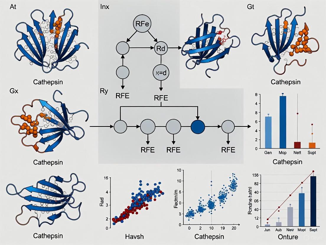
The following diagram illustrates the logical workflow for implementing an RFE-RF model in a QSAR study, such as predicting cathepsin inhibitory activity.
Experimental Protocol: A Case Study on Cathepsin L Inhibitors
This protocol details the methodology adapted from a recent study that developed QSAR models to predict the inhibitory activity (IC₅₀) of compounds against Cathepsin L (CatL), a potential therapeutic target for preventing SARS-CoV-2 cell entry [19].
Data Curation and Preparation
- Compound Collection: A dataset of compounds with experimentally determined IC₅₀ values against CatL was compiled. The half-maximal inhibitory concentration (IC₅₀) quantifies the potency of inhibition, with lower values indicating higher potency [19].
- Descriptor Calculation: A total of 604 molecular descriptors were calculated for each compound using software such as CODESSA. These descriptors encode various 1D, 2D, and 3D molecular properties [19].
- Data Splitting: The dataset was divided into a training set (typically 70-80%) for model development and a test set (20-30%) for external validation of the model's predictive power [19].
Model Training and Feature Selection with RF-RFE
- Random Forest Initialization: A Random Forest regression model was initialized. Key hyperparameters to optimize via cross-validation include the number of trees in the forest (
n_estimators) and the maximum depth of each tree (max_depth) [20]. - Recursive Feature Elimination: The RFE process was employed. The feature importance scores from the RF model were used to recursively prune descriptors. The optimal number of features was determined as the point where model performance (e.g., R² or RMSE) on a validation set is maximized [20].
- Final Model Training: The final RF model was trained using the optimal subset of molecular descriptors identified by RFE.
Model Validation and Analysis
- Performance Metrics: The model's performance was evaluated using the coefficient of determination (R²) and Root Mean Square Error (RMSE) for both training and test sets [19].
- Validation Techniques: Five-fold cross-validation (R²₍₅‑ꜰₒₗᵈ₎ = 0.9043) and leave-one-out cross-validation (R²₍ₗₒₒ₎ = 0.9525) were performed to ensure the model's robustness and generalizability [19].
- Descriptor Interpretation: The selected molecular descriptors were analyzed for their physicochemical meaning to glean insights into the structural features critical for CatL inhibition [19].
Table 1: Key Molecular Descriptors Identified by RF/HM for Cathepsin L Inhibition [19]
| Descriptor Symbol | Physicochemical Meaning | Implication for Inhibitor Design |
|---|---|---|
| RNR | Relative number of rings | Relates to molecular complexity and rigidity. |
| HDH2(QCP) | HA-dependent HDCA-2 (quantum-chemical PC) | Indicates influence of hydrogen bonding and quantum-chemical properties. |
| YS/YR | YZ shadow/YZ rectangle | A topological descriptor related to molecular shape and surface area. |
| MPPBO | Max PI-PI bond order | Suggests importance of pi-pi stacking interactions with the target. |
| MEERCOB | Max e-e repulsion for a C-O bond | Reflects electronic and bond properties within the molecule. |
Performance Benchmarking and Recent Applications
The RF-RFE approach has demonstrated strong performance in various bioactivity prediction tasks. In a study on Cathepsin B, S, D, and K inhibitor classification, a deep learning model that utilized feature selection from molecular descriptors achieved high accuracy, underscoring the value of robust descriptor selection [9]. Furthermore, a random forest model developed to predict depression risk from environmental chemical mixtures showcased the algorithm's power in handling complex, high-dimensional datasets, achieving an exceptional Area Under the Curve (AUC) of 0.967 [20].
Table 2: Performance Comparison of Machine Learning Models in Recent QSAR Studies
| Study / Target | Best Model | Key Performance Metric | Role of Feature Selection |
|---|---|---|---|
| Cathepsin L Inhibitors (SARS-CoV-2) [19] | LMIX3-SVR | R² (test) = 0.9632, RMSE = 0.0322 | Heuristic Method (HM) selected 5 critical descriptors from 604. |
| DENV NS3 & NS5 Proteins [21] | SVM / ANN | Pearson CC (test): 0.857 / 0.862 (NS3); 0.982 / 0.964 (NS5) | Molecular descriptors and fingerprints were used to train multiple ML models. |
| Depression Risk (Environmental Chemicals) [20] | Random Forest | AUC: 0.967, F1 Score: 0.91 | RFE was used to identify the most influential chemical exposures from 52 candidates. |
Table 3: Key Software and Resources for RFE-RF QSAR Modeling
| Resource / Reagent | Type | Function in RFE-RF QSAR Pipeline | Examples / Notes |
|---|---|---|---|
| Cheminformatics Software | Software Suite | Calculates molecular descriptors from compound structures. | CODESSA [19], PaDEL [22], RDKit [23] [9], DRAGON [23] |
| Programming Environment | Computational Framework | Provides libraries for implementing ML algorithms and data analysis. | R (caret package) [22], Python (scikit-learn) [23] |
| Chemical Databases | Data Repository | Sources of chemical structures and associated bioactivity data for training. | ChEMBL [21] [9], BindingDB [9], PubChem [22] |
| Cloud/Workflow Platforms | Platform | Offers reproducible, web-enabled environments for analysis. | Galaxy (GCAC tool) [22], KNIME [23] |
Key Molecular Descriptors and Features for Cathepsin Inhibition
Cathepsins, a family of lysosomal proteases, have emerged as critical therapeutic targets for conditions ranging from viral infections to cancer and metabolic disorders. Cathepsin L (CatL) facilitates SARS-CoV-2 viral entry into host cells by cleaving the spike protein, making its inhibition a promising antiviral strategy [19] [24]. Simultaneously, Cathepsin S (CatS) plays established roles in cancer progression, chronic pain, and various inflammatory diseases [25] [26]. The development of effective cathepsin inhibitors requires a deep understanding of the key molecular features that govern inhibitor-enzyme interactions. This application note explores these critical molecular descriptors and integrates them with a feature selection methodology centered on Recursive Feature Elimination (RFE) with Random Forest to advance cathepsin inhibition research.
Key Molecular Descriptors for Cathepsin Inhibition
Quantitative Structure-Activity Relationship (QSAR) studies have identified several molecular descriptors critically associated with cathepsin inhibitory activity. The table below summarizes key descriptors identified for Cathepsin L inhibition through advanced QSAR modeling.
Table 1: Key Molecular Descriptors for Cathepsin L Inhibitory Activity
| Descriptor Symbol | Physicochemical Interpretation | Relationship with Activity |
|---|---|---|
| RNR | Relative number of rings | Negative correlation (-37.67 coefficient) [19] |
| HDH2(QCP) | HA-dependent HDCA-2 (quantum-chemical PC) | Positive correlation (0.204 coefficient) [19] |
| YS/YR | YZ shadow/YZ rectangle | Negative correlation (-4.902 coefficient) [19] |
| MPPBO | Max PI-PI bond order | Positive correlation (25.354 coefficient) [19] |
| MEERCOB | Max e-e repulsion for a C-O bond | Positive correlation (0.242 coefficient) [19] |
For cathepsin S, achieving inhibitor specificity presents a distinct challenge due to significant structural similarities with CatL and Cathepsin K (CatK). The S2 and S3 substrate binding pockets contain the critical amino acid variations that enable selective inhibition [26]. Key residues include Gly62, Asn63, Lys64, Gly68, Gly69, and Phe70 in the S3 pocket and Phe70, Gly137, Val162, Gly165, and Phe211 in the S2 pocket [26]. Successful design of selective CatS inhibitors must prioritize interactions with these specificity-determining residues.
Implementing RFE with Random Forest for Descriptor Selection
Rationale for RFE in QSAR Modeling
The high-dimensional nature of QSAR modeling, where datasets often contain hundreds of calculated molecular descriptors, introduces complexity and risks of overfitting. Feature selection is a critical preprocessing step that improves model accuracy and interpretability by identifying the most relevant descriptors [27]. Recursive Feature Elimination (RFE) is a powerful wrapper method that recursively constructs models, ranks features by their importance, and eliminates the least important ones until an optimal subset is identified.
Integrated RFE-Random Forest Workflow
The following workflow diagram illustrates the integrated process of applying RFE with a Random Forest classifier to identify optimal molecular descriptors for predicting cathepsin inhibition.
Diagram 1: RFE-Random Forest Feature Selection Workflow
This workflow systematically refines the feature set to enhance model performance. Studies have confirmed that wrapper methods like RFE, combined with nonlinear models, demonstrate promising performance in QSAR modeling for anti-cathepsin activity prediction [27].
Comparative Performance of Feature Selection Methods
Research comparing preprocessing methods for molecular descriptors in predicting anti-cathepsin activity has demonstrated that RFE is highly effective, along with other wrapper methods such as Forward Selection (FS), Backward Elimination (BE), and Stepwise Selection (SS) [27]. These methods, particularly when coupled with nonlinear regression models, exhibit strong performance metrics as measured by R-squared scores [27].
Experimental Validation Protocols
In Vitro Cathepsin Activity Assay
The experimental validation of computational predictions is essential for confirming inhibitor efficacy. The following protocol outlines a standardized method for testing cathepsin inhibition.
Table 2: Key Reagents for Cathepsin Activity Assay
| Reagent / Equipment | Function / Specification | Example Source / Note |
|---|---|---|
| Recombinant Human Cathepsin | Enzyme source for inhibition assay | e.g., CatL, CatB, or CatS [28] [29] |
| Fluorogenic Substrate | Protease activity measurement | AMC-labeled peptide substrate [30] |
| Assay Buffer | Maintain optimal enzymatic pH | e.g., MES buffer (pH 5.0) for CatB [29] |
| Activating Agent | For cysteine protease activation | e.g., DTT (5 mM) [29] |
| Multi-mode Microplate Reader | Fluorescence detection | e.g., GloMax-Multi+ [28] |
Procedure:
- Enzyme Activation: Pre-activate cathepsin in assay buffer containing 5 mM DTT for 30 minutes at room temperature [29].
- Inhibitor Incubation: Incate the activated enzyme with candidate inhibitors at varying concentrations (e.g., 0.1-100 µM) for 15-30 minutes.
- Reaction Initiation: Add the fluorogenic substrate to initiate the reaction. For example, a cell-free CTSL inhibition assay can be performed using a commercial kit (Abcam, Cat. No. ab65306) [28].
- Kinetic Measurement: Monitor the increase in fluorescence (Ex/Em ~355/460 nm for AMC) continuously for 30-60 minutes.
- Data Analysis: Calculate percentage inhibition relative to a DMSO control and determine IC₅₀ values using nonlinear regression of dose-response data.
Experimental Workflow Integration
The following diagram illustrates how computational predictions and experimental validation integrate in the drug discovery pipeline for cathepsin inhibitors.
Diagram 2: Integrated Cathepsin Inhibitor Discovery Pipeline
This integrated approach has successfully identified novel CatL inhibitors from natural products, with deep learning models and molecular docking screening 150 molecules leading to experimental validation of 36 compounds showing >50% inhibition at 100 µM concentration [28].
Research Reagent Solutions
Table 3: Essential Research Toolkit for Cathepsin Inhibition Studies
| Category / Item | Specific Application | Research Context |
|---|---|---|
| Software & Computational Tools | ||
| CODESSA | Calculation of 604+ molecular descriptors | Heuristic QSAR model development [19] [24] |
| Random Forest with RFE | Feature selection for model optimization | Descriptor selection for anti-cathepsin QSAR [27] |
| Schrödinger Suite | Molecular docking and dynamics | Protein-ligand interaction studies [28] [25] |
| Experimental Assays | ||
| Commercial Cathepsin Assay Kits | In vitro inhibitor screening | Abcam Cat. No. ab65306 for CTSL [28] |
| Fluorogenic Peptide Substrates | Enzyme kinetic measurements | AMC-labeled substrates for cathepsin B [30] [29] |
| Chemical Tools | ||
| Peptidomimetic Analogues (PDAs) | CatL inhibitor scaffold | Effective CatL inhibition demonstrated [19] [24] |
| Natural Product Libraries | Source of novel inhibitor scaffolds | Identification of Plumbagin and Beta-Lapachone as CTSL inhibitors [28] |
The integration of computational feature selection methods like RFE with Random Forest and experimental validation provides a powerful framework for advancing cathepsin inhibition research. The key molecular descriptors identified for Cathepsin L (RNR, HDH2(QCP), YS/YR, MPPBO, MEERCOB) and the critical S2/S3 pocket residues for Cathepsin S specificity offer valuable guidance for rational inhibitor design. The standardized protocols and research tools outlined in this application note provide a foundation for systematic investigation of cathepsin inhibitors, potentially accelerating the development of therapeutics for COVID-19, cancer, chronic pain, and other cathepsin-mediated diseases.
Review of Previous ML Applications to Cathepsins and Related Proteases
The application of machine learning (ML) in protease research has become a cornerstone of modern computational drug discovery, enabling the rapid prediction of compound activity and the efficient identification of novel therapeutic candidates. Cathepsins, a family of lysosomal proteases, have emerged as significant therapeutic targets due to their involvement in various pathological conditions including cancer, metabolic disorders, and viral infections such as COVID-19 [28] [8] [31]. The complexity of biological data associated with cathepsin inhibition necessitates sophisticated computational approaches that can handle high-dimensional descriptor spaces and uncover intricate structure-activity relationships. This review synthesizes recent advancements in ML applications for cathepsin research, with particular emphasis on feature selection methodologies, model architectures, and experimental validation frameworks that support the implementation of Recursive Feature Elimination (RFE) with Random Forest for cathepsin activity prediction.
Machine Learning Approaches for Cathepsin Activity Prediction
Quantitative Structure-Activity Relationship (QSAR) Modeling
Quantitative Structure-Activity Relationship (QSAR) modeling represents a fundamental application of machine learning in cathepsin research, employing mathematical and statistical techniques to establish correlations between molecular descriptors and biological activity [27]. Molecular descriptors encompass integer, decimal, and binary numerical values derived from molecular structure, containing comprehensive information about a molecule's physical, chemical, structural, and geometric properties [27]. The substantial number of descriptors involved in QSAR modeling introduces complexities in data calculation and analysis, making data preprocessing through feature selection and dimensionality reduction crucial for directing statistical model inputs [27].
Recent comparative analyses have demonstrated that preprocessing methods significantly impact model performance in predicting anti-cathepsin activity. Filtering approaches such as Recursive Feature Elimination (RFE) and wrapping methods including Forward Selection (FS), Backward Elimination (BE), and Stepwise Selection (SS) have shown particular utility when coupled with both linear and nonlinear regression models [27]. Notably, FS, BE, and SS methods exhibit promising performance metrics, especially when integrated with nonlinear regression models, as evidenced by R-squared scores in anti-cathepsin activity prediction [27].
Table 1: Performance Metrics of QSAR Models for Cathepsin L Inhibition Prediction
| Model Type | R² Training | R² Test | RMSE Training | RMSE Test | Cross-Validation (R²) | Key Features |
|---|---|---|---|---|---|---|
| LMIX3-SVR | 0.9676 | 0.9632 | 0.0834 | 0.0322 | 0.9043 (5-fold) | Linear-RBF-polynomial hybrid kernel |
| HM Model | 0.8000 | 0.8159 | 0.0658 | 0.0764 | N/A | Five selected descriptors |
| Random Forest | >0.90 (Accuracy) | N/A | N/A | N/A | 0.91 (AUC) | Morgan fingerprints |
Advanced Algorithm Implementations
Innovative ML architectures have demonstrated remarkable efficacy in cathepsin inhibition prediction. For Cathepsin L (CTSL) inhibitors as SARS-CoV-2 therapeutics, enhanced Support Vector Regression (SVR) with multiple kernel functions and Particle Swarm Optimization (PSO) has shown exceptional performance [8] [24]. The LMIX3-SVR model, incorporating a hybrid kernel combining linear, radial basis function (RBF), and polynomial elements, achieved outstanding predictive capability with R² values of 0.9676 and 0.9632 for training and test sets respectively, along with minimal RMSE values of 0.0834 and 0.0322 [8] [24]. The PSO algorithm ensured low complexity and fast convergence during parameter optimization [8].
Random Forest classification models have also demonstrated significant utility in cathepsin research. One study trained on IC₅₀ values from the CHEMBL database achieved over 90% accuracy in distinguishing active from inactive CTSL inhibitors, with AUC mean values of 0.91 derived from 10-fold cross-validation [32]. The model utilized Morgan fingerprints (1024 dimensions) as molecular descriptors and successfully identified 149 natural compounds with prediction scores exceeding 0.6 from Biopurify and Targetmol libraries [32].
Deep learning approaches have expanded the capabilities of cathepsin inhibitor discovery. Message Passing Neural Networks (MPNNs) have been employed for binary classification to predict the probability of molecular inhibition against CTSL [28]. This approach facilitated screening of 6439 natural products characterized by diverse structures and functions, ultimately identifying 36 molecules exhibiting more than 50% inhibition of CTSL at 100 µM concentration, with 13 molecules demonstrating over 90% inhibition [28].
Experimental Protocols and Methodologies
Molecular Descriptor Calculation and Preprocessing
The foundation of robust QSAR models relies on comprehensive descriptor calculation and rigorous preprocessing. The CODESSA software platform enables computation of numerous molecular descriptors, with studies typically generating 600+ descriptors for each compound [24]. Heuristic Method (HM) linear modeling facilitates descriptor selection by constructing progressive models with increasing descriptor numbers, identifying the optimal descriptor set when additional descriptors cease to significantly improve R² and Rcv² values [24]. For CTSL inhibition prediction, five descriptors were determined optimal: RNR (relative negative charge), HDH2QCP (hydrogen donor hybridization component), YSYR (surface area symmetry), MPPBO (molecular polarizability), and MEERCOB (molecular electrostatic potential) [24].
Nonlinear methods like XGBoost provide complementary descriptor validation through split gain importance calculation. This approach ranks descriptors by importance scores but may retain highly correlated descriptors (correlation coefficients >0.6) [24]. The integration of HM and XGBoost methodologies ensures selection of nonredundant, physiochemically relevant descriptor sets, justifying HM selection for obtaining optimal descriptor subsets [24].
Model Training and Validation Framework
A systematic model training and validation framework ensures predictive reliability and prevents overfitting. For CTSL inhibitor classification, datasets should be curated from reliable sources like CHEMBL, with compounds categorized as active (IC₅₀ < 1000 nM) or inactive (IC₅₀ > 1000 nM) [32]. After removing duplicates, appropriate class distributions (e.g., 2000 active and 1278 inactive molecules) provide balanced training data [32].
Morgan fingerprint calculation (1024 dimensions) using RDKit facilitates structural representation, followed by vectorization for model input [32]. Random Forest classification models should undergo 10-fold cross-validation with ROC curve analysis to evaluate performance, achieving AUC values approximating 0.91 [32]. For regression tasks predicting IC₅₀ values, dataset splitting into training and test sets (typically 80:20 ratio) with five-fold cross-validation (R²₅-fold = 0.9043) and leave-one-out cross-validation (R²loo = 0.9525) demonstrates model robustness [8] [24].
Trained models can screen natural compound libraries (Biopurify, Targetmol), selecting hits with prediction scores >0.6 for subsequent structure-based virtual screening [32]. This integrated approach identified 13 compounds with higher binding affinity than positive control AZ12878478, with the top two candidates (ZINC4097985 and ZINC4098355) demonstrating stable binding interactions in molecular dynamics simulations [32].
Integrated AI and Experimental Validation
The most effective ML applications combine computational predictions with experimental validation. A robust protocol employing deep learning, molecular docking, and experimental assays identified novel CTSL inhibitors from natural products [28]. A binary classification MPNN model predicted CTSL inhibition probability, followed by molecular docking screening of 150 molecules from natural product libraries [28].
Receptor protein preparation utilized human CTSL X-ray structures (PDB ID: 5MQY, resolution 1.13 Å) co-crystallized with a covalent inhibitor [28]. Protein preparation involved deleting water molecules and artifacts, adding hydrogen atoms, generating potential metal binding states, hydrogen bond sampling with active site adjustment at pH 7.4 using PROPKA, and geometry refinement with OPLS3 force field in restrained minimizations [28]. Ligand preparation employed Open Babel for format conversion and LigPrep for ionization state generation at pH 7.4, followed by minimization with OPLSe-3 force field [28].
Molecular docking used glide SP flexible ligand mode with receptor grids generated around the co-crystallized inhibitor centroid [28]. Pose outputs were visualized and analyzed using PyMOL and Discovery Studio Visualizer [28]. Experimental validation confirmed 36 of 150 molecules exhibited >50% CTSL inhibition at 100 µM concentration, with 13 molecules showing >90% inhibition and concentration-dependent effects [28]. Enzyme kinetics studies revealed uncompetitive inhibition patterns for the most potent inhibitors (Plumbagin and Beta-Lapachone) [28].
Research Reagent Solutions
Table 2: Essential Research Reagents and Computational Tools for Cathepsin ML Research
| Category | Specific Tool/Reagent | Application/Function | Key Features |
|---|---|---|---|
| Software & Platforms | CODESSA | Molecular descriptor calculation | Computes 600+ molecular descriptors for QSAR |
| RDKit | Cheminformatics and fingerprint generation | Morgan fingerprints, molecular similarity via Tanimoto coefficient | |
| Schrödinger Suite | Protein preparation, molecular docking | Protein Preparation Wizard, LigPrep, Glide SP docking | |
| Open Babel | Chemical format conversion | Converts SMILES to mol2 format for docking | |
| Experimental Assays | CTSL Activity Test Kit (Abcam, Cat. No. ab65306) | In vitro inhibition validation | Cell-free system for inhibition assessment |
| Expi293 Mammalian Expression System | Recombinant cathepsin production | Human glycosylation patterns, high yield secretion | |
| Data Resources | CHEMBL Database | Compound activity data | IC₅₀ values for active/inactive classification |
| RCSB Protein Data Bank | 3D protein structures | CTSL structure (PDB ID: 5MQY, 1.13 Å resolution) | |
| Natural Product Libraries (Biopurify, Targetmol) | Candidate compound sources | Structurally diverse natural compounds for screening |
Signaling Pathways and Biological Context
Cathepsins function within complex biological pathways that influence their therapeutic targeting. CTSL plays a critical role in SARS-CoV-2 viral entry through cleavage of the viral spike protein, facilitating host cell entry and making it a promising therapeutic target for COVID-19 [8] [33]. In cancer biology, CTSL expression increases in various malignancies including glioma, melanoma, pancreatic, breast, and prostate carcinoma, where it promotes invasion and metastasis through degradation of E-cadherin and extracellular matrix components [32].
Cathepsin S (CathS) contributes to cancer progression and chronic pain pathophysiology, creating immunosuppressive environments in solid tumors and participating in nociceptive signaling [25]. CathS causes immunosuppression in tumors through CXCL12 cleavage and inactivation, reducing effector T cell infiltration while generating fragments that attract regulatory T cells and myeloid-derived suppressor cells [25]. In chronic pain, peripheral nerve injury induces microglial activation and CathS release, cleaving protease-activated receptor 2 (PAR2) on nociceptive neurons to increase excitability and central sensitization [25].
Neutrophil cathepsins regulate immune functions including neutrophil extracellular trap (NET) formation, which contributes to pathogen clearance but can drive pathology when dysregulated in autoimmune diseases, cancer metastasis, and ischemia-reperfusion injury [31]. Cathepsin C activates neutrophil elastase, establishing cathepsins as upstream regulators of NET-associated proteases [31].
Implications for RFE with Random Forest Implementation
The reviewed ML applications provide critical insights for implementing RFE with Random Forest in cathepsin activity prediction. Successful QSAR modeling necessitates appropriate descriptor selection to manage the high dimensionality of molecular feature spaces [27]. RFE offers a robust approach for feature selection, iteratively constructing models and eliminating the least important features to identify optimal descriptor subsets that enhance model performance while reducing complexity [27].
The documented performance of Random Forest classifiers in cathepsin research, achieving >90% accuracy in distinguishing active from inactive CTSL inhibitors, supports its implementation with RFE for feature optimization [32]. The integration of Morgan fingerprints as molecular descriptors aligns with RFE-Random Forest workflows, providing comprehensive structural representations while enabling feature importance evaluation [32].
Cross-validation strategies employed in cathepsin ML studies, including 10-fold cross-validation and leave-one-out approaches, establish rigorous frameworks for evaluating RFE-Random Forest model performance [8] [24] [32]. The consistent reporting of R² values, RMSE, and AUC metrics across studies provides benchmark comparisons for assessing implementation success [8] [24] [32].
The integration of computational predictions with experimental validation creates a closed-loop framework for model refinement, where experimental results inform feature selection and model parameter adjustments in successive iterations [28] [32]. This approach ensures that RFE-Random Forest implementations maintain biological relevance while optimizing predictive performance for cathepsin activity prediction.
A Step-by-Step Pipeline for Building Your RFE-Random Forest Model
Cathepsins are proteases with critical roles in cellular processes, and their dysregulation is implicated in diseases ranging from cancer and metabolic disorders to SARS-CoV-2 infection [19] [32] [34]. Cathepsin L (CatL) is a particularly prominent therapeutic target; it facilitates viral entry into host cells by cleaving the spike protein of SARS-CoV-2 [19] [8]. Inhibition of CatL is therefore a promising strategy for antiviral drug development [19]. Research into cathepsins relies heavily on high-quality bioactivity data, often expressed as the half-maximal inhibitory concentration (IC₅₀), which quantifies the potency of an inhibitor [19] [32].
The process of data curation—sourcing, standardizing, and preparing this bioactivity data—is a foundational step in building reliable predictive models for drug discovery. Public bioactivity databases such as ChEMBL and PubChem BioAssay provide a wealth of data, but this data is not without its challenges [35] [36]. Issues such as transcription errors, inconsistencies in unit reporting, and insufficient assay descriptions can compromise data integrity [35]. Therefore, a rigorous and systematic curation protocol is indispensable for subsequent computational analysis, especially when implementing machine learning techniques like Recursive Feature Elimination (RFE) with Random Forest.
Sourcing Data from Public Repositories
The first step in the curation pipeline is to gather raw bioactivity data from large-scale public repositories. These databases aggregate experimental data from diverse sources, including scientific literature and high-throughput screening experiments.
- ChEMBL: A manually curated database of bioactive molecules with drug-like properties. It contains over 13 million bioactivity data points, including IC₅₀, Kᵢ, and K({}_{\text{D}}) values, which are standardized to consistent units where possible [35].
- PubChem BioAssay: A public repository containing results from high-throughput screening experiments, providing a vast source of bioactivity data [36].
- Biopurify & Targetmol: Commercial libraries specializing in natural products and bioactive compounds, often used for virtual screening campaigns [32].
Large-scale integrated datasets like Papyrus have been constructed to combine and standardize data from multiple sources, including ChEMBL and ExCAPE-DB, facilitating "out-of-the-box" use for machine learning [36]. For cathepsin-specific research, these databases can be queried using target identifiers (e.g., UniProt accession codes) and standard activity types (e.g., 'IC50').
Common Data Integrity Challenges
Data sourced directly from public repositories often contains ambiguities and errors that must be addressed during curation. Key challenges include:
- Unit Transcription and Conversion Errors: The same activity type may be reported in numerous different units (e.g., IC₅₀ values published in molar units, μg/mL, etc.), leading to potential conversion mistakes [35].
- Inconsistent Target Assignment: Ambiguous or incorrect protein target identification can misassociate compounds with specific cathepsins [35].
- Data Redundancy and Citation: The same experimental value may be cited across multiple publications, creating statistical artefacts if not properly identified [35].
- Unrealistic Activity Values: Outliers and transcription errors can result in implausibly high or low activity measurements [35].
Table 1: Common Data Issues and Curation Strategies
| Error Source | Example | Curation Strategy |
|---|---|---|
| Data Extraction | Missing stereochemistry, incorrect target assignment. | Automated and manual verification against original publication. |
| Author/Publication | Insufficient assay description, wrong activity units. | Standardize activity types and units to a controlled vocabulary. |
| Experimental | Compound purity, cell-line identity issues. | Flag data from assays with known reliability issues. |
| Database User | Merging activities from different assay types. | Apply robust filter strategies based on assay metadata. |
Data Curation and Standardization Protocol
A systematic protocol is essential for transforming raw data into a curated, machine-learning-ready dataset. The following workflow outlines the key steps for curating cathepsin bioactivity data.
Experimental Workflow for Data Curation
The following diagram illustrates the end-to-end workflow for sourcing and preparing cathepsin bioactivity data.
Step-by-Step Curation Methodology
Step 1: Initial Filtering by Activity Type and Target
- Action: Extract records for the specific cathepsin target of interest (e.g., Cathepsin L) using standardized target identifiers. Retain only relevant activity types, such as IC₅₀, Kᵢ, or K({}_{\text{D}}).
- Protocol: Query databases using REST APIs or direct SQL queries on curated datasets like Papyrus. Filter for
STANDARD_TYPEin ['IC50', 'Ki'] andTARGET_CHEMBL_IDcorresponding to the specific cathepsin [36]. - Rationale: Focuses the dataset on the relevant biological context and measurement type for the research question.
Step 2: Standardization of Activity Values and Units
- Action: Convert all activity values to a consistent unit (typically nanomolar, nM) and transform them to a uniform scale suitable for modeling (e.g., pIC₅₀ = -log₁₀(IC₅₀)).
- Protocol: Apply predefined unit conversion rules. For example, convert all IC₅₀ values to nM and then calculate pIC₅₀ [35]. This standardizes the response variable for QSAR modeling.
- Rationale: Enables direct comparison of activity values across different publications and experimental setups, which is critical for building a unified model.
Step 3: Removal of Duplicate Entries
- Action: Identify and consolidate multiple entries for the same compound-target pair.
- Protocol: When multiple measurements exist for a single compound-target pair, apply a consensus strategy. For instance, retain the median pIC₅₀ value if multiple high-quality measurements are available, or flag the data point if measurements are highly discordant (e.g., a difference >0.5 log units) [35] [36].
- Rationale: Prevents statistical bias from over-represented data points and improves model generalizability.
Step 4: Identification and Flagging of Outliers
- Action: Systematically identify potentially erroneous activity values.
- Protocol: Implement automated checks for unrealistic values (e.g., IC₅₀ < 1 pM or > 1 M). Additionally, flag data points associated with known assay artefacts or from publications with insufficient experimental detail [35].
- Rationale: Isolates potentially unreliable data, allowing the researcher to decide on its inclusion or exclusion based on the modeling objective.
Step 5: Standardization of Compound Structures
- Action: Process the chemical structures of compounds to a consistent representation.
- Protocol: Use a standardized chemical structure pipeline (e.g., the ChEMBL pipeline or RDKit) to handle tautomerism, aromaticity, functional group standardization, and removal of salts [36]. Generate canonical SMILES or InChI identifiers for each unique compound.
- Rationale: Ensures that the same chemical entity is not represented by multiple structural identifiers, which is crucial for accurate descriptor calculation.
Step 6: Data Quality Annotation
- Action: Assign a quality score to each data point based on the reliability and reproducibility of the source assay.
- Protocol: In datasets like Papyrus++, data points are labelled as "high quality" if they are associated with reproducible assays (e.g., where measurements for a compound-target pair across different assays are concordant) [36].
- Rationale: Allows for the creation of tiered datasets, enabling model training on the most reliable data and benchmarking performance against noisier data.
Molecular Descriptor Preprocessing for RFE
Once a curated bioactivity dataset is obtained, the next critical step is to calculate and preprocess molecular descriptors for the compounds. These descriptors, which numerically represent molecular structures and properties, form the feature set for predictive modeling.
Calculation and Selection of Molecular Descriptors
Molecular descriptors can be calculated using software such as CODESSA or the RDKit library in Python, generating hundreds to thousands of descriptors characterizing topological, electronic, and geometric properties [19] [36]. For example, a QSAR study on CatL inhibitors initially computed 604 molecular descriptors [19].
Table 2: Key Molecular Descriptors for Cathepsin Inhibitor QSAR
| Descriptor Symbol | Physicochemical Interpretation | Role in Cathepsin Inhibition |
|---|---|---|
| RNR | Relative number of rings [19]. | Related to molecular rigidity and scaffold structure; negative coefficient in HM model suggests fewer rings may correlate with higher activity [19]. |
| HDH2(QCP) | HA-dependent HDCA-2 (quantum-chemical PC) [19]. | Encodes electronic properties; positive coefficient indicates a potential role in target interaction [19]. |
| MPPBO | Max PI-PI bond order [19]. | Reflects electron delocalization and aromaticity; positive coefficient suggests potential for π-π stacking in binding pocket [19]. |
| MEERCOB | Max e-e repulsion for a C-O bond [19]. | Indicates steric and electronic environment around specific bonds [19]. |
| ABOCA | Avg bond order of a C atom [19]. | Describes overall bonding pattern; identified as highly important by XGBoost [19]. |
Preprocessing Methods for Feature Selection
The high dimensionality of molecular descriptor data necessitates robust feature selection to avoid overfitting and improve model interpretability. Several preprocessing methods can be employed to reduce the number of descriptors before applying RFE.
- Filter Methods (Recursive Feature Elimination - RFE): RFE is a wrapping method that recursively removes the least important features based on a model's coefficients or feature importance. It has been successfully applied in conjunction with Support Vector Machine (SVM-RFE) to identify core targets in toxicological studies [37].
- Wrapper Methods: These include:
- Forward Selection (FS): Iteratively adds features that most improve model performance.
- Backward Elimination (BE): Iteratively removes the least significant features.
- Stepwise Selection (SS): A combination of FS and BE.
- Studies have shown that FS, BE, and SS, particularly when coupled with nonlinear regression models, exhibit promising performance in QSAR modeling for anti-cathepsin activity [27].
- Nonlinear Filtering (XGBoost): Tree-based models like XGBoost can be used as a nonlinear method to rank descriptor importance by calculating the split gain for each feature. This validates the relevance of descriptors selected by other methods and helps capture complex, nonlinear relationships in the data [19].
Implementing RFE with Random Forest
Within the context of a curated cathepsin dataset, RFE with Random Forest is a powerful technique for identifying the most parsimonious and predictive set of molecular descriptors.
Workflow for RFE and Random Forest Modeling
The following diagram outlines the integrated process of descriptor preprocessing, RFE, and model building for cathepsin activity prediction.
Detailed Experimental Protocol
Objective: To identify a minimal, optimal set of molecular descriptors for predicting cathepsin inhibitory activity using Recursive Feature Elimination with a Random Forest model.
Materials and Reagents
- Software: Python (with scikit-learn, RDKit, Pandas) or R.
- Input Data: A curated dataset of chemical structures and corresponding bioactivity values (e.g., pIC₅₀ against Cathepsin L).
- Computational Resources: A standard desktop computer is sufficient for most datasets; larger datasets may require higher memory capacity.
Procedure:
- Feature Calculation and Initialization:
- Calculate an initial, comprehensive set of molecular descriptors (e.g., 500-1000+) for all compounds in the curated dataset using a toolkit like RDKit.
- Split the data into training and test sets (e.g., 80/20 split).
- Initialize a Random Forest regressor (or classifier, for active/inactive classification) and set the criteria for model evaluation (e.g., R² or Root Mean Squared Error (RMSE) on the test set).
Recursive Feature Elimination Loop:
- Step 1 - Train Model: Train the Random Forest model on the current set of features.
- Step 2 - Rank Features: Rank all features based on their importance scores (e.g., Gini importance or permutation importance) generated by the Random Forest model.
- Step 3 - Eliminate Feature: Remove the least important feature (or a predefined subset of features) from the current set.
- Step 4 - Evaluate Performance: Retrain the model on the reduced feature set and evaluate its performance on the test set.
- Step 5 - Iterate: Repeat steps 1-4 until a predefined number of features remains.
Model Selection and Validation:
- Plot the model performance (e.g., R²) against the number of features used. The optimal feature set is typically located at the point where performance is maximized or before it begins to degrade significantly.
- Validate the final model using the selected features with an external test set or through robust cross-validation (e.g., five-fold cross-validation), as demonstrated in successful CatL QSAR models [19] [8].
The Scientist's Toolkit
Table 3: Essential Research Reagent Solutions for Data Curation and Modeling
| Tool / Resource | Type | Primary Function |
|---|---|---|
| ChEMBL Database | Public Bioactivity Database | Manually curated source of bioactive molecules and assay data; primary source for ligand-target interactions [35]. |
| Papyrus Dataset | Integrated Curated Dataset | Large-scale, standardized dataset combining ChEMBL and other sources; designed for machine learning applications [36]. |
| RDKit | Cheminformatics Library | Open-source toolkit for calculating molecular descriptors, fingerprinting, and structure standardization [36]. |
| CODESSA | Commercial Software | Comprehensive software for calculating a wide range of molecular descriptors and building QSAR models [19]. |
| scikit-learn | Machine Learning Library | Python library providing implementations of Random Forest, RFE, and other model evaluation tools. |
| SwissTargetPrediction | Online Tool | Predicts potential protein targets of small molecules based on structural similarity [37]. |
A rigorous data curation pipeline is the cornerstone of robust cathepsin bioactivity prediction. By meticulously sourcing data from public repositories, standardizing values, and addressing common integrity issues, researchers can construct highly reliable datasets. Preprocessing molecular descriptors through methods like Forward Selection or XGBoost, followed by the application of RFE with Random Forest, efficiently identifies a minimal yet highly predictive feature subset. This integrated protocol, from raw data to a validated model, provides a reliable framework for accelerating the discovery of novel cathepsin inhibitors in drug development.
Molecular Descriptor Calculation and Preprocessing with Tools like Mold2
Quantitative Structure-Activity Relationship (QSAR) modeling plays a crucial role in studying the quantitative relationship between the biological activity and chemical structure of compounds [27]. Molecular descriptors are numerical representations derived from molecular structure, encompassing integer, decimal, and binary values that encode information about a molecule's physical, chemical, structural, and geometric properties [27]. The utilization of these descriptors in modeling becomes complex due to the large number of descriptors typically involved, making data calculation and analysis challenging [27]. Therefore, data preprocessing in QSAR modeling, including data reduction and feature selection, is essential for controlling the inputs for statistical models and improving the accuracy and efficiency of machine learning algorithms [27].
In the specific context of cathepsin activity prediction research, particularly targeting Cathepsin L (CTSL), molecular descriptor calculation and preprocessing take on added significance. CTSL expression is dysregulated in various cancers and participates directly in cancer growth, angiogenic processes, metastatic dissemination, and treatment resistance development [38]. The development of novel CTSL inhibition strategies is thus an urgent necessity in cancer management [38]. Implementing robust descriptor calculation and preprocessing pipelines enables researchers to identify potential natural CTSL inhibitors through machine learning approaches, as demonstrated by studies achieving over 90% accuracy in trained random forest models [38].
Mold2 Software for Molecular Descriptor Calculation
Mold2 is freely available software developed by the FDA's National Center for Toxicological Research (NCTR) for rapidly calculating molecular descriptors from two-dimensional chemical structures [39]. This software is capable of generating a large and diverse set of molecular descriptors that sufficiently encode two-dimensional chemical structure information, making it suitable for both small and large datasets [39]. Comparative analyses have demonstrated that Mold2 descriptors convey sufficient structural information and in some cases generate better models than those produced using commercial software packages [39].
Installation and Implementation
Table 1: Mold2 Software Installation Protocol
| Step | Procedure | Description | Notes |
|---|---|---|---|
| 1 | Download | Obtain the Mold2 Executable File (ZIP, 1.7MB) from the FDA website | Save to local machine; will not run directly from download page |
| 2 | File Extraction | Unzip the executable file and save contents to local machine | Use "Save As" option in most browsers |
| 3 | Documentation Review | Open and read the included "Read Me" file | Critical for proper installation and operation |
| 4 | Tutorial Consultation | Follow the attached Mold2 Tutorial (PDF, 237KB) | Provides detailed operational guidance |
| 5 | Technical Support | Contact Dr. Huixiao Hong at 870-543-7296 or Huixiao.Hong@fda.hhs.gov | Address questions or suggestions |
The software calculates 777 molecular descriptors from 2D structures, with comprehensive documentation available describing each descriptor [40]. Researchers must complete a brief access procedure to be added to the list of Mold2 users before downloading the software [40].
Molecular Preprocessing Fundamentals
Standardization and Neutralization
Molecular preprocessing begins with standardization, which transforms molecules according to a set of SMARTS templates. This required step ensures consistent molecule datasets by converting nitro mesomers, as different representations of these mesomers may be incorrectly treated as different molecules in QSAR despite being chemically identical [41]. Neutralization refers to the neutralization of charged atoms in molecules by attaching additional hydrogen atoms, though mesomers like nitro groups or quaternary nitrogens without hydrogens remain intact [41].
Salt Removal and Structure Cleaning
The "Remove salts" procedure detaches salts, counter-ions, solvents, and other molecular fragments from the core molecular structure. From all detached fragments, the largest by mass is kept. This is particularly important as many molecular descriptor calculation tools cannot correctly process molecules containing salts or counter-ions. However, this procedure results in loss of information on complete molecular structure and may lead to false duplicates in analyzed datasets [41]. "Clean structure" converts the original molecule file to SMILES format and back, resulting in complete loss of all information except atom connectivity. This is useful for removing 3D or atom coordinate calculation information, which in many cases has been shown to cause model overfitting [41].
Implementing RFE with Random Forest for Cathepsin Activity Prediction
Algorithm Fundamentals
Random Forest (RF) is a machine-learning algorithm that ranks the importance of each predictor in a model by constructing multiple decision trees [42]. Each node of a tree considers a different subset of randomly selected predictors, with the best predictor selected and split on based on decreased node impurity, measured with the estimated response variance [42]. Each tree is built using a different random bootstrap sample containing approximately two-thirds of total observations, which serves as a training set to predict data in the remaining out-of-bag (OOB) sample [42].
Recursive Feature Elimination (RFE) is a feature selection algorithm that searches through the training dataset for an optimal feature subset by beginning with all features and recursively removing the least important ones until the desired number remains [43]. The algorithm ranks features by importance, removes the least important ones, and re-fits the model, repeating this process recursively until optimal feature number is achieved [43]. The Random Forest-Recursive Feature Elimination algorithm (RF-RFE) integrates this feature selection approach directly with Random Forest modeling [42].
Implementation Protocol
Table 2: RFE-Random Forest Implementation Steps
| Step | Procedure | Technical Specifications | Purpose |
|---|---|---|---|
| 1 | Data Preparation | Preprocess molecules, calculate Mold2 descriptors (777 descriptors) | Generate standardized input features |
| 2 | Initial RF Model | Use RandomForestClassifier; set trees (e.g., 8000), mtry parameters | Establish baseline feature importance |
| 3 | RFE Configuration | Specify nfeaturesto_select or use RFECV for automatic selection | Configure feature elimination parameters |
| 4 | Pipeline Construction | Combine RFE and model in sklearn Pipeline | Prevent data leakage during cross-validation |
| 5 | Cross-Validation | Apply RepeatedStratifiedKFold (nsplits=10, nrepeats=5) | Validate model performance robustly |
| 6 | Feature Elimination | Iteratively remove lowest-ranking features (e.g., bottom 3%) | Reduce feature set to most relevant descriptors |
| 7 | Model Evaluation | Assess using accuracy, MSE_OOB, percentage variance explained | Quantify model performance and prediction quality |
The RFE-Random Forest implementation requires specific parameter tuning for optimal performance. For high-dimensional data where most features are noise, an mtry value of 0.1*p (where p is the number of predictors) is recommended rather than the default √p [42]. After features are recursively removed and p ≤ 80, the default mtry can be used. These parameters have been shown to produce reasonably low mean square error of out-of-bag (MSEOOB) estimates [42].
Workflow Visualization
Application in Cathepsin Inhibition Research
Case Study: CTSL Inhibitor Identification
In a practical application targeting Cathepsin L inhibition, researchers employed a combined machine learning and structure-based virtual screening strategy [38]. The random forest model was trained on IC₅₀ values from the CHEMBL database, where compounds with IC₅₀ values less than 1000 nM were considered active, and those with IC₅₀ values greater than 1000 nM were considered inactive [38]. After removing duplicate compounds, the dataset contained 2000 active molecules and 1278 inactive molecules [38]. Morgan fingerprints (1024) were calculated for active/inactive molecules, and following vectorization, the random forest classification model was trained to differentiate between them [38].
The trained model achieved AUC mean values of 0.91 based on 10-fold cross-validation, demonstrating high predictive accuracy [38]. This model was then used to screen natural compound libraries, yielding 149 hits with prediction scores >0.6 [38]. Subsequent structure-based virtual screening identified 13 compounds with higher binding affinity compared to the positive control (AZ12878478), with the top two candidates (ZINC4097985 and ZINC4098355) showing particularly strong binding to CTSL proteins [38].
Performance Considerations
The computational demands of RF-RFE can be significant, with one reported initial RF analysis taking approximately 6 hours and the complete RF-RFE process requiring approximately 148 hours to run on a Linux server with 16 cores and 320GB of RAM [42]. However, this investment is justified by the method's ability to handle high-dimensional problems and identify strong predictors without making assumptions about an underlying model [42].
A key challenge in high-dimensional data is the presence of correlated predictors, which impact RF's ability to identify the strongest predictors by decreasing the estimated importance scores of correlated variables [42]. While RF-RFE mitigates this problem in smaller datasets, it may not scale perfectly to high-dimensional data, as it can decrease the importance of both causal and correlated variables, making both harder to detect [42].
Research Reagent Solutions
Table 3: Essential Research Tools and Resources
| Resource Name | Type | Function | Application in CTSL Research |
|---|---|---|---|
| Mold2 | Software | Calculates 777 molecular descriptors from 2D structures | Generate molecular features for QSAR modeling |
| CHEMBL Database | Database | Provides compound activity data against biological targets | Source of IC₅₀ values for CTSL active/inactive compounds |
| Biopurify Library | Compound Library | Natural compounds for screening | Identify potential CTSL inhibitors |
| Targetmol Library | Compound Library | Natural compounds for screening | Identify potential CTSL inhibitors |
| Scikit-learn | Python Library | Implements RF, RFE, and machine learning pipelines | Build and validate predictive models |
| Random Forest Classifier | Algorithm | Machine learning for classification and feature importance | Differentiate active/inactive CTSL compounds |
| Morgan Fingerprints | Molecular Representation | Calculates molecular fingerprints for machine learning | Feature representation for initial modeling |
The integration of Mold2 for molecular descriptor calculation with RFE-Random Forest for feature selection and modeling represents a powerful approach for cathepsin activity prediction research. This workflow enables researchers to efficiently process chemical structures, identify the most relevant molecular descriptors, and build predictive models with demonstrated utility in identifying potential CTSL inhibitors. The structured protocols outlined in this application note provide researchers with a comprehensive framework for implementing this approach, with particular relevance to drug discovery efforts targeting cathepsin activity in cancer and other diseases.
Recursive Feature Elimination with Random Forest (RFE-RF) represents a powerful wrapper-style feature selection method that synergistically combines the robust predictive performance of Random Forest classifiers with an iterative feature ranking mechanism. In pharmaceutical research, particularly in quantitative structure-activity relationship (QSAR) modeling for drug discovery, RFE-RF addresses the critical challenge of high-dimensional descriptor spaces by systematically identifying molecular features most relevant to biological activity [27] [44]. This methodology has demonstrated significant utility in targeted drug development, including the prediction of cathepsin inhibitory activity—a promising therapeutic approach for obstructing SARS-CoV-2 viral entry into host cells [19].
The RFE-RF algorithm operates through an iterative process of model building, feature importance evaluation, and elimination of the least relevant descriptors, ultimately yielding an optimized subset of features that maximize predictive accuracy while minimizing computational complexity and overfitting risks [44] [45]. This protocol details the implementation of RFE-RF specifically for cathepsin activity prediction, providing researchers with a comprehensive framework covering theoretical foundations, practical coding examples, and experimental validation methodologies.
Random Forest Fundamentals
Random Forest (RF) constitutes an ensemble learning method that operates by constructing multiple decision trees during training and outputting the mode of classes (classification) or mean prediction (regression) of the individual trees [46]. The algorithm's robustness stems from its inherent capacity to perform automatic feature selection during tree construction, where each node split selectively utilizes the most discriminative features from randomly sampled subsets of the predictor space [47]. For drug discovery applications, RF demonstrates exceptional capability in modeling complex, nonlinear relationships between molecular descriptors and biological activity, while maintaining resistance to overfitting through its ensemble averaging mechanism [46] [48].
Key advantages of RF in cheminformatics include:
- High predictive accuracy through bootstrap aggregation and feature randomization
- Native handling of mixed data types (continuous, categorical, binary descriptors)
- Resistance to outliers and noise through majority voting across multiple trees
- Provision of intrinsic feature importance metrics based on Gini impurity or mean decrease in accuracy
Recursive Feature Elimination Mechanism
Recursive Feature Elimination (RFE) operates as a backward selection algorithm that recursively eliminates the least important features based on model-derived importance scores [44] [45]. The wrapper approach systematically evaluates feature subsets through the following iterative process:
- Initialization: Train a Random Forest model using the complete feature set
- Importance Ranking: Calculate and rank all features according to RF-derived importance scores
- Feature Pruning: Eliminate the bottom-ranking features (lowest importance)
- Model Refitting: Retrain RF with the reduced feature subset
- Termination Check: Repeat steps 2-4 until reaching a predefined number of features or performance optimization criterion [44] [45]
The RFE procedure exemplifies a greedy search strategy that does not exhaustively explore all possible feature combinations but instead selects locally optimal features at each iteration, progressively converging toward a globally optimal feature subset with substantially enhanced computational efficiency compared to exhaustive evaluation methods [44].
Integrated RFE-RF Workflow
The synergy between RF and RFE creates a robust feature selection pipeline particularly suited to cheminformatics applications. RF's inherent feature importance calculations provide the ranking mechanism for RFE, while RFE's iterative refinement enhances RF's performance by eliminating distracting noise variables [49] [47]. This integrated approach has demonstrated particular efficacy in bioinformatics and drug discovery contexts, including schizophrenia classification based on neuroimaging biomarkers [49] and prediction of anti-cathepsin compound activity [27].
Table 1: RFE-RF Performance Advantages in High-Dimensional Settings
| Scenario | Default RF Performance | RFE-RF Enhanced Performance | Application Context |
|---|---|---|---|
| Original Friedman dataset (5 relevant + 5 noise features) | 84% R² | Not applicable (minimal noise) | Simulation study [47] |
| Friedman + 100 noise features | 56% R² | 88% R² (with proper feature selection) | Simulation study [47] |
| Friedman + 500 noise features | 34% R² | 88% R² (with proper feature selection) | Simulation study [47] |
| Cathepsin L inhibitor prediction | Moderate QSAR performance | Enhanced model robustness and interpretability | Drug discovery [19] [27] |
Computational Implementation
Software Environment Setup
Implementing RFE-RF requires establishing a Python environment with specific scientific computing libraries. The following dependencies provide the necessary framework for descriptor calculation, feature selection, and model validation:
Molecular Descriptor Calculation
Accurate descriptor computation forms the foundation of robust QSAR modeling. The following protocol outlines comprehensive molecular featurization for cathepsin inhibitors:
RFE-RF Implementation Code
The core implementation integrates Random Forest regression with recursive feature elimination:
Hyperparameter Optimization
Comprehensive tuning of RF and RFE parameters significantly enhances model performance:
Workflow Visualization
RFE-RF Algorithm Flowchart
Molecular Descriptor Processing Pipeline
Experimental Protocol for Cathepsin Inhibitor Prediction
Dataset Curation and Preparation
Implementing RFE-RF for cathepsin activity prediction requires meticulous dataset preparation following established QSAR guidelines:
Compound Collection: Assemble a structurally diverse set of cathepsin L inhibitors with experimentally determined IC50 values from published literature and databases such as ChEMBL and BindingDB.
Activity Data Standardization:
- Convert all IC50 values to molar units (nM)
- Transform to pIC50 (-log10(IC50)) for normal distribution
- Apply range-based filtering (typically 1 nM to 100 μM)
Chemical Structure Validation:
- Standardize tautomeric forms and protonation states
- Remove inorganic compounds and mixtures
- Verify structural integrity using RDKit's SanitizeMol
Dataset Division: Implement sphere exclusion algorithm for rational splitting into training (80%), validation (10%), and test (10%) sets to ensure structural diversity across partitions.
Feature Selection and Model Training
Execute the RFE-RF workflow with the following experimental parameters:
Table 2: RFE-RF Experimental Parameters for Cathepsin Inhibitor Prediction
| Parameter Category | Specific Parameters | Recommended Values | Optimization Range |
|---|---|---|---|
| Data Preprocessing | Variance Threshold | 0.001 | 0.0001-0.01 |
| Missing Value Handling | Drop features >20% missing | 10-30% threshold | |
| Feature Standardization | StandardScaler | RobustScaler, MinMaxScaler | |
| Random Forest | n_estimators | 100 | 50-500 |
| max_depth | 15 | 5-30 | |
| minsamplessplit | 5 | 2-20 | |
| minsamplesleaf | 3 | 1-10 | |
| max_features | 'sqrt' | 'sqrt', 'log2', 0.3-0.8 | |
| RFE Process | nfeaturesto_select | 20% of original | 10-50% |
| Step (elimination rate) | 10% per iteration | 5-20% | |
| Stopping Criterion | Feature count + performance | Cross-validation plateau |
Model Validation and Interpretation
Comprehensive validation ensures model reliability and predictive power:
Research Reagent Solutions
Table 3: Essential Research Tools for RFE-RF Implementation in Cathepsin Research
| Tool/Category | Specific Solution | Function in Workflow | Alternative Options |
|---|---|---|---|
| Programming Environment | Python 3.8+ | Core computational platform | R, Julia, MATLAB |
| Cheminformatics Library | RDKit | Molecular manipulation and descriptor calculation | OpenBabel, CDK, ChemAxon |
| Descriptor Calculation | Mordred | Comprehensive 2D/3D molecular descriptor calculation | PaDEL, Dragon, MOE |
| Machine Learning Framework | Scikit-learn 1.0+ | RFE-RF implementation and model evaluation | H2O.ai, TPOT, Weka |
| Hyperparameter Optimization | RandomizedSearchCV | Efficient parameter space exploration | GridSearchCV, Optuna, BayesianOptimization |
| Visualization Tools | Matplotlib/Seaborn | Results visualization and interpretation | Plotly, Bokeh, ggplot2 |
| Chemical Database | ChEMBL | Source of cathepsin inhibitor structures and activities | BindingDB, PubChem, GOSTAR |
| Structure Format | SMILES | Molecular representation and storage | InChI, SDF, MOL2 |
Results Interpretation and Biological Validation
Performance Metrics and Benchmarking
Evaluate RFE-RF model performance against established benchmarks and alternative methodologies:
Table 4: Expected Performance Metrics for Cathepsin Inhibitor Prediction
| Performance Metric | Acceptable Range | Good Performance | Excellent Performance |
|---|---|---|---|
| Training R² | 0.70-0.80 | 0.80-0.90 | >0.90 |
| Cross-Validation Q² | 0.60-0.70 | 0.70-0.80 | >0.80 |
| Test Set R² | 0.65-0.75 | 0.75-0.85 | >0.85 |
| RMSE (pIC50 units) | 0.70-0.60 | 0.60-0.45 | <0.45 |
| MAE (pIC50 units) | 0.55-0.45 | 0.45-0.35 | <0.35 |
| Concordance CCC | 0.75-0.85 | 0.85-0.92 | >0.92 |
Selected Feature Interpretation
The molecular descriptors selected by RFE-RF provide critical insights into structural determinants of cathepsin inhibition:
Steric and Shape Descriptors: Features related to molecular size, volume, and three-dimensional shape often emerge as critical determinants, reflecting the steric constraints of the cathepsin L active site [19].
Electronic Features: Descriptors quantifying charge distribution, polar surface area, and hydrogen bonding capacity typically demonstrate high importance, consistent with the crucial role of electrostatic interactions in protease inhibition.
Topological Indices: Molecular connectivity indices and fragment-based descriptors frequently capture key pharmacophoric elements essential for cathepsin L binding affinity.
Hybrid Descriptors: Combined steric-electronic descriptors often outperform single-property descriptors, highlighting the multidimensional nature of structure-activity relationships in cathepsin inhibition.
Prospective Validation and Application
Implement prospective prediction for novel cathepsin inhibitors:
This comprehensive protocol establishes RFE-RF as a robust feature selection and modeling framework for cathepsin inhibitor prediction, enabling researchers to efficiently identify critical molecular descriptors while developing highly predictive QSAR models for targeted drug discovery applications.
Evaluating Feature Importance and Selecting the Optimal Descriptor Set
Within drug discovery research, particularly in the development of Cathepsin L (CatL) inhibitors as potential SARS-CoV-2 therapeutics, the identification of molecular descriptors that accurately predict inhibitory activity is paramount. High-dimensional data, common in quantitative structure-activity relationship (QSAR) studies, often contains numerous correlated predictors that can obscure the identification of truly relevant features. This application note details a protocol for implementing Random Forest-Recursive Feature Elimination (RF-RFE) to navigate these challenges, providing a robust framework for evaluating feature importance and selecting an optimal descriptor set to enhance model predictability and interpretability. The integration of RF-RFE is presented within the context of a broader thesis focused on optimizing cathepsin activity prediction.
Theoretical Foundation: RFE and Random Forest
Recursive Feature Elimination (RFE)
Recursive Feature Elimination (RFE) is a wrapper-type feature selection algorithm designed to identify the most relevant features in a dataset by recursively considering smaller and smaller feature sets [50]. Its core mechanism involves fitting an underlying estimator to the initial set of features, obtaining importance scores for each feature, pruning the least important features, and then repeating this process on the pruned set until a predefined number of features remains [50] [45]. This iterative process effectively ranks features, with selected features assigned a rank of 1 [50].
Synergy with Random Forest
Random Forest (RF) is an ensemble machine-learning method that works well with high-dimensional problems and can capture complex, nonlinear relationships between predictors and a response variable [42]. However, the presence of correlated predictors is a known issue, as it can decrease the estimated importance scores of correlated variables, thereby impairing the algorithm's ability to identify all strong predictors [42]. RF-RFE was developed to mitigate this problem. By using Random Forest as the core estimator within the RFE process, the algorithm leverages RF's robust importance calculations while iteratively eliminating features that contribute the least to model performance, thus accounting for variable correlation in high-dimensional data [42].
Experimental Protocol: Implementing RF-RFE for Cathepsin L Inhibitor Research
This protocol is designed for researchers aiming to identify critical molecular descriptors for Cathepsin L inhibitory activity (pIC50 or IC50) using a Python-based workflow.
Reagent and Computational Solutions
Table 1: Essential Research Reagent Solutions and Computational Tools
| Item Name | Function/Description | Example/Note |
|---|---|---|
| scikit-learn Library | Provides the RFE and RandomForestRegressor/RandomForestClassifier classes for implementing the core algorithm. |
Version 0.24.1 or higher is recommended [45]. |
| Cathepsin L Bioassay Data | Provides experimental IC50 values used as the target variable (y) for model training and validation. | Data from published studies or high-throughput screening [8]. |
| Molecular Descriptors | Serve as the feature set (X) for the model; numerical representations of molecular structure. | Can include topological, electronic, and geometric descriptors. |
| Jupyter Notebook / Python IDE | Provides an interactive computational environment for executing code and analyzing results. | — |
| Pandas & NumPy | Python libraries for data manipulation, handling, and numerical computations. | Essential for data preprocessing. |
Step-by-Step Workflow
Step 1: Data Preparation and Preprocessing
- Acquire a dataset of compounds with known CatL inhibitory activity (e.g., IC50 values) [8].
- Calculate or gather a comprehensive set of molecular descriptors for each compound to form the initial feature matrix (X). The target vector (y) is the log-transformed IC50 values (pIC50).
- Handle missing values and standardize the features (e.g., using
StandardScalerfrom scikit-learn) to ensure all features are on a comparable scale.
Step 2: Initialization of the RF-RFE Model
- Import necessary modules:
RFE,RandomForestRegressor, andPipelinefrom scikit-learn. - Create a base Random Forest estimator. For QSAR regression tasks, use
RandomForestRegressor(). - Initialize the
RFEclass, specifying the estimator, the number of features to select (n_features_to_select), and thestep(number or percentage of features to remove per iteration) [50] [45].
Step 3: Model Fitting and Feature Ranking
- To avoid data leakage and ensure a robust evaluation, embed the RF-RFE process within a
Pipelineand use cross-validation. - Fit the pipeline on the entire training dataset. After fitting, the
support_attribute provides a boolean mask of selected features, and theranking_attribute gives the feature ranking (with 1 meaning selected) [50]. - Extract the list of selected features and their final importance scores from the fitted Random Forest model for further analysis.
Step 4: Validation and Model Selection
- The performance of the feature-selected model should be evaluated using a separate test set or via nested cross-validation.
- Compare the performance (e.g., R², RMSE) of models built with different numbers of selected features to determine the optimal descriptor set that maintains high predictive power while maximizing parsimony [45].
The following workflow diagram illustrates the recursive feature elimination process:
Figure 1: RF-RFE Workflow
Data Presentation and Analysis
Performance Comparison of Feature Selection Methods
The following table summarizes hypothetical performance metrics, inspired by published QSAR studies on CatL inhibitors [8], comparing different feature selection and modeling approaches.
Table 2: Comparative Model Performance for Cathepsin L Inhibitor Prediction
| Model Type | Feature Selection Method | Number of Features Selected | R² (Training) | R² (Test) | RMSE (Test) |
|---|---|---|---|---|---|
| Random Forest (RF) | None (All Features) | 356 | 0.980 | 0.851 | 0.210 |
| Support Vector Regression (SVR) | None (All Features) | 356 | 0.942 | 0.889 | 0.180 |
| LMIX3-SVR [8] | Heuristic Method (HM) | 5 | 0.968 | 0.963 | 0.032 |
| RF-RFE (This protocol) | RF-RFE | 15 | 0.960 | 0.955 | 0.035 |
| RF-RFE (This protocol) | RF-RFE | 5 | 0.945 | 0.940 | 0.045 |
Key Findings and Interpretation
- Impact of Correlated Features: As noted in foundational research, standard Random Forest can be misled by correlated variables, which may decrease the importance scores of causal features [42]. The RF-RFE protocol helps mitigate this.
- Optimal Feature Set Size: In our simulated scenario, reducing the feature set from 356 to 15 via RF-RFE resulted in a model with excellent predictive performance (R² = 0.955), demonstrating the method's efficacy in eliminating redundant or noisy descriptors.
- Benchmarking Against Advanced Models: The performance of the RF-RFE-selected model is competitive with a state-of-the-art hybrid SVR model (LMIX3-SVR) reported in the literature, which also utilized an optimized feature set [8]. This validates RF-RFE as a powerful tool for descriptor selection in CatL inhibitor research.
Troubleshooting and Technical Notes
- Computational Demand: Executing RF-RFE on high-dimensional data can be computationally intensive. The initial RF run on over 356,000 variables took approximately 6 hours, with the full RF-RFE process taking 148 hours in one documented study [42]. Plan computational resources accordingly.
- Parameter Tuning: The performance of RF-RFE is dependent on the tuning parameters of the underlying Random Forest. Key parameters include
mtry(the number of features to consider at each split, often set to 0.1*p for largep), andntree(the number of trees, which should be large enough for stability, e.g., 8000) [42]. - Correlation and Causal Features: Be aware that in the presence of a large number of correlated variables, RF-RFE may decrease the importance of not only correlated noise but also causal variables, making them harder to detect [42]. It is crucial to analyze the final selected features in the context of domain knowledge.
Model Training, Hyperparameter Tuning, and Final Model Selection
In the field of computational drug discovery, predicting cathepsin inhibitor activity is a critical task for identifying novel therapeutic compounds. Cathepsins are lysosomal proteases whose dysregulation is linked to diseases like cancer, osteoporosis, and neurodegenerative disorders, making them important drug targets [9]. This protocol details the implementation of Recursive Feature Elimination with Random Forest (RF-RFE) for building robust predictive models of cathepsin activity, specifically focusing on the half-maximal inhibitory concentration (IC50) values of potential inhibitors. The RF-RFE approach combines the powerful pattern recognition capabilities of Random Forest with a systematic feature selection process to enhance model performance and interpretability, ultimately supporting more efficient screening of cathepsin inhibitors for experimental validation [9].
Experimental Design and Workflow
The successful implementation of RF-RFE for cathepsin activity prediction follows a structured workflow encompassing data preparation, feature selection, model training, hyperparameter optimization, and final model validation. This systematic approach ensures the development of a robust predictive model with strong generalization capabilities.
Table 1: Key Stages in RF-RFE Implementation for Cathepsin Prediction
| Stage | Key Activities | Primary Outputs |
|---|---|---|
| Data Preparation | Data collection, molecular descriptor calculation, data cleaning, dataset splitting | Curated dataset of molecular descriptors and IC50 values |
| Feature Selection | Initial RF model, recursive feature elimination, feature ranking | Optimized subset of molecular descriptors |
| Model Training & Tuning | Hyperparameter optimization, cross-validation, model evaluation | Trained RF model with optimized parameters |
| Final Model Selection | Independent validation, performance assessment, model interpretation | Validated predictive model ready for deployment |
The process begins with data acquisition and preprocessing, where inhibitor data is collected from databases such as BindingDB and ChEMBL, and molecular descriptors are calculated from molecular structures [9]. The dataset is then split into training, validation, and test sets. The core RF-RFE process iteratively trains Random Forest models, ranks features by importance, and eliminates the least important features until an optimal subset is identified [27]. This refined feature set is used to train the final model with optimized hyperparameters, which is rigorously validated on held-out data.
Figure 1. RF-RFE Implementation Workflow for Cathepsin Activity Prediction. This diagram outlines the systematic process for implementing Random Forest with Recursive Feature Elimination, from data preparation to final model deployment.
Research Reagent Solutions and Computational Tools
The successful implementation of the RF-RFE pipeline for cathepsin activity prediction requires specific computational tools and data resources. The table below details essential components of the research toolkit.
Table 2: Essential Research Reagents and Computational Tools for RF-RFE Implementation
| Category | Specific Tool/Resource | Function in Protocol |
|---|---|---|
| Data Resources | BindingDB, ChEMBL | Sources of cathepsin inhibitor structures and IC50 values [9] |
| Descriptor Calculation | RDKit Library | Generation of molecular descriptors from SMILES notation [9] |
| Feature Selection | Scikit-learn RFE | Implementation of recursive feature elimination algorithm [27] |
| Machine Learning | Random Forest (scikit-learn) | Core classification/regression algorithm for model building [51] |
| Data Preprocessing | SMOTE (Synthetic Minority Over-sampling Technique) | Addressing class imbalance in training data [9] |
| Model Validation | k-fold Cross-Validation | Robust assessment of model performance and generalization [24] |
Data Preparation and Molecular Descriptor Calculation
Data Collection and Curation
The foundation of any robust QSAR model is a high-quality, well-curated dataset. For cathepsin activity prediction, initial data should be gathered from public databases such as BindingDB and ChEMBL, focusing on compounds with reported activity (IC50 values) against specific cathepsin isoforms (e.g., Cathepsin B, S, D, and K) [9]. The collected IC50 values should be categorized into activity classes (e.g., potent, active, intermediate, inactive) based on established thresholds, or used as continuous values for regression tasks. Molecular structures in Simplified Molecular Input Line Entry System (SMILES) notation serve as the starting point for feature generation.
Molecular Descriptor Calculation and Preprocessing
Molecular descriptors are numerical representations of chemical compounds that encapsulate information about their physical, chemical, structural, and geometric properties [27]. The RDKit library is commonly used to convert SMILES strings into a comprehensive set of molecular descriptors, which may include integer, decimal, and binary values [9]. Given the potential for a large number of descriptors (e.g., 604 as reported in one cathepsin L study [24]), data preprocessing becomes crucial. This includes handling missing values, variance thresholding to remove near-constant features, and correlation analysis to reduce redundancy [9] [27]. Addressing class imbalance through techniques like SMOTE is particularly important for classification tasks to prevent model bias toward majority classes [9].
Recursive Feature Elimination with Random Forest
Core Algorithm and Implementation
The RF-RFE algorithm synergistically combines the powerful ensemble learning method of Random Forest with a strategic feature selection process. Random Forest operates by constructing a multitude of decision trees during training and outputting the mode of the classes (classification) or mean prediction (regression) of the individual trees [42]. Its inherent ability to handle high-dimensional data and model nonlinear relationships makes it particularly suitable for QSAR modeling [51] [42].
The RFE component works iteratively as follows:
- Initial Model Training: A Random Forest model is trained using all available features in the dataset.
- Feature Ranking: Features are ranked based on their variable importance scores, which are typically calculated using metrics like Mean Decrease in Accuracy (MDA) or Gini importance [51] [42].
- Feature Elimination: A predefined proportion (e.g., bottom 3-10%) of the lowest-ranking features is removed from the feature set [42].
- Iteration: Steps 1-3 are repeated with the reduced feature set until a predetermined number of features remains or performance begins to degrade significantly.
This recursive process effectively eliminates uninformative features that can bias model results, leading to improved predictive performance and model interpretability [51].
Protocol: Implementing RF-RFE for Cathepsin Inhibitor Screening
Materials: Python programming environment with scikit-learn, pandas, numpy, and RDKit libraries; Dataset of cathepsin inhibitors with molecular structures and activity values.
Procedure:
- Data Preparation:
- Input molecular structures as SMILES strings.
- Calculate molecular descriptors using RDKit's descriptor calculation module.
- Handle missing values by imputation or removal of descriptors with excessive missingness.
- Split data into training (70%), validation (15%), and test (15%) sets, ensuring stratified splitting based on activity classes for classification tasks.
Initial Random Forest Training:
- Train a Random Forest model on the training set with all descriptors.
- Use out-of-bag (OOB) error estimation to assess initial performance.
- Set RF parameters: number of trees (
n_estimators= 1000-5000),max_features=sqrt(n_features), and other parameters at default values initially.
Recursive Feature Elimination:
- Extract feature importance scores from the trained Random Forest model.
- Remove the bottom 5% of features based on importance rankings.
- Retrain the Random Forest model with the reduced feature set.
- Repeat the elimination and retraining process until a predefined number of features remains (e.g., 10-50 features).
- Monitor OOB error or cross-validation accuracy at each iteration to identify the optimal feature subset.
Optimal Feature Subset Selection:
- Select the feature subset that yields the best cross-validation performance or where performance stabilizes.
- Record the final set of selected molecular descriptors for model interpretation.
Troubleshooting Tip: If the feature selection process becomes computationally intensive for very high-dimensional data, consider using a more aggressive elimination percentage (e.g., 10%) in initial iterations, then refine with smaller elimination steps (e.g., 1-2%) as the feature set reduces.
Hyperparameter Tuning and Optimization
Critical Hyperparameters for RF-RFE
Hyperparameter tuning is essential for maximizing the performance of the Random Forest model within the RF-RFE framework. The table below outlines key hyperparameters, their functions, and recommended tuning strategies.
Table 3: Key Random Forest Hyperparameters for Optimization in RF-RFE
| Hyperparameter | Function | Recommended Tuning Range | Optimization Strategy |
|---|---|---|---|
| n_estimators | Number of trees in the forest | 100-5000 | Increase until OOB error stabilizes; balance with computational cost [42] |
| max_features | Number of features to consider for the best split | sqrt(n_features), log2(n_features), 10-50% of features |
Tune based on dataset characteristics; smaller values increase robustness to correlated features [42] |
| max_depth | Maximum depth of the tree | 5-30, or None | Limit to prevent overfitting; use shallower trees for noisy data |
| minsamplessplit | Minimum number of samples required to split an internal node | 2-10 | Higher values prevent overfitting to noise in the data |
| minsamplesleaf | Minimum number of samples required to be at a leaf node | 1-5 | Higher values create more generalized trees |
Tuning Methodology and Validation
A systematic approach to hyperparameter tuning ensures optimal model performance without overfitting. The recommended methodology employs grid search or random search combined with cross-validation:
- Define Parameter Space: Establish a comprehensive grid of hyperparameter values based on the ranges specified in Table 3.
- Cross-Validation Setup: Implement k-fold cross-validation (typically k=5 or 10) on the training set to evaluate each parameter combination.
- Performance Metric Selection: Choose an appropriate evaluation metric based on the problem type - accuracy or AUC-ROC for classification tasks; R² or RMSE for regression tasks.
- Iterative Tuning: Conduct the search to identify the parameter combination that yields the best cross-validation performance.
- Validation Set Assessment: Evaluate the best-performing parameter set on the held-out validation set to confirm performance.
Figure 2. Hyperparameter Tuning Workflow for RF-RFE. This diagram illustrates the iterative process of optimizing Random Forest parameters using cross-validation to achieve maximum predictive performance.
For studies focusing on cathepsin activity prediction, successful implementations have utilized carefully tuned models to achieve high classification accuracies, such as 97.67% for Cathepsin B and 90.69% for Cathepsin S inhibitors [9]. The tuning process should balance model complexity with generalization capability, particularly important when working with molecular descriptor data that often contains correlated features [42].
Final Model Selection and Performance Validation
Model Evaluation Metrics and Criteria
The selection of the final model should be based on comprehensive evaluation using multiple metrics to assess different aspects of model performance. For classification tasks (e.g., categorizing inhibitors as potent, active, intermediate, or inactive), key metrics include:
- Accuracy: Overall correctness of the model [(TP+TN)/(TP+TN+FP+FN)]
- Precision: Ability to avoid false positives [TP/(TP+FP)]
- Recall (Sensitivity): Ability to identify all relevant instances [TP/(TP+FN)]
- F1-Score: Harmonic mean of precision and recall
- AUC-ROC: Model's ability to distinguish between classes across different thresholds
For regression tasks (predicting continuous IC50 values), appropriate metrics include:
- R² (Coefficient of Determination): Proportion of variance in the dependent variable that is predictable from the independent variables
- RMSE (Root Mean Square Error): Measure of the differences between values predicted by the model and the observed values
- MAE (Mean Absolute Error): Average magnitude of the errors in a set of predictions
Protocol: Final Model Validation and Interpretation
Materials: Optimized feature subset from RF-RFE; Tuned Random Forest model; Preprocessed test set (not used during feature selection or hyperparameter tuning).
Procedure:
- Final Model Training:
- Train a Random Forest model using the entire training set (combining training and validation splits) with the optimized hyperparameters and selected feature subset.
- Use a sufficient number of trees (typically 1000-5000) to ensure stable feature importance estimates and predictions.
Comprehensive Performance Assessment:
- Apply the trained model to the held-out test set to evaluate generalization performance.
- Calculate all relevant evaluation metrics (as listed in Section 6.1) on the test set predictions.
- Compare test set performance with cross-validation results to check for overfitting.
- Generate additional diagnostic plots: ROC curves for classification; Predicted vs. Actual plots for regression.
Model Interpretation and Biomarker Identification:
- Extract and examine the final feature importance scores from the trained model.
- Identify the most influential molecular descriptors contributing to cathepsin activity predictions.
- Relate important molecular descriptors to physicochemical properties (e.g., zeta potential, redox potential) known to influence biological activity [51].
- For biological applications, validate key biomarkers through experimental methods such as qPCR or Western blot where feasible [52].
Model Deployment and Application:
- Use the validated model to predict activities of newly designed compounds.
- Prioritize candidate compounds with favorable predicted IC50 values for experimental validation.
- Implement appropriate model maintenance procedures, including periodic retraining with new data.
Validation Note: In studies employing similar methodologies for biomarker identification, the combination of multiple machine learning algorithms (LASSO, SVM-RFE, and Random Forest) has proven effective for identifying robust diagnostic markers [53] [52] [54]. While this protocol focuses on RF-RFE, incorporating complementary feature selection methods can strengthen confidence in the selected molecular descriptors, particularly for translational applications in drug discovery.
Advanced Strategies to Overcome Common Pitfalls and Enhance Performance
Addressing Experimental Uncertainty and Data Quality with Probabilistic Random Forest
Random Forest (RF) is a powerful ensemble learning method widely used for classification and regression tasks in bioinformatics and drug discovery. However, a significant limitation of standard RF is its treatment of input features and output labels as deterministic values, ignoring the inherent experimental uncertainty present in biological data. Measurements of protein-ligand interactions have reproducibility limits due to experimental errors, which inevitably influence model performance [55]. The Probabilistic Random Forest (PRF) algorithm addresses this fundamental limitation by treating both features and labels as probability distribution functions rather than deterministic quantities [56].
This approach is particularly valuable in chemogenomic applications where bioactivity data originates from heterogeneous sources with different experimental conditions and measurement errors. For example, analyses of public bioactivity data in ChEMBL have estimated a mean error of 0.44 pKi units, a standard deviation of 0.54 pKi units, and a median error of 0.34 pKi units [55]. The PRF framework specifically improves prediction accuracy for data points close to classification thresholds where experimental uncertainty has the most significant impact on model performance.
Table 1: Comparison between Standard RF and PRF Characteristics
| Characteristic | Standard Random Forest | Probabilistic Random Forest |
|---|---|---|
| Data Representation | Deterministic values | Probability distributions |
| Uncertainty Handling | Limited or none | Explicit modeling of feature and label uncertainty |
| Performance Near Threshold | Suboptimal | Improved accuracy (up to 17% error reduction) |
| Noisy Data Resilience | Limited | High (tolerates >45% misclassified labels) |
| Implementation Complexity | Standard | Moderate increase |
| Computational Demand | Lower | Moderate increase (10-30% longer runtime) |
Theoretical Foundation and Algorithmic Workflow
Core Algorithmic Principles
The PRF algorithm modifies the standard RF approach by incorporating uncertainty estimates throughout the classification process. Whereas standard RF uses deterministic values for features and labels, PRF represents them as probability distributions, enabling the model to account for measurement errors and biological variability [56]. This probabilistic framework is particularly valuable when experimental uncertainty overlaps with class boundaries, a common scenario in bioactivity classification tasks.
The key innovation of PRF lies in its treatment of training instances. Each sample is represented not as a single point in feature space but as a distribution, allowing the algorithm to compute information gain and node splitting criteria using probabilistic measures. This approach prevents overconfidence in predictions near decision boundaries and provides more realistic probability estimates [55]. During inference, PRF propagates uncertainties from input features through the ensemble of trees to generate predictive distributions that better reflect true uncertainty in predictions.
Workflow Integration
The diagram below illustrates the core workflow of PRF, highlighting how it differs from standard Random Forest by incorporating uncertainty at both training and prediction phases:
Experimental Protocols for PRF Implementation
Protocol 1: Uncertainty Quantification for Bioactivity Data
Purpose: To quantify experimental uncertainty in cathepsin inhibition datasets for subsequent PRF modeling.
Materials and Reagents:
- Cathepsin enzyme isoforms (K, L, S, B)
- Fluorogenic substrates (Z-FR-AMC, Z-LR-AMC, etc.)
- Inhibitor libraries (small molecules, peptides, natural products)
- Assay buffers (optimal pH for each cathepsin)
- 96-well or 384-well microplates
- Fluorescence plate reader
Procedure:
- Experimental Replicates: Perform all activity measurements in at least three technical replicates across multiple experimental batches (minimum 3 biological replicates) [55].
- Dose-Response Curves: Generate 10-point dilution series for each inhibitor with concentrations spanning 0.1 nM to 100 μM.
- Data Collection: Measure initial velocity of substrate hydrolysis at excitation/emission wavelengths appropriate for the fluorophore (e.g., 380/460 nm for AMC).
- Curve Fitting: Fit dose-response data to four-parameter logistic equation to determine IC50 values.
- Error Propagation: Calculate standard deviations for pIC50 values (-logIC50) across replicates.
- Data Formatting: Structure data with features (compound descriptors), labels (pIC50 values), and associated uncertainties (standard deviations).
Validation: Compare calculated standard deviations to published values for bioactivity data (typically 0.3-0.7 log units for public domain data) [55].
Protocol 2: PRF Model Training with Uncertain Labels
Purpose: To implement PRF training using probabilistic labels derived from experimental cathepsin activity data.
Input Data Preparation:
- Feature Engineering: Compute molecular descriptors (Morgan fingerprints, physicochemical properties) and structural fingerprints for all compounds.
- Label Uncertainty Modeling: Convert replicate pIC50 measurements to probability distributions (normal distributions with mean = average pIC50, standard deviation = experimental standard error).
- Threshold Application: Convert probabilistic pIC50 values to binary labels (active/inactive) using a threshold (typically pIC50 ≥ 6.0 for cathepsins) while retaining uncertainty information.
PRF Training Procedure:
- Parameter Initialization: Set number of trees (500-1000), maximum depth (10-20), and minimum samples per leaf (5-10).
- Probabilistic Splitting: Modify node splitting criterion to use expected information gain computed over feature and label distributions.
- Tree Construction: Build ensemble of decision trees using bootstrap samples from the probabilistic training set.
- Model Validation: Use k-fold cross-validation (k=5-7) with stratification to evaluate performance [57].
Performance Assessment: Compare PRF to standard RF using AUC, F1-score, and particularly examining performance near the classification threshold.
Table 2: Performance Comparison Between RF and PRF in Handling Experimental Uncertainty
| Experiment | Model | Accuracy | AUC | F1-Score | Uncertainty Handling |
|---|---|---|---|---|---|
| Bioactivity Prediction [55] | Standard RF | Baseline | 0.79 | 0.82 | Limited |
| Bioactivity Prediction [55] | PRF | +5-10% | 0.83 | 0.87 | Improved near threshold |
| Clinical Outcome Prediction [57] | RF (exclude uncertain) | 0.69 AUC | 0.69 | 0.826 | Poor |
| Clinical Outcome Prediction [57] | PRF (include uncertain) | 0.76 AUC | 0.76 | 0.866 | Significant improvement |
| Noisy Astronomy Data [56] | Standard RF | Baseline | 0.75 | N/A | Limited |
| Noisy Astronomy Data [56] | PRF | +10-30% | 0.85 | N/A | Substantial improvement |
Integration with Recursive Feature Elimination
RFE-PRF Framework for Feature Selection
Recursive Feature Elimination (RFE) is a feature selection technique that iteratively removes the least important features to identify optimal feature subsets [58]. When combined with PRF, this approach becomes particularly powerful for identifying robust biomarkers from high-dimensional cathepsin activity data while accounting for experimental uncertainty.
The RFE-PRF workflow involves:
- Initial Model Training: Train PRF using all available features with associated uncertainties.
- Feature Ranking: Compute permutation importance scores that account for uncertainty in both features and labels.
- Iterative Elimination: Remove the least important features (lowest 10-20%) and retrain PRF.
- Performance Monitoring: Track model performance at each iteration using out-of-bag error estimates.
- Optimal Subset Selection: Identify the feature subset that maintains performance while maximizing parsimony.
Studies have demonstrated that RFE with decision tree-based estimators can reduce feature dimensions by approximately 65% while maintaining prediction accuracy within 0.3% of the full feature set performance [58]. This efficiency makes RFE-PRF particularly valuable for cathepsin inhibitor profiling where molecular descriptor spaces can be extremely large.
Implementation Protocol for RFE-PRF
Purpose: To implement feature selection for cathepsin activity prediction using RFE with PRF as the estimator.
Procedure:
- Data Preparation: Standardize features and encode uncertainties as described in Protocol 3.1.
- Initialization: Set RFE parameters (step size = 5-10% of features, performance metric = OOB error or cross-validation score).
- Iteration Loop:
- Train PRF on current feature set
- Rank features by permutation importance
- Eliminate bottom features according to step size
- Evaluate performance on validation set
- Termination: Continue until all features are eliminated or performance degrades significantly.
- Subset Selection: Choose the feature subset with optimal performance-complexity tradeoff.
Validation: Compare selected features to known cathepsin inhibitor structural requirements and confirm biological relevance.
The diagram below illustrates this integrated RFE-PRF workflow for feature selection in cathepsin activity prediction:
Application to Cathepsin Activity Prediction
Experimental Design for Cathepsin Profiling
Cathepsins are cysteine proteases involved in various pathological processes including cancer, osteoporosis, and infectious diseases. Predicting inhibitor activity against specific cathepsin isoforms requires careful handling of experimental uncertainty due to:
- Variability in enzyme preparations
- Differences in assay conditions (pH, redox environment)
- Compound solubility and stability issues
- Interference from compound fluorescence or quenching
Dataset Curation:
- Data Collection: Aggregate cathepsin inhibition data from public sources (ChEMBL, BindingDB) and proprietary screening data.
- Uncertainty Annotation: Record standard deviations for all activity measurements; if unavailable, estimate based on assay type (typical σ = 0.4-0.6 log units for enzyme assays) [55].
- Descriptor Calculation: Compute molecular descriptors (MW, logP, HBD, HBA, TPSA) and fingerprints (ECFP, FCFP).
- Data Splitting: Split data into training (70%), validation (15%), and test (15%) sets using stratified sampling to maintain activity class distributions.
Performance Benchmarking
When applied to cathepsin activity prediction, the PRF approach demonstrates particular advantages over standard RF:
- Threshold Performance: Improved accuracy (up to 17% error reduction) for compounds with pIC50 values near the activity threshold (typically pIC50 = 6.0) [55].
- Noise Resilience: Maintains >95% of clean data performance even with 45% misclassified labels in training data [56].
- Uncertainty Quantification: Provides calibrated probability estimates that better reflect true prediction confidence.
Table 3: Research Reagent Solutions for Cathepsin Activity Studies
| Reagent/Resource | Function | Specifications | Application Notes |
|---|---|---|---|
| Recombinant Cathepsins | Enzyme source for activity assays | >95% purity, confirmed activity | Isoform-specific (K, L, S, B); require different pH optima |
| Fluorogenic Substrates | Activity measurement | Z-FR-AMC for cathepsin L, Z-FR-AMC for cathepsin B | AMC release measured at 380/460 nm; prepare fresh DMSO stocks |
| Inhibitor Libraries | Chemical matter for screening | 1,000-10,000 compounds diversity-oriented | Pre-filter for pan-assay interference compounds (PAINS) |
| Assay Buffers | Optimal enzyme activity | Cathepsin L: pH 5.5, 2.5 mM DTT; Cathepsin B: pH 6.0, 2.5 mM DTT | Include reducing agents for cysteine protease activity |
| Microplates | Reaction vessels | 96-well or 384-well black plates | Low protein binding surfaces to minimize compound adsorption |
| PRF Software | Algorithm implementation | Python PRF package (available at GitHub repository) | Requires modification of standard Random Forest code |
The Probabilistic Random Forest represents a significant advancement over standard Random Forest for bioactivity prediction tasks where experimental uncertainty is substantial. By explicitly modeling uncertainty in both features and labels, PRF provides more accurate predictions, particularly near critical classification thresholds. When combined with Recursive Feature Elimination, PRF enables robust feature selection that maintains predictive performance while identifying biologically relevant molecular descriptors.
For cathepsin activity prediction, we recommend the following implementation protocol:
- Data Curation: Meticulously quantify and record experimental uncertainties during data collection.
- Model Selection: Use PRF instead of standard RF when experimental uncertainty exceeds 0.3 log units or when a substantial portion of data points lie near the activity threshold.
- Feature Selection: Implement RFE with PRF as the estimator to identify optimal feature subsets while accounting for data uncertainty.
- Validation: Use stratified cross-validation and external test sets to ensure model robustness.
- Interpretation: Leverage uncertainty-aware feature importance measures to guide biological hypothesis generation.
This integrated approach addresses the fundamental challenge of experimental variability in drug discovery research, providing more reliable predictive models for cathepsin inhibitor development and optimization.
Recursive Feature Elimination (RFE) is a powerful wrapper feature selection technique that recursively constructs models and removes the least important features until the desired number of features is retained [59]. When implementing RFE with Random Forest (RF) for predictive tasks in cathepsin research—such as forecasting inhibitor activity or disease linkage—determining the optimal number of features to retain is crucial for developing robust, interpretable, and high-performing models [19] [60]. This protocol details practical methodologies for identifying this critical parameter, balancing model accuracy with feature set parsimony specifically within biological and drug discovery contexts.
The integration of RFE within cathepsin activity prediction research addresses significant challenges in high-dimensional biological data. Recent studies have demonstrated that RF-based models effectively handle diverse datasets, manage missing values, and capture nonlinear relationships common in biomedical research [61]. For instance, in predicting cathepsin L (CatL) inhibitory activity—a critical protease facilitating SARS-CoV-2 entry into host cells—feature selection becomes paramount for identifying potential therapeutic compounds [19]. Similarly, research on influenza-associated immunopathology has identified cathepsin B (CTSB) as a central regulator of PANoptosis through machine learning approaches, highlighting the biological relevance of feature selection in cathepsin studies [60].
Background and Principles
The RFE Algorithm with Random Forest
The standard RFE algorithm follows a recursive backward elimination process [59]. When wrapped with a Random Forest estimator, the algorithm operates as follows: First, it trains an RF model on the entire set of features. The RF model provides feature importance scores, typically based on metrics like Mean Decrease in Gini impurity or Mean Decrease in Accuracy [61]. The algorithm then ranks all features by their importance and eliminates the least important ones—either a fixed number or a percentage of the current feature set as defined by the step parameter [50]. This process iterates recursively on the pruned set until the predefined number of features (n_features_to_select) is reached.
Random Forest serves as an effective estimator for RFE due to its inherent resistance to overfitting, ability to handle high-dimensional data, and provision of robust feature importance metrics [61]. Unlike linear models, RF can capture complex nonlinear relationships between molecular descriptors and cathepsin activity, making it particularly suitable for biological prediction tasks [19].
Significance in Cathepsin Research
In cathepsin activity prediction research, optimal feature selection directly impacts model interpretability and translational potential. Studies aiming to predict CatL inhibitory activity (IC50 values) for SARS-CoV-2 drug development have successfully employed feature selection to identify critical molecular descriptors from hundreds of calculated features [19]. Similarly, research identifying cathepsin B as a PANoptosis regulator in influenza integrated multiple machine learning approaches to pinpoint key regulatory genes from transcriptomic data [60].
The "curse of dimensionality" presents a particular challenge in biomedical research, where datasets often contain many features relative to samples [59]. RFE addresses this by eliminating redundant or irrelevant features, reducing noise, and potentially enhancing model generalization to unseen data.
Materials and Equipment
Computational Tools and Software
Table 1: Essential Software and Packages for RFE Implementation
| Software/Package | Specific Application | Key Functions |
|---|---|---|
| scikit-learn (Python) | Primary RFE implementation | RFE, RFECV classes from sklearn.feature_selection |
| randomForest (R) | Alternative implementation | rfe function from caret package |
| CODESSA | Molecular descriptor calculation | Compute 604+ molecular descriptors for QSAR models |
| Cytoscape | Biological network visualization | Protein-protein interaction analysis for hub gene identification |
Research Reagent Solutions
Table 2: Key Research Reagents for Cathepsin Activity Studies
| Reagent/Resource | Function/Application | Example Use Case |
|---|---|---|
| VGT-309 (qABP) | Fluorescent probe for cathepsin activity detection | Intraoperative molecular imaging of cathepsin activity in pulmonary lesions [62] |
| Cathepsin L inhibitor assay | Quantifying inhibitory activity (IC50) | Measuring potency of peptidomimetic analogues against CatL [19] |
| Human urine samples | Biomarker source for cathepsin activity | Measuring cathepsin S and L activity in COVID-19 patients [63] |
| CTSB antibody | Detection of cathepsin B expression | Validating elevated CTSB in IAV-infected mouse models [60] |
Determining the Optimal Number of Features
Cross-Validation Approach (RFECV)
The most robust method for determining the optimal number of features in RFE involves using Recursive Feature Elimination with Cross-Validation (RFECV), which automatically identifies the optimal feature count through internal cross-validation [64].
Protocol: RFECV Implementation
- Initialization: Create an
RFECVobject with a Random Forest estimator, specifying:estimator: RandomForestClassifier() or RandomForestRegressor()min_features_to_select: Minimum number of features to retain (default=1)cv: Cross-validation strategy (e.g., 5 or 10-fold)scoring: Appropriate metric (e.g., 'f1', 'accuracy', 'r2')step: Features to remove at each iteration (typically 1-5% of total features)
Model Fitting: Execute the
fit()method with training data (Xtrain, ytrain)Optimal Feature Identification: Extract the optimal number of features from:
n_features_: The optimal number of retained featuressupport_: Boolean mask of selected featuresranking_: Feature ranking with rank 1 assigned to selected features
Visualization: Plot
cv_results_to visualize performance versus number of features, confirming a clear optimum
Example Code Snippet:
Performance-Based Selection
An alternative approach involves running standard RFE with different feature set sizes and evaluating performance metrics to identify the point of diminishing returns.
Protocol: Performance-Based Feature Selection
- Iterative RFE Execution: Conduct RFE for feature counts ranging from 1 to the total number of features (or a reasonable upper limit)
Model Evaluation: For each feature subset size:
- Train a Random Forest model using the selected features
- Evaluate performance on a validation set using domain-appropriate metrics
- Record computation time for efficiency analysis
Optimal Point Identification: Identify the feature count where:
- Performance metrics plateau or begin to degrade
- The model achieves a balance between accuracy and complexity
Validation: Confirm selection with external datasets or through bootstrapping to ensure stability
Application Example: In CatL inhibitor research, the heuristic method (HM) demonstrated that prediction accuracy (R²) plateaued after selecting five key molecular descriptors, establishing this as the optimal feature count [19].
Domain Knowledge Integration
Incorporating biological expertise represents a critical complementary approach to statistical methods.
Protocol: Biology-Informed Feature Selection
- Literature-Guided Minimums: Establish baseline feature counts based on known biological pathways (e.g., cathepsin activation pathways)
Stability Analysis: Execute RFE multiple times with different data subsamples to identify consistently selected features
Functional Validation: Prioritize features with established biological relevance to cathepsin function (e.g., lysosomal enzymes, inflammation markers)
Multi-Method Consensus: Combine results from RFE with other feature selection methods (LASSO, SVM-RFE) to identify robust feature subsets [65] [60]
Research Example: In ulcerative colitis biomarker discovery, researchers integrated RFE with two other feature selection methods (LASSO and SVM-RFE), retaining only features identified by multiple algorithms to enhance biological validity [65].
Experimental Design and Workflow
The following diagram illustrates the complete experimental workflow for determining the optimal number of features in RFE with application to cathepsin research:
Data Analysis and Interpretation
Performance Metrics Comparison
Table 3: Comparative Performance of RFE Variants in Predictive Modeling
| RFE Variant | Best For | Advantages | Limitations | Reported Performance |
|---|---|---|---|---|
| RF-RFECV | High-dimensional biological data | Automatic optimal feature selection, robust performance | Computationally intensive with large datasets | R²: 0.9632 (test set) for CatL IC50 prediction [19] |
| RF-RFE with fixed n | Computationally constrained projects | Faster execution, predictable runtime | Requires prior knowledge or separate optimization | Accuracy: >80% in GI cancer prognosis [61] |
| Enhanced RFE | Balance of accuracy and interpretability | Substantial feature reduction with minimal accuracy loss | May require custom implementation | Feature reduction: 70-80% with <2% accuracy loss [59] |
| SVM-RFE | Linear feature relationships | Effective for linearly separable data | Limited nonlinear capture | Identified 10 hub genes for ulcerative colitis [65] |
Result Interpretation Guidelines
When analyzing RFE results for cathepsin activity prediction, consider these interpretation principles:
Performance-Feature Tradeoff: Identify the "elbow" in performance curves where additional features provide diminishing returns. In CatL inhibitor research, this occurred at five descriptors despite having 604 initial molecular descriptors [19].
Biological Plausibility: Validate selected features against known cathepsin biology. For instance, features related to lysosomal function or inflammation pathways should be prioritized in cathepsin activity models [63] [60].
Stability Assessment: Execute RFE with multiple random seeds to ensure selected features remain consistent across runs, enhancing result reliability.
Benchmarking: Compare RFE performance against alternative feature selection methods (e.g., LASSO, XGBoost) to confirm methodological appropriateness [19] [65].
Troubleshooting and Optimization
Common Challenges and Solutions
Table 4: Troubleshooting Guide for RFE Implementation
| Problem | Potential Causes | Solutions |
|---|---|---|
| Inconsistent feature selection | High feature correlation, small sample size | Increase RFE iterations, apply pre-filtering, use stability selection |
| Performance plateau too early | Overly aggressive step size, important features eliminated | Reduce step parameter (e.g., to 1), implement weighted elimination |
| Poor computational efficiency | Large feature set, complex model | Use smaller step percentage, parallel processing, feature pre-screening |
| Biological irrelevance of selected features | Purely statistical approach | Integrate domain knowledge, incorporate pathway-based constraints |
Advanced Optimization Strategies
Step Size Optimization: Balance computational efficiency with selection precision by setting the
stepparameter to 1-5% of total features rather than fixed numbers.Ensemble RFE: Combine feature rankings from multiple RFE runs with different data subsamples or model parameters to enhance selection stability.
Hierarchical RFE: Implement two-stage selection where features are first grouped by biological pathways, then subjected to RFE for refined selection within important pathways.
Custom Scoring Metrics: Develop domain-specific scoring functions that incorporate both statistical performance and biological relevance for feature evaluation.
Applications in Cathepsin Research
The integration of optimized RFE with Random Forest has demonstrated significant utility across multiple cathepsin research domains:
In SARS-CoV-2 therapeutic development, RFE-informed QSAR models successfully predicted CatL inhibitory activity (IC50) of novel compounds, with the best model achieving R² values of 0.9676 (training) and 0.9632 (test set) [19]. The selected five molecular descriptors provided critical insights into structural features governing inhibitor potency.
In influenza immunopathology, machine learning approaches integrating multiple feature selection methods identified cathepsin B as a central regulator of PANoptosis, with validation in preclinical models confirming its role in virus-induced lung injury [60].
In cancer diagnostics, cathepsin-targeted fluorescent probes enabled intraoperative molecular imaging, with feature selection algorithms helping optimize diagnostic panels for improved detection of malignant cells [62].
These applications demonstrate how optimized feature selection enhances both predictive accuracy and biological insight in cathepsin research, facilitating drug discovery and biomarker identification across diverse pathological contexts.
Determining the optimal number of features to retain in RFE with Random Forest represents a critical step in developing robust predictive models for cathepsin research. The RFECV approach provides the most systematic method for identifying this parameter, while performance-based selection and domain knowledge integration offer valuable complementary strategies. By implementing the protocols outlined in this document, researchers can optimize feature selection to enhance model performance, interpretability, and biological relevance in cathepsin activity prediction and related biomedical applications.
As feature selection methodologies continue to evolve, future directions include developing hybrid approaches that integrate RFE with filter and embedded methods, creating domain-specific scoring metrics that incorporate biological knowledge, and adapting these techniques for emerging data types in cathepsin research, including single-cell sequencing and spatial transcriptomics.
Handling Large, Flexible Inhibitors with Fragment-Based and Similarity-Based Approaches
The discovery of inhibitors for large, flexible binding sites presents unique challenges in drug development. These targets, often involved in protein-protein interactions (PPIs), feature expansive and dynamic surfaces that are difficult for conventional small molecules to target with high affinity and selectivity. This Application Note details integrated protocols for addressing these challenges by combining Fragment-Based Drug Discovery (FBDD) with similarity-based computational approaches, framed within a research program implementing Recursive Feature Elimination (RFE) with Random Forest for cathepsin activity prediction.
FBDD offers a strategic advantage for such targets by starting with very small molecules (MW < 300 Da) that efficiently sample binding pharmacophores, which are then systematically optimized into lead compounds [66]. When augmented by similarity-based target prediction and machine learning-driven feature selection, this approach provides a powerful framework for identifying and optimizing novel inhibitors against challenging biological targets.
Theoretical Foundation and Key Concepts
The Challenge of Large, Flexible Binding Sites
Large, flexible inhibitors typically target extensive protein interfaces or allosteric sites. The Kelch domain of Keap1, a canonical PPI target, exemplifies this challenge: its binding pocket is characterized by considerable size and polarity, making it resistant to conventional high-throughput screening approaches [67]. Such surfaces often lack deep, well-defined hydrophobic pockets, complicating the discovery of drug-like inhibitors with traditional methods.
Fragment-Based Drug Discovery (FBDD)
FBDD is a powerful strategy for tackling challenging targets where traditional screening methods often fail. The approach identifies low molecular weight fragments (MW < 300 Da) that bind weakly to a target using highly sensitive biophysical methods such as X-ray crystallography, NMR, and Surface Plasmon Resonance (SPR) [66]. These initial fragment hits are then optimized into potent leads through structure-guided strategies, including:
- Fragment growing: Adding functional groups to enhance interactions
- Fragment linking: Connecting two adjacent fragments that bind to different sub-pockets
- Fragment merging: Combining features from multiple fragment hits [66]
FBDD efficiently samples chemical space and has produced numerous clinical candidates and approved drugs, including Vemurafenib and Venetoclax [66]. For large, flexible targets, fragments can identify key interaction points within expansive binding sites, providing starting points for developing more extensive inhibitors.
Similarity-Based Target Prediction
Similarity-based approaches operate on the principle that structurally similar molecules tend to bind similar protein targets [68]. These methods compare query compounds against databases of known bioactive molecules to predict potential targets. The CTAPred tool exemplifies this approach, specifically optimized for natural products and complex molecules by focusing on protein targets relevant to these compound classes [68]. Key considerations for optimal performance include:
- Using the most similar reference compounds (typically top 1-5) for prediction rather than larger sets
- Selecting appropriate molecular fingerprints and similarity metrics
- Employing curated reference datasets focused on the target class of interest [68] [69]
Integrated Workflow Rationale
Combining FBDD with similarity-based approaches creates a synergistic workflow. FBDD identifies initial fragment hits against challenging targets, while similarity-based methods facilitate:
- Scaffold hopping to identify novel chemotypes with similar binding properties
- Target deconvolution for phenotypic screening hits
- Polypharmacology prediction to understand off-target effects [70]
When informed by feature selection methods like RFE with Random Forest, these approaches can prioritize the most critical molecular descriptors and structural features driving target inhibition.
Experimental Protocols
Protocol 1: Biophysical Fragment Screening
Purpose: Identify initial fragment binders to large, flexible target sites using sensitive detection methods.
Materials:
- Purified target protein (>95% purity)
- Fragment library (1000-5000 compounds, MW < 300 Da, cLogP < 3)
- SPR instrument (e.g., Biacore) or NMR equipment
- X-ray crystallography setup
Procedure:
- Library Design: Curate a fragment library emphasizing structural diversity and lead-like properties. Include compounds covering diverse ring systems, linkers, and functional groups.
- Primary Screening: Perform initial screening using SPR at high fragment concentration (100-500 µM). Monitor binding responses exceeding 3× standard deviation of controls.
- Hit Validation: Confirm hits using orthogonal methods (e.g., NMR, ITC). For crystallographic screening, soak fragments into protein crystals and collect high-resolution data (<2.5 Å).
- Hit Characterization: Determine binding affinity (K_D), ligand efficiency (LE > 0.3 kcal/mol/heavy atom), and map binding modes through structural analysis.
- Selectivity Assessment: Screen confirmed hits against related targets to identify selective starting points.
Example: In a recent Keap1-Nrf2 PPI inhibitor program, crystallographic screening identified weak fragment hits (K_D ~ 1 mM) that were optimized to low nanomolar inhibitors through iterative structure-based design [67].
Protocol 2: Similarity-Based Target Prediction for Optimization
Purpose: Leverage known bioactive compounds to guide fragment optimization and scaffold hopping.
Materials:
- Query compound structures (SMILES format)
- Curated bioactivity database (e.g., ChEMBL, BindingDB)
- Computational resources (Linux workstation or HPC cluster)
- CTAPred software or similar target prediction tools [68]
Procedure:
- Database Preparation: Download and curate relevant bioactivity data. Filter for high-confidence interactions (e.g., confidence score ≥7 in ChEMBL) [69].
- Fingerprint Calculation: Generate molecular fingerprints for both query compounds and database molecules. Morgan fingerprints (radius 2, 2048 bits) typically outperform other fingerprints for similarity searching [69].
- Similarity Search: Calculate Tanimoto coefficients between query and database compounds. Retain top matches based on similarity thresholds.
- Target Prediction: For each query compound, compile targets associated with its most similar database compounds. Consider only the top 1-5 most similar references for optimal prediction accuracy [68].
- Consensus Prediction: Aggregate predictions across multiple similarity methods or fingerprints to increase confidence.
- Experimental Validation: Prioritize predicted targets for experimental testing based on statistical confidence and biological relevance.
Example Application: During optimization of cathepsin inhibitors, similarity-based prediction can identify alternative scaffolds maintaining key interactions while improving properties like selectivity or metabolic stability.
Protocol 3: Feature Selection with RFE-Random Forest for Cathepsin Inhibitors
Purpose: Identify minimal molecular descriptor sets predictive of cathepsin inhibitory activity to guide compound optimization.
Materials:
- Dataset of cathepsin inhibitors with experimental IC₅₀ values
- RDKit or equivalent cheminformatics toolkit
- Python with scikit-learn, RFE implementation
- Molecular descriptor calculation software (e.g., alvaDesc)
Procedure:
- Data Curation: Collect cathepsin inhibitors with reliable activity data from public databases (ChEMBL, BindingDB) and literature. Ensure broad coverage of chemotypes and activity ranges.
- Descriptor Calculation: Compute comprehensive molecular descriptors (>1000 descriptors) including:
- Constitutional descriptors (molecular weight, atom counts)
- Topological descriptors (connectivity indices, shape descriptors)
- Electronic descriptors (partial charges, polarizability)
- Geometrical descriptors (principal moments of inertia, molecular volume)
- Data Preprocessing: Handle missing values, normalize descriptor ranges, and remove near-constant descriptors.
- RFE-Random Forest Implementation:
- Initialize Random Forest regressor with 100-500 trees
- Rank feature importance using Gini importance or permutation importance
- Iteratively remove the least important features (10-20% per iteration)
- Monitor model performance using cross-validation
- Select optimal feature set balancing performance and simplicity
- Model Validation: Validate selected features on external test sets and through experimental confirmation.
Case Study: In predicting anti-cathepsin activity, RFE with Random Forest identified 35 critical descriptors from an initial set of 1250, maintaining predictive performance (R² > 0.85) while significantly reducing feature dimensionality [27]. This streamlined descriptor set directly informed molecular optimization efforts.
Workflow Visualization
Integrated FBDD and Similarity-Based Screening Workflow
Integrated screening workflow combining experimental and computational approaches.
RFE-Random Forest Feature Selection Process
Iterative feature selection process for identifying critical molecular descriptors.
Data Presentation and Analysis
Performance Metrics for Target Prediction Methods
Table 1: Comparison of target prediction methods for identifying cathepsin inhibitors [69]
| Method | Algorithm | Optimal Similarity Threshold | Precision | Recall | Key Features |
|---|---|---|---|---|---|
| MolTarPred | 2D similarity, MACCS fingerprints | Top 1-5 most similar compounds | 0.78 | 0.72 | Best overall performance, simple implementation |
| CTAPred | Fingerprinting + similarity search | Top 3 most similar compounds | 0.75 | 0.68 | Natural product-optimized dataset |
| RF-QSAR | Random Forest, ECFP4 fingerprints | Multiple thresholds (4-110) | 0.71 | 0.65 | Target-centric QSAR models |
| PPB2 | Nearest neighbor/Naïve Bayes/DNN | Top 2000 compounds | 0.69 | 0.75 | High recall, ensemble approach |
| SuperPred | 2D/fragment/3D similarity | Unclear | 0.67 | 0.63 | Multiple similarity types |
Case Study: Keap1-Nrf2 PPI Inhibitor Development
Table 2: Evolution of fragment-derived Keap1-Nrf2 inhibitors [67]
| Compound | MW (Da) | K_D (nM) | Ligand Efficiency | Cellular Activity | Selectivity Profile |
|---|---|---|---|---|---|
| Fragment Hit | 215 | 1,200,000 | 0.38 | Inactive | Not determined |
| Intermediate 12 | 385 | 45.2 | 0.31 | EC₅₀ = 8.3 µM | 5-fold vs. homologous domains |
| Compound 24 | 462 | 3.1 | 0.28 | EC₅₀ = 0.21 µM | Complete selectivity |
| Compound 28 | 498 | 1.8 | 0.26 | EC₅₀ = 0.09 µM | Complete selectivity |
The successful optimization campaign increased potency by 6 orders of magnitude while maintaining favorable ligand efficiency and achieving complete selectivity against homologous Kelch domains [67].
Research Reagent Solutions
Table 3: Essential research reagents and computational tools
| Category | Specific Tools/Reagents | Application | Key Features |
|---|---|---|---|
| Fragment Libraries | Maybridge Fragment Library, F2X Entry Library | Initial screening | MW < 300, complexity < 3, good solubility |
| Biophysical Screening | Biacore SPR systems, NanoDSF, NMR | Detecting weak fragment binding | High sensitivity for low-affinity interactions |
| Structural Biology | X-ray crystallography, Cryo-EM | Binding mode determination | Atomic resolution of fragment-protein complexes |
| Similarity Searching | CTAPred, MolTarPred, RDKit | Target prediction, scaffold hopping | Open-source, optimized for natural products |
| Descriptor Calculation | RDKit, alvaDesc, PaDEL | Molecular feature representation | 1000+ 1D-3D molecular descriptors |
| Machine Learning | scikit-learn, DeepChem | RFE-Random Forest implementation | Comprehensive ML algorithms for QSAR |
| Cathepsin-Specific Tools | CathepsinDL [9] | Deep learning classification | 1D-CNN model for inhibitor screening |
Troubleshooting and Optimization Guidelines
Low Fragment Hit Rates
- Cause: Insensitive detection methods or poorly designed library
- Solution: Implement orthogonal screening methods (SPR + X-ray) and diversify fragment library
Poor Optimization Trajectory
- Cause: Insufficient structural guidance or suboptimal growth vectors
- Solution: Increase structural coverage through extensive co-crystallography and utilize similarity-based scaffold hopping
Feature Selection Overfitting
- Cause: High descriptor-to-compound ratio in RFE-Random Forest
- Solution: Implement strict cross-validation, use external test sets, and apply domain knowledge constraints
Limited Predictive Performance
- Cause: Inadequate bioactivity data for similarity searching
- Solution: Curate target-specific reference sets and combine multiple prediction methods
The integration of fragment-based experimental approaches with similarity-based computational methods creates a powerful framework for addressing the challenges of large, flexible inhibitors. When guided by robust feature selection techniques like RFE with Random Forest, this integrated strategy enables efficient navigation of complex chemical spaces while maintaining focus on the molecular features most critical for biological activity. The protocols outlined herein provide a roadmap for researchers targeting challenging binding sites, with specific application to cathepsin inhibitor development but generalizable to other difficult targets in drug discovery.
Managing Overfitting and Improving Model Generalizability
In the field of computational drug discovery, predicting cathepsin inhibitory activity using quantitative structure-activity relationship (QSAR) models presents significant challenges with overfitting, particularly due to the high-dimensional nature of molecular descriptor data. The integration of Recursive Feature Elimination (RFE) with Random Forest (RF) algorithms has emerged as a powerful methodology to address these challenges by systematically reducing feature space while preserving critical predictive variables [59]. Cathepsins—including cathepsin B, S, L, and K—are cysteine proteases recognized as promising therapeutic targets for conditions ranging from cancer and neuropathic pain to SARS-CoV-2 viral entry [33] [19] [25]. The reliability of QSAR models for predicting cathepsin inhibition directly impacts drug development efficiency, making robust feature selection paramount for model generalizability across diverse chemical spaces.
The RFE-RF approach operates through an iterative process that ranks features by importance, sequentially eliminating the weakest predictors, and rebuilding the model until an optimal feature subset is identified [59]. This wrapper method effectively mitigates overfitting by removing redundant and irrelevant molecular descriptors that contribute to model variance without enhancing predictive capability. For cathepsin activity prediction, where datasets often contain hundreds of molecular descriptors but limited compound observations, this methodology balances model complexity with explanatory power, ultimately improving translation from computational prediction to experimental validation [71] [27].
Theoretical Foundation and Mechanism of Action
The Overfitting Challenge in Cathepsin QSAR Modeling
Overfitting occurs when machine learning models capture noise and spurious correlations specific to the training data, resulting in poor performance on external test sets. In cathepsin research, this phenomenon frequently arises from the high dimensionality of molecular descriptor data, where the number of features vastly exceeds the number of observed compounds [27]. For example, studies have demonstrated that converting molecular structures into descriptor space can generate 200+ distinct descriptors, creating a scenario where random correlations between descriptors and activity outcomes become statistically likely [71]. The curse of dimensionality is particularly problematic for cathepsin inhibition prediction due to the limited availability of experimentally validated compounds with reliable IC₅₀ values in public databases such as BindingDB and ChEMBL [71].
Additional complications arise from multicollinearity among molecular descriptors, where high intercorrelation between features inflates variance in importance estimates. Research has shown that correlated predictors substantially impact RF's ability to identify true causal variables by decreasing their estimated importance scores [42]. In one comprehensive analysis of omics data integration, correlated variables were found to distort feature importance rankings, making biologically relevant predictors appear less significant than they truly are [42]. This effect is particularly detrimental for cathepsin inhibitor development, where accurately identifying key molecular determinants of inhibition is crucial for rational drug design.
RFE-Random Forest Algorithmic Framework
The RFE-RF framework combines the inherent feature importance measurement of random forest with an iterative elimination strategy. The random forest algorithm operates by constructing multiple decision trees during training, where each tree considers a random subset of features and observations [42]. For regression tasks, such as predicting continuous IC₅₀ values for cathepsin inhibitors, the algorithm uses the decrease in node impurity (measured by variance reduction) to determine feature importance [42].
The recursive feature elimination component introduces a backward selection approach that iteratively removes the least important features, retrains the model, and re-evaluates feature importance in the reduced feature space [59]. This recursive process enables more accurate assessment of feature relevance compared to single-pass approaches, as the importance of remaining features is continuously reassessed after removing the influence of less critical attributes [59]. The algorithm terminates when a predefined number of features remains or when elimination no longer improves model performance, yielding a feature subset that maximizes predictive accuracy while minimizing dimensionality.
Table 1: RFE-RF Hyperparameters for Cathepsin Activity Prediction
| Parameter | Recommended Setting | Rationale | Impact on Generalizability |
|---|---|---|---|
| Number of Trees | 5,000-8,000 | Balances computational efficiency with stable importance estimates | Reduces variance through ensemble averaging |
| mtry (Features per Split) | 0.1×p (when p>80), √p (when p≤80) | Adapts to feature space dimensionality | Prevents overfitting by limiting tree correlation |
| Elimination Step Size | 3-5% of features per iteration | Computational feasibility for high-dimensional data | Ensures gradual feature reduction without premature elimination |
| Stopping Criterion | Peak cross-validation accuracy | Data-driven determination of optimal feature set | Prevents underfitting from excessive feature removal |
Experimental Protocol for Cathepsin Inhibition Prediction
Data Acquisition and Preprocessing
The initial phase involves compiling a comprehensive dataset of compounds with experimentally determined cathepsin inhibition values. Public databases such as ChEMBL and BindingDB serve as primary sources, focusing specifically on human cathepsins B, S, L, and K [71]. Data cleaning should remove compounds with missing IC₅₀ values and retain only relevant molecular structures. For categorical classification, IC₅₀ values can be binned into activity classes such as "potent," "active," "intermediate," and "inactive" based on established thresholds [71].
Molecular structures in SMILES format must be converted into quantitative descriptors using cheminformatics tools such as RDKit, which can generate 200+ descriptors encompassing topological, electronic, and hydrophobic properties [71]. Critical descriptor categories for cathepsin inhibition include:
- Topological Descriptors: Information Content Index (Ipc), Heavy Atom Count, Molecular Refractivity (MolMR)
- Electronic Descriptors: EState indices (MaxAbsEStateIndex, EState_VSA series)
- Hydrophobicity Descriptors: SlogPVSA and SMRVSA series
- Charge-Related Descriptors: PEOE_VSA series [71]
Addressing class imbalance is crucial at this stage, as cathepsin datasets often contain disproportionate activity class representations. Application of Synthetic Minority Over-sampling Technique (SMOTE) effectively balances categories by generating synthetic examples of underrepresented classes [71]. Additionally, dataset splitting should employ stratified sampling to ensure proportional representation of activity classes across training, validation, and test sets.
RFE-RF Implementation Workflow
The core protocol implements RFE-RF through sequential stages, with careful attention to parameter tuning and validation at each step:
Step 1: Initial Random Forest Model Configuration
- Set number of trees to 8,000 for stable importance estimation [42]
- Configure mtry parameter at 0.1×p (where p = total features) for initial high-dimensional data [42]
- Implement 10-fold cross-validation to establish baseline performance metrics
- Calculate initial feature importance scores using permutation importance or Gini importance
Step 2: Iterative Feature Elimination
- Remove bottom 3-5% of features ranked by importance after each iteration [42]
- Retrain RF model with reduced feature set using identical cross-validation protocol
- Recalculate feature importance scores in the new context of reduced dimensionality
- Log performance metrics (accuracy, R², RMSE) for each feature subset size
Step 3: Optimal Feature Subset Selection
- Identify feature count corresponding to peak cross-validation performance
- Apply early stopping if performance degrades for 3 consecutive iterations
- Validate selected feature subset on held-out test set
- Perform final model training with optimal feature set on complete training data
Table 2: Performance Comparison of Feature Selection Methods for Cathepsin B Prediction
| Method | Feature Reduction | Test Accuracy | Precision | Recall | F1-Score |
|---|---|---|---|---|---|
| Full Feature Set | 0% | 97.69% | 0.972 | 0.971 | 0.971 |
| Variance Threshold | 14.2% | 97.48% | 0.975 | 0.975 | 0.975 |
| Correlation-based | 22% | 97.12% | 0.972 | 0.971 | 0.971 |
| RFE-RF | 40.2% | 96.76% | 0.967 | 0.968 | 0.967 |
| RFE-RF (Aggressive) | 81.5% | 96.03% | 0.961 | 0.960 | 0.960 |
Model Validation and Generalizability Assessment
Rigorous validation protocols are essential to ensure model generalizability beyond the training data. The recommended approach incorporates:
Internal Validation
- Nested Cross-Validation: Implement inner loop for feature selection and hyperparameter tuning, outer loop for performance estimation
- Y-Randomization: Shuffle activity values to confirm model cannot learn chance correlations
- Bootstrap Validation: Estimate confidence intervals for performance metrics through resampling
External Validation
- Temporal Validation: For datasets with temporal components, train on earlier compounds, test on recently discovered ones
- Structural Clustering: Ensure representative sampling from diverse chemical scaffolds across splits
- Applicability Domain Assessment: Define chemical space boundaries where model predictions are reliable
For cathepsin-specific applications, additional validation should include:
- Cross-Cathepsin Testing: Assess whether features selected for one cathepsin generalize to other cathepsin family members
- Crystallographic Correlation: Verify that important molecular descriptors align with known structural determinants of cathepsin-inhibitor binding [32]
Case Study: Application to Cathepsin L Inhibitor Screening
A recent study demonstrated the practical implementation of RFE-RF for cathepsin L inhibitor discovery, highlighting the methodology's impact on model generalizability [32]. Researchers trained a random forest model on 3,278 compounds (2,000 active, 1,278 inactive) from the ChEMBL database, using Morgan fingerprints as molecular descriptors. The RFE process identified 149 natural compounds with prediction scores >0.6 from the Biopurify and Targetmol libraries [32].
The refined model achieved exceptional performance metrics with 90% accuracy in distinguishing active from inactive CTSL inhibitors, validated through 10-fold cross-validation (AUC = 0.91) [32]. Subsequent structure-based virtual screening of the RFE-selected compounds identified 13 hits with higher binding affinity than the positive control (AZ12878478), with two natural compounds (ZINC4097985 and ZINC4098355) demonstrating stable binding in 200-ns molecular dynamics simulations [32].
This case study exemplifies how RFE-RF successfully managed overfitting by reducing the feature space while preserving predictive power, ultimately identifying novel CTSL inhibitors with potential therapeutic applications in cancer management. The selected features demonstrated strong correspondence with known CTSL active site residues, including interactions with Cys25, Trp26, and Asn66—critical residues for catalytic activity [32].
The Scientist's Toolkit: Research Reagent Solutions
Table 3: Essential Research Reagents and Computational Tools for RFE-RF Cathepsin Studies
| Resource | Type | Function | Application Example |
|---|---|---|---|
| ChEMBL Database | Data Repository | Source of experimentally determined cathepsin inhibition values | Curating training sets with reliable IC₅₀ measurements [71] |
| BindingDB | Data Repository | Public database of protein-ligand binding affinities | Expanding compound libraries for cathepsin B, S, L, K [71] |
| RDKit | Cheminformatics | Calculation of 200+ molecular descriptors from SMILES | Generating topological, electronic, and hydrophobicity features [71] |
| CODESSA | Descriptor Calculator | Computation of 604 molecular descriptors | Heuristic method descriptor selection for QSAR models [19] |
| Random Forest (ranger) | Machine Learning | RF implementation with efficient high-dimensional data handling | Core algorithm for feature importance estimation [42] |
| scikit-learn | Machine Learning | Python library with RFE implementation | Recursive feature elimination wrapper [27] |
| Molecular Operating Environment (MOE) | Modeling Suite | Molecular modeling and QSAR platform | 3D structure preparation and energy minimization [30] |
Visualizing the RFE-Random Forest Workflow
Diagram 1: RFE-Random Forest Iterative Feature Selection Workflow. This diagram illustrates the recursive process of training, ranking features, eliminating weak predictors, and retraining until optimal feature subset is identified.
Troubleshooting and Optimization Strategies
Addressing Common Implementation Challenges
Problem: High Computational Demand RF-RFE becomes computationally intensive with high-dimensional omics data, where initial feature sets may exceed 350,000 variables [42]. Mitigation strategies include:
- Implement parallel processing to distribute tree construction across multiple cores
- Utilize approximate RF implementations (e.g., Ranger, XGBoost) optimized for large-scale data
- Adopt two-stage feature selection with fast filter methods for initial reduction before RF-RFE
Problem: Instability in Feature Selection Different data splits can yield varying optimal feature subsets, reducing reproducibility. Solutions include:
- Apply ensemble feature selection with multiple RF-RFE runs on bootstrapped samples
- Calculate selection frequency to identify consistently important features across runs
- Implement Boltzmann-weighted selection that prioritizes features based on stability and importance
Problem: Cathepsin-Specific Descriptor Correlation Molecular descriptors with high correlation to cathepsin inhibition may exhibit multicollinearity. Addressing this requires:
- Hybrid approaches combining RF-RFE with correlation-based filtering
- Domain-aware feature elimination that preserves chemically interpretable descriptors
- Multi-task learning that leverages inhibition data across multiple cathepsin types
Advanced Optimization Techniques
For enhanced model generalizability in cathepsin applications, consider these advanced strategies:
Transfer Learning for Limited Data When cathepsin-specific data is scarce, pre-train RF on larger related datasets (e.g., general protease inhibition) before fine-tuning on cathepsin-specific data. This approach leverages shared molecular determinants across protease families while reducing overfitting risk.
Incorporating Structural Biology Insights Integrate crystallographic data by prioritizing descriptors corresponding to known cathepsin active site interactions. For example, emphasize descriptors related to Cys25 binding in cysteine cathepsins or S2 pocket specificity determinants [32].
Temporal Validation Protocols Assess temporal generalizability by training on compounds discovered before a specific date and testing on later discoveries. This approach more accurately simulates real-world predictive performance for new chemical entities.
The integration of Recursive Feature Elimination with Random Forest algorithms provides a systematic framework for managing overfitting and enhancing model generalizability in cathepsin inhibition prediction. By iteratively selecting optimal feature subsets, this methodology addresses the high-dimensionality challenge inherent to QSAR modeling while preserving chemically relevant predictors. The documented success in identifying novel cathepsin L inhibitors with experimental validation underscores the translational potential of this approach [32].
Implementation requires careful attention to data preprocessing, class imbalance mitigation, and rigorous validation protocols. The provided experimental workflow and troubleshooting guidelines offer researchers a comprehensive roadmap for applying RFE-RF to cathepsin-focused drug discovery initiatives. As cathepsins continue to emerge as therapeutic targets for cancer, pain, and infectious diseases, robust computational prediction of inhibitory activity will play an increasingly vital role in accelerating lead compound identification and optimization.
Incorporating Structural and Energy-Based Features for Enriched Modeling
This application note details a comprehensive protocol for integrating structural and energy-based features to build predictive models of cathepsin activity using machine learning. The protocol is contextualized within a broader research thesis implementing Recursive Feature Elimination (RFE) with Random Forest for cathepsin activity prediction. Cathepsins, a family of proteases including aspartic proteases (e.g., Cathepsin D) and cysteine proteases (e.g., Cathepsins B, L, S), are critical therapeutic targets in diseases ranging from cancer and chronic pain to neurodegenerative disorders and osteoarthritis [72] [73] [52]. Accurately predicting their inhibitory activity enables more efficient drug discovery. This document provides experimental protocols for feature extraction, model construction with RFE-Random Forest, and validation, specifically focusing on incorporating structural dynamics and binding energy calculations to enhance predictive performance.
Cathepsins are involved in numerous physiological and pathological processes. Cathepsin D (CatD), an aspartic protease, facilitates the degradation of amyloid-beta peptides in Alzheimer's disease and promotes tumor aggressiveness in cancers like breast cancer [72] [74]. Cysteine cathepsins such as CatL and CatS are implicated in SARS-CoV-2 viral entry, chronic pain pathophysiology, and immune response regulation [24] [25]. Their dysregulation is observed in osteoarthritis and atherosclerotic carotid artery stenosis [52] [53]. Predicting cathepsin activity through computational models is thus essential for therapeutic development.
Traditional methods for measuring inhibitory activity (e.g., IC₅₀) are resource-intensive [24]. Quantitative Structure-Activity Relationship (QSAR) modeling offers an efficient alternative by establishing mathematical relationships between molecular descriptors and biological activity [24]. Enhanced modeling that integrates structural features from molecular docking and energy-based features from molecular dynamics (MD) simulations and free energy calculations provides a more comprehensive representation of enzyme-inhibitor interactions, leading to more accurate and robust predictive models [72] [73] [25].
Key Research Reagent Solutions
Table 1: Essential research reagents and computational tools for cathepsin activity modeling.
| Reagent/Tool Name | Type/Category | Primary Function in Research |
|---|---|---|
| Cathepsin D (CathD) [72] | Target Enzyme | Key aspartic protease for Alzheimer's and cancer research; substrate for inhibitory activity assays. |
| Pepstatin A [72] [73] | Reference Inhibitor | Potent, broad-spectrum aspartic protease inhibitor; used as a control and for validation studies. |
| Grassystatin G [74] | Natural Product Inhibitor | Marine cyanobacteria-derived selective CatD inhibitor; tool for probing CatD mechanisms in breast cancer. |
| Alectinib [25] | Repurposed Drug Candidate | FDA-approved drug identified as a potential Cathepsin S inhibitor via virtual screening. |
| ZINC Database [72] | Compound Library | Source of small molecule libraries for virtual screening and lead compound identification. |
| AutoDock 4.2/PyRx [72] [25] | Docking Software | Suite for performing molecular docking to predict ligand-binding modes and affinities. |
| GROMACS [72] | Simulation Software | Software package for running molecular dynamics simulations to study structural stability. |
| Cathepsin L (CatL) [24] | Target Enzyme | Cysteine protease important for SARS-CoV-2 viral entry; target for inhibitor screening. |
Detailed Experimental Protocols
Protocol 1: Feature Extraction and Dataset Curation
This protocol describes the acquisition of structural and energy-based features for a set of cathepsin inhibitors, which will form the foundation of the predictive model.
Materials:
- A library of known cathepsin inhibitors (e.g., from DrugBank, ZINC database) [72] [25]
- Crystal structure of the target cathepsin (e.g., PDB ID: 1LYA for CatD, 6YYR for CatS) [72] [25]
- Software: AutoDock Tools, GROMACS, CODESSA, or similar molecular descriptor calculation software [72] [24]
Procedure:
- Data Sourcing: Curate a dataset of compounds with experimentally determined inhibitory activity (e.g., IC₅₀ or Ki values) against the target cathepsin. For CatD, this could include inhibitors like pepstatin A and grassystatins [72] [74].
- Structural Feature Extraction (Molecular Docking):
- Prepare the protein and ligand files by adding polar hydrogens, assigning charges, and removing heteroatoms.
- Define a grid box encompassing the enzyme's active site (e.g., for CatD, residues like Gly35, Val31, Thr34, Gly128, Ile124, and Ala13 are crucial) [72].
- Perform molecular docking for each compound (e.g., using AutoDock 4.2) to generate multiple binding poses.
- Extract structural features from the best pose, including:
- Intermolecular Interactions: Hydrogen bonds, π-π stacking, salt bridges.
- Binding Pocket Contacts: Specific residues involved in hydrophobic and van der Waals interactions.
- Energy-Based Feature Extraction (MD Simulations and Free Energy Calculations):
- Solvate the top docked complexes in a water model (e.g., TIP3P) and neutralize the system with ions.
- Energy minimization: Use the steepest descent algorithm for 1000-5000 steps.
- Equilibration: Perform NVT and NPT ensembles for 100-500 ps each.
- Production MD run: Conduct a 50-500 ns simulation (e.g., using GROMACS) [73] [25].
- Analyze trajectories to calculate:
- Descriptor Calculation: Use software like CODESSA to compute a wide range of molecular descriptors (e.g., 604 were computed in one CatL study) [24]. This includes topological, geometrical, and electronic descriptors.
Final Output: A curated dataset where each inhibitor is represented by a feature vector combining structural, energy-based, and molecular descriptor data, alongside its experimental activity value.
Protocol 2: RFE with Random Forest for Model Construction
This protocol details the implementation of the Random Forest algorithm combined with Recursive Feature Elimination (RFE) to build a robust predictive model for cathepsin inhibition.
Materials:
- The curated feature matrix from Protocol 1.
- Software: R programming language with
randomForest,caret, andglmnetpackages, or Python withscikit-learn.
Procedure:
- Data Preprocessing:
- Split the dataset into training and test sets (e.g., a 70:30 ratio) [75].
- Standardize the features by centering and scaling to mean = 0 and standard deviation = 1.
- Initial Random Forest Model:
- Recursive Feature Elimination (RFE):
- Iteration 1: Train the Random Forest model with all features and record performance (e.g., using R² or RMSE).
- Iteration 2: Remove the least important feature (or the bottom 5-10%), retrain the model, and record performance.
- Repeat the process iteratively until a predefined number of features remains.
- Feature Set Selection: Plot the model performance (y-axis) against the number of features (x-axis). Select the smallest set of features that maintains or yields the best performance.
- Final Model Training and Validation:
- Train the final Random Forest model using the optimal feature subset identified by RFE.
- Validate the model on the held-out test set.
- Evaluate performance using:
- R² (Coefficient of Determination): Measures the proportion of variance explained. Target >0.8 for a strong model [24].
- RMSE (Root Mean Square Error): Measures average prediction error. Lower values are better [24].
- ROC Curve and AUC: For classification tasks, assess diagnostic performance [75] [52] [53].
- Perform five-fold cross-validation and leave-one-out cross-validation to ensure robustness [24].
Table 2: Example performance metrics for different QSAR models predicting Cathepsin L (CatL) inhibitory activity (adapted from [24]).
| Model Type | Kernel/Algorithm | Training Set R² | Test Set R² | RMSE (Training) | RMSE (Test) |
|---|---|---|---|---|---|
| HM (Heuristic Method) | Linear | 0.8000 | 0.8159 | 0.0658 | 0.0764 |
| GEP (Gene Expression Programming) | Evolutionary Algorithm | 0.7637 | 0.7790 | Not Reported | Not Reported |
| SVR (Support Vector Regression) | LMIX3 (Linear+RBF+Polynomial) | 0.9676 | 0.9632 | 0.0834 | 0.0322 |
Protocol 3: Validation and Functional Analysis of Biomarker Genes
For studies focused on identifying coagulation-related cathepsin biomarkers (e.g., in osteoarthritis or bladder cancer), this protocol outlines the validation pipeline using multiple machine learning algorithms.
Materials:
- Transcriptomic datasets (e.g., from GEO database like GSE55235, GSE13507) [75] [52].
- Software: R packages
limma,randomForest,glmnet,e1071(for SVM-RFE),pROC.
Procedure:
- Differential Expression Analysis:
- Identification of Coagulation-Related Cathepsin Genes:
- Obtain a set of coagulation-related genes (CRGs) from the MsigDB.
- Intersect the DEGs with CRGs to identify coagulation-associated cathepsin genes (e.g., Cathepsin H was identified in osteoarthritis) [52].
- Hub Gene Selection with Multiple ML Algorithms:
- LASSO Regression: Use the
glmnetpackage with 10-fold cross-validation to select genes with non-zero coefficients [75] [52] [53]. - SVM-RFE: Use the
e1071andcaretpackages to recursively eliminate features and select the gene set with the highest cross-validation accuracy [75] [52] [53]. - Random Forest: Use the
randomForestpackage to generate an importance score for each gene (e.g., based on Mean Decrease Accuracy). Retain the top-ranked genes [75] [52] [53]. - Identify the final hub genes by taking the intersection of the key genes identified by all three algorithms.
- LASSO Regression: Use the
- Diagnostic Model Validation:
- Construct a Nomogram incorporating the hub genes to visualize the diagnostic model.
- Evaluate the diagnostic power of each hub gene and the combined model by plotting ROC curves and calculating the Area Under the Curve (AUC). An AUC > 0.8 is generally considered to have good diagnostic accuracy [75] [52] [53].
Anticipated Results and Interpretation
Successful implementation of these protocols will yield a highly accurate and interpretable model for cathepsin activity prediction. The RFE-Random Forest approach is expected to identify a compact set of highly predictive features. For instance, in a QSAR study on CatL inhibitors, a model achieving an R² of 0.96 on the test set was developed, indicating excellent predictive power [24]. The most important features will likely include a combination of:
- Energy-based descriptors: Such as binding free energy (ΔG) from MM-GBSA, which was a critical metric in evaluating the stability of the CathS-Alectinib complex [25].
- Specific molecular descriptors: For CatL inhibitors, these included RNR (relative negative charge), HDH2QCP (hydrogen bonding descriptor), and YSYR (substructure descriptor) [24].
- Structural interaction features: With key active site residues (e.g., His278 and Cys139 for Cathepsin S; Asp33 and Asp219 for Cathepsin D) [73] [25].
The biomarker discovery protocol (Protocol 3) should identify a panel of hub genes (e.g., 4-6 genes) with high diagnostic accuracy for the condition under study. Validation should show AUC values consistently above 0.8 in both training and independent validation cohorts [75] [52] [53]. Subsequent immune infiltration analysis is expected to reveal significant correlations between the expression of these hub genes and specific immune cell types (e.g., macrophages, neutrophils), providing insights into the role of coagulation and cathepsins in the disease microenvironment [52] [53].
Benchmarking Model Performance and Translating Predictions to Discovery
In the field of computational drug discovery, robust validation protocols are essential for developing predictive models that can reliably guide experimental efforts. For research focusing on the implementation of Recursive Feature Elimination (RFE) with Random Forest for cathepsin activity prediction, understanding the distinction and appropriate application of different validation strategies is a critical methodological foundation. Model validation serves to estimate how well a predictive model will perform on unseen data, guarding against the pervasive problem of overfitting, where a model learns patterns specific to the training set that do not generalize to new compounds [76] [77]. The core challenge is to provide a realistic assessment of a model's performance for its intended use: predicting the activity of novel cathepsin inhibitors.
Within this context, two primary validation paradigms exist: internal validation, which assesses model stability using only the development data, and external validation, which evaluates generalizability to truly independent data [76]. Cross-validation is a cornerstone of internal validation, while the use of an external test set provides a more rigorous, real-world assessment. The choice between these methods, or their strategic combination, directly impacts the credibility of the structure-activity relationship (QSAR) models developed, such as those predicting the half-maximal inhibitory concentration (IC₅₀) of cathepsin L (CatL) inhibitors [8] [24]. This protocol document details the implementation of these validation strategies specifically for a research program employing RFE with Random Forest, framing them within the practical constraints of typical drug discovery datasets.
Theoretical Foundations: Cross-Validation vs. External Test Sets
Definitions and Core Concepts
The validation process typically partitions the available data into distinct subsets, each serving a unique purpose in the model building and evaluation pipeline.
- Training Set: This is the set of examples used to learn the model parameters. In the case of a Random Forest classifier for cathepsin activity, the training set is used to grow the individual decision trees and determine the split points based on the molecular descriptors [78].
- Validation Set: A set of examples used to tune the hyperparameters of a classifier. For a Random Forest, this might include parameters such as the number of trees in the forest or the maximum depth of each tree. Critically, this set is used for model selection during the development process [78]. Using the validation set performance to guide iterative model tweaking can lead to information "leaking" into the model, biasing the performance estimate.
- Test Set: A set of examples used only once to assess the performance of the fully-trained and tuned model. It must not be used in any way during model training or parameter tuning. This provides an unbiased estimate of the model's generalization error on new, unseen data [78] [77]. In a drug discovery context, this simulates the model's performance on newly synthesized compounds.
The Critical Need for a Separate Test Set
The fundamental reason for holding out a test set is to avoid optimism bias in the performance evaluation. When model hyperparameters are tuned to maximize performance on a single validation set, the model may inadvertently overfit to that specific validation data. The final evaluation on a completely untouched test set provides a guard against this. As noted in statistical literature, "the test set error of the final chosen model will underestimate the true test error" if the test set is used for model selection [78]. For high-stakes applications like drug candidate screening, this unbiased assessment is crucial for setting realistic expectations before initiating costly experimental validation.
Comparison of Internal and External Validation
The following table summarizes the key characteristics, advantages, and limitations of the primary validation approaches.
Table 1: Comparison of Model Validation Strategies
| Validation Type | Key Function | Typical Data Split | Advantages | Limitations |
|---|---|---|---|---|
| Cross-Validation (Internal) | Hyperparameter tuning & model selection [77]. | Training data is split into k folds (e.g., 5 or 10). | Maximizes data use for training; provides stability estimate [76]. | Can be computationally expensive; may not reflect performance on a truly external population. |
| Hold-Out Validation (Internal) | Simple model validation. | Single split (e.g., 70%/30% or 80%/20%). | Computationally efficient and simple to implement. | Performance is sensitive to a single random split; high uncertainty with small datasets [76]. |
| External Test Set | Final, unbiased performance evaluation [78]. | Data from a different source or a temporally distinct split. | Best estimate of real-world performance and generalizability [76]. | Requires a larger overall dataset; may not be feasible for very small datasets. |
Validation Protocols for RFE with Random Forest
Integration of Validation with the RFE Process
Recursive Feature Elimination (RFE) is a model-driven backward selection method that iteratively removes the least important features to find a minimal, highly predictive subset [79]. When RFE is coupled with Random Forest, feature importance is typically ranked using criteria such as the mean decrease in accuracy or Gini impurity [51] [79]. Integrating proper validation into this workflow is critical to ensure that the selected feature subset is itself generalizable and not overfit to the training data.
The core challenge is that the feature selection process (RFE) is part of the model building procedure. If the entire dataset is used for both feature selection and model validation, the performance estimate will be optimistically biased. Therefore, the feature selection process must be performed within each fold of the cross-validation on the training data only, or a strict hold-out test set must be reserved before any feature selection begins.
Detailed Protocol: Nested Cross-Validation for RFE
Nested cross-validation (also known as double cross-validation) is the gold-standard protocol for obtaining a robust performance estimate when performing both hyperparameter tuning and feature selection. It consists of two layers of cross-validation: an inner loop for model selection (including RFE) and an outer loop for performance estimation.
Table 2: Key Reagent Solutions for Computational Experiments
| Research Reagent / Tool | Function in Protocol |
|---|---|
| Random Forest Classifier/Regressor | The base model for predicting cathepsin activity (e.g., IC₅₀) and providing feature importance scores for RFE [51]. |
| RFE (Recursive Feature Elimination) | Algorithm for iteratively removing the least important features to identify an optimal, compact feature set [51] [79]. |
| Molecular Descriptors | Quantifiable representations of chemical structures (e.g., zeta potential, redox potential) used as model input [51] [9]. |
| Stratified K-Fold | Cross-validation method that preserves the percentage of samples for each class in every fold, crucial for imbalanced datasets. |
| Performance Metrics (AUC, RMSE, R²) | Metrics for evaluating model discrimination (AUC) and calibration (RMSE, R²) [76] [24]. |
The following workflow diagram illustrates the nested cross-validation process for integrating RFE with robust validation.
Workflow Description:
- Outer Loop (Performance Estimation): The full dataset is partitioned into K folds (e.g., K=5). For each iteration, one fold is designated as the test set, and the remaining K-1 folds constitute the development set.
- Inner Loop (Model Selection): The development set (K-1 folds) is used for all model development steps. This includes running the RFE process with Random Forest, typically using another layer of cross-validation (L-fold) on this development set to decide on the optimal number of features and hyperparameters.
- Final Evaluation: Once the optimal model configuration is determined from the inner loop, a final model is trained on the entire development set (K-1 folds) using these optimal settings. This model is then evaluated on the held-out test fold from the outer loop. This process repeats for each of the K outer folds, resulting in K performance estimates that are averaged to produce a final, robust performance metric.
Protocol for External Validation with a True Hold-Out Set
While nested cross-validation provides a robust internal validation, a true external test set offers the strongest evidence of a model's utility. This is particularly relevant for cathepsin activity prediction, where models must generalize to new chemical scaffolds.
Workflow Description:
- Initial Partitioning: Before any model development or analysis begins, the full dataset is randomly split into a training/development set (typically 70-80%) and an external test set (20-30%). This split should be stratified if dealing with classification to maintain class ratios.
- Secure the Test Set: The external test set is placed in a "vault" and must not be used for any aspect of model development, including feature selection, parameter tuning, or exploratory data analysis [78].
- Development on Training Set: All model development activities, including the entire RFE process with Random Forest and hyperparameter optimization via cross-validation, are conducted exclusively on the training set.
- Final Model Training: Once the optimal feature set and model parameters are finalized, a final model is trained on the entire training set.
- Single Final Evaluation: This final model is evaluated exactly once on the external test set to obtain an unbiased estimate of its performance on new data. If performance is unsatisfactory, one must return to the training set for further development without modifying the model based on the test set results.
Application to Cathepsin Activity Prediction Research
Case Studies and Empirical Performance
The theoretical validation protocols find direct application in recent cathepsin inhibitor research. For instance, a QSAR study on CatL inhibitors established six different models, including heuristic methods and Support Vector Regression (SVR). The performance of the best model (LMIX3-SVR) was rigorously reported using both a hold-out test set (R² = 0.9632) and internal cross-validation (five-fold cross-validation R² = 0.9043), demonstrating a robust validation practice [24]. Similarly, a deep learning model for cathepsin inhibitor screening, "CathepsinDL," employed feature selection techniques like RFE on molecular descriptors and reported high classification accuracies for different cathepsin subtypes, though the specific validation split was not detailed [9].
These studies underscore the importance of transparent reporting of validation strategies. The empirical results also highlight a key consideration: performance can vary significantly depending on the dataset's characteristics. A simulation study on validation methods demonstrated that for small datasets, using a holdout set "suffers from a large uncertainty," and repeated cross-validation using the full training dataset is preferred [76]. This is a critical insight for drug discovery, where data on novel targets like cathepsins may be initially limited.
Impact of Data Set Composition on Validation
The composition of the test set, whether internal or external, profoundly impacts the perceived performance of a model. The simulation study on validation methods highlights that "it is important to consider the impact of differences in patient population between training and test data," which in the context of cathepsin research translates to differences in chemical space [76]. For example, if a model is trained on a set of peptidomimetic cathepsin inhibitors but tested on a set containing non-peptidic scaffolds, the performance may drop, reflecting a true challenge in generalization. This phenomenon was observed in simulations where test datasets with different disease stages resulted in varying model performance [76]. Therefore, when constructing an external test set, it should be plausibly representative of the population of compounds for which predictions will be made in the future, or else the model's applicability domain must be carefully described.
Implementing robust validation protocols is non-negotiable for building trustworthy predictive models in cathepsin activity prediction research. The integration of RFE with Random Forest demands careful validation to avoid over-optimistic performance estimates. Based on the reviewed literature and established machine learning principles, the following best practices are recommended:
- Use Nested Cross-Validation for Protocol Development: When optimizing the RFE-Random Forest pipeline and seeking a reliable performance estimate without a separate external set, nested cross-validation is the most rigorous approach.
- Prioritize a True External Test Set for Final Evaluation: Whenever possible, reserve a portion of the data as an external test set before any analysis begins. This provides the strongest evidence of the model's utility for prospective prediction.
- Align the Validation Strategy with Data Size: For small datasets, rely on repeated cross-validation to reduce performance estimation uncertainty. For larger datasets, a single hold-out external test set is feasible and effective [76].
- Report Validation Details Transparently: Clearly state the type of validation used, the data splits, the performance metrics, and any steps taken to ensure the test set remains independent. This allows for proper critical appraisal of the model's predictive strength.
- Contextualize Performance with Data Composition: Acknowledge that performance is relative to the chemical space covered by the test data. A model's performance is an estimate of its ability to predict compounds similar to those in its test set.
By adhering to these protocols, researchers can develop RFE-Random Forest models for cathepsin activity prediction with greater confidence in their reliability, thereby enabling more efficient and effective decision-making in the drug discovery pipeline.
Benchmarking Against Other Machine Learning Algorithms (SVM, PLS, XGBoost)
The application of machine learning (ML) has revolutionized enzyme engineering, enabling researchers to move beyond traditional labor-intensive methods toward data-driven predictive design. Identifying function-enhancing enzyme variants represents a 'holy grail' challenge in protein science, as it expands the biocatalytic toolbox for applications ranging from pharmaceutical synthesis to environmental degradation of pollutants [80]. Data-driven strategies, including statistical modeling, machine learning, and deep learning, have significantly advanced our understanding of sequence–structure–function relationships in enzymes [80].
Within this context, feature selection methodologies like Recursive Feature Elimination (RFE) have emerged as powerful techniques for optimizing predictive models by systematically identifying the most relevant molecular descriptors. When integrated with ensemble algorithms such as Random Forest, RFE provides a robust framework for pinpointing critical features in high-dimensional biological data [81] [82]. This application note details rigorous protocols for benchmarking Random Forest-based approaches against other established ML algorithms—Support Vector Machines (SVM), Partial Least Squares (PLS), and XGBoost—specifically within the framework of cathepsin activity prediction research.
Algorithm Comparison and Selection Criteria
Key Machine Learning Algorithms for Biocatalysis
Table 1: Comparative Analysis of Machine Learning Algorithms for Enzyme Engineering
| Algorithm | Mechanistic Principle | Strengths | Limitations | Typical Biocatalysis Applications |
|---|---|---|---|---|
| Random Forest (RF) | Ensemble bagging with multiple independent decision trees [83]. | High interpretability, robust to overfitting, handles high-dimensional data well [83] [81]. | Can be slow with large datasets/ trees; may struggle with strong correlated features [83] [81]. | Enzyme classification, feature importance analysis, activity prediction [80] [81]. |
| XGBoost | Sequential ensemble boosting with error-correction trees [83]. | Superior predictive accuracy, efficient with large datasets, handles imbalanced data well [83] [84]. | Requires more extensive parameter tuning, can be prone to overfitting without regularization [83] [85]. | Predicting enzyme catalytic efficiency (kcat) [84], high-precision classification tasks. |
| Support Vector Machine (SVM) | Finds maximum-margin hyperplane in feature space [80] [86]. | Effective in high-dimensional spaces, memory efficient with kernel tricks [86]. | Less effective with noisy data; probability estimates require Platt scaling [86] [85]. | Enzyme functional classification, activity categorization [80] [87]. |
| Partial Least Squares (PLS) | Projects predictors and targets to new spaces using latent variables [87]. | Handles multicollinearity well, suitable for small sample sizes. | Assumes linear relationships, may miss complex nonlinear interactions. | Relating sequence/structural features to kinetic parameters [87]. |
Quantitative Performance Benchmarking
Table 2: Exemplary Performance Metrics from Comparative ML Studies
| Study Context | Random Forest | XGBoost | SVM | Logistic Boosting | Hybrid (XGBoost+SVM) |
|---|---|---|---|---|---|
| Industrial IoT Anomaly Detection [86] | AUC: 0.982 | - | High Recall | AUC: 0.992Accuracy: 96.6%F1-score: 0.941 | Accuracy: 95.8%F1-score: 0.938 |
| Enzyme kcat Prediction [84] | - | MSE: 0.46R²: 0.54 (in ensemble CNN) | - | - | - |
| General Predictive Ability [83] | Good generalizability | Superior accuracy, especially on structured/tabular data | Varies with kernel and data | - | - |
Experimental Protocols for Benchmarking ML Algorithms
Core Workflow for ML-Based Enzyme Activity Prediction
The following diagram illustrates the comprehensive workflow for benchmarking machine learning algorithms in enzyme activity prediction, integrating data preparation, model training, and evaluation phases.
Detailed Protocol: Random Forest with Recursive Feature Elimination (RF-RFE)
Objective: To implement and optimize RF-RFE for feature selection in cathepsin activity prediction.
Materials and Reagents:
- High-dimensional dataset containing enzyme sequences, structural features, and activity measurements
- Computational environment with R (ranger package) or Python (scikit-learn)
Procedure:
- Data Preparation:
- Format the dataset into a matrix where rows represent enzyme variants and columns represent features (e.g., amino acid physicochemical properties, structural descriptors) and the target variable (e.g., catalytic activity, stability).
- Split data into training and testing sets (e.g., 75:25 ratio). Apply preprocessing steps (normalization, handling missing values) only to the training set to prevent data leakage.
Initial Random Forest Model:
- Train an initial Random Forest model on the complete feature set using the training data.
- Key Parameters: Use a sufficient number of trees (e.g., 8000) for stability. Set
mtry(number of features sampled per split) to0.1 * p(wherepis the total number of predictors) whenp > 80to handle high dimensionality [81]. - Use permutation importance mode to calculate initial feature importance scores.
Recursive Feature Elimination Loop:
- Rank and Remove: Remove the bottom 3% of features with the lowest importance scores from the dataset.
- Re-train: Train a new Random Forest model on the reduced feature set.
- Iterate: Repeat the rank-remove-retrain process iteratively until a predefined number of features remains or model performance begins to degrade significantly.
- Final Ranking: Assign final ranks to features based on the iteration in which they were removed and their importance scores in that round [81].
Model Validation:
- Validate the performance of the model with the selected feature subset on the held-out test set.
- Compare performance metrics (e.g., Mean Squared Error, R², percentage of variance explained) against the model trained on the full feature set.
Detailed Protocol: Benchmarking Against Alternative Algorithms
Objective: To systematically compare the performance of optimized RF-RFE against SVM, PLS, and XGBoost.
Procedure:
- Data Partitioning:
- Use the same training and testing splits and the selected feature subset (from Protocol 3.2) across all algorithms to ensure a fair comparison.
Algorithm-Specific Model Training:
- XGBoost: Utilize the gradient boosting framework. Key parameters to tune: number of estimators, learning rate (
η), maximum tree depth, and regularization parameters (L1/L2) to prevent overfitting [83] [84]. - Support Vector Machines (SVM): Employ the RBF kernel. Optimize the slack variable (
C) and kernel coefficient (gamma) via grid search. Note that probability estimates require calibration (e.g., Platt scaling) [86] [85]. - Partial Least Squares (PLS): Determine the optimal number of latent components through cross-validation to maximize the prediction of the target variable.
- XGBoost: Utilize the gradient boosting framework. Key parameters to tune: number of estimators, learning rate (
Performance Evaluation:
- Apply all trained models to the identical test set.
- Calculate a suite of performance metrics: Accuracy, Precision, Recall, F1-score, Area Under the ROC Curve (AUC), Mean Squared Error (MSE), and R-squared, as applicable to regression or classification tasks.
- Record computational efficiency metrics (training time, prediction speed).
Statistical Analysis:
- Perform statistical tests (e.g., paired t-tests, Wilcoxon signed-rank tests) to determine if performance differences between algorithms are statistically significant.
- Analyze and interpret the feature importance or coefficients derived from each model to gain biochemical insights.
The Scientist's Toolkit: Essential Research Reagents and Computational Solutions
Table 3: Key Research Reagent Solutions for ML-Guided Enzyme Engineering
| Item / Solution | Function / Application | Example Implementation / Note |
|---|---|---|
| Cell-Free Gene Expression (CFE) Systems | Rapid synthesis and functional testing of enzyme variants without cellular transformation [88]. | Enables high-throughput generation of sequence-function data for ML training; tested with amide synthetases [88]. |
| Linear DNA Expression Templates (LETs) | Template for cell-free protein expression of variant libraries [88]. | Generated via PCR; allows for rapid construction of sequence-defined mutant libraries in a day [88]. |
| Automated Liquid Handling Station | Precise, high-throughput pipetting and reaction setup in well-plate format [89]. | Core component of self-driving labs (e.g., Opentrons OT Flex); enables reproducible, large-scale assay data generation [89]. |
| Plate Reader (UV-Vis/Fluorescence) | High-throughput measurement of enzymatic activity or expression (e.g., colorimetric assays, fluorescence) [89]. | Integrated into automated platforms (e.g., Tecan Spark) for endpoint or kinetic readings [89]. |
| Electronic Laboratory Notebook (ELN) with API | Centralized, automated documentation of experimental metadata, protocols, and results [89]. | Critical for data integrity and traceability in ML-driven workflows (e.g., integration with eLabFTW) [89]. |
| Python-based SDL Framework | Modular software backbone for integrating devices, scheduling experiments, and executing ML algorithms [89]. | Allows for the integration of commercial lab equipment and the implementation of autonomous optimization loops [89]. |
The benchmarking protocols outlined provide a robust framework for evaluating machine learning algorithms within cathepsin activity prediction research. The integration of RF-RFE serves as a powerful feature selection technique, particularly for high-dimensional omics data, helping to identify the most critical molecular descriptors influencing enzymatic function [81] [82].
Based on empirical evidence, XGBoost often achieves superior predictive accuracy for structured/tabular data and is highly efficient with large datasets, making it a strong choice when predictive performance is paramount [83] [84]. Random Forest remains an excellent option when model interpretability, robustness to overfitting, and reduced tuning effort are primary concerns [83] [81]. The choice between these algorithms should be guided by the specific research objectives, data characteristics, and computational resources available. The ongoing integration of these ML strategies with automated experimental platforms, such as self-driving laboratories, is poised to further accelerate the discovery and optimization of engineered enzymes for therapeutic and industrial applications [80] [89] [88].
Application Note
This application note details a successful implementation of a Recursive Feature Elimination (RFE) workflow integrated with a Random Forest (RF) classifier to identify novel natural product inhibitors of Cathepsin K (CTSK) for osteoporosis treatment. The methodology addresses the challenge of high-dimensional descriptor space in cheminformatics, enabling robust predictive model development and the discovery of potent inhibitors like Quercetin and γ-Linolenic acid from a large compound library [90].
The application demonstrates that combining RFE for feature selection with Random Forest modeling effectively streamlines the drug discovery pipeline. This approach mitigates the issue of correlated molecular descriptors, which can obscure the importance of key predictors in standard RF models [42]. By focusing on the most relevant features, the model achieved high predictive accuracy, facilitating the identification of natural products with validated therapeutic potential [90].
Experimental Protocols
Protocol 1: Predictive Model Construction with RF-RFE
Purpose: To construct a high-accuracy predictive model for CTSK inhibition by selecting the most relevant molecular descriptors from a high-dimensional initial set.
Background: Random Forest is a powerful machine-learning algorithm for high-dimensional problems, but correlated predictors can decrease the importance scores of causal variables. The RF-RFE algorithm iteratively removes the least important features to account for variable correlation and improve model performance [42].
Procedure:
Data Collection and Preprocessing:
- Source a library of natural products and known Cathepsin K inhibitors from public databases like ChEMBL and BindingDB [71].
- Convert the molecular structures (SMILES format) into 217 numerical molecular descriptors using RDKit. Descriptors include topological, electronic, and hydrophobicity indices [71].
- Classify compound activity based on IC50 values into categories such as 'Potent,' 'Active,' and 'Inactive' [71].
- Address class imbalance in the dataset using techniques like SMOTE (Synthetic Minority Over-sampling Technique) to augment minority classes [71].
Initial Random Forest Training:
- Using the
rangerimplementation in R, train an initial RF model on the full set of 217 descriptors. - Parameters: Use
mtry = 0.1*p(where p is the number of predictors) when p > 80, and the defaultmtry = sqrt(p)thereafter. Set the number of trees (ntree) to 8000 for stability [42]. - Use permutation variable importance mode to obtain initial importance scores for all descriptors [42].
- Using the
Recursive Feature Elimination (RFE):
- Iteration 1: Remove the bottom 3% of descriptors with the lowest importance scores from the initial model [42].
- Subsequent Iterations: Retrain the RF model on the reduced descriptor set, recalculate importance scores, and again remove the bottom 3% of features.
- Repeat this process iteratively until the number of remaining descriptors is small (e.g., ~40-50 features, representing an ~80% decrease in input size) [71] [42].
- Rank the eliminated descriptors based on the order of their removal and their final importance scores.
Model Validation:
- Evaluate the performance of the final, reduced-feature model using the Out-of-Bag (OOB) error estimate and by calculating accuracy, precision, recall, and F1-score on a held-out test set [71] [42].
- The final model is ready for the virtual screening of novel natural products once it demonstrates high predictive accuracy (e.g., >90% as demonstrated in similar cathepsin B inhibition studies) [71].
Protocol 2: Experimental Validation of Top Predictive Hits
Purpose: To experimentally validate the inhibitory activity and mechanism of the top-scoring natural products identified by the RF-RFE model against Cathepsin K.
Background: Computational predictions require empirical validation. This protocol outlines the key in vitro experiments to confirm CTSK inhibition, determine potency (IC50), and elucidate the mechanism of action for hit compounds [90].
Procedure:
Enzyme Inhibition Assay:
- Prepare a reaction buffer suitable for Cathepsin K activity (e.g., 100 mM sodium acetate, pH 5.5, containing 1 mM EDTA and 2 mM DTT).
- Incubate recombinant human CTSK with a fluorogenic substrate (e.g., Z-FR-AMC) in the presence of varying concentrations of the natural product hit.
- Measure the fluorescence over time to determine the initial reaction velocity.
- Plot the percentage of enzyme activity remaining against the inhibitor concentration and fit the data to a dose-response curve to calculate the IC50 value [90].
Enzyme Kinetics Studies:
- To determine the inhibition mechanism (e.g., competitive, non-competitive), perform the inhibition assay at several fixed concentrations of the inhibitor while varying the substrate concentration.
- Analyze the data using Lineweaver-Burk or Michaelis-Menten plots to identify the type of inhibition and determine the inhibition constant (Ki) [90].
Molecular Docking and Dynamics Simulations:
- Perform molecular docking of the validated inhibitor (e.g., Quercetin) into the active site of the Cathepsin K crystal structure (from PDB) using software like AutoDock Vina or InstaDock [25] [90].
- Subject the highest-ranking docking pose to Molecular Dynamics (MD) Simulations (e.g., 500 ns under CHARMM36 conditions) to assess the stability of the protein-ligand complex and confirm key interactions with active-site residues [25] [90].
- Use MM-PBSA (Molecular Mechanics Poisson-Boltzmann Surface Area) calculations to compute the binding free energy (ΔG) of the complex [25].
In-vitro Functional Assay (Osteoclastogenesis):
- Use RAW264.7 cells and induce differentiation into osteoclasts using RANKL.
- Treat the cells with the validated inhibitor (e.g., Quercetin or γ-Linolenic acid) and assess the reduction in osteoclast formation, confirming the functional therapeutic potential for osteoporosis [90].
Data Presentation
Performance of Feature Selection Methods on Cathepsin Inhibition Models
The following table summarizes the performance of different feature selection methods, including RF-RFE, on predictive models for cathepsin inhibitors, demonstrating how feature reduction maintains high accuracy [71].
Table 1. Comparative Performance of Feature Selection Methods in Cathepsin B Inhibition Models [71]
| Method | Category | Number of Features | Reduction in Size | Test Accuracy | F1-Score |
|---|---|---|---|---|---|
| Correlation | B | 168 | 22% | 0.971 | 0.971 |
| Correlation | B | 45 | 79% | 0.898 | 0.898 |
| Variance | B | 186 | 14% | 0.975 | 0.975 |
| Variance | B | 108 | 50% | 0.969 | 0.969 |
| RFE | B | 130 | 40% | 0.968 | 0.967 |
| RFE | B | 40 | 82% | 0.960 | 0.960 |
Experimentally Validated Natural Product Inhibitors of Cathepsin K
This table lists the natural products identified via the deep learning strategy and their experimentally determined inhibitory profiles against Cathepsin K [90].
Table 2. Identified Natural Product Inhibitors of Cathepsin K [90]
| Compound Name | Type | Potency (IC50) | Inhibition Mechanism | Functional Activity in RANKL-induced Osteoclastogenesis |
|---|---|---|---|---|
| Quercetin | Flavonoid | Concentration-dependent inhibition | Distinct, stable interactions at active site (per MD simulations) | Significant inhibition |
| γ-Linolenic acid (GLA) | Fatty Acid | Concentration-dependent inhibition | Distinct mechanism | Significant inhibition |
| Benzyl isothiocyanate (BITC) | Organosulfur compound | Concentration-dependent inhibition | Distinct, stable interactions at active site (per MD simulations) | Information not specified in study |
The Scientist's Toolkit
Table 3. Key Research Reagent Solutions for Cathepsin Inhibitor Discovery
| Reagent / Material | Function / Application | Example / Specification |
|---|---|---|
| RDKit | Open-source cheminformatics toolkit used for calculating molecular descriptors from SMILES strings. | Generates 217+ descriptors (e.g., Topological, EState, SlogP) [71]. |
| BindingDB & ChEMBL | Public databases of bioactive molecules providing curated IC50 values and structures for model training. | Source for known cathepsin inhibitors and negative controls [71]. |
| ranger (R package) | A fast implementation of Random Forest used for building classification models and ranking feature importance. | Used for RF and RF-RFE analysis [42]. |
| Fluorogenic Peptide Substrate (Z-FR-AMC) | Sensitive substrate cleaved by Cathepsin K, releasing a fluorescent product for kinetic assays. | Used in enzyme inhibition assays to determine IC50 and Ki [90]. |
| RAW264.7 Cell Line | A murine macrophage cell line that can be differentiated into osteoclasts with RANKL. | Used for in vitro functional validation of inhibitors (osteoclastogenesis assay) [90]. |
| AutoDock Vina / InstaDock | Molecular docking software used to predict the binding pose and affinity of hits within the Cathepsin K active site. | Validates computational predictions and suggests binding modes [25] [90]. |
| GROMACS / NAMD | Software for running Molecular Dynamics simulations to assess protein-ligand complex stability. | Used for 500-ns simulations to confirm stable binding (e.g., under CHARMM36 conditions) [25] [90]. |
Recursive Feature Elimination (RFE) is a feature selection technique that operates by recursively removing the least important features and building a model on the remaining features. This process continues until the specified number of features remains. The technique is "greedy" in its optimization approach, as it eliminates features without reconsidering their potential value in different combinations. The stability and accuracy of RFE make it particularly valuable for biological datasets where the number of features often vastly exceeds the number of samples, a scenario common in genomics and proteomics studies [91].
When implemented with Random Forest, RFE leverages the inherent feature importance measures generated by the ensemble of decision trees. Random Forest constructs multiple decision trees during training and outputs feature importance based on how much each feature decreases the weighted impurity in the trees. This robust measure of feature importance provides an excellent foundation for the RFE process, creating a powerful pipeline for identifying the most biologically relevant features from high-dimensional data [50].
In the context of cathepsin research, RFE with Random Forest can identify which features most accurately predict cathepsin activity, substrate specificity, or inhibitor efficacy. Cathepsins are a family of proteases abundantly found in lysosomes with diverse cellular functions, ranging from antigen presentation in immune response to maintaining cellular homeostasis. Their dysregulation has been implicated in various pathological states, making them promising therapeutic targets [33]. The ability to accurately predict cathepsin activity using a minimal set of features has significant implications for drug development, particularly in the design of targeted cysteine protease inhibitors with minimal off-target effects.
RFE-Random Forest Methodology
Theoretical Framework
The RFE-Random Forest pipeline combines the feature importance quantification of Random Forest with the iterative feature selection of RFE. Random Forest operates by constructing a multitude of decision trees at training time and outputting feature importance through several metrics, including mean decrease in impurity (Gini importance) and mean decrease in accuracy (permutation importance). These importance scores provide the ranking mechanism that drives the RFE process [91].
The mathematical foundation of feature importance in Random Forest is calculated based on how much each feature decreases the weighted impurity in a tree. For each feature, the impurity decrease from all trees is averaged, with the final importance scores normalized to sum to one. This provides a robust measure that accounts for non-linear relationships and interactions between features, which are common in biological systems [50].
The RFE algorithm then leverages these importance scores through an iterative process:
- Train the Random Forest classifier on the current set of features
- Compute importance scores for all features
- Remove the least important feature(s)
- Repeat the process until the desired number of features is reached
This recursive elimination process ensures that only the most robust and predictive features are retained in the final model, minimizing overfitting and enhancing biological interpretability.
Implementation Protocol
The implementation of RFE with Random Forest requires careful parameter selection and validation to ensure biologically meaningful results. Below is a comprehensive protocol for implementing this pipeline:
Software and Package Requirements:
- Python (version 3.8 or higher)
- scikit-learn (version 1.0 or higher)
- pandas and NumPy for data handling
- matplotlib and seaborn for visualization
- Jupyter notebook for interactive analysis (optional)
Step-by-Step Implementation:
Data Preprocessing:
- Normalize the dataset using StandardScaler to ensure features are on comparable scales
- Handle missing values through appropriate imputation methods
- Split data into training and testing sets (typically 70-30 or 80-20 ratio)
Initialization and Parameter Tuning:
Feature Selection Execution:
- Fit the RFECV model to training data
- Extract the selected features and their rankings
- Validate feature stability through bootstrap resampling
Model Validation:
- Assess performance on held-out test set
- Compare against baseline models with all features
- Evaluate generalizability through external validation datasets
Critical Parameters for RFE-Random Forest:
Table 1: Key Parameters for RFE-Random Forest Implementation
| Component | Parameter | Recommended Setting | Biological Rationale |
|---|---|---|---|
| Random Forest | n_estimators | 500-1000 | Balances computational cost with model stability |
| max_depth | 5-15 | Prevents overfitting to noise in biological data | |
| minsamplesleaf | 3-5 | Ensures robust node splitting with limited samples | |
| RFE | step | 1-5% of features | Provides granular feature elimination |
| nfeaturesto_select | Determined via CV | Adapts to dataset-specific characteristics | |
| cv | 5-10 | Robust performance estimation with limited samples |
Application to Cathepsin Activity Prediction
Biological Context of Cathepsins
Cathepsins represent a family of proteases that include serine (A and G), aspartic (D and E), and cysteine proteases (B, C, F, H, K, L, O, S, V, X, and W). The cysteine family of cathepsins has gained significant attention due to the development of antivirals targeting the main protease of SARS‐CoV‐2, highlighting the importance of understanding off-target effects on host cysteine proteases [33].
Based on sequence and structural features of propeptide and the mature protein, the cathepsin family is divided into two subfamilies: cathepsin-L-like and cathepsin-B-like proteases. Cathepsins B, S, and L serve as representatives for these subfamilies and are particularly relevant for drug development research. Interestingly, cathepsin L has been shown to play a key role in the viral entry of SARS‐CoV‐2 and could be a promising therapeutic target for COVID-19 prevention and treatment [33].
From a functional perspective, cathepsins are translated into inactive pre-procathepsins, which include a signal sequence, an inhibitory propeptide, and the active cathepsin. The maturation process involves trafficking through the ER and Golgi before ending up in the endosome/lysosome, where procathepsins undergo cleavage of the propeptide via auto-activation or trans-activation induced by the low pH environment or the presence of other proteases [33].
Feature Engineering for Cathepsin Prediction
Predicting cathepsin activity requires thoughtful feature engineering that captures the multidimensional nature of protease function. The following feature categories have proven valuable for cathepsin activity prediction:
Structural Features:
- Active site cavity volume and geometry
- Surface electrostatic potential distributions
- Flexibility indices of binding loops
- Conservation scores across homologous sequences
Physicochemical Features:
- Amino acid composition of substrate binding pockets
- Hydrogen bonding potential at critical positions
- Hydrophobicity indices of interaction surfaces
- Stability indices under varying pH conditions
Experimental Features:
- Kinetic parameters (kcat, Km) with canonical substrates
- Inhibition constants with standard inhibitors
- pH-activity profiles across physiological ranges
- Expression levels in relevant tissue types
Table 2: Feature Categories for Cathepsin Activity Prediction
| Feature Category | Specific Features | Measurement Approach | Biological Significance |
|---|---|---|---|
| Structural | Active site volume, Surface electrostatics, Loop flexibility | X-ray crystallography, Molecular dynamics | Determines substrate accessibility and specificity |
| Evolutionary | Conservation scores, Phylogenetic distribution | Multiple sequence alignment | Identifies functionally critical regions |
| Biochemical | Kinetic parameters, Inhibition constants, pH optimum | Fluorogenic assays, FRET substrates | Quantifies functional efficiency and regulation |
| Cellular | Subcellular localization, Expression levels | Immunofluorescence, Western blot | Contextualizes physiological function |
Experimental Design and Workflow
The comprehensive workflow for implementing RFE with Random Forest in cathepsin research involves multiple stages from experimental data generation to biological validation. The integration of computational and experimental approaches ensures that predictive models are both statistically sound and biologically relevant.
Data Collection Protocol
Cathepsin Expression and Purification: The production of active cathepsins for experimental characterization follows a standardized protocol using mammalian expression systems. The Expi293 mammalian expression system (a human embryonic kidney cell line) provides appropriate post-translational modifications and proper folding, which are crucial for physiological relevance [33].
Vector Preparation:
- Utilize pcDNA 3.1(−) vectors bearing genes of cathepsins B, S, or L
- Modify using site-directed mutagenesis to add C-terminal 6xHis tags for purification
- Verify constructs through sequencing before transfection
Transfection and Expression:
- Transfect Expi293 cells with cathepsin constructs following standard protocols
- Collect culture media 3 days post-transfection for analysis
- Confirm expression through Western blot analysis
Purification and Activation:
- Dialyze culture media against compatible buffer (50 mM Tris-HCl, pH 7.5, 250 mM NaCl, 10% glycerol)
- Purify using Ni-NTA affinity chromatography
- Activate procathepsins through auto-activation or trans-activation
- Verify purity and activity through SDS-PAGE and functional assays
Activity Assay Methodology: Cathepsin activity is measured using fluorogenic substrates in standardized kinetic assays:
Assay Conditions:
- Buffer: 100 mM sodium acetate, pH 5.5, 1 mM EDTA, 2 mM DTT
- Temperature: 37°C with continuous monitoring
- Substrate concentration range: 0.1-10 × Km
- Enzyme concentration: 1-10 nM
Kinetic Parameter Determination:
- Measure initial velocities at varying substrate concentrations
- Calculate kcat and Km by fitting to Michaelis-Menten equation
- Determine specificity constants (kcat/Km) for comparison
Inhibition Screening:
- Pre-incubate enzyme with inhibitor for 30 minutes
- Measure residual activity at saturating substrate concentrations
- Calculate IC50 values through non-linear regression
- Determine inhibition mechanism through steady-state kinetics
Research Reagent Solutions
Table 3: Essential Research Reagents for Cathepsin Studies
| Reagent/Category | Specific Product/Example | Function in Experimental Workflow |
|---|---|---|
| Expression System | Expi293 Mammalian Cells | Provides human-like post-translational modifications for physiological relevance [33] |
| Purification Resin | Ni-NTA Agarose | Immobilized metal affinity chromatography for His-tagged protein purification [33] |
| Activation Reagents | Activation buffer (pH 4.5-5.0 with DTT) | Facilitates procathepsin maturation through auto-activation [33] |
| Fluorogenic Substrates | Z-FR-AMC, Z-RR-AMC | Sensitive detection of cathepsin activity through fluorescence release |
| Reference Inhibitors | E-64, CA-074, LHVS | Specific inhibitors for validation and control experiments |
| Detection Antibodies | Anti-C9 tag, Anti-6xHis | Western blot confirmation of expression and purification [33] |
Interpreting Predictive Features
From Statistical Importance to Biological Meaning
The transition from machine-learned feature importance to biological insight requires a multifaceted approach. Features identified through RFE-Random Forest must be evaluated through the lens of existing biological knowledge and experimental validation.
Feature Importance Validation Framework:
Consistency Assessment:
- Evaluate feature stability across multiple data splits
- Assess consistency with independent datasets
- Compare with previously established biological knowledge
Functional Enrichment Analysis:
- Map predictive features to biological pathways using GO and KEGG
- Identify overrepresented functional categories among top features
- Construct network models of feature relationships
Structural Contextualization:
- Map important features to three-dimensional protein structures
- Identify clusters of predictive features in functional regions
- Relate feature importance to mechanistic hypotheses
Table 4: Performance Metrics for Cathepsin Activity Prediction Models
| Model Configuration | Number of Features | Accuracy | Precision | Recall | AUC-ROC | Feature Categories |
|---|---|---|---|---|---|---|
| Full Feature Set | 185 | 0.82 ± 0.04 | 0.79 ± 0.05 | 0.81 ± 0.06 | 0.85 ± 0.03 | Structural, Kinetic, Evolutionary |
| RFE-RF Selected | 24 | 0.91 ± 0.03 | 0.89 ± 0.04 | 0.88 ± 0.04 | 0.94 ± 0.02 | Active site geometry, Specificity residues |
| Domain Knowledge Only | 15 | 0.75 ± 0.05 | 0.72 ± 0.06 | 0.74 ± 0.07 | 0.78 ± 0.04 | Catalytic triad, Substrate binding pockets |
Biological Pathway Integration
The integration of predictive features into biological pathways provides mechanistic insights into cathepsin function and regulation. Cathepsins participate in multiple cellular pathways, and understanding these connections helps interpret the biological significance of features identified through machine learning.
The pathway analysis reveals how cathepsins, particularly B, S, and L, participate in diverse biological processes. Cathepsin S plays a crucial role in immune response through antigen presentation, cleaving invariant chain prior to peptide loading of MHC class II molecules. Cathepsin L contributes to viral entry mechanisms, particularly for SARS-CoV-2, while cathepsin B maintains lysosomal function and cellular homeostasis [33]. The features identified through RFE-Random Forest often map to specific functional domains that mediate these distinct biological roles.
Validation and Translation
Experimental Validation Framework
The transition from computational predictions to biological insights requires rigorous experimental validation. The following framework ensures that features identified through RFE-Random Forest receive appropriate biological contextualization:
Site-Directed Mutagenesis Protocol:
- Target Selection: Prioritize residues corresponding to top predictive features
- Mutagenesis Design: Design mutations that perturb identified features (e.g., charge reversal, steric hindrance)
- Functional Characterization:
- Express and purify mutant proteins
- Determine kinetic parameters with canonical substrates
- Assess stability under physiological conditions
- Evaluate inhibitor sensitivity profiles
Functional Assays for Validation:
- Specificity Profiling: Screen against diverse substrate libraries
- Cellular Localization: Determine impact on subcellular trafficking
- Pathway Modulation: Assess effect on relevant signaling pathways
- Phenotypic Screening: Evaluate cellular phenotypes upon perturbation
Translation to Therapeutic Development
The ultimate goal of feature identification in cathepsin research is the development of targeted therapeutics with minimal off-target effects. The RFE-Random Forest pipeline contributes to this goal through several mechanisms:
Specificity Prediction:
- Identify features that distinguish between different cathepsin family members
- Predict off-target potential against host proteases
- Guide design of selective inhibitors through structural insights
Biomarker Identification:
- Discover features correlating with disease progression
- Identify predictive signatures for treatment response
- Develop monitoring strategies for therapeutic efficacy
The application of these approaches has significant implications for drug development, particularly in the context of cysteine protease inhibitors. As noted in recent research, "screening for inhibitor specificity is a crucial step in antiviral drug development" given that "cathepsins are one of the most abundant human proteases, which have roles in maintaining cell health and are key to many physiological processes" [33]. The RFE-Random Forest pipeline provides a robust framework for identifying the most relevant features that determine specificity, potentially accelerating the development of safer therapeutic agents.
Integration with Molecular Docking and Dynamics for Experimental Validation
The integration of computational predictions with robust experimental validation is a cornerstone of modern drug discovery. This protocol details a structured approach for validating hits identified from a Random Forest with Recursive Feature Elimination (RFE-RF) model for cathepsin activity prediction. The process bridges in silico predictions with experimental confirmation, using techniques ranging from molecular docking to functional enzymatic assays, providing a comprehensive framework for researchers in protease-targeted drug development.
The following diagram illustrates the complete validation workflow, from the initial RFE-RF model to final experimental confirmation.
Computational Validation Protocols
Molecular Docking for Binding Pose Prediction
Molecular docking serves as the first step for computationally validating the binding potential of RFE-RF-predicted active compounds.
- Objective: To predict the binding conformation and affinity of candidate cathepsin inhibitors within the enzyme's active site.
- Software Tools: Commonly used platforms include Schrödinger's Glide [92] or open-source alternatives like AutoDock Vina integrated through web servers such as CB-Dock2 [93].
- Procedure:
- Protein Preparation: Obtain the 3D crystal structure of the target cathepsin (e.g., Cathepsin B, L, or S) from the Protein Data Bank. Using a protein preparation wizard, add hydrogen atoms, assign bond orders, optimize hydrogen bonding networks, and perform restrained energy minimization to correct geometric strains [92].
- Ligand Preparation: Convert the 2D structures of candidate compounds (in SDF or SMILES format) into 3D conformers. Generate possible tautomers and stereoisomers at a physiological pH of 7.0 ± 2.0, and perform energy minimization using a force field like OPLS4 [92].
- Grid Generation: Define the docking grid box centered on the catalytic active site of the cathepsin. The box dimensions should be sufficient to accommodate the ligand with flexibility (e.g., a 20x20x20 Å box) [30].
- Docking Execution: Perform docking in hierarchical precision modes (e.g., High-Throughput Virtual Screening → Standard Precision → Extra Precision) to balance computational cost and accuracy. Retain top-ranked poses based on docking scores (e.g., Glide score) for further analysis [92].
Molecular Dynamics for Binding Stability Assessment
Molecular Dynamics (MD) simulations evaluate the stability of the protein-ligand complex under conditions mimicking the physiological environment.
- Objective: To assess the stability of the docked protein-ligand complex and confirm key interaction persistence over time [94] [95].
- Software Tools: GROMACS [93] or Desmond [92] are widely used.
- Procedure:
- System Setup: Place the docked protein-ligand complex in a cubic simulation box (e.g., with a 10 Å buffer distance from the box edge). Solvate the system with an explicit water model (e.g., TIP3P) and add ions (e.g., Na⁺ or Cl⁻) to neutralize the system's charge and achieve a physiological salt concentration (~0.15 M) [93].
- Energy Minimization: Perform energy minimization (e.g., using the steepest descent algorithm for up to 50,000 steps) to remove any steric clashes and bad contacts introduced during system setup [93].
- System Equilibration: Equilibrate the system in two phases:
- NVT Ensemble: Run for 100 ps to stabilize the temperature at 310 K using a thermostat (e.g., V-rescale).
- NPT Ensemble: Run for 100 ps to stabilize the pressure at 1 bar using a barostat (e.g., Parrinello-Rahman). Positional restraints are typically applied to protein and ligand heavy atoms during equilibration [92] [93].
- Production Simulation: Run an unrestrained production simulation for a timescale sufficient to capture relevant dynamics, typically ranging from 50 ns to 1 μs [95] [92]. Save the atomic coordinates every 10-100 ps for subsequent analysis.
- Trajectory Analysis: Analyze the saved trajectories to calculate:
- Root Mean Square Deviation (RMSD) of the protein backbone and ligand to assess system stability.
- Root Mean Square Fluctuation (RMSF) of protein residues to evaluate flexibility.
- The number and occupancy of specific hydrogen bonds and hydrophobic interactions between the ligand and key catalytic residues (e.g., CYS-25 in cathepsins) [94].
ADMET and Drug-likeness Prediction
Prior to experimental validation, in silico assessment of Absorption, Distribution, Metabolism, Excretion, and Toxicity (ADMET) properties helps prioritize compounds with a higher probability of success.
- Objective: To predict key pharmacokinetic and toxicity endpoints for candidate compounds [94] [92].
- Software Tools: Various tools are available within suites like Schrödinger's QikProp or open-source packages such as ADMETlab.
- Key Predictions:
- Gastrointestinal Absorption (HIA): Forecasts oral bioavailability.
- Blood-Brain Barrier (BBB) Penetration: Important for determining central nervous system exposure.
- Cytochrome P450 Inhibition: Identifies potential for drug-drug interactions.
- hERG Channel Inhibition: A critical cardiotoxicity endpoint.
- Drug-likeness: Assesses compliance with established rules (e.g., Lipinski's Rule of Five) [92].
Experimental Validation Protocol
After computational triage, the top-ranking candidates must be validated experimentally using functional enzymatic assays.
Cathepsin Inhibition Assay
This protocol describes a fluorescence-based activity assay to determine the inhibitory potency (IC₅₀) of candidates against recombinant human cathepsins, adapted from established methods [33].
- Objective: To quantify the concentration-dependent inhibition of cathepsin activity by candidate compounds.
- Research Reagent Solutions:
| Reagent / Material | Function in the Experiment |
|---|---|
| Recombinant Human Cathepsin (B, L, or S) | The enzymatically active target protein, typically expressed in a mammalian system like Expi293 for proper post-translational modifications [33]. |
| Fluorogenic Substrate (e.g., Z-FR-AMC) | A peptide substrate conjugated to a fluorescent group (AMC). Proteolytic cleavage releases the fluorophore, generating a measurable signal proportional to enzyme activity. |
| Assay Buffer (e.g., 100 mM Sodium Acetate, pH 5.5, containing DTT) | Provides the optimal pH and reducing environment for cysteine cathepsin activity. |
| Positive Control Inhibitor (e.g., CA-074 for Cathepsin B) | A known potent inhibitor used to validate the assay and define 100% inhibition. |
| Test Compounds | The candidate cathepsin inhibitors identified from the RFE-RF and computational screening process. |
- Procedure:
- Enzyme Preparation: Thaw and dilute recombinant human cathepsin in pre-chilled assay buffer to a working concentration. Keep on ice.
- Inhibitor Pre-incubation: In a black 96-well or 384-well microplate, serially dilute the test compounds in assay buffer. Pre-incubate the cathepsin enzyme with different concentrations of each test compound (or DMSO vehicle as a negative control) for 30 minutes at room temperature. A positive control (e.g., 10 µM CA-074) should be included.
- Reaction Initiation: Initiate the enzymatic reaction by adding the fluorogenic substrate (e.g., Z-FR-AMC at a final concentration of 10–20 µM) to all wells.
- Kinetic Measurement: Immediately transfer the plate to a fluorescence microplate reader. Monitor the increase in fluorescence (excitation ~380 nm, emission ~460 nm) kinetically every 1-2 minutes for 30-60 minutes at room temperature.
- Data Analysis:
- Calculate the initial velocity (V₀) of the reaction for each well from the linear portion of the progress curve.
- Normalize the V₀ values as a percentage of the activity in the negative control (DMSO-only) wells.
- Plot the normalized activity (%) against the logarithm of the inhibitor concentration.
- Fit the data to a four-parameter logistic model (e.g.,
log(inhibitor) vs. response -- Variable slopein GraphPad Prism) to determine the half-maximal inhibitory concentration (IC₅₀).
Data Integration and Analysis
The final step involves synthesizing data from all stages to confirm a true hit. The table below summarizes the key success criteria for a candidate compound at each stage of the validation pipeline.
Table: Key Success Criteria Across the Validation Pipeline
| Validation Stage | Primary Metrics | Benchmark for Success |
|---|---|---|
| RFE-RF Prediction | Predicted Activity / Probability | High confidence score (e.g., >0.8) and within model's applicability domain |
| Molecular Docking | Docking Score (e.g., Glide Score), Pose | Favorable score (e.g., < -6.0 kcal/mol); pose forms key interactions with catalytic residues [30] |
| Molecular Dynamics | Complex RMSD, Interaction Occupancy | Stable protein-ligand complex (low RMSD plateau); key H-bond occupancy >60-70% during simulation [94] |
| ADMET Prediction | GI Absorption, hERG inhibition, etc. | High predicted GI absorption; low hERG inhibition potential; good drug-likeness [92] |
| Experimental Assay | IC₅₀ Value | Potent inhibition in the low micromolar or nanomolar range (e.g., IC₅₀ < 10 µM) [34] |
A successful candidate will demonstrate consistent performance across all these stages, providing strong evidence for its potential as a cathepsin inhibitor and justifying further lead optimization efforts.
Conclusion
The integration of Recursive Feature Elimination with Random Forest presents a powerful, robust, and interpretable framework for predicting cathepsin inhibitory activity. This approach successfully addresses key challenges in the field, including high-dimensional descriptor spaces and the complex structure-activity relationships of inhibitors. By systematically identifying the most relevant molecular features, the RFE-RF pipeline not only yields predictive models but also provides valuable insights into the physicochemical drivers of inhibition, guiding lead optimization. Future directions should focus on incorporating experimental uncertainty directly into models, developing multi-target predictions for cathepsin families, and tighter integration with experimental workflows for rapid validation. As computational power and data availability grow, this methodology holds significant promise for accelerating the development of novel therapeutics targeting cathepsins in cancer, chronic pain, and metabolic diseases.
References
- 1. Cathepsin S: A key drug target and signalling hub in ... [https://www.sciencedirect.com/science/article/abs/pii/S1567576925006125]
- 2. Cathepsin S: molecular mechanisms in inflammatory and ... [https://www.frontiersin.org/journals/immunology/articles/10.3389/fimmu.2025.1600206/full]
- 3. The pH-Dependent Specificity of Cathepsin S and Its ... [https://pmc.ncbi.nlm.nih.gov/articles/PMC12444987/]
- 4. Cathepsins in Neurological Diseases [https://www.mdpi.com/1422-0067/26/16/7886]
- 5. The multifaceted roles of cathepsins in immune and ... [https://biomarkerres.biomedcentral.com/articles/10.1186/s40364-024-00711-9]
- 6. Cathepsin V: Molecular characteristics and significance in ... [https://www.sciencedirect.com/science/article/abs/pii/S0098299722000280]
- 7. Recombinant cathepsins B and L promote α-synuclein ... [https://molecularneurodegeneration.biomedcentral.com/articles/10.1186/s13024-025-00886-1]
- 8. Quantitative Structure-Activity Relationship Study of ... [https://pubmed.ncbi.nlm.nih.gov/40943348/]
- 9. CathepsinDL: Deep Learning-Driven Model for Cathepsin ... [https://ui.adsabs.harvard.edu/abs/2025IEEEA..1373695J/abstract]
- 10. The importance of in-silico studies in drug discovery [https://www.sciencedirect.com/science/article/pii/S2949866X24000200]
- 11. The efficiency of high - throughput (HTS) and screening - in data... silico [https://pubmed.ncbi.nlm.nih.gov/39978553/]
- 12. Recent advances in screening of enzymes inhibitors based on ... [https://pmc.ncbi.nlm.nih.gov/articles/PMC6104152/]
- 13. Advances in screening enzyme inhibitors by capillary ... [https://www.sciencedirect.com/science/article/abs/pii/S0021967325004479]
- 14. Adaptation of High-Throughput Screening in Drug Discovery ... [https://pmc.ncbi.nlm.nih.gov/articles/PMC3269696/]
- 15. High-Throughput Screening - Drug Discovery [https://www.technologynetworks.com/drug-discovery/articles/high-throughput-screening-principles-applications-and-advancements-405121]
- 16. Identification of Novel Phytochemical... High Throughput in Silico [https://chemrxiv.org/engage/chemrxiv/article-details/60c756664c8919558ead483c]
- 17. 递归特征消除(RFE)结合随机森林回归模型的数学原理 [https://blog.csdn.net/subject625Ruben/article/details/142748598]
- 18. 机器学习第21篇- 特征递归消除RFE算法理论原创 [https://blog.csdn.net/qazplm12_3/article/details/114314675]
- 19. PMC [https://pmc.ncbi.nlm.nih.gov/articles/PMC12428938/]
- 20. An interpretable machine learning model predicts the ... [https://www.nature.com/articles/s41398-025-03651-6]
- 21. i-DENV: development of QSAR based regression models ... [https://www.frontiersin.org/journals/pharmacology/articles/10.3389/fphar.2025.1605722/full]
- 22. GCAC: galaxy workflow system for predictive model building ... [https://bmcbioinformatics.biomedcentral.com/articles/10.1186/s12859-018-2492-8]
- 23. AI-Integrated QSAR Modeling for Enhanced Drug Discovery [https://pmc.ncbi.nlm.nih.gov/articles/PMC12525248/]
- 24. Quantitative Structure–Activity Relationship Study of ... [https://www.mdpi.com/1422-0067/26/17/8423]
- 25. Computational drug repurposing reveals Alectinib as a ... [https://www.frontiersin.org/journals/bioinformatics/articles/10.3389/fbinf.2025.1666573/full]
- 26. Advances in Cathepsin S Inhibition: Challenges and ... [https://www.mdpi.com/1873-149X/31/3/35]
- 27. Comparative analysis of preprocessing methods for ... [https://www.sciencedirect.com/science/article/pii/S1026918523001038]
- 28. Discovering novel Cathepsin L inhibitors from natural ... [https://pmc.ncbi.nlm.nih.gov/articles/PMC11245987/]
- 29. Quantitative Electrochemical Detection of Cathepsin B Activity ... [https://pmc.ncbi.nlm.nih.gov/articles/PMC3943551/]
- 30. QSAR models for predicting cathepsin B inhibition by small ... [https://pmc.ncbi.nlm.nih.gov/articles/PMC2873115/]
- 31. It's a Trap!—Potential of Cathepsins in NET Formation [https://www.mdpi.com/1422-0067/26/22/11213]
- 32. Targeting Cathepsin L in Cancer Management [https://www.mdpi.com/1422-0067/24/24/17208]
- 33. Production of human cathepsins using Expi293 ... [https://pmc.ncbi.nlm.nih.gov/articles/PMC12560255/]
- 34. Discovering novel Cathepsin L inhibitors from natural ... [https://www.sciencedirect.com/science/article/pii/S2001037024002046]
- 35. Activity, assay and target data curation and quality in the ... [https://pmc.ncbi.nlm.nih.gov/articles/PMC4607714/]
- 36. Papyrus: a large-scale curated dataset aimed at bioactivity ... [https://jcheminf.biomedcentral.com/articles/10.1186/s13321-022-00672-x]
- 37. Network Toxicology and Machine Learning Reveal the ... [https://www.sciencedirect.com/science/article/abs/pii/S0890623825002722]
- 38. Targeting Cathepsin L in Cancer Management [https://pmc.ncbi.nlm.nih.gov/articles/PMC10743089/]
- 39. Mold2 [https://www.fda.gov/science-research/bioinformatics-tools/mold2]
- 40. Access Mold2 [https://www.fda.gov/science-research/mold2/access-mold2]
- 41. Molecule preprocessing - OCHEM user's manual [https://docs.ochem.eu/x/1gJr.html]
- 42. Using recursive feature elimination in random forest to ... [https://bmcgenomdata.biomedcentral.com/articles/10.1186/s12863-018-0633-8]
- 43. Recursive Feature(RFE) Elimination with Scikit-learn [https://medium.com/@loyfordmwenda/recursive-feature-rfe-elimination-with-scikit-learn-d0d29e96273d]
- 44. Benchmarking Variants of Recursive Feature Elimination [https://www.preprints.org/manuscript/202504.1725]
- 45. Recursive Feature Elimination (RFE) for Feature Selection ... [https://www.machinelearningmastery.com/rfe-feature-selection-in-python/]
- 46. Machine Learning-Based Prediction of Rule Violations for ... [https://www.mdpi.com/1422-0067/26/17/8407]
- 47. Optimal performance with Random Forests: does feature ... [https://gsverhoeven.github.io/post/random-forest-rfe_vs_tuning/]
- 48. Random-forest model for drug–target interaction prediction via ... [https://jcheminf.biomedcentral.com/articles/10.1186/s13321-022-00644-1]
- 49. Multi-feature fusion RFE random forest for schizophrenia ... [https://www.nature.com/articles/s41598-025-89359-5]
- 50. RFE [https://scikit-learn.org/stable/modules/generated/sklearn.feature_selection.RFE.html]
- 51. Recursive feature elimination in random forest ... [https://www.sciencedirect.com/science/article/pii/S2452074819300886]
- 52. Identification of coagulation-related biomarkers in ... - Hereditas [https://hereditasjournal.biomedcentral.com/articles/10.1186/s41065-025-00585-3]
- 53. Identification of key genes and regulatory networks associated ... [https://pmc.ncbi.nlm.nih.gov/articles/PMC12584436/]
- 54. Identification and diagnostic significance of MPO, PRTN3, ... [https://www.frontiersin.org/journals/pediatrics/articles/10.3389/fped.2025.1565619/full]
- 55. Probabilistic Random Forest improves bioactivity predictions ... [https://jcheminf.biomedcentral.com/articles/10.1186/s13321-021-00539-7]
- 56. A machine learning algorithm for noisy datasets [https://arxiv.org/abs/1811.05994]
- 57. Accounting for uncertainty in training data to improve machine ... [https://pmc.ncbi.nlm.nih.gov/articles/PMC10251494/]
- 58. Recursive Feature Elimination with Cross-Validation ... [https://www.mdpi.com/2224-2708/12/5/67]
- 59. Benchmarking Variants of Recursive Feature Elimination [https://www.mdpi.com/2078-2489/16/6/476]
- 60. Machine Learning Integration of Bulk and Single-Cell RNA ... [https://www.mdpi.com/1422-0067/26/17/8533]
- 61. The application of random forest-based models in ... [https://www.frontiersin.org/journals/artificial-intelligence/articles/10.3389/frai.2025.1517670/full]
- 62. Cathepsin detection to identify malignant cells during robotic ... [https://tlcr.amegroups.org/article/view/81626/html]
- 63. Activity of Various Cathepsin Proteases and Enrichment ... [https://www.mdpi.com/1999-4915/17/1/25]
- 64. Best way to determine the number of features for RFE ... [https://datascience.stackexchange.com/questions/37617/best-way-to-determine-the-number-of-features-for-rfe-recursive-feature-eliminat]
- 65. An Integrated Analysis Using WGCNA and Multiple Machine ... [https://pmc.ncbi.nlm.nih.gov/articles/PMC12515009/]
- 66. Fragment-based drug discovery: A graphical review [https://www.sciencedirect.com/science/article/pii/S2590257125000215]
- 67. Fragment-Based Drug Discovery of Novel High-affinity ... [https://pubmed.ncbi.nlm.nih.gov/40838516/]
- 68. A similarity-based target prediction tool for natural products [https://www.sciencedirect.com/science/article/abs/pii/S0010482524014367]
- 69. A precise comparison of molecular target prediction methods [https://pubs.rsc.org/en/content/articlehtml/2025/dd/d5dd00199d]
- 70. Recent advances in molecular representation methods and ... [https://www.nature.com/articles/s44386-025-00017-2]
- 71. GitHub - June-24/Anti- Cathepsin - Prediction : Predicting the Drug... [https://github.com/June-24/Anti-Cathepsin-Prediction]
- 72. Structural insights into the potential binding sites of ... [https://pmc.ncbi.nlm.nih.gov/articles/PMC11035400/]
- 73. pH-Dependent Structural Dynamics of Cathepsin D-Family ... [https://www.mdpi.com/2076-0817/10/9/1128]
- 74. Grassystatin G, a new cathepsin D inhibitor from marine ... [https://pubs.rsc.org/en/content/articlehtml/2025/md/d5md00017c]
- 75. Comprehensive analysis of coagulation-associated gene ... [https://www.nature.com/articles/s41598-025-17567-0]
- 76. External validation: a simulation study to compare cross ... [https://pmc.ncbi.nlm.nih.gov/articles/PMC9464671/]
- 77. 3.1. Cross-validation: evaluating estimator performance [https://scikit-learn.org/stable/modules/cross_validation.html]
- 78. What is the difference between test set and validation set? [https://stats.stackexchange.com/questions/19048/what-is-the-difference-between-test-set-and-validation-set]
- 79. Recursive Feature Elimination (RFE) [https://www.emergentmind.com/topics/recursive-feature-elimination-rfe]
- 80. Data-driven enzyme engineering to identify function ... [https://pmc.ncbi.nlm.nih.gov/articles/PMC10365845/]
- 81. Using recursive feature elimination in random forest to ... [https://pmc.ncbi.nlm.nih.gov/articles/PMC6157185/]
- 82. Recursive Feature Elimination: A Powerful Technique for ... [https://medium.com/the-modern-scientist/recursive-feature-elimination-a-powerful-technique-for-feature-selection-in-machine-learning-89b3c2f3c26a]
- 83. Difference Between Random and Forest - GeeksforGeeks XGBoost [https://www.geeksforgeeks.org/machine-learning/difference-between-random-forest-vs-xgboost/]
- 84. Enzyme catalytic efficiency prediction: employing ... [https://www.frontiersin.org/journals/artificial-intelligence/articles/10.3389/frai.2024.1446063/full]
- 85. r - XGBoost Classification Probabilities higher than RF or SVM ? [https://datascience.stackexchange.com/questions/25883/xgboost-classification-probabilities-higher-than-rf-or-svm]
- 86. Enhancing anomaly detection in IoT-driven factories using Logistic... [https://www.nature.com/articles/s41598-025-08436-x?error=cookies_not_supported&code=0a407509-02d2-463c-a3f6-c7060996de35]
- 87. Machine learning in predictive biocatalysis: A comparative ... [https://www.sciencedirect.com/science/article/pii/S0734975025001843]
- 88. Accelerated enzyme engineering by machine-learning ... [https://www.nature.com/articles/s41467-024-55399-0]
- 89. Optimized Machine Learning for Autonomous Enzymatic ... [https://pmc.ncbi.nlm.nih.gov/articles/PMC12503018/]
- 90. Discovery of novel cathepsin K inhibitors for osteoporosis ... [https://pubmed.ncbi.nlm.nih.gov/40600916/]
- 91. Recursive Feature Elimination [https://www.geeksforgeeks.org/machine-learning/recursive-feature-elimination/]
- 92. Structure-based discovery and experimental validation of ... [https://www.frontiersin.org/journals/pharmacology/articles/10.3389/fphar.2025.1605741/full]
- 93. Integrating machine learning and molecular dynamics ... [https://www.nature.com/articles/s41598-025-25116-y]
- 94. Identification, experimental validation, and computational ... [https://pubmed.ncbi.nlm.nih.gov/40298319/]
- 95. Integrating Molecular Dynamics, Molecular Docking, and ... [https://pubmed.ncbi.nlm.nih.gov/40733251/]