Mastering Nasopharyngeal Swab Collection: A Scientific Guide for Research and Diagnostic Accuracy
This article provides a comprehensive scientific overview of nasopharyngeal (NP) swab collection, tailored for researchers, scientists, and drug development professionals.
Mastering Nasopharyngeal Swab Collection: A Scientific Guide for Research and Diagnostic Accuracy
Abstract
This article provides a comprehensive scientific overview of nasopharyngeal (NP) swab collection, tailored for researchers, scientists, and drug development professionals. It bridges fundamental principles with advanced applications, covering the anatomical and physiological rationale behind the procedure, step-by-step standardized protocols, and common pitfalls affecting sample quality. It further delves into modern optimization strategies, including innovative swab designs and pre-clinical testing models, and concludes with a rigorous comparison of NP swabs against alternative sampling methods for respiratory pathogen detection and mucosal immunity studies. The content synthesizes current research and technical guidelines to support assay development, vaccine evaluation, and the standardization of diagnostic and research protocols.
The Science of the Nasopharynx: Anatomical and Physiological Principles for Effective Sampling
Defining the Nasopharyngeal Swab and Its Clinical-Research Applications
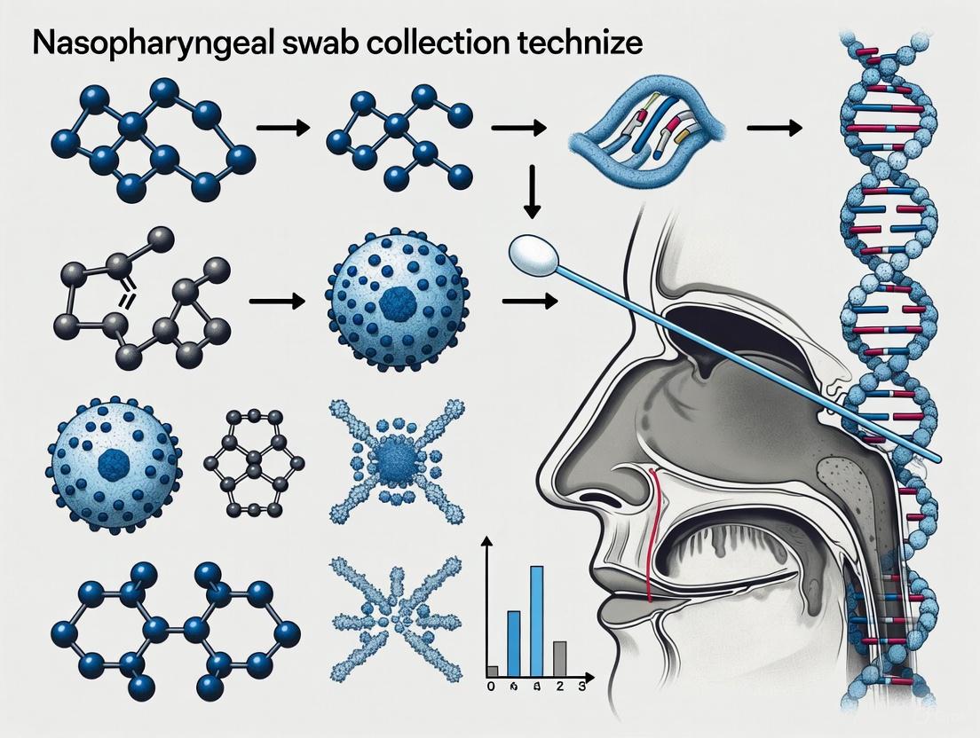
A nasopharyngeal (NP) swab is a specialized medical device designed to collect a fluid sample from the nasopharynx, the upper part of the throat behind the nose [1]. It consists of a long, flexible shaft with a soft, absorbent tip made from materials such as nylon flocked fibers or medical-grade foam [2] [3]. In clinical practice and research, this specimen collection method is critical for the diagnosis and study of respiratory infections, including SARS-CoV-2, influenza, and RSV [4] [1]. Its primary value lies in its ability to directly sample the site of active viral replication, thereby providing a high-quality specimen for downstream analytical techniques such as viral culture, antigen detection, and molecular analysis via polymerase chain reaction (PCR) [4] [5].
The global pandemic underscored the NP swab's role as a cornerstone of respiratory diagnostics and a vital tool in public health surveillance and pharmaceutical development [5] [3]. The reliability of data generated in clinical trials for vaccines or antivirals is fundamentally linked to the quality of the original specimen, placing a premium on standardized and efficacious swab collection techniques [6] [5]. Consequently, ongoing research focuses not only on the pathogens detected but also on optimizing the swab itself—its design, material composition, and collection protocol—to improve patient comfort, sample yield, and diagnostic accuracy [5] [3].
Technical Definition and Anatomical Target
The nasopharyngeal swab is technically defined by its precise anatomical target: the nasopharynx. The nasopharynx is a mucosa-lined cavity located posterior to the nasal cavity and above the soft palate, constituting the superior portion of the pharynx [7]. It serves as a key reservoir for the replication of many respiratory pathogens [4].
To reach this site, the swab must be inserted through a nostril and advanced along the floor of the nasal passage, a path that measures approximately 5 to 7 cm or about half the distance from the nostril to the front of the ear in a typical adult [4] [8]. The swab's design is tailored to this task. It typically features a long (often 6 inches), slender, and flexible shaft that allows it to navigate the curvature of the nasal passage without causing significant trauma [1] [2]. The tip is minimally sized and made of synthetic materials to maximize absorption of secretions and subsequent release of the specimen into transport media, while also avoiding substances that could inactivate viruses or inhibit PCR reactions, such as calcium alginate or wooden shafts [4] [9].
The following diagram illustrates the anatomical pathway and key procedural steps for NP swab collection:
Clinical and Research Applications
The nasopharyngeal swab is the preferred specimen type for detecting a wide array of respiratory pathogens due to its high diagnostic yield [4]. Its applications span direct clinical diagnostics and broader research initiatives.
In a clinical diagnostic context, NP swabs are routinely used to identify the causative agents of respiratory illnesses such as COVID-19 (SARS-CoV-2), influenza A and B, respiratory syncytial virus (RSV), and other viral and bacterial infections [1] [7]. A positive result confirms active infection, guiding patient isolation decisions and therapeutic strategies [4].
In the research and development sphere, NP swabs are indispensable. They are crucial for:
- Viral Surveillance: Tracking the prevalence and geographic spread of respiratory viruses and the emergence of new variants [4] [3].
- Clinical Trials: Serving as a primary endpoint in vaccine and antiviral drug trials to measure efficacy in preventing or clearing infection [5] [3]. The cycle threshold (Ct) values obtained from RT-qPCR of NP swabs provide a quantitative measure of viral load [5] [10].
- Pathogenesis Studies: Enabling researchers to study the kinetics of viral shedding and the host-pathogen interaction at the site of infection [5].
Compared to less invasive methods like anterior nasal or saliva sampling, NP swabs generally offer superior sensitivity because they collect samples directly from the primary site of viral replication [2] [10]. For instance, one study noted a 97% detection rate for RSV with NP swabs compared to 76% for nasal swabs [2].
Quantitative Analysis of Swab Collection Variables
The performance of a nasopharyngeal swab is influenced by multiple variables, including the collection technique and the swab's physical design. Research has quantitatively assessed these factors to optimize protocols and improve swab efficacy.
Table 1: Comparison of Swab Collection Techniques on Sample Quality and Patient Discomfort
| Variable | 'In-Out' Technique (No Rotation) | 'Rotation' Technique (10-Second Rotation) | Significance |
|---|---|---|---|
| Human DNA Recovery (RPP30 cells/μL) | 500 (IQR 235-738) | 503 (IQR 398-685) | P = 0.83 [6] |
| Human RNA Recovery (RNase P copies/μL) | Not Significant | Not Significant | Strong Correlation (ρ = 0.84) with DNA [6] |
| Median Discomfort Score (0-10 scale) | 5 (IQR 3.75-5) | 4.5 (IQR 4-6) | P = 0.51 [6] |
| Participant Preference for Swab over Saliva | 29.4% (10/34) | 10% (3/30) | P = 0.068 [6] |
Table 2: Performance of Different Swab Types in Anatomical vs. Simplified Models
| Swab Type / Model | Collected Volume (μL ± SD) | Release Volume (μL ± SD) | Release Percentage (% ± SD) |
|---|---|---|---|
| Heicon (Injection-Molded) in Cavity Model | 12.30 ± 3.24 | 10.31 ± 3.70 | 82.48 ± 12.70 [5] |
| Commercial (Nylon Flocked) in Cavity Model | 22.71 ± 3.40 | 15.81 ± 4.21 | 69.44 ± 12.68 [5] |
| Heicon (Injection-Molded) in Tube Model | 59.65 ± 4.49 | 40.94 ± 5.13 | 68.77 ± 8.49 [5] |
| Commercial (Nylon Flocked) in Tube Model | 192.47 ± 10.82 | 49.99 ± 13.89 | 25.89 ± 6.76 [5] |
Key findings from this quantitative data indicate that the act of rotating the swab after insertion does not significantly increase nucleic acid recovery but may negatively impact patient tolerance [6]. Furthermore, Table 2 highlights a critical point for research: traditional "tube model" testing, which involves immersing a swab in a liquid tube, does not accurately replicate the complex environment of the nasal cavity and can dramatically overestimate the collection capacity of flocked swabs while underestimating their release efficiency. Anatomically accurate models demonstrate that injection-molded swabs can have superior release characteristics, a key factor for diagnostic sensitivity [5].
Detailed Experimental Protocols for Research
To ensure reproducibility and standardization in research involving NP swabs, detailed methodologies are paramount. Below are protocols derived from recent, rigorous studies.
Protocol 1: Evaluating Swab Collection Technique
This protocol is designed to compare the impact of different physical swabbing maneuvers on sample quality and participant discomfort [6].
Objective: To determine if rotating a nasopharyngeal swab in place after insertion improves nucleic acid yield compared to a simple "in-out" technique, and to assess the corresponding level of participant discomfort.
Materials:
- Sterile, synthetic-tipped NP swabs with flexible plastic shafts (e.g., Puritan UniTranz-RT).
- Viral transport medium.
- Nucleic acid extraction kit (e.g., NucliSens easyMAG, BioMérieux).
- Droplet Digital PCR (ddPCR) or RT-ddPCR system.
- Primers and probes for human genomic targets (e.g., RPP30, RNase P).
Methodology:
- Participant Recruitment & Consent: Recruit adult volunteers and obtain informed consent. Exclude individuals with respiratory symptoms.
- Randomization & Blinding: Randomly assign participants to either the "in-out" or "rotation" group. Keep participants blinded to the technique until immediately before the procedure.
- Swab Collection (by single trained professional):
- Inspect nostrils for obstruction and have the participant blow their nose if necessary.
- Tilt the participant's head back slightly. Gently insert the swab along the nasal floor to the nasopharynx.
- For 'In-Out' Group: Withdraw the swab immediately upon reaching the nasopharynx.
- For 'Rotation' Group: Rotate the swab in place for 10 seconds, then withdraw.
- Discomfort Assessment: Immediately after the procedure, ask the participant to rate their discomfort on a validated 0-10 scale.
- Sample Processing: Place the swab in viral transport media. Process samples within 5 hours.
- Extract total nucleic acids from a fixed volume (e.g., 1 mL) of transport medium.
- Sample Quality Analysis:
- Quantify human DNA using ddPCR with an RPP30 assay.
- Quantify human RNA using RT-ddPCR with an RNase P assay.
- Report results as cells/μL extract and copies/μL extract, respectively.
- Statistical Analysis: Use non-parametric tests (e.g., Mann-Whitney U) to compare nucleic acid recovery and discomfort scores between groups.
Protocol 2: Validating Swab Efficiency Using a Bio-Mimetic Model
This advanced protocol uses a 3D-printed anatomical model to pre-clinically evaluate new swab designs under physiologically relevant conditions [5].
Objective: To compare the sample collection and release efficiency of experimental and commercial NP swabs using an anatomically accurate nasopharyngeal cavity model.
Materials:
- 3D-printed nasopharyngeal cavity (flexible Agilus30 for soft tissue, rigid VeroBlue for bone).
- SISMA hydrogel (or similar mucus-mimicking substance with validated shear-thinning properties).
- Swabs for testing (e.g., experimental injection-molded vs. commercial nylon flocked).
- RT-qPCR instrumentation.
- Inactivated virus (e.g., Yellow Fever Virus - YFV) for spiking.
Methodology:
- Model Preparation: Line the 3D-printed nasopharyngeal cavity with a uniform layer of SISMA hydrogel.
- Virus Spiking: Spike the hydrogel with a known titer of inactivated virus (e.g., YFV) to simulate an infected host.
- Sample Collection:
- Insert the test swab into the model following a standardized clinical protocol (e.g., insert along the cavity floor, rotate, hold for seconds, withdraw while rotating).
- Repeat the process for each swab type and a simple tube model for baseline comparison.
- Sample Elution: Place each swab into a fixed volume of transport medium and vortex to elute the collected sample.
- Quantitative Analysis:
- Gravimetric Analysis: Weigh the swab before and after collection to determine the volume of hydrogel collected and after elution to determine the release volume.
- Molecular Analysis: Perform RT-qPCR on the eluate to detect viral RNA. The Cycle Threshold (Ct) value is a proxy for viral load recovery.
- Data Calculation:
- Calculate collection efficiency (volume collected).
- Calculate release efficiency (% of collected volume released into transport media).
- Compare Ct values; a lower Ct indicates higher viral RNA recovery.
- Statistical Comparison: Use t-tests or ANOVA to compare performance metrics between swab types and between the anatomical and tube models.
The workflow for this sophisticated validation protocol is outlined below:
The Scientist's Toolkit: Essential Research Reagents and Materials
Table 3: Key Research Reagent Solutions for NP Swab Studies
| Item | Function / Rationale | Specific Examples / Properties |
|---|---|---|
| NP Swabs | Core device for specimen collection from the nasopharynx. | Synthetic tip (nylon flocked, polyester, foam): Avoids PCR inhibitors [4] [2]. Flexible plastic shaft: Minimizes injury risk [9]. |
| Viral Transport Media (VTM) | Preserves viral integrity and viability during transport and storage. | Contains antibiotics and antifungals to prevent microbial overgrowth. Must be compatible with downstream assays [4]. |
| Nucleic Acid Extraction Kits | Isolates viral RNA/DNA from the sample for molecular detection. | Critical for standard RT-PCR. Kits using triazole-based reagents (e.g., PREP-NA) are cited [10]. |
| Direct RT-PCR Master Mix | Enables PCR amplification without prior nucleic acid extraction. | Contains reverse transcriptase, DNA polymerase, primers, and probes. Allows for faster, cost-effective testing (e.g., SARS-CoV-2 Lite Kit) [10]. |
| 3D-Printed Nasopharyngeal Model | Provides a physiologically relevant platform for pre-clinical swab testing. | Dual-material (flexible/rigid) printing mimics nasal anatomy and tissue properties [5]. |
| Mucus-Mimicking Hydrogel | Simulates the viscoelastic and shear-thinning properties of native mucus for in vitro testing. | SISMA hydrogel: Rheological properties closely match human sinus mucus [5]. |
| Digital PCR (ddPCR) | Provides absolute quantification of target nucleic acids without a standard curve, ideal for sample quality assessment. | Used to quantify human reference genes (RPP30, RNase P) as a surrogate for cellularity and sample adequacy [6]. |
The nasopharyngeal swab remains a vital tool in both clinical diagnostics and scientific research. Its definition extends beyond a simple collection device; it is an integral component whose design and collection technique directly impact the quality and reliability of all subsequent data. Current research, utilizing advanced methods like 3D-printed anatomical models and standardized molecular assessments, continues to refine our understanding of swab performance [5].
Future developments in this field are likely to focus on several key areas. The push for less invasive yet highly sensitive collection methods will drive the adoption and validation of anterior nasal and saliva samples for specific applications, though the NP swab will likely remain the gold standard for many respiratory pathogens [10]. Furthermore, innovation in swab design—such as new materials and geometries—aims to simultaneously maximize patient comfort, sample collection, and release efficiency [5] [3]. Finally, the integration of NP swabs with rapid, point-of-care, and direct RT-PCR platforms will be crucial for enhancing testing scalability and speed in future outbreak responses [3] [10]. For researchers, a thorough understanding of the fundamentals outlined in this guide is essential for designing robust experiments and accurately interpreting results in the ongoing study of respiratory disease.
Within the context of research on nasopharyngeal swab collection fundamentals, precise navigation of the nasopharyngeal anatomy is paramount for obtaining quality specimens for diagnostic purposes, such as detecting respiratory pathogens including SARS-CoV-2. This technical guide delineates the critical anatomical landmarks and provides a data-driven framework for sample collection. It synthesizes empirical anatomical studies and established clinical protocols to outline a standardized methodology aimed at maximizing specimen adequacy while ensuring patient safety. The guidance herein is intended to provide researchers and clinical developers with a detailed anatomical and procedural foundation.
The nasopharynx, or epipharynx, is the most superior part of the pharynx, situated directly posterior to the nasal cavities and superior to the soft palate [11] [12]. It serves as the initial and primary site of replication for several respiratory pathogens, making it a critical zone for diagnostic sample collection [13]. Its anatomical structure is that of a roughly cuboidal, air-containing cavity, measuring approximately 2.5–3.5 cm in its anterior-posterior diameter and about 4–5.5 cm in its widest transverse diameter and height [14] [15]. A thorough understanding of its boundaries and contents is the foundation for effective swab collection.
Key boundaries include:
- Superiorly/Roof: The body of the sphenoid bone and the basilar part of the occipital bone (the skull base) [14] [12].
- Anteriorly: The posterior nasal apertures, known as the choanae, which provide a direct connection to the nasal cavity [12] [15].
- Posteriorly: The posterior pharyngeal wall, which overlies the anterior aspect of the first two cervical vertebrae (the atlas and axis) [14].
- Inferiorly/Floor: The superior surface of the soft palate, which dynamically separates the nasopharynx from the oropharynx below [11] [12].
- Laterally: The medial pterygoid plates and the superior pharyngeal constrictor muscles, surrounded by the visceral fascia [12].
The nasopharyngeal mucosa is primarily lined with pseudostratified ciliated columnar epithelium, which is continuous with the nasal cavity [15]. Contained within its roof and posterior wall is a significant collection of lymphoid tissue known as the pharyngeal tonsil, or adenoids, which is part of Waldeyer's ring [11] [12].
Critical Anatomical Landmarks and Their Clinical Significance
Successful navigation to the nasopharyngeal mucosa requires identification of, or orientation towards, several key anatomical structures. The lateral walls of the nasopharynx are of paramount importance for specimen collection, housing the most significant landmarks.
Table 1: Key Anatomical Landmarks in the Nasopharynx
| Landmark | Anatomical Description | Clinical Significance for Swabbing |
|---|---|---|
| Choanae | The paired posterior openings of the nasal cavities, separated by the nasal septum [15]. | The swab must pass through one choana to transition from the nasal cavity into the nasopharynx [13]. |
| Fossa of Rosenmüller | A deep mucosal recess located posterior and superior to the torus tubarius [14] [12]. | A primary site for pathogen replication and a common origin for nasopharyngeal carcinoma; it is a critical target for swab rotation to collect adequate material [14] [15]. |
| Torus Tubarius | The cartilaginous prominence forming the posterior and superior margin of the Eustachian tube opening [14] [12]. | A key visual and physical landmark during endoscopic examination; the fossa of Rosenmüller is located directly behind it [15]. |
| Eustachian Tube Orifice | The opening of the Eustachian tube on the lateral wall, connecting the nasopharynx to the middle ear [11] [12]. | Serves as a reference point for orientation; the swab should be directed posterior to this orifice to target the fossa [15]. |
| Adenoids | Lymphoid tissue located in the roof and posterior wall of the nasopharynx [12]. | Often atrophied in adults; enlarged in children, which may partially obstruct the passage and require gentle navigation [12]. |
Beyond these specific structures, the overall configuration of the nasal cavity guides the swab's path. The swab must be advanced parallel to the hard palate and along the floor of the nasal cavity, passing beneath the inferior and middle nasal turbinates, to successfully reach the nasopharynx [13] [16]. A major safety consideration is the cribriform plate of the ethmoid bone, a fragile structure located superiorly in the nasal cavity that houses the olfactory epithelium. Angling the swab upwards toward the bridge of the nose risks contact with this area [13]. Research indicates that the angle required to target the cribriform plate is significantly steeper (mean angle of 36.7° from a reference line between the subnasale and nasion) than the recommended path for nasopharyngeal swabbing, making such injury unlikely with proper technique [13].
Quantitative Anatomical Guidance for Targeted Swab Insertion
Empirical anatomical studies provide quantitative data to guide the direction and depth of swab insertion, moving beyond qualitative description to a metric-based protocol.
A pivotal anatomical study involving simulation on 157 body donors provided precise measurements for swab navigation [13] [17]. The research established key angles and distances by referencing external facial landmarks: the nasion (the midpoint of the nasofrontal suture), the subnasale (the junction of the nasal columella and the upper lip), and the tragus (the small prominence anterior to the external opening of the ear) [13].
Table 2: Key Angles and Distances for Nasopharyngeal Swab Navigation
| Parameter | Measurement (Mean & Range) | Anatomical Correlation |
|---|---|---|
| Angle relative to Subnasale-Nasion line (αA) | 82.9° (69 – 96.5°) [13] | The swab should be held nearly perpendicular to this imaginary line, guiding it parallel to the palate and floor of the nasal cavity [13]. |
| Angle relative to Subnasale-Tragus line (βA) | 9.3° ((-2) – 17.6°) [13] | The swab should be aligned almost parallel to this line, corresponding with the common advice to direct the swab "toward the ear" [13]. |
| Distance from nares to posterior pharyngeal wall | 8.7 cm (7.3 – 10.5 cm) [13] | The approximate depth of insertion required to reach the posterior nasopharynx. This distance was significantly longer in males [13]. |
| Distance from nares to cribriform plate | 6.1 cm (5.0 – 7.7 cm) [13] | Highlights the safety margin; the dangerous upward path to the cribriform plate is shorter than the correct path to the nasopharynx [13]. |
These data validate and refine common clinical guidance. The widespread instruction to insert the swab to a depth equivalent to the distance from the nostril to the ear opening is substantiated by the close alignment of the swab with the subnasale-tragus line [13]. The study found that a specific three-step procedure based on these angles was successful in entering the nasopharynx in all specimens without pre-existing deformations, whereas a commonly used alternative method succeeded in less than 50% of cases [13].
Detailed Experimental Protocol for Nasopharyngeal Swab Collection
The following protocol integrates quantitative anatomical guidance with standardized clinical procedures to define a robust methodology for researchers and trained healthcare providers [13] [16] [18].
Pre-Collection Preparation
- Patient Positioning: Seat the patient upright with their head against a headrest. Tilt the patient's head back approximately 70 degrees from the horizontal plane to straighten the passage from the nares to the nasopharynx [16].
- Landmark Identification: Mentally visualize the anatomical path. Identify the subnasale and tragus landmarks to conceptualize the insertion axis, which should be nearly parallel to the imaginary line between them [13].
- Swab Selection: Use only sterile, synthetic fiber swabs (e.g., polyester, rayon, or flocked nylon) with thin, flexible plastic or wire shafts. Do not use calcium alginate swabs or swabs with wooden shafts, as they may contain substances that inactivate viruses and inhibit molecular tests [16]. The swab shaft must be long enough to reach the nasopharynx (typically requiring a shaft length of 9-15 cm).
Step-by-Step Collection Procedure
- Insertion: Gently insert the swab into one nostril. Keep the swab parallel to the palate (hard palate) and advance it along the floor of the nasal cavity, not upwards toward the bridge of the nose. A slight lifting of the ala nasi (the outer wall of the nostril) by the swab shaft may be necessary to achieve the correct angle [13] [18].
- Advancement: Advance the swab straight back along the nasal septum. Continue until resistance is encountered, indicating contact with the posterior wall of the nasopharynx. The depth will typically be between 7 and 10 cm for an adult, roughly equivalent to the distance from the patient's nostril to the tragus of the ear [13] [16] [8].
- Rotation and Dwell Time: Once the swab is in place, gently rub and roll it against the nasopharyngeal mucosa. Leave the swab in place for several seconds (recommendations range from 5-15 seconds) to ensure the tip absorbs secretions fully [16] [18] [8].
- Withdrawal: Slowly remove the swab from the nostril while rotating it gently [16] [8].
- Specimen Placement: Immediately place the swab into a sterile tube containing viral transport media. Break or cut the swab shaft at the scored breakpoint and close the tube lid securely [18].
- Labeling and Storage: Label the specimen tube with patient identifiers and collection details. Place the tube in a biohazard bag and refrigerate it (typically at 2-8°C) until transport to the laboratory [18] [8].
The following workflow diagram summarizes the key decision points and actions in this protocol.
Diagram 1: Nasopharyngeal swab collection workflow.
The Researcher's Toolkit: Essential Materials and Reagents
The integrity of a nasopharyngeal specimen is dependent on the correct use of specialized materials and reagents. The selection of these components is critical for preserving pathogen viability and nucleic acid integrity for downstream diagnostic and research applications.
Table 3: Essential Research Reagents and Materials for NP Specimen Collection
| Item | Specification / Type | Primary Function in Protocol |
|---|---|---|
| Swab | Synthetic tip (flocked nylon, Dacron/polyester). Flexible shaft (plastic or wire). Shaft length: 9-15 cm. | To effectively collect and release respiratory epithelial cells and secretions from the nasopharyngeal mucosa [16] [18]. |
| Viral Transport Media (VTM) | Sterile solution containing protein stabilizer (e.g., bovine serum albumin), antimicrobial agents, and buffer. | To maintain viral viability and preserve nucleic acid integrity during specimen storage and transport [18]. |
| Personal Protective Equipment (PPE) | N95 respirator (or higher), eye protection, gloves, and gown. | To protect the collector from exposure to potentially infectious respiratory droplets and aerosols [16]. |
| Specimen Transport Tube | Sterile, leak-proof, screw-cap tube, often with a break-point notch for the swab. | To serve as a secure, sealed primary container for the swab and VTM, preventing leakage and contamination [16] [18]. |
| Biohazard Bag | Sealed, durable plastic bag with an separate outer pocket for paperwork. | To provide a secondary secure containment layer for the specimen tube during transport [18] [8]. |
Common Methodological Pitfalls and Specimen Integrity
Adherence to the outlined protocol is critical for research validity. Several methodological errors can compromise specimen quality and lead to false-negative results in testing.
- Incorrect Insertion Angle: Directing the swab upwards (superiorly) instead of parallel to the palate risks contact with the nasal turbinates and the cribriform plate. This results in poor cell yield and potential safety hazards, and samples the wrong anatomical site, which is not colonized by typical respiratory pathogens in the same manner as the nasopharynx [13].
- Insufficient Depth of Insertion: Failure to advance the swab until resistance is met means the swab tip likely remains in the nasal vestibule or cavity, not the nasopharynx. Specimens from the anterior nose contain significantly lower viral loads for pathogens like SARS-CoV-2 compared to nasopharyngeal specimens, drastically reducing test sensitivity [16].
- Inadequate Dwell Time: Briefly touching the nasopharynx without a several-second pause for absorption and gentle rotation fails to collect sufficient secretions and cells. This is a common source of low-cellularity specimens [16] [8].
- Improper Specimen Handling: Using inappropriate swab materials (e.g., wood or calcium alginate), allowing the swab to dry out, or failing to refrigerate the specimen promptly can degrade viral genetic material and proteins, rendering the specimen unusable for accurate analysis [16].
A systematic review of technique is essential, as an incorrect execution has been cited as a major contributor to the high false-negative rate (approximately 30%) observed in some molecular tests for respiratory viruses like SARS-CoV-2 [19].
The procedure of nasopharyngeal swabbing, while seemingly simple, is fundamentally an exercise in precise anatomical navigation. A deep understanding of the key landmarks—the choanae, torus tubarius, and most importantly, the fossa of Rosenmüller—combined with data-driven guidance on insertion angles and depth, forms the basis of effective technique. The standardized protocol and reagent specifications detailed in this guide provide a framework for obtaining specimens of the highest possible quality. For the research community and drug developers, consistency and anatomical accuracy in sample collection are not merely procedural concerns but are prerequisites for generating reliable, reproducible, and clinically meaningful data in the study of respiratory pathogens and the development of novel diagnostics and therapeutics.
Airway mucus, a complex gel with an anisotropic three-dimensional network structure, serves as a crucial component of the respiratory defense barrier. It plays a vital role in maintaining airway hydration, supporting the function of airway epithelial cells, and protecting against infectious agents and environmental particles [20]. Through linear and nonlinear rheological mechanisms mediated by ciliary motion and coughing, airway mucus expels foreign pathogens and toxic particles while selectively permitting the passage of specific nutrients and proteins [20]. The effectiveness of these clearance functions depends fundamentally on the proper rheological properties of mucus under normal physiological conditions. This review examines the rheological behaviors of airway mucus in relation to health and disease, with particular focus on implications for pathogen and antibody collection methodologies essential for diagnostic and therapeutic development.
Fundamental Rheological Properties of Airway Mucus
Composition and Structure
Airway mucus is a complex biological fluid consisting of approximately 97% water and 3% solids, including mucins, nonmucinous proteins, salts, lipids, and cellular debris [20]. This composition gives rise to its characteristic viscoelastic properties. Mucins, high molecular weight glycoproteins, form a crosslinked polymer network that provides mucus with its distinctive gel-like characteristics [21]. The mucus layer exhibits a biphasic structure, comprising a periciliary liquid layer that facilitates ciliary movement and an overlying gel layer that traps particles and pathogens [21].
Rheological Behavior in Health and Disease
In healthy states, airway mucus demonstrates ideal viscoelastic properties with relatively low viscosity and elasticity, allowing easy transport by ciliary action [20]. At the macroscopic level, mucus behaves as a non-Newtonian, thixotropic material, exhibiting both flow-related viscous properties and deformation-resistant elastic characteristics [21]. Under low shear conditions, its viscosity can be 100 to 10,000 times greater than that of water. However, as shear rate increases toward physiological limits during coughing, mucus transitions into a shear-thinning state where its viscosity significantly decreases [21].
In respiratory diseases such as cystic fibrosis (CF), chronic obstructive pulmonary disease (COPD), asthma, and severe COVID-19, excessive mucus secretion is accompanied by abnormal rheological behaviors [20] [22]. These pathological changes lead to impaired mucus flow, airway obstruction, and potentially life-threatening conditions. The table below summarizes key rheological parameters in health and disease states:
Table 1: Rheological Properties of Airway Mucus in Health and Disease
| Condition | Viscoelasticity | Mucin Concentration | Clearance Efficiency | Key Characteristics |
|---|---|---|---|---|
| Healthy | Low to moderate | Normal (~3% solids) | Efficient | Optimal viscosity and elasticity for ciliary transport [20] [23] |
| Cystic Fibrosis | Highly viscoelastic | Elevated | Severely impaired | Dehydrated, hyperconcentrated mucus with abnormal salt composition [20] [23] |
| COPD | Highly viscoelastic | Elevated | Impaired | Excessive secretion with altered macromolecular composition [20] [23] |
| Asthma | Highly viscoelastic | Elevated | Impaired | Plug formation, especially during exacerbations [20] [23] |
| COVID-19 | Highly viscoelastic | Elevated | Impaired | Mucus accumulation and plugging observed in severe cases [20] |
Implications for Pathogen Entrapment and Clearance
Microbial Interactions with Mucus
The mucus layer serves as the first point of contact for inhaled pathogens, with its rheological properties directly influencing microbial entrapment and clearance. The nasal microbiome differs significantly between healthy individuals and those with inflammatory conditions such as asthma and allergic rhinitis (AR) [24] [25]. In healthy nasal mucus, the Proteobacteria (Ralstonia genus) and Actinobacteria (Propionibacterium genus) phyla predominate, whereas patients with AR show significant abundance of the Firmicutes (Staphylococcus genus) phylum [24]. Specifically, Staphylococcus aureus demonstrates markedly greater abundance (37.69%) in the nasal mucus of AR patients, suggesting this dysbiosis plays a role in allergic inflammation [24].
Host-microbial commensalism shapes the innate immune response in the nasal mucosa, and the microbial characteristics of nasal mucus directly impact the mechanisms of the initial allergic responses in the nasal epithelium [24]. The dysbiosis observed in allergic nasal mucus creates an environment conducive to pathogen persistence and inflammation, further altering mucus rheology in a vicious cycle that impedes clearance.
Mucociliary Clearance Mechanisms
The mucociliary system, comprising cilia, the periciliary fluid layer (PCL), and the mucus layer, represents a crucial innate defense mechanism essential for maintaining lung health [20]. Cilia extend from bronchial epithelial cells through the PCL into the mucus layer, beating in a coordinated fashion to clear the airway lumen of excess mucus and entrapped pathogens [20]. In large airways, mucus is often cleared effectively by coughing-induced high-velocity airflow, while in distal small airways, clearance primarily relies on upward mobilization through ciliary beating [20].
The effectiveness of mucociliary clearance is highly dependent on the physical, specifically rheological, properties of mucus [20]. When mucus becomes highly viscoelastic under pathological conditions, it resists mobilization and clearance by both cough and ciliary action, leading to mucus accumulation or plugging that promotes chronic bacterial infection [20].
Figure 1: Interrelationship Between Mucus Properties, Pathogen Clearance, and Sampling Efficiency. Mucus rheological properties directly influence pathogen entrapment and clearance mechanisms. Disease states alter mucus rheology, creating a feedback loop that affects both pathogen persistence and diagnostic sampling efficiency.
Methodological Approaches to Mucus Rheology
Macrorheology vs. Microrheology
The study of mucus rheology employs two complementary approaches: macrorheology and microrheology. Macrorheology provides insights into the overall viscoelastic behavior of mucus, revealing its general mechanical properties but often overlooking local variations [20]. Cone-plate geometries enable macrorheological measurements with small sample volumes (as low as 86 µL), making them suitable for homogeneous samples like those from Air-Liquid Interface (ALI) cultures [23].
Microrheology addresses the limitation of local variations by examining heterogeneity, local mechanical properties, and the diffusion characteristics of drugs and viruses within the mucus gel network at nano- and microscale levels [20]. Techniques like particle tracking microrheology (PTMR) specialize in assessing local mechanical properties but require specialized microscopic equipment and raise concerns about interactions between the sample and seeded particles [23].
Experimental Protocols for Mucus Collection and Analysis
Native Human Mucus Collection Methods
Researchers have developed various methods for collecting native mucus from human airways, each with advantages and limitations:
Table 2: Comparison of Mucus Collection Methods for Research
| Collection Method | Sample Source | Advantages | Limitations | Typical Volume |
|---|---|---|---|---|
| Bronchoscopy [20] | Tracheobronchial tree | Direct collection from lungs, essential diagnostic information | Requires anesthesia, specialized personnel, small yield | Small (sufficient for rheology) |
| Endotracheal Tubes [20] | Intubated patients | Direct collection from healthy patients, large volumes | Hydration variations affect reproducibility | Large |
| Spontaneous Sputum [20] | Patients with respiratory disease | No medical intervention needed | Cannot collect from healthy individuals, saliva contamination | Unpredictable |
| Induced Sputum [20] | Healthy volunteers or patients | Non-invasive, broader sample base | Saline aerosols may dilute mucus, potential bronchoconstriction | Variable |
| ALI Cultures [20] [23] | In vitro differentiated cells | Controlled environment, no infection interference | May not fully replicate in vivo complexity | ~4 µL per culture |
Rheological Measurement Protocol
The following protocol outlines standardized methodology for mucus rheological characterization:
Sample Preparation: Fresh mucus samples are homogenized by gradually increasing vortex stirring until visually forming a torus. This vortex intensity is maintained for 30 seconds, resulting in visually homogeneous samples [23].
Geometry Selection: For homogeneous samples (e.g., ALI mucus), cone-plate geometries are used with volumes as low as 86 µL. For heterogeneous samples (e.g., sputum), rough plate-plate geometries with 25 mm diameter are employed, requiring larger volumes (330-690 µL) [23].
Oscillatory Strain Sweep: Samples are subjected to increasing oscillatory strain amplitudes ranging from 0.1 to 10,000% at 1 Hz and 37°C [23]. This measures viscoelastic parameters in both linear (low strains) and nonlinear (high strains) regimes.
Data Analysis: Key parameters include elastic modulus (G'), viscous modulus (G''), and critical strain (γc) where the structure begins to break down. These parameters are correlated with physiological functions and clearance efficiency [23].
Implications for Pathogen and Antibody Collection
Optimizing Sampling Techniques
The rheological properties of mucus significantly impact the efficiency of pathogen and antibody collection from the nasal cavity and airways. A standardized comparison of three common nasal sampling methods revealed substantial differences in collection capabilities for SARS-CoV-2 RBD-specific IgA [26]:
Table 3: Performance Comparison of Nasal Sampling Methods
| Sampling Method | Description | Single-Day Detection Rate | 5-Day Consecutive Detection Rate | Median IgA Concentration |
|---|---|---|---|---|
| Nasopharyngeal Swab (M1) [26] | Nylon flocked swab inserted to nasopharyngeal region | 68.8% | 48.7% | 28.7 U/mL |
| Nasal Swab (M2) [26] | Cotton swab inserted ~2 cm to nasal turbinate | 88.3% | 77.3% | 93.7 U/mL |
| Expanding Sponge (M3) [26] | Polyvinyl alcohol sponge expanded in nostril for 5 minutes | 95.5% | 88.9% | 171.2 U/mL |
The expanding sponge method (M3) significantly outperformed both swab methods across all metrics (p<0.05), demonstrating that sampling methodologies that better interact with the mucus layer yield superior diagnostic and research outcomes [26].
Impact on Diagnostic Sensitivity
The rheological properties of mucus affect not only sampling efficiency but also diagnostic sensitivity. Comparative studies of SARS-CoV-2 detection methods have demonstrated that anterior nasal samples yield significantly lower viral loads than nasopharyngeal samples (p<0.001) [27]. Specifically, the median viral loads for nasopharyngeal samples (NPS) were 53,560 (IQR 605-608,050), compared to 1,792 (IQR 7-81,513) for anterior nasal samples with NP-type swabs [27].
The sensitivity of antigen tests using anterior nasal samples was 72.5% (95% CI 58.3-84.1%) compared to RT-PCR with nasopharyngeal samples, highlighting how sampling location and mucus properties impact detection efficacy [27]. However, anterior nasal collection was associated with significantly lower degrees of coughs or sneezes induction and reduced pain severity (p<0.001), suggesting a trade-off between patient comfort and diagnostic sensitivity [27].
Research Reagent Solutions and Experimental Models
Essential Research Materials
Table 4: Key Research Reagents for Mucus Rheology and Sampling Studies
| Reagent/Material | Function | Application Examples |
|---|---|---|
| Xanthan Gum Solutions [21] | Synthetic mucus simulating normal (0.25% w/v) and diseased (1% w/v) conditions | In vitro nasal spray deposition studies |
| MucilAir ALI Cultures [23] | Ready-to-use 3D model of human airway epithelium | Controlled mucus production without infection interference |
| FLOQSwabs [27] | Nasopharyngeal flocked swabs for sample collection | Antigen testing and PCR sample collection |
| Polyvinyl Alcohol Sponge [26] | Expanding sponge for nasal lining fluid collection | Superior mucosal antibody collection |
| DNeasy PowerWater Kit [24] | DNA extraction from mucus samples | Microbiome analysis from nasal swabs |
| Human/NHP Kit [26] | Electrochemiluminescence detection of immunoglobulins | Quantitative nasal antibody measurement |
Synthetic Mucus Models
Synthetic mucus solutions enhance physiological realism in deposition studies. Xanthan gum (XG) saline solutions effectively mimic the viscoelastic properties of human nasal mucus, with 0.25% w/v XG representing healthy mucus and 1% w/v XG simulating diseased conditions [21]. These synthetic coatings significantly influence spray deposition patterns, facilitating broader and more uniform liquid distribution due to diffusion and lubrication effects compared to uncoated surfaces [21].
The inclusion of mucoadhesive polymers in nasal formulations offers a potential solution to improve drug retention; however, they adversely affect drug diffusion, necessitating precise formulation strategies [21]. Synthetic mucus models provide valuable platforms for optimizing intranasal formulations and delivery devices while controlling for the variability inherent in native mucus samples.
Figure 2: Experimental Workflow for Mucus Research. The comprehensive methodology encompasses sample collection from various sources, application of multiple analytical techniques, and integrated data interpretation to understand mucus rheology and its implications for pathogen and antibody collection.
The rheological properties of airway mucus play a fundamental role in respiratory defense, significantly influencing both pathogen clearance and the efficiency of diagnostic sampling. In healthy states, mucus exhibits optimal viscoelastic properties for effective clearance, while in diseases such as CF, COPD, asthma, and COVID-19, pathological alterations impair mucus function and promote infection. Understanding these rheological properties is essential for developing improved sampling techniques, as demonstrated by the superior performance of expanding sponge methods over conventional swabs for antibody collection. Standardized experimental approaches, including synthetic mucus models and validated detection assays, provide critical tools for advancing respiratory diagnostics and therapeutic development. Future research should focus on establishing clearer correlations between specific rheological parameters and sampling efficiencies across different disease states to further optimize collection methodologies for both clinical and research applications.
Why the Nasopharynx? The Rationale for Targeting Respiratory Pathogens and Mucosal Immunity
The nasopharynx serves as the critical frontline defense and primary portal of entry for numerous respiratory pathogens. This anatomical niche represents far more than a passive conduit for air passage; it is a sophisticated immunological inductive site where initial host-pathogen interactions determine subsequent infection outcomes. Understanding the nasopharynx's unique structural and immunological properties provides the fundamental rationale for its central role in diagnostic specimen collection, therapeutic intervention, and vaccine development. This whitepaper examines the scientific basis for targeting the nasopharynx through the integrated lenses of mucosal immunology, clinical diagnostics, and therapeutic design, contextualized within ongoing research on nasopharyngeal swab collection techniques.
The nasopharynx, positioned behind the nasal cavity and above the soft palate, constitutes the superior portion of the pharynx. Its strategic anatomical location places it directly in the path of inhaled air, making it the first contact point for airborne pathogens. More significantly, it houses organized nasopharyngeal-associated lymphoid tissue (NALT), a specialized component of the mucosa-associated lymphoid tissue (MALT) that represents the largest immune component in the entire human body [28]. This combination of permanent anatomical exposure and concentrated immunological capacity establishes the nasopharynx as a critical battleground in respiratory infections.
The rationale for focusing on this region extends beyond mere convenience for sample collection. Evidence confirms that the early replication of respiratory viruses like SARS-CoV-2 occurs primarily in upper airway mucosal surfaces, especially the nasopharynx [28]. Upper airway antigenic priming within NALT initiates a dynamic, compartmentalized regional immune network that shapes the entire subsequent immune response [28]. Consequently, understanding the nasopharyngeal environment is essential for comprehending pathogenesis, developing effective diagnostics, and designing next-generation interventions against respiratory pathogens.
Anatomical and Immunological Foundations
Structural Specialization for Immune Surveillance
The nasopharynx features a specialized epithelium and underlying lamina propria that facilitate both pathogen capture and immune activation. Unlike the stratified squamous epithelium found in the oropharynx, regions of the nasopharynx contain a ciliated pseudostratified columnar epithelium with goblet cells that contribute to mucociliary clearance. The surface is bathed in a mucous layer containing secretory immunoglobulin A (SIgA), antimicrobial peptides, and cytokines that form the first biochemical barrier against invasion [29].
Beneath the epithelial layer lies the lamina propria, a thin layer of loose connective tissue rich in immune cells including mature plasma cells, macrophages, dendritic cells (DCs), and intraepithelial lymphocytes (IELs) [30]. These cells constitute a diffuse mucosal lymphoid tissue that provides rapid response capability. IELs are particularly positioned to be the first lymphocytes to encounter respiratory viruses breaching the epithelial barrier [30].
Organized Lymphoid Tissue: NALT
NALT represents the organized inductive site of nasopharyngeal immunity, functioning as a critical antigen sampling and processing center. This structured lymphoid tissue contains:
- Specialized epithelial cells (M-cells): Transport antigens from the lumen to underlying immune cells [29]
- Antigen-presenting cells (DCs): Capture antigens and migrate to draining lymph nodes [29]
- B-cell and T-cell zones: Support lymphocyte activation and differentiation [29]
- Germinal centers: Facilitate B-cell maturation, somatic hypermutation, and class-switching [29]
Upon antigen encounter in NALT, activated lymphocytes acquire homing receptors that direct them to distant mucosal effector sites, including the tracheobronchial epithelium, regional lymph nodes, and secretory glands [28]. This interconnected network enables a localized response in the nasopharynx to generate protective immunity throughout the respiratory tract and beyond, exemplified by the presence of SARS-CoV-2-specific antibodies in saliva, tears, and breast milk [28].
Mucosal Immune Defense Mechanisms
Secretary IgA (SIgA): The Primary Mucosal Immunoglobulin
SIgA stands as the dominant antibody class in mucosal secretions and represents a first line of adaptive defense. Its structural and functional properties make it uniquely suited for nasopharyngeal protection:
Table 1: Properties of Secretary IgA (SIgA) in Mucosal Defense
| Property | Functional Significance | Reference |
|---|---|---|
| Polymeric structure (dimers/tetramers) | Enhanced virus neutralization capacity | [28] |
| Association with secretory component | Resistance to proteolytic degradation | [31] |
| Transcytosis via pIgR | Active transport across epithelium into secretions | [29] |
| Non-inflammatory neutralization | Effective pathogen clearance without tissue damage | [29] |
| Mucosal homing of IgA+ plasmablasts | Targeted deployment to sites of antigen exposure | [28] |
Natural SARS-CoV-2 infection induces mucosal SIgA responses detectable in saliva, nasal secretions, and bronchoalveolar lavage fluid [28]. Notably, the neutralizing activity of IgA polymers in the nasopharynx proves approximately 15-fold more potent than IgA monomers and 7-fold more potent than plasma IgG [28], highlighting the functional superiority of the mucosal IgA response at the initial site of viral encounter.
Tissue-Resident Memory Cells
Antigen-specific tissue-resident memory T (TRM) and B cells induce rapid in-situ protection upon re-exposure to pathogens at mucosal entry sites. These cells reflect a compartmentalization of the immune system, with studies showing activated TRM cells in the airways that do not correlate with systemic blood T-cell responses [28]. Their persistence at the site of initial antigen encounter provides:
- Rapid response: Faster reaction than circulating memory cells
- Localized protection: Concentration at the precise location of pathogen entry
- Durability: Persistence for at least six months in respiratory tissues [29]
Following mucosal infection, specific lung-resident memory B cells form early at the portals of pathogen entry and provide superior protection compared to circulating memory cells [28]. This principle makes TRM induction a critical goal for next-generation vaccine design.
Innate Immune Components
The nasopharyngeal mucosa contains numerous innate immune elements that provide immediate, non-specific protection:
- Mucosa-associated invariant T (MAIT) cells: Major actors in epithelial barrier protection that favor maintenance of tissue-resident and central memory T cells [28]
- Innate lymphoid cells (ILCs): Categorized into ILC1, ILC2, and ILC3 subsets that regulate mucosal immunity through cytokine secretion [29]
- Complement components: Demonstrate direct activation signatures within bronchoalveolar lavage fluid, where serum-derived complement is absent [28]
- Alveolar macrophages: Professional phagocytes that clear pathogens and modulate immune responses [29]
The nasopharyngeal innate immune system is further reinforced by physical and chemical barriers, including mucus that traps pathogens, cilia that propel them toward the oropharynx, and antimicrobial peptides that directly disrupt microbial integrity.
Diagnostic and Research Methodologies
Nasopharyngeal Swab Collection: Technical Considerations
Proper nasopharyngeal swab collection is essential for accurate pathogen detection and immunological research. The procedure requires trained healthcare professionals familiar with nasal anatomy and standardized techniques [6]. Key anatomical considerations include:
- Insertion depth: Approximately 7 cm in adults, or half the distance from the nose to the front of the ear [6] [8]
- Insertion angle: Along the floor of the nasal cavity, parallel to the palate (not upward toward the brain) [8]
- Navigation: Following the natural path of the nasal canal, avoiding deviations caused by septal irregularities
Table 2: Comparison of Nasopharyngeal Swab Collection Techniques
| Parameter | "In-Out" Technique | Rotation Technique | Clinical Significance | |
|---|---|---|---|---|
| Nucleic acid recovery | 500 [235-738] cells/μL extract | 503 [398-685] cells/μL extract | No significant difference (P = .83) | [6] |
| Participant discomfort (median) | 5 [3.75-5] on 11-point scale | 4.5 [4-6] on 11-point scale | No significant difference (P = .51) | [6] |
| Preference for swab over saliva | 29.4% of participants | 10% of participants | Rotation may be less tolerable (P = .068) | [6] |
| Procedural efficiency | Faster collection | Requires 10-second pause after placement | Potential workflow implications | [6] |
A comparative study of 69 adult volunteers found that swab rotation following nasopharyngeal contact did not recover additional nucleic acid compared to simple placement and withdrawal [6]. This suggests that most specimen collection occurs during insertion and contact with the nasopharyngeal wall, not during prolonged manipulation.
Anatomical Variations and Procedural Optimization
Research reveals that discomfort levels and potentially nucleic acid recovery can differ by ethnicity, consistent with variations in nasal anatomy [6]. Asian participants reported significantly higher discomfort scores compared to White participants (median 5 vs. 4, P = .047) [6], highlighting the importance of technique adaptation to individual anatomical differences.
Optimal swab collection involves:
- Patient positioning with head in neutral position, slightly tilted back [8]
- Assessment of nasal patency before swab insertion [6]
- Selection of appropriate swab type (flocked swabs generally provide superior sample collection) [32]
- Proper specimen handling with immediate placement in transport medium [8]
Immunological Sampling and Analysis
Beyond pathogen detection, nasopharyngeal swabs enable critical assessment of mucosal immunity through measurement of:
- Secretory IgA levels: Quantification of pathogen-specific SIgA in nasal secretions
- Cellular composition: Identification of resident immune populations
- Cytokine profiles: Analysis of local inflammatory mediators
- Transcriptomic signatures: Evaluation of host response through RNA analysis
These applications make nasopharyngeal sampling invaluable for both diagnostic purposes and research into mucosal immune responses to infection and vaccination.
Implications for Therapeutic and Vaccine Development
Limitations of Systemic Vaccination
Conventional intramuscular vaccines against respiratory pathogens primarily induce serum IgG antibodies and systemic cell-mediated immunity but are poorly capable of generating protective mucosal immunity at the pathogen entry site [28]. While the lower respiratory tract is partially protected by transudation of serum IgG antibodies, the upper respiratory tract relies more heavily on SIgA antibodies that are inefficiently induced by parenteral vaccination [29]. This immunological gap may explain why systemically immunized individuals can still harbor virus in their salivary and nasal secretions, potentially transmitting infection despite protection from severe disease [31].
Rationale for Mucosal Vaccination
Mucosal vaccines, particularly intranasal formulations, offer distinct advantages for combating respiratory pathogens:
Table 3: Comparison of Vaccine Administration Routes
| Parameter | Parenteral Vaccination | Mucosal Vaccination | Implication |
|---|---|---|---|
| Primary antibody response | Serum IgG | Secretory IgA (SIgA) | Mucosal vaccines better block transmission |
| Tissue-resident memory cells | Limited induction | Robust generation in respiratory tract | Faster local response upon exposure |
| Site of action | Systemic compartment | Mucosal inductive sites (e.g., NALT) | Direct pathogen interception at entry point |
| Transmission blocking | Limited effect | Potent reduction | Community-level herd immunity |
| Administration | Needle-based | Needle-free | Improved acceptability and mass deployment |
Intranasal vaccines marshal early protective immune responses in the upper respiratory tract before pathogens gain a foothold in the lower respiratory tract [28]. They control infectiousness, contagiousness, viral spread, and onward transmission more effectively than parenteral vaccination alone [28]. Experimentally, a single intranasal dose of adenovirus-vectored vaccine protected against SARS-CoV-2 infection throughout the respiratory tract in non-human primates [28].
Historical Precedents and Current Development
The concept of mucosal vaccination is not novel, with successful precedents including:
- Oral polio vaccine: Contributed to near-global eradication of wild poliovirus [29]
- Live-attenuated influenza vaccine (LAIV): Introduced in the 1960s [29]
- Oral rotavirus vaccines: Effectively reduce severe childhood gastroenteritis [28]
These successes highlight the potential of mucosal vaccines to induce robust local immunity while offering logistical advantages such as needle-free administration and potential for self-administration, particularly valuable for rapid mass immunization during outbreaks [28] [29].
Experimental Models and Research Tools
Key Research Reagent Solutions
Table 4: Essential Research Materials for Nasopharyngeal Immunity Studies
| Reagent/Material | Function/Application | Technical Notes |
|---|---|---|
| Flocked nasopharyngeal swabs | Specimen collection from nasal mucosa | Superior sample collection and release compared to fiber swabs [6] |
| Amies transport medium | Preserves specimen viability during transport | Maintains organism viability without significant multiplication [32] |
| Purified mucins | Study of pathogen-mucus interactions | Can serve as decoy receptors for influenza viruses [29] |
| Anti-human IgA antibodies | Detection and quantification of SIgA responses | Must distinguish secretory component for SIgA specificity |
| pIgR-expressing epithelial cells | Study of IgA transcytosis | Models mucosal antibody transport mechanisms |
| Tetramer reagents | Identification of antigen-specific T cells | Requires knowledge of immunodominant epitopes |
Methodological Framework for Swab Collection Research
The following diagram illustrates a standardized research methodology for evaluating nasopharyngeal swab collection techniques based on current evidence:
Immunological Signaling in Nasopharyngeal Immunity
The nasopharyngeal immune response involves complex interactions between epithelial cells, antigen-presenting cells, and lymphocytes, as illustrated in the following signaling pathway:
The nasopharynx represents an immunological sweet spot for both understanding and combating respiratory infections. Its strategic position as the initial contact point for inhaled pathogens, combined with its sophisticated lymphoid architecture, makes it indispensable for diagnostic sampling, pathogenesis research, and therapeutic intervention. The compartmentalization of mucosal immunity explains why systemic vaccination alone may be insufficient to block respiratory virus transmission, highlighting the need for intranasal vaccines that engage local immune mechanisms.
Future research should prioritize:
- Refinement of nasopharyngeal sampling techniques to maximize patient comfort and diagnostic yield
- Development of standardized correlates of protection for mucosal immunity
- Advanced mucosal vaccine platforms that safely and effectively induce durable SIgA responses and tissue-resident memory
- Exploration of the interconnectedness between nasopharyngeal immunity and distant mucosal sites through the common mucosal immune system
As respiratory pathogens continue to pose significant global health threats, leveraging the unique properties of the nasopharyngeal environment will be essential for developing next-generation strategies to block infection at its point of entry and ultimately interrupt transmission chains at the population level.
Executing Precision: A Step-by-Step Protocol for Standardized NP Swab Collection
The accuracy of any diagnostic or research test for respiratory viruses is fundamentally dependent on the quality of the original specimen collected. For nasopharyngeal sampling, the choice of swab and transport medium is not merely a procedural detail but a critical variable that can determine the success or failure of downstream analysis. The SARS-CoV-2 pandemic underscored this reality, as global shortages of recommended supplies compelled researchers and clinicians to validate alternatives, generating a wealth of new data on equipment performance [33]. Within the broader context of nasopharyngeal swab collection technique research, optimizing these fundamental tools is paramount. This guide provides an in-depth technical analysis of swab design characteristics and transport media composition, synthesizing recent experimental findings to equip scientists and drug development professionals with the evidence needed to make informed selections for their research pipelines.
Swab Design: A Critical Determinant of Sample Yield
The swab serves as the primary interface for specimen collection, and its physical and material properties directly influence the quantity and quality of the biological sample obtained. Performance is governed by a combination of tip material, shaft design, and overall construction, each contributing to the swab's collection and elution efficiency.
Swab Tip Material
The tip material is arguably the most significant factor affecting a swab's ability to absorb and release mucosal specimens. The following table summarizes the key characteristics of common swab tip materials, supported by quantitative performance data.
Table 1: Comparative Analysis of Swab Tip Materials
| Tip Material | Key Characteristics | Average Fluid Retention (μL) [33] | Sample Release Efficiency | Primary Applications & Considerations |
|---|---|---|---|---|
| Flocked Nylon | Perpendicular nylon fibers; no internal core. Designed for superior elution. | ~115 - 126 μL (PurFlock Ultra) | High - Consistently superior sample release for microbial and viral DNA/RNA [34]. | Gold standard for molecular diagnostics (RT-PCR). Reduces false negatives. |
| Polyester (Spun) | Fibrous, sponge-like tip. Good absorption. | ~127 μL (Puritan Standard Polyester) | Moderate to High - Good release, though can vary by specific design. | Versatile; used in nasopharyngeal and nasal swabs. |
| Medical Foam | Porous, absorbent foam. | Quantitative data not available in search results. | Moderate - Release may be less efficient than flocked types. | Anterior nasal sampling. |
| Cotton | Traditional, fibrous organic material. | ~13.4 μL (Puritan Cotton), ~218 μL (MedPro Cotton) | Low - Organic compounds may inhibit PCR [16] [33]. | Being phased out for molecular viral testing due to inhibition. |
Swab Shaft Design
The shaft of the swab must fulfill two primary functions: it must provide sufficient rigidity for the clinician or researcher to control the sampling procedure, while also possessing the flexibility to navigate the tortuous path of the nasopharyngeal cavity safely and comfortably.
- Material: Shafts are typically made from plastic (polystyrene) or flexible aluminum with a plastic coating. Wooden shafts are not recommended for viral specimen collection, as they may contain substances that inactivate viruses or inhibit molecular tests [16] [33].
- Length and Diameter: Standard swabs for nasopharyngeal collection are typically 6 inches long. The diameter, particularly for nasopharyngeal swabs, is often thin and may be tapered to enhance flexibility and patient comfort during insertion [2].
- Flexibility vs. Rigidity: A balance is critical. The shaft must be flexible enough to navigate the nasal anatomy without causing trauma but rigid enough to provide tactile feedback and prevent buckling during sample collection. One study specifically noted the use of a "flexible shaft (wire or plastic)" as essential for proper nasopharyngeal specimen (NP) collection [16].
Experimental Protocols for Swab Validation
Research to validate swab performance employs rigorous in vitro methodologies. The following are detailed protocols from key studies:
Protocol 1: Evaluating Collection and Release Efficiency [5]
- Model Setup: An anatomically accurate, dual-material 3D-printed nasopharyngeal cavity is lined with a mucus-mimicking SISMA hydrogel. A standard tube model serves as a control.
- Sample Collection: Swabs are inserted into the model using a standardized protocol that mimics clinical NP sampling.
- Volume Measurement: The collected hydrogel volume is determined by weighing the swab before and after collection.
- Release Calculation: The swab is vortexed in a known volume of elution buffer. The released volume is measured, and the release percentage is calculated as (Released Volume / Collected Volume) * 100.
- Viral Detection Validation: The process is repeated with virus-loaded hydrogel (e.g., Yellow Fever Virus), and the eluent is tested via RT-qPCR to compare cycle threshold (Ct) values, which correlate with viral load recovery.
Protocol 2: Comparing Fluid Retention Across Swab Types [33]
- Preparation: A pre-weighed volume (e.g., 1200 μL) of a standard medium like DMEM is placed in a cryovial.
- Immersion: Each swab type is dipped and rotated in the medium to ensure the tip is fully coated.
- Measurement: The swab is removed, and the remaining medium is weighed. The weight difference is used to calculate the mean volume of media retained by the swab type (n=5).
- Viral Recovery: Swabs are submerged in serial dilutions of SARS-CoV-2, placed in transport medium, and subjected to RNA extraction and RT-PCR to assess detection sensitivity.
Transport Media: Preserving Sample Integrity
Viral Transport Media (VTM) is a buffered solution designed to preserve the viability and nucleic acid integrity of viral specimens during the window between collection and laboratory analysis [35].
Core Components and Functions
The effectiveness of VTM lies in its carefully balanced formulation:
- Balanced Buffer Salts (e.g., HEPES): Maintain a neutral pH (typically 7.3 ± 0.2) to protect viral RNA/DNA from acidic or basic degradation [35].
- Protein Stabilizers (e.g., Gelatin, Bovine Serum Albumin): Act as a protein source to stabilize viral envelopes and support the viability of fastidious organisms [35].
- Cryoprotectants and Preservatives (e.g., Sucrose, Glutamic Acid): Provide nutrient preservation and protect samples during freezing or extended storage [35].
- Antimicrobial Agents (e.g., Amphotericin B, Vancomycin, Colistin): Inhibit the growth of contaminating bacteria and fungi that could compromise the sample [35].
Comparison of Transport Medium Options
During supply chain shortages, researchers have evaluated alternatives to commercial VTM. The table below summarizes the performance of different transport solutions based on a key study.
Table 2: Transport Medium Alternatives for SARS-CoV-2 Detection [33]
| Transport Medium | Key Characteristics | Compatibility with SARS-CoV-2 RT-PCR |
|---|---|---|
| Standard VTM/UTM | Formulated with buffers, stabilizers, and antimicrobials. | High - Validated for use; preserves viral RNA effectively. |
| Phosphate-Buffered Saline (PBS) | A simple salt solution without preservatives. | High - No meaningful difference in viral yield compared to VTM for up to 72h at RT. |
| 0.9% Normal Saline | An isotonic sodium chloride solution. | High - Performs comparably to VTM for molecular detection. |
| DMEM | Cell culture medium containing nutrients and buffers. | High - Suitable for maintaining viral specimen for testing. |
| 100% Ethanol | A disinfectant and preservative. | Effective - Inactivates virus immediately; suitable for RNA preservation if compatible with downstream assay. |
| Dry Swab (No Medium) | Swab transported without liquid medium. | Effective - Demonstrates high sensitivity (90.48% in one study) if processed promptly or rehydrated in-lab [36]. |
Experimental Protocol for Evaluating Transport Media
The following protocol can be used to assess the stability of viral samples in different transport mediums over time:
- Sample Inoculation: Serially dilute a target virus (e.g., SARS-CoV-2) in a base medium like DMEM. Immerse a standardized swab (e.g., cotton-tipped) into the virus dilution.
- Transport Simulation: Place the inoculated swab into cryovials containing different test transport media (e.g., VTM, PBS, saline, ethanol).
- Time-Course Analysis: Store the samples at room temperature. For each medium, inactivate the virus and extract RNA at multiple time points (e.g., 0, 24, 48, and 72 hours post-inoculation).
- Molecular Detection: Perform RT-qPCR on all samples. Compare the cycle threshold (Ct) values across the different media and time points to assess the preservation of viral RNA [33].
The Scientist's Toolkit: Key Research Reagent Solutions
The following table details essential materials and their specific functions in nasopharyngeal swab research and sample processing, as cited in the studies reviewed.
Table 3: Essential Research Reagents for Nasopharyngeal Specimen Studies
| Research Reagent | Specific Function & Application |
|---|---|
| SISMA Hydrogel [5] | Mucus-mimicking material that simulates the viscoelastic and shear-thinning properties of human nasopharyngeal mucus for in vitro testing of swab performance. |
| UniTranz-RT Universal Transport Medium [35] | A modified Hank's Balanced Salt Solution, fortified with stabilizers and antimicrobials, validated for preserving a broad spectrum of viruses and fastidious bacteria for up to 48 hours at room temperature. |
| HEPES Buffer [35] | A component of transport media that maintains a stable, neutral pH (7.3 ± 0.2), which is critical for protecting the integrity of sensitive viral pathogens during transport. |
| MagMAX DNA Multi-Sample Ultra Kit [34] | A magnetic bead-based nucleic acid extraction kit optimized for the purification of microbial DNA from complex swab samples, compatible with automated systems. |
| iTaq Universal SYBR Green Supermix [34] | A ready-to-use master mix for quantitative PCR (qPCR), used for the absolute quantitation of microbial DNA (e.g., via 16S rRNA gene amplification) eluted from swabs. |
| QIAamp Viral RNA Mini Kit [36] | A spin-column-based kit for the isolation of pure viral RNA from transport media or rehydrated dry swab samples, suitable for downstream RT-PCR detection of viruses like SARS-CoV-2. |
The selection of nasopharyngeal sampling equipment is a foundational decision that directly impacts the reliability and reproducibility of research data. Evidence consistently demonstrates that synthetic flocked swabs with flexible plastic shafts offer superior sample collection and release characteristics. Furthermore, while standard VTM is the preservative of choice, several isotonic solutions like PBS and saline are functionally equivalent for molecular detection of viral RNA, providing critical flexibility during supply constraints. The emerging validation of dry polyester swab methods further expands the toolkit for research in resource-limited settings. By grounding equipment selection in the rigorous experimental data and protocols outlined in this guide, researchers can significantly enhance the quality of specimen collection, ensuring the integrity of their research from the point of collection to the final analysis.
Within the critical research on nasopharyngeal swab collection techniques, pre-procedural preparation forms the foundational pillar for ensuring both sample integrity and safety. For researchers, scientists, and drug development professionals, standardizing these elements is paramount to generating reliable, reproducible data for diagnostic development and efficacy studies. This guide details the essential pre-analytical variables of operator precautions, framed through the lens of biosafety, and patient positioning, grounded in anatomical precision, which collectively mitigate assay error and protect personnel from aerosol-transmitted pathogens [37] [38].
Operator Precautions: Biosafety Foundations
The handling of human specimens, particularly for respiratory pathogens, necessitates rigorous biosafety protocols. These precautions are designed to protect laboratory personnel and the environment from potential exposure to infectious agents. The requirements are stratified into four Biosafety Levels (BSLs), with nasopharyngeal swab processing for pathogens like SARS-CoV-2 typically requiring BSL-2 or higher containment, depending on the agent's risk assessment and the procedures performed [39] [40].
Biosafety Level Specifications for Laboratory Processing
The following table summarizes the key containment requirements across BSLs relevant to handling specimens containing respiratory viruses.
Table 1: Biosafety Level Requirements for Laboratory Processing of Infectious Agents
| Containment Aspect | BSL-1 | BSL-2 | BSL-3 |
|---|---|---|---|
| Laboratory Practices | Standard microbiological practices are followed [39]. | Access is restricted when work is conducted [39] [40]. | Access is restricted and controlled at all times; medical surveillance is required [39] [40]. |
| Safety Equipment (PPE) | Lab coats, gloves, and eye protection are worn as needed [39]. | Appropriate PPE is worn; all aerosol-generating procedures are performed within a Biological Safety Cabinet (BSC) [39] [41]. | Appropriate PPE must be worn, including respirators; all work with infectious agents must be performed within a BSC [39] [40]. |
| Facility Construction | A sink for handwashing is required [40]. | Self-closing doors; a sink and eyewash station are readily available [39] [40]. | A hands-free sink and eyewash are available; the lab must have sustained directional airflow; entrance is through two self-closing doors [39] [40]. |
Pre-Procedural Safety Equipment and Preparation
For the initial swab collection procedure, which is considered an aerosol-generating event, precautions beyond standard BSL-2 laboratory practice are recommended [38].
- Personal Protective Equipment (PPE): Operators should wear maximum personal protective equipment. This includes an N95 or higher-level respirator, goggles, protective coveralls, double-layer latex gloves, and water-resistant shoe covers [38].
- Donning Order: The sequence of donning PPE is critical: hand disinfection → medical mask → disposable cap → goggles → protective coverall → shoe covers → first layer of gloves → full protection face mask → second layer of gloves [38].
- Glove Change Protocol: A strict "two gloves per person" policy should be followed between patient samplings: hand disinfection → removal of the second glove layer → hand disinfection → removal of the first glove layer → hand disinfection → donning a new first layer of gloves → hand disinfection → donning a new second layer of gloves [38].
Patient Positioning: Optimizing for Anatomical Access
Correct patient positioning is a critical experimental variable that ensures the swab consistently reaches the nasopharynx, the site of highest viral concentration for many respiratory pathogens [42] [43]. Inconsistent positioning is a major contributor to false-negative results, which can compromise research findings and clinical trial data [38].
Quantitative Anatomical Guidance
Understanding the anatomy is key to reproducible sampling. The average distance from the nasal aperture to the nasopharynx is approximately 9.4 cm in adult females and 10.0 cm in adult males [37]. A common misconception is to measure from the philtrum (the groove above the upper lip) to the tragus of the ear; however, research shows no correlation between this external measurement and the internal depth required [37].
Table 2: Anatomical Measurements and Positioning Parameters for Nasopharyngeal Swabbing
| Parameter | Measurement/Guidance | Research Significance |
|---|---|---|
| Insertion Depth | Average: 9.4 cm (female), 10.0 cm (male) [37]. | Essential for reaching the nasopharynx where viral load is highest [42]. |
| Head Tilt Angle | Approximately 70 to 90 degrees from the horizontal plane, "slightly up" or "tilting the patient's head back 70°" [1] [9]. | Straightens the passage from the nose to the nasopharynx, bypassing the nasal sill and inferior turbinate [37] [9]. |
| Swab Trajectory | "Aimed both parallel to the floor of the nose and septum" [37]. | Follows the natural contour of the nasal cavity, minimizing patient discomfort and the risk of injury [37] [38]. |
Procedural Workflow for Standardized Positioning
The following diagram outlines the logical sequence of actions for proper patient preparation and positioning, integrating the key parameters from the research.
Experimental Protocols & Research Reagents
For research aimed at validating new collection methods or comparing sample types, meticulous protocol design is essential.
Detailed Methodology for Comparative Specimen Studies
A robust experimental design for comparing swab techniques or specimen types involves simultaneous collection from the same subject to control for inter-individual variability [42].
- Subject Recruitment and Grouping: Recruit patients confirmed positive for the respiratory pathogen of interest (e.g., via PCR). Include a control group of healthy individuals. Sample size should be calculated for statistical power [42].
- Simultaneous Sample Collection: Collect multiple sample types from each subject in a defined order to avoid cross-contamination and ensure comparability. A typical sequence is: nasal swab, nasopharyngeal swab (from one nostril), a second nasopharyngeal swab (from the other nostril using a different product if testing devices), and finally saliva samples [42].
- Standardized Swab Handling:
- Nasopharyngeal Swab: Insert swab along the floor of the nose to a depth of ~9-10 cm, rotate 2-3 times against the nasopharyngeal wall, hold for several seconds (5-15 seconds based on protocol), then remove while rotating [1] [37] [38].
- Nasal Swab: For mid-turbinate sampling, the patient or operator inserts the swab about 2-3 cm into the nostril and firmly rubs it against the nasal wall with multiple rotations (e.g., 5-10 times) [42].
- Specimen Processing: Immerse all swabs immediately in identical volumes of viral transport medium. Snap the swab shaft at the score line. Transport all samples to the lab on ice and process within a strict time window (e.g., 1-2 hours) [42] [9].
- Quantitative Analysis: Extract nucleic acids and perform quantitative real-time PCR (qRT-PCR) for the target pathogen. The primary outcome measure is the Cycle Threshold (Ct) value, which inversely correlates with viral load. Compare median Ct values and positivity rates across different sample types using appropriate statistical tests (e.g., Friedman test for paired groups, Wilcoxon test for two groups) [42].
The Scientist's Toolkit: Essential Research Reagents
Table 3: Key Reagents and Materials for Nasopharyngeal Swab Research
| Research Reagent / Material | Function & Specification |
|---|---|
| Flexible Shaft Swab | Sample collection. Tip must be synthetic (e.g., polyester, flocked) to not inhibit PCR; shaft must be flexible plastic to minimize injury risk [37] [9]. |
| Viral Transport Medium (VTM) | Preservation of viral RNA/DNA and integrity of the specimen during transport and storage [42] [9]. |
| Nucleic Acid Extraction Kit | Isolation of high-quality viral RNA/DNA from the clinical sample, a critical step for downstream molecular analysis [42]. |
| qRT-PCR Assay Master Mix | Amplification and detection of specific viral targets. Contains reverse transcriptase, polymerase, primers, probes, and dNTPs [42]. |
| Personal Protective Equipment (PPE) | Operator safety. For collection: N95 respirator, goggles, gown, double gloves. For processing in lab: BSL-2 practices, including lab coat, gloves, and BSC [39] [38]. |
Pre-procedural preparation, encompassing rigorously applied operator precautions and anatomically-informed patient positioning, is not merely an administrative step but a fundamental component of robust scientific methodology in nasopharyngeal swab research. Adherence to detailed biosafety protocols protects personnel and preserves sample quality, while precise, standardized positioning ensures consistent access to the target tissue, thereby maximizing assay sensitivity. For researchers and drug developers, mastering and standardizing these pre-analytical factors is essential for generating high-quality, reliable data that can accurately inform diagnostic development, therapeutic efficacy studies, and public health policy.
The diagnostic accuracy of respiratory pathogens, including SARS-CoV-2, hinges fundamentally on the efficacy of nasopharyngeal swab collection. This in-depth technical guide examines the four critical procedural steps—insertion depth, path, rotation, and dwell time—framed within the broader thesis that rigorous standardization of these fundamentals is essential for reliable research outcomes and diagnostic consistency. Variations in technique can significantly impact sample quality, viral load retrieval, and ultimately, the sensitivity of downstream assays, making mastery of these steps imperative for researchers and clinicians alike [5] [6]. This paper synthesizes anatomical research, clinical studies, and recent pre-clinical model data to provide a foundational reference for professionals engaged in assay development and diagnostic protocol optimization.
Anatomical & Technical Foundations
The success of nasopharyngeal swabbing is predicated on a thorough understanding of the nasal anatomy and its physiological barriers. The target site for sample collection is the nasopharynx, the top part of the throat behind the nose, which requires the swab to pass through the nasal cavity and the choana [13] [1].
Quantitative Anatomical Parameters
Systematic anatomical studies performed on human body donors provide critical, evidence-based parameters for guiding the swab's insertion path and depth, thereby minimizing the risk of discomfort or rare complications such as nosebleeds and avoiding the cribriform plate [13] [1].
Table 1: Key Anatomical Measurements for Swab Insertion
| Anatomical Parameter | Mean Measurement | Range | Significance for Swab Technique |
|---|---|---|---|
| Distance from nostril to posterior pharynx | 8.7 cm | 7.3 - 10.5 cm | Guides total insertion depth; significantly longer in males [13]. |
| Distance from nostril to cribriform plate | 6.1 cm | 5.0 - 7.7 cm | Highlights danger zone to be avoided by directing swab parallel to palate, not upwards [13]. |
| Angle relative to palate (subnasale-tragus line) | 0.8° | (-10) - 14° | Provides the optimal trajectory for targeting the nasopharynx [13]. |
| Angle relative to cribriform path (subnasale-tragus line) | -35.4° | (-45.5) - (-25.5°) | Demonstrates the incorrect, upward angle that risks contact with the olfactory epithelium [13]. |
Optimized Collection Path
Based on these measurements, a simple and effective three-step procedure has been validated [13]:
- Tilt the patient's head back approximately 70 degrees to straighten the nasal passage [13] [16].
- Lift the ala nasi (the outer wall of the nostril) slightly with the swab's shaft to facilitate entry.
- Advance the swab parallel to the palate (floor of the nasal cavity) toward the tragus of the ear, not upwards toward the bridge of the nose, until resistance is met, indicating contact with the nasopharynx [13] [16].
Diagram 1: Optimal swab insertion path and critical angles for targeting the nasopharynx and avoiding the cribriform plate, based on anatomical measurements [13].
Experimental Protocols for Technique Validation
Research into the critical steps of swabbing has been facilitated by the development of advanced in vitro models and structured clinical evaluations.
In Vitro Pre-Clinical Model Protocol
A novel in vitro model addresses the limitations of traditional simple immersion tests by replicating the anatomical and rheological challenges of the live nasopharynx [5].
- Model Fabrication: The nasopharyngeal cavity is reconstructed from patient CT scans using dual-material 3D printing. A rigid resin (e.g., VeroBlue) simulates bone, while a flexible resin (e.g., Agilus30) mimics soft tissue and cartilage [5].
- Mucus Simulation: The cavity is lined with a shear-thinning hydrogel (SISMA) that precisely replicates the viscosity and viscoelastic properties of real nasopharyngeal mucus [5].
- Experimental Workflow: Swabs are inserted into the model following a standardized protocol that mimics clinical practice. The collected hydrogel is then weighed to determine collection volume, and subsequently released into a transport medium for volume and molecular analysis (e.g., via RT-qPCR for viral RNA) to calculate release efficiency [5].
Diagram 2: Workflow for using an anatomically accurate in vitro model to evaluate swab performance [5].
Clinical Validation of Rotation Technique
A clinical study compared two World Health Organization (WHO) and Centers for Disease Control and Prevention (CDC)-recommended collection techniques to objectively assess the value of post-insertion rotation [6].
- Study Design: 69 adult volunteers were assigned to one of two techniques performed by a single experienced healthcare provider: a simple "in-out" method or a "rotation" method where the swab was rotated in place for 10 seconds before withdrawal [6].
- Primary Outcomes: Sample quality was quantified by measuring the recovery of human nucleic acids (DNA via RPP30 and RNA via RNase P) using droplet digital PCR (ddPCR). Participant discomfort was rated on an 11-point scale [6].
- Key Finding: The study found no statistically significant difference in nucleic acid recovery between the "in-out" and "rotation" techniques. However, rotation was associated with lower patient tolerability [6].
Quantitative Analysis of Critical Steps
Data from controlled studies provide a evidence-based foundation for optimizing each critical step.
Insertion Depth and Path
The anatomical study established that the mean distance from the nostril to the posterior pharyngeal wall is 8.7 cm, providing a definitive benchmark for insertion depth. Furthermore, the optimal angle for insertion was found to be nearly parallel to the palate (approximately 0.8° from the subnasale-tragus line), which is starkly different from the dangerous upward angle toward the cribriform plate (-35.4°) [13].
Rotation
The clinical evaluation of rotation techniques yielded definitive quantitative results on its utility and impact on patient experience.
Table 2: Comparison of Swab Rotation Techniques on Sample Quality and Discomfort
| Technique | Description | Nucleic Acid Recovery (Median RPP30 cells/μL) | Median Discomfort Score (0-10 Scale) | Statistical Significance (Recovery) |
|---|---|---|---|---|
| "In-Out" | Swab removed immediately after nasopharyngeal contact. | 500 cells/μL | 5 | P = 0.83(Not Significant) |
| "Rotation" | Swab rotated in place for 10 seconds after insertion. | 503 cells/μL | 4.5 |
The data clearly show that a 10-second rotation does not enhance the cellular yield of the sample but may reduce patient comfort, as a higher proportion of participants in the rotation group preferred giving a saliva sample over a repeat swab [6].
Dwell Time
While the specific "dwell time" (the time the swab is left in place after insertion but before rotation or withdrawal) was not isolated in the clinical study, guidelines from major health organizations provide specific recommendations. The World Health Organization (WHO) advises leaving the swab in place for "a few seconds," while the U.S. Centers for Disease Control and Prevention (CDC) recommends leaving it for "several seconds" to allow the tip to absorb secretions [6] [16]. The clinical finding that rotation—which inherently increases dwell time—did not improve sample quality suggests that a brief dwell time of a few seconds is sufficient.
Impact on Diagnostic Sensitivity
The collection technique's fidelity directly impacts the sensitivity of subsequent molecular tests. Research using the in vitro model and Yellow Fever Virus (YFV)-loaded SISMA hydrogel demonstrated that the challenges of an anatomical model significantly affect viral RNA retrieval.
Swabs performed in the simplified tube model showed lower cycle threshold (Ct) values (indicating higher viral load) compared to the anatomically accurate cavity model. The Heicon swab showed a Ct difference of 4.17 cycles (~20-fold less RNA detected), and the nylon flocked swab showed a Ct difference of 4.79 cycles (>25-fold less RNA detected) due to the anatomical complexity [5]. This underscores that technique and anatomical factors can cause false negatives by substantially reducing the target analyte available for detection.
Diagram 3: Logical relationship demonstrating how suboptimal swab technique can lead to reduced diagnostic sensitivity and potential false-negative results [5].
The Scientist's Toolkit: Research Reagent Solutions
The following reagents and materials are essential for conducting rigorous pre-clinical research on nasopharyngeal swab performance and collection efficiency.
Table 3: Essential Research Materials for Nasopharyngeal Swab Studies
| Research Material | Function/Application in Research | Key Considerations |
|---|---|---|
| 3D-Printed Nasopharyngeal Cavity | Anatomically accurate model for controlled in vitro testing of swab collection and release. | Should combine rigid and flexible resins to mimic bone and soft tissue; derived from patient CT scans for fidelity [5]. |
| SISMA Hydrogel | Mucus-mimicking substance that replicates the shear-thinning viscosity and rheology of nasopharyngeal mucus. | Enables quantitative measurement of collection and release volumes; can be loaded with viral surrogates [5]. |
| Synthetic Tipped Swabs | Standardized sample collection device. | Must use swabs with synthetic (e.g., nylon flocked) tips and thin plastic/wire shafts. Calcium alginate or wooden shafts can inhibit molecular tests [16]. |
| Viral Transport Medium (VTM) | Liquid medium used to store and transport the swab sample after collection. | Preserves viral integrity for downstream analysis; critical for evaluating sample release efficiency into the medium [5] [16]. |
| Purified Viral RNA / Viral Surrogates | Molecular target for quantifying sample adequacy and assay sensitivity. | Used to spike hydrogel for RT-qPCR studies; Ct values provide a quantitative measure of viral recovery [5]. |
The diagnostic accuracy of SARS-CoV-2 testing relies fundamentally on proper post-collection specimen handling. Pre-analytical variables during storage and transport significantly impact viral RNA stability and detection sensitivity, potentially compromising epidemiological data and clinical decision-making. Within the broader context of nasopharyngeal swab collection research, establishing standardized handling protocols is paramount for ensuring result reliability across diverse testing environments. This technical guide synthesizes evidence-based recommendations for maintaining sample integrity from collection through laboratory analysis, providing researchers and laboratory professionals with critical insights into optimizing pre-analytical workflows for molecular diagnostics.
Effects of Storage Conditions on Sample Stability
Temperature and Temporal Considerations
Research demonstrates that storage temperature and duration significantly influence SARS-CoV-2 RNA detectability. Specimens stored in viral transport media (VTM) maintain remarkable stability across a range of temperatures. One study found that samples in VTM showed minimal degradation when stored for up to 5 days, with a maximum increase in cycle threshold (Ct) value of only 1.34 cycles even at 40°C [44]. Another investigation reported that nasopharyngeal specimens in VTM remained stable for up to 21 days under both refrigeration (2-8°C) and ambient conditions (18-25°C) without clinically significant impact on RT-PCR results [45].
The stability of viral nucleocapsid antigen detected via chemiluminescence immunoassay (CLIA) also demonstrates temperature dependence. One study found that more than 80% of samples with Ct values <30 could be detected after 7 days even when stored at 37°C, though performance was better in cold chain-maintained samples [46].
Table 1: SARS-CoV-2 RNA Stability in Viral Transport Media Under Different Conditions
| Storage Temperature | Maximum Storage Duration with Stable Detection | Ct Value Change | Detection Method |
|---|---|---|---|
| 2-8°C (Refrigeration) | 21 days [45] | Not clinically significant [45] | RT-PCR (Multiple platforms) |
| Ambient (~20-25°C) | 21 days [45] | Not clinically significant [45] | RT-PCR (Multiple platforms) |
| 30°C | 5 days [44] | Minimal increase [44] | RT-PCR (E gene target) |
| 40°C | 5 days [44] | Increase of ≤1.34 cycles [44] | RT-PCR (E gene target) |
| 37°C | 7 days (for high viral loads) [46] | Significant reduction in 3/4 swab transport systems [47] | CLIA (Antigen detection) |
Transport Media Comparisons
The choice of transport medium significantly affects RNA stability, particularly under suboptimal temperature conditions. VTM consistently outperforms alternative media across temperature ranges. When compared to normal saline (0.9% NaCl), VTM demonstrates superior preservation of viral RNA, with saline-based transport resulting in a loss of sensitivity (increased Ct values) over time and with increasing temperatures-up to 7.8 cycles compared to VTM after 120 hours at elevated temperatures [44]. In extreme cases, SARS-CoV-2 was not detected in 3 out of 9 samples stored in normal saline when tested after 120 hours of incubation [44].
Dry swab transport presents additional challenges, requiring the addition of lysis buffer or other solutions upon laboratory arrival, which may introduce procedural variability [44]. One study noted that "transportation of samples in VTM provides a high level of confidence in the results despite the potential for considerable, uncontrolled variation in temperature and longer transportation periods" [44].
Table 2: Transport Media Performance Comparison for SARS-CoV-2 RNA Detection
| Transport Medium | Performance at Ambient Temperature | Performance at Elevated Temperatures (37-40°C) | Key Considerations |
|---|---|---|---|
| Viral Transport Media (VTM) | Stable for up to 21 days [45] | Stable for 5 days with minimal Ct increase [44] | Contains stabilizers and antibiotics to prevent bacterial contamination [44] |
| Normal Saline (0.9% NaCl) | Significant Ct increase over time [44] | Up to 7.8 Ct increase vs. VTM after 120 h; potential false negatives [44] | Lacks RNA-stabilizing components; not recommended for prolonged transport |
| Dry Swabs | Variable performance; requires processing additives [44] | Substantial Ct increases observed (up to 3.08 cycles at 50°C/24h) [44] | Cold chain recommended; requires addition of lysis buffer before extraction [44] |
| Phosphate-Buffered Saline (PBS) | Moderate performance with cold chain [44] | Not specifically evaluated in studies | Previously used with cold chain or sample freezing [44] |
Specimen Type and Collection Method Considerations
Alternative Specimen Types
While nasopharyngeal swabs (NPS) remain the gold standard for SARS-CoV-2 detection, alternative specimen types offer varying diagnostic sensitivities under different handling conditions. Nasal swabs (NS) present a less invasive alternative with reasonable sensitivity, particularly early in infection. One study reported nasal swab sensitivity of 86.4% for samples collected up to 9 days after symptom onset, compared to 100% for NPS over the same period [48]. Saliva swabs (SS) demonstrated lower sensitivity (63.6%) within the first 9 days after symptom onset [48].
For rapid antigen testing (RAT), nasal cavity and nasopharyngeal specimens show comparable performance in high viral load scenarios. One evaluation found RAT sensitivity of 77.5% for nasal cavity specimens and 81.7% for nasopharyngeal specimens, with sensitivity exceeding 89% for nasal specimens within 5 days of symptom onset when viral loads are typically highest [49].
Figure 1: Respiratory Specimen Collection Hierarchy. The CDC recommends nasopharyngeal specimens as the preferred upper respiratory tract sample for SARS-CoV-2 diagnostic testing, with alternatives available for specific scenarios [16].
Impact of Collection Technique
The technique employed during specimen collection affects both sample quality and patient tolerance. Research comparing nasopharyngeal swab collection with versus without rotation found that swab rotation following nasopharyngeal contact did not recover additional nucleic acid as measured by human DNA/RNA copy numbers [6]. Furthermore, the rotation technique was less tolerable for participants, with Asian participants reporting significantly higher discomfort scores compared to White participants, potentially reflecting anatomical differences [6].
Proper swab selection is also critical for sample integrity. CDC guidelines recommend using only synthetic fiber swabs with thin plastic or wire shafts designed for nasopharyngeal sampling, while avoiding calcium alginate swabs or swabs with wooden shafts that may contain substances that inactivate viruses and inhibit molecular tests [16].
Experimental Approaches for Studying Pre-analytical Variables
Methodologies for Stability Assessment
Research on pre-analytical variables employs rigorous methodological approaches to quantify the impact of handling conditions on detection sensitivity. Typical protocols involve inoculating swabs with cultured SARS-CoV-2 virus or using patient-derived positive samples, then subjecting them to various temperature and time conditions before processing [44] [45]. One study immersed swabs in PBS containing cultured SARS-CoV-2 (delta strain, B.1.617.2) at a concentration of approximately 23,700 copies/mL, then stored them in different media at temperatures ranging from 20°C to 50°C for variable periods (8 hours to 5 days) [44].
Another investigation created a shared pool approach by combining thirty anonymized remnant nasopharyngeal swab samples positive for SARS-CoV-2, then aliquoting this pooled material for stability testing under different conditions over 21 days [45]. This approach controlled for inter-sample variability while enabling precision data collection across multiple testing platforms.
RT-PCR targeting of specific SARS-CoV-2 genes (commonly E, N, RdRp, or ORF1a) with cycle threshold (Ct) values serving as the primary metric for viral RNA detection stability [44] [45] [46]. Changes in Ct values under different storage conditions provide quantitative measures of RNA stability, with smaller changes indicating better preservation.
Figure 2: Experimental Workflow for Assessing Specimen Stability. Research on pre-analytical variables follows systematic protocols to evaluate how storage conditions affect detection sensitivity, with RT-PCR cycle threshold (Ct) values as the primary outcome measure [44] [45] [46].
Research Reagent Solutions
Table 3: Essential Research Reagents for Pre-analytical Studies
| Reagent/Category | Specific Examples | Research Application |
|---|---|---|
| Transport Media | Viral Transport Media (Σ-VIROCULT) [44], Universal Transport Medium (UTM) [48], Normal Saline (0.9% NaCl) [44] | Comparing RNA stability across different transport conditions |
| Swab Types | Nylon flocked swabs (HanChang Medic Co., Heicon injection-molded swabs [50], Puritan UniTranz-RT system [6] | Evaluating collection efficiency and sample release characteristics |
| Nucleic Acid Extraction | innuPREP Virus DNA/RNA Kit (Analytik Jena) [44], ELITe InGenius system [51], NucliSENS easyMAG [46] | Isulating RNA for downstream detection while controlling for extraction efficiency |
| Molecular Detection | Real-time RT-PCR kits (targeting E, N, RdRp, ORF1a genes) [44] [45] [49], Droplet digital PCR (ddPCR) [6] | Quantifying viral RNA with high sensitivity and precision across conditions |
| Antigen Detection | VITROS SARS-CoV-2 Antigen assay [46], STANDARD Q COVID-19 Ag test [49] | Evaluating protein stability under different storage conditions |
| Sample Stabilization | Phosphate-buffered saline (PBS), Lysis buffers [44], SISMA hydrogel (mucus mimic) [50] | Creating controlled matrices for swab performance testing |
Implications for Research and Diagnostic Practice
The cumulative evidence on pre-analytical variables informs evidence-based guidelines for specimen handling in both research and clinical settings. The robust stability of SARS-CoV-2 RNA in VTM across a range of temperatures supports more flexible transport protocols, particularly in resource-limited settings where cold chain maintenance presents challenges [44] [45]. However, for alternative transport media like normal saline, strict adherence to time and temperature limits is essential to prevent false-negative results [44].
The anatomical complexity of the nasopharyngeal cavity significantly influences swab performance, with in vitro models demonstrating that simplified tube testing protocols overestimate swab release efficiency compared to anatomically accurate models [50]. This highlights the importance of physiologically relevant models in swab design and validation processes.
For researchers designing studies involving SARS-CoV-2 detection, implementing standardized protocols that account for these pre-analytical variables is essential for generating comparable, reliable data. This includes consistent documentation of storage conditions, transport times, and media used when reporting experimental results.
Post-collection handling parameters significantly influence SARS-CoV-2 detection sensitivity, with transport media, storage temperature, and duration serving as critical pre-analytical variables. Viral transport media maintains RNA stability across wider temperature ranges and longer durations compared to alternatives like normal saline. The development of standardized handling protocols based on this evidence is essential for both diagnostic accuracy and research reliability. Future work should continue to refine stability parameters for emerging variants and alternative testing modalities, while improving anatomical models for swab performance evaluation.
Beyond the Basics: Troubleshooting Common Issues and Enhancing Sample Quality
Nasopharyngeal swab collection remains a critical diagnostic procedure for detecting respiratory pathogens, including SARS-CoV-2. While essential for public health surveillance and clinical management, this technique faces significant procedural challenges that can impact both sample quality and patient safety. Anatomical variations, gag reflex sensitivity, and procedural discomfort represent three fundamental obstacles that can compromise diagnostic accuracy and patient compliance. Within the broader context of research on nasopharyngeal swab collection fundamentals, addressing these challenges requires a multidisciplinary approach integrating anatomical knowledge, material science, and clinical technique optimization. This technical guide examines the evidence-based strategies and methodological considerations for mitigating these procedural challenges, providing researchers and drug development professionals with standardized frameworks for improving swab collection protocols in both clinical and research settings.
Quantitative Analysis of Procedural Challenges
Complication Prevalence and Patient Discomfort Metrics
Table 1: Documented Complication Rates and Discomfort Indicators in Nasopharyngeal Swabbing
| Parameter | Reported Incidence/Value | Study Details | Citation |
|---|---|---|---|
| Overall Complication Rate | 0.0012% - 0.026% | Required further medical evaluation | [52] |
| Most Frequent Complications | Retained swabs, epistaxis, CSF leakage | Case reports and literature review | [52] |
| Mean Pain Score (VAS) | Higher in females and those with negative procedure expectations | Visual Analog Scale (0-10) assessment | [53] |
| Discomfort vs. Pain | Discomfort scores exceeded pain scores | Subjective patient reporting | [53] |
| False Negative Association | Lower viral load retrieval in anatomical models | 20-25x decrease in detected RNA in cavity vs. tube model | [5] |
Performance Metrics of Alternative Sampling Methods
Table 2: Comparative Analysis of Respiratory Sample Collection Methods
| Method | Target Site | Relative Sensitivity/PPA | Patient Comfort & Key Challenges | Citation |
|---|---|---|---|---|
| Nasopharyngeal Swab (M1) | Nasopharynx | Clinical sensitivity: 63% (SARS-CoV-2) | Discomfort, pain, epistaxis risk, anatomical challenges | [54] [26] |
| Nasal Swab (M2) | Nasal turbinate (approx. 2 cm insertion) | Inferior to nasopharyngeal | Better tolerated than nasopharyngeal | [26] |
| Expanding Sponge (M3) | Nasal cavity | Superior single-day (95.5%) and 5-day (88.9%) detection rates | Better tolerated, left in place for 5 minutes | [26] |
| Swish & Gargle (SG) | Oral cavity and oropharynx | PPA: 80% (vs. NP ~70%) | High comfort, self-collection potential, requires coordination | [55] |
| Saliva Collection | Oral cavity | 94.0% PPA with nasal test within 5 days of symptoms | Non-invasive, minimal resource requirements | [56] |
Anatomical Variations and Procedural Safety
High-Risk Anatomical Considerations
The complex anatomy of the nasal cavity and nasopharynx presents the primary challenge for consistent sample collection. The nasal fossa extends 9-12 cm from the nostril to the nasopharynx, with multiple potential sites for improper swab deviation [54]. Significant anatomical risk factors include:
- Severe septal deviations: Can obstruct the swab path and increase mucosal injury risk [52]
- Pre-existing skull base defects: Associated with reported cases of cerebrospinal fluid (CSF) leakage after swabbing [52]
- Previous sinus or transsphenoidal pituitary surgery: Alters normal anatomy and creates potential weak points [52]
- Nasal polyps or tumors: Can cause obstruction and require alternative collection strategies [54]
Evidence-Based Procedural Guidance for Anatomical Challenges
Optimal Insertion Technique: The swab should be inserted in the inferomedial angle of the nostril, proceeding along a plane parallel to the hard palate, following an imaginary line connecting the nostril to the external ear canal [52] [54]. The angle of insertion should remain within 30° of the nasal floor to minimize complication risks [52].
Technical Pitfalls: Common technical errors include directing the swab upward toward the olfactory fissure, which increases the risk of cribriform plate injury, or angling too laterally into the inferior or middle nasal meatus instead of the nasopharynx [54]. Excessive medial pressure against the highly innervated nasal septum causes significant pain and may trigger sneezing, potentially aerosolizing infectious particles [54].
Management of Anatomical Obstructions: When encountering resistance, the direction should be gently altered to navigate around the obstruction. In cases of significant nasal obstruction, using the contralateral nostril is recommended rather than applying forceful insertion [54].
Gag Reflex and Oropharyngeal Sampling Challenges
Physiological Mechanisms and Mitigation Strategies
The gag reflex represents a significant challenge during oropharyngeal sampling, potentially resulting in inadequate samples due to premature withdrawal. This reflex is mediated by cranial nerves IX (glossopharyngeal) and X (vagus), with trigger zones primarily located on the posterior tongue and pharyngeal walls.
Technique Optimization: Selective sampling of the oropharyngeal mucosa of the posterior wall and base of the tongue is essential [54]. The swab should be inserted parallel to the lingual retractor toward the posterior wall, systematically sampling one lateral side, then the contralateral side, and finally the base of the tongue [54].
Patient Positioning and Preparation: Patients should be asked to breathe slowly and focus on breathing to reduce vagal stimulation [54]. Proper tongue retraction is crucial, as limited visualization increases the risk of triggering gag reflexes and subsequent coughing with aerosol emission [54].
Methodological Considerations for Research Protocols
For research requiring standardized sample collection, especially in longitudinal studies, gag reflex sensitivity should be documented as a potential confounding variable. Alternative methods such as saline swish-and-gargle or saliva collection may be preferable for participants with hypersensitive gag reflexes, as these methods have demonstrated favorable performance characteristics while minimizing patient discomfort [55] [56].
Methodological Protocols for Experimental Evaluation
In Vitro Nasopharyngeal Model Development
Protocol 1: Development of 3D-Printed Anatomical Model for Swab Evaluation [5]
- Step 1: Anatomical Reconstruction: Obtain head CT scans from patients and reconstruct hard and soft tissues of the nasopharyngeal region using appropriate segmentation software.
- Step 2: Multi-material 3D Printing: Fabricate the model using rigid VeroBlue (elastic modulus 2.2-3.0 GPa) to simulate bone and flexible Agilus30 (Shore hardness ~40A) to mimic soft tissue and cartilage compliance.
- Step 3: Mucus Simulation: Line the printed cavity with SISMA hydrogel, which demonstrates shear-thinning behavior and viscosity parameters (close to 10 Pa·s at low shear rates) similar to human nasal mucus.
- Step 4: Validation: Assess model fidelity through deformation response to swab insertion and rheological comparison of hydrogel to natural mucus.
Protocol 2: Standardized Swab Collection and Release Efficiency Testing [5]
- Step 1: Sample Collection: Insert test swabs into the nasopharyngeal model following standardized insertion protocols (parallel to palate, along nasal floor).
- Step 2: Sample Elution: Place swabs into transport medium and vortex or centrifuge according to established laboratory protocols.
- Step 3: Quantitative Analysis: Measure collected and released volumes of SISMA hydrogel, calculating release percentage as (Released Volume/Collected Volume) × 100.
- Step 4: Molecular Validation: Spike hydrogel with viral particles (e.g., yellow fever virus as SARS-CoV-2 surrogate) and compare cycle threshold (Ct) values between swab types and collection methods.
Clinical Evaluation of Discomfort and Pain
Protocol 3: Visual Analog Scale (VAS) Assessment for Procedural Discomfort [53]
- Step 1: Participant Recruitment: Include asymptomatic individuals undergoing required testing to eliminate confounding from disease symptoms.
- Step 2: Pre-Procedure Assessment: Document participant demographics, previous swabbing experience, and anticipatory anxiety levels.
- Step 3: Standardized Swabbing: Perform all procedures according to established guidelines with consistent head positioning (head tilted back 70° for nasopharyngeal sampling).
- Step 4: Immediate Post-Procedure Assessment: Administer VAS for pain (0="no pain" to 10="worst pain") and discomfort (0="no discomfort" to 10="worst discomfort") immediately following the procedure.
- Step 5: Statistical Analysis: Evaluate factors associated with increased pain/discomfort using logistic regression models, with particular attention to gender differences and previous procedural expectations.
Visualization of Procedural Challenges and Mitigation Strategies
Procedural Challenges and Mitigation Strategies Flow - This diagram illustrates the relationship between primary procedural challenges in nasopharyngeal swabbing (anatomical variations, gag reflex, and patient discomfort) and the corresponding evidence-based mitigation strategies developed through current research.
The Scientist's Toolkit: Research Reagent Solutions
Table 3: Essential Research Materials for Nasopharyngeal Swab Procedure Development
| Research Material | Function/Application | Specification Notes | Citation |
|---|---|---|---|
| SISMA Hydrogel | Mucus-mimicking material for in vitro testing | Shear-thinning behavior, viscosity ~10 Pa·s at low shear rates, comparable to natural mucus | [5] |
| Multi-material 3D Models | Anatomically accurate testing platform | VeroBlue for bone (2.2-3.0 GPa), Agilus30 for soft tissue (~Shore 40A) | [5] |
| Nylon Flocked Swabs | Standard for sample collection efficiency studies | Compared against novel designs for release efficiency | [5] |
| Injection-Molded Swabs | Alternative swab design evaluation | Heicon-type; superior release efficiency in some studies | [5] |
| Polyvinyl Alcohol Sponge | Expanding sponge collection method | Superior detection rates for immunological studies | [26] |
| Visual Analog Scale (VAS) | Quantitative assessment of pain/discomfort | 10-cm line with extreme definitions (0=no pain, 10=worst pain) | [53] |
| Universal Transport Medium (UTM) | Standardized sample preservation | Compatible with various swab types and molecular assays | [26] |
Addressing the fundamental challenges of nasopharyngeal swab collection requires continued research into anatomical modeling, swab design optimization, and alternative sampling methodologies. The integration of advanced materials like SISMA hydrogel and multi-material 3D printing provides researchers with physiologically relevant platforms for evaluating swab performance under controlled conditions. Evidence demonstrates that procedural modifications—including maintaining a 30° insertion angle relative to the nasal floor, directing the swab laterally away from the sensitive septum, and utilizing alternative methods like swish-and-gargle or saliva collection for specific populations—can significantly reduce complications and improve patient tolerance. For the research community, standardized protocols for assessing collection efficiency, viral load retrieval, and patient discomfort are essential for generating comparable data across studies. As diagnostic technologies evolve, these fundamental principles of nasopharyngeal sampling will continue to inform the development of next-generation collection devices and techniques that maximize diagnostic accuracy while minimizing patient discomfort and procedural risks.
Accurate diagnostic testing for respiratory pathogens like SARS-CoV-2 is foundational to effective clinical management and public health response. While much attention has focused on analytical performance of testing platforms, the pre-analytical phase—specifically specimen collection technique—critically influences test results. Within the broader context of nasopharyngeal swab collection research, this technical guide examines how sampling method, force, and anatomical approach affect cellular yield and viral load measurements. Evidence indicates that seemingly minor technical variations in swab collection can significantly impact nucleic acid recovery and consequent cycle threshold (Ct) values, potentially affecting diagnostic sensitivity and quantitative assessment. This whitepaper synthesizes current research to provide researchers, scientists, and drug development professionals with evidence-based methodologies and standardized approaches for optimizing specimen collection in both clinical and research settings.
Quantitative Impact of Sampling Methods on Diagnostic Yield
Research consistently demonstrates that sampling method selection directly influences diagnostic sensitivity and viral load quantification. The following tables summarize key comparative findings from controlled studies.
Table 1: Comparison of Nasopharyngeal vs. Nasal Swab Performance Characteristics
| Parameter | Nasopharyngeal Swabs (NPS) | Nasal Swabs (NS) | Study Details |
|---|---|---|---|
| Viral Load Measurement | Significantly lower Ct values (higher viral load) [57] | Ct values significantly higher by 5.9 (95% CI: 3.7–8.1) [57] | NAAT; 60 paired samples [57] |
| Antigen Level in QAT | Significantly higher antigen levels [57] | Significantly lower antigen levels (p<0.01) [57] | Quantitative Antigen Test; 60 paired samples [57] |
| Positive Agreement with NPS | Reference standard | 95.1% when NPS Ct ≤30 [57] | NAAT; samples with high viral load [57] |
| Overall Concordance with NPS | Reference standard | 76.7% in NAAT; 65.0% in QAT [57] | 60 paired samples [57] |
Table 2: Comparison of Nasopharyngeal vs. Oropharyngeal Swab Performance
| Parameter | Nasopharyngeal Swabs (NPS) | Oropharyngeal Swabs (OPS) | Study Details |
|---|---|---|---|
| Detection Rate | 46.7% (56/120) [58] | 10.0% (12/120) (P < 0.001) [58] | 120 paired samples; confirmed COVID-19 inpatients [58] |
| Mean Ct Value | 37.8 (95% CI: 37.0–38.6) [58] | 39.4 (95% CI: 38.9–39.8) (P < 0.001) [58] | Lower Ct indicates higher viral load [58] |
| Viral Shedding Duration | Median 25.0 days [58] | Median 20.5 days [58] | Maximum duration 41 vs. 39 days, respectively [58] |
Table 3: Performance of Anterior Nasal Sampling with Rhinoswab
| Performance Metric | Result | Study Details |
|---|---|---|
| Sensitivity | 80.7% (95% CI 73.8–86.2) [59] | Compared to OP/NP reference standard [59] |
| Specificity | 99.6% (95% CI 97.3–100) [59] | Compared to OP/NP reference standard [59] |
| Median Ct in Positives | 30.4 (IQR 27.4–33.0) [59] | OP/NP median Ct: 21.3 (IQR 19.3–24.5) [59] |
| Discordant Samples | 8.0% (33/412) [59] | OP/NP positive only; median Ct 27.7 [59] |
Experimental Protocols for Method Comparison
Protocol: Paired Swab Comparison Study
Objective: To compare the diagnostic performance and viral load recovery between nasopharyngeal swabs (NPS) and nasal swabs (NS) for SARS-CoV-2 detection using nucleic acid amplification tests (NAAT) and quantitative antigen tests (QAT) [57].
Materials:
- Copan FLOQSwab with plastic shafts
- Sterile tubes containing 3 mL Universal Transport Medium (UTM)
- Fully automated Lumipulse L2400 system (Fujirebio Inc.) for QAT
- LightCycler480 System (Roche Diagnostics) for NAAT
- Ampdirect 2019-nCoV Detection Kit (Shimadzu Corporation)
- Lumipulse Presto SARS-CoV-2 Ag reagent (Fujirebio Inc.)
Methodology:
- Patient Population: Recruit confirmed COVID-19 patients within nine days of symptom onset [57].
- Sample Collection Order:
- Standardized Collection: All sampling should be performed by a single trained medical professional to minimize technique variation [57].
- Immediate Processing: Perform NAAT and QAT simultaneously using fresh samples after collection [57].
- Data Analysis:
- Calculate agreement rates and kappa coefficients between sample types [57].
- Compare Ct values between NS and NPS using paired t-tests [57].
- Analyze correlation between antigen levels and Ct values using Pearson correlation [57].
- Stratify analysis by viral load thresholds (e.g., Ct ≤30 vs. >30) [57].
Protocol: Swab Rotation Impact on Cellular Yield
Objective: To evaluate the effect of swab rotation following nasopharyngeal contact on nucleic acid recovery and participant discomfort [6].
Materials:
- Puritan UniTranz-RT transport system (Puritan Medical Products)
- NucliSens easyMAG system (BioMérieux) for nucleic acid extraction
- Automated Droplet Generator and QX200 Droplet Reader (BioRad)
- ddPCR and RT-ddPCR reagents for human RPP30 and RNase P detection
Methodology:
- Participant Recruitment: Enroll adult volunteers without respiratory symptoms [6].
- Randomization: Assign participants to "in-out" (no rotation) or "rotation" (10-second rotation) technique groups [6].
- Standardized Collection:
- Identify less congested nostril by having participants alternately apply pressure to each nasal ala [6].
- Estimate depth to nasopharynx by holding swab externally from nasal ala to tragus [6].
- Insert swab along lateral aspect of nasal cavity floor into nasopharynx [6].
- For rotation group: rotate swab in place for 10 seconds before removal [6].
- Discomfort Assessment: Have participants rate discomfort on 11-point scale (0=no discomfort; 10=most severe discomfort) [6].
- Sample Processing:
- Statistical Analysis:
Anatomical and Technical Considerations in Swab Collection
Anatomical Factors Affecting Swab Collection
Factors Influencing Swab Collection Effectiveness
Research demonstrates that anatomical factors significantly impact swab collection efficacy. Nasal septum deviation (NSD) reduces diagnostic sensitivity, with one study finding significantly lower SARS-CoV-2 detection in patients with NSD (52.3% vs 72.7%, p=0.048) when using unilateral sampling [60]. Bilateral sampling increased detection in NSD patients to 79.5%, demonstrating the importance of anatomical adaptation [60]. Ethnic differences in nasal anatomy also affect procedure tolerability, with Asian participants reporting significantly higher discomfort scores compared to White participants (median 5 vs 4, p=0.047) [6].
Diagnostic Testing Workflow from Collection to Analysis
Diagnostic Testing Workflow from Collection to Analysis
The sampling method initiates a cascade that ultimately determines diagnostic accuracy. Nasopharyngeal swabs provide superior cellular yield as evidenced by significantly lower Ct values (mean 37.8 for NPS vs 39.4 for OPS, p<0.001) [58]. This higher yield translates to enhanced diagnostic sensitivity, with NPS showing 98.3% concordance between NAAT and QAT methodologies [57]. Anterior nasal sampling offers a moderate approach with 80.7% sensitivity compared to OP/NP reference standard [59], while oropharyngeal sampling alone demonstrates substantially reduced detection rates of only 10% [58].
Essential Research Reagent Solutions
Table 4: Essential Research Reagents for Swab Collection Studies
| Reagent/Equipment | Manufacturer | Function/Application | Key Characteristics |
|---|---|---|---|
| Copan FLOQSwab | Copan Italia s.p.a. [57] | Specimen collection | Flocked fiber tip; plastic shaft; optimized for cell release |
| Universal Transport Medium (UTM) | Copan Italia s.p.a. [57] | Sample preservation & transport | 3 mL volume; maintains viral integrity |
| Puritan UniTranz-RT | Puritan Medical Products [6] | Transport system | Compatible with molecular applications |
| Lumipulse Presto SARS-CoV-2 Ag | Fujirebio Inc. [57] | Quantitative antigen detection | Fully automated; high throughput |
| Ampdirect 2019-nCoV Detection Kit | Shimadzu Corporation [57] | NAAT detection | Used on LightCycler480 platform |
| EasyMAG Nucleic Acid Extractor | BioMérieux [6] | Automated nucleic acid extraction | Standardizes extraction process |
| DDPCR Reagents | BioRad [6] | Absolute quantification | Human RPP30/RNase P for cellularity assessment |
The body of evidence demonstrates that sampling technique profoundly influences cellular yield and viral load measurements in respiratory testing. Nasopharyngeal swabs remain the reference standard for maximum sensitivity, particularly in low viral load scenarios, while anterior nasal sampling offers a viable alternative with moderate sensitivity and enhanced patient tolerability. Critical technical considerations include swab insertion depth, contact time with mucosa, and adaptation to anatomical variations. Rotation technique appears to increase discomfort without improving cellular yield, suggesting that gentle placement and withdrawal may optimize the patient experience and sample quality balance. These findings underscore the necessity of standardizing collection protocols in both clinical practice and research settings to ensure reproducible and reliable results. Future methodological development should focus on optimizing patient comfort while maintaining diagnostic sensitivity, particularly through bilateral sampling approaches when anatomical barriers are present.
The accuracy of diagnostic tests for respiratory infections, including COVID-19, is fundamentally dependent on the efficacy of the initial sample collection. Nasopharyngeal swabs serve as the primary tool for retrieving samples from the complex anatomy of the nasopharyngeal cavity, and their design directly influences the quantity and quality of the collected biological material. The global pandemic highlighted critical shortages and performance limitations of existing swab technologies, accelerating innovation in this previously niche field. Within the broader context of research on nasopharyngeal swab collection techniques, understanding the material composition, structural design, and functional performance of different swab types is paramount for improving diagnostic sensitivity and reliability.
This technical guide provides an in-depth analysis of three principal swab technologies: traditional flocked swabs, emerging injection-molded designs, and sponge-based swabs. Each technology presents distinct advantages and limitations in sample collection and release efficiency, patient comfort, and manufacturing scalability. We examine these technologies through rigorous experimental data, standardized testing methodologies, and performance comparisons to provide researchers and developers with a comprehensive evidence base for swab selection and future innovation. The evolution of swab design represents a critical intersection of materials science, bioengineering, and clinical diagnostics, with profound implications for public health response capabilities.
Fundamental Design Principles and Mechanisms
The three swab types operate on distinct physical principles for sample collection and release:
Flocked Swabs: Feature nylon fibers attached perpendicularly to a plastic shaft using electrostatic flocking technology, creating a brush-like structure. This design creates fine capillary channels that rapidly absorb and release samples through powerful capillary action. The absence of an internal core prevents sample entrapment, enabling elution of over 90% of the collected sample into liquid transport media [61]. The fiber orientation and density are optimized to create a soft, flexible tip that maintains structural integrity during collection while minimizing patient discomfort.
Injection-Molded Swabs: Manufactured using single-step injection molding of thermoplastic polymers, creating a monolithic structure with engineered surface patterns, protrusions, and cavities. These swabs utilize surface tension and micro-textured geometries for sample retention rather than absorption. Their hydrophobic properties can impede initial absorption but facilitate superior release efficiency during the elution phase in transport media. The design allows for precise control over flexibility and anatomical conformity through material selection and structural engineering [50].
Sponge-Based Swabs: Utilize open-cell foam or sponge materials, typically polyurethane or polyester, attached to a plastic shaft. These operate through a combination of absorption into the porous matrix and surface adhesion. The compressible nature of sponge materials allows for conformal contact with the irregular nasopharyngeal surface topography. However, the internal pore structure can trap significant portions of the collected sample, resulting in lower release efficiency compared to other designs, particularly for molecular diagnostics where sample recovery is critical [62].
Comparative Performance Metrics
Quantitative evaluation of swab performance requires standardized metrics for sample collection, release efficiency, and viral detection sensitivity. The following table synthesizes performance data from multiple experimental studies:
Table 1: Comparative Performance Metrics of Swab Technologies
| Performance Metric | Flocked Swabs | Injection-Molded Swabs | Sponge-Based Swabs |
|---|---|---|---|
| Sample Collection Volume | High (1.8× more than injection-molded in nasopharyngeal model) [50] | Moderate (Collected 4.8× more in tube vs. anatomical model) [50] | Variable (Highly dependent on foam density and compressibility) [62] |
| Sample Release Efficiency | 25.89-69.44% (Varies by model and protocol) [50] | 68.77-82.48% (Superior release percentage) [50] | Lower for molecular detection; superior for culture-based recovery [63] |
| Viral Detection (Ct values) | Ct 26.69 (tube) to 31.48 (cavity) [50] | Ct 25.91 (tube) to 30.08 (cavity) [50] | Ct ~35 in pooling workflows [62] |
| DNA Recovery (for MDRO detection) | 3 log copies higher than sponge for molecular detection [63] | Limited data available | Superior for culture-based recovery; inferior for molecular detection [63] |
| Volume Retention | High retention (13.89% in DDW, 23.75% in CCW) [62] | Low retention (4.58% in DDW, 4.69% in CCW) [62] | Moderate retention (comparable to injection-molded) [62] |
| Primary Applications | Molecular diagnostics, virology, bacteriology [61] | Molecular diagnostics, rapid antigen testing [50] | Culture-based bacteriology, environmental sampling [63] |
The performance variations between swab types significantly impact their suitability for different diagnostic applications. Flocked swabs demonstrate superior collection capacity but variable release efficiency, while injection-molded designs provide more consistent release performance. Sponge swabs show particular advantages in culture-based recovery where maintaining microorganism viability is paramount, though they underperform in molecular applications requiring high nucleic acid yield [63].
Experimental Models and Evaluation Methodologies
Anatomically Accurate Nasopharyngeal Models
Advanced experimental models have been developed to overcome the limitations of traditional tube-based swab testing, which fails to replicate the anatomical complexity of the human nasopharynx. The most sophisticated approaches utilize:
3D-Printed Anatomical Replicas: Created from patient CT scans using dual-material 3D printing with rigid VeroBlue (elastic modulus 2.2-3.0 GPa) to simulate bony structures and flexible Agilus30 (Shore hardness ~40A) to mimic soft tissues and cartilage. This combination provides high structural fidelity and realistic deformation response during swab insertion [50].
Mucus-Mimicking Hydrogels: SISMA hydrogel demonstrates shear-thinning behavior and viscosity parameters nearly identical to human nasal mucus (approximately 10 Pa·s at low shear rates). Rheological comparison shows similar viscosity profiles with changes in shear rate, with flow behavior index values of 0.234 for SISMA versus 0.187 for natural mucus [50].
Silk-Glycerol Sponge Models: Alternative tissue-mimicking systems using silk-glycerol sponges within silicone tubing to replicate the soft tissue mechanics of the nasal cavity, saturated with artificial nasal fluid containing physiologically relevant components [62].
Standardized Testing Protocols
Gravimetric Analysis for Uptake and Release:
- Pre-weigh dry swabs (N=5 for each type)
- Load anatomical model with 2% w/v polyethylene oxide (PEO) solution or artificial nasal fluid
- Perform standardized swabbing procedure: insert along nasal cavity floor to nasopharynx, rotate, hold for several seconds, and withdraw
- Weigh swabs immediately after collection to determine mass uptake
- Transfer to fixed volume of transport media (typically 1-3 mL saline or PBS)
- Vortex for standardized duration (30-60 seconds)
- Measure eluted volume or analyze eluate composition [62]
Fluorescent Microparticle Release Quantification:
- Saturate tissue model with fluorescence-labeled microparticles (1-10μm diameter) as cellular surrogates
- Perform standardized swabbing procedure
- Elute into known volume of buffer with vortexing
- Measure fluorescence intensity of eluate (ex/em: 495/519 nm for FITC)
- Compare to standard curve for particle quantification [62]
Viral Detection Efficiency:
- Spike artificial nasal fluid or hydrogel with heat-inactivated SARS-CoV-2 (~5000 copies/mL) or other target viruses
- Perform standardized swabbing using anatomical model
- Elute into viral transport media
- Extract RNA and perform RT-qPCR with target-specific primers/probes
- Compare cycle threshold (Ct) values across swab types and models [50]
Research Reagent Solutions and Materials
Table 2: Essential Research Reagents and Materials for Swab Performance Evaluation
| Reagent/Material | Function/Application | Specifications/Alternatives |
|---|---|---|
| SISMA Hydrogel | Mucus simulant for physiologically relevant collection studies | Shear-thinning behavior (n=0.234), ~10 Pa·s viscosity at low shear rates [50] |
| Artificial Nasal Fluid | Synthetic secretion for basic uptake/release studies | 2% w/v Polyethylene oxide (PEO) in PBS or physiologically balanced salt solutions [62] |
| Fluorescent Microparticles | Cellular material surrogate for release quantification | FITC-labeled, 1-10μm diameter polystyrene particles [62] |
| Heat-Inactivated Virus | Safe viral detection efficiency studies | SARS-CoV-2, YFV, or other target viruses at known concentrations (e.g., 5000 copies/mL) [50] |
| Viral Transport Media (VTM) | Sample preservation for molecular analysis | Commercially available UTM or VTM with protein stabilizers and antimicrobial agents [61] |
| Lysis Buffer | Nucleic acid release for molecular detection | Guanidinium thiocyanate-based buffers or commercial extraction kits [64] |
| PCR Reagents | Viral detection and quantification | RT-qPCR master mixes with target-specific primers/probes (e.g., CDC SARS-CoV-2 assay) [50] |
Visualization of Swab Testing Workflow
The following diagram illustrates the comprehensive experimental workflow for evaluating swab performance, incorporating anatomical models, multiple testing modalities, and analytical endpoints:
Implications for Diagnostic Accuracy and Future Directions
The technological innovations in swab design have direct implications for diagnostic sensitivity, particularly in challenging clinical and surveillance contexts. Research demonstrates that the anatomical complexity of the nasopharyngeal cavity significantly impacts swab performance, with both flocked and injection-molded swabs showing approximately 4-5 cycle threshold differences (representing 20-25 fold decreases in detected RNA) between simplified tube models and anatomically accurate cavity models [50]. This highlights the critical importance of physiologically relevant testing methodologies for accurate performance prediction.
The choice of swab technology also significantly impacts specialized testing applications such as sample pooling for surveillance. Studies evaluating pooling workflows found that injection-molded swabs performed most consistently across different pooling protocols due to their low volume retention and high release capacity [62]. Similarly, sponge swabs demonstrated superior performance for culture-based recovery of multidrug-resistant organisms in healthcare environments, while flocked swabs provided better molecular detection [63]. These application-specific performance characteristics underscore the need for targeted swab selection based on diagnostic methodology.
Future innovation in swab technology will likely focus on material science advancements, including sustainable materials, integrated transport media systems, and designs compatible with automated processing platforms. The growing emphasis on point-of-care diagnostics is also driving development of miniaturized swabs for portable devices and integrated systems that combine collection with rapid analysis [64]. As respiratory pathogen surveillance becomes increasingly integrated into public health infrastructure, continued refinement of swab technologies will remain essential for optimizing diagnostic accuracy and pandemic preparedness.
The SARS-CoV-2 pandemic starkly revealed a critical bottleneck in global healthcare systems: the shortage and variable performance of nasopharyngeal swabs used for vital diagnostic testing [50]. Traditional pre-clinical swab testing methods, which often involve immersing swabs in saline solutions or using simplified tube models, fail to account for the complex anatomy of the nasopharyngeal cavity and the unique rheological properties of mucus [50]. This simplification can lead to misleading performance data and ultimately compromise the sensitivity and reliability of viral diagnostics, with false-negative rates for conditions like COVID-19 reaching approximately 30% in some reports [65]. Within the broader context of nasopharyngeal swab collection technique research, it becomes evident that advancing fundamental testing methodologies is paramount.
This technical guide outlines a paradigm shift in pre-clinical testing through the development of highly sophisticated, anatomically accurate 3D-printed models integrated with biologically relevant mucus simulants. These advanced platforms facilitate a more authentic evaluation of nasopharyngeal swab performance by replicating the mechanical, structural, and fluid dynamic challenges inherent in the clinical swabbing process. By bridging the gap between conventional simplified testing and complex in vivo conditions, these models offer researchers, scientists, and drug development professionals a robust tool for optimizing swab design, validating collection efficiency, and improving the overall reliability of diagnostic assays [50].
Model Fabrication: Engineering Anatomical Biofidelity
Digital Reconstruction and Multi-Material 3D Printing
The foundation of a physiologically relevant model is an anatomically precise physical structure. The fabrication process begins with the digital reconstruction of the nasopharyngeal cavity from medical imaging data, such as computed tomography (CT) scans [50] [66]. This process involves segmenting the DICOM image data to isolate the airways and converting them into a standard triangle language (.stl) mesh file suitable for 3D printing.
A significant advancement in this field is the use of multi-material 3D printing, which allows different regions of the model to mimic the mechanical properties of distinct biological tissues. One validated approach uses:
- VeroBlue: A rigid material with a modulus of elasticity (2.2–3.0 GPa) comparable to human cranial bone, used to replicate the bony structures of the nasal cavity that constrain swab insertion [50].
- Agilus30: A flexible, translucent material with a Shore hardness similar to hyaline cartilage (~40 on the A scale), used to simulate the soft, deformable tissues of the nasal passage [50].
The combination of these materials produces a model that is not only anatomically accurate but also provides a haptic response during swab insertion that closely mirrors the clinical experience, including the resistance from the posterior nasopharyngeal wall [50] [66].
Mucus Simulants: Replicating Key Rheological Properties
The biological fluid being collected is as critical as the anatomy. Natural mucus is a viscoelastic, shear-thinning gel. To accurately test swab performance, a simulant must replicate these properties. The SISMA hydrogel has been developed for this purpose, demonstrating remarkable precision in mimicking nasopharyngeal mucus [50].
Table 1: Characteristics of an Advanced Mucus Simulant (SISMA Hydrogel)
| Property | Specification | Physiological Basis |
|---|---|---|
| Viscosity at low shear | ~10 Pa·s | Matches reported values for sinus nasal mucus [50] |
| Flow Behavior Index (n) | 0.234 | Close to the value of 0.187 for natural mucus, indicating similar shear-thinning behavior [50] |
| Functional Compatibility | Compatible with RT-qPCR | Allows for embedding of viral material (e.g., Yellow Fever Virus as a surrogate) to validate detection [50] |
Experimental Framework and Performance Validation
Quantitative Swab Performance Testing
With the anatomical model and mucus simulant in place, a standardized protocol for evaluating swab performance can be implemented. The key is to compare results from the advanced anatomical model against those from a traditional simple tube model, thereby quantifying the value of added biofidelity.
The experimental workflow involves loading the nasopharyngeal cavity model with a precise volume of SISMA hydrogel, performing a standardized swabbing procedure, and then quantifying the swab's collection and release capabilities. The same procedure is repeated using a standard tube containing the hydrogel. Performance is measured by calculating the volume of hydrogel collected and, more importantly, the percentage of the collected sample that is successfully released into a viral transport medium (VTM) [50]. This process is summarized in the workflow below.
Data from such experiments reveal critical performance differentiators. For instance, a study comparing a novel injection-molded Heicon swab to a commercial nylon flocked swab found that while the flocked swab collected more hydrogel in both models, the Heicon swab demonstrated superior release efficiency in the anatomically complex cavity (82.5% vs. 69.4%) [50]. This suggests that absorption capacity alone is a poor predictor of clinical efficacy; effective release into the transport medium is paramount and is best evaluated in a biofidelic model.
Table 2: Comparative Swab Performance in Anatomical vs. Tube Models
| Performance Metric | Heicon Swab (Cavity) | Heicon Swab (Tube) | Commercial Swab (Cavity) | Commercial Swab (Tube) |
|---|---|---|---|---|
| Average Sample Collected (µL) | Not Reported | 4.8x more than in cavity | Not Reported | 8.4x more than in cavity |
| Average Sample Released (µL) | 10.31 ± 3.70 | 40.94 ± 5.13 | 15.81 ± 4.21 | 49.99 ± 13.89 |
| Release Efficiency (%) | 82.48 ± 12.70 | 68.77 ± 8.49 | 69.44 ± 12.68 | 25.89 ± 6.76 |
Functional Validation with Viral Detection Assays
The ultimate validation of the testing platform is its ability to predict performance in real-world viral detection. This is tested by loading the SISMA hydrogel with viral material, such as Yellow Fever Virus (YFV), and performing the swabbing protocol. The eluted sample is then analyzed via RT-qPCR to measure the cycle threshold (Ct) value [50].
Results consistently show that the anatomical model presents a greater challenge for viral retrieval, accurately reflecting clinical conditions. For both Heicon and commercial swabs, Ct values were significantly higher in the cavity model compared to the tube model (e.g., ΔCt = 4.17 for Heicon, equivalent to a ~20-fold decrease in detected RNA) [50]. This confirms that simplified models can overestimate viral load retrieval and underscores the importance of using anatomically accurate models for pre-clinical validation.
The Scientist's Toolkit: Essential Reagents and Materials
The successful implementation of this advanced testing methodology requires a specific set of reagents and materials.
Table 3: Essential Research Reagent Solutions for Advanced Swab Testing
| Item | Function/Description | Example/Specification |
|---|---|---|
| Multi-material 3D Printer | Fabricates anatomically accurate models with tissue-like mechanical properties. | Printer capable of jetting rigid (VeroBlue) and flexible (Agilus30) polymers [50]. |
| SISMA Hydrogel | Mucus-mimicking simulant with shear-thinning viscoelasticity. | Viscosity ~10 Pa·s at low shear; Flow Behavior Index ~0.23 [50]. |
| Viral Transport Medium (VTM) | Preserves viral RNA/DNA from collected samples for subsequent molecular analysis. | Standard VTM compatible with RT-qPCR assays. |
| Test Virus/Surrogate | Provides quantifiable genetic material for functional swab validation. | Yellow Fever Virus (YFV) or other suitable, safe surrogates at defined concentrations (e.g., 5000 copies/mL) [50]. |
| RT-qPCR System | Gold-standard method for quantifying viral RNA recovery from swabs. | System capable of generating Cycle Threshold (Ct) values for comparison [50]. |
Discussion and Implications for Swab Design and Research
The integration of anatomically accurate 3D-printed models and advanced mucus simulants creates a powerful new paradigm for pre-clinical testing. The data generated reveals that swab performance is governed by a complex interplay of factors, all of which can be systematically studied with this approach.
The relationship between these factors is critical. For example, a swab material's hydrophobicity significantly impacts its release efficiency, a parameter that becomes crucial within the confined space of the anatomical model [50]. Furthermore, the model's biomechanical properties, combining rigid and flexible elements, directly influence the swab's path and the contact pressure applied during sample collection, factors absent in a simple tube.
This methodology extends beyond swab validation. Similar 3D-printed upper respiratory tract models are being used to study particulate deposition for pulmonary drug delivery, providing invaluable data for pharmaceutical development [67]. The core principle remains the same: by increasing the biofidelity of in vitro testing platforms, researchers can generate more predictive and translatable data, ultimately accelerating development and improving the efficacy of medical devices and therapeutics.
The adoption of advanced pre-clinical testing platforms that utilize anatomically accurate 3D-printed models and physiologically relevant mucus simulants represents a fundamental advancement in the science of nasopharyngeal swab validation. This approach moves beyond oversimplified benchmarks to provide a comprehensive, rigorous, and clinically predictive assessment of swab performance. For researchers and drug development professionals, these tools offer the unprecedented ability to deconstruct the complex variables affecting diagnostic sensitivity, thereby guiding the design of next-generation swabs and enhancing the reliability of global diagnostic capabilities for future public health challenges.
Data-Driven Decisions: Validating NP Swab Efficacy Against Alternative Sampling Methods
The diagnostic accuracy for respiratory pathogens like SARS-CoV-2 depends fundamentally on the performance of nasopharyngeal swabs at the initial point of specimen collection. Inefficient sample collection or release can lead to false-negative results, compromising patient care and public health responses [5]. The global pandemic underscored the critical need for standardized, physiologically relevant methods to benchmark swab performance, moving beyond traditional immersion tests in saline solutions that fail to account for the complex anatomy of the nasopharyngeal cavity and the unique viscoelastic properties of mucus [5]. This guide details the advanced in vitro models, quantitative methodologies, and analytical techniques required to rigorously assess the sample collection and release efficiency of nasopharyngeal swabs, providing a foundational framework for researchers and product developers in the field of diagnostic device validation.
Advanced In Vitro Anatomical Models for Swab Evaluation
The 3D-Printed Nasopharyngeal Cavity Model
Anatomically accurate in vitro models represent a significant advancement over simplistic tube-based standards.
- Model Fabrication: Reconstruction begins with head CT scans from patients to create a digital model of the nasopharyngeal region's hard and soft tissues [5]. This model is fabricated using dual-material 3D printing.
- Material Properties:
- Physiological Relevance: The combination of these materials provides high structural fidelity and a realistic deformation response to swab insertion, restrained by the model's bony framework [5].
The Anterior Nasal Tissue Model
For studies focused on anterior nasal swabbing, a simpler, cost-effective tissue model has been developed [68].
- Base Structure: A cylindrical natural sponge made of cellulose mimics the soft tissue of the nasal passage.
- Mucus Mimic: The model is saturated with a solution that replicates the viscosity of healthy nasal mucus, such as a 2 wt.% polyethylene oxide solution [68].
Synthetic Mucus Formulations
The rheological properties of native mucus are critical for realistic testing. A shear-thinning hydrogel known as SISMA has been developed to closely match these properties [5]. Its viscosity behavior and flow consistency index (n-value) are nearly identical to actual sinus nasal mucus, ensuring that swab interactions with the hydrogel are physiologically relevant [5].
Table 1: Key Components of Advanced In Vitro Nasopharyngeal Models
| Component | Description | Physiological Target | Key Characteristics |
|---|---|---|---|
| Rigid Structure | 3D-printed VeroBlue [5] | Nasal bones | Modulus of elasticity of 2.2–3.0 GPa |
| Soft Tissue Lining | 3D-printed Agilus30 [5] | Hyaline Cartilage | Shore hardness ~40 on the A scale |
| Mucus Simulant | SISMA Hydrogel [5] or 2% Polyethylene Oxide [68] | Nasopharyngeal Mucus | Shear-thinning behavior, viscosity ~10 Pa·s at low shear rates |
Quantitative Methodologies for Efficiency Assessment
Gravimetric Analysis for Collection Efficiency
The most direct method to quantify a swab's collection capacity is through gravimetric analysis.
- Procedure:
- Weigh the dry swab prior to the procedure.
- Perform the swabbing protocol in the in vitro model (anatomical or anterior nasal) saturated with the mucus-mimicking hydrogel.
- Weigh the swab immediately after collection.
- Calculation: The mass uptake is calculated as the difference between the post- and pre-swab weights [68]. This value represents the total volume of sample collected.
Sample Release Efficiency via Molecular Detection
While collection is important, the ultimate diagnostic performance depends on the swab's ability to release the sample into transport media for analysis. This is quantified using molecular methods.
- Spiking the Model: The mucus simulant in the in vitro model is spiked with a known concentration of a target, such as heat-inactivated SARS-CoV-2 [68] or Yellow Fever Virus (YFV) [5].
- Swab Elution: After the swabbing procedure, the swab is placed in a fixed volume of viral transport media (VTM) and subjected to a release protocol (e.g., vortexing, sonication) [68].
- Quantification:
- RT-qPCR: The eluted sample is processed via reverse transcription quantitative polymerase chain reaction (RT-qPCR). The cycle threshold (Ct) value is recorded; a lower Ct value indicates a higher viral RNA concentration and superior release efficiency [5] [68].
- Release Percentage: The release volume can be calculated from the collected volume and the Ct value data, providing a percentage efficiency [5].
Biological Material Recovery as a Surrogate
Human nucleic acids present in the sample can be used as a surrogate to benchmark swab performance in both pre-clinical and clinical studies.
- Targets: Human genes like GAPDH (DNA) or RNase P (RNA) are quantified using RT-qPCR or droplet digital PCR (ddPCR) [69] [6].
- Application: This method is particularly useful for clinical studies comparing swabs, as it measures the recovery of total biological material from the patient's nasal cavity, which correlates with the potential recovery of pathogen [68].
Experimental Protocols for Swab Benchmarking
Protocol A: Anatomical Model Swab Evaluation
This protocol uses the 3D-printed nasopharyngeal cavity for a high-fidelity assessment [5].
Workflow Diagram: Anatomical Model Evaluation
Step-by-Step Procedure:
- Model Preparation: Line the 3D-printed nasopharyngeal cavity with the SISMA hydrogel [5].
- Sample Spiking: Spike the hydrogel with a known concentration of a viral target (e.g., YFV) [5].
- Swab Collection: Insert the test swab into the model, following a standardized protocol for depth and contact.
- Sample Elution: Place the swab into VTM and agitate to release the collected sample.
- Molecular Analysis: Perform RT-qPCR to determine the Ct value for the viral target.
- Data Analysis: Calculate collection volume (gravimetrically) and release percentage based on Ct values and standard curves.
Protocol B: Anterior Nasal Tissue Model Swab Evaluation
This protocol provides a more accessible model for benchtop validation [68].
Workflow Diagram: Anterior Nasal Model Evaluation
Step-by-Step Procedure:
- Tissue Model Preparation: Create a cylindrical tissue model from a cellulose sponge and saturate it with a polyethylene oxide-based mucus mimic [68].
- Virus Inactivation: Spike the model with a known titer of heat-inactivated SARS-CoV-2 [68].
- Swabbing: Insert the test swab until resistance is met, rotate it five times, hold for 15 seconds, and remove [68].
- Pick-Up Quantification: Weigh the swab before and after the procedure to determine the mass uptake [68].
- Release Quantification: Place the swab in VTM, vortex, and sonicate. Analyze the eluate via RT-qPCR to determine the Ct value for SARS-CoV-2 targets (e.g., N1 gene) [68].
Comparative Performance Data and Analysis
Data from controlled studies reveal significant performance variations between swab types and designs.
Table 2: Quantitative Comparison of Swab Performance in Different Models
| Evaluation Model | Swab Type | Mean Collected Volume (µL) | Mean Release Volume (µL) | Release Percentage (%) | Key Findings |
|---|---|---|---|---|---|
| Anatomically Accurate Cavity [5] | Heicon (Injection-Molded) | 12.30 ± 3.24 | 10.31 ± 3.70 | 82.48 ± 12.70 | Superior release efficiency in anatomical model. |
| Commercial (Nylon Flocked) | 22.71 ± 3.40 | 15.81 ± 4.21 | 69.44 ± 12.68 | Higher collection, but lower release efficiency. | |
| Standard Tube Model [5] | Heicon (Injection-Molded) | 59.65 ± 4.49 | 40.94 ± 5.13 | 68.77 ± 8.49 | Simplified model overestimates performance. |
| Commercial (Nylon Flocked) | 192.47 ± 10.82 | 49.99 ± 13.89 | 25.89 ± 6.76 | High collection but very poor release. | |
| Anterior Nasal Model [68] | ClearTip (Injection-Molded) | Mass Uptake Reported | Lower Ct values vs. Flocked | Inferred Higher | Better release of inactivated virus. |
| Standard Flocked Swab | Mass Uptake Reported | Higher Ct values vs. Injection-Molded | Inferred Lower | Inferior release in this model. |
Key Insights from Performance Data
- Anatomical Complexity is Critical: The simplified tube model demonstrated a 4.8 to 8.4 times greater collected volume than the anatomical cavity model, highlighting that it significantly overestimates real-world swab performance [5].
- Release Efficiency Over Collection Mass: The commercial flocked swab collected nearly 10 times more hydrogel than the Heicon swab in the tube model, but released a much smaller percentage (25.89% vs. 68.77%) [5]. This underscores that a high collection capacity is counterproductive if the material is not effectively released.
- Impact on Diagnostic Sensitivity: In YFV-loaded assays, the complex anatomical model led to a 20- to 25-fold decrease in detected RNA (a difference of 4-5 Ct values) compared to the tube model, quantitatively demonstrating the challenge of sample retrieval under realistic conditions [5].
The Scientist's Toolkit: Essential Research Reagents and Materials
Table 3: Key Reagents and Materials for Swab Performance Research
| Item | Function/Description | Example Use Case |
|---|---|---|
| SISMA Hydrogel [5] | Mucus-mimicking material with shear-thinning behavior and viscosity identical to human nasal mucus. | Simulating nasopharyngeal mucus in anatomical cavity models. |
| Polyethylene Oxide Solution [68] | A 2 wt.% solution used as a physiologically relevant mucus mimic. | Saturating the anterior nasal tissue model for pick-up and release studies. |
| Heat-Inactivated Virus [68] | Non-infectious viral particles (e.g., SARS-CoV-2, YFV) for safe spiking of models. | Quantifying swab release efficiency via RT-qPCR. |
| Viral Transport Media (VTM) [70] [68] | Liquid medium designed to preserve viral RNA and maintain viability during transport. | Eluting samples from swabs for subsequent molecular analysis. |
| Phosphate-Buffered Saline (PBS) [70] | A balanced salt solution used for rehydrating dry swabs in the lab. | Processing dry-stored polyester swabs for RT-PCR. |
| Human GAPDH/RNase P Assays [69] [6] | PCR assays targeting constitutive human genes to quantify total biological material recovery. | Benchmarking swab collection efficiency in clinical studies, independent of pathogen. |
Clinical Validation and Broader Implications
Pre-clinical benchmarking must be followed by clinical validation to confirm real-world performance. Studies have compared novel swabs to established gold standards in patient populations [68]. Furthermore, research has expanded to evaluate different sampling techniques, such as finding that swab rotation in the nasopharynx does not significantly increase nucleic acid recovery but does increase patient discomfort [6]. The choice of swab material also significantly impacts diagnostic performance, with studies showing that the swab head material is a more critical factor than the swab's shape [69].
The methodologies outlined in this guide provide a robust framework for the objective evaluation of nasopharyngeal swabs. As the field advances, the integration of these standardized, physiologically relevant benchmarking protocols will be fundamental to developing next-generation sampling devices that ensure optimal diagnostic sensitivity and reliability.
The critical role of mucosal immunity, particularly in the upper respiratory tract, has been underscored by the pursuit of next-generation vaccines against pathogens such as SARS-CoV-2 and influenza. Mucosal IgA antibodies are the first line of defense, exhibiting superior efficiency in neutralizing viruses and preventing their entry and replication at the site of initial infection [26]. The accurate measurement of these antibodies is therefore paramount for evaluating vaccine efficacy. However, this field has been critically hampered by a lack of standardized procedures for both sampling nasal antibodies and detecting them, compromising the comparability of data across different studies and impeding the development of effective mucosal vaccines [26] [71].
This review addresses this fundamental challenge by providing a comparative analysis of nasopharyngeal swabs (NPS) and the emerging expanding sponge technique for sample collection in mucosal immunity studies. Framed within the broader context of optimizing nasopharyngeal swab collection technique research, we synthesize recent evidence to demonstrate that the choice of sampling method is not trivial but a decisive factor in data quality and outcome. We will summarize quantitative performance data, detail standardized experimental protocols, and provide a scientific toolkit to guide researchers in selecting and implementing the most appropriate methods for their specific investigations into mucosal immunity.
Quantitative Comparison of Sampling Method Performance
A direct comparative study analyzed three common nasal sampling methods to evaluate their effectiveness in collecting specimens for detecting SARS-CoV-2 receptor-binding domain (RBD)-specific IgA [26] [71]. The methods were:
- M1: Nasopharyngeal Swab (NPS): A nylon flocked swab inserted into the nasopharyngeal region.
- M2: Nasal Swab: A cotton swab inserted approximately 2 cm into the nostril at the level of the nasal turbinate.
- M3: Expanding Sponge Method: A polyvinyl alcohol sponge inserted into the nostril to absorb nasal mucosal lining fluid [26].
The study established a rigorously validated ELISA for nasal SARS-CoV-2 WT-RBD specific IgA and applied it to clinical samples. The key performance metrics are summarized in the table below.
Table 1: Performance Comparison of Nasal Sampling Methods for SARS-CoV-2 RBD IgA Detection
| Performance Metric | M1: Nasopharyngeal Swab | M2: Nasal Swab | M3: Expanding Sponge |
|---|---|---|---|
| Single-Day Detection Rate (above LOQ) | 68.8% | 88.3% | 95.5% |
| 5-Day Consecutive Detection Rate (above LOQ) | 48.7% | 77.3% | 88.9% |
| Median SARS-CoV-2 RBD IgA Concentration (U/mL) | 28.7 U/mL | 93.7 U/mL | 171.2 U/mL |
| Statistical Significance vs. M3 | p < 0.0001 | p < 0.05 | (Reference) |
The data unequivocally demonstrates the superior performance of the expanding sponge method (M3) across all measured parameters. It achieved significantly higher detection rates, both on a single day and over consecutive days, and recovered a median IgA concentration nearly six times greater than the traditional nasopharyngeal swab (M1) [26] [71]. This suggests that M3 provides a more robust and consistent sample, which is critical for reliable immunogenicity assessments.
Furthermore, the limitations of relying solely on NPS for pathogen detection are highlighted in a separate study on Respiratory Syncytial Virus (RSV). That research found that using multiple specimen types (NPS, saliva, sputum, and serology) increased RSV detection by 112% compared to using an NPS alone. The test sensitivity for NPS was only 47.2%, lower than that for saliva (61.4%), sputum (70.1%), and serology (73.0%) [72].
Standardized Detection and Analytical Methodology
The validity of any comparative analysis hinges on a standardized and validated detection assay. The aforementioned study [26] [71] established the first standardized ELISA for nasal SARS-CoV-2 WT-RBD specific IgA, following International Council for Harmonisation (ICH) Q2(R2) and Q14 guidelines.
Assay Development and Validation
The development involved Analytical Target Profiling (ATP) and risk assessment to define critical quality attributes. Through Design of Experiment (DOE) optimization, the researchers created an ELISA with:
- Exclusive specificity for the target antigen.
- High precision, with an intermediate precision of <17%.
- High accuracy, with a relative bias of <±4%.
The assay was validated using 154 clinical samples, showing strong concordance with electrochemiluminescence (ECL) assays, with a concordance correlation coefficient of 0.87 for quantitative results and a kappa coefficient of 0.85 for qualitative results [26] [71]. This rigorous validation ensures that performance differences between sampling methods are attributable to the sampling efficiency itself and not to assay variability.
Workflow for Standardized Nasal Antibody Evaluation
The following diagram illustrates the integrated workflow from sample collection to data analysis, as implemented in the comparative study.
Detailed Experimental Protocols
To ensure reproducibility, this section outlines the detailed methodologies for the key procedures cited in the comparative studies.
Sample Collection Protocols
The following table details the specific steps for the three sampling methods evaluated in the foundational study [26].
Table 2: Detailed Protocols for Nasal Sampling Methods
| Method | Swab/Sponge Type | Insertion & Placement | Handling Technique | Elution & Storage |
|---|---|---|---|---|
| M1: Nasopharyngeal Swab | Nylon flocked swab (e.g., Copan Diagnostics) | Inserted into nostril to nasopharyngeal region. | Rotated once and left in place for 15 seconds. | Placed in universal transport medium (UTM), swab removed, and sample centrifuged. |
| M2: Nasal Swab | Cotton swab (e.g., Likangyuan LKY-X-X2) | Inserted ~2 cm from nostril to level of nasal turbinate. | Rotated 30 times. | Placed in UTM, swab removed, and sample centrifuged. |
| M3: Expanding Sponge | Polyvinyl alcohol (PVA) sponge (e.g., Beijing Yingjia Medic) | Sponge pre-soaked in saline, inserted into nostril. | Left in place for 5 minutes to absorb fluid. | Absorbed fluid expelled using a syringe, then centrifuged and aliquoted. |
For a traditional NPS, general clinical guidelines also recommend tilting the patient's head back 70 degrees, inserting the swab parallel to the palate until resistance is met (about 4-6 cm in adults), rotating the swab gently, leaving it for several seconds, and then slowly removing it while rotating [8] [9] [16]. It is critical to use only synthetic swabs with flexible plastic shafts, as calcium alginate or wooden-shafted swabs can inhibit PCR testing [9] [16].
Protocol for the Validated IgA ELISA
While the exact ELISA protocol is complex, its key steps and critical reagents are summarized below based on the established method [26]:
- Coating: The WT SARS-CoV-2 RBD antigen is immobilized on the ELISA plate.
- Blocking: A blocking buffer is used to prevent non-specific binding.
- Sample Incubation: Nasal samples are added, allowing specific IgA to bind to the antigen.
- Detection: A conjugated anti-human IgA antibody is added.
- Signal Development: A substrate is added, and the reaction is stopped before the signal is measured.
- Quantification: Results are calculated against a standard curve calibrated in Units per Milliliter (U/mL), with a defined lower limit of quantification (LOQ).
The Scientist's Toolkit: Essential Research Reagents and Materials
Successful execution of mucosal immunity studies requires specific, high-quality materials. The following table catalogs the key reagents and their functions as identified from the research.
Table 3: Essential Research Reagents and Materials for Nasal Mucosal Sampling
| Item | Specific Example(s) | Function in Protocol |
|---|---|---|
| Nasopharyngeal Swab | Nylon flocked swab (Copan Diagnostics) [26] | Standardized sample collection from the nasopharynx (Method M1). |
| Nasal Swab | Cotton swab (Likangyuan LKY-X-X2) [26] | Sample collection from the anterior nasal turbinate (Method M2). |
| Expanding Sponge | Polyvinyl Alcohol (PVA) sponge (Beijing Yingjia Medic) [26] | Absorbs nasal mucosal lining fluid for high-yield collection (Method M3). |
| Transport Medium | Universal Transport Medium (UTM) (Copan Diagnostics) [26] | Preserves specimen integrity during transport and storage. |
| Validated ELISA Kit | In-house developed ELISA for SARS-CoV-2 RBD IgA [26] | Quantifies antigen-specific IgA concentrations in nasal samples. |
| 3D Nasopharyngeal Model | Dual-material (VeroBlue, Agilus30) 3D-printed cavity [50] | Provides anatomically accurate pre-clinical testing of swab efficiency. |
| Mucus Simulant | SISMA Hydrogel [50] | Mimics the viscoelastic properties of native nasopharyngeal mucus for in vitro testing. |
The evidence presented firmly establishes that the expanding sponge technique (M3) outperforms traditional swab-based methods for sampling nasal mucosal antibodies. Its superior performance in detection rates and antibody concentration is likely due to its larger surface area and longer contact time, which allows for more efficient absorption of mucosal lining fluid compared to the brief surface swiping of a flocked or cotton swab [26]. This makes it particularly suitable for longitudinal studies and for evaluating the immunogenicity of mucosal vaccines where accurate quantification is critical.
The traditional NPS, while the historical gold standard for viral pathogen detection [73], shows clear limitations for comprehensive mucosal immunity profiling, as evidenced by its lower recovery of IgA [26] and its lower sensitivity for RSV detection compared to other sample types [72]. The choice of method must therefore be aligned with the specific research objective—pathogen detection versus immune response quantification.
In conclusion, the advancement of mucosal vaccine development is contingent upon methodological standardization. This analysis underscores the necessity of moving beyond traditional swabs and adopting more efficient sampling methods like the expanding sponge technique, coupled with rigorously validated detection assays. Future research should focus on further refining these protocols and exploring their application across a wider range of respiratory pathogens and vaccine platforms.
The diagnostic sensitivity of pathogen detection tests, particularly those based on nucleic acid amplification like PCR, is fundamentally intertwined with the quality of the original clinical sample. The Cycle Threshold (Ct) value, which represents the number of amplification cycles required for a target gene's signal to cross a detection threshold, serves as a critical proxy for both viral load and sample adequacy [74]. A lower Ct value indicates a higher amount of target nucleic acid in the sample, which is inversely related to the viral load [75] [76]. However, the observed Ct value is not solely dependent on the patient's true viral load; it is also significantly influenced by sample collection techniques, handling procedures, and pre-analytical variables [6].
This technical guide explores the fundamental relationship between sample quality and diagnostic sensitivity through the lens of PCR Ct values. Framed within a broader thesis on the fundamentals of nasopharyngeal swab collection technique research, this document provides researchers, scientists, and drug development professionals with a rigorous, evidence-based framework for understanding and optimizing this critical relationship. The insights herein are essential for developing robust diagnostic protocols, accurately interpreting test results, and advancing the design of specimen collection systems.
The Critical Link Between Ct Values and Diagnostic Performance
Fundamental Principles of Ct Values
The Ct value is a semi-quantitative output of real-time PCR (qPCR) and reverse transcription qPCR (RT-qPCR) assays. It is defined as the cycle number at which the fluorescence generated by amplification exceeds a defined threshold level, indicating a positive reaction [74]. The underlying principle is that the quantity of the target nucleic acid is inversely proportional to the Ct value; each unit decrease in Ct value corresponds to an exponential (approximately twofold) increase in the starting template concentration, assuming amplification efficiency is optimal [77] [76].
The diagnostic sensitivity of a test—its ability to correctly identify positive samples—is directly contingent upon the amount of target material available for amplification. Consequently, samples with low Ct values (high viral load) are consistently detected with high sensitivity, whereas sensitivity drops off significantly as Ct values increase (lower viral load) [78]. This relationship is formalized through the Percent Positive Agreement (PPA) function, which models test sensitivity as a logistic function of Ct values or viral load [78]. One study demonstrated that uncontrolled variation in the underlying viral load distribution (proxied by Ct values) across study cohorts can introduce substantial bias into sensitivity estimates, complicating cross-test comparisons [78]. This underscores the necessity of standardizing evaluations against a common reference Ct distribution to obtain fair and comparable performance metrics.
Pre-Analytical Factors Influencing Ct Values and Sample Quality
The journey of a sample from collection to amplification is fraught with variables that can degrade sample quality and artificially elevate Ct values, thereby reducing apparent test sensitivity. These pre-analytical factors include:
- Collection Technique: The method of swab collection itself significantly impacts nucleic acid yield. A study comparing two nasopharyngeal swab techniques—"in-out" versus "rotation in place for 10 seconds"—found that rotation did not recover additional human DNA/RNA (measured by RPP30 and RNase P copy numbers) and was less tolerable for participants [6]. This suggests that the crucial factor is proper placement in the nasopharynx, not additional manipulation once placed.
- Anatomic and Demographic Factors: The same study revealed that Asian participants reported significantly higher discomfort scores during swab collection and also had significantly higher nucleic acid recovery, pointing to a potential correlation between nasal anatomy, procedural discomfort, and sample quality [6].
- Sample Pooling: In efforts to increase testing capacity, sample pooling is employed. However, this practice dilutes individual samples and causes a predictable shift in Ct values. One mathematical model determined that while a 4-sample pool maximizes reagent efficiency, it also reduces analytical sensitivity to 87.18%–92.52%, with sensitivity dropping as low as 77.09%–80.87% in a 12-sample pool [79].
- Sample Transport and Storage: The choice of transport medium and time-to-processing can affect RNA integrity and stability, thereby influencing Ct values. Different transport media, such as Para-Pak C&S and FecalSwab, have been evaluated for their performance in preserving pathogen load for accurate quantification [76].
Table 1: Impact of Pre-Analytical Factors on Sample Quality and Ct Values
| Factor | Impact on Sample Quality & Ct Value | Supporting Evidence |
|---|---|---|
| Swab Collection Technique | No significant improvement in nucleic acid yield with post-placement rotation; patient discomfort may increase. | [6] |
| Sample Pooling | Dilution effect causes a positive shift in Ct values, reducing analytical sensitivity. A 4-sample pool is optimal for balancing efficiency and sensitivity. | [79] |
| Patient Anatomy | Significant differences in nucleic acid recovery and discomfort levels, correlated with self-reported ethnicity (e.g., Asian vs. White participants). | [6] |
| Pathogen/Variant | Different SARS-CoV-2 variants (e.g., B.1 vs. BA.2) show distinct median Ct values and viral clearance kinetics. | [75] |
Experimental Approaches and Methodologies
Establishing the PPA Function for Sensitivity Standardization
To correct for sampling bias and enable fair comparisons of antigen test (AT) sensitivity across different studies, a statistical methodology has been developed that recalibrates performance data against a standardized viral load distribution [78].
Protocol:
- Data Collection: Collect paired results from the test under evaluation (e.g., a lateral-flow antigen test) and a reference qRT-PCR assay. The sample size must be sufficient to model the relationship across a range of Ct values.
- Logistic Regression Modeling: Model the binary AT result (positive or negative) against the reference qPCR Ct value using a logistic regression model. This produces a PPA function (PPAf) that describes the probability of a positive AT result as a continuous function of Ct.
- Reweighting to a Reference Distribution: Apply the derived PPAf to a predefined, standardized distribution of Ct values. This calculates an adjusted, bias-corrected sensitivity estimate that is independent of the particular viral load distribution in the original study cohort.
Application: This method was validated using clinical data from a community study, where raw sensitivity estimates for ATs from different suppliers were inconsistent due to varying underlying Ct distributions. After reweighting to a common standard, the adjusted sensitivities provided a more accurate and comparable assessment of test performance [78].
Evaluating Swab Collection Techniques
A controlled study to evaluate the impact of swab collection techniques on sample quality can be designed as follows, based on a published model [6]:
Protocol:
- Participant Recruitment and Blinding: Recruit adult volunteers, excluding those with symptoms of active infection. Participants should be blinded to the specific collection technique they will undergo until immediately before the procedure.
- Standardized Swab Collection: A single, experienced healthcare provider should perform all collections to minimize operator-dependent variability.
- Pre-collection: Inspect nasal passages for mucus and obstructions. Estimate the depth to the nasopharynx.
- Group Assignment: Randomly assign participants to one of two techniques:
- "In-Out" Technique: The swab is inserted into the nasopharynx and immediately withdrawn.
- "Rotation" Technique: After insertion, the swab is rotated in place for a set period (e.g., 10 seconds) before withdrawal.
- Objective Quality Assessment: Immediately place swabs in transport medium. Quantify nucleic acid recovery using droplet digital PCR (ddPCR) or RT-ddPCR targeting a human housekeeping gene (e.g., RPP30 for DNA, RNase P for RNA) as a surrogate for sample cellularity and quality.
- Subjective Tolerability Assessment: Have participants rate their discomfort during the procedure on a standardized scale (e.g., 0-10). Solicit their preference for providing a swab versus an alternative sample type like saliva.
Key Findings from this Protocol: The referenced study found no significant difference in human nucleic acid recovery between the "in-out" and "rotation" techniques, but rotation was less tolerable for participants [6].
Analyzing Dynamic Ct Changes Across Pathogen Variants
A retrospective cohort study design is effective for characterizing how Ct values and viral clearance kinetics differ between pathogen variants.
Protocol:
- Cohort Definition: Include patients with confirmed infection, grouped by the circulating variant (e.g., determined via whole genome sequencing). The study should encompass distinct waves of different variants.
- Data Extraction: From medical records, systematically extract:
- Serial Ct values from repeated oropharyngeal or nasopharyngeal swabs (e.g., for SARS-CoV-2 ORF1ab and N genes).
- Demographic data (age, sex).
- Clinical data (symptom onset, severity, comorbidities, vaccination status).
- Laboratory parameters (e.g., creatinine, lymphocyte percentage, D-dimer).
- Data Analysis:
- Compare median Ct values and nucleic acid conversion times (time to a negative test) between variant groups.
- Use statistical models (e.g., linear regression, multivariate analysis) to identify correlations between Ct values and clinical/laboratory parameters.
Application: A study of 259 COVID-19 patients infected with B.1, BA.2, and BA.5 variants found that the B.1 variant had the lowest median Ct values (indicating highest viral load) and the longest nucleic acid conversion time (18 days). In contrast, the BA.2 variant had the highest Ct values and shortest conversion time (14 days) [75]. Furthermore, lower Ct values (higher viral load) have been associated with factors like older age and lack of vaccination, and have been correlated with specific laboratory abnormalities such as elevated monocyte count [74].
Data Analysis, Visualization, and Interpretation
Quantitative Synthesis of Correlative Data
The relationship between Ct values, clinical factors, and disease outcomes has been quantified across numerous studies. The following table synthesizes key findings to provide a consolidated resource for researchers.
Table 2: Correlations Between Ct Values, Pathogen Variants, and Clinical Parameters
| Correlation Category | Specific Finding | Quantitative Data (Median/OR, p-value) | Source |
|---|---|---|---|
| Variant-Specific Ct Values | B.1 vs. BA.2 (ORF1ab gene) | 31.37 (B.1) vs. 33.00 (BA.2) | [75] |
| B.1 vs. BA.2 (N gene) | 30.49 (B.1) vs. 32.00 (BA.2) | [75] | |
| Viral Clearance Kinetics | Nucleic acid conversion time (B.1 vs. BA.2) | 18 days (B.1) vs. 14 days (BA.2) | [75] |
| Clinical Predictors of Low Ct (High Viral Load) | Older Age | P < 0.001 | [74] |
| Unvaccinated Status | P = 0.003 | [74] | |
| Elevated Monocyte Count | OR: 3.556 (95% CI: 1.330–9.503) | [74] | |
| Vaccinated Status (protective) | OR: 0.209 (95% CI: 0.051–0.854) | [74] |
Workflow for Ct-Based Epidemic Nowcasting
Population-level Ct value distributions are not only useful for diagnostic quality control but also for public health surveillance. The following diagram illustrates the conceptual and analytical workflow for using Ct values to nowcast epidemic trends, a method validated during the COVID-19 pandemic.
Diagram: Ct value-based epidemic nowcasting workflow. GAM = Generalized Additive Model [80] [81].
Advanced Statistical Analysis of qPCR Data
For the rigorous analysis of qPCR data, including Ct values, several statistical methods and software packages are available. The rtpcr package in R is a comprehensive tool that accommodates amplification efficiency values and multiple reference genes, implementing a methodology consistent with the Pfaffl method [77]. This method is more flexible than the common 2−ΔΔCT (Livak) method as it accounts for differences in amplification efficiencies between target and reference genes [77] [82]. The package can perform t-tests, analysis of variance (ANOVA), or analysis of covariance (ANCOVA) to calculate fold change or relative expression, providing standard errors and confidence intervals [77]. Best practices in the field increasingly advocate for sharing raw fluorescence data and analysis scripts to ensure rigor, reproducibility, and adherence to FAIR (Findable, Accessible, Interoperable, Reproducible) data principles [82].
The Scientist's Toolkit: Essential Research Reagents and Materials
Table 3: Key Research Reagents and Materials for Sample Quality and Ct Value Studies
| Item | Function/Application | Specific Example |
|---|---|---|
| Nasopharyngeal Swabs | Collection of respiratory specimens from the nasopharynx. | Puritan UniTranz-RT transport system [6]. |
| Transport Media | Preservation of specimen integrity during transport to the lab. | FecalSwab, Para-Pak C&S [76]. |
| Nucleic Acid Extraction Kits | Isolation of pure DNA/RNA from clinical samples for downstream PCR. | Kits used on platforms like the NucliSens easyMAG [6]. |
| qPCR/qRT-PCR Kits | Amplification and detection of specific pathogen targets with fluorescence. | BioGerm 2019-nCoV kit (targets ORF1ab and N genes) [74]. |
| Droplet Digital PCR (ddPCR) | Absolute quantification of nucleic acid copy number; used for standard curve generation and high-precision measurement. | Bio-Rad QX200 Droplet Reader system [6] [76]. |
| Reference Gene Assays | Normalization for sample quality and nucleic acid extraction efficiency. | Human RNase P or RPP30 gene assays [6] [74]. |
| Automated PCR Platforms | Integrated, multiplexed syndromic testing that provides qualitative results and Ct values. | QIAstat-Dx Gastrointestinal Panel 2 [76]. |
| Statistical Software & Packages | For advanced analysis of qPCR data, including efficiency calculation and differential expression. | R environment with the 'rtpcr' package [77]. |
The correlation between sample quality and diagnostic sensitivity, as quantified through PCR Ct values, is a cornerstone of reliable molecular diagnostics. This guide has established that Ct values are not merely a binary indicator of infection but a rich source of information on pre-analytical quality, pathogen biology, and clinical progression. Key insights confirm that sample collection technique directly influences nucleic acid yield, that pathogen variants can exhibit distinct viral kinetic profiles reflected in Ct value trajectories, and that statistical methods like the PPA function are essential for unbiased test evaluation.
For researchers and drug developers, a rigorous focus on standardizing and optimizing the pre-analytical phase—from swab collection to nucleic acid preservation—is paramount. Furthermore, embracing robust statistical methodologies and open-source analytical tools for qPCR data analysis is critical for ensuring rigor and reproducibility. As the field advances, leveraging population-level Ct value distributions for public health surveillance and integrating these insights into the development of future diagnostics and collection devices will be essential for improving global health outcomes.
The accurate evaluation of nasopharyngeal immunoglobulin A (IgA) is foundational to advancing mucosal vaccine development. Secretory IgA (SIgA) at the respiratory mucosa serves as a critical first line of defense against pathogenic invasion by neutralizing pathogens at their point of entry [29]. Within the context of a broader thesis on nasopharyngeal swab collection technique research, this whitepaper establishes that proper specimen collection is not merely a preliminary step but a fundamental determinant of data reliability in establishing immune correlates of protection. The respiratory mucosa possesses a specialized immune system, where locally produced dimeric IgA is transported across the epithelium by the polymeric immunoglobulin receptor (pIgR) to form SIgA in the lumen [29]. This SIgA neutralizes pathogens and prevents their adhesion and dissemination. However, the measurement of these key antibodies is highly dependent on the consistency and quality of the sampling technique. Research indicates that the methodological variations in swab collection can significantly impact nucleic acid recovery and participant discomfort, potentially introducing pre-analytical variables that confound the assessment of true mucosal immunity [6]. Consequently, standardizing the collection of nasopharyngeal specimens is paramount for generating comparable and valid data on IgA levels, which in turn is essential for determining the efficacy of next-generation mucosal vaccines.
The Scientific Rationale for Nasopharyngeal IgA as a Correlate of Protection
The designation of an immune marker as a correlate of protection (CoP) requires robust epidemiological and clinical evidence. Nasopharyngeal IgA has emerged as a leading candidate for a CoP for respiratory mucosal vaccines due to its direct functional role and location. Evidence from human challenge studies and large cohort studies consistently demonstrates an inverse correlation between mucosal IgA levels and infection outcomes.
Table 1: Evidence Supporting Nasopharyngeal IgA as a Correlate of Protection
| Evidence Type | Key Finding | Study Details |
|---|---|---|
| Influenza Challenge Study | Baseline nasal mucosal SIgA was a significant predictor of reduced viral shedding after challenge [83]. | A cohort of 74 adults challenged with A/H1N1 virus; Poisson regression showed a significant coefficient of -0.1962 (p=0.033) for D-1 SIgA predicting shedding outcome [83]. |
| SARS-CoV-2 Cohort Study | High spike-specific mucosal IgA levels were associated with protection against infection for at least eight months, including against omicron sublineages [84]. | A longitudinal study of healthcare workers found that mucosal IgA levels were boosted by re-infections but not by a systemic vaccine booster, highlighting the role of mucosal antigen exposure [84]. |
| SARS-CoV-2 Analysis | Mucosal IgA exhibits a broader virus-neutralizing capacity than serum IgG, blocking the virus at the site of entry [84]. | Mucosal IgA is critical for limiting viral spread and shedding, a finding also supported by studies on influenza [84]. |
The protective mechanism of SIgA extends beyond simple neutralization. It operates in the complex mucosal environment, where it is more resistant to proteases than serum-derived IgG, supporting its critical role in immune defense of the upper respiratory tract [85]. Furthermore, the durability of the mucosal IgA response is remarkably enhanced by repeated mucosal exposure. One study found that the increased odds ratio of detectable mucosal IgA persisted for at least 22 months post-infection, indicating that natural infection can elicit long-lasting mucosal memory [84]. However, the same study revealed a crucial nuance: repeated systemic vaccinations were associated with a lower magnitude of subsequent mucosal IgA responses following infection [84]. This suggests that while systemic vaccination is vital for preventing severe disease, it may potentially limit the subsequent generation of mucosal IgA by reducing viral load and inflammation in the mucosa upon exposure. This evidence strengthens the rationale for vaccines that directly target the mucosal immune system.
Methodological Framework: From Swab Collection to IgA Quantification
Standardized Nasopharyngeal Swab Collection Protocol
A precise and consistent collection technique is the first critical step for reliable IgA measurement. The following protocol synthesizes best practices from clinical research guidelines [6] [8].
- Pre-Collection Preparation:
- Swab Insertion:
- Tilt the participant's head back to a neutral position [8].
- Identify the less congested nostril by having the participant alternately press on each nasal ala [6].
- Visually inspect the nasal passage for obstructions and estimate the distance to the nasopharynx (typically ~7 cm or from the nasal ala to the tragus of the ear) [6] [8].
- Insert a flexible-shaft swab along the floor of the nasal cavity, straight back (not upwards), until resistance is met at the posterior nasopharynx [8].
- Sample Collection:
- Upon contact with the nasopharynx, gently rotate the swab and leave it in place for a few seconds to absorb secretions [8]. Evidence suggests that prolonged rotation (e.g., 10 seconds) does not increase nucleic acid yield and may increase participant discomfort [6].
- Withdraw the swab gently without touching the sides of the nostril.
- Post-Collection Processing:
It is important to note that factors such as ethnicity may influence anatomy and procedural discomfort. One study found that Asian participants reported significantly higher discomfort scores and had different anatomical considerations, underscoring the need for a tailored, gentle approach [6].
Laboratory Quantification of Nasopharyngeal IgA
The core analytical method for quantifying antigen-specific IgA from swab eluates is a quantitative Enzyme-Linked Immunosorbent Assay (ELISA). The following validated protocol is adapted from recent vaccine studies [85].
- Sample Pre-processing: Nasopharyngeal swabs are eluted in a fixed volume of buffer. The eluate is centrifuged to remove debris, and the supernatant is aliquoted for testing and storage at ≤ -80°C. Total protein concentration may be measured to normalize IgA levels.
- ELISA Procedure:
- Coating: Immobilize a purified recombinant SARS-CoV-2 spike protein (or other target antigen) onto the surface of microtiter plate wells by direct absorption overnight at 4°C [85].
- Blocking: Wash the coated plates with PBST (Phosphate-Buffered Saline with Tween-20) and block with a protein-based buffer (e.g., 1% Bovine Serum Albumin in PBST) for ~1 hour to prevent non-specific binding.
- Sample Incubation: Add dilutions of reference standards, quality controls, and human nasal eluate samples to the wells. Incubate for 2 hours to allow antibodies to complex with the immobilized antigen [85].
- Detection: After washing, add a secondary goat anti-human IgA antibody conjugated with Horseradish Peroxidase (HRP). Incubate for 1 hour at room temperature [85].
- Signal Development: Add a colorimetric HRP substrate, such as 3,3',5,5'-tetramethylbenzidine (TMB). Stop the reaction after a defined period (e.g., 20 minutes) with a stop solution [85].
- Quantification: Measure the optical density at 450 nm on an ELISA plate reader. The quantity of anti-spike IgA in the test samples is quantitated by interpolation from a reference standard curve run on the same plate. The lower limit of quantification (LLOQ) is typically set at 5.0 ng/mL [85].
- Quality Control: Include high, mid, and low-quality control (QC) samples in each assay run to ensure precision and accuracy.
Diagram 1: NP swab collection and IgA analysis workflow.
Data Interpretation and Integration with Systemic Immunity
Interpreting nasopharyngeal IgA data requires an understanding of its relationship with other immune parameters. A key concept is that mucosal and systemic immunity can be discordant. Intramuscular vaccination robustly elevates serum IgG but has a negligible effect on mucosal IgA in virus-naïve individuals [84]. Furthermore, the temporal sequence of antigen exposure (infection vs. vaccination) significantly influences mucosal IgA levels. Individuals who experience infection before systemic vaccination develop higher mucosal IgA levels compared to those whose first viral encounter is a breakthrough infection after vaccination [84]. This underscores that the history of mucosal viral exposure is a critical confounder that must be accounted for in the analysis of nasopharyngeal IgA data from vaccine trials.
Table 2: Key Considerations for Interpreting Nasopharyngeal IgA Data
| Factor | Influence on Nasopharyngeal IgA | Implication for Vaccine Trials |
|---|---|---|
| Prior Mucosal Infection | Strong positive association; boosts levels and longevity (≥22 months) [84]. | Stratify participants by infection history. A marker for prime/boost response. |
| Systemic Vaccination | Repeated vaccination associated with lower subsequent mucosal IgA after infection [84]. | Suggests vaccination may modulate mucosal response generation. |
| Swab Collection Technique | Technique affects sample quality/nucleic acid yield; rotation may not help [6]. | Standardize protocol across trial sites to minimize pre-analytical variability. |
| Time Since Last Antigen Exposure | Levels wane over time; kinetics differ from serum IgG [29]. | Define optimal timing for sample collection post-vaccination. |
To provide a holistic view of protection, nasopharyngeal IgA should be integrated into a multi-parameter assessment that includes serum IgG for systemic protection, and where possible, evaluation of tissue-resident memory T cells (Trm), which are pivotal for long-term mucosal defense [29].
The Scientist's Toolkit: Essential Reagents and Materials
Table 3: Research Reagent Solutions for Nasopharyngeal IgA Evaluation
| Item | Function/Application | Example & Notes |
|---|---|---|
| NP Swab & Transport System | Collection and transport of nasopharyngeal specimens. | Puritan UniTranz-RT (Puritan Medical Products). Ensure swab has a flexible shaft. |
| Purified Antigen | Coating antigen for ELISA to capture antigen-specific IgA. | Recombinant SARS-CoV-2 spike protein (e.g., produced at Novavax) [85]. |
| Anti-human IgA-HRP | Detection antibody for ELISA. | Goat anti-human IgA conjugated with Horseradish Peroxidase [85]. |
| IgA Reference Standard | Quantitation of IgA concentration in samples via standard curve. | Purified recombinant IgA specific to the pathogen's receptor binding domain [85]. |
| ELISA Substrate | Colorimetric development for HRP-based detection. | 3,3',5,5'-tetramethylbenzidine (TMB) [85]. |
| Saliva Collection Kit | For comparative mucosal sampling (e.g., salivary IgA). | Salivette method (absorbent roll) [85]. |
Diagram 2: Factors influencing mucosal IgA response.
The rigorous evaluation of nasopharyngeal IgA is indispensable for de-risking and accelerating the development of effective mucosal vaccines. As outlined in this technical guide, establishing this antibody as a correlate of protection hinges on meticulous attention to detail at every stage—from a standardized and gentle swab collection protocol that ensures participant compliance and sample integrity, to a validated and quantitative analytical assay. The evidence is clear: nasopharyngeal IgA is a functionally relevant immune marker whose levels are strongly predictive of protection against infection and shedding. However, its interpretation is complex, being profoundly shaped by the host's history of mucosal exposure and systemic vaccination. Future work must focus on validating standardized assays across laboratories and establishing definitive threshold concentrations for protection. Mastering the assessment of nasopharyngeal IgA will ultimately empower researchers to design vaccines that not only protect against severe disease but also achieve the paramount goal of sterilizing immunity at the respiratory mucosa.
Conclusion
The nasopharyngeal swab remains a cornerstone procedure for respiratory pathogen detection and mucosal immunology research, but its efficacy is profoundly dependent on technical precision and scientific understanding. A mastery of the anatomical foundations, a standardized collection protocol, and awareness of troubleshooting techniques are fundamental to ensuring sample quality and data reliability. Recent advancements, including novel swab materials and sophisticated pre-clinical testing models using 3D-printed anatomical replicas and mucus-mimicking hydrogels, provide powerful tools for optimization and validation. Comparative studies highlight that while NP swabs are highly effective, alternative methods like the expanding sponge may offer superior performance for specific applications such as measuring mucosal antibodies. For the research and drug development community, future directions must focus on the continued standardization of collection and detection methodologies, the integration of these improved techniques into clinical trial design for next-generation vaccines, and the exploration of how optimized sampling can refine our understanding of the correlates of protection against respiratory infections.
References
- 1. What Is a Nasopharyngeal Swab? [https://my.clevelandclinic.org/health/diagnostics/22878-nasopharyngeal-swab]
- 2. Nasopharyngeal Swab vs Nasal Swab: Comparing Methods [https://blog.puritanmedproducts.com/nasopharyngeal-swab-vs-nasal-swab]
- 3. What is Nasopharyngeal Swab? Uses, How It Works & Top ... [https://www.linkedin.com/pulse/what-nasopharyngeal-swab-uses-how-works-top-companies-nfnef/]
- 4. Nasopharyngeal Swab - an overview [https://www.sciencedirect.com/topics/medicine-and-dentistry/nasopharyngeal-swab]
- 5. A new in vitro pre-clinical method for testing SARS-CoV-2 ... [https://pmc.ncbi.nlm.nih.gov/articles/PMC12549864/]
- 6. Evaluation of Nasopharyngeal Swab Collection Techniques ... [https://pmc.ncbi.nlm.nih.gov/articles/PMC7665666/]
- 7. Specimen Collection Nose and Throat Swabs [https://elsevier.health/en-US/preview/specimen-nose-throat-swabs]
- 8. How to collect a Nasopharyngeal (NP) swab [https://www.ottawapublichealth.ca/en/professionals-and-partners/how-to-collect-a-nasopharyngeal--np--swab.aspx]
- 9. Nasopharyngeal Swab - OSCE Guide [https://geekymedics.com/nasopharyngeal-swab-osce-guide/]
- 10. Clinical Evaluation of Nasopharyngeal, Oropharyngeal ... [https://www.mdpi.com/2075-4418/12/5/1091]
- 11. Nasopharynx: definition, structure and function [https://www.kenhub.com/en/library/anatomy/nasopharynx]
- 12. Anatomy, Head and Neck, Nasopharynx - StatPearls - NCBI [https://www.ncbi.nlm.nih.gov/books/NBK557635/]
- 13. Performing nasopharyngeal swabs—Guidelines based on ... [https://pmc.ncbi.nlm.nih.gov/articles/PMC8426742/]
- 14. Nasopharynx - an overview | ScienceDirect Topics [https://www.sciencedirect.com/topics/medicine-and-dentistry/nasopharynx]
- 15. Anatomy and Physiology of Nasopharynx - Ear, Nose & ... [https://drsanu.com/articles/anatomy-physiology-nasopharynx/]
- 16. Interim Guidelines for Collecting and Handling of Clinical ... [https://www.cdc.gov/covid/hcp/clinical-care/clinical-specimen-guidelines.html]
- 17. Performing nasopharyngeal swabs-Guidelines based on ... [https://pubmed.ncbi.nlm.nih.gov/34216513/]
- 18. Nasal, Nasopharyngeal (NP), & Oropharyngeal (OP) Swab ... [https://clevelandcliniclabs.com/laboratory-resources/specimen-collection-transport/collection/nasal-nasopharyngeal-np-oropharyngeal-op-swab-collection-instructions/]
- 19. How to perform a nasopharyngeal swab in adults ... [https://www.sciencedirect.com/science/article/pii/S187972962030140X]
- 20. Airway Mucus Rheology: Physical Insights for Navigating ... [https://arxiv.org/html/2510.15562v1]
- 21. Enhancing Physiological Realism in Nasal Spray ... [https://www.mdpi.com/2673-8015/5/2/11]
- 22. Airway Mucus Rheology: Physical Insights for Navigating ... [https://ui.adsabs.harvard.edu/abs/2025arXiv251015562L/abstract]
- 23. Rheological comparison of sputum and reconstituted ... [https://www.nature.com/articles/s41598-024-80932-y]
- 24. Compositional Alterations of the Nasal Microbiome and ... [https://pmc.ncbi.nlm.nih.gov/articles/PMC9723291/]
- 25. Nasal Bacterial Microbiome Differs Between Healthy ... [https://www.frontiersin.org/journals/cellular-and-infection-microbiology/articles/10.3389/fcimb.2022.841995/full]
- 26. Exploring the standardized detection and sampling ... [https://www.frontiersin.org/journals/immunology/articles/10.3389/fimmu.2025.1571418/full]
- 27. Diagnostic performance and characteristics of anterior ... [https://www.nature.com/articles/s41598-021-90026-8]
- 28. Mucosal immunity to SARS-CoV-2: a clinically relevant key to ... [https://pmc.ncbi.nlm.nih.gov/articles/PMC8358136/]
- 29. Advances and prospects of respiratory mucosal vaccines [https://www.nature.com/articles/s41541-025-01280-0]
- 30. Mucosal immune response in biology, disease prevention ... [https://www.nature.com/articles/s41392-024-02043-4]
- 31. Mucosal immunity: The missing link in comprehending ... [https://www.frontiersin.org/journals/immunology/articles/10.3389/fimmu.2022.957107/full]
- 32. The Complete Guide to MRSA Nasal Swabs [https://blog.puritanmedproducts.com/how-to-swab-for-mrsa]
- 33. Comparison analysis of different swabs and transport ... [https://pmc.ncbi.nlm.nih.gov/articles/PMC7414358/]
- 34. Comparison of swab types for collection and analysis ... [https://pmc.ncbi.nlm.nih.gov/articles/PMC8591448/]
- 35. What is a Viral Transport Medium? How to Choose the Best ... [https://blog.puritanmedproducts.com/viral-transport-media]
- 36. Validation of polyester nasal swabs for post-mortem SARS ... [https://pmc.ncbi.nlm.nih.gov/articles/PMC12634021/]
- 37. How to Perform a Nasopharyngeal Swab – An Otolaryngology ... [https://pmc.ncbi.nlm.nih.gov/articles/PMC7261357/]
- 38. Practice Alert: Proper Positioning for Swabbing ... [https://www.nysna.org/blog/2020/09/29/practice-alert-proper-positioning-swabbing-nasopharyngeal-area-affects-reliability]
- 39. CDC LC Quick Learn: Recognize the four Biosafety Levels [https://www.cdc.gov/training/quicklearns/biosafety/]
- 40. Biosafety Levels & Lab Safety Guidelines [https://aspr.hhs.gov/S3/Pages/Biosafety-Level-Requirements.aspx]
- 41. Biosafety Level (BSL) Practices Chart [https://blink.ucsd.edu/safety/research-lab/biosafety/containment/chart.html]
- 42. Comparison of Nasal Swabs, Nasopharyngeal ... [https://pmc.ncbi.nlm.nih.gov/articles/PMC10151282/]
- 43. Quantitative analysis of different respiratory specimens on ... [https://www.sciencedirect.com/science/article/pii/S0732889322001663]
- 44. Evaluation of the impact of pre-analytical conditions on ... [https://pmc.ncbi.nlm.nih.gov/articles/PMC9374597/]
- 45. Extended storage of SARS-CoV-2 nasopharyngeal swabs ... [https://pmc.ncbi.nlm.nih.gov/articles/PMC8685649/]
- 46. Effect of storage temperatures simulating transport conditions ... [https://pmc.ncbi.nlm.nih.gov/articles/PMC10163922/]
- 47. Pre-analytical sample stabilization by different ... [https://www.sciencedirect.com/science/article/pii/S1871678422000279]
- 48. Diagnostic accuracy of nasopharyngeal swab, nasal ... [https://pmc.ncbi.nlm.nih.gov/articles/PMC8006266/]
- 49. Evaluation of the Diagnostic Accuracy of Nasal Cavity and ... [https://www.mdpi.com/2075-4418/12/3/710]
- 50. A new in vitro pre-clinical method for testing SARS-CoV-2 ... [https://www.nature.com/articles/s41598-025-21107-1]
- 51. Sensitivity of SARS-CoV-2 Detection With Nasopharyngeal ... [https://www.frontiersin.org/journals/public-health/articles/10.3389/fpubh.2020.593491/full]
- 52. Complications of Nasopharyngeal Swabs and Safe ... [https://pmc.ncbi.nlm.nih.gov/articles/PMC8938608/]
- 53. Factors Affecting Procedural Pain and Discomfort ... [https://pmc.ncbi.nlm.nih.gov/articles/PMC11562252/]
- 54. Proper Procedures for Performing Nasopharyngeal and ... [https://pmc.ncbi.nlm.nih.gov/articles/PMC8015770/]
- 55. Efficacy of swish and gargle and other collection methods ... [https://pmc.ncbi.nlm.nih.gov/articles/PMC12571679/]
- 56. Saliva Has High Sensitivity and Specificity for Detecting SARS ... [https://pmc.ncbi.nlm.nih.gov/articles/PMC12346843/]
- 57. Viral load may impact the diagnostic performance of nasal ... [https://pmc.ncbi.nlm.nih.gov/articles/PMC9359764/]
- 58. Nasopharyngeal Swabs Are More Sensitive Than ... [https://www.frontiersin.org/journals/medicine/articles/10.3389/fmed.2020.00334/full]
- 59. Evaluation of a New Standardized Nasal Sampling Method ... [https://www.mdpi.com/2076-2607/12/1/210]
- 60. Relationship Between the Nasopharyngeal Swab ... [https://hasekidergisi.com/articles/relationship-between-the-nasopharyngeal-swab-sampling-method-nasal-obstruction-and-sars-cov-2-positivity/haseki.galenos.2022.8492]
- 61. FLOQSwabs® Flocked Swabs - Copan Diagnostics [https://www.copanusa.com/products/flocked-swabs-traditional-swabs/floqswabs-flocked-swabs/]
- 62. Underscoring the effect of swab type, workflow, and ... [https://www.nature.com/articles/s41598-023-34337-y]
- 63. Comparative performance of sponge versus flocked swabs for ... [https://pubmed.ncbi.nlm.nih.gov/40420635/]
- 64. Towards practical point-of-care quick, ubiquitous, ... [https://pmc.ncbi.nlm.nih.gov/articles/PMC12583579/]
- 65. How to perform a nasopharyngeal swab in adults and children ... [https://pmc.ncbi.nlm.nih.gov/articles/PMC7274641/]
- 66. Multi-material three dimensional printed models for simulation ... [https://pmc.ncbi.nlm.nih.gov/articles/PMC6598282/]
- 67. A 3D printed human upper respiratory tract model for ... [https://www.sciencedirect.com/science/article/abs/pii/S0378517321001113]
- 68. Preclinical Validation of a Novel Injection-Molded Swab for ... [https://www.mdpi.com/2075-4418/12/1/206]
- 69. The sampling efficiencies of commercial nasopharyngeal ... [https://www.sciencedirect.com/science/article/pii/S2590053622000295]
- 70. Validation of polyester nasal swabs for post-mortem SARS ... [https://jogh.org/2025/jogh-15-04288/]
- 71. Exploring the standardized detection and sampling ... [https://pubmed.ncbi.nlm.nih.gov/40463384/]
- 72. Detection by Nasopharyngeal Swabs Alone ... [https://pmc.ncbi.nlm.nih.gov/articles/PMC12308649/]
- 73. Comparison between Nasopharyngeal and Saliva ... [https://www.mdpi.com/2227-9067/10/5/899]
- 74. The association of cycle threshold value with clinical ... [https://pmc.ncbi.nlm.nih.gov/articles/PMC12002790/]
- 75. Dynamic changes of Ct values of N gene and ORF1ab ... [https://www.frontiersin.org/journals/microbiology/articles/10.3389/fmicb.2025.1606306/full]
- 76. Use of PCR cycle threshold values for toxins A and B ... [https://www.nature.com/articles/s41598-025-02526-6]
- 77. rtpcr: a package for statistical analysis and graphical ... [https://pmc.ncbi.nlm.nih.gov/articles/PMC12530193/]
- 78. Estimating Antigen Test Sensitivity via Target Distribution ... [https://pmc.ncbi.nlm.nih.gov/articles/PMC12536919/]
- 79. Reagent efficiency and analytical sensitivity optimization ... [https://www.sciencedirect.com/science/article/pii/S2405844025000027]
- 80. Nowcasting epidemic trends using hospital [https://www.nature.com/articles/s41467-025-65237-6]
- 81. Generalizing population RT-qPCR cycle threshold values ... [https://pmc.ncbi.nlm.nih.gov/articles/PMC12571249/]
- 82. Analyzing qPCR data: Better practices to facilitate rigor and ... [https://www.sciencedirect.com/science/article/pii/S2405580825004431]
- 83. Nasal and systemic immune responses correlate with viral ... [https://pmc.ncbi.nlm.nih.gov/articles/PMC12375958/]
- 84. Impact of systemic SARS-CoV-2 vaccination on mucosal IgA ... [https://pmc.ncbi.nlm.nih.gov/articles/PMC12466159/]
- 85. Detection of Anti‐SARS‐CoV‐2 Mucosal Immunoglobulin ... [https://pmc.ncbi.nlm.nih.gov/articles/PMC12538530/]