Mastering Sequencing Coverage: A Comprehensive Guide to Calculating and Optimizing Your AmpliSeq Childhood Cancer Panel Experiments
This guide provides researchers and clinical scientists with a comprehensive framework for calculating and optimizing sequencing coverage specifically for the AmpliSeq for Illumina Childhood Cancer Panel.
Mastering Sequencing Coverage: A Comprehensive Guide to Calculating and Optimizing Your AmpliSeq Childhood Cancer Panel Experiments
Abstract
This guide provides researchers and clinical scientists with a comprehensive framework for calculating and optimizing sequencing coverage specifically for the AmpliSeq for Illumina Childhood Cancer Panel. It bridges foundational NGS principles with advanced methodological applications, covering essential calculation techniques, coverage optimization strategies, and troubleshooting common pitfalls. The content also incorporates validation data and comparative analysis to ensure reliable detection of somatic variants, gene fusions, and CNVs in pediatric cancer samples, ultimately enhancing the precision and clinical utility of genomic data in pediatric oncology research and diagnostics.
Understanding Sequencing Coverage: The Foundation of Reliable Pediatric Cancer Panel Data
In targeted next-generation sequencing (NGS) projects, such as those utilizing the AmpliSeq for Illumina Childhood Cancer Panel, a thorough understanding of sequencing coverage is non-negotiable for generating reliable, interpretable data. Within the context of childhood cancer research, where the accurate detection of somatic variants can inform prognosis and therapeutic strategies, properly defining and achieving adequate coverage is a critical step in the experimental workflow. This guide details the key metrics of NGS coverage and their direct impact on variant calling, providing a foundational resource for researchers and scientists engaged in drug development and clinical genomics.
Core Concepts: Depth and Coverage
The terms "sequencing depth" and "coverage" are often used interchangeably, but they describe distinct, complementary concepts that are fundamental to evaluating data quality.
Sequencing Depth (or Read Depth): This refers to the number of times a specific nucleotide base is read during the sequencing process [1]. It is expressed as an average, for example, "100x depth," meaning each base in the sequenced region was read 100 times, on average. A higher depth provides greater confidence in the base call at that position, helping to distinguish true biological variants from random sequencing errors [1]. This is especially critical when detecting rare variants or sequencing heterogeneous samples like tumor tissue.
Sequencing Coverage: This metric describes the breadth of sequencing, defined as the proportion or percentage of the target genome or region that has been sequenced at least once [1]. It is typically expressed as a percentage; for instance, "95% coverage" means that 95% of the intended bases in the panel were covered by at least one read.
The Relationship: In theory, increasing the overall sequencing depth also boosts the likelihood of covering more of the target region. However, due to technical biases (e.g., in library preparation or regions with high GC content), some areas may remain underrepresented or entirely missed, even at high overall depth [1]. Therefore, a successful sequencing project aims for a balance: sufficient depth to call variants confidently and comprehensive coverage to ensure the entire target region is represented.
Why Coverage is Critical for Variant Calling
The primary goal of many NGS applications, including cancer panel sequencing, is to identify variants. The quality of coverage directly dictates the success of this endeavor.
Variant Calling Confidence: High sequencing depth ensures that when a variant is detected, it is not due to a sequencing error [1]. In somatic variant calling, where a mutation may be present in only a fraction of cells (low allele frequency), high depth is essential for statistical power to detect these true, low-frequency events amidst background noise [2].
Completeness of Data: High coverage ensures that the entirety of the target region is represented in the data. If coverage is too low, there will be gaps in the sequenced data, leading to missed variants and an incomplete genomic profile [1]. For the Childhood Cancer Panel, a gap could mean missing a clinically actionable mutation.
Coverage Uniformity: This is a crucial but often overlooked metric. It describes how evenly sequencing reads are distributed across the target regions [3]. Two runs can have the same average depth (e.g., 500x) but vastly different quality. One might have uniform coverage (every region covered between 400x and 600x), while another could have low uniformity, with some regions covered at 50x and others at 2000x. The latter scenario risks missing variants in poorly covered regions, despite a high average depth [3].
Table 1: Recommended Sequencing Coverage for Common NGS Applications
| Sequencing Method | Recommended Coverage | Rationale |
|---|---|---|
| Whole Genome Sequencing (WGS) | 30x - 50x [4] | Balances cost with confident variant calling across the entire genome. |
| Whole-Exome Sequencing (WES) | 100x [4] | Higher depth is required to reliably call variants in the protein-coding exome. |
| Targeted Panels (e.g., Childhood Cancer Panel) | Often >500x | Very high depth is needed to detect low-frequency somatic mutations in a subset of genes. |
| RNA Sequencing | Varies (often 20-50 million reads) | Depth depends on gene expression levels and the goal of detecting rare transcripts [4]. |
The Scientist's Toolkit: Essential Reagents and Materials
For researchers using the AmpliSeq Childhood Cancer Panel, specific reagents and materials are required to execute the workflow successfully.
Table 2: Key Research Reagent Solutions for the AmpliSeq Childhood Cancer Panel Workflow
| Item | Function | Example Product |
|---|---|---|
| Targeted Panel | Contains primers to amplify 203 genes associated with childhood and young adult cancers. | AmpliSeq for Illumina Childhood Cancer Panel [5] |
| Library Prep Kit | Provides reagents for PCR-based library construction from the amplified targets. | AmpliSeq Library PLUS for Illumina [5] |
| Index Adapters | Unique molecular barcodes added to each sample for multiplexing. | AmpliSeq CD Indexes for Illumina [5] |
| cDNA Synthesis Kit | Converts input RNA to cDNA for profiling gene fusions and expression (required for RNA inputs). | AmpliSeq cDNA Synthesis for Illumina [5] |
| Library Normalization Beads | Streamlines the process of normalizing library concentrations before pooling. | AmpliSeq Library Equalizer for Illumina [5] |
| FFPE DNA Repair Mix | Addresses DNA damage from formalin fixation, improving data quality from precious archival samples. | SureSeq FFPE DNA Repair Mix (from OGT) [2] |
A Roadmap from Sequencing to Variant Calling
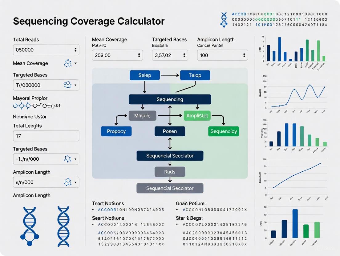
The following diagram illustrates the core workflow from generating sequencing data to making confident variant calls, highlighting how coverage metrics influence each step.
FAQs and Troubleshooting Guides
FAQ 1: What is the difference between a sequencing artifact and a true biological variant?
A sequencing artifact is a variation introduced by non-biological processes during the sequencing workflow, such as library preparation, PCR amplification, or the sequencing process itself [6]. In contrast, a true biological variant is an actual mutation present in the original sample.
- Examples of Artifacts: Apparent insertions or deletions due to sequencing errors in homopolymer regions, base miscalls, or biases in PCR amplification that alter the apparent abundance of a variant [6].
- How to Differentiate: Proper experimental design, including replicates and control samples, along with the use of bioinformatic tools that filter out low-quality calls and common artifacts, helps distinguish real variants. High sequencing depth also allows you to statistically distinguish low-frequency true variants from noise [1] [2].
FAQ 2: Our coverage is highly uneven, with some amplicons having very low reads. What could be the cause?
Uneven coverage is a common issue in amplicon-based sequencing like the AmpliSeq panels. Potential root causes and solutions are outlined below.
Table 3: Troubleshooting Guide for Uneven Coverage
| Problem Category | Typical Failure Signals | Common Root Causes & Corrective Actions |
|---|---|---|
| Sample Input/Quality | Low library complexity; smear in electropherogram [7]. | Cause: Degraded DNA/RNA or contaminants (phenol, salts).Fix: Re-purify input sample; use fluorometric quantification (e.g., Qubit) instead of absorbance alone [7]. |
| Amplification/PCR Bias | Over-amplification artifacts; high duplicate rate; specific amplicon dropouts [7]. | Cause: Too many PCR cycles; inefficient polymerase due to inhibitors; primer exhaustion.Fix: Optimize PCR cycle number; use high-fidelity polymerases; ensure primers are designed for uniform amplification [7]. |
| Library Preparation | Unexpected fragment size; high adapter-dimer peaks [7]. | Cause: Inaccurate quantification leading to suboptimal adapter-to-insert ratios.Fix: Accurately quantify fragmented DNA and titrate adapter concentrations to minimize dimers and maximize ligation efficiency [7]. |
FAQ 3: How do I determine the appropriate depth of coverage for my childhood cancer study?
The required depth depends on your specific study objectives and the variants of interest [1].
- Define Study Objectives: Are you searching for common germline variants or low-frequency somatic mutations? Detecting rare variants (e.g., a somatic mutation present in <10% of cells) requires a much higher depth than calling common germline variants [1] [2].
- Consider Variant Type: Single Nucleotide Variants (SNVs) are relatively straightforward to call. However, detecting Insertions-Deletions (Indels) and Structural Variants (SVs) often requires higher coverage and more sophisticated bioinformatic tools [2].
- Account for Tumor Purity: If sequencing tumor samples, the lower the tumor cell percentage (purity), the higher the sequencing depth required to detect somatic mutations present in only one allele of the tumor cells.
- Leverage Coverage Calculators: Use tools like the Illumina Sequencing Coverage Calculator to estimate the reagents and sequencing runs needed to achieve your desired depth for a given panel size [8] [4].
In the context of the AmpliSeq Childhood Cancer Panel, a precise understanding of NGS coverage is not merely a quality control metric—it is the foundation upon which accurate variant discovery rests. By rigorously planning experiments to achieve sufficient depth, breadth, and uniformity of coverage, researchers can generate data with the statistical confidence needed to uncover the genetic drivers of childhood cancers. This, in turn, accelerates drug development and paves the way for more personalized and effective therapeutic strategies.
In targeted next-generation sequencing (NGS) using the AmpliSeq for Illumina Childhood Cancer Panel, coverage depth is the foundational determinant of assay sensitivity. "Coverage" refers to the number of times a specific nucleotide is read during sequencing, while "sensitivity" defines the lowest level at which a variant can be reliably detected. For childhood cancer research, where specimens often have low tumor purity or require minimal residual disease monitoring, understanding this relationship is critical for generating meaningful data.
The AmpliSeq Childhood Cancer Panel investigates 203 genes associated with cancers in children and young adults, detecting multiple variant classes including single nucleotide variants (SNVs), insertions-deletions (indels), copy number variants (CNVs), and gene fusions [5]. Each variant type has unique detection challenges, necessitating specific coverage requirements to achieve the sensitivity required for robust research outcomes. Failure to achieve adequate coverage risks missing clinically actionable variants, potentially compromising research conclusions and subsequent drug development decisions.
Coverage Recommendations and Performance Data
Quantitative Coverage Targets
Based on manufacturer specifications and recent validation studies, the table below summarizes minimum and recommended coverage depths for different variant types in childhood cancer research panels:
Table 1: Coverage Recommendations for Variant Detection
| Variant Type | Minimum Coverage | Recommended Mean Coverage | Demonstrated Sensitivity | Key Influencing Factors |
|---|---|---|---|---|
| SNVs/Indels | 500-1,000x | 2,500-6,000x | 95% at AF ≥0.5% [9] | Allele fraction, background error rate |
| Gene Fusions | 20-1100 reads | N/A | >99% with ≥1100 reads [10] | Breakpoint location, RNA quality |
| CNVs | 500-1,000x | 2,500x | 5 copies for amplification [10] | Tumor purity, bin size for analysis |
| Low AF Variants (MRD) | 10,000x | 10,000x | ~80% at AF 0.2% [9] | Sequencing depth, error suppression |
For the AmpliSeq Childhood Cancer Panel specifically, Illumina recommends a minimum coverage of 1,000x and a mean coverage of 6,000x, requiring approximately 2 million reads per DNA sample [11]. These targets ensure that even variants with low allele fractions (AF) are detected with high confidence while maintaining cost-effectiveness for research applications.
Experimental Validation of Coverage-Sensitivity Relationship
Recent studies with pediatric cancer panels have experimentally quantified how coverage depth impacts sensitivity. The SJPedPanel, a comprehensive panel for childhood malignancies, demonstrated in dilution experiments that detection rates fall to approximately 80% at AF 0.2% even with ultradeep sequencing at 10,000x coverage [9]. This highlights the practical limitations of detecting very low-frequency variants, important for minimal residual disease monitoring.
The CANSeqTMKids pan-cancer panel established a limit of detection (LoD) at 5% allele fraction for SNVs and indels with validated sensitivity and specificity greater than 99% [10]. This performance was achieved using optimized laboratory protocols and bioinformatic pipelines with coverage depths tailored to each variant type. For fusion detection, this panel required approximately 1,100 reads for reliable identification [10].
Table 2: Experimental Performance Metrics from Recent Studies
| Study/Panel | Genes Covered | Input Requirements | Optimal Input | Specimen Types Validated |
|---|---|---|---|---|
| SJPedPanel [9] | 357 genes + 297 introns + 7,590 SNPs | Low input optimized | 10,000x for AF 0.2% | FFPE, bone marrow, blood |
| CANSeqTMKids [10] | 203 unique genes | 5 ng nucleic acid, 20% neoplastic content | 5% AF for SNVs/Indels | FFPE, cell blocks, blood, bone marrow |
| OncoKids [12] | 44 full genes, 82 hotspots, 24 amplifications | 20 ng DNA, 20 ng RNA | 1421 fusion targets | FFPE, frozen tissue, bone marrow |
Technical Implementation Guide
Experimental Design and Sequencing Planning
Proper experimental design begins with understanding your sequencing requirements. For a typical run on an Illumina MiSeq v3 flow cell providing approximately 25 million reads, you can sequence:
- Up to 11 DNA samples (targeting ~2 million reads/sample at 8:1 DNA:RNA ratio)
- Up to 96 RNA-only samples (targeting ~0.25 million reads/sample) [11]
The amplification-based library preparation for the AmpliSeq Childhood Cancer Panel requires 5-6 hours for library preparation with less than 1.5 hours of hands-on time, using only 10 ng of high-quality DNA or RNA input [5]. This low input requirement makes it particularly suitable for precious pediatric samples with limited material.
Sample Quality Considerations
For formalin-fixed paraffin-embedded (FFPE) tissues, a common source for pediatric cancer samples, the AmpliSeq for Illumina Direct FFPE DNA product allows for DNA preparation without deparaffinization or DNA purification [5]. When working with RNA samples for fusion detection, the AmpliSeq cDNA Synthesis for Illumina kit is required to convert total RNA to cDNA [5].
The following workflow diagram illustrates the key decision points in experimental planning:
Troubleshooting Common Coverage Issues
Frequently Asked Questions
Why do I have uneven coverage across my amplicons? Over-amplification during library preparation can result in uneven coverage of amplicons and compromised uniformity [13]. If your library concentration is greater than 20 nM after amplification, re-amplify your targets with less input DNA or reduce the number of target amplification cycles. Proper quantification of input DNA using recommended methods like the TaqMan RNase P Detection Reagents Kit or Qubit dsDNA HS Assay Kit is critical to prevent this issue [13].
How can I improve detection of low-frequency variants? For variants with allele fractions below 1%, increase sequencing depth to 10,000x and utilize computational error suppression methods. The SJPedPanel demonstrated that while detection rates are approximately 95% at AF 0.5%, they decrease to about 80% at AF 0.2% even with 10,000x coverage [9]. Implement unique molecular identifiers (UMIs) and error suppression algorithms to reduce background error rates which typically range from 10⁻⁶ to 10⁻⁴ for substitutions [9].
What causes failed fusion detection in RNA samples? Insufficient read depth for fusion transcripts is a common cause. The CANSeqTMKids panel established that approximately 1,100 reads are needed for reliable fusion detection [10]. Ensure adequate input RNA quality and quantity, and use the AmpliSeq cDNA Synthesis for Illumina kit for proper cDNA conversion. For the AmpliSeq Childhood Cancer Panel, target at least 0.25 million reads per RNA sample [11].
Why are my CNV results inconsistent? CNV detection requires sufficient coverage breadth and depth. Target a minimum of 1,000x coverage with even coverage distribution across targets. The CANSeqTMKids panel established a limit of detection of 5 copies for gene amplifications [10]. Use panels that include single nucleotide polymorphism (SNP) markers spread across chromosomes, like the 7,590 SNPs in the SJPedPanel, to improve CNV detection accuracy [9].
Essential Research Reagent Solutions
Table 3: Key Research Reagents for Childhood Cancer Panel Implementation
| Product Name | Function | Application Notes |
|---|---|---|
| AmpliSeq Library PLUS [5] | Library preparation reagents | Available in 24, 96, and 384 reaction formats |
| AmpliSeq CD Indexes [5] | Sample multiplexing | Includes 96 unique indexes per set; multiple sets available |
| AmpliSeq cDNA Synthesis [5] | RNA to cDNA conversion | Required for RNA panels; enables fusion detection |
| AmpliSeq Library Equalizer [5] | Library normalization | Bead-based normalization to ~100 pM; alternative to quantification |
| AmpliSeq Direct FFPE DNA [5] | FFPE DNA preparation | Processes unstained slide-mounted FFPE tissues without deparaffinization |
| AmpliSeq Sample ID Panel [5] | Sample identification | Human SNP genotyping panel for sample tracking and identification |
Optimal sequencing coverage for the AmpliSeq Childhood Cancer Panel is not a single value but a carefully considered parameter that balances detection sensitivity, specificity, and practical research constraints. By implementing the coverage recommendations and troubleshooting strategies outlined in this guide, researchers can maximize the analytical sensitivity of their childhood cancer studies across all variant types—from low-frequency SNVs to complex structural variants—ultimately advancing drug development and precision medicine for pediatric malignancies.
Frequently Asked Questions
What is the Lander/Waterman equation and why is it fundamental to my sequencing experiment?
The Lander/Waterman equation is the foundational mathematical model for predicting the coverage of a sequencing project. Coverage depth refers to the average number of sequencing reads that align to, or "cover," each base in your sequenced sample. The equation calculates coverage (C) based on your read length (L), number of reads (N), and haploid genome length (G) as follows: C = LN / G [14]. For targeted panels like the AmpliSeq Childhood Cancer Panel, "G" represents the total size of the targeted regions, not the entire genome. This calculation helps ensure your experiment generates enough data to detect variants with confidence [14] [15].
How do I apply the Lander/Waterman equation to the AmpliSeq Childhood Cancer Panel?
The AmpliSeq Childhood Cancer Panel investigates 203 genes using amplicon sequencing [5]. To calculate coverage, you first need to know the total size of the targeted regions. While the total panel size isn't explicitly listed, the principle remains the same: you substitute this effective "G" into the equation along with your average read length (L) and the number of passing reads you expect from your run (N). Illumina recommends sequencing amplicons to a length that covers the entire amplicon insert [14].
My coverage is uneven. What are the limitations of the Lander/Waterman model?
The standard Lander/Waterman theory relies on the assumption that sequenced fragments are randomly and uniformly distributed across the target [15] [16]. In reality, several experimental factors can lead to uneven coverage, including:
- GC Bias: Regions with high or low GC content may amplify less efficiently during library preparation.
- Amplification Bias: The PCR-based AmpliSeq protocol can sometimes lead to uneven representation of targets.
- Position-Specific Bias (Edge Effects): Especially in filtered or targeted libraries, the ends of amplicons or genomic islands can have a lower probability of being sequenced than central regions [16].
Because of these factors, the actual coverage achieved may differ from the theoretical prediction, and a higher average coverage is often required to ensure all regions are sequenced sufficiently [15].
What coverage depth should I target for somatic variant detection in childhood cancer samples?
While the optimal coverage can vary based on the specific variant type and allelic fraction, high coverage is critical for confidently detecting low-frequency somatic variants. The Lander/Waterman equation helps you determine the number of reads needed to achieve this. For cancer research, where detecting heterozygous mutations is important, theories suggest that the amount of data needed is "significantly higher than for traditional haploid projects" [15]. In practice, at least 30-fold redundancy (where each nucleotide is spanned by an average of 30 sequence reads) is now standard, with requirements potentially rising to 50-fold or more for certain applications like detecting structural variants [15].
Troubleshooting Guides
Problem: Inadequate Coverage Across All Targets
Issue: Your data shows that some genomic regions have coverage far below the calculated average, potentially causing you to miss variants.
Solutions:
- Recalculate Library Concentration: Use qPCR-based quantification (like the Ion Library TaqMan Quantitation Kit) instead of fluorometric methods for a more accurate measure of amplifiable library content [17].
- Increase Sequencing Depth: Use the Lander/Waterman equation to determine how many additional reads are needed. For example, if your initial run with
Nreads yielded coverageC, to achieve a new coverage targetC_target, you need approximatelyN_new = N * (C_target / C)reads. - Verify Input DNA Quality: The panel requires 10 ng of high-quality DNA. Degraded or low-quality DNA from FFPE samples can lead to poor amplification of larger amplicons. Consider using the AmpliSeq for Illumina Direct FFPE DNA product for such samples to improve performance [5].
Issue: Your data shows uniformly high coverage, far exceeding what is necessary for confident variant calling, leading to unnecessary sequencing costs.
Solutions:
- Optimize Loading Concentration: If using an Illumina instrument, over-sequencing often results from loading too much library onto the flow cell. Prefer qPCR-based quantification for precise loading [17].
- Use the Sequencing Coverage Calculator: Illumina provides an online tool to help determine the optimal amount of sequencing reagents and number of runs for your desired coverage, preventing wastage [8] [14].
- Employ a More Efficient Strategy: Recent research suggests that by integrating sequencing and computation, it's possible to break the traditional Lander-Waterman bound, potentially reducing the total number of bases that need to be sequenced from O(G ln G) to O(G) by terminating the sequencing of redundant fragments early [18].
Key Parameters for Coverage Calculation
Table 1: Essential variables for the Lander/Waterman equation in the context of the AmpliSeq Childhood Cancer Panel.
| Variable | Description | Considerations for the Childhood Cancer Panel |
|---|---|---|
| C | Coverage Depth | Aim for >30x minimum for somatic variant detection; higher coverage (e.g., 50x) may be needed for structural variant calling [15]. |
| L | Read Length | Must be long enough to cover the entire amplicon insert; Illumina generally recommends 2 x 150 bp for targeted sequencing [14]. |
| N | Number of Reads | This is the value you solve for; determined by your sequencing instrument's output and library loading concentration. |
| G | Target Size | The total base-pair size of all 203 genes and other targeted regions in the panel. Consult the panel's manifest file for the exact value. |
The Scientist's Toolkit
Table 2: Essential research reagent solutions for the AmpliSeq Childhood Cancer Panel workflow [5].
| Product Name | Function | Specifications |
|---|---|---|
| AmpliSeq Childhood Cancer Panel | Ready-to-use primer pool for investigating 203 cancer-associated genes. | Sufficient for 24 reactions; detects SNPs, indels, CNVs, fusions. |
| AmpliSeq Library PLUS | Reagents for preparing sequencing libraries from the amplified targets. | Sold in 24, 96, or 384 reactions. |
| AmpliSeq CD Indexes | Unique barcode sequences to multiplex multiple samples in a single run. | Various sets (A-D) available, each with 96 unique indexes. |
| AmpliSeq cDNA Synthesis for Illumina | Converts total RNA to cDNA for RNA-based fusion detection. | Required when using the panel with RNA input. |
| AmpliSeq for Illumina Direct FFPE DNA | Prepares DNA from FFPE tissues without need for deparaffinization or purification. | 24 reactions per kit; ideal for challenging clinical samples. |
Experimental Planning Workflow
The following diagram illustrates the key steps in planning your sequencing run, from sample preparation to data analysis, with an emphasis on where coverage calculation is critical.
The AmpliSeq Childhood Cancer Panel for Illumina is a targeted resequencing solution designed for the comprehensive evaluation of 203 genes associated with childhood and young adult cancers, including leukemias, brain tumors, and sarcomas [5]. This ready-to-use panel saves significant time and effort by eliminating the need for researchers to identify targets, design primers, and optimize panels themselves [5].
The entire library preparation process has an assay time of 5-6 hours (excluding library quantification, normalization, or pooling) with a hands-on time of less than 1.5 hours [5]. The workflow is compatible with several Illumina sequencing systems, including the MiSeq, NextSeq 500, NextSeq 1000, and MiniSeq Systems [5].
Technical Specifications
The table below summarizes the core technical specifications and requirements for the AmpliSeq Childhood Cancer Panel.
| Specification Category | Details |
|---|---|
| Target | 203 genes associated with childhood and young adult cancers [5] |
| Input Quantity | 10 ng of high-quality DNA or RNA [5] |
| Method | Amplicon sequencing [5] |
| Hands-On Time | < 1.5 hours [5] |
| Total Assay Time (Library Prep) | 5-6 hours [5] |
| Variant Classes Detected | Single nucleotide polymorphisms (SNPs), Insertions-deletions (indels), Copy number variants (CNVs), Gene fusions, Somatic variants [5] |
| Compatible Instruments | MiSeq System, NextSeq 550 System, NextSeq 2000 System, NextSeq 1000 System, MiSeqDx in Research Mode, MiniSeq System [5] |
| Specialized Sample Types | Blood, Bone marrow, FFPE tissue, Low-input samples [5] |
Research Reagent Solutions
To perform an experiment with the Childhood Cancer Panel, the core panel is not sufficient. The table below lists the essential companion products required for a complete workflow.
| Reagent Solution | Function |
|---|---|
| AmpliSeq Library PLUS | Provides reagents for preparing sequencing libraries. Must be purchased separately from the panel and index adapters [5]. |
| AmpliSeq CD Indexes | Used to label individual samples (e.g., Set A-D), allowing multiple samples to be sequenced together. Sufficient for 96 samples per set [5]. |
| AmpliSeq cDNA Synthesis for Illumina | Required to convert total RNA to cDNA when using the panel with RNA inputs [5]. |
| AmpliSeq for Illumina Direct FFPE DNA | Enables DNA preparation and library construction from FFPE tissues without the need for deparaffinization or DNA purification [5]. |
| AmpliSeq Library Equalizer for Illumina | An easy-to-use solution for normalizing libraries after preparation to ensure balanced sequencing [5]. |
Frequently Asked Questions (FAQs)
Q1: What is the minimum input requirement, and can I use RNA with this panel? The panel requires only 10 ng of high-quality input material. It is versatile and supports both DNA and RNA. If you are starting with RNA, you must first use the AmpliSeq cDNA Synthesis for Illumina kit to convert your RNA to cDNA [5].
Q2: How long does the library preparation process take? The hands-on time for library preparation is less than 1.5 hours. The total assay time for library preparation is 5-6 hours, though this does not include subsequent steps like library quantification, normalization, or pooling [5].
Q3: My samples are from FFPE tissue. Does this require a special protocol? Yes, for FFPE tissues, the AmpliSeq for Illumina Direct FFPE DNA product is available. It allows for DNA preparation and library construction directly from slide-mounted FFPE tissues without the need for separate deparaffinization or DNA purification steps [5].
Q4: What types of variants can this panel detect? This targeted panel is designed to detect a comprehensive range of somatic variant classes, including SNPs, insertions-deletions (indels), copy number variants (CNVs), and gene fusions [5].
Q5: How does amplicon-based sequencing perform compared to other enrichment methods? Independent studies have found that amplicon-based methods like AmpliSeq show high concordance with other technologies. One study comparing AmpliSeq to SureSelect for exome sequencing reported a high concordance (>97%) with microarray genotypes and, when validating against a reference standard, demonstrated sensitivity and positive predictive values of >93% and >80%, respectively [19]. An optimized bioinformatics pipeline can further improve these results.
Frequently Asked Questions
Q1: What does a 'good' coverage histogram look like for my AmpliSeq Childhood Cancer Panel run? A good coverage histogram demonstrates a highly uniform distribution with a strong peak at high coverage depths and minimal reads at low coverages. For the AmpliSeq Childhood Cancer Panel, a mean read depth of greater than 1000x has been validated, providing high sensitivity for variant detection [20]. The histogram should show the vast majority of bases within the target regions falling within this high-coverage range, with only a very small percentage, if any, in the low-coverage bins (e.g., [0x:1x) or [1x:3x)) [21].
Q2: A significant portion of my data is in the [0x:1x) and [1x:3x) bins. What could be the cause? This indicates poor or insufficient coverage in certain genomic regions, which can lead to false negatives. Potential causes and solutions are detailed in the troubleshooting guide below.
Q3: What is the minimum coverage depth required for reliable mutation detection in a clinical setting? There is no universal consensus, but coverage requirements depend on the intended Limit of Detection (LOD) for variant allele frequency (VAF). For the Childhood Cancer Panel, a depth of >1000x was validated to achieve 98.5% sensitivity for DNA variants at 5% VAF [20]. One study recommends a minimum depth of 1,650x for a targeted NGS panel to robustly detect mutations at ≥3% VAF, based on binomial probability distribution to minimize false positives/negatives [22].
Q4: How does coverage uniformity impact the detection of fusion genes? The RNA component of the panel, which detects fusion genes, is particularly reliant on uniform coverage across fusion breakpoints. In a validation study, the panel demonstrated 94.4% sensitivity for RNA analysis [20]. Inadequate or uneven coverage can lead to missed fusion events, which are often clinically critical, as 97% of identified fusions were found to refine diagnosis [20].
Troubleshooting Guide: Poor Coverage Distribution
| Observation | Potential Cause | Recommended Action |
|---|---|---|
| Isolated regions of low coverage | Specific amplicons failing to amplify due to sequence complexity (e.g., high GC content), or primer binding issues. | 1. Inspect Amplicons: Check the performance of specific amplicons in the panel. 2. Re-assess Input DNA/RNA: Ensure input nucleic acids meet quality and quantity specifications (100 ng DNA/RNA used in validation [20]). 3. Use Fresh Reagents: Avoid multiple freeze-thaw cycles of enzymatic components. |
| Global low coverage across most targets | Insufficient sequencing depth, degraded nucleic acid sample, or issues during library preparation (e.g., failed PCR amplification). | 1. Check Total Reads: Verify that the sequencing run yielded the expected number of total reads or clusters. 2. Assess Nucleic Acid Quality: Use a fluorometer for accurate concentration and methods like TapeStation to check integrity [20]. 3. Review Library Prep: Confirm that all steps in the AmpliSeq library preparation protocol were followed correctly. |
| High duplicate read rate | Starting with an insufficient amount of input DNA or RNA, leading to over-amplification of a limited number of original molecules. | 1. Increase Input: Use the recommended 100 ng of DNA and RNA as input [20]. The panel can work with as little as 10 ng, but this may impact uniformity [5]. 2. Normalize Libraries Accurately: Use the AmpliSeq Library Equalizer to ensure balanced representation of libraries before pooling [5]. |
Experimental Protocol: Validating Panel Performance
The following protocol, adapted from a published validation study for the AmpliSeq Childhood Cancer Panel, outlines key steps to generate and assess coverage data [20].
1. Sample Selection and Nucleic Acid Extraction:
- Use commercial reference controls (e.g., SeraSeq Tumor Mutation DNA Mix, SeraSeq Myeloid Fusion RNA Mix) alongside patient samples.
- Extract DNA and RNA using standardized kits (e.g., QIAamp DNA Mini Kit, TriPure reagent).
- Quantification and Quality Control: Precisely quantify DNA and RNA using a fluorometer (e.g., Qubit). Assess purity (OD260/280 >1.8) and integrity using systems like TapeStation or Labchip.
2. Library Preparation and Sequencing:
- Use 100 ng of DNA and RNA as input.
- For RNA, first perform cDNA synthesis using the AmpliSeq cDNA Synthesis for Illumina kit [5].
- Prepare libraries using the AmpliSeq for Illumina Childhood Cancer Panel according to the manufacturer's instructions.
- Pool DNA and RNA libraries at a 5:1 ratio.
- Sequence on an Illumina MiSeq or NextSeq system.
3. Data Analysis and Coverage Assessment:
- Process sequencing data through a pipeline (e.g., DRAGEN) to generate a coverage histogram report [21].
- The histogram report (e.g.,
_hist.csvfile) provides the percentage of bases in the target regions that fall within defined coverage ranges (e.g., [0x:1x), [1x:3x), [100x:inf)) [21]. - Calculate the mean depth of coverage and the percentage of targets covered at a minimum depth (e.g., 100x, 500x, or 1000x).
4. Key Performance Metrics (Validation Benchmarks): The following table summarizes the performance metrics achieved during an independent technical validation of the panel [20].
| Performance Metric | Result Achieved |
|---|---|
| Mean Read Depth | > 1000x |
| DNA Sensitivity (at 5% VAF) | 98.5% |
| RNA Sensitivity | 94.4% |
| Specificity | 100% |
| DNA Reproducibility | 100% |
| RNA Reproducibility | 89% |
The Scientist's Toolkit: Essential Research Reagents
The following reagents are critical for successfully running the AmpliSeq Childhood Cancer Panel.
| Research Reagent | Function | Specification |
|---|---|---|
| AmpliSeq for Illumina Childhood Cancer Panel | Core panel containing primers to amplify targets in 203 genes associated with pediatric cancers. | 24 reactions. Detects SNPs, Indels, CNVs, and fusions [5]. |
| AmpliSeq Library PLUS | Reagents for preparing sequencing libraries from the amplified PCR products. | Sold separately in 24, 96, or 384 reactions [5]. |
| AmpliSeq CD Indexes | Unique dual indexes (UDIs) used to barcode individual samples for multiplexing. | Sets A-D available; each set contains 96 indexes [5]. |
| AmpliSeq cDNA Synthesis for Illumina | Converts total RNA to cDNA, which is required for the RNA fusion component of the panel. | Essential for detecting the 97 gene fusions included in the panel [5]. |
| AmpliSeq Library Equalizer | Bead-based normalization reagent to ensure balanced representation of libraries in a pooled sequence run. | Improves coverage uniformity across samples [5]. |
Workflow for Investigating Coverage Issues
This diagram outlines a logical pathway for diagnosing and addressing common coverage problems.
Practical Guide: Calculating and Achieving Optimal Coverage for Your Childhood Cancer Panel Runs
This technical support guide provides a detailed walkthrough for using the Illumina Sequencing Coverage Calculator, specifically tailored for researchers employing the AmpliSeq for Illumina Childhood Cancer Panel (Catalog ID: 20028446) [8]. Proper calculation of sequencing coverage is a foundational step in experiment planning, ensuring that your data has the statistical confidence required to detect rare variants and make reliable biological conclusions [3] [4]. This resource will help you determine the precise reagents and sequencing runs needed to achieve your desired coverage depth.
Sequencing Coverage Fundamentals
What is Sequencing Coverage?
Sequencing coverage or depth describes the number of unique sequencing reads that align to a specific region in a reference genome [3]. Expressed as an "X" factor (e.g., 30x), it represents how many times, on average, a base in the genome is read [4]. Higher coverage directly translates to greater confidence in variant calling, especially for detecting low-frequency mutations common in cancer research [3].
Key Metrics and Equations
The fundamental equation for calculating coverage is the Lander/Waterman equation [4]: C = (L × N) / G
- C: Coverage (X)
- L: Read Length (bp)
- N: Number of reads
- G: Haploid genome length (bp)
For targeted panels like the AmpliSeq Childhood Cancer Panel, "G" typically refers to the total size of the targeted genomic regions.
Evaluating Your Sequencing Run
After sequencing, these metrics help assess data quality [4]:
- Mean Mapped Read Depth: The average number of reads aligned to a reference base position. This is your actual coverage.
- Coverage Uniformity: How evenly reads are distributed across the target regions. Higher uniformity means fewer under- or over-sequenced areas.
- Inter-Quartile Range (IQR): A measure of coverage variability. A lower IQR indicates more uniform coverage.
Step-by-Step Guide to the Coverage Calculator
Accessing the Tool
The Illumina Sequencing Coverage Calculator is available on the official Illumina support website [23] [24]. Illumina also provides a video tutorial demonstrating the calculator's use for various applications, including pre-defined panels [24].
Defining Your Experiment Parameters
Before using the calculator, gather the following information:
- Panel Selection: Choose "AmpliSeq for Illumina Childhood Cancer Panel" from the pre-defined options [24] [8].
- Desired Coverage: Set your target mean coverage. For variant calling in cancer research, this is often 100x or higher to ensure statistical power.
- Number of Samples: Input the total number of samples you plan to multiplex in a single run.
- Instrument and Flow Cell Type: Select your specific Illumina instrument (e.g., MiniSeq, NovaSeq 6000) and flow cell model, as this determines total data output [24].
The following diagram illustrates the logical process of using the calculator and related experimental steps:
Interpreting Calculator Outputs
The calculator will provide several key outputs to guide your experiment planning:
- Number of Reads Required: The total reads needed to achieve your target coverage for all samples.
- Flow Cells or Sequencing Runs: The number of flow cells (or a fraction thereof) required to generate the necessary data.
- Recommended Reagent Kits: The specific library prep and sequencing reagent kits compatible with your setup.
Frequently Asked Questions (FAQs)
The calculator recommends more flow cells than I anticipated. How can I optimize my run? Consider increasing the number of samples you multiplex per run. The calculator allows you to adjust this parameter. Be aware that excessive multiplexing can lead to lower coverage per sample, so balance is key. You can also verify if a different flow cell type (e.g., one with higher output) is available for your instrument.
What is a common cause of low coverage uniformity in my results? Poor uniformity, indicated by a high IQR, often stems from issues during the library preparation stage. Inefficient or biased target enrichment, PCR artifacts, or low-quality input DNA can lead to some genomic regions being over-represented while others are under-represented [3]. Review your lab protocols carefully.
My raw read depth seems sufficient, but my mapped read depth is low. Why? Raw read depth is the total data produced by the sequencer. Mapped read depth is the data that successfully aligns to your target regions [4]. A large discrepancy usually means a significant portion of your reads were discarded during alignment due to poor quality, adapter contamination, or off-target sequencing.
How does the calculator account for different instruments? The tool has built-in performance profiles for each Illumina sequencing system (e.g., MiniSeq, NovaSeq 6000) [24]. These profiles account for the instrument's specific output, read length capabilities, and flow cell configurations, ensuring accurate run planning.
Troubleshooting Guide
| Problem | Possible Cause | Solution |
|---|---|---|
| Consistently Low Coverage | Insufficient sequencing depth; poor library complexity. | Re-calculate required reads using the calculator. Re-assess library quality (e.g., Bioanalyzer profile) and re-prep if necessary. |
| Poor Coverage Uniformity | Biases in target enrichment or amplification during library prep [3]. | Optimize hybridization conditions and PCR cycles. Use validated, high-quality input DNA. |
| Low Signal Intensity | Issues with cluster generation; flow cell or reagent problems [25]. | Check cluster density images and metrics. Ensure reagents are fresh and stored correctly. |
| Calculator Output Seems Inaccurate | Incorrect input parameters (e.g., wrong panel size, sample count). | Double-check that the "AmpliSeq Childhood Cancer Panel" is selected and all sample information is entered correctly. |
The Scientist's Toolkit: Essential Research Reagent Solutions
| Item | Function |
|---|---|
| AmpliSeq for Illumina Childhood Cancer Panel | A targeted panel designed to enrich for genomic regions and variants relevant to childhood cancers, enabling focused sequencing. |
| Library Preparation Kit | Reagents used to fragment DNA and ligate Illumina-specific adapters, making the sample compatible with the sequencing platform. |
| Sequencing Reagents (Flow Cell, SBS Chemistry) | The consumables and chemistry that enable the actual sequencing-by-synthesis process on the instrument. |
| Illumina Sequencing Coverage Calculator | An online planning tool that determines the reagents and sequencing runs required to achieve a desired coverage depth [23] [8]. |
The table below summarizes typical coverage recommendations for various sequencing methods, which can serve as a benchmark. Always consult literature specific to your childhood cancer research for precise targets.
| Sequencing Method | Recommended Coverage | Key Considerations for Childhood Cancer Panel |
|---|---|---|
| Whole Genome Sequencing (WGS) | 30× to 50× [4] | Not typically used for targeted panels; shown for reference. |
| Whole-Exome Sequencing | 100× [4] | A good analogy for the depth often needed for robust variant detection in targeted gene panels. |
| Targeted Panels (e.g., AmpliSeq) | Varies by application | 100x - 500x+ is common for somatic variant detection in cancer to confidently identify subclonal populations. |
Sequencing Coverage Calculator for AmpliSeq Childhood Cancer Panel Research
Next-generation sequencing (NGS) has revolutionized molecular diagnostics for pediatric cancers, providing comprehensive genetic information that refines diagnosis, prognosis, and therapeutic strategies. The AmpliSeq for Illumina Childhood Cancer Panel represents a targeted approach specifically designed for the unique genetic landscape of childhood and young adult cancers. This technical support center addresses the critical interplay between sample number, read length, and desired mean depth—three fundamental parameters that researchers must optimize to generate clinically actionable data from this panel.
Key Input Parameters for Coverage Calculations
Sample Number
The number of samples pooled in a single sequencing run directly impacts the achievable coverage per sample. The Childhood Cancer Panel is designed for multiplexing, with available index adapter sets allowing for labeling of up to 384 unique samples [5]. When planning an experiment, consider that increasing the number of samples pooled per run will decrease the average coverage per sample unless sequencing throughput is correspondingly increased [26].
Read Length
The Childhood Cancer Panel generates amplicons with average sizes of 114 bp for DNA targets and 122 bp for RNA fusion targets [20]. These sizes inform the appropriate read length selection on Illumina sequencing platforms. The panel is compatible with multiple Illumina systems including MiSeq, NextSeq 550, NextSeq 1000, NextSeq 2000, and MiniSeq systems [5]. Each platform offers different read length capabilities that should be matched to the amplicon sizes.
Desired Mean Depth
The target mean depth of coverage is determined by the specific variant types being investigated and their expected allele frequencies. For somatic variant detection in pediatric cancers, where mutant allele frequencies can be relatively low, a mean read depth greater than 1000× has been demonstrated to provide 98.5% sensitivity for variants with 5% variant allele frequency (VAF) [20]. This depth ensures reliable detection of clinically relevant low-frequency variants.
Table 1: Recommended Minimum Coverage by Variant Type
| Variant Type | Recommended Minimum Coverage | Key Considerations |
|---|---|---|
| Single Nucleotide Variants (SNVs) | 500× | Higher depth (≥1000×) enables detection of low VAF variants [20] |
| Insertions-Deletions (Indels) | 500× | Similar requirements as SNVs [20] |
| Gene Fusions | 1000× | Essential for reliable fusion detection in RNA [20] |
| Copy Number Variants (CNVs) | 1000× | Higher depth improves accuracy of copy number assessment [27] |
Parameter Interrelationships and Optimization
The relationship between sample number, read length, and desired mean depth follows a predictable mathematical relationship where total sequencing output must equal the product of sample number, mean depth, and target region size. Researchers can manipulate coverage by either increasing sequencing throughput (using a larger flow cell output or more powerful sequencing platform) or reducing the number of samples pooled per run [26].
For the Childhood Cancer Panel, which covers 203 genes associated with pediatric cancers, the total target space must be considered when calculating required sequencing output [5]. The panel detects multiple variant classes including single nucleotide polymorphisms (SNPs), gene fusions, somatic variants, insertions-deletions (indels), and copy number variants (CNVs) [5].
Experimental Protocols and Validation Data
Library Preparation Protocol
The AmpliSeq Childhood Cancer Panel requires 10 ng of high-quality DNA or RNA input and has a total hands-on time of less than 1.5 hours [5]. The complete library preparation process takes approximately 5-6 hours, excluding library quantification, normalization, or pooling time [5]. For RNA studies, the AmpliSeq cDNA Synthesis for Illumina kit is required to convert total RNA to cDNA prior to library preparation [5].
Performance Validation
Technical validation of the Childhood Cancer Panel demonstrates robust performance characteristics. In a study focused on pediatric acute leukemia, the panel achieved a mean read depth greater than 1000× with 98.5% sensitivity for DNA variants at 5% variant allele frequency and 94.4% sensitivity for RNA fusions [20]. The assay also demonstrated 100% specificity and reproducibility for DNA and 89% reproducibility for RNA [20].
Table 2: Experimental Performance Metrics for Childhood Cancer Panel
| Performance Metric | DNA Analysis | RNA Analysis |
|---|---|---|
| Mean Read Depth | >1000× | >1000× |
| Sensitivity | 98.5% (for variants with 5% VAF) | 94.4% |
| Specificity | 100% | Not specified |
| Reproducibility | 100% | 89% |
| Limit of Detection | Established with commercial controls | Established with fusion mixes |
Technical Support: FAQs
How can I increase coverage for a fixed number of samples?
You can manipulate coverage by increasing sequencing throughput (e.g., using a larger flow cell output or more powerful sequencing platform) or reducing the number of samples pooled per run [26]. The Childhood Cancer Panel is compatible with multiple Illumina sequencing systems including MiSeq, NextSeq series, and MiniSeq, allowing flexibility in throughput selection [5].
What analysis tools are available for data from the Childhood Cancer Panel?
Local Run Manager and BaseSpace Sequence Hub have apps specifically designed for analysis of Childhood Cancer Panel data [26]. The DNA Amplicon Analysis App and RNA Amplicon Analysis App are available on BaseSpace Sequence Hub, with similar modules available in Local Run Manager [26]. For specialized analysis, OncoCNV caller is available for CNV analysis, and BaseSpace Variant Interpreter can be used for further interpretation of variant calls [26].
Can I combine different AmpliSeq panels on the same sequencing run?
It is possible to run three different AmpliSeq for Illumina designs, each with barcodes, on the same sequencing run [26]. However, researchers must ensure that their target amplicon size and required coverage can be achieved in a single run, considering the combined target space of all panels [26].
How is on-target performance measured for this panel?
On-target bases metric shows the percentage of total sequenced bases that map to target regions in the reference genome [26]. This metric reflects the percentage of bases from amplicons that were both designed and successfully generated sequence data mapping to the intended target regions [26].
The Scientist's Toolkit: Research Reagent Solutions
Table 3: Essential Research Reagents for Childhood Cancer Panel Workflow
| Product Name | Function | Specifications |
|---|---|---|
| AmpliSeq Childhood Cancer Panel | Core panel targeting 203 childhood cancer genes | 24 reactions; detects SNPs, fusions, indels, CNVs [5] |
| AmpliSeq Library PLUS | Library preparation reagents | Available in 24, 96, or 384 reaction formats [5] |
| AmpliSeq CD Indexes | Sample multiplexing | Sets A-D available; each set contains 96 indexes [5] |
| AmpliSeq cDNA Synthesis | RNA to cDNA conversion | Required for RNA studies with the panel [5] |
| AmpliSeq Direct FFPE DNA | DNA from FFPE tissues | Enables DNA preparation without deparaffinization [5] |
| AmpliSeq Library Equalizer | Library normalization | Bead-based normalization to ~100 pM [5] |
Optimizing sequencing coverage for the AmpliSeq Childhood Cancer Panel requires careful consideration of sample number, read length, and desired mean depth in the context of specific research objectives. The robust validation data and flexible platform compatibility of this panel enable researchers to tailor their sequencing approach to detect clinically relevant variants across the spectrum of pediatric malignancies. By understanding the interrelationships between these key parameters and utilizing the appropriate analytical tools, researchers can maximize the diagnostic and therapeutic insights gained from their childhood cancer sequencing studies.
Frequently Asked Questions (FAQs)
Q1: What is the difference between sequencing depth and coverage? These terms are often used interchangeably but have distinct meanings [1].
- Sequencing Depth (or Read Depth): Refers to the number of times a specific nucleotide is read during sequencing. It is expressed as an average (e.g., 100x) and higher depth increases confidence in variant calling, which is crucial for detecting rare variants [1].
- Coverage: Pertains to the proportion of the target genome or region that has been sequenced at least once. It is typically expressed as a percentage (e.g., 95% coverage). High coverage ensures the entire target region is represented, preventing gaps in the data [1].
Q2: Why are coverage targets of >1000x sometimes necessary? High coverage depths are essential for the reliable detection of low-frequency variants, which are common in cancer genomics. A peer-reviewed study recommends a minimum depth of coverage of 1,650x for targeted NGS mutation analysis of variants at a ≥3% Variant Allele Frequency (VAF). This high depth, coupled with a threshold of at least 30 mutated reads, minimizes the probability of both false positive and false negative results based on binomial probability distribution [22].
Q3: How does the AmpliSeq for Illumina Childhood Cancer Panel facilitate analysis? Official Illumina support documentation indicates that analysis for this panel is supported by specialized apps [26]:
- DNA Amplicon Analysis App (v2.0 or higher) in BaseSpace Sequence Hub for alignment and variant calling.
- OncoCNV caller, a BaseSpace Lab App, is available for CNV analysis. These integrated tools help researchers manage and interpret the complex data generated by deep sequencing.
Q4: What are common causes of insufficient coverage and how can they be resolved? The following table outlines common issues and their solutions.
| Issue | Description | Troubleshooting Steps |
|---|---|---|
| Insufficient Sequencing Throughput | The number of sequenced reads is too low to achieve the desired average depth across all targets. | Increase sequencing output by using a larger flow cell or a more powerful sequencing platform. Alternatively, reduce the number of samples pooled in a single sequencing run [26]. |
| Library Preparation Biases | Errors during DNA processing, amplification, or library prep can introduce artifacts and reduce effective coverage. | Use the Adaptors optimized for the AmpliSeq workflow as recommended by Illumina. Ensure high-quality input DNA and follow protocol guidelines meticulously [22] [26]. |
| Regional Drop-outs | Certain genomic regions (e.g., high-GC content, repetitive elements) are inherently difficult to sequence. | While amplicon-based panels are designed to minimize this, some regions may still be underrepresented. Analysis software metrics like "on-target bases" can help identify these issues [1] [26]. |
The Scientist's Toolkit: Research Reagent Solutions
The following table details key materials and their functions for successful NGS experiments with the Childhood Cancer Panel.
| Item | Function |
|---|---|
| AmpliSeq for Illumina Childhood Cancer Panel | A targeted panel designed to amplify and sequence specific genes associated with childhood cancers. |
| Optimized Adapters | Specific adapters provided with the assay for library preparation. Note that Nextera or TruSeq Adapters are not compatible [26]. |
| MiSeq / NextSeq Reagents | Sequencing reagents tailored for the Illumina sequencing platform being used (e.g., MiSeq for moderate throughput). |
| DNA Amplicon Analysis App | A dedicated software tool for alignment and variant calling from the sequenced data [26]. |
Experimental Protocols and Data Analysis
Validating Coverage Depth for Low VAF Detection A 2019 study provides a methodological framework for determining minimum coverage depth in diagnostic NGS [22]:
- Define Key Parameters:
- Limit of Detection (LOD): The minimum Variant Allele Frequency (VAF) you need to detect (e.g., 3%, 1%, or 0.5%).
- Sequencing Error Rate: The intrinsic error rate of your sequencing platform (conventionally between 0.1% and 1%).
- Assay-Specific Error: The additional error introduced during DNA processing and library preparation.
- Overall Error Rate: The sum of sequencing and assay-specific errors.
- Apply Binomial Probability Distribution: Use these parameters to calculate the probability of false positives and false negatives for a given coverage depth and variant-calling threshold (e.g., minimum number of variant-supporting reads).
- Utilize a Coverage Calculator: The study provides a user-friendly calculator (available on GitHub:
mvasinek/olgen-coverage-limit) to perform these statistical calculations and determine the required depth for a desired confidence level [22].
Lessons from a Real-World Cohort Analysis of a diagnostic cohort of 859 CLL patients highlights the practical importance of high-sensitivity NGS. The study, which used a minimum target read depth of 5,000x and an LOD of 1%, found that 25% of patients were positive for TP53 mutations. Crucially, over half (52.6%) of these positive cases carried variants with a VAF of 10% or lower, which would be missed by lower-sensitivity methods like Sanger sequencing [22].
Workflow and Data Relationships
The following diagram illustrates the logical process for determining the required coverage depth for an NGS experiment, incorporating key concepts from validation studies.
Decision Workflow for NGS Coverage Depth
The table below summarizes quantitative data on coverage depth recommendations from the literature, providing a quick reference for experimental planning.
| Recommended Depth | Variant Allele Frequency (VAF) | Key Rationale & Context |
|---|---|---|
| ~1,650x | ≥3% | Minimizes false positives/negatives; recommended with threshold of ≥30 mutated reads based on binomial distribution [22]. |
| 500x | 5% | A previously recommended depth for a LOD of 5% in clinical oncology [22]. |
| 100x - 500x | 5% - 10% | A range of coverages has been suggested, but a depth of 100x with a 10% LOD can lead to a high false negative rate (e.g., 45% theoretically) [22]. |
| >1000x | <5% | Generally required for confident detection of low-frequency subclonal variants, as demonstrated in a real-world CLL cohort [22]. |
Strategies for Sample Pooling and Flow Cell Selection on MiSeq, NextSeq, and Other Compatible Systems
Frequently Asked Questions (FAQs)
Q1: What are the primary causes of low cluster density on the MiSeq, and how can I avoid it?
Low cluster density, or underclustering, is a common issue that can lead to poor run performance, lower Q30 scores, and reduced data output. The main causes and their solutions are outlined below [28]:
- Inaccurate Library Quantification: Ensure precise quantification and quality checks of your sequencing libraries. The method should be appropriate for your specific library preparation kit.
- Improper NaOH Denaturation: Always prepare a fresh dilution of NaOH for denaturation and use it within 12 hours. The pH of the stock solution must be above 12.5. To minimize pipetting errors, prepare at least 1 ml of diluted NaOH.
- Issues with Structured or GC-Rich Libraries: For libraries with high GC content or secondary structures, improve cluster density consistency by using a heat denaturation step. After NaOH denaturation and dilution in HT1, incubate the library at 96°C for 2 minutes, then immediately place it on ice for 5 minutes before proceeding to cluster generation.
- Incorrect Handling of Low-Diversity Libraries: Low-diversity libraries, such as amplicon panels, require special handling. A minimum 5% spike-in of the PhiX control library is required to provide the nucleotide diversity needed for effective template generation. Furthermore, you may need to empirically reduce the library loading concentration by 30-40% below the optimal range.
Q2: My research uses the AmpliSeq for Illumina Childhood Cancer Panel. What are the key specifications and compatible instruments?
The AmpliSeq Childhood Cancer Panel is a targeted resequencing solution for investigating 203 genes associated with pediatric and young adult cancers [5]. Key specifications are summarized in the table below.
Table 1: AmpliSeq Childhood Cancer Panel Specifications [5]
| Specification | Detail |
|---|---|
| Assay Time | 5-6 hours (library prep only) |
| Hands-on Time | < 1.5 hours |
| Input Quantity | 10 ng high-quality DNA or RNA |
| Input Type | Blood, Bone Marrow, FFPE Tissue, Low-input samples |
| Method | Amplicon Sequencing |
| Variant Classes | SNPs, Indels, CNVs, Gene Fusions, Somatic Variants |
| Compatible Instruments | MiSeq, MiSeqDx (Research Mode), MiniSeq, NextSeq 550, NextSeq 1000/2000 |
Q3: How should I pool samples and select a flow cell for optimal cost-efficiency and data output?
The optimal strategy depends on your project's scale and desired coverage. The following table summarizes common sequencing options based on the throughput of a single NovaSeq X lane, which can be used as a reference for planning on other systems [29].
Table 2: Sequencing Option Comparison for Project Planning
| Sequencing Option | Typical Read Output (Paired-End) | Best For |
|---|---|---|
| Read Blocks (e.g., 100M) | 100 million reads (or multiples) | Small projects or pilot studies |
| Partial Lane | ~700 million to <1.2 billion reads | Projects requiring more than 700M reads but not a full lane |
| Full Lane | ~1.2 billion reads | Larger projects where a full lane is more cost-effective than multiple blocks |
| Full Flow Cell | ~1.5B to 25B reads (varies by flow cell) | Very large projects requiring maximum data output or custom primers |
General Guidelines:
- Pre-Pooled Libraries: If you pool your own libraries, submit them at a concentration of 4 nM in a 30 µL volume. Avoid pooling different application types (e.g., CRISPR and 10X libraries) as they may require vastly different loading concentrations [29].
- Core Facility Pooling Services: Utilizing core facility pooling services is highly recommended. They perform quality control and can use "light sequencing" to balance library proportions in the pool, which often includes a quality guarantee for the final data [29].
- Adapter Dimers: Ensure your library has a low adapter dimer percentage. Illumina recommends <0.5% for patterned flow cells. Adapter dimers waste sequencing reads and reduce data quality [29].
Troubleshooting Guides
Issue 1: Poor Data Quality or Run Failure Due to Low Nucleotide Diversity
Problem: Amplicon libraries, like those from the Childhood Cancer Panel, have low nucleotide diversity in the initial sequencing cycles. This can cause cluster identification failures, poor color matrix estimation, and low-quality data [30].
Solution: Implement a wet-lab or bioinformatics strategy to introduce base diversity.
- Standard Protocol: Spike-in of the PhiX Control Library (at least 5%) is the standard method to introduce diversity [28].
- Advanced Protocol - 'N' Spacer-Linked Primers: For a method that can eliminate the need for PhiX, use a pool of target-specific primers with "N" (0-10) spacers. This creates a sequencing frameshift, introducing base diversity naturally within your library and enabling high-quality data without PhiX, thus freeing up all sequencing capacity for your samples [30].
- Primer Design: Redesign your target-specific amplification primers to include a pool of "N" nucleotides (0 to 10) at the 5' end.
- Library Preparation: Perform library construction using this pooled primer set.
- Sequencing: Sequence the library on your Illumina platform (e.g., MiSeq) without a PhiX spike-in.
- Data Processing: Use a dedicated tool, like the "MetReTrim" software, to trim the variable "N" spacers from your raw reads before downstream analysis [30].
The following diagram illustrates the experimental workflow for this advanced method.
Issue 2: Inaccurate Sample Representation and Index Hopping
Problem: Misassignment of reads between samples (index hopping) can lead to cross-contamination and inaccurate data, particularly on patterned flow cell instruments like the NovaSeq X series [29].
Solution: Use Unique Dual Indexes (UDIs) and validate your final pool.
- Use Unique Dual Indexes (UDIs): UDIs are required for NovaSeq X sequencing and are strongly recommended for all systems. They provide a two-layer barcode system that effectively prevents index hopping artifacts [29].
- Quality Control: Assess your final library pool using a fragment analyzer (e.g., TapeStation) to check for adapter dimers and confirm library size. A library quantification method that assesses molarity, such as qPCR, is preferred over fluorometric methods for accurate pooling [29].
- Core Facility Re-balancing: If you use a core facility's pooling service, they may perform "light sequencing" on a MiSeq system to precisely measure the representation of each library in the pool and re-balance it before deep sequencing, ensuring even coverage across samples [29].
The Scientist's Toolkit: Research Reagent Solutions
The following table lists the essential components required to run the AmpliSeq for Illumina Childhood Cancer Panel [5].
Table 3: Essential Reagents for the AmpliSeq Childhood Cancer Panel Workflow
| Item | Catalog ID Example | Function |
|---|---|---|
| Childhood Cancer Panel | 20028446 | The core primer pool targeting 203 cancer-associated genes. |
| Library Prep Kit (e.g., AmpliSeq Library PLUS) | 20019101 (24 rxns) | Reagents for preparing the sequencing libraries (panel and indexes sold separately). |
| Index Adapters (e.g., AmpliSeq CD Indexes) | 20019105 (Set A) | Unique dual indexes for multiplexing samples in a single run. |
| Library Equalizer | 20019171 | Beads and reagents for normalizing library concentrations, simplifying pool preparation. |
| cDNA Synthesis Kit | 20022654 | Required if starting with RNA input to convert it to cDNA. |
| Direct FFPE DNA Kit | 20023378 | Enables library prep directly from FFPE tissues without DNA purification. |
Flow Cell Selection Workflow
Use the following decision diagram to guide your selection of an appropriate sequencing platform and flow cell configuration for your project.
Sequencing Configuration and Coverage Guidelines
The following table outlines the recommended sequencing configuration for the AmpliSeq for Illumina Childhood Cancer Panel across different Illumina sequencing systems to achieve optimal coverage [31].
Table 1: Sequencing System Guidelines for the Childhood Cancer Panel
| System | Reagent Kit | Max # Combined* Samples per Run | Recommended Combined* DNA:RNA Pooling Volume Ratio | Run Time |
|---|---|---|---|---|
| MiniSeq System | MiniSeq Mid Output Reagent Kit | 1 | 5:1 | 17 hours |
| MiniSeq High Output Reagent Kit | 4 | 5:1 | 24 hours | |
| MiSeq System | MiSeq Reagent Kit v2 | 2 | 5:1 | 24 hours |
| MiSeq Reagent Kit v3 | 4 | 5:1 | 32 hours | |
| NextSeq System | NextSeq Mid Output v2 Kit | 22 | 5:1 | 26 hours |
| NextSeq High Output v2 Kit | 48 | 5:1 | 29 hours |
*Combined means paired DNA and RNA from the same sample, generating two separately indexed libraries. The DNA to RNA pooling ratio is based on recommended read coverage.
Experimental Workflow: From Sample to Insight
The following diagram illustrates the integrated workflow from library preparation through data analysis, incorporating both on-instrument and cloud-based analysis paths.
Workflow Overview: The process begins with a minimal input of 10 ng of high-quality DNA or RNA [5]. Following the AmpliSeq for Illumina library preparation protocol, which requires 5-6 hours of assay time, libraries are quantified and normalized [5]. The sequenced data can then be analyzed either directly on the instrument using Local Run Manager or uploaded to the cloud for analysis with specialized BaseSpace Apps [32] [33].
Troubleshooting Guides
FAQ: BaseSpace Connectivity and Upload Issues
Q: My instrument cannot connect to or upload data to BaseSpace. What should I check? [34]
A: Follow these steps to resolve connectivity issues:
- Basic Checks: Perform an instrument power cycle and inspect the physical Ethernet connection for damage, ensuring the port LEDs are green.
- System Settings: Verify that the instrument's date, time, and time zone settings in the Windows Control Panel are correct for your local region.
- Network Configuration: Check for a valid IP address via Command Prompt (
ipconfig). Consult your local IT team to review firewall settings, ensuring outbound ports 80, 443, and 8080 are open and that all required BaseSpace URLs are on the firewall allow list.
Q: I see a "Permission denied" error when running BaseSpace CLI on Linux or Mac OS. How can I fix this? [35]
A: This is a known issue documented in Illumina's knowledge base. Refer to the specific troubleshooting article for resolving permission-related errors with BaseSpace CLI on these operating systems.
Q: A BaseSpace analysis is stuck or stalled. What can I do? [35]
A: The Illumina knowledge base includes a dedicated article titled "What to do if a BaseSpace analysis is stuck or stalled running" for this specific scenario.
FAQ: Local Run Manager Performance Issues
Q: My Local Run Manager shows an error "Originally caused by an internal server error" on my NextSeq 500/550. How do I resolve this? [36]
A: This error often indicates the PostgreSQL service is not running.
- Close the Local Run Manager/control software.
- Open the Windows Services App as an administrator.
- Locate the PostgreSQL service. If its status is not 'Running', right-click and select Start.
- Relaunch the control software. If the problem persists, a power cycle may resolve transient communication issues.
Q: Where can I find documentation for my specific Local Run Manager analysis module? [32]
A: Illumina provides comprehensive workflow guides for each analysis module. The documentation portal includes guides for the DNA Amplicon, RNA Amplicon, 16S Metagenomics, TruSight Tumor 15, and many other analysis modules compatible with Local Run Manager.
The Scientist's Toolkit: Research Reagent Solutions
Table 2: Essential Reagents for the AmpliSeq Childhood Cancer Panel Workflow [5] [31]
| Item | Function | Catalog ID Example |
|---|---|---|
| AmpliSeq for Illumina Childhood Cancer Panel | Ready-to-use primer pair pools for targeted amplification of 203 cancer-associated genes. | 20028446 |
| AmpliSeq Library PLUS for Illumina | Reagents for preparing sequencing libraries. Available in 24-, 96-, and 384-reaction configurations. | 20019101 |
| AmpliSeq CD Indexes | Unique indexes (barcodes) for multiplexing samples. Sold in sets (A-D), each with 96 indexes. | 20019105 |
| AmpliSeq cDNA Synthesis for Illumina | Converts total RNA to cDNA, which is required when using the panel with RNA samples. | 20022654 |
| AmpliSeq for Illumina Direct FFPE DNA | Prepares DNA directly from FFPE tissues without the need for deparaffinization or DNA purification. | 20023378 |
| AmpliSeq Library Equalizer for Illumina | Beads and reagents for normalizing libraries before pooling them for sequencing. | 20019171 |
Troubleshooting Coverage Issues: Strategies for Optimization and Data Quality Enhancement
Why is My On-Target Percentage Low and How Can I Improve It?
A: A low on-target rate indicates that a significant portion of your sequencing reads are not mapping to the intended genomic regions, which wastes data and reduces the quality of your results. For the AmpliSeq Childhood Cancer Panel, "on-target bases" is defined as the percentage of total sequenced bases that map to the target regions in the reference genome [37].
Common causes and their solutions are detailed below:
| Cause | Explanation | Corrective Action |
|---|---|---|
| Poor Input Quality [7] | Degraded DNA/RNA or contaminants (e.g., phenol, salts) inhibit enzymes and cause off-target priming. | Re-purify input sample; ensure high purity (260/230 > 1.8, 260/280 ~1.8); use fluorometric quantification (e.g., Qubit) over UV absorbance [7]. |
| Incomplete Panel Design [37] | Amplicons fail to generate sequence data mapping to the target regions. | Use Illumina-optimized adapters; verify panel design for your targets. Nextera or TruSeq adapters are not compatible [37]. |
| Suboptimal Sequencing [37] | Insufficient data to confidently cover all targets. | Increase sequencing throughput (e.g., use a larger flow cell) or reduce the number of samples pooled per run to increase coverage per sample [37]. |
What Causes Uneven Coverage and How Can It Be Normalized?
A: Uneven coverage, where read counts vary significantly between samples, is a common issue on sequencing platforms [38]. It can lead to poor variant calling sensitivity in low-coverage samples and wasted resources on over-sequenced ones.
- Primary Cause: The most common cause is an uneven amount of amplified PCR product from each library being added to the sequencing run [38].
- Impact on Research: For the Childhood Cancer Panel, this can directly affect the reliability of detecting variants, especially those with low variant allele frequency (VAF). The panel's DNA component, for instance, may not detect variants occurring at an allele frequency of less than 10% [39].
The following workflow is recommended to prevent and correct for uneven coverage:
What Are the Key Performance Metrics for the Childhood Cancer Panel?
A: Technical validation studies have established key performance benchmarks for the AmpliSeq for Illumina Childhood Cancer Panel. Researchers should aim to meet or exceed these metrics to ensure data quality.
The table below summarizes the expected performance metrics based on a clinical validation study:
| Metric | DNA Performance | RNA Performance |
|---|---|---|
| Mean Read Depth | >1000x [40] | >1000x [40] |
| Sensitivity | 98.5% (for variants with 5% VAF) [40] | 94.4% [40] |
| Specificity | 100% [40] | 100% [40] |
| Reproducibility | 100% [40] | 89% [40] |
| Limit of Detection (LOD) | Validated with 10% VAF controls [40] | Detects specific gene fusions [40] |
The Scientist's Toolkit: Essential Research Reagent Solutions
| Item | Function | Example Use Case |
|---|---|---|
| SeraSeq Tumor Mutation DNA Mix [40] | Multiplex biosynthetic positive control for DNA variant analysis. | Assessing panel sensitivity, specificity, and limit of detection for SNVs and InDels [40]. |
| SeraSeq Myeloid Fusion RNA Mix [40] | Synthetic RNA fusion positive control. | Validating fusion calling sensitivity and accuracy for fusions like RUNX1::RUNX1T1 and BCR::ABL1 [40]. |
| NA12878 DNA [40] | Well-characterized human reference DNA. | Serves as a negative control for DNA variant calling [40]. |
| Fluorometric Quantification Kits [40] | Accurately measure concentration of amplifiable nucleic acids (e.g., Qubit dsDNA BR Assay). | Critical for normalizing library concentrations before pooling to prevent uneven coverage [40] [38]. |
| AmpliSeq for Illumina Childhood Cancer Panel [40] | Targeted NGS panel covering 203 genes for pediatric cancer. | Simultaneous analysis of SNVs, InDels, fusions, and CNVs in childhood leukemia and solid tumors [40] [39]. |
How is Coverage Calculated and What is Sufficient for My Experiment?
A: In NGS, "coverage" can have two meanings, which are critical to distinguish [41]:
- Coverage (Redundancy): The average number of times a base in the reference is sequenced. Calculated as: (Number of sequenced bases) / (Target region size).
- Coverage (Breadth): The percentage of the target region covered by at least one read.
For the AmpliSeq Childhood Cancer Panel, a mean read depth greater than 1000x has been used in technical validations to achieve high sensitivity [40]. You can manipulate the coverage you achieve by either increasing sequencing throughput (e.g., using a more powerful flow cell) or by reducing the number of samples pooled together on a single run [37]. For project-specific calculations, Illumina provides a Sequencing Coverage Calculator to help determine optimal parameters [23] [41].
FAQs on Coverage and Sample Pooling
What is the recommended sequencing coverage for the AmpliSeq Childhood Cancer Panel? Illumina recommends a minimum coverage of 1000x and a mean coverage of 6000x for targeted panels like the AmpliSeq Childhood Cancer Panel [11]. A validation study for this panel successfully achieved a mean read depth greater than 1000x, which allowed for a high sensitivity of 98.5% for DNA variants with a 5% variant allele frequency (VAF) [20].
How many reads are required per sample to achieve the recommended coverage? To achieve the recommended coverage, you should target approximately 2 million reads per DNA sample and 0.25 million reads per RNA sample [11].
How should DNA and RNA libraries be pooled for a combined run? For simultaneous sequencing of DNA and RNA libraries from the same sample, Illumina recommends using a pooling volume ratio of 8:1 (8 µL of DNA final library to 1 µL of RNA final library) [11]. The validated protocol for the Childhood Cancer Panel pools the final DNA and RNA libraries at a 5:1 ratio (DNA:RNA) before sequencing [20].
What are the sample throughput recommendations for different Illumina sequencers? The following table summarizes the maximum number of samples for combined DNA/RNA runs on various instruments, based on the provided pooling recommendations [11]:
| Instrument | Max DNA/RNA Sample Pairs | Total Reads per Run (Millions) |
|---|---|---|
| MiniSeq Mid-Output | 3 | 8 |
| MiniSeq High-Output | 11 | 25 |
| MiSeq v2 | 6 | 15 |
| MiSeq v3 | 11 | 25 |
What is a critical pre-sequencing caveat regarding tumor sample quality? The tumor content in your sample must be greater than 50%. The DNA and RNA must also meet minimum quality and concentration requirements for the assay to work reliably. The DNA component of the test does not reliably detect variants with an allele frequency of less than 10% [42].
Troubleshooting Guides
Problem: Inadequate or Uneven Coverage Across Samples
| Potential Cause | Recommended Solution |
|---|---|
| Inaccurate library quantification | Use qPCR for library quantification, especially when processing 5 or fewer samples or when barcode balancing is a priority [17]. |
| Suboptimal pooling | Precisely follow the recommended 8:1 (DNA:RNA) volumetric pooling ratio for combined libraries and ensure thorough mixing [11]. |
| Low input DNA/RNA quality or quantity | Use the recommended input of 10 ng of high-quality DNA or RNA [5]. Verify quality using a fluorometric method (e.g., Qubit) and integrity analysis (e.g., TapeStation) [20]. |
Problem: Failed or Low-Quality Library Preparation
| Potential Cause | Recommended Solution |
|---|---|
| Use of degraded FFPE DNA | The panel is validated for FFPE tissue, but tumor content must be >50% [42]. For the best results with FFPE samples, consider using the AmpliSeq for Illumina Direct FFPE DNA accessory product, which allows for DNA preparation without deparaffinization or DNA purification [5]. |
| Incomplete normalization | Use the AmpliSeq Library Equalizer for Illumina for easy and efficient normalization of libraries before pooling and sequencing [5]. |
| General protocol deviation | Consult the AmpliSeq for Illumina Childhood Cancer Panel Reference Guide for the detailed, step-by-step protocol [43] [44]. |
Experimental Workflow for Library Preparation and Pooling
The following diagram illustrates the key steps in the library preparation and pooling process for the AmpliSeq Childhood Cancer Panel, based on the manufacturer's protocol and validation studies [20] [11].
The Scientist's Toolkit: Essential Research Reagent Solutions
The table below lists key products required to perform a complete workflow with the AmpliSeq Childhood Cancer Panel [5].
| Product Name | Function | Catalog ID Example |
|---|---|---|
| AmpliSeq for Illumina Childhood Cancer Panel | Ready-to-use primer pool for investigating 203 target genes. | 20028446 |
| AmpliSeq Library PLUS | Core reagents for preparing sequencing libraries. | 20019101 (24 rxns) |
| AmpliSeq CD Indexes | Unique barcodes to multiplex samples in a single run. | 20019105 (Set A) |
| AmpliSeq Library Equalizer | Beads and reagents for normalizing libraries before pooling. | 20019171 |
| AmpliSeq cDNA Synthesis for Illumina | Converts total RNA to cDNA when starting with RNA samples. | 20022654 |
| AmpliSeq for Illumina Direct FFPE DNA | Prepares DNA from FFPE tissues without deparaffinization. | 20023378 |
Addressing Challenges with FFPE and Low-Quality Input Samples Using Direct FFPE DNA Protocols
Formalin-Fixed Paraffin-Embedded (FFPE) samples are invaluable for cancer research and clinical diagnostics due to their role in preserving tissue morphology and enabling long-term storage. However, FFPE-induced DNA degradation, crosslinking, and inconsistent quality present significant hurdles for reliable molecular analysis, particularly for sensitive techniques like next-generation sequencing (NGS). This technical guide addresses these challenges within the context of research utilizing the AmpliSeq for Illumina Childhood Cancer Panel, providing targeted troubleshooting and methodologies to ensure success with low-quality input samples.
Performance and Challenges of FFPE-Derived DNA in Pathogen Detection
Analysis of 623 FFPE tissue samples using a DNA-pathogen-detecting mNGS workflow reveals both the feasibility and the inherent challenges of working with archival tissues [45].
Table 1: mNGS Detection Results from FFPE Tissues
| Metric | Value | Details |
|---|---|---|
| Samples Analyzed | 623 | FFPE tissue samples |
| Positive Detection Rate | 36.8% (229 samples) | At least one plausible microorganism identified |
| Negative Results | 53.6% (334 samples) | No pathogen detected |
| Uninterpretable Results | 9.6% (60 samples) | Quality control failures or suspected contamination |
| Bacterial Detection | 63.3% of positives | Most frequent family: Mycobacteriaceae (e.g., M. xenopi) |
| Viral Detection | 16.2% of positives | Includes novel human circovirus |
| Fungal Detection | 12.2% of positives | Includes Coccidioides posadasii |
| Mixed Infections | 4.4% of positives | >1 pathogen detected |
This data underscores that while mNGS is a powerful tool for expanding diagnostic yield in FFPE samples, factors leading to uninterpretable results (nearly 1 in 10 samples) must be addressed through robust quality control and optimized protocols [45].
Troubleshooting Guide and FAQs
FAQ 1: What are the main causes of FFPE DNA degradation and how do they impact my AmpliSeq results?
FFPE DNA degradation stems from the fixation and storage process itself [46].
- Formalin-Induced Damage: Formaldehyde causes DNA-protein crosslinks, cytosine deamination (leading to artifactual C>T mutations), and oxidative base lesions [46].
- Physical Fragmentation: The embedding process involving heat and dehydration results in fragmented DNA with non-uniform ends [46].
- Archival Duration: DNA integrity declines substantially over time, with samples stored for over 7 years frequently failing quality thresholds. Prolonged storage leads to increased fragmentation, depurination, and accumulating damage that reduces amplifiable template [46].
Impact on AmpliSeq Childhood Cancer Panel: This panel is a targeted amplicon sequencing assay. Severe DNA fragmentation can lead to:
- Failed Library Preparation: Inability to amplify target regions, especially longer amplicons.
- Reduced Sequencing Uniformity: Inconsistent coverage across the 203 genes.
- Artifactual Variants: False positive single nucleotide variants (SNVs) from cytosine deamination.
- Inaccurate Copy Number Variant (CNV) Calling: Due to biased amplification.
FAQ 2: My FFPE DNA is highly fragmented. Can I still use it with the Childhood Cancer Panel?
Yes, but it requires careful quality control and potentially, DNA repair. The AmpliSeq for Illumina Direct FFPE DNA protocol is specifically designed for this scenario, allowing library construction from slide-mounted FFPE tissues without separate deparaffinization or DNA purification [5].
Recommended Workflow:
- Quantify and Quality Control: Use a fluorometric assay (e.g., Qubit Fluorometer) for concentration and gel electrophoresis to assess fragmentation [46].
- Employ Enzymatic Repair: Treat heavily degraded DNA with a commercial repair kit (e.g., PreCR Repair Mix). Studies show this can reduce base substitution artifacts and improve amplification efficiency at previously underrepresented genomic sites [46].
- Use the Direct FFPE DNA Protocol: Follow the Illumina-recommended method (Catalog ID 20023378) to create libraries directly from tissue curls/sections [5].
- Prioritize Targeted Panels: For severely degraded samples, targeted short-amplicon panels like the Childhood Cancer Panel are more likely to succeed than whole-exome or whole-genome sequencing [46].
FAQ 3: How can I pre-screen my FFPE samples to determine their suitability for the assay?
Implement a nanoscale quality control (QC) framework to stratify samples effectively [46].
Table 2: Pre-Screening Methods for FFPE DNA Integrity
| Method | Purpose | Procedure Summary | Interpretation |
|---|---|---|---|
| Gel Electrophoresis | Assess gross DNA fragmentation | Run extracted DNA on a 1% agarose gel; visualize under UV light [46]. | A sharp, high-molecular-weight band indicates good integrity. A smeared appearance indicates fragmentation. |
| qPCR Amplification | Evaluate amplifiability and degree of fragmentation | Perform single-plex qPCR on a set of target loci with varying amplicon lengths [46]. | A significant drop in amplification efficiency for longer amplicons indicates fragmentation. A quantifiable inverse correlation exists between fragmentation and amplification efficiency [46]. |
| Fluorometric Quantification | Accurately measure DNA concentration | Use a dye-based assay (e.g., Qubit) [46]. | Provides accurate concentration for input into the AmpliSeq library prep (requires 10 ng) [5]. |
Experimental Protocol: QC and Repair of FFPE DNA for AmpliSeq
This protocol integrates the above troubleshooting advice into a actionable methodology.
Materials:
- QIAamp DNA FFPE Tissue Kit (Qiagen) [46]
- Qubit 4.0 Fluorometer and dsDNA HS Assay Kit [46]
- Agarose gel electrophoresis equipment [46]
- PreCR Repair Mix (NEB, M0309) [46]
- CFX96 Real-Time PCR Thermal System (Bio-Rad) or equivalent [46]
- AmpliSeq for Illumina Childhood Cancer Panel (20028446) [5]
- AmpliSeq Library PLUS for Illumina (20019101) [5]
- AmpliSeq for Illumina Direct FFPE DNA (20023378) [5]
Procedure:
- DNA Extraction: Extract genomic DNA from FFPE tissue sections using the QIAamp DNA FFPE tissue kit, following the manufacturer's protocol [46].
- Quality Control:
- Quantification: Dilute DNA to 20 ng/μL using the Qubit Fluorometer [46].
- Integrity Check: Perform agarose gel electrophoresis. Load 10 μL of DNA sample mixed with loading buffer. Run at 100V for 60 minutes and visualize [46].
- Amplifiability Check: Perform qPCR on a control gene with amplicon sizes (e.g., short ~100bp and long ~300bp). Use a reaction volume of 10 μL with SYBR Green master mix and 1 μL of gDNA [46].
- DNA Repair (For Degraded Samples): Treat a 20-100 ng aliquot of DNA with the PreCR repair mix according to the manufacturer's instructions [46].
- Library Preparation: Use the AmpliSeq for Illumina Direct FFPE DNA protocol to create sequencing libraries from 10 ng of (repaired) DNA, followed by target amplification with the Childhood Cancer Panel and indexing as per the standard workflow [5].
Experimental Workflow Visualization
The following diagram illustrates the logical workflow for processing FFPE samples, from QC to sequencing, incorporating key decision points.
The Scientist's Toolkit: Key Research Reagent Solutions
Table 3: Essential Materials for FFPE-Based Sequencing with the Childhood Cancer Panel
| Item | Function | Example Product (Catalog ID) |
|---|---|---|
| DNA Extraction Kit | Purifies DNA from FFPE tissues, optimizing for cross-linked, fragmented material. | QIAamp DNA FFPE Tissue Kit (Qiagen) [46] |
| DNA Repair Mix | Enzymatically reverses common FFPE damage (crosslinks, deaminated bases, nicks). | PreCR Repair Mix (NEB, M0309) [46] |
| Targeted Panel | PCR-based NGS panel for detecting somatic variants in childhood cancers. | AmpliSeq for Illumina Childhood Cancer Panel (20028446) [5] |
| Direct FFPE DNA Prep | Enables library construction directly from FFPE tissue, skipping purification. | AmpliSeq for Illumina Direct FFPE DNA (20023378) [5] |
| Library Prep Reagents | Core reagents for building sequencing libraries from amplicons. | AmpliSeq Library PLUS for Illumina (20019101) [5] |
| Fluorometer | Accurate, dye-based quantification of DNA concentration. | Qubit 4.0 Fluorometer [46] |
Best Practices for Library Preparation and Quantification to Maximize Usable Data Output
In targeted resequencing research, such as with the AmpliSeq for Illumina Childhood Cancer Panel, the quality of library preparation and quantification directly determines the success of your sequencing run. Inaccurate practices lead to suboptimal cluster density, poor coverage, and failed experiments, wasting valuable time and resources [47]. This guide outlines established best practices and troubleshooting procedures to ensure your library prep maximizes high-quality, usable data output, providing a solid foundation for accurate sequencing coverage calculation in childhood cancer research.
Library Quantification Methods
Accurate library quantification is critical for achieving uniform sample pooling and optimal flow cell occupancy. Different methods are recommended for different library types and applications [47].
Table: Comparison of Library Quantification Methods
| Method | Principle | Best For | Advantages | Limitations |
|---|---|---|---|---|
| qPCR | Selectively quantifies full-length, amplifiable fragments using primers to P5/P7 adapters [47] | Most library types, especially those requiring precise molarity for pooling [47] | Quantifies only sequence-competent molecules; considered the gold standard for accuracy [47] [48] | Labor-intensive; requires serial dilutions and size data; higher cost and potential for user variability [48] |
| Fluorometry (e.g., Qubit) | Fluorescent dyes bind to dsDNA (or ssDNA/RNA) [47] | Libraries with broad fragment size distributions (e.g., Nextera XT); provides concentration in ng/μL [47] | Selective for nucleic acids over contaminants; fast and easy workflow [47] | Overestimates concentration by including adapter dimers and incomplete fragments; requires size analysis for molarity conversion [47] [48] |
| Automated Electrophoresis (e.g., Bioanalyzer, TapeStation) | Microfluidics-based separation and analysis of nucleic acid size distribution [47] | Quality control for all libraries; quantification only for libraries with narrow size distributions (e.g., Small RNA, Targeted RNA, AmpliSeq panels) [47] | Provides information on size distribution and profile integrity; detects adapter dimers [47] [49] | Decreasing quantification accuracy with broader size distributions; not optimal for quantifying broad libraries [47] |
Method to Avoid: UV Spectrophotometry (e.g., NanoDrop) quantifies single-stranded nucleic acids and free nucleotides along with complete library fragments, leading to significant overestimation of usable concentration and should be avoided for sequencing applications [47] [50].
AmpliSeq Childhood Cancer Panel Specifications
The AmpliSeq for Illumina Childhood Cancer Panel generates amplicon libraries with a characteristically narrow size distribution, making it a good candidate for quantification using automated electrophoresis systems, in addition to qPCR [47].
Table: AmpliSeq Childhood Cancer Panel Library Characteristics
| Component | Number of Pools | Concentration | Number of Amplicons | Average Amplicon Length (bp) | Average Library Length (bp) |
|---|---|---|---|---|---|
| DNA | 2 | 4X | 3069 | 114 | 254 |
| RNA | 2 | 5X | 1701 | 122 | 262 |
Experimental Workflow for Library Preparation and QC
The following diagram illustrates the core steps in a robust NGS library preparation workflow, highlighting critical quality control checkpoints.
Detailed Methodologies for Key Steps
Adapter Ligation Optimization
- Use freshly prepared or properly stored adapters to prevent degradation [51].
- For blunt-end ligations, use high enzyme concentrations at room temperature for 15–30 minutes. For cohesive ends, use lower temperatures (12–16°C) for longer durations, even overnight, to enhance efficiency for low-input samples [51].
- Precisely control the adapter-to-insert molar ratio to reduce the formation of adapter dimers [51].
Library Amplification
- Use high-fidelity polymerases to minimize errors [52].
- Use the minimum number of PCR cycles necessary to yield sufficient library mass. Over-amplification introduces bias, increases duplicate rates, and skews representation toward smaller fragments [49] [52]. It is better to repeat the amplification than to over-cycle a single reaction [49].
Purification and Size Selection
- Use magnetic bead-based cleanups (e.g., AMPure XP) with a consistent and optimized sample-to-bead ratio. An incorrect ratio can lead to loss of desired fragments or incomplete removal of primer dimers [7].
- Mix beads thoroughly before use and follow incubation times precisely [49].
- When washing with ethanol, use fresh ethanol and pre-wet pipette tips to ensure volume accuracy. Completely remove residual ethanol before elution, but avoid over-drying the bead pellet, which can make resuspension difficult [49].
The Scientist's Toolkit: Essential Research Reagent Solutions
Table: Key Components for the AmpliSeq Childhood Cancer Panel Workflow
| Item | Function | Example Product(s) |
|---|---|---|
| Target Enrichment Panel | Primer pools designed to amplify 203 genes associated with childhood and young adult cancers. | AmpliSeq for Illumina Childhood Cancer Panel [5] |
| Library Preparation Kit | Provides core reagents for amplicon-based library construction. | AmpliSeq Library PLUS for Illumina (24, 96, 384 reactions) [5] |
| Index Adapters | Unique oligonucleotide sequences used to barcode individual libraries for multiplexing. | AmpliSeq CD Indexes (Sets A, B, C, D) [5] [31] |
| cDNA Synthesis Kit | Converts input RNA to cDNA for RNA sequencing. | AmpliSeq cDNA Synthesis for Illumina [5] |
| Library Normalization Kit | Simplifies and automates the process of normalizing library concentrations before pooling. | AmpliSeq Library Equalizer for Illumina [5] |
| Direct FFPE DNA Kit | Prepares DNA from formalin-fixed, paraffin-embedded (FFPE) tissue without needing deparaffinization or DNA purification. | AmpliSeq for Illumina Direct FFPE DNA [5] |
| Sample ID Panel | A genotyping panel used to generate unique sample IDs, aiding in sample tracking and identification. | AmpliSeq for Illumina Sample ID Panel [5] |
| qPCR Quantification Kit | Accurately quantifies amplifiable library fragments for precise pooling. | KAPA qPCR kits or equivalent [47] |
Troubleshooting Guides and FAQs
Frequently Encountered Problems and Solutions
Problem: Low Library Yield
- Causes: Poor input quality/degradation, contaminants inhibiting enzymes, inaccurate quantification/pipetting, inefficient fragmentation or ligation, overly aggressive purification [7].
- Solutions:
- Re-purify input sample and check purity via 260/280 and 260/230 ratios.
- Use fluorometric quantification (Qubit) for input DNA instead of UV spectrophotometry.
- Optimize fragmentation parameters (time, enzyme concentration).
- Titrate adapter-to-insert ratio and ensure fresh ligase buffer.
- Review bead-based cleanup ratios to minimize loss.
Problem: Presence of Adapter Dimers (~70-90 bp peak)
- Causes: Suboptimal adapter ligation conditions, insufficient cleanup or size selection, incorrect bead ratios during cleanup [49] [7].
- Solutions:
Problem: Over-amplification Artifacts
Problem: Uneven Coverage Across Amplicons
- Causes: Bias introduced during the amplification steps, often due to overcycling ("AMP" bias) [49].
- Solutions:
- Limit the number of cycles in both the target amplification and final library amplification steps [49].
- Ensure balanced primer performance within the panel design.
FAQs
Q: Why is qPCR considered the gold standard for library quantification for pooling? A: qPCR selectively quantifies only full-length library fragments that contain both P5 and P7 adapter sequences, which are the only molecules capable of binding to the flow cell and forming clusters. This prevents non-functional fragments (like adapter dimers or incomplete products) from contributing to the concentration measurement, leading to more accurate pooling and cluster density [47].
Q: Can I use my AmpliSeq library quantification data directly with a sequencing coverage calculator? A: Yes, accurate quantification data is a critical input for coverage calculators. Providing the precise nanomolar (nM) concentration of your pooled libraries, derived from a qPCR assay, allows the calculator to more accurately model expected coverage across your target regions, ensuring your sequencing run is designed to meet your research goals.
Q: My Bioanalyzer trace shows a sharp peak at ~70 bp. Can I sequence this library? A: It is highly recommended to not sequence this library without further cleanup. The ~70 bp peak indicates adapter dimers, which will compete for sequencing space and polymerase, significantly decreasing the useful data output and potentially causing run failure. An additional size-selection cleanup is required to remove this artifact [49].
Q: How can I minimize human error during library preparation? A: Implement automation where possible. Automated liquid handlers can dispense reagents with high precision on a nanoliter scale, standardizing protocols, reducing pipetting errors and variability between users, and increasing overall reproducibility [51].
Validating Performance: Assessing Sensitivity, Specificity, and Clinical Utility of Coverage Parameters
Core Validation Metrics for NGS-Based Assays
For clinical and research applications, demonstrating the precision of an assay through key analytical metrics is fundamental. The table below summarizes the performance characteristics of two advanced sequencing assays, illustrating the established benchmarks for sensitivity and specificity.
Table 1: Key Analytical Validation Metrics for Advanced Sequencing Assays
| Metric | NeXT Personal (ctDNA Assay) | OncoKids (Pediatric Solid Tumor Panel) |
|---|---|---|
| Sensitivity | 100% (98.5%–100% Confidence Interval) [53] | Robust performance demonstrated for analytical sensitivity [12] |
| Specificity | 100% (Confidence Interval: 99.92%–100%) [53] | Robust performance demonstrated for specificity [12] |
| Limit of Detection (LOD) | 3.45 Parts Per Million (PPM) [53] | Validated with a range of tumor types and alterations [12] |
| Precision (Coefficient of Variation) | 12.8% to 3.6% (over 25 to 25,000 PPM range) [53] | High reproducibility confirmed [12] |
| Assay Type | Tumor-informed, whole genome sequencing-based ctDNA assay [53] | Amplification-based NGS for DNA and RNA from tumor tissue [12] |
| Input Quantity | 2 to 30 ng cell-free DNA [53] | 20 ng DNA; 20 ng RNA [12] |
These metrics show that ultra-sensitive assays like NeXT Personal can achieve a detection threshold as low as 1.67 PPM, with a quantitative linearity of up to 300,000 PPM (Pearson correlation coefficient = 0.9998) [53]. For the AmpliSeq Childhood Cancer Panel, whichinterrogates 203 genes, the input requirement is 10 ng of high-quality DNA or RNA [5].
Experimental Protocols for Establishing Sensitivity and Specificity
Protocol for Tumor-Informed ctDNA Assay Validation (e.g., NeXT Personal)
This protocol outlines the steps for analytically validating an ultra-sensitive, tumor-informed circulating tumor DNA (ctDNA) assay, designed for detecting molecular residual disease (MRD) [53].
Sample Preparation and Whole Genome Sequencing (WGS):
- Obtain tumor tissue and matched normal specimens from patients.
- Perform WGS on both the tumor and normal samples to identify somatic variants specific to the patient's tumor. In validation studies, this can include cell line pairs and commercially available reference materials [53].
Personalized Panel Design:
- Analyze WGS data to accurately identify up to approximately 1,800 somatic variants.
- Create a custom, patient-specific panel targeting these identified variants [53].
Contrived Sample Testing for Analytical Measurements:
- Prepare samples of known ctDNA concentration through serial dilution of reference materials into a matched normal control.
- Use this dilution series to assess key parameters:
- Accuracy: Compare measured values against known target values across the dilution series.
- Linearity: Evaluate the assay's response across a wide range of concentrations (e.g., 0.8 to 300,000 PPM).
- Limit of Detection (LOD): Determine the lowest concentration detectable in 95% of replicates (LOD95).
- Precision: Calculate the coefficient of variation across multiple replicates at different concentration levels [53].
Specificity Testing:
- Process a large number of donor normal plasma samples that are known negatives.
- Specificity is calculated as the rate of negative calls on these normal samples. The confidence interval for specificity can also be established through in silico methods [53].
Workflow: Tumor-Informed ctDNA Assay
The following diagram illustrates the logical workflow for a tumor-informed ctDNA assay, from sample collection to result interpretation.
Troubleshooting Guides and FAQs
Frequently Asked Questions (FAQs)
Q1: How is "sensitivity" specifically defined and calculated in these assays? Sensitivity is the rate of true positive detections when testing known positive samples. It is calculated as (Number of True Positives) / (Number of True Positives + Number of False Negatives). In validation studies, this is determined using samples with confirmed alterations or contrived samples with known concentrations of the analyte [53] [54].
Q2: What is the difference between "Limit of Detection (LOD)" and "sensitivity" in an assay validation context? The terms are related but distinct. Sensitivity is a clinical performance metric referring to the assay's ability to correctly identify positive samples. The Limit of Detection (LOD) is an analytical performance metric, defined as the lowest concentration of an analyte that can be reliably detected in a specific sample matrix. For example, the NeXT Personal assay has a demonstrated LOD of 3.45 PPM, which contributes to its high clinical sensitivity [53] [54].
Q3: My assay has high sensitivity but poor specificity. What could be causing this, and how can I address it? High sensitivity with poor specificity indicates a high rate of false positives. Potential causes and solutions include:
- Insufficient Washing: Increase the number and/or duration of wash steps to remove unbound reagents more effectively [55] [56] [57].
- Insufficient Blocking: Optimize the concentration and incubation time of your blocking buffer to prevent non-specific binding [56].
- Panel Design: For NGS panels, reviewing and refining the bait design or bioinformatic filters can help reduce off-target capture and false positive calls.
Q4: How do I calculate the required sequencing coverage for my AmpliSeq Childhood Cancer Panel run to ensure sensitivity? The general formula for calculating coverage is: Coverage = Total Amount of Data (in Gb) / Genome Size (in Gb) [58]. To determine the data needed for a desired coverage, rearrange the formula: Total Data = Genome Size * Coverage. For targeted panels like the AmpliSeq Childhood Cancer Panel, the "genome size" is the total targeted base pair size of the panel. Illumina provides a dedicated Sequencing Coverage Calculator tool to help determine the reagents and sequencing runs needed for your desired coverage on their platforms [8].
Troubleshooting Common Assay Performance Issues
Table 2: Troubleshooting Guide for Assay Performance
| Problem | Potential Source | Recommended Action |
|---|---|---|
| Low Sensitivity/ High False Negatives | Input quantity below assay specification | Ensure input DNA/RNA meets the minimum requirement (e.g., 10 ng for AmpliSeq Childhood Cancer Panel) [5]. |
| Inefficient library preparation | Check reagent concentrations and purity; verify protocol steps like incubation times and temperatures [54]. | |
| Low Specificity/ High False Positives | Insufficient washing | Increase the number and duration of washes; add a soak step between washes to reduce non-specific binding [55] [56]. |
| Contaminated reagents or consumables | Prepare fresh buffers and use fresh, clean plastics (e.g., plate sealers, pipette tips) for each step to avoid carry-over contamination [56] [57]. | |
| Poor Precision/ High Replicate Variability | Inconsistent sample handling or pipetting | Calibrate pipettes and ensure all reagents and samples are thoroughly mixed before use. Use consistent incubation temperatures [56] [57]. |
| Uneven coating or binding in plate-based steps | Ensure an equal volume of solution is added to each well; use plate sealers to prevent evaporation during incubations [56]. |
The Scientist's Toolkit: Essential Research Reagent Solutions
Table 3: Key Research Reagent Solutions for the AmpliSeq Childhood Cancer Panel Workflow
| Product Name | Function | Usage Note |
|---|---|---|
| AmpliSeq for Illumina Childhood Cancer Panel | Ready-to-use targeted panel for investigating 203 genes associated with pediatric and young adult cancers. | Detects SNPs, indels, CNVs, gene fusions, and somatic variants. Sufficient for 24 samples [5]. |
| AmpliSeq Library PLUS | Reagents for preparing sequencing libraries. | Required for library construction. Purchase separate panels and index adapters [5]. |
| AmpliSeq CD Indexes | Unique oligonucleotide sequences used to label (index) individual samples. | Allows sample multiplexing by labeling each sample with a unique barcode for pooling before sequencing [5]. |
| AmpliSeq cDNA Synthesis for Illumina | Converts total RNA to complementary DNA (cDNA). | Required when working with RNA inputs for the panel [5]. |
| AmpliSeq for Illumina Direct FFPE DNA | Prepares DNA from formalin-fixed, paraffin-embedded (FFPE) tissues. | Enables library construction without the need for deparaffinization or DNA purification [5]. |
Workflow: Integrating a Coverage Calculator in Experimental Design
Integrating coverage calculations early in the experimental design phase is crucial for achieving the desired sensitivity and ensuring cost-effective use of resources.
Frequently Asked Questions (FAQs)
What is the minimum recommended coverage for reliably detecting variants at 5% VAF with the Childhood Cancer Panel?
For the AmpliSeq for Illumina Childhood Cancer Panel, a mean read depth greater than 1000x is recommended to achieve high sensitivity for variants at 5% Variant Allele Frequency (VAF). In a technical validation study, this coverage enabled the panel to demonstrate a sensitivity of 98.5% for DNA variants at 5% VAF [20].
Why is coverage depth critical for detecting low-frequency variants?
The reliability of variant calling is highly dependent on the number of times a genomic region is sequenced (read depth). Detecting low-frequency variants, such as those at 5% VAF, requires sufficient coverage to ensure that the variant allele is sampled multiple times, providing statistical confidence in the call and distinguishing true somatic variants from sequencing artifacts [20] [7].
My sequencing run achieved a mean coverage of 1000x, but my low-VAF sensitivity is poor. What could be the cause?
Even with a high mean coverage, poor uniformity can create "cold spots" with insufficient coverage in some regions, leading to dropped variants. Other common issues include poor library complexity, adapter contamination, or PCR over-amplification, which can introduce biases and artifacts that obscure true low-frequency variants [7]. The table below outlines common problems and solutions.
| Common Problem | Impact on Low-VAF Detection | Corrective Action |
|---|---|---|
| Low Library Complexity | Reduces unique molecular coverage; increases duplicate rate | Optimize input DNA quality and quantity; use fluorometric quantification [7]. |
| Poor Coverage Uniformity | Creates regions with coverage too low to detect low-VAF variants | Ensure proper library preparation and use of appropriate controls [20]. |
| Adapter Contamination | Misalignment of reads can create false positive variant calls | Optimize library purification and size selection; use validated cleanup protocols [7]. |
| PCR Over-amplification | Introduces artifacts and duplicates that mask true low-frequency variants | Titrate PCR cycle numbers to use the minimum number necessary [7]. |
How is the Limit of Detection (LOD) formally established for this panel?
The LOD is established through rigorous validation using commercially available reference standards with known variant concentrations. This involves sequencing dilution series of positive controls to determine the lowest VAF at which a variant can be consistently and accurately detected with high sensitivity and specificity [20]. For the Childhood Cancer Panel, the validation process confirmed its performance for variants at 5% VAF [20].
Experimental Protocol: Determining the Limit of Detection
This protocol outlines the steps for validating the Limit of Detection (LOD) for low-frequency variants, based on the methodology used for the AmpliSeq Childhood Cancer Panel [20].
1. Sample Selection and Preparation
- Positive Controls: Use commercially available reference standards with known variants at defined allele frequencies. Examples include SeraSeq Tumor Mutation DNA Mix for DNA variants and SeraSeq Myeloid Fusion RNA Mix for RNA fusions [20].
- Negative Controls: Include samples with known wild-type sequences for the genes of interest, such as the NA12878 cell line for DNA [20].
- Nucleic Acid Extraction: Extract DNA and RNA using methods that yield high-purity material. Assess purity via spectrophotometry (e.g., OD260/280 >1.8) and integrity via automated electrophoresis systems (e.g., TapeStation, Labchip) [20].
2. Library Preparation and Sequencing
- Library Prep: Use the AmpliSeq for Illumina Childhood Cancer Panel kit according to the manufacturer's instructions.
- Sequencing: Pool DNA and RNA libraries at a 5:1 ratio. Sequence on an Illumina platform, such as the MiSeq System, to a minimum mean coverage of 1000x [5] [20].
3. Data Analysis and LOD Calculation
- Variant Calling: Process sequencing data through the appropriate bioinformatics pipeline to call SNVs, InDels, and fusions.
- Sensitivity and Specificity Calculation:
- Sensitivity: (True Positives / (True Positives + False Negatives)) * 100. Compare detected variants against the known variants in the control material.
- Specificity: (True Negatives / (True Negatives + False Positives)) * 100. Verify the absence of variant calls in the negative control regions [20].
- Establishing LOD: The LOD is the lowest VAF level at which the assay meets pre-defined performance criteria for sensitivity and specificity (e.g., >95%) [20].
Research Reagent Solutions
The following reagents are essential for successfully running the AmpliSeq Childhood Cancer Panel and achieving reliable results for low-VAF variants.
| Research Reagent | Function | Catalog Number Example |
|---|---|---|
| AmpliSeq Childhood Cancer Panel | Ready-to-use primer pool for targeting 203 genes associated with pediatric cancer. | 20028446 [5] |
| AmpliSeq Library PLUS | Core reagents for preparing sequencing libraries from the amplified PCR products. | 20019101 (24 rxns) [5] |
| AmpliSeq CD Indexes | Unique barcode sequences used to multiplex samples in a single sequencing run. | Set A: 20019105 [5] |
| AmpliSeq cDNA Synthesis for Illumina | Enzyme mix for converting total RNA to cDNA, required for RNA fusion detection. | 20022654 [5] |
| AmpliSeq for Illumina Direct FFPE DNA | Reagents for preparing DNA directly from FFPE tissues without purification. | 20023378 [5] |
Workflow for LOD Validation
The following diagram illustrates the logical flow and key decision points in an LOD validation experiment.
Coverage and Performance Relationship
This diagram visualizes the relationship between sequencing coverage and the confidence in detecting low-VAF variants.
Sequencing coverage, or depth, is a critical parameter in next-generation sequencing (NGS) experiments. It describes the average number of reads that align to, or "cover," known reference bases [4]. The level of coverage directly impacts the confidence of variant discovery, with higher coverage enabling more reliable base calls and the detection of low-frequency variants [4]. This technical support guide explains how coverage for the AmpliSeq for Illumina Childhood Cancer Panel compares to other common targeted NGS and broader sequencing approaches, providing essential information for researchers to plan and troubleshoot their experiments effectively.
FAQ: Coverage and Experimental Design
How is sequencing coverage calculated?
The fundamental equation for calculating coverage is the Lander/Waterman equation: C = LN / G [4].
- C: Coverage
- L: Read length
- N: Number of reads
- G: Haploid genome length
To determine the number of reads needed for a desired coverage, the formula is rearranged: N = C × G / L. Our Sequencing Coverage Calculator is available to help you easily determine the reagents and sequencing runs required to achieve your desired coverage [23] [8] [4].
What are the typical coverage recommendations for different NGS methods?
Coverage requirements vary significantly based on the sequencing method and application goals. The table below summarizes standard recommendations.
Table 1: Standard NGS Coverage Recommendations
| Sequencing Method | Recommended Coverage | Key Application Rationale |
|---|---|---|
| Whole Genome Sequencing (WGS) | 30× to 50× [4] | Balanced coverage for variant discovery across the entire genome. |
| Whole Exome Sequencing (WES) | 100× [4] | Deeper focus on the protein-coding exome, where most known disease variants reside. |
| RNA Sequencing | Varies (e.g., 25-100 Million reads) [59] | Depth is based on transcript abundance; rare transcripts require more reads. |
| ChIP-Seq | 100× [4] | High depth needed to confidently call protein-binding sites. |
How does the Childhood Cancer Panel's coverage profile compare to WES and WGS?
Targeted panels like the Childhood Cancer Panel operate on a different principle than WES or WGS. They focus on a predefined set of genes, allowing for much deeper sequencing of a smaller genomic region. This results in a superior coverage profile for detecting certain variant types.
Table 2: Coverage and Performance Comparison of NGS Approaches
| Feature | Targeted Gene Panels (e.g., Childhood Cancer Panel) | Whole Exome Sequencing (WES) | Whole Genome Sequencing (WGS) |
|---|---|---|---|
| Analyzed Region | 50–500 selected genes [60] | All coding exons (~1–2% of genome) [60] | Entire genome (coding + non-coding) [60] |
| Average Coverage | 500–1000× [60] | 80–150× [60] | 30–50× [60] |
| Coverage Uniformity | Very high [60] | Variable [60] | High and uniform [60] |
| Sensitivity for Low-Frequency Variants | High (ideal for mosaicism or variant allele frequency < 10%) [60] | Moderate [60] | Lower unless sequenced at high depth [60] |
| Primary Clinical Indication | Conditions with a clear phenotype and known genes [60] | Rare diseases, complex phenotypes [60] | Unresolved cases, complex diseases [60] |
Diagram 1: A workflow to guide the selection of the appropriate NGS method based on research goals, highlighting the position of targeted panels.
Which target enrichment method should I use, and how does it affect coverage?
The Childhood Cancer Panel uses amplicon sequencing for target enrichment. The choice between amplicon sequencing and the other primary method, hybridization capture, influences workflow and performance.
Table 3: Comparison of Targeted Sequencing Enrichment Methods
| Feature | Amplicon Sequencing | Hybridization Capture |
|---|---|---|
| Workflow | Fewer steps, less time [61] | More steps, more time [61] |
| Input Amount | 10–100 ng [61] | 1–250 ng for library prep [61] |
| Variant Allele Frequency Sensitivity | Down to 5% [61] | Down to 1% (with UMIs) [61] |
| Best-Suited Applications | Detecting germline SNPs/indels, disease-associated variants [61] | Detecting rare/ somatic variants, exome sequencing [61] |
The Scientist's Toolkit: Research Reagent Solutions
Table 4: Essential Reagents for the AmpliSeq Childhood Cancer Panel Workflow
| Item | Catalog ID Example | Function | Essential For |
|---|---|---|---|
| Childhood Cancer Panel | 20028446 [5] | Primer pool targeting 203 childhood cancer genes. | Core targeted sequencing. |
| Library Prep Kit | 20019101 (24 rxns) [5] | Reagents for PCR-based library construction. | Preparing sequencing-ready libraries. |
| Index Adapters | 20019105 (Set A) [5] | Unique barcodes to label individual samples. | Multiplexing samples in a single run. |
| cDNA Synthesis Kit | 20022654 [5] | Converts total RNA to cDNA. | Sequencing RNA targets (e.g., gene fusions). |
| Direct FFPE DNA Kit | 20023378 [5] | Prepares DNA from FFPE tissue without purification. | Working with archived clinical samples. |
| Library Equalizer | 20019171 [5] | Beads and reagents for library normalization. | Pooling libraries at equimolar concentrations. |
FAQ: Troubleshooting Coverage Issues
My coverage is uneven across the targeted regions. What could be the cause?
Uneven coverage, indicated by a high Inter-Quartile Range (IQR), can arise from several factors [4]:
- Input DNA/RNA Quality: Degraded or low-quality starting material (especially from FFPE tissues) can lead to poor and uneven amplification. Use the AmpliSeq for Illumina Direct FFPE DNA kit for optimized performance with challenging samples [5].
- PCR Bias: Amplicon-based methods can be sensitive to sequence-specific amplification efficiency differences. Ensure your library amplification cycle number is not excessive.
- Panel Design: While the Childhood Cancer Panel is optimized for uniformity, some genomic regions are inherently difficult to amplify due to local sequence context (e.g., high GC content).
I am not achieving the desired coverage depth. How can I troubleshoot this?
If your overall coverage is lower than expected, consider the following:
- Check Sample Loading: Verify that your normalized and pooled library was loaded at the correct concentration onto the flow cell.
- Review Sequencing Output: Confirm the total number of raw reads generated by the instrument meets the expected output for your specific flow cell and cycle kit [59].
- Confirm Library Quality: Use a bioanalyzer or similar instrument to check that your final library fragment size distribution is correct and free of adapter dimers, which can occupy sequencing capacity without producing useful data.
- Use the Coverage Calculator: Re-visit the Sequencing Coverage Calculator to ensure your experimental design (read length, number of samples multiplexed) is sufficient to achieve your desired coverage goal [23] [8].
Diagram 2: A systematic troubleshooting guide for addressing common coverage-related issues in targeted NGS experiments.
Frequently Asked Questions
What is the purpose of a sequencing coverage calculator for the AmpliSeq Childhood Cancer Panel? The calculator determines the amount of reagents and sequencing runs required to achieve your desired depth of sequence coverage. Sufficient coverage is critical for accurately detecting genetic variants, especially low-frequency mutations, which can directly impact diagnostic precision in pediatric cancers [8].
My sequencing results for my pediatric ALL sample show inconsistent variant calls. What could be the cause? Inconsistent variant calling is frequently due to insufficient sequencing coverage depth. This can prevent the detection of key somatic mutations, potentially leading to misclassification of disease risk. We recommend using the coverage calculator to ensure your coverage meets the minimum requirements for the Childhood Cancer Panel. Gaps in data, analogous to gaps in health insurance coverage, can lead to incomplete information and suboptimal clinical outcomes [62].
How does the diagnostic refinement rate of 43% relate to sequencing coverage? This statistic represents the proportion of pediatric leukemia cases where initial diagnoses were significantly altered or refined due to comprehensive genetic profiling. Achieving optimal sequencing coverage with the AmpliSeq Childhood Cancer Panel is a foundational step in this process, as it ensures that the full spectrum of genetic alterations is captured, enabling more precise disease classification [8].
Troubleshooting Guides
Issue: Low-Quality or Failed Libraries
- Problem: The prepared sequencing libraries are of low quality or have failed, leading to poor data output.
- Solution:
- Verify Starting Material: Ensure that the input DNA or RNA quantity and quality (Qubit, Bioanalyzer) meet the kit's specifications [8].
- Check Reagent Integrity: Confirm that all enzymes and master mixes have been stored correctly and are not past their expiration dates.
- Review Protocol Steps: Carefully follow the manufacturer's recommended protocols for library preparation, paying close attention to incubation times and temperatures [8].
Issue: Inadequate or Non-Uniform Coverage
- Problem: The sequencing data shows regions with very low or no coverage, compromising the ability to call variants in those areas.
- Solution:
- Recalculate Coverage Needs: Use the Sequencing Coverage Calculator to ensure your sequencing depth is sufficient for your application. Increase the number of sequencing runs if necessary [8].
- Review Panel Design: Confirm that the AmpliSeq Childhood Cancer Panel includes probes for the genomic regions of interest for your research.
- Check for Technical Artifacts: Investigate potential PCR biases or sample degradation that could lead to uneven coverage.
Research Reagent Solutions
The following materials are essential for successful experimentation with the AmpliSeq Childhood Cancer Panel.
| Item | Function |
|---|---|
| AmpliSeq for Illumina Childhood Cancer Panel (Catalog #20028446) | A targeted sequencing panel designed to analyze key genes and regions associated with childhood cancers [8]. |
| Library Prep Kit | Contains the necessary enzymes, buffers, and primers for the preparation of sequencing-ready libraries from your input nucleic acids [8]. |
| Sequencing Coverage Calculator | A web-based tool provided by Illumina to determine the precise reagents and sequencing runs needed to achieve a desired depth of coverage for your project [8]. |
| Illumina Sequencer & Reagents | The platform and corresponding flow cells/buffers used to perform the high-throughput sequencing of the prepared libraries [8]. |
Quantitative Data on Pediatric ALL Treatment
The following data, derived from real-world studies, provides context for the economic and systemic environment of pediatric leukemia care, underscoring the importance of efficient and accurate diagnostic tools.
TABLE: Healthcare Utilization and Cost in Pediatric ALL (36-Month Treatment) [63]
| Age Group at Diagnosis | Median Inpatient Days | Median Outpatient Encounters | Median Treatment Cost (USD) |
|---|---|---|---|
| 1-9 years | Baseline | Baseline | $394,000 |
| 10-12 years | +23.5 more days | +22.2 more encounters | 1.5-fold higher |
| ≥ 13 years | +25.2 more days | +22.2 more encounters | 1.7-fold higher |
TABLE: Impact of Insurance Coverage on Pediatric Cancer Outcomes [64]
| Factor | Clinical Impact | Evidence |
|---|---|---|
| Medicaid Enrollment | More likely distant stage diagnosis and worse survival vs. private insurance. | Analysis of SEER-Medicaid linked data [64]. |
| Insurance Coverage Disruption | Significant reductions in healthcare utilization and worse survival outcomes. | Associated with loss of well-child visits, physician visits, and prescription drug use [64]. |
Experimental Workflow for Coverage Optimization
The following diagram outlines a logical workflow for determining the optimal sequencing coverage, which is critical for achieving diagnostic refinement.
Diagnostic Refinement Pathway
This diagram illustrates the conceptual pathway through which optimal technical sequencing coverage translates into a tangible impact on clinical diagnosis.
What is the difference between sequencing depth and coverage, and why does it matter for the Childhood Cancer Panel?
Sequencing depth (or read depth) refers to the number of times a specific nucleotide is read during sequencing. For the AmpliSeq Childhood Cancer Panel, sufficient depth is critical for confidently identifying somatic variants, especially in heterogeneous tumor samples. Coverage refers to the percentage of the targeted genomic region (in this case, the 203 genes) that is sequenced at least once. A high coverage percentage ensures that key regions are not missing from your data, which could lead to missed variants. For clinical applications, both metrics must be optimized to balance data quality with cost-effectiveness [1].
What are the minimum recommended coverage and depth for reliable somatic variant detection with this panel?
For the AmpliSeq Childhood Cancer Panel, the following quality metrics are recommended for comprehensive profiling:
Table 1: Recommended Sequencing Metrics for the Childhood Cancer Panel
| Metric | Recommended Minimum | Ideal for Low-Frequency Variants | Key Considerations |
|---|---|---|---|
| Average Sequencing Depth | 500x | 1000x+ | Higher depth increases confidence in variant calls and enables detection of subclonal populations. |
| Uniformity of Coverage | >80% of targets at 0.2x mean depth | >90% of targets at 0.2x mean depth | Ensures consistent performance across all 203 genes in the panel. |
| Minimum Coverage Threshold | Ensure no key actionable genes fall below 100x | - | Identify clinically critical genes and verify their coverage post-sequencing. |
These targets ensure reliable detection of single nucleotide variants (SNVs), insertions-deletions (indels), and copy number variants (CNVs) in pediatric solid tumors and leukemias [5] [65].
Troubleshooting Common Workflow Issues
My sequencing data shows poor coverage uniformity. What are the potential causes and solutions?
Poor uniformity, where some genomic regions have very high depth while others are low, can stem from several issues in the workflow. Common causes and solutions are detailed below.
Table 2: Troubleshooting Coverage Uniformity Issues
| Problem | Potential Root Cause | Recommended Solution |
|---|---|---|
| Inconsistent Amplicon Performance | PCR bias during library amplification; amplicons with high GC content. | Use the recommended number of PCR cycles (as per the AmpliSeq protocol); ensure proper input DNA quality and quantity. |
| Insufficient Library Quantification | Inaccurate normalization of libraries prior to pooling. | Use the AmpliSeq Library Equalizer for Illumina for consistent normalization instead of methods based solely on concentration. |
| Suboptimal Input DNA | Input DNA is degraded or from challenging sources like FFPE. | Use the AmpliSeq for Illumina Direct FFPE DNA kit to prepare DNA without need for deparaffinization or purification. Use 10 ng of high-quality DNA as a starting point. |
| Sequencing Artifacts | Issues with cluster generation or flow cell performance. | Follow Illumina's sequencing calibration and maintenance schedules for systems like the MiSeq or NextSeq series. |
The variant calling from my tumor sample seems insufficiently sensitive. How can I optimize my input material?
Low variant-calling sensitivity often relates to sample quality and tumor content. The panel requires only 10 ng of high-quality DNA or RNA, but sample characteristics are crucial [5]. For formalin-fixed, paraffin-embedded (FFPE) tissue samples, which are common in pediatric cancer pathology, DNA can be fragmented and cross-linked. The dedicated AmpliSeq for Illumina Direct FFPE DNA product allows for library construction without the need for deparaffinization or DNA purification, improving results from this sample type [5]. Furthermore, a precise assessment of tumor cell percentage (tumor purity) by a qualified molecular pathologist is essential before sequencing, as a low percentage will directly reduce the apparent variant allele frequency, potentially pushing true variants below the detection limit.
Essential Research Reagent Solutions
Successful implementation of the coverage workflow relies on several key products beyond the core panel. The table below lists these essential reagents and their functions.
Table 3: Key Research Reagent Solutions for the AmpliSeq Childhood Cancer Panel Workflow
| Product Name | Catalog ID Example | Function in the Workflow |
|---|---|---|
| AmpliSeq Library PLUS for Illumina | 20019101 (24 rxns) | Provides core reagents for preparing sequencing libraries. Must be purchased separately from the panel and index adapters. |
| AmpliSeq CD Indexes for Illumina | 20019105 (Set A) | Unique dual indexes used to label individual samples, allowing multiple libraries to be pooled and sequenced in a single run. |
| AmpliSeq Library Equalizer for Illumina | 20019171 | Bead-based reagent for normalizing libraries after preparation, ensuring balanced representation in the pooled library. |
| AmpliSeq for Illumina Direct FFPE DNA | 20023378 | Enables DNA preparation and library construction directly from FFPE tissues, bypassing DNA purification and improving yield. |
| AmpliSeq cDNA Synthesis for Illumina | 20022654 | Required for RNA input, converting total RNA to cDNA for use with the RNA-based components of the panel. |
Experimental Protocol for Coverage Validation
What is a detailed methodology for validating coverage and analytical sensitivity?
This protocol ensures that the entire workflow, from sample to variant call, is fit for its purpose before processing clinical or research samples.
Objective: To establish and validate that the AmpliSeq Childhood Cancer Panel workflow achieves the required coverage depth, uniformity, and sensitivity for detecting somatic variants in pediatric cancer samples.
Materials:
- AmpliSeq for Illumina Childhood Cancer Panel (20028446)
- AmpliSeq Library PLUS for Illumina
- AmpliSeq CD Indexes
- Control DNA: Commercially available reference standards with known variants (e.g., from Horizon Discovery or the Genome in a Bottle Consortium)
- Extracted DNA from patient-derived FFPE samples or cell lines
Methodology:
- Sample Preparation:
- Use a range of DNA inputs (e.g., 10 ng as recommended, but also test 5 ng and 20 ng) to assess the workflow's robustness.
- Include control DNA with known variant allele frequencies (e.g., 5%, 10%, 15%) to determine the limit of detection.
- For FFPE simulation, use sheared or artificially degraded DNA controls.
Library Preparation:
- Perform library construction strictly according to the AmpliSeq for Illumina Childhood Cancer Panel Reference Guide.
- Use the AmpliSeq Library Equalizer for consistent normalization of libraries before pooling.
- Pool a minimum of 24 libraries to assess multiplexing performance and index balancing.
Sequencing:
- Sequence on a compatible Illumina platform (e.g., MiSeq, NextSeq 550/1000/2000) using a output cartridge that provides >1 Gb of data to ensure sufficient depth.
- Follow the instrument's standard sequencing protocol.
Data Analysis & Validation:
- Use the Illumina BaseSpace or local bioinformatics pipeline for alignment and variant calling.
- Calculate the average depth and coverage uniformity (percentage of bases covered at >0.2x the mean depth) across all 203 genes.
- Verify that the known variants in the control materials are detected at the expected allele frequencies.
- Establish your lab's limit of detection (LOD) by determining the lowest variant allele frequency at which a known variant can be reliably and reproducibly called [65].
Workflow Visualization and Pathway Analysis
The following diagrams illustrate the core experimental workflow and the logical process for optimizing sequencing coverage.
Experimental Workflow
Coverage Optimization Logic
Conclusion
Precise calculation and optimization of sequencing coverage are not merely technical prerequisites but fundamental to unlocking the full potential of the AmpliSeq Childhood Cancer Panel. By integrating foundational knowledge with practical methodology, rigorous troubleshooting, and evidence-based validation, researchers can ensure the detection of clinically actionable variants with high confidence. As the field advances towards even more sophisticated applications in personalized medicine, the principles outlined here will remain critical for generating robust, reproducible genomic data that directly informs diagnostic, prognostic, and therapeutic strategies for childhood cancers, ultimately improving patient outcomes through precision genomics.
References
- 1. Sequencing Depth vs. Coverage : Key in Metrics Explained NGS [https://3billion.io/blog/sequencing-depth-vs-coverage]
- 2. Summoning insights: NGS best practice variant calling [https://www.ogt.com/us/resources/ngs-resources-and-support/ngs-resources/summoning-insights-ngs-variant-calling-best-practices/]
- 3. Sequencing 101: Sequencing coverage [https://www.pacb.com/blog/sequencing-101-sequencing-coverage/]
- 4. Sequencing Coverage for NGS Experiments [https://www.illumina.com/science/technology/next-generation-sequencing/plan-experiments/coverage.html]
- 5. AmpliSeq for Illumina Childhood Cancer Panel [https://www.illumina.com/products/by-type/sequencing-kits/library-prep-kits/ampliseq-childhood-cancer-panel.html]
- 6. bioinformatics - What is a sequencing artifact? [https://biology.stackexchange.com/questions/84670/what-is-a-sequencing-artifact]
- 7. How to Troubleshoot Sequencing Preparation Errors (NGS ... [https://www.cd-genomics.com/resource-sequencing-preparation-troubleshooting.html]
- 8. AmpliSeq for Illumina Childhood Cancer Panel [https://support.illumina.com/sequencing/sequencing_kits/ampliseq-for-illumina-childhood-cancer.html]
- 9. SJPedPanel: A Pan-Cancer Gene Panel for Childhood ... [https://pmc.ncbi.nlm.nih.gov/articles/PMC11393547/]
- 10. Analytical validation and implementation of a pan cancer ... [https://www.frontiersin.org/journals/genetics/articles/10.3389/fgene.2023.1067457/full]
- 11. Pooling recommendations for the AmpliSeq for Illumina ... [https://knowledge.illumina.com/library-preparation/ampliseq/library-preparation-ampliseq-reference_material-list/000001047]
- 12. OncoKids: A Comprehensive Next-Generation Sequencing ... [https://www.sciencedirect.com/science/article/pii/S1525157818301028]
- 13. Ion AmpliSeq™ Comprehensive Cancer Panel - FAQs [https://www.thermofisher.com/store/v3/products/faqs/4477685]
- 14. Sequencing Read Length | How to calculate NGS read length [https://www.illumina.com/science/technology/next-generation-sequencing/plan-experiments/read-length.html]
- 15. DNA sequencing theory - Wikipedia [https://en.wikipedia.org/wiki/DNA_sequencing_theory]
- 16. Extension of Lander-Waterman theory for sequencing filtered ... [https://bmcbioinformatics.biomedcentral.com/articles/10.1186/1471-2105-6-245]
- 17. NGS Library Preparation Support—Getting Started [https://www.thermofisher.com/us/en/home/technical-resources/technical-reference-library/next-generation-sequencing-support-center/ngs-library-preparation-support/ngs-library-preparation-support-getting-started.html]
- 18. Breaking Lander-Waterman's Coverage Bound - PMC [https://pmc.ncbi.nlm.nih.gov/articles/PMC5091917/]
- 19. Evaluation of the Performance of AmpliSeq and SureSelect ... [https://pmc.ncbi.nlm.nih.gov/articles/PMC6774276/]
- 20. Technical Validation and Clinical Utility of an NGS ... [https://pmc.ncbi.nlm.nih.gov/articles/PMC9021638/]
- 21. Histogram Coverage Report [https://support-docs.illumina.com/SW/TruSight_SW_Suite/Content/SW/DRAGEN/CoverageHistReport_fDG_dtSW.htm]
- 22. Standardization of Sequencing Coverage Depth in NGS [https://pmc.ncbi.nlm.nih.gov/articles/PMC6738196/]
- 23. Sequencing Coverage Calculator [https://support.illumina.com/downloads/sequencing_coverage_calculator.html]
- 24. How to use the Illumina Sequencing Coverage Calculator ... [https://knowledge.illumina.com/library-preparation/general/library-preparation-general-reference_material-list/000005861]
- 25. Signal Intensity Illumina Calculated [https://hortibook.com/IlluminaCalculatedSignalIntensity.php]
- 26. AmpliSeq for Illumina Childhood Cancer Panel Support [https://support.illumina.com/sequencing/sequencing_kits/ampliseq-for-illumina-childhood-cancer/questions1.html]
- 27. Guidelines for Validation of Next-Generation Sequencing– ... [https://pmc.ncbi.nlm.nih.gov/articles/PMC6941185/]
- 28. Best practices to avoid low cluster density on the MiSeq [https://knowledge.illumina.com/instrumentation/miseq/instrumentation-miseq-troubleshooting-list/000001844]
- 29. Sequencing Only - Molecular Biology Core Facilities [https://mbcf.dana-farber.org/sequencing-only.html]
- 30. High-quality single amplicon sequencing method for illumina ... [https://bmcgenomics.biomedcentral.com/articles/10.1186/s12864-023-09233-4]
- 31. AmpliSeq for Illumina Childhood Cancer Panel [https://support.illumina.com/sequencing/sequencing_kits/ampliseq-for-illumina-childhood-cancer/product-compatibility.html]
- 32. Local Run Manager Documentation [https://support.illumina.com/sequencing/sequencing_software/local-run-manager/documentation.html]
- 33. BaseSpace Apps Documentation [https://support.illumina.com/sequencing/sequencing_software/basespace-apps/documentation.html]
- 34. Troubleshooting BaseSpace connectivity issues on ... [https://knowledge.illumina.com/instrumentation/general/instrumentation-general-troubleshooting-list/000002250]
- 35. Troubleshooting [https://knowledge.illumina.com/software/cloud-software/software-cloud-software-troubleshooting-list]
- 36. Troubleshooting Local Run Manager originally caused by ... [https://knowledge.illumina.com/instrumentation/nextseq-500-550/instrumentation-nextseq-500-550-troubleshooting-list/000007806]
- 37. for Illumina AmpliSeq Support - Questions... Childhood Cancer Panel [https://emea.support.illumina.com/sequencing/sequencing_kits/ampliseq-for-illumina-childhood-cancer/questions1.html]
- 38. uneven coverage of sequencing and rarefactions [https://forum.qiime2.org/t/uneven-coverage-of-sequencing-and-rarefactions/26015]
- 39. | KK Women's and... AmpliSeq Childhood Cancer Panel [https://www.kkh.com.sg/our-specialties/elab-book/ampliseq-childhood-cancer-panel]
- 40. Frontiers | Technical Validation and Clinical Utility of an NGS Targeted... [https://www.frontiersin.org/journals/molecular-biosciences/articles/10.3389/fmolb.2022.854098/full]
- 41. How to calculate the coverage for a NGS experiment [https://www.ecseq.com/support/ngs/how-to-calculate-the-coverage-for-a-sequencing-experiment]
- 42. AmpliSeq Childhood Cancer Panel [https://www.kkh.com.sg/our-specialties/elab-book/ampliseq-childhood-cancer-panel.html]
- 43. AmpliSeq for Illumina Childhood Cancer Panel [https://support.illumina.com/sequencing/sequencing_kits/ampliseq-for-illumina-childhood-cancer/documentation.html]
- 44. AmpliSeq for Illumina Childhood Cancer Panel Reference ... [https://support.illumina.com/downloads/ampliseq-for-illumina-childhood-cancer-panel-reference-guide-1000000068489.html]
- 45. Unbiased DNA pathogen detection in tissues: Real-world ... [https://www.sciencedirect.com/science/article/pii/S0023683725001643]
- 46. A nanoscale quality control framework for assessing FFPE ... [https://pubs.rsc.org/en/content/articlehtml/2025/nh/d5nh00176e]
- 47. Best practices for library quantification [https://knowledge.illumina.com/library-preparation/general/library-preparation-general-reference_material-list/000003750]
- 48. How to optimize NGS library QC to improve your ... [https://www.tecan.com/blog/the-benefits-of-optimizing-ngs-library-qc]
- 49. NGS Library Preparation Support—Troubleshooting [https://www.thermofisher.com/us/en/home/technical-resources/technical-reference-library/next-generation-sequencing-support-center/ngs-library-preparation-support/ngs-library-preparation-support-troubleshooting.html]
- 50. How-to: NGS Quality Control [https://frontlinegenomics.com/how-to-ngs-quality-control/]
- 51. 5 Best Practices for NGS Library Prep Success [https://dispendix.com/blog/5-best-practices-for-ngs-library-prep-success]
- 52. NGS Library Preparation [https://www.cd-genomics.com/resource-library-preparation-for-ngs.html]
- 53. Analytical validation of NeXT Personal®, an ultra-sensitive ... [https://pmc.ncbi.nlm.nih.gov/articles/PMC10939476/]
- 54. ELISA Assay Validation: Best Practices, Methods, and Key ... [https://www.nebiolab.com/elisa-assay-validation-guide/]
- 55. Troubleshooting Guide: ELISA [https://www.rndsystems.com/resources/troubleshooting-guide-elisa-development]
- 56. ELISA Troubleshooting Guide [https://www.bio-techne.com/resources/protocols-troubleshooting/elisa-troubleshooting]
- 57. ELISA Troubleshooting Guide [https://www.thermofisher.com/us/en/home/life-science/protein-biology/protein-biology-learning-center/protein-biology-resource-library/pierce-protein-methods/overview-elisa/elisa-troubleshooting-guide.html]
- 58. How do I calculate coverage? [https://nanoporetech.com/support/software/data-analysis/how-do-i-calculate-coverage]
- 59. Resources - Genome Analysis and Technology Core [https://med.virginia.edu/gatc/services/educational-resources/]
- 60. NGS Approaches in Clinical Diagnostics: From Workflow to ... [https://pmc.ncbi.nlm.nih.gov/articles/PMC12524640/]
- 61. Targeted next generation sequencing (NGS) | IDT [https://www.idtdna.com/pages/technology/next-generation-sequencing/dna-sequencing/targeted-sequencing]
- 62. Gaps in insurance coverage for pediatric patients with acute ... [https://researchexperts.utmb.edu/en/publications/gaps-in-insurance-coverage-for-pediatric-patients-with-acute-lymp-2/]
- 63. Real-World Cost of Pediatric Acute Lymphoblastic ... [https://pmc.ncbi.nlm.nih.gov/articles/PMC9653200/]
- 64. Insurance Coverage Disruptions Lead to Worse Outcomes ... [https://pmc.ncbi.nlm.nih.gov/articles/PMC10959675/]
- 65. Integration of next-generation sequencing in clinical ... [https://pmc.ncbi.nlm.nih.gov/articles/PMC5243883/]