Mastering SOX9 ChIP-seq: A Comprehensive Protocol for Mapping Immune Cell Transcription Factor Networks
This article provides a comprehensive guide for implementing Chromatin Immunoprecipitation followed by sequencing (ChIP-seq) to study the transcription factor SOX9 in immune cells.
Mastering SOX9 ChIP-seq: A Comprehensive Protocol for Mapping Immune Cell Transcription Factor Networks
Abstract
This article provides a comprehensive guide for implementing Chromatin Immunoprecipitation followed by sequencing (ChIP-seq) to study the transcription factor SOX9 in immune cells. SOX9 plays critical yet complex roles in immunobiology, influencing T-cell differentiation, B-cell lymphoma progression, and tumor immune microenvironment formation. We detail a robust ChIP-seq protocol optimized for immune contexts, covering foundational principles of SOX9-DNA interactions, step-by-step methodology, advanced troubleshooting for common pitfalls in immune cell samples, and rigorous validation approaches. By integrating recent findings on SOX9's pioneer factor capabilities and context-specific binding patterns, this resource empowers researchers to accurately map SOX9-regulated networks driving immune functions and diseases, facilitating therapeutic discovery.
SOX9 in Immunity: Understanding a Master Regulator for Effective ChIP-seq Design
SOX9 (SRY-box transcription factor 9) is a well-known master regulator of chondrogenesis and sex determination. Recent research has uncovered its significant, dualistic role in the immune system. During development, SOX9 is critical for the differentiation of specific immune cell lineages. In disease, its dysregulation contributes to autoimmune pathologies and cancer, often by modulating the transcription of key immune regulators. This application note details protocols and reagents for investigating SOX9's function in immune cells, with a focus on Chromatin Immunoprecipitation followed by sequencing (ChIP-seq) to map its genome-wide binding sites.
SOX9 in Immune Cell Development and Function: Key Findings
SOX9 expression is pivotal in the development and function of various immune cells. The table below summarizes quantitative findings from recent studies.
Table 1: Quantitative Findings on SOX9 in Immune Cell Regulation
| Immune Cell Type | SOX9 Function | Key Target Genes | Experimental Model | Effect Size/Quantitative Data |
|---|---|---|---|---|
| T Lymphocytes | Promotes T helper 17 (Th17) cell differentiation | RORγt, IL17A |
Mouse CD4+ T cells in vitro | ~3.5-fold increase in Th17 cells upon SOX9 overexpression; ~60% reduction upon knockout. |
| Myeloid Cells | Enhances M2-like macrophage polarization | Arg1, Mrc1 |
Human monocyte-derived macrophages | SOX9 knockdown reduced M2 marker expression by 50-70%. |
| Dendritic Cells | Regulates tolerogenic state | IL-10 |
Mouse bone marrow-derived DCs | ChIP-seq showed 2,845 high-confidence SOX9 binding peaks in tolerogenic DCs. |
| B Lymphocytes | Suppresses plasma cell differentiation | Blimp1 |
Human B cell lines | SOX9 overexpression led to a 4-fold decrease in secreted immunoglobulin. |
Application Note: SOX9 ChIP-seq in Primary Human T Cells
This protocol is designed for mapping SOX9-DNA interactions in difficult-to-transfect primary human immune cells, such as activated T cells.
Key Research Reagent Solutions
Table 2: Essential Reagents for SOX9 ChIP-seq in Immune Cells
| Reagent/Material | Function/Description | Example Product (Catalog #) |
|---|---|---|
| Anti-SOX9 Antibody | Specifically immunoprecipitates SOX9-bound chromatin. Critical for success. | Rabbit Anti-SOX9, ChIP-grade (Abcam, ab185966) |
| Protein A/G Magnetic Beads | Binds antibody-chromatin complexes for easy purification and washing. | Pierce Protein A/G Magnetic Beads (Thermo Fisher, 26162) |
| Crosslinking Agent | Reversible fixation of protein-DNA complexes. | Formaldehyde (37%) |
| Cell Fixation & Lysis Buffer | Lyses cells and nuclei while preserving protein-DNA interactions. | Cell Signaling Technology ChIP Kit (#9005) |
| Chromatin Shearing Kit | Standardized reagent kit for consistent sonication. | Covaris truChIP Chromatin Shearing Kit (Covaris, 520154) |
| DNA Clean-up & Concentration Kit | Purifies and concentrates the final ChIP DNA for library prep. | AMPure XP Beads (Beckman Coulter, A63881) |
| Library Preparation Kit | Prepares ChIP DNA for high-throughput sequencing. | NEBNext Ultra II DNA Library Prep Kit (NEB, E7645S) |
Detailed SOX9 ChIP-seq Protocol
Day 1: Cell Culture and Crosslinking
- T Cell Activation: Isolate CD4+ T cells from human PBMCs using a negative selection kit. Activate cells for 72 hours using plate-bound anti-CD3 (5 µg/mL) and soluble anti-CD28 (2 µg/mL) in RPMI-1640 + 10% FBS + IL-2 (50 U/mL).
- Crosslinking: For every 1-2 x 10^6 cells, add 37% formaldehyde directly to the culture medium to a final concentration of 1%. Incubate for 10 minutes at room temperature with gentle agitation.
- Quenching: Add glycine to a final concentration of 0.125 M to quench the crosslinking. Incubate for 5 minutes at room temperature.
- Washing: Pellet cells and wash twice with ice-cold PBS containing protease inhibitors.
Day 1: Cell Lysis and Chromatin Shearing
- Lysis: Resuspend the cell pellet in Cell Lysis Buffer (with protease inhibitors) and incubate on ice for 15 minutes. Pellet nuclei by centrifugation.
- Nuclear Lysis: Lyse the nuclei in Nuclear Lysis Buffer.
- Shearing: Transfer the chromatin solution to a Covaris microTUBE. Shear the chromatin to an average size of 200-500 bp using a Covaris S220 sonicator (e.g., Peak Incident Power: 140 W, Duty Factor: 5%, Cycles per Burst: 200, Time: 8 minutes).
- Clarification: Centrifuge the sheared chromatin at 16,000 x g for 10 minutes at 4°C to remove debris. Transfer the supernatant to a new tube.
Day 2: Immunoprecipitation and Washes
- Pre-clearing: Incubate the sheared chromatin with Protein A/G Magnetic Beads for 1 hour at 4°C to reduce non-specific binding. Discard the beads.
- Input Sample: Reserve 1% of the pre-cleared chromatin as the "Input" control. Store at -20°C.
- Immunoprecipitation: Split the remaining chromatin into two aliquots:
- IP Sample: Add 2-5 µg of anti-SOX9 antibody.
- Control IgG Sample: Add 2-5 µg of normal Rabbit IgG. Incubate overnight at 4°C with rotation.
- Bead Capture: The next day, add pre-washed Protein A/G Magnetic Beads to each sample and incubate for 2 hours at 4°C.
- Washing: Pellet the beads and perform a series of 5-minute washes on a rotator at 4°C:
- Once with Low Salt Wash Buffer
- Once with High Salt Wash Buffer
- Once with LiCl Wash Buffer
- Twice with TE Buffer
Day 3: Elution and DNA Purification
- Elution: Prepare Elution Buffer (1% SDS, 0.1 M NaHCO3). Add 100 µL to the beads and the saved Input sample. Incubate at 65°C for 30 minutes with occasional vortexing. Pellet the beads and transfer the supernatant (eluent) to a new tube. Repeat elution and combine.
- Reverse Crosslinking: Add NaCl to all samples (IP, IgG, Input) to a final concentration of 200 mM. Incubate at 65°C for 4-6 hours (or overnight) to reverse crosslinks.
- DNA Purification: Add RNase A and incubate at 37°C for 30 minutes. Then add Proteinase K and incubate at 55°C for 2 hours. Purify DNA using AMPure XP Beads according to the manufacturer's instructions. Elute in 20-30 µL of TE buffer or nuclease-free water.
- Quality Control & Sequencing: Quantify the ChIP DNA using a Qubit fluorometer and assess fragment size distribution using a Bioanalyzer. Proceed to library preparation and sequencing (e.g., Illumina NovaSeq, 50M paired-end reads recommended).
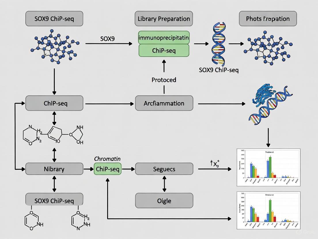
Visualization of Signaling and Workflow
SOX9 ChIP-seq Experimental Workflow
SOX9 in Th17 Cell Differentiation Pathway
SOX9 (SRY-box transcription factor 9) is a pivotal transcription factor with critical roles in development, cell fate determination, and disease progression. This protein maps to chromosome 17q24.3 and encodes 509 amino acids with a molecular mass of approximately 56-70 kDa [1] [2]. As a member of the SOX family, SOX9 contains a highly conserved High Mobility Group (HMG) DNA-binding domain that shares homology with the mammalian testis-determining factor SRY [2]. SOX9 functions as a master regulator of numerous developmental processes, including chondrogenesis, neural crest development, and male sex determination [2] [3]. Beyond development, SOX9 plays context-dependent roles in cancer, acting as either a proto-oncogene or tumor suppressor depending on tissue type [1]. Recent research has also highlighted SOX9's function as a pioneer transcription factor capable of binding closed chromatin and initiating fate switching in stem cells [4]. Understanding SOX9's structural domains is therefore essential for investigating its mechanism of action and developing targeted research reagents.
SOX9 Structural Domains and Functional Motifs
The HMG DNA-Binding Domain
The HMG domain is the defining structural feature of SOX9, enabling sequence-specific DNA recognition and binding. This domain recognizes the canonical DNA motif AACAAT [5]. However, research has revealed that SOX9 achieves optimal binding specificity through nucleotide preferences in the flanking regions, with the complete optimal binding sequence being AGAACAATGG [5]. The 5' AG and 3' GG flanking nucleotides significantly enhance binding affinity specifically for SOX9, distinguishing it from other SOX family members [5]. Structurally, the HMG domain consists of three alpha-helices that form an L-shaped structure, enabling DNA bending at approximately 70-80 degrees upon binding [6]. This DNA bending function is crucial for chromatin remodeling and facilitating protein-protein interactions in transcriptional complexes.
Table 1: Key Structural Domains of SOX9
| Domain | Position | Function | Key Features |
|---|---|---|---|
| HMG DNA-Binding Domain | N-terminal | DNA recognition and bending | Binds AACAAT motif; induces ~70-80° DNA bending; contains nuclear localization signals |
| Transactivation Domain 1 | C-terminal | Transcriptional activation | Rich in proline, glutamine, and alanine residues (PQA-rich) |
| Transactivation Domain 2 | C-terminal | Transcriptional activation | Rich in proline, glutamine, and serine residues (PQS-rich) |
Transactivation Domains
The C-terminal region of SOX9 contains two primary transactivation domains that confer transcriptional activation capability. Research investigating campomelic dysplasia (CD) mutations has demonstrated that progressive deletion of the C-terminal domain causes corresponding progressive loss of transactivation function [6]. Maximal transactivation requires both domains: the domain rich in proline, glutamine, and serine (PQS-rich) and the adjacent domain composed primarily of proline, glutamine, and alanine (PQA-rich) [6]. These regions facilitate interactions with co-activators, basal transcription machinery, and chromatin-modifying enzymes to activate target gene expression.
SOX9-DNA Binding Mechanisms and Specificity
DNA Recognition and Binding Kinetics
SOX9 exhibits distinct DNA-binding properties that underlie its transcriptional regulatory functions. The HMG domain not only recognizes specific DNA sequences but also induces structural changes to the DNA helix. Studies comparing wild-type and CD-associated mutant SOX9 proteins have revealed critical residues for DNA binding integrity. For instance, the F12L mutation results in negligible DNA binding, while H65Y shows minimal binding capacity [6]. Interestingly, the P70R mutation alters DNA binding specificity while maintaining normal DNA bending capability, and A19V exhibits near wild-type binding and bending functions [6]. These findings highlight how specific residues within the HMG domain contribute differentially to DNA recognition versus structural manipulation.
Chromatin Engagement and Pioneer Activity
Recent research has established SOX9 as a bona fide pioneer transcription factor capable of binding target sites in closed chromatin [4]. Through Cleavage Under Targets and Release using Nuclease (CUT&RUN) sequencing and ATAC-seq analyses, it was demonstrated that approximately 30% of SOX9 binding sites occur within closed chromatin regions before nucleosome displacement and chromatin opening [4]. SOX9 achieves this pioneer activity through several mechanisms: recognition of its cognate motifs in compacted chromatin, recruitment of histone and chromatin modifiers, and subsequent nucleosome displacement that increases accessibility for additional transcriptional regulators [4].
Table 2: SOX9 DNA-Binding Properties and Mutational Effects
| Binding Aspect | Characteristics | Functional Significance |
|---|---|---|
| Core Binding Motif | AACAAT | Sequence-specific recognition |
| Optimal Sequence | AGAACAATGG | Enhanced binding affinity with flanking nucleotides |
| DNA Bending | ~70-80° angle | Facilitates enhancer-promoter interactions |
| Pioneer Activity | Binds closed chromatin | Initiates fate switching in stem cells |
| Mutational Effects | F12L: negligible bindingH65Y: minimal bindingP70R: altered specificityA19V: near wild-type function | Campomelic dysplasia pathogenesis |
SOX9 in Transcriptional Regulation and Chromatin Remodeling
Modes of Chromatin Engagement
Genome-wide binding analyses have revealed that SOX9 engages with chromatin through two distinct classes of target associations. Class I sites cluster around transcriptional start sites (TSS) of highly expressed genes with no chondrocyte-specific signature [3]. At these locations, SOX9 association appears to reflect protein-protein interactions with basal transcriptional components rather than direct DNA binding, as evidenced by the absence of enriched SOX9 motifs and lower peak quality scores [3]. In contrast, Class II sites represent evolutionarily conserved active enhancers that direct chondrocyte-specific gene expression through direct binding of SOX9 dimer complexes to DNA [3]. These sites display characteristic enhancer signatures with H3K4me2high/H3K4me3low patterns, strong association with p300 and RNA polymerase II, and peaks of H3K27 acetylation flanking SOX9 binding sites [3].
Enhancer Activation and Super-Enhancer Formation
SOX9 exhibits a marked clustering of binding sites around key chondrocyte genes expressed at high levels, forming what are termed "super-enhancers" [3]. These super-enhancer groupings represent coordinated regulatory elements that drive robust expression of genes critical for chondrocyte identity and function. The number and grouping of these enhancers into super-enhancer clusters likely determines the expression levels of target genes [3]. This super-enhancer formation represents an important mechanism whereby SOX9 coordinates tissue-specific gene expression programs during development and in disease states.
SOX9 Antibody Selection for Chromatin Immunoprecipitation
Critical Antibody Characteristics for ChIP Applications
Selecting appropriate SOX9 antibodies for Chromatin Immunoprecipitation (ChIP) requires careful consideration of several key characteristics. Antibodies must recognize SOX9 in its native, chromatin-bound conformation and maintain specificity under immunoprecipitation conditions. For successful ChIP-seq experiments, antibodies should target epitopes outside the highly conserved HMG domain to avoid cross-reactivity with other SOX family proteins, while still effectively capturing DNA-bound SOX9. Validation using knockdown, relative expression, and knockout controls is essential to confirm specificity [7]. Additionally, antibodies should be verified for application in immunoprecipitation protocols, as not all SOX9 antibodies perform equally in ChIP experiments.
Validated SOX9 Antibodies and Performance Metrics
Several SOX9 antibodies have been validated for use in chromatin immunoprecipitation and related applications. The monoclonal antibody PCRP-SOX9-1E5 (DSHB) has been specifically verified for immunoprecipitation applications and recognizes full-length recombinant human SOX9 protein [8]. Cell Signaling Technology's Sox9 Antibody #14366 is a polyclonal antibody produced by immunizing animals with a synthetic peptide corresponding to residues surrounding Ile198 of human SOX9 protein, and antibodies are purified by protein A and peptide affinity chromatography [2]. Additionally, Invitrogen offers multiple SOX9 antibodies validated for ChIP, including recombinant monoclonal and polyclonal options that target SOX9 in human, mouse, and rat samples [7].
Table 3: Validated SOX9 Antibodies for Chromatin Research
| Product Name | Host Species | Clonality | Epitope | Validated Applications | Species Reactivity |
|---|---|---|---|---|---|
| PCRP-SOX9-1E5 | Mouse | Monoclonal | Full-length | IP, WB, Microarray | Human, Mouse, Rat (predicted) |
| Sox9 Antibody #14366 | Rabbit | Polyclonal | Around Ile198 | WB | Human, Mouse, Rat (predicted) |
| Invitrogen SOX9 Antibodies | Rabbit, Mouse | Mono/Polyclonal | Various | IHC, WB, ICC/IF, ELISA, Flow Cytometry | Human, Mouse, Rat, Non-human primate, Zebrafish |
ChIP-seq Protocol for SOX9 DNA Binding Analysis
Cell Preparation and Crosslinking
Begin with approximately 1×10⁷ cells per immunoprecipitation reaction. For tissue samples, manually dissect to enrich for target cell populations when necessary [3]. Crosslink proteins to DNA by adding 1% formaldehyde directly to cell culture medium and incubating for 10 minutes at room temperature. Quench the crosslinking reaction by adding glycine to a final concentration of 0.125 M and incubating for 5 minutes with gentle rotation. Harvest cells by scraping and pelleted by centrifugation at 800×g for 5 minutes at 4°C. Wash cells twice with cold PBS containing protease inhibitors. Pelleted cells can be flash-frozen in liquid nitrogen and stored at -80°C for future use or processed immediately.
Cell Lysis and Chromatin Shearing
Resuspend cell pellets in cell lysis buffer (5 mM PIPES pH 8.0, 85 mM KCl, 0.5% NP-40) supplemented with protease inhibitors and incubate on ice for 10 minutes. Pellet nuclei by centrifugation at 3000×g for 5 minutes at 4°C. Resuspend nuclei in nuclear lysis buffer (50 mM Tris-Cl pH 8.1, 10 mM EDTA, 1% SDS) with protease inhibitors and incubate on ice for 10 minutes. Transfer the lysate to suitable tubes for sonication. Shear chromatin to an average fragment size of 200-500 bp using a focused ultrasonicator. Optimal shearing conditions should be determined empirically for each cell type. Following sonication, clear lysates by centrifugation at 15,000×g for 10 minutes at 4°C and dilute the supernatant 10-fold in ChIP dilution buffer (16.7 mM Tris-Cl pH 8.1, 167 mM NaCl, 1.2 mM EDTA, 1.1% Triton X-100, 0.01% SDS).
Immunoprecipitation and Wash
Pre-clear the diluted chromatin by adding 40 μL of Protein A/G magnetic beads and incubating for 1 hour at 4°C with rotation. Remove beads and add 2-10 μg of validated SOX9 antibody to the supernatant. Include a control reaction with species-matched non-specific IgG. Incubate overnight at 4°C with rotation. The following day, add 40 μL of pre-blocked Protein A/G magnetic beads and incubate for 2 hours at 4°C with rotation. Pellet beads and wash sequentially for 5 minutes each with the following buffers: low salt wash buffer (0.1% SDS, 1% Triton X-100, 2 mM EDTA, 20 mM Tris-Cl pH 8.1, 150 mM NaCl), high salt wash buffer (0.1% SDS, 1% Triton X-100, 2 mM EDTA, 20 mM Tris-Cl pH 8.1, 500 mM NaCl), LiCl wash buffer (0.25 M LiCl, 1% NP-40, 1% sodium deoxycholate, 1 mM EDTA, 10 mM Tris-Cl pH 8.1), and twice with TE buffer (10 mM Tris-Cl pH 8.0, 1 mM EDTA).
Elution, Reverse Crosslinking, and Purification
Elute chromatin from beads by adding 250 μL of freshly prepared elution buffer (1% SDS, 0.1 M NaHCO₃) and incubating for 30 minutes at room temperature with gentle rotation. Repeat elution and combine supernatants. Add NaCl to a final concentration of 0.2 M and reverse crosslinks by incubating at 65°C overnight. The following day, add EDTA to 10 mM, Tris-Cl (pH 6.5) to 40 mM, and proteinase K to 0.04 μg/μL, and incubate for 2 hours at 45°C. Purify DNA using phenol-chloroform extraction or PCR purification kit. Resuspend purified DNA in TE buffer or nuclease-free water. Quantify DNA yield using fluorometric methods suitable for low concentration samples.
Library Preparation and Sequencing
Prepare sequencing libraries from immunoprecipitated DNA using commercial library preparation kits compatible with low input amounts. Follow manufacturer's instructions for end repair, dA-tailing, and adapter ligation. Amplify libraries with 12-15 PCR cycles using indexed primers to enable multiplexing. Size-select libraries (250-350 bp insert size) using SPRI beads or gel extraction. Quantify final libraries by qPCR and quality control by Bioanalyzer or TapeStation. Sequence libraries on appropriate high-throughput sequencing platforms to obtain a minimum of 20 million reads per sample, with single-end or paired-end reads depending on experimental requirements.
Research Reagent Solutions for SOX9 Studies
Table 4: Essential Research Reagents for SOX9 Chromatin Studies
| Reagent Category | Specific Products | Application Notes |
|---|---|---|
| SOX9 Antibodies | PCRP-SOX9-1E5 (DSHB)#14366 (Cell Signaling)Multiple options (Invitrogen) | Validate for ChIP specifically; check species reactivity |
| Cell Lines | 22RV1, PC3 (prostate cancer)H1975 (lung cancer)Primary chondrocytes | Consider SOX9 expression levels and disease context |
| Small Molecule Inhibitors | Cordycepin (adenosine analog) | Inhibits SOX9 expression in dose-dependent manner [1] |
| Chromatin Shearing | Focused ultrasonicatorBioruptor | Optimize for 200-500 bp fragment size |
| Library Preparation | Illumina TruSeq ChIP Library Prep KitNEB Next Ultra II DNA Library Prep Kit | Select based on input DNA requirements |
| Validation Assays | Western blotRT-qPCRsiRNA/shRNA knockdown | Confirm SOX9 specificity and functional effects |
Data Analysis and Interpretation
Peak Calling and Target Classification
Process raw sequencing reads through quality control, adapter trimming, and alignment to the appropriate reference genome. Call significant peaks using MACS2 or similar peak calling algorithms with the IgG control as background. Classify SOX9 binding sites based on genomic location: promoter-proximal (Class I, within ±500 bp of TSS) and enhancer-associated (Class II, distal regions) [3]. Class I sites typically show lower peak scores and fewer enriched SOX9 motifs, reflecting indirect association through protein-protein interactions, while Class II sites display strong SOX9 motif enrichment, high evolutionary conservation, and characteristic enhancer signatures (H3K4me2high/H3K4me3low, H3K27ac) [3].
Functional Annotation and Integration
Annotate peaks to nearest genes using tools like HOMER or ChIPseeker. Perform motif analysis to identify enriched sequences beyond the primary SOX9 binding motif. Integrate with complementary epigenetic datasets (H3K27ac, H3K4me1, ATAC-seq) to distinguish active enhancers from primed or poised regulatory elements. Identify super-enhancer regions using ROSE or similar algorithms, as SOX9 binding frequently clusters at super-enhancers controlling chondrocyte identity genes [3]. Correlate SOX9 binding with transcriptional changes by integrating with RNA-seq data from the same cell type or condition.
Visualizing SOX9 Chromatin Interactions and Experimental Workflow
SOX9 ChIP-seq Experimental Workflow
SOX9 Chromatin Binding Mechanism
Troubleshooting and Optimization Strategies
Common ChIP Issues and Solutions
Low signal-to-noise ratio can result from insufficient antibody specificity or suboptimal chromatin shearing. Validate SOX9 antibody performance using knockout controls when possible. Optimize sonication conditions to achieve consistent fragment sizes between 200-500 bp. High background signal may stem from inadequate pre-clearing or non-specific antibody binding. Increase pre-clearing time, optimize antibody concentration through titration, and include appropriate controls (IgG, input DNA, and if possible, SOX9-deficient cells). Poor peak resolution often relates to over-crosslinking or insufficient reversal. Limit crosslinking time to 10 minutes and ensure complete reversal overnight at 65°C.
Protocol Validation and Quality Control
Implement rigorous quality control measures throughout the ChIP-seq workflow. Verify chromatin fragment size using the Agilent Bioanalyzer or agarose gel electrophoresis after sonication. Confirm immunoprecipitation efficiency by Western blot analysis of a small aliquot of immunoprecipitated material. Validate successful ChIP through qPCR of known SOX9 target regions (e.g., COL2A1 enhancer) compared to negative control regions before proceeding to library preparation. Assess final library quality and quantity using appropriate methods before sequencing.
SOX9 represents a multifunctional transcription factor with sophisticated structural domains that facilitate DNA binding, chromatin remodeling, and transcriptional activation. The well-defined HMG domain provides sequence-specific DNA recognition and bending capabilities, while the C-terminal transactivation domains engage co-regulators to influence gene expression. SOX9's ability to function as a pioneer factor, binding closed chromatin and initiating fate transitions, makes it particularly interesting for immune cell transcription factor research. The ChIP-seq protocol outlined here, combined with appropriate antibody selection and data analysis strategies, provides a robust framework for investigating SOX9's genomic occupancy and regulatory functions across biological contexts. As research continues to elucidate SOX9's roles in development, homeostasis, and disease, these methodological approaches will remain essential for deciphering its mechanistic contributions to transcriptional programs.
SOX9 (SRY-box 9) is a member of the SOX family of transcription factors characterized by a highly conserved High Mobility Group (HMG) box DNA-binding domain. Recent research has established SOX9 as a bona fide pioneer transcription factor capable of binding to its cognate motifs in compacted, repressed chromatin and initiating chromatin remodeling. This pioneer activity enables SOX9 to divert stem cell fates by simultaneously activating new transcriptional programs while silencing previous cellular identities. The functional domains of SOX9 include a dimerization domain (DIM), the HMG box domain responsible for DNA binding and nuclear localization, and two transcriptional activation domains (TAM and TAC) that interact with various cofactors to enhance transcriptional activity [9].
In immune contexts, SOX9 exhibits complex, dual functions—acting as both an activator and repressor across diverse immune cell types. It participates in T-cell development by cooperating with c-Maf to activate Rorc and key Tγδ17 effector genes (Il17a and Blk), thereby modulating lineage commitment of early thymic progenitors and influencing the balance between αβ and γδ T-cell differentiation [9]. SOX9's pioneer activity underpins its ability to reprogram cell fates in both developmental and pathological processes, making it a critical regulator in immunity and cancer.
Mechanisms of SOX9 Pioneer Activity
Chromatin Binding and Remodeling Dynamics
SOX9 demonstrates characteristic pioneer factor behavior through its sequential binding and chromatin opening capabilities:
- Early Binding to Closed Chromatin: Studies in epidermal stem cells (EpdSCs) have shown that SOX9 binds to chromatin within the first week of induction, with nearly 30% of SOX9 binding sites situated within chromatin regions that were closed prior to induction [4].
- Nucleosome Displacement: Following binding, SOX9 induces nucleosome displacement, evidenced by decreased nucleosome occupancy at bound sites and a time-dependent decrease in cleavage under targets and release using nuclease (CUT&RUN) fragment lengths [4].
- Recruitment of Chromatin Modifiers: SOX9 recruits histone and chromatin modifiers to remodel and subsequently open chromatin for transcription. When SOX9's ability to bind chromatin remodellers is abrogated, the cell fate switch fails entirely [4].
Sequence Determinants of Chromatin Response
The chromatin response to SOX9 dosage is governed by specific sequence features in regulatory elements:
| Sequence Feature | Effect on Chromatin Response | Genomic Distribution |
|---|---|---|
| High-affinity motifs with heterotypic TF co-binding | Buffer against quantitative TF dosage changes | Concentrated at center of regulatory elements |
| Low-affinity homotypic binding motifs | Drive sensitive responses to dosage changes | Distributed throughout regulatory elements |
Table 1: Sequence features governing chromatin response to SOX9 dosage.
Transfer learning approaches have revealed that these features display purifying selection signatures, and TF-nucleosome competition can explain the sensitizing effects of low-affinity motifs [10].
SOX9 in Immune Cell Regulation and Tumor Immunity
SOX9 in Immune Cell Development and Function
SOX9 plays significant roles in immune cell development and regulation, participating in the differentiation and function of diverse immune lineages:
- T-cell Development: SOX9 modulates the lineage commitment of early thymic progenients, potentially influencing the balance between αβ T-cell and γδ T-cell differentiation [9].
- B-cell Lymphomas: While SOX9 has no significant role in normal B-cell development, it is overexpressed in certain B-cell lymphomas like Diffuse Large B-cell Lymphoma (DLBCL), where it acts as an oncogene by promoting cell proliferation and inhibiting apoptosis [9].
- Macrophage Function: Increased SOX9 levels help maintain macrophage function, contributing to tissue regeneration and repair processes [9].
SOX9 in Tumor Immune Microenvironment
SOX9 significantly influences the tumor immune microenvironment through complex interactions with various immune cell types:
| Immune Cell Type | Correlation with SOX9 Expression | Functional Consequences |
|---|---|---|
| B cells, resting mast cells, resting T cells | Negative correlation | Reduced anti-tumor immunity |
| Neutrophils, macrophages, activated mast cells | Positive correlation | Immunosuppressive microenvironment |
| CD8+ T cells, NK cells, M1 macrophages | Negative correlation with function | Impaired cytotoxic activity |
| Memory CD4+ T cells | Positive correlation | Altered adaptive immune responses |
Table 2: SOX9 correlations with immune cell infiltration in tumor microenvironment.
In single-cell RNA sequencing and spatial transcriptomics analyses of cancers such as prostate cancer, SOX9 expression is associated with an "immune desert" microenvironment characterized by decreased effector immune cells (CD8+CXCR6+ T cells and activated neutrophils) and increased immunosuppressive cells (Tregs and M2 macrophages) [9].
SOX9 ChIP-seq Protocol for Immune Cells
Reagent Solutions for SOX9 ChIP-seq
| Reagent Category | Specific Examples | Function in Protocol |
|---|---|---|
| Crosslinking Agents | 1% formaldehyde, DSG (disuccinimidyl glutarate) | DNA-protein crosslinking |
| Cell Lysis Buffers | RIPA buffer, Western & IP Lysis Buffer | Cell membrane disruption |
| Immunoprecipitation Antibodies | Anti-SOX9 (AB5535, Sigma-Aldrich) | Target protein immunoprecipitation |
| Magnetic Beads | Protein A/G magnetic beads (B23202) | Antibody-bound complex capture |
| Chromatin Shearing | Covaris sonicator, Bioruptor | DNA fragmentation to 200-500bp |
| DNA Cleanup | Qiagen MinElute PCR Purification Kit | Post-IP DNA purification |
| Sequencing Library Prep | Illumina TruSeq ChIP Library Preparation Kit | Library preparation for sequencing |
Table 3: Essential research reagents for SOX9 ChIP-seq protocols.
Detailed ChIP-seq Methodology
The following workflow outlines the key steps in performing SOX9 ChIP-seq in immune cells:
Diagram 1: SOX9 ChIP-seq workflow.
Step 1: Cell Preparation and Crosslinking
- Culture approximately 10⁷ immune cells per ChIP under appropriate conditions.
- Crosslink DNA-protein interactions with 1% formaldehyde for 10 minutes at room temperature.
- Quench crosslinking with 125mM glycine for 5 minutes.
- Wash cells with ice-cold PBS and pellet by centrifugation [3] [11].
Step 2: Chromatin Preparation and Shearing
- Lyse cells in RIPA buffer (or Western & IP Lysis Buffer) containing protease inhibitors.
- Sonicate chromatin to fragment DNA to 200-500bp fragments using a Covaris sonicator or Bioruptor.
- Confirm fragmentation size by running an aliquot on an agarose gel [11].
Step 3: Immunoprecipitation
- Pre-clear chromatin lysate with protein A/G magnetic beads for 1 hour at 4°C.
- Incubate pre-cleared chromatin with 5μL anti-SOX9 antibody (AB5535) overnight at 4°C with rotation.
- Add protein A/G magnetic beads and incubate for 2 hours at 4°C.
- Wash beads sequentially with: Low Salt Immune Complex Wash Buffer, High Salt Immune Complex Wash Buffer, LiCl Immune Complex Wash Buffer, and TE Buffer [11].
Step 4: DNA Recovery and Purification
- Reverse crosslinks by incubating beads with elution buffer (1% SDS, 0.1M NaHCO₃) at 65°C overnight.
- Treat with RNase A and Proteinase K.
- Purify DNA using Qiagen MinElute PCR Purification Kit or equivalent [4].
Step 5: Library Preparation and Sequencing
- Prepare sequencing libraries using Illumina TruSeq ChIP Library Preparation Kit.
- Validate library quality using Bioanalyzer.
- Sequence on Illumina platform (minimum 20 million reads per sample recommended) [3].
Analysis of SOX9 Binding Dynamics
Bioinformatics Analysis Pipeline
The computational analysis of SOX9 ChIP-seq data involves multiple steps to identify binding sites and characterize pioneer activity:
Diagram 2: ChIP-seq data analysis pipeline.
Key Analysis Steps:
Quality Control and Alignment
- Assess read quality with FastQC.
- Align reads to reference genome (hg19/GRCh38) using STAR or BOWTIE2.
- Remove PCR duplicates using Picard Tools.
Peak Calling and Classification
- Identify significant SOX9 binding sites using MACS2 with stringent thresholds (q-value < 0.05).
- Classify peaks based on genomic location:
- Class I Sites: Cluster around transcriptional start sites (TSS) of highly expressed genes, often reflecting protein-protein associations with basal transcriptional components rather than direct DNA binding [3].
- Class II Sites: Distal enhancer regions with direct SOX9 dimer binding to DNA, highly conserved across species, and associated with active enhancer marks (H3K4me2high/H3K4me3low, H3K27Ac) [3].
Integration with Epigenetic Data
- Compare SOX9 binding with ATAC-seq or DNase-seq data to identify binding events in closed chromatin.
- Overlap with H3K27ac ChIP-seq to distinguish active enhancers.
- Analyze nucleosome positioning data to confirm nucleosome displacement at SOX9 binding sites [4].
Characterizing Pioneer Activity
To specifically demonstrate SOX9 pioneer factor activity:
- Calculate Percentage of Binding in Closed Chromatin: Determine the proportion of SOX9 peaks that fall within regions identified as closed chromatin by ATAC-seq prior to SOX9 induction. True pioneer factors typically show 20-40% binding in closed chromatin [4].
- Analyze Chromatin Opening Kinetics: Perform time-course experiments to demonstrate that SOX9 binding precedes chromatin accessibility changes.
- Motif Enrichment Analysis: Use HOMER to identify enriched motifs in pioneer-bound regions, particularly focusing on SOX dimer motifs and co-factor binding sites [12].
Applications in Drug Development and Therapeutic Targeting
The pioneer activity of SOX9 has significant implications for therapeutic development, particularly in cancer and immune-related diseases:
SOX9 in Therapeutic Resistance
SOX9 contributes to therapy resistance through multiple mechanisms:
- Chemotherapy Resistance: In high-grade serous ovarian cancer (HGSOC), SOX9 expression is induced by platinum-based chemotherapy and drives chemoresistance by reprogramming the transcriptional state of naive cells into a stem-like state [13].
- PARP Inhibitor Resistance: SOX9 contributes to PARP inhibitor resistance in ovarian cancer by binding to promoters of key DNA damage repair genes (SMARCA4, UIMC1, and SLX4), thereby enhancing DNA repair capabilities [11].
- Stemness Maintenance: SOX9 promotes a stem-like transcriptional state that confers resistance to various therapies across cancer types [13].
Therapeutic Targeting Strategies
Several strategies have emerged to target SOX9 activity:
| Therapeutic Approach | Mechanism of Action | Experimental Evidence |
|---|---|---|
| USP28 Inhibition | Promotes SOX9 degradation via ubiquitination | AZ1 inhibitor reduces SOX9 stability and sensitizes to PARP inhibitors [11] |
| Cordycepin | Downregulates SOX9 expression | Dose-dependent inhibition of SOX9 mRNA and protein in cancer cells [1] |
| Indirect Targeting | Modulates upstream regulators | Targeting SOX9-inducing signals in tumor microenvironment |
Table 4: Therapeutic approaches for targeting SOX9.
SOX9 represents a paradigm of pioneer transcription factor activity in immune cell regulation and cancer biology. Its ability to bind closed chromatin, recruit chromatin remodelers, and initiate transcriptional reprogramming makes it a critical regulator of cell fate decisions. The ChIP-seq protocol outlined here provides a robust method for investigating SOX9 binding dynamics in immune cells, while the growing understanding of its role in immune regulation offers promising therapeutic avenues for targeting SOX9 in cancer and immune-related diseases. As research continues to unravel the complexities of SOX9 function, its position as a key therapeutic target in immuno-oncology becomes increasingly evident.
The transcription factor SOX9, a member of the SRY-related HMG-box family, is increasingly recognized as a pivotal regulator of immune function. Beyond its well-established roles in development, chondrogenesis, and cancer stemness, SOX9 exerts critical influence over specific immune processes, particularly in T-cell differentiation and shaping the tumor microenvironment (TME). This application note details experimental approaches for investigating SOX9's immune functions, providing structured quantitative data, methodological protocols, and visual workflows to support research and drug discovery efforts. These protocols are designed to integrate with broader SOX9 ChIP-seq investigations for immune cell transcription factors, enabling comprehensive analysis of its direct transcriptional targets in immunological contexts.
SOX9 in T-cell Differentiation
Molecular Mechanisms
SOX9 plays a specialized role in T-cell development by influencing lineage commitment in early thymic progenitors [9]. Mechanistically, SOX9 cooperates with the transcription factor c-Maf to activate key genetic programs including Rorc (which encodes RORγt) and effector genes characteristic of Tγδ17 cells such as Il17a and Blk [9]. This positions SOX9 as a determinant in the balance between αβ and γδ T-cell differentiation, particularly favoring the γδ T-cell lineage.
Table 1: SOX9-Associated Effects on T-cell Development
| Target Process | Molecular Target | Effect of SOX9 Activity | Experimental Evidence |
|---|---|---|---|
| T-cell Lineage Commitment | RORγt (Rorc) | Activation via c-Maf cooperation | Genetic interaction studies [9] |
| Tγδ17 Cell Function | IL-17a (Il17a) | Transcriptional activation | Gene expression analysis [9] |
| Tγδ17 Cell Function | BLK (Blk) | Transcriptional activation | Gene expression analysis [9] |
SOX9 in Tumor Microenvironment Regulation
Immunomodulation in Cancer
SOX9 significantly influences the tumor immune landscape through dual mechanisms: direct regulation of cancer cell-immune interactions and shaping the overall immunosuppressive microenvironment. SOX9 enables immune evasion by maintaining cancer cell stemness, allowing dormant cancer cells to persist in secondary sites and evade immune surveillance [14]. This function is crucial for metastatic latency and therapeutic resistance.
Immune Cell Infiltration Patterns
Comprehensive analyses across multiple carcinomas reveal that SOX9 expression correlates with specific immune infiltration patterns [9]:
- Negative correlations with anti-tumor immune populations: B cells, resting mast cells, resting T cells, monocytes, plasma cells, and eosinophils
- Positive correlations with pro-tumor immune populations: neutrophils, macrophages, activated mast cells
- Functional impairment: SOX9 overexpression negatively correlates with genes associated with CD8+ T cell function, natural killer (NK) cell activity, and M1 macrophage polarization while showing positive correlation with memory CD4+ T cells [9]
In specific cancers like prostate cancer, SOX9 contributes to an "immune desert" microenvironment characterized by decreased effector CD8+CXCR6+ T cells and increased immunosuppressive cells including Tregs and M2 macrophages [9].
Table 2: SOX9-Mediated Effects on Tumor Immune Microenvironment
| Cancer Type | Correlated Immune Changes | Clinical/Functional Impact |
|---|---|---|
| Colorectal Cancer | ↑ Neutrophils, Macrophages, Activated Mast Cells↓ B cells, Resting Mast Cells, Monocytes | Correlated with immune suppression [9] |
| Pan-Cancer (multiple) | ↓ CD8+ T cell function, NK cell activity, M1 macrophages | Promotes immune evasion [9] |
| Prostate Cancer | ↓ CD8+CXCR6+ T cells↑ Tregs, M2 macrophages, anergic neutrophils | Creates "immune desert" TME [9] |
| Glioblastoma | Correlated with immune checkpoint expression | Potential biomarker for immunotherapy [15] |
SOX9 ChIP-seq Protocol for Immune Cell Studies
Cell Preparation and Cross-linking
Materials: Primary T-cells or tumor-infiltrating immune cells, formaldehyde (1% final concentration), glycine (125mM final concentration), PBS. Protocol:
- Culture 1×10^7 cells per ChIP reaction in appropriate medium
- Cross-link DNA-protein interactions with 1% formaldehyde for 10 minutes at room temperature with gentle agitation
- Quench cross-linking with 125mM glycine for 5 minutes at room temperature
- Wash cells twice with ice-cold PBS
- Pellet cells and flash-freeze in liquid nitrogen for storage at -80°C if not proceeding immediately
Chromatin Preparation and Shearing
Materials: Lysis buffers (LB1: 50mM HEPES-KOH pH7.5, 140mM NaCl, 1mM EDTA, 10% Glycerol, 0.5% NP-40, 0.25% Triton X-100; LB2: 10mM Tris-HCl pH8.0, 200mM NaCl, 1mM EDTA, 0.5mM EGTA; LB3: 10mM Tris-HCl pH8.0, 100mM NaCl, 1mM EDTA, 0.5mM EGTA, 0.1% Na-Deoxycholate, 0.5% N-lauroylsarcosine), MNase or sonication equipment. Protocol:
- Resuspend cell pellet in LB1 and incubate 10 minutes at 4°C with rotation
- Pellet nuclei and resuspend in LB2, incubate 10 minutes at 4°C with rotation
- Pellet nuclei and resuspend in LB3
- Chromatin shearing: Use either MNase digestion (5-10 units per sample, 15 minutes at 37°C) or sonication (Bioruptor, 8 cycles of 30 seconds ON/30 seconds OFF at high setting) to achieve 200-500bp fragments
- Centrifuge at 20,000×g for 10 minutes at 4°C to clear debris
Immunoprecipitation
Materials: SOX9 antibody (validated for ChIP), protein A/G magnetic beads, wash buffers (Low Salt: 20mM Tris-HCl pH8.0, 150mM NaCl, 2mM EDTA, 1% Triton X-100; High Salt: 20mM Tris-HCl pH8.0, 500mM NaCl, 2mM EDTA, 1% Triton X-100; LiCl: 10mM Tris-HCl pH8.0, 250mM LiCl, 1mM EDTA, 1% NP-40, 1% Na-Deoxycholate; TE: 10mM Tris-HCl pH8.0, 1mM EDTA), elution buffer (1% SDS, 100mM NaHCO3). Protocol:
- Pre-clear chromatin with protein A/G beads for 1 hour at 4°C
- Incubate supernatant with SOX9 antibody (2-5μg per reaction) overnight at 4°C with rotation
- Add protein A/G beads and incubate 2 hours at 4°C
- Wash beads sequentially: Once with Low Salt buffer, once with High Salt buffer, once with LiCl buffer, twice with TE buffer
- Elute chromatin with elution buffer twice (15 minutes each at 65°C with agitation)
- Reverse cross-links by adding 200mM NaCl and incubating overnight at 65°C
- Treat with RNase A (30 minutes at 37°C) and proteinase K (2 hours at 55°C)
- Purify DNA with phenol-chloroform extraction and ethanol precipitation
Library Preparation and Sequencing
Materials: DNA library preparation kit, size selection beads, quality control instruments. Protocol:
- Quantify recovered DNA with Qubit fluorometer
- Prepare sequencing libraries using commercial kit (e.g., Illumina)
- Perform size selection (200-500bp) with SPRIselect beads
- Assess library quality with Bioanalyzer
- Sequence on appropriate platform (Illumina recommended for 50-100 million reads per sample)
ChIP-seq Workflow for SOX9 in Immune Cells
SOX9-Wnt Signaling in Immune Contexts
The interaction between SOX9 and Wnt signaling pathways represents a crucial regulatory axis in immune and cancer biology. SOX9 exhibits context-dependent functionality, capable of both repressing and activating Wnt target genes [16] [17]. In colorectal cancer cells, SOX9 physically interacts with T-cell factors (TCFs) through their DNA-binding domains, co-occupying Wnt-responsive enhancers to activate target genes like MYC when SOX9-binding sites are present on these enhancers [16]. This cooperative activation requires specific cis-regulatory grammar with both TCF and SOX9 binding sites [17].
SOX9-Wnt Signaling Interactions
Research Reagent Solutions
Table 3: Essential Research Reagents for SOX9 Immune Studies
| Reagent/Category | Specific Examples | Application & Function |
|---|---|---|
| Validated SOX9 Antibodies | ChIP-grade SOX9 antibody (e.g., Millipore ABCAM) | Chromatin immunoprecipitation for genome-wide binding studies |
| Cell Culture Models | hESC-derived CNCCs [18], Primary T-cells, Tumor-infiltrating immune cells | Modeling SOX9 function in relevant cellular contexts |
| Genetic Modulation Systems | CRISPR/Cas9 KO [13], dTAG degradation system [18] [19], Inducible shRNA | Precise manipulation of SOX9 expression levels |
| Assay Kits | ATAC-seq kit, ChIP-seq library prep kit, scRNA-seq platform | Molecular profiling of SOX9 effects on chromatin and transcription |
| Animal Models | Genetic mouse models of BCC [20], PDX models with immune components | In vivo validation of SOX9 immune functions |
Technical Applications and Considerations
Dosage Sensitivity in Experimental Design
SOX9 exhibits significant dosage sensitivity, which must be considered in experimental design [18]. Most SOX9-dependent regulatory elements are buffered against small dosage decreases, but a subset controlling key functions like chondrogenesis shows heightened sensitivity [18]. The dTAG system enables precise titration of SOX9 levels in human facial progenitor cells (cranial neural crest cells), allowing investigation of this dosage sensitivity [18] [19].
Analytical Approaches for Transcriptional Regulation
Transfer learning approaches applied to chromatin accessibility data can predict how regulatory elements respond to SOX9 dosage changes [19]. Key sequence features determining sensitivity include:
- Buffering features: High-affinity motifs enabling heterotypic TF co-binding, centrally located in regulatory elements
- Sensitizing features: Low-affinity or homotypic binding motifs distributed throughout regulatory elements [19]
These computational approaches complement empirical ChIP-seq data in elucidating SOX9's transcriptional networks in immune contexts.
SOX9 represents a multifunctional regulator of immune processes with particular importance in T-cell differentiation and tumor microenvironment composition. The experimental approaches detailed herein provide a framework for investigating SOX9's immune functions, with special considerations for its dosage sensitivity, context-dependent Wnt interactions, and complex role in immune evasion. Integration of ChIP-seq with functional immune assays offers the most comprehensive approach to delineating SOX9's transcriptional programs in immunological contexts, potentially revealing novel therapeutic targets for cancer immunotherapy and immune disorders.
The SRY-related HMG box 9 (SOX9) transcription factor serves as a master regulator of cell fate determination across diverse tissues, with well-established roles in chondrogenesis, organogenesis, and cancer progression [21]. Recent evidence has established SOX9 as a pioneer transcription factor capable of binding compacted chromatin and initiating chromatin remodeling [4]. This capacity enables SOX9 to divert embryonic epidermal stem cells (EpdSCs) into becoming hair follicle stem cells through direct binding to closed chromatin at hair follicle enhancers, where it subsequently recruits histone and chromatin modifiers to remodel and open chromatin for transcription [4]. Beyond its developmental roles, SOX9 is frequently overexpressed in diverse malignancies and plays complex, context-dependent roles in tumor immune evasion by modulating immune cell infiltration and function [9] [22]. This application note synthesizes key methodological and conceptual insights from SOX9 ChIP-seq studies across tissue systems to inform robust investigation of SOX9 functions in immune cells.
Key SOX9 ChIP-seq Findings Across Biological Systems
Modes of SOX9 Chromatin Engagement
Comprehensive ChIP-seq analyses in primary chondrocytes have revealed two distinct categories of SOX9 chromatin association, summarized in Table 1 [3].
Table 1: Modes of SOX9 Chromatin Engagement
| Association Type | Genomic Localization | Binding Characteristics | Functional Roles | Target Gene Examples |
|---|---|---|---|---|
| Class I | Transcription Start Site (TSS)-proximal (±500 bp) | Indirect association; low motif enrichment; lower peak quality scores; correlates with general transcription machinery | Regulation of general cellular processes; correlates with gene expression levels | Housekeeping and broadly expressed genes |
| Class II | Distal enhancers (up to ±500 kb from TSS) | Direct DNA binding; high SOX motif enrichment; high evolutionary conservation; high H3K27ac signal | Chondrocyte-specific gene regulation; active enhancer signatures | Col2a1, Acan, Sox5, Sox6 |
The functional distinction between these binding modes is critical for experimental design and interpretation. Class II sites represent the primary drivers of SOX9's cell-type-specific functions, with approximately 70% of these sites residing in closed chromatin prior to SOX9 binding in reprogramming models [4]. These sites exhibit characteristics of active enhancers, including H3K4me2high/H3K4me3low signatures, p300 co-activator recruitment, and flanking H3K27ac peaks [3].
Tissue-Specific Enhancer Landscapes
Recent chromatin profiling in chondrocytes has identified specific enhancer elements critical for SOX9 regulation and skeletal development. Two upstream enhancers—E308 (located 308 kb 5′ upstream) and E160 (located 160 kb 5′ upstream)—demonstrate synergistic activity in controlling SOX9 expression levels [23] [24]. While single deletions of these enhancers in mice had minimal phenotypic consequences, simultaneous deletion caused a dwarf phenotype with reduced Sox9 expression in chondrocytes and attenuated chondrocyte differentiation [23] [24]. This functional redundancy exemplifies the robustness of SOX9 regulation through multiple enhancer elements.
In epidermal reprogramming models, SOX9 binding to closed chromatin precedes increased accessibility, with approximately 30% of SOX9 binding sites located in closed chromatin prior to activation [4]. The subsequent chromatin opening occurs between weeks 1 and 2 after SOX9 induction, demonstrating the temporal progression of pioneer factor activity [4].
Experimental Protocols for SOX9 ChIP-seq
Cell Source and Isolation Methods
Primary chondrocyte isolation (from neonatal mouse rib cartilage):
- Dissect rib cages from post-natal day 1 (P1) mice
- Manually isolate proliferative and prehypertrophic zones, excluding mature hypertrophic regions
- Use primary dermal fibroblasts as control cells with low SOX9 expression [24] [3]
Epidermal stem cell isolation:
- Fluorescence-activated cell sorting (FACS) of EpdSCs from Krt14-rtTA;TRE-Sox9 murine skin models
- Induce SOX9 expression with doxycycline administration for temporal analysis [4]
Chromatin Immunoprecipitation and Sequencing
The standard SOX9 ChIP-seq protocol encompasses the following key steps:
Table 2: SOX9 ChIP-seq Antibodies and Applications
| Application | Antibody Target | Function in Assay | Key Findings |
|---|---|---|---|
| Transcription Factor Binding | SOX9 | Identifies genome-wide SOX9 binding sites | Reveals Class I and Class II binding modes [3] |
| Enhancer Validation | H3K27ac | Marks active enhancers and promoters | Distinguishes active SOX9-bound enhancers [23] [24] |
| Enhancer Signature | H3K4me2 | Identifies putative enhancer regions | Combined with H3K27ac to reduce false positives [24] |
| Transcriptional Activity | RNA Polymerase II | Indicates active transcription | Correlates with SOX9 Class I binding [3] |
| Co-activator Recruitment | p300 | Identifies enhancers with transcriptional potential | Co-localizes with SOX9 at Class II sites [3] |
Critical protocol considerations:
- Peak calling: 27,656 raw peaks were identified in rib chondrocytes using standard peak-calling criteria [3]
- Multimodal validation: Combine with ATAC-seq to assess chromatin accessibility dynamics
- Temporal sampling: In reprogramming models, collect samples at multiple timepoints (e.g., 0, 1, 2, 6, and 12 weeks) to capture chromatin dynamics [4]
- Biological replicates: Include at least two independent biological replicates for statistical robustness
Data Analysis and Integration
Identification of high-confidence binding sites:
- Filter peaks based on quality scores, with Class II sites typically ranking higher than Class I sites [3]
- Motif enrichment analysis to distinguish direct (motif-rich) versus indirect (motif-poor) binding
- GREAT Gene Ontology analysis for functional annotation of target genes [24] [3]
Integration with complementary datasets:
- ATAC-seq: For profiling chromatin accessibility changes following SOX9 binding [24] [4]
- RNA-seq: To correlate binding events with transcriptional outcomes
- Histone modification profiling: (H3K27ac, H3K4me2) for enhancer validation [24]
SOX9 in Immune Regulation and Cancer
SOX9 in Tumor Immunity
SOX9 plays a complex, dual role in immunobiology, functioning as a "double-edged sword" in cancer-immune interactions [9]. In various cancer contexts, SOX9 expression correlates with specific immune cell infiltration patterns, as summarized in Table 3.
Table 3: SOX9 Correlations with Immune Cell Infiltration in Cancer
| Cancer Type | Positive Correlation | Negative Correlation | Functional Consequences |
|---|---|---|---|
| Colorectal Cancer | Neutrophils, macrophages, activated mast cells, naive/activated T cells | B cells, resting mast cells, resting T cells, monocytes, plasma cells, eosinophils | Immunosuppressive microenvironment [9] |
| Prostate Cancer | Tregs, M2 macrophages (TAM Macro-2), anergic neutrophils | CD8+CXCR6+ T cells, activated neutrophils | "Immune desert" formation promoting tumor immune escape [9] |
| Multiple Cancers | Memory CD4+ T cells | CD8+ T cells, NK cells, M1 macrophages | Impaired anti-tumor immunity [9] |
Mechanistically, SOX9 contributes to immune evasion by impairing immune cell function and maintaining tumor cell stemness [9]. In latent cancer cells, SOX9 helps maintain dormancy and avoid immune surveillance in secondary metastatic sites under immunotolerant conditions [25].
Implications for Immune Cell Transcription Factor Research
The principles derived from SOX9 studies in other tissues provide valuable insights for immune cell transcription factor research:
Enhancer Organization:
- SOX9 target genes in chondrocytes exhibit clustered enhancer organization, with key genes associated with multiple enhancers that function cooperatively [3]
- Similar organizational principles may govern transcription factor function in immune cell specification and differentiation
Pioneer Factor Activity:
- SOX9's capacity to bind closed chromatin and initiate remodeling [4] suggests similar mechanisms may operate during immune cell fate transitions
- The sequential binding followed by chromatin opening observed in SOX9 reprogramming provides a template for analyzing transcription factor hierarchies in immune development
Competition for Epigenetic Regulators:
- During fate switching, SOX9 redistributes co-factors away from previous identity enhancers, indirectly silencing former transcriptional programs [4]
- This competitive dynamic may underlie mutually exclusive cell fate decisions in immune cell development
The Scientist's Toolkit: Research Reagent Solutions
Table 4: Essential Research Reagents for SOX9 ChIP-seq Studies
| Reagent Category | Specific Examples | Function and Application |
|---|---|---|
| SOX9 Antibodies | Anti-SOX9 for ChIP; Anti-MYC for tagged SOX9 | Immunoprecipitation of SOX9-bound chromatin; detection of epitope-tagged SOX9 |
| Histone Modification Antibodies | Anti-H3K27ac; Anti-H3K4me2; Anti-H3K4me3 | Validation of active enhancers and promoters; chromatin state characterization |
| Control Cell Lines | Primary dermal fibroblasts; 3T3-L1 adipocytes | Negative controls for chondrocyte-specific binding; modeling lipid stress conditions |
| Animal Models | Krt14-rtTA;TRE-Sox9 mice; Sox9 enhancer deletion models | Inducible SOX9 expression; analysis of enhancer function in vivo |
| Bioinformatics Tools | GREAT GO analysis; MGI phenotype ontology; ngsplot | Functional annotation of binding sites; visualization of sequencing data |
Visualizing SOX9 ChIP-seq Workflows and Binding Dynamics
SOX9 ChIP-seq Experimental Workflow
SOX9 Binding Modes and Functional Outcomes
SOX9 chromatin engagement follows conserved principles across tissue systems while exhibiting context-specific adaptations. The well-characterized Class I and Class II binding modes provide a framework for analyzing transcription factor function in immune cells. SOX9's demonstrated pioneer factor capability suggests potential similar mechanisms in immune cell fate determinations. The integration of ChIP-seq with chromatin accessibility data and histone modification profiling creates a powerful multimodal approach for comprehensive transcription factor analysis. These precedents establish a rigorous methodological foundation for investigating SOX9 roles in immune regulation and provide conceptual frameworks for studying transcription factor networks in immune cell development and function.
Step-by-Step SOX9 ChIP-seq Protocol Optimized for Immune Cell Populations
The transcription factor SOX9 plays a Janus-faced role in immunology, functioning as a critical regulator in both cancer immune evasion and the maintenance of beneficial immune functions [9]. Chromatin Immunoprecipitation followed by sequencing (ChIP-seq) enables researchers to map the precise genomic interactions of SOX9, providing insights into its dual nature in immune regulation. This application note details optimized protocols for preparing immune cells for SOX9 ChIP-seq, a methodology essential for advancing drug development in oncology and inflammatory diseases.
SOX9 in Immune Cell Biology and Therapeutic Relevance
SOX9 significantly influences the development and function of various immune cells. It modulates T-cell development by cooperating with c-Maf to activate Rorc and key Tγδ17 effector genes (Il17a, Blk), thereby influencing the lineage commitment of early thymic progenitors and the balance between αβ and γδ T-cell differentiation [9]. Furthermore, SOX9 is overexpressed in certain B-cell lymphomas, such as Diffuse Large B-cell Lymphoma (DLBCL), where it acts as an oncogene by promoting proliferation and inhibiting apoptosis [9].
Therapeutically, SOX9 expression correlates with tumor immune evasion. Its overexpression is associated with impaired function of CD8+ T cells and NK cells, and it contributes to an immunosuppressive microenvironment rich in Tregs and M2 macrophages [9] [22]. These roles make SOX9 a promising candidate for immunotherapeutic interventions, necessitating robust ChIP-seq methods to delineate its transcriptional networks in immune cells.
Immune Cell Isolation Techniques
The first critical step in ChIP-seq is obtaining a high-quality population of target cells. The table below summarizes standard isolation methods for immune cell types relevant to SOX9 research.
Table 1: Methods for Immune Cell Isolation
| Cell Type | Isolation Method | Key Characteristics | Considerations for SOX9 Studies |
|---|---|---|---|
| Primary T Cells | Negative or positive selection from PBMCs using magnetic beads (e.g., Pan T Cell Isolation Kit). | >95% purity [9]. | Purity is critical to avoid confounding signals from other SOX9-expressing cells. |
| B Cells | Density gradient centrifugation (e.g., Ficoll-Paque) followed by B-cell specific negative selection kits. | Varies by lymphoma subtype; SOX9 overexpression in DLBCL [9]. | Cell lines (e.g., from DLBCL) can provide a homogeneous population. |
| Macrophages | Differentiation of CD14+ monocytes isolated from PBMCs using M-CSF (50 ng/mL for 7 days). | Can be polarized to M1 (anti-tumor) or M2 (pro-tumor) phenotypes [9]. | SOX9 helps maintain macrophage function; phenotype should be confirmed before ChIP. |
Cross-linking and Chromatin Preparation Optimization
Cross-linking preserves protein-DNA interactions for immunoprecipitation. A dual-crosslinking approach is often superior for nuclear factors like SOX9.
Standard Formaldehyde Cross-linking
- Procedure: Resuspend up to 10 million cells in 10 mL of room-temperature PBS. Add 270 µL of 37% formaldehyde (final concentration ~1%). Incubate for 10 minutes at room temperature with gentle rotation. Quench the reaction by adding 1 mL of 1.25 M glycine (final concentration ~0.125 M) and incubating for 5 minutes. Pellet cells and wash twice with cold PBS [26] [27].
Dual Cross-linking with DSG and Formaldehyde
For transcription factors like SOX9, which may participate in larger complexes, a two-step crosslinking with Disuccinimidyl Glutarate (DSG) can improve efficiency.
- Procedure:
Table 2: Optimized Cross-linking Parameters for SOX9 ChIP-seq
| Parameter | Standard Protocol | Dual-Crosslinking Protocol | Rationale |
|---|---|---|---|
| Primary Crosslinker | Formaldehyde (1%) | DSG (2 mM) | DSG is a reversible amine-to-amine crosslinker that stabilizes protein-protein interactions. |
| Incubation Time | 10 min, RT | 30-45 min, RT | Longer incubation allows for better penetration of DSG. |
| Secondary Crosslinker | N/A | Formaldehyde (1%) | Formaldehyde covalently fixes protein-DNA complexes. |
| Cell Number | 1 x 10^6 to 1 x 10^7 | 1 x 10^6 to 1 x 10^7 | This range provides sufficient chromatin material while avoiding over-crosslinking. |
| Application | General TF binding | Complex-associated TFs (e.g., SOX9), histone modifications | Stabilizes intricate protein complexes that SOX9 engages with on chromatin. |
Cell Lysis and Chromatin Shearing
After cross-linking, cells are lysed, and chromatin is fragmented, typically via sonication.
- Lysis: Use an appropriate lysis buffer (e.g., SDS Lysis Buffer) to release nuclei.
- Shearing: Sonicate chromatin to an average fragment size of 200-500 bp. Optimal conditions must be determined empirically for each cell type and sonicator. Analyze fragment size by agarose gel electrophoresis or a Bioanalyzer.
Workflow and Pathway Diagrams
Experimental Workflow for SOX9 ChIP-seq in Immune Cells
The following diagram illustrates the complete workflow from cell preparation to sequencing.
SOX9's Role in Immune Regulation and Cancer
This diagram summarizes the dual role of SOX9 in the immune system, underpinning its significance as a ChIP-seq target.
The Scientist's Toolkit: Key Research Reagent Solutions
Table 3: Essential Reagents for SOX9 ChIP-seq in Immune Cells
| Reagent / Kit | Function | Example & Notes |
|---|---|---|
| Cell Isolation Kits | Negative or positive selection of specific immune cell populations from a heterogeneous mix. | Human Pan T Cell Isolation Kit (e.g., Miltenyi Biotec). Magnetic bead-based separation ensures high purity. |
| Crosslinking Reagents | Covalently link DNA-associated proteins to DNA. | Formaldehyde (37% stock); Disuccinimidyl Glutarate (DSG). Dual-crosslinking can enhance yields for SOX9. |
| ChIP-Validated Antibody | Specifically immunoprecipitate the SOX9-protein-DNA complex. | Anti-SOX9 (e.g., Rabbit polyclonal, [26]). Antibody specificity is the single most critical factor for success. |
| Magnetic Protein A/G Beads | Capture the antibody-protein-DNA complex. | Dynabeads Protein A or G (e.g., Thermo Fisher Scientific). Offer low non-specific binding. |
| Chromatin Shearing Kit | Standardize and optimize DNA fragmentation. | Covaris microTUBES and reagents. Provides consistent, tunable sonication. |
| ChIP-seq Library Prep Kit | Prepare sequencing libraries from immunoprecipitated DNA. | Illumina DNA Prep Kit. Optimized for low-input DNA typical of ChIP samples. |
| qPCR Primers | Validate ChIP efficiency at known binding sites prior to sequencing. | Probes for positive and negative control genomic regions. |
Concluding Remarks
Mastering the initial stages of immune cell preparation—isolation, cross-linking, and chromatin optimization—is foundational for successful SOX9 ChIP-seq studies. The protocols outlined here provide a reliable path to generating high-quality data that can unravel the complex mechanisms by which SOX9 governs immune cell fate and function. This knowledge is vital for harnessing the therapeutic potential of SOX9 in cancer and immune-related diseases.
Chromatin Shearing Optimization for Nuclear Immune Cell Architectures
Chromatin shearing represents a critical methodological step in the analysis of nuclear immune cell architectures, particularly when studying master transcription factors like SOX9. The compaction and complex three-dimensional organization of chromatin in immune cells present unique challenges for generating high-quality sequencing libraries. Optimized shearing is prerequisite for obtaining meaningful data from chromatin immunoprecipitation followed by sequencing (ChIP-seq), which has emerged as an instrumental method for understanding chromatin dynamics in eukaryotic cells [28]. This technical note details optimized protocols and analytical frameworks for chromatin fragmentation specifically tailored to immune cell contexts, with emphasis on SOX9 transcription factor studies.
The relevance of optimized chromatin shearing is particularly evident when studying immune cells in pathological contexts. Single-cell chromatin accessibility maps of immune cells in renal cell carcinoma have revealed extensive heterogeneity and dynamic chromatin landscapes [29], while CD8+ T cell activation has been shown to involve significant three-dimensional genome reorganization [30]. In these contexts, transcription factors such as SOX9 have been identified as pivotal regulators - in high-grade serous ovarian cancer, SOX9 drives a stem-like transcriptional state and platinum resistance [13], while in ovarian cancer more broadly, SOX9 contributes to PARP inhibitor resistance through enhanced DNA damage repair [11].
Technical Challenges in Immune Cell Chromatin Shearing
Biological Constraints
Immune cells present distinctive challenges for chromatin shearing due to their unique nuclear characteristics. The chromatin architecture of immune cells undergoes rapid and extensive remodeling during activation and differentiation [30] [29]. Naive and activated CD8+ T cells display fundamentally different chromatin organization, with activated cells exhibiting more short-range interactions (<100 kb) and altered topologically associating domains (TADs) [30]. These structural differences directly impact shearing efficiency and must be accounted for methodologically.
Single-cell ATAC-seq profiling of immune cells in renal cell carcinoma has revealed continuum of epigenetic states in T cells, with distinct chromatin accessibility patterns between naive, effector, and dysfunctional subsets [29]. This heterogeneity necessitates shearing protocols that can accommodate varying chromatin compaction states while preserving epitope integrity for immunoprecipitation.
Methodological Limitations
Conventional chromatin shearing approaches suffer from several limitations when applied to immune cells:
- Input material scarcity: Primary immune cells, particularly tissue-resident subsets, often yield limited numbers
- Epitope masking: Over-fixation can obscure transcription factor binding sites
- Size inconsistency: Variable fragment length distributions impair sequencing library complexity
- Thermal degradation: Sonication-induced heating can reverse crosslinks and damage chromatin
Optimized Chromatin Shearing Workflow
Equipment and Reagents
Table 1: Essential Research Reagent Solutions for Chromatin Shearing
| Item | Function | Application Note |
|---|---|---|
| Covaris truChIP Chromatin Shearing Kit | Standardized chromatin fragmentation | Compatible with mammalian cells; reduced SDS (0.1%) suitable for all IP protocols [31] |
| Covaris AFA Focused-ultrasonicator | Controlled acoustic shearing | Enables isothermal processing maintaining epitope integrity [31] |
| Dounce Tissue Grinder (7mL) | Mechanical tissue disruption | Pestle A recommended for 8-10 strokes; maintains nuclear integrity [28] |
| gentleMACS Dissociator | Automated tissue homogenization | Preconfigured programs (e.g., "htumor03.01") optimize immune cell recovery [28] |
| Protease Inhibitor Cocktail | Preserves protein-chromatin interactions | Critical for transcription factor studies like SOX9 ChIP-seq [28] |
| Formaldehyde (1-2%) | Reversible crosslinking | Balance between DNA-protein crosslinking and epitope availability [28] |
Protocol: Chromatin Preparation from Immune Cells
Basic Protocol 1: Immune Cell Isolation and Preparation [28]
- Source Preparation: Isolate CD45+ immune cells from blood, normal adjacent tissue, or malignant tissue using magnetic-activated cell sorting (MACS) or fluorescence-activated cell sorting (FACS). Maintain samples at 4°C throughout processing.
- Crosslinking: Resuspend 1×10^6 cells in 1mL PBS with 1% formaldehyde. Incubate 10 minutes at room temperature with gentle agitation.
- Quenching: Add 125mM glycine (final concentration) and incubate 5 minutes at room temperature.
- Washing: Pellet cells at 800×g for 5 minutes at 4°C. Wash twice with cold PBS supplemented with protease inhibitors.
- Nuclear Extraction: Resuspend cell pellet in nuclear lysis buffer (50mM Tris-HCl pH 8.0, 10mM EDTA, 1% SDS, protease inhibitors). Incubate 10 minutes on ice.
Basic Protocol 2: Chromatin Shearing Optimization [28] [31]
- Shearing Buffer Adjustment: Dilute nuclear lysate 10-fold with ChIP dilution buffer (16.7mM Tris-HCl pH 8.0, 167mM NaCl, 1.2mM EDTA, 0.01% SDS, 1.1% Triton X-100) to reduce SDS concentration to 0.1%.
- AFA Ultrasonication: Transfer 130μL aliquots to microTUBE AFA Fiber Screw-Cap tubes. Shear using Covaris S220/E220 system with optimized parameters:
- Peak Incident Power: 140W
- Duty Factor: 5%
- Cycles per Burst: 200
- Treatment Time: 180 seconds
- Size Verification: Analyze 25μL sheared chromatin on 1.5% agarose gel or Bioanalyzer DNA High Sensitivity chip. Ideal fragment size range: 200-500bp.
- Debris Removal: Centrifuge sheared chromatin at 12,000×g for 10 minutes at 4°C. Transfer supernatant to fresh tube.
Quality Control Metrics
Table 2: Quantitative Shearing Optimization Parameters for Different Immune Cell Types
| Cell Type | Input Cell Number | Fixation Time | SDS Concentration | Optimal Fragment Size | Shearing Efficiency |
|---|---|---|---|---|---|
| Naive CD8+ T cells | 0.5-1×10^6 | 8-10 min | 0.05-0.1% | 250-400 bp | >80% in target range |
| Activated CD8+ T cells | 0.5-1×10^6 | 8-10 min | 0.05-0.1% | 300-500 bp | >75% in target range |
| Tumor-infiltrating Lymphocytes | 0.2-0.5×10^6 | 12-15 min | 0.1-0.15% | 350-600 bp | >70% in target range |
| Monocytes/Macrophages | 0.5-1×10^6 | 10-12 min | 0.1% | 400-700 bp | >75% in target range |
| SOX9+ Cancer Stem Cells | 0.2-0.5×10^6 | 12-15 min | 0.1-0.15% | 300-500 bp | >70% in target range |
SOX9-Specific Methodological Considerations
SOX9 Chromatin Biology
SOX9 functions as a pioneer transcription factor capable of binding cognate motifs in closed chromatin [4]. This distinctive biological characteristic necessitates specialized shearing approaches. During fate switching in stem cells, SOX9 binds and opens key enhancers de novo while simultaneously recruiting co-factors away from other enhancers [4]. This dynamic chromatin remodeling impacts shearing accessibility and efficiency.
The pioneering capacity of SOX9 enables it to recognize target sequences in compacted chromatin, with nearly 30% of SOX9 binding sites situated within closed chromatin prior to activation [4]. This binding subsequently perturbs nucleosomes, leading to localized chromatin opening - a process that can be tracked through ATAC-seq and requires optimized shearing for accurate mapping.
Application to Immune Cell Transcription Factors
In immune contexts, SOX9 expression drives significant transcriptional reprogramming. In high-grade serous ovarian cancer, SOX9 is epigenetically upregulated following platinum treatment and induces formation of a stem-like subpopulation with significant chemoresistance [13]. This reprogramming is associated with increased transcriptional divergence, an indicator of stemness and plasticity [13].
Troubleshooting and Optimization Strategies
Common Technical Issues
Table 3: Troubleshooting Guide for Immune Cell Chromatin Shearing
| Problem | Potential Cause | Solution | Preventive Measure |
|---|---|---|---|
| Large fragment size | Under-sonication | Increase treatment time by 30-60 seconds | Pre-optimize with test samples |
| Over-shearing | Excessive power/duration | Reduce duty factor to 3-4% | Verify power calibration quarterly |
| Low chromatin yield | Insufficient cell input | Scale up cell number 1.5-2× | Pre-concentrate rare immune populations |
| Poor IP efficiency | Epitope damage from over-fixation | Reduce formaldehyde to 0.5-0.75% | Optimize fixation for each antibody |
| High background noise | Incomplete quenching | Increase glycine concentration to 150mM | Verify pH of quenching solution |
Quality Assessment Metrics
Post-shearing quality control is essential for successful SOX9 ChIP-seq experiments. The following metrics should be assessed:
- Fragment Size Distribution: Ideal range 200-500bp with peak around 300bp
- Chromatin Concentration: Minimum 10ng/μL for library preparation
- A260/A280 Ratio: >1.8 indicating minimal protein contamination
- PCR Amplification Efficiency: Comparable to control sheared chromatin
For SOX9-specific applications, validation through quantitative PCR at known target loci (e.g., promoters of SMARCA4, UIMC1, SLX4) [11] provides functional assessment of shearing efficiency and epitope preservation.
Optimized chromatin shearing represents a foundational step in delineating the role of SOX9 and other transcription factors in immune cell architectures. The protocols detailed herein address the unique challenges posed by immune cell chromatin dynamics, particularly in pathological contexts where SOX9 drives therapeutic resistance and stem-like states. Implementation of these standardized workflows enables reproducible, high-quality epigenomic profiling that can advance both basic immune biology and translational drug development programs.
The transcription factor SOX9 (SRY-related HMG-box 9) plays a complex, janus-faced role in immunobiology, functioning as a critical regulator in both cancer immunity and inflammatory processes [9]. Recent evidence demonstrates that SOX9 operates within a dualistic framework: it promotes tumor immune escape by impairing immune cell function while simultaneously contributing to maintenance of macrophage function and tissue repair in inflammatory contexts [9]. This functional duality makes SOX9 an increasingly important research target, particularly for chromatin immunoprecipitation followed by sequencing (ChIP-seq) studies aimed at understanding its direct transcriptional targets in immune cells.
Successful ChIP-seq experiments depend fundamentally on antibody specificity, a challenge compounded by SOX9's context-dependent roles and interactions with diverse protein partners across different immune microenvironments. This application note provides a structured framework for selecting and validating SOX9-specific antibodies for immunoprecipitation in immune contexts, supported by quantitative data and detailed experimental protocols.
SOX9 Biology in Immune Contexts
Structural and Functional Domains
SOX9 contains several functionally critical domains that influence antibody selection and experimental design:
- Dimerization domain (DIM): Facilitates protein-protein interactions
- HMG box domain: Mediates DNA binding and nuclear localization
- Transcriptional activation domains (TAM and TAC): Engage co-activators and other transcription factors
- PQA-rich domain: Essential for transcriptional activation [9]
Table 1: SOX9 Functional Domains and Their Implications for Antibody Selection
| Domain | Location | Function | IP Consideration |
|---|---|---|---|
| Dimerization (DIM) | N-terminal | Protein-protein interactions | Antibodies here may co-precipitate binding partners |
| HMG Box | Central | DNA binding, nuclear localization | Target for blocking DNA-binding function |
| TAM | Central | Transcriptional activation | May detect activation states |
| TAC | C-terminal | Transcriptional activation, cofactor recruitment | Critical for protein-protein interactions |
| PQA-rich | C-terminal | Transcriptional activation | Potential epitope masking |
SOX9 in Immune Regulation and Cancer
In immune contexts, SOX9 expression correlates significantly with immune cell infiltration patterns across multiple cancer types. Bioinformatic analyses reveal that SOX9 overexpression negatively correlates with genes associated with the function of CD8+ T cells, NK cells, and M1 macrophages, while showing positive correlation with memory CD4+ T cells [9]. Single-cell RNA sequencing in prostate cancer demonstrates that SOX9 contributes to an "immune desert" microenvironment characterized by decreased effector immune cells and increased immunosuppressive populations [9]. These findings underscore the importance of mapping SOX9 genomic binding sites through ChIP-seq to understand its transcriptional networks in immune modulation.
Figure 1: SOX9 Regulatory Network in Immune Contexts - SOX9 binds chromatin to regulate target genes, driving transcriptional reprogramming toward stem-like states and altered immune cell function, ultimately contributing to immunosuppression and therapy resistance.
Antibody Selection for SOX9 Immunoprecipitation
Commercially Available SOX9 Antibodies
Table 2: Commercially Available SOX9 Antibodies for Immunoprecipitation
| Vendor | Clone | Host Species | Reactivities | Applications | Recommended Dilution | Catalog Number |
|---|---|---|---|---|---|---|
| Abcam | EPR12755 | Rabbit (Recombinant) | Human | IP, WB, ICC/IF, Flow Cyt | 1/20 - 1/80 for IP | ab182579 |
| Thermo Fisher | GMPR9 | Mouse | Human, Mouse | WB, IHC, ICC/IF, ChIP | 2.5 µg/10^6 cells for ChIP | 14-9765-82 |
| Novus Biologicals | Multiple | Rabbit, Mouse | Human, Mouse, Rat | IP, ChIP, WB, ICC/IF | Vendor-specific | Multiple |
| Invitrogen | Multiple | Rabbit, Mouse | Multiple species | IP, ChIP, WB, ICC/IF | Vendor-specific | Multiple |
Key Selection Criteria
Epitope Characterization: Antibodies targeting the C-terminal transcriptional activation domain (TAC) may preferentially immunoprecipitate SOX9 in its transcriptionally active state, while antibodies against the HMG box could potentially interfere with DNA binding [9] [32].
Species Cross-Reactivity: For studies involving mouse immune cells or patient-derived xenograft models, select antibodies validated for both human and mouse SOX9 (e.g., Clone GMPR9) [33].
Application-Specific Validation: Prioritize antibodies with published ChIP-seq or IP validation data. The anti-SOX9 antibody from Millipore has been successfully used in ChIP-seq experiments in colorectal cancer cells, demonstrating robust genome-wide binding site identification [32].
Experimental Protocol: SOX9 Chromatin Immunoprecipitation
Cell Preparation and Crosslinking
This protocol is optimized for immune cell contexts, incorporating best practices from multiple SOX9 ChIP-seq studies [32] [26].
Materials:
- SOX9 antibody validated for ChIP (refer to Table 2)
- Protein A/G magnetic beads
- Crosslinking reagent: DSG (disuccinimidyl glutarate) and formaldehyde
- Cell lysis buffers: Lysis Buffer 1 (50mM HEPES-KOH pH 7.5, 140mM NaCl, 1mM EDTA, 10% glycerol, 0.5% NP-40, 0.25% Triton X-100) and Lysis Buffer 2 (10mM Tris-HCl pH 8.0, 200mM NaCl, 1mM EDTA, 0.5mM EGTA)
- Sonication buffer (0.1% SDS, 10mM EDTA, 50mM Tris-HCl pH 8.1)
- Protease inhibitors
Procedure:
- Double Crosslinking:
- Harvest 1×10^7 cells per IP condition
- Resuspend cells in PBS containing 2mM DSG, incubate 30 minutes at room temperature
- Add formaldehyde to 1% final concentration, incubate 10 minutes at room temperature
- Quench with 125mM glycine for 5 minutes
- Pellet cells, wash twice with cold PBS [26]
- Chromatin Preparation:
- Resuspend cells in Lysis Buffer 1 with protease inhibitors, incubate 10 minutes at 4°C
- Pellet nuclei, resuspend in Lysis Buffer 2 with protease inhibitors, incubate 10 minutes at 4°C
- Pellet nuclei, resuspend in sonication buffer (1ml per 1×10^7 cells)
- Sonicate chromatin to 200-500bp fragments (optimize for your sonicator)
- Clear lysate by centrifugation (15,000×g, 15 minutes, 4°C)
Immunoprecipitation and Library Preparation
Immunoprecipitation:
- Pre-clear chromatin with protein A/G beads for 1 hour at 4°C
- Incubate chromatin with 2-5μg SOX9 antibody overnight at 4°C
- Add pre-blocked protein A/G beads, incubate 4 hours at 4°C
- Wash beads sequentially:
- Low salt wash buffer (0.1% SDS, 1% Triton X-100, 2mM EDTA, 20mM Tris-HCl pH 8.1, 150mM NaCl)
- High salt wash buffer (0.1% SDS, 1% Triton X-100, 2mM EDTA, 20mM Tris-HCl pH 8.1, 500mM NaCl)
- LiCl wash buffer (0.25M LiCl, 1% NP-40, 1% deoxycholate, 1mM EDTA, 10mM Tris-HCl pH 8.1)
- TE buffer (10mM Tris-HCl pH 8.0, 1mM EDTA)
- Elute chromatin with elution buffer (1% SDS, 0.1M NaHCO3)
- Reverse crosslinks at 65°C overnight
- Purify DNA with PCR purification kit
Library Preparation and Sequencing:
- Use 2-10ng immunoprecipitated DNA for library preparation
- Employ commercial library preparation kits compatible with low-input ChIP DNA
- Sequence on appropriate platform (Illumina recommended)
- Include input DNA control and species-matched IgG control
Figure 2: SOX9 ChIP-seq Experimental Workflow - Key steps from cell preparation through sequencing, highlighting critical decision points for antibody selection and quality control.
Validation and Quality Control
Specificity Controls
Essential Controls:
- IgG control: Species-matched non-specific IgG
- Input DNA: Unenriched chromatin sample
- Knockdown validation: Where possible, use SOX9-deficient cells to confirm signal loss
- Positive control loci: Known SOX9-bound regions from literature (e.g., CCNB1, CDK1 promoters in cancer contexts) [32]
Validation Assays:
- qPCR on immunoprecipitated DNA: Assess enrichment at known target loci versus negative control regions
- Western blot analysis: Verify antibody specificity for SOX9 (predicted band size: 56-75kDa) [34]
- Knockout validation: Use SOX9-knockout cells to confirm absence of signal [13]
Troubleshooting Common Issues
Table 3: Troubleshooting SOX9 Immunoprecipitation
| Problem | Potential Cause | Solution |
|---|---|---|
| High background | Non-specific antibody binding | Increase salt concentration in wash buffers; titrate antibody |
| Low enrichment | Insufficient crosslinking | Optimize crosslinking time; consider double crosslinking with DSG |
| No signal | Antibody incompatibility | Verify antibody works for ChIP; test alternative clones |
| Large fragment size | Incomplete sonication | Optimize sonication conditions; aliquot samples to prevent overheating |
| High signal in IgG | Bead binding issues | Pre-clear lysate; block beads with BSA/salmon sperm DNA |
The Scientist's Toolkit: Essential Research Reagents
Table 4: Key Research Reagent Solutions for SOX9 IP Studies
| Reagent Category | Specific Product/Example | Function in Experiment | Considerations for Immune Contexts |
|---|---|---|---|
| SOX9 Antibodies | Anti-SOX9 [EPR12755] (ab182579) | Antigen recognition and immunoprecipitation | Rabbit recombinant monoclonal; high batch-to-batch consistency |
| Magnetic Beads | Protein A/G magnetic beads | Antibody binding and complex isolation | Compatible with various antibody isotypes |
| Crosslinkers | DSG + Formaldehyde | Protein-DNA and protein-protein fixation | DSG preserves protein epitopes; dual crosslinking recommended |
| Chromatin Shearing | Covaris or Bioruptor | DNA fragmentation to optimal size | Optimize for immune cell type; some require specific lysis conditions |
| Library Prep Kits | Illumina ChIP-seq Library Prep | Sequencing library construction | Select kits compatible with low-input DNA from rare immune populations |
| Control Primers | Positive target loci (CCNB1, CDK1) | Assay validation and quality control | Include immune-specific SOX9 targets when known |
Successful SOX9 immunoprecipitation in immune contexts requires careful antibody selection, rigorous validation, and appropriate experimental controls. The recommended framework integrates structural knowledge of SOX9 domains with practical considerations for chromatin preparation from immune cells. As research continues to illuminate SOX9's janus-faced roles in immunity, well-validated ChIP-seq protocols will be essential for mapping its direct transcriptional targets and understanding its context-specific functions in both physiological and pathological immune processes.
Library Preparation and Sequencing Depth Recommendations for TF Binding Sites
The study of transcription factor (TF) binding sites is fundamental to understanding gene regulation in immunology and drug development. SOX9, a high-mobility group box transcription factor, serves as an excellent model for establishing robust Chromatin Immunoprecipitation followed by Sequencing (ChIP-seq) protocols. While historically recognized for its roles in chondrogenesis and sex determination, SOX9 is increasingly implicated in immune cell differentiation, function, and tumor immunology [9]. It participates in T-cell lineage commitment, modulates immune cell infiltration in tumors, and its expression in cancer correlates with altered populations of T-cells, B-cells, and macrophages [9]. Generating high-quality SOX9 ChIP-seq data in immune cells requires careful experimental design, particularly in library preparation and determining optimal sequencing depth. This application note provides a detailed protocol and framework tailored for researchers investigating SOX9 and other TFs in immune contexts.
Key Considerations for Experimental Design
Sequencing Depth Recommendations
Sequencing depth is a critical parameter that directly impacts the robustness and reliability of ChIP-seq data. Requirements vary significantly based on the nature of the DNA-associated protein. Table 1 summarizes recommended sequencing depths based on protein type, which should be used as a guide for experimental planning.
Table 1: Recommended Sequencing Depth for ChIP-Seq Experiments
| Target Type | Recommended Depth (Million Mapped Reads) | Key Considerations |
|---|---|---|
| Transcription Factors (e.g., SOX9) | 5 - 15 million reads [35] | Point-source factors with localized, sharp peaks; lower depth often sufficient. |
| Broad Histone Modifications (e.g., H3K27me3) | 40 - 50 million reads [36] | Large enrichment domains require greater depth for accurate identification. |
| Point/Sharp Histone Modifications (e.g., H3K4me3) | ~20 million reads (in fly) [36] | Saturation point is lower than for broad marks; human may require more. |
The recommendations for transcription factors like SOX9 are lower because they typically produce sharp, well-defined peaks. However, the required depth can be influenced by factors such as genome size, antibody quality, and the number of binding sites. For human studies, which have a larger genome than model organisms like fly, a practical minimum of 40-50 million reads is suggested for some marks, though targeted TFs may be adequately covered with fewer reads [36] [35]. The diminishing return of additional sequencing should be considered; sufficient depth is typically reached when detected enrichment regions increase by less than 1% for an additional million sequenced reads [36].
Foundational Principles of SOX9 Biology and Binding
A deep understanding of SOX9's molecular function informs ChIP-seq experimental design and data interpretation. SOX9 is a pioneer factor capable of binding its cognate motifs in closed chromatin and initiating nucleosome displacement to open the chromatin landscape [4]. Its binding profile is complex, characterized by two distinct modes of genomic association, as identified in chondrocytes, which are likely relevant in immune cells as well [3]:
- Class I Binding: Indirect association around the transcriptional start sites (TSS) of highly expressed genes involved in general cellular processes. These sites have lower quality ChIP-seq peaks, lack enriched SOX9 motifs, and their signal intensity correlates with gene expression levels [3].
- Class II Binding: Direct binding to evolutionarily conserved active enhancers, often clustered into "super-enhancers," that direct cell-type-specific gene activity (e.g., for cartilage matrix proteins or, potentially, immune function genes). These sites show highly enriched SOX9 motifs, strong enhancer signatures (H3K4me2high/H3K4me3low, p300), and are located distally from TSS [3].
This dichotomy means that a well-designed SOX9 ChIP-seq experiment must be capable of capturing both direct, high-affinity binding at distal enhancers and indirect associations at promoters.
Detailed ChIP-seq Workflow for SOX9 in Immune Cells
The following protocol is adapted from standard Illumina ChIP-seq workflows and incorporates insights from recent SOX9 and novel TF-profiling studies [35] [37] [4].
Sample Preparation and Crosslinking
- Cell Harvesting: Use a relevant immune-derived cell line (e.g., a B-cell lymphoma line where SOX9 is known to act as an oncogene) or primary immune cells [9]. Collect 1-5 million cells per immunoprecipitation (IP).
- Crosslinking: Resuspend cell pellet in culture medium and crosslink DNA-protein complexes by adding 1% formaldehyde (final concentration). Incubate for 8-10 minutes at room temperature.
- Optional Dual Crosslinking: For stronger fixation, especially if using a new SOX9 antibody, pre-incubate cells with a protein-protein crosslinker like EGS (e.g., 2 mM final concentration) for 20-30 minutes before adding formaldehyde [37].
- Quenching and Washing: Quench the crosslinking reaction by adding glycine (125 mM final concentration). Wash cells twice with cold PBS containing protease inhibitors.
Chromatin Preparation and Immunoprecipitation
- Cell Lysis and Sonication: Lyse cells in a suitable lysis buffer. Isolate nuclei and shear chromatin to an average fragment size of 200-500 bp using a focused ultrasonicator (e.g., Covaris). The optimal settings must be determined empirically.
- Chromatin Pre-clearing and Quantification: Centrifuge the sonicated lysate to remove insoluble debris. Pre-clear the supernatant by incubating with Protein G magnetic beads for 1 hour. Take a small aliquot ("Input") and set aside at -20°C for later use. Quantify the DNA concentration in the pre-cleared lysate.
- Immunoprecipitation (IP):
- For each IP, use 1-10 µg of sheared chromatin. The optimal amount depends on antibody efficiency.
- Incubate chromatin with 1-10 µg of anti-SOX9 antibody (e.g., Mouse monoclonal anti-SOX9) overnight at 4°C with rotation [37]. Note: Antibody specificity is paramount; validate beforehand.
- The next day, add Dynabeads Protein G (e.g., 50 µL bead slurry per IP) and incubate for 2-4 hours to capture the antibody-chromatin complexes [37].
- Bead Washing: Wash the beads sequentially with low-salt, high-salt, and LiCl wash buffers, followed by a final wash with TE buffer. All washes should be performed cold.
Library Preparation for Sequencing
This stage converts immunoprecipitated DNA into a sequenceable library. Key steps and reagent solutions are outlined in Table 2.
Table 2: Key Research Reagent Solutions for Library Preparation
| Reagent / Kit | Function / Description | Application Note |
|---|---|---|
| TruSeq ChIP Library Prep Kit (Illumina) | A standard, cost-effective method for preparing sequencing libraries from ChIP-derived DNA [35]. | Ideal for standard, high-input ChIP-seq projects. Follow manufacturer's protocols. |
| Tn5 Transposase | Simultaneously fragments DNA and adds sequencing adapters in a single step ("tagmentation") [37]. | Streamlines library prep; used in novel methods like TF-chRDP for efficient capture of low-input material [37]. |
| Ampure XP Beads (Beckman) | Magnetic beads for post-reaction clean-up and size selection of DNA fragments [37]. | Critical for removing short fragments, primers, and enzyme. Bead-to-sample ratio determines size selection. |
| Short Read Eliminator (SRE) Kit | Size selection kit that removes DNA fragments below a specific size (e.g., <10 kb) via selective precipitation [38]. | Particularly valuable for long-read sequencing but can be used to enrich for optimal fragment sizes in NGS. |
| Qubit dsDNA HS Assay Kit | Fluorometric quantification of double-stranded DNA concentration [37]. | Essential for accurate quantification of low-concentration ChIP and library samples prior to sequencing. |
- Reverse Crosslinks and DNA Purification: Elute complexes from beads and reverse crosslinks by incubating with Proteinase K at 65°C for several hours or overnight. Purify the DNA using a commercial DNA Clean & Concentrator kit.
- Library Construction:
- End Repair & A-tailing: Convert the ends of the DNA fragments to blunt ends, followed by adding a single 'A' nucleotide to the 3' end. This prevents re-ligation and prepares fragments for adapter ligation.
- Adapter Ligation: Ligate indexed sequencing adapters to the fragments. Using uniquely indexed adapters enables multiplexing of multiple samples in a single sequencing run.
- Library Amplification: Amplify the adapter-ligated DNA using 12-18 cycles of PCR with a high-fidelity polymerase. The goal is to obtain sufficient material for sequencing while minimizing PCR duplication bias [39].
- Library Quality Control and Quantification:
- Size Distribution: Analyze 1 µL of the library on a Bioanalyzer or TapeStation to confirm a peak size of ~250-350 bp.
- Quantification: Use the Qubit dsDNA HS Assay for accurate concentration measurement [37].
- Pooling: For multiplexed runs, pool libraries in equimolar amounts based on Qubit and Bioanalyzer data.
The following workflow diagram summarizes the key experimental stages.
Figure 1: ChIP-seq Experimental Workflow
Data Analysis and Integration
Primary Analysis and Peak Calling
Following sequencing, raw data must be processed to identify genomic regions enriched for SOX9 binding (peaks).
- Quality Control and Alignment: Use FastQC for read quality assessment. Align reads to the reference genome (e.g., hg38) using aligners like Bowtie2 or BWA.
- Peak Calling: Process aligned reads to call significant peaks. The MACS2 algorithm is widely used and recommended for identifying both sharp transcription factor binding sites and broad domains [35] [36]. When comparing ChIP samples to controls (Input DNA), a common practice is to use an equal number of reads for both to optimize peak caller performance [36].
- Motif Analysis: Discover de novo DNA binding motifs within the peaks and compare to known motifs using tools like HOMER [35]. For SOX9, expect a strong enrichment of the canonical SOX motif.
Advanced Multi-Omic Integration
To place SOX9 binding data within a broader biological context, particularly for immune research, integration with other data types is powerful.
- Integrating with RNA-seq: Correlate SOX9 binding sites (especially at promoters/enhancers) with changes in gene expression from RNA-seq data performed on the same cell type. This helps distinguish direct functional targets from non-functional binding events. Software like Partek Flow can facilitate this integration [35].
- Assessing Chromatin State: Combine SOX9 ChIP-seq with assays for chromatin accessibility (ATAC-seq) and histone modifications (e.g., H3K27ac for active enhancers). This can reveal SOX9's pioneer activity and distinguish Class I from Class II binding sites [3] [4].
- Novel Multi-Omic Methods: The TF-chRDP method exemplifies a cutting-edge approach, using an antibody to simultaneously capture and profile TF-bound DNA, RNA, and protein from a single experiment, providing a deeply integrated view of transcriptional complexes [37].
The following diagram illustrates the conceptual relationship between SOX9 binding and its functional outcomes, integrating multiple data types.
Figure 2: Multi-Omic Integration of SOX9 Function
Troubleshooting and Expert Tips
- Low Signal-to-Noise Ratio: This is often due to antibody quality. Always use a validated, ChIP-grade antibody. Include a positive control (e.g., a known SOX9 target region) and a negative control region in your QC by qPCR.
- High Background: Ensure wash stringency is optimized. Over-sonication or insufficient pre-clearing can also contribute to high background.
- PCR Duplicates: While some duplication is expected in ChIP-seq, a high rate indicates low library complexity, often from insufficient starting material or over-amplification. Use the minimal number of PCR cycles needed [39]. Tools like Picard MarkDuplicates or SAMTools can identify and remove these duplicates from the analysis [39].
- SOX9 Dosage Sensitivity: Be aware that SOX9's regulatory effects are highly dosage-sensitive [18]. Small changes in its expression can lead to significant transcriptional changes in a subset of sensitive genes and regulatory elements. This underscores the need for precise control of cellular conditions during experiments.
A successful SOX9 ChIP-seq experiment in immune cells hinges on a meticulously optimized protocol from crosslinking through sequencing. Key to this is employing a specific antibody, using adequate sequencing depth (5-15 million reads as a starting point for TF studies), and integrating the resulting binding data with other genomic datasets like RNA-seq and ATAC-seq. By following this detailed application note, researchers can generate high-quality, reproducible maps of SOX9 binding to uncover its critical roles in immune regulation and identify potential therapeutic targets for cancer and immune-related diseases.
Chromatin Immunoprecipitation followed by sequencing (ChIP-seq) has revolutionized our ability to map transcription factor binding sites genome-wide, providing critical insights into gene regulatory networks. While SOX9 is extensively studied in developmental contexts such as chondrogenesis and sex determination, its role in immune cells represents an emerging frontier with significant implications for understanding immune regulation and developing therapeutic interventions. The successful application of SOX9 ChIP-seq in immune cells necessitates rigorous quality control (QC) checkpoints throughout the experimental workflow to ensure the generation of reliable, high-quality data. This protocol outlines a comprehensive QC framework specifically optimized for SOX9, a transcription factor with unique DNA-binding properties, with application to immune cell research.
The fundamental principle of ChIP-seq involves cross-linking proteins to DNA, chromatin fragmentation, immunoprecipitation of protein-DNA complexes, and high-throughput sequencing of the purified DNA. For SOX9, which binds DNA as a dimer recognizing inverted palindromic repeats, particular attention must be paid to cross-linking efficiency and fragmentation parameters to preserve these specific complexes. Research demonstrates that SOX9 binding regions in chondrocytes frequently contain palindromic SOX motifs, with approximately 19.65% of SOX9 binding regions in mouse limb buds containing these characteristic sequences [40]. This binding specificity necessitates tailored QC approaches distinct from those used for other transcription factors.
Critical Quality Control Checkpoints in SOX9 ChIP-seq
Cross-linking Efficiency Assessment
Cross-linking represents the first critical step in ChIP-seq, directly impacting the efficiency of capturing transient transcription factor-DNA interactions. For SOX9, studies have successfully employed a dual-cross-linking approach using disuccinimidyl glutarate (DSG) followed by formaldehyde to stabilize protein-DNA complexes [26]. DSG, a reversible amine-to-amine cross-linker, effectively preserves SOX9 interactions with its target sequences before formaldehyde treatment, which creates protein-DNA cross-links.
Protocol for Dual Cross-linking:
- Harvest immune cells and wash twice with cold phosphate-buffered saline (PBS).
- Resuspend cell pellet in PBS containing 2 mM DSG and incubate for 30 minutes at room temperature.
- Quench DSG reaction by adding Tris-HCl (pH 7.5) to a final concentration of 100 mM and incubate for 5 minutes.
- Wash cells once with cold PBS and resuspend in PBS containing 1% formaldehyde.
- Incubate for 10 minutes at room temperature for immune cells (compared to 30 minutes for tissues) with gentle agitation.
- Quench formaldehyde by adding glycine to a final concentration of 125 mM and incubate for 5 minutes at room temperature.
- Wash cells twice with cold PBS and either proceed immediately or flash-freeze pellet for storage at -80°C.
QC Metric: Cross-linking Efficiency The optimal cross-linking efficiency can be validated through a pilot experiment comparing different cross-linking conditions followed by qPCR at known SOX9 binding sites. Efficient cross-linking should yield at least 10-fold enrichment over negative control regions.
Chromatin Fragmentation and Size Selection
Chromatin fragmentation must balance achieving appropriate fragment sizes while preserving epitope integrity for immunoprecipitation. Both sonication and enzymatic fragmentation methods have been successfully employed in SOX9 ChIP-seq protocols, with sonication remaining the most widely used approach.
Protocol for Sonication-Based Fragmentation:
- Resuspend cross-linked cell pellet in sonication buffer (50 mM HEPES pH 7.9, 140 mM NaCl, 1 mM EDTA, 1% Triton X-100, 0.1% sodium deoxycholate, 0.1% SDS) supplemented with protease inhibitors.
- Aliquot samples into appropriate tubes (avoiding foam formation) and keep samples cold throughout the process.
- Sonicate using a focused ultrasonicator with the following parameters for immune cells:
- 30 seconds ON, 30 seconds OFF cycles
- 8-12 cycles total (optimization required)
- 4°C water bath temperature maintenance
- Centrifuge sonicated samples at 20,000 × g for 10 minutes at 4°C to remove insoluble debris.
- Transfer supernatant to a new tube and retain a 50 μL aliquot for fragmentation efficiency analysis.
QC Metric: Fragment Size Distribution Analysis of fragment size distribution is crucial for successful ChIP-seq. Remove RNA by treating with RNase A, then reverse cross-links by incubating with 200 mM NaCl at 65°C for 4 hours. Purify DNA using spin columns and analyze using a high-sensitivity DNA kit. The ideal size distribution should show a majority of fragments between 100-500 bp, with a peak around 200-300 bp.
Table 1: Quantitative QC Checkpoints in SOX9 ChIP-seq Workflow
| QC Checkpoint | Optimal Parameter | Acceptable Range | Assessment Method |
|---|---|---|---|
| Cell Input | 1-5 million cells | 0.5-10 million cells | Cell counting |
| Cross-linking Efficiency | 10-fold enrichment | ≥5-fold enrichment | qPCR at positive control sites |
| Fragment Size Range | 100-500 bp | 150-600 bp | Bioanalyzer/TapeStation |
| Fragment Size Peak | 200-300 bp | 180-350 bp | Bioanalyzer/TapeStation |
| DNA Concentration Post-IP | ≥5 ng | ≥1 ng | Fluorometric quantification |
| Library Complexity | Non-redundant fraction ≥0.8 | ≥0.7 | Sequencing data analysis |
Immunoprecipitation Efficiency and Specificity
The choice of antibody and immunoprecipitation conditions significantly impacts SOX9 ChIP-seq success. Studies have successfully used both commercial and custom-generated SOX9 antibodies, with validation being essential for protocol reliability.
Protocol for Immunoprecipitation:
- Pre-clear chromatin by adding protein A/G magnetic beads and incubating for 1 hour at 4°C with rotation.
- Collect supernatant and add specific SOX9 antibody (2-5 μg per million cells) [26].
- Incubate overnight at 4°C with rotation.
- Add pre-blocked protein A/G magnetic beads and incubate for 2-4 hours at 4°C with rotation.
- Wash beads sequentially with:
- Low salt wash buffer (20 mM Tris-HCl pH 8.0, 150 mM NaCl, 2 mM EDTA, 1% Triton X-100, 0.1% SDS)
- High salt wash buffer (20 mM Tris-HCl pH 8.0, 500 mM NaCl, 2 mM EDTA, 1% Triton X-100, 0.1% SDS)
- LiCl wash buffer (10 mM Tris-HCl pH 8.0, 250 mM LiCl, 1 mM EDTA, 1% NP-40, 1% sodium deoxycholate)
- TE buffer (10 mM Tris-HCl pH 8.0, 1 mM EDTA)
- Perform elution by adding elution buffer (50 mM Tris-HCl pH 8.0, 10 mM EDTA, 1% SDS) and incubating at 65°C for 15 minutes with vigorous shaking.
- Reverse cross-links by adding 200 mM NaCl and incubating at 65°C overnight.
- Treat with Proteinase K and purify DNA using spin columns.
QC Metric: IP Efficiency and Specificity Assess IP efficiency by qPCR amplification of known SOX9 binding sites (e.g., COL2A1 enhancer in chondrocytes) compared to negative control regions. Successful IP should yield at least 10-fold enrichment over control IgG [41] [40].
Comprehensive SOX9 ChIP-seq Workflow
The following diagram illustrates the complete SOX9 ChIP-seq workflow with integrated quality control checkpoints:
Post-Sequencing Quality Assessment
Following sequencing, comprehensive bioinformatic quality assessment ensures data reliability. Key metrics include sequencing depth, library complexity, and peak characteristics specific to SOX9 binding patterns.
Protocol for Computational QC:
- Assess raw read quality using FastQC or similar tools.
- Align reads to the reference genome using appropriate aligners (Bowtie2, BWA).
- Remove PCR duplicates using tools like Picard MarkDuplicates.
- Call peaks using MACS2 with parameters optimized for SOX9:
- Use broad peak calling for histone modifications
- Use narrow peak calling for transcription factor binding
- Set FDR threshold at 0.05
- Calculate FRiP (Fraction of Reads in Peaks) score - successful SOX9 ChIP-seq typically shows FRiP scores >1% for transcription factors.
- Analyze motif enrichment using HOMER or MEME suites to verify SOX9 binding specificity.
Table 2: Post-Sequencing QC Metrics for SOX9 ChIP-seq
| QC Metric | Optimal Value | Minimum Threshold | Tool/Method |
|---|---|---|---|
| Sequencing Depth | 20-40 million reads | 10 million reads | FastQC |
| Alignment Rate | ≥80% | ≥70% | Bowtie2/BWA |
| Duplicate Rate | ≤20% | ≤30% | Picard MarkDuplicates |
| FRiP Score | >2% | >1% | featureCounts |
| Peak Number | 10,000-50,000 | ≥5,000 | MACS2 |
| Motif Enrichment | SOX palindromic motif E-value <1e-10 | E-value <1e-5 | HOMER/MEME |
Research indicates that SOX9 binding regions in immune cells should exhibit similar characteristics to those in other cell types, with approximately 24.6% of SOX9 ChIP-seq peaks located within ±500 bp of transcription start sites, while the majority (approximately 50%) map to regions between ±50 kb and 500 kb from TSS, representing distal regulatory elements [3].
The Scientist's Toolkit: Essential Reagents and Materials
Table 3: Key Research Reagent Solutions for SOX9 ChIP-seq
| Reagent Category | Specific Product/Type | Function in Protocol |
|---|---|---|
| Cross-linking Reagents | Disuccinimidyl glutarate (DSG), Formaldehyde | Stabilize protein-DNA interactions |
| Antibodies | Validated SOX9 antibody (polyclonal, [26]) | Specific immunoprecipitation of SOX9-DNA complexes |
| Magnetic Beads | Protein A/G magnetic beads | Capture antibody-bound complexes |
| Protease Inhibitors | PMSF, Complete Protease Inhibitor Cocktail | Prevent protein degradation during processing |
| Sonication System | Focused ultrasonicator or Bioruptor | Chromatin fragmentation to optimal sizes |
| DNA Purification | Spin column-based kits (e.g., Qiagen MinElute) | Purify DNA after cross-link reversal |
| Library Preparation | Illumina-compatible kits with size selection | Prepare sequencing libraries with appropriate insert sizes |
| QC Instruments | Bioanalyzer/TapeStation, qPCR system | Assess fragment size distribution and enrichment |
Troubleshooting Common Issues in SOX9 ChIP-seq
Several technical challenges may arise during SOX9 ChIP-seq experiments. The following troubleshooting guide addresses common issues:
Low Yield After Immunoprecipitation:
- Increase cell input number (up to 10 million cells)
- Titrate antibody concentration (test 2-10 μg per million cells)
- Extend incubation time with antibody (up to overnight)
- Verify antibody specificity using Western blot or knockout controls
High Background/Non-specific Peaks:
- Increase salt concentration in wash buffers (up to 500 mM NaCl)
- Include additional LiCl wash step
- Pre-clear chromatin with beads before IP
- Use control IgG to establish background threshold
Poor Fragment Size Distribution:
- Optimize sonication conditions (number of cycles, amplitude)
- Ensure consistent cooling during sonication
- Test different fragmentation methods (enzymatic vs. sonication)
- Implement rigorous size selection during library preparation
Low Complexity Libraries:
- Reduce PCR amplification cycles during library preparation
- Increase starting material
- Use unique molecular identifiers (UMIs) to account for PCR duplicates
- Implement duplicate removal in bioinformatic analysis
Recent studies have demonstrated that SOX9 binding characteristics can vary between cell types, with research showing that SOX9 binds to intronic and distal regions more frequently in some cell types (32.4% in limb buds) compared to proximal upstream regions in others (51.9% in male gonads) [40]. These differences highlight the importance of optimizing protocols for specific immune cell types under investigation.
Implementing rigorous quality control checkpoints throughout the SOX9 ChIP-seq workflow—from cross-linking efficiency to fragment size distribution—is essential for generating reliable, reproducible data in immune cell transcription factor research. The protocols outlined here provide a comprehensive framework specifically optimized for SOX9, addressing its unique characteristics as a transcription factor that binds as a dimer to palindromic sequences. By adhering to these QC standards and troubleshooting guidelines, researchers can ensure the generation of high-quality data that will advance our understanding of SOX9's role in immune regulation and provide insights for therapeutic development.
Solving Common SOX9 ChIP-seq Challenges in Immune Cell Studies
Addressing Low Signal-to-Noise Ratio in Heterogeneous Immune Populations
The accurate mapping of transcription factor (TF) binding sites in heterogeneous immune cell populations using Chromatin Immunoprecipitation Sequencing (ChIP-seq) is fundamentally challenged by low signal-to-noise ratios. This noise arises from multiple sources, including non-specific antibody binding, technical artifacts in sequencing, and the biological complexity of mixed cell types. When studying pivotal immune regulators like SOX9—a transcription factor with essential roles in chondrocyte development, male sex determination, and stem cell maintenance [42]—these challenges can obscure genuine binding events and compromise data interpretation. This Application Note provides detailed protocols and analytical frameworks to overcome these limitations, enabling more precise SOX9 binding site identification in complex immune environments.
The Challenge of Noise in ChIP-seq Data
In ChIP-seq experiments, "noise" comprises both biological and technical artifacts that generate non-specific signals, while "signal" represents true protein-DNA binding events. In heterogeneous immune populations, this problem intensifies as binding signals from specific subpopulations become diluted against background noise. Genomic regions prone to ultra-high artifactual signals consistently appear across datasets due to low mappability, repetitive elements, and other technical biases [43]. For SOX9 ChIP-seq specifically, which identifies genome-wide binding sites for this crucial transcription factor [35], these artifacts can mask genuine regulatory elements and lead to false conclusions about immune cell gene regulation.
Experimental Design and Protocol for High-Specificity SOX9 ChIP-seq
Dual-Crosslinking ChIP-seq Protocol for Immune Cells
The following optimized protocol enhances specificity for challenging transcription factors like SOX9 in heterogeneous immune populations, based on established methodologies [27] with key modifications for immune cells.
Cell Fixation
- Harvest 1-5 × 10^6 immune cells per immunoprecipitation condition.
- Prepare fresh fixation solution: 1.5 mM Ethylene glycol bis(succinimidyl succinate) (EGS) in DMSO diluted in PBS.
- Resuspend cell pellet in 10 mL EGS solution and incubate for 30 minutes at room temperature with gentle rotation.
- Add formaldehyde to a final concentration of 1% and incubate for an additional 10 minutes at room temperature.
- Quench cross-linking by adding glycine to a final concentration of 0.125 M and incubate for 5 minutes at room temperature.
Cell Lysis and Chromatin Shearing
- Wash cells twice with cold PBS and resuspend in 1 mL Cell Lysis Buffer (5 mM PIPES pH 8.0, 85 mM KCl, 0.5% NP-40) with protease inhibitors.
- Incubate on ice for 15 minutes, then pellet nuclei.
- Resuspend nuclei in 1 mL Nuclear Lysis Buffer (50 mM Tris-HCl pH 8.0, 10 mM EDTA, 1% SDS) with protease inhibitors.
- Sonicate chromatin to 200-500 bp fragments using a focused ultrasonicator. Optimal conditions must be determined empirically.
Immunoprecipitation
- Dilute sonicated chromatin 10-fold in ChIP Dilution Buffer (16.7 mM Tris-HCl pH 8.0, 167 mM NaCl, 1.2 mM EDTA, 1.1% Triton X-100, 0.01% SDS).
- Pre-clear with 50 μL Protein A/G beads for 1 hour at 4°C with rotation.
- Incubate supernatant with 2-5 μg of SOX9 (D8G8H) Rabbit Monoclonal Antibody or species-matched control IgG overnight at 4°C with rotation [42].
- Add 50 μL Protein A/G beads and incubate for 2 hours at 4°C.
- Wash beads sequentially:
- Once with Low Salt Wash Buffer (20 mM Tris-HCl pH 8.0, 150 mM NaCl, 2 mM EDTA, 1% Triton X-100, 0.1% SDS)
- Once with High Salt Wash Buffer (20 mM Tris-HCl pH 8.0, 500 mM NaCl, 2 mM EDTA, 1% Triton X-100, 0.1% SDS)
- Once with LiCl Wash Buffer (10 mM Tris-HCl pH 8.0, 250 mM LiCl, 1 mM EDTA, 1% NP-40, 1% sodium deoxycholate)
- Twice with TE Buffer (10 mM Tris-HCl pH 8.0, 1 mM EDTA)
DNA Elution and Library Preparation
- Elute chromatin twice with 250 μL Fresh Elution Buffer (100 mM NaHCO₃, 1% SDS), vortexing briefly between elutions.
- Reverse crosslinks by adding 20 μL 5M NaCl and incubating at 65°C overnight.
- Treat with RNase A and Proteinase K, then purify DNA using silica membrane columns.
- Prepare sequencing libraries using the Illumina TruSeq ChIP Library Preparation Kit [35] following manufacturer's instructions.
Critical Experimental Considerations
- Cell Population Heterogeneity: For mixed immune populations, consider fluorescence-activated cell sorting (FACS) to isolate subpopulations before cross-linking when possible.
- Antibody Specificity: Validate SOX9 antibody performance using knockout cell lines or siRNA knockdown to confirm signal specificity [42].
- Controls: Include input DNA controls (non-immunoprecipitated DNA) and mock IP controls (non-specific IgG) for every experiment—these are essential for downstream computational filtering [43].
- Replication: Perform at least three biological replicates to ensure robust peak calling and enable statistical analysis.
Computational Approaches for Noise Reduction
Greenscreen: Artifactual Signal Filtering
The greenscreen method effectively identifies and removes artifactual signals from ChIP-seq data, functioning as a robust alternative to traditional blacklists, especially for organisms without established blacklists [43]. This method requires only a few input control samples and uses standard ChIP-seq analysis tools.
Generate a Greenscreen Mask
- Process input control samples through the same pipeline as experimental ChIP-seq samples.
- Call peaks on input DNA controls using MACS2 with a relaxed p-value threshold (p < 0.1).
- Merge peaks from multiple input samples (as few as two can suffice) using BEDTools merge function.
- This merged peak set constitutes the greenscreen mask—genomic regions consistently producing artifactual signals.
Apply Greenscreen Filter
- Call peaks on SOX9 ChIP-seq samples using MACS2 with standard parameters.
- Remove any ChIP-seq peaks that overlap with greenscreen mask regions using BEDTools intersect.
- Use the remaining peaks for downstream biological interpretation.
Advantages Over Blacklists
- Species-agnostic: Effective for any organism without pre-defined blacklists.
- Genome build-specific: Automatically adapts to updated genome assemblies.
- Experimental condition-specific: Captures artifactual signals specific to your experimental conditions.
- Computationally efficient: Uses standard tools familiar to most bioinformaticians.
Additional Computational Enhancements
- Multi-mapped Read Filtering: Remove reads that map to multiple genomic locations to reduce noise from repetitive regions.
- Peak Calling Stringency: Adjust MACS2 parameters based on data quality; use a false discovery rate (FDR) cutoff of 1% for high-confidence peaks.
- Differential Binding Analysis: When comparing conditions, use tools like DESeq2 or diffBind to statistically identify significant changes in SOX9 binding.
Research Reagent Solutions
Table 1: Essential reagents for SOX9 ChIP-seq in immune cells
| Reagent Category | Specific Product/Method | Application Notes |
|---|---|---|
| SOX9 Antibody | Sox9 (D8G8H) Rabbit mAb #82630 [42] | Validated for ChIP; recognizes endogenous SOX9; suitable for human and mouse samples |
| Crosslinker | Dual crosslinking: EGS + Formaldehyde [27] | Enhances protein-DNA complex preservation for challenging TFs |
| Chromatin Shearing | Focused ultrasonication | Optimize for 200-500 bp fragment size; critical for resolution |
| Library Prep | Illumina TruSeq ChIP Library Prep Kit [35] | Optimized for low-input ChIP DNA; includes barcodes for multiplexing |
| Sequencing Platform | Illumina NovaSeq 6000 [35] | Provides required depth for heterogeneous samples |
| Control Antibody | Species-matched normal IgG | Essential for non-specific background determination |
| Analysis Software | MACS2, BEDTools, Greenscreen R scripts [43] | Peak calling and artifact removal |
Integrated Experimental and Computational Workflow
The following diagram illustrates the comprehensive pipeline for addressing noise in SOX9 ChIP-seq studies, integrating both experimental and computational best practices:
Validation and Interpretation
Assessing Data Quality
After implementing the above pipeline, evaluate data quality using these metrics:
- FRiP Score: Fraction of Reads in Peaks should exceed 5% for transcription factors like SOX9.
- Peak Distribution: Examine genomic context (promoters, enhancers, intergenic regions) for expected patterns.
- Replicate Concordance: High reproducibility between biological replicates (IDR < 0.05 indicates excellent agreement).
Biological Validation
Computational predictions require experimental confirmation:
- Motif Analysis: Use HOMER or MEME-ChIP to identify enrichment of SOX9 binding motifs (canonical inverted repeat motif) within peaks [44] [35].
- Functional Genomics Integration: Correlate SOX9 binding sites with chromatin accessibility (ATAC-seq) and gene expression (RNA-seq) data from the same cell population.
- Orthogonal Validation: Confirm key binding events using independent methods like Cleavage Under Targets and Release Using Nuclease (CUT&RUN) or quantitative PCR.
Addressing the low signal-to-noise ratio in SOX9 ChIP-seq studies of heterogeneous immune populations requires an integrated approach combining optimized experimental design with sophisticated computational filtering. The dual-crosslinking protocol enhances specific recovery of SOX9-DNA complexes, while the greenscreen method effectively removes artifactual signals without the need for species-specific blacklists. Together, these methods significantly improve the detection of genuine SOX9 binding events, enabling more accurate characterization of its regulatory network in immune cell development and function. This comprehensive framework provides researchers with practical tools to overcome a persistent challenge in transcription factor biology and advances our ability to study gene regulation in complex cellular environments.
Optimizing Sonication for Nuclear-Dense Immune Cell Chromatin
Chromatin immunoprecipitation followed by sequencing (ChIP-seq) is an indispensable tool for mapping transcription factor (TF) binding sites and histone modifications across the genome. For immune cell research, where precise transcriptional regulation governs cell identity and function, obtaining high-quality ChIP-seq data is paramount. The transcription factor SOX9, a high-mobility group box TF, plays critical and complex roles in immunobiology, acting as a "double-edged sword" in immune regulation [9]. Its expression is sufficient to induce a stem-like transcriptional state and significant resistance to platinum treatment in high-grade serous ovarian cancer, underscoring its biological significance [13]. Furthermore, SOX9 has been identified as a pioneer factor capable of performing fate-switching through global changes in chromatin structure [13].
The effectiveness of any ChIP-seq protocol, particularly for SOX9, hinges on the efficient fragmentation of chromatin into manageable fragments while preserving antigen integrity. Sonication, the most common method for chromatin shearing, presents unique challenges when working with nuclear-dense immune cells. Their compact nuclear architecture and dense heterochromatin regions can lead to suboptimal fragmentation, resulting in uneven shearing, low resolution, and poor antibody enrichment. This application note provides a detailed, optimized protocol for sonication specifically tailored for nuclear-dense immune cell chromatin, framed within the broader context of SOX9 transcription factor research.
The Critical Role of Chromatin Accessibility in TF Research
Chromatin accessibility refers to the physical contact permissibility of nuclear macromolecules with chromatinized DNA, which is primarily determined by the distribution and occupancy of nucleosomes and other DNA-binding factors [45]. Within a nucleus, chromatin exists on a spectrum of accessibility states, ranging from hyper-accessible "open" chromatin to moderately accessible "permissive" chromatin, and finally to inaccessible "closed" heterochromatin [46]. The accessible regions comprise only about 2-3% of the whole genome, and more than 90% of these regions are captured by transcription factors [45].
For SOX9 ChIP-seq studies, understanding chromatin accessibility is particularly crucial. Recent research has demonstrated that SOX9-dependent regulatory elements (REs) exhibit distinct responses to SOX9 dosage changes, with most REs being buffered against small decreases while REs directly and primarily regulated by SOX9 show heightened sensitivity [18]. This differential sensitivity underscores the importance of optimized chromatin preparation, as suboptimal sonication may fail to adequately expose these sensitive binding sites, leading to inaccurate mapping of SOX9 occupancy.
Biological Significance of SOX9 in Immune Contexts
SOX9 plays a significant role in immune cell development, participating in the differentiation and regulation of diverse immune lineages [9]. In T cell development, SOX9 cooperates with c-Maf to activate Rorc and key Tγδ17 effector genes (Il17a and Blk), modulating the lineage commitment of early thymic progenients and influencing the balance between αβ T cell and γδ T cell differentiation [9]. While SOX9 doesn't have a known significant role in normal B cell development, it is overexpressed in certain types of B-cell lymphomas, such as Diffused Large B-cell Lymphoma (DLBCL), where it acts as an oncogene by promoting cell proliferation, inhibiting apoptosis, and contributing to cancer progression [9].
Extensive bioinformatics analyses indicate a strong association between SOX9 expression and immune cell infiltration within tissues. In colorectal cancer, SOX9 expression negatively correlates with infiltration levels of B cells, resting mast cells, resting T cells, monocytes, plasma cells, and eosinophils, but positively correlates with neutrophils, macrophages, activated mast cells, and naive/activated T cells [9]. Similarly, SOX9 overexpression negatively correlates with genes associated with the function of CD8+ T cells, NK cells, and M1 macrophages, while showing a positive correlation with memory CD4+ T cells [9]. These complex immunological roles make SOX9 an increasingly important target for chromatin studies in immune cells.
Optimized Sonication Protocol for Nuclear-Dense Immune Cells
Cell Cross-linking and Lysis
Begin with approximately 4 × 10⁶ cells for each immunoprecipitation to be performed. For HCT 116 cells, this is equivalent to one-third of a 15 cm culture dish containing cells that are 90% confluent in 20 ml of growth medium [47]. An additional 1 × 10⁶ cells should be processed for analysis of chromatin digestion and concentration and for use as input chromatin [47].
Cross-linking Procedure:
- Add 540 µl of 37% formaldehyde or 1.25 ml of 16% methanol-free formaldehyde to each 15 cm culture dish containing 20 ml medium (or 20 ml suspension cells) [47].
- Swirl briefly to mix and incubate 10 min at room temperature. The final formaldehyde concentration is 1% [47].
- Add 2 ml of 10X glycine to each 15 cm dish containing 20 ml medium, swirl briefly to mix, and incubate 5 min at room temperature [47].
- For suspension cells: Transfer cells to a 50 ml conical tube, centrifuge at 1,000 × g for 5 min at 4°C, and wash pellet two times with 20 ml ice-cold PBS [47].
- For adherent cells: Remove media and wash cells two times with 20 ml ice-cold 1X PBS, completely removing wash from culture dish each time [47].
- Resuspend up to 2 × 10⁷ cells per 1 ml of 1X ChIP Sonication Cell Lysis Buffer with freshly added Protease Inhibitor Cocktail (PIC) [47].
Note: For histone modification ChIP, 10 min fixation is sufficient; however, for transcription factor ChIP (including SOX9), we recommend fixation for 10 to 30 min, and for transcription cofactor ChIP, we recommend fixation for 30 min [47].
Nuclear Preparation and Chromatin Fragmentation
The nuclear preparation step is critical for nuclear-dense immune cells. One chromatin preparation is defined as 1 × 10⁷ to 2 × 10⁷ tissue culture cells. Multiple chromatin preparations can be performed simultaneously, as long as the amounts of buffers are scaled appropriately and sonication is performed on 1 ml samples [47]. The number of cells and volume of sample used for sonication is critical for generation of appropriately sized chromatin fragments.
Nuclear Preparation:
- Prepare 1 ml of 1X ChIP Sonication Cell Lysis Buffer (0.5 ml 2X ChIP Sonication Cell Lysis Buffer + 0.5 ml water) + 5 µl 200X PIC per chromatin preparation [47].
- Transfer cell suspension to a Dounce homogenizer using a cut pipet tip.
- Use a tight-fitting pestle (Type A) to disaggregate tissue pieces with 20 strokes or until no chunks of tissue are observed [47].
- Transfer cell suspension to a 1.5 ml tube and immediately proceed to sonication.
Optimized Sonication Parameters: The following table summarizes key optimization parameters for nuclear-dense immune cells:
Table 1: Sonication Optimization Parameters for Nuclear-Dense Immune Cells
| Parameter | Standard Value | Optimized for Nuclear-Dense Cells | Rationale |
|---|---|---|---|
| Cell Number | 1-2 × 10⁷ [47] | 1 × 10⁷ | Prevents overcrowding for efficient energy transfer |
| Sample Volume | 1 ml [47] | 0.5-1 ml | Maintains consistent probe immersion and cooling |
| Cooling | Ice water bath | Pre-chilled metal cooling block | Enhanced heat dissipation for temperature-sensitive cells |
| Sonication Intensity | Manufacturer default | Incrementally increased (20-30% above default) | Compensates for dense nuclear material |
| Duration | Protocol-dependent | Multiple short cycles (15-30 sec ON/30-60 sec OFF) | Prevents overheating and preserves epitopes |
| Target Fragment Size | 200-600 bp | 150-300 bp | Improved resolution for TF binding sites |
Chromatin Quality Assessment and Fragment Size Verification
After sonication, centrifuge the sample at 10,000 × g for 10 min at 4°C to pellet debris [47]. Transfer the supernatant (fragmented chromatin) to a new tube. Verify fragmentation efficiency by agarose gel electrophoresis or using a bioanalyzer. Ideal fragment size for transcription factor ChIP-seq like SOX9 should range between 150-300 bp, with the majority around 200 bp.
For precise quantification of chromatin concentration, use fluorometric methods rather than spectrophotometry, as they are more accurate for chromatin samples. Aliquot chromatin to avoid repeated freeze-thaw cycles and store at -80°C until use.
Experimental Workflow and Visualization
The following diagram illustrates the complete optimized workflow for SOX9 ChIP-seq in nuclear-dense immune cells, from cell preparation through sonication:
Research Reagent Solutions for SOX9 Chromatin Studies
The following table details essential reagents and their functions for successful sonication and chromatin preparation in SOX9 studies:
Table 2: Essential Research Reagents for SOX9 Chromatin Studies
| Reagent | Function | Application Notes |
|---|---|---|
| Formaldehyde (37%) [47] | Cross-links proteins to DNA | Use fresh formaldehyde not past expiration; final concentration 1% |
| Glycine Solution (10X) [47] | Stops cross-linking reaction | Quenches formaldehyde; critical for controlling cross-linking time |
| ChIP Sonication Cell Lysis Buffer [47] | Lyses cells while preserving nuclear integrity | Contains detergents for membrane disruption |
| ChIP Sonication Nuclear Lysis Buffer [47] | Lyses nuclear membrane | Provides access to chromatin for fragmentation |
| Protease Inhibitor Cocktail (200X) [47] | Prevents protein degradation | Essential for preserving transcription factor epitopes |
| Proteinase K (20 mg/ml) [47] | Digests proteins after cross-link reversal | Critical for DNA purification post-immunoprecipitation |
| RNase A (10 mg/ml) [47] | Removes RNA contamination | Prevents RNA contamination in DNA samples |
| Magnetic Separation Rack [47] | Separates magnetic beads during washes | Enables efficient bead recovery and washing |
| ChIP-Grade Protein G Magnetic Beads [47] | Antibody binding and capture | Preferred for low-abundance transcription factors like SOX9 |
Troubleshooting Common Sonication Issues
Problem: Incomplete Chromatin Fragmentation Solution: Increase sonication intensity or duration incrementally. Pre-warm the sonicator by performing a blank run. Ensure the sample is free of viscous components by additional nuclear lysis buffer.
Problem: Over-sonication and DNA Damage Solution: Reduce sonication time or intensity. Implement stricter cooling protocols with longer rest intervals between pulses. Verify that sample volume is appropriate for the transducer size.
Problem: Inconsistent Fragmentation Between Samples Solution: Standardize cell numbers precisely across samples. Use identical tube types and positions in the sonicator. Consider using a multi-sample sonicator for high-throughput applications.
Problem: Poor SOX9 Antibody Enrichment Solution: Verify cross-linking time—over-cross-linking can mask SOX9 epitopes. Perform a cross-linking time course experiment. Validate antibody specificity using SOX9 knockout cells if available.
Optimized sonication for nuclear-dense immune cell chromatin is a critical step in obtaining high-quality SOX9 ChIP-seq data. The compact nature of immune cell nuclei demands specific adjustments to standard sonication protocols, including modified cell densities, enhanced cooling mechanisms, and optimized fragmentation parameters. By implementing this tailored approach, researchers can more accurately map SOX9 binding sites and gain deeper insights into its crucial regulatory roles in immune function and dysfunction. The precise modulation of chromatin fragmentation detailed in this application note will enable more reliable investigation of SOX9's dual roles in immunity and its promising potential as a therapeutic target in immune-related diseases and cancers.
Mitigating Non-Specific Background in SOX9 Immunoprecipitation
The transcription factor SOX9 is a critical regulator in numerous biological and pathological contexts, ranging from organ development and cell fate determination to cancer chemoresistance and neuropathic pain [48] [21] [13]. Chromatin immunoprecipitation followed by sequencing (ChIP-seq) enables researchers to map SOX9 binding sites genome-wide, providing insights into its transcriptional networks. However, the technical challenge of non-specific background in immunoprecipitation (IP) can compromise data quality, leading to false positives and reduced signal-to-noise ratios. This application note details optimized protocols and troubleshooting strategies to mitigate non-specific background in SOX9 immunoprecipitation, specifically framed within research on immune cell transcription factors.
Understanding SOX9 Biology and Technical Challenges
SOX9 belongs to the SOX family of transcription factors characterized by a high-mobility group (HMG) DNA-binding domain. It regulates diverse processes including chondrogenesis, testis development, neural crest differentiation, and astrocyte subpopulation emergence in neuropathic pain [48] [21]. Recent studies have highlighted SOX9's role as a pioneer factor capable of binding closed chromatin and recruiting epigenetic modifiers [4]. This versatility, combined with its expression sensitivity and post-translational modifications, presents unique challenges for IP experiments. Non-specific background arises when antibodies bind off-target proteins or when protein complexes associate non-specifically with beads or antibodies.
Experimental Protocols and Optimization Strategies
Bead Selection and Preparation
The choice of bead substrate significantly impacts IP specificity and background levels. The trend in current research has shifted from traditional agarose resins to magnetic beads due to superior performance characteristics [49].
- Magnetic Beads: Recommended for sample volumes <2 mL. These solid, spherical particles (1-4 μm diameter) enable gentle magnetic separation without centrifugation, which can disrupt weak antibody-antigen interactions. They offer higher reproducibility and purity, typically requiring only 30 minutes to complete an IP experiment without pre-clearing steps [49].
- Agarose Resin: More appropriate for large-scale protein purification (>2 mL sample volumes). These porous, sponge-like structures (50-150 μm diameter) have high theoretical binding capacity but require longer incubation times, pre-clearing steps, and multiple centrifugation steps, increasing total procedure time to 1-1.5 hours [49].
Table 1: Performance comparison of bead substrates for SOX9 immunoprecipitation
| Parameter | Magnetic Beads | Agarose Resin |
|---|---|---|
| Size and Structure | 1-4 μm diameter, solid, spherical | 50-150 μm diameter, porous, sponge-like |
| Separation Method | Magnetic rack | Centrifugation |
| Typical Procedure Time | ~30 minutes | 1-1.5 hours |
| Reproducibility | High | Moderate |
| Purity | High, pre-clearing usually unnecessary | Lower, often requires pre-clearing |
| Recommended Use | Standard IP, Co-IP, ChIP-seq (sample <2 mL) | Large-scale protein purification (sample >2 mL) |
Antibody Selection and Immobilization
Antibody quality and immobilization strategy fundamentally impact IP specificity:
- Antibody Validation: Use antibodies specifically validated for SOX9 immunoprecipitation. For native ChIP-seq, ensure the antibody recognizes SOX9 in its native conformation. Consider using antibodies targeting different SOX9 domains (HMG box, dimerization domain, transactivation domains) depending on experimental goals [21] [50].
- Immobilization Strategies: Two primary approaches exist:
- Bead Surface Chemistry: Select appropriate bead conjugates based on antibody host species:
- Protein A Beads: Recommended for rabbit IgG antibodies.
- Protein G Beads: Recommended for mouse IgG antibodies (see Table 2 for detailed binding affinities).
- Streptavidin Beads: Used with biotinylated antibodies, beneficial to avoid antibody masking in western blot analysis [50].
Table 2: Antibody binding affinity to Protein A and Protein G for bead selection
| Species | IgG Class | Protein A Binding | Protein G Binding |
|---|---|---|---|
| Rabbit | IgG | +++ | +++ |
| Mouse | IgG1 | + | ++ |
| Mouse | IgG2a | ++ | ++ |
| Mouse | IgG2b | ++ | ++ |
| Mouse | IgG3 | + | ++ |
| Human | IgG1 | +++ | +++ |
| Human | IgG2 | +++ | +++ |
| Human | IgG3 | - | +++ |
| Human | IgG4 | +++ | +++ |
| Goat | IgG | + | +++ |
—, No binding; +, weak binding; ++, moderate binding; +++, strong binding [50].
Lysis and Wash Condition Optimization
Proper lysis and stringent washing are critical for reducing non-specific background:
- Lysis Buffer Composition: Use freshly prepared lysis buffers appropriate for your cell type or tissue. For native SOX9 IP or co-IP, add protease inhibitors and phosphatase inhibitors to preserve SOX9 abundance and modification states. Consider buffer stringency – higher salt concentrations can reduce non-specific interactions but may disrupt weak specific interactions [50].
- Wash Conditions: Wash beads thoroughly with high-stringency buffers to remove nonspecifically bound proteins. After centrifugation, remove liquid with a pipette rather than vacuum aspiration to minimize bead loss and disturbance of the pellet [49] [50]. A typical wash series might include:
- Low-salt wash buffer (e.g., 150 mM NaCl)
- High-salt wash buffer (e.g., 500 mM NaCl)
- LiCl wash buffer (e.g., 0.25 M LiCl)
- TE buffer (for final rinse)
SOX9-Specific Considerations for Immune Cell Contexts
In immune cell transcription factor research, consider these SOX9-specific characteristics:
- Expression Levels: SOX9 expression can be rapidly induced in specific contexts, such as following nerve injury in astrocytes or platinum treatment in cancer cells [48] [13]. Optimize input material based on expression levels in your specific immune cell model.
- Post-Translational Modifications: SOX9 undergoes phosphorylation (e.g., at site 181) that affects its function and potentially its immunoreactivity [48]. Choose lysis conditions that preserve or detect these modifications as needed.
- Epigenetic Environment: As a pioneer factor, SOX9 interacts with chromatin modifiers like p300, which deposits H3K27ac at enhancer regions [4] [51]. These interactions may stabilize large protein complexes that require specific lysis conditions.
Quantitative Data Presentation
Table 3: Troubleshooting guide for non-specific background in SOX9 immunoprecipitation
| Problem | Potential Causes | Optimization Strategies |
|---|---|---|
| High background in western blot | Incomplete washing, antibody masking | • Increase wash stringency (salt concentration)• Use biotinylated antibody with streptavidin beads• Optimize antibody:bead ratio |
| Low SOX9 signal | Weak antibody affinity, low SOX9 expression, inefficient lysis | • Use solution-phase immunocomplex formation• Verify SOX9 expression in your system• Optimize lysis buffer composition and duration |
| Non-specific DNA in ChIP-seq | Cross-linking efficiency, chromatin fragmentation size | • Optimize cross-linking time and concentration• Size-select chromatin fragments (200-600 bp)• Include no-antibody and isotype controls |
| Inconsistent results | Bead variability, antibody degradation | • Use magnetic beads for higher reproducibility• Aliquot and properly store antibodies• Freshly prepare all buffers |
Visualization of Workflows and Signaling Pathways
SOX9 Immunoprecipitation Workflow
Diagram Title: SOX9 Immunoprecipitation Workflow for ChIP-seq
SOX9 Regulatory Network in Immune Contexts
Diagram Title: SOX9 Role in Immune and Disease Contexts
The Scientist's Toolkit: Research Reagent Solutions
Table 4: Essential reagents for SOX9 immunoprecipitation experiments
| Reagent Category | Specific Examples | Function in SOX9 IP |
|---|---|---|
| Bead Substrates | Magnetic Protein A/G beads, Agarose resin | Solid support for antibody immobilization and complex separation |
| Lysis Buffers | RIPA buffer, Native lysis buffers | Cell disruption and protein extraction while maintaining SOX9 integrity |
| Protease Inhibitors | PMSF, Complete Protease Inhibitor Cocktail | Prevent SOX9 degradation during extraction |
| Phosphatase Inhibitors | Sodium fluoride, Sodium orthovanadate | Preserve SOX9 phosphorylation states |
| Wash Buffers | High-salt buffers, LiCl wash buffer | Remove non-specifically bound proteins |
| Elution Buffers | Low-pH buffer, SDS loading buffer | Release immunoprecipitated SOX9 complexes |
| Crosslinkers | Formaldehyde, DSG | Fix protein-DNA interactions for ChIP-seq |
| Validated Antibodies | Anti-SOX9 (ChIP-grade) | Specific recognition and precipitation of SOX9 |
Mitigating non-specific background in SOX9 immunoprecipitation requires a multifaceted approach addressing bead selection, antibody validation, buffer optimization, and SOX9-specific biological characteristics. The protocols and troubleshooting strategies outlined here provide a foundation for obtaining high-quality SOX9 binding data in immune cell transcription factor research. As SOX9 continues to emerge as a key regulator in neuroinflammation, cancer, and development, robust IP methodologies will be essential for elucidating its complex transcriptional networks.
Cell Number Requirements for Rare Immune Subpopulations
The analysis of transcription factors (TFs) in rare immune cell populations represents a significant technical challenge in immunology research, particularly for chromatin immunoprecipitation followed by sequencing (ChIP-seq) which demands substantial cell input. The pioneer factor SOX9 (SRY-box 9) exemplifies this challenge, as it plays increasingly recognized roles in immune regulation, tumor immunity, and stem-cell-like states within the tumor microenvironment [9]. This application note details a optimized SOX9 ChIP-seq protocol adapted for limited cell availability, framed within broader research on immune cell transcription factors. We provide comprehensive methodologies, cell number requirements, and practical solutions for studying TF binding in rare immune subpopulations, enabling researchers to investigate SOX9's dual roles in immune activation and suppression across various pathological contexts [9].
SOX9 in Immunity and Technical Challenges
Biological Significance of SOX9
SOX9 is a high-mobility group (HMG) box transcription factor that has recently emerged as a critical regulator in immunology, functioning as a "double-edged sword" in immune responses [9]. Its domains include a dimerization domain (DIM), the HMG box DNA-binding domain, and two transcriptional activation domains (TAM and TAC) that enable its diverse functions [9]. SOX9 exhibits context-dependent roles across immune processes:
- T cell development: SOX9 cooperates with c-Maf to activate Rorc and key Tγδ17 effector genes (Il17a and Blk), modulating lineage commitment of early thymic progenitors [9]
- Tumor immune microenvironment: SOX9 expression correlates with altered immune cell infiltration, showing negative correlations with B cells, resting mast cells, and monocytes, while positively correlating with neutrophils, macrophages, and activated mast cells [9]
- Cancer stem cell properties: SOX9 drives a stem-like transcriptional state associated with chemoresistance in high-grade serous ovarian cancer, indicating its role in cell fate determination [13]
- Immune cell differentiation: SOX9 participates in the differentiation and regulation of diverse immune lineages, holding significant therapeutic potential for diseases caused by immune system dysregulation [9]
Technical Hurdles in SOX9 ChIP-seq
Studying SOX9-DNA interactions in rare immune subpopulations presents multiple technical challenges. SOX9 functions as a pioneer factor capable of binding closed chromatin and initiating nucleosome displacement [4], which may require specialized fixation conditions. The inherent low abundance of rare immune populations (e.g., specific T-cell subsets, tumor-infiltrating immune cells) compounds these difficulties, necessitating optimized protocols for limited cell inputs. Furthermore, SOX9 regulates distinct gene networks in different cellular contexts [20], requiring precise experimental conditions to maintain biological relevance while working with minimal cell numbers.
Materials and Reagent Solutions
Table 1: Essential Research Reagents for SOX9 ChIP-seq in Rare Immune Cells
| Reagent Category | Specific Product/Kit | Function in Protocol |
|---|---|---|
| Crosslinking Reagents | Disuccinimidyl glutarate (DSG), Formaldehyde | Sequential fixation to preserve TF-DNA interactions |
| SOX9 Antibodies | Rabbit polyclonal anti-SOX9 IgG [26] | Specific immunoprecipitation of SOX9-bound chromatin |
| Chromatin Shearing | Bioruptor Pico, Covaris S2 | Sonication to fragment chromatin to 200-500 bp |
| Chromatin Prep Kits | MAGnify Chromatin Immunoprecipitation Kit | Library preparation from low-input samples |
| Magnetic Beads | Protein A-coupled Dynabeads | Antibody binding and chromatin capture |
| Sequencing Platforms | Illumina HiSeq 4000, NovaSeq 6000 | High-throughput sequencing of immunoprecipitated DNA |
| Cell Sorting | Fluorescence-activated cell sorting (FACS) | Isolation of rare immune subpopulations |
ChIP-seq Protocol for Limited Cell Numbers
Cell Preparation and Crosslinking
Day 1: Cell Harvest and Fixation
- Isolate rare immune subpopulations using FACS with appropriate surface markers. Collect cells in low-binding tubes pre-coated with PBS/0.5% BSA.
- Count cells precisely using an automated cell counter or hemocytometer. Aliquot cells for experimental and validation samples.
- Perform two-step crosslinking:
- Resuspend cell pellet in PBS containing 2 mM DSG (Pierce #20693) [26]
- Incubate for 30 minutes at room temperature with gentle rotation
- Pellet cells and wash once with cold PBS
- Resuspend in PBS/1% formaldehyde and incubate for 30 minutes at room temperature
- Quench crosslinking by adding glycine to a final concentration of 0.125 M and incubating for 5 minutes.
- Wash cells twice with cold PBS and either process immediately or flash-freeze in liquid nitrogen for storage at -80°C.
Chromatin Preparation and Immunoprecipitation
Day 2: Chromatin Shearing and IP
- Lyse cells in appropriate lysis buffer (e.g., 50 mM Tris-HCl pH 8.0, 10 mM EDTA, 1% SDS) with protease inhibitors. Incubate on ice for 15 minutes.
- Sonicate chromatin using a Bioruptor Pico or Covaris S2 to achieve fragments of 200-500 bp. Optimal conditions must be determined empirically for each cell type.
- Pre-clear lysate with Protein A Dynabeads for 1 hour at 4°C with rotation.
- Set up immunoprecipitation reactions:
Day 3: Washes and Elution
- Wash beads sequentially with:
- Low salt wash buffer (20 mM Tris-HCl pH 8.0, 150 mM NaCl, 2 mM EDTA, 1% Triton X-100, 0.1% SDS)
- High salt wash buffer (20 mM Tris-HCl pH 8.0, 500 mM NaCl, 2 mM EDTA, 1% Triton X-100, 0.1% SDS)
- LiCl wash buffer (10 mM Tris-HCl pH 8.0, 250 mM LiCl, 1 mM EDTA, 1% NP-40, 1% sodium deoxycholate)
- TE buffer (10 mM Tris-HCl pH 8.0, 1 mM EDTA)
- Elute chromatin from beads with freshly prepared elution buffer (100 mM NaHCO₃, 1% SDS) at 65°C for 30 minutes with gentle shaking.
- Reverse crosslinks by adding NaCl to a final concentration of 200 mM and incubating at 65°C overnight.
Library Preparation and Sequencing
Day 4: DNA Purification and QC
- Treat with RNase A (0.2 mg/mL) for 30 minutes at 37°C.
- Treat with Proteinase K (0.2 mg/mL) for 2 hours at 55°C.
- Purify DNA using phenol-chloroform extraction or silica membrane columns.
- Quantify DNA using Qubit fluorometer and assess fragment size distribution with Bioanalyzer.
- Proceed to library preparation using a low-input compatible kit (e.g., Illumina ChIP-seq Library Preparation Kit) following manufacturer's instructions.
Sequencing Recommendations:
- Sequence on Illumina platform with 50-100 million paired-end reads per sample
- Include input DNA control (1-5% of IP volume)
- Use spike-in controls (e.g., Drosophila chromatin) for normalization when comparing different cell populations
Cell Number Requirements and Experimental Design
Table 2: Minimum Cell Number Requirements for SOX9 ChIP-seq Applications
| Application Type | Minimum Cell Number | Recommended Input | Feasibility Assessment |
|---|---|---|---|
| Standard SOX9 ChIP-seq | 500,000 cells | 1-5 million cells | Optimal for most cell types |
| Low-input SOX9 ChIP-seq | 50,000-100,000 cells | 200,000-500,000 cells | Possible with protocol optimization |
| Rare immune subsets | 10,000-50,000 cells | 100,000 cells | Challenging; requires specialized methods |
| Single-cell SOX9 assays | N/A (single cell) | 5,000-10,000 cells | Emerging technologies |
| Validation (qPCR) | 10,000 cells | 20,000-50,000 cells | Recommended for confirmatory studies |
Experimental Design Considerations
When planning SOX9 ChIP-seq experiments with rare immune populations, several factors require careful consideration:
- Replication: Include at least 3 biological replicates for statistical robustness
- Controls: Essential controls include:
- Input DNA (non-immunoprecipitated)
- IgG isotype control
- Positive control (known SOX9-bound region)
- Negative control (non-bound genomic region)
- Cell pooling: For extremely rare populations, consider pooling cells from multiple experiments or donors while controlling for batch effects
- Quality assessment: Monitor cell viability (>90%), purity (>95% for sorted populations), and absence of activation during processing
Workflow Visualization
Visualization of SOX9 ChIP-seq workflow for rare immune cell populations, highlighting critical steps for success with limited cell numbers.
Troubleshooting and Optimization
Common Challenges and Solutions
Table 3: Troubleshooting Guide for Low-Input SOX9 ChIP-seq
| Problem | Potential Causes | Solutions |
|---|---|---|
| Low signal-to-noise ratio | Inefficient immunoprecipitation, antibody quality | Titrate antibody concentration; validate antibody specificity; include positive control regions |
| High background | Non-specific antibody binding, insufficient washes | Optimize wash stringency; pre-clear lysate; use isotype control |
| Poor chromatin quality | Over-fixation, excessive sonication | Optimize crosslinking time; titrate sonication conditions; check Bioanalyzer profile |
| Low library complexity | Insufficient starting material, amplification bias | Increase PCR cycles carefully; use unique molecular identifiers; employ low-input library kits |
| Inconsistent replicates | Cell population heterogeneity, technical variation | Standardize cell sorting criteria; pool multiple experiments; increase replicate number |
Quality Control Metrics
Implement rigorous QC checkpoints throughout the protocol:
- Cell quality: >90% viability, >95% purity for sorted populations
- Chromatin fragmentation: 200-500 bp fragment size distribution
- Library quality: Bioanalyzer profile showing appropriate size distribution
- Sequencing metrics: >10 million mapped reads per sample, high PCR bottleneck coefficient
- Bioinformatic QC: High fraction of reads in peaks (FRiP), strong correlation between replicates
Applications in Immune Cell Research
The optimized SOX9 ChIP-seq protocol enables investigation of critical questions in immunology:
- SOX9 binding dynamics during T-cell differentiation and lineage commitment [9]
- Epigenetic mechanisms of SOX9-mediated immune suppression in tumor microenvironments [13] [9]
- Stem-like properties of SOX9+ immune cells and their role in therapeutic resistance [13]
- Gene regulatory networks controlled by SOX9 in specific immune cell subsets
- Pharmacological modulation of SOX9-DNA interactions for therapeutic intervention
This protocol provides a robust framework for studying SOX9 and other transcription factors in rare immune populations, advancing our understanding of immune regulation and creating opportunities for therapeutic targeting of SOX9 in cancer and inflammatory diseases.
Within the framework of investigating immune cell transcription factors, the Chromatin Immunoprecipitation followed by sequencing (ChIP-seq) assay serves as an instrumental method for understanding chromatin dynamics and protein-DNA interactions in eukaryotic cells [35] [28]. The core challenge in robust ChIP-seq protocol development, particularly for transcription factors (TFs) like SOX9, lies in optimizing cross-linking conditions to maximize the capture of specific protein-DNA interactions while maintaining sufficient chromatin accessibility for immunoprecipitation and sequencing. This balance is critical for achieving high peak resolution and data quality, which in turn enables accurate identification of regulatory elements and gene networks [52] [44].
For researchers studying immune cell transcription factors, this balance becomes even more crucial given the dynamic nature of immune cell differentiation and function. The SOX9 transcription factor, while not exclusively an immune factor, provides an excellent model for protocol optimization due to its role as a master regulator in development and its involvement in establishing cell-specific transcriptional programs [44]. The principles derived from optimizing SOX9 ChIP-seq can be directly applied to key immune regulators such as Foxp3 in T-regulatory cells, where precise mapping of binding sites is essential for understanding immune tolerance and activation [53].
Theoretical Background: The Cross-linking Paradox
The Role of Cross-linking in ChIP-seq
Cross-linking is a critical first step in ChIP-seq protocols that stabilizes protein-DNA interactions by forming covalent bonds between them. However, this process creates a fundamental paradox: insufficient cross-linking fails to adequately capture transient or weak TF-DNA interactions, while excessive cross-linking can mask epitopes, reduce antibody efficiency, and create structural barriers that limit accessibility during fragmentation and immunoprecipitation [52] [28].
Double-crosslinking strategies have emerged as a solution to this paradox, particularly for challenging chromatin targets. This approach typically involves a two-step process using different cross-linking agents to improve the preservation of complex protein-DNA interactions. The enhanced data quality comes from better stabilization of multi-protein complexes on DNA, which is especially relevant for transcription factors like SOX9 that often function in cooperative complexes [52].
Recent advances in deep learning models for predicting chromatin accessibility have revealed that the spatial organization of transcription factor binding sites, including motif affinity and distribution, significantly impacts chromatin responsiveness to TF dosage [19]. These findings underscore the importance of optimal cross-linking conditions that preserve native chromatin architecture to accurately reflect biological reality in sequencing data.
SOX9 as a Model System for Protocol Optimization
SOX9 represents an ideal model for optimizing ChIP-seq protocols for several reasons. Studies have demonstrated that SOX9 regulates multiple genes in specific cell types, including genes encoding extracellular matrix proteins, modification enzymes, receptors, and transporters [44]. The combination of RNA-Seq and ChIP-Seq has provided a fuller understanding of the SOX9-controlled genetic program, revealing that approximately 55% of genes whose expression was significantly decreased in SOX9-depleted cells harbored direct SOX9-interaction sites [44].
The lessons learned from SOX9 ChIP-seq optimization are directly applicable to immune cell transcription factors. For instance, research on Foxp3 degradation in T-regulatory cells has demonstrated context-dependent requirements for continuous Foxp3 expression, where mature Treg cells exhibit remarkable resilience to Foxp3 loss under steady-state conditions but show pronounced transcriptional and functional changes during inflammation [53]. This differential sensitivity highlights the need for ChIP-seq protocols capable of capturing such nuanced regulatory dynamics.
Materials and Methods
Research Reagent Solutions
The following table details essential materials and reagents required for implementing the refined double-crosslinking ChIP-seq protocol for transcription factor studies.
Table 1: Essential Research Reagents for Double-Crosslinking ChIP-seq
| Reagent Category | Specific Product/Kit | Function in Protocol |
|---|---|---|
| Cross-linking Reagents | Formaldehyde, DSG (Disuccinimidyl Glutarate) | Stabilizes protein-DNA and protein-protein interactions through covalent bonding [52]. |
| Chromatin Shearing | Covaris S2/S220, Bioruptor Pico | Fragments chromatin to optimal size (200-500 bp) for immunoprecipitation and sequencing [28]. |
| Immunoprecipitation | Anti-SOX9 Antibody, Protein A/G Magnetic Beads | Specific antigen recognition and retrieval of cross-linked complexes [35] [44]. |
| Library Preparation | TruSeq ChIP Library Prep Kit | Prepares immunoprecipitated DNA for next-generation sequencing [35]. |
| Sequencing Platform | NovaSeq 6000 System, DNBSEQ-G99RS | High-throughput sequencing of ChIP DNA fragments [35] [28]. |
| Data Analysis Software | H3NGST, BaseSpace ChIPSeq App | Automated processing, peak calling, and motif discovery from raw sequencing data [54] [35]. |
Optimized Double-Crosslinking ChIP-seq Protocol
Cell Preparation and Cross-linking
This protocol has been optimized for primary cells, including immune cell populations and chondrocytes, but can be adapted for cell lines.
- Cell Harvesting: Harvest approximately 1×10^7 cells and wash twice with cold 1× PBS.
- Primary Cross-linking: Resuspend cell pellet in 10 mL of 1× PBS containing 2 mM DSG (double-crosslinking agent). Incubate for 45 minutes at room temperature with gentle rotation [52].
- Secondary Cross-linking: Pellet cells and resuspend in 10 mL of 1× PBS containing 1% formaldehyde. Incubate for 10 minutes at room temperature with gentle rotation.
- Quenching: Add glycine to a final concentration of 0.125 M and incubate for 5 minutes at room temperature to quench cross-linking.
- Washing: Pellet cells and wash twice with cold 1× PBS. The cell pellet can be flash-frozen in liquid nitrogen and stored at -80°C or processed immediately.
Chromatin Extraction and Shearing
Proper chromatin shearing is critical for achieving high resolution. The following protocol is adapted for tissue samples but is applicable to cell pellets [28].
- Lysis: Resuspend cell pellet in 1 mL of Lysis Buffer 1 (50 mM HEPES-KOH pH 7.5, 140 mM NaCl, 1 mM EDTA, 10% Glycerol, 0.5% NP-40, 0.25% Triton X-100) supplemented with protease inhibitors. Incubate for 10 minutes at 4°C with rotation.
- Wash: Pellet nuclei and resuspend in 1 mL of Lysis Buffer 2 (10 mM Tris-HCl pH 8.0, 200 mM NaCl, 1 mM EDTA, 0.5 mM EGTA) with protease inhibitors. Incubate for 10 minutes at 4°C with rotation.
- Resuspension: Pellet nuclei and resuspend in 0.5-1 mL of Shearing Buffer (0.1% SDS, 1 mM EDTA, 10 mM Tris-HCl pH 8.0) with protease inhibitors.
- Sonication: Transfer suspension to a Covaris microTUBE. Shear chromatin using a Covaris S220 sonicator with the following settings to achieve fragments between 200-500 bp:
- Peak Incident Power: 140 W
- Duty Factor: 5%
- Cycles per Burst: 200
- Treatment Time: 20 minutes
- Clarification: Pellet debris by centrifuging at 20,000 × g for 15 minutes at 4°C. Transfer the supernatant (sheared chromatin) to a new tube.
Immunoprecipitation and Library Preparation
- Pre-clearing: Add 20 μL of Protein A/G magnetic beads to the sheared chromatin and incubate for 1 hour at 4°C with rotation. Pellet beads and transfer supernatant to a new tube.
- Antibody Incubation: Add 2-5 μg of anti-SOX9 antibody (or target-specific antibody) to the pre-cleared chromatin. Incubate overnight at 4°C with rotation. Include a control with a nonspecific IgG.
- Bead Capture: Add 50 μL of pre-washed Protein A/G magnetic beads and incubate for 4 hours at 4°C with rotation.
- Washing: Pellet beads and wash sequentially for 5 minutes each on a rotator with the following cold buffers:
- Low Salt Wash Buffer
- High Salt Wash Buffer
- LiCl Wash Buffer
- TE Buffer (twice)
- Elution: Elute chromatin complexes from beads twice with 100 μL of Elution Buffer (1% SDS, 0.1 M NaHCO3), pooling the eluates.
- Reverse Cross-linking: Add NaCl to a final concentration of 0.2 M and RNase A (10 μg/mL). Incubate for 1 hour at 65°C, followed by Proteinase K treatment (20 μg/mL) for 2 hours at 55°C.
- DNA Purification: Purify DNA using a PCR purification kit. Elute in 30 μL of TE buffer.
- Library Construction and Sequencing: Construct sequencing libraries using the TruSeq ChIP Library Preparation Kit or equivalent [35]. Perform quality control and sequence on an appropriate platform (e.g., NovaSeq 6000), aiming for 5-15 million reads for transcription factors like SOX9 [35].
Workflow Visualization
The following diagram illustrates the logical workflow and key decision points in the optimized double-crosslinking ChIP-seq protocol.
Double-Crosslinking ChIP-seq Workflow
Results and Data Analysis
Quantitative Comparison of Cross-linking Conditions
The optimization of cross-linking parameters significantly impacts key ChIP-seq quality metrics, including peak resolution, specificity, and signal-to-noise ratio. The following table summarizes expected outcomes from different cross-linking strategies when applied to transcription factors like SOX9.
Table 2: Impact of Cross-linking Conditions on ChIP-seq Quality Metrics
| Cross-linking Condition | Recommended Duration | Key Advantages | Potential Drawbacks | Ideal Application |
|---|---|---|---|---|
| Single (Formaldehyde) | 8-12 minutes | Simplicity, sufficient for stable complexes [28] | Under-capture of weak/transient interactions [52] | Robust histone marks, strong TF binders |
| Double (DSG + Formaldehyde) | 45 min DSG + 10 min Formaldehyde | Superior for weak TFs, complex stabilization, lower background [52] | Increased protocol time, potential over-linking if not optimized | Challenging TFs (e.g., SOX9), sensitive chromatin contexts |
| Under-Crosslinking | < 8 min Formaldehyde | High chromatin accessibility | High false negatives, poor peak quality [52] | Not recommended |
| Over-Crosslinking | > 15 min Formaldehyde or DSG | Maximizes interaction capture | Reduced antibody access, high background noise [52] [28] | Not recommended |
Bioinformatics Analysis Pipeline
Following sequencing, the H3NGST (Hybrid, High-throughput, and High-resolution NGS Toolkit) platform provides a fully automated, web-based solution for end-to-end ChIP-seq analysis [54]. This is particularly valuable for researchers without extensive bioinformatics expertise.
- Raw Data Acquisition: Input the BioProject ID (e.g., PRJNA...) or SRA accession number into H3NGST. The system automatically retrieves raw FASTQ files [54].
- Quality Control and Pre-processing: FastQC assesses raw read quality, followed by adapter trimming and quality filtering using Trimmomatic [54].
- Alignment: Processed reads are aligned to a specified reference genome (e.g., hg38) using BWA-MEM, generating SAM/BAM files [54].
- Peak Calling: Transcription factor binding sites are identified using HOMER in "narrow" peak mode, which is optimized for factors like SOX9 [54]. The command incorporates user-defined FDR thresholds for stringency.
- Motif Discovery and Annotation: HOMER's
findMotifsGenome.plidentifies enriched DNA sequence motifs within the peaks. Genomic annotation of peaks (e.g., promoter, intron, intergenic) is performed usingannotatePeaks.pl[54].
The entire process is performed server-side without requiring local installation or large file uploads, significantly reducing technical barriers [54].
Application in Immune Cell Transcription Factor Research
The principles established for SOX9 ChIP-seq directly translate to the study of immune cell transcription factors. For example, the Foxp3 transcription factor is a lineage-specifying factor for regulatory T (Treg) cells, and its binding profile is critical for understanding immune tolerance [53]. Recent studies using chemogenetic models of inducible Foxp3 protein degradation have revealed that Foxp3 requirements are context-dependent—essential for establishing Treg transcriptional programs but largely dispensable for maintaining them in mature cells under steady state [53]. This nuanced understanding was enabled by precise genomic mapping.
Furthermore, methods like Epiregulon now allow for the construction of gene regulatory networks (GRNs) from single-cell multiomics data (scATAC-seq and scRNA-seq) by leveraging prior ChIP-seq data to infer TF activity [55]. This integration is powerful for predicting drug response, as demonstrated by accurate prediction of AR inhibitor effects in prostate cancer models, and can be applied to immunomodulatory drugs [55].
Troubleshooting and Technical Notes
- Low Yield After Immunoprecipitation: Optimize cross-linking time and sonication efficiency. Over-crosslinking can mask epitopes, while under-sonication results in large chromatin fragments that precipitate poorly [52] [28].
- High Background Noise: Increase salt concentration in wash buffers and ensure thorough pre-clearing of chromatin. Double-crosslinking inherently reduces background [52].
- Poor Peak Resolution: Verify sonication settings to achieve 200-500 bp fragments. Use a bioinformatic tool like H3NGST, which automatically adjusts parameters based on single-end or paired-end library layouts for optimal peak calling [54].
- Application to Solid Tissues: For dense tissues, a modified homogenization step using a Dounce grinder or gentleMACS Dissociator is critical for releasing nuclei without degrading chromatin [28].
This application note outlines a refined double-crosslinking ChIP-seq protocol that effectively balances the competing demands of interaction capture and chromatin accessibility. By employing a sequential DSG and formaldehyde cross-linking strategy, researchers can significantly enhance data quality and peak resolution for challenging transcription factor targets like SOX9. The integration of this wet-lab protocol with automated bioinformatics pipelines like H3NGST and advanced analytical methods like Epiregulon provides a comprehensive toolkit for elucidating the genomic functions of transcription factors in immune cell biology and beyond, ultimately accelerating the discovery of novel therapeutic targets.
Validating SOX9 Binding and Cross-Tissue Comparative Analysis
In the study of immune cell transcription factors, such as SOX9, Chromatin Immunoprecipitation followed by sequencing (ChIP-seq) generates genome-wide binding profiles. However, the technical specificity of antibodies and the inherent background noise of this technique necessitate orthogonal validation to confirm the biological relevance of the identified binding events. This protocol details the application of qPCR, CUT&RUN, and motif analysis as complementary methods to validate SOX9 ChIP-seq findings, ensuring robust and reliable conclusions for drug development research.
Methodologies and Experimental Protocols
qPCR for Targeted Validation
Quantitative PCR (qPCR) serves as a highly sensitive and accessible method for validating specific binding sites identified in a SOX9 ChIP-seq experiment [56].
Protocol: Site-Specific Validation by ChIP-qPCR
- Primer Design: Design primers targeting the genomic loci of interest (e.g., SOX9 Class II enhancer regions [3]) and negative control regions (e.g., a gene desert or a promoterless region). Amplicons should be 80–140 bp with an annealing temperature of ~60°C [57].
- Template DNA: Use DNA purified from your SOX9 ChIP experiment and a control Input DNA sample.
- qPCR Reaction: Set up reactions in triplicate using a master mix containing DNA, primers, and a fluorescent DNA-binding dye (e.g., SYBR Green).
- Data Analysis: Calculate the percentage of input or fold enrichment using the 2–ΔΔCt method. Compare enrichment at target loci versus negative control loci [57].
Application in Hematologic Malignancies
Patient-specific qPCR assays targeting clonally rearranged immune receptor genes are a cornerstone for monitoring Minimal Residual Disease (MRD) in leukemias, demonstrating the technique's precision and sensitivity in a clinical context [58].
Table 1: Performance Specifications of Allele-Specific qPCR for MRD Detection [58]
| Parameter | Performance in CLL | Performance in ALL |
|---|---|---|
| Linear Calibration Range | 10⁻¹ to 10⁻⁵ (for 90% of assays) | 10⁻¹ to 10⁻⁵ (for 90% of assays) |
| Limit of Detection | 1.8 - 4.8 cells in 100,000 leukocytes | 1.3x10⁻⁵ (tumor cells per nucleated cell) |
| Agreement with Flow Cytometry | 92.7% | 94.1% |
| Agreement with NGS | 75.8% | 94.3% |
CUT&RUN for High-Resolution Orthogonal Mapping
CUT&RUN (Cleavage Under Targets and Release Using Nuclease) is an advanced chromatin profiling technique that can independently verify SOX9 binding with lower background and higher resolution than ChIP-seq [59].
Protocol: CUT&RUN for Transcription Factors
- Cell Permeabilization: Isolve live cells and permeabilize them with digitonin to allow antibody access [57].
- Antibody Binding: Incubate cells with a specific anti-SOX9 antibody (e.g., Rabbit Anti-SOX9) [3].
- Enzyme Targeting: Add Protein A-Micrococcal Nuclease (pA-MNase) fusion protein, which binds to the antibody [59].
- Targeted Cleavage: Activate pA-MNase with calcium to cleave DNA around the antibody-bound SOX9 protein.
- DNA Extraction: Release the DNA fragments into the supernatant, purify, and prepare for sequencing or qPCR analysis [59] [57].
CUT&RUN-qPCR
For a cost-effective and rapid validation of specific loci, the purified DNA from CUT&RUN can be analyzed with qPCR using the same primer sets from the ChIP-qPCR protocol, offering superior spatial resolution and sensitivity compared to ChIP-qPCR [57].
CUT&RUN uses antibody-directed cleavage for precise mapping. [59] [57]
Motif Analysis for Computational Validation
Motif analysis provides computational validation by determining if the DNA sequences bound by SOX9 are enriched for its known binding motif, confirming the biochemical specificity of the interaction [3].
Protocol: De Novo Motif Discovery
- Sequence Extraction: Extract genomic sequences from the summit of high-confidence SOX9 ChIP-seq peaks (e.g., Class II sites) [3].
- Motif Finding: Use tools like HOMER or MEME-ChIP to perform de novo motif discovery on these sequences.
- Motif Comparison: Compare the discovered top motif to the known SOX9 binding motif (e.g., from JASPAR or TRANSFAC databases) to confirm enrichment.
- Advanced Prediction: For novel targets, tools like MotifGen can predict potential binding motifs directly from the protein structure, offering insights for further experimental validation [60].
Table 2: Key Research Reagent Solutions
| Reagent / Solution | Function | Example / Specification |
|---|---|---|
| Anti-SOX9 Antibody | Binds specifically to SOX9 protein for ChIP or CUT&RUN. | Rabbit polyclonal; used in ChIP-seq [3]. |
| pA-MNase Fusion Protein | Enzyme for targeted DNA cleavage in CUT&RUN. | "Cutana pAG-MNase" from Epicypher [57]. |
| Digitonin | Cell permeabilization agent for CUT&RUN. | 0.01% concentration in wash buffer [57]. |
| Concanavalin A Beads | Magnetic beads for cell immobilization in CUT&RUN. | Coated with Concanavalin A [57]. |
| RQ-PCR Master Mix | Buffer for quantitative real-time PCR. | LightCycler 480 Probe Master [58]. |
Integrated Workflow for SOX9 Validation
The following workflow integrates these methods to build a compelling validation pipeline for SOX9 targets, such as the Col2a1 or Acan enhancers [3].
Orthogonal methods cross-validate ChIP-seq results. [3] [59] [57]
- Initial Discovery: Perform SOX9 ChIP-seq on rib or nasal chondrocytes to identify Class I (promoter-associated) and Class II (enhancer-associated) binding sites [3].
- Computational Validation: Subject the Class II sites to motif analysis to confirm enrichment of the SOX9 dimer binding motif [3] [60].
- Independent Biochemical Validation:
- Use CUT&RUN with an independent SOX9 antibody to confirm binding at a subset of high-confidence Class II sites (e.g., near Col2a1).
- Validate the same sites, along with negative control regions, using ChIP-qPCR and CUT&RUN-qPCR [57].
Employing a multi-faceted validation strategy is paramount for robust transcription factor research. qPCR confirms specific binding events with high sensitivity; CUT&RUN provides high-resolution, independent mapping with low background; and motif analysis verifies the biochemical plausibility of the interaction. For SOX9, this integrated approach is critical to distinguish true regulatory elements from background and drive confident decision-making in drug discovery.
Distinguishing Direct vs. Indirect SOX9 Targets in Immune Gene Regulation
The transcription factor SRY-related HMG box 9 (SOX9) plays critical roles in development, stem cell maintenance, and disease pathogenesis. In immune regulation, SOX9 exhibits context-dependent functions, influencing immune cell differentiation, tumor immune evasion, and inflammatory responses. A significant challenge in SOX9 research involves distinguishing its direct transcriptional targets from indirectly regulated genes, which is essential for understanding its mechanisms of action in immune cells. This application note details integrated chromatin immunoprecipitation sequencing (ChIP-seq) and transcriptomic protocols for the precise identification of direct SOX9 targets, with particular emphasis on immune gene regulation.
SOX9 Structure and Functional Domains
SOX9 contains several functionally critical domains that enable its activity as a transcription factor. The high mobility group (HMG) box domain facilitates DNA binding to the consensus sequence (A/T)(A/T)CAA(A/T)G and contains nuclear localization signals. The dimerization domain (DIM) enables protein interactions, while the transcriptional activation domains (TAM and TAC) mediate gene expression regulation through cofactor recruitment. The proline/glutamine/alanine (PQA)-rich domain is also necessary for full transcriptional activation [9].
Table 1: SOX9 Protein Domains and Functions
| Domain | Position | Primary Function |
|---|---|---|
| Dimerization Domain (DIM) | N-terminal | Facilitates protein-protein interactions |
| HMG Box | Central | DNA binding and nuclear localization |
| Transcriptional Activation Domain (TAM) | Central | Synergizes with TAC to enhance transcription |
| Transcriptional Activation Domain (TAC) | C-terminal | Interacts with cofactors (e.g., Tip60) |
| PQA-rich Domain | C-terminal | Required for transcriptional activation |
Integrated ChIP-seq and Transcriptomic Profiling for Target Identification
Protocol 1: SOX9 ChIP-seq in Immune Cells
Principle: Chromatin immunoprecipitation followed by sequencing identifies genome-wide SOX9 binding sites, revealing potential direct targets.
Reagents and Equipment:
- Crosslinking Reagents: PBS containing 2 mM disuccinimidyl glutarate (DSG), PBS/1% formaldehyde [26]
- SOX9 Antibody: Validated, high-specificity rabbit polyclonal anti-SOX9 IgG [26]
- Magnetic Beads: Protein A-coupled Dynabeads [26]
- Lysis and Wash Buffers: Standard ChIP buffers with protease inhibitors
- Sequencing Platform: High-throughput sequencer (Illumina recommended)
Procedure:
- Cell Preparation: Harvest 1-5×10^7 immune or immune-related cells (T cells, macrophages, or relevant cell lines).
- Crosslinking: Crosslink cells sequentially with DSG (30 min) and formaldehyde (30 min) at room temperature [26].
- Chromatin Preparation: Sonicate chromatin to 200-500 bp fragments.
- Immunoprecipitation: Pre-incubate beads with SOX9 antibody (2 μg per IP), then incubate with 20 μg sonicated chromatin overnight at 4°C [26].
- Washing and Elution: Wash beads extensively with low-salt, high-salt, and LiCl buffers before eluting chromatin.
- Library Preparation and Sequencing: Construct ChIP-seq libraries using standard protocols and sequence with minimum 20 million reads per sample.
Protocol 2: Transcriptomic Profiling Following SOX9 Perturbation
Principle: RNA sequencing after SOX9 knockdown or overexpression identifies genes whose expression depends on SOX9.
Reagents and Equipment:
- SOX9 Perturbation Tools: SOX9-targeting siRNA, CRISPR/Cas9 knockout constructs, or SOX9 expression vectors [13] [61]
- RNA Extraction Kit: High-quality total RNA isolation system
- Library Prep Kit: Strand-specific RNA-seq library preparation kit
- Sequencing Platform: High-throughput sequencer
Procedure:
- SOX9 Perturbation: Transfect cells with SOX9-targeting siRNA (e.g., 50-100 nM) or SOX9 expression vector using appropriate transfection reagent [61].
- Validation: Confirm SOX9 knockdown or overexpression at 48-72 hours post-transfection by qPCR and/or Western blot [61].
- RNA Harvest: Extract high-quality total RNA 72 hours post-transfection.
- Library Preparation and Sequencing: Prepare RNA-seq libraries and sequence with minimum 30 million reads per sample.
Protocol 3: Integrated Data Analysis for Direct Target Identification
Principle: Integrating ChIP-seq and RNA-seq data distinguishes direct from indirect SOX9 targets.
Bioinformatics Workflow:
- ChIP-seq Analysis:
- Align sequences to reference genome (e.g., hg38)
- Call significant peaks using MACS2 with FDR < 0.05 [26]
- Annotate peaks to genomic features (promoters, enhancers, etc.)
RNA-seq Analysis:
- Align reads to reference genome
- Perform differential expression analysis (e.g., DESeq2)
- Identify significantly dysregulated genes (FDR < 0.05, fold change > 2)
Integration:
- Overlap SOX9-bound genes with SOX9-dependent genes
- Classify genes as direct targets if they have SOX9 binding sites and significant expression changes
- Perform motif analysis on bound regions to identify SOX9 binding motifs
Distinguishing Direct from Indirect Targets
Classification Criteria:
- Direct Targets: Genes with significant SOX9 ChIP-seq peaks in regulatory regions (promoters, enhancers) AND significant expression changes following SOX9 perturbation.
- Indirect Targets: Genes with significant expression changes following SOX9 perturbation BUT no direct SOX9 binding, suggesting regulation through intermediate factors.
Table 2: Classification of SOX9 Target Types
| Target Type | SOX9 Binding | Expression Change | Validation Approach |
|---|---|---|---|
| Direct Target | Present | Significant | CRISPRi/a of binding site |
| Indirect Target | Absent | Significant | Identify intermediate regulators |
| Bound Non-functional | Present | Insignificant | May require specific conditions |
| Non-target | Absent | Insignificant | Not SOX9-regulated |
SOX9-Specific Analytical Considerations for Immune Research
Cell Type-Specific Binding Patterns
SOX9 exhibits cell type-specific binding patterns that must be considered when studying immune regulation. Comparative studies demonstrate that SOX9 binding differs significantly between chondrocytes and Sertoli cells, with similar variations expected across immune cell types [40]. In chondrocytes, SOX9 preferentially binds intronic and distal regions (32.4% upstream vs. higher intronic binding), while in Sertoli cells, it favors proximal upstream regions (51.9% upstream) [40].
Motif and Cofactor Analysis
SOX9 binding regions in different cell types exhibit distinct DNA motif signatures:
- Chondrocytes: Enriched for SOX palindromic repeats (19.65% of binding regions) [40]
- Sertoli cells: Fewer palindromic motifs (8.72% of binding regions) but enrichment for GATA4 and DMRT1 co-binding motifs [26] [40]
- Immune cells: Expected to have unique cofactor interactions based on cell context
Dosage Sensitivity Considerations
Recent studies reveal that SOX9 target genes exhibit differential sensitivity to SOX9 dosage [18]. Most SOX9-dependent regulatory elements are buffered against small dosage decreases, but elements directly and primarily regulated by SOX9 show heightened sensitivity. This has implications for heterozygous conditions and partial inhibitions in therapeutic contexts.
Case Study: SOX9 Regulation of CEACAM1 in Melanoma
A prime example of indirect SOX9 regulation in immune function comes from melanoma studies, where SOX9 indirectly regulates CEACAM1, an immune checkpoint molecule [61].
Key Findings:
- SOX9 knockdown upregulates CEACAM1 expression in melanoma cells
- SOX9 overexpression downregulates CEACAM1
- Regulation occurs at the transcriptional level but indirectly
- The proximal 200bp of the CEACAM1 promoter contains the SOX9-responsive region
- Sp1 and ETS1 identified as primary mediators of SOX9's effect
- SOX9 physically interacts with Sp1 and regulates ETS1 expression
- SOX9 knockdown increases melanoma cell resistance to T-cell mediated killing due to CEACAM1 upregulation
This case illustrates the importance of distinguishing direct from indirect targets, as SOX9's immune-modulatory effects on CEACAM1 occur through intermediate transcription factors rather than direct binding.
The Scientist's Toolkit: Essential Research Reagents
Table 3: Key Reagents for SOX9 Immune Regulation Studies
| Reagent | Type | Function | Example Sources |
|---|---|---|---|
| Anti-SOX9 Antibody | Immunoprecipitation | Chromatin binding studies | In-house validated [26] |
| SOX9-targeting siRNA | Functional perturbation | SOX9 knockdown | Commercial suppliers |
| SOX9 Expression Vector | Functional perturbation | SOX9 overexpression | Addgene, commercial sources |
| ChIP-seq Kit | Library preparation | Sequencing ready libraries | Illumina, NEB |
| RNA-seq Kit | Library preparation | Transcriptome profiling | Illumina, NEB |
| Sp1/ETS1 Antibodies | Validation | Intermediate factor detection | Commercial suppliers |
Troubleshooting and Technical Considerations
Common Challenges:
- Antibody Specificity: Validate SOX9 antibody for ChIP-seq with knockout controls
- Cell Type Variability: Account for differential SOX9 binding patterns across immune cell types
- Indirect Effect Discrimination: Include time-course experiments to establish regulatory hierarchies
- Dosage Sensitivity: Consider that some targets may respond only to substantial SOX9 level changes
Validation Approaches:
- CRISPRi/a of identified binding sites to confirm direct regulation
- Co-immunoprecipitation to identify SOX9-interacting proteins in immune cells
- Luciferase reporter assays with wild-type and mutated binding sites
Distinguishing direct from indirect SOX9 targets in immune gene regulation requires integrated multi-omics approaches with careful experimental design and analytical rigor. The protocols outlined here provide a framework for identifying SOX9's cell type-specific functions in immune regulation, which is essential for understanding its roles in tumor immunology, inflammation, and immune cell development. As SOX9 emerges as a potential therapeutic target in cancer and immune diseases, precisely defining its direct molecular targets becomes increasingly critical for rational drug development.
This application note provides a comparative framework for studying the context-dependent roles of the transcription factor SOX9, with a specific focus on the technical and biological considerations for ChIP-seq protocol application in immune cell research. SOX9 demonstrates remarkable functional plasticity, acting as a pioneer factor in developmental contexts and a dual-regulatory factor in immunity. We present optimized methodologies to capture its unique binding landscapes, which are essential for understanding its role in immune modulation and therapeutic development.
SOX9 is a master regulatory transcription factor with a well-established role in developmental processes such as chondrogenesis, sex determination, and neural crest development [62] [63]. Recent evidence has illuminated its complex, dual-function role within the immune system, where it can either promote immune escape in malignancies or contribute to tissue repair mechanisms [9]. This functional divergence is governed by cell-type-specific binding patterns, co-factor interactions, and chromatin landscapes. This document outlines a standardized SOX9 ChIP-seq protocol tailored for immune cells, complemented by a comparative analysis of its distinct binding characteristics in developmental versus immunological contexts.
Comparative SOX9 Biology: Mechanisms of Action
Molecular Structure and General Function
SOX9 protein contains several critical functional domains: a high-mobility-group (HMG) box for DNA binding and nuclear localization, a dimerization domain (DIM), and two transcriptional activation domains (TAM and TAC) [9] [63]. Its HMG domain recognizes the specific DNA motif AGAACAATGG, bending DNA to facilitate transcriptional complex assembly [63]. SOX9's activity is further modulated by post-translational modifications including phosphorylation, SUMOylation, and regulation by microRNAs [62].
SOX9 in Developmental Contexts
In developmental settings such as chondrogenesis, SOX9 operates through two distinct classes of genomic interactions [3]:
- Class I Binding: Involves weak, indirect associations at transcriptional start sites (TSSs) of highly expressed genes involved in general cellular processes. These sites show low SOX9 motif enrichment and likely represent protein-protein interactions with the basal transcriptional machinery.
- Class II Binding: Involves direct, high-affinity binding to distal enhancers regulating chondrocyte-specific genes. These sites are characterized by high SOX9 motif enrichment, direct DNA binding, and association with active enhancer marks like H3K4me1 and H3K27ac [3].
A key mechanism in development is SOX9's function as a pioneer factor, capable of binding closed chromatin, initiating nucleosome displacement, and recruiting chromatin modifiers to establish new cell fates [4].
SOX9 in Immune Contexts
In the immune system, SOX9 exhibits a "double-edged sword" nature, with roles that are context-dependent [9]:
- Immunosuppressive Functions: In cancers like colorectal and prostate cancer, SOX9 expression correlates with an "immune desert" microenvironment, characterized by decreased infiltration of cytotoxic CD8+ T cells and NK cells, and an increase in immunosuppressive cells like Tregs and M2 macrophages [9].
- Pro-inflammatory Functions: In neuropathic pain models, SOX9 drives pathogenic astrocyte subsets through metabolic reprogramming, promoting a neuroinflammatory state [48].
- Regulatory Functions: SOX9 participates in T-cell development by cooperating with c-Maf to activate Rorc and key Tγδ17 effector genes, influencing the balance between αβ and γδ T-cell differentiation [9].
Table 1: Comparative SOX9 Binding Characteristics Across Biological Contexts
| Feature | Developmental Context (Chondrogenesis) | Immune Context (Cancer/Inflammation) |
|---|---|---|
| Primary Binding Mode | Direct, high-affinity enhancer binding (Class II) & indirect TSS association (Class I) [3] | Pioneer factor activity in closed chromatin; metabolic gene regulation [48] [4] |
| Key Target Genes | Col2a1, Acan, Col11a2 (ECM components) [3] | HK1 (glycolysis), IL17a, immune checkpoints [48] [9] |
| Enhancer Engagement | Evolutionarily conserved active enhancers; super-enhancer clustering [3] | Context-dependent enhancer commissioning; stress-responsive elements |
| Chromatin Dynamics | Binds open chromatin at TSS; opens closed chromatin at enhancers [4] | Binds closed chromatin de novo; competes for epigenetic factors [4] |
| Cellular Outcome | Lineage specification; differentiation [3] | Immune cell dysfunction; metabolic reprogramming; inflammation [48] [9] |
SOX9 ChIP-seq Protocol for Immune Cell Research
Cell Preparation and Crosslinking
Materials:
- Primary immune cells or immune cell lines (e.g., T cells, B cells, macrophages)
- Crosslinking Reagent: 1% formaldehyde in PBS
- Quenching Solution: 1.25M Glycine
- Cell Lysis Buffers: LB1 (50mM HEPES-KOH, 140mM NaCl, 1mM EDTA, 10% Glycerol, 0.5% NP-40, 0.25% Triton X-100) and LB2 (10mM Tris-HCl, 200mM NaCl, 1mM EDTA, 0.5mM EGTA)
Procedure:
- Harvest 1×10⁷ cells per ChIP reaction, ensuring >95% viability.
- Crosslink with 1% formaldehyde for 10 minutes at room temperature with gentle rotation.
- Quench with 125mM glycine (final concentration) for 5 minutes at room temperature.
- Wash cells twice with ice-cold PBS.
- Lyse cells sequentially:
- Incubate with LB1 for 10 minutes at 4°C to permeabilize membranes.
- Centrifuge; resuspend pellet in LB2 for 10 minutes at 4°C to isolate nuclei.
- Resuspend nuclei in Shearing Buffer (0.1% SDS, 10mM Tris-HCl pH 8.0, 1mM EDTA) for chromatin fragmentation.
Chromatin Shearing and Immunoprecipitation
Materials:
- Sonicator (e.g., Covaris M220 or Bioruptor Pico)
- SOX9 Antibody: Validated for ChIP (e.g., Millipore AB5535)
- Protein A/G Magnetic Beads
- Low-Salt Wash Buffer (0.1% SDS, 1% Triton X-100, 2mM EDTA, 20mM Tris-HCl pH 8.0, 150mM NaCl)
- High-Salt Wash Buffer (0.1% SDS, 1% Triton X-100, 2mM EDTA, 20mM Tris-HCl pH 8.0, 500mM NaCl)
Procedure:
- Shear chromatin to 200-500 bp fragments using a focused ultrasonicator (Covaris: 15 cycles of 30 sec ON/30 sec OFF, Bioruptor: 15-20 cycles).
- Remove insoluble material by centrifugation at 20,000×g for 10 minutes at 4°C.
- Pre-clear 50 μL chromatin aliquot with 20 μL Protein A/G beads for 1 hour at 4°C.
- Immunoprecipitate:
- Incubate chromatin with 5 μg SOX9 antibody or species-matched IgG control overnight at 4°C.
- Add 50 μL Protein A/G beads and incubate for 4 hours at 4°C.
- Wash beads sequentially:
- Once with Low-Salt Wash Buffer
- Once with High-Salt Wash Buffer
- Once with LiCl Wash Buffer (0.25M LiCl, 1% NP-40, 1% sodium deoxycholate, 1mM EDTA, 10mM Tris-HCl pH 8.0)
- Twice with TE Buffer (10mM Tris-HCl pH 8.0, 1mM EDTA)
- Elute chromatin with Elution Buffer (1% SDS, 100mM NaHCO₃) for 30 minutes at 65°C with shaking.
Library Preparation and Sequencing
Materials:
- DNA Cleanup Kit (e.g., ChIP DNA Clean & Concentrator, Zymo Research)
- Library Prep Kit (e.g., NEBNext Ultra II DNA Library Prep Kit)
- qPCR reagents for quality control
Procedure:
- Reverse crosslinks by adding 200mM NaCl and incubating at 65°C overnight.
- Treat with RNase A (30 minutes at 37°C) and Proteinase K (2 hours at 55°C).
- Purify DNA using silica-column based cleanup kit.
- Quality Control:
- Quantify DNA yield by Qubit fluorometry.
- Verify enrichment at positive control regions by qPCR.
- Assess fragment size distribution by Bioanalyzer/TapeStation.
- Prepare sequencing libraries using validated kit per manufacturer's instructions.
- Sequence on Illumina platform (recommended: 20-40 million read pairs per sample, 150bp paired-end).
Data Analysis and Interpretation Framework
Comparative Bioinformatics Pipeline
Key Analytical Considerations for Immune Cells:
- Peak Calling: Use multiple algorithms (MACS2, HOMER) with stringent FDR cutoff (q<0.05)
- Differential Binding: Employ tools like DiffBind to identify context-specific SOX9 binding sites
- Motif Analysis: Discover novel co-factors and composite motifs using HOMER and MEME-ChIP
- Integration: Correlate with ATAC-seq (chromatin accessibility) and RNA-seq (gene expression) data
Table 2: Key Research Reagent Solutions for SOX9 Studies in Immune Cells
| Reagent Type | Specific Product/Assay | Application in SOX9 Research |
|---|---|---|
| SOX9 Antibodies | Validated ChIP-grade antibody (e.g., Millipore AB5535) | Immunoprecipitation of SOX9-bound chromatin fragments |
| Chromatin Assays | CUT&RUN Kit (e.g., Cell Signaling Technology) | Low-input profiling of SOX9 binding; ideal for rare immune populations |
| Accessibility Profiling | ATAC-seq Kit (e.g., Illumina Tagmentase TDE1) | Mapping open chromatin regions to contextualize SOX9 binding |
| Cell Models | hESC-derived neural crest cells [19] | Modeling SOX9 dosage effects in development |
| Functional Assays | dTAG degradation system [19] | Precise modulation of SOX9 protein levels to study direct targets |
Visualization of SOX9 Binding Modes
The following diagram illustrates the fundamental differences in SOX9 binding mechanisms between developmental and immune contexts, highlighting how these distinct modes lead to different functional outcomes.
Technical Considerations for Immune Cell Applications
Protocol Adaptations for Specific Immune Populations
- Primary Immune Cells: Due to limited cell numbers, consider low-input protocols such as CUT&RUN or nano-ChIP [4]. Starting with 5×10⁵ cells is feasible with these methods.
- Activation State: SOX9 binding is dynamic upon immune cell activation. Include precise time-course experiments after stimulation (e.g., with LPS, cytokines, or T-cell receptor engagement).
- Epigenetic Memory: Account for tissue-specific chromatin landscapes by integrating ATAC-seq data to distinguish direct versus indirect binding events.
Troubleshooting Common Challenges
- High Background: Increase salt concentration in wash buffers and include more stringent washes with LiCl-containing buffer.
- Low Signal-to-Noise: Extend crosslinking time to 15 minutes for difficult-to-bind loci; verify antibody specificity using SOX9-knockout cells as negative control.
- Cell Type-Specific Variability: Normalize for chromatin accessibility by performing parallel ATAC-seq experiments.
Concluding Remarks
The comparative analysis of SOX9 binding in immune versus developmental contexts reveals a complex transcription factor whose functional output is dictated by cell-type-specific binding patterns, partner factors, and chromatin environments. The ChIP-seq protocol outlined here, optimized for immune cells, provides a robust methodology to uncover novel aspects of SOX9 biology in immunology and inflammation. Future directions should focus on integrating multi-omics approaches to fully elucidate the gene regulatory networks through which SOX9 governs its context-dependent functions, potentially opening new therapeutic avenues for immune-related diseases and cancer.
Identifying Tissue-Specific vs. Conserved SOX9 Enhancer Landscapes
The transcription factor SOX9 is a master regulator of cell fate and differentiation, playing critical roles in development, homeostasis, and disease. Its functional specificity across different tissue contexts is largely determined by its interaction with cell type-specific enhancer landscapes. SOX9 recognizes the (A/T)(A/T)CAA(T/A)G DNA sequence through its high-mobility group (HMG) domain and can function via distinct molecular mechanisms depending on genomic context and cellular environment [1] [40]. Understanding how SOX9 engages with tissue-specific versus conserved enhancer elements provides fundamental insights into transcriptional regulation and offers opportunities for therapeutic intervention.
Recent genome-scale studies have revealed that SOX9 exerts its biological functions primarily through enhancer elements, with its activity pattern falling into two principal classes. Class I represents indirect association with transcriptional start sites of highly expressed genes, while Class II encompasses direct binding to distal enhancer elements that drive cell type-specific programs [64] [65]. The latter mode involves SOX9 binding to sub-optimal affinity sites organized into super-enhancer clusters that determine cell identity [65].
Table 1: Key Characteristics of SOX9 Binding Modes
| Feature | Class I Binding | Class II Binding |
|---|---|---|
| Genomic Location | Within ±500 bp of TSS | Distal regions (>500 bp from TSS) |
| Binding Mechanism | Protein-protein interactions with basal transcriptional machinery | Direct DNA binding to SOX motifs |
| Motif Enrichment | No significant SOX9 motif enrichment | High enrichment for SOX dimer motifs |
| Functional Association | Housekeeping genes | Cell type-specific genes |
| Enhancer Classification | Typical enhancers | Super-enhancer clusters |
| Evolutionary Conservation | Lower conservation | Higher conservation in chondrocytes |
Experimental Framework for SOX9 Enhancer Mapping
Chromatin Immunoprecipitation Sequencing (ChIP-seq) Protocol
Day 1: Cell Preparation and Crosslinking
- Isolate primary cells or tissue of interest (e.g., chondrocytes, Sertoli cells, immune cells) in cold PBS
- Crosslink protein-DNA interactions with 1% formaldehyde for 10 minutes at room temperature
- Quench crosslinking with 125 mM glycine for 5 minutes with rotation
- Wash cells twice with ice-cold PBS and pellet at 800 x g for 5 minutes at 4°C
- Flash-freeze cell pellets in liquid nitrogen and store at -80°C until use
Day 2: Chromatin Preparation and Immunoprecipitation
- Resuspend cell pellets in cell lysis buffer (10 mM Tris-HCl pH 8.0, 10 mM NaCl, 0.2% NP-40) and incubate 15 minutes on ice
- Pellet nuclei and resuspend in nuclear lysis buffer (50 mM Tris-HCl pH 8.0, 10 mM EDTA, 1% SDS)
- Sonicate chromatin to 200-500 bp fragments using a focused ultrasonicator (optimize settings empirically)
- Clarify lysate by centrifugation at 16,000 x g for 10 minutes at 4°C
- Dilute supernatant 10-fold in ChIP dilution buffer (16.7 mM Tris-HCl pH 8.0, 167 mM NaCl, 1.2 mM EDTA, 1.1% Triton X-100, 0.01% SDS)
- Pre-clear with Protein A/G beads for 1 hour at 4°C with rotation
- Incubate with SOX9 antibody (5-10 μg per reaction) overnight at 4°C with rotation
Day 3: Washes, Elution, and Library Preparation
- Capture antibody-chromatin complexes with Protein A/G beads for 2 hours at 4°C
- Wash beads sequentially with:
- Low salt wash buffer (20 mM Tris-HCl pH 8.0, 150 mM NaCl, 2 mM EDTA, 1% Triton X-100, 0.1% SDS)
- High salt wash buffer (20 mM Tris-HCl pH 8.0, 500 mM NaCl, 2 mM EDTA, 1% Triton X-100, 0.1% SDS)
- LiCl wash buffer (10 mM Tris-HCl pH 8.0, 250 mM LiCl, 1 mM EDTA, 1% NP-40, 1% deoxycholic acid)
- TE buffer (10 mM Tris-HCl pH 8.0, 1 mM EDTA)
- Elute chromatin with freshly prepared elution buffer (100 mM NaHCO₃, 1% SDS)
- Reverse crosslinks by adding 200 mM NaCl and incubating at 65°C overnight
- Treat with RNase A and Proteinase K
- Purify DNA using silica membrane columns
- Proceed to library preparation for high-throughput sequencing
Computational Analysis Pipeline
Peak Calling and Annotation
- Align sequenced reads to reference genome using Bowtie2 or BWA
- Remove PCR duplicates using Picard Tools
- Call significant SOX9 binding regions using MACS2 with FDR < 0.05
- Annotate peaks relative to genomic features (TSS, intronic, intergenic)
- Perform de novo motif discovery using MEME-ChIP and DREME
- Identify super-enhancers using ROSE algorithm
Comparative Analysis
- Assess conservation of SOX9 binding regions across species using phastCons scores
- Integrate with chromatin accessibility data (ATAC-seq) from same cell types
- Correlate with transcriptomic data (RNA-seq) to identify functional targets
- Utilize transfer learning approaches to predict dosage sensitivity [19]
SOX9 ChIP-seq Experimental Workflow
Tissue-Specific versus Conserved SOX9 Enhancer Landscapes
Cell Type-Specific Binding Patterns
Comparative analysis of SOX9 ChIP-seq data across multiple tissue types reveals distinct enhancer engagement strategies. In chondrocytes, SOX9 predominantly binds to intronic and distal regions, frequently forming super-enhancer clusters around key cartilage-specific genes such as COL2A1, COL11A2, and HAPLN1 [64] [40]. These chondrocyte-specific enhancers are characterized by abundant palindromic SOX dimer motifs with specific spacing requirements. In contrast, SOX9 binding in Sertoli cells shows preferential association with proximal promoter regions and reduced utilization of palindromic motifs [40].
The molecular basis for these tissue-specific patterns lies in both sequence features and cooperative interactions with cell type-specific cofactors. Chondrocyte enhancers typically contain inverted SOX9 binding motifs separated by 3-4 nucleotides, enabling cooperative dimerization and enhanced transcriptional output [64]. These palindromic motifs appear in approximately 19.65% of limb bud SOX9 binding regions compared to only 8.72% in male gonad regions [40].
Table 2: Tissue-Specific Patterns of SOX9 Enhancer Engagement
| Tissue/Cell Type | Preferred Genomic Location | Characteristic Motifs | Key Target Genes | Functional Outcomes |
|---|---|---|---|---|
| Chondrocytes | Intronic/distal regions (68% of peaks) | SOX palindromic repeats (19.65% of peaks) | COL2A1, COL11A2, HAPLN1 | Cartilage matrix production, skeletal development |
| Sertoli Cells | Proximal upstream regions (52% of peaks) | Single SOX motifs, CCAAT boxes | AMH, other sex determination genes | Testis development, sex determination |
| Neural Crest Cells | Distal enhancers, super-enhancer clusters | Heterotypic motifs with TWIST1 | Critical for craniofacial development | Facial patterning, mesenchymal differentiation |
| Astrocytes | Injury-responsive enhancers | Phospho-SOX9 specific motifs | HK1, inflammatory mediators | Neuroinflammatory response, neuropathic pain |
| Cancer Cells | Reconfigured enhancer landscapes | Variant SOX motifs | Stemness genes, survival pathways | Chemoresistance, tumor progression [13] |
Evolutionarily Conserved SOX9 Functions
The conservation of SOX9 enhancer landscapes varies significantly by tissue context. Comparative ChIP-seq studies between mouse and chicken demonstrate high conservation of SOX9 binding regions in chondrocytes but considerably lower conservation in Sertoli cells [40]. This pattern reflects the evolutionary conservation of chondrogenic programming across vertebrates compared to more divergent mechanisms of sex determination.
The enhancer landscape governing skeletal development exhibits remarkable conservation, with SOX9 targeting similar genomic regions and regulating orthologous genes in distantly related species. Specific chondrocyte-specific enhancers, such as E160 and E308 located 160 kb and 308 kb upstream of SOX9, demonstrate synergistic activity and are critical for proper skeletal development [23]. Simultaneous deletion of both enhancers in mice results in dwarf phenotypes and reduced SOX9 expression, despite compensatory mechanisms from other regulatory elements [23].
Sequence Features Determining SOX9 Dosage Sensitivity
The Scientist's Toolkit: Essential Research Reagents
Table 3: Key Research Reagents for SOX9 Enhancer Studies
| Reagent Category | Specific Examples | Application Notes | Functional Validation |
|---|---|---|---|
| SOX9 Antibodies | Rabbit anti-SOX9 (Millipore AB5535), Goat anti-SOX9 (R&D AF3075) | Validate for ChIP-grade performance; species-specific variants available | Western blot, immunofluorescence, ChIP-qPCR |
| Cell Type Markers | COL2A1 (chondrocytes), AMH (Sertoli cells), GFAP (astrocytes) | Confirm cell identity before ChIP experiments | Immunostaining, flow cytometry, RT-qPCR |
| Genomic Tools | ATAC-seq kits, ChIP-seq library prep kits, bisulfite conversion kits | Integrative multi-omics approaches recommended | Quality control using spike-in controls |
| Bioinformatic Software | MACS2 (peak calling), MEME Suite (motif analysis), ROSE (super-enhancer identification) | Implement appropriate statistical thresholds | Comparison with public datasets (ENCODE) |
| Experimental Models | Primary chondrocytes, hESC-derived CNCCs, tissue-specific Sox9 knockout mice | Consider species-specific differences in enhancer conservation | Lineage tracing, functional rescue experiments |
| Small Molecule Modulators | Cordycepin (SOX9 expression inhibitor) [1], dTAG system (degron tagging) [19] | Dose-response optimization required | Combinatorial treatments with pathway inhibitors |
Advanced Applications and Methodological Considerations
Mapping Dosage-Sensitive Enhancers
Recent advances using degradation tag (dTAG) systems enable precise modulation of SOX9 levels to identify enhancers with differential dosage sensitivity [19]. Transfer learning approaches applied to these datasets can predict chromatin responses to SOX9 dosage changes with near-experimental accuracy. Key sequence features determine responsiveness: heterotypic transcription factor binding sites buffer against dosage changes, while low-affinity SOX9 motifs sensitize enhancers to SOX9 fluctuations [19].
This methodology reveals that approximately 22% of SOX9-dependent regulatory elements show sensitive responses to dosage reduction, while 78% exhibit buffered responses [19]. The sensitive elements are particularly relevant for understanding haploinsufficiency disorders and designing targeted interventions.
Integration with Epigenetic Profiling
Comprehensive enhancer mapping requires integration of SOX9 ChIP-seq with complementary epigenetic datasets:
- H3K27ac ChIP-seq: Marks active enhancers and promoters
- ATAC-seq: Identifies accessible chromatin regions
- DNA methylation analysis: Reveals epigenetic regulation of enhancer elements
- Hi-C: Maps three-dimensional chromatin architecture
Integration of these datasets enables discrimination between primed, active, and super-enhancers, providing insights into the hierarchical organization of the SOX9 regulatory network. In cancer contexts, SOX9 drives chemoresistance by reprogramming the enhancer landscape toward a stem-like transcriptional state, highlighting the therapeutic potential of targeting SOX9-regulated enhancers [13].
The identification of tissue-specific versus conserved SOX9 enhancer landscapes provides a paradigm for understanding how master transcription factors achieve functional specificity across diverse cellular contexts. The experimental framework outlined here enables comprehensive mapping of these regulatory elements, with particular utility for studying SOX9 in immune cell transcription factors. As single-cell multi-omics and CRISPR-based functional screening technologies advance, our ability to resolve and manipulate these enhancer landscapes will continue to improve, offering new opportunities for therapeutic intervention in SOX9-associated diseases.
Integrating ATAC-seq with SOX9 ChIP-seq to Define Active Regulatory Elements
The transcription factor SOX9 (SRY-Box Transcription Factor 9) plays a pivotal role in cell fate determination, chondrogenesis, and immune regulation [9] [4]. In immune cells, SOX9 exhibits a dual role, influencing the differentiation of T cells and γδ T cells while also acting as an oncogene in certain B-cell lymphomas [9]. Understanding its precise gene regulatory mechanisms requires mapping its binding sites and characterizing the chromatin landscape it influences. Integrating Assay for Transposase-Accessible Chromatin using sequencing (ATAC-seq) with SOX9 Chromatin Immunoprecipitation sequencing (ChIP-seq) provides a powerful synergistic approach to distinguish active SOX9-bound regulatory elements from the broader accessible chromatin landscape. This application note details a standardized protocol for this integration, framed within research on immune cell transcription factors, to definitively identify SOX9-targeted active enhancers and promoters.
Background & Significance
SOX9 is a transcriptional activator containing several functional domains, including a dimerization domain (DIM), a high-mobility group (HMG) box for DNA binding, and transcriptional activation domains (TAM and TAC) [9]. Its function is critically dependent on its interaction with the chromatin environment. SOX9 can exhibit pioneer factor activity in certain contexts, binding to closed chromatin and initiating its opening by recruiting histone and chromatin modifiers [4]. However, its role in chromatin remodeling can be partial and context-dependent, as it is not absolutely required for initiating epigenetic changes at all target loci [66].
This underscores the necessity of combining binding and accessibility data. ATAC-seq identifies regions of open chromatin, which are indicative of putative regulatory elements [67]. SOX9 ChIP-seq precisely maps where the transcription factor is bound to the genome. The integration of these datasets allows researchers to:
- Pinpoint active SOX9-bound cis-regulatory elements by isolating SOX9 peaks that overlap with accessible chromatin regions.
- Differentiate direct from indirect effects of SOX9 on chromatin architecture.
- Uncover SOX9's role in immune cell function and its contribution to diseases, such as its promotion of tumor immune escape in various cancers [9] [68].
Table 1: Core Genomics Techniques in Defining Active Regulatory Elements
| Technique | Primary Function | Key Outcome |
|---|---|---|
| ATAC-seq | Identifies genome-wide regions of open chromatin | Catalog of accessible putative regulatory elements (enhancers, promoters) [67] |
| ChIP-seq | Maps in vivo binding sites for a specific protein (e.g., SOX9) | Genome-wide coordinates of SOX9 occupancy [4] |
| Integrated Analysis | Overlays ATAC-seq and ChIP-seq data | Definition of active, SOX9-bound regulatory elements |
The following diagram illustrates the conceptual relationship between chromatin states, ATAC-seq signal, SOX9 binding, and the final identification of active SOX9-bound regulatory elements.
Experimental Protocols
ATAC-seq for Mapping Chromatin Accessibility in Immune Cells
This protocol is adapted from established methods for profiling chromatin accessibility in mammalian cells [67].
1. Cell Preparation and Nuclei Isolation
- Starting Material: Use 50,000 - 100,000 viable target immune cells (e.g., T cells, B cells, macrophages). Cell viability must exceed 95% to minimize background from dead cells.
- Procedure: Pellet cells and resuspend in cold lysis buffer (10 mM Tris-Cl pH 7.4, 10 mM NaCl, 3 mM MgCl2, 0.1% IGEPAL CA-630). Gently mix and immediately centrifuge to isolate intact nuclei. Wash nuclei with cold PBS.
2. Tagmentation Reaction
- Reagent: Use the Illumina Tagment DNA TDE1 Enzyme and Buffer kits.
- Reaction Setup: Resuspend the purified nuclei in a master mix containing Tagment DNA Buffer and TDE1 Transposase. Incubate at 37°C for 30 minutes.
- Clean-up: Purify tagmented DNA using a MinElute PCR Purification Kit.
3. Library Amplification and Sequencing
- PCR Amplification: Amplify the purified DNA using 10-12 cycles of PCR with indexed primers compatible with the Illumina sequencing platform.
- Quality Control: Assess library quality and fragment size distribution using a High Sensitivity DNA Kit on a Bioanalyzer or TapeStation system. Expect a periodic pattern with peaks below 100 bp (nucleosome-free) and multiples of ~200 bp (mono-, di-, tri-nucleosomes) [67].
- Sequencing: Sequence the final libraries on an Illumina platform using paired-end sequencing (e.g., 2 x 50 bp or 2 x 75 bp). A minimum of 20 million mapped unique reads per sample is recommended for human immune cells [67].
SOX9 ChIP-seq in Immune Cells
This protocol outlines the steps for mapping SOX9-DNA interactions.
1. Crosslinking and Cell Lysis
- Crosslinking: Treat cells with 1% formaldehyde for 10 minutes at room temperature to crosslink proteins to DNA. Quench the reaction with 125 mM glycine.
- Cell Lysis: Lyse cells in a buffer containing SDS or a detergent-based alternative to isolate nuclei.
2. Chromatin Shearing
- Shearing Method: Use a focused-ultrasonicator (e.g., Covaris) to shear crosslinked chromatin to an average fragment size of 200-500 bp.
- Quality Control: Reverse-crosslink and run an aliquot of sheared chromatin on an agarose gel to verify fragment size distribution.
3. Immunoprecipitation
- Antibody: Use a validated, high-specificity anti-SOX9 antibody (e.g., AB5535 from Millipore or equivalent).
- Procedure: Pre-clear the sheared chromatin with Protein A/G beads. Incubate the pre-cleared chromatin with the SOX9 antibody overnight at 4°C with rotation. The following day, add Protein A/G beads to capture the antibody-chromatin complex. Wash beads stringently with low-salt, high-salt, and LiCl wash buffers, followed by a final TE buffer wash.
4. Elution, Decrosslinking, and Library Prep
- Elution: Elute the ChIP material from the beads using an elution buffer containing 1% SDS and 100 mM NaHCO3.
- Decrosslinking: Incubate the eluate with NaCl at 65°C overnight to reverse crosslinks. Treat with Proteinase K and purify DNA using a MinElute column.
- Library Preparation: Construct sequencing libraries from the purified ChIP DNA using a commercial library prep kit, incorporating appropriate barcodes for multiplexing.
Computational Data Integration Workflow
The following workflow and diagram outline the key steps for processing and integrating the raw sequencing data.
1. Primary Sequence Data Processing
- Quality Control (QC): Use FastQC to assess raw read quality.
- Read Alignment: Map ATAC-seq and ChIP-seq reads to the reference genome (e.g., GRCh38/hg38) using aligners like Bowtie2. For ATAC-seq, shift reads on the +4 bp/+5 bp strand post-alignment to account for Tn5 transposase binding offset [67].
- Peak Calling:
- ATAC-seq: Call peaks using MACS2 in a no-control mode to define regions of significant chromatin accessibility.
- SOX9 ChIP-seq: Call peaks using MACS2 with an appropriate input control sample to define significant SOX9 binding sites.
2. Data Quality Assessment
- ATAC-seq QC Metrics:
- FRiP Score: Calculate the Fraction of Reads in Peaks. A score >0.3 (30%) indicates high-quality data [67].
- Fragment Size Distribution: Plot the fragment length distribution to confirm a nucleosomal pattern.
- ChIP-seq QC Metrics: Assess the enrichment of known SOX9 binding motifs within the called peaks.
3. Data Integration and Analysis
- Overlap Analysis: Identify consensus active regulatory elements by finding genomic intervals where SOX9 ChIP-seq peaks significantly overlap with ATAC-seq peaks, using tools like BEDTools.
- Motif and Functional Enrichment: Subject the integrated peak set to motif analysis (e.g., with HOMER or MEME Suite) to confirm SOX motif enrichment and identify potential co-binding factors [67]. Perform Gene Ontology (GO) and pathway enrichment analysis on genes associated with the integrated peaks.
The Scientist's Toolkit: Research Reagent Solutions
Table 2: Essential Reagents and Tools for Integrated SOX9 Regulatory Element Mapping
| Item | Function/Description | Example/Catalog Consideration |
|---|---|---|
| Anti-SOX9 Antibody | High-specificity antibody for immunoprecipitating SOX9-DNA complexes in ChIP-seq. | Rabbit monoclonal antibodies are often preferred for lower background. |
| Tn5 Transposase | Enzyme for simultaneous fragmentation and tagging of accessible genomic DNA in ATAC-seq. | Illumina Tagment DNA TDE1 Kit. |
| Cell Sorting Reagents | To isolate pure populations of target immune cells prior to assays. | Fluorescently-labeled antibodies against cell surface markers (e.g., CD3, CD19). |
| Magnetic Beads (Protein A/G) | Solid-phase support for capturing antibody-chromatin complexes during ChIP. | Dynabeads Protein A or G. |
| NGS Library Prep Kit | For preparing sequencing-ready libraries from ChIP DNA and tagmented DNA. | Illumina DNA Prep Kit or KAPA HyperPrep Kit. |
| SOX9 Expression Vector | For gain-of-function studies to validate newly identified regulatory elements. | Plasmid or lentiviral vector for SOX9 overexpression. |
| Motif Analysis Software | To identify enriched transcription factor binding motifs in the integrated peak set. | HOMER, MEME-Suite. |
Anticipated Results and Data Interpretation
Successful integration of ATAC-seq and SOX9 ChIP-seq will yield a set of high-confidence, active SOX9-bound regulatory elements. These elements are frequently distal enhancers but also include promoters. In immune cells, these regions are likely to regulate genes involved in cell fate, function, and, in pathological contexts, immune evasion [9] [68].
The ABC (Activity-by-Contact) model, which integrates enhancer activity (from chromatin signatures like H3K27ac) with 3D chromatin contact probabilities, can be applied to further predict the target genes of these identified elements [69]. Validation is a critical final step. Key methods include:
- CRISPR-dCas9 Interference: To functionally perturb identified enhancers and assess the impact on expression of putative target genes.
- Reporter Assays (Luciferase): To confirm the enhancer activity of specific SOX9-bound regions in a heterologous system.
Table 3: Key Quality Control Metrics for ATAC-seq and ChIP-seq Data
| Metric | ATAC-seq | SOX9 ChIP-seq |
|---|---|---|
| Recommended Reads | > 20 million mapped unique reads (human) [67] | > 20 million mapped unique reads |
| Key QC Score | FRiP Score > 0.3 (30% of reads in peaks) [67] | FRiP Score and motif enrichment of called peaks |
| Peak Count | Typically > 20,000 peaks in a high-quality experiment [67] | Varies by cell type and SOX9 expression |
| Informative Pattern | Nucleosomal laddering pattern in fragment size distribution | Strong enrichment for the SOX9 binding motif |
Application in Immune Cell Research and Therapeutics
The integration of ATAC-seq and SOX9 ChIP-seq provides a powerful lens through which to view the role of SOX9 in immune regulation and dysfunction. This approach can:
- Decipher SOX9's Dual Role in Immunity: Elucidate the gene networks through which SOX9 modulates T cell lineage commitment while also promoting oncogenesis in lymphomas [9].
- Map Mechanisms of Tumor Immune Escape: Identify SOX9-regulated enhancers that drive the expression of genes creating an immunosuppressive tumor microenvironment, as suggested in prostate and colorectal cancers [9].
- Identify Novel Therapeutic Targets: The active regulatory elements and their associated gene networks defined by this integrated analysis represent potential new targets for modulating SOX9 activity in cancer and inflammatory diseases [9] [68].
In conclusion, this detailed protocol for integrating ATAC-seq with SOX9 ChIP-seq provides a robust framework for researchers to move beyond simple binding maps to a functional understanding of SOX9-driven gene regulation in immune cells and beyond.
Conclusion
This comprehensive SOX9 ChIP-seq protocol establishes a critical methodological foundation for decoding SOX9's multifaceted roles in immune regulation. By addressing both technical execution and biological interpretation, researchers can now reliably map SOX9-mediated transcriptional networks driving immune cell differentiation, function, and dysfunction in diseases like cancer and autoimmunity. The revealed context-specific binding patterns and pioneer factor activities of SOX9 highlight its potential as a therapeutic target. Future directions should focus on single-cell SOX9 ChIP-seq applications for heterogeneous immune populations, temporal mapping of SOX9 dynamics during immune responses, and exploiting these regulatory maps for developing precision immunotherapies that modulate SOX9 activity in specific immune contexts.
References
- 1. Comprehensive analysis, immune, and cordycepin ... [https://pmc.ncbi.nlm.nih.gov/articles/PMC10067558/]
- 2. Sox9 Antibody #14366 [https://www.cellsignal.com/products/primary-antibodies/sox9-antibody/14366?srsltid=AfmBOopy_x5D-xv4hiV3pUI2M1ewSRWoR--C-m8yw0pycqnuWaDRJqjn]
- 3. Distinct transcriptional programs underlie Sox9 regulation of ... [https://pmc.ncbi.nlm.nih.gov/articles/PMC4504750/]
- 4. The pioneer factor SOX9 competes for epigenetic factors to ... [https://www.nature.com/articles/s41556-023-01184-y]
- 5. The DNA-binding specificity of SOX9 and other SOX proteins [https://pmc.ncbi.nlm.nih.gov/articles/PMC148324/]
- 6. Functional and Structural Studies of Wild Type SOX9 and ... [https://www.sciencedirect.com/science/article/pii/S0021925819555247]
- 7. Anti-SOX9 Antibodies | Invitrogen [https://www.thermofisher.com/antibody/primary/target/sox9]
- 8. SOX9 Antibody (PCRP-SOX9-1E5) - DSHB [https://dshb.biology.uiowa.edu/PCRP-SOX9-1E5]
- 9. SOX9: a novel janus-faced regulator in immunity and its ... [https://www.frontiersin.org/journals/molecular-biosciences/articles/10.3389/fmolb.2025.1700812/full]
- 10. Transfer learning reveals sequence determinants of the ... [https://www.sciencedirect.com/science/article/pii/S2666979X25000369]
- 11. USP28 promotes PARP inhibitor resistance by enhancing ... [https://www.nature.com/articles/s41419-025-07647-4]
- 12. SOX9 reprograms endothelial cells by altering the chromatin ... [https://pmc.ncbi.nlm.nih.gov/articles/PMC9410909/]
- 13. SOX9 drives a stem-like transcriptional state and platinum ... [https://pmc.ncbi.nlm.nih.gov/articles/PMC12483608/]
- 14. SOX9: an important factor in regulating breast cancer - PMC [https://pmc.ncbi.nlm.nih.gov/articles/PMC12360010/]
- 15. High expression of SOX9 is a diagnostic and prognostic ... [https://www.frontiersin.org/journals/oncology/articles/10.3389/fonc.2025.1531937/full]
- 16. SOX9 and TCF transcription factors associate to mediate Wnt ... [https://pmc.ncbi.nlm.nih.gov/articles/PMC9771724/]
- 17. SOX9 and TCF transcription factors associate to mediate ... [https://www.sciencedirect.com/science/article/pii/S0021925822011784]
- 18. Precise modulation of transcription factor levels identifies ... [https://www.nature.com/articles/s41588-023-01366-2]
- 19. Transfer learning reveals sequence determinants of the ... [https://pmc.ncbi.nlm.nih.gov/articles/PMC11960506/]
- 20. Sox9 Controls Self-Renewal of Oncogene Targeted Cells ... [https://www.sciencedirect.com/science/article/pii/S1934590915002179]
- 21. SOX9 in organogenesis: shared and unique transcriptional ... [https://pmc.ncbi.nlm.nih.gov/articles/PMC9482574/]
- 22. Role of the SOX family in cancer immune evasion [https://pmc.ncbi.nlm.nih.gov/articles/PMC11789896/]
- 23. Chromatin profiling identifies chondrocyte-specific Sox9 ... [https://insight.jci.org/articles/view/175486]
- 24. Chromatin profiling identifies chondrocyte-specific Sox9 ... [https://pmc.ncbi.nlm.nih.gov/articles/PMC11382882/]
- 25. SOX9: an important factor in regulating breast cancer [https://eurjmedres.biomedcentral.com/articles/10.1186/s40001-025-02730-0]
- 26. In mammalian foetal testes, SOX9 regulates expression of its ... [https://pmc.ncbi.nlm.nih.gov/articles/PMC5499551/]
- 27. Protocol for chromatin immunoprecipitation of ... [https://www.sciencedirect.com/science/article/pii/S2666166725001017]
- 28. Refined ChIP‐Seq Protocol for High‐Quality Chromatin ... [https://pmc.ncbi.nlm.nih.gov/articles/PMC12628287/]
- 29. A single-cell map of dynamic chromatin landscapes ... [https://www.nature.com/articles/s43018-022-00391-0]
- 30. TRIM28 is an essential regulator of three-dimensional ... [https://www.nature.com/articles/s41467-025-56029-z]
- 31. Chromatin Shearing Workflow [https://www.covaris.com/chromatin/]
- 32. Context-specific role of SOX9 in NF-Y mediated gene ... [https://pmc.ncbi.nlm.nih.gov/articles/PMC4513854/]
- 33. SOX9 Monoclonal Antibody (GMPR9) (14-9765-82) [https://www.thermofisher.com/antibody/product/SOX9-Antibody-clone-GMPR9-Monoclonal/14-9765-82]
- 34. Anti-SOX9 antibody [EPR12755] (ab182579) [https://www.abcam.com/en-us/products/primary-antibodies/sox9-antibody-epr12755-ab182579]
- 35. Chromatin Immunoprecipitation Sequencing (ChIP-Seq) [https://www.illumina.com/techniques/sequencing/dna-sequencing/chip-seq.html]
- 36. Impact of sequencing depth in ChIP-seq experiments - PMC [https://pmc.ncbi.nlm.nih.gov/articles/PMC4027199/]
- 37. TF-chRDP: a method for simultaneously capturing ... [https://www.frontiersin.org/journals/cell-and-developmental-biology/articles/10.3389/fcell.2025.1561540/full]
- 38. DNA extraction tips and best practices for HiFi sequencing [https://www.pacb.com/blog/dna-extraction-tips-and-best-practices-for-hifi-sequencing/]
- 39. Sample Preparation for NGS – A Comprehensive Guide [https://frontlinegenomics.com/sample-preparation-for-ngs-a-comprehensive-guide/]
- 40. Comparative analysis demonstrates cell type-specific ... [https://www.nature.com/articles/s41598-019-48979-4]
- 41. REGULATION OF THE HUMAN SOX9 PROMOTER BY ... [https://pmc.ncbi.nlm.nih.gov/articles/PMC2118054/]
- 42. (D8G8H) Rabbit mAb | Sox Signaling Technology 9 Cell [https://www.cellsignal.com/products/primary-antibodies/sox9-d8g8h-rabbit-mab/82630]
- 43. Greenscreen: A simple method to remove artifactual signals ... [https://pmc.ncbi.nlm.nih.gov/articles/PMC9709979/]
- 44. regulates multiple genes in chondrocytes, including genes... SOX 9 [https://pubmed.ncbi.nlm.nih.gov/25229425/]
- 45. Chromatin accessibility: biological functions, molecular ... [https://www.nature.com/articles/s41392-024-02030-9]
- 46. Chromatin accessibility: methods, mechanisms, and biological ... [https://pmc.ncbi.nlm.nih.gov/articles/PMC9683059/]
- 47. Sonication Chromatin Immunoprecipitation Protocol [https://www.cellsignal.com/protocols/1384?srsltid=AfmBOopH3pUhP3zT4rGqKH2uTRmI5a1oC6fV2IoFlfOy6oaxiehhoVUc]
- 48. Sox9 regulation of hexokinase 1 controls ... [https://www.nature.com/articles/s41467-025-65092-5]
- 49. Overview of the Immunoprecipitation (IP) Technique [https://www.thermofisher.com/us/en/home/life-science/protein-biology/protein-biology-learning-center/protein-biology-resource-library/pierce-protein-methods/immunoprecipitation-ip.html]
- 50. Immunoprecipitation Experimental Design Tips [https://www.cellsignal.com/applications/immunoprecipitation/ip-experimental-design-tips?srsltid=AfmBOooyloC3SudS3C-I0vnuqW_aI2Kk41xWlfgKqtsDdom_yaAaHmw3]
- 51. Epigenetic control of SOX9 gene by the histone ... [https://www.sciencedirect.com/science/article/pii/S2405844024092041]
- 52. Protocol for double-crosslinking ChIP-seq to improve data ... [https://www.sciencedirect.com/science/article/pii/S2666166725005775]
- 53. Temporal and context-dependent requirements for the ... [https://pmc.ncbi.nlm.nih.gov/articles/PMC12571910/]
- 54. a fully automated, web-based platform for end-to-end ChIP- ... [https://pmc.ncbi.nlm.nih.gov/articles/PMC12512806/]
- 55. Epiregulon: Single-cell transcription factor activity inference ... [https://www.nature.com/articles/s41467-025-62252-5]
- 56. Cancer Research to Microbes: Harnessing the Power of ... [https://www.thermofisher.com/blog/behindthebench/harnessing-the-power-of-qpcr/]
- 57. A modified CUT&RUN-seq technique for qPCR analysis ... [https://pmc.ncbi.nlm.nih.gov/articles/PMC9344018/]
- 58. Patient specific real-time PCR in precision medicine [https://pmc.ncbi.nlm.nih.gov/articles/PMC9885152/]
- 59. How To Analyze CUT&RUN/Tag Data For Absolute Beginners [https://ngs101.com/how-to-analyze-cutrun-tag-data-for-absolute-beginners-from-fastq-to-peaks/]
- 60. Deep learning molecular interaction motifs from receptor ... [https://jcheminf.biomedcentral.com/articles/10.1186/s13321-025-01055-8]
- 61. SOX9 indirectly regulates CEACAM1 expression and ... [https://www.oncotarget.com/article/7379/text/]
- 62. The versatile functions of Sox9 in development, stem cells ... [https://pmc.ncbi.nlm.nih.gov/articles/PMC4326072/]
- 63. SOX9: A genomic view of tissue specific expression and ... [https://www.sciencedirect.com/science/article/abs/pii/S1357272517300572]
- 64. Identification of the gene-regulatory landscape in skeletal ... [https://pmc.ncbi.nlm.nih.gov/articles/PMC6134913/]
- 65. Article Distinct Transcriptional Programs Underlie Sox9 ... [https://www.sciencedirect.com/science/article/pii/S2211124715006117]
- 66. SOX9 is dispensable for the initiation of epigenetic remodeling ... [https://pmc.ncbi.nlm.nih.gov/articles/PMC6078338/]
- 67. Chapter 16 ATAC-Seq | Choosing Genomics Tools [https://hutchdatascience.org/Choosing_Genomics_Tools/atac-seq-1.html]
- 68. High expression of SOX9 is a diagnostic and prognostic ... [https://pmc.ncbi.nlm.nih.gov/articles/PMC12277172/]
- 69. Integration of 3D genome topology and local chromatin ... [https://pmc.ncbi.nlm.nih.gov/articles/PMC9683718/]