Multi-Omics for Elucidating Molecular Pathways: A Comprehensive Guide for Researchers and Drug Developers
This article provides a comprehensive overview of how multi-omics approaches are revolutionizing the elucidation of complex molecular pathways in biomedical research and drug discovery. Tailored for researchers, scientists, and drug development professionals, it covers the foundational principles of integrating genomics, transcriptomics, proteomics, and metabolomics data. The scope extends to advanced methodological strategies for data integration and analysis, practical solutions for overcoming technical and computational challenges, and validation frameworks for translating discoveries into clinically actionable insights. By synthesizing current trends, tools, and real-world applications, this resource aims to equip professionals with the knowledge to leverage multi-omics for uncovering disease mechanisms and accelerating therapeutic development.
Multi-Omics for Elucidating Molecular Pathways: A Comprehensive Guide for Researchers and Drug Developers
Abstract
This article provides a comprehensive overview of how multi-omics approaches are revolutionizing the elucidation of complex molecular pathways in biomedical research and drug discovery. Tailored for researchers, scientists, and drug development professionals, it covers the foundational principles of integrating genomics, transcriptomics, proteomics, and metabolomics data. The scope extends to advanced methodological strategies for data integration and analysis, practical solutions for overcoming technical and computational challenges, and validation frameworks for translating discoveries into clinically actionable insights. By synthesizing current trends, tools, and real-world applications, this resource aims to equip professionals with the knowledge to leverage multi-omics for uncovering disease mechanisms and accelerating therapeutic development.
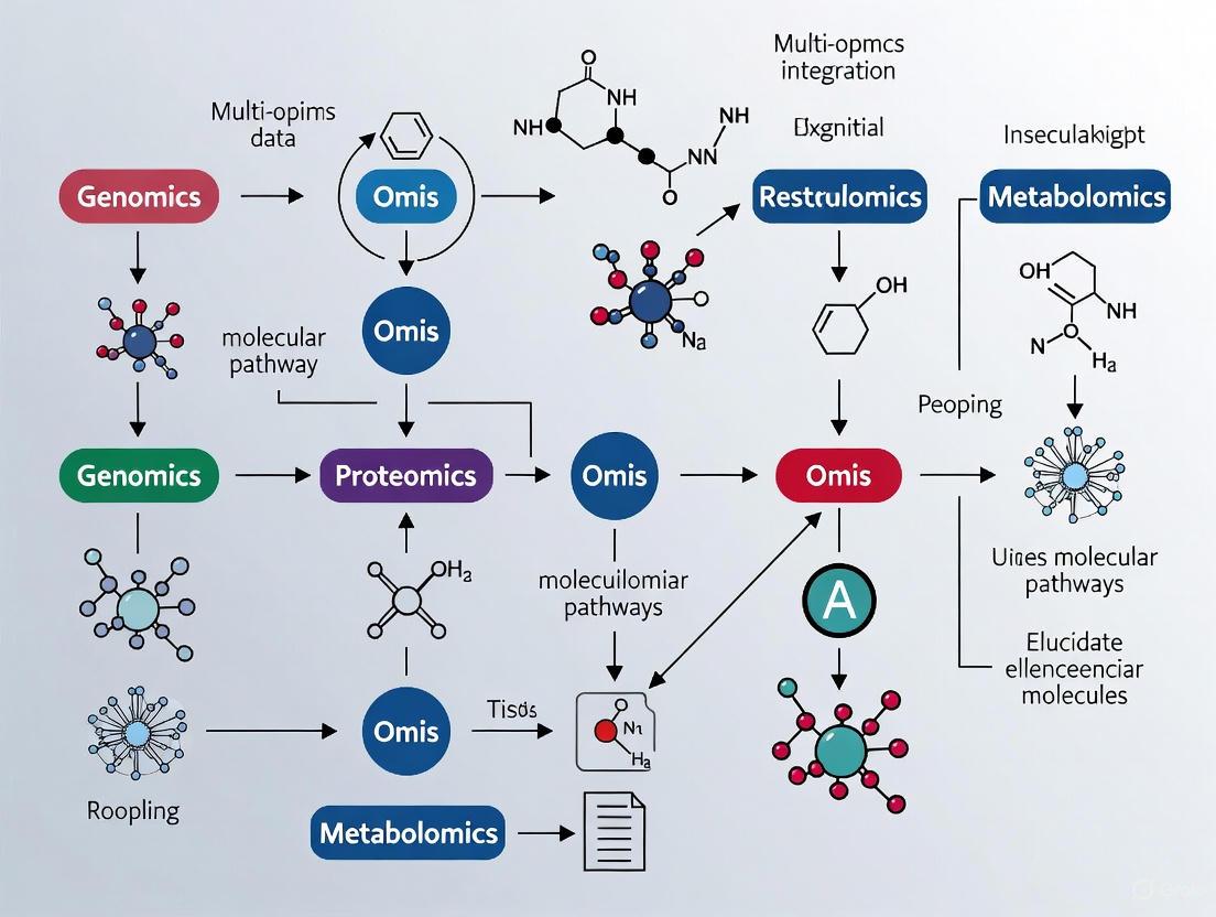
Demystifying Multi-Omics: Core Concepts and System-Level Insights for Pathway Analysis
High-throughput technologies have revolutionized biological research, enabling comprehensive analysis of molecular systems at multiple levels. The integration of genomics, transcriptomics, proteomics, and metabolomics—collectively termed multi-omics—provides unprecedented insights into the complex flow of information underlying biological processes and disease mechanisms. This technical guide delineates the core omics layers, their respective technologies, and their roles in elucidating molecular pathways. By presenting structured comparisons, experimental protocols, and visualization frameworks, we aim to equip researchers with methodologies for effective data integration to advance biomarker discovery, therapeutic target identification, and systems-level understanding in biomedical research.
Omics technologies provide a global assessment of complete sets of biological molecules, moving beyond single-molecule studies to system-wide analyses [1]. The field has been driven largely by technological advances that have made possible cost-efficient, high-throughput analysis of biologic molecules. Each omics layerinterrogates a distinct level of biological organization, from genetic blueprint to functional metabolites, offering unique insights into different aspects of biological systems [2] [1]. When integrated, these technologies enable researchers to understand the flow of information that underlies disease, moving beyond correlations to identify potential causative changes [1]. This multi-omics approach is particularly valuable for interpreting complex diseases where genetic variants alone explain only a fraction of heritability, and dysregulation across multiple molecular layers contributes to pathogenesis [3] [1].
Core Omics Layers: Technologies and Biological Significance
The four primary omics layers provide complementary insights into biological systems, each capturing a different dimension of the central dogma of biology and its regulatory networks.
Table 1: Core Omics Technologies and Their Applications
| Omics Layer | Molecules Analyzed | Key Technologies | Primary Biological Information | Common Applications |
|---|---|---|---|---|
| Genomics | DNA sequences, genetic variants | Genotyping arrays, Whole Genome Sequencing (WGS), Exome sequencing [1] | Genetic blueprint, inherited variations, disease-associated polymorphisms [1] | Genome-wide association studies (GWAS), identification of disease-risk alleles [3] [1] |
| Transcriptomics | RNA transcripts (coding, non-coding) | Microarrays, RNA-Seq, single-cell RNA-Seq [1] | Dynamic gene expression, alternative splicing, regulatory RNAs [2] [1] | Expression quantitative trait loci (eQTL) mapping, pathway activity inference, biomarker discovery [3] [4] |
| Proteomics | Proteins, peptides | Mass spectrometry (MS), affinity purification, protein arrays [1] | Protein abundance, post-translational modifications, protein-protein interactions [2] [1] | Signaling pathway analysis, drug target identification, metabolic engineering [2] [4] |
| Metabolomics | Metabolites (≤1.5 kDa) | Mass spectrometry (MS), NMR spectroscopy [2] [1] | End products of cellular processes, metabolic fluxes, physiological status [2] [1] | Biomarker development, disease diagnosis, metabolic pathway analysis [2] [1] |
Biological Roles and Workflow Relationships
Each omics layer provides unique insights into different stages of biological information flow. Genomics offers a static view of genetic potential, while transcriptomics captures dynamic regulatory responses. Proteomics reveals the functional effectors of cellular processes, and metabolomics reflects the ultimate biochemical outcomes [2] [1]. This hierarchical relationship creates a comprehensive picture of biological systems when integrated.
Diagram 1: Information flow through omics layers
Methodologies for Multi-Omics Integration
Data Integration Approaches and Workflows
Integrating multiple omics data sets is challenging but necessary to fully understand complex biological systems [2]. Several methodological frameworks have been developed, which can be categorized into three primary approaches: correlation-based strategies, combined omics integration, and machine learning techniques [2].
Table 2: Multi-Omics Data Integration Approaches
| Integration Approach | Key Methods | Omics Data Types | Primary Application | Tools/Examples |
|---|---|---|---|---|
| Correlation-based | Co-expression analysis, Gene-metabolite networks, Similarity Network Fusion [2] | Transcriptomics & Metabolomics, Proteomics & Metabolomics [2] | Identify co-regulated modules, construct interaction networks [2] | WGCNA, Cytoscape, PCC analysis [2] |
| Statistical & Enrichment | Pathway enrichment, Signaling Pathway Impact Analysis (SPIA) [4] | Genomics, Transcriptomics, Proteomics [4] | Pathway activation assessment, functional interpretation [4] | IMPaLA, PaintOmics, ActivePathways, SPIA [4] |
| Machine Learning | Supervised/unsupervised learning, multivariate modeling [2] [3] | All omics layers [2] [3] | Disease classification, risk prediction, pattern recognition [3] | DIABLO, OmicsAnalyst, random forest, elastic-net [3] [4] |
| Network-based | Topology-based pathway analysis, protein-protein interaction networks [4] | Transcriptomics, Proteomics, Metabolomics [4] | Identify key regulatory nodes, drug targeting [4] | Oncobox, TAPPA, Pathway-Express, iPANDA [4] |
Experimental Protocol: Multi-Omics Pathway Analysis
The following workflow represents a comprehensive approach for integrating multiple omics layers to assess pathway activation and drug efficacy, adapted from established methodologies in the field [4].
Diagram 2: Multi-omics pathway activation workflow
Step-by-Step Protocol:
Multi-omics Data Collection: Generate molecular profiles using high-throughput technologies. Essential data types include:
- DNA methylation arrays or sequencing for epigenomic regulation
- mRNA-seq for protein-coding transcript levels
- miRNA-seq for microRNA expression profiling
- lncRNA/asRNA-seq for long non-coding and antisense RNA quantification [4]
Differential Expression Analysis: Identify statistically significant molecular differences between case and control samples for each omics layer using appropriate statistical methods (e.g., moderated t-tests, DESeq2, or EdgeR for count data).
Pathway Database Integration: Utilize curated pathway databases (e.g., OncoboxPD containing 51,672 uniformly processed human molecular pathways) with annotated gene functions and interaction topologies [4].
Signaling Pathway Impact Analysis (SPIA): Calculate pathway activation levels using topology-based algorithms that consider:
- Perturbation factors (PF) for all genes in a pathway
- Direction of interactions (activation/inhibition)
- Pathway accumulation accuracy scores [4]
Drug Efficiency Index (DEI) Calculation: Evaluate potential therapeutic efficacy by integrating pathway activation data with drug target information to generate personalized drug rankings [4].
Biological Interpretation: Integrate results across omics layers to identify dysregulated pathways, key regulatory nodes, and potential therapeutic targets.
The Scientist's Toolkit: Essential Research Reagents and Solutions
Successful multi-omics research requires specialized reagents and computational tools to ensure data quality and integration capabilities.
Table 3: Essential Research Reagents and Solutions for Multi-Omics Studies
| Reagent/Tool Category | Specific Examples | Function and Application | Technical Considerations |
|---|---|---|---|
| Nucleic Acid Extraction Kits | DNA/RNA co-extraction kits, miRNA-specific isolation kits | High-quality nucleic acid preservation for parallel genomic/transcriptomic analysis | Maintain RNA integrity (RIN >8), prevent degradation [4] |
| Mass Spectrometry-Grade Solvents | LC-MS/MS compatible solvents, digest buffers | Optimal protein extraction, digestion, and metabolite preservation | Minimize contaminants, ensure batch-to-batch reproducibility [1] |
| Pathway Analysis Databases | OncoboxPD, KEGG, Reactome, Gene Ontology | Pathway topology information for activation calculations | Uniform pathway curation, functional annotations [4] |
| Reference Data Repositories | 1000 Genomes Project, GTEx, ARIC Study, NIAGADS | Control datasets, QTL mapping references, normalization | Population-matched controls, consistent processing [3] |
| Statistical Computing Environments | R/Bioconductor, Python, specialized omics packages | Data normalization, integration, and visualization | Implement reproducible workflows, version control [2] [5] |
Case Study: Multi-Omics in Alzheimer's Disease Research
A recent study demonstrates the power of integrative multi-omics approaches for complex disease characterization [3]. Researchers conducted genome-, transcriptome-, and proteome-wide association studies (GWAS, TWAS, PWAS) on 15,480 individuals from the Alzheimer's Disease Sequencing Project (ADSP) to identify AD-associated molecular signals [3]. The analysis revealed 104 genomic, 319 transcriptomic, and 17 proteomic associations with AD, with novel associations enriched in signaling, myeloid differentiation, and immune pathways [3]. Integrative Risk Models (IRMs) developed using genetically-regulated components of gene and protein expression significantly outperformed traditional polygenic score models, with the best-performing random forest classifier achieving an AUROC of 0.703 and AUPRC of 0.622 [3]. This case study illustrates how multi-omics integration enhances both biological insight and predictive accuracy for complex diseases.
The strategic integration of genomics, transcriptomics, proteomics, and metabolomics provides a powerful framework for elucidating complex biological systems and disease mechanisms. By leveraging the complementary strengths of each omics layer and applying appropriate integration methodologies, researchers can uncover molecular pathways and interactions that remain invisible to single-omics approaches. As technologies advance and analytical methods mature, multi-omics integration will increasingly drive discoveries in basic research, biomarker development, and therapeutic innovation, ultimately enabling more personalized and effective medical interventions.
Biological systems are fundamentally complex, driven by the dynamic interplay between genetic blueprint, epigenetic regulation, gene expression, protein translation, and metabolic activity. Traditional single-omics approaches—analyzing one biological layer, such as the genome or transcriptome in isolation—provide a valuable but inherently limited snapshot of this intricate system. While genomics identifies DNA-level alterations and transcriptomics reveals gene expression dynamics, they individually fail to capture the cascading effects and regulatory feedback loops that characterize complex pathways [6]. The fundamental shortcoming of single-omics is its reductionist nature; it attempts to explain a system's behavior by examining a single component, averaging signals across heterogeneous cell populations and thereby obscuring critical cellular nuances and rare but consequential cell states [7]. As a result, single-omics strategies often yield incomplete mechanistic insights and suboptimal clinical predictions, unable to fully elucidate the molecular mechanisms underlying disease pathogenesis, drug response, or therapeutic resistance [6].
This review argues that a multi-omics integrative framework is not merely an enhancement but a necessity for accurately modeling complex biological pathways. By simultaneously measuring and integrating data from multiple molecular layers, researchers can move from observing correlations to understanding causality, ultimately constructing a more holistic and predictive model of cellular behavior.
The Biological Hierarchy: A Cascade of Information Flow
The flow of biological information is not perfectly linear, but it follows a general hierarchical structure from static genetic instruction to dynamic functional outcome. A perturbation at one level can propagate through subsequent layers, but feedback mechanisms can also exert influence upstream. Single-omics approaches, which focus on a single tier of this hierarchy, cannot capture these complex inter-layer dynamics.
- Genomics: Provides the foundational blueprint, identifying inherited variations and acquired mutations (e.g., SNVs, CNVs) [6].
- Epigenomics: Regulates genomic accessibility through dynamic, reversible modifications like DNA methylation and histone changes, influencing gene expression without altering the DNA sequence itself [6] [8].
- Transcriptomics: Captures the immediate downstream effect, quantifying RNA expression levels that reflect the active transcriptional programs of the cell [6].
- Proteomics: Represents the functional effectors, cataloging protein expression, post-translational modifications (e.g., phosphorylation), and interactions that directly execute cellular processes [9] [6].
- Metabolomics: Profiles the biochemical endpoints, revealing small-molecule metabolites that constitute the final response to genomic, environmental, and therapeutic influences [6].
The following diagram illustrates this hierarchical flow of biological information and the feedback loops that a multi-omics approach is required to capture:
Biological Information Flow and Multi-Layer Regulation
For example, unraveling the cause of a disease may reveal a metabolite deficiency caused by the failure of an enzyme to be phosphorylated because a gene is not expressed due to aberrant methylation as a result of a rare germline variant [9]. This cascade of events, spanning multiple biological layers, is invisible to any single-omics investigation.
The Multi-Omics Experimental Workflow: From Sample to Insight
Implementing a multi-omics study requires a structured workflow that encompasses sample preparation, high-throughput data generation, computational integration, and biological interpretation. The following diagram outlines a generalized protocol for a multi-omics study, integrating steps from single-cell isolation to final data integration:
Generalized Multi-Omics Experimental Workflow
Key Research Reagent Solutions
The execution of a multi-omics experiment relies on a suite of specialized reagents and platforms. The following table details essential materials and their functions in a typical workflow.
Table 1: Essential Research Reagents and Platforms for Multi-Omics Studies
| Item Name | Function |
|---|---|
| Bacterial Artificial Chromosomes (BACs) | Used in hierarchical shotgun sequencing to clone large (150-200 kb) fragments of the genome for amplification and sequencing [9]. |
| Hairpin Adapters | Ligated to DNA fragments in PacBio SMRT sequencing to circularize the template, enabling multiple passes of the same fragment by the polymerase for high-fidelity (HiFi) reads [9]. |
| Template-Switching Oligos (TSOs) | Enable the construction of full-length cDNA libraries in single-cell RNA-seq methods (e.g., SMART-seq3, FLASH-seq), allowing for the identification of 5' transcript ends and isoforms [7]. |
| Cell Barcodes (DNA Oligos) | Unique nucleotide sequences attached to molecules from individual cells during library preparation, allowing samples from thousands of cells to be pooled and sequenced simultaneously while retaining cell-of-origin information [7] [10]. |
| Unique Molecular Identifiers (UMIs) | Random nucleotide tags incorporated during reverse transcription in scRNA-seq protocols to label individual mRNA molecules, mitigating PCR amplification bias and enabling accurate transcript quantification [7]. |
| Zero-Mode Waveguides (ZMWs) | Microscopic wells in PacBio SMRT cells where single molecules of DNA polymerase are immobilized, enabling real-time observation of DNA synthesis for long-read sequencing [9]. |
Analytical Technologies and Sequencing Platforms
The choice of sequencing technology is critical and involves trade-offs between read length, accuracy, throughput, and cost. The table below compares the major sequencing platforms.
Table 2: Comparison of Sequencing Technology Generations
| Platform (Generation) | Sequencing Technology | Read Length | Key Strengths | Key Limitations |
|---|---|---|---|---|
| Sanger (First) | Chain termination | 800-1,000 bp | High accuracy, low analysis difficulty | Low throughput, high historical cost [9] |
| Illumina (Second/Next) | Sequencing by synthesis | 100-300 bp | High throughput, high accuracy, moderate cost | Short reads struggle with repetitive regions [9] |
| PacBio (Third) | Circular consensus sequencing | 10,000-25,000 bp | Very long reads, moderate accuracy | High cost, high computing needs [9] |
| Oxford Nanopore (Third) | Electrical detection | 10,000-30,000 bp | Very long reads, portable devices | Lower read accuracy, high computing needs [9] |
Data Integration Strategies and Computational Methodologies
The core challenge of multi-omics lies in the computational integration of disparate data types. Several conceptual strategies have been developed, each with distinct advantages and limitations.
Table 3: Multi-Omics Data Integration Strategies
| Integration Strategy | Description | Advantages | Disadvantages |
|---|---|---|---|
| Early Integration | Raw or pre-processed data from different omics layers are concatenated into a single large matrix before analysis [11] [12]. | Simple to implement. | Creates a high-dimension, noisy dataset; discounts data distribution differences [12]. |
| Intermediate Integration | Datasets are integrated by identifying common latent structures (e.g., via joint matrix decomposition) [11] [12]. | Reduces dimensionality; can separate shared and omics-specific signals [11]. | Often requires robust pre-processing to handle data heterogeneity [12]. |
| Late Integration | Each omics dataset is analyzed separately, and the results (e.g., model predictions) are combined at the final stage [11] [12]. | Avoids challenges of merging raw data; uses optimized models for each data type. | Fails to capture inter-omics interactions during analysis [12]. |
| Hierarchical Integration | Incorporates prior knowledge of regulatory relationships between different omics layers (e.g., genomic variants influencing transcript levels) [12]. | Most accurately reflects biological causality; true trans-omics analysis. | Methods are often specific to certain omics types; less generalizable [12]. |
The Role of Artificial Intelligence and Machine Learning
Machine learning (ML) and deep learning (DL) are indispensable for navigating the high-dimensionality and non-linear relationships in multi-omics data. Unlike traditional statistics, AI models can identify complex patterns that bridge biological layers.
- Supervised Learning: Used with labeled datasets to predict outcomes such as patient survival or drug response. Algorithms like Random Forest (RF) and Support Vector Machines (SVM) are trained on omics data to create predictive classifiers, requiring careful feature labeling and parameter tuning to avoid overfitting [13].
- Unsupervised Learning: Applied to explore hidden structures without pre-defined labels. Methods like k-means clustering and principal component analysis (PCA) are used for dimensionality reduction and to identify novel cellular subpopulations or disease subtypes from omics data [13].
- Deep Learning: Utilizes artificial neural networks for automatic feature extraction from raw data. Graph Neural Networks (GNNs) can model biological networks, while multi-modal transformers can fuse disparate data types like MRI radiomics and transcriptomics to predict disease progression [6] [13].
- Transfer Learning: A technique where a model developed for one task is reused as the starting point for a model on a second task, which is particularly useful for leveraging knowledge from large public omics databases for specific clinical questions with limited data [13].
Case Study: Single-Cell Multi-Omics in Cancer Research
The transition from bulk to single-cell analysis represents a paradigm shift, moving beyond tissue-level averages to dissect the cellular heterogeneity that drives complex diseases like cancer. Single-cell multi-omics technologies now allow for the simultaneous measurement of multiple modalities—such as genome, epigenome, transcriptome, and proteome—within the same individual cell [7] [10].
The following diagram illustrates a specific workflow for single-cell multi-omics profiling that integrates transcriptomic and epigenomic data:
Single-Cell Multi-Omics Profiling Workflow
Application in Cancer: This approach has been pivotal in characterizing the tumor microenvironment. For example, in breast cancer, an adaptive multi-omics integration framework that combined genomics, transcriptomics, and epigenomics data achieved a concordance index (C-index) of 78.31 for survival prediction, significantly outperforming single-omics models [11]. Similarly, integrating single-cell transcriptomics with T-cell receptor sequencing (scTCR-seq) can identify clonally expanded T-cells and link their transcriptional state to antigen specificity, providing critical insights into anti-tumor immunity and immunotherapy resistance [10].
The evidence is conclusive: single-omics approaches are fundamentally insufficient for deconstructing the complex, dynamic, and interconnected pathways that govern biological systems and disease states. The imperative for multi-omics integration is not merely a trend but a necessary evolution in biological research. By simultaneously querying multiple layers of biological information and leveraging advanced computational strategies, including machine learning, researchers can move from descriptive snapshots to predictive, causal models. This holistic perspective is crucial for transforming our understanding of biology and accelerating the development of precise diagnostics and effective therapeutics for complex human diseases.
The complexity of biological systems extends far beyond the scope of single-omics studies. Multi-omics represents a fundamental shift in biological research, integrating data from various molecular layers—including genomics, transcriptomics, proteomics, epigenomics, and metabolomics—to construct a comprehensive view of how living systems function and interact [14]. This approach is revolutionizing molecular pathways research by enabling scientists to move from observing correlations to understanding causal relationships and regulatory mechanisms across different biological levels.
The power of multi-omics lies in its ability to capture the flow of biological information from DNA to RNA to proteins and metabolites, revealing how perturbations at one level propagate through the system [15]. For researchers and drug development professionals, this integrated perspective is invaluable for identifying robust biomarkers, understanding disease mechanisms, and discovering novel therapeutic targets that might remain hidden when examining single omics layers in isolation [16]. As we advance into an era of precision medicine, multi-omics provides the analytical framework necessary to decipher the complexity of human diseases and develop targeted interventions based on a holistic understanding of molecular pathways.
Multi-Omics Data Integration Methodologies
Effective integration of diverse omics datasets is both a technical challenge and critical success factor in multi-omics research. The integration strategies can be categorized into distinct methodological approaches, each with specific strengths and applications in pathway analysis and biological discovery.
Table 1: Multi-Omics Data Integration Approaches
| Integration Method | Core Principle | Common Applications | Key Advantages |
|---|---|---|---|
| Conceptual Integration | Links omics data through shared biological concepts or entities | Hypothesis generation, exploratory analysis | Leverages existing knowledge bases; intuitive interpretation |
| Statistical Integration | Applies quantitative techniques to combine or compare datasets | Pattern identification, biomarker discovery | Identifies co-expression patterns; handles large datasets |
| Model-Based Integration | Uses mathematical models to simulate system behavior | Dynamic pathway modeling, drug response prediction | Captures system dynamics; enables predictive simulations |
| Network & Pathway Integration | Represents data within biological network structures | Pathway analysis, target prioritization | Contextualizes findings; integrates multiple granularity levels |
More advanced topology-based methods have emerged that incorporate the biological reality of pathways by considering the type, direction, and function of molecular interactions [4]. Methods such as Signaling Pathway Impact Analysis (SPIA) and Drug Efficiency Index (DEI) utilize pathway topology databases to calculate pathway activation levels (PALs), providing more biologically realistic assessments of pathway dysregulation than non-topological approaches [4].
A critical consideration in data integration is the vertical integration of different omics modalities from the same samples, which requires specialized approaches to handle varying statistical properties, technological noise, and feature dimensions across datasets [15]. The Quartet Project has developed reference materials and ratio-based profiling methods that address fundamental reproducibility challenges by scaling absolute feature values of study samples relative to a common reference sample, significantly improving data comparability across platforms and laboratories [15].
Experimental Workflows and Analytical Frameworks
Standardized Multi-Omics Workflow
Implementing a robust multi-omics study requires careful experimental design and execution. The following diagram illustrates a generalized workflow that can be adapted for various research objectives:
Alzheimer's Disease Case Study: Experimental Protocol
A recent investigation exemplifies the application of multi-omics to elucidate complex disease pathways. Researchers performed an integrative multi-omics analysis on 15,480 individuals from the Alzheimer's Disease Sequencing Project (ADSP) to characterize AD risk and identify molecular pathways [3].
Experimental Methodology:
- Cohort Description: 15,480 individuals from ADSP R4 release
- Omics Profiling:
- Genome-wide association study (GWAS) for genomic variants
- Transcriptome-wide association study (TWAS) for gene expression
- Proteome-wide association study (PWAS) for protein expression
- Statistical Analysis: Association testing under significant thresholds with multiple testing correction
- Pathway Analysis: Enrichment analysis of identified associations using functional annotation databases
- Risk Modeling: Development of integrative risk models (IRMs) using elastic-net logistic regression and random forest classifiers
Key Findings:
- Identification of 104 genomic, 319 transcriptomic, and 17 proteomic associations with AD
- Novel associations enriched in signaling, myeloid differentiation, and immune pathways
- Random forest model with transcriptomic and covariate features achieved AUROC of 0.703, significantly outperforming polygenic risk scores alone [3]
This study demonstrates how multi-omics approaches can enhance both biological understanding and predictive modeling for complex diseases.
Pathway Analysis and Computational Framework
Topology-Based Pathway Analysis
Understanding how multi-omics data influences biological pathways requires specialized computational approaches that consider the structure and dynamics of molecular networks. The following diagram illustrates how different omics layers are integrated into topology-based pathway analysis:
Key Computational Tools for Multi-Omics Pathway Analysis
Table 2: Computational Frameworks for Multi-Omics Pathway Analysis
| Tool/Method | Integration Approach | Analytical Output | Application Context |
|---|---|---|---|
| SPIA | Topology-based pathway impact | Pathway activation scores, perturbation factors | Signaling pathway dysregulation analysis |
| DIABLO | Multivariate supervised integration | Patient stratification, feature selection | Biomarker discovery, subtype identification |
| MultiGSEA | Statistical enrichment | Gene set enrichment p-values | Functional profiling across omics layers |
| iPANDA | Network decomposition | Pathway activation levels | Disease stratification, drug response |
| ActivePathways | Data fusion across omics | Integrated pathway p-values | Multi-omics data prioritization |
The SPIA (Signaling Pathway Impact Analysis) framework exemplifies advanced topology-based approaches, calculating pathway perturbation by considering both the enrichment of differentially expressed genes and the propagation of perturbations through pathway topologies [4]. This method incorporates the type and direction of molecular interactions, providing more biologically meaningful pathway activation scores than enrichment-based methods alone.
Recent advances enable the integration of non-coding RNA and DNA methylation data into pathway analysis by accounting for their regulatory effects. For instance, methylation-based and ncRNA-based SPIA values are calculated with negative signs compared to standard mRNA-based values, reflecting their repressive effects on gene expression while utilizing the same pathway topology graphs [4].
Essential Research Reagents and Reference Materials
Successful multi-omics studies require carefully selected reagents and reference materials to ensure data quality and reproducibility. The following table details key solutions used in advanced multi-omics research:
Table 3: Essential Research Reagent Solutions for Multi-Omics Studies
| Reagent/Resource | Type | Function | Application Example |
|---|---|---|---|
| Quartet Reference Materials | Multi-omics reference standards | Provides ground truth for data integration and QC | Cross-platform standardization [15] |
| Laser-Capture Microdissection | Tissue processing | Isolation of specific cell populations | Rare neuron analysis in schizophrenia [17] |
| GTEx v8 Reference | Transcriptome database | Tissue-specific expression reference | Transcriptomic imputation [3] |
| ARIC Study References | Proteomic database | Protein quantitative trait loci (pQTL) | Proteome-wide association studies [3] |
| OncoboxPD Pathway Database | Pathway knowledge base | 51,672 uniformly processed human pathways | Topology-based pathway analysis [4] |
| AAV Vector Systems | Gene delivery vehicle | Therapeutic gene transfer | Gene therapy safety assessment [17] |
| Single-Cell Multi-Omics Kits | Library preparation | Simultaneous profiling of multiple modalities | Cellular heterogeneity resolution |
The Quartet reference materials represent a particularly significant advancement, providing matched DNA, RNA, protein, and metabolites derived from immortalized cell lines from a family quartet [15]. These materials establish "built-in truth" defined by genetic relationships and central dogma information flow, enabling objective assessment of data quality and integration performance across laboratories and platforms.
For drug discovery applications, AAV vector systems require specialized reagents to assess integration sites and potential genotoxicity. Methods such as target enrichment sequencing, whole genome sequencing, and shearing extension primer tag selection are employed to identify junctions between vector DNA and host DNA, ensuring therapeutic safety [17].
Applications in Drug Discovery and Therapeutic Development
Multi-omics approaches are transforming pharmaceutical development by providing comprehensive insights into disease mechanisms and therapeutic responses. Several exemplar applications demonstrate their impact across the drug development pipeline:
Target Identification and Validation
In schizophrenia research, investigators used laser-capture microdissection combined with RNA-seq to characterize rare parvalbumin interneurons implicated in disease pathology [17]. This approach enabled precise profiling of this limited neuronal subpopulation, identifying GluN2D—a subunit of the glutamate receptor—as a potential drug target that would have been difficult to detect using conventional transcriptomic methods.
Biomarker Discovery for Biologics
For biologic therapies, multi-omics facilitates identification of biomarkers predicting immune responses. Researchers employed single-cell RNA-seq with VDJ capture to identify T-cell clones activated by therapeutic exposure [17]. By comparing bulk and single-cell data, they validated clonal expansion patterns and established methods for early detection of immunogenic responses, enabling proactive management of adverse effects.
Gene Therapy Safety Assessment
Comprehensive integration site analysis using multiple sequencing methods demonstrated that AAV vectors integrate randomly throughout the human genome without enrichment in cancer-associated loci [17]. This multi-omics safety assessment provided critical evidence for the therapeutic profile of AAV-based gene therapies, highlighting their low oncogenic risk compared to earlier vector systems.
Integrative Risk Prediction
As demonstrated in the Alzheimer's Disease case study, multi-omics data significantly enhances disease risk prediction compared to traditional approaches [3]. Integrative risk models combining transcriptomic features with clinical covariates achieved superior performance (AUROC: 0.703) over polygenic scores alone, highlighting the clinical value of multi-dimensional molecular profiling for complex diseases.
These applications underscore how multi-omics approaches provide the comprehensive molecular perspective necessary for informed decision-making throughout the therapeutic development process, from initial target identification to post-market safety monitoring.
Major Research Consortia and Public Data Repositories (e.g., TCGA)
Major research consortia and public data repositories are foundational to modern multi-omics research, providing the large-scale, integrated datasets necessary to elucidate complex molecular pathways. Initiatives like The Cancer Genome Atlas (TCGA) and the Alzheimer's Disease Sequencing Project (ADSP) have generated petabytes of genomic, transcriptomic, proteomic, and epigenomic data, enabling researchers to move beyond single-layer analysis to a more holistic understanding of disease biology [18] [19]. The effective use of these resources requires navigating specific data portals, understanding consortium governance, and applying sophisticated computational integration strategies to uncover the interconnected regulatory and metabolic networks that define physiological and pathological states [20] [21] [22]. This guide provides a technical overview of these key resources, their data structures, and the methodologies for their integration, serving as a roadmap for researchers aiming to leverage these assets for pathway discovery and therapeutic development.
Landscape of Major Consortia and Repositories
Large-scale collaborative efforts are crucial for generating the sample sizes and data diversity required for robust multi-omics discovery. The table below summarizes key resources relevant to multi-omics pathway research.
Table 1: Major Multi-Omics Research Consortia and Data Repositories
| Name | Primary Focus | Key Data Types | Access Portal | Notable Scale & Features |
|---|---|---|---|---|
| The Cancer Genome Atlas (TCGA) [19] [23] | Cancer Genomics | Genomic, Epigenomic, Transcriptomic, Proteomic | Genomic Data Commons (GDC) Portal [19] | >20,000 patients; 33 cancer types; over 2.5 petabytes of data [19] |
| Alzheimer's Disease Sequencing Project (ADSP) [18] | Neurodegenerative Disease | Whole Genome Sequencing, Transcriptomic, Proteomic | NIAGADS [18] | 15,480 individuals (in focused analysis); genome-, transcriptome-, proteome-wide association studies [18] |
| NCI Cohort Consortium [24] | Cancer Epidemiology & Risk | Genomic, Biospecimens, Epidemiologic Data | dbGaP [24] | >50 cohorts; >7 million people; international scope [24] |
| Qatar Metabolomics Study of Diabetes (QMDiab) [21] | Diabetes & Metabolic Disease | Genomic, Methylation, Transcriptomic, Proteomic, Metabolomic | "The Molecular Human" Web Interface [21] | 391 participants; 18 diverse omics platforms; 6,304 molecular traits per sample [21] |
| MLOmics [25] | Pan-Cancer Machine Learning | mRNA, miRNA, DNA Methylation, Copy Number Variation | MLOmics Database [25] | 8,314 TCGA patient samples; 32 cancer types; pre-processed, model-ready data [25] |
| Cancer Imaging Archive (TCIA) [23] | Cancer Imaging | Medical Images, Radiomics, Clinical Data | TCIA Website [23] | Curated archive of medical images; linked with TCGA and other molecular data [23] |
These resources are supported by central data portals and knowledgebases designed to facilitate access and analysis:
- Genomic Data Commons (GDC): A unified data repository that enables data sharing across cancer genomic studies in support of precision medicine, hosting data from TCGA and other NCI programs [23].
- Database of Genotypes and Phenotypes (dbGaP): Developed by NIH to archive and distribute results from studies investigating genotype-phenotype interactions. Many NCI-funded genomic datasets are available here [26] [23] [24].
- Omics Discovery Index (OmicsDI): A knowledge discovery framework that provides a searchable index across heterogeneous public omics data from genomics, proteomics, transcriptomics, and metabolomics studies [27].
Experimental Protocols for Multi-Omic Data Generation and Integration
Leveraging data from consortia requires an understanding of both the experimental protocols used for data generation and the computational workflows for integration. The following methodology, adapted from a large-scale multi-omics study on Alzheimer's disease, provides a robust framework [18].
Genome-Wide Association Studies (GWAS)
- Objective: Identify genetic loci associated with disease risk or specific molecular traits (quantitative trait loci, QTLs).
- Protocol:
- Quality Control (QC): Perform rigorous QC on genetic variant data. Remove variants with low minor allele count (e.g., MAC < 20), low call rate (e.g., < 95%), and duplicate samples [18].
- Association Testing: Conduct association testing using an additive genetic model in tools like PLINK v2.0. Adjust for covariates including age, sex, and genetic principal components to account for population stratification [18].
- Significance Thresholding: Apply a genome-wide significance threshold (e.g., ( p < 5 \times 10^{-8} )) to identify significant associations [18].
Transcriptome-Wide and Proteome-Wide Association Studies (TWAS/PWAS)
- Objective: Identify genes and proteins whose expression levels are associated with a trait, using genetic variation as a anchor to infer causality.
- Protocol:
- Reference Panel: Use genetically regulated expression or protein models (e.g., from GTEx Project v8 via PredictDB) that predict molecular abundance based on genetic variation [18].
- Imputation & Association: Impute the genetically regulated component of gene or protein expression into the study cohort and test for association with the phenotype of interest.
- Validation: Perform mediation analysis to test if the effect of a genetic variant on the trait is mediated through the expression level of a specific gene or protein [18].
Multi-Omics Integration and Pathway Analysis
- Objective: Integrate signals from multiple molecular layers to identify coherent biological pathways and build predictive models.
- Protocol:
- Univariate Discovery: Conduct GWAS, TWAS, and PWAS independently to identify significant associations within each molecular layer [18].
- Pathway Enrichment Analysis: Use tools like GOrilla or GSEA to test for over-representation of significant genes/proteins in known biological pathways (e.g., cholesterol metabolism, immune signaling) [18].
- Multivariate Predictive Modeling:
- Feature Construction: Use genetically regulated components of gene and protein expression as features in predictive models [18].
- Model Training: Employ machine learning algorithms such as:
- Elastic-net logistic regression for feature selection and classification.
- Random Forest to capture non-linear effects and complex interactions [18].
- Model Evaluation: Assess performance using metrics like Area Under the Receiver Operating Characteristic (AUROC) and Area Under the Precision-Recall Curve (AUPRC), comparing against baseline models like polygenic risk scores [18].
Figure 1: A high-level workflow for a multi-omics study, from data generation to integration and interpretation.
Computational Strategies for Multi-Omics Integration
The integration of disparate omics layers is a central challenge. The choice of method depends on whether the data is matched (from the same sample) or unmatched (from different samples) [20].
Table 2: Multi-Omics Integration Methods and Their Applications
| Method | Type | Underlying Methodology | Best Suited For | Key Features |
|---|---|---|---|---|
| MOFA+ [20] [22] | Matched (Vertical) | Unsupervised Bayesian factor analysis | Identifying latent sources of variation across omics layers; exploratory analysis. | Infers factors that capture co-variation across modalities; no phenotype supervision required. |
| DIABLO [22] | Matched (Vertical) | Supervised multiblock sPLS-DA | Building predictive models for a known phenotype; biomarker discovery. | Uses phenotype labels to identify features that are discriminative and correlated across omics. |
| Similarity Network Fusion (SNF) [22] | Matched (Vertical) | Network-based integration | Data clustering and subtyping; identifying sample groups with multi-omics concordance. | Fuses sample-similarity networks from each omics layer into a single network. |
| GLUE [20] | Unmatched (Diagonal) | Graph-linked variational autoencoder | Integrating multiple omics from different cells or studies. | Uses prior biological knowledge to guide the integration of unpaired data. |
| Seurat v4/v5 [20] | Matched & Unmatched | Weighted nearest neighbors / Bridge integration | Single-cell multi-omics integration; transferring labels across datasets. | Robust and widely used framework for single-cell data; can integrate RNA, protein, ATAC-seq. |
| MCIA [22] | Matched (Vertical) | Multiple co-inertia analysis | Jointly visualizing relationships between samples and features across multiple omics tables. | Multivariate statistical method that projects multiple datasets into a shared space. |
Figure 2: Overview of core multi-omics integration strategies and their primary analytical outputs.
The Scientist's Toolkit: Research Reagent Solutions
The following table details essential computational tools and data resources that form the backbone of multi-omics research.
Table 3: Essential Computational Tools and Data Resources for Multi-Omics Research
| Item/Reagent | Function | Specific Application in Multi-Omics |
|---|---|---|
| GTEx eQTL Models | Reference panels of genetically regulated gene expression. | Imputing transcriptomic abundance in TWAS; available via PredictDB [18]. |
| PLINK v2.0 | Whole-genome association analysis toolset. | Performing QC and GWAS on large-scale sequencing data [18]. |
| MOFA+ | Unsupervised integration tool for multi-omics data. | Decomposing multi-omics datasets into latent factors that capture shared biology [20] [22]. |
| Seurat Suite | R toolkit for single-cell genomics. | Integrating and analyzing matched single-cell multi-omics data (RNA, ATAC, protein) [20]. |
| MLOmics Database | Pre-processed, machine-learning-ready cancer multi-omics database. | Training and evaluating ML models on pan-cancer classification and subtyping tasks [25]. |
| Omics Playground | Integrated, code-free platform for multi-omics analysis. | Providing access to multiple integration methods (MOFA, DIABLO, SNF) for biologists and translational researchers [22]. |
| COmics Web Interface | Interactive tool for exploring molecular networks. | Visualizing and hypothesis generation from integrated multi-omics networks, as demonstrated in the QMDiab study [21]. |
Major research consortia and their associated public data repositories have fundamentally transformed the scale and scope of multi-omics research. By providing standardized, high-quality data from thousands of individuals, resources like TCGA and ADSP empower the scientific community to dissect the complex, interconnected molecular pathways underlying human disease. The full potential of these assets is realized through sophisticated computational integration strategies—from unsupervised factor analysis to supervised machine learning—that can weave disparate data types into a coherent molecular narrative. As these datasets continue to grow in size and diversity, and as integration methodologies become more powerful and accessible, the path to discovering novel disease mechanisms, predictive biomarkers, and therapeutic targets becomes increasingly clear.
Multi-Omics in Action: Integration Strategies and Applications in Drug Discovery
The overarching goal of multi-omics research is to achieve a holistic understanding of biological systems by integrating complementary molecular data layers. Biological systems are complex organisms with numerous regulatory features, including DNA, mRNA, proteins, metabolites, and epigenetic factors, each of which can be influenced by disease and cause changes in cell signaling cascades and phenotypes [28]. The fundamental challenge lies in synthesizing these diverse data types—each with unique scales, noise characteristics, and technological limitations—to reveal how genes, proteins, and epigenetic factors collectively influence disease phenotypes [28].
Multi-omics data integration methods have evolved to address this complexity, generally falling into three primary categories: conceptual integration, which combines findings at the interpretation stage; statistical integration, which identifies relationships across datasets; and model-based integration, which uses mathematical frameworks to predict system behavior [28]. The choice of integration strategy is critical, as it determines the biological insights that can be gleaned, from discovering novel biomarkers to unraveling complex molecular pathways in diseases such as cancer [29] [20].
This technical guide provides a comprehensive overview of these integration approaches, focusing on their application in elucidating molecular pathways. We detail methodologies, present comparative analyses in structured tables, and provide visualization workflows to assist researchers in selecting and implementing appropriate integration strategies for their multi-omics investigations.
Classification of Integration Approaches
Multi-omics integration methodologies can be categorized based on their underlying principles and the stage at which integration occurs. These approaches are not mutually exclusive, and hybrid methods are increasingly common. The three primary frameworks—conceptual, statistical, and model-based—offer distinct advantages and are suited to different research objectives.
Table 1: Core Data Integration Approaches in Multi-Omics Research
| Integration Approach | Core Principle | Typical Methods | Primary Use Cases |
|---|---|---|---|
| Conceptual Integration | Independent analysis of each omics layer with integration during biological interpretation | Pathway enrichment analysis, network mapping | Hypothesis generation, functional validation, placing results in biological context |
| Statistical Integration | Identification of statistical relationships and correlations across omics datasets | Correlation analysis, co-expression networks (WGCNA), multivariate (PCA, CCA) | Identifying co-regulated features, biomarker discovery, data reduction |
| Model-Based Integration | Using mathematical models to predict system behavior from multi-omics inputs | Constraint-based modeling, deep learning (AE, VAE, GAN), machine learning | Predictive modeling, classification, disentangling regulatory mechanisms |
The integration process can also be characterized by its architecture, particularly in computational approaches:
- Early Integration: Features from each modality are concatenated before being processed by a model [30].
- Intermediate Integration: Modalities remain separate but model learns inter-modality relationships to generate an integrated representation or shared latent space [30].
- Late Integration: Separate models are trained for each modality, with predictions combined for a final aggregated result [30].
Furthermore, integration strategies must account for data pairing. Matched (vertical) integration combines omics data profiled from the same cell or sample, using the biological unit itself as an anchor. In contrast, unmatched (diagonal) integration combines data from different cells, samples, or studies, requiring computational alignment in a latent space [20].
Conceptual Integration Approaches
Conceptual integration represents a knowledge-driven framework where multi-omics data are analyzed independently and combined during the interpretation phase using established biological knowledge. This approach leverages curated pathway databases and molecular interaction networks to contextualize findings across omics layers.
Pathway-Based Integration
Pathway analysis facilitates conceptual integration by transforming molecular-level abundance data into pathway-level activity scores. Methods like single-sample Pathway Analysis (ssPA) condense molecular measurements into pathway activity scores for each sample, creating a pathway-level matrix that can be used for downstream analysis and integration [31]. Tools such as PathIntegrate employ ssPA to transform multi-omics datasets from molecular to pathway-level, then apply predictive models to integrate the data [31]. This approach outputs multi-omics pathways ranked by their contribution to outcome prediction, the contribution of each omics layer, and the importance of individual molecules within pathways.
Pathway-based integration offers several advantages: it provides a more parsimonious model when there are fewer input pathways than molecules, enables detection of multiple small correlated signals that may be missed in molecular-level data, and increases robustness to data noise by maximizing biological variation while reducing technical variation [31].
Network-Based Integration
Network-based approaches construct molecular interaction networks that incorporate multiple omics layers, using prior knowledge of biological interactions. These networks can include protein-protein interactions, gene regulatory networks, and metabolic pathways, providing a framework for interpreting multi-omics data in the context of established biology.
Gene-metabolite networks exemplify this approach, visualizing interactions between genes and metabolites in a biological system. These networks are generated by collecting gene expression and metabolite abundance data from the same biological samples, then integrating them using correlation analysis or other statistical methods to identify co-regulated or co-expressed genes and metabolites [32]. Visualization software such as Cytoscape or igraph enables the construction of these networks, where genes and metabolites are represented as nodes connected by edges representing the strength and direction of their interactions [32].
Statistical Integration Approaches
Statistical integration methods identify quantitative relationships across omics datasets through correlation measures, co-expression patterns, and multivariate analyses. These approaches are particularly valuable for identifying coordinated changes across molecular layers and for dimension reduction.
Correlation-Based Methods
Correlation analysis represents a fundamental statistical integration approach, quantifying the degree to which variables from different omics datasets are related. Simple scatterplots can visualize expression patterns and identify consistent or divergent trends between omics layers [33]. For example, transcript-to-protein ratios can be investigated in scatter plot quadrants representing discordant or unanimous up- or down-regulation [33].
Pearson's or Spearman's correlation analysis and their multivariate generalizations, such as the RV coefficient, are employed to test correlations between whole sets of differentially expressed features across different biological contexts [33]. These analyses can determine the extent and nature of interaction between sets of differentially expressed proteins and metabolites, assess whether up-regulated proteins correlate with increased metabolites, identify molecular regulatory pathways of correlated genes and proteins, or assess transcription-protein correspondence [33].
Correlation Networks and Co-Expression Analysis
Correlation networks extend basic correlation analysis by transforming pairwise associations into graphical representations. In these networks, nodes represent biological entities, and edges are constructed based on correlation thresholds, facilitating visualization and analysis of complex relationships within and between datasets [33].
Weighted Gene Co-Expression Network Analysis (WGCNA) is a widely used method that identifies clusters (modules) of highly correlated, co-expressed genes [33] [32]. WGCNA constructs a scale-free network that assigns weights to gene interactions, emphasizing strong correlations while reducing the impact of weaker connections. These modules can be summarized by their eigengenes (representative expression profiles) and linked to clinically relevant traits or other omics data [33] [32]. For example, co-expression analysis can be performed on transcriptomics data to identify gene modules, which are then linked to metabolites from metabolomics data to identify metabolic pathways co-regulated with the identified gene modules [32].
xMWAS is an integrated tool that performs correlation and multivariate analyses for multi-omics integration. It performs pairwise association analysis using Partial Least Squares (PLS) components and regression coefficients, then employs these coefficients to generate integrative network graphs [33]. Communities of highly interconnected nodes can be identified using multilevel community detection methods that maximize modularity—a measure of how well the network is divided into modules with higher internal connectivity [33].
Figure 1: Workflow for Statistical Integration via Correlation Networks
Model-Based Integration Approaches
Model-based integration employs mathematical and computational models to synthesize multi-omics data, often with predictive capabilities. These approaches range from constraint-based biochemical models to sophisticated machine learning and deep learning architectures.
Constraint-Based Modeling
Constraint-based models use stoichiometric metabolic networks as a scaffold for integrating multi-omics data, particularly transcriptomics and metabolomics. INTEGRATE is an example pipeline that uses constraint-based stoichiometric metabolic models to characterize multi-level metabolic regulation [34]. It computes differential reaction expression from transcriptomics data and uses constraint-based modeling to predict if differential expression of metabolic enzymes directly causes differences in metabolic fluxes. Concurrently, it uses metabolomics to predict how differences in substrate availability translate into flux differences [34].
This approach helps discriminate fluxes regulated at different levels:
- Transcriptional control: Flux variations mainly determined by enzyme abundance changes
- Metabolic control: Flux variations mainly determined by substrate abundance changes
- Combined control: Flux variations determined by concerted changes in both substrates and enzymes [34]
Machine Learning and Deep Learning Approaches
Machine learning, particularly deep learning, has revolutionized model-based multi-omics integration by handling high-dimensional, heterogeneous data and capturing non-linear relationships.
Table 2: Deep Learning Approaches for Multi-Omics Integration
| Method Category | Key Examples | Integration Strategy | Key Features |
|---|---|---|---|
| Non-Generative Models | MOLI [30], MOGONET [35] | Late or intermediate integration | Modality-specific encoding, graph convolutional networks |
| Autoencoders | Variational Autoencoders (VAE) [20] [30] | Intermediate integration | Learn shared latent representation, dimensionality reduction |
| Generative Models | Generative Adversarial Networks (GAN) [30] | Intermediate integration | Handle missing data, generate synthetic samples |
| Multi-View Models | Multi-block PLS, PathIntegrate Multi-View [31] | Simultaneous integration | Model interactions between omics datasets |
Deep learning architectures can be further categorized by their integration strategy:
Feedforward Neural Networks (FNNs): Methods like MOLI use modality-specific encoding FNNs to learn features separately before concatenation and final prediction [30]. To address inter-modality interactions, superlayered neural networks (SNN) include separate FNN superlayers for each modality with cross-connections allowing information flow between modalities [30].
Graph Convolutional Networks (GCNs): Methods like MOGONET leverage biological relationships by constructing graphs for each omics data type and applying graph convolutional networks to learn features, which are then integrated for classification [35].
Autoencoders: These learn compressed representations of input data through encoder-decoder structures. Variational autoencoders and other autoencoder architectures can integrate multi-omics data by learning a shared latent representation that captures the essential biological signal across modalities [30].
Multi-View Models: Frameworks like PathIntegrate Multi-View use multi-block partial least squares regression (MB-PLS) to model interactions between pathway-transformed omics datasets [31].
Figure 2: Model-Based Multi-Omics Integration Approaches
Experimental Protocols for Multi-Omics Integration
Implementing robust multi-omics integration requires systematic experimental and computational workflows. Below, we detail two representative protocols for pathway-based and model-based integration.
Protocol 1: Pathway-Based Integration with PathIntegrate
Objective: To integrate multi-omics data at the pathway level for improved interpretability and signal detection in low signal-to-noise scenarios.
Materials:
- Multi-omics datasets (e.g., transcriptomics, proteomics, metabolomics)
- Pathway databases (e.g., KEGG, Reactome)
- PathIntegrate Python package
Methodology:
- Data Preprocessing: Normalize and scale each omics dataset separately to address technical variation.
- Pathway Transformation: Apply single-sample pathway analysis (ssPA) using principal component analysis (PCA) or kernel PCA to transform molecular-level data into pathway activity scores.
- Model Training:
- For single-view integration: Concatenate pathway-transformed datasets and apply classification or regression models.
- For multi-view integration: Use multi-block PLS to model interactions between pathway-transformed omics datasets.
- Model Interpretation: Extract important pathways ranked by contribution to prediction, assess contribution of each omics layer, and identify key molecules within significant pathways.
Validation: Use semi-synthetic data with inserted known signals to benchmark performance against molecular-level integration methods [31].
Protocol 2: Model-Based Integration with Constraint-Based Modeling
Objective: To characterize multi-level metabolic regulation by integrating transcriptomics and metabolomics data.
Materials:
- Transcriptomics data (RNA-seq or microarray)
- Metabolomics data (targeted or untargeted)
- Stoichiometric metabolic model (e.g., Recon)
- INTEGRATE computational pipeline
Methodology:
- Data Processing: Identify differentially expressed genes and differentially abundant metabolites between conditions.
- Flux Prediction from Transcriptomics: Use Gene-Protein-Reaction associations in the metabolic model to predict metabolic fluxes from transcriptomics data.
- Flux Prediction from Metabolomics: Use substrate abundance data to predict metabolic fluxes through constraint-based modeling.
- Integration and Regulation Assessment: Intersect flux predictions from both omics layers to classify reactions as under:
- Transcriptional control if flux variation correlates with enzyme abundance changes
- Metabolic control if flux variation correlates with substrate abundance changes
- Combined control if both mechanisms are involved
Application: Demonstrate using immortalized normal and cancer breast cell lines to identify therapeutic targets [34].
Table 3: Key Research Reagent Solutions for Multi-Omics Integration
| Resource Category | Specific Tools/Databases | Function and Application |
|---|---|---|
| Data Repositories | TCGA, CPTAC, ICGC, CCLE, METABRIC [29] | Provide curated multi-omics datasets from various cancer types and cell lines for method development and validation |
| Pathway Resources | KEGG, Reactome, GO | Curated pathway knowledge for conceptual integration and pathway-based analysis |
| Statistical Tools | WGCNA, xMWAS [33] [32] | Perform correlation network analysis and identify co-expression modules across omics layers |
| Model-Based Platforms | INTEGRATE [34], PathIntegrate [31], MOFA+ [20] | Implement specific model-based integration approaches for disentangling regulatory mechanisms |
| Deep Learning Frameworks | MOLI [30], MOGONET [35], CustOmics [35] | Provide specialized deep learning architectures for multi-omics data integration and classification |
| Visualization Software | Cytoscape [32], igraph [32] | Enable network visualization and exploration of multi-omics relationships |
Successful multi-omics integration requires careful consideration of biological and technical factors. Biological complexity—including varying numbers of genes and proteins across organisms, wide dynamic ranges of molecules, and differences in lifetime expression of mRNA and proteins—must be accounted for in study design and interpretation [28]. Technical considerations include handling missing data, high dimensionality, batch effects, and platform-specific limitations [30] [33]. Furthermore, emerging evidence highlights the importance of considering microbiome influences on host gene and protein expression, as microbiota and their metabolites can affect the host epigenetic landscape and therapeutic responses [28].
As multi-omics technologies continue to advance, integration methods will increasingly need to handle spatial data, single-cell resolutions, and ever-larger datasets. The development of more interpretable deep learning models and standardized benchmarking frameworks will be crucial for translating multi-omics integration into clinical applications and personalized medicine.
Network and pathway-based integration represents a sophisticated computational approach for analyzing multi-omics datasets by mapping diverse molecular measurements onto shared biochemical networks. This methodology moves beyond simple gene lists to leverage the known topology and directional relationships within biological pathways, enabling more accurate interpretation of complex molecular data in health and disease. By considering the structural and functional relationships between genes, proteins, and metabolites, researchers can identify dysregulated pathways, discover novel therapeutic targets, and understand compensatory mechanisms in drug resistance. This technical guide explores the fundamental principles, methodologies, and applications of network-based integration approaches, providing researchers and drug development professionals with practical frameworks for implementing these advanced analytical techniques in multi-omics research.
Network and pathway-based integration has emerged as a powerful paradigm for analyzing multi-omics data by leveraging the inherent structure of biological systems. Unlike earlier enrichment methods that treated pathways as simple gene lists, modern network-based approaches incorporate the topological organization of pathways—including the directionality of interactions, regulatory relationships, and biochemical reaction flows—to provide more biologically meaningful interpretations of multi-omics datasets. This methodology recognizes that cellular functions emerge from complex networks of molecular interactions rather than from individual molecules acting in isolation.
The fundamental premise of network-based integration is that different omics layers—genomics, transcriptomics, proteomics, epigenomics, and metabolomics—provide complementary views of the same underlying biological processes. By mapping these diverse measurements onto unified pathway representations, researchers can identify consistent patterns across molecular layers that might be missed when analyzing each dataset separately. This approach has proven particularly valuable in cancer research, where pathway-level analyses have revealed convergent biological processes despite heterogeneous genetic alterations across patients. Network-based methods effectively address the "high-dimensionality" challenge in multi-omics studies, where the number of measured features vastly exceeds the number of samples, by leveraging prior biological knowledge to constrain possible interpretations [36].
Core Methodologies and Analytical Frameworks
Topology-Based Pathway Analysis
Topology-based methods incorporate the biological reality of pathways by considering the type, direction, and functional role of molecular interactions. These approaches have consistently outperformed non-topological methods in benchmarking studies by more accurately reflecting biological mechanisms [4]. The core mathematical framework for many topology-based methods involves calculating pathway perturbation by accounting for upstream and downstream effects within the network.
The Pathway-Express (PE) algorithm calculates a pathway score combining traditional enrichment statistics with perturbation factors propagated through the network topology [4]. For a pathway K, the PE-score is computed as:
[PE(K) = -\log(P{hypergeometric}(K)) \times \frac{\sum{g \in K} PF(g)}{N_{de}(K)}]
Where (P{hypergeometric}) is the hypergeometric p-value for enrichment of differentially expressed genes, (PF(g)) is the perturbation factor for gene g, and (N{de}(K)) is the number of differentially expressed genes in pathway K. The perturbation factor for each gene is calculated as:
[PF(g) = \Delta E(g) + \sum{u=1}^{n} \frac{\beta{ug} \cdot PF(u)}{N_{ds}(u)}]
Where (\Delta E(g)) represents the normalized expression change of gene g, (\beta{ug}) is the interaction coefficient between genes u and g, and (N{ds}(u)) is the number of downstream genes of u [4].
The Signaling Pathway Impact Analysis (SPIA) method extends this approach by combining the probability of observing a certain number of differentially expressed genes in a pathway (PNDE) with the probability of observing a certain amount of pathway perturbation (PPERT) calculated from the topology [4]. The combined evidence is computed as:
[PG = P{NDE} \times P_{PERT}]
[PG = P{NDE} \times (1 - P_{PERT})]
[PG = P{NDE} \times P_{PERT}]
These probabilities are then combined into a global p-value assessing the overall significance of pathway perturbation [4].
Directional Integration Methods
Directional integration methods incorporate expected relationships between different omics layers based on biological principles or experimental design. The Directional P-value Merging (DPM) method enables researchers to define directional constraints when integrating multiple datasets, prioritizing genes with consistent directional changes across omics layers while penalizing those with inconsistent patterns [37].
The DPM method computes a directionally weighted score across k datasets as:
[X{DPM} = -2(-|\Sigma{i=1}^{j} \ln(Pi) oi ei| + \Sigma{i=j+1}^{k} \ln(P_i))]
Where (Pi) represents the p-value from dataset i, (oi) is the observed directional change (e.g., +1 for upregulation, -1 for downregulation), and (e_i) is the expected direction defined by the constraints vector [37]. This approach allows explicit testing of hypotheses based on biological principles, such as the expected inverse relationship between promoter methylation and gene expression, or the positive relationship between mRNA and protein expression implied by the central dogma.
Table 1: Comparison of Network-Based Integration Methods
| Method | Statistical Approach | Data Types Supported | Key Features | Applications |
|---|---|---|---|---|
| ActivePathways [38] | Brown's combined probability test + ranked hypergeometric test | Genomic mutations, expression, epigenetic data | Identifies pathways enriched across multiple datasets; highlights contributing evidence | Pan-cancer analysis of coding and non-coding drivers |
| SPIA [4] | Topology-based perturbation analysis | Gene expression, non-coding RNA, methylation | Incorporates pathway topology; calculates net pathway perturbation | Drug efficiency indexing; pathway activation assessment |
| DPM [37] | Directional P-value merging | Any with directional information (e.g., expression, methylation) | User-defined directional constraints; integrates directional and non-directional data | Biomarker discovery; pathway regulation in gliomas |
| PARADIGM [36] | Bayesian network inference | Multiple omics layers simultaneously | Integrates diverse evidence types; estimates pathway activity | Patient stratification; causal network identification |
| TIGERS [39] | Tensor imputation + trajectory analysis | Single-cell transcriptomics | Predicts missing drug responses; identifies pathway trajectories | Drug mechanism of action at single-cell level |
Tensor-Based Methods for Single-Cell Data
The TIGERS (Tensor-based Imputation of Gene-Expression Data at the Single-Cell Level) method addresses the challenge of analyzing drug-induced single-cell transcriptomic data with high missing value rates [39]. This approach represents data as a third-order tensor (drugs × genes × cells) and uses tensor-train decomposition to impute missing values while preserving biological structure.
The performance evaluation of TIGERS demonstrated significantly lower relative standard errors (RSE mean = 0.527 at 10% missing rate) compared to standard imputation methods like MAGIC and SAVER (RSE mean = 2.136) [39]. The method successfully preserved cell-type-specific expression patterns for marker genes such as insulin (beta cells) and glucagon (alpha cells) in pancreatic islets, enabling accurate pathway trajectory analysis across inferred cell states.
Experimental Protocols and Workflows
Protocol for Integrative Pathway Analysis of Multi-Omics Data
Step 1: Data Preprocessing and Quality Control
- Perform platform-specific normalization and quality control for each omics dataset
- Map all features to common gene identifiers using reference databases
- Generate statistical significance measures (P-values) and directional effects (e.g., fold-changes) for each gene in each dataset
- For mutation data, annotate functional impact and calculate gene-level burden scores
Step 2: Define Integration Strategy and Directional Constraints
- Select appropriate integration method based on data types and research question
- For directional methods like DPM, define constraints vector based on biological relationships (e.g., mRNA-protein: +1; methylation-expression: -1)
- Configure analysis parameters (significance thresholds, multiple testing correction)
Step 3: Perform Data Integration and Pathway Analysis
- Execute chosen integration method (e.g., ActivePathways, SPIA, DPM)
- Calculate combined significance scores across omics datasets
- Perform pathway enrichment analysis using comprehensive pathway databases
- Identify significantly enriched pathways with evidence from individual omics layers
Step 4: Result Interpretation and Validation
- Visualize enriched pathways as network maps highlighting multi-omics evidence
- Identify master regulator genes and key bottlenecks in dysregulated pathways
- Perform experimental validation of key findings using targeted assays
- Correlate pathway activities with clinical outcomes where available [38] [37]
Protocol for Resistance Pathway Mapping
Step 1: Generate Resistance Models
- Treat sensitive cell lines with increasing drug concentrations to develop resistance
- Use combinatorial approaches: ORF overexpression, CRISPR activation, or chemical libraries
- Select for resistant clones over 4-8 weeks with appropriate drug selection
Step 2: Multi-Omics Profiling of Resistant Models
- Perform whole-exome or whole-genome sequencing to identify acquired mutations
- Conduct RNA sequencing to identify transcriptional adaptations
- Perform proteomic profiling to assess protein expression and phosphorylation changes
- Analyze epigenetic modifications (methylation, chromatin accessibility)
Step 3: Pathway-Centric Data Integration
- Map alterations onto curated signaling pathways (MAPK, PI3K, apoptotic, etc.)
- Identify consistently altered pathways across multiple resistant models
- Distinguish driver from passenger alterations using functional impact scores
- Validate candidate resistance pathways using pharmacological or genetic inhibition [40]
Table 2: Research Reagent Solutions for Multi-Omics Pathway Studies
| Reagent/Resource | Type | Function | Example Sources |
|---|---|---|---|
| Quartet Reference Materials [15] | Reference standards | Multi-omics proficiency testing; batch effect correction | Chinese Quartet Project; National Reference Materials |
| Oncobox Pathway Databank [4] | Pathway database | 51,672 uniformly processed human pathways for activation analysis | OncoboxPD |
| Lentiviral ORF Libraries [40] | Functional screening | Gain-of-function resistance gene identification | Addgene, commercial vendors |
| CRISPR Activation Libraries [40] | Functional screening | Identification of resistance drivers via transcriptional activation | Commercial vendors |
| Tensor Decomposition Algorithms [39] | Computational tool | Missing data imputation for single-cell drug response data | TIGERS implementation |
| Pathway Annotations [38] [36] | Knowledge base | Gene set collections for enrichment analysis | GO, Reactome, KEGG, MSigDB |
Pathway Visualization and Data Representation
Effective visualization of integrated pathway networks requires careful consideration of color theory and accessibility principles. The following diagrams adhere to WCAG 2.1 contrast standards, using a restricted palette to ensure clarity while maintaining sufficient visual distinction between network elements [41].
Network Integration Workflow
Resistance Pathway Mapping
Applications in Biomedical Research
Cancer Driver Discovery
Network and pathway-based integration has revolutionized cancer driver discovery by enabling the identification of pathways disrupted through complementary mechanisms across genomic alterations. In the Pan-Cancer Analysis of Whole Genomes (PCAWG) study, ActivePathways integration of coding and non-coding mutations revealed developmental processes and signal transduction pathways as frequently altered in cancer, with 87% of tumor cohorts showing pathways apparent only through integrated analysis of both mutation types [38]. This approach identified 101 pathways supported by both coding and non-coding mutations and 72 pathways detectable only through integration, highlighting the limitations of single-data-type analyses.
Drug Resistance Mapping
Systematic mapping of resistance pathways using multi-omics integration has revealed that diverse resistance mechanisms often converge on a limited set of core signaling pathways. In BRAF-mutant melanoma, resistance to RAF inhibitors occurs through multiple molecular alterations including NRAS, MEK, and ERK mutations, BRAF amplification and alternative splicing, and IGF-1R expression changes—all ultimately reactivating the MAPK pathway or activating the parallel PI3K pathway [40]. Similar pathway convergence has been observed in resistance to EGFR inhibitors in lung cancer, ALK inhibitors, and HER2-targeted therapies in breast cancer, suggesting that combination therapies targeting these core pathways may overcome multiple resistance mechanisms.
Biomarker Discovery and Patient Stratification
Directional integration methods like DPM have enabled the discovery of prognostic biomarkers with consistent signals across multiple omics layers. In ovarian cancer, directional integration of survival information with transcriptomic and proteomic data identified candidate biomarkers showing consistent prognostic associations at both RNA and protein levels [37]. Similarly, in IDH-mutant gliomas, directional integration of DNA methylation, transcriptomic, and proteomic data revealed characteristic pathway regulation patterns that may inform patient stratification and targeted therapy approaches.
Implementation Considerations
Data Quality and Reference Materials
Successful network-based integration requires high-quality data from each omics platform. The Quartet Project provides multi-omics reference materials from immortalized cell lines of a family quartet, enabling proficiency testing and batch effect correction across platforms and laboratories [15]. These reference materials facilitate the implementation of ratio-based profiling approaches that scale absolute feature values of study samples relative to common reference samples, significantly improving reproducibility in multi-omics measurement and integration.
Computational Infrastructure
Network-based integration methods vary in their computational requirements. Tensor decomposition approaches like TIGERS require significant memory resources for large single-cell datasets [39], while methods like ActivePathways and DPM can be implemented on standard bioinformatics workstations. For large-scale analyses, cloud computing resources or high-performance computing clusters may be necessary, particularly when analyzing thousands of samples across multiple omics dimensions.
Method Selection Guidelines
Choosing appropriate integration methods depends on the research question, data types, and available samples. Topology-based methods like SPIA are preferable when pathway structure information is critical to the biological question. Directional methods like DPM are ideal for testing specific hypotheses about relationships between omics layers. Tensor-based methods like TIGERS are essential for single-cell data with high missing value rates. For discovery-focused analyses without strong prior hypotheses, unsupervised integration methods offer an unbiased approach to identifying novel patterns across omics datasets [36].
The integration of multi-omics data is paramount for elucidating complex molecular pathways in biological research and drug development. This whitepaper provides an in-depth technical analysis of four powerful computational frameworks—MOFA, DIABLO, SNF, and MiDNE—that are central to this integration. Each tool employs a distinct mathematical strategy, enabling researchers to uncover coordinated signals across genomic, transcriptomic, proteomic, and metabolomic layers. We detail their core methodologies, provide structured comparisons, and outline experimental protocols for their application, offering a comprehensive guide for scientists seeking to deploy these powerful methods in pathway-centric research.
The following table summarizes the core characteristics, strengths, and primary applications of MOFA, DIABLO, SNF, and MiDNE.
Table 1: Core Characteristics of Multi-Omics Integration Frameworks
| Tool | Integration Type | Learning Type | Core Methodology | Primary Application | Key Strength |
|---|---|---|---|---|---|
| MOFA [42] [43] | Intermediate | Unsupervised | Bayesian group Factor Analysis | Identifying latent sources of variation across omics layers | Disentangles shared and data-specific sources of variation |
| DIABLO [44] [45] | Intermediate | Supervised | Multiblock sPLS-DA | Multi-omics biomarker discovery for categorical outcomes | Balances integration with model discrimination for prediction |
| SNF [46] [47] | Late | Unsupervised | Similarity Network Fusion | Sample clustering and subtype classification | Robust to noise and missing data; effective for patient stratification |
| MiDNE [48] | Intermediate | Unsupervised | Multiplex Network Embedding | Discovering gene-drug and gene-gene interactions | Integrates experimental data with pharmacological knowledge for drug repurposing |
A critical differentiator among these tools is their learning paradigm. MOFA and SNF are unsupervised, making them ideal for exploratory analysis to discover novel patterns or subgroups without pre-defined labels [42] [47]. In contrast, DIABLO is supervised, designed to identify molecular features that are predictive of a known categorical outcome, such as disease subtype or treatment response [44] [45]. MiDNE is also unsupervised but is uniquely tailored for integrating omics data with existing drug-target interaction networks [48].
The following diagram illustrates the high-level logical relationship and data flow between the different integration approaches employed by these frameworks.
Core Methodologies and Experimental Protocols
MOFA (Multi-Omics Factor Analysis)
MOFA is a Bayesian framework that infers a set of latent factors that capture the major sources of variation across multiple omics data matrices [42]. It uses Automatic Relevance Determination (ARD) to automatically infer the number of factors and to disentangle which factors are shared across multiple omics modalities and which are specific to a single data type [43]. The model is trained using stochastic variational inference, making it scalable to large datasets, including single-cell multi-omics data [43].
Table 2: Key Research Reagents for a MOFA Workflow
| Reagent / Resource | Function / Description |
|---|---|
| Multi-Omics Data Matrices | Input data (e.g., RNA-seq, methylation, proteomics) with features as columns and (the same) samples as rows. |
| Sample Group Information | Metadata defining groups (e.g., patients, conditions, batches) for the group-wise ARD prior [43]. |
| MOFA2 R/Python Package | Primary software implementation for model training and analysis [49]. |
| Variance Decomposition Plot | Key diagnostic plot showing the proportion of variance explained by each factor in each omics view [42]. |
Protocol: Unsupervised Discovery of Molecular Drivers with MOFA
- Data Preprocessing: Normalize and preprocess each omics dataset individually. It is critical to scale the data appropriately, for example, by z-scoring features within each modality [42] [43].
- Model Training: Create a MOFA object and train the model, specifying the number of factors (can be initially set high, as ARD will shut down unnecessary factors). The model decomposes the variation in the data according to the equation: Data = W * Z + E, where W are the feature weights, Z are the latent factors, and E is the residual noise [42].
- Downstream Analysis:
- Variance Decomposition: Examine the percentage of variance explained by each factor across omics to prioritize biologically important factors.
- Factor Inspection: Analyze the loadings (W) to identify the top features driving each factor. Perform gene set enrichment analysis on these features.
- Integration with Phenotypes: Correlate the latent factors (Z) with known clinical or phenotypic traits to annotate the biological meaning of the factors [42] [43].
DIABLO (Data Integration Analysis for Biomarker Discovery)
DIABLO is a supervised method that uses a multiblock generalization of sPLS-DA to identify correlated features across multiple omics datasets that jointly predict a categorical outcome [44] [45]. It achieves this by maximizing the covariance between the selected features from each dataset and the outcome, while also encouraging correlation between the selected features from different datasets, guided by a user-defined design matrix [44].
Protocol: Multi-Omics Biomarker Signature Discovery with DIABLO
- Input Data Preparation: Format each omics dataset into a matrix with shared samples as rows and features as columns. Normalize data appropriately (e.g., VST for RNA-seq, centering for metabolomics) [50].
- Tuning Parameter Selection: Use cross-validation (
tune.block.splsda) to determine the optimal number of components and the number of features to select (keepX) from each dataset for a sparse model [50]. - Model Training: Run the final
block.splsdamodel with the tuned parameters. The model constructs latent components that maximize discrimination between pre-defined classes. - Validation and Interpretation:
- Use
plotIndivto visualize sample separation. - Use
plotLoadingsto identify the top contributing features from each omics block to each component. - Use
circosPlotto visualize correlations between selected features from different omics types, revealing potential multi-omics interactions [50]. - Assess model performance and potential for overfitting via cross-validation error rates and performance on a held-out test set, if available.
- Use
SNF (Similarity Network Fusion)
SNF is a network-based method that constructs and fuses sample-similarity networks from different omics types [46] [47]. For each data type, it creates a similarity matrix that captures the relationships between samples. These matrices are then iteratively fused using a message-passing algorithm that propagates information through nearest-neighbor networks, strengthening consistent patterns and dampening noise [47].
Protocol: Cancer Subtyping via Similarity Network Fusion
- Similarity Matrix Construction: For each omics dataset, compute a sample-by-sample similarity matrix using an appropriate distance metric (e.g., Euclidean distance) and convert it into a normalized weight matrix, P, and a sparse kernel matrix, S, based on K-nearest neighbors [47].
- Network Fusion: Fuse the networks from each omics type iteratively. The update equation for the status of each network at iteration t is: Pt(v) = S(v) × (∑k≠v Pt-1(k))/(m-1) × (S(v))T where v denotes a specific omics view and m is the total number of views [47]. This process continues until convergence.
- Clustering and Subtype Identification: Apply spectral clustering to the final fused network to identify clusters of samples, which represent molecular subtypes [47].
- Survival and Validation Analysis: Validate the clinical relevance of the identified subtypes by performing survival analysis (e.g., Kaplan-Meier curves) and comparing them to known clinical classifications.
MiDNE (Multi-omics genes and Drugs Network Embedding)
MiDNE constructs a multiplex heterogeneous network where each omics layer forms a separate network of gene-gene interactions, which are then connected to a drug-target interaction network [48]. It then uses Random Walk with Restart Algorithm (RWRA) to project genes and drugs into a unified low-dimensional latent space, enabling the discovery of novel gene-drug and gene-gene associations [48].
The following diagram illustrates the multi-step workflow of the MiDNE framework.
Protocol: Discovering Gene-Drug Interactions with MiDNE
- Network Inference: For each omics layer (e.g., transcriptomics, methylation, CNV), infer a gene-gene interaction network using metrics tailored to the data type (e.g., Pearson correlation for gene expression) [48].
- Data Integration: Integrate these omics-specific networks with a knowledge base of drug-target interactions (e.g., from DrugBank) to construct a multiplex heterogeneous network [48].
- Network Embedding: Apply the Random Walk with Restart Algorithm (RWRA) on this integrated network. The RWRA simulates a walker that traverses the network, starting from a given node and moving to neighboring nodes with a probability α, or restarting from the seed node with probability (1-α). This process generates a diffusion profile for each node, which is then embedded into a low-dimensional space [48].
- Downstream Analysis: Cluster the embedded representations of genes and drugs to identify functional modules. Explore the neighborhood of a drug of interest in the latent space to identify potentially novel gene targets, or the neighborhood of a gene to find potentially repurposable drugs [48].
Implementation and Access
All four frameworks are publicly available and implemented to facilitate use by the scientific community.
- MOFA is implemented as both an R package (
MOFA2) and a Python package (mofapy2), accompanied by extensive tutorials and documentation [49]. - DIABLO is a key function within the widely-used
mixOmicsR package, which also provides detailed case studies and vignettes [44] [50]. - SNF has implementations in multiple languages, including R (
SNFtool) and Python, with numerous published scripts available for reference [47]. - MiDNE is distributed as an R package on GitHub and is also available as a user-friendly Shiny web application that can be run via Docker, requiring no local installation or configuration [48].
The pursuit of novel therapeutic targets represents a fundamental challenge in modern drug development. Traditional approaches, often reliant on single-omics data and observational studies, face significant limitations including confounding factors, reverse causality, and high clinical failure rates [51] [52]. Within the broader context of multi-omics for elucidating molecular pathways, a powerful paradigm has emerged: integrating genetic insights with functional molecular data to systematically bridge the gap between genetic associations and druggable proteins. This whitepaper provides an in-depth technical guide to these methodologies, focusing specifically on the integration of genome-wide association studies (GWAS) with expression quantitative trait loci (eQTL) and protein quantitative trait loci (pQTL) data through Mendelian randomization (MR) and co-localization analysis [53] [54]. Designed for researchers and drug development professionals, this document outlines robust computational and experimental frameworks for identifying and validating causal disease genes, thereby enhancing the efficiency of therapeutic discovery.
Core Methodological Framework
Foundational Concepts and Definitions
The journey from genetic locus to druggable protein relies on several key concepts and data types. A druggable genome encompasses genes encoding proteins capable of binding drug-like molecules, with one comprehensive study identifying approximately 4,479 such genes [53] [55]. Genetic instrumental variables (IVs), typically single nucleotide polymorphisms (SNPs), are used in MR to infer causality and must satisfy three critical assumptions: strong association with the exposure (e.g., gene expression), independence from confounders, and affecting the outcome only through the exposure [54] [56]. Quantitative Trait Loci (QTLs) map genetic variants that influence molecular phenotypes. cis-eQTLs/pQTLs are variants located near (typically within 1 Mb) the gene they regulate and are prioritized for their likely direct effects [56].
Mendelian randomization serves as the cornerstone analytical framework, using genetic variants as natural experiments to infer causal relationships between a modifiable exposure (e.g., protein abundance) and a disease outcome [53] [54]. This approach minimizes confounding and reverse causation biases inherent in observational studies, effectively simulating a randomized controlled trial [52].
Integrated Multi-Omics Workflow for Target Identification
The following diagram illustrates the sequential, multi-layered workflow for target identification and validation, integrating genetic, transcriptomic, and proteomic data.
Key Analytical Techniques and Protocols
Mendelian Randomization Analysis
Objective: To estimate the causal effect of genetically predicted gene expression or protein abundance on disease risk [53] [54].
Detailed Protocol:
Instrumental Variable (IV) Selection:
- Data Sources: Obtain cis-eQTL data from consortia like the eQTLGen Consortium (31,684 blood samples) or cis-pQTL data from sources like deCODE (35,559 individuals) or the UK Biobank Pharma Proteomics Project (54,219 individuals) [53] [54] [56].
- Clumping and Linkage Disequilibrium (LD): Perform LD clumping using a reference panel (e.g., 1000 Genomes Project European samples) to retain independent SNPs. Common parameters include an R² threshold < 0.001 or 0.3 within a 10,000 kb or 100 kb window, respectively [53] [54] [56].
- Significance Threshold: Select SNPs associated with the exposure at a genome-wide significance threshold (P < 5×10⁻⁸ for pQTLs) or a False Discovery Rate (FDR) < 0.05 for eQTLs [53] [56].
- Strength Assessment: Calculate the F-statistic for each IV to guard against weak instrument bias, typically excluding variants with F < 10 [56].
Causal Estimation:
- Primary Method: Apply the Inverse-Variance Weighted (IVW) method as the primary analysis when multiple independent IVs are available [53] [54] [56].
- Supplementary Methods: Use the MR-Egger, weighted median, and weighted mode methods to test robustness. For exposures with only one IV, use the Wald ratio method [53] [56].
- Output: Results are expressed as Odds Ratios (ORs) with 95% Confidence Intervals (CIs) per unit increase in genetically predicted exposure.
Sensitivity Analysis:
- Horizontal Pleiotropy: Assess via the MR-Egger intercept test (P > 0.05 suggests no significant pleiotropy) [53] [54].
- Heterogeneity: Evaluate using Cochran's Q test (a significant P-value indicates heterogeneity, suggesting potential pleiotropy) [54] [55].
- Leave-One-Out Analysis: Systematically exclude each SNP to determine if the causal effect is driven by a single influential variant [53].
Bayesian Co-localization Analysis
Objective: To determine whether the genetic association signal for the exposure (gene expression/protein) and the outcome (disease) are driven by a shared causal genetic variant, as opposed to distinct but correlated variants in LD [57] [56].
Detailed Protocol:
- Data Preparation: Extract summary statistics for the exposure and outcome datasets for a specific genomic region (e.g., ±1 Mb from the gene transcription start site).
- Posterior Probability Calculation: Use software such as
COLOCto calculate the posterior probabilities for five distinct hypotheses:- H0: No association with either trait.
- H1/H2: Association with only one trait.
- H3: Association with both traits, but driven by two distinct causal variants.
- H4: Association with both traits, driven by a single shared causal variant.
- Interpretation: A posterior probability for H4 (PPH4) > 80% is considered strong evidence for co-localization, indicating the same variant influences both the molecular trait and the disease [57] [56].
Objective: To test for a causal effect of gene expression on a trait and to distinguish it from linkage (two distinct but correlated variants) [53] [55].
Detailed Protocol:
- SMR Test: Uses summary-level data from GWAS and eQTL/pQTL studies to test if the effect of a SNP on the trait is mediated by the gene expression level. A significant p-value (PSMR < 0.05) suggests a causal mediation effect.
- HEIDI Test (Heterogeneity in Dependent Instruments): Follows the SMR test to determine if the observed association is due to a single causal variant (pleiotropy) or multiple variants in linkage disequilibrium. A non-significant result (PHEIDI > 0.05) supports the presence of a single shared causal variant, consistent with a true causal relationship [53] [55].
Data Synthesis and Application
Exemplary Findings from Recent Studies
The following table synthesizes key druggable targets identified through the described multi-omics MR framework across various diseases, highlighting the power of this approach.
Table 1: Exemplary Druggable Targets Identified via Multi-omics MR Studies
| Disease | Identified Gene Target | Omics Data Used | Reported Effect (OR) | Key Validation Steps | Source |
|---|---|---|---|---|---|
| Cutaneous Melanoma | EPS15L1 |
eQTL, pQTL | Increased Risk | Co-localization, Reverse MR, Molecular Biology Experiments | [53] |
| Cutaneous Melanoma | HGS |
eQTL, pQTL | Increased Risk | Co-localization, Reverse MR, Molecular Biology Experiments | [53] |
| Lung Squamous Cell Carcinoma | DNMT1, ACSS2, YBX1 |
eQTL, pQTL | Varied (Risk/Protective) | SMR, HEIDI Test, Prognostic & Immune Infiltration Analysis | [55] |
| Lung Squamous Cell Carcinoma | MST1, CPA4, MPO |
pQTL | Varied (Risk/Protective) | SMR, HEIDI Test, Prognostic & Immune Infiltration Analysis | [55] |
| Osteomyelitis | LTA4H, LAMC1, QDPR |
eQTL | Varied (Risk/Protective) | Meta-analysis, MR-Egger, pQTL Validation | [54] |
| Low Back Pain | P2RY13 |
eQTL, pQTL | N/A | Bayesian Colocalization, SMR, Steiger Filtering | [56] |
| Sciatica | NT5C, GPX1 |
eQTL, pQTL | N/A | Bayesian Colocalization, SMR, Steiger Filtering | [56] |
Post-Identification Validation and Clinical Translation
Once candidate targets are identified, a suite of downstream analyses is critical for validation and contextualization.
- Phenome-wide Association Study (PheWAS): Assesses the potential on-target side effects of modulating the identified target by screening its genetic association with hundreds of other diseases and traits [55] [56]. For instance, a PheWAS might reveal that a gene protecting against disease A increases the risk for cardiometabolic disorder B [56].
- Drug Repurposing and Molecular Docking: Existing drug databases (e.g., DGIdb, ClinicalTrials.gov) can be screened to find molecules known to interact with the target protein. Molecular docking simulations can then be employed to assess the binding affinity and potential efficacy of these drugs or new compounds [53] [57]. For example, one study demonstrated interactions between identified target proteins and doxorubicin [53].
- Analysis of the Tumor Immune Microenvironment: For oncology applications, algorithms like CIBERSORT can deconvolute bulk tumor transcriptomic data to quantify the proportions of 22 immune cell types. This helps elucidate the relationship between target gene expression and immune infiltration, providing mechanistic insights [53] [55].
- Single-cell and Spatial Transcriptomics: Technologies like single-cell RNA sequencing and spatial transcriptomics (e.g., GSE238004) allow researchers to validate the expression patterns of candidate genes across specific cell types within a tissue, crucial for understanding cell-type-specific effects and reducing toxicity [53] [52].
Successful implementation of the described workflow requires a collection of key data resources and software tools.
Table 2: Key Resources for Multi-omics Target Identification
| Category | Resource Name | Description | Primary Function |
|---|---|---|---|
| Data Resources | eQTLGen Consortium | eQTLs from 31,684 blood samples [54] [56]. | Source of cis-eQTL data for exposure. |
| deCODE / UK Biobank Pharma Proteomics | pQTLs from >35,000 individuals [53] [54] [56]. | Source of cis-pQTL data for exposure. | |
| FinnGen / UK Biobank | Large-scale GWAS summary statistics for diverse diseases [53] [54] [56]. | Source of outcome data. | |
| DGIdb / Finan et al. (2017) | Curated database of ~4,479 druggable genes [54] [55] [56]. | Filter for clinically actionable targets. | |
| Software & Algorithms | TwoSampleMR (R package) |
Comprehensive toolkit for MR analysis [53] [54]. | Conducting MR and sensitivity analyses. |
COLOC / SMR |
Software for Bayesian co-localization and Summary-data-based MR [54] [57]. | Testing for shared causal variants. | |
CIBERSORT |
Algorithm for deconvoluting immune cell fractions from transcriptomic data [53]. | Characterizing tumor immune microenvironment. | |
mixOmics (R package) |
Toolkit for multi-omics data integration (e.g., DIABLO) [58] [59]. | Multi-omics dimensionality reduction and integration. |
The integration of multi-omics data—particularly through Mendelian randomization and co-localization frameworks—provides a powerful, genetically validated roadmap for transitioning from non-coding genetic associations to causal genes and, ultimately, to druggable protein targets. The rigorous methodologies outlined in this guide, from IV selection and causal inference to post-identification validation, offer a systematic approach to overcoming the historical challenges of confounding and high failure rates in drug development. As multi-omics datasets continue to expand in scale and depth, and as analytical tools become more sophisticated, this target identification pipeline is poised to become an indispensable component of precision medicine, accelerating the development of effective, mechanism-based therapies for a wide spectrum of complex diseases.
Biomarker Discovery for Patient Stratification and Precision Medicine
Biomarkers, defined as measurable indicators of biological processes, pathogenic states, or pharmacological responses to therapeutic intervention, have become indispensable tools in precision medicine [60] [61]. They serve critical functions in disease detection, diagnosis, prognosis, prediction of treatment response, and disease monitoring, enabling healthcare providers to move from a one-size-fits-all approach to personalized therapeutic strategies [60]. The emergence of high-throughput technologies for generating multi-omics data—encompassing genomics, transcriptomics, proteomics, metabolomics, and epigenomics—has profoundly transformed biomarker discovery [62]. These technologies provide unprecedented insights into the complex molecular pathways underlying disease heterogeneity, thereby creating new opportunities for patient stratification in drug development and clinical practice [62] [63].
The integration of multi-omics data presents both extraordinary promise and significant challenges. While individual omics layers offer valuable snapshots of biological systems, their integration provides a more comprehensive understanding of cellular dynamics and disease mechanisms [62] [61]. However, the sheer volume, heterogeneity, and complexity of multi-omics datasets necessitate sophisticated computational approaches for meaningful biological inference and biomarker identification [62] [64]. This technical guide examines current methodologies, computational strategies, and validation frameworks for biomarker discovery within the context of multi-omics research, with particular emphasis on their application to patient stratification and precision medicine.
Multi-Omics Technologies in Biomarker Discovery
Omics Layers and Their Contributions
Multi-omics strategies integrate complementary molecular data types to provide a multidimensional perspective on biological systems and disease processes [62]. Each omics layer contributes unique insights into the complex networks that govern cellular life, enabling the identification of robust biomarker signatures that reflect the interplay between different molecular levels [62] [61].
Table 1: Omics Technologies and Their Applications in Biomarker Discovery
| Omics Layer | Measured Entities | Key Technologies | Biomarker Examples | Clinical Applications |
|---|---|---|---|---|
| Genomics | DNA sequences, mutations, copy number variations, SNPs | Whole exome sequencing (WES), whole genome sequencing (WGS) | Tumor mutational burden (TMB), EGFR mutations | FDA-approved predictive biomarker for pembrolizumab; guides EGFR TKI therapy in NSCLC [62] [60] |
| Transcriptomics | RNA expression levels (mRNA, lncRNA, miRNA) | RNA sequencing, microarrays | Oncotype DX (21-gene), MammaPrint (70-gene) | Prognostic and predictive biomarkers for adjuvant chemotherapy decisions in breast cancer [62] |
| Proteomics | Protein abundance, post-translational modifications | Mass spectrometry (LC-MS/MS), reverse-phase protein arrays | HER2 protein overexpression | Predictive biomarker for trastuzumab efficacy in breast and gastric cancers [62] [61] |
| Metabolomics | Small molecule metabolites, lipids | LC-MS, GC-MS, NMR spectroscopy | 2-hydroxyglutarate (2-HG) | Diagnostic and mechanistic biomarker in IDH1/2-mutant gliomas [62] |
| Epigenomics | DNA methylation, histone modifications | Whole genome bisulfite sequencing, ChIP-seq | MGMT promoter methylation | Predictive biomarker for temozolomide response in glioblastoma [62] |
Advanced Profiling Technologies
Recent technological advances have significantly expanded the resolution and scope of biomarker discovery. Single-cell multi-omics approaches enable the characterization of cellular states and activities at unprecedented resolution, revealing tumor heterogeneity and cellular plasticity that bulk sequencing methods often obscure [62] [63]. Spatial transcriptomics and proteomics provide spatially resolved molecular data, preserving architectural context and enabling the study of tumor-immune interactions and microenvironmental influences on disease progression [62]. These technologies are increasingly being integrated with high-throughput profiling platforms that can simultaneously capture multiple molecular layers from limited clinical samples, thereby accelerating the discovery of clinically actionable biomarkers [63].
Experimental Design and Workflow
Strategic Study Design
A meticulously planned study design is foundational to successful biomarker discovery. The scientific objective and scope must be clearly defined, including precise specifications of primary and secondary biomedical outcomes, subject inclusion and exclusion criteria, and the intended use context (e.g., risk stratification, screening, diagnosis, prognosis, or prediction) [60] [65]. Collaborators should jointly assess feasibility and suitability of the planned design in relation to study goals during the initial planning phase [65].
Key considerations include selection of relevant experimental conditions, appropriate tissue pools or cell types, measurement platforms, biological sampling design, and measurement arrangement to control for batch effects [65]. Dedicated sample size determination methods and sample selection strategies (e.g., confounder matching between cases and controls) should be implemented to ensure adequate statistical power and efficient use of biospecimen resources [65]. Legal and ethical requirements for data collection must be addressed early, with defined strategies for data security, privacy, and standardized documentation following established reporting guidelines such as CONSORT or STARD [65].
Biomarker Discovery Workflow
The biomarker discovery process follows a structured, multi-stage approach from sample collection through clinical implementation [61]. Each stage requires rigorous execution and quality control to ensure the identification of clinically useful biomarkers.
Sample Collection and Preparation
The initial stage involves collecting appropriate biological samples (e.g., blood, urine, tissue) from well-characterized patient cohorts that directly reflect the target population and intended use context [60] [61]. Proper handling and storage protocols are essential to maintain sample integrity, with careful attention to pre-analytical factors such as patient status, biospecimen collection procedures, handling conditions, and freeze-thaw cycles [61] [66]. Biobanking of samples for retrospective analysis represents a valuable resource for biomarker discovery and validation [66].
High-Throughput Screening and Data Generation
This phase employs various high-throughput technologies to generate comprehensive molecular profiles across large sample sets [61]. Platform selection should align with study objectives, with consideration for emerging technologies that enable simultaneous capture of multiple omics layers from limited sample material [63]. Quality control procedures are critical at this stage, including statistical outlier checks and data type-specific quality metrics using established software packages (e.g., fastQC for NGS data, arrayQualityMetrics for microarray data) [65].
Data Analysis and Candidate Selection
Bioinformatics and statistical tools process and interpret the resulting data to identify promising biomarker candidates [61]. Analytical plans should be predetermined and include definitions of outcomes of interest, specific hypotheses, and success criteria to avoid data-driven biases [60]. Researchers focus on markers that effectively distinguish between diseased and healthy samples or indicate specific disease characteristics, with particular attention to controlling false discovery rates when evaluating multiple biomarkers simultaneously [60].
Computational and Statistical Methodologies
Multi-Omics Data Integration Strategies
The integration of diverse omics datasets presents both analytical challenges and opportunities for identifying robust biomarker signatures. Three primary computational strategies have emerged for multimodal data integration [65]:
Early Integration: This approach focuses on extracting common features from several data modalities before analysis. Canonical correlation analysis (CCA) and sparse variants of CCA are typical examples, creating a unified feature space for subsequent machine learning applications [65].
Intermediate Integration: These algorithms join data sources during model building, with multimodal neural network architectures and support vector machines with multiple kernel functions representing contemporary implementations that can capture complex interactions between omics layers [65].
Late Integration: This strategy involves learning separate models for each data modality and then combining predictions through meta-models or stacked generalization approaches [65].
Table 2: Metrics for Biomarker Evaluation and Validation
| Metric Category | Specific Metric | Calculation/Definition | Interpretation Guidelines |
|---|---|---|---|
| Analytical Performance | Sensitivity | True Positives / (True Positives + False Negatives) | Proportion of true cases correctly identified; should be high for screening biomarkers [60] |
| Specificity | True Negatives / (True Negatives + False Positives) | Proportion of true controls correctly identified; complementary to sensitivity [60] | |
| Accuracy | (True Positives + True Negatives) / Total Samples | Overall correctness of the biomarker test [60] | |
| Clinical Validity | Positive Predictive Value | True Positives / (True Positives + False Positives) | Proportion of test-positive patients who have the disease; depends on prevalence [60] |
| Negative Predictive Value | True Negatives / (True Negatives + False Negatives) | Proportion of test-negative patients who truly do not have the disease [60] | |
| AUC-ROC | Area under receiver operating characteristic curve | Overall measure of discriminative ability; ranges from 0.5 (no discrimination) to 1.0 (perfect discrimination) [60] | |
| Statistical Significance | Hazard Ratio | Effect size measure in survival analysis | Magnitude and direction of association with clinical outcomes [60] |
| P-value | Probability of observed results under null hypothesis | Typically < 0.05 considered statistically significant [60] | |
| False Discovery Rate | Proportion of false positives among significant findings | Important for controlling type I errors in high-dimensional data [60] |
Machine Learning and AI Approaches
Machine learning and deep learning methods have dramatically enhanced biomarker discovery by enabling analysis of large, complex multi-omics datasets [64]. These approaches can identify subtle patterns and interactions that may be missed by traditional statistical methods, potentially improving predictive accuracy and clinical utility [64].
Key artificial intelligence techniques include neural networks, transformers, large language models, and feature selection methods, which are increasingly being applied to omics data and clinical settings [64]. These methods are particularly valuable for identifying functional biomarkers, such as biosynthetic gene clusters with relevance to antibiotic and anticancer drug discovery [64]. However, challenges remain regarding data quality, biological complexity, model interpretability, validation, and generalization, emphasizing the importance of developing validated, trustworthy, and explainable AI methods for clinical applications [64].
Validation and Clinical Translation
Analytical and Clinical Validation
The journey from biomarker discovery to clinical implementation requires rigorous validation across multiple dimensions [61] [66]. Analytical validation assesses biomarker assay performance parameters including selectivity, accuracy, precision, recovery, sensitivity, reproducibility, and stability to ensure repeatable measurements with low variance [66]. Depending on the intended use, biomarker assays must meet specific regulatory standards such as the Clinical Laboratory Improvement Amendments (CLIA) for human sample testing [66].
Clinical qualification generates evidence connecting the biomarker to biological and clinical endpoints within a specific context of use [66]. The U.S. Food and Drug Administration (FDA) has established formal guidance documents for biomarker qualification, providing a framework for regulatory approval in drug development [66]. This process requires demonstration of clinical utility through association with meaningful patient outcomes, treatment responses, or disease trajectories [60] [66].
Regulatory Considerations and Implementation Challenges
The translation of biomarkers from research discoveries to clinical tools faces significant regulatory and implementation hurdles [63] [66]. In Europe, the In Vitro Diagnostic Regulation (IVDR) has introduced more stringent requirements for biomarker-based tests, creating challenges related to uncertainty in requirements, inconsistencies between jurisdictions, lack of centralized databases, and unpredictable review timelines [63]. These regulatory complexities can potentially delay the synchronization of companion diagnostics with drug development programs [63].
Most biomarker candidates fail to progress through the complete development pipeline due to both technical and hypothesis-driven failures [66]. The costs of bringing a biomarker to market are extremely high, often requiring co-development with pharmaceutical products and substantial investments in technical validation, clinical studies, and regulatory submissions [66]. Additionally, changing clinical practice represents a significant implementation barrier that requires years of education, evidence accumulation, and workflow integration [63] [66].
The Scientist's Toolkit: Essential Research Reagents and Platforms
Table 3: Key Research Reagent Solutions for Biomarker Discovery
| Reagent/Platform Category | Specific Examples | Primary Function | Application Notes |
|---|---|---|---|
| High-Throughput Proteomic Profiling | SomaScan, Olink | Measure thousands of proteins from minimal sample volumes | Enable large-scale biomarker screening; require significant investment from discovery to validation [66] |
| Next-Generation Sequencing | AVITI24 (Element Biosciences), 10x Genomics | High-throughput DNA/RNA sequencing with single-cell resolution | Identify genetic variations, expression patterns; 10x Genomics allows millions of cells analyzed simultaneously [63] |
| Spatial Biology Platforms | 10x Genomics Visium, NanoString GeoMx | Spatially resolved transcriptomics and proteomics | Preserve architectural context; reveal tumor heterogeneity and microenvironment interactions [62] [63] |
| Mass Spectrometry Systems | LC-MS/MS systems | Protein identification and quantification | Detect low-abundance proteins; provide insights into functional protein changes [62] [61] |
| Protein Array Technologies | Analytical, functional, and reverse-phase arrays | High-throughput protein detection and interaction studies | Facilitate cancer biomarker research; provide detailed protein profiles for diagnosis and prognosis [61] |
| Multi-Omics Integration Tools | Canonical correlation analysis, multimodal neural networks | Integrate diverse data types (genomics, proteomics, etc.) | Identify complex biomarker signatures; require specialized computational expertise [65] [64] |
Biomarker discovery has evolved from a focus on single molecules to integrated multi-omics approaches that capture the complexity of biological systems and disease processes [62] [63]. The convergence of advanced profiling technologies, sophisticated computational methods, and growing biological datasets has created unprecedented opportunities for identifying biomarkers with genuine clinical utility for patient stratification and precision medicine [62] [64]. However, realizing this potential requires navigating significant challenges in study design, data integration, analytical validation, clinical qualification, and regulatory approval [60] [66].
Future progress will depend on continued technological innovations, particularly in single-cell and spatial multi-omics, as well as developments in artificial intelligence that can extract meaningful biological insights from complex datasets [62] [64]. Equally important will be the establishment of robust regulatory frameworks, clinical infrastructure, and collaborative ecosystems that support the translation of biomarker discoveries into tools that improve patient outcomes [63] [66]. As these scientific and operational elements align, biomarker-driven stratification promises to advance precision medicine from promise to practice.
Schizophrenia (SCZ) is a debilitating mental illness affecting approximately 1% of the global population, characterized by positive symptoms (delusions and hallucinations), negative symptoms (apathy and social withdrawal), and cognitive deficits [67]. Despite its significant societal burden and healthcare costs, the molecular etiology of schizophrenia remains incompletely understood, posing substantial challenges for diagnosis and treatment development. The landscape of schizophrenia research has been transformed by the acknowledgment of its intricate polygenic nature, with genome-wide association studies (GWAS) revealing a multitude of risk alleles scattered across the genome, each contributing a cumulative effect to overall disease susceptibility [68].
Traditional bulk transcriptomic analyses of brain tissue, which provide population-averaged gene expression data, have identified numerous molecular alterations associated with schizophrenia but cannot resolve cellular heterogeneity. Psychiatric disorders such as major depressive disorder (MDD), bipolar disorder (BD), and schizophrenia are characterized by altered cognition and mood, brain functions that depend on information processing by cortical microcircuits [69]. These circuits comprise diverse cell types, including excitatory pyramidal neurons and specialized inhibitory interneuron subpopulations, each playing distinct functional roles. To address the limitations of bulk tissue analysis, laser-capture microdissection (LCM) combined with RNA sequencing (RNA-seq) enables cell type-specific molecular profiling, offering unprecedented resolution for deciphering schizophrenia's complex pathophysiology within the framework of multi-omics integration.
Technical Methodology: LCM and RNA-seq Workflow
Experimental Design and Tissue Preparation
The foundational study illustrating this approach utilized post-mortem brain tissue from the subgenual anterior cingulate cortex, a region critically implicated in mood and cognitive control [69]. The experimental design involved:
- Subject Cohort: 76 subjects evenly distributed between SCZ, BD, and MDD patients and healthy controls from the University of Pittsburgh Brain Tissue Donation Program
- Tissue Processing: Fresh-frozen brain tissues were cryosectioned at optimal thickness for LCM (typically 5-20μm) and mounted on specialized membrane slides
- Cell Identification: Tissue sections were stained using standardized protocols (e.g., Nissl stain or immunohistochemistry) to visualize neuronal subpopulations
Table 1: Key Characteristics of Laser-Capture Microdissection for Cell Type-Specific Transcriptomics
| Parameter | Specification | Rationale |
|---|---|---|
| Tissue Section Thickness | 10-20μm | Optimal balance between RNA yield and histological resolution |
| Cell Identification Method | Immunofluorescence or Nissl staining | Enables visual identification of specific neuronal subtypes |
| Cells Pooled per Sample | ~130 cells | Ensures sufficient RNA while maintaining cell type specificity |
| Total Transcriptomes | 380 bulk transcriptomes from ~50,000 neurons | Provides statistical power for cross-disorder comparisons |
Laser-Capture Microdissection Protocol
The LCM procedure enables precise isolation of specific cell populations under direct microscopic visualization:
- Tissue Staining and Dehydration: Sections undergo rapid staining and ethanol dehydration series to preserve RNA integrity
- Cell Selection: Target cells identified based on morphological characteristics or marker expression
- Microdissection: Infrared or UV laser systems selectively capture cells of interest onto polymer caps
- RNA Extraction and Quality Control: Captured cells are lysed, and RNA is extracted using specialized kits with comprehensive quality assessment
RNA Sequencing and Bioinformatics Analysis
The RNA-seq workflow for LCM-derived material requires specialized approaches due to limited starting material:
- RNA Amplification: Smart-seq2 or similar protocols enable full-length transcript amplification from small input RNA
- Library Preparation and Sequencing: Illumina platforms generate high-depth sequencing data
- Bioinformatic Processing:
- Alignment to reference genome (e.g., HISAT2 with hg19)
- Transcript assembly and quantification (e.g., StringTie)
- Differential expression analysis (e.g., DESeq2 with FDR adjustment)
- Functional enrichment analysis (GO, KEGG, etc.)
Figure 1: Experimental workflow for laser-capture microdissection and RNA-seq analysis
Key Findings: Cell Type-Specific Transcriptomic Pathology
Neuronal Subtype-Specific Alterations in Schizophrenia
The application of LCM-RNA-seq to schizophrenia research has revealed striking cell type-specific transcriptional alterations that were previously obscured in bulk tissue analyses. The study profiling cortical microcircuits identified:
- Hundreds of differentially expressed (DE) genes across disorders and neuronal subtypes, with the vast majority found in interneurons, particularly parvalbumin-positive (PVALB) interneurons [69]
- Distinct DE patterns unique to each cell type, with only partial overlap across disorders for genes involved in the formation and maintenance of neuronal circuits
- Coordinated alterations in biological pathways between select pairs of microcircuit cell types, partially shared across SCZ, BD, and MDD
Table 2: Cell Type-Specific Transcriptional Alterations in Schizophrenia
| Cell Type | Key Alterations | Functional Implications |
|---|---|---|
| PVALB+ Interneurons | Highest number of DE genes; synaptic and metabolic pathways | Impaired cortical synchrony and cognitive control |
| SST+ Interneurons | Distinct DE pattern; neuronal signaling pathways | Altered network integration and modulation |
| VIP+ Interneurons | Specific transcriptional changes; cell communication pathways | Disrupted disinhibition circuits |
| Pyramidal Neurons | More limited DE; partially shared across disorders | Compromised excitatory transmission |
Convergence of Genetic Risk and Transcriptomic Alterations
A critical finding from these single-cell transcriptomic studies is the convergence between genetic risk variants identified in GWAS and cell type-specific gene expression changes:
- DE genes coincided with known risk variants from psychiatric genome-wide association studies
- This suggests cell type-specific convergence between genetic and transcriptomic risk for psychiatric disorders
- The findings support a model of transdiagnostic cortical microcircuit pathology in SCZ, BD, and MDD
Multi-Omics Integration: Connecting Transcriptomics with Other Data Layers
Integration with Neuroimaging and Clinical Data
Recent studies have successfully integrated LCM-RNA-seq findings with other data modalities, demonstrating the power of multi-omics approaches in schizophrenia research:
- A comprehensive study integrating blood transcriptomic profiles, neuroimaging-derived brain phenotypes, and clinical symptomatology identified 994 differentially expressed genes (DEGs) in schizophrenia patients, with the vast majority (921 genes) downregulated [70] [68]
- Partial Least Squares correlation analysis demonstrated significant cross-modal relationships among gene expression, neuroimaging patterns, and clinical presentation
- Six genes (GRK2, KLF3, TAOK2, ARFGAP45, AP1M1, and GPAT2) were shared across gene sets associated with both brain function and clinical symptoms, suggesting a common transcriptional basis for these features of schizophrenia [70]
Mitochondrial Dysfunction Revealed by Multi-Omics
Integrative multi-omics approaches have further elucidated the role of mitochondrial dysfunction in schizophrenia:
- Transcriptomic analyses identified significant enrichment in pathways related to oxidative phosphorylation (OXPHOS) and mitochondrial respiration [71]
- Machine learning algorithms prioritized six hub genes from OXPHOS-related DEGs, with three (MALAT1, PPIL3, and ITM2A) demonstrating strong diagnostic potential and robust correlations with OXPHOS scores
- Single-nucleus RNA sequencing indicated that OXPHOS is the principal ATP-generating pathway in the brain, with notable enrichment in excitatory neurons and endothelial cells [71]
Figure 2: Multi-omics integration framework for schizophrenia research
The Scientist's Toolkit: Essential Research Reagents and Solutions
Table 3: Essential Research Reagents for LCM-RNA-seq Experiments
| Reagent/Category | Specific Examples | Function in Experimental Workflow |
|---|---|---|
| Tissue Preservation | RNAlater, PAXgene Tissue systems | Preserves RNA integrity in post-mortem specimens |
| Cell Identification | Anti-PVALB, Anti-SST, Anti-VIP antibodies | Immunofluorescence identification of neuronal subtypes |
| LCM Consumables | PEN membrane slides, LCM caps | Enable precise laser capture of target cells |
| RNA Extraction | PicoPure RNA Isolation Kit, Arcturus Paradise PLUS | Isolves high-quality RNA from small cell populations |
| RNA Amplification | Smart-seq2 reagents, NuGEN Ovation systems | Amplifies cDNA from limited RNA input |
| Sequencing Library Prep | Illumina Nextera XT, SMARTer Stranded Kit | Prepares sequencing libraries from amplified cDNA |
| Bioinformatics Tools | DESeq2, Seurat, HISAT2, StringTie | Processes sequencing data and identifies differentially expressed genes |
Discussion and Future Directions
The application of laser-capture microdissection and RNA-seq to schizophrenia research has fundamentally advanced our understanding of the cell type-specific molecular pathology underlying this complex disorder. By resolving transcriptional alterations in specific neuronal subpopulations, this approach has revealed:
- The particular vulnerability of parvalbumin-positive interneurons to transcriptional dysregulation in schizophrenia
- Distinct molecular signatures across different cell types within the same cortical tissue
- Convergence of genetic risk factors with cell type-specific gene expression changes
- Transdiagnostic pathways shared across psychiatric disorders with distinct clinical presentations
Future directions in this field include:
- Integration with spatial transcriptomics to preserve architectural context while achieving single-cell resolution [72]
- Application to larger cohorts to enhance statistical power and capture the heterogeneity of schizophrenia
- Multi-omics integration with proteomic, epigenomic, and metabolomic data to build comprehensive molecular networks
- Development of novel therapeutic strategies targeting specific cell types and pathways identified through these approaches
The continued refinement and application of LCM-RNA-seq technologies within a multi-omics framework holds significant promise for elucidating the complex pathophysiology of schizophrenia and developing targeted interventions for this devastating disorder.
Navigating the Multi-Omics Maze: Overcoming Data and Computational Hurdles
The integration of multi-omics data—spanning genomics, transcriptomics, proteomics, metabolomics, and epigenomics—represents a powerful framework for elucidating complex molecular pathways in biomedical research. However, the staggering heterogeneity of data generated across these biological layers poses a formidable analytical challenge [6]. This heterogeneity manifests primarily in three dimensions: formats (discrete mutations vs. continuous intensity values), scales (millions of genetic variants vs. thousands of metabolites), and noise profiles (technical artifacts from different sequencing platforms) [73] [6]. The "four Vs" of big data—volume, velocity, variety, and veracity—are particularly acute in multi-omics studies, where dimensionality often dwarfs sample sizes in most research cohorts [6]. Successfully harmonizing these disparate data streams is not merely a technical prerequisite but a critical scientific endeavor that enables researchers to move from single-analyte snapshots to a systems-level understanding of disease mechanisms and therapeutic responses [14] [29].
Understanding the Dimensions of Heterogeneity
Format Disparities Across Omics Layers
Each omics technology generates data with distinct structural characteristics and semantic meanings, creating fundamental integration barriers. Genomics data typically consists of discrete, categorical values such as single-nucleotide variants (SNVs), copy number variations (CNVs), and structural rearrangements [6]. Transcriptomics, particularly RNA sequencing (RNA-seq), produces count-based read data that requires normalization (e.g., TPM, FPKM) to enable cross-sample comparison [73]. Proteomics data from mass spectrometry provides continuous intensity values reflecting protein abundance, often with post-translational modifications that add complexity [14] [6]. Metabolomics captures small-molecule metabolites through NMR spectroscopy or liquid chromatography–mass spectrometry (LC-MS), generating quantitative profiles that represent the most direct link to observable phenotype [73] [6]. These format disparities are further complicated when integrating phenotypic data from electronic health records (EHRs), which contain both structured information (ICD codes, lab values) and unstructured clinical notes requiring natural language processing for interpretation [73].
Scale and Dimensionality Variations
The dramatic differences in data dimensionality across omics layers create what is known as the "curse of dimensionality," where the number of features vastly exceeds sample sizes [6]. Genomic profiling can encompass 3 billion base pairs in whole genome sequencing, though typically analyzed for millions of variants [73]. Transcriptomics measures expression across approximately 20,000 protein-coding genes, while epigenomics might profile over 500,000 CpG sites for methylation patterns [6]. Proteomics typically quantifies thousands of proteins, and metabolomics profiles hundreds to thousands of small molecules [73] [6]. This dimensional mismatch is not merely numerical but biological—a gene detected at the RNA level may be missing in protein datasets due to sensitivity limitations, creating fundamental integration challenges [20].
Noise Profiles and Technical Variability
Each omics platform introduces distinct technical noise and systematic biases that can obscure biological signals if not properly addressed. Batch effects represent a particularly insidious source of error, where variations from different technicians, reagents, sequencing machines, or even the time of day a sample was processed can create systematic noise [73]. Sample preparation protocols vary significantly across omics types—extraction methods optimized for DNA may degrade RNA or proteins, leading to platform-specific sensitivity limitations [20]. In single-cell technologies, the limited molecular capture per cell amplifies technical noise, while spatial omics must contend with resolution mismatches between modalities [20] [6]. The pervasive issue of missing data arises from both technical limitations (e.g., undetectable low-abundance proteins) and biological constraints (e.g., tissue-specific metabolite expression), requiring sophisticated imputation strategies [73] [6].
Table 1: Characteristics of Major Omics Data Types and Their Integration Challenges
| Omics Layer | Data Format | Typical Scale | Primary Noise Sources | Normalization Needs |
|---|---|---|---|---|
| Genomics | Discrete variants (SNVs, CNVs) | Millions of variants | Sequencing errors, coverage bias | Coverage depth, GC content |
| Transcriptomics | Count-based reads | ~20,000 genes | Amplification bias, RNA quality | TPM, FPKM, DESeq2 [73] [6] |
| Proteomics | Continuous intensity | Thousands of proteins | Ionization efficiency, sample prep | Median normalization, imputation [73] |
| Metabolomics | Quantitative peaks | Hundreds-thousands of metabolites | Instrument drift, matrix effects | Probabilistic quotient, batch correction [6] |
| Epigenomics | Ratio or count-based | >500,000 CpG sites | Bisulfite conversion efficiency | Beta-value transformation, background correction [6] |
Computational Methodologies for Data Harmonization
Data Preprocessing and Normalization
Effective multi-omics integration begins with rigorous preprocessing to render disparate data types biologically comparable. Normalization strategies must be tailored to each data type: RNA-seq data typically requires normalization for sequencing depth and gene length (e.g., TPM, FPKM), while proteomics data needs intensity normalization to correct for technical variation between mass spectrometry runs [73]. For DNA methylation data, beta-value transformation standardizes measurements across the 0-1 range, while copy number variants often undergo segmentation and log-ratio transformation [6]. Batch effect correction represents a critical step, with methods like ComBat using empirical Bayes frameworks to remove technical artifacts while preserving biological signals [73] [6]. Missing data imputation employs techniques ranging from k-nearest neighbors (k-NN) for low-missingness scenarios to more sophisticated matrix factorization or deep learning-based reconstruction for datasets with substantial missingness [73] [6].
Integration Strategies and Computational Architectures
The timing and methodology of integration significantly influence the biological insights that can be derived from multi-omics datasets. Researchers typically select from three principal integration strategies based on their specific research questions and data characteristics [73]:
Table 2: Multi-Omics Integration Strategies and Their Applications
| Integration Strategy | Timing | Key Methods | Advantages | Limitations |
|---|---|---|---|---|
| Early Integration | Before analysis | Simple concatenation | Captures all cross-omics interactions; preserves raw information | High dimensionality; computationally intensive; prone to overfitting [73] |
| Intermediate Integration | During analysis | MOFA+ [20], Similarity Network Fusion (SNF) [73] | Reduces complexity; incorporates biological context through networks | Requires domain knowledge; may lose some raw information [73] |
| Late Integration | After individual analysis | Ensemble methods, weighted averaging | Handles missing data well; computationally efficient; robust | May miss subtle cross-omics interactions not captured by single models [73] |
Early integration (feature-level integration) merges all omics features into a single massive dataset before analysis, typically through simple concatenation of data vectors [73]. This approach preserves all raw information and has the potential to capture complex, unforeseen interactions between modalities, but suffers from extreme dimensionality that can overwhelm conventional statistical methods [73].
Intermediate integration transforms each omics dataset into a more manageable representation before combination. Methods include multi-omics factor analysis (MOFA+), which identifies latent factors that capture shared variation across omics layers [20], and Similarity Network Fusion (SNF), which constructs and fuses patient similarity networks from each omics layer [73]. These approaches effectively reduce dimensionality while preserving key biological relationships.
Late integration (model-level integration) builds separate predictive models for each omics type and combines their predictions at the end using ensemble methods like weighted averaging or stacking [73]. This approach is particularly valuable when dealing with partially missing datasets, as models can be built on available modalities and combined meaningfully.
Advanced AI and Machine Learning Approaches
Artificial intelligence has become indispensable for multi-omics integration, providing the computational framework to handle non-linear relationships and high-dimensional spaces [73] [6]. Autoencoders and Variational Autoencoders (VAEs) are unsupervised neural networks that compress high-dimensional omics data into dense, lower-dimensional "latent spaces" where integration becomes computationally tractable [73]. Graph Neural Networks (GNNs) model biological systems as networks, with genes and proteins as nodes and their interactions as edges, enabling the integration of multi-omics data onto established biological networks [6]. Multi-modal transformers, adapted from natural language processing, employ self-attention mechanisms to weigh the importance of different features and data types, learning which modalities matter most for specific predictions [73] [6]. Explainable AI (XAI) techniques like SHapley Additive exPlanations (SHAP) address the "black box" problem of complex models by interpreting how genomic variants and other features contribute to predictions such as chemotherapy toxicity risk scores [6].
Experimental Protocols for Multi-Omics Harmonization
Protocol 1: Cross-Platform Data Normalization
Objective: To normalize disparate omics datasets to comparable scales while preserving biological variance and minimizing technical artifacts.
Materials and Reagents:
- Raw multi-omics datasets (e.g., FASTQ, .idat, .raw files)
- High-performance computing infrastructure
- R/Python with packages: DESeq2 [6], ComBat [73] [6], limma, or specialized pipelines like STATegra [14]
Procedure:
- Quality Control: For each dataset, perform modality-specific QC: sequence quality metrics (FastQC) for genomics/transcriptomics, peak intensity distribution for metabolomics/proteomics, and bisulfite conversion efficiency for epigenomics.
- Platform-Specific Normalization: Apply appropriate normalization: DESeq2 median-of-ratios for RNA-seq [6], quantile normalization for proteomics arrays [6], beta-mixture quantile dilation for methylation data.
- Batch Effect Correction: Identify batch covariates (processing date, platform, technician). Apply ComBat or remove unwanted variation (RUV) methods using these covariates while protecting biological variables of interest [73] [6].
- Cross-Modal Alignment: Employ STATegra or similar pipelines to project datasets into comparable spaces using mutual nearest neighbors or canonical correlation analysis for partially paired designs [14].
- Validation: Verify normalization by confirming known biological relationships persist (e.g., correlation between mRNA and protein for housekeeping genes) while technical artifacts are minimized.
Protocol 2: Multi-Omics Factor Analysis for Integration
Objective: To identify latent factors that capture shared and specific variations across omics modalities using MOFA+ [20].
Materials and Reagents:
- Normalized, batch-corrected matrices for ≥2 omics types
- R/Python with MOFA2 package installed
- Sample metadata with biological and technical covariates
Procedure:
- Data Input: Load normalized matrices, ensuring samples are aligned across modalities. Label features by data type (e.g., "genomics:TP53", "proteomics:AKT1").
- Model Setup: Initialize MOFA+ model with standard parameters. Set sparsity options to prioritize biologically interpretable factors with fewer driving features.
- Model Training: Run model training with convergence criteria (ELBO stabilization). For large datasets, use stochastic variational inference.
- Factor Interpretation: Extract factors and examine variance explained per view. Correlate factors with sample metadata (e.g., clinical outcomes, disease status) to annotate biological meaning.
- Downstream Analysis: Use factors for dimension reduction plots, colored by known biological groups to validate capture of relevant biology. Perform gene set enrichment on feature weights to annotate factors.
Essential Research Reagents and Computational Tools
Table 3: Key Computational Tools and Data Resources for Multi-Omics Integration
| Tool/Resource Name | Type | Primary Function | Application Context |
|---|---|---|---|
| MOFA+ [20] | Statistical Tool | Factor analysis for multi-omics | Identifies latent factors across omics layers; handles missing data |
| Seurat v4/v5 [20] | Computational Framework | Weighted nearest-neighbor integration | Single-cell multi-omics; integrates mRNA, protein, chromatin accessibility |
| GLUE [20] | AI Tool | Graph-linked unified embedding | Unmatched integration using prior biological knowledge; triple-omic capacity |
| Similarity Network Fusion (SNF) [73] | AI Method | Patient similarity network fusion | Integrates patient similarities from different omics for subtyping |
| TCGA [29] | Data Repository | Multi-omics cancer atlas | Reference datasets for >33 cancer types with genomic, transcriptomic, epigenomic data |
| CPTAC [29] | Data Repository | Proteogenomic data | Proteomics data corresponding to TCGA cohorts |
| ICGC [29] | Data Repository | International cancer genomics | Whole genome sequencing, genomic variations across cancer types |
| CCLE [29] | Data Repository | Cancer cell line encyclopedia | Pharmacological profiles with multi-omics data for drug response studies |
The field of multi-omics integration is rapidly evolving, with several emerging technologies poised to address current limitations in data heterogeneity. Federated learning approaches enable privacy-preserving collaborative analysis across institutions without sharing raw data, overcoming significant barriers in data access and governance [6]. Single-cell multi-omics technologies are advancing to provide unprecedented resolution of cellular heterogeneity, allowing researchers to analyze genomic, transcriptomic, and proteomic changes at the individual cell level within tissues [74] [20]. The rise of spatial omics adds the critical dimension of tissue context, enabling the mapping of molecular interactions within their native architectural framework [20] [6]. Quantum computing holds promise for tackling the exponentially complex optimization problems inherent in large-scale multi-omics integration [6]. Furthermore, generative AI approaches are being developed to synthesize in silico "digital twins"—patient-specific avatars that simulate treatment responses and enable personalized therapeutic optimization without risk to actual patients [6].
In conclusion, addressing data heterogeneity through sophisticated harmonization of formats, scales, and noise profiles represents both the primary challenge and most promising opportunity in multi-omics research. The computational methodologies and experimental protocols outlined in this work provide a framework for researchers to extract meaningful biological insights from complex, multi-dimensional datasets. As integration strategies continue to mature alongside advancing AI capabilities, the field moves closer to realizing the full potential of multi-omics approaches for elucidating molecular pathways, identifying novel therapeutic targets, and ultimately advancing precision medicine across diverse disease contexts [74] [14] [73]. Success in this endeavor will require ongoing collaboration between computational biologists, experimental researchers, and clinical practitioners to ensure that integration methodologies remain grounded in biological reality while leveraging the full power of modern computational analytics.
In the pursuit of elucidating complex molecular pathways, multi-omics research has become an indispensable framework. This approach integrates diverse biological data layers—genomics, transcriptomics, proteomics, metabolomics—to construct a comprehensive understanding of system-wide biology [75]. However, the formidable potential of multi-omics is constrained by a critical pre-processing bottleneck: the lack of standardized protocols and the pervasive issue of batch effects. These technical variations, introduced during sample handling, experimental processing, and data generation, are unrelated to the biological phenomena of interest but can severely compromise data integrity, leading to misleading conclusions and irreproducible results [76]. The profound negative impact of this bottleneck is magnified in large-scale studies involving longitudinal design, multiple centers, or single-cell technologies, where technical variability can easily obscure genuine biological signals, particularly when investigating subtle molecular pathway alterations [76] [77]. Addressing this pre-processing challenge is therefore not merely a technical formality but a fundamental prerequisite for ensuring the reliability and biological relevance of multi-omics insights.
The Nature and Impact of Batch Effects in Multi-Omics Research
Batch effects are technical variations that arise from differences in experimental conditions and can be introduced at virtually every stage of a high-throughput study [76]. The fundamental cause can be partially attributed to the inconsistent relationship between the true abundance of an analyte and its measured intensity across different experimental runs [76]. These non-biological variations manifest as systematic biases in the data, which can distort downstream analyses, reduce statistical power, and, in the most severe cases, lead to completely erroneous conclusions.
The consequences of uncorrected batch effects are far-reaching. They can:
- Dilute genuine biological signals, reducing the power to detect true associations in molecular pathway analysis [76].
- Introduce spurious correlations, leading to false discoveries in biomarker identification and pathway enrichment analyses [76].
- Compromise reproducibility, as technical artifacts can be misinterpreted as consistent biological findings across studies [76].
- Result in misdirected resources, exemplified by clinical cases where batch effects led to incorrect patient classifications and treatment recommendations [76].
Table 1: Major Sources of Batch Effects in Multi-Omics Studies
| Stage of Workflow | Specific Sources of Variation | Primary Omics Affected |
|---|---|---|
| Study Design | Non-randomized sample collection, confounded experimental design | All |
| Sample Preparation | Reagent lot variations, protocol differences, storage conditions | All, especially proteomics/metabolomics |
| Data Generation | Different sequencing platforms, mass spectrometry configurations, analysis pipelines | All |
| Data Processing | Different normalization methods, quantification algorithms, software versions | All |
Quantitative Evidence: The Scale of the Problem
The impact of batch effects is not merely theoretical; it has quantifiable consequences on data quality and analytical outcomes. Recent methodological comparisons highlight the performance trade-offs in batch effect correction. The following table summarizes key quantitative findings from benchmarking studies that evaluated different batch effect correction approaches for incomplete omics data, a common scenario in multi-omics integration.
Table 2: Performance Comparison of Batch Effect Correction Methods for Incomplete Omics Data
| Method | Data Retention | Computational Efficiency | Handling of Design Imbalance | Primary Use Case |
|---|---|---|---|---|
| BERT (2025) | Retains all numeric values (0% loss) [78] | Up to 11x runtime improvement over HarmonizR [78] | Supports covariates and reference samples to address imbalance [78] | Large-scale integration of profiles with missing values |
| HarmonizR (with Full Dissection) | Up to 27% data loss with 50% missing values [78] | Baseline for comparison | Limited handling of imbalanced designs [78] | Medium-scale proteomics/data with moderate missingness |
| HarmonizR (with Blocking of 4 batches) | Up to 88% data loss with 50% missing values [78] | Faster than full dissection, slower than BERT [78] | Limited handling of imbalanced designs [78] | Smaller datasets where data loss is acceptable |
Figure 1: Sources and consequences of batch effects in multi-omics studies. Technical variations introduced at multiple experimental stages converge to create batch effects, which in turn lead to significant negative outcomes in data analysis and research validity [76].
Methodologies for Batch Effect Assessment and Mitigation
Foundational Principles for Experimental Design
Proactive study design represents the first and most crucial line of defense against batch effects. Strategic planning can significantly reduce the introduction of technical variation and mitigate its confounding influence on biological interpretation. Key principles include:
- Randomization and Balancing: Ensuring that biological groups of interest are evenly distributed across processing batches, time points, and instrumentation platforms. This prevents the confounding of technical variables with biological conditions [76].
- Incorporation of Reference Samples: Including well-characterized control samples or reference materials in each batch to facilitate technical calibration and enable more robust batch effect correction during data analysis [78].
- Comprehensive Metadata Collection: Meticulously documenting all potential sources of technical variation, including reagent lots, instrument calibration dates, personnel, processing times, and storage conditions. This metadata is essential for diagnosing and modeling batch effects during computational correction [76].
- Protocol Standardization: Implementing standardized operating procedures (SOPs) across collaborating laboratories to minimize inter-site variability in sample processing and data generation [76].
Computational Correction Strategies and Algorithms
When prevention through design is insufficient, computational correction methods are required to remove batch effects from the data. These algorithms can be broadly categorized, each with specific strengths and applications in the multi-omics context.
Table 3: Computational Methods for Batch Effect Correction in Multi-Omics Data
| Method Category | Representative Algorithms | Mechanism of Action | Advantages | Limitations |
|---|---|---|---|---|
| Model-Based Adjustment | ComBat, limma [78] | Uses linear mixed models to estimate and subtract batch-specific effects | Preserves biological variance, well-established | Assumes batch effect is additive/multiplicative |
| Tree-Based Integration | BERT (Batch-Effect Reduction Trees) [78] | Decomposes integration into binary tree of pairwise corrections using ComBat/limma | Handles incomplete data, high performance, scalable | Relatively new, less community experience |
| Imputation-Free Frameworks | HarmonizR [78] | Employs matrix dissection to create complete sub-matrices for parallel integration | Avoids imputation artifacts, handles missing data | Can incur significant data loss in blocking mode |
| AI-Driven Integration | MOFA+, Deep Learning models [77] [79] | Uses neural networks to learn latent representations that are batch-invariant | Captures non-linear relationships, powerful for integration | Complex, "black box" nature, requires large sample sizes |
Figure 2: A decision workflow for batch effect correction in multi-omics studies. The path chosen depends on the completeness of the data, with modern methods like BERT specifically designed to handle the missing values common in omics datasets [78].
Quality Control and Validation Metrics
Rigorous assessment of batch correction effectiveness is essential before proceeding with downstream biological interpretation. Standard quality control practices include:
- Principal Component Analysis (PCA): Visualizing data before and after correction to confirm the reduction of batch-associated clustering while preserving biological stratification.
- Silhouette Width Analysis: Quantifying the degree to which samples from the same biological group cluster together compared to samples from different groups. The Average Silhouette Width (ASW) score ranges from -1 to 1, with higher values indicating better separation of biological groups [78].
- Integration with Positive Controls: Verifying that known biological relationships and positive control markers remain detectable after batch correction, ensuring that biological signals were not inadvertently removed.
The Scientist's Toolkit: Essential Research Reagent Solutions
Successful mitigation of batch effects requires both strategic reagents and computational tools. The following table details key resources that support robust multi-omics integration by reducing technical variation at source or enabling its computational removal.
Table 4: Essential Research Reagent Solutions for Batch Effect Mitigation
| Reagent/Tool | Function | Application in Batch Control |
|---|---|---|
| Standard Reference Materials | Commercially available or internally validated control samples (e.g., reference cell lines, pooled plasma samples) | Served as inter-batch calibrators; allows for technical variation assessment and normalization [78] |
| Lot-Tracked Reagents | Reagents with documented lot numbers and quality control certificates | Enables monitoring of performance variations between reagent lots and statistical adjustment for lot effects [76] |
| Internal Standard Spikes | Isotopically-labeled compounds (for proteomics/metabolomics) or synthetic RNA spikes (for transcriptomics) | Added to samples prior to processing to correct for technical variation in extraction and instrument response [76] |
| BERT (Batch-Effect Reduction Trees) | Open-source R package for data integration | Corrects batch effects in large-scale, incomplete omics profiles while retaining all numeric values [78] |
| HarmonizR | Open-source Python framework for data harmonization | Provides imputation-free batch effect correction for proteomics and other omics data with missing values [78] |
The challenge of batch effects represents a significant bottleneck in multi-omics research, with implications for the validity of molecular pathway elucidation and the reproducibility of scientific findings. While the problem is profound, a systematic approach combining rigorous experimental design with advanced computational correction strategies can effectively mitigate these technical variations. The development of novel methods like BERT for handling incomplete data, along with the continued refinement of established algorithms, provides researchers with an expanding toolkit to address this pre-processing challenge. As multi-omics technologies continue to evolve toward single-cell resolution and increased clinical application, the commitment to standardized protocols and robust batch effect management will be paramount for translating complex molecular data into meaningful biological insights and therapeutic advancements.
In the field of molecular pathways research, the transition from single-omics analysis to multi-omics integration represents a paradigm shift essential for understanding complex biological systems. Complex phenotypes and diseases arise from dynamic interactions across multiple biological layers—genomic, epigenomic, transcriptomic, proteomic, and metabolomic. While single-omics analyses can identify individual components, they fail to capture the regulatory networks and non-linear relationships that drive biological pathways [22]. Multi-omics integration addresses this limitation by providing a holistic view of biological systems, enabling researchers to uncover cross-layer interactions and emergent properties that remain invisible when analyzing omics layers in isolation [80].
The selection of an appropriate integration method is not merely a technical choice but a fundamental strategic decision that directly impacts biological interpretation. Within the expanding toolkit of multi-omics methods, three approaches have demonstrated particular utility for pathway research: Multi-Omics Factor Analysis (MOFA), Data Integration Analysis for Biomarker discovery using Latent cOmponents (DIABLO), and Similarity Network Fusion (SNF). Each employs distinct mathematical frameworks and makes different assumptions about data structure, making them differentially suited to specific research questions in molecular pathway elucidation [22]. This guide provides an in-depth technical comparison of these three methods, with a specific focus on their application to pathway research in drug development and molecular biology.
Core Methodologies and Mathematical Frameworks
Multi-Omics Factor Analysis (MOFA): Unsupervised Dimension Reduction
MOFA is an unsupervised Bayesian framework that identifies latent factors representing principal sources of variation across multiple omics datasets. Methodologically, MOFA decomposes each omics data matrix into a set of shared latent factors and omics-specific weights, effectively capturing the common variance across data types while accounting for their distinct statistical distributions [22] [81]. The model operates under the assumption that the observed multi-omics data can be explained by a small number of latent variables that represent coordinated variations across platforms.
The mathematical formulation of MOFA can be represented as:
X[(m)] = ZW[(m)]T + ε[(m)]
Where for each omics modality m: X[(m)] is the data matrix, Z contains the latent factors, W[(m)] contains the weights, and ε[(m)] represents residual noise [82]. The Bayesian framework incorporates sparsity-inducing priors to automatically select relevant features and prevent overfitting, making it particularly suitable for high-dimensional data where the number of features far exceeds the sample size [81].
A key advantage of MOFA is its ability to handle missing data naturally within its probabilistic framework, assuming data are missing at random [81]. The model outputs factors that can be correlated with sample metadata, such as clinical outcomes or experimental conditions, to facilitate biological interpretation.
DIABLO: Supervised Integration for Predictive Biomarker Discovery
DIABLO (Data Integration Analysis for Biomarker discovery using Latent cOmponents) is a supervised multivariate method designed specifically for classification and biomarker discovery. Based on an extension of sparse Generalized Canonical Correlation Analysis (sGCCA), DIABLO identifies linear combinations of variables from multiple omics datasets that maximally covary with each other while simultaneously discriminating between predefined phenotypic groups [80].
The core optimization problem solved by DIABLO for each dimension h = 1,...,H is:
max∑ci,j cov(Xh(i)ah(i), Xh(j)ah(j))
Subject to constraints ||ah(q)||2 = 1 and ||ah(q)||1 ≤ λ(q) for all 1 ≤ q ≤ Q, where ah(q) is the variable loading vector for dataset q on dimension h, and ci,j are elements of a design matrix specifying which datasets should be connected [80]. The ℓ1 penalty enables feature selection, producing sparse models that identify a small subset of discriminative variables across omics layers.
DIABLO incorporates supervision by substituting one omics dataset in the optimization function with a dummy indicator matrix Y that encodes class membership, allowing the method to find multi-omics features that maximally separate predefined phenotypic groups [80]. This supervised approach makes DIABLO particularly powerful for diagnostic biomarker discovery and molecular classification problems where the objective is to identify coherent multi-omics signatures predictive of known clinical outcomes.
Similarity Network Fusion (SNF): Network-Based Integration
Similarity Network Fusion (SNF) takes a fundamentally different approach by constructing and fusing sample-similarity networks across omics modalities. Rather than integrating raw data directly, SNF first constructs a separate network for each omics dataset where nodes represent samples and edges encode similarity between samples, typically calculated using Euclidean distance or other appropriate kernels [22].
The fusion process in SNF is iterative and non-linear, using message-passing principles to diffuse information across the networks until they converge to a single consensus network that represents the shared information across all omics layers [22]. This network-based approach allows SNF to capture complex, non-linear relationships between samples that might be missed by linear factorization methods.
Mathematically, for each omics data type v, SNF constructs a similarity matrix W(v) that measures similarity between samples. The fusion process iteratively updates each network using:
P(v) = S(v) × (∑k≠v P(k)/(m-1)) × (S(v))T
Where P(v) represents the status matrix for view v, and S(v) is the kernel similarity matrix [22]. After convergence, the fused network captures complementary information from all omics datasets, which can then be analyzed using community detection algorithms to identify sample clusters that represent distinct molecular subtypes or disease subgroups.
Table 1: Core Methodological Characteristics Comparison
| Characteristic | MOFA | DIABLO | SNF |
|---|---|---|---|
| Integration Type | Unsupervised | Supervised | Unsupervised |
| Core Methodology | Bayesian matrix factorization | Multivariate discriminant analysis | Network fusion |
| Feature Selection | Automatic via sparsity priors | Sparse loadings via ℓ1 penalty | Not inherent, requires pre-filtering |
| Missing Data Handling | Native support | Limited | Requires complete cases |
| Output | Latent factors | Discriminative components & classification model | Fused sample network |
| Primary Visualization | Factor plots, weights | Sample plots, loadings plots, circos plots | Network graphs, heatmaps |
Method Selection Guide: Aligning Research Questions with Appropriate Methods
Decision Framework for Method Selection
Choosing between MOFA, DIABLO, and SNF requires careful consideration of the research objective, study design, and data characteristics. The following decision framework provides guidance for method selection based on these criteria:
Select MOFA when: Your research aims to explore unhypothesized biological variation across multiple omics layers without pre-defined sample groupings. MOFA is particularly suitable for hypothesis generation in cohort studies where you seek to identify major sources of variation that may correlate with clinical outcomes or experimental conditions [45] [81]. It excels at capturing continuous gradients of variation rather than discrete clusters.
Choose DIABLO when: You have known sample categories (e.g., disease vs. control, different molecular subtypes) and aim to identify multi-omics biomarker panels that discriminate these groups or build a predictive classifier for new samples [80] [45]. DIABLO is the preferred method when the research question is explicitly focused on classification or diagnostic biomarker discovery.
Opt for SNF when: Your primary goal is sample clustering to identify novel molecular subtypes that exhibit consistent patterns across multiple omics data types, particularly when you suspect non-linear relationships between molecular layers [22]. SNF has demonstrated particular strength in cancer subtyping applications where distinct patient subgroups with prognostic significance exist.
Complementary Use of Multiple Methods
Increasingly, sophisticated multi-omics analyses employ these methods in a complementary fashion to leverage their respective strengths. A powerful approach demonstrated in chronic kidney disease research uses both MOFA and DIABLO on the same dataset—MOFA to identify major sources of biological variation without supervision, and DIABLO to specifically find features associated with clinical outcomes [45]. This dual approach identified both known and novel molecular pathways in CKD progression, including complement and coagulation cascades, cytokine-cytokine receptor interaction, and JAK/STAT signaling pathways [45].
Experimental Protocols and Implementation Guidelines
Standardized Multi-Omics Analysis Workflow
Implementing MOFA, DIABLO, or SNF requires careful attention to experimental design and computational protocols. The following workflow outlines a standardized pipeline for multi-omics integration:
Sample Preparation and Data Generation
- Collect biospecimens from carefully phenotyped subjects under standardized conditions
- Generate multi-omics data (transcriptomics, proteomics, metabolomics, etc.) from the same biological samples
- Ensure appropriate sample size—DIABLO and MOFA have demonstrated good performance with moderate sample sizes (n=30-100) [45]
- Implement quality control measures specific to each omics platform
Data Preprocessing and Normalization
- Normalize each omics dataset using platform-specific methods (e.g., TMM for RNA-seq, quantile normalization for proteomics) [80] [83]
- Address batch effects using appropriate combat or surrogate variable analysis
- Perform feature filtering to remove low-quality measurements
- Scale and center variables as required by the specific integration method
- For SNF, ensure complete cases across all omics measurements or implement appropriate imputation
Method-Specific Implementation Protocols
MOFA Implementation:
- Format data as a list of matrices with matched samples
- Set up the MOFA object and specify data options
- Train the model with automatic determination of optimal number of factors or specify based on elbow plot of variance explained
- Examine the variance explained by factors across omics
- Correlate factors with sample metadata to facilitate interpretation [81]
DIABLO Implementation:
- Format data as a list of matrices with matched samples
- Specify the design matrix defining connections between datasets
- Set the number of components and number of features to select per dataset through cross-validation
- Train the multivariate model and assess prediction accuracy
- Examine selected features and their contributions to component loadings [80]
SNF Implementation:
- Format each omics dataset as a sample×feature matrix
- Construct individual similarity networks for each data type
- Set parameters for K (number of neighbors) and α (hyperparameter)
- Perform network fusion through iterative diffusion process
- Apply spectral clustering to identify sample subgroups in the fused network [22]
Case Study: Chronic Kidney Disease Molecular Profiling
A recent study on chronic kidney disease (CKD) progression provides an exemplary protocol for applying multi-omics integration to elucidate molecular pathways [45]. The researchers applied both MOFA and DIABLO to the same dataset comprising tissue transcriptomics, urine and plasma proteomics, and targeted urine metabolomics from 37 CKD participants with longitudinal outcome data.
Experimental Workflow:
- Sample Collection: Baseline biosamples from 37 participants in the C-PROBE cohort
- Multi-omics Profiling: Transcriptomics (16,840 features), proteomics (1,301 features), metabolomics (164 features)
- Data Preprocessing: Retained top 20% most variable genes to normalize dimensionality
- MOFA Analysis: Identified 7 independent factors explaining variation across omics layers
- DIABLO Analysis: Supervised integration to identify features associated with CKD progression
- Validation: Replicated findings in an independent cohort of 94 participants
The complementary application of both methods identified urinary proteins significantly associated with long-term outcomes and revealed three shared enriched pathways: complement and coagulation cascades, cytokine-cytokine receptor interaction, and JAK/STAT signaling [45]. This demonstrates how unsupervised and supervised approaches can converge on biologically meaningful pathway insights.
Pathway Discovery and Visualization Outputs
Biological Interpretation of Method Outputs
Each integration method produces distinct outputs that require specific interpretation strategies for pathway discovery:
MOFA Pathway Interpretation:
- Factor Characterization: Identify which factors explain substantial variance across multiple omics types
- Feature Inspection: Examine features with highest absolute weights for each significant factor
- Metadata Correlation: Correlate factor values with clinical or phenotypic metadata
- Pathway Enrichment: Perform enrichment analysis on high-weight features from each factor using databases like GO, KEGG, or Reactome [45]
- Multi-omics Mapping: Identify coordinated changes across omics layers—e.g., genes, proteins, and metabolites all contributing to the same factor
DIABLO Pathway Interpretation:
- Component Examination: Analyze early components that explain maximum discrimination between groups
- Loading Analysis: Identify features with highest loadings that drive class separation
- Correlation Network Visualization: Construct circos plots or network diagrams showing strong cross-omics correlations between selected features [80]
- Enrichment Analysis: Perform pathway enrichment on discriminative features from each omics type
- Biological Validation: Relate identified features to known biological pathways and mechanisms relevant to the phenotype
SNF Pathway Interpretation:
- Cluster Characterization: Define molecular profiles for each identified subtype based on all omics data
- Differential Analysis: Identify features significantly different between clusters for each omics type
- Subtype-Specific Pathways: Perform pathway enrichment separately for each subtype to identify distinct biological processes
- Clinical Correlation: Associate subtypes with clinical outcomes to establish clinical relevance
- Network Topology: Examine network properties of identified clusters for insights into biological organization
Case Study: Rhabdomyosarcoma Subtype Characterization
A comprehensive multi-omics study of rhabdomyosarcoma subtypes employed both MOFA and DIABLO to characterize molecular differences between embryonal (ERMS) and alveolar (ARMS) subtypes [84]. The analysis integrated untargeted plasma proteomics and metabolomics profiling from children with ERMS (n=18), ARMS (n=17), and healthy controls (n=18).
The DIABLO analysis revealed distinct molecular signatures: ARMS displayed elevated oncogenic and stemness-associated proteins (cyclin E1, FAP, myotrophin) and metabolites involved in lipid transport and polyamine biosynthesis, while ERMS was enriched in immune-related and myogenic proteins (myosin-9, SAA2, S100A11) and glutamate/glycine metabolites [84]. Pathway analyses highlighted subtype-specific activation of PI3K-Akt and Hippo signaling in ARMS and immune and coagulation pathways in ERMS.
This case demonstrates how multi-omics integration can elucidate distinct molecular programs even within the same cancer type, providing potential biomarkers for precision diagnostics and revealing subtype-specific therapeutic targets.
Table 2: Method Applications in Disease Studies
| Disease Area | Method Used | Biological Insights | Reference |
|---|---|---|---|
| Chronic Kidney Disease | MOFA + DIABLO | Complement/coagulation cascades, JAK-STAT signaling | [45] |
| Rhabdomyosarcoma | DIABLO + MOFA | PI3K-Akt signaling in ARMS, immune pathways in ERMS | [84] |
| Vaccine Response | MOFA | IL-neg CD4+ CD45Ra-neg pSTAT5 as top feature | [81] |
| Cancer Subtyping | SNF | Novel molecular subtypes with prognostic significance | [22] |
Computational Tools and Research Reagent Solutions
Software Implementations and Platforms
Each multi-omics integration method is supported by specific computational tools and packages:
MOFA Implementations:
- MOFA+: Primary R implementation with Python wrapper available
- BiomiX: User-friendly tool incorporating MOFA alongside single-omics analysis [85]
- Omics Playground: Web-based platform with MOFA integration for users without coding expertise [22]
DIABLO Resources:
- mixOmics R Package: Primary implementation in Bioconductor with comprehensive tutorials [80]
- RFLOMICS: Shiny-based interface facilitating DIABLO analysis without programming [83]
SNF Resources:
- SNFtool: R package implementing the core SNF algorithm
- Omics Playground: Incorporates SNF alongside other integration methods [22]
Essential Research Reagent Solutions
Table 3: Key Research Reagents and Computational Tools
| Resource Type | Specific Tool/Reagent | Function in Multi-omics Research |
|---|---|---|
| Computational Packages | mixOmics (R/Bioconductor) | Implements DIABLO with comprehensive visualization |
| Computational Packages | MOFA+ (R/Python) | Bayesian factor analysis for multi-omics data |
| Computational Packages | SNFtool (R CRAN) | Network fusion for multi-omics clustering |
| User-Friendly Platforms | Omics Playground | Web-based analysis without coding requirements |
| User-Friendly Platforms | RFLOMICS | Shiny interface for guided multi-omics analysis |
| User-Friendly Platforms | BiomiX | Standalone tool with MOFA implementation [85] |
| Data Resources | The Cancer Genome Atlas | Reference multi-omics datasets for method validation |
| Data Resources | CEU Mass Mediator | Metabolite annotation database [85] |
| Quality Control Tools | XCMS | Metabolomics data processing and peak detection [85] |
| Quality Control Tools | DESeq2/EdgeR | RNA-seq differential expression analysis [85] |
MOFA, DIABLO, and SNF represent three powerful but distinct approaches to multi-omics integration, each with particular strengths for elucidating molecular pathways. MOFA excels in unsupervised exploration of major sources of biological variation across omics layers. DIABLO provides robust supervised classification and biomarker discovery with inherent feature selection. SNF offers unique capabilities for identifying sample subgroups through non-linear network fusion.
The emerging trend in sophisticated multi-omics analysis involves the complementary application of multiple methods on the same dataset, as demonstrated in the CKD study where both MOFA and DIABLO converged on the same key pathways [45]. This approach leverages the respective strengths of unsupervised exploration and supervised validation to generate more biologically robust insights.
Future methodological developments will likely focus on deep learning approaches such as variational autoencoders [82], enhanced handling of temporal multi-omics data, and improved interpretability of integrated results. Tools like Flexynesis are already making deep learning-based multi-omics integration more accessible to researchers without specialized computational expertise [86]. As these methods continue to evolve, they will further empower researchers to unravel the complex molecular pathways underlying disease and therapeutic response, accelerating the development of precision medicine approaches.
The integration of multi-omics data represents a frontier in molecular biology, offering unprecedented potential for elucidating complex biological systems. However, this integration generates intricate algorithm outputs that pose significant interpretation challenges for researchers. Translating these computational results into biological meaning requires specialized frameworks that bridge computational analysis and biological insight. This process is essential for advancing molecular pathways research, particularly in complex fields like neurodegenerative disease and cancer biology, where multiple molecular layers interact to produce phenotypic outcomes [87] [4].
The fundamental challenge lies in moving beyond statistical associations to establish functional biological context. As multi-omics approaches simultaneously examine genomics, transcriptomics, epigenomics, proteomics, and other molecular layers, researchers require robust methodologies to extract meaningful patterns from these diverse data types [4]. This guide provides a comprehensive framework for interpreting complex algorithm outputs through biological network analysis, feature importance interpretation, and pathway-level integration, with particular emphasis on applications in molecular pathways research.
Foundational Concepts for Biological Interpretation
Multi-Omics Data Types and Their Relationships
Biological interpretation begins with understanding the distinct characteristics and relationships between different omics layers. Each data type provides unique insights into biological systems, with regulatory hierarchies and interactions creating the complexity that interpretation frameworks must decipher.
Table 1: Multi-Omics Data Types and Their Biological Significance
| Data Type | Measured Molecules | Biological Significance | Common Analysis Methods |
|---|---|---|---|
| Genomics | DNA sequences, mutations | Genetic predisposition, inherited variants | GWAS, variant calling |
| Epigenomics | DNA methylation, histone modifications | Regulatory mechanisms, gene silencing | Methylation arrays, ChIP-seq |
| Transcriptomics | mRNA, non-coding RNA | Gene expression levels, regulatory responses | RNA-seq, microarrays |
| Proteomics | Proteins, peptides | Functional molecules, signaling pathways | Mass spectrometry, protein arrays |
| Metabolomics | Metabolites | Metabolic activity, physiological state | Mass spectrometry, NMR |
Multi-omics data integration leverages the complementary nature of these molecular layers. For example, DNA methylation typically downregulates gene expression, while non-coding RNAs like miRNAs and antisense lncRNAs post-transcriptionally regulate mRNA abundance and translation [4]. Understanding these directional relationships is crucial for accurate biological interpretation, as they define how perturbations in one molecular layer propagate through the system.
Algorithm Output Components Requiring Biological Interpretation
Computational algorithms processing multi-omics data generate several output types that require biological contextualization:
- Feature Importance Scores: Numerical values indicating each feature's (e.g., gene, protein) contribution to predicting outcomes or explaining variance [87].
- Pairwise Interaction Values: Quantified relationships between features across different data types or within the same dataset [87].
- Pathway Activation Levels: Scores representing the inferred activity states of molecular pathways based on integrated data [4].
- Cluster Assignments: Groups of biologically related elements identified through unsupervised learning.
- Network Topologies: Graph structures representing relationships between biological entities [88].
Each output type requires specific interpretation approaches to extract biological meaning, as detailed in subsequent sections.
Core Interpretation Methodologies
Biological Network Analysis and Visualization
Biological networks provide powerful frameworks for interpreting complex relationships in multi-omics data. In these representations, nodes typically represent biological entities (proteins, genes, metabolites), while edges represent their relationships (physical interactions, regulatory relationships, similarities) [88].
Visualization Pattern 1: Network Layout The first critical step in network interpretation is applying appropriate layout algorithms to make relationships intelligible. Force-directed or "spring-embedded" layouts position connected nodes near each other while repelling unconnected nodes, revealing inherent network structure [88]. For hierarchical data, such as regulatory cascades, hierarchical layouts may be more appropriate. The following Graphviz diagram illustrates these layout concepts:
Visualization Pattern 2: Visual Features for Multi-Omics Data Network visual features (colors, shapes, sizes) effectively encode multiple data dimensions simultaneously. Node color can represent subcellular localization or omics type, size can indicate expression change magnitude, and edge thickness can show correlation strength [88]. This multi-attribute visualization reveals patterns that might be missed in separate analyses.
Analysis Pattern 1: Guilt by Association The "guilt by association" principle infers functions for uncharacterized elements based on their network neighbors. If an unannotated protein interacts with multiple proteins sharing a common function, it likely participates in that same function or pathway [88]. This approach successfully identified the GINS complex members involved in DNA replication based on their interactions with replication fork proteins.
Analysis Pattern 2: Highly Interconnected Clusters Dense network regions often correspond to protein complexes or functional pathways. The Origin Recognition Complex (ORC) in yeast exemplifies this pattern, with members Orc1-6 showing more connections to each other than to other proteins [88]. Similar clustering can identify novel complexes when uncharacterized proteins group with established complexes.
Analysis Pattern 3: Global System Relationships Network overviews reveal higher-order relationships between systems and processes. For example, network analysis might show that the nucleosome and replication fork systems have high internal transcriptional correlation but lack direct physical connections, indicating they function at different cell cycle points [88].
Machine Learning Interpretation Frameworks
Advanced machine learning algorithms require specialized interpretation methods, particularly for complex multi-omics data.
The COSIME Algorithm Framework COSIME (Cooperative Multi-view Integration with Scalable and Interpretable Model Explainer) represents a recent advancement in interpretable multi-omics machine learning. This algorithm analyzes two different datasets simultaneously to predict disease outcomes while identifying influential features and their interactions [87].
The following workflow diagram illustrates COSIME's two-stage interpretation process:
COSIME's key interpretation advantage lies in its ability to identify pairwise interactions across datasets—for example, how "gene A from cell type X" and "gene B from cell type Y" interact to affect outcomes, even when neither feature is important individually [87]. This capability captures biological complexities that single-dataset analyses miss.
Feature Importance Interpretation Feature importance scores rank variables by their predictive contribution, but biological interpretation requires additional context. Consider these guidelines:
- Cross-Validation: Assess importance stability across multiple model runs or data splits.
- Biological Plausibility: Evaluate whether top-ranked features have established disease relevance.
- Pathway Enrichment: Test if important features cluster in specific pathways or processes.
- Multi-Omics Consistency: Check if features important in one data type (e.g., transcriptomics) align with other types (e.g., proteomics).
Pathway-Level Integration Methods
Pathway analysis transforms individual molecular findings into functional biological insights by mapping data onto curated molecular pathways.
Topology-Based Pathway Analysis Topology-based methods outperform simple enrichment approaches by incorporating biological context about interaction types, directions, and pathway structure [4]. The Signaling Pathway Impact Analysis (SPIA) algorithm combines traditional enrichment with perturbation propagation through pathway topology:
Table 2: Topology-Based Pathway Analysis Methods
| Method | Key Features | Input Data Types | Advantages |
|---|---|---|---|
| SPIA | Combines enrichment with pathway topology | Gene expression | Identifies dysregulated pathways considering network structure |
| DEI | Drug Efficiency Index for personalized therapy | Multi-omics | Ranks drug efficacy based on pathway disruptions |
| iPANDA | Robust pathway activation scoring | Gene expression | Handles data heterogeneity effectively |
| TAPPA | Topology-based phenotype association | Various molecular profiles | Incorporates protein interaction information |
Multi-Omics Pathway Integration Protocol Integrating diverse molecular data into pathway analysis requires specialized approaches:
- Data Normalization: Standardize each omics dataset using appropriate methods (e.g., TPM for RNA-seq, RPKM for miRNA-seq).
- Directional Consistency: Account for regulatory relationships between omics layers. For example, DNA methylation and certain non-coding RNAs typically suppress gene expression, requiring negative weighting in integrated scores [4].
- Pathway Activation Calculation: Compute activation scores using topology-aware algorithms like SPIA, which propagates molecular perturbations through pathway structures.
- Multi-Omics Reconciliation: Resolve potential contradictions between omics layers by considering regulatory hierarchies and temporal dynamics.
The following diagram illustrates the multi-omics pathway integration process:
Experimental Protocols for Validation
Multi-Omics Pathway Activation Protocol
This protocol details the pathway activation assessment using topology-aware methods like SPIA with multi-omics data inputs.
Materials and Reagents
- Molecular Profiles: DNA methylation array data, RNA-seq data (mRNA), small RNA-seq data (miRNA), lncRNA sequencing data
- Pathway Database: Curated pathway collection with topological information (e.g., OncoboxPD with 51,672 human pathways) [4]
- Analysis Software: R/Bioconductor with SPIA package or custom implementation
- Control Samples: Matched normal samples for differential expression calculation
Procedure
- Differential Expression Analysis
- For each omics data type, compute differential expression between case and control samples
- For mRNA: calculate log2 fold changes and p-values
- For methylation data: compute differential methylation scores
- For miRNA and lncRNA: calculate expression changes
Data Transformation for Integration
- Apply sign correction for inhibitory omics layers: SPIAmethyl,ncRNA = -SPIAmRNA [4]
- Normalize effect sizes across platforms using z-score transformation
- Resolve gene symbols to standard identifiers across all platforms
Pathway Activation Calculation
- For each pathway, compute the probability PND of obtaining the observed number of differentially expressed genes by chance
- Calculate perturbation factors (PF) for all genes in the pathway:
- PF(g) = ΔE(g) + Σ β(g,u) * PF(u) / Nds(u)
- where ΔE(g) is the normalized expression change, β represents interaction type, and Nds(u) is the number of downstream genes [4]
- Compute the pathway perturbation accumulation (Acc) as: Acc = B·(I - B)-1·ΔE
- Combine enrichment and perturbation into final SPIA score
Result Interpretation
- Identify significantly activated pathways (FDR < 0.05)
- Compare pathway results across different omics layers
- Resolve discrepancies through regulatory hierarchy consideration
Network-Based Validation Protocol
This protocol validates computational predictions using biological network analysis.
Materials
- Protein-Protein Interaction Data: BioGRID, STRING, or IntAct databases [88]
- Gene Ontology Annotations: GO database for functional annotations [88]
- Network Visualization Software: Cytoscape or Graphviz-based tools
- Additional Experimental Data: Co-expression data, genetic interaction data
Procedure
- Network Construction
- Import significant features from multi-omics analysis as network nodes
- Add protein-protein interaction edges from curated databases
- Annotate nodes with functional information (e.g., subcellular localization)
Network Layout and Visualization
- Apply force-directed layout algorithm to organize the network
- Encode node visual features: color by omics type, size by effect magnitude
- Encode edge visual features: thickness by correlation strength, style by interaction type
Pattern Application
- Apply "guilt by association" to predict functions for uncharacterized elements
- Identify densely interconnected clusters as potential complexes
- Examine global relationships between functional modules
Hypothesis Generation
- Formulate testable hypotheses about biological mechanisms
- Prioritize candidate genes for experimental follow-up
- Identify potential therapeutic targets based on network position
Table 3: Research Reagent Solutions for Multi-Omics Interpretation
| Resource Category | Specific Tools/Services | Function/Purpose |
|---|---|---|
| Pathway Databases | OncoboxPD, KEGG, Reactome | Provide curated pathway topology for activation analysis |
| Interaction Networks | BioGRID, STRING, IntAct | Source of protein-protein interactions for network construction |
| Annotation Resources | Gene Ontology, Subcellular Localization DB | Functional context for interpretation |
| Analysis Software | R/Bioconductor, Cytoscape, COSIME | Perform specialized multi-omics analyses |
| Visualization Tools | Graphviz, Cytoscape, PaintOmics | Create interpretable visualizations of complex results |
| Multi-Omics Platforms | IMPaLA, MultiGSEA, OmicsAnalyst | Integrated analysis of multiple molecular layers |
Case Study: Alzheimer's Disease Multi-Omics Interpretation
A recent study demonstrates practical application of these interpretation principles, integrating genome-wide, transcriptome-wide, and proteome-wide association studies (GWAS, TWAS, PWAS) from 15,480 individuals in the Alzheimer's Disease Sequencing Project [89].
Interpretation Approach
- Multi-Omics Association Integration: Identified 104 genomic, 319 transcriptomic, and 17 proteomic associations with AD, then mapped these to molecular pathways
- Pathway Enrichment Analysis: Revealed enrichment in signaling, myeloid differentiation, and immune pathways, suggesting novel AD mechanisms
- Integrative Risk Modeling: Combined genetically-regulated expression components with clinical covariates using random forest classifiers
- Performance Validation: Achieved AUROC of 0.703 and AUPRC of 0.622, significantly outperforming traditional polygenic risk scores [89]
This case exemplifies how methodical interpretation of multi-omics algorithm outputs yields biological insights that single-omics approaches cannot provide, ultimately improving disease risk prediction and revealing novel therapeutic targets.
Translating algorithm outputs into biological meaning requires systematic approaches that combine computational rigor with biological expertise. The methodologies presented here—biological network analysis, machine learning interpretation, and pathway-level integration—provide researchers with structured frameworks for this essential task. As multi-omics technologies continue evolving, interpretation approaches must similarly advance to fully leverage these rich data sources for elucidating molecular pathways and advancing therapeutic development.
Strategies for Cost and Resource Management in Large-Scale Studies
Large-scale multi-omics studies represent a paradigm shift in molecular biology, enabling the comprehensive analysis of biological systems through integrated genomic, transcriptomic, proteomic, and epigenomic datasets. These investigations are fundamental for elucidating complex molecular pathways in disease mechanisms and therapeutic development [90]. However, the scale and complexity of multi-omics research introduce substantial financial and operational challenges that demand sophisticated management strategies. The traditional approach of cost reduction through siloed budget cuts proves inadequate, often stifling innovation and compromising long-term research value [91]. Instead, successful large-scale studies require strategic cost optimization—a holistic framework that aligns financial resources with scientific objectives to maximize research impact while maintaining fiscal responsibility. This guide outlines evidence-based strategies for managing costs and resources throughout the multi-omics research lifecycle, from experimental design to data integration and analysis.
Strategic Frameworks for Research Cost Optimization
Prioritizing Long-Term Scientific Investment over Short-Term Savings
Strategic cost management in large-scale studies requires shifting from reactive cost-cutting to proactive investment in capabilities that enhance long-term research efficiency and value. This approach mirrors trends in industry, where organizations are "laser-focused on objectives like working with strategic partners, optimizing physical assets, streamlining supply chains, capitalizing on advanced automation including artificial intelligence" [91]. For multi-omics research, this translates to:
- Investing in scalable data infrastructure: Legacy technology systems create significant efficiency barriers, with approximately 50% of executives in a 2024 survey citing legacy infrastructure as a primary obstacle to efficiency [91]. Upfront investment in unified data systems that replace siloed architectures creates a "single source of truth" that reduces downstream analytical costs.
- Targeting computational efficiency: Strategic implementation of AI and machine learning platforms can identify problem areas with greater precision than manual approaches, potentially generating "large efficiency gains" despite requiring initial capital investment [91].
- Balancing technology portfolios: Allocate resources across established and emerging technologies, recognizing that mature technologies offer predictable costs while strategic investments in innovations like single-cell multi-omics position studies for future discoveries [90].
Implementing Cross-Functional Resource Integration
Traditional research management often operates in silos, with separate budgets for sequencing, proteomics, bioinformatics, and clinical coordination. This fragmented approach leads to missed opportunities for efficiency through expanded economies of scale [91]. A transformational approach reveals how early-stage inefficiencies create compounding costs downstream:
"Take, for instance, if a supplier added a new food product... but accidentally miscoded the quantity or mislabeled important details... What might sound like a minor data-entry error would snowball across teams" [91].
In multi-omics research, similar cascading inefficiencies occur when sample collection errors affect multiple analytical platforms or when poor data management compromises integrated analyses. Addressing this requires:
- Establishing regular cross-disciplinary touchpoints: Create structured communication channels between principal investigators, core facility managers, bioinformaticians, and administrative staff to identify cost synergies and process improvements.
- Mapping end-to-end workflows: Document how resources flow across experimental phases to identify where early investments in quality control prevent expensive rework in later stages.
- Developing shared metrics: Implement key performance indicators (KPIs) that track cost efficiency across the entire research lifecycle rather than within individual budgetary silos.
Quantitative Cost Planning for Multi-Omics Studies
Effective financial management requires meticulous planning and evidence-based budgeting. The tables below summarize key cost considerations and strategic approaches for large-scale multi-omics investigations.
Table 1: Cost Management Strategies for Large-Scale Research Operations
| Strategy Category | Specific Application in Multi-Omics Research | Potential Impact |
|---|---|---|
| Comprehensive Planning & Budgeting | Develop detailed budgets encompassing reagents, sequencing, computational analysis, and personnel [92]. | Creates realistic financial expectations; prevents budget overruns. |
| Contingency Planning | Incorporate 5-10% contingency for unexpected experimental repeats or analytical challenges [92]. | Provides buffer for technical variability and protocol optimization. |
| Effective Contract Management | Utilize fixed-price contracts with core facilities for cost certainty; cost-plus for exploratory methods [92]. | Manages financial risk through appropriate contractual agreements. |
| Detailed Cost Tracking | Implement real-time monitoring of sequencing and storage expenses against budget [92]. | Enables early identification of cost variances for timely correction. |
| Efficient Resource Management | Schedule shared equipment use; implement just-in-time inventory for costly reagents [92]. | Reduces equipment downtime and material storage costs. |
| Value Engineering | Perform cost-benefit analysis of different sequencing depths or platform technologies [92]. | Identifies cost-effective alternatives without compromising data quality. |
Table 2: Strategic Reinvestment Opportunities for Cost Optimization
| Reinvestment Area | Strategic Rationale | Long-Term Benefit |
|---|---|---|
| Unified Data Architecture | Replacing siloed data systems to create a single source of truth [91]. | Reduces time spent on data harmonization; enables more efficient integrated analysis. |
| AI-Enhanced Analytics | Implementing machine learning platforms for automated quality control and preliminary analysis [91]. | Decreases manual inspection time; improves precision in identifying relevant signals. |
| Purpose-Built Computational Tools | Investing in analytical pipelines designed for multi-omics data integration [90]. | Overcomes limitations of single-data-type pipelines; enables novel insights from integrated datasets. |
| Collaborative Partnerships | Engaging with specialized centers for emerging technologies (e.g., single-cell omics) [90]. | Access to specialized expertise without maintaining expensive in-house capabilities. |
Experimental Protocols for Cost-Effective Multi-Omics Research
Integrated Sample Processing and Quality Control Workflow
The following workflow diagram outlines a standardized protocol for sample processing that maximizes resource utilization while maintaining data quality across omics layers:
Sample Collection and Aliquot Protocol:
- Standardized Collection: Implement uniform procedures across all collection sites using identical collection kits to minimize batch effects. For the Alzheimer's Disease Sequencing Project (ADSP), this involved harmonizing samples from 40 global cohorts [18].
- Centralized QC: Establish a central biorepository for quality assessment, including RNA integrity number (RIN) >8.0 for transcriptomics, DNA concentration >50ng/μL for genomics, and protein concentration verification for proteomics.
- Aliquot Strategy: Divide each sample into multiple aliquots during initial processing to avoid repeated freeze-thaw cycles and enable staggered experimental processing.
Nucleic Acid Extraction and Library Preparation:
- Parallel Extraction: Perform DNA and RNA extraction simultaneously from the same aliquot using commercial kits with proven cost-effectiveness for large studies.
- Quality Control Checkpoints: Implement stringent QC after extraction (e.g., Bioanalyzer, Qubit quantification) to prevent wasting resources on poor-quality samples.
- Batch Balancing: Process cases and controls together across multiple batches to avoid confounding biological signals with technical batch effects.
Cost-Effective Computational Analysis Pipeline
The integrated computational workflow below demonstrates how to maximize analytical value while controlling computational costs:
Data Processing and Quality Control Protocol:
- Centralized Storage: Implement a unified data lake architecture to avoid duplication and facilitate efficient data retrieval. Studies show that legacy, siloed data systems are a primary efficiency barrier [91].
- Automated QC Pipelines: Develop standardized quality control protocols for each data type using tools such as FastQC for sequencing data, ProPCA for proteomics, and MUVR for metabolomics.
- Modular Analysis Design: Create reusable computational workflows for each omics layer to enable efficient processing of additional datasets without recreating analytical pipelines.
Integrated Multi-Omics Analysis:
- Network Integration: Map multiple omics datasets onto shared biochemical networks to improve mechanistic understanding. As demonstrated in recent studies, this approach connects "analytes (genes, transcripts, proteins, and metabolites) based on known interactions" [90].
- Dimensionality Reduction: Apply cost-efficient computational methods such as MOFA+ for integrated dimension reduction across omics layers.
- Pathway Enrichment Analysis: Utilize ensemble enrichment methods to identify conserved biological pathways across analytical approaches, similar to the cholesterol and immune signaling pathways identified in the ADSP study [18].
The Scientist's Toolkit: Essential Research Reagent Solutions
Table 3: Key Research Reagents and Platforms for Multi-Omics Studies
| Reagent/Platform | Function in Multi-Omics Research | Cost-Saving Considerations |
|---|---|---|
| Next-Generation Sequencing Kits | Library preparation for whole genome, transcriptome, and epigenome sequencing [18]. | Bulk purchasing agreements; evaluate yield efficiency to reduce repeats. |
| Multiplexed Proteomics Assays | Simultaneous measurement of hundreds to thousands of proteins [18]. | Choose kits with validated multiplexing capacity to minimize sample requirements. |
| Single-Cell Multi-Omics Platforms | Correlated analysis of genomic, transcriptomic, and epigenomic features from same cells [90]. | Strategic use for key subsets rather than entire cohort; shared facility access. |
| Automated Nucleic Acid Extraction Systems | High-throughput, consistent DNA/RNA purification with minimal manual intervention [18]. | Reduces technical variability and technician time despite higher upfront cost. |
| Cross-Linking Reagents | Protein-protein and protein-DNA interaction mapping for pathway elucidation. | Optimize concentration to maximize yield while minimizing reagent consumption. |
| Spatial Transcriptomics Slides | Tissue context preservation while capturing transcriptomic data [90]. | Prioritize for samples where spatial context is biologically critical to justify cost. |
| Cloud Computing Credits | Flexible, scalable computational resources for integrated data analysis [90]. | Reserved instances for predictable workloads; spot instances for flexible analyses. |
Effective cost and resource management in large-scale multi-omics studies requires a fundamental shift from traditional cost-reduction tactics to strategic optimization frameworks. By implementing cross-functional resource integration, investing in scalable data infrastructure, and applying rigorous quantitative planning, research organizations can maximize the scientific return on investment while maintaining fiscal responsibility. The integrated protocols and strategies outlined in this guide provide a roadmap for navigating the financial complexities of contemporary molecular pathway research, enabling researchers to pursue ambitious scientific questions while exercising prudent stewardship of research resources. As multi-omics technologies continue to evolve, these cost management principles will become increasingly essential for advancing our understanding of complex biological systems and translating these insights into therapeutic innovations.
From Insight to Impact: Validating Multi-Omics Findings and Assessing Clinical Utility
Bench validation, the experimental confirmation of computational predictions, serves as the critical bridge between multi-omics discoveries and clinically applicable insights. In modern molecular pathways research, high-throughput sequencing technologies generate vast amounts of potential therapeutic targets and disease mechanisms. However, without rigorous experimental validation, these computational findings remain hypothetical. The integration of knockdown approaches (such as RNA interference), overexpression systems, and pharmacological inhibition provides a comprehensive framework for establishing causal relationships between molecular targets and phenotypic outcomes. This multi-modal validation strategy is particularly crucial in drug development pipelines, where understanding mechanism of action directly impacts clinical success rates.
The convergence of bench validation methods with multi-omics data creates a powerful cycle of discovery and verification. Single-cell RNA sequencing and spatial transcriptomics can reveal cellular heterogeneity and tumor microenvironment interactions that drive disease progression [93]. Similarly, proteogenomic analyses simultaneously examine protein and gene expression patterns to identify druggable pathways [94]. However, these advanced analytics must ultimately be grounded in traditional bench science to transform observational correlations into validated biological insights. This technical guide provides detailed methodologies for designing and implementing integrated validation experiments that meet the evidentiary standards required for both scientific publication and therapeutic development.
Core Experimental Approaches for Pathway Validation
Gene Knockdown Methodologies
Gene knockdown approaches enable researchers to investigate gene function by reducing expression through molecular techniques. RNA interference remains the most widely utilized method, with several implementation options:
Small Interfering RNA provides transient but potent gene silencing, typically lasting 3-7 days. The protocol begins with designing siRNA duplexes of 21-23 nucleotides with 2-nucleotide 3' overhangs, targeting unique regions of the transcript of interest. For initial validation, transfert cells at 30-50% confluence using lipid-based transfection reagents with 10-50 nM siRNA concentration. Include both negative control siRNAs and positive controls to validate transfection efficiency. Assess knockdown efficiency at 48-72 hours post-transfection via quantitative PCR for mRNA reduction and western blotting for protein level confirmation.
Short Hairpin RNA enables stable gene knockdown through viral delivery and genomic integration. Design shRNA sequences as 45-50 nucleotide stem-loop structures cloned into viral vectors. Package into lentiviral particles using HEK293T cells by co-transfecting with packaging plasmids. Transduce target cells at appropriate multiplicity of infection, then select with antibiotics for 5-7 days. Validate knockdown and use for long-term functional assays.
Recent advances in CRISPR interference offer an alternative knockdown approach using catalytically dead Cas9 fused to repressive domains, providing precise temporal control without permanent genetic alteration.
Gene Overexpression Systems
Overexpression experiments establish the sufficiency of a gene product to drive biological phenotypes. The core protocol involves amplifying the coding sequence and cloning into mammalian expression vectors containing strong promoters and selection markers.
Plasmid Transfection: For transient overexpression, utilize vectors with CMV or EF1α promoters driving expression of your gene of interest. Transfect cells at 70-80% confluence using appropriate methods and analyze effects 24-72 hours post-transfection.
Viral Transduction: For stable overexpression, clone genes into lentiviral or retroviral vectors. Generate viral particles as described for shRNAs, transduce target cells, and select with appropriate antibiotics. Confirm overexpression via western blot and functional assays.
Inducible Systems: For toxic genes or temporal control, use tetracycline-inducible systems with regulatory elements. Establish stable cell lines expressing the tet repressor, then introduce response plasmids containing your gene downstream of tet-responsive elements. Induce expression with doxycycline and monitor kinetics.
Pharmacological Inhibition Strategies
Small molecule inhibitors provide reversible, dose-dependent modulation of target activity with clinical relevance. Key considerations include:
Inhibitor Selection: Choose compounds with demonstrated specificity and potency. Consult published literature and manufacturer data for IC50 values against your target and related proteins. Prefer compounds with clinical relevance when available.
Dose-Response Analysis: Treat cells with inhibitors across a concentration range (typically 3-4 logs) for 24-72 hours. Calculate IC50 values using non-linear regression of dose-response curves. Include DMSO controls matched to highest concentration.
Treatment Validation: Assess target engagement through phospho-specific antibodies for kinases, substrate accumulation, or direct binding assays. Monitor pathway modulation downstream of the target.
Combination Strategies: For pathway validation, combine inhibitors with genetic approaches to establish on-target effects and identify compensatory mechanisms.
Table 1: Core Experimental Approaches for Pathway Validation
| Method | Key Applications | Timeframe | Primary Readouts |
|---|---|---|---|
| siRNA Knockdown | Acute gene function assessment; validation of omics-predicted essentials | 3-7 days | mRNA/protein reduction; phenotypic screening |
| shRNA Knockdown | Long-term gene silencing; in vivo validation | Weeks to months | Stable line generation; tumor growth assays |
| CRISPRa Overexpression | Gain-of-function studies; rescue experiments | 1-2 weeks | Gene expression; compensatory pathway analysis |
| Pharmacological Inhibition | Target validation; therapeutic potential | 24-72 hours | IC50 determination; pathway modulation |
| Combined Approaches | Mechanism of action; signaling hierarchy | 1-3 weeks | Genetic-pharmacologic interaction; synthetic lethality |
Integrated Workflow Design
Experimental Planning and Quality Control
Successful integration of knockdown, overexpression, and inhibitor experiments requires meticulous planning and quality control. Begin with comprehensive literature review and multi-omics data analysis to prioritize targets and design appropriate validation strategies. For quality control, implement the following checkpoints:
Cell Line Authentication: Perform STR profiling to confirm cell line identity and routinely test for mycoplasma contamination. Use early passage cells to minimize genetic drift.
Reagent Validation: For antibodies, verify specificity using knockout controls. For chemical inhibitors, confirm batch-to-batch consistency and store according to manufacturer specifications.
Experimental Controls: Include both positive and negative controls for each experiment type. For knockdown, use validated targeting sequences and non-targeting controls. For overexpression, include empty vector controls. For inhibitors, include vehicle controls and, when available, inactive analogs.
Multi-Omics Informed Experimental Design
Leverage multi-omics data to design biologically relevant validation experiments:
Transcriptomics Integration: Use single-cell RNA sequencing data to identify cell-type specific targets and relevant model systems [93]. Bulk RNA-seq can reveal expression patterns across conditions to inform experimental timing.
Proteogenomic Correlation: Analyze discordance between mRNA and protein levels from proteogenomic studies to prioritize targets where protein levels align with phenotypic effects [94].
Network Analysis: Utilize interactome proximity calculations to identify compensatory pathways that may require co-targeting in validation experiments [94].
Workflow for Integrated Validation
Research Reagent Solutions
Table 2: Essential Research Reagents for Bench Validation Experiments
| Reagent Category | Specific Examples | Primary Applications | Key Considerations |
|---|---|---|---|
| Knockdown Tools | siRNA, shRNA, CRISPRi | Gene function loss studies; essentiality validation | Off-target effects; knockdown efficiency; duration |
| Overexpression Systems | cDNA clones, ORFs, viral vectors | Gene sufficiency; rescue experiments; protein production | Expression level control; localization; toxicity |
| Pharmacologic Inhibitors | Kinase inhibitors, pathway blockers | Target validation; combination therapy | Specificity; solubility; stability in assay conditions |
| Detection Reagents | Antibodies, dyes, probes | Target engagement; phenotypic readouts | Specificity validation; signal-to-noise optimization |
| Cell Culture Models | Primary cells, engineered lines, organoids | Physiological relevance; genetic context | Authentication; characterization; passage number |
| Delivery Vehicles | Lipofectamine, viral particles, nanoparticles | Reagent introduction into biological systems | Efficiency; toxicity; transduction capability |
Multi-Omics Data Integration with Bench Validation
Analytical Frameworks for Validation Data
Integrate bench validation results with multi-omics datasets through structured analytical approaches:
Pathway Enrichment Analysis: After identifying hits from knockdown screens, perform gene set enrichment analysis to determine which biological pathways are significantly affected. Compare with pathways identified in original omics data to confirm relevance.
Network Proximity Calculations: Calculate the distance between validated targets and disease modules in protein-protein interaction networks to assess biological plausibility [94].
Machine Learning Integration: Incorporate validation results as features in predictive models for drug response or disease progression. For example, use random survival forests to combine genetic dependency data with clinical outcomes [93].
Cross-Platform Validation Strategies
Ensure robustness of findings through orthogonal validation approaches:
Genetic-Pharmacologic Concordance: Compare phenotypic effects of genetic knockdown with pharmacological inhibition of the same target. Strong concordance increases confidence in target validation.
Multi-Omic Correlation: Assess whether protein-level changes after manipulation correlate with transcriptomic and proteomic findings from primary data.
Rescue Experiments: Demonstrate that overexpression can reverse phenotypic effects of knockdown, confirming specificity of observed effects.
Multi-Omics to Bench Validation Pipeline
Advanced Technical Protocols
Combinatorial Knockdown and Inhibition Protocol
This protocol assesses synthetic lethality and compensatory pathway activation:
- Day 1: Seed cells in 96-well plates at optimal density for 72-hour growth.
- Day 2: Transfert with target siRNA or non-targeting control (25 nM final concentration).
- Day 3: Add inhibitor compounds across a 8-point dilution series in triplicate.
- Day 5: Assess viability using CellTiter-Glo or alternative assay.
- Analysis: Calculate combination indices using Chou-Talalay method to determine synergistic, additive, or antagonistic effects.
Overexpression Rescue Protocol
This protocol confirms target specificity by reversing knockdown phenotypes:
- Establish stable cell line expressing doxycycline-inducible cDNA for gene of interest.
- Seed cells and induce expression with 1 μg/mL doxycycline for 24 hours.
- Transfect with targeting or control siRNA as described in section 2.1.
- Assess rescue of phenotype at 72 hours post-transfection.
- Confirm expression levels via western blot with simultaneous detection of endogenous and tagged protein.
High-Content Imaging and Analysis Protocol
This protocol enables multiparametric phenotypic assessment:
- Seed cells in 384-well imaging plates.
- Perform manipulations (knockdown, overexpression, inhibition) as required.
- At assay endpoint, fix cells and stain with appropriate markers.
- Image plates using high-content imager with minimum 9 fields per well.
- Extract morphological features and intensity measurements.
- Use machine learning approaches to identify subtle phenotypic changes.
Integrated bench validation combining knockdown, overexpression, and inhibitor approaches provides a robust framework for translating multi-omics discoveries into mechanistically understood therapeutic targets. The systematic implementation of these complementary techniques, coupled with rigorous analytical frameworks, accelerates the development of targeted therapies and enhances our understanding of disease biology. As multi-omics technologies continue to evolve, generating increasingly complex datasets, the demand for sophisticated validation strategies will only grow. The methodologies outlined in this technical guide provide a foundation for researchers to design and execute comprehensive validation studies that meet the evidentiary standards required for both scientific advancement and therapeutic development.
In the field of modern drug development, computational validation has become a cornerstone for translating complex biological data into actionable insights. Model-based integration represents a sophisticated approach that uses mathematical and computational models to simulate or predict the behavior of biological systems by combining data from different omics levels, such as genomics, transcriptomics, proteomics, and metabolomics [14]. This methodology is particularly valuable for hypothesis-driven mechanistic modeling, which plays a critical role in predicting the effectiveness of newly discovered drugs and determining optimal dosage regimens to assist clinical trial design [95].
The foundation of computational validation rests on Quantitative Systems Pharmacology (QSP) modeling, which has seen dramatically increased adoption in recent years. From 2013 to 2020, the US Food and Drug Administration received a rising number of new drug applications with QSP model support, more than one-fifth of which were for oncologic diseases [95]. These models enable clinical trial simulation (also known as in silico or virtual clinical trials) through the generation of virtual patient populations that statistically match real patient cohorts, allowing researchers to compare different therapy combinations and potential biomarkers for patient stratification [95].
Table: Fundamental Concepts in Computational Validation
| Concept | Definition | Application in Drug Development |
|---|---|---|
| Model-Based Integration | Using mathematical/computational models to simulate biological system behavior based on different omics data [14] | Integrates multi-omics data to predict system-level responses to perturbations |
| Quantitative Systems Pharmacology (QSP) | Mechanistic modeling approach that incorporates disease biology, drug mechanisms, and their interactions [95] | Predicts effectiveness of new drugs and optimizes dosage regimens |
| Virtual Patients | Model parameterizations that generate physiologically plausible outputs [95] | Enable clinical trial simulation without exposing humans to risk |
| In Silico Clinical Trials | Simulation of clinical trials using virtual patient populations [95] | Compares therapy combinations and biomarkers prior to costly human trials |
Multi-Omics Data Integration for Model Parameterization
Data Types and Integration Approaches
The integration of multi-omics data is fundamental to building robust PK/PD and systems pharmacology models. Multi-omics is a cutting-edge approach that combines data from different biomolecular levels—including DNA, RNA, proteins, metabolites, and epigenetic marks—to obtain a holistic view of how living systems work and interact [14]. This integration presents significant challenges due to data heterogeneity, high dimensionality, and complexity, which require advanced computational methods for effective analysis and interpretation [14].
Several structured approaches exist for integrating diverse omics datasets in computational modeling:
Conceptual Integration: This method uses existing knowledge and databases to link different omics data based on shared concepts or entities, such as genes, proteins, pathways, or diseases. For example, gene ontology (GO) terms or pathway databases can annotate and compare different omics datasets to identify common or specific biological functions or processes [14]. Open-source pipelines such as STATegra or OmicsON have demonstrated enhanced capacity to detect specific features overlapping between compared omics sets [14].
Statistical Integration: This approach employs statistical techniques to combine or compare different omics data based on quantitative measures, such as correlation, regression, clustering, or classification. For example, correlation analysis can identify co-expressed genes or proteins across different omics datasets, while regression analysis can model the relationship between gene expression and drug response [14].
Network and Pathway Data Integration: This method uses networks or pathways to represent the structure and function of the biological system based on different omics data. Networks are graphical representations of nodes and interactions in the system, while pathways are collections of related biological processes. For example, protein-protein interaction (PPI) networks can visualize physical interactions between proteins in different omics datasets, and metabolic pathways can illustrate biochemical reactions involved in drug metabolism [14].
Table: Multi-Omics Data Types and Applications in Computational Modeling
| Data Type | Biological Elements Analyzed | Role in Computational Modeling |
|---|---|---|
| Genomics | DNA sequences, genetic variants (SNPs, CNVs) | Identifies genetic influences on drug metabolism and response [14] |
| Transcriptomics | RNA expression levels (mRNA) | Reveals gene expression changes in diseases and drug responses [14] |
| Proteomics | Protein expression levels, post-translational modifications | Quantifies drug targets and signaling pathway components [14] |
| Metabolomics | Metabolite levels, metabolic fluxes | Captures downstream effects of drug actions and disease processes [14] |
| Epigenomics | DNA methylation, histone modifications | Identifies regulatory mechanisms influencing drug sensitivity [14] |
Workflow for Multi-Omics Data Integration in Model Development
The integration of multi-omics data into computational models follows a structured workflow to ensure physiological relevance and predictive power. The first step involves data collection from different sources or platforms, which can include different levels of biological organization (e.g., cell, tissue, organ), different sample types (e.g., blood, urine, biopsy), various time points or conditions (e.g., before or after treatment), and diverse individuals or populations (e.g., healthy, diseased) [14]. The quality and quantity of omics data can vary greatly depending on experimental design and procedures, requiring careful quality control assessment before integration [14].
The next step involves data integration to combine omics data in a meaningful way that preserves or enhances the information content of each dataset. This can be particularly challenging depending on the type of omics data being combined [14]. With the unprecedented increase in omics data on specific cancer types from collaborative studies such as TCGA, AURORA, Human Tumor Atlas Network (HTAN), and iAtlas, it has become possible to use immune cell proportions derived from omics data for virtual patient generation [95]. In recent QSP studies, virtual patients are selected whose pre-treatment characteristics statistically match real patient data using methods such as the Probability of Inclusion by Allen et al., where the probability is proportional to the ratio between the multivariate probability density function of the real patient data and that of the plausible patient cohort [95].
PK/PD Modeling in Multi-Omics Context
Fundamentals of Pharmacokinetic-Pharmacodynamic Modeling
Pharmacokinetic-pharmacodynamic (PK/PD) modeling represents a cornerstone of computational validation in drug development, providing a mathematical framework to describe the relationship between drug administration, concentration time course in the body (pharmacokinetics), and the resulting pharmacological effects (pharmacodynamics). These models started as semi-mechanistic approaches to accompany regulatory submissions and have evolved with advancing mechanistic understanding of pathophysiology and increasing computational power [95]. In the context of multi-omics research, PK/PD models can be significantly enhanced by incorporating genomic, proteomic, and metabolomic data to better account for inter-individual variability in drug response [14].
The strength of PK/PD modeling lies in its ability to quantify exposure-response relationships, which is crucial for determining optimal dosing regimens. More advanced physiologically-based pharmacokinetic (PBPK) models incorporate anatomical, physiological, and biochemical information to predict drug concentration time courses in different tissues and organs, providing a more biologically realistic framework than traditional compartmental models [14]. When integrated with multi-omics data, these models can identify specific genetic variants (e.g., SNPs, copy number variations), gene expression levels, protein expression levels, metabolite levels, and epigenetic modifications that influence how different individuals respond to a given drug [14].
Integration of Multi-Omics Data into PK/PD Models
The integration of multi-omics data into PK/PD models follows a systematic approach to identify and quantify sources of variability in drug response. One key application is the identification of covariates that explain differences in model parameters between individuals. For example, genomic data can identify genetic polymorphisms in drug-metabolizing enzymes (e.g., CYP450 family) that affect clearance rates, while proteomic data can quantify expression levels of drug targets that influence pharmacodynamic parameters [14]. Transcriptomic and epigenomic data can further reveal regulatory mechanisms that contribute to inter-individual variability [14].
Another critical application is the development of systems pharmacology models that incorporate multi-omics data to represent disease processes at multiple biological scales. In immuno-oncology, for instance, QSP models have been developed with progressively more detail of the tumor immune microenvironment (TiME), including various cell types and cytokines, with the goal of predicting the effectiveness of immune checkpoint inhibitors in combination with other therapies across multiple cancer types [95]. These models have been parameterized using data from multiplex digital pathology and genomic analysis, and further integrated with agent-based models (spQSP-IO) to account for spatio-temporal heterogeneity calibrated by multiplex digital pathology and spatial transcriptomics [95].
Table: Key Parameters in PK/PD Modeling and Their Multi-Omics Correlates
| PK/PD Parameter | Biological Meaning | Multi-Omics Correlates |
|---|---|---|
| Clearance (CL) | Volume of plasma cleared of drug per unit time | Genomic variants in metabolizing enzymes, transcriptomic levels of drug transporters |
| Volume of Distribution (Vd) | Theoretical volume to contain total drug amount at plasma concentration | Proteomic data on tissue binding proteins, expression of drug transporters |
| Absorption Rate (Ka) | Rate of drug entry into systemic circulation | Genomic variants in gut transporters, metabolomic data on gut microbiome |
| EC₅₀ | Drug concentration producing 50% of maximum effect | Proteomic data on target receptor density, transcriptomic data on signaling pathway components |
| Emax | Maximum achievable effect | Proteomic data on downstream effector molecules, transcriptomic data on pathway activity |
Quantitative Systems Pharmacology (QSP) Modeling
Principles and Development of QSP Models
Quantitative Systems Pharmacology (QSP) represents an advanced modeling approach that aims to quantitatively analyze the dynamic interactions between drug treatments and biological systems across multiple scales of organization, from molecular and cellular levels to tissue and whole-body levels [95]. Unlike traditional PK/PD models that often employ empirical equations, QSP models are fundamentally mechanistic, incorporating known biology about disease processes, drug mechanisms of action, and their interactions [95]. This mechanistic foundation makes QSP particularly valuable for translational research, as these models can help bridge the gap between preclinical findings and clinical outcomes by explicitly representing biological processes common across species.
The development of QSP models requires iterative calibration and validation against experimental and clinical data. Due to their complexity, QSP models typically consist of hundreds of cellular and molecular species, making it challenging to establish initial conditions for all model variables that correspond to patient status at the beginning of drug administration [95]. To address this challenge, models are often initialized with a single cancer cell, baseline levels of cytokines, naïve T cells, antigen-presenting cells, and cell surface molecules, with other variables set to zero [95]. Measurements from healthy individuals can assist in estimating these baseline patient characteristics [95]. A pre-treatment tumor size is randomly assigned to each virtual patient, and model outputs at the time point when this tumor size is reached are considered the patient's pre-treatment characteristics, which then set the initial conditions for clinical trial simulation [95].
Virtual Patient Generation in QSP
A cornerstone of QSP modeling is the generation of virtual patient populations that capture the heterogeneity observed in real patient populations. In immuno-oncology, this is particularly challenging due to strong inter-patient, inter-tumoral, and intra-tumoral heterogeneities [95]. The first step in generating a virtual patient population involves selecting a subset of model parameters that best represent inter-individual heterogeneity and randomly generating their values via Latin Hypercube Sampling [95]. While some studies assume uniform distribution for all parameters with defined upper and lower boundaries, in QSP-IO modeling, parameter distributions are often estimated by published experimental or clinical data, with lognormal distribution commonly assumed for physiological/biological parameters [95].
Parameters that cannot be directly measured or have limited availability from literature are calibrated by iterations of clinical trial simulation, with at least 1000 virtual patients randomly generated in each iteration to calculate outputs of interest [95]. This process is time-consuming but necessary due to the nonlinear nature of the models, where median parameter values do not correspond to median model output values [95]. The resulting virtual patients can then be used to simulate clinical trials, compare different therapy combinations, and identify potential biomarkers for patient stratification, significantly accelerating the drug development process [95].
Experimental Protocols and Methodologies
Virtual Patient Generation Protocol
The generation of physiologically plausible virtual patients follows a rigorous protocol to ensure clinical relevance and predictive power. The protocol begins with parameter selection and distribution estimation, where a subset of model parameters representing inter-individual heterogeneity is selected, and their distributions are estimated from published experimental or clinical data [95]. Lognormal distribution is commonly assumed for physiological/biological parameters, while parameters with limited availability are calibrated through iterative clinical trial simulations [95].
The core of the protocol involves virtual patient simulation and selection:
Parameter Sampling: Randomly generate at least 1000 parameter sets via Latin Hypercube Sampling from the calibrated parameter distributions [95].
Model Initialization: Initialize the model for each parameter set with a single cancer cell, baseline levels of cytokines, naïve T cells, antigen-presenting cells, and cell surface molecules, setting other variables to zero [95].
Pre-treatment Characterization: Assign a pre-treatment tumor size to each virtual patient and run the simulation until this tumor size is reached. The model outputs at this time point represent the patient's pre-treatment characteristics [95].
Patient Selection: Select virtual patients whose pre-treatment characteristics statistically match real patient data using methods such as the Probability of Inclusion, where the probability is proportional to the ratio between the multivariate probability density function of the real patient data and that of the plausible patient cohort [95].
Validation: Compare distributions of key immune subset ratios (e.g., CD8/CD4, CD8/Treg, M1/M2 macrophages) in the virtual patient cohort to those in real patient data using statistical tests such as Kolmogorov-Smirnov test [95].
Model Calibration and Validation Protocol
Calibration and validation are critical steps in ensuring the reliability of computational models. The model calibration protocol involves:
Sensitivity Analysis: Identify parameters that have the greatest influence on model outputs to focus calibration efforts on the most influential parameters.
Iterative Parameter Adjustment: Compare medians of model outputs to clinically measured values across multiple iterations of clinical trial simulation, adjusting parameters each iteration to improve agreement with clinical data [95].
Multi-Objective Optimization: Simultaneously optimize multiple model outputs to ensure the model accurately captures various aspects of the biological system.
The model validation protocol includes:
Internal Validation: Assess model performance using the same data used for calibration, but through techniques such as cross-validation.
External Validation: Test the model against completely independent datasets not used during calibration.
Predictive Validation: Evaluate the model's ability to correctly predict outcomes in new clinical settings or for different therapeutic interventions.
Clinical Face Validation: Ensure model outputs and predictions are clinically plausible and align with domain expertise.
Table: Essential Research Reagent Solutions for Computational Validation
| Reagent/Category | Specific Examples | Function in Computational Validation |
|---|---|---|
| Multi-Omics Databases | TCGA, AURORA, HTAN, iAtlas [95] | Provide clinically annotated multi-omics data for model parameterization and validation |
| Pathway Analysis Tools | Gene Ontology, KEGG, Reactome [14] | Enable conceptual integration of multi-omics data through shared biological pathways |
| Statistical Integration Software | R, Python with scikit-learn, STATegra [14] | Perform correlation, regression, clustering of multi-omics datasets |
| Network Visualization Tools | Cytoscape, Gephi [14] | Construct and analyze protein-protein interaction and signaling networks |
| QSP Modeling Platforms | MATLAB, SimBiology, R with mrgsolve [95] | Implement mechanistic models and simulate virtual patient populations |
| Virtual Patient Generation Tools | Latin Hypercube Sampling algorithms [95] | Generate diverse virtual patient cohorts representing population heterogeneity |
Applications in Drug Development and Personalized Medicine
Drug Target Identification and Validation
Multi-omics integrated computational models provide powerful approaches for drug target identification and validation by revealing molecular signatures of diseases and drug responses across different biological levels [14]. These models can identify genes, proteins, metabolites, and epigenetic marks that are differentially expressed or regulated in diseased versus healthy samples, or in responsive versus non-responsive samples to a given drug [14]. Furthermore, they can construct molecular networks or pathways of diseases and drug responses by inferring interactions among genes, proteins, metabolites, and epigenetic marks involved in disease mechanisms or drug mechanisms of action [14].
Computational models also enable target prioritization based on relevance to diseases and drug responses using multi-omics data. Potential drug targets can be ranked based on differential expression or regulation, network centrality, functional annotation, disease association, drug association, or other criteria [14]. Finally, selected drug targets can be validated using experimental methods or computational models that test the effects of modulating the drug targets on diseases and drug responses, providing guidance for designing experiments such as knockdowns, overexpressions, mutations, inhibitors, activators, or combinations thereof for the drug targets [14].
Predictive Biomarker Discovery and Patient Stratification
Another critical application of computational validation is in predictive biomarker discovery and patient stratification. Multi-omics data can characterize inter-individual variability of drug responses by identifying genetic variants, gene expression levels, protein expression levels, metabolite levels, and epigenetic modifications that influence how different individuals respond to a given drug [14]. These models can classify subtypes or groups of individuals with similar drug responses by clustering individuals based on their molecular signatures or profiles of drug responses into responders versus non-responders, sensitive versus resistant, or toxic versus non-toxic groups [14].
Most importantly, these approaches enable prediction of optimal drug responses for individual patients using machine learning methods such as support vector machines, random forests, or neural networks to build predictive models that can estimate efficacy, safety, toxicity, adverse effects, resistance, sensitivity, dosage, and duration of drug responses [14]. This capability is particularly valuable in immuno-oncology, where the probability of success for drugs moving from phase I to approval was merely 3.4% in oncology from 2000-2015, but significantly improved for trials that used biomarkers for patient selection [95].
Advanced Applications: From Virtual Patients to Digital Twins
Virtual Patients for Clinical Trial Simulation
Virtual patients are formally defined as model parameterizations that generate physiologically plausible outputs, with parameters confined by experimentally and clinically observed values [95]. By generating a virtual patient population with similar characteristics to the target patient cohort, mechanistic models can compare different therapy combinations and potential biomarkers for patient stratification [95]. In immuno-oncology, virtual patients have been commonly generated via random sampling from chosen distributions or by models whose variables can be relatively easily measured in clinical settings, such as imaging-based models [95].
The strong inter-patient, inter-tumoral, and intra-tumoral heterogeneities in cancer require large clinical datasets to determine the physiological plausibility of randomly generated virtual patients [95]. This challenge is being addressed by emerging multi-omics data, which involve large numbers of molecular data that characterize the tumor microenvironment in individual patients [95]. In recent applications, our group has applied various virtual patient generation methods to a quantitative systems pharmacology model for immuno-oncology (QSP-IO), using data from multiplex digital pathology and genomic analysis [95]. The latest model version was used for retrospective analysis of anti-PD-L1 treatment in non-small cell lung cancer, as well as prospective prediction for the effectiveness of a masked antibody in triple-negative breast cancer [95].
Digital Twins in Precision Oncology
In parallel with efforts to generate virtual patients that resemble real patients' characteristics, digital twins are being developed in precision oncology with the goal to monitor and optimize treatment for individual patients through personalized models [95]. While sharing a similar definition with virtual patients, digital twins are typically generated for different goals in immuno-oncology, with stricter requirements for individual patient matching [95]. Digital twins are often generated in a study-specific manner with models customized to particular clinical settings, such as specific treatments, cancer types, and data types [95].
The development of digital twins represents a natural evolution from virtual patient populations, focusing on creating highly accurate computational representations of individual patients for treatment personalization. While virtual patients aim to capture population heterogeneity for clinical trial simulation, digital twins aim to precisely model individual patient responses to optimize therapy in real-time. Both concepts benefit from advances in multi-omics technologies and computational modeling approaches, with research on these two concepts informing each other [95]. As these technologies mature, they hold the potential to significantly accelerate drug development and improve patient survival through more precise targeting and personalized treatment approaches.
The integration of multi-omics data has revolutionized our approach to understanding complex biological systems, particularly in the realm of molecular pathways research. Multi-omics strategies encompass large-scale, high-throughput analyses of multiple molecular layers, including genomics, transcriptomics, proteomics, metabolomics, and epigenomics [62]. This comprehensive framework enables researchers to move beyond single-dimensional analyses to capture the intricate networks that govern cellular behavior and disease pathogenesis. In the specific context of target prioritization and biomarker performance, multi-omics integration provides unprecedented opportunities to identify clinically actionable signatures, understand therapeutic mechanisms, and optimize drug development pipelines.
The fundamental premise of multi-omics approaches lies in their ability to provide complementary biological information that, when integrated, offers a more holistic view of disease biology than any single omics layer could provide independently. For researchers and drug development professionals, this translates to enhanced ability to identify robust biomarkers and prioritize therapeutic targets with higher confidence. However, the process requires sophisticated computational integration methods and careful experimental design to overcome challenges related to data heterogeneity, technical variability, and biological complexity [62] [96]. This guide provides a comprehensive technical framework for assessing the efficacy of target prioritization and biomarker performance within multi-omics research, with specific methodologies, protocols, and evaluation metrics tailored for research and clinical applications.
Foundational Concepts and Analytical Frameworks
Multi-Omics Integration Strategies
The analytical process for multi-omics data integration can be broadly categorized into two primary approaches: horizontal and vertical integration. Horizontal integration (within-omics) combines multiple datasets from the same omics type across different batches, technologies, or laboratories to increase statistical power and robustness [15]. This approach must address technical variations known as batch effects, which can confound biological signals if not properly corrected. Conversely, vertical integration (cross-omics) combines diverse datasets from different molecular modalities obtained from the same set of biological samples [62] [15]. This strategy aims to reconstruct interconnected molecular networks that reflect the flow of biological information from DNA to RNA to proteins and metabolites.
The selection of appropriate integration strategies depends heavily on the specific research objectives. When the goal is sample classification or disease subtyping, data-driven clustering approaches that combine complementary information across omics layers are particularly valuable [97]. For instance, cancer subtyping has been significantly enhanced through multi-omics integration, enabling identification of molecular subtypes with distinct clinical outcomes and therapeutic vulnerabilities [97]. When the objective is feature identification, multi-omics integration can reveal multilayered molecular networks that pinpoint perturbed biological pathways and potential therapeutic targets [15].
Computational Methods for Data Integration
Multiple computational frameworks have been developed to address the challenges of multi-omics data integration. These methods can be broadly categorized into network-based approaches, statistics-based methods, and emerging deep learning techniques [97]. Network-based methods, such as Similarity Network Fusion (SNF) and Neighborhood-based Multi-Omics clustering (NEMO), construct networks that represent similarities between samples across different omics layers and then fuse these networks to identify consistent patterns [97]. Statistics-based methods, including iClusterBayes and moCluster, employ statistical models to simultaneously decompose variation across multiple data types and identify latent structures that correspond to biologically meaningful subgroups [97].
Recent advances in artificial intelligence and machine learning have further expanded the toolbox for multi-omics integration. Deep learning approaches can automatically learn hierarchical representations from complex multi-omics data, often capturing non-linear relationships that might be missed by traditional statistical methods [62] [98]. For biomarker discovery specifically, AI-driven analysis can uncover hidden patterns in vast datasets to reveal deeper, more connected insights into disease biology, ultimately predicting how patients will respond to therapies and supporting more personalized treatment decisions [98].
Figure 1: Multi-Omics Data Integration Workflow for Target and Biomarker Research
Methodologies for Evaluating Biomarker Performance
Quality Control and Technical Validation
Establishing robust quality control (QC) metrics is fundamental for reliable biomarker evaluation in multi-omics studies. The Quartet Project has pioneered approaches for multi-omics QC by providing reference materials derived from immortalized cell lines of a family quartet (parents and monozygotic twin daughters) [15]. These materials enable built-in truth defined by genetic relationships and the central dogma of molecular biology, allowing for objective assessment of data quality and integration performance. For quantitative omics profiling, the project introduces the signal-to-noise ratio (SNR) as a key QC metric, which helps distinguish technical variation from biological signals [15].
A particularly innovative approach advocated by the Quartet Project is ratio-based profiling, which scales the absolute feature values of study samples relative to those of a concurrently measured common reference sample [15]. This method addresses the fundamental limitation of absolute feature quantification, which has been identified as a root cause of irreproducibility in multi-omics measurement. By converting absolute measurements to ratios against a common reference, data become more reproducible and comparable across batches, laboratories, and analytical platforms. This paradigm shift from absolute to ratio-based quantification represents a significant advancement for multi-omics biomarker studies.
Biomarker Classification and Clinical Applicability
Biomarkers derived from multi-omics studies can be categorized by their clinical applications and molecular characteristics. The table below summarizes major biomarker classes with their validation considerations and clinical contexts.
Table 1: Classification Framework for Multi-Omics Derived Biomarkers
| Biomarker Class | Molecular Basis | Validation Approach | Clinical Context | Exemplar Biomarkers |
|---|---|---|---|---|
| Diagnostic | Genomic alterations, protein expression, metabolic profiles | Analytical validity, clinical sensitivity/specificity | Disease detection and classification | Tumor Mutational Burden (TMB) for immunotherapy response [62] |
| Prognostic | Gene expression signatures, protein markers, epigenetic modifications | Survival analysis, multivariate Cox models | Disease outcome prediction | Oncotype DX (21-gene) for breast cancer recurrence [62] |
| Predictive | Target expression, pathway activation, drug metabolism signatures | Randomized controlled trials with biomarker stratification | Treatment selection | MGMT promoter methylation for temozolomide response in glioblastoma [62] |
| Pharmacodynamic | Pathway modulation, protein phosphorylation, metabolic changes | Pre-post treatment measurements in clinical trials | Monitoring therapeutic effect | Phosphoprotein signatures for kinase inhibitor activity [62] |
| Monitoring | Circulating proteins, metabolites, cell-free DNA | Longitudinal sampling in treated patients | Disease status tracking | 10-metabolite plasma signature for gastric cancer [62] |
Experimental Protocols for Biomarker Verification
A critical phase in biomarker development is the transition from discovery to verification. The following protocol outlines a standardized approach for multi-omics biomarker verification:
Sample Preparation and QC: Process patient-derived samples alongside reference materials (e.g., Quartet reference materials) [15]. For tissue samples, ensure consistent preservation methods (e.g., flash-freezing in liquid nitrogen versus formalin-fixed paraffin-embedded). For blood-based biomarkers, standardize collection tubes, processing time, and storage conditions across all samples. Implement a minimum of three technical replicates for each reference material to assess technical variability.
Multi-Omics Data Generation: Perform coordinated DNA, RNA, protein, and metabolite extraction from the same sample aliquot when possible. For genomics, utilize whole exome sequencing (WES) or targeted sequencing panels covering clinically relevant genes. For transcriptomics, employ RNA sequencing with sufficient depth (recommended ≥50 million reads per sample for mRNA). For proteomics, implement liquid chromatography-tandem mass spectrometry (LC-MS/MS) with both data-dependent and data-independent acquisition modes. For metabolomics, apply LC-MS/MS with reverse-phase and HILIC chromatography to maximize metabolite coverage.
Data Processing and Normalization: For each omics data type, perform platform-specific quality control. For sequencing data, include adapter trimming, quality filtering, and removal of low-complexity reads. Apply ratio-based normalization using common reference materials to enable cross-platform and cross-batch comparisons [15]. Implement batch effect correction methods such as Combat or removeUnwantedVariation (RUV) when integrating multiple datasets.
Biomarker Performance Assessment: Evaluate biomarker candidates using receiver operating characteristic (ROC) analysis for diagnostic biomarkers. For prognostic biomarkers, employ Kaplan-Meier survival analysis and multivariate Cox proportional hazards models. Assess clinical utility by calculating net reclassification improvement (NRI) or decision curve analysis to determine how the biomarker improves clinical decision-making compared to existing standards.
Framework for Target Prioritization in Multi-Omics Research
Computational Approaches for Target Identification
Target prioritization from multi-omics data requires sophisticated computational approaches that can integrate diverse data types and identify biologically meaningful signals. Knowledge graphs have emerged as a powerful framework for representing and analyzing multi-omics data [96]. In this approach, biological entities (genes, proteins, metabolites, diseases) are represented as nodes, while their relationships (interactions, regulations, associations) are represented as edges. This structured representation enables more efficient retrieval of relevant biological information and facilitates the identification of novel connections across omics layers.
Graph Retrieval-Augmented Generation (GraphRAG) represents an advanced implementation of knowledge graphs that combines retrieval with structured graph representations [96]. This approach converts unstructured and multi-modal data into knowledge graphs where relationships between entities are explicit and easier to retrieve. GraphRAG has demonstrated significant improvements in retrieval precision and contextual depth compared to traditional methods, with studies reporting up to 3x improvement in answer quality and requiring between 26-97% fewer tokens than alternative approaches [96]. For target prioritization, this translates to more efficient identification of biologically relevant candidates with supporting evidence from multiple omics layers.
Experimental Validation of Prioritized Targets
Once targets have been prioritized computationally, rigorous experimental validation is essential to confirm their biological and therapeutic relevance. The following protocol outlines a multi-stage target validation workflow:
In Silico Confirmation: Before initiating wet-lab experiments, perform comprehensive in silico analyses to triage target candidates. This includes examining expression patterns across normal tissues (to assess potential toxicity), conservation across species (to determine translational relevance), and presence of druggable domains or structures. Utilize published chemical genomics data to identify existing compounds that might interact with the target, which can accelerate subsequent drug development.
Genetic Perturbation Studies: Implement CRISPR-based gene knockout or knockdown in relevant cell line models. Assess the phenotypic consequences of target modulation, focusing on disease-relevant readouts such as cell proliferation, apoptosis, migration, or pathway activation. For oncology targets, evaluate the differential effect between cancer cells and non-transformed counterparts to establish a therapeutic window.
Multi-Omics Mechanistic Studies: After establishing a phenotypic effect, apply multi-omics profiling to understand the mechanistic basis of target function. Perform transcriptomic, proteomic, and phosphoproteomic analyses following target perturbation to identify downstream pathways and networks. Integrate these data with the original multi-omics datasets that identified the target to confirm consistency across experimental contexts.
High-Content Validation: For the most promising targets, implement orthogonal validation approaches including protein-protein interaction studies (e.g., co-immunoprecipitation followed by mass spectrometry), subcellular localization, and assessment of post-translational modifications. Develop or obtain high-quality antibodies or nanobodies for target detection across biological models.
Figure 2: Target Prioritization and Validation Workflow in Multi-Omics Research
Performance Metrics and Benchmarking
Quantitative Assessment of Multi-Omics Integration
Evaluating the performance of multi-omics integration methods requires robust metrics that reflect biological truth and clinical utility. Benchmarking studies have revealed that contrary to intuition, incorporating more omics data types does not always improve performance [97]. In some cases, integrating additional data types can negatively impact the accuracy of sample classification or feature selection, likely due to increased noise or technical artifacts outweighing any additional biological signal.
To systematically assess integration methods, researchers should employ multiple performance dimensions including accuracy (measured by both clustering accuracy and clinical significance), robustness (consistency across subsamples or perturbations), and computational efficiency (runtime and resource requirements) [97]. For cancer subtyping applications, survival analysis and enrichment of clinical parameters provide critical validation of biologically meaningful classification. The table below summarizes key metrics for evaluating multi-omics integration performance in target and biomarker research.
Table 2: Performance Metrics for Multi-Omics Integration Methods
| Performance Dimension | Specific Metrics | Interpretation | Optimal Range |
|---|---|---|---|
| Clustering Accuracy | Adjusted Rand Index (ARI), Normalized Mutual Information (NMI) | Agreement with known biological groups | Higher values (0-1 scale) indicate better performance |
| Clinical Relevance | Log-rank test p-value for survival differences, Hazard Ratios | Association with clinical outcomes | p < 0.05, HR > 2 or < 0.5 for meaningful effects |
| Biological Coherence | Pathway enrichment (e.g., -log10(p-value)), Functional annotation | Alignment with established biology | Higher enrichment scores indicate more biologically coherent results |
| Technical Robustness | Coefficient of variation across replicates, Intra-cluster similarity | Consistency and reproducibility | Lower technical variation indicates higher robustness |
| Computational Efficiency | Runtime (CPU hours), Memory usage (GB) | Practical implementation feasibility | Method and dataset dependent |
Reference Materials and Ground Truth Establishment
The availability of well-characterized reference materials is crucial for establishing ground truth in multi-omics studies. The Quartet Project has developed publicly available multi-omics reference materials derived from immortalized cell lines from a family quartet, providing built-in truth defined by genetic relationships [15]. These materials enable objective assessment of data quality and integration performance through metrics such as Mendelian concordance rates for genomic variants and signal-to-noise ratios for quantitative omics profiling.
When using reference materials for method validation, researchers should implement ratio-based profiling approaches that scale absolute feature values of study samples relative to those of concurrently measured reference samples [15]. This methodology significantly improves reproducibility and comparability across batches, laboratories, and analytical platforms. For biomarker studies specifically, reference materials facilitate the calculation of analytical sensitivity (limit of detection) and specificity (absence of cross-reactivity) across multi-omics platforms.
Essential Research Reagents and Materials
Successful multi-omics research for target prioritization and biomarker validation requires access to well-characterized reagents and reference materials. The following table summarizes essential research solutions and their applications in multi-omics studies.
Table 3: Essential Research Reagent Solutions for Multi-Omics Studies
| Reagent Category | Specific Examples | Primary Function | Key Considerations |
|---|---|---|---|
| Reference Materials | Quartet DNA, RNA, protein, metabolite references [15] | Quality control, batch effect correction, technical variability assessment | Ensure compatibility with specific analytical platforms |
| Nucleic Acid Extraction Kits | DNA/RNA co-extraction kits, FFPE RNA extraction kits | Simultaneous preservation of molecular integrity across analytes | Evaluate yield, purity, and compatibility with downstream assays |
| Proteomics Standards | UPS2 proteomic standard, Stable Isotope Labeled Standards (SIS) | Quantification calibration, retention time alignment | Match complexity to biological samples being analyzed |
| Metabolomics Standards | MSK-IMPACT metabolomics standards, NIST SRM 1950 | Identification and quantification of metabolites | Cover diverse chemical classes (lipids, polar metabolites) |
| Multi-Omics Integration Tools | Knowledge graph databases, GraphRAG implementations [96] | Structured data representation and relationship mining | Assess scalability to large datasets and interoperability with existing pipelines |
| Cell Line Models | Cancer cell line panels (e.g., CCLE), iPSC-derived cells | Experimental validation of targets and biomarkers | Consider genetic background, phenotypic relevance, and availability |
Emerging Technologies and Future Directions
The field of multi-omics research is rapidly evolving with several emerging technologies poised to enhance target prioritization and biomarker evaluation. Single-cell multi-omics technologies enable the simultaneous measurement of multiple molecular layers from individual cells, providing unprecedented resolution to address cellular heterogeneity [62]. These approaches are particularly valuable for understanding tumor microenvironments, immune cell diversity, and developmental trajectories where bulk tissue measurements may obscure important biological signals.
Spatial multi-omics represents another frontier, combining molecular profiling with spatial context within tissues [62]. Techniques such as spatial transcriptomics and spatial proteomics preserve the architectural relationships between cells, enabling researchers to understand how cellular organization influences biological function and therapeutic response. For target prioritization, spatial context can reveal whether potential targets are expressed in the appropriate cellular compartments and microenvironments to be therapeutically accessible.
Artificial intelligence continues to transform multi-omics research, with emerging applications in generative models for hypothesis generation and causal inference for distinguishing drivers from passengers in disease pathways [98] [96]. The integration of AI with multi-omics data holds particular promise for predicting drug responses, identifying biomarker signatures, and optimizing individualized treatment strategies [98]. As these technologies mature, they will increasingly enable researchers to move from correlation to causation in target identification and to develop more robust, clinically actionable biomarkers across diverse patient populations.
Traditional single-omics approaches have provided foundational insights into biological systems by focusing on individual molecular layers, such as the genome, transcriptome, or proteome. While these methods have revolutionized our understanding of basic biological processes, they inherently offer a fragmented view of cellular systems by examining each molecular layer in isolation. The emergence of multi-omics represents a paradigm shift in biological research, enabling the simultaneous analysis of multiple molecular dimensions to construct a more holistic and causal understanding of biological systems [99]. This integrated approach is particularly transformative for elucidating complex molecular pathways in disease mechanisms and therapeutic development.
Multi-omics integration moves beyond correlative observations to establish causal relationships between different biological layers, revealing how genetic variations influence gene expression, how epigenetic modifications regulate transcriptional activity, and how these changes ultimately manifest in protein function and metabolic phenotypes [7]. For researchers and drug development professionals, this comprehensive perspective is invaluable for identifying robust biomarkers, understanding therapeutic mechanisms of action, and developing personalized treatment strategies that account for the complex interplay of molecular factors driving disease pathogenesis and treatment response [14] [100].
Fundamental Conceptual Differences Between Approaches
Traditional Single-Omics: Isolated Analytical Perspectives
Traditional single-omics methodologies focus on comprehensively analyzing one specific type of biological molecule, providing depth within a single dimension but lacking contextual integration with other regulatory layers.
Key Single-Omics Modalities and Their Limitations:
- Genomics: Interrogates DNA sequences to identify genetic variants, including single nucleotide polymorphisms (SNPs), copy number variations (CNVs), and structural variations. While foundational, genomics alone cannot reveal how genetic variations dynamically influence cellular function or how environmental factors modulate genetic predisposition [9] [99].
- Transcriptomics: Profiles RNA expression patterns to reveal actively expressed genes and alternative splicing events. However, mRNA abundance does not necessarily correlate with protein levels due to post-transcriptional regulation, translational efficiency, and RNA stability mechanisms [7] [99].
- Proteomics: Identifies and quantifies protein expression, post-translational modifications, and protein-protein interactions. Despite providing functional information about cellular effectors, proteomics alone cannot elucidate the genetic and regulatory mechanisms driving protein expression changes [14] [99].
- Epigenomics: Maps chemical modifications to DNA and histone proteins that regulate gene expression without altering DNA sequence. While critical for understanding gene regulation, epigenetic patterns must be integrated with transcriptional data to establish their functional consequences [9] [99].
The fundamental limitation of single-omics approaches lies in their inability to establish causal relationships between molecular layers. When applied sequentially, these methods can only generate correlative associations, leaving critical gaps in understanding the mechanistic pathways connecting genetic predisposition to functional phenotypes [99].
Multi-Omics Integration: A Unified Analytical Framework
Multi-omics approaches simultaneously analyze multiple molecular layers, either through computational integration of separate single-omics datasets or through simultaneous measurement technologies that capture different omics layers from the same biological sample [7] [101]. This enables researchers to construct comprehensive regulatory networks that bridge genomic variation, epigenetic regulation, transcriptional activity, protein function, and metabolic phenotypes.
The conceptual advancement of multi-omics lies in its capacity to model biological systems as interconnected networks rather than linear pathways. For example, multi-omics can reveal how a non-coding genetic variant (genomics) influences chromatin accessibility (epigenomics), thereby modulating transcription factor binding and gene expression (transcriptomics), ultimately altering protein abundance (proteomics) and metabolic flux (metabolomics) [102] [103]. This systems-level perspective is particularly powerful for understanding complex diseases like cancer, where heterogeneous cell populations exhibit diverse molecular profiles that drive pathogenesis and therapeutic resistance [99].
Table 1: Comparative Analysis of Single-Omics vs. Multi-Omics Approaches
| Analytical Dimension | Traditional Single-Omics | Integrated Multi-Omics |
|---|---|---|
| Scope of Analysis | Single molecular layer | Multiple interconnected molecular layers |
| Causal Inference | Limited to correlations within one data type | Enables causal relationships across biological layers |
| Cellular Heterogeneity | Averages signals across cell populations | Resolves cell-to-cell variation through single-cell methods |
| Regulatory Mechanisms | Indirect inference of regulation | Direct mapping of regulatory networks |
| Technical Requirements | Standardized, established protocols | Advanced computational integration methods |
| Biomarker Discovery | Single-type biomarkers | Multi-dimensional biomarker signatures |
| Therapeutic Development | Limited mechanistic insights | Comprehensive understanding of drug mechanisms |
Technical Methodologies and Workflows
Single-Cell Multi-Omics Technologies
The revolution in single-cell resolution has transformed multi-omics by enabling researchers to analyze multiple molecular layers within individual cells, thereby capturing the profound heterogeneity within seemingly homogeneous tissues [7] [101]. This is particularly critical for understanding complex tissues like tumors, where different subclones may drive disease progression and therapeutic resistance.
Key Single-Cell Multi-Omics Technologies:
- CITE-seq (Cellular Indexing of Transcriptomes and Epitopes by Sequencing): Simultaneously measures single-cell transcriptomes and surface protein expression, enabling immunophenotyping alongside gene expression analysis [104].
- 10x Multiome: Provides parallel assessment of gene expression (scRNA-seq) and chromatin accessibility (scATAC-seq) within the same single cells, linking transcriptional regulation to epigenetic states [102] [104].
- SHARE-seq and SNARE-seq: Advanced methods that couple chromatin accessibility with gene expression profiling in single cells, offering enhanced sensitivity for mapping regulatory landscapes [104].
- G&T-seq (Genome and Transcriptome Sequencing): Enables parallel sequencing of genomic DNA and mRNA from the same single cell, connecting genetic variants to their transcriptional consequences [101].
These technologies typically rely on cell barcoding strategies that label biomolecules from individual cells with unique molecular identifiers, allowing pooled sequencing while maintaining cell-specific information. Sophisticated microfluidic systems enable high-throughput processing of thousands of individual cells simultaneously, making large-scale single-cell multi-omics studies feasible [7].
Computational Integration Approaches
Multi-omics data integration employs sophisticated computational methods to harmonize diverse data types and extract biologically meaningful patterns:
- Conceptual Integration: Uses existing biological knowledge from databases to link different omics data through shared entities like genes, proteins, or pathways. This approach is useful for hypothesis generation but may not capture novel relationships [14].
- Statistical Integration: Applies multivariate statistical techniques including correlation analysis, regression models, and clustering algorithms to identify coordinated patterns across omics datasets. Methods like canonical correlation analysis (CCA) identify shared structures across modalities [14] [104].
- Model-Based Integration: Utilizes mathematical and computational models to simulate system behavior based on multi-omics data. Network models represent interactions between biomolecules, while kinetic models simulate dynamic processes [14].
- Machine Learning Integration: Employs supervised and unsupervised learning algorithms, including deep neural networks, to identify complex patterns across omics layers. Methods like MOFA+ use variational inference to reconstruct integrated low-dimensional representations [105] [104].
The Smmit pipeline exemplifies an efficient computational approach for integrating multi-sample single-cell multi-omics data. This two-step process first uses Harmony to integrate multiple samples within each modality, then applies Seurat's weighted nearest neighbor (WNN) function to integrate across modalities, effectively removing batch effects while preserving biological signals [104].
Diagram 1: Multi-omics computational workflow for regulatory network inference
Analytical Advantages of Multi-Omics Approaches
Unraveling Causal Biological Mechanisms
Multi-omics integration enables researchers to move beyond correlative associations to establish causal relationships between molecular events. The HALO framework exemplifies this advancement by modeling the temporal causal relationships between chromatin accessibility and gene expression [102]. This approach distinguishes between coupled cases (where chromatin accessibility and gene expression exhibit dependent changes over time) and decoupled cases (where they change independently), revealing nuanced regulatory dynamics that would be impossible to detect with single-omics approaches.
In practice, HALO employs Granger causality analysis to assess context-specific distal cis-regulation, identifying situations where chromatin regions become more accessible without corresponding increases in gene transcription. This approach has proven particularly valuable for understanding regulatory regions overlapping with super enhancers, which exhibit complex temporal relationships with gene expression [102]. Such detailed mechanistic insights are critical for understanding the precise regulatory failures in disease states and for developing targeted interventions.
Resolving Cellular Heterogeneity
Single-cell multi-omics technologies excel at identifying rare cell populations that drive critical biological processes but may be missed by bulk analysis. In oncology, these rare subclones—which can constitute as little as 0.1% of a cell population—often drive therapeutic resistance and disease relapse [99]. By simultaneously measuring multiple molecular features in individual cells, multi-omics approaches can precisely characterize these rare populations and identify their unique molecular signatures.
This capability is particularly valuable for understanding tumor evolution and measurable residual disease (MRD) monitoring. Multi-omics analysis enables researchers to map complex clonal architectures and track how different subclones emerge and evolve under therapeutic selective pressures, providing critical insights for designing dynamic treatment strategies that anticipate and counter resistance mechanisms [99].
Table 2: Multi-Omics Applications in Disease Research and Drug Development
| Application Area | Single-Omics Approach | Multi-Omics Advantage | Impact on Research/Drug Development |
|---|---|---|---|
| Tumor Heterogeneity | Inferred from single data type | Direct measurement of co-occurring genomic, transcriptomic, and proteomic features | Identifies rare resistant subclones; guides combination therapies |
| Regulatory Mechanism Elucidation | Indirect inference from correlation | Causal modeling of epigenetic-transcriptional-protein relationships | Identifies master regulators as therapeutic targets |
| Biomarker Discovery | Single-dimensional biomarkers | Multi-dimensional signatures with better predictive power | Improved patient stratification; more reliable diagnostic markers |
| Drug Mechanism of Action | Limited to target engagement or expression changes | Comprehensive view of drug effects across molecular layers | Better understanding of efficacy and resistance mechanisms |
| Cell and Gene Therapy | Separate quality control assays | Simultaneous characterization of genetic modifications and functional protein expression | More comprehensive safety and efficacy profiling |
Enhanced Biomarker Discovery and Therapeutic Target Identification
Multi-omics approaches significantly enhance biomarker discovery by identifying multi-dimensional signatures that outperform single-omics biomarkers in predictive power and clinical utility. By integrating genomic, transcriptomic, proteomic, and metabolomic data, researchers can develop composite biomarkers that more accurately reflect disease states, predict treatment response, and monitor therapeutic efficacy [14] [100].
For therapeutic target identification, multi-omics enables target prioritization based on multiple criteria, including differential expression or regulation across omics layers, network centrality in molecular interaction networks, functional annotation, and established disease associations [14]. This comprehensive assessment increases confidence in target selection and reduces late-stage attrition in drug development pipelines.
Case Studies in Multi-Omics Applications
HALO: Causal Modeling of Epigenome-Transcriptome Interactions
The HALO framework represents a sophisticated multi-omics approach that models hierarchical causal relationships between chromatin accessibility and gene expression in single-cell multi-omics data [102]. The methodology involves:
- Representation Learning: HALO factorizes scATAC-seq and scRNA-seq data into both coupled and decoupled latent representations using modality-specific encoders, capturing both shared and modality-specific information.
- Causal Constraint Application: Specific mathematical constraints enforce the causal relationships between chromatin accessibility and gene expression representations.
- Interpretable Decoding: A nonlinear interpretable decoder reconstructs genes and peaks with additive contributions from individual representations, enabling biological interpretation.
- Granger Causality Analysis: This statistical test assesses whether past values of chromatin accessibility improve the prediction of future gene expression values, establishing causal directionality.
Application of HALO to mouse skin hair follicle data demonstrated its ability to effectively separate coupled and decoupled representations, distinguishing epigenetic factors critical for lineage specification and identifying temporal cis-regulation interactions relevant to cellular differentiation [102]. This approach reveals how regulatory elements dynamically influence gene expression during cellular development, providing unprecedented insights into differentiation pathways.
Metabolic Regulatory Network Reconstruction in Tobacco
A comprehensive multi-omics study of Nicotiana tabacum integrated dynamic transcriptomic and metabolomic profiles from field-grown tobacco leaves across two ecologically distinct regions [103]. This research:
- Generated Multi-Omics Data: Collected temporal transcriptome and metabolome data after topping (removal of the inflorescence) under open field conditions in high-altitude mountainous areas and low-altitude flat areas.
- Constructed Regulatory Networks: Mapped 25,984 genes and 633 metabolites into 3.17 million regulatory pairs using multi-algorithm integration approaches.
- Identified Key Transcriptional Hubs: Discovered three pivotal transcriptional regulators (NtMYB28, NtERF167, and NtCYC) controlling the synthesis of hydroxycinnamic acids, lipids, and aroma compounds, respectively.
- Validated Functional Roles: Engineered tobacco plants with modified expression of these hubs, achieving substantial yield improvements of target metabolites through metabolic flux rewiring.
This systems-level atlas of tobacco metabolic regulation demonstrates how multi-omics integration can identify key regulatory genes governing developmental processes and metabolic pathways, with significant implications for metabolic engineering and crop improvement [103].
Diagram 2: Multi-omics approach for metabolic network reconstruction
Experimental Design and Reagent Solutions
Essential Research Reagents and Platforms
Successful multi-omics studies require specialized reagents and platforms designed to preserve molecular integrity while enabling multi-dimensional data generation:
Table 3: Essential Research Reagents and Platforms for Multi-Omics Studies
| Reagent/Platform | Function | Application in Multi-Omics |
|---|---|---|
| 10x Genomics Multiome Kit | Simultaneous scRNA-seq and scATAC-seq | Parallel profiling of gene expression and chromatin accessibility from same single cells |
| CITE-seq Antibodies | Oligo-tagged antibodies for surface protein detection | Integrated transcriptome and proteome measurement at single-cell resolution |
| Cell Barcoding Reagents | Unique molecular identifiers for single-cell tracking | Demultiplexing pooled single-cell libraries and tracking cell origins |
| Single-Cell Isolation Systems | Microfluidic devices for nanoliter-scale reactions | High-throughput processing of thousands of individual cells |
| Whole Genome Amplification Kits | Amplification of minimal DNA from single cells | Single-cell genomic analysis alongside other molecular layers |
| Multiplexed Sequencing Adapters | Sample indexing for pooled sequencing | Cost-efficient sequencing of multiple samples in parallel runs |
Methodological Considerations for Robust Multi-Omics Studies
Designing effective multi-omics experiments requires careful consideration of several methodological factors:
- Temporal Resolution: Capturing dynamic processes requires appropriate time-series designs. The HALO framework incorporates real-time points or estimated latent time to account for temporal dynamics in cellular development [102].
- Spatial Context: Increasingly, spatial transcriptomics and proteomics technologies are being integrated with dissociation-based single-cell methods to preserve architectural context [7].
- Sample Processing: Standardized protocols for sample collection, storage, and processing are critical for minimizing technical variability across omics platforms [105].
- Data Harmonization: Methods like conditional variational autoencoders can harmonize data from different sources or platforms, correcting for batch effects while preserving biological signals [105] [104].
Multi-omics approaches represent a fundamental advancement over traditional single-omics methods by enabling researchers to construct comprehensive, causal models of biological systems rather than observing isolated molecular events. The capacity to simultaneously measure and computationally integrate multiple molecular layers provides unprecedented insights into the regulatory networks underpinning development, homeostasis, and disease pathogenesis.
For drug development professionals, multi-omics offers particularly transformative potential by revealing the complex mechanisms of drug action, resistance, and toxicity across multiple biological layers. This comprehensive understanding can significantly reduce late-stage attrition in drug development pipelines by identifying more robust targets, validating mechanisms of action, and enabling better patient stratification strategies [14] [100].
As multi-omics technologies continue to evolve—with improvements in sensitivity, throughput, and computational integration—they will increasingly become the standard approach for elucidating molecular pathways in both basic research and therapeutic development. The ongoing convergence of multi-omics with artificial intelligence and machine learning promises to further enhance our ability to extract biologically meaningful insights from these complex, high-dimensional datasets, ultimately accelerating the development of more effective and personalized therapeutics [105] [100].
The Role of AI and Machine Learning in Enhancing Predictive Accuracy and Validation
In modern molecular pathways research, the integration of multi-omics data—genomics, transcriptomics, epigenomics, proteomics, and metabolomics—presents both unprecedented opportunities and significant validation challenges. The complexity of biological systems requires advanced computational approaches to accurately interpret how multiple molecular layers interact in health and disease. Artificial intelligence (AI) and machine learning (ML) have emerged as transformative technologies that enhance predictive accuracy and enable robust validation across these diverse data modalities. By moving beyond traditional statistical approaches, AI models can identify complex, non-linear patterns within high-dimensional multi-omics datasets, leading to more accurate biological insights and improved predictive capabilities for disease risk and therapeutic outcomes [3] [4]. This technical guide examines the current state of AI-driven validation in multi-omics research, providing detailed methodologies, performance benchmarks, and practical implementation frameworks for researchers and drug development professionals.
Fundamental Concepts in Model Validation for Multi-Omics
Beyond Basic Accuracy: Comprehensive Metric Selection
In multi-omics classification problems, accuracy alone provides an incomplete and potentially misleading assessment of model performance, particularly when dealing with imbalanced datasets where important minority classes may be systematically overlooked [106]. The selection of appropriate validation metrics must align with the specific biological question and dataset characteristics.
For binary classification tasks common in case-control studies, the confusion matrix-derived metrics provide complementary insights:
- Precision: Measures how many of the predicted positives are actually positive, particularly important when false positives are costly
- Recall (Sensitivity): Quantifies how many of the actual positives are correctly identified, crucial when missing positives (false negatives) is unacceptable
- F1 Score: Provides a harmonic mean of precision and recall, offering a balanced metric when class distribution is uneven
- Matthews Correlation Coefficient (MCC): A comprehensive metric considering true/false positives/negatives that performs well even with imbalanced classes [106]
For multi-class problems, macro-averaging and micro-averaging approaches extend these metrics, while multilabel classification requires specialized approaches such as the Hamming Score, which compares the total number of labels active in both reality and predictions with the number of properly predicted labels [106].
The Accuracy Paradox in Biological Data
The accuracy paradox manifests when models achieve high overall accuracy by correctly predicting majority classes while consistently misclassifying critical minority classes. This is particularly problematic in biomedical contexts where correctly identifying rare events—such as serious medical conditions or specific molecular subtypes—is paramount [106]. For example, a cancer prediction model might achieve 94.64% overall accuracy while misdiagnosing almost all malignant cases in an imbalanced dataset where malignant samples represent only 5.6% of cases [106]. In such scenarios, high accuracy provides a false sense of model efficacy while potentially missing biologically and clinically significant patterns.
Advanced Validation Frameworks for AI Systems
Contemporary AI validation extends beyond standard performance metrics to encompass several critical dimensions:
- Data Validation: Checking for data leakage, imbalance, corruption, or missing values while analyzing distribution drift between training and production datasets [107]
- Bias & Fairness Audits: Evaluating model decisions across protected classes (gender, race, age) using fairness indicators and counterfactual testing [107]
- Explainability (XAI): Applying tools like SHAP and LIME to interpret model decisions and provide human-readable explanations [107]
- Robustness & Adversarial Testing: Introducing noise, missing data, or adversarial examples to test model resilience [107]
- Monitoring in Production: Tracking model drift, performance degradation, and anomalous behavior in real-time with alert systems [107]
AI-Enhanced Multi-Omics Data Integration Frameworks
Multi-Omics Data Integration Approaches
The integration of diverse molecular data types requires sophisticated computational frameworks that can accommodate different statistical properties and biological meanings of each omics layer. Several mathematical approaches have been developed for this purpose:
Table 1: Multi-Omics Data Integration Approaches
| Approach Category | Key Methods | Best Use Cases | Limitations |
|---|---|---|---|
| Statistical & Enrichment | IMPaLA, Pathway Multiomics, MultiGSEA, PaintOmics, ActivePathways | Pathway enrichment analysis, initial data exploration | Limited capacity for complex pattern recognition |
| Machine Learning | DIABLO, OmicsAnalyst (supervised); Clustering, PCA, tensor decomposition (unsupervised) | Predictive modeling, biomarker discovery, patient stratification | Requires careful hyperparameter tuning, risk of overfitting |
| Network-Based | Oncobox, TAPPA, TBScore, Pathway-Express, SPIA, iPANDA | Pathway activation analysis, understanding system-level biology | Dependency on quality of prior knowledge networks |
Topological Pathway Analysis Using SPIA
Signaling Pathway Impact Analysis (SPIA) combines the enrichment of differentially expressed genes with the perturbation measured by pathway topology, providing a more biologically realistic assessment of pathway activation than enrichment analysis alone [4]. The method calculates a pathway perturbation score that considers both the statistical significance of gene expression changes and their positional importance within the pathway structure.
The pathway perturbation accuracy for a gene g is calculated as:
Where PF(g) represents the perturbation factor and ΔE(g) represents the normalized expression change.
This can be expressed in matrix form as:
Where B is the adjacency matrix representing pathway topology, I is the identity matrix, and ΔE is the vector of normalized expression changes [4].
The resulting pathway perturbation score provides a quantitative measure of pathway activation that considers both the magnitude of expression changes and their propagation through the pathway topology.
Multi-Omics Integration for Pathway Activation
Different molecular data types provide complementary information about pathway activity. While mRNA expression data directly measures transcriptional output, non-coding RNAs and epigenetic modifications provide crucial regulatory context. The SPIA framework can be extended to incorporate these multi-omics dimensions by calculating modified pathway activation scores:
This formulation accounts for the generally repressive effects of DNA methylation and certain non-coding RNA species on gene expression, providing a more comprehensive assessment of pathway dysregulation [4].
Experimental Protocols and Implementation
Protocol: Multi-Omics Pathway Activation Analysis
Purpose: To quantify pathway activation levels using integrated multi-omics data. Input Data Requirements:
- RNA-seq data (counts or FPKM/TPM normalized)
- DNA methylation data (beta values or M-values)
- Small RNA-seq data for miRNA quantification
- Clinical/phenotypic metadata
Processing Steps:
- Data Preprocessing:
- Normalize RNA-seq data using DESeq2 or edgeR
- Annotate methylation arrays to gene regions
- Quantify miRNA expression and map to target genes
Differential Expression Analysis:
- Perform differential analysis for each molecular layer separately
- Apply multiple testing correction (Benjamini-Hochberg FDR)
- Generate signed p-values representing direction of change
Pathway Database Curation:
- Utilize uniformly processed pathway databases (e.g., OncoboxPD with 51,672 human pathways)
- Annotate pathway topology with activation/repression relationships
- Validate pathway completeness and currency
Multi-Omics Integration:
- Calculate mRNA-based SPIA scores using standard approach
- Compute methylation-adjusted scores using inverse relationship
- Integrate miRNA and lncRNA effects based on target gene mapping
Validation and Interpretation:
- Compare pathway activation patterns across molecular layers
- Perform sensitivity analysis on key parameters
- Correlate pathway activities with clinical outcomes [4]
Protocol: Alzheimer's Disease Risk Prediction Model
Purpose: To develop an integrative risk model (IRM) for Alzheimer's Disease using multi-omics data.
Data Sources:
- 15,480 individuals from Alzheimer's Disease Sequencing Project (ADSP) R4
- Genome-wide association studies (GWAS)
- Transcriptome-wide association studies (TWAS)
- Proteome-wide association studies (PWAS)
- Clinical covariates [3]
Methodological Steps:
- Univariate Association Analysis:
- Conduct GWAS, TWAS, and PWAS using standardized pipelines
- Apply genome-wide significance thresholds (p < 5×10^-8 for GWAS)
- Perform conditional analysis to identify independent signals
Feature Selection:
- Identify 104 genomic, 319 transcriptomic, and 17 proteomic associations
- Calculate polygenic scores for common variants
- Extract genetically-regulated components of gene and protein expression
Model Training:
- Implement elastic-net logistic regression with nested cross-validation
- Train random forest classifiers with 1000 trees
- Optimize hyperparameters using Bayesian optimization
Model Validation:
- Evaluate using area under ROC curve (AUROC)
- Calculate area under precision-recall curve (AUPRC)
- Compute F1-score and balanced accuracy
- Compare against baseline PGS and covariate-only models [3]
Visualization: Multi-Omics Pathway Analysis Workflow
Multi-Omics Pathway Analysis Workflow
Performance Benchmarks and Case Studies
Alzheimer's Disease Risk Prediction Performance
A recent large-scale study demonstrates the superior performance of AI-driven multi-omics integration compared to traditional approaches for Alzheimer's Disease risk prediction:
Table 2: Performance Comparison of Alzheimer's Disease Prediction Models
| Model Type | AUROC | AUPRC | F1-Score | Balanced Accuracy | Key Features |
|---|---|---|---|---|---|
| Polygenic Score (PGS) | 0.581 | 0.442 | 0.392 | 0.558 | Common variants only |
| Clinical Covariates | 0.624 | 0.513 | 0.451 | 0.601 | Age, sex, APOE ε4 |
| Integrative Risk Model (IRM) | 0.703 | 0.622 | 0.587 | 0.665 | Transcriptomic + covariates |
| Random Forest IRM | 0.703 | 0.622 | 0.587 | 0.665 | Transcriptomic + clinical features |
The integrative risk model identified 104 genomic, 319 transcriptomic, and 17 proteomic associations with Alzheimer's Disease, with novel associations enriched in signaling, myeloid differentiation, and immune pathways [3]. The best-performing model significantly outperformed both PGS and baseline covariate models, demonstrating the value of multi-omics integration for complex disease prediction.
Drug Efficiency Index for Personalized Therapy
The Drug Efficiency Index (DEI) represents an AI-driven approach to personalized drug ranking based on multi-omics pathway activation. This methodology integrates multiple molecular data types to predict individual patient response to therapeutic interventions:
Table 3: Multi-Omics Correlations in Drug Efficiency Prediction
| Data Type Comparison | Correlation Strength | Biological Interpretation | Clinical Utility |
|---|---|---|---|
| mRNA vs. antisense lncRNA | Strong positive correlation | Coordinated regulation of gene expression | Enhanced pathway activity prediction |
| mRNA vs. miRNA | Weaker correlation | Post-transcriptional repression | Identification of regulatory disruptions |
| mRNA vs. DNA methylation | Inverse relationship | Epigenetic silencing mechanisms | Detection of stable regulatory patterns |
| Multi-omics integrated | Highest predictive value | Comprehensive molecular portrait | Personalized drug ranking |
The DEI platform enables integrative analysis of several levels of gene expression regulation of protein-coding genes and their regulators, including methylation and noncoding RNAs, providing a more accurate assessment of potential drug efficacy than single-omics approaches [4].
Table 4: Essential Research Reagents and Computational Tools
| Resource Category | Specific Tools/Databases | Function/Purpose | Key Features |
|---|---|---|---|
| Pathway Databases | OncoboxPD, KEGG, Reactome, WikiPathways | Pathway topology information | 51,672 uniformly processed human pathways [4] |
| Analysis Software | SPIA, DEI, iPANDA, MultiGSEA | Pathway activation calculation | Topology-aware analysis, multi-omics integration |
| ML Frameworks | DIABLO, OmicsAnalyst, scikit-learn | Predictive modeling | Multi-omics data integration, feature selection |
| Validation Platforms | Genqe.ai, SHAP, LIME | Model validation & interpretation | Bias detection, explainable AI, performance monitoring |
| Data Resources | ADSP, GTEx, ARIC, MetaCyc | Reference data & controls | Population-specific baselines, normal tissue expression |
AI and machine learning have fundamentally transformed the validation paradigm in multi-omics research, enabling more accurate and biologically meaningful interpretations of complex molecular datasets. By implementing comprehensive validation frameworks that extend beyond basic accuracy metrics, researchers can develop more robust models that better capture the complexity of biological systems. The integration of topological pathway information with multi-omics data represents a particularly promising approach, as it incorporates prior biological knowledge while allowing for data-driven discovery of novel relationships. As these methodologies continue to mature, we anticipate further improvements in predictive accuracy for disease risk, treatment response, and biological pathway identification, ultimately accelerating the translation of multi-omics discoveries into clinical applications and therapeutic interventions.
Conclusion
Multi-omics integration has unequivocally transitioned from a niche approach to a central paradigm for elucidating molecular pathways and driving drug discovery. By synthesizing data across genomic, transcriptomic, proteomic, and metabolomic layers, researchers can move beyond correlation to uncover causal mechanisms and actionable therapeutic targets. The future of the field is poised for transformative growth, driven by trends such as single-cell and spatial multi-omics, which will reveal cellular heterogeneity with unprecedented clarity, and the deepening synergy with artificial intelligence for pattern recognition and predictive modeling. For biomedical and clinical research, this promises a shift towards more robust in silico discovery, shorter development cycles, and the ultimate realization of precision medicine through deeply personalized, effective treatments. Overcoming remaining challenges in data standardization, interoperability, and global collaboration will be essential to fully harness this potential and translate multi-omics insights into tangible clinical breakthroughs.
References
- 1. Multi-omics approaches to disease | Genome Biology | Full Text [https://genomebiology.biomedcentral.com/articles/10.1186/s13059-017-1215-1]
- 2. Strategies for Comprehensive Multi-Omics Integrative Data ... [https://pmc.ncbi.nlm.nih.gov/articles/PMC11592251/]
- 3. Integrative multi-omics approaches identify molecular ... [https://www.medrxiv.org/content/10.1101/2025.05.31.25328688v1]
- 4. Multi-omics data integration for topology-based pathway ... [https://pubs.rsc.org/en/content/articlehtml/2025/mo/d5mo00151j]
- 5. Principles of Effective Data Visualization - PMC [https://pmc.ncbi.nlm.nih.gov/articles/PMC7733875/]
- 6. AI-driven multi-omics integration in precision oncology [https://pmc.ncbi.nlm.nih.gov/articles/PMC12634751/]
- 7. Advances in single-cell omics and multiomics for high- ... [https://www.nature.com/articles/s12276-024-01186-2]
- 8. Navigating Challenges and Opportunities in Multi-Omics ... [https://www.mdpi.com/2227-9059/12/7/1496]
- 9. From Omics to Multi-Omics: A Review of Advantages and ... [https://pmc.ncbi.nlm.nih.gov/articles/PMC11675490/]
- 10. Single-cell sequencing to multi-omics - Biomarker Research [https://biomarkerres.biomedcentral.com/articles/10.1186/s40364-024-00643-4]
- 11. Adaptive multi-omics integration framework for breast ... [https://www.nature.com/articles/s41598-025-22202-z]
- 12. Challenges in multi-omics data integration [https://www.mindwalkai.com/blog/challenges-in-multi-omics-data-integration]
- 13. Machine learning and multi-omics integration [https://translational-medicine.biomedcentral.com/articles/10.1186/s12967-025-06425-2]
- 14. Multi-Omics Integration for the Design of Novel Therapies and ... [https://pmc.ncbi.nlm.nih.gov/articles/PMC10594525/]
- 15. Multi-omics data integration using ratio-based quantitative ... [https://www.nature.com/articles/s41587-023-01934-1]
- 16. A guide to multi-omics data collection and integration for ... [https://pmc.ncbi.nlm.nih.gov/articles/PMC9747357/]
- 17. Exemplar uses of multi-omics in drug discovery [https://frontlinegenomics.com/exemplar-uses-of-multi-omics-in-drug-discovery/]
- 18. Integrative multi‐omics approaches identify molecular ... [https://pmc.ncbi.nlm.nih.gov/articles/PMC12614089/]
- 19. The Cancer Genome Atlas Program (TCGA) - NCI [https://www.cancer.gov/ccg/research/genome-sequencing/tcga]
- 20. A Guide to Multi-omics Integration Strategies [https://frontlinegenomics.com/a-guide-to-multi-omics-integration-strategies/]
- 21. A roadmap to the molecular human linking multiomics with ... [https://www.nature.com/articles/s41467-024-51134-x]
- 22. Multi-Omics: Challenges and Solutions [https://bigomics.ch/blog/key-challenges-in-multi-omics-data-integration/]
- 23. NCI Data Catalog [https://www.cancer.gov/about-nci/organization/cbiit/catalog]
- 24. Research Consortia & Collaborative Research Groups | EGRP ... [https://epi.grants.cancer.gov/Consortia/]
- 25. Cancer Multi-Omics Database for Machine Learning [https://www.nature.com/articles/s41597-025-05235-x]
- 26. Human Multi-Omic Data - Data-Driven Science (DDS) Hub - NIH [https://www.ddshub.nih.gov/data-sources/human-multiomic-data]
- 27. OmicsDI: Home [https://www.omicsdi.org/]
- 28. Multi-omics data integration considerations and study ... [https://pmc.ncbi.nlm.nih.gov/articles/PMC8058243/]
- 29. Multi-omics Data Integration, Interpretation, and Its Application [https://pmc.ncbi.nlm.nih.gov/articles/PMC7003173/]
- 30. Deep learning-based approaches for multi-omics data ... [https://biodatamining.biomedcentral.com/articles/10.1186/s13040-024-00391-z]
- 31. PathIntegrate: Multivariate modelling approaches for pathway ... [https://pmc.ncbi.nlm.nih.gov/articles/PMC10802464/]
- 32. Strategies for Comprehensive Multi-Omics Integrative Data ... [https://www.mdpi.com/2079-7737/13/11/848]
- 33. Algorithms and tools for data-driven omics integration to ... [https://translational-medicine.biomedcentral.com/articles/10.1186/s12967-025-06446-x]
- 34. Model-based multi-omics data integration to characterize multi ... [https://journals.plos.org/ploscompbiol/article?id=10.1371/journal.pcbi.1009337]
- 35. A review of multi-omics data integration through deep ... [https://www.frontiersin.org/journals/genetics/articles/10.3389/fgene.2023.1199087/full]
- 36. Unsupervised Multi-Omics Data Integration Methods [https://pmc.ncbi.nlm.nih.gov/articles/PMC8981526/]
- 37. Directional integration and pathway enrichment analysis for multi-omics data | Nature Communications [https://www.nature.com/articles/s41467-024-49986-4]
- 38. Integrative pathway enrichment analysis of multivariate omics data | Nature Communications [https://www.nature.com/articles/s41467-019-13983-9]
- 39. Pathway trajectory analysis with tensor imputation reveals ... [https://www.nature.com/articles/s43588-022-00352-8]
- 40. Mapping the pathways of resistance to targeted therapies [https://pmc.ncbi.nlm.nih.gov/articles/PMC4609261/]
- 41. Color palettes and accessibility features for data visualization [https://medium.com/carbondesign/color-palettes-and-accessibility-features-for-data-visualization-7869f4874fca]
- 42. Multi‐Omics Factor Analysis—a framework for ... [https://pmc.ncbi.nlm.nih.gov/articles/PMC6010767/]
- 43. MOFA+: a statistical framework for comprehensive integration ... [https://genomebiology.biomedcentral.com/articles/10.1186/s13059-020-02015-1]
- 44. DIABLO [https://mixomics.org/mixdiablo/]
- 45. Leveraging complementary multi-omics data integration ... [https://pmc.ncbi.nlm.nih.gov/articles/PMC11949029/]
- 46. Integrative Network Fusion: A Multi-Omics Approach in ... [https://pmc.ncbi.nlm.nih.gov/articles/PMC7340129/]
- 47. Network diffusion based methods in integrating 'omics data [https://nbisweden.github.io/workshop_omics_integration/session_nmf/SNF_main.html]
- 48. MiDNE a tool for Multi-omics genes and drugs interactions ... [https://pmc.ncbi.nlm.nih.gov/articles/PMC12593681/]
- 49. MOFA | Multi-Omics Factor Analysis - GitHub Pages [https://biofam.github.io/MOFA2/]
- 50. Multi-Omic Integration Analysis Part 1 - Ariana S. Huffmyer, PhD [https://ahuffmyer.github.io/ASH_Putnam_Lab_Notebook/Multi-Omic-Integration-Analysis-Part-1/]
- 51. Unlocking precision medicine: Innovative strategies for ... [https://www.sciencedirect.com/science/article/pii/S2950523224000010]
- 52. Advancing drug discovery through multiomics [https://www.ddw-online.com/advancing-drug-discovery-through-multiomics-37448-202510/]
- 53. Identification of druggable targets in melanoma by multi ... [https://www.frontiersin.org/journals/genetics/articles/10.3389/fgene.2025.1657356/full]
- 54. Identification of genetically-supported new drug targets for ... [https://pmc.ncbi.nlm.nih.gov/articles/PMC12505852/]
- 55. Druggable genome-wide Mendelian randomization ... [https://www.nature.com/articles/s41598-025-15977-8]
- 56. Druggable Genome-Wide Mendelian Randomization ... [https://www.dovepress.com/druggable-genome-wide-mendelian-randomization-identifies-distinct-ther-peer-reviewed-fulltext-article-JPR]
- 57. Multi-Omics Analysis for Identifying Cell-Type-Specific ... [https://pubmed.ncbi.nlm.nih.gov/39830273/]
- 58. mikelove/awesome-multi-omics: List of software packages ... [https://github.com/mikelove/awesome-multi-omics]
- 59. mixOmics – From Single to Multi-Omics Data Integration [https://mixomics.org/]
- 60. Biomarker Discovery and Validation: Statistical Considerations [https://pmc.ncbi.nlm.nih.gov/articles/PMC8012218/]
- 61. Biomarker discovery and validation: Bridging research and ... [https://www.abcam.com/en-us/knowledge-center/immunology-and-infectious-disease/biomarker-discovery-and-validation]
- 62. Multi-omics strategies for biomarker discovery and application ... [https://pmc.ncbi.nlm.nih.gov/articles/PMC12638490/]
- 63. Insights from Biomarkers & Precision Medicine 2025 [https://www.signifyresearch.net/insights/biomarkers-billions-and-breakthroughs-insights-from-biomarkers-precision-medicine-2025/]
- 64. Machine learning enhances biomarker discovery: From multi [https://esmed.org/MRA/mra/article/view/6888]
- 65. Ten quick tips for biomarker discovery and validation ... [https://pmc.ncbi.nlm.nih.gov/articles/PMC9371329/]
- 66. From Biomarker Discovery to Clinical Use [https://www.nordicbioscience.com/why-biomarkers/from-biomarker-discovery-to-clinical-use/]
- 67. The multiomics landscape of plasma exosomes in first ... [https://bmcpsychiatry.biomedcentral.com/articles/10.1186/s12888-025-07205-4]
- 68. Deciphering the molecular tapestry of schizophrenia [https://www.nature.com/articles/s41398-025-03692-x]
- 69. Transcriptomic pathology of neocortical microcircuit cell ... [https://pubmed.ncbi.nlm.nih.gov/39237723/]
- 70. Deciphering the molecular tapestry of schizophrenia [https://pubmed.ncbi.nlm.nih.gov/41271614/]
- 71. Multi-omics identification and validation of oxidative ... [https://www.frontiersin.org/journals/genetics/articles/10.3389/fgene.2025.1690947/full]
- 72. Advancements in single-cell RNA sequencing and spatial ... [https://www.frontierspartnerships.org/journals/acta-biochimica-polonica/articles/10.3389/abp.2025.13922/full]
- 73. Integrating Multi-Omics for Precision Medicine: 2025 Guide [https://lifebit.ai/blog/integrating-multi-modal-genomic-and-multi-omics-data-for-precision-medicine/]
- 74. Multiomics Trends in 2025 [https://aimed-analytics.com/blog/multiomics-trends-in-2025]
- 75. Frontiers | State of the Field in Multi - Omics Research: From... [https://www.frontiersin.org/journals/genetics/articles/10.3389/fgene.2020.610798/full]
- 76. Assessing and mitigating batch in large-scale effects studies omics [https://genomebiology.biomedcentral.com/articles/10.1186/s13059-024-03401-9]
- 77. AI-driven multi - omics integration in precision oncology: bridging the... [https://link.springer.com/article/10.1007/s10238-025-01965-9]
- 78. High performance data integration for large-scale analyses ... [https://www.nature.com/articles/s41467-025-62237-4]
- 79. Challenges in AI-driven multi-omics data analysis for ... [https://www.sciencedirect.com/science/article/pii/S2352914825000681]
- 80. DIABLO: an integrative approach for identifying key ... [https://pmc.ncbi.nlm.nih.gov/articles/PMC6735831/]
- 81. Comprehensive analysis of multi-omics vaccine response ... [https://www.frontiersin.org/journals/bioinformatics/articles/10.3389/fbinf.2025.1636240/full]
- 82. A technical review of multi-omics data integration methods [https://pubmed.ncbi.nlm.nih.gov/40748323/]
- 83. RFLOMICS [https://bioconductor.org/packages/devel/bioc/vignettes/RFLOMICS/inst/doc/RFLOMICS.html]
- 84. Integrative Multi-Omics Profiling of Rhabdomyosarcoma ... [https://www.mdpi.com/2073-4409/14/14/1115]
- 85. BiomiX, a user-friendly bioinformatic tool for democratized ... [https://bmcbioinformatics.biomedcentral.com/articles/10.1186/s12859-024-06022-y]
- 86. Flexynesis: A deep learning toolkit for bulk multi-omics data ... [https://www.nature.com/articles/s41467-025-63688-5]
- 87. New algorithm helps decipher complex biological data to ... [https://www.waisman.wisc.edu/2025/10/21/new-algorithm-helps-decipher-complex-biological-data-to-advance-neurodevelopmental-research/]
- 88. How to visually interpret biological data using networks [https://pmc.ncbi.nlm.nih.gov/articles/PMC4154490/]
- 89. Integrative multi-omics approaches identify molecular ... [https://pubmed.ncbi.nlm.nih.gov/40502591/]
- 90. 2025 Trends: Multiomics [https://www.genengnews.com/topics/omics/2025-trends-multiomics/]
- 91. Unlocking the promise of cost optimization [https://www.deloitte.com/us/en/insights/topics/operations/cost-optimization-operations-strategy.html]
- 92. Cost Management Strategies for Large-Scale Construction ... [https://www.inbuild.ai/posts/cost-management-for-large-scale-construction]
- 93. Integrated transcriptomics and machine learning reveal ... [https://www.frontiersin.org/journals/immunology/articles/10.3389/fimmu.2025.1612987/full]
- 94. Advances in Computational Drug Repurposing, Driver Genes ... [https://pmc.ncbi.nlm.nih.gov/articles/PMC12562273/]
- 95. From virtual patients to digital twins in immuno-oncology [https://www.nature.com/articles/s41746-024-01188-4]
- 96. How to Structure Your Multi-Omics Data: Knowledge Graph + ... [https://blackthorn.ai/blog/how-to-structure-your-multi-omics-data/]
- 97. Evaluation and comparison of multi-omics data integration ... [https://pmc.ncbi.nlm.nih.gov/articles/PMC8384175/]
- 98. AI and the future of biomarker analysis in early R&D [https://www.drugtargetreview.com/article/190428/ai-and-the-future-of-biomarker-analysis-in-early-rd/]
- 99. What is single-cell multi-omics? [https://missionbio.com/company/blog/what-is-single-cell-multi-omics/]
- 100. Emerging trends and hot topics in the application of multi ... [https://www.sciencedirect.com/science/article/pii/S1573412924000021]
- 101. Single-cell omics: A new direction for functional genetic ... [https://pmc.ncbi.nlm.nih.gov/articles/PMC9846559/]
- 102. HALO: hierarchical causal modeling for single cell multi- ... [https://www.nature.com/articles/s41467-025-63921-1]
- 103. Multi-omics analyses reveal regulatory networks ... [https://www.nature.com/articles/s41467-025-65299-6]
- 104. Smmit: A pipeline for integrating multiple single-cell multi ... [https://pmc.ncbi.nlm.nih.gov/articles/PMC12451375/]
- 105. Ten quick tips for avoiding pitfalls in multi-omics data ... [https://pmc.ncbi.nlm.nih.gov/articles/PMC10325053/]
- 106. How to Check the Accuracy of Your Machine Learning ... [https://www.deepchecks.com/how-to-check-the-accuracy-of-your-machine-learning-model/]
- 107. Testing AI Itself: How to Validate Machine Learning Models ... [https://medium.com/@james.genqe/testing-ai-itself-how-to-validate-machine-learning-models-in-2025-1a48c2a79c86]