Navigating Nasal Anatomy: A Scientific Guide to Swab Sampling Sites for Optimal Diagnostic Yield
This article provides a comprehensive analysis of nasal and nasopharyngeal anatomical variations and their critical impact on swab sampling efficacy for diagnostics and research.
Navigating Nasal Anatomy: A Scientific Guide to Swab Sampling Sites for Optimal Diagnostic Yield
Abstract
This article provides a comprehensive analysis of nasal and nasopharyngeal anatomical variations and their critical impact on swab sampling efficacy for diagnostics and research. Tailored for researchers, scientists, and drug development professionals, it synthesizes foundational anatomy, standardized methodological protocols, strategies for troubleshooting suboptimal collection, and comparative validation data across sampling sites and devices. By integrating current research on anatomical complexity, swab design performance, and viral dynamics, this resource aims to enhance the reliability of sample collection, inform the development of novel biomedical devices, and improve the accuracy of both clinical diagnostics and mucosal immunity studies.
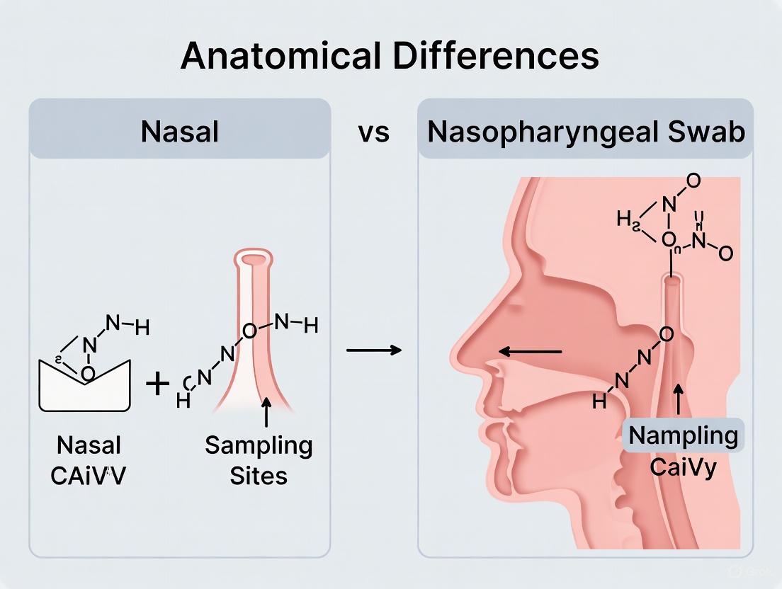
The Blueprint of the Nose: Deconstructing Nasal and Nasopharyngeal Anatomy for Effective Sampling
This guide details the key anatomical structures encountered from the nasal opening to the nasopharynx, providing a technical reference for researchers designing and validating sampling protocols for nasal and nasopharyngeal swabs.
The pathway from the nostrils to the nasopharynx is a continuous anatomical tunnel lined predominantly by respiratory epithelium, which serves to warm, humidify, and filter inspired air [1] [2]. It can be conceptually divided into three main anatomical regions: the external nose, the nasal cavity, and the nasopharynx. Understanding the transitions between these regions, their specific structural compositions, and their physiological functions is critical for ensuring that swab-based sampling targets the correct microenvironment for a given diagnostic or research purpose. The journey begins externally at the nostril and proceeds posteriorly through the nasal cavity, culminating in the nasopharynx, which sits above the soft palate [3] [4].
Detailed Anatomical Landmarks and Quantitative Data
External Nose and Nasal Vestibule
The external nose is the pyramidal-shaped entrance to the respiratory system. Its structure is supported by nasal bones superiorly and cartilages inferiorly [5] [6].
Key Landmarks:
- Nasion: The midline point at the nasofrontal suture, often the deepest point of the nasal root [5] [6].
- Nasal Root: The most superior and depressed part of the nose, between the eyebrows [6].
- Nasal Dorsum: The midline ridge of the nose extending from the root to the tip (pronasale) [5] [6].
- Nasal Tip (Apex): The most anterior point of the nose [5] [6].
- Nostrils (Nares): The anterior openings to the nasal cavities [2].
- Columella: The tissue that connects the nasal tip to the nasal base and separates the nares [6].
- Ala: The lateral tissue forming the outer wall of each nostril [6].
Nasal Vestibule: This is the slight dilation just inside the naris, lined with skin containing hair follicles (vibrissae) and sebaceous glands. It is bounded posteriorly by the limen nasi, a ridge that marks the transition from skin to respiratory mucosa [7].
Nasal Cavity
The nasal cavity is a paired chamber separated by the nasal septum. It extends from the nostrils to the posterior choanae, the openings that lead into the nasopharynx [1] [7]. Its primary function is respiratory air conditioning and olfaction [1].
- Lateral Wall Structures: The lateral walls are highly complex, featuring three bony projections called turbinates or conchae [1].
- Meatuses: The recesses beneath each turbinate are called meatuses, which serve as drainage sites for paranasal sinuses and the nasolacrimal duct [1].
- Inferior Meatus: Located beneath the inferior turbinate; it houses the opening of the nasolacrimal duct, which drains tears [1].
- Middle Meatus: Located beneath the middle turbinate; it is the site of drainage for the frontal, maxillary, and anterior ethmoidal sinuses [1].
- Superior Meatus: Located beneath the superior turbinate; it drains the posterior ethmoidal sinuses [1].
- Spheno-ethmoidal Recess: Located above the superior turbinate; it drains the sphenoid sinus [1].
- Nasal Septum: A central partition composed of bone posteriorly and the septal (quadrangular) cartilage anteriorly. The anteroinferior portion of the septum is highly vascular (Kiesselbach's area/Little's area), a common site for epistaxis (nosebleeds) [1] [2].
- Olfactory Region: Located at the apex of the nasal cavity, lined with olfactory epithelium housing specialized nerve cells for smell. This region is situated near the cribriform plate of the ethmoid bone [1].
Nasopharynx
The nasopharynx is the most superior part of the pharynx, functioning primarily as an airway. It is a roughly cuboidal chamber located directly behind the posterior nasal apertures (choanae) and superior to the soft palate [3] [4] [8]. Its rigid walls are always patent, ensuring an open airway [4].
-
- Anteriorly: Posterior nasal choanae.
- Posteriorly: Posterior pharyngeal wall overlying the basiocciput and the first two cervical vertebrae.
- Superiorly (Roof): Skull base, formed by the body of the sphenoid and basiocciput.
- Inferiorly (Floor): The superior surface of the soft palate and the pharyngeal isthmus, which communicates with the oropharynx.
- Laterally: Supported by the medial pterygoid plates and the superior pharyngeal constrictor muscles.
Key Contents and Landmarks [3] [4] [8]:
- Pharyngeal Isthmus: The opening in the floor of the nasopharynx, closed off during swallowing by elevation of the soft palate.
- Eustachian Tube Opening: Located on the posterolateral wall; it connects the nasopharynx to the middle ear cavity to equalize pressure.
- Torus Tubarius: The cartilaginous bulge in the pharyngeal wall formed by the medial end of the Eustachian tube, encircling its opening.
- Fossa of Rosenmüller (Pharyngeal Recess): A deep mucosal recess located poster superior to the torus tubarius. This is a critical clinical landmark as it is the most common site of origin for nasopharyngeal carcinoma [3] [8] [9].
- Adenoids (Pharyngeal Tonsils): A collection of lymphoid tissue located in the roof and posterior wall of the nasopharynx, part of Waldeyer's ring. They are typically prominent in children but regress after puberty [3] [4] [10].
Table 1: Quantitative Anatomical Dimensions of the Nasopharynx
| Dimension | Measurement Range | Source |
|---|---|---|
| Anterior-Posterior Diameter | 2.0 - 3.5 cm | [3] [8] |
| Height | ~4.0 cm | [3] |
| Transverse Diameter | 4.0 - 5.5 cm | [8] |
Table 2: Comparative Characteristics of Nasal and Nasopharyngeal Regions
| Feature | Nasal Vestibule | Nasal Cavity (Respiratory Region) | Nasopharynx |
|---|---|---|---|
| Lining Epithelium | Keratinized stratified squamous epithelium (skin) with hairs [7] | Ciliated pseudostratified columnar epithelium with goblet cells (Respiratory mucosa) [1] | Predominantly ciliated pseudostratified columnar epithelium; transitions to stratified squamous in lower areas [3] |
| Key Functions | Filtration of large particles; structural support of nasal opening [2] | Warming, humidifying, and filtering inspired air; olfaction [1] [2] | Airway; pressure equalization for middle ear; immune surveillance; voice resonance [3] [4] [10] |
| Key Clinical/Sampling Landmarks | Nostril (Nares), Limen Nasi | Inferior/Middle Turbinates, Nasal Septum (Kiesselbach's area), Choanae | Fossa of Rosenmüller, Torus Tubarius, Eustachian Tube Orifice, Adenoids |
Neurovascular Supply and Lymphatic Drainage
A rich neurovascular network serves the nasal and nasopharyngeal regions, which is vital for tissue viability, function, and has implications for procedural complications and pathogen spread.
-
- The blood supply is derived from branches of both the internal and external carotid arteries.
- Key vessels include the sphenopalatine artery (a terminal branch of the maxillary artery), the greater palatine artery, the superior labial artery, and the anterior and posterior ethmoidal arteries (branches of the ophthalmic artery). These vessels form extensive anastomoses, particularly in the anterior septum (Kiesselbach's area).
- The nasopharynx is supplied by the ascending pharyngeal artery, the artery of the pterygoid canal (vidian artery), and the sphenopalatine artery.
-
- Venous drainage follows the arterial supply, forming plexuses that drain into the facial vein, the pterygoid plexus, and the pharyngeal plexus, ultimately draining into the internal jugular vein.
- A critical clinical consideration is that some nasal veins communicate with the cavernous sinus via the ophthalmic veins. This represents a potential pathway for infection to spread from the face to the cranial cavity.
-
- General Sensory Innervation: Provided primarily by branches of the trigeminal nerve (CN V). The ophthalmic nerve (V1) and maxillary nerve (V2) supply the nasal cavity. The anterior nasopharynx is innervated by V2, while the posterior nasopharynx is supplied by the glossopharyngeal nerve (CN IX).
- Special Sensory Innervation: Olfactory nerve (CN I) fibers pass through the cribriform plate to provide the sense of smell.
- Motor Innervation: Muscles of the soft palate (critical for closing off the nasopharynx during swallowing) are supplied by the vagus nerve (CN X), except for the tensor veli palatini, which is supplied by the mandibular nerve (V3).
Lymphatic Drainage [3] [8] [9]:
- The nasal cavity drains to upper cervical nodes.
- The nasopharynx has a particularly rich lymphatic network. The first-tier drainage nodes are the retropharyngeal lymph nodes (node of Rouvière). From there, drainage proceeds to the upper deep cervical lymph nodes (Levels II and V). This pattern is critically important in the metastasis of nasopharyngeal carcinoma.
Visualization of the Sampling Pathway
The following diagram illustrates the key anatomical landmarks and the general pathway a swab must traverse during nasopharyngeal sampling.
Diagram 1: The pathway illustrates the key anatomical landmarks from the nostril to the target site for nasopharyngeal swabbing.
Experimental Protocols for Anatomical Sampling Validation
For research on swab sampling, validating the precise anatomical location of sample collection is paramount. Below are detailed methodologies for key experimental approaches.
Endoscopic Verification of Swab Placement
This protocol ensures the swab tip reaches the nasopharynx and contacts the mucosa of the Fossa of Rosenmüller.
- Objective: To visually confirm contact between the swab tip and the nasopharyngeal mucosa, specifically the Fossa of Rosenmüller.
- Materials: Flexible nasal endoscope (2.7mm or smaller diameter), light source, video recording system, standard nasopharyngeal swabs, topical decongestant (e.g., oxymetazoline 0.05%), topical anesthetic (e.g., lidocaine spray).
- Procedure:
- Participant Preparation: Position the participant seated with their head against a headrest. Administer 1-2 sprays of topical decongestant/anesthetic into each nostril for patient comfort and to reduce mucosal edema.
- Endoscope Insertion: Gently insert the lubricated tip of the flexible endoscope into the more patent nostril. Advance it along the floor of the nasal cavity, inferior to the inferior turbinate.
- Landmark Identification: Navigate the endoscope past the posterior choanae to enter the nasopharynx. Identify key landmarks: the torus tubarius, the pharyngeal roof, and the Fossa of Rosenmüller posterior to the torus.
- Swab Insertion and Verification: Under direct endoscopic visualization, a second operator inserts the swab via the same nostril. The endoscopist guides the operator to ensure the swab passes the posterior choanae and contacts the mucosa of the Fossa of Rosenmüller.
- Sample Collection and Withdrawal: Maintain visualization as the swab is rubbed against the mucosal surface for the prescribed time. Document the contact via video recording. The swab is then withdrawn under direct vision to confirm it remained in the target area.
- Validation Metrics: Successful placement is defined as direct endoscopic observation of swab-mucosa contact in the Fossa of Rosenmüller. Video recordings should be retained for independent audit and inter-rater reliability analysis.
Imaging-Based Analysis of Swab Trajectory and Contact
This protocol uses imaging techniques to objectively quantify swab placement and mucosal interaction.
- Objective: To quantitatively analyze the depth of swab insertion, trajectory angle, and confirm contact with the nasopharyngeal wall using radiographic or cross-sectional imaging.
- Materials: Anatomically correct training model of the nasal airway and nasopharynx; standard nasopharyngeal swabs; radiopaque marker tape (for lateral X-ray) or a swab with a radiopaque tip; lateral fluoroscopy unit or CT scanner.
- Procedure for Lateral X-ray/Fluoroscopy:
- Marker Application: Affix a strip of radiopaque marker tape along the length of the swab shaft in 1 cm increments.
- Swab Insertion: An operator inserts the swab into the nasal model until resistance is felt or the target depth is reached.
- Image Acquisition: Capture a lateral X-ray or real-time fluoroscopic image.
- Image Analysis: Measure the angle of insertion relative to the hard palate and the depth of insertion from the nostril to the swab tip. Contact with the posterior nasopharyngeal wall is inferred by a change in the trajectory of the swab tip or confirmed by contrast.
- Procedure for CT Imaging:
- Model Preparation: Place the nasal model in the CT scanner in a position mimicking a human patient.
- Baseline Scan: Acquire a baseline CT scan of the model without a swab.
- Swab Insertion and Scanning: Insert the swab and perform a second CT scan.
- 3D Reconstruction and Analysis: Use 3D reconstruction software to co-register the pre- and post-insertion images. Precisely measure the tip location and the degree of mucosal indentation, if visible, to infer contact pressure.
- Validation Metrics:
- Insertion Depth: Distance from the nare to the swab tip.
- Trajectory Angle: Angle between the swab shaft and the hard palate on lateral view.
- Tip Location: Confirmed presence in the nasopharynx, ideally within the Fossa of Rosenmüller on CT.
Table 3: Research Reagent Solutions for Sampling Studies
| Item | Function/Application in Research |
|---|---|
| Flexible Nasal Endoscope | Provides direct visualization for validating swab placement and mucosal contact in the nasopharynx [10]. |
| Anatomically Accurate Nasal Models | Allows for standardized, repeatable practice and imaging studies of swab trajectory without requiring human subjects. |
| Radiopaque Marker Tape | Enables precise measurement of insertion depth and angle on X-ray or fluoroscopic images. |
| CT/MRI Imaging | Provides high-resolution, cross-sectional anatomical data to correlate swab tip location with specific nasopharyngeal substructures (e.g., Fossa of Rosenmüller) [8] [9]. |
| Viral Transport Media (VTM) | Standardized medium for preserving viral integrity and nucleic acids from collected swab samples for downstream analysis. |
| Quantitative PCR (qPCR) Assays | Gold-standard method for quantifying pathogen load (e.g., EBV DNA, SARS-CoV-2 RNA) from samples, allowing for comparison of sampling efficiency [9]. |
Clinical Significance in Sampling and Research
The anatomical distinctions between the nasal cavity and nasopharynx have direct implications for diagnostic sensitivity and research outcomes.
- Pathogen Reservoir Differences: The nasopharynx is a primary reservoir for many respiratory pathogens, including SARS-CoV-2, Epstein-Barr Virus (EBV) – strongly associated with nasopharyngeal carcinoma – and respiratory syncytial virus (RSV) [3] [10] [9]. Sampling the nasal vestibule or anterior nares may yield lower viral loads compared to nasopharyngeal sampling, leading to false-negative results if the pathogen concentration is higher in the nasopharynx.
- Adenoids and Immune Response: The presence of lymphoid tissue (adenoids) in the nasopharyngeal roof makes it a key site for immune surveillance [3] [4]. Swabs from this region may contain immune cells (e.g., lymphocytes) in addition to epithelial cells and pathogens, which can be a critical factor in studies investigating host immune response.
- Sampling Depth and Technique: Reaching the nasopharynx requires the swab to be inserted along the nasal floor until resistance is felt, indicating contact with the posterior wall, a distance typically equivalent to the distance from the nostril to the ear lobe [10]. Inadequate depth of insertion is a major source of sampling error, resulting in an inferior nasal or oropharyngeal sample instead of a true nasopharyngeal sample. The angle of insertion (parallel to the hard palate) is crucial to successfully navigate the nasal cavity and pass through the posterior choanae.
The nasal cavity serves as the primary portal for respiratory function, fulfilling the critical roles of humidifying, warming, and filtering inspired air [11]. Its complex anatomy, however, is subject to significant variations that can profoundly influence both respiratory physiology and clinical procedures. Septal deviation and turbinate hypertrophy represent two of the most prevalent anatomical variations encountered in clinical and research settings. Within the specific context of nasopharyngeal swab sampling—a procedure that gained paramount importance during the COVID-19 pandemic—these variations present substantial challenges to standardization and efficacy [12]. For researchers and drug development professionals, a precise understanding of these anatomical nuances is indispensable for optimizing sampling techniques, ensuring data reliability in clinical trials for respiratory therapeutics, and advancing the development of intranasal vaccines and drug delivery systems. This technical guide provides an in-depth examination of these variations, their quantitative assessment, and their direct implications for respiratory research methodologies.
Anatomical Foundations of the Nasal Cavity
The nasal cavity is a vertically oriented, midline structure divided into two symmetrical passages by the nasal septum. Its structural integrity is maintained by a combination of bony and cartilaginous elements [11] [13].
The Nasal Septum
The septum is a osteocartilaginous wall that forms the medial boundary of each nasal passage. Its anterior portion is composed of the quadrangular septal cartilage, while the posterior bony portion includes the perpendicular plate of the ethmoid bone superiorly and the vomer and maxillary crest inferiorly [14] [13]. A perfectly straight septum is rare; most individuals exhibit some degree of deviation, which can be either developmental, presenting as a smooth C or S-shaped curve, or post-traumatic, typically more irregular and dislocated [14].
The Nasal Turbinates
The lateral walls of the nasal cavity feature three (sometimes four) paired, medially projecting bony structures known as conchae. When covered by their specialized mucosal lining, they are referred to as turbinates. These are classified as superior, middle, and inferior, with the superior and middle turbinates being extensions of the ethmoid bone, and the inferior turbinates constituting separate bones [11] [15]. The turbinates are richly vascularized, erectile tissues governed by the autonomic nervous system. Their primary functions include regulating nasal airflow resistance, humidification, heating, and filtration [15] [16]. The inferior turbinate, in particular, is the largest and most influential in regulating nasal airflow and resistance.
Table 1: Anatomical Components of the Nasal Cavity
| Structure | Components | Primary Function |
|---|---|---|
| Nasal Septum | Quadrangular cartilage, perpendicular plate of ethmoid, vomer | Structural support; separation of nasal passages |
| Superior Turbinate | Part of ethmoid bone; covered by olfactory mucosa | Olfaction; drainage for posterior ethmoid sinus |
| Middle Turbinate | Part of ethmoid bone; may be pneumatized (concha bullosa) | Protection of sinus ostia; airflow direction |
| Inferior Turbinate | Independent bone; highly vascular submucosa | Humidification, heating, and regulation of airflow |
Nasal Septal Deviation (NSD)
Prevalence and Etiology
Nasal septal deviation is a highly common anatomical variation. Global prevalence rates show remarkable variation, reported anywhere from 26% to 97%, a range attributable to differing definitions of clinically significant deviation across studies [14]. One study employing cone-beam computed tomography (CBCT) found a prevalence as high as 86.6% [14]. Etiologically, NSD is classified as either developmental, often manifesting as a smooth "C-shaped or S-shaped" deformity, or traumatic, which tends to be more acute and irregular [14]. Research on Caucasian neonates has indicated that a degree of septal deviation is present at birth in a significant proportion of the population, suggesting that compressive forces during parturition are a major causative factor [17].
Classification Systems
Several systems exist to classify NSD, aiding in diagnosis, communication, and surgical planning.
Mladina's Classification: This is a widely used system that categorizes deviations into seven distinct types based on their morphology and location as observed during rhinoscopy or on CT imaging [14]:
- Type I & II: Involve a vertical ridge in the cartilaginous septum that does not reach (I) or does reach (II) the nasal dorsum.
- Type III: A vertical ridge in the deeper, bony part of the septum.
- Type IV: A combination of Types I and III, with ridges in both anterior and deeper areas.
- Type V: A unilateral horizontal deformity with a compensatory "ledge" on one side.
- Type VI: A bilateral deformity with a dislocation on one side and a deviation on the other.
- Type VII: A combination of two or more of the above types.
Angular Classification: Another method quantifies the deviation by measuring the Naso Septal Angle on CT scans, classifying its severity into four types [18]:
- Type I (Normal): Angle less than 5°
- Type II (Mild): Angle from 5° to 10°
- Type III (Moderate): Angle from 10° to 15°
- Type IV (Severe): Angle greater than 15°
Table 2: Classification and Prevalence of Nasal Septal Deviation
| Classification System | Type / Degree | Description | Prevalence Notes |
|---|---|---|---|
| Mladina's Classification | Type I & II | Vertical ridge in cartilaginous septum | More associated with rhinosinusitis [14] |
| Type VII | Combination of two or more types | Most common type found in one CBCT study [14] | |
| Angular Classification [18] | Type I (Normal) | Angle < 5° | - |
| Type II (Mild) | Angle 5° - 10° | 76.7% prevalence in males with chronic rhinosinusitis [18] | |
| Type III (Moderate) | Angle 10° - 15° | - | |
| Type IV (Severe) | Angle > 15° | - | |
| Inferior Turbinate Contact [14] | Degree I | Deviation not touching inferior turbinate | - |
| Degree II | Deviation touching inferior turbinate | - | |
| Degree III | Deviation compressing inferior turbinate | - |
Clinical and Research Implications
A deviated septum alters normal laminar airflow, creating turbulent flow and changing air pressure dynamics within the nasal cavity. This can lead to symptoms of nasal obstruction and impaired drainage of the paranasal sinuses, potentially contributing to conditions like rhinosinusitis [18] [14]. From a research perspective, NSD poses a significant challenge to standardized nasopharyngeal sampling. A deviated septum can obstruct the passage of a swab, necessitating adjustment in the angle of insertion and potentially resulting in inconsistent depth of sampling or failure to reach the nasopharyngeal mucosa [12]. Furthermore, the altered anatomy can affect the distribution and collection of nasal mucosal lining fluid, a critical sample for measuring mucosal immunity [19].
Turbinate Hypertrophy
Definition and Pathophysiology
Turbinate hypertrophy refers to the persistent enlargement of the turbinates, particularly the inferior turbinates. It is important to distinguish this from the normal, cyclical congestion and decongestion of the turbinates known as the nasal cycle, which occurs every 2-4 hours [15]. True hypertrophy involves hyperplasia of the mucosal tissues, submucosal glands, and underlying bone. The pathophysiology involves a rich blood supply and autonomic nervous system control, where parasympathetic stimulation leads to vasodilation and congestion, while sympathetic stimulation causes vasoconstriction and decongestion [15] [16].
Etiology and Comorbidities
The causes of turbinate hypertrophy are multifactorial. The most common include [15] [16]:
- Allergic Rhinitis: Exposure to environmental allergens triggers an inflammatory response with eosinophilic infiltration, leading to congestion.
- Non-Allergic/Vasomotor Rhinitis: Dysregulation of autonomic nervous system control, often triggered by temperature changes, hormones, or medications.
- Chronic Rhinosinusitis: Persistent inflammation directly causes turbinate swelling.
- Anatomical Compensatory Hypertrophy: This occurs in the nasal cavity opposite a significant septal deviation, as the wider space prompts the turbinate to enlarge in an attempt to normalize airflow resistance [14].
Turbinate hypertrophy frequently coexists with NSD. A large study of patients with sinonasal complaints found that 72% had inferior turbinate hypertrophy, 76% had septal deviation, and 67% had nasal valve collapse, with considerable overlap between these conditions [15].
Assessment Methodologies
Accurate assessment of nasal anatomy is crucial for both clinical diagnosis and research standardization.
Imaging Techniques
- Computed Tomography (CT): The gold standard for detailed bony and soft tissue visualization of the nasal cavity and paranasal sinuses [18] [14]. It allows for precise measurement of septal deviation angles, identification of concha bullosa (pneumatization of the middle turbinate), and assessment of sinus ostial patency.
- Cone-Beam CT (CBCT): Offers high-resolution imaging with lower radiation dose than conventional CT and is increasingly used for pre-operative planning and quantitative assessment of nasal obstruction [14].
Experimental Protocol for CT Analysis of NSD [18]:
- Image Acquisition: Coronal and axial slices are obtained using a spiral CT scanner with a bone algorithm, typically at 3-mm intervals.
- Angle Measurement: A reference line is drawn from the crista galli to the maxillary crest. A second line is drawn to the point of maximum septal deviation. The angle between these two lines is calculated using radiological software.
- Classification: The calculated angle is then categorized into one of the four types (I-IV) as described in section 3.2.
Functional and Clinical Assessment
- Anterior Rhinoscopy and Nasal Endoscopy: Direct visualization allows assessment of the anterior septum and inferior turbinates. Endoscopy provides a more detailed view of the middle meatus and posterior structures [14].
- Acoustic Rhinometry (AR): Measures the cross-sectional area of the nasal cavity by analyzing acoustic reflections of a sound pulse. It is particularly useful for evaluating anterior obstructions, such as those caused by a deviated septum or anterior turbinate hypertrophy [14].
- Rhinomanometry (RMM): A dynamic physiologic test that calculates nasal airway resistance by measuring transnasal pressure and airflow during respiration [14].
Implications for Nasopharyngeal Swab Sampling
The performance of nasopharyngeal swabs is highly dependent on navigable nasal anatomy. Research on cadavers has provided critical quantitative data on the optimal angles and depths for swab insertion to reliably reach the nasopharynx while avoiding critical structures like the cribriform plate [12].
Anatomical Guidance for Swab Insertion
A seminal study simulating swab procedures defined key parameters [12]:
- Target Angle: To successfully reach the nasopharynx, the swab should be oriented at a mean angle of approximately 76.3° relative to the line connecting the subnasale and nasion (the root of the nose).
- Danger Angle: In contrast, a path toward the cribriform plate (which risks serious complication) follows a much steeper angle of approximately 36.7°.
- Insertion Depth: The average distance from the posterior nares to the back of the pharynx is approximately 8.7 cm (longer in males), guiding the necessary swab insertion depth.
These data underscore how a deviated septum or hypertrophic turbinate can physically obstruct the ideal path of the swab, forcing operators to deviate from the optimal angle and potentially resulting in an inadequate sample or patient discomfort.
Diagram 1: NP Swab Protocol with Anatomical Variations
Impact on Sample Quality
The choice of sampling method directly influences the quality and volume of the collected mucosal lining fluid, which is critical for detecting pathogens or immune markers like SARS-CoV-2 specific IgA. A comparative clinical study found that an expanding sponge method (M3) significantly outperformed both nasopharyngeal (M1) and nasal swabs (M2) in terms of detection rate and median IgA concentration [19]. This suggests that sampling methods that can better conform to or overcome anatomical obstructions yield superior results for immunological research.
Table 3: Research Reagent Solutions for Nasal Sampling and Analysis
| Item | Function/Application | Example from Literature |
|---|---|---|
| Nylon Flocked Swab | Collection of nasopharyngeal samples; improved release of cellular material | Used in "M1" method for nasopharyngeal swabbing [19] |
| Cotton Swab | Collection of nasal samples from anterior to middle vault | Used in "M2" method for nasal swabbing [19] |
| Polyvinyl Alcohol (PVA) Sponge | Expanding sponge for adsorption of nasal mucosal lining fluid | Used in "M3" method, showed superior IgA collection [19] |
| Universal Transport Medium (UTM) | Preservation of viral integrity and nucleic acids for transport | Samples placed in UTM post-collection [19] |
| ELISA Kits | Detection and quantification of specific immunoglobulins (e.g., IgA) | Validated ELISA for SARS-CoV-2 RBD-specific IgA detection [19] |
| Electrochemiluminescence (ECL) Assay | High-sensitivity, high-throughput detection of serum antibodies | Used as a comparator for validating novel ELISA methods [19] |
Septal deviation and turbinate hypertrophy are not merely clinical curiosities but fundamental anatomical variables that must be accounted for in the design and execution of nasal and nasopharyngeal research. The quantitative data on prevalence, classification, and anatomical measurements provided in this guide form a critical knowledge base. For scientists developing intranasal vaccines or therapeutics, assessing mucosal immunity, or standardizing pathogen detection protocols, failure to control for these variations introduces significant confounding variability. Future research must continue to refine sampling tools and techniques that are robust in the face of anatomical diversity, ensuring that collected data is both accurate and reproducible. Integrating pre-sampling anatomical assessment, perhaps via low-dose imaging or functional tests, may become a necessary step in high-precision clinical trials for respiratory-focused biologics and drugs.
The human nose, a critical interface between the external environment and the respiratory system, exhibits pronounced and functionally significant asymmetry between its left and right chambers. This anatomical variation, far from being a mere curiosity, has profound implications for respiratory physiology, the deposition of inhaled particles, and the efficacy of nasopharyngeal swab sampling. For researchers and drug development professionals, understanding these inter-chamber differences is paramount for optimizing diagnostic strategies and developing targeted therapeutic interventions.
Historically, many parametric studies and standardized nasal models have been based on the assumption of symmetrical nasal chambers [20]. However, emerging evidence from computational fluid dynamics (CFD) and detailed anatomical studies reveals that morphological asymmetry is the norm rather than the exception. This asymmetry significantly influences airflow partitioning, particle deposition patterns, and potentially, the consistency of sample collection from the nasal cavity [20] [21]. Within the context of anatomical differences in nasal and nasopharyngeal swab sampling sites, this inherent variability presents both a challenge and an opportunity for refining sampling protocols to enhance diagnostic reliability.
This article provides a comprehensive technical examination of nasal asymmetry, synthesizing quantitative data on its anatomical basis, functional consequences, and direct relevance to swab-based sampling research. By integrating detailed methodologies, data summaries, and visual workflows, we aim to equip scientists with the knowledge to advance the precision of nasal diagnostic and therapeutic applications.
Anatomical Basis of Nasal Asymmetry
Nasal asymmetry originates from several key anatomical features. The most prominent is nasal septum deviation, a condition found in a surprisingly high percentage of the population. One survey of patients with ear, nose, and throat (ENT) disease found that 89.2% of them had nasal septum deviation [20]. Even in healthy individuals, perfect symmetry is rare, leading to natural inter-chamber variations.
The nasal valve region, recognized as the narrowest part of the entire adult breathing system, represents a critical zone where minimal anatomical changes can dramatically alter airflow resistance and distribution [22]. The mobility of the lateral nasal wall in this region, acting like a "Starling resistor," means that its mechanical properties and inspiratory flow rate collectively determine the flow-dependent portion of nasal resistance [22].
Specific anatomical regions exhibit particularly noticeable differences. Research indicates that significant inter-chamber differences are often observed in the inferior and middle passages, areas where most of the inhaled flow is distributed [20]. Furthermore, the shape of the vestibule notch and the aforementioned septum deviation are identified as primary contributors to discrepant flow behavior between the two chambers [20].
Table 1: Key Anatomical Features Contributing to Nasal Asymmetry
| Anatomical Feature | Description of Variation | Functional Impact |
|---|---|---|
| Nasal Septum | Deviation from the midline is highly prevalent [20]. | Alters cross-sectional area and flow path direction in each chamber. |
| Nasal Valve | The narrowest point of the airway; cross-sectional area and lateral wall motility vary [22]. | Major determinant of nasal resistance; prone to inspiratory collapse. |
| Inferior & Middle Passages | Volume and surface area differ between chambers [20]. | Affects regional flow distribution and air conditioning. |
| Vestibule Notch | Phenotypic shape varies (e.g., smooth vs. notched) [20]. | Influences initial airflow stream and particle deposition patterns. |
| Turbinates | Size and geometry of inferior/middle turbinates are asymmetric. | Modifies airflow resistance, heating, and humidification. |
Quantitative Analysis of Inter-Chamber Variations
Airflow Dynamics
Computational Fluid Dynamics (CFD) studies have quantified the significant impact of anatomical asymmetry on airflow apportionment. In a detailed assessment of nasal inter-chamber variations, results showed noticeable differences in flow behavior, particularly in the inferior and middle passages [20]. The study attributed these discrepancies primarily to the shape of the vestibule notch and septum deviation.
A larger CFD study of 22 healthy subjects further underscored the variability of "normal" nasal airflow [21]. The study found that the location of the major flow path and coronal velocity distributions varied greatly across individuals. Contrary to some classical descriptions, the study found that on average, more flow passed through the middle meatus than the inferior meatus, and this flow distribution correlated with better subjective patency ratings (( r = -0.65, p < 0.01 )) [21].
The pressure distribution within the nasal cavity is also highly asymmetric. Research shows that more than 50% of the total pressure drop during inspiration occurs near the head of the inferior turbinate [21]. Furthermore, wall shear stress, nasal resistance, turbulence kinetic energy, and vorticity were all found to be lower in the wider turbinate region compared to the constricted nasal valve region [21].
Table 2: Quantitative Measurements of Nasal Airflow and Function
| Parameter | Measurement/Significance | Inter-Chamber Variation |
|---|---|---|
| Flow Apportionment | Varies significantly; often favors one chamber over the other. | Differences in percentage of total flow can be substantial [20]. |
| Major Flow Path | More commonly through middle meatus than inferior meatus in healthy subjects [21]. | Location of primary stream varies (middle vs. inferior) between sides and individuals [21]. |
| Pressure Drop | >50% of total drop occurs near the inferior turbinate head [21]. | The gradient and site of maximal pressure drop differ between chambers. |
| Nasal Resistance | Measured by rhinomanometry at a reference pressure drop of 75 Pa [21]. | Can vary by over 50% between sides in healthy individuals. |
| Wall Shear Stress | Lower in turbinates than in the nasal valve region [21]. | Distribution patterns are asymmetric, reflecting local geometry. |
Nanoparticle Deposition Patterns
The anatomical and flow asymmetries between nasal chambers directly lead to significant variations in regional nanoparticle deposition. This is particularly critical for assessing inhalation exposure to airborne pollutants or the distribution of nasal drug delivery systems.
For 1 nm particles, deposition in the olfactory region can show inter-chamber differences of up to 400% [20]. This extreme variation highlights the potential for asymmetric exposure of the olfactory nerve and central nervous system to inhaled nanomaterials. The deposition efficiency for nanoparticles is highly size-dependent, with dramatic changes occurring in the 1-2 nm range due to the varying dominance of diffusion effects [20].
The formula for particle deposition efficiency is defined as: [ DE = \frac{\text{Number of particles deposited}}{\text{Number of particles entering the nasal cavity}} ] This efficiency is influenced by particle diameter, air viscosity, particle density, and the Cunningham correction factor which accounts for non-continuum effects at small particle sizes [20].
Methodologies for Assessing Nasal Asymmetry
Computational Fluid Dynamics (CFD) Analysis
Protocol for CFD Simulation of Nasal Airflow:
- Model Reconstruction: A three-dimensional model of the nasal cavity is reconstructed from computed tomography (CT) scans. The model should preserve external facial features around the nares, as these have been shown to influence inhalation conditions [20].
- Mesh Generation: Computational mesh is generated using polyhedral elements with a typical cell dimension of 0.5 mm. Approximately 1.3 million mesh elements are typically sufficient after independence testing. Eight layers of prism layers are attached along the nasal wall to simulate the near-wall region accurately [20].
- Boundary Conditions: A steady inhalation flow with a volume flow rate of 15 L/min (representing resting breathing) is imposed. The laminar flow regime is appropriate for this flow rate [20].
- Numerical Solution: The continuity and momentum equations are solved assuming incompressible flow.
- Particle Trajectory Simulation: After obtaining the fluid field, nanoparticles of various diameters (e.g., 1, 2, 5, 10, 50, 100 nm) are released from a spherical location at the nose tip. The Lagrangian Discrete Phase Model (DPM) is employed, accounting for drag force, Brownian force, and gravity [20].
- Data Analysis: Particle deposition efficiency is calculated for different regions of the nasal cavity, and inter-chamber differences are quantified.
Anatomical Comparison Method
Protocol for Inter-Chamber Shape Comparison:
- Coordinate System Alignment: Constrain the nasal cavity to a 3D coordinate system. The X-Y plane is determined by fitting a virtual plane based on the bottom edge of the inferior turbinate on both sides, corresponding to the natural breathing position and accounting for gravity [20].
- Mirroring Procedure: One side of the nasal chamber is mirrored and aligned to the other side to quantify asymmetry and analyze geometric deviation.
- Deviation Analysis: The mirrored geometry is compared to the original contralateral side to identify and quantify specific areas of anatomical deviation.
Experimental Elastography
Protocol for Nasal Valve Elastography:
- Sensor Placement: Fix electro-optical distance sensors (e.g., VISHAY V90 CNY70) in a 3D-printed housing. Position the sensors 2–3 mm laterally to the center of the movable, anterior lateral nasal wall, close to the nasal valve [22].
- Flow Measurement: Simultaneously measure inspiratory flow using an encased analogue unidirectional flow sensor (e.g., Sensirion SFM 3200) connected to the nostril.
- Data Collection: Record five measurements for each participant during both normal and forced breathing (>500 cm³/sec per nostril). Calculate the arithmetic mean for analysis [22].
- Parameter Calculation: Assess total inward movement [mm] as the sum of movements of the left and right lateral nasal walls. Record the time shift between maximum flow and maximum movement.
Nasal Analysis Workflow: This diagram illustrates the integrated methodology for assessing nasal asymmetry, combining computational and experimental approaches.
Implications for Nasopharyngeal Swab Sampling
The anatomical and functional asymmetry of the nasal cavity has direct and significant implications for nasopharyngeal swab sampling, a critical tool for diagnosing respiratory infections. Variations in nasal geometry directly affect the swab's path and the quality of the sample obtained.
Research comparing sampling methods has demonstrated that the expanding sponge method (M3) achieved superior performance compared to nasopharyngeal swabs (M1) and nasal swabs (M2) [19]. Specifically, M3 showed a significantly higher single-day detection rate (95.5% above the limit of quantification), a higher 5-day consecutive detection rate (88.9%), and a higher median SARS-CoV-2 WT-RBD IgA concentration (171.2 U/mL) [19]. This superior performance is likely due to the sponge's ability to better conform to the asymmetric nasal anatomy and absorb mucosal lining fluid more effectively.
The "flypaper-like" distribution of mucosal IgA across the nasal surfaces, with substantial concentration variations across anatomical sites, makes standardized sampling particularly challenging [19]. Studies have reported collection capability differences of up to 5-fold between different sampling methods [19]. This variability, compounded by natural anatomical asymmetry, severely compromises cross-study comparability and underscores the need for standardized sampling protocols that account for nasal asymmetry.
Table 3: Research Reagent Solutions for Nasal Airflow and Sampling Studies
| Reagent/Equipment | Function/Application | Specification Notes |
|---|---|---|
| Flocked Nasal Swabs | Sample collection from nasal mucosa; optimal cell elution [23]. | Nylon flocked tip (e.g., COPAN FLOQSwabs); superior sample release vs. wound fiber. |
| Expanding Sponge | Absorption of nasal mucosal lining fluid [19]. | Polyvinyl alcohol sponge; shows superior Ig detection rates. |
| Computational Mesh | Discretization of nasal geometry for CFD simulation [20]. | Polyhedral elements (~0.5 mm size); ~1.3 million elements; 8 prism layers. |
| Electro-Optical Sensors | Measuring lateral nasal wall movement (elastography) [22]. | e.g., VISHAY V90 CNY70; measures displacement at nasal valve. |
| Transport Medium | Preservation and transport of biological samples [19]. | Universal Transport Medium (UTM); ensures sample viability. |
The significance of nasal asymmetry extends far beyond academic interest, representing a fundamental feature of human anatomy with direct consequences for respiratory function, particle deposition, and the accuracy of diagnostic sampling. The quantitative data presented herein unequivocally demonstrates that inter-chamber variations in anatomy result in substantial differences in airflow dynamics and nanoparticle deposition patterns. For researchers and drug development professionals, acknowledging and accounting for this asymmetry is crucial for advancing the field of nasal biomedicine. Future research should focus on developing asymmetry-informed sampling protocols and computational models to enhance the reliability of nasal diagnostics and the efficacy of respiratory therapeutics.
The efficacy of respiratory sample collection, a cornerstone of modern diagnostics for pathogens like SARS-CoV-2 and influenza, is profoundly influenced by the physical and rheological properties of mucus and the mucosal lining. The nasopharyngeal and nasal cavities, key sites for sample collection, are protected by a complex gel known as mucus. This gel is not a simple fluid but a viscoelastic material whose behavior under stress dictates how easily it can be collected and released by a swab. Understanding this interplay is crucial for developing reliable diagnostic tests, evaluating mucosal immunity, and designing effective drug delivery systems. Framed within broader research on anatomical differences between nasal and nasopharyngeal swab sampling sites, this review synthesizes the critical role of mucus rheology in sample collection, detailing its fundamental properties, the impact of anatomy on sampling efficiency, and standardized methods for its analysis.
Fundamental Rheological Properties of Mucus
Composition and Structure
Mucus is a complex aqueous gel composed of 90–95% water and a solid fraction that is predominantly gel-forming mucins [24]. These mucins, such as MUC5AC and MUC5B, are large glycoproteins that form a cross-linked, three-dimensional network, giving mucus its distinctive structural and rheological characteristics [25] [24]. This network also entraps lipids, salts, cellular debris, and various proteins [25] [24]. The specific composition and pH of mucus vary significantly across different anatomical locations, leading to distinct rheological profiles tailored to specific functions, from lubrication in the respiratory tract to creating a barrier in the cervix [24].
Key Rheological Behaviors
The functional integrity of mucus is governed by its non-Newtonian rheological properties.
- Viscoelasticity: Mucus exhibits both solid-like (elastic) and liquid-like (viscous) properties. The storage modulus (G′) represents the elastic component, and the loss modulus (G″) represents the viscous component. In healthy mucus, G′ typically dominates G″, indicating a solid-like structure at rest that is crucial for its barrier function [24].
- Yield Stress (τy): This is the minimum stress required to initiate flow in mucus. Below the yield stress, mucus behaves like a solid; above it, it flows like a liquid. This property is critical for processes like mucociliary clearance, where ciliary beating must generate enough force to exceed τy and mobilize mucus [24].
- Shear-Thinning: When a stress exceeding the yield stress is applied, the viscosity of mucus decreases. This property, known as shear-thinning, is essential for both physiological functions (like coughing) and diagnostic procedures (like swab collection), as it allows thick mucus to flow under applied force [26] [27].
Table 1: Key Rheological Properties of Human Mucus and Their Physiological Significance.
| Property | Description | Measurement Techniques | Physiological & Diagnostic Significance |
|---|---|---|---|
| Yield Stress (τy) | Minimum stress to initiate flow [24]. | Steady shear rheology, oscillatory shear rheology [24]. | Determines the force required for swab collection and ciliary clearance [24]. |
| Viscoelasticity | Combination of solid-like (elastic) and liquid-like (viscous) behavior [25] [24]. | Oscillatory rheology (G′, G″), magnetic rotational spectroscopy [25] [24]. | Affects sample retention on swabs and the reliability of release for testing [26]. |
| Shear-Thinning | Viscosity decreases with increasing applied stress [26] [24]. | Steady shear rheology [24]. | Facilitates mucus flow during coughing and swab rotation during sample collection [26]. |
Figure 1: The interrelationship between the fundamental rheological properties of mucus and their ultimate physiological and diagnostic functions.
Anatomical Sampling Sites and Swab Performance
Comparative Anatomy of Sampling Sites
The anatomical distinctions between the nasal cavity and the nasopharynx directly influence sampling technique and efficacy.
- Nasal (Anterior Nares) Swab: This method involves inserting a swab approximately 0.5 to 0.75 inches (1.3 to 2 cm) into the nostril to collect sample from the nasal membrane [28]. It is less invasive, more comfortable for patients, and suitable for self-collection [29] [28].
- Nasopharyngeal Swab: This method requires inserting a flexible swab through the nostril along the nasal floor to the nasopharynx, the upper part of the throat behind the nose, typically until resistance is met [28]. This procedure is more invasive and must be performed by a trained healthcare professional [28].
Swab Performance and Mucus Interaction
The complex rheology of mucus and the anatomical constraints of the nasopharynx create significant challenges for sample collection. The yield stress and viscoelasticity of mucus determine how much force is needed for a swab to penetrate the mucus layer and how much sample is retained on the swab upon withdrawal [24].
Recent research using anatomically accurate 3D-printed nasopharyngeal models lined with a mucus-mimicking hydrogel (SISMA) has quantified these challenges. Studies show that the anatomical complexity of the nasal cavity significantly reduces the volume of hydrogel collected and released by swabs, compared to a simple tube model [26] [27]. For instance, one study found that commercial flocked swabs collected 8.4 times more synthetic mucus in a tube than in the anatomically accurate cavity model [26]. Furthermore, the model demonstrated that sample release efficiency—the percentage of collected sample released for testing—is highly dependent on swab design, with one novel injection-molded swab (Heicon) showing 82.48% release efficiency in the cavity model compared to 69.44% for a commercial flocked swab [26]. These findings underscore that traditional, simplistic swab testing methods fail to predict real-world performance accurately.
Table 2: Comparison of Nasal and Nasopharyngeal Sampling Methods.
| Parameter | Nasal Swab (Anterior Nares) | Nasopharyngeal Swab |
|---|---|---|
| Insertion Depth | ~0.5-0.75 inches (~1.3-2 cm) [28] | To the nasopharynx (behind the nose) [28] |
| Patient Comfort | More comfortable, less invasive [29] [28] | Less comfortable, more invasive [28] |
| Suitable for Self-Collection | Yes [28] | No, requires a trained professional [28] |
| Relative Sensitivity (Pathogen Detection) | Generally lower (e.g., 76% for RSV) [28] | Generally higher (e.g., 97% for RSV, ~95.9% for SARS-CoV-2) [29] [28] |
| Key Advantages | Patient-friendly, suitable for mass screening [29] | Considered the gold standard for sensitivity for many pathogens [29] |
Methodologies for Rheological Analysis and Sample Collection
Standardized Rheological Measurement Protocols
Accurate characterization of mucus requires robust and standardized methodologies.
Macrorheology: This approach measures the bulk viscoelastic properties of a mucus sample.
- Procedure: A rheometer with a parallel-plate or cone-and-plate geometry is used. The sample is subjected to either a controlled shear stress (or strain) in steady shear to determine yield stress and viscosity, or to an oscillatory strain in small-amplitude oscillatory shear (SAOS) to determine the viscoelastic moduli (G′ and G″) [24].
- Data Analysis: The yield stress can be identified from a steady shear flow curve as the stress value where viscosity drops precipitously, or from an amplitude sweep in oscillatory tests as the stress where G′ begins to drop significantly (the crossover point with G″) [24].
Microrheology: This technique addresses the heterogeneity of mucus by measuring local rheological properties at the micro-scale.
- Procedure: Fluorescent or inert tracer particles are embedded in the mucus sample. Their Brownian motion is tracked using microscopy, and the mean squared displacement (MSD) of the particles is calculated [25] [24].
- Data Analysis: The MSD is related to the local viscoelastic moduli of the mucus network, providing insights into microenvironments that can affect the diffusion of viruses, nanoparticles, or drugs [25].
Clinically Validated Sampling Protocols
Standardized collection is vital for reproducible results in both diagnostics and research.
Nasopharyngeal Swab Collection for Immunoassay [30]:
- Swab: Use a nylon flocked swab.
- Insertion: Insert the swab into the nostril to the nasopharyngeal region.
- Collection: Rotate the swab once and maintain its position for 15 seconds to allow saturation with mucosal lining fluid.
- Processing: Place the swab in a universal transport medium (UTM). Within 4 hours, remove the swab, centrifuge the medium (e.g., 1000 rpm for 3 min), and aliquot the supernatant for analysis.
Expanding Sponge Method for Superior IgA Recovery [30]:
- Sponge Preparation: Soak a polyvinyl alcohol sponge in saline to expand it, then place it in a syringe to expel excess fluid.
- Insertion: Insert one piece of the dehydrated sponge into the nostril.
- Collection: Leave the sponge in place for 5 minutes to absorb nasal mucosal lining fluid.
- Processing: Expel the absorbed fluid using a syringe, followed by centrifugation and aliquoting. This method has been shown to achieve significantly higher detection rates and concentrations of SARS-CoV-2 specific IgA compared to swab methods [30].
Figure 2: A workflow comparing macrorheology and microrheology approaches for characterizing mucus properties.
The Scientist's Toolkit: Essential Research Reagents and Materials
Table 3: Key Materials and Reagents for Mucus and Sampling Research.
| Item | Function/Application | Specific Examples & Notes |
|---|---|---|
| SISMA Hydrogel | A synthetic mucus simulant that replicates the shear-thinning behavior and viscosity of native nasopharyngeal mucus for standardized in vitro testing [26] [27]. | Viscosity close to 10 Pa·s at low shear rates; used for swab validation in 3D anatomical models [26]. |
| 3D-Printed Nasopharyngeal Model | An anatomically accurate in vitro platform for pre-clinical evaluation of swab performance under physiologically relevant conditions [26] [27]. | Crafted with rigid (VeroBlue) and flexible (Agilus30) resins to mimic bone and soft tissue [26]. |
| Nylon Flocked Swabs | The standard for nasopharyngeal sample collection; designed for efficient absorption and release of specimens [30] [28]. | Ultrafine flocked tip on a flexible handle (e.g., HydraFlock sterile swabs) [28]. |
| Expanding Polyvinyl Alcohol (PVA) Sponge | For collecting nasal mucosal lining fluid with high efficiency, particularly for immunoassay analysis [30]. | Superior for recovering total IgA and specific antibodies (e.g., SARS-CoV-2 RBD IgA) [30]. |
| Universal Transport Medium (UTM) | Preserves the integrity of viral, bacterial, and molecular targets in collected samples during transport and storage [30] [29]. | Used to store swabs and sponge eluents prior to processing [30]. |
| Rheometer | The primary instrument for macroscale characterization of mucus viscoelasticity and yield stress [24]. | Equipped with parallel-plate or cone-and-plate geometries for steady and oscillatory shear tests [24]. |
The rheological properties of mucus are not merely academic curiosities; they are fundamental determinants of success in respiratory sample collection. The yield stress, viscoelasticity, and shear-thinning behavior of mucus directly impact the force required for swab collection, the amount of sample retained, and the efficiency of its release for diagnostic analysis. The anatomical differences between nasal and nasopharyngeal sites add a critical layer of complexity, influencing both patient comfort and diagnostic sensitivity. Moving forward, the integration of sophisticated tools—such as 3D-printed anatomical models and biomimetic hydrogels—into a standardized framework for swab evaluation and mucus analysis will be essential. This approach will drive the development of more effective sampling devices and protocols, ultimately enhancing the accuracy of diagnostic testing, the assessment of mucosal immunity, and the development of novel therapeutic strategies. A deep understanding of the interface between mucus rheology and sampling technology is therefore indispensable for advancing public health and personalized medicine.
Precision in Practice: Standardized Protocols for Nasal and Nasopharyngeal Swab Collection
Step-by-Step Guide to Nasopharyngeal Swab Collection
In the study of respiratory pathogens and mucosal immunity, the quality of collected specimens is a foundational variable that can determine the success or failure of downstream analyses. Nasopharyngeal swab collection represents a critical gateway for obtaining samples that accurately reflect the biological events occurring at the mucosal interface between host and environment. For researchers, scientists, and drug development professionals, standardization of this procedure is not merely a technical formality but a prerequisite for generating comparable, reproducible data across studies and institutions.
The nasopharynx serves as the primary reservoir and replication site for numerous significant respiratory pathogens, including SARS-CoV-2, influenza, and respiratory syncytial virus (RSV). A properly collected specimen yields high numbers of organisms and host cells, providing sufficient biological material for culture, molecular diagnostics, and immunological assays [31]. Within the context of anatomical differences research, variations in nasal anatomy across populations can significantly impact both the quality of the obtained sample and the procedural discomfort experienced by participants [32]. This guide provides a standardized protocol for nasopharyngeal swab collection while contextualizing the procedure within the broader research landscape of anatomical variations and their methodological implications.
Anatomical and Research Context
Anatomical Landscape of the Nasopharynx
The nasopharynx is the uppermost portion of the pharynx, lying posterior to the nasal cavity and above the soft palate. It is lined with respiratory epithelium and serves as a critical site for pathogen attachment and replication. Accessing this region requires navigation through the nasal cavity, passing the nasal turbinates and septum, along the floor of the nasal passage until reaching the posterior wall [33].
From a research perspective, individual anatomical variations introduce important covariates that must be considered in study design and data interpretation. Ethnic differences in nasal anatomy have been documented to significantly affect both procedural discomfort and nucleic acid recovery. One controlled study found that Asian participants reported significantly higher discomfort scores during swab collection compared to White participants (median scores of 5 vs. 4 on an 11-point scale) and yielded different nucleic acid recovery profiles [32]. These findings highlight the importance of documenting participant ethnicity in study methodologies and considering anatomical variations when interpreting experimental results across diverse populations.
Research Applications and Significance
Nasopharyngeal swabs serve multiple research purposes beyond routine diagnostic testing:
- Viral pathogenesis studies: Investigating viral load kinetics and shedding patterns for respiratory viruses
- Mucosal immunology: Profiling host immune responses at the site of initial infection
- Vaccine development: Evaluating mucosal immune responses to candidate vaccines
- Transmission dynamics: Understanding pathogen spread and superspreading events
- Therapeutic development: Assessing antiviral efficacy in clinical trials
The integrity of these research applications depends fundamentally on the consistency and quality of specimen collection procedures [19].
Comprehensive Collection Protocol
Pre-Collection Preparation
Materials and Equipment:
- Synthetic flocked or polyester swabs with flexible plastic or wire shafts
- Appropriate viral transport medium or sterile container
- Cooler or refrigeration unit for specimen storage
- Personal protective equipment (PPE): gloves, mask, eye protection
- Biohazard bag with separate outer pocket for documentation
- Requisition forms and labels
- Timer or clock
Swab Selection Criteria: Critical to research quality is the selection of appropriate swab materials. Calcium alginate swabs or swabs with wooden shafts must be avoided, as they may contain substances that inactivate viruses and inhibit molecular tests [34]. Synthetic fiber swabs (typically nylon or polyester) with thin plastic or wire shafts designed specifically for nasopharyngeal sampling are recommended. Flocked swabs have demonstrated superior recovery of respiratory epithelial cells compared to rayon-tipped swabs, though pathogen detection rates may be equivocal [33].
Patient Positioning and Preparation:
- Position the participant sitting upright with head against a headrest or wall for stability
- Tilt the head back to approximately 70 degrees to straighten the nasal passage [34]
- Ask the participant to blow their nose to clear nasal passages if possible; if not, gently wipe nares with a cotton tip swab or tissue [31]
- Visually inspect nasal passages for obvious deviation or obstruction
- Estimate the distance from the participant's nostril to the tragus of the ear to determine appropriate insertion depth [31]
Step-by-Step Collection Procedure
Perform hand hygiene and don appropriate PPE (gloves, mask, eye protection) [31]
Open swab packaging carefully, handling only the distal end of the swab shaft to maintain sterility [35]
Insert the swab into the nostril along the nasal septum, parallel to the palate (horizontally, not upward), following the floor of the nasal passage [31] [34]
Advance the swab to the nasopharynx until resistance is encountered, typically at a depth equivalent to the distance from the nostril to the tragus of the ear (approximately 4-7 cm or 1.6-2.75 inches in adults) [31] [32]
Maintain contact with the nasopharyngeal mucosa for 10-15 seconds to allow absorption of secretions [35] [34]
Gently rub and roll the swab against the nasopharyngeal mucosa [34]. Note: Research evidence suggests that rotation following nasopharyngeal contact does not recover additional nucleic acid and may decrease participant tolerance [32]
Withdraw the swab slowly while rotating it gently, taking care not to touch the sides of the nostril during removal [31]
Immediately place the swab into transport medium, ensuring the tip is fully immersed
Break the swab shaft at the scored line against the rim of the tube, then cap tightly [31] [35]
Label the specimen vial with participant identifier, collection date and time, and other required information
Post-Collection Processing
- Place the specimen in the inner pocket of a biohazard bag
- Complete requisition forms with detailed clinical and methodological notes and place in the outer pocket
- Refrigerate specimens immediately at 2-8°C until transport
- Arrange transport to the laboratory as soon as possible, maintaining cold chain
- Document any deviations from standard protocol or participant reactions
Methodological Variations and Experimental Evidence
Technique Comparison: Rotation Versus Simple Placement
Recent research has questioned the necessity of swab rotation following placement in the nasopharynx. A controlled study of 69 participants compared two collection techniques: simple insertion and immediate removal ("in-out") versus insertion followed by 10 seconds of rotation before removal ("rotation") [32].
Table 1: Comparison of Swab Collection Techniques
| Technique | Nucleic Acid Recovery (Median RPP30 cells/μL) | Participant Discomfort (Median Score 0-10) | Participant Preference for Swab vs. Saliva |
|---|---|---|---|
| In-Out | 500 [235-738] | 5 [3.75-5] | 29.4% (10/34) |
| Rotation | 503 [398-685] | 4.5 [4-6] | 10% (3/30) |
The study found no significant difference in nucleic acid recovery between the two techniques, as measured by human RPP30 (DNA) and RNase P (RNA) copy numbers [32]. However, participant tolerance data suggested that the rotation technique was less well tolerated, with a lower preference for repeated swab collection compared to saliva donation.
Sampling Method Comparisons in Research Settings
Different sampling methods yield variations in detection sensitivity for specific applications. A 2025 study comparing nasal sampling methods for detecting SARS-CoV-2 RBD IgA found significant differences in performance across three common techniques [19].
Table 2: Comparison of Nasal Sampling Methods for SARS-CoV-2 RBD IgA Detection
| Sampling Method | Single-Day Detection Rate (Above LOQ) | 5-Day Consecutive Detection Rate (Above LOQ) | Median IgA Concentration (U/mL) |
|---|---|---|---|
| Nasopharyngeal Swab (M1) | 68.8% | 48.7% | 28.7 |
| Nasal Swab (M2) | 88.3% | 77.3% | 93.7 |
| Expanding Sponge (M3) | 95.5% | 88.9% | 171.2 |
The expanding sponge method significantly outperformed both nasopharyngeal and standard nasal swabs for immunological studies, highlighting how methodological choices must align with specific research objectives [19].
Research Reagent Solutions
Table 3: Essential Research Reagents and Materials for Nasopharyngeal Sampling
| Item | Specification | Research Application |
|---|---|---|
| Swab Type | Flocked nylon or polyester tip with flexible plastic or wire shaft [35] [34] | Optimal cellular recovery and pathogen release |
| Transport Medium | Viral transport medium (VTM) or universal transport medium (UTM) [35] | Preserves viral integrity and nucleic acids during transport |
| Alternative: Dry Swabs | Polyester swabs in sterile dry tubes [36] | Cost-effective option with comparable sensitivity for molecular detection when processed promptly |
| Storage Tubes | Sterile leak-proof screw-cap containers | Maintains sample integrity and prevents contamination |
| RNA Stabilization Buffer | Guanidinium thiocyanate-based buffers | Preserves nucleic acids for molecular studies |
For resource-constrained settings or large-scale surveillance studies, dry polyester swabs have demonstrated excellent performance characteristics, with one study reporting 90.48% sensitivity for SARS-CoV-2 detection compared to 76.19% for wet swabs in transport media [36].
Quality Assurance and Troubleshooting
Common Technical Challenges
- Nasal Obstruction: Encountered in approximately 7.2% of participants [32]. If obstruction is met, withdraw the swab slightly and attempt redirection, or try the other nostril with the same swab [34]
- Excessive Discomfort: Often indicates incorrect angle of insertion (upward rather than horizontal). Reposition the head and ensure parallel orientation to the palate
- Inadequate Sample: Typically results from insufficient contact time with nasopharyngeal mucosa or failure to reach the nasopharynx
Methodological Documentation for Research
To ensure reproducibility and proper interpretation of results, research protocols should document:
- Exact swab type and manufacturer
- Transport medium composition
- Contact time with nasopharyngeal mucosa
- Whether rotation was used
- Participant positioning details
- Time from collection to processing/storage
- Storage temperature and duration
- Any procedural deviations
Workflow Visualization
The following diagram illustrates the complete nasopharyngeal swab collection and processing workflow for research applications:
NP Swab Collection Workflow
Standardized nasopharyngeal swab collection is a fundamental technical competency that underpins valid and reproducible research in respiratory infectious diseases, mucosal immunology, and therapeutic development. While the core procedure follows consistent anatomical principles, researchers must carefully consider how methodological variations—including swab type, collection technique, and processing protocols—interact with anatomical differences across study populations to influence experimental outcomes. By adhering to evidence-based protocols and thoroughly documenting methodological details, the research community can enhance data quality, improve cross-study comparability, and advance our understanding of respiratory pathogenesis and host-pathogen interactions at the mucosal interface.
The anterior nasal (AN) swab, also referred to as a nasal swab, is a critical tool for respiratory virus detection in both clinical and research settings. This sampling method involves collecting a specimen from the anterior portion of the nasal cavity, approximately 0.5 to 0.75 inches (1 to 1.5 cm) inside the nostril [34] [28]. Unlike nasopharyngeal (NP) swabs that must reach the posterior nasopharynx and require trained healthcare professionals, AN swabs can be reliably collected by patients themselves or with minimal assistance, significantly expanding testing accessibility [28] [37]. The anatomical target for AN swabbing is the nasal septum and lateral nasal wall surfaces within the vestibular and anterior cavitary regions, areas known to harbor respiratory viruses during active infection.
Within research on anatomical differences between sampling sites, AN swabs represent a less invasive alternative that demonstrates particular value in pediatric populations and community-based surveillance [37]. The growing body of comparative evidence positions AN sampling as a method that balances patient comfort with diagnostic accuracy, though its performance relative to NP sampling varies based on viral load, timing of collection, and specific pathogen characteristics. For researchers and drug development professionals, understanding the technical specifications, performance characteristics, and implementation protocols for AN swab sampling is essential for designing robust diagnostic studies and developing novel testing methodologies.
Anatomical Differences Between Sampling Sites
Comparative Anatomy of Nasal Sampling Regions
The human nasal cavity presents distinct anatomical regions with different epithelial structures, secretory functions, and viral replication potentials. Understanding these differences is fundamental to optimizing sampling strategies and interpreting research findings.
Anterior Nares: The AN swab targets the nasal mucosa approximately 1-1.5 cm from the nostril opening, sampling the stratified squamous epithelium transitioning to respiratory epithelium. This region is readily accessible with standard swabs and requires minimal insertion depth [28].
Nasopharynx: The NP swab must traverse the entire nasal cavity (approximately 2 inches or 5-7 cm in adults) to reach the posterior nasopharynx, where the mucosa consists primarily of ciliated pseudostratified columnar epithelium with abundant goblet cells [38] [39]. This region represents the anatomical "gold standard" for respiratory virus detection due to high viral concentrations, but requires specialized flexible-shaft swabs and trained personnel for proper collection [39] [28].
Mid-Turbinate Region: An intermediate option, the nasal mid-turbinate (NMT) swab is inserted approximately 2 cm (less than 1 inch) into the nostril until resistance is met at the turbinates, sampling the inferior and middle meatal spaces [34].
Table 1: Anatomical and Technical Comparison of Nasal Sampling Sites
| Parameter | Anterior Nares | Nasal Mid-Turbinate | Nasopharynx |
|---|---|---|---|
| Insertion Depth | 0.5-0.75 inches (1-1.5 cm) | ~1 inch (2 cm) | ~2 inches (5-7 cm) |
| Anatomical Region Sampled | Nasal septum and lateral wall in anterior cavity | Inferior and middle turbinate surfaces | Posterior nasopharynx |
| Epithelial Type | Stratified squamous to respiratory epithelium | Respiratory epithelium | Ciliated pseudostratified columnar epithelium |
| Collection Personnel | Self, caregiver, or untrained staff | Self or trained staff | Trained healthcare professional only |
| Swab Specifications | Medium tip, polystyrene handle | Tapered swab | Mini-tip, flexible shaft |
| Patient Comfort | High | Moderate | Low |
Anatomical Variations Impacting Sampling Efficacy
CT-based anatomical studies reveal significant variations in paranasal sinus and nasal cavity anatomy that can influence sampling efficacy and consistency. Research demonstrates that deviated nasal septum (DNS) and nasal septal spurs are present in substantial portions of the population (40% and 48.8% respectively) and show statistically significant correlation with sinonasal mucosal disease (p=0.049 and p=0.027) [40]. These variations may create physical barriers to proper swab contact with the mucosal surface or alter mucus flow patterns, potentially affecting specimen quality. Other common anatomical variations include agger nasi cells (59.2%), ethmoid bullosa (48%), and concha bullosa (25.6%), though these do not demonstrate statistically significant correlation with mucosal disease in all studies [40]. Researchers must account for these anatomical variations when standardizing sampling protocols and interpreting results across diverse populations.
Anterior Nasal Swab Sampling Technique
Standardized Collection Protocol
Proper specimen collection is the most critical step in ensuring accurate diagnostic results and research outcomes. The following protocol, adapted from CDC guidelines and manufacturer specifications, details the optimal technique for anterior nasal swab collection [34] [28]:
Patient Positioning: Position the patient with their head tilted back approximately 70 degrees to straighten the nasal passage and improve access [34].
Swab Insertion: Using a sterile swab designed for anterior nasal collection, insert the entire collection tip (typically ½ to ¾ of an inch, or 1 to 1.5 cm) inside one nostril, parallel to the palate (not upward toward the nasal bridge) [34].
Sample Collection: Firmly sample the nasal wall by rotating the swab in a circular path against the nasal wall at least 4 times. Ensure adequate collection time of approximately 15 seconds to absorb secretions and collect any nasal drainage present on the swab [34].
Repeat Procedure: Using the same swab, repeat the identical collection procedure in the other nostril to maximize specimen yield and test sensitivity [34] [28].
Specimen Storage: Immediately place the swab, tip first, into the appropriate transport media or testing device as specified by the test manufacturer or research protocol [34].
For self-collection, patients should receive clear visual and written instructions demonstrating the proper angle and depth of insertion. Healthcare providers should observe the self-collection process when possible to provide guidance and ensure protocol adherence.
Swab Selection and Specifications
Appropriate swab selection is essential for obtaining quality specimens while maintaining patient comfort. The CDC specifies that only synthetic fiber swabs with thin plastic or wire shafts should be used for nasal specimen collection. Calcium alginate swabs or swabs with wooden shafts must be avoided as they may contain substances that inactivate viruses and inhibit molecular tests [34]. Recommended swab types include:
- Sterile Foam Swabs: Feature regular foam-tipped applicators with plastic handles, typically 6 inches long, with high particle collection capacity [28].
- Flocked Swabs: Utilize multi-length fibers (such as HydraFlock technology) to rapidly absorb and release specimens, enhancing diagnostic yield [28].
- Polyester Swabs: Employ spun polyester fiber tips, suitable for various diagnostic screenings including respiratory virus detection [28].
For research applications requiring bulk packaging, special handling procedures must be followed to maintain sterility. Before engaging with patients and while wearing clean protective gloves, individual swabs should be distributed from bulk containers into individual sterile disposable plastic bags to prevent cross-contamination [34].
Performance Comparison and Validation Data
Detection Sensitivity Across Respiratory Pathogens
Multiple studies have compared the detection sensitivity of anterior nasal swabs against the reference standard of nasopharyngeal sampling across various respiratory pathogens. The following table summarizes key comparative performance data:
Table 2: Detection Sensitivity of Anterior Nasal Swabs Compared to Nasopharyngeal Swabs
| Pathogen | AN Sensitivity | NP Sensitivity | Testing Method | Study Population | Citation |
|---|---|---|---|---|---|
| SARS-CoV-2 | 95.7% (within 24h of NP) | Reference standard | QIAstat-Dx Respiratory Panel | Pediatric | [37] |
| Influenza | 89% | 94% | rRT-PCR | Adult | [38] |
| RSV | 76% | 97% | Not specified | Not specified | [28] |
| Seasonal Coronavirus | Lower sensitivity (exact % not specified) | Reference standard | QIAstat-Dx Respiratory Panel | Pediatric | [37] |
A 2012 comparative study of influenza detection demonstrated that while NP swabs showed higher sensitivity (94%) compared to AN swabs (89%) when using rRT-PCR, this difference did not reach statistical significance, suggesting that less invasive methods may be acceptable in the era of molecular testing [38]. Importantly, sensitivity for both methods was significantly higher with rRT-PCR (88.6-94.3%) compared to viral culture (40.0-51.4%), highlighting the critical role of testing methodology in overall assay performance [38].
Recent research on SARS-CoV-2 detection, particularly with the Omicron variant, suggests that combined sampling approaches may enhance detection. One study found that while nasal or throat swabs alone each detected 64.5% of SARS-CoV-2 cases, combining the contributions of each swab increased positive percent agreement with RT-PCR to 88.7% [41]. Similarly, using a single swab for combined nasal/throat sampling demonstrated improved detection (81.6%) compared to nasal swab alone (68.4%) [41].
Temporal Factors in Detection Sensitivity
The timing of anterior nasal swab collection relative to symptom onset and in comparison to NP sampling significantly impacts detection sensitivity. Research in pediatric populations demonstrates that the sensitivity of AN swabs is highest (95.7%) when collected within 24 hours of paired NP specimen collection, with sensitivity decreasing slightly as the time between collections increases, though remaining above 80% [37]. This temporal relationship underscores the importance of standardized collection timing in research protocols and suggests that viral load or distribution between anatomical sites may shift during infection progression.
Experimental Protocols and Methodologies
Paired Sample Validation Study Design
Robust validation of anterior nasal swab performance requires carefully controlled studies with paired sampling methodologies. The following protocol outlines a standardized approach for comparative studies:
Participant Recruitment and Eligibility:
- Enroll participants presenting with symptoms of acute respiratory illness (<10 days duration) including fever, chills, or cough [38].
- Include asymptomatic individuals for surveillance studies, particularly when investigating specific variants like Omicron [41].
- Obtain written informed consent and document demographic data, symptom onset, vaccination status, and relevant medical history [38].
Paired Sample Collection:
- Collect the less invasive specimen first (AN swab) followed by the more invasive NP swab to minimize discomfort and potential impact on specimen quality [38].
- For AN collection: Use a large-tipped, plastic-shafted Dacron swab inserted approximately 1 centimeter, rubbing along the nasal septum for 3-5 seconds before withdrawal [38].
- For NP collection: Use a smaller Dacron swab on a flexible shaft inserted half the distance from the nares to the external ear canal, or to a depth of approximately 2 inches, until resistance is encountered [38] [39].
- Place each swab immediately into separate containers with appropriate transport media (e.g., M4-RT viral transport media) [38].
Laboratory Testing:
- Test all specimens using highly sensitive molecular methods such as real-time reverse transcriptase polymerase chain reaction (rRT-PCR) [38].
- Include target genes such as the matrix 1 protein (M1) of influenza A and non-structural protein 1 (NS1) of influenza B, or corresponding targets for other respiratory viruses [38].
- Utilize the human RNase P gene primer and probe set as an internal positive control for human nucleic acids and specimen adequacy [38].
- Consider parallel testing by viral culture in Madin-Darby canine kidney (MDCK) cells for comprehensive comparison, despite lower sensitivity [38].
Statistical Analysis:
- Calculate sensitivity and specificity with 95% confidence intervals compared to a composite gold standard or NP results [38] [37].
- Use chi-square tests to compare sensitivities, with P values <0.05 considered statistically significant [38].
- Perform sub-analysis based on time between specimen collections, viral load, specific pathogens, and patient demographics [37].
Quality Control and Specimen Handling
Proper handling and storage of anterior nasal swab specimens is essential for maintaining sample integrity and research validity:
- Refrigeration: Refrigerate specimens for <24 hours until aliquots can be processed [38].
- Long-term Storage: Freeze aliquots at -70°C or lower for long-term preservation until testing [38].
- Transport Considerations: When using pneumatic tube systems for transport, perform risk assessment to ensure sample integrity is maintained during transit [34].
- Specimen Identification: Follow Clinical Laboratory Improvement Amendments (CLIA) requirements by ensuring positive specimen identification with at least two separate unique identifiers, such as patient name and additional unique identifier [34].
Research Applications and Implementation Considerations
The Scientist's Toolkit: Essential Research Materials
Table 3: Essential Research Reagents and Materials for AN Swab Studies
| Item | Specifications | Research Application |
|---|---|---|
| AN Swabs | 6" sterile foam, flocked, or polyester swabs with polystyrene handles | Specimen collection from anterior nares |
| Viral Transport Media | M4-RT or equivalent validated media | Preserve specimen integrity during transport |
| RNA/DNA Extraction Kits | Magnetic bead or silica membrane-based | Nucleic acid isolation for molecular detection |
| rRT-PCR Reagents | Primers, probes, enzymes for target pathogens | Molecular detection of respiratory viruses |
| Cell Culture Systems | MDCK cells for influenza; appropriate lines for other viruses | Viral culture comparison studies |
| Positive Controls | Inactivated virus or synthetic nucleic acids | Assay validation and quality control |
Safety Considerations and Complication Profiles
While anterior nasal swabbing is generally safe, researchers must be aware of potential complications and contraindications. The complication rate for nasal swabbing is extremely low (0.0012-0.026%), with minor epistaxis (nosebleed) being the most frequently reported adverse event [39]. The risk of serious complications such as cerebrospinal fluid (CSF) leakage is negligible with proper AN technique given the shallow insertion depth, though this serious complication has been reported rarely with improperly performed NP swabs in patients with pre-existing skull base defects [39]. Researchers should screen participants for known nasal anatomical abnormalities, severe septal deviations, or history of sinus/skull base surgery, as these conditions may require modified technique or exclusion from self-collection protocols.
For safe swab insertion, the angle should remain within 30° of the nasal floor, following the natural anatomy of the nasal passage [39]. The swab should be gently inserted along the nasal septum just above the nasal floor, avoiding forceful insertion. If significant resistance is encountered, the procedure should be stopped and alternative collection methods considered [39].
Anterior nasal swab sampling represents a valuable methodological approach in respiratory virus research, offering a favorable balance of patient comfort, accessibility, and diagnostic performance. The technique demonstrates particularly strong utility in pediatric populations, community-based surveillance, and serial testing scenarios where repeated sampling is required. While NP sampling remains the historical gold standard for maximum sensitivity, advancing molecular detection technologies have narrowed this performance gap, making AN sampling a scientifically valid option for many research applications.
Future directions in nasal sampling research include optimizing combined sampling approaches (nasal/throat), developing standardized specimen collection devices specifically designed for anterior nasal anatomy, and establishing validated protocols for self-collection in diverse populations. Additionally, the growing field of nasal biopharmaceutics and developments in nasal cast technology promise enhanced understanding of deposition patterns and specimen yield [42] [43]. As respiratory virus diagnostics continue to evolve, anterior nasal swab sampling will undoubtedly play an increasingly important role in both clinical and research settings.
The evaluation of respiratory mucosal immunity is critical for the development of next-generation vaccines and therapeutics. While nasopharyngeal and anterior nasal swabs have been standard collection methods, their limitations in sample quality and patient comfort have driven the exploration of superior alternatives. This whitepaper examines the expanding sponge method as a transformative approach for sampling nasal mucosal lining fluid, providing a comprehensive technical analysis of its validated experimental protocols, performance metrics, and implementation frameworks. Within the context of anatomical variations in nasal and nasopharyngeal structures, we demonstrate how the expanding sponge achieves significantly higher detection rates and analyte recovery compared to traditional swabs, offering researchers a standardized tool for advancing respiratory mucosal research.
The upper respiratory tract presents a complex anatomical environment for diagnostic sampling and immunology research. Traditional methods primarily target two distinct regions: the nasopharynx (the upper part of the throat behind the nose) and the anterior nasal cavity. Nasopharyngeal swabs (NPS) are designed to reach the posterior nasopharynx, an area rich in respiratory epithelial cells and a primary site for pathogen replication [28] [44]. In contrast, standard nasal swabs sample the anterior nares, a more accessible but less virally abundant region [28]. The choice between these methods involves a fundamental trade-off: while NPS samples are considered the gold standard for sensitivity, their collection is highly invasive, requires trained healthcare professionals, and causes significant patient discomfort, which can hinder repeated-measures study designs [45] [44].
The critical challenge in this field stems from substantial inter-individual anatomical variations that directly impact sampling consistency and efficacy. Multidetector computed tomography (MDCT) studies reveal that anatomical variations such as frontal sinus aplasia (4.6%), frontal sinus hypoplasia with persistent metopic suture (9.8%), and varying patterns of sphenoid sinus pneumatization are prevalent in the general population [46]. These natural structural differences create inconsistent sampling environments, making standardized collection difficult. Furthermore, mucosal antibodies like secretory IgA are not uniformly distributed but form a "flypaper-like" layer over mucosal surfaces, with concentration variations across anatomical sites [19]. This biological and structural complexity has necessitated the development of more robust sampling technologies that can overcome anatomical variability while maximizing analyte recovery.
The Expanding Sponge Method: Mechanism and Advantages
The expanding sponge method represents a significant technological advancement in nasal fluid collection. This technique utilizes a polyvinyl alcohol (PVA) sponge that is inserted into the nasal cavity after initial hydration. Unlike swabs that primarily collect surface cells through scraping, the sponge acts through continuous absorption, drawing in the mucosal lining fluid (NLF) during its dwell time [19].
Key Technical Advantages
- Superior Absorption Capacity: The porous, open-cell structure of the PVA sponge provides a high surface area for fluid absorption, enabling collection of a larger volume of NLF compared to the minimal samples obtained by flocked or foam swabs [19] [47].
- Extended Mucosal Contact: By remaining in place for 3-5 minutes, the sponge achieves sustained contact with the mucosal surface, allowing for equilibration and absorption of the target analytes from a larger surface area, including recessed anatomical sites that swabs may not reliably contact [19].
- Anatomical Conformity: As the hydrated sponge expands slightly in the nasal cavity, it conforms to individual anatomical variations, providing more consistent sampling across diverse patient populations despite differences in nasal cavity dimensions and structures [19] [46].
The fundamental improvement lies in the sponge's ability to sample the nasal mucosal lining fluid itself, rather than just epithelial cells or surface secretions. This is particularly crucial for quantifying secreted immunological factors like IgA, which are dissolved in the mucosal fluid and may not be adequately captured by superficial swabbing techniques [19].
Experimental Protocol: Standardized Implementation
The following section details the standardized methodology for implementing the expanding sponge method, as validated in recent clinical studies for the detection of SARS-CoV-2 RBD-specific IgA [19].
Materials and Pre-Collection Processing
Table: Essential Research Reagents and Materials
| Item | Specification/Function |
|---|---|
| Polyvinyl Alcohol (PVA) Sponge | Medical grade; Dimensions: ~1cm³ post-hydration; Sterilized [19] |
| Physiological Saline (0.9% NaCl) | For sponge pre-hydration and expansion [19] |
| Disposable Syringe | 10mL capacity; for fluid expulsion post-collection [19] |
| Universal Transport Medium (UTM) | For sample preservation and transport; typically contains protein stabilizers and antimicrobial agents [19] |
| Sterile Scissors | For sponge division prior to insertion [19] |
| Cryogenic Vials | For sample aliquoting and storage at -80°C [19] |
Step-by-Step Collection Procedure
- Sponge Preparation: Aseptically hydrate the dehydrated PVA sponge by soaking it in 50 mL of physiological saline until fully expanded. Place the hydrated sponge into a 10 mL disposable syringe and depress the plunger to the 4 mL mark to expel excess fluid [19].
- Sponge Division: Using sterile scissors, divide the dehydrated sponge into two equal parts, and further cut one part into three equal pieces. One piece is sufficient for a single nostril sample [19].
- Sample Collection: Insert one sponge piece into the patient's nostril, ensuring it remains in place for exactly 5 minutes to allow for sufficient absorption of nasal mucosal lining fluid [19].
- Sample Elution: Place the sample-containing sponge into a 1.5 mL tube containing Universal Transport Medium (UTM). Within 4 hours of collection, use a syringe to compress the sponge and expel the absorbed fluid, followed by centrifugation (e.g., 1000 rpm for 3 minutes at room temperature) to remove any particulate matter [19].
- Sample Storage: Aliquot the supernatant into cryogenic vials and store at -80°C until analysis to preserve analyte integrity [19].
Analytical Detection Method: Validated ELISA for Nasal IgA
The following workflow outlines the established and validated ELISA protocol for quantifying SARS-CoV-2 WT-RBD specific IgA from sponge-collected samples, developed according to International Council for Harmonisation (ICH) Q2(R2) guidelines [19].
Key Validation Parameters of the ELISA Method:
- Specificity: Demonstrates exclusive specificity for the target antigen with no cross-reactivity [19].
- Precision: Achieves an intermediate precision of <17% [19].
- Accuracy: Maintains a relative bias of <±4%, meeting predefined Analytical Target Profile (ATP) requirements [19].
- Concordance: Shows strong concordance with electrochemiluminescence assays (concordance correlation coefficient of 0.87 for quantitative results) [19].
Performance Comparison: Quantitative Data Analysis
Rigorous clinical comparison has demonstrated the superior performance of the expanding sponge method over traditional swabbing techniques across multiple key metrics relevant to respiratory immunology research.
Table: Comparative Performance of Nasal Sampling Methods
| Performance Metric | Expanding Sponge (M3) | Nasopharyngeal Swab (M1) | Nasal Swab (M2) |
|---|---|---|---|
| Single-Day Detection Rate (above dilution-adjusted LOQ) | 95.5% | 68.8% | 88.3% |
| 5-Day Consecutive Detection Rate (above dilution-adjusted LOQ) | 88.9% | 48.7% | 77.3% |
| Median SARS-CoV-2 RBD IgA Concentration (U/mL) | 171.2 | 28.7 | 93.7 |
| Statistical Significance (vs. Sponge) | (Reference) | p < 0.0001 | p < 0.05 |
| Sample Volume Recovered | High (~200-500µL) | Low | Low |
| Patient Comfort & Suitability for Self-Collection | Moderate | Low | High [28] |
The data unequivocally shows that the expanding sponge method (M3) outperforms both nasopharyngeal swabs (M1) and standard nasal swabs (M2) in detection rates and quantitative recovery of immunological analytes. The nearly six-fold higher median IgA concentration compared to nasopharyngeal swabs is particularly significant for research requiring precise quantification of mucosal antibody levels [19].
Furthermore, the enhanced performance of nasal swabs collected with 10 rubs compared to 5 rubs (median Ct value 24.3 vs. 28.9 for SARS-CoV-2 E gene) highlights the impact of collection technique vigor, yet still falls short of the sponge's comprehensive sampling capability [45].
Implications for Respiratory Mucosal Research and Drug Development
The implementation of the expanding sponge method addresses critical bottlenecks in mucosal immunology research and vaccine development. By providing a standardized, high-yield sampling approach, it enables more reliable cross-study comparisons and accelerates the development of mucosal vaccines [19].
Application in Mucosal Vaccine Development
Vaccines capable of inducing robust mucosal immunity, particularly specific IgA antibodies, represent an ideal strategy for preventing infection and transmission of respiratory pathogens like SARS-CoV-2 and influenza. The expanding sponge method provides the necessary tool for accurate assessment of immunogenicity in clinical trials of intranasal and other mucosal vaccine candidates [19]. This is crucial because serum antibody levels alone do not adequately reflect mucosal immune responses [19].
Integration with Advanced Analytical Techniques
The quality and volume of samples obtained via the expanding sponge method make them compatible with a wide range of analytical platforms, including:
- Electrochemiluminescence (ECL) Immunoassays: Demonstrated strong concordance (CCC=0.87) with the validated ELISA method [19].
- Mass Spectrometry Imaging (MSI): While typically used on tissue sections, the rich protein content of sponge-eluted samples could potentially be analyzed using techniques like MALDI or DESI for discovery proteomics [48].
- Multiplex Immunoassays: Sufficient sample volume allows for simultaneous quantification of multiple immune mediators beyond IgA, including IgG, IgM, and inflammatory cytokines.
The following diagram illustrates the central role of standardized sampling in advancing mucosal research and therapeutic development.
The expanding sponge method represents a significant methodological advancement in the field of respiratory sample collection, particularly for immunological research and mucosal vaccine development. By overcoming the limitations of traditional swabs—including inconsistent recovery due to anatomical variations, limited sample volume, and lower detection sensitivity—this technique provides researchers with a robust tool for quantifying mucosal immune markers. The standardized protocol and validated analytical methods described in this whitepaper offer a framework for implementation in clinical studies, potentially accelerating the development of novel mucosal therapeutics and vaccines through the generation of more reliable and comparable immunogenicity data. As research into respiratory mucosal immunity continues to expand, the expanding sponge method is poised to become an indispensable component of the scientific toolkit for advancing human health.
The diagnostic accuracy of tests for respiratory pathogens, including SARS-CoV-2, is fundamentally dependent on the efficacy of the specimen collection device. The swab serves as the critical interface between the patient and the diagnostic assay, and its design and material composition directly influence the quantity and quality of the biological sample obtained. The global shortage of nasopharyngeal swabs during the COVID-19 pandemic catalyzed intense innovation in swab design and manufacturing, leading to the development and validation of alternatives to traditional flocked swabs [49] [50]. This whitepaper provides an in-depth technical analysis of the three predominant swab technologies—flocked, injection-molded, and sponge (foam)—framed within the context of anatomical sampling sites. It synthesizes comparative performance data, detailed experimental methodologies from key studies, and practical guidance for researchers and drug development professionals seeking to optimize diagnostic and research outcomes through informed swab selection.
Anatomical Context and Its Influence on Swab Design
The selection of a sampling site—be it nasopharyngeal, mid-turbinate, or anterior nasal—imposes specific requirements on swab design, driven by anatomical and physiological constraints.
- Nasopharyngeal Sampling: This method requires collecting a sample from the upper part of the throat behind the nose. The swab must be long enough (typically around 15 cm) to reach the nasopharynx and have a narrow, mini-tip to navigate the narrow nasal passage with minimal patient discomfort [28] [51]. The procedure involves inserting the swab parallel to the chin until resistance is met, then rotating it to collect respiratory epithelial cells and secretions [28]. This method is considered the gold standard for many respiratory viruses due to its high cell yield [52].
- Anterior Nasal Sampling: This method involves inserting a swab only 0.5 to 0.75 inches into the nostril to collect a sample from the nasal membrane [28]. The swabs used can be slightly more rigid than nasopharyngeal swabs and are often designed for self-collection. While generally more comfortable for the patient, the cell and viral yield may be lower compared to deeper nasopharyngeal sampling [28] [52]. The design of a swab—its length, tip size, flexibility, and material—is therefore a direct response to the target anatomy, balancing the need for a sufficient specimen with patient comfort and procedural safety.
Comparative Analysis of Swab Technologies
Flocked Swabs
- Design and Material Composition: Flocked swabs, such as the widely used FLOQSwabs, feature a solid molded plastic applicator shaft with an outer layer of short, perpendicularly oriented nylon fibers attached using an adhesive electrostatic process [53]. This structure creates a high-surface-area, brush-like tip.
- Mechanism of Action: The flocked design has no internal core to trap the sample, enabling powerful capillary action that keeps the sample close to the surface. This facilitates rapid sample uptake and instantaneous elution of over 90% of the collected specimen into transport media or assay reagents [53].
- Performance Characteristics: Studies consistently demonstrate the superior performance of flocked swabs. In a comparative study, flocked nasopharyngeal swabs collected significantly more respiratory epithelial cells than rayon swabs (geometric mean of 58.6 vs. 23.9 cells/high-powered field) [52]. This high cell yield translates directly to improved diagnostic sensitivity, particularly for intracellular pathogen detection methods like direct fluorescent antibody (DFA) testing and molecular assays [52] [53].
Injection-Molded Swabs
- Design and Material Composition: Injection-molded swabs are monolithic devices produced by injecting biocompatible nylon or other polymers into a mold. This creates a swab with a rigid or semi-rigid handle and a tip that often features engineered structures, such as blades or grooves, to enhance sample collection [49] [54]. Examples include the IM2 swab and the ClearTip swab.
- Mechanism of Action: The tip design, often with parallel blades or surface textures, functions to scrape and collect cellular material from the mucosal surface when rotated [49]. Being non-absorbent or minimally absorbent, these swabs are designed to collect a sample at the surface rather than retaining it within a porous matrix.
- Performance Characteristics: Clinical studies show injection-molded swabs perform comparably to flocked swabs. The IM2 swab demonstrated a 96.0% overall agreement and a 94.9% positive percent agreement with the Copan FLOQSwab for SARS-CoV-2 detection, with no significant difference in PCR cycle threshold (Ct) values [49]. The ClearTip swab showed greater inactivated virus release in a benchtop model and a greater ability to report positive samples in a small clinical study compared to flocked swabs [54] [51]. Their key advantage lies in manufacturability: injection molding is highly scalable and cost-effective for high-throughput production, addressing supply chain limitations [49] [51].
Sponge (Foam) Swabs
- Design and Material Composition: Sponge swabs feature a tip made of medical-grade polyurethane foam attached to a plastic handle (often polystyrene or ABS) [55] [56]. The foam is characterized by its softness, elasticity, and high absorbency.
- Mechanism of Action: The foam tip acts like a sponge, absorbing secretions and trapping cells through its porous, high-surface-area structure. The performance is highly dependent on the foam's properties, with a focus on efficient sample collection and release [57] [55].
- Performance Characteristics: The primary advantage of foam swabs is high patient comfort due to their soft, compressible nature, making them suitable for sensitive patients and anterior nasal sampling [55]. They are reported to have a high sample release rate, often over 90% [55]. However, some studies suggest that highly absorbent swabs may retain a significant portion of the collected sample, potentially requiring collection into larger volumes of transport media and leading to sample dilution [51].
Table 1: Quantitative Comparison of Swab Performance from Clinical and Preclinical Studies
| Swab Type | Study Model | Key Metric | Result | Citation |
|---|---|---|---|---|
| Flocked (FLOQSwab) | Symptomatic Patients (NPS) | Epithelial Cell Yield | 67.2 cells/hpf | [52] |
| Rayon | Symptomatic Patients (NPS) | Epithelial Cell Yield | 29.3 cells/hpf | [52] |
| Injection-Molded (IM2) | COVID-19 Patients (NPS) | Positive Percent Agreement | 94.9% (vs. FLOQSwab) | [49] |
| Injection-Molded (IM2) | Mechanical Testing | Average Tensile Force | 65 N | [49] |
| Flocked (FLOQSwab) | Mechanical Testing | Average Tensile Force | 19 N | [49] |
| Injection-Molded (ClearTip) | Preclinical (Virus Release) | RT-PCR Ct Value | Lower Ct vs. Flocked | [51] |
Table 2: Summary of Swab Technology Characteristics
| Characteristic | Flocked Swabs | Injection-Molded Swabs | Sponge (Foam) Swabs |
|---|---|---|---|
| Primary Material | Nylon fibers on plastic core | Solid biocompatible nylon | Polyurethane foam |
| Sample Mechanism | Capillary action, surface capture | Scraping, surface capture | Absorption |
| Sample Release | High (>90%) | High (surface elution) | Variable (reportedly >90%) |
| Manufacturing | Complex, multi-step | Highly scalable, cost-effective | Established, scalable |
| Patient Comfort | High (flexible shaft) | Comparable to standard | Very High (soft tip) |
| Ideal Application | High-sensitivity diagnostics | Large-scale testing, supply resilience | Anterior nasal, comfort-focused sampling |
Experimental Protocols for Swab Validation
Validating new swab designs requires a combination of mechanical, preclinical, and clinical studies to ensure safety, efficacy, and performance comparable to existing standards.
Mechanical and Material Testing
Prior to clinical use, swabs must undergo rigorous mechanical testing to ensure they can withstand the forces of collection without breaking.
- Methods: Independent testing facilities perform tests according to international standards.
- Tensile Strength: Tested according to ISO 527-1:2012 to determine the force required to break the swab under tension. The IM2 swab supported an average tensile force of 65 N, compared to 19 N for a flocked swab [49].
- Flexural Strength: Tested according to ISO 178:2010 to determine the maximum load before breaking during bending [49].
- Torsional Strength: The number of turns a swab can withstand on itself before breaking. The IM2 swab tolerated an average of 22 turns (7920°) [49].
Preclinical Validation Using an In Vitro Tissue Model
The development of synthetic tissue models allows for efficient, standardized, and safe preliminary validation.
- Tissue Model Preparation: A model can be created using a natural cellulose sponge to mimic soft tissue structure. The sponge is shaped into a cylinder, inserted into external tubing to retain mucus, and saturated with a physiologically relevant mucus solution (e.g., 2 wt.% polyethylene oxide) that mimics the viscosity of healthy nasal mucus [51].
- Swab Pick-Up Quantification: The saturated tissue model is swabbed using a standardized procedure (insert until resistance, twist five times, hold for 15 seconds). The swab's weight is measured before and after the procedure to determine the mass of mucus uptake via gravimetrical analysis (n=5 per swab type) [51].
- Swab Release Quantification: The tissue model is saturated with mucus solution spiked with a known concentration of heat-inactivated virus (e.g., 10^6 copies/mL). After the swabbing procedure, the swab is placed in transport media, vortexed, and sonicated to release the sample. The eluate is then tested via RT-qPCR to quantify viral RNA recovery, reported as cycle threshold (Ct) values. Lower Ct values indicate more efficient viral pick-up and release [51].
Clinical Diagnostic Study Protocol
The definitive validation of a novel swab requires a head-to-head clinical comparison with a market-leading standard.
- Study Design: A typical diagnostic study recruits confirmed positive patients (e.g., 40 COVID-19 cases) and healthy controls (e.g., 10 controls). From each participant, paired swabs (the novel swab and the standard swab) are consecutively collected from the same nostril [49].
- Randomization and Blinding: The order of swab administration should be randomized (e.g., "odd" numbered participants receive the test swab first, "even" numbered receive the standard first) to control for order effects. Samples are coded and processed blindly in the laboratory to prevent bias [49].
- Outcome Measures:
- Primary: Overall agreement (OA) and positive percent agreement (PPA) between the novel and standard swab.
- Secondary: Comparison of mean RT-PCR Ct values for viral targets (e.g., ORF1ab, E-gene); patient-reported pain/discomfort using a visual analog scale (VAS); and incidence of adverse events [49].
Swab Validation Pathway
The Scientist's Toolkit: Essential Research Reagents and Materials
Table 3: Key Reagents and Materials for Swab Validation Studies
| Item | Function/Application | Example Specifications |
|---|---|---|
| Universal Transport Media (UTM) | Preserves viral integrity and sample viability during transport and storage. | Contains proteins, buffers, and antimicrobial agents; compatible with viral and molecular assays [49] [52]. |
| Heat-Inactivated SARS-CoV-2 | Safe, non-infectious viral stock for preclinical validation and spiking experiments. | e.g., USA-WA1/2020 strain (NR-52286, BEI Resources) [51]. |
| Polyethylene Oxide (PEO) Solution | Mimics the viscous properties of healthy human nasal mucus for in vitro tissue models. | 2 wt.% solution in deionized water [51]. |
| RT-qPCR Master Mix | Amplifies and detects viral RNA in sample eluates for quantitative comparison. | e.g., CDC 2019-nCoV RT-PCR Diagnostic Panel, or qScript XLT One-Step RT-qPCR ToughMix [51]. |
| Cell Culture Sponges | Serves as a synthetic scaffold to simulate the soft tissue of the nasal passage. | Natural cellulose sponge, cylindrical structure (e.g., 3M 7456T-C41) [51]. |
| Direct Fluorescent Antibody (DFA) Stain | Detects and quantifies infected respiratory epithelial cells on slides. | Fluorescein isothiocyanate-labeled monoclonal antibody against respiratory viruses [52]. |
The choice between flocked, injection-molded, and sponge swabs is not a matter of identifying a single superior technology, but rather of selecting the optimal tool for a specific diagnostic or research application. Flocked swabs currently set the benchmark for diagnostic sensitivity, particularly for low viral loads, due to their exceptional cell collection and release properties. Injection-molded swabs represent a paradigm shift in manufacturing, offering comparable clinical performance with superior scalability and cost-effectiveness, making them ideal for pandemic response and large-scale surveillance. Sponge swabs prioritize patient comfort and are well-suited for anterior nasal sampling and home-testing kits. The anatomical sampling site profoundly influences the required swab design characteristics, and validation must be conducted with rigorous, multi-stage testing. For researchers and drug developers, this evolving landscape offers a suite of highly engineered options to ensure that the first step in the diagnostic pathway—specimen collection—is as efficient and reliable as the assays that follow.
Overcoming Sampling Challenges: Strategies for Enhanced Collection Efficiency and Patient Comfort
Nasal and nasopharyngeal swabbing is a cornerstone procedure for diagnosing respiratory infections and developing mucosal vaccines. Its efficacy, however, is fundamentally challenged by anatomical variations, patient discomfort, and the triggering of protective reflexes like sneezing. These obstacles can compromise sample quality, test sensitivity, and patient compliance. Within the broader thesis on anatomical differences in nasal and nasopharyngeal sampling sites, this whitepaper provides a technical guide for researchers and drug development professionals. It synthesizes current research to detail the physiological basis of these challenges and presents standardized, evidence-based protocols for overcoming them, thereby enhancing the reliability and comfort of upper respiratory tract sampling.
Physiological and Anatomical Foundations of Sampling Obstacles
The upper respiratory tract's structure and innervation are the primary sources of sampling challenges.
Nasal Innervation and the Sneeze Reflex
The sneeze reflex is a protective, stereotyped physiological response designed to expel irritants from the respiratory tract. It is primarily initiated by the stimulation of MrgprC11+ sensory neurons in the nasal mucosa, which specifically express the neuropeptide neuromedin B (NMB) [58]. When triggered by stimuli such as capsaicin, histamine, allergens, or viral proteins, these neurons release NMB, which activates NMBR+ neurons in the brainstem's "sneeze-evoking region" [58]. The TRPV1 ion channel also plays a critical role as a downstream transduction channel in this pathway [58]. The resulting reflex arc involves coordinated action of multiple neural structures, leading to a forceful expulsion of air at speeds nearing 100 kilometers per hour [58]. During swabbing, mechanical stimulation can directly activate this pathway, leading to potential sample loss, operator contamination, and patient distress.
Anatomical Variations and Resistance
The nasopharyngeal cavity features a complex anatomy with narrow passages, bony structures, and soft, flexible tissues. The shear-thinning properties of nasal mucus, which becomes less viscous under force, and the deformation of soft tissues upon swab contact are key physical factors influencing resistance during sampling [26]. Anatomical variations, such as a deviated nasal septum, turbinate hypertrophy, or narrow nasal valves, can exacerbate this resistance, increasing the risk of improper sampling, swab impaction, or patient injury [59]. A study evaluating complications found that from over 360,000 samples, issues like swab breakage and impaction, though rare (0.0055%), were directly related to the procedure's invasiveness [59].
Table 1: Key Neural and Anatomical Factors in Sampling Obstacles
| Factor | Description | Impact on Sampling |
|---|---|---|
| MrgprC11+ Neurons | "Sneeze neurons" in the nasal mucosa expressing NMB [58] | Direct activation by swab contact triggers the sneeze reflex. |
| TRPV1 Channel | Cation channel sensitive to chemical stimuli [58] | Mediates response to irritants; a target for modulation. |
| Mucus Viscoelasticity | Shear-thinning hydrogel behavior [26] | Influences swab collection and release efficiency. |
| Soft Tissue Deformation | Flexibility of nasal and pharyngeal tissues [26] | Can cause resistance and variable swab pathing. |
Quantitative Comparison of Sampling Method Performance
Selecting an appropriate sampling method is critical for balancing patient comfort with diagnostic yield. Research directly compares the performance of different techniques.
Collection Capability for Immunological Analysis
A standardized study comparing three nasal sampling methods for detecting SARS-CoV-2 RBD-specific IgA demonstrated clear performance differences [19]. The expanding sponge method (M3) significantly outperformed nasopharyngeal (M1) and nasal swabs (M2) across all metrics, including single-day detection rate (95.5% for M3 vs. 68.8% for M1 and 88.3% for M2) and median IgA concentration (171.2 U/mL for M3 vs. 28.7 U/mL for M1 and 93.7 U/mL for M2) [19]. This superior performance is likely due to the sponge's larger surface area and longer contact time, which allows for absorption of a greater volume of mucosal lining fluid [19].
Diagnostic Accuracy for Pathogen Detection
For SARS-CoV-2 RNA detection via RT-PCR, the diagnostic accuracy of an anterior nasal swab (Rhinoswab) was compared to the reference standard of combined oro/nasopharyngeal (OP/NP) sampling [60]. The anterior nasal swab showed a sensitivity of 80.7% and a specificity of 99.6% [60]. While sensitive, it yielded a significantly higher cycle threshold (Ct) value (median Ct 30.4) compared to OP/NP samples (median Ct 21.3), indicating a lower viral RNA load in the anterior nasal sample [60]. This highlights a trade-off between patient comfort and potential analytical sensitivity.
Table 2: Performance Comparison of Nasal Sampling Methods
| Sampling Method | Key Performance Metric | Result | Implied Patient Comfort |
|---|---|---|---|
| Expanding Sponge (M3) [19] | Detection Rate (IgA) | 95.5% | Moderate (5-min placement) |
| Median IgA Concentration | 171.2 U/mL | ||
| Nasal Swab (M2) [19] | Detection Rate (IgA) | 88.3% | High |
| Median IgA Concentration | 93.7 U/mL | ||
| Anterior Nasal Swab [60] | Sensitivity (vs. OP/NP) | 80.7% | Very High |
| Median Ct Value | 30.4 | ||
| Nasopharyngeal Swab (M1) [19] | Detection Rate (IgA) | 68.8% | Low |
| Median IgA Concentration | 28.7 U/mL |
Experimental Protocols for Standardized Sampling and Validation
Robust and reproducible sampling requires standardized protocols. Below are detailed methodologies from recent studies.
Protocol for Standardized Nasal Mucosal Lining Fluid Collection
This protocol, validated for immunological assays, describes three methods [19]:
- Nasopharyngeal Swab (M1): A nylon flocked swab is inserted into the nostril to the nasopharynx, rotated once, and held in place for 15 seconds [19].
- Nasal Swab (M2): A cotton swab is inserted approximately 2 cm into the nostril to the level of the nasal turbinate and rotated 30 times [19].
- Expanding Sponge Method (M3): A polyvinyl alcohol sponge is hydrated, dehydrated, and a segment is inserted into the nostril and left in place for 5 minutes to absorb mucosal fluid [19].
- Post-Collection Processing: For all methods, the swab or sponge is placed in a universal transport medium (UTM). Within 4 hours, the absorbed liquid is expelled via centrifugation (1000 rpm, 3 min, room temperature) and aliquoted for analysis [19].
Protocol for an In Vitro Pre-Clinical Swab Validation Model
This innovative protocol uses an anatomically accurate model to evaluate swab performance before clinical use [26]:
- Model Fabrication: Reconstruct the nasopharyngeal cavity from patient CT scans using dual-material 3D printing. VeroBlue resin simulates bony structures, while flexible Agilus30 simulates soft tissues [26].
- Mucus Simulation: Line the model with SISMA hydrogel, which mimics the shear-thinning viscosity of native nasal mucus [26].
- Swab Testing: Execute the sampling protocol in the model. Compare swabs by measuring the collected and released volumes of hydrogel.
- Molecular Validation: Spike the hydrogel with a virus (e.g., Yellow Fever Virus as a SARS-CoV-2 surrogate) and use RT-qPCR to compare cycle threshold (Ct) values obtained from different swabs in the model versus a standard tube [26]. This quantifies viral retrieval efficiency under physiologically relevant conditions.
The Scientist's Toolkit: Key Research Reagent Solutions
The following reagents and materials are essential for implementing the described protocols and ensuring high-quality sample collection and analysis.
Table 3: Essential Research Reagents and Materials
| Item | Function/Description | Example Use Case |
|---|---|---|
| Nylon Flocked Swabs | Swab with perpendicular fibers for superior cell collection and sample release [19] [60]. | Nasopharyngeal and anterior nasal sampling for PCR [19] [60]. |
| Polyvinyl Alcohol Sponge | Expands upon hydration to absorb mucosal lining fluid during prolonged placement [19]. | Collecting nasal mucosal lining fluid for immunoglobulin (IgA) detection [19]. |
| Universal Transport Medium (UTM) | Preserves viral integrity and stabilizes proteins/nucleic acids during transport [19] [61]. | Transport of swabs/sponges for subsequent PCR or immunoassay [19]. |
| Polyester Swabs (Dry) | Cost-effective, cold-chain-independent collection for molecular detection; rehydrated in PBS in-lab [61]. | Large-scale surveillance and post-mortem sampling in resource-constrained settings [61]. |
| SISMA Hydrogel | Mucus-mimicking material with shear-thinning behavior for in vitro swab validation [26]. | Pre-clinical testing of swab collection and release efficiency [26]. |
| AutoPure-12 System | Automated platform for high-throughput DNA extraction and bisulfite conversion from swab samples [62]. | Processing nasopharyngeal swabs for methylation-based cancer biomarker studies [62]. |
Visualization of Key Concepts
Sneeze Reflex Neural Pathway
This diagram illustrates the physiological pathway triggered during swabbing that leads to a sneeze.
Sneeze Reflex Pathway Triggered by Swabbing
In Vitro Swab Validation Workflow
This diagram outlines the experimental workflow for pre-clinical swab validation using an anatomically accurate model.
Workflow for Pre-Clinical Swab Validation
Overcoming the obstacles of resistance, discomfort, and triggered reflexes in nasal sampling is achievable through a science-led approach that integrates an understanding of anatomy, neurophysiology, and materials science. The expanding sponge method offers a high-yield alternative for immunological studies, while anterior nasal swabs provide a patient-friendly option for molecular detection with good sensitivity. Employing standardized, validated protocols and utilizing innovative tools like 3D-printed anatomical models for pre-clinical testing are essential for ensuring sample quality and reproducibility. For researchers in drug development and mucosal immunology, adopting these evidence-based practices is crucial for advancing the development of reliable diagnostics and effective mucosal vaccines.
Technique Adjustments for Patients with Anatomical Variations or Prior Surgery
The reliability of nasopharyngeal swabbing for diagnosing respiratory pathogens like SARS-CoV-2 is fundamentally dependent on proper technique and the ability to navigate individual anatomical differences. Standardized swabbing protocols often fail to account for the significant anatomical variations within the general population or the altered anatomy in patients who have undergone prior nasal or sinus surgery. This can lead to insufficient sample collection, false-negative results, and patient discomfort. Within the broader thesis on anatomical differences at nasal and nasopharyngeal swab sampling sites, this technical guide provides evidence-based, quantitative adjustments to swabbing techniques. It is designed to equip researchers and clinicians with the methodologies necessary to ensure consistent and high-quality sample acquisition from patients with non-standard anatomy, thereby improving the accuracy of downstream diagnostic assays in both clinical and research settings.
Quantitative Data on Nasopharyngeal Anatomy and Swab Performance
Successful swabbing requires an understanding of the anatomical landscape. The following tables consolidate quantitative data from anatomical studies and swab performance evaluations under simulated physiological conditions.
Table 1: Key Anatomical Parameters for Swab Guidance [12]
This table summarizes critical angles and distances measured during simulated nasopharyngeal swabbing on human body donors, providing objective metrics to guide the swab's path and avoid critical structures like the cribriform plate.
| Parameter Description | Mean Value | Observed Range | Clinical Significance |
|---|---|---|---|
| Angle between swab & subnasale-nasion line (Swab along palate) | 82.9° | 69° – 96.5° | Primary guidance for standard insertion; near-horizontal orientation. |
| Angle between swab & subnasale-tragus line (Swab along palate) | 9.3° | (-2)° – 17.6° | Correlates with "insert toward the ear" guidance; near-parallel to this line. |
| Distance (Nares to Pharynx) | 8.7 cm | 7.3 – 10.5 cm | Indicates necessary insertion depth; significantly longer in males. |
| Angle to Cribriform Plate (vs. subnasale-nasion) | 36.7° | 29.5° – 48° | Highlights dangerous, upward trajectory to be avoided. |
| Distance (Nares to Cribriform Plate) | 6.1 cm | 5.0 – 7.7 cm | Confirms that a shallow, upward insertion risks serious injury. |
Table 2: Swab Performance in Anatomical vs. Simplified Models [27]
This table compares the sample collection and release efficiency of two swab types in a traditional tube model versus an anatomically accurate 3D-printed nasal cavity model, demonstrating the critical impact of physiological conditions on performance.
| Swab Type / Metric | Tube Standard Model | Anatomical Cavity Model | Statistical Significance |
|---|---|---|---|
| Heicon (Injection-Molded) | |||
| └ Volume Collected (µL) | Not Specified (4.8x more than in cavity) | Not Specified (Baseline) | N/A |
| └ Volume Released (µL) | 40.94 ± 5.13 | 10.31 ± 3.70 | p < 0.0001 |
| └ Release Efficiency | 68.77 ± 8.49% | 82.48 ± 12.70% | p = 0.0281 |
| Commercial (Nylon Flocked) | |||
| └ Volume Collected (µL) | Not Specified (8.4x more than in cavity) | Not Specified (Baseline) | N/A |
| └ Volume Released (µL) | 49.99 ± 13.89 | 15.81 ± 4.21 | p < 0.0001 |
| └ Release Efficiency | 25.89 ± 6.76% | 69.44 ± 12.68% | p < 0.0001 |
Experimental Protocols for Anatomical Simulation and Validation
To objectively evaluate swab performance and technique, researchers have developed advanced in vitro and ex vivo methodologies that move beyond simplistic models.
- Anatomical Reconstruction: The process begins with acquiring head CT scans from patients. Using this imaging data, the hard and soft tissues of the nasopharyngeal region are reconstructed to create a high-fidelity digital model.
- Dual-Material 3D Printing: The model is fabricated using a multi-material 3D printer. Rigid VeroBlue resin (elastic modulus 2.2–3.0 GPa) is used to simulate bone structures, while flexible Agilus30 (Shore hardness ~40A) mimics the mechanical properties of soft tissue and cartilage. This combination allows the model to deform realistically upon swab insertion.
- Mucus Simulation: The model is lined with a SISMA hydrogel, which is engineered to replicate the shear-thinning behavior and viscosity (approximately 10 Pa·s at low shear rates) of genuine nasopharyngeal mucus.
- Sample Collection: Swabs are inserted into the 3D-printed cavity following a standardized clinical protocol. The swab head is rubbed against the hydrogel-lined walls to collect the mucus simulant.
- Sample Release and Quantification: After collection, the swab is placed in a microtube containing a defined volume of elution buffer (e.g., viral transport medium) and vortexed to release the collected hydrogel. The volume of SISMA hydrogel released is precisely quantified.
- PCR Validation: To assess functional performance, the SISMA hydrogel can be spiked with a virus (e.g., Yellow Fever Virus at 5000 copies/mL). Following the collection and release protocol, RNA is extracted from the eluate and quantified via RT-qPCR. The cycle threshold (Ct) values provide a measure of viable viral material recovery.
- Specimen Preparation: This methodology uses human head/neck specimens from body donors. Pins are placed at key anatomical landmarks: nasion (bridge of the nose), subnasale (base of the nose), and the mid-tragus.
- Swab Simulation: A metal probe or commercial swab is inserted into the nares and advanced to key positions: a) along the hard palate to the posterior pharyngeal wall, b) to touch the fornix pharyngis, and c) to contact the cribriform plate (in sagittally sectioned specimens).
- Data Acquisition: For each probe position, digital images are captured from a strict lateral view. Using image analysis software, lines are drawn connecting the anatomical landmarks, and the angles between these lines and the probe are measured. Distances from the posterior nares to the pharynx and cribriform plate are also recorded.
Research Reagent Solutions and Essential Materials
Table 3: Key Materials for Anatomical Swab Research [27] [12]
| Item | Function / Application |
|---|---|
| SISMA Hydrogel | A synthetic mucus simulant that replicates the viscoelastic and shear-thinning properties of human nasopharyngeal mucus for in vitro swab testing. |
| VeroBlue & Agilus30 Resins | Polymers for dual-material 3D printing; used to fabricate anatomically accurate nasal cavity models with rigid (bone) and flexible (soft tissue) components. |
| Nylon Flocked Swabs | A common commercial swab type used as a benchmark for comparing the performance of novel swab designs in collection and release studies. |
| Injection-Molded Swabs (e.g., Heicon) | Alternative swab designs, often made from more hydrophobic polymers, evaluated for their sample release efficiency and clinical utility. |
| Viral Transport Medium (VTM) | A solution used to elute collected samples from swabs, preserving viral RNA/DNA for subsequent molecular analysis like RT-qPCR. |
Technique Adjustment Workflows for Anatomical Variations
The following diagrams outline evidence-based procedural adjustments to address specific anatomical challenges.
The efficiency of diagnostic and forensic testing is fundamentally dependent on the initial sample collection phase. The choice of swab material plays a critical, yet often overlooked, role in optimizing the release of the collected sample into the subsequent analysis workflow. The hydrophobicity and inherent chemical properties of the swab material directly influence key parameters of performance: the extraction efficiency, defined as the effectiveness of material transfer from the swab to the extraction solution, and the recovery efficiency, representing the overall transfer effectiveness from the sampled surface to the final extraction solution [63]. Within the context of anatomical sampling for respiratory pathogens, such as SARS-CoV-2, optimizing these efficiencies is paramount for obtaining reliable and sensitive test results [12] [64]. This guide examines the scientific principles behind swab material performance, providing researchers and drug development professionals with the data and methodologies needed to make informed, evidence-based selections for nasal and nasopharyngeal sampling.
The Material Science of Swabs
Swab performance is governed by the physical structure and chemical composition of the tip material, which dictate its interaction with biological samples through absorption, adsorption, and release mechanisms.
Swab Material Types and Properties
The most common swab materials can be categorized into three primary designs, each with distinct characteristics [63]:
- Wound Swabs (e.g., Cotton, Rayon): Constructed from many fibers wound around a shaft. Their cellulose-based structure contains hydroxyl (O–H) groups that form strong hydrogen bonds with nucleic acids and carbohydrates in cell membranes. While beneficial for sample collection, these strong bonds can hinder subsequent sample release during extraction [63].
- Flocked Swabs (e.g., Nylon): Composed of short nylon fiber strands attached perpendicularly to a plastic shaft. This open-fiber morphology is designed to enhance sample collection and release. However, the polyamide material contains N–H groups that also form hydrogen bonds with nucleic acids, which can still impede optimal extraction [63].
- Foam Swabs (Polyurethane): Feature a porous, sponge-like structure. Polyurethane contains polar carbonyl (C=O) groups, which engage only in weak dipole-dipole interactions with samples. This, combined with its open structure, makes it particularly effective for sample release. Its flexible nature allows it to conform to and penetrate irregular surfaces [63].
Table 1: Characteristics of Common Swab Materials
| Material | Design Category | Key Functional Groups | Primary Interaction with Sample | Effect on Extraction |
|---|---|---|---|---|
| Cotton | Wound | Hydroxyl (O–H) | Strong Hydrogen Bonding | Hindered |
| Rayon | Wound | Hydroxyl (O–H) | Strong Hydrogen Bonding | Hindered |
| Nylon | Flocked | Amide (N-H) | Strong Hydrogen Bonding | Hindered |
| Polyester | Wound/Knitted | Ester (C=O) | Weak Dipole-Dipole | Less Hindered |
| Foam (PU) | Foam/Pad | Carbonyl (C=O) | Weak Dipole-Dipole & Sponge Effect | Enhanced |
The Role of Hydrophobicity
Hydrophobicity significantly influences a swab's absorption capacity and release efficiency. Absorption capacity, largely determined by the swab tip's dimensions and fiber density, affects the maximum amount of sample a swab can hold [63]. Foam swabs, with their open, sponge-like architecture, generally exhibit high absorption. However, the inherently hydrophobic nature of polyurethane foam can cause aqueous solutions to remain on the swab's outer surface rather than being fully absorbed internally. While this might slightly reduce initial uptake on very dry surfaces, it dramatically enhances elution efficiency as the sample is not trapped within a dense, hydrophilic fiber network [63]. This property makes foam particularly advantageous for recovering viral particles, where maximum release is critical for detection sensitivity.
Quantitative Performance Data
Empirical studies across diagnostic and forensic fields provide critical data on the comparative performance of different swab materials.
Recovery Efficiencies Across Surfaces and Sample Types
A comprehensive study evaluating 15 different swabs for microbial recovery found that performance is highly dependent on the surface material and the target organism [65] [66]. The results demonstrated that no single swab material is universally superior; the optimal choice is application-specific.
Table 2: Swab Material Recovery Efficiencies on Different Surfaces
| Swab Material | Listeria on Glass/Plastic (4 cm²) | Listeria on Wood (4 cm²) | Listeria on Plastic/Wood (100 cm²) | Virus Sampling (All Surfaces) |
|---|---|---|---|---|
| Cotton | Highest DNA yield (Selefa, Puritan) | Moderate | Moderate | Moderate |
| Flocked Nylon | Poor | Poor | Poor | Poor |
| Foam | Moderate | Highest DNA yield (Critical, Macrofoam) | Highest DNA yield (Critical, Macrofoam) | Advantageous (Sigma Virocult) |
The study also highlighted substantial performance variations between different swabs of the same material, indicating that design and manufacturing processes are as critical as the base material itself [65] [66].
Performance in Anatomical Sampling
Research into SARS-CoV-2 testing has validated the importance of swab selection in a clinical context. One study utilized FLOQSwabs (flocked nylon) and HydraFlock (flocked nylon) for nasopharyngeal and anterior nasal sampling, successfully detecting the virus in patient samples [64]. Another study observed that foam swabs were particularly effective for collecting microbes from the complex topography of the nasal cavity, suggesting their conformability is beneficial for anatomical surfaces [67]. This is consistent with findings that foam can penetrate into porous substrates, much like the nasal mucosa, improving recovery from irregular surfaces [63].
Experimental Protocols for Swab Evaluation
For researchers aiming to validate or compare swab materials, standardizing the experimental methodology is crucial for obtaining reliable, reproducible results.
Protocol for Quantifying Swab Recovery and Extraction Efficiency
The following protocol, adapted from forensic science methodology, provides a framework for evaluating swab performance [63].
Surface Preparation and Contamination:
- Select representative substrates (e.g., non-porous plastic/glass, porous wood).
- Mark standardized areas (e.g., 4 cm² and 100 cm²).
- Apply a known quantity and volume of the target material (e.g., bacterial culture like Listeria monocytogenes, viral transport medium like mengovirus, or synthetic DNA solution).
- Allow the inoculum to dry completely under controlled conditions.
Sampling Procedure:
- Use a standardized swabbing protocol across all tests. A typical method involves:
- Moistening the swab tip with a consistent volume of a suitable buffer (e.g., phosphate-buffered saline).
- Swabbing the contaminated area systematically, rotating the swab to ensure all surfaces contact the sample.
- Applying consistent pressure and using a specific pattern (e.g., S-pattern) to cover the entire area.
- Include negative control swabs (swabs used on uncontaminated surfaces) to account for background contamination.
- Use a standardized swabbing protocol across all tests. A typical method involves:
Sample Elution:
- Place the swab head into a sterile microcentrifuge tube containing a fixed volume of elution buffer.
- Use a standardized elution method, such as vortexing for a fixed duration (e.g., 2-5 minutes) or employing a mechanical shaker.
- Centrifuge the tube to pellet any debris, and transfer the eluate to a clean tube.
Quantification and Analysis:
- Extract nucleic acids from the eluate using a standardized kit protocol.
- Quantify the target using appropriate molecular methods:
- For DNA targets: Use quantitative PCR (qPCR) to determine the absolute copy number recovered.
- For RNA viruses: Use reverse-transcription qPCR (RT-qPCR).
- Compare the quantified amount to the known initial quantity applied to the surface to calculate the recovery efficiency.
- To assess extraction efficiency specifically, spike a known amount of target directly onto dry swabs and proceed with the elution and quantification steps. The percentage recovered indicates the swab's release capability.
Workflow for Swab Evaluation
The logical sequence for a comprehensive swab evaluation is outlined in the diagram below. This workflow ensures a systematic approach from material selection to data-driven conclusions.
Diagram: Experimental Workflow for Swab Evaluation
The Scientist's Toolkit: Research Reagent Solutions
Selecting the right tools is fundamental for research in this field. The following table details key materials and reagents referenced in the cited studies.
Table 3: Essential Research Materials for Swab Performance Studies
| Item | Example Product/Brand | Function in Research |
|---|---|---|
| Flocked Nasal Swabs | FLOQSwabs (Copan), HydraFlock (Puritan) [64] | Standardized tool for nasopharyngeal & anterior nasal sampling in clinical/comparative studies. |
| Foam-Tipped Swabs | Macrofoam, Critical Swab [65] [66] | Evaluating performance of foam material on porous surfaces and for virus recovery. |
| Microbial Standards | ZymoBIOMICS Microbial Community Standards [67] | Provides a known, reproducible mock community for quantifying swab recovery efficiency. |
| Spike-in Controls | ZymoBIOMICS Spike-in Control [67] | Internal control for absolute quantification in sequencing studies, accounting for workflow losses. |
| Molecular Kits | QIAamp PowerFecal Pro DNA Kit [67] | Standardized nucleic acid extraction from complex samples collected by swabs. |
| qPCR Assays | Allplex 2019-nCoV Assay [64] | Target-specific quantification of pathogen load (e.g., SARS-CoV-2) from eluted samples. |
The evidence clearly demonstrates that swab material and its properties—particularly hydrophobicity and the nature of its chemical interactions with the sample—are decisive factors in optimizing sample release. Strong hydrogen-bonding materials like cotton and nylon may excel at sample collection but can act as a trap, reducing the amount of material available for analysis [63]. In contrast, materials like foam, which rely on weaker dipole interactions and a porous, often hydrophobic, structure, facilitate superior sample release, making them highly effective for virus sampling and use on porous or anatomically complex surfaces [65] [63] [66].
For scientists and drug development professionals, this necessitates a paradigm shift where the swab is treated as a critical component of the assay system, not merely a disposable collection device. The optimal choice is context-dependent. For nasal and nasopharyngeal sampling, where sensitivity is paramount and the mucosal surface is topographically complex, the data suggests that flocked nylon and foam swabs offer favorable performance characteristics [64] [67]. Future research and development should focus on engineering swab materials with precisely tuned surface energies and geometries that maximize both collection and release, ultimately enhancing the accuracy and reliability of diagnostic and research outcomes across medical and forensic fields.
The accurate detection of respiratory pathogens, including SARS-CoV-2, hinges on effective specimen collection from the upper respiratory tract. While nasopharyngeal (NP) swabs have long been the gold standard, their invasiveness and requirement for trained personnel present significant limitations. Growing evidence demonstrates that combined nose and throat swabbing offers a superior approach for comprehensive viral detection, particularly during early infection. This whitepaper synthesizes current anatomical research and clinical performance data to establish a scientific rationale for adopting combined sampling methodologies. We present quantitative comparisons of viral load across specimen types, detailed experimental protocols for validating combined sampling, and essential reagent solutions to guide researchers and drug development professionals in optimizing diagnostic strategies for respiratory virus surveillance and clinical trials.
The upper respiratory tract presents a complex anatomical landscape where viral tropism and replication dynamics vary significantly between sites. The nasopharynx, situated behind the nasal cavity and above the soft palate, provides a large mucosal surface area for viral attachment and replication. Traditional nasopharyngeal swabbing targets this region specifically, requiring insertion along the palate to a depth of approximately 8.7 cm in adults, following an angle of approximately 83° from the subnasale-nasion line to successfully reach the nasopharynx while avoiding the cribriform plate [12]. However, the oropharynx and anterior nasal regions also represent significant reservoirs for respiratory viruses, with varying viral loads throughout the infection cycle.
Recent virological investigations have revealed that SARS-CoV-2 often presents in the throat days before detectable levels accumulate in the nasal cavity [68]. Longitudinal viral load data quantifying SARS-CoV-2 in prospectively collected specimens demonstrate that up to 71% of individuals with naturally acquired infection had viral loads >1000 copies/mL in throat swabs for at least one day before viral loads in the nose reached this level [68]. In some cases, virus was detectable in the throat 3-7 days earlier than in the nose [68]. This temporal pattern underscores a critical limitation of single-site sampling, particularly for early infection detection when interrupting transmission is most crucial.
The anatomical differences between sampling sites directly influence their clinical applications. Anterior nasal swabs collect from the nasal vestibule and anterior turbinate regions, reaching only 0.5-0.75 inches into the nostril, making them suitable for self-collection [28]. In contrast, nasopharyngeal swabs must traverse the entire nasal cavity parallel to the chin until resistance is met (approximately half the distance from nostril to ear) [28]. Mid-turbinate nasal swabs represent an intermediate approach, sampling the turbinate regions without reaching the nasopharynx. Understanding these anatomical relationships is fundamental to designing effective sampling strategies that maximize diagnostic sensitivity while accommodating practical collection considerations.
Comparative Performance of Sampling Methodologies
Detection Sensitivity Across Specimen Types
Multiple clinical studies have systematically compared the detection rates of respiratory viruses across different specimen types, with particular focus on SARS-CoV-2 during the COVID-19 pandemic. The consensus emerging from this research indicates that while nasopharyngeal swabs generally provide the highest viral concentrations, combined approaches significantly improve detection sensitivity, especially during early infection.
Table 1: Detection Sensitivity of Respiratory Viruses by Specimen Type
| Specimen Type | Overall Sensitivity | SARS-CoV-2 Sensitivity | Influenza Sensitivity | RSV Sensitivity | Study Details |
|---|---|---|---|---|---|
| Nasopharyngeal (NP) Swab | Gold standard | 97% detection rate [28] | No significant difference from nasal [28] | 97% detection rate [28] | Requires trained staff [69] |
| Anterior Nasal (AN) Swab | 84.3% vs. NP [69] | 100% within 24h of NP [69] | 100% within 24h of NP [69] | 100% within 24h of NP [69] | Suitable for self-collection [37] |
| Throat Swab (TS) | 79% vs. NP [70] | Lower concentration than NS (p=0.073) [70] | N/A | N/A | Requires proper technique [71] |
| Combined Nasal & Throat | 21.4% increase vs. nasal alone [71] | 24% improvement with Panbio Ag-RDT [68] | N/A | N/A | Maximizes early detection [68] |
| Throat Washings | 85% vs. NP [70] | Lower concentration than NS (p=0.019) [70] | N/A | N/A | Enables self-collection [70] |
A 2025 pediatric study comparing anterior nasal swabs to nasopharyngeal swabs for multiple respiratory viruses demonstrated 84.3% overall sensitivity for nasal swabs compared to nasopharyngeal specimens, with sensitivity increasing to 95.7% when nasal swabs were collected within 24 hours of nasopharyngeal swabs [69]. Notably, for specific viruses including adenovirus, influenza, parainfluenza, RSV, and SARS-CoV-2, anterior nasal swabs achieved 100% sensitivity when collected within 24 hours of nasopharyngeal sampling [69]. These findings challenge the conventional paradigm that nasopharyngeal swabs are universally superior, particularly for surveillance purposes and in pediatric populations where less invasive sampling is preferable.
Quantitative Viral Load Comparisons
Beyond mere detection sensitivity, the quantitative viral load recovered from different specimen types provides crucial information for assessing infectious potential and optimizing diagnostic assays. Studies implementing reverse-transcription quantitative PCR (RT-qPCR) have enabled precise comparison of SARS-CoV-2 RNA concentrations across sampling methodologies.
Table 2: SARS-CoV-2 RNA Concentrations Across Respiratory Specimen Types
| Specimen Type | Median Concentration (copies/mL) | Statistical Significance | Detection Rate | Study Population |
|---|---|---|---|---|
| Nasopharyngeal Swab | 5.8×10⁴ [70] | Reference standard | 85% (29/34) [70] | 34 adults, 6.1±3.7 days post-symptom onset [70] |
| Oropharyngeal Swab | 1.4×10⁴ [70] | P=0.073 vs. NS [70] | 79% (27/34) [70] | Same as above [70] |
| Throat Washings | 4.3×10³ [70] | P=0.019 vs. NS [70] | 85% (29/34) [70] | Same as above [70] |
| Saliva | 3.4×10³ [70] | No significant difference [70] | 76% (16/21) [70] | Subset of 21 patients [70] |
| Anterior Nasal Swab | Comparable to NP [72] | No significant difference [72] | 91.7% (33/36) [72] | 36 hospitalized patients [72] |
Research comparing specimen types on automated SARS-CoV-2 testing systems found that nasopharyngeal swabs showed the highest sensitivity, with other respiratory specimens exhibiting mean 2.5 log10 copies/mL lower viral concentrations or being undetectable in up to 20% of cases [72]. This quantitative advantage of nasopharyngeal swabs may be particularly important in later infection stages when viral loads are declining, though combined nasal-throat sampling appears superior for early detection.
The combinatorial analysis of quantitative results reveals interesting patterns in individual patients. One study found that while the most prevalent pattern (50% of patients) was NS > OS > TW, a significant subset (25%) showed the highest viral concentration in throat washings, and 19% showed the highest concentration in oropharyngeal swabs [70]. This individual variability strengthens the case for combined sampling approaches that can accommodate different viral shedding patterns across patients.
Experimental Protocols and Methodologies
Protocol for Combined Nasal-Throat Specimen Collection
The following detailed protocol for collecting combined nasal-throat specimens is synthesized from multiple methodological approaches described in the literature:
Materials Required:
- Sterile flocked swabs (minimum 6" length)
- Dry transport tubes or viral transport media
- Personal protective equipment (gloves, mask, eye protection)
- Timer or clock
- Biohazard bag for specimen transport
Step-by-Step Procedure:
Patient Preparation: Explain the procedure to the patient and ensure they are in a comfortable position, ideally sitting with their head against a headrest. Ask the patient to remove any facial obstructions and slightly tilt their head back.
Throat Sampling:
- Using a single swab, ask the patient to open their mouth wide and say "Ah" to depress the tongue.
- Gently rub the swab over the posterior pharynx and palatine tonsils (or tonsillar pillars if tonsils have been removed) using a gentle abrasive motion.
- Avoid touching the tongue, teeth, or gums with the swab as this may contaminate the specimen with oral flora or inhibit PCR reactions.
- If using a combined sampling approach, proceed directly to nasal sampling with the same swab.
Nasal Sampling:
- Insert the same swab into one nostril, parallel to the palate (direction toward the ear), until resistance is met at the nasopharynx, typically at a depth of half the distance from the nostril to the ear.
- Roll the swab gently 3-5 times against the nasal mucosa to ensure adequate cellular collection.
- Leave the swab in place for 10-15 seconds to absorb secretions.
- Slowly remove the swab while rotating it gently.
Specimen Handling:
- Place the swab immediately into transport media or a dry sterile tube.
- Break the swab shaft at the score mark if present, ensuring the tip remains in the tube.
- Securely cap the tube and label with patient identifiers.
- Store specimens at 2-8°C if processing within 72 hours, or at -70°C for longer storage.
Validation Considerations: For test developers seeking regulatory approval, the FDA is likely to accept noninferiority studies for combined throat-nasal specimens, potentially requiring approximately 30 positive and 30 negative results in symptomatic patients for Emergency Use Authorization, and 120 positive and 500 negative results for over-the-counter clearance [68]. The Q-Submission process is recommended to determine specific FDA expectations for validation [68].
Protocol for Self-Collection of Anterior Nasal Specimens
For surveillance studies and at-home testing, anterior nasal self-collection provides a less invasive alternative:
Materials Required:
- Sterile foam-tipped or flocked swabs
- Clear pictorial instructions
- Transport media and leak-proof container
Step-by-Step Procedure:
Patient Instruction: Provide both verbal and pictorial guidance on proper self-collection technique. Demonstrate the procedure using a mirror.
Collection Process:
- Have the patient gently insert the swab approximately 0.5-0.75 inches (1-2 cm) into one nostril.
- Firmly sample the nasal wall by rotating the swab in a circular motion against the nasal mucosa for 10-15 seconds.
- Using the same swab, repeat the process in the second nostril.
- Immediately place the swab into the provided transport device.
Quality Assessment:
- Visually inspect returned specimens for adequate swab saturation.
- Consider incorporating sample adequacy controls (e.g., human genomic targets) to verify proper collection.
Studies evaluating self-collection versus healthcare worker collection have shown that for nasal specimens, there is no significant difference in sensitivity, whereas self-collected throat specimens show reduced sensitivity compared to healthcare worker collection (58.0% vs. 69.4% for throat RAT in symptomatic participants) [71]. This highlights the importance of proper technique for throat sampling and suggests that nasal self-collection may be more reliable than throat self-collection.
Research Reagent Solutions and Essential Materials
The selection of appropriate collection devices and processing reagents is critical for optimizing recovery of nucleic acids and antigens from respiratory specimens. The following table details key research reagents and their applications in respiratory virus detection studies.
Table 3: Essential Research Reagents for Respiratory Specimen Collection and Processing
| Reagent/Material | Specifications | Research Application | Performance Considerations |
|---|---|---|---|
| Flocked Nasopharyngeal Swabs | 6" length, mini-tip, ultrafine fibers [28] | NP specimen collection for maximal viral yield | Rapid absorption/release of specimens; flexibility for patient comfort [28] |
| Foam-Tipped Anterior Nasal Swabs | 6" length, medical-grade foam tip [28] | AN specimen collection for self-collection studies | High particle collection capacity; rigid enough for self-guidance [28] |
| Sterile Polyester Swabs | 6" length, spun polyester tip [28] | Throat and nasal sampling | Balance of comfort and specimen collection efficiency [28] |
| Viral Transport Media | Contains protein stabilizers, antimicrobial agents | Specimen preservation during transport | Maintains viral integrity without interfering with downstream assays [72] |
| Dry Transport Tubes | No preservatives, sterile | Direct processing for certain molecular assays | Avoids potential PCR inhibition from transport media [72] |
| Nucleic Acid Extraction Kits | Compatible with diverse specimen types | RNA/DNA extraction for molecular detection | Efficiency varies by specimen matrix; validation required [72] |
| PCR Master Mixes | One-step RT-qPCR formulation | SARS-CoV-2 RNA detection and quantification | Sensitivity down to 300 copies/mL achievable [70] |
For automated high-throughput testing systems like the cobas6800 (Roche) and NeuMoDx (Qiagen), which represent widely used platforms in clinical research, specimen preprocessing typically involves dilution with cell culture medium (DMEM) followed by centrifugation to minimize PCR inhibitors [72]. The dilution factors vary by system (1:2.5 for c6800; 1:4.3 for NMDx), highlighting the importance of platform-specific optimization when validating new specimen types [72].
Anatomical and Technical Considerations for Optimal Sampling
The anatomical basis for swab sampling site selection is supported by detailed morphometric studies. Research simulating nasopharyngeal swabs in anatomical specimens has quantified critical parameters for successful specimen collection. The optimal angle between the swab inserted along the palate and the subnasale-nasion line measures approximately 82.9°, while the angle to the subnasale-tragus line measures approximately 9.3° [12]. The average distance between the posterior lower rim of the nares and the pharynx measures 8.7 cm (range 7.3-10.5 cm), with significantly longer distances in males [12].
These anatomical measurements have practical implications for swab design and insertion technique. Successful nasopharyngeal sampling typically requires slight elevation of the ala nasi (nostril wing) by the swab shaft to navigate the nasal valve region [12]. In approximately 13% of specimens, intense elevation was necessary when advancing along the palate, though entering through the uppermost choana required less manipulation [12]. Understanding these anatomical constraints helps explain why non-invasive anterior nasal sampling sometimes fails to detect nasopharyngeal virus, particularly when viral loads are low.
The technical execution of throat swabbing also significantly impacts detection sensitivity. A study comparing healthcare worker-collected versus self-collected throat specimens found significantly higher sensitivity with trained collectors (69.4% vs. 58.0% for RAT in symptomatic participants) [71]. This performance gap underscores that proper throat sampling technique—firmly abrading the posterior pharynx and tonsillar areas while avoiding the tongue and teeth—requires training and practice to execute effectively.
Figure 1: Decision Framework for Respiratory Specimen Selection. This diagram illustrates the key considerations for selecting appropriate sampling strategies based on clinical context, population characteristics, and testing objectives.
The accumulating anatomical and clinical evidence strongly supports the adoption of combined nose-throat sampling for comprehensive detection of respiratory viruses, particularly SARS-CoV-2. The 21.4% increase in sensitivity for healthcare worker-collected specimens and 15.5% increase for self-collected specimens when using combined nasal-throat sampling compared to nasal alone demonstrates the significant diagnostic advantage of this approach [71]. The anatomical rationale for this improved performance lies in the variable temporal patterns of viral replication across respiratory sites, with the throat frequently showing earlier positivity while the nasopharynx maintains higher viral loads as infection progresses.
For researchers and drug development professionals, these findings have important implications for clinical trial design and diagnostic test development. Combined sampling approaches should be strongly considered for studies where early detection is paramount, such as therapeutic trials evaluating antiviral efficacy or transmission interruption studies. The high sensitivity of anterior nasal swabs when collected close to nasopharyngeal sampling (95.7% within 24 hours) supports their use in pediatric studies and surveillance applications where less invasive collection is preferred [69].
Future research directions should include expanded validation of combined sampling for other respiratory pathogens, including influenza and RSV, and continued optimization of collection devices and transport systems to maximize nucleic acid and antigen recovery. Test developers are urged to validate their assays with combined throat-nasal specimens to improve early detection capabilities, particularly for vulnerable populations who rely on highly sensitive testing for timely therapeutic intervention [68]. As respiratory virus diagnostics continue to evolve, the integration of anatomical insights with virological data will further refine sampling strategies to achieve optimal detection across the spectrum of respiratory infections.
Data-Driven Decisions: Validating Swab Performance and Comparing Sampling Site Efficacy
The development of predictive in vitro models is crucial for advancing research in drug delivery and diagnostic sampling, particularly for complex anatomical sites like the nasal cavity and nasopharynx. Traditional models often fail to recapitulate the critical biological and structural barriers present in vivo, leading to inaccurate predictions of product performance. This whitepaper details a comprehensive framework for validating advanced in vitro models that integrate two key technologies: 3D-printed anatomical replicas for physiological accuracy and synthetic biosimilar mucus for functional biorelevance. Within the context of nasal and nasopharyngeal swab research, we demonstrate how these models enable the rigorous evaluation of sampling efficiency, drug permeability, and formulation behavior, thereby providing researchers with robust tools to accelerate development and improve the translational potential of intranasal products and diagnostic protocols.
The nasal route is increasingly explored for drug delivery, including for systemic circulation, neurological targets, and vaccination, as well as for diagnostic specimen collection [73]. However, the anatomical intricacy, physiological variability, and presence of a protective mucus layer in the nasal cavity pose significant challenges for reliable evaluation of drug delivery systems and sampling devices [73]. Research into nasal and nasopharyngeal swabs, for instance, has shown that sampling location and technique significantly impact diagnostic yield, with nasopharyngeal swabs generally providing higher viral concentrations for pathogen detection [45] [44]. The replication of these complex in vivo conditions is essential for developing and validating new swabbing techniques, formulations, and devices in a controlled laboratory setting.
Conventional in vitro models suffer from critical limitations. Anatomical models often lack the precise geometry of the human nasal airways, while the most commonly utilized mucus mimic—a simple mucin solution—fails to replicate the chemical complexity, nanostructure, and rheological behavior of native mucus [74]. The development of highly biorelevant in vitro models that incorporate accurate anatomy and functional biology is therefore imperative. This guide outlines the components, validation methodologies, and practical applications of such models, providing a technical roadmap for their implementation in preclinical research and development.
Core Components of a Biorelevant In Vitro Model
3D-Printed Anatomical Replicas
Technology and Workflow: The creation of a physiologically accurate nasal replica begins with medical imaging data, typically from Cone Beam Computed Tomography (CBCT) or CT scans [75]. These Digital Imaging and Communications in Medicine (DICOM) files are processed to segment the nasal cavity anatomy, resulting in a 3D digital model that can be printed using various additive manufacturing technologies. One validated protocol involves printing the model in plastic resin, followed by CBCT scanning of the printed object to confirm its dimensional fidelity to the original in vivo scans [75]. This process ensures the replica's anatomical accuracy, a prerequisite for meaningful experimental outcomes.
Validation and Applications: The utility of 3D-printed replicas extends beyond static anatomical representation. Research has demonstrated that these models can realistically replicate nasal airflow patterns, making them suitable for experimental testing of nasal function, such as rhinomanometry [75]. The high precision of this technology is evidenced by studies showing that linear measurements of 3D-printed nasal cavities are very close to those taken in vivo, confirming their suitability for assessing drug deposition patterns and predicting bioavailability [75] [73]. These models provide a customizable and reproducible platform for studying the effects of anatomical variations on swab sampling efficiency, aerosol deposition, and fluid dynamics.
Synthetic Biosimilar Mucus
Rationale and Composition: The gastrointestinal mucus layer presents both chemical and physical barriers to absorption, a challenge that also applies to the nasal mucosa [74]. While simple mucin solutions are commonly used, the extraction and processing of commercial mucin break down intermolecular bonds, resulting in a loss of gel-forming capacity and poor replication of native mucus characteristics [74]. A biosimilar mucus (BSM) model, developed to bridge this gap, employs endogenous quantities of protein, lipid, and salts, coupled with a rheology-modifying polymer to replicate the viscoelastic and shear-thinning properties of native intestinal mucus [74]. This synthetic BSM has been shown to replicate the natural mucus permeation barrier observed in native porcine jejunal mucus.
Functional Advantages: The application of BSM with a known composition provides significant benefits for permeation studies, particularly for acid or enzyme-labile drugs and biologics [74]. Unlike native mucus, which retains intestinal contents and proteolytic enzymes, the synthetic model allows for the analysis of permeability without the confounder of concurrent degradation. Furthermore, BSM can differentiate between the permeation of nanoparticles with varying surface chemistries (cationic, anionic, and PEGylated), a capability not afforded by simple 5% mucin solutions [74]. This makes it an invaluable tool for formulators designing complex drug delivery systems for intranasal administration.
Integrated Model Validation: Methodologies and Data
Validating an integrated model requires demonstrating that both the anatomical and biological components accurately mimic key in vivo behaviors and responses. The following experimental protocols and data outputs form a core part of this validation process.
Experimental Protocol: Permeation Study using Biosimilar Mucus
This protocol is adapted from high-throughput permeation models used in drug development [74].
- Objective: To quantify the permeation of model compounds (e.g., FITC-dextrans) and nanoparticle formulations through a biosimilar mucus layer.
- Materials:
- Transwell inserts (e.g., 24-well, polycarbonate, 8 µm pore size) [74].
- Biosimilar Mucus (BSM): Prepared from mucin, additional proteins (e.g., BSA), lipids (e.g., Lipoid S 100), salts, and a rheology-modifying polymer (e.g., Carbopol 974 PNF) [74].
- Test Compounds: Fluorescein isothiocyanate-dextrans (FITC-DEX) of varying molecular weights and/or surface-modified Poly (lactic-co-glycolic) acid (PLGA) nanoparticles [74].
- Analytical Instrumentation: Fluorescence plate reader or HPLC for quantifying compound concentration.
- Method:
- Mucus Hydration: Place the BSM formulation on the apical side of the Transwell insert and allow it to hydrate for a predetermined time.
- Sample Application: Apply the test compound or nanoparticle formulation to the apical chamber.
- Permeation: At scheduled time intervals, withdraw samples from the basolateral chamber.
- Analysis: Quantify the amount of permeated compound in the basolateral samples using fluorescence or chromatography. Calculate the apparent permeability coefficient (Papp).
- Validation Criterion: The model is validated if the permeation profile of various FITC-DEX molecules through the BSM is equivalent to their permeation through native porcine intestinal mucus, confirming replication of the natural permeation barrier [74].
Experimental Protocol: 3D-Printed Replica Dimensional and Functional Validation
This protocol is designed to ensure the printed replica is a true representation of the original anatomy [75].
- Objective: To confirm the dimensional accuracy and functional utility of a 3D-printed nasal cavity.
- Materials:
- 3D-Printed Nasal Replica: Fabricated from patient CBCT scans.
- CBCT Scanner: For imaging the printed replica.
- Rhinomanometry Equipment: For measuring nasal airflow and pressure.
- Method:
- Dimensional Analysis: Perform a CBCT scan of the 3D-printed nasal replica. Use software to compare linear measurements and volumetric data (e.g., maxillary sinus volume, nasal cavity volume) between the original in vivo scan (in vivo) and the scan of the printed replica (in vitro) [75].
- Functional Analysis: Connect the 3D-printed replica to a rhinomanometer. Measure the pressure-flow relationships under controlled conditions and compare the resulting graphs to in vivo rhinomanometric data from the original patient [75].
- Validation Criterion: The model is validated if linear measurements in vitro are very close to those in vivo, and the rhinomanometric graphs from the replica are close to the in vivo graphs, confirming its suitability for functional testing [75].
Quantitative Validation Data
The table below summarizes key quantitative metrics from validation studies, providing benchmarks for model performance.
Table 1: Key Metrics for Validating 3D-Printed Nasal Replicas and Synthetic Mucus Models
| Model Component | Validation Metric | Data from Literature | Significance |
|---|---|---|---|
| 3D-Printed Replica [75] | Nasal Cavity Volume Ratio (in vitro / in vivo) | 1.20 ± 0.1 (mean ± SD) | Indicates a close match, though slightly larger in vitro volume may account for material properties. |
| Maxillary Sinus Volume Ratio (in vitro / in vivo) | 1.05 ± 0.01 (mean ± SD) | Demonstrates high accuracy in replicating complex sinus geometry. | |
| Linear Measurement Fidelity | Very close to in vivo | Confirms the model's anatomical precision for deposition studies. | |
| Synthetic Mucus [74] | FITC-DEX Permeation | Equivalent to native porcine mucus | Replicates the selective permeation barrier of natural mucus. |
| Nanoparticle Differentiation | Can differentiate PLGA-NP by surface charge (cationic, anionic, PEGylated) | Provides a sensitive platform for evaluating formulation strategies. |
The Scientist's Toolkit: Essential Research Reagents and Materials
Successful implementation of these advanced models requires specific materials and reagents. The following table details key solutions for establishing a biorelevant in vitro nasal model.
Table 2: Research Reagent Solutions for Advanced Nasal In Vitro Models
| Item | Function / Explanation | Example from Literature |
|---|---|---|
| Transwell Inserts | Permeable supports that create an apical-basolateral chamber system for mucus permeation and drug transport studies. | Corning Transwell cell culture inserts (polycarbonate, 8 µm pore size) [74]. |
| Mucin & Biosimilar Components | The primary glycoprotein of mucus; BSM requires additional components to mimic native rheology and chemistry. | Mucin Type III, Bovine Serum Albumin (BSA), Lipoid S 100 (lipid), Cholesterol, Carbopol 974 PNF (rheology modifier) [74]. |
| Nanoparticle Formulations | Model drug carriers for testing permeation through mucus; surface properties (charge, PEGylation) are key variables. | Poly (lactic-co-glycolic) acid (PLGA) nanoparticles, surface-modified with CTAB (cationic), PVA/TPGS (anionic), or Pluronic F127 (PEGylated) [74]. |
| Fluorescent Tracers | Model compounds of varying sizes used to characterize the permeation barrier properties of the mucus layer. | Fluorescein isothiocyanate-dextrans (FITC-DEX) of different molecular weights [74]. |
| 3D Printing Resins | Materials used in additive manufacturing to create anatomically accurate nasal cavity replicas. | Various photopolymer resins suitable for producing high-resolution, biocompatible models from CBCT data [75] [73]. |
Visualizing the Validation Workflow
The following diagram illustrates the integrated process of developing and validating a biorelevant in vitro nasal model, from clinical data to functional analysis.
Model Development and Validation Workflow
Application in Nasal and Nasopharyngeal Swab Research
The integrated model directly addresses critical questions in swab-based sampling research. Studies have shown that nasopharyngeal swabs, which collect from the upper part of the throat behind the nose, yield higher virus concentrations and detection rates for pathogens like RSV and SARS-CoV-2 compared to anterior nasal swabs [28] [45]. A validated 3D-printed model of the nasal cavity and nasopharynx, lined with synthetic mucus of controlled viscosity and composition, can be used to systematically investigate the variables affecting this performance gap.
Researchers can use these models to:
- Quantify Sampling Efficiency: By loading the synthetic mucus with a known concentration of a viral surrogate (e.g., virus-like particles or fluorescent tracers), the amount of material recovered by different swab types (nasopharyngeal vs. nasal) and collection techniques (e.g., number of rubs) can be precisely measured [45].
- Optimize Swab Design: The impact of swab material (e.g., flocked fiber, foam), handle flexibility, and tip geometry on patient comfort and specimen collection can be evaluated in a controlled anatomical environment [28] [44].
- Standardize Protocols: The model allows for the development and refinement of standardized sampling protocols, reducing variability introduced by practitioner technique and patient anatomy.
The integration of 3D-printed anatomical replicas and synthetic biosimilar mucus represents a paradigm shift in the development of predictive in vitro models for nasal and nasopharyngeal research. This synergistic approach directly addresses the shortcomings of traditional models by incorporating critical physiological and biological complexities. The rigorous validation frameworks and protocols outlined in this whitepaper provide researchers with a clear pathway to implement these advanced tools. By enabling more accurate and human-relevant assessment of drug delivery systems and diagnostic sampling techniques, these biorelevant models hold the potential to de-risk development, reduce reliance on animal testing, and ultimately accelerate the creation of more effective intranasal therapeutics and reliable diagnostic standards.
The choice of sampling site for upper respiratory virus detection is a critical pre-analytical factor that directly impacts diagnostic sensitivity and research outcomes. This whitepaper synthesizes current evidence on viral load recovery from nasopharyngeal (NP), oropharyngeal (OP), and anterior nasal (AN) swabs. Quantitative analysis reveals a consistent hierarchy in viral load yield and detection sensitivity, driven by anatomical and biological factors. Understanding these differences is essential for optimizing surveillance strategies, diagnostic protocols, and therapeutic development for respiratory pathogens like SARS-CoV-2 and influenza.
Table 1: Comparative Performance of Respiratory Swab Types for SARS-CoV-2 Detection
| Swab Type | Collection Site | Relative Sensitivity (%) | Mean Ct Value | Key Advantages | Key Limitations |
|---|---|---|---|---|---|
| Nasopharyngeal (NP) | Posterior nasopharynx | 92.5-100 [76] [77] | 24.98 [76] | Highest viral load; considered gold standard | Technically challenging; patient discomfort |
| Oropharyngeal (OP) | Posterior oropharynx/tonsils | 72-94.1 [78] [76] | 26.63 [76] | Better patient tolerance | Lower viral load; higher false negative rate |
| Anterior Nasal (AN) | Nasal vestibule (1-2 cm depth) | 66.7-88.3 [78] [79] | 30.60 [76] | Well-tolerated; suitable for self-collection | Lower sensitivity than NP |
| Combined NP/OP | Both sites | 100 [76] | N/A | Maximum sensitivity | Most invasive; requires two procedures |
| Combined AN/OP | Both sites | 96.1 [76] | N/A | High sensitivity with better tolerance | Still requires two collection procedures |
The upper respiratory tract presents a heterogeneous environment for viral replication and shedding. SARS-CoV-2 primarily targets multiciliated cells and goblet cells in the respiratory epithelium, whose distribution varies significantly across anatomical sites [80]. The nasopharynx, with its extensive mucosal surface area and high concentration of angiotensin-converting enzyme 2 (ACE2) receptors, serves as the principal site for viral replication, typically yielding higher viral loads compared to other upper respiratory sites [78] [80].
The nasopharyngeal swab collects specimen from the posterior nasopharynx, requiring insertion approximately 8-11 cm deep until resistance is met [76]. This region's anatomical position and receptor density make it optimal for viral detection. In contrast, the oropharyngeal swab samples the posterior oropharyngeal wall and tonsils using a painting and rotating motion without touching the cheeks, teeth, or gums [76]. The anterior nasal swab is collected by inserting the swab approximately 1-3 cm into the nasal cavity and brushing along the septum and inferior nasal concha [76], or specifically from the nasal vestibule about 1 cm inside the nostril [79].
These anatomical differences directly influence viral load measurements, which are crucial for determining infectiousness. Higher viral loads in respiratory specimens pose greater risks for onward transmission, making accurate quantification essential for both clinical care and public health interventions [80].
Quantitative Viral Load Comparison Across Sampling Sites
SARS-CoV-2 Viral Load Dynamics
Multiple comparative studies have established clear patterns in viral load recovery across different swab types. A prospective Danish study with 51 SARS-CoV-2-positive participants found that NP swabs demonstrated a mean Ct value of 24.98, significantly lower than OP swabs (26.63, p=0.084) and anterior nasal swabs (30.60, p=0.002), indicating higher viral RNA concentration in NP specimens [76]. The same study reported sensitivity rates of 92.5% for NP swabs, 94.1% for OP swabs, and 82.4% for anterior nasal swabs, though the difference between NP and OP was not statistically significant (p=1.00) [76].
The large-scale EPICC cohort study (n=755) reinforced these findings, showing that NP swabs detected the greatest percentage of cases (75%) within the first week post-symptom onset, compared to anterior nasal swabs (66%) and OP swabs (62%) [77]. This study reported overall concordance of 75% for NP/anterior nasal pairs and 72% for NP/OP pairs, with kappa values of 0.50 and 0.45, respectively, indicating moderate agreement beyond chance [77].
Table 2: Temporal Dynamics of Swab Sensitivity Relative to Symptom Onset
| Days Post-Symptom Onset | NP Swab Sensitivity | AN Swab Sensitivity | OP Swab Sensitivity |
|---|---|---|---|
| <7 days | 75% [77] | 66% [77] | 62% [77] |
| 7-13 days | Decreasing | Decreasing more rapidly | Decreasing more rapidly |
| >14 days | Detectable | Often undetectable | Often undetectable |
Influenza Viral Load Comparisons
Similar patterns emerge for influenza virus detection. A 2024 analysis of 93 paired midturbinate (similar to anterior nasal) and nasopharyngeal swabs found that NP swabs yielded significantly higher viral loads (median 6.37 log10 vp/mL) compared to midturbinate swabs (median 6.04 log10 vp/mL, p=0.0002) [81]. This represented a 53% lower viral load in midturbinate swabs, with similar magnitude differences observed for both influenza A and B viruses [81].
Methodological Protocols for Comparative Studies
Standardized Swab Collection Procedures
Nasopharyngeal Swab Collection:
- Use a flexible minitip flocked swab (e.g., Copan FLOQSwabs) [76] [81]
- Tilt patient's head slightly back [76]
- Insert swab into nasal cavity pointing toward earlobe following nasal floor [76]
- Advance approximately 8-11 cm deep until resistance is met at posterior nasopharyngeal wall [76]
- Leave in place for several seconds, rotate 3 times, and withdraw [76]
- Place immediately in universal transport medium and refrigerate (2-8°C) [81]
Oropharyngeal Swab Collection:
- Use a rigid-shaft flocked swab [76]
- Use tongue depressor to improve visualization [76]
- Collect specimen from both palatine tonsils and posterior oropharyngeal wall [76]
- Use painting and rotating motion without touching cheeks, teeth, or gums [76]
- Place in transport medium and refrigerate promptly [81]
Anterior Nasal Swab Collection:
- For nasal vestibule sampling: insert swab approximately 1 cm into nostril, rotate firmly for 10-15 seconds while touching nasal walls [79]
- For slightly deeper sampling: insert 1-3 cm into nasal cavity, brush along septum and inferior nasal concha, rotate 3 times [76]
- For self-collection: insert swab 0.5-0.75 inches into nostril, rotate 10-15 seconds in each nostril with same swab [82]
Laboratory Processing and Viral Load Quantification
RNA Extraction and RT-PCR:
- Viral RNA extraction using MagNA Pure 96 system (Roche) or QIAamp viral RNA mini kit (Qiagen) [36] [81]
- RT-PCR assays: Allplex SARS-CoV-2 assay (Seegene) or SARS-CoV-2 CDC qPCR Probe Assay (IDT) [76] [77]
- Target genes: E gene, N gene, RdRP gene, S gene for SARS-CoV-2; matrix gene for influenza [76] [81]
- Quantification via cycle threshold (Ct) values or absolute copy numbers using standard curves [77] [80]
Quality Control Measures:
- Include human ribonuclease P gene (RP) as sample adequacy control [77]
- Use international WHO standard for assay calibration [80]
- Process samples within 24-36 hours of collection with refrigerated transport [36] [81]
- Store at -80°C for long-term preservation [36] [81]
The Scientist's Toolkit: Essential Research Reagents and Materials
Table 3: Key Research Reagent Solutions for Respiratory Viral Load Studies
| Reagent/Material | Specification | Research Application |
|---|---|---|
| Flocked Swabs | Nylon fibers; plastic shaft [76] [81] | Optimal specimen collection and release |
| Universal Transport Media (UTM) | Copan UTM [76] [81] | Preserves viral integrity during transport |
| RNA Extraction Kits | QIAamp Viral RNA Mini Kit (Qiagen) [36] | Nucleic acid purification for RT-PCR |
| PCR Assays | CDC qPCR Probe Assay (IDT) [77] | Target amplification and detection |
| Cell Lines | Vero E6, Caco-2, Calu-3 [80] | Virus isolation and quantification |
| International Standards | WHO International Standard for SARS-CoV-2 [80] | Assay calibration and harmonization |
Research Workflow and Conceptual Framework
Figure 1: Research Workflow for Comparative Viral Load Studies Across Respiratory Sites
Implications for Research and Drug Development
The hierarchical relationship between sampling site and viral load recovery has significant implications for clinical trial design and therapeutic development. The superior sensitivity of NP swabs makes them preferable for Phase 3 vaccine efficacy trials where endpoint determination is critical [78] [77]. However, anterior nasal swabs offer practical advantages for large-scale surveillance studies and serial monitoring due to their suitability for self-collection and better participant compliance [79].
For antiviral drug development, the timing of specimen collection relative to symptom onset emerges as a crucial consideration. The sensitivity advantage of NP swabs is most pronounced early in infection (<7 days post-symptom onset), with all swab types showing decreased sensitivity as viral loads decline during convalescence [77]. This temporal dynamic necessitates careful protocol specification for trials using viral clearance as an endpoint.
Combination sampling strategies (e.g., NP/OP or AN/OP) may optimize sensitivity in critical scenarios, with combined NP/OP sampling achieving 100% sensitivity in one study [76]. However, this approach increases collection complexity and cost, necessitating thoughtful trade-off analysis based on research objectives and resource constraints.
Emerging standardized detection systems for nasal antibodies, including validated ELISA methods for SARS-CoV-2 RBD-specific IgA, represent promising tools for evaluating mucosal immune responses to vaccines and infections [19]. These advancements will further refine our understanding of how sampling methodology influences research outcomes in respiratory pathogen studies.
The accurate detection of respiratory pathogens through polymerase chain reaction (PCR) testing is a cornerstone of modern infectious disease management, influencing treatment decisions, infection control measures, and public health responses. The diagnostic yield of PCR testing is fundamentally dependent on the quality and origin of the clinical specimen obtained, making the choice of sampling site a critical pre-analytical variable. This technical guide examines the sensitivity of PCR performance across different respiratory sampling sites—specifically nasopharyngeal, oropharyngeal, anterior nasal, and saliva specimens—within the context of anatomical differences that influence specimen quality. The nasopharynx, located behind the nasal cavity and above the soft palate, provides an environment particularly conducive to viral replication for many respiratory pathogens, while the anterior nares and oropharynx represent less invasive but potentially less sensitive alternatives. Understanding the comparative performance of specimens from these distinct anatomical sites is essential for researchers designing diagnostic studies, clinical trial protocols, and public health testing strategies. This review synthesizes current evidence on sampling site sensitivity, provides detailed methodological protocols for specimen collection, and explores the anatomical basis for observed differences in pathogen detection.
Comparative Sensitivity of Sampling Sites
The diagnostic sensitivity of PCR testing varies significantly across different respiratory sampling sites, with the optimal site dependent on the specific pathogen targeted, patient population, and testing context. The following comparative analysis synthesizes findings from multiple head-to-head studies evaluating various specimen types.
Table 1: Comparative Sensitivity of Different Sampling Sites for SARS-CoV-2 Detection via PCR
| Sampling Site | Sensitivity (%) | Comparative Reference | Key Advantages | Key Limitations |
|---|---|---|---|---|
| Nasopharyngeal Swab (NPS) | 92.5 (95% CI, 85 to 99) | Gold standard [76] | Highest theoretical yield; considered reference standard | Technically challenging; patient discomfort; infection risk for healthcare workers |
| Oropharyngeal Swab (OPS) | 94.1 (95% CI, 87 to 100) | Compared to NPS (p = 1.00) [76] | Equivalent sensitivity to NPS; possibly better patient tolerance | Requires visualization; potential for contamination with oral flora |
| Anterior Nasal Swab | 82.4 (95% CI, 72 to 93) | Compared to NPS (p = 0.07) [76] | Minimal patient discomfort; suitable for self-collection | Lower sensitivity than NPS/OPS; potential for inadequate sampling |
| Saliva | 44.6 | Compared to NPS in pediatric population [83] | Non-invasive; excellent patient tolerance; self-collection possible | Significantly reduced sensitivity, especially in children; variable quality |
For SARS-CoV-2 detection, a prospective head-to-head comparison of 51 confirmed positive participants found that oropharyngeal swabs (OPS) demonstrated the highest sensitivity at 94.1%, slightly surpassing nasopharyngeal swabs (NPS) at 92.5%, with no statistically significant difference (p = 1.00) [76]. This challenges the conventional hierarchy that positions NPS as the unequivocal gold standard. The same study reported significantly lower sensitivity for nasal swabs at 82.4% (p = 0.07), though the combination of OPS with either NPS or nasal swabs increased sensitivity to 100% and 96.1%, respectively [76]. These findings suggest that complementary sampling from multiple sites can enhance overall detection rates.
Viral load dynamics, as reflected in cycle threshold (Ct) values, further illuminate differences between sampling sites. The mean Ct value for NPS specimens was 24.98 compared to 26.63 for OPS (p = 0.084) and 30.60 for nasal swabs (p = 0.002) [76]. The significantly higher Ct values for nasal swabs indicate lower viral loads in anterior nasal specimens, explaining their reduced sensitivity. Similar patterns extend to other respiratory pathogens beyond SARS-CoV-2. A pediatric study comparing sample types for respiratory virus detection found anterior nasal samples more accurately detected respiratory viruses compared to saliva samples when tested with multiplex respiratory panels [84].
Table 2: Sampling Site Performance Across Multiple Respiratory Pathogens
| Pathogen | Optimal Sampling Site | Alternative Acceptable Sites | Evidence Quality |
|---|---|---|---|
| SARS-CoV-2 | Nasopharyngeal or Oropharyngeal Swab | Anterior Nasal Swab (with sensitivity reduction) | Strong; multiple head-to-head studies [76] |
| Influenza A | Nasopharyngeal Swab | Anterior Nasal Swab (equivalent detection in some studies) | Moderate; detected equivalently across sites in pediatric study [84] |
| Respiratory Syncytial Virus (RSV) | Nasopharyngeal Swab | Anterior Nasal Swab (equivalent detection in some studies) | Moderate; detected equivalently across sites in pediatric study [84] |
| Rhinovirus/Enterovirus | Nasopharyngeal Swab | Not sufficiently studied | Limited; insufficient comparative data |
The comparative performance of sampling sites exhibits particular nuances in pediatric populations. One evaluation of saliva versus nasopharyngeal swabs for SARS-CoV-2 detection in children found only 50.2% overall percentage agreement between the two specimen types, with saliva demonstrating particularly low sensitivity (44.6%) compared to NPS [83]. This substantial performance discrepancy highlights how patient factors, including age and ability to comply with collection instructions, significantly influence optimal sampling site selection.
Anatomical and Methodological Foundations
The variation in pathogen detection sensitivity across different sampling sites originates in the anatomical and cellular composition of the respiratory epithelium, which influences both pathogen tropism and the effectiveness of specimen collection techniques.
Anatomical Basis for Sampling Differences
The nasopharynx is lined with ciliated pseudostratified columnar epithelium and contains abundant goblet cells, creating an environment where many respiratory viruses preferentially replicate and persist [12]. This anatomical region remains the benchmark for respiratory virus detection because pathogens typically achieve higher concentrations in the nasopharynx compared to more anterior nasal locations [84]. The transition from the nasal vestibule (lined with stratified squamous epithelium) to the respiratory epithelium of the posterior nasal cavity and nasopharynx represents a critical anatomical boundary that influences both pathogen colonization and the technical approach to specimen collection.
Endoscopic measurements have precisely quantified the anatomical dimensions relevant to proper swab insertion. The mean insertion depth to the posterior nasopharyngeal wall is approximately 9.40 cm (SD, 0.64 cm), while the mid-turbinate region is located at a mean depth of 4.17 cm (SD, 0.48 cm) from the nasal vestibule [85]. These measurements provide evidence-based guidance for proper swab insertion, as underestimating the required depth can result in insufficient sampling from the optimal anatomical site. The angle of insertion is equally critical, with studies recommending alignment toward the ear lobe rather than upward toward the cribriform plate, which risks both specimen inadequacy and patient discomfort [12].
Standardized Sampling Protocols
Standardized specimen collection protocols are essential for maintaining consistency and optimizing diagnostic yield across research studies and clinical practice. The following methodologies represent evidence-based approaches for different sampling sites:
Nasopharyngeal Swab Collection Protocol [76] [85]:
- Patient Positioning: Position the patient seated with head tilted slightly backward
- Swab Selection: Use a flexible minitip flocked swab
- Insertion Technique: Insert swab into nasal cavity parallel to the palate (not angled upward), aiming toward the ear lobe
- Insertion Depth: Advance approximately 8-11 cm until resistance is encountered at the posterior nasopharyngeal wall
- Sampling Technique: Leave swab in place for several seconds, then rotate 3-5 times against the nasopharyngeal mucosa
- Withdrawal and Storage: Withdraw carefully and place immediately into appropriate transport medium
Oropharyngeal Swab Collection Protocol [76]:
- Visualization: Use a tongue depressor to improve visualization of the oropharynx
- Swab Selection: Use a rigid-shaft flocked swab
- Sampling Technique: Swab both palatine tonsils and the posterior oropharyngeal wall with a painting and rotating motion
- Precautions: Avoid touching the cheeks, teeth, or gums to prevent contamination with oral flora
- Storage: Immediately place swab into sterile transport medium
Anterior Nasal Swab Collection Protocol [76] [86]:
- Patient Preparation: Ask patient to blow their nose if possible
- Insertion Depth: Insert swab approximately 1-3 cm into nasal cavity (until resistance is met at the turbinate)
- Sampling Technique: Brush swab along the nasal septum and inferior nasal concha, rotating 3-5 times
- Bilateral Sampling: Repeat in both nostrils using the same swab
- Storage: Immediately place into appropriate transport medium
These standardized protocols emphasize proper technique to ensure adequate cellular material is collected while minimizing patient discomfort. Deviation from these evidence-based methods, particularly regarding insertion depth and sampling technique, can significantly reduce diagnostic sensitivity.
Research Applications and Methodological Toolkit
The strategic selection of respiratory sampling sites carries significant implications for research design, diagnostic development, and clinical trial planning, with methodological choices needing alignment with study objectives and practical constraints.
Research Reagent Solutions
The following table details essential materials and their research applications for studies investigating respiratory sampling sites:
Table 3: Research Reagent Solutions for Respiratory Pathogen Detection Studies
| Reagent/Equipment | Research Function | Example Applications | Technical Notes |
|---|---|---|---|
| Flocked swabs (multiple tip types) | Optimal cellular collection and release | Nasopharyngeal, oropharyngeal, and nasal sampling [76] | Minitip flexible for NPS; rigid-shaft for OPS; shorter for nasal |
| Universal Transport Media (UTM) | Preserve specimen integrity during transport | All swab-based sampling methods [87] [84] | Maintains viral viability and nucleic acid integrity |
| Multiplex PCR panels (e.g., BioFire RP2.1) | Simultaneous detection of multiple pathogens | Comparative sensitivity studies across sampling sites [84] [88] | Enables comprehensive pathogen detection and co-infection analysis |
| RNA extraction kits (e.g., QIAamp 96) | Nucleic acid purification for PCR amplification | High-throughput processing of comparative specimens [87] | Critical for RT-PCR-based detection methods |
| Real-time PCR instruments | Target amplification and detection | Reference standard testing for comparative studies [76] [87] | Provides quantitative Ct values for viral load comparison |
Research Design Considerations
When designing studies evaluating sampling site sensitivity, several methodological considerations emerge from the current evidence base. First, the implementation of paired sampling, where multiple specimen types are collected from the same individual at the same time point, provides the most rigorous within-subject comparison and controls for inter-individual variation in viral shedding [76] [87]. Second, the selection of an appropriate reference standard is crucial, with many studies utilizing nasopharyngeal PCR as the comparator despite evidence suggesting oropharyngeal swabs may offer equivalent or superior sensitivity for some pathogens [76].
The timing of specimen collection relative to symptom onset represents another critical consideration, as sampling site sensitivity may vary throughout the course of infection. Studies have demonstrated that viral loads peak early in SARS-CoV-2 infection, with sensitivity differences between sampling sites potentially becoming more pronounced during the convalescent phase [76]. Population characteristics, particularly age, significantly influence optimal sampling strategy, as demonstrated by the substantially lower sensitivity of saliva specimens in pediatric populations compared to adults [83].
For commercial test development and regulatory approval, the sampling method specified in instructions for use must align with the analytical and clinical performance data. The finding that anterior nasal and nasopharyngeal swabs demonstrate equivalent performance for some rapid antigen tests [87] [86] supports the development of less invasive testing options, though the lower test line intensity observed with anterior nasal swabs may influence interpretation [87].
The sensitivity of PCR detection for respiratory pathogens exhibits significant variation across different sampling sites, driven by anatomical factors, pathogen tropism, and technical execution of specimen collection. The conventional hierarchy positioning nasopharyngeal swabs as the unequivocal gold standard requires refinement based on emerging evidence that oropharyngeal swabs can demonstrate equivalent or superior sensitivity for certain pathogens like SARS-CoV-2. Anterior nasal sampling offers a less invasive alternative with moderately reduced sensitivity, while saliva specimens, despite their convenience, show substantially lower sensitivity, particularly in pediatric populations. These findings highlight the importance of context-specific sampling site selection, considering factors including target pathogen, patient population, testing objectives, and practical implementation constraints. Future research directions should include expanded head-to-head comparisons across a broader range of respiratory pathogens, refined understanding of temporal patterns of pathogen detection across anatomical sites throughout infection course, and optimization of sampling techniques for specific high-priority populations such as children and immunocompromised individuals. The evidence synthesized in this review provides a foundation for researchers and clinicians to make informed decisions regarding respiratory sampling strategies, ultimately enhancing the diagnostic accuracy of respiratory pathogen detection.
Within the broader scope of research on anatomical differences between nasal and nasopharyngeal swab sampling sites, evaluating sample adequacy is a critical prerequisite for generating reliable data. The sampling site's anatomy directly influences the volume of collected mucosal lining fluid, the efficiency with which the sample is released into transport media, and the subsequent recovery of target biomarkers. Standardized evaluation of these parameters—collection volume, release efficiency, and biomarker recovery—is therefore fundamental to validating sampling methods, ensuring cross-study comparability, and accurately interpreting assay results related to mucosal immunity and pathogen detection [19] [26]. This guide provides researchers and drug development professionals with a technical framework for conducting these essential evaluations.
Anatomical and Physicochemical Foundations of Sampling
The nasopharyngeal and nasal cavities present distinct anatomical and physicochemical environments that profoundly impact sampling adequacy.
- Anatomical Complexity: The nasopharyngeal cavity is a convoluted space located behind the nose and above the soft palate. Its surface area is large, and access is restricted, making sample collection technically challenging. Advanced pre-clinical models now use dual-material 3D printing (rigid VeroBlue for bone and flexible Agilus30 for soft tissue) to mimic this anatomy, providing a more realistic platform for swab testing than traditional tubes [26].
- Mucus Biomimetics: Nasopharyngeal mucus exhibits shear-thinning behavior, meaning its viscosity decreases under stress (e.g., during swab rotation). The SISMA hydrogel is a validated mimic with viscosity close to 10 Pa·s at low shear rates, matching the rheological profile of real mucus. Testing swab performance with such realistic fluids is crucial for predicting clinical efficacy [26].
- Sampling Site Biomarker Gradient: The concentration of biomarkers, whether host-derived (e.g., IgA) or pathogen-derived (e.g., viral RNA), is not uniform across the respiratory tract. Studies indicate a gradient, with nasopharyngeal swabs often recovering higher viral concentrations than anterior nasal swabs [45] [89]. This underscores the necessity of defining the sampling site precisely in any research protocol.
Quantitative Evaluation of Swab Performance
A rigorous, quantitative assessment of swab performance is fundamental to selecting the appropriate tool for a given study. The key parameters to evaluate are collection volume, release efficiency, and their impact on biomarker recovery.
Core Performance Metrics and Calculations
The following calculations are essential for standardizing swab evaluations. Formulae are adapted from established methods [90].
- Volume Absorption Capacity: The maximum volume of fluid a swab can retain.
Absorbed Volume (µL) = (Weight of soaked swab - Dry swab weight) / Density of fluid - Volume Release Capacity: The volume of fluid released from the swab into the transport medium.
Released Volume (µL) = (Weight of tube with eluate - Weight of empty tube) / Density of fluid - Release Percentage: The proportion of absorbed fluid that is successfully released, indicating swab efficiency.
Release Percentage (%) = (Released Volume / Absorbed Volume) * 100 - Swab Extraction Efficiency: For biomarkers, this measures the amount recovered post-extraction compared to the known initial quantity [90].
Extraction Efficiency (%) = (Quantity of biomarker in eluate / Theoretical quantity of biomarker placed on swab) * 100
Comparative Performance Data
The tables below consolidate experimental data from published studies to illustrate how different swabs and sampling methods perform against these metrics.
Table 1: Swab Material and Design Impact on Fluid Handling
| Swab Type / Model | Absorbed Volume (µL) | Released Volume (µL) | Release Percentage (%) | Key Finding |
|---|---|---|---|---|
| Nylon Flocked (Commercial) [26] | 192.47 ± 10.82 | 49.99 ± 13.89 | 25.89 ± 6.76 | High absorption, poor release in tube model. |
| Injection-Molded (Heicon) [26] | 59.65 ± 4.49 | 40.94 ± 5.13 | 68.77 ± 8.49 | Superior release efficiency. |
| Nylon Flocked (Type 1) [90] | 125.8 (range: 55.5-125.8) | Not Specified | Not Specified | Significant variation between manufacturers. |
| Nylon Flocked (Type 3) [90] | 55.5 (range: 55.5-125.8) | Not Specified | Not Specified | Significantly lower absorption than Type 1. |
Table 2: Impact of Sampling Method on Biomarker Recovery in Clinical Studies
| Sampling Method | Target Analyte | Key Performance Finding | Reference |
|---|---|---|---|
| Expanding Sponge (M3) | SARS-CoV-2 RBD IgA | Significantly superior detection rate (95.5%) and median concentration (171.2 U/mL). | [19] |
| Nasal Swab (M2) | SARS-CoV-2 RBD IgA | Lower performance than M3 (88.3% detection rate; 93.7 U/mL). | [19] |
| Nasopharyngeal Swab (M1) | SARS-CoV-2 RBD IgA | Lowest performance (68.8% detection rate; 28.7 U/mL). | [19] |
| 10-Rub Nasal Swab | SARS-CoV-2 RNA (PCR Ct) | Ct=24.3, comparable to NPS. Superior to 5-rub swab (Ct=28.9). | [45] |
| Dry Polyester Swab | SARS-CoV-2 RNA | Post-mortem sensitivity of 90.48%, outperforming wet swabs (76.19%). | [61] |
Experimental Protocol: Evaluating Swab Collection and Release
This detailed protocol, based on methods used in recent studies, allows for the systematic comparison of different swabs in a controlled laboratory setting [26] [90].
1. Preparation:
- Swabs: Select the swab types to be evaluated (e.g., nylon flocked, injection-molded, sponge).
- Test Fluid: Prepare a simulated mucosal fluid. For basic fluid handling tests, phosphate-buffered saline (PBS) is sufficient. For biomarker recovery studies, spike PBS or a mucus mimic like SISMA hydrogel with a known concentration of a target analyte (e.g., purified IgA, inactivated virus).
- Equipment: Calibrated microbalance, vortex mixer, centrifuge, and appropriate collection tubes.
2. Absorption Capacity Measurement:
- Pre-weigh a dry 1.5 mL microcentrifuge tube (W~tube~).
- Add a known volume (e.g., 500 µL) of test fluid and weigh again (W~tube+fluid~).
- Immerse the swab tip in the fluid for 5-10 seconds while gently rotating.
- Remove the swab, allow excess fluid to drip off for a standardized time (e.g., 10 seconds), and immediately weigh the tube with the remaining fluid (W~tube+residual~).
- Calculation:
Absorbed Volume (µL) = [ (W_tube+fluid - W_tube+residual) / Fluid Density ]
3. Release Capacity Measurement:
- Transfer the soaked swab into a new, pre-weighed microcentrifuge tube (W~empty~).
- Add a fixed volume of elution buffer (e.g., PBS or commercial transport media).
- Vortex the tube for 5-10 seconds. For a more rigorous elution, centrifuge at 1000-3000 rpm for 5 minutes.
- Remove the swab and weigh the tube with the eluate (W~tube+eluate~).
- Calculation:
Released Volume (µL) = [ (W_tube+eluate - W_empty) / Fluid Density ] - Calculation:
Release Percentage (%) = (Released Volume / Absorbed Volume) * 100
4. Biomarker Recovery Analysis:
- Use the eluate from Step 3 for downstream analysis (e.g., ELISA for IgA, RT-PCR for viral RNA).
- Compare the measured concentration of the biomarker to the known concentration spiked into the original test fluid to determine the Extraction Efficiency.
The following workflow diagram summarizes this experimental protocol for evaluating swab performance.
The Scientist's Toolkit: Essential Research Reagents and Materials
Selecting the right tools is critical for experiments evaluating sample adequacy. The following table details key reagents and materials, their functions, and technical considerations based on cited research.
Table 3: Research Reagent Solutions for Sampling Adequacy Studies
| Item | Function / Purpose | Examples & Technical Notes |
|---|---|---|
| Swab Types | Physical collection of mucosal lining fluid. | Nylon Flocked: High absorption, variable release [90].Injection-Molded (e.g., Heicon): Superior release efficiency [26].Expanding Polyvinyl Alcohol Sponge: Highest clinical recovery of IgA [19]. |
| Mucus Mimetic Hydrogel | Simulates rheological properties of native mucus for in vitro testing. | SISMA Hydrogel: Validated for shear-thinning behavior and viscosity (~10 Pa·s) matching nasopharyngeal mucus [26]. |
| Transport Media | Preserves sample integrity during storage and transport. | Universal Transport Medium (UTM): Standard for viral and bacterial pathogens.Phosphate-Buffered Saline (PBS): Simple, defined buffer; used for dry swab rehydration [61]. |
| Standardized Antigens / Antibodies | Positive controls for immunoassay development and validation. | SARS-CoV-2 RBD Protein: Critical for establishing a validated ELISA for specific IgA, ensuring specificity and meeting ICH Q2(R2) guidelines [19]. |
| 3D-Printed Anatomical Models | Provides physiologically relevant pre-clinical testing platform. | Dual-material nasopharyngeal cavity: Combines rigid (VeroBlue) and flexible (Agilus30) resins to simulate clinical sampling challenges and differentiate swab performance more accurately than tubes [26]. |
The rigorous evaluation of sample adequacy through the measurement of volume collection, release efficiency, and biomarker recovery is not merely a procedural step but a cornerstone of robust scientific research in nasal and nasopharyngeal sampling. The data clearly shows that swab design and sampling method directly impact analytical results, with expanding sponges showing superior performance for IgA recovery and injection-molded swabs offering better release characteristics in vitro. As the field advances, the adoption of standardized, anatomically accurate testing models and a focus on fit-for-purpose methodological validation will be crucial. This will enhance the reliability of data, improve cross-study comparisons, and accelerate the development of sensitive diagnostics and effective mucosal vaccines.
Conclusion
The efficacy of nasal and nasopharyngeal swab sampling is inextricably linked to a deep understanding of underlying anatomy and its inherent variations. Research consistently demonstrates that nasopharyngeal sampling often provides superior viral load retrieval and detection sensitivity for pathogens like SARS-CoV-2 compared to oropharyngeal sampling. Furthermore, the development of advanced testing models, such as 3D-printed anatomical cavities lined with mucus-mimicking hydrogels, provides a more physiologically relevant platform for validating swab performance beyond simplistic tube models. The choice of sampling device—with flocked swabs generally showing superior collection capacity and novel designs like injection-molded swabs potentially offering better release efficiency—also plays a critical role. Future directions must focus on standardizing sampling and detection protocols for mucosal immunity research, personalizing sampling approaches based on individual anatomy, and continuing innovation in swab design and pre-clinical testing methodologies to enhance diagnostic accuracy and advance the development of mucosal vaccines and therapeutics.
References
- 1. The Nasal Cavity - Structure - Vasculature - Innervation [https://teachmeanatomy.info/head/organs/the-nose/nasal-cavity/]
- 2. Nose: Anatomy, Function & Related Conditions [https://my.clevelandclinic.org/health/body/21778-nose]
- 3. Anatomy, Head and Neck, Nasopharynx - StatPearls - NCBI [https://www.ncbi.nlm.nih.gov/books/NBK557635/]
- 4. Nasopharynx: definition, structure and function [https://www.kenhub.com/en/library/anatomy/nasopharynx]
- 5. Anatomy of the Nose: Landmarks, Structure, & Surgical ... [https://www.theplasticsfella.com/anatomy-of-the-nose/]
- 6. Anatomy of the Nose [https://elementsofmorphology.nih.gov/anatomy-nose.shtml]
- 7. Nasal cavity, paranasal sinuses, nasopharynx [https://www.pathologyoutlines.com/topic/nasalnormalanatomy.html]
- 8. Nasopharynx - an overview | ScienceDirect Topics [https://www.sciencedirect.com/topics/medicine-and-dentistry/nasopharynx]
- 9. Nasopharynx [https://oncohemakey.com/nasopharynx-3/]
- 10. Nasopharynx: What Is It, Function & Anatomy [https://my.clevelandclinic.org/health/body/22376-nasopharynx]
- 11. Anatomy, Head and Neck, Nasal Cavity - StatPearls - NCBI - NIH [https://www.ncbi.nlm.nih.gov/books/NBK544232/]
- 12. Performing nasopharyngeal swabs—Guidelines based on ... [https://pmc.ncbi.nlm.nih.gov/articles/PMC8426742/]
- 13. Anatomy, Head and Neck, Nose - StatPearls - NCBI Bookshelf [https://www.ncbi.nlm.nih.gov/books/NBK532870/]
- 14. Nasal Septal Deviation: A Comprehensive Narrative Review [https://pmc.ncbi.nlm.nih.gov/articles/PMC9736816/]
- 15. Turbinate Dysfunction: Practice Essentials, Problem, ... [https://emedicine.medscape.com/article/877872-overview]
- 16. Turbinate Hypertrophy [https://www.enthealth.org/conditions/turbinate-hypertrophy/]
- 17. Deviated nasal septum. Incidence and etiology [https://pubmed.ncbi.nlm.nih.gov/99070/]
- 18. Classification of Naso Septal Deviation Angle and its ... [https://pmc.ncbi.nlm.nih.gov/articles/PMC6848586/]
- 19. Exploring the standardized detection and sampling ... [https://www.frontiersin.org/journals/immunology/articles/10.3389/fimmu.2025.1571418/full]
- 20. Detailed Assessment of Nasal Inter-Chamber Anatomical ... [https://www.mdpi.com/2311-5521/7/3/89]
- 21. What is normal nasal airflow? A computational study of 22 ... [https://pmc.ncbi.nlm.nih.gov/articles/PMC4144275/]
- 22. Quantitative measurement of the flow depending nasal ... [https://www.sciencedirect.com/science/article/abs/pii/S0021929024004044]
- 23. Top Nasal Sampling Swab Factories: 2025 Global Review [https://hanheng-medical.com/nasal-sampling-swab0402/]
- 24. The yielding behaviour of human mucus [https://www.sciencedirect.com/science/article/abs/pii/S0001868623002166]
- 25. Airway Mucus Rheology: Physical Insights for Navigating ... [https://arxiv.org/html/2510.15562v1]
- 26. A new in vitro pre-clinical method for testing SARS-CoV-2 ... [https://pmc.ncbi.nlm.nih.gov/articles/PMC12549864/]
- 27. A new in vitro pre-clinical method for testing SARS-CoV-2 ... [https://www.nature.com/articles/s41598-025-21107-1]
- 28. Nasopharyngeal Swab vs Nasal Swab: Comparing Methods [https://blog.puritanmedproducts.com/nasopharyngeal-swab-vs-nasal-swab]
- 29. Clinical Evaluation of Nasopharyngeal, Oropharyngeal, ... [https://pmc.ncbi.nlm.nih.gov/articles/PMC9140003/]
- 30. Exploring the standardized detection and sampling methods ... [https://pmc.ncbi.nlm.nih.gov/articles/PMC12129971/]
- 31. How to collect a Nasopharyngeal (NP) swab [https://www.ottawapublichealth.ca/en/professionals-and-partners/how-to-collect-a-nasopharyngeal--np--swab.aspx]
- 32. Evaluation of Nasopharyngeal Swab Collection Techniques ... [https://pmc.ncbi.nlm.nih.gov/articles/PMC7665666/]
- 33. Nasopharyngeal Swabs vs. Nasal Aspirates for Respiratory ... [https://pmc.ncbi.nlm.nih.gov/articles/PMC8620365/]
- 34. Interim Guidelines for Collecting and Handling of Clinical ... [https://www.cdc.gov/covid/hcp/clinical-care/clinical-specimen-guidelines.html]
- 35. Nasal, Nasopharyngeal (NP), & Oropharyngeal (OP) Swab ... [https://clevelandcliniclabs.com/laboratory-resources/specimen-collection-transport/collection/nasal-nasopharyngeal-np-oropharyngeal-op-swab-collection-instructions/]
- 36. Validation of polyester nasal swabs for post-mortem SARS ... [https://pmc.ncbi.nlm.nih.gov/articles/PMC12634021/]
- 37. Research Days: Viral Detection in Anterior Nasal Swabs ... [https://scholarlyexchange.childrensmercy.org/researchdays/GME_Research_Days_2025/ResearchDay5/10/]
- 38. Comparison of Nasal and Nasopharyngeal Swabs for ... [https://pmc.ncbi.nlm.nih.gov/articles/PMC3494547/]
- 39. Complications of Nasopharyngeal Swabs and Safe ... [https://pmc.ncbi.nlm.nih.gov/articles/PMC8938608/]
- 40. Anatomic Variations of Paranasal Sinuses and Nasal ... [https://jmsh.ac.in/articles/anatomic-variations-of-paranasal-sinuses-and-nasal-cavity-based-on-ct-scan-and-its-clinical-correlation]
- 41. Investigating the Sensitivity of Nasal or Throat Swabs ... [https://www.sciencedirect.com/org/science/article/pii/S2165049722015013]
- 42. Advances in Nasal Biopharmaceutics to Support Product ... [https://pmc.ncbi.nlm.nih.gov/articles/PMC12566803/]
- 43. Exploring Nasal Casts in Nasal Drug Development [https://aptar.com/en-us/resources/exploring-nasal-casts-in-nasal-drug-development]
- 44. Nasopharyngeal Swab vs. Nasal Swab - iProcess [https://iprocess.net/nasopharyngeal-swab-vs-nasal-swab/]
- 45. Comparison of Nasal Swabs, Nasopharyngeal ... [https://pmc.ncbi.nlm.nih.gov/articles/PMC10151282/]
- 46. Evaluation of the Frequency of Anatomic Variations ... [https://pmc.ncbi.nlm.nih.gov/articles/PMC11882386/]
- 47. Sponges and Sponge-Like Materials in Sample Preparation [https://pmc.ncbi.nlm.nih.gov/articles/PMC7465383/]
- 48. Spatial pharmacology using mass spectrometry imaging [https://pmc.ncbi.nlm.nih.gov/articles/PMC10842749/]
- 49. Clinical Diagnostic Study of a Novel Injection Molded Swab ... [https://pmc.ncbi.nlm.nih.gov/articles/PMC7799401/]
- 50. Researchers Create 3D-Printed Nasal Swab for COVID-19 ... [https://www.rsna.org/media/press/i/2228]
- 51. Preclinical Validation of a Novel Injection-Molded Swab for ... [https://www.mdpi.com/2075-4418/12/1/206]
- 52. Comparison of Flocked and Rayon Swabs for Collection ... [https://pmc.ncbi.nlm.nih.gov/articles/PMC1489401/]
- 53. FLOQSwabs® Flocked Swabs - Copan Diagnostics [https://www.copanusa.com/products/flocked-swabs-traditional-swabs/floqswabs-flocked-swabs/]
- 54. Preclinical Validation of a Novel Injection-Molded Swab for ... [https://www.hopkinsmedicine.org/infectious-diseases/research/publications/2022/103390diagnostics12010206]
- 55. Nasal Foam Sampling Swab - A professional supplier of swabs [https://www.chenyanglobal.com/nasal-foam-sampling-swab.html]
- 56. Disposable Nasal swab, sponge [https://www.qeaklab.com/disposable-nasal-swab-sponge-product/]
- 57. Foamtec Swabs for Medical and Industrial Use [https://www.foamtecintlwcc.com/products/cleanroom-swabs/sampling-and-diagnostic/]
- 58. The sneeze reflex in physiological and pathological states [https://pmc.ncbi.nlm.nih.gov/articles/PMC12098519/]
- 59. Complications of Nasopharyngeal Swab in the Diagnosis ... [https://revistas.usal.es/cinco/index.php/2444-7986/article/download/32489/30865?inline=1]
- 60. Evaluation of a New Standardized Nasal Sampling Method ... [https://www.mdpi.com/2076-2607/12/1/210]
- 61. Validation of polyester nasal swabs for post-mortem SARS ... [https://jogh.org/2025/jogh-15-04288/]
- 62. Nasopharyngeal carcinoma detected noninvasively in the ... [https://link.springer.com/article/10.1186/s12885-025-14508-y]
- 63. What Are the Limitations and Challenges of Swab-Based ... [https://www.mdpi.com/2673-6756/4/1/6]
- 64. Sampling site for SARS-CoV-2 RT-PCR—An intrapatient four ... [https://journals.plos.org/plosone/article?id=10.1371/journal.pone.0260184]
- 65. Impact of swab material on microbial surface sampling [https://www.sciencedirect.com/science/article/pii/S0167701220305893]
- 66. Impact of swab material on microbial surface sampling [https://pubmed.ncbi.nlm.nih.gov/32721418/]
- 67. Quantitative profiling of bacterial communities via full ... [https://pmc.ncbi.nlm.nih.gov/articles/PMC12581304/]
- 68. Validating Combination Throat-Nasal Swab Specimens for ... [https://pmc.ncbi.nlm.nih.gov/articles/PMC11478585/]
- 69. Anterior nasal swabs compared to nasopharyngeal ... [https://pubmed.ncbi.nlm.nih.gov/40153904/]
- 70. Comparison of Throat Washings, Nasopharyngeal Swabs ... [https://www.mdpi.com/1999-4915/13/4/653]
- 71. Using both nose, throat swabs boosts sensitivity of rapid ... [https://www.cidrap.umn.edu/covid-19/using-both-nose-throat-swabs-boosts-sensitivity-rapid-covid-testing]
- 72. Quantitative analysis of different respiratory specimens on two ... [https://pmc.ncbi.nlm.nih.gov/articles/PMC9411103/]
- 73. technologies in the development of... 3 D printing [https://journals.innovareacademics.in/index.php/ijap/article/view/54101]
- 74. Optimisation of a High-Throughput Model for Mucus ... [https://pmc.ncbi.nlm.nih.gov/articles/PMC9782027/]
- 75. Three-Dimensional Printing of the Nasal Cavities for Clinical... [https://pubmed.ncbi.nlm.nih.gov/31949270/]
- 76. Head-to-Head Comparison of Nasopharyngeal ... [https://pmc.ncbi.nlm.nih.gov/articles/PMC9857511/]
- 77. Anatomical Site, Viral Ribonucleic Acid Abundance, and Time ... [https://pmc.ncbi.nlm.nih.gov/articles/PMC8689744/]
- 78. Comparison of SARS-CoV-2 Specimen Types [https://health.ucdavis.edu/blog/lab-best-practice/comparison-of-sars-cov-2-specimen-types-nasal-swabs-nasopharyngeal-swabs-and-beyond/2021/09]
- 79. Comparing SARS-CoV-2 Testing in Anterior Nasal ... [https://www.frontiersin.org/journals/cellular-and-infection-microbiology/articles/10.3389/fcimb.2021.653794/full]
- 80. SARS-CoV-2 viral load and shedding kinetics [https://www.nature.com/articles/s41579-022-00822-w]
- 81. Comparison of influenza viral load in nasopharyngeal and ... [https://pmc.ncbi.nlm.nih.gov/articles/PMC10800293/]
- 82. Comparing Methods: Anterior Nasal Swab Sampling vs ... [https://www.chenyanglobal.com/anterior-nasal-swab-vs-nasopharyngeal-swab.html]
- 83. Sensitivity of nasopharyngeal swab and saliva specimens ... [https://www.sciencedirect.com/science/article/pii/S2772707623000231]
- 84. Saliva and Anterior Nasal Samples for Detecting ... [https://pmc.ncbi.nlm.nih.gov/articles/PMC12606524/]
- 85. Optimal Insertion Depth for Nasal Mid-Turbinate and ... [https://www.mdpi.com/2075-4418/11/7/1257]
- 86. Head-to-head comparison of nasal and nasopharyngeal ... [https://journals.plos.org/plosone/article?id=10.1371/journal.pone.0278653]
- 87. Head-to-head comparison of anterior nares and ... [https://pmc.ncbi.nlm.nih.gov/articles/PMC11934397/]
- 88. Pathogen detection by multiplex PCR: A comparative study ... [https://www.sciencedirect.com/science/article/pii/S2212534525001212]
- 89. Diagnostic performance and characteristics of anterior ... [https://www.nature.com/articles/s41598-021-90026-8]
- 90. Comparison of three nasopharyngeal swab types and the ... [https://www.medrxiv.org/content/10.1101/2020.10.21.20206078v1.full-text]