Optimizing RNA Extraction from Swab Samples: A Comprehensive Guide for Robust Molecular Analysis
This article provides a systematic guide for researchers and drug development professionals on optimizing RNA extraction from various swab types, a critical pre-analytical step for reliable molecular diagnostics and research.
Optimizing RNA Extraction from Swab Samples: A Comprehensive Guide for Robust Molecular Analysis
Abstract
This article provides a systematic guide for researchers and drug development professionals on optimizing RNA extraction from various swab types, a critical pre-analytical step for reliable molecular diagnostics and research. Covering foundational principles to advanced applications, we explore the impact of swab matrix and collection media on nucleic acid yield, evaluate specialized protocols for nasopharyngeal and other swabs, and present actionable troubleshooting strategies for common issues like low yield and contamination. The content synthesizes recent comparative data on commercial kits, manual methods, and extraction-free protocols, offering evidence-based recommendations to enhance sensitivity, specificity, and efficiency in downstream analyses like RT-qPCR and sequencing.
Core Principles and Challenges in Swab-Based RNA Isolation
The Critical Impact of Swab Type and Collection Media on RNA Integrity
The accuracy of molecular diagnostic tests is fundamentally dependent on the integrity of RNA from the moment of sample collection. The pre-analytical phase—specifically, the choice of swab and collection media—introduces significant variability that can compromise downstream RNA extraction, amplification, and sequencing results [1]. This application note details the critical impact of these components on RNA stability, providing structured data and validated protocols to guide researchers in optimizing RNA recovery for reliable transcriptomic analyses and assay development.
Critical Components for RNA Preservation
Composition and Function of Viral Transport Media
Viral Transport Medium (VTM) is a balanced solution designed to preserve viral specimen viability during transit to the laboratory. Its effectiveness hinges on a formulation that maintains RNA integrity outside a living host [1].
Table 1: Key Components of Viral Transport Media and Their Functions
| Component | Function | Impact on RNA Integrity |
|---|---|---|
| Balanced Buffer Salts (e.g., HEPES) | Maintains neutral pH (typically 7.2-7.4) | Protects RNA from acid hydrolysis and degradation. |
| Antimicrobial Agents (e.g., Amphotericin B, Vancomycin) | Inhibits growth of contaminating bacteria and fungi. | Prevents RNase release from microbial contaminants. |
| Protein Stabilizers (e.g., Gelatin, Bovine Serum Albumin) | Stabilizes viral particles and protects surface proteins. | Acts as a competitive substrate for RNases, reducing RNA degradation. |
| Cryoprotectants (e.g., Sucrose, Glutamic Acid) | Stabilizes viral particles during freezing or extended storage. | Helps maintain virion structure, protecting encapsulated RNA. |
| pH Indicator (e.g., Phenol Red) | Visual indicator of pH shift. | Allows for quick assessment of media viability prior to use. |
Universal Transport Media (UTM) shares similar core components but is often validated for a broader spectrum of pathogens, including viruses, chlamydiae, and mycoplasmas [1]. High-quality media are validated to maintain specimen stability for up to 48 hours at both refrigerated (4°C) and room temperatures (20–25°C) [1].
Swab Material and Construction
The physical composition of the collection swab is equally critical. Incompatible materials can irreversibly bind nucleic acids or introduce enzymatic inhibitors that persist through RNA extraction [1] [2].
Table 2: Impact of Swab Material on Diagnostic Recovery
| Swab Material | Key Characteristics | Impact on RNA Recovery | Market Share (2024) |
|---|---|---|---|
| Flocked Nylon | Numerous perpendicular fibers create a porous mesh structure. | Superior sample elution, high recovery of viral particles. | 46.54% [2] |
| Polyester/Rayon | Tighter, woven fiber structure. | Good performance; scalable manufacturing. | Projected CAGR of 5.78% (2025-2030) [2] |
| Cotton | Natural organic material. | Not recommended for PCR; interferes with polymerase chain reaction. | Not specified [1] |
Nasopharyngeal swabs for PCR testing cannot be made with cotton, as organic materials interfere with the enzymatic reactions [1]. Flocked swabs led the market in 2024 due to their superior elution efficiency, though polyester/rayon is gaining traction for supply chain resilience [2].
Experimental Protocols for Validation
Protocol: Evaluating Swab-Medium Pair Performance
This protocol validates the compatibility of specific swab and transport media combinations for specific downstream RNA applications.
1. Sample Preparation:
- Generate a standardized viral mock community or use a clinically relevant sample matrix.
- Spike in a known quantity of an RNA virus (e.g., Influenza A, SARS-CoV-2) with in vitro transcribed RNA as a control.
2. Sample Collection and Storage:
- Using the swab type under investigation, collect the sample or immerse in the mock community.
- Place the swab into the transport medium and vortex thoroughly.
- Store samples at multiple temperatures (e.g., 4°C, 25°C) for varying durations (e.g., 0h, 24h, 48h, 72h) to simulate real-world transport conditions. [1]
3. RNA Extraction and QC:
- Extract total RNA using a standardized method. The QIAGEN RNeasy PowerMicrobiome (PM) Kit has demonstrated high performance in recovery from complex samples. [3]
- Assess RNA quality and quantity using methods such as:
- Spectrophotometry (A260/A280, A260/A230) to check for protein and chemical contamination.
- Bioanalyzer/Femto Pulse to determine RNA Integrity Number (RIN) and fragment size distribution. [4]
4. Downstream Analysis:
- Perform RT-qPCR to assess the recovery of the target viral RNA. Compare Ct values across swab-medium pairs.
- For metatranscriptomic studies, proceed with RNA-seq library preparation and sequencing. Monitor metrics like the number of detected genes, replicate variability, and transcriptome coverage. [4]
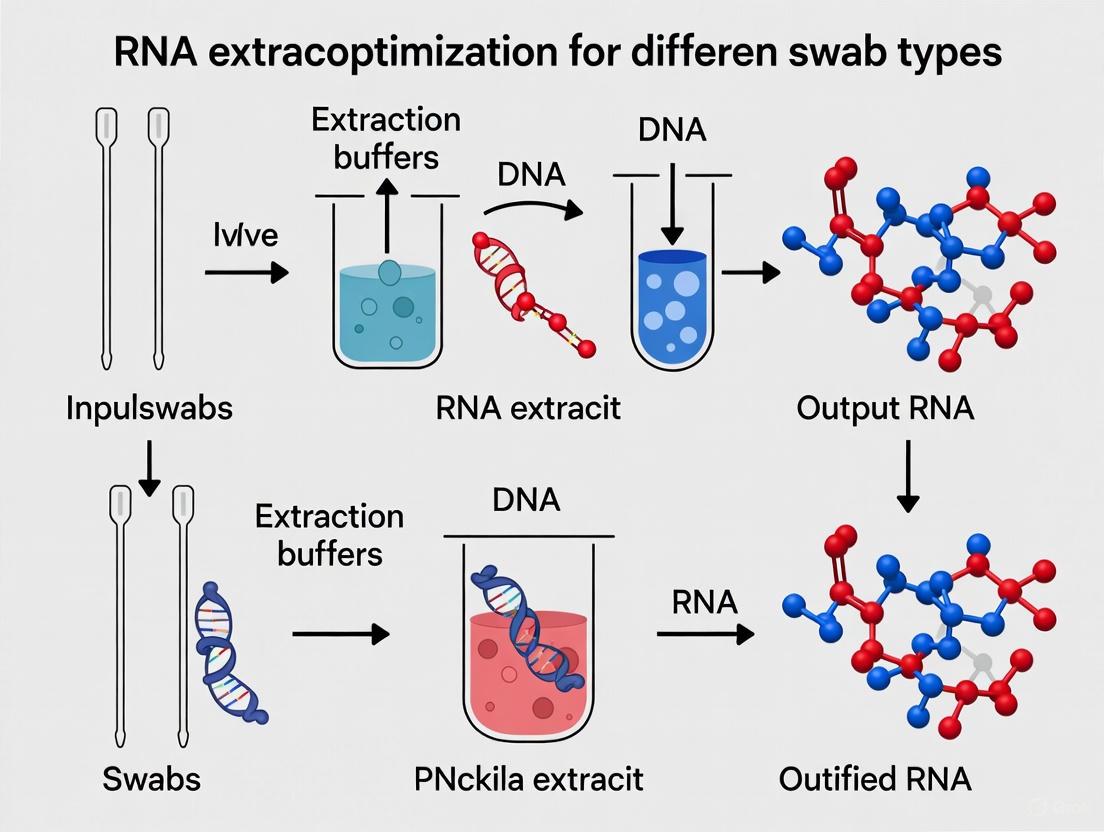
Workflow Diagram: Sample Collection to RNA Extraction
The following diagram illustrates the critical decision points in the sample collection and processing workflow that directly impact RNA integrity.
Research Reagent Solutions
The following toolkit lists essential materials and their proven applications for research aimed at optimizing RNA extraction from swab samples.
Table 3: Research Reagent Solutions for RNA Integrity Optimization
| Product Category | Example Products | Research Application & Function |
|---|---|---|
| Universal Transport Media | Puritan's UniTranz-RT [1], COPAN eNAT [2] | Broad-spectrum pathogen preservation; room-temperature RNA stability for up to 14 days (eNAT). |
| RNA Stabilization Buffers | DNA/RNA Shield (Zymo Research) [3], RNALater [3] | Immediate inactivation of RNases at point of collection; preserves RNA for metatranscriptomic studies. |
| Total RNA Extraction Kits | RNeasy PowerMicrobiome Kit (QIAGEN) [3], miRNeasy Advanced Kit (QIAGEN) [4] | Efficient isolation of high-integrity total RNA from complex sample matrices; some optimized for low-abundance RNAs. |
| Specialized Swabs | Puritan Flocked Swabs [1], ClearTip Injection-Molded Polyester [2] | Maximize specimen elution and cellular material release; designed for compatibility with molecular assays. |
The fidelity of RNA in downstream analyses is directly predetermined by the initial choices of swab material and collection media. Flocked nylon swabs paired with a balanced, antimicrobial-rich transport medium that includes protein stabilizers and cryoprotectants provide a robust foundation for preserving RNA integrity. Systematically validating this pre-analytical workflow using the provided protocols is not merely a preliminary step but a critical component of experimental rigor. By standardizing and optimizing these factors, researchers and drug development professionals can significantly reduce technical variability, enhance the sensitivity of molecular assays, and ensure the reliability of their data in transcriptomic studies and diagnostic applications.
The study of the respiratory microbiome is fundamentally challenged by a unique compositional imbalance: samples from the upper and lower respiratory tract are characterized by low microbial biomass overshadowed by an overwhelming presence of host nucleic acids. This disparity poses significant methodological hurdles for metagenomic and metatranscriptomic analyses, as the desired microbial signals can be obscured by host-derived reads, compromising the sensitivity and accuracy of microbial profiling [5] [6]. Efficiently navigating this host-microbe dynamic is not merely a technical consideration but a prerequisite for obtaining biologically meaningful data. The field has responded by developing specialized protocols for nucleic acid extraction and host depletion, which are critical for optimizing the recovery of microbial genetic material and enabling a comprehensive view of the active microbial communities in health and disease [5] [6] [7].
Quantitative Data on Sample Composition and Method Performance
Host DNA Content in Various Respiratory Samples
The following table summarizes the typical host DNA content found in different respiratory sample types, which directly impacts the required sequencing effort and choice of host depletion strategy.
Table 1: Host DNA Content in Respiratory Samples
| Sample Type | Typical Host DNA Content (%) | Notes |
|---|---|---|
| Bronchoalveolar Lavage (BAL) | 99.7% [7] | Representative of lower respiratory tract; very high host content. |
| Sputum | 99.2% [7] | Sample from critically ill patients or those with chronic lung disease. |
| Nasopharyngeal Swab (NPS) | 94.1% [7] | Common upper respiratory tract sample; lower host content than BAL. |
| Saliva | ~90% [7] | Less invasive collection, but still dominated by host material. |
Performance of Host DNA Depletion Methods
Multiple host DNA depletion methods have been benchmarked. Their performance varies by sample type, and the choice of method involves trade-offs between host depletion efficiency, microbial DNA retention, and potential taxonomic bias.
Table 2: Performance of Host DNA Depletion Methods on Respiratory Samples
| Method (Abbreviation) | Key Principle | Performance Highlights |
|---|---|---|
| Saponin Lysis + Nuclease (S_ase) [6] | Pre-extraction; lysis of human cells with saponin, digestion of freed DNA. | - Most effective host removal for BAL and OP [6].- Can significantly reduce bacterial DNA load [6]. |
| HostZERO Kit (K_zym) [6] | Pre-extraction commercial kit. | - Best for increasing microbial reads in BAL (100-fold increase) [6].- High host removal efficiency [6] [7]. |
| QIAamp DNA Microbiome Kit (K_qia) [6] [7] | Pre-extraction commercial kit. | - High host removal for nasal swabs (75% decrease) and sputum [7].- Good bacterial retention in OP samples [6]. |
| Filtering + Nuclease (F_ase) [6] | Pre-extraction; 10μm filtering followed by nuclease digestion. | - Balanced performance in host depletion and microbial recovery [6].- Moderate increase in microbial reads (65-fold in BAL) [6]. |
| Nuclease Digestion (R_ase) [6] | Pre-extraction; digestion of free DNA. | - Highest bacterial DNA retention rate in BAL (median 31%) [6].- Less effective at increasing microbial read proportion [6]. |
| Osmotic Lysis + PMA (O_pma) [6] | Pre-extraction; osmotic lysis of human cells, PMA degradation of DNA. | - Least effective at increasing microbial reads (2.5-fold in BAL) [6]. |
| Benzonase Treatment [7] | Pre-extraction; enzyme-based digestion of DNA/RNA. | - Effective for increasing final microbial reads in sputum [7].- Did not significantly reduce host content in nasal swabs [7]. |
Detailed Experimental Protocols
Protocol A: RNA Extraction Using Combined Mechanical and Chemical Lysis
This protocol, adapted from a 2025 study, is designed for comprehensive metatranscriptomic analysis of respiratory samples and is superior for lysing robust gram-positive bacteria and fungi [5].
Detailed Procedure:
- Sample Input: Use 200 µL to 400 µL of sample (e.g., pooled nasopharyngeal swab or bronchoalveolar lavage material).
- Lysis: Employ the Quick-DNA/RNA Miniprep Plus Kit (or equivalent). Perform lysis using a combination of chemical lysis buffers and vigorous mechanical bead beating. This dual approach is critical for disrupting the tough cell walls of gram-positive bacteria and fungi [5].
- Nucleic Acid Binding: Bind the released nucleic acids to the provided spin column matrix.
- Washing: Perform two wash steps as per the manufacturer's instructions to remove impurities.
- Elution: Elute the purified total RNA in nuclease-free water.
Downstream Processing for Metatranscriptomics:
- DNase Treatment: Treat the extracted RNA with TURBO DNase and/or Baseline-ZERO DNase to remove any contaminating genomic DNA [5].
- rRNA Depletion: Use the NEBNext rRNA Depletion Kit to deplete eukaryotic ribosomal RNA, thereby enriching for messenger RNA and non-ribosomal microbial RNA [5].
- Library Preparation & Sequencing: Proceed with standard RNA library preparation protocols. For Nanopore sequencing, this may include steps like RNA circularization, cDNA synthesis, whole-transcriptome amplification, end-prep, barcoding, and adapter ligation before loading onto a flow cell [5].
Protocol B: Host DNA Depletion for Metagenomic Sequencing of Frozen Respiratory Samples
This protocol benchmarks several methods optimized for frozen respiratory samples, which are common in biorepositories [7].
Detailed Procedure for F_ase Method (Filtering + Nuclease):
- Sample Preparation: Thaw frozen respiratory samples (e.g., nasal swab, BAL, or sputum). Note that freezing can reduce the viability of some bacteria like Pseudomonas aeruginosa; adding a cryoprotectant like glycerol during storage can mitigate this [7].
- Filtration: Pass the sample through a 10 µm filter. This size allows most bacterial and viral particles to pass through while capturing larger human host cells [6].
- Nuclease Digestion: Treat the flow-through containing the microbial cells with a nuclease enzyme to digest any free-floating host DNA that is not protected within a cell or viral capsid [6].
- Microbial Pellet Collection: Centrifuge the nuclease-treated filtrate to pellet the intact microbial cells.
- DNA Extraction: Proceed with standard DNA extraction from the microbial pellet using a commercial kit, such as those employing mechanical and chemical lysis for comprehensive recovery [6].
Alternative Method: S_ase (Saponin Lysis + Nuclease)
- Optimized Saponin Treatment: Incubate the sample with a low concentration of saponin (e.g., 0.025%) to selectively permeabilize and lyse human cells [6].
- Nuclease Digestion: Add a nuclease to digest the released host DNA.
- Microbial Pellet Collection: Centrifuge to pellet the intact microbial cells.
- DNA Extraction: Extract DNA from the pellet using a standard kit [6].
Visualizing the Experimental Workflow
The following diagram illustrates the key decision points and parallel pathways for processing low-biomass, high-host-content respiratory samples.
The Scientist's Toolkit: Essential Research Reagents
Table 3: Key Reagents and Kits for Optimized Nucleic Acid Studies
| Item | Function/Application |
|---|---|
| Quick-DNA/RNA Miniprep Plus Kit (Zymo Research) [5] | Simultaneous DNA/RNA extraction using combined chemical and mechanical lysis (bead beating). Ideal for robust lysis of Gram-positive bacteria and fungi in respiratory samples. |
| NucleoSpin Virus Kit (Macherey-Nagel) [5] | RNA extraction kit employing chemical lysis only. Serves as a comparator for protocols optimized for viruses or more fragile cells. |
| NEBNext rRNA Depletion Kit [5] | Depletes eukaryotic ribosomal RNA from total RNA extracts, crucial for enriching the microbial transcriptome in host-dominated samples. |
| HostZERO Microbial DNA Kit (Zymo Research) [6] [7] | Pre-extraction host depletion kit. Effective at significantly increasing the proportion of microbial reads in metagenomic sequencing. |
| QIAamp DNA Microbiome Kit (Qiagen) [6] [7] | Pre-extraction host depletion kit. Demonstrates high host removal efficiency for nasal and sputum samples. |
| TURBO DNase (Invitrogen) [5] | High-activity recombinant enzyme used to remove residual DNA during RNA extraction, preventing false positives in RNA-seq. |
| Saponin [6] | Detergent used at low concentrations (e.g., 0.025%) in pre-extraction methods to selectively lyse mammalian cells for host DNA depletion. |
| Magnetic Silica Beads [8] [9] | Solid matrix for binding nucleic acids in high-throughput, automated extraction systems. Enable rapid and efficient purification. |
RNA extraction and analysis are foundational to advancements in molecular diagnostics, genomics, and therapeutic development [10]. However, the inherent instability of RNA molecules presents significant challenges for researchers. RNA is structurally weaker than DNA, with highly reactive hydroxyl groups that make it prone to enzymatic degradation and heat-induced damage [11]. Ribonucleases (RNases), ubiquitous environmental enzymes that degrade RNA, represent a particular threat as they are extremely stable and difficult to inactivate [11]. For researchers working with swab-derived samples—especially in respiratory disease research and surveillance—understanding and mitigating these challenges is paramount to obtaining reliable, reproducible results. This application note details the key challenges of RNase activity, inhibition strategies, and sample stability, providing structured protocols and data to optimize RNA extraction workflows for different swab types within research contexts.
Key Challenges in RNA Workflows
Working with RNA, especially from swab samples which often have low microbial biomass, requires careful consideration of several interconnected challenges.
RNase Activity and RNA Instability
RNA is an inherently unstable molecule. In solution, it rapidly degrades through oxidation and hydrolysis via a transesterification reaction involving the ribose 2' hydroxyl group [12]. This intrinsic chemical instability is compounded by the presence of RNases, which are resilient enzymes that can remain active even after typical sterilization procedures. The low microbial RNA content in respiratory samples like nasopharyngeal swabs (NPS) is often overshadowed by host RNA, requiring high sequencing depth for adequate microbial transcript coverage [5].
Sample Collection and Storage Logistics
The choice of collection method directly impacts RNA stability and downstream analysis success. Traditional viral transport media (VTM), while preserving RNA viability, created significant supply chain bottlenecks during the SARS-CoV-2 pandemic [13]. Dry swab methods have emerged as a practical, cost-effective, and cold-chain-independent alternative [13]. One study demonstrated that dry polyester nasal swabs showed superior diagnostic sensitivity (90.48%) compared to wet swabs in VTM (76.19%) for post-mortem SARS-CoV-2 detection [13]. This approach is particularly valuable for surveillance in resource-constrained settings.
Efficient Lysis of Diverse Microbial Communities
Respiratory samples contain a diverse array of microorganisms with unique structural properties, complicating RNA extraction. Gram-positive bacteria and fungi possess robust cell walls that require vigorous lysis strategies. A comparative study of RNA extraction kits for metatranscriptomic analysis found that a protocol combining chemical and mechanical lysis (CML) significantly increased dsDNA library yields and enhanced detection of gram-positive bacteria and fungi compared to chemical lysis (CL) alone [5]. The lysis method must therefore be tailored to the sample type and research objectives.
Quantitative Data Comparison
The table below summarizes key performance data for various RNA handling strategies, lysis methods, and stabilization technologies, providing a quick reference for experimental planning.
Table 1: Performance Comparison of RNA Handling, Lysis, and Stabilization Methods
| Parameter | Dry Polyester Swabs [13] | Wet Swabs in VTM [13] | Chemical Lysis (CL) Only [5] | Chemical + Mechanical Lysis (CML) [5] | RNAshell Encapsulation [12] |
|---|---|---|---|---|---|
| Key Metric | Sensitivity: 90.48% | Sensitivity: 76.19% | Lower dsDNA library yield | Significantly higher dsDNA library yield (p<0.0001) | Stable for years at room temperature |
| Logistics | Room temp storage; cost-effective; cold-chain independent | Requires cold chain; supply chain vulnerabilities | Standard protocol | Superior for robust microbes (e.g., Gram-positive bacteria, fungi) | Eco-friendly shipping and storage |
| Primary Application | Surveillance in resource-limited settings | Standard clinical testing | Viral detection | Comprehensive metatranscriptomics | Long-term storage of quality control materials |
Table 2: Market Data for RNase Control and Related Products (2025-2035 Outlook)
| Product Category | Market Value (2025) | Projected Market Value (2035) | CAGR | Dominant Application/Format |
|---|---|---|---|---|
| Mouse RNase Inhibitor [14] | USD 193.2 Million | USD 548.7 Million | 11.0% | 20KU format (38% share); Biopharmaceutical Labs (42% share) |
| Recombinant RNase Inhibitor (USA) [15] | USD 8.66 Billion | USD 18.96 Billion | 13.95% | Adoption in industrial, commercial, and technological segments |
| RNA Extraction & Purification Kits [10] | USD 685 Million (2025) | USD 925 Million (2031) | 5.3% (2025-2031) | Driven by molecular diagnostics and precision medicine |
Featured Experimental Protocols
Protocol 1: RNA Extraction from Dry Swabs for Surveillance
This protocol, adapted from a community surveillance study in Pakistan, validates the use of dry polyester nasal swabs for sensitive pathogen detection [13].
Sample Collection
- Swab Type: Use a single polyester-tipped swab with a plastic shaft.
- Collection: Collect samples from both anterior nares (left and right), ensuring contact with the nasopharynx to absorb secretions.
- Storage: Place the dry swab directly into a dry collection tube without any liquid transport medium.
- Timeline: Process samples within a median of 24 hours (IQR = 20, 30) of collection. Transport to the lab in cold chain conditions (2-8°C) if processing is not immediate.
Laboratory Processing
- Rehydration: In the laboratory, rehydrate the dry swab by adding 2.5 mL of Phosphate-Buffered Saline (PBS) to the tube.
- Incubation: Incubate the swab in PBS for 10 minutes to release viral particles and RNA.
- RNA Extraction: Extract RNA using the QIAamp Viral RNA Mini Kit (QIAGEN) according to the manufacturer's instructions. This kit employs a silica-membrane spin column methodology in the presence of chaotropic salts to bind, wash, and elute high-quality RNA.
- Downstream Application: Detect the pathogen of interest using Reverse Transcription Polymerase Chain Reaction (RT-PCR). The cited study used this method for SARS-CoV-2 detection, reporting a high sensitivity of 90.48% [13].
Protocol 2: Enhanced Lysis for Metatranscriptomics from Respiratory Samples
This protocol is designed for comprehensive microbiome studies where detecting a broad range of organisms, including those with tough cell walls, is essential [5].
Sample Preparation
- Sample Types: Nasopharyngeal Swabs (NPS) or Bronchoalveolar Lavage (BAL) samples.
- Input Volume: Use 200 µL to 400 µL of the sample. A larger input volume can help recover higher RNA yield from low-biomass samples.
RNA Extraction with Combined Lysis
- Lysis: Use the Quick-DNA/RNA Miniprep Plus Kit (Zymo Research). This kit combines chemical and mechanical lysis (CML). The mechanical lysis is performed via bead beating, which is crucial for physically disrupting the robust cell walls of gram-positive bacteria and fungi.
- DNase Treatment: Treat the extracted RNA with TURBO DNase (Invitrogen) and/or Baseline-ZERO DNase (Lucigen) to remove residual genomic DNA contamination.
- rRNA Depletion: Use the NEBNext rRNA Depletion Kit v2 (New England Biolabs) to deplete eukaryotic ribosomal RNA (rRNA), thereby enriching for microbial and host mRNA sequences.
- Library Prep and Sequencing: Proceed with library preparation using a platform such as the Ligation Sequencing Kit (SQK-LSK109) from Oxford Nanopore Technologies for long-read sequencing on a MinION device.
Key Advantage: The CML protocol has been shown to yield significantly higher dsDNA libraries and better detection of gram-positive bacteria and fungi without compromising viral detection, making it superior for comprehensive metatranscriptomic analyses [5].
Protocol 3: High-Yield, Rapid Nucleic Acid Extraction for Broad Application
This magnetic bead-based protocol, termed SHIFT-SP, optimizes for speed and yield, which is beneficial for high-throughput diagnostics and working with low-concentration samples [8].
Optimized Binding Conditions
- Beads: Use magnetic silica beads.
- Binding Buffer: Employ a Lysis Binding Buffer (LBB) with a low pH (e.g., pH 4.1). A lower pH reduces the negative charge on silica beads, minimizing electrostatic repulsion with negatively charged nucleic acids and favoring binding [8].
- Mixing Method: Use a rapid "tip-based" mixing method, where the binding mix is aspirated and dispensed repeatedly with a pipette. This exposes the beads to the sample more efficiently than orbital shaking, achieving ~85% DNA binding within 1 minute [8].
- Temperature: Perform binding at an elevated temperature of 62°C.
SHIFT-SP Workflow
- Binding: Aspirate and dispense the sample (in LBB) with magnetic silica beads for 1-2 minutes.
- Washing: Use a magnet to separate the beads and wash them to remove contaminants and chaotropic salts.
- Elution: Elute the purified nucleic acids in an appropriate low-salt buffer (e.g., TE buffer). The entire process can be completed in 6-7 minutes [8].
Application Note: This method extracts nearly all the nucleic acid in a sample and is highly efficient for both DNA and RNA. Its high yield is particularly beneficial for detecting pathogens present at low concentrations, such as in sepsis, or for extracting circulating free nucleic acids for oncology applications [8].
Workflow Visualization
The following diagram illustrates the parallel pathways for processing dry and wet swab samples, highlighting the key decision points and steps that impact RNA stability and analysis outcomes.
The Scientist's Toolkit: Essential Research Reagent Solutions
Successful RNA research requires a suite of specialized reagents to maintain RNA integrity from sample collection to final analysis. The table below details key solutions and their functions.
Table 3: Essential Reagents for RNase Control and RNA Workflows
| Research Reagent Solution | Primary Function | Key Characteristics | Example Applications |
|---|---|---|---|
| Recombinant RNase Inhibitors [15] [11] | Binds to and inhibits a broad spectrum of RNases non-competitively. | High purity (recombinant source), superior stability, consistent batch-to-batch performance. | cDNA synthesis, RT-PCR, in vitro transcription, RNA sequencing. |
| Silica-Based Extraction Kits [10] [13] [16] | Binds, washes, and elutes RNA using silica membranes/beads in chaotropic salts. | Robust, automatable, effective inhibitor removal. Compatible with manual and automated workflows. | Routine RNA purification from swabs, lavages, and other samples. |
| Chemical + Mechanical Lysis Kits [5] | Combines chemical reagents (chaotropes) with physical disruption (bead beating) for comprehensive cell wall lysis. | Essential for breaking robust microorganisms like gram-positive bacteria and fungi in microbiome studies. | Metatranscriptomic analysis of respiratory samples. |
| RNAshell/Encapsulation Tech [12] | Long-term room temperature storage of RNA by vacuum drying with stabilizers in an inert atmosphere. | Protects RNA from atmospheric oxygen and moisture; enables eco-friendly shipping without cold chain. | Archiving control materials, biobanking, shipping reference standards. |
| Magnetic Silica Beads [8] | Solid matrix for nucleic acid binding that can be rapidly separated using a magnet in optimized buffers. | Enables very fast (<10 min) and high-yield extraction protocols; automation-friendly. | High-throughput sample preparation, rapid diagnostics (e.g., STAT testing). |
The initial step of cell lysis is a critical determinant of success in RNA extraction protocols. The choice between chemical and mechanical disruption methods directly impacts RNA yield, integrity, and suitability for downstream applications such as RNA sequencing (RNA-Seq) and quantitative PCR (qPCR) [17]. For research aimed at optimizing RNA extraction from various swab types—which may collect diverse cellular samples from bacterial, animal, or human sources—selecting the appropriate lysis technique is paramount. This article provides a comparative analysis of chemical and mechanical lysis fundamentals, offering detailed protocols and data-driven recommendations to guide researchers in tailoring lysis strategies for specific cell types encountered in swab-based research.
Chemical Lysis Methods
Principles and Mechanisms
Chemical lysis methods disrupt cells by employing reagents that compromise the integrity of cellular membranes and walls without physical force. These methods include detergents, enzymes, and alkaline agents. Detergents solubilize lipid components of cell membranes by disrupting hydrophobic and hydrophilic interactions [18]. Enzymatic methods, using agents like lysozyme for bacteria or zymolyase for yeast, selectively degrade specific structural components of the cell wall, forming spheroplasts that are readily lysed with minimal shear stress [17]. Chemical lysis is typically performed under mild conditions, making it suitable for preserving labile macromolecules, but its efficiency can vary significantly with cell type.
Application Notes for RNA Extraction
For RNA extraction, chemical lysis is often performed with strong denaturants like guanidinium isothiocyanate (GITC), which simultaneously inactivates RNases released during disruption, thereby preserving RNA integrity [17]. This is particularly crucial for samples with high endogenous RNase activity, such as pancreatic tissue. Chemical methods are ideal for simple, rapid processing of multiple samples and are easily adaptable to automated high-throughput workflows. However, they may introduce inhibitors that require careful removal during subsequent purification steps and can be less effective for cells with robust walls.
Mechanical Lysis Methods
Principles and Mechanisms
Mechanical lysis utilizes physical force to shear cell membranes and walls. These methods include bead beating, high-pressure homogenization (e.g., French Press), sonication, and grinding [18] [17]. The mechanisms involve grinding, cavitation, or shear forces generated by rapid pressure changes. Bead milling agitates samples with small glass or ceramic beads, physically breaking apart cells through friction and impact [18]. High-pressure homogenization forces cell suspensions through a narrow orifice at high pressure, generating shear forces that rupture cells [19]. Sonication employs ultrasonic waves to create cavitation bubbles in the liquid, whose collapse produces intense local shear forces [19].
Application Notes for RNA Extraction
Mechanical methods are highly effective for tough cell walls but require careful optimization to prevent RNA degradation from heat or released RNases. It is critical to perform mechanical disruption in the presence of RNase inhibitors and to keep samples cold [20] [17]. While mechanical methods generally provide high yields and are scalable, some, like bead beating and French Press, can lead to significant RNA degradation even with RNase inhibitors, making them less suitable for applications requiring intact mRNA, such as mRNA-protein complex purification [20]. Grinding frozen tissue with a mortar and pestle under liquid nitrogen effectively preserves RNA integrity by halting enzymatic activity during disruption [20] [17].
Comparative Analysis of Lysis Methods
The table below provides a comparative summary of key lysis methods, highlighting their suitability for different cell types, key parameters, and relative performance in RNA extraction.
Table 1: Comparative Analysis of Cell Lysis Methods for RNA Extraction
| Lysis Method | Mechanism | Typical Cell Types | Key Parameters | Relative RNA Integrity | Scalability | Throughput |
|---|---|---|---|---|---|---|
| Detergent-Based | Solubilizes membranes | Mammalian cells, Gram-negative bacteria | Detergent type & concentration, incubation time | Moderate to High [17] | High | High |
| Enzymatic | Degrades specific cell wall components | Yeast, Gram-positive bacteria, Fungi | Enzyme type, concentration, temperature, time | High [17] | Moderate | Moderate |
| Bead Beating | Physical grinding with beads | Yeast, Fungi, Bacteria, Soil samples | Bead size, material, agitation speed, time | Low to Moderate [20] | Moderate | Moderate |
| Sonication | Cavitation from sound waves | Bacteria, Yeast, Soft tissues | Amplitude, duration, pulse on/off time | Low to Moderate [19] | Low (for lab-scale) | Low to Moderate |
| High-Pressure Homogenization | Shear forces from pressure release | Yeast, Bacteria, Microalgae | Pressure (psi), number of passes | Moderate to High [19] | High | High |
| Mortar & Pestle Grinding | Physical grinding at cryogenic temps | Hard animal tissues, Plant tissues, Fungi | Grinding time, liquid nitrogen use | High [20] [17] | Low | Low |
Detailed Experimental Protocols
Protocol 1: Enzymatic Lysis of Yeast Cells for Intact RNA
This protocol is adapted from methods suitable for purifying intact mRNA-protein complexes and is designed to maximize RNA integrity [20].
Reagents and Equipment:
- Lysis Buffer (e.g., containing RNase inhibitors such as 20 mM Ribonucleoside Vanadyl Complex (RVC) and 100 U/mL SuperaseIn) [20]
- Zymolyase or Lyticase enzyme
- Water bath or incubator set to 30°C
- Refrigerated centrifuge
Procedure:
- Harvest yeast cells by centrifugation at 3,000 × g for 5 minutes at 4°C.
- Wash the cell pellet with an appropriate buffer (e.g., ice-cold PBS) and centrifuge again.
- Resuspend the cell pellet in Lysis Buffer containing RNase inhibitors.
- Add Zymolyase to a final concentration of 10-20 U/mL and mix gently by inversion.
- Incubate the suspension at 30°C for 30-60 minutes to form spheroplasts. Monitor spheroplast formation microscopically.
- Pellet the spheroplasts by gentle centrifugation (1,500 × g for 10 minutes at 4°C).
- Carefully remove the supernatant. The spheroplasts can now be lysed by gentle vortexing in a GITC-based lysis solution for RNA extraction [17].
- Proceed immediately with RNA purification.
Protocol 2: Mechanical Lysis by Mortar and Pestle for Frozen Tissues
This method is recommended for difficult-to-lyse tissues and when preserving RNA integrity is a top priority [20] [17].
Reagents and Equipment:
- Liquid nitrogen
- Mortar and pestle (pre-chilled)
- Cryogenic container (e.g., dewar flask)
- GITC-based lysis solution (e.g., from commercial RNA isolation kits)
Procedure:
- Pre-chill the mortar and pestle on dry ice or by adding liquid nitrogen.
- Submerge the tissue sample (≤100 mg) in liquid nitrogen in the mortar to "snap-freeze" it.
- Grind the frozen tissue vigorously with the pestle to a fine, powdery consistency. Continue adding liquid nitrogen during grinding to keep the sample frozen.
- Allow the liquid nitrogen to evaporate, but do not let the powder thaw.
- Use a pre-cooled spatula to transfer the frozen powder to a tube containing an appropriate volume of GITC-based lysis solution.
- Immediately vortex the mixture vigorously until the powder is fully dispersed and dissolved.
- The lysate is now ready for RNA purification.
Workflow Diagram: Lysis Method Selection for RNA Extraction
The following diagram outlines a decision workflow for selecting an appropriate lysis method based on cell type and research objectives.
The Scientist's Toolkit: Essential Reagents and Materials
Table 2: Key Research Reagent Solutions for Cell Lysis and RNA Stabilization
| Item | Function/Description | Example Application |
|---|---|---|
| Guanidinium Isothiocyanate (GITC) | A potent chaotropic agent that denatures proteins and inactivates RNases, stabilizing RNA during and after lysis. [17] | Core component of lysis buffers for most RNA extraction protocols. |
| RNase Inhibitors (e.g., SuperaseIn, RVC) | Enzymatic or chemical compounds that specifically inhibit RNase activity to prevent RNA degradation. [20] | Added to lysis buffers, especially critical for mechanical methods and sensitive samples. |
| RNAlater Stabilization Solution | A reagent that rapidly penetrates tissues to stabilize and protect cellular RNA at harvest, allowing temporary storage without freezing. [17] | Preservation of swab samples and tissues prior to RNA extraction. |
| Zymolyase | An enzyme preparation with β-1,3-glucanase activity that digests the cell walls of yeast and fungi. [17] | Enzymatic lysis of yeast cells to form spheroplasts for gentle lysis. |
| Lysozyme | An enzyme that catalyzes the breakdown of bacterial cell walls by hydrolyzing β-1,4-glycosidic bonds. [17] | Enzymatic lysis of Gram-positive and some Gram-negative bacteria. |
| Polyvinylpyrrolidone (PVP) | A polymer that binds to and helps remove polyphenols and polysaccharides that can co-purify with RNA. [17] | Treatment of lysates from plant tissues and some fungi to improve RNA purity. |
| Silica Membranes/ Magnetic Beads | Solid-phase matrices that bind nucleic acids in the presence of high-salt buffers, allowing for purification and washing. | Key components of column- and bead-based RNA purification kits following lysis. |
Optimizing the cell lysis step is foundational to successful RNA extraction from diverse swab types. The choice between chemical and mechanical methods involves a careful trade-off between RNA yield, integrity, and practical considerations like throughput and scalability. Chemical methods offer gentleness and high-throughput potential for standard samples like mammalian cells, while mechanical methods are indispensable for breaking tough cellular walls but require stringent controls to prevent RNA degradation. Researchers are advised to select their lysis protocol based on the specific cell type being processed and the downstream application requirements, using the comparative data and detailed protocols provided herein as a guide.
Specialized Protocols and Kit Selection for Different Swab Types
Within the broader scope of a thesis on RNA extraction optimization for different swab types, this application note provides a detailed protocol for processing nasopharyngeal swabs. The COVID-19 pandemic underscored that the accuracy of molecular diagnostics, particularly for RNA viruses like SARS-CoV-2, is highly dependent on the upstream sample preparation process [21] [22]. Efficient nucleic acid extraction is a critical prerequisite for downstream analyses such as real-time reverse transcription polymerase chain reaction (rRT-PCR), which remains the gold standard for viral detection [23]. The workflow from lysis to elution must be robust, efficient, and adaptable to high demands. This document consolidates and compares validated methodologies, including both solid-phase extraction and extraction-free protocols, to provide researchers and drug development professionals with a reliable framework for optimizing the processing of nasopharyngeal swab samples.
Comparative Analysis of RNA Extraction Method Performance
The selection of an RNA extraction method involves balancing sensitivity, time, cost, and applicability to high-throughput workflows. The following table summarizes key performance metrics from recent studies for direct comparison.
Table 1: Performance Comparison of Different RNA Extraction and Direct Detection Methods
| Method / Kit Name | Principle | Total Processing Time | Sensitivity / Notes | Reference |
|---|---|---|---|---|
| Standard Magnetic Bead Kit (MAGABIO PLUS VIRUS DNA/RNA PURIFICATION KIT II) | Silica-based magnetic beads, includes Proteinase K | ~35 minutes | Considered the benchmark for sensitivity in its study. | [23] |
| Rapid Magnetic Bead Kit (MAGABIO PLUS VIRUS RNA PURIFICATION KIT II) | Optimized silica-based magnetic beads, no Proteinase K | ~9 minutes | Results comparable to the standard kit, faster turnaround. | [23] |
| SHIFT-SP Method | Optimized magnetic silica beads, low-pH binding, tip-based mixing | 6-7 minutes | Extracts nearly all nucleic acid in sample; high yield. | [8] |
| Extraction-Free Protocol (Group VI) | Heat treatment, sample dilution, Proteinase K, RNase inhibitors | Rapid (bypasses extraction) | 84.26% sensitivity; mean Ct value increase of +3.8 vs. standard. | [21] |
| In-house Phenol-free Kit | Silica column-based | Not specified | No significant difference from commercial kit; suitable for sequencing. | [24] |
Detailed Experimental Protocols
Protocol A: Rapid Magnetic Bead-Based RNA Extraction
This protocol, adapted from a comparative study, provides a fast and reliable method for RNA purification using magnetic beads [23].
1. Reagent Preparation:
- Lysis Buffer: Prepare a lysis buffer containing guanidine thiocyanate and Triton X-100. For optimal binding efficiency, the pH should be adjusted to approximately 4.1 [8].
- Wash Buffers: Prepare two wash buffers (Wash 1 and Wash 2) typically containing ethanol or other solvents to remove salts and impurities without eluting the RNA.
- Elution Buffer: Use nuclease-free water or a low-salt buffer (e.g., 10 mM Tris-HCl, pH 8.0).
2. Sample Lysis:
- Pipette 200 µL of nasopharyngeal swab sample (in universal transport medium) into a 1.5 mL microcentrifuge tube.
- Add 300 µL of Lysis Buffer directly to the sample. Vortex thoroughly for 15 seconds to ensure complete mixing and lysis.
- Note: This rapid protocol omits the separate Proteinase K digestion step used in longer protocols [23].
3. Nucleic Acid Binding:
- Add 50 µL of magnetic silica bead suspension to the lysate.
- For optimal binding, use a "tip-based" mixing method: repeatedly aspirate and dispense the entire mixture with a pipette for 1-2 minutes. This method has been shown to achieve over 90% binding efficiency within 2 minutes, significantly faster than orbital shaking [8].
- Incubate the mixture at 62°C during mixing to enhance binding kinetics.
4. Washing:
- Place the tube on a magnetic stand to capture the beads. Discard the supernatant.
- Resuspend the bead pellet in 500 µL of Wash Buffer 1 by vortexing or pipetting. Capture the beads on the magnetic stand and discard the supernatant.
- Repeat this process with 500 µL of Wash Buffer 2.
- Air-dry the bead pellet for a few minutes to ensure complete ethanol evaporation.
5. Elution:
- Remove the tube from the magnetic stand.
- Add 50-100 µL of Elution Buffer to the beads and resuspend thoroughly by pipetting.
- Incubate at 62°C for 1-2 minutes to facilitate the release of RNA from the beads [8].
- Place the tube back on the magnetic stand and transfer the eluted RNA (supernatant) to a new, nuclease-free tube.
- The extracted RNA is now ready for downstream applications like rRT-PCR. Store at -80°C for long-term preservation.
Protocol B: Extraction-Free rRT-PCR for Rapid Detection
This protocol is ideal for situations requiring high-throughput, rapid testing where maximum sensitivity is not the primary concern [21].
1. Sample Pre-treatment:
- Prepare a master mix containing Proteinase K and RNase inhibitors. The estimated cost of these reagents is about $1.15 per sample [21].
- Combine the master mix with an equal volume of the nasopharyngeal swab sample (e.g., 50 µL sample + 50 µL master mix) in a PCR tube. This constitutes a 1:1 dilution.
2. Heat Inactivation:
- Seal the PCR tubes and place them in a thermal cycler or heat block.
- Incubate at 95°C for 5 minutes to inactivate nucleases and potentially inhibitory substances.
3. Direct Amplification:
- Centrifuge the tubes briefly to collect condensation.
- Use 10-20 µL of the heat-treated, diluted sample directly as the template in the rRT-PCR reaction.
- Note: This method demonstrates high sensitivity for samples with low Ct values (high viral load) but has reduced detection rates for samples with Ct values >30 [21].
Workflow Visualization and Decision Pathway
The following diagram illustrates the logical pathway for selecting the most appropriate protocol based on research objectives and sample conditions.
The Scientist's Toolkit: Essential Reagents and Materials
Table 2: Key Research Reagent Solutions for RNA Extraction from Nasopharyngeal Swabs
| Reagent / Material | Function in the Workflow | Examples / Notes |
|---|---|---|
| Lysis Binding Buffer (LBB) | Disrupts viral envelope and cells, inactivates RNases; creates conditions for NA binding to silica. | Contains chaotropic salts (e.g., guanidine thiocyanate). For optimal DNA/RNA binding, a low pH (e.g., ~4.1) is critical [8]. |
| Magnetic Silica Beads | Solid-phase matrix for binding nucleic acids, enabling separation via a magnetic stand. | Bead quantity impacts yield; 30-50 µL beads can achieve >90% binding for high inputs [8]. |
| Proteinase K | Digests proteins and nucleases, improving viral nucleic acid accessibility and reducing PCR inhibitors. | Used in standard extraction kits [23] and some extraction-free protocols [21]. |
| RNase Inhibitors | Protects the integrity of viral RNA by inhibiting ubiquitous RNase enzymes during sample preparation. | Incorporated into advanced extraction-free protocols to prevent RNA degradation [21]. |
| Wash Buffers | Remove contaminants, salts, and other impurities from the bead-NA complex without causing elution. | Typically contain ethanol or other solvents. Protocols may use 2-3 wash steps for purity [23]. |
| Elution Buffer | A low-ionic-strength solution that releases pure RNA from the silica matrix into an aqueous solution. | Nuclease-free water or Tris buffer. Higher temperature (e.g., 62°C) can increase elution efficiency [8]. |
Nucleic acid extraction is a foundational step in molecular diagnostics and research, with magnetic bead and silica column technologies emerging as the dominant solid-phase extraction methods. The optimization of this process is particularly critical when working with RNA from swab samples, where sample volume and potential inhibitors can compromise downstream applications. This application note provides a systematic evaluation of these competing technologies, focusing on their performance characteristics, protocol efficiency, and suitability for different research scenarios. Within the broader context of RNA extraction optimization for various swab types, understanding the technical distinctions between these platforms enables researchers to make informed decisions that enhance RNA yield, quality, and experimental reproducibility.
Fundamental Principles
Both magnetic bead and silica column technologies utilize the binding of nucleic acids to silica surfaces in the presence of chaotropic salts, but their mechanics differ substantially. Silica column technology employs a stationary silica membrane embedded in a spin column through which samples and buffers are centrifuged. In contrast, magnetic bead technology uses paramagnetic silica particles that are mixed with the sample and retrieved using a magnetic field, allowing for more flexible liquid handling [8] [25].
The binding efficiency in both systems is influenced by pH, with lower pH (approximately 4.1) demonstrating superior nucleic acid binding due to reduced electrostatic repulsion between the negatively charged silica and nucleic acid backbone [8]. The mode of bead movement during binding significantly affects efficiency; "tip-based" mixing, where the binding mix is aspirated and dispensed repeatedly, achieved ~85% DNA binding within 1 minute compared to only ~61% with orbital shaking [8].
Quantitative Performance Metrics
Table 1: Direct Performance Comparison of Extraction Technologies
| Performance Parameter | Magnetic Bead Technology | Silica Column Technology |
|---|---|---|
| Typical Processing Time | 6-7 minutes (SHIFT-SP method) [8] | 25 minutes [8] |
| DNA Yield Efficiency | ~98% binding efficiency [8] | Approximately 50% of magnetic bead yield [8] |
| RNA Yield from Cells | ~48% lower yield than phenol-chloroform [26] | |
| Automation Compatibility | Excellent - easily automated for high-throughput [8] [25] | Limited - primarily manual processing |
| Typical Elution Volume | Can be minimized for concentrated eluate [8] | Fixed by column capacity |
| Suitability for Swab Samples | Effective for oro/nasopharyngeal swabs [25] | Compatible with swab eluates |
Table 2: Cost and Throughput Considerations
| Consideration | Magnetic Bead Technology | Silica Column Technology |
|---|---|---|
| Initial Setup Cost | Higher (requires magnet equipment) | Lower (centrifuge typically available) |
| Per-sample Cost | ~$0.48/sample possible with in-house reagents [26] | Commercial kits often more expensive [26] |
| Hands-on Time | Minimal with automation | Significant throughout process |
| Sample Throughput | High (parallel processing) | Limited (sequential processing) |
| Reagent Preparation | Often requires buffer optimization | Typically pre-packaged |
Experimental Protocols
Magnetic Bead-Based RNA Extraction from Swab Samples
This protocol adapts the method described by [25] for efficient RNA extraction from oro- and nasopharyngeal swab samples.
Reagents and Equipment
- Lysis/Binding Buffer: 4 M guanidine thiocyanate, 10 mM MES pH 5.5, 1% β-mercaptoethanol [26]
- Silica-coated magnetic particles: Synthesized from Fe₃O₄ magnetite nanoparticles coated with silica via tetraethyl orthosilicate (TEOS) hydrolysis [25]
- Wash Buffer 1: 1 M guanidine thiocyanate, 10 mM Tris pH 7.0 [26]
- Wash Buffer 2: 80% ethanol, 10 mM Tris pH 7.0 [26]
- Elution Buffer: RNase-free water or TE buffer
- Magnetic rack for 1.5-2.0 mL tubes
- Vortex mixer with horizontal adaptation for tube mixing
- Thermal shaker capable of maintaining 62°C
Step-by-Step Procedure
Sample Lysis:
- Transfer 200 μL of swab transport medium to a 1.5 mL microcentrifuge tube.
- Add 300 μL of Lysis/Binding Buffer and 10 μL of proteinase K (if needed for protein-rich samples).
- Vortex thoroughly and incubate at 62°C for 5 minutes with shaking at 1000 rpm.
Binding:
- Add 50 μL of silica-coated magnetic bead suspension to the lysate.
- Mix using "tip-based" method (repeated aspiration/dispersion) for 2 minutes at room temperature [8].
- Place tube on magnetic rack for 1 minute until solution clears.
- Carefully remove and discard supernatant.
Washing:
- Resuspend beads in 500 μL of Wash Buffer 1 while tube is on magnetic rack.
- Rotate tube 180° and incubate for 30 seconds.
- Return to original position and discard supernatant.
- Repeat with 500 μL of Wash Buffer 2.
- Perform final wash with 500 μL of 80% ethanol.
Elution:
- Air-dry beads for 5 minutes at room temperature to evaporate residual ethanol.
- Remove from magnetic rack and resuspend in 30-50 μL of Elution Buffer.
- Incubate at 70°C for 3 minutes to enhance elution efficiency [8].
- Place on magnetic rack and transfer eluate to a clean tube.
- Store at -80°C for long-term preservation.
Silica Column-Based RNA Extraction
This protocol follows the cost-effective method described by [26] with modifications for swab samples.
Reagents and Equipment
- Buffer A: 4 M guanidine thiocyanate, 10 mM MES pH 5.5, 1% β-mercaptoethanol [26]
- Buffer B: 1 M guanidine thiocyanate, 10 mM Tris pH 7.0 [26]
- Buffer C: 80% ethanol, 10 mM Tris pH 7.0 [26]
- Generic silica spin columns (e.g., Epoch Life Science)
- Microcentrifuge capable of 13,000 × g
- Swab sample in transport medium
Step-by-Step Procedure
Sample Preparation:
- Centrifuge 200 μL of swab transport medium at 5000 × g for 1 minute.
- Transfer supernatant to new tube (retain pellet if cellular RNA is target).
Lysing and Binding:
- Add 300 μL of Buffer A (with fresh β-mercaptoethanol) to the sample.
- Mix by vortexing and incubate at room temperature for 15 minutes.
- Add 300 μL of 70% ethanol and mix thoroughly by pipetting.
- Load entire mixture onto silica column placed in collection tube.
Centrifugation and Washing:
- Centrifuge at 8000 × g for 30 seconds. Discard flow-through.
- Add 600 μL of Buffer B. Centrifuge at 8000 × g for 30 seconds. Discard flow-through.
- Add 500 μL of Buffer C. Centrifuge at 8000 × g for 30 seconds. Discard flow-through.
- Repeat Buffer C wash step once.
- Perform final "dry" spin at 8000 × g for 2 minutes to remove residual ethanol.
Elution:
- Transfer column to clean 1.5 mL microcentrifuge tube.
- Add 30 μL of RNase-free water directly to the silica membrane.
- Incubate at room temperature for 2 minutes.
- Centrifuge at 8000 × g for 30 seconds to elute RNA.
- Store RNA at -80°C for downstream applications.
Methodology for Comparative Evaluation
Quality Assessment and Downstream Validation
To ensure extracted RNA is suitable for sensitive downstream applications, implement the following quality control measures:
- Spectrophotometric Analysis: Use NanoDrop to determine RNA concentration and purity. Acceptable parameters: A260/A280 ratio of ~1.8-2.0 and A260/A230 ratio ≥1.8 [27] [28].
- Integrity Analysis: For critical applications, analyze RNA integrity using:
- Downstream Application Validation:
- Perform qRT-PCR with housekeeping genes to confirm absence of inhibitors.
- Use percentage of reads mapping to rRNA as a metric for sequencing library preparations [29].
Experimental Design for Technology Comparison
When comparing magnetic bead versus silica column technologies for specific swab types:
- Sample Partitioning: Split single swab samples into equal aliquots for parallel processing with both methods.
- Spike-In Controls: Introduce known quantities of synthetic RNA (e.g., from External RNA Controls Consortium) before extraction to calculate precise recovery rates [30].
- Technical Replicates: Process minimum of 5 replicates per method to account for technical variability.
- Operator Variability Assessment: Have multiple technicians perform extractions to evaluate protocol robustness.
The Scientist's Toolkit: Research Reagent Solutions
Table 3: Essential Reagents and Their Functions in Nucleic Acid Extraction
| Reagent/Kits | Function | Technology Type |
|---|---|---|
| Guanidine Thiocyanate | Chaotropic salt that denatures proteins, inhibits RNases, and promotes nucleic acid binding to silica [26] [27] | Both |
| Silica-coated Magnetic Beads | Paramagnetic particles that bind nucleic acids in chaotropic conditions for magnetic separation [8] [25] | Magnetic Bead |
| Silica Spin Columns | Stationary silica membranes that bind nucleic acids under centrifugal force [26] | Silica Column |
| β-mercaptoethanol | Reducing agent that disrupts disulfide bonds in proteins and inhibits RNases [26] [27] | Both |
| Tris-EDTA (TE) Buffer | Common elution and storage buffer that stabilizes nucleic acids [8] | Both |
| Commercial Kits (SHIFT-SP) | Optimized magnetic bead systems for rapid, high-yield extraction [8] | Magnetic Bead |
| Commercial Kits (RNeasy, NucleoSpin) | Reliable silica column systems for consistent RNA purification [27] [28] | Silica Column |
Workflow Visualization
Diagram 1: Comparative Workflow: Magnetic Bead vs. Silica Column RNA Extraction
The comparative analysis presented herein demonstrates that both magnetic bead and silica column technologies can effectively extract RNA from swab samples, albeit with distinct advantages suited to different research contexts.
For high-throughput laboratories processing large sample volumes, particularly in clinical diagnostic settings, magnetic bead technology offers compelling advantages in processing time, automation potential, and yield. The SHIFT-SP method's 6-7 minute processing time represents a significant improvement in workflow efficiency [8]. The compatibility of magnetic bead platforms with liquid handling robots further enhances their utility in screening environments.
For low-throughput research settings with budget constraints or specialized sample requirements, silica column technology provides a reliable, cost-effective alternative. The ability to create customized, inexpensive buffers ($0.48/sample) makes this approach particularly valuable for pilot studies or resource-limited environments [26].
When optimizing RNA extraction for specific swab types, researchers should consider implementing the quality control measures outlined in Section 4.1 and validate their chosen method with intended downstream applications, as extraction efficiency directly impacts the sensitivity of subsequent molecular analyses.
The optimization of ribonucleic acid (RNA) extraction is a cornerstone of molecular biology, directly influencing the success and cost-efficiency of downstream applications in research and diagnostics. Within the broader context of a thesis on RNA extraction optimization for different swab types, this application note focuses on two pivotal methodological approaches: acid-phenol extraction and heat shock protocols. The persistent challenges of reagent costs, sample throughput, and purity demands necessitate the development and refinement of robust, alternative techniques. This document provides a detailed comparative analysis and detailed protocols for these methods, equipping researchers and drug development professionals with the tools to enhance their nucleic acid extraction workflows, particularly when processing diverse swab-based specimens.
Acid-Phenol Extraction: A Cost-Effective Workhorse
Acid-phenol extraction, utilizing a phenol:chloroform:isoamyl alcohol (P/C/I) mixture at an acidic pH (typically 4.5–5), is a classic liquid-liquid partitioning technique that effectively separates RNA from other cellular components. The acidic pH favors the partitioning of RNA into the aqueous phase, while DNA and proteins remain in the organic phase or the interphase.
Quantitative Comparison of Phenol-Based Reagents
The choice of reagent significantly impacts both the yield and the cost of RNA extraction, especially at scale. The following table summarizes a key comparative analysis.
Table 1: Cost and Yield Analysis of Phenol-Based Reagents for dsRNA Extraction from a 5L Bacterial Culture [31] [32]
| Extraction Reagent | Estimated Reagent Cost per 5L Culture (EUR) | Relative Yield (µg/OD600 of cells) | Key Contaminants |
|---|---|---|---|
| TRIzol | ~1,700 | ~30 | Bacterial gDNA (without DNase) |
| QIAzol | ~770 | ~30 | Bacterial gDNA (without DNase) |
| P/C/I (pH 4.5-5) | Low cost (fraction of commercial reagents) | Comparable mass yield | Higher relative gDNA/sRNA without optimization |
Optimized Acid-Phenol Protocol for Bacterial dsRNA Extraction
The following protocol is optimized for the extraction of double-stranded RNA (dsRNA) from HT115(DE3) bacterial cultures but can be adapted for total RNA from other sample types [31].
Materials:
- Lysis Buffer: 1% SDS (Sodium Dodecyl Sulfate), 2 mM EDTA, pH 8.0
- Acidic Phenol:Chloroform:Isoamyl Alcohol (25:24:1, pH 4.5-5.0)
- Chloroform
- 3M Sodium Acetate (NaOAc), pH 5.2
- 100% and 70% Ethanol
- Nuclease-Free Water
- RNase A (DNase-free)
- DNase I (RNase-free) and corresponding buffer
Procedure:
- Cell Lysis: Pellet bacterial cells from 1-100 mL of culture. Resuspend the pellet in Lysis Buffer using a minimal volume (e.g., 500 µL per 50 OD600-mL of cells). Boil the suspension for 5 minutes to ensure complete lysis [31].
- Digestion of Single-Stranded RNA: Cool the lysate to room temperature. Add RNase A to a final concentration of 10 µg/mL and incubate at 37°C for 30 minutes to digest single-stranded RNAs [31].
- Acid-Phenol Extraction: Add an equal volume of acidic P/C/I to the lysate. Vortex vigorously for 30 seconds. Centrifuge at ≥12,000 × g for 5 minutes at 4°C to separate phases.
- Aqueous Phase Recovery: Carefully transfer the upper aqueous phase to a new tube. Avoid the white interphase, which contains proteins and DNA.
- Chloroform Wash (Optional but Recommended): Add an equal volume of chloroform, vortex, and centrifuge as in step 3. This step improves purity by removing residual phenol [33].
- DNase Treatment (Critical for Purity): Recover the final aqueous phase. Add DNase I and its buffer according to the manufacturer's instructions. Incubate at 37°C for 20-30 minutes to remove genomic DNA contamination, as confirmed by [31] [32].
- Ethanol Precipitation: Add 0.1 volumes of 3M NaOAc (pH 5.2) and 2.5 volumes of 100% ethanol. Mix and precipitate at -20°C for at least 1 hour or overnight.
- RNA Recovery: Centrifuge at ≥12,000 × g for 30 minutes at 4°C to pellet the RNA. Carefully decant the supernatant.
- Wash and Resuspend: Wash the pellet with 1 mL of 70% ethanol. Centrifuge again for 10 minutes, carefully discard the supernatant, and air-dry the pellet for 5-10 minutes. Do not over-dry. Resuspend the final RNA pellet in nuclease-free water.
Workflow and Strategic Considerations
The diagram below illustrates the key decision points in the acid-phenol extraction workflow.
Key Advantages and Considerations:
- Cost-Efficiency: The P/C/I method drastically reduces costs compared to commercial phenol-guanidine reagents, making it suitable for large-scale applications like dsRNA production for biocontrol [31] [32].
- Batch Effects in Transcriptomics: Researchers performing meta-analyses should note that acid-phenol extraction can preferentially solubilize membrane-associated mRNAs compared to kit-based methods. This can create a significant batch effect (PCA shows 26.9% variance), confounding cross-study comparisons unless the RNA isolation method is accounted for [34].
- Purity: The initial extract may contain genomic DNA, but this is effectively removed by the integrated DNase treatment step [31] [32].
The Role of Heat Shock in Molecular Workflows
While not a direct RNA extraction method, the "heat shock" technique is a critical, versatile component in molecular biology protocols. Its function varies significantly depending on the context.
Heat Shock in Bacterial Transformation
In this context, heat shock is a physical method to induce the uptake of plasmid DNA by chemically competent bacterial cells.
Protocol: High-Efficiency Chemical Transformation [35]
- Thaw: Thaw chemically competent E. coli cells (e.g., HT115(DE3)) on ice.
- Incubate with DNA: Add 1 pg–100 ng of plasmid DNA to 50 µL of cells. Incubate on ice for 30 minutes. Note: Shortening this step reduces efficiency.
- Heat Shock: Transfer the tube to a 42°C water bath for exactly 30 seconds (10 seconds for BL21 strains). This thermal pulse creates pores in the bacterial membrane.
- Recovery: Immediately place the tube on ice for 5 minutes.
- Outgrowth: Add 950 µL of room-temperature SOC or outgrowth medium. Incubate at 37°C for 60 minutes with shaking to allow expression of the antibiotic resistance gene.
- Plate: Spread 50-100 µL of the culture onto pre-warmed selective agar plates and incubate overnight.
Heat Shock as a Lysis and Pathogen Inactivation Method
In RNA extraction, a heat step can serve as a simple and effective lysis and inactivation method.
- Viral Inactivation: For SARS-CoV-2 testing, a heat step (56°C for 15 minutes) in RLT buffer was successfully integrated into an adapted RNA extraction protocol for nasopharyngeal swabs, ensuring sample safety [36].
- Bacterial Lysis: As detailed in the acid-phenol protocol above, boiling bacterial cells in an SDS-containing buffer is a highly efficient and low-cost method for lysis [31].
Table 2: "Heat Shock" in Protocol Contexts
| Protocol Context | Primary Function | Typical Conditions |
|---|---|---|
| Bacterial Transformation [35] | Facilitate plasmid DNA uptake | 42°C for 30 seconds |
| Pathogen Inactivation [36] | Inactivate viruses (e.g., SARS-CoV-2) for safe handling | 56°C for 15 minutes |
| Cell Lysis [31] | Disrupt cell membranes to release nucleic acids | Boiling (~100°C) for 5 minutes in SDS buffer |
The Scientist's Toolkit: Essential Research Reagents
The following table details key reagents and materials used in the featured protocols, with explanations of their critical functions.
Table 3: Research Reagent Solutions for Acid-Phenol and Heat Shock Protocols
| Reagent/Material | Function in Protocol |
|---|---|
| Acidic P/C/I (pH 4.5-5) | Organic solvent mixture that partitions RNA into the aqueous phase while DNA and proteins remain in the organic phase or interphase [31]. |
| Sodium Dodecyl Sulfate (SDS) | Anionic surfactant used in lysis buffer to disrupt cell membranes and solubilize lipids and proteins [31]. |
| RNase A | Enzyme that digests single-stranded RNA, crucial for purifying dsRNA from total RNA pools in bacterial production systems [31]. |
| DNase I | Enzyme that degrades genomic DNA contamination, essential for obtaining pure RNA for sensitive downstream applications [31]. |
| SOC / Outgrowth Medium | Nutrient-rich recovery medium used after bacterial heat shock transformation to boost cell viability and allow expression of antibiotic resistance genes [35]. |
| Chemically Competent Cells | Bacterial cells (e.g., HT115(DE3), NEB 10-beta) treated to be permeable to exogenous DNA, the foundation of transformation protocols [31] [35]. |
| Guanidine-based Buffers (RLT, QIAzol) | Chaotropic agents that denature proteins, inactivate RNases, and are core components of many commercial and lab-developed lysis buffers [27]. |
The integration of well-optimized, fundamental techniques like acid-phenol extraction and heat shock processing remains highly valuable in modern molecular biology. The acid-phenol protocol offers a robust, scalable, and cost-effective path to high-quality RNA, particularly for large-scale applications such as dsRNA biopesticide production. Simultaneously, the strategic use of heat, whether for bacterial transformation, pathogen inactivation, or cell lysis, enhances the safety, efficiency, and versatility of molecular workflows. By mastering these alternative methods and understanding their strategic advantages and limitations, researchers can significantly optimize their RNA extraction processes for a wide array of sample types, including complex swab specimens.
The efficacy of any microbiome study is fundamentally constrained by the initial step of nucleic acid extraction. A significant challenge in this field is the problem of microbial "dark matter"—microbes that remain undetected not due to their absence in the sample, but because of their resistance to conventional lysis methods [37]. This bias is not random; it systematically favors microorganisms with fragile cell structures, such as Gram-negative bacteria, while underrepresenting robust microbes like Gram-positive bacteria and fungi, whose tough cell walls effectively sequester genetic material away from extraction chemistries [5] [37]. The distinction between merely killing a cell and fully lysing it is critical; a dead but structurally intact microbe will still yield little to no RNA for downstream analysis [37].
The structural diversity of microbial cell envelopes necessitates tailored disruption approaches. Chemical lysis, using detergents and enzymes, is gentle and effective for disrupting the lipid membranes of Gram-negative bacteria and viruses. However, it often proves insufficient for breaking down the thick, cross-linked peptidoglycan layers of Gram-positive bacteria or the chitinous walls of fungi [5]. Mechanical lysis, particularly bead beating, physically disrupts these robust structures through violent agitation with small beads, making it the most effective single method for a wide range of taxa [37]. Consequently, combining the complementary strengths of chemical and mechanical lysis offers a promising path toward a more comprehensive and unbiased recovery of microbial RNA from complex communities.
Comparative Analysis: Chemical vs. Combined Lysis Efficacy
Key Performance Metrics from Recent Studies
The superiority of a combined chemical-mechanical lysis (CML) approach over chemical lysis (CL) alone is demonstrated by several key performance metrics, as evidenced by controlled studies on respiratory samples.
Table 1: Performance Comparison of Lysis Methods in Respiratory Microbiome Studies
| Performance Metric | Chemical Lysis (CL) | Combined Chemical-Mechanical Lysis (CML) | Significance and Implications |
|---|---|---|---|
| Library Yield & Sequencing Depth | Lower double-stranded DNA (dsDNA) library yields and sequencing read counts [5]. | Significantly higher dsDNA library yields, leading to higher sequencing read counts for both BAL and NPS samples (p < 0.0001) [5]. | Higher yield provides greater coverage for confident microbial detection and taxonomic assignment. |
| Taxonomic Bias | Systematically favors Gram-negative bacteria with fragile membranes [37]. | Enhanced detection of robust microorganisms, including Gram-positive bacteria and fungi, without compromising viral detection [5]. | Reduces lysis-induced bias, providing a more accurate profile of the true microbial community. |
| Impact on Community Profile | Can skew apparent abundance of entire phyla, as seen in early microbiome studies [37]. | Provides a more balanced and comprehensive view of microbial diversity in a sample [5]. | Critical for obtaining biologically accurate results, especially in diverse clinical or environmental samples. |
Specialized Applications and Validation
The principles of optimized lysis extend beyond respiratory microbiome studies. Research on Avian Influenza Virus (AIV) from clinical samples found that a magnetic particle-based extraction method, which often incorporates bead beating, was the most consistent performer across multiple parameters, including CT value, RNA purity, total yield, and AIV read count in nanopore sequencing [38]. Furthermore, specific protocols have been developed to address the most challenging microbes; for instance, an optimized bead-beating RNA extraction method has been published specifically for tough-to-lyse Gram-positive bacteria [39]. These studies underscore that the choice of lysis protocol is not merely a technicality but a decisive factor in the success of downstream applications like metatranscriptomics and diagnostics.
Protocol: A Detailed Workflow for Combined Lysis RNA Extraction
This protocol is designed for the processing of nasopharyngeal swab (NPS) or bronchoalveolar lavage (BAL) samples, based on methodologies validated in recent literature [5]. It can be adapted for other sample types with appropriate modifications to the initial sample processing steps.
Materials and Equipment
- Sample: NPS in transport medium or BAL fluid (200-400 µL input recommended) [5].
- Lysis Kit: Quick-DNA/RNA Miniprep Plus Kit (Zymo Research) or equivalent kit combining chemical and mechanical lysis [5].
- Bead Beater: A robust bead-beating instrument capable of high-speed agitation.
- Beads: A mixture of different sized beads (e.g., 0.1 mm and 0.5 mm zirconia/silica beads) is optimal for disrupting a wide range of microbes.
- DNase I: RNase-free DNase I enzyme (e.g., TURBO DNase from Invitrogen) [5].
- Microcentrifuge, vortexer, and standard molecular biology reagents.
Step-by-Step Procedure
Step 1: Sample Preparation
- Thaw frozen samples on ice if necessary. Vortex NPS transport medium to ensure homogeneity.
- For BAL, brief centrifugation may be applied to pellet large debris, and the supernatant is used for RNA extraction.
- Transfer 200-400 µL of the sample to a sterile, nuclease-free microcentrifuge tube containing the lysing matrix (beads).
Step 2: Combined Chemical-Mechanical Lysis
- Add the recommended volume of chemical lysis buffer from the chosen kit to the sample tube. The buffer typically contains guanidine salts, which denature proteins and nucleases, and detergents to dissolve lipid membranes [5] [40].
- Securely cap the tube and place it in the bead beater.
- Process the sample at high speed for a defined period (e.g., 3-5 minutes). Optimization Note: The duration and speed of bead beating may require optimization for specific sample types and equipment to balance between complete lysis and RNA shearing [37].
- After bead beating, incubate the sample at room temperature for 5-10 minutes to allow chemical lysis to proceed further.
Step 3: RNA Purification
- Centrifuge the lysate briefly to pellet the beads and cellular debris.
- Carefully transfer the supernatant to a new nuclease-free tube.
- Follow the manufacturer's instructions for the subsequent purification steps, which typically involve binding RNA to a silica spin column, washing with ethanol-based buffers to remove impurities, and eluting in nuclease-free water [5].
Step 4: DNase Treatment and Quality Control
- To eliminate contaminating genomic DNA, treat the purified RNA with DNase I. Incubate the RNA with the enzyme according to the supplier's recommendations (e.g., 15-30 minutes at 37°C) [5].
- Inactivate the DNase (often by adding a chelating agent like EDTA and heating).
- Quantify the RNA concentration using a fluorometer and assess purity via spectrophotometry (A260/A280 ratio ~2.0 is ideal). For integrity assessment, automated electrophoresis systems provide metrics like RNA Integrity Number (RIN) or DV200, which are particularly important for degraded samples from FFPE tissues [41].
Workflow Visualization
The following diagram illustrates the complete experimental workflow, from sample collection to downstream analysis, highlighting the critical decision points and steps.
The Scientist's Toolkit: Essential Reagents and Equipment
Successful implementation of a combined lysis strategy requires specific reagents and equipment. The following table details key solutions used in the featured experiments and the broader field.
Table 2: Essential Research Reagent Solutions for Combined Lysis RNA Extraction
| Item | Function/Principle | Example Products & Notes |
|---|---|---|
| Combined Lysis Kits | Provides optimized buffers for chemical lysis paired with a lysing matrix for mechanical disruption. | Quick-DNA/RNA Miniprep Plus (Zymo Research) [5]. Kits should be selected based on sample type. |
| Chemical Lysis Buffers | Guanidine-based buffers denature proteins and protect RNA; detergents dissolve lipid membranes. | Guanidine thiocyanate or guanidinium-HCl based buffers are common in kits like MagMAX mirVana [40]. |
| Lysing Matrix (Beads) | Provides mechanical force to physically disrupt tough cell walls (Gram-positive, fungi). | Zirconia/silica beads of varying sizes (e.g., 0.1 mm and 0.5 mm mixture) for comprehensive lysis [37]. |
| DNase I Enzyme | Degrades contaminating genomic DNA post-extraction to prevent false positives in RNA-seq. | TURBO DNase (Invitrogen), Baseline-ZERO DNase (Lucigen) [5]. Must be RNase-free. |
| rRNA Depletion Kits | Removes abundant ribosomal RNA to increase the sequencing depth of informative mRNA transcripts. | NEBNext rRNA Depletion Kit (New England Biolabs) [5]. Critical for host-dominated samples. |
| Inhibitor Removal Technology | Removes co-extracted compounds (e.g., polyphenols, polysaccharides) that inhibit downstream reactions. | Silica column or magnetic bead-based cleanups; specific "Inhibitor Removal" columns in many kits [37]. |
| Automated Purification Systems | High-throughput, reproducible nucleic acid extraction using magnetic beads. | KingFisher Flex Purification System (ThermoFisher) used with magnetic bead-based kits [40]. |
The strategic integration of bead beating with chemical lysis is a critical advancement for RNA extraction protocols aiming to characterize complex and diverse microbial communities. Moving beyond single-method approaches mitigates the systematic bias that has long cast a "streetlight" effect on microbiome research, revealing only the easiest-to-lyse microbes [37]. As demonstrated, this combined strategy delivers significantly higher nucleic acid yields and provides a more accurate taxonomic profile by ensuring the robust lysis of Gram-positive bacteria and fungi [5]. For researchers engaged in swab-based research, drug development, or any field requiring a complete picture of the active microbiome, adopting a tailored, combined lysis method is not just an optimization—it is a necessity for illuminating the vast and functionally critical microbial "dark matter."
Solving Common Problems and Maximizing RNA Yield and Quality
Low RNA yield from swab samples presents a significant bottleneck in downstream molecular applications such as RT-PCR and next-generation sequencing, particularly in critical areas like viral diagnostics and gene expression studies. The two most critical factors determining success are adequate sample input and complete lysis efficiency. Inefficient lysis fails to release the full complement of RNA from cells and virions, while improper sample input can lead to column overloading or insufficient material for detection. This application note details optimized protocols and methodologies to address these challenges, ensuring high-quality RNA extraction from various swab types for reliable research outcomes.
The Impact of Sample Input on RNA Yield
The quantity of starting biological material must be carefully balanced with the capacity of the purification system. Insufficient input fails to provide enough RNA for detection, while overloading can bind the silica membrane or beads, reducing yield and introducing inhibitors.
Table 1: Sample Input Guidelines for Swab Types
| Swab Type | Recommended Input | Elution Volume | Expected Yield Range | Key Considerations |
|---|---|---|---|---|
| Buccal/Nasal | Cut swab tip + ~400 µl lysis buffer [42] | 30-100 µL | Variable (viral load dependent) | Transferring the swab itself to the column is critical to avoid RNA loss [42]. |
| Throat/Oropharyngeal | Cut swab tip + ~400 µl lysis buffer [42] | 30-100 µL | Variable (viral load dependent) | Homogenization reduces viscosity and captures insoluble debris [42]. |
| Saliva | Variable volume (e.g., 0.5-1 ml) | 30-100 µL | Variable (viral load dependent) | Proteinase K digestion reduces viscosity and degrades nucleases [43]. |
Optimizing Lysis Efficiency for Maximum RNA Release
The lysis step is paramount for liberating RNA from both human cells and viral particles. Inefficient lysis is a primary cause of low yield. Optimizations include buffer composition, mechanical disruption, and enzymatic treatment.
Lysis Buffer Composition and Pre-Treatment
- Chaotropic Salts: Guanidinium thiocyanate-based lysis buffers are highly effective. They denature proteins (inactivating RNases and viruses) and facilitate the subsequent binding of RNA to silica matrices [8] [44].
- Sorbitol Pre-Wash for Complex Samples: For samples rich in polysaccharides and polyphenols, a sorbitol pre-wash can significantly improve yield and purity by removing these contaminants without precipitating the RNA [45].
- In-Buffer Lysis for Direct Processing: For swabs collected in stabilization buffers like DNA/RNA Shield, lysis can occur directly during sample collection, streamlining the workflow and preserving RNA integrity [43].
Mechanical and Enzymatic Lysis Enhancements
- Homogenization Spin Columns: Using a homogenizer spin column with a dual frit design can mechanically disrupt samples, reduce lysate viscosity, and capture debris in a single centrifugation step, significantly improving RNA yield from buccal swabs compared to alternative methods [42].
- Enzymatic Digestion: Adding proteinase K to the lysis step digests proteins and nucleases, making sample handling easier (particularly for viscous saliva) and protecting RNA from degradation [43].
- Pectinase Treatment for Particulates: For samples with fruit or plant particulates, adding pectinase to the lysis buffer helps degrade PCR inhibitors, improving both yield and the purity of the extracted nucleic acids [44].
Experimental Protocols for Enhanced RNA Extraction
Optimized Manual Protocol for Buccal Swabs
This protocol, adapted from Porvair Sciences and Zymo Research workflows, maximizes RNA recovery from nasal and throat swabs [42] [43].
Lysis:
- Cut the tip of the collected buccal swab directly into a microcentrifuge tube.
- Add 400 µL of a guanidinium-thiocyanate-based lysis buffer (e.g., from a kit like Qiagen RNeasy or InvitRNA LINK).
- Vortex vigorously for 1 minute.
- Add Proteinase K (optional but recommended for viscous samples) and incubate at 56°C for 10 minutes.
Homogenization:
- Transfer the entire lysate, including the swab tip, to a Homogenizer Spin Column.
- Centrifuge at maximum speed (>12,000 × g) for 5 minutes. The clarified flow-through is your homogenized lysate.
RNA Binding and Purification:
- Add an equal volume of 70% ethanol to the homogenized lysate and mix thoroughly.
- Load the mixture onto a silica spin column and centrifuge.
- Perform on-column DNase digestion to remove genomic DNA contamination.
Washing and Elution:
- Wash the column twice with a wash buffer containing ethanol.
- Centrifuge the dry column for an additional minute to remove residual ethanol.
- Elute RNA in 30-50 µL of nuclease-free water or TE buffer. Store at -80°C for long-term preservation.
High-Throughput Magnetic Bead Protocol
This automatable protocol is based on the SHIFT-SP and commercial MagBead kits, offering speed and high yield [8] [43].
Sample Lysis: Combine swab sample collection buffer with an equal volume of Lysis/Binding Buffer (LBB) at low pH (e.g., ~4.1), which enhances nucleic acid binding to silica beads [8].
Rapid Binding:
- Add magnetic silica beads to the lysate.
- Use vigorous "tip-based" mixing (repeated aspiration and dispensing) for 1-2 minutes instead of orbital shaking. This method exposes beads to the entire sample more efficiently, achieving ~85% binding in 1 minute [8].
Washing: Use a magnet to capture beads. Remove the supernatant and wash the beads with an ethanol-based wash buffer.
Elution:
- Elute the RNA in a small volume of low-salt elution buffer (e.g., nuclease-free water) at an elevated temperature (e.g., 62°C) for 1-2 minutes to maximize elution efficiency and final concentration [8].
Workflow Diagram: Optimized RNA Extraction from Swabs
The following diagram illustrates the critical decision points and steps in the optimized RNA extraction workflow for swab samples, highlighting strategies to overcome low yield.
The Scientist's Toolkit: Essential Reagents and Kits
Table 2: Key Research Reagent Solutions for RNA Extraction
| Item | Function | Example Use Case |
|---|---|---|
| Chaotropic Lysis Buffer | Denatures proteins, inactivates RNases, enables RNA binding to silica. | Foundation of most extraction kits; essential for initial sample disruption [44] [8]. |
| Homogenizer Spin Column | Mechanically disrupts samples, reduces viscosity, and removes debris. | Critical pre-step for processing buccal swabs to increase final RNA yield [42]. |
| Proteinase K | Enzyme that digests proteins and degrades nucleases. | Added to lysis buffer to process viscous saliva samples and protect RNA [43]. |
| Magnetic Silica Beads | Solid matrix for nucleic acid binding in automated, high-throughput workflows. | Used in kits like Quick-DNA/RNA Viral MagBead for processing hundreds of samples [43] [8]. |
| DNase I (RNase-free) | Enzyme that degrades contaminating genomic DNA. | "On-column" digestion during purification to ensure RNA purity for sensitive applications [46]. |
| Nucleic Acid Stabilizer | Stabilizes and protects RNA in samples during collection and storage. | Solution like DNA/RNA Shield inactivates viruses and preserves sample integrity before extraction [43]. |
| Sorbitol Wash Buffer | Removes polysaccharides and polyphenols without precipitating RNA. | Pre-lysis wash for complex samples to drastically improve RNA purity and yield [45]. |
| Inhibitor Removal Kit | Purifies nucleic acid extracts by removing PCR inhibitors. | Final clean-up step for samples from challenging matrices (e.g., dates, plants) [44]. |
Achieving high RNA yields from swab samples is contingent upon a meticulously optimized workflow that prioritizes both sufficient sample input and maximized lysis efficiency. By implementing the strategies outlined—including mechanical homogenization, enzymatic digestion, optimized binding conditions, and the use of specialized reagents—researchers can consistently overcome the challenge of low yield. These proven protocols for manual and high-throughput processing provide a reliable foundation for generating high-quality RNA, essential for robust and reproducible data in diagnostic development and biomedical research.
Preventing and Removing Genomic DNA Contamination
The integrity of RNA is a cornerstone of reliable data in molecular biology research, particularly in sensitive downstream applications such as reverse transcription quantitative polymerase chain reaction (RT-qPCR), RNA sequencing, and microarray analysis. The presence of contaminating genomic DNA (gDNA) in RNA preparations represents a significant and frequent challenge, potentially leading to inaccurate quantification, false-positive results, and compromised data integrity [47] [48]. This issue is especially critical in research focused on RNA extraction optimization from various swab types, where sample yield may be limited and the need for precision is paramount. The persistence of gDNA can interfere with the precise measurement of gene expression, as PCR-based methods can amplify both cDNA (derived from RNA) and contaminating DNA, leading to overestimation of transcript levels [47]. Therefore, a robust strategy encompassing preventative measures, effective removal techniques, and rigorous quality control is essential for any successful RNA-based research program. This application note provides detailed protocols and actionable strategies to identify, prevent, and eliminate genomic DNA contamination, ensuring the highest quality RNA for your experimental workflows.
Understanding Genomic DNA Contamination
Genomic DNA contamination originates when DNA molecules, native to the biological sample, are co-purified with the RNA during the extraction process. The primary consequence of this contamination is observed in RT-qPCR experiments. Here, the contaminating gDNA can serve as an efficient amplification template for the polymerase enzyme, yielding a positive signal that is erroneously interpreted as originating from a RNA transcript. This effect is most pronounced when the primer binding sites are located within a single exon, or when the intron span is small, allowing for amplification even from the non-spliced gDNA [47]. The impact is particularly severe when quantifying low-abundance transcripts, where the signal from gDNA can constitute a substantial portion of the total amplification, thereby skewing the results and leading to incorrect biological conclusions [47].
Beyond RT-qPCR, gDNA contamination can also introduce a high background in RNA sequencing (NGS) projects, complicating data analysis and reducing the clarity of the transcriptomic profile [47]. The financial and temporal costs of repeating these sophisticated experiments make the initial effort to remove gDNA contamination a critical and cost-effective imperative.
Prevention of Contamination: Strategic Laboratory Practice
Preventing contamination is invariably more effective than removing it after the fact. A cornerstone of this preventative approach is the physical separation of laboratory procedures into distinct, dedicated areas.
Spatial Separation of Pre- and Post-Amplification Activities
The laboratory should be organized to create a unidirectional workflow that moves from DNA/RNA-free zones to areas where amplified products are handled. This physical segregation minimizes the risk of carry-over contamination, which is a primary source of experimental failure [49] [50].
- Pre-PCR Reagent Preparation Area: A dedicated bench space for preparing and aliquoting master mixes and reagents. This area must be kept scrupulously clean and free from any DNA or RNA templates [50].
- Sample Preparation and Nucleic Acid Isolation Area: A separate area, ideally equipped with a Class II laminar flow cabinet, should be used for tissue homogenization and the initial isolation of nucleic acids from swab samples or other biological materials. This cabinet protects the sample from contamination by the operator's DNA [49].
- PCR Setup Area: A designated station for combining purified nucleic acid templates with the prepared master mix.
- Post-PCR Analysis Area: All activities involving amplified DNA products, including gel electrophoresis, purification of PCR products, and analysis, must be confined to a separate room or designated bench space. Under no circumstances should equipment, reagents, or materials from the post-PCR area be brought back into the pre-PCR areas [50].
Procedural and Reagent Controls
In addition to spatial separation, stringent procedural controls are vital.
- Dedicated Equipment and Supplies: Each work area should have its own set of pipettes, pipette tips with aerosol barriers, lab coats, gloves, and waste containers [49] [50].
- Reagent Aliquoting: Reagents should be aliquoted in small, single-use volumes to minimize the number of repeated samplings and the potential for introduction of contaminants [49].
- Use of DNase/Rnase-free Consumables: Employ sterilized, disposable plasticware to prevent cross-contamination [49].
- Routine Decontamination: Pre-amplification work areas should be routinely cleaned with a surface decontaminant effective against nucleic acids. Performing regular wipe-tests to detect amplified product contamination is a recommended best practice [49].
Protocols for Removal of Genomic DNA Contamination
Even with meticulous prevention, gDNA contamination can occur. The following protocols provide reliable methods for its removal.
Solid-Phase Selection with Specialized Kits
Certain commercial RNA extraction kits are engineered with built-in gDNA removal systems. For instance, kits like the RNeasy Plus Universal include a specific "gDNA Eliminator" spin column or a proprietary lysis buffer that selectively binds or degrades genomic DNA before RNA binds to the silica membrane [48]. This method is highly efficient and integrates seamlessly into the extraction workflow, making it an excellent first choice for many applications.
Enzymatic Digestion with DNase I
The most common and effective method for removing gDNA from RNA preparations is treatment with RNase-free DNase I. This enzyme cleaves DNA phosphodiester bonds, degrading contaminating gDNA into short oligonucleotides.
Detailed Protocol: On-Column DNase I Digestion
This protocol is typically performed during the RNA purification process on a silica-membrane column.
Reagents and Equipment:
- RNase-free DNase I enzyme
- DNase Digestion Buffer (e.g., containing Tris-HCl, MgCl₂, CaCl₂)
- Purified RNA sample bound to a silica-membrane column (e.g., from RNeasy Mini Kit)
- Microcentrifuge
- RNase-free tubes and tips
Procedure:
- After loading the RNA-containing lysate onto the column and performing the initial wash steps, prepare the DNase I digestion mix directly on the membrane.
- DNase I Mix Formulation: Combine the following in an RNase-free tube:
- 70 µL of RNase-free water
- 10 µL of DNase Digestion Buffer
- 10 µL (or 50-100 Kunitz units) of RNase-free DNase I [48].
- Pipette the 90 µL DNase I mix directly onto the center of the silica membrane in the column.
- Incubate the column at room temperature (15–25°C) for 15–30 minutes. Note: Some protocols recommend doubling the standard incubation time to ensure complete digestion [48].
- After incubation, add a second wash buffer to the column and centrifuge to remove the DNase I mixture.
- Proceed with the final wash and elution steps as directed by the column purification kit instructions.
Considerations:
- Post-DNase Repurification: Following the digestion, it is crucial to repurify the RNA using a spin-column method to remove the DNase enzyme, divalent cations, and the degraded DNA fragments, which could otherwise inhibit downstream enzymatic reactions [48].
- Heat-Labile DNase: Advanced kits utilize recombinant heat-labile dsDNase, which can be irreversibly inactivated by a simple heating step (e.g., 5 minutes at 50°C), eliminating the need for a repurification step and preserving RNA quality and quantity [47].
Verification of RNA Purity and Quality
After DNase treatment, it is essential to verify the success of gDNA removal and the overall quality of the RNA.
Spectrophotometric Analysis (e.g., NanoDrop)
UV spectroscopy provides a rapid assessment of RNA concentration and purity based on absorbance ratios.
- A260/A280 Ratio: This ratio assesses protein contamination. For pure RNA, the ideal ratio is ~2.1, though a range of 1.8–2.1 is generally considered acceptable [51] [52] [53]. A ratio significantly lower than 1.8 suggests residual protein.
- A260/A230 Ratio: This ratio indicates the presence of contaminants such as guanidine salts, phenol, or other organic compounds. A ratio of >1.8 is desirable for pure RNA [51] [52]. A low A260/A230 ratio signals the need for further purification.
Table 1: Interpretation of Spectrophotometric RNA Quality Metrics
| Absorbance Ratio | Ideal Value | Indication | Common Causes of Deviation |
|---|---|---|---|
| A260/A280 | ~2.1 (1.8–2.1 acceptable) | Protein contamination | Residual phenol or protein from extraction |
| A260/A230 | >1.8 | Salt/organic solvent contamination | Guanidine thiocyanate, EDTA, or carbohydrates |
Important Note: The pH of the solution used to dilute the RNA can significantly affect the A260/A280 ratio. For accurate and reproducible readings, use a slightly alkaline buffer such as TE (pH 8.0) instead of nuclease-free water, which is often acidic [53].
Assessment of RNA Integrity
Agarose Gel Electrophoresis A quick and cost-effective method to check RNA integrity and detect significant gDNA contamination.
- Procedure: Run 100-500 ng of RNA on a standard 1% agarose gel.
- Interpretation: Intact total RNA from eukaryotic sources will show two sharp, clear ribosomal RNA bands (28S and 18S), with the upper band (28S) approximately twice the intensity of the lower band (18S) [52]. A smear of RNA below these bands indicates degradation. The presence of a high molecular weight band at the top of the gel suggests gDNA contamination [52].
Automated Capillary Electrophoresis (e.g., Agilent Bioanalyzer) This is the gold standard for assessing RNA integrity, providing an RNA Integrity Number (RIN) on a scale of 1 (completely degraded) to 10 (perfectly intact) [51] [52]. A RIN value above 8 is typically recommended for demanding applications like RNA-seq. This system also generates an electropherogram that can reveal the presence of gDNA contamination.
Functional Testing via No-Reverse Transcription Control (No-RT Control)
The most sensitive test for residual gDNA contamination relevant to RT-qPCR is the inclusion of a No-RT Control.
- Protocol: For a given RNA sample, include a control reaction where the reverse transcriptase enzyme is omitted or inactivated during the cDNA synthesis step. All other components, including the RNA template and gene-specific primers, are included.
- Interpretation: This control reaction is then run in the qPCR assay alongside the experimental samples. A significant amplification signal (e.g., Ct value <5 cycles later than the +RT sample) in the No-RT control indicates persistent gDNA contamination that could compromise the experimental results. If this occurs, the RNA sample should be subjected to an additional DNase treatment and repurification.
The Scientist's Toolkit: Essential Reagents and Equipment
Table 2: Key Research Reagent Solutions for gDNA Control
| Item | Function/Benefit | Example/Kits |
|---|---|---|
| RNase-free DNase I | Enzymatically degrades contaminating genomic DNA. | Qiagen RNase-free DNase, Turbo DNA-free Kit |
| gDNA Removal Spin Columns | Selectively removes gDNA during RNA purification. | RNeasy Plus Universal Kits [48] |
| Heat-Labile dsDNase | Allows for easy enzyme inactivation by heating, avoiding repurification. | gDNA removal kits based on HL-dsDNase [47] |
| Positive Displacement Pipettes/Plugged Tips | Prevents aerosol carryover during pipetting, a common source of cross-contamination. | Various manufacturers |
| RNA-Specific Spectrophotometer | Precisely quantifies RNA concentration and assesses purity (A260/A280, A260/230) with minimal sample volume. | NanoDrop [52] |
| Automated Electrophoresis System | Provides a quantitative assessment of RNA integrity (RIN) and detects contamination. | Agilent 2100 Bioanalyzer [51] [53] |
Experimental Workflow for gDNA Control
The following diagram illustrates the integrated workflow for preventing, removing, and verifying the absence of genomic DNA contamination in RNA samples.
Workflow for Genomic DNA Control. This diagram outlines the critical steps for ensuring RNA purity, from preventative measures in the lab to enzymatic removal of DNA and final quality verification before downstream use.
Ribonucleic acid (RNA) is a fundamental molecule in gene expression and regulation, yet its single-stranded structure and ubiquitous ribonucleases (RNases) make it highly susceptible to degradation. This vulnerability poses a significant challenge in molecular research, clinical diagnostics, and drug development, where RNA integrity directly impacts the fidelity of downstream applications such as reverse transcription quantitative polymerase chain reaction (RT-qPCR), next-generation sequencing (NGS), and microarray analysis [46]. Effective RNA stabilization and handling are particularly crucial in the context of RNA extraction optimization from various swab types, where sample quantity is often limited and the risk of degradation is elevated during collection, transport, and storage. This application note provides detailed protocols and best practices for mitigating RNA degradation, framed within a broader research thesis on optimizing RNA extraction from diverse respiratory and tissue specimens.
The Challenge of RNA Integrity
RNA integrity is paramount for accurate transcriptome representation. Degraded RNA can lead to skewed gene expression data, failed library preparations, and ultimately, unreliable scientific conclusions and diagnostic results. The primary threats to RNA include:
- Ubiquitous RNases: These enzymes are remarkably stable and do not require cofactors to function, posing a constant risk to RNA samples [54].
- Chemical Hydrolysis: The presence of a 2'-hydroxyl group in the ribose moiety makes RNA susceptible to alkaline hydrolysis, especially under high-temperature conditions [54].
- Physical Shearing: Vigorous mechanical disruption can fragment RNA, particularly high molecular weight species [54].
The challenges are compounded when working with swab-based samples, which often start with low biomass. Different swab types and collection materials exhibit varying efficiencies in sample uptake and release, influencing the initial quantity and quality of recoverable RNA [55]. Furthermore, certain sample types, such as pancreatic tissue or intrinsic laryngeal muscles, present additional difficulties due to their high intrinsic RNase activity or unique cellular composition [56] [57].
Table 1: Standard Acceptable Metrics for RNA Quality Assessment
| Quality Metric | Acceptable Range | Measurement Technique | Indication |
|---|---|---|---|
| A260/A280 Ratio | 1.8 - 2.0 [46] | UV Spectrophotometry (e.g., NanoDrop) | Purity from protein contamination |
| A260/A230 Ratio | >2.0 [58] | UV Spectrophotometry | Purity from organic compound contamination |
| RNA Integrity Number (RIN) | ≥7.0 (ideal) [46] [56] | Capillary Electrophoresis (e.g., Bioanalyzer) | Overall intactness of RNA; assesses 18S and 28S ribosomal peaks |
Core Principles for RNA Stabilization
Inactivation of RNases
The immediate and complete inactivation of RNases upon sample collection is the most critical step in preserving RNA. This is achieved through the use of strong denaturants. The most common and effective approach involves the use of chaotropic salts, such as guanidinium isothiocyanate (GITC) or guanidine hydrochloride, which are key components of many commercial lysis buffers and reagents like TRIzol [58] [46]. These substances disrupt the three-dimensional structure of proteins, thereby inactating RNases and protecting RNA from degradation.
Physical and Chemical Stabilization
Beyond immediate lysis, several strategies contribute to RNA stability:
- Rapid Physical Stabilization: Flash-freezing samples in liquid nitrogen is a highly effective method to halt all enzymatic activity instantly. This is essential for tissues with high RNase content, such as the pancreas [46] [57].
- Use of Stabilization Solutions: Commercial solutions like RNAlater provide a non-toxic alternative for tissue stabilization. They rapidly permeate tissues to stabilize and protect cellular RNA without freezing, which is particularly useful for field collections or when immediate processing is not feasible [46]. For optimal effect, tissues should be sectioned into small pieces (<0.5 cm in one dimension) to allow rapid penetration of the solution [46] [57].
- Chelating Agents: Divalent cations like Mg²⁺ can catalyze RNA hydrolysis. The addition of chelating agents such as ethylenediaminetetraacetic acid (EDTA) to buffers helps to sequester these cations and enhance RNA stability during storage [54].
Optimized Handling and Storage Protocols
Adherence to rigorous handling protocols is non-negotiable for successful RNA work. The following guidelines and workflows are designed to minimize RNase introduction and RNA degradation.
Creating an RNase-Free Environment
- Dedicated Workspace: Establish a clean, designated area for RNA work, separate from general laboratory activities [54].
- Decontamination: Routinely clean all surfaces, pipettors, and equipment with RNase-deactivating reagents such as RNaseZap solutions or wipes [46].
- RNase-Free Consumables: Use sterile, disposable plasticware (tubes, tips) certified to be RNase-free. Treat reusable glassware with 0.1 M NaOH/1 mM EDTA or diethyl pyrocarbonate (DEPC)-treated water [54].
- Personal Protective Equipment (PPE): Always wear gloves and change them frequently, especially after touching potentially contaminated surfaces [54].
Sample Handling from Collection to Lysis
Proper handling begins the moment a sample is collected. The workflow below outlines the critical steps for preserving RNA integrity from collection to the point of lysis.
Key Steps in the Pre-Lysis Workflow:
- Immediate Stabilization: Upon collection, the sample must be stabilized instantly using one of the validated methods in the diagram to prevent endogenous RNases from degrading the RNA [46] [54].
- Rapid Transport: Keep samples on ice or in the appropriate stabilization solution during transport to the laboratory to maintain low temperatures and minimize any residual enzymatic activity [54].
- Efficient Homogenization and Lysis: Process samples promptly upon receipt. For tissues, use rigorous mechanical homogenization (e.g., bead beating, rotor-stator homogenizer) in the presence of a chaotropic lysis buffer to ensure complete cell disruption and RNase inactivation. For difficult tissues rich in fats or nucleases, a phenol-based method like TRIzol is recommended [46].
Storage of Purified RNA and Stabilized Samples
Proper storage conditions are essential for the long-term stability of RNA.
- Purified RNA:
- Short-Term: Store at -20°C for up to a few weeks.
- Long-Term: Store at -80°C in single-use aliquots to prevent degradation from repeated freeze-thaw cycles and to minimize the risk of accidental RNase contamination [46].
- Resuspension: Resuspend RNA pellets in RNase-free water or TE buffer, and consider specialized RNA storage solutions that minimize base hydrolysis [46].
- Stabilized Tissues: Tissues stored in RNAlater can be kept at 4°C for about a week, at -20°C for longer-term storage, or at -80°C for archival preservation [54].
Table 2: Research Reagent Solutions for RNA Stabilization and Handling
| Reagent/Material | Function | Application Notes |
|---|---|---|
| Guanidinium Isothiocyanate (GITC) | Chaotropic salt; denatures proteins and inactivates RNases [58]. | Key component of many lysis buffers; can be added to TRIzol to enhance yield and purity (GITC-T method) [58]. |
| TRIzol Reagent | Monophasic solution of phenol and guanidine isothiocyanate [46]. | Ideal for difficult samples (high in lipids, nucleases); effective for simultaneous isolation of RNA, DNA, and proteins. |
| RNAlater Stabilization Solution | Aqueous, non-toxic reagent that penetrates tissue to stabilize RNA [46]. | Allows for storage of unfrozen tissues; critical for clinical samples and field collections. |
| RNaseZap / Decontamination Solutions | Chemical reagents that destroy RNases on surfaces [46]. | Essential for decontaminating workstations, pipettors, and instrumentation before RNA work. |
| PureLink DNase Set | Enzyme mixture for DNA digestion. | Enables convenient on-column DNA removal during RNA purification, improving RNA purity for sensitive applications like qRT-PCR [46]. |
Application-Specific Protocol: RNA Extraction from Swabs
The following optimized protocol is designed for the recovery of high-quality RNA from various swab types, incorporating the stabilization and handling principles outlined above.
Materials and Equipment
- Swab Types: Based on performance studies, consider flocked nylon or injected molded (IM) swabs, which have demonstrated superior sample release and lower volume retention compared to traditional cotton or foam swabs, leading to higher analyte recovery [55].
- Lysis Buffer: A commercial lysis buffer containing a chaotropic salt (e.g., from a PureLink RNA Mini Kit) or TRIzol Reagent.
- Transport Medium: If required, use a compatible viral transport medium (VTM) or phosphate-buffered saline (PBS). Studies have shown that alternatives like normal saline or DMEM can function effectively if standard VTMs are unavailable [59].
- Equipment: Microcentrifuge, vortex mixer, RNase-free tubes and tips, and a validated homogenization system (e.g., bead mill).
Step-by-Step Procedure
Collection and Inactivation:
- Collect the sample using the appropriate swab technique.
- Immediately place the swab tip into a tube containing the recommended volume of lysis buffer (e.g., 350-600 µL). Ensure the swab head is fully submerged.
- Vortex vigorously for at least 15 seconds to ensure complete lysis and virus inactivation if working with infectious agents.
Sample Processing and Homogenization:
- For swabs in lysis buffer, incubate at room temperature for 5-10 minutes.
- If the swab matrix is particularly absorbent, press the swab head against the tube wall and spin to expel as much liquid as possible before discarding the swab.
- For samples in VTM, first centrifuge to pellet any debris, then transfer a specific volume of the supernatant to a new tube before adding lysis buffer. Alternatively, proceed with a kit-specific protocol.
RNA Extraction:
- Follow the manufacturer's instructions for your selected RNA extraction kit (e.g., spin-column based like PureLink RNA Mini Kit).
- Critical Step: Include an on-column DNase digestion step using a kit like the PureLink DNase Set to remove genomic DNA contamination, which is crucial for downstream applications like qRT-PCR [46].
Elution and Storage:
- Elute the purified RNA in a small volume (e.g., 30-50 µL) of RNase-free water or the provided elution buffer.
- Quantify the RNA using a UV-Vis spectrophotometer (e.g., NanoDrop) and assess quality/ integrity via capillary electrophoresis (e.g., Bioanalyzer) if required.
- Aliquot the RNA and store at -80°C.
Mitigating RNA degradation is a multifaceted process that requires diligence at every stage, from experimental planning and sample collection to final storage. The consistent application of stabilization reagents—primarily chaotropic salts and specialized stabilization solutions—coupled with rigorous, RNase-free handling techniques, forms the foundation of successful RNA-based research. This is especially true in the context of optimizing RNA extraction from diverse swab types, where initial sample quality directly dictates the success of downstream analyses. By integrating the protocols and best practices detailed in this application note, researchers and drug development professionals can significantly enhance the reliability and reproducibility of their molecular data, thereby advancing both basic science and clinical diagnostics.
Overcoming Column Clogging and Buffer Carryover Issues
Within the broader scope of optimizing RNA extraction from various swab types, column clogging and buffer carryover represent significant technical bottlenecks that compromise RNA yield, quality, and downstream application success. These issues frequently stem from incomplete sample lysis and overloading of particulate matter, which obstruct efficient flow-through and binding in silica-membrane columns [60]. This application note details the root causes of these problems and provides validated, detailed protocols to overcome them, ensuring reliable and efficient RNA purification for critical research and development workflows.
Root Cause Analysis and Strategic Solutions
Column clogging and buffer carryover often occur simultaneously, resulting from a common origin: inadequate sample preparation. Incomplete lysis leaves cellular debris and intact nuclei that physically obstruct the column membrane [60]. Furthermore, exceeding the recommended input of starting material, particularly for complex samples, can overwhelm the column's binding capacity and pore size. Incomplete wash steps due to clogging then lead to ethanol and salt carryover, which can inhibit sensitive downstream reactions like RT-qPCR and RNA-seq [61].
The table below summarizes the primary causes and the corresponding strategic solutions to address them.
Table 1: Root Causes and Strategic Solutions for Column Clogging and Buffer Carryover
| Root Cause | Impact | Strategic Solution |
|---|---|---|
| Incomplete Cell Lysis [60] | Accumulation of cellular debris causing physical blockage of the silica membrane. | Implement optimized, sample-specific lysis regimens combining chemical and mechanical methods [60]. |
| Excessive Sample Input [61] | Overwhelms the column's binding capacity and pore size, leading to clogging and inefficient washing. | Carefully choose input amounts to ensure buffer volumes are adequate and columns are not overloaded [61]. |
| Inadequate Washing [61] | Contaminants and salts are not fully removed, leading to buffer carryover and downstream inhibition. | Perform all recommended wash steps and include a final centrifugation of the empty column to ensure no residual ethanol is present [61]. |
Diagram: Troubleshooting workflow for column clogging and buffer carryover, mapping root causes to their solutions and consequences.
Experimental Protocols for Reliable RNA Extraction
The following protocols provide detailed methodologies to prevent column clogging and buffer carryover, tailored for challenging sample types like nasopharyngeal and buccal swabs.
Protocol for Optimized Sample Lysis and Homogenization
This protocol is designed to ensure complete disruption of cells from swab tips, which is critical for preventing clogging in subsequent column-based steps [60].
Materials:
- Lysis Buffer (e.g., containing guanidinium isothiocyanate) [46]
- Proteinase K (optional, for tough samples) [60] [61]
- RNase-free bead homogenizer (e.g., 0.1mm glass or zirconia beads) [61]
- RNase-free microcentrifuge tubes
Method:
- Transfer: Place the swab tip directly into a tube containing an appropriate volume of Lysis Buffer. Vortex vigorously for 10-15 seconds to elute material from the swab.
- Mechanical Homogenization: Add a scoop of RNase-free beads to the tube. Homogenize using a bead beater for 1-2 minutes at high speed.
- Incubation (if needed): For fibrous or difficult-to-lyse samples, add Proteinase K to a final concentration of 100 µg/mL and incubate at 55°C for 10-15 minutes [61].
- Clarification: Centrifuge the lysate at 12,000 × g for 2 minutes to pellet insoluble debris, beads, and inhibitors.
- Transfer: Carefully transfer the clarified supernatant to a new RNase-free tube. Avoid disturbing the pellet, as this is a critical step to prevent column clogging.
Protocol for Column-Based RNA Purification with DNase Treatment
This protocol uses the clarified lysate from the previous section, focusing on efficient binding and washing to prevent carryover.
Materials:
- Silica-based RNA purification columns and collection tubes
- Wash Buffers (typically Wash 1 and Wash 2/3 containing ethanol)
- DNase I enzyme and reaction buffer (for on-column digestion) [60] [46]
- Nuclease-free water or TE buffer (for elution)
Method:
- Binding: Add an equal volume of ethanol (usually 70%) to the clarified lysate and mix thoroughly by pipetting. Transfer the mixture to the RNA purification column.
- Centrifugation: Centrifuge at ≥ 11,000 × g for 30 seconds. Discard the flow-through. If the flow-through is viscous or does not pass through, this indicates a clog. Re-clarify the original lysate with additional centrifugation.
- DNase Treatment (On-Column):
- Washing:
- Wash 1: Add the first wash buffer to the column. Centrifuge and discard the flow-through.
- Wash 2: Add the second ethanol-containing wash buffer. Centrifuge and discard the flow-through.
- Final Spin: To prevent ethanol carryover, centrifuge the empty column for an additional 2 minutes [61].
- Elution: Transfer the column to a new RNase-free collection tube. Apply nuclease-free water (pre-warmed to 55-65°C can increase yield) directly to the membrane center, wait 1-2 minutes, and centrifuge to elute the purified RNA.
Research Reagent Solutions and Materials
The table below lists key reagents and materials essential for implementing the protocols above and overcoming the highlighted issues.
Table 2: Essential Research Reagents and Materials for RNA Extraction Optimization
| Item | Function & Rationale |
|---|---|
| Lysis Buffer (e.g., with Guanidinium) [46] | Chaotropic salt denatures proteins and inactivates RNases, enabling stable RNA extraction while disrupting sample structure. |
| Proteinase K [60] [61] | Broad-spectrum protease that digests proteins and aids in the lysis of tough samples, reducing viscosity and clogging. |
| Bead Homogenizer [61] | Mechanical shearing using beads ensures complete cell disruption, which is critical for difficult-to-lyse samples and preventing clogging. |
| DNase I (On-Column) [60] [46] | Digests contaminating genomic DNA during the purification process, preventing false positives in downstream RT-qPCR and ensuring accurate gene expression data. |
| RNA Purification Columns | Silica-membrane columns selectively bind RNA, allowing for the separation from contaminants, proteins, and other inhibitors present in the sample lysate. |
| Ethanol-Containing Wash Buffers | Removes salts and other contaminants from the bound RNA on the column; proper execution is critical to prevent carryover without losing RNA. |
Column clogging and buffer carryover are not inevitable. They are manageable challenges that can be systematically addressed through rigorous sample preparation, appropriate sample input, and meticulous adherence to washing and elution protocols. By integrating the detailed protocols and solutions outlined in this application note, researchers can significantly improve the reliability, yield, and quality of RNA extracted from diverse swab types, thereby enhancing the fidelity of downstream molecular analyses in drug development and diagnostic research.
Optimizing for Low Viral Load Samples to Reduce False Negatives
Molecular detection of RNA viruses is a cornerstone of modern infectious disease diagnosis and research. However, the accurate identification of low viral load samples remains a significant challenge, often leading to false-negative results that can impact patient management and public health responses. The efficiency of RNA extraction is a critical pre-analytical variable that directly influences detection sensitivity. This application note details optimized protocols and strategic approaches for RNA extraction tailored specifically for samples with low viral concentrations, framed within the broader context of swab-type research and molecular assay optimization.
Critical Factors in RNA Extraction for Low Viral Loads
Optimizing detection sensitivity for samples with low viral concentrations requires addressing several key technical aspects, from sample collection to nucleic acid purification.
Sample Collection and Input Considerations
- Sample Type Selection: The choice of specimen collection method significantly impacts initial viral yield. For SARS-CoV-2 detection, nasopharyngeal swabs demonstrate the highest sensitivity in hospitalized symptomatic patients compared to anterior nasal swabs, throat swabs, saliva swabs, and gargle lavages [62]. In advanced disease stages, when viral loads subside, saliva swabs and gargle lavages exhibited a high false-negative rate of up to 20% [62].
- Sample Input Volume: Maximizing sample input volume within processing constraints can improve detection likelihood. Protocols utilizing 5mL of raw wastewater for SARS-CoV-2 detection have been successfully implemented, though this volume may need adjustment for clinical specimens based on viscosity and inhibitor content [63].
Lysis and Binding Optimization
Effective viral lysis and efficient RNA binding are crucial for recovering scarce RNA molecules from low viral load samples.
- Lysis Buffer Composition: A non-toxic sucrose-based lysis buffer, supplemented with proteinase K (5.0 μg/sample) and carrier RNA (2.5 μg/sample), significantly enhances RNA recovery from low-titer samples by facilitating complete virion disruption and protecting minimal RNA quantities during processing [64].
- Binding Enhancement: The addition of an acidic arginine binding buffer significantly improves viral RNA binding to glass fiber membranes across a wide concentration range, including very low concentrations, reducing RNA loss during wash steps [64].
Inhibitor Removal Strategies
Samples with low viral loads are particularly susceptible to the effects of PCR inhibitors, making effective purification essential.
- Complex Matrices: For challenging samples like wastewater, a protocol incorporating a neutral phenol-chloroform treatment followed by RNA capture with magnetic silica beads and a final purification on a resin column (NS2 protocol) demonstrated excellent inhibitor removal while maintaining RNA recovery [63].
- Rapid Inhibitor Inactivation: For direct PCR approaches, combining sample dilution with heat treatment and the addition of proteinase K and RNase inhibitors reduces inhibitory substances while preserving RNA integrity, though with some sensitivity trade-off at very high Ct values [21].
Quantitative Comparison of RNA Detection Methods
The following tables summarize performance characteristics of various extraction and detection methods relevant to low viral load samples.
Table 1: Comparison of RNA Extraction and Direct Detection Methods for SARS-CoV-2
| Method | Sample Type | Sensitivity | Specificity | Key Advantages | Limitations for Low Viral Loads |
|---|---|---|---|---|---|
| RNAES Protocol [64] | Serum | 98.6% detection vs. automated extraction | N/A | Ultra-low cost ($0.08/sample); Ambient RNA storage | Clinical validation primarily for dengue |
| Direct RT-PCR (Tata MD) [65] | Nasopharyngeal/Oropharyngeal | 93.9% | 100% | Rapid (≈1 hour); No extraction step | Reduced detection at Ct>30 |
| Extraction-Free with Proteinase K/RNase Inhibitors [21] | Nasopharyngeal | 84.3% | N/A | Cost-effective ($1.15/sample); Simplified workflow | Significant false negatives at Ct>30 |
| SalivaDirect Protocol [66] | Saliva | 88.2% | N/A | Non-invasive collection; Reduced healthcare worker risk | Lower sensitivity vs. standard NP extraction |
Table 2: Performance Across Viral Load Ranges for SARS-CoV-2 Detection
| Ct Value Range | Viral Load Classification | Direct PCR Detection Rate [65] | Extraction-Free Method Detection Rate [21] | Recommended Approach |
|---|---|---|---|---|
| 0-20 | High | 100% | >95% | Most methods adequate; direct protocols suitable |
| 20-30 | Moderate | 100% | 79.6%-100% | Standard extraction optimal; some direct methods viable |
| 30-40 | Low | 54.5% (6/11 detected) | 0%-34.6% | High-efficiency extraction required; direct methods not recommended |
Optimized Protocols for Low Viral Load Detection
RNAES Protocol for Resource-Limited Settings
The RNA Extraction and Storage (RNAES) protocol provides a cost-effective solution for maintaining detection sensitivity while enabling ambient temperature RNA storage [64].
Methodology
- Lysis: Combine 140μL of serum or plasma with 560μL of sucrose lysis buffer (containing 5.0μg proteinase K and 2.5μg carrier RNA)
- Incubation: Incubate at room temperature for 10 minutes
- Binding Enhancement: Add acidic arginine binding buffer with ethanol
- RNA Capture: Transfer mixture to RNAES packet containing glass fiber (GF/D) membrane
- Washing: Allow capillary flow through membrane; add wash buffer
- Elution: Elute RNA in 60μL nuclease-free water
- Storage: RNA bound to dried membranes remains stable at ambient temperature for 35 days
Performance Notes
- Successfully detected DENV RNA in 71/72 replicates (98.6%) from clinical samples
- Viral loads ranged from 4.73 to 8.22 log10 copies/mL
- Correlation with automated EMAG extraction demonstrated comparable performance
- Particularly effective for low-concentration samples compared to commercial systems
Enhanced Extraction for Inhibitor-Rich Samples (NS2 Protocol)
For complex matrices like wastewater that contain PCR inhibitors, the NS2 protocol provides superior recovery while eliminating amplification interference [63].
Methodology
- Lysis: Add 10mL of NucliSENS lysis buffer to 5mL of raw wastewater
- Homogenization: Vortex at high speed for 1 minute
- Incubation: Incubate at room temperature for 10 minutes
- Neutral Phenol-Chloroform Treatment: Add phenol-chloroform-isoamyl alcohol, mix thoroughly, and separate phases by centrifugation
- RNA Binding: Transfer aqueous phase and add magnetic silica beads
- Washing: Perform multiple wash steps with wash buffers
- Final Purification: Pass through resin column for additional purification
- Elution: Elute RNA in 50μL elution buffer
Performance Notes
- Demonstrated significantly higher SARS-CoV-2 RNA detection than silica column-based methods (p < 0.0001)
- Provided complete removal of RT-qPCR inhibitors
- Suitable for high-throughput applications in viral surveillance
Direct Detection Protocol for Rapid Results
When extraction steps must be omitted, optimized direct detection can provide acceptable sensitivity for moderate viral loads [21].
Methodology
- Sample Preparation: Dilute nasopharyngeal swab sample 1:1 in nuclease-free water
- Enzyme Treatment: Add Proteinase K (final concentration 100μg/mL) and RNase inhibitors (according to manufacturer's recommendation)
- Heat Treatment: Incubate at 95°C for 5 minutes
- Cooling: Centrifuge briefly and place on ice
- Direct Amplification: Use 5μL of treated sample in 20μL RT-PCR reaction
Performance Notes
- Achieves 84.3% sensitivity compared to standard extraction
- Mean Ct value increase of +3.8 cycles compared to extracted methods
- Not recommended for samples with expected Ct values >30
- Reduces processing time and cost significantly
The Scientist's Toolkit: Essential Research Reagents
Table 3: Key Reagents for Optimized RNA Extraction from Low Viral Load Samples
| Reagent | Function | Optimized Concentration | Application Notes |
|---|---|---|---|
| Carrier RNA | Improves RNA recovery by providing molecular carrier during precipitation | 2.5μg/sample [64] | Particularly critical for low viral load samples; enhances binding efficiency |
| Proteinase K | Digests proteins and enhances viral lysis | 5.0μg/sample [64] | Essential for complete virion disruption; higher concentrations not beneficial |
| Acidic Arginine Buffer | Enhances RNA binding to silica matrices | Component-specific concentration [64] | Significantly improves binding efficiency across multiple log concentrations |
| RNase Inhibitors | Protects RNA from degradation during processing | Manufacturer's recommendation [21] | Critical for extraction-free methods and samples with high RNase content |
| Magnetic Silica Beads | Solid-phase matrix for nucleic acid binding | Manufacturer's recommendation [63] | Enable automation and higher throughput processing |
| Phenol-Chloroform-Isoamyl Alcohol | Organic extraction for inhibitor removal | 25:24:1 ratio [63] | Effective for complex matrices but requires careful handling |
Workflow Visualization
Diagram 1: Optimized workflow for low viral load sample processing, highlighting critical enhancement points.
Optimizing RNA extraction for low viral load samples requires a multifaceted approach addressing sample collection, processing methodology, and inhibitor management. The protocols detailed herein provide validated strategies for enhancing detection sensitivity across various laboratory settings and resource availability scenarios. For clinical and research applications where false negatives carry significant consequences, implementation of these optimized methods can substantially improve detection reliability while maintaining operational practicality.
Performance Metrics and Comparative Analysis of Extraction Methods
Within the broader research on RNA extraction optimization for different swab types, the selection of an appropriate nucleic acid extraction and detection methodology is a critical determinant of experimental success and diagnostic accuracy. This landscape encompasses both conventional RNA extraction kits and emerging extraction-free protocols, each with distinct advantages in sensitivity, specificity, and operational efficiency. The ongoing optimization of these methods is driven by the need for scalable, cost-effective, and reliable testing solutions, particularly in response to global infectious disease challenges. This application note provides a comparative analysis of commercial kits and methodologies, presenting structured quantitative data and detailed protocols to inform researchers and drug development professionals in their experimental design and technology selection.
Comparative Performance Data of RNA Extraction and Direct Detection Methods
The evaluation of nucleic acid testing workflows requires a multi-faceted analysis of key performance indicators. The following tables summarize critical data on the analytical performance of various kits and the clinical sensitivity of different sample types, providing a foundation for evidence-based protocol selection.
Table 1: Analytical Performance of Commercial SARS-CoV-2 Detection Kits
| Kit Name/Type | Methodology | Sensitivity | Specificity | PPV | NPV | LoD (copies/mL) | Key Findings |
|---|---|---|---|---|---|---|---|
| Tata MD CHECK RT-PCR XF [65] | Direct RT-PCR (Extraction-free) | 93.9% | 100% | 100% | 84.4% | Information Missing | Significantly reduced turnaround time (~1 hour); reduced sensitivity at high Ct values (>30). |
| SX08 Kit [67] | RNA Extraction-free RT-PCR | High Coincidence | High Coincidence | Information Missing | Information Missing | Information Missing | Exhibited a 100% coincidence rate with reference methods (κ=1); excellent linearity (R²=0.997). |
| KYD Kit [67] | RNA Extraction-free RT-PCR | Information Missing | Information Missing | Information Missing | Information Missing | Information Missing | Lower coincidence rate (90.91%, κ=0.814) compared to other kits. |
| SS Kit [67] | Magnetic Bead-based RNA Extraction | Information Missing | Information Missing | Information Missing | Information Missing | Achieved the lowest LoD | Highest accuracy; best for low viral load detection but involves lengthier process. |
Table 2: Clinical Sensitivity Comparison by Sample Type and Method
| Sample Type | Detection Method | Comparative Sensitivity | Notes and Context |
|---|---|---|---|
| Combined Oro-/Nasopharyngeal Swab [68] | Real-time PCR | Reference Standard | Significantly lower Cq values compared to matched buccal swabs (mean difference ~7.36 Cq for E-gene). |
| Buccal Swab (Saliva) [68] | Real-time PCR | Reduced Sensitivity | 15.89% of buccal swabs were false-negative versus matched oro-/nasopharyngeal swabs. |
| Saliva (Drool Technique) [69] | RT-PCR | ~100% (in curbside patients) | Comparable to nasal swab results in a clinical setting; requires proper collection (e.g., spitting until cup is 1/3 full). |
| Saliva (Drool Technique) [69] | ddPCR | 93.75% (in curbside patients) | Potential for higher sensitivity in detecting low-abundance viral loads that evade RT-PCR. |
| Nasal Washes/Swabs [70] | Various | Industry Standard | Projected to hold 44.6% market share in viral RNA extraction by 2025; standard for respiratory virus diagnosis. |
Experimental Protocols for Key Comparative Studies
Protocol A: Direct versus Conventional RT-PCR for SARS-CoV-2 Detection
This protocol, adapted from a cross-sectional study comparing the Tata MD CHECK RT-PCR XF kit (direct) with conventional RT-PCR, outlines the key steps for a method comparison study [65].
3.1.1 Sample Collection and Processing
- Collection: Obtain nasopharyngeal and oropharyngeal swabs from symptomatic patients and place them in Viral Transport Media (VTM).
- Sample Splitting: Aliquot each VTM sample into two duplicate tubes upon laboratory receipt.
- Conventional Arm: For one tube, perform RNA extraction using a magnetic bead-based viral nucleic acid extraction kit (e.g., apsLABS MAGSPIN-73 PLUS or similar) on an automated system like the KingFisher Flex.
- Direct Arm: For the second tube, proceed directly to PCR without an RNA extraction step, using the direct RT-PCR kit.
3.1.2 Reverse Transcription-Polymerase Chain Reaction
- Conventional RT-PCR: Use the extracted RNA as a template in a conventional RT-PCR kit (e.g., GB SARS-CoV-2 RT-PCR kit). Perform amplification on a real-time PCR system (e.g., BIORAD CFX96 Touch). A Cycle Threshold (Ct) value below 37 is typically considered positive.
- Direct RT-PCR: Use the direct PCR kit (e.g., Tata MD CHECK RT-PCR XF) according to the manufacturer's instructions, adding the pre-treated sample directly to the master mix. Perform amplification on the same real-time PCR system. A Ct value ≤ 35 is typically considered positive.
3.1.3 Data Analysis
- Calculate sensitivity, specificity, Positive Predictive Value (PPV), and Negative Predictive Value (NPV) for the direct method, using the conventional method as the reference standard.
- Perform a concordance analysis of Ct values between the two methods, with particular attention to performance across different Ct value ranges (e.g., 0-20, 20-30, 30-40). Statistical tests like Fisher's exact test can be used to compare detection rates in higher Ct ranges.
Protocol B: Comparison of RNA Extraction-Free SARS-CoV-2 RT-PCR Assays
This protocol is designed for the performance evaluation of different extraction-free kits against a gold-standard extraction-based method, as seen in comparative kit studies [67].
3.2.1 Linearity and Precision Testing
- Sample Preparation: Serially dilute SARS-CoV-2 standard reference material (e.g., pseudovirus particles) in a two-fold or ten-fold gradient to create a series of concentrations.
- Testing: Test each dilution in replicates (e.g., 20 times for intra-batch precision, and over multiple days for inter-batch precision) using the extraction-free kits and the reference extraction-based kit.
- Analysis: Plot the mean Ct values against the log of the theoretical concentration for each kit. Calculate the linear regression equation, slope (K), and coefficient of determination (R²). Calculate the Coefficient of Variation (CV) for Ct values to assess precision.
3.2.2 Limit of Detection (LoD) and Specificity Determination
- LoD: Perform serial dilutions of a low-concentration quality control material. Test each concentration multiple times (e.g., 4 replicates). The LoD is defined as the lowest concentration at which all replicates test positive.
- Specificity: Test the kits against a panel of other common respiratory pathogens (e.g., Influenza A/B, RSV, Adenovirus) to check for cross-reactivity and confirm assay specificity.
3.2.3 Clinical Sample Validation
- Blinded Testing: Use a panel of well-characterized clinical nasopharyngeal swab samples, including positive and negative specimens.
- Comparison: Test all samples with the extraction-free kits and the reference method. Calculate the positive, negative, and overall percent agreement, as well as Cohen's kappa coefficient to measure agreement beyond chance.
Workflow and Decision Pathway Visualization
The following diagrams illustrate the core experimental workflows and the logical decision process for selecting an appropriate nucleic acid testing method based on research priorities.
Experimental Workflow Comparison
Method Selection Decision Pathway
The Scientist's Toolkit: Key Research Reagent Solutions
Selecting the appropriate reagents and kits is fundamental to optimizing RNA extraction and detection workflows. The following table details essential solutions used in the featured experiments and their specific functions.
Table 3: Essential Research Reagents and Kits for RNA Extraction and Detection
| Item Name | Function & Application | Key Characteristics |
|---|---|---|
| Tata MD CHECK RT-PCR XF Kit [65] | Direct detection of SARS-CoV-2 without prior RNA extraction. | Cost-effective; reduces turnaround time to ~1 hour; ideal for resource-limited or high-throughput settings. |
| Magnetic Bead-based RNA Kits (e.g., QIAamp, MagMAX) [70] [71] | Purification of viral RNA from various sample matrices. | High yield and purity; amenable to automation on platforms like KingFisher or GeneRotex96; suitable for low LoD applications. |
| Direct/Extraction-Free RT-PCR Kits (e.g., SX08, SX002) [67] | Nucleic acid detection with a simplified, rapid workflow. | Incorporates sample release agents; bypasses extraction step; excellent for screening but may have higher LoD. |
| Droplet Digital PCR (ddPCR) Assays [69] | Absolute quantification of viral load with high sensitivity. | Mitigates PCR inhibition; superior for detecting low-abundance targets; does not require a standard curve. |
| Automated Nucleic Acid Extractors (e.g., KingFisher Flex, GeneRotex96) [67] | Automated purification of nucleic acids. | Enables high-throughput, standardized processing; reduces manual labor and cross-contamination risk. |
| RNA Stabilization Media (e.g., DNA/RNA Shield) [72] | Preservation of sample integrity from collection to processing. | Inactivates nucleases and pathogens; maintains RNA stability at various temperatures, crucial for accurate results. |
The data and protocols presented herein underscore a fundamental trade-off in molecular diagnostics and research: the balance between the superior sensitivity of traditional RNA extraction protocols and the operational efficiency of direct, extraction-free methods. Conventional magnetic bead-based extraction coupled with RT-PCR or the more sensitive ddPCR remains the gold standard for applications requiring the lowest possible limit of detection, such as in quantifying low viral loads or working with suboptimal sample types like buccal swabs [68] [69] [67].
Conversely, the demonstrated 93.9% sensitivity and 100% specificity of direct RT-PCR assays, along with their significant reductions in turnaround time and cost, make them a powerful tool for large-scale screening and rapid diagnosis in well-equipped clinical and public health laboratories [65]. The development of more robust extraction-free kits with high coincidence rates (κ=1) further validates their place in the researcher's toolkit [67].
Future optimization of RNA extraction for different swab types will likely focus on integrating automation and novel chemistries to bridge this performance gap. Trends point toward fully automated, compact systems and the use of smart kits with cloud-based tracking, aiming to deliver the sensitivity of traditional methods with the speed and simplicity of direct assays [70]. The choice of methodology must therefore be a deliberate decision, informed by the specific requirements of the research question or clinical application, and grounded in empirical performance data as outlined in this application note.
In the context of optimizing RNA extraction from various swab types, the reliability of downstream reverse transcription quantitative polymerase chain reaction (RT-qPCR) is paramount. This gene analysis technique is a gold standard for sensitive and specific detection of nucleic acids, with the Cycle threshold (Cq) value serving as a fundamental quantitative readout [73] [74]. Accurate diagnosis and gene expression profiling depend on robust Cq values, which can be significantly influenced by the performance of the RT-qPCR kits themselves [75]. This application note outlines a protocol for benchmarking the analytical sensitivity of RT-qPCR kits by comparing their Cq values against a recognized gold standard, ensuring that optimized RNA extraction protocols are paired with equally reliable detection methods.
Data Presentation: Comparative Performance of RT-qPCR Kits
The following tables summarize quantitative data from a study comparing two commercial RT-qPCR kits used for SARS-CoV-2 detection, highlighting key performance metrics relevant to any diagnostic or gene expression application [75].
Table 1: Key Performance Characteristics of Two RT-qPCR Kits
| Parameter | TaqMan 2019-nCoV Kit v1 (Thermo Fisher) | MaxCov19 Kit (TAAG Genetics) |
|---|---|---|
| Target Genes | ORF1ab, N, S | N1, E |
| Recommended RNA Input | 2 µL | 5 µL |
| Internal Reference Gene | RNase P | RNase P |
| Overall Result | Higher sensitivity | Lower sensitivity |
| Impact on Diagnosis | Reliable detection | Potential for false negatives |
Table 2: Experimental Cq Value Comparison from Nasopharyngeal Swab Samples (NPSs) [75]
| Sample Type | Gene/Parameter | TaqMan Kit (Mean Cq ± SD) | TAAG Kit (Mean Cq ± SD) | p-value |
|---|---|---|---|---|
| RNA Input (91 NPSs) | RNase P (Internal Control) with 2µL input | 35.28 ± 3.74 | Not Reported | Not Significant |
| RNA Input (91 NPSs) | RNase P (Internal Control) with 5µL input | 35.14 ± 5.37 | Not Reported | Not Significant |
| SARS-CoV-2 Positive Samples (34 NPSs) | Viral Genes | Lower Cq values | Higher Cq values | < 0.05 |
Experimental Protocol: Kit Performance Comparison
Sample Collection and RNA Extraction
- Sample Type: Collect nasopharyngeal swab samples (NPSs) and preserve them in an appropriate transport medium [75].
- RNA Extraction: Extract total RNA using a dedicated RNA extraction kit. The extracted RNA should be used immediately for RT-qPCR or stored at -80°C to prevent degradation [75] [28].
RT-qPCR Amplification Using a High-Sensitivity Kit (TaqMan)
- Reaction Setup: Prepare a reaction mix containing:
- 5 µL of TaqMan Fast Virus 1-Step Master Mix (4X)
- 1 µL of viral gene assay (20X)
- 1 µL of RNase P assay (20X)
- 11 µL of nuclease-free water
- 2 µL of extracted RNA [75]
- Thermal Cycling Conditions:
- Reverse Transcription: 50°C for 5 min
- Pre-denaturation: 95°C for 20 s
- 45 Cycles of:
- Denaturation: 95°C for 3 s
- Annealing/Extension: 60°C for 30 s [75]
RT-qPCR Amplification Using a Kit for Benchmarking
- Reaction Setup: Prepare a reaction mix as specified by the manufacturer. As an example, for the TAAG kit:
- Use the provided master mix.
- Load 2 µL of extracted RNA and 3 µL of nuclease-free water per reaction [75].
- Thermal Cycling: Perform amplification on a real-time PCR instrument according to the manufacturer's recommended cycling protocol [75].
Data Analysis
- Cq Value Collection: Record the Cq values for both the viral targets and the internal reference gene (e.g., RNase P) from the instrument's software [75] [74].
- Statistical Comparison: Use statistical tests (e.g., paired two-sided student's t-test) to compare the Cq values obtained from the two kits for the same set of samples. A p-value of less than 0.05 is typically considered statistically significant [75].
- PCR Efficiency and LOD: To establish PCR efficiency and the limit of detection (LOD), run RT-qPCRs using 10-fold serial dilutions of a RNA pool. Calculate primer efficiency using the formula:
Efficiency (E%) = (10(-1/Slope))-1) * 100. An efficiency between 90–110% is generally acceptable [75] [74].
Workflow Visualization
Diagram 1: Benchmarking workflow for comparing RT-qPCR kits. NPS: Nasopharyngeal Swab.
Diagram 2: Key steps in a one-step RT-qPCR protocol.
The Scientist's Toolkit: Research Reagent Solutions
Table 3: Essential Reagents and Kits for RT-qPCR Benchmarking Studies
| Item | Function | Example from Literature |
|---|---|---|
| RT-qPCR Kits | One-step or two-step kits for reverse transcription and amplification of target RNA. | TaqMan 2019-nCoV Assay Kit v1, MaxCov19 Kit [75] |
| Nucleic Acid Extraction Kit | Isolates high-quality, DNA-free total RNA from complex biological samples. | Pure RNA Extraction Kit; Modified SDS-based or CTAB methods for difficult tissues [75] [28] |
| Master Mix | Contains DNA polymerase, dNTPs, buffers, and MgCl₂ for efficient PCR amplification. | TaqMan Fast Virus 1-Step Master Mix [75] |
| Fluorescent Probe/Dye | Enables real-time detection of amplified PCR products. | TaqMan probes (target-specific), SYBR Green dye (intercalating) [73] [74] |
| Internal Reference Gene Assay | Controls for RNA input and reaction efficiency; essential for data normalization. | RNase P gene assay [75] |
| Nuclease-Free Water | A critical reagent to prevent degradation of RNA and DNA templates during reaction setup. | Used as a diluent in all reaction mixes [75] |
Robust RNA extraction is a critical prerequisite for advanced molecular analyses. Within the broader context of optimizing RNA extraction from various swab types, this document details the essential protocols and application notes for validating RNA quality through downstream applications including Reverse Transcription Quantitative PCR (RT-qPCR), sequencing, and metatranscriptomics. The guidelines and data presented here are synthesized from current, peer-reviewed research to ensure researchers can achieve reliable, reproducible results in drug development and biomedical research.
RNA Quality Assessment and Impact on Downstream Applications
The success of any downstream application is contingent on the quality, quantity, and purity of the isolated RNA. The table below summarizes the key quality metrics and their impact on different applications.
Table 1: RNA Quality Metrics and Their Impact on Downstream Applications
| Quality Metric | Optimal Value/Range | Measurement Tool | Impact on Downstream Applications |
|---|---|---|---|
| Purity (A260/280) | ~2.0 | Nanodrop | RT-qPCR: Deviations indicate protein contamination, inhibiting reverse transcriptase and polymerase enzymes. [45]Sequencing: Contaminants can interfere with library preparation steps. [45] |
| Purity (A260/230) | ≥2.0 | Nanodrop | Low values suggest polysaccharide/polyphenol carryover, which can potently inhibit enzymatic reactions in RT-qPCR and sequencing. [45] |
| Concentration | Application-dependent | Qubit Fluorometry | Provides accurate quantification vs. spectrophotometry; critical for input mass in library prep (sequencing) and normalization (RT-qPCR). [76] |
| RNA Integrity Number (RIN) | ≥7.0 for sequencing | Bioanalyzer/TapeStation | Sequencing: Low RIN (<7) causes 3' bias, inaccurate gene expression, and poor coverage. [45] [76]RT-qPCR: Affects amplification efficiency, especially for longer amplicons. [77] |
Protocol: Comprehensive RNA QC Workflow
This protocol should be performed prior to any major downstream application.
Quantification and Purity Check via Spectrophotometry:
- Use 1-2 µL of RNA eluate for analysis.
- Record the concentration (ng/µL), A260/280, and A260/230 ratios.
- Note: While useful for a quick assessment, spectrophotometry can overestimate concentration and is sensitive to various contaminants.
Accurate Quantification via Fluorometry:
- Use a broad-range RNA assay (e.g., Qubit RNA BR Assay) following the manufacturer's instructions.
- This method is resistant to common contaminants and provides the true RNA concentration for calculating input mass.
Integrity Analysis via Microcapillary Electrophoresis:
- Use an instrument such as the Agilent Bioanalyzer or TapeStation.
- Load 1 µL of RNA sample as per the kit protocol (e.g., RNA Nano Kit).
- The software will generate an electrophoretogram and an RNA Integrity Number (RIN). A RIN of 7.0 or higher is typically required for reliable RNA-seq results. [45] [76]
Validation with RT-qPCR
RT-qPCR is a highly sensitive technique for gene expression analysis, but its accuracy is entirely dependent on proper experimental design and validation.
Reference Gene Selection and Validation
A critical, often overlooked step is the selection of appropriate reference genes (RGs). Their expression must be stable across all experimental conditions. A 2025 study highlights that common housekeeping genes can be highly unstable under specific conditions, such as in dormant cancer cells induced by mTOR inhibition. [77]
Table 2: Stability of Candidate Reference Genes in mTOR-Inhibited Cancer Cell Lines [77]
| Candidate Reference Gene | Reported Stability in AZD8055-Treated Cells |
|---|---|
| ACTB (β-Actin) | Categorically inappropriate; expression undergoes dramatic changes. |
| RPS23, RPS18, RPL13A (Ribosomal Proteins) | Categorically inappropriate; expression undergoes dramatic changes. |
| B2M, YWHAZ | Best reference genes identified in A549 lung adenocarcinoma cells. |
| TUBA1A, GAPDH | Best reference genes identified in T98G glioblastoma cells. |
| PGK1, EIF2B1, TBP, CYC1 | Stability varied; not optimal in the tested cell lines. |
Protocol: Experimental Validation of Reference Genes
- Selection: Choose a panel of at least 3-5 candidate RGs from different functional pathways. Do not rely on a single gene. [77]
- Primer Design:
- Use design software (e.g., Primer3, Primer-BLAST) to create amplicons 80-150 bp long.
- Ensure primers span an exon-exon junction to avoid amplification of genomic DNA.
- Check specificity in silico using tools like NCBI's Primer-BLAST against the host genome. [78]
- Empirical Testing:
- Stability Analysis:
- Use algorithms like geNorm, NormFinder, or BestKeeper to analyze Ct values across samples.
- Select the most stable RGs for normalization of your target gene data. [77]
Primer and Probe Design for Regulated Bioanalysis
For cell and gene therapy development (e.g., biodistribution, transgene expression), rigorous primer/probe design is mandated. [78]
- Design Software: Leverage tools like PrimerQuest (IDT), Primer Express, or Geneious.
- Specificity: Target unique genomic regions or exon-exon junctions specific to the transgene to distinguish from endogenous genes.
- Probe-Based Detection: Use hydrolysis probes (e.g., TaqMan) for superior specificity and multiplexing capability over intercalating dyes.
- Platform Transfer: Primers/probes validated for qPCR are generally compatible with digital PCR (dPCR), but must be re-validated with the specific dPCR mastermix. [78]
Validation with Sequencing and Metatranscriptomics
Sequencing applications, particularly metatranscriptomics, require high-quality RNA to accurately profile the transcriptome of complex microbial communities.
Optimized RNA Extraction for Challenging Samples
Samples from swabs or tissues rich in secondary metabolites (e.g., polyphenols, polysaccharides) present unique challenges. An optimized protocol incorporating a sorbitol pre-wash has been shown to significantly improve RNA quality from grape berry skins, a relevant model for difficult-to-process samples. [45]
Protocol: Sorbitol Pre-Wash for Enhanced RNA Purity and Integrity [45]
- Homogenization: Grind 100 mg of tissue (or swab material) to a fine powder in liquid nitrogen.
- Sorbitol Pre-Wash: Add 1 mL of pre-cooled Sorbitol Wash Buffer (100 mM Tris-HCl pH 8.0, 100 mM LiCl, 5% w/v Sorbitol) and vortex vigorously.
- Centrifugation: Pellet the material at 12,000 × g for 10 minutes at 4°C. Carefully discard the supernatant, which contains dissolved contaminants.
- RNA Extraction: Proceed with your preferred extraction method (e.g., CTAB-based protocol or commercial kit) using the washed pellet.
- Validation: The implementation of this step led to a dramatic improvement in RIN values, from un-assessable/low (1.2) to high (≥7.0), making the RNA suitable for RNA-seq. [45]
Metatranscriptomics Workflow for Low-Biomass Samples
Skin and swab samples are characterized by low microbial biomass and high host RNA contamination, requiring a specialized metatranscriptomics workflow. [72] [79]
Table 3: Key Steps in a Robust Skin Metatranscriptomics Workflow [72]
| Step | Protocol Recommendation | Purpose & Rationale |
|---|---|---|
| Sample Preservation | Immediate preservation in DNA/RNA Shield or similar buffer. | Stabilizes RNA at the point of collection, preventing degradation. |
| Lysis & NA Isolation | Bead beating combined with column-based purification (e.g., direct-to-column TRIzol). | Ensures efficient lysis of hardy microbial cells while enabling purification. |
| rRNA Depletion | Use custom oligonucleotides targeting host and microbial rRNA. | Enriches for mRNA, drastically increasing the proportion of microbial sequencing reads (2.5-40x enrichment reported). [72] |
| Library Prep & Sequencing | Illumina platforms (e.g., NovaSeq 6000) with paired-end reads. | Generates sufficient depth and read length for accurate taxonomic and functional assignment. |
| Bioinformatics & QC | Use of a skin-specific microbial gene catalog (e.g., iHSMGC); filter potential kitome contaminants. | Increases annotation sensitivity and reduces false positives from contamination or misclassification. [72] |
Integrated DNA and RNA Sequencing for Clinical Validation
For clinical oncology applications, combining RNA-seq with Whole Exome Sequencing (WES) from a single sample significantly enhances the detection of clinically relevant alterations. A validated framework for this integrated approach includes: [76]
- Analytical Validation: Using custom reference samples with known SNVs and CNVs to establish accuracy and sensitivity.
- Orthogonal Testing: Confirming results in patient samples using established methods.
- Clinical Utility Assessment: Demonstrating improved detection of fusions, allele-specific expression, and complex alterations in real-world cases (>2,200 tumors tested), leading to clinical actionability in 98% of cases. [76] [80]
The Scientist's Toolkit: Research Reagent Solutions
Table 4: Essential Reagents and Kits for RNA Workflows
| Item | Function / Application | Example Use-Case / Note |
|---|---|---|
| Sorbitol Wash Buffer | Pre-wash to remove polyphenols and polysaccharides from difficult samples. | Critical for improving RNA yield and RIN from plant, fungal, or polyphenol-rich swab samples. [45] |
| DNA/RNA Shield | Sample preservation solution that immediately stabilizes nucleic acids at collection. | Prevents RNA degradation during swab storage and transport, crucial for field studies. [72] |
| Magnetic Silica Beads | Solid-phase matrix for high-yield, automatable nucleic acid extraction. | SHIFT-SP method enables rapid (6-7 min), high-yield NA extraction, compatible with many sample types. [8] |
| TruSeq Stranded mRNA Kit | Library preparation from RNA for sequencing. | Used in clinical validation studies for generating high-quality RNA-seq libraries from fresh frozen and FFPE samples. [76] |
| SureSelect XTHS2 Capture Kits | Exome capture for integrated WES and RNA-seq. | Enables focused sequencing of exonic regions, providing a cost-effective multimodal view of the tumor genome and transcriptome. [76] |
| Custom rRNA Depletion Oligos | Probes to remove host and microbial ribosomal RNA. | Essential for metatranscriptomics to enrich mRNA, increasing microbial read coverage from low-biomass swabs. [72] |
| AllPrep DNA/RNA Kit | Concurrent isolation of genomic DNA and total RNA from a single sample. | Ideal for integrated DNA/RNA analysis from precious, limited swab or biopsy samples. [76] |
This application note provides a structured framework for evaluating RNA extraction protocols in respiratory sample processing, focusing on the critical balance between analytical performance and operational constraints. For laboratories engaged in large-scale surveillance, diagnostic testing, or microbiota research, selecting an appropriate nucleic acid extraction method directly impacts data quality, turnaround time, and financial resources. This document synthesizes recent evidence to compare conventional and emerging extraction methodologies, providing detailed protocols and quantitative comparisons to guide implementation decisions for research and diagnostic applications.
The global RNA extraction and purification market, valued at approximately $685 million in 2025, reflects the critical role of these techniques across molecular diagnostics and life sciences research [10]. Efficient nucleic acid extraction forms the foundational step for downstream applications including PCR, qRT-PCR, and next-generation sequencing, with its performance directly influencing diagnostic accuracy and research outcomes. The COVID-19 pandemic highlighted vulnerabilities in diagnostic supply chains, intensifying the need for robust, scalable extraction methodologies that maintain analytical sensitivity while optimizing resource utilization [81] [82]. This analysis examines the operational trade-offs between high-throughput automated systems, rapid extraction methods, and conventional kit-based approaches specifically for swab-based respiratory samples, providing evidence-based guidance for protocol selection.
Quantitative Comparison of Extraction Methodologies
Performance Metrics Across Extraction Platforms
Table 1: Comparative analysis of RNA extraction methods for respiratory samples
| Methodology | Max Throughput (samples/run) | Processing Time | Relative Cost | Key Applications | Limitations |
|---|---|---|---|---|---|
| NAxtra Magnetic Particles [83] | 96-288 | 14-60 minutes | Low | Bacterial microbiota profiling, viral detection | Requires liquid handling robotics |
| Traditional Column-Based Kits [84] | 24-96 | 90-120 minutes | High | Gold-standard diagnostic testing | Labor-intensive, costly reagents |
| Phenol/Guanidine-Based [84] | 24-48 | 120-180 minutes | Low | Research settings, resource-limited labs | Uses toxic chemicals, manual protocol |
| Heat-Induced RNA Release (HIRR) [81] | 96+ | <30 minutes | Very Low | Rapid screening, high-throughput settings | Reduced sensitivity (56% vs GSDP) |
Diagnostic Performance Under Resource Constraints
Table 2: Diagnostic accuracy of alternative methodologies compared to gold-standard protocols
| Method Combination | Clinical Sample | Sensitivity (%) | Specificity (%) | Suitable Use Case |
|---|---|---|---|---|
| RT-LAMP + NPS + Traditional RNA [81] | Nasopharyngeal Swab | 96 | 97 | Clinical diagnostics |
| RT-LAMP + Saliva + Traditional RNA [81] | Saliva | 88-96 | 95-100 | Community screening |
| RT-LAMP + Saliva + HIRR [81] | Saliva | 56 | 87 | Mass surveillance only |
| SYBR RT-qPCR + Q-tips + Classical [84] | Oropharyngeal | Comparable to probe RT-qPCR | Comparable to probe RT-qPCR | Resource-limited settings |
Detailed Experimental Protocols
Intended Application: Bacterial microbiota profiling from low-microbial biomass respiratory samples (nasopharyngeal aspirates, nasal swabs, saliva).
Equipment and Reagents:
- NAxtra nucleic acid extraction kit (Lybe Scientific)
- Tecan Fluent Automated Workstation or KingFisher Flex system
- Qubit dsDNA HS assay kit (Life Technologies)
- Molecular biology grade water
Procedure:
- Sample Preparation: Transfer 100μL of sample (nasopharyngeal aspirate, nasal swab in transport medium, or saliva) to deep-well plate.
- Lysis: Add 300μL lysis buffer containing magnetic nanoparticles. Mix thoroughly by pipetting.
- Incubation: Incubate at room temperature for 5 minutes to allow nucleic acid binding.
- Magnetic Separation: Engage magnetic field for 2 minutes to capture particle-nucleic acid complexes.
- Washing: Perform two wash steps with 500μL wash buffer while maintaining magnetic capture.
- Elution: Resuspend particles in 80μL molecular biology grade water (reduced from 100μL to increase DNA concentration).
- Quality Assessment: Quantify DNA using Qubit fluorometer with 2μL eluate.
Operational Notes: When automated on Tecan Fluent, process 288 samples in 60 minutes. For KingFisher systems, process 96 samples in 14 minutes. Protocol is suitable for downstream 16S rRNA gene sequencing with recommended sequencing depth of 50,000 reads/sample for low biomass samples.
Intended Application: Rapid SARS-CoV-2 screening from saliva samples in high-throughput settings.
Equipment and Reagents:
- Dry bath incubator (95°C capability)
- Microcentrifuge tubes
- Proteinase K (optional)
- Colorimetric RT-LAMP reagents
Procedure:
- Sample Collection: Collect 0.5-1mL saliva in sterile tube. Instruct participants to abstain from eating, drinking, or oral hygiene for 30 minutes prior.
- Heat Inactivation: Transfer 50μL saliva to microcentrifuge tube. Heat at 95°C for 5-30 minutes.
- Cooling and Clarification: Briefly centrifuge at 10,000×g for 30 seconds to pellet debris.
- Direct Amplification: Transfer 2-5μL supernatant to RT-LAMP reaction mix.
- Amplification Detection: Incubate at 65°C for 30 minutes with colorimetric pH indicator.
Operational Notes: For acidic saliva samples (9-22% of collections), add 1μL 1M NaOH to neutralize pH before amplification. Sensitivity significantly lower than traditional extraction (56% vs gold standard) but provides rapid, low-cost alternative for surveillance.
Intended Application: Gold-standard diagnostic testing when maximum sensitivity is required.
Equipment and Reagents:
- QIAamp Viral RNA Mini Kit (Qiagen) or equivalent
- Microcentrifuge
- Ethanol (96-100%)
- Vortex mixer
Procedure:
- Sample Preparation: Mix 140μL sample (from swab in transport media) with 560μL AVL buffer containing carrier RNA.
- Incubation: Incubate at room temperature for 10 minutes.
- Ethanol Addition: Add 560μL ethanol (96-100%) and mix thoroughly by vortexing.
- Column Binding: Apply 630μL mixture to QIAamp column, centrifuge at 6000×g for 1 minute.
- Washing: Wash with 500μL AW1 buffer, centrifuge at 6000×g for 1 minute. Wash with 500μL AW2 buffer, centrifuge at full speed for 3 minutes.
- Elution: Add 60μL AVE elution buffer, incubate at room temperature for 1 minute, centrifuge at 6000×g for 1 minute.
Operational Notes: For dry swabs (rayon or cotton), immerse directly in 560μL AVL buffer and proceed. This method shows equivalent performance to classical phenol/guanidine protocols with reduced toxicity [84].
Workflow Visualization: High-Throughput RNA Extraction
The Scientist's Toolkit: Essential Research Reagents
Table 3: Key reagents and materials for RNA extraction optimization
| Reagent/Material | Function | Example Products | Application Notes |
|---|---|---|---|
| Magnetic Nanoparticles | Nucleic acid binding and purification | NAxtra particles [83] | Enable automation, reduce processing time |
| Silica Membrane Columns | Selective nucleic acid binding | QIAamp Viral RNA Mini Kit [84] | Gold standard for sensitivity |
| Guanidinium-Based Lysis Buffers | Viral inactivation, RNase inhibition | QIAzol, TRIzol [84] [85] | Effective for difficult samples |
| Colorimetric RT-LAMP Master Mix | Isothermal amplification with visual readout | WarmStart LAMP Kit [81] | Eliminates need for expensive detection equipment |
| Automated Nucleic Acid Extractors | High-throughput processing | KingFisher Flex, Tecan Fluent [83] | Essential for large-scale studies |
Operational Implementation Considerations
Throughput and Scalability Analysis
High-throughput automated systems like the Tecan Fluent platform with NAxtra chemistry can process 288 samples in approximately 60 minutes, representing a 5-8× improvement over manual column-based methods [83]. This scalability is particularly valuable for population-level surveillance studies and pandemic response. For conventional laboratory settings, batch processing of 24-96 samples using column-based methods remains the benchmark for diagnostic accuracy, though with significantly higher hands-on time requirements [84].
Economic Considerations and Resource Allocation
The cost-benefit analysis must extend beyond reagent costs to include personnel time, equipment depreciation, and facility requirements. While HIRR methods reduce reagent costs by >90% compared to commercial kits, the associated sensitivity reduction to 56% limits application to high-prevalence surveillance scenarios [81]. Magnetic particle-based automation offers a favorable middle ground, with moderate startup investment but significantly reduced operational costs at scale [83] [86].
Sample Type-Specific Optimization
Respiratory sample characteristics significantly impact extraction efficiency. Saliva samples typically yield higher nucleic acid concentrations (0.242-17.8 ng/μL) compared to nasal swabs (0.058-4.44 ng/μL) [83]. However, saliva may require additional processing to overcome PCR inhibitors. For low-microbial biomass samples like nasopharyngeal aspirates, extraction efficiency becomes critical for downstream sequencing applications, with the NAxtra protocol demonstrating particular utility in this context [83].
Selection of RNA extraction methodology represents a strategic decision balancing throughput requirements, resource constraints, and analytical performance needs. For high-volume surveillance applications where moderate sensitivity is acceptable, heat-induced RNA release with colorimetric detection provides the most operationally feasible approach. When maximum analytical sensitivity is required for diagnostic applications, traditional column-based methods remain the gold standard despite higher resource requirements. Magnetic particle-based automated extraction offers an optimal balance for large-scale research studies, providing robust performance with significantly improved throughput and reduced operational costs. Implementation decisions should be guided by specific application requirements, available infrastructure, and the prevalence of the target pathogen in the population of interest.
The success of downstream molecular applications, from routine reverse transcriptase–polymerase chain reaction (RT-PCR) to advanced RNA sequencing (RNA-seq), is fundamentally dependent on the quality of the starting RNA material [87] [88]. This is particularly critical in research focused on RNA extraction optimization from various swab types, where sample yield and quality can be highly variable. Ribonucleic acid (RNA) is notoriously labile, and its integrity can be compromised by ubiquitous RNases, suboptimal handling, or inefficient extraction protocols [87] [89]. Therefore, implementing a robust quality control (QC) system is a non-negotiable first step in any RNA-based workflow.
Two cornerstone methods for assessing RNA quality are spectrophotometric analysis and the RNA Integrity Number (RIN). Spectrophotometry provides a rapid assessment of RNA concentration and purity from contaminants, while the RIN offers a sophisticated, automated evaluation of RNA degradation [87] [53] [88]. Used in concert, these methods provide a comprehensive picture of RNA sample quality, enabling researchers to make informed decisions about the suitability of their samples for subsequent, often costly, experiments. This application note details the principles, protocols, and interpretation for these two essential QC techniques within the context of optimizing RNA extraction from swabs.
Principles of RNA Quality Assessment
Spectrophotometric Ratios (A260/A280 and A260/A230)
Ultraviolet (UV) spectroscopy is a widely used method for the rapid quantification of nucleic acids and the detection of common contaminants [53] [88]. The concentration of RNA is determined by measuring its absorbance at 260 nm (A260), with an A260 of 1.0 corresponding to approximately 40 µg/mL of RNA [53]. Purity is assessed by calculating the ratio of absorbances at key wavelengths.
The A260/A280 ratio is a primary indicator of protein contamination, as aromatic amino acids in proteins absorb light at 280 nm. The A260/A230 ratio is used to identify contamination by other compounds, such as guanidine thiocyanate (a common reagent in extraction kits), carbohydrates, or phenol, which absorb strongly at 230 nm [88].
RNA Integrity Number (RIN)
The RIN is an algorithm developed by Agilent Technologies to assign an integrity value to an RNA sample on a scale of 1 (completely degraded) to 10 (perfectly intact) [87] [89]. This system moves beyond the subjective interpretation of gel electrophoresis and provides a standardized, reproducible metric. The RIN is calculated using microfluidic capillary electrophoresis, which separates RNA fragments by size. The resulting electropherogram is analyzed, considering the entire trace, including the presence and height of the 18S and 28S ribosomal RNA peaks, the baseline, and the region of lower molecular weight fragments [87] [89]. This comprehensive approach allows for the detection of partial degradation that might not be apparent from ribosomal ratios alone.
Methodologies and Protocols
Protocol: Spectrophotometric RNA Analysis
This protocol is adapted for use with microvolume spectrophotometers (e.g., NanoDrop), which require only 0.5-2 µL of sample [88].
Materials:
- Purified RNA sample
- Nuclease-free water or TE buffer (10 mM Tris-HCl, 1 mM EDTA, pH 8.0)
- Microvolume spectrophotometer and appropriate pipettes
Procedure:
- Blank Instrument: Initialize the spectrophotometer and apply 1-2 µL of the chosen blanking solution (nuclease-free water or TE buffer) to the measurement pedestal. Perform the blank measurement.
- Measure Sample: Wipe the pedestal clean. Apply 1-2 µL of the RNA sample to the measurement surface.
- Record Measurements: Execute the measurement and record the following values:
- RNA concentration (ng/µL)
- A260/A280 ratio
- A260/A230 ratio
- Clean Up: Carefully wipe the pedestal clean between samples to prevent carryover.
Important Considerations:
- pH Sensitivity: The A260/A280 ratio is highly dependent on pH. Using nuclease-free water (often slightly acidic) can artificially lower the ratio, while TE buffer (pH 8.0) provides more accurate and consistent ratios [53].
- Contamination: This method does not distinguish between RNA and DNA. Treating RNA samples with DNase is recommended to remove genomic DNA contamination [53]. Furthermore, contaminants that absorb at 260 nm can lead to an overestimation of concentration.
- No Integrity Data: Spectrophotometry cannot detect RNA degradation, as free nucleotides also absorb at 260 nm [88].
Protocol: RNA Integrity Number (RIN) Assessment
The RIN is typically determined using an Agilent 2100 Bioanalyzer system with the RNA Nano Kit [87] [53].
Materials:
- Agilent 2100 Bioanalyzer instrument
- RNA Nano Kit (includes RNA Nano Chip, priming station, gel-dye mix, ladder, and markers)
- RNA 6000 Nano Ladder
- RNA sample(s)
- RNase-free reagents and tubes
Procedure:
- Chip Preparation: The RNA Nano Chip is placed in the priming station. The gel-dye mix is pipetted into the designated well, and a syringe is used to pressurize and distribute the gel into the microfluidic channels.
- Load Ladder and Samples: The RNA 6000 Ladder is loaded into the ladder well. Each RNA sample is then loaded into a separate sample well. The chip typically holds 11 samples plus one ladder.
- Run Analysis: The chip is vortexed and placed in the Bioanalyzer. The instrument runs the samples using electrophoresis, and a fluorescent dye bound to the RNA allows for detection.
- Data Interpretation: The software generates an electropherogram and a virtual gel image for each sample. The RIN value is automatically calculated and displayed.
Interpretation of Results:
- RIN 10-9: Pristine RNA. Ideal for any downstream application.
- RIN 8-7: Good quality RNA. Suitable for most applications, including RNA-seq and microarrays [87].
- RIN 6-5: Moderately degraded. May be acceptable for RT-qPCR, which uses short amplicons [87] [88].
- RIN <5: Heavily degraded. Generally unsuitable for most downstream applications.
Data Interpretation and Application
Interpreting Quality Control Metrics
The following table summarizes the ideal values and implications of deviations for key QC metrics.
Table 1: Interpretation of RNA Quality Control Metrics
| Metric | Ideal Value | Value Deviation | Potential Cause & Implication |
|---|---|---|---|
| A260/A280 | 1.8 - 2.1 [53] [88] | < 1.8 | Protein contamination (e.g., phenol). Can inhibit enzymatic reactions. |
| > 2.1 | Often indicates significant RNA degradation. | ||
| A260/A230 | > 1.7 [88] | < 1.7 | Contamination by chaotropic salts (e.g., guanidine), EDTA, or carbohydrates. Can affect downstream assays. |
| RIN | 10 (intact) - 8 (good) [87] | < 7 | RNA degradation. Leads to 3'-bias in RNA-seq and reduced sensitivity in gene expression assays. |
Required QC Values for Downstream Applications
Different molecular techniques have varying tolerances for RNA quality. Matching your sample's QC results to the application's requirements is crucial for experimental success and resource management.
Table 2: Recommended RNA Quality Thresholds for Common Applications
| Application | Recommended RIN | Recommended A260/A280 | Rationale |
|---|---|---|---|
| RNA Sequencing | 8 - 10 [87] [90] | 1.8 - 2.1 | High-quality, full-length transcript data is required for accurate mapping and isoform analysis. |
| Microarray | 7 - 10 [87] | 1.8 - 2.1 | Relies on hybridization of labeled transcripts to probes. Degradation skews results. |
| qPCR / RT-qPCR | 5 - 7 (acceptable) [87] [88] | 1.8 - 2.1 | Short amplicons ( < 150 bp) can often be amplified successfully from partially degraded RNA. |
| Gene Arrays | 6 - 8 [87] | 1.8 - 2.1 | A moderate level of integrity is often sufficient for reproducible results. |
The Scientist's Toolkit: Research Reagent Solutions
Table 3: Essential Reagents and Kits for RNA Quality Control
| Item | Function/Application | Example |
|---|---|---|
| Microvolume Spectrophotometer | Rapid quantification of RNA concentration and assessment of purity (A260/A280, A260/A230). | NanoDrop (Thermo Fisher) [88] |
| Capillary Electrophoresis System | Automated assessment of RNA integrity and quantification (RIN). | Agilent 2100 Bioanalyzer [87] [53] |
| RNA Integrity Number (RIN) | Algorithm-based metric (scale 1-10) for standardized assessment of RNA degradation. | Agilent Bioanalyzer Software [87] [89] |
| DNase I, RNase-free | Enzymatic degradation of contaminating genomic DNA in RNA samples prior to QC and downstream applications. | Various commercial suppliers [53] |
| RNA Stabilization Reagents | Prevents degradation of RNA in samples prior to and during extraction, especially crucial for swab samples. | RNAlater, commercial swab transport media |
| Fluorometric RNA Quantification Kits | Highly sensitive and specific quantification of RNA in samples with very low concentration. | QuantiFluor RNA System (Promega) [88] |
Decision Workflow for RNA Quality Control
The following diagram illustrates a logical workflow for processing and quality-controlling RNA samples, integrating both spectrophotometric and RIN analyses to guide downstream applications.
RNA QC and Proceed Decision Workflow
Establishing a rigorous quality control pipeline using both spectrophotometric ratios and the RNA Integrity Number is fundamental to any research program involving RNA, including the optimization of extraction protocols from swabs. While spectrophotometry provides a quick and essential check for sample purity and concentration, the RIN value delivers a critical, objective assessment of RNA integrity that is highly predictive of downstream performance. By adhering to the protocols and interpretation guidelines outlined in this document, researchers can ensure the reliability and reproducibility of their gene expression data, efficiently allocate valuable resources, and ultimately accelerate the pace of their scientific discoveries.
Conclusion
Optimizing RNA extraction from swabs is not a one-size-fits-all endeavor but requires a tailored approach based on swab type, target pathogen, and intended downstream application. The integration of proper sample stabilization, complete lysis strategies—particularly combining chemical and mechanical methods for robust microorganisms—and rigorous DNA contamination control forms the foundation of success. Comparative data reveals that while commercial kits like the Zymo Quick RNA Viral Kit often provide robust performance, well-optimized alternative methods can offer viable, cost-effective solutions, especially in resource-limited settings. Future directions should focus on developing more field-deployable inactivation and extraction methods, standardizing protocols for emerging pathogens, and further integrating these optimized workflows with advanced sequencing technologies to enhance pandemic preparedness and precision medicine initiatives.
References
- 1. What is a Viral Transport Medium? How to Choose the Best ... [https://blog.puritanmedproducts.com/viral-transport-media]
- 2. Swab And Viral Transport Medium Market Size and Share [https://www.mordorintelligence.com/industry-reports/swab-and-viral-transport-medium-market]
- 3. Storage media and RNA extraction approaches ... [https://pmc.ncbi.nlm.nih.gov/articles/PMC9186325/]
- 4. Blood collection tube and RNA purification method ... [https://www.nature.com/articles/s41467-025-58607-7]
- 5. Impact of RNA extraction on respiratory microbiome ... [https://pmc.ncbi.nlm.nih.gov/articles/PMC12512950/]
- 6. Benefits and challenges of host depletion methods in ... [https://www.nature.com/articles/s41522-025-00762-2]
- 7. Host DNA depletion on frozen human respiratory samples ... [https://www.nature.com/articles/s42003-024-07290-3]
- 8. A rapid and high-yield method for nucleic acid extraction [https://www.nature.com/articles/s41598-025-95226-0]
- 9. Comparison of Two RNA Extraction Methods for the ... [https://pmc.ncbi.nlm.nih.gov/articles/PMC9317615/]
- 10. RNA Extraction Purification Kits Market Outlook 2025-2032 [https://www.intelmarketresearch.com/rna-extractionpurification-kits-2025-2032-817-6013]
- 11. RNase Control Market Size, Industry Share, and Forecast ... [https://www.fortunebusinessinsights.com/rnase-control-market-109001]
- 12. Room temperature storage and shipping of encapsulated ... [https://www.sciencedirect.com/science/article/pii/S016609342500062X?dgcid=rss_sd_all]
- 13. Validation of polyester nasal swabs for post-mortem SARS ... [https://pmc.ncbi.nlm.nih.gov/articles/PMC12634021/]
- 14. Mouse RNase Inhibitor Market [https://www.futuremarketinsights.com/reports/mouse-rnase-inhibitor-market]
- 15. United States Recombinant Rnase Inhibitor Market Size ... [https://www.linkedin.com/pulse/united-states-recombinant-rnase-inhibitor-brwaf/]
- 16. Effective RNA Extraction with Readily Available Reagents [https://pubmed.ncbi.nlm.nih.gov/40840590/]
- 17. Cell Disruption: Getting the RNA Out [https://www.thermofisher.com/us/en/home/references/ambion-tech-support/rna-isolation/general-articles/cell-disruption-getting-the-rna-out.html]
- 18. Cell Disruption Comparison: Mechanical vs. Non- ... [https://www.pion-inc.com/blog/cell-disruption-comparison-mechanical-vs-non-mechanical-methods]
- 19. Sonication vs High-Pressure Cell Disruption [https://bioprocess-eng.co.uk/news-insights/insights/cell-lysis-methods-compared-sonication-vs-high-pressure-cell-disruption/]
- 20. RNA integrity as a quality indicator during the first steps of ... [https://pmc.ncbi.nlm.nih.gov/articles/PMC524479/]
- 21. Optimization of extraction-free protocols for SARS-CoV-2 ... [https://www.nature.com/articles/s41598-023-47645-0]
- 22. the relationship between force applied and SARS-CoV-2 ... [https://pmc.ncbi.nlm.nih.gov/articles/PMC12060361/]
- 23. Comparison of Two RNA Extraction Methods for the ... [https://www.mdpi.com/2075-4418/12/7/1561]
- 24. Protocols Phenol-free in-house kit for RNA extraction with ... [https://www.sciencedirect.com/science/article/pii/S0166093425000096]
- 25. Silica-coated magnetic particles for efficient RNA extraction ... [https://www.sciencedirect.com/science/article/pii/S2405844024014087]
- 26. A cost-effective RNA extraction technique from animal cells ... [https://pmc.ncbi.nlm.nih.gov/articles/PMC5503482/]
- 27. Optimization of RNA extraction methods from human ... [https://www.nature.com/articles/s41598-021-00355-x]
- 28. Optimized SDS-Based Protocol for High-Quality RNA ... [https://pmc.ncbi.nlm.nih.gov/articles/PMC11858800/]
- 29. Comparative evaluation of RNA-Seq library preparation ... [https://www.nature.com/articles/s41598-019-49889-1]
- 30. Comparing the performance of RNA-seq kits for inputs ... [https://www.takarabio.com/learning-centers/next-generation-sequencing/rna-seq/technotes/stranded-rna-seq-competitor-kit-comparison?srsltid=AfmBOoq26e2sEaYR1GA-4b-YmAbVflYr9OPkBRJpmrwc83XxhDOSBz3b]
- 31. An Optimized/Scale Up-Ready Protocol for Extraction of ... [https://pmc.ncbi.nlm.nih.gov/articles/PMC10253028/]
- 32. An Optimized/Scale Up-Ready Protocol for Extraction of ... [https://www.mdpi.com/1422-0067/24/11/9266]
- 33. Standardizing high-throughput RNA extraction: Modifying ... [https://www.sciencedirect.com/science/article/abs/pii/S1056871925007993]
- 34. Comparison of RNA isolation methods on RNA-Seq [https://pmc.ncbi.nlm.nih.gov/articles/PMC7082934/]
- 35. Chemical Transformation Tips [https://www.neb.com/en-us/tools-and-resources/usage-guidelines/chemical-transformation-tips?srsltid=AfmBOopQCuEKhFq-cGqCFMaviLwWC-JjPVrWbrLUNQ1i5ER68d8N7v7Z]
- 36. RNA Extraction Alternative Method for SARS-CoV-2 ... [https://www.annclinlabsci.org/content/52/4/677.full]
- 37. How to Master the Microbiome: Lysis and Extraction [https://www.zymoresearch.com/blogs/blog/how-to-master-the-microbiome-lysis-and-extraction?srsltid=AfmBOoqVhwKlECZ5a1K5KJ4EAjQbtdF0XiIBZpNCxPv4h5aRDGVtYM7s]
- 38. Evaluation of Commercial RNA Extraction Protocols for Avian ... [https://pmc.ncbi.nlm.nih.gov/articles/PMC11437427/]
- 39. An optimised bead beating RNA extraction method for ... [https://www.scilit.com/publications/5db99236f7a47a57c1fbcfa288f8f369]
- 40. Comparative analysis of high-throughput RNA extraction ... [https://www.sciencedirect.com/science/article/abs/pii/S1056871924000595]
- 41. Systematic comparison of quantity and quality of RNA ... [https://translational-medicine.biomedcentral.com/articles/10.1186/s12967-024-05890-5]
- 42. Effective Buccal Swab RNA Extraction for COVID-19 ... [https://www.labmanager.com/effective-buccal-swab-rna-extraction-for-covid-19-diagnostic-testing-23790]
- 43. How to Extract RNA from Swab and Saliva Samples [https://www.zymoresearch.com/blogs/blog/how-to-extract-rna-from-swab-and-saliva-samples?srsltid=AfmBOoozknlwlR1V2Y2pw5GqbLVExGjKB5xlHWqWAEioHBEDxivIdPwb]
- 44. Application of an Optimized Direct Lysis Method for Viral ... [https://pmc.ncbi.nlm.nih.gov/articles/PMC7522280/]
- 45. Optimized Protocol for High-Quality RNA Extraction from ... [https://www.mdpi.com/2223-7747/14/7/988]
- 46. Top Ten Ways to Improve Your RNA Isolation [https://www.thermofisher.com/us/en/home/references/ambion-tech-support/rna-isolation/general-articles/ten-ways-to-improve-your-rna-isolation.html]
- 47. Contamination Control of Genomic DNA [https://www.enzo.com/basic-research/technology-platforms/genomic-analysis/pcr/dna-purification-and-contamination-control/]
- 48. How can I avoid or remove genomic DNA contamination ... [https://www.qiagen.com/us/resources/faq/2662]
- 49. 14.2: Avoidance of contamination [https://www.transfusionguidelines.org/red-book/chapter-14-guidelines-for-the-use-of-dna-pcr-techniques-in-blood-establishments/14-2-avoidance-of-contamination]
- 50. Seven tips for avoiding DNA contamination in your PCR [https://www.takarabio.com/about/bioview-blog/tips-and-troubleshooting/avoid-dna-contamination-in-pcr?srsltid=AfmBOoqndIHJhgRA_Pak9wtuT4WJZnol_Nvu4PO1KokYCMPSohuqEr_z]
- 51. RNA quantification and quality control methods [https://www.qiagen.com/us/knowledge-and-support/knowledge-hub/bench-guide/rna/rna-analysis/quantification-and-analysis-of-rna]
- 52. RNA quality control: 3 Key Considerations [https://bitesizebio.com/13527/how-to-quality-control-check-your-rna-samples/]
- 53. Quantitating RNA | Thermo Fisher Scientific - US [https://www.thermofisher.com/us/en/home/references/ambion-tech-support/rna-isolation/tech-notes/quantitating-rna.html]
- 54. Best practices for RNA storage and sample handling [https://www.qiagen.com/us/knowledge-and-support/knowledge-hub/bench-guide/rna/introduction/general-remarks-on-rna-handling-storage-and-stabilization]
- 55. Underscoring the effect of swab type, workflow, and ... [https://www.nature.com/articles/s41598-023-34337-y]
- 56. An optimized method for high-quality RNA extraction from ... [https://www.nature.com/articles/s41598-022-25643-y]
- 57. Method Optimization for Extracting High-Quality RNA From ... [https://www.sciencedirect.com/science/article/pii/S1936523318300925]
- 58. Optimizing total RNA extraction method for human and ... [https://pmc.ncbi.nlm.nih.gov/articles/PMC11439393/]
- 59. Comparison analysis of different swabs and transport ... [https://www.sciencedirect.com/science/article/pii/S0166093420301993]
- 60. Tips & Tricks For RNA Isolation [https://www.zymoresearch.com/blogs/blog/tips-tricks-for-rna-isolation?srsltid=AfmBOoqBAlC6GNyYYgVc8kcGBVK_QLdGE0zeXfWHDZ_SSfQsUesrrn72]
- 61. General Guidelines for Successful RNA Purification Using ... [https://www.neb.com/en-us/tools-and-resources/usage-guidelines/general-guidelines-for-successful-rna-purification-using-the-monarch-total-rna-miniprep-kit?srsltid=AfmBOornWZRetTIZBoKpczmFkupYxbOAXsbwbCI3xvNf6r3k5YDHyrBb]
- 62. Quantitative analysis of different respiratory specimens on ... [https://www.sciencedirect.com/science/article/pii/S0732889322001663]
- 63. Optimized protocol for direct extraction of SARS-CoV-2 ... [https://pmc.ncbi.nlm.nih.gov/articles/PMC12051151/]
- 64. Simple and Economical Extraction of Viral RNA and Storage ... [https://pmc.ncbi.nlm.nih.gov/articles/PMC9241768/]
- 65. Optimizing SARS-CoV-2 Detection: A Rapid, Cost-Effective ... [https://pmc.ncbi.nlm.nih.gov/articles/PMC12374254/]
- 66. Comparison Between a Standard and SalivaDirect RNA ... [https://pmc.ncbi.nlm.nih.gov/articles/PMC8040950/]
- 67. Performance Comparison and Clinical Evaluation of Three ... [https://brieflands.com/articles/jjm-156608]
- 68. Direct comparison of clinical diagnostic sensitivity of saliva ... [https://pmc.ncbi.nlm.nih.gov/articles/PMC10207859/]
- 69. Sensitive detection and quantification of SARS-CoV-2 in ... [https://www.nature.com/articles/s41598-021-91835-7]
- 70. Viral RNA Extraction Kit Market - Trends & Forecast 2025 ... [https://www.futuremarketinsights.com/reports/viral-rna-extraction-kit-market]
- 71. Top Viral RNA Extraction Kit Companies & How to ... [https://www.linkedin.com/pulse/top-viral-rna-extraction-kit-companies-how-compare-qyttc/]
- 72. Skin metatranscriptomics reveals a landscape of variation ... [https://www.nature.com/articles/s41587-025-02797-4]
- 73. Brief guide to RT-qPCR - PMC [https://pmc.ncbi.nlm.nih.gov/articles/PMC11612376/]
- 74. Introduction to Quantitative PCR (qPCR) Gene Expression ... [https://www.thermofisher.com/us/en/home/life-science/pcr/real-time-pcr/real-time-pcr-learning-center/gene-expression-analysis-real-time-pcr-information/introduction-gene-expression.html]
- 75. The Comparative Analysis of Two RT-qPCR Kits for Detecting ... [https://pmc.ncbi.nlm.nih.gov/articles/PMC9259548/]
- 76. Clinical and analytical validation of a combined RNA ... [https://www.nature.com/articles/s43856-025-00934-3]
- 77. Selection and validation of reference genes for RT-qPCR ... [https://www.nature.com/articles/s41598-025-02951-7]
- 78. Recommendations for Method Development and ... [https://link.springer.com/article/10.1208/s12248-023-00880-9]
- 79. A Focus on Metagenomics, Metatranscriptomics, and ... - PMC [https://pmc.ncbi.nlm.nih.gov/articles/PMC12388303/]
- 80. BostonGene Publishes Landmark Study Providing Clinical ... [https://bostongene.com/news-and-publications/news/bostongene-publishes-landmark-study-providing-clinical-and-analytical-validation-for-its-multimodal-rna-and-dna-assay]
- 81. Cross comparison of alternative diagnostic protocols ... [https://www.frontiersin.org/journals/molecular-biosciences/articles/10.3389/fmolb.2024.1445142/full]
- 82. Comparison analysis of different swabs and transport ... [https://pmc.ncbi.nlm.nih.gov/articles/PMC7414358/]
- 83. A preliminary evaluation of a fast, low-cost, and high ... [https://www.nature.com/articles/s41598-024-84682-9]
- 84. Complementary methods for SARS-CoV-2 diagnosis in ... [https://www.nature.com/articles/s41598-021-91457-z]
- 85. Efficiency of five RNA extraction protocols for Grammostola ... [https://bioone.org/journals/the-journal-of-arachnology/volume-53/issue-2/JoA-S-24-023/Efficiency-of-five-RNA-extraction-protocols-for-Grammostola-actaeon-Pocock/10.1636/JoA-S-24-023.full]
- 86. RNA Extraction And Purification Market Size 2025-2029 [https://www.technavio.com/report/rna-extraction-and-purification-market-industry-analysis]
- 87. What is the RNA Integrity Number (RIN)? [https://www.cd-genomics.com/longseq/resource-rna-integrity-number.html]
- 88. Methods of RNA Quality Assessment [https://www.promega.com/resources/pubhub/methods-of-rna-quality-assessment/]
- 89. RNA integrity number [https://en.wikipedia.org/wiki/RNA_integrity_number]
- 90. Designing Successful RNA-Seq Experiments for Drug ... [https://alitheagenomics.com/blog/designing-successful-rna-seq-experiments-for-drug-discovery-a-strategic-approach]