Physical Separation in PCR Labs: Essential Guide to Preventing Contamination for Reliable Results
This article provides a comprehensive guide for researchers and drug development professionals on implementing and optimizing physical separation between PCR setup and analysis areas.
Physical Separation in PCR Labs: Essential Guide to Preventing Contamination for Reliable Results
Abstract
This article provides a comprehensive guide for researchers and drug development professionals on implementing and optimizing physical separation between PCR setup and analysis areas. Covering foundational principles, practical application methodologies, advanced troubleshooting techniques, and validation protocols, it addresses the critical need to prevent amplicon contamination in sensitive molecular diagnostics. The content synthesizes current best practices, regulatory considerations, and innovative strategies to ensure data integrity, assay reproducibility, and compliance in biomedical research and clinical settings.
Why Physical Separation is Non-Negotiable: The Science Behind PCR Contamination Risks
The integrity of polymerase chain reaction (PCR) experiments is fundamentally dependent on the prevention of DNA contamination. Amplified DNA products, or amplicons, are a potent source of contamination because they contain the very sequences that subsequent PCR assays are designed to detect. The introduction of even minuscule amounts of these amplicons into a new reaction can lead to false-positive results, systematically compromising data quality, experimental validity, and diagnostic accuracy [1] [2]. This application note delineates the sources and consequences of amplicon contamination and provides detailed, actionable protocols framed within the critical context of the physical separation of PCR setup and analysis areas—a cornerstone of contamination prevention.
Contamination in PCR can originate from multiple sources, each with the potential to invalidate experimental results.
- Amplicon Carryover: Previously amplified PCR products are the most significant contamination risk. Their high concentration makes them far more likely to contaminate a new reaction than genomic DNA [2].
- Cross-Contamination of Samples: Aerosols generated during pipetting, or the use of contaminated equipment, can transfer template DNA between samples processed in the same batch [3].
- Environmental Contamination: Contaminating DNA can be ubiquitous in the lab environment, including on benchtops, equipment, and gloves [1].
- Reagent Contamination: Enzymes, water, buffers, and other reagents can become contaminated with exogenous DNA if handled in post-PCR areas or with non-dedicated equipment [1].
Impact on Data Integrity
The consequences of contamination are severe and multifaceted. Contamination leads to systematic genotype misclassification and can cause false positive associations in genetic studies [3]. In sensitive applications like circulating tumor DNA (ctDNA) detection, contamination with high molecular weight genomic DNA can decrease assay sensitivity and lead to inconsistent next-generation sequencing results [4]. Ultimately, this can necessitate costly experiment repetition, delay research timelines, and erode confidence in scientific findings.
Foundational Principle: Physical Separation of Work Areas
The most critical strategy for preventing amplicon contamination is the implementation of a strict unidirectional workflow across physically separated locations [1] [2]. This principle ensures that amplified DNA products never encounter the reagents or equipment used for PCR setup.
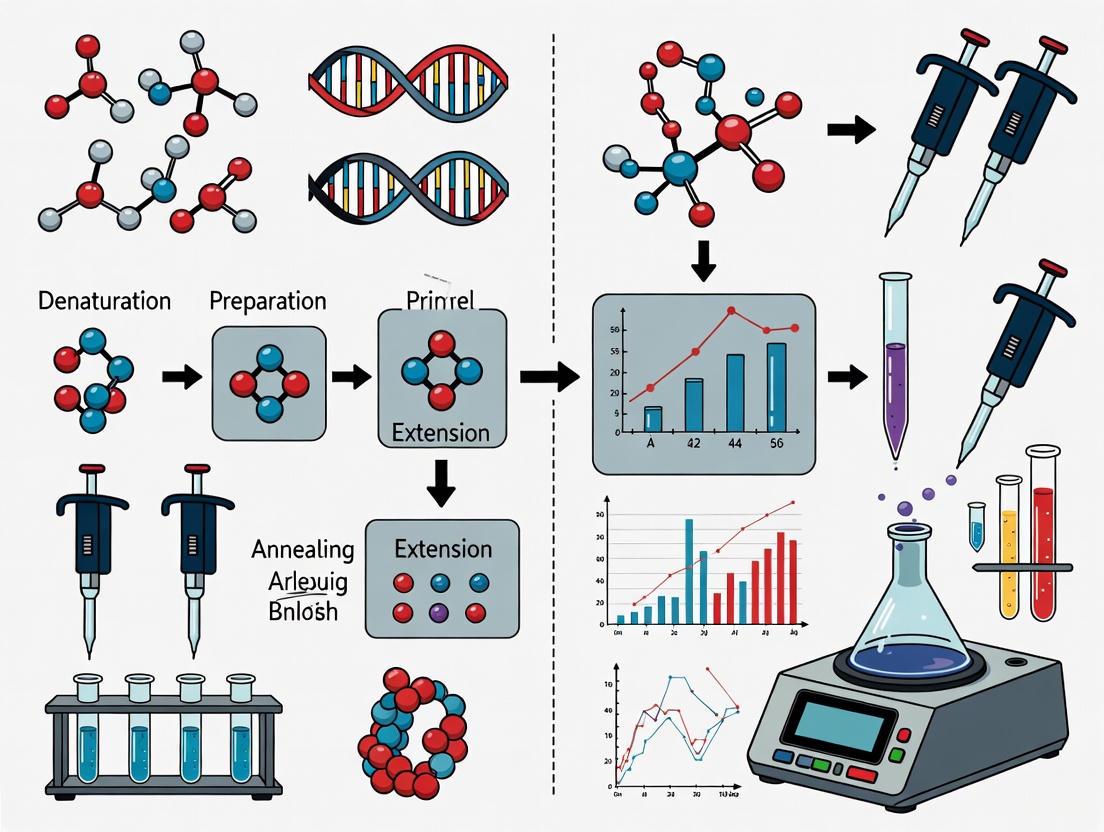
Workflow Diagram: Pre-PCR and Post-PCR Physical Separation
The following diagram illustrates the mandatory unidirectional workflow and physical separation of areas to prevent cross-contamination.
Diagram Title: Unidirectional PCR Workflow
This physical separation must be absolute. No reagents, equipment, consumables, or personal items (including lab notebooks and pens) used in the post-PCR area should ever be brought back into the pre-PCR area [1].
Application Notes: Protocols for Contamination Prevention
Protocol: Establishing and Maintaining Physically Separate Workstations
Objective: To create and maintain dedicated pre-PCR and post-PCR areas that prevent amplicon carryover.
Materials:
- Two separate benchtops or rooms, clearly labeled.
- Dedicated sets of pipettes, pipette tips with aerosol filters, lab coats, and waste containers for each area [1] [2].
- Surface decontaminants (e.g., 5% bleach solution, commercial nucleic acid degrading solutions) [2].
- UV crosslinker or cabinet for UV sterilization (optional but recommended).
Methodology:
- Designate Areas: Assign one benchtop as the pre-PCR area (for PCR reaction setup only) and a separate benchtop as the post-PCR area (for gel electrophoresis, PCR product purification, and analysis) [1].
- Equip Areas: Keep all equipment, including pipettes, centrifuges, and racks, strictly separate. The thermal cycler (PCR machine) and electrophoresis apparatus must be housed in the post-PCR area [1].
- Implement Workflow: Always begin experiments in the pre-PCR area by assembling master mixes and loading templates. Once the reaction tube is sealed, it can be transported to the post-PCR area for amplification and subsequent analysis, following the unidirectional flow shown in the diagram.
- Decontaminate Regularly: Before starting work, wipe down the pre-PCR benchtop and pipettes with a surface decontaminant. A 5% bleach solution can be used to degrade DNA, followed by ethanol wiping to prevent corrosion [2]. UV sterilization can be used for tubes, racks, and other non-porous surfaces.
Protocol: Reagent and Sample Handling to Minimize Contamination
Objective: To prepare and handle reagents and samples in a way that minimizes the introduction and spread of contaminants.
Materials:
- DNase-free, ultrapure water.
- Reagents certified for low-level residual DNA.
- Positive displacement tips or filter aerosol-barrier tips.
- Microcentrifuge tubes and PCR plates.
- Uracil-DNA Glycosylase (UDG) and dUTP.
Methodology:
- Aliquot Reagents: Upon receipt, aliquot all reagents (e.g., primers, dNTPs, polymerase, water) into single-use volumes. Store these aliquots separately from other DNA samples and use them solely in the pre-PCR area [1] [2].
- Use Aerosol-Barrier Tips: Always use filter tips or positive displacement pipettes during reaction setup to prevent aerosol contamination from the pipette shaft [1] [2].
- Employ UDG Decontamination: A powerful enzymatic method to prevent carryover contamination involves substituting dTTP with dUTP in the PCR master mix. This generates uracil-containing amplicons. In subsequent PCR setups, a pre-PCR incubation with Uracil-DNA Glycosylase (UDG) will cleave any contaminating uracil-containing amplicons, preventing their amplification [5].
- Include Mandatory Controls: Every PCR run must include a negative template control (NTC) where the template DNA is replaced with ultrapure water. The absence of amplification in the NTC lane on a gel confirms the reaction is free of contaminating DNA [1].
Protocol: Detecting and Estimating DNA Contamination
Objective: To screen for and quantify contamination within DNA samples, particularly prior to sequencing.
Materials:
- DNA samples.
- Array-based genotype data or low-pass sequencing data (optional).
- Software tools (e.g., VerifyBamID).
Methodology:
- Using Array-Based Genotype Data: Contamination can be detected from genotyping array intensity data alone, allowing for pre-sequencing screening. Methods analyze the B-allele frequency (BAF), expecting values clustered near 0, 0.5, or 1 for AA, AB, and BB genotypes, respectively. Contaminated samples show BAF values that deviate from these expected clusters, and the level of contamination can be estimated from these deviations [3].
- Using Sequencing Data: For samples that have been sequenced, likelihood-based methods can jointly analyze the sequencing reads and known genotypes to detect and estimate contamination levels as low as 1%. These methods evaluate whether the observed distribution of sequencing reads at heterozygous sites is consistent with a single source sample or a mixture [3].
- Fragment Size Analysis (for cfDNA): In cell-free DNA (cfDNA) testing, contamination with high molecular weight genomic DNA can be identified by fragment size analysis. Pure cfDNA has a characteristic peak at 160-180 bp. Deviations from this profile indicate contamination [4].
The Scientist's Toolkit: Essential Reagent Solutions
The following table details key reagents and materials essential for implementing a robust contamination control strategy.
Table 1: Key Research Reagent Solutions for PCR Contamination Control
| Item | Function & Importance | Application Notes |
|---|---|---|
| Aerosol-Barrier Pipette Tips | Create a physical barrier preventing aerosols from contaminating the pipette shaft and subsequent reactions. | Critical for all liquid handling in pre-PCR area. Use in both pre- and post-PCR, but with dedicated sets [1] [2]. |
| UDG Enzyme & dUTP | Enzymatic prevention of amplicon carryover. UDG cleaves uracil bases in contaminating DNA from previous dUTP-containing reactions. | Add UDG to master mix; pre-incubate reactions before thermal cycling. Note that some proofreading enzymes cannot incorporate dUTP [5]. |
| DNase I, RNase-free | Degrades contaminating genomic DNA in RNA samples prior to reverse transcription-PCR (RT-PCR). | Essential for RT-PCR to prevent false positives from genomic DNA. Requires heat inactivation post-treatment [2]. |
| Nuclease-Free Water | A reagent certified to be free of nucleases and contaminating DNA/RNA. | Used for making master mixes, dilutions, and negative controls. Aliquot for single use [2]. |
| Bleach (5% Sodium Hypochlorite) | Effective chemical decontaminant that degrades any nucleic acids on surfaces. | Use to routinely decontaminate pre-PCR benchtops and equipment. Allow several minutes of contact time [2]. |
The table below summarizes key quantitative findings and recommendations related to DNA contamination.
Table 2: Quantitative Data on DNA Contamination and Control
| Parameter | Value or Observation | Context & Significance |
|---|---|---|
| Lowest Detectable Contamination | 1% | Contamination levels as low as 1% can be reliably detected using sequence and array-based genotype data [3]. |
| Impact on Heterozygosity | Increased HET/HOM ratio | Contaminated samples show unusually large numbers of heterozygous genotypes and an elevated heterozygous-to-homozygous genotype ratio [3]. |
| Recommended dNTP Concentration | 0.2 mM (each dNTP) | Higher concentrations can inhibit PCR; lower concentrations (~0.01-0.05 mM) can improve fidelity with non-proofreading enzymes [5]. |
| cfDNA Fragment Size | 160-180 bp | Deviation from this peak size indicates contamination with high molecular weight genomic DNA in cfDNA assays [4]. |
| Template Input (50 µL reaction) | Plasmid DNA: 0.1-1 ng; gDNA: 5-50 ng | Higher DNA inputs increase the risk of nonspecific amplification. Optimal amounts vary by polymerase and template [5]. |
The threat of amplified DNA contamination to assay integrity is profound, yet manageable. A comprehensive strategy centered on the strict physical separation of pre- and post-PCR workflows, combined with meticulous laboratory practices and the strategic use of enzymatic and chemical decontamination, forms the foundation of reliable PCR-based science. By adopting the detailed protocols and principles outlined in this document, researchers and drug development professionals can safeguard their experiments from false positives, ensure the generation of high-quality data, and uphold the highest standards of scientific rigor.
The extreme sensitivity of the Polymerase Chain Reaction (PCR), which can amplify minuscule amounts of DNA into billions of copies, is also its greatest vulnerability [6] [7]. This characteristic makes the technique prone to contamination, which can lead to false-positive results and compromised data integrity. A cornerstone strategy to mitigate this risk is the physical separation of the PCR process into distinct, dedicated zones: Pre-PCR, Amplification, and Post-PCR [8] [9]. This separation is not merely a recommendation but a fundamental requirement for any laboratory dedicated to reliable molecular diagnostics and research [9]. Within the context of a broader thesis on physical separation, this application note delineates the core principles of these zones, provides detailed protocols for their establishment, and synthesizes key data to guide researchers, scientists, and drug development professionals in designing and operating a contamination-resistant PCR laboratory.
Defining the Three Core PCR Zones
The PCR workflow is linearly segregated into three specialized areas to prevent amplicons (amplified DNA products) from contaminating reactions in their preliminary stages. The overarching rule is a unidirectional workflow from "clean" to "dirty" areas, with no retrograde movement of equipment, reagents, or personnel without rigorous decontamination [8] [9].
Table 1: Core Functions and Contamination Control Measures for PCR Zones
| Zone | Primary Function | Key Contamination Risks | Essential Control Measures |
|---|---|---|---|
| Pre-PCR (Clean Area) | Nucleic acid extraction; reaction mix preparation [9] [10]. | Contamination of samples or master mixes with amplicons or foreign DNA [7]. | Positive air pressure [8] [9]; dedicated equipment and PPE; use of laminar flow cabinets; aliquoting reagents [8]. |
| Amplification | Thermal cycling of assembled PCR reactions [9]. | Tube leakage or aerosol generation during handling post-amplification. | Physical separation; placement in a contained area or room [9]. |
| Post-PCR (Dirty Area) | Analysis of PCR products (e.g., gel electrophoresis, sequencing) [7] [9]. | Amplicon aerosols contaminating the clean areas. | Negative air pressure [8] [9]; dedicated equipment and PPE; closed-tube systems (e.g., real-time PCR) [9] [10]. |
The logical and physical relationships between these zones are outlined in the workflow below.
Pre-PCR Zone: The "Clean Area"
The Pre-PCR zone is dedicated to all activities prior to thermal cycling. This area must be meticulously maintained to be free of PCR amplicons [7]. Key activities include the preparation of the master mix, which contains all reaction components except the nucleic acid template, and the extraction of DNA/RNA from samples [9] [10]. To preserve the integrity of this zone, it should be kept at a slight positive air pressure to prevent the influx of contaminated air from adjacent areas [8] [9]. All work, particularly the assembly of the master mix, should be performed within a laminar flow cabinet or PCR workstation, preferably equipped with UV light to decontaminate surfaces and equipment by cross-linking any stray DNA [8] [9]. All equipment—pipettes, centrifuges, coolers—must be dedicated to this room and never travel to the post-PCR areas [7] [8].
Amplification Zone
This area is designated for the thermal cyclers that carry out the DNA amplification process [9]. While the reaction tubes are closed during cycling, this area is still considered "dirty" because the tubes contain high concentrations of amplicons and are often opened here for subsequent analysis if using endpoint PCR. For this reason, it is ideally a separate room or a defined area within the post-PCR room [9]. Some guidelines consider it part of the post-PCR zone due to the high amplicon concentration present [8].
Post-PCR Zone: The "Dirty Area"
The Post-PCR zone is dedicated to the analysis of the amplification products. Activities in this area, such as gel electrophoresis, fragment analysis, or sequencing, involve handling open tubes containing vast quantities of amplicons, creating a significant contamination risk [7] [9]. Consequently, this area must be physically isolated and maintained at a slight negative air pressure to contain amplicon aerosols and prevent their escape [8] [9]. All equipment, including pipettes, gel documentation systems, and centrifuges, must be dedicated to this zone. Personnel must change gloves and lab coats before leaving to prevent carrying amplicons into other areas [7] [8].
Laboratory Design and Workflow Protocols
Physical Layout Configurations
The ideal laboratory design is based on the availability of space and the required throughput. The following protocol outlines the recommended configurations.
Protocol 1: Laboratory Spatial Design
- Ideal Configuration (Multiple Rooms): Allocate at least three separate rooms for (a) reagent preparation and master mix assembly, (b) sample and nucleic acid preparation, and (c) amplification and post-PCR analysis [9] [10]. This provides the highest level of contamination control.
- Acceptable Configuration (Two Rooms): Establish one room for all pre-PCR activities (combining reagent and sample preparation) and a second room for all post-PCR activities (amplification and analysis) [8].
- Minimum Configuration (Single Room): If space is severely limited, designate separate, distantly spaced benches for pre- and post-PCR work within a single room [8]. Implement strict temporal separation by performing all pre-PCR work first, followed by a thorough decontamination, and then post-PCR work [8] [9].
Establishing a Unidirectional Workflow
The most critical operational protocol is enforcing a unidirectional workflow. The following diagram and steps detail this process.
Protocol 2: Implementing and Maintaining a Unidirectional Workflow
- Personnel Movement: Always move from the Pre-PCR zone to the Post-PCR zone. Never return to a clean area after entering a dirty area without a complete change of personal protective equipment (PPE) and washing of hands [9].
- Material Movement: Dedicate all equipment, consumables, and reagents to their specific zone. Never move items from the Post-PCR zone back into the Pre-PCR zone [7] [8]. For example, a pipette used for loading gels must never be used for setting up reactions.
- Reagent Management: Aliquot all bulk reagents upon receipt into single-use volumes. This practice prevents the contamination of an entire stock and reduces the number of freeze-thaw cycles, preserving reagent integrity [8].
- Decontamination: Regularly clean all work surfaces, equipment, and common touchpoints (e.g., doorknobs, freezer handles) with a freshly prepared 10% bleach solution, followed by distilled water or ethanol to prevent corrosion, as sodium hypochlorite can degrade contaminating DNA [8].
The Scientist's Toolkit: Essential Reagents and Materials
The following table catalogs the essential materials required for setting up a robust PCR laboratory, with an emphasis on contamination control.
Table 2: Essential Research Reagent Solutions and Laboratory Equipment
| Category | Item | Function and Specification |
|---|---|---|
| Consumables | Aerosol-Barrier (Filter) Pipette Tips | Prevents aerosol contamination of pipette shafts and cross-contamination between samples [8]. |
| Single-Use, Sterile, DNase-/RNase-Free Tubes & Plates | Ensures reaction vessels are free of nucleases and contaminating nucleic acids. | |
| Reagents | Hot-Start DNA Polymerase | Reduces non-specific amplification and primer-dimer formation by remaining inactive until the high-temperature initial denaturation step [6] [11]. |
| Molecular Grade Water | A pure, nuclease-free water source that is critical for reaction consistency and success. | |
| dNTPs, Reaction Buffers, MgCl₂ | The fundamental building blocks and co-factors for DNA synthesis [12] [13]. | |
| Equipment | Dedicated Pipette Sets (Pre- & Post-PCR) | Mandatory for physical separation; prevents amplicon carryover [7] [8]. |
| Laminar Flow Cabinet / PCR Workstation | Provides a sterile, UV-irradiable work environment for master mix preparation in the Pre-PCR zone [8]. | |
| Thermal Cyclers | Instruments that automate the temperature cycling required for DNA amplification. | |
| Analytical Instruments (e.g., Gel Electrophoresis, Real-Time PCR systems) | For the analysis and detection of PCR products; must be housed in the Post-PCR zone [7]. |
Advanced Considerations: Optimization and Troubleshooting
Contamination Monitoring and Control
Despite best practices, contamination can occur. A robust quality control system is essential.
Protocol 3: Contamination Monitoring with Controls
- Negative Controls: Include a negative control (e.g., water instead of template DNA) in every PCR run. Amplification in this control indicates contamination of the master mix or reagents [8].
- Positive Controls: Use a known, weak-positive template to verify that the reaction efficiency is sufficient to detect low-copy targets [8].
- UV Decontamination: Use UV light in laminar flow cabinets and workstations to cross-link and destroy contaminating DNA on surfaces before and after use. Note that dry-state DNA is more resistant to UV, and UV can damage dNTPs and enzymes, so it should not be used on assembled reaction mixes containing these components [9].
Methodological Optimization for Specific Applications
Certain PCR applications require specific modifications to the core protocol. The selection of a DNA polymerase is a key decision point, as different enzymes offer varying benefits for specialized applications.
Table 3: DNA Polymerase Selection Guide for Specialized PCR Applications
| Application | Recommended Polymerase Type | Rationale and Key Benefit |
|---|---|---|
| Standard & Fast PCR | Standard or highly processive Hot-Start Taq | Highly processive enzymes enable shorter extension times, drastically reducing total PCR run time [11]. |
| Long-Range PCR | Blend of non-proofreading (e.g., Taq) and proofreading (e.g., Pfu) polymerases | The proofreading enzyme corrects misincorporated nucleotides, allowing the polymerase to synthesize longer DNA fragments without stalling [6]. |
| High-Fidelity PCR | Proofreading polymerases (e.g., Pfu) | Enzymes with 3'→5' exonuclease activity have lower error rates, essential for cloning and sequencing [6]. |
| GC-Rich PCR | Highly processive or hyperthermostable polymerases | These enzymes can better navigate through templates with strong secondary structures and allow for higher denaturation temperatures to melt GC-rich regions [11]. |
| Direct PCR | Highly processive, inhibitor-tolerant polymerases | These enzymes can amplify DNA directly from crude samples (e.g., blood, cells) without a separate DNA purification step, as they are less inhibited by sample debris [11]. |
The exquisite sensitivity of the Polymerase Chain Reaction (PCR), which allows for the amplification of millions of DNA copies from a few initial templates, is also its greatest vulnerability [14] [15]. This application note, framed within a broader thesis on the physical separation of PCR work areas, delineates the profound consequences of laboratory contamination on diagnostic accuracy. Contamination, primarily through aerosolized amplicons, directly compromises the fundamental parameters of assay performance: sensitivity and specificity [16]. When amplified DNA products from previous reactions infiltrate new preparations, they act as rogue templates, leading to false-positive results that erode test specificity [17]. Conversely, contaminants like enzyme inhibitors or unintended nucleases can co-purify with samples, preventing successful amplification of the true target and resulting in false negatives that degrade test sensitivity [14]. The integrity of PCR-based diagnostics, therefore, hinges on stringent laboratory practices, chief among them the physical segregation of pre- and post-amplification processes. Failure to implement this separation reliably corrupts experimental data, leading to misplaced clinical decisions, inappropriate patient management, and ultimately, a loss of confidence in molecular diagnostic results [14] [18].
The Critical Definitions: Sensitivity and Specificity
In diagnostic testing, sensitivity is the test's ability to correctly identify individuals who have the disease (true positive rate), while specificity is its ability to correctly identify those without the disease (true negative rate) [19] [20].
- Sensitivity is calculated as: True Positives / (True Positives + False Negatives) [19] [21]. A highly sensitive test minimizes the chance of missing a true condition (low false negatives).
- Specificity is calculated as: True Negatives / (True Negatives + False Positives) [19] [21]. A highly specific test minimizes the chance of falsely identifying a condition (low false positives).
The relationship between these two metrics is often a trade-off; however, proper laboratory design and practice aim to maximize both simultaneously by reducing external artifacts such as contamination [19] [20].
Table 1: Outcome Matrix of a Diagnostic Test
| Condition Present | Condition Absent | |
|---|---|---|
| Test Positive | True Positive (TP) | False Positive (FP) |
| Test Negative | False Negative (FN) | True Negative (TN) |
Consequences of Laboratory Failure on Diagnostic Accuracy
Impact on Specificity and the False Positive Problem
The most direct consequence of PCR contamination is a reduction in specificity, leading to a high rate of false positives. Amplicons from previous amplification reactions are the predominant source of contamination [17] [16]. These are short, amplified DNA sequences that are easily aerosolized when tubes are opened and are present in enormous quantities, making them ideal templates for subsequent amplification [15]. When these contaminants are introduced into a new reaction mixture, they are efficiently amplified by the DNA polymerase, generating a positive signal in the absence of the true target template [17]. This leads to a false positive, which directly increases the number of false positives in the test outcome matrix, thereby reducing specificity [19]. In a clinical context, this could mean healthy individuals are misdiagnosed as infected, leading to unnecessary anxiety, further invasive testing, and inappropriate treatment [14] [21].
Impact on Sensitivity and the False Negative Problem
While less intuitively obvious, contamination can also suppress true positive signals, thereby reducing sensitivity and causing false negatives. This can occur through several mechanisms:
- Competition for Reagents: Contaminating amplicons compete with the genuine target for essential reaction components such as primers, nucleotides (dNTPs), and DNA polymerase. In samples with a low copy number of the true target, this competition can be decisive, leading to amplification failure or a significant reduction in amplification efficiency [22].
- Co-purified Inhibitors: Contamination during sample collection or nucleic acid extraction can introduce substances that inhibit DNA polymerases. These inhibitors can co-purify with the nucleic acids and prevent amplification altogether, resulting in a false negative [14].
A test with compromised sensitivity fails in its primary duty to "rule out" disease, with serious implications for patient safety and public health [14] [19].
Quantitative Impact on Assay Performance
The following table summarizes the potential impacts of laboratory failures on key PCR assay performance metrics.
Table 2: Impact of Laboratory Failures on PCR Assay Performance
| Laboratory Failure | Primary Consequence | Impact on Sensitivity | Impact on Specificity | Overall Diagnostic Accuracy |
|---|---|---|---|---|
| Amplicon Contamination | False Positive Results | No change or potential increase | Severe Reduction | Severely Compromised |
| Sample Cross-Contamination | False Positive/Negative Results | Potential Reduction | Reduction | Compromised |
| Introduction of Inhibitors | Amplification Failure | Severe Reduction | No direct change | Severely Compromised |
| Reagent Degradation | Unreliable Amplification | Reduction | Potential Reduction | Compromised |
A study validating a real-time PCR assay for bovine mastitis pathogens demonstrated what is achievable with stringent protocols, reporting 100% analytical specificity and sensitivity across a large set of bacterial isolates [18]. This serves as a benchmark and underscores that high accuracy is attainable with meticulous practice.
Essential Protocols for Physical Separation and Contamination Control
The following protocol provides a detailed methodology for establishing a physically separated PCR laboratory workflow to safeguard diagnostic sensitivity and specificity.
Protocol: Establishing a Physically Separated PCR Laboratory
Principle: To prevent the introduction of amplified DNA products (amplicons) into pre-amplification reagents and samples by enforcing a unidirectional workflow through physical separation and dedicated equipment [17] [15] [16].
Materials and Reagents:
- Dedicated rooms or designated bench spaces for pre-PCR and post-PCR processes.
- Dedicated pipettes (preferably positive-displacement or with aerosol-resistant filter tips) [15] [16].
- Dedicated lab coats, gloves, and consumables (tubes, racks, centrifuges, vortexers) for each area [17] [16].
- Reagents for surface decontamination: 10% fresh bleach solution and 70% ethanol [17] [15].
- No-Template Controls (NTCs): PCR-grade water [17] [22].
- Optional: Uracil-N-glycosylase (UNG) system for carryover prevention [17].
Procedure:
- Laboratory Zoning:
- Establish Two Distinct Areas: A pre-PCR area and a post-PCR area. These should be physically separated, ideally in different rooms with independent ventilation [15] [16].
- Pre-PCR Area: Dedicated exclusively to reagent preparation, sample preparation (including nucleic acid extraction), and assembly of PCR reaction mixes [15].
- Post-PCR Area: Dedicated exclusively to the thermocycling process and all downstream analysis of amplified products (e.g., gel electrophoresis) [15] [16].
Unidirectional Workflow:
Dedicated Equipment and Consumables:
Rigorous Personal Practice:
- Always wear clean gloves in both areas, changing them frequently, especially if contamination is suspected [15] [16].
- Use aerosol-resistant filter tips for all pipetting steps to minimize the creation of aerosols [17] [16].
- Add the DNA template last to the PCR reaction mix to minimize the opportunity for aerosol contamination of other reagents [16].
- Aliquot all PCR reagents upon receipt to avoid repeated freeze-thaw cycles and prevent widespread contamination of stock solutions [16].
Environmental Decontamination:
Processual Decontamination (UNG System):
- Incorporate the UNG system into the PCR protocol. Use a dNTP mix containing dUTP instead of dTTP in all PCR reactions [17].
- In subsequent reactions, the UNG enzyme will enzymatically degrade any uracil-containing carryover amplicons present in the master mix before the PCR cycling begins, preventing their amplification [17].
Quality Control with NTCs:
The logical relationships and workflow described in this protocol are visualized below.
The Scientist's Toolkit: Essential Reagent Solutions
The following table details key reagents and materials critical for maintaining the integrity of PCR experiments and achieving high sensitivity and specificity.
Table 3: Essential Research Reagent Solutions for PCR
| Item | Function & Importance | Key Considerations |
|---|---|---|
| Aerosol-Resistant Filter Tips | Prevents aerosolized contaminants from entering pipette shafts and cross-contaminating samples and reagents [16]. | Essential for all pipetting steps, especially in the pre-PCR area. |
| No-Template Control (NTC) | A critical quality control to detect contamination in reagents or the environment. Contains all PCR components except the DNA template [17]. | Amplification in the NTC indicates significant contamination, invalidating the run. |
| Uracil-N-Glycosylase (UNG) | An enzymatic system to prevent carryover contamination from previous PCR amplifications. Degrades uracil-containing DNA [17]. | Requires the use of dUTP instead of dTTP in all PCR mixes. Inactivated at high PCR temperatures. |
| 10% Bleach Solution | A potent decontaminant for destroying DNA on work surfaces and equipment. Sodium hypochlorite oxidizes nucleic acids [17] [15]. | Must be made fresh frequently. Contact time of 10-15 minutes is recommended before wiping. |
| High-Fidelity DNA Polymerase | For applications requiring high accuracy, such as cloning. Offers superior proofreading activity to reduce replication errors [22]. | Lower error rate compared to standard Taq polymerase. |
| Pre-mixed Master Mixes | Optimized, ready-to-use solutions containing buffer, dNTPs, and polymerase. Increases reproducibility and reduces setup time and contamination risk [22]. | Available from numerous commercial suppliers. Often includes UNG. |
| Sterile, Nuclease-Free Water | The solvent for PCR reactions. Must be free of nucleases and contaminants to prevent degradation of templates and primers or inhibition of the reaction [16]. | A dedicated bottle for PCR use only in the pre-PCR area is mandatory. |
The consequences of failure in maintaining a physically separated PCR workflow are severe and quantifiable, leading directly to a degradation of diagnostic sensitivity and specificity. The implementation of rigorous protocols, as outlined in this application note, is not optional but fundamental to generating reliable, accurate, and clinically actionable data. By adhering to the principles of physical segregation, unidirectional workflow, and stringent contamination control, researchers and diagnosticians can safeguard the integrity of their molecular assays, ensuring that the powerful tool of PCR fulfills its promise as a cornerstone of modern infectious disease diagnosis and biomedical research.
The design and operation of a modern laboratory, particularly one performing sensitive molecular techniques like Polymerase Chain Reaction (PCR), are governed by a framework of international and national regulatory standards. These standards are not arbitrary; they are established to ensure the accuracy, reliability, and safety of test results, which are critical for patient diagnosis, treatment, and research integrity. For laboratories handling human specimens, adherence to these standards is often a legal requirement, while for research facilities, it represents a commitment to scientific rigor and data credibility. The core of these regulations emphasizes a robust Quality Management System (QMS), detailed documentation, and rigorous personnel competency assessments. This article explores the interconnected roles of three pivotal sets of regulations—ISO 15189, the Clinical Laboratory Improvement Amendments (CLIA), and FDA 21 CFR Part 11—and their specific implications for the physical and operational design of laboratories, with a special focus on PCR workflows.
Table: Overview of Key Laboratory Regulatory Standards
| Standard | Primary Focus | Geographic Application | Key Emphasis for Lab Design |
|---|---|---|---|
| ISO 15189 [23] [24] | Quality and competence for medical laboratories | International; widely adopted in Europe and other regions [23] | Risk management, process control (pre- to post-examination), and patient-centered outcomes [24] |
| CLIA [25] [26] | Quality assurance for clinical human diagnostic testing | United States; mandatory for all U.S. clinical labs [23] [26] | Personnel qualifications, proficiency testing, and quality control across all testing phases [26] [27] |
| FDA 21 CFR Part 11 [28] [29] | Trustworthiness of electronic records and signatures | United States (FDA-regulated industries) | Validation, security, and audit trails for computerized systems [28] |
ISO 15189: International Standard for Medical Laboratory Quality
ISO 15189 is an international standard specifically designed for medical laboratories, outlining requirements for quality and competence [23]. Its core objective is to ensure that laboratories deliver accurate, timely, and reliable results, thereby enhancing patient care and fostering confidence in diagnostic services [23]. A significant update to the standard was published in 2022, and laboratories with existing accreditation are required to transition to this new version by the end of 2025 [24].
Key Requirements and Structural Organization
The structure of ISO 15189:2022 is organized into clauses that define specific requirements for medical laboratories [23]:
- Clause 4: General Requirements: Mandates impartiality, confidentiality of patient information, and the integration of patient-centered care into all services [23].
- Clause 5: Structural and Governance Requirements: Establishes the need for a defined legal entity, a designated laboratory director, and clear organizational responsibilities [23].
- Clause 6: Resource Requirements: Covers personnel competence, equipment calibration and maintenance, and facilities with controlled environmental conditions to ensure testing integrity and personnel safety [23].
- Clause 7: Process Requirements: The operational heart of the standard, focusing on robust processes across the entire diagnostic cycle—pre-examination, examination, and post-examination phases [23].
- Clause 8: Management System Requirements: Describes the establishment and maintenance of a documented Quality Management System, including risk management, corrective actions, and internal audits [23].
Implications for Laboratory Design and Workflow
The requirements of ISO 15189 have a direct and profound impact on how a laboratory is physically designed and how workflows are managed. The standard's emphasis on process control and risk management necessitates a layout that prevents errors and contamination.
- Facilities and Environmental Conditions (Clause 6): The laboratory must design its workspace to safeguard patient safety and ensure result reliability. This includes controlling access to different sections, ensuring proper ventilation, and implementing measures for contamination control [23]. For PCR labs, this directly translates to the need for physical or temporal separation of activities [8] [10].
- Process Requirements (Clause 7): The standard requires documented procedures for sample handling, from collection through transportation and storage [23]. This demands a logical, unidirectional workflow to ensure sample integrity and traceability, a concept that is critical in PCR setup to prevent amplicon contamination [8].
A major update in the 2022 version is the intensified focus on risk management [24]. Laboratories are now required to carry out risk management for all activities that could pose a risk to patients. This means that during the design phase, a laboratory must proactively identify potential failure points—such as the risk of cross-contamination in an open-plan lab—and design controls to mitigate those risks.
Figure 1: ISO 15189:2022 implementation workflow based on a hospital lab's transition plan [24].
CLIA: U.S. Regulatory Benchmarks for Laboratory Testing
The Clinical Laboratory Improvement Amendments (CLIA) of 1988 are the federal regulatory standards for all clinical laboratory testing performed on humans in the United States [26]. The core purpose of CLIA is to ensure the accuracy, reliability, and timeliness of patient test results, regardless of where a test is performed [26]. In 2025, the Centers for Medicare & Medicaid Services (CMS) enacted the first major set of updates to CLIA in decades, refining requirements for personnel, proficiency testing, and communications [25] [27].
Core Components of CLIA Compliance
CLIA regulations establish a comprehensive framework for laboratory quality assurance, covering the entire testing process [26].
- Quality Assurance (QA) Plan: Laboratories must establish a written, ongoing QA plan that analyzes every aspect of operation. This plan must include [26]:
- Standard operating procedures (SOPs) for each step of testing.
- Defined administrative responsibilities and recordkeeping.
- Specified corrective actions when problems are identified.
- Procedures to ensure staff competency and high-quality test performance.
- Personnel Competency: CLIA requires laboratories to formally assess staff competency. This assessment must be performed semiannually during the first year of employment and annually thereafter, and must include six components: direct observation of testing and instrument maintenance, monitoring of test reporting, record review, assessment of test performance, and assessment of problem-solving skills [26].
- Method Verification: Before reporting patient results, a laboratory must verify that its test methods consistently provide accurate results. This verification must establish accuracy, precision, reportable range, and reference ranges. This is commonly achieved using proficiency testing samples, previously tested patient specimens, or commercial reference materials [26].
- Procedure Manual: A CLIA-mandated procedure manual must be readily available and followed by all personnel. It must be comprehensive, including detailed procedures for specimen collection/rejection, step-by-step testing instructions, calibration procedures, quality control, remedial actions, and reference ranges [26].
2025 CLIA Updates and Laboratory Design Implications
The 2025 CLIA updates bring several key changes that influence laboratory operations and, by extension, design [25] [27]:
- Digital-Only Communication: CMS is phasing out paper mailings, requiring labs to maintain accurate electronic contact information to avoid missing critical notices [25].
- Updated Personnel Qualifications: The rules have been tightened for lab directors and staff. "Physical science" has been removed as an acceptable degree, and "board eligibility only" is no longer sufficient for certain roles. Laboratories must review job descriptions and personnel files to ensure compliance [27]. Existing staff may be "grandfathered in" provided their employment is continuous [25].
- Announced Inspections: Accrediting bodies can now announce inspections up to 14 days in advance, making it essential for labs to maintain a state of continuous audit-readiness [25].
These updates underscore the need for a laboratory design that supports rigorous documentation, streamlined workflows, and stable operating conditions. For instance, a well-designed PCR lab with a unidirectional workflow directly supports the CLIA requirement for a QA plan that reduces errors and ensures the accuracy of the analytical phase [26].
Table: Summary of Key 2025 CLIA Personnel Qualification Updates
| Role | Key Changes in Education/Training | Key Changes in Duties/Responsibilities |
|---|---|---|
| Laboratory Director (High Complexity) | - Equivalent qualifications pathway removed [27].- MD/DO must now have 20 CE hours in lab practice + 2 years experience [27].- New equivalency options for doctoral degrees [27]. | Must be onsite at least once every six months [27]. |
| Technical Supervisor (High Complexity) | - Equivalent qualifications and ASC certification pathways removed [27].- New equivalency options for bachelor's and master's degrees [27].- Updated experience requirements for subspecialties [27]. | (No major changes specified in search results) |
| Testing Personnel (Moderate Complexity) | - Expanded equivalency options for bachelor's and master's degrees, similar to director pathways [27].- Updated requirements for associate degree pathway [27]. | (No major changes specified in search results) |
FDA 21 CFR Part 11: Electronic Records and Signatures
In an increasingly digital world, the integrity of electronic data is paramount. FDA 21 CFR Part 11 establishes the U.S. criteria under which electronic records and electronic signatures are considered trustworthy, reliable, and equivalent to paper records and handwritten signatures [28]. This regulation applies to records required to be maintained by other FDA regulations (the "predicate rules") or submitted to the FDA [29].
Key Requirements for Closed Systems
Most laboratory information systems are "closed systems," meaning access is controlled by the persons responsible for the system's content [28]. For such systems, Part 11 mandates a set of strict controls [28]:
- System Validation: Systems must be validated to ensure accuracy, reliability, consistent intended performance, and the ability to discern invalid or altered records.
- Audit Trails: The use of secure, computer-generated, time-stamped audit trails is required to independently record the date and time of operator entries and actions that create, modify, or delete electronic records. These audit trails must not obscure previously recorded information.
- Access Control: System access must be limited to authorized individuals through authority checks. Furthermore, written policies must hold individuals accountable for actions initiated under their electronic signatures.
- Operational Checks: The system must use operational checks to enforce permitted sequencing of steps and events, and device checks to validate the source of data input.
The FDA has stated it employs a "narrow interpretation" of the scope of Part 11, applying it primarily when records are explicitly required by a predicate rule and are maintained electronically [29]. However, for systems that fall under its scope, the requirements for data integrity are rigorous.
Figure 2: Electronic record and signature lifecycle under FDA 21 CFR Part 11 for a closed system [28].
Applied Protocol: Designing a Compliant PCR Laboratory
The theoretical requirements of ISO, CLIA, and FDA converge in the practical design of a specialized workspace like a PCR laboratory. The extreme sensitivity of PCR makes it highly susceptible to contamination from amplicons (PCR products), which can lead to false-positive results. Therefore, the primary design goal is to implement a unidirectional workflow that physically separates the pre-amplification and post-amplification processes [8] [10].
Spatial Separation and Workflow Design
The ideal PCR lab is divided into separate, dedicated rooms to compartmentalize different stages of the process. The following protocol outlines the standard for a three-area design [10]:
Area 1: Pre-PCR - Sample Preparation and Nucleic Acid Extraction
- Function: This area is dedicated to receiving, aliquoting, and processing samples, and extracting nucleic acids (DNA/RNA).
- Design Specifications: This room should contain a biosafety cabinet for working with potentially infectious agents, a refrigerator, freezer, and a dry heat block or water bath [10]. To prevent the escape of any infectious agents, this room should be kept under negative pressure [10]. All equipment and consumables used here must remain in this room.
Area 2: Pre-PCR - Master Mix Preparation
- Function: This is a clean area where all reaction components—except for the template nucleic acid—are mixed to create the PCR master mix [10].
- Design Specifications: This area must be equipped with a dedicated PCR cabinet or laminar flow hood, which should be decontaminated with UV light and bleach before and after use [8] [10]. To prevent the ingress of contaminating aerosols, this room should be kept under positive pressure [10]. Reagents should be aliquoted into small volumes here to avoid repeated freeze-thaw cycles and minimize contamination risk [8].
Area 3: Post-PCR - Amplification and Product Analysis
- Function: This area is dedicated to the thermal cycling process (amplification) and the subsequent detection/analysis of the PCR products (amplicons).
- Design Specifications: This room houses the thermal cyclers and real-time PCR instruments [10]. All equipment, including pipettes with aerosol-barrier filter tips, must be dedicated to this room and never taken back to pre-PCR areas [8] [10]. To ensure amplicon aerosols do not escape, this room should be kept under negative pressure [10].
Critical Note on Unidirectional Workflow: Personnel and materials must move in a "forward flow" from clean (pre-PCR) to dirty (post-PCR) areas only [10]. Moving from the post-PCR area to a pre-PCR area requires a complete change of personal protective equipment and decontamination of any items to prevent amplicon back-contamination [8].
Experimental Protocol: Establishing a Unidirectional PCR Workflow
This protocol details the steps for processing samples while adhering to the spatial separations described above.
Objective: To amplify and detect a specific nucleic acid target from patient samples while minimizing the risk of cross-contamination. Principle: By physically separating the stages of PCR setup, amplification, and analysis, and by employing a unidirectional workflow, the risk of contaminating reactions with amplicons from previous runs is drastically reduced.
Procedure:
- Pre-PCR: Sample Preparation Area
- Don a dedicated lab coat and gloves upon entering the area.
- Inside the biosafety cabinet, extract nucleic acids from patient samples according to the validated SOP.
- Prepare positive and negative control samples.
- Place the extracted nucleic acids in a designated tray for transfer to the next area. Do not remove any materials from this area other than the prepared samples in the transfer tray.
Pre-PCR: Master Mix Preparation Area
- Enter the area wearing a dedicated lab coat and gloves. Do not enter if you have been in the post-PCR area.
- Decontaminate the PCR cabinet with a freshly made bleach solution and distilled water, followed by UV irradiation for at least 15 minutes [8].
- Inside the cabinet, prepare the master mix for all reactions using filter tips to prevent pipette contamination [8].
- Aliquot the master mix into individual PCR tubes or a plate.
- Seal the plate or tubes and place them in the transfer tray with the samples. The template nucleic acid has not yet been added.
Transition Step: Addition of Template
- This critical step can be performed in the Sample Preparation Area or a dedicated clean zone [10].
- Transport the tray containing the master mix and the extracted nucleic acids to this location.
- In a controlled manner, add the template nucleic acid to their respective master mix tubes. It is recommended to add the sample last, as dispensing into a liquid reduces the risk of aerosolizing the sample [8].
- Securely cap all tubes.
Post-PCR: Amplification and Analysis Area
- Do not enter this area wearing pre-PCR lab coat or gloves. Change into dedicated post-PCR PPE.
- Transfer the sealed PCR plate/tubes to the thermal cycler in this room and start the amplification program.
- After amplification, perform product analysis (e.g., gel electrophoresis, real-time PCR analysis) using equipment dedicated to this room.
- Under no circumstances should any equipment, racks, or materials from this room be taken back to a pre-PCR area.
Required Controls:
- Always include a negative control (no template) to monitor for master mix contamination.
- Always include a positive control to confirm the amplification process is working correctly [8].
The Scientist's Toolkit: Essential Reagents and Materials for a PCR Laboratory
Table: Key Research Reagent Solutions and Equipment for a Compliant PCR Lab
| Item | Function/Application | Key Quality & Contamination Control Considerations |
|---|---|---|
| Filter Pipette Tips [8] | To accurately dispense microliter volumes of reagents and samples. | Prevents aerosols from contaminating the pipette shaft and, consequently, other samples or reagents. Essential for all pre-PCR pipetting. |
| Nucleic Acid Extraction Kits | To isolate pure DNA/RNA from complex biological samples (e.g., blood, tissue). | Must be certified free of DNase, RNase, and PCR inhibitors. The quality of the extraction directly impacts amplification efficiency and specificity. |
| PCR Master Mix | A pre-mixed solution containing Taq polymerase, dNTPs, MgCl₂, and reaction buffers. | Purchased as a concentrated solution and must be aliquoted upon arrival to prevent contamination and preserve enzyme activity through limited freeze-thaw cycles [8]. |
| Laminar Flow Hood / PCR Cabinet [8] [10] | Provides a sterile, particle-free workspace for critical pre-PCR steps like master mix preparation. | Must be decontaminated with UV light and bleach before and after use to destroy any contaminating DNA [8] [10]. |
| Thermal Cycler | An instrument that automates the temperature cycling required for DNA amplification. | Must be placed in the dedicated post-PCR area. Requires regular calibration and maintenance as part of the laboratory's equipment management program (e.g., per ISO 15189, Clause 6) [23]. |
| Real-Time PCR System | For amplification and simultaneous detection of PCR products, enabling quantification. | A "closed-tube" system that significantly reduces the risk of amplicon contamination compared to conventional PCR that requires post-amplification handling [10]. |
Implementing Effective Separation: From Basic Setups to Advanced Workflow Designs
The exquisite sensitivity of the Polymerase Chain Reaction (PCR), which allows for the amplification of a single DNA molecule, is simultaneously its greatest strength and most significant vulnerability [30]. This very sensitivity makes the technique profoundly prone to contamination from amplicons (PCR products), which can lead to false-positive results and a complete loss of data credibility [8] [31]. Consequently, the physical design of a molecular laboratory is not merely a logistical consideration but a critical experimental control. A well-designed lab layout, centered on the physical separation of pre- and post-amplification activities, is the most effective strategy for preventing contamination and ensuring the integrity of molecular diagnostics and research [30]. This application note details the core principles and practical protocols for implementing laboratory layout strategies that safeguard the reliability of PCR-based workflows.
Core Principles of Contamination Control
The fundamental goal of laboratory design for PCR is to prevent the introduction of amplifiable DNA into reactions before thermal cycling. This is primarily achieved by controlling the movement of amplicons, which are present in extremely high concentrations after amplification [8].
Spatial Separation: The Gold Standard
The ideal laboratory design physically separates pre- and post-PCR activities into distinct rooms [9]. This spatial segregation creates defined "clean" and "dirty" zones, preventing the flow of amplicons into areas where reagents and samples are prepared.
- Pre-PCR (Clean Area): This area is dedicated to activities involving the manipulation of raw samples and reagents before any amplification has occurred. It should be further subdivided into dedicated spaces for reagent preparation and sample/nucleic acid handling [8] [31]. The pre-PCR area must be maintained free of any PCR amplicons [30].
- Post-PCR (Dirty Area): This area is reserved for the amplification process itself and any subsequent analysis of PCR products, such as gel electrophoresis [9]. This is where amplicons are present in high copy numbers, creating a significant contamination risk [30].
Unidirectional Workflow
A strict unidirectional workflow must be enforced, meaning the flow of materials and personnel must always proceed from the clean pre-PCR areas to the dirty post-PCR areas, with no backtracking [8] [9] [30]. This logical workflow, from sample to result, ensures that amplicons are not carried back into clean spaces.
Temporal Separation
When spatial separation is limited, temporal separation provides an alternative control. This involves performing all pre-PCR activities (e.g., reaction setup) in the morning and all post-PCR activities (e.g., amplification and analysis) in the afternoon, or dedicating different days to different types of work [8] [9]. This prevents aerosols from post-PCR work from contaminating freshly set-up reactions.
Implementing Laboratory Layout Configurations
The level of physical separation achievable depends on the available space and resources. The following configurations are recommended, from ideal to minimal.
Ideal Layout: Four Separate Rooms
For laboratories performing a high volume of work or using methods that require opening tubes post-amplification (e.g., nested PCR), a four-room layout is the gold standard [9] [31].
Table 1: Four-Room Laboratory Layout Specification
| Room Name | Primary Function | Key Equipment | Containment Measures |
|---|---|---|---|
| Reagent Preparation | Preparation & aliquoting of master mixes; must be free of DNA/RNA [9] [31] | Pipettes, vortex, centrifuge, fridge/freezer, laminar flow cabinet [30] | Positive air pressure; UV-equipped biosafety cabinet for setup [8] [32] |
| Sample Preparation | Nucleic acid extraction; addition of template to reactions [9] | Biosafety cabinet, centrifuge, pipettes, vortex, fridge/freezer for samples [30] | Positive air pressure; dedicated biosafety cabinet [8] |
| Amplification | Thermal cycling (PCR) [9] | Thermal cyclers | Negative air pressure; doors kept closed [8] [30] |
| Post-Amplification Analysis | Analysis of amplicons (e.g., gel electrophoresis, sequencing) [9] | Electrophoresis system, gel imager, DNA sequencer [30] | Negative air pressure; dedicated equipment that never leaves the room [9] |
Standard Layout: Two or Three Rooms
A highly effective and common compromise is a two-room layout, which strictly separates pre-PCR and post-PCR activities [8] [30].
- Room 1: Pre-PCR Lab. This clean room combines the functions of the reagent preparation and sample preparation rooms. Work should be conducted within a laminar flow hood or biosafety cabinet to maintain a sterile field [8] [32]. The air pressure should be slightly positive to prevent contaminated air from flowing in [8].
- Room 2: Post-PCR Lab. This dirty room houses the thermal cyclers and analysis equipment. The air pressure should be slightly negative to ensure that amplicon aerosols do not escape [8] [30].
Minimal Layout: Single Room with Dedicated Cabinets and Temporal Control
When only a single room is available, stringent procedural controls are essential to mitigate contamination risk.
- Physical Partitions: Designate separate, distanced benches or cabinets for pre- and post-PCR work [8] [30].
- Dedicated Biosafety Cabinets: Use a dedicated Class II biosafety cabinet (BSC) for all pre-PCR setup. The HEPA-filtered laminar airflow protects the reactions from ambient aerosols, and the cabinet can be decontaminated with UV light between uses [32].
- Strict Temporal Separation: Perform all pre-PCR work during a dedicated time block, followed by a thorough decontamination of the pre-PCR area, before beginning any post-PCR work [9].
- Dedicated Equipment: Maintain completely separate sets of pipettes, tip boxes, centrifuges, lab coats, and other consumables for the pre- and post-PCR areas within the room [31].
Diagram 1: PCR lab layout strategies from ideal to minimal.
Detailed Experimental Protocols
Protocol: Unidirectional Workflow and Area Management
This protocol ensures the physical and procedural separation of PCR activities.
- Personnel Movement: Lab personnel must move in a unidirectional manner: from the Reagent Prep room to the Sample Prep room, to the Amplification room, and finally to the Post-Analysis room. Movement from a "dirty" (post-PCR) area back to a "clean" (pre-PCR) area on the same day is prohibited [31]. If absolutely necessary, personnel must shower and change all personal clothing before re-entering clean areas [9].
- Material Movement: No equipment, reagents, consumables, or lab notebooks may be moved from a post-PCR area to a pre-PCR area [9] [31]. Each room must have its own dedicated set of equipment, including pipettes, centrifuges, vortex mixers, lab coats, gloves, and waste containers [31].
- Decontamination of Items: In the extreme case that an essential item must be moved against the workflow (e.g., from post-PCR to pre-PCR), it must be thoroughly decontaminated first. This involves wiping the item with a freshly made 10% sodium hypochlorite (bleach) solution, allowing a minimum contact time of 10 minutes, followed by a wipe-down with sterile water to remove residual bleach that could inhibit PCR [31]. Commercially available DNA-degrading solutions can also be used.
Protocol: Pre-PCR Reaction Setup in a Biosafety Cabinet
This protocol details the procedure for setting up PCR reactions within a biosafety cabinet to minimize contamination.
- Cabinet Preparation: Before beginning work, wipe down all surfaces inside the BSC (pipettes, tube racks, etc.) with 70% ethanol or a commercial DNA-decontaminating solution [31]. If the BSC is equipped with a UV light, expose the interior to UV light for 30 minutes with the sash closed. Do not place reagents inside during UV decontamination [31].
- Reagent Preparation: Remove master mix components and nuclease-free water from the freezer and quickly centrifuge them to pull the liquid to the bottom of the tube [31]. Place them on a cold block or ice within the BSC.
- Personal Protective Equipment (PPE): Don a fresh, clean lab coat and powder-free gloves designated for the pre-PCR area before starting work in the cabinet [31].
- Master Mix Assembly: Thaw and briefly centrifuge all reagents. Prepare a master mix containing all common components (water, buffer, dNTPs, primers, enzyme) on ice or a cold block [31]. Use filter pipette tips for all liquid transfers to prevent aerosol contamination of the pipette shaft [8] [31].
- Aliquoting and Template Addition: Aliquot the master mix into the reaction tubes or plate. Always add the sample template last [8]. Use a fresh filter tip for each sample. When adding template, change gloves prior to handling positive controls to prevent their cross-contamination [31]. After all components are added, close the tubes or seal the plate immediately.
- Post-Setup Cleanup: After reaction setup is complete, wipe down all surfaces within the BSC again with 70% ethanol or a DNA decontaminant. Properly dispose of all gloves and waste from the pre-PCR area.
Protocol: Routine Laboratory Decontamination
A rigorous and routine cleaning regimen is essential for contamination control.
- Surface Decontamination: Before and after all work sessions, decontaminate all bench spaces, BSCs, and equipment surfaces with a freshly prepared 10% bleach solution, followed by distilled water, or a validated commercial DNA-destroying decontaminant [8] [31]. For equipment that could be corroded by bleach (e.g., centrifuges, metallic parts of pipettes), use 70% ethanol followed by UV irradiation [31].
- Equipment Decontamination: Pipettes should be routinely decontaminated. If autoclaving is permitted by the manufacturer, this is the preferred method. If not, clean the exterior with 10% bleach (if safe for the materials) or 70% ethanol, followed by UV exposure in a closed cabinet [31].
- UV Irradiation: Use UV light to decontaminate closed spaces like BSCs, dead air boxes, and surfaces when not in use. Note that UV is less effective on dry DNA and should not be used on liquid reagents as it can damage dNTPs and enzymes [9]. Clean UV bulbs monthly to remove deposits that reduce effectiveness [9].
The Scientist's Toolkit: Essential Research Reagent Solutions
Table 2: Essential Materials and Reagents for a Contamination-Controlled PCR Lab
| Item | Function | Application Notes |
|---|---|---|
| Filter Pipette Tips | Prevent aerosols from entering and contaminating pipette shafts; critical for all pre-PCR pipetting [8] [31]. | Confirm with the manufacturer that the filter tips fit the brand of pipette used [31]. |
| Aliquoting Tubes/Vials | Store reagents in small, single-use volumes to avoid multiple freeze-thaw cycles and prevent contamination of master stocks [8] [31]. | Use sterile, DNase/RNase-free consumables. |
| 10% Sodium Hypochlorite (Bleach) | Primary chemical decontaminant for surfaces; degrades DNA through oxidative cleavage [31]. | Must be made fresh daily. A minimum 10-minute contact time is required. Rinse with sterile water after use on surfaces that contact reagents [31]. |
| DNA-Decontaminating Solutions | Commercial alternatives to bleach for surface decontamination; often less corrosive [31]. | Use according to manufacturer's instructions. Validate for effectiveness in destroying DNA. |
| UV Lamp (UV-C) | Physical decontamination method for nucleic acids on surfaces and in closed cabinets; causes thymidine dimerization [9]. | Less effective on dry DNA. Do not use with reagents. Requires regular cleaning and performance monitoring [9]. |
| Hot-Start DNA Polymerase | Reduces non-specific amplification and primer-dimer formation by requiring heat activation, improving assay specificity and yield [6]. | Available in antibody-mediated, aptamer-based, or chemically modified formulations. |
| Class II Biosafety Cabinet (BSC) | Provides a HEPA-filtered, clean workspace for pre-PCR setup; protects both the product and the user [32]. | Must be dedicated to pre-PCR work only. Decontaminate with UV and chemical agents before and after use [32]. |
Diagram 2: A multi-faceted strategy for effective PCR contamination control.
In molecular biology research, particularly in research involving the physical separation of PCR setup and analysis areas, controlling the environment is paramount to preventing contamination and ensuring the integrity of results. Aerosolized amplicons from post-amplification analysis are a primary source of contamination that can lead to false positives in subsequent reactions. This document outlines critical application notes and protocols for implementing engineering controls, including HEPA filtration, air pressure differentials, and specialized HVAC design, to establish a contamination-free workflow.
Core Technical Specifications and Data
HEPA Filter Performance Standards
High-Efficiency Particulate Air (HEPA) filters are defined by their ability to remove a high percentage of small particles. The specific performance varies based on the testing standard applied [33].
Table 1: HEPA Filter Classification and Efficiency Standards
| Standard | Classification Example | Efficiency | Test Particle Size |
|---|---|---|---|
| IEST-RP-CC001 (North America) | Type C (99.97%) to Type K (99.9999%) | 99.97% - 99.9999% | 0.3 microns |
| ISO 29463 / EN 1822 | ISO 35 E / H13 | 99.95% | MPPS (0.1 - 0.2 microns) |
HEPA filters function through a combination of capture mechanisms, including interception, impaction, and diffusion, as particles navigate a tortuous path created by randomly arranged glass microfibers [33]. For applications involving hazardous drugs or powders, Containment Ventilated Enclosures (CVEs)—which are negatively pressurized and HEPA-filtered—provide critical personnel and environmental protection [34].
Air Changes Per Hour (ACH) and Airflow Calculations
Air Changes Per Hour (ACH) is a critical metric defining how frequently the entire air volume in a room is replaced. Required ACH rates depend on the desired cleanliness level and the activities within the space [35].
Table 2: Recommended ACH Ranges for Controlled Environments
| Environment / ISO Class | Typical ACH Range | Application Notes |
|---|---|---|
| Hospital Isolation Room | ≥ 12 ACH [36] | Minimum for infection control in patient care spaces. |
| ISO Class 8 (Cleanroom) | 10 - 30 ACH [35] | For areas with low particle-generating potential. |
| ISO Class 7 (Cleanroom) | 30 - 65 ACH [35] | For moderate particle generation. |
| ISO Class 6 (Cleanroom) | 80 - 150 ACH [35] | For higher levels of activity and particle generation. |
| ISO Class 5 (Cleanroom) | 200 - 450 ACH [35] | For critical, high-precision processes. |
To select a standalone air purifier or calculate a room's airflow requirements, use the following formula, where CFM is Cubic Feet per Minute [37]:
Required CFM = (Room Volume in cubic feet x Target ACH) / 60
For a room that is 10 ft x 15 ft with an 8 ft ceiling (volume = 1,200 ft³) aiming for 12 ACH, the required CFM is (1,200 x 12) / 60 = 240 CFM. The Clean Air Delivery Rate (CADR) of a portable air purifier should be roughly two-thirds of the room's square footage for optimal performance [38].
Air Pressure Differential Specifications
Maintaining a pressure differential is a fundamental engineering control for directing airflow and containing contaminants.
Table 3: Pressure Differential Guidelines and Monitoring
| Parameter | Specification | Purpose/Notes |
|---|---|---|
| Negative Pressure Differential | -0.01" WC to -0.03" WC [36] | Contains contaminants within an isolation room. Prevents dirty air from escaping. |
| Positive Pressure Differential | >0.01" WC relative to less clean areas | Prevents dirty air from entering a clean space. |
| Monitoring Method | Room Pressure Monitor (RPM) [36] | Provides continuous visual/audible alarms if pressure is lost. |
| Simple Verification Test | Smoke or Tissue Test [39] | A tissue pulled under a door indicates negative pressure. Not quantitative. |
Application Protocols for PCR Laboratory Setup
Protocol: Establishing a Negative Pressure PCR Analysis Area
Objective: To create a negatively pressurized containment zone for post-amplification analysis, preventing the escape of aerosolized amplicons.
Materials:
- Room Pressure Monitor (RPM) with alarm [36]
- HVAC system with adjustable exhaust or standalone negative air machine with HEPA filtration [34]
- Containment Ventilated Enclosure (CVE) or Class I Biosafety Cabinet [34]
Methodology:
- Room Preparation: Seal the room to be as airtight as possible, minimizing leaks around windows, light fixtures, and electrical outlets. A gap under the door (typically ~0.5 inches) is required for makeup air to enter [39].
- System Setup: Configure the HVAC or exhaust system to remove more air from the room than is supplied. Alternatively, use a certified Negative Air Machine that HEPA-filters air and exhausts it directly outside or, in a ductless configuration, uses redundant HEPA filters [34].
- Pressure Monitoring: Install an RPM that continuously monitors the pressure differential between the analysis room and the adjacent corridor. Set the alarm threshold to a minimum of -0.01" WC [36].
- Verification and Validation:
Protocol: Sizing and Placing HEPA Filtration Units
Objective: To select and position portable HEPA air purifiers to achieve target ACH in specific PCR setup or analysis areas.
Materials:
- Tape measure
- Calculator
- Portable Air Purifier with true HEPA filter and documented CADR or CFM rating [38]
Methodology:
- Calculate Room Volume: Measure the length, width, and height of the room. Calculate the volume:
Length (ft) x Width (ft) x Height (ft) = Volume (ft³). - Determine Required CFM: Based on the room's function, select a target ACH (e.g., 6-12 ACH for a sample setup area). Use the formula:
Required CFM = (Room Volume x Target ACH) / 60[37]. - Select Air Purifier: Choose a unit whose CFM or CADR rating meets or exceeds the calculated Required CFM. For rooms with high ceilings, select a unit with extra capacity [38]. Ensure the unit uses a true HEPA filter, not "HEPA-like" [38].
- Placement and Operation: Place the unit in a central location for optimal air circulation. Run it continuously on an appropriate setting to maintain the desired ACH.
- Maintenance: Follow the manufacturer's guidelines for replacing the HEPA and pre-filters. Monitor the unit's pressure drop indicator if available [33].
Diagrams and Workflows
PCR Lab Airflow and Containment Logic
PCR Lab Pressure Containment
HEPA Selection and Sizing Workflow
HEPA System Sizing Logic
The Scientist's Toolkit: Essential Research Reagent Solutions
Table 4: Key Materials for Environmental Control and Validation
| Item | Function / Application |
|---|---|
| HEPA Filter | Removes a minimum of 99.97% of particles at 0.3 microns, providing sterile, particle-free air to critical environments [33]. |
| Room Pressure Monitor (RPM) | Continuously monitors and provides visual/audible alarms for pressure differentials in positive or negative pressure rooms [36]. |
| Containment Ventilated Enclosure (CVE) | A negatively pressurized hood with HEPA filtration providing personnel and environmental protection during procedures like pharmaceutical compounding or handling of powdered reagents [34]. |
| Particle Counter | Instrument used to validate cleanroom and HEPA filter performance by measuring the concentration of airborne particles of specific sizes [35]. |
| Negative Air Machine | A portable unit that pulls air from a space, passes it through a HEPA filter, and exhausts it to create negative pressure, often used for temporary containment [37]. |
| Activated Carbon Filter | Often used in conjunction with HEPA to adsorb and neutralize odors, gases, and volatile organic compounds (VOCs) [33]. |
In molecular biology laboratories, particularly those conducting polymerase chain reaction (PCR) experiments, preventing contamination is paramount to achieving accurate and reliable results. A fundamental strategy in contamination control involves the physical separation of PCR setup and analysis areas, a practice supported by rigorous material science and decontamination protocol development. The selection of appropriate non-porous, chemical-resistant materials for laboratory surfaces directly influences the efficacy of decontamination procedures and minimizes the risk of false positives caused by amplicon contamination or other nucleic acid contaminants. This document provides detailed application notes and protocols for selecting and maintaining laboratory surfaces to support a contamination-controlled research environment, specifically framed within a thesis investigating the physical separation of PCR workspace.
The Role of Material Properties in Decontamination Efficacy
The effectiveness of any decontamination protocol is intrinsically linked to the physical and chemical properties of the surface materials. Contaminants can infiltrate porous or damaged surfaces, creating reservoirs that are protected from chemical decontaminants [40].
Key Material Characteristics for Contamination Control
- Non-porosity: Smooth, non-porous surfaces prevent the penetration of liquids and microscopic contaminants, allowing decontaminants to make direct contact with all potential contamination. Porous materials can harbor contaminants within their microstructure, shielding them from surface-level decontamination efforts [40].
- Chemical Resistance: Surfaces must withstand repeated exposure to sporicidal chemicals without degrading. Corrosion or etching can create microscopic rough areas that trap contaminants and compromise cleaning efficacy [41] [40].
- Structural Integrity and Durability: Materials should resist scratching, impact, and other mechanical damage that can create voids and cracks, which become persistent contamination sites [42] [41].
- Seamlessness: Seamless finishes, including coving at floor-to-wall junctions, eliminate joints and crevices where pathogens and amplicons can accumulate. Engineered seamless environments are free from cracks that can harbor pathogens [41].
Recommended Materials and Quantitative Performance
The following materials have been validated in high-stakes environments such as pharmaceutical production and cleanrooms, where standards for sterility and cleanliness are rigorously enforced [41].
Material Performance Comparison
Table 1: Quantitative Performance of Non-Porous, Chemical-Resistant Materials
| Material Type | Key Characteristics | Chemical Resistance Profile | Recommended Application Area | Validated Decontamination Log Reduction |
|---|---|---|---|---|
| Epoxy Resin Systems | Seamless, resin-rich, high durability | Excellent resistance to acids, alkalis, and bleach-based disinfectants [41]. | Flooring, workbenches, sinks, and coving in processing and production areas [41]. | ≥6-log microbial reduction demonstrated with VHP on compatible non-porous surfaces [43]. |
| Polyurethane / Polyurea Systems | Excellent abrasion and impact resistance | Superior resistance to a wide range of chemicals and thermal shock [41]. | High-traffic flooring, warehouse, and loading dock areas [41]. | Compatible with high-level disinfection protocols; maintains integrity under repeated cleaning [41]. |
| Superhydrophobic Coatings (e.g., EFAAD) | Nonporous hierarchal micro/nano structure, spontaneous dewetting | Robust resistance to harsh chemical cleaners; restores hydrophobicity after drying [42]. | Coating for equipment, walls, and surfaces exposed to high-pressure liquid or aerosolized contaminants [42]. | Withstands hydrostatic pressure up to 5 MPa and water jet impact at 85.4 m/s [42]. |
| Stainless Steel (304/316) | Hard, smooth, easily cleanable | Good resistance to oxidizers like hydrogen peroxide and sodium hypochlorite [40]. | Biosafety cabinets, equipment housings, sink bowls, and utility fixtures [41]. | Standard material for sterilizable equipment; efficacy dependent on decontaminant contact time and concentration [40]. |
| Industrial-Grade Polymers | Can be engineered for specific properties | Varies by polymer; selection must ensure compatibility with common lab decontaminants (e.g., bleach, VHP) [43]. | Modular cleanroom panels, specialized equipment components [41]. | Performance is formulation-specific; requires validation for intended use and decontaminants [43]. |
Experimental Protocols for Surface Decontamination
The following protocols are designed for use on the non-porous, chemical-resistant materials described in Section 3.
Protocol 1: Routine Decontamination of Work Surfaces with Sodium Hypochlorite (Bleach)
Principle: Sodium hypochlorite causes extensive nicking in DNA, preventing amplification by PCR and effectively decontaminating amplicons [44]. This protocol is suitable for daily use on benches, biosafety cabinets, and equipment.
Materials:
- Commercial bleach (e.g., Clorox, containing ~5.84% available chlorine) [44]
- Nuclease-free water or clean tap water [44]
- Opaque spray bottles
- Single-use wipes
- Personal Protective Equipment (PPE): lab coat, gloves, and safety glasses [44]
Procedure:
- Preparation of Decontaminant: Dilute commercial bleach 1:10 with water to create a ~0.5% sodium hypochlorite working solution. Prepare fresh dilutions at least weekly and store in opaque containers at room temperature to prevent decomposition [44].
- Application: Generously spray the 10% bleach solution onto all work surfaces, ensuring complete coverage [44].
- Contact Time: Allow the bleach to sit for 15-30 minutes to ensure complete reaction and decontamination [44].
- Clearance: Thoroughly wipe down all surfaces with single-use wipes to remove the bleach solution.
- Rinse: Follow with a wipe using nuclease-free water or a clean water rinse to remove any corrosive residue, which could damage surfaces and equipment over time [44].
- Drying: Allow surfaces to air dry completely before commencing work.
Notes: This procedure should be performed before and after each PCR setup session, and especially after any spills [44]. HCl is not recommended for DNA decontamination, as it is less effective than bleach [44].
Protocol 2: DNA Decontamination of Master Mixes using dsDNase
Principle: This protocol uses a double-strand specific DNase (dsDNase) to degrade contaminating DNA in PCR master mixes prior to the addition of the target template. The enzyme is subsequently irreversibly inactivated by heating in the presence of DTT, preserving the sensitivity of the subsequent PCR [45] [46].
Materials:
- PCR Decontamination Kit (e.g., from Enzo or ArcticZymes) containing dsDNase and DTT [45] [46]
- PCR Master Mix (without DNA template)
- Primers and probes (if using)
- Thermal cycler
Procedure:
- Assembly: Combine the PCR master mix (including all reagents except the target DNA template) with the recommended amount of dsDNase from the decontamination kit [45] [46].
- Incubation: Incubate the mixture at room temperature for a brief period (as per kit instructions, typically 5-15 minutes) to allow the dsDNase to digest any contaminating double-stranded DNA.
- Enzyme Inactivation: Add the provided DTT (Inactivation Aid) and incubate the mixture at 60°C for the time specified in the kit protocol (e.g., 5-15 minutes). This step irreversibly inactivates the dsDNase [45].
- PCR Setup: The master mix is now decontaminated and ready for use. Add the target DNA template to the mix and proceed with the standard PCR amplification protocol [45] [46].
Notes: This method is highly effective for removing contaminating DNA from reagents without affecting PCR sensitivity and is compatible with probe-based qPCR mixes [45] [46].
Protocol 3: Vaporized Hydrogen Peroxide (VHP) Decontamination for Enclosed Spaces
Principle: Vaporized Hydrogen Peroxide is a broad-spectrum antimicrobial agent that achieves uniform distribution throughout an enclosed space, such as a biosafety cabinet or a small dedicated PCR room, enabling volumetric decontamination of surfaces and air [43].
Materials:
- VHP Generator (portable or fixed system) [43]
- A compatible, sealed enclosure (e.g., biosafety cabinet, isolated room)
Procedure:
- Preparation: Remove all materials that are sensitive to oxidative stress or cannot withstand VHP exposure. Seal the enclosure to be decontaminated.
- Decontamination Cycle: Activate the VHP generator according to the manufacturer's instructions. The system will typically proceed through phases of conditioning (humidity adjustment), gassing (VHP injection and maintenance), and aeration (breaking down VHP into water vapor and oxygen) [43].
- Monitoring: Monitor parameters such as hydrogen peroxide concentration, humidity, and temperature to ensure they remain within the validated range for efficacy.
- Completion and Re-entry: After the aeration phase is complete and residual hydrogen peroxide levels are confirmed to be safe (typically below 1 ppm), the area can be re-entered.
Notes: VHP is proven to achieve a 6-log microbial reduction and is effective against viruses, bacteria, and bacterial spores [43]. However, its oxidative properties can degrade sensitive materials and electronics, so material compatibility must be verified beforehand [43]. Cycle times are typically 2-4 hours [43].
Workflow and Logical Relationships
The following diagram illustrates the logical decision-making process for selecting and applying decontamination protocols within a physically separated PCR laboratory, based on the specific contamination control objective.
Decontamination Protocol Selection Workflow
The Scientist's Toolkit: Research Reagent Solutions
Table 2: Essential Reagents and Materials for PCR Area Decontamination
| Item | Function / Principle of Action | Key Considerations |
|---|---|---|
| Sodium Hypochlorite (Bleach) | Oxidizes and nicks DNA, and is a broad-spectrum disinfectant [44]. | Requires fresh dilutions (e.g., 1:10); corrosive to some metals; requires rinsing [44]. |
| PCR Decontamination Kit (dsDNase) | Enzymatically degrades contaminating dsDNA in PCR master mixes prior to template addition [45] [46]. | Is heat-inactivated with DTT; does not reduce PCR sensitivity; ideal for probe-based qPCR [45]. |
| Vaporized Hydrogen Peroxide (VHP) Systems | Generates a vapor for broad-spectrum volumetric decontamination of rooms and cabinets [43]. | Achieves 6-log reduction; requires specialized equipment; check material compatibility [43]. |
| EPA-Registered Disinfectants/Sanitizers | Formulated chemical agents with validated efficacy claims against specific pathogens [47]. | Check for EPA approval and specific claims (e.g., virucidal, sporicidal); follow label instructions [47]. |
| Seamless Epoxy Resin Flooring | Provides a continuous, non-porous, and chemical-resistant work surface [41]. | Meets regulatory requirements for pharmaceutical facilities; easily cleanable and durable [41]. |
| HEPA/ULPA Filtration | Provides continuous, particle-free laminar airflow in biosafety cabinets and cleanrooms [48]. | Critical for maintaining ISO-classified air quality; requires regular maintenance and certification [48]. |
Within molecular biology research, particularly in polymerase chain reaction (PCR) workflows, the physical separation of pre- and post-amplification areas is a critical foundational principle. However, spatial separation alone is insufficient to guarantee the integrity of sensitive experiments. This application note elaborates on the essential operational protocols that must underpin this physical separation, focusing on the use of dedicated equipment, strict personal protective equipment (PPE) procedures, and robust sample tracking systems. These practices are vital for preventing cross-contamination, which can lead to false-positive results, compromised data, and wasted resources, thereby ensuring the reliability of research outcomes in drug development and other scientific fields [49] [17].
Physical Separation and Unidirectional Workflow
The cornerstone of an effective contamination control strategy is the establishment of distinct physical zones and a strict unidirectional workflow.
Laboratory Zoning Principles
Ideally, a PCR laboratory should be divided into two separate rooms. The pre-PCR area is dedicated to reagent preparation and sample handling, while the post-PCR area is used for DNA amplification and product analysis [49]. This separation is crucial because amplified DNA (amplicons) present in the post-PCR area exist in extremely high copy numbers and are a potent source of contamination for future reactions [17]. When a two-room setup is not feasible, pre- and post-amplification activities must be performed on separate benches that are as far apart as possible within the same room [49].
Unidirectional Workflow
A strict unidirectional workflow must be enforced, meaning materials and personnel must move from the pre-PCR area to the post-PCR area, never in reverse [49] [15]. Reagents, equipment, or consumables used in the post-PCR area must never be introduced into the pre-PCR space without thorough decontamination [49]. This one-way traffic is the most critical procedural defense against amplicon carryover.
The following diagram illustrates the logical relationship between laboratory zones, workflow direction, and key preventative measures.
Dedicated Equipment and Consumables
To prevent the introduction of amplicons into pre-PCR reactions, all equipment and consumables must be dedicated to their specific zone.
Essential Dedicated Equipment
The table below itemizes critical equipment that must be duplicated and kept separate for pre- and post-PCR use.
Table 1: Dedicated Equipment for Pre-PCR and Post-PCR Areas
| Equipment Category | Pre-PCR Area Function | Post-PCR Area Function | Contamination Risk if Shared |
|---|---|---|---|
| Pipettes [49] [15] | Aliquoting master mix, adding sample DNA. | Handling amplified products for analysis. | Extremely High: Aerosols can contaminate pipette shafts. |
| Centrifuges & Vortexers [17] [15] | Mixing master mix and samples. | Processing tubes post-amplification. | High: Surface contamination from tube aerosols. |
| Refrigerators/Freezers [15] | Storage of clean reagents, enzymes, primers. | Storage of amplified PCR products. | High: Contamination from opening tubes containing amplicons. |
| Consumables (tips, tubes) [49] | Used with clean reagents and samples. | Used with amplicon-laden products. | Very High: Direct contact with contaminants. |
| Personal Protective Equipment (PPE) [17] [15] | Dedicated lab coats and gloves. | Dedicated lab coats and gloves. | Moderate to High: Gloves and coats can harbor amplicons. |
Consumable Specifications and Practices
- Filter Pipette Tips: Use aerosol-resistant filter tips for all pre-PCR pipetting to prevent aerosols from contaminating the pipette barrel [49].
- DNase-/RNase-Free: Ensure all plasticware is certified free of nucleases and PCR inhibitors [49].
- Aliquoting Reagents: Upon receipt, aliquot bulk reagents (e.g., primers, dNTPs, master mix) into single-use volumes. This practice prevents the contamination of entire stocks and reduces freeze-thaw cycles [49] [17].
Personal Protective Equipment (PPE) and Workflow Protocols
Personnel are a primary vector for contamination, making strict PPE and workflow protocols non-negotiable.
PPE Requirements
Lab coats or gowns and gloves must be worn at all times and be dedicated to each area [17] [15]. Gloves should be changed frequently, especially when moving between different tasks, after touching potentially contaminated surfaces, or if any splash or spill is suspected [17] [15].
Personnel Workflow
The movement of personnel must be rigorously controlled. Ideally, technologists who have worked in the post-PCR area should not re-enter the pre-PCR area on the same day [15]. If moving from the post-PCR to the pre-PCR area is absolutely necessary, personnel must change their lab coat and gloves completely before entering the clean pre-PCR space [49] [17]. It is also important to be aware that contamination can be transmitted via personal items like jewelry, cell phones, or even hair [17].
Decontamination Procedures and Contamination Control
Regular and rigorous decontamination of workspaces and equipment is a fundamental component of laboratory hygiene.
Surface Decontamination
All work surfaces, including bench tops, equipment surfaces (e.g., centrifuges, vortexers), and common touch points (e.g., fridge handles, doorknobs), must be decontaminated before and after work sessions [49] [17] [15]. The most effective solution is a freshly diluted sodium hypochlorite (bleach) solution at 10-15% (equivalent to 0.5-1% final concentration of sodium hypochlorite) [17] [15]. The surface should be soaked with the bleach solution and left for 10-15 minutes before being wiped down with de-ionized water to remove residue [17] [15]. Bleach solutions are unstable and must be made fresh daily or at least weekly to remain effective [17]. Following bleach decontamination, surfaces can be wiped with 70% ethanol to aid in rapid drying [15].
Enzymatic Contamination Control
For qPCR experiments, a powerful enzymatic method can be employed to target carryover contamination. The use of Uracil-N-Glycosylase (UNG) in the master mix can destroy amplicons from previous PCRs. This requires that dNTPs in the PCR mix contain dUTP instead of dTTP, so all newly synthesized amplicons contain uracil. The UNG enzyme, active at room temperature, will cleave any uracil-containing DNA present in the reaction setup (i.e., contaminating amplicons) before thermocycling begins. The high temperatures of the PCR cycle then inactivate the UNG, allowing the new, uracil-containing target DNA to amplify without interference [17].
Sample Tracking and Identification Systems
Accurate sample identification is the cornerstone of data integrity and traceability, preventing misidentification and sample mix-ups that can invalidate experimental results.
Best Practices for Sample Tracking
- Standardize Labeling Formats: Implement a consistent system for information on each label, such as sample ID, date, and type. This reduces confusion and ensures compatibility with Laboratory Information Management Systems (LIMS) [50].
- Use Durable Labels: Labels must withstand extreme conditions like freezing, thawing, and chemical exposure. High-quality materials (e.g., polyester, vinyl) with strong adhesives and thermal/laser printing ensure long-term legibility [50].
- Apply Unique Identifiers: Every sample must carry a unique barcode or identifier that is never reused, even for similar samples. This supports full traceability and provides a clear audit trail [50].
- Integrate with LIMS: Barcode labeling is most effective when fully integrated with a LIMS. Scanning barcodes links samples to digital records, tracking them from collection to analysis and storage while reducing manual entry errors [50].
Advanced Molecular Sample Tracking
For high-value or irreplaceable samples, such as those processed for next-generation sequencing (NGS), more robust molecular tracking methods can be implemented. One validated protocol involves genotyping a customized panel of 60 single-nucleotide polymorphisms (SNPs) using OpenArray technology. This method creates a unique genetic fingerprint for each sample. By comparing the SNP profile from the original sample with that derived from the NGS data, laboratories can verify sample identity and detect any sample mix-ups throughout the analytical process. This protocol, tested on a cohort of 758 samples, achieved a random match probability of 3.29 × 10⁻¹⁵, providing an extremely high level of confidence in sample tracking [51].
Experimental Protocols for Quality Control
Protocol: Using Controls to Monitor for Contamination
Including appropriate controls in every PCR run is essential for detecting contamination and verifying assay performance [49] [17] [15].
No Template Control (NTC): This well contains all PCR reaction components—master mix, primers, water—but no DNA template [17] [15].
- Methodology: Prepare the NTC in the same way as test samples, using the same master mix and reagents. Replace the sample volume with nuclease-free water.
- Expected Result & Interpretation: A clean NTC (no amplification) indicates that the reagents and environment are free of contaminating DNA. Amplification in the NTC signifies contamination. If all NTCs are positive at a similar Ct value, a reagent is likely contaminated. If only some NTCs are positive with varying Ct values, random environmental contamination (e.g., aerosols) is the probable cause [17].
Positive Control: This well contains a known, validated sample of the target DNA sequence.
- Methodology: Use a predetermined amount of the target DNA template in the reaction.
- Expected Result & Interpretation: Successful amplification of the positive control confirms that the extraction (if performed), reagents, and thermocycling conditions are functioning correctly [15].
Research Reagent Solutions
The following table details key reagents and materials essential for implementing the contamination control protocols described in this document.
Table 2: Key Research Reagent Solutions for Contamination Control
| Item | Function/Application | Key Specifications |
|---|---|---|
| Aerosol-Resistant Filter Pipette Tips [49] | Prevents aerosol contamination of pipette interiors; used for all pre-PCR pipetting. | DNase-/RNase-free, PCR-inhibitor free. |
| Sodium Hypochlorite (Bleach) [17] [15] | Primary surface decontaminant; destroys DNA contaminants. | 10-15% dilution (0.5-1% sodium hypochlorite), made fresh. |
| UNG Enzyme [17] | Enzymatically degrades carryover uracil-containing amplicons in qPCR setups. | Included in specific qPCR master mixes; requires dUTP in dNTP mix. |
| TaqMan OpenArray Plates & Assays [51] | For high-throughput SNP genotyping to enable robust molecular sample tracking. | Pre-designed TaqMan assays for selected SNPs. |
| DMSO [52] | Additive to optimize PCR amplification of templates with high GC-content. | Molecular biology grade; typical final concentration 1-10%. |
| BSA (Bovine Serum Albumin) [52] | Additive that binds inhibitors, improving PCR amplification from complex samples. | Molecular biology grade; typical final concentration ~400ng/μL. |
Solving Common Separation Failures: Decontamination and Workflow Optimization Strategies
Identifying the origin of contamination is a critical challenge across multiple scientific disciplines, from environmental microbiology to molecular biology and next-generation sequencing. Effective contamination source tracking enables researchers and drug development professionals to implement targeted corrective actions, ensuring the integrity of both environmental quality and experimental data. In molecular biology, particularly in polymerase chain reaction (PCR) applications, the physical separation of PCR setup and analysis areas is a fundamental principle underpinning the prevention of cross-contamination. This application note details the tools, protocols, and methodologies for accurately tracking and pinpointing contamination breaches, with specific emphasis on maintaining spatial segregation throughout experimental workflows.
Microbial Source Tracking (MST) for Fecal Contamination
Microbial Source Tracking (MST) employs DNA-based techniques to determine the sources of fecal bacteria in environmental samples, such as lakes, rivers, and streams [53] [54]. This approach is vital for public health, as fecal contamination of recreational waters is responsible for an estimated 90 million illnesses annually in the U.S. [55] [54].
Key Principles and Molecular Targets
MST utilizes quantitative Polymerase Chain Reaction (qPCR) methods to amplify and detect host-associated genetic markers from fecal bacteria found in contaminated water samples [54]. The presence and concentration of specific microbial genetic markers indicate the source of the pollution.
Table 1: Common Molecular Targets in Microbial Source Tracking
| Source Target | Genetic Marker(s) | Relevance / Data Interpretation |
|---|---|---|
| General Fecal Indicator | GenBAC, TENT, TECOLI | Indicates general fecal contamination from warm-blooded animals [55]. |
| Human | HF183 | Quantifies fecal Bacteroides from humans [55] [56]. |
| Ruminant (e.g., Cattle, Deer) | BacR, Rum2Bac, CowM2 | Quantifies genetic markers of fecal Bacteroides from grazing animals [55] [56]. |
| Canine (Dog) | DBACT, BacCan | Quantifies a genetic marker of fecal Bacteroides from dogs [55] [56]. |
| Avian (Gull) | GULL-CAT, Gull4 | Quantifies a marker of Catellicoccus marimammalium for gull fecal contamination [55] [56]. |
| Canada Goose | CGBACT-1,2 | Quantifies two genetic markers of fecal Bacteroides from Canada Geese [55]. |
Experimental Protocol: qPCR for Microbial Source Tracking
1. Sample Collection:
- Collect water samples from impacted sites (e.g., recreational beaches, watersheds) using sterile containers. For comprehensive trend analysis, establish a regular sampling schedule, such as multiple times per week over an entire season [54].
2. DNA Extraction:
- Extract nucleic acids from the water sample filters using your laboratory's preferred method [56]. The goal is to isolate microbial DNA for subsequent analysis.
3. qPCR/Digital PCR Setup - PHYSICAL SEPARATION AREA 1 (Clean Setup Area): This step must be performed in a dedicated, clean environment, physically separated from areas where amplified DNA products are handled [13].
- Prepare the reaction mixture in sterile, nuclease-free PCR tubes. A typical master mix includes [13]:
- qPCR/dPCR Master Mix: Contains buffer, dNTPs, and a thermostable DNA polymerase.
- Magnesium Chloride (MgCl₂): Final concentration typically 1.5-5.0 mM. Optimize for each assay [13].
- Primers and Probes: Add host-specific primers (e.g., HF183 for human) and fluorescent probes. Use 20-50 pmol per reaction [13] [55].
- Template DNA: Add the extracted DNA from step 2.
- Sterile Water: Add to bring the reaction to the final volume (e.g., 50 µl).
- Gently mix the reagents by pipetting up and down. Cap the tubes and transfer them to the thermal cycler located in a separate room [13].
4. Amplification and Analysis - PHYSICAL SEPARATION AREA 2 (Amplification/Analysis Area):
- Place the sealed reaction plates in the thermal cycler and run the appropriate qPCR or digital PCR program [56].
- Analyze the amplification data to determine the presence and quantity of the specific host-associated markers. Quantification allows for assessment of the contamination level [55] [54].
Preventing and Detecting Contamination in Molecular Biology
Contamination can severely compromise PCR experiments, leading to false positives or spurious results.
Principles of Physical Separation
The core principle is to isolate the pre-amplification steps from post-amplification steps. Aerosols containing amplified DNA products are a primary source of contamination. Physical separation involves using dedicated rooms, equipment, and consumables for reagent preparation, sample/DNA setup, and product analysis [13].
Protocol: Basic PCR Setup with Contamination Controls
1. Reagent Preparation - Clean Area:
- Wear gloves and use dedicated pipettors and lab coats for the clean area.
- Arrange all reagents (buffer, dNTPs, MgCl₂, primers, polymerase, sterile water) in a freshly filled ice bucket and allow them to thaw completely [13].
- When setting up multiple reactions, prepare a Master Mix in a sterile 1.8 ml microcentrifuge tube to minimize pipetting errors and tube-to-tube variation [13].
- Include a negative control (all reagents except template DNA, replaced with an equal volume of water) and, if available, a positive control with known template and primers [13].
2. Reaction Assembly - Clean Area:
- Label PCR tubes.
- Pipette the Master Mix into each tube first, then add the template DNA individually.
- Gently mix the reagents by pipetting up and down. Cap the tubes securely [13].
3. Amplification and Analysis - Separate Analysis Area:
- Transfer the sealed PCR tubes to a thermal cycler located in a separate room or designated area.
- After amplification, open the tubes and analyze the products (e.g., via agarose gel electrophoresis) only in the post-amplification area. Do not return these tubes or products to the clean setup area [13].
The Scientist's Toolkit: Research Reagent Solutions
Table 2: Essential Materials for Contamination Source Tracking Experiments
| Item | Function | Example/Note |
|---|---|---|
| Host-Associated qPCR Assays | Detects and quantifies source-specific fecal indicators. | HF183 (human), CowM2 (cow), BacCan (dog) assays [55] [56]. |
| Digital PCR Kits | Provides absolute quantification of targets; resistant to inhibition. | GT-Digital MST Panels for platforms from Bio-Rad or QIAGEN [56]. |
| DNA Polymerase | Enzymatically amplifies target DNA segments. | Thermostable enzymes (e.g., Taq DNA polymerase) [13]. |
| Primers & Probes | Binds specifically to target DNA sequences for amplification. | Designed to be specific, with optimal G-C content and melting temperatures [13] [56]. |
| dNTPs | Building blocks (nucleotides) for new DNA strands. | A mixture of dATP, dCTP, dTTP, and dGTP [13]. |
| PCR Buffer with Mg²⁺ | Provides optimal chemical environment for polymerase activity. | Magnesium (Mg²⁺) is a critical co-factor; concentration often requires optimization [13]. |
Workflow Diagram for Contamination-Secure PCR
The diagram below illustrates the critical workflow that maintains physical separation to prevent contamination.
Effective identification and management of contamination sources rely on robust, method-specific tools and stringent laboratory practices. Microbial Source Tracking with qPCR/dPCR provides actionable data to pinpoint fecal pollution origins in environmental waters. Concurrently, adhering to the principle of physical separation during PCR setup and analysis is non-negotiable for ensuring data integrity and preventing false positives in molecular assays. By integrating the detailed protocols and tools outlined in this application note, researchers and drug development professionals can significantly enhance the reliability of their findings and the efficacy of their contamination mitigation strategies.
Within the broader research on the physical separation of polymerase chain reaction (PCR) setup and analysis areas, the establishment of robust decontamination protocols is a critical determinant of experimental success. The extreme sensitivity of PCR, which can amplify a single DNA molecule into billions of copies, makes it exceptionally vulnerable to contamination from previously amplified products (amplicons), leading to false-positive results [8] [57]. This application note details effective decontamination strategies, framing them as an essential supplement to spatial separation. By integrating chemical, UV, and enzymatic solutions into a cohesive system, research and drug development professionals can safeguard the integrity of their molecular diagnostics and research data.
Foundational Concepts: Physical Separation and Workflow
The cornerstone of contamination prevention is a laboratory design that enforces a unidirectional workflow [8] [57] [31]. This physically separates "pre-PCR" (clean) areas from "post-PCR" (dirty) areas, preventing amplicons from contaminating reagents and samples.
Laboratory Zoning Principles
Ideal lab design involves dedicated rooms for specific tasks. When space is limited, physically separated areas within a single room are a minimum requirement [8] [57].
Table 1: PCR Laboratory Zones and Their Functions
| Laboratory Zone | Primary Function | Key Restrictions | Recommended Air Pressure |
|---|---|---|---|
| Reagent Preparation | Aliquoting reagents; Master mix preparation [57] [31] | No handling of samples or amplified products [57] | Slightly positive [8] |
| Sample Preparation | Nucleic acid extraction; Template addition [57] [31] | No handling of amplified products [57] | Slightly negative [8] |
| Amplification | Thermal cycling [57] [31] | No handling of PCR reagents or extracted nucleic acid [57] | Slightly negative [8] |
| Product Analysis | Gel electrophoresis, product handling [57] [31] | No other reagents brought in [57] | Slightly negative [8] |
Logical Workflow and Key Relationships
The following diagram illustrates the critical unidirectional workflow and the primary decontamination methods applicable to each stage.
Personnel and materials must never move from a post-PCR area back to a pre-PCR area. If movement is unavoidable, personnel must change lab coats and gloves, and equipment must be thoroughly decontaminated first [8] [31].
Decontamination Methods and Efficacy Data
Chemical Decontamination
Chemical solutions are the first line of defense for surface decontamination. Their efficacy varies based on concentration and contact time.
Table 2: Efficacy of Chemical Decontaminants Against DNA Contamination
| Decontaminant | Recommended Concentration & Contact Time | Reported Efficacy | Key Considerations |
|---|---|---|---|
| Sodium Hypochlorite (Bleach) | 10% dilution, 10-30 minute contact time [44] [31] | >97% removal after 1st cleaning session; ~100% after multiple sessions [58] | Corrosive to metals; requires rinse with sterile water [44] [31] |
| Ethanol | 70-75% solution [57] [59] | Intermediate-level disinfection [59] | Must be combined with UV light for complete DNA decontamination [57] [31] |
| Quaternary Ammonium | As per manufacturer's instructions | >98% removal after 1st cleaning session [58] | Less effective than hypochlorite; requires multiple applications [58] |
Ultraviolet (UV) Radiation
UV light induces thymine dimers in DNA, rendering amplicons non-amplifiable [59]. It is particularly useful for decontaminating closed spaces like laminar flow cabinets, air, and surfaces that cannot be treated with liquids [57] [59]. Standard practice involves irradiating workstations for at least 30 minutes before and after use [57] [31]. It is crucial to note that UV irradiation is a supplement to, not a replacement for, chemical cleaning [57].
Enzymatic Decontamination
The Uracil-DNA-Glycosylase (UNG) system is a powerful method to prevent carryover contamination within the PCR reaction itself. In this protocol, dTTP in the master mix is replaced with dUTP. Consequently, all newly synthesized PCR products contain uracil. In subsequent reactions, UNG enzyme is included in the master mix, where it selectively degrades any uracil-containing contaminating amplicons from previous runs. Before the amplification step, a heating step inactivates UNG, allowing the new PCR to proceed normally with natural dTTP [57]. This method is most effective for T-rich amplicons [57].
Detailed Experimental Protocols
Protocol: Surface Decontamination with Sodium Hypochlorite
This protocol is adapted from established good laboratory practices for molecular testing [44] [31].
- Principle: Sodium hypochlorite causes extensive nicking of DNA, preventing its amplification by PCR [44].
- Reagents: Commercial bleach (typically 5-6% sodium hypochlorite), sterile deionized water.
- Equipment: Opaque spray bottle, disposable wipes, personal protective equipment (lab coat, gloves, safety glasses).
Procedure:
- Prepare Fresh Solution Daily: Dilute commercial bleach 1:10 with clean water to create a 10% (approx. 0.5-0.6% active chlorine) working solution [44] [31]. Store in an opaque spray bottle at room temperature.
- Pre-Cleaning: Remove all reagents and consumables from the work surface.
- Application: Generously spray the 10% bleach solution onto all work surfaces, including the interior of biosafety cabinets, pipettes, tube racks, and centrifuge surfaces (if compatible).
- Incubate: Allow the solution to sit for a minimum of 10-15 minutes [31]. Do not wipe during this contact time.
- Rinse and Wipe: After contact, thoroughly wipe all surfaces with disposable wipes soaked in sterile deionized water to remove corrosive bleach residues [44] [31].
- Dry: Allow surfaces to air dry or use clean wipes to dry them.
Protocol: Validation of Decontamination Efficacy via Environmental Surveillance
This quality control protocol, based on the work of Huang et al., allows labs to verify their decontamination procedures [59].
- Principle: Systematic sampling of air and surfaces followed by qPCR analysis to detect residual contaminating DNA.
- Reagents: 0.9% sodium chloride solution, sterile swabs, qPCR master mix for a high-sensitivity target (e.g., HBV, SARS-CoV-2).
- Equipment: Sterile Petri plates, microcentrifuge tubes, real-time PCR instrument.
Procedure:
- Sampling Plan: Identify critical sampling locations in all pre-PCR and post-PCR areas (e.g., biosafety cabinet work surfaces, pipette handles, benchtops, refrigerator doors) [59].
- Air Sampling: Place an open Petri dish containing 2 mL of 0.9% sodium chloride solution in the designated area for 30 minutes [59].
- Surface Sampling: Use a sterile swab moistened with saline to thoroughly swab a defined area (e.g., 10 cm x 10 cm). Place the swab in a tube containing 2 mL of saline [59].
- Analysis: Use 200 µL of the collected sample as a template in a quantitative PCR (qPCR) reaction [59].
- Interpretation: The absence of amplification in the no-template control (NTC) and environmental samples, or Ct values below a predetermined threshold, indicates successful decontamination. A sudden appearance of positive signals in environmental samples indicates a contamination event, triggering remedial decontamination.
The Scientist's Toolkit: Essential Research Reagent Solutions
Table 3: Key Reagents and Materials for PCR Decontamination
| Item | Function/Application |
|---|---|
| Sodium Hypochlorite | Primary chemical decontaminant for surfaces; degrades contaminating DNA [44] [31]. |
| 70% Ethanol | Intermediate-level disinfectant for surfaces and equipment sensitive to corrosion [57] [59]. |
| Aerosol-Resistant Filter Tips | Prevent aerosol cross-contamination between samples during pipetting [8] [57] [31]. |
| Uracil-DNA-Glycosylase (UNG) | Enzymatic system to prevent carryover contamination from previous PCRs within the reaction tube [57]. |
| DNA-Destroying Commercial Decontaminants | Validated, ready-to-use alternatives to sodium hypochlorite, often less corrosive [57] [31]. |
| Sterile Swabs & Saline | Essential for environmental surveillance and monitoring the efficacy of decontamination protocols [59]. |
Effective decontamination is not a standalone activity but an integral component of a holistic strategy for maintaining PCR integrity, which is built upon the foundational research principle of physical separation. By implementing the detailed protocols for chemical, UV, and enzymatic decontamination outlined in this application note, researchers can create a robust defense system against contamination. Furthermore, incorporating regular environmental surveillance provides data-driven validation of these procedures, ensuring the generation of reliable, reproducible results essential for high-quality research and drug development.
Polymersse Chain Reaction (PCR) is a fundamental and highly effective molecular biology technique, but its extreme sensitivity also makes it particularly prone to specific artifacts, including smeared bands on gels, false-positive results, and low yield. A critical, though often overlooked, factor contributing to these issues is the physical layout of the laboratory itself. Contamination from amplified PCR products (amplicons) is a primary source of these problems, and a poorly designed workspace that fails to separate pre- and post-amplification activities significantly increases this risk [8]. This application note, framed within broader research on the physical separation of PCR work areas, details the connection between spatial organization and common artifacts. It provides validated protocols and strategies to help researchers and drug development professionals diagnose, rectify, and prevent these issues, thereby enhancing data integrity and experimental reproducibility.
The Spatial Connection to Common PCR Artifacts
The core principle for preventing PCR contamination is a unidirectional workflow. Materials and personnel should never move from post-PCR areas (where amplicons are abundant) back to pre-PCR areas (where templates are minimal) [8]. Failure to maintain this separation is a major contributor to the artifacts summarized in Table 1.
Table 1: Common PCR Artifacts and Their Link to Spatial Issues
| PCR Artifact | Primary Manifestation | Spatial & Contamination Link | Additional Common Causes |
|---|---|---|---|
| Smears on Gels | A diffuse, non-discrete band or background spread on an agarose gel [60] | Amplicon or sample cross-contamination from post-PCR areas [60] | Suboptimal cycling conditions (e.g., excessive cycles), poor primer design, too much template DNA [60] |
| False Positives | Amplification in a negative (no-template) control [8] | Direct carryover of amplicons from previous reactions into new reaction setups [8] | Contaminated reagents, contaminated equipment (e.g., pipettes) [8] |
| Low or No Yield | Absence of or faint target band [60] | Presence of PCR inhibitors transferred via contaminated surfaces or equipment from post-PCR areas [60] [61] | PCR inhibitors in template, suboptimal primer annealing, degraded template, incorrect reagent concentrations [60] |
Experimental Protocols for Diagnosis and Resolution
Protocol 1: Diagnosing the Source of a PCR Smear
Objective: To determine whether a smeared amplification result is due to amplicon contamination or suboptimal PCR conditions.
Materials:
- Freshly aliquoted PCR reagents (polymerase, dNTPs, buffer)
- Nuclease-free water
- Previously amplified DNA product (for positive contamination control)
- Dedicated pipettes and filter tips
Method:
- Run Controls: Set up two parallel PCR reactions:
- Test Reaction: Using the original template and primers.
- Negative Control: A no-template control using nuclease-free water.
- Amplify: Run both samples through the standard PCR protocol.
- Analyze: Visualize the results on an agarose gel.
Interpretation:
- If the negative control is blank (no smear), the smear in the test reaction is likely due to suboptimal PCR conditions, not spatial contamination. Proceed to optimize conditions as in Protocol 2 [60].
- If the negative control is also smeared, amplicon contamination of your reagents or workspace is confirmed. Proceed to decontamination procedures as in Protocol 3 [60].
Protocol 2: Resolving Smears via PCR Optimization
Objective: To improve amplification specificity when contamination has been ruled out.
Method:
- Increase Stringency: Raise the annealing temperature in increments of 2°C [60].
- Reduce Input: Lower the amount of template DNA by 2–5 fold [60].
- Modify Cycling:
- Primer Re-design: If optimization fails, use BLAST alignment to check primer specificity and re-design if the 3' ends are complementary to non-target sites [60].
Protocol 3: Laboratory Decontamination Procedure
Objective: To eradicate amplicon contamination from the pre-PCR workspace and equipment.
Materials: 10% fresh sodium hypochlorite (bleach) solution, 70% ethanol, nuclease-free water, UV lamp (optional) [60] [8].
Method:
- Surface Decontamination: Thoroughly wipe all work surfaces, equipment exteriors, and pipettes with a 10% bleach solution, followed by distilled water to prevent corrosion [60] [8].
- UV Irradiation: Leave pipettes and other movable equipment under a UV lamp in a laminar flow hood overnight. UV light cross-links and damages residual DNA [60].
- Replace Reagents: Discard all suspect liquid reagents (especially water and buffer aliquots) and prepare new aliquots from fresh stock [8].
- Re-establish Separation: Physically move the pre-PCR area to a new, pre-cleaned location if possible, ensuring no instruments or pipettes from the post-PCR area are introduced [60].
Diagram: Logical workflow for diagnosing and addressing PCR smears.
The Scientist's Toolkit: Essential Reagent Solutions
The correct choice of reagents is critical for mitigating the issues discussed. Table 2 lists key solutions that can improve PCR specificity and resilience.
Table 2: Key Research Reagent Solutions for Robust PCR
| Reagent / Material | Function & Rationale |
|---|---|
| High-Fidelity DNA Polymerase | Enzymes with proofreading activity reduce misincorporation errors, which can be exacerbated by overcycling or damaged template [60]. |
| Hot-Start Polymerase | Designed to be inactive at room temperature, preventing non-specific primer binding and extension during reaction setup, thereby increasing specificity and reducing smearing [60]. |
| PCR-Grade Water & Buffers | Certified nuclease-free and supplied sterile to prevent introduction of contaminants or inhibitors that can cause reaction failure [8]. |
| Aerosol-Resistant Filter Tips | Prevent aerosols from contaminating the pipette barrel and subsequent reactions, a critical barrier against cross-contamination [8]. |
| DNase Decontamination Reagents | Freshly prepared 10% bleach solution is effective at degrading DNA contaminants on surfaces and equipment [60] [8]. |
| Nucleic Acid Cleanup Kits | Kits (e.g., silica-membrane based) can purify template DNA to remove inhibitors like salts, phenols, or humic acids that cause low yield [60]. |
Implementing an Optimal PCR Laboratory Layout
The most effective strategy for preventing artifacts is proactive spatial design. The ideal configuration involves two separate rooms: a pre-PCR room for master mix and sample preparation, and a post-PCR room for amplification and analysis [8]. A unidirectional workflow must be enforced.
Diagram: Ideal unidirectional workflow for a contamination-minimized PCR lab.
If a two-room setup is not feasible, the pre- and post-PCR areas should be placed on separate benches as far apart as possible within the same room, with dedicated equipment and lab coats for each zone [8]. Temporal separation, such as performing all reaction setups in the morning and all amplification in the afternoon, can further reduce the risk of amplicon carryover [8].
In molecular biology laboratories, the polymerase chain reaction (PCR) technique is a cornerstone for genetic research and diagnostic applications [62]. However, the technique's exquisite sensitivity makes it particularly vulnerable to contamination from amplified DNA products (amplicons), which can compromise experimental results and lead to false positives [63]. A fundamental yet challenging requirement for any PCR facility is therefore implementing robust contamination controls while maintaining an efficient, practical workflow for laboratory personnel. This application note details evidence-based strategies and protocols for designing PCR workspaces that successfully balance these critical demands, providing a framework for researchers and drug development professionals to optimize their molecular biology operations.
Foundational Design Principles for PCR Laboratories
Spatial Segregation and Workflow Zoning
The most effective strategy for preventing amplicon contamination is the physical separation of PCR activities into distinct, dedicated areas [62] [64] [8]. This spatial segregation should follow a unidirectional workflow to prevent backtracking of materials or personnel from high-amplicon areas to amplicon-free zones [8] [63].
- Ideal Layout: A two-room configuration is optimal. The first room should be dedicated to pre-PCR activities (reagent preparation, sample handling) and maintained at a slightly positive air pressure to prevent the influx of contaminants. The second room should house the amplification and post-PCR analysis areas and be kept at a slightly negative air pressure to ensure any amplicon aerosols do not escape [8].
- Practical Adaptations: Where a two-room setup is not feasible due to space or budget constraints, the same principles can be applied within a single room. In this case, the pre-PCR and post-PCR areas should be placed as far apart as possible, ideally on separate benches, with strict procedural controls to enforce a unidirectional workflow [8]. All materials and reagents used in the amplification and analysis areas must never be taken back into the pre-PCR space without thorough decontamination [8].
Environmental Controls
Environmental stability is critical for PCR integrity. Key parameters to control include:
- Airflow and Filtration: The use of High-Efficiency Particulate Air (HEPA) filtration is recommended to reduce airborne contaminants [62] [64]. A unidirectional airflow pattern should be established and carefully managed through the location of supply diffusers and return air vents [64].
- Temperature and Humidity: The laboratory should be equipped with a reliable HVAC system to maintain a stable ambient temperature. Fluctuations can affect the performance of PCR enzymes. High humidity levels should be avoided as they can lead to condensation on PCR tubes, potentially introducing contamination [62].
Table 1: PCR Laboratory Zoning Specifications
| Zone | Primary Function | Air Pressure | Key Equipment | Contamination Control Measures |
|---|---|---|---|---|
| Pre-PCR | Reagent/master mix preparation; Sample preparation | Slightly Positive [8] | Laminar flow/Biosafety cabinet [8], dedicated pipettes, centrifuges [62] | Use of filter tips [8]; UNG enzyme system [63]; aliquoting reagents [8] |
| Amplification | Thermal cycling | Slightly Negative [8] | Thermal cyclers [62] | Physical separation; closed-system instruments [65] |
| Post-PCR | Analysis of amplified products (e.g., gel electrophoresis) | Slightly Negative [8] | Electrophoresis systems, plate readers | Dedicated equipment and PPE; located farthest from pre-PCR area [8] |
Experimental Protocols for Contamination Control
Standard Operating Procedure for Unidirectional Workflow
Purpose: To prevent carryover contamination of amplicons into pre-PCR areas. Scope: Applicable to all laboratory personnel performing PCR experiments.
Procedure:
- Personal Preparation: Before entering the pre-PCR area, don a fresh laboratory coat and gloves [8].
- Reagent and Sample Preparation: Perform all pre-PCR setup within a laminar flow hood or biosafety cabinet that has been decontaminated before and after use with a freshly prepared 10% sodium hypochlorite (bleach) solution, followed by wiping with distilled water [8] [63].
- Amplification: Transport sealed reaction tubes to the amplification area. Load the thermal cycler and start the run.
- Post-Amplification Analysis: After amplification, put on a fresh pair of gloves before handling tubes for analysis. Perform all analysis steps in the designated post-PCR area.
- Returning to Pre-PCR Area: If personnel must return to the pre-PCR area after working in the post-PCR area, they must remove their current lab coat and gloves, and don a fresh set to prevent transferring amplicon contamination [8].
Protocol for Uracil-N-Glycosylase (UNG) Sterilization
Purpose: To enzymatically destroy contaminating amplicons from previous PCR reactions prior to the start of a new amplification [63].
Principle: UNG recognizes and excises uracil bases from DNA. By incorporating dUTP instead of dTTP in the PCR master mix, all newly synthesized amplicons contain uracil. In subsequent reactions, UNG added to the master mix will degrade any uracil-containing contaminant amplicons before the PCR cycling begins [63].
Reagents:
- PCR Master Mix (containing dATP, dCTP, dGTP, dUTP)
- Uracil-N-Glycosylase (UNG)
- Forward and Reverse Primers
- Nuclease-free Water
- Template DNA
Method:
- Prepare the PCR master mix on ice, including all components plus UNG enzyme.
- Aliquot the master mix into reaction tubes.
- Add template DNA to the respective tubes.
- Incubate the reaction tubes at room temperature (20-25°C) for 10 minutes. During this step, UNG will hydrolyze any contaminating uracil-containing DNA.
- Transfer the tubes to a thermal cycler and initiate the program. The initial denaturation step (typically 95°C for 2-5 minutes) will simultaneously inactivate the UNG enzyme and activate the DNA polymerase.
- Proceed with the remaining PCR cycles.
Notes: UNG works best with thymine-rich targets and may have reduced activity with G+C-rich templates. UNG and dUTP concentrations may require optimization for specific assays [63].
The Scientist's Toolkit: Essential Reagents and Materials
Table 2: Key Research Reagent Solutions for PCR Contamination Control
| Item | Function | Application Notes |
|---|---|---|
| Filter Pipette Tips | Prevent aerosols from contaminating the pipette shaft or from the pipette contaminating reactions [8]. | Essential for all pre-PCR liquid handling; more expensive but critical for contamination control [8]. |
| Uracil-N-Glycosylase (UNG) | Enzyme that degrades uracil-containing DNA from previous amplifications; a pre-PCR sterilization method [63]. | Requires substitution of dTTP with dUTP in the master mix. Must be inactivated at 95°C prior to amplification [63]. |
| 10% Sodium Hypochlorite (Bleach) | Chemical decontaminant that causes oxidative damage to nucleic acids, rendering them unamplifiable [63]. | Used for routine cleaning of work surfaces, equipment, and laminar flow cabinets [8] [63]. |
| Dedicated Pipette Sets | To enforce unidirectional workflow and prevent physical transfer of amplicons [65] [8]. | Requires separate, color-coded or labeled pipettes for pre-PCR, amplification, and post-PCR areas. |
| Laminar Flow/Biosafety Cabinet | Provides a HEPA-filtered, clean air environment for sensitive pre-PCR setup [8]. | Should be decontaminated with bleach before and after each use [8]. |
Workflow Visualization and Process Mapping
The following diagram illustrates the logical flow of materials and personnel through an optimally designed PCR laboratory, highlighting the critical control points for contamination prevention and workflow efficiency.
Measuring Success: Quality Control, Process Validation, and Comparative Method Assessment
The exquisite sensitivity of the Polymerase Chain Reaction (PCR) is a double-edged sword. While it enables the amplification of minute quantities of nucleic acids, this very characteristic makes it exceptionally vulnerable to contamination, potentially leading to false-positive results and compromised data integrity [15]. Within the context of groundbreaking research on the physical separation of PCR setup and analysis areas, this application note establishes the critical role of a robust Quality Control (QC) program. Such a program, built upon the twin pillars of systematic environmental monitoring (EM) and rigorous negative controls, is not merely a regulatory formality but a fundamental prerequisite for generating reliable, reproducible, and scientifically defensible results in molecular biology and drug development [66] [67]. The physical segregation of pre- and post-PCR processes forms the structural backbone of contamination control, and the QC metrics detailed herein serve as the essential monitoring system to verify its ongoing efficacy [8] [68].
The Foundation: Physical Separation of PCR Workflows
The most significant source of PCR contamination is aerosolized amplicons (PCR products) generated when opening tubes after amplification [66]. A robust QC program, therefore, begins with a laboratory design that enforces a unidirectional workflow to prevent these amplicons from contacting pre-PCR reagents and samples.
Laboratory Zoning and Workflow
The core principle is the spatial and temporal separation of PCR activities [8]. The following diagram illustrates the recommended unidirectional workflow and laboratory zoning to minimize cross-contamination.
Diagram 1: Unidirectional PCR workflow with physical separation. The workflow moves from sample reception to analysis without backtracking, physically separating areas where amplified DNA is handled from those where reactions are set up [8] [68].
- Dedicated Pre-PCR Area: This zone, ideally maintained at a slightly positive air pressure to prevent the influx of contaminants, is reserved for all pre-amplification activities [8]. It should be further subdivided into distinct areas for:
- Dedicated Post-PCR Area: This zone, physically separated from the pre-PCR space and ideally under negative air pressure, is designated for all post-amplification steps, including thermal cycling and analysis of PCR products [8]. Amplified products, which are a primary contamination hazard, are confined to this area.
- Unidirectional Workflow: Materials, reagents, and personnel must flow strictly from pre-PCR to post-PCR areas. Movement in the reverse direction should be prohibited unless accompanied by a complete change of personal protective equipment (PPE) and thorough decontamination [8] [68]. Equipment, including pipettes, centrifuges, and lab coats, must be dedicated to each area and never interchanged [66].
Pillar 1: Environmental Monitoring (EM) for PCR Laboratories
Environmental monitoring provides objective data on the cleanliness of the laboratory environment, serving as an early warning system for potential contamination.
EM Protocol for Contamination Control
The following protocol outlines a systematic approach to monitoring the PCR laboratory environment, with a focus on viable (microbial) and nucleic acid contamination.
Objective: To routinely monitor and trend the level of particulate, microbial, and nucleic acid contamination in the pre-PCR and post-PCR laboratory environments. Frequency: Weekly for critical surfaces; monthly for non-critical areas and air sampling. More frequent monitoring is recommended after any spill or procedural change.
| Monitoring Type | Method & Equipment | Sampling Location | Procedure |
|---|---|---|---|
| Surface Monitoring (Viable) | Contact plates with neutraling agents or swabs [67] | Pre-PCR: Bench tops, pipettes, centrifuge handles, refrigerator doors [66]. Post-PCR: Thermocycler lids, gel apparatus [66]. | 1. Press contact plate onto a defined surface area for 5-10 seconds. 2. Incubate plates at appropriate temperatures (e.g., 20-25°C and 30-35°C) for up to 5 days. 3. Count Colony Forming Units (CFUs) [67]. |
| Air Monitoring (Viable) | Settle plates (passive) and active air samplers [67] | Pre-PCR: Inside laminar flow hoods, reagent preparation benches. | 1. Settle Plates: Expose open agar plates for a specified duration (e.g., 1-4 hours) [67]. 2. Active Air Samplers: Draw a known volume of air over a growth medium per manufacturer's instructions. |
| Surface Monitoring (Nucleic Acid) | Swab sampling followed by qPCR analysis | Pre-PCR: Critical equipment (pipettes), reagent storage areas. | 1. Swab a defined surface area using a moistened, DNA-free swab. 2. Elute nucleic acids from the swab into a sterile buffer. 3. Analyze the eluate using a qPCR assay designed to detect a universal sequence (e.g., 16S rRNA) or common amplicons [69]. |
Data Analysis and Trending
The value of EM lies in the systematic analysis of collected data. Simply checking against action limits is insufficient; trending contamination recovery rates over time is a more powerful tool for predicting issues [70].
Contamination Recovery Rate: This metric, defined as the percentage of samples showing any contamination in a given period, is more informative than focusing solely on CFU counts due to the inherent variability of microbial methods [70]. The table below provides suggested alert limits based on this principle.
Table 1: Suggested Initial Contamination Recovery Rates for EM Data Trending [70]
| Environment / Zone | Recommended Contamination Frequency Alert Limit |
|---|---|
| Pre-PCR Area (ISO Class 5 equivalent) | Less than 1% of samples tested |
| Post-PCR Area (ISO Class 7 equivalent) | Less than 5% of samples tested |
Statistical Process Control: Utilize control charts (e.g., Shewhart charts) to plot contamination recovery rates or CFU counts [70] [67]. This allows for the visualization of trends and the identification of deviations from the established baseline, enabling proactive intervention before a major contamination event occurs.
Pillar 2: Implementation and Application of Negative Controls
Negative controls are the primary in-experiment safeguard for detecting contamination in PCR reagents and during setup.
Protocol for Negative Control Implementation
Objective: To verify the absence of contaminating nucleic acids in PCR reagents and the master mix assembly process. Procedure:
- No-Template Control (NTC): This is the most critical negative control. It consists of the complete PCR master mix—containing all reagents (water, buffer, dNTPs, primers, polymerase)—but with the template DNA replaced by nuclease-free water [66] [15].
- Setup: The NTC should be assembled in the pre-PCR area, ideally in a laminar flow hood decontaminated with a 10% bleach solution, using the same master mix and pipettes as the test samples [66] [8].
- Placement: Include at least one NTC for every master mix prepared. If processing multiple sample plates, include at least one NTC per plate.
- Analysis: Following amplification, the NTC should yield no amplification product. A clear band or Cq value in the NTC indicates contamination, which must be investigated before proceeding with data analysis [66].
Interpretation and Response to Positive Negative Controls
A positive signal in an NTC invalidates the entire experiment and necessitates a systematic investigation to identify the contamination source. The following workflow provides a logical framework for this troubleshooting process.
Diagram 2: A systematic investigative workflow for responding to a positive No-Template Control (NTC) result [66].
The Scientist's Toolkit: Essential Reagents and Materials
A robust QC program relies on the consistent use of high-quality, dedicated materials. The following table details essential items for contamination control.
Table 2: Essential Research Reagent Solutions for PCR QC
| Item | Function & Importance in QC |
|---|---|
| Filter Pipette Tips | Prevent aerosolized contaminants from entering and contaminating pipette shafts, a common source of cross-contamination [8]. |
| 10% Bleach Solution (freshly made) | Primary decontaminant for destroying nucleic acids on surfaces (bench tops, equipment) and in liquid waste. A 15-minute contact time is effective for DNA degradation [66] [15]. |
| Dedicated Pre-PCR Lab Coats and Gloves | PPE used in the pre-PCR area must never be worn in post-PCR areas. Gloves should be changed frequently [66] [8]. |
| Aliquoted Reagents | Storing PCR reagents (polymerase, buffers, dNTPs, water) in small, single-use aliquots prevents the contamination of an entire stock and reduces freeze-thaw cycles [66] [8]. |
| UDG (Uracil-DNA Glycosylase) System | An enzymatic method to prevent carryover contamination. dTTP in the master mix is replaced with dUTP, generating uracil-containing amplicons. A pre-PCR UDG treatment degrades these prior amplicons, while the new, natural DNA template is unaffected [5]. |
| Nuclease-Free Water | Certified free of nucleases and contaminating DNA/RNA, making it the standard for preparing reagents and negative controls [8]. |
| Growth Media for EM | Tryptic Soy Agar (TSA) is commonly used in contact plates and settle plates for the cultivation of environmental bacteria and fungi [67]. |
Establishing a robust QC program is a dynamic and non-negotiable component of modern PCR-based research. It integrates foundational laboratory design—the physical separation of PCR setup and analysis areas—with two active monitoring pillars: proactive environmental monitoring and definitive negative controls. By implementing the detailed protocols and data-trending strategies outlined in this application note, researchers and drug development professionals can move beyond reactive troubleshooting to a state of predictive control. This rigorous approach not only safeguards the integrity of individual experiments but also underpins the overall credibility and reproducibility of scientific findings.
In the context of research on the physical separation of polymerase chain reaction (PCR) setup and analysis areas, establishing a robust validation framework is a fundamental requirement for regulatory compliance and data integrity. In regulated industries such as pharmaceuticals, medical devices, and biotechnology, validation provides documented evidence that systems, equipment, and processes consistently meet predetermined specifications and quality attributes throughout their lifecycle [71] [72]. This application note details the experimental protocols and validation strategies necessary to document the efficacy of physical separation measures, a critical control point in preventing contamination in molecular biology workflows like PCR. The guidance is framed within the strictures of GxP regulations (Good Practice, where 'x' represents various domains such as Laboratory (GLP), Manufacturing (GMP), or Clinical (GCP)), which require a systematic, risk-based approach to validation [72].
Experimental Design and Protocol
Objective and Scope
The primary objective of this protocol is to generate documented evidence demonstrating that the implemented physical separation between PCR setup and analysis areas effectively prevents cross-contamination of amplicons (PCR products), thereby ensuring the integrity of results. The validation scope encompasses the dedicated areas for reagent preparation, sample preparation, PCR amplification, and post-PCR analysis. The protocol must be sufficiently detailed that a trained colleague could execute it correctly without prior knowledge of the experiment, ensuring reproducibility [73] [74].
Materials and Reagents
The following table itemizes the key research reagent solutions and essential materials required for executing the validation experiments.
Table 1: Research Reagent Solutions and Essential Materials
| Item | Function / Description |
|---|---|
| Template DNA | The DNA source for replication (e.g., genomic DNA, cDNA, plasmid DNA). Input amounts must be optimized; typically, 0.1–1 ng of plasmid DNA or 5–50 ng of gDNA in a 50 µL PCR [5]. |
| DNA Polymerase | Enzyme responsible for replicating the target DNA. The type (e.g., standard, high-fidelity) and concentration (typically 1–2 units per 50 µL reaction) are critical for amplification efficiency and specificity [5]. |
| Primers | Synthetic DNA oligonucleotides (15–30 bases) designed to bind sequences flanking the target. Concentration (0.1–1 μM), melting temperature (Tm 55–70°C), and GC content (40–60%) must be carefully controlled to ensure specific amplification [5]. |
| dNTPs | Deoxynucleoside triphosphates (dATP, dCTP, dGTP, dTTP) serving as the building blocks for new DNA strands. Typically used at a final concentration of 0.2 mM each for optimal incorporation [5]. |
| Magnesium Ions (Mg²⁺) | Acts as a cofactor for DNA polymerase activity. Concentration must be optimized, as it stabilizes primer-template binding and influences enzyme fidelity and yield [5]. |
| UDG (Uracil-DNA Glycosylase) | A strategic reagent for contamination control. When dTTP is partially replaced with dUTP in the PCR mix, UDG can be used in a pre-treatment step to cleave any carryover contaminating amplicons from previous reactions, preventing false positives [5]. |
Experimental Workflow for Validation
The following diagram illustrates the logical sequence and decision points for the validation of the physical separation protocol.
Validation Workflow
Detailed Experimental Methodology
Phase 1: Contamination Challenge Studies
This phase is part of the Operational Qualification (OQ), which confirms that system components function correctly across their intended operational ranges [72].
- Purpose: To challenge the physical barriers and unidirectional workflows with known contaminants and verify the system's ability to prevent amplification in negative controls.
- Procedure:
- In the designated post-PCR analysis area, prepare a high-concentration stock (e.g., 10^9 copies/µL) of a specific PCR amplicon (the "challenge contaminant").
- Following the standard operational procedure, personnel will move from the post-PCR area to the pre-PCR setup area, simulating a potential breach in unidirectional workflow.
- In the clean setup area, set up multiple PCR reactions containing all necessary components for amplification except for the specific target template. Instead, use a different, non-homologous template or no template at all.
- Include a minimum of 10 negative control reactions per experimental run.
- Execute the PCR amplification and analyze the products using gel electrophoresis or qPCR.
- Acceptance Criterion: The validation is successful only if 100% of the negative control reactions show no amplification of the "challenge contaminant" signal across a minimum of three independent experimental runs [72].
Phase 2: Environmental Monitoring
This phase provides supporting data for the Installation Qualification (IQ), which verifies proper installation according to specifications, and ongoing Performance Qualification (PQ), which demonstrates consistent performance under actual operating conditions [72].
- Purpose: To actively monitor the pre-PCR setup area for the presence of airborne or surface-bound contaminating DNA.
- Procedure:
- Use sterile swabs to sample critical surfaces in the setup area (e.g., pipettes, bench tops, reagent racks, equipment handles).
- Use settle plates or air samplers to monitor airborne particulates.
- Elute the samples and use them as a template in a highly sensitive qPCR assay designed to detect a common, high-abundance amplicon (or the specific challenge contaminant).
- Acceptance Criterion: qPCR results from environmental samples must remain below a pre-defined threshold cycle (Ct) value, indicating an absence of significant contamination.
Validation Framework and Data Integrity
A robust validation framework is not a one-time event but an ongoing process integrated into the system's lifecycle [71] [75]. The following phases, adapted from GxP principles, should be documented in a Validation Master Plan (VMP) [72].
Table 2: Key Phases of the GxP Validation Lifecycle
| Phase | Documentation | Purpose and Activities |
|---|---|---|
| Planning & Risk Assessment | Validation Master Plan (VMP), Risk Assessment (e.g., FMEA) | Forms the foundation, outlining the validation policy, responsibilities, and risk-based approach. Identifies Critical Process Parameters [72]. |
| Requirements Specification | User Requirement Specifications (URS), Functional Requirements (FRS) | Defines what the system (separation protocol) must do to meet user needs and regulatory requirements. Details process flows and data handling [72]. |
| Design Qualification (DQ) | Design Qualification Report | Ensures the proposed design of the laboratory layout and procedures aligns with the URS and regulatory standards before implementation [72]. |
| Installation Qualification (IQ) | IQ Protocols, Checklists, SOPs | Verifies and documents that the laboratory equipment, environmental controls, and physical barriers have been installed correctly according to design specifications [72]. |
| Operational Qualification (OQ) | OQ Protocols, Test Data, Deviation Reports | Confirms that each component of the separated areas functions as intended under operational ranges. Includes the contamination challenge studies and protocol execution tests [72]. |
| Performance Qualification (PQ) | PQ Protocols, Performance Data, Trend Analysis | Demonstrates and documents that the entire system works consistently and effectively under routine, real-world operating conditions, supported by environmental monitoring data [72]. |
Data Presentation and Analysis
All data generated during the validation must be meticulously recorded. The following table provides a template for summarizing the quantitative results from contamination challenge studies.
Table 3: Summary of Quantitative Data from Contamination Challenge Studies
| Experimental Run ID | Number of Negative Controls Tested | Number of Amplified Negative Controls | Contamination Rate (%) | Pass/Fail (vs. 0% Criterion) |
|---|---|---|---|---|
| RUNVAL001 | 10 | 0 | 0.0 | Pass |
| RUNVAL002 | 10 | 0 | 0.0 | Pass |
| RUNVAL003 | 10 | 0 | 0.0 | Pass |
| Total / Average | 30 | 0 | 0.0 | Pass |
Documenting the efficacy of physical separation for PCR areas through a rigorous validation framework is not merely a regulatory checkbox; it is a critical investment in data integrity and product safety. By adhering to a structured, phased approach—from Planning and Risk Assessment to Performance Qualification—researchers and drug development professionals can generate the documented evidence required by regulators [75] [72]. This application note provides a detailed protocol that, when executed and documented thoroughly, will support compliance with FDA and other international regulatory standards, mitigate the risk of contamination, and underpin the reliability of scientific data generated in the pursuit of drug development [71].
In polymerase chain reaction (PCR) research, the extreme sensitivity of nucleic acid amplification technologies presents a significant challenge: the risk of contamination from previously amplified products, which can lead to false-positive results and compromised data integrity. Physical separation of PCR setup and post-amplification analysis areas is therefore a cornerstone of rigorous molecular biology practice. This Application Note provides a comparative analysis of different laboratory separation approaches, evaluating their effectiveness in preventing contamination and their subsequent impact on key data quality metrics. Contamination can originate from various sources, including amplicons (amplified DNA products), sample cross-contamination, and environmental nucleic acids, making spatial separation a critical control measure. The recommendations and data presented here are designed to guide researchers, scientists, and drug development professionals in implementing robust workflows that ensure the reliability and reproducibility of their PCR-based data, a concern of paramount importance in both research and clinical diagnostics [76] [77].
The Critical Need for Spatial Separation in PCR Workflows
The fundamental principle behind physical separation is the creation of a one-way workflow for samples and reagents, moving from a clean, nucleic acid-free environment to areas where amplified products are handled. This prevents the carryover of amplicons, which are present in vast quantities after PCR, into new reaction setups. Even minute amounts of amplicon contamination can serve as a template in subsequent reactions, leading to erroneous amplification in negative controls and false positives in experimental samples.
The necessity for such separation is amplified when working with low-biomass samples, where the target nucleic acid is scarce. In these scenarios, the contaminant "noise" can be equal to or greater than the true biological "signal," severely distorting results and their interpretation [77]. Contamination can be introduced from multiple sources, including laboratory environments, sampling equipment, reagents, and human operators [77]. A failure to implement adequate separation controls can therefore invalidate experimental findings and misdirect research or diagnostic conclusions.
Comparative Analysis of Separation Approaches
We evaluated three common laboratory configurations for their effectiveness in controlling contamination and supporting high-quality data generation. The quantitative impact on data quality is summarized in Table 1.
Table 1: Impact of Laboratory Separation Configurations on Data Quality Metrics
| Separation Approach | Contamination Rate in NTCs | Cross-Contamination Rate | Library Complexity (in NGS) | Reported False Positive Rate (Indels in NGS) | Key Limitations |
|---|---|---|---|---|---|
| Temporal Separation in a Single Room | High (>10%) | High | Low | High | High risk of amplicon carryover; significant workflow disruption. |
| Designated Separate Rooms | Low (<1%) | Low | High | Baseline | High infrastructure cost and footprint; requires strict adherence to unidirectional workflow. |
| Single Room with Dedicated PCR Cabinets | Very Low (<0.1%) | Very Low | High | Improved (89% reduction reported [78]) | Optimal balance of cost, practicality, and contamination control for most labs. |
Note: NTC: No-Template Control; NGS: Next-Generation Sequencing.
Temporal Separation in a Single Room
This approach involves performing pre- and post-PCR workflows in the same physical space at different times, accompanied by thorough decontamination between procedures.
- Impact on Data Quality: This method carries a high risk of contamination and is generally not recommended for sensitive applications. Aerosolized amplicons can settle on surfaces, equipment, and ventilation systems, making them difficult to eradicate completely. This often results in sporadic contamination in negative controls, undermining confidence in experimental results [76] [77]. In next-generation sequencing (NGS) workflows, contamination can reduce library complexity and increase duplicate read rates, as noted in studies of hybrid capture protocols [78].
Designated Separate Rooms
The gold-standard approach involves the use of physically isolated, dedicated rooms for reagent preparation, sample preparation/PCR setup, and post-PCR analysis. Access is controlled and follows a unidirectional workflow.
- Impact on Data Quality: This configuration is highly effective at minimizing contamination. The strict physical separation results in consistently negative no-template controls and low cross-contamination between samples. This preserves sample integrity and is a prerequisite for sensitive applications like rare mutation detection, liquid biopsy, and low-biomass microbiome studies [77]. It directly supports high library complexity and improved variant calling accuracy in NGS [78].
Single Room with Dedicated PCR Cabinets
A practical and highly effective alternative for many laboratories is the use of dedicated laminar flow cabinets or PCR workstations within a single laboratory. These cabinets should be equipped with HEPA filtration and ultraviolet (UV) light for decontamination.
- Impact on Data Quality: When used correctly, this approach can achieve contamination rates comparable to separate rooms. UV light exposure before and after use degrades any nucleic acids present within the cabinet. This method preserves data quality by creating a clean, controlled micro-environment for reaction assembly. Its effectiveness is reflected in improved performance metrics, such as the 89% reduction in indel false positives reported for a streamlined hybrid capture workflow that minimizes post-hybridization handling [78].
Detailed Experimental Protocol for Contamination Control
This protocol outlines the procedure for setting up a digital PCR (dPCR) experiment using a dedicated PCR cabinet to ensure the highest data quality.
Materials and Equipment
- Personal Protective Equipment (PPE): Lab coat, gloves, and hair cover [76].
- Dedicated Pre-PCR Workstation: Laminar flow cabinet with UV light.
- Decontamination Reagents: DNA-removing solutions (e.g., 10% bleach, commercial DNA degradation solutions), 80% ethanol [77].
- PCR Consumables: Filtered pipette tips, sterile, DNA-free microcentrifuge tubes and PCR plates.
- Pipettes: Sets of dedicated pipettes for pre- and post-PCR work.
- Reagents: dPCR master mix, primers/probes, and nuclease-free water.
Pre-Experiment Setup and Decontamination
- Personal Preparation: Don a clean lab coat and gloves within the pre-PCR area. Change gloves if they become contaminated by touching non-sterile surfaces.
- Cabinet Decontamination:
- Wipe down all interior surfaces of the PCR cabinet with a DNA-degrading solution (e.g., dilute bleach), followed by 80% ethanol to remove residual cleaning solution [77].
- Place all required, pre-aliquoted reagents, consumables, and pipettes inside the cabinet.
- Close the cabinet sash and expose the interior to UV light for a minimum of 15-20 minutes.
- Reagent and Sample Preparation: After UV treatment, open the cabinet and organize the workspace to minimize movement. Prepare the dPCR reaction mix in the following order to reduce the risk of primer-dimer formation and contamination:
- Combine nuclease-free water, dPCR master mix, and finally, primers/probes.
- Gently mix by pipetting and briefly centrifuge.
- Aliquot the master mix into the dPCR reaction plate or cartridge.
- Add the template DNA sample to the designated reactions last. Include negative controls (NTCs) containing nuclease-free water instead of template in each run.
Post-Setup and Analysis
- Seal the plate or load the cartridge according to the dPCR system manufacturer's instructions within the cabinet.
- Remove the sealed plate/cartridge from the cabinet and transfer it to the thermal cycler or dPCR instrument for amplification.
- Data Analysis: Following amplification, analyze the data. Critically examine the NTCs for any amplification signals. Any positive signal in the NTCs indicates contamination, and the experiment must be repeated after investigating and rectifying the source.
Workflow Visualization
The following diagram illustrates the logical workflow and critical control points for maintaining physical separation.
The Scientist's Toolkit: Essential Reagents and Materials
Table 2: Key Research Reagent Solutions for Contamination-Control PCR
| Item | Function in Protocol | Application Note |
|---|---|---|
| UV-equipped Laminar Flow Cabinet | Provides a sterile, nucleic acid-free workspace for PCR setup. HEPA filters remove particulates; UV light degrades contaminating DNA. | Essential for creating a physical barrier against environmental contamination in a shared lab space. |
| DNA Degradation Solution (e.g., Bleach) | Chemical degradation of contaminating DNA on surfaces and equipment. | More effective than ethanol alone for destroying nucleic acids. Must be followed by ethanol wiping to protect equipment [77]. |
| Filtered Pipette Tips | Prevent aerosol carryover from pipette shafts into reagents, a common source of contamination. | A non-negotiable consumable for all PCR setup steps. |
| Pre-PCR Aliquoted Reagents | Master mixes, nucleotides, and buffers aliquoted into single-use volumes to minimize freeze-thaw cycles and cross-contamination. | Using premixed, ready-to-use PCR amplification kits can reduce handling errors and improve consistency [79]. |
| dPCR Master Mix | A optimized ready-to-use solution containing a thermostable, high-fidelity DNA polymerase, dNTPs, and buffer. | Designed for the partitioning step in digital PCR; often includes a proprietary background suppressor. |
| No-Template Control (NTC) | A control reaction containing all PCR components except the template DNA. Used to detect reagent or environmental contamination. | The most critical control for any PCR experiment. A failed NTC invalidates the entire run. |
The physical separation of PCR setup and analysis areas is not merely a best practice but a fundamental requirement for generating reliable and reproducible molecular data. As demonstrated, the choice of separation strategy has a direct and quantifiable impact on critical data quality metrics, including contamination rates and variant-calling accuracy. While designated separate rooms represent the ideal, the use of dedicated PCR cabinets with rigorous decontamination protocols provides a highly effective and practical solution for most research and diagnostic settings. Adherence to the detailed protocols and workflows outlined in this Application Note will empower scientists to safeguard their experiments from contamination, thereby ensuring the integrity of their research outcomes and the efficacy of drug development pipelines.
Leveraging Digital PCR and Advanced Platforms for Enhanced Contamination Detection
Contamination detection is a critical pillar of quality control in pharmaceutical manufacturing and biomedical research, ensuring product safety and efficacy. The growing complexity of biologics, biosimilars, and cell-based therapies has intensified the need for detection methods with superior sensitivity and precision [80] [81]. Digital PCR (dPCR) emerges as a transformative third-generation technology that enables absolute quantification of nucleic acids, offering a powerful tool for identifying microbial and viral contaminants at the single-molecule level [82] [83]. This application note details the integration of dPCR within a comprehensive quality control framework, with a specific focus on how its implementation aligns with the essential principle of physical separation between PCR setup and analysis areas to prevent cross-contamination and ensure result integrity [79].
Digital PCR: Principles and Advantages
Core Technology
Digital PCR operates by partitioning a single PCR reaction mixture into thousands to millions of discrete, parallel reactions, so that each partition contains either 0, 1, or a few nucleic acid target molecules according to a Poisson distribution [82]. Following end-point amplification, the fraction of positive partitions is counted, and the absolute concentration of the target nucleic acid is calculated using Poisson statistics, eliminating the need for a standard curve [82] [83].
The two primary partitioning methods are:
- Droplet Digital PCR (ddPCR): The sample is dispersed into numerous picoliter-to-nanoliter water-in-oil droplets using microfluidic circuits [82] [84].
- Microchamber-based dPCR (cdPCR): The sample is loaded into an array of microscopic wells embedded in a solid chip [82].
Comparative Advantages for Contamination Detection
The unique architecture of dPCR confers several critical advantages for contamination detection, particularly for low-abundance targets:
Table 1: Key Advantages of dPCR in Contamination Detection
| Advantage | Technical Basis | Application in Contamination Control |
|---|---|---|
| Absolute Quantification | Does not require a standard curve; uses Poisson statistics on positive/negative partition counts [82]. | Provides direct, reproducible quantification of contaminant load without reference materials. |
| Superior Sensitivity | Partitions dilute the background of non-target DNA, enabling detection of rare targets [82] [83]. | Identifies low-level microbial contaminants (e.g., <10 CFU) that other methods may miss [83] [85]. |
| High Tolerance to Inhibitors | The partitioning of sample matrix reduces the effective concentration of inhibitors in each reaction [83]. | Robust detection in complex samples like biologics, cell cultures, and finished products [81]. |
| Enhanced Precision | Lower intra-assay variability compared to qPCR, especially at low target concentrations [83]. | Provides highly reliable and repeatable data for quality control and lot release testing. |
Application in Pharmaceutical and Bioprocessing Contamination Detection
Market Context and Growing Relevance
The market for contamination detection in pharmaceutical products is experiencing significant growth, driven by stringent regulatory requirements and the expansion of biologics and personalized medicines [80] [81]. Within this landscape, the PCR and molecular diagnostics segment is expected to register the fastest growth, underscoring the increasing adoption of these sensitive techniques [80] [81]. Furthermore, the biologics & cell culture samples segment is the fastest-growing sample type, demanding precise and rapid detection methods for which dPCR is uniquely suited [81].
Performance Data: dPCR vs. Traditional Methods
Recent studies directly compare the performance of dPCR against established methods like quantitative real-time PCR (qPCR) and blood culture.
Table 2: Comparative Performance of dPCR vs. Established Methods
| Study Context | Method | Key Performance Findings | Reference |
|---|---|---|---|
| Detection of Periodontal Pathobionts (Subgingival plaque) | dPCR | Linear dynamic range (R² > 0.99); lower intra-assay variability (median CV: 4.5%); superior sensitivity for low bacterial loads. | [83] |
| qPCR | Higher intra-assay variability; false negatives for targets at < 3 log₁₀ Geq/mL; 5-fold underestimation of A. actinomycetemcomitans prevalence. | [83] | |
| Blood Pathogen Detection (149 patients with suspected infections) | dPCR | Detected 42 positive specimens and 63 pathogenic strains; average detection time: 4.8 hours; wide dynamic range (25.5 - 439,900 copies/mL). | [85] |
| Blood Culture (Gold Standard) | Detected only 6 positive specimens and 6 strains; average detection time: 94.7 hours. | [85] |
These findings highlight dPCR's transformative potential for rapid, sensitive, and comprehensive contamination screening, significantly shortening the time-to-result compared to culture-based methods [85].
Essential Protocols for Contamination Detection
Adherence to the following protocols is critical for achieving robust, reliable, and contamination-free results.
Protocol 1: Nanoplate-Based Multiplex dPCR for Microbial Detection
This protocol, adapted from a study on oral pathobionts, is ideal for detecting multiple bacterial contaminants simultaneously [83].
The Scientist's Toolkit: Research Reagent Solutions Table 3: Essential Materials for Nanoplate-based dPCR
| Item | Function | Example/Specification |
|---|---|---|
| dPCR Instrument | Partitions sample, performs thermocycling, and conducts endpoint fluorescence imaging. | QIAcuity Four (Qiagen) with Nanoplate 26k [83]. |
| dPCR Master Mix | Provides optimized buffer, salts, dNTPs, and hot-start polymerase for robust amplification. | QIAcuity Probe PCR Kit (4x concentration) [83]. |
| Primers & Probes | Define the specific contaminant target(s) for amplification and detection. | Hydrolysis probes (e.g., TaqMan) double-quenched, specific for target microbial genes [83]. |
| Restriction Enzyme | Digests long genomic DNA to prevent shearing and facilitate efficient partitioning. | Anza 52 PvuII (0.025 U/µL) [83]. |
| Nuclease-Free Water | Serves as a reagent-free diluent to achieve desired reaction volume. | PCR-grade, free of RNases and DNases. |
Step-by-Step Procedure:
- Reaction Setup: Prepare a 40 µL reaction mixture on ice containing:
- 10 µL of template DNA (100 ng/µL recommended for single-copy targets).
- 10 µL of 4x Probe PCR Master Mix.
- 0.4 µM of each specific forward and reverse primer.
- 0.2 µM of each specific hydrolysis probe.
- 0.025 U/µL of restriction enzyme.
- Nuclease-free water to volume. Note: Physically separate this pre-PCR setup area from any post-PCR amplification and analysis areas to prevent amplicon contamination [79].
Partitioning: Transfer the reaction mixture to a nanoplated PCR plate. The QIAcuity instrument will automatically prime the system and partition each sample into approximately 26,000 individual reactions [83].
Thermal Cycling: Run the plate with the following cycling conditions:
- Enzyme activation: 2 min at 95°C.
- 45 cycles of:
- Denaturation: 15 s at 95°C.
- Annealing/Extension: 1 min at 58°C (optimize temperature for primers/probes).
Endpoint Imaging and Analysis: The instrument images each partition across relevant fluorescence channels (e.g., FAM, HEX, ROX). The software counts positive and negative partitions and calculates the absolute concentration (copies/µL) using Poisson distribution [83].
Protocol 2: Droplet Digital PCR (ddPCR) for Blood Pathogen Screening
This protocol outlines a general workflow for detecting a broad panel of viral, bacterial, and fungal pathogens in blood samples, as demonstrated in clinical studies [85].
Step-by-Step Procedure:
- Sample Preparation and DNA Extraction:
- Collect whole blood into EDTA tubes. Centrifuge at 1,600 × g for 10 min to separate plasma.
- Extract plasma DNA using a commercial nucleic acid purification kit and an automated system (e.g., Auto-Pure10B) [85]. This extraction should be performed in a pre-PCR, dedicated clean area.
Droplet PCR Reaction Assembly:
- Rehydrate a master mix pellet containing primers and probes with 15 µL of extracted DNA.
- Vortex and centrifuge the mixture to ensure homogeneity.
Droplet Generation and Amplification:
Droplet Reading and Analysis:
The Essential Workflow: From Sample to Result
The following diagram illustrates the core dPCR workflow, highlighting the critical physical separation of pre-and post-PCR areas, a cornerstone of contamination prevention.
Digital PCR represents a significant advancement in the landscape of contamination detection. Its unparalleled sensitivity, precision, and ability to provide absolute quantification without standard curves make it an indispensable tool for ensuring the safety of increasingly complex pharmaceutical products like biologics and cell therapies [82] [81]. The successful implementation of dPCR, however, extends beyond the technology itself. It requires integration into a rigorous laboratory workflow that enforces physical separation of pre- and post-PCR processes, utilizes dedicated equipment and consumables, and includes appropriate negative controls [79] [86]. By adhering to these principles and the detailed protocols outlined herein, researchers and drug development professionals can leverage dPCR to establish a robust, reliable, and future-proof contamination detection strategy.
Conclusion
Physical separation of PCR setup and analysis areas remains a cornerstone of reliable molecular diagnostics, directly impacting assay sensitivity, specificity, and overall data integrity. By integrating foundational principles with methodological rigor, proactive troubleshooting, and robust validation, laboratories can effectively minimize contamination risks. Future directions will involve adapting these established principles to emerging technologies like digital PCR and point-of-care testing, while navigating evolving regulatory landscapes. For researchers and drug development professionals, mastering spatial separation is not merely a technical requirement but a critical component of scientific excellence and translational success in an era of increasingly sensitive molecular applications.
References
- 1. Seven tips for avoiding DNA contamination in your PCR [https://www.takarabio.com/about/bioview-blog/tips-and-troubleshooting/avoid-dna-contamination-in-pcr?srsltid=AfmBOooPzOhSjNE7TrWFHejC33nP32yY0tW52JA0UhTzhTq5NmAzrX1f]
- 2. Could your PCR be affected by contamination? | IDT [https://www.idtdna.com/page/support-and-education/decoded-plus/could-your-pcr-be-affected-by-contamination/]
- 3. Detecting and Estimating Contamination of Human DNA ... [https://pmc.ncbi.nlm.nih.gov/articles/PMC3487130/]
- 4. Quantitative PCR–Based Method to Assess Cell-Free DNA ... [https://www.sciencedirect.com/science/article/pii/S1525157822000745]
- 5. PCR Setup—Six Critical Components to Consider [https://www.thermofisher.com/us/en/home/life-science/cloning/cloning-learning-center/invitrogen-school-of-molecular-biology/pcr-education/pcr-reagents-enzymes/pcr-component-considerations.html]
- 6. PCR Amplification | An Introduction to PCR Methods [https://www.promega.com/resources/guides/nucleic-acid-analysis/pcr-amplification/]
- 7. Is separating Pre-PCR and Post-PCR working areas ... [https://faqs.lexogen.com/faq/is-separating-pre-pcr-and-post-pcr-working-areas-n]
- 8. Setting up a PCR lab from scratch - INTEGRA Biosciences [https://www.integra-biosciences.com/united-states/en/blog/article/setting-pcr-lab-scratch]
- 9. How to Set Up a Molecular Pathology Lab [https://pmc.ncbi.nlm.nih.gov/articles/PMC10510618/]
- 10. PCR laboratories [https://dna-technology.com/dnaproducts/laboratories]
- 11. PCR Methods - Top Ten Strategies [https://www.thermofisher.com/us/en/home/life-science/cloning/cloning-learning-center/invitrogen-school-of-molecular-biology/pcr-education/pcr-reagents-enzymes/pcr-methods.html]
- 12. Polymerase chain reaction [https://en.wikipedia.org/wiki/Polymerase_chain_reaction]
- 13. Polymerase Chain Reaction: Basic Protocol Plus ... [https://pmc.ncbi.nlm.nih.gov/articles/PMC4846334/]
- 14. PCR-based diagnostics for infectious diseases [https://pmc.ncbi.nlm.nih.gov/articles/PMC7106425/]
- 15. 6 Ways to Minimize Contamination during PCR [https://www.labclinics.com/2019/07/22/six-ways-to-minimize-contamination-during-pcr/?lang=en]
- 16. The Devil is in the Details: How to Setup a PCR Laboratory [https://bitesizebio.com/19880/the-devil-is-in-the-details-how-to-setup-a-pcr-laboratory/]
- 17. Avoiding Contamination | Thermo Fisher Scientific - ES [https://www.thermofisher.com/us/en/home/life-science/pcr/real-time-pcr/real-time-pcr-learning-center/real-time-pcr-basics/good-laboratory-practice-avoid-contamination-qpcr-experiments.html]
- 18. Analytical specificity and sensitivity of a real-time ... [https://www.sciencedirect.com/science/article/pii/S0022030209704035]
- 19. Sensitivity and specificity [https://en.wikipedia.org/wiki/Sensitivity_and_specificity]
- 20. Explanation of Sensitivity and Specificity [http://getthediagnosis.org/definitions.html]
- 21. How to Interpret Sensitivity and Specificity [https://www.wpspublish.com/blog/understanding-sensitivity-and-specificity-casl-2]
- 22. PCR Protocols & Primer Design Guide | Boster Bio [https://www.bosterbio.com/protocol-and-troubleshooting/pcr-protocol?srsltid=AfmBOooiEUoQ7-uvnCJ7DBCMU3RdM7AQvW7G_cYkmm1557C7ana6CJ7b]
- 23. ISO 15189: Definition, Requirements, Implementation, and ... [https://simplerqms.com/iso-15189/]
- 24. Insights on updated accreditation guidelines ISO 15189:2022 [https://diagnostics.roche.com/global/en/article-listing/lab-leaders/article/iso-15189-updated-accreditation-guidelines.html]
- 25. What 2025 CLIA Updates Mean for Labs and Compliance [https://reesscientific.com/blog/2025-clia-updates-for-labs]
- 26. Clinical Laboratory Improvement Amendments (CLIA) [https://www.aafp.org/family-physician/practice-and-career/managing-your-practice/clia.html]
- 27. CMS Enacts New CLIA Regulations Effective January 2025 [https://www.bakerdonelson.com/cms-enacts-new-clia-regulations-effective-january-2025]
- 28. 21 CFR Part 11 -- Electronic Records; Electronic Signatures [https://www.ecfr.gov/current/title-21/chapter-I/subchapter-A/part-11]
- 29. Part 11, Electronic Records; Electronic Signatures - Scope ... [https://www.fda.gov/regulatory-information/search-fda-guidance-documents/part-11-electronic-records-electronic-signatures-scope-and-application]
- 30. Molecular Laboratory Floor Plan & Equipment Needed [https://customfabricators.net/molecular-laboratory-floor-plan/]
- 31. Dos and don'ts for molecular testing [https://www.who.int/teams/global-malaria-programme/case-management/diagnosis/nucleic-acid-amplification-based-diagnostics/dos-and-don-ts-for-molecular-testing]
- 32. PCR in Biosafety Cabinets: Reliable Molecular Workflows [https://www.dhlab.com/pcr-in-biosafety-cabinets-maximising-reliability-in-molecular-workflows/]
- 33. HEPA Filters: Understanding Performance Standards ... [https://cleanair.camfil.us/2025/09/22/hepa-filters-understanding-performance-standards-applications-and-selection-criteria/]
- 34. Containment Ventilated Enclosure with HEPA Filter [https://www.cleatech.com/product-category/fume-hoods/containment-ventilated-enclosures/]
- 35. How Do You Calculate Air Changes Per Hour (ACH)? [https://www.mecart-cleanrooms.com/learning-center/hvac-cleanroom-design-calculations/]
- 36. Creating and Monitoring a Negative Pressure Space [https://www.setra.com/blog/creating-and-monitoring-a-negative-pressure-space]
- 37. CFM Calculator for Airflow Optimization [https://omnicleanair.com/airflow-calculator/]
- 38. Calculate the Air Purifier Needed for Your Room Size [https://www.fieldcontrols.com/blog/air-purifier-room-size-calculate/]
- 39. Negative room pressure [https://en.wikipedia.org/wiki/Negative_room_pressure]
- 40. Review of Decontamination Techniques for the Inactivation ... [https://pmc.ncbi.nlm.nih.gov/articles/PMC6547374/]
- 41. Pharmaceutical Manufacturing [https://usa.sika.com/en/construction/floor-wall/projects/pharmaceutical-manufacturing.html]
- 42. Nonporous water-proof coatings with resistance to harsh ... [https://www.sciencedirect.com/science/article/abs/pii/S138589472305115X]
- 43. Vaporized Hydrogen Peroxide Decontamination ... [https://www.intelmarketresearch.com/vaporized-hydrogen-peroxide-decontamination-equipment-market-8851]
- 44. PCR: The Right Way to Decontaminate and Eliminate ... [https://bitesizebio.com/3473/pcr-the-right-way-to-decontaminate-and-eliminate-false-positives/]
- 45. PCR decontamination kit [https://www.enzo.com/product/pcr-decontamination-kit/]
- 46. PCR Decontamination Kit - Effective Solution [https://www.arcticzymes.com/product-details/pcr-decontamination-kit]
- 47. Interim Guidance to Expand Viral Claims [https://microchemlab.com/news/2024/interim-guidance-to-expand-viral-claims/]
- 48. Cleanroom Standards & ISO 14644 [https://www.14644.dk/blog]
- 49. How to set a up lab | INTEGRA PCR [https://www.integra-biosciences.com/global/en/blog/article/setting-pcr-lab-scratch]
- 50. Best Practices for Barcode Labeling and Sample Identification [https://www.qi-a.com/learning-center/best-practices-for-barcode-labeling-and-sample-identification/]
- 51. Sample Tracking Tool: A Comprehensive Approach Based on ... [https://pmc.ncbi.nlm.nih.gov/articles/PMC11763353/]
- 52. Scientists optimize PCR to ensure successful amplification and analysis . [https://www.the-scientist.com/optimizing-pcr-proven-tips-and-troubleshooting-tricks-71660]
- 53. Microbial Source Tracking [https://www.luminultra.com/microbial-source-tracking]
- 54. Microbial Source Tracking: How did that get in there? [https://www.epa.gov/sciencematters/microbial-source-tracking-how-did-get-there]
- 55. Microbial Source Tracking - Molecular DNA Testing [https://microbe.com/microbial-source-tracking/]
- 56. Microbial Source Tracking [https://www.gtmolecular.com/microbial-source-tracking/]
- 57. The Devil in the Details: Contamination in a PCR Lab [https://www.rapidmicrobiology.com/news/the-devil-in-the-details-contamination-in-a-pcr-lab]
- 58. Comparison of Decontamination Efficacy of Cleaning ... [https://pmc.ncbi.nlm.nih.gov/articles/PMC5737182/]
- 59. DNA decontamination methods for internal quality ... [https://pmc.ncbi.nlm.nih.gov/articles/PMC6816905/]
- 60. Troubleshooting your PCR [https://www.takarabio.com/learning-centers/pcr/faq/troubleshooting?srsltid=AfmBOoq6EnyCzHOsrl0E1_s__NshQxeH15WBlLEfOpLU10FdqCJa1AJb]
- 61. How to Troubleshoot Sequencing Preparation Errors (NGS ... [https://www.cd-genomics.com/resource-sequencing-preparation-troubleshooting.html]
- 62. PCR Room Design: Layout and Equipment Considerations for ... [https://www.sz-pharma.com/a-news-pcr-room-design-layout-and-equipment-considerations-for-optimal-performance.html]
- 63. Preventing PCR Amplification Carryover Contamination in ... [https://www.annclinlabsci.org/content/34/4/389.long]
- 64. PCR Room Design: Key Principles for Efficient Lab Spaces [https://www.coohom.com/article/pcr-room-design-key-principles-for-efficient-lab-spaces]
- 65. Optimizing PCR Workflows: 4 Strategies for Lab Managers [https://www.labmanager.com/optimizing-pcr-workflows-4-strategies-for-lab-managers-33688]
- 66. Clean Up Your Act! How To Clean Up PCR Contamination [https://bitesizebio.com/20773/clean-up-your-act-how-to-clean-up-pcr-contamination/]
- 67. Environmental Monitoring in Cleanrooms: A Step-by- ... [https://www.pharmanow.live/pharma-manufacturing/cleanrooms-environmental-monitoring-cleanrooms-guide]
- 68. Finalizing the Run with Amplicons in Your Negative Control? [https://www.eppendorf.com/us-en/lab-academy/topics-methods-technology/amplification-pcr/finalizing-the-run-with-amplicons-in-your-negative-control/]
- 69. Validating Real-Time Polymerase Chain Reaction (PCR) Assays [https://pmc.ncbi.nlm.nih.gov/articles/PMC7834634/]
- 70. Early Warning Opportunities for Sterile Compounders [https://eagleanalytical.com/updates/environmental-monitoring/]
- 71. Validation: Where Quality Meets Compliance [https://glemser.com/blog/validation-explained-ensuring-quality-compliance-in-regulated-industries/]
- 72. GxP Validation Process: What It Is And How to Implement It [https://www.sware.com/blog/gxp-validation-process]
- 73. Chapter 15 Experimental protocols | Lab Handbook [https://ccmorey.github.io/labHandbook/experimental-protocols.html]
- 74. A guideline for reporting experimental protocols in life sciences [https://pmc.ncbi.nlm.nih.gov/articles/PMC5978404/]
- 75. Why ERP Validation is Critical for Regulatory Compliance [https://www.arbourgroup.com/blog/2025/january/why-erp-valdation-is-critical-for-regulatory-compliance/]
- 76. Polymerase Chain Reaction (PCR) - StatPearls - NCBI - NIH [https://www.ncbi.nlm.nih.gov/books/NBK589663/]
- 77. Guidelines for preventing and reporting contamination in ... [https://www.nature.com/articles/s41564-025-02035-2]
- 78. A simplified hybrid capture approach retains high ... [https://pmc.ncbi.nlm.nih.gov/articles/PMC12403294/]
- 79. A Guide To PCR Amplification Kits For Modern Research [https://techfundingnews.com/a-guide-to-pcr-amplification-kits-for-modern-research/]
- 80. Contamination Detection in Pharmaceutical Products Market [https://www.globenewswire.com/news-release/2025/10/30/3177319/0/en/Contamination-Detection-in-Pharmaceutical-Products-Market-In-Depth-Insights-from-Towards-Healthcare.html]
- 81. Contamination Detection in Pharma Products Market 2025 ... [https://www.towardshealthcare.com/insights/contamination-detection-in-pharma-products-market-sizing]
- 82. Digital PCR: from early developments to its future application ... [https://pubs.rsc.org/en/content/articlehtml/2025/lc/d5lc00055f]
- 83. Digital PCR outperforms quantitative real-time PCR for the ... [https://pmc.ncbi.nlm.nih.gov/articles/PMC12288185/]
- 84. dPCR Protocol [https://www.sigmaaldrich.com/US/en/technical-documents/protocol/genomics/qpcr/dpcr?srsltid=AfmBOoq4TMU_HIBqF_KIBnrA7HSk9cIkxUbV5RfW3wxAzpV8tStxoxYx]
- 85. Comparative analysis between digital PCR and blood ... [https://www.frontiersin.org/journals/medicine/articles/10.3389/fmed.2025.1615409/full]
- 86. Getting started with PCR purification [https://www.news-medical.net/whitepaper/20250512/Getting-started-with-PCR-purification.aspx]