PICADAR in Primary Care: A Critical Evaluation for Researchers and Drug Development Professionals
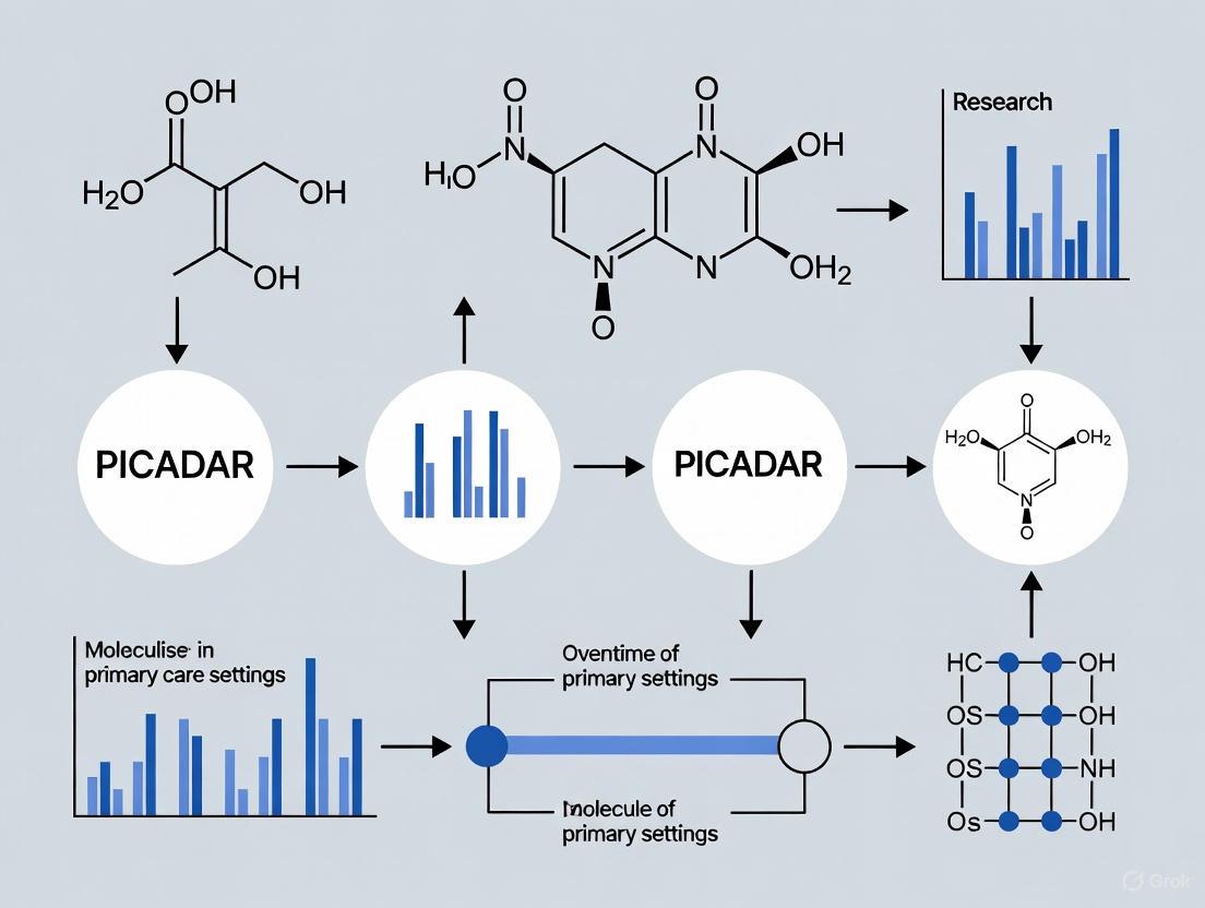
This article provides a comprehensive analysis of the PICADAR (PrImary CiliARy DyskinesiA Rule) predictive tool for primary ciliary dyskinesia (PCD), tailored for researchers, scientists, and drug development professionals.
PICADAR in Primary Care: A Critical Evaluation for Researchers and Drug Development Professionals
Abstract
This article provides a comprehensive analysis of the PICADAR (PrImary CiliARy DyskinesiA Rule) predictive tool for primary ciliary dyskinesia (PCD), tailored for researchers, scientists, and drug development professionals. It explores the tool's foundational development and clinical rationale, details its methodological application and scoring system, critically examines its limitations and sensitivity issues based on recent genetic studies, and validates its performance against alternative predictive instruments. The synthesis underscores PICADAR's role in patient stratification for clinical trials and identifies critical gaps for future diagnostic biomarker and therapeutic development.
The Foundation of PICADAR: Origin, Rationale, and Clinical Need
Primary ciliary dyskinesia (PCD) is a rare, genetic ciliopathy characterized by impaired mucociliary clearance, leading to chronic and progressive upper and lower respiratory tract disease. Serious health complications and significant impacts on patient quality of life make an accurate and timely diagnosis crucial. However, the diagnostic pathway for PCD is fraught with challenges, as there is no single 'gold standard' test. Confirmatory tests are highly specialized, requiring expensive equipment and experienced scientists, and are typically available only at specialized centers [1] [2] [3]. This complexity contributes to significant underdiagnosis and diagnostic delays, often lasting years [4]. To bridge this gap and guide general practitioners and non-specialists on whom to refer for advanced testing, there is a pressing need for simple, evidence-based predictive tools. This application note explores the diagnostic dilemma of PCD, frames the role of the PICADAR (PrImary CiliARy DyskinesiA Rule) predictive tool within primary care settings, and provides a detailed overview of the current diagnostic and research methodologies.
The PCD Diagnostic Landscape
The clinical presentation of PCD is heterogeneous, with symptoms that are often non-specific and overlap with more common respiratory conditions like asthma, cystic fibrosis, and recurrent infections [2]. A typical phenotype may include neonatal respiratory distress in term neonates, persistent wet cough, chronic rhinitis, recurrent otitis media, and laterality defects such as situs inversus (found in approximately 50% of patients) [1] [3]. Due to this variability, the European Respiratory Society (ERS) recommends diagnostic testing for patients presenting with several of these features [3].
The definitive diagnostic process is multi-faceted and involves a combination of sophisticated tests, each with its own limitations as summarized in Table 1. This table synthesizes the core diagnostic methods used in PCD confirmation, highlighting their principles and key challenges.
Table 1: Established Diagnostic Tests for Primary Ciliary Dyskinesia (PCD)
| Diagnostic Method | Principle | Key Limitations |
|---|---|---|
| Nasal Nitric Oxide (nNO) | Measures low nNO levels, a hallmark of PCD. | Difficult in young children (<5 years); requires specialized equipment; can be low in other conditions like CF [2] [3]. |
| High-Speed Video Microscopy Analysis (HSVA) | Qualitatively and quantitatively analyzes ciliary beat frequency and pattern. | Requires significant expertise; subjective qualitative analysis; secondary dyskinesia from infection can confound results [5] [3]. |
| Transmission Electron Microscopy (TEM) | Identifies hallmark ultrastructural defects in ciliary axonemes (e.g., absent dynein arms). | Invasive (nasal brushing/biopsy); ~30% of PCD patients have normal ultrastructure; expensive and technically demanding [6] [2] [3]. |
| Genetic Testing | Identifies biallelic pathogenic mutations in one of over 50 known PCD-associated genes. | ~10-30% of patients have no identified mutations; variants of uncertain significance complicate interpretation [2] [3]. |
The absence of a single gold standard and the resource-intensive nature of these tests create a significant bottleneck. Consequently, the ERS guideline suggests using combinations of distinct PCD symptoms and predictive tools to identify patients who should be referred for definitive diagnostic testing [3].
PICADAR: A Predictive Tool for Clinical Use
Tool Development and Rationale
The PICADAR tool was developed to provide a practical, evidence-based method for identifying patients with a high probability of PCD before they undergo complex diagnostic procedures [1]. It is a clinical prediction rule derived from logistic regression analysis of patient history data readily available in a non-specialist setting. The tool is designed to be quick and easy to use by general respiratory and ENT specialists, helping to prioritize referrals to specialized PCD centers without overburdening services.
The PICADAR Scoring System
PICADAR applies specifically to patients with a persistent wet cough. Its calculation is based on seven predictive parameters from the patient's clinical history, each assigned a point value. The total score indicates the probability of PCD.
Table 2: The PICADAR Scoring Model
| Predictive Parameter | Points |
|---|---|
| Situs Inversus | 4 |
| Congenital Cardiac Defect | 2 |
| Full-Term Gestation | 1 |
| Neonatal Chest Symptoms | 1 |
| Admission to Neonatal Intensive Care Unit | 1 |
| Chronic Rhinitis | 1 |
| Ear Symptoms | 1 |
The diagnostic workflow for applying PICADAR in a clinical setting, from patient presentation to referral decision, is outlined in the diagram below.
Performance and Limitations in Practice
Upon external validation, the original PICADAR model demonstrated a sensitivity of 0.90 and specificity of 0.75 for its recommended cut-off score of 5 points [1]. This indicates a strong ability to correctly identify patients with PCD, though its precision in ruling out the disease is more moderate.
However, a recent 2025 study has highlighted critical limitations, revealing that PICADAR's performance is not uniform across all PCD patient subgroups. The overall sensitivity was found to be 75%, but it varies dramatically [7]:
- 95% sensitivity in individuals with laterality defects.
- 61% sensitivity in individuals with situs solitus (normal organ arrangement).
- 83% sensitivity in individuals with hallmark ultrastructural defects on TEM.
- 59% sensitivity in individuals without hallmark ultrastructural defects.
Furthermore, the tool could not be applied to 6.1% of patients in a large cohort because they did not have a chronic wet cough, automatically ruling out PCD according to the tool's initial question [8]. These findings underscore that while PICADAR is a valuable initial screening instrument, it should be used with caution and cannot be the sole factor for estimating the likelihood of PCD, particularly in patients without classic laterality defects [7].
Comparative Analysis of PCD Predictive Tools
Several predictive tools have been developed to aid in PCD diagnosis. A 2021 study compared PICADAR with two other tools: a Clinical Index (CI) and the North American Criteria Defined Clinical Features (NA-CDCF). The study, which enrolled 1401 patients, found that all three scores were significantly higher in the PCD group. The area under the ROC curve (AUC) for the Clinical Index was larger than for NA-CDCF, while PICADAR and NA-CDCF did not differ significantly [8]. A key practical advantage noted for the Clinical Index was that it does not require assessment for laterality or congenital heart defects, which are necessary for PICADAR and NA-CDCF. The study also confirmed that combining any of these clinical tools with nasal nitric oxide measurement further improved their predictive power [8].
Table 3: Comparison of PCD Predictive Clinical Tools
| Feature | PICADAR | Clinical Index (CI) | NA-CDCF |
|---|---|---|---|
| Primary Requirement | Persistent wet cough | Not specified | Not specified |
| Key Components | 7 items (e.g., situs, cardiac defects, neonatal symptoms) | 7 items (e.g., neonatal symptoms, bronchiectasis, otitis) | 4 criteria (laterality defects, neonatal RDS, year-round nasal congestion, year-round wet cough) |
| Need for Radiology/Echo | Yes (for situs/cardiac defect confirmation) | No | Yes (for situs confirmation) |
| Reported AUC | 0.87 (External Validation) [1] | Larger than NA-CDCF (p=0.005) [8] | Not significantly different from PICADAR [8] |
| Notable Advantage | Widely recognized and validated | Feasible without needing all diagnostic tests for laterality [8] | Simple, criteria-based |
Advanced and Emerging Methodologies
Quantitative Ciliary Beat Analysis (Protocol)
The qualitative evaluation of ciliary beat pattern via HSVA can be subjective. Quantitative analysis offers a more objective approach. The following protocol is adapted from a pilot study that established quantitative parameters for PCD diagnosis [5].
Objective: To quantitatively analyze ciliary beat pattern from high-speed videomicroscopy recordings to distinguish primary from secondary ciliary dyskinesia. Materials:
- Nasal cytology brush (2 mm)
- Inverted microscope with a 100x oil immersion objective
- High-speed digital camera (e.g., capable of ≥355 frames per second)
- Cell culture medium (e.g., B1 BSA medium)
- Temperature-controlled stage (37°C)
Procedure:
- Sample Collection: Obtain ciliated epithelium by brushing the middle part of the inferior nasal turbinate.
- Sample Preparation: Suspend cells in culture medium and examine within three hours of collection.
- Video Recording: At 37°C, record 20 distinct, intact ciliated edges (>50 μm) per patient. Record at a high frame rate (e.g., 355 fps) for a sufficient duration (e.g., 1800 frames). Exclude areas with mucus or isolated cells.
- Quantitative Analysis:
- Determine the percentage of beating ciliated edges.
- For 10 cilia per patient, track the movement of the cilium tip through a complete beating cycle using video analysis software. Define key points: base (P0), and the tip positions at the start and end of the active (P1, P2) and recovery strokes.
- Calculate key parameters, including:
- Ciliary beat frequency (Hz)
- Distance traveled by the cilium tip per second (μm/s)
- Area swept by the cilium per second (μm²/s)
- Beat pattern angles and durations
- Weight parameters like 'distance traveled' by the 'percentage of beating ciliated edges'.
Validation: In the pilot study, the weighted 'distance traveled by the cilium tip' parameter showed 96% sensitivity and 95% specificity in distinguishing PCD from non-PCD patients, outperforming qualitative evaluation in cases with partial ciliary motility [5].
Machine Learning and Automated Analysis
Emerging technologies are being leveraged to address the challenges of PCD diagnosis.
Automated Ciliary Ultrastructure Analysis: Software such as 'PCD Quant' is being developed for the automatic quantitative analysis of TEM images. The goal is to objectively determine the ratio of primary and secondary ciliary defects and analyze the mutual orientation of cilia in the ciliary border on a large scale, thus improving diagnostic consistency and speed [6].
Machine Learning for Patient Screening: A 2025 feasibility study demonstrated the use of a random forest model to screen for PCD using large-scale health insurance claims data. The model used diagnostic, procedural, and pharmaceutical codes as features. When trained on a dataset that included patients with codes suggestive of PCD, the model achieved a sensitivity of 0.82–0.90 and a positive predictive value (PPV) of 0.51–0.54, performance deemed suitable for a broad screening tool. This approach shows promise for identifying at-risk populations who may benefit from definitive diagnostic testing [4].
The logical flow of this machine learning approach, from data aggregation to model deployment, is illustrated below.
The Scientist's Toolkit: Research Reagent Solutions
Table 4: Essential Research Reagents and Materials for PCD Diagnostic Investigations
| Reagent / Material | Application | Function / Rationale |
|---|---|---|
| Glutaraldehyde | TEM Sample Fixation | Primary fixative that cross-links proteins, preserving ciliary ultrastructure for electron microscopy [6]. |
| Osmium Tetroxide | TEM Sample Post-Fixation | Secondary fixative that stabilizes and stains lipids, enhancing membrane contrast for TEM imaging [6]. |
| Nasal Nitric Oxide Analyzer (Chemiluminescence) | nNO Measurement | Precisely measures low nasal NO output, a key screening biomarker for PCD [3]. |
| High-Speed Video Camera (≥355 fps) | HSVA | Captures ciliary beating in slow motion for subsequent qualitative and quantitative analysis of beat pattern and frequency [5]. |
| Next-Generation Sequencing Panels (PCD Gene Panels) | Genetic Testing | Identifies pathogenic mutations in over 50 known PCD-associated genes for diagnostic confirmation and genotyping [8]. |
| Anti-Ciliary Protein Antibodies | Immunofluorescence (IF) | Detects the absence or mislocalization of specific ciliary proteins (e.g., dynein arms), which can confirm a diagnosis even with normal TEM [3]. |
The diagnostic dilemma in PCD, characterized by non-specific symptoms and the lack of a single gold-standard test, creates a powerful impetus for simple predictive tools like PICADAR. In a primary care setting, PICADAR serves as a valuable first-line screening instrument to identify high-risk patients who warrant referral to specialized centers. However, its limitations, particularly its reduced sensitivity in patients without laterality defects or hallmark ultrastructural defects, necessitate a cautious application. It should not be used as a standalone exclusionary tool. The future of PCD diagnosis lies in the continued refinement of clinical prediction rules and their integration with objective, quantitative methodologies, such as automated ciliary analysis and machine learning-based screening. These advanced protocols, alongside established diagnostic tests, form a comprehensive toolkit that will enable researchers and clinicians to improve the accuracy and timeliness of PCD diagnosis, ultimately leading to better patient management and outcomes.
Primary Ciliary Dyskinesia (PCD) is a rare, genetically heterogeneous disorder characterized by abnormal ciliary function, leading to impaired mucociliary clearance of the airways [1]. The diagnostic pathway for PCD is complex, requiring specialized equipment and expertise typically available only at specialized referral centers [9]. To address the challenge of identifying which patients with chronic respiratory symptoms should be referred for definitive PCD testing, Behan et al. (2016) developed PICADAR (PrImary CiliARy DyskinesiA Rule), a clinical prediction tool [1] [10].
PICADAR was derived and validated to provide primary care physicians, pediatricians, and general respiratory specialists with a simple, evidence-based method to quantify the pre-test probability of PCD using readily available clinical information [1]. This application note deconstructs the seven predictive parameters of the PICADAR score, providing researchers and clinicians with detailed methodologies for its application within primary care and research settings.
The Seven Predictive Parameters of PICADAR
The PICADAR tool is applicable to patients with a persistent wet cough and incorporates seven clinical parameters obtained from patient history [1] [10]. Each parameter is assigned a points value, and the sum yields a total score that correlates with the probability of a PCD diagnosis.
Table 1: The Seven Predictive Parameters of the PICADAR Score
| Predictive Parameter | Clinical Description | Points Value |
|---|---|---|
| Full-term gestation | Gestational age ≥ 37 weeks [1] | 2 |
| Neonatal chest symptoms | Respiratory distress or other chest symptoms present at birth [1] | 1 |
| Neonatal intensive care admission | Admission to a special care baby unit or NICU after birth [1] | 1 |
| Chronic rhinitis | Nasal congestion or rhinorrhea persisting for >3 months [1] | 1 |
| Ear symptoms | History of chronic otitis media, hearing impairment, or tympanostomy tube placement [1] | 1 |
| Situs inversus | Complete transposition of thoracic and abdominal organs, confirmed by imaging [1] [11] | 2 |
| Congenital cardiac defect | Any structural heart defect present at birth (e.g., heterotaxy-related defects) [1] | 2 |
Parameter Specifics and Clinical Context
- Full-term gestation: Most patients diagnosed with PCD are born at term, which is a significant positive predictor [12].
- Situs inversus: This parameter is a strong predictor, but its prevalence varies genetically. Notably, only about 25% of Japanese PCD patients have situs inversus, contrasting with the ~50% often cited in other populations [11].
- Neonatal respiratory symptoms: These often manifest as respiratory distress syndrome (RDS) in term neonates, including tachypnea, grunting, or requirement for respiratory support [1] [13].
Performance and Validation of PICADAR
The predictive performance of PICADAR was established through internal and external validation studies. The tool demonstrates good accuracy in discriminating between PCD-positive and PCD-negative individuals.
Table 2: Performance Characteristics of the PICADAR Tool
| Performance Measure | Derivation Cohort (n=641) | External Validation Cohort (n=187) |
|---|---|---|
| Prevalence of PCD | 75 (12%) | 93 (50%)* |
| Area Under the Curve (AUC) | 0.91 | 0.87 |
| Sensitivity (at score ≥5) | 0.90 | Not specified |
| Specificity (at score ≥5) | 0.75 | Not specified |
*The validation cohort was selectively enriched with PCD-positive cases [1].
A sensitivity of 0.90 and specificity of 0.75 for a cut-off score of 5 points was reported in the original derivation cohort, indicating a high true positive rate [1]. The AUC values of 0.91 and 0.87 indicate good to excellent diagnostic discrimination [1].
However, a 2025 preprint by Schramm et al. highlighted important limitations, reporting an overall sensitivity of only 75% in a genetically confirmed PCD cohort [7]. The sensitivity was significantly higher in individuals with laterality defects (95%) compared to those with situs solitus (normal organ arrangement), where it dropped to 61% [7]. This indicates PICADAR's performance is lower in patients without laterality defects.
Experimental Protocol for PICADAR Assessment
Clinical Workflow for PICADAR Application
The following diagram outlines the standardized protocol for applying the PICADAR tool in a clinical or research setting.
Step-by-Step Assessment Methodology
Patient Identification: Apply PICADAR only to patients with a persistent (chronic) wet cough. The tool is not validated for patients without this symptom [1] [7].
Structured Clinical Interview: Conduct a thorough interview focusing on the seven parameters. For adult patients, neonatal history may require verification of medical records if parental recall is insufficient [13].
Data Collection and Scoring:
- Neonatal History: Document gestational age, presence of chest symptoms (e.g., tachypnea, grunting, supplemental oxygen requirement), and whether admission to a neonatal intensive care unit (NICU) or special care baby unit was required [1].
- Chronic Symptoms: Establish the presence of chronic rhinitis (>3 months duration) and chronic ear symptoms (e.g., recurrent otitis media, effusion, hearing loss) [1] [13].
- Anatomical Abnormalities: Confirm the presence of situs inversus (via chest X-ray or other imaging) and any congenital heart defects (via echocardiography or medical record review) [1] [12].
Calculation and Interpretation:
- Sum the points for all applicable parameters (see Table 1).
- A score of 5 points or higher suggests a high probability of PCD and warrants referral to a specialized diagnostic center [1].
- A score below 5 points does not exclude PCD, particularly in cases of situs solitus or milder phenotypes. Clinical judgment remains essential [7].
The Scientist's Toolkit: Essential Research Reagents and Materials
Table 3: Key Research Reagents and Materials for PCD Diagnostic Research
| Reagent/Material | Primary Function in PCD Research | Experimental Context |
|---|---|---|
| Nasal Epithelial Cells (NECs) | Primary cell source for functional, structural, and molecular analyses | Obtained via nasal brushing; used in HSVM, TEM, IF, and cell culture [9] |
| Air-Liquid Interface (ALI) Culture Media | Supports differentiation of ciliated epithelium in vitro | Enables ciliogenesis in cultured cells, crucial for repeated or standardized HSVM/TEM [9] |
| High-Speed Video Microscopy (HSVM) | Analyzes ciliary beat frequency and pattern | Functional assessment of ciliary motility; requires expertise for pattern recognition [13] [9] |
| Transmission Electron Microscopy (TEM) | Visualizes ultrastructural defects in ciliary axoneme | Identifies hallmark defects (e.g., ODA/IDA absence); considered a definitive diagnostic test [12] [9] |
| Immunofluorescence (IF) Antibodies | Detects absence/mislocalization of ciliary proteins | Targets proteins like DNAH5, GAS8; useful when TEM is inconclusive [9] |
| Next-Generation Sequencing (NGS) | Identifies pathogenic variants in >50 PCD-associated genes | Genetic confirmation; especially valuable in patients with normal ultrastructure [12] [9] |
Integration in Primary Care and Research Settings
In primary care, PICADAR serves as a triage tool, not a diagnostic test. Its strength lies in using easily obtainable clinical data to streamline referrals to specialized centers, promoting early diagnosis without overburdening specialized services [1]. For researchers, PICADAR provides a standardized framework for phenotyping patients in cohort studies and clinical trials, ensuring a consistent pre-test probability among enrolled subjects [13].
Recent studies suggest that combining PICADAR with other tools like nasal nitric oxide (nNO) measurement can further improve predictive power before initiating invasive or expensive confirmatory testing [13]. However, clinicians and researchers must be aware of its reduced sensitivity in patients with situs solitus and those without hallmark ultrastructural defects on TEM [7]. Continued validation across diverse populations and genotypes is essential to refine its clinical utility and account for phenotypic variations across different genetic backgrounds and ethnicities [11] [12].
The PICADAR (PrImary CiliARy DyskinesiA Rule) tool represents a significant advancement in the initial identification of patients suspected of having Primary Ciliary Dyskinesia (PCD), a rare genetic disorder characterized by abnormal ciliary function leading to chronic respiratory symptoms [1]. Before the development of such predictive tools, diagnosing PCD was challenging due to the non-specific nature of its symptoms and the requirement for highly specialized, expensive diagnostic tests available only in specialized centers [1] [13]. This application note details the original validation cohorts and experimental protocols that established PICADAR as a validated clinical prediction rule, providing researchers and clinicians with a reliable method to select patients for further specialized testing.
Cohort Demographics and Design
The original validation study for PICADAR was designed as a multi-center investigation to ensure robustness and generalizability. The study population was divided into two distinct groups to facilitate both the creation and the external validation of the prediction tool [1].
Study Populations
- Derivation Group: This group consisted of 641 consecutive patients referred to the University Hospital Southampton (UHS) PCD diagnostic centre between 2007 and 2013. Within this group, 75 patients (12%) received a positive PCD diagnosis, while 566 (88%) were negative. The median age at assessment was 9 years (range: 0–79 years), and 44% were male [1].
- External Validation Group: To validate the score externally, a sample of 187 patients was used from the Royal Brompton Hospital (RBH). This group was intentionally selected to include a similar number of positive and negative diagnoses (93 PCD-positive and 94 PCD-negative) from patients referred between 1983 and 2013. The validation group was significantly younger than the derivation group, with a median age of 3 years, and was more likely to be from non-white ethnic backgrounds and consanguineous families, reflecting the different patient populations served by the two centers [1].
Table 1: Characteristics of the Derivation and Validation Cohorts
| Characteristic | Derivation Group | Validation Group | p-value |
|---|---|---|---|
| Total Subjects | 641 | 157 | - |
| PCD-Positive | 75 (12%) | 80 (51%) | - |
| PCD-Negative | 566 (88%) | 77 (49%) | - |
| Age at Assessment (years) | 9 (0–79) | 3 (0–18) | <0.001 |
| Male Sex | 283 (44%) | 78 (50%) | 0.211 |
Experimental Protocol
Diagnostic Testing Protocol
A definitive diagnosis of PCD, which served as the reference standard against which PICADAR was validated, was established using a combination of specialized tests, in line with contemporary guidelines [1] [13]. The diagnostic workflow is summarized below:
Diagnostic Criteria: A positive PCD diagnosis was typically confirmed through one of the following pathways [1]:
- A typical clinical history plus at least two abnormal diagnostic tests. The "hallmark" abnormal tests included:
- Characteristic Transmission Electron Microscopy (TEM) defects.
- Characteristic Ciliary Beat Pattern (CBP) observed via High-Speed Video Microscopy Analysis (HSVMA).
- Low nasal nitric oxide (nNO ≤ 30 nL·min⁻¹).
- In selected cases with a very strong clinical phenotype (e.g., sibling with confirmed PCD, or full clinical presentation including neonatal respiratory distress at term, daily wet cough, persistent rhinitis, and glue ear), a diagnosis could be made based on a single hallmark TEM finding or repeated HSVMA consistent with PCD.
To minimize false positives, CBP was only considered positive if the pattern was typical of PCD rather than secondary ciliary dyskinesia, confirmed either from two brushing biopsies or from one biopsy with re-analysis after air-liquid interface culture [1].
Data Collection and Predictive Model Development
Clinical data was collected prospectively using a proforma completed by a clinician during a patient interview prior to any diagnostic testing [1]. The model development involved several key statistical steps:
- Variable Selection: Twenty-seven potential predictor variables were initially identified from information readily available in a non-specialist setting.
- Univariate Analysis: Each variable was individually tested using parametric (t-test) or non-parametric (Mann-Whitney) tests, Chi-squared tests, or Fisher's exact tests to compare their distribution between PCD-positive and PCD-negative groups.
- Multivariate Logistic Regression: Significant predictors from the univariate analysis were entered into a logistic regression model using forward step-wise methods to identify the most parsimonious set of independent predictors for a positive PCD diagnosis.
- Model Performance Assessment: The model's ability to discriminate between PCD and non-PCD cases was evaluated using Receiver Operating Characteristic (ROC) curve analysis, calculating the Area Under the Curve (AUC). Model calibration was assessed using the Hosmer-Lemeshow goodness-of-fit test.
The PICADAR Tool & Validation Results
The PICADAR Scoring System
The logistic regression model was simplified into a practical points-based tool—PICADAR. It is important to note that PICADAR is designed for use in patients with a persistent wet cough [1]. The tool comprises seven predictive parameters, each assigned a point value, as detailed below.
Table 2: The PICADAR Clinical Prediction Rule [1]
| # | Predictive Parameter | Points |
|---|---|---|
| 1 | Full-term gestation | 2 |
| 2 | Neonatal chest symptoms (at term) | 2 |
| 3 | Admission to a neonatal intensive care unit | 2 |
| 4 | Chronic rhinitis | 1 |
| 5 | Ear symptoms (chronic otitis media or hearing impairment) | 1 |
| 6 | Situs inversus | 4 |
| 7 | Congenital cardiac defect | 2 |
| Total Possible Points | 14 |
Performance and Validation Metrics
The performance of the PICADAR tool was robust in both the initial derivation and the external validation phases.
- Internal Validation: In the derivation group, the AUC was 0.91, indicating excellent discriminatory power [1].
- External Validation: The tool maintained strong performance in the independent RBH cohort, with an AUC of 0.87 [1].
- Sensitivity and Specificity: At the recommended cut-off score of 5 points, PICADAR demonstrated a sensitivity of 0.90 and a specificity of 0.75 [1]. This means it correctly identifies 90% of true PCD cases while incorrectly flagging 25% of non-PCD cases for further testing.
Table 3: Performance Metrics of the PICADAR Tool
| Metric | Derivation Cohort | External Validation Cohort |
|---|---|---|
| Area Under the Curve (AUC) | 0.91 | 0.87 |
| Sensitivity (at cut-off ≥5) | 0.90 | - |
| Specificity (at cut-off ≥5) | 0.75 | - |
Research Toolkit
The following reagents and equipment are essential for conducting definitive PCD diagnostics, as used in the validation of predictive tools like PICADAR.
Table 4: Essential Research Reagents and Equipment for PCD Diagnostics
| Item | Function/Application |
|---|---|
| Electrochemical Nasal Nitric Oxide (nNO) Analyzer (e.g., Niox Mino/Vero) | Measures nasal nitric oxide levels, a key screening and diagnostic biomarker for PCD which is typically very low [13] [8]. |
| High-Speed Video Microscope (e.g., Keyence Motion Analyzer) | Allows for the analysis of ciliary beat frequency and pattern (CBP/HSVMA) from nasal brushings [13] [8]. |
| Transmission Electron Microscope (TEM) | Used to visualize and assess the ultrastructure of cilia for hallmark defects [1] [13]. |
| Next-Generation Sequencing (NGS) Panels | Genetic testing for mutations in over 50 known PCD-associated genes to provide a definitive molecular diagnosis [13] [8]. |
| Nasal Brushing Biopsy Kit | For obtaining ciliated epithelial cell samples from the inferior nasal turbinate for HSVMA and TEM analysis [1]. |
Discussion
The rigorous validation of PICADAR across two distinct UK cohorts established it as a simple, effective, and valid tool for predicting PCD in symptomatic patients with chronic wet cough. Its high sensitivity ensures that few true PCD cases are missed, while its specificity helps prevent the overburdening of specialized diagnostic centers [1]. Subsequent independent validation in a large, unselected cohort of 1401 patients confirmed its utility, showing that while other tools like the Clinical Index (CI) may exist, PICADAR remains a robust predictor [13]. Furthermore, studies have shown that combining PICADAR with nNO measurement can further improve its predictive power, offering an even more effective strategy for triaging patients in a clinical pathway [13]. For the primary care and general respiratory research setting, PICADAR provides an evidence-based, data-driven protocol for deciding whom to refer for complex, costly confirmatory testing.
Primary Ciliary Dyskinesia (PCD) is a rare, genetically heterogeneous disorder caused by impaired ciliary function, leading to chronic otosinopulmonary disease. The diagnostic journey is often protracted due to the nonspecific nature of clinical presentations, such as persistent wet cough, chronic rhinitis, and recurrent otitis media, which overlap with more common respiratory conditions [10] [14]. The definitive diagnostic tests for PCD—including transmission electron microscopy (TEM), high-speed video microscopy analysis (HSVA), and genetic testing—are highly specialized, expensive, and available only at specialized reference centers [10] [1]. This creates a significant bottleneck, where primary and secondary care physicians lack clear guidance on which patients to refer for complex testing. The PICADAR tool (PrImary CiliARy DyskinesiA Rule) was developed to fill this critical gap. It is a symptom-based predictive score designed for use in non-specialist settings to identify patients with a high probability of PCD, thereby streamlining referral to specialist centers and promoting earlier diagnosis [10] [15].
PICADAR Tool: Application Notes and Scoring Protocol
PICADAR is a clinical prediction rule that uses seven readily obtainable parameters from a patient's history. Its application is intended for patients with a persistent wet cough. The tool assigns points for each predictive feature, and the total score estimates the probability of PCD, guiding the need for specialist referral [10] [1].
Predictive Parameters and Scoring System
The seven parameters and their assigned points are summarized in the table below.
Table 1: The PICADAR Scoring System and Predictive Parameters [10]
| Predictive Parameter | Score |
|---|---|
| Full-term gestation | 2 |
| Neonatal chest symptoms (within first 4 weeks of life) | 2 |
| Admission to a neonatal intensive care unit (NICU) | 1 |
| Chronic rhinitis (symptoms lasting >3 months) | 1 |
| Chronic ear symptoms (e.g., otitis media, glue ear) | 1 |
| Situs inversus | 4 |
| Congenital cardiac defect | 2 |
Interpretation and Recommended Action
The total PICADAR score is calculated by summing the points for all applicable parameters. The interpretation and subsequent action are based on the total score, as detailed in the following table.
Table 2: Interpreting the PICADAR Score and Recommended Clinical Actions [10]
| Total Score | Risk of PCD | Recommended Action |
|---|---|---|
| 0 - 4 | Low | PCD is unlikely; consider alternative diagnoses. |
| ≥ 5 | High | Refer to a specialist PCD center for definitive diagnostic testing. |
The original validation study reported that at a cut-off score of 5 points, PICADAR demonstrated a sensitivity of 0.90 and a specificity of 0.75, with an area under the curve (AUC) of 0.91 upon internal validation and 0.87 upon external validation [10].
Experimental Protocol: Original PICADAR Derivation and Validation
The following protocol outlines the methodology used in the original study to develop and validate the PICADAR tool.
Study Design and Population
- Design: Observational, analytical study using a derivative cohort for model development and an external cohort for validation [1].
- Derivative Cohort: 641 consecutive patients referred for PCD testing at the University Hospital Southampton (UHS). Of these, 75 (12%) received a positive PCD diagnosis [10] [1].
- Validation Cohort: 187 patients (93 PCD-positive, 94 PCD-negative) from the Royal Brompton Hospital (RBH) to externally validate the tool's performance [1].
- Diagnostic Standard (Reference Test): A positive PCD diagnosis was based on a typical clinical history plus at least two abnormal diagnostic tests. These tests included hallmark TEM defects, hallmark ciliary beat pattern (CBP) abnormalities, or low nasal nitric oxide (nNO ≤30 nL·min⁻¹). In select cases with a very strong phenotype, a diagnosis could be made based on a single definitive test [1].
Data Collection and Statistical Analysis
- Data Collection: A pre-test clinical proforma was used to collect data on 27 potential predictor variables through a clinical interview. This included neonatal history, situs abnormalities, congenital defects, and chronic respiratory symptoms [1].
- Model Development:
- Univariate Analysis: Potential predictors were individually tested for association with the diagnostic outcome using t-tests, Mann-Whitney tests, Chi-squared tests, or Fisher's exact tests as appropriate.
- Multivariate Analysis: Significant predictors from univariate analysis were entered into a logistic regression model using forward step-wise methods to identify the most parsimonious set of independent predictors.
- Score Creation: The regression coefficients from the final logistic model were rounded to the nearest integer to create the practical PICADAR point score [1].
- Model Performance:
- Discrimination: The model's ability to distinguish between PCD-positive and PCD-negative patients was assessed using Receiver Operating Characteristic (ROC) curve analysis and calculation of the Area Under the Curve (AUC).
- Calibration: The Hosmer-Lemeshow goodness-of-fit test was used to assess how well the predicted probabilities agreed with the observed outcomes [1].
Workflow Diagram: PICADAR Development and Application
The following diagram illustrates the complete pathway from tool development to its clinical application.
The Scientist's Toolkit: Essential Reagents and Materials for PCD Diagnostic Research
The definitive diagnosis of PCD relies on specialized techniques. The following table lists key reagents and materials essential for research and diagnostic work in a specialist PCD center.
Table 3: Key Research Reagent Solutions for PCD Diagnostic Testing
| Reagent / Material | Function in PCD Diagnostics |
|---|---|
| Nasal Epithelial Cell Brush/Biopsy | To obtain ciliated epithelium for functional (HSVA) and structural (TEM, IF) analyses. The primary sample for diagnostic testing [14]. |
| Cell Culture Media (e.g., DMEM) | For air-liquid interface (ALI) culture of brushed epithelial cells. This allows ciliary re-differentiation and can help distinguish primary from secondary ciliary dyskinesia [1]. |
| Glutaraldehyde Fixative | For high-quality preservation of ciliary ultrastructure prior to processing for Transmission Electron Microscopy (TEM) [14]. |
| Antibodies for Immunofluorescence (IF) | Specific antibodies against ciliary proteins (e.g., DNAH5, GAS8) are used to detect the absence or mislocalization of proteins, providing a genetic clue [14]. |
| Next-Generation Sequencing (NGS) Panels | Targeted gene panels or whole-exome sequencing to identify mutations in over 50 known PCD-associated genes, confirming the molecular diagnosis [14]. |
| High-Speed Video Camera | Essential equipment for High-Speed Video Microscopy Analysis (HSVA) to capture and analyze ciliary beat pattern and frequency [14]. |
Critical Evaluation and Limitations in Clinical Application
While PICADAR is a valuable screening tool, recent evidence highlights important limitations that must be considered in a primary care research context. A 2025 study by Omran et al. found that PICADAR's overall sensitivity in a genetically confirmed PCD cohort was 75%, significantly lower than in the original derivation study [16]. The tool's performance was highly variable across genetic and anatomic subgroups:
- Sensitivity was 95% in patients with laterality defects (situs inversus).
- Sensitivity dropped to 61% in patients with normal situs (situs solitus) [16].
- The tool also showed lower sensitivity in individuals without hallmark ultrastructural defects on TEM (59%) compared to those with such defects (83%) [16].
A critical design limitation is that the tool's initial question excludes patients without a daily wet cough from further evaluation. The 2025 study found that 7% of genetically confirmed PCD patients did not report a daily wet cough and would have been ruled out by PICADAR [16]. Therefore, PICADAR should be used as a triage aid, not a standalone diagnostic factor. A low score should not definitively rule out PCD in patients with a compelling clinical history, and alternative predictive tools or direct referral may be necessary for complex cases [16].
Applied Methodology: Implementing and Scoring the PICADAR Tool
Primary ciliary dyskinesia (PCD) is a rare genetic disorder characterized by abnormal ciliary function, leading to chronic respiratory symptoms that begin in early childhood [1]. Diagnosis is challenging due to nonspecific symptoms and the requirement for highly specialized, expensive diagnostic testing available only at specialized centers [1] [13]. The PICADAR (PrImary CiliARy DyskinesiA Rule) tool was developed as a clinical prediction rule to identify high-risk patients who should be referred for definitive PCD testing [1] [10]. In primary care settings, where access to specialized diagnostics is limited, PICADAR serves as a crucial screening tool that enables clinicians to make evidence-based referral decisions using readily available clinical history [1].
This protocol details the implementation of PICADAR within primary care and research contexts, providing comprehensive guidance on its application, interpretation, and integration with subsequent diagnostic pathways.
PICADAR Scoring Parameters and Point Assignment
The PICADAR tool assigns points for seven key clinical features obtained from patient history [1] [10]. The tool applies specifically to patients with persistent wet cough, and points are allocated as follows:
Table 1: PICADAR Scoring Criteria and Point Values
| Clinical Parameter | Criteria for Point Assignment | Points Assigned |
|---|---|---|
| Full-term gestation | Gestational age ≥37 weeks [1] | 2 |
| Neonatal chest symptoms | Respiratory distress or symptoms present at birth [1] | 2 |
| Neonatal intensive care admission | Admission to NICU after birth [1] | 2 |
| Chronic rhinitis | Year-round nasal congestion or rhinorrhea [13] | 1 |
| Ear symptoms | Chronic otitis media or hearing problems [1] | 1 |
| Situs inversus | Laterality defect with complete organ reversal [1] [10] | 4 |
| Congenital cardiac defect | Any structural heart defect present at birth [1] [10] | 3 |
Calculation and Interpretation
To calculate the PICADAR score, clinicians sum the points for all applicable clinical features present in the patient's history. The total score determines the probability of PCD and corresponding referral recommendation:
- Score <5 points: Low probability of PCD; alternative diagnoses should be considered [17].
- Score ≥5 points: Indicates increased probability of PCD and warrants referral to a specialist center for further testing [1] [10]. A score of ≥10 points indicates a >90% probability of PCD [17].
Experimental Protocol for PICADAR Validation
The following protocol outlines the methodology used in the original derivation and validation of the PICADAR tool.
Patient Population and Data Collection
- Derivation Cohort: 641 consecutive patients referred for PCD testing at University Hospital Southampton (2007-2013) [1].
- Validation Cohort: 187 patients (93 PCD-positive, 94 PCD-negative) from Royal Brompton Hospital [1].
- Inclusion Criteria: Patients with persistent respiratory symptoms referred for PCD diagnostic testing [1].
- Data Collection: A standardized proforma was used to collect patient data through clinical interview prior to diagnostic testing [1].
Reference Standard for PCD Diagnosis
The diagnostic criteria for PCD in the validation studies required a typical clinical history plus at least two abnormal diagnostic tests from among the following [1]:
- "Hallmark" transmission electron microscopy (TEM) defects
- "Hallmark" ciliary beat pattern (CBP) abnormalities
- Nasal nitric oxide (nNO) ≤30 nL·min⁻¹
In some cases, patients with a strong clinical phenotype (e.g., sibling with PCD, classic symptoms) were diagnosed based on a single definitive test result [1].
Statistical Analysis and Model Development
- Predictor Selection: 27 potential clinical variables were initially assessed [1].
- Model Development: Logistic regression analysis with forward step-wise methods identified the seven significant predictors included in PICADAR [1].
- Performance Metrics: Receiver operating characteristic (ROC) curve analysis determined the area under the curve (AUC) [1].
- Model Validation: External validation was performed using the separate cohort from Royal Brompton Hospital [1].
PICADAR Clinical Decision Pathway
Performance Characteristics and Comparative Analysis
The PICADAR tool has demonstrated robust performance in both derivation and validation studies, with the external validation showing an area under the curve (AUC) of 0.87, sensitivity of 0.90, and specificity of 0.75 at the recommended cut-off score of 5 points [1] [10].
Table 2: Performance Comparison of PCD Predictive Tools
| Tool | Number of Items | Target Population | AUC | Sensitivity | Specificity | Key Limitations |
|---|---|---|---|---|---|---|
| PICADAR [1] [10] | 7 | Patients with persistent wet cough | 0.87 (externally validated) | 0.90 | 0.75 | Requires complete neonatal history; not applicable without chronic wet cough [13] |
| Clinical Index (CI) [13] | 7 | Patients with chronic respiratory symptoms | Comparable to PICADAR (study-specific) | Not reported | Not reported | Does not assess laterality or congenital heart defects [13] |
| NA-CDCF [13] | 4 | Patients with respiratory symptoms | No significant difference from PICADAR [13] | Not reported | Not reported | Limited to four clinical criteria [13] |
Research Reagent Solutions for PCD Diagnostic Confirmation
The following reagents and materials are essential for the definitive diagnostic tests used to confirm PCD following positive PICADAR screening.
Table 3: Essential Research Reagents for PCD Diagnostic Confirmation
| Reagent/Material | Application in PCD Diagnosis | Specific Function |
|---|---|---|
| Nasal Nitric Oxide (nNO) Analyzer [13] | nNO measurement | Measures nasal NO concentration; levels ≤30 nL·min⁻¹ are indicative of PCD [1] |
| High-Speed Video Microscopy System [13] | Ciliary beat pattern analysis | Captures ciliary movement for frequency and pattern analysis to identify dyskinesia [1] [13] |
| Transmission Electron Microscope [13] | Ciliary ultrastructure examination | Visualizes internal ciliary structure to identify hallmark defects (e.g., outer dynein arm缺失) [1] [13] |
| Next-Generation Sequencing Panel [13] | Genetic testing | Identifies pathogenic variants in over 50 known PCD-related genes [13] |
PCD Diagnostic Pathway After Positive PICADAR
Implementation in Primary Care and Research Settings
In primary care, PICADAR provides a practical, evidence-based framework for identifying children and adults who require specialist referral for PCD investigation [1]. The tool addresses the critical challenge of PCD underdiagnosis by improving appropriate referrals to specialized centers without overburdening limited resources [1] [17].
For research applications, PICADAR enables standardized patient selection across multicenter studies, ensuring enrollment of appropriately characterized participants for studies on PCD genetics, pathophysiology, and therapeutic development [13]. When combined with nNO measurement, the predictive power of PICADAR is significantly enhanced, creating a highly effective screening cascade in both clinical and research settings [13].
In the diagnosis of Primary Ciliary Dyskinesia (PCD), a rare genetic disorder affecting mucociliary clearance, the PICADAR (PrImary CiliARy DyskinesiA Rule) prediction tool serves a critical function in primary care and general respiratory settings by identifying patients who require specialized testing [1] [18]. This tool addresses the significant challenge posed by the non-specific nature of PCD symptoms, which often leads to underdiagnosis or delayed diagnosis [1]. Specialized diagnostic tests for PCD are highly complex, require expensive equipment and expert scientists, and are typically only available in specialized centers [1] [14]. The PICADAR score, derived from seven easily obtainable clinical parameters, provides a evidence-based method for triaging symptomatic patients, thereby facilitating earlier diagnosis while preventing overburdening of specialist services [1] [17]. This application note focuses on the critical cut-off score of ≥5 points, examining the evidence behind this threshold and its practical implications for clinicians and researchers.
The PICADAR Scoring System & Quantitative Performance
The PICADAR tool is designed for patients with a persistent wet cough and incorporates seven predictive clinical parameters, each assigned a specific point value. A patient's total score is the sum of these points, which correlates with their probability of having PCD [1] [17].
Table 1: The PICADAR Scoring System and Associated Probability of PCD
| Clinical Parameter | Points Assigned |
|---|---|
| Full-term gestation | 2 |
| Neonatal chest symptoms | 2 |
| Neonatal intensive care unit admission | 1 |
| Chronic rhinitis | 1 |
| Chronic ear symptoms | 1 |
| Situs inversus | 2 |
| Congenital cardiac defect | 2 |
| Total Score Interpretation | Probability of PCD |
| ≥ 5 points | >11.1% probability |
| ≥ 10 points | >90% probability |
The performance of the PICADAR tool has been rigorously validated. In the original derivation study of 641 referrals, the tool demonstrated a sensitivity of 0.90 and a specificity of 0.75 at the ≥5 cut-off [1]. The Area Under the Curve (AUC) was 0.91 upon internal validation and 0.87 upon external validation in a second diagnostic center, indicating good to excellent discriminative ability [1] [17]. These performance metrics are summarized in the table below.
Table 2: Diagnostic Performance of the PICADAR Tool at the ≥5 Cut-off
| Performance Metric | Derivation Cohort (n=641) | External Validation Cohort |
|---|---|---|
| Sensitivity | 0.90 | 0.86 |
| Specificity | 0.75 | 0.73 |
| Area Under the Curve (AUC) | 0.91 | 0.87 |
Rationale for the ≥5 Cut-off: Balancing Sensitivity and Specificity
The selection of a score of 5 as the critical cut-off was driven by the need to optimize the tool's clinical utility. This threshold represents a deliberate balance between capturing the maximum number of true PCD cases (high sensitivity) while filtering out a substantial proportion of patients who do not have the condition (reasonable specificity) [1].
A score of ≥5 points translates to a greater than 11.1% probability of a positive PCD diagnosis, which is a clinically significant risk that warrants further investigation [17] [19]. In practice, this threshold effectively identifies the vast majority of PCD patients (90% sensitivity), ensuring that few true cases are missed at the referral stage [1]. Concurrently, its 75% specificity helps prevent the over-referral of patients without PCD to specialized centers, making efficient use of limited and expensive diagnostic resources [1]. This balance is crucial for a screening tool intended for use in a broad population of symptomatic patients.
Protocol for Clinical Application in Primary Care
Patient Assessment and Data Collection
This protocol guides the systematic assessment of a patient with a persistent wet cough to calculate a PICADAR score.
- Step 1: Confirm Key Symptom: Verify the presence of a persistent wet cough, which is a prerequisite for applying the tool [1].
- Step 2: Elicit Neonatal History: Ask specific questions about the patient's first few days of life.
- Step 3: Evaluate Chronic Symptoms: Establish the history of chronic upper respiratory tract involvement.
- Step 4: Identify Anatomical Defects: Determine the presence of laterality defects or associated congenital conditions.
Score Calculation and Referral Decision
- Step 5: Calculate PICADAR Score: Assign points as detailed in Table 1 and sum them.
- Step 6: Action Based on Threshold:
- Score ≥5: The patient has a high probability of PCD and should be referred to a specialist PCD diagnostic center for confirmatory testing [1] [18].
- Score <5: PCD is less likely, but clinical judgment should prevail. If a strong suspicion remains (e.g., family history), referral may still be considered [18].
The following diagram illustrates this clinical decision pathway.
Research and Diagnostic Integration
Integration with Specialized PCD Diagnostics
A positive PICADAR screen (score ≥5) is not diagnostic but serves to identify patients who require definitive testing. The diagnostic pathway for PCD is multi-faceted, as there is no single gold standard test [14] [18]. The following workflow outlines how PICADAR integrates with advanced diagnostic modalities in a research or specialist setting.
Key Research Reagent Solutions for PCD Diagnostic Confirmation
Specialist confirmation of PCD relies on a suite of sophisticated techniques. The table below details key reagents and their functions in the diagnostic workflow.
Table 3: Essential Research Reagents and Materials for PCD Diagnostic Confirmation
| Reagent / Material | Primary Function in PCD Diagnostics |
|---|---|
| Nasal Epithelial Cell Brush/Biopsy | Primary sample for ex vivo analysis (HSVA, TEM). Serves as a source for establishing Air-Liquid Interface (ALI) cultures [19]. |
| Air-Liquid Interface (ALI) Culture Media | Enables in vitro differentiation of ciliated epithelial cells from patient biopsies, crucial for confirming a primary ciliary defect and ruling out secondary dyskinesia [19]. |
| Antibodies for Immunofluorescence (e.g., against CFAP300, DNAH5) | Used to visualize the localization and presence of specific ciliary proteins. The absence or mislocalization of proteins provides direct molecular evidence of defects [19]. |
| Chemiluminescence Nitric Oxide Analyzer | Gold-standard equipment for measuring low nasal nitric oxide (nNO), a key screening biomarker for PCD in patients aged >6 years [18]. |
| Next-Generation Sequencing Panels | Targeted genetic panels or whole-exome sequencing to identify mutations in over 50 known PCD-associated genes, confirming the molecular etiology [14] [19]. |
| Glutaraldehyde Fixative | Essential for preparing stable samples for Transmission Electron Microscopy (TEM) to visualize the ultrastructural defects of cilia (e.g., absent dynein arms) [19]. |
The PICADAR score cut-off of ≥5 is a foundational element in the PCD diagnostic cascade, providing an evidence-based, practical, and cost-effective method for patient selection in primary care [1]. Its high sensitivity ensures that the vast majority of true PCD cases are flagged for specialist review, addressing the historical problem of underdiagnosis. For researchers and drug development professionals, the consistent application of this tool across primary care settings is key to identifying well-characterized patient cohorts for clinical trials and genetic studies.
It is critical for researchers to note that the predictive value of individual clinical features, such as situs inversus, can vary across different ethnic populations due to differences in prevalent genetic mutations, which may influence the total PICADAR score [11]. Future research should focus on the prospective application of PICADAR in diverse primary care environments and its integration with other screening methods, such as nNO, to further refine the diagnostic pathway and enable earlier intervention for this rare genetic disorder.
Primary Ciliary Dyskinesia (PCD) is a rare, genetically heterogeneous disorder caused by mutations in over 50 genes, leading to impaired ciliary function and mucociliary clearance [14]. The PICADAR (PrImary CiliARy DyskinesiA Rule) tool represents a significant advancement as a validated clinical prediction rule designed to identify high-risk patients requiring specialized PCD testing [1] [10]. Within drug development and clinical research, accurate patient screening and stratification are paramount. The mandatory 'persistent daily wet cough' criterion serves as the foundational entry point for PICADAR application, ensuring that research cohorts are enriched with patients exhibiting the core respiratory manifestation of PCD. This protocol outlines the standardized analysis of this prerequisite symptom, providing researchers with a framework for consistent application in primary care settings and clinical trial recruitment.
Core Predictive Parameters of the PICADAR Tool
The PICADAR tool was developed through logistic regression analysis of clinical data from 641 consecutive patients referred for PCD testing [1]. It incorporates seven readily obtainable clinical parameters that collectively predict the probability of a PCD diagnosis. The tool's performance characteristics are robust, demonstrating an area under the curve (AUC) of 0.91 upon internal validation and 0.87 upon external validation in an independent cohort [1] [10]. A cut-off score of 5 points yields a sensitivity of 0.90 and specificity of 0.75 [1].
Table 1: PICADAR Scoring Parameters and Point Allocation
| Predictive Parameter | Clinical Description | Point Allocation |
|---|---|---|
| Full-term Gestation | Born at or beyond 37 weeks gestation [1] | 2 points |
| Neonatal Chest Symptoms | Respiratory distress, tachypnoea, or requirement for oxygen in a term neonate [1] [20] | 2 points |
| Neonatal Intensive Care Admission | Admission to a special care baby unit or NICU after birth [1] | 1 point |
| Chronic Rhinitis | Year-round, persistent nasal congestion or discharge starting in early life [1] [14] | 1 point |
| Ear Symptoms | Chronic otitis media, recurrent acute otitis, or hearing impairment related to middle ear effusion [1] [8] | 1 point |
| Situs Inversus | Complete transposition of thoracic and abdominal organs, confirmed by imaging [1] [21] | 2 points |
| Congenital Cardiac Defect | Any structural heart defect present at birth (excluding patent foramen ovale) [1] [20] | 2 points |
Table 2: PICADAR Interpretation and Diagnostic Probability
| Total PICADAR Score | Risk Stratification | Recommended Research & Clinical Action |
|---|---|---|
| <5 Points | Low to Moderate Probability | PCD is unlikely; consider alternative diagnoses (e.g., asthma, protracted bacterial bronchitis) [1] |
| ≥5 Points | High Probability | Strongly indicative of PCD; refer for definitive diagnostic testing [1] [20] |
The Pathophysiological Basis of the Mandatory Cough Criterion
Role of Motile Cilia in Airway Defense
The mandatory 'persistent daily wet cough' criterion is grounded in the fundamental pathophysiology of PCD. Motile cilia line the respiratory epithelium and function as the primary mechanism for clearing mucus, debris, and pathogens from the airways [14]. In PCD, genetic mutations disrupt the structure and function of these cilia, leading to severely impaired mucociliary clearance [14]. This results in the accumulation of thick, stagnant secretions in the airways, which manifests clinically as a chronic, productive (wet) cough that is present on a daily basis [1] [20].
This cough is distinct from other forms of chronic cough in its temporal pattern and quality. It is typically year-round, beginning in infancy, and does not have prolonged symptom-free intervals [14] [22]. The cough is 'wet' or 'productive' in nature, though young children may swallow rather than expectorate sputum. This symptom is a universal finding in patients with PCD, making it a necessary gatekeeper criterion for applying the PICADAR tool [1] [20].
Differentiation from Other Causes of Chronic Cough
The following diagram illustrates the diagnostic pathway for a child with persistent wet cough, highlighting the role of the PICADAR tool in identifying potential PCD cases for further investigation.
Experimental Protocols for Symptom Validation
Standardized Patient Interview Protocol
Objective: To consistently identify and document the mandatory 'persistent daily wet cough' criterion in study participants.
Materials: Structured interview form; audio recording device (optional, for cough sound analysis); validated cough-specific quality of life questionnaire (PC-QOL for children).
Procedure:
- Introduce the Symptom: "I am going to ask you about your child's cough. We are particularly interested in a cough that sounds moist or phlegmy, not a dry, tickly cough."
- Establish Duration and Frequency: "Over the past 4 weeks or more, has the cough been present on a daily basis?" and "Are there any entire days when the cough is completely absent?" [22]. A true positive requires daily occurrence without extended, symptom-free intervals.
- Characterize the Cough: Ask the parent/caregiver to imitate the cough or play a recording. A 'wet' or 'productive' quality is essential [22]. Inquire about the sound upon waking, as secretions pool overnight.
- Assess Temporal Pattern: Confirm the cough is year-round, not seasonal. "Does the cough continue at the same intensity through all seasons, including summer?" [14].
- Document Onset: Establish the age of onset. A positive criterion is typically met if the cough began in infancy (before 6-12 months of age) [14] [20].
- Quantify Impact: Administer the PC-QOL questionnaire to objectify the symptom burden on daily activities, sleep, and family life [22].
Validation Notes: In research settings, the history should be corroborated by a clinician directly hearing the cough during the consultation. For remote studies, request parents provide smartphone audio/video recordings of the child's cough.
Protocol for External Validation of Predictive Tools
Objective: To compare the diagnostic accuracy of PICADAR against other clinical prediction tools and objective measures like nasal Nitric Oxide (nNO).
Materials: Clinical data from patients with suspected PCD; PICADAR scoring sheet; NA-CDCF (North American Criteria Defined Clinical Features) criteria; CI (Clinical Index) form; nNO measurement device (e.g., Niox Vero) [8] [13].
Procedure:
- Cohort Enrollment: Recruit a consecutive or random sample of patients referred for PCD evaluation. Apply inclusion/exclusion criteria (e.g., age >1 year, presence of chronic respiratory symptoms) [8] [13].
- Data Collection: Prospectively or retrospectively collect data required for all three predictive tools (PICADAR, NA-CDCF, CI) from medical records or structured interviews.
- Tool Application: Calculate scores for each tool according to their published algorithms. Note that PICADAR can only be applied to patients with a persistent wet cough [8].
- nNO Measurement: Perform nNO measurement in eligible patients (typically >3-5 years old) using standardized techniques (e.g., tidal breathing via nasal olive) [8] [13]. Record values in parts per billion (ppb).
- Definitive Diagnosis: Establish a definitive PCD diagnosis based on international guidelines (e.g., ERS), using a combination of transmission electron microscopy, high-speed video microscopy, and/or genetic testing [14] [20] [13].
- Statistical Analysis:
- Calculate sensitivity, specificity, positive predictive value (PPV), and negative predictive (NPV) for each tool at their recommended cut-offs.
- Construct Receiver Operating Characteristic (ROC) curves and compare the Areas Under the Curve (AUC) using DeLong's test [8] [13].
- Perform logistic regression to evaluate the combined predictive power of a clinical tool with nNO measurement.
The Scientist's Toolkit: Research Reagent Solutions
Table 3: Essential Materials and Reagents for PCD Diagnostic Research
| Item / Reagent | Function / Application in PCD Research |
|---|---|
| Electrochemical nNO Analyzer (e.g., Niox Vero) | Measures nasal nitric oxide levels, a key screening biomarker that is typically extremely low in PCD patients [8] [20] [13]. |
| High-Speed Video Microscope (e.g., Keyence Motion Analyzer) | Captures ciliary beat frequency and pattern from nasal brushings, enabling functional analysis of ciliary motion [14] [8] [13]. |
| Transmission Electron Microscope (TEM) | Visualizes the ultrastructural defects in ciliary axonemes (e.g., absent dynein arms, microtubule disorganization) for definitive diagnosis [14] [20] [13]. |
| Next-Generation Sequencing (NGS) Panels | Targeted gene panels (e.g., 39+ PCD-associated genes) or whole-exome sequencing to identify causative mutations and confirm diagnosis genetically [14] [13]. |
| Cell Culture Reagents (e.g., DMEM, FBS, Antibiotics) | For air-liquid interface (ALI) culture of ciliated epithelial cells. This reduces secondary dyskinesia and allows for more accurate functional and genetic analysis [1] [13]. |
| Immunofluorescence Antibodies | Antibodies against ciliary proteins (e.g., DNAH5, GAS8) used to localize and visualize protein defects in patient-derived cilia, complementing TEM findings [14]. |
The early and accurate diagnosis of Primary Ciliary Dyskinesia (PCD) remains a significant challenge in clinical practice, often suffering from profound underdiagnosis and diagnostic delays. The PICADAR (PrImary CiliAry DyskinesiA Rule) tool emerges as a critical clinical prediction rule designed to identify patients requiring specialized testing by utilizing easily obtainable clinical history [1]. This application note details protocols for sourcing the reliable patient history data necessary to calculate the PICADAR score within primary care settings, directly supporting its role in a broader research context aimed at improving early PCD detection and referral pathways.
PCD is a rare, genetically heterogeneous disorder caused by impaired mucociliary clearance. Its symptoms are nonspecific but often begin in the neonatal period, making a detailed early history vital [1] [14]. Specialized diagnostic tests for PCD are complex and limited to specialized centers, creating a barrier to diagnosis [1] [12]. The PICADAR tool was developed to address this gap by providing a simple, evidence-based method for general respiratory and ENT specialists to determine whom to refer for definitive testing [1].
The tool applies to patients with a persistent wet cough and is based on seven clinical parameters readily ascertained from patient history. The predictive performance of PICADAR is robust, with reported sensitivity of 0.90 and specificity of 0.75 at a recommended cut-off score of 5 points. The area under the curve (AUC) for the internally and externally validated tool was 0.91 and 0.87, respectively, confirming its good accuracy and validity [1].
Structured Data Sourcing Protocol for PICADAR Parameters
A systematic approach to history-taking is fundamental to generating a reliable PICADAR score. The following protocol outlines the key data points and recommended sourcing methods for each of the seven parameters.
Table 1: Data Sourcing Protocol for PICADAR Parameters
| PICADAR Parameter | Data Sourcing Method | Key Questions and Considerations |
|---|---|---|
| Full-term gestation | Review birth records/medical notes; direct parent interview. | "Was your baby born at full-term (≥37 weeks)?" |
| Neonatal chest symptoms | Review neonatal discharge summary; structured parental recall. | "Did your baby have any breathing problems, a cough, or require oxygen support in their first month of life?" |
| Neonatal intensive care unit (NICU) admission | Verify through birth hospital records; parental report. | "Was your baby admitted to the special care baby unit or neonatal intensive care after birth?" Document reason for admission. |
| Chronic rhinitis | Patient/parent history, focusing on onset and persistence. | "Has your child had a constantly runny or blocked nose since infancy, lasting more than 3 months?" |
| Ear symptoms | Patient/parent history and review of primary care records. | "Has your child had recurrent ear infections, glue ear, or hearing problems?" |
| Situs inversus | Clinical examination (e.g., cardiac apex location); review of previous imaging reports (chest X-ray, echocardiogram). | "Have you ever been told your child's organs are mirrored or on the opposite side?" |
| Congenital cardiac defect | Review of pediatric cardiology assessments; history of cardiac surgery or intervention. | "Has your child been diagnosed with a heart condition present from birth?" |
Workflow for Patient Assessment and Data Collection
The following diagram illustrates the logical workflow for assessing a patient and systematically collecting the data required for the PICADAR tool, from initial presentation to final referral decision.
Quantitative Performance Data of the PICADAR Tool
The predictive value of the PICADAR tool has been demonstrated in multiple studies. The table below summarizes key quantitative data from its derivation and subsequent validation.
Table 2: PICADAR Performance and Predictive Values
| Study / Population | Sample Size (PCD+/Total) | Area Under Curve (AUC) | Sensitivity | Specificity | Recommended Cut-off |
|---|---|---|---|---|---|
| Derivation Cohort [1] | 75 / 641 | 0.91 | 0.90 | 0.75 | 5 points |
| External Validation [1] | 93 / 187 | 0.87 | - | - | 5 points |
| Korean Multicenter Study [12] | 41 / 41 | - | - | - | 15 patients had >5 points |
Experimental and Diagnostic Validation Protocols
The diagnostic protocols used to validate the PICADAR tool in its original study and current PCD diagnostic guidelines involve a multi-step process. Adherence to standardized protocols is critical for research aiming to validate or apply PICADAR in new populations.
Reference Standard for PCD Diagnosis
The original PICADAR study used a composite reference standard for a positive PCD diagnosis, typically requiring a typical clinical history plus at least two abnormal diagnostic tests [1]:
- "Hallmark" ultrastructural defect on transmission electron microscopy (TEM).
- "Hallmark" ciliary beat pattern (CBP) observed via high-speed video microscopy analysis (HSVMA).
- Low nasal nitric oxide (nNO) measurement (≤30 nL·min⁻¹).
In some cases, a strong clinical phenotype (e.g., sibling with PCD, full clinical phenotype including neonatal respiratory distress and daily wet cough) with a single definitive test result was considered diagnostic [1].
Genetic Testing Protocol
Genetic testing has become a cornerstone of PCD diagnosis. The protocol typically involves:
- DNA Extraction: Genomic DNA is extracted from a whole blood sample [12].
- Sequencing: Whole-exome sequencing is performed using platforms like Illumina HiSeq 2500 with a SureSelect Human All Exon probe set for target enrichment [12].
- Bioinformatic Analysis: A standardized pipeline (e.g., Burrows-Wheeler Alignment Tool, GATK, SnpEff) is used for sequence alignment, variant calling, and annotation against a reference genome (hg19) [12].
- Variant Filtering and Interpretation: Focus is placed on genes known to be associated with PCD. The clinical significance of identified variants is classified according to the American College of Medical Genetics (ACMG) guidelines [12]. Over 50 genes are implicated in PCD, with common ones including DNAH5, DNAI1, and DNAH11 [14].
The Scientist's Toolkit: Key Research Reagents and Materials
Table 3: Essential Reagents and Materials for PCD Diagnostic Research
| Category / Item | Function / Application in PCD Research |
|---|---|
| Transmission Electron Microscopy (TEM) | Visualizes ultrastructural defects in ciliary components (e.g., outer/inner dynein arms, microtubule disorganization) [1] [12]. |
| High-Speed Video Microscopy (HSVMA) | Analyzes ciliary beat frequency and pattern to identify characteristic dyskinetic movements [1] [14]. |
| Nasal Nitric Oxide (nNO) Analyzer | Measures nNO levels, which are characteristically low in PCD, serving as a sensitive screening tool [1] [23]. |
| Whole-Exome Sequencing Kits | Identifies pathogenic mutations in the over 50 known PCD-associated genes for genetic diagnosis and genotype-phenotype correlation [14] [12]. |
| Immunofluorescence Staining Reagents | Detects absence or mislocalization of specific ciliary proteins (e.g., DNAH5) as a functional diagnostic assay [14]. |
The Biological Basis: Linking Laterality Defects and Ciliary Function
The inclusion of situs inversus and congenital heart defects in the PICADAR tool is grounded in the essential role of motile cilia in establishing left-right body asymmetry during embryogenesis. The following diagram illustrates the key signaling pathway involved.
This pathway explains why approximately 50% of patients with PCD exhibit laterality defects such as situs inversus totalis or heterotaxy [23] [14] [24]. It is crucial to note that mutations affecting certain ciliary structures, such as the central apparatus (e.g., genes RSPH9, RSPH4A, HYDIN), typically do not cause laterality defects because the embryonic nodal cilia lack this structure [14] [24].
The reliable application of the PICADAR tool in primary care and research is contingent upon systematic and accurate data sourcing for the seven key clinical parameters. The detailed protocols, performance data, and biological context provided in this document serve as a foundation for standardizing this process. Implementing these structured approaches can significantly improve the early identification of PCD, facilitate timely referral to specialist centers, and ultimately contribute to better long-term patient outcomes through early intervention. Future research should focus on the continued validation of PICADAR in diverse populations and the integration of these data-sourcing protocols into electronic health records to further streamline the diagnostic pathway.
Limitations and Sensitivity Analysis: Critical Troubleshooting for Robust Application
Limitations of PICADAR as a diagnostic predictive tool for primary ciliary dyskinesia
Primary ciliary dyskinesia (PCD) is a rare genetic disorder affecting motile cilia, leading to chronic respiratory diseases, laterality defects, and fertility issues. With over 50 associated genes identified and an estimated prevalence of 1 in 7,554 people, accurate diagnosis remains challenging due to nonspecific symptoms and the need for specialized diagnostic testing. The Primary Ciliary Dyskinesia Rule (PICADAR) was developed as a clinical prediction tool to identify high-risk patients requiring specialist referral. However, recent evidence reveals significant limitations in its sensitivity, particularly in genetically confirmed cohorts and patients without classic phenotypic features. This assessment examines PICADAR's performance gaps and provides methodological guidance for researchers evaluating diagnostic tools for rare respiratory diseases.
Performance Analysis of PICADAR
Extensive validation studies demonstrate concerning limitations in PICADAR's sensitivity, especially in specific patient subgroups and real-world clinical settings.
Table 1: PICADAR Performance Across Different Patient Cohorts
| Study Population | Sample Size | Sensitivity | Specificity | Key Limitations Identified |
|---|---|---|---|---|
| Genetically confirmed PCD cohort | 269 | 75% | N/R | Missed 25% of genetically confirmed cases [16] |
| Original derivation cohort | 641 | 90% | 75% | Established initial performance benchmarks [1] |
| External validation cohort | 187 | 86% | 73% | Demonstrated reduced accuracy in external validation [1] |
| Unselected referral cohort | 1,401 | Performance inferior to Clinical Index | 6.1% unable to be assessed due to absence of chronic wet cough [13] | |
| PCD with situs solitus | Subgroup | 61% | N/R | Significantly reduced detection in patients without laterality defects [16] |
| PCD without hallmark ultrastructural defects | Subgroup | 59% | N/R | Poor sensitivity in patients with normal ciliary ultrastructure [16] |
Table 2: Impact of Clinical Features on PICADAR Sensitivity
| Clinical Feature | Presence in PCD | Effect on PICADAR Sensitivity | Clinical Implications |
|---|---|---|---|
| Laterality defects | 44-50% of cases [1] [25] | 95% with defects vs 61% without [16] | Major screening gap for patients with normal situs |
| Hallmark ultrastructural defects | ~70-83% of cases [1] [26] | 83% with defects vs 59% without [16] | Misses PCD with normal ultrastructure |
| Daily wet cough | 93% of PCD cases [16] | 100% exclusion without this symptom | Automatically excludes 7% of genuine PCD cases |
| Neonatal respiratory distress | Common presentation [25] | Incorporated in scoring | Relies on accurate neonatal history recall |
Experimental Protocols for Diagnostic Tool Validation
Protocol 1: Genetic Cohort Validation
Objective: To evaluate PICADAR sensitivity in a genetically confirmed PCD population.
Methodology:
- Study Population: 269 individuals with genetically confirmed PCD diagnosis [16]
- Data Collection: Retrospective analysis of clinical features from medical records
- PICADAR Application: Calculation of scores based on seven parameters:
- Full-term gestation
- Neonatal chest symptoms
- Neonatal intensive care admission
- Chronic rhinitis
- Ear symptoms
- Situs inversus
- Congenital cardiac defect
- Statistical Analysis: Sensitivity calculation based on proportion scoring ≥5 points; subgroup analyses by laterality defects and ultrastructural abnormalities [16]
Key Findings:
- 18/269 (7%) genetically confirmed PCD patients reported no daily wet cough, automatically ruling out PCD by PICADAR criteria
- Median PICADAR score: 7 (IQR: 5-9)
- Significant sensitivity differences: 95% with laterality defects vs. 61% with situs solitus (p<0.0001) [16]
Protocol 2: Multi-Tool Comparative Assessment
Objective: To compare predictive performance of PICADAR against other screening tools.
Methodology:
- Study Population: 1,401 consecutive referrals for PCD testing [13]
- Tools Compared:
- PICADAR (7-item questionnaire)
- Clinical Index (7-item questionnaire)
- NA-CDCF (4 clinical criteria)
- Assessment: Calculation of area under ROC curve (AUC) for each tool
- Additional Testing: Nasal nitric oxide measurement in 569 patients [13]
Key Findings:
- PICADAR could not be calculated in 6.1% of referrals lacking chronic wet cough
- Clinical Index demonstrated larger AUC than NA-CDCF (p=0.005)
- PICADAR and NA-CDCF showed no significant difference in performance (p=0.093)
- nNO measurement improved predictive power for all tools [13]
PICADAR Assessment Workflow and Limitations
Research Reagent Solutions for Diagnostic Validation Studies
Table 3: Essential Research Materials and Methodologies for PCD Diagnostic Studies
| Reagent/Methodology | Specific Application | Research Function | Considerations |
|---|---|---|---|
| Next-generation sequencing panels | Genetic confirmation of PCD [25] | Identifies pathogenic variants in >50 PCD genes | Covers ~90% of known PCD cases; requires specialized interpretation |
| Transmission electron microscopy (TEM) | Ciliary ultrastructure analysis [16] [26] | Detects hallmark axonemal defects | Misses ~26% of PCD cases with normal ultrastructure [26] |
| High-speed video microscopy (HSVM) | Ciliary beat pattern analysis [13] | Assesses ciliary motility and frequency | Requires experienced personnel; may show secondary dyskinesia |
| Nasal nitric oxide (nNO) measurement | Screening and diagnostic support [13] | Low nNO supports PCD diagnosis | Requires cooperative patients >3 years old; standardized protocols essential |
| Immunofluorescence microscopy | Protein localization in cilia [25] | Detects absence of specific ciliary proteins | Complements TEM and genetic findings |
| Cell culture techniques | Differentiation of primary cells [1] | Reduces secondary ciliary dyskinesia | Allows re-testing after respiratory infection resolution |
Implications for Research and Clinical Practice
The identified limitations of PICADAR have significant implications for PCD diagnostic strategies, particularly in primary care settings where initial screening occurs. The inadequate sensitivity (75%) in genetically confirmed cohorts and particularly poor performance in patients without laterality defects (61%) necessitates a more nuanced approach to PCD screening [16].
Future research should focus on developing more inclusive prediction models that incorporate genetic and molecular findings alongside clinical features. The integration of nNO measurement with clinical prediction tools shows promise for improved screening accuracy [13]. Additionally, increasing accessibility to genetic testing may eventually enable genotype-first diagnostic approaches, especially for high-risk populations such as neonates with unexplained respiratory distress or children with situs inversus [25].
For primary care practitioners and researchers, these findings emphasize that PICADAR should not be used as a standalone exclusion tool. A low PICADAR score does not rule out PCD, particularly in patients with strong clinical suspicion but absent classic features like daily wet cough or laterality defects. Maintaining high clinical suspicion and implementing multimodal diagnostic approaches remain essential for reducing diagnostic delays in this heterogeneous genetic disorder.
The Primary Ciliary Dyskinesia Rule (PICADAR) is a diagnostic predictive tool recommended by the European Respiratory Society (ERS) to estimate the probability of a primary ciliary dyskinesia (PCD) diagnosis and guide subsequent diagnostic testing [16]. PCD is a rare genetic condition caused by dysfunction of motile cilia, leading to chronic respiratory disease, otitis media, and laterality defects such as situs inversus totalis (SIT) and heterotaxy [27]. This application note critically evaluates PICADAR's performance, with a specific focus on its significantly reduced sensitivity in key patient subgroups: those with situs solitus (normal organ arrangement) and those with normal ciliary ultrastructure. Recent evidence demonstrates that PICADAR's one-size-fits-all approach is insufficient, potentially missing over a third of genuine PCD cases in these populations [16]. Within the broader thesis of optimizing PCD diagnosis in primary care, this analysis underscores the necessity for phenotype-aware and genotype-stratified diagnostic protocols to improve early detection and patient outcomes.
The following tables summarize the key quantitative findings from recent studies evaluating PICADAR's performance and the phenotypic distribution of PCD.
Table 1: PICADAR Score Performance Across Phenotypic Subgroups in a Genetically Confirmed PCD Cohort (n=269) [16]
| Phenotypic Subgroup | Number of Patients | Median PICADAR Score (IQR) | Overall Sensitivity (%) | Patients scoring <5 points (False Negatives) |
|---|---|---|---|---|
| All Genetically Confirmed PCD | 269 | 7 (5 – 9) | 75% (202/269) | 67 (25%) |
| With Laterality Defects (SIT/Heterotaxy) | Information missing | 10 (8 – 11) | 95% | Information missing |
| With Situs Solitus (normal arrangement) | Information missing | 6 (4 – 8) | 61% | Information missing |
| With Hallmark Ultrastructural Defects | Information missing | Information missing | 83% | Information missing |
| With Normal Ultrastructure | Information missing | Information missing | 59% | Information missing |
Table 2: Distribution of Laterality Phenotypes in a Large Genotyped PCD Cohort (n=1,236) [27]
| Laterality Phenotype | Prevalence in PCD Cohort | Association with Ciliary Ultrastructure | Associated Genetic Variants |
|---|---|---|---|
| Situs Solitus | 55% | More common in cases without hallmark ultrastructural defects. | Reported in 17/46 PCD genes, including many associated with the central pair and radial spoke apparatus (e.g., DNAH11, RPGR). |
| Situs Inversus Totalis (SIT) | 39% | Significantly higher in variants associated with ciliary ultrastructural defects (51%). | Common in mutations affecting outer and inner dynein arms. |
| Heterotaxy (Situs Ambiguus) | 3% | Information missing | Often associated with complex congenital heart disease (CHD). |
Table 3: Genotype-Phenotype Correlations Influencing Disease Severity and Presentation [27]
| Gene Group / Specific Gene | Associated Lung Function (FEV1 Z-score) | Key Phenotypic Features | Impact on PICADAR Scoring |
|---|---|---|---|
| Severe Lung Disease Group (CCDC39, CCDC40, CCNO, MCIDAS) | -2.96 to -4.36 (Significantly worse) | Accelerated decline in lung function, reduced ciliary number. | Likely higher scores due to severe respiratory symptoms. |
| Milder Lung Disease Group (DNAH11, ODAD1) | -0.83 to -0.85 (Relatively better) | Often normal ciliary ultrastructure. | Lower scores, higher risk of false negatives. |
| Genes without Laterality Defects (e.g., RPGR, DNAH11) | Information missing | Situs solitus, X-linked inheritance (RPGR), potential retinitis pigmentosa. | Absence of situs inversus lowers PICADAR score. |
Experimental Protocols
Protocol for Validating PICADAR Sensitivity in a PCD Cohort
Objective: To determine the real-world sensitivity of the PICADAR tool across different phenotypic subgroups of genetically confirmed PCD patients.
Background: While PICADAR is an established screening tool, its performance can vary significantly based on patient phenotype. This protocol outlines a method for a retrospective validation study [16] [21].
Materials: See "Research Reagent Solutions" for key materials. Methodology:
- Cohort Selection: Identify a cohort of patients with a confirmed PCD diagnosis based on genetic testing and/or comprehensive functional testing (high-speed video microscopy, transmission electron microscopy). A large, multi-center cohort of 1,236 patients, as in the ERN LUNG registry, is ideal for power [27].
- Data Collection: Retrospectively collect data for each patient corresponding to the seven items in the PICADAR questionnaire:
- Presence of a daily wet cough since infancy (initial gatekeeping question).
- Presence of neonatal respiratory distress.
- Admission to a neonatal intensive care unit (NICU) for respiratory symptoms.
- Persistent perennial rhinitis.
- Chronic otitis media with effusion.
- Presence of situs inversus totalis.
- Presence of congenital cardiac defects.
- PICADAR Scoring: Calculate the PICADAR score for each patient. A score of ≥5 points is typically considered to indicate a "high probability" of PCD [16] [21].
- Stratification and Analysis:
- Stratify the patient cohort into subgroups based on:
- Laterality: Situs solitus vs. situs inversus totalis vs. heterotaxy.
- Ciliary Ultrastructure: Hallmark defect (e.g., outer dynein arm缺失) vs. normal ultrastructure.
- Genotype: Specific genetic variants (e.g., DNAH11, CCDC40, RPGR).
- Calculate the sensitivity of PICADAR for the entire cohort and for each subgroup. Sensitivity is defined as the percentage of genetically confirmed PCD patients who had a PICADAR score ≥5.
- Stratify the patient cohort into subgroups based on:
- Statistical Analysis: Compare median PICADAR scores and sensitivity between subgroups (e.g., situs solitus vs. situs inversus) using appropriate statistical tests like the Mann-Whitney U test [16].
Protocol for Integrated Phenotypic and Genetic Characterization of PCD
Objective: To comprehensively characterize the clinical and genetic features of PCD patients, enabling detailed genotype-phenotype correlations.
Background: PCD is caused by variants in over 53 genes, and disease severity, laterality, and ultrastructure are strongly linked to the affected gene [27]. This multi-modal diagnostic protocol is essential for understanding PICADAR's limitations.
Materials: See "Research Reagent Solutions" for key materials. Methodology:
- Clinical Phenotyping:
- Laterality Assessment: Determine situs via abdominal ultrasonography and echocardiography to identify situs solitus, situs inversus, or heterotaxy, including associated congenital heart defects [28] [27].
- Respiratory Phenotyping: Assess lung function via spirometry (e.g., FEV1 z-scores). Evaluate chronic upper and lower airway symptoms (rhinitis, otitis, bronchiectasis) via history and CT imaging (e.g., Bhalla score) [21].
- nNO Measurement: Perform nasal nitric oxide (nNO) measurement; consistently low nNO is a supportive indicator of PCD.
- Functional Ciliary Analysis:
- High-Speed Video Microscopy (HSVM): Analyze ciliary beat frequency and pattern from nasal epithelial brush biopsies. Cilia from PCD patients typically exhibit dyskinetic, stiff, or uncoordinated movement [29].
- Transmission Electron Microscopy (TEM): Quantify axonemal ultrastructure defects (e.g., missing outer/inner dynein arms, microtubular disorganization) [29].
- Genetic Analysis:
- Data Integration: Correlate genetic findings with clinical presentation (laterality, lung function) and functional/structural ciliary defects to establish genotype-phenotype relationships [27].
The Scientist's Toolkit: Research Reagent Solutions
Table 4: Essential Materials for PCD Diagnostic Research
| Item / Reagent | Function / Application | Specific Examples / Notes |
|---|---|---|
| High-Speed Video Microscope | To capture and analyze ciliary beat frequency and pattern from fresh nasal epithelial samples for functional diagnosis [29]. | Essential for identifying dyskinetic, stiff, or uncoordinated ciliary motion. |
| Transmission Electron Microscope (TEM) | To visualize and quantify defects in the ciliary axonemal ultrastructure (e.g., missing dynein arms) [16] [29]. | Used to classify "hallmark" vs. "normal" ultrastructure. |
| PCD Next-Generation Sequencing (NGS) Panel | To identify pathogenic variants in the over 53 known PCD genes for genetic confirmation and genotype-phenotype studies [27]. | Should include genes like DNAH5, DNAH11, CCDC39, CCDC40, CCNO, RPGR. |
| Nasal Nitric Oxide (nNO) Analyzer | To measure nasal NO levels; consistently low nNO is a strong, non-invasive supportive indicator of PCD. | Recommended by ERS/ATS guidelines for PCD diagnostics. |
| Anti-RPGR / Anti-DNAH11 Antibodies | For immunofluorescence (IF) localization of ciliary proteins to validate the impact of genetic variants on protein localization [29]. | e.g., Reduced RPGR signal by IF confirms pathogenicity of an RPGR variant. |
| Air-Liquid Interface (ALI) Culture System | To culture patient nasal epithelial cells and differentiate them into ciliated epithelium, allowing for repeated functional and molecular testing [29]. | Useful when primary samples are scarce or of poor quality. |
Visualization of Phenotype-Impact on Diagnostic Pathway
The following diagram synthesizes the core finding of this application note: how a patient's phenotype directly influences their path through the PICADAR-based diagnostic workflow, creating a significant risk of false negatives in specific subgroups.
The pursuit of personalized medicine in primary care, particularly through tools like the PICADAR (PrImary Ciliary Dyskinesia in AfRican versus Caucasian populations) score, is complicated by significant genetic and ethnic variations among diverse populations. These variations present substantial challenges for the accurate clinical presentation of diseases and the reliability of diagnostic scores. A primary concern is the common, yet problematic, practice of using racial/ethnic identity as a proxy for genetic heritage in clinical decision-making, which can reinforce entrenched notions of inherent biological differences and potentially lead to biased treatment [30]. Furthermore, the limited genetic diversity in existing research poses a major hurdle, as pharmacogenetic studies have historically been performed primarily in cohorts of non-Hispanic whites of European descent, creating a "bottleneck" that fails to capture the full spectrum of global genetic variation [31]. This is compounded by the complex genetic etiology of many conditions; for instance, Primary Ciliary Dyskinesia (PCD) is associated with mutations in more than 50 genes, with varying frequencies and phenotypic expressions across different ancestral backgrounds [14]. The culmination of these factors means that without careful consideration of population-specific genetic diversity, the application of clinical scores and diagnostic protocols risks inaccuracy and inequitable healthcare outcomes.
Quantitative Data on Genetic Diversity and Clinical Impact
Variations in genetic ancestry directly influence the frequency of pharmacogenetic variants and the risk of drug-related adverse events. Large-scale genomic analyses reveal distinct risk profiles across global populations.
Table 1: Global Risk Proximity for Drug-Related Adverse Events Based on Genetic Ancestry
| Genetic Ancestry Group | Relative Risk Proximity for Drug Toxicity | Key Genetic Characteristics |
|---|---|---|
| Admixed Americans | Higher Risk | Mixed African, European, and Native American ancestries [32] [31] |
| Europeans | Higher Risk | Lower genetic diversity due to ancestral "bottleneck"; shorter linkage disequilibrium regions [32] [31] |
| East Asians | Lower Risk | Protective genetic profile [32] |
| Oceanians | Lower Risk | Relatively protective genetic profile [32] |
| Africans | Not specified in risk table | Highest genetic diversity; three times as many rare variants compared to Europeans and Asians; longer linkage disequilibrium regions [31] |
The distribution of genetic variants is not uniform, and these differences have direct clinical implications. For example, specific gene variants such as the Arg389Gly in the ADRB1 gene and Gln41Leu in the GRK5 gene, which affect responses to cardiovascular drugs like beta-blockers, are overrepresented in populations of African ancestry [31]. The following table summarizes key genetic variants with known ethnic disparities and their clinical consequences in different therapeutic areas.
Table 2: Select Pharmacogenetic Variants with documented Ethnic Disparities and Clinical Impact
| Gene | Drug/Therapeutic Area | Variant and Functional Effect | Allele Frequency Disparities | Clinical Impact |
|---|---|---|---|---|
| ADRB1 | Beta-blockers (Cardiology) | Arg389Gly; Alters receptor function [31] | Overrepresented in African ancestral populations [31] | Reduced efficacy in managing congestive heart failure in a subgroup of African Americans [31] |
| GRK5 | Beta-blockers (Cardiology) | Gln41Leu; Alters receptor kinase activity [31] | Overrepresented in African ancestral populations [31] | Reduced efficacy in managing congestive heart failure; associated with mortality reduction [31] |
| DNAH5 | Primary Ciliary Dyskinesia (PCD) | Mutations cause outer dynein arm (ODA) defects [14] | Most frequently mutated PCD locus, but population-specific frequencies not well-delineated [14] | Milder course of PCD; relatively preserved lung function [14] |
| CCDC39/CCDC40 | Primary Ciliary Dyskinesia (PCD) | Mutations cause inner dynein arm (IDA) and microtubule disorganization (MTD) [14] | Population-specific frequencies not well-delineated [14] | More severe PCD course; poorer lung function; greater tendency for bronchiectasis [14] |
Experimental Protocols for Validating Clinical Tools in Diverse Cohorts
To ensure the accuracy of clinical scoring tools like PICADAR across diverse populations, a robust validation protocol is essential. The following provides a detailed methodology for conducting such studies.
Protocol: Multi-Cohort Validation of a Clinical Diagnostic Score
1.0 Objective: To validate the diagnostic accuracy (sensitivity and specificity) of the PICADAR score in two or more distinct ethnic or genetically defined populations.
2.0 Primary Endpoints:
- Sensitivity and Specificity of the PICADAR score in each pre-defined population cohort.
- Positive Predictive Value (PPV) and Negative Predictive Value (NPV) within each cohort.
3.0 Study Population and Recruitment:
- 3.1 Cohort Design: A prospective, multi-center, observational study.
- 3.2 Participants: Recruit consecutive patients suspected of having PCD. The sample must be large enough to perform meaningful sub-group analyses.
- 3.3 Population Stratification: Pre-stratify participants into cohorts based on self-reported ethnicity and genetic ancestry determination (see section 4.0). Example cohorts could include: African American, Hispanic, European, and East Asian.
- 3.4 Inclusion Criteria:
- Age ≥ 5 years.
- Presence of at least two key clinical features suggestive of PCD (e.g., neonatal respiratory distress in a term infant, laterality defect, daily wet cough since infancy, persistent non-seasonal rhinitis) [14].
- 3.5 Exclusion Criteria:
- Inability to perform or contraindication to definitive PCD diagnostic tests (e.g., nNO, HSVA, TEM).
4.0 Methods and Workflow: The validation study follows a structured workflow where participants from diverse cohorts undergo parallel clinical, genetic, and functional assessments to determine the true accuracy of the clinical score.
5.0 Data Collection and Variables:
- 5.1 PICADAR Score Variables: Collect all data necessary to calculate the PICADAR score [14].
- 5.2 Quantitative Clinical Data: Record additional quantitative metrics for comparison between groups, as outlined in Table 3.
- 5.3 Gold Standard Diagnosis: The final PCD diagnosis is established by a consensus panel using results from definitive tests, including nasal nitric oxide (nNO) measurement, high-speed video microscopy analysis (HSVA), transmission electron microscopy (TEM), and extensive genetic testing capable of detecting mutations in over 50 known PCD-associated genes [14].
6.0 Statistical Analysis:
- 6.1 Descriptive Statistics: Summarize the characteristics of each cohort using means/medians for continuous variables and frequencies for categorical variables [33].
- 6.2 Diagnostic Accuracy: Calculate sensitivity, specificity, PPV, and NPV for the PICADAR score in each cohort, with 95% confidence intervals.
- 6.3 Comparative Analysis: Use Chi-square tests to compare sensitivity and specificity between cohorts. Employ regression analysis to identify factors (e.g., specific genetic variants, ancestry proportion) associated with score inaccuracy [34] [35].
Table 3: Protocol for Quantitative Data Comparison Between Cohorts
| Variable Category | Specific Data to Collect | Summary & Comparison Method |
|---|---|---|
| Demographics | Age, Sex, Self-reported Ethnicity, Genetic Ancestry Proportion | Mean, Median, Standard Deviation, IQR by cohort [34] |
| Clinical Features | Neonatal respiratory distress (Y/N), Daily wet cough (Y/N), Laterality defect (Y/N) | Frequency (%) by cohort; Difference in proportions between cohorts [34] |
| Genetic Data | Pathogenic mutations identified (e.g., in DNAH5, CCDC39, etc.) | Frequency of key mutations by cohort [14] |
| Diagnostic Test Results | nNO value, HSVA beat pattern, TEM ultrastructural defect | Mean nNO by cohort; Frequency of specific defects by cohort [14] |
The Scientist's Toolkit: Research Reagent Solutions
Successfully conducting this research requires a suite of specialized reagents and technologies. The following table details essential materials and their functions in the validation workflow.
Table 4: Key Research Reagent Solutions for Diverse Cohort Validation Studies
| Item/Category | Function/Application in Protocol |
|---|---|
| Genetic Ancestry Array | Genome-wide genotyping platform (e.g., from the CAAPA consortium) designed to capture genetic diversity in admixed populations, used for accurate ancestry determination [31]. |
| Next-Generation Sequencing (NGS) Panel | Targeted panel or whole-exome/genome sequencing to identify mutations in over 50 known PCD-associated genes (e.g., DNAH5, DNAH11, CCDC39, CCDC40) and discover novel variants [14] [31]. |
| High-Speed Video Microscopy (HSVA) System | To capture and analyze the ciliary beat pattern and frequency from nasal epithelial biopsies, a key functional diagnostic for PCD [14]. |
| Nasal Nitric Oxide (nNO) Analyzer | To measure nasal NO levels, which are characteristically low in PCD patients, serving as a sensitive screening tool [14]. |
| Transmission Electron Microscope (TEM) | For ultrastructural analysis of ciliary cross-sections to identify definitive defects (e.g., absent outer dynein arms) [14]. |
| Bioinformatics Pipeline for Rare Variants | Analytical software for "collapsing" rare variant tests and other complex methods to analyze the burden of rare genetic variants, which are more frequent in populations of African ancestry [31]. |
The diagnosis of Primary Ciliary Dyskinesia (PCD) presents significant challenges in primary care and general respiratory medicine due to the non-specific nature of its symptoms and the limited availability of specialized confirmatory testing. This protocol details an optimized diagnostic strategy that integrates two non-invasive screening tools: the PICADAR (PrImary CiliAry DyskinesiA Rule) clinical prediction score and nasal Nitric Oxide (nNO) measurement. When used in combination, these tools demonstrate enhanced predictive power for identifying patients who require referral for definitive PCD testing, thereby facilitating earlier diagnosis and improving access to specialized care. The following application note provides a structured framework for implementing this combined approach in clinical and research settings, complete with performance data, standardized protocols, and practical workflow guidance.
Primary Ciliary Dyskinesia is a rare, genetically heterogeneous disorder caused by defects in the structure and function of motile cilia, leading to impaired mucociliary clearance. Clinical manifestations include neonatal respiratory distress, persistent wet cough, chronic rhinosinusitis, otitis media, bronchiectasis, and in approximately 50% of cases, situs inversus [14]. The diagnostic journey for PCD is often protracted due to the heterogeneity of presentation and the requirement for complex, expensive confirmatory tests such as genetic sequencing, transmission electron microscopy (TEM), and high-speed video microscopy analysis (HSVA) [14].
The PICADAR tool is a validated clinical score that uses seven readily obtainable patient history elements to estimate the probability of PCD [10]. Independently, nasal Nitric Oxide (nNO) measurement serves as a valuable physiological biomarker, as patients with PCD consistently exhibit markedly low nNO levels [36] [37]. While each tool has individual merit, recent evidence indicates that their combined use creates a synergistic effect, enhancing sensitivity and improving the triage of patients for definitive testing [36] [38]. This document establishes a standardized protocol for this combined approach, aiming to optimize PCD diagnosis in adult and pediatric populations within primary and secondary care.
Performance Data & Comparative Analysis
The synergistic performance of PICADAR and nNO measurement has been evaluated in several patient cohorts. The data below summarize the diagnostic accuracy of each tool individually and in combination.
Table 1: Diagnostic Accuracy of PICADAR and nNO as Independent Tools
| Tool | Cut-off Value | Sensitivity | Specificity | Study Population | Citation |
|---|---|---|---|---|---|
| PICADAR (Original) | >5 points | 0.90 | 0.75 | Referred patients with persistent wet cough | [10] |
| PICADAR (Modified for Adults) | >2 points | 1.00 | 0.89 | Adults with bronchiectasis | [36] |
| nNO | <77 nL/min | 0.94 | 0.82 | Consecutive referrals for PCD diagnostics | [38] |
| nNO | <100 nL/min | 1.00 | 0.73 | Consecutive referrals for PCD diagnostics | [38] |
Table 2: Performance of Combined PICADAR and nNO Testing (Using "OR" Rule: Positive if either test is positive)
| Combination Strategy | Sensitivity | Specificity | True Positives | False Positives | Citation |
|---|---|---|---|---|---|
| PICADAR OR nNO (<30 nL/min) | 0.94 | 0.89 | 31/33 | 12/111 | [38] |
| PICADAR OR nNO (<77 nL/min) | 0.94 | 0.78 | 31/33 | 25/111 | [38] |
| PICADAR OR nNO (<100 nL/min) | 1.00 | 0.70 | 33/33 | 33/111 | [38] |
Key Insights:
- The combination of PICADAR and nNO (using a 100 nL/min nNO threshold) achieves 100% sensitivity, ensuring no PCD cases are missed during screening [38].
- This high sensitivity comes at the cost of reduced specificity, leading to more false positives. However, for a screening tool intended to triage patients for further testing, maximizing sensitivity is often the priority.
- A modified PICADAR score with a lower threshold (≥2 points) has shown excellent discriminative value in an adult bronchiectasis population, with a sensitivity of 1.00 and specificity of 0.89 [36].
Experimental Protocols
Protocol 1: PICADAR Score Calculation
The PICADAR score is calculated based on a patient's early life and clinical history [10].
Materials & Equipment:
- Patient medical records or structured interview questionnaire.
- PICADAR scoring sheet.
Step-by-Step Procedure:
- Confirm Inclusion Criterion: The tool applies to patients with a persistent daily wet cough starting in infancy or early childhood [10].
- Data Collection: Gather information on the following seven clinical features:
- Full-term gestation (≥37 weeks)
- Neonatal chest symptoms (e.g., cough, tachypnea, requiring suction) before day 6 of life
- Admission to a neonatal intensive care unit (NICU)
- Chronic rhinitis (persisting throughout the year)
- Chronic ear or hearing symptoms (e.g., otitis media, otorrhea, hearing loss)
- Situs inversus (confirmed by imaging)
- Congenital cardiac defect (confirmed by echocardiography)
- Assign Points: Allocate points as specified in the table below.
- Calculate Total Score: Sum the points from all categories.
Table 3: PICADAR Scoring System
| Clinical Feature | Points |
|---|---|
| Full-term gestation | 2 |
| Neonatal chest symptoms | 2 |
| Neonatal intensive care admission | 1 |
| Chronic rhinitis | 1 |
| Ear symptoms | 1 |
| Situs inversus | 2 |
| Congenital cardiac defect | 2 |
| Total Possible Score | 11 |
Interpretation:
- A total score of ≥5 points is the validated cut-off for a high probability of PCD, warranting referral for definitive testing [10].
- In adult bronchiectasis cohorts, a modified score of ≥2 points has been used effectively [36].
Protocol 2: Nasal Nitric Oxide (nNO) Measurement
nNO measurement should be performed according to international standards, such as those from the American Thoracic Society/European Respiratory Society (ATS/ERS) [37].
Research Reagent Solutions & Essential Materials
Table 4: Key Materials for nNO Measurement
| Item | Function/Description |
|---|---|
| Chemiluminescence NO Analyzer | Measures nitric oxide concentration in parts per billion (ppb) or nL/min. Must be capable of on-line recording. |
| Nasal Olfactory Olfactory Probe | A soft plastic catheter or nozzle placed at the orifice of the nasal cavity to sample air. |
| Nose Clip | Prevents air leakage and velum closure during exhalation through the mouth. |
| Calibration Gas | Certified NO gas mixture for regular calibration of the analyzer to ensure accuracy. |
| Biofeedback System | Visual or auditory feedback to help the patient maintain a constant exhalation pressure. |
Step-by-Step Procedure:
- Patient Preparation:
- The patient should be clinically stable, without evidence of an acute pulmonary exacerbation or upper respiratory infection in the preceding 2-4 weeks [37].
- The patient should be seated comfortably.
- Instruct the patient to refrain from smoking, eating, or drinking (especially caffeine) for at least one hour prior to the test.
- Equipment Calibration: Calibrate the NO analyzer according to the manufacturer's instructions using calibration gas.
- Technique:
- A nose clip is applied to occlude one nostril.
- The nasal probe is placed securely at the opening of the other, unoccluded nostril, ensuring a tight seal.
- The patient is instructed to inhale deeply through the mouth and then exhale gently against a small resistance (to close the velum) into a mouthpiece connected to the analyzer. The target exhalation pressure is typically 5-20 cm H₂O, maintained for at least 10-15 seconds.
- The nNO value is recorded as the plateau concentration, which is usually stable for at least 3 seconds [37].
- The procedure is repeated in the other nostril.
- Recording: Record the average plateau value from both nostrils in nL/min or ppb.
Interpretation:
- nNO values are highly age and equipment-dependent. However, established cut-offs include:
- Values consistently below 200 ppb (or ~100 nL/min) in children and adults are strongly suggestive of PCD and warrant further investigation.
Integrated Workflow for PCD Screening
The following diagram illustrates the logical workflow for implementing the combined PICADAR and nNO screening strategy in a clinical or research setting.
The Scientist's Toolkit: Research Reagent Solutions
Table 5: Essential Materials for a Comprehensive PCD Diagnostic Workflow
| Category | Item | Function/Application |
|---|---|---|
| Clinical Screening | PICADAR Proforma | Standardized form for collecting the 7 clinical predictors to ensure consistent scoring. |
| Biomarker Analysis | Chemiluminescence NO Analyzer | Gold-standard instrument for accurate nNO measurement. |
| Genetic Confirmation | Next-Generation Sequencing (NGS) Panels | Targeted panels for >50 known PCD-associated genes (e.g., DNAH5, DNAI1, DNAH11, CCDC39, CCDC40). |
| Functional Analysis | High-Speed Video Microscopy (HSVA) | Records and analyzes ciliary beat pattern and frequency from nasal epithelial brushings. |
| Ultrastructural Analysis | Transmission Electron Microscopy (TEM) | Visualizes the ultrastructure of ciliary axoneme (e.g., dynein arm defects, microtubular disorganization). |
| Cell Culture | Air-Liquid Interface (ALI) Culture Media | Differentiates and expands ciliated respiratory epithelial cells for functional and genetic re-testing. |
Discussion & Limitations
While the combination of PICADAR and nNO provides a powerful screening algorithm, it is crucial to recognize its limitations. Recent research highlights that the sensitivity of PICADAR is significantly lower in patients with situs solitus (61%) and those without hallmark ultrastructural defects on TEM (59%) [16]. This underscores that the tool is not infallible and a low score should not absolutely rule out PCD in patients with a compelling clinical picture.
Furthermore, nNO measurement can be influenced by technical factors, nasal polyposis, and acute viral infections, potentially leading to false-positive or false-negative results. Therefore, this combined strategy serves as a screening and triage tool, not a definitive diagnostic. A positive result from this algorithm must be followed by confirmatory testing at a specialized center, which may include genetic testing, HSVA, TEM, and immunofluorescence (IF) microscopy [14]. The integrated approach outlined here maximizes case detection and provides a rational, evidence-based pathway for managing patients with suspected PCD in primary care and general respiratory settings.
Validation and Comparative Performance: PICADAR vs. Alternative Predictive Tools
Primary ciliary dyskinesia (PCD) is a rare, genetically heterogeneous disorder characterized by impaired mucociliary clearance due to dysfunctional motile cilia. Clinical manifestations include chronic wet cough, recurrent respiratory infections, chronic rhinosinusitis, otitis media, laterality defects, and bronchiectasis [13] [39]. Diagnosing PCD is challenging due to non-specific symptoms and the absence of a single gold-standard diagnostic test [13] [8]. Specialized confirmatory tests such as nasal nitric oxide (nNO) measurement, high-speed video microscopy analysis (HSVA), transmission electron microscopy (TEM), and genetic testing are technically demanding, expensive, and limited to specialized centers [1] [8]. This diagnostic complexity often leads to under-diagnosis and delayed intervention [13].
To address these challenges, several clinical predictive tools have been developed to identify high-risk patients for referral to specialized diagnostics. This application note provides a structured, evidence-based comparison of three prominent tools: the PICADAR (PrImary CiliARy DyskinesiA Rule), the Clinical Index (CI), and the North American Criteria Defined Clinical Features (NA-CDCF). Framed within broader research on optimizing PCD diagnosis in primary care settings, this document summarizes quantitative performance data, details experimental protocols for tool application, and provides visual workflows to guide researchers and clinicians.
A direct comparison of the predictive characteristics of PICADAR, CI, and NA-CDCF from recent validation studies is summarized in the table below. These studies analyzed the tools' ability to discriminate between confirmed PCD and non-PCD cases in patients referred for specialist evaluation.
Table 1: Comparative Performance of PCD Predictive Tools
| Tool (Reference) | Study Cohort | PCD Prevalence | AUC (95% CI) | Reported Sensitivity | Reported Specificity | Optimal Cut-off |
|---|---|---|---|---|---|---|
| CI [13] | 1401 patients | 4.8% (67/1401) | 0.85 | 0.87 | 0.72 | ≥ 3 points |
| PICADAR [13] | 1401 patients | 4.8% (67/1401) | 0.80 | 0.81 | 0.68 | ≥ 5 points |
| NA-CDCF [13] | 1401 patients | 4.8% (67/1401) | 0.75 | 0.69 | 0.72 | ≥ 2 criteria |
| CI [40] | 73 patients | 58.9% (43/73) | 0.90 (0.81–0.96) | - | - | - |
| PICADAR [40] | 73 patients | 58.9% (43/73) | 0.78 (0.67–0.87) | - | - | - |
| NA-CDCF [40] | 73 patients | 58.9% (43/73) | 0.74 (0.62–0.83) | - | - | - |
Key Performance Insights
- CI Demonstrates Strong Discriminatory Power: Across multiple studies, the Clinical Index consistently achieved the highest Area Under the Curve (AUC), indicating superior overall accuracy in distinguishing PCD from non-PCD cases [13] [40]. In a large cohort of 1401 patients, its AUC was significantly larger than that of NA-CDCF [13].
- PICADAR's Validated Performance: The original PICADAR derivation and validation study reported an AUC of 0.91 upon internal validation and 0.87 upon external validation, with a sensitivity of 0.90 and specificity of 0.75 at a cut-off score of 5 points [1] [10]. Subsequent independent validations have generally confirmed its utility, though sometimes with lower AUCs [13] [40].
- Notable Limitations of PICADAR: A 2025 study highlighted a critical limitation: PICADAR cannot be applied to patients without a persistent daily wet cough, which was the case for 7% of genetically confirmed PCD patients in that cohort [16]. Furthermore, its sensitivity drops significantly to 61% in patients with situs solitus (normal organ arrangement) and to 59% in those without hallmark ultrastructural defects on TEM [16].
- Feasibility and Applicability: The CI tool demonstrated high feasibility as it could be calculated for all referred patients. In contrast, PICADAR could not be assessed in 6.1% of patients in one study due to the absence of chronic wet cough [13]. The CI also does not require assessment of laterality or congenital heart defects, which can necessitate additional diagnostics like chest X-ray or echocardiography [13] [8].
Tool Criteria and Scoring Protocols
This section outlines the components and scoring methodologies for each predictive tool, providing a clear protocol for researchers and clinicians.
PICADAR (PrImary CiliARy DyskinesiA Rule)
PICADAR is a 7-parameter tool designed for patients with persistent wet cough. Points are assigned as follows [1]:
- Full-term gestation (>37 weeks): 1 point
- Neonatal chest symptoms (e.g., respiratory distress): 2 points
- Admission to Neonatal Intensive Care Unit (NICU): 2 points
- Chronic rhinitis: 1 point
- Ear symptoms (chronic otitis media, serous otitis, or hearing loss): 1 point
- Situs inversus (confirmed by chest X-ray or ultrasound): 4 points
- Congenital cardiac defect: 2 points
Interpretation: A total score of ≥5 points is recommended as the referral threshold, indicating a high probability of PCD [1] [10].
Clinical Index (CI)
The CI is a 7-item questionnaire where each "yes" answer scores 1 point [13] [8]:
- Significant respiratory difficulties after birth?
- Rhinitis or excessive mucus production in the first 2 months of life?
- At least one episode of pneumonia?
- Three or more episodes of bronchitis?
- Treatment for chronic secretoric otitis or >3 episodes of acute otitis?
- Year-round nasal discharge or obstruction?
- Antibiotic treatment for acute upper respiratory tract infections >3 times?
Interpretation and Management Guidance:
- 0-1 point (Very low risk): PCD not suspected; investigate other causes.
- 2 points (Low risk)
- 3 points (Medium risk): Exclude other causes and refer for PCD screening.
- 4 points (High risk)
- ≥5 points (Very high risk): High probability of PCD; always refer for specialized testing (e.g., HSVM) [13].
North American Criteria Defined Clinical Features (NA-CDCF)
The NA-CDCF tool is based on four clinical features [13] [8]. The presence of each criterion counts for 1 point, for a maximum of 4 points.
- Laterality defects (e.g., situs inversus or situs ambiguus)
- Unexplained neonatal respiratory distress syndrome (RDS) in a term infant
- Early-onset, year-round nasal congestion
- Early-onset, year-round wet cough
Interpretation: The presence of ≥2 criteria suggests a higher likelihood of PCD and warrants referral for definitive testing [13].
Diagnostic Workflow Integration
The following diagram illustrates the logical relationship and application pathway for the three predictive tools within a diagnostic workflow for suspected PCD.
The Scientist's Toolkit: Essential Research Reagents & Materials
The following table details key reagents and materials essential for conducting the definitive diagnostic tests that follow a positive predictive tool screening.
Table 2: Key Reagents and Materials for PCD Diagnostic Research
| Item Name | Function/Application | Technical Notes |
|---|---|---|
| Electrochemical nNO Analyzer (e.g., Niox Vero) | Measures nasal nitric oxide levels; low nNO is a strong PCD indicator [13]. | Standardized tidal breathing technique with aspiration flow rate of 5 mL/s. nNO is significantly lower in PCD patients [13] [8]. |
| High-Speed Video Microscope (e.g., Keyence Motion Analyzer) | Records ciliary beat frequency and pattern from nasal brush biopsies [13]. | Requires expertise to distinguish primary (PCD) from secondary (e.g., post-infection) ciliary dyskinesia. Analysis adheres to international recommendations [13] [1]. |
| Transmission Electron Microscope (TEM) | Visualizes ultrastructural defects in ciliary axoneme (e.g., absent dynein arms) [13] [39]. | Considered a hallmark diagnostic, but ~30% of PCD patients have normal ultrastructure. Samples from nasal brushings or bronchial biopsies [1] [39]. |
| Next-Generation Sequencing (NGS) Panel | Identifies pathogenic mutations in >50 known PCD-associated genes [13] [39]. | Panels often include genes like DNAH5, DNAI1, CCDC39, CCDC40. Crucial for confirming diagnosis, especially in cases with normal TEM [13] [8]. |
| Air-Liquid Interface (ALI) Culture Media | Culture system to re-differentiate ciliated epithelium from biopsy samples [1]. | Used to exclude secondary ciliary dyskinesia; cilia are analyzed after culture when the epithelium is healthy and free from inflammatory damage [1]. |
This application note provides a head-to-head comparative analysis of three primary predictive tools for PCD. The evidence indicates that while PICADAR is a validated and widely recognized tool, the Clinical Index (CI) demonstrates a competitive, and in some studies superior, predictive performance with potentially greater feasibility as it relies solely on respiratory history without requiring imaging for laterality defects. NA-CDCF, while simple, may have lower discriminatory power.
A critical insight for primary care and research is that no single tool is infallible. PICADAR's reliance on daily wet cough can lead to missed diagnoses in a minority of patients [16]. Therefore, these tools should be used as complementary screening instruments to triage patients for definitive testing. Combining a positive score on any of these tools with a low nNO measurement significantly enhances predictive power [13]. Future efforts should focus on validating and refining these tools across diverse populations and healthcare settings to facilitate earlier diagnosis and improve clinical outcomes for individuals with PCD.
The Area Under the Receiver Operating Characteristic Curve (AUC-ROC), often abbreviated as AUC, is a fundamental performance metric for evaluating the discriminative ability of diagnostic models and binary classifiers [41] [42]. It provides a single, summary measure of how well a model can distinguish between two classes, such as diseased versus non-diseased individuals [41]. The ROC curve itself is a plot that visualizes the trade-off between a model's True Positive Rate (TPR/Sensitivity) and False Positive Rate (FPR/1-Specificity) across all possible classification thresholds [43] [44].
The AUC value represents the probability that the model will rank a randomly chosen positive instance higher than a randomly chosen negative instance [43]. Perfect discrimination corresponds to an AUC of 1.0, while random guessing (no discriminative ability) yields an AUC of 0.5 [41] [42]. In clinical and research settings, AUC values are interpreted as follows: ≥0.9 (excellent), 0.8-0.9 (considerable/good), 0.7-0.8 (fair), 0.6-0.7 (poor), and 0.5-0.6 (fail) [41]. This metric is particularly valuable in primary care research for evaluating predictive tools like PICADAR, which help identify patients who should be referred for specialized diagnostic testing for conditions such as Primary Ciliary Dyskinesia (PCD) [1].
AUC Analysis of PICADAR in Primary Ciliary Dyskinesia Research
PICADAR Tool and Initial Validation
The PICADAR (PrImary CiliARy DyskinesiA Rule) tool was developed to address the challenge of identifying patients with PCD, a rare genetic disorder characterized by abnormal ciliary function [1] [10]. PCD diagnosis requires highly specialized, expensive equipment and experienced scientists, creating a need for effective pre-screening tools in primary care settings [1]. PICADAR was designed as a simple clinical prediction rule based on seven readily available parameters from patient history: full-term gestation, neonatal chest symptoms, neonatal intensive care unit admission, chronic rhinitis, ear symptoms, situs inversus, and congenital cardiac defect [1] [10].
In the original derivation study (2016) involving 641 consecutively referred patients, PICADAR demonstrated excellent discriminative ability with an AUC of 0.91 upon internal validation [1] [10]. The tool maintained considerable performance upon external validation at a second diagnostic center, achieving an AUC of 0.87 [1]. At the optimal cutoff score of 5 points, PICADAR showed a sensitivity of 0.90 and specificity of 0.75, indicating strong potential for identifying appropriate candidates for specialist PCD testing [1].
Comparative Performance Across Validation Studies
Subsequent validation studies have further evaluated PICADAR's performance in different clinical settings and compared it with other predictive tools. A 2021 study by the University Hospital Motol in Prague compared PICADAR against two other instruments: a Clinical Index (CI) and the North America Criteria Defined Clinical Features (NA-CDCF) [13] [8].
Table 1: Comparison of PCD Predictive Tools Performance (2021 Study)
| Predictive Tool | Number of Items | AUC | Key Limitations |
|---|---|---|---|
| PICADAR | 7 | 0.84 | Not applicable to patients without chronic wet cough (6.1% of cohort) |
| Clinical Index (CI) | 7 | 0.90 | Does not assess laterality or congenital heart defects |
| NA-CDCF | 4 | 0.76 | Lower AUC compared to other tools |
This independent validation demonstrated that while PICADAR maintained good discriminative ability (AUC=0.84), the Clinical Index potentially outperformed it in their cohort of 1401 patients [13]. The study also highlighted that PICADAR could not be assessed in 6.1% of patients who lacked chronic wet cough, a mandatory criterion for its application [13]. When combined with nasal nitric oxide (nNO) measurement, all three tools showed improved predictive power, suggesting the value of multimodal assessment [13].
Experimental Protocols for AUC Analysis
Core Protocol for ROC Curve Generation and AUC Calculation
Objective: To generate a Receiver Operating Characteristic (ROC) curve and calculate the Area Under the Curve (AUC) to evaluate the discriminative performance of a binary classifier or diagnostic tool.
Materials:
- Dataset with known binary outcomes (e.g., disease positive/negative)
- Model or tool that outputs continuous prediction scores or probabilities
- Statistical software with ROC/AUC capabilities (e.g., R, Python with scikit-learn, SPSS)
Procedure:
- Generate Prediction Scores: Obtain continuous prediction scores or probabilities for all instances in the dataset using the model under evaluation.
- Vary Classification Threshold: Systematically vary the classification threshold from the minimum to maximum possible prediction score.
- Calculate TPR and FPR: For each threshold, create a confusion matrix and calculate:
- Plot ROC Curve: Graph the calculated pairs of FPR (x-axis) and TPR (y-axis) to create the ROC curve [43].
- Calculate AUC: Compute the area under the ROC curve using numerical integration methods, typically the trapezoidal rule [44].
Interpretation:
- AUC = 1.0: Perfect discrimination
- AUC = 0.5: No discrimination (random guessing)
- AUC > 0.8: Considered clinically useful [41]
- AUC > 0.9: Excellent discriminative ability [41]
Protocol for Comparing AUCs Between Models
Objective: To statistically compare the AUCs of two or more correlated diagnostic models or predictive tools.
Materials:
- Same dataset applied to multiple models
- Statistical software with DeLong test implementation
Procedure:
- Calculate Individual AUCs: Compute the AUC for each model using the standard protocol.
- Assess Correlation: Determine if the models are tested on the same dataset (correlated ROC curves).
- Apply DeLong Test: Use the DeLong method to compare correlated AUCs, which provides:
- Standard error of the difference between AUCs
- Confidence interval for the difference
- Statistical significance (p-value) [45]
- Interpret Results: A statistically significant difference (typically p<0.05) indicates that one model has superior discriminative ability.
Important Considerations:
- The DeLong test is inappropriate for comparing nested models (where one model contains a subset of another model's predictors) [45]
- For nested models, test the statistical significance of added predictors first, then estimate the AUC improvement with confidence intervals [45]
- Consider clinical significance alongside statistical significance
Workflow Visualization: ROC Curve Analysis Process
The following diagram illustrates the complete workflow for performing AUC analysis, from data preparation through interpretation and model comparison:
Figure 1: ROC Curve Analysis Workflow
The Scientist's Toolkit: Essential Reagents and Materials
Table 2: Essential Research Reagents and Computational Tools for AUC Analysis
| Tool/Reagent | Function/Application | Specific Examples/Protocols |
|---|---|---|
| Statistical Software | ROC curve generation, AUC calculation, and statistical comparison | R (pROC package), Python (scikit-learn), SPSS, SAS [42] |
| High-Speed Video Microscopy (HSVM) | Reference standard for PCD diagnosis; captures ciliary beat pattern and frequency | Keyence Motion Analyzer Microscope VW-6000/5000 [13] |
| Nasal Nitric Oxide (nNO) Analyzer | PCD screening tool; typically low nNO in PCD patients | Niox Mino or Niox Vero electrochemical analyzers [13] |
| Transmission Electron Microscopy (TEM) | Confirmatory PCD testing; identifies ultrastructural ciliary defects | Standard laboratory processing with international consensus interpretation [13] |
| Genetic Testing Platforms | Definitive PCD diagnosis; identifies disease-causing mutations | Next-generation sequencing panels (39 PCD genes), MLPA for DNAH5 and DNAI1 [13] |
AUC analysis provides a robust framework for evaluating the discriminative performance of diagnostic tools like PICADAR in primary care and research settings. The validation history of PICADAR demonstrates how AUC metrics enable direct comparison between different predictive instruments and across diverse patient populations. When implementing AUC analysis, researchers should adhere to standardized protocols for ROC curve generation, apply appropriate statistical tests like the DeLong method for model comparison, and consider both clinical and statistical significance when interpreting results. Proper implementation of these methodologies ensures reliable assessment of diagnostic tools, ultimately improving patient referral pathways and access to specialized testing.
Application Notes: PICADAR in Primary Care
Clinical Context and Purpose
Primary Ciliary Dyskinesia (PCD) is a rare genetic disorder characterized by abnormal ciliary function, leading to chronic respiratory symptoms beginning in infancy. Diagnosis is challenging due to non-specific symptoms and the requirement for highly specialized, expensive diagnostic testing available only at specialized centers [1]. PICADAR (PrImary CiliARy DyskinesiA Rule) was developed as a practical clinical prediction tool to identify patients with persistent wet cough who should be referred for definitive PCD testing [1] [10]. This addresses the critical need for effective triage in primary care settings where initial patient presentation occurs.
Target Population and Implementation Setting
PICADAR applies to patients presenting with persistent wet cough in primary care settings. The tool is designed for use by general practitioners, respiratory specialists, and ear, nose, and throat specialists who require guidance on appropriate referral to specialized PCD diagnostic centers [1] [15]. The tool is particularly valuable in resource-limited settings where access to specialized diagnostic equipment like nasal nitric oxide measurement, high-speed video microscopy, and transmission electron microscopy is constrained [13].
Data Collection Feasibility
PICADAR utilizes seven clinical parameters readily obtained through standard patient history-taking, without requiring specialized diagnostic investigations in the primary care setting [1]. The data elements are typically available in routine clinical records or can be easily ascertained during patient consultation. This pragmatic approach ensures the tool's feasibility in busy primary care practices where time and resources are limited.
Table 1: PICADAR Scoring System and Data Requirements
| Clinical Parameter | Score | Data Availability in Primary Care | Specialist Referral Required? |
|---|---|---|---|
| Full-term gestation | 2 | High (birth records) | No |
| Neonatal chest symptoms | 1 | High (parental recall/records) | No |
| Neonatal intensive care admission | 1 | High (medical history) | No |
| Chronic rhinitis | 1 | High (clinical history) | No |
| Ear symptoms | 1 | High (clinical history) | No |
| Situs inversus | 2 | Moderate (physical exam/imaging) | Possibly for confirmation |
| Congenital cardiac defect | 2 | Moderate (medical history) | Possibly for confirmation |
Experimental Protocols and Validation
Original Validation Methodology
The PICADAR tool was derived and validated through a multi-center study following rigorous methodology [1] [10]. The derivation cohort included 641 consecutive patients referred for PCD testing at University Hospital Southampton, with 75 (12%) receiving a positive PCD diagnosis. External validation was performed using a sample of 187 patients from Royal Brompton Hospital [1].
Diagnostic Reference Standard: The diagnostic outcome for PCD was determined using a combination of specialized tests including transmission electron microscopy, ciliary beat pattern analysis, nasal nitric oxide measurement, and genetic testing [1]. This comprehensive approach ensured robust classification of patients as PCD-positive or negative for tool validation.
Statistical Analysis: Logistic regression analysis identified significant predictors from 27 potential variables. The model's performance was assessed using receiver operating characteristic curve analysis, with sensitivity, specificity, and area under the curve calculated [1]. The final tool was simplified into a practical scoring system by rounding regression coefficients to the nearest integer.
Performance Characteristics
The validation studies demonstrated PICADAR's robust performance characteristics [1] [13]:
Table 2: Performance Metrics of PCD Predictive Tools
| Tool | Sensitivity | Specificity | AUC | Cut-off Score | Population |
|---|---|---|---|---|---|
| PICADAR (original) | 0.90 | 0.75 | 0.91 (internal) | 5 points | Persistent wet cough |
| PICADAR (external validation) | - | - | 0.87 | 5 points | Mixed referrals |
| Clinical Index (CI) | - | - | 0.84 | 3 points | Unselected suspected PCD |
| NA-CDCF | - | - | 0.79 | - | Unselected suspected PCD |
AUC: Area Under the Curve
Comparative Tool Assessment
Research has compared PICADAR with other predictive tools. A 2021 study evaluating 1401 patients with suspected PCD found that while all three tools (PICADAR, Clinical Index, and NA-CDCF) showed significant predictive power, PICADAR could not be assessed in 6.1% of patients due to the absence of chronic wet cough [13]. The Clinical Index demonstrated potential advantages in certain populations as it does not require assessment of laterality or congenital heart defects, which may necessitate specialist evaluation [13].
Implementation Protocol for Primary Care
Clinical Assessment Pathway
The following diagram illustrates the clinical decision pathway for implementing PICADAR in primary care settings:
Data Collection Protocol
Patient History Framework:
- Neonatal History: Document gestational age at birth, respiratory symptoms in neonatal period, admission to neonatal intensive care unit
- Respiratory Symptoms: Characterize chronic wet cough (duration, pattern), chronic rhinitis, history of pneumonia or bronchiectasis
- Otological Symptoms: Document recurrent otitis media or chronic ear symptoms
- Laterality Assessment: Inquire about known situs inversus or congenital heart defects (confirmed through medical records or physical examination)
Examination Components:
- Basic assessment for dextrocardia (cardiac auscultation, percussion)
- Documentation of chronic rhinorrhea or nasal congestion
- Assessment for tympanic membrane abnormalities suggestive of chronic otitis
Limitations and Considerations
PICADAR demonstrates limited sensitivity in individuals without laterality defects or absent hallmark ultrastructural defects and should be used with caution as the sole factor for estimating PCD likelihood [15]. The tool's performance is enhanced when combined with nasal nitric oxide measurement, though this may not be available in all primary care settings [13]. Clinical judgment remains essential, particularly for patients with strong clinical features but sub-threshold PICADAR scores.
The Scientist's Toolkit: Research Reagent Solutions
Table 3: Essential Materials for PCD Diagnostic Research
| Research Reagent/Equipment | Function/Application | Primary Care Relevance |
|---|---|---|
| Nasal nitric oxide (nNO) analyzer | Screening tool; nNO ≤30 nL·min⁻¹ suggests PCD | Not typically available; requires specialist referral |
| High-speed video microscopy (HSVMA) | Ciliary beat pattern analysis for characteristic PCD patterns | Specialist center equipment |
| Transmission electron microscopy (TEM) | Ultrastructural analysis of ciliary defects | Specialist center equipment |
| Genetic testing panels (39+ PCD genes) | Identification of disease-causing mutations | Specialist center resource |
| Clinical history proforma | Standardized data collection for PICADAR parameters | Implementable in primary care |
| Electrochemical nNO analyzer (Niox Mino/Vero) | Portable nNO measurement with tidal breathing technique | Potentially implementable in some primary care settings |
Future Research Directions
The integration of PICADAR into primary care requires further implementation studies assessing its real-world impact on referral patterns, time to diagnosis, and resource utilization. Research should explore the combination of PICADAR with limited nNO testing in primary care settings to enhance predictive accuracy. Additionally, adaptation of the tool for specific populations, including adults and diverse ethnic groups, warrants investigation to optimize its utility across healthcare settings.
The PICADAR (PrImary CiliARy DyskinesiA Rule) prediction tool is a clinical scoring system designed to identify patients with a high probability of Primary Ciliary Dyskinesia (PCD) for subsequent specialized testing [46]. This application note consolidates quantitative validation data on its diagnostic performance, providing researchers and clinicians with a synthesized overview of its operational characteristics. As a tool intended for use in primary care and general respiratory settings, understanding its validated sensitivity, specificity, and accuracy is crucial for appropriate implementation in PCD diagnostic workflows and research protocols.
Consolidated Performance Metrics
Validation studies for PICADAR report its performance in distinguishing PCD-positive from PCD-negative individuals referred for specialist testing. The table below summarizes the key quantitative metrics from its initial derivation and subsequent independent evaluations.
Table 1: Consolidated Diagnostic Performance of PICADAR
| Metric | Original Derivation & Validation [46] | Recent Sensitivity Analysis (Genetically Confirmed PCD) [16] |
|---|---|---|
| Patient Cohort | 641 referrals (75 PCD-positive) | 269 individuals with genetically confirmed PCD |
| Sensitivity | 0.90 (90%) for a cut-off score of 5 points [46] | 0.75 (75%) for a cut-off score of 5 points [16] |
| Specificity | 0.75 (75%) for a cut-off score of 5 points [46] | Not fully assessed in this sensitivity-focused study |
| Area Under the Curve (AUC) | 0.91 (internal validation), 0.87 (external validation) [46] | Not reported |
| Key Limitations | Developed and validated in a referral population | Markedly lower sensitivity in patients without laterality defects (61%) or without hallmark ultrastructural defects (59%) [16] |
Experimental Protocols for Validation
The validation of PICADAR relies on a structured methodology, from patient assessment to diagnostic confirmation. The diagram below outlines the core workflow for applying and validating the tool.
Patient History and Scoring Protocol
The PICADAR tool is administered through a clinical interview prior to diagnostic testing [46]. It evaluates seven predictive parameters readily available from patient history, scored to generate a total points [46]:
- Full-term gestation
- Neonatal chest symptoms present at birth
- Admission to a neonatal unit after birth
- Chronic rhinitis lasting >3 months
- Chronic ear symptoms
- Situs inversus (mirror-image organ arrangement)
- Congenital cardiac defect
The predictive score for each parameter corresponds to their regression coefficient rounded to the nearest integer [46]. A cut-off score of ≥5 points is recommended to identify patients requiring specialist referral [46].
Reference Standard for PCD Diagnosis
In validation studies, the definitive diagnostic outcome for PCD is established through a composite reference standard in specialist centers, as no single gold standard test exists [46] [14]. The final diagnosis is typically based on a combination of the following, as per European guidelines [46] [47]:
- High-speed video microscopy analysis (HSVA): To assess ciliary beat pattern and frequency.
- Transmission electron microscopy (TEM): To evaluate ciliary ultrastructure for hallmark defects.
- Nasal nitric oxide (nNO) measurement: Low nNO is a sensitive screening marker for PCD.
- Genetic testing: Identification of biallelic pathogenic mutations in a known PCD-associated gene.
A positive PCD diagnosis is usually confirmed by a typical clinical history plus at least two abnormal diagnostic tests [46].
The Scientist's Toolkit: Research Reagent Solutions
The following table details key materials and assays essential for conducting PICADAR validation studies and the subsequent PCD diagnostic confirmation process.
Table 2: Essential Reagents and Materials for PCD Diagnostic Research
| Item / Assay | Function in PCD Diagnosis / Validation |
|---|---|
| Clinical History Proforma | Standardized data collection tool for the seven PICADAR parameters from patient history [46]. |
| Nasal Nitric Oxide (nNO) Analyzer | Measures nNO concentration; chronically low levels are a sensitive screening marker for PCD and part of the diagnostic confirmation [14] [47]. |
| High-Speed Video Microscope | Captures ciliary beat frequency and pattern (CBF/CBP) from nasal epithelial samples; abnormal patterns are diagnostic [46] [47]. |
| Transmission Electron Microscope | Visualizes ciliary ultrastructure (e.g., absence of dynein arms); identifies hallmark structural defects for diagnosis [14] [47]. |
| Genetic Testing Panel | Identifies pathogenic mutations in over 50 known PCD-associated genes (e.g., DNAH5, DNAH11, CCDC39, CFAP300) for molecular confirmation [14] [47]. |
| Air-Liquid Interface (ALI) Culture System | In vitro culture of nasal epithelial cells to regenerate cilia; used to exclude secondary dyskinesia and confirm primary ciliary defects [47]. |
PICADAR is a validated clinical tool with good reported accuracy (AUC 0.87-0.91) for predicting PCD in referral populations [46]. However, its overall sensitivity of 75% in genetically confirmed cohorts, which drops significantly to approximately 60% in patients with normal organ arrangement (situs solitus) or normal ciliary ultrastructure [16], is a critical limitation. Researchers and clinicians must incorporate these performance characteristics into study designs and clinical pathways, using PICADAR as an initial screening trigger rather than a definitive rule-in or rule-out test. Its utility is highest in flagging patients for subsequent, complex multimodal testing in specialist centers.
Conclusion
PICADAR represents a significant step towards standardizing PCD suspicion in primary care, offering a structured approach to identify patients for specialist referral. However, its application requires a nuanced understanding of its limitations, particularly its variable sensitivity across different genetic and phenotypic subgroups. For researchers and drug development professionals, this underscores the tool's utility in patient pre-screening for cohort studies but also highlights its inadequacy as a standalone diagnostic measure. Future directions must focus on developing next-generation, genetically-informed predictive models, validating PICADAR in broader, more diverse populations, and integrating it with cost-effective, accessible biomarker tests like nNO. These advancements are crucial for reducing diagnostic delays, enabling earlier therapeutic intervention, and improving patient stratification for clinical trials in PCD.
References
- 1. a diagnostic predictive tool for primary ciliary dyskinesia [https://pmc.ncbi.nlm.nih.gov/articles/PMC4819882/]
- 2. Primary ciliary dyskinesia: clinical manifestations and ... [https://pubmed.ncbi.nlm.nih.gov/40916971/]
- 3. ERS Task Force guideline for the diagnosis of primary ciliary ... [https://pmc.ncbi.nlm.nih.gov/articles/PMC6054534/]
- 4. Feasibility of machine learning analysis for the identification of ... [https://ojrd.biomedcentral.com/articles/10.1186/s13023-025-03966-z]
- 5. Quantitative analysis of ciliary beating in primary ciliary ... [https://ojrd.biomedcentral.com/articles/10.1186/1750-1172-7-78]
- 6. Quantitative Assessment of Ciliary Ultrastructure with the Use ... [https://pmc.ncbi.nlm.nih.gov/articles/PMC8394936/]
- 7. Limitations of PICADAR as a diagnostic predictive tool for ... [https://www.medrxiv.org/content/10.1101/2025.09.22.25336363v1]
- 8. Evaluation of a Clinical Index as a Predictive Tool for ... [https://www.mdpi.com/2075-4418/11/6/1088]
- 9. A Comprehensive Approach for the Diagnosis of Primary ... [https://pmc.ncbi.nlm.nih.gov/articles/PMC8466881/]
- 10. a diagnostic predictive tool for primary ciliary dyskinesia [https://pubmed.ncbi.nlm.nih.gov/26917608/]
- 11. Analysis of the clinical features of Japanese patients with ... [https://www.sciencedirect.com/science/article/pii/S0385814621002248]
- 12. Clinical Manifestations and Genotype of Primary Ciliary ... [https://e-aair.org/DOIx.php?id=10.4168/aair.2023.15.6.757]
- 13. Evaluation of a Clinical Index as a Predictive Tool for Primary ... [https://pmc.ncbi.nlm.nih.gov/articles/PMC8232329/]
- 14. Primary Ciliary Dyskinesia—Current Diagnostic and ... [https://pmc.ncbi.nlm.nih.gov/articles/PMC12525432/]
- 15. a diagnostic predictive tool for primary ciliary dyskinesia [https://www.semanticscholar.org/paper/PICADAR%3A-a-diagnostic-predictive-tool-for-primary-Behan-Dimitrov/6a07baf86d1b4853db031cd999d7c7f804482cea]
- 16. Limitations of PICADAR as a diagnostic predictive tool for ... [https://www.frontiersin.org/journals/molecular-biosciences/articles/10.3389/fmolb.2025.1691758/full]
- 17. a diagnostic predictive tool for primary ciliary dyskinesia [https://www.academia.edu/55769744/PICADAR_a_diagnostic_predictive_tool_for_primary_ciliary_dyskinesia]
- 18. European Respiratory Society guidelines for the diagnosis of ... [https://publications.ersnet.org/content/erj/49/1/1601090]
- 19. CFAP300 Loss-of-Function Mutations with Primary Ciliary ... [https://www.mdpi.com/1422-0067/26/15/7655]
- 20. Guidelines for the diagnosis of primary ciliary dyskinesia [https://www.waidid.org/publications/guidelines-for-the-diagnosis-of-primary-ciliary-dyskinesia]
- 21. Evaluation of the Clinical and Genetic Characteristics ... [https://pubmed.ncbi.nlm.nih.gov/39902670/]
- 22. a simplified approach for the primary care setting [https://all-imm.com/index.php/aei/article/download/38/221/1668]
- 23. Referral Rates and Diagnostic Evaluation for Primary ... [https://pmc.ncbi.nlm.nih.gov/articles/PMC12139576/]
- 24. Spectrum of Genetic Variants in a Cohort of 37 Laterality ... [https://www.frontiersin.org/journals/genetics/articles/10.3389/fgene.2022.861236/full]
- 25. Primary Ciliary Dyskinesia: Integrating Genetics into ... [https://link.springer.com/article/10.1007/s13665-023-00332-x]
- 26. Prevalence of primary ciliary dyskinesia in consecutive ... [https://www.nature.com/articles/pr2016263]
- 27. the power of registries in defining genotype-phenotype ... [https://pmc.ncbi.nlm.nih.gov/articles/PMC7616396/]
- 28. Complementary role of echocardiography, karyotyping ... [https://pmc.ncbi.nlm.nih.gov/articles/PMC12428034/]
- 29. Phenotypic presentation of an RPGR primary ciliary ... [https://publications.ersnet.org/content/erj/66/suppl69/pa5920]
- 30. Pharmacogenetics in Primary Care: The Promise of ... [https://pmc.ncbi.nlm.nih.gov/articles/PMC3593998/]
- 31. Pharmacogenetics: Implications of Race and Ethnicity on ... [https://pmc.ncbi.nlm.nih.gov/articles/PMC3933289/]
- 32. Genetic ancestry in population pharmacogenomics ... [https://www.sciencedirect.com/science/article/pii/S2589004224021412]
- 33. Quantitative Data Analysis Methods & Techniques 101 [https://gradcoach.com/quantitative-data-analysis-methods/]
- 34. 14 Comparing quantitative data between individuals [https://bookdown.org/pkaldunn/SRM-Textbook/BetweenQuantData.html]
- 35. A Practical Guide to Quantitative Data Analysis Methods ... [https://www.looppanel.com/blog/quantitative-data-analysis-methods]
- 36. Nasal Nitric Oxide Measurement and a Modified PICADAR Score for the Screening of Primary Ciliary Dyskinesia in Adults with Bronchiectasis - PubMed [https://pubmed.ncbi.nlm.nih.gov/28783864/]
- 37. Nasal Nitric Oxide Measurement and a Modified PICADAR Score for the Screening of Primary Ciliary Dyskinesia in Adults with Bronchiectasis [https://www.thieme-connect.com/products/ejournals/html/10.1055/s-0043-111909]
- 38. Effective patient selection for PCD diagnostics; the PICADAR score with nasal nitric oxide | European Respiratory Society [https://publications.ersnet.org/content/erj/48/suppl60/pa3124]
- 39. Primary Ciliary Dyskinesia—Current Diagnostic and ... [https://www.mdpi.com/2077-0383/14/19/6808]
- 40. Evaluation of screening tools for primary ciliary dyskinesia ... [https://pmc.ncbi.nlm.nih.gov/articles/PMC11229824/]
- 41. A guide to interpreting the area under the curve value - PMC [https://pmc.ncbi.nlm.nih.gov/articles/PMC10664195/]
- 42. AUC ROC Curve in Machine Learning [https://www.geeksforgeeks.org/machine-learning/auc-roc-curve/]
- 43. Classification: ROC and AUC | Machine Learning [https://developers.google.com/machine-learning/crash-course/classification/roc-and-auc]
- 44. How to explain the ROC curve and ROC AUC score? [https://www.evidentlyai.com/classification-metrics/explain-roc-curve]
- 45. Misuse of DeLong test to compare AUCs for nested models [https://pmc.ncbi.nlm.nih.gov/articles/PMC3684152/]
- 46. PICADAR: a diagnostic predictive tool for primary ciliary ... [https://publications.ersnet.org/content/erj/47/4/1103]
- 47. CFAP300 Loss-of-Function Mutations with Primary Ciliary ... [https://pmc.ncbi.nlm.nih.gov/articles/PMC12347484/]