Precision in Practice: Evaluating the Clinical Utility of the AmpliSeq Childhood Cancer Panel for Pediatric Acute Leukemia
This article provides a comprehensive analysis of the AmpliSeq™ for Illumina® Childhood Cancer Panel, a targeted next-generation sequencing (NGS) solution, for the molecular characterization of pediatric acute leukemia.
Precision in Practice: Evaluating the Clinical Utility of the AmpliSeq Childhood Cancer Panel for Pediatric Acute Leukemia
Abstract
This article provides a comprehensive analysis of the AmpliSeq™ for Illumina® Childhood Cancer Panel, a targeted next-generation sequencing (NGS) solution, for the molecular characterization of pediatric acute leukemia. Tailored for researchers and drug development professionals, we explore the panel's foundational technology, detailing its design covering 203 genes relevant to childhood cancers. We delve into methodological protocols for DNA and RNA analysis and demonstrate its clinical application in refining diagnosis, prognosis, and identifying targetable mutations. The discussion includes troubleshooting and optimization strategies for the assay, alongside a rigorous validation of its performance metrics—including sensitivity, specificity, and limit of detection—and a comparative analysis with other testing methodologies. Evidence synthesized from recent studies confirms the panel's significant role in advancing precision medicine, identifying clinically impactful results in a substantial proportion of pediatric leukemia patients and directly informing therapeutic decisions.
The Genomic Landscape of Pediatric Acute Leukemia and the Need for Targeted NGS
Pediatric acute leukemia, comprising both acute lymphoblastic leukemia (ALL) and acute myeloid leukemia (AML), represents the most common childhood cancer and remains the primary cause of cancer-related death in this population [1]. Despite significant improvements in survival rates over recent decades, these diseases present substantial clinical challenges due to their profound biological heterogeneity [2]. This heterogeneity manifests through diverse genetic mechanisms including gene fusions, single nucleotide variants, insertions/deletions, and copy number variants that drive leukemogenesis and influence clinical behavior [1]. Pediatric leukemias possess distinctive genetic features that differentiate them from adult forms, typically demonstrating a lower mutational burden yet often containing clinically relevant alterations [1].
The clinical management of pediatric acute leukemia has been transformed by molecular profiling, which enables refined risk stratification and personalized treatment approaches [3]. However, this progress has unveiled new complexities in diagnosis, prognosis, and therapeutic decision-making. Current survival rates for childhood ALL exceed 85% in favorable subtypes, while outcomes for specific high-risk AML groups remain poor with cure rates below 30% [4] [2]. This disparity highlights the critical need for advanced diagnostic tools that can comprehensively capture the genetic diversity of these malignancies to guide appropriate therapy selection.
Molecular Heterogeneity in Pediatric Leukemia: Diagnostic and Prognostic Implications
Genetic Alterations with Clinical Significance
The genetic landscape of pediatric acute leukemia encompasses several clinically significant alterations with profound diagnostic and prognostic implications. KMT2A rearrangements in infant ALL are associated with a particularly poor prognosis, with long-term survival historically below 50% [2]. In AML, emerging high-risk subtypes include those with CBFA2T3::GLIS2 fusions, BCL11B structural variants, UBTF tandem duplications, and ETS family fusions [2]. Additionally, mutations in WT1 and KIT have been validated as adverse prognostic indicators in pediatric AML, while FLT3-ITD and CEBPA mutations require nuanced interpretation due to variable effects across studies [3].
Table 1: Key Genetic Alterations in Pediatric Acute Leukemia and Their Clinical Significance
| Genetic Alteration | Leukemia Type | Prevalence/Context | Prognostic Impact | Therapeutic Implications |
|---|---|---|---|---|
| KMT2A rearrangements | Infant ALL | Common driver | Poor (historical survival <50%) | Menin inhibitors under investigation |
| NUP98::NSD1 fusion | AML | Identified mainly by NGS | Poor | Often indicates need for HSCT |
| UBTF tandem duplications | AML | High-risk subtype | Poor | Responds to menin inhibitors |
| WT1 mutations/overexpression | AML | ~10-15% of cases | Adverse | Requires intensified therapy |
| KIT mutations | AML (particularly CBF-AML) | Context-dependent | Adverse in specific cytogenetic backgrounds | Potential for tyrosine kinase inhibitors |
| FLT3-ITD | AML | Variable frequency | Inconsistent across studies (methodological heterogeneity) | FLT3 inhibitors (midostaurin, gilteritinib) |
Clinical Consequences of Molecular Heterogeneity
The molecular heterogeneity of pediatric acute leukemia directly impacts clinical outcomes through multiple mechanisms. Specific genetic alterations can confer therapy resistance, drive clonal evolution under selective pressure, and initiate lineage switches that evade targeted therapies [2]. For instance, in B-ALL treated with CD19-directed CAR-T cell therapy, relapses occur in approximately 37% of patients within 12 months, with 41% of these relapses showing loss or downregulation of CD19 antigen [5]. Early relapse within 6 months after CAR-T therapy is associated with a significantly increased risk of death (hazard ratio = 4.75), highlighting the aggressive nature of resistant disease [5].
The integration of molecular data into risk stratification models has transformed pediatric AML management, identifying candidates for targeted therapies and determining eligibility for hematopoietic stem cell transplantation [3]. This precision medicine approach is particularly valuable for identifying high-risk patients who might benefit from treatment intensification, as well as avoiding excessive toxicity for those with favorable genetics [6].
The AmpliSeq Childhood Cancer Panel: Technical Validation and Performance
Panel Specifications and Workflow
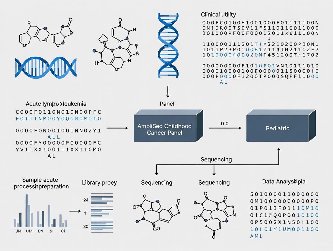
The AmpliSeq for Illumina Childhood Cancer Panel is a targeted next-generation sequencing solution specifically designed for comprehensive genomic profiling of childhood and young adult cancers [1] [7]. This integrated workflow employs PCR-based library preparation and Illumina sequencing-by-synthesis technology to simultaneously evaluate 203 genes associated with pediatric malignancies [7]. The panel detects multiple variant types including single nucleotide variants, insertions-deletions, copy number variants, and gene fusions from minimal input material (10 ng of DNA or RNA) [7].
Table 2: Technical Specifications of the AmpliSeq Childhood Cancer Panel
| Parameter | Specification | Clinical Utility in Pediatric Leukemia |
|---|---|---|
| Genes covered | 203 genes | Includes most relevant targets in pediatric leukemias |
| Input requirement | 10 ng high-quality DNA or RNA | Suitable for limited bone marrow samples |
| Assay time | 5-6 hours (library prep) | Rapid turnaround for clinical decision-making |
| Variant types detected | SNPs, indels, CNVs, gene fusions | Comprehensive genetic profiling in single assay |
| Specialized sample compatibility | Blood, bone marrow, FFPE | Flexible sample requirements |
| Instrument compatibility | MiSeq, NextSeq systems | Fits existing laboratory infrastructure |
Analytical Performance Metrics
Technical validation of the AmpliSeq Childhood Cancer Panel demonstrates excellent performance characteristics for pediatric leukemia applications. The assay achieves a mean read depth greater than 1000×, providing sufficient coverage for sensitive variant detection [1]. Validation studies report a high sensitivity of 98.5% for DNA variants with 5% variant allele frequency, and 94.4% sensitivity for RNA fusions [1]. The panel maintains 100% specificity and reproducibility for DNA analysis, with slightly lower reproducibility for RNA (89%) [1]. This performance enables reliable detection of clinically relevant alterations that might be missed by conventional diagnostic approaches.
Diagram 1: AmpliSeq Childhood Cancer Panel Workflow. The integrated process from sample collection to clinical reporting demonstrates the streamlined workflow for comprehensive genetic profiling of pediatric leukemias.
Comparison with Conventional Diagnostic Approaches
Conventional diagnosis of pediatric leukemia typically involves a combination of karyotype analysis, fluorescence in situ hybridization, and polymerase chain reaction-based methods [6]. These approaches have inherent limitations including the inability to identify cryptic gene fusions by karyotyping, the need for targeted probes in FISH, and requirement for pre-designed primers in RT-PCR [6]. Studies implementing the AmpliSeq panel in pediatric AML have demonstrated that the majority of clinically significant aberrations (63% in one series) were identified exclusively by NGS and not through conventional methods [6].
The comprehensive nature of the AmpliSeq panel addresses these limitations by enabling simultaneous detection of multiple variant types in a single assay. This unified approach is particularly valuable for identifying rare genetic subtypes and unusual fusion partners that might escape detection by targeted methods [6]. Additionally, the panel's ability to detect secondary abnormalities in genes such as TP53 and NRAS provides a more complete genetic profile to guide risk-adapted therapy [6].
Clinical Utility in Pediatric Leukemia Management
Impact on Diagnostic Refinement and Risk Stratification
Implementation of the AmpliSeq Childhood Cancer Panel has demonstrated significant impact on refining diagnoses and improving risk stratification in pediatric leukemia. Validation studies report that 49% of mutations and 97% of fusions identified by the panel had clinical impact, with 41% of mutations refining diagnosis and 49% considered targetable [1]. Overall, the panel detected clinically relevant findings in 43% of patients tested across a cohort of pediatric acute leukemia cases [1].
In clinical practice, NGS testing has directly influenced transplantation decisions, with studies reporting that specific findings such as NUP98::NSD1 and KMT2A::MLLT10 fusions identified exclusively by the panel led to referral for hematopoietic stem cell transplantation in first remission for patients who otherwise lacked poor prognostic factors [6]. These findings highlight how comprehensive molecular profiling can uncover high-risk features not apparent through conventional testing alone.
Diagram 2: Molecular Profiling Informs Risk Stratification. The AmpliSeq panel detects diverse genetic alterations that directly influence risk classification and subsequent treatment decisions in pediatric acute leukemia.
Therapeutic Implications and Treatment Selection
The comprehensive genetic profiling enabled by the AmpliSeq panel directly supports precision medicine approaches in pediatric leukemia. Identification of targetable mutations facilitates selection of appropriate targeted therapies, including FLT3 inhibitors for FLT3-mutated AML, BCR::ABL1 tyrosine kinase inhibitors for Philadelphia chromosome-positive ALL, and emerging therapies such as menin inhibitors for KMT2A-rearranged leukemias and UBTF-TD AML [4] [2]. The panel's ability to detect unusual fusion partners and rare breakpoints expands the population eligible for these targeted approaches [6].
For patients with relapsed or refractory disease, NGS profiling can identify resistance mechanisms and guide salvage therapy selection. In B-ALL patients failing CD19-CAR-T cell therapy, molecular characterization helps distinguish between CD19-positive and CD19-negative relapses, with the latter associated with significantly worse survival (30% at 12 months versus 68% for CD19-positive relapse) [5]. This distinction is critical for selecting appropriate subsequent therapies, which may include CD22-directed agents or second CAR-T infusions [5].
Essential Research Reagent Solutions for Pediatric Leukemia Investigation
Table 3: Key Research Reagents for Pediatric Leukemia Molecular Profiling
| Reagent/Kit | Manufacturer | Primary Function | Application in Validation Studies |
|---|---|---|---|
| AmpliSeq for Illumina Childhood Cancer Panel | Illumina | Targeted NGS library preparation for 203 genes | Comprehensive variant detection in pediatric leukemias [1] |
| AllPrep DNA/RNA Mini Kit | QIAGEN | Simultaneous purification of genomic DNA and total RNA | Nucleic acid extraction from bone marrow/blood [6] |
| SeraSeq Tumor Mutation DNA Mix | SeraCare | Multiplex biosynthetic positive control for DNA variants | Sensitivity and specificity assessment [1] |
| SeraSeq Myeloid Fusion RNA Mix | SeraCare | Synthetic RNA fusion positive control | RNA fusion detection performance validation [1] |
| AmpliSeq Library PLUS | Illumina | Library preparation reagents | Construction of sequencing libraries [7] |
| AmpliSeq CD Indexes | Illumina | Sample barcoding | Multiplexed sequencing of multiple samples [7] |
| AmpliSeq cDNA Synthesis for Illumina | Illumina | Reverse transcription of RNA to cDNA | RNA fusion analysis workflow [7] |
The profound heterogeneity of pediatric acute leukemia presents both a challenge and opportunity for precision medicine approaches. The AmpliSeq Childhood Cancer Panel addresses this complexity by providing comprehensive molecular profiling that refines diagnosis, improves risk stratification, and guides therapeutic decisions. Technical validation demonstrates excellent sensitivity and specificity for detecting clinically relevant variants, with significant impact on patient management observed in real-world clinical series.
As the molecular landscape of pediatric leukemia continues to be elucidated, integrated genomic profiling using targeted NGS panels represents an essential tool for advancing research and clinical care. The ability to identify both established and novel genetic alterations in a single efficient workflow positions this technology as a cornerstone of modern pediatric oncology practice, ultimately contributing to improved outcomes for children with these challenging malignancies.
The accurate and comprehensive genetic profiling of pediatric acute leukemia is a cornerstone of modern precision medicine, guiding risk stratification, treatment selection, and prognosis. For decades, diagnostic workflows have relied on a combination of conventional techniques—karyotyping (cytogenetic analysis), fluorescence in situ hybridization (FISH), and reverse transcription-polymerase chain reaction (RT-PCR). While these methods have been instrumental in identifying numerous stratifying genetic aberrations, they possess inherent limitations that can impact diagnostic precision. The emergence of next-generation sequencing (NGS) technologies, such as the AmpliSeq for Illumina Childhood Cancer Panel, offers a paradigm shift by integrating the detection of multiple variant types into a single, streamlined assay. This guide objectively compares the performance of conventional diagnostic tools with targeted NGS solutions within the context of pediatric acute leukemia research, providing supporting experimental data to illustrate the evolving landscape of molecular characterization.
Systematic Analysis of Conventional Tool Limitations
The limitations of conventional diagnostic techniques can be categorized into issues of resolution, throughput, and the fundamental need for a priori knowledge of the genetic alteration.
Limitations of Karyotyping
Karyotyping provides a global view of the chromosome complement but is constrained by several factors:
- Low Resolution and Cryptic Rearrangements: As a microscopic technique, karyotyping has a resolution limit of approximately 5-10 million base pairs, making it incapable of detecting submicroscopic chromosomal anomalies such as small deletions, duplications, or cryptic rearrangements that do not alter chromosomal banding patterns [8] [9].
- Dependence on Cell Culture and Mitotic Cells: Karyotyping requires fresh tissue with living, dividing cells and successful cell culture, which typically takes 1-10 days. This process can fail due to a lack of mitotic cells or be compromised by the overgrowth of normal supporting stromal cells, leading to a false-normal result [8]. A 2025 prospective study of 467 pediatric ALL patients reported that karyotyping was conclusive for only 64% of patients, significantly lower than molecular methods [10].
- Inability to Detect Balanced Rearrangements without Copy Number Change: While karyotyping can identify balanced translocations, it cannot reveal the specific genes involved at the breakpoints without subsequent testing. Furthermore, CNV-seq, a molecular technique, demonstrates a higher detection rate for chromosomal abnormalities (26.0%) compared to karyotyping (22.6%) in prenatal diagnostics, underscoring the limitations of conventional cytogenetics [9].
Limitations of Fluorescence In Situ Hybridization (FISH)
FISH offers higher resolution than karyotyping but introduces other constraints:
- Targeted Nature and Need for Prior Knowledge: FISH is not an agnostic discovery tool; it requires a pre-existing suspicion of a specific aberration to select the correct DNA probe. This means it will only find what it is designed to look for, potentially missing novel or unexpected fusions [10] [6].
- Probe Availability and Throughput: Each genetic abnormality requires a unique, specific FISH probe. Screening for numerous potential aberrations becomes labor-intensive, costly, and requires a large sample volume, which is often limited in pediatric cases [8] [6].
- Limited Resolution and False-Negatives in Cryptic Fusions: While FISH resolution is higher than karyotyping, it may still fail to detect very small rearrangements or gene fusions where the breakpoints fall outside the probe's target region [6].
Limitations of Polymerase Chain Reaction (PCR) and RT-PCR
PCR-based methods are highly sensitive but have specific blind spots:
- Dependence on Known Fusion Partners and Primers: RT-PCR is exquisitely targeted, requiring precise knowledge of the fusion partners and breakpoints to design effective primers. It will yield false-negative results for fusions involving alternative exons or novel partner genes. In the 467-patient ALL study, RT-PCR was false-negative for six patients who had fusions involving alternatively fused exons [10].
- Inability to Detect Unknown or Complex Rearrangements: Like FISH, RT-PCR is ineffective for discovering new genetic lesions. A case study in AML highlights this pitfall: despite strong clinical and morphological suspicion of a
KAT6A::CREBBPfusion, RNA-sequencing bioinformatic tools failed to identify it among hundreds of other fusion transcripts, and the fusion was only confirmed by targeted RT-PCR [11]. This illustrates that PCR, while a powerful confirmatory tool, is not suited for unbiased screening.
Table 1: Comparative Limitations of Conventional Diagnostic Tools in Pediatric Leukemia
| Diagnostic Tool | Key Technical Limitations | Impact on Diagnostic Yield | Evidence from Literature |
|---|---|---|---|
| Karyotyping | Low resolution (5-10 Mb); requires cell culture and mitotic cells; cannot detect submicroscopic changes [8]. | Conclusive in only 64% of pediatric ALL cases; misses cryptic abnormalities [10]. | 22.6% abnormality detection rate vs. 26.0% for CNV-seq in a comparative study [9]. |
| FISH | Targeted approach requires prior knowledge; limited probe availability; cannot discover novel fusions [10] [6]. | May miss fusions not covered by the probe set; 96% conclusiveness in a prospective cohort [10]. | In pediatric AML, FISH is not routinely performed in some settings due to economic limitations [6]. |
| RT-PCR | Requires precise primer design for known fusions/breakpoints; false negatives with alternative exons [10]. | False-negative in ~1.3% of pediatric ALL cases due to alternative exon fusions [10]. | Pathogenetic fusions can be missed by discovery methods and require targeted PCR for confirmation [11]. |
The Integrated NGS Solution: AmpliSeq Childhood Cancer Panel
Targeted next-generation sequencing panels like the AmpliSeq for Illumina Childhood Cancer Panel are designed to overcome the limitations of the conventional diagnostic workflow. This panel uses an integrated DNA and RNA approach to analyze a comprehensive set of genetic alterations in a single assay.
Technical Workflow and Validation
The experimental protocol for the AmpliSeq Childhood Cancer Panel involves a parallel analysis of DNA and RNA from patient samples.
Detailed Methodology:
- Nucleic Acid Extraction: DNA and RNA are co-extracted from patient samples, which can include bone marrow, peripheral blood, or formalin-fixed paraffin-embedded (FFPE) tissue. Input requirements are low, as little as 10-20 ng of DNA and RNA [1] [7].
- Library Preparation: For DNA, 100 ng is used to generate amplicons covering full exons, hotspot regions, and copy number variant (CNV) targets. For RNA, 100 ng is reverse-transcribed to cDNA, which is then used to generate amplicons targeting 1,421 specific gene fusion pairs. Libraries are prepared using a PCR-based protocol with sample-specific barcodes [1].
- Sequencing and Analysis: DNA and RNA libraries are pooled at a defined ratio (e.g., 5:1) and sequenced on an Illumina platform (e.g., MiSeq, NextSeq). Subsequent bioinformatic analysis aligns sequences to a reference genome (hg19) to identify single nucleotide variants (SNVs), insertions/deletions (indels), CNVs, and gene fusions [1].
Validation Metrics: A 2022 validation study of this panel demonstrated robust performance, with a mean read depth of >1000x. The assay showed a sensitivity of 98.5% for DNA variants (at 5% variant allele frequency) and 94.4% for RNA fusions, with 100% specificity for DNA and 89% reproducibility for RNA [1] [12].
Performance Comparison and Clinical Utility
The agnostic nature of the AmpliSeq panel allows it to detect a wider range of alterations than conventional methods.
- Superior Detection of Fusions and Mutations: The panel identified gene fusions and mutations with high clinical impact. In one study, 97% of the fusions and 49% of the mutations identified by the panel were demonstrated to have clinical impact, refining diagnosis or revealing targetable lesions [1]. In an 11-patient pediatric AML cohort, NGS uncovered diverse aberrations (fusions, indels, CNVs, SNVs) that were largely missed by conventional methods, directly influencing the decision to pursue hematopoietic stem cell transplantation in two cases [6].
- Comprehensive View in a Single Assay: Research shows that combining RNA-seq (for fusions) and SNP array (for CNVs and aneuploidies) outperforms the classic combination of FISH, karyotyping, and MLPA, detecting all stratifying genetic aberrations in ALL with high conclusiveness (97% for RNAseq, 99% for SNP array) and a turnaround time of under 15 days [10]. The AmpliSeq panel integrates these capabilities into a unified workflow.
Table 2: Experimental Data Comparison: Conventional Workflow vs. Targeted NGS Panel
| Performance Metric | Conventional Tools (Karyotyping, FISH, PCR) | AmpliSeq Childhood Cancer Panel | Supporting Data |
|---|---|---|---|
| Analytical Sensitivity (Fusions) | High for known targets, but false negatives occur [10]. | 94.4% sensitivity for RNA fusions [1]. | Validation using commercial fusion RNA controls [1]. |
| Turnaround Time (Median) | Variable; 9 days (FISH), <7 days (RT-PCR/MLPA), 10 days (SNP array) [10]. | Library prep ~5-6 hours; total time to results is days [7]. | Data from a real-world prospective study of 467 patients [10]. |
| Conclusiveness / Failure Rate | Karyotyping: 64% conclusive; FISH/RNAseq failures do not always overlap [10]. | Robust performance with low input (10 ng DNA/RNA); high success rate [1] [7]. | A combined RNAseq+SNP array approach was >96% conclusive [10]. |
| Clinical Impact (Findings) | Limited by targeted nature. | 43% of patients tested had clinically relevant results [1]. | Study of 76 pediatric AL patients; 97% of found fusions refined diagnosis [1]. |
Visualizing Diagnostic Workflows
The following diagram illustrates the streamlined, agnostic nature of the NGS workflow compared to the parallel, targeted pathways required by conventional methods.
The Scientist's Toolkit: Essential Research Reagents and Materials
The implementation and validation of the AmpliSeq Childhood Cancer Panel require a suite of specific reagents and tools.
Table 3: Essential Research Reagent Solutions for Targeted NGS
| Reagent / Material | Function in Workflow | Specifications & Notes |
|---|---|---|
| AmpliSeq for Illumina Childhood Cancer Panel [7] | Core panel for library prep; contains primers for 203 genes associated with pediatric cancer. | Includes 97 gene fusions, 82 DNA variants, 44 full exon coverage, and 24 CNV targets. |
| AmpliSeq Library PLUS [7] | Reagents for preparing sequencing libraries from the amplicons generated by the panel. | Sold separately from the panel; available in 24, 96, and 384 reactions. |
| AmpliSeq CD Indexes [7] | Unique barcode adapters for multiplexing samples in a single sequencing run. | Essential for cost-effective batch processing; multiple sets (A-D) are available. |
| AmpliSeq cDNA Synthesis for Illumina [7] | Converts total RNA to cDNA, which is required for the RNA-based fusion detection part of the panel. | Must be purchased separately for RNA analysis. |
| SeraSeq Myeloid Fusion RNA Mix [1] | Commercial positive control containing synthetic RNA fusions (e.g., ETV6::ABL1, RUNX1::RUNX1T1). |
Used for assay validation, monitoring sensitivity, and establishing limits of detection. |
| Qubit Fluorometer & Assay Kits [1] [6] | For accurate quantification of DNA and RNA input; more reliable for NGS than spectrophotometry. | Ensures adherence to the low input requirement (10-100 ng). |
| Illumina MiSeq/NextSeq Systems [1] [7] | Sequencing instruments compatible with the AmpliSeq panel for generating the sequencing data. | The choice of instrument affects sequencing depth and throughput. |
The conventional diagnostic toolkit of karyotyping, FISH, and PCR, while foundational, presents significant limitations in resolution, throughput, and discovery power for the molecular characterization of pediatric acute leukemia. Quantitative data from recent studies underscore issues with conclusiveness, false-negative rates, and the inability to provide a comprehensive genetic profile efficiently. The AmpliSeq Childhood Cancer Panel represents a validated, integrated NGS solution that addresses these gaps. By simultaneously assessing fusions, SNVs, indels, and CNVs from minimal nucleic acid input, it enhances diagnostic accuracy, reveals therapeutically relevant alterations, and provides a robust platform for research and clinical development in pediatric oncology. This transition to multiplexed genomic profiling is crucial for advancing precision medicine and improving outcomes for young patients with leukemia.
Precision medicine is revolutionizing pediatric oncology by moving beyond a one-size-fits-all approach to cancer treatment. This paradigm shift relies fundamentally on biomarkers—biological molecules, genes, or characteristics that provide critical information about a specific cancer. In pediatric acute leukemia, these biomarkers are increasingly guiding diagnosis, prognostic stratification, and therapeutic selection. The integration of comprehensive molecular profiling technologies, particularly next-generation sequencing (NGS) panels like the AmpliSeq for Illumina Childhood Cancer Panel, is accelerating this transformation by enabling simultaneous assessment of multiple biomarker classes from minimal sample input.
The AmpliSeq for Illumina Childhood Cancer Panel is a targeted NGS solution specifically designed for pediatric and young adult cancers. This panel simultaneously investigates 203 genes associated with childhood cancers through amplicon sequencing, requiring only 10 ng of high-quality DNA or RNA input. The library preparation process requires approximately 5-6 hours of total assay time with less than 1.5 hours of hands-on time, making it suitable for integration into clinical workflows [7].
The panel detects multiple variant classes crucial for leukemia management, including:
- Single nucleotide polymorphisms (SNPs)
- Insertions-deletions (indels)
- Gene fusions
- Copy number variants (CNVs)
- Somatic variants
This comprehensive approach allows researchers and clinicians to obtain extensive genetic information from limited specimen quantities, which is particularly valuable in pediatric settings where sample volume is often constrained.
Performance Validation in Pediatric Acute Leukemia
Analytical Performance Metrics
Rigorous technical validation studies have demonstrated the reliability of the AmpliSeq Childhood Cancer Panel for acute leukemia applications. A 2022 study comprehensively evaluated the panel's performance characteristics with the following results [1]:
Table 1: Analytical Performance Metrics of the AmpliSeq Childhood Cancer Panel
| Parameter | DNA Analysis | RNA Analysis |
|---|---|---|
| Sensitivity | 98.5% (for variants with 5% VAF) | 94.4% |
| Specificity | 100% | 100% |
| Reproducibility | 100% | 89% |
| Mean Read Depth | >1000× | >1000× |
The panel achieved a limit of detection (LOD) of 5% variant allele frequency (VAF) for DNA variants, demonstrating sufficient sensitivity to detect subclonal mutations that may have clinical significance. The high read depth (>1000×) provides confidence in variant calling, particularly important for detecting low-frequency variants in heterogeneous leukemia samples [1].
Experimental Protocol for Validation
The validation study followed a rigorous methodology to ensure robust performance assessment [1]:
Sample Selection: 76 pediatric patients diagnosed with BCP-ALL (n=51), T-ALL (n=11), and AML (n=14) were selected, prioritizing those with non-defining genetic results using conventional diagnostics.
Nucleic Acid Extraction: DNA was extracted using Gentra Puregene kit, QIAamp DNA Mini Kit, or QIAamp DNA Micro Kit. RNA was extracted using TriPure reagent or Direct-zol RNA MiniPrep. Quality assessment was performed via spectrophotometry (OD260/280 >1.8) and fragment analysis.
Library Preparation: Libraries were prepared using 100 ng of DNA or RNA according to manufacturer's instructions. RNA was reverse transcribed to cDNA using the AmpliSeq cDNA Synthesis kit. The panel generates 3,069 DNA amplicons and 1,701 RNA amplicons targeting fusion genes.
Sequencing: Barcoded libraries were pooled at a 5:1 DNA:RNA ratio and sequenced on MiSeq instruments.
Data Analysis: Variant calling was performed using Illumina's recommended pipelines, with results compared to conventional methods including Sanger sequencing, fragment analysis, and quantitative RT-PCR.
Clinical Utility and Biomarker Impact
The true value of any diagnostic platform lies in its ability to generate clinically actionable information. In the validation cohort, the AmpliSeq Childhood Cancer Panel demonstrated significant clinical impact [1]:
Table 2: Clinical Impact of Genetic Alterations Identified in Pediatric Acute Leukemia
| Impact Category | DNA Mutations | RNA Fusion Genes |
|---|---|---|
| Refined Diagnosis | 41% | 97% |
| Therapeutically Targetable | 49% | Information not specified |
| Overall Clinically Relevant Findings | 43% of patients |
The panel identified clinically relevant results in 43% of patients tested, with fusion genes proving particularly impactful for diagnostic refinement. This capability is crucial in acute leukemia, where accurate genetic classification directly influences risk stratification and treatment selection [1].
Comparison with Alternative Sequencing Approaches
While targeted panels like AmpliSeq offer a balanced approach for routine clinical use, other sequencing strategies provide complementary strengths. Research institutions are increasingly exploring integrated approaches for comprehensive genomic characterization.
Table 3: Comparison of Sequencing Approaches for Pediatric Leukemia Biomarker Discovery
| Sequencing Method | Key Advantages | Limitations | Best Application Context |
|---|---|---|---|
| Targeted Panels (AmpliSeq) | Fast turnaround (5-6 hr library prep), low DNA/RNA input (10 ng), focused clinically actionable content, established validation [7] [1] | Limited to pre-defined genes, may miss novel alterations | Routine clinical diagnostics, therapy selection |
| Whole Genome + Transcriptome Sequencing | Comprehensive view of all genomic alterations, detects novel fusions and structural variants [13] | Higher cost, complex data analysis, longer turnaround time | Complex cases, research discovery, comprehensive subclassification |
| Adaptive Whole-Genome Sequencing | Rapid results (48 hours), flexible target selection, cost-effective [14] | Emerging technology, limited clinical validation | Resource-limited settings, rapid diagnosis |
St. Jude Children's Research Hospital has demonstrated that integrating whole genome sequencing with whole transcriptome sequencing provides the most comprehensive diagnostic capability, particularly for identifying rare AML subtypes that may be missed by targeted approaches alone [13]. Meanwhile, researchers at UNC Lineberger have developed an adaptive whole-genome sequencing approach using nanopore technology that can deliver genomic classification of pediatric acute leukemia in as little as 48 hours, significantly faster than conventional methods [14].
Biomarker Classes in Pediatric Leukemia
Diagnostic and Prognostic Biomarkers
The AmpliSeq panel detects several critical biomarker classes that redefine leukemia classification and risk stratification:
Gene Fusions: Childhood acute leukemias are frequently characterized by recurrent gene fusions that serve as primary drivers and definitive diagnostic markers. The panel detects 97 different fusion types, including ETV6::RUNX1 in B-ALL, KMT2A rearrangements in infant leukemia, and RUNX1::RUNX1T1 in AML [15] [1].
Copy Number Variations: Specific copy number alterations provide important prognostic information in B-ALL, including deletions in genes like IKZF1, ETV6, and RB1, which are associated with higher relapse risk [15].
Therapeutic Biomarkers
The panel identifies numerous biomarkers with direct therapeutic implications:
Kinase Pathway Alterations: Mutations in genes like FLT3, JAK2, and ABL-class fusions can indicate susceptibility to targeted kinase inhibitors [15] [16].
Surface Antigens: While not directly detected by DNA/RNA sequencing, the molecular context provided by the panel complements immunophenotyping data for antigens like CD19, CD20, and CD22, which are targets for immunotherapy and CAR-T cell therapy [17] [18].
Signaling Pathways in Pediatric Leukemia
The clinical utility of biomarkers stems from their position within critical cancer signaling pathways. The diagram below illustrates key pathways and their therapeutic implications in pediatric acute leukemia:
The Scientist's Toolkit: Essential Research Reagents
Implementation of the AmpliSeq Childhood Cancer Panel requires specific companion products that facilitate different aspects of the workflow and accommodate various sample types:
Table 4: Essential Research Reagent Solutions for AmpliSeq Workflow
| Product Category | Specific Product | Function in Workflow |
|---|---|---|
| Library Preparation | AmpliSeq Library PLUS | Provides reagents for preparing sequencing libraries (24, 96, or 384 reactions) [7] |
| Sample Indexing | AmpliSeq CD Indexes Sets A-D | Enables multiplexing of up to 384 samples through unique barcode sequences [7] |
| RNA Sample Preparation | AmpliSeq cDNA Synthesis for Illumina | Converts total RNA to cDNA for RNA-based panel applications [7] |
| Sample Identification | AmpliSeq for Illumina Sample ID Panel | Genotypes research samples using SNP targets to verify sample identity and track quality [7] |
| FFPE Sample Processing | AmpliSeq for Illumina Direct FFPE DNA | Enables DNA preparation from FFPE tissues without deparaffinization or DNA purification [7] |
| Library Normalization | AmpliSeq Library Equalizer for Illumina | Simplifies library normalization through bead-based technology before pooling and sequencing [7] |
Biomarker-Driven Clinical Decision Making
The identification of biomarkers through the AmpliSeq panel directly influences patient management decisions. The clinical decision pathway below illustrates how biomarker results integrate into diagnostic and therapeutic algorithms:
Market Context and Adoption Trends
The growing emphasis on biomarker-driven oncology is reflected in market trends. The pediatric cancer biomarker market was valued at $830.41 million in 2023 and is projected to reach $1,635.68 million by 2032, with a compound annual growth rate of 7.84% [17] [18]. Leukemia represents the largest segment (41.2% share) due to its status as the most common childhood cancer and the well-established role of biomarkers like CD19, CD20, CD22, and minimal residual disease markers in clinical management [17] [18].
North America currently dominates the pediatric cancer biomarker market, holding 43.8% of the global share, driven by advanced healthcare infrastructure, significant R&D investment, and supportive regulatory initiatives like the RACE for Children Act [17]. However, the Asia-Pacific region is expected to witness the highest growth rate as healthcare infrastructure and biomarker awareness improve.
The AmpliSeq for Illumina Childhood Cancer Panel represents a significant advancement in the molecular characterization of pediatric acute leukemia, offering a validated, efficient platform for comprehensive biomarker assessment. By simultaneously detecting multiple variant classes from minimal input material, this targeted sequencing approach facilitates refined diagnosis, accurate risk stratification, and identification of therapeutic targets. While emerging technologies like whole-genome and adaptive sequencing offer complementary advantages, targeted panels provide an optimal balance of comprehensiveness, throughput, and clinical practicality for routine implementation. As precision medicine continues to evolve in pediatric oncology, integrated biomarker platforms will play an increasingly central role in optimizing outcomes for children with leukemia.
The AmpliSeq for Illumina Childhood Cancer Panel represents a significant advancement in the molecular characterization of pediatric and young adult cancers. This targeted next-generation sequencing (NGS) panel is specifically designed to investigate 203 genes associated with childhood cancers, providing a comprehensive solution for evaluating somatic variants across multiple pediatric cancer types, including leukemias, brain tumors, and sarcomas [7]. Unlike adult-focused cancer panels, this tool addresses the distinctive genetic landscape of pediatric malignancies, which typically feature a lower mutational burden but with clinically significant alterations [1].
The integrated workflow combines PCR-based library preparation with Illumina sequencing by synthesis (SBS) technology and automated analysis, creating a standardized approach for childhood cancer research [7]. The panel's design saves researchers considerable time and effort that would otherwise be spent identifying targets, designing primers, and optimizing panels, thereby accelerating genomic research in pediatric oncology [7]. A key advantage of this panel is its ability to detect multiple variant types—including single nucleotide polymorphisms (SNPs), gene fusions, somatic variants, insertions-deletions (indels), and copy number variants (CNVs)—from minimal input quantities of only 10 ng of high-quality DNA or RNA [7].
Technical Specifications and Panel Design
The technical architecture of the AmpliSeq Childhood Cancer Panel is optimized for comprehensive genomic profiling in pediatric oncology research. The panel employs an amplicon sequencing method that generates 3,069 amplicons per DNA sample with an average size of 114 bp, covering coding regions of the targeted genes [1]. Simultaneously, the RNA component targets 1,701 amplicons with an average size of 122 bp, specifically designed to detect 97 gene fusions relevant to childhood cancers [1].
Key Technical Features
- Input Requirements: Compatible with only 10 ng of high-quality DNA or RNA, facilitating analysis of precious pediatric samples [7]
- Hands-on Time: Less than 1.5 hours, with total assay time of 5-6 hours for library preparation [7]
- Sample Compatibility: Works with various specialized sample types including blood, bone marrow, and FFPE tissue [7]
- Instrument Flexibility: Compatible with multiple Illumina sequencing systems including MiSeq, NextSeq, and MiniSeq platforms [7]
Gene Content and Coverage
The panel's content is strategically curated to encompass genes with established roles in pediatric malignancies. The DNA component provides coverage for 82 DNA variants and 44 full exons, while also targeting 24 genes for copy number variant analysis [1]. This comprehensive design ensures researchers can simultaneously assess multiple alteration types from a single sample, conserving valuable biomaterial that is often limited in pediatric cases.
Table 1: Technical Specifications of the AmpliSeq Childhood Cancer Panel
| Parameter | Specification |
|---|---|
| Number of Genes | 203 genes |
| Input Quantity | 10 ng high-quality DNA or RNA |
| Assay Time | 5-6 hours (library prep only) |
| Hands-on Time | <1.5 hours |
| Amplicon Count | 3,069 DNA amplicons; 1,701 RNA amplicons |
| Variant Types Detected | SNPs, gene fusions, somatic variants, indels, CNVs |
| Compatible Instruments | MiSeq, NextSeq 550, NextSeq 2000, NextSeq 1000, MiniSeq |
Performance Validation in Pediatric Acute Leukemia
Analytical Performance Metrics
Rigorous technical validation of the AmpliSeq Childhood Cancer Panel has demonstrated exceptional performance characteristics specifically for pediatric acute leukemia applications. A comprehensive study assessing the panel's capabilities reported a mean read depth greater than 1000×, providing sufficient coverage for reliable variant calling [1]. The panel exhibited high sensitivity for both DNA (98.5% for variants with 5% variant allele frequency) and RNA (94.4%), with 100% specificity and reproducibility for DNA and 89% reproducibility for RNA components [1] [12].
The validation utilized commercial controls including SeraSeq Tumor Mutation DNA Mix and SeraSeq Myeloid Fusion RNA Mix to establish accuracy metrics [1]. The DNA positive control contained biosynthetic mixtures of clinically relevant DNA variants at an average variant allele frequency of 10%, spanning 22 key cancer genes including AKT1, BRAF, FLT3, NRAS, and TP53 [1]. The RNA control included synthetic fusions relevant to leukemia such as ETV6::ABL1, TCF3::PBX1, BCR::ABL1, RUNX1::RUNX1T1, and PML::RARA [1].
Comparative Performance Against Alternative Methods
When compared to conventional molecular techniques, the AmpliSeq Childhood Cancer Panel demonstrates superior comprehensive profiling capabilities. Traditional methods for pediatric leukemia characterization typically require multiple separate tests including:
- FLT3-ITD and NPM1 analysis by labeled-PCR amplification
- FLT3 tyrosine kinase domain, cKIT, and GATA1 mutations by Sanger sequencing
- Fusion gene detection by quantitative RT-PCR with specific primers and probes [1]
This fragmented approach is labor-intensive, time-consuming, and requires significant sample material. In contrast, the AmpliSeq panel consolidates these analyses into a single workflow, simultaneously evaluating 203 genes while conserving sample resources [1].
Table 2: Performance Metrics of the AmpliSeq Childhood Cancer Panel in Leukemia Research
| Performance Metric | DNA Analysis | RNA Analysis |
|---|---|---|
| Sensitivity | 98.5% (for variants with 5% VAF) | 94.4% |
| Specificity | 100% | Not specified |
| Reproducibility | 100% | 89% |
| Mean Read Depth | >1000× | >1000× |
| Limit of Detection | Established with commercial controls | Established with commercial controls |
Comparison with Alternative Pediatric Cancer Panels
While several NGS panels are available for cancer genomics, few are specifically optimized for pediatric malignancies. The AmpliSeq Childhood Cancer Panel stands alongside other specialized tools like the OncoKids panel, which is also designed specifically for pediatric cancers [19]. Understanding the competitive landscape helps researchers select the most appropriate tool for their specific research needs.
OncoKids Panel Comparison
The OncoKids panel is another amplification-based NGS assay designed to detect diagnostic, prognostic, and therapeutic markers across the spectrum of pediatric malignancies, including leukemias, sarcomas, brain tumors, and embryonal tumors [19]. This panel requires 20 ng of DNA and 20 ng of RNA as input and is compatible with formalin-fixed, paraffin-embedded and frozen tissue, bone marrow, and peripheral blood [19].
Key differences in the OncoKids panel content include:
- DNA coverage of 44 cancer predisposition loci, tumor suppressor genes, and oncogenes
- Mutation hotspots in 82 genes
- Amplification events in 24 genes
- RNA content targeting 1,421 gene fusions [19]
Both panels share similar applications in pediatric cancer research, but the AmpliSeq Childhood Cancer Panel has been specifically validated for acute leukemia diagnostics, with demonstrated clinical utility in refining diagnosis, prognosis, and treatment strategies [1].
Comprehensive Genomic Profiling Alternatives
The broader field of comprehensive genomic profiling (CGP) offers additional alternatives, though primarily focused on adult cancers. Leading CGP specialists include:
- Foundation Medicine: Offers FoundationOne CDx for solid tumors and FoundationOne Heme for blood cancers [20]
- Caris Life Sciences: Provides molecular profiling services assessing DNA, RNA, and protein signatures [20]
- Tempus: Deploys AI-powered CGP panels covering both DNA and RNA sequencing [20]
These platforms typically utilize different technological approaches and may have less optimized content for pediatric-specific malignancies compared to the purpose-built AmpliSeq Childhood Cancer Panel.
Clinical Utility in Pediatric Acute Leukemia
Impact on Diagnostic Refinement
The implementation of the AmpliSeq Childhood Cancer Panel has demonstrated significant clinical utility in the molecular characterization of pediatric acute leukemia. Validation studies involving 76 pediatric patients diagnosed with B-cell precursor ALL (n=51), T-ALL (n=11), and AML (n=14) revealed that the panel identified clinically relevant results in 43% of patients tested in the cohort [1] [12]. This represents a substantial improvement over conventional diagnostic approaches, particularly for cases with non-defining genetic results using standard methodologies.
The clinical impact varied by alteration type, with 97% of the fusion genes identified demonstrating clinical significance for diagnostic refinement [1]. For mutations, 49% were considered targetable, while 41% refined diagnostic classification [1] [12]. This high rate of clinical impact underscores the panel's value in precision medicine approaches to pediatric leukemia, where accurate molecular characterization directly influences risk stratification and treatment selection.
Practical Workflow Integration
The practical integration of the AmpliSeq Childhood Cancer Panel into routine research workflows is facilitated by its streamlined process. The following diagram illustrates the complete experimental workflow from sample to data analysis:
Essential Research Reagent Solutions
Successful implementation of the AmpliSeq Childhood Cancer Panel requires several specialized reagents and components that form the complete research solution:
Table 3: Essential Research Reagent Solutions for Panel Implementation
| Component | Function | Key Specifications |
|---|---|---|
| AmpliSeq Library PLUS | Library preparation reagents | Available in 24, 96, or 384 reactions [7] |
| AmpliSeq CD Indexes | Sample barcoding for multiplexing | 8 bp indexes in Sets A-D; sufficient for 96 samples per set [7] |
| AmpliSeq cDNA Synthesis | Converts total RNA to cDNA | Required for RNA panels; includes reaction mix and enzyme blend [7] |
| AmpliSeq Library Equalizer | Normalizes libraries for sequencing | Contains beads and reagents for library normalization [7] |
| AmpliSeq Direct FFPE DNA | DNA preparation from FFPE tissues | 24 reactions for slide-mounted FFPE tissues without deparaffinization [7] |
Molecular Pathways in Pediatric Acute Leukemia
The AmpliSeq Childhood Cancer Panel targets genes involved in critical signaling pathways dysregulated in pediatric leukemia. Understanding these pathways helps contextualize the panel's design and clinical utility. The following diagram illustrates key pathways and gene interactions detected by the panel:
The AmpliSeq Childhood Cancer Panel represents a significant advancement in pediatric cancer genomics, offering researchers a optimized tool for comprehensive molecular characterization of childhood leukemias and other malignancies. With its targeted design covering 203 relevant genes, ability to detect multiple variant types, and demonstrated clinical utility in refining diagnoses and identifying targetable alterations, this panel addresses a critical need in pediatric oncology research.
The validation data confirms its robust performance characteristics, including high sensitivity and specificity, making it suitable for implementation in research settings aiming to translate genomic findings into improved patient outcomes. As precision medicine continues to transform pediatric oncology, purpose-built tools like the AmpliSeq Childhood Cancer Panel will play an increasingly vital role in unraveling the molecular complexity of childhood cancers and accelerating the development of targeted therapeutic strategies.
For researchers selecting genomic profiling tools for pediatric leukemia studies, the AmpliSeq Childhood Cancer Panel offers a balanced approach—providing comprehensive content across relevant genes while maintaining practical workflow requirements compatible with diverse sample types typically available in pediatric settings.
The treatment of pediatric acute leukemia (AL) has been revolutionized by the comprehensive molecular profiling of tumor genomes, which offers invaluable insights for diagnostic refinement, prognostic stratification, and therapeutic targeting [21]. Next-generation sequencing (NGS) technologies have enabled the parallel analysis of numerous genetic alterations, overcoming the limitations of traditional single-gene testing methods that are often laborious, require large amounts of DNA, and present challenges in consolidating results for clinical reporting [1] [22]. Pediatric leukemias, while exhibiting a lower mutational burden compared to adult cancers, harbor clinically relevant genomic alterations including single nucleotide variants (SNVs), insertions-deletions (indels), copy number variants (CNVs), and gene fusions that drive oncogenesis [1] [21]. These alterations affect critical signaling pathways, transcriptional regulators, and epigenetic modifiers, creating opportunities for precision medicine approaches [23].
Targeted NGS panels, such as the AmpliSeq for Illumina Childhood Cancer Panel, have been developed specifically to address the unique genetic landscape of childhood and young adult cancers [7] [1]. This panel is designed as a targeted resequencing solution for comprehensive evaluation of somatic variants across multiple pediatric cancer types, including leukemias, brain tumors, and sarcomas [7]. By consolidating the analysis of multiple variant types into a single assay, it provides researchers and clinicians with an efficient tool for genomic characterization while saving the time and effort associated with identifying targets, designing primers, and optimizing panels [7]. This article examines the technical performance, clinical utility, and practical application of the AmpliSeq Childhood Cancer Panel within the broader context of pediatric leukemia research, comparing its capabilities with alternative genomic assessment approaches.
The AmpliSeq for Illumina Childhood Cancer Panel is a PCR-based targeted sequencing assay that simultaneously analyzes 203 genes associated with childhood and young adult cancers [7] [1]. The panel content is strategically designed to cover major variant classes relevant to pediatric malignancies through 3,069 DNA amplicons and 1,701 RNA amplicons, with average sizes of 114 bp and 122 bp, respectively [1]. The DNA component targets 82 genes for hotspot mutations, 44 genes for full exon coverage, and 24 genes for CNV analysis, while the RNA component targets 97 gene fusions known to be clinically significant in pediatric cancers [7] [1].
The assay demonstrates practical utility in clinical research settings with a relatively short hands-on time of less than 1.5 hours and a total assay time of 5-6 hours for library preparation alone [7]. The panel requires only 10 ng of high-quality DNA or RNA as input, making it suitable for precious tumor samples with limited material [7]. It is compatible with various Illumina sequencing platforms, including MiSeq, NextSeq, and MiniSeq systems, and can be used with diverse sample types including blood, bone marrow, and formalin-fixed paraffin-embedded (FFPE) tissue [7]. For RNA fusion detection, a prerequisite cDNA synthesis step is required using the AmpliSeq cDNA Synthesis for Illumina kit to convert total RNA to cDNA [7].
Table 1: Technical Specifications of the AmpliSeq Childhood Cancer Panel
| Parameter | Specification |
|---|---|
| Target Genes | 203 genes [7] |
| Variant Types Detected | Single nucleotide variants (SNVs), Insertions-deletions (Indels), Copy number variants (CNVs), Gene fusions [7] |
| DNA Targets | 82 DNA variants, 44 full exon coverage, 24 CNVs [1] |
| RNA Targets | 97 gene fusions [1] |
| Input Requirement | 10 ng high-quality DNA or RNA [7] |
| Hands-on Time | <1.5 hours [7] |
| Total Assay Time (Library Prep) | 5-6 hours [7] |
| Compatible Instruments | MiSeq, NextSeq 550, NextSeq 1000/2000, MiniSeq Systems [7] |
Analytical Performance and Validation
Rigorous technical validation studies have demonstrated that the AmpliSeq Childhood Cancer Panel achieves high sensitivity and specificity for detecting genomic alterations in pediatric leukemia samples. In a comprehensive validation study focused on pediatric acute leukemia, the panel demonstrated a 98.5% sensitivity for DNA variants with a variant allele frequency (VAF) of 5%, and 94.4% sensitivity for RNA fusions [1]. The assay also showed excellent specificity (100%) and reproducibility for DNA, while RNA reproducibility was slightly lower at 89% [1].
The panel achieves a mean read depth greater than 1000×, providing sufficient coverage for accurate variant calling [1]. For clinical reporting, the laboratory at KK Women's and Children's Hospital has established a cutoff of 10% variant allele frequency for DNA variants and a minimum sequencing coverage of 100x, though the DNA component does not reliably detect variants occurring at allele frequencies below 10% [24]. The panel requires tumor content greater than 50% in samples for optimal performance [24].
When compared to other sequencing approaches, the AmpliSeq panel demonstrates robust performance in detecting multiple variant types simultaneously. The MiSeq platform, which is compatible with this panel, has shown quantitative accuracy for mutation detection down to an allelic frequency of 1.5% in dilution studies, suggesting potential utility for monitoring minimal residual disease and clonal evolution [22]. However, standard analysis pipelines may have limitations in detecting certain alteration types such as FLT3 internal tandem duplications (ITDs), which require specialized indel detection algorithms for optimal identification [22].
Table 2: Analytical Performance of the AmpliSeq Childhood Cancer Panel in Pediatric Leukemia
| Performance Metric | Result | Experimental Details |
|---|---|---|
| DNA Sensitivity | 98.5% | For variants at 5% VAF using commercial controls [1] |
| RNA Sensitivity | 94.4% | For fusion detection using commercial controls [1] |
| Specificity | 100% | For both DNA and RNA components [1] |
| DNA Reproducibility | 100% | Concordance between replicate experiments [1] |
| RNA Reproducibility | 89% | Concordance between replicate experiments [1] |
| Mean Read Depth | >1000× | Achieved across targeted regions [1] |
| Limit of Detection | 10% VAF | Established as clinical reporting cutoff [24] |
Comparison with Alternative NGS Approaches
The AmpliSeq Childhood Cancer Panel occupies a specific niche in the landscape of genomic profiling tools for pediatric malignancies. When compared to other available NGS approaches, each platform demonstrates distinct advantages and limitations based on their technical design and application scope.
The OncoKids panel represents another amplification-based NGS assay designed specifically for pediatric malignancies, with comparable input requirements (20 ng DNA and RNA) and similar compatibility with various sample types including FFPE tissue, bone marrow, and peripheral blood [19]. Like the AmpliSeq panel, OncoKids covers a comprehensive range of alteration types including mutations in cancer predisposition genes, hotspot mutations, gene amplification events, and gene fusions [19].
Larger comprehensive genomic profiling (CGP) approaches, such as those utilized in major precision medicine platforms like ZERO Childhood Cancer, INFORM, and MAPPYACTS, often employ whole-exome sequencing (WES), whole-genome sequencing (WGS), and RNA sequencing [21] [23]. These more extensive profiling methods can identify novel alterations beyond predefined gene panels and are particularly valuable for rare tumor types or cases with unclear driver mutations. However, they typically require longer turnaround times (3-6 weeks), higher costs, and more complex bioinformatics infrastructure [21] [23].
Targeted panels like the AmpliSeq Childhood Cancer Panel offer advantages in terms of faster turnaround time (4-6 weeks in clinical practice), lower cost, and deeper sequencing coverage of specific clinically relevant genes [24]. The focused nature of these panels makes them particularly suitable for routine diagnostic applications where established biomarkers guide clinical decision-making. A recent systematic review and meta-analysis of NGS applications in childhood and AYA solid tumors reported that targeted sequencing approaches successfully identify actionable alterations in 57.9% of cases and influence clinical decision-making in 22.8% of patients [23].
Table 3: Comparison of Genomic Profiling Approaches in Pediatric Cancer
| Parameter | AmpliSeq Childhood Cancer Panel | OncoKids Panel | Comprehensive Genomic Profiling (WES/WGS/RNAseq) |
|---|---|---|---|
| Technology | Amplicon-based targeted sequencing | Amplification-based NGS | Whole exome/genome and transcriptome sequencing |
| Gene Content | 203 genes | 44 predisposition genes, 82 hotspots, 24 CNV genes, 1421 fusions | Entire exome/genome and transcriptome |
| Input Requirements | 10 ng DNA or RNA | 20 ng DNA and RNA | Typically >50-100 ng DNA and RNA |
| Turnaround Time | 4-6 weeks (clinical) | Not specified | 3-6 weeks [21] |
| Key Advantage | Focused content, fast hands-on time | Pediatric-specific content | Unbiased discovery of novel alterations |
Experimental Protocol and Research Toolkit
Implementing the AmpliSeq Childhood Cancer Panel in a research setting requires specific laboratory protocols and reagents. The following section outlines the standard methodology and essential research tools required for successful assay execution.
Library Preparation and Sequencing Workflow
The library preparation process begins with quality assessment of input nucleic acids. DNA and RNA purity should be determined by spectrophotometry (OD260/280 ratio >1.8), with concentration measured by fluorometric quantification [1]. For the DNA library, 100 ng of DNA is used to generate 3,069 amplicons, while 100 ng of RNA is reverse-transcribed to cDNA using the AmpliSeq cDNA Synthesis kit before generating 1,701 amplicons targeting fusion genes [1].
Library preparation involves consecutive PCR amplifications to create amplicon libraries with specific barcodes for each sample [1]. After cleanup steps, quality controls are performed to assess library integrity. DNA and RNA libraries are typically pooled at a 5:1 ratio (DNA:RNA), diluted to appropriate concentrations (17-20 pM), and sequenced on a MiSeq or other compatible Illumina sequencer [1]. The entire workflow from nucleic acid extraction to sequencing data can be completed within several days, with actual hands-on time of less than 1.5 hours [7].
Essential Research Reagent Solutions
Successful implementation of the AmpliSeq Childhood Cancer Panel requires several key reagents and components that constitute the core research toolkit. The table below details these essential materials and their functions in the experimental workflow.
Table 4: Research Reagent Solutions for AmpliSeq Childhood Cancer Panel
| Reagent/Component | Function | Specifications |
|---|---|---|
| AmpliSeq Childhood Cancer Panel | Core primer pool for targeting 203 cancer-related genes | 24 reactions; contains primers for 3,069 DNA and 1,701 RNA amplicons [7] |
| AmpliSeq Library PLUS | Reagents for library preparation | Available in 24, 96, or 384 reactions [7] |
| AmpliSeq CD Indexes | Sample barcoding for multiplexing | 8 bp indexes; available in sets A-D (384 total indexes) [7] |
| AmpliSeq cDNA Synthesis | Converts total RNA to cDNA for RNA panels | Required for RNA fusion detection; includes reaction mix and enzyme blend [7] |
| AmpliSeq Library Equalizer | Normalizes libraries for sequencing | Includes beads and reagents for library normalization [7] |
| AmpliSeq for Illumina Direct FFPE DNA | DNA preparation from FFPE tissue | 24 reactions for DNA preparation without deparaffinization [7] |
Clinical Utility in Pediatric Leukemia
The true value of genomic profiling in pediatric leukemia lies in its ability to inform clinical decision-making and improve patient outcomes. Studies evaluating the AmpliSeq Childhood Cancer Panel have demonstrated significant clinical impact in pediatric acute leukemia. In one validation study, the panel identified clinically relevant results in 43% of patients tested, with 49% of mutations and 97% of the fusions demonstrating clinical impact [1]. Specifically, 41% of mutations refined diagnosis, while 49% were considered targetable [1]. For RNA fusions, an impressive 97% had diagnostic refinement value [1].
The panel's comprehensive approach enables simultaneous assessment of multiple therapeutic and prognostic biomarkers, which is particularly valuable in pediatric leukemias where complex genetic interactions influence disease behavior and treatment response [22]. The identification of targetable mutations has created opportunities for precision-guided therapies (PGT) in relapsed or refractory cases, where conventional treatment options are limited [21]. Major precision oncology platforms have reported that patients receiving molecularly targeted therapies based on high-level evidence demonstrate meaningful responses and survival benefit, particularly when treatment is initiated early in the disease course [21].
Beyond therapeutic targeting, the panel contributes to refined diagnostic classification and risk stratification in pediatric leukemias. The identification of specific fusion genes and mutations can define distinct leukemia subtypes with implications for prognosis and treatment intensity [22]. Additionally, comprehensive profiling can occasionally uncover germline predisposition variants even when not specifically targeted, highlighting the importance of appropriate genetic counseling in the pediatric oncology setting [23] [24].
The AmpliSeq for Illumina Childhood Cancer Panel represents a strategically designed targeted sequencing solution that addresses the distinctive genomic landscape of pediatric leukemias and other childhood malignancies. By enabling the simultaneous detection of SNVs, indels, CNVs, and fusion genes in a single efficient assay, it provides researchers and clinicians with a comprehensive tool for genomic characterization. Technical validation studies confirm its high sensitivity, specificity, and reproducibility for identifying clinically relevant alterations in pediatric leukemia samples [1].
When compared to alternative genomic approaches, the panel offers a balanced combination of content relevance, workflow efficiency, and practical feasibility for implementation in clinical research settings. While larger comprehensive genomic profiling methods can identify novel alterations beyond predefined gene content, targeted panels like AmpliSeq provide deeper coverage of established biomarkers with faster turnaround times and lower costs [21] [23]. The demonstrable clinical utility of the panel, with significant proportions of identified alterations refining diagnosis or suggesting targeted therapy approaches, underscores its value in the modern pediatric oncology landscape [1].
As precision medicine continues to evolve in pediatric oncology, targeted NGS panels will likely play an increasingly important role in routine diagnostic workflows, particularly when integrated with multidisciplinary molecular tumor boards for interpretation and clinical translation of results [21]. The continued refinement of these panels, coupled with growing evidence linking molecular targeting to improved patient outcomes, supports their expanded adoption in the management of pediatric leukemias and other childhood cancers.
From Sample to Sequence: Implementing the AmpliSeq Panel in a Diagnostic Workflow
In the field of pediatric acute leukemia research, the refinement of diagnostic, prognostic, and therapeutic strategies increasingly depends on precise genetic information. Next-generation sequencing (NGS) technologies have revolutionized molecular diagnostics by enabling comprehensive genomic profiling. However, implementing these technologies in clinical settings remains challenging due to varying requirements for input nucleic acid quality, hands-on time, and automation compatibility. Targeted sequencing panels, such as the AmpliSeq for Illumina Childhood Cancer Panel, offer a balanced solution by focusing on clinically relevant genes with optimized workflows. This guide objectively compares the technical specifications and experimental performance of the AmpliSeq Childhood Cancer Panel against alternative approaches, providing researchers with critical data for selecting appropriate library preparation methods within the context of pediatric leukemia genomics.
Technical Specifications Comparison
The table below summarizes key technical specifications for the AmpliSeq Childhood Cancer Panel and alternative NGS library preparation approaches, highlighting parameters critical for clinical and research applications in pediatric leukemia.
| Parameter | AmpliSeq for Illumina Childhood Cancer Panel | Ion AmpliSeq Technology | Conventional NGS Methods |
|---|---|---|---|
| Minimum Input (DNA/RNA) | 10 ng of high-quality DNA or RNA [7] | As little as 1 ng of input DNA or RNA [25] | Varies significantly; often requires 50-1000 ng, depending on method |
| Hands-On Time | < 1.5 hours [7] | Approximately 45 minutes [25] | Typically 4-8 hours, often involving multiple manual steps |
| Total Assay Time | 5-6 hours (library prep only) [7] | From sample to results in as little as 24 hours [25] | Often several days due to complex workflows and sequencing setup |
| Automation Capability | Compatible with liquid handling robots [7] | Integrated with Ion Chef System for automated workflow [25] | Varies; some protocols are difficult to automate |
| Specialized Sample Support | Blood, bone marrow, FFPE tissue, low-input samples [7] | Formalin-fixed paraffin-embedded (FFPE) tissue, fine needle biopsies, circulating cell-free DNA [25] | May require protocol modifications for challenging samples |
The comparison reveals that targeted panels like AmpliSeq are specifically engineered to address common challenges in clinical sequencing, particularly when working with precious pediatric leukemia samples that may be limited in quantity or derived from formalin-fixed paraffin-embedded (FFPE) tissue.
Experimental Validation and Performance Metrics
Technical Validation of the AmpliSeq Childhood Cancer Panel
A 2022 validation study focused on pediatric acute leukemia assessed the AmpliSeq Childhood Cancer Panel's performance against conventional molecular techniques. The research utilized samples from 76 pediatric patients diagnosed with B-cell precursor ALL (n=51), T-ALL (n=11), and AML (n=14) [1].
Methodology: The validation involved several critical steps [1]:
- Sample Selection and Preparation: Patient samples were collected from multiple centers with DNA extraction performed using Gentra Puregene kit, QIAamp DNA Mini Kit, or QIAamp DNA Micro Kit. RNA was extracted using guanidine thiocyanate-phenol-chloroform or column-based methods.
- Quality Control: Nucleic acid purity was verified by spectrophotometry (OD260/280 ratio >1.8), integrity by Labchip or TapeStation, and concentration by fluorometric quantification using Qubit Fluorometer.
- Library Preparation: The AmpliSeq Childhood Cancer Panel was used with 100 ng of DNA and RNA input. RNA was reverse transcribed to cDNA using the AmpliSeq cDNA Synthesis kit. Amplicon libraries were prepared with sample-specific barcodes.
- Sequencing: DNA and RNA libraries were pooled at a 5:1 ratio, diluted to 17-20 pM, and sequenced on a MiSeq Sequencer.
- Sensitivity: 98.5% for DNA variants with 5% variant allele frequency (VAF); 94.4% for RNA fusions
- Specificity: 100% for DNA variants
- Reproducibility: 100% for DNA; 89% for RNA
- Sequencing Depth: Mean read depth >1000×
- Clinical Impact: The panel identified clinically relevant findings in 43% of patients, with 49% of mutations and 97% of fusions having demonstrated clinical impact. Specifically, 41% of mutations refined diagnosis, while 49% were considered targetable.
Nucleic Acid Quality Control Protocols
Proper quality control of input nucleic acids is crucial for successful NGS library preparation. The following protocols are recommended for optimal results:
DNA QC Protocol [26]:
- Quantification: Use fluorometric methods (e.g., Qubit Fluorometer with dsDNA BR Assay Kit) for accurate DNA quantification. Avoid spectrophotometric methods alone as they cannot distinguish between DNA, RNA, and free nucleotides.
- Purity Assessment: Measure OD260/280 and OD260/230 ratios via spectrophotometry (e.g., NanoDrop). Ideal ratios are ~1.8 for 260/280 and 2.0-2.2 for 260/230. Significantly lower 260/230 ratios indicate contaminants that may require additional purification.
- Size Assessment: For fragment size distribution, use Agilent 2100 Bioanalyzer for fragments <10 kb or pulsed-field gel electrophoresis for longer fragments.
RNA QC Protocol:
- RNA Integrity: Assess RNA quality using Agilent 2100 Bioanalyzer with RNA Integrity Number (RIN) as a quantitative metric [1].
- Contamination Check: Verify the absence of genomic DNA contamination that could lead to false positives [27].
Workflow Diagram: Library Preparation and Clinical Utility
The following diagram illustrates the complete workflow from sample preparation to clinical application, highlighting key decision points and outcomes in pediatric leukemia research.
Library Preparation Workflow and Clinical Impact in Pediatric Leukemia
Research Reagent Solutions
The table below details essential reagents and kits required for implementing the AmpliSeq Childhood Cancer Panel workflow in pediatric leukemia research.
| Reagent/Kits | Manufacturer | Function in Workflow | Key Specifications |
|---|---|---|---|
| AmpliSeq for Illumina Childhood Cancer Panel | Illumina | Target enrichment for 203 pediatric cancer genes | Detects SNPs, fusions, indels, CNVs; 24 reactions per kit [7] |
| AmpliSeq Library PLUS | Illumina | Library construction | Includes reagents for 24, 96, or 384 libraries [7] |
| AmpliSeq CD Indexes | Illumina | Sample multiplexing | Unique barcodes for sample identification; available in sets of 96 [7] |
| AmpliSeq cDNA Synthesis for Illumina | Illumina | RNA-to-cDNA conversion | Required for RNA fusion detection; number of reactions varies by panel [7] |
| Qubit dsDNA BR Assay Kit | Thermo Fisher Scientific | Accurate DNA quantification | Fluorometric measurement; specific for double-stranded DNA [26] |
| Agilent 2100 Bioanalyzer | Agilent Technologies | Nucleic acid size and quality assessment | Provides RNA Integrity Number (RIN) and DNA fragment sizing [26] |
The AmpliSeq for Illumina Childhood Cancer Panel offers a technically robust solution for targeted sequencing in pediatric acute leukemia research, with demonstrated high sensitivity, specificity, and clinical utility. Its optimized protocol requiring only 10 ng of input DNA or RNA and less than 1.5 hours of hands-on time provides significant advantages over conventional NGS methods, particularly when processing precious clinical samples like bone marrow aspirates and FFPE tissues. The validation data confirms that this approach identifies clinically impactful variants in a substantial proportion (43%) of pediatric leukemia patients, potentially refining diagnosis and revealing targetable alterations. For research applications focused on pediatric hematologic malignancies, this panel represents a balanced approach between comprehensive genomic assessment and practical workflow efficiency, enabling seamless integration into both research and potential clinical diagnostic pipelines.
The integration of targeted next-generation sequencing (NGS) panels into clinical oncology represents a significant advancement in molecular diagnostics. The AmpliSeq for Illumina Childhood Cancer Panel is a prime example, designed for comprehensive genomic profiling of pediatric cancers. This panel targets 203 genes associated with childhood and young adult cancers, detecting single nucleotide variants (SNVs), insertions-deletions (indels), copy number variants (CNVs), and gene fusions from minimal DNA and RNA input (10 ng) [7]. The selection of an appropriate sequencing platform is critical for generating reliable clinical data. This guide objectively compares the performance of Illumina's MiSeq and NextSeq systems in the context of pediatric acute leukemia research using this panel, providing supporting experimental data and implementation frameworks.
Platform Comparison: MiSeq vs. NextSeq
Technical Specifications and Performance Metrics
The choice between benchtop sequencers involves balancing output, run time, and data quality to meet specific project needs.
Table 1: Key Sequencing Platform Specifications for Targeted Panel Sequencing [28] [29] [30]
| Specification | MiSeq System | NextSeq 550 System | NextSeq 1000/2000 Systems |
|---|---|---|---|
| Maximum Output | 0.3 - 15 Gb | 20 - 120 Gb | Up to 540 Gb |
| Maximum Reads per Run | 1 - 25 million (single reads) | 130 - 400 million (single reads) | Up to 1.8 billion (single reads) |
| Read Lengths | Up to 2 x 300 bp | Up to 2 x 150 bp | Up to 2 x 300 bp |
| Typical Run Time (for common configs) | ~5.5 - 56 hours | ~11 - 29 hours | ~8 - 44 hours |
| Quality Scores (> Q30) | >70% (for 2x300 bp, v3 chemistry) | Information not in search results | Information not in search results |
| Key Chemistry Difference | 4-color chemistry [31] | 2-color chemistry [31] | Information not in search results |
Output and Throughput Considerations for Clinical Panels
The AmpliSeq Childhood Cancer Panel is compatible with both MiSeq and NextSeq systems, as well as the MiniSeq and MiSeqDx [7]. The decision primarily hinges on project scale and required turnaround time.
MiSeq is well-suited for lower-throughput clinical environments. With a maximum output of 15 Gb, it efficiently handles a smaller number of samples per run. Its key advantage is support for longer read lengths (2x300 bp), which can be beneficial for sequencing across amplicons or difficult genomic regions [28]. However, longer reads, such as 2x250 bp, require significantly longer run times, up to 39 hours [28].
NextSeq 550 and 1000/2000 Systems are designed for medium-to-high throughput. The higher output (120-540 Gb) allows for multiplexing dozens of samples in a single run, significantly reducing the per-sample cost and processing time [29] [30]. This is crucial for large-scale research studies or clinical laboratories with a high sample volume. The shorter maximum read length on the NextSeq 550 (2x150 bp) is typically sufficient for targeted panel sequencing but should be verified against panel design [30].
Experimental Validation in Pediatric Acute Leukemia
Validation Methodology and Performance Metrics
A 2022 study provides critical experimental data on the use of the AmpliSeq Childhood Cancer Panel for pediatric acute leukemia (AL), with sequencing performed on the MiSeq system [1] [12]. The validation established rigorous performance metrics.
Table 2: Key Experimental Reagents and Solutions for NGS-Based Leukemia Profiling
| Research Reagent / Solution | Function in the Workflow |
|---|---|
| AmpliSeq for Illumina Childhood Cancer Panel | Targeted PCR-based panel to amplify and prepare libraries for 203 cancer-related genes from DNA and RNA. |
| AmpliSeq Library PLUS | Reagents for preparing sequencing libraries from the amplified panel products. |
| AmpliSeq CD Indexes | Unique barcode adapters used to label individual samples, enabling multiplexing. |
| AmpliSeq cDNA Synthesis for Illumina | Converts total RNA to cDNA, which is required for the RNA (fusion) component of the panel. |
| SeraSeq Tumor Mutation DNA Mix & Myeloid Fusion RNA Mix | Commercial controls with known mutations used to assess assay sensitivity, specificity, and limit of detection. |
| MiSeq Sequencer | The Illumina benchtop sequencing system used to generate the genetic sequence data. |
The experimental workflow involved nucleic acid extraction from patient samples (including bone marrow and blood), library preparation using the AmpliSeq kit, and sequencing on the MiSeq platform. The study used commercial positive and negative controls to validate the entire process [1].
Figure 1: Experimental workflow for pediatric acute leukemia profiling using the AmpliSeq Childhood Cancer Panel on the MiSeq System.
Key Validation Outcomes and Clinical Utility
The technical validation on the MiSeq system demonstrated performance metrics suitable for clinical application [1] [12]:
- High Sensitivity and Specificity: The assay showed 98.5% sensitivity for DNA variants with a 5% variant allele frequency (VAF) and 94.4% sensitivity for RNA fusions. Specificity was 100% for DNA variants.
- Deep Sequencing: The protocol achieved a mean read depth greater than 1000x, ensuring accurate variant detection.
- Robust Reproducibility: The test showed 100% reproducibility for DNA and 89% for RNA.
- Clinical Impact: In a cohort of 76 pediatric AL patients, the panel found clinically relevant results in 43% of patients. It refined diagnosis in 41% of mutations identified and found 49% of mutations to be therapeutically targetable. Fusion genes identified via RNA sequencing had a 97% clinical impact rate for refining diagnosis.
This data validates the MiSeq system, coupled with the AmpliSeq Childhood Cancer Panel, as a reliable and reproducible method for integrating targeted NGS into pediatric hematology practice.
Platform Selection Guide
Choosing between MiSeq and NextSeq systems depends on the laboratory's specific operational needs and scale.
Figure 2: A decision tree for selecting between MiSeq and NextSeq systems for targeted sequencing.
Select the MiSeq System if: Your lab requires longer read lengths (2x300 bp), has a lower to moderate sample volume, and can accommodate longer run times. It is ideal for labs that need to run a few samples at a time with a quick turnaround for a smaller batch or those with budget constraints for instrument purchase [28] [31].
Select a NextSeq System if: Your primary needs are higher throughput and a lower per-sample cost. It is the better choice for core facilities or large clinical labs that need to multiplex a large number of samples (e.g., entire patient cohorts) in a single run for efficient population-level analysis [29] [30]. Be mindful of the potential differences in data structure due to its 2-color chemistry, which may require pipeline adjustments [31].
Both the MiSeq and NextSeq platforms offer robust support for the AmpliSeq Childhood Cancer Panel, facilitating its clinical utility in pediatric acute leukemia. The MiSeq system has been experimentally validated to provide the high-quality data, sensitivity, and reproducibility required for clinical diagnostics [1] [12]. The NextSeq system offers a powerful alternative for scaling this application to higher-throughput settings. The final choice should be guided by a clear assessment of sample volume, turnaround time requirements, read-length necessities, and budget, ensuring that the selected platform optimally supports the critical goal of improving molecular characterization and personalized treatment for children with cancer.
The molecular characterization of pediatric acute leukemia (AL) has become increasingly dependent on next-generation sequencing (NGS) technologies. Unlike adult cancers, pediatric malignancies typically exhibit a lower mutational burden but harbor clinically significant structural variants, particularly gene fusions, that drive disease pathogenesis and inform risk stratification [1]. The AmpliSeq for Illumina Childhood Cancer Panel represents a targeted sequencing solution specifically designed for this patient population, enabling comprehensive evaluation of 203 genes associated with childhood and young adult cancers through a single integrated workflow [32].
This specialized panel addresses a critical diagnostic gap, as many commercially available NGS panels focus primarily on adult cancer profiles, leaving pediatric oncologists with limited options for comprehensive molecular profiling [1]. By targeting multiple variant types including single nucleotide variants (SNVs), insertions-deletions (indels), copy number variants (CNVs), and gene fusions across DNA and RNA from a minimal 10 ng of input material, this platform provides researchers and clinicians with a efficient tool for refining diagnoses and identifying therapeutic targets in pediatric AL patients [32] [1].
Performance Benchmarks: AmpliSeq Childhood Cancer Panel vs. Alternative Approaches
Technical Validation Metrics
Rigorous validation of the AmpliSeq Childhood Cancer Panel demonstrates its reliability for clinical research applications. A 2022 study focused on pediatric acute leukemia reported excellent performance characteristics across multiple technical parameters [1].
Table 1: Technical Performance Metrics of the AmpliSeq Childhood Cancer Panel
| Parameter | DNA Analysis | RNA Analysis |
|---|---|---|
| Mean Read Depth | >1000× | Not Specified |
| Sensitivity | 98.5% (for variants with 5% VAF) | 94.4% |
| Specificity | 100% | Not Specified |
| Reproducibility | 100% | 89% |
| Input Requirement | 10 ng high-quality DNA | 10 ng high-quality RNA |
| Hands-on Time | <1.5 hours (library preparation) | <1.5 hours (library preparation) |
The panel demonstrates particular strength in identifying clinically impactful alterations in pediatric AL. In the same validation study, 49% of mutations and 97% of fusions identified had direct clinical implications, with 41% of mutations refining diagnosis and 49% considered targetable for therapeutic intervention [1]. Overall, the panel yielded clinically relevant findings in 43% of pediatric AL patients tested, highlighting its significant utility in this population [1].
Comparative Analysis with Alternative Methodologies
Traditional diagnostic approaches for pediatric AL, including karyotyping, fluorescence in situ hybridization (FISH), and polymerase chain reaction (PCR), have inherent limitations in comprehensive mutation profiling. Karyotype analysis often misses cryptic gene fusions, while FISH and PCR require prior knowledge of targets and specific probes or primers [6]. The AmpliSeq panel overcomes these limitations through untargeted detection of multiple variant types in a single assay.
Table 2: Methodological Comparison for Genetic Alteration Detection in Pediatric AML
| Methodology | Genomic Alterations Detected | Limitations | Advantages |
|---|---|---|---|
| Karyotyping | Chromosomal rearrangements | Low resolution; misses cryptic fusions | Genome-wide view |
| FISH | Specific gene rearrangements | Requires targeted probes | Single-cell resolution |
| PCR | Known fusion transcripts | Requires pre-designed primers | High sensitivity |
| AmpliSeq Childhood Cancer Panel | SNVs, Indels, CNVs, Fusions (203 genes) | Fixed gene content | Comprehensive; minimal input; DNA/RNA combined |
Case series evidence demonstrates the clinical impact of this comprehensive approach. In one study of 11 pediatric AML patients, the AmpliSeq panel identified aberrations in all subjects, with most findings only detectable through NGS analysis [6]. Critically, the panel revealed poor-prognosis fusions (NUP98::NSD1 and KMT2A::MLLT10) in two patients without other poor prognostic factors, leading to their referral for hematopoietic stem cell transplantation (HSCT) [6].
Experimental Protocols and Workflow Specifications
Library Preparation and Sequencing
The AmpliSeq Childhood Cancer Panel utilizes a PCR-based library preparation protocol with minimal hands-on time requirements. The complete assay time ranges from 5-6 hours for library preparation, excluding additional time for library quantification, normalization, or pooling [32].
For DNA analysis, the panel generates 3,069 amplicons per sample with an average size of 114 bp, covering coding regions of relevant genes. For RNA fusion detection, 100 ng of input RNA is reverse transcribed to cDNA using the AmpliSeq cDNA Synthesis kit, then used to study 1,701 amplicons targeting gene fusions [1]. The protocol supports integration with various Illumina sequencing platforms including MiSeq, NextSeq 500/550/1000/2000, and MiniSeq systems [32].
In practice, libraries are prepared with specific barcodes for each sample, followed by quality control steps after cleanup. Final libraries are typically diluted to 2 nM, with DNA and RNA libraries pooled at a 5:1 ratio (DNA:RNA) before sequencing on platforms such as the MiSeq sequencer [1].
Bioinformatics Processing: From Raw Data to Variant Calls
The data analysis pipeline for NGS-based variant discovery represents a critical component of the overall workflow, with specific requirements for processing AmpliSeq panel data.
Data Preprocessing and Quality Control
Data preprocessing represents the obligatory first phase in variant discovery, converting raw sequence data into analysis-ready BAM files [33]. This process begins with quality assessment of FASTQ files, followed by adapter trimming and quality filtering using tools such as Cutadapt, FastP, or Trimmomatic [34]. The choice of preprocessing tool can significantly impact downstream results, with studies demonstrating fluctuations in mutation detection frequencies and even erroneous HLA typing outcomes depending on the software selected [34].
Processed reads are then aligned to a reference genome (e.g., hg19 for the AmpliSeq panel) using aligners such as BWA-MEM, which is specifically recommended for high-quality queries due to its improved speed and accuracy [35]. The resulting Sequence Alignment/Map (SAM) files are converted to compressed Binary Alignment/Map (BAM) format, sorted by coordinate, and processed to mark PCR duplicates—a critical step for mitigating biases introduced during library amplification [33] [35].
Variant Calling and Fusion Detection
For variant calling, the processed BAM files undergo base quality score recalibration (BQSR) to correct for systematic errors in quality scores assigned by the sequencer [33]. The GATK Best Practices pipeline represents the widely accepted standard for variant discovery, though emerging tools like the LUSH toolkit offer significantly faster processing times (1.6 hours for 30× WGS data versus approximately 27 hours for GATK) while maintaining comparable accuracy [36].
For fusion detection in pediatric AL, the AmpliSeq panel employs specialized algorithms for identifying chimeric transcripts from RNA sequencing data. While numerous bioinformatic approaches exist for fusion detection, targeted panels like AmpliSeq offer the advantage of focused analysis on clinically relevant fusions [37]. For DNA-based fusion detection in panels with intronic bait probes, combinatorial pipelines such as FindDNAFusion have demonstrated accuracy up to 98.0% through integration of multiple fusion-calling tools with careful filtering of artifacts [38].
Clinical Utility in Pediatric Acute Leukemia
Impact on Diagnostic Refinement and Treatment Decisions
The implementation of the AmpliSeq Childhood Cancer Panel has demonstrated significant impact on the management of pediatric acute leukemia. In the validation study by the research team, the panel identified fusion genes with particularly high clinical impact, with 97% of detected fusions refining diagnostic classification [1]. This is particularly relevant in pediatric AL, where specific fusion events often dictate risk stratification and therapeutic choices.
The clinical utility extends beyond diagnostic refinement to direct therapeutic intervention. In one study, two pediatric AML patients with normal karyotypes were found to harbor poor-prognosis fusions (NUP98::NSD1 and KMT2A::MLLT10) exclusively through NGS testing, leading to their referral for hematopoietic stem cell transplantation in first remission—a decision that would not have been made based on conventional diagnostics alone [6]. Both patients underwent successful transplantation and did not relapse, demonstrating the life-changing potential of comprehensive molecular profiling [6].
Technical Considerations for Implementation
The Researcher's Toolkit: Essential Reagents and Materials
Successful implementation of the AmpliSeq Childhood Cancer Panel requires specific reagents and materials that constitute essential components of the experimental workflow.
Table 3: Essential Research Reagent Solutions for AmpliSeq Childhood Cancer Panel Implementation
| Component | Function | Specific Example |
|---|---|---|
| Library Preparation Kit | Generates sequencing libraries from input nucleic acids | AmpliSeq Library PLUS |
| Panel Content | Targets specific genomic regions of interest | AmpliSeq Childhood Cancer Panel (203 genes) |
| Index Adapters | Enables sample multiplexing | AmpliSeq CD Indexes |
| cDNA Synthesis Kit | Converts RNA to cDNA for fusion detection | AmpliSeq cDNA Synthesis for Illumina |
| FFPE Optimization | Enhances performance with archival tissues | AmpliSeq for Illumina Direct FFPE DNA |
| Library Normalization | Standardizes library concentrations | AmpliSeq Library Equalizer for Illumina |
| Sample Tracking | Ensures sample identity and quality | AmpliSeq for Illumina Sample ID Panel |
Bioinformatics Pipeline Options
While GATK remains the gold standard for variant calling, researchers should consider the trade-offs between established protocols and emerging alternatives. The LUSH toolkit, for instance, demonstrates significant advantages in processing speed while maintaining accuracy comparable to GATK [36]. This accelerated processing (76× faster for the BAM to VCF step) could substantially reduce turnaround times in clinical research settings where rapid results may impact patient management decisions [36].
For fusion detection, a combinatorial approach integrating multiple callers with careful filtering has shown superior performance compared to individual tools. The FindDNAFusion pipeline, which combines outputs from JuLI, Factera, and GeneFuse with systematic artifact filtering, achieved 98.0% accuracy in detecting somatic fusions from DNA-based NGS panels with intronic bait probes [38]. This approach is particularly valuable when RNA is unavailable for analysis.
The AmpliSeq Childhood Cancer Panel represents a significant advancement in the molecular characterization of pediatric acute leukemia, offering researchers and clinicians a comprehensive tool for detecting clinically relevant variants across multiple alteration classes. With demonstrated sensitivity exceeding 98% for DNA variants and 94% for RNA fusions, combined with its streamlined workflow requiring minimal input material, this targeted NGS solution addresses the distinctive genetic landscape of pediatric malignancies while overcoming limitations of conventional diagnostic approaches [1].
The clinical utility of this panel is particularly evident in its ability to identify actionable alterations that refine diagnosis and guide therapeutic decisions, with studies reporting clinically impactful findings in 43% of pediatric AL patients tested [1]. As targeted therapies continue to emerge for molecularly defined leukemia subtypes, comprehensive genomic profiling using platforms like the AmpliSeq Childhood Cancer Panel will become increasingly essential for advancing precision medicine in pediatric oncology.
The genetic characterization of pediatric acute leukemia is a cornerstone of modern precision medicine, directly influencing diagnosis, risk stratification, and treatment selection. Acute Lymphoblastic Leukemia (ALL) and Acute Myeloid Leukemia (AML), while both acute leukemias, originate from distinct cell lineages and possess unique genetic profiles. ALL, which accounts for approximately 75% of childhood leukemia cases, involves the malignant transformation of lymphoid precursor cells [39] [40]. AML, comprising 15-20% of pediatric leukemia cases, arises from early myeloid cells and has the highest mortality rate among all childhood leukemias [41] [42]. The comprehensive and accurate identification of genetic drivers in these diseases is therefore critical for patient management.
Targeted Next-Generation Sequencing (NGS) panels, such as the AmpliSeq for Illumina Childhood Cancer Panel, have emerged as powerful tools to address the genetic complexity of these malignancies in a clinical setting. This panel is a pediatric pan-cancer solution designed to investigate 203 genes associated with cancer in children and young adults [7] [1]. It facilitates the simultaneous evaluation of multiple variant types—including single nucleotide variants (SNVs), insertions/deletions (indels), copy number variants (CNVs), and gene fusions—from both DNA and RNA in a single assay [7]. This integrated approach is particularly valuable for pediatric cancers, which have a relatively low mutational burden but are often driven by clinically relevant structural variants and fusions [1]. This review evaluates the impact of this targeted sequencing approach on diagnosis and risk stratification in ALL and AML through clinical case studies and performance data.
Methodology: Experimental Protocols and Workflow
Library Preparation and Sequencing
The standard experimental protocol for utilizing the AmpliSeq Childhood Cancer Panel, as described in technical validations and product specifications, involves a PCR-based library construction process [7] [1]. The following workflow is typically employed:
- Nucleic Acid Extraction: DNA and RNA are co-extracted from patient samples, which can include bone marrow aspirate, peripheral blood, or FFPE tissue. Input requirements are low, requiring only 10 ng of high-quality DNA or RNA, making the assay suitable for restricted samples [7] [6].
- cDNA Synthesis: For RNA targets, total RNA is reverse-transcribed to cDNA using the AmpliSeq cDNA Synthesis kit [7] [1].
- Library Preparation: Amplicon libraries are generated through a series of PCR reactions. The panel creates 3,069 DNA amplicons and 1,421 RNA fusion amplicons per sample [6] [1]. Specific barcode indexes are incorporated for sample multiplexing.
- Library Pooling and Normalization: DNA and RNA libraries are pooled, typically at a 5:1 ratio, and can be normalized using a bead-based method like the AmpliSeq Library Equalizer [7].
- Sequencing: The pooled libraries are sequenced on Illumina platforms, such as the MiSeq or NextSeq series [7]. The total hands-on time for library preparation is less than 1.5 hours, with a total assay time of 5-6 hours (excluding library quantification and normalization) [7].
Data Analysis
Sequencing reads are aligned to a reference genome (e.g., hg19). For fusion detection, tools like Ion Reporter (for Thermo Fisher's equivalent panel) use specific filters, selecting amplicon reads with a forward-to-reverse primer ratio between 0.6 and 1.4 [6]. Variant calling generates VCF files, and subsequent interpretive analysis classifies variants based on type (SNV, indel, CNV, fusion), functional effect (missense, nonsense, etc.), and clinical significance (pathogenic, likely pathogenic, etc.) [6]. Copy number variation analysis is performed using specialized algorithms like the "Variability Corrections Informatics Baseline" [6].
Workflow Diagram
The following diagram illustrates the integrated DNA and RNA sequencing workflow:
The Scientist's Toolkit: Essential Research Reagents
The following table details key reagents and consumables required to implement the AmpliSeq Childhood Cancer Panel in a research setting.
Table 1: Key Research Reagent Solutions for the AmpliSeq Childhood Cancer Panel Workflow
| Item | Catalog Number Example | Function in Workflow |
|---|---|---|
| Childhood Cancer Panel | 20028446 [7] | Pre-designed primer pool targeting 203 genes for DNA and RNA variant detection. |
| Library PLUS Kit | 20019101 (24 rxns) [7] | Reagents for PCR-based library construction and amplification. |
| CD Indexes | 20019105 (Set A) [7] | Unique nucleotide sequences for labeling and multiplexing individual samples. |
| cDNA Synthesis Kit | 20022654 [7] | Enzymes and mix to convert total RNA to cDNA for RNA-based fusion detection. |
| Library Equalizer | 20019171 [7] | Bead-based reagents for normalizing library concentrations prior to pooling. |
| Direct FFPE DNA Kit | 20023378 [7] | Reagents for DNA preparation from FFPE tissues without deparaffinization. |
Clinical Case Studies and Performance Data
Impact on Diagnostic Refinement
Clinical validations demonstrate the panel's significant impact on refining diagnoses. In a 2022 study, the AmpliSeq Childhood Cancer Panel was applied to 76 pediatric acute leukemia patients. The assay demonstrated high sensitivity and specificity: 98.5% for DNA variants at 5% variant allele frequency (VAF) and 94.4% for RNA fusions [1]. The study found that 49% of the mutations and 97% of the fusions identified had a direct clinical impact, with 41% of mutations refining the initial diagnosis [1]. This is crucial in a disease like AML, where genetic subtypes defined by fusions (e.g., RUNX1::RUNX1T1, CBFB::MYH11) or mutations (e.g., in NPM1, CEBPA) are integral to the World Health Organization (WHO) and International Consensus Classification (ICC) systems [43].
A separate 2025 case series on pediatric AML highlighted the panel's ability to detect "aberrations... mainly only in the NGS panel" that were missed by conventional methods [6]. These findings underscore the panel's utility as a complementary tool that can uncover critical diagnostic information not always captured by standard cytogenetics and FISH.
Impact on Risk Stratification and Therapeutic Decisions
The panel's comprehensive profiling directly influences risk stratification and therapy selection. In the 2025 case series of 11 pediatric AML patients, NGS findings directly led to two patients being referred for hematopoietic stem cell transplantation (HSCT) in first remission after the panel identified high-risk fusions (NUP98::NSD1 and KMT2A::MLLT10) that were not associated with other poor prognostic factors at diagnosis [6]. Both patients underwent HSCT and did not relapse, illustrating the profound therapeutic impact of precise genetic data.
The panel's design aligns with the genetic markers used in the European LeukemiaNet (ELN) risk stratification framework [41] [43]. It can detect mutations in genes like NPM1 and FLT3-ITD, whose co-occurrence shifts risk from favorable to intermediate, as well as mutations in TP53, RUNX1, and ASXL1, which are classified as adverse risk [41] [44]. The following table summarizes key genetic abnormalities detectable by the panel and their clinical significance in risk stratification.
Table 2: Key Genetic Abnormalities in ALL and AML Detectable by Targeted NGS and Their Clinical Impact
| Genetic Abnormality | Leukemia Type | ELN 2022 / Clinical Risk Category | Clinical / Therapeutic Impact |
|---|---|---|---|
NPM1 mutation without `FLT3-ITD |
AML | Favorable [41] [43] | Standard chemotherapy; favorable overall survival. |
BCR::ABL1 fusion (t(9;22)) |
ALL (Ph+), AML | Adverse [41] [43] | Use of tyrosine kinase inhibitors (e.g., imatinib). |
KMT2A rearrangements (e.g., KMT2A::MLLT3) |
AML | Intermediate [41] [43] | Informs consolidation therapy intensity. |
NUP98::NSD1 fusion |
AML | Poor [6] | Consider for first remission HSCT. |
IKZF1 deletion |
B-ALL | Poor [44] | Associated with high rates of remission-induction failure and relapse. |
| `FLT3-ITD | AML | Varies (Intermediate/Adverse) [41] | Indication for FLT3 inhibitors (e.g., midostaurin, gilteritinib). |
Comparison with Alternative Diagnostic Modalities
While conventional cytogenetics and FISH remain fundamental for identifying large chromosomal abnormalities, targeted NGS panels offer a distinct set of advantages. The following table compares the key attributes of these diagnostic modalities.
Table 3: Comparison of Genetic Diagnostic Modalities in Acute Leukemia
| Attribute | Targeted NGS (e.g., AmpliSeq Childhood Cancer Panel) | Conventional Cytogenetics (Karyotyping) | Fluorescence In Situ Hybridization (FISH) |
|---|---|---|---|
| Primary Targets | SNVs, small indels, CNVs, gene fusions [1] | Large chromosomal rearrangements, aneuploidy [43] | Known rearrangements, aneuploidy at targeted loci [41] |
| Resolution | Single nucleotide [41] | 5-10 Megabase pairs [43] | ~100-500 Kilobases [41] |
| Throughput | High (multiple genes/patients simultaneously) [44] | Low | Low (limited by number of probes) |
| Typical TAT | 2-7 days [43] | 7-21 days [43] | 1-3 days |
| Key Advantage | Comprehensive profiling of variant types in a single test. | Genome-wide view without need for pre-defined targets. | High sensitivity for specific, known abnormalities. |
The integration of targeted NGS panels like the AmpliSeq Childhood Cancer Panel into the diagnostic pathway for pediatric ALL and AML represents a significant advancement in precision medicine. Clinical case studies confirm its utility in providing a comprehensive genetic profile that refines diagnosis, accurately stratifies risk, and directly informs critical therapeutic decisions, such as the allocation of patients to HSCT [6] [1]. By consolidating the detection of multiple variant classes into a single, efficient assay with high sensitivity and specificity, this technology addresses the unique genetic landscape of childhood leukemias. It enables researchers and clinicians to move beyond the limitations of traditional, sequential testing algorithms, ensuring that patient management is guided by the most complete genomic information available. As the field continues to evolve, the data generated by such panels will be instrumental in further refining risk classifications and identifying new targets for therapy in the pursuit of improved outcomes for all children with acute leukemia.
Next-generation sequencing (NGS) has fundamentally transformed the approach to molecular diagnostics in pediatric oncology. By enabling comprehensive genomic profiling, NGS allows researchers and clinicians to move beyond traditional, single-gene testing methods. This article objectively compares the performance of the AmpliSeq for Illumina Childhood Cancer Panel, a targeted NGS solution, within the specific context of pediatric acute leukemia research, evaluating its utility in identifying actionable therapeutic targets.
Pediatric acute leukemia (AL), which includes acute lymphoblastic leukemia (ALL) and acute myeloid leukemia (AML), remains a leading cause of cancer-related death in children despite significant advances in treatment protocols [1]. A major clinical challenge is the heterogeneity of the disease; a substantial proportion of patients experience relapse, often due to the emergence of therapy-resistant subclones [1]. The discovery of stem-like cells in pediatric T-ALL, which are often minimal at diagnosis but expand dramatically at relapse, underscores the critical need for advanced diagnostic methods that can uncover the molecular drivers of resistance [45] [46].
The shift towards precision medicine requires tools that can efficiently and reliably detect a wide range of genomic alterations—including single nucleotide variants (SNVs), insertions-deletions (indels), copy number variants (CNVs), and gene fusions—from often limited patient samples. Targeted NGS panels like the AmpliSeq Childhood Cancer Panel are designed to meet this need by focusing on a curated set of genes with established relevance to childhood cancers [7] [12].
Technical Validation of the AmpliSeq Childhood Cancer Panel
A 2022 validation study assessed the AmpliSeq Childhood Cancer Panel for its feasibility in routine pediatric hematology practice. The study focused on the panel's performance in detecting genetic alterations relevant to acute leukemia, providing critical data on its analytical robustness [12] [1].
Table 1: Key Analytical Performance Metrics from the Validation Study
| Performance Parameter | DNA (SNVs/Indels) | RNA (Fusions) |
|---|---|---|
| Sensitivity | 98.5% (for variants at 5% VAF) | 94.4% |
| Specificity | 100% | 100% |
| Reproducibility | 100% | 89% |
| Mean Read Depth | >1000x | >1000x |
| Limit of Detection | 5% Variant Allele Frequency (VAF) | Not Specified |
The assay demonstrated a high mean read depth greater than 1000x, ensuring confident variant calling [12] [1]. Its exceptional sensitivity for DNA-level variants down to 5% VAF makes it suitable for detecting subclonal populations, which is essential for understanding tumor heterogeneity and emerging resistance [12]. The high specificity indicates a very low false-positive rate, increasing confidence in the results.
Experimental Protocol for Validation
The validation methodology provides a blueprint for researchers seeking to implement this panel:
- Sample Selection: The study used 76 pediatric patient samples diagnosed with BCP-ALL, T-ALL, and AML, alongside commercial positive and negative controls [1].
- Nucleic Acid Extraction: DNA and RNA were co-extracted from patient samples. Quality control was performed using spectrophotometry (OD260/280 ratio >1.8) and integrity analysis via Labchip or TapeStation [1].
- Library Preparation: Libraries were prepared using the AmpliSeq for Illumina Childhood Cancer Panel kit. Briefly, 100 ng of DNA and 100 ng of RNA (converted to cDNA) were used as input. The process involves a PCR-based protocol to generate thousands of amplicons covering the panel's targets. Sample-specific barcodes were incorporated for multiplexing [1].
- Sequencing: Pooled libraries (DNA:RNA at a 5:1 ratio) were sequenced on an Illumina MiSeq sequencer [1].
- Data Analysis: The resulting sequencing data were analyzed through alignment, variant calling, and annotation pipelines. Variants were classified based on their clinical impact for diagnosis, prognosis, and therapy selection [12] [1].
The following diagram illustrates this validated experimental workflow from sample to result:
Clinical Utility and Comparison with Broader NGS Approaches
The ultimate test of any diagnostic tool is its clinical impact. In the validation cohort, the AmpliSeq Childhood Cancer Panel identified clinically relevant findings in 43% of patients [12] [1]. The breakdown of these findings highlights its role in guiding therapy:
- Fusion Genes: 97% of the fusion genes identified had a clinical impact, primarily refining diagnostic subgroups [12].
- Mutations: 49% of the somatic mutations detected were considered "targetable," meaning they could potentially be addressed with existing or investigational targeted therapies, while 41% helped refine the diagnosis [12].
These results demonstrate the panel's significant utility in moving beyond traditional diagnostic boundaries to inform personalized treatment strategies.
When selecting a genomic profiling approach, researchers must weigh factors such as diagnostic yield, turnaround time, and cost. The table below compares the AmpliSeq Childhood Cancer Panel with whole-exome sequencing (WES) and whole-genome sequencing (WGS).
Table 2: Comparison of Targeted NGS Panels with Broader Genomic Approaches
| Feature | Targeted Panel (e.g., AmpliSeq Childhood Cancer) | Whole Exome Sequencing (WES) | Whole Genome Sequencing (WGS) |
|---|---|---|---|
| Analyzed Region | 203 selected genes [7] | All protein-coding exons (~1-2% of genome) [47] | Entire genome (coding + non-coding) [47] |
| Average Coverage | High (>1000x) [12] | Moderate (80-150x) [47] | Lower (30-50x at standard coverage) [47] |
| Sensitivity for Low-Frequency Variants | High (ideal for VAF <5-10%) [12] [47] | Moderate [47] | Lower [47] |
| Risk of Incidental Findings | Low [47] | Moderate [47] | High [47] |
| Detection of Gene Fusions | Yes (via RNA sequencing) [7] [12] | Limited | Excellent [47] |
| Turnaround Time & Cost | Faster, lower cost [47] | Moderate [47] | Slower, higher cost [47] |
| Primary Clinical Indication | Conditions with clear phenotype and known genes (e.g., pediatric AL) [12] [47] | Heterogeneous diseases, gene discovery [47] | Unresolved cases, complex structural variants [47] |
As the data show, the key advantage of the AmpliSeq panel is its deep coverage and high sensitivity for a curated gene set, making it highly efficient for its intended purpose. In contrast, WES and WGS are discovery-oriented tools but generate more data of uncertain clinical significance and require greater bioinformatic resources.
From Genomic Findings to Therapeutic Pathways
The molecular data generated by the AmpliSeq panel can reveal specific, druggable pathways. For instance, the panel can detect mutations in genes involved in key signaling pathways such as NOTCH1, JAK-STAT, and RAS-MAPK, all of which are implicated in T-ALL pathogenesis and relapse [46]. Furthermore, recent research on stem-like cells in relapsed T-ALL shows these cells possess active regulons and expression patterns that confer therapy resistance, highlighting potential future targets for stemness-directed therapies [45] [46].
The process of translating raw NGS data into a potential treatment strategy involves a multi-step, integrated workflow, as shown below:
Essential Research Reagent Solutions
Implementing the AmpliSeq Childhood Cancer Panel requires several key reagents and kits, which are part of an integrated workflow [7].
Table 3: Key Research Reagents for the AmpliSeq Workflow
| Product Name | Function in the Workflow | Specifications |
|---|---|---|
| AmpliSeq for Illumina Childhood Cancer Panel | Core panel containing primers to target 203 genes associated with childhood cancer. | Sufficient for 24 samples; detects SNPs, indels, CNVs, and fusions [7]. |
| AmpliSeq Library PLUS | Reagents for preparing sequencing libraries from the amplified targets. | Sold in 24, 96, or 384 reactions [7]. |
| AmpliSeq CD Indexes | Unique barcode sequences used to label individual samples for multiplexed sequencing. | Each set includes 96 indexes; multiple sets (A-D) are available [7]. |
| AmpliSeq cDNA Synthesis for Illumina | Converts input RNA to cDNA for the detection of gene fusions via the RNA component of the panel. | Required for RNA input [7]. |
| AmpliSeq for Illumina Direct FFPE DNA | Prepares DNA directly from FFPE tissues, bypassing the need for deparaffinization and purification. | Enables analysis of challenging archived samples [7]. |
The AmpliSeq for Illumina Childhood Cancer Panel represents a technically robust and clinically impactful tool for refining diagnosis, prognosis, and treatment selection in pediatric acute leukemia. Validation data confirm its high sensitivity, specificity, and reliability. When compared to broader NGS approaches, its targeted design offers a practical balance of depth, throughput, and cost-effectiveness for a clinical research setting. By identifying actionable mutations and fusions in a significant proportion of patients, this panel provides a solid foundation for advancing precision medicine in pediatric oncology, ultimately helping to guide more informed and personalized therapeutic decisions.
Ensuring Assay Fidelity: Best Practices, Troubleshooting, and Quality Control
The precision of genomic analysis in pediatric acute leukemia research is fundamentally dependent on the quality of the input nucleic acids. Molecular techniques, particularly next-generation sequencing (NGS) panels like the AmpliSeq for Illumina Childhood Cancer Panel, are vital for identifying genetic drivers and molecular subtypes of leukemia, enabling tailored treatment plans [13]. The analytical performance of these sophisticated tools, however, is intrinsically linked to the purity, integrity, and accurate quantification of the DNA and RNA samples used. Inaccurate quality assessment can lead to failed assays, ambiguous variant calls, or missed fusions, directly impacting diagnostic confidence and potentially affecting patient management strategies.
The integrated whole genome and whole transcriptome sequencing approach, which provides a gold standard for pediatric acute myeloid leukemia (AML) diagnosis, relies on robust nucleic acid inputs to deliver comprehensive genetic evaluation [13]. This guide objectively compares the performance of various quality assessment methods, providing the experimental data and protocols necessary for researchers to ensure their nucleic acid samples meet the stringent requirements of modern clinical cancer genomics.
Comparative Analysis of Nucleic Acid Quality Assessment Methods
Several techniques are employed to determine the concentration, purity, and integrity of nucleic acids, each with distinct strengths and limitations. The choice of method depends on the specific requirements of the downstream application, such as the AmpliSeq Childhood Cancer Panel, which specifies an input of 10 ng of high-quality DNA or RNA [7].
Table 1: Comparison of Nucleic Acid Quantification and Quality Assessment Methods [48]
| Method | Key Principle | Information Provided | Key Strengths | Key Limitations |
|---|---|---|---|---|
| UV-Vis Spectrophotometry | Measures UV light absorption at 260 nm, 280 nm, and 230 nm. | Concentration (via A260); Purity (A260/A280 and A260/A230 ratios). | Simple, quick, and inexpensive. | Non-specific; cannot differentiate between DNA, RNA, and free nucleotides; inaccurate with contaminants. |
| Fluorometry | Uses fluorescent dyes (e.g., PicoGreen, RiboGreen) that bind specifically to nucleic acids. | Highly specific concentration, even for low-abundance samples. | High specificity and sensitivity; less prone to contamination interference. | Requires specific dyes; results depend on calibration standards. |
| Agarose Gel Electrophoresis | Separates nucleic acids by size using an electric field and gel matrix. | Visual assessment of integrity and size; detection of degradation. | Low-cost; provides a visual integrity check. | Not truly quantitative; time-consuming and labor-intensive. |
| Capillary Electrophoresis (CE) | Separates nucleic acids by size and charge within a narrow capillary. | Highly accurate quantification and sizing (e.g., RNA Integrity Number, RIN). | High accuracy and resolution; automated and high-throughput. | Expensive; requires specialized instrumentation. |
| qPCR | Amplifies a specific target sequence and monitors accumulation in real-time. | Presence of amplifiable DNA/RNA; detects PCR inhibitors. | Highly sensitive; assesses functionality rather than just presence. | Requires specific primers/probes; does not provide a general quality overview. |
Beyond the general methods, specialized protocols are developed for challenging sample types. A study on Chestnut rose juices, which are acidic processed beverages, compared four DNA extraction methods and used a combination of NanoDrop spectrophotometry, gel electrophoresis, and TaqMan real-time PCR for comprehensive quality and quantifiability assessment [49]. The "combination approach" was identified as the most effective, highlighting that for difficult matrices, a multi-faceted quality control strategy is superior to relying on a single method.
Table 2: Performance of DNA Extraction Methods from a Complex Processed Food Matrix (Chestnut Rose Juices) [49]
| Extraction Method | DNA Concentration | DNA Quality (Spectrophotometry & qPCR) | Handling & Cost |
|---|---|---|---|
| Non-commercial modified CTAB | High | Poor | Not specified |
| Commercial Method A | Not specified | Not specified | Not specified |
| Commercial Method B | Not specified | Not specified | Not specified |
| Combination Approach | High | Highest Performance | More time-consuming and costly |
Experimental Protocols for Key Quality Assessment Experiments
Protocol 1: Comprehensive DNA Quality Assessment for Processed Samples
This protocol is adapted from a study aiming to optimize DNA extraction from challenging, processed food matrices, a situation analogous to working with degraded clinical samples like FFPE tissue [49].
- Sample Preparation: Begin with the material of interest (e.g., 200 µL of juice, or a section of FFPE tissue). For tissues, a deparaffinization step may be required, though some kits like the AmpliSeq for Illumina Direct FFPE DNA are designed to work without it [7].
- DNA Extraction: Perform DNA extraction using a validated method. The referenced study compared a non-commercial CTAB method, two commercial kits, and a combination approach, finding the combination method most effective for its specific matrix [49].
- Quantification & Purity Check: Quantify the extracted DNA using a NanoDrop One spectrophotometer. Measure the absorbance at 260 nm for concentration and calculate the A260/A280 and A260/230 ratios to assess protein or chemical contamination. Pure DNA typically has an A260/A280 ratio of ~1.8 [48].
- Integrity Check: Analyze DNA integrity via gel electrophoresis. Load 1-2 µL of the DNA sample onto a 1% agarose gel. Intact genomic DNA should appear as a tight, high-molecular-weight band, while degraded DNA will appear as a smear toward the lower molecular weight region [49] [48].
- Functionality Assessment: Perform a real-time PCR (qPCR) assay with primers targeting a single-copy gene (e.g., the ITS2 region in the plant study or a housekeeping gene in human DNA). The amplification efficiency and Cq values will indicate the presence of PCR inhibitors and the amplifiability of the DNA, which is the ultimate test of quality for downstream sequencing [49].
Protocol 2: Assessing DNA Degradation Using Multi-Amplicon qPCR
This protocol evaluates the extent of DNA fragmentation, which is critical for amplicon-based panels like the AmpliSeq Childhood Cancer Panel, as large amplicons will not amplify efficiently from degraded DNA.
- Primer Design: Design multiple TaqMan qPCR assays that target the same genomic region but generate amplicons of varying sizes (e.g., 100 bp, 200 bp, 300 bp, 500 bp).
- qPCR Setup: Run the DNA sample with the full set of multi-sized assays on a real-time PCR instrument.
- Data Analysis: Compare the Cq values or the calculated DNA concentration derived from each assay. A significant increase in Cq (or decrease in calculated concentration) with increasing amplicon size is a clear indicator of DNA degradation. Intact DNA will show similar results across all amplicon sizes [49].
The Scientist's Toolkit: Essential Research Reagent Solutions
Successful nucleic acid analysis requires a suite of reliable reagents and kits. The following table details key solutions used in the featured experiments and the broader field.
Table 3: Research Reagent Solutions for Nucleic Acid Analysis
| Product / Reagent | Function / Application |
|---|---|
| AmpliSeq for Illumina Childhood Cancer Panel | Targeted NGS panel for investigating 203 genes associated with childhood and young adult cancers [7]. |
| AmpliSeq Library PLUS for Illumina | Contains reagents for preparing sequencing libraries for use with AmpliSeq panels [7]. |
| AmpliSeq for Illumina Direct FFPE DNA | Enables DNA preparation and library construction from FFPE tissues without deparaffinization or DNA purification [7]. |
| AmpliSeq CD Indexes for Illumina | Unique indexing primers used to label individual samples, allowing them to be pooled and sequenced together [7]. |
| PicoGreen / RiboGreen Dyes | Fluorometric dyes that bind specifically to double-stranded DNA or RNA, respectively, enabling highly sensitive quantification [48]. |
| SYBR Green | A fluorescent dye commonly used in qPCR and for DNA quantification via fluorometry [48]. |
| TaqMan Probes | Hydrolysis probes that provide high specificity in real-time PCR assays, used for assessing DNA amplifiability and detecting inhibitors [49]. |
| CTAB (Cetyltrimethylammonium bromide) | A reagent used in non-commercial DNA extraction protocols, particularly effective for removing polysaccharides and polyphenols [49]. |
Visualizing Workflows and Relationships
DNA Quality Control for NGS Workflow
The following diagram outlines the logical workflow for assessing DNA quality prior to NGS library preparation, integrating the methods discussed to ensure sample viability.
Quality Metrics Impact on Sequencing
This diagram illustrates the logical relationship between specific nucleic acid quality metrics and their potential impact on the performance and outcome of NGS sequencing.
In the context of pediatric acute leukemia research, where accurately identifying genetic drivers and molecular subtypes with tools like the AmpliSeq Childhood Cancer Panel is critical for diagnosis and treatment, nucleic acid quality is non-negotiable [13]. Relying on a single quality assessment method is insufficient; a holistic approach combining spectrophotometry or fluorometry for quantification, electrophoresis for integrity, and qPCR for functionality provides the most robust guarantee that samples will perform optimally in downstream NGS applications. The integrated sequencing approach that provides a gold standard for pediatric AML diagnosis is built upon a foundation of high-quality, well-characterized nucleic acids, ensuring that determinations of patient subtype and risk can be made with the highest possible confidence [13].
Targeted amplicon sequencing has become a cornerstone of modern clinical genomics, offering a cost-effective and sensitive method for detecting genetic variants in cancer research. However, its effectiveness, particularly in applications with limited or degraded starting material, can be compromised by two significant challenges: low library yield and sequencing artifacts. In the context of pediatric acute leukemia, where obtaining high-quality, high-quantity tumor samples is often difficult, addressing these pitfalls is not merely a technical exercise but a clinical necessity. This guide objectively compares the performance of the AmpliSeq for Illumina Childhood Cancer Panel with alternative approaches, providing supporting experimental data on its utility in overcoming these common hurdles.
Performance Comparison of Targeted Sequencing Approaches
The following table summarizes key performance metrics from validation studies and technical comparisons of different targeted sequencing methods and panels relevant to pediatric cancer.
Table 1: Performance Comparison of Targeted Sequencing Methods and Panels in Pediatric Cancer Genomics
| Method / Panel | Key Performance Metrics | Input DNA/RNA | Handles Challenging Samples (e.g., FFPE, Low Input) | Primary Application Context |
|---|---|---|---|---|
| AmpliSeq for Illumina Childhood Cancer Panel [7] [1] | Sensitivity: 98.5% (for variants at 5% VAF); Reproducibility: 100% (DNA); Mean Read Depth: >1000x [1] | 10 ng DNA; 10 ng RNA [7] | Yes (Validated with FFPE, bone marrow, blood) [1] | Pediatric pan-cancer; 203 genes for SNVs, Indels, CNVs, fusions [7] [1] |
| Ion AmpliSeq Panels (e.g., Identity, Ancestry) [50] | 99% genotype concordance with ForenSeq Kintelligence Kit; suitable for degraded DNA from human remains [50] | 1 ng (per library, as recommended) [50] | Yes (Optimized for degraded skeletal DNA) [50] | Forensic SNP genotyping (Identity, Ancestry, Phenotyping) [50] |
| Molecular Barcoding in High Multiplex Amplicon Sequencing [51] | Enabled detection of SNVs at 1% mutant fraction with minimal false positives; improved quantification accuracy [51] | Not Specified | Yes (Demonstrated utility in FFPE samples) [51] | Research method for sensitive variant detection and RNA quantification [51] |
| OncoKids NGS Panel [19] | Robust sensitivity, reproducibility, and limit of detection [19] | 20 ng DNA; 20 ng RNA [19] | Yes (Compatible with FFPE, frozen tissue, bone marrow) [19] | Pediatric malignancies; 44 genes, 82 hotspots, 24 CNVs, 1421 fusions [19] |
Detailed Experimental Protocols from Key Studies
Technical Validation of the AmpliSeq Childhood Cancer Panel
A 2022 study provided a comprehensive technical and clinical validation of the AmpliSeq for Illumina Childhood Cancer Panel in the context of pediatric acute leukemia. The methodology and outcomes are detailed below [1].
- Library Preparation & Sequencing: For DNA, 100 ng was used to generate 3,069 amplicons. For RNA, 100 ng was reverse-transcribed to cDNA. The panel uses a PCR-based protocol to create barcoded amplicon libraries. DNA and RNA libraries were pooled at a 5:1 ratio and sequenced on a MiSeq sequencer [1].
- Sensitivity & Specificity Assessment: Using commercial control samples (SeraSeq Tumor Mutation DNA Mix and Myeloid Fusion RNA Mix), the panel demonstrated a sensitivity of 98.5% for DNA variants at 5% variant allele frequency (VAF) and 94.4% for RNA fusions. Specificity for DNA was 100% [1].
- Clinical Utility Evaluation: The panel was applied to 76 pediatric patients with acute leukemia. It found clinically relevant results in 43% of patients, with 49% of identified mutations and 97% of fusions having a clinical impact, refining diagnosis or revealing targetable alterations [1].
Protocol for Molecular Barcoding to Mitigate Artifacts
A 2015 study developed a specific protocol to integrate molecular barcodes into high multiplex PCR, dramatically reducing amplification artifacts [51].
- Primer Design: One primer per amplicon pair was designed with a molecular barcode region (a random 6-12mer) situated between a 5' universal sequence and the 3' target-specific sequence. All barcoded primers were pooled separately from non-barcoded primers [51].
- Key Workflow Steps:
- Initial Extension: Barcoded primers are annealed and extended on the target DNA. Each original DNA molecule is copied and tagged with a unique barcode.
- Purification: Unused barcoded primers are removed via size selection to prevent barcode resampling and dimer formation.
- Limited Amplification: A first PCR is performed using the non-barcoded primers and a universal primer.
- Final Library Amplification: A second universal PCR adds platform-specific adapters to create the final sequencing library [51].
- Outcome: This protocol allowed for the detection of single nucleotide variants (SNVs) at a 1% fraction with minimal false positives by enabling bioinformatic distinction between PCR duplicates/errors and original molecules [51].
Comparative Analysis of TAS Platforms for Challenging Samples
A 2024 study directly compared two major commercial targeted amplicon sequencing (TAS) platforms—Ion AmpliSeq panels and the ForenSeq Kintelligence Kit—when applied to degraded DNA from skeletonized human remains, a scenario analogous to working with suboptimal clinical samples [50].
- Methodology: Both platforms were used to genotype SNPs from DNA extracted from the petrous bone of six cadavers. The concordance of 177 SNPs common to both platforms was analyzed across 1,062 genotype comparisons [50].
- Results and Artifact Mitigation: The study found a 99% (1,055/1,062) genotype concordance rate. Of the few non-concordant SNPs, only 0.3% were due to erroneous genotypes from allele dropout. The authors concluded that both platforms were suitable, emphasizing that using optimized relative variant frequency windows for allele calling was critical for mitigating one of the primary sources of artifacts [50].
The Scientist's Toolkit: Essential Reagents & Materials
The following reagents are critical for executing the AmpliSeq for Illumina workflow and ensuring success, particularly with challenging samples.
Table 2: Key Research Reagent Solutions for the AmpliSeq Workflow
| Reagent / Kit Name | Function in the Workflow |
|---|---|
| AmpliSeq for Illumina Childhood Cancer Panel [7] | Ready-to-use primer pool targeting 203 genes associated with childhood cancer for DNA and RNA. |
| AmpliSeq Library PLUS for Illumina [7] | Master mix containing enzymes and buffers for the PCR-based library construction from the amplicons. |
| AmpliSeq CD Indexes for Illumina [7] | Unique dual indexes used to barcode individual samples, allowing for multiplexing in a single sequencing run. |
| AmpliSeq cDNA Synthesis for Illumina [7] | Converts input RNA into cDNA, which is required for the RNA-based fusion gene analysis component of the panel. |
| AmpliSeq for Illumina Direct FFPE DNA [7] | Prepares DNA directly from FFPE tissues without the need for deparaffinization or DNA purification, saving time and input material. |
| AmpliSeq Library Equalizer for Illumina [7] | Bead-based normalization reagent used to equalize the concentration of individual libraries before pooling, ensuring balanced sequencing depth. |
Workflow and Pathway Visualizations
AmpliSeq Childhood Cancer Panel Workflow
This diagram illustrates the end-to-end workflow for using the AmpliSeq Childhood Cancer Panel, from sample to data, highlighting steps critical for mitigating low yield and artifacts.
Molecular Barcoding for Artifact Reduction
This diagram outlines the core mechanism of molecular barcoding, a advanced method that can be integrated into amplicon sequencing workflows to distinguish true biological variants from PCR-generated errors.
Clinical Utility in Pediatric Acute Leukemia
The ultimate test of any genomic assay is its impact on clinical decision-making. In a cohort of 76 pediatric acute leukemia patients, the AmpliSeq Childhood Cancer Panel demonstrated significant clinical utility [1]. The panel identified clinically impactful findings in 43% of patients [1]. Furthermore, 49% of the mutations and 97% of the fusion genes discovered were instrumental in refining diagnoses or revealing targetable alterations [1]. This high clinical impact, combined with its robust technical performance, supports the feasibility of incorporating this targeted NGS panel into routine pediatric hematology practice to advance precision medicine.
Strategies for Contamination Prevention in PCR-Based NGS Workflows
Next-generation sequencing (NGS) has revolutionized molecular diagnostics, providing critical genetic information for managing conditions such as pediatric acute leukemia. The AmpliSeq for Illumina Childhood Cancer Panel represents a significant advancement in this field, enabling comprehensive evaluation of somatic variants in a targeted approach. However, the exquisite sensitivity of PCR-based NGS methods makes these workflows particularly vulnerable to contamination, which can compromise diagnostic accuracy and clinical utility. Effective contamination prevention is not merely a technical consideration but a fundamental requirement for reliable molecular characterization in clinical and research settings. This guide examines and compares the primary strategies for preventing contamination in PCR-based NGS workflows, with specific application to the context of pediatric cancer research.
Understanding Contamination Risks in PCR-Based NGS
In PCR-based NGS workflows, contamination primarily occurs through the carry-over of amplification products from previous reactions into new ones. A single PCR can generate as many as 10⁹ copies of target sequences, and even minimal aerosolization can introduce up to 10⁶ amplification products into subsequent reactions [52]. This risk is particularly heightened in two-step PCR procedures commonly used in NGS library preparation, where amplified products from the first round can easily contaminate the second amplification step [53].
The clinical implications of contamination are substantial, especially in sensitive applications like minimal residual disease detection in leukemia patients, where false-positive results could lead to incorrect treatment decisions. Documented cases exist where false-positive PCR findings have led to misdiagnosis, including cases of Lyme disease with fatal outcomes [52].
Comparative Analysis of Contamination Prevention Strategies
The table below summarizes the primary contamination prevention methods used in PCR-based NGS workflows, their mechanisms of action, advantages, and limitations:
Table 1: Comparison of Contamination Prevention Strategies for PCR-Based NGS
| Method | Mechanism | Advantages | Limitations | Implementation in NGS Workflows |
|---|---|---|---|---|
| Physical Segregation | Spatial separation of pre- and post-PCR activities | Highly effective; no chemical modifications needed | Requires dedicated equipment and space; increased organizational complexity | Essential foundation for all NGS workflows; particularly critical for clinical panels like AmpliSeq Childhood Cancer Panel |
| Uracil-N-Glycosylase (UNG) | Enzymatic degradation of uracil-containing contaminants from previous reactions | Well-established; effective for most targets; incorporated into commercial kits | Reduced activity with GC-rich targets; may require optimization; potential residual activity | Compatible with AmpliSeq workflow; requires dUTP incorporation in amplification steps |
| K-Box Method | Sequence-based blocking through synergistic sequence elements | Prevents and identifies contaminations; sample-specific protection | Requires custom primer design; bioinformatics optimization needed | Ideal for two-step PCR NGS libraries; demonstrated in TCRβ sequencing for leukemia [53] |
| UV Irradiation | Induction of thymidine dimers to render DNA unamplifiable | Simple; inexpensive; no protocol modifications | Reduced efficacy for short or GC-rich templates; may damage enzymes/primers | Supplementary method for surface and equipment decontamination |
| Psoralen Treatment | Intercalation and cross-linking of DNA after UV exposure | Effective post-PCR sterilization | Requires photoactivation equipment; may interfere with downstream analysis | Less common in modern NGS workflows; potential for specialized applications |
Technical Validation of the AmpliSeq Childhood Cancer Panel
The AmpliSeq for Illumina Childhood Cancer Panel has undergone rigorous technical validation specifically for pediatric acute leukemia applications. The panel demonstrated exceptional performance metrics when validated with clinical samples, including high sensitivity and specificity:
Table 2: Performance Metrics of AmpliSeq Childhood Cancer Panel in Pediatric Leukemia Validation
| Parameter | DNA Analysis | RNA Analysis | Experimental Details |
|---|---|---|---|
| Sensitivity | 98.5% (for variants with 5% VAF) | 94.4% | Tested using commercial controls (SeraSeq Tumor Mutation DNA Mix and Myeloid Fusion RNA Mix) [1] |
| Specificity | 100% | 100% | Evaluated with negative controls (NA12878 for DNA, IVS-0035 for RNA) [1] [12] |
| Reproducibility | 100% | 89% | Assessed through replicate testing [1] |
| Mean Read Depth | >1000× | >1000× | Achieved across all samples using MiSeq sequencing [1] |
| Clinical Impact | 49% of mutations | 97% of fusions | 43% of patients had clinically relevant findings [1] |
This validation study utilized a range of sample types, including blood, bone marrow, and FFPE tissue, with input quantities as low as 10 ng of DNA or RNA [7]. The panel's comprehensive coverage includes 203 genes associated with childhood cancers, detecting single nucleotide variants, insertions-deletions, copy number variants, and gene fusions relevant to leukemia subtyping and treatment selection.
Detailed Methodologies for Key Contamination Prevention Protocols
K-Box Implementation in Two-Step PCR
The K-box method represents an innovative sequence-based approach to contamination prevention, particularly valuable for two-step PCR NGS workflows. The methodology involves:
Primer Design: First amplification primers contain three sequence elements: K1 (7 nucleotides for suppression), K2 (3 nucleotides for detection), and S (2 nucleotides as separators) [53].
Architecture: The K-box elements are incorporated as tails on first amplification primers, while second amplification primers contain only the K1 elements.
Mechanism: Only amplicons with matching K1 sequences in both PCR steps undergo efficient amplification. Sample-specific K1 elements prevent cross-contamination between samples, while K2 elements enable detection of any residual contamination during bioinformatics analysis.
Separator Function: S elements prevent amplification bias by avoiding accidental homology between the K-box sequences and the target genomic regions.
In validation experiments using TCRβ gene rearrangements (relevant for leukemia immunophenotyping), the K-box method effectively blocked spike-in contaminations even at high rates, as demonstrated by ultra-deep sequencing [53].
UNG Protocol for Amplicon Sterilization
The UNG method is widely implemented in diagnostic PCR and NGS workflows:
Reaction Setup: Incorporate dUTP in place of dTTP during amplification, generating uracil-containing amplicons [52].
Contamination Degradation: Add UNG enzyme to subsequent PCR mixes and incubate at room temperature for 10 minutes before thermal cycling. This step degrades any uracil-containing contaminants from previous reactions.
Enzyme Inactivation: Heat to 95°C at the start of PCR to inactivate UNG, preventing degradation of newly synthesized products.
Optimization Considerations: Balance dUTP/dTTP ratios for efficient amplification while maintaining UNG sensitivity. GC-rich targets may require higher dTTP concentrations.
Physical Segregation Standards
Implementing physical barriers requires strict laboratory design:
Dedicated Areas: Separate pre-PCR (reaction setup), amplification, and post-PCR (product analysis) areas with unidirectional workflow [52].
Equipment Dedication: Assign specific pipettes, centrifuges, and supplies to each area without cross-use.
Decontamination Protocols: Regularly clean surfaces with 10% sodium hypochlorite (bleach) followed by ethanol removal [52].
Personal Protective Equipment: Designate separate lab coats and gloves for each area, with color-coding to prevent transfer.
The Scientist's Toolkit: Essential Research Reagents
Table 3: Key Research Reagent Solutions for Contamination Prevention
| Reagent/Equipment | Function in Contamination Prevention | Application in AmpliSeq Workflow |
|---|---|---|
| UNG Enzyme | Degrades uracil-containing amplicons from previous reactions | Can be incorporated into library preparation steps |
| Aerosol-Barrier Tips | Prevents cross-contamination during pipetting | Essential for all liquid handling steps |
| AmpliSeq Library PLUS | Provides optimized reagents for targeted amplification | Core component of Childhood Cancer Panel workflow |
| AmpliSeq CD Indexes | Enables sample multiplexing with unique barcodes | Reduces PCR cycle numbers needed, lowering contamination risk |
| Sodium Hypochlorite Solution | Surface decontamination through nucleic acid oxidation | Laboratory cleaning between procedures |
| UV Crosslinker | Irradiates surfaces and equipment to damage contaminating DNA | Treatment of benchtops and reusable equipment |
Clinical Utility in Pediatric Acute Leukemia
The implementation of robust contamination prevention strategies enables the AmpliSeq Childhood Cancer Panel to deliver clinically impactful results for pediatric leukemia patients. In validation studies, the panel identified clinically relevant findings in 43% of patients, with fusion genes demonstrating particularly high clinical impact (97% of identified fusions influenced diagnosis or treatment) [1]. The detection of these genetic alterations directly informed therapeutic decisions, including hematopoietic stem cell transplantation for patients with specific mutations (IL10RA, IL7R, TCIRG1) and medication adjustments for those with epileptic encephalopathy-related mutations [1].
The panel's 5-6 hour library preparation time and compatibility with multiple Illumina sequencing systems make it suitable for rapid diagnostic applications, while its coverage of 203 genes ensures comprehensive assessment of relevant genomic alterations in childhood leukemias [7]. The technical validation demonstrated 100% specificity for DNA analysis, underscoring the effectiveness of the overall workflow including contamination controls [12].
Effective contamination prevention in PCR-based NGS workflows requires a multifaceted approach combining physical, enzymatic, and methodological strategies. The integration of robust contamination controls with targeted panels like the AmpliSeq Childhood Cancer Panel enables reliable molecular characterization essential for pediatric leukemia diagnostics. While physical segregation and UNG treatment provide broad protection against amplicon contamination, sequence-based methods like the K-box system offer sample-specific protection particularly valuable in two-step PCR applications. The selection of appropriate contamination prevention strategies should be guided by specific experimental requirements, with the understanding that implementing multiple complementary approaches provides the most comprehensive protection against false results. As NGS continues to transform clinical diagnostics, maintaining rigorous contamination controls remains fundamental to ensuring accurate patient results and enabling precision medicine approaches in pediatric oncology.
Utilizing Commercial Controls for Sensitivity and Limit of Detection (LOD) Studies
The integration of next-generation sequencing (NGS) into clinical oncology requires rigorous validation to ensure reliable detection of genetic variants. For pediatric acute leukemia, where genetic alterations often occur at low variant allele frequencies and carry significant clinical implications, establishing assay sensitivity and limit of detection (LOD) is particularly crucial. Commercial controls provide standardized materials for these validation studies, enabling objective performance assessment across different testing platforms and laboratories. Within pediatric leukemia research, the AmpliSeq for Illumina Childhood Cancer Panel has emerged as a targeted sequencing solution, but its performance characteristics must be thoroughly understood through systematic evaluation using these controlled reference materials. This guide examines the experimental approaches for validating targeted NGS panels using commercial controls, with specific data from the AmpliSeq Childhood Cancer Panel validation study.
Experimental Protocols for Sensitivity and LOD Assessment
Control Selection and Characterization
The validation of the AmpliSeq Childhood Cancer Panel utilized commercially available reference materials to establish sensitivity, specificity, and LOD parameters [1]. These controls provided standardized variant calls at known frequencies against which the panel's performance could be measured:
DNA Positive Control: SeraSeq Tumor Mutation DNA Mix (v2 AF10 HC) was employed, containing a multiplex biosynthetic mixture of clinically relevant DNA variants across 22 genes (including FLT3, NPM1, NRAS, and TP53) at an average variant allele frequency (VAF) of 10% [1].
RNA Positive Control: SeraSeq Myeloid Fusion RNA Mix provided synthetic RNA fusions combined with RNA from the GM24385 human reference line, including leukemia-relevant fusions (ETV6::ABL1, TCF3::PBX1, BCR::ABL1, RUNX1::RUNX1T1, and PML::RARA) [1].
Negative Controls: NA12878 (Coriell Institute) served as the DNA negative control, while IVS-0035 (Invivoscribe) functioned as the RNA negative control to establish baseline specificity and false-positive rates [1].
Library Preparation and Sequencing Methodology
The validation study followed a standardized protocol for library preparation and sequencing [1]:
Input Requirements: 100 ng of DNA and 100 ng of RNA per sample were used as input materials.
Library Construction: The AmpliSeq for Illumina Childhood Cancer Panel kit was used to generate 3,069 DNA amplicons (average size: 114 bp) and 1,701 RNA amplicons (average size: 122 bp) per sample, targeting coding regions and fusion events relevant to pediatric cancers.
RNA Processing: RNA was reverse transcribed to cDNA using the AmpliSeq cDNA Synthesis kit before library preparation.
Sequencing Parameters: Libraries were pooled at a 5:1 DNA:RNA ratio and sequenced on a MiSeq Sequencer, achieving a mean read depth greater than 1000× across targets.
Data Analysis and Validation Metrics
The analytical validation assessed multiple performance parameters using the commercial controls [1]:
- Sensitivity: Calculated as the percentage of expected variants correctly identified by the panel.
- Specificity: Determined as the percentage of true negative calls in negative control samples.
- Reproducibility: Assessed through replicate testing of the same samples across different runs.
- Limit of Detection: Established by testing dilution series of positive controls to determine the lowest VAF at which variants could be reliably detected.
Performance Comparison: AmpliSeq Childhood Cancer Panel vs. Alternative Approaches
Quantitative Performance Metrics
Table 1: Analytical Performance Comparison of Pediatric Cancer NGS Panels
| Performance Parameter | AmpliSeq Childhood Cancer Panel | OncoKids Panel |
|---|---|---|
| DNA Sensitivity | 98.5% (for variants with 5% VAF) [1] | Not specified in available abstract [19] |
| RNA Sensitivity | 94.4% [1] | Not specified in available abstract [19] |
| Specificity | 100% for DNA; 100% for RNA [1] | Not specified in available abstract [19] |
| Reproducibility | 100% for DNA; 89% for RNA [1] | Robust performance reported [19] |
| Mean Read Depth | >1000× [1] | Not specified in available abstract [19] |
| DNA Input | 100 ng (recommended); 10 ng (minimum) [1] [7] | 20 ng [19] |
| RNA Input | 100 ng (recommended); 10 ng (minimum) [1] [7] | 20 ng [19] |
| Genes Covered | 203 genes (97 fusions, 82 DNA variants, 44 full exon coverage, 24 CNVs) [1] | 44 cancer predisposition loci (full coding), 82 hotspots, 24 amplifications, 1,421 fusions [19] |
Table 2: Clinical Utility in Pediatric Acute Leukemia
| Clinical Impact Measure | AmpliSeq Childhood Cancer Panel Results |
|---|---|
| Patients with clinically relevant findings | 43% of tested cohort [1] |
| Mutations with clinical impact | 49% of identified mutations [1] |
| Fusions with clinical impact | 97% of identified fusions [1] |
| Mutations refining diagnosis | 41% of mutations [1] |
| Targetable mutations | 49% of mutations [1] |
| Fusions refining diagnosis | 97% of fusions [1] |
Technical Comparative Analysis
The AmpliSeq Childhood Cancer Panel demonstrates robust performance characteristics suitable for clinical research applications in pediatric leukemia. Its high sensitivity (98.5% for DNA variants at 5% VAF) enables reliable detection of somatic mutations present at low allele frequencies, which is particularly important in leukemia where clonal heterogeneity affects disease progression and treatment response [1]. The panel's comprehensive coverage of 203 genes including fusion events, SNVs, InDels, and CNVs provides a multifaceted view of the genetic landscape.
The OncoKids panel represents an alternative approach, with a different distribution of content including 44 cancer predisposition genes with full exon coverage compared to the AmpliSeq panel's combination of hotspots and full exon coverage [19]. Both panels are designed specifically for pediatric malignancies and support low input requirements, making them suitable for precious pediatric samples with limited material.
Research Reagent Solutions for NGS Validation
Table 3: Essential Research Reagents for NGS Validation Studies
| Reagent / Control | Manufacturer | Primary Application | Key Features |
|---|---|---|---|
| SeraSeq Tumor Mutation DNA Mix | SeraCare | DNA sensitivity/LOD studies | Multiplex biosynthetic mixture; 22 genes; ~10% VAF [1] |
| SeraSeq Myeloid Fusion RNA Mix | SeraCare | RNA fusion detection sensitivity | Synthetic RNA fusions; GM24385 background; leukemia fusions [1] |
| NA12878 | Coriell Institute | DNA negative control | Well-characterized reference material [1] |
| IVS-0035 | Invivoscribe | RNA negative control | Establishes baseline specificity [1] |
| AmpliSeq cDNA Synthesis for Illumina | Illumina | RNA library preparation | Converts total RNA to cDNA for fusion detection [1] [7] |
| AmpliSeq Library Equalizer | Illumina | Library normalization | Normalizes libraries before sequencing [7] |
Experimental Workflow Visualization
Diagram 1: Experimental workflow for NGS validation using commercial controls, illustrating the parallel processing of DNA and RNA leading to performance metric assessment.
Diagram 2: Control to metric relationship map showing how different commercial controls contribute to specific performance parameter assessments.
The systematic validation of NGS panels using commercial controls provides essential performance metrics that inform their appropriate application in pediatric leukemia research. The AmpliSeq Childhood Cancer Panel demonstrates high sensitivity and specificity for detecting genetic alterations relevant to acute leukemia, with significant clinical impact observed in a substantial proportion of tested cases. The use of standardized commercial controls enables objective comparison across different testing platforms and ensures reliable detection of clinically actionable variants. As targeted NGS panels become increasingly integrated into pediatric hematology practice, continued rigorous validation using these controlled approaches will be essential for maintaining quality standards and advancing precision medicine in childhood leukemia.
This guide objectively compares the performance of the AmpliSeq for Illumina Childhood Cancer Panel with other next-generation sequencing (NGS) alternatives in the context of pediatric acute leukemia research, with a focus on interpreting two key analytical challenges: Variants of Uncertain Significance (VUS) and variants with low Variant Allele Frequency (VAF). The data presented is critical for researchers, scientists, and drug development professionals selecting and validating genomic tools for precision oncology.
Experimental Protocols & Performance Validation
The clinical utility of any NGS panel is grounded in its analytical validation. The following summarizes the key experimental approaches used to validate the AmpliSeq Childhood Cancer Panel and other comparable technologies.
Validation of the AmpliSeq Childhood Cancer Panel
A 2022 study detailed the technical validation of the AmpliSeq Childhood Cancer Panel for pediatric acute leukemia (AL) diagnostics [1]. The methodology and performance metrics are summarized below.
- Library Preparation & Sequencing: Libraries were prepared from 100 ng of DNA and 100 ng of RNA (converted to cDNA) from 76 pediatric AL patient samples using the AmpliSeq for Illumina Childhood Cancer Panel kit. DNA and RNA libraries were pooled at a 5:1 ratio and sequenced on a MiSeq sequencer [1].
- Data Analysis: The panel simultaneously interrogated 203 genes for multiple variant types, including single nucleotide variants (SNVs), insertions/deletions (InDels), copy number variants (CNVs), and gene fusions. Focus was placed on genes relevant to AL [1].
- Performance Metrics:
- Sensitivity: 98.5% for DNA variants at 5% VAF; 94.4% for RNA fusions.
- Specificity: 100% for DNA variants.
- Reproducibility: 100% for DNA and 89% for RNA.
- Mean Read Depth: >1000x [1].
Comparison Protocol: Adaptive Sampling with Long-Read Sequencing
A 2025 study investigated an alternative, single-assay approach for classifying pediatric acute leukemia using Oxford Nanopore Technologies (ONT) whole-genome sequencing with adaptive sampling [54]. This method serves as a relevant point of comparison.
- Library Preparation & Sequencing: DNA was extracted and sheared to optimal fragment sizes. Ligation-based library preparation was performed using ONT kits, and samples were sequenced on PromethION 2 Solo or MinION devices for up to 72 hours [54].
- Data Analysis - Adaptive Sampling: This in silico enrichment technique was used to selectively sequence DNA fragments from a predefined list of 59-223 genes frequently involved in leukemia translocations and fusions. This allowed for real-time genomic characterization, identifying driving alterations in as little as 15 minutes for karyotype abnormalities and up to 6 hours for complex structural variants [54].
- Performance: The method demonstrated concordance with standard-of-care testing in characterizing karyotype abnormalities, structural variants, and subchromosomal copy-number variants, with the potential for reduced turnaround time and cost [54].
Comparative Performance Data
The following tables synthesize quantitative data from validation studies to facilitate a direct comparison of the AmpliSeq panel with other methodologies.
Table 1: Analytical Sensitivity and Specificity
| Metric | AmpliSeq Childhood Cancer Panel (MiSeq) | Targeted Gene Panel (MiSeq, 54-gene, AML) | Adaptive Sampling (ONT, WGS) |
|---|---|---|---|
| DNA Sensitivity (5% VAF) | 98.5% [1] | Not explicitly stated | Not applicable (WGS) |
| RNA/Fusion Sensitivity | 94.4% [1] | Not applicable | Demonstrated concordance with standard-of-care [54] |
| Specificity | 100% (DNA) [1] | >99% [22] | Not explicitly stated |
| Reproducibility | 100% (DNA), 89% (RNA) [1] | High inter-/intra-run reproducibility [22] | Not explicitly stated |
| Limit of Detection (VAF) | Established at 5% VAF for validation [1] | Quantitative detection down to ~1.5% allelic frequency [22] | Not explicitly stated |
Table 2: Technical Specifications and Workflow
| Specification | AmpliSeq Childhood Cancer Panel | Adaptive Sampling (ONT) | OncoKids Panel (Ion Torrent) |
|---|---|---|---|
| Technology Platform | Illumina MiSeq/NextSeq Systems [1] [7] | Oxford Nanopore PromethION/MinION [54] | Ion Torrent [19] |
| Input Requirement | 10-100 ng DNA/RNA [1] [7] | Not specified (WGS-based) | 20 ng DNA/RNA [19] |
| Hands-on Time | <1.5 hours (library prep) [7] | Not explicitly stated | Not explicitly stated |
| Assay Time | 5-6 hours (library prep) [7] | Up to 48-72 hrs (sequencing) [54] | Not explicitly stated |
| Variant Types Detected | SNVs, InDels, CNVs, Gene Fusions [1] [7] | SVs, CNVs, SNVs, Gene Fusions [54] | SNVs, InDels, CNVs, Gene Fusions [19] |
| Key Advantage | High sensitivity/specificity for targeted genes; integrated workflow [1] | Single-assay for karyotype to fusions; real-time analysis [54] | Validated for broad pediatric malignancies [19] |
Interpretation of Complex Results
Variants of Uncertain Significance (VUS)
In pediatric leukemia, the mutational burden is relatively low but clinically relevant [1]. The AmpliSeq panel's focused design on 203 cancer-associated genes helps reduce the number of VUS by omitting less relevant genes [1]. However, VUS are still encountered. The clinical impact of findings must be interpreted through the lens of pediatric-specific genomics.
- Clinical Actionability: In the validation cohort, 49% of mutations and 97% of the fusions identified by the AmpliSeq panel were demonstrated to have clinical impact. Furthermore, 41% of mutations refined diagnosis, and 49% were considered targetable [1]. This highlights that in pediatric AL, a significant proportion of definitive variants have direct clinical utility.
- Pediatric vs. Adult Genomics: Pediatric AML is uniquely characterized by frequent chromosomal translocations and fusion genes, unlike adult AML which more often involves somatic accumulation of SNVs in genes like DNMT3A and IDH1/2 [55]. This distinction is crucial for interpreting VUS, as a variant in a gene commonly mutated in adult AML may have uncertain significance in a pediatric context.
Low Variant Allele Frequency (VAF)
The accurate detection of low-VAF variants is critical for identifying subclonal populations, which may have implications for therapy resistance and disease monitoring.
- Sensitivity Threshold: The AmpliSeq panel validation established a high sensitivity (98.5%) for variants at a 5% VAF threshold for DNA mutations [1]. This is a common benchmark for reliable detection in clinical settings.
- Comparison with Other Panels: Other targeted panels using Illumina's MiSeq platform have demonstrated a higher quantitative sensitivity, with accurate mutation detection down to an allelic frequency of 1.5% [22]. This suggests that the same underlying sequencing technology can be pushed to detect lower VAFs, which is potentially applicable to the AmpliSeq panel with optimized bioinformatics.
- Disease Monitoring Utility: The ability to detect low-VAF variants enables the use of these panels for monitoring response to therapy and clonal evolution [22]. The high depth of coverage (>1000x) achieved by the AmpliSeq panel is a prerequisite for such sensitive detection [1].
Signaling Pathways and Workflow
The value of genomic data is realized through its interpretation within biological pathways. The following diagram illustrates the core pathways and genetic alterations in pediatric acute leukemia that are detected by panels like AmpliSeq, contextualizing the variants being interpreted.
The analytical workflow for generating this data is multi-step. The diagram below outlines the key stages from sample to interpretation, highlighting where challenges like low VAF and VUS arise.
The Scientist's Toolkit: Research Reagent Solutions
The successful implementation of the AmpliSeq Childhood Cancer Panel relies on a suite of specific reagents and tools. The following table details key components and their functions.
Table 3: Essential Research Reagents and Materials
| Item | Function in Workflow | Specification/Note |
|---|---|---|
| AmpliSeq for Illumina Childhood Cancer Panel | Core panel for targeted amplification of 203 genes associated with pediatric cancer. | Includes primers for SNVs, InDels, CNVs, and fusions [1] [7]. |
| AmpliSeq Library PLUS | Reagents for preparing sequencing libraries from the amplified PCR products. | Sold separately from the panel itself [7]. |
| AmpliSeq CD Indexes | Unique barcode sequences used to label individual samples for multiplexed sequencing. | Allow pooling of multiple libraries in a single sequencing run [7]. |
| AmpliSeq cDNA Synthesis for Illumina | Enzyme mix to convert total RNA to cDNA. | Required for working with the RNA component of the panel to detect fusion genes [7]. |
| AmpliSeq Library Equalizer | Beads and reagents for normalizing library concentrations post-preparation. | Ensures balanced representation of each sample in the pooled library [7]. |
| MiSeq/NextSeq Sequencing System | Illumina instrument for Sequencing by Synthesis (SBS). | Generates high-quality sequencing data; compatible with the panel [1] [7]. |
| Commercial Control Materials | Multiplex biosynthetic mixtures of known DNA variants and RNA fusions. | Essential for assessing assay sensitivity, specificity, and limit of detection during validation [1]. |
Performance Metrics and Benchmarking: How the AmpliSeq Panel Stacks Up
In the evolving landscape of pediatric oncology, the clinical utility of comprehensive genomic profiling has become increasingly evident. For pediatric acute leukemia research, precise molecular characterization is crucial for accurate diagnosis, risk stratification, and identification of therapeutic targets. Next-generation sequencing (NGS) panels, such as the AmpliSeq Childhood Cancer Panel, have emerged as powerful tools for detecting a broad spectrum of genetic alterations in a single assay. This guide provides an objective comparison of the AmpliSeq Childhood Cancer Panel's performance characteristics against alternative approaches, with supporting experimental data framed within the context of clinical utility for pediatric acute leukemia research.
Performance Comparison of Genomic Profiling Methods
The table below summarizes the key performance metrics of the AmpliSeq Childhood Cancer Panel alongside other commonly used genomic analysis methods in pediatric leukemia research.
Table 1: Performance Comparison of Genomic Analysis Methods for Pediatric Leukemia
| Method | Genetic Alterations Detected | Sensitivity & Specificity | Sample Input & Hands-On Time | Key Advantages | Key Limitations |
|---|---|---|---|---|---|
| AmpliSeq Childhood Cancer Panel (Illumina) | SNPs, indels, CNVs, gene fusions, somatic variants [7] | High sensitivity for low-frequency variants; Specificity demonstrated through validated bioinformatics pipelines [56] | 10 ng DNA/RNA; <1.5 hours hands-on time [7] | Comprehensive variant detection; streamlined workflow; integrated analysis of DNA and RNA | Targeted approach limits discovery of novel genes outside panel |
| Oncomine Childhood Cancer Research Assay (OCCRA) (Thermo Fisher) | SNVs, InDels, CNVs, fusions, deletions across 203 genes [57] | Robust performance for analytical sensitivity and reproducibility; validated with 192 clinical samples [19] | 20 ng DNA and 20 ng RNA [57] | Optimized for pediatric cancers; simultaneous DNA and RNA analysis from low-input samples | Platform-specific instrumentation required |
| Karyotype Analysis | Chromosomal rearrangements (large scale) | Limited resolution (>5-10 Mb) [57] | Fresh cells, cell culture | Low cost; genome-wide view | Low resolution; requires viable dividing cells; limited to detectable chromosomal changes |
| FISH (Fluorescence In Situ Hybridization) | Specific gene rearrangements/amplifications | High for targeted loci but requires pre-specified probes [57] | Tissue sections, cell suspensions | High specificity for known alterations | Targeted approach; cannot discover novel fusions without prior knowledge |
| RT-PCR | Specific gene fusion transcripts | High sensitivity for targeted fusions [57] | High-quality RNA | Extreme sensitivity for monitoring minimal residual disease | Limited to known fusion partners; requires pre-designed primers |
Experimental Validation Data from Pediatric AML Studies
Recent studies implementing NGS panels in pediatric acute myeloid leukemia (AML) have generated compelling validation data supporting their clinical utility.
Table 2: Experimental Validation Data from Pediatric AML Sequencing Studies
| Study & Panel | Sample Characteristics | Sensitivity & Specificity Metrics | Key Findings in Pediatric AML | Clinical Impact |
|---|---|---|---|---|
| Oncomine Childhood Cancer Research Assay [57] | 11 pediatric AML patients (bone marrow/peripheral blood); FFPE tissue compatible [57] | Detection of aberrations in all patients; identification of fusions (CBFB::MYH11, NUP98::NSD1) and mutations (FLT3, KIT, RUNX1) [57] | 69% of patients had actionable targets; NGS identified critical alterations missed by conventional methods [57] | Changed HSCT decisions in 2 cases; refined genetic risk stratification in all cases [57] |
| Targeted Gene Sequencing Panel (Custom 451-gene panel) [56] | 136 patient DNA samples (including hematological malignancies); validation with standardized controls [56] | >99% sensitivity and specificity; >97% precision; liquid biopsy detection to 1.25% variant allele frequency [56] | Demonstrated utility for disparate cancers including hematological malignancies; validated for liquid biopsy application [56] | Provided framework for clinical implementation of comprehensive sequencing panels |
| Droplet Digital PCR Assay (HBV DNA detection) [58] | Serum samples for viral detection; analytical validation study [58] | Lower limit of detection: 1.6 IU/mL; specificity: 96.2%; low intra-run (CV: 0.69%) and inter-run variability (CV: 4.54%) [58] | Ultra-sensitive detection methodology; model for rigorous validation of molecular assays [58] | Established benchmark for sensitivity in detection of low-frequency targets |
Essential Research Reagent Solutions
The table below details key reagents and materials essential for implementing the AmpliSeq Childhood Cancer Panel in a research setting.
Table 3: Essential Research Reagent Solutions for AmpliSeq Childhood Cancer Panel Implementation
| Reagent/Material | Function | Specifications | Compatibility Notes |
|---|---|---|---|
| AmpliSeq Childhood Cancer Panel [7] | Targeted resequencing of 203 childhood cancer genes | 24 reactions; detects SNPs, indels, CNVs, fusions | Compatible with various Illumina sequencing systems |
| AmpliSeq Library PLUS [7] | Library preparation reagents | Available in 24, 96, or 384 reactions | Requires separate purchase of panel and index adapters |
| AmpliSeq CD Indexes [7] | Sample multiplexing | 96 indexes per set (Sets A-D available); 8 bp indexes | Enable pooling of multiple samples in a single run |
| AmpliSeq cDNA Synthesis for Illumina [7] | RNA to cDNA conversion | Required for RNA panels; 100-200 reactions depending on panel | Essential for fusion transcript detection |
| AmpliSeq for Illumina Direct FFPE DNA [7] | DNA preparation from FFPE tissue | 24 reactions; eliminates deparaffinization and purification steps | Enables analysis of archived clinical specimens |
| AllPrep DNA/RNA Mini Kit (Qiagen) [57] | Simultaneous DNA/RNA extraction | Compatible with blood, bone marrow, and tissue samples | Used in validation studies for pediatric leukemia samples |
| I.DOT Non-Contact Liquid Handler (DISPENDIX) [59] | Automated nanoliter-scale dispensing | 4 nL minimum volume; 0.1 nL resolution | Enhances reproducibility and minimizes reagent use |
Experimental Protocols for Validation
Protocol 1: Analytical Validation of Sequencing Panels
The rigorous validation of targeted sequencing panels requires a multi-faceted approach to establish sensitivity, specificity, and reproducibility [56]:
Control Samples: Utilize well-characterized DNA controls including:
Sensitivity and Precision Measurement: Test panels against control samples with known mutations. Calculate sensitivity as >99% and precision as >97% through repeated measurements [56].
Specificity Validation: Employ Sanger sequencing of specific genes (e.g., AIP) in patient cohorts to verify true negatives and confirm >99% specificity [56].
Limit of Detection Determination: Use serial dilutions of synthetic circulating tumor DNA (ctDNA) reference materials to establish the lower limit of detection, typically achieving reliable detection at 1.25% variant allele frequency [56].
Protocol 2: Integrated DNA/RNA Analysis for Pediatric Leukemia
The following workflow diagram illustrates the comprehensive protocol for simultaneous DNA and RNA analysis from pediatric leukemia samples:
Diagram Title: Integrated DNA/RNA Analysis Workflow for Pediatric Leukemia
This protocol, adapted from validation studies [57] [56], involves:
Sample Collection and Nucleic Acid Extraction: Collect bone marrow aspirate or peripheral blood samples. Extract total DNA and RNA using standardized kits (e.g., AllPrep DNA/RNA Mini Kit), with quality control via Nanodrop (A260/280 ratio 1.6-1.8 for DNA, 1.8-2.0 for RNA) and Qubit fluorometer quantification [57].
Library Preparation: Use 10-20 ng of input DNA and RNA [7] [57]. For RNA, first convert to cDNA using AmpliSeq cDNA Synthesis kit [7]. Prepare sequencing libraries using AmpliSeq Library PLUS reagents with incorporation of CD Indexes for sample multiplexing [7].
Target Enrichment and Sequencing: Employ the Childhood Cancer Panel for target enrichment, covering relevant genes and fusion transcripts. Perform sequencing on Illumina platforms (MiSeq, NextSeq series) with appropriate coverage depth (mean coverage >395x) [56].
Bioinformatic Analysis: Process sequencing data through accredited bioinformatics pipelines, including alignment to reference genome (hg19), variant calling, and annotation. Utilize tools like Ion Reporter for variant interpretation and Integrative Genomics Viewer for visualization [57].
Critical Success Factors for Diagnostic Assays
The development of robust diagnostic assays for clinical research requires attention to five essential pillars that ensure reliability and regulatory compliance [59]:
Sensitivity: The ability to detect low concentrations of analyte is crucial for identifying low-abundance variants and avoiding false negatives. This requires precise handling and accurate dispensing of tiny volumes, achievable through nanoliter-scale dispensing technology [59].
Specificity: An assay must correctly identify target analytes without cross-reactivity to non-target molecules. Non-contact dispensing methods help maintain high target-to-background ratios by preventing contamination and ensuring uniform reagent concentrations [59].
Miniaturization: Scaling down assays conserves expensive reagents (by up to 50%) and precious patient samples while increasing throughput. This is particularly valuable for pediatric cancers where sample material is often limited [59].
Reproducibility: Consistent performance over time and across operators is essential for clinical utility. Automated liquid handling systems eliminate operator-related variability and ensure uniform assay performance [59].
Automation: Automated platforms enable scalable, compliant workflows that maintain data integrity and support regulatory requirements such as IVDR, CLIA, and FDA 21 CFR Part 11 [59].
The technical validation of the AmpliSeq Childhood Cancer Panel demonstrates its robust performance for pediatric acute leukemia research, with comprehensive variant detection capabilities exceeding conventional diagnostic methods. The panel's high sensitivity, specificity, and reproducibility, coupled with its streamlined workflow, position it as a valuable tool for precision oncology initiatives. Implementation of rigorously validated NGS panels in pediatric leukemia research has demonstrated tangible clinical impact, improving risk stratification and informing treatment decisions, including hematopoietic stem cell transplantation timing. As precision medicine continues to evolve in pediatric oncology, comprehensive genomic profiling using validated targeted panels will play an increasingly vital role in optimizing outcomes for children with acute leukemia.
The treatment landscape for pediatric acute leukemia has evolved significantly, moving toward precision medicine strategies that rely on comprehensive genetic profiling for diagnosis, risk stratification, and therapeutic selection. Next-generation sequencing (NGS) panels have emerged as essential tools in this paradigm, enabling simultaneous assessment of multiple biomarker types from limited specimen quantities. This comparative analysis examines two dedicated pediatric cancer NGS panels: the Oncomine Childhood Cancer Research Assay (OCCRA) and the OncoKids panel. Both platforms were specifically designed to address the unique genomic architecture of childhood malignancies, which characteristically feature a lower mutational burden but a higher prevalence of structural variants, particularly gene fusions, compared to adult cancers [1] [60]. Within the context of pediatric acute leukemia research, we evaluate their technical specifications, analytical performance, and demonstrated clinical utility to inform researchers and drug development professionals selecting appropriate genomic tools for their investigative needs.
Technical Specifications and Design Philosophy
Both OCCRA and OncoKids were conceived to overcome the limitations of adult-focused genomic panels when applied to pediatric malignancies. The distinctive genetic landscape of childhood cancers, with their predisposition to gene fusions and relatively low single-nucleotide variant burden, necessitated this specialized approach [60].
Table 1: Core Panel Design Specifications
| Feature | Oncomine Childhood Cancer Research Assay (OCCRA) | OncoKids |
|---|---|---|
| Total Genes | 203 genes [61] | 150 genes (82 mutation hotspots, 44 full exon, 24 CNV) [62] |
| DNA Analysis | SNVs, Indels, CNVs (28 genes) [61] | SNVs, Indels, CNVs (24 genes) [62] |
| RNA Analysis | 97 fusion drivers [61] | 1,421 targeted gene fusions [62] |
| Sample Input | Compatible with blood, bone marrow, FFPE, fresh/frozen tissue [61] | 20 ng DNA and 20 ng RNA [62] |
| Sequencing Platform | Ion GeneStudio S5 System [61] | Ion Torrent S5 sequencing platform [60] |
| Workflow Timeline | 2-3 days from sample to result [61] | Not explicitly stated |
The Oncomine Childhood Cancer Research Assay provides coverage of 203 unique genes, including thousands of fusion drivers that more commonly occur in childhood sarcomas and leukemias [61]. Its design includes mutation coverage (86 genes), copy number variation analysis (28 genes), full exon coverage (44 genes), and fusion and expression analysis (97 genes). The assay is optimized for the Ion GeneStudio S5 System and can utilize a variety of sample types, including blood and bone marrow, with a workflow that delivers results in 2-3 days [61].
The OncoKids panel uses low input amounts of DNA (20 ng) and RNA (20 ng) and is compatible with formalin-fixed, paraffin-embedded and frozen tissue, bone marrow, and peripheral blood [62]. The DNA content covers the full coding regions of 44 cancer predisposition loci, tumor suppressor genes, and oncogenes; hotspots for mutations in 82 genes; and amplification events in 24 genes. The RNA content targets 1,421 gene fusions, reflecting the prevalence of these structural variants in pediatric cancers [62].
Experimental Validation and Performance Metrics in Acute Leukemia
Robust validation is crucial for implementing NGS panels in both research and clinical settings. Studies have demonstrated the performance of these panels specifically in the context of pediatric acute leukemia.
Table 2: Analytical Performance Metrics
| Performance Parameter | AmpliSeq for Illumina Childhood Cancer Panel [1] | OncoKids [62] |
|---|---|---|
| Mean Read Depth | >1000× [1] | Robust performance (specific depth not stated) [62] |
| Sensitivity (DNA) | 98.5% for variants with 5% VAF [1] | Robust sensitivity [62] |
| Sensitivity (RNA) | 94.4% for fusion detection [1] | Robust sensitivity [62] |
| Specificity | 100% [1] | Robust specificity [62] |
| Reproducibility (DNA) | 100% [1] | Robust reproducibility [62] |
| Reproducibility (RNA) | 89% [1] | Robust reproducibility [62] |
A 2022 validation study of the AmpliSeq for Illumina Childhood Cancer Panel (closely related to OCCRA) focused specifically on pediatric acute leukemia demonstrated exceptional performance characteristics [1]. The assay achieved a mean read depth greater than 1000×, with 98.5% sensitivity for DNA variants at 5% variant allele frequency (VAF) and 94.4% sensitivity for RNA fusion detection. The methodology utilized 100 ng of DNA and 100 ng of RNA per sample, with DNA libraries generating 3,069 amplicons and RNA libraries targeting 1,701 amplicons after reverse transcription to cDNA [1].
The OncoKids panel was validated using a large cohort of 192 unique clinical samples representing a wide range of pediatric tumor types and alterations. The validation study confirmed robust performance for analytical sensitivity, reproducibility, and limit of detection, supporting its use for routine clinical testing of various pediatric malignancies, including leukemias [62]. The panel's design allows it to replace many single-gene or narrowly focused NGS panels, as well as various fluorescence in situ hybridization assays, thereby saving time and preserving precious tissue samples [60].
Clinical Utility in Pediatric Acute Leukemia Research and Diagnostics
The ultimate value of genomic panels lies in their ability to generate clinically actionable information. Both OCCRA and OncoKids demonstrate significant clinical impact in pediatric acute leukemia.
In the validation study of the AmpliSeq for Illumina Childhood Cancer Panel, researchers found that 49% of mutations and 97% of the fusions identified had clinical impact in pediatric acute leukemia cases. Specifically, 41% of mutations refined diagnosis, while 49% of them were considered targetable. For RNA analysis, fusion genes were even more clinically impactful, with 97% refining diagnostic classification [1]. Overall, the panel found clinically relevant results in 43% of patients tested in the cohort, demonstrating substantial utility in guiding precision medicine approaches [1].
The development of OncoKids was driven by the recognition that "we could not simply modify a panel used for adult cancers because the genomic profiles of childhood cancers are so very different" [60]. This panel enables researchers and clinicians to identify specific mutations associated with an individual's cancer, thereby facilitating the selection of treatment options that might be most effective, particularly for patients with relapsed or refractory disease [60].
The following diagram illustrates the typical NGS workflow for pediatric leukemia analysis shared by both panels:
The Scientist's Toolkit: Essential Research Reagent Solutions
The following table details key reagents and materials essential for implementing these pediatric cancer panels in a research setting:
Table 3: Essential Research Reagents and Materials
| Item | Function | Application Notes |
|---|---|---|
| AmpliSeq for Illumina Childhood Cancer Panel Kit | Contains primers for targeted amplification of 203 genes | Includes both DNA and RNA components for comprehensive profiling [1] |
| Ion Torrent Oncomine Childhood Assay | Targeted NGS panel for childhood cancer research | Covers 203 genes; optimized for Ion GeneStudio S5 System [61] |
| Nucleic Acid Extraction Kits | Isolation of high-quality DNA and RNA from limited samples | Compatible with FFPE, fresh/frozen tissue, bone marrow, blood [1] |
| SeraSeq Tumor Mutation DNA Mix | Positive control for DNA variant analysis | Contains clinically relevant DNA variants at known VAF [1] |
| SeraSeq Myeloid Fusion RNA Mix | Positive control for RNA fusion detection | Contains synthetic RNA fusions for assay validation [1] |
| Ion 540 Chip | Sequencing substrate | Allows multiplexing of up to 8 samples [61] |
| Qubit Fluorometer with Assay Kits | Accurate nucleic acid quantification | Essential for quality control pre-library preparation [1] |
The comparative analysis of the Oncomine Childhood Cancer Research Assay and OncoKids reveals two robust, purpose-built solutions for pediatric acute leukemia genomics. Both panels address the fundamental genetic characteristics of childhood malignancies, with comprehensive coverage of relevant fusion drivers, mutation hotspots, and copy number variations. The demonstrated analytical performance and clinical utility of these panels in research settings underscore their value in advancing precision oncology for pediatric leukemia patients. The validation data show significant capability to identify clinically impactful alterations, with fusion detection proving particularly valuable for diagnosis and therapeutic targeting. For researchers and drug development professionals, both platforms offer validated pathways to generate meaningful genomic data that can refine diagnostic classification, inform risk stratification, and identify potential targets for intervention, ultimately contributing to improved outcomes in pediatric acute leukemia.
The molecular characterization of pediatric acute lymphoblastic leukemia (pALL) is essential for accurate diagnosis, risk stratification, and treatment guidance. Next-generation sequencing (NGS) targeted panels have become indispensable tools in clinical laboratories, enabling the simultaneous assessment of multiple genetic alterations in a cost-effective manner. This comparison guide objectively evaluates the performance of a custom-designed panel, ALLseq, against a commercial alternative, the AmpliSeq for Illumina Childhood Cancer Panel, within the context of their clinical utility for pediatric acute leukemia research.
Panel Design and Technical Specifications
The core of any targeted NGS panel is its design, which dictates the spectrum of detectable genomic alterations. The ALLseq custom panel and the AmpliSeq Childhood Cancer Panel take distinct approaches to cover the complex genetic landscape of pediatric ALL.
ALLseq was specifically designed as a comprehensive, disease-targeted panel for pALL. Its design encompasses a curated set of targets to detect single-nucleotide variants (SNVs), insertion-deletions (indels), copy number variations (CNVs), gene fusions, and altered gene expression in a single experiment [63] [64]. It includes hotspots and whole coding sequences for key genes, 271 potential fusions (covering 634 isoforms), and expression quantitation for 7 genes [63]. A key feature is its capability to assess CNVs for several genes directly from the panel data [63].
In contrast, the AmpliSeq for Illumina Childhood Cancer Panel is a broader, pan-pediatric cancer panel that investigates 203 genes associated with various childhood and young adult cancers, including leukemias, brain tumors, and sarcomas [1] [7]. It is a ready-to-use solution that detects multiple variant types—SNVs, indels, CNVs, and gene fusions—across these cancer types, saving researchers the time and effort associated with custom panel design [7]. Its workflow is integrated, with a total hands-on time of less than 1.5 hours and compatibility with input quantities as low as 10 ng of DNA or RNA [7].
Table 1: Design and Technical Specifications Comparison
| Feature | ALLseq (Custom Panel) | AmpliSeq Childhood Cancer Panel (Commercial) |
|---|---|---|
| Panel Scope | Disease-targeted (pALL) | Pan-cancer (Pediatric) |
| Key Targeted Alterations | SNVs, Indels, CNVs, Fusions, Gene Expression [63] | SNPs, Somatic variants, Indels, CNVs, Gene Fusions [7] |
| DNA Input | 10 ng gDNA [65] | 10 ng high-quality DNA [7] |
| RNA Input | 10 ng [65] | 10 ng high-quality RNA [7] |
| Library Prep Method | AmpliSeq (on Ion Chef System) [65] | AmpliSeq (PCR-based) [1] [7] |
| Reported Hands-on Time | Not explicitly stated | < 1.5 hours [7] |
| Primary Sequencing Platform | Ion S5 (Thermo Fisher Scientific) [65] | MiSeq, NextSeq series (Illumina) [7] |
Diagram 1: Simplified workflow comparison of ALLseq and AmpliSeq panels
Analytical Performance and Validation
Technical validation is critical for implementing an NGS panel in a clinical research setting. Both panels have undergone rigorous performance testing, demonstrating high sensitivity and specificity.
The ALLseq panel was validated using 25 molecularly characterized samples. It demonstrated 100% sensitivity and specificity for detecting SNVs, indels, CNVs, and fusions in its designed targets [63] [64]. The limit of detection (LOD) was established at a 2% variant allele frequency (VAF) for SNVs/indels and a 0.5 copy number ratio for CNVs [64]. Its mean read depth was reported at 1903x, with on-target and uniformity percentages exceeding 95% [63].
The AmpliSeq Childhood Cancer Panel was validated with a focus on acute leukemia genes. It showed a high sensitivity of 98.5% for DNA variants with 5% VAF and 94.4% for RNA fusions [1] [12]. Its specificity and reproducibility were 100% for DNA and 89% for RNA [1]. This validation achieved a mean read depth of greater than 1000x [1].
Table 2: Analytical Performance Metrics
| Performance Metric | ALLseq | AmpliSeq Childhood Cancer Panel |
|---|---|---|
| Sensitivity (SNVs/Indels) | 100% [64] | 98.5% (at 5% VAF) [1] |
| Sensitivity (Fusions) | 100% [64] | 94.4% [1] |
| Specificity | 100% [64] | 100% (DNA) [1] |
| Limit of Detection (SNVs) | 2% VAF [64] | 5% VAF (reported) [1] |
| Mean Read Depth | ~1903x [63] | >1000x [1] |
| Reproducibility | Implied by 100% specificity [64] | 100% (DNA), 89% (RNA) [1] |
Clinical Utility in Pediatric ALL
The ultimate value of a diagnostic panel lies in its ability to generate clinically actionable information. Both panels show significant utility in refining diagnosis and informing treatment strategies for pediatric ALL.
In its validation study, the ALLseq panel provided clinically relevant information for more than 83% of pediatric ALL patients [64]. It was able to identify driver and secondary alterations in a single experiment, supporting accurate diagnosis, risk stratification, and in some cases, treatment selection [63].
The AmpliSeq panel was evaluated in a cohort of 76 pediatric acute leukemia patients. It found clinically relevant results in 43% of the tested patients [1] [12]. Of the identified mutations, 41% refined diagnosis and 49% were considered targetable. For RNA, fusion genes were highly impactful, refining diagnosis in 97% of cases where they were identified [1]. This demonstrates the feasibility of incorporating a targeted NGS panel into routine pediatric hematology practice.
Experimental Protocols and Research Reagents
A successful NGS workflow depends on robust laboratory protocols and high-quality reagents. Below is a summary of the key methodologies and essential research tools.
Detailed Methodologies
ALLseq Library Preparation and Sequencing [63] [65]:
- Nucleic Acid Extraction: DNA and RNA are co-extracted from bone marrow or peripheral blood samples, typically using automated platforms like the QIAsymphony SP/AS with QIAamp DNA Mini Kit and RNeasy Midi Kit.
- Library Preparation: Uses 10 ng of gDNA and RNA. The process is automated on the Ion Chef System (Thermo Fisher Scientific).
- Sequencing: Performed on the Ion S5 sequencer (Thermo Fisher Scientific).
- Data Analysis: Variant calling is performed using Ion Reporter software, with a typical VAF threshold of >3% for considering a variant relevant [65].
AmpliSeq Childhood Cancer Panel Workflow [1] [7]:
- Nucleic Acid Extraction: DNA and RNA are extracted using various validated methods, with purity (OD260/280 >1.8) and integrity assessed.
- Library Preparation: Uses 100 ng of DNA and RNA (RNA is first reverse transcribed to cDNA). Amplicon libraries are generated via consecutive PCRs with sample-specific barcodes.
- Library Pooling: DNA and RNA libraries are pooled at a 5:1 ratio.
- Sequencing: The final pool is sequenced on an Illumina MiSeq instrument.
The Scientist's Toolkit: Research Reagent Solutions
Table 3: Essential Materials for Targeted NGS in Pediatric ALL
| Item | Function | Example Products / Kits |
|---|---|---|
| Nucleic Acid Extraction Kits | Isolate high-quality DNA and RNA from patient samples. | QIAamp DNA Mini Kit, RNeasy Midi Kit (Qiagen), Direct-zol RNA MiniPrep (Zymo Research) [1] [65] |
| Targeted NGS Panel | Enriches and prepares libraries for genomic regions of interest. | ALLseq Custom Panel, AmpliSeq for Illumina Childhood Cancer Panel [63] [7] |
| Library Preparation System | Automates and standardizes the library prep process. | Ion Chef System (Thermo Fisher Scientific) [65] |
| cDNA Synthesis Kit | Converts RNA to cDNA for fusion gene analysis. | AmpliSeq cDNA Synthesis for Illumina [7] |
| Library Normalization Reagents | Normalizes library concentrations for balanced sequencing. | AmpliSeq Library Equalizer for Illumina [7] |
| Sequencing Platform | Performs high-throughput sequencing of prepared libraries. | Ion S5 (Thermo Fisher Scientific), MiSeq/NextSeq systems (Illumina) [63] [7] |
| Variant Analysis Software | Identifies, filters, and annotates sequence variants. | Ion Reporter (Thermo Fisher Scientific), CLC bio, Custom Pipelines [63] [66] |
Diagram 2: Generalized NGS wet-lab workflow for targeted sequencing
Integrated Analysis in the Current Diagnostic Landscape
The role of targeted NGS panels is evolving. A 2025 benchmarking study highlights that while targeted panels like ALLseq are powerful, the most comprehensive diagnostic classification is achieved by combining different technological approaches [65].
This study in pediatric ALL found that while standard-of-care methods (karyotyping, FISH) identified clinically relevant alterations in only 46.7% of cases, the addition of emerging technologies dramatically increased this yield [65]. Specifically, a combination of digital MLPA (dMLPA) and RNA-seq was the most effective for precise classification, achieving a detection rate of 95% [65]. In this context, targeted panels like ALLseq remain crucial for sensitive detection of SNVs/indels and CNVs, but their integration with other methods provides the most complete molecular portrait for complex diseases like pALL.
Both the custom ALLseq panel and the commercial AmpliSeq Childhood Cancer Panel offer robust and validated solutions for the molecular characterization of pediatric acute lymphoblastic leukemia. The choice between them depends on the specific needs of the research or clinical laboratory.
- ALLseq offers a disease-tailored design with demonstrated high performance (100% sensitivity/specificity) and a low limit of detection (2% VAF), making it an excellent choice for laboratories focused exclusively on ALL that have the resources for custom panel development and validation [63] [64].
- The AmpliSeq Childhood Cancer Panel provides a broad, ready-to-use solution with a streamlined workflow, high sensitivity, and proven clinical utility across various pediatric cancers, offering efficiency and standardization for laboratories studying a wider range of pediatric malignancies [1] [7].
The trend in molecular diagnostics is moving towards integrating multiple platforms. Targeted NGS panels are a cornerstone of this approach, providing a critical layer of genetic information that, when combined with techniques like RNA-seq and dMLPA, can achieve a near-complete genomic characterization to guide personalized treatment decisions in pediatric ALL [65].
The integration of next-generation sequencing (NGS) into clinical oncology represents a paradigm shift toward precision medicine. This guide objectively evaluates the clinical utility of the AmpliSeq for Illumina Childhood Cancer Panel, a targeted NGS panel designed for pediatric and young adult cancers, with a specific focus on acute leukemia. By defining key clinical utility metrics—including diagnostic refinement, therapeutic impact, and influence on patient management—and presenting comparative performance data against both conventional methodologies and other sequencing approaches, this analysis provides researchers and clinicians with a evidence-based framework for test selection. Supporting experimental data demonstrate that the panel identifies clinically relevant genomic alterations in 43% of pediatric acute leukemia patients, confirming its significant role in refining diagnosis and guiding treatment strategies in a real-world clinical setting.
In the evolving landscape of molecular diagnostics, clinical utility is a critical measure of a test's effectiveness in real-world practice. The National Cancer Institute (NCI) defines clinical utility as "the likelihood that a test will, by prompting an intervention, result in an improved health outcome" [67]. For genomic tests in pediatric oncology, this extends beyond technical accuracy to encompass several impactful domains: the ability to refine or change diagnosis, provide prognostic stratification, identify targetable alterations for therapy, and ultimately influence clinical decision-making to improve survival and quality of life [68] [67].
The AmpliSeq for Illumina Childhood Cancer Panel is a targeted NGS solution designed to address the unique genomic landscape of pediatric cancers, which often differs significantly from adult cancers. The panel simultaneously investigates 203 genes, detecting single nucleotide variants (SNVs), insertions/deletions (InDels), copy number variants (CNVs), and gene fusions from both DNA and RNA in a single assay [1] [7]. This guide evaluates its clinical utility within the specific context of pediatric acute leukemia, a disease where rapid and comprehensive genetic characterization is paramount for risk-adapted therapy.
Performance Benchmarks: AmpliSeq Childhood Cancer Panel vs. Conventional Methods
A validation study of the AmpliSeq Childhood Cancer Panel focused on pediatric acute leukemia established key performance and clinical utility metrics, summarized in the table below [1].
Table 1: Analytical Performance and Clinical Utility of the AmpliSeq Childhood Cancer Panel in Pediatric Acute Leukemia
| Metric Category | Specific Metric | Performance Result | Clinical Utility Implication |
|---|---|---|---|
| Overall Clinical Impact | Patients with clinically relevant findings | 43% (of cohort) | Demonstrates broad impact on patient management |
| Diagnostic Refinement | Mutations refining diagnosis | 41% (of mutations) | Improves diagnostic accuracy and subclassification |
| Fusion genes refining diagnosis | 97% (of fusions) | Crucial for identifying defining genetic subtypes | |
| Therapeutic Impact | Targetable mutations | 49% (of mutations) | Identifies candidates for targeted or investigational therapies |
| Analytical Performance | DNA Sensitivity (5% VAF) | 98.5% | Reliable detection of low-frequency variants |
| RNA Sensitivity | 94.4% | High detection rate for fusion transcripts | |
| Specificity & Reproducibility (DNA) | 100% | Ensures high confidence in reported variants | |
| Mean Read Depth | >1000x | Ensures high-quality data for confident variant calling |
Conventional diagnostic workflows for acute leukemia typically rely on a series of separate tests: karyotyping, fluorescence in situ hybridization (FISH), and polymerase chain reaction (PCR). This sequential approach is laborious, time-consuming, and has inherent limitations, such as the inability of karyotyping to detect cryptic gene fusions and the requirement for pre-designed probes and primers in FISH and PCR [6]. In contrast, the targeted NGS panel consolidates this testing into a single, comprehensive assay, demonstrating its superior efficiency and diagnostic yield.
Experimental Protocols for Validating Clinical Utility
The following methodology outlines the validation and clinical utility assessment of the AmpliSeq Childhood Cancer Panel, providing a reproducible framework for researchers.
Sample Selection and Nucleic Acid Extraction
- Patient Cohort: The validation study included 76 pediatric patients diagnosed with B-cell precursor ALL (BCP-ALL; n=51), T-ALL (n=11), and AML (n=14) [1]. Selection prioritized patients with high-quality DNA/RNA and those with non-defining genetic results from conventional methods.
- Nucleic Acid Extraction: DNA was extracted using kits such as the QIAamp DNA Mini Kit (Qiagen). RNA was extracted via column-based methods or guanidine thiocyanate-phenol-chloroform. Quality control was performed via spectrophotometry (OD260/280 >1.8) and instrument-based integrity assessment (e.g., Agilent TapeStation) [1].
Library Preparation and Sequencing
- Library Prep: For each sample, 100 ng of DNA and 100 ng of RNA (converted to cDNA) were used. The panel generates 3,069 DNA amplicons and 1,701 RNA fusion amplicons. Libraries were prepared with sample-specific barcodes using the AmpliSeq for Illumina protocol [1] [7].
- Sequencing: Barcoded DNA and RNA libraries were pooled at a 5:1 ratio, normalized, and sequenced on an Illumina MiSeq sequencer [1].
Data Analysis and Clinical Correlation
- Bioinformatics: Sequencing data was aligned to the human reference genome (hg19). Variant calling was performed for SNVs, InDels, CNVs, and fusions. Variants were filtered and annotated using appropriate software [1].
- Determining Clinical Impact: Identified variants were correlated with clinical data. Clinical impact was defined by the variant's ability to: a) refine or change the diagnosis, b) provide prognostic information, or c) suggest a targeted therapeutic intervention. This assessment is typically performed through a multidisciplinary molecular tumor board [1] [21].
The following diagram illustrates the integrated workflow from sample to clinical report, highlighting how the panel consolidates multiple conventional tests.
Clinical Impact Analysis: Beyond Technical Performance
The true value of a diagnostic test is measured by its tangible impact on patient management. Data from the validation study and other real-world implementations provide compelling evidence of the panel's clinical utility.
Refining Diagnosis and Prognostic Stratification
The panel demonstrated a profound ability to refine diagnostic classification. Notably, 97% of the fusion genes identified were found to have a direct clinical impact, often serving as defining genetic lesions in leukemia subtyping (e.g., RUNX1::RUNX1T1, CBFB::MYH11, KMT2A rearrangements) [1]. In a separate case series, NGS testing identified critical aberrations, such as NUP98::NSD1 and KMT2A::MLLT10 fusions, that were associated with poor prognosis. This information directly led to the referral of these patients for hematopoietic stem cell transplantation (HSCT) in first remission, a decision that would not have been made based on conventional diagnostics alone [6].
Enabling Precision Medicine and Therapeutic Selection
A central goal of clinical genomics is to identify "actionable" targets. The AmpliSeq panel found that 49% of the mutations it identified were considered targetable, highlighting a direct path to precision-guided therapies (PGT) [1]. This is consistent with findings from larger precision medicine platforms. For instance, the MAPPYACTS trial reported an objective response rate of 17% in pediatric patients with relapsed/refractory cancers who received PGT, with responses rising to 38% when therapy was based on high-level evidence [21]. Similarly, the INFORM registry showed that patients with ALK, BRAF, or NTRK mutations who received matched targeted therapy achieved a statistically significant improvement in both progression-free and overall survival [21].
Table 2: Comparison of Genomic Testing Approaches in Pediatric Acute Leukemia
| Feature | Conventional Methods (Karyotype, FISH, PCR) | AmpliSeq Childhood Cancer Panel (Targeted NGS) | Comprehensive NGS (WGS/RNA-Seq)* |
|---|---|---|---|
| Number of Genes/Targets | Limited, requires a priori hypothesis | 203 genes (targeted) | Entire exome/genome & transcriptome |
| Variant Types Detected | Separate tests for fusions, CNVs, SNVs | SNVs, InDels, CNVs, Fusions in one test | SNVs, InDels, CNVs, Fusions, SVs |
| Diagnostic Turnaround Time | Weeks (multiple sequential tests) | 5-6 hours hands-on time; days to result [7] | Several weeks |
| Actionable Target Detection | Limited to pre-defined targets | High (49% of mutations targetable) [1] | Potentially higher |
| Clinical Utility & Impact | Established, but can be incomplete | High (43% of patients) [1] | Evolving, reported in 30-69% of cases [21] |
| Cost & Infrastructure | Lower (individual tests) | Moderate | High |
| Best Use Case | Initial screening where NGS is unavailable | Routine diagnostics, rapid precision medicine | Complex cases, research, when targeted panels are negative |
Data from platforms like ZERO, INFORM, and MAPPYACTS that often use WGS and RNA-Seq [21].
Essential Research Toolkit for Implementation
Successfully deploying and validating a targeted NGS panel in a research or clinical setting requires specific reagents and tools. The following table details key solutions for the AmpliSeq workflow.
Table 3: Research Reagent Solutions for the AmpliSeq Childhood Cancer Panel Workflow
| Product Name | Function in Workflow | Key Specification |
|---|---|---|
| AmpliSeq for Illumina Childhood Cancer Panel [7] | Core panel for library preparation from DNA and RNA | 24 reactions; targets 203 genes |
| AmpliSeq Library PLUS for Illumina [7] | Reagents for preparing sequencing libraries | Sold separately (24, 96, or 384 reactions) |
| AmpliSeq CD Indexes for Illumina [7] | Unique barcodes for sample multiplexing | 96 indexes per set (Sets A-D available) |
| AmpliSeq cDNA Synthesis for Illumina [7] | Converts total RNA to cDNA for fusion detection | Required for RNA input |
| AmpliSeq Library Equalizer for Illumina [7] | Normalizes libraries prior to pooling | Saves time vs. manual quantification |
| SeraSeq Tumor Mutation DNA & Fusion RNA Mixes [1] | Multiplex positive controls for validation | Validates sensitivity, specificity, and LOD |
The body of evidence confirms that the AmpliSeq for Illumina Childhood Cancer Panel delivers substantial clinical utility in the management of pediatric acute leukemia. Its robust analytical performance, coupled with its demonstrated ability to refine diagnosis in a majority of cases, identify targetable alterations in nearly half of all mutations, and provide clinically impactful findings for 43% of patients, positions it as a superior alternative to fragmented conventional testing methods. For researchers and clinicians, this panel offers a validated, efficient, and comprehensive tool that is feasibly incorporated into daily hematology practice, ultimately enabling more precise, personalized, and effective care for children with leukemia.
The integration of next-generation sequencing (NGS) into clinical diagnostics represents a significant advancement for the management of pediatric acute leukemia, a disease characterized by diverse genetic drivers. Targeted panels, such as the AmpliSeq for Illumina Childhood Cancer Panel, offer a comprehensive solution for refining diagnosis, prognosis, and treatment selection. A critical factor in their adoption is a clear understanding of their economic and logistical footprint within a clinical laboratory. This guide objectively compares the AmpliSeq Childhood Cancer Panel with alternative approaches, focusing on cost-effectiveness and turnaround time, to inform researchers, scientists, and drug development professionals.
Performance and Clinical Utility in Pediatric Acute Leukemia
The clinical utility of a molecular diagnostic test is measured by its ability to generate results that directly influence patient management. The AmpliSeq Childhood Cancer Panel has been rigorously validated in multiple studies for this purpose.
Key Performance Metrics
A 2022 validation study of the AmpliSeq Childhood Cancer Panel demonstrated robust technical performance, which is the foundation of reliable clinical application [1]. The assay achieved a mean read depth greater than 1000x, ensuring sufficient coverage for accurate variant calling. It showed a high sensitivity of 98.5% for DNA variants with a 5% variant allele frequency (VAF) and 94.4% for RNA fusions, minimizing false negatives. The test also maintained 100% specificity and reproducibility for DNA, indicating an excellent ability to avoid false positives and deliver consistent results [1].
Demonstrated Clinical Impact
The ultimate value of this panel is its tangible impact on clinical decision-making. In a cohort of pediatric acute leukemia patients, the panel identified clinically relevant results in 43% of patients [1]. The genetic information it provided was used to refine diagnosis in 41% of patients with mutations and identified therapeutically targetable mutations in 49% of the mutations found [1]. Furthermore, the detection of fusion genes was particularly impactful, refining diagnosis in 97% of cases [1]. This high rate of clinical actionability underscores the panel's role in advancing precision medicine for childhood leukemia.
Comparative Analysis of Logistical and Economic Workflows
Implementing NGS in a clinical lab requires careful consideration of workflow logistics, which directly influence operational costs and result availability. The table below compares the standard workflow of the AmpliSeq Childhood Cancer Panel with an alternative solution and traditional methods.
Table 1: Logistical and Economic Workflow Comparison
| Feature | AmpliSeq for Illumina Childhood Cancer Panel | Oncomine Childhood Cancer Research Assay (OCCRA) | Conventional Methods (FISH, Karyotype, PCR) |
|---|---|---|---|
| Technology Platform | Illumina (MiSeq, NextSeq series) [7] | Ion Torrent (Oncomine Assay) [6] | Stand-alone platforms |
| Hands-On Time (Library Prep) | < 1.5 hours [7] | Information Missing | Varies by test; cumulative time is high |
| Total Assay Time (Library Prep) | 5-6 hours [7] | Information Missing | Varies by test |
| Turnaround Time (TAT) - Clinical Lab | 4-6 weeks (batched testing) [24] | Information Missing | Several weeks (as multiple sequential tests are often required) |
| Input Requirement | 10 ng of DNA or RNA [7] | 20 ng of DNA and RNA [6] | Requires separate inputs for each test |
| Multiplexing Capability | Up to 96-plex (with CD Indexes) [7] | Information Missing | Not applicable |
| Key Economic Advantage | Consolidates multiple tests (SNV, Fusion, CNV) into a single run, saving reagent and labor costs [1] | Similar consolidated approach [6] | High cumulative cost of multiple reagents and labor hours [6] |
Analysis of Comparative Data
The data reveals that the primary logistical advantage of the AmpliSeq Childhood Cancer Panel is its highly streamlined and efficient workflow. With less than 1.5 hours of hands-on time and a total library preparation time of 5-6 hours, it significantly reduces technologist labor compared to running a series of individual tests [7]. While the overall turnaround time in a clinical setting can be 4-6 weeks due to batching for cost-effectiveness, this must be compared to the often longer cumulative time required to complete a full cytogenetic and molecular workup using conventional methods [24].
Economically, the panel's multiplexing capability is a key driver of cost-effectiveness. By testing for SNVs, fusions, and CNVs simultaneously from a single DNA/RNA sample, it eliminates the need for multiple standalone tests (e.g., karyotyping, FISH, and PCR), each with its own associated reagent and labor costs [1] [6]. This consolidated approach makes comprehensive genomic profiling more accessible, particularly in resource-limited settings [6].
Detailed Experimental Protocol for Panel Validation
The following workflow diagram and protocol detail the key steps for implementing and validating the AmpliSeq Childhood Cancer Panel, as described in the literature [1].
Diagram: Experimental and Clinical Workflow of the AmpliSeq Childhood Cancer Panel
Step-by-Step Methodology
- Sample Selection and Nucleic Acid Extraction: Bone marrow aspirate or peripheral blood samples are collected from patients at diagnosis or relapse. DNA and RNA are co-extracted using kits such as the AllPrep DNA/RNA Mini Kit (Qiagen) or similar [1] [6].
- Quality Control (QC): Extract purity is determined via spectrophotometry (OD260/280 ratio >1.8 for DNA, >1.8-2.0 for RNA), while concentration is measured by fluorometry (e.g., Qubit Fluorometer). Integrity is assessed using systems like Labchip or TapeStation [1] [6].
- Library Preparation: Following the manufacturer's protocol, 100 ng of DNA and 100 ng of RNA (reverse-transcribed to cDNA) are used as input. The panel generates 3,069 DNA amplicons and 1,701 RNA fusion amplicons. Amplicon libraries are prepared with sample-specific barcodes via consecutive PCRs [1].
- Library Pooling and Sequencing: DNA and RNA libraries are pooled at a optimized ratio (e.g., 5:1). The pooled library is normalized, diluted, and sequenced on an Illumina platform such as the MiSeq or NextSeq series [1] [7].
- Data Analysis: Sequencing reads are aligned to a reference genome (e.g., hg19). Variant calling for SNVs, Indels, and CNVs, as well as fusion detection, is performed using the panel's specific bioinformatic pipeline (e.g., within Ion Reporter for OCCRA) and visualized with tools like the Integrative Genomics Viewer (IGV) [6].
- Clinical Correlation and Reporting: Identified genetic variants are classified based on their clinical significance (pathogenic, likely pathogenic, etc.). The final report is integrated with the patient's clinical and pathologic data to guide therapeutic decisions, such as referral for hematopoietic stem cell transplantation (HSCT) [1] [6].
Essential Research Reagent Solutions
Successful implementation of the NGS workflow relies on a suite of specialized reagents. The following table details the key components required for the AmpliSeq Childhood Cancer Panel.
Table 2: Key Research Reagent Solutions for the AmpliSeq Workflow
| Reagent / Product Name | Function in the Workflow | Key Specification |
|---|---|---|
| AmpliSeq Childhood Cancer Panel [7] | Core panel containing primer pairs to amplify 203 target genes. | Targets SNVs, CNVs, and gene fusions; sufficient for 24 samples. |
| AmpliSeq Library PLUS Kit [7] | Reagents for preparing sequencing libraries from the amplicons. | Sold in 24, 96, or 384 reactions. |
| AmpliSeq CD Indexes [7] | Unique molecular barcodes to label individual samples for multiplexing. | Allows pooling of up to 96 samples per run; multiple sets available. |
| AmpliSeq cDNA Synthesis Kit [7] | Converts total RNA to cDNA for the RNA-based fusion gene analysis. | Required for working with RNA panels. |
| AllPrep DNA/RNA Mini Kit [6] | For the simultaneous co-extraction of high-quality DNA and RNA from a single sample. | Maximizes yield from precious clinical samples like bone marrow. |
| SeraSeq Tumor Mutation & Fusion Mixes [1] | Multiplex biosynthetic controls with known variants used for assay validation, QC, and determining sensitivity/LOD. | Verifies assay performance for both DNA variants and RNA fusions. |
Discussion: Economic Value in Clinical Context
The economic value of the AmpliSeq Childhood Cancer Panel is best evaluated not merely by its per-test cost, but by its overall impact on the clinical care pathway. Evidence shows that the panel identifies actionable genetic alterations that would otherwise be missed by conventional diagnostics. For instance, one study reported that NUP98::NSD1 and KMT2A::MLLT10 fusions were detected exclusively by the NGS panel in pediatric AML patients, leading to their referral for allogeneic HSCT in first remission—a decision that would not have been made based on standard testing alone [6]. By preventing relapse in these high-risk patients, the test can help avoid the far greater costs associated with relapse therapy, such as salvage chemotherapy, prolonged hospitalization, and additional supportive care.
This positions the panel as a cost-effective intervention. Its higher upfront cost is balanced by its ability to consolidate multiple tests and, more importantly, to generate genetic information that enables more precise, risk-adapted therapy. This can minimize ineffective treatment, direct resources to the patients who need them most, and ultimately improve outcomes, thereby reducing the long-term economic burden of relapsed or refractory disease.
For clinical laboratories serving pediatric oncology, the AmpliSeq Childhood Cancer Panel presents a compelling balance of logistical efficiency and clinical value. Its streamlined workflow and multiplex design directly address economic and operational challenges by reducing hands-on time and consolidating testing. Most importantly, its high clinical utility, demonstrated by its capacity to refine diagnoses and uncover targetable alterations in a significant proportion of patients, provides a strong rationale for its adoption. For researchers and clinicians dedicated to improving outcomes in pediatric acute leukemia, this targeted NGS panel is a powerful tool that makes the promise of precision medicine a practical reality in the clinical lab.
Conclusion
The integration of the AmpliSeq Childhood Cancer Panel into the diagnostic workflow for pediatric acute leukemia represents a significant advancement in precision oncology. Validation studies confirm it is a reliable, sensitive, and reproducible method that successfully refines diagnosis and uncovers targetable alterations in a substantial portion of patients, directly impacting clinical decision-making, including the choice for hematopoietic stem cell transplantation. Future directions must focus on standardizing sequencing and reporting protocols across institutions to enhance data comparability. For researchers and drug developers, the comprehensive genomic profiles generated by this panel are invaluable for identifying new therapeutic targets and stratifying patients for clinical trials, ultimately accelerating the development of more effective, less toxic treatments for children with leukemia.
References
- 1. Technical Validation and Clinical Utility of an NGS ... [https://pmc.ncbi.nlm.nih.gov/articles/PMC9021638/]
- 2. Pediatric acute leukemias and myelodysplastic syndromes ... [https://pmc.ncbi.nlm.nih.gov/articles/PMC12399938/]
- 3. Prognostic significance of cytogenetic and molecular features ... [https://pmc.ncbi.nlm.nih.gov/articles/PMC12625333/]
- 4. A tour of leukemia progress in 2025, viewed through the MD ... [https://pmc.ncbi.nlm.nih.gov/articles/PMC12548704/]
- 5. Challenges in the treatment of pediatric acute lymphoblastic ... [https://tp.amegroups.org/article/view/120112/html]
- 6. Challenges in accuracy in molecular genetic diagnosis of ... [https://link.springer.com/article/10.1007/s00277-025-06437-4]
- 7. AmpliSeq for Illumina Childhood Cancer Panel [https://www.illumina.com/products/by-type/sequencing-kits/library-prep-kits/ampliseq-childhood-cancer-panel.html]
- 8. Advantages and limitations of cytogenetic, molecular ... [https://pmc.ncbi.nlm.nih.gov/articles/PMC2778709/]
- 9. Comparative Efficacy of CNV-Sequencing and Karyotyping ... [https://bmrat.org/index.php/BMRAT/article/view/908]
- 10. Challenging Conventional Diagnostic Methods by ... [https://pmc.ncbi.nlm.nih.gov/articles/PMC11913173/]
- 11. Sequencing-Fusion Gene Identification Programs in ... [https://pmc.ncbi.nlm.nih.gov/articles/PMC4010518/]
- 12. Technical Validation and Clinical Utility of an NGS ... [https://pubmed.ncbi.nlm.nih.gov/35463953/]
- 13. Integrated sequencing approach provides gold standard ... [https://sjr-redesign.stjude.org/progress/2025/integrated-sequencing-approach-provides-gold-standard-pediatric-aml-diagnosis.html]
- 14. Adaptive whole-genome sequencing could transform ... [https://unclineberger.org/news/adaptive-whole-genome-sequencing-could-transform-childhood-leukemia-diagnosis/]
- 15. A Comprehensive Review of Somatic and Germline ... [https://pubmed.ncbi.nlm.nih.gov/40722698/]
- 16. Precision Medicine for Acute Lymphoblastic Leukemia in ... [https://pubmed.ncbi.nlm.nih.gov/39594904/]
- 17. Pediatric Cancer Biomarker Market to Surpass USD 1.63 [https://www.globenewswire.com/news-release/2025/09/05/3145211/0/en/Pediatric-Cancer-Biomarker-Market-to-Surpass-USD-1-63-Billion-by-2032-Driven-by-Advances-in-Precision-Oncology-and-Early-Diagnosis-SNS-Insider.html]
- 18. Pediatric Cancer Biomarker Market Size & Forecast Report ... [https://www.snsinsider.com/reports/pediatric-cancer-biomarker-market-5658]
- 19. OncoKids: A Comprehensive Next-Generation Sequencing ... [https://www.sciencedirect.com/science/article/pii/S1525157818301028]
- 20. 10 Leading Specialists in Comprehensive Genomic Profiling [https://www.insideprecisionmedicine.com/topics/precision-medicine/10-leading-specialists-in-comprehensive-genomic-profiling/]
- 21. The Growing Role of Precision Medicine in Paediatric ... [https://link.springer.com/article/10.1007/s11523-025-01178-w]
- 22. Next-generation sequencing-based multigene mutational ... [https://haematologica.org/article/view/6962]
- 23. The Clinical Utility of Next-Generation Sequencing in ... [https://pmc.ncbi.nlm.nih.gov/articles/PMC12026244/]
- 24. AmpliSeq Childhood Cancer Panel [https://www.kkh.com.sg/our-specialties/elab-book/ampliseq-childhood-cancer-panel.html]
- 25. Ion AmpliSeq Targeted Sequencing Technology [https://www.thermofisher.com/br/en/home/life-science/sequencing/dna-sequencing/targeted-sequencing/targeted-sequencing-ion-torrent-next-generation-sequencing.html]
- 26. Input DNA/RNA QC [https://nanoporetech.com/document/input-dna-rna-qc]
- 27. Comparison of RNA- and DNA-based 16S amplicon ... [https://www.nature.com/articles/s41598-025-00969-5]
- 28. MiSeq Sequencing System specifications [https://www.illumina.com/systems/sequencing-platforms/miseq/specifications.html]
- 29. Sequencing Platform Comparison Tool [https://emea.illumina.com/systems/sequencing-platforms/comparison-tool_msm_moved_msm_moved.html]
- 30. Sequencing Platforms | Illumina NGS platforms [https://www.illumina.com/systems/sequencing-platforms.html]
- 31. Nextseq and Miseq data output difference? [https://www.biostars.org/p/365296/]
- 32. AmpliSeq for Illumina Childhood Cancer Panel [https://emea.illumina.com/products/by-type/sequencing-kits/library-prep-kits/ampliseq-childhood-cancer-panel.html]
- 33. Data pre-processing for variant discovery - GATK [https://gatk.broadinstitute.org/hc/en-us/articles/360035535912-Data-pre-processing-for-variant-discovery]
- 34. Assessing the Impact of Data Preprocessing on Analyzing ... [https://pmc.ncbi.nlm.nih.gov/articles/PMC7409520/]
- 35. Variant Calling Workflow - Data Carpentry [https://datacarpentry.github.io/wrangling-genomics/04-variant_calling.html]
- 36. Fast and accurate DNASeq variant calling workflow composed ... [https://humgenomics.biomedcentral.com/articles/10.1186/s40246-024-00666-w]
- 37. Overview of Fusion Detection Strategies Using Next ... [https://pubmed.ncbi.nlm.nih.gov/30649725/]
- 38. Regular article FindDNAFusion: An Analytical Pipeline with ... [https://www.sciencedirect.com/science/article/pii/S1525157823002751]
- 39. : Symptoms, Diagnosis... AML vs ALL Leukemia Comparison 2025 [https://www.inliber.com/en/posts/aml-vs-all-leukemia-comparison-2024-symptoms-diagnosis-treatment-costsM6MA]
- 40. : ALL Leukemia . vs AML [https://www.webmd.com/cancer/lymphoma/leukemia-all-vs-aml]
- 41. A Mutational Landscape in Acute Myeloid Leukemia [https://pmc.ncbi.nlm.nih.gov/articles/PMC12524057/]
- 42. Integrated sequencing approach provides gold standard ... [https://www.stjude.org/research/progress/2025/integrated-sequencing-approach-provides-gold-standard-pediatric-aml-diagnosis.html]
- 43. Genomic Evaluation of AML—Main Techniques and Novel ... [https://pmc.ncbi.nlm.nih.gov/articles/PMC12386541/]
- 44. The Role of Next-Generation Sequencing and Reduced Time to... [https://www.emjreviews.com/hematology/article/the-role-of-next-generation-sequencing-and-reduced-time-to-diagnosis-in-haematological-diseases-status-quo-and-prospective-overview-of-promising-molecular-testing-approaches/]
- 45. Role of stem-like cells in chemotherapy resistance and ... [https://pubmed.ncbi.nlm.nih.gov/40579412/]
- 46. Role of stem-like cells in chemotherapy resistance and ... [https://www.nature.com/articles/s41467-025-61222-1]
- 47. NGS Approaches in Clinical Diagnostics: From Workflow to ... [https://pmc.ncbi.nlm.nih.gov/articles/PMC12524640/]
- 48. A Guide to DNA and RNA Quantification and Quality [https://www.technologynetworks.com/genomics/articles/a-guide-to-dna-and-rna-quantification-and-quality-392900]
- 49. Comparative evaluation of various DNA extraction methods ... [https://pmc.ncbi.nlm.nih.gov/articles/PMC11748555/]
- 50. Comparison of commercial targeted amplicon sequencing ... [https://pmc.ncbi.nlm.nih.gov/articles/PMC11732929/]
- 51. Reducing amplification artifacts in high multiplex amplicon ... [https://pmc.ncbi.nlm.nih.gov/articles/PMC4528782/]
- 52. Preventing PCR Amplification Carryover Contamination in ... [https://www.annclinlabsci.org/content/34/4/389.long]
- 53. A new method to prevent carry-over contaminations in two ... [https://pmc.ncbi.nlm.nih.gov/articles/PMC4787772/]
- 54. Real-time genomic characterization of pediatric acute ... [https://www.nature.com/articles/s41375-025-02565-y]
- 55. Novel classification system and high-risk categories of ... [https://haematologica.org/article/view/11895]
- 56. Development and validation of a targeted gene ... [https://www.nature.com/articles/s41598-019-52000-3]
- 57. Challenges in accuracy in molecular genetic diagnosis of ... [https://pmc.ncbi.nlm.nih.gov/articles/PMC12552407/]
- 58. Development and Validation of a High-Sensitivity Droplet ... [https://pubmed.ncbi.nlm.nih.gov/40087905/]
- 59. 5 Essentials of Diagnostic Assay Development [https://dispendix.com/blog/5-essentials-of-diagnostic-assay-development-enhancing-precision-with-advanced-dispensing-technology]
- 60. CHLA Launches OncoKids – a Comprehensive DNA and ... [https://www.rna-seqblog.com/chla-launches-oncokids-a-comprehensive-dna-and-rna-pediatric-cancer-panel/]
- 61. Oncomine Childhood Cancer Research NGS Assay [https://www.thermofisher.com/us/en/home/clinical/preclinical-companion-diagnostic-development/oncomine-oncology/oncomine-childhood-research-assay.html]
- 62. OncoKids: A Comprehensive Next-Generation Sequencing ... [https://pubmed.ncbi.nlm.nih.gov/30138724/]
- 63. Design and Validation of a Custom Next-Generation ... [https://pmc.ncbi.nlm.nih.gov/articles/PMC10002321/]
- 64. Design and Validation of a Custom Next-Generation ... [https://pubmed.ncbi.nlm.nih.gov/36901871/]
- 65. Benchmarking standard-of-care and emerging genomic ... [https://www.nature.com/articles/s41416-025-03204-0]
- 66. Comparative analysis of two next-generation sequencing ... [https://www.sciencedirect.com/science/article/pii/S2213716522002089]
- 67. The concept of clinical utility for diagnostic tests [https://www.thermofisher.com/blog/clinical-conversations/the-concept-of-clinical-utility-for-diagnostic-tests/]
- 68. Methods for determining clinical utility [https://www.sciencedirect.com/science/article/abs/pii/S0009912023002023]