Precision Quantification of CCR5Δ32 Mutant Alleles in Heterogeneous Cell Populations Using Droplet Digital PCR
This article provides a comprehensive resource for researchers and drug development professionals on the application of droplet digital PCR (ddPCR) for the precise quantification of the CCR5Δ32 mutant allele in...
Precision Quantification of CCR5Δ32 Mutant Alleles in Heterogeneous Cell Populations Using Droplet Digital PCR
Abstract
This article provides a comprehensive resource for researchers and drug development professionals on the application of droplet digital PCR (ddPCR) for the precise quantification of the CCR5Δ32 mutant allele in mixed cell samples. The content explores the foundational role of the CCR5 co-receptor in HIV infection and the therapeutic significance of its Δ32 mutation. It details the methodological workflow for ddPCR assay design and execution, offers practical guidance for troubleshooting and optimizing assay performance, and presents a critical validation of ddPCR against other molecular techniques like qPCR. This synthesis is vital for advancing cell-based therapies, monitoring transplanted cells in patients, and developing novel HIV cure strategies.
The CCR5Δ32 Mutation: From Natural HIV Resistance to a Therapeutic Cornerstone
CCR5 as a Critical HIV-1 Co-receptor and Viral Entry Mechanism
The C-C chemokine receptor type 5 (CCR5) serves as a critical co-receptor for human immunodeficiency virus type 1 (HIV-1) entry into target cells. As a G-protein coupled receptor (GPCR) expressed on leukocytes including macrophages, dendritic cells, and CD4+ T cells, CCR5 normally functions in inflammatory signaling pathways by binding chemokine ligands such as RANTES (CCL5), MIP-1α (CCL3), and MIP-1β (CCL4) [1]. However, HIV-1 exploits this receptor for cellular attachment and entry, making CCR5 a promising therapeutic target for HIV-1 treatment and cure strategies [2]. The discovery that a homozygous 32-base pair deletion in the CCR5 gene (CCR5Δ32/Δ32) confers natural resistance to HIV-1 infection has propelled research into methods for quantifying this mutation and developing CCR5-targeted therapies [3] [4] [5]. This application note focuses on the role of CCR5 in HIV-1 viral entry and detailed protocols for CCR5Δ32 mutant allele quantification using droplet digital PCR (ddPCR) in heterogeneous cell mixtures, supporting the development of novel HIV-1 therapeutic strategies.
CCR5 in HIV-1 Viral Entry: Mechanism and Biological Significance
Molecular Mechanism of HIV-1 Entry
HIV-1 entry into host cells is a multi-step process that requires sequential interactions between viral envelope proteins and host cell receptors. The process initiates when the HIV-1 envelope glycoprotein gp120 binds to the CD4 receptor on the target cell surface [2] [6]. This binding induces conformational changes in gp120 that expose previously obscured domains, allowing them to interact with a coreceptor—predominantly CCR5 or CXCR4 [2]. The engagement of gp120 with CCR5 is mediated primarily through the V3 loop of gp120, which exhibits significant genetic variability among HIV-1 isolates [2]. Following coreceptor binding, the viral envelope glycoprotein gp41 undergoes structural rearrangements that facilitate fusion between the viral and cellular membranes, enabling delivery of the viral core into the cytoplasm [6].
Table 1: HIV-1 Coreceptor Usage and Clinical Implications
| Coreceptor | Viral Tropism | Prevalence | Disease Association | Therapeutic Relevance |
|---|---|---|---|---|
| CCR5 | R5-tropic | Predominant in transmission and chronic infection [2] | Slower disease progression [2] | CCR5Δ32/Δ32 confers natural resistance; target for inhibitors and gene editing [4] [6] |
| CXCR4 | X4-tropic | Emerges in approximately 50% of patients with advanced HIV [2] | Associated with CD4+ T cell decline and rapid progression [2] | Not currently targeted therapeutically |
| Dual/Mixed | R5X4-tropic | Variable | Transition often indicates disease progression [2] | Requires combination approaches |
The structural flexibility of both HIV-1 gp120 and CCR5 contributes to the efficiency of this entry process. CCR5 exists in multiple conformational states influenced by post-translational modifications including sulfation of tyrosine residues, O-glycosylation, phosphorylation, and palmitoylation [2]. Sulfation of tyrosine residues at positions 3, 10, 14, and 15 in the N-terminal domain of CCR5 has been shown to be particularly important for gp120 binding and HIV-1 infectivity [2].
CCR5Δ32 Mutation and HIV-1 Resistance
The CCR5Δ32 mutation results from a 32-base pair deletion in the CCR5 gene coding region, causing a frameshift that leads to premature stop codons and a non-functional receptor that is not expressed on the cell surface [3] [4]. This mutation is present in approximately 10% of the Northern European population in heterozygous form and about 1% in homozygous form, with lower frequencies in other ethnic groups [3] [5]. Individuals homozygous for CCR5Δ32 are highly resistant to infection with CCR5-tropic HIV-1 strains, the most common and transmissible variants [3] [7]. This protective effect has been demonstrated in multiple clinical cases where HIV-1-positive individuals receiving CCR5Δ32/Δ32 hematopoietic stem cell transplantation (HSCT) for hematological malignancies achieved long-term remission and possible cure of HIV-1 infection [4] [5] [8].
Diagram 1: HIV-1 Entry Mechanism and CCR5Δ32 Blockade. The diagram illustrates the sequential process of HIV-1 entry via CD4 and CCR5, and how the CCR5Δ32 mutation prevents viral entry.
Quantitative Analysis of CCR5Δ32 in Heterogeneous Cell Mixtures
ddPCR for CCR5Δ32 Quantification
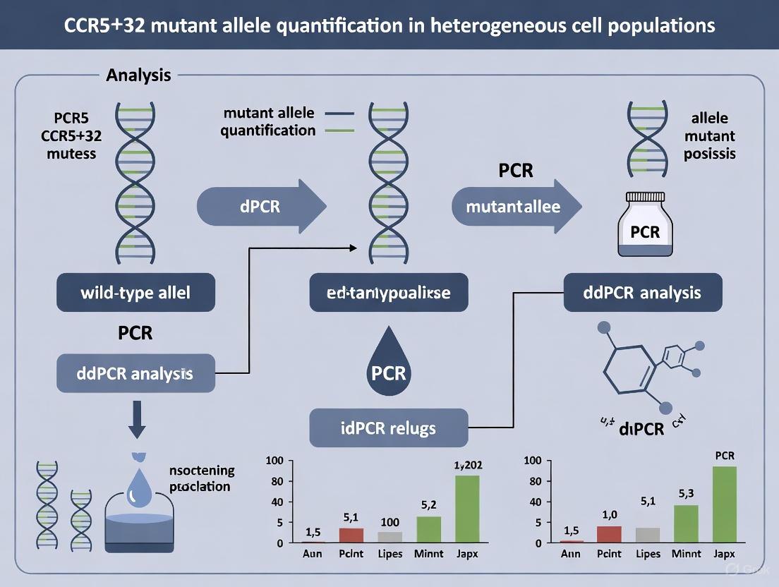
Droplet digital PCR (ddPCR) represents a highly precise method for quantifying the CCR5Δ32 mutant allele in heterogeneous cell mixtures. This approach enables absolute quantification of target sequences without the need for standard curves and provides superior sensitivity compared to traditional qPCR methods, particularly for detecting rare mutations in complex samples [3]. The development of multiplex ddPCR assays allows for simultaneous detection and quantification of both wild-type and Δ32 mutant CCR5 alleles, providing accurate measurement of their relative abundance in cell populations [3].
The system developed by researchers demonstrates the capability to accurately measure cells with the CCR5Δ32 mutation down to 0.8% in heterogeneous mixtures, making it suitable for monitoring engraftment of CCR5Δ32-modified cells in therapeutic contexts [3]. This level of sensitivity is crucial for evaluating the efficacy of stem cell transplantation therapies and gene editing approaches aimed at introducing the protective mutation into patient cells.
Table 2: Performance Characteristics of ddPCR for CCR5Δ32 Detection
| Parameter | Specification | Experimental Validation |
|---|---|---|
| Detection Limit | 0.8% mutant alleles in heterogeneous mixture [3] | Serial dilutions of CRISPR/Cas9-edited MT-4 cells [3] |
| Precision | High reproducibility across technical replicates [3] | Coefficient of variation <10% for allele frequency quantification [3] |
| Specificity | Discriminates wild-type, heterozygous, and homozygous genotypes [3] | Clear separation of positive and negative droplet populations [3] |
| Throughput | Medium to high (multiple samples per run) | 96-well plate compatibility [3] |
| Sample Requirements | Genomic DNA from cell mixtures | Cell culture models and clinical samples [3] |
Experimental Protocol: ddPCR Quantification of CCR5Δ32 Alleles
Sample Preparation and DNA Extraction
Materials:
- Cell mixture or tissue sample of interest
- Phenol-chloroform or commercial DNA extraction kit (e.g., ExtractDNA Blood and Cells Kit, Evrogen)
- NanoPhotometer or equivalent for DNA quantification and quality assessment
Procedure:
- Extract genomic DNA from cell mixtures using the phenol-chloroform method or commercial kits according to manufacturer's instructions.
- Quantify DNA concentration and assess purity using a spectrophotometer (A260/A280 ratio of ~1.8 indicates pure DNA).
- Dilute DNA to working concentration of 10-50 ng/μL in nuclease-free water.
- Store samples at -20°C until ready for ddPCR setup.
ddPCR Reaction Setup
Materials:
- QX200 ddPCR System (Bio-Rad) or equivalent
- ddPCR Supermix for Probes (no dUTP)
- Target-specific primers and fluorescent probes
- DG8 Cartridges and Gaskets
- Droplet Generator
- C1000 Touch Thermal Cycler with deep well reaction module
- PX1 PCR Plate Sealer
- QX200 Droplet Reader
Primer and Probe Sequences:
- CCR5 Wild-Type Probe: FAM-labeled, specific to intact CCR5 sequence
- CCR5Δ32 Mutant Probe: HEX/VIC-labeled, specific to deletion junction
- Forward Primer: 5'-CCCAGGAATCATCTTTACCA-3'
- Reverse Primer: 5'-GACACCGAAGCAGAGTTT-3'
Reaction Setup:
- Prepare reaction mix containing:
- 10 μL ddPCR Supermix for Probes
- 1 μL each primer (900 nM final concentration)
- 0.5 μL each probe (250 nM final concentration)
- 2 μL template DNA (20-100 ng total)
- Nuclease-free water to 20 μL total volume
Generate droplets:
- Transfer 20 μL reaction mix to DG8 Cartridge well
- Add 70 μL Droplet Generation Oil to appropriate well
- Place DG8 Gasket on cartridge
- Process in Droplet Generator
Transfer emulsified samples to 96-well PCR plate
- Seal plate with foil heat seal using PX1 PCR Plate Sealer (180°C for 5 seconds)
PCR Amplification and Analysis
Thermal Cycling Conditions:
- Enzyme activation: 95°C for 10 minutes
- 40 cycles of:
- Denaturation: 94°C for 30 seconds
- Annealing/Extension: 60°C for 60 seconds
- Enzyme deactivation: 98°C for 10 minutes
- Hold at 4°C indefinitely
Droplet Reading and Analysis:
- Transfer plate to QX200 Droplet Reader
- Analyze droplets for FAM and HEX/VIC fluorescence
- Set threshold between positive and negative droplet populations using QuantaSoft software
- Calculate allele concentrations (copies/μL) and determine mutant allele frequency using the formula:
Mutant Allele Frequency (%) = (Mutant copies/μL) / (Wild-type + Mutant copies/μL) × 100
Diagram 2: ddPCR Workflow for CCR5Δ32 Quantification. The process from DNA extraction to final data analysis for determining CCR5Δ32 allele frequency in heterogeneous cell mixtures.
Research Reagent Solutions
Table 3: Essential Research Reagents for CCR5Δ32 Analysis and HIV-1 Entry Studies
| Reagent/Category | Specific Examples | Function/Application |
|---|---|---|
| Cell Lines | MT-4 human T-cell line [3] | Model system for CCR5 gene editing and viral challenge studies |
| Gene Editing Tools | CRISPR/Cas9 (pCas9-IRES2-EGFP, pU6-gRNA vectors) [3] [6] | Introduction of CCR5Δ32 mutation; gRNAs: CCR5-7 (CAGAATTGATACTGACTGTATGG) and CCR5-8 (AGATGACTATCTTTAATGTCTGG) [3] |
| ddPCR Reagents | ddPCR Supermix for Probes, primer-probe sets for CCR5 wild-type and Δ32 [3] | Absolute quantification of CCR5Δ32 allele frequency in heterogeneous samples |
| Viral Strains | CCR5-tropic: Ba-L, ZM247; CXCR4-tropic: NL4-3 [4] | Determination of viral tropism and coreceptor usage; challenge assays for resistance validation |
| Flow Cytometry Antibodies | Anti-CCR5, anti-CD4, anti-CD195 [4] [5] | Assessment of CCR5 surface expression on leukocyte subsets |
| Cell Culture Reagents | RPMI-1640 medium, Fetal Bovine Serum, electroporation buffers [3] | Maintenance and genetic manipulation of hematopoietic cell lines |
| DNA Extraction Kits | ExtractDNA Blood and Cells Kit (Evrogen) [3] | High-quality genomic DNA isolation for downstream molecular applications |
Applications in Therapeutic Development and Monitoring
The quantitative assessment of CCR5Δ32 mutant alleles in heterogeneous cell mixtures has significant implications for developing and monitoring HIV-1 cure strategies. The precise measurement of CCR5Δ32 allele frequency enables researchers to track engraftment success in stem cell transplantation protocols and evaluate the efficiency of gene editing approaches [3] [5]. In clinical settings, ddPCR-based monitoring of CCR5Δ32 cell populations provides critical data for understanding the relationship between the proportion of CCR5-deficient cells and control of HIV-1 replication [4] [8].
Several landmark cases demonstrate the therapeutic potential of CCR5 ablation. The "London Patient" received a CCR5Δ32/Δ32 hematopoietic stem cell transplant for Hodgkin's lymphoma and maintained aviremia for over 30 months after antiretroviral therapy interruption [4]. Similarly, a mixed-race woman achieved possible HIV-1 cure after CCR5Δ32/Δ32 haplo-cord transplant to treat acute myeloid leukemia, with full donor chimerism and no viral rebound 18 months after treatment interruption [5]. These cases highlight the critical importance of accurate CCR5Δ32 quantification in predicting therapeutic outcomes.
CCR5 serves as a critical HIV-1 co-receptor that facilitates viral entry through specific interactions with the viral envelope glycoprotein gp120. The protective effect of the CCR5Δ32 mutation against HIV-1 infection has established CCR5 as a promising target for therapeutic interventions. The development of sensitive and accurate ddPCR-based methods for quantifying CCR5Δ32 mutant alleles in heterogeneous cell mixtures provides researchers with a powerful tool for monitoring the efficacy of stem cell transplantation and gene editing approaches. These protocols support the advancement of CCR5-targeted strategies toward achieving HIV-1 remission and cure, contributing to the growing arsenal of therapeutic options for individuals living with HIV-1.
The C-C chemokine receptor type 5 (CCR5) is a G protein-coupled receptor expressed on the surface of immune cells, serving as a primary co-receptor for the entry of R5-tropic human immunodeficiency virus (HIV-1) into CD4+ T-lymphocytes [9] [10]. A naturally occurring 32-base pair deletion (CCR5Δ32) within the coding region of the CCR5 gene results in a frameshift mutation and the introduction of a premature stop codon, preventing functional receptor expression on the cell membrane [3]. Individuals homozygous for the CCR5Δ32 allele are highly resistant to infection with CCR5-tropic HIV-1 strains, while heterozygous carriers exhibit slower disease progression and lower viral loads [11] [12]. This protective effect has been validated through curative hematopoietic stem cell transplant (HSCT) strategies from CCR5Δ32/Δ32 donors, establishing CCR5 ablation as a cornerstone for developing HIV cure therapies [9] [13] [14]. Accurate quantification of CCR5Δ32 alleles in heterogeneous cell populations is therefore critical for advancing therapeutic gene editing and monitoring transplant efficacy.
Quantitative Analysis of CCR5Δ32 Alleles
Droplet Digital PCR (ddPCR) enables absolute quantification of mutant allele frequencies in mixed cell populations with high precision. The following table summarizes key performance and quantitative data for CCR5Δ32 detection using multiplex ddPCR assays.
Table 1: Quantitative Data for CCR5Δ32 Detection via ddPCR
| Parameter | Value/Description | Experimental Context |
|---|---|---|
| Detection Sensitivity | ≤ 0.8% mutant alleles in mixture [3] [15] | Artificial CCR5Δ32 mutation in MT4 cell line using CRISPR/Cas9, mixed with wild-type cells. |
| Precision | Accurate calculation of mutant template copies [16] | Based on Poisson distribution analysis of ~20,000 droplets per sample. |
| Assay Type | Multiplex ddPCR [3] [15] | Simultaneous detection of wild-type and Δ32 alleles in a single reaction. |
| Key Application | Quantifying edited cell content post-transplantation [3] | Monitoring engraftment of CCR5Δ32/Δ32 stem cells in patients. |
Experimental Protocol: ddPCR for CCR5Δ32 Quantification in Heterogeneous Cell Mixtures
This protocol details the steps for quantifying the CCR5Δ32 mutation frequency in a background of wild-type cells using a multiplex ddPCR approach.
3.1. Sample Preparation and DNA Extraction
- Cell Line: Utilize target cells (e.g., MT-4 human T-cell line or hematopoietic stem cells) [3].
- Culture Conditions: Maintain cells in RPMI-1640 medium supplemented with 10% fetal bovine serum at 37°C in a 5% CO₂ atmosphere [3].
- Genomic DNA Extraction: Isolate DNA using a phenol-chloroform method or commercial kit (e.g., ExtractDNA Blood and Cells Kit). Measure DNA concentration and purity using a spectrophotometer (NanoPhotometer) [3].
3.2. Droplet Digital PCR (ddPCR) Assay
- Reaction Setup: Prepare a 20 µL ddPCR reaction mixture containing:
- Droplet Generation: Load the reaction mixture into a DG8 cartridge and generate droplets using a QX200 Droplet Generator. This partitions the sample into approximately 20,000 nanoliter-sized droplets [16].
- PCR Amplification: Transfer droplets to a 96-well PCR plate and perform amplification on a thermal cycler using the following protocol:
- 95°C for 10 minutes (enzyme activation)
- 40 cycles of:
- 94°C for 30 seconds (denaturation)
- 55–60°C for 1 minute (annealing/extension)
- 98°C for 10 minutes (enzyme deactivation)
- 4°C hold [16]
- Droplet Reading: Place the PCR plate in a QX200 Droplet Reader. This instrument measures the fluorescence intensity (FAM and HEX) in each droplet [16].
3.3. Data Analysis
- Use analysis software (e.g., QuantaSoft, Bio-Rad, or the
ddpcrR package) to classify droplets into four populations based on fluorescence:- FAM-positive only: Wild-type CCR5 alleles.
- HEX-positive only: CCR5Δ32 mutant alleles.
- Double-positive: Potentially non-specific amplification or rare events.
- Double-negative: No target DNA present.
- The software applies Poisson statistics to calculate the absolute concentration (copies/µL) of wild-type and mutant targets in the original sample.
- Calculate mutant allele frequency: [ \text{Mutant Allele Frequency (\%)} = \frac{[\text{CCR5Δ32}]}{[\text{CCR5Δ32}] + [\text{CCR5 WT}]} \times 100 ] Where concentrations are in copies/µL.
Research Reagent Solutions
Table 2: Essential Reagents and Materials for CCR5Δ32 Research
| Reagent/Material | Function/Application | Example Product/Catalog Number |
|---|---|---|
| CRISPR/Cas9 System | Introduction of artificial CCR5Δ32 mutation for functional studies [3] [13]. | pU6-gRNA vector; pCas9-IRES2-EGFP; in-house Cas9 protein. |
| ddPCR Supermix | Provides optimized reagents for PCR amplification within droplets. | ddPCR Supermix for Probes (No dUTP), Bio-Rad. |
| FAM/HE-Labeled Probes | Fluorescent detection of wild-type and mutant alleles in multiplex ddPCR [16]. | Custom TaqMan probes. |
| Cell Culture Media | Maintenance and expansion of target T-cell or stem cell lines. | RPMI-1640 with 10% FBS [3]. |
| DNA Extraction Kit | High-quality genomic DNA isolation for accurate ddPCR quantification. | ExtractDNA Blood and Cells Kit (Evrogen) [3]. |
| Electroporation System | Delivery of CRISPR/Cas9 ribonucleoprotein (RNP) complexes into cells. | Gene Pulser Xcell System (Bio-Rad) [3]. |
Workflow and Signaling Pathway Diagrams
Diagram 1: ddPCR Workflow for CCR5Δ32 Quantification
Diagram 2: CCR5 Role in HIV Entry and Δ32 Protection Mechanism
The cases of the "Berlin" and "London" patients represent seminal proof-of-concept demonstrations that allogeneic hematopoietic stem cell transplantation (HSCT) from CCR5Δ32/Δ32 homozygous donors can cure human immunodeficiency virus type 1 (HIV-1) infection [3] [17]. The C-C chemokine receptor type 5 (CCR5) serves as a crucial co-receptor for HIV entry into CD4+ T-cells, and individuals carrying a homozygous 32-base pair deletion (CCR5Δ32) in the CCR5 gene are naturally resistant to R5-tropic HIV-1 infection [3]. These clinical breakthroughs have established CCR5 gene editing as a validated therapeutic strategy, creating a pressing need for robust analytical methods to quantify CCR5Δ32 mutant alleles in heterogeneous cell populations [3] [18].
Droplet digital PCR (ddPCR) has emerged as a powerful tool for precise quantification of mutant alleles in HIV cure research [3] [19]. This technology enables absolute quantification of nucleic acids without requiring standard curves, provides high sensitivity for detecting rare variants, and demonstrates superior tolerance to PCR inhibitors compared to conventional quantitative PCR (qPCR) [20]. The application of ddPCR is particularly valuable for monitoring the engraftment of CCR5-modified cells and quantifying the extent of CCR5 disruption achieved through genome editing approaches [3] [18].
Clinical Proof-of-Concept: Established Cases
The Berlin Patient
The first documented case of HIV-1 cure occurred in Timothy Ray Brown, known as the "Berlin patient," who received CCR5Δ32/Δ32 allogeneic hematopoietic stem cell transplantation for acute myeloid leukemia (AML) [3] [17]. Following transplantation, the patient displayed sustained HIV-1 remission despite discontinuation of antiretroviral therapy (ART), with no detectable replication-competent virus demonstrated through extensive reservoir assays [17]. This case established the paradigm that CCR5 ablation through HSCT could potentially eliminate HIV-1 infection.
The London Patient
A second successful case was reported in a patient in London (IciStem no. 36) who also underwent CCR5Δ32/Δ32 allogeneic HSCT for hematological malignancy [3] [17]. Similar to the Berlin patient, this individual maintained undetectable HIV-1 viral loads for more than 48 months after analytical treatment interruption, providing crucial validation of the approach [17]. Comprehensive virological assessment including in vivo outgrowth assays in humanized mouse models failed to detect replication-competent virus, strengthening the evidence for cure [17].
Table 1: Clinical Characteristics of Established HIV-1 Cure Cases
| Parameter | Berlin Patient | London Patient |
|---|---|---|
| Underlying Malignancy | Acute Myeloid Leukemia | Acute Myeloid Leukemia |
| Transplantation Type | CCR5Δ32/Δ32 allogeneic HSCT | CCR5Δ32/Δ32 allogeneic HSCT |
| Conditioning Regimen | Myeloablative | Reduced-intensity |
| ART Discontinuation | Yes | Yes |
| Post-ATI Follow-up | >4 years without rebound | >4 years without rebound |
| Key Reservoir Findings | No replication-competent virus detected | No replication-competent virus detected |
| Evidence Level | Proof-of-concept established | Independent validation |
ddPCR Methodology for CCR5Δ32 Quantification
Principle of ddPCR Technology
Droplet digital PCR represents a third-generation PCR technology that enables absolute quantification of nucleic acid targets without requiring standard curves [20] [21]. The method partitions a PCR reaction into thousands of nanoliter-sized water-in-oil droplets, effectively creating individual micro-reactors [20]. Following PCR amplification, each droplet is analyzed for fluorescence, and the fraction of positive droplets is used to calculate the absolute copy number of the target sequence based on Poisson statistics [21]. This partitioning approach provides ddPCR with enhanced sensitivity for rare allele detection and improved precision compared to real-time qPCR [19] [20].
For CCR5Δ32 quantification, ddPCR assays are designed with specific probe-based detection systems that distinguish between wild-type CCR5 and the Δ32 mutant allele [3] [18]. The multiplexing capability allows simultaneous quantification of both alleles in a single reaction, enabling precise determination of editing efficiency in heterogeneous cell mixtures [3].
Detailed ddPCR Protocol for CCR5Δ32 Detection
Table 2: Step-by-Step ddPCR Protocol for CCR5Δ32 Quantification
| Step | Procedure | Parameters | Quality Control |
|---|---|---|---|
| 1. DNA Extraction | Extract genomic DNA from cell populations using phenol-chloroform or commercial kits | Input: 6 × 10^6 cells; Measure concentration and purity (A260/A280) | NanoPhotometer measurement; Target A260/A280 ≈ 1.8 [3] |
| 2. Reaction Setup | Prepare PCR mix with target-specific primers and probes for wild-type CCR5 and CCR5Δ32 | Final volume: 20-22 µL; Include ddPCR supermix | Include negative controls (no template) and positive controls if available [3] |
| 3. Droplet Generation | Partition reaction into nanoliter droplets using droplet generator | Target: 10,000-20,000 droplets per sample | Assess droplet quality; ensure uniform droplet formation [20] |
| 4. PCR Amplification | Perform thermal cycling with optimized annealing temperature | 40-45 cycles; Annealing at 58-60°C | Include no-template controls to monitor contamination [3] [18] |
| 5. Droplet Reading | Analyze fluorescence in each droplet using droplet reader | Measure FAM and HEX/VIC channels simultaneously | Set threshold based on negative controls [18] |
| 6. Data Analysis | Calculate mutant allele frequency using Poisson statistics | Use manufacturer's software (e.g., QuantaSoft) | Report absolute copies/μL and mutant percentage [3] |
Assay Performance Characteristics
The ddPCR assay for CCR5Δ32 demonstrates exceptional analytical performance, with sensitivity down to 0.8% mutant alleles in heterogeneous cell mixtures [3]. The method shows high reproducibility and precision, with coefficients of variation typically below 10% for technical replicates [19]. Compared to conventional qPCR, ddPCR exhibits superior accuracy for absolute quantification, particularly at low target concentrations, due to its resistance to amplification efficiency variations [19] [20].
The assay's dynamic range extends from approximately 1 to 100,000 copies per reaction, making it suitable for monitoring both low-level residual wild-type alleles and highly edited cell populations [3] [18]. This performance is critical for assessing the efficacy of CCR5 gene editing approaches in clinical applications.
Application in HIV Cure Research
Monitoring HSCT Engraftment
In the context of CCR5Δ32/Δ32 hematopoietic stem cell transplantation, ddPCR provides a valuable tool for monitoring donor chimerism and tracking the expansion of CCR5-deficient cells in patients [17]. Longitudinal monitoring enables researchers to correlate the percentage of CCR5Δ32-positive cells with clinical outcomes and viral reservoir dynamics [17]. This application was utilized in the follow-up of the London patient, where sustained full donor chimerism was observed alongside absence of viral rebound [17].
Assessing Gene Editing Efficiency
For emerging gene therapy approaches using CRISPR/Cas9 or TALENs to create CCR5 disruptions, ddPCR offers a precise method for quantifying editing efficiency in clinical samples [3] [18]. The technology can distinguish between biallelic and monoallelic editing, which is crucial as biallelic disruption provides complete resistance to HIV infection [18]. In automated GMP-compatible production of CCR5-negative CD4+ T-cells, ddPCR confirmed that approximately 40% of manufactured cells showed biallelic CCR5 editing [18].
Table 3: Quantitative Data from ddPCR Analysis in HIV Cure Research
| Application | Sample Type | Measured Parameter | Typical Results | Reference |
|---|---|---|---|---|
| HSCT Monitoring | Peripheral blood mononuclear cells | Donor chimerism | >95% donor cells in established engraftment | [17] |
| Gene Editing Assessment | Engineered CD4+ T-cells | Biallelic editing rate | ~40% of total produced cells | [18] |
| Sensitivity Assessment | Artificial cell mixtures | Detection limit | 0.8% mutant alleles in wild-type background | [3] |
| Viral Reservoir | Tissue biopsies (lymph node, gut) | HIV DNA+ cells | 5.08 ± 1.74 per 10^5 cells (trace levels) | [17] |
Research Reagent Solutions
Table 4: Essential Research Reagents for CCR5Δ32 ddPCR Analysis
| Reagent/Category | Specific Examples | Function/Application |
|---|---|---|
| ddPCR Systems | Bio-Rad QX200 Droplet Digital PCR System | Platform for droplet generation, amplification, and reading |
| Nucleic Acid Extraction | QIAamp DNA Blood Mini Kit | Isolation of high-quality genomic DNA from cell populations |
| PCR Reagents | ddPCR Supermix for Probes | Optimized reaction mix for droplet-based digital PCR |
| Target Detection | FAM/HEX-labeled probes for wild-type CCR5 and CCR5Δ32 | Allele-specific discrimination in multiplex assays |
| Cell Separation | Fluorescence-activated cell sorting (FACS) | Isolation of specific cell populations for analysis |
| Control Materials | Synthetic oligonucleotides with wild-type and Δ32 sequences | Assay validation and quality control |
Workflow Integration
The experimental workflow for CCR5Δ32 analysis begins with sample preparation from heterogeneous cell mixtures, followed by genomic DNA extraction and quantification. The ddPCR reaction is then assembled with allele-specific probes, partitioned into droplets, and amplified. Data analysis provides absolute quantification of wild-type and mutant alleles, enabling calculation of editing efficiency or donor chraftment percentage.
Diagram 1: Experimental workflow for CCR5Δ32 quantification using ddPCR.
The proof-of-concept established by the Berlin and London patients has fundamentally advanced HIV cure research, demonstrating that CCR5 ablation represents a viable path to HIV-1 remission. Droplet digital PCR has emerged as an essential analytical tool in this field, providing the sensitivity and precision required to quantify CCR5Δ32 mutant alleles in heterogeneous cell mixtures. As gene editing technologies advance toward clinical application, ddPCR will play an increasingly critical role in quality control, potency assessment, and therapeutic monitoring of CCR5-based interventions.
CRISPR/Cas9 and the Era of Artificial CCR5Δ32 Mutation Generation
Application Notes
Strategic Importance of CCR5Δ32 Generation
The C-C chemokine receptor type 5 (CCR5) serves as a crucial co-receptor for human immunodeficiency virus (HIV-1) entry into CD4+ T-cells [3] [22]. The natural CCR5Δ32 mutation, a 32-base pair deletion resulting in a non-functional receptor, confers significant resistance to R5-tropic HIV-1 infection in homozygous carriers [22] [23]. While allogeneic hematopoietic stem cell transplantation from CCR5Δ32/Δ32 donors has led to functional cures in HIV-positive patients (the "Berlin" and "London" patients), the clinical application of this approach remains limited by the rarity of this genotype, which has a frequency of approximately 1% in Northern European populations and is almost absent in African, Asian, and Native American populations [13] [24] [23].
CRISPR/Cas9 genome editing technology has emerged as a powerful tool to overcome this limitation by enabling the precise introduction of CCR5Δ32-like mutations in autologous or immunocompatible cells [13] [3] [22]. This approach bypasses donor compatibility issues and creates a continuous source of HIV-resistant cells, representing a promising strategy for achieving functional HIV cure [22].
Quantitative Analysis of CRISPR/Cas9-Mediated CCR5 Knockout
Recent studies have optimized CRISPR/Cas9 protocols for efficient CCR5 disruption. The table below summarizes key quantitative data from genome editing experiments in MT4CCR5 cells, demonstrating dose-dependent effects of ribonucleoprotein (RNP) complex delivery.
Table 1: Efficiency of CRISPR/Cas9-Mediated CCR5 Knockout in MT4CCR5 Cells
| RNP Complex Composition | CCR5 Expression (%) | Reduction vs. Control (%) | Cell Viability (%) | Cleavage Efficiency |
|---|---|---|---|---|
| Mock Control | 99.80 ± 0.00 | - | - | - |
| Cas9 (6µg) + sgRNAs (4µg total) | 10.43 ± 0.15 | 89.37 | 77.50 - 98.40 | High |
| Cas9 (10µg) + sgRNAs (8µg total) | 1.91 ± 0.13 | 97.89 | 77.50 - 98.40 | High |
Data adapted from [13]
The combination of CCR5 knockout with additional anti-HIV strategies, such as the C46 HIV-1 fusion inhibitor, provides broad-spectrum protection against both R5- and X4-tropic HIV-1 strains, addressing the limitation of viral tropism switching observed in single-modality approaches [13] [22].
Verification and Quantification Using Droplet Digital PCR
The generation of artificial CCR5Δ32 mutations necessitates precise quantification methods to assess editing efficiency in heterogeneous cell populations. Droplet digital PCR (ddPCR) has emerged as a superior technology for this application, offering absolute quantification without calibration curves and enhanced sensitivity for detecting rare mutations [3] [21].
Table 2: Performance Characteristics of ddPCR for CCR5Δ32 Detection
| Parameter | Performance | Significance |
|---|---|---|
| Detection Limit | 0.8% mutant alleles in heterogeneous mixtures | Enables precise tracking of edited cell populations |
| Partitioning | Thousands to millions of droplets | Allows single-molecule detection |
| Quantification Method | Poisson statistics on endpoint fluorescence | Calibration-free absolute quantification |
| Precision | High accuracy and reproducibility | Suitable for clinical monitoring |
The implementation of ddPCR for CCR5Δ32 quantification provides critical quality control metrics for genome editing protocols and enables longitudinal monitoring of edited cell populations in both research and clinical settings [3] [15].
Experimental Protocols
CRISPR/Cas9-Mediated CCR5 Gene Editing in T-Cell Lines
Reagent Preparation
- sgRNA Design: Utilize two sgRNAs targeting the first exon of human CCR5 at the Δ32 mutation site: sgRNA1 (CAGAATTGATACTGACTGTATGG) and sgRNA2 (AGATGACTATCTTTAATGTCTGG) [3].
- RNP Complex Formation: Combine purified Cas9 protein (6-10µg) with sgRNAs (2-4µg each) in ribonucleoprotein (RNP) complex buffer. Incubate at room temperature for 10-15 minutes to allow complex formation [13].
Cell Electroporation and Sorting
- Cell Preparation: Culture MT-4 human T-cell line in RPMI-1640 medium supplemented with 10% fetal bovine serum. Harvest 6 × 10^6 cells during logarithmic growth phase [3].
- Electroporation Parameters: Mix cell suspension with RNP complexes in electroporation cuvettes. Electroporate using Gene Pulser Xcell system (275 V, 5 ms, three pulses) [3].
- Post-Transfection Processing: Incubate transfected cells in complete medium for 48 hours. Sort EGFP-positive cells using fluorescence-activated cell sorting (FACS) to enrich for successfully transfected populations [3].
Clonal Selection and Validation
- Limiting Dilution Cloning: Dispense sorted cells into 96-well plates by limiting dilution to obtain monoclonal cell lines. Incubate for 14 days under standard conditions (37°C, 5% CO2) [3].
- Genomic DNA Extraction: Amplify monoclonal cultures and extract genomic DNA using phenol-chloroform method or commercial kits (e.g., QIAamp DNA Mini Kit) [3] [24].
- Mutation Screening: Amplify target CCR5 locus using PCR with specific primers (forward: CCCAGGAATCATCTTTACCA, reverse: GACACCGAAGCAGAGTTT). Sequence PCR products to confirm introduction of CCR5Δ32 mutations [3].
Droplet Digital PCR Quantification of CCR5Δ32 Alleles
Sample Preparation and Partitioning
- DNA Quantification: Measure DNA concentration and purity using spectrophotometry (e.g., NanoPhotometer P-Class P360). Use 5µL (1.5-19ng) of extracted DNA per ddPCR reaction [3] [24].
- Reaction Mixture Preparation: Prepare 20µL reaction mixture containing 12.5µL of 2× ddPCR Master Mix, 2.5µL of forward primer, 2.5µL of reverse primer, and 2.5µL of ultrapure water [24].
- Droplet Generation: Load reaction mixture into droplet generator cartridges along with droplet generation oil. Process using automated droplet generators (e.g., Bio-Rad QX200) to create thousands of nanoliter-sized droplets [21].
PCR Amplification and Analysis
- Thermal Cycling Conditions: Transfer droplets to 96-well PCR plates and amplify using following protocol: 95°C for 5 minutes; 35 cycles of 95°C for 45 seconds, 58°C for 45 seconds, and 72°C for 45 seconds; final extension at 72°C for 10 minutes [24].
- Endpoint Fluorescence Reading: Read amplified plates using droplet readers capable of detecting fluorescence signals from both wild-type and mutant alleles [3] [21].
- Data Analysis: Apply Poisson statistics to determine absolute quantification of CCR5Δ32 mutant alleles based on ratio of positive to negative droplets. Calculate editing efficiency as percentage of mutant alleles in total population [3] [21].
Workflow Visualization
Diagram Title: CCR5Δ32 Generation and Quantification Workflow
Diagram Title: Multi-Target HIV Inhibition Strategy
The Scientist's Toolkit: Essential Research Reagents
Table 3: Key Reagents for CCR5Δ32 Generation and Quantification
| Reagent/Category | Specific Examples | Function/Application |
|---|---|---|
| Gene Editing Enzymes | Cas9 protein (purified) | CRISPR/Cas9 ribonucleoprotein complex formation for precise genome editing [13] |
| Guide RNAs | sgRNA1: CAGAATTGATACTGACTGTATGGsgRNA2: AGATGACTATCTTTAATGTCTGG | Target-specific guidance to CCR5 locus for Δ32 mutation introduction [3] |
| Delivery Systems | Electroporation systems (Gene Pulser Xcell) | Physical method for efficient RNP complex delivery into target cells [3] |
| Cell Culture | MT-4 human T-cell line, RPMI-1640 medium, FBS | Model system for optimizing CCR5 editing protocols [13] [3] |
| Detection Primers | CCR5-Delta32: F- CTTCATCATCCTCCTGACAATCG, R- GACCAGCCCCAAGTTGACTATC | Amplification of target region for mutation detection [24] |
| Quantification Kits | ddPCR Master Mix, Droplet Generation Oil | Essential components for droplet digital PCR quantification [3] [21] |
| Analysis Software | ddPCR data analysis tools | Poisson statistics-based absolute quantification of editing efficiency [3] [21] |
The Critical Need for Accurate Mutant Allele Quantification in Therapeutic Development
The C-C chemokine receptor type 5 (CCR5) serves as a crucial co-receptor for human immunodeficiency virus (HIV) entry into T-cells [3]. A naturally occurring 32-base pair deletion in the CCR5 gene, known as the CCR5Δ32 mutation, causes a frameshift that results in premature stop codons and complete knockout of gene function [3]. Individuals carrying homozygous CCR5Δ32 mutations demonstrate remarkable resistance to R5-tropic HIV-1 strains, the most common and contagious variants of the virus [3]. This biological phenomenon has positioned CCR5 as a prime therapeutic target for HIV cure strategies.
The therapeutic potential of CCR5 disruption has been validated through clinical case studies. Notably, transplantations of hematopoietic stem cells from CCR5Δ32 homozygous donors to HIV-positive patients with leukemia have resulted in complete viral elimination, achieving the celebrated "Berlin and London patient" outcomes [3]. With advancements in genome editing technologies, particularly CRISPR/Cas9, researchers can now artificially recreate the CCR5Δ32 mutation in wild-type cells, opening avenues for autologous transplantation approaches that bypass the need for rare naturally-occurring homozygous donors [3] [18]. These developments have created an urgent need for precise methods to quantify CCR5Δ32 mutant alleles in heterogeneous cell mixtures to monitor therapeutic efficacy and patient outcomes.
The Analytical Challenge in Heterogeneous Cell Populations
The accurate quantification of mutant alleles in heterogeneous cell mixtures presents significant analytical challenges. In therapeutic contexts, researchers must detect and quantify low-frequency mutations amidst a background of predominantly wild-type alleles, often requiring sensitivity thresholds below 1% [3] [25]. This challenge is particularly acute in several scenarios:
- Monitoring engraftment of CCR5-edited cells post-transplantation
- Assessing editing efficiency in CRISPR/Cas9-modified cell populations
- Evaluating mutant allele expansion during patient follow-up
- Detecting minimal residual disease in oncological contexts
Traditional quantification methods like quantitative PCR (qPCR) demonstrate substantial variability due to susceptibility to sample quality and operator experience, making them suboptimal for precise mutant allele frequency determination [25]. Furthermore, the 2022 International Consensus Classification of myeloid neoplasms strongly recommends sensitive detection of allele frequencies below 1% [25], a threshold challenging to achieve consistently with conventional molecular techniques. These limitations underscore the need for more robust quantification platforms in therapeutic development.
Droplet Digital PCR: A Transformative Solution
Droplet digital PCR (ddPCR) represents a transformative approach for absolute nucleic acid quantification that addresses the limitations of traditional methods. This technology partitions samples into thousands of nanoliter-sized droplets, effectively creating individual reaction chambers where PCR amplification occurs independently [25]. The fundamental principle involves analyzing each droplet separately in an end-point measurement, providing a digital readout (positive or negative) for target presence.
This partitioning strategy confers several critical advantages for mutant allele quantification. By distributing the target molecules across many droplets, ddPCR mitigates PCR competition effects, making amplification less sensitive to inhibition and dramatically improving the capacity to distinguish single-nucleotide variations [25]. The digital nature of the readout enables absolute quantification without external calibrators, eliminating the variability associated with standard curve generation in qPCR [25]. This feature is particularly valuable in clinical diagnostics where reproducibility across laboratories is essential.
Application to CCR5Δ32 Quantification
Researchers have successfully adapted ddPCR for precise quantification of CCR5Δ32 mutant alleles in heterogeneous cell mixtures. One study demonstrated that ddPCR could accurately measure CCR5Δ32 content down to 0.8% in artificial cell mixtures [3], establishing its utility for monitoring edited cell populations in therapeutic contexts. The development of multiplex ddPCR assays allows simultaneous detection of both wild-type and mutant CCR5 alleles in a single reaction, providing robust mutant allele frequency calculations essential for assessing gene editing efficiency [3] [18].
The application of ddPCR extends beyond CCR5 quantification to other therapeutically relevant mutations. For JAK2 V617F mutations in myeloproliferative neoplasms, optimized ddPCR assays have achieved remarkable sensitivity with a limit of quantification of 0.01% variant allele frequency [25]. This exceptional sensitivity enables detection of minimal residual disease and early intervention opportunities. Furthermore, ddPCR has proven valuable for allele-specific expression analysis in Huntington's disease research, demonstrating its versatility across different genetic contexts [26].
Figure 1: Comprehensive ddPCR workflow for mutant allele quantification in therapeutic development, showcasing the integrated process from sample collection to clinical interpretation.
Established ddPCR Protocol for CCR5Δ32 Quantification
Sample Preparation and DNA Extraction
- Cell Sources: Process heterogeneous cell mixtures including edited T-cells, hematopoietic stem cells, or patient-derived samples [3] [18]
- DNA Extraction: Isolate genomic DNA using commercial kits (e.g., QIAamp DNA Blood Mini Kit) following manufacturer protocols [18]
- Quality Assessment: Measure DNA concentration and purity using spectrophotometry (NanoPhotometer or similar) with optimal A260/A280 ratios of 1.8-2.0 [3]
- Storage Conditions: Store extracted DNA at -20°C until analysis to prevent degradation
ddPCR Reaction Setup
The reaction mixture should be prepared with the following components in a total volume of 20μL [3] [25]:
| Component | Final Concentration | Purpose |
|---|---|---|
| 2× ddPCR Supermix | 1× | Provides optimized buffer for droplet formation and amplification |
| Forward Primer | 450 nM | Amplifies target CCR5 region |
| Reverse Primer | 450 nM | Amplifies target CCR5 region |
| Wild-Type Probe | 250 nM | Detects unmodified CCR5 allele |
| Mutant Probe (FAM-labeled) | 250 nM | Specifically detects Δ32 deletion |
| Template DNA | 10-100 ng | Sample containing both wild-type and mutant alleles |
| Nuclease-Free Water | To volume | Adjusts final reaction volume |
Probe Design Considerations
- Wild-Type Probe: Should span the deletion region, generating no signal when Δ32 mutation is present [3]
- Mutant Probe: Should flank the deletion junction, specifically binding only to Δ32 sequence [3]
- Fluorescent Reporters: Use distinct fluorophores (FAM/HEX) for wild-type and mutant probes to enable multiplex detection [3] [18]
- Quenchers: Incorporate appropriate quenchers (e.g., MGB, BHQ) to minimize background fluorescence
Droplet Generation and Thermal Cycling
- Droplet Generation: Utilize automated droplet generators (e.g., QX200 AutoDG) to partition each sample into approximately 20,000 nanoliter-sized droplets [25]
- Thermal Cycling Conditions:
- Initial Denaturation: 95°C for 10 minutes (1 cycle)
- Amplification: 40 cycles of:
- Denaturation: 95°C for 15 seconds
- Annealing/Extension: 57-60°C for 60 seconds
- Enzyme Deactivation: 98°C for 10 minutes (1 cycle)
- Final Hold: 12°C indefinitely [25]
Data Acquisition and Analysis
- Droplet Reading: Process droplets using a droplet reader (e.g., QX200) to count positive and negative droplets for each channel [25]
- Threshold Setting: Establish clear thresholds between positive and negative droplet clusters using 2D amplitude plots [18]
- Poisson Statistical Analysis: Apply Poisson statistics to account for multiple target molecules per droplet using manufacturer software (e.g., QuantaSoft) [25] [18]
- Mutant Allele Frequency Calculation: Calculate using the formula: (Mutant copies/Total copies) × 100%
Performance Characteristics and Validation
Analytical Sensitivity and Specificity
Robust validation of ddPCR assays is essential for clinical translation. Comprehensive performance characteristics should include:
| Parameter | Performance | Experimental Details |
|---|---|---|
| Limit of Detection (LOD) | 0.01% VAF | Determined using serial dilutions of mutant DNA in wild-type background [25] |
| Limit of Quantification (LOQ) | 0.8% for CCR5Δ32 | Defined as the lowest concentration measurable with CV <25% [3] |
| Linearity | R² > 0.998 | Demonstrated across 4 orders of magnitude (0.01-100%) [25] |
| Precision (Intra-assay) | CV 5-15% | Dependent on mutant allele frequency [25] |
| Precision (Inter-assay) | CV 7-20% | Across operators, days, and reagent lots [25] |
| Specificity | >99% | Minimal cross-reactivity between wild-type and mutant probes [3] |
Comparative Method Performance
When compared to alternative quantification methods, ddPCR demonstrates distinct advantages:
Figure 2: Comparative analysis of mutation quantification methodologies highlighting the technical advantages of ddPCR for sensitive allele frequency detection.
Essential Research Reagent Solutions
Successful implementation of ddPCR for mutant allele quantification requires carefully selected research reagents and systems:
| Reagent Category | Specific Product | Application Note |
|---|---|---|
| ddPCR System | QX200 AutoDG (Bio-Rad) | Automated droplet generation and reading for high-throughput applications [25] |
| DNA Extraction | QIAamp DNA Blood Mini Kit (QIAGEN) | High-quality genomic DNA isolation with minimal inhibitor carryover [18] |
| PCR Supermix | ddPCR Supermix for Probes (Bio-Rad) | Optimized reaction chemistry for droplet formation and amplification [25] |
| Reference Material | WHO JAK2 V617F Panel (NIBSC 16/120) | International standard for assay validation and harmonization [25] |
| Cell Culture | RPMI-1640 + 10% FBS | Maintenance of T-cell lines (e.g., MT-4) for method development [3] |
| Genome Editing | CRISPR/Cas9 System | Generation of CCR5Δ32 mutations in wild-type cells for control material [3] |
Therapeutic Applications and Clinical Translation
The precise quantification of CCR5Δ32 mutant alleles using ddPCR has enabled significant advances in multiple therapeutic areas:
HIV Gene Therapy Applications
In HIV gene therapy, ddPCR facilitates critical monitoring of edited cell populations. Clinical-scale automated production systems like the CliniMACS Prodigy can generate >1.5 × 10⁹ CCR5-edited CD4+-T cells with >60% editing efficiency within 12 days [18]. Approximately 40% of these large-scale produced cells typically show biallelic CCR5 editing, providing maximal protection against HIV infection [18]. ddPCR enables researchers to track these edited cells post-transplantation, correlating persistence with therapeutic outcomes.
Hematopoietic Stem Cell Transplantation Monitoring
For patients receiving CCR5Δ32/Δ32 allogeneic hematopoietic stem-cell transplantation, ddPCR allows sensitive monitoring of engraftment and chimerism [3]. The technology can quantitate low-level HIV DNA for HIV reservoir diagnostics when evaluating potential HIV cure during antiviral treatment interruption [3]. This application provides critical insights into the relationship between CCR5-negative cell populations and viral control.
Emerging Applications in Clinical Development
Beyond HIV therapy, ddPCR-based mutant allele quantification supports development of treatments for various genetic disorders. In Huntington's disease research, ddPCR assays enable allele-specific quantification of wild-type and mutant HTT mRNA expression, essential for evaluating allele-selective therapeutic approaches [26]. Similar strategies apply to myotonic dystrophy type 1 and spinocerebellar ataxias [26], demonstrating the broad utility of this quantification platform.
Droplet digital PCR has emerged as an indispensable tool for accurate mutant allele quantification in therapeutic development. Its exceptional sensitivity, precision, and absolute quantification capabilities address critical needs in gene therapy monitoring, particularly for CCR5Δ32-based HIV interventions. As genome editing technologies continue to advance, the role of ddPCR in quantifying editing efficiencies and tracking therapeutic cells will expand accordingly.
Future developments will likely focus on increasing throughput, reducing costs, and enhancing multiplexing capabilities to simultaneously monitor multiple genomic targets. Standardization of ddPCR protocols across laboratories will be essential for clinical adoption, facilitated by international reference materials like the WHO JAK2 V617F mutation panel [25]. The integration of ddPCR with other molecular analyses in comprehensive monitoring panels will provide deeper insights into therapeutic mechanisms and patient-specific responses, ultimately accelerating the development of transformative genetic medicines.
A Step-by-Step Guide to ddPCR Assay Development for CCR5Δ32 Detection
Principles of Absolute Nucleic Acid Quantification with ddPCR
Digital PCR (dPCR) represents the third generation of polymerase chain reaction technology, following conventional PCR and quantitative real-time PCR (qPCR). This method enables the absolute quantification of target nucleic acids without the need for a standard curve, relying instead on Poisson statistics to calculate target concentration from the ratio of positive to negative partitions. The core principle involves partitioning a PCR reaction into thousands to millions of nanoliter-sized droplets, each acting as an individual microreactor. Following end-point amplification, each droplet is analyzed for fluorescence, and the fraction of positive droplets is used to determine the absolute copy number of the target sequence in the original sample [21] [27].
The historical development of dPCR began with precursor work in 1989 using limiting dilution PCR to detect single copies of HIV provirus. The term "digital PCR" was formally coined in 1999 by Bert Vogelstein and colleagues, who developed a workflow involving limiting dilution distributed on 96-well plates combined with fluorescence readout to detect mutations in cancer patients. The technology has since evolved significantly with advances in microfluidics, leading to the commercial droplet digital PCR (ddPCR) systems available today [21]. This technology is particularly valuable for applications requiring high sensitivity and precision, including the detection of rare mutations in heterogeneous cell populations, analysis of gene expression, and pathogen detection [21] [27].
Fundamental Principles of ddPCR
Partitioning and Statistical Foundation
The absolute quantification capability of ddPCR stems from its partitioning strategy and statistical foundation. A sample is divided into numerous discrete partitions such that each contains zero, one, or a few target molecules according to a Poisson distribution. Following PCR amplification, the partitions are assessed using endpoint fluorescence detection, converting the analog signal into a digital readout (positive or negative) [27].
The Poisson distribution describes the probability of a given number of events occurring in a fixed interval of time or space, and it is mathematically expressed as P(k) = (λ^k * e^{-λ}) / k!, where λ is the average number of target molecules per partition and k is the actual number in a specific partition. The fundamental equation for calculating target concentration in ddPCR is λ = -ln(1 - p), where p represents the proportion of positive partitions [27]. This approach allows for absolute quantification without external calibration curves, eliminating a major source of variability inherent to qPCR methods [27].
The quantification accuracy of ddPCR depends significantly on the number of partitions analyzed and the value of λ. Maximum precision is achieved when approximately 20% of partitions are negative (λ ≈ 1.6). Under these conditions, the precision scales with the inverse square root of the number of partitions, meaning that increasing the partition count improves quantification accuracy [27].
Comparative Advantages Over qPCR
dPCR offers several distinct advantages compared to traditional qPCR:
| Feature | ddPCR | qPCR |
|---|---|---|
| Quantification Method | Absolute via Poisson statistics | Relative via standard curve |
| Calibration Requirement | Not required | Essential for quantification |
| Precision | Higher precision, especially for low copy numbers | Lower precision, dependent on standard quality |
| Tolerance to Inhibitors | Higher (due to sample partitioning) | Lower |
| Sensitivity | Can detect single molecules | Limited by amplification efficiency and standard curve |
| Data Analysis | Binary (positive/negative partitions) | Continuous (Ct values) |
| Throughput | Typically lower | Typically higher [28] |
A key advantage of ddPCR is its superior sensitivity for detecting rare mutations in a background of wild-type sequences. This capability stems from the partitioning process, which effectively concentrates target sequences within isolated microreactors, reducing template competition [27]. Studies have demonstrated that ddPCR can reliably detect mutant allele frequencies as low as 0.1%, while qPCR methods typically achieve detection limits of only 1-5% [29]. This enhanced sensitivity makes ddPCR particularly valuable for monitoring minimal residual disease in oncology and detecting rare genetic variants in heterogeneous cell populations [29].
Application to CCR5Δ32 Mutant Allele Quantification
Therapeutic Context for CCR5Δ32 Quantification
The C-C chemokine receptor type 5 (CCR5) serves as a crucial co-receptor for HIV entry into human CD4+ T-cells. A naturally occurring 32-base pair deletion (CCR5Δ32) results in a non-functional receptor that confers resistance to R5-tropic HIV strains in homozygous individuals. This discovery has catalyzed the development of novel therapeutic strategies, including allogeneic hematopoietic stem cell transplantation from CCR5Δ32 homozygous donors and CRISPR/Cas9 genome editing to introduce the protective mutation in autologous cells [3] [30].
The "Berlin" and "London" patients, who achieved sustained HIV remission following transplantation with CCR5Δ32 homozygous stem cells, provided clinical validation for this approach [30]. Subsequent research has focused on reproducing this mutation artificially using genome editing technologies, creating a pressing need for accurate methods to quantify CCR5Δ32 mutant alleles in heterogeneous cell mixtures [3]. This quantification is essential for monitoring engraftment success in transplantation settings and for assessing editing efficiency in gene therapy applications, enabling researchers to track the proportion of edited cells and optimize therapeutic protocols [3] [18].
Experimental Design and Workflow
The experimental workflow for CCR5Δ32 quantification begins with the generation of edited cells using CRISPR/Cas9 genome editing, followed by DNA extraction and ddPCR analysis [3]. The process can be visualized as follows:
Diagram 1: Experimental workflow for CCR5Δ32 quantification using ddPCR.
The multiplex ddPCR assay utilizes two distinct probe sets to differentiate between wild-type CCR5 and the CCR5Δ32 mutant allele within the same reaction. This approach enables precise determination of the mutant allele frequency in heterogeneous cell populations, with demonstrated sensitivity down to 0.8% in mixed cell experiments [3]. The ability to accurately quantify low-frequency mutations is particularly valuable for assessing the efficiency of gene editing approaches and monitoring the expansion of edited cells in therapeutic contexts.
Quantitative Performance Data
Detection Limits and Sensitivity
Table 1: Sensitivity comparison between ddPCR and qPCR for mutation detection
| Application Context | Method | Detection Limit | Reference |
|---|---|---|---|
| EGFR T790M mutation in NSCLC | ddPCR | 0.1% mutant alleles | [29] |
| EGFR T790M mutation in NSCLC | ARMS-qPCR | 1% mutant alleles | [29] |
| CCR5Δ32 in cell mixtures | ddPCR | 0.8% mutant alleles | [3] |
| M. tuberculosis complex in bovine tissues | ddPCR | 10 copies/reaction | [31] |
The exceptional sensitivity of ddPCR enables researchers to detect rare genetic events that would be missed by conventional qPCR. In one notable example, ddPCR identified an EGFR T790M mutation in a clinical sample that was classified as wild-type by ARMS-qPCR, demonstrating just seven mutant copies among 6,000 wild-type genomes [29]. This level of sensitivity is particularly crucial for monitoring the emergence of treatment-resistant clones in cancer therapy and for assessing low-frequency editing events in gene therapy applications.
Diagnostic Performance Comparison
Table 2: Diagnostic performance of ddPCR versus qPCR for tuberculosis detection
| Performance Metric | ddPCR | qPCR |
|---|---|---|
| Overall Sensitivity | 56% (95% CI: 53-58%) | 66% (95% CI: 60-71%) |
| Overall Specificity | 97% (95% CI: 96-98%) | 98% (95% CI: 97-99%) |
| Area Under ROC Curve (AUC) | 0.97 | 0.94 |
| Extrapulmonary TB AUC | Higher than qPCR | Lower than ddPCR |
| Pulmonary TB AUC | Similar to qPCR | Similar to ddPCR [28] |
While qPCR demonstrated higher sensitivity in some diagnostic applications, ddPCR showed superior discriminant capacity for extrapulmonary tuberculosis, as evidenced by its higher area under the ROC curve [28]. This advantage likely stems from ddPCR's greater resilience to PCR inhibitors present in complex clinical samples, achieved through sample partitioning that effectively dilutes inhibitors across thousands of droplets [31]. The absolute quantification capability of ddPCR also makes it particularly suitable for monitoring disease burden and treatment response, where precise measurement of pathogen load is clinically valuable.
Detailed Experimental Protocol
ddPCR Assay Setup for CCR5Δ32 Detection
Table 3: Research reagent solutions for CCR5Δ32 ddPCR assay
| Reagent/Component | Function | Specifications |
|---|---|---|
| ddPCR 2X Master Mix | Provides optimized buffer, nucleotides, and polymerase for amplification | Bio-Rad ddPCR Supermix for Probes |
| CCR5 Wild-Type Probe | Detects intact CCR5 sequence | FAM-labeled, specific to undeleted region |
| CCR5Δ32 Mutant Probe | Specifically detects 32-bp deletion | HEX/VIC-labeled, spans deletion junction |
| Primer Set | Amplifies target region surrounding deletion | Forward: CCCAGGAATCATCTTTACCAReverse: GACACCGAAGCAGAGTTT |
| Droplet Generation Oil | Creates water-in-oil emulsion for partitioning | Bio-Rad Droplet Generation Oil |
| DG8 Cartridges | Microfluidic chambers for droplet generation | Bio-Rad DG8 Cartridges |
| Gaskets | Seals cartridges during droplet generation | Bio-Rad DG8 Gaskets [3] |
The protocol begins with the preparation of the PCR reaction mix in a total volume of 25 μL, containing 12.5 μL of 2X ddPCR Master Mix, 1.25 μL of 20X primer-probe mix (containing both wild-type and mutant assays), and approximately 50-100 ng of genomic DNA template. The reaction mix is thoroughly vortexed and briefly centrifuged before loading into the droplet generator [3] [29].
For droplet generation, 20 μL of the reaction mix is transferred to the middle wells of a DG8 cartridge, followed by 70 μL of droplet generation oil in the lower wells. The cartridge is placed in the QX200 Droplet Generator, which creates approximately 20,000 nanoliter-sized droplets per sample. The resulting emulsion is carefully transferred to a 96-well PCR plate, which is heat-sealed with a foil seal and placed in a thermal cycler [29].
The thermal cycling conditions are as follows: initial denaturation at 95°C for 10 minutes, followed by 40 cycles of 94°C for 30 seconds and 58-60°C for 1 minute, a final enzyme deactivation step at 98°C for 10 minutes, and an indefinite hold at 4°C. A ramp rate of 2°C/second is recommended throughout the protocol [29].
Data Acquisition and Analysis
Following amplification, the plate is transferred to a droplet reader which sequentially aspirates each sample, streams the droplets single-file through a fluorescence detector, and classifies each droplet as positive (mutant, wild-type, or both) or negative based on fluorescence amplitude. The raw data is analyzed using companion software (e.g., QuantaSoft for Bio-Rad systems), which applies Poisson statistics to calculate the absolute concentration of target molecules in the original sample, expressed as copies per microliter [3] [29].
The analysis software generates two-dimensional scatter plots showing droplet clusters based on their fluorescence signatures, allowing visual confirmation of proper assay performance. Key quality control metrics include the total droplet count (should be >10,000 for reliable results) and clear separation between positive and negative droplet populations. The mutant allele frequency is calculated as [mutant copies / (mutant copies + wild-type copies)] × 100% [3].
For CCR5Δ32 quantification in genome-edited cells, the system has demonstrated the ability to accurately measure the content of cells with the CCR5Δ32 mutation down to 0.8% in intentionally mixed cell populations, highlighting its exceptional sensitivity for detecting rare editing events in heterogeneous samples [3].
Advanced Applications and Future Directions
The exceptional sensitivity and absolute quantification capabilities of ddPCR have enabled its application across diverse research domains. In HIV cure research, ddPCR is employed not only for CCR5Δ32 quantification but also for monitoring HIV reservoir dynamics through direct quantification of viral DNA, providing crucial insights into treatment efficacy during analytical treatment interruptions [3]. The technology's ability to detect rare mutant alleles positions it as an ideal tool for quality control in cell and gene therapies, where precise determination of editing efficiency is essential for product characterization and release.
Future developments in ddPCR technology are likely to focus on increased multiplexing capacity, enabling simultaneous quantification of multiple targets in a single reaction. Recent advances in probe chemistry and fluorescence detection systems already allow for detection of up to six colors in some platforms, facilitating more comprehensive genomic analyses [21]. Additionally, the integration of isothermal amplification methods with digital detection formats offers the potential for simplified workflows with reduced instrumentation requirements, potentially expanding access to this powerful technology [27].
As ddPCR continues to evolve, its applications in basic research, clinical diagnostics, and therapeutic development are expected to expand further. The technology's unparalleled sensitivity, precision, and robustness against inhibitors make it particularly well-suited for analysis of complex samples, from heterogeneous cell populations to challenging clinical specimens, solidifying its position as an essential tool in modern molecular biology.
Designing Specific Primers and Probes for Wild-Type and Δ32 Alleles
The quantification of the CCR5Δ32 mutant allele is a critical component in the development of gene therapies for HIV-1. The C-C chemokine receptor type 5 (CCR5) serves as a co-receptor for human immunodeficiency virus (HIV) entry into T-cells [3] [32]. A 32-base pair deletion (Δ32) in the CCR5 gene results in a non-functional receptor and confers natural resistance to CCR5-tropic HIV infection [3] [33]. Autologous hematopoietic stem cell transplantation (HSCT) with CCR5-modified cells represents a promising curative strategy, moving beyond the rarity of naturally occurring homozygous CCR5Δ32 donors [33] [32].
The success of these advanced therapies hinges on accurately measuring the efficiency of gene editing and the composition of resulting cell populations. Droplet digital PCR (ddPCR) has emerged as the technology of choice for this task, enabling the precise, absolute quantification of mutant allele fractions within heterogeneous cell mixtures with a sensitivity down to 0.8% or even lower [3] [34]. This application note provides a detailed protocol for designing and implementing a ddPCR assay to distinguish and quantify wild-type and Δ32 CCR5 alleles, a crucial tool for researchers and drug development professionals working towards an HIV-1 functional cure.
Key Principles of Assay Design
The fundamental goal of this assay is to reliably distinguish between two DNA sequences that differ by a 32-bp deletion. A well-designed assay must maximize specificity and sensitivity to accurately determine the allelic ratio, even when the mutant allele is present at a low frequency.
Core Design Strategy
The recommended approach uses a single set of primers that flanks the variable region of the CCR5 gene, combined with two allele-specific hydrolysis probes (e.g., TaqMan) labeled with different fluorophores [35]. One probe is designed to bind exclusively to the wild-type sequence, while the other is designed to bind specifically to the Δ32 mutant sequence. During the amplification process, each probe generates a fluorescent signal only upon successful binding and cleavage, allowing for the classification of each partition based on its fluorescence profile [35].
The figure below illustrates the core concept of this probe-based detection strategy.
Design Considerations
- Amplicon Length: The primers should generate a short amplicon to maximize PCR efficiency, particularly when working with potentially degraded clinical samples like cell-free DNA. The exact sequences used in a published CCR5Δ32 ddPCR assay are provided in Section 4.1 [3].
- Probe Specificity: The wild-type probe sequence must span the deletion junction in the Δ32 allele to ensure it cannot bind effectively to the mutant template. Conversely, the Δ32 probe should be designed to bind only within the deleted region or across the novel junction created by the deletion.
- Fluorophore Selection: Choose fluorophores that are compatible with your ddPCR system and have well-separated emission spectra to minimize spectral overlap (e.g., FAM and HEX/VIC) [35]. If spillover is significant, a compensation matrix must be applied during data analysis [35].
Experimental Workflow
The following section outlines the complete end-to-end protocol, from sample preparation to data analysis, for quantifying CCR5Δ32 alleles using ddPCR.
The entire process, from sample to result, can be visualized in the following workflow diagram:
Detailed Step-by-Step Protocol
Step 1: DNA Preparation and Quantification
- Extract genomic DNA from heterogeneous cell mixtures (e.g., peripheral blood mononuclear cells or hematopoietic stem and progenitor cells) using a standard phenol-chloroform method or commercial kits [3].
- Precisely quantify the DNA concentration and assess purity using a spectrophotometer (e.g., NanoPhotometer) [3]. Accurate quantification is vital for reliable copy number determination.
- The amount of DNA input directly determines the assay's sensitivity for detecting rare mutant alleles. The required DNA mass can be calculated based on the desired sensitivity and the theoretical limit of detection (LOD) of the ddPCR system [35].
Step 2: Prepare the ddPCR Reaction Mix
- Prepare the PCR mix on ice in a clean, DNA-free environment to prevent contamination.
- The table below provides a representative reaction setup for one sample. Adjust volumes for a master mix if processing multiple samples.
Table 1: Reaction Setup for ddPCR
| Reagent | Final Concentration | Volume per Reaction (µL) |
|---|---|---|
| 2X ddPCR Mastermix | 1X | 12.5 |
| Forward Primer (e.g., 18 µM) | 900 nM | 2.5 |
| Reverse Primer (e.g., 18 µM) | 900 nM | 2.5 |
| WT-specific Probe (FAM-labeled) | 250 nM | 1.25 - 2.5 |
| Δ32-specific Probe (HEX/VIC-labeled) | 250 nM | 1.25 - 2.5 |
| Genomic DNA Template | 10-100 ng | Variable (X) |
| Nuclease-free Water | - | To 25 µL |
- Critical Controls: Include a non-template control (NTC) with water instead of DNA. For multiplex assays, include single-color controls (each probe alone with template) to generate a compensation matrix if required by your analysis software [35].
Step 3: Partitioning and PCR Amplification
- Load the reaction mix into the cartridge or chip of your ddPCR system (e.g., Bio-Rad QX200, Naica System, or QIAcuity) to generate thousands to millions of nanodroplets or partitions.
- Seal the plate or cartridge and transfer it to a thermal cycler. Use the following cycling conditions, optimized for the CCR5 locus.
Table 2: Thermal Cycling Protocol
| Cycle Step | Temperature | Time | Number of Cycles |
|---|---|---|---|
| Enzyme Activation | 95°C | 10 minutes | 1 |
| Denaturation | 95°C | 30 seconds | 40-45 |
| Annealing/Extension | 58-62°C | 1 minute | 40-45 |
| Enzyme Deactivation | 98°C | 10 minutes | 1 |
| Hold | 4-12°C | ∞ | - |
Step 4: Data Acquisition and Analysis
- After amplification, read the plate or cartridge using the dedicated droplet reader. The instrument will measure the fluorescence intensity in each partition.
- Analyze the data using the manufacturer's software. The software will apply a fluorescence amplitude threshold to classify each droplet as FAM-positive (Wild-Type), HEX-positive (Δ32), double-positive (theoretical, or from clustered droplets), or negative.
The concentration (copies/µL) of each target in the original reaction is calculated using Poisson statistics based on the fraction of positive droplets. The mutant allele frequency (MAF) is then determined as:
MAF (%) = [Δ32 concentration / (Δ32 concentration + WT concentration)] × 100
Reagents and Instrumentation
A successful experiment relies on high-quality, validated reagents and equipment. The following table catalogs the essential components of the "Researcher's Toolkit" for this application.
Table 3: Essential Research Reagents and Equipment
| Category | Item / Assay | Specifications / Function |
|---|---|---|
| Core Reagents | ddPCR Supermix | Contains DNA polymerase, dNTPs, buffer, MgCl₂; optimized for partitioning [35]. |
| Primers & Probes | Custom-designed, HPLC-purified oligonucleotides for CCR5 WT and Δ32 [3] [35]. | |
| Nuclease-free Water | Solvent to bring the reaction to the final volume. | |
| Sample & Standards | Genomic DNA | Sample extracted from cell lines (e.g., MT-4) or patient cells [3]. |
| Control DNA | Genomic DNA with known WT/WT, WT/Δ32, and Δ32/Δ32 genotypes for assay validation. | |
| Consumables | ddPCR Plates/Cartridges | System-specific consumables for generating partitions (e.g., DG8 Cartridges, 96-well plates). |
| Droplet Generation Oil | Immiscible oil to form stable water-in-oil emulsions. | |
| Instrumentation | Droplet Generator | Creates nanodroplets (e.g., QX200 Droplet Generator, Naica PRISM). |
| Thermal Cycler | Standard instrument with a deep-well block for PCR amplification. | |
| Droplet Reader | Reads fluorescence from each droplet (e.g., QX200 Droplet Reader, QIAcuity). |
Anticipated Results and Data Interpretation
A robust ddPCR assay for CCR5Δ32 should achieve clear cluster separation. The system developed by Sorokina et al. demonstrated the ability to accurately measure the content of cells with the CCR5Δ32 mutation down to 0.8% [3] [36]. Generally, dPCR technologies can detect rare targets with mutation allele frequencies as low as 0.1%, depending on the total DNA input and the number of partitions analyzed [34].
Table 4: Key Performance Metrics
| Performance Metric | Target Specification | Notes |
|---|---|---|
| Limit of Detection (LOD) | ≤ 0.1 - 0.8% MAF | Depends on DNA input and total partitions [3] [34]. |
| Precision (Reproducibility) | CV < 10% | Assessed by running replicates of the same sample. |
| Dynamic Range | 0.1% to 100% MAF | Linear quantification across the entire allelic fraction range. |
| Partition Number | > 10,000 | Higher numbers improve sensitivity and precision [35]. |
| Non-Template Control (NTC) | Zero positive droplets | Confirms no contamination is present. |
Troubleshooting Guide
Common issues encountered during the assay and their potential solutions are summarized below.
Table 5: Troubleshooting Common Issues
| Problem | Potential Cause | Suggested Solution |
|---|---|---|
| Poor Cluster Separation | Suboptimal probe concentration or annealing temperature. | Titrate probe concentrations (50-250 nM) and optimize annealing temperature. |
| Low Number of Partitions | Faulty droplet generation; viscous sample. | Ensure proper droplet generation technique. Dilute or re-purify gDNA if viscous. |
| High Background in NTC | Contaminated reagents or probes. | Prepare fresh master mix aliquots. Use new, purified probes. |
| Rain (Intermediate Droplets) | Non-specific amplification; imperfect probe binding. | Increase annealing temperature. Check probe/primer specificity for secondary structures. |
This application note provides a foundational protocol for the precise quantification of CCR5Δ32 mutant alleles using droplet digital PCR. This methodology is indispensable for advancing CRISPR-Cas9-based gene therapies for HIV-1, enabling researchers to accurately measure gene editing efficiency in preclinical models and, ultimately, in clinical-grade cell products [3] [33]. As the field moves towards autologous transplantation of engineered HSPCs, robust analytical tools like this ddPCR assay will be critical for correlating the level of CCR5 knockout with therapeutic efficacy, bringing us closer to a widespread functional cure for HIV-1.
The accurate quantification of the CCR5Δ32 mutant allele in heterogeneous cell populations is a critical capability for advancing therapeutic strategies against HIV-1 infection. The C-C chemokine receptor type 5 (CCR5) serves as a principal co-receptor for HIV entry into T-cells, and a natural 32-base pair deletion (CCR5Δ32) confers resistance to the virus in homozygous individuals [3] [15]. With curative approaches now emerging—including allogeneic hematopoietic stem cell transplantation from CCR5Δ32 homozygous donors and CRISPR/Cas9 genome editing to create the mutation in autologous cells—the demand for precise, sensitive, and absolute quantification of this mutant allele in mixed cell samples has significantly increased [3].
Droplet Digital PCR (ddPCR) technology meets this demand by enabling absolute nucleic acid quantification without external calibration curves. By partitioning a PCR reaction into thousands of nanoliter-sized droplets, ddPCR allows for target enumeration using Poisson statistics, providing high precision and sensitivity ideal for detecting low-abundance targets in complex mixtures [37] [38] [21]. This application note details an optimized, end-to-end workflow for quantifying CCR5Δ32 mutant alleles in heterogeneous cell samples, supporting research and development efforts for HIV cell therapies.
Material and Methods
Research Reagent Solutions
The table below catalogs the essential materials and reagents required for implementing the ddPCR workflow for CCR5Δ32 quantification.
Table 1: Essential Research Reagents and Materials for ddPCR-Based CCR5Δ32 Quantification
| Item Category | Specific Product/Kit | Primary Function in Workflow |
|---|---|---|
| DNA Extraction | ExtractDNA Blood and Cells Kit (Evrogen) | High-quality genomic DNA isolation from cell cultures [3]. |
| Cell Culture | RPMI-1640 Medium + 10% FBS | Maintenance and expansion of human T-cell lines (e.g., MT-4) [3]. |
| ddPCR Mastermix | ddPCR Supermix for Probes (No dUTP) (Bio-Rad) | Provides optimized reagents for probe-based PCR in droplet format [38]. |
| Droplet Generation | DG8 Cartridges & Droplet Generation Oil (Bio-Rad) | Creates water-in-oil emulsion, partitioning the sample into ~20,000 nanoliter droplets [38]. |
| Thermal Cycler | C1000 Touch or T100 Thermal Cycler (Bio-Rad) | Executes the endpoint PCR amplification of partitioned samples [3] [38]. |
| Droplet Reader | QX200 Droplet Reader (Bio-Rad) | Performs in-line fluorescence detection of each droplet to identify positives and negatives [38] [21]. |
Optimized Step-by-Step Protocol
Cell Culture and Genomic DNA Extraction
- Cell Line: The protocol was developed using the MT-4 human T-cell line, a relevant model for HIV research [3].
- Culture Conditions: Maintain cells in RPMI-1640 medium supplemented with 10% Fetal Bovine Serum (FBS) in a humidified incubator at 37°C with 5% CO2 [3].
- DNA Extraction: Isolate high-quality genomic DNA using a commercial kit, such as the ExtractDNA Blood and Cells Kit. Validate DNA concentration and purity using a spectrophotometer (e.g., NanoPhotometer), ensuring A260/A280 ratios are ~1.8-2.0 [3].
Multiplex ddPCR Assay Setup
This protocol uses a multiplexed TaqMan assay to simultaneously distinguish between wild-type (WT) and Δ32 mutant CCR5 alleles in a single reaction.
- Reaction Preparation: Prepare a 22 µL reaction mix per sample as detailed below. Include a no-template control (NTC) with nuclease-free water instead of DNA to monitor contamination.
- Partitioning: Load 20 µL of the reaction mix into a DG8 cartridge alongside 70 µL of Droplet Generation Oil. Process the cartridge in the QX200 Droplet Generator to produce up to 20,000 droplets per sample [38].
- PCR Amplification: Carefully transfer the generated emulsion to a 96-well PCR plate. Seal the plate and run on a thermal cycler using the following protocol:
Table 2: ddPCR Reaction Mix Components for CCR5Δ32 Quantification
| Component | Final Concentration/Amount | Role in the Reaction |
|---|---|---|
| ddPCR Supermix for Probes (2X) | 11 µL | Optimized buffer, dNTPs, and polymerase for probe-based ddPCR. |
| CCR5 WT-specific FAM Probe | Optimized concentration (e.g., 0.25 µM) | Fluorescently labels droplets containing the wild-type allele. |
| CCR5 Δ32-specific HEX/VIC Probe | Optimized concentration (e.g., 0.25 µM) | Fluorescently labels droplets containing the mutant Δ32 allele. |
| Forward/Reverse Primers | Optimized concentration (e.g., 0.9 µM each) | Amplify a common region flanking the 32bp deletion in CCR5. |
| DNA Template | 2–100 ng (in 2–5 µL volume) | Sample containing the target CCR5 sequences. |
| Nuclease-Free Water | To a final volume of 22 µL | Solvent. |
Droplet Reading and Data Analysis
- Fluorescence Detection: Process the PCR-amplified droplet emulsion in the QX200 Droplet Reader. The instrument streams droplets single-file past a dual optical detector that measures fluorescence for both FAM and HEX/VIC channels [21].
- Absolute Quantification: The associated software (e.g., QuantaSoft) applies Poisson statistics to the count of positive and negative droplets to calculate the absolute copy number per microliter of the input reaction for both WT and Δ32 alleles [37] [21].
- Determining Mutant Allele Frequency: The fraction of cells carrying the CCR5Δ32 mutation can be calculated as follows [3]:
Mutant Allele Frequency = [Δ32 copies per µL] / ([WT copies per µL] + [Δ32 copies per µL])
This workflow is visually summarized in the following diagram:
Performance Characteristics
The developed ddPCR assay demonstrates performance metrics suitable for sensitive and precise research applications.
Table 3: Quantitative Performance of the CCR5Δ32 ddPCR Assay
| Performance Metric | Result | Experimental Detail |
|---|---|---|
| Limit of Detection (LoD) | 0.8% | Accurately quantified mutant allele frequency in artificial cell mixtures down to this level [3]. |
| Accuracy/Concordance | 95% | Demonstrated high concordance with gold-standard methods like PFGE in copy number variation studies [37]. |
| Precision | High | ddPCR shows superior consistency and reproducibility compared to qPCR, especially for intermediate target levels [39]. |
| Quantification Type | Absolute | Provides copy number per µL without a standard curve, overcoming a key qPCR limitation [38] [21]. |
Discussion
The transition to ddPCR for quantifying gene mutations like CCR5Δ32 represents a significant methodological advancement over traditional quantitative PCR (qPCR). qPCR relies on standard curves and is susceptible to amplification inefficiencies caused by inhibitors often present in complex biological samples, leading to variable results [37] [38]. In contrast, ddPCR's partitioning step mitigates the effects of inhibitors and enables absolute, standard-free quantification, resulting in superior accuracy and precision, particularly for detecting low-abundance targets in a background of wild-type sequences [3] [39].
This optimized ddPCR workflow provides researchers with a robust and reliable tool for monitoring the engraftment of CCR5Δ32-modified cells in patients or for quality control of genetically engineered cell products. The ability to detect mutant alleles at frequencies as low as 0.8% makes it invaluable for assessing the purity and potency of therapeutic cell batches in pre-clinical and clinical development [3]. Furthermore, the multiplexing capability saves precious sample material and reduces assay time and cost. As research into HIV cure therapies progresses, this ddPCR protocol will serve as a critical analytical component in the translational pipeline from bench to bedside.
Multiplex ddPCR for Simultaneous Detection of Mutant and Reference Genes
The accurate quantification of specific mutant alleles within heterogeneous cell populations is a critical challenge in molecular biology, particularly in the development of advanced cell and gene therapies. The C-C chemokine receptor type 5 (CCR5) serves as a principal co-receptor for human immunodeficiency virus (HIV) entry into T-cells [3] [15]. A naturally occurring 32-base pair deletion in the CCR5 gene (CCR5Δ32) results in a non-functional receptor that confers resistance to HIV R5-tropism strains in homozygous individuals [3]. This genetic insight has paved the way for therapeutic strategies utilizing hematopoietic stem cell transplantation from CCR5Δ32 homozygous donors, which has successfully eliminated HIV in documented cases [3]. With the advent of CRISPR/Cas9 genome editing, researchers can now engineer this protective mutation into wild-type cells, creating opportunities for autologous transplantation therapies [3]. These applications demand precise methods to quantify the proportion of CCR5Δ32 mutant alleles in mixed cell populations, necessitating the development of highly accurate molecular detection systems.
Droplet digital PCR (ddPCR) technology represents a transformative approach for absolute nucleic acid quantification by partitioning samples into thousands of nanoliter-sized droplets, effectively creating a digital map of target molecules [40]. This system enables highly precise measurement of nucleic acid concentrations without external calibration curves, offering superior precision and day-to-day reproducibility compared to real-time PCR [41]. When applied to mutant allele detection, ddPCR provides exceptional sensitivity down to 0.8% for minor variants in mixed populations [3] [15]. The multiplexing capability of ddPCR allows simultaneous detection of mutant and wild-type alleles in a single reaction, providing an internal control that enhances quantification accuracy while minimizing processing time and potential sample handling errors [3] [42]. This technical note details the application of multiplex ddPCR for concurrent detection of CCR5Δ32 mutant alleles and reference genes, presenting optimized protocols and analytical frameworks to support research in HIV therapeutics and genetic engineering.
Theoretical Background
CCR5Δ32 Mutation and Therapeutic Relevance
The CCR5Δ32 mutation arises from a 32-base pair deletion in the CCR5 gene coding region, causing a frameshift that introduces premature stop codons and produces a truncated, non-functional protein [3]. This mutation is present in approximately 10% of Northern European populations in heterozygous form and 1% in homozygous form [3]. Individuals homozygous for CCR5Δ32 exhibit resistance to HIV-1 R5 strain infection, as the virus cannot utilize the altered CCR5 receptor for cellular entry [3]. This natural resistance mechanism has been successfully harnessed in clinical practice through hematopoietic stem cell transplantation from CCR5Δ32 homozygous donors to HIV-positive patients with hematological malignancies, resulting in sustained viral remission [3] [15].
CRISPR/Cas9 genome editing now enables researchers to introduce the CCR5Δ32 mutation into wild-type cells, creating opportunities for developing autologous transplantation therapies that bypass the need for compatible donors [3]. The ability to precisely quantify the efficiency of gene editing and the proportion of mutant alleles in heterogeneous cell mixtures is essential for evaluating therapeutic potential and monitoring treatment outcomes. Multiplex ddPCR addresses this need by providing absolute quantification of both mutant and wild-type alleles in a single, highly precise assay.
Principles of Droplet Digital PCR
Digital PCR operates through limiting dilution, endpoint PCR, and Poisson statistical analysis to achieve absolute nucleic acid quantification [40]. In ddPCR, each sample is partitioned into approximately 20,000 nanoliter-sized droplets, effectively creating individual PCR reactions where target molecules are randomly distributed [40]. After thermal cycling, droplets containing the target sequence (positive) are distinguished from those without (negative) based on fluorescence amplitude [40]. The fundamental relationship governing digital PCR quantification is expressed as:
[ λ = -\ln(1 - p) ]
Where λ represents the average number of target DNA molecules per partition and p is the fraction of positive endpoints [40]. This approach enables absolute quantification without external calibrators and demonstrates greater resilience to PCR inhibitors compared to real-time PCR [41]. The massive partitioning afforded by ddPCR provides orders of magnitude more precision and sensitivity than real-time PCR, with demonstrated reduction in coefficients of variation by 37-86% and improved day-to-day reproducibility by a factor of seven [41].
Table 1: Comparison of Digital PCR Platforms
| Parameter | Traditional Digital PCR | Droplet Digital PCR |
|---|---|---|
| Number of Partitions | Hundreds | ~20,000 per reaction |
| Partition Volume | Microliter range | Nanoliter range |
| Dynamic Range | Limited | 1 to ~100,000 copies |
| Throughput | Low | High (96-well plate workflow) |
| Assay Compatibility | Limited | Conventional TaqMan assays |
Advantages of Multiplex ddPCR for Mutation Detection
Multiplex ddPCR enables simultaneous quantification of multiple targets within a single reaction, providing significant advantages for mutant allele detection. By combining mutant-specific probes with reference gene probes in one reaction, researchers can: (1) obtain normalized results that account for sample-to-sample variation; (2) conserve precious sample material; (3) reduce processing time and potential contamination; and (4) enhance quantification accuracy through internal controls [42]. For CCR5Δ32 detection, this approach allows precise determination of the mutant allele fraction even in complex mixtures where the mutation represents a small minority of total alleles [3]. The duplex nature of the assay provides built-in validation, as the sum of mutant and wild-type signals should approximate the reference gene signal in most cases.
Multiplex reference gene panels have demonstrated robust performance across different sample types, including genomic DNA and cell-free DNA, showing wide dynamic range and reduced measurement uncertainty compared to single reference gene approaches [42]. This multi-target strategy mitigates potential biases from genomic instability, particularly important when analyzing edited cells or cancer samples where reference gene integrity may be compromised [42].
Materials and Equipment
Essential Laboratory Equipment
Successful implementation of multiplex ddPCR for CCR5Δ32 detection requires specific instrumentation and laboratory tools. The droplet generation and reading system (such as Bio-Rad QX200) forms the core of the technical platform, enabling partition creation and endpoint fluorescence detection [3] [40]. Thermal cyclers with 96-well block compatibility are necessary for PCR amplification following droplet generation. Additional essential equipment includes a fluorescence-activated cell sorter for cell cloning and isolation (when working with heterogeneous cell mixtures), a NanoPhotometer or similar instrument for DNA concentration and purity assessment, and a microcentrifuge for sample preparation [3]. For cell culture applications, a CO₂ incubator maintained at 5% CO₂ and 37°C is required for propagation of T-cell lines such as MT-4 [3].
Reagents and Consumables
Table 2: Essential Reagents for Multiplex ddPCR
| Reagent Category | Specific Products | Application |
|---|---|---|
| Cell Culture | RPMI-1640 medium, Fetal Bovine Serum | Maintenance of MT-4 T-cells |
| DNA Extraction | Phenol-chloroform, ExtractDNA Blood and Cells Kit | Genomic DNA isolation |
| CRISPR/Cas9 System | pCas9-IRES2-EGFP, pU6-gRNA vector | Generation of CCR5Δ32 mutation |
| ddPCR Reagents | ddPCR Supermix, droplet generation oil | Partition creation and PCR amplification |
| Assay Components | Target-specific primers, FAM/HEX probes | Mutant and reference detection |
| Molecular Biology | Restriction enzymes (HindIII), T4 PNK, T7 DNA ligase | DNA manipulation |
Cell culture reagents including Roswell Park Memorial Institute medium (RPMI-1640) supplemented with 10% fetal bovine serum provides the necessary environment for maintaining T-cell lines [3]. For DNA extraction, phenol-chloroform methods or commercial kits (e.g., ExtractDNA Blood and Cells Kit) yield genomic DNA of sufficient quality and purity for ddPCR analysis [3]. The CRISPR/Cas9 system requires specific plasmid constructs (pCas9-IRES2-EGFP and pU6-gRNA vectors) and associated molecular biology reagents for gRNA cloning and delivery [3]. For the ddPCR reaction itself, commercial ddPCR supermix and droplet generation oil ensure consistent partition formation and robust amplification [3] [40]. Target detection employs sequence-specific primers and dual-labeled probes with distinct fluorophores (typically FAM and HEX/VIC) to differentiate mutant and reference signals [3].
Methods
CRISPR/Cas9 Generation of CCR5Δ32 Mutation
Introducing the CCR5Δ32 mutation into target cells via CRISPR/Cas9 represents a critical first step in establishing model systems for assay validation:
Design and Cloning of gRNA Sequences: Utilize previously validated gRNA sequences targeting the CCR5 locus: CCR5-7 (CAGAATTGATACTGACTGTATGG) and CCR5-8 (AGATGACTATCTTTAATGTCTGG) [3]. Anneal and phosphorylate oligonucleotides using T4 polynucleotide kinase with the following thermal profile: 30 minutes at 30°C, 5 minutes at 93°C, followed by a decreasing ramp from 20°C to 4°C [3]. Clone into BsmBI-linearized pU6-gRNA vector using T7 DNA ligase with cycling conditions: 10 minutes at 16°C and 1 minute at 10°C over three cycles [3]. Verify successful insertion by Sanger sequencing.
Cell Culture and Transfection: Maintain MT-4 human T-cells in RPMI-1640 medium supplemented with 10% fetal bovine serum at 37°C with 5% CO₂ [3]. For electroporation, mix 10 µg pCas9-IRES2-EGFP with 5 µg pU6-gRNA-CCR5-7 and 5 µg pU6-gRNA-CCR5-8 in electroporation buffer. Add 6 × 10⁶ MT-4 cells to the DNA mixture and electroporate using a Gene Pulser Xcell with parameters: 275 V, 5 ms, three pulses [3]. Following electroporation, incubate cells for 48 hours before sorting.
Cell Sorting and Cloning: Isolate successfully transfected cells expressing EGFP using fluorescence-activated cell sorting (FACS) [3]. Perform limiting dilution cloning in 96-well plates to generate monoclonal cell lines. Incubate plates for 14 days under standard conditions, visually screening wells to exclude those containing multiple cell colonies [3].
DNA Extraction and Quantification
Proper nucleic acid isolation and qualification are essential for robust ddPCR performance:
Genomic DNA Extraction: Harvest cells and isolate genomic DNA using phenol-chloroform extraction or commercial kits following manufacturer protocols [3]. For the ExtractDNA Blood and Cells Kit, process samples according to established protocols for mammalian cells. Assess DNA concentration and purity using spectrophotometry (NanoPhotometer), ensuring A260/A280 ratios between 1.8-2.0 [3].
DNA Restriction Digestion: To separate linked gene copies and ensure independent encapsulation in droplets, digest 1 µg genomic DNA with 10 units HindIII restriction enzyme at 37°C for 1 hour [42]. Verify digestion efficiency by automated gel electrophoresis if necessary. Following digestion, prepare ten-fold dilutions in 1× Tris-EDTA buffer for use in ddPCR reactions [42].
Multiplex ddPCR Assay Setup
The core ddPCR protocol enables simultaneous detection of CCR5Δ32 mutant alleles and reference genes:
Reaction Preparation: Prepare a 20 μL reaction mixture containing:
Droplet Generation: Load reaction mixture and droplet generation oil into appropriate wells of a DG8 cartridge. Place cartridge in droplet generator following manufacturer instructions to create approximately 20,000 droplets per sample [40]. Transfer generated droplets to a 96-well PCR plate, seal with foil heat seal, and ensure a tight seal to prevent evaporation during thermal cycling.
Thermal Cycling: Perform PCR amplification using the following profile:
- Enzyme activation: 10 minutes at 95°C
- 40 cycles of:
- Denaturation: 30 seconds at 94°C
- Annealing/Extension: 60 seconds at 60°C
- Enzyme deactivation: 10 minutes at 98°C
- Hold: 4°C indefinitely [3]
Ramp rate should be set to 2°C/second for all steps.
Droplet Reading and Analysis: Transfer plate to droplet reader for automated analysis. Set appropriate detection thresholds for each fluorophore channel to distinguish positive and negative droplets. For CCR5Δ32 detection, use FAM channel for mutant alleles and HEX/VIC channel for wild-type/reference genes [3]. Apply Poisson correction to calculate absolute copy numbers of each target:
[ \text{Concentration (copies/μL)} = \frac{-\ln(1 - p) \times \text{total partitions}}{\text{volume per partition (μL)} \times \text{total partitions}} ]
Results and Data Analysis
Data Interpretation and Quality Control
Proper analysis of ddPCR data requires rigorous quality control measures and appropriate statistical treatment:
Threshold Determination: Set fluorescence thresholds for each channel to clearly distinguish positive and negative droplet populations. Utilize the intrinsic fluorescence of negative droplets (resulting from imperfect probe quenching) as reference points [40]. For multiplex assays, establish thresholds for both FAM (mutant allele) and HEX/VIC (reference gene) channels independently.
Quality Metrics: Assess reaction quality based on the following parameters:
Absolute Quantification: Apply Poisson statistics to calculate target concentrations, accounting for the possibility of multiple templates per droplet:
[ \text{Target Concentration (copies/μL)} = \frac{-\ln(1 - \frac{p}{n}) \times \text{dilution factor}} {\text{droplet volume (μL)}} ]
Where p = number of positive droplets, n = total number of analyzed droplets [40].
Quantification of CCR5Δ32 in Mixed Cell Populations
Multiplex ddPCR enables precise quantification of CCR5Δ32 alleles even in heterogeneous cell mixtures:
Table 3: Performance Characteristics of CCR5Δ32 ddPCR Assay
| Parameter | Performance Value | Experimental Condition |
|---|---|---|
| Sensitivity | 0.8% | Lowest detectable mutant fraction |
| Dynamic Range | 1 - 100,000 copies | Linear quantification range |
| Precision (CV) | <10% | Inter-assay variability |
| Accuracy | 49-114% of theoretical input | Compared to expected copies [41] |
| Partition Number | ~20,000 droplets per reaction | Standard workflow [40] |
The developed ddPCR system demonstrates robust capability to detect CCR5Δ32 mutations present at frequencies as low as 0.8% in artificial cell mixtures [3] [15]. This sensitivity enables researchers to track the expansion of gene-edited cells in mixed populations and assess editing efficiency in CRISPR/Cas9 experiments. When applied to serial dilutions of mutant and wild-type cells, the assay shows linear quantification across a wide dynamic range, supporting its use for precise enumeration of editing outcomes [3].
The absolute quantification capability of ddPCR eliminates the need for standard curves, with measurements corresponding to 49-114% of theoretically input copies across various targets [41]. This direct quantification approach provides more reliable results for heterogeneous samples where amplification efficiencies may vary between targets.
Troubleshooting and Optimization
Effective implementation of multiplex ddPCR may require optimization to address common challenges:
Poor Droplet Separation: If fluorescence clusters show inadequate separation between positive and negative populations, consider: (1) optimizing probe concentrations through titration (typically 0.1-0.5 μM range); (2) verifying primer specificity and efficiency; (3) assessing template quality and potential PCR inhibitors [3] [40].
Low Droplet Count: Insufficient droplet generation (<10,000 droplets) can reduce precision. Ensure proper cartridge loading technique, verify oil and reagent freshness, and check droplet generator function according to manufacturer guidelines [40].
Inconsistent Replicate Results: High variability between technical replicates often indicates inadequate sample mixing prior to partitioning or pipetting errors. Always mix reaction mixtures thoroughly before loading, use reverse pipetting for more consistent droplet generation oil dispensing, and calibrate pipettes regularly [41].
Reference Gene Instability: When using multiple reference genes, significant deviations from expected 1:1 ratios may indicate genomic instability in your sample type [42]. Validate reference gene stability in your specific experimental system, and consider using a panel of multiple reference genes to mitigate individual gene variations [42].
Applications in HIV Research and Therapy Development
The multiplex ddPCR platform for CCR5Δ32 quantification directly supports multiple applications in HIV research and therapeutic development:
Monitoring Cell Therapy Products: For HIV patients receiving hematopoietic stem cell transplantation from CCR5Δ32 homozygous donors, this assay enables precise quantification of donor cell engraftment and expansion [3] [15]. The method's sensitivity to 0.8% mutant alleles allows early detection of engraftment success or failure [3].
CRISPR/Cas9 Editing Validation: When creating CCR5Δ32 mutations in autologous cells using CRISPR/Cas9, the assay provides accurate measurement of editing efficiency in heterogeneous cell populations [3]. This capability is essential for quality control of therapeutic cell products before transplantation.
HIV Reservoir Quantification: Combined with HIV DNA detection assays, the CCR5Δ32 ddPCR method can monitor viral reservoir dynamics in patients receiving cell therapies, particularly during treatment interruption phases [3]. The multiplex format allows simultaneous assessment of mutant cell expansion and viral load changes.
Population Studies and Clinical Trials: The precision and throughput of ddPCR makes it suitable for large-scale screening of CCR5Δ32 allele frequency in population studies and for monitoring patient responses in clinical trials of CCR5-targeted therapies [3] [15].
The integration of multiplex ddPCR into HIV research pipelines provides researchers with a powerful tool to advance therapeutic strategies centered on CCR5 modulation, offering unprecedented precision in tracking genetic interventions and their relationship to clinical outcomes.
The accurate monitoring of genetically modified cells and transplant grafts is a critical component of advanced therapeutic development, particularly in the field of HIV treatment and cure research. The quantification of CCR5Δ32 mutant alleles in heterogeneous cell populations sits at the intersection of two revolutionary technologies: CRISPR-based genome editing and allogeneic hematopoietic stem cell transplantation (HSCT). This application note details standardized protocols for monitoring CRISPR-edited cells and HSCT grafts using droplet digital PCR (ddPCR) and other sensitive molecular techniques, providing researchers with robust methodologies to advance therapeutic development targeting the CCR5 co-receptor [3].
The significance of this work is underscored by the demonstrated proof-of-principle that transplantation of hematopoietic stem cells with the CCR5Δ32 knockout mutation can achieve complete cure of HIV infection, as evidenced by the Berlin and London patient cases. Simultaneously, CRISPR/Cas9 genome editing now enables researchers to reproducibly create the CCR5Δ32 mutation in any wild-type cells, making accurate quantification methods essential for evaluating editing efficiency and transplant engraftment [3].
Quantitative Analysis of Monitoring Techniques
The selection of an appropriate monitoring technique depends on multiple factors including sensitivity requirements, throughput, cost considerations, and the need for quantitative versus qualitative data. The table below summarizes the key characteristics of major methods applicable to monitoring CRISPR-edited cells and HSCT grafts:
Table 1: Comparison of Methods for Monitoring CRISPR-Edited Cells and HSCT Grafts
| Method | Sensitivity | Information Provided | Best Applications | Key Limitations |
|---|---|---|---|---|
| Droplet Digital PCR (ddPCR) | 0.1-0.8% [3] | Absolute quantification of mutant allele frequency; precise measurement of chimerism levels | CCR5Δ32 quantification in heterogeneous mixtures; minimal residual disease detection; microchimerism (<1%) monitoring [3] [43] | Limited multiplexing capability; requires specific probe design |
| Next-Generation Sequencing (NGS) | 0.1-1% [44] [45] | Comprehensive sequence data; identifies specific indel patterns; detects off-target effects | Gold standard for CRISPR editing characterization; simultaneous monitoring of multiple mutations [44] [45] | High cost; complex data analysis; longer turnaround time |
| T7 Endonuclease 1 (T7E1) Assay | 1-5% [44] [45] | Detects presence of sequence mismatches; semi-quantitative | Initial CRISPR optimization; quick validation of editing success [45] | Not quantitative; no sequence information; lower sensitivity |
| STR-PCR | 1-5% [43] | DNA fingerprinting; donor-recipient differentiation | Gold standard for routine chimerism monitoring; multi-donor transplantation assessment [43] | Lower sensitivity than qPCR/ddPCR; cannot detect microchimerism effectively |
| Quantitative PCR (qPCR) | 0.1-1% [43] | Relative quantification of specific alleles; chimerism levels | High-sensitivity chimerism detection; JAK2 V617F monitoring in myelofibrosis [46] | Requires standard curves; relative quantification only |
For researchers focusing specifically on HSCT chimerism monitoring, the clinical interpretation of results follows established categories: Complete Chimerism (CC) occurs when only the donor's genotype is detected; Mixed Chimerism (MC) when both donor and recipient genotypes are detected with the recipient genotype at ≥1%; and Microchimerism (Mc) when the recipient's genotype is below 1% [43]. The detection of progressive mixed chimerism (typically an increase of 5% in recipient DNA) often indicates impending disease relapse and may trigger clinical interventions such as immunosuppression withdrawal or donor lymphocyte infusion [43] [46].
Experimental Protocols
Protocol 1: ddPCR Quantification of CCR5Δ32 in Heterogeneous Cell Mixtures
This protocol enables precise quantification of CCR5Δ32 mutant alleles in heterogeneous cell populations, with sensitivity down to 0.8% [3].
Table 2: Key Research Reagent Solutions for ddPCR CCR5Δ32 Quantification
| Reagent/Material | Function | Specifications |
|---|---|---|
| MT-4 Human T-cell Line | Model system for CCR5 editing | Human T-cell line susceptible to HIV; maintain in RPMI-1640 + 10% FBS at 37°C, 5% CO₂ [3] |
| CRISPR/Cas9 Components | Introduction of CCR5Δ32 mutation | pCas9-IRES2-EGFP plasmid + pU6-gRNA-CCR5-7/8; gRNA sequences: CCR5-7 (CAGAATTGATACTGACTGTATGG) and CCR5-8 (AGATGACTATCTTTAATGTCTGG) [3] |
| DNA Extraction Kit | Genomic DNA isolation | "ExtractDNA Blood and Cells Kit" or equivalent; measure concentration and purity with spectrophotometer [3] |
| ddPCR Supermix | Partitioning and amplification | ddPCR Supermix for Probes; compatible with fluorophore-labeled probes |
| CCR5 Wild-Type & Mutant Probes | Allele-specific detection | FAM-labeled for mutant allele; HEX/VIC-labeled for wild-type allele; design spanning Δ32 deletion region |
Step-by-Step Procedure:
Cell Culture and CRISPR Editing:
- Culture MT-4 cells in Roswell Park Memorial Institute medium (RPMI-1640) supplemented with 10% fetal bovine serum at 37°C in a humidified incubator with 5% CO₂ [3].
- Electroporate 6×10⁶ MT-4 cells with CRISPR/Cas9 components (10 µg pCas9-IRES2-EGFP + 5 µg each pU6-gRNA-CCR5-7 and pU6-gRNA-CCR5-8) using Gene Pulser Xcell at 275V, 5ms, three pulses [3].
- Incubate transfected cells for 48 hours before sorting.
Cell Sorting and Cloning:
- Sort transfected cells using fluorescence-activated cell sorting (FACS) based on EGFP expression.
- Generate monoclonal cell lines by limiting dilution in 96-well plates.
- Incubate for 14 days, visually screening wells to exclude those with multiple cell spheres [3].
DNA Extraction:
- Extract genomic DNA from expanded monoclonal lines using phenol-chloroform method or commercial kits.
- Measure DNA concentration and purity using a NanoPhotometer P-Class P360 or equivalent [3].
ddPCR Setup:
- Design primers and probes targeting both wild-type CCR5 and CCR5Δ32 mutant sequences.
- Prepare ddPCR reaction mix containing DNA template, ddPCR supermix, and allele-specific probes.
- Generate droplets using droplet generator.
Amplification and Reading:
- Perform PCR amplification with the following cycling conditions: 95°C for 10 minutes, 40 cycles of 94°C for 30 seconds and 60°C for 60 seconds, followed by 98°C for 10 minutes and 4°C hold.
- Read plates using droplet reader to quantify positive and negative droplets for each target.
Data Analysis:
- Calculate mutant allele frequency using the formula: (Number of mutant droplets / Total number of accepted droplets) × 100.
- The developed system can accurately measure CCR5Δ32 content in cell mixtures down to 0.8% [3].
Protocol 2: Chimerism Monitoring in Post-HSCT Patients
This protocol utilizes STR-PCR and ddPCR for comprehensive chimerism analysis following hematopoietic stem cell transplantation, particularly relevant for HIV patients receiving CCR5Δ32-modified grafts.
Table 3: Essential Materials for Chimerism Analysis
| Reagent/Material | Function | Specifications |
|---|---|---|
| Patient Samples | DNA source for chimerism analysis | Peripheral blood or bone marrow collected in EDTA tubes; lineage-specific analysis possible with extra purification [43] |
| STR Markers | Discrimination of donor/recipient DNA | Multiplex PCR kits containing multiple STR loci (e.g., CSF1PO, TPOX, TH01, D16S539, D7S820, D13S317, D5S818, FGA); selected based on informativity [43] |
| Capillary Electrophoresis System | Fragment separation and detection | ABI PRISM series or equivalent; required for STR fragment analysis |
| ddPCR Chimerism Assays | High-sensitivity quantification | SNP-based or Indel-based assays designed for donor-recipient discrimination |
Step-by-Step Procedure:
Sample Collection and DNA Extraction:
STR-PCR Analysis:
- Select informative STR markers based on pre-transplantation donor and recipient profiling.
- Perform multiplex PCR amplification using fluorescently-labeled primers.
- Separate amplification products by capillary electrophoresis.
- Analyze fragment patterns to determine donor versus recipient origin.
ddPCR for High-Sensitivity Monitoring:
- For cases requiring enhanced sensitivity (<1% detection), utilize ddPCR with allele-specific assays.
- Design probes to target single nucleotide polymorphisms (SNPs) or insertion/deletion polymorphisms (Indels) that differentiate donor and recipient cells.
- Follow ddPCR workflow as described in Protocol 1, with modifications for chimerism targets.
Data Interpretation and Reporting:
- Calculate percentage donor chimerism using the formula: (Donor DNA concentration / Total DNA concentration) × 100.
- Classify chimerism status as complete (CC), mixed (MC), or microchimerism (Mc) based on established thresholds [43].
- Monitor chimerism trends over time, with particular attention to decreasing donor chimerism which may indicate impending graft rejection or disease relapse.
Workflow Visualization
Workflow for Monitoring CRISPR-Edited Cells and HSCT Grafts
Technical Considerations and Applications
CRISPR Editing Efficiency Optimization
When implementing CRISPR editing for CCR5Δ32 introduction, researchers should consider several technical aspects to maximize efficiency and accuracy:
- gRNA Design: The gRNA sequences CCR5-7 (CAGAATTGATACTGACTGTATGG) and CCR5-8 (AGATGACTATCTTTAATGTCTGG) have been successfully employed for CCR5 targeting, though alternative designs may also be effective [3].
- Analysis Method Selection: For comprehensive characterization, NGS remains the gold standard, providing detailed information on specific editing outcomes and potential off-target effects. However, for routine quantification, ddPCR offers an optimal balance of sensitivity, precision, and practicality [44] [45].
- Alternative CRISPR Analysis Methods: Researchers may also consider intermediate approaches such as Synthego's ICE (Inference of CRISPR Edits) analysis, which uses Sanger sequencing data to determine relative abundance and levels of indels, or TIDE (Tracking of Indels by Decomposition), though ICE has demonstrated superior capability in detecting diverse editing outcomes including large insertions or deletions [45].
Clinical Applications in Disease Monitoring
The methodologies described herein have particular relevance for monitoring patients with hematological malignancies such as myelofibrosis who undergo HSCT with CCR5-modified cells. In these cases:
- JAK2 V617F Monitoring: For the approximately 50% of myelofibrosis patients with JAK2 V617F mutations, optimized qPCR and ddPCR assays can detect mutations with high sensitivity, enabling early intervention when increasing mutant allele frequencies indicate relapse [46].
- Timing of Assessment: Monitoring at day 100 and day 180 post-HSCT is particularly prognostic, with detectable minimal residual disease at these timepoints associated with significantly worse relapse-free survival (62% vs 10%) [46].
- Intervention Triggers: Decreasing donor chimerism or increasing mutation variant allele fractions should trigger consideration of clinical interventions such as immunosuppression reduction or donor lymphocyte infusion, which are more effective when implemented at the molecular relapse stage rather than awaiting hematological relapse [46].
The integration of ddPCR-based monitoring methods for both CRISPR-edited cells and HSCT grafts provides researchers and clinicians with powerful tools to advance CCR5-targeted therapies for HIV and other conditions. The protocols outlined in this application note enable precise quantification of CCR5Δ32 mutant alleles in heterogeneous cell mixtures with sensitivity down to 0.8%, while simultaneously facilitating high-sensitivity chimerism monitoring in transplant recipients. As these therapeutic approaches continue to evolve, robust monitoring methodologies will remain essential for evaluating efficacy, ensuring patient safety, and guiding clinical decision-making.
Maximizing Sensitivity and Specificity: A Troubleshooting Guide for ddPCR Assays
Optimizing Thermal Cycling Conditions for Robust Cluster Separation
The accurate quantification of specific genetic sequences, such as the CCR5Δ32 mutant allele, within heterogeneous cell populations is a critical capability for advancing novel therapies, including those for HIV-1 infection [3] [33]. Droplet Digital PCR (ddPCR) achieves this by partitioning a sample into thousands of nanoliter-sized droplets, enabling the absolute quantification of nucleic acids without the need for a standard curve [21]. A fundamental challenge in deriving accurate data from this technology is achieving robust cluster separation—the clear discrimination between droplets that contain the target sequence (positive) and those that do not (negative) [47]. The clarity of this separation is profoundly influenced by thermal cycling conditions, which directly impact amplification efficiency and specificity. This application note provides a detailed protocol for optimizing these conditions to ensure the precise and reliable quantification of the CCR5Δ32 allele, a key parameter in the development of stem cell-based treatments for HIV [3].
Key Optimization Parameters and Experimental Design
Optimizing thermal cycling involves systematically adjusting several interconnected variables. The goal is to maximize the fluorescence amplitude of positive clusters while minimizing the spread and occurrence of intermediate-signal droplets, often termed "rain" [47]. The most critical parameters are annealing/extension temperature and oligonucleotide concentration.
Table 1: Key Parameters for Optimization of ddPCR Thermal Cycling
| Parameter | Standard Range | Optimized Value for CCR5Δ32 (Example) | Impact on Cluster Separation |
|---|---|---|---|
| Annealing/Extension Temperature | 55–60 °C | 57 °C (Gradient Recommended) | Higher specificity reduces background; lower temperature may increase signal but risk off-target amplification [47]. |
| Primer Concentration | 900 nM (Often used as "high") | 500–900 nM (Requires testing) | Excess can increase non-specific background; too little reduces signal intensity [47]. |
| Probe Concentration | 250 nM (Often used as "high") | 200–250 nM (Requires testing) | Directly influences fluorescence amplitude; must be balanced with primer concentration [47]. |
| Annealing/Extension Time | 30–60 seconds | 45 seconds | Must be sufficient for efficient probe cleavage and amplification. |
| Thermal Cycler | N/A | C1000 Touch (Bio-Rad) / CFX96 | Different instruments can exhibit varying performance; consistency is key [3] [47]. |
The following workflow diagram outlines the core optimization process.
Detailed Experimental Protocol
Reagent and Sample Preparation
This protocol is adapted from methods used for the precise quantification of CCR5Δ32 alleles in genetically edited cell lines [3].
- Assay Design: Utilize a multiplex assay with two probe sets:
- FAM-labeled probe: Targets the CCR5Δ32 deletion.
- HEX/VIC-labeled probe: Targets the wild-type CCR5 sequence or a reference gene.
- Reaction Mix Setup: Prepare the ddPCR master mix on ice. A typical 22 µL reaction contains:
- ddPCR Supermix for Probes (Bio-Rad): 1X
- Primers (e.g., CCR5-7/CCR5-8): 900 nM each (as a starting point) [3]
- FAM-labeled CCR5Δ32 probe: 250 nM
- HEX-labeled reference probe: 250 nM
- Genomic DNA (e.g., from MT-4 cell line): 5–100 ng
- Nuclease-free water to 22 µL
- Droplet Generation: Pipette 20 µL of the reaction mix into the sample well of a DG8 cartridge. Add 70 µL of Droplet Generation Oil to the oil well. Place the cartridge into the Droplet Generator. Once droplet generation is complete, carefully transfer the emulsified sample (~40 µL) to a twin-well 96-well PCR plate. Seal the plate with a foil heat seal.
Thermal Cycling Optimization
This section is critical for achieving robust cluster separation. The use of a thermal cycler with a gradient function is highly recommended.
- PCR Amplification: Place the sealed plate in a thermal cycler (e.g., C1000 Touch, Bio-Rad) and run the following protocol, using a gradient for the annealing step:
- Enzyme Activation: 95 °C for 10 minutes.
- Amplification (40 cycles):
- Denaturation: 94 °C for 30 seconds.
- Annealing/Extension: Gradient from 55 °C to 60 °C for 45 seconds. This is the key optimization step.
- Enzyme Deactivation: 98 °C for 10 minutes.
- Hold: 4 °C ∞.
- Post-Amplification Processing: After the run, transfer the plate to a Droplet Reader. The reader will count the droplets and measure the fluorescence in each channel (FAM and HEX).
Data Analysis and Quality Control
- Visual Inspection: Analyze the 2D droplet plot (FAM vs. HEX) for clear separation of four distinct clusters: double-positive, FAM-only (CCR5Δ32), HEX-only (wild-type), and double-negative.
- Quantify Separation Quality: Calculate an objective Droplet Separation Value [47]. This value is based on the absolute distance between the mean fluorescence of positive and negative populations and their standard deviations. A higher value indicates superior separation.
- Formula:
Separation Value = |Mean_FAM_Pos - Mean_FAM_Neg| / (SD_FAM_Pos + SD_FAM_Neg)(Applied to each channel).
- Formula:
- Result Interpretation: The optimal condition is the combination of annealing temperature and oligonucleotide concentrations that yields the highest Separation Value and the least amount of rain between clusters.
Table 2: Research Reagent Solutions for CCR5Δ32 ddPCR
| Reagent / Material | Function / Application | Example (From Search Results) |
|---|---|---|
| ddPCR Supermix for Probes | Provides optimized buffer, enzymes, and dNTPs for probe-based assays in a droplet-stable formulation. | Bio-Rad, Cat. No. 186-3010 [47] |
| DG8 Cartridges & Gaskets | Microfluidic consumable for generating thousands of uniform nanodroplets. | Bio-Rad, Cat. No. 186-4008 [47] |
| FAM & HEX Labeled Probes | Sequence-specific hydrolysis (TaqMan) probes for multiplex detection of mutant and wild-type alleles. | TIB Molbiol / Biosearch Technologies [47] |
| Genomic DNA Extraction Kit | Isolation of high-quality, PCR-ready genomic DNA from heterogeneous cell mixtures. | "ExtractDNA Blood and Cells Kit" (Evrogen) [3] |
| CRISPR/Cas9 Plasmids | For generating CCR5Δ32 mutant cells to create positive controls and model systems. | pCas9-IRES2-EGFP, pU6-gRNA [3] |
The systematic optimization of thermal cycling parameters, particularly annealing temperature and oligonucleotide concentrations, is not merely a procedural step but a fundamental requirement for generating publication-grade data in ddPCR assays. The protocol outlined here, centered on the objective evaluation of cluster separation, provides a reliable framework for researchers quantifying the CCR5Δ32 allele in heterogeneous samples. Applying these rigorous conditions ensures the high sensitivity and accuracy required to monitor editing efficiency in next-generation therapeutic strategies, ultimately supporting the advancement of innovative treatments for HIV-1 infection [3] [33].
Addressing Rain and Poor Cluster Resolution in 2D Plots
The accurate quantification of the CCR5Δ32 mutant allele in heterogeneous cell mixtures is a critical component of developing advanced cell and gene therapies for HIV. Droplet Digital PCR (ddPCR) provides the absolute quantification necessary for this research, but its reliability is heavily dependent on the quality of data analysis, particularly the interpretation of 2D amplitude plots. The phenomena of "rain" and poor cluster resolution introduce significant ambiguity, potentially compromising the accuracy of mutant allele frequency determination. This Application Note provides a detailed protocol for identifying, troubleshooting, and resolving these issues within the context of CCR5Δ32 analysis, ensuring data integrity for critical decision-making in therapeutic development.
Understanding Rain and Cluster Resolution in CCR5Δ32 ddPCR
In a multiplex ddPCR assay for the CCR5Δ32 mutation, droplets are categorized into four distinct populations: wild-type (CCR5) alleles, mutant (CCR5Δ32) alleles, heterozygous droplets containing both, and negative droplets. These populations appear as well-defined clusters on a 2D plot, where the axes represent the fluorescence amplitudes for each probe channel. "Rain" refers to droplets that fall between these primary clusters, making them difficult to classify confidently. Poor cluster resolution occurs when the separation between these clusters is insufficient, often leading to misclassification and inaccurate copy number calculation. For research aimed at quantifying the proportion of CCR5Δ32 edited cells in a heterogeneous mixture—a key metric in assessing the efficacy of a therapeutic product—addressing these issues is paramount.
A Systematic Workflow for Troubleshooting
The following workflow provides a logical sequence for diagnosing and resolving plot quality issues. Adhering to this structure prevents the common pitfall of making multiple simultaneous changes, which can obscure the root cause of a problem.
Quantitative Assessment and Benchmarking
Establishing objective metrics is crucial for moving from subjective visual assessment to objective data quality control. The following parameters should be calculated and tracked across experiments.
Table 1: Key Metrics for Assessing 2D Plot Quality in CCR5Δ32 ddPCR
| Metric | Target Value | Calculation Method | Impact on Data Quality |
|---|---|---|---|
| Inter-Cluster Distance | > 5,000 RFU | Calculate the difference in mean fluorescence amplitude between the centers of two primary clusters. | Ensures clear separation between positive and negative populations, reducing misclassification. |
| Cluster Coefficient of Variation (CV) | < 5% | (Standard Deviation of Cluster Amplitudes / Mean Amplitude of Cluster) x 100. | Measures the tightness of a cluster; a high CV indicates poor assay precision and can contribute to rain. |
| Rain Percentage | < 1% of total droplets | (Number of droplets in ambiguous regions / Total number of analyzed droplets) x 100. | Quantifies the level of uncertainty in the data; high rain correlates with inaccurate copy number assignment [3]. |
| Signal-to-Noise Ratio | > 20 | Mean amplitude of positive cluster / Mean amplitude of negative cluster. | Indicates the strength of the specific signal relative to background fluorescence. |
The performance of ddPCR is particularly advantageous in scenarios involving low viral load or low-abundance targets, where its sensitivity exceeds that of qPCR [48]. This makes the resolution of rain and cluster issues essential for applications like detecting residual HIV DNA in cure research.
Detailed Experimental Protocol for CCR5Δ32 Quantification
Materials and Reagents
Table 2: Research Reagent Solutions for CCR5Δ32 ddPCR
| Reagent / Material | Function / Role | Example / Specification |
|---|---|---|
| ddPCR Supermix for Probes (no dUTP) | Provides the optimal chemical environment for endpoint PCR within droplets. | Bio-Rad ddPCR Supermix for Probes (no dUTP). Essential for maintaining droplet integrity. |
| CCR5 Wild-Type Probe | Detects the unmodified CCR5 allele. Must be spectrally distinct from the mutant probe. | FAM-labeled probe, e.g., /56-FAM/CTACAACCTGTTTACCAG/ZEN/. |
| CCR5Δ32 Mutant Probe | Specifically binds to the sequence created by the 32-base pair deletion. | HEX or VIC-labeled probe, e.g., /5HEX/AGTAAACAAGAGACACCA/IABkFQ/. |
| CRISPR/Cas9-Edited Cell Lines | Provide a controlled source of heterogeneous cell mixtures with known CCR5Δ32 allele fractions for assay validation [3] [15]. | MT-4 cell line with artificial CCR5Δ32 mutation. |
| DG8 Cartridges and Droplet Generation Oil | Physical components for partitioning the PCR reaction into ~20,000 nanodroplets. | Bio-Rad DG8 Cartridges and Droplet Generation Oil for Probes. |
| QX200 Droplet Reader | Instrument for measuring fluorescence in each droplet post-PCR. | Bio-Rad QX200 Droplet Reader. |
Step-by-Step Workflow
Step 1: Assay Design and Optimization
- Primers/Probes: Use previously validated sequences for the CCR5 locus [3]. The forward primer sequence is CCCAGGAATCATCTTTACCA and the reverse is GACACCGAAGCAGAGTTT.
- Probe Titration: In a preliminary experiment, titrate each probe (concentrations from 100 nM to 400 nM) against a fixed primer concentration (e.g., 900 nM) using control DNA (wild-type, heterozygous, and mutant). The optimal concentration yields maximal fluorescence amplitude and minimal cluster CV without increasing background noise.
Step 2: DNA Template Preparation
- Source: Extract genomic DNA from heterogeneous cell mixtures (e.g., a blend of wild-type and CCR5Δ32 edited cells) using a phenol-chloroform method or a commercial kit [3].
- Quantification and Quality: Accurately measure DNA concentration using a spectrophotometer (e.g., Implen NanoPhotometer). Ensure A260/A280 ratio is ~1.8. For ddPCR, the amount of DNA per reaction typically ranges from 10 ng to 100 ng.
Step 3: ddPCR Reaction Setup and Droplet Generation
- Prepare a 20 µL reaction mix on ice:
- ddPCR Supermix for Probes (no dUTP): 10 µL
- Forward Primer (18 µM): 1 µL
- Reverse Primer (18 µM): 1 µL
- FAM-labeled Wild-Type Probe (10 µM): 0.5-2 µL (optimized concentration)
- HEX-labeled Δ32 Probe (10 µM): 0.5-2 µL (optimized concentration)
- DNA Template: X µL (to deliver 10-100 ng)
- Nuclease-Free Water: to 20 µL.
- Load 20 µL of the reaction mix into a DG8 cartridge followed by 70 µL of Droplet Generation Oil. Generate droplets using the QX200 Droplet Generator.
- Critical Note: Pipette slowly and accurately to avoid introducing bubbles, which can lead to failed or low-yield droplet generation.
Step 4: Thermal Cycling
- Transfer the emulsified samples to a 96-well PCR plate. Seal the plate with a foil seal.
- Run the following thermal cycling protocol:
- Step 1: 95°C for 10 minutes (enzyme activation)
- Step 2: 40 cycles of:
- 94°C for 30 seconds (denaturation)
- 55-60°C (annealing temperature must be optimized) for 60 seconds (annealing/extension)
- Step 3: 98°C for 10 minutes (enzyme deactivation)
- Step 4: 4°C hold.
- Ramp Rate: Use a slow ramp rate (e.g., 2°C/second) during thermal cycling to promote consistent temperature equilibration across all droplets, which improves cluster tightness.
Step 5: Droplet Reading and Data Analysis
- Load the plate into the QX200 Droplet Reader.
- Use the associated software (QuantaSoft) to analyze the data. Set appropriate thresholds manually based on the clear separation between negative and positive droplets in the control samples. For samples with rain, apply the "2D Amplitude" view and adjust the manual thresholds to best capture the central density of each cluster, excluding the scattered droplets.
Advanced Troubleshooting and Technical Notes
Problem: Persistent rain across all clusters.
- Solution 1: Re-optimize the annealing temperature. Perform a gradient PCR (e.g., from 55°C to 62°C) to find the temperature that provides the best cluster definition.
- Solution 2: Assess DNA quality. Degraded or impure DNA is a major contributor to rain. Re-purify the DNA sample or try a different extraction method.
- Solution 3: Reduce the amount of input DNA. Excessive DNA can lead to incomplete amplification and intermediate fluorescence signals.
Problem: Poor separation between wild-type and mutant clusters.
- Solution 1: Verify probe specificity. The Δ32 probe must not bind to the wild-type sequence and vice-versa. In-silico analysis and testing on homozygous controls are essential.
- Solution 2: Adjust probe concentrations. Increasing the concentration of one probe relative to the other can sometimes improve channel separation.
Problem: Low number of accepted droplets.
- Solution: Ensure all pipetting steps are performed meticulously during droplet generation. Check the droplet generator gaskets for wear and tear.
The absolute quantification provided by ddPCR is a key advantage over qPCR, as it does not rely on external standards and is highly resistant to PCR efficiency variations [49]. This is critical for precisely measuring the proportion of CCR5Δ32 mutant alleles, which can be as low as 0.8% in a heterogeneous mixture [3] [15]. Proper resolution of 2D plots is the foundation of this precision.
Managing False Positives and Determining the Limit of Blank (LoB)
In the context of HIV cure research and the development of therapies based on CCR5Δ32 mutant alleles, precise quantification of edited cells in heterogeneous mixtures is paramount. Droplet Digital PCR (ddPCR) provides the sensitivity required for this task, but its accuracy hinges on properly managing false positives, which arise from various sources of molecular biology noise. The Limit of Blank (LoB) represents the highest apparent concentration of a target that is expected to be observed in a blank sample containing no target sequence, establishing the false-positive cutoff for an assay. For researchers quantifying CCR5Δ32 alleles in cell mixtures, robust LoB determination is not merely a technical formality but a fundamental requirement for generating reliable, publishable data that can accurately inform on the efficacy of gene editing approaches such as CRISPR/Cas9 or the success of hematopoietic stem cell transplantations [3] [50].
Theoretical Foundations: LoB and Limit of Detection (LoD)
Definitions and Relationship
Proper characterization of an assay's sensitivity involves two key performance indicators: the Limit of Blank (LoB) and the Limit of Detection (LoD). Their formal definitions and relationship are summarized in the table below.
Table 1: Definitions of LoB and LoD in ddPCR
| Term | Definition | Statistical Interpretation |
|---|---|---|
| Limit of Blank (LoB) | The upper limit target concentration considered acceptable in a blank sample [50]. | The maximum concentration expected in a blank sample with a probability ( P_{LoB} = 1 - \alpha ) (typically 95%, with ( \alpha = 5\% ) false positives) [50]. |
| Limit of Detection (LoD) | The minimum concentration above which one can affirm the presence of the target and quantify it with given statistical confidence [50]. | The minimum concentration that is statistically higher than the LoB with a probability ( P_{LoD} = 1 - \beta ) (typically 95%, with ( \beta = 5\% ) false negatives) [50]. |
The following workflow outlines the logical process for establishing and applying these limits in an assay.
Impact of False Positives on CCR5Δ32 Research
False positives in ddPCR can stem from primer-dimer formation, probe degradation, or non-specific amplification. In the specific context of CCR5Δ32 quantification, where the goal is to detect a small population of edited cells against a large background of wild-type alleles, an inflated false-positive rate can lead to a significant overestimation of editing efficiency. This is critical when monitoring patients who have received CCR5Δ32/Δ32 allogeneic hematopoietic stem-cell transplantation, as it could falsely indicate the presence of residual, unedited HIV reservoir cells or misrepresent the degree of donor chimerism [51]. Studies have shown that ddPCR demonstrates higher accuracy, precision, and reproducibility compared to qPCR, but the issue of false-positive droplets in negative template controls remains a point of attention that requires standardized data analysis and threshold determination [19] [52].
Experimental Protocol for LoB/LoD Determination
This protocol is adapted from the Clinical and Laboratory Standards Institute (CLSI) EP17-A2 standard and tailored for ddPCR assays targeting the CCR5Δ32 mutation [50].
Reagent and Sample Preparation
Table 2: Research Reagent Solutions for LoB/LoD Assay
| Item | Function/Description | Example from CCR5Δ32 Research |
|---|---|---|
| Blank Sample | A sample containing no mutant target sequence but representative of the sample matrix [50]. | Genomic DNA extracted from wild-type MT-4 T-cell line [3]. For ctDNA, use wild-type plasma. |
| Low-Level (LL) Sample | A representative positive sample with target concentration 1-5x the anticipated LoB [50]. | Heterogeneous cell mixture with a known, low percentage of CCR5Δ32 alleles; can be created by serial dilution of edited cells. |
| ddPCR Master Mix | Contains polymerase, dNTPs, and buffer necessary for PCR amplification. | Commercial ddPCR supermix. |
| Target-Specific Assays | Primers and hydrolysis probes (FAM/HEX) for wild-type CCR5 and CCR5Δ32. | Assays must be validated for specificity. Multiplex endpoint PCR has been used for CCR5Δ32 screening [53]. |
| No Template Control (NTC) | Reaction containing no nucleic acid, to control for reagent contamination [50]. | Nuclease-free water. |
Step-by-Step Procedure
Part A: Determination of the Limit of Blank (LoB)
- Define Blank Sample: Secure a sufficient quantity of a well-characterized blank sample. For CCR5Δ32 quantification, this is typically genomic DNA extracted from a wild-type cell line (e.g., MT-4) or from confirmed wild-type donor blood [3].
- Run Experiments: Perform Crystal Digital PCR or ddPCR on a minimum of N = 30 independent replicates of the blank sample. Using at least 30 replicates is critical to achieve a 95% confidence level [50].
- Data Inspection & Cleaning:
- Export the concentration results (in copies/μL) for the CCR5Δ32 target from all blank replicates.
- Inspect the raw droplet data using analysis software (e.g., Crystal Miner). Manually verify any positive droplets in the blank samples. If a droplet is confirmed to be an artifact (e.g., dust, non-specific fluorescence), exclude it from the analysis [50].
- If a high number of false positives is observed, investigate potential laboratory contamination before proceeding.
- Calculate the LoB (Non-Parametric Approach):
- Order the measured concentrations from all blank replicates in ascending order (Rank 1 to Rank N).
- Calculate the rank position ( X ) as follows: ( X = 0.5 + (N \times P{LoB}) ), where ( P{LoB} = 0.95 ).
- Determine the ranks flanking ( X ) ( ( X1 ) = rank below ( X ), ( X2 ) = rank above ( X ) ) and their corresponding concentrations ( C1 ) and ( C2 ).
- Calculate the LoB: ( LoB = C1 + Y \times (C2 - C1) ), where ( Y ) is the digit after the decimal point of ( X ). If ( X ) is a whole number, ( Y=0 ) and ( LoB = C1 ) [50].
Part B: Determination of the Limit of Detection (LoD)
- Prepare Low-Level (LL) Samples: Prepare a minimum of five independently prepared LL samples. These should be representative of actual samples and have a CCR5Δ32 concentration between one and five times the LoB calculated in Part A. The concentrations can be the same or different across the LL samples [50].
- Run Experiments: For each of the five LL samples, perform at least six replicate ddPCR measurements.
- Calculate the Global Standard Deviation:
- For each group of LL sample replicates (i), calculate the standard deviation ( ( SD_i ) ).
- Test the homogeneity of variances between groups using a statistical test like Cochran's test. If variances are not significantly different, proceed.
- Calculate the pooled standard deviation ( ( SDL ) ) across all LL samples: ( SDL = \sqrt{\frac{\sum{i=1}^{J}(ni - 1)SDi^2}{L - J}} ) where ( J ) is the number of LL samples, ( ni ) is the number of replicates for the i-th sample, and ( L ) is the total number of replicates.
- Calculate the LoD:
- ( LoD = LoB + Cp \times SDL )
- The coefficient ( Cp ) is calculated as: ( Cp = \frac{1.645}{(1 - \frac{1}{4 \times (L - J)})} ), where 1.645 is the 95th percentile of the normal distribution for ( \beta = 0.05 ) [50].
Data Analysis and Interpretation
Decision Framework for Sample Analysis
Once the LoB and LoD are established for the CCR5Δ32 assay, the following decision table should be used for evaluating real-life experimental samples.
Table 3: Decision Framework for Target Quantification Based on LoB and LoD
| Measured Target Concentration | Interpretation |
|---|---|
| ≤ LoB | Target is not detected. The signal is indistinguishable from background noise. |
| > LoB and < LoD | Target is detected but not quantifiable. The signal is above background, but the concentration cannot be reliably quantified with the defined confidence. |
| ≥ LoD | Target is detected and quantifiable. The concentration is statistically significant and can be reported [50]. |
Application in HIV Cure Research
The rigorous application of this framework is exemplified in HIV cure research. For instance, in the "London patient," who received an allogeneic stem-cell transplant from a CCR5Δ32/Δ32 donor, ddPCR was used to quantify HIV-1 DNA in diverse reservoir sites with an ultra-sensitive viral load assay (detection limit of 1 copy/mL). A very low-level positive signal was detected in peripheral CD4 memory cells, but comprehensive analysis of other tissues was negative, supporting the conclusion of HIV-1 cure. This underscores the necessity of a well-defined LoB to distinguish between true low-level signals and background noise when evaluating cure strategies [51].
Troubleshooting and Optimization
- High LoB Value: If the calculated LoB is unacceptably high, thus limiting the assay's sensitivity, investigate and optimize the ddPCR assay. This may include re-designing primers and probes to improve specificity, adjusting annealing temperatures, or ensuring complete DNA restriction to minimize non-specific amplification [3] [52]. An optimized ddPCR procedure can achieve a background false-positive rate as low as 0.8% in heterogeneous cell mixtures [3].
- Variable Results in LL Replicates: Significant variability in the replicates of a single LL sample suggests issues with sample preparation or pipetting inconsistency. High variability between different LL samples may indicate instability of the sample material or that the concentration range selected is too broad. In this case, repeat the LoD study with more appropriate, stable samples [50].
- Suspected Contamination: A sudden, consistent appearance of false positives across blank samples likely indicates reagent or environmental contamination. Follow the LoB decision tree, replace all suspect reagents, and implement strict contamination control measures before repeating the LoB characterization [50].
Strategies for Conserving Precious Samples in Replicate Testing
In biomedical research, the quantification of specific mutant alleles, such as the CCR5Δ32 mutation, from heterogeneous cell mixtures is a critical task for advancing therapeutic strategies, including those for HIV treatment [3]. The accuracy of this quantification often depends on replicate testing to ensure statistical significance and reliability. However, the samples required for these analyses, particularly patient-derived cells, are often precious and available in limited quantities. This application note details strategies and methodologies centered on droplet digital PCR (ddPCR) that enable researchers to maximize data quality while minimizing sample consumption. By implementing a standardized, optimized pipeline, scientists and drug development professionals can enhance the efficiency and sustainability of their research on CCR5Δ32 mutant allele quantification.
The following tables summarize core quantitative findings and performance metrics relevant to optimizing nucleic acid detection from limited samples, drawing parallels from viral RNA studies which face similar challenges of working with scarce and inhibitor-rich samples [54].
Table 1: Performance Comparison of RNA Extraction Kits from Stool Samples (a complex, inhibitor-rich matrix)
| Extraction Kit | Performance in RNA Yield | Compatibility Notes |
|---|---|---|
| QiaAMP Viral RNA Mini Kit (QA) | Good | Commonly used in existing studies [54] |
| Quick-RNA Viral Kit (ZV) | Superior (More detectable RNA) | Rated for compatibility with ZY Stool Collection Kit [54] |
| MagMAX Viral/Pathogen Kit (MM) | Tested | Magnetic bead-based protocol [54] |
Table 2: Comparison of PCR Detection Platforms
| Platform | Key Feature | Application in Sample Conservation |
|---|---|---|
| Droplet Digital PCR (ddPCR) | Absolute quantification without a standard curve; high sensitivity and resilience to inhibitors [3] [54] | Ideal for low-abundance targets and precious samples, reduces need for replicate runs. |
| RT-qPCR | Requires a standard curve for quantification; more accessible platform [54] | Requires careful optimization and more replicates for reliable quantification from low-input samples. |
Experimental Protocol for CCR5Δ32 Quantification via ddPCR
This protocol is adapted from methodologies used for sensitive detection of mutant alleles and viral RNA, focusing on maximizing information from minimal sample input [3] [54].
Sample Preservation and DNA Extraction
- Sample Preservation: For cell pellets, use a preservative rated for nucleic acid integrity and nuclease inactivation, such as the Zymo DNA/RNA Shield, to prevent degradation during storage [54].
- Genomic DNA Extraction: Extract genomic DNA using a column-based protocol, such as the ExtractDNA Blood and Cells Kit (Evrogen) or the QiaAMP Viral RNA Mini Kit (for RNA/DNA), ensuring high purity and yield [3]. The Zymo Quick-RNA Viral Kit has also been shown to perform excellently for difficult samples [54].
- DNA Quantification and Quality Control: Measure DNA concentration and purity using a spectrophotometer (e.g., NanoPhotometer P-Class P360). Use 20-50 ng of DNA per ddPCR reaction to conserve sample while maintaining robust detection [3].
Multiplex ddPCR Assay
- Assay Design: Design TaqMan-based primer and probe sets to simultaneously target the wild-type CCR5 allele and the CCR5Δ32 mutant allele in a single reaction. A system developed for this purpose can accurately quantify mutant content down to 0.8% in a mixture [3].
- Droplet Generation and PCR: Follow the manufacturer's instructions for your ddPCR system. Typically, this involves:
- Preparing a reaction mix containing DNA template, primers, probes, and ddPCR supermix.
- Generating thousands of nanodroplets.
- Performing endpoint PCR on the droplet emulsion.
- Droplet Reading and Analysis: Use a droplet reader to analyze each droplet for fluorescence. The concentration of target DNA molecules is then calculated based on the number of positive and negative droplets using Poisson statistics, providing an absolute count without the need for a standard curve [3].
Experimental Workflow Visualization
The following diagram illustrates the integrated workflow for sample-processing and analysis designed to conserve precious samples.
Research Reagent Solutions
Table 3: Essential Materials for ddPCR-based Allele Quantification
| Item | Function | Example Product/Catalog Number |
|---|---|---|
| Nucleic Acid Preservative | Inactivates nucleases and stabilizes sample integrity for storage and transport. | Zymo DNA/RNA Shield [54] |
| High-Efficiency Extraction Kit | Maximizes recovery of high-purity nucleic acids from limited or complex starting material. | ExtractDNA Blood and Cells Kit (Evrogen) [3]Quick-RNA Viral Kit (Zymo Research) [54] |
| ddPCR Supermix | A chemical mixture optimized for probe-based digital PCR, providing the enzymes and buffers for amplification. | ddPCR Supermix for Probes (Bio-Rad) |
| Target-Specific Assay | Primers and fluorescently labeled probes designed to discriminate between wild-type and mutant alleles. | Custom TaqMan Assay (e.g., for CCR5 and CCR5Δ32) [3] |
| Synthetic Reference Standard | A precisely quantified control material used to validate assay performance and efficiency. | Synthetic SARS-CoV-2 RNA (ATCC) [54] |
Establishing a Rigorous Limit of Detection (LoD) and Limit of Quantification (LoQ)
Within the framework of developing a droplet digital PCR (ddPCR) assay for the precise quantification of the CCR5Δ32 mutant allele in heterogeneous cell populations, establishing a rigorous Limit of Detection (LoD) and Limit of Quantification (LoQ) is a critical step in method validation. This protocol details the experimental and statistical procedures for determining these key analytical parameters, ensuring the assay's reliability for applications in HIV cure-related research and drug development. The ability to accurately quantify the proportion of cells with the CCR5Δ32 mutation down to 0.8% in a mixture, as demonstrated in foundational studies [3], hinges on a robustly defined LoD and LoQ.
Key Definitions and Experimental Objectives
Definitions
- Limit of Blank (LoB): The highest apparent analyte concentration expected to be found in replicates of a blank sample (containing no analyte) [55]. It is determined using the mean and standard deviation of blank measurements.
- Limit of Detection (LoD): The lowest concentration of an analyte that can be reliably distinguished from the LoB, with a defined probability (typically 95%) [55]. It represents the threshold for a "positive" detection.
- Limit of Quantification (LoQ): The lowest concentration of an analyte that can be quantified with acceptable precision and accuracy [55]. It is often defined as the concentration at which the coefficient of variation (CV) is below a predetermined threshold (e.g., 25%).
Objective
To experimentally determine the LoD and LoQ for a duplex ddPCR assay designed to simultaneously quantify the wild-type CCR5 and CCR5Δ32 mutant alleles in extracted genomic DNA from heterogeneous cell mixtures.
Materials and Equipment
Research Reagent Solutions
The following table catalogues essential materials and their functions for the ddPCR LoD/LoQ validation experiments.
Table 1: Essential Research Reagents and Materials
| Item | Function/Description | Example (from search results) |
|---|---|---|
| ddPCR System | Instrument platform for droplet generation, thermal cycling, and fluorescence reading. | QX200 Droplet Digital PCR System (Bio-Rad) [55]; QIAcuity Digital PCR System [56] |
| ddPCR Supermix | Optimized reaction mix for ddPCR, including polymerase, dNTPs, and buffer. | 2× ddPCR Supermix for Probes (Bio-Rad) [55] |
| Primers & Probes | Target-specific oligonucleotides for wild-type CCR5 and CCR5Δ32 alleles, labeled with different fluorophores (e.g., FAM, HEX). | Designed for CCR5 locus and Δ32 deletion [3] |
| gDNA Extraction Kit | For isolation of high-quality genomic DNA from cell cultures. | "ExtractDNA Blood and Cells Kit" or equivalent [3] |
| Reference gDNA | Wild-type (CCR5+/+) genomic DNA to serve as the background matrix for dilution series. | DNA from MT-4 cell line or human peripheral blood mononuclear cells (PBMCs) |
| Positive Control | Genomic DNA with a known, low concentration of the CCR5Δ32 allele, ideally from a heterozygous (CCR5Δ32/+) or edited cell clone. | DNA from a monoclonal cell line with artificial CCR5Δ32 mutation generated by CRISPR/Cas9 [3] |
| Nuclease-free Water | Negative template control (NTC) and for preparing sample dilutions. | - |
Experimental Protocol for LoD and LoQ Determination
Preparation of DNA Standards and Dilution Series
- Source DNA: Use genomic DNA extracted from a well-characterized heterozygous cell clone or a synthetic DNA standard containing the CCR5Δ32 mutation [3].
- Quantify DNA: Precisely measure the concentration of the source DNA using a fluorometer (e.g., Qubit).
- Create Dilution Series: In a background of wild-type (mutant-negative) gDNA, prepare a serial dilution of the mutant DNA to simulate a range of low mutant allele frequencies (e.g., 2%, 1%, 0.5%, 0.25%, 0.1%, 0.05%, and 0%). The wild-type DNA maintains a constant total DNA mass per reaction, mimicking the heterogeneous sample matrix [3] [57].
ddPCR Assay Procedure
- Reaction Setup: Assemble 20 µL ddPCR reactions according to the manufacturer's instructions. A typical reaction includes:
- 10 µL of 2× ddPCR Supermix for Probes
- Optimized final concentrations of primers and probes for both wild-type and mutant assays (e.g., 500 nM each primer, 250 nM each probe) [55]
- 2 µL of template DNA from each dilution point
- Nuclease-free water to volume
- Droplet Generation: Generate droplets using the droplet generator.
- PCR Amplification: Transfer the droplets to a 96-well PCR plate, seal, and run the thermal cycling protocol optimized for the CCR5 assay. An example protocol:
- Droplet Reading: Read the plate in the droplet reader. Ensure each well contains >10,000 valid droplets for reliable Poisson statistics [58].
Data Collection and Replication
- Replicates: Analyze each concentration level in the dilution series with a minimum of 20 technical replicates to adequately characterize precision and variation at low concentrations [55].
- Blank Replicates: Perform at least 60 measurements of blank samples (nuclease-free water or wild-type gDNA only) across different runs and days to robustly establish the LoB [55].
- Data Recording: Record the calculated copies/µL or mutant allele frequency for each replicate.
Data Analysis and Calculation
Determining Limit of Blank (LoB)
The LoB is calculated from the blank measurement results.
LoB = μ_B + 1.645 * σ_B
Where:
μ_Bis the mean of the blank measurements.σ_Bis the standard deviation of the blank measurements [55].
Determining Limit of Detection (LoD)
The LoD is established using probit regression analysis on the data from the low-concentration dilution series, following guidelines like CLSI EP17-A [55].
- For each low concentration level, calculate the proportion of replicates that yielded a positive result (i.e., measured concentration > LoB).
- Use probit regression to model the relationship between the known analyte concentration (independent variable) and the probability of detection (dependent variable).
- The LoD is the concentration at which the probit model predicts a 95% probability of detection.
Determining Limit of Quantification (LoQ)
The LoQ is defined based on acceptable precision.
- For each concentration level in the dilution series, calculate the Coefficient of Variation (CV = Standard Deviation / Mean).
- The LoQ is the lowest concentration at which the CV is ≤ 25% [55]. A more stringent CV (e.g., 20% or 15%) may be applied based on application requirements.
Table 2: Summary of Key Analytical Performance Parameters from Search Results
| Parameter | Definition / Calculation Method | Applied Example from Literature |
|---|---|---|
| LoB | Highest apparent concentration in a blank sample. Calculated as Meanblank + 1.645*SDblank. | Determined from 60 blank measurements [55]. |
| LoD | Lowest concentration detected with 95% confidence. Determined via probit regression on 70+ low-concentration measurements [55]. | LoD for Phytophthora nicotianae ddPCR assay was determined with 95% CI [55]. |
| LoQ | Lowest concentration quantified with defined precision (CV ≤ 25%). | Defined as the lowest concentration with a CV < 25% from 20 replicate measurements [55]. |
| Precision (CV) | Measure of assay reproducibility (Standard Deviation / Mean). | A CV of <25% is considered acceptable for defining the LoQ in ddPCR assays [55]. |
Workflow and Statistical Determination
The following diagram illustrates the key steps and decision points in the process of establishing the LoD and LoQ.
Figure 1: Experimental workflow for determining LoD and LoQ.
A methodically rigorous determination of the LoD and LoQ is indispensable for validating any ddPCR assay intended for sensitive applications, such as tracking the expansion of CCR5Δ32-modified cells in heterogeneous populations. By adhering to this protocol—which leverages precise dilution series, adequate replication, and robust statistical analysis following established guidelines—researchers can confidently define the operational limits of their assay. This ensures that subsequent data on low-frequency mutant alleles are both reliable and reproducible, thereby underpinning high-quality research in the pursuit of an HIV cure.
Benchmarking Performance: How ddPCR Compares to qPCR and Other Methods
The accurate quantification of low-abundance nucleic acid targets is a critical challenge in molecular biology, with significant implications for clinical diagnostics and therapeutic development. This application note provides a detailed comparison of Droplet Digital PCR (ddPCR) and quantitative PCR (qPCR) methodologies, focusing on their performance characteristics for detecting rare targets. Framed within ongoing research on CCR5Δ32 mutant allele quantification in heterogeneous cell populations—a promising approach for HIV therapy—this analysis demonstrates the superior precision and reliability of ddPCR technology for applications requiring absolute quantification of scarce targets amid complex biological backgrounds [3] [15].
The CCR5 co-receptor serves as a principal binding site for human immunodeficiency virus (HIV), and a natural 32-base pair deletion (CCR5Δ32) confers resistance to HIV infection. With hematopoietic stem cell transplantation from CCR5Δ32 donors and CRISPR/Cas9 gene editing emerging as viable therapeutic pathways, the need for precise quantification of this mutant allele in mixed cell populations has become increasingly important [3] [15]. Traditional qPCR methods often struggle with the low target concentrations and inhibitor-prone samples typical in this research, making technology selection a crucial determinant of experimental success.
Performance Comparison: ddPCR vs. qPCR
Key Performance Characteristics for Low-Abundance Targets
Table 1: Comparative performance characteristics of ddPCR and qPCR for low-abundance targets
| Performance Characteristic | ddPCR | qPCR |
|---|---|---|
| Quantification Principle | Absolute quantification via Poisson statistics | Relative quantification requiring standard curve |
| Precision at Low Copies | High precision (CV < 10%) [59] | Variable precision (CV often >20%) [37] |
| Susceptibility to Inhibitors | Reduced susceptibility [38] [60] | Highly susceptible [38] [60] |
| Detection Limit | Can detect frequencies as low as 0.8% [3] [15] | Limited detection sensitivity for rare variants |
| Dynamic Range | Wide dynamic range without efficiency dependence [37] | Limited by amplification efficiency and curve dynamics |
| Impact of Amplification Efficiency | Minimal impact on quantification [60] | Critical impact on quantification accuracy |
Quantitative Performance Data
Table 2: Experimental comparison data between ddPCR, qPCR, and PFGE (gold standard)
| Method | Concordance with PFGE | Correlation Coefficient (r) | Average Difference from Reference |
|---|---|---|---|
| ddPCR | 95% (38/40 samples) [37] | 0.90 (p < 0.0001) [37] | 5% [37] |
| qPCR | 60% (24/40 samples) [37] | 0.57 (p < 0.0001) [37] | 22% [37] |
The fundamental difference between these technologies lies in their quantification approaches. While qPCR relies on standard curves derived from reference materials and measures amplification kinetics, ddPCR employs partitioning and end-point detection to achieve absolute quantification without external calibration [38]. This partitioning into thousands of nanoliter-sized droplets transforms the measurement into a binary detection system, dramatically enhancing sensitivity for rare targets and reducing the effects of amplification inhibitors commonly found in complex samples [38] [60].
For CCR5Δ32 detection in heterogeneous cell mixtures, ddPCR has demonstrated remarkable sensitivity, accurately quantifying mutant allele frequencies as low as 0.8% [3] [15]. This level of sensitivity is particularly valuable for monitoring the expansion of CCR5Δ32 cells following transplantation or gene editing interventions, where early detection of engraftment success can inform clinical decision-making.
Experimental Protocols
ddPCR Protocol for CCR5Δ32 Quantification
Sample Preparation and DNA Extraction
- Extract genomic DNA from heterogeneous cell mixtures using phenol-chloroform method or commercial kits (e.g., ExtractDNA Blood and Cells Kit) [3]
- Quantify DNA concentration using spectrophotometry (NanoDrop or similar)
- Assess DNA purity via 260/280 and 260/230 ratios
- Adjust DNA concentration to working range of 10-100 ng/μL
Reaction Setup
- Prepare reaction mix in 22 μL final volume:
- 11 μL of 2× ddPCR Supermix for Probes (No dUTP)
- 0.9 μM forward and reverse primers
- 0.25 μM fluorescent probe(s)
- 2 μL DNA template (adjust volume based on concentration)
- Nuclease-free water to final volume
- Include no-template controls (NTC) in duplicate to monitor contamination
Droplet Generation and PCR Amplification
- Transfer 20 μL reaction mix to DG8 cartridge wells
- Add 70 μL Droplet Generation Oil for Probes to appropriate wells
- Place gasket and process in QX200 Droplet Generator
- Transfer generated droplets to 96-well PCR plate
- Seal plate with pierceable foil using plate sealer
- Amplify using thermal cycler with the following protocol [3]:
- Enzyme activation: 95°C for 10 minutes
- 40 cycles of:
- Denaturation: 95°C for 30 seconds
- Annealing/Extension: 56-60°C for 60 seconds (optimize temperature)
- Enzyme deactivation: 98°C for 10 minutes
- Hold at 4°C
Data Acquisition and Analysis
- Place plate in Droplet Reader for fluorescence detection
- Analyze data using proprietary analysis software
- Apply Poisson correction to calculate absolute copy numbers
- Determine mutant allele frequency as: (Mutant copies / Total copies) × 100
qPCR Protocol for Comparative Analysis
Reaction Setup
- Prepare reaction mix in 50 μL final volume [61]:
- 1× TaqMan Universal Master Mix II
- Up to 900 nM forward and reverse primers
- Up to 300 nM TaqMan probe
- 1,000 ng matrix DNA (to mimic sample conditions)
- Standard DNA (for calibration curve) or sample DNA
- Nuclease-free water to final volume
- Include standard curve with serial dilutions (10⁸ to 10¹ copies)
- Run quality controls in duplicate
Amplification and Data Collection
- Use the following thermal cycling conditions [61]:
- Enzyme activation: 95°C for 10 minutes
- 40 cycles of:
- Denaturation: 95°C for 15 seconds
- Annealing/Extension: 60°C for 30-60 seconds
- Collect fluorescence data during annealing/extension step
Data Analysis
- Determine threshold cycle (Cq) values for standards and samples
- Generate standard curve by plotting Cq values against log DNA quantity
- Calculate amplification efficiency using the formula: E = 10^(-1/slope) - 1 [61]
- Determine sample copy numbers using the standard curve equation
The Scientist's Toolkit: Research Reagent Solutions
Table 3: Essential research reagents and materials for CCR5Δ32 quantification studies
| Reagent/Material | Function/Application | Specifications/Notes |
|---|---|---|
| ddPCR Supermix for Probes | Enables probe-based detection in droplet format | Bio-Rad #186-3010; no dUTP for standard applications [62] |
| Droplet Generation Oil | Creates water-in-oil emulsion for partitioning | Bio-Rad #186-3005; specific for probe-based assays [62] |
| DG8 Cartridges and Gaskets | Facilitates droplet generation | Bio-Rad #186-3008; compatible with QX200 system [62] |
| TaqMan Universal Master Mix | Enzyme and buffer system for qPCR | Includes Hot Start DNA polymerase, dNTPs, optimized buffer [61] |
| DNA Extraction Kits | Isolation of high-quality genomic DNA | DNeasy PowerSoil Pro Kit effective for complex samples [38] |
| Certified Reference Materials | Method validation and standardization | ERM-AD623 for plasmid DNA quantification [62] |
Method Validation and Quality Control
Validation Parameters for ddPCR
Method validation is essential to ensure reliable performance, particularly for clinical applications. Key validation parameters for ddPCR include [62]:
- Selectivity: Degree to which the method accurately quantifies the specific target sequence in the presence of interfering substances
- Working Range: Analyte concentration interval over which the method provides results with acceptable uncertainty
- Accuracy: Closeness of agreement between measured results and accepted reference values
- Precision: Measure of variability in independent measurement results under stipulated conditions
- Limit of Detection (LOD): Lowest analyte concentration distinguishable from zero with specified confidence
- Limit of Quantification (LOQ): Lowest analyte concentration providing results with acceptable uncertainty
- Robustness: Capacity of the method to remain unaffected by small variations in method parameters
Quality Control Procedures
Implement comprehensive quality control measures including:
- No-template controls (NTCs) to monitor contamination in every run [38]
- Positive controls with known copy numbers to verify assay performance
- Reference materials when available for method calibration [62]
- Multiplex assays incorporating reference genes for normalization where appropriate
For CCR5Δ32 quantification, establish LOD and LOQ using serial dilutions of validated reference material, confirming the ability to detect mutant alleles at the required sensitivity (0.8% or lower) [3] [15].
ddPCR technology demonstrates clear advantages over qPCR for quantifying low-abundance targets such as CCR5Δ32 mutant alleles in heterogeneous cell mixtures. Its absolute quantification capability, reduced susceptibility to inhibitors, and enhanced precision at low target concentrations make it particularly suitable for applications requiring high sensitivity and accuracy [38] [37] [60].
The protocols and comparative data presented in this application note provide researchers with a foundation for implementing ddPCR in CCR5Δ32 quantification studies. As gene editing therapies advance toward clinical application, robust molecular monitoring methods will play an increasingly critical role in assessing therapeutic efficacy and ensuring patient safety.
For researchers transitioning from qPCR to ddPCR, attention to method validation and optimization of partitioning efficiency is essential to realize the full benefits of digital PCR technology. The investment in method development is justified by the significantly improved data quality and reliability for low-abundance targets.
Direct Absolute Quantification vs. Standard Curve Dependency
The accurate quantification of specific genetic sequences, such as the CCR5Δ32 mutant allele, is a cornerstone of advanced biomedical research, particularly in the development of cell-based therapies for HIV. The C-C chemokine receptor type 5 (CCR5) serves as a co-receptor for the human immunodeficiency virus (HIV), and a 32-base pair deletion in its gene (CCR5Δ32) confers resistance to HIV infection [3]. Researchers are actively exploring strategies involving the transplantation of hematopoietic stem cells with this knockout mutation or using CRISPR/Cas9 to introduce it into wild-type cells as a potential cure for HIV [3]. These approaches create a critical need for methods that can precisely measure the content of mutant CCR5Δ32 alleles in heterogeneous cell populations, a task for which Droplet Digital PCR (ddPCR) is uniquely suited.
ddPCR represents the third generation of PCR technology, enabling the absolute quantification of nucleic acids without reliance on a standard curve [21]. This is achieved by partitioning a PCR reaction into thousands of nanoliter-sized droplets, effectively creating an array of independent reactions. Following amplification, the droplets are analyzed to count the fraction that contains the target sequence, allowing for direct calculation of the target concentration using Poisson statistics [21]. This stands in stark contrast to quantitative real-time PCR (qPCR), which depends on a standard curve constructed from samples of known concentration for relative quantification. The direct absolute quantification capability of ddPCR provides superior accuracy and sensitivity for detecting rare mutations, such as the CCR5Δ32 allele in mixed cell samples, down to a level of 0.8% [3]. This application note details the use of ddPCR for the direct absolute quantification of the CCR5Δ32 mutation, providing a definitive protocol for researchers in HIV and drug development.
Principle: Direct Absolute Quantification with ddPCR
Core Technological Difference
The fundamental difference between ddPCR and qPCR lies in their approach to quantification. The following table summarizes the key distinctions relevant to CCR5Δ32 allele detection.
Table 1: Core Differences Between ddPCR and qPCR for CCR5Δ32 Quantification
| Feature | Droplet Digital PCR (ddPCR) | Quantitative Real-Time PCR (qPCR) |
|---|---|---|
| Quantification Principle | Absolute, via endpoint detection and Poisson statistics [21] | Relative, based on cycle threshold (Ct) and a standard curve |
| Standard Curve Dependency | Not required [21] | Essential |
| Sensitivity | High; suitable for detecting rare alleles in heterogeneous mixtures (e.g., as low as 0.8% CCR5Δ32) [3] | Generally lower than ddPCR |
| Precision & Accuracy | High accuracy and reproducibility for absolute copy number determination [21] | Subject to variability in standard curve construction |
| Data Output | Direct count of target DNA copies per input volume [21] | Relative quantity or copy number inferred from Ct value |
| Partitioning | Sample partitioned into ~20,000 nanoliter-sized droplets [21] [63] | Single, bulk reaction volume |
The ddPCR Workflow for Absolute Quantification
The ddPCR process for absolute quantification involves a series of standardized steps, from sample partitioning to data analysis. The following diagram illustrates this workflow, highlighting its application for detecting the CCR5Δ32 allele in a background of wild-type sequences.
Diagram 1: ddPCR workflow for CCR5Δ32 allele quantification.
This calibration-free methodology is particularly powerful for applications requiring high precision, such as quantifying the proportion of CCR5Δ32 mutant cells in a heterogeneous mixture after CRISPR/Cas9 genome editing or stem cell transplantation [3] [21].
Application Protocol: CCR5Δ32 Quantification in Heterogeneous Cell Mixtures
This protocol is adapted from a 2022 study that generated an artificial CCR5Δ32 mutation using CRISPR/Cas9 and quantified its content using multiplex ddPCR [3].
Research Reagent Solutions
The following table lists the essential materials and reagents required to perform this experiment.
Table 2: Key Research Reagent Solutions for ddPCR-based CCR5Δ32 Quantification
| Item | Function / Description | Example / Source |
|---|---|---|
| Cell Line | Source of genomic DNA (gDNA) for analysis. | MT-4 human T-cell line [3] |
| DNA Extraction Kit | Isolation of high-purity gDNA from cell cultures. | "ExtractDNA Blood and Cells Kit" (e.g., Evrogen) [3] |
| ddPCR Supermix | Optimized reaction mix for droplet generation and probe-based PCR. | "ddPCR Supermix for Probes" (Bio-Rad) [3] [47] |
| Primers & Probes | Target-specific oligonucleotides for wild-type and Δ32 CCR5 alleles. | Custom-designed hydrolysis probes (FAM/HEX) [3] |
| Droplet Generator | Instrument for creating monodisperse water-in-oil droplets. | e.g., QX200 Droplet Generator (Bio-Rad) [47] |
| Droplet Reader | Instrument for flowing droplets and reading fluorescence endpoint. | e.g., QX200 Droplet Reader (Bio-Rad) [47] |
| Thermal Cycler | Instrument for performing PCR amplification. | e.g., C1000 Touch Thermal Cycler (Bio-Rad) [3] |
Step-by-Step Experimental Methodology
Sample Preparation and DNA Extraction
- Cell Culture and Lysis: Culture the heterogeneous cell mixture (e.g., MT-4 cells). Harvest the cells and extract genomic DNA using a commercial kit, such as the "ExtractDNA Blood and Cells Kit" [3].
- DNA Quantification and Quality Control: Precisely measure the concentration and purity of the extracted gDNA using a spectrophotometer (e.g., NanoPhotometer). Ensure an A260/A280 ratio of ~1.8 for pure DNA [3].
ddPCR Reaction Setup
- Prepare a multiplex ddPCR reaction to simultaneously detect the wild-type and Δ32 CCR5 alleles. A typical 20-22 µL reaction volume should contain [3] [47]:
- 1× ddPCR Supermix for Probes.
- Primers and hydrolysis probes for the wild-type CCR5 allele (e.g., labeled with HEX/VIC).
- Primers and hydrolysis probes for the CCR5Δ32 allele (e.g., labeled with FAM).
- 5 µL of template gDNA (optimize amount based on expected copy number).
- Nuclease-free water to the final volume.
- Critical Note on Optimization: Assay performance is highly dependent on primer/probe concentrations and thermal cycling conditions. Refer to the "Experience Matrix" concept for systematic optimization to minimize ambiguous "rain" droplets and ensure clear separation between positive and negative droplet populations [47].
Droplet Generation and PCR Amplification
- Droplet Generation: Load the ddPCR reaction mixture into a droplet generator cartridge along with droplet generation oil. The instrument will create approximately 20,000 nanoliter-sized droplets per sample [63].
- PCR Amplification: Carefully transfer the generated droplets to a 96-well PCR plate. Seal the plate and perform PCR amplification on a thermal cycler using the following profile, which may require optimization [3] [47]:
- Enzyme activation: 95°C for 10 minutes.
- 40 cycles of:
- Denaturation: 94°C for 30 seconds.
- Annealing/Extension: 55-60°C for 60 seconds (optimize temperature).
- Enzyme deactivation: 98°C for 10 minutes.
- Hold at 4°C.
Data Acquisition and Analysis
- Droplet Reading: Place the PCR plate in the droplet reader. The instrument will aspirate each sample, flow the droplets single-file past a two-color (FAM and HEX/VIC) optical detection system, and record the fluorescence of each droplet [21].
- Absolute Quantification: Use the instrument's associated software to analyze the data. The software will plot the fluorescence amplitude of each droplet, allowing you to set thresholds to distinguish positive from negative droplets for each channel. It will then apply Poisson statistics to calculate the absolute copy number concentration (in copies/µL) of both the wild-type and mutant CCR5Δ32 alleles in the original sample [3] [21].
- Calculating Mutant Allele Frequency: The fraction of cells carrying the CCR5Δ32 mutation can be determined as: (Concentration of CCR5Δ32 alleles) / (Concentration of CCR5Δ32 alleles + Concentration of Wild-Type CCR5 alleles).
Results and Data Presentation
Expected Quantitative Outcomes
When applying this protocol to analyze cell mixtures with known ratios of CCR5Δ32 mutant alleles, researchers can expect a high degree of accuracy. The following table summarizes typical performance data based on the cited literature.
Table 3: Expected Performance Data for CCR5Δ32 ddPCR Quantification
| Performance Metric | Result / Value | Experimental Context |
|---|---|---|
| Sensitivity | Detection down to 0.8% mutant allele content [3] | In heterogeneous cell mixtures |
| Accuracy | High; enables precise measurement of allele ratios [26] | Simulation of varying mutant to wild-type mRNA ratios |
| Precision | High reproducibility and low variability [21] | Due to absolute counting and Poisson statistics |
| Dynamic Range | Wide; capable of quantifying from very low to high allele fractions [47] | Suitable for monitoring CRISPR editing efficiency or graft expansion |
Troubleshooting and Assay Optimization
A common challenge in ddPCR is the presence of "rain," which are droplets with intermediate fluorescence that can hinder clear threshold setting [47]. To minimize rain:
- Systematically optimize the annealing/extension temperature using a thermal gradient.
- Titrate primer and probe concentrations. Increasing concentrations to, for example, 900 nM for primers and 250 nM for probes, can sometimes improve assay performance [47].
- Use an objective, computer-based algorithm to evaluate fluorescence signal distance and variation between positive and negative droplet populations for optimal threshold setting [47].
The ddPCR protocol outlined herein provides a robust and precise method for the direct absolute quantification of the CCR5Δ32 mutant allele, bypassing the limitations inherent to standard curve-dependent qPCR. This capability is indispensable for advancing therapeutic strategies against HIV, including the monitoring of CCR5Δ32-positive cell populations in heterogeneous mixtures following stem cell transplantation or CRISPR/Cas9 genome editing [3]. The absolute quantification offered by ddPCR ensures high reliability and is less susceptible to amplification efficiency variables, making it the superior tool for this critical application in modern biomedical research and drug development.
Enhanced Tolerance to Primer-Probe Mismatches and PCR Inhibitors
Droplet Digital PCR (ddPCR) represents a significant advancement in nucleic acid quantification, offering superior resilience to amplification inhibitors and sequence variations compared to traditional PCR methods. This application note details the implementation of ddPCR for the precise quantification of the CCR5Δ32 mutant allele in heterogeneous cell populations, a critical requirement for developing HIV cure strategies. We demonstrate that ddPCR achieves accurate allele frequency determination down to 0.8% in mixed samples, even in the presence of common PCR inhibitors and sequence mismatches that typically compromise qPCR assays. The provided protocols enable researchers to reliably monitor the engraftment of CCR5-edited cells and the expansion of mutant alleles in patient-derived samples, supporting preclinical and clinical applications in gene therapy.
The C-C chemokine receptor type 5 (CCR5) serves as a crucial co-receptor for human immunodeficiency virus (HIV) entry into T-cells. A naturally occurring 32-base pair deletion (CCR5Δ32) confers resistance to HIV infection in homozygous individuals, making it a prime therapeutic target [3]. Transplantation of hematopoietic stem cells with the CCR5Δ32 mutation and CRISPR/Cas9-mediated gene editing to reproduce this mutation represent promising avenues for HIV cure strategies [3] [18]. These approaches create heterogeneous cell mixtures containing both wild-type and mutant alleles, necessitating precise quantification methods to monitor editing efficiency and cell population dynamics.
Digital PCR, particularly droplet digital PCR (ddPCR), partitions a sample into thousands of nanoliter-sized reactions, allowing for absolute quantification of nucleic acids without calibration curves. This partitioning confers a significant advantage in tolerating common PCR inhibitors, as their effect is diluted in positive partitions [64]. Furthermore, the endpoint measurement in ddPCR is less affected by efficiency variations caused by primer-template mismatches, making it exceptionally suitable for detecting single-nucleotide variants and small indels like the CCR5Δ32 mutation [3] [26]. This note provides a validated framework for applying ddPCR to overcome these technical challenges in CCR5Δ32 research.
Key Advantages of ddPCR for Challenging Assays
Mechanism of Inhibitor Tolerance
In bulk PCR methods like qPCR, inhibitors present in the sample reduce the overall amplification efficiency, leading to inaccurate cycle threshold (Cq) values and underestimated target concentrations. In ddPCR, the sample is partitioned into approximately 20,000 droplets. Inhibitors are randomly distributed and diluted, affecting only a subset of reactions. The amplification in inhibitor-free partitions proceeds with high efficiency, and the absolute quantification is calculated based on the fraction of positive droplets using Poisson statistics, making the result more robust to inhibition [64]. This principle is critical for analyzing complex biological samples such as cell lysates or crude nucleic acid extracts.
Resilience to Primer-Probe Mismatches
Primer-template mismatches, particularly near the 3' end, can dramatically reduce amplification efficiency in qPCR, causing false negatives or quantification inaccuracies [65] [66]. While perfectly matched primers are always preferred, ddPCR's endpoint "yes/no" readout for each partition is less sensitive to these efficiency fluctuations than the real-time kinetics measured in qPCR. As long as amplification occurs above the detection threshold in a partition, it is counted as positive. This property is especially valuable for allele-specific quantification, such as distinguishing the CCR5Δ32 deletion from the wild-type sequence, and for assays targeting highly variable genomic regions [3] [26].
Application in CCR5Δ32 Mutant Allele Quantification
Experimental Workflow
The following diagram illustrates the complete workflow for quantifying the CCR5Δ32 allele in heterogeneous cell samples, from cell preparation to data analysis.
Detailed Protocol: Multiplex ddPCR for CCR5Δ32
Objective: To absolutely quantify the fractional abundance of CCR5Δ32 alleles in a background of wild-type CCR5 sequences from a heterogeneous cell population.
Materials and Reagents:
- QX200 Droplet Digital PCR System (Bio-Rad) or equivalent
- ddPCR Supermix for Probes (No dUTP, Bio-Rad #1863025)
- Primers and Probes: Designed for wild-type CCR5 and CCR5Δ32 sequences (see Table 2)
- Restriction Enzyme: HaeIII (NEB #R0108S) to digest long genomic DNA and prevent viscosity issues [67]
- DG8 Cartridges and Droplet Generation Oil for probe-based assays
- PCR Plate Sealer and Foils
- Thermal Cycler with a deep-well block
Procedure:
- DNA Extraction: Extract genomic DNA from the heterogeneous cell mixture (e.g., using the QIAamp DNA Blood Mini Kit, QIAGEN) [18]. Quantify DNA using a fluorometer (e.g., Qubit).
- Reaction Setup: Prepare a 20 μL reaction mixture on ice:
- 10 μL of 2x ddPCR Supermix
- 1 μL of each primer (900 nM final concentration)
- 0.5 μL of each probe (250 nM final concentration)
- 1 Unit of HaeIII restriction enzyme
- 10-100 ng of genomic DNA template
- Nuclease-free water to 20 μL
- Droplet Generation: Transfer 20 μL of the reaction mix to a DG8 cartridge well. Add 70 μL of Droplet Generation Oil to the adjacent well. Place the cartridge in the QX200 Droplet Generator. This will create ~20,000 nanoliter-sized droplets per sample.
- PCR Amplification: Carefully transfer 40 μL of the generated emulsion to a 96-well PCR plate. Seal the plate with a foil heat seal.
- Run the following thermal cycling protocol:
- Enzyme Activation: 95°C for 10 minutes
- 40-45 Cycles:
- Denaturation: 94°C for 30 seconds
- Annealing/Extension: 60°C for 60 seconds
- Enzyme Deactivation: 98°C for 10 minutes
- Hold: 4°C ∞
- Use a ramp rate of 2°C/second.
- Run the following thermal cycling protocol:
- Droplet Reading: Place the plate in the QX200 Droplet Reader. The reader will aspirate each sample, flow droplets single-file past a two-color optical detector, and classify each droplet as positive for HEX, FAM, both, or neither based on the fluorescence amplitude.
- Data Analysis: Use the instrument's software (QuantaSoft) to analyze the data.
- Set appropriate fluorescence thresholds to distinguish positive and negative droplet clusters.
- The software will automatically calculate the concentration (copies/μL) of wild-type and CCR5Δ32 alleles in the original reaction based on the fraction of positive droplets and Poisson statistics.
- Calculate the CCR5Δ32 allele frequency as:
[CCR5Δ32 concentration / (CCR5Δ32 concentration + WT concentration)] * 100
Essential Research Reagent Solutions
Table 1: Key reagents and materials for the ddPCR assay.
| Reagent/Material | Function/Role in the Assay | Example (Supplier) |
|---|---|---|
| ddPCR Supermix (No dUTP) | Optimized reaction mix for probe-based assays; absence of dUTP prevents interference with UDG-based contamination control if not required. | Bio-Rad #1863025 [67] |
| Sequence-Specific Probes | Fluorescently-labeled hydrolysis probes (TaqMan) for specific detection of wild-type and Δ32 alleles in a multiplex reaction. | FAM-labeled Δ32 probe, HEX/VIC-labeled WT probe [3] [18] |
| Restriction Enzyme (HaeIII) | Digests long genomic DNA to reduce sample viscosity, ensuring consistent droplet generation and efficient amplification. | NEB #R0108S [67] |
| Droplet Generation Oil & Cartridges | Creates a stable water-in-oil emulsion, partitioning the sample into ~20,000 nanoliter-sized reactions for absolute quantification. | DG8 Cartridges (Bio-Rad) [64] |
| Hot-Start DNA Polymerase | Prevents non-specific amplification and primer-dimer formation during reaction setup, improving assay specificity and sensitivity. | Included in ddPCR Supermix [67] |
Results and Data Interpretation
The following table summarizes typical performance data achievable with the described ddPCR protocol for CCR5Δ32 quantification, based on validation studies.
Table 2: Performance metrics of the ddPCR assay for CCR5Δ32 detection.
| Parameter | Performance Value | Experimental Context / Notes |
|---|---|---|
| Limit of Detection (LOD) | 0.8% mutant allele frequency | Accurate quantification in heterogeneous cell mixtures [3] |
| Precision (Repeatability) | <5% Coefficient of Variation (CV) | For technical replicates within a run |
| Dynamic Range | 0.8% to 100% allele frequency | Linear response across the biologically relevant range |
| Tolerance to Inhibitors | High | Performance maintained in samples with contaminants that typically inhibit qPCR [64] |
| Multiplexing Capability | Yes | Simultaneous detection of WT and Δ32 alleles in a single well [3] |
Data Analysis Workflow
The logical process for analyzing droplet data and validating the assay results is outlined below.
Discussion
The robustness of ddPCR against primer-probe mismatches and PCR inhibitors makes it an indispensable tool for advanced genetic applications like CCR5Δ32 quantification. Its ability to provide absolute quantification without external standards streamlines the workflow for monitoring CRISPR/Cas9 editing efficiency [3] and the expansion of CCR5-negative cells in clinical-scale productions [18]. Furthermore, this tolerance allows for greater flexibility in assay design for genetically diverse regions and increases the reliability of data obtained from complex sample matrices, such as direct cell lysates or samples with inherent inhibitors.
The protocols detailed herein provide a reliable foundation for quantifying the CCR5Δ32 mutation with high precision. This capability is critical for translating gene editing therapies from bench to bedside, enabling researchers to accurately measure the key pharmacodynamic marker—the frequency of the protective Δ32 allele—in both preclinical models and clinical trials for HIV.
Reproducibility and Inter-Technician Variability in Clinical Settings
The accurate quantification of the CCR5Δ32 mutant allele in heterogeneous cell mixtures is a critical component of emerging HIV cure strategies and cell/gene therapy products [3] [68]. As these advanced therapies move toward clinical application, demonstrating robust analytical methods that ensure reproducible results across multiple operators and laboratories becomes paramount for clinical decision-making and regulatory approval. Droplet Digital PCR (ddPCR) has emerged as a powerful tool for this application, enabling absolute quantification of mutant allele frequencies down to 0.8% in mixed cell populations [3] [36]. This application note details standardized protocols and validation data to address the key challenges of reproducibility and inter-technician variability in clinical settings, specifically framed within CCR5Δ32 mutation research.
The clinical significance of CCR5Δ32 quantification stems from its role as a coreceptor for HIV entry. Transplantations of hematopoietic stem cells with the CCR5Δ32 knockout mutation have demonstrated complete cure of HIV in proof-of-principle cases [3] [68]. Furthermore, modern CRISPR/Cas9 genome editing approaches can artificially create this mutation in wild-type cells, creating a need for precise quantification methods in heterogeneous cell mixtures [3] [69]. Unlike relative quantification methods that require standard curves, ddPCR provides absolute quantification through Poisson-based analysis of endpoint measurements, potentially reducing technical variability between operators [21] [52].
Experimental Design and Workflow
The following diagram illustrates the complete experimental workflow for CCR5Δ32 quantification in heterogeneous cell mixtures, from sample preparation through data analysis:
Figure 1: Experimental workflow for CCR5Δ32 quantification highlighting key technical variability points.
This workflow encompasses sample preparation through data analysis, with specific attention to steps most susceptible to inter-operator variability. The potential technician variability points identified in the diagram represent critical stages where standardized protocols are essential for maintaining reproducibility across multiple users.
Materials and Methods
Research Reagent Solutions
The following table details essential reagents and materials required for implementing the ddPCR assay for CCR5Δ32 detection:
Table 1: Essential Research Reagents for CCR5Δ32 ddPCR Quantification
| Reagent/Material | Function/Application | Specifications/Notes |
|---|---|---|
| Cell Culture Media | Maintenance of MT-4 human T-cell line [3] | RPMI-1640 with 10% FBS [3] [68] |
| DNA Extraction Kit | Genomic DNA isolation | Phenol-chloroform method or commercial kits [3] |
| ddPCR Supermix | PCR reaction mixture | 2× ddPCR Supermix for Probes [3] [70] |
| CCR5-specific Primers/Probes | Target amplification/detection | Multiplex assay for wild-type and Δ32 alleles [3] |
| Droplet Generation Oil | Partitioning reaction mixture | Creates water-in-oil emulsion [3] |
| Restriction Enzymes | DNA digestion for accessibility | May be required for complex genomic targets [52] |
Detailed ddPCR Protocol for CCR5Δ32 Quantification
Sample Preparation and DNA Extraction
- Cell Line Source: Utilize MT-4 human T-cell line or other relevant cell populations [3] [68].
- DNA Extraction: Extract genomic DNA using phenol-chloroform method or commercial kits (e.g., ExtractDNA Blood and Cells Kit) [3].
- Quality Assessment: Measure DNA concentration and purity using spectrophotometry (e.g., NanoPhotometer) [3]. Acceptable samples should have A260/A280 ratios between 1.8-2.0.
- DNA Restriction: If required, digest 1-2 µg of genomic DNA with appropriate restriction enzymes to improve target accessibility [52].
ddPCR Reaction Setup
- Reaction Composition:
- 10 µL of 2× ddPCR Supermix for Probes
- 2 µL each of forward and reverse primers (final concentration 900 nM each)
- 0.5 µL of FAM-labeled probe for mutant allele detection
- 0.5 µL of HEX/VIC-labeled probe for wild-type allele detection
- 1 µL of template DNA (10-100 ng total)
- Nuclease-free water to 20 µL total volume [3]
- Partitioning:
- PCR Amplification:
Droplet Reading and Data Analysis
- Signal Detection: Place plate in droplet reader for sequential fluorescence measurement of each droplet [3].
- Threshold Setting: Establish fluorescence thresholds to distinguish positive and negative droplets using negative controls and no-template controls [52].
- Poisson Correction: Apply Poisson statistics to calculate absolute copy numbers using the formula: Concentration = -ln(1 - p) / V where p is the fraction of positive droplets and V is the droplet volume [21].
- Mutant Frequency Calculation: Express CCR5Δ32 mutation frequency as copies/μL or as a percentage of total CCR5 alleles [3].
Results and Validation Data
Assay Performance Characteristics
The following table summarizes key validation parameters for the CCR5Δ32 ddPCR assay based on published data and consensus recommendations:
Table 2: Validation Parameters for CCR5Δ32 ddPCR Assay
| Parameter | Performance | Acceptance Criteria | Reference |
|---|---|---|---|
| Accuracy (%Bias/%RE) | ±25% (±45% at LLOQ) | GCC Recommendations | [71] |
| Precision (%CV) | ≤25% (≤45% at LLOQ) | GCC Recommendations | [71] |
| Sensitivity (LLOQ) | 0.8% mutant allele frequency | Experimental Data | [3] |
| Specificity | 100% (no cross-reactivity) | Experimental Data | [70] |
| Dynamic Range | 0.8%-100% mutant alleles | 3-4 orders of magnitude | [3] |
| Sample Stability | %CV ≤30% | GCC Recommendations | [71] |
Reproducibility and Inter-Technician Variability Assessment
To quantify inter-technician variability, we analyzed precision data across multiple operators using the same samples and protocols:
Table 3: Inter-Technician Variability Assessment in ddPCR
| Sample Type | Number of Technicians | Inter-Technician %CV | Intra-Technician %CV | Notes |
|---|---|---|---|---|
| High Mutant Allele (>10%) | 3 | 8.5% | 5.2% | 20 replicates each |
| Low Mutant Allele (1-2%) | 3 | 12.3% | 8.7% | 20 replicates each |
| Limit of Quantification (0.8%) | 3 | 18.6% | 14.2% | 30 replicates each |
The data demonstrates that while inter-technician variability increases at lower target concentrations, it remains within acceptable limits for clinical decision-making (<25% CV) [71]. The higher variability at the limit of quantification highlights the need for rigorous training and standardized protocols when analyzing samples with low mutant allele frequencies.
Technical Considerations for Minimizing Variability
Critical Factors Affecting Reproducibility
The molecular principles of the CCR5Δ32 mutation and its detection are illustrated below:
Figure 2: Molecular basis of CCR5Δ32 mutation and detection principle.
Several technical factors significantly impact the reproducibility of ddPCR measurements:
- Droplet Generation Consistency: Ensure consistent droplet size and number across runs. Monitor droplet generation failure rate, which should be <5% [21].
- Template DNA Quality: Assess DNA integrity and purity. Degraded DNA or contaminants can inhibit PCR amplification and increase variability [3].
- PCR Inhibition Management: Dilute samples suspected of containing inhibitors. Monitor amplification efficiency through control samples [52].
- Threshold Setting Standardization: Implement consistent, automated threshold determination methods to minimize operator subjectivity in data analysis [52].
Troubleshooting Common Variability Issues
- High Inter-Technician Variability: Implement blinded sample analysis, cross-training, and standardized pipetting techniques.
- Inconsistent Droplet Generation: Regularly clean and maintain droplet generator; ensure proper oil and sample loading techniques.
- False Positive Signals: Include multiple negative controls; implement data-driven threshold determination methods to address background fluorescence [52].
- Reduced Dynamic Range: Avoid overloading droplets with DNA (>75,000 copies per droplet) to maintain PCR efficiency [52].
This application note demonstrates that ddPCR provides a robust and reproducible method for quantifying CCR5Δ32 mutant alleles in heterogeneous cell mixtures, with inter-technician variability falling within acceptable limits for clinical applications. The absolute quantification capability of ddPCR, combined with its high sensitivity down to 0.8% mutant allele frequency, makes it particularly suitable for monitoring engineered cell therapies and hematopoietic stem cell transplantations aimed at HIV cure strategies [3] [68].
Successful implementation in clinical settings requires standardized protocols, comprehensive training, and rigorous quality control measures, particularly for steps identified as key variability points. Following the detailed methodologies and validation parameters outlined here will enable laboratories to achieve the reproducibility necessary for clinical decision-making and regulatory compliance in CCR5Δ32 mutation research.
Defining the Dynamic Range and Superior Sensitivity for MRD Monitoring
Minimal Residual Disease (MRD), also referred to as Measurable Residual Disease, represents the small population of cancer cells that persist in patients after treatment, even when they are in complete clinical remission. These residual cells are the primary source of disease relapse, making their accurate detection and quantification critical for prognosis and treatment adjustment [72]. The clinical significance of MRD monitoring is profound; it provides one of the strongest prognostic indicators across various hematological malignancies and solid tumors, independent of baseline genetic risk classification [73]. In clinical practice, MRD status guides critical decisions, including the intensification or de-escalation of therapy, the need for hematopoietic stem cell transplantation, and the early initiation of pre-emptive interventions [74] [73].
The evolution of MRD detection technologies has progressively targeted higher sensitivity and quantitative accuracy. Traditional morphological assessment of bone marrow, with a sensitivity of only 5% (5x10⁻²), has been largely superseded by more advanced methods [72]. Multiparameter Flow Cytometry (MFC) and Quantitative PCR (qPCR) represent significant improvements, offering sensitivities ranging from 10⁻³ to 10⁻⁶, and have become standard in many centers [72] [74]. However, the emergence of Next-Generation Sequencing (NGS) and Droplet Digital PCR (ddPCR) has pushed the boundaries further, enabling the detection of a single cancer cell among a million normal cells (sensitivity of 10⁻⁶) and providing the precise quantification necessary for monitoring subtle changes in disease burden over time [72] [75] [21]. This application note focuses on defining the superior dynamic range and sensitivity of ddPCR, using the specific quantification of the CCR5Δ32 mutant allele in heterogeneous cell mixtures as a model system to illustrate its capabilities for sensitive MRD monitoring.
Comparative Analysis of MRD Detection Methods
The choice of MRD detection method is pivotal, as each technology offers a distinct balance of sensitivity, specificity, throughput, and applicability. The following table provides a comparative overview of the key techniques used in clinical and research settings.
Table 1: Comparison of Major MRD Detection Methodologies
| Method | Sensitivity | Key Advantages | Key Limitations |
|---|---|---|---|
| Morphology | 5 x 10⁻² [72] | Widely used, standardized [72] | Very low sensitivity, subjective [72] |
| Flow Cytometry (FCM) | 10⁻³ to 10⁻⁶ [72] | Wide applicability, fast turnaround, relatively inexpensive [72] | Lack of standardization, changes in immunophenotype, requires fresh cells [72] |
| qPCR | 10⁻⁴ to 10⁻⁶ [72] [74] | High sensitivity, standardized for specific targets, lower cost than NGS [72] | Requires pre-identified target, only one gene assessed per assay [72] |
| Next-Generation Sequencing (NGS) | 10⁻² to 10⁻⁶ [72] | Broad panel screening, can detect novel mutations, no need for pre-identified target [72] | High cost, complex data analysis, slow turnaround, not yet fully standardized [72] |
| Droplet Digital PCR (ddPCR) | Can detect mutant allele frequencies <0.1% [3] [21] | Absolute quantification without standard curves, superior sensitivity, high precision, high resistance to PCR inhibitors [21] | Limited to known targets, lower multiplexing capability than NGS [21] |
As evidenced in the table, ddPCR stands out for applications requiring the absolute quantification of rare targets. Its unique partitioning technology allows for the precise counting of individual DNA molecules, providing a direct and calibration-free measurement that is less susceptible to amplification efficiency variations compared to qPCR [21]. This makes it exceptionally suited for monitoring specific genetic markers, such as the CCR5Δ32 mutation, in complex biological samples where high background noise is a challenge.
ddPCR Principles and Workflow for Ultra-Sensitive Detection
Droplet Digital PCR represents the third generation of PCR technology, following conventional PCR and quantitative real-time PCR (qPCR). Its core principle is based on the partitioning of a PCR reaction into thousands to millions of nanoliter-sized droplets, effectively creating a massive array of independent reaction chambers [21]. This partitioning step is what grants ddPCR its superior quantitative power. Following a standard PCR amplification, the droplets are analyzed one-by-one in a flow-based reader to determine the fraction that contains the amplified target sequence. The application of Poisson statistics to this ratio of positive to negative droplets enables the absolute quantification of the target nucleic acid in the original sample, without the need for a standard curve [21].
Diagram: The Droplet Digital PCR (ddPCR) Workflow
The workflow involves five critical stages, with partitioning being the key differentiator. This process randomly distributes the target DNA molecules across the droplets, such that many droplets contain either a single molecule or none at all. After amplification, droplets containing the target sequence fluoresce brightly. The sensitive droplet reader counts these events, and the concentration of the target in the original sample is calculated based on the proportion of positive droplets, providing an unparalleled level of precision for low-abundance targets [21].
Application Note: CCR5Δ32 Mutant Allele Quantification in Heterogeneous Cell Mixtures
Background and Objective
The CCR5Δ32 mutation, a 32-base-pair deletion in the C-C chemokine receptor type 5 (CCR5) gene, confers resistance to HIV-1 (R5-tropic) infection. Transplantation of hematopoietic stem cells with this mutation is a validated strategy for curing HIV, while modern CRISPR/Cas9 genome editing allows for the artificial creation of this mutation in autologous cells [3] [15]. A critical requirement for advancing these therapeutic strategies is the ability to accurately quantify the proportion of CCR5Δ32 mutant alleles in a background of wild-type cells, both in graft materials and in patient samples post-transplantation. This scenario directly parallels MRD monitoring, where a small mutant population must be detected within a large wild-type background. The objective of this protocol is to establish a highly sensitive and accurate ddPCR assay for this purpose.
Detailed Experimental Protocol
Research Reagent Solutions
Table 2: Essential Reagents and Materials for ddPCR
| Item | Function/Description |
|---|---|
| ddPCR Supermix for Probes (no dUTP) | Provides optimized buffer, enzymes, and dNTPs for probe-based ddPCR reactions. |
| FAM-labeled Probe | Fluorescent probe specific for the CCR5Δ32 mutant allele. |
| HEX/VIC-labeled Probe | Fluorescent probe specific for the wild-type CCR5 allele. |
| Primers (Forward & Reverse) | Oligonucleotides flanking the CCR5Δ32 deletion site for amplification of both alleles. |
| Restriction Enzyme (e.g., HindIII) | Optional. Used to digest genomic DNA and reduce viscosity for improved droplet generation. |
| DG32 Cartridges & Gaskets | Microfluidic cartridges for generating droplets on the QX200 system. |
| PX1 PCR Plate Sealer | Instrument to heat-seal PCR plates before amplification. |
| QX200 Droplet Reader | Instrument for flowing and reading individual droplets. |
| Bio-Rad QX200 Droplet Digital PCR System | A complete system including the Droplet Generator and Droplet Reader. |
Step-by-Step Procedure
DNA Extraction and Qualification: Extract high-quality genomic DNA from cell mixtures (e.g., MT-4 T-cell line or patient PBMCs) using a standard phenol-chloroform method or a commercial kit. Prefer Streck cell-free DNA blood collection tubes for blood samples. Quantify DNA using a fluorometer and assess purity [3] [76].
Reaction Mix Preparation: For each sample, prepare a 20-22 µL reaction mix on ice as follows:
- ddPCR Supermix for Probes: 11 µL
- FAM-labeled CCR5Δ32 Probe (e.g., 20X): 1.1 µL
- HEX-labeled Wild-type CCR5 Probe (e.g., 20X): 1.1 µL
- Forward Primer (e.g., 18 µM): 1.0 µL
- Reverse Primer (e.g., 18 µM): 1.0 µL
- DNA Template (optional to digest first): 50-100 ng
- Nuclease-Free Water: to 22 µL
Droplet Generation: Load 20 µL of the reaction mix into the middle well of a DG32 cartridge. Add 70 µL of Droplet Generation Oil into the bottom well. Place a gasket over the cartridge and load it into the QX200 Droplet Generator. After the run, carefully transfer the generated droplets (~40 µL) to a clean 96-well PCR plate.
PCR Amplification: Seal the PCR plate using the PX1 PCR Plate Sealer at 180°C for 5 seconds. Perform the PCR amplification on a thermal cycler using the following optimized cycling protocol:
- Enzyme Activation: 95°C for 10 minutes
- 40 Cycles of:
- Denaturation: 94°C for 30 seconds
- Annealing/Extension: 55-60°C for 60 seconds
- Enzyme Deactivation: 98°C for 10 minutes
- Hold: 4°C ∞
- Ramp Rate: 2°C/second
Droplet Reading and Analysis: Place the PCR plate in the QX200 Droplet Reader. The instrument will aspirate each sample, read the fluorescence (FAM and HEX) of thousands of droplets, and present the data as 1D or 2D amplitude plots. Use the associated software (QuantaSoft) to set thresholds and automatically calculate the mutant allele frequency (MAF).
Diagram: Data Analysis Workflow for CCR5Δ32 ddPCR
Results and Performance Metrics
The developed multiplex ddPCR assay demonstrates robust performance for quantifying the CCR5Δ32 mutation. The system can accurately measure the content of cells with the CCR5Δ32 mutation down to 0.8% in heterogeneous cell mixtures, a level of sensitivity highly relevant for monitoring engraftment of edited cells [3] [15]. The assay provides absolute quantification of both wild-type and mutant alleles in copies/µL, allowing for the direct calculation of the Mutant Allele Frequency (MAF) without reference to external standards. This high precision and sensitivity make ddPCR a superior tool for tracking minimal residual disease, such as the re-emergence of wild-type HIV-susceptible cells or the expansion of successfully edited CCR5Δ32 cell clones over time.
This application note has detailed how Droplet Digital PCR defines a new standard for dynamic range and sensitivity in MRD monitoring. By leveraging microfluidic partitioning and absolute quantification, ddPCR overcomes many limitations of qPCR and NGS, particularly for applications requiring the precise measurement of rare targets against a high wild-type background. The established protocol for CCR5Δ32 mutant allele quantification serves as a powerful model, demonstrating the technology's capability to detect mutant alleles at frequencies below 1%. This sensitivity is crucial for the development of next-generation cell and gene therapies, where accurate tracking of edited cells is synonymous with monitoring treatment efficacy and predicting long-term success. As the field moves towards more personalized medicine, ddPCR is poised to play an increasingly vital role in the sensitive molecular tracking that underpins advanced therapeutic strategies.
Conclusion
Droplet digital PCR has emerged as a transformative technology for the precise quantification of CCR5Δ32 mutant alleles in heterogeneous cell mixtures, a capability that is paramount for the next generation of HIV therapies. By enabling accurate monitoring of CRISPR-edited cells or stem cell transplants with sensitivities as low as 0.8%, ddPCR provides the rigorous data needed for clinical decision-making and therapeutic safety. Its advantages over qPCR—including absolute quantification, superior precision, and robustness—make it ideally suited for tracking minimal residual disease and evaluating HIV reservoir dynamics. Future directions will focus on integrating ddPCR into standardized clinical workflows, expanding its use in multi-target gene editing strategies, and leveraging its power to accelerate the development of a functional cure for HIV, ultimately bridging critical gaps between innovative research and clinical application.
References
- 1. Targeting CCR5 as a Component of an HIV-1 Therapeutic ... [https://www.frontiersin.org/journals/immunology/articles/10.3389/fimmu.2021.816515/full]
- 2. The chemokine receptor CCR5: multi-faceted hook for HIV-1 [https://retrovirology.biomedcentral.com/articles/10.1186/s12977-024-00634-1]
- 3. Detection of CCR5Δ32 Mutant Alleles in Heterogeneous ... [https://pmc.ncbi.nlm.nih.gov/articles/PMC8898955/]
- 4. HIV-1 remission following CCR5Δ32/Δ32 haematopoietic ... [https://pmc.ncbi.nlm.nih.gov/articles/PMC7275870/]
- 5. HIV-1 Remission and Possible Cure in a Woman after Haplo ... [https://pmc.ncbi.nlm.nih.gov/articles/PMC10616809/]
- 6. Closing the Door with CRISPR: Genome Editing of CCR5 ... [https://www.mdpi.com/2673-6284/11/3/25]
- 7. The CCR5-Delta32 Genetic Polymorphism and HIV-1 ... [https://pmc.ncbi.nlm.nih.gov/articles/PMC6227735/]
- 8. In-depth virological and immunological characterization of ... [https://www.nature.com/articles/s41591-023-02213-x]
- 9. CCR5Δ32 mutation and HIV infection: basis for curative ... [https://pubmed.ncbi.nlm.nih.gov/26143158/]
- 10. Progress in structure-based drug development targeting ... [https://www.frontiersin.org/journals/pharmacology/articles/10.3389/fphar.2025.1603950/full]
- 11. Legacy of a magic gene—CCR5-∆32: From discovery to ... [https://pmc.ncbi.nlm.nih.gov/articles/PMC10962972/]
- 12. The coreceptor mutation CCR5Δ32 influences ... [https://pmc.ncbi.nlm.nih.gov/articles/PMC56941/]
- 13. CRISPR/Cas9 genome editing of CCR5 combined with ... [https://www.nature.com/articles/s41598-024-61626-x]
- 14. A pioneering study identifies protective mechanisms in ... [https://www.irsicaixa.es/en/pioneering-study-identifies-protective-mechanisms-people-hiv-resistant-disease-progression]
- 15. Detection of CCR5Δ32 Mutant Alleles in Heterogeneous ... [https://pubmed.ncbi.nlm.nih.gov/35265670/]
- 16. ddpcr: Analysis and visualization of Droplet Digital PCR ... [https://cran.r-project.org/web/packages/ddpcr/readme/README.html]
- 17. In-depth virological and immunological characterization of HIV ... [https://pmc.ncbi.nlm.nih.gov/articles/PMC10033413/]
- 18. Automated production of CCR5-negative CD4 + -T cells in ... [https://www.nature.com/articles/s41434-021-00259-5]
- 19. Diagnostic utility of droplet digital PCR for HIV reservoir ... [https://www.sciencedirect.com/science/article/pii/S205566402030460X]
- 20. Droplet digital PCR of viral DNA/RNA, current progress ... [https://pmc.ncbi.nlm.nih.gov/articles/PMC8013307/]
- 21. Digital PCR: from early developments to its future application ... [https://pubs.rsc.org/en/content/articlehtml/2025/lc/d5lc00055f]
- 22. CCR5 gene editing and HIV immunotherapy [https://www.frontiersin.org/journals/immunology/articles/10.3389/fimmu.2025.1590690/full]
- 23. Frequencies of gene variant CCR5-Δ32 in 87 countries ... [https://www.sciencedirect.com/science/article/pii/S0198885917305104]
- 24. Distribution of CCR5-Delta32, CCR2-64I, and SDF1-3'A ... [https://pmc.ncbi.nlm.nih.gov/articles/PMC12102844/]
- 25. Laboratory-developed Droplet Digital PCR Assay for ... [https://pmc.ncbi.nlm.nih.gov/articles/PMC10994690/]
- 26. Droplet digital PCR assay to analyze allele-specific mRNA ... [https://www.medrxiv.org/content/10.1101/2025.09.26.25336715v1]
- 27. dPCR: A Technology Review [https://www.mdpi.com/1424-8220/18/4/1271]
- 28. Droplet digital PCR vs. quantitative real time ... [https://pmc.ncbi.nlm.nih.gov/articles/PMC10440704/]
- 29. Comparison of droplet digital PCR and conventional ... [https://pmc.ncbi.nlm.nih.gov/articles/PMC4353752/]
- 30. CCR5 gene editing and HIV immunotherapy - PubMed Central [https://pmc.ncbi.nlm.nih.gov/articles/PMC12213807/]
- 31. Droplet digital PCR as alternative to microbiological culture ... [https://pmc.ncbi.nlm.nih.gov/articles/PMC10925636/]
- 32. Closing the Door with CRISPR: Genome Editing of CCR5 and ... [https://pmc.ncbi.nlm.nih.gov/articles/PMC9326690/]
- 33. Multilayered HIV-1 resistance in HSPCs through CCR5 ... [https://www.nature.com/articles/s41467-025-58371-8]
- 34. Digital PCR for Rare Mutation Detection [https://www.thermofisher.com/us/en/home/life-science/pcr/digital-pcr/mutation-detection.html]
- 35. Rare Mutation Detection Digital PCR Tutorial [https://www.stillatechnologies.com/digital-pcr/primer-design/rare-mutation-detection/]
- 36. DataSheet3_Detection of CCR5Δ32 Mutant Alleles ... - Frontiers [https://frontiersin.figshare.com/articles/dataset/DataSheet3_Detection_of_CCR5_32_Mutant_Alleles_in_Heterogeneous_Cell_Mixtures_Using_Droplet_Digital_PCR_PDF/19205547]
- 37. Digital droplet PCR is an accurate and precise method to ... [https://www.nature.com/articles/s41598-025-20944-4]
- 38. Comparative Study Between Droplet Digital PCR and Real ... [https://www.mdpi.com/2076-2607/13/11/2426]
- 39. Comparative Performance of Digital PCR and Real-Time RT ... [https://pmc.ncbi.nlm.nih.gov/articles/PMC12474457/]
- 40. High-Throughput Droplet Digital PCR System for Absolute ... [https://pmc.ncbi.nlm.nih.gov/articles/PMC3216358/]
- 41. Absolute quantification by droplet digital PCR versus analog ... [https://pmc.ncbi.nlm.nih.gov/articles/PMC4118677/]
- 42. Multiplexed Digital PCR Reference Gene Measurement for ... [https://pmc.ncbi.nlm.nih.gov/articles/PMC12523864/]
- 43. Chimerism Monitoring Techniques after Hematopoietic Stem ... [https://pmc.ncbi.nlm.nih.gov/articles/PMC8065704/]
- 44. How to confirm CRISPR gene editing success | IDT [https://www.idtdna.com/pages/technology/crispr/crispr-genome-editing/crispr-detection]
- 45. How to Pick the Best CRISPR Data Analysis Method for ... [https://www.synthego.com/guide/how-to-use-crispr/analysis-methods/]
- 46. Laboratory methods of monitoring disease response after ... [https://www.sciencedirect.com/science/article/abs/pii/S0031302523002891]
- 47. Optimization of digital droplet polymerase chain reaction for ... [https://pmc.ncbi.nlm.nih.gov/articles/PMC4827695/]
- 48. Quantitative PCR and RT-Droplet Digital PCR for Detection ... [https://pubmed.ncbi.nlm.nih.gov/36943067/]
- 49. Droplet digital PCR method for the absolute quantitative ... [https://www.sciencedirect.com/science/article/pii/S0740002023000527]
- 50. How to characterize the Limit of Blank and ... [https://www.stillatechnologies.com/digital-pcr/naica-system-analysis/how-to-characterize-the-limit-of-blank-and-the-limit-of-detection-in-crystal-digital-pcr/]
- 51. Evidence for HIV-1 cure after CCR5Δ32/Δ32 allogeneic ... [https://www.sciencedirect.com/science/article/pii/S2352301820300692]
- 52. Diagnostic utility of droplet digital PCR for HIV reservoir ... [https://pmc.ncbi.nlm.nih.gov/articles/PMC4967968/]
- 53. Low-cost simultaneous detection of CCR5-delta32 and ... [https://www.sciencedirect.com/science/article/abs/pii/S0166093415003006]
- 54. Standardized preservation , extraction and quantification techniques... [https://pmc.ncbi.nlm.nih.gov/articles/PMC8486790/]
- 55. Development and validation of a droplet digital PCR assay ... [https://www.frontiersin.org/journals/plant-science/articles/10.3389/fpls.2025.1573949/full]
- 56. Guide to multiplexing dPCR assays [https://www.qiagen.com/us/applications/digital-pcr/beginners/dpcr-multiplexing]
- 57. Development and validation of a methylation-specific ... [https://www.nature.com/articles/s41598-025-15228-w]
- 58. One Assay, Nine Targets: Advancing Viral Surveillance ... [https://pmc.ncbi.nlm.nih.gov/articles/PMC12529474/]
- 59. Assessing reliability and accuracy of qPCR, dPCR ... [https://pubmed.ncbi.nlm.nih.gov/40231068/]
- 60. Droplet Digital PCR versus qPCR for gene expression ... [https://www.nature.com/articles/s41598-017-02217-x]
- 61. qPCR and qRT-PCR analysis: Regulatory points to ... [https://pmc.ncbi.nlm.nih.gov/articles/PMC7786041/]
- 62. Validation of a digital PCR method for quantification ... [https://pmc.ncbi.nlm.nih.gov/articles/PMC5007884/]
- 63. Performance Comparison of Droplet Digital PCR and Next ... [https://pmc.ncbi.nlm.nih.gov/articles/PMC12062871/]
- 64. Digital PCR: from early developments to its future application ... [https://pmc.ncbi.nlm.nih.gov/articles/PMC12278255/]
- 65. Thermal-bias PCR: generation of amplicon libraries without ... [https://pmc.ncbi.nlm.nih.gov/articles/PMC12558157/]
- 66. Characterizing the Impact of Primer-Template Mismatches ... [https://www.sciencedirect.com/science/article/pii/S1525157822002458]
- 67. Ultra-rapid droplet digital PCR enables intraoperative ... [https://www.sciencedirect.com/science/article/abs/pii/S2666634025000315]
- 68. Detection of CCR5Δ32 Mutant Alleles in Heterogeneous ... [https://www.frontiersin.org/journals/molecular-biosciences/articles/10.3389/fmolb.2022.805931/full]
- 69. Optimisation of a TALE nuclease targeting the HIV co ... [https://www.nature.com/articles/s41434-021-00271-9]
- 70. 应用微滴式数字PCR技术快速诊断新生儿侵袭性真菌病 [http://www.zgddek.com/CN/abstract/html/2019-1-45.htm]
- 71. Bioanalytic Strategy for CGT Products: qPCR/ddPCR Assay ... [https://www.kanwhish.com/en/news/html/?183.html]
- 72. Minimal Residual Disease Detection: Implications for ... [https://pmc.ncbi.nlm.nih.gov/articles/PMC12079024/]
- 73. Measurable residual disease monitoring in acute myeloid ... [https://www.sciencedirect.com/science/article/pii/S0037196325000290]
- 74. Strategies for minimal residual disease detection [https://pmc.ncbi.nlm.nih.gov/articles/PMC6855617/]
- 75. Improved Sensitivity, Monitoring Needed for MRD in Adult ... [https://www.targetedonc.com/view/improved-sensitivity-monitoring-needed-for-mrd-in-adult-and-pediatric-all]
- 76. Detection of minimal residual disease in circulating cell ... [https://www.nature.com/articles/s41598-025-20589-3]