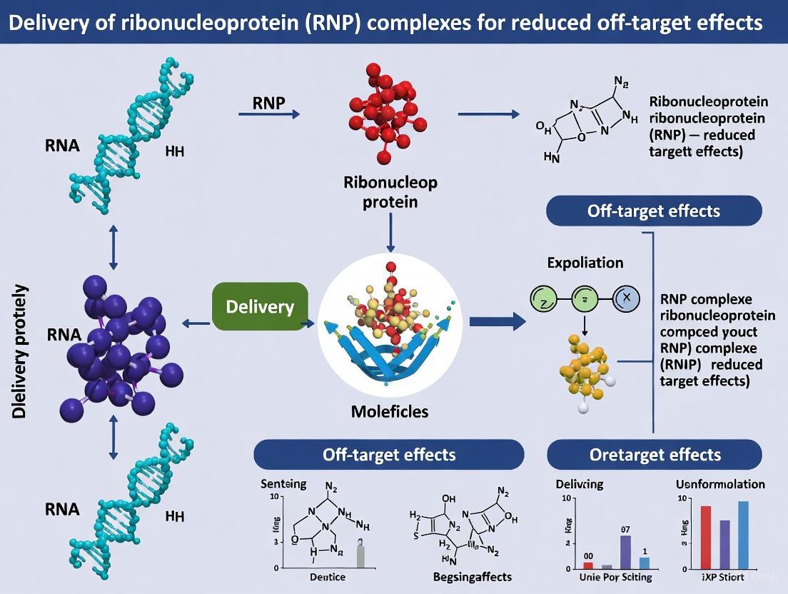
RNP Complex Delivery: A Strategic Approach to Minimize CRISPR Off-Target Effects
The therapeutic application of CRISPR-Cas9 genome editing is significantly hampered by off-target effects, which pose safety risks in clinical settings.
RNP Complex Delivery: A Strategic Approach to Minimize CRISPR Off-Target Effects
Abstract
The therapeutic application of CRISPR-Cas9 genome editing is significantly hampered by off-target effects, which pose safety risks in clinical settings. This article explores the delivery of pre-assembled Cas9 ribonucleoprotein (RNP) complexes as a powerful strategy to enhance editing precision. We provide a comprehensive analysis for researchers and drug development professionals, covering the foundational mechanisms of RNP action, advanced delivery methodologies including lipid nanoparticles and polymeric nanocarriers, practical optimization strategies to boost efficiency, and comparative data validating RNP superiority over DNA- and mRNA-based formats in reducing off-target activity. The synthesis of current evidence positions RNP delivery as a critical advancement for safer, more effective gene therapies.
The RNP Advantage: Foundations of Precision and Safety in Genome Editing
The Cas9 ribonucleoprotein (RNP) complex is a pre-assembled, functional unit comprising the Cas9 nuclease protein complexed with a guide RNA (gRNA). This complex serves as the catalytic core of the CRISPR-Cas9 system, capable of recognizing and cleaving specific DNA sequences upon delivery into cells [1]. The fundamental components include the Cas9 protein, which provides the DNA-cleaving activity, and the single guide RNA (sgRNA), a synthetic fusion of CRISPR RNA (crRNA) and trans-activating crRNA (tracrRNA) that programs the complex's target specificity [2] [3]. In therapeutic and research applications, direct delivery of pre-formed RNPs has emerged as a powerful strategy that offers significant advantages over alternative methods that rely on intracellular transcription and translation of plasmid DNA [1] [4].
The RNP format is particularly valued within a research context focused on reducing off-target effects, as its transient activity within cells—typically persisting for less than 24 hours—sharply limits the time window for non-specific genomic cleavage [4] [5] [6]. This transient nature, combined with the immediate availability of the fully assembled nuclease, facilitates rapid and precise genome editing with minimized risk of plasmid DNA integration and reduced cellular toxicity [5] [7]. Consequently, RNP delivery has become the preferred method for applications requiring high fidelity, including the development of therapeutic agents and the generation of precise animal models [1] [7].
Structural Composition and Functional Domains of the Cas9 RNP
The Cas9 protein exhibits a bilobed architecture consisting of a recognition (REC) lobe and a nuclease (NUC) lobe, which together form a positively charged groove accommodating the sgRNA:DNA heteroduplex [2]. The REC lobe, primarily responsible for binding sgRNA and target DNA, can be further divided into three regions: the Bridge helix, the REC1 domain, and the REC2 domain. While the REC2 domain is not absolutely critical for DNA cleavage, its deletion reduces editing efficiency, suggesting a supportive role in the recognition process [2].
The NUC lobe contains the catalytic centers and DNA interaction domains essential for target cleavage and recognition:
- RuvC Domain: Cleaves the non-complementary strand of the target DNA. The domain is assembled from three split RuvC motifs (I-III) within the Cas9 structure [2] [1].
- HNH Domain: Cleaves the DNA strand complementary to the sgRNA guide sequence. This domain displays significant conformational flexibility and is often poorly resolved in crystal structures, indicating its dynamic nature during the catalytic cycle [2].
- PAM-Interacting (PI) Domain: Responsible for recognizing the protospacer adjacent motif (PAM), a short sequence adjacent to the target DNA that is essential for initiating the DNA binding process. For Streptococcus pyogenes Cas9, the PAM sequence is 5'-NGG-3' [2] [1].
The sgRNA component is engineered to replace the natural two-part RNA system (crRNA:tracrRNA duplex) with a single chimeric molecule. Its core structure includes a target-specific ~20-nucleotide spacer sequence (derived from crRNA) and a scaffold sequence (derived from tracrRNA) that is essential for Cas9 binding [2] [3]. A synthetic tetraloop structure connects these two RNA elements, creating a continuous molecule that is both structurally stable and functionally efficient for programming DNA target recognition [3].
Figure 1: Structural organization of the Cas9 RNP complex, highlighting key protein domains and sgRNA components.
Advantages of RNP Delivery for Reducing Off-Target Effects
The delivery of pre-assembled Cas9 RNP complexes offers distinct pharmacological advantages over nucleic acid-based delivery methods, particularly regarding editing specificity and cellular toxicity profiles. These benefits are crucial for therapeutic applications where precision and safety are paramount.
Transient Activity and Reduced Off-Target Effects
The transient presence of active Cas9 nuclease in cells following RNP delivery is a fundamental factor in reducing off-target effects. Unlike plasmid DNA, which can persist for days to weeks and lead to prolonged Cas9 expression, RNPs are rapidly degraded within cells—typically within 24 hours—significantly narrowing the window for non-specific editing events [4] [5]. Experimental evidence demonstrates that RNP delivery can achieve a 28-fold reduction in off-target to on-target mutation ratios compared to plasmid-based delivery methods [4]. This substantial improvement in specificity stems from the limited temporal window for Cas9 activity, which decreases the probability of cleavage at partially complementary off-target sites [6].
Elimination of DNA Vector Integration
RNP delivery completely avoids the risk of plasmid DNA integration into the host genome, a concern associated with DNA-based delivery methods [4] [5]. Random integration of plasmid components can cause insertional mutagenesis, potentially disrupting essential genes or regulatory elements and leading to unpredictable consequences including oncogenesis. The DNA-free nature of RNP delivery eliminates this risk, providing a safer profile for therapeutic applications [7].
Reduced Cellular Toxicity
Foreign DNA introduction often triggers innate immune responses and cellular stress pathways, including cyclic GMP-AMP synthase (cGAS) activation in primary human cells and pluripotent stem cells [4]. RNP delivery bypasses these reactions, resulting in significantly improved cell viability compared to plasmid transfection. Studies report at least twice as many viable colonies following RNP transfection in sensitive cell types like embryonic stem cells [4]. The reduced cytotoxicity enables more efficient editing in challenging primary cells and stem cells, which are crucial for therapeutic development.
Immediate Catalytic Activity
Pre-complexed RNPs are immediately active upon delivery into cells, bypassing the delays associated with transcription and translation required for DNA-based methods [5] [7]. This immediate activity is particularly advantageous for primary cells with low division rates or limited transcriptional activity, where plasmid expression might be inefficient. The rapid kinetics also contribute to more synchronized editing across a cell population, yielding more homogeneous outcomes [1].
Table 1: Comparative Analysis of RNP vs. Plasmid Delivery Methods
| Characteristic | RNP Delivery | Plasmid Delivery | Experimental Evidence |
|---|---|---|---|
| Off-target effects | Low | High | 28-fold reduction in off-target/on-target ratio for specific loci [4] |
| Duration of activity | Short (~24 hours) | Prolonged (days to weeks) | Rapid clearance via protein degradation pathways [5] |
| DNA integration risk | None | Present | Eliminates random integration of plasmid components [4] [7] |
| Cellular toxicity | Low | High | ≥2x more viable colonies in embryonic stem cells [4] |
| Time to editing | Immediate | Delayed (requires transcription/translation) | Reduces total experimental time by up to 50% [4] |
| Editing efficiency in difficult cells | High | Variable | Successful in primary cells, T cells, iPSCs [1] [7] |
Quantitative Optimization of RNP Complex Formation
Precise stoichiometric control during RNP assembly is critical for achieving maximal editing efficiency while minimizing off-target effects. Recent investigations using nano differential scanning fluorimetry (nanoDSF) have quantitatively demonstrated that an equimolar ratio of Cas9 protein to guide RNA optimizes RNP complex formation and functionality [8].
Deviations from this ideal stoichiometry have significant consequences for editing outcomes. Excess gRNA relative to Cas9 protein substantially decreases knock-in efficiency while dramatically increasing the occurrence of on-target large deletions—a concerning class of editing artifacts that can compromise experimental and therapeutic outcomes [8]. Conversely, increasing the Cas9:gRNA ratio beyond equimolar proportions does not yield further improvements in knock-in efficiency, suggesting a saturation point in productive complex formation [8].
Optimal conditions for precise genome editing using the RNP platform have been empirically determined through systematic testing in human induced pluripotent stem cells (hiPSCs) and rat embryos. The highest efficiency in homology-directed repair (approximately 50% conversion rate in a GFP-to-BFP reporter system) was achieved using 0.4 μM Cas9 with an equimolar amount of gRNA, combined with 2 μM single-stranded oligonucleotide donor template [8]. These parameters provide a validated benchmark for researchers designing RNP-based editing experiments, particularly for applications requiring high precision.
Table 2: Optimized Stoichiometry for RNP Complex Assembly
| Parameter | Optimal Condition | Impact of Deviation | Experimental Validation |
|---|---|---|---|
| Cas9:gRNA ratio | 1:1 (equimolar) | Excess gRNA decreases KI efficiency and increases large deletions | NanoDSF analysis [8] |
| Cas9 concentration | 0.4 μM | Higher concentrations not beneficial for KI efficiency | GFP-BFP conversion assay in hiPSCs [8] |
| Donor DNA concentration | 2 μM (ssODN) | Concentration critical for HDR efficiency | Gene conversion assays [8] |
| Complex formation | 10-15 min incubation at room temperature | Inadequate complexing reduces editing efficiency | Standardized protocols [7] |
Detailed Experimental Protocols for RNP Delivery
RNP Complex Assembly and Delivery Workflow
Figure 2: Experimental workflow for RNP complex assembly and delivery, highlighting key steps and method options.
Primary T Cell Editing Protocol
RNP-mediated editing of primary human T cells has proven highly efficient for both knock-out and knock-in applications, including the generation of CAR-T cells [7].
Isolate primary T cells from human peripheral blood mononuclear cells (PBMCs) using standard Ficoll separation and activate with CD3/CD28 beads.
Prepare RNP complex:
- Combine 10 μg (∼60 pmol) of purified Cas9 protein with equimolar synthetic sgRNA (with chemical modifications to enhance stability).
- Incubate at room temperature for 15 minutes to allow complete complex formation.
Electroporation:
- Wash T cells and resuspend in appropriate electroporation buffer at 1-2×10^7 cells/mL.
- Mix 100 μL cell suspension with pre-formed RNP complex (and optional single-stranded DNA donor template for HDR).
- Electroporate using optimized program for primary T cells (e.g., Lonza 4D-Nucleofector, DS137 program).
Post-transfection processing:
- Immediately transfer cells to pre-warmed culture medium supplemented with cytokines (IL-2, IL-7, IL-15).
- Remove activation beads after 24-48 hours.
- Analyze editing efficiency at 72-96 hours post-electroporation via flow cytometry (for surface protein knock-outs) or next-generation sequencing (for precise edits).
This protocol typically achieves 70-90% knockout efficiency for target genes like PD-1, with high viability and minimal off-target effects [7].
Mouse Embryo Editing via RNP Electroporation
RNP delivery to mouse zygotes represents a highly efficient method for generating animal models without plasmid integration [7].
Collect fertilized zygotes from superovulated female mice at 0.5 days post-coitum.
Prepare RNP complex:
- Dilute purified Cas9 protein and synthetic sgRNA in nuclease-free microinjection buffer to final concentration of 100 ng/μL Cas9 (equimolar sgRNA).
- Incubate 10 minutes at room temperature.
Electroporation:
- Place groups of 30-50 zygotes between electrodes in a specialized chamber with pre-warmed electrolyte solution.
- Apply optimized electrical parameters (e.g., 30V, 3ms pulse width, 4 pulses with 100ms intervals).
- Alternatively, use CRISPR-EZ protocol for enhanced efficiency.
Embryo transfer and genotyping:
- Culture electroporated embryos overnight to assess development to 2-cell stage.
- Transfer viable embryos to pseudopregnant foster females.
- Analyze resulting pups via PCR and sequencing to identify founders with desired edits.
This method achieves high editing efficiency (typically >80%) with significantly reduced mosaicism compared to pronuclear injection, while completely avoiding the risk of plasmid integration [7].
Advanced Delivery Systems for RNP Complexes
Nanoparticle-Mediated RNP Delivery
Encapsulation of RNP complexes within nanoparticles represents a promising strategy for non-invasive in vivo delivery, offering protection from enzymatic degradation and enhanced cellular uptake [9]. Lipid nanoparticles (LNPs) and other synthetic carriers have been successfully employed to deliver Cas9 RNPs to various tissues, demonstrating therapeutic potential in disease models [1] [9]. These systems protect the RNP from degradation by serum nucleases and proteases, significantly extending functional half-life in circulation while facilitating endosomal escape through mechanisms such as the proton sponge effect [9].
Receptor-Targeted Delivery Systems
Advanced targeting strategies employ Cas9 proteins engineered with surface-exposed cysteine residues (Cas9M1C/C80S) that enable site-specific conjugation to pyridyl disulfide-activated ligands [5]. These modifications allow the RNP complex to be directed to specific cell types through receptor-mediated endocytosis, enhancing specificity and reducing off-target effects in heterogeneous tissues. For instance, fusion with P2C peptides derived from Drosophila melanogaster Yolk Protein 1 enables targeted delivery to ovarian tissue in insects, demonstrating the potential for tissue-specific editing in vivo [1].
NanoMEDIC Platform
The NanoMEDIC system utilizes engineered virus-like particles (VLPs) to deliver Cas9/gRNA RNP complexes with exceptional precision [6]. In comparative studies, NanoMEDIC-mediated delivery produced 58.3-87.5% precise gene removal without indels, significantly outperforming plasmid transfection (8.3-29.4% indel-free editing) [6]. This enhanced precision is attributed to the transient presence of Cas9, which enables rapid repair through non-homologous end joining (NHEJ) without prolonged nuclease activity that can generate extended deletions [6].
Table 3: Key Research Reagent Solutions for RNP-Based Genome Editing
| Reagent/Category | Function/Application | Examples/Specifications |
|---|---|---|
| Cas9 Proteins | Core nuclease component | His-tagged SpCas9, Cas9M1C/C80S (for conjugation), C80L/C574E (enhanced stability) [2] [5] |
| Synthetic sgRNAs | Target specification | Chemically modified for enhanced stability, research-grade with performance guarantees [4] [7] |
| Delivery Reagents | Cellular introduction | Cationic lipids, electroporation kits (e.g., Lonza 4D-Nucleofector), polymer nanoparticles [1] [5] |
| Assembly Tools | RNP complex formation | NanoDSF for stoichiometry optimization, buffer systems for complex stability [8] |
| Detection Assays | Editing validation | DNA displacement assays for sgRNA detection, NGS for off-target profiling, flow cytometry for functional knockouts [3] [7] |
| Control Systems | Experimental validation | Validated positive control sgRNAs, RNP complexes with known activity [7] |
Troubleshooting Common RNP Experimental Challenges
Low Editing Efficiency
When encountering insufficient editing efficiency, multiple factors should be systematically investigated. First, verify the quality and concentration of both Cas9 protein and sgRNA components using appropriate spectroscopic and integrity assessment methods [7]. Second, ensure optimal stoichiometry during RNP complex assembly, ideally confirmed through biophysical methods like nanoDSF [8]. Third, optimize delivery parameters—for electroporation, this includes voltage, pulse characteristics, and cell density; for lipid-based delivery, optimize charge ratios and incubation times [1] [7]. Finally, validate sgRNA activity using established positive controls and consider testing multiple guide RNAs targeting the same locus, as efficiency can vary significantly based on sequence context and chromatin accessibility [7].
High Toxicity or Poor Cell Viability
Cellular toxicity following RNP delivery often stems from suboptimal delivery conditions rather than the RNP complex itself. To mitigate viability issues, (1) titrate RNP concentration to the minimum required for efficient editing, (2) optimize delivery parameters to reduce cellular stress—particularly for sensitive primary cells, (3) implement enhanced recovery protocols including antioxidant supplements and appropriate cytokine support immediately following transfection, and (4) consider alternative delivery methods such as nanoparticle encapsulation rather than electroporation for particularly sensitive cell types [4] [7].
Unexpected Editing Patterns
The occurrence of large deletions or complex on-target rearrangements represents a significant concern, particularly for therapeutic applications. Recent research indicates that excess gRNA relative to Cas9 protein dramatically increases the frequency of large on-target deletions [8]. Adherence to precisely equimolar Cas9:gRNA ratios during complex assembly is therefore critical. Additionally, the choice of delivery platform influences editing precision—NanoMEDIC and other VLP systems have demonstrated superior capability for generating precise, indel-free edits compared to plasmid-based approaches [6]. Sequencing-based characterization of editing outcomes across multiple single-cell clones is recommended to fully assess the spectrum of modifications at the target locus.
Inconsistent Results Across Experiments
Reproducibility issues in RNP experiments often originate from variability in complex assembly or delivery conditions. Implement strict quality control measures for each component, including functional validation of each new sgRNA batch against standardized targets. Maintain consistent incubation conditions for RNP complex formation (time, temperature, buffer composition), and standardize delivery protocols across experiments. For in vivo applications, carefully control injection techniques and animal physiological states to minimize variability in RNP exposure levels [7] [8].
The delivery of CRISPR-based editors as pre-assembled ribonucleoprotein (RNP) complexes represents a paradigm shift in therapeutic genome editing. The core kinetic advantage of this approach lies in its transient activity: by directly introducing the functional Cas9-guide RNA complex into cells, the genome editor is active immediately upon nuclear entry and is rapidly degraded, sharply limiting the window for off-target interactions. In contrast, DNA-based delivery methods (plasmids, viral vectors) or even mRNA-based systems require intracellular transcription and/or translation, leading to prolonged and variable expression levels of the nuclease that significantly increase the risk of off-target cleavages at sites with partial homology to the guide RNA [10] [11]. This fundamental difference in temporal exposure underpins the enhanced specificity of RNP delivery, making it a critical strategy for advancing the safety profile of CRISPR therapeutics.
The kinetic profile of RNP complexes is characterized by three distinct phases: rapid cellular uptake, immediate nuclear activity, and swift degradation. This compressed activity timeline minimizes the thermodynamic sampling of DNA that leads to off-target effects. As the field moves toward clinical applications, establishing standardized protocols for RNP delivery and quantification is essential for realizing the full potential of this kinetic advantage in reducing genotoxicity concerns [12].
Quantitative Evidence: RNP Delivery Reduces Off-Target Effects
Multiple independent studies have demonstrated that transient RNP delivery achieves comparable on-target editing while significantly reducing off-target activity compared to plasmid-based methods.
Table 1: Comparison of Editing Efficiency and Specificity Across Delivery Methods
| Delivery Method | Editing System | On-Target Efficiency | Off-Target Indels | Reference |
|---|---|---|---|---|
| Gesicle RNP Delivery | Cas9-sgRNA (EMX1 locus) | Equivalent to plasmid | Undetectable at off-target site 4 | [11] |
| Plasmid Transfection | Cas9-sgRNA (EMX1 locus) | Equivalent to gesicle | Significant at off-target site 4 | [11] |
| NanoMEDIC RNP Delivery | Cas9/gRNA (HTLV-1) | 58.3–87.5% precise excision | Minimal indels (8.3–29.4% with plasmid) | [6] |
| EDV RNP Delivery | Cas9 RNP | >1300 RNPs/nucleus required | 30-50x more efficient than electroporation | [12] |
The data consistently demonstrate that methods enabling transient RNP activity achieve high on-target efficiency while virtually eliminating off-target effects. The Gesicle production system showed complete elimination of detectable indels at a known off-target site (Off-target 4) while maintaining equivalent editing at the intended EMX1 locus [11]. Similarly, the NanoMEDIC system achieved 58.3–87.5% precise gene removal without insertions or deletions, a significant improvement over plasmid transfection which produced substantial unintended mutations [6]. These findings highlight the direct correlation between reduced nuclease exposure time and improved editing fidelity.
Table 2: Quantifying RNP Requirements and Efficiency Across Platforms
| Platform | Cell Types Tested | Key Efficiency Metrics | Off-Target Reduction Mechanism | |
|---|---|---|---|---|
| Enveloped Delivery Vehicles (EDVs) | HEK293T, U2OS, HeLa | >1300 Cas9 RNPs/nucleus required for editing; 30-fold more efficient than electroporation | Shortened nuclear residence time | [12] |
| Lipid Nanoparticles (iGeoCas9) | Mouse neural progenitor cells, HEK293T, human bronchial epithelial cells | 4% to 99% editing efficiency depending on locus; 16-37% editing in mouse liver/lungs | Transient exposure without viral integration | [13] |
| Cyclodextrin Nanosponges (Ppoly) | CHO-K1 cells | 50% integration efficiency vs. 14% with commercial reagent; >90% encapsulation efficiency | Reduced cytotoxicity; protected delivery | [14] |
| Engineered Virus-Like Particles (eVLPs) | HEK293T, primary T cells, stem cell-derived neurons | Durable epigenetic silencing lasting weeks after single treatment | Hit-and-run editing without sustained expression | [15] |
The efficiency of RNP delivery varies by platform but consistently demonstrates favorable specificity profiles. EDV-mediated delivery was found to be 30-fold more efficient than electroporation and achieved editing at least twice as fast at comparable total Cas9 RNP doses [12]. This enhanced efficiency at lower effective doses further contributes to reduced off-target effects by minimizing the total nuclease load required for effective editing.
Protocol: RNP Delivery Using the Guide-it CRISPR/Cas9 Gesicle System
Background and Principle
The Guide-it CRISPR/Cas9 Gesicle Production System utilizes cell-derived nanovesicles (gesicles) for co-delivery of active Cas9 protein complexed with gene-specific sgRNA. This protocol enables efficient RNP delivery while preventing both overexpression and genomic integration of Cas9, which are common limitations of plasmid-based delivery methods [11]. The transient nature of gesicle-delivered RNP complexes typically restricts functional activity to a 24-72 hour window, capitalizing on the kinetic advantage to minimize off-target effects.
Materials and Reagents
- Guide-it CRISPR/Cas9 Gesicle Production System (Takara Bio)
- pGuide-it-sgRNA1 Vector
- Gesicle Producer 293T Cell Line
- Xfect Transfection Reagent
- Target cells for editing (e.g., HEK 293T)
- Protamine sulfate
- Guide-it Mutation Detection Kit
- Tissue culture plates and standard cell culture reagents
Experimental Procedure
Step 1: sgRNA Vector Preparation
- Clone the target sequence (e.g., EMX1 target sequence) into the pre-linearized pGuide-it-sgRNA1 vector using the provided ligation mix.
- Transform the ligation reaction into Stellar Competent Cells and select positive clones.
- Verify correct insertion by sequencing using the provided control primers.
Step 2: Gesicle Production
- Plate Gesicle Producer 293T cells at appropriate density in 10-cm tissue culture dishes.
- The following day, transfert cells with the Guide-it Gesicle Packaging Mix and your cloned pGuide-it-sgRNA1 vector.
- The Packaging Mix contains lyophilized Xfect Transfection Reagent premixed with optimized plasmids encoding Cas9 and all other elements needed for gesicle production.
- Harvest the gesicle-containing supernatant 48-72 hours post-transfection.
- Concentrate gesicles by centrifugation and resuspend in opti-MEM or PBS for storage at -80°C.
Step 3: Target Cell Editing
- Plate target cells (e.g., HEK 293T) at 5.0 × 10^5 cells per well in 24-well plates.
- 24 hours later, treat cells with 30 μL of concentrated Cas9 gesicles in the presence of protamine sulfate to enhance uptake.
- Include controls: untreated cells and plasmid-transfected cells (500 ng each of Cas9 and sgRNA plasmids).
- Incubate cells for 72 hours to allow editing to occur.
Step 4: Analysis of Editing Efficiency and Specificity
- Prepare crude DNA extracts from treated cells.
- Amplify the target locus (EMX1) and known off-target sites (e.g., off-target 4) by PCR.
- Detect indel formation using the Guide-it Resolvase enzyme (provided in the Mutation Detection Kit).
- Analyze cleavage products by agarose gel electrophoresis and quantify by densitometry.
- For confirmation, subclone PCR amplicons and perform Sanger sequencing to verify precise editing at the target site and absence of indels at off-target sites.
Expected Results and Troubleshooting
Successful gesicle-mediated RNP delivery should yield equivalent on-target editing efficiency compared to plasmid transfection, but with significantly reduced or undetectable off-target activity [11]. If editing efficiency is low, consider optimizing gesicle concentration, incubation time, or the amount of protamine sulfate used. Always include plasmid transfection controls to directly compare off-target rates between methods.
Protocol: Assessing RNP Delivery Efficiency with Fluorescence Correlation Spectroscopy
Background and Principle
Fluorescence Correlation Spectroscopy (FCS) provides a quantitative method to determine the number of functional Cas9 RNPs delivered to cell nuclei, enabling researchers to correlate RNP dosage with editing outcomes. This protocol establishes that >1300 Cas9 RNPs per nucleus are typically required for productive genome editing [12]. By quantifying actual nuclear delivery rather than assuming delivery based on input amounts, FCS helps optimize RNP delivery systems to achieve the minimal effective dose, further enhancing the kinetic advantage by preventing unnecessary nuclease overload.
Materials and Reagents
- STELLARIS 8 confocal microscope (Leica Microsystems) or equivalent FCS-capable system
- HC Plan-Apo 63×/1.4NA water immersion objective
- Alexa Fluor 594 hydrazide or ATTO 550 dye standards
- Hoechst 33342 nuclear stain
- Cas9-NLS protein labeled with appropriate fluorophore
- Custom FCS analysis software
- Eight-well microscopy dishes
- Mammalian cells for analysis (HEK293T, HeLa, or other relevant lines)
Experimental Procedure
Step 1: Microscope Calibration
- On the day of each experiment, prepare 300 μL of 10-100 nM Alexa Fluor 594 hydrazide dye standard in Milli-Q water.
- Add to one well of an eight-well microscopy dish and incubate at 37°C for at least 30 minutes.
- Adjust the correction collar of the objective to maximize counts per molecule for the dye standard.
- Obtain ten 5-second autocorrelation traces from the well containing dye standard to calculate the focal volume of the microscope.
Step 2: Sample Preparation and Staining
- Deliver Cas9 RNPs to cells using your preferred method (EDVs, electroporation, or other RNP delivery system).
- On the day of measurement, incubate cells with 300 nM Hoechst 33342 for 5 minutes to visualize nuclei.
- Wash cells 2× with Dulbecco's phosphate-buffered saline (DPBS) and incubate with prewarmed DMEM for imaging.
- Maintain cells at 37°C and 5% CO₂ during all imaging procedures.
Step 3: FCS Measurements
- For each sample, position the laser focus within the nucleus of a living cell.
- Acquire multiple FCS measurements (typically 10-20 per cell) with acquisition times of 5-10 seconds each.
- Measure sufficient cells to achieve statistical power (typically 20-30 cells per condition).
- For each measurement, generate autocorrelation curves that will be used to determine particle concentration and diffusion times.
Step 4: Data Analysis
- Fit autocorrelation curves using appropriate diffusion models to determine the number of fluorescent particles in the focal volume.
- Calculate particles per nucleus based on focal volume dimensions and nuclear volume.
- Correlate RNP numbers with editing efficiency measured by parallel experiments.
- Establish the minimum RNP count required for effective editing (expected to be >1300 RNPs/nucleus).
Expected Results and Interpretation
EDV-mediated delivery typically results in more efficient nuclear delivery compared to electroporation, with 30-50 fold higher editing efficiency across multiple human cell types [12]. The nuclear residence time of RNPs delivered via packaged methods like EDVs is prolonged compared to electroporation, contributing to their enhanced efficiency. This protocol provides a quantitative framework for optimizing delivery systems to achieve the kinetic advantage—sufficient RNP for on-target editing without excess that could promote off-target effects.
Visualization of RNP Delivery and Kinetic Advantage
The following diagrams illustrate the core concepts and experimental workflows related to the kinetic advantage of transient RNP delivery.
Diagram 1: Kinetic advantage of RNP versus plasmid delivery pathways. The compressed activity timeline of RNP delivery limits off-target interactions.
The Scientist's Toolkit: Essential Reagents for RNP Research
Table 3: Key Research Reagent Solutions for RNP Delivery Studies
| Reagent/System | Supplier/Reference | Primary Function | Key Applications |
|---|---|---|---|
| Guide-it CRISPR/Cas9 Gesicle System | Takara Bio [11] | Produces cell-derived nanovesicles for RNP delivery | Reduced off-target editing in mammalian cells |
| Cas9-NLS Protein | QB3 MacroLab, UC Berkeley [12] | High-purity nuclease for RNP assembly | Electroporation and packaged delivery studies |
| pGuide-it-sgRNA1 Vector | Takara Bio [11] | Cloning and expression of target-specific sgRNA | Gesicle production and RNP complex formation |
| Cationic Hyper-branched Cyclodextrin Polymer (Ppoly) | [14] | Nanosponge for RNP encapsulation and delivery | Enhanced knock-in efficiency with reduced cytotoxicity |
| SM102 Lipid Nanoparticles | [16] | Ionizable lipid for RNP encapsulation | In vivo delivery of base and prime editor RNPs |
| GeoCas9/iGeoCas9 | [13] | Thermostable Cas9 variant for stable RNP formulation | LNP-mediated delivery to liver and lung tissues |
| RENDER Platform | [15] | Engineered virus-like particles for epigenome editor delivery | Transient epigenetic silencing without DNA breaks |
The selection of appropriate reagents is critical for successful RNP-based experiments. The thermostable iGeoCas9 variant is particularly valuable for LNP encapsulation due to its resistance to denaturation during formulation [13]. For in vivo applications, LNPs utilizing the ionizable lipid SM102 have demonstrated remarkable efficiency improvements of over 300-fold compared to naked RNP delivery, without detectable off-target edits [16]. The emerging RENDER platform expands the application of transient delivery to epigenome editing, enabling durable gene silencing without DNA breaks through hit-and-run delivery of CRISPRoff and similar editors [15].
The kinetic advantage of transient RNP delivery represents a fundamental safety principle in therapeutic genome editing. By minimizing the duration of nuclease exposure through direct delivery of pre-assembled complexes, researchers can achieve high on-target efficiency while dramatically reducing off-target effects. The protocols and quantitative data presented here provide a framework for implementing this approach across multiple delivery platforms. As the field advances, standardization of RNP delivery and quantification methods will be essential for translating this kinetic advantage into safer genetic therapies. Future directions will likely focus on optimizing nuclear delivery efficiency and further compressing the activity timeline to maximize specificity while maintaining therapeutic efficacy.
The therapeutic application of the CRISPR-Cas9 system is fundamentally constrained by off-target effects, which pose significant safety risks in clinical settings. Central to mitigating this challenge is the delivery modality of the CRISPR machinery. Ribonucleoprotein (RNP) complexes, consisting of preassembled Cas9 protein and guide RNA, represent a transformative approach by leveraging two core mechanistic principles: immediate activity upon cellular entry and rapid degradation that limits the window for erroneous editing. Unlike plasmid or mRNA-based systems that require transcription and/or translation, RNPs are functionally active immediately upon delivery, initiating genome editing within hours rather than days [4]. This rapid onset is coupled with a short intracellular half-life—typically under 24 hours—ensuring the nuclease is cleared before sustained activity can promote off-target cleavage [4] [17]. This "fast-on/fast-off" kinetic profile underpins the enhanced fidelity of RNP delivery, establishing it as a critical platform for therapeutic genome editing.
Key Mechanisms and Quantitative Advantages of RNP Delivery
The fidelity enhancements offered by RNP delivery are demonstrated quantitatively across multiple studies. The following table summarizes core experimental data comparing RNP to DNA-based delivery methods.
Table 1: Quantitative Comparison of RNP vs. Plasmid DNA Delivery Outcomes
| Metric | RNP Delivery Performance | Plasmid DNA Delivery Performance | Experimental Context |
|---|---|---|---|
| Off-Target Reduction | 28-fold lower off-target/on-target ratio [4] | Higher off-target/on-target ratio | Gene OT3-18 [4] |
| Indel-Free Precision | 58.3–87.5% of edited DNA sequences [6] | 8.3–29.4% of edited DNA sequences [6] | NanoMEDIC delivery in 293FT & NP-2 cells [6] |
| Knock-In Efficiency | 50% HDR efficiency [18] [14] | 14% HDR efficiency (CRISPRMAX) [18] [14] | TILD-CRISPR in CHO-K1 cells [18] |
| Cell Viability | >80% cell viability post-transfection [18] | Significant reduction, inverse correlation with plasmid dose [4] | Cyclodextrin-based polymer delivery [18] |
The mechanistic relationship between the transient nature of RNPs and the resulting reduction in off-target effects is illustrated below.
Experimental Protocol: Evaluating RNP Fidelity Using a TILD-CRISPR Workflow
This protocol details the methodology for achieving high knock-in efficiency with minimal off-target effects using cyclodextrin-based nanosponges for RNP delivery, adapted from the TILD-CRISPR approach [18] [14].
Materials and Reagent Preparation
- Cas9 RNP Complex: Pre-complex recombinant S. pyogenes Cas9 protein (commercially available) and synthetic sgRNA at a molar ratio of 1:2. Incubate at 25°C for 10 minutes to form the RNP complex.
- Cationic Hyper-Branched Cyclodextrin Polymer (Ppoly): Synthesize as described in [18] using carbonyldiimidazole (CDI) as a crosslinker and choline chloride to introduce positive charges.
- Donor DNA Template: Prepare a linear double-stranded DNA donor with ~1000 bp homology arms. Avoid using circular plasmid donors to enhance HDR efficiency [18].
- Cell Line: CHO-K1 or other relevant cell lines, maintained under standard culture conditions.
- Control: Commercial transfection reagent (e.g., CRISPRMAX).
Step-by-Step Procedure
Complex Formation:
- Mix the pre-formed Cas9 RNP complex and linear donor DNA template with the Ppoly polymer in nuclease-free buffer.
- Incubate the mixture for 30 minutes at room temperature to form stable RNP/Ppoly nanoparticles.
Nanoparticle Characterization (Quality Control):
- Size and Zeta Potential: Use Dynamic Light Scattering (DLS) to confirm the formation of complexes. The RNP/Ppoly complex should have an average size of approximately 140 nm and a positive zeta potential [18].
- Encapsulation Efficiency: Use a fluorescence-based assay to verify that over 90% of the RNP is successfully encapsulated [18].
Cell Transfection:
- Seed cells to achieve 70-80% confluency at the time of transfection.
- Replace the culture medium with fresh medium.
- Add the RNP/Ppoly nanoparticles directly to the cell culture. For a 24-well plate, a final Ppoly concentration of 0.1-0.5 mg/mL is effective [18].
- Incubate cells under standard conditions for 48-72 hours.
Post-Transfection Selection and Cloning:
- Apply appropriate antibiotic selection 48 hours post-transfection to enrich for successfully edited cells.
- Perform single-cell cloning by serial dilution to isolate monoclonal cell populations.
- Expand clonal lines for molecular analysis.
Efficiency and Fidelity Analysis:
- Knock-In Confirmation: Perform junction PCR on genomic DNA from monoclonal lines using primers specific to the insertion site and the integrated sequence (e.g., GFP). Analyze products via gel electrophoresis.
- Editing Specificity: Assess potential off-target effects by sequencing the top in silico-predicted off-target sites for indels in the monoclonal lines. Compare the frequency of off-target indels in RNP-transfected cells versus those transfected with plasmid DNA.
The Scientist's Toolkit: Essential Reagents for RNP Fidelity Research
Table 2: Key Reagents for RNP-Based Genome Editing
| Reagent / Tool | Function / Description | Key Characteristics |
|---|---|---|
| High-Fidelity Cas9 Variants (e.g., HiFi Cas9 [17], Sniper2L [19]) | Engineered Cas9 proteins with reduced off-target activity without compromising on-target efficiency in the RNP format. | HiFi Cas9 (R691A) is a point mutant identified via bacterial screening for RNP compatibility [17]. |
| Cyclodextrin-Based Nanosponges (Ppoly) [18] [14] | A cationic hyper-branched polymer that encapsulates RNP complexes. | High encapsulation efficiency (>90%), low cytotoxicity (≥80% cell viability), enhances HDR [18]. |
| Linearized dsDNA Donors [18] | A double-stranded DNA repair template for HDR, linearized in vitro. | Used in TILD-CRISPR; features long homology arms (e.g., 1000 bp) to boost precise knock-in efficiency [18]. |
| NanoMEDIC [6] | A virus-like particle (VLP) system for RNP delivery. | Achieves high precision, producing 58.3-87.5% removal-edited DNA without indels [6]. |
| Lipid Nanoparticles (LNPs) [16] | Synthetic, chemically defined nanoparticles optimized for RNP encapsulation and delivery. | Formulations using ionizable lipids (e.g., SM102) can enhance in vivo editing efficiency by >300-fold vs. naked RNP [16]. |
| Redox-Responsive Nanogels [20] | Polymeric nanogels that degrade in the reductive cytosolic environment, triggering RNP release. | Encapsulation efficiency of ~60%; reductive release mimics intracellular conditions [20]. |
The strategic adoption of RNP complexes, characterized by their immediate activity and rapid degradation, provides a robust mechanistic solution to the critical challenge of off-target effects in CRISPR-Cas9 genome editing. The experimental data and protocols outlined herein demonstrate that this approach, especially when combined with advanced non-viral delivery systems and high-fidelity enzymes, enables highly precise genetic modifications with direct implications for the development of safer therapeutic applications.
The therapeutic application of CRISPR-Cas9 gene editing is critically dependent on the efficient and safe delivery of its molecular components into the nucleus of target cells [21]. The functional unit of the system is the ribonucleoprotein (RNP) complex formed by the Cas nuclease and a guide RNA (gRNA) [22]. This complex can be delivered using three primary biological formats: plasmid DNA (pDNA), which encodes the genetic instructions for the cell to produce the machinery; messenger RNA (mRNA) and gRNA, which provide a more direct transient template; and the pre-assembled ribonucleoprotein (RNP) complex of Cas9 protein and gRNA [22] [21]. The choice of cargo format profoundly impacts key performance metrics, including editing efficiency, kinetics, specificity, and cytotoxicity. This application note provides a comparative analysis of these formats, with a specific focus on RNP delivery for minimizing off-target effects, and details protocols for their experimental implementation.
Comparative Analysis of Cargo Formats
The table below summarizes the core characteristics of the three primary CRISPR cargo formats.
Table 1: Comparative Analysis of CRISPR-Cas9 Cargo Formats
| Feature | Plasmid DNA (pDNA) | mRNA + gRNA | Ribonucleoprotein (RNP) |
|---|---|---|---|
| Molecular Format | DNA plasmid(s) encoding Cas9 and sgRNA [22] | Cas9 mRNA + single-guide RNA (sgRNA) [22] | Pre-complexed Cas9 protein and sgRNA [22] |
| Mechanism of Action | Nuclear import, transcription to mRNA, then translation to protein [22] | Cytoplasmic translation into Cas9 protein [22] | Direct nuclear localization and DNA cleavage [22] |
| Onset of Editing | Slow and asynchronous (days) [21] | Fast and synchronized (hours) [21] | Very fast (hours) [4] |
| Editing Efficiency | Variable and often lower [4] | High [23] | High, especially in hard-to-transfect cells [13] [4] |
| Persistence | Long (days to weeks), risk of random integration [4] | Short (days) [22] | Very short (~24 hours) [4] |
| Off-Target Effects | High, due to prolonged expression [4] | Moderate [24] | Lowest, due to transient activity [23] [4] |
| Cytotoxicity & Immunogenicity | High; can trigger innate immune responses [4] [21] | Moderate; immunogenicity can be mitigated by mRNA modification [13] [21] | Lowest; minimal immune activation, well-tolerated [13] [4] |
| Manufacturing & Storage | Easy bacterial production, stable [22] | Complex in vitro transcription, requires cold chain [22] | Complex protein purification, most labile format [22] |
Key Findings from Comparative Studies
- In Vitro and In Vivo Performance: A 2024 study directly comparing Lipid Nanoparticle (LNP)-mediated delivery of mRNA/sgRNA versus RNP found that the mRNA format led to higher gene editing efficiencies in both reporter HEK293T cells and HEPA 1-6 cells in vitro. In vivo, LNPs delivering mRNA/sgRNA achieved 60% gene knock-out in hepatocytes, whereas editing was not conclusively detected for the RNP format in this particular systemic administration setup [23].
- Advancements in RNP Delivery: A landmark 2025 study demonstrated that engineering a thermostable Cas9 (iGeoCas9) and formulating it into RNPs with specialized LNPs enabled high-efficiency editing in vivo. This approach achieved 37% editing in mouse liver and, notably, 19% editing of the SFTPC gene in lung tissue after a single intravenous injection, showcasing a major improvement for non-viral RNP delivery to non-liver tissues [13].
- Off-Target Specificity: Multiple studies confirm that RNP delivery results in significantly lower off-target effects compared to plasmid DNA. The short intracellular half-life of the RNP complex limits the time window for non-specific editing, reducing the ratio of off-target to on-target mutations by more than 28-fold in some cases [4]. This makes RNPs the preferred format for applications requiring high precision.
Experimental Protocols
Protocol 1: RNP Complex Assembly and Delivery via Electroporation
This protocol is optimized for high-efficiency editing with minimal off-target effects in vitro [22] [4].
Reagents and Materials:
- Purified recombinant Cas9 protein with Nuclear Localization Signal (NLS)
- Synthetic, chemically modified sgRNA (research-grade)
- Electroporation buffer (e.g., Neon Transfection System Resuspension Buffer)
- Cell culture reagents and appropriate growth media
Procedure:
- RNP Complex Assembly:
- Resuspend synthetic sgRNA in nuclease-free buffer.
- Mix Cas9 protein and sgRNA at a molar ratio of 1:1.2 to 1:2.5 (e.g., 10 µg Cas9 with 4 µg of a 100-nt sgRNA).
- Incubate the mixture at room temperature for 10-20 minutes to allow for complete RNP complex formation.
Cell Preparation:
- Harvest and wash the target cells (e.g., immortalized cells, primary cells, or stem cells).
- Resuspend the cell pellet in the appropriate electroporation buffer at a pre-optimized density (e.g., 1-10 x 10^6 cells/mL).
Electroporation:
- Combine the cell suspension with the pre-assembled RNP complex. Mix gently.
- Transfer the mixture to an electroporation cuvette.
- Electroporate using a pre-optimized program. For many mammalian cell types, a typical program might be 1,400 V for 20 ms with 1 pulse.
- Immediately after pulsing, add pre-warmed culture medium to the cuvette and transfer the cells to a culture plate.
Post-Transfection Analysis:
- Allow cells to recover for 48-72 hours before assessing editing efficiency via genomic DNA extraction and next-generation sequencing or T7 Endonuclease I assay.
- Evaluate cell viability and potential off-target effects at predicted sites.
Protocol 2: LNP Formulation for In Vivo RNP Delivery
This protocol outlines the formulation of LNPs for the systemic delivery of stable RNPs, based on the methodology used for iGeoCas9 [13].
Reagents and Materials:
- Ionizable/cationic lipid (e.g., DODAP, DLin-MC3-DMA)
- Helper phospholipid (e.g., DSPC)
- Cholesterol
- PEGylated lipid (e.g., DMG-PEG 2000)
- Ethanol and aqueous citrate buffer (pH 4.0)
- Stable RNP complex (e.g., iGeoCas9-sgRNA)
Procedure:
- Lipid Mixture Preparation:
- Dissolve the ionizable lipid, phospholipid, cholesterol, and PEG-lipid in ethanol at a specific molar ratio (e.g., 50:10:38.5:1.5 mol%). The total lipid concentration is typically 10-20 mM.
Aqueous Phase Preparation:
- Dilute the pre-assembled RNP complex in a low-pH citrate buffer (e.g., 50 mM, pH 4.0).
Nanoparticle Formation:
- Rapidly mix the ethanolic lipid solution with the aqueous RNP solution using a microfluidic device (e.g., NanoAssemblr) at a controlled flow rate and ratio (e.g., 1:3 to 1:5, aqueous-to-ethanol ratio) to form LNPs via rapid mixing.
- Dialyze the resulting LNP suspension against a large volume of PBS (pH 7.4) for several hours at 4°C to remove ethanol and adjust the pH.
Characterization and Administration:
- Measure the particle size, polydispersity index (PDI), and encapsulation efficiency of the RNP-LNPs using dynamic light scattering and other relevant assays.
- Administer the formulated RNP-LNPs to the animal model via systemic injection (e.g., intravenous). Dosing will depend on the target organ and specific LNP formulation.
Cargo Format Decision Workflow
The following diagram illustrates the logical process for selecting the appropriate CRISPR cargo format based on experimental goals and constraints.
The Scientist's Toolkit: Essential Research Reagents
Table 2: Key Reagents for CRISPR-Cas9 RNP Research
| Reagent / Material | Function / Application | Key Characteristics |
|---|---|---|
| High-Fidelity Cas9 Nuclease | Introduces double-strand breaks at target DNA sequences. | Recombinantly purified, high specific activity, contains Nuclear Localization Signals (NLS) [22]. |
| Chemically Modified sgRNA | Guides Cas9 to the specific genomic locus. | Synthetic, research-grade; modifications (e.g., 2'-O-Me) enhance stability and reduce immunogenicity [22] [21]. |
| Ionizable Lipids | Core component of LNPs for in vivo delivery. | Enables efficient encapsulation and endosomal escape; pH-sensitive (e.g., DODAP) [13]. |
| Electroporation System | Physical method for delivering RNPs into cells in vitro. | High efficiency for a wide range of cell types, including primary and stem cells [4]. |
| Stable Cas9 Variants (e.g., iGeoCas9) | Thermostable Cas9 for improved LNP-RNP formulation. | Engineered for enhanced stability and efficiency; withstands organic solvents used in LNP production [13]. |
Advanced Delivery Systems for RNP Complexes: From LNPs to Nanogels
Lipid nanoparticles (LNPs) have emerged as a leading non-viral delivery platform for a range of therapeutic payloads, most notably nucleic acids and ribonucleoprotein (RNP) complexes. Their successful deployment in mRNA COVID-19 vaccines and the siRNA drug Onpattro has validated their safety, efficacy, and scalability for clinical use [25]. For CRISPR-Cas-based genome editing, the delivery of preassembled Cas protein and guide RNA as an RNP complex offers significant advantages, including reduced off-target effects and transient editing activity, which minimizes immunogenic risks and unintended genomic alterations [1] [26]. This application note details the use of LNPs for the in vivo delivery of CRISPR RNP complexes, providing structured data, detailed protocols, and key resources to support researchers in this rapidly advancing field.
Current Applications and Quantitative Data
LNPs are being leveraged to deliver CRISPR RNP complexes for precise genome editing across a variety of tissues and disease models. The following table summarizes key recent applications and their outcomes.
Table 1: Recent Advances in LNP-Mediated RNP Delivery for In Vivo Genome Editing
| Application / System | Target / Model | Key Outcome(s) | Citation / Year |
|---|---|---|---|
| Prime Medicine LNP | AATD (Alpha-1 Antitrypsin Deficiency) gene correction in humanized mice & non-human primates | Achieved 72% gene correction in mice; >50% editing efficiency in NHP; Restoration of wild-type AAT protein; No detected off-target effects | [27] (2025) |
| Self-deliverable RNP (A22p-Cas9) | Mouse brain (striatum) via direct injection | Robust genome editing in neural progenitor cells and neurons in vivo; Effective without helper materials | [26] (2024) |
| RNP-MITO-Porter | Mitochondrial DNA (m.7778G>T mutation in mt-Atp8) in mouse fibroblast cells | Successful delivery of RNP directly into mitochondria; Confirmed sequence-specific double-strand breaks in mtDNA | [28] (2025) |
| MC3-based LNP (Reference) | General siRNA and mRNA delivery (Mechanistic Study) | Identified galectin-9+ endosomal damage as a conduit for cytosolic release; Highlighted cargo-lipid segregation as a key barrier | [29] (2025) |
Detailed Experimental Protocols
Protocol: Formulating RNP-LNPs Using a Microfluidic Device
This protocol, adapted from a mitochondrial targeting study, describes the encapsulation of CRISPR RNP complexes into LNPs using an invasive lipid nanoparticle (iLiNP) device [28].
1. RNP Complex Formation:
- Materials: Cas9 protein, target-specific sgRNA, Nuclease-free buffer (e.g., 1X PBS or 30 mM HEPES, pH 7.4).
- Procedure: Incubate Cas9 protein with sgRNA at a molar ratio optimized for your system (e.g., 1:1.2 to 1:2 protein:sgRNA) for 10-20 minutes at room temperature to allow for RNP complex assembly.
2. Preparation of Lipid and Aqueous Phases:
- Lipid Phase (Organic Phase): Dissolve lipid components in ethanol. A sample mitochondrial-targeting formulation includes:
- DOPE (9 mol%)
- Sphingomyelin (2 mol%)
- DMG-PEG2000 (0.22 mol%) [Note: May be omitted for homogeneity in some systems]
- Stearylated-octaarginine (STR-R8) (1.1 mol%) as a cationic cell-penetrating peptide.
- Total lipid concentration should typically be in the range of 1-10 mM.
- Aqueous Phase: Dilute the prepared RNP complex into a suitable buffer, such as 30 mM HEPES (pH 7.4). The final ethanol concentration exposed to the RNP should be minimized to prevent protein denaturation.
3. Microfluidic Mixing:
- Use a microfluidic device (e.g., iLiNP device) with controlled flow rates.
- Critical Parameter: The flow rate ratio (FRR) between the aqueous and organic phases is crucial. Test ratios such as 425:75 (aqueous:organic, µL/min) with a total flow rate (TFR) of 500 µL/min. Adjust to achieve a homogeneous particle size.
- The rapid mixing process promotes the formation of disk-shaped lipid assemblies that stabilize into spherical LNPs encapsulating the RNP payload.
4. Dialysis and Purification:
- Dialyze the resulting LNP suspension against a large volume of an appropriate outer aqueous phase (e.g., 10 mM HEPES buffer or PBS, pH 7.4) for several hours to remove residual ethanol and buffer exchange.
- The final formulation can be concentrated if necessary and sterilized by filtration (0.22 µm).
5. Characterization:
- Particle Size and Dispersity: Use dynamic light scattering (DLS) to measure hydrodynamic diameter and polydispersity index (PdI). Aim for a diameter of ~70-100 nm and a PdI <0.2.
- Zeta Potential: Measure surface charge using electrophoretic light scattering.
- Encapsulation Efficiency: Quantify the percentage of RNP successfully loaded into the LNPs, for example, using a Ribogreen assay after particle disruption.
Protocol: Evaluating Intracellular Barriers to LNP Delivery
This protocol outlines methods to visualize and quantify key intracellular steps in LNP-mediated RNP delivery, based on a recent Nature Communications study [29].
1. Live-Cell Imaging of Endosomal Escape:
- Cell Seeding: Plate adherent cells (e.g., HeLa, HEK293) in glass-bottom imaging dishes.
- Transfection: Treat cells with fluorescently labeled RNP-LNPs (e.g., labeled sgRNA).
- Staining: Transfect cells with a plasmid expressing a membrane damage sensor, such as galectin-9 fused to a fluorescent protein (e.g., Gal9-GFP).
- Image Acquisition: Use fast live-cell or confocal microscopy to track the co-localization of RNP-LNPs (red) and galectin-9 (green) in real-time.
- Analysis: Vesicles that become positive for both signals indicate endosomal compartments that have been damaged by the LNP, a process conducive to cargo release.
2. Analysis of Cargo-Lipid Segregation:
- Dual-Labeling: Prepare LNPs containing both a fluorescently labeled ionizable lipid (e.g., BODIPY-MC3) and a spectrally distinct labeled RNA payload.
- Microscopy and Analysis: After cellular uptake, use super-resolution microscopy to visualize individual endosomes. Quantify the fluorescence intensity of both labels within the same vesicle. A dissociation of the signals indicates segregation of the lipid and RNA cargo during endosomal sorting, which can limit delivery efficiency.
The Scientist's Toolkit: Research Reagent Solutions
Table 2: Essential Reagents for LNP-RNP Research and Development
| Reagent / Material | Function / Application | Key Notes |
|---|---|---|
| Ionizable Lipids (e.g., MC3) | Core LNP component for nucleic acid/RNP encapsulation and endosomal escape [25]. | Becomes protonated in acidic endosomes, facilitating membrane disruption. |
| PEG-Lipids (e.g., DMG-PEG2000) | Stabilizes LNP formulation; reduces non-specific binding; modulates pharmacokinetics [25] [28]. | Critical for controlling particle size and preventing aggregation during formation. |
| Cell-Penetrating Peptides (e.g., A22p, STR-R8) | Enhances cellular uptake and self-delivery capability of RNPs or LNPs [26] [28]. | A22p, derived from semaphorin-3a, shows high efficacy for neural progenitor cells. |
| Non-Immunogenic Lipids (e.g., POZ-Lipid) | Safer alternative to PEGylated lipids; reduces risk of anti-PEG immune responses [27]. | Serina Therapeutics' POZ technology avoids IgM/IgG antibodies upon repeated dosing. |
| Galectin-9 Fluorescent Reporter | Live-cell sensor for detecting LNP-induced endosomal membrane damage [29]. | A key tool for mechanistic studies of endosomal escape efficiency. |
| Microfluidic Devices (e.g., iLiNP) | Enables reproducible, scalable, and aseptic production of homogeneous LNP formulations [28]. | Provides superior control over particle characteristics compared to bulk mixing methods. |
Visualizing Key Pathways and Workflows
LNP-Mediated RNP Delivery and Endosomal Escape Pathway
Experimental Workflow for RNP-LNP Development
The delivery of CRISPR-Cas9 as a ribonucleoprotein (RNP) complex represents a pivotal advancement in genome editing therapeutics, offering transient editing activity that minimizes off-target effects and reduces immunogenicity compared to plasmid DNA or mRNA formats [1] [30]. Despite these advantages, the intracellular delivery of substantial RNP complexes remains a significant challenge for clinical translation. Polymer-based nanoparticles have emerged as promising vehicles to overcome these delivery barriers, providing protection for RNPs during transit, enhancing cellular uptake, and facilitating endosomal escape while maintaining favorable safety profiles [18] [31]. Among these, cyclodextrin-based polymers and cationic nanocarriers have demonstrated particular utility for RNP delivery, enabling efficient genome editing across diverse cell types and in vivo models. This document details the application and protocols for utilizing these polymer-based systems to achieve efficient RNP delivery with minimal off-target effects, supporting a broader thesis on enhanced precision in genome editing.
Application Notes: Cyclodextrin Polymers for RNP Delivery
Cationic Hyper-Branched Cyclodextrin-Based Polymers (Ppoly)
Cyclodextrins (CDs) are cyclic oligosaccharides that form nanostructures with hydrophobic central cavities and hydrophilic exteriors, enabling host-guest interactions with various therapeutic molecules [32]. Their unique architecture allows for chemical modification and polymerization, creating versatile delivery platforms. Cationic hyper-branched cyclodextrin-based polymers (Ppoly) have been specifically engineered for RNP delivery through synthesis using choline chloride to introduce positive charges and carbonyldiimidazole (CDI) as a crosslinking agent [18].
These Ppoly systems demonstrate remarkable efficiency in delivering CRISPR-Cas9 RNPs for targeted genome editing. When evaluated using the TILD-CRISPR method (which couples donor DNA linearization with RNP complexes), Ppoly achieved a remarkable 50% integration efficiency in CHO-K1 cells, significantly outperforming the 14% observed with commercial CRISPRMAX reagent while maintaining cell viability above 80% [18]. The encapsulation efficiency for RNP complexes exceeded 90%, with the nanosponge architecture improving stability and biocompatibility while facilitating effective transport to target cells.
Table 1: Performance Metrics of Cationic Hyper-Branched Cyclodextrin Polymers
| Parameter | Performance | Experimental Context |
|---|---|---|
| Knock-in Efficiency | 50% | GFP gene integration in CHO-K1 cells using TILD-CRISPR [18] |
| Cell Viability | >80% | Post-transfection viability in CHO-K1 cells [18] |
| Encapsulation Efficiency | >90% | RNP complex loading [18] |
| Particle Size | Approximately 107.7 nm (increased after RNP loading) | Dynamic Light Scattering measurement [18] |
The following diagram illustrates the experimental workflow for utilizing cyclodextrin-based polymers in RNP delivery and genome editing:
Figure 1: Experimental workflow for cyclodextrin polymer-based RNP delivery showing key steps from complex formation to genome editing.
Characterization of RNP-Loaded Cyclodextrin Polymers
The formation and stability of RNP-Ppoly complexes can be validated through several analytical techniques:
Fourier Transform Infrared (FTIR) Spectroscopy: Confirms successful complex formation through characteristic spectral changes. The RNP spectrum displays amide I (approximately 1650 cm⁻¹) and amide II (approximately 1540 cm⁻¹) bands from the Cas9 protein, along with phosphate group vibrations (1000-1250 cm⁻¹ range) from the RNA backbone. Upon complexation with Ppoly, broadening and dampening of O-H/N-H stretches and reduced intensity in amide regions indicate intermolecular interactions between cationic amine groups of Ppoly and polar/charged areas of the RNP complex [18].
Dynamic Light Scattering (DLS) and Zeta Potential: Determine particle size distribution and surface charge. The average size of Ppoly alone is approximately 107.7 nm, with increases observed after RNP complex formation. Zeta potential measurements help confirm successful encapsulation when the surface charge of the final LNPs is neutral, which is important for minimizing in vivo uptake by the immune mononuclear phagocyte system [18] [33].
Application Notes: Cationic Nanocarriers for RNP Delivery
Cationic Lipid-Enhanced Lipid Nanoparticles (LNPs)
Traditional lipid nanoparticles (LNPs) rely on ionizable cationic lipids that become protonated in acidic environments to encapsulate nucleic acids. However, these conventional methods are unsuitable for RNP delivery because Cas9 RNPs denature in acidic buffers, resulting in increased hydrodynamic size from 10 to 150 nm [33]. To overcome this limitation, a modified approach incorporates permanently cationic lipids (e.g., DOTAP) into standard LNP formulations, enabling encapsulation under neutral pH conditions that preserve RNP structure and function.
This strategy has been successfully applied to multiple LNP classes, including dendrimer lipid nanoparticles (DLNPs), stable nucleic acid lipid particles (SNALPs), and lipid-like nanoparticles (LLNPs) [33]. Incorporating 10-20 mole% of permanently cationic lipid (DOTAP) into traditional LNP formulations enabled controlled self-assembly using phosphate-buffered saline (PBS) solutions of RNPs instead of acidic buffers, preserving Cas9 function and resulting in stable RNP-loaded nanoparticles with sizes <200 nm and neutral surface charge [33].
Table 2: Performance Metrics of Cationic Lipid-Modified LNPs for RNP Delivery
| Parameter | Performance | Experimental Context |
|---|---|---|
| Optimal DOTAP Incorporation | 10-20 mole% | Total lipid composition [33] |
| Particle Size | <200 nm | Dynamic Light Scattering measurement [33] |
| Optimal Cas9/sgRNA Ratio | 1:3 (mol/mol) | In vitro gene editing efficacy [33] |
| Stability | 60 days at 4°C | Constant gene editing activity after storage [33] |
| Cellular Uptake Mechanism | Lipid raft-dependent | Inhibited by MβCD treatment [33] |
Characterization and Evaluation of RNP-Loaded Cationic Nanocarriers
The following protocol ensures proper formulation and assessment of cationic nanocarriers for RNP delivery:
Formulation Integrity Assessment: Confirm successful RNP encapsulation through size and zeta potential measurements. LNPs with encapsulated RNPs should show slightly larger sizes than empty nanoparticles, while surface charge should be neutral, indicating successful encapsulation and reduced immune recognition in vivo [33].
Stability Testing: Monitor RNP-loaded nanoparticle stability at 4°C for extended periods. Effective formulations should maintain consistent size (PDI <0.2) and gene editing activity for at least 60 days, supporting future clinical translation [33].
Cellular Uptake Mechanism Evaluation: Track intracellular trafficking using fluorescently labeled Cas9 protein. Energy-dependent uptake occurs primarily through lipid raft-mediated endocytosis, with Cas9 proteins gradually entering the nucleus within 6 hours due to nuclear localization signals [33].
Experimental Protocols
Protocol 1: RNP Complex Formation and Characterization
Principle: Preassembling Cas9 protein with sgRNA in vitro creates functional RNP complexes that enable rapid, transient genome editing with reduced off-target effects compared to nucleic acid-based delivery formats [1] [30].
Materials:
- Purified Cas9 protein (commercial source or expressed and purified using affinity tags)
- In vitro transcribed or synthetic sgRNA
- Nuclease-free duplex buffer (e.g., 30 mM HEPES, pH 7.5, 100 mM KCl)
- Thermal cycler or water bath
Procedure:
- Complex Formation:
- Mix Cas9 protein and sgRNA at a molar ratio of 1:3 (Cas9:sgRNA) in duplex buffer [33].
- Incubate at room temperature for 15-20 minutes to form functional RNP complexes.
- For challenging targets or to enhance stability, refold sgRNA by heating to 65-70°C for 5 minutes followed by slow cooling before complex formation [16].
- Complex Characterization:
- Verify successful complex formation using Dynamic Light Scattering (DLS) to monitor changes in hydrodynamic radius.
- Confirm complex stability using differential scanning fluorimetry (DSF), noting that RNP complexes demonstrate enhanced stability compared to Cas9 protein alone [16].
- Additional stabilizers such as 10% (w/v) sucrose may be incorporated to further enhance RNP stability [16].
Protocol 2: Cyclodextrin Polymer-Based RNP Encapsulation
Principle: Cationic hyper-branched cyclodextrin polymers (Ppoly) electrostatically interact with RNP complexes, forming stable nanocomplexes that facilitate cellular uptake while protecting RNPs from degradation [18].
Materials:
- Cationic hyper-branched cyclodextrin-based polymer (Ppoly)
- Preformed RNP complexes (from Protocol 1)
- Phosphate-buffered saline (PBS), pH 7.4
Procedure:
- Nanocomplex Formation:
- Combine Ppoly and RNP complexes at optimal weight ratios in PBS buffer.
- Incubate at room temperature for 30 minutes to allow self-assembly into stable nanocomplexes.
Characterization:
- Determine encapsulation efficiency (typically >90%) using appropriate quantification methods.
- Measure particle size and zeta potential via Dynamic Light Scattering.
- Verify complex formation through FTIR spectroscopy, observing dampening of amide I and II bands along with broadening of O-H/N-H stretches indicating successful interactions [18].
Functional Assessment:
- Transfect cells at optimal concentrations determined by dose-response studies.
- Evaluate gene editing efficiency 48-72 hours post-transfection via junction PCR, T7 Endonuclease I assay, or sequencing.
- Assess cytotoxicity using standard viability assays (e.g., MTT, CCK-8), expecting >80% cell viability with optimized formulations [18].
Protocol 3: Cationic Lipid-Modified LNP Formulation for RNPs
Principle: Incorporating permanently cationic lipids (e.g., DOTAP) into standard LNP formulations enables efficient RNP encapsulation under neutral pH conditions, preserving protein structure and function [33].
Materials:
- Ionizable cationic lipid (e.g., 5A2-SC8)
- Permanently cationic lipid (e.g., DOTAP)
- Helper lipids (DOPE, cholesterol)
- PEG lipid (e.g., DMG-PEG 2000)
- Preformed RNP complexes (from Protocol 1)
- Ethanol (100%)
- Phosphate-buffered saline (PBS), pH 7.4
- Microfluidic device or T-tube apparatus
Procedure:
- Lipid Solution Preparation:
- Prepare lipid mixture in ethanol with the following molar composition:
- Ionizable cationic lipid: 15%
- DOPE: 15%
- Cholesterol: 30%
- DMG-PEG 2000: 3%
- DOTAP: 7% (5-20% range may be optimized) [33]
- Prepare lipid mixture in ethanol with the following molar composition:
LNP Formulation:
- Mix ethanolic lipid solution with aqueous RNP solution (in PBS, pH 7.4) at 1:3 volume ratio using microfluidic device or rapid mixing.
- Dialyze against PBS (pH 7.4) to remove ethanol and free components.
- Sterile filter through 0.22 μm membrane.
Characterization and Quality Control:
- Measure particle size (typically <200 nm) and polydispersity index (PDI <0.2) via DLS.
- Confirm neutral surface charge via zeta potential.
- Verify encapsulation efficiency and RNP integrity.
- Assess in vitro gene editing efficiency in reporter cell lines (e.g., HeLa-Luc, HeLa-GFP).
The Scientist's Toolkit: Essential Research Reagents
Table 3: Key Reagents for Polymer-Based RNP Delivery Systems
| Reagent/Category | Specific Examples | Function/Application |
|---|---|---|
| Cyclodextrin Polymers | Cationic hyper-branched cyclodextrin-based polymer (Ppoly) | Forms stable nanocomplexes with RNPs through electrostatic interactions; minimal cytotoxicity [18] |
| Cationic Lipids | DOTAP (1,2-dioleoyl-3-trimethylammonium-propane) | Enables RNP encapsulation under neutral pH in LNP formulations; enhances cellular uptake [33] |
| Ionizable Lipids | 5A2-SC8, SM102 | Promotes endosomal escape through pH-dependent charge changes; critical for cytosolic delivery [16] [33] |
| Stabilizing Agents | DMG-PEG 2000, sucrose | Enhances nanoparticle stability and shelf life; prevents aggregation [16] [33] |
| Characterization Tools | Dynamic Light Scattering, FTIR Spectroscopy | Assesses nanoparticle size, stability, and complex formation [18] |
| Efficiency Assays | T7 Endonuclease I assay, junction PCR, flow cytometry | Quantifies gene editing efficiency and knock-in success rates [18] [33] |
The following diagram illustrates the mechanism of action for cationic nanocarriers in RNP delivery, highlighting key stages from cellular entry to genomic integration:
Figure 2: Mechanism of cationic nanocarrier-mediated RNP delivery showing critical intracellular trafficking stages from endocytosis to precise gene integration.
Within the field of ribonucleoprotein (RNP) complex delivery, a primary challenge is achieving high editing efficiency while minimizing off-target effects. Viral vector delivery raises concerns due to immunogenicity and long-term expression, which can increase off-target activity [20]. Delivery of pre-complexed CRISPR-Cas9 RNP reduces off-target effects by limiting the exposure time of the genome to the active editing complex, as the RNP is degraded within cells mostly within 24 hours [20]. However, RNPs lack intrinsic cell entry mechanisms and require protective carriers. Nanogels—three-dimensional, cross-linked hydrogel particles in the nanoscale size range—have emerged as promising candidates to fulfill this role [34] [35]. Their high water content, biocompatibility, and tunable physicochemical properties make them ideal for protecting and delivering sensitive biomolecules like RNPs [34] [36]. Furthermore, nanogels can be engineered to be redox-responsive, remaining stable in the extracellular milieu but rapidly degrading in the reductive intracellular environment of the cytosol, thus facilitating the controlled release of their cargo [34] [35]. This application note details the properties, synthesis, and characterization of redox-responsive nanogels for RNP delivery, providing a proven protocol for their implementation in genome editing workflows.
Key Properties and Quantitative Characterization
Nanogels offer a unique combination of properties that are advantageous for RNP delivery. Table 1 summarizes the core characteristics of polyglycidol-based nanogels (CRISPR-Gels) and their relevance to RNP delivery applications.
Table 1: Key Characteristics of CRISPR-Gel Nanogels for RNP Delivery
| Property | Description | Significance for RNP Delivery |
|---|---|---|
| High Water Content | Hydrophilic, water-swellable polymer network [34]. | Creates a biocompatible, aqueous microenvironment that helps maintain RNP stability and activity [35]. |
| Stimuli-Responsiveness | Degradation triggered by intracellular reducing conditions (e.g., glutathione) via disulfide bond cleavage [20] [35]. | Enables controlled, site-specific RNP release in the cytosol, minimizing premature release and enhancing editing efficiency. |
| Loading Efficiency | 60 ± 2% for Cas9-RNP via inverse nanoprecipitation [20]. | Demonstrates a high capacity for encapsulating the large, complex RNP structure efficiently. |
| Biocompatibility | Composed of polymers like thiol-functionalized polyglycidol (PG-SH) [20]. | Reduces cytotoxicity and immune responses, a critical advantage over viral vectors and some cationic lipid systems. |
| Tunable Size | Particle size can be engineered from ~100 nm to over 450 nm [20] [37]. | Allows for optimization of cellular uptake and biodistribution. |
The synthesis of these nanogels can be fine-tuned to control their physical properties. Table 2 presents quantitative data from a proof-of-concept study on CRISPR-Gels, highlighting the impact of RNP encapsulation on nanoparticle characteristics.
Table 2: Quantitative Characterization of Empty and RNP-Loaded Nanogels (CRISPR-Gels) [20]
| Parameter | Empty Nanogels | CRISPR-Gels (RNP-Loaded) |
|---|---|---|
| Z-Average Diameter (DLS) | 239 ± 3 nm | 497 ± 10 nm |
| Polydispersity Index (PDI) | 0.08 ± 0.02 | 0.05 |
| Encapsulation Efficiency | Not Applicable | 60 ± 2% |
The data in Table 2 confirms successful RNP loading, as evidenced by the significant increase in particle diameter and the maintenance of a narrow, monodisperse size distribution (low PDI), which is crucial for reproducible cellular uptake and consistent performance [20].
Experimental Protocol: Synthesis and Evaluation of CRISPR-Gels
This protocol describes the synthesis of redox-responsive nanogels from thiol-functionalized polyglycidol (PG-SH) for the encapsulation of Cas9 RNP, and the subsequent evaluation of RNP activity and release using a cell-free transcription-translation (TXTL) assay.
Materials and Reagents
Table 3: Essential Research Reagents for CRISPR-Gel Workflow
| Reagent / Kit | Function / Description |
|---|---|
| Thiol-functionalized Polyglycidol (PG-SH) | Polymer precursor that forms the nanogel matrix via disulfide cross-linking [20]. |
| S. pyogenes Cas9 Nuclease | Active component of the RNP complex. |
| sgRNA or pegRNA | Guide RNA for specific genomic targeting. |
| E. coli TXTL Lysate Kit | Cell-free system to mimic the intracellular reductive environment and provide a real-time fluorescence readout for RNP activity [20]. |
| Acetone (HPLC Grade) | Solvent for inverse nanoprecipitation synthesis. |
| DLS/NTA Instrument | For characterizing nanoparticle size, distribution, and concentration (e.g., Zetasizer, NanoSight). |
Part A: Synthesis of CRISPR-Gels via Inverse Nanoprecipitation
Objective: To encapsulate active Cas9-RNP complexes into redox-responsive nanogels with high efficiency.
Procedure:
- RNP Complex Formation: Pre-complex the S. pyogenes Cas9 protein with a molar excess of sgRNA (or pegRNA for prime editing) in nuclease-free buffer. Incubate at room temperature for 10-20 minutes to form the active RNP complex [20] [38].
- Polymer Preparation: Dissolve thiol-functionalized polyglycidol (PG-SH) in nuclease-free water to a defined concentration.
- Mixing: Combine the aqueous PG-SH solution with the pre-formed RNP complex.
- Nanoprecipitation: Add the aqueous PG-SH/RNP mixture dropwise into a large excess of acetone under constant stirring. The polymer will precipitate, entrapping the RNP.
- Oxidative Cross-linking: Allow the reaction to proceed under constant stirring for several hours to facilitate the oxidation of thiol groups into disulfide bonds, forming a stable nanogel network.
- Purification: Recover the formed CRISPR-Gels by centrifugation. Carefully remove the acetone supernatant and wash the pellet with nuclease-free water to remove any unencapsulated RNP and residual solvent.
- Resuspension: Resuspend the final CRISPR-Gel pellet in an appropriate buffer (e.g., PBS or nuclease-free water) for characterization and use.
- Characterization: Determine the particle size, polydispersity, and concentration using Dynamic Light Scattering (DLS) and Nanoparticle Tracking Analysis (NTA). Measure encapsulation efficiency via a fluorescent RNA assay or similar method on the wash supernatants and resuspended gel fraction [20].
Part B: TXTL-Based RNP Release and Activity Assay
Objective: To simulate the reductive intracellular environment and quantitatively assess the release of functional RNP from the CRISPR-Gels in a high-throughput manner.
Procedure:
- Assay Principle: The E. coli TXTL system contains the high glutathione concentrations and enzymes found in a bacterial cytosol, which mimics the reductive environment of the mammalian cell cytosol. This environment degrades the nanogel's disulfide bonds, triggering its dissolution and RNP release. The released, active RNP can then cleave a target DNA plasmid (e.g., encoding GFP), which is detected by a loss of fluorescence as transcription and translation of the reporter gene is ablated [20].
- Setup: Prepare the TXTL master mix according to the manufacturer's instructions, including the target GFP plasmid DNA.
- Sample Loading: Add either (a) free RNP (positive control), (b) CRISPR-Gels, or (c) empty nanogels (negative control) to the TXTL reaction.
- Incubation and Monitoring: Transfer the reactions to a real-time PCR machine or a fluorescent plate reader maintained at a constant temperature (e.g., 29-37°C). Monitor the fluorescence intensity (ex/em for GFP) every few minutes for 2-4 hours.
- Data Analysis: The degradation of nanogels and subsequent RNP activity is observed within 30–90 minutes as a sharp decrease in the fluorescence signal compared to the negative control. The time-point and rate of fluorescence decay provide a quantitative measure of RNP release and activity kinetics [20].
Pathway and Workflow Visualization
The following diagram illustrates the complete experimental workflow for the synthesis, characterization, and functional validation of CRISPR-Gels.
CRISPR-Gel Synthesis and Testing Workflow
The mechanism of redox-responsive release and subsequent genomic editing is a critical pathway, detailed in the following diagram.
Redox-Responsive RNP Release Mechanism
The therapeutic application of CRISPR-Cas ribonucleoprotein (RNP) complexes for precise genome editing represents a transformative approach in molecular medicine. However, the efficient intracellular delivery of these macromolecular complexes remains a significant translational hurdle. Direct RNP delivery offers crucial advantages over nucleic acid-based delivery methods, including reduced off-target effects, transient editing activity, and minimized immunogenic responses [26] [1]. The scientific community has increasingly turned to bio-inspired nanoplatforms to overcome these delivery challenges, with Virus-Like Particles (VLPs) and Extracellular Vesicles (EVs) emerging as particularly promising candidates. These platforms mimic natural biological processes while offering engineering flexibility for therapeutic payload delivery [39] [40].
VLPs are nanostructures that self-assemble from viral structural proteins but lack viral genetic material, making them non-infectious and non-replicative [39] [41]. Their inherent properties including optimal size (20-200 nm), repetitive surface structure, and efficient drainage to lymphatic nodes make them ideal for therapeutic delivery applications [39] [42]. EVs, in contrast, are naturally occurring, cell-derived lipid bilayer nanoparticles that play crucial roles in intercellular communication by shuttling biological cargo between cells [40]. Both platforms offer distinct advantages for RNP delivery, particularly in the context of reducing off-target effects through transient editing activity and targeted delivery.
Table 1: Comparative Analysis of Bio-Inspired Delivery Platforms for RNP Complexes
| Characteristic | Virus-Like Particles (VLPs) | Extracellular Vesicles (EVs) |
|---|---|---|
| Origin | Recombinant viral structural proteins | Cell-derived lipid bilayers |
| Size Range | 20-200 nm | ~75 nm (mode reported) |
| Genetic Material | None (non-infectious) | Can carry native RNA/protein cargo |
| Assembly | Self-assembling in expression systems | Formed through cellular biogenesis |
| Modification Strategies | Genetic fusion, chemical conjugation, encapsulation | Surface engineering, cargo loading |
| Production Systems | Bacterial, yeast, mammalian, insect, plant cells | Cell culture systems (e.g., HEK293T) |
| Immunogenicity | Highly immunogenic by design | Potentially lower (self-derived) |
| Cargo Capacity | Varies by platform; can be limiting | Flexible, but optimization required |
| Clinical Translation | Multiple approved vaccines | Emerging therapeutic platform |
VLP Platforms for RNP Delivery
Fundamental Characteristics and Advantages
VLPs possess several inherent characteristics that make them particularly suitable for RNP delivery applications. Their highly organized, repetitive surface structures enable efficient cross-linking of B-cell receptors and activation of complement, leading to enhanced immune recognition – a property that can be harnessed for targeted delivery [42]. The absence of viral genetic material eliminates the risk of insertional mutagenesis and replication-competent virus formation, addressing critical safety concerns for therapeutic applications [39]. Furthermore, VLP platforms can be engineered to display high densities of antigenic epitopes in authentic conformations, promoting efficient recognition by the immune system and enabling cell-specific targeting [39] [42].
The size range of most VLPs (20-200 nm) is optimal for drainage into lymphatic nodes and efficient uptake by antigen-presenting cells, particularly dendritic cells [39]. This characteristic facilitates efficient delivery to immune cells and potentially other target tissues. VLPs can be broadly classified into non-enveloped and enveloped types based on their structural complexity. Non-enveloped VLPs, derived from viruses such as caliciviruses and papillomaviruses, typically consist of single or multiple self-assembled viral coat proteins without host-derived lipid membranes [39]. These platforms generally offer easier production and purification. Enveloped VLPs (eVLPs) incorporate host cell-derived lipid membranes containing embedded viral glycoproteins, creating more complex structures that can enhance delivery efficiency but present greater manufacturing challenges [39] [41].
VLP Engineering and Functionalization Strategies
Multiple engineering approaches enable the functionalization of VLPs for RNP delivery. Genetic fusion allows for the precise incorporation of targeting moieties, cell-penetrating peptides, or other functional domains directly into VLP structural proteins [39]. Chemical conjugation strategies, such as lysine coupling, provide alternative methods for attaching therapeutic payloads or targeting ligands to pre-formed VLPs [42]. Additionally, encapsulation techniques enable the packaging of RNPs within the internal cavities of VLPs, protecting them from degradation and facilitating efficient intracellular delivery [41].
Recent innovations in VLP engineering include the development of mosaic VLPs that incorporate heterologous epitopes from different pathogens or functional domains, enabling multi-specific targeting and enhanced functionality [42]. The cucumber-mosaic virus-like particle platform (CuMVTT) represents an advanced example, incorporating a tetanus toxin epitope to enhance T-helper cell responses and spontaneously packaging bacterial RNA as a TLR7/8 agonist during expression [42]. Such platforms demonstrate the potential for engineered VLPs to stimulate both adaptive and innate immune responses while delivering therapeutic payloads.
Table 2: VLP Production Systems and Their Characteristics
| Expression System | Examples | Advantages | Limitations |
|---|---|---|---|
| Bacterial | E. coli | Low cost, high yield, rapid production | Lack of post-translational modifications |
| Yeast | S. cerevisiae | Scalability, eukaryotic processing | Different glycosylation patterns |
| Insect Cells | Baculovirus system | Proper folding, higher-order assembly | More complex production process |
| Mammalian Cells | HEK293, CHO cells | Authentic post-translational modifications | High cost, lower yields |
| Plant Systems | Tobacco, algae | Scalability, low cost, safety | Different glycosylation patterns |
EV Platforms for RNP Delivery
Native Properties and Therapeutic Advantages
Extracellular Vesicles represent a naturally evolved delivery system that offers distinct advantages for RNP complex delivery. As endogenous nanocarriers, EVs exhibit inherently low immunogenicity and excellent biocompatibility, making them ideal for repeated therapeutic administration [40]. Their natural composition includes membrane proteins that facilitate tissue homing and cellular uptake, enabling targeted delivery to specific cell types. EVs demonstrate remarkable stability in circulation and can cross biological barriers that challenge synthetic delivery systems, including the blood-brain barrier [40].
The lipid bilayer structure of EVs protects their cargo from degradation during transit, ensuring efficient delivery of intact RNP complexes to target cells. Importantly, EVs can be engineered to incorporate specific targeting ligands or functional domains to enhance their delivery precision and efficiency. Native EVs already possess natural tropism to specific tissues based on their cell of origin, which can be further enhanced through surface engineering approaches [40]. This inherent targeting capability positions EVs as promising vehicles for delivering RNPs to difficult-to-access tissues, including neuronal populations in the central nervous system.
EV Engineering Strategies for RNP Loading
A critical advancement in EV-mediated RNP delivery is the development of modular loading strategies that enable efficient encapsulation of Cas9 RNP complexes. One innovative approach leverages the high-affinity interaction between MS2 coat proteins (MCPs) and MS2 RNA aptamers [40]. In this system, tandem MCPs are fused to the N-terminus of the EV-enriched tetraspanin CD63, while MS2 aptamers are incorporated into the tetraloop and second stemloop of the sgRNA. Co-expression of these components in producer cells (e.g., HEK293T) results in efficient loading of Cas9 RNPs into EVs during biogenesis through specific MCP-MS2 interactions [40].
To enhance controlled release of RNP cargo upon delivery, researchers have incorporated UV-cleavable linker domains, such as PhoCl, between the MCP and CD63 moieties [40]. This design enables precise temporal activation of RNP release through UV illumination after EV isolation and cellular uptake. The modular nature of this system allows for the interchangeable use of various Cas9 variants, including transcriptional activators (dCas9-VPR) and base editors (ABE8e), without requiring re-engineering of the loading machinery [40]. This versatility significantly expands the therapeutic applications of EV-based RNP delivery platforms.
Experimental Protocols and Workflows
Protocol: EV-Mediated Cas9 RNP Delivery via MS2-MCP System
Materials and Reagents:
- Plasmids: MCP-CD63 fusion construct, Cas9 expression vector, MS2-sgRNA expression vector
- Cell lines: HEK293T cells (for EV production) and target cell lines
- Culture media: Appropriate complete media for cell lines
- Transfection reagent: PEI or commercial alternatives
- Purification: Tangential Flow Filtration (TFF) system, Size Exclusion Chromatography (SEC) columns
- Characterization: Nanoparticle Tracking Analysis (NTA) instrument, Western blot reagents
Procedure:
Step 1: EV Production and RNP Loading
- Culture HEK293T cells in appropriate medium under standard conditions (37°C, 5% CO₂)
- Co-transfect cells with three plasmids: MCP-CD63 loading construct, Cas9 expression vector, and MS2-sgRNA construct at optimized ratios (typically 1:1:1 mass ratio)
- Incubate transfected cells for 48 hours to allow for EV biogenesis and RNP loading
Step 2: EV Isolation and Purification
- Collect conditioned media and remove cells and debris by centrifugation (2,000 × g, 30 minutes)
- Concentrate supernatant using Tangential Flow Filtration (TFF) with appropriate molecular weight cutoff (typically 100-500 kDa)
- Further purify EVs using Size Exclusion Chromatography (SEC) to remove soluble proteins and contaminants
- Confirm EV isolation through Nanoparticle Tracking Analysis (mode ~75 nm) and Western blot for EV markers (CD63, ALIX, TSG101) [40]
Step 3: Functional Validation
- Verify Cas9 loading via Western blot analysis of EV lysates
- Confirm association of Cas9 with EVs through OptiPrep density gradient centrifugation
- Validate functional activity through in vitro editing assays in target cells
Step 4: UV Activation and Delivery
- Administer EVs to target cells at appropriate multiplicity
- Incubate for specified time to allow cellular uptake (typically 4-24 hours)
- Expose cells to UV light (specific wavelength for PhoCl activation) to cleave the linker and release RNP cargo
- Assess editing efficiency 48-72 hours post-treatment using appropriate methods (flow cytometry, sequencing, or functional assays)
Figure 1: Workflow for EV-mediated RNP delivery using the MS2-MCP system
Protocol: CPP-Mediated Self-Deliverable RNP Engineering
Materials and Reagents:
- Cas9 expression and purification system
- Cell-penetrating peptides (CPPs): A22p, Bac7, HBP, CA-Tat
- sgRNA synthesis reagents
- Bacterial expression system (E. coli)
- Purification: Nickel-NTA resin, Im7-6B resin
- Target cells (e.g., neural progenitor cells)
Procedure:
Step 1: CPP-Cas9 Fusion Construct Design
- Select candidate CPPs based on screening data (cationic peptides generally preferred)
- Design fusion constructs with CPPs at C-terminus of Cas9 bearing two NLS copies at N-terminus
- Include CL7 tag at N-terminus and His6 tag at C-terminus for purification
Step 2: Protein Expression and Purification
- Express CPP-Cas9 fusion proteins in E. coli expression system
- Purify using two-step process: nickel-NTA chromatography followed by Im7-6B resin
- Confirm protein purity (>90%) and yield (3-10 mg/L culture) [26]
Step 3: RNP Complex Assembly
- Complex purified CPP-Cas9 proteins with sgRNA at optimal molar ratios (typically 1:1.2-1.5 protein:RNA)
- Incubate 15-30 minutes at room temperature to allow RNP formation
Step 4: Functional Validation
- Test self-delivery capability in target cells (e.g., Ai9 tdTomato NPCs)
- Compare to reference standards (e.g., 2x-Cas9-2x RNP)
- Assess editing efficiency through flow cytometry, sequencing, or functional assays
- Optimize construct based on screening results (A22p triplicate copy fusions showed substantial improvement) [26]
Quantitative Performance Data
Table 3: Performance Metrics of Bio-Inspired RNP Delivery Systems
| Platform | Specific Approach | Editing Efficiency | Key Advantages | Limitations |
|---|---|---|---|---|
| EV-MS2 | MCP-CD63 with MS2-sgRNA | High Cas9 loading confirmed by Western; functional editing demonstrated | Modular; preserves Cas9 functionality; UV-cleavable for controlled release | Requires optimization of loading efficiency; potential variability in EV production |
| CPP-RNP | A22p-triplicate fusion to Cas9 | >3-fold improvement vs. reference standard in NPCs [26] | Self-deliverable; no helper materials needed; effective in neural cells | Screening required to identify functional CPPs; potential cytotoxicity with some peptides |
| VLP | CuMVTT-RBD platform | High specific antibody titers; neutralization of VOCs [42] | Highly immunogenic; scalable production; stable at 4°C for 14 months | Primarily demonstrated for vaccines; RNP delivery potential requires further validation |
| LNP | Selective Organ Targeting (SORT) | Variable across cell types; high in some applications | Organ-specific targeting; well-established manufacturing | Endosomal entrapment; potential cytotoxicity at high doses |
The Scientist's Toolkit: Essential Research Reagents
Table 4: Key Research Reagents for Bio-Inspired RNP Delivery
| Reagent/Category | Specific Examples | Function/Application |
|---|---|---|
| Expression Systems | HEK293T cells, E. coli, Baculovirus-insect cell system | Production of VLPs, EVs, and recombinant proteins |
| EV Markers | CD63, ALIX, TSG101 antibodies | Characterization and validation of EV preparations |
| RNA Aptamers | MS2, PP7 | Modular loading of cargos via specific RNA-protein interactions |
| Cell-Penetrating Peptides | A22p, Bac7, HBP, CA-Tat, Penetratin | Enhancing cellular uptake of RNP complexes |
| Cleavable Linkers | PhoCl (UV-cleavable), TEV protease site | Controlled release of cargo upon specific activation |
| Purification Tags | His6, CL7, Strep-tag | Efficient purification of recombinant proteins |
| Characterization Tools | Nanoparticle Tracking Analysis, Western blot, TEM | Quality control and validation of nanoparticles |
| Editing Reporters | Ai9 tdTomato system, luciferase, GFP | Functional assessment of editing efficiency |
VLPs and EVs represent promising bio-inspired platforms that address critical challenges in therapeutic RNP delivery. While VLPs offer highly immunogenic scaffolds with proven clinical track records in vaccinology, EVs provide naturally evolved delivery systems with inherent biocompatibility and targeting capabilities. The modular engineering strategies developed for both platforms – particularly the MS2-MCP system for EVs and CPP fusion approaches for self-deliverable RNPs – demonstrate the potential for efficient, specific RNP delivery with reduced off-target effects.
Future developments in this field will likely focus on enhancing the precision targeting of these platforms to specific tissues and cell types, improving loading efficiencies for diverse RNP cargos, and optimizing manufacturing processes for clinical translation. The integration of stimulus-responsive elements, such as the UV-cleavable linkers already demonstrated in EV platforms, will enable greater temporal and spatial control over RNP activity. As these bio-inspired delivery systems continue to evolve, they hold significant promise for realizing the full therapeutic potential of CRISPR-based genome editing while minimizing off-target effects through transient RNP delivery.
The precision of therapeutic delivery remains a significant challenge in biomedical science. Receptor-mediated endocytosis (RME) represents a fundamental biological process that can be harnessed to achieve cell-type-specific targeting of therapeutic cargo, including ribonucleoprotein (RNP) complexes. The utilization of pre-assembled Cas9-gRNA RNP complexes for genome editing has gained prominence due to reduced off-target effects and transient activity compared to plasmid-based delivery methods [4] [5]. This application note details strategic methodologies for leveraging RME to deliver RNPs to specific cell types, framed within the broader context of minimizing off-target effects in genome editing and therapeutic applications.
Fundamental Advantages of RNP Delivery: RNP delivery offers significant advantages over nucleic acid-based delivery methods. The transient nature of RNP activity—typically degraded within 24 hours—sharply reduces off-target editing frequencies compared to plasmid vectors that may persist for weeks [4]. Furthermore, RNP delivery eliminates risk of genomic integration of foreign DNA and demonstrates reduced cellular toxicity, particularly valuable in primary and stem cells [4] [5]. Direct delivery of pre-complexed proteins and RNA bypasses transcriptional and translational variables, leading to more predictable and efficient editing outcomes [43].
Biological Principles of Receptor-Mediated Endocytosis
Receptor-mediated endocytosis constitutes the primary mechanism for selective internalization of macromolecules in eukaryotic cells. This process involves specific recognition of ligand-receptor pairs at the plasma membrane, followed by invagination and formation of intracellular vesicles. The internalized cargo traverses endosomal compartments, with successful therapeutic delivery requiring escape into the cytoplasm before degradation in lysosomes [44].
Key Endocytic Pathways: Multiple endocytic pathways coexist, each with distinct characteristics and functional roles:
- Clathrin-Mediated Endocytosis (CME): The most characterized pathway involving clathrin-coated pits (85-150 nm diameter), responsible for internalizing numerous receptor-ligand complexes [45].
- Caveolae-Dependent Endocytosis: Flask-shaped invaginations (50-100 nm diameter) enriched in caveolin proteins, particularly prominent in endothelial cells [45].
- Clathrin- and Caveolae-Independent Pathways: Diverse mechanisms including the FEME (fast endophilin-mediated endocytosis) pathway [45].
- Macropinocytosis: Actin-driven uptake of large volumes of extracellular fluid (0.2-5 μm vesicles), often induced by growth factors or in transformed cells [45].
The strategic selection of targeting ligands must consider the specific endocytic route they engage, as this influences intracellular trafficking fate and ultimately therapeutic efficacy.
Strategic Approaches for Cell-Type Specific RNP Delivery
Ligand Engineering and Selection
Successful receptor-mediated delivery requires identification of ligands with high affinity and specificity for receptors enriched on target cell populations.
Table 1: Characterization of Targeting Ligands for RNP Delivery
| Ligand/Peptide | Origin/Target | Application Model | Key Findings |
|---|---|---|---|
| P2C (41 aa) | Derived from D. melanogaster Yolk Protein 1 | Mosquito ovaries (ReMOT Control) [46] | Delivers Cas9 RNP to oocytes; heritable editing in multiple mosquito species |
| A22p (3 copies) | Human semaphorin-3a derivative | Neural progenitor cells, mouse brain [26] | C-terminal fusion to Cas9 enables robust editing in neural cells |
| Bac7 | Antimicrobial peptide | Neural progenitor cells [26] | Cationic peptide demonstrating efficient RNP delivery |
| P2C-EGFP-Cas9 fusion | P2C ligand with EGFP linker | A. aegypti mosquitoes [46] | Visual confirmation of ovarian delivery with editing capability |
| CPP-NLS combinations | Various cell-penetrating peptides with nuclear localization signals | Mouse neurons [26] | Enhanced cellular uptake and nuclear import |
Nanocarrier Systems for RNP Delivery
Nanoparticle encapsulation provides protection from enzymatic degradation and enhances pharmacokinetic properties of RNP complexes [44]. Lipid-based nanoparticles, polymeric nanocapsules, and inorganic carriers have been successfully employed. Surface functionalization with targeting ligands enables cell-type specificity through RME. For instance, nanoparticles targeting the αvβ3 integrin (enriched on cancer cells) or the α-7 nicotinic receptor (expressed in endothelial and neuronal cells) demonstrate the potential for tissue-specific delivery [44].
Critical Design Parameters:
- Particle Size: 10-100 nm diameter optimal for endocytic uptake
- Surface Charge: Cationic surfaces promote interaction with negatively charged cell membranes
- Ligand Density: Optimal ligand presentation critical for receptor clustering and activation
- Endosomal Escape: Incorporation of endosomolytic agents (e.g., chloroquine) or peptides enhances cytoplasmic release [46]
Direct Protein Engineering
Genetic fusion of targeting motifs directly to Cas9 protein represents an elegant approach for simplifying delivery. C-terminal or N-terminal fusion of cell-penetrating peptides (CPPs) or receptor ligands to Cas9 creates self-deliverable RNPs [26]. Systematic screening of 34 CPPs identified cationic peptides as most effective for RNP delivery to neural progenitor cells, with A22p emerging as particularly potent [26]. Optimization of fusion constructs includes consideration of peptide copy number, positioning, and combination with nuclear localization signals (NLS).
Experimental Protocols
Protocol: RNP Complex Assembly with Targeting Ligands
Materials:
- Purified Cas9 protein (commercial source or recombinant)
- Synthetic single-guide RNA (sgRNA)
- Targeting ligand (peptide conjugate or recombinant fusion)
- Nuclease-free buffer (20 mM HEPES, 150 mM KCl, pH 7.4)
Procedure:
- Complex Formation: Incubate Cas9 protein with sgRNA at molar ratio of 1:1.2 in nuclease-free buffer.
- Incubation Conditions: Maintain at room temperature for 10-20 minutes to allow RNP formation.
- Ligand Association:
- For covalent fusions: Pre-complexed RNP ready for use
- For non-covalent association: Incubate RNP with ligand conjugate (pyridyl disulfide chemistry) for 30 minutes at 4°C [46]
- Quality Assessment: Analyze complex formation by gel shift assay or dynamic light scattering.
- Storage: Use immediately or store at -80°C for short-term preservation.
Protocol: Receptor-Mediated Delivery In Vitro
Materials:
- Target cells (adherent or suspension)
- Completed RNP complex with targeting ligand
- Appropriate cell culture medium
- Endosomal release reagent (e.g., chloroquine) - optional
Procedure:
- Cell Preparation: Plate cells to achieve 60-80% confluency at time of treatment.
- Complex Delivery:
- Endosomal Enhancement: If needed, add chloroquine (50-100 μM) during treatment [46]
- Incubation: Maintain cells with complexes for 4-24 hours, then replace with fresh medium.
- Analysis: Assess editing efficiency 48-72 hours post-delivery by sequencing or functional assays.
Protocol: Tissue-Specific Delivery In Vivo
Materials:
- Purified targeted RNP complexes
- Animal model (e.g., Ai9 reporter mice)
- Injection apparatus (microinjection system for precise delivery)
- Anesthesia and surgical equipment
Procedure:
- Complex Preparation: Concentrate RNP to 1-5 mg/mL in physiological buffer.
- Administration:
- Dosing: Typically 125-250 pmol RNP per injection site [26]
- Tissue Analysis: Harvest tissue after 7-14 days; analyze by immunohistochemistry, sequencing, or functional readouts.
Quantitative Assessment and Optimization
Table 2: Efficiency Metrics for Receptor-Mediated RNP Delivery Systems
| Delivery Platform | Editing Efficiency | Cell Type/Model | Off-Target Ratio Reduction |
|---|---|---|---|
| P2C-Cas9 RNP + EER | 0.3 mutants per injected mosquito | A. aegypti ovaries [46] | Not specified |
| Self-deliverable Cas9-A22p(3x) | >3-fold improvement over reference | Neural progenitor cells [26] | Not specified |
| RNP (general) | 28-fold lower off-target:on-target ratio vs plasmid [4] | Various cell types | 28-fold reduction |
| RNP electroporation | Up to 70% editing efficiency [4] | Primary cells, stem cells | Significant vs plasmid |
| SpyCas9 RNPs (NLS) | Robust editing at 250 pmol dosage | Mouse striatum [26] | Not specified |
The Scientist's Toolkit
Table 3: Essential Research Reagents for RME-Mediated RNP Delivery
| Reagent/Category | Specific Examples | Function/Application |
|---|---|---|
| Cas9 Proteins | SpyCas9, LbCas12a, His-tagged variants | Core editing nuclease; protein engineering scaffold |
| Targeting Ligands | P2C, A22p, Bac7, transferrin, RGD peptides | Cell-type specific targeting via receptor recognition |
| sgRNA | Synthetic sgRNA, chemically modified guides | Target specificity; chemical modifications enhance stability |
| Endosomal Release Agents | Chloroquine, monensin, endosomolytic peptides | Enhance endosomal escape and cytoplasmic bioavailability |
| Nanoparticle Systems | Lipid nanoparticles, PEGylated nanocapsules, gold nanoparticles | RNP protection and enhanced pharmacokinetic properties |
| Characterization Tools | Dynamic light scattering, gel shift assays, flow cytometry | Validate RNP complex formation, size, and cellular uptake |
Pathway and Workflow Visualizations
Receptor-Mediated RNP Delivery Pathway
Ligand-Receptor Internalization Process: This diagram illustrates the sequential pathway from receptor binding to genomic editing, highlighting the critical endosomal escape step.
Experimental Workflow for Targeted RNP Delivery
Targeted RNP Implementation Workflow: This workflow outlines the key experimental stages from initial ligand selection to final assessment of editing outcomes.
Receptor-mediated endocytosis presents a powerful strategy for achieving cell-type-specific delivery of RNP complexes, directly addressing the critical need for precision in therapeutic genome editing. The integration of targeting ligands through direct protein fusion or nanocarrier functionalization enables spatial control of editing activity, while the transient nature of RNP action inherently reduces off-target effects. Continued optimization of ligand-receptor pairs, endosomal escape mechanisms, and delivery formulations will further enhance the specificity and efficacy of this promising approach. As the field advances, receptor-mediated delivery of RNPs holds particular promise for addressing genetically-defined neurological disorders, cancers, and other conditions requiring precise cellular targeting.
Optimizing RNP Delivery: Protocols for Enhanced Efficiency and Specificity
Therapeutic ribonucleoprotein (RNP) complexes, particularly CRISPR-Cas9, represent a revolutionary advance in genome editing with the potential to treat a wide range of genetic disorders. The direct delivery of preassembled Cas9 protein complexed with guide RNA (gRNA) offers significant advantages over DNA-based delivery methods, including reduced off-target effects and transient editing activity without risk of genomic integration [1] [5]. However, the clinical application of RNPs faces a critical biological challenge: the efficient traversal of multiple cellular barriers to reach their intracellular targets. This application note examines the key bottlenecks in RNP delivery—endosomal escape and nuclear localization—within the context of optimizing RNP complexes for enhanced precision editing with minimal off-target effects. We provide quantitative analyses, mechanistic insights, and practical protocols to guide researchers in developing more efficient RNP delivery systems.
Quantitative Analysis of Intracellular Delivery Barriers
The journey of RNP complexes from extracellular administration to functional activity in the nucleus involves multiple efficiency losses. The table below summarizes key quantitative barriers identified in therapeutic nucleic acid and RNP delivery studies.
Table 1: Quantitative Barriers in Intracellular Delivery
| Delivery Stage | Efficiency Range | Key Challenge | Impact on RNP Activity |
|---|---|---|---|
| Cellular Uptake | Variable (method-dependent) | Endocytic entrapment | RNPs trapped in membrane-bound vesicles [47] |
| Endosomal Escape | 1-2% (for TNAs) | Endosomal membrane barrier | >98% of internalized cargo degraded or recycled [47] [48] |
| Cytosolic Availability | <2% of administered dose | Cytosolic dispersion | Limited RNP molecules reach nuclear envelope [48] |
| Nuclear Localization | Variable (size-dependent) | Nuclear pore complex | Larger complexes require active transport mechanisms [47] |
The most significant bottleneck is the endosomal escape problem, where an overwhelming majority of internalized RNP complexes remain trapped in endosomal compartments and are ultimately degraded in lysosomes or recycled back to the extracellular space [47]. Even with advanced delivery systems, studies estimate that less than 2% of exogenously administered therapeutic nucleic acids successfully escape endosomal entrapment [47] [48]. This severely limits the number of RNP complexes that reach the cytosol and subsequently the nucleus, where they must localize to exert their genome-editing function.
Mechanisms of Endosomal Trafficking and Escape
Cellular Uptake and Endosomal Trafficking Pathways
RNP complexes primarily enter cells through endocytic pathways. The specific route of internalization—including clathrin-mediated endocytosis, caveolin-mediated endocytosis, macropinocytosis, or the CLIC/GEEC pathway—influences subsequent intracellular trafficking and escape potential [47]. Following internalization, RNPs are encapsulated in early endosomes, which undergo a maturation process through multivesicular bodies (MVBs) and late endosomes, ultimately fusing with lysosomes for degradation [47].
Recent evidence suggests that endosome-Golgi transport pathways play a crucial role in facilitating escape. COPII-coated vesicles and Golgi-derived vesicles containing the mannose-6-phosphate receptor (M6PR) have been implicated in this process [47]. The fusion of these vesicles with endosomal membranes may create temporary openings that allow RNP complexes to escape into the cytosol.
Proposed Mechanisms of Endosomal Escape
The precise mechanisms underlying endosomal escape remain incompletely understood, but several hypotheses have emerged:
- Spontaneous Membrane Fluctuations: Biological membranes undergo transient fluctuations that may create temporary openings, allowing a small fraction of RNP complexes to escape [47].
- Membrane Fusion Events: During MVB formation, the processes of intraluminal vesicle formation and retrofusion may provide opportunities for escape [47].
- Proton-Sponge Effect: Certain delivery vehicles buffer endosomal acidification, leading to osmotic swelling and eventual membrane disruption.
- Membrane Fusion/Destabilization: Lipid-based nanoparticles may promote fusion with or destabilization of the endosomal membrane [49].
The following diagram illustrates the major intracellular trafficking pathways and potential escape mechanisms for delivered RNP complexes:
Super-resolution microscopy studies have revealed that early endocytic and recycling compartments (particularly Rab11+ endosomes) have the highest probability for macromolecular escape, while late endosomes and lysosomes are largely unproductive for delivery [49]. This underscores the importance of early escape in the trafficking pathway for productive RNP delivery.
Experimental Protocols for Studying Endosomal Escape
Quantitative Analysis of Endosomal Escape Efficiency
Purpose: To quantify the efficiency of RNP escape from endosomal compartments into the cytosol.
Materials:
- Fluorescently labeled Cas9 protein (e.g., Cy3-Cas9)
- Alexa Fluor 488-conjugated gRNA
- Appropriate cell line (e.g., HEK293, HeLa, or primary cells)
- Lipid-based transfection reagent or electroporation system
- Confocal microscope with high-resolution capabilities
- Image analysis software (e.g., ImageJ, Volocity)
Procedure:
- Complex Preparation: Precomplex Cy3-Cas9 with Alexa Fluor 488-gRNA at a molar ratio of 1:1.2 in nuclease-free buffer. Incubate at room temperature for 10 minutes to form RNP complexes.
- Cell Transfection: Deliver RNP complexes to cells using preferred method (lipofection, electroporation). Include appropriate controls (untreated cells, cells with fluorescent markers alone).
- Fixation and Staining: At designated time points (2, 4, 8, 16, 24 hours post-transfection), fix cells with 4% paraformaldehyde. Permeabilize with 0.1% Triton X-100 and stain with endosomal markers:
- Early endosomes: Anti-EEA1 antibody (Cy5 conjugate)
- Recycling endosomes: Anti-Rab11 antibody (Cy5 conjugate)
- Late endosomes/lysosomes: Anti-LAMP1 antibody (Cy5 conjugate)
- Image Acquisition: Acquire z-stack images using confocal microscopy with consistent settings across samples.
- Quantitative Analysis:
- Calculate Pearson's correlation coefficient between Cas9 (Cy3) and endosomal markers (Cy5) to determine co-localization.
- Identify cytosolic Cas9 puncta that do not co-localize with endosomal markers.
- Express escape efficiency as: (Number of cytosolic RNP puncta / Total number of RNP puncta) × 100%.
Expected Outcomes: Typically, 1-5% of internalized RNP complexes will successfully escape endosomal compartments, with highest efficiency observed in early and recycling endosomes [49].
Functional Assessment of Nuclear Localization and Editing
Purpose: To evaluate nuclear import and functional activity of escaped RNP complexes.
Materials:
- Cell lines with stably integrated GFP reporter with premature stop codon
- Cas9 RNP complex targeting GFP sequence
- Nuclear stain (e.g., DAPI, Hoechst 33342)
- Flow cytometer with cell sorting capability
- Western blot equipment for Cas9 detection
Procedure:
- RNP Delivery: Transfect GFP-reporter cells with Cas9 RNP complexes targeting the GFP sequence.
- Nuclear Fractionation: At various time points (4, 8, 12, 24, 48 hours), separate nuclear and cytoplasmic fractions using hypotonic lysis and centrifugation.
- Western Blot Analysis: Detect Cas9 protein in nuclear and cytoplasmic fractions using anti-Cas9 antibody. Normalize to nuclear (Lamin B1) and cytoplasmic (GAPDH) markers.
- Functional Assessment: Measure GFP fluorescence restoration by flow cytometry at 48-72 hours post-transfection.
- Next-Generation Sequencing: Amplify and sequence the GFP target region to quantify editing efficiency and specificity.
Data Interpretation: Successful nuclear localization correlates with increased GFP fluorescence and precise genome editing. Reduced off-target effects should be observed compared to plasmid-based delivery methods [6] [5].
Strategies to Enhance Endosomal Escape and Nuclear Localization
Material-Based Enhancement Strategies
Various delivery materials and strategies can significantly improve endosomal escape efficiency:
Table 2: Strategies for Enhancing Endosomal Escape and Nuclear Localization
| Strategy | Mechanism of Action | Advantages for RNP Delivery | Example Materials/Approaches |
|---|---|---|---|
| Ionizable LNPs | pH-dependent membrane disruption in endosomes | Efficient RNP encapsulation and protection [48] | DLin-MC3-DMA, MOD5 (Modernavaccine) [49] |
| Peptide-Based Delivery | Membrane fusion or pore formation | Cell-type specificity with targeting ligands [47] | Cell-penetrating peptides (CPPs), fusion peptides |
| Polymer-Based Systems | Proton-sponge effect or membrane destabilization | Tunable chemical properties | PEI, PBAE, chitosan-based nanoparticles |
| Receptor-Targeted RNPs | Receptor-mediated internalization and trafficking | Cell-type specific delivery [5] | Ligand-conjugated Cas9 (e.g., P2C peptide) [1] |
| Physical Methods | Direct cytoplasmic delivery bypassing endosomes | High efficiency for amenable cells [1] | Electroporation, microinjection, nucleofection |
Optimizing RNP Complexes for Nuclear Import
The following approaches can enhance nuclear localization of RNP complexes:
- Size Reduction: Utilize smaller Cas9 orthologs (e.g., SaCas9, CjCas9) that more readily traverse nuclear pores.
- Nuclear Localization Signals (NLS): Incorporate strong SV40 NLS or c-Myc NLS sequences on Cas9 protein. Both monopartite and bipartite NLS sequences can be tested for optimal performance.
- Timing Considerations: Coordinate RNP delivery with cell division when the nuclear envelope breaks down, facilitating nuclear access.
- Cytosolic Stability Enhancements: Modify gRNA with phosphorothioate linkages or 2'-O-methyl analogs to increase resistance to nucleases during cytosolic transit.
The Scientist's Toolkit: Essential Research Reagents
Table 3: Essential Reagents for RNP Delivery and Tracking Studies
| Reagent Category | Specific Examples | Research Application | Key Considerations |
|---|---|---|---|
| Cas9 Protein Sources | His-tagged SpCas9, commercial Cas9 | RNP complex assembly | Purity, nuclease activity, labeling compatibility |
| gRNA Preparation | Synthetic crRNA/tracrRNA, in vitro transcribed | Target-specific complex formation | Modification options, scale-up capability |
| Delivery Materials | Lipofectamine CRISPRMAX, ionizable lipids | Cellular RNP delivery | Cell-type specificity, cytotoxicity |
| Endosomal Markers | EEA1, Rab11, LAMP1 antibodies | Tracking intracellular trafficking | Antibody specificity, compatibility with Cas9 labels |
| Tracking Tools | Fluorescent dyes (Cy3, Alexa Fluor), quantum dots | RNP complex visualization | Labeling efficiency, effect on function |
| Analysis Methods | Flow cytometry, confocal microscopy, NGS | Functional assessment | Sensitivity, quantitative capability |
Overcoming the dual barriers of endosomal escape and nuclear localization is essential for realizing the full potential of RNP-based genome editing with minimal off-target effects. The transient nature of RNP activity inherently reduces off-target editing compared to plasmid-based methods, but inefficient delivery compromises overall efficacy and requires higher initial doses that may increase nonspecific interactions. By implementing the quantitative assessment protocols and enhancement strategies outlined in this application note, researchers can systematically optimize their RNP delivery platforms. Continued innovation in understanding fundamental biological barriers and developing advanced delivery materials will further advance the therapeutic application of RNPs for precise genome editing.
Within the broader research on ribonucleoprotein (RNP) complex delivery for reduced off-target effects, controlling the cellular outcome of CRISPR-Cas9 editing is a paramount challenge. The competition between the high-fidelity homology-directed repair (HDR) pathway and the error-prone non-homologous end joining (NHEJ) pathway significantly influences the precision and efficiency of genome editing [50]. This application note provides detailed methodologies and structured data, focusing on two cornerstone strategies to bias this balance toward HDR: optimizing donor template design and implementing robust cell cycle synchronization protocols. By framing these techniques within the context of RNP delivery, we aim to empower researchers to achieve higher knock-in efficiencies for therapeutic and research applications.
Upon CRISPR-Cas9-induced DNA double-strand break (DSB), mammalian cells preferentially employ the NHEJ pathway for repair. This pathway is active throughout the cell cycle and often results in small insertions or deletions (indels) [50] [51]. In contrast, HDR requires a homologous donor template and is restricted primarily to the S and G2 phases of the cell cycle, after DNA replication provides a sister chromatid template [52] [53]. Consequently, HDR is inherently less frequent, often constituting a minority of repair events.
The following diagram illustrates the critical decision points and strategic interventions to steer repair toward HDR.
Optimizing Donor Template Design
The design of the donor template is a critical determinant of HDR efficiency. Below is a structured comparison of the primary donor template types used in modern protocols.
Table 1: Donor Template Types and Design Considerations
| Template Type | Typical Length | Key Advantages | Key Limitations | Optimal Use Cases |
|---|---|---|---|---|
| ssODN [14] [52] | 100-200 nt | • High purity & yield• Low immunogenicity | • Limited cargo capacity (< 2 kb) | • Single nucleotide changes• Short tag insertions |
| Long ssDNA (lssDNA) [14] | 2 kb+ | • Superior HDR efficiency vs. ssODN• Reduced NHEJ competition | • Declining efficiency beyond ~2 kb• Complex production | • Insertion of larger tags, small promoters |
| Linearized dsDNA [14] | Varies | • Flexible sequence design• Suitable for large inserts | • Potential for random integration• Lower HDR vs. lssDNA | • TILD-CRISPR method• Large transgene insertion |
| dsDNA with Microhomology [14] | Varies | • Can exploit alternative repair pathways (e.g., MMEJ) | • Not a pure HDR mechanism• Can generate deletions | • Specific applications where MMEJ is desirable |
Protocol: TILD-CRISPR for Enhanced Knock-In
The Targeted Integration with Linearized dsDNA (TILD)-CRISPR method enhances HDR by using linearized double-stranded donors, proving highly effective in various models [14].
- Design and Prepare Linear Donor: Design a dsDNA donor plasmid containing your transgene flanked by homology arms (800-1000 bp recommended). Incorporate a unique restriction site outside the homology arms for linearization. Perform a high-fidelity restriction digest and purify the linear donor fragment [14].
- Assemble RNP Complexes: Complex purified Cas9 protein with synthesized sgRNA at a molar ratio of 1:1.2 to 1:1.5 (Cas9:sgRNA). Incubate at room temperature for 10-20 minutes to form active ribonucleoprotein (RNP) complexes.
- Formulate Delivery Nanoparticles: Complex the prepared RNP and linear donor DNA with a delivery vehicle. A protocol using cyclodextrin-based nanosponges (Ppoly) has achieved high efficiency with minimal cytotoxicity [14].
- Note: Alternative non-viral delivery materials like lipid nanoparticles (LNPs) or other polymers can be substituted, but efficiency should be validated.
- Transfect Cells: Deliver the formulated nanoparticles to your target cells (e.g., CHO-K1, HEK-293T). Optimize transfection conditions for your specific cell type.
- Validate and Screen: After 48-72 hours, harvest cells and extract genomic DNA. Screen for successful HDR events using junction PCR and sequence analysis of the target locus.
Implementing Cell Cycle Synchronization
Synchronizing the cell population in HDR-permissive phases (S and G2/M) is a powerful method to increase the proportion of HDR events. The quantitative effects of various small molecule inhibitors are summarized below.
Table 2: Cell Cycle Synchronizing Agents and Their Effects on HDR
| Compound | Primary Target / Mechanism | Cell Cycle Arrest Phase | Reported HDR Enhancement (vs. Control) | Key Considerations |
|---|---|---|---|---|
| Nocodazole [52] | Microtubule polymerization inhibitor | G2/M | ~3-fold in pig embryos [52] | Widely used; effective but can be toxic at higher doses. |
| Docetaxel [52] | Microtubule stabilizer | G2/M | Significant increase in 293T & BHK-21 cells [52] | Can show cell-type-specific efficacy. |
| Irinotecan [52] | Topoisomerase I inhibitor (DNA damage) | S/G2 | ~2-fold in pig embryos [52] | A DNA-damaging agent; may activate complex stress responses. |
| Mitomycin C [52] | DNA alkylating agent (DNA damage) | S/G2 | ~2-fold in pig embryos [52] | Can cause severe embryo toxicity [52]. |
| XL413 [53] | CDC7 kinase inhibitor | G1/S (pretreatment) | 1.7-fold in human iPS cells [53] | Arrests at G1/S; requires release into S phase for HDR. |
| Cold Shock (32°C) [53] | Slows metabolism & cell cycle | G2/M | 1.4-fold in human iPS cells [53] | Simple, non-chemical method; synergizes with other treatments. |
Protocol: Small Molecule-Mediated Synchronization
This protocol outlines the use of chemical inhibitors to synchronize mammalian cell cultures for enhanced HDR efficiency, based on established research [52].
- Cell Preparation: Seed the target cells (e.g., 293T, BHK-21, primary fibroblasts) at an appropriate density and allow them to adhere and grow for 24 hours until they are 50-70% confluent.
- Small Molecule Treatment: Add a pre-optimized concentration of the chosen cell cycle inhibitor directly to the culture medium.
- Example Concentrations for Reference:
- Nocodazole: 0.5 - 2.5 µM
- Docetaxel: 1 - 5 µM
- Irinotecan: 1 - 10 µM
- Mitomycin C: 1 - 5 µM (use lower doses for sensitive primary cells)
- Treatment Duration: Incubate cells with the inhibitor for 12-24 hours to achieve efficient synchronization.
- Example Concentrations for Reference:
- CRISPR RNP Delivery: Following the synchronization period, deliver the pre-assembled CRISPR RNP complexes and donor template via your chosen method (e.g., electroporation, nanoparticle transfection). It is crucial to perform the transfection while the cells are still under the influence of the cell cycle inhibitor.
- Post-Transfection Incubation & Release: After transfection, leave the cells in the inhibitor-containing medium for an additional 12-24 hours to maintain synchronization during the critical initial DNA repair period.
- Wash and Recover: Carefully wash the cells to remove the small molecule inhibitor and replace it with fresh, complete culture medium. Allow the cells to recover and proliferate for several days before beginning analysis and screening for HDR events.
The Scientist's Toolkit: Essential Reagents for HDR Enhancement
Table 3: Key Research Reagent Solutions
| Item | Function / Application | Specific Examples / Notes |
|---|---|---|
| CRISPR RNP Complexes | Direct delivery of editing machinery; reduces off-target effects and immunogenicity [54] [14]. | • Purified Cas9 protein (SpCas9, high-fidelity variants)• Chemically modified synthetic sgRNA [54] |
| Non-Viral Delivery Vectors | Encapsulate and deliver RNP/nucleic acids; high efficiency with low toxicity [20] [14]. | • Cyclodextrin-based nanosponges (Ppoly) [14]• Lipid Nanoparticles (LNPs) [55] [20] |
| Cell Cycle Inhibitors | Synchronize cells in S/G2 phase to create an HDR-permissive environment [52]. | • Nocodazole (microtubule inhibitor)• Irinotecan (topoisomerase inhibitor) |
| NHEJ Pathway Inhibitors | Chemically suppress the competing NHEJ repair pathway to favor HDR [53]. | • NU7441 (DNA-PKcs inhibitor)• SCR7 (Ligase IV inhibitor) |
| Specialized Donor Templates | Serve as the homologous template for precise HDR-mediated integration [56] [14]. | • Long single-stranded DNA (lssDNA) for Easi-CRISPR• Linearized dsDNA for TILD-CRISPR |
Integrated Workflow for Maximizing HDR
Combining the strategies of template design and cell cycle control into a single, cohesive workflow yields the best results. The following diagram outlines this integrated experimental pathway.
Maximizing HDR efficiency over NHEJ is not achieved through a single intervention but through a synergistic combination of strategies. As detailed in this application note, the cornerstone of this approach lies in the rational design of donor templates and the precise temporal control of the cell cycle, all delivered within the favorable context of RNP complexes for enhanced specificity and safety. By adopting the structured protocols and utilizing the toolkit of reagents outlined herein, researchers and drug development professionals can significantly advance their genome editing outcomes, pushing the boundaries of therapeutic application and functional genetic research.
Within the broader thesis on ribonucleoprotein (RNP) complex delivery for reduced off-target effects, formulating delivery vehicles that simultaneously achieve high encapsulation efficiency and maintain cell viability presents a central challenge. The transient activity of delivered RNPs is a key factor in minimizing off-target mutations, but this benefit is nullified if the delivery vehicle itself proves cytotoxic or inefficient [4] [16]. This application note details optimized protocols and data for formulating non-viral nanoparticles to deliver CRISPR-Cas9 RNP complexes, specifically balancing the critical parameters of encapsulation efficiency and cell viability. The strategies herein, including the use of cationic cyclodextrin-based polymers and optimized lipid nanoparticles (LNPs), provide a framework for developing safe and effective RNP delivery systems for precise genome editing.
Key Performance Data
The following tables summarize quantitative data from recent studies on RNP delivery systems, highlighting the relationship between formulation components, encapsulation efficiency, and cell viability.
Table 1: Performance Metrics of Non-Viral RNP Delivery Systems
| Delivery System | Encapsulation Efficiency | Cell Viability | Editing Efficiency (Knock-In) | Reference Cell Line |
|---|---|---|---|---|
| Cationic Hyper-Branched Cyclodextrin Polymer (Ppoly) | >90% | >80% | 50% | CHO-K1 [18] |
| Commercial CRISPRMAX Reagent | Not Specified | Lower than Ppoly | 14% | CHO-K1 [18] |
| Optimized LNPs for Base Editors | Not Specified | Not Specified | >300-fold enhancement vs. naked RNP | In Vivo Model [16] |
| Lipofectamine CRISPRMAX (Bovine Zygotes) | N/A | No significant effect on blastocyst development | 65.2% (with Neon electroporation) | Bovine Zygotes [57] |
Table 2: Formulation Optimization Parameters and Their Impacts
| Parameter | Impact on Encapsulation Efficiency | Impact on Cell Viability | Optimization Guidance |
|---|---|---|---|
| Ionizable Lipid Selection | Critical for complexing anionic RNP; lipids like SM102 enhance encapsulation and potency [16]. | Can influence cytotoxicity; requires screening to identify well-tolerated lipids [16]. | Screen lipids with pKa > 6. SM102 identified as effective for RNP-LNPs [16]. |
| Polymer Architecture | Hyper-branched cyclodextrin (Ppoly) showed >90% RNP loading [18]. | Minimal cytotoxicity, maintaining >80% viability [18]. | Cationic polymers facilitate electrostatic binding to RNP complexes. |
| Lipid:PEG Ratio | Affects stability and encapsulation retention [58]. | May influence immunogenicity and overall biocompatibility. | Optimizing DMG-PEG2000 concentration is crucial for in vivo performance [16]. |
| RNP Pre-Complexing | Stabilizes the protein, potentially improving loading [16]. | Higher viability due to reduced operational time and transient activity [4]. | Pre-assemble RNP with sgRNA refolded by heating and slow cooling for stability [16]. |
Experimental Protocols
Protocol: Formulating Cationic Cyclodextrin-Based Polymer (Ppoly) for RNP Delivery
This protocol describes the formulation of a cationic hyper-branched cyclodextrin polymer (Ppoly) for the efficient delivery of Cas9 RNP complexes, based on a study that achieved high encapsulation and low cytotoxicity [18].
Key Reagents and Materials:
- Cationic hyper-branched cyclodextrin-based polymer (Ppoly) synthesized using choline chloride and carbonyldiimidazole (CDI) [18].
- Purified Cas9 protein and in vitro-transcribed sgRNA.
- Nuclease-free water and buffers.
- Equipment for Dynamic Light Scattering (DLS) and Zeta Potential analysis.
Procedure:
- RNP Complex Formation: Pre-complex the purified Cas9 protein with sgRNA at an optimal molar ratio in a suitable buffer. Incubate at room temperature for 10-15 minutes to form the RNP complex.
- Polymer/RNP Complexation: Mix the pre-formed RNP complex with the Ppoly polymer solution. The complexation is driven by electrostatic interactions between the cationic groups of the polymer and the anionic backbone of the RNP.
- Incubation: Allow the mixture to incubate at room temperature for 20-30 minutes to form stable RNP/Ppoly complexes.
- Characterization:
- Size and Charge: Determine the hydrodynamic diameter and polydispersity index (PDI) of the complexes via Dynamic Light Scattering (DLS). Measure the zeta potential to confirm surface charge reversal to positive, indicating successful complexation [18].
- Encapsulation Efficiency: Use a fluorescence-based assay or other suitable methods to quantify the amount of RNP encapsulated, typically exceeding 90% with this system [18].
Protocol: Optimizing Lipid Nanoparticles (LNPs) for RNP Delivery
This protocol outlines the optimization of LNPs for the delivery of base editor or prime editor RNPs, which can dramatically enhance editing efficiency while maintaining safety [16].
Key Reagents and Materials:
- Ionizable lipids (e.g., SM102, MC3).
- Helper lipids (Phospholipids like DMPC, POPC, DPPC), Cholesterol, and PEG-lipids (e.g., DMG-PEG2000).
- Purified base editor or prime editor protein and engineered guide RNA (e.g., epegRNA).
- Microfluidic mixer.
- Equipment for differential scanning fluorimetry (DSF).
Procedure:
- RNP Preparation and Stabilization:
- Pre-assemble the base editor or prime editor RNP complex.
- For enhanced stability, refold the guide RNA by heating and slow cooling before complexing with the protein. Assess RNP stability using Differential Scanning Fluorimetry (DSF). The addition of 10% (w/v) sucrose can further stabilize the RNP [16].
- LNP Formulation via Microfluidic Mixing:
- Prepare the lipid phase in ethanol, containing the ionizable lipid, helper lipid, cholesterol, and PEG-lipid.
- Prepare the aqueous phase containing the stabilized RNP complex.
- Use a microfluidic device to rapidly mix the two phases at a controlled flow rate ratio (e.g., 3:1 aqueous:ethanol) to form LNPs encapsulating the RNP [16] [58].
- LNP Purification and Characterization:
- Dialyze or use tangential flow filtration to remove ethanol and buffer-exchange the LNP formulation into a suitable storage buffer (e.g., PBS).
- Characterize the LNPs for particle size, PDI, and zeta potential using DLS.
- Determine encapsulation efficiency using a Ribogreen assay or similar.
Workflow and Pathway Visualizations
The following diagram illustrates the logical workflow for developing and optimizing an RNP delivery formulation, from component preparation to in vitro validation.
Diagram 1: A logical workflow for the development and optimization of a ribonucleoprotein (RNP) delivery formulation, highlighting key steps from component preparation to final validation.
The molecular signaling pathway below outlines the cellular journey of an RNP-loaded nanoparticle, from cellular uptake to genome editing and eventual degradation, which underlies the reduced off-target effects.
Diagram 2: The cellular pathway of nanoparticle-delivered RNP, from uptake to editing, highlighting endosomal escape and transient activity as key to efficacy and safety.
The Scientist's Toolkit: Research Reagent Solutions
Table 3: Essential Reagents and Materials for RNP Delivery Formulation
| Item | Function/Application | Specific Examples / Notes |
|---|---|---|
| Cationic Cyclodextrin-Based Polymer (Ppoly) | A hyper-branched polymer that electrostatically complexes with RNP, offering high encapsulation (>90%) and low cytotoxicity [18]. | Synthesized using choline chloride and CDI crosslinker [18]. |
| Ionizable Cationic Lipids | Critical component of LNPs; complexes with anionic RNP and facilitates endosomal escape upon protonation in acidic environments [16] [58]. | SM102 (for enhanced in vivo RNP delivery), MC3 (used in aNP prototypes) [16] [58]. |
| PEG-Lipids | Stabilizes nanoparticles, reduces aggregation, and modulates pharmacokinetics and biodistribution in vivo [16]. | DMG-PEG2000; its concentration requires optimization for RNP-LNPs [16]. |
| Commercial Transfection Reagents | Ready-to-use formulations for RNP delivery in vitro, useful for initial screening and proof-of-concept studies. | Lipofectamine CRISPRMAX [57]. |
| Microfluidic Mixer | Enables reproducible and scalable production of uniform, stable LNPs with high encapsulation efficiency [16] [58]. | Essential for clinical translation and reproducible in vivo studies. |
| Stabilizing Excipients | Added to RNP complexes or final formulations to enhance stability during storage and processing. | Sucrose (10% w/v) was shown to stabilize ABE RNP complexes [16]. |
Ribonucleoprotein (RNP) complexes, comprising a Cas protein and a guide RNA, have emerged as a premier tool for genome editing due to their rapid activity and reduced off-target effects compared to DNA- or mRNA-based delivery systems [18] [55]. The transient nature of RNP activity minimizes the window for unintended edits and immune responses. However, this same characteristic poses a significant challenge: the inherent susceptibility of RNPs to degradation by ubiquitous proteases and RNases before they can reach their intracellular target [16] [59]. This application note details validated strategies and protocols to protect RNPs from degradation, ensuring high editing efficiency and supporting the advancement of therapeutic genome editing.
Quantitative Data on RNP Stabilization Strategies
The following table summarizes key quantitative findings from recent studies on enhancing RNP stability and delivery.
Table 1: Quantitative Data on RNP Stabilization and Delivery Efficacy
| Stabilization Approach | Key Experimental Findings | Impact on Editing Efficiency | Reference |
|---|---|---|---|
| Hyper-branched Cyclodextrin Polymer (Ppoly) | >90% RNP encapsulation efficiency; cell viability >80%. | 50% knock-in efficiency in CHO-K1 cells (vs. 14% with commercial reagent). | [18] |
| Optimized Lipid Nanoparticles (LNPs) | Encapsulation within LNPs using ionizable lipid SM102. | >300-fold enhancement in vivo editing efficiency vs. naked RNP. | [16] |
| RNP Complex Formation & Sucrose | RNP complexation with sgRNA improved protein stability; further stabilized by 10% (w/v) sucrose. | Increased RNP melting temperature (Tm), indicating enhanced physical stability. | [16] |
| Packaged Delivery (EDVs) | >1300 Cas9 RNPs per nucleus required for editing; delivered via Enveloped Delivery Vehicles (EDVs). | >30-fold more efficient and ≥2-fold faster than electroporation. | [60] |
Experimental Protocols for RNP Stabilization and Assessment
Protocol 1: Formulation of Stable RNP Complexes for Delivery
This protocol is adapted from methods used to achieve high-efficiency knock-in with minimal cytotoxicity [18] [16].
Materials:
- Purified Cas9 protein (e.g., ABE8e, PE2)
- Target-specific sgRNA or epegRNA
- Nuclease-Free Duplex Buffer (e.g., 30 mM HEPES pH 7.5, 100 mM Potassium Acetate)
- Sucrose (ultra-pure)
- RNaseOUT Recombinant Ribonuclease Inhibitor
Procedure:
- sgRNA Refolding: Resuspend the sgRNA in nuclease-free duplex buffer. Heat to 95°C for 5 minutes and then cool slowly to room temperature (over ~30-45 minutes) to ensure proper folding.
- RNP Complex Assembly: Combine the purified Cas9 protein with the refolded sgRNA at a molar ratio of 1:1.2 (protein:guide RNA) in a buffer containing 30 mM HEPES (pH 7.5) and 100 mM potassium acetate.
- Stabilization: To the assembled RNP, add sucrose to a final concentration of 10% (w/v) and RNaseOUT at a concentration of 1 U/µL.
- Incubation: Incubate the mixture at 25°C for 15-30 minutes to allow for complete complex formation.
- Storage: Aliquot the stabilized RNP complexes and flash-freeze in liquid nitrogen. Store at -80°C for long-term use. Avoid multiple freeze-thaw cycles.
Protocol 2: Encapsulation of RNPs in Cationic Cyclodextrin-Based Polymers
This protocol outlines the formulation of a nano-delivery system that provides a physical barrier against nucleases and proteases [18].
Materials:
- Pre-assembled and stabilized RNP complexes (from Protocol 1)
- Cationic hyper-branched cyclodextrin-based polymer (Ppoly)
- Nuclease-Free Water
Procedure:
- Polyper Preparation: Dissolve the cationic hyper-branched cyclodextrin-based polymer (Ppoly) in nuclease-free water to a concentration of 1 mg/mL.
- Complexation: Combine the RNP solution with the Ppoly solution at a predetermined optimal weight ratio (e.g., 1:5 RNP:Ppoly). This ratio should be determined empirically for each system.
- Mixing: Add the Ppoly solution to the RNP solution dropwise while vortexing gently to ensure homogeneous nanoparticle formation.
- Incubation: Allow the mixture to incubate at room temperature for 30 minutes to form stable RNP/Ppoly complexes.
- Characterization: Determine the encapsulation efficiency (>90% expected) and particle size (~150-200 nm) using dynamic light scattering (DLS). Verify the surface charge via zeta potential analysis, expecting a shift towards positive values due to the cationic polymer.
Protocol 3: Assessing RNP Stability via Differential Scanning Fluorimetry (DSF)
This protocol is used to measure the thermal stability of RNPs, which correlates with their resistance to degradation and functional integrity [16].
Materials:
- Stabilized RNP sample (e.g., naked RNP, RNP with sucrose, or encapsulated RNP)
- Fluorescent dye (e.g., SYPRO Orange)
- Real-time PCR instrument or dedicated DSF instrument
- Microplate (compatible with the instrument)
Procedure:
- Sample Preparation: Mix the RNP sample with the fluorescent dye according to the manufacturer's instructions in a 96-well PCR plate. Use a buffer-only control with dye for background subtraction.
- Loading: Seal the plate and centrifuge briefly to remove air bubbles.
- Thermal Ramp: Run the DSF program with a thermal ramp from 25°C to 95°C at a rate of 1°C per minute, with fluorescence acquisition at each temperature step.
- Data Analysis: Plot the fluorescence intensity as a function of temperature. The melting temperature (Tm) is defined as the temperature at the inflection point of the sigmoidal curve. An increase in Tm for stabilized RNPs (e.g., with sucrose or in complex form) indicates enhanced stability.
Visualization of RNP Stability Challenges and Solutions
The following diagram illustrates the primary degradation pathways for RNPs and the corresponding stabilization strategies detailed in this note.
Diagram: Pathways of RNP Degradation and Corresponding Protective Strategies.
The Scientist's Toolkit: Essential Reagents for RNP Protection
The table below lists key reagents and materials crucial for implementing the RNP stabilization protocols outlined in this document.
Table 2: Essential Research Reagents for RNP Stabilization Workflows
| Reagent / Material | Function / Application | Specific Example / Note |
|---|---|---|
| Cationic Hyper-branched Cyclodextrin Polymer (Ppoly) | Forms stable nanocomplexes with RNPs, providing a protective barrier and enhancing cellular uptake. | Synthesized using choline chloride and CDI crosslinker; achieves >90% encapsulation efficiency [18]. |
| Ionizable Lipids for LNPs (e.g., SM102) | Key component of lipid nanoparticles for encapsulating and protecting RNPs during in vivo delivery. | LNP formulations with SM102 showed >300-fold enhancement in vivo editing vs. naked RNP [16]. |
| Sucrose (Ultra-pure) | Acts as a chemical stabilizer and cryoprotectant, increasing the thermal stability of the RNP complex. | 10% (w/v) final concentration significantly stabilizes ABE RNP complexes [16]. |
| Recombinant Ribonuclease Inhibitor | Protects the guide RNA component from degradation by RNases during RNP assembly and storage. | Essential for all in vitro handling steps involving guide RNA [59]. |
| RNase Decontamination Solution | Used to create an RNase-free work environment on surfaces and equipment. | Critical for preventing exogenous RNase contamination [59]. |
| RNase-Free Water and Labware | Provides a nuclease-free base for buffer preparation and sample handling. | Includes DEPC-treated water, filter tips, and dedicated pipettors [59]. |
| Cell-Penetrating Peptides (CPPs) | Covalently fused to Cas protein to enhance cellular uptake and potentially bypass endosomal degradation. | Examples include TAT, CPP5, and ANTP; fused to N-terminus of editors [16]. |
Ribonucleoprotein (RNP) complexes, comprising a preassembled Cas nuclease and a guide RNA (gRNA), represent a promising modality for therapeutic genome editing due to their rapid kinetics and reduced off-target effects compared to plasmid DNA or mRNA delivery [6] [4]. This transient activity minimizes unintended genomic modifications and cellular toxicity [61] [4]. However, the transition from laboratory-scale RNP use to robust, scalable Good Manufacturing Practice (GMP) processes suitable for clinical trials presents significant challenges. This document details these challenges and provides standardized protocols and data to support the translation of RNP-based therapies.
Quantitative Analysis of RNP Delivery Platforms
The editing efficiency, cytotoxicity, and scalability of RNP delivery vary significantly across platforms. The table below summarizes key performance metrics from recent studies.
Table 1: Performance Comparison of RNP Delivery Modalities
| Delivery Method | Cell Type/Model | Editing Efficiency (Indel %) | Cell Viability | Key Advantage | Key Challenge | Source |
|---|---|---|---|---|---|---|
| NanoMEDIC (VLP) | 293FT, NP-2 | 58.3 - 87.5% (excision) | High | High precision, indel-free repair [6] | Complex particle production [6] | [6] |
| Enveloped Delivery Vehicles (EDVs) | HEK293T, HeLa | >30x electroporation | High | >30-fold more efficient than electroporation [12] | Scalable production [12] | [12] |
| Cationic Cyclodextrin Polymer (Ppoly) | CHO-K1 | 50% (KI efficiency) | >80% | High knock-in efficiency, low cytotoxicity [18] | Polymer synthesis & characterization [18] | [18] |
| Self-deliverable Cas9-CPP | Neural Progenitor Cells (NPCs) | Robust editing in mouse brain | High | Bypasses need for delivery vehicle [26] | Protein engineering complexity [26] | [26] |
| Electroporation (RNP) | Mesenchymal Stem Cells (MSCs) | Up to 85.1% (B2M KO) | >90% | High efficiency in hard-to-transfect cells [61] | Ex vivo only, impacts viability [12] [61] | [61] |
| Plasmid Transfection (Comparison) | Various | 8.3 - 29.4% | Low (dose-dependent) | Low cost, easy to use [4] | High off-target effects, random integration [6] [4] | [6] [4] |
Critical Challenges in Scale-Up and Clinical Translation
- Scalable Production of GMP-Compliant Components: Manufacturing recombinant Cas nuclease and synthetic gRNA at high purity and scale is resource-intensive. Consistency in RNP complex assembly must be rigorously controlled to ensure product potency and safety [62] [63].
- Delivery Vehicle Manufacturing Complexity: Nanoparticle systems like VLPs and EDVs show great efficacy but involve complex, multi-step manufacturing processes. Scaling these processes while maintaining batch-to-batch consistency in particle size, encapsulation efficiency, and potency is a major hurdle [6] [12] [18].
- Maintaining Potency and Purity: As processes scale, factors like mixing dynamics and heat transfer change, which can impact the stability and activity of the final RNP product. stringent Quality Control (QC) assays for identity, purity, and potency are essential [62] [63].
- Regulatory and Analytical Hurdles: Defining Critical Quality Attributes (CQAs) for an RNP drug product, which is both a biologic and a complex, is novel. Developing robust potency assays and demonstrating the absence of off-target effects in a clinically relevant manner are significant challenges [62] [64].
Experimental Protocols
Protocol 4.1: RNP Delivery via Cationic Polymer Nanoparticles for High-Efficiency Knock-In
Based on the TILD-CRISPR method with cyclodextrin-based polymer (Ppoly) [18].
1. RNP Complex Assembly
- Dilute purified Cas9 protein to a concentration of 40 µM in a nuclease-free buffer.
- Resynthesize sgRNA to 100 µM in duplex buffer.
- Combine Cas9 protein and sgRNA at a molar ratio of 1:1.5 (Cas9:sgRNA).
- Incubate the mixture at room temperature for 15 minutes to form the RNP complex.
2. Nanoparticle Formulation (Ppoly/RNP)
- Prepare a sterile solution of cationic hyper-branched cyclodextrin-based polymer (Ppoly) in Opti-MEM.
- Mix the pre-assembled RNP complex with the Ppoly solution at a defined weight/weight ratio. Vortex gently and incubate for 20 minutes at room temperature to allow for complex formation.
- QC Check: Characterize the resulting nanoparticles using Dynamic Light Scattering (DLS) and Zeta Potential analysis. Expected size: ~107-120 nm; Zeta potential: positive charge. Encapsulation efficiency should exceed 90% [18].
3. Cell Transfection
- Seed CHO-K1 or target cells to reach 70-80% confluency at the time of transfection.
- Replace the culture medium with fresh medium.
- Add the Ppoly/RNP complex mixture dropwise to the cells. Gently swirl the plate.
- Incubate cells for 48-72 hours before analyzing editing outcomes.
4. Analysis of Knock-In Efficiency
- Harvest cells and extract genomic DNA.
- Perform junction PCR using primers flanking the integration site and specific for the inserted transgene (e.g., GFP).
- Quantify integration efficiency via gel electrophoresis, digital PCR, or next-generation sequencing. This protocol has demonstrated up to 50% knock-in efficiency, outperforming commercial lipid-based reagents (14%) [18].
Protocol 4.2: Functional Validation of B2M-Knockout in MSCs via RNP Electroporation
Optimized for primary Mesenchymal Stem Cells (MSCs) to enhance survival in allogeneic settings [61].
1. gRNA Design and RNP Preparation
- Design three gRNAs targeting exon 1 of the human B2M gene. Perform in silico off-target analysis to select the lead candidate (B2M-TS1).
- Complex purified Cas9 protein with synthetic sgRNA (at a ratio of 10 µg Cas9 : 2.5 µg crRNA : 2.5 µg tracrRNA) and incubate for 15 minutes at room temperature to form RNP.
2. MSC Nucleofection
- Harvest early-passage (P5) human MSCs. Resuspend 1x10^5 cells in 100 µL of recommended nucleofection solution (e.g., SF solution for 293T cells).
- Mix the cell suspension with the pre-complexed RNP.
- Electroporate using a 4D-Nucleofector System with the optimized pulse code (e.g., CM-130 for 293T).
- Immediately transfer electroporated cells to pre-warmed culture medium.
3. Validation of Knockout and Functional Assays
- Flow Cytometry: 72 hours post-electroporation, analyze cells for surface MHC class I expression. Successful B2M knockout shows >80% reduction.
- Cell Survival Co-culture: Co-culture wild-type and B2M-KO MSCs with activated human T-cells for 48 hours. Measure MSC survival using a CCK-8 assay. B2M-KO MSCs can show a >2.4-fold increase in survival [61].
- Off-Target Analysis: Validate editing specificity using targeted deep sequencing of the top in silico-predicted off-target sites to confirm minimal off-target activity.
Workflow and Pathway Visualizations
RNP Manufacturing & Delivery Workflow
Title: End-to-End RNP Therapeutic Manufacturing Workflow
RNP Mechanism for Reduced Off-Targets
Title: RNP vs. Plasmid DNA Delivery Mechanism
The Scientist's Toolkit: Research Reagent Solutions
Table 2: Essential Reagents for RNP-Based Genome Editing
| Reagent / Material | Function | Example Use Case |
|---|---|---|
| Purified Cas9 Nuclease | Core editing enzyme. Pre-complex with gRNA to form active RNP. | Generation of double-strand breaks at genomic target sites [18] [61]. |
| Synthetic sgRNA (chemically modified) | Guides Cas9 to specific DNA sequence. Chemical modifications enhance stability. | Improved resistance to nuclease degradation, higher editing efficiency [18] [4]. |
| Cationic Polymers (e.g., Ppoly) | Form nanoparticles with RNP complexes, facilitating cellular uptake. | Non-viral delivery for high-efficiency knock-in in vitro [18]. |
| Enveloped Delivery Vehicles (EDVs) | Virus-like particles for packaging and delivering RNPs. | Highly efficient RNP delivery; >30x more effective than electroporation [12]. |
| Cell-Penetrating Peptides (CPPs) | Fused to Cas9 protein to enable "self-delivery" without additional vehicles. | Engineered RNPs for direct editing of neural progenitor cells and in vivo brain injection [26]. |
| 4D-Nucleofector System | Electroporation device for high-efficiency RNP delivery into hard-to-transfect cells. | Optimized protocols for primary cells like MSCs [61]. |
| Linearized dsDNA Donor | Homology-directed repair (HDR) template for precise knock-in. | Used in TILD-CRISPR method with RNP delivery for targeted gene integration [18]. |
Benchmarking RNP Performance: Efficacy, Specificity, and Clinical Readiness
The therapeutic application of CRISPR-Cas technology necessitates a delicate balance between achieving high editing efficiency and maintaining stringent safety standards. Ribonucleoprotein (RNP) complex delivery, wherein pre-assembled Cas protein and guide RNA are delivered directly into cells, has emerged as a premier strategy for minimizing off-target effects while enabling rapid, transient editing activity [65] [33]. Unlike viral delivery methods that lead to prolonged nuclease expression, RNPs degrade quickly within cells, substantially reducing the window for unintended genomic alterations [24] [66]. However, this transient nature also demands highly efficient delivery and precise quantification to ensure therapeutic efficacy. This document outlines the critical metrics and methodologies required to accurately evaluate the success of RNP-mediated genome editing, providing a standardized framework for researchers developing next-generation therapies. Proper quantification not only validates editing success but also informs the optimization of delivery platforms—from lipid nanoparticles to electroporation—enabling the translation of precise genome editing from bench to bedside.
Core Metrics for Assessing Editing Outcomes
The evaluation of RNP editing experiments hinges on quantifying a suite of complementary metrics. The table below summarizes the key parameters, their definitions, and the technologies used for their measurement.
Table 1: Key Metrics for Quantifying Genome Editing Efficiency
| Metric | Definition | Measurement Technologies | Significance in RNP Context |
|---|---|---|---|
| Indel Frequency | The percentage of alleles containing small insertions or deletions at the target site resulting from NHEJ repair. | T7 Endonuclease I (T7EI) assay, targeted deep sequencing [66]. | Direct measure of knockout efficiency; high frequency (>20-85%) is achievable with RNP [66]. |
| Knock-in Efficiency | The percentage of alleles that have successfully integrated an exogenous DNA template via HDR. | Flow cytometry (for fluorescent reporters), Sanger sequencing, targeted deep sequencing [67]. | Critical for precise gene insertion; varies with donor template (e.g., nanoplasmid > linear dsDNA) [67]. |
| Cell Viability / Recovery | The survival rate and proliferative capacity of cells post-editing, relative to untreated controls. | Flow cytometry with viability dyes, real-time cell analysis, CCK-8 kit [65] [66]. | Indicates cytotoxicity of the RNP delivery method; RNP is superior to plasmid DNA in maintaining high viability [66]. |
| Protein Expression/Function | Phenotypic confirmation of editing via loss or gain of protein function. | Flow cytometry (e.g., MHC class I loss [66]), Western blot, functional assays (e.g., cytokine production [67]). | Validates that genetic edits lead to the intended functional consequence at the protein level. |
| Off-Target Editing | The frequency of unintended mutations at genomic sites with sequence similarity to the guide RNA. | In silico prediction (Cas-OFFinder), Digenome-seq, GUIDE-seq, targeted deep sequencing of nominated sites [24] [66]. | A crucial safety metric; RNP delivery is associated with significantly lower off-target effects than plasmid-based delivery [66]. |
Quantitative Insights from RNP Delivery Platforms
The choice of delivery vehicle and donor template profoundly influences the quantitative outcomes of editing experiments. The following table synthesizes key performance data from recent studies utilizing different RNP delivery strategies.
Table 2: Performance Comparison of RNP Delivery and Donor Template Formats
| Delivery System / Template | Cell Type | Editing Efficiency (Indel or KI) | Cell Viability/Recovery | Key Findings |
|---|---|---|---|---|
| Electroporation of RNP + Linear dsDNA Donor | Primary human CD8+ T cells | 13.9 - 32.1% (KI) | Compromised at high DNA concentrations (4 µg) [67]. | High DNA amounts maximize knock-in but impair cell recovery [67]. |
| Electroporation of RNP + pUC57 Plasmid Donor | Primary human CD8+ T cells | 24 - 44% (KI) | Compromised at high DNA concentrations (6 µg); better with 2 µg [67]. | Higher knock-in efficiency than linear dsDNA, but toxicity remains a concern [67]. |
| Electroporation of RNP + Nanoplasmid Donor | Primary human CD8+ T cells | 36.2 - 46.6% (KI) | Minimal impact on viability and recovery, even at 4 µg DNA [67]. | Superior template: high knock-in efficiency with excellent cell health [67]. |
| Cationic Lipid Nanoparticle (LNP) | HeLa reporter cells | Near-complete knockout (>90% indel, ~100% GFP loss) [33]. | N/A | Formulated at neutral pH to preserve RNP activity; stable for 60 days at 4°C [33]. |
| Electroporation of Cas9 RNP | Human Mesenchymal Stem Cells (MSCs) | Up to 85.1% indel (B2M knockout) [66]. | >90% viability across all tested conditions [66]. | Demonstrated high efficiency and low cytotoxicity in therapeutically relevant stem cells [66]. |
Detailed Experimental Protocols
Protocol 1: Non-viral Knock-in in Primary Human T Cells using Plasmid Donors
This protocol enables precise gene integration in T cells using electroporation of Cas9 RNP and a nanoplasmid donor template, achieving high efficiency with minimal cytotoxicity [67].
- Key Reagents: HiFi-Cas9 protein [67], chemically synthesized sgRNA [67], nanoplasmid donor DNA with 500-bp homology arms [67], PRIME-XV T cell culture medium or RPMI-1640 + 10% FBS (R10) with IL-7 and IL-15 cytokines [67], Human T Cell TransAct [67].
- Procedure:
- Isolate and Activate T Cells: Isolate CD4+ or CD8+ T cells from human peripheral blood. Culture in PRIME-XV or R10 medium supplemented with IL-7 and IL-15 (10-20 ng/mL each). Stimulate with TransAct (e.g., 25 μL per mL) for 48 hours [67].
- Prepare RNP Complex: Complex HiFi-Cas9 protein with sgRNA at a molar ratio of 1:3 (Cas9:sgRNA) in a neutral buffer. Incubate at room temperature for 10-20 minutes to form the RNP [67].
- Assemble Electroporation Mix: Combine the pre-formed RNP complex with 4 μg of nanoplasmid donor DNA [67].
- Electroporation: Use a 4D-Nucleofector system (Lonza) with the appropriate cell-specific program. Resuspend the cell-DNA-RNP mixture in the recommended nucleofection solution. Transfer to a nucleofection cuvette and execute the program [67].
- Post-Transfection Recovery: Immediately after nucleofection, add pre-warmed culture medium to the cuvette. Transfer cells to a culture plate and incubate at 37°C, 5% CO2. Assess editing efficiency and cell viability by flow cytometry 72 hours post-transfection [67].
Protocol 2: RNP-Mediated Knockout in Mesenchymal Stem Cells (MSCs)
This protocol is optimized for highly efficient gene knockout in human MSCs via electroporation of Cas9 RNP, achieving indel frequencies above 80% with minimal impact on cell viability [66].
- Key Reagents: Cas9 protein, crRNA, tracrRNA [66], MSC culture medium, Neon Transfection System or 4D-Nucleofector [66].
- Procedure:
- Cell Culture: Culture primary human MSCs in standard conditions. Use early-passage cells (e.g., P5) for optimal editing efficiency [66].
- RNP Complex Formation: Complex purified Cas9 protein (e.g., 10 μg) with crRNA (2.5 μg) and tracrRNA (2.5 μg) to form the RNP. Incubate for 10-20 minutes at room temperature [66].
- Electroporation: Use the optimized electroporation condition determined for MSCs. For the Neon Transfection System, one effective condition is 1,200 V, 20 ms, 2 pulses. Resuspend the cell pellet in the RNP complex solution and electroporate [66].
- Post-Transfection Analysis: Plate the transfected cells and incubate for 72 hours. Analyze indel frequency at the target locus using targeted deep sequencing. Assess cell viability using a method like the CCK-8 kit [66].
Quantification and Validation Methods
- Measuring Indel Frequency with T7EI Assay: After allowing time for editing, extract genomic DNA from the transfected cell population. Amplify the target region by PCR using specific primers. Hybridize and re-anneal the PCR products to form heteroduplexes if indels are present. Digest the heteroduplex DNA with T7 Endonuclease I, which cleaves at mismatch sites. Separate the cleavage products by gel electrophoresis and quantify the band intensities. The indel frequency is calculated from the relative intensities of the cleaved and uncleaved bands [33].
- Assessing Knock-in Efficiency via Flow Cytometry: When the knock-in construct contains a fluorescent reporter (e.g., mNeonGreen), the efficiency can be directly quantified 3 days post-transfection by analyzing the percentage of cells exhibiting fluorescence using flow cytometry [67].
- Validating Specific Knockouts: For knockouts of surface proteins like B2M, confirmation involves flow cytometric analysis to demonstrate reduced surface expression of the protein (e.g., MHC class I) and Western blotting to confirm the loss of the target protein itself [66].
Visualization of the RNP Delivery and Assessment Workflow
The following diagram illustrates the logical flow from RNP delivery to the quantitative assessment of editing outcomes, highlighting the key steps and decision points.
The Scientist's Toolkit: Essential Research Reagents
Successful execution of RNP-based editing experiments requires a suite of specialized reagents. The table below details critical components and their functions.
Table 3: Essential Reagents for RNP-Based Genome Editing
| Reagent / Material | Function and Importance | Examples / Notes |
|---|---|---|
| High-Fidelity Cas9 Protein | The core nuclease component of the RNP. HiFi variants reduce off-target effects [67]. | e.g., HiFi Cas9 (R691A mutant) [67]. Purified protein, often with tags (e.g., 1D4, His-tag) for purification [16]. |
| Chemically Synthesized sgRNA | Guides the Cas9 protein to the specific genomic target site. Chemical synthesis offers high purity and avoids plasmid contaminants. | Can be delivered as a single guide RNA (sgRNA) or as a complex of crRNA and tracrRNA [66]. |
| Donor DNA Template | Provides the template for precise HDR-mediated knock-in. Format significantly impacts efficiency and toxicity. | Nanoplasmid: Small backbone (<0.5 kb), high KI efficiency, low toxicity [67]. Linear dsDNA: Can be used but requires high amounts, reducing viability [67]. |
| Electroporation System | Enables physical delivery of RNP complexes into hard-to-transfect primary cells. | e.g., 4D-Nucleofector (Lonza), Neon Transfection System. Program optimization is cell-type-specific [67] [66]. |
| Lipid Nanoparticles (LNPs) | A nanocarrier system for in vivo or in vitro RNP delivery. Protects RNPs and facilitates cellular uptake. | Formulations often include a permanently cationic lipid (e.g., DOTAP) for efficient RNP encapsulation at neutral pH [33]. |
| Cell Culture Supplements | Enhance cell health and post-transfection recovery, critical for maintaining viability after editing. | Cytokines (IL-7, IL-15 for T cells [67]), specific media (PRIME-XV), and activation agents (TransAct [67]). |
| Analysis Kits | For quantifying editing outcomes and cell health. | T7EI assay kit [33], cell viability kit (e.g., CCK-8 [66]), genomic DNA extraction kits. |
Head-to-Head Comparisons: RNP vs. Plasmid and mRNA Delivery Formats
The therapeutic application of CRISPR-Cas9 genome editing hinges on the efficient and safe delivery of its molecular components into target cells. The choice of delivery format—plasmid DNA (pDNA), messenger RNA (mRNA), or pre-assembled ribonucleoprotein (RNP) complex—profoundly influences critical outcomes including editing efficiency, specificity, and cellular toxicity [1] [55]. This application note provides a structured, data-driven comparison of these three primary delivery modalities, contextualized within a research framework focused on minimizing off-target effects. While pDNA and mRNA require intracellular transcription and/or translation to produce active Cas9 nuclease, RNP delivery introduces the pre-complexed Cas9 protein and guide RNA directly into the cell, enabling immediate activity and rapid degradation [1] [4]. This transient nature is a key advantage for enhancing specificity. The following sections present quantitative comparisons, detailed protocols for RNP delivery, and a curated toolkit to support researchers in implementing these methods.
Quantitative Comparison of Delivery Formats
The table below summarizes the performance characteristics of pDNA, mRNA, and RNP delivery formats, drawing from direct comparative studies and empirical data.
Table 1: Performance characteristics of pDNA, mRNA, and RNP delivery formats
| Feature | Plasmid DNA (pDNA) | mRNA | Ribonucleoprotein (RNP) |
|---|---|---|---|
| Time to Activity | Slow (requires transcription and translation) [4] | Moderate (requires translation) [1] | Fast (immediately active) [1] [5] |
| Typical Editing Efficiency | Variable; can be low at low concentrations [4] | High | Consistently high across diverse cell types (e.g., >70% in immortalized cells) [4] |
| Off-Target Mutation Rate | High (prolonged Cas9 expression) [4] | Moderate | Significantly reduced (e.g., 28-fold lower vs. pDNA for specific loci) [4] |
| Cytotoxicity | High, especially at high concentrations [4] | Moderate | Low; better cell viability (e.g., ≥80% in CHO-K1 cells) [4] [18] |
| Risk of Genomic Integration | Yes (random plasmid integration possible) [4] | No | No [5] |
| Immunogenicity | Can trigger immune sensors for foreign DNA [4] | Moderate | Lower [18] |
| Suitability for Hard-to-Transfect Cells | Poor | Moderate | Excellent (e.g., primary cells, stem cells) [4] [5] |
| Experimental Duration | Longer, more labor-intensive [4] | Moderate | Shorter (up to 50% reduction in total time vs. pDNA) [4] |
Table 2: Key quantitative data from comparative studies
| Parameter | Plasmid DNA (pDNA) | RNP Complex | Notes & Citation |
|---|---|---|---|
| Cell Viability | Dosage-dependent reduction [4] | Remains above 80% [18] | Higher plasmid concentrations cause more severe cytotoxicity [4]. |
| HDR Knock-in Efficiency | Lower (Baseline) | Remarkably higher (e.g., 50% vs. 14% with commercial reagent) [18] | Demonstrated in CHO-K1 cells using TILD-CRISPR method [18]. |
| Off-Target to On-Target Ratio | Higher (Baseline) | 28-fold lower for specific loci [4] | Ratio for gene OT3-18 was 28-fold lower with RNP delivery [4]. |
| Protein Persistence in Cell | Up to several weeks [4] | ~24 hours [4] | Short RNP persistence reduces off-target opportunities. |
Intracellular Processing and Its Impact on Specificity
The fundamental difference in how pDNA, mRNA, and RNP are processed inside the cell directly explains their divergent performance profiles, particularly regarding off-target effects. The following diagram illustrates these distinct pathways.
CRISPR Cargo Processing Pathways
The RNP complex provides a faster and more transient editing activity because it bypasses multiple intracellular processing steps required by nucleic acid-based methods. Upon cellular entry, the pre-formed Cas9-sgRNA complex is immediately available for genome binding and cleavage [1] [5]. This complex is then rapidly degraded by cellular proteases and nucleases within approximately 24 hours, sharply curtailing the window for off-target activity [4]. In contrast, pDNA must enter the nucleus for transcription into mRNA, which is then exported to the cytoplasm for translation into Cas9 protein. This process not only delays the onset of editing but also results in sustained Cas9 expression for days to weeks, significantly increasing the probability of off-target cleavage [4]. mRNA delivery streamlines this process by eliminating the transcription step, but the translated Cas9 protein still persists longer than directly delivered RNP. This mechanistic understanding underpins the superior specificity profile of the RNP format.
Detailed Experimental Protocol: RNP Delivery via Electroporation
This protocol is optimized for high-efficiency RNP delivery into mammalian cells, particularly those that are difficult to transfect, such as primary cells and stem cells.
RNP Complex Assembly
- Reconstitute Components: Resuspend synthetic sgRNA (e.g., from IDT or Synthego) in nuclease-free water to a stock concentration of 160 µM. Thaw purified Cas9 protein (commercially available or purified in-house from a system like pET-28b-Cas9-His [5]) on ice.
- Complex Formation: For a single reaction, combine the following in a nuclease-free microcentrifuge tube:
- 1.5 µL of 160 µM sgRNA (final 24 pmol)
- 1.0 µL of 62 µM Cas9 protein (final 6.2 pmol)
- 2.5 µL of nuclease-free duplex buffer (e.g., IDT)
- Optional: For HDR, add 1-5 µg of linearized dsDNA donor template with 500-1000 bp homology arms [18].
- Incubation: Mix gently by pipetting and incubate the tube at room temperature for 10-20 minutes to allow for RNP complex formation. Keep the complex on ice until electroporation.
Cell Preparation and Electroporation
- Harvest Cells: Culture and expand target cells (e.g., CHO-K1, primary T-cells, iPSCs) according to standard methods. Harvest cells in their logarithmic growth phase.
- Wash and Count: Wash the cell pellet once with 1x PBS to remove serum and contaminants. Resuspend the cells in an appropriate electroporation buffer (e.g., P3 Primary Cell Buffer for Lonza 4D-Nucleofector) at a concentration of 1-2 x 10^7 cells/mL.
- Combine and Electroporate: For each reaction, transfer 20 µL of cell suspension (200,000 cells) to a new tube. Add the pre-assembled 5 µL RNP complex (and donor DNA if applicable) directly to the cells. Mix gently and transfer the entire volume to a certified electroporation cuvette.
- Pulse Cells: Electroporate using a device-specific optimized program (e.g., for primary T-cells, use the "EO-115" program on a Lonza 4D-Nucleofector X Unit).
Post-Transfection Processing and Analysis
- Recovery: Immediately after pulsing, add 80 µL of pre-warmed, serum-free culture medium to the cuvette. Gently transfer the cells to a culture plate containing pre-warmed complete medium.
- Culture: Culture the cells at 37°C with 5% CO₂. Assess cell viability 24 hours post-electroporation using trypan blue exclusion or an ATP-based assay. Expect viability >80% with optimized RNP delivery [18].
- Editing Analysis: After 48-72 hours, harvest a portion of the cells for genomic DNA extraction.
- Surveyor/T7E1 Assay: PCR-amplify the target region and treat the amplicon with the Surveyor or T7 Endonuclease I enzyme, which cleaves mismatched DNA heteroduplexes. Analyze fragments by gel electrophoresis to quantify indel frequency.
- Next-Generation Sequencing (NGS): For the most accurate assessment of on-target and off-target editing, perform deep sequencing of the PCR-amplified target locus and potential off-target sites predicted by in silico tools.
- HDR Analysis: For knock-in experiments, use junction PCR with primers spanning the integration site and internal to the inserted sequence to confirm precise integration [18]. Flow cytometry for a reporter like GFP can provide a rapid efficiency estimate.
The Scientist's Toolkit: Essential Reagents for RNP Workflows
Table 3: Key research reagent solutions for RNP-based genome editing
| Item | Function in Workflow | Examples & Notes |
|---|---|---|
| Cas9 Nuclease | The core enzyme that cleaves target DNA. | Recombinant SpCas9 (commercial vendors). Can be His-tagged for in-house purification [5]. |
| Synthetic sgRNA | Programs Cas9 to a specific genomic locus. | Chemically synthesized, research-grade sgRNA (e.g., from Synthego, IDT). Can be chemically modified to enhance stability [4]. |
| Electroporation System | Enables physical delivery of RNPs into cells. | Lonza 4D-Nucleofector System; Neon Transfection System (Thermo Fisher). |
| Cationic Polymer Transfection Reagent | Enables chemical delivery of RNPs. | CRISPRMAX (Thermo Fisher); novel cyclodextrin-based polymers (Ppoly) for high HDR efficiency [18]. |
| HDR Donor Template | Provides template for precise gene insertion/correction. | Linearized dsDNA with long homology arms (500-1000 bp) for TILD-CRISPR [18]; single-stranded oligodeoxynucleotides (ssODNs) for small edits. |
| cGMP Plasmid DNA | Required for producing mRNA or as a donor template. | Small-scale GMP (ssGMP) plasmids (e.g., Aldevron) for early-phase trials and viral vector production [68]. |
| Cell Viability Assay | Assesses cytotoxicity of the delivery process. | Trypan Blue Exclusion; CellTiter-Glo Luminescent Assay. |
| NGS-based Assay | Gold standard for quantifying on- and off-target editing. | Targeted amplicon sequencing for the edited locus. |
The collective data from recent studies firmly establishes RNP delivery as the superior format for CRISPR-Cas9 applications where high specificity, low toxicity, and rapid editing kinetics are paramount. The quantitative comparisons and mechanistic insights provided in this application note demonstrate that RNP complexes consistently mitigate the primary risks associated with plasmid and mRNA delivery, namely prolonged nuclease expression and elevated off-target effects. The detailed protocol and reagent toolkit offer a practical foundation for researchers in drug development and biomedical science to adopt and optimize RNP-based workflows, thereby advancing the safer translation of CRISPR technologies into therapeutic realities.
The CRISPR-Cas9 system has revolutionized biomedical research by enabling precise genetic modifications through a programmable complex of Cas nuclease and guide RNA (gRNA). Despite its transformative potential, off-target effects—where the Cas complex cleaves untargeted genomic sites—remain a significant concern for therapeutic applications. These unintended editing events can lead to adverse outcomes including genotoxicity, oncogene activation, and unpredictable phenotypic consequences [24] [69]. As ribonucleoprotein (RNP) complexes gain prominence for their reduced off-target profiles and transient activity, robust detection methodologies become increasingly critical for comprehensive risk assessment [18] [69].
The target recognition mechanism of CRISPR systems involves R-loop formation between the gRNA and genomic DNA, a reversible process that proceeds through distinct intermediates. Mismatches between gRNA and DNA, particularly beyond the seed region adjacent to the PAM site, can permit off-target recognition and cleavage [70]. Understanding these dynamics provides the foundation for developing detection methods that capture the full spectrum of off-target activity in RNP-based editing.
Classification of Off-Target Detection Methods
Off-target detection methodologies fall into two primary categories: biased (hypothesis-driven) and unbiased (discovery-based) approaches. Biased methods rely on computational predictions to identify potential off-target sites for subsequent validation, while unbiased methods employ experimental techniques to genome-widely identify cleavage sites without prior assumptions [71] [24]. The following sections detail the leading assays within each paradigm, with particular emphasis on their application to RNP delivery systems.
Table 1: Comparison of Major Off-Target Detection Approaches
| Approach | Examples | Input Material | Detection Context | Key Strengths | Key Limitations |
|---|---|---|---|---|---|
| In silico | Cas-OFFinder, CRISPOR, CCTop | Genome sequence + computational models | Predicted sites based on sequence similarity | Fast, inexpensive, no lab work; useful for guide design | Predictions only; no chromatin, repair, or nuclease activity captured |
| Biochemical | CIRCLE-seq, CHANGE-seq, SITE-seq, DIGENOME-seq | Purified genomic DNA | Naked DNA (no chromatin) | Ultra-sensitive; comprehensive; standardized | May overestimate cleavage; lacks biological context |
| Cellular | GUIDE-seq, DISCOVER-seq, UDiTaS, HTGTS | Living cells (edited) | Native chromatin + repair | Reflects true cellular activity; identifies biologically relevant edits | Requires efficient delivery; less sensitive; may miss rare sites |
| In situ | BLISS, BLESS, END-seq | Fixed/permeabilized cells or nuclei | Chromatinized DNA in native location | Preserves genome architecture; captures breaks in situ | Technically complex; lower throughput; variable sensitivity |
Advanced Detection Assays: Principles and Protocols
Biochemical Detection Methods
Biochemical methods employ purified genomic DNA and Cas RNP complexes under controlled in vitro conditions to map potential cleavage sites without cellular influences. These approaches offer exceptional sensitivity for detecting rare off-target events.
CHANGE-seq Protocol CHANGE-seq represents a significant advancement in biochemical off-target profiling with reduced bias and enhanced sensitivity [71] [24].
DNA Preparation and Circularization: Extract high-molecular-weight genomic DNA (minimum 100ng). Fragment DNA by sonication to ~300bp fragments, then end-repair and A-tail using standard kits. Ligate adapters containing Tn5 transposase recognition sites and circularize fragments using circligase.
In Vitro Cleavage: Incubate circularized DNA with pre-assembled Cas9 RNP complex (30pmol Cas9 protein:36pmol sgRNA) in NEBuffer r3.1 at 37°C for 4 hours. Include negative control without RNP.
Tagmentation Library Preparation: Add Tn5 transposase to cleaved samples and incubate at 55°C for 10 minutes. This simultaneously fragments DNA and adds sequencing adapters at cleavage junctions.
PCR Amplification and Sequencing: Amplify tagmented DNA with indexed primers (8-12 cycles). Purify libraries with SPRI beads and quantify by qPCR. Sequence on Illumina platforms (minimum 10 million read pairs per sample).
Bioinformatic Analysis: Map reads to reference genome, identify cleavage junctions, and compare to negative control to eliminate background signals. Significant off-target sites typically show ≥10-fold enrichment over control.
SITE-seq Protocol SITE-seq utilizes biotinylated Cas9 RNP to directly capture cleavage fragments [71].
Biotinylated RNP Complex Formation: Assemble Cas9 RNP with 5'-biotinylated sgRNA in a 1:1.2 molar ratio (Cas9:sgRNA) in binding buffer. Incubate 10 minutes at room temperature.
Genomic DNA Digestion: Extract genomic DNA (1μg) and incubate with biotinylated RNP in Cas9 reaction buffer at 37°C for 2 hours.
Streptavidin Capture: Bind reaction to Dynabeads MyOne Streptavidin C1 beads, washing twice with buffer containing 0.1% Tween-20 and once without detergent.
On-Bead End Repair: Perform end repair and dA-tailing on captured fragments while bound to beads.
Adapter Ligation and Elution: Ligate Illumina sequencing adapters, then elute DNA from beads at 65°C for 10 minutes in elution buffer.
Library Amplification and Sequencing: Amplify with index primers (12-15 cycles) and sequence on Illumina platform.
Cellular Detection Methods
Cellular methods assess nuclease activity directly in living or fixed cells, capturing the influence of chromatin structure, DNA repair pathways, and cellular context on editing outcomes.
GUIDE-seq Protocol GUIDE-seq employs double-stranded oligodeoxynucleotides (dsODNs) that integrate into double-strand breaks, enabling genome-wide profiling of CRISPR cleavage sites [71] [72].
Cell Transfection: Culture HEK293T or other relevant cell lines to 70-80% confluence. Transfect with Cas9 RNP complex (2.5μg Cas9 protein, 1.5μg sgRNA) and 50pmol GUIDE-seq dsODN using Lipofectamine CRISPRMAX or electroporation.
Genomic DNA Extraction: Harvest cells 72 hours post-transfection. Extract genomic DNA using DNeasy Blood & Tissue Kit with RNase A treatment. Quantity DNA and assess quality by agarose gel electrophoresis.
Library Preparation: Fragment 1μg genomic DNA by sonication to ~500bp. End-repair, A-tail, and ligate Illumina sequencing adapters.
dsODN Enrichment: Perform two rounds of PCR enrichment using dsODN-specific primers (first PCR: 12 cycles; nested PCR: 18 cycles with index primers).
Sequencing and Analysis: Sequence on Illumina platform (minimum 5 million reads). Analyze using GUIDE-seq computational pipeline to identify integration sites and quantify off-target editing.
DISCOVER-seq Protocol DISCOVER-seq leverages endogenous DNA repair machinery to identify off-target edits in vivo by monitoring MRE11 recruitment to break sites [71] [24].
In Vivo/Ex Vivo Editing: Deliver Cas9 RNP to target cells or model organisms using appropriate delivery method (e.g., electroporation for primary cells, LNP for in vivo delivery).
Cell Fixation and Crosslinking: Harvest cells 6 hours post-RNP delivery. Fix with 1% formaldehyde for 10 minutes at room temperature. Quench with 125mM glycine for 5 minutes.
Chromatin Immunoprecipitation: Sonicate crosslinked chromatin to 200-500bp fragments. Immunoprecipitate with anti-MRE11 antibody overnight at 4°C. Include species-matched IgG as negative control.
Library Preparation and Sequencing: Reverse crosslinks, purify DNA, and prepare sequencing libraries using NEBNext Ultra II DNA Library Prep Kit. Sequence on Illumina platform.
Data Analysis: Map reads to reference genome, call peaks compared to control, and identify significant MRE11 enrichment sites as potential off-target loci.
Visualization of Experimental Workflows
Diagram 1: Comprehensive off-target assessment workflow integrating computational prediction, biochemical screening, cellular validation, and final quantification stages.
Diagram 2: Decision framework for selecting appropriate off-target detection methods based on RNP delivery and experimental objectives.
Table 2: Key Research Reagent Solutions for Off-Target Assessment
| Reagent/System | Provider | Function | Application Context |
|---|---|---|---|
| Alt-R CRISPR-Cas9 System | Integrated DNA Technologies | High-fidelity Cas9 variants for reduced off-target editing | RNP delivery in mammalian cells |
| rhAmpSeq CRISPR Analysis System | Integrated DNA Technologies | Targeted amplicon sequencing for multiplexed on- and off-target quantification | Validation and quantification of nominated off-target sites |
| Cyclodextrin-Based Polymer (Ppoly) | Research-grade synthesis | Non-viral RNP delivery with high efficiency and low cytotoxicity | Therapeutic RNP delivery with >90% encapsulation efficiency [18] |
| CHANGE-seq Kit | Commercial protocols | Genome-wide unbiased off-target profiling in vitro | Biochemical off-target nomination with single-base resolution |
| GUIDE-seq dsODN | Commercial synthesis | Double-stranded oligo tag for capturing DSBs in cells | Cellular off-target discovery in relevant cell models |
| Anti-MRE11 Antibody | Multiple suppliers | Immunoprecipitation of early repair foci for DISCOVER-seq | In vivo off-target detection in sensitive models |
Comprehensive off-target assessment requires a multi-faceted approach that integrates complementary methodologies. For RNP-based therapeutic development, we recommend initiating with sensitive biochemical methods (CHANGE-seq or CIRCLE-seq) for broad off-target nomination, followed by cellular validation (GUIDE-seq or DISCOVER-seq) to establish biological relevance, and culminating in targeted amplicon sequencing for precise quantification of identified sites [71] [72]. This tiered strategy maximizes detection sensitivity while providing physiologically relevant data for safety assessment.
As CRISPR therapeutics advance toward clinical application, standardization of off-target assessment protocols becomes increasingly critical. The evolving regulatory landscape, exemplified by recent FDA guidance emphasizing genome-wide off-target analysis, underscores the importance of robust, reproducible detection methodologies [71] [73]. By implementing the advanced assays and frameworks outlined in this document, researchers can thoroughly characterize the off-target profiles of RNP-based gene editing systems, facilitating the development of safer genetic therapies.
Application Notes
This case study examines high-efficiency gene integration strategies for two critical cell types in biotherapeutic development: Chinese Hamster Ovary (CHO-K1) cells for recombinant protein production and primary human B cells for adoptive cell therapy. The research is framed within a broader thesis on utilizing ribonucleoprotein (RNP) complex delivery to achieve precise genomic edits while minimizing off-target effects, a significant advantage over nucleic acid-based delivery methods [1] [6].
In CHO-K1 cells, the combination of the Tol2 transposon system with cycloheximide (CHX) resistance selection enables the rapid establishment of stable cell lines producing high levels of therapeutic proteins, such as monoclonal antibodies [74]. This approach efficiently generates clones with stable transgene integration and consistent long-term productivity.
In primary human B cells, efficient gene editing is achieved through electroporation of CRISPR-Cas9 RNP complexes, often combined with adeno-associated virus (AAV) vectors as donor templates for homologous recombination [75] [76]. Recent advances include the development of a single-chain full immunoglobulin (scFull-Ig) cassette, which allows for the precise insertion of a defined antibody specificity into the immunoglobulin heavy chain (IgH) locus in a single editing step [77]. The transient nature of RNP activity limits off-target effects and reduces the risk of undesirable immune responses or insertional mutagenesis [1] [6].
Table 1: Gene Integration Efficiency in CHO-K1 Cells using Tol2 Transposon System
| Component | Conditions / Target | Efficiency / Output |
|---|---|---|
| Stable Integration | Donor vector + 15-25 µg pCAGGS-T2TP | ~1% EGFP+ cells maintained after 15 days [74] |
| CHX Selection | 3 – 30 µM cycloheximide | 20-22 resistant colonies per 96-well plate [74] |
| Antibody Production | Top clonal cell lines | 100.4 – 107.3 mg/L [74] |
| Gene Copy Number | pT2LCHC-CHX in cell pools | 1 to 15 copies per cell [74] |
Table 2: Gene Editing Efficiency in Primary Human B Cells using RNP Delivery
| Component | Conditions / Target | Efficiency / Output |
|---|---|---|
| Gene Knockout | CD19-targeting RNP | >70% protein knockout [76] |
| Homologous Recombination | AAVS1-targeting RNP + AAV6 donor | Up to 25% site-specific integration [76] |
| Viability Post-Electroporation | Standard conditions | Not specified |
| Viability with Caspase Inhibitor | Addition of Q-VD-OPH | ~3-fold improvement [75] |
| Homologous Recombination Efficiency | Optimized activation & caspase inhibition | Quantified by CRISPR-GA analysis [75] |
Experimental Protocols
Protocol 1: Stable Cell Line Generation in Suspension CHO Cells using Tol2 Transposon System
This protocol describes the generation of recombinant CHO cell lines expressing a human monoclonal antibody, utilizing the Tol2 transposon system for integration and cycloheximide resistance for selection [74].
1. Vector Construction and Transfection
- Clone genes of interest (e.g., antibody light chain (Lc) and heavy chain (Hc)) and the mutant ribosomal protein L36a (CHX-resistant gene) into a Tol2 donor vector under the control of CMV promoters [74].
- Culture suspension-adapted CHO cells in appropriate serum-free medium.
- Co-transfect cells with the donor vector (10 µg) and the transposase-expressing vector pCAGGS-T2TP (25 µg) via electroporation [74].
2. Selection and Clone Isolation
- At 4 days post-transfection, seed cells by limiting dilution into 96-well plates.
- Apply cycloheximide selection pressure at concentrations ranging from 3 to 30 µM [74].
- Refresh the selection medium every 3-4 days for 3 weeks until resistant colonies are visible.
- Expand CHX-resistant colonies and screen for antibody production using assays like ELISA.
3. Characterization of Stable Clones
- Quantify integrated gene copy number in selected cell pools or clones using qPCR on genomic DNA [74].
- Assess long-term stability of protein production by measuring antibody titer and viable cell density over extended cultivation periods (e.g., >60 days) [74].
Protocol 2: CRISPR-Mediated B Cell Receptor Engineering in Primary Human B Cells
This protocol details the editing of primary human B cells using CRISPR-Cas9 RNP electroporation to rewrite the B cell receptor (BCR) specificity via homologous recombination, a key step toward B cell-based adoptive therapies [75] [77] [76].
1. B Cell Isolation and Activation
- Isolate primary human B cells from PBMCs (e.g., from leukapheresis samples) using immunomagnetic negative selection [76].
- Activate and expand B cells in culture medium supplemented with IL-4 (125 IU/mL) and multimeric CD40 ligand (e.g., 8 U/mL). Refresh cytokines and CD40L every 3-4 days [76].
- Pre-activate B cells for 5-7 days before electroporation to improve cell viability and editing efficiency [75].
2. RNP Complex Formation and Electroporation
- For a single reaction, complex 1 µg of chemically modified sgRNA (e.g., targeting the IgH locus) with 1 µg of purified Cas9 protein. Incubate for 20 minutes at room temperature to form the RNP [76].
- Alternatively, use a two-component Alt-R CRISPR-Cas9 system by complexing crRNA:tracrRNA duplexes with Cas9 protein [76].
- For homologous recombination, include a single-stranded or double-stranded DNA donor template (e.g., 100 pmol for a 100 pmol Cas9 RNP reaction) during electroporation [75]. AAV6 virus can also be used as a high-efficiency donor template [76].
- Wash and resuspend 3x10^5 activated B cells in an electroporation buffer (e.g., "T Buffer").
- Electroporate cells using the Neon Transfection System with optimized parameters (e.g., 1400 V, 10 ms width, 3 pulses) [76].
3. Post-Electroporation Culture and Analysis
- Immediately after electroporation, transfer cells into recovery medium. To significantly enhance viability, add a pan-caspase inhibitor such as Q-VD-OPH [75].
- Maintain post-activation signals with IL-4 and CD40L for several days to support proliferation and homology-directed repair [75].
- After 3-7 days, analyze editing efficiency via flow cytometry (for surface marker knockout), T7E1 assay, or TIDE analysis on PCR amplicons of the target region [76].
- Confirm precise BCR knock-in using locus-specific PCR, DNA sequencing, and cross-boundary PCR [75] [77]. Assess functional BCR expression and signaling via phospho-flow cytometry [77].
Signaling Pathways and Workflows
Experimental Workflow for Gene Integration
RNP Complex Action Mechanism
The Scientist's Toolkit: Research Reagent Solutions
Table 3: Essential Reagents for High-Efficiency Gene Integration
| Reagent / Material | Function / Application | Examples / Notes |
|---|---|---|
| Tol2 Transposon System | Enables precise, high-cargo-capacity gene integration in CHO cells [74]. | pT2AL200R150G (donor), pCAGGS-T2TP (transposase) [74]. |
| Cycloheximide (CHX) Resistance Gene | Selection marker for generating stable CHO cell pools and clones [74]. | Mutant human 60S ribosomal protein L36a (e.g., pT2LCHC-CHX vector) [74]. |
| CRISPR-Cas9 Ribonucleoprotein (RNP) | Direct delivery of nuclease for precise genome editing with reduced off-target effects [1] [75]. | Chemically modified sgRNA complexed with purified Cas9 protein [76]. |
| AAV6 Donor Template | High-efficiency delivery of homologous recombination donor DNA [76]. | For site-specific knock-in (e.g., into AAVS1 safe harbor or IgH locus). |
| B Cell Activation Kit | Stimulates primary B cell proliferation, essential for HDR efficiency [75]. | e.g., CellXVivo Human B Cell Expansion Kit (CD40L + cytokine mix) [75]. |
| Caspase Inhibitor | Improves viability of primary cells after electroporation [75]. | Q-VD-OPH, added to culture media post-electroporation [75]. |
| Electroporation System | Physical method for delivering RNP complexes into cells [75] [76]. | Neon Transfection System (e.g., 1400V, 10ms, 3 pulses for B cells) [76]. |
The therapeutic application of CRISPR-Cas genome editing holds transformative potential for treating genetic disorders. A critical factor influencing both the efficacy and clinical translatability of these technologies is the selection of an appropriate delivery modality for the CRISPR machinery. Among the available options, the direct delivery of preassembled Cas9 ribonucleoprotein (RNP) complexes offers significant safety advantages by enabling transient genome editing activity. This application note systematically evaluates the safety profile of RNP delivery systems, focusing on their immunogenic potential and cytotoxic effects, while providing detailed protocols for assessing these critical parameters within a research and preclinical development context. The content is framed within a broader thesis that RNP delivery represents a strategic approach for minimizing off-target effects while maintaining robust on-target editing efficiency.
Safety Advantages of RNP Delivery
Reduced Immunogenicity
A primary safety concern with CRISPR-based therapies is the potential for immunogenic responses against the bacterial-derived Cas9 nuclease. Prolonged intracellular expression of Cas9, as occurs with DNA-based delivery (plasmids or viral vectors), increases the likelihood of eliciting cellular and humoral immune responses. Research indicates that a significant portion of the human population possesses pre-existing Cas9-neutralizing antibodies and Cas9-specific cytotoxic T cells, which can potentially eliminate edited cells and compromise therapeutic efficacy [78]. In contrast, RNP delivery offers transient exposure, typically lasting less than 72 hours, which minimizes antigen presentation and subsequent immune activation [12] [78]. Studies directly comparing delivery methods have demonstrated that RNP delivery results in significantly lower elicitation of Cas9-specific immunoglobulin G (IgG) compared to lentiviral vectors that cause prolonged Cas9 expression [78].
Minimized Off-Target Effects and Enhanced Precision
The transient nature of RNP complexes not only reduces immunogenicity but also directly contributes to higher editing precision. The brief window of nuclease activity limits the time during which off-target cleavage can occur. Empirical evidence shows that delivery methods resulting in extended nuclease expression correlate with increased off-target editing [12]. A comparative study of enveloped delivery vehicles (EDVs) for RNP delivery found that they induced fewer off-target effects than lentiviral vectors [78]. The rapid clearance of RNPs from the cell ensures that the genome editing machinery is not present to act at genomic sites with partial homology to the guide RNA, thereby enhancing the overall specificity of the editing process [1].
Lower Cytotoxicity
Cytotoxicity represents another critical parameter in evaluating the safety of delivery systems. DNA transfection methods and viral vectors can trigger pathway activation and adverse cellular responses that impair cell viability and proliferation [79]. RNP delivery, particularly when using advanced synthetic carriers or physical methods optimized for protein delivery, demonstrates superior cell viability outcomes. Electroporation of RNPs, while still stressful to cells, is documented as being less cytotoxic than nucleic acid electroporation [12]. Furthermore, non-viral synthetic carriers designed specifically for RNP delivery generally exhibit better biocompatibility and lower acute toxicity compared to viral systems or cationic lipid/nucleotide complexes [1] [55].
Table 1: Comparative Safety Profiles of CRISPR-Cas9 Delivery Modalities
| Delivery Modality | Potential Immunogenicity | Off-Target Risk | Cytotoxicity | Typical Editing Duration |
|---|---|---|---|---|
| RNP (Direct Delivery) | Low (Transient exposure) | Low | Low to Moderate | Short (Hours to few days) |
| Plasmid DNA | High (Prolonged expression) | High | Moderate to High | Extended (Days to weeks) |
| mRNA | Moderate (Transient translation) | Moderate | Moderate | Moderate (Days) |
| Lentiviral Vector | High (Integration, persistent expression) | High | Variable | Long-term (Permanent) |
| Adeno-Associated Virus | Moderate (Non-integrating but persistent) | Moderate | Variable | Long-term (Months to years) |
Quantitative Safety and Efficiency Assessment
Dosage Efficiency and Delivery Optimization
The efficiency of a delivery system directly influences the amount of cargo required to achieve therapeutic effects, which in turn impacts potential toxicity. Recent quantitative studies using fluorescence correlation spectroscopy have determined that productive genome editing typically requires >1300 Cas9 RNPs per nucleus [12]. When comparing delivery strategies, enveloped delivery vehicles (EDVs) demonstrated >30-fold higher editing efficiency than electroporation and achieved editing at least 2-fold faster at comparable total Cas9 RNP doses [12]. This enhanced efficiency suggests that RNP delivery systems, particularly advanced synthetic carriers, can achieve therapeutic effects with substantially lower total RNP doses, thereby reducing the potential for dose-dependent toxicity and immune stimulation.
Table 2: Quantitative Comparison of RNP Delivery Efficiency and Characteristics
| Delivery Method | Typical Editing Efficiency | Cell Viability Impact | Required Nuclear RNP Dose | Time to Peak Editing | Key Advantages |
|---|---|---|---|---|---|
| Electroporation | Variable (10-80%) | Moderate to High Impact | Higher | Slower (24-72 hours) | Broad applicability to many primary cell types |
| Lipid Nanoparticles | Moderate to High (40-90%) | Low to Moderate Impact | Intermediate | Intermediate (12-48 hours) | Suitable for in vivo delivery |
| Enveloped Delivery Vehicles | High (Up to 90%) | Low Impact | Lower (>30x more efficient than electroporation) | Faster (2-fold) | High efficiency, potential for targeting |
| Virus-Like Particles | High (Comparable to LV and AAV) | Low Impact | Not Specified | Fast | Programmable cell tropism, transient |
| Self-deliverable RNPs | Robust in vitro and in vivo | Low Impact | Not Specified | Fast | No helper materials needed |
Experimental Protocols for Safety Assessment
Protocol: Assessing RNP-Mediated Cytotoxicity
Purpose: To quantitatively evaluate the impact of RNP delivery on cell viability and proliferation across different delivery methods.
Materials:
- Target cells (e.g., HEK293T, primary T cells, neural progenitor cells)
- Cas9 RNP complexes (assembled from purified Cas9 protein and synthetic sgRNA)
- Delivery reagents/methods (e.g., electroporation reagents, lipid nanoparticles, transfection reagents)
- Cell viability assay kit (e.g., MTT, MTS, or CellTiter-Glo)
- Flow cytometer with Annexin V/PI staining capability
- Cell culture equipment and reagents
Procedure:
- Cell Preparation: Seed target cells at appropriate densities in multi-well plates and maintain under standard culture conditions until 70-80% confluency.
- RNP Complex Formation:
- Resuspend sgRNA in duplex buffer to 100 μM concentration.
- Combine sgRNA and 40 μM Cas9-NLS at a molar ratio of 1.5:1.
- Incubate at room temperature for 10-15 minutes to form RNP complexes [12].
- Delivery Optimization:
- For electroporation: Use 4D-nucleofector system with appropriate cell-specific buffer and pulse codes [12].
- For lipid-based delivery: Complex RNPs with lipid nanoparticles at optimized weight ratios.
- Include appropriate controls (untreated cells, delivery reagent-only, etc.).
- Viability Assessment:
- At 24, 48, and 72 hours post-delivery, measure viability using metabolic activity assays according to manufacturer protocols.
- Perform Annexin V/PI staining for apoptosis analysis via flow cytometry at 48 hours.
- Data Analysis:
- Normalize viability data to untreated controls.
- Compare viability across delivery methods and RNP doses.
- Statistical analysis using one-way ANOVA with post-hoc tests (n≥3 independent experiments).
Protocol: Evaluating Immunogenic Potential of RNP Delivery
Purpose: To characterize innate and adaptive immune responses triggered by RNP delivery systems.
Materials:
- THP-1-derived macrophages or primary peripheral blood mononuclear cells (PBMCs)
- RNP complexes delivered via different methods (EDVs, LNPs, electroporation)
- ELISA kits for human IFN-β, TNF-α, IL-6
- FACS antibodies for T cell activation markers (CD69, CD25)
- Cas9-specific IgG detection assay
- qPCR equipment and reagents
Procedure:
- Innate Immune Response Profiling:
- Treat THP-1-derived macrophages with RNP complexes delivered via different methods.
- After 6 and 24 hours, collect supernatant for cytokine analysis via ELISA.
- Extract RNA and analyze expression of IFNB1, ISG15, and RIG-I via qPCR [78].
- T Cell Activation Assay:
- Isolate PBMCs from healthy donors.
- Expose to RNP-treated antigen-presenting cells or directly to RNP delivery systems.
- After 72 hours, analyze CD4+ and CD8+ T cells for activation markers using flow cytometry.
- Humoral Immune Response Assessment:
- In animal models (e.g., mice), administer RNPs via different delivery routes.
- Collect serum at baseline, 7, 14, and 28 days post-administration.
- Measure Cas9-specific IgG antibodies using customized ELISA [78].
- Data Interpretation:
- Compare cytokine profiles and immune cell activation across delivery methods.
- Correlate RNP persistence with magnitude of immune responses.
Advanced Engineering Strategies for Enhanced Safety
Self-Deliverable RNPs with Cell-Penetrating Peptides
Recent engineering approaches have focused on developing self-deliverable RNPs that eliminate the need for potentially cytotoxic delivery materials. Screening and systematic engineering of Cas9 fused to different cell-penetrating peptides has identified potent constructs capable of effective self-delivery and genome editing [26]. A C-terminal Cas9 fusion with three copies of A22p, a peptide derived from human semaphorin-3a, demonstrated substantially improved editing efficacy in neural progenitor cells and in mouse brain tissue following direct injection [26]. These advanced constructs maintain the transient activity profile of RNPs while eliminating potential toxicity associated with synthetic delivery materials.
Programmable Delivery Vehicles for Cell-Type Specificity
The development of cell-tropism programmable delivery systems represents another strategy for enhancing safety by minimizing off-target editing in non-target tissues. Engineered virus-like particles can be pseudotyped with specific targeting motifs to direct RNP delivery to particular cell types [78]. Similarly, enveloped delivery vehicles can be engineered with antibody-derived targeting motifs to enable cell-type-specific Cas9 delivery both ex vivo and in vivo [12]. This targeted approach reduces the total body burden of editing components and minimizes potential toxicity in sensitive tissues.
The Scientist's Toolkit: Essential Research Reagents
Table 3: Key Research Reagent Solutions for RNP Delivery and Safety Assessment
| Reagent/Category | Specific Examples | Function/Application | Safety Relevance |
|---|---|---|---|
| Cas9 Proteins | Cas9-NLS, high-fidelity variants (hfCas12Max) | Genome editing nuclease; NLS variants for nuclear targeting | Reduced off-target effects with high-fidelity variants |
| Synthetic Guide RNAs | Chemically modified sgRNAs, crRNA-tracrRNA hybrids | Guides Cas9 to specific genomic loci; synthetic versions enhance stability | Higher purity and reduced immune activation compared to in vitro transcribed |
| Delivery Materials | Lipid nanoparticles, cationic polymers, cell-penetrating peptides (A22p, Bac7) | Facilitate cellular uptake of RNPs; CPPs enable self-delivery | Biocompatibility, minimal cytotoxicity, cell-type specificity |
| Virus-Like Particles | Engineered lentiviral VLPs (RIDE system), MS2-modified Gag | Biosynthetic particles for efficient RNP delivery | Reduced immunogenicity versus viral vectors; transient activity |
| Safety Assessment Kits | Cell viability assays (CellTiter-Glo), cytokine ELISA kits, off-target analysis kits | Quantify cytotoxicity, immune response, and editing fidelity | Critical for comprehensive safety profiling |
| Off-Target Analysis | GUIDE-seq, CHANGE-seq, DISCOVER-seq | Genome-wide identification of off-target editing events | Essential for validating editing specificity and therapeutic safety |
Visualizing Safety Assessment Workflows and Engineering Strategies
RNP Safety Assessment Workflow
CPP Engineering for Self-Deliverable RNPs
RNP delivery systems represent a strategically advantageous platform for CRISPR-based therapeutics, offering significantly improved safety profiles through reduced immunogenicity, minimized off-target effects, and lower cytotoxicity compared to alternative delivery modalities. The transient nature of RNP activity inherently limits both immune recognition and off-target potential, while advanced engineering approaches—including self-deliverable CPP-fused RNPs and programmable delivery vehicles—further enhance specificity and biocompatibility. As the field progresses toward clinical translation, comprehensive safety assessment using the standardized protocols outlined herein will be essential for validating the therapeutic potential of RNP-based genome editing applications.
Conclusion
The delivery of pre-assembled RNP complexes represents a paradigm shift in CRISPR-based therapeutics, directly addressing the critical challenge of off-target effects through its transient and precise mode of action. Evidence from diverse delivery platforms—including LNPs, polymer-based nanoparticles, and nanogels—consistently demonstrates that the RNP format offers a superior balance of high on-target editing efficiency and minimized off-target activity compared to DNA or mRNA formats. While challenges in tissue-specific targeting and large-scale manufacturing remain, the continued refinement of nanocarrier designs and delivery strategies is rapidly closing these gaps. The integration of RNP technology is poised to accelerate the development of safer, more reliable gene therapies and cell engineering applications, solidifying its role as a cornerstone of next-generation precision medicine.
References
- 1. Strategies in the delivery of Cas9 ribonucleoprotein for CRISPR/Cas9 genome editing - PMC [https://pmc.ncbi.nlm.nih.gov/articles/PMC7738854/]
- 2. Crystal Structure of Cas9 in Complex with Guide RNA and ... [https://pmc.ncbi.nlm.nih.gov/articles/PMC4139937/]
- 3. The engineered single guide RNA structure as a biomarker ... [https://www.nature.com/articles/s41598-023-37525-y]
- 4. 5 Reasons Why Ribonucleoproteins Are a Better ... [https://www.synthego.com/blog/crispr-plasmid-pitfalls/]
- 5. CRISPR 101: Ribonucleoprotein (RNP) Delivery [https://blog.addgene.org/crispr-101-ribonucleoprotein-rnp-delivery]
- 6. Targeted gene removal by SpCas9 transduced by a protein ... [https://www.sciencedirect.com/science/article/pii/S235295202500026X]
- 7. Ribonucleoprotein (RNP) Protocols Using Synthetic sgRNAs and Cas9 proteins [https://www.sigmaaldrich.com/US/en/technical-documents/protocol/genomics/advanced-gene-editing/ribonucleoprotein-based-geneome-editing]
- 8. Excess of guide RNA reduces knockin efficiency and ... [https://www.sciencedirect.com/science/article/pii/S2589004223004765]
- 9. Non-Invasive Delivery of CRISPR/Cas9 Ribonucleoproteins (Cas9 RNPs) into Cells via Nanoparticles for Membrane Transport [https://www.mdpi.com/1999-4923/17/2/201]
- 10. Off-target effects in CRISPR-Cas genome editing for ... [https://www.sciencedirect.com/science/article/pii/S2162253125001908]
- 11. Reduced off-target effects with CRISPR/Cas9 gesicles [https://www.takarabio.com/learning-centers/gene-function/gene-editing/crisprcas9-delivery-methods/cas9-gesicles-reduced-off-target-effects?srsltid=AfmBOoppb4uBKjilfTARGwI9BD-7vcW6DdZ-E1qDTPoARG2cUPPBT8I0]
- 12. Packaged delivery of CRISPR–Cas9 ribonucleoproteins ... [https://pmc.ncbi.nlm.nih.gov/articles/PMC11878570/]
- 13. Lung and liver editing by lipid nanoparticle delivery of a ... [https://www.nature.com/articles/s41587-024-02437-3]
- 14. Efficient Delivery of CRISPR-Cas9 RNP Complexes with ... [https://www.mdpi.com/1422-0067/26/21/10682]
- 15. Programmable epigenome editing by transient delivery of ... [https://www.nature.com/articles/s41467-025-63167-x]
- 16. Safer and efficient base editing and prime editing via ... [https://www.nature.com/articles/s41551-024-01296-2]
- 17. A high-fidelity Cas9 mutant delivered as ... [https://pmc.ncbi.nlm.nih.gov/articles/PMC6107069/]
- 18. Efficient Delivery of CRISPR-Cas9 RNP Complexes with ... [https://pmc.ncbi.nlm.nih.gov/articles/PMC12608112/]
- 19. Sniper2L is a high-fidelity Cas9 variant with high activity [https://www.nature.com/articles/s41589-023-01279-5]
- 20. Efficient encapsulation of CRISPR-Cas9 RNP in ... [https://pmc.ncbi.nlm.nih.gov/articles/PMC12297123/]
- 21. Non-viral delivery of the CRISPR/Cas system: DNA versus ... [https://pubs.rsc.org/en/content/articlehtml/2022/bm/d1bm01658j]
- 22. CRISPR/Cas Systems: Biological Formats and Delivery ... [https://www.yeasenbio.com/blogs/enzyme/crispr-cas-systems-biological-formats-and-delivery-strategies-for-genome-editing]
- 23. Comparative analysis of lipid Nanoparticle-Mediated ... [https://pubmed.ncbi.nlm.nih.gov/38325664/]
- 24. Off-target effects in CRISPR/Cas9 gene editing - PMC [https://pmc.ncbi.nlm.nih.gov/articles/PMC10034092/]
- 25. Lipid-Nanoparticle-Based Delivery of CRISPR/Cas9 Genome ... [https://pmc.ncbi.nlm.nih.gov/articles/PMC9176214/]
- 26. Engineering self-deliverable ribonucleoproteins for ... [https://www.nature.com/articles/s41467-024-45998-2]
- 27. Lipid Nanoparticle (LNP) News Brief: March 17-24, 2025 [https://www.precigenome.com/post/lipid-nanoparticle-lnp-drug-delivery-advances-march-17-24-2025?srsltid=AfmBOoppqR18lx2F1T_xHsMT58a4Ib92sW1KhbzEPcZvqcaaAXy_xKTO]
- 28. Lipid nanoparticle delivery of the CRISPR/Cas9 system ... [https://www.nature.com/articles/s41598-025-03671-8]
- 29. Cellular and biophysical barriers to lipid nanoparticle ... [https://www.nature.com/articles/s41467-025-60959-z]
- 30. Exploring non-viral methods for the delivery of CRISPR-Cas ... [https://stemcellres.biomedcentral.com/articles/10.1186/s13287-024-03848-4]
- 31. Nanoparticle based delivery of CRISPR/Cas9 genome editing ... [https://pmc.ncbi.nlm.nih.gov/articles/PMC6398936/]
- 32. Cyclodextrins in drug delivery: applications in gene and ... [https://pmc.ncbi.nlm.nih.gov/articles/PMC7524011/]
- 33. Systemic nanoparticle delivery of CRISPR-Cas9 ... [https://www.nature.com/articles/s41467-020-17029-3]
- 34. Nanogels: an overview of properties, biomedical applications ... [https://pmc.ncbi.nlm.nih.gov/articles/PMC4862943/]
- 35. Stimulus-Responsive Polymeric Nanogels as Smart Drug ... [https://pmc.ncbi.nlm.nih.gov/articles/PMC6661071/]
- 36. Nanogels as next-generation drug delivery systems [https://polscientific.com/journal/JBM/articles/online_first/397]
- 37. Recent advances in nanogels for drug delivery and ... [https://pubs.rsc.org/en/content/articlehtml/2024/bm/d4bm00224e]
- 38. Optimized Ribonucleoprotein Complexes Enhance Prime ... [https://pmc.ncbi.nlm.nih.gov/articles/PMC12345536/]
- 39. Virus-Like Particles: Revolutionary Platforms for ... [https://pmc.ncbi.nlm.nih.gov/articles/PMC8761975/]
- 40. A modular strategy for extracellular vesicle-mediated ... [https://pmc.ncbi.nlm.nih.gov/articles/PMC12639036/]
- 41. Virus-like particles: preparation, immunogenicity and their ... [https://jnanobiotechnology.biomedcentral.com/articles/10.1186/s12951-021-00806-7]
- 42. Virus-like particle vaccinology, from bench to bedside [https://www.nature.com/articles/s41423-022-00897-8]
- 43. Ribonucleoprotein (RNP) Protocols Using Synthetic ... [https://www.sigmaaldrich.com/US/en/technical-documents/protocol/genomics/advanced-gene-editing/ribonucleoprotein-based-geneome-editing?srsltid=AfmBOor-4jLugz-YZJA86bWLhieUJxjSBWir7kjig_-J_wh3oMZiGbck]
- 44. Non-Invasive Delivery of CRISPR/Cas9 Ribonucleoproteins ... [https://pmc.ncbi.nlm.nih.gov/articles/PMC11859894/]
- 45. Key principles and methods for studying the endocytosis of ... [https://www.nature.com/articles/s41565-021-00858-8]
- 46. Targeted delivery of CRISPR-Cas9 ribonucleoprotein into ... [https://www.nature.com/articles/s41467-018-05425-9]
- 47. Endosomal Escape and Nuclear Localization [https://pmc.ncbi.nlm.nih.gov/articles/PMC11677605/]
- 48. Linkage between endosomal escape of LNP-mRNA and ... [https://www.nature.com/articles/s41467-019-12275-6]
- 49. Endosomal escape of delivered mRNA from ... - PubMed Central [https://pmc.ncbi.nlm.nih.gov/articles/PMC8666849/]
- 50. Fine-Tuning Homology-Directed Repair (HDR) for ... [https://pmc.ncbi.nlm.nih.gov/articles/PMC12071731/]
- 51. Current Strategies for Increasing Knock-In Efficiency in ... [https://www.mdpi.com/1422-0067/25/5/2456]
- 52. Modulation of cell cycle increases CRISPR-mediated ... [https://cellandbioscience.biomedcentral.com/articles/10.1186/s13578-023-01159-4]
- 53. Synergistic gene editing in human iPS cells via cell cycle ... [https://www.nature.com/articles/s41467-020-16643-5]
- 54. CRISPR Editing in Primary Cells [https://www.synthego.com/crispr-primary-cells/]
- 55. CRISPR Delivery Methods: Cargo, Vehicles, and Challenges [https://www.synthego.com/blog/delivery-crispr-cas9/]
- 56. CRISPR Knock-In Protocol: Efficiency Boost [https://www.ubigene.us/application/crispr-knock-in-protocol.html]
- 57. Optimizing the delivery of CRISPR/Cas9 ... [https://www.sciencedirect.com/science/article/abs/pii/S0378111925005049]
- 58. Nature-inspired platform nanotechnology for RNA delivery ... [https://www.nature.com/articles/s41565-024-01847-3]
- 59. 5 Ways to Really Screw Up Your RNA Prep [https://bitesizebio.com/13537/5-ways-to-really-screw-up-your-rna-prep/]
- 60. Packaged delivery of CRISPR-Cas9 ribonucleoproteins ... [https://pubmed.ncbi.nlm.nih.gov/40036508/]
- 61. Highly efficient genome editing via CRISPR-Cas9 ... [https://www.bmbreports.org/view.html?uid=1944&vmd=Full]
- 62. Manufacturing Cell and Gene Therapies - PubMed Central - NIH [https://pmc.ncbi.nlm.nih.gov/articles/PMC10961620/]
- 63. Scale-Up Manufacturing for Clinical Trials [https://www.upm-inc.com/scale-up-manufacturing-for-clinical-trials]
- 64. Challenges in the translation and commercialization of cell ... [https://bmcbiotechnol.biomedcentral.com/articles/10.1186/s12896-015-0190-4]
- 65. Protocol for assessment of the efficiency of CRISPR/Cas RNP ... [https://pmc.ncbi.nlm.nih.gov/articles/PMC8577758/]
- 66. Highly efficient genome editing via CRISPR-Cas9 ... [https://pmc.ncbi.nlm.nih.gov/articles/PMC10828435/]
- 67. High-efficiency nonviral CRISPR/Cas9-mediated gene editing ... [https://pmc.ncbi.nlm.nih.gov/articles/PMC9040063/]
- 68. Small-Scale. Big Impact [https://www.aldevron.com/blog/ssgmp-plasmid-dna-innovation]
- 69. Latest Developed Strategies to Minimize the Off-Target ... [https://www.mdpi.com/2073-4409/9/7/1608]
- 70. A quantitative model for the dynamics of target recognition ... [https://www.nature.com/articles/s41467-022-35116-5]
- 71. BLOG: Selecting the Right Gene Editing Off-Target Assay [https://seqwell.com/guide-to-selecting-right-gene-editing-off-target-assay/]
- 72. CRISPR off-target detection with NGS | IDT [https://www.idtdna.com/pages/technology/crispr/crispr-genome-editing/crispr-detection/next-generation-sequencing]
- 73. Measurement and clinical interpretation of CRISPR off- ... [https://www.nature.com/articles/s41588-025-02428-3]
- 74. Efficient production of recombinant proteins in suspension ... [https://www.nature.com/articles/s41598-023-34636-4]
- 75. CRISPR-Mediated Editing of the B Cell Receptor in ... [https://pmc.ncbi.nlm.nih.gov/articles/PMC6374785/]
- 76. Engineering of Primary Human B cells with CRISPR/Cas9 ... [https://www.nature.com/articles/s41598-018-30358-0]
- 77. Single-hit genome editing optimized for maturation in B ... [https://www.nature.com/articles/s41598-024-74005-3]
- 78. Customizable virus-like particles deliver CRISPR–Cas9 ... [https://www.nature.com/articles/s41565-024-01851-7]
- 79. RNP: An Efficient Alternative to CRISPR Plasmids [https://www.synthego.com/resources/rnp-crispr-plasmid-appnote/]