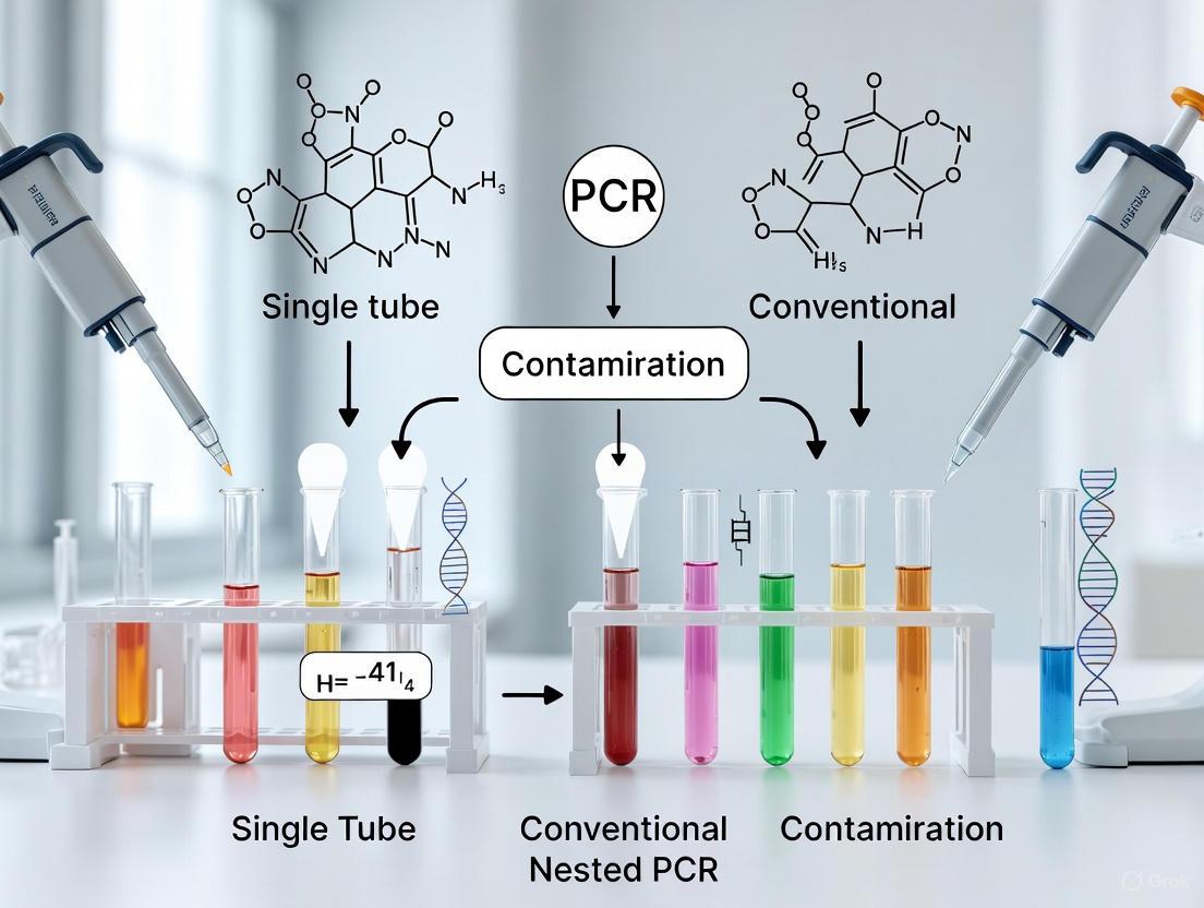
Single-Tube vs. Conventional Nested PCR: A Comparative Analysis of Contamination Rates and Workflow Efficiency
Nested PCR is renowned for its high sensitivity and specificity but is historically plagued by high contamination rates due to its multi-tube, multi-step nature.
Single-Tube vs. Conventional Nested PCR: A Comparative Analysis of Contamination Rates and Workflow Efficiency
Abstract
Nested PCR is renowned for its high sensitivity and specificity but is historically plagued by high contamination rates due to its multi-tube, multi-step nature. This article provides a comprehensive analysis for researchers and drug development professionals, contrasting conventional nested PCR with the innovative single-tube approach. We explore the foundational principles behind contamination, detail methodological workflows and their real-world applications, offer troubleshooting and optimization strategies, and present a rigorous validation of contamination rates and sensitivity. The synthesis of this information aims to guide laboratories in selecting the optimal PCR strategy to enhance diagnostic accuracy, ensure reliable research outcomes, and accelerate therapeutic development.
Understanding Nested PCR: Why Contamination is a Foundational Challenge
Core Principles of Conventional Two-Tube Nested PCR
Conventional two-tube nested PCR represents a significant evolution in polymerase chain reaction technology, specifically designed to address limitations in specificity and sensitivity encountered in standard PCR protocols. This technique employs two successive amplification rounds with two distinct primer sets to exponentially enhance target detection while minimizing non-specific amplification [1] [2]. The fundamental innovation lies in its architectural approach: an initial round of amplification with outer primers that flank the target region, followed by a second round using inner primers (nested primers) that bind within the first amplification product [3]. This sequential priming strategy creates a powerful molecular verification system, where successful second-round amplification confirms the specificity of the initial product.
The technique's development was driven by diagnostic challenges across microbiology, virology, and parasitology where pathogen detection often requires exceptional sensitivity to identify low-abundance targets or specificity to distinguish between closely related organisms [1] [4]. Within the broader context of nested PCR methodology evolution, the conventional two-tube approach establishes the foundational principles that later innovations, particularly single-tube formats, would seek to refine—primarily by addressing its inherent contamination vulnerability while preserving its diagnostic power [5] [4].
Core Principles and Workflow
The operational framework of conventional two-tube nested PCR rests on sequential amplification phases physically separated in distinct reaction vessels. The first amplification round employs a pair of outer primers designed to complement sequences flanking the target region, typically generating a primary amplicon of several hundred base pairs. Following this initial amplification, a small aliquot of the first reaction product is transferred to a fresh reaction tube containing the second primer pair for the nested round of amplification [1].
The Two-Stage Amplification Mechanism
The nested PCR process follows a meticulously structured two-stage amplification:
First Stage - Target Enrichment: The outer primers initiate amplification from the original template DNA, generating an intermediate product that contains the target sequence along with flanking regions. This initial amplification significantly enriches the specific target sequence relative to background DNA, even if non-specific amplification occurs simultaneously [1].
Second Stage - Specificity Verification: The inner primers, designed to bind within the first amplicon, now amplify only the precise target region. If the first round produced non-specific products due to primer mismatch, the probability that these non-specific products would contain complementary binding sites for the second primer pair is extremely low [1] [2]. This dual verification mechanism dramatically enhances methodological specificity.
The following diagram illustrates the complete workflow and underlying molecular mechanism of the two-tube nested PCR process:
Molecular Mechanism of Enhanced Specificity
The exceptional specificity of nested PCR stems from its requirement for four independent priming events (two forward and two reverse) to generate the final amplicon. The statistical probability of non-specific binding occurring with all four primers at their respective target sites is exponentially lower than in standard PCR, which requires only two correct priming events [1] [2]. This molecular verification system effectively eliminates false positives arising from mispriming in either amplification round.
Additionally, the two-round approach overcomes limitations related to the single amplification plateau effect inherent in standard PCR. By initiating the second round from the already-amplified products of the first round, the effective amplification factor increases dramatically, substantially enhancing detection sensitivity for low-abundance targets [1].
Comparative Experimental Data: Two-Tube vs. Single-Tube Formats
Research directly comparing conventional two-tube nested PCR with emerging single-tube methodologies reveals a complex trade-off between performance and practicality. The data demonstrate that while both formats maintain high specificity, significant differences emerge in sensitivity, contamination risk, and operational requirements.
Table 1: Performance comparison between two-tube and single-tube nested PCR formats
| Parameter | Conventional Two-Tube Nested PCR | Single-Tube Nested PCR | Experimental Context |
|---|---|---|---|
| Detection Sensitivity | 23.6% positive detection rate | 38.6% positive detection rate | PCMV detection in clinical samples [5] |
| Detection Limit | 1 pg target bacterial DNA | 1 fg target bacterial DNA | Multiplex pathogen detection [6] |
| Contamination Risk | High (tube transfer required) | Significantly reduced | Echinococcus spp. detection [4] |
| Hands-on Time | Longer (multiple setup steps) | Reduced approximately 50% | Workflow efficiency assessment [4] |
| Throughput Capacity | Lower | Higher | Clinical laboratory implementation [5] |
The superior sensitivity of single-tube formats demonstrated in these comparative studies stems from optimized reaction dynamics and reduced sample loss during transfer steps. However, this sensitivity advantage must be balanced against potential specificity concerns in some applications, particularly when amplifying targets with high sequence homology to non-target organisms.
Table 2: Contamination incidence rates in laboratory implementation
| Contamination Source | Two-Tube Nested PCR Risk | Single-Tube Nested PCR Risk | Prevention Strategies |
|---|---|---|---|
| Amplicon Carryover | High (aerosols during transfer) | Moderate (tube never opened) | Physical separation of workspaces [7] [8] |
| Cross-Sample Contamination | Moderate | Low | Use of aerosol-resistant tips [7] |
| Reagent Contamination | Moderate | Low | Aliquoting reagents; UV irradiation [8] |
| False Positives | Variable (dependent on technique) | Consistently low | Incorporation of UNG system [8] |
Detailed Experimental Protocol
Implementing conventional two-tube nested PCR requires meticulous attention to reaction composition, cycling parameters, and contamination control throughout the sequential amplification steps.
First Round Amplification
The initial amplification round focuses on generating sufficient target material for the second round while minimizing non-specific background amplification.
Reaction Composition:
- Template DNA: 1-2 μL (or 1-100 ng total DNA)
- External primers (each): 0.5 μL (final concentration 0.2 μM)
- dNTP mixture: 0.5 μL (final concentration 200 μM each dNTP)
- 10× PCR buffer: 2.5 μL
- MgCl₂: 1.5 μL (final concentration 1.5-2.0 mM)
- Taq DNA polymerase: 0.25 μL (1.25 U)
- Sterile ultrapure water: to final volume of 25 μL [1]
Thermal Cycling Conditions:
- Initial denaturation: 94°C for 2 minutes
- 30-35 cycles of:
- Denaturation: 94°C for 30 seconds
- Annealing: 45-60°C for 30 seconds (based on primer Tm)
- Extension: 72°C for 1 minute (adjust based on amplicon length)
- Final extension: 72°C for 5 minutes
- Hold: 4°C indefinitely [1]
Second Round Amplification
The nested amplification employs the first-round product as template with internal primers for ultimate specificity.
Reaction Composition:
- First-round PCR product: 1-2 μL (typically diluted 1:10 to 1:1000)
- Internal primers (each): 0.5 μL (final concentration 0.2 μM)
- dNTP mixture: 0.5 μL (final concentration 200 μM each dNTP)
- 10× PCR buffer: 2.5 μL
- MgCl₂: 1.5 μL (final concentration 1.5-2.0 mM)
- Taq DNA polymerase: 0.25 μL (1.25 U)
- Sterile ultrapure water: to final volume of 25 μL [1]
Thermal Cycling Conditions:
- Identical to first-round parameters
- Annealing temperature may be optimized for internal primers [1]
Product Analysis
Following amplification, products from both rounds are typically analyzed by agarose gel electrophoresis. The second-round product should show a single, specific band of expected size, typically shorter than the first-round amplicon due to the internal priming sites [1].
Contamination Control Measures
The primary limitation of conventional two-tube nested PCR remains its vulnerability to contamination during the transfer of first-round products to the second reaction tube. Implementing rigorous contamination control protocols is therefore essential for reliable results.
Physical and Procedural Barriers
Spatial Separation: Establish physically separated pre-amplification and post-amplification areas with dedicated equipment, laboratory coats, and supplies for each area [7] [8]. Maintain unidirectional workflow from clean to contaminated areas.
Decontamination Protocols: Regularly clean work surfaces and equipment with 10% sodium hypochlorite (bleach) followed by 70% ethanol [7] [8]. Fresh bleach solutions should be prepared regularly due to instability.
Technical Practices: Use aerosol-resistant pipette tips and positive-displacement pipettes. Open tubes carefully to minimize aerosol formation, and keep reactions covered as much as possible [7].
Biochemical Contamination Prevention
UNG System: Incorporate uracil-N-glycosylase (UNG) with dUTP in the reaction mix to degrade carryover contamination from previous amplifications [7] [8]. UNG selectively hydrolyzes uracil-containing DNA before amplification while leaving natural thymine-containing templates unaffected.
UV Irradiation: Expose reaction mixtures to UV light (254-300 nm) for 5-20 minutes before adding template DNA to inactivate potential contaminants [8]. Note that effectiveness varies with amplicon length and GC content.
The Scientist's Toolkit: Essential Research Reagents
Successful implementation of conventional two-tube nested PCR requires carefully selected and optimized reagents. The following table outlines essential components and their functions:
Table 3: Essential reagents for conventional two-tube nested PCR
| Reagent | Function | Optimization Considerations |
|---|---|---|
| Template DNA | Source of target sequence | Quality and concentration critical; avoid inhibitors [1] |
| Outer Primers | First-round amplification | Design to flank target region; Tm ~60-65°C [1] |
| Inner Primers | Second-round amplification | Design to bind within first product; Tm similar to outer primers [1] |
| Taq DNA Polymerase | DNA synthesis | Hot-start versions recommended to reduce non-specific amplification [3] |
| dNTP Mixture | Nucleotide substrates | Balanced solution of dATP, dCTP, dGTP, dTTP (or dUTP for UNG) [1] |
| MgCl₂ Solution | Cofactor for polymerase | Concentration critical (typically 1.5-2.0 mM); affects specificity [1] |
| PCR Buffer | Reaction environment | Provides optimal pH and salt conditions [1] |
Application Spectrum with Experimental Evidence
Conventional two-tube nested PCR has established utility across diverse research and diagnostic applications where exceptional sensitivity and specificity are required.
Infectious Disease Detection: The method has proven particularly valuable in detecting low-abundance pathogens in clinical samples. For respiratory pathogen detection, multiplex nested PCR assays demonstrated 100- to 1000-fold higher sensitivity than conventional methods, detecting 21 different viruses and bacteria with significantly higher positive rates (48.5%) compared to virus isolation (20.1%) or immunofluorescence assays (13.5%) [9].
Parasitology Applications: In Echinococcus spp. detection, nested PCR formats provide essential differentiation between morphologically similar species [4]. While newer single-tube methods offer advantages, the conventional two-tube approach established the foundational sensitivity and specificity benchmarks in this field.
Virology and Microbial Diagnostics: Modified nested PCR protocols demonstrate enhanced detection of challenging pathogens like Leishmania parasites in blood and tissue samples, enabling diagnosis even with extremely low parasite loads [1]. Similarly, the method has been adapted for Mycobacterium tuberculosis detection with significantly improved sensitivity over conventional PCR [1].
Technical Variations and Modifications
Several methodological adaptations have evolved from the core two-tube nested PCR protocol to address specific research needs:
Semi-Nested PCR: This variant uses three primers instead of four, with one primer from the first amplification reused in the second round along with one new internal primer [1]. This approach is particularly useful when primer design constraints prevent development of two complete primer sets.
Reverse Transcriptase Nested PCR: Combining reverse transcription with nested PCR enables highly sensitive detection of low-copy RNA targets, such as in hepatitis C virus (HCV) infection diagnosis [1].
Consensus Nested PCR: Employing degenerate primers based on conserved sequences within microbial genera, this approach allows detection of unknown variants or subtypes, particularly valuable in virology for detecting novel viruses [1].
Conventional two-tube nested PCR remains a foundational molecular biology technique that establishes the performance standards for amplification specificity and sensitivity. While newer single-tube formats offer practical advantages in contamination control and workflow efficiency, the two-tube method provides the conceptual framework and performance benchmarks that continue to inform PCR-based diagnostic development.
The decision between conventional two-tube and single-tube nested PCR formats ultimately depends on specific application requirements, laboratory infrastructure, and technical expertise. For laboratories establishing initial nested PCR capabilities, mastering the conventional two-tube approach provides fundamental insights into reaction dynamics and contamination control that translate effectively to more advanced methodologies. In applications where maximal sensitivity is paramount and appropriate contamination controls are established, the conventional two-tube nested PCR continues to offer exceptional performance that newer formats seek to emulate with greater convenience.
In molecular diagnostics and life sciences research, the amplification cascade—a series of sequential reactions that exponentially multiply a target signal—represents a powerful tool for detecting minute quantities of nucleic acids. While these techniques provide exceptional sensitivity, their implementation in multi-tube, multi-step formats introduces significant contamination risks that can compromise experimental integrity. This guide objectively compares conventional nested polymerase chain reaction (PCR) methodologies with emerging single-tube approaches, examining how their structural differences impact contamination rates, operational efficiency, and diagnostic reliability within research and drug development environments.
Contamination Mechanisms in Conventional Nested PCR
Conventional nested PCR operates through a two-stage amplification process in physically separate tubes, creating multiple opportunities for contaminating molecules to infiltrate reactions.
The Contamination Cascade
The primary vulnerability of open-tube nested PCR stems from the requirement to transfer amplification products between reaction vessels. When a tube is opened after the first amplification round, aerosolized amplicons can escape into the laboratory environment, settling on surfaces, equipment, and subsequent reaction mixtures. These contaminating molecules then become templates for future amplification cycles, generating false-positive results that undermine experimental validity [10].
This risk is particularly acute in high-throughput settings where numerous samples are processed simultaneously. Studies have documented that carryover contamination remains a significant challenge for conventional nested protocols, despite rigorous laboratory practices [11]. The problem intensifies when detecting low-abundance targets, where contaminating DNA may rival or exceed actual target concentrations in clinical samples.
Single-Tube Nested Platforms: A Contamination-Control Solution
Single-tube nested PCR systems address these vulnerabilities by physically containing the entire amplification process within a sealed reaction vessel, eliminating the need for intermediate transfer steps.
Technical Implementation
These integrated platforms utilize differential primer annealing temperatures or compartmentalized reagent deposition to temporally separate the primary and secondary amplification phases without breaking tube seals. For instance, some implementations use outer primers that activate at higher initial cycling temperatures, followed by inner primers that dominate at lower subsequent temperatures [5] [12]. This sequential primer activation within a single tube mimics the nested approach while maintaining a closed system.
The contamination-proof advantage of single-tube systems has been demonstrated across multiple applications. Research on porcine cytomegalovirus detection documented that a one-tube nested real-time PCR assay provided superior sensitivity (38.6% detection rate) compared to conventional nested PCR (23.6%) while eliminating between-reaction contamination [5]. Similarly, a one-tube nested quantitative real-time PCR for Brucella detection achieved a 100-fold increase in sensitivity over conventional qPCR while operating as a closed-tube system [12].
Comparative Experimental Data: Contamination and Performance
The table below summarizes key performance metrics between conventional and single-tube nested PCR systems, highlighting contamination-related advantages:
Table 1: Performance Comparison Between Conventional and Single-Tube Nested PCR Methods
| Parameter | Conventional Nested PCR | Single-Tube Nested PCR | Experimental Context |
|---|---|---|---|
| Contamination Risk | High (requires tube opening between rounds) [10] | Minimal (closed-tube system) [5] | General methodology |
| Sensitivity | 23.6% detection rate | 38.6% detection rate | Porcine cytomegalovirus detection [5] |
| Analytical Sensitivity | 1 pg/μL (conventional qPCR) | 100 fg/μL (100-fold improvement) | Brucella detection [12] |
| Specificity | 100% (with careful practices) | 100% (reduced false positives from contamination) | Brucella detection [12] |
| Operational Time | ~8 hours (two-step process) [11] | ~4 hours (streamlined process) [11] | Dengue virus detection |
| Cross-Reactivity | Potential with related pathogens | No cross-reactivity with related pathogens demonstrated [11] | Dengue virus vs other flavi/alphaviruses |
Workflow Comparison and Contamination Points
The diagram below illustrates the procedural differences between conventional and single-tube nested PCR workflows, highlighting critical contamination risk points:
Research Reagent Solutions for Contamination Control
The table below outlines essential reagents and their functions in implementing contamination-resistant nested PCR workflows:
Table 2: Essential Research Reagents for Contamination-Controlled Nested PCR
| Reagent/Category | Function in Contamination Control | Implementation Example |
|---|---|---|
| Primer Design Software | Designs outer/inner primers with distinct annealing temperatures | Primer3Plus used for one-tube PCMV assay [5] |
| Hot-Start DNA Polymerase | Reduces non-specific amplification and primer-dimer formation | Platinum Taq DNA polymerase in norovirus detection [13] |
| dNTP Mix | Balanced nucleotides for efficient amplification in complex reactions | Used in Brucella one-tube nested qPCR [12] |
| Probe-Based Detection Chemistry | Enables real-time monitoring in sealed tubes (e.g., FAM-BHQ pairs) | TaqMan probes in one-tube PCMV assay [5] |
| Internal Control Templates | Monitors for amplification inhibitors and reaction efficiency | Included in Brucella detection assay validation [12] |
| Stabilized Reaction Master Mixes | Maintains reagent integrity during complex thermal cycling | Thunderbird probe qPCR mix in PCMV detection [5] |
The evidence demonstrates that single-tube nested PCR systems significantly reduce contamination risks while maintaining or improving analytical performance compared to conventional nested methods. For research and drug development applications where result reliability is paramount, the transition to single-tube platforms represents both a methodological improvement and a quality control enhancement. The minimal false-positive rates, reduced hands-on time, and preserved sensitivity make these integrated systems particularly valuable for diagnostic development, clinical validation studies, and high-throughput screening environments where amplification cascade techniques are essential tools.
In molecular diagnostics and research, the exquisite sensitivity of polymerase chain reaction (PCR) is a double-edged sword. While it enables the detection of minute quantities of nucleic acid, this very characteristic makes the technique exceptionally vulnerable to false-positive results caused by the contamination of reactions with amplification products (amplicons) from previous assays [8]. This phenomenon, known as amplicon carryover contamination, represents one of the most significant challenges in laboratories employing PCR-based techniques. The risk escalates when moving from single-round amplification to more complex protocols like conventional nested PCR, which requires transferring amplified products between tubes. This guide objectively compares the contamination risks and performance profiles of conventional nested PCR against its modern counterpart, single-tube nested PCR, providing researchers with the experimental data and protocols necessary to inform their methodological choices.
Amplicon carryover contamination occurs when previously amplified DNA fragments inadvertently enter a new PCR setup, serving as efficient templates and generating false-positive results. A typical PCR can generate as many as 10^9 copies of the target sequence, and aerosolized droplets can contain up to 10^6 amplification products [8]. If uncontrolled, this leads to the rapid buildup of aerosolized amplicons that contaminate laboratory reagents, equipment, and ventilation systems.
The primary sources of contamination include:
- Cross-contamination between clinical specimens with high target organism loads
- Plasmid clones from previously analyzed organisms present in the laboratory environment
- Accumulated amplification products from repeated amplification of the same target sequence [8]
In next-generation sequencing workflows, contamination has been documented due to evaporation during PCR assays, particularly when using high denaturation temperatures, leading to detectable amplicons on thermocyclers, pipettes, bench surfaces, and even doorknobs [14].
Comparative Analysis: Conventional vs. Single-Tube Nested PCR
Fundamental Methodological Differences
The core distinction between these methodologies lies in their workflow design and consequent contamination risk profile.
| Feature | Conventional Nested PCR | Single-Tube Nested PCR |
|---|---|---|
| Workflow Design | Two physically separate amplification reactions in different tubes | Two sequential reactions in a single, closed tube |
| Primer Addition | Second primer set added after tube transfer | All primers included in initial master mix |
| Amplicon Transfer Risk | High (manual transfer of first-round products) | Eliminated (no post-amplification tube opening) |
| Contamination Control | Relies on spatial separation and meticulous technique | Built-in through closed-tube design |
| Hands-on Time | Higher | Lower |
| Throughput | Lower due to complex workflow | Higher due to simplified workflow |
Experimental Performance Data
Direct comparisons of these methodologies in detecting various pathogens reveal significant differences in sensitivity and contamination incidence.
Table 1: Detection Sensitivity Comparison Across PCR Methodologies
| Target Pathogen | Sample Type | Conventional Nested PCR Sensitivity | Single-Tube Nested PCR Sensitivity | Reference |
|---|---|---|---|---|
| Porcine Cytomegalovirus (PCMV) | Clinical tissues and blood | 23.6% (30/127) | 38.6% (49/127) | [5] |
| Human Cytomegalovirus (HCMV) | Peripheral blood leukocytes | Not directly tested | Detection limit: 180 copies/mL | [15] |
| Tuberculosis | Pulmonary specimens | Not directly tested | Overall sensitivity: 89% | [16] |
Table 2: Contamination Risk Factors and Mitigation Strategies
| Risk Factor | Conventional Nested PCR | Single-Tube Nested PCR |
|---|---|---|
| Aerosol Release | High during transfer of first-round products | Minimal (system remains closed) |
| Surface Contamination | Frequent without stringent controls | Rare with proper technique |
| Cross-Contamination | Significant risk between samples | Reduced risk |
| Primary Mitigation | Physical separation of pre- and post-amplification areas | Closed-tube design inherently reduces risk |
A 2020 study on Porcine Cytomegalovirus detection provides compelling evidence for the sensitivity advantage of single-tube nested formats. The research demonstrated a 38.6% detection rate (49/127) with one-tube nested real-time PCR compared to 23.6% (30/127) with conventional nested PCR and only 12.6% with conventional single-round PCR across 127 clinical samples [5]. This substantial improvement highlights how the single-tube approach enhances detection capability while simultaneously reducing contamination risk.
Detailed Experimental Protocols
Conventional Nested PCR Workflow
The following protocol for detecting Human Cytomegalovirus illustrates the contamination-prone transfer step characteristic of conventional nested PCR [15]:
First Amplification Round
- Reaction Volume: 20 μL
- Components: Master mix, outer primers, template DNA, and ddH₂O
- Thermal Cycling: Pre-denaturation at 94°C for 5 minutes; 40 cycles of 94°C for 30 seconds, 60°C for 30 seconds, 72°C for 30 seconds; final extension at 72°C for 10 minutes
Product Transfer
- Transfer 2 μL of the first-round amplification mixture to a fresh tube
- This open-tube step represents the primary contamination risk point
Second Amplification Round
- Reaction Volume: 20 μL
- Components: Master mix and inner primers
- Thermal Cycling: 40 cycles with same parameters except annealing at 55°C for 30 seconds
Detection
- Analyze amplified products by agarose gel electrophoresis
- A 293-bp fragment indicates positive detection
Single-Tube Nested PCR Workflow
This optimized protocol for bovine genotyping demonstrates the streamlined, closed-tube approach [10]:
Reaction Setup
- Perform all reagent preparations in a dedicated pre-amplification area
- Add outer and inner primers simultaneously to the master mix at optimized concentrations (e.g., 0.2 μM outer and 0.5 μM inner primers for ROSA26 gene detection)
- Include template DNA, with reaction components making up a total volume of 20 μL
Unified Thermal Cycling
- Use a specialized cycling program that accommodates both amplification rounds:
- Initial denaturation: 95°C for 3 minutes
- First amplification phase (10 cycles): 95°C for 3 seconds, 60°C for 30 seconds
- Second amplification phase (40 cycles): 95°C for 3 seconds, 55°C for 30 seconds
- The higher annealing temperature in the first phase favors outer primer binding, while the lower temperature in the second phase enables inner primer utilization
- Use a specialized cycling program that accommodates both amplification rounds:
Detection
- For real-time formats: Monitor fluorescence throughout amplification
- For conventional formats: Analyze final products by gel electrophoresis
- No post-amplification processing is required before detection
Decontamination Protocols for Amplicon Contamination
When contamination occurs, systematic decontamination is essential. Research on SARS-CoV-2 amplicon contamination in next-generation sequencing laboratories provides evidence-based protocols [14]:
Environmental Surface Decontamination
- Sodium Hypochlorite Treatment: Apply fresh 0.5% sodium hypochlorite solution to all laboratory surfaces for 30 minutes
- Equipment Immersion: Soak racks and small equipment in 0.5% sodium hypochlorite for 10 minutes
- Ethanol Wipe Down: Follow with 75% ethanol spray and wiping
- DNase Application: Use commercial DNA decontamination reagents on sensitive equipment like pipettes and thermocyclers
Preventive Measures
- UNG Incorporation: Add uracil-N-glycosylase (UNG) to PCR mixes to hydrolyze contaminating amplicons from previous reactions [8]
- Physical Separation: Maintain strict unidirectional workflow from clean pre-amplification to post-amplification areas
- UV Irradiation: Expose reagents and workstations to UV light to induce thymidine dimers in contaminating DNA [8]
The Scientist's Toolkit: Essential Research Reagents
Table 3: Key Reagents for Contamination Control in PCR
| Reagent/Equipment | Function | Application Notes |
|---|---|---|
| Uracil-N-Glycosylase (UNG) | Enzymatically degrades uracil-containing contaminating amplicons | Most effective with thymine-rich targets; requires dUTP in reaction mix [8] |
| dUTP | Replaces dTTP in amplification, creating UNG-sensitive products | Must be optimized for each target; may require dTTP supplementation [8] |
| Sodium Hypochlorite | Oxidatively damages nucleic acids through chlorination | Use 0.5-10% solutions; requires ethanol removal after treatment [8] [14] |
| DNA Decontamination Reagent | Commercial formulations containing DNases | Effective for equipment decontamination; follow manufacturer protocols [14] |
| Aerosol-Barrier Pipette Tips | Prevents aerosol transfer during pipetting | Essential in all amplification setups |
| Internal Control DNA | Identifies PCR inhibition in reaction | Critical for validating negative results [17] [5] |
The methodological evolution from conventional to single-tube nested PCR represents a significant advancement in managing amplicon carryover contamination. While conventional nested PCR offers theoretical sensitivity benefits, its open-tube format creates substantial contamination risks that can compromise experimental results. Single-tube nested PCR methodologies address this fundamental limitation by containing the entire amplification process within a closed system, simultaneously reducing contamination while maintaining—and in some cases enhancing—analytical sensitivity. For research and diagnostic applications where result fidelity is paramount, particularly in clinical, pharmaceutical, and regulatory settings, single-tube nested PCR provides a superior balance of sensitivity, specificity, and contamination control. As molecular diagnostics continue to evolve, this methodological approach offers a more robust framework for reliable nucleic acid detection.
The polymerase chain reaction (PCR) has fundamentally revolutionized molecular diagnostics since its inception, enabling precise detection of pathogenic nucleic acids. Among its variations, nested PCR emerged as a powerful technique to enhance the sensitivity and specificity of target sequence detection by utilizing two sets of primers in sequential amplification rounds [3]. This method significantly reduces false-positive results from nonspecific amplification, as any non-target sequences amplified in the first round are unlikely to be re-amplified by the second primer set targeting an internal sequence [18]. Despite these advantages, conventional nested PCR suffers from a critical limitation: the requirement to transfer amplification products from the first reaction tube to a second for the nested amplification [10]. This open-tube transfer process creates substantial risk of amplicon contamination in laboratory settings, potentially leading to false-positive results and compromising diagnostic accuracy [10] [19].
The single-tube nested PCR system represents a paradigm shift in molecular assay design, effectively addressing the contamination vulnerability of conventional nested PCR while retaining its sensitivity benefits. By containing both amplification rounds within a single closed tube, this innovative approach maintains the diagnostic robustness of traditional nested PCR while dramatically reducing contamination risks [10] [19]. This article comprehensively compares the performance, methodologies, and practical applications of single-tube nested PCR systems against conventional alternatives, providing researchers and drug development professionals with evidence-based insights for molecular assay selection.
Performance Comparison: Single-Tube vs. Conventional Nested PCR
Sensitivity and Detection Limits
Multiple studies have demonstrated that single-tube nested PCR systems achieve exceptional sensitivity, often detecting target pathogens at significantly lower concentrations than conventional PCR methods.
Table 1: Comparison of Detection Limits Between PCR Methods
| Pathogen/Target | Conventional PCR | Nested PCR | Single-Tube Nested PCR | Reference |
|---|---|---|---|---|
| Porcine cytomegalovirus | 12.6% (16/127) | 23.6% (30/127) | 38.6% (49/127) | [20] |
| Campylobacter jejuni (DNA copy detection) | 100 copies | 10 copies | 10 copies | [19] |
| Feline calicivirus (clinical samples) | 1.85% (1/54) | 31.48% (17/54) | Comparable to nested PCR | [18] |
| Target bacteria (16S rDNA) | 1 pg | - | 1 fg | [6] |
In a comprehensive evaluation for porcine cytomegalovirus (PCMV) detection, one-tube nested real-time PCR demonstrated superior detection capabilities, identifying 38.6% of positive samples compared to 23.6% with conventional nested PCR and only 12.6% with conventional PCR [20]. Similarly, for Campylobacter jejuni detection in ground chicken, the single-tube nested PCR format achieved a detection limit of 10 DNA copies, 100 times lower than conventional PCR with inner primers alone [19].
The exceptional sensitivity of single-tube nested PCR is particularly valuable for applications involving limited target availability. When optimizing single-tube nested PCR for bovine gene detection, researchers successfully amplified the ROSA26 gene from samples with low DNA concentration, including single cells and in vitro-produced embryos [10]. This level of sensitivity enables applications in preimplantation genetic diagnosis and analysis of precious clinical samples where target material is minimal.
Contamination Rates and Operational Efficiency
The fundamental advantage of single-tube nested PCR systems lies in their contamination control and workflow efficiency.
Table 2: Contamination Risk and Workflow Comparison
| Parameter | Conventional Nested PCR | Single-Tube Nested PCR |
|---|---|---|
| Amplicon contamination risk | High (open tube transfer) | Dramatically reduced (closed system) |
| Hands-on time | Significant (two separate setups) | Reduced (single reaction setup) |
| Total processing time | ~3+ hours (including transfer) | ~1.5 hours [20] |
| Technical expertise required | High | Moderate |
| Reagent consumption | Higher | Reduced |
Traditional nested PCR requires transferring the first-round amplification product to a second reaction tube, creating opportunities for aerosol contamination that can compromise subsequent tests [10] [19]. Single-tube systems eliminate this risk by containing both amplification rounds within a sealed environment. As noted in research on Campylobacter jejuni detection, single-tube nested PCR "dramatically reduces the risk of amplicon cross-contamination" while providing "sensitivity levels equal to or greater than those of nested PCR, and with less time and reagents" [19].
The operational efficiency gains are substantial. The one-tube nested real-time PCR assay for PCMV detection required "approximately 1.5 h for completion" [20], significantly less than conventional nested PCR protocols. This streamlined workflow enables more rapid diagnostic turnaround while maintaining the sensitivity advantages of nested amplification.
Experimental Protocols and Methodologies
Core Principles of Single-Tube Nested PCR Design
Single-tube nested PCR systems employ strategic primer design and thermal cycling parameters to sequentially engage outer and inner primer sets within a single reaction vessel. The fundamental principle involves designing outer and inner primers with distinct annealing temperatures [10] [6]. Outer primers feature higher annealing temperatures (e.g., above 65°C), while inner primers have lower annealing temperatures (e.g., below 56°C) [6]. This temperature differential enables controlled, sequential amplification stages within a single tube.
During initial PCR cycles with higher annealing temperatures, only the outer primers bind and amplify the target region, generating an intermediate amplicon that serves as template for the second amplification phase. Subsequent cycles with lower annealing temperatures enable the inner primers to bind to their complementary sequences within the first amplicon, producing the final specific product [6]. This sequential activation is further controlled through primer concentration optimization, with outer primers typically used at lower concentrations (0.005-0.01 μM) to ensure they are depleted before the second amplification phase, preventing competition with inner primers [6].
Diagram 1: Single-Tube Nested PCR Workflow. This diagram illustrates the sequential stages of single-tube nested PCR, showing how temperature control enables two amplification rounds in a single tube.
Detailed Experimental Protocol for Pathogen Detection
The following protocol for detection of bacterial pathogens in research mice exemplifies a standardized approach to single-tube nested PCR, optimized for multiple target detection [6]:
Reaction Setup:
- Prepare a 20 μL reaction mixture containing:
- 10 μL of 2× Taq Master Mix
- 0.01 μM each of universal outer primers (UP-F/UP-R)
- 0.15 μM of each species-specific inner primer
- 1 ng template DNA
Thermal Cycling Conditions:
- Initial denaturation: 95°C for 5 minutes
- Stage 1 (15 cycles):
- Denaturation: 94°C for 30 seconds
- Annealing: 65°C for 30 seconds (enables only outer primer binding)
- Extension: 72°C for 30 seconds
- Stage 2 (25 cycles):
- Denaturation: 94°C for 30 seconds
- Annealing: 55°C for 30 seconds (enables inner primer binding)
- Extension: 72°C for 30 seconds
- Final extension: 72°C for 5 minutes
This protocol demonstrates the critical principle of using temperature-dependent primer activation to achieve sequential amplifications. The higher annealing temperature in Stage 1 ensures selective outer primer binding, while the lower temperature in Stage 2 enables inner primer binding to the enriched templates [6].
Adaptation for Real-Time Detection Platforms
Single-tube nested PCR has been successfully adapted to real-time platforms, combining the sensitivity of nested amplification with the quantification capabilities and reduced contamination risk of real-time PCR. In one-tube nested real-time PCR for PCMV detection, researchers utilized the following approach [20]:
Reaction Composition:
- 10 μL of 2× Thunderbird probe qPCR mix
- 2.5 μL of primer/probe mixture (5 pmol each primers and 5 pmol TaqMan probe)
- 3 μL template DNA
- Total reaction volume: 20 μL
Thermal Cycling Parameters:
- Initial activation: 95°C for 3 minutes
- Stage 1 (10 cycles): 95°C for 3 seconds, 60°C for 30 seconds
- Stage 2 (40 cycles): 95°C for 3 seconds, 55°C for 30 seconds
This configuration enabled specific detection of PCMV with a significantly higher detection rate (38.6%) compared to conventional nested PCR (23.6%) while completing the analysis in approximately 1.5 hours [20]. The inclusion of an internal control (IC) DNA in the reaction mixture further enhanced reliability by monitoring nucleic acid extraction quality and PCR inhibition [20].
The Scientist's Toolkit: Essential Research Reagents
Successful implementation of single-tube nested PCR requires careful selection of specialized reagents and components optimized for the sequential amplification process.
Table 3: Essential Research Reagents for Single-Tube Nested PCR
| Reagent/Category | Specific Examples | Function & Importance |
|---|---|---|
| Primers | Outer and inner primer sets with distinct Tm values | Core components enabling sequential amplification; outer primers typically have higher Tm (≥65°C), inner primers lower Tm (≤56°C) [6] |
| DNA Polymerase | Hot-start Taq DNA polymerase | Prevents nonspecific amplification during reaction setup; essential for maintaining specificity with multiple primer sets [3] |
| PCR Buffer | Optimized buffer with MgCl₂ | Provides optimal ionic environment; MgCl₂ concentration particularly critical for multiplex efficiency [21] |
| dNTPs | dATP, dCTP, dGTP, dTTP | Building blocks for DNA synthesis; balanced concentrations crucial for efficient amplification [21] |
| Template DNA | Extracted nucleic acids | Sample quality critical; internal control DNA recommended to monitor extraction efficiency and inhibition [20] |
| Probe Systems | TaqMan probes, base-quenched probes | Enable real-time detection in closed-tube systems; fluorophore-labeled probes (FAM, VIC, CY5) allow multiplex detection [22] |
The strategic design of primer systems forms the foundation of successful single-tube nested PCR. Research on bovine genotyping emphasized that "to optimize STnPCR for low-concentration samples like single cells, it's crucial to ensure that the initial round of amplification fully utilizes the concentration of primers targeting the outer regions, depleting them by the end of the first PCR" [10]. This precise primer balancing act enables the sequential amplification process without physical transfer of reaction products.
Hot-start DNA polymerase is particularly valuable for single-tube nested PCR applications, as it prevents nonspecific amplification and primer-dimer formation during reaction setup at lower temperatures [3] [21]. This technology employs antibody-based or chemical modification to inhibit polymerase activity until an initial high-temperature activation step, thereby enhancing assay specificity when multiple primer sets are present [3].
Applications Across Research and Diagnostic Fields
Clinical Diagnostics and Pathogen Detection
Single-tube nested PCR has demonstrated particular utility in clinical diagnostics where sensitivity and contamination control are paramount. In veterinary medicine, researchers detected feline calicivirus with significantly higher sensitivity compared to conventional PCR (31.48% vs. 1.85% positivity in clinical samples) [18]. The method has proven equally valuable in human medicine for detecting challenging pathogens like Helicobacter pylori, where researchers developed a highly sensitive nested PCR assay targeting a short 148 bp fragment of the 16S rRNA gene to overcome the challenge of degraded bacterial DNA in stool samples [23].
For food safety applications, researchers developed an "ultra-sensitive single-tube nested PCR assay for rapid detection of Campylobacter jejuni in ground chicken" with a detection limit of 10 DNA copies, substantially improving upon conventional PCR sensitivity [19]. This enhanced detection capability is crucial for identifying low-level pathogen contamination that could nevertheless cause human illness.
Genetic Analysis and Specialized Research Applications
The technology has enabled advanced genetic analyses previously challenged by template limitation. Single-tube nested PCR has been successfully applied to genotyping single cells and in vitro-produced bovine embryos, demonstrating sufficient sensitivity to analyze minimal genetic material [10]. This capability opens possibilities for preimplantation genetic diagnosis and analysis of rare cell populations.
Multiplex applications have further expanded the utility of single-tube nested PCR systems. Researchers developed a "single-tube multiplex nested PCR system for efficient detection of multiple pathogens" targeting Staphylococcus aureus, Pseudomonas aeruginosa, Klebsiella pneumoniae, and Rodentibacter pneumotropicus simultaneously [6]. This approach maintained high sensitivity (detecting as little as 1 fg of target bacterial DNA) while providing the practical efficiency of multiplex detection.
Advanced detection platforms have integrated single-tube nested PCR with real-time detection capabilities. The development of "2D polymerase chain reaction for single-tube detection of high-risk human papillomaviruses" enabled closed-tube genotyping of 11 HR-HPV types by combining asymmetric PCR amplification with melting curve analysis across multiple fluorescent channels [22]. Such innovations demonstrate how the fundamental principle of nested amplification in a single tube can be enhanced with complementary technologies to address complex diagnostic challenges.
The introduction of single-tube nested PCR systems represents a genuine paradigm shift in molecular detection technology, successfully addressing the critical limitation of conventional nested PCR: contamination vulnerability during amplicon transfer. Through strategic primer design and thermal cycling optimization, these systems maintain the superior sensitivity and specificity of nested amplification while dramatically reducing false-positive results from laboratory contamination [10] [19].
Evidence across multiple applications demonstrates that single-tube nested PCR consistently outperforms conventional PCR in detection sensitivity, with some studies showing up to 100-fold improvement in detection limits [6] [19]. The methodology has proven adaptable across diverse platforms, including endpoint detection, real-time PCR, and multiplex configurations, making it suitable for applications ranging from clinical diagnostics to food safety testing and genetic research [20] [6] [22].
As molecular diagnostics continues to advance toward more automated, contamination-resistant workflows, single-tube nested PCR systems offer a robust solution that balances exceptional sensitivity with practical operational efficiency. For researchers and drug development professionals requiring reliable detection of low-abundance targets, this technology provides a validated approach that maintains diagnostic accuracy while streamlining laboratory workflows. The continued refinement and application of single-tube nested PCR principles will undoubtedly support future innovations in molecular detection across life sciences and medical diagnostics.
The polymerase chain reaction (PCR) is a foundational technique in molecular biology, but its application to low-abundance targets demands enhanced sensitivity and specificity. Nested PCR addresses this by using two sets of primers in sequential reactions to amplify a specific DNA sequence, significantly reducing non-specific amplification [18]. However, a major drawback of conventional nested PCR is the high risk of contamination when transferring amplification products from the first reaction to the second [10]. Single-tube nested PCR (ST-nPCR) represents a sophisticated redesign that confines the entire nested amplification process within a single sealed tube. This guide objectively compares the primer engineering and reaction segregation strategies of conventional and single-tube nested PCR, focusing on their performance implications and providing a framework for researchers to select the optimal method for sensitive detection applications, particularly in drug development and clinical diagnostics.
Primer Engineering: A Comparative Analysis
The core distinction between conventional and single-tube nested PCR lies in the strategic design and management of primer sets.
Conventional Nested PCR Primer Design
In conventional nested PCR, two discrete primer sets are used in two physically separate reaction tubes.
- Primer Set Segregation: The outer primer set is designed to bind to sequences flanking the target region, generating a larger primary amplicon. The inner primer set (nested primers) is designed to bind within the primary amplicon, generating a shorter, secondary product [18] [10].
- Design Freedom: Primers for each stage are designed independently. The primary considerations are the specificity and efficiency of each primer pair for its respective amplicon, without concern for cross-interaction between the two sets during a single reaction [24].
- Two-Stage Protocol: The two primer sets are never active in the same reaction mixture. The product of the first PCR is used as a template for the second, separate PCR [18].
Single-Tube Nested PCR Primer Engineering
Single-tube nested PCR requires a more nuanced primer design to coordinate both amplification stages within a single, sealed tube.
- Coordinated Primer Design: All primers—outer forward, outer reverse, inner forward, and inner reverse—are present in the same reaction mix from the start. This necessitates careful design to prevent primer-dimer formations and non-specific interactions between all four primers [6] [10].
- Thermodynamic Segregation: A common strategy is to engineer a significant difference in the annealing temperatures (
Tm) between the outer and inner primer sets. Outer primers are designed to be longer with a higherTm(e.g., >65°C), while inner primers are shorter with a lowerTm(e.g., <56°C) [6]. The thermal cycling protocol then uses a high annealing temperature in the initial cycles, permitting only the outer primers to bind and amplify the target. Subsequent cycles use a lower annealing temperature, allowing the inner primers to preferentially bind and amplify the enriched template. - Concentration Management: An alternative or complementary approach is the "balanced" or "primer depletion" method. The outer primers are used at a very low concentration (e.g., 0.005-0.01 µM) so that they are functionally exhausted by the end of the first stage of amplification. The inner primers, present at higher concentrations, then drive the second stage of amplification without competition [25] [10]. One study optimized this by using a primer containing the sequence of the inner primer attached to the 5′ end of the opposite outer primer, ensuring balanced amplification and increased sensitivity [25].
Table 1: Key Differences in Primer Engineering Strategies
| Design Feature | Conventional Nested PCR | Single-Tube Nested PCR |
|---|---|---|
| Primer Set Physical Proximity | Separate tubes for each set | All primers combined in a single tube |
| Primary Design Constraint | Specificity of each primer pair for its target | Specificity plus lack of interaction between all four primers |
| Segregation Mechanism | Physical transfer of template | Thermodynamic (Tm difference) and/or concentration-based depletion |
| Typical Outer Primer Tm | Standard, optimized independently | Deliberately high (e.g., >65°C) [6] |
| Typical Inner Primer Tm | Standard, optimized independently | Deliberately low (e.g., 50-55°C) [6] |
| Primer Concentration Strategy | Standard concentrations | Low concentration outer primers to allow for depletion [10] |
Reaction Segregation and Workflow
The method of segregating the two amplification stages directly impacts workflow, contamination risk, and throughput.
Conventional Nested PCR Workflow
The process is linear and requires physical intervention.
- First Amplification: The sample is amplified using the outer primer set in a dedicated tube.
- Template Transfer: After the first PCR is complete, the reaction tube is opened, and an aliquot of the amplified product is physically transferred to a new tube containing the inner primer mix. This transfer step is a major source of potential contamination, as it exposes the environment to the first-round amplicons, which can then become templates for subsequent reactions [26] [10].
- Second Amplification: The second PCR is carried out in the new tube.
Single-Tube Nested PCR Workflow
The process is consolidated and sealed.
- Unified Reaction Setup: All required components—template DNA, both outer and inner primers, polymerase, dNTPs, and buffer—are assembled in a single tube, which is then sealed [10].
- Sequential In-Tube Amplification: The tube undergoes a single, multi-stage thermal cycling program. The initial cycles (e.g., 15 cycles) are run at a high annealing temperature, activating only the outer primers. This is followed by a second set of cycles (e.g., 25 cycles) at a lower annealing temperature, enabling the inner primers to amplify the product generated in the first stage [6].
- No Physical Transfer: The tube remains closed throughout the entire process, eliminating the risk of carryover contamination during transfer and drastically reducing the potential for false positives [10].
The following workflow diagrams illustrate the key differences in reaction segregation between the two methods:
Performance and Experimental Data
Quantitative comparisons demonstrate that while both methods offer high sensitivity, single-tube nested PCR achieves this with a significantly reduced contamination profile.
Sensitivity and Specificity
Both nested PCR formats are substantially more sensitive than conventional PCR. Studies consistently show that nested PCR can detect targets that are missed by conventional PCR [18]. For instance:
- A study detecting Feline Calicivirus (FCV) found a positivity rate of 31.48% using both nested PCR and RT-LAMP, compared to only 1.85% for conventional PCR [18].
- Single-tube nested PCR has demonstrated a sensitivity of up to 1 fg of target bacterial DNA, a 1000-fold improvement over the 1 pg sensitivity of conventional multiplex PCR [6].
- The "balanced heminested" single-tube approach showed a statistically significant higher sensitivity (75%) compared to standard heminested PCR (60%) when detecting Mycobacterium tuberculosis in smear-negative samples [25].
Contamination Rates
Contamination is the most critical differentiator.
- Conventional Nested PCR: The requirement to open the first reaction tube for template transfer is a well-documented and major risk for carryover contamination, leading to false-positive results [26] [10]. This necessitates rigorous laboratory workflows with separate pre- and post-amplification areas, the use of UV hoods, and dedicated equipment to mitigate risk [26].
- Single-Tube Nested PCR: By containing the entire process within a sealed tube, this method "substantially" reduces cross-contamination between the two PCR rounds [10]. This makes it particularly valuable for clinical diagnostics, forensics, and any high-throughput application where false positives can have significant consequences.
Table 2: Quantitative Performance Comparison
| Performance Metric | Conventional Nested PCR | Single-Tube Nested PCR | Supporting Data |
|---|---|---|---|
| Theoretical Sensitivity | Very High (fg levels) | Very High (fg levels) | Detects 1 fg bacterial DNA [6] |
| Specificity | High | High | Reduces non-specific amplification [18] [10] |
| Contamination Risk | High | Very Low | Eliminates transfer-based carryover [10] |
| Time to Result | Longer (setup + 2 runs) | Shorter (single run) | [6] [10] |
| Amenability to Multiplexing | Challenging | Demonstrated (e.g., 4-plex) | Single-tube multiplex nested PCR developed [6] |
| Throughput Potential | Lower | Higher | Simplified workflow enables scaling [10] |
Detailed Experimental Protocols
Protocol for Conventional Nested PCR
This protocol is adapted from a study comparing PCR methods for Feline Calicivirus detection [18].
- First Round PCR:
- Reaction Mix: Template DNA, standard PCR buffer, 200 µM of each dNTP, 1.5 mM MgCl₂, 0.5 µM of each outer primer, and 1.25 U of DNA polymerase.
- Cycling Conditions: Initial denaturation at 94°C for 5 min; 35 cycles of 94°C for 30 s, 55°C for 30 s, 72°C for 30 s; final extension at 72°C for 5 min.
- Second Round PCR:
- Reaction Mix: 1-2 µL of the first-round PCR product, standard PCR buffer, 200 µM of each dNTP, 1.5 mM MgCl₂, 0.5 µM of each inner primer, and 1.25 U of DNA polymerase.
- Cycling Conditions: Use the same profile as the first round.
- Detection: Analyze the second-round product by agarose gel electrophoresis.
Protocol for Single-Tube Nested PCR
This protocol is adapted from an optimized study for bovine genotyping and pathogen detection [6] [10].
- Unified Reaction Setup:
- Reaction Mix: Template DNA, 2× PCR Master Mix, outer primers at a low concentration (e.g., 0.01 µM each), and inner primers at a higher concentration (e.g., 0.15 µM each). The total reaction volume is 20 µL.
- Consolidated Cycling Conditions:
- Stage 1 (Enrichment): Initial denaturation at 95°C for 5 min; 15 cycles of 94°C for 30 s, 65°C for 30 s (high annealing temp for outer primers), and 72°C for 30 s.
- Stage 2 (Detection): 25 cycles of 94°C for 30 s, 55°C for 30 s (low annealing temp for inner primers), and 72°C for 30 s; final extension at 72°C for 5 min.
- Detection: Analyze the final product by agarose gel electrophoresis.
The Scientist's Toolkit: Essential Research Reagents
Successful implementation of these nested PCR techniques, particularly the single-tube format, relies on key reagents and consumables.
Table 3: Essential Research Reagent Solutions
| Reagent / consumable | Function / Importance | Application Notes |
|---|---|---|
| High-Fidelity DNA Polymerase | Catalyzes DNA synthesis; some possess 3'→5' exonuclease (proofreading) activity to increase replication fidelity. | Critical for long amplicons in the first round. Standard Taq is often sufficient. |
| dNTP Mix | Building blocks for new DNA strands. | Quality dNTPs ensure efficient amplification. |
| Primers (Outer & Inner) | Sequence-specific oligonucleotides that define the target amplicon. | Ultra-pure, HPLC-purified primers are essential for specificity, especially in single-tube formats [6]. |
| PCR Buffer with Mg²⁺ | Provides optimal ionic environment and pH. Mg²⁺ is a cofactor for polymerase activity. | Mg²⁺ concentration may require optimization; higher fidelity is associated with lower Mg²⁺ [24]. |
| Nuclease-Free Water | Solvent for reactions. | Must be free of nucleases to prevent degradation of primers and template. |
| Filter Pipette Tips | Aerosol barrier tips prevent carryover contamination by blocking aerosols from entering the pipette shaft. | Critical best practice for preventing contamination in all molecular workflows, especially when setting up pre-amplification mixes [26]. |
| DNA-Binding Dye (e.g., Sybr Green) | For real-time detection and quantification in qPCR formats. | Enables quantitative analysis without gel electrophoresis. |
The evolution from conventional to single-tube nested PCR represents a significant advancement in molecular detection technology. The key design differences are profound: conventional nested PCR relies on physical segregation of reactions, offering design simplicity at the cost of high contamination risk. In contrast, single-tube nested PCR employs sophisticated primer engineering—using thermodynamic and concentration-based strategies—to achieve reaction segregation within a sealed environment, thereby minimizing contamination while maintaining exceptional sensitivity and specificity.
For researchers and drug development professionals, the choice is clear. In high-throughput diagnostics, pathogen detection, and any scenario where false positives are unacceptable, the single-tube nested PCR method is objectively superior. Its streamlined workflow, reduced hands-on time, and robust contamination control make it the more reliable and efficient choice for pushing the boundaries of detection in low-biomass and critical applications.
Implementing Single-Tube Nested PCR: Methodologies and Diverse Applications
The polymerase chain reaction (PCR) is a cornerstone technique in molecular biology, enabling the amplification of specific DNA sequences from minute starting quantities [27]. While all PCR methods share fundamental principles—thermal cycling of denaturation, annealing, and extension using a thermostable DNA polymerase—their implementation significantly impacts workflow efficiency and results reliability [28] [27]. A critical consideration in molecular diagnostics and research is the risk of contamination from amplified DNA products or environmental sources, which can lead to false-positive results [29]. This guide objectively compares conventional (or nested) PCR protocols against single-tube real-time PCR (qPCR) protocols, with particular focus on their relative contamination risks, required workflows, and performance characteristics. Understanding these differences is essential for researchers and drug development professionals selecting appropriate methodologies for specific applications, particularly when working with low-abundance targets or in regulated environments where result accuracy is paramount.
Fundamental Principles and Definitions
Conventional PCR
Conventional PCR, also referred to as end-point PCR, is the original amplification method where reactions run to completion and products are analyzed after all cycles are finished [30] [31]. This method is primarily qualitative, determining only the presence or absence of a target sequence [31]. Measurement occurs at the plateau phase of amplification, where reaction components have been depleted and the accumulation of product has ceased [30]. In traditional workflows, results are typically visualized using agarose gel electrophoresis with DNA-binding dyes like ethidium bromide, a process that requires manual post-amplification handling and increases contamination risk [30] [31].
Nested PCR
Nested PCR is a variant of conventional PCR designed to enhance specificity and sensitivity. It involves two successive amplification rounds using two sets of primers [28]. The first round uses outer primers to generate an initial amplicon, which then serves as the template for a second round using inner primers that bind within the first amplicon [28]. While this significantly improves detection limits, it substantially increases contamination risk because the highly amplified products from the first round must be transferred to a new tube for the second reaction, creating multiple opportunities for aerosol contamination [28].
Single-Tube Real-Time PCR (qPCR)
Single-tube real-time PCR (qPCR) monitors DNA amplification as it occurs during the exponential phase of the reaction, when product doubling is most reproducible [30] [31]. This method is inherently quantitative and utilizes fluorescent reporting systems—either DNA-binding dyes or sequence-specific probes—to track product accumulation in real-time [30] [27]. The closed-tube nature of qPCR is a key feature; tubes remain sealed throughout amplification and detection, dramatically reducing the risk of amplicon contamination compared to open, post-amplification processing methods [30] [31].
Step-by-Step Workflow Comparison
Conventional PCR Workflow
The conventional PCR workflow involves multiple open-tube steps that present significant contamination risks [30] [28]:
Sample and Reagent Preparation: Researchers assemble the master mix containing DNA polymerase, dNTPs, primers, and buffer, then add template DNA [27].
Primary Amplification: The reaction undergoes 30-40 cycles of denaturation, annealing, and extension in a thermal cycler [27].
Product Transfer (High Contamination Risk): For nested protocols, the tube must be opened to transfer a portion of the amplified product to a new reaction tube containing secondary primers [28].
Secondary Amplification: The transferred product undergoes additional thermal cycling with the nested primer set [28].
Post-Amplification Analysis (High Contamination Risk): The final PCR product is removed from the tube and analyzed using agarose gel electrophoresis, followed by staining and visualization under UV light [30] [31]. Each tube opening and product handling creates potential for aerosol contamination of laboratory surfaces and equipment.
Single-Tube Real-Time PCR Workflow
The single-tube qPCR workflow minimizes contamination risk through a closed-tube design [30] [31]:
Single-Tube Setup: Researchers prepare a single reaction mix containing all necessary components—DNA polymerase, dNTPs, primers, buffer, and fluorescent detection system (dyes or probes) [27].
Tube Sealing: Reaction tubes or plates are sealed after setup, remaining closed throughout the entire process [31].
Amplification with Real-Time Monitoring: The sealed plate undergoes thermal cycling while the instrument's optical detection system monitors fluorescence accumulation during each cycle [30] [27]. Data collection occurs during the exponential phase of amplification when product doubling is most reproducible [30].
Automated Analysis: Software automatically calculates results based on fluorescence thresholds (Cq values) without any post-amplification handling [30] [31]. The entire process from amplification to quantification occurs without tube openings, preventing amplicon contamination.
Comparative Experimental Data
Performance Characteristics and Contamination Rates
Table 1: Direct comparison of conventional and single-tube PCR methodologies
| Parameter | Conventional/Nested PCR | Single-Tube Real-Time PCR |
|---|---|---|
| Quantification Capability | Qualitative/Semi-quantitative [30] [31] | Fully quantitative [30] [31] |
| Detection Point | Plateau phase (end-point) [30] | Exponential phase (real-time) [30] |
| Sensitivity | Lower sensitivity [30] | Detection capable down to 2-fold change [30] |
| Dynamic Range | Short dynamic range <2 logs [30] | Increased dynamic range of detection [30] |
| Post-PCR Processing | Required (gel electrophoresis, staining) [30] [31] | None required [30] [31] |
| Contamination Risk | High (multiple open-tube steps) [28] | Low (closed-tube system) [30] [31] |
| Result Output | Band intensity on gel [30] | Exact Cq values [30] [31] |
| Throughput | Lower (manual processing) [31] | Higher (automated) [31] |
| Multiplexing Capability | Limited [32] | Possible with multiple probes [32] |
Experimental Evidence from Comparative Studies
Table 2: Experimental performance data from published studies
| Study Application | Conventional PCR Performance | Single-Tube qPCR Performance | Reference |
|---|---|---|---|
| Pathogen Detection in Cosmetics | Effective but time-consuming; may miss viable but non-cultivable cells [33] | 100% detection rate across all replicates; superior sensitivity in complex matrices [33] | [33] |
| Respiratory Virus Detection | 96.9% overall sensitivity [32] | 87.9% overall sensitivity [32] | [32] |
| Reagent Contamination Assessment | High contamination risk with post-amplification processing [29] | Not applicable (closed-tube system prevents post-amplification contamination) [29] | [29] |
| Quantification Precision | Poor precision [30] | High precision; collects data during exponential phase [30] | [30] |
Research by Facellitate highlights that while PCR is highly sensitive and specific, the technique remains very susceptible to contamination from other sources of DNA or the environment, which can mislead data interpretation [34]. A 2025 study examining bacterial DNA contamination of commercial PCR enzymes found contaminating bacterial DNA in seven of nine commercial products tested [29]. This contamination is particularly problematic for conventional PCR workflows, where additional open-tube steps can introduce these contaminants or spread amplicons through the laboratory environment [29].
In a comparative study of respiratory virus detection methods, conventional multiplex RT-PCR demonstrated higher sensitivity (96.9%) compared to real-time RT-PCR (87.9%), though both significantly outperformed the Luminex xTAG RVP fast assay (68.3% sensitivity) [32]. This highlights that while conventional methods can be highly sensitive, this comes with the trade-off of significantly higher contamination risk due to more extensive manual handling [32].
Detailed Experimental Protocols
Conventional Nested PCR Protocol for Pathogen Detection
Application: Detection of low-abundance pathogens in clinical or cosmetic samples [33]
Sample Preparation:
- Extract DNA from samples using validated extraction kits (e.g., PowerSoil Pro kit) [33]
- Include negative extraction controls (medium control, zero control, extraction control) [33]
- Quantify DNA concentration and normalize if necessary
Primary PCR Reaction Setup:
- Prepare master mix containing:
- 1X PCR buffer
- 1.5-2.5 mM MgCl₂ (concentration requires optimization)
- 200 μM of each dNTP
- 0.2-0.5 μM of each outer primer
- 0.5-1.0 U DNA polymerase
- Template DNA (1-100 ng)
- Nuclease-free water to final volume [27]
- Include negative control (water instead of template) and positive control (known target DNA)
Primary Thermal Cycling Conditions:
- Initial denaturation: 95°C for 2-5 minutes
- 30-35 cycles of:
- Denaturation: 95°C for 15-30 seconds
- Annealing: Primer-specific temperature (50-65°C) for 30-60 seconds
- Extension: 72°C for 1 minute per kb of amplicon
- Final extension: 72°C for 5-10 minutes
- Hold at 4°C [27]
Secondary PCR Reaction Setup:
- Prepare fresh master mix containing:
- 1X PCR buffer
- 1.5-2.5 mM MgCl₂
- 200 μM of each dNTP
- 0.2-0.5 μM of each inner (nested) primer
- 0.5-1.0 U DNA polymerase
- 1-5 μL of primary PCR product (diluted 1:10 to 1:100)
- Nuclease-free water to final volume
- Critical Note: Physical separation of pre- and post-amplification areas is essential to prevent contamination [28]
Secondary Thermal Cycling Conditions:
- Use similar conditions to primary PCR but with 25-30 cycles
Post-Amplification Analysis:
- Prepare 1.5-2.0% agarose gel in TBE or TAE buffer with ethidium bromide or SYBR-safe dye [29]
- Load 5-10 μL of PCR product mixed with loading dye
- Run electrophoresis at 5-8 V/cm until adequate separation
- Visualize under UV light and document results [31]
Single-Tube Real-Time PCR Protocol for Quality Control
Application: Quantitative detection of microorganisms in quality control testing [33]
Sample Preparation:
- Extract DNA using automated systems (e.g., QIAcube Connect) with appropriate kits [33]
- Include extraction controls (medium, zero, and extraction controls)
- Assess DNA quality and quantity if absolute quantification is required
qPCR Reaction Setup:
- Prepare master mix containing:
- 1X qPCR master mix (commercial formulations recommended)
- Sequence-specific primers (0.1-0.9 μM final concentration)
- Fluorescent probe (0.1-0.3 μM) or DNA-binding dye
- Template DNA (2-5 μL)
- Nuclease-free water to final volume (typically 20-25 μL) [33]
- Perform reactions in duplicate or triplicate for statistical reliability
- Include no-template controls, positive controls, and if absolute quantification is needed, a standard dilution series
Sealing and Plate Setup:
- Seal plates with optical-quality seals
- Centrifuge briefly to remove bubbles and collect contents
- Critical Note: Maintain closed-tube integrity throughout process [31]
Real-Time Thermal Cycling Conditions:
- Initial denaturation: 95°C for 2-10 minutes
- 40-45 cycles of:
- Denaturation: 95°C for 10-15 seconds
- Annealing/Extension: 60°C for 30-60 seconds (with fluorescence acquisition) [33]
- Specific temperatures and times should be optimized for each assay
Data Analysis:
- Set fluorescence threshold in exponential phase of amplification above background noise
- Determine Cq (quantification cycle) values for each reaction
- For absolute quantification: Plot standard curve of Cq vs. log concentration of standards and calculate unknown concentrations [31]
- For relative quantification: Use ΔΔCq method with reference genes for normalization [31]
The Scientist's Toolkit: Essential Research Reagents
Table 3: Key reagents and materials for PCR workflows
| Reagent/Material | Function | Conventional PCR | Single-Tube qPCR |
|---|---|---|---|
| DNA Polymerase | Catalyzes DNA synthesis | Taq polymerase [27] | Taq polymerase (often hot-start) [27] |
| Primers | Target-specific amplification | Unlabeled oligonucleotides [27] | Unlabeled oligonucleotides [27] |
| dNTPs | DNA building blocks | Required [27] | Required [27] |
| Buffer Components | Optimal reaction conditions | MgCl₂, Tris-HCl, KCl [27] | MgCl₂, Tris-HCl, KCl [27] |
| Detection System | Product detection | Ethidium bromide, SYBR-safe [29] | SYBR Green, TaqMan probes [27] |
| PCR Tubes | Reaction vessels | Standard tubes [35] | Optical-grade tubes/plates [31] |
| Nucleic Acid Extraction Kit | Template isolation | Required [33] | Required [33] |
| Agarose | Electrophoresis matrix | Required [31] | Not required |
| Positive Controls | Assay validation | Target DNA [33] | Target DNA [33] |
The choice between conventional and single-tube PCR protocols involves significant trade-offs between sensitivity, quantification capability, workflow efficiency, and contamination risk. Conventional nested PCR offers high sensitivity and does not require specialized instrumentation, making it accessible for resource-limited settings [32]. However, this comes with substantially higher contamination risk due to multiple open-tube steps and post-amplification processing requirements [28]. Single-tube real-time PCR provides excellent quantification capabilities, reduced contamination risk through closed-tube design, higher throughput, and faster results, though it requires more specialized instrumentation and reagents [30] [31].
For applications where absolute quantification is essential or where high-throughput processing is needed, single-tube qPCR methods provide significant advantages. In cases where extreme sensitivity is required and contamination control measures are rigorously implemented, conventional nested PCR may still be appropriate. Researchers must weigh these factors in the context of their specific application, available resources, and required data quality when selecting the most appropriate PCR methodology.
Polymerase chain reaction (PCR) remains a foundational technology in molecular biology, but its accuracy can be compromised by contamination, particularly in multi-step nested PCR protocols. Conventional nested PCR significantly improves sensitivity and specificity by using two sets of primers in sequential reactions, yet this very characteristic necessitates tube transfer between amplification rounds, creating substantial contamination risks. Amplified products from the first PCR can easily contaminate laboratory surfaces and equipment, leading to false-positive results in subsequent reactions and potentially compromising experimental integrity and diagnostic accuracy.
Single-tube nested PCR systems represent a significant methodological advancement by physically containing both amplification stages within a single sealed vessel. This approach intrinsically reduces contamination potential by eliminating the post-amplification manipulation steps required in conventional nested PCR. However, this technological innovation demands sophisticated primer design strategies to coordinate multiple primer sets functioning simultaneously without interference. The primer design must address thermodynamic compatibility, prevent primer-dimer formation, and ensure staged amplification efficiency—all within a unified reaction environment. This guide examines the critical primer design parameters that enable successful single-tube implementations while objectively comparing their performance against conventional nested PCR through experimental data and methodological frameworks.
Primer Design Fundamentals for Single-Tube Systems
Core Principles of Primer Design
Effective primer design for single-tube systems builds upon established PCR fundamentals while addressing additional complexities of coordinated multi-primer reactions. Several critical parameters must be optimized to ensure successful amplification:
Primer Length: Optimal primers generally range from 18-30 nucleotides to balance specificity and hybridization efficiency [36] [37]. Longer primers (within this range) enhance specificity for complex templates like genomic DNA, while shorter primers may suffice for homogeneous targets like plasmids.
Melting Temperature (T~m~): Primer pairs should have T~m~ values within 5°C of each other, with ideal calculated melting temperatures ranging between 50-72°C [36]. The T~m~ directly influences the reaction's annealing temperature (T~a~), with optimal T~a~ typically 2-5°C above the primer T~m~ [37].
GC Content: Ideal GC content falls between 40-60%, with approximately 8-12 G or C bases in a 20-nucleotide primer [36] [37]. GC distribution should be relatively even, avoiding stretches of identical bases, particularly at the 3' end where a "GC clamp" (1-2 G/C bases) can enhance binding specificity without promoting non-specific amplification [37].
Secondary Structures: Primers must be screened for self-complementarity and hairpin formation that can interfere with target binding [36] [38]. The parameters "self-complementarity" and "self 3'-complementarity" should be minimized, with hairpin T~m~ values well below (<10°C) the annealing temperature to ensure structures dissociate efficiently during amplification [38].
Advanced Considerations for Single-Tube Systems
Single-tube multiplex and nested systems introduce additional design complexities that extend beyond conventional requirements:
Stage-Specific Primer Design: Single-tube nested PCR requires two primer sets with distinct thermodynamic properties. Universal outer primers typically have higher T~m~ values (65-72°C) to dominate initial amplification cycles, while target-specific inner primers feature lower T~m~ values (below 60°C) to preferentially amplify target sequences in later cycles [39]. This T~m~ differential ensures sequential primer activation and prevents inner primers from interfering during initial amplification stages.
Multiplex Compatibility: Highly multiplexed single-tube systems face the challenge of primer-dimer potential that grows quadratically with primer count [40]. A 96-plex system (192 primers) presents over 18,000 possible primer-dimer interactions that must be minimized through computational optimization. Advanced algorithms like SADDLE (Simulated Annealing Design using Dimer Likelihood Estimation) systematically reduce dimer formation from >90% in naive designs to <5% in optimized sets [40].
Efficiency Prediction: Modern design tools employ piecewise logistic models to predict amplification efficiency for both target and non-target products [41]. These computational approaches score primers across multiple parameters and select optimal combinations that maximize specific amplification while minimizing off-target effects, even in complex multiplex reactions.
Table 1: Critical Design Parameters for Single-Tube System Primers
| Parameter | Standard PCR | Single-Tube Nested PCR | Highly Multiplexed PCR |
|---|---|---|---|
| Primer Length | 18-24 nucleotides [37] | 20-30 nucleotides [36] | 20-30 nucleotides [40] |
| T~m~ Range | 52-58°C [38] | Stage-dependent: 50-72°C outer, <60°C inner [36] [39] | Consistent across all primers [40] |
| GC Content | 40-60% [36] [37] | 40-60% [36] | 25-75% [40] |
| Specificity Validation | BLAST analysis [42] | Thermodynamic specificity prediction [41] | Multi-dimensional dimer prediction [40] |
| Design Approach | Individual pair optimization | Coordinated set design with T~m~ staging | Algorithmic optimization (e.g., SADDLE) [40] |
Experimental Comparison: Single-Tube vs. Conventional Nested PCR
Sensitivity and Detection Limit Analysis
Controlled comparisons demonstrate significant sensitivity advantages for single-tube nested PCR systems over both conventional nested PCR and one-step RT-PCR methods. In pathogen detection applications, specifically designed single-tube multiplex nested PCR (MN-PCR) systems have achieved detection limits of 1 fg of target bacterial DNA in a 20-μL reaction volume, representing a 1000-fold improvement over conventional multiplex PCR which detected a minimum of only 1 pg [39].
Similar enhancements have been documented in viral detection. When comparing a one-step real-time RT-PCR to a two-step nested real-time PCR for norovirus detection, the nested approach consistently detected one log~10~ lower virus concentration [43]. Furthermore, when combined with dot blot hybridization, the detection limit of the nested real-time PCR improved by an additional log~10~, whereas the same confirmation technique impaired the detection limit of the one-step method [43]. This demonstrates the compatibility of single-tube nested systems with downstream confirmation techniques without sacrificing sensitivity.
Table 2: Quantitative Performance Comparison of PCR Methods
| Performance Metric | Conventional Multiplex PCR | One-Step Real-Time RT-PCR | Single-Tube Nested PCR |
|---|---|---|---|
| Detection Limit | 1 pg bacterial DNA [39] | Varies by target; ~1 log~10~ higher than nested [43] | 1 fg bacterial DNA [39] |
| Signal Enhancement with Hybridization | Not reported | Impaired detection by 1 log~10~ [43] | Improved detection by 1 log~10~ [43] |
| Multiplexing Capacity | Limited by primer dimers [40] | Limited by spectral overlap | 384-plex demonstrated [40] |
| Contamination Risk | High (tube transfer required) | Moderate (single tube) | Low (physically contained) |
| Assay Workflow | Multi-step, time-consuming [39] | Simplified, rapid | Single-tube, efficient [39] |
Contamination Rate Assessment
The fundamental advantage of single-tube nested PCR systems lies in their dramatic reduction of contamination risk. Conventional nested PCR requires transfer of amplification products from the first reaction to a second tube containing inner primers, creating multiple opportunities for amplicon contamination of laboratory surfaces, pipettors, and reagents [43]. This contamination potential represents a significant methodological vulnerability, particularly in diagnostic settings where false positives can have serious implications.
Single-tube systems physically contain all amplification stages within a sealed vessel, eliminating the need for post-amplification manipulation. This containment strategy has been successfully implemented in pathogen detection panels for SPF rodents, where a one-tube multiplex nested PCR strategy enabled direct detection of multiple pathogens without culturing, significantly reducing hands-on time and contamination opportunities [39]. Similarly, in human norovirus detection, the two-step nested real-time PCR demonstrated superior sensitivity for low-level virus concentrations typically found in environmental samples while minimizing false positives through reduced contamination risk [43].
Implementation Protocols
Single-Tube Multiplex Nested PCR Methodology
The following protocol outlines the experimental methodology for establishing a single-tube multiplex nested PCR system, based on successfully implemented systems for pathogen detection [39]:
Primer Design Workflow:
- Sequence Alignment: Align target sequences (e.g., 16S rDNA genes for bacterial targets) with related organisms to identify conserved and variable regions
- Universal Primer Design: Design degenerate universal primers targeting conserved regions, potentially incorporating modified bases like locked nucleic acids (LNAs) to enhance binding stability
- Specific Primer Design: Create multiple species-specific primer sets targeting variable regions with shorter length and lower T~m~ than universal primers
- Concentration Optimization: Adjust primer concentrations to ensure universal primers are consumed during initial amplification cycles and do not produce visible bands themselves
Reaction Setup:
- Single-Tube Assembly: Combine universal primers, multiple specific primer sets, template DNA, polymerase, and dNTPs in a single tube
- Thermal Cycling - Stage 1:
- Initial Denaturation: 95°C for 2-5 minutes
- 10-15 cycles of:
- Denaturation: 95°C for 15-30 seconds
- Annealing: 65°C or higher for 30-60 seconds (enables universal primer binding only)
- Extension: 72°C for 30-60 seconds per kilobase
- Thermal Cycling - Stage 2:
- 25-35 cycles of:
- Denaturation: 95°C for 15-30 seconds
- Annealing: Below 60°C for 30-60 seconds (enables specific primer binding)
- Extension: 72°C for 30-60 seconds per kilobase
- Final Extension: 72°C for 5-10 minutes
- 25-35 cycles of:
Validation and Analysis:
- Electrophoresis: Separate amplification products by agarose gel electrophoresis
- Product Verification: Confirm target amplification through sequencing, hybridization, or specific probe detection
- Sensitivity Determination: Perform limit of detection studies with serial template dilutions
- Specificity Assessment: Validate against related non-target organisms to confirm specificity
Comparative Experimental Protocol
To directly compare conventional versus single-tube nested PCR approaches, the following experimental design provides a structured framework [43]:
Sample Preparation:
- Template Dilution Series: Prepare 10-fold serial dilutions of target nucleic acid (e.g., bacterial DNA or viral cDNA) spanning a range that includes the expected detection limit
- Negative Controls: Include multiple no-template controls to assess contamination
- Cross-Reactivity Panel: Include related non-target templates to evaluate specificity
Parallel Amplification:
- Conventional Nested PCR:
- First Round: Amplify with outer primers using standard cycling conditions
- Product Transfer: Remove aliquots from first-round reactions for second amplification (perform in dedicated pre-amplification area if available)
- Second Round: Amplify transferred product with inner primers using appropriate cycling conditions
- Single-Tube Nested PCR:
- Assembly: Combine all primers (outer and inner) with template in single reaction tubes
- Thermal Cycling: Implement two-stage protocol with high-to-low annealing temperature transition
Contamination Monitoring:
- Spatial Separation: Perform reagent preparation, sample addition, and amplification in physically separated areas
- Dedicated Equipment: Use separate pipettors for pre- and post-amplification steps
- Environmental Monitoring: Include surface swabs and air samples as additional contamination controls
Data Analysis:
- Detection Limit Calculation: Determine the last dilution giving positive amplification for each method
- Contamination Rate Assessment: Calculate percentage of negative controls showing false-positive amplification
- Statistical Comparison: Use appropriate statistical tests to compare sensitivity and specificity between methods
Figure 1: Single-Tube Nested PCR Workflow with Stage-Specific Primer Activation
Research Reagent Solutions
Successful implementation of single-tube nested PCR systems requires specific reagent systems optimized for complex amplification environments. The following table details essential materials and their functions in establishing robust single-tube assays.
Table 3: Essential Research Reagents for Single-Tube Nested PCR
| Reagent Category | Specific Examples | Function in Single-Tube Systems |
|---|---|---|
| Specialized Polymerases | Proofreading enzymes (Pfu, Diamond Taq) [44], Hot-start antibodies [44] | Minimizes non-specific amplification; proofreading activity may require phosphorothioate modifications to prevent primer degradation [36] [44] |
| Primer Design Tools | PrimerScore2 [41], SADDLE algorithm [40], OligoAnalyzer [38] | Enables scoring-based primer selection; predicts dimer likelihood in multiplex setups; evaluates secondary structures |
| Modified Nucleotides | Locked Nucleic Acids (LNAs) [39], Phosphorothioate bonds [44] | Enhances binding stability in universal primers; protects against exonuclease degradation |
| Detection Chemistries | SYBR Green, EvaGreen [44], Dual-labeled probes [44] | Enables real-time monitoring in single-tube systems; ZEN double-quenched probes reduce background fluorescence [44] |
| Optimized Master Mixes | PrimeTime Gene Expression Master Mix [44], One-Step RT-qPCR Master Mix [44] | Provides stabilized reaction environment with reference dye compatibility for different instruments |
Single-tube nested PCR systems represent a significant methodological advancement that effectively addresses the fundamental contamination vulnerability of conventional nested PCR while enhancing detection sensitivity. The strategic implementation of universal and specific primer sets with staged thermodynamic activation enables contained, efficient amplification without compromising specificity. Experimental data consistently demonstrates 100-1000-fold improvement in detection limits compared to conventional methods, with dramatically reduced false-positive rates due to eliminated amplicon handling.
The sophisticated primer design strategies outlined—including computational dimer prediction, stage-specific T~m~ optimization, and coordinated multiplex primer sets—provide researchers with robust frameworks for developing single-tube assays across diverse applications from pathogen detection to gene fusion identification. As molecular diagnostics continues to emphasize reproducibility and contamination control, single-tube nested PCR systems offer a technically superior approach that balances exceptional sensitivity with practical contamination resistance, making them particularly valuable for clinical diagnostic development, pharmaceutical quality control, and any application where result reliability is paramount.
Sequential amplification techniques, particularly nested Polymerase Chain Reaction (PCR), represent a powerful approach in molecular diagnostics and research for enhancing the sensitivity and specificity of nucleic acid detection. This process involves two successive rounds of PCR amplification using two sets of primers, with the second set (nested primers) binding internally to the first amplicon [45] [46]. While significantly improving detection capabilities for low-abundance targets, this method introduces substantial complexity in thermocycling parameter optimization and carries an elevated risk of amplicon contamination when performed in conventional, two-tube formats [47]. The central challenge lies in balancing the enhanced detection power of sequential amplification against the practical limitations of contamination control and operational efficiency. This guide objectively compares the performance of conventional two-tube nested PCR against emerging single-tube approaches, with a specific focus on thermocycling condition optimization and contamination management—critical considerations for researchers, scientists, and drug development professionals working with challenging samples in fields from infectious disease diagnosis to genetic testing.
Principles of Sequential Amplification
Fundamental Mechanism
Sequential amplification, in the context of nested PCR, employs a two-stage amplification strategy to enhance detection of minimal target sequences. The initial PCR round uses outer primers that anneal to flanking regions of the target DNA, generating an intermediate amplicon [46] [2]. This product then serves as the template for a second amplification round utilizing inner primers that bind internally to the first amplicon, producing a shorter, specific product [45]. This sequential priming mechanism significantly reduces non-specific amplification because the second reaction is unlikely to amplify non-specifically generated products from the first round [46].
The key advantage of this approach lies in its verification mechanism: successful amplification in the second round confirms the specificity of the first-round product, as the inner primers can only bind efficiently to the correct intermediate amplicon [2]. This dual-amplification, dual-verification system makes nested PCR particularly valuable for detecting pathogens present in low concentrations, such as in latent infections or during early disease stages [46], and for analyzing suboptimal nucleic acid samples, including those from formalin-fixed, paraffin-embedded tissues [2].
Workflow Comparison: Conventional vs. Single-Tube Methods
The fundamental difference between conventional and single-tube nested PCR lies in their physical implementation and consequent contamination risk.
Thermocycling Parameter Optimization
Standard Nested PCR Protocol
Conventional two-tube nested PCR requires separate optimization of thermocycling conditions for each amplification round, with careful consideration of template dilution between reactions.
Table 1: Standard Two-Tube Nested PCR Protocol Components
| Reaction Component | First Round PCR | Second Round PCR |
|---|---|---|
| Template | 1-2 μL original DNA | 1-2 μL diluted first-round product (typically 1:10 to 1:1000) |
| Primers | Outer primers (0.2 μM each) | Inner primers (0.2 μM each) |
| dNTPs | 200 μM each dNTP | 200 μM each dNTP |
| PCR Buffer | 1× concentration | 1× concentration |
| MgCl₂ | 1.5-2.0 mM | 1.5-2.0 mM |
| DNA Polymerase | 1.25 U | 1.25 U |
| Final Volume | 25 μL | 25 μL |
Table 2: Standard Thermocycling Conditions for Two-Tube Nested PCR
| PCR Step | First Round Temperature & Duration | Second Round Temperature & Duration | Purpose |
|---|---|---|---|
| Initial Denaturation | 94°C for 2-5 minutes | 94°C for 2-5 minutes | Complete separation of DNA strands; activation of hot-start polymerases |
| Denaturation | 94°C for 30-45 seconds | 94°C for 20-30 seconds | DNA melting between cycles |
| Annealing | 45-60°C for 30-45 seconds | 45-60°C for 20-30 seconds | Primer binding to template |
| Extension | 72°C for 1 minute/kb | 72°C for 1 minute/kb | DNA synthesis by polymerase |
| Cycle Number | 15-30 cycles | 25-35 cycles | Exponential amplification |
| Final Extension | 72°C for 5-10 minutes | 72°C for 5-10 minutes | Complete synthesis of all amplicons |
The dilution of the first-round PCR product (typically 1:100 to 1:1000) before the second round is critical to reduce carryover of primers, enzymes, and buffer components that might inhibit the second reaction [48] [46]. Annealing temperatures must be optimized for each primer set, generally starting 3-5°C below the calculated Tm of the primers [49].
Single-Tube Nested PCR Optimization
Single-tube nested PCR employs sophisticated thermocycling strategies to spatially and temporally separate the two amplification rounds within a single closed tube. This approach typically utilizes primers with distinct melting temperatures and carefully orchestrated temperature phasing.
Table 3: Single-Tube Nested PCR Thermocycling Strategy
| Reaction Phase | Temperature & Duration | Primer Activity | Key Consideration |
|---|---|---|---|
| First Amplification Phase | Initial denaturation: 94°C for 2-5 minutesCycling (20-30 cycles):- Denaturation: 94°C for 30s- Annealing: 68°C for 30s- Extension: 72°C for 1 min/kb | Outer primers (25bp, higher Tm) only | Higher annealing temperature prevents inner primer binding |
| Second Amplification Phase | Cycling (20-30 cycles):- Denaturation: 94°C for 30s- Annealing: 46°C for 30s- Extension: 72°C for 1 min/kb | Inner primers (17bp, lower Tm) become active | Lower annealing temperature enables inner primer binding to first-round products |
| Final Extension | 72°C for 5-10 minutes | Both primer sets inactive | Completes all amplification products |
The critical innovation in single-tube nested PCR is the design of outer primers with higher length (25bp) and Tm (enabling binding at 68°C), while inner primers are shorter (17bp) with lower Tm (requiring 46°C for annealing) [46]. This temperature differential creates effective phase separation between the two amplification rounds without physical manipulation.
Performance Comparison & Experimental Data
Sensitivity and Specificity
Multiple studies have demonstrated that both conventional and single-tube nested PCR formats achieve exceptional sensitivity and specificity when properly optimized, significantly surpassing standard PCR methods, particularly for challenging low-abundance targets.
In parasite detection, a modified high-sensitivity nested PCR successfully detected Leishmania parasites in blood and tissue samples with extremely low parasite loads, enabling earlier diagnosis and more timely interventions [46]. Similarly, for Mycobacterium tuberculosis detection, nested PCR combined with real-time PCR has emerged as a highly sensitive method that overcomes the limitations of conventional PCR [46].
The specificity advantage of nested PCR stems from its dual amplification requirement. As noted in technical reviews, "If the mismatch of the first primers (external primers) results in the amplification of the non-specific product, it is very unlikely that the same non-specific region will be recognized by the second primers and continue to be amplified" [46]. This sequential verification mechanism effectively filters out non-specific amplification products that often plague conventional PCR.
Contamination Rates: Quantitative Comparison
Contamination represents the most significant operational challenge in sequential amplification protocols. Quantitative data from high-risk laboratory environments demonstrates the dramatic impact of methodology on contamination rates.
Table 4: Contamination Rate Comparison in Sequential Amplification Methods
| Parameter | Conventional Two-Tube Nested PCR | Single-Tube Nested PCR | Data Source |
|---|---|---|---|
| Inherent Contamination Risk | High (tube opening required) | Very low (closed system) | [46] [47] |
| Major Contamination Source | Aerosolized amplicons during transfer | Primarily reagent contamination | [47] |
| Mean Contamination Percentage | Up to 56.5% in high-throughput settings | Not quantified but substantially lower | [47] |
| Most Contaminated Areas | Detection room > Amplification room > Master mix room | Not applicable | [47] |
| Key Control Interventions | Physical separation, UV irradiation, surface cleaning, AC filter maintenance | Primarily reagent quality control | [47] |
A systematic study conducted in a high-burden mycobacterial reference laboratory found that conventional nested PCR workflows exhibited contamination rates as high as 56.5% in negative controls [47]. The most significantly contaminated areas were identified as the detection room, followed by amplification and master mix preparation areas, demonstrating how aerosolized amplicons propagate through the workflow [47]. Through rigorous interventions including surface cleaning, pipette decontamination, and air conditioning filter maintenance, the laboratory achieved a 36.5-53.5% reduction in contamination rates [47].
Single-tube nested PCR fundamentally circumvents these issues by eliminating the tube-opening step. As noted in protocol descriptions, "Single-tube nested PCR both rounds of PCR reactions are performed in a single PCR tube, reducing the possibility of cross-contamination" [46]. While reagent contamination remains possible, the elimination of amplicon aerosolization dramatically reduces the overall contamination risk profile.
Operational Efficiency and Practical Implementation
Beyond performance characteristics, practical implementation factors significantly influence method selection for different laboratory settings.
Table 5: Operational Efficiency Comparison
| Operational Factor | Conventional Two-Tube Nested PCR | Single-Tube Nested PCR |
|---|---|---|
| Hands-on Time | Significant (tube transfer required) | Minimal (single setup) |
| Total Processing Time | Longer (sequential setup) | Shorter (parallel processing) |
| Technical Skill Requirement | Higher (precision transfer needed) | Lower (standard PCR skill) |
| Equipment Requirements | Standard thermal cycler sufficient | Standard thermal cycler sufficient |
| Reagent Costs | Potentially higher (two separate reactions) | Potentially lower (single reaction vessel) |
| Quality Control Complexity | High (multiple verification steps) | Moderate (standard PCR QC) |
| Suitability for Automation | Limited | High |
| Optimal Use Scenarios | Research settings with low sample numbers; method development | High-throughput clinical diagnostics; resource-limited settings |
The single-tube approach offers clear advantages in operational efficiency, particularly in high-throughput environments. The simplified workflow reduces hands-on time and minimizes opportunities for procedural errors. Additionally, the closed-tube format makes single-tube nested PCR more amenable to automation, further enhancing its utility in clinical diagnostic settings with high sample volumes.
Research Reagent Solutions
Successful implementation of sequential amplification protocols requires careful selection and quality control of key reagents. The following essential materials represent critical components for optimizing nested PCR performance.
Table 6: Essential Research Reagents for Sequential Amplification
| Reagent Category | Specific Examples | Function & Importance | Optimization Considerations |
|---|---|---|---|
| DNA Polymerases | Taq polymerase, Pfu, Hot-start variants | Catalyzes DNA synthesis; thermostability crucial for repeated cycling | Taq: 1 min/kb extension; Pfu: 2 min/kb extension; Hot-start reduces nonspecific amplification [49] |
| Primer Sets | Outer primers, Inner (nested) primers | Target sequence recognition; specificity determination | Outer primers: 25bp, higher Tm (~68°C); Inner primers: 17bp, lower Tm (~46°C) for single-tube methods [46] |
| Buffer Systems | MgCl₂-containing buffers, Additive-enhanced formulations | Reaction environment optimization; impacts specificity and yield | Standard: 1.5-2.0 mM MgCl₂; GC-rich targets may require DMSO, glycerol, or betaine [50] |
| dNTP Mixes | dATP, dCTP, dGTP, dTTP mixtures | Nucleotide substrates for DNA synthesis | Standard: 200 μM each dNTP; quality critical to prevent misincorporation [46] |
| Template Preparation Kits | Qiagen DNA Mini Kit, PowerSoil Pro Kit | Nucleic acid extraction and purification | Purity critical for efficient amplification; inhibitor removal essential [48] [33] |
| Decontamination Reagents | DNase solutions, Sodium hypochlorite | Amplicon degradation; contamination control | Essential for work surface decontamination in conventional nested PCR [47] |
Sequential amplification through nested PCR represents a powerful tool for enhancing detection sensitivity and specificity in molecular applications. The thermocycling optimization strategies differ significantly between conventional two-tube and single-tube approaches, with each method presenting distinct advantages and limitations. Conventional nested PCR offers flexibility in individual reaction optimization but carries substantial contamination risks requiring rigorous environmental controls. Single-tube nested PCR dramatically reduces contamination potential through its closed-tube design while maintaining excellent sensitivity and specificity, though it requires more sophisticated primer design and thermocycling protocols. The choice between these approaches should be guided by application-specific requirements, available laboratory infrastructure, and throughput needs. For clinical diagnostics and high-throughput applications where contamination control is paramount, single-tube methods offer significant advantages. For research applications requiring maximum flexibility in reaction optimization, conventional nested PCR remains valuable when appropriate contamination controls are implemented.
Nested Polymerase Chain Reaction (nPCR) is a powerful molecular technique designed to significantly enhance the sensitivity and specificity of pathogen detection by involving two sequential rounds of amplification with two sets of primers [18]. While highly effective, conventional nPCR is notoriously vulnerable to contamination during the transfer of first-round products to a second tube, leading to false-positive results [10]. To mitigate this risk, single-tube nested PCR (ST-nPCR) was developed, wherein both amplification rounds are performed in a single, sealed tube [10]. This guide objectively compares the performance of single-tube and conventional nested PCR formats through experimental data from bacteriology and virology, providing a practical resource for researchers and drug development professionals.
Performance Comparison: Single-Tube vs. Conventional Nested PCR
Experimental data from clinical and laboratory studies consistently demonstrate that single-tube nested PCR maintains the high sensitivity of conventional nested PCR while effectively eliminating cross-contamination risks. Furthermore, it offers advantages in workflow efficiency.
Table 1: Comparative Performance of PCR Methodologies in Pathogen Detection
| Pathogen | Sample Type | Conventional nPCR Positivity Rate | Single-Tube nPCR Positivity Rate | Key Finding |
|---|---|---|---|---|
| Feline Calicivirus (FCV) [18] | Oropharyngeal swabs | 31.48% (17/54) | 31.48% (17/54) | ST-nPCR matched nPCR sensitivity; both superior to conventional PCR (1.85%) |
| Porcine Cytomegalovirus (PCMV) [5] | Pig tissues & blood | 23.6% (30/127) | 38.6% (49/127) | ST-nPCR (One-tube nested real-time) was more sensitive than conventional nPCR |
| Helicobacter pylori [23] | Human stool | 6.25% (Long 454 bp amplicon) | 51.0% (Short 148 bp amplicon) | Amplicon length critically impacts sensitivity in complex samples like stool |
| Selected Bacteria (S. aureus, P. aeruginosa, etc.) [6] | Bacterial DNA | Detected 1 pg | Detected 1 fg | ST-nPCR (MN-PCR) was 1000x more sensitive than conventional multiplex PCR |
Key Advantages of Single-Tube Nested PCR
- Elimination of Cross-Contamination: The single-tube system prevents aerosol contamination between the first and second PCR rounds, a major drawback of conventional nPCR [10].
- Operational Efficiency: It streamlines the workflow by removing the need for manual transfer of amplicons, reducing hands-on time and the potential for human error [10].
- High Sensitivity and Specificity: The two-stage amplification with inner and outer primers preserves the superior sensitivity and specificity intrinsic to the nested principle [18] [6].
Experimental Protocols and Methodologies
This section details the standard protocols for both conventional and single-tube nested PCR, as applied in recent research.
Conventional Nested PCR Protocol
A study on Feline Calicivirus (FCV) provides a typical two-step protocol [18]:
- First Round PCR: The reaction mixture includes PCR master mix, outer primers (e.g., CaliAF and CaliAR), template DNA, and nuclease-free water. A common cycling program is: initial denaturation at 95°C for 5 minutes; 40 cycles of denaturation at 94°C for 30 seconds, annealing at 60°C for 30 seconds, and extension at 72°C for 30 seconds; with a final extension at 72°C for 10 minutes.
- Product Transfer: An aliquot (e.g., 2 µL) from the first-round PCR product is manually transferred to a new tube containing the second-round PCR mixture.
- Second Round PCR: The new tube contains master mix and inner primers. The amplification cycle is repeated, often with a similar or adjusted annealing temperature. The final product is then analyzed by gel electrophoresis.
Single-Tube Nested PCR Protocol
An optimized protocol for detecting bacterial pathogens in mice illustrates the single-tube approach [6]:
- Reaction Setup: The 20 µL reaction contains:
- A 2x Taq Master Mix (including DNA polymerase, MgCl₂, and dNTPs).
- Outer (Universal) Primers at a low concentration (0.01 µM) to ensure they are depleted during the first stage.
- Inner (Species-Specific) Primers at a higher concentration (0.15 µM).
- Template DNA.
- Cycling Conditions: The tube undergoes a single, continuous run with two distinct phases:
- Enrichment Phase (15 cycles): Uses a high annealing temperature (65°C). This favors the longer outer primers, generating an enriched 16S rDNA amplicon. The low primer concentration ensures they are consumed.
- Detection Phase (25 cycles): Uses a low annealing temperature (55°C). The inner primers, which are shorter and specific to the target pathogen, now bind to the enriched template from the first phase and generate the final, specific amplicon.
The following diagram illustrates the streamlined workflow of the single-tube method compared to the conventional approach:
The Scientist's Toolkit: Essential Reagents and Materials
Successful implementation of nested PCR, particularly the single-tube format, requires careful selection of reagents and materials. The following table lists key solutions and their functions based on cited experimental data.
Table 2: Key Research Reagent Solutions for Nested PCR
| Reagent/Material | Function in the Assay | Example from Literature |
|---|---|---|
| Outer & Inner Primer Pairs | Outer primers generate an initial, larger amplicon. Inner primers bind internally to this product for a second, specific amplification. | Designed from 16S rDNA variable regions for bacteria [6]; from ORF2 gene for Feline Calicivirus [18]. |
| DNA Polymerase & Master Mix | Enzyme and optimized buffer for DNA amplification. Critical for efficient two-stage amplification in single-tube protocols. | 2x Taq Master Mix (Vazyme) used in a single-tube multiplex nested PCR [6]. |
| Nucleic Acid Extraction Kits | To obtain pure, inhibitor-free DNA/RNA from clinical samples (tissue, blood, swabs). | Automated system (Miracle-AutoXT) used for DNA from pig serum/tissue [5]; Qiagen kit for DNA from blood [48]. |
| Positive Control Plasmid | Contains a cloned target sequence to validate primer efficiency and reaction success. | Recombinant plasmid with FCV ORF2 gene used for assay development [18]. |
Critical Considerations for Optimal Assay Design
Beyond choosing a platform, several technical factors are critical for developing a robust nested PCR assay.
- Primer Design and Concentration: This is the most crucial factor. Primers must be designed so the outer set produces a larger amplicon that contains the binding site for the inner set. For single-tube protocols, primer length and annealing temperature must be strategically different. Outer primers are typically longer and used at a lower concentration to be depleted after the first stage, while inner primers are shorter and more abundant [6] [10].
- Amplicon Length in Complex Samples: When analyzing samples where DNA may be degraded, such as stool, shorter amplicons are preferable. A study on Helicobacter pylori showed a dramatic increase in detection rate (from 6.25% to 51.0%) when targeting a 148 bp fragment versus a 454 bp fragment from stool samples [23].
- Contamination Vigilance: Even with single-tube methods, general laboratory contamination remains a concern. Multiple commercial PCR enzymes and their components have been found to contain trace bacterial DNA, which can lead to false positives in highly sensitive assays. Including negative controls (no-template) is non-negotiable [51].
Both conventional and single-tube nested PCR are highly sensitive methods for pathogen detection in bacteriology and virology. The choice between them involves a trade-off between practical robustness and technical demands. Conventional nPCR remains a powerful but technically demanding technique with a high contamination risk. Single-tube nested PCR emerges as a superior choice for routine diagnostics and high-throughput settings, offering an optimal balance of high sensitivity, specificity, and operational reliability by effectively minimizing the primary drawback of conventional nPCR: contamination.
Polymerase chain reaction (PCR) is a cornerstone of modern molecular diagnostics, enabling sensitive detection of pathogens. This guide compares conventional nested PCR and single-tube nested PCR, focusing on their application in tuberculosis and malaria testing. Conventional nested PCR employs two separate amplification rounds with two primer sets to enhance sensitivity and specificity but carries a high risk of cross-contamination during tube transfer [10]. Single-tube nested PCR contains both primer sets in one tube, performing sequential amplification in a closed system, minimizing contamination [10] [52]. This article objectively compares their performance, supported by experimental data, and situates the discussion within broader research on contamination rates.
Performance Comparison: Key Metrics
The tables below summarize experimental data comparing conventional and single-tube nested PCR.
Table 1: Overall Diagnostic Performance in Clinical Studies
| Pathogen / Disease | Assay Type | Sensitivity | Specificity | Reference Method | Citation |
|---|---|---|---|---|---|
| Plasmodium spp. (Malaria) | Multiplex Single-Tube Nested PCR | 100% | 100% | Microscopy & Nested PCR | [52] |
| Orientia tsutsugamushi (Scrub Typhus) | Conventional Nested PCR (N-PCR) | 85.4% (95% CI: 70.8–94.4) | 100% | Indirect Immunofluorescence Assay (IFA) | [53] |
| Orientia tsutsugamushi (Scrub Typhus) | Real-Time Quantitative PCR (Q-PCR) | 82.9% (95% CI: 67.9–92.8) | 100% | Indirect Immunofluorescence Assay (IFA) | [53] |
| Orientia tsutsugamushi (Scrub Typhus) | Conventional PCR (C-PCR) | 7.3% (95% CI: 1.6–19.9) | 100% | Indirect Immunofluorescence Assay (IFA) | [53] |
| Porcine Cytomegalovirus (PCMV) | One-Tube Nested Real-Time PCR | 38.6% detection rate (49/127) | 100% (Sequence Confirmed) | Sequencing | [5] |
| Porcine Cytomegalovirus (PCMV) | Conventional Nested PCR | 23.6% detection rate (30/127) | 100% (Sequence Confirmed) | Sequencing | [5] |
| Feline Calicivirus (FCV) | Nested PCR | 31.48% (17/54 samples) | Not Specified | Virus Isolation / Sequencing | [18] |
| Feline Calicivirus (FCV) | Conventional PCR | 1.85% (1/54 samples) | Not Specified | Virus Isolation / Sequencing | [18] |
Table 2: Analytical Sensitivity and Workflow Comparison
| Parameter | Conventional Nested PCR | Single-Tube Nested PCR |
|---|---|---|
| Detection Limit (Plasmodium spp.) | 10 plasmid copies for P. vivax & P. ovale [52] | 10 plasmid copies for P. vivax & P. ovale [52] |
| Assay Time | ~4-6 hours (including transfer time) [52] | ~2 hours [52] |
| Contamination Risk | High (due to open-tube transfer) [10] | Significantly Reduced (closed-tube system) [10] [52] |
| Hands-on Time | High | Low |
| Throughput | Lower | Higher, amenable to multiplexing [52] |
| Reagent Consumption | Higher | Reduced [52] |
Experimental Protocols
Protocol: Multiplex Single-Tube Nested PCR for Malaria
This protocol for detecting five human Plasmodium species targets the mitochondrial cox3 gene and uses a single tube with outer and inner primer sets [52].
- Sample Preparation: Collect finger-prick blood onto filter paper. Genomic DNA is extracted using a commercial kit.
- Primer Design: Two sets of primers are designed: outer primers amplify a larger fragment of the cox3 gene, and inner primers bind internally to the first amplicon for specific species identification. Primers are optimized for compatibility in a multiplex reaction [52].
- Reaction Setup: The PCR mixture includes:
- Template DNA
- PCR Master Mix: containing buffer, dNTPs, and a thermostable DNA polymerase.
- Primer Mix: containing both outer and inner primer sets.
- Thermocycling Conditions:
- Initial Denaturation: 94°C for 2 minutes.
- First Amplification Rounds (10 cycles):
- Denaturation: 94°C for 30 seconds.
- Annealing: 60°C for 90 seconds.
- Extension: 72°C for 90 seconds.
- Second Amplification Rounds (35 cycles):
- Denaturation: 94°C for 30 seconds.
- Annealing: 58°C for 90 seconds.
- Extension: 72°C for 90 seconds.
- Final Extension: 72°C for 10 minutes.
- Amplicon Detection: Products are visualized using single-stranded tag hybridization (STH) on a chromatographic printed-array strip, allowing for species differentiation without gel electrophoresis [52].
Protocol: Conventional Nested PCR for Scrub Typhus
This protocol detects Orientia tsutsugamushi by targeting the 47-kDa gene and requires physical transfer of the first-round PCR product [53].
- Sample Preparation: DNA is extracted from buffy coat of patient blood.
- First Round PCR:
- Primers: Outer primers (e.g., OtsuFP555/OtsuRP1224).
- Reaction Setup: Standard PCR mixture with template DNA.
- Thermocycling:
- Initial denaturation: 94°C for 5 minutes.
- 40 cycles of: 94°C for 30s, 56°C for 30s, 72°C for 1 min.
- Final extension: 72°C for 10 minutes.
- Second Round PCR:
- Template: An aliquot (e.g., 2 µL) from the first-round reaction is transferred to a new tube containing the second PCR mixture.
- Primers: Inner primers (e.g., OtsuFP630/OtsuRP747).
- Thermocycling: Repeated as in the first round.
- Amplicon Detection: Products are analyzed by agarose gel electrophoresis [53].
Contamination Control
Contamination is a critical concern in diagnostic PCR due to the risk of false positives. The table below outlines common sources and mitigation strategies.
Table 3: Contamination Sources and Control Methods
| Contamination Source | Impact | Control Methods |
|---|---|---|
| Amplicon Carryover (from previous runs) | False Positives | - Uracil-DNA Glycosylase (UNG) treatment- Physical separation of pre- and post-PCR areas [54] |
| Cross-Contamination during sample/reagent handling | False Positives | - Use of dedicated equipment and lab coats- Automated liquid handlers [54] |
| Contaminated Reagents (e.g., enzymes, water) | False Positives/Failures | - Use of high-quality, certified reagent lots- Routine use of No Template Controls (NTCs) [54] |
Essential Research Reagent Solutions
The table below lists key reagents and their functions for setting up nested PCR assays.
Table 4: Essential Research Reagents for Nested PCR
| Reagent / Material | Function | Considerations |
|---|---|---|
| Thermostable DNA Polymerase | Catalyzes DNA synthesis. | Choose enzymes with high processivity and inhibitor tolerance [55]. |
| dNTP Mix | Building blocks for new DNA strands. | Use high-purity solutions to prevent nonspecific amplification. |
| Primer Pairs (Outer & Inner) | Sequence-specific amplification. | Design with similar Tm; ensure inner primers bind within the outer amplicon [10]. |
| PCR Buffer | Provides optimal chemical environment. | May contain enhancers like BSA or glycerol to counteract inhibitors [55]. |
| Template DNA | Contains the target sequence to be amplified. | Extraction method is critical for yield and purity [52]. |
| UNG (Uracil-N-Glycosylase) | Degrades contaminating amplicons from previous reactions. | Essential for contamination control in labs running repeated assays [54]. |
| Positive Control Plasmid | Contains cloned target sequence. | Verifies assay functionality; use with caution to avoid contamination [54]. |
Workflow and Signaling Pathways
The following diagram illustrates the procedural and contamination-risk differences between the two nested PCR methods.
Nested PCR Workflow Comparison and Contamination Risk
Single-tube nested PCR demonstrates sensitivity equivalent or superior to conventional nested PCR for pathogen detection, with significantly reduced contamination risk and faster turnaround times [53] [52] [5]. While conventional nested PCR remains a highly sensitive method, its operational complexities and high contamination risk limit its utility in high-throughput clinical settings. Single-tube formats offer a more reliable and efficient alternative for diagnostics, particularly for diseases like malaria. Effective contamination control, through reagent selection and laboratory practice, remains essential for any diagnostic PCR operation.
Minimizing Contamination: Practical Troubleshooting and Protocol Optimization
In the realm of molecular diagnostics, polymerase chain reaction (PCR) stands as a foundational technology for pathogen detection, genetic testing, and biomedical research. Among its various formats, nested PCR represents a gold standard for sensitivity and specificity, utilizing two sets of amplification primers to detect low-abundance targets. However, this enhanced detection capability comes with a significant drawback: high susceptibility to cross-contamination during the transfer of first-round amplification products to the second reaction tube.
The evolution of single-tube nested PCR (ST-nPCR) systems addresses this fundamental limitation by containing both amplification reactions within a sealed vessel. This technical advancement frames a critical thesis within molecular diagnostics: that streamlined workflow design and proactive contamination control measures can significantly enhance assay reliability while maintaining analytical performance. This guide objectively compares the contamination rates and performance metrics between single-tube and conventional nested PCR systems, providing researchers with experimental data and practical frameworks for implementation.
Fundamental Principles: Conventional vs. Single-Tube Nested PCR
Technical Mechanisms and Workflow Differences
Conventional nested PCR employs two sequential amplification rounds using two primer sets. The first round uses outer primers to amplify a larger target sequence, followed by physical transfer of an aliquot of this product to a new tube for the second amplification with inner primers that bind within the first product. This transfer process creates multiple opportunities for amplicon contamination of laboratory surfaces, equipment, and reagents, potentially leading to false-positive results in subsequent experiments [10] [18].
Single-tube nested PCR integrates both amplification reactions within a single sealed tube through sophisticated primer design and thermal cycling optimization. The system typically employs temperature-dependent primer activation or carefully balanced primer concentrations to ensure sequential amplification without physical transfer. By eliminating the tube-opening step between rounds, the method substantially reduces contamination risk while retaining the sensitivity benefits of nested amplification [10] [56] [6].
Comparative Experimental Performance Data
Recent studies across diverse applications demonstrate that single-tube nested PCR systems achieve sensitivity equivalent or superior to conventional nested PCR while virtually eliminating cross-contamination concerns.
Table 1: Performance Comparison of Conventional vs. Single-Tube Nested PCR
| Parameter | Conventional Nested PCR | Single-Tube Nested PCR | Experimental Context |
|---|---|---|---|
| Detection Rate | 23.6% (30/127 samples) | 38.6% (49/127 samples) | Porcine cytomegalovirus detection [20] |
| Sensitivity Limit | 1 pg DNA | 1 fg DNA | Multiplex bacterial detection [6] |
| Assay Time | ~3 hours (including transfer) | ~1.5 hours | One-tube nested real-time PCR [20] |
| Contamination Risk | High (tube transfer required) | Minimal (closed system) | Multiple studies [10] [56] [6] |
| Positivity Rate | 31.48% (17/54 samples) | 31.48% (17/54 samples) | Feline calicivirus detection [18] |
The implementation of ST-nPCR systems demonstrates particular value in clinical laboratories with limited settings for detecting fastidious microorganisms. One optimization study achieved a detection limit between 0.1 and 1 attogram, corresponding to approximately 0.2-2 copies of a plasmid positive control, using Q5 Taq polymerase which lacks 5′-3′ exonuclease and strand displacement capabilities. This sensitivity level compares favorably with TaqMan probe-based real-time PCR assays while maintaining a simpler workflow [56].
Experimental Protocols and Methodologies
Standardized Single-Tube Nested PCR Protocol
The following protocol has been optimized for sensitive detection of bacterial pathogens, as demonstrated in multiplexed applications for research animal facilities [6]:
Reaction Setup:
- Prepare a 20 μL reaction mixture containing:
- 10 μL of 2× Taq Master Mix (1 U of Taq DNA polymerase, 3 mM MgCl₂, and 400 μM of each dNTP)
- Outer primers (universal primers): 0.01 μM each
- Inner primers (species-specific primers): 0.15 μM each
- Template DNA: 1-100 ng
- Nuclease-free water to volume
Thermal Cycling Conditions:
- Initial denaturation: 95°C for 5 minutes
- Stage 1 (Enrichment - outer primer amplification):
- 15 cycles of:
- 94°C for 30 seconds
- 65°C for 30 seconds
- 72°C for 30 seconds
- 15 cycles of:
- Stage 2 (Detection - inner primer amplification):
- 25 cycles of:
- 94°C for 30 seconds
- 55°C for 30 seconds
- 72°C for 30 seconds
- 25 cycles of:
- Final extension: 72°C for 5 minutes
- Hold: 4°C indefinitely
Critical Optimization Parameters:
- Primer design: Outer primers must be at least 27 bp with annealing temperature >65°C; inner primers should be ≤20 bp with annealing temperature <56°C
- Primer concentration: Outer primers must be limiting (0.005-0.01 μM) to ensure depletion during first stage
- Cycle balancing: 15 cycles for enrichment phase and 25 cycles for detection phase provides optimal amplification [6]
Contamination Monitoring Protocol
Rigorous contamination monitoring is essential for validating both conventional and single-tube nested PCR systems:
No Template Controls (NTCs):
- Include a minimum of one NTC per run containing all reaction components except template DNA
- Replace with molecular grade water or TE buffer
- Interpretation: Amplification in NTC wells indicates potential contamination
- Pattern analysis: Consistent amplification across NTCs suggests reagent contamination; sporadic amplification suggests environmental aerosol contamination [7]
Internal Controls:
- Incorporate non-competitive internal control DNA to monitor PCR inhibition
- Use distinct primer sets or probe labels to differentiate from target amplification
- Provides quality control for nucleic acid extraction and reaction setup [20]
Contamination Prevention Strategies and Workflow Design
Physical Laboratory Separation
Establishing distinct physical zones for PCR workflow stages represents the most fundamental contamination control strategy:
Table 2: Laboratory Zoning for Contamination Control
| Workflow Area | Primary Function | Equipment & Supplies | Containment Measures |
|---|---|---|---|
| Pre-Amplification Area | Sample preparation, reagent mixing, reaction setup | Dedicated pipettes, centrifuges, vortexers; aliquoted reagents | Positive displacement pipettes; aerosol-resistant tips; regular surface decontamination |
| Amplification Area | Thermal cycling | Thermal cyclers located in separate room or enclosed spaces | Physical separation from pre-and post-amplification areas |
| Post-Amplification Area | Product analysis, gel electrophoresis | Dedicated equipment for product handling | One-way workflow; mandatory protective equipment change before returning to clean areas |
Maintaining unidirectional workflow from clean to dirty areas prevents amplicon carryover into reaction setup zones. Personnel must change lab coats and gloves when moving from post-amplification to pre-amplification areas, ideally with physical separation including independent ventilation systems [7].
Reagent and Enzymatic Controls
Uracil-N-Glycosylase (UNG) System:
- Incorporation of dUTP in place of dTTP during amplification
- Pre-incubation with UNG enzyme to cleave uracil-containing contaminants from previous reactions
- Enzyme inactivation at high temperatures before target amplification
- Most effective for thymine-rich sequences [7]
Polymerase Selection:
- Q5 Taq polymerase (lacks 5′-3′ exonuclease and strand displacement) demonstrates superior performance in ST-nPCR
- Reduces non-specific amplification and primer-independent artifacts
- Enhances sensitivity to detect 0.2-2 target copies [56]
Visualization of Workflows and Contamination Risks
The visualization clearly demonstrates the critical divergence point in contamination risk between the two methodologies. Conventional nested PCR requires tube opening after the first amplification round, creating amplicon aerosolization risk that contaminates laboratory environments. In contrast, single-tube systems maintain a sealed environment throughout the entire amplification process, physically containing amplification products.
Research Reagent Solutions for Contamination Control
Table 3: Essential Reagents for Contamination-Resistant Molecular Assays
| Reagent Category | Specific Examples | Function in Contamination Control | Application Notes |
|---|---|---|---|
| Polymerases | Q5 Taq polymerase (lacks 5′-3′ exonuclease) | Reduces non-specific amplification; improves ST-nPCR sensitivity | Optimal for low-copy number detection [56] |
| Enzymatic Cleanup | Uracil-N-Glycosylase (UNG) | Degrades carryover contamination from previous amplifications | Requires dUTP substitution for dTTP in amplification [7] |
| Primer Systems | Modified inner primers (e.g., locked nucleic acids) | Enhanced specificity reduces primer-dimer formation and false positives | Particularly valuable in multiplex ST-nPCR [6] |
| Nucleic Acid Substrates | dUTP mixture, uracil-containing primers | Creates substrates for enzymatic degradation in subsequent reactions | Compatible with UNG systems [7] |
| Surface Decontaminants | Fresh 10-15% bleach solution, 70% ethanol | Destroys amplifiable DNA on surfaces and equipment | Bleach requires 10-15 minute contact time for effectiveness [7] |
The methodological evolution from conventional to single-tube nested PCR systems represents a significant advancement in proactive contamination control for molecular diagnostics. Experimental data consistently demonstrates that ST-nPCR platforms achieve equivalent or superior sensitivity to conventional nested PCR while substantially reducing false-positive results from amplicon carryover contamination.
The integration of streamlined workflow design with enzymatic contamination control systems provides a robust framework for maintaining assay integrity across diverse laboratory settings. Particularly for clinical applications requiring high sensitivity detection of low-abundance targets, single-tube nested PCR offers a compelling combination of performance and contamination resistance that aligns with quality assurance requirements in diagnostic and research environments.
As molecular diagnostics continue to evolve toward more automated and multiplexed platforms, the principles of contamination-resistant workflow design exemplified by single-tube nested PCR systems will remain essential for generating reliable, reproducible results across diverse applications from basic research to clinical diagnostics.
Optimizing Primer Concentrations and Annealing Temperatures for Robust Amplification
In molecular biology and diagnostic drug development, the polymerase chain reaction (PCR) remains a foundational technology. The reliability of any PCR-based assay is profoundly influenced by two critical optimization parameters: primer concentration and annealing temperature. Proper optimization ensures robust amplification, minimizes nonspecific products, and enhances assay sensitivity and specificity. This guide objectively compares the performance of conventional single-round PCR, nested PCR (both conventional and single-tube formats), and real-time quantitative PCR (qPCR), with a specific focus on how optimization impacts performance and contamination rates—a key consideration within broader research on single-tube versus conventional nested PCR methodologies.
Performance Comparison of PCR Methodologies
The choice of PCR methodology entails significant trade-offs between sensitivity, specificity, speed, and susceptibility to contamination. The following table summarizes the comparative performance of different PCR types based on published experimental data.
Table 1: Comparative Performance of Different PCR Methodologies
| PCR Method | Reported Sensitivity | Reported Specificity | Key Advantages | Key Limitations |
|---|---|---|---|---|
| Conventional PCR | 45% (for V. vulnificus detection) [57] | 100% (for V. vulnificus detection) [57] | Simplicity; low cost [58] | Low sensitivity; gel electrophoresis required [57] |
| Nested PCR (Conventional) | 86-97.3% [57] [59] | 73-100% [57] [59] | High sensitivity; specific for challenging templates [60] [59] | High contamination risk from amplicon carryover [57] [61] |
| Single-Tube Nested PCR | 100% (for SFTSV detection) [59] | 100% (for SFTSV detection) [59] | High sensitivity and specificity; reduced contamination risk [62] [63] [59] | Complex primer design; requires rigorous optimization [62] [63] |
| Real-Time PCR (qPCR) | 71-100% [57] [59] | 100% [57] [59] | Quantification; high throughput; low contamination risk; fast [57] | Higher equipment cost; can be less sensitive than nested formats in some cases [60] [59] |
Experimental data from a study on Vibrio vulnificus infection diagnosis clearly demonstrates the sensitivity gap: conventional PCR showed 45% sensitivity, nested PCR 86%, and qPCR 100% [57]. Similarly, for Severe Fever with Thrombocytopenia Syndrome (SFTS) virus diagnosis, a custom nested PCR targeting the M segment (NPCR-M) demonstrated a 97.3% positivity rate in initial patient samples, compared to 63% for single-round PCR-M. Furthermore, NPCR-M maintained a high detection rate (85%) in follow-up samples over 40 days, outperforming both qPCR (71%) and a different nested PCR (75%) [59]. This confirms that nested PCR formats, through a second round of amplification, can achieve superior sensitivity, especially for low-abundance targets or in the later stages of infection.
Detailed Experimental Protocols
Protocol for Single-Tube, Cell Lysis-Based PCR
This protocol, developed for rapid identification of mycobacteria, integrates cell lysis and amplification, eliminating DNA isolation [62].
- Cell Lysis: Resuspend bacterial cells in 50-100 µL of lysis buffer (e.g., containing Triton X-100 and proteinase K). Incubate at 60°C for 15 minutes, followed by 95°C for 10 minutes to inactivate proteases. Centrifuge briefly, and use the supernatant directly as the PCR template [62].
- PCR Master Mix Setup (50 µL reaction):
- Thermal Cycling:
Protocol for Conventional Nested PCR
This two-stage protocol offers high sensitivity but requires careful contamination control [59].
- First Round PCR:
- Set up a 50 µL reaction as described above.
- Use outer primer pair.
- Run for 25-30 cycles.
- Second Round PCR:
- Transfer 1-2 µL of the first-round PCR product to a new tube containing a fresh 50 µL master mix.
- Use inner primer pair.
- Run for another 25-30 cycles.
- Critical Contamination Controls:
- Physical Separation: Perform reagent preparation, first-round PCR setup, and second-round PCR setup in separate, dedicated areas [61].
- No-Template Controls (NTC): Include in both rounds to detect reagent contamination.
- Aerosol Prevention: Use positive displacement or filter-barrier pipette tips throughout [61].
Optimization of Critical Parameters
Primer Concentration and Design
Optimal primer concentration is crucial for specificity and yield. A general starting range is 0.1–1 µM for each primer [64].
Table 2: Effects of Primer Concentration and Design
| Parameter | Recommended Range | Consequence of Low Concentration | Consequence of High Concentration |
|---|---|---|---|
| Primer Concentration | 0.1 - 1 µM [64] | Low or no amplification of desired target [64] | Mispriming, nonspecific amplification, and primer-dimer formation [64] |
| Primer Length | 15 - 30 nucleotides [64] [58] | Reduced specificity | Increased cost; potential for secondary structures |
| GC Content | 40 - 60% [64] [58] | Unstable primer-template binding | High Tm; nonspecific binding |
| 3' End Stability | One G or C base (GC clamp) [64] [58] | Reduced priming efficiency and "breathing" of ends [58] | Potential for mispriming if too stable |
Annealing Temperature Calculation and Optimization
The annealing temperature (Ta) is arguably the most critical cycling parameter for specific amplification.
- Initial Estimation: A common rule of thumb is to set the Ta at 5°C below the calculated Tm of the primers [65].
- Precise Calculation: A more accurate formula is: Ta Opt = 0.3 x (Tm of primer) + 0.7 x (Tm of product) – 14.9 [65] Here, the Tm of the primer is for the less stable primer-template pair, and the Tm of the product is the melting temperature of the PCR amplicon.
- Effects of Incorrect Ta:
The following diagram illustrates the logical workflow for optimizing these key parameters.
The Scientist's Toolkit: Essential Research Reagent Solutions
Table 3: Key Reagents for PCR Optimization
| Reagent / Material | Function / Role in Optimization | Key Considerations |
|---|---|---|
| DNA Polymerase | Enzyme that synthesizes new DNA strands [64]. | Taq polymerase is standard; high-fidelity or specialized enzymes may be needed for GC-rich or long templates [64]. |
| dNTPs | Building blocks (dATP, dCTP, dGTP, dTTP) for new DNA strands [64]. | Use equimolar concentrations of each; typical final concentration is 0.2 mM each. Higher concentrations can inhibit PCR [64]. |
| MgCl₂ | Cofactor for DNA polymerase; stabilizes primer-template binding [64] [58]. | Concentration (0.5-5.0 mM) is critical and often requires optimization. Affects enzyme activity, specificity, and yield [64] [58]. |
| PCR Additives | Enhance amplification of difficult templates (e.g., GC-rich) [58]. | DMSO (1-10%), Betaine (0.5-2.5 M), or formamide can be tested during optimization [58]. |
| UDG (Uracil-DNA Glycosylase) | Prevents carryover contamination by degrading uracil-containing PCR products from previous reactions [64]. | Used in conjunction with dUTP in the PCR mix. A pre-PCR incubation step is required [64]. |
| Primer Design Software | In silico tools for designing optimal primer sequences [58]. | Tools like NCBI Primer-BLAST and Primer3 help avoid secondary structures and ensure specificity [58]. |
Contamination Control: A Core Thesis Consideration
The risk of amplicon contamination is a fundamental differentiator between conventional nested PCR and single-tube/single-round methods. Conventional nested PCR is highly susceptible to false positives from carryover amplicons due to tube opening between rounds [57] [61]. Single-tube nested PCR and qPCR significantly mitigate this risk.
- Single-Tube Nested PCR: Contains all reagents for both amplification rounds in one sealed tube, physically preventing amplicon aerosol release between rounds [62] [63].
- qPCR: Detects amplification in real-time within a sealed plate, eliminating post-PCR processing [57].
- Proactive Decontamination:
- UDG Treatment: Incorporating dUTP instead of dTTP and pre-treating with UDG enzymatically destroys contaminating amplicons from prior reactions [64].
- Workflow Segregation: Maintain separate physical areas for pre-PCR (reagent prep), PCR amplification, and post-PCR (product analysis) activities [61].
- Surface Decontamination: Regularly clean pipettes and surfaces with a 5% bleach solution or use UV sterilization to degrade DNA [61].
Optimizing primer concentrations and annealing temperatures is non-negotiable for developing robust, reliable PCR assays. While single-round conventional PCR and qPCR offer simplicity and speed, nested PCR methods provide unparalleled sensitivity for challenging diagnostic and research applications. The choice between conventional and single-tube nested PCR should be heavily influenced by the context of contamination risk. Single-tube nested PCR emerges as a powerful compromise, delivering the high sensitivity of nested PCR with a significantly reduced contamination profile, making it an excellent candidate for high-stakes environments like clinical diagnostics and drug development where both accuracy and reproducibility are paramount.
In the field of molecular diagnostics and research, polymerase chain reaction (PCR) technologies are indispensable. However, techniques that offer enhanced sensitivity, such as nested PCR, are particularly vulnerable to artifacts including primer-dimer formation and non-specific amplification, which can compromise assay accuracy. Single-tube nested PCR (STnPCR) has emerged as a transformative methodology that minimizes contamination risks associated with conventional two-tube nested protocols while maintaining high sensitivity and specificity [10]. This guide objectively compares the performance of single-tube and conventional nested PCR, with a specific focus on their respective susceptibility to and management of technical pitfalls like primer-dimer formation and non-specific amplification. By providing experimental data and detailed protocols, we aim to equip researchers, scientists, and drug development professionals with the knowledge to implement these techniques effectively while mitigating common amplification artifacts.
Understanding the Amplification Pitfalls
Primer-Dimer Formation
Primer dimers are small, unintended DNA fragments that form when PCR primers anneal to each other instead of the target DNA template. They typically appear as bands below 100 bp on an electrophoresis gel and often have a smeary appearance [66] [67]. Formation occurs through two primary mechanisms:
- Self-dimerization: A single primer contains regions complementary to itself, creating a structure with a free 3' end that DNA polymerase can extend.
- Cross-primer dimerization: Two different primers with complementary regions bind together, creating free 3' ends that can be extended by DNA polymerase [66].
The most significant primer-dimer formation often occurs before PCR cycling begins, during reaction setup when reagents are mixed at permissive temperatures [66].
Non-Specific Amplification
Non-specific amplification encompasses the amplification of any non-target DNA sequences during PCR. This can include:
- Primer multimers: Larger complexes formed when primer dimers join with other dimers, creating ladder-like patterns on gels
- PCR smears: Wide ranges of DNA fragments of different lengths resulting from random DNA amplification
- Amplicons of unexpected sizes: Off-target products longer or shorter than the intended amplicon [67]
These artifacts compete with target amplicons for reaction components, potentially reducing PCR efficiency and yielding false-positive results or inaccurate quantification [68] [67].
Single-Tube vs. Conventional Nested PCR: Comparative Analysis
Fundamental Methodological Differences
Conventional nested PCR employs two sequential amplification rounds in separate tubes. The first round uses outer primers to generate a larger primary amplicon, which is then transferred to a second reaction containing inner primers that bind within the first amplicon to generate a smaller product [10]. This transfer step creates significant contamination risk, as amplicons from the first reaction can easily contaminate the second reaction and subsequent experiments [10].
Single-tube nested PCR performs both amplification rounds in a single sealed tube through two approaches:
- Primer depletion: Using limiting concentrations of outer primers that become exhausted after the first amplification phase
- Temperature differentiation: Employing primers with different melting temperatures, with higher Tm outer primers used in initial cycles and lower Tm inner primers dominating later cycles [10] [16]
Table 1: Key Methodological Differences Between Conventional and Single-Tube Nested PCR
| Parameter | Conventional Nested PCR | Single-Tube Nested PCR |
|---|---|---|
| Reaction Vessels | Two separate tubes | Single tube |
| Physical Transfer | Required between reactions | Eliminated |
| Amplicon Containment | Low - high contamination risk | High - minimal contamination risk |
| Primer Design | Standard requirements | Must have distinct melting temperatures or concentration optimization |
| Hands-on Time | Extended | Reduced |
Contamination Rate Comparison
The fundamental advantage of STnPCR lies in its significantly reduced contamination risk. By containing both amplification rounds within a single sealed tube, it eliminates the amplicon carryover that frequently occurs during transfer steps in conventional nested PCR [10]. Experimental studies demonstrate that this methodological difference translates to substantially improved reliability:
Table 2: Contamination and Performance Comparison in Experimental Studies
| Study Application | Method | Contamination Issues | Sensitivity | Specificity |
|---|---|---|---|---|
| Porcine cytomegalovirus detection [20] | One-tube nested real-time PCR | Minimal reported | 38.6% detection rate (49/127 samples) | Excellent (κ = 1 with sequencing) |
| Tuberculosis diagnosis [16] | Single-tube nested PCR | Significantly reduced | 89% (pulmonary), 42% (extrapulmonary) | 99.7% |
| Bovine genotyping [10] | Optimized STnPCR | Effectively eliminated | Successful with single cells | High with proper optimization |
Artifact Formation in Both Methods
Both conventional and single-tube nested PCR remain susceptible to primer-dimer formation and non-specific amplification if not properly optimized. However, STnPCR introduces additional complexities as both primer pairs are present simultaneously, potentially increasing interaction opportunities [10]. The sequential nature of conventional nested PCR allows for independent optimization of each reaction, potentially reducing these interactions but introducing other contamination risks.
Experimental Optimization and Protocols
Optimized Single-Tube Nested PCR Protocol
Based on bovine genotyping research [10], the following protocol demonstrates effective STnPCR optimization:
Reagent Setup:
- Template DNA: 3 μL (or 1-5 cells in lysis buffer)
- PCR Master Mix: 10 μL 2× concentration
- Outer Primers: 0.1-0.2 μM each
- Inner Primers: 0.3-0.5 μM each
- Molecular Grade Water: to 20 μL final volume
Thermal Cycling Conditions:
- Initial Denaturation: 95°C for 3-5 minutes
- First Stage Amplification (10-15 cycles):
- Denaturation: 95°C for 15-30 seconds
- Annealing: 60-65°C (outer primer Tm) for 30 seconds
- Extension: 72°C for 30-60 seconds
- Second Stage Amplification (30-40 cycles):
- Denaturation: 95°C for 15-30 seconds
- Annealing: 55-60°C (inner primer Tm) for 30 seconds
- Extension: 72°C for 30-60 seconds
- Final Extension: 72°C for 5-10 minutes
This protocol successfully detected ROSA26 and TSPY genes in samples with low DNA concentration, including single cells and bovine embryos, when using appropriate primer concentration combinations [10].
Primer Design and Optimization Strategies
Effective STnPCR requires careful primer design and concentration optimization:
Table 3: Primer Optimization for Single-Tube Nested PCR
| Optimization Parameter | Strategy | Impact on Artifact Reduction |
|---|---|---|
| Melting Temperature | Outer primers: 5-10°C higher Tm than inner primers | Prevents inner primer interference during early cycles |
| Primer Concentration | Outer: 0.1-0.2 μM; Inner: 0.3-0.5 μM [10] | Limits primer-primer interactions |
| Complementarity Check | Bioinformatics tools to avoid 3' complementarity | Reduces primer-dimer formation |
| Specificity Validation | BLAST analysis against relevant genome | Minimizes non-target amplification |
Research on bovine genotyping demonstrated that different combinations of outer and inner primer concentrations (e.g., 0.2 μM outer/0.5 μM inner or 0.1 μM outer/0.3 μM inner) successfully amplified target genes while minimizing artifacts [10].
Troubleshooting Common Artifacts
Addressing Primer-Dimer Formation
- Optimize Primer Concentration: Reduce primer concentrations to achieve a lower primer-to-template ratio [66]
- Implement Hot-Start PCR: Use hot-start DNA polymerases that remain inactive until high temperatures are reached, preventing pre-cycling amplification [66] [67]
- Increase Annealing Temperature: Use the highest possible annealing temperature that maintains efficiency to reduce non-specific interactions [66]
- Improve Primer Design: Design primers with minimal 3' complementarity using specialized software tools [66]
- Include No-Template Controls: Always run NTCs to identify primer-dimer sources [69] [66]
Reducing Non-Specific Amplification
- Optimize Template Quality: Ensure DNA extracts are clean and free of contaminants that promote non-specific amplification [67]
- Adjust Magnesium Concentration: Titrate Mg²⁺ concentrations as higher levels can promote mispriming
- Limit Cycle Numbers: Use the minimum number of cycles required for adequate amplification to reduce artifact accumulation [67]
- Include Additives: Consider betaine, DMSO, or formamide to improve specificity in difficult amplifications
- Implement Touchdown PCR: Gradually decrease annealing temperature during initial cycles to increase stringency
Contamination Control Measures
- Physical Separation: Maintain separate areas for pre-PCR (reaction setup) and post-PCR activities [69] [70]
- Enzymatic Prevention: Incorporate uracil-DNA glycosylase (UNG or UDG) with dUTP in place of dTTP to degrade carryover contaminants [69] [16]
- Environmental Controls: Use UV irradiation and sodium hypochlorite solutions to decontaminate work surfaces and equipment [70]
- Personal Protective Equipment: Wear gloves, lab coats, and potentially masks to reduce human-derived contamination [70]
Research Reagent Solutions
Table 4: Essential Reagents for Optimized Nested PCR
| Reagent Category | Specific Examples | Function in Artifact Prevention |
|---|---|---|
| Hot-Start Polymerases | Hot-start Taq polymerases | Prevents primer extension during reaction setup, reducing primer-dimer formation [66] |
| dNTP Mixtures | dUTP/dNTP blends | Enables enzymatic contamination control with UNG/UDG systems [69] |
| Specialized Master Mixes | Optimized buffer systems | Provides ideal salt conditions and additives for enhanced specificity |
| Nucleic Acid Binding Resins | Instagene, Capture resins | Removes PCR inhibitors from samples that can promote artifacts [16] |
| DNA Decontamination Reagents | UNG/UDG enzymes | Degrades carryover amplicons from previous reactions [69] |
Single-tube nested PCR represents a significant advancement over conventional nested PCR by substantially reducing contamination risks while maintaining high sensitivity and specificity. However, both techniques require careful optimization to mitigate primer-dimer formation and non-specific amplification. Key considerations include meticulous primer design with appropriate melting temperature differentials, optimized reagent concentrations, implementation of hot-start protocols, and comprehensive contamination control measures. When properly optimized, STnPCR provides a robust platform for applications requiring high sensitivity, including pathogen detection, low-template samples, and clinical diagnostics, while minimizing the false results that can compromise research and diagnostic outcomes.
The exquisite sensitivity of the Polymerase Chain Reaction (PCR), which enables the amplification of a single DNA molecule, is a double-edged sword. This very characteristic makes the technique profoundly susceptible to false-positive results caused by the inadvertent amplification of contaminating DNA, most notably "carryover contamination" from amplification products of previous PCRs [71] [72]. In a research and diagnostic landscape increasingly reliant on precise molecular data, controlling this contamination is not merely a recommendation but a necessity. Among the most effective biochemical strategies to combat this issue is the incorporation of the uracil-N-glycosylase (UNG) system, a pre-emptive strike against contaminating amplicons that is particularly valuable in sophisticated PCR setups like single-tube nested protocols [25].
The UNG carry-over prevention system functions through a simple yet ingenious two-step mechanism. First, in all PCRs set up in the laboratory, dTTP is partially or completely replaced with dUTP during the amplification reaction. This results in all newly synthesized PCR products incorporating uracil bases in place of thymine, making them genetically "marked" [72] [73]. Second, in every subsequent PCR preparation, the pre-assembled reaction mix is treated with the UNG enzyme before thermal cycling begins. The UNG enzyme selectively catalyzes the hydrolysis of the glycosylic bond linking the uracil base to the deoxyribose sugar in uracil-containing DNA. This action creates apyrimidinic (AP) sites in the DNA backbone, which block replication by DNA polymerases and are highly labile, leading to the strand breaking apart under the elevated temperatures of PCR [71] [72]. Since the enzyme does not affect natural thymine-containing DNA (such as the original genomic template) and is itself inactivated during the initial high-temperature denaturation step, the UNG system provides a closed-tube method to selectively destroy contaminating amplicons from past reactions while leaving the native target DNA intact [71].
The UNG Mechanism: A Detailed Workflow
The following diagram illustrates the step-by-step biochemical workflow of the UNG-mediated anti-contamination system, from dUTP incorporation to the degradation of carryover contaminants.
Diagram Title: UNG-Mediated Decontamination Workflow
The logical process depicted above is initiated by the systematic incorporation of dUTP. It is crucial to note that for optimal performance of some DNA polymerases, a small amount of dTTP may be included alongside dUTP to ensure efficient amplification. Experimental data from Promega demonstrated that using a mixture of 175µM dUTP and 25µM dTTP provided robust and consistent amplification of a β-actin target with GoTaq DNA Polymerase, whereas 200µM dUTP alone yielded inconsistent results [73]. Following UNG treatment, the PCR proceeds normally. The initial denaturation step (typically at 95°C) serves a dual purpose: it completes the degradation of the contaminated DNA strands by breaking them at the abasic sites, and simultaneously heat-inactivates the UNG enzyme, preventing it from degrading the new, uracil-containing amplicons synthesized in the current reaction [71] [73].
Experimental Comparisons & Data
The efficacy of the UNG system is not merely theoretical; it is robustly supported by experimental data across various PCR applications. The following table summarizes key performance metrics from studies that have implemented the UNG anti-contamination method.
Table 1: Experimental Performance Data of UNG in PCR Applications
| PCR Application / Study | Key Experimental Findings | Contamination Control Outcome |
|---|---|---|
| General qPCR (ThermoFisher) [71] | UNG incubation at 50°C for 2 minutes prior to PCR. Active on single- and double-stranded dU-DNA. | Effectively degrades carryover contamination from previous amplifications without affecting native DNA templates or PCR reagents. |
| Balanced Heminested PCR for Tuberculosis [25] | Single-tube protocol with 0.5 U UNG, 600 µM dUTP, and 200 µM each dATP, dCTP, dGTP. 15 min incubation at 25°C before amplification. | Enabled sensitive single-tube nested PCR with a "minimum risk of cross-contamination," contributing to 75% sensitivity and 100% specificity in smear-negative samples. |
| GoTaq DNA Polymerase with dUTP/UNG [73] | Second-round PCR with UNG treatment of 175 µM dUTP-containing amplicons. | Complete elimination of PCR product carryover; no amplification band observed post-UNG treatment, whereas a strong band was present without UNG. |
The data from the tuberculosis study is particularly instructive. The researchers developed a single-tube balanced heminested PCR (B-HN) for detecting Mycobacterium tuberculosis, which incorporated the UNG-dUTP system. This allowed all primers and reagents to be added at the beginning without the need to open the tube, thereby completely avoiding the high risk of contamination inherent in conventional multi-tube nested PCR. The result was a highly sensitive and specific assay, demonstrating that the UNG system is compatible with complex primer setups and is a critical enabler for advanced single-tube methodologies [25].
UNG in Single-Tube vs. Conventional Nested PCR: A Critical Comparison
The choice between single-tube and conventional nested PCR has significant implications for contamination risk and workflow efficiency, with the UNG system playing a pivotal role in mitigating the inherent drawbacks of each method.
Table 2: Contamination Risk & Workflow Comparison of Nested PCR Formats
| Feature | Conventional (Two-Tube) Nested PCR | Single-Tube Nested PCR |
|---|---|---|
| Workflow | Two physically separate amplification reactions. The product of the first PCR is transferred to a second tube for the nested reaction. | A single tube contains primers for both rounds of amplification. The reaction typically uses a specialized thermocycler protocol. |
| Primary Contamination Risk | Very High. The transfer of first-round amplicons is a major source of carryover contamination, posing a risk to both the current sample and the laboratory environment. | Lower. Eliminates the amplicon transfer step, thereby significantly reducing the risk of aerosol-based contamination. |
| Role of UNG | Crucial, but not foolproof. UNG can degrade contaminants in the reaction mix but cannot prevent contamination during the open-tube transfer step. | Highly Effective. As a closed-tube system, UNG can degrade any uracil-containing contaminants that accidentally entered during setup, providing a robust final defense. |
| Sensitivity | Very high (e.g., 100-1000 fold increase over conventional PCR) but at the cost of high contamination risk [25]. | High and comparable to conventional nested PCR (e.g., 75% vs 60% sensitivity in TB detection) but with superior practicality and safety [25]. |
| Practical Application | Requires stringent physical separation of pre- and post-PCR areas, which can be resource-intensive [74]. | More suitable for clinical and resource-limited settings where physical containment is challenging. |
The integration of UNG is particularly powerful in single-tube nested PCR. For example, a one-tube nested real-time PCR for detecting Porcine Cytomegalovirus (PCMV) demonstrated a significantly higher detection rate (38.6%) compared to conventional nested PCR (23.6%) and standard PCR (12.6%), all while maintaining a closed-tube format to minimize contamination risks [20]. This highlights how the combination of a nested primer approach for sensitivity and a closed-tube UNG system for cleanliness creates a superior assay.
Limitations and Technical Considerations of UNG
Despite its utility, the UNG system is not a universal solution and has several important limitations that researchers must consider when designing experiments.
Incompatibility with Bisulfite-Treated DNA: A major limitation arises in DNA methylation analysis. The sodium bisulfite conversion process, which is fundamental to this analysis, transforms unmethylated cytosine residues into uracil [75]. Consequently, UNG treatment would not only degrade contaminants but also the very bisulfite-converted template DNA of interest, rendering the analysis impossible. A proposed workaround is to omit the desulfonation step after bisulfite treatment, creating "SafeBis DNA" where uracil residues remain sulfonated and are resistant to UNG cleavage [75].
Residual Enzyme Activity: The E. coli-derived UNG enzyme is not fully inactivated by standard pre-PCR heat treatment and can retain some activity. This residual activity can, over time, degrade the newly synthesized dU-containing PCR products if they are stored and analyzed later, for instance, in endpoint genotyping reads [71]. A solution is to use a heat-labile UNG (e.g., cloned from Atlantic cod), which is completely inactivated during the reverse transcription step in one-step RT-PCR or during the initial denaturation [71].
Specific Template and Primer Requirements: UNG is not suitable for all amplification targets. It cannot be used to amplify dU-containing templates, as in nested-PCR protocols where the first-round product is used as a template for a second round, because the enzyme will degrade the template itself [71]. Furthermore, for UNG to efficiently degrade primer-dimers, primers should ideally contain dA-nucleotides near their 3' ends [71].
The Scientist's Toolkit: Research Reagent Solutions
Successfully implementing a UNG-based contamination control strategy requires a set of specific reagents and protocols. The following table details the key components.
Table 3: Essential Reagents for UNG-Based Contamination Control
| Reagent / Tool | Function / Description | Example Usage & Notes |
|---|---|---|
| Uracil-N-Glycosylase (UNG) | Enzyme that catalyzes the removal of uracil bases from DNA, creating abasic sites that block polymerase extension. | Used at 0.2-0.5 U per 50 µL reaction. Incubated at 25-50°C for 2-15 minutes before PCR [25] [73]. |
| dUTP | Deoxyribonucleotide triphosphate that replaces dTTP in PCR mixes, leading to uracil incorporation in amplicons. | Often used at 175-600 µM, sometimes with a trace of dTTP (e.g., 25 µM) for optimal polymerase efficiency [25] [73]. |
| UNG-Compatible DNA Polymerase | A DNA polymerase that can efficiently incorporate dUTP and is compatible with UNG in the reaction buffer. | Enzymes like GoTaq DNA Polymerase and many master mixes from ThermoFisher are verified for this use [71] [73]. |
| Heat-Labile UNG | An engineered UNG that is completely and rapidly inactivated at high temperatures (e.g., 50-55°C). | Critical for one-step RT-PCR and applications where residual UNG activity must be avoided [71]. |
| Laminar Flow Hood / PCR Workstation | Provides a sterile, particulate-free workspace for setting up PCRs to prevent initial contamination. | Recommended by the WHO for pre-PCR mixing, adding DNA, and for nested PCR reactions [74]. |
The integration of uracil-N-glycosylase (UNG) as an anti-contamination reagent represents a cornerstone of robust molecular biology practice. Its ability to selectively degrade uracil-labeled amplicons from previous reactions provides a powerful, biochemical defense against the pervasive problem of carryover contamination. As the experimental data and comparisons show, this system is particularly transformative for sophisticated and highly sensitive applications like single-tube nested PCR, where it helps unlock high levels of sensitivity and specificity without the corresponding high risk of false positives. While researchers must be mindful of its limitations—such as incompatibility with bisulfite-converted DNA and the potential for residual activity—the strategic implementation of UNG, combined with sound laboratory practices and proper workspace management (e.g., laminar flow hoods), creates a multi-layered defense. This ensures the integrity of results, bolsters confidence in genetic data, and is indispensable for researchers and drug development professionals working at the cutting edge of molecular diagnostics and research.
In the field of molecular diagnostics, particularly for pathogen detection in pharmaceutical drug development and clinical research, the validation of assay performance is paramount. The balance between achieving exceptional sensitivity and maintaining robust specificity presents a significant technical challenge, further complicated by the risk of amplicon contamination in multi-step procedures. This guide objectively compares the performance of single-tube nested PCR platforms against conventional nested PCR and other amplification techniques, focusing on quantitative benchmarks critical for researchers, scientists, and drug development professionals. The data presented herein supports a broader thesis investigating contamination rates and operational efficiency between single-tube and conventional nested PCR methodologies, providing evidence-based guidance for assay selection in regulated research environments.
Performance Benchmarking: Quantitative Comparison of PCR Methodologies
Table 1: Comparative Sensitivity of PCR Assay Formats
| Assay Format | Reported Sensitivity | Template DNA Detected | Comparative Sensitivity | Application Context |
|---|---|---|---|---|
| Single-Tube Multiplex Nested PCR (MN-PCR) | 1 fg in 20 µL reaction [6] | Bacterial DNA (16S rDNA) | 1000x more sensitive than conventional multiplex PCR (1 pg) [6] | Multiplex detection of mouse pathogens |
| One-Tube Nested qPCR (Brucellosis) | 100 fg/µL [12] | Brucella genomic DNA (bcsp31 gene) | 100x more sensitive than conventional qPCR [12] | Clinical human brucellosis diagnosis |
| Conventional Multiplex PCR | 1 pg in 20 µL reaction [6] | Bacterial DNA | Baseline sensitivity | Laboratory animal health monitoring |
| One-Tube Nested Real-Time PCR (PCMV) | CT value < 35 [5] | Porcine cytomegalovirus DNA | Higher than nested PCR and conventional PCR [5] | Xenotransplantation safety screening |
| Colorimetric RT-LAMP (FCV) | 14.3 × 10¹ copies/µL [18] | Feline calicivirus RNA | Comparable to nested PCR, more sensitive than conventional PCR [18] | Feline respiratory pathogen detection |
Table 2: Diagnostic Specificity and Clinical Performance
| Assay Format | Reported Specificity | Clinical Samples | Sensitivity (Clinical) | Key Performance Metrics |
|---|---|---|---|---|
| One-Tube Nested qPCR (Brucellosis) | 100% [12] | 250 human clinical samples | 98.6% [12] | Reduced CT values by average of 6.4 compared to conventional qPCR [12] |
| Single-Tube Real-Time PCR (Dermatophytes) | Concordance with PCR-RLB in 133/145 samples [76] | 145 nail, skin, and hair samples | Species-level identification in samples incomplete by PCR-RLB [76] | 4-hour total assay time after overnight lysis |
| One-Tube Nested Real-Time PCR (PCMV) | 100% agreement with sequencing [5] | 127 porcine tissues and blood | 38.6% detection rate vs. 23.6% for nested PCR [5] | 1.5-hour completion time |
| Nested PCR (FCV) | Higher than conventional PCR [18] | 54 feline oropharyngeal swabs | 31.48% vs. 1.85% for conventional PCR [18] | Requires gel electrophoresis, contamination risk |
Experimental Protocols and Methodologies
Single-Tube Multiplex Nested PCR (MN-PCR) Protocol
The single-tube multiplex nested PCR strategy enables simultaneous direct detection of multiple pathogens without culturing. The protocol employs a pair of universal primers targeting conserved 16S rDNA regions and multiple species-specific primers for variable regions [6].
Optimized Reaction Conditions [6]:
- Reaction Volume: 20 µL containing 2× Taq Master Mix
- Primer Concentrations: 0.01 µM each universal primer (UP-F/UP-R); 0.15 µM each species-specific primer
- Thermal Cycling Conditions:
- Initial Denaturation: 95°C for 5 minutes
- Stage 1 (Enrichment): 15 cycles of:
- Denaturation: 94°C for 30 seconds
- Annealing: 65°C for 30 seconds (enables only universal primer amplification)
- Extension: 72°C for 30 seconds
- Stage 2 (Detection): 25 cycles of:
- Denaturation: 94°C for 30 seconds
- Annealing: 55°C for 30 seconds (enables species-specific amplification)
- Extension: 72°C for 30 seconds
- Final Extension: 72°C for 5 minutes
Key Design Considerations: Universal primers are modified with degenerate bases and locked nucleic acids to enhance binding efficiency. The low concentration of universal primers ensures they are consumed during the first stage, preventing interference in the second amplification phase [6].
One-Tube Nested qPCR Assay Protocol
This closed-tube approach for brucellosis detection uses two primers and two probes that sequentially react with the Brucella bcsp31 gene, achieving 100-fold higher sensitivity than conventional qPCR [12].
Primer and Probe Design [12]:
- Targets the conserved bcsp31 gene encoding a 31 kDa outer membrane protein
- Specificity verified through BLAST analysis against non-Brucella sequences
- Fluorescent probes labeled with FAM and quenched with BHQ
Clinical Validation Methodology [12]:
- 250 clinical samples evaluated alongside culture and serological tests
- Comparison with conventional qPCR using standardized CT cutoff of 38
- Reproducibility assessed through intra-batch and inter-batch replicates (CV <5%)
Technical Visualization: Workflow and Contamination Considerations
Diagram 1: Comparative Workflow: Conventional vs. Single-Tube Nested PCR
The Scientist's Toolkit: Essential Research Reagent Solutions
Table 3: Key Research Reagents and Their Applications
| Reagent/Equipment | Function in Assay Validation | Specific Application Example |
|---|---|---|
| Universal Primers (Modified) | Target conserved genomic regions; designed with degenerate bases and locked nucleic acids to enhance binding efficiency [6] | 16S rDNA amplification in multiplex nested PCR for bacterial detection [6] |
| Species-Specific Primers | Bind variable regions for pathogen identification; designed with annealing temperatures below 56°C for stage-specific amplification [6] | Differentiation of S. aureus, P. aeruginosa, K. pneumoniae, and R. pneumotropicus [6] |
| NIST SRM 2917 | Standardized calibrant for qPCR quality control; contains multiple target sequences for water quality monitoring assays [77] | Interlaboratory standardization and quality assurance in qPCR experiments [77] |
| Unique Molecular Identifiers (UMIs) | Random nucleotide tags (e.g., 14-mer) to trace individual DNA templates and discriminate PCR errors from sequencing errors [78] | High-throughput measurement of PCR fidelity and error rate quantification [78] |
| Internal Control (IC) DNA | Monitor nucleic acid extraction efficiency and PCR inhibition; amplified with separate primer set in multiplex reactions [5] | Quality control in one-tube nested real-time PCR for porcine cytomegalovirus [5] |
Statistical Considerations in PCR Data Analysis
Robust statistical analysis is essential for accurate interpretation of qPCR data, particularly when establishing sensitivity and specificity benchmarks. The quantification cycle (Cq) is influenced not only by target concentration but also by PCR efficiency and quantification threshold setting [79]. Proper data quality control measures must be implemented to ensure reliable results.
Key Statistical Principles [79] [80]:
- Cq values are highly dependent on PCR efficiency, which differs between assays and samples
- Reporting only ΔCq or ΔΔCq values without efficiency correction can lead to inaccurate gene expression ratios
- Efficiency-corrected starting concentrations provide more reliable quantification than Cq-based calculations alone
- Data quality should be examined through correlation between Cq and log template concentration
Interlaboratory Standardization [77]:
- Implementation of standardized reference materials (e.g., NIST SRM 2917) improves interlaboratory reproducibility
- Calibration models should demonstrate R² ≥ 0.980 and efficiency values between 90-110%
- False positive rates in no-template controls should be monitored, with particular attention to contamination sources including Taq polymerase reagents
The validation data presented demonstrates that single-tube nested PCR platforms consistently outperform conventional nested PCR and standard PCR methods in both sensitivity and operational efficiency. The 100- to 1000-fold improvement in detection limits, combined with minimal risk of amplicon contamination, positions single-tube methodologies as superior choices for critical applications in drug development and clinical diagnostics. Furthermore, the integration of these assays with robust statistical analysis and standardized reference materials ensures reproducible performance across laboratories, establishing reliable benchmarks for assay validation in regulated research environments.
Data-Driven Comparison: Validating Sensitivity, Specificity, and Contamination Rates
The detection of low-abundance pathogens in clinical samples presents a significant challenge in molecular diagnostics. When analyzing food, environmental, or patient samples, the concentration of the target organism is frequently extremely low, necessitating amplification assays with exceptional sensitivity [43]. This challenge is compounded by the low infectious dose of many pathogens, which can be as few as 20 virus particles, making a low limit of detection (LOD) crucial for reliable diagnosis and public health protection [43].
Nested Polymerase Chain Reaction (PCR) has long been recognized for its superior sensitivity and specificity compared to conventional PCR. However, its propensity for amplicon contamination due to the requirement for tube opening between amplification rounds has limited its utility in routine diagnostics [43]. The evolution of PCR technology has introduced two significant advancements to address this limitation: Single-Tube Nested (STN) PCR and Real-Time Quantitative PCR (qPCR). STN-PCR manipulates annealing temperatures to sequentially engage outer and inner primer sets within a single closed tube, dramatically reducing contamination risk while preserving the sensitivity benefits of nested amplification [81]. Meanwhile, real-time PCR simplifies workflow and provides quantitative data.
This guide provides a head-to-head comparison of the analytical and diagnostic sensitivity of these techniques across various clinical scenarios, offering evidence-based insights for researchers and diagnosticians seeking to optimize their molecular testing protocols.
Analytical and Diagnostic Sensitivity: Direct Comparisons
The following table summarizes key findings from direct comparisons of different PCR methodologies across various pathogens.
Table 1: Direct Comparison of PCR Method Performance in Clinical Detection
| Pathogen | Comparison | Key Finding | Reference/Assay |
|---|---|---|---|
| Human Norovirus (GII.2) | Nested real-time PCR vs. One-step real-time RT-PCR | Nested assay consistently detected one log₁₀ lower virus concentration [43]. | Jothikumar et al. (2005) vs. Boxman et al. (2007) [43] |
| SARS-CoV-2 | Single-Tube Nested (STN) RT-PCR vs. Non-nested RT-PCR | STN assays showed 100% (99/99) sensitivity vs. 95% (94/99) for non-nested assay on initial specimens; detected 25.9% more positives in follow-up specimens [81]. | In-house STN COVID-19-RdRp/Hel and N assays [81] |
| Severe Fever with Thrombocytopenia Syndrome (SFTS) Virus | Reverse Transcription Nested PCR (NPCR-M) vs. Real-time PCR (QPCR-S) | NPCR-M demonstrated an 85% (104/122) detection rate across 40 days vs. 71% (87/122) for QPCR-S; maintained a minimum 70% detection rate in convalescent phase [59]. | Designed NPCR-M vs. referenced QPCR-S [59] |
| Cyclospora spp. | Single-Tube Nested qPCR (cytb assay) vs. Standard 18S TaqMan qPCR | The cytb STN assay had a ~10-fold lower relative detection limit (0.613 oocysts/gram) than the 18S assay [82]. | Novel cytb mitochondrial target assay [82] |
| Bovine Herpesvirus 6 (BoHV6) | Nested PCR vs. qPCR (gB gene) | qPCR was 10-fold more sensitive than nested PCR, detecting 2 copies/reaction vs. 20 copies/reaction for nested PCR [83]. | gB gene-based assays [83] |
In-Depth Case Studies
SARS-CoV-2 Detection in the COVID-19 Pandemic
The COVID-19 pandemic highlighted the critical need for highly sensitive diagnostic tests. A seminal study developed two novel STN real-time RT-PCR assays targeting the RdRp/Hel and N genes of SARS-CoV-2. These assays achieved an LOD of 1.8 × 10⁻¹ TCID₅₀/mL and showed no cross-reactivity with other human coronaviruses or common respiratory viruses [81].
In a clinical validation with 213 initial respiratory specimens from suspected patients, both STN assays demonstrated a sensitivity of 100%, correctly identifying all 99 positive cases. In contrast, a widely used non-nested comparator assay missed 5 of these cases, yielding a sensitivity of only 95%. The superior sensitivity of the STN format was further confirmed in follow-up specimens from confirmed patients that had tested negative by the non-nested assay. The STN assays recovered positives in 28 out of 108 (25.9%) of these samples, proving particularly valuable for detecting low viral loads during convalescence or in suboptimally collected samples [81].
Furthermore, the STN assays showed promise in a pooled testing strategy, which is essential for mass screening. While the non-nested assay detected only 1 out of 4 sample pools containing one low-positive specimen mixed with 49 negatives, the STN assays detected 2 out of the 4 pools, effectively doubling the sensitivity for pooled surveillance [81].
Respiratory Virus Detection Using Multiplex Nested PCR
Respiratory infections are caused by a diverse range of pathogens with similar clinical presentations, making multiplex assays particularly valuable. One study developed a rapid multiplex nested PCR system to detect 21 different respiratory viruses and bacteria, including influenza viruses, parainfluenza viruses, respiratory syncytial viruses, coronaviruses, and bacteria like Mycoplasma pneumoniae [9].
This system used fast PCR technology, completing the nested amplification in approximately 35 minutes. The assays were found to be 100 to 1000-fold more sensitive than conventional culture and immunofluorescence methods. When tested on 303 clinical specimens, the multiplex nested PCR achieved an overall positive detection rate of 48.5%, significantly outperforming virus isolation (20.1%) and direct immunofluorescence assay (13.5%). This high sensitivity was especially important for detecting viruses that are difficult to culture, such as rhinoviruses, human metapneumoviruses, and coronaviruses, which contributed a major gain of 15.6% to the overall positive rate [9].
Experimental Protocols and Methodologies
Key Workflow Differences
The fundamental difference between the compared methods lies in their workflow, which directly impacts the risk of contamination and operational complexity.
Table 2: Core Methodological Differences Between PCR Formats
| Step | Conventional Two-Step Nested PCR | Single-Tube Nested (STN) PCR | One-Step Real-Time RT-PCR |
|---|---|---|---|
| Primary Amplification | Tube 1: Outer primers, 25-30 cycles. | Single Tube: Outer primers, high annealing temperature. | Single Tube: One primer set, combined RT and PCR. |
| Transfer Step | Physical transfer of amplicon from Tube 1 to Tube 2. | No tube opening. Temperature change eludes inner primers. | Not applicable. |
| Secondary Amplification | Tube 2: Inner primers, 25-30 cycles. | Single Tube: Inner primers, lower annealing temperature. | Not applicable. |
| Detection | Gel electrophoresis or other post-PCR analysis. | Real-time fluorescence monitoring or post-PCR analysis. | Real-time fluorescence monitoring. |
| Main Contamination Risk | High (due to open tube post-amplification) | Very Low | Low |
Detailed Protocol: Single-Tube Nested Real-Time RT-PCR for SARS-CoV-2
The following workflow diagram and protocol detail the sophisticated yet streamlined design of a single-tube nested assay, representing a significant advancement in sensitive nucleic acid detection.
Figure 1: Workflow of a Single-Tube Nested Real-Time RT-PCR Assay.
Protocol Steps [81]:
Assay Design:
- Two sets of primers (outer and inner) and a TaqMan probe are designed for the target gene.
- The outer primers are designed to have a significantly higher annealing temperature than the inner primers.
- The inner primers and the FAM-labeled probe bind to a sequence located within the product generated by the outer primers.
Reaction Setup:
- A single reaction tube is prepared containing the RNA template, both sets of primers, the probe, and the reaction master mix (e.g., Invitrogen's SuperScript III RT/ Platinum Taq High Fidelity Enzyme Mix).
Thermal Cycling:
- Reverse Transcription: 50°C for 15 minutes.
- Initial Denaturation: 95°C for 2 minutes.
- Primary Amplification (Outer Primers): 45 cycles of:
- 94°C for 15 seconds (denaturation)
- High Annealing Temperature (e.g., 55-66°C, specific to outer primers) for 15 seconds
- 72°C for 30 seconds (extension)
- Secondary Amplification (Inner Primers): 45 cycles of:
- 94°C for 15 seconds (denaturation)
- Low Annealing Temperature (e.g., 51-54°C, specific to inner primers) for 15 seconds
- 72°C for 30 seconds (extension)
- Fluorescence is measured during the annealing/extension step of every cycle.
This design allows the outer primers to amplify efficiently during the high-temperature annealing phase, generating a large pool of template for the inner primers. When the annealing temperature is lowered, the inner primers, with their perfectly matched lower annealing temperature, take over and amplify the target sequence with high specificity, leading to a detectable fluorescent signal [81] [82].
The Scientist's Toolkit: Essential Research Reagents and Materials
The performance of highly sensitive PCR assays depends critically on the quality and suitability of the laboratory materials used. Below is a guide to key components.
Table 3: Essential Reagents and Materials for Sensitive PCR
| Item | Function & Importance | Key Considerations |
|---|---|---|
| High-Fidelity Enzyme Mix | Combines reverse transcriptase and DNA polymerase for one-step RT-PCR. Reduces error rates. | Select mixes with high processivity and fidelity. Essential for STN assays combining RT and PCR [43] [81]. |
| Sequence-Specific Primers & Probes | Outer primers, inner primers, and hydrolysis (TaqMan) probes are the core of the assay. | Inner primers must have a lower Tm than outer primers. Probes (e.g., FAM-labeled) require a quencher (e.g., BHQ) [81] [82]. |
| Nucleic Acid Extraction Kit | Purifies target RNA/DNA from complex clinical samples (stool, respiratory swabs, blood). | Automated systems (e.g., NucliSENS EasyMag) improve yield and consistency, which is critical for low-target samples [43] [82]. |
| Optimal PCR Tubes | Houses the reaction. Critical for efficient heat transfer and preventing sample loss. | Use thin-wall polypropylene tubes for fast, uniform thermal conductivity. Low-retention tubes minimize DNA binding, maximizing template availability [35] [84]. |
| Standardized Template | Used for determining the Limit of Detection (LOD) and standard curve generation. | Serial dilutions of quantified nucleic acid from culture isolates or synthetic controls. Expressed as TCID₅₀/mL or copies/μL [81] [59]. |
The direct, head-to-head comparisons of PCR methodologies presented in this guide consistently demonstrate that nested PCR formats, particularly the advanced Single-Tube Nested (STN) real-time PCR, offer a superior limit of detection compared to standard one-step real-time PCR assays. The evidence shows that this enhanced sensitivity translates into tangible clinical benefits: earlier detection of infection, more accurate diagnosis during the convalescent phase when viral loads are low, and improved capability for pooled sample screening.
While conventional two-step nested PCR remains the most sensitive option in some studies [59], the high risk of amplicon contamination limits its practicality for routine diagnostics. The STN format successfully bridges this gap by retaining much of the sensitivity gains of nested PCR while drastically reducing the contamination risk through its closed-tube design. For applications where the absolute lowest LOD is paramount and contamination can be meticulously controlled, conventional nested PCR may be considered. However, for most clinical and public health laboratory settings requiring robust, sensitive, and specific detection of pathogens in challenging samples, Single-Tube Nested real-time PCR represents an optimal balance of performance and practicality.
The pursuit of diagnostic methods with heightened sensitivity and specificity is a cornerstone of molecular biology. Nested Polymerase Chain Reaction (PCR) has long been a gold standard for detecting low-abundance targets due to its superior sensitivity achieved through a two-step amplification process. However, this conventional approach necessitates transferring the initial amplification product to a second reaction tube, inherently increasing the risk of aerosol contamination and false positives. The emergence of single-tube nested PCR (STNPCR) formats presents a compelling solution, aiming to retain the analytical sensitivity of traditional nested PCR while radically reducing the potential for cross-contamination. This analysis objectively compares the specificity and cross-reactivity profiles of single-tube versus conventional nested PCR formats, situating the discussion within a broader thesis on contamination rates. The evaluation is supported by experimental data and detailed protocols from recent studies, providing a resource for researchers and drug development professionals in selecting and optimizing molecular diagnostic assays.
Performance Comparison: Single-Tube vs. Conventional Nested PCR
A direct comparison of key performance metrics, including specificity, sensitivity, and operational practicality, is essential for evaluating these two methodological frameworks. The following table synthesizes experimental data from multiple studies to facilitate an objective comparison.
Table 1: Comparative Analysis of Single-Tube and Conventional Nested PCR Formats
| Feature | Single-Tube Nested PCR (STNPCR) | Conventional Two-Tube Nested PCR |
|---|---|---|
| Fundamental Principle | Two sets of primers (outer & inner) are physically or thermodynamically partitioned within a single tube [85] [86]. | Amplicon from the first PCR with outer primers is physically transferred to a new tube for the second PCR with inner primers [85] [4]. |
| Inherent Cross-Contamination Risk | Drastically reduced by eliminating tube opening and amplicon transfer [85] [4] [87]. | Inherently high due to the required transfer of first-round amplicons [4]. |
| Specificity & Cross-Reactivity Control | Achieved via primer immobilization [85], LNA-modified primers with high Tm [86], or sequential annealing temperatures [6]. | Relies on precise laboratory technique and spatial separation of pre- and post-amplification areas. |
| Reported Specificity | 100% (reported for Dengue, Tuberculous Meningitis, and Porcine Cytomegalovirus assays) [85] [20] [87]. | High, but perpetually at risk from procedural error during transfer. |
| Analytical Sensitivity | Consistently high, often equivalent or superior:- Dengue Virus: 10-100 copies [85]- M. tuberculosis: 1 fg of DNA [87]- Echinococcus spp.: 0.1 fg/μL [4] | High, but can be compromised by inefficient amplicon transfer:- Dengue Virus: 100 copies [85] |
| Operational Efficiency | Faster (reduced hands-on time), more cost-effective (fewer consumables), and better suited for field use or high-throughput settings [85] [4]. | Slower, more labor-intensive, and requires more reagents and disposables [4]. |
Experimental Protocols for Specificity Assessment
The high specificity of STNPCR is not automatic; it is achieved through meticulously optimized experimental designs. Below are detailed protocols from key studies that successfully minimized cross-reactivity.
Immobilized Primers for Dengue Virus Serotyping
A study adapting a two-step nested PCR for Dengue virus to a single-tube format utilized a primer immobilization strategy [85].
- Primer Design: Used published outer (D1, D2) and serotype-specific inner primers (TS1, TS2, TS3, TS4) [85].
- Immobilization Protocol: A 7 μL solution containing the inner primers was immobilized onto the microtube cap by incubation at 37°C until dry. The main PCR mixture, containing the outer primers, was placed in the bottom of the same tube [85].
- Amplification Workflow: The first round of 15 cycles was performed. Subsequently, the thermal cycler was paused at a high temperature (92°C), and the closed tubes were inverted to solubilize the inner primers from the cap, allowing the second round of 45 cycles to proceed [85].
- Specificity Validation: The assay demonstrated no cross-reactivity when tested against sera from healthy individuals and showed a specificity of 100% in clinical samples [85].
Locked Nucleic Acid (LNA) Probes for Multiplex Respiratory Virus Detection
A multiplex one-tube nested real-time PCR (mOTNRT-PCR) was developed for simultaneous detection of RSV, HRV, and HMPV using LNA technology [86].
- Primer/Probe Design: Two sets of primers were designed per target. The outer primers were modified with LNA to confer a significantly higher annealing temperature (~64°C). The inner primers were designed with a lower annealing temperature (~54°C) [86].
- Amplification Workflow: The initial PCR cycles were run at a high annealing temperature (64°C), allowing only the LNA-modified outer primers to bind and amplify. Subsequent cycles were run at a lower annealing temperature (54°C), enabling the inner primers to bind to the amplicons generated in the first stage and initiate the nested amplification, all in a closed-tube format [86].
- Specificity Validation: The assay was tested against a panel of other common respiratory pathogens, including influenza viruses, parainfluenza virus, and adenovirus. No cross-reactivity or unspecific amplification was observed, confirming high specificity [86].
Universal 16S rDNA Primers for Bacterial Pathogen Detection
A single-tube multiplex nested PCR (MN-PCR) was developed to detect four bacterial pathogens (S. aureus, P. aeruginosa, K. pneumoniae, R. pneumotropicus) by targeting the 16S rDNA gene [6].
- Primer Design: A pair of universal outer primers was designed to target conserved regions of the 16S rDNA gene, producing a ~1500 bp amplicon. Multiple sets of shorter, species-specific inner primers were designed to bind within the variable regions of this amplicon [6].
- Amplification Workflow: The reaction was split into two stages with different annealing temperatures. The first stage (15 cycles at 65°C) used the universal primers for broad-target amplification. The second stage (25 cycles at 55°C) used the species-specific primers for targeted, specific amplification [6].
- Specificity Validation: The assay showed no cross-reactivity with a panel of non-target bacteria such as Streptococcus pneumoniae, Escherichia coli, and Salmonella typhimurium [6].
Workflow Visualization
The following diagram illustrates the core logical pathways that differentiate conventional and single-tube nested PCR methodologies, highlighting the critical points where cross-contamination occurs or is prevented.
The Scientist's Toolkit: Essential Reagents for STNPCR
Successful implementation of a specific and sensitive STNPCR assay relies on a set of key reagents and components.
Table 2: Essential Research Reagent Solutions for STNPCR
| Reagent / Component | Critical Function | Considerations for Specificity |
|---|---|---|
| Outer Primers | Initiate the first round of amplification, enriching the target sequence [85] [6]. | Must be designed to bind conserved regions flanking the target. Length and Tm are optimized to be significantly different from inner primers [86] [6]. |
| Inner Primers | Bind internally to the first amplicon for specific, second-round amplification [85] [87]. | Critical for final specificity. Can be immobilized [85] or designed with a lower Tm than outer primers [86]. |
| LNA-Modified Primers | Outer primers with Locked Nucleic Acid bases have increased thermal stability (Tm) [86]. | Creates a large Tm gap between outer and inner primer sets, enabling temperature-controlled, single-tube nested PCR and reducing mis-priming [86]. |
| High-Fidelity DNA Polymerase | Catalyzes DNA synthesis with superior accuracy to reduce replication errors. | Minimizes incorporation of incorrect nucleotides, which is crucial for maintaining sequence integrity over multiple amplification cycles. |
| dNTP Mix | The building blocks (A, T, C, G) for new DNA strands. | A balanced, high-purity mix is essential to prevent premature termination and random misincorporation that can lead to off-target products. |
| Optimized Buffer System | Provides the ideal chemical environment (pH, Mg2+ concentration) for polymerase activity [85]. | Mg2+ concentration is a key variable that must be optimized to ensure both high efficiency and primer specificity, minimizing spurious amplification [85]. |
In the field of molecular diagnostics, the exquisite sensitivity of polymerase chain reaction (PCR) techniques makes them uniquely vulnerable to contamination, potentially leading to false-positive results and compromised data integrity [8]. This challenge is particularly acute in nested PCR protocols, where amplified products from the first round of amplification can easily contaminate subsequent reactions. The fundamental distinction between conventional multi-tube nested PCR and emerging single-tube systems represents a critical frontier in contamination control research.
Traditional nested PCR significantly enhances sensitivity and specificity by using two sets of primers and two successive amplification reactions [88]. However, this typically requires transferring the initial PCR product to a new tube for the second round of amplification—an "open-tube" procedure that creates substantial risk for amplicon contamination of laboratory environments, equipment, and reagents [8] [88]. In response, single-tube nested PCR systems have been developed to physically contain the entire amplification process, potentially eliminating the primary contamination vector while maintaining the analytical benefits of nested amplification.
This guide objectively compares documented contamination rates and prevention methodologies between these approaches, providing researchers with evidence-based insights for selecting appropriate molecular diagnostic platforms.
Experimental Comparisons and Performance Data
Analytical Sensitivity and Diagnostic Performance
While direct head-to-head comparisons of contamination rates are limited in the literature, numerous studies demonstrate the superior analytical sensitivity of nested PCR formats compared to conventional methods, alongside implicit contamination risk profiles.
Table 1: Performance Comparison of PCR Methodologies in Clinical Studies
| PCR Format | Clinical Application | Detection Sensitivity | Key Advantages | Noted Contamination Risks |
|---|---|---|---|---|
| Conventional Multi-tube Nested PCR | Severe Fever with Thrombocytopenia Syndrome (SFTS) virus detection | 85% (104/122 samples) [59] | High sensitivity in convalescent phase samples | Requires product transfer between tubes [8] |
| Single-Tube Nested Real-Time PCR | Porcine Cytomegalovirus (PCMV) detection | 38.6% (49/127 samples) [5] | 1000x more sensitive than conventional PCR; closed-tube format | Minimal risk with proper controls [5] |
| Automated Nested Multiplex PCR (FilmArray) | Respiratory pathogen detection | Comparable to existing platforms; detects >100 targets [88] | Full automation in enclosed pouch; minimal contamination risk | System entirely enclosed [88] |
| Conventional Multiplex RT-PCR | Respiratory virus detection | 96.9% overall sensitivity [32] | Cost-effective for resource-limited settings | Standard laboratory contamination risks apply [32] |
Contamination Control Methodologies
The fundamental difference in contamination control between multi-tube and single-tube systems reflects their distinct architectural approaches:
Multi-Tube Systems rely on physical separation of laboratory areas for sample preparation, amplification, and product analysis [8] [26]. This requires unidirectional workflow and dedicated equipment for each area to prevent amplicon transfer [8]. Additional chemical and enzymatic methods include:
Single-Tube Systems utilize physical containment to prevent amplicon release:
- The FilmArray system fully encloses the process in a disposable pouch with integrated reagents, performing nucleic acid extraction, reverse transcription, nested multiplex PCR, and detection without opening tubes [88]
- One-tube nested real-time PCR formats contain both amplification rounds in a single vessel, transitioning between stages through temperature cycling without physical transfer [5]
Experimental Protocols for Contamination Assessment
Protocol for Evaluating Cross-Contamination in Multi-Tube Systems
Objective: Quantify amplicon transfer between adjacent tubes during second-round PCR setup in conventional nested PCR.
Table 2: Key Research Reagent Solutions for Contamination Monitoring
| Reagent/Equipment | Function | Contamination Control Feature |
|---|---|---|
| Aerosol-Resistant Pipette Tips | Liquid transfer | Physical barrier against aerosol uptake [26] |
| UNG Enzyme System | Contaminant degradation | Hydrolyzes uracil-containing prior amplicons [8] |
| 10% Sodium Hypochlorite | Surface decontamination | Oxidatively damages nucleic acids [8] |
| Dedicated Area Equipment | PCR setup | Prevents amplicon transfer between workstations [26] |
| Negative Controls | Process monitoring | Detects contamination events; essential for validation [26] |
Methodology:
- Setup: Perform first-round amplification with a high-copy target (≥10^9 copies/reaction) in a designated "pre-amplification" area [8]
- Transfer: Take first-round products to "post-amplification" area; simultaneously open adjacent tubes containing negative controls during second-round setup [26]
- Amplification: Complete second-round amplification with conditions specific to the target pathogen (typically 25-40 cycles) [9] [59]
- Analysis: Electrophorese products on agarose gels; count contamination events as amplification in negative controls [5]
Key Experimental Parameters:
- Room Conditions: Document air flow patterns, workstation proximity, and technician experience level [8]
- Timing: Measure duration of tube openings during transfers [26]
- Replication: Minimum of 100 replicates per condition for statistical power [89]
Protocol for Single-Tube System Validation
Objective: Verify absence of cross-contamination between reactions in fully automated systems.
Methodology:
- Sample Loading: Arrange alternating positive (high-titer control) and negative (molecular grade water) samples in the instrument [88]
- Automated Processing: Run full diagnostic panel without intervention; all reagent transfers occur through enclosed channels [88]
- Detection: Monitor real-time amplification curves for any signal in negative samples [5] [88]
- Analysis: Calculate contamination rate as percentage of negative samples showing amplification [88]
Validation Metrics:
- Limit of Detection: Compare with conventional methods (e.g., 1 fg vs 1 pg for multiplex nested PCR) [6]
- Carryover Rate: Document any signal in negative controls positioned after high-positive samples [88]
Discussion and Research Implications
Quantitative Contamination Rate Assessment
Direct comparative studies quantifying contamination events in multi-tube versus single-tube nested PCR systems remain limited in the current literature. However, extrapolation from available data provides insights:
Multi-Tube Systems: Historical data indicate that a single PCR reaction can generate up to 10^9 copies of amplification product, with even microscopic aerosols containing sufficient DNA to cause false positives in subsequent reactions [8]. One study of blood culture contamination found that single-site sampling (analogous to single-tube workflows) showed higher rates of commensal pathogen detection (31.7% versus 20.5%) compared to multi-site sampling, though this difference approached but did not reach statistical significance (p=0.06) in that study [89].
Single-Tube Systems: Automated platforms like the FilmArray demonstrate no detectable cross-contamination between samples when properly utilized, as the system entirely eliminates manual intervention between amplification stages [88]. Similarly, one-tube nested real-time PCR formats for porcine cytomegalovirus detection showed no evidence of contamination across 127 clinical samples when appropriate negative controls were implemented [5].
Research Applications and System Selection
The choice between multi-tube and single-tube nested PCR systems involves balancing practical considerations:
Multi-Tube Approaches remain valuable for:
- Resource-limited settings where equipment costs are prohibitive [32]
- Custom assay development requiring frequent protocol modifications
- High-volume targets where established protocols exist with rigorous contamination controls
Single-Tube Systems offer advantages for:
The evolution from multi-tube to single-tube nested PCR systems represents a significant advancement in contamination control for molecular diagnostics. While conventional multi-tube methods can achieve excellent sensitivity and specificity when implemented with rigorous contamination controls, they inherently carry higher risks of amplicon contamination due to required transfer steps between amplification rounds. Single-tube systems, particularly fully automated platforms, effectively eliminate the primary contamination vectors through physical containment of the entire amplification process.
Available evidence suggests that single-tube nested PCR systems can reduce contamination rates to negligible levels when properly implemented, while maintaining the analytical sensitivity advantages of nested amplification. Researchers and clinical laboratories should prioritize single-tube formats for applications requiring high reliability, minimal false positives, and operational efficiency. For resource-limited settings or highly customized applications, multi-tube approaches remain viable when complemented by comprehensive contamination control protocols including physical workspace separation, chemical decontamination, and enzymatic amplicon degradation systems.
Future research should directly quantify contamination event rates across platforms through standardized protocols to provide more definitive comparative data. Additionally, technological innovations that further automate sample processing while reducing costs will make robust contamination control accessible to broader research and clinical communities.
The polymerase chain reaction (PCR) stands as a cornerstone of modern molecular diagnostics, enabling the detection and analysis of nucleic acids with unparalleled sensitivity. However, the pursuit of lower detection limits, particularly in challenging samples like low-biomass environments or degraded clinical specimens, has led to the development of various PCR methodologies. Among these, the comparison between single-tube nested PCR and conventional nested PCR presents a critical trade-off between diagnostic sensitivity and contamination risk, forming the core of a broader thesis on molecular assay optimization. This guide objectively compares the performance of these two approaches, focusing on their statistical validation through confidence intervals (CIs) and p-values to provide researchers, scientists, and drug development professionals with a data-driven framework for selection.
Nested PCR, in its conventional form, significantly enhances sensitivity and specificity by employing two sets of primers in sequential amplification reactions [18]. This process involves transferring the product from the first PCR to a second tube for the nested amplification, a step that inherently increases the risk of laboratory contamination with amplified DNA products [13]. Single-tube nested PCR addresses this fundamental vulnerability by containing both amplification rounds within a sealed tube, drastically reducing manipulation-related contamination [90] [5]. The statistical evaluation of these competing methods—through metrics like sensitivity, specificity, and their associated confidence intervals—provides the empirical foundation for assessing their real-world diagnostic utility.
Performance Comparison: Single-Tube vs. Conventional Nested PCR
A direct, controlled comparison of these techniques reveals critical differences in detection capability. A study on a genogroup II norovirus found that while both a one-step real-time RT-PCR (a form of single-tube assay) and a nested real-time PCR displayed similar amplification efficiencies and standard curves, the nested assay consistently detected one log10 lower virus, demonstrating a clear advantage in raw sensitivity [13]. This enhanced detection limit is crucial for applications involving minimal target nucleic acid, such as in food and environmental samples, or in clinical samples with degraded genetic material.
Table 1: Comparative Diagnostic Performance of PCR Methodologies
| Methodology | Target | Sensitivity | Specificity | Concordance with Sequencing | Key Statistical Findings |
|---|---|---|---|---|---|
| One-Tube Nested Real-Time PCR | Porcine Cytomegalovirus | 38.6% (49/127) | 100% (by sequencing) | Perfect agreement (κ=1) | Significantly higher detection rate than conventional and nested PCR (p-value not provided) [5] |
| Conventional Nested PCR | Porcine Cytomegalovirus | 23.6% (30/127) | 100% (by sequencing) | Perfect agreement (κ=1) | [5] |
| Conventional PCR | Porcine Cytomegalovirus | 12.6% (16/127) | 100% (by sequencing) | Perfect agreement (κ=1) | [5] |
| One-Tube Nested RT-PCR (OSN-qRT-PCR) | SARS-CoV-2 in Wastewater | Higher detection rate | Not specified | High correlation with clinical cases | Superior to ordinary qRT-PCR in samples with low viral loads [90] |
| Nested Real-Time PCR | Norovirus (GII.2) | Detected 1 log10 lower virus | Confirmed by dot blot | Similar amplification efficiency to one-step RT-PCR | [13] |
The statistical superiority of nested formats is further exemplified in a study of Helicobacter pylori, where a nested PCR (NPCR) for a short 148 bp amplicon detected the bacterium in 51.0% of patient stool samples, compared to only 6.25% for a long 454 bp NPCR amplicon and 27.9% via a stool antigen test (SAT) [91]. This highlights the impact of amplicon size and methodology on perceived prevalence and diagnostic yield. Similarly, for Feline Calicivirus (FCV), nested PCR and RT-LAMP demonstrated a positivity rate of 31.48%, drastically outperforming conventional PCR at 1.85% [18]. These findings underscore that the choice of diagnostic method can fundamentally alter the outcome of a study or clinical diagnosis.
Table 2: Statistical Comparison of Multiplex PCR Assays for Respiratory Viruses
| Assay Name | Type | Sensitivity (%, [95% CI]) | Specificity (%, [95% CI]) | Concordance with Sequencing | P-Value vs. Sequencing |
|---|---|---|---|---|---|
| One-step RV real-time PCR | Single-Tube Multiplex | 94.1% [88.3–97.6] | 96.6% [92.2–98.9] | 95.5% (253/265) | 0.0189 |
| Seeplex RV Detection | End-point Multiplex | 83.3% [75.4–89.5] | 95.2% [90.3–98.0] | 89.8% (238/265) |
The data in Table 2 provides a clear example of how confidence intervals and p-values are used to validate diagnostic performance. The One-step RV assay's significantly higher sensitivity and concordance rate, supported by a p-value of 0.0189, offers statistically robust evidence for its superiority in a multiplex context [92]. The 95% confidence intervals for sensitivity and specificity further illustrate the precision of these estimates, allowing researchers to assess the reliability of the reported performance metrics.
Experimental Protocols for Key Comparisons
Protocol: Comparison of One-Step Real-Time RT-PCR and Nested Real-Time PCR for Norovirus
This protocol is derived from a controlled comparison study for detecting genogroup II norovirus [13].
- Sample Preparation: A 20% human fecal suspension containing Snow Mountain virus (GII.2) is clarified by centrifugation (1,200 x g, 2 minutes). The supernatant is serially diluted (10⁻¹ to 10⁻⁸) in DEPC-treated water.
- Nucleic Acid Extraction: 100 µL of each dilution is used as input for RNA extraction on an automated NucliSENS EasyMag system, with a final elution volume of 40 µL.
- One-Step Real-Time RT-PCR:
- Reaction Mix: 2 µL template RNA, 200 nM primers (JJV2F, COG2R), 200 nM GII probe (RING2-TP), 1x reaction mix, 1 µL SuperScript III RT/Platinum Taq High Fidelity Enzyme Mix.
- Cycling Conditions: RT: 50°C for 15 min; Enzyme inactivation: 95°C for 2 min; 45 cycles of: 94°C for 15 s, 55°C for 15 s, 72°C for 30 s.
- Nested Real-Time PCR:
- First Round (RT-PCR): 5 µL template RNA, 200 nM primers (JV12Y, JV13I), 1x reaction mix, 1 µL SuperScript III RT/Platinum Taq Enzyme Mix.
- Cycling Conditions: RT: 50°C for 15 min; 95°C for 2 min; 45 cycles of: 95°C for 30 s, 37°C for 40 s, 72°C for 40 s; Final extension: 72°C for 5 min.
- Second Round (Nested Real-Time PCR): 2.5 µL of the first-round product, 240 nM nested primers (NoroGII-Fa, -Fb, -Rb), 120 nM FAM-labeled probes (NoroGIIA-p, -B-p, -C-p), 1x PCR buffer, 10 mM MgCl₂, 80 µM dNTPs, 2.5 U Platinum Taq DNA polymerase.
- Cycling Conditions: 95°C for 2 min; 45 cycles of: 95°C for 15 s, 52°C for 30 s, 72°C for 30 s; Final extension: 72°C for 5 min.
- Validation: Results are compared via PCR units and confirmed with dot blot hybridization using DIG-labeled oligonucleotide probes.
Protocol: One-Tube Nested Real-Time PCR for Porcine Cytomegalovirus (PCMV)
This protocol outlines the specific steps for a commercially formulated single-tube assay [5].
- Sample DNA Extraction: DNA is extracted from 200 µL of serum or 20 mg of organ tissue homogenate using a commercial automated nucleic acid extraction system. DNA concentration and purity are assessed via spectrophotometry (A260/A280).
- One-Tube Nested Real-Time PCR:
- Reaction Mix: 10 µL of 2x Thunderbird probe qPCR mix, 2.5 µL of a primer/probe mixture (containing 5 pmol each of inner and outer primers and a FAM-BHQ1 labeled TaqMan probe), 3 µL template DNA.
- Primer/Probe Design: The assay contains two sets of primers (outer and inner) and a probe specific to the PCMV DNA polymerase gene, all optimized for sequential activation in a single tube.
- Cycling Conditions (CFX-96 Real-Time PCR System):
- Pre-denaturation: 95°C for 3 min.
- First Nested Rounds (10 cycles): Denaturation: 95°C for 3 s; Annealing/Extension: 60°C for 30 s.
- Second Nested Rounds (40 cycles): Denaturation: 95°C for 3 s; Annealing/Extension: 55°C for 30 s.
- Result Interpretation: A sample is considered positive if the cycle threshold (Cᴛ) value is <35. The kit includes an internal control to monitor for PCR inhibition.
Diagram 1: A comparative workflow of Single-Tube vs. Conventional Nested PCR, highlighting the critical point of contamination risk in the conventional method.
Contamination Rates and Mitigation Strategies
The primary thesis advocating for single-tube methods is fundamentally rooted in contamination control. The process of transferring amplicons in conventional nested PCR creates a significant risk for false positives due to cross-contamination of the laboratory environment [13] [28]. This risk is not merely theoretical; it presents a severe challenge to the reliability of results, particularly in high-throughput settings or when detecting low-abundance targets.
The statistical consequence of contamination is a distortion of diagnostic specificity. While the cited studies for PCMV and FCV reported 100% specificity for both conventional and single-tube nested PCRs, this was confirmed via sequencing in a research context [5] [18]. In routine practice, the added manipulation in conventional nested PCR unavoidably increases the variable of human error. Single-tube nested protocols effectively eliminate this variable by performing both amplification rounds in a sealed, unopened tube, thereby preserving the integrity of the result from amplicon contamination [90] [5].
For all molecular work, especially nested methods, stringent anti-contamination protocols are mandatory. These include:
- Physical Separation: Performing PCR setup, amplification, and post-PCR analysis in separate, dedicated rooms with unidirectional workflow [28].
- Meticulous Laboratory Practice: Using aerosol-barrier pipette tips, dedicated equipment and labware, and frequent surface decontamination [70] [28].
- Use of Controls: Including multiple negative controls (both no-template and extraction controls) throughout the process to monitor for contamination [70].
The Scientist's Toolkit: Essential Research Reagents & Materials
Table 3: Key Research Reagents and Materials for PCR Development
| Item | Function/Application | Key Characteristics |
|---|---|---|
| Nucleic Acid Extraction Kit | Isolation of DNA/RNA from complex samples (stool, tissue, wastewater). | High purity, removal of PCR inhibitors, compatibility with sample type. Example: QIAamp Fast DNA Stool Mini Kit [91]. |
| Reverse Transcriptase Enzyme | Synthesis of complementary DNA (cDNA) from RNA templates. | High fidelity, processivity, and efficiency for one-step RT-PCR protocols. |
| Thermostable DNA Polymerase | Amplification of DNA targets during thermal cycling. | Thermostability, fidelity, and robustness. Example: Platinum Taq DNA polymerase [13] [5]. |
| dNTPs | Building blocks for new DNA strand synthesis. | High purity, neutral pH, and free of contaminants. |
| Primers & Probes | Sequence-specific binding for target amplification and detection. | High specificity, optimized annealing temperatures, and minimal self-complementarity. Hydrolysis probes (e.g., FAM-BHQ1) are common [5]. |
| Real-Time PCR Master Mix | Optimized buffer for qPCR, often including dyes, salts, and enzyme. | Contains all components except primers and template; optimized for efficiency and specificity [5]. |
| PCR Test Tubes/Plates | Reaction vessels for thermal cycling. | Thin-walled for rapid heat transfer, optical clarity for fluorescence detection, and sealable to prevent evaporation and contamination [93]. |
The statistical validation of diagnostic performance, through confidence intervals and p-values, provides an unambiguous framework for evaluating single-tube and conventional nested PCR. The evidence consistently shows that conventional nested PCR offers superior sensitivity, making it a powerful tool for pushing detection limits in research and challenging diagnostic scenarios [13] [91] [18]. However, this advantage is counterbalanced by a significantly higher operational risk of amplicon contamination.
The single-tube nested PCR format presents a robust alternative that dramatically reduces this contamination risk, as evidenced by its successful application in PCMV and SARS-CoV-2 detection [5] [90]. While its absolute sensitivity may be marginally lower in some direct comparisons, its practical sensitivity in a routine laboratory setting—where contamination can undermine even the most sensitive assay—is often higher. The choice between methodologies, therefore, is not based on sensitivity alone but on a holistic assessment of the application. For regulated drug development, clinical diagnostics, and high-throughput screening where reproducibility and false-positive control are paramount, the single-tube method is often the more reliable and efficient choice. For pure research applications requiring the absolute lowest limit of detection, where meticulous contamination controls can be rigorously enforced, the conventional nested approach remains a valuable, if more cumbersome, technique.
Workflow Efficiency, Cost-Benefit Analysis, and Throughput
The polymerase chain reaction (PCR) is a cornerstone technique in molecular biology, clinical diagnostics, and drug development. Among its variations, nested PCR is renowned for its high sensitivity and specificity, achieved through two successive amplification rounds with two primer sets. However, the conventional method carries a significant risk of amplicon contamination due to the need to transfer first-round products to a second tube. The single-tube nested PCR approach addresses this limitation by containing both amplification reactions within a single, closed tube. This article provides a comparative analysis of single-tube versus conventional nested PCR, focusing on workflow efficiency, cost-benefit implications, and throughput within the critical context of contamination control.
Conventional Nested PCR
Conventional nested PCR involves a two-step process where the first round of amplification uses an outer set of primers to generate a primary amplicon. This product is then physically transferred to a new reaction tube containing a second set of primers that bind internally to the first amplicon for a second round of amplification. [94] [95] This design significantly enhances assay specificity, as it is unlikely for non-specifically amplified products from the first round to possess the correct internal binding sites for the second set of primers. [94] [95] While this method dramatically improves sensitivity and specificity compared to standard PCR, the requirement to open the reaction tube after the first round creates a well-documented risk of aerosol-mediated contamination, potentially leading to false-positive results in subsequent reactions. [94]
Single-Tube Nested PCR
Single-tube nested PCR is an innovative evolution designed to mitigate the contamination risk inherent in the conventional method. This approach confines both amplification rounds within a single tube, eliminating the need for intermediate transfer. This is typically achieved through primer engineering and sophisticated thermal cycling protocols. [90] One common strategy involves using outer primers with a higher annealing temperature and inner primers with a lower annealing temperature. The first stage of the PCR is run at a high annealing temperature, permitting only the outer primers to bind. This is followed by a second stage at a lower annealing temperature, allowing the inner primers to amplify the product generated in the first stage. [6] [90] Another method involves adjusting the concentration of the outer primers so that they are consumed and do not interfere in the later stages of the reaction. [6]
Comparative Experimental Data and Performance Analysis
A direct comparison of the two methodologies reveals critical differences in their performance characteristics, particularly regarding sensitivity and the potential for contamination.
Sensitivity and Detection Limit
Both conventional and single-tube nested PCR are celebrated for their superior sensitivity compared to standard PCR. A controlled study comparing a one-step real-time RT-PCR to a two-step nested real-time RT-PCR for norovirus detection found that the nested assay consistently detected one log~10~ lower level of virus. [13] This demonstrates the inherent sensitivity gain from a nested approach, whether performed in one or two tubes.
Similarly, the development of a one-tube multiplex nested PCR (MN-PCR) for detecting bacterial pathogens demonstrated a remarkable sensitivity, capable of detecting as little as 1 fg of target bacterial DNA in a 20-µL reaction volume. In contrast, a conventional multiplex PCR used for comparison could only detect down to 1 pg, a thousand-fold less sensitivity. [6] This highlights that the single-tube format can preserve, and even enhance, the exceptional sensitivity of the nested principle.
Contamination Rates
The primary motivation for developing single-tube formats is the reduction of amplicon contamination.
- Conventional Nested PCR: The process of transferring the first-round PCR product is a significant contamination hazard. Each opened tube risks releasing billions of amplicons into the laboratory environment, which can contaminate reagents, equipment, and subsequent reactions, leading to catastrophic false positives. [94] This necessitates stringent laboratory practices, such as physical separation of pre- and post-amplification areas, which adds complexity to the workflow.
- Single-Tube Nested PCR: By performing both amplification rounds in a sealed tube, this method drastically reduces the opportunity for amplicon contamination. [90] This is arguably its most significant advantage, making it especially suitable for high-throughput settings and diagnostic applications where reliability is paramount.
Table 1: Quantitative Performance Comparison of Nested PCR Formats
| Performance Metric | Conventional Nested PCR | Single-Tube Nested PCR | Supporting Experimental Data |
|---|---|---|---|
| Sensitivity | Consistently higher than standard PCR; detected 1 log~10~ lower norovirus. [13] | Excellent; detected as little as 1 fg of bacterial DNA. [6] | [13] [6] |
| Specificity | High; second primer set minimizes non-specific amplification. [95] | High; retains the specificity benefits of nested primer design. [90] | [90] [95] |
| Contamination Risk | High due to tube opening between rounds. [94] | Very low; closed-tube system. [90] | [94] [90] |
| Limit of Detection (LOD) | Significantly lower than one-step real-time RT-PCR. [13] | Can improve detection rates in samples with low viral loads. [90] | [13] [90] |
Workflow Efficiency and Cost-Benefit Analysis
The choice between conventional and single-tube nested PCR has profound implications for laboratory workflow, operational costs, and overall throughput.
Workflow and Hands-on Time
The single-tube method offers a substantially streamlined workflow. It reduces pipetting steps, the number of tubes handled, and the need for reagent preparation for a second round. [90] This translates to less hands-on time for technicians and a faster time-to-result. In contrast, the conventional method is more labor-intensive and time-consuming, requiring careful manipulation between amplification stages. [94]
Cost Considerations
A cost-benefit analysis presents a nuanced picture:
- Reagent Cost: The single-tube approach may have a higher per-reaction cost as it requires all primers and enzymes to be present in the initial master mix. However, this can be offset by savings in labor and consumables (fewer tips and tubes).
- Infrastructure and Labor Cost: Conventional nested PCR often demands dedicated physical spaces for pre- and post-PCR activities to manage contamination, which represents a significant indirect cost. [94] The single-tube method reduces this requirement. Furthermore, the reduced hands-on time lowers labor costs per sample, a critical factor in high-throughput environments.
- Cost of Error: The potential financial and reputational cost of a false-positive result due to amplicon contamination in a conventional nested PCR setup can be substantial. The single-tube format acts as a form of insurance against this risk.
Throughput and Scalability
Single-tube nested PCR is inherently more amenable to automation and high-throughput applications. The simplified, closed-tube workflow allows for easier integration into automated liquid handling systems, enabling the processing of hundreds or thousands of samples in a single run without increased contamination risk. [6] [90] Conventional nested PCR is more cumbersome to automate and is generally less scalable due to the manual transfer step.
Table 2: Workflow and Economic Comparison
| Factor | Conventional Nested PCR | Single-Tube Nested PCR |
|---|---|---|
| Hands-on Time | High | Low |
| Ease of Automation | Low | High |
| Throughput | Lower, more cumbersome to scale | Higher, easily scalable |
| Required Laboratory Setup | Physically separated pre- and post-PCR areas | Standard PCR workstation often sufficient |
| Risk of Amplicon Contamination | High | Very Low |
| Cost per Reaction (Reagents) | Typically lower | Potentially higher |
| Total Cost (including labor & infrastructure) | Potentially higher | Potentially lower, especially at scale |
Detailed Experimental Protocols
This protocol is adapted for a generic target.
First Round Amplification:
- Prepare Reaction Mix: In a 25 μL reaction volume, combine:
- 1-2 μL template DNA
- 0.5 μL of each outer primer (final concentration 0.2 μM)
- 0.5 μL dNTP mixture (final concentration 200 μM of each dNTP)
- 2.5 μL 10x PCR buffer
- 1.5 μL MgCl₂ (final concentration 1.5-2.0 mM)
- 0.25 μL Taq DNA polymerase (1.25 U)
- Sterile ultrapure water to 25 μL.
- Thermal Cycling:
- Initial Denaturation: 94°C for 2 minutes.
- 30-35 cycles of:
- Denaturation: 94°C for 30 seconds.
- Annealing: 45-60°C (based on primer Tm) for 30 seconds.
- Extension: 72°C for 1 minute.
- Final Extension: 72°C for 5 minutes.
- Hold at 4°C.
Second Round Amplification:
- Prepare Reaction Mix: In a new tube, prepare a 25 μL mixture identical to the first round, but replace the template and outer primers with:
- 1-2 μL of a 1:10 to 1:1000 dilution of the first-round PCR product.
- 0.5 μL of each inner (nested) primer.
- Thermal Cycling: Use the same cycling conditions as the first round.
This protocol is based on a multiplex system for bacterial detection.
- Prepare Reaction Mix: In a single 20 μL reaction tube, combine:
- 1 ng template DNA.
- 2x Taq Master Mix.
- Outer Primers (UP-F/UP-R): 0.01 μM each (low concentration to prevent interference in later stages).
- Inner Primers (e.g., KP-F/KP-R, SA-F/SA-R, etc.): 0.15 μM of each species-specific primer.
- Thermal Cycling (Two-Stage):
- Initial Denaturation: 95°C for 5 minutes.
- Stage 1 (Enrichment - Outer Primer Amplification): 15 cycles of:
- 94°C for 30 seconds.
- 65°C for 30 seconds (high annealing temp allows only outer primers to bind).
- 72°C for 30 seconds.
- Stage 2 (Detection - Inner Primer Amplification): 25 cycles of:
- 94°C for 30 seconds.
- 55°C for 30 seconds (low annealing temp allows inner primers to bind).
- 72°C for 30 seconds.
- Final Extension: 72°C for 5 minutes.
Diagram 1: Single-tube nested PCR workflow.
Essential Research Reagent Solutions
The successful implementation of either nested PCR method relies on critical reagents and components.
Table 3: Key Research Reagents and Materials
| Reagent/Material | Function in Nested PCR | Specific Considerations |
|---|---|---|
| DNA Polymerase | Enzyme that synthesizes new DNA strands. | High fidelity is often preferred. Beware of bacterial DNA contamination in commercial enzymes. [29] |
| dNTP Mixture | Building blocks (A, dTTP, dCTP, dGTP) for new DNA synthesis. | Quality and concentration are critical for amplification efficiency and fidelity. [94] |
| Outer Primers | First set of primers that amplify the initial, larger target region. | Should be designed with a higher annealing temperature than inner primers. [6] |
| Inner (Nested) Primers | Second set of primers that bind internally to the first amplicon. | Must be specific and should not form primer-dimers with outer primers. [94] |
| PCR Buffer with MgCl₂ | Provides optimal chemical environment for polymerase activity. | Mg²⁺ concentration is a critical cofactor and may require optimization. [94] |
| Template DNA | The target nucleic acid to be amplified. | Quality and quantity must be appropriate; inhibitors must be removed. [13] |
| Nuclease-Free Water | Solvent for reactions, free of RNases and DNases. | Essential for preventing degradation of reagents and template. [29] |
Diagram 2: Contamination risks in conventional nested PCR.
The choice between conventional and single-tube nested PCR is a strategic decision balancing the paramount need for sensitivity and specificity against practical considerations of workflow, cost, and contamination control. While both methods offer exceptional sensitivity, the single-tube format provides a decisive advantage in modern laboratory environments by significantly reducing the risk of amplicon contamination, streamlining the workflow, and enhancing scalability for high-throughput applications. For most diagnostic, pharmaceutical, and research settings where reliability, efficiency, and throughput are critical, single-tube nested PCR presents a superior and more sustainable solution, despite a potentially higher per-reaction reagent cost. The continued evolution and adoption of single-tube protocols are likely to further establish it as the new gold standard for nested amplification.
Conclusion
The evidence overwhelmingly demonstrates that single-tube nested PCR presents a superior alternative to conventional methods by fundamentally addressing the critical issue of contamination. By physically containing the entire amplification process, it maintains the high sensitivity and specificity of nested PCR while drastically reducing false positives and reagent waste. This streamlined workflow enhances laboratory efficiency, reduces hands-on time, and is particularly advantageous for high-throughput settings and diagnostics with paucibacillary samples. Future directions should focus on the further automation of single-tube assays, the development of multiplexed panels for syndromic testing, and the creation of standardized, commercially available kits to make this robust technology more accessible. Widespread adoption will undoubtedly improve the reliability of molecular diagnostics and accelerate discoveries in biomedical research and drug development.
References
- 1. Nested PCR: Principle, Protocol and Application [https://www.bocsci.com/resources/nested-pcr-principle-protocol-and-applications.html?srsltid=AfmBOooiRmCGVPj7bgSKTk-A1TzjgVBDqW0wOqLCjpndqaLFnTJDuYqu]
- 2. Nested Polymerase Chain Reaction - an overview [https://www.sciencedirect.com/topics/immunology-and-microbiology/nested-polymerase-chain-reaction]
- 3. PCR Methods - Top Ten Strategies [https://www.thermofisher.com/us/en/home/life-science/cloning/cloning-learning-center/invitrogen-school-of-molecular-biology/pcr-education/pcr-reagents-enzymes/pcr-methods.html]
- 4. Optimization of single-tube nested PCR for the detection ... [https://www.sciencedirect.com/science/article/abs/pii/S0014489423000358]
- 5. One-Tube Nested Real-Time PCR Assay for Rapid ... [https://www.frontiersin.org/journals/veterinary-science/articles/10.3389/fvets.2020.586045/full]
- 6. Single-tube Multiplex Nested PCR System for Efficient ... [https://pmc.ncbi.nlm.nih.gov/articles/PMC9536825/]
- 7. Avoiding Contamination | Thermo Fisher Scientific - ES [https://www.thermofisher.com/us/en/home/life-science/pcr/real-time-pcr/real-time-pcr-learning-center/real-time-pcr-basics/good-laboratory-practice-avoid-contamination-qpcr-experiments.html]
- 8. Preventing PCR Amplification Carryover Contamination in ... [https://www.annclinlabsci.org/content/34/4/389.long]
- 9. Rapid Multiplex Nested PCR for Detection of Respiratory ... [https://pmc.ncbi.nlm.nih.gov/articles/PMC2168518/]
- 10. Optimizing single-tube nested PCR for enhanced ... [https://www.sciencedirect.com/science/article/abs/pii/S0378111924007194]
- 11. Development and evaluation of one step single tube multiplex ... [https://pmc.ncbi.nlm.nih.gov/articles/PMC2267776/]
- 12. A novel, highly sensitive, one-tube nested quantitative real ... [https://pmc.ncbi.nlm.nih.gov/articles/PMC10714840/]
- 13. Comparison of a one-step real-time RT-PCR and a nested ... [https://journals.plos.org/plosone/article?id=10.1371/journal.pone.0248581]
- 14. A decontamination strategy for resolving SARS-CoV-2 amplicon contamination in a next-generation sequencing laboratory - PMC [https://pmc.ncbi.nlm.nih.gov/articles/PMC8926888/]
- 15. Monitoring human cytomegalovirus infection with nested PCR [https://pmc.ncbi.nlm.nih.gov/articles/PMC2859376/]
- 16. Single-tube nested PCR in the diagnosis of tuberculosis [https://pmc.ncbi.nlm.nih.gov/articles/PMC500452/]
- 17. A Single-Tube Real-Time PCR Assay for Mycoplasma ... [https://pmc.ncbi.nlm.nih.gov/articles/PMC3949616/]
- 18. Comparison of PCR, Nested PCR, and RT-LAMP for Rapid ... [https://www.mdpi.com/2076-2615/14/16/2432]
- 19. Development of an ultra-sensitive single-tube nested PCR ... [https://www.sciencedirect.com/science/article/abs/pii/S0740002022000764]
- 20. One-Tube Nested Real-Time PCR Assay for Rapid ... [https://pmc.ncbi.nlm.nih.gov/articles/PMC7656657/]
- 21. Multiplex PCR: Optimization and Application in Diagnostic ... [https://pmc.ncbi.nlm.nih.gov/articles/PMC88949/]
- 22. Application of 2D polymerase chain reaction for single-tube ... [https://www.frontiersin.org/journals/microbiology/articles/10.3389/fmicb.2025.1528094/full]
- 23. Peculiarities of Diagnostic Reliability—Nested PCR Versus ... [https://www.mdpi.com/2076-2607/13/7/1498]
- 24. Variants of PCR [https://en.wikipedia.org/wiki/Variants_of_PCR]
- 25. Single-Tube Balanced Heminested PCR for Detecting ... [https://pmc.ncbi.nlm.nih.gov/articles/PMC86365/]
- 26. How to prevent PCR contamination [https://www.minipcr.com/how-to-prevent-pcr-contamination/]
- 27. An Introduction to PCR [https://www.technologynetworks.com/genomics/articles/an-introduction-to-pcr-345445]
- 28. Polymerase Chain Reaction (PCR) - StatPearls - NCBI - NIH [https://www.ncbi.nlm.nih.gov/books/NBK589663/]
- 29. Bacterial DNA Contamination of Commercial PCR Enzymes [https://www.mdpi.com/2076-2607/13/4/732]
- 30. Real-Time vs Digital vs Traditional PCR [https://www.thermofisher.com/us/en/home/life-science/pcr/real-time-pcr/real-time-pcr-learning-center/real-time-pcr-basics/real-time-vs-digital-vs-traditional-pcr.html]
- 31. dPCR vs qPCR vs end-point PCR [https://www.qiagen.com/us/knowledge-and-support/knowledge-hub/bench-guide/pcr/digital-pcr/digital-pcr-vs-quantitative-pcr-vs-end-point-pcr]
- 32. Comparison of the conventional multiplex RT–PCR, real time ... [https://pmc.ncbi.nlm.nih.gov/articles/PMC7166673/]
- 33. Development of real-time PCR methods for quality control ... [https://pmc.ncbi.nlm.nih.gov/articles/PMC12058689/]
- 34. Advantages and disadvantages of PCR technology [https://facellitate.com/advantages-and-disadvantages-of-pcr-technology/]
- 35. Impact of PCR Tube Materials on Test Results [https://www.needle.tube/product-articles-2/impact-of-pcr-tube-materials-on-test-results-choosing-the-right-material-for-accurate-and-reliable-pcr-testing]
- 36. Proven tips for PCR primer design [https://www.neb.com/en-us/nebinspired-blog/proven-tips-for-pcr-primer-design?srsltid=AfmBOorFz4JZddHy8YHEuRV5z1-tlmtsgnNuK5FGd7CT_tuRQcHT3cK2]
- 37. Primer design guide - 5 tips for best PCR results [https://the-dna-universe.com/2022/09/05/primer-design-guide-the-top-5-factors-to-consider-for-optimum-performance/]
- 38. A Definitive, Practical Guide to Designing Primers for PCR ... [https://medium.com/@zweyehtut/a-definitive-practical-guide-to-designing-primers-for-pcr-qpcr-88d7bf6e2d93]
- 39. Single-tube Multiplex Nested PCR System for Efficient ... [https://pubmed.ncbi.nlm.nih.gov/35878997/]
- 40. Designing highly multiplex PCR primer sets with Simulated ... [https://www.nature.com/articles/s41467-022-29500-4]
- 41. High-throughput primer design by scoring in piecewise ... [https://www.nature.com/articles/s41598-022-25561-z]
- 42. qPCR primer design revisited - PMC - PubMed Central - NIH [https://pmc.ncbi.nlm.nih.gov/articles/PMC5702850/]
- 43. Comparison of a one-step real-time RT-PCR and a nested ... [https://pmc.ncbi.nlm.nih.gov/articles/PMC8031402/]
- 44. PrimeTime qPCR Primer Assays | IDT [https://www.idtdna.com/pages/products/qpcr-and-pcr/gene-expression/primetime-primer-only-assays]
- 45. Nested PCR [https://www.creative-biolabs.com/gene-therapy/nested-pcr.htm]
- 46. Nested PCR: Principle, Protocol and Application [https://www.bocsci.com/resources/nested-pcr-principle-protocol-and-applications.html?srsltid=AfmBOor8ziVHujY4CGvBK8-bWP0KaZ0ZiOj1K3pFDNTHaOAoq2uqPCgH]
- 47. Mitigating PCR /Amplicon Contamination in a High Risk ... [https://www.longdom.org/open-access/mitigating-pcr-amplicon-contamination-in-a-high-risk-high-burden-mycobacterial-reference-laboratory-in-a-resource-limite-37103.html]
- 48. Optimization of the real-time PCR platform method using ... [https://www.nature.com/articles/s41598-025-16972-9]
- 49. PCR Cycling Parameters—Six Key Considerations for ... [https://www.thermofisher.com/us/en/home/life-science/cloning/cloning-learning-center/invitrogen-school-of-molecular-biology/pcr-education/pcr-reagents-enzymes/pcr-cycling-considerations.html]
- 50. PCR Optimization: Factors Affecting Reaction Specificity ... [https://www.mybiosource.com/learn/pcr-optimization-factors-affecting-reaction-specificity-and-efficien/]
- 51. Bacterial DNA Contamination of Commercial PCR Enzymes [https://pmc.ncbi.nlm.nih.gov/articles/PMC12029200/]
- 52. Rapid and sensitive multiplex single-tube nested PCR for ... [https://www.sciencedirect.com/science/article/abs/pii/S138357691730377X]
- 53. Comparison of Conventional, Nested, and Real-Time ... [https://pmc.ncbi.nlm.nih.gov/articles/PMC3043474/]
- 54. Controlling for contamination in qPCR testing - LGC [https://blog.biosearchtech.com/how-to-control-for-contamination-in-qpcr-testing]
- 55. Evaluation of PCR-enhancing approaches to reduce ... [https://www.sciencedirect.com/science/article/abs/pii/S0048969724069250]
- 56. Improving sensitivity of single tube nested PCR to detect ... [https://www.sciencedirect.com/science/article/pii/S2405844020300918]
- 57. Comparison of Conventional, Nested, and Real-Time PCR ... [https://pmc.ncbi.nlm.nih.gov/articles/PMC2546753/]
- 58. Polymerase Chain Reaction: Basic Protocol Plus ... [https://pmc.ncbi.nlm.nih.gov/articles/PMC4846334/]
- 59. Comparison of RT-PCR, RT-nested PCRs, and real-time ... [https://www.nature.com/articles/s41598-021-96066-4]
- 60. Comparison of real-time PCR and conventional hemi ... [https://www.sciencedirect.com/science/article/pii/S1198743X14639732]
- 61. Could your PCR be affected by contamination? | IDT [https://www.idtdna.com/page/support-and-education/decoded-plus/could-your-pcr-be-affected-by-contamination/]
- 62. Development of a single - tube , cell lysis-based, genus-specific PCR ... [https://pubmed.ncbi.nlm.nih.gov/14715804/]
- 63. (PDF) Optimization of single - tube nested PCR for... - Academia.edu [https://www.academia.edu/102920711/Optimization_of_single_tube_nested_PCR_for_the_diagnosis_of_visceral_leishmaniasis]
- 64. PCR Setup—Six Critical Components to Consider [https://www.thermofisher.com/us/en/home/life-science/cloning/cloning-learning-center/invitrogen-school-of-molecular-biology/pcr-education/pcr-reagents-enzymes/pcr-component-considerations.html]
- 65. How to Calculate Annealing Temperature for PCR | IDT [https://www.idtdna.com/pages/support/faqs/how-do-you-calculate-the-annealing-temperature-for-pcr-]
- 66. Troubleshooting primer dimer in PCR [https://www.minipcr.com/primer-dimer-pcr/]
- 67. Troubleshooting Non-Specific Amplification [https://bento.bio/resources/bento-lab-advice/troubleshooting-non-specific-amplification-with-bento-lab/]
- 68. Amplification of nonspecific products in quantitative ... [https://pmc.ncbi.nlm.nih.gov/articles/PMC5727009/]
- 69. Amplification of the No Template Control (NTC) [https://www.thermofisher.com/us/en/home/life-science/pcr/real-time-pcr/real-time-pcr-learning-center/real-time-pcr-basics/real-time-pcr-troubleshooting-tool/gene-expression-quantitation-troubleshooting/amplification-no-template-control.html]
- 70. Guidelines for preventing and reporting contamination in ... [https://www.nature.com/articles/s41564-025-02035-2]
- 71. What is UNG/UDG? [https://www.thermofisher.com/hk/en/home/life-science/pcr/real-time-pcr/real-time-pcr-learning-center/real-time-pcr-basics/what-is-ung-udg.html]
- 72. Use of uracil DNA glycosylase to control carry-over ... [https://pubmed.ncbi.nlm.nih.gov/2227421/]
- 73. Minimizing PCR Cross-Contamination using GoTaq® DNA ... [https://worldwide.promega.com/resources/pubhub/enotes/minimizing-pcr-crosscontamination-using-gotaq-dna-polymerase-with-dutp-and-ung/]
- 74. Help Prevent PCR Contamination with Laminar Flow Hoods [https://www.sentryair.com/blog/industry-applications/laboratories/help-prevent-pcr-contamination-with-laminar-flow-hoods/]
- 75. Control of carry-over contamination for PCR-based DNA ... [https://pmc.ncbi.nlm.nih.gov/articles/PMC1747185/]
- 76. Evaluation of a single-tube real-time PCR for detection and ... [https://www.sciencedirect.com/science/article/pii/S1198743X14617158]
- 77. Interlaboratory performance and quantitative PCR data ... [https://www.sciencedirect.com/science/article/abs/pii/S0043135422011071]
- 78. A high-throughput assay for quantitative measurement of ... [https://www.nature.com/articles/s41598-017-02727-8]
- 79. Use and Misuse of Cq in qPCR Data Analysis and Reporting [https://pmc.ncbi.nlm.nih.gov/articles/PMC8229287/]
- 80. Statistical analysis of real-time PCR data - BMC Bioinformatics [https://bmcbioinformatics.biomedcentral.com/articles/10.1186/1471-2105-7-85]
- 81. Development and Evaluation of Novel and Highly Sensitive Single-Tube Nested Real-Time RT-PCR Assays for SARS-CoV-2 Detection [https://www.mdpi.com/1422-0067/21/16/5674]
- 82. Comparison of two novel one-tube nested real-time qPCR ... [https://pmc.ncbi.nlm.nih.gov/articles/PMC10715049/]
- 83. Comparison of nested PCR and qPCR for the detection ... [https://www.sciencedirect.com/science/article/pii/S0166093413003388]
- 84. Benefits of PCR Tubes: An In-Depth Guide - Siny Medical [https://www.sinymedical.com/benefits-of-pcr-tubes/]
- 85. - Single using immobilized internal primers for the... tube nested PCR [https://pmc.ncbi.nlm.nih.gov/articles/PMC1993823/]
- 86. A multiplex one- tube real time RT- nested assay for simultaneous... PCR [https://virologyj.biomedcentral.com/articles/10.1186/s12985-018-1061-0]
- 87. Novel approach based on one- tube and... | PLOS One nested PCR [https://journals.plos.org/plosone/article?id=10.1371/journal.pone.0186985]
- 88. FilmArray, an Automated Nested Multiplex PCR System for ... [https://journals.plos.org/plosone/article?id=10.1371/journal.pone.0026047]
- 89. P-1582. Comparing Contamination Rates in Single-Site vs ... [https://pmc.ncbi.nlm.nih.gov/articles/PMC11778002/]
- 90. Comparison of ordinary reverse transcription real-time ... [https://pmc.ncbi.nlm.nih.gov/articles/PMC10482794/]
- 91. Peculiarities of Diagnostic Reliability—Nested PCR Versus ... [https://pmc.ncbi.nlm.nih.gov/articles/PMC12301005/]
- 92. Comparison of two multiplex PCR assays for the detection of ... [https://pmc.ncbi.nlm.nih.gov/articles/PMC7162222/]
- 93. Consumer Trends in PCR Test Tubes Market 2025-2033 [https://www.datainsightsmarket.com/reports/pcr-test-tubes-1717681]
- 94. Nested PCR: Principle, Protocol and Application [https://www.bocsci.com/resources/nested-pcr-principle-protocol-and-applications.html?srsltid=AfmBOopFcDGrtyfcPYQ-ov_fghZ9qCG9lBih8cQeZPn_YCMabNm3QvuB]
- 95. Advantages & Limits of Nested PCR vs. Standard PCR [https://www.aatbio.com/resources/application-notes/advantages-limits-of-nested-pcr-vs-standard-pcr]