SOX9 Gene Editing in Organoid Models: Unraveling Immune Modulation and Therapeutic Potential
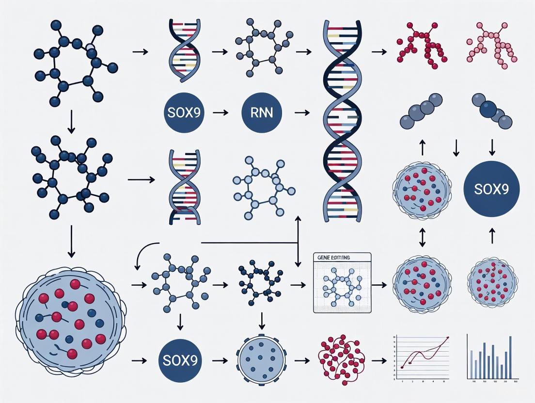
This article explores the convergence of SOX9 gene editing and organoid technology for advanced immune function studies.
SOX9 Gene Editing in Organoid Models: Unraveling Immune Modulation and Therapeutic Potential
Abstract
This article explores the convergence of SOX9 gene editing and organoid technology for advanced immune function studies. SOX9, a key transcription factor, drives tumor progression and modulates anti-tumor immunity by suppressing immune cell infiltration. We examine how CRISPR/Cas9 systems enable precise SOX9 manipulation in human tissue-derived organoids, creating physiologically relevant models to dissect its role in immune suppression. The content covers foundational biology, advanced methodologies like CRISPRi/a, troubleshooting for complex 3D systems, and validation strategies comparing organoid models with in vivo findings. This synthesis provides researchers and drug development professionals with a comprehensive framework for leveraging SOX9-edited organoids in immuno-oncology and regenerative medicine applications.
SOX9 Biology and Its Critical Role in Tumor-Immune Interactions
SOX9 as a Driver of KRAS-Induced Lung Adenocarcinoma and Immune Evasion
The transcription factor SOX9 is integral to proper tissue development and homeostasis. In the context of KRAS-mutant lung adenocarcinoma (LUAD), it transitions to a potent oncogenic driver. KRAS mutations are among the most common oncogenic drivers in LUAD, present in approximately 25-30% of cases, yet the molecular mechanisms that fuel tumor progression and modulate the tumor microenvironment (TME) remain incompletely understood [1] [2] [3]. Emerging evidence solidifies SOX9's role not only in promoting tumor growth but also in orchestrating a suppressive immune microenvironment, thereby facilitating immune evasion [1] [4] [3]. This application note details the experimental approaches and protocols used to delineate the oncogenic functions of SOX9 in KRAS-driven LUAD, with a specific focus on its utility as a target in organoid models for immune studies.
Key Quantitative Data Synthesis
The following tables consolidate critical quantitative findings from foundational studies investigating SOX9 in KRAS-driven LUAD models.
Table 1: In Vivo Impact of Sox9 Knockout on KrasG12D-Driven Lung Tumorigenesis in Mouse Models
| Experimental Model | Key Genotype/Treatment | Tumor Number | Tumor Burden | Tumor Progression (Grade 3 Tumors) | Overall Survival |
|---|---|---|---|---|---|
| CRISPR/Cas9 Knockout [1] | KrasG12D; sgSox9.2-pSECC |
Significantly decreased (at 18, 24, 30 weeks) | Significantly decreased (p=0.029) | 1 tumor observed | N/A |
| CRISPR/Cas9 Control [1] | KrasG12D; sgTom (control) |
Control level | Control level | 12 tumors observed | N/A |
| Cre-LoxP GEMM [1] | KrasLSL-G12D; Sox9flox/flox (KSf/f) |
N/A | Significantly reduced (p=0.011) | Significantly fewer (p=0.0008) | Significantly longer (p=0.0012) |
| Cre-LoxP Control [1] | KrasLSL-G12D; Sox9w/w (KSw/w) |
N/A | Control level | Control level | Control level |
Table 2: Impact of SOX9 on the Tumor Immune Microenvironment in KrasG12D-Driven LUAD
| Immune Parameter | Effect of High SOX9 Expression | Experimental Validation Method |
|---|---|---|
| CD8+ T Cells | Functional suppression and reduced infiltration [1] [4] | Flow cytometry, IHC [1] |
| Natural Killer (NK) Cells | Functional suppression and reduced infiltration [1] [4] | Flow cytometry, IHC [1] |
| Dendritic Cells (DCs) | Inhibition of tumor-infiltrating DCs [1] [4] | Flow cytometry, IHC [1] |
| Overall Immune Infiltration | Suppressed immune cell infiltration [1] [3] | Flow cytometry, gene expression, IHC [1] |
| Tumor Stiffness | Significant increase in collagen fibers [1] [4] | Collagen-related gene expression, histology [1] |
| Tumor Immune Status | Creates an "immune cold" tumor [3] | Analysis of immune cell infiltration [3] |
Table 3: Correlation Between SOX9 and Clinical Outcomes
| Dataset / Context | SOX9 Expression Level | Correlation with Patient Survival | Additional Clinical Associations |
|---|---|---|---|
| TCGA LUAD [1] | High (top 20%) | Significantly shorter survival (p = 0.0039) [1] | N/A |
| TCGA LUAD [1] | Low (lowest 15%) | Significantly longer survival [1] | N/A |
| Human NSCLC [1] | High | Shorter overall survival [1] | Associated with poor prognosis [3] |
Detailed Experimental Protocols
Protocol: CRISPR/Cas9-Mediated Sox9 Knockout in a KrasG12D Murine LUAD Model
This protocol describes the use of the pSECC CRISPR system for somatic knockout of Sox9 concurrent with activation of the KrasG12D allele in the mouse lung [1].
Application Note: This model is ideal for studying the impact of a specific gene on tumor initiation and progression in an immunocompetent host.
Materials:
- Biological Model: C57BL/6 J mice.
- Vector: pSECC (sgRNA + Cre) all-in-one vector [1].
- sgRNAs: Three designed sgRNAs targeting Sox9; non-targeting tdTomato sgRNA (sgTom) as control [1].
- Delivery Method: Intratracheal instillation.
- Analysis Timepoints: 18, 24, and 30 weeks post-infection.
Procedure:
- sgRNA Cloning: Clone validated Sox9-targeting sgRNAs (e.g., sgSox9.2) and the control sgTom into the pSECC vector.
- Virus Production: Package the pSECC constructs into lentiviral particles.
- Model Generation: Perform intratracheal delivery of the lentiviral preparations to adult mice.
- Tumor Monitoring: Allow tumor development over the course of 18-30 weeks.
- Endpoint Analysis: At designated timepoints, harvest lung tissue for:
- Tumor Quantification: Count tumor numbers and calculate tumor burden.
- Histopathology: Perform H&E staining to grade tumors (Grade 1-3).
- IHC/IF Staining: Analyze SOX9 and Ki67 expression to correlate with tumor grade and proliferation.
- Immune Profiling: Conduct flow cytometry on dissociated tumors to quantify CD8+ T, NK, and dendritic cell infiltration.
Protocol: Genetic Engineered Mouse Model (GEMM) with Conditional Sox9 Knockout
This protocol utilizes the Cre-LoxP system for a constitutive, lung-wide knockout of Sox9 in a KrasG12D-driven LUAD setting [1].
Application Note: This model provides a more complete and uniform deletion of the target gene, useful for studying its non-redundant functions.
Materials:
- Biological Model:
KrasLSL-G12D; Sox9flox/flox(KSf/f) mice andKrasLSL-G12D; Sox9w/w(KSw/w) control mice [1]. - Vector: Lenti-Cre.
- Delivery Method: Intratracheal instillation.
Procedure:
- Model Setup: Cross
KrasLSL-G12Dmice withSox9flox/floxmice to generate experimental KSf/f and control KSw/w cohorts. - Tumor Initiation: Administer lenti-Cre intratracheally to mice to activate the KrasG12D allele and, in KSf/f mice, delete Sox9.
- Survival Study: Monitor mice for survival until a predefined ethical endpoint (e.g., 380 days).
- Endpoint Analysis: Upon sacrifice, collect lungs for analysis of tumor burden, tumor grade distribution, and SOX9/Ki67 IHC staining.
Protocol: Evaluating SOX9-Driven Tumor Growth Using 3D Organoid Allografts
This protocol assesses the cell-autonomous and non-cell-autonomous effects of SOX9 on tumor cell growth using 3D organoids and syngeneic allograft models [1].
Materials:
- Cell Lines: KrasG12D mouse lung tumor cell lines (e.g., mTC11, mTC14) with low endogenous SOX9 [1].
- Expression Vector: Plasmid for mouse Sox9 overexpression (mSox9OE); empty vector (EV) control.
- Host Mice: Immunocompetent syngeneic C57BL/6 J mice and immunocompromised mice (e.g., NSG) [1].
Procedure:
- Cell Line Engineering: Stably transduce KrasG12D mouse lung tumor cells with mSox9OE or EV control constructs.
- 3D Organoid Culture:
- Embed transduced cells in Matrigel and culture in appropriate 3D organoid medium.
- Monitor organoid growth over 7-14 days.
- Quantify organoid size and number of cells per organoid.
- Fix and process organoids for IHC analysis of SOX9 and Ki67.
- Syngeneic Allograft:
- Subcutaneously inject mSox9OE or EV control cells into immunocompetent syngeneic mice and immunocompromised mice.
- Measure tumor volume regularly over several weeks.
- Key Comparison: Compare tumor growth curves between immunocompetent and immunodeficient hosts to dissect SOX9's tumor-intrinsic effects from its role in immune modulation.
Signaling Pathways and Experimental Workflows
The following diagrams, generated using DOT language, illustrate the core mechanistic findings and experimental workflows.
Diagram 1: SOX9 in KRAS-LUAD Oncogenesis and Immune Evasion. This diagram illustrates the central role of SOX9 in KRAS-driven LUAD. KRAS activation induces SOX9 expression. SOX9 then drives tumor progression directly by accelerating growth and indirectly by elevating collagen deposition (increasing tumor stiffness) and suppressing anti-tumor immunity, creating a permissive environment for tumor development [1] [4] [3].
Diagram 2: Workflow for In Vivo SOX9 Functional Studies. This workflow outlines the two primary murine models used to study SOX9 function in KRAS-driven LUAD. Researchers can choose between a somatic knockout approach using CRISPR/Cas9 or a constitutive knockout using a pre-engineered GEMM. Both paths lead to comprehensive analysis of tumor phenotypes and the immune microenvironment [1].
The Scientist's Toolkit: Research Reagent Solutions
Table 4: Essential Reagents and Models for SOX9 and KRAS LUAD Research
| Reagent/Model | Function/Application | Specific Examples / Notes |
|---|---|---|
| pSECC Vector | All-in-one vector for concurrent Cre-mediated recombination and CRISPR/Cas9 gene editing in vivo [1]. | Used for somatic knockout of Sox9 and activation of KrasG12D [1]. |
| KrasLSL-G12D Mouse | Foundational GEMM for studying KRAS-driven LUAD. | Often crossed with other floxed alleles (e.g., Sox9flox/flox) [1]. |
| Lenti-Cre Virus | For spatially controlled activation of conditional alleles in vivo. | Intratracheal delivery initiates lung-specific tumorigenesis [1]. |
| Syngeneic Mouse Models | For tumor allograft studies in immunocompetent hosts. | C57BL/6 J mice; essential for evaluating SOX9's role in immune evasion [1]. |
| KrasG12D Lung Tumor Cell Lines | Pre-clinical models for in vitro and allograft studies. | mTC11, mTC14; can be engineered for SOX9 gain/loss-of-function [1]. |
| 3D Organoid Culture | To model tumor cell growth in a more physiologically relevant 3D context. | Used to demonstrate SOX9-driven organoid growth [1]. |
| Anti-SOX9 Antibody | Detection and quantification of SOX9 protein expression. | Used for IHC and IF on mouse and human tumor sections [1] [5]. |
| Immune Cell Markers (CD8, NK, DC) | Profiling tumor immune microenvironment by flow/IHC. | Critical for demonstrating reduced infiltration upon SOX9 expression [1] [4]. |
SOX9-Mediated Suppression of CD8+ T Cells, NK Cells, and Dendritic Cell Infiltration
The SOX9 transcription factor is a critical regulator of development and tissue homeostasis, but its dysregulation has emerged as a significant driver of tumor progression. Recent investigations have revealed a novel and pivotal role for SOX9 in orchestrating an immunosuppressive tumor microenvironment (TME). This application note delineates the mechanisms by which SOX9 suppresses the infiltration and function of key anti-tumor immune cells—CD8+ T cells, natural killer (NK) cells, and dendritic cells (DCs)—and provides detailed protocols for modeling this immune evasion in organoid systems, underpinning a broader thesis on SOX9 gene editing for immune function research.
Background and Mechanistic Insights
SOX9 as an Oncogenic Driver and Immune Regulator
SOX9 is frequently overexpressed in numerous solid malignancies, including lung, breast, and liver cancers, where its expression often correlates with poor patient survival [1] [6] [7]. While historically studied for its roles in cell proliferation and stemness, SOX9 is now recognized as a master regulator of the TME. In KrasG12D-driven lung adenocarcinoma (LUAD) models, loss of Sox9 significantly reduces tumor burden and progression, contributing to longer overall survival. This tumor suppression was markedly attenuated in immunocompromised mice, providing the first clues to SOX9's essential role in modulating anti-tumor immunity [1].
Mechanism of Immune Cell Suppression
Research demonstrates that SOX9 functionally suppresses tumor-associated CD8+ T, NK, and dendritic cells. This is achieved through a dual mechanism:
- Altered Cytokine and Factor Expression: SOX9 negatively correlates with genes associated with the cytotoxic functions of CD8+ T cells and NK cells, as well as those of anti-tumor M1 macrophages [7].
- Extracellular Matrix (ECM) Remodeling: SOX9 significantly elevates collagen-related gene expression and increases collagen fiber deposition within tumors. It is proposed that SOX9 increases tumor stiffness and inhibits tumor-infiltrating DCs, thereby creating a physical and biochemical barrier that suppresses CD8+ T cell and NK cell infiltration and activity [1]. Bioinformatic analyses of human cancers confirm that SOX9 overexpression negatively correlates with the expression of genes critical to CD8+ T cell and NK cell function [7].
Table 1: Key Experimental Findings on SOX9-Mediated Immune Suppression
| Experimental Model | Finding Related to CD8+ T Cells | Finding Related to NK Cells | Finding Related to Dendritic Cells |
|---|---|---|---|
| KrasG12D LUAD GEMM (Mouse) | Functional suppression of tumor-associated CD8+ T cells [1] | Functional suppression of tumor-associated NK cells [1] | Suppression of tumor-infiltrating dendritic cells [1] |
| Human Pan-Cancer Analysis (TCGA) | Negative correlation with genes associated with CD8+ T cell function [7] | Negative correlation with genes associated with NK cell function [7] | - |
| Bioinformatic Analysis | Negative correlation with infiltration levels in specific cancer types [7] | - | - |
Application Notes: Experimental Models and Workflows
Organoid Models for Studying SOX9-Immune Interactions
Organoids are 3D self-organizing structures derived from stem cells that recapitulate the microarchitecture and physiology of their tissue of origin, providing a powerful tool to dissect tumor-immune interactions [8] [9]. Primary human organoids can be generated from healthy or pathological tissue samples, including lung, intestine, and breast, and maintained in culture for extended periods while retaining original tissue properties [8].
Key Culture Components for Epithelial Organoids:
- Extracellular Matrix (ECM): Matrigel or other ECM hydrogels to provide a tissue-like structural scaffold.
- Essential Growth Factors: The culture medium must replicate the stem cell niche. Key factors often include:
- Wnt Agonists (e.g., R-spondin, Wnt-3a, CHIR99021): Major drivers for LGR5+ stem cell growth.
- EGF: Promotes proliferation.
- Noggin / A83-01: Inhibits BMP/TGF-β signaling to maintain stemness.
- Tissue-Specific Factors (e.g., FGF7/FGF10 for lung organoids) [8].
Table 2: Essential Research Reagent Solutions for SOX9 Organoid-Immune Studies
| Reagent Category | Specific Examples | Function in the Experimental System |
|---|---|---|
| Organoid Culture | Matrigel / BME, R-spondin, Noggin, EGF, FGF7/FGF10, A83-01, CHIR99021 | Supports the growth and maintenance of 3D primary epithelial organoids that mimic the native tissue [8]. |
| Gene Editing | CRISPR/Cas9 systems (e.g., pSECC for combined KO/Cre), dCas9-VP64 (CRISPRa), dCas9-KRAB (CRISPRi) [1] [10] | Enables knockout, activation (CRISPRa), or inhibition (CRISPRi) of SOX9 to study gain- and loss-of-function effects. |
| Immune Coculture | Isolated primary immune cells (CD8+ T, NK, DCs), Transwell inserts | Allows for the introduction of immune components into the organoid system to study infiltration and functional crosstalk. |
| Analysis | Flow cytometry antibodies (CD45, CD3, CD8, CD56/NKp46, CD11c), IHC/IF antibodies (SOX9, Ki67, Collagen) | Critical for phenotyping and quantifying immune cell populations and analyzing SOX9 expression and ECM changes. |
Experimental Protocol: CRISPR-Cas9-Mediated SOX9 Manipulation in Organoids
The following protocol outlines a strategy for investigating SOX9 function using CRISPR-Cas9 in organoid models.
Part A: SOX9 Knockout in Established Organoids
- Guide RNA (gRNA) Design: Design and clone gRNAs targeting critical exons of the SOX9 gene into an appropriate delivery vector (e.g., lentiCRISPRv2, pSECC). The pSECC system allows for concurrent CRISPR/Cas9-mediated knockout and Cre-recombinase activation, ideal for use in KrasG12D mutant models [1] [11].
- Viral Transduction: Produce lentiviral particles containing the SOX9-targeting gRNA construct.
- Organoid Infection: Infect target organoids with the lentiviral supernatant in the presence of polybrene (e.g., 8 µg/mL). Spinfect at 600 × g for 60-90 minutes at 32°C to enhance infection efficiency.
- Selection and Validation: Apply appropriate antibiotics (e.g., Puromycin, Blasticidin) 48 hours post-infection to select for successfully transduced organoids. Expand selected organoids and validate SOX9 knockout via:
- Western Blotting for SOX9 protein.
- RT-qPCR for SOX9 mRNA.
- Immunohistochemistry (IHC) on organoid sections.
Part B: CRISPRa/i for Precise SOX9 Modulation For fine-tuning SOX9 expression without complete knockout:
- System Setup: Utilize a dual-vector system expressing:
- dSpCas9-VP64 (for activation, CRISPRa) and/or dSaCas9-KRAB (for inhibition, CRISPRi) [10].
- A gRNA expression vector (e.g., Lenti-EGFP-dual-gRNA) targeting the SOX9 promoter or regulatory regions. Effective gRNA sequences for mouse Sox9 activation have been published, e.g.,
Sox9-2: CGGGTTGGGTGACGAGACAGG[10].
- Transduction and Analysis: Co-transduce organoids with both vectors and assess SOX9 expression changes and subsequent phenotypic effects.
Experimental Protocol: Organoid-Immune Cell Co-culture and Analysis
This protocol details the setup for assessing immune cell infiltration and function following SOX9 modulation.
Immune Cell Isolation:
- Isolate CD8+ T cells, NK cells, and DCs from human peripheral blood mononuclear cells (PBMCs) or mouse splenocytes using magnetic-activated cell sorting (MACS) or fluorescence-activated cell sorting (FACS) to achieve high purity.
Co-culture Establishment:
- Option 1: Direct Co-culture. Seed isolated immune cells directly into the organoid culture well. This allows for direct cell-to-cell contact.
- Option 2: Indirect Co-culture. Use Transwell inserts with permeable membranes. Place organoids in the bottom well and immune cells in the insert. This allows the exchange of soluble factors (cytokines, chemokines) without direct contact, useful for distinguishing between mechanisms.
Functional Analysis of Immune Cells:
- Flow Cytometry: After 24-72 hours of co-culture, dissociate the organoids (using enzymes like TrypLE or collagenase) to create a single-cell suspension.
- Stain for immune cell markers (e.g., CD45, CD3, CD8 for T cells; CD56, NKp46 for NK cells; CD11c for DCs).
- Include functional markers:
- Activation: CD69, CD25
- Cytotoxicity: intracellular Granzyme B, Perforin
- Proliferation: Ki67, CFSE dilution
- Inhibition/exhaustion: PD-1, TIM-3, LAG-3
- Conditioned Media Analysis: Collect culture supernatants and analyze using cytokine/chemokine arrays or ELISA to quantify secreted factors (e.g., IFN-γ, TNF-α, CCL, CXCL families).
- Flow Cytometry: After 24-72 hours of co-culture, dissociate the organoids (using enzymes like TrypLE or collagenase) to create a single-cell suspension.
Analysis of SOX9-Mediated TME Remodeling:
- Immunohistochemistry/Immunofluorescence: On organoid sections, stain for:
- SOX9 (nuclear)
- Collagen (e.g., Masson's Trichrome, Picrosirius Red, or specific collagen I/III antibodies)
- Immune cell markers (as above) to visualize spatial distribution.
- Gene Expression: Use RT-qPCR to analyze expression of SOX9, collagen genes (e.g., COL1A1, COL3A1), and immune-related genes in the organoids.
- Immunohistochemistry/Immunofluorescence: On organoid sections, stain for:
Data Interpretation and Expected Outcomes
- Successful SOX9 Knockout/Modulation: Confirmed by a significant reduction (KO/i) or increase (a) in SOX9 protein and mRNA levels in treated organoids compared to controls.
- Reduced Immune Infiltration & Function: Organoids with high SOX9 expression are expected to show:
- Fewer infiltrated CD8+ T and NK cells upon dissociation and flow cytometry.
- Lower expression of cytotoxic molecules (Granzyme B, Perforin) in the immune cells that are present.
- Higher expression of exhaustion markers (e.g., PD-1) on T cells.
- Altered cytokine profile in conditioned media, potentially with reduced levels of pro-inflammatory cytokines like IFN-γ.
- ECM Remodeling: SOX9-high organoids will display increased collagen deposition and fiber density upon histological examination, providing a mechanistic link to the impaired immune infiltration.
The protocols outlined herein provide a robust framework for utilizing organoid models and CRISPR-Cas9 gene editing to rigorously investigate the mechanism by which SOX9 creates an immunosuppressive TME. By integrating genetic manipulation with sophisticated organoid-immune co-culture systems, researchers can effectively dissect how SOX9-driven ECM remodeling and signaling suppression impedes the infiltration and function of critical anti-tumor immune populations. This approach not only advances fundamental knowledge but also paves the way for identifying novel therapeutic strategies to reverse SOX9-mediated immune evasion and enhance the efficacy of cancer immunotherapies.
SOX9 Regulation of Collagen Deposition and Tumor Microenvironment Stiffness
The tumor microenvironment (TME) is a complex ecosystem where biochemical and biophysical signals interact to drive cancer progression. Extracellular matrix (ECM) stiffness, primarily governed by collagen deposition and cross-linking, has emerged as a critical regulator of tumorigenesis, immune evasion, and therapeutic resistance [12] [13]. The transcription factor SOX9 plays a pivotal role in this process, serving as a molecular nexus that integrates mechanical cues with transcriptional programs that shape the stromal landscape [14] [15]. This application note explores the mechanistic relationship between SOX9 and collagen deposition within engineered organoid models, providing detailed protocols for investigating this axis through CRISPR-based gene editing and functional readouts. The insights gained are directly applicable to immune studies research, particularly in understanding how stromal remodeling influences anti-tumor immunity and response to immunotherapy.
Key Quantitative Relationships Between SOX9, Collagen, and Tissue Stiffness
Table 1: Experimental Measurements of SOX9, Collagen, and Matrix Stiffness
| Parameter | Experimental System | Measurement Value | Biological Context |
|---|---|---|---|
| Tissue Stiffness | Normal breast tissue | ~800 Pa (Elastic Modulus) | Physiological baseline [13] |
| Breast cancer tissue | 5-10 kPa (Elastic Modulus) | Pathological stiffening [13] | |
| Stiff hydrogel for breast organoids | 1,800 - 3,000 Pa (Elastic Modulus) | Experimentally-induced SOX9 upregulation [15] | |
| Collagen Hydrogel Stiffness | Low Collagen (2.5 mg/mL) | 24.22 ± 0.50 kPa (Elastic Modulus, Day 2) | 3D cancer spheroid model [16] |
| High Collagen (6 mg/mL) | 40.12 ± 0.00 kPa (Elastic Modulus, Day 2) | 3D cancer spheroid model [16] | |
| SOX9 Expression | Breast organoids on stiff matrix | Significantly upregulated (protein & mRNA) | Correlated with luminal progenitor markers KIT and TNFSF11 [15] |
| Collagen Deposition Post-Treatment | Anti-progestin (Ulispiral Acetate) therapy | Dramatic decrease in collagen fiber coherency | Reduced tissue stiffness and breast cancer risk [15] |
Mechanistic Insights: SOX9 at the Nexus of Stiffness and Collagen Deposition
SOX9 regulates collagen deposition and TME stiffness through a multi-faceted role, responding to and reinforcing the biomechanical properties of the tumor stroma.
SOX9 as a Mechanoresponsive Transcriptional Regulator: In breast organoid models, culturing on stiff matrices (1,800-3,000 Pa) significantly upregulates SOX9 expression alongside luminal progenitor markers (KIT) and PR target genes (TNFSF11) [15]. This demonstrates that SOX9 is a key downstream effector of biomechanical signaling. This stiffness-induced SOX9 expression creates a pro-tumorigenic feedback loop, where SOX9-expressing luminal progenitor cells engage in paracrine crosstalk with stromal fibroblasts, driving further ECM remodeling and stiffening [15].
SOX9 Directly Regulates Chondrogenic and Fibrotic Programs: As a master transcription factor, SOX9 maintains cartilage homeostasis by triggering chondrocytes to express key ECM components, including type II collagen (COL2A1) and aggrecan [17]. This anabolic function is subverted in pathological contexts, including fibrosis across various organs (cardiac, liver, kidney, pulmonary) and cancer [14]. The regulation of SOX9 itself is complex, involving modifications like phosphorylation, acetylation, and methylation, which control its stability, nuclear localization, and transcriptional activity [14] [17].
Therapeutic Targeting of the SOX9-Collagen Axis: Interventions targeting this axis show promise. Anti-progestin therapy (e.g., Ulipristal Acetate) in high-risk patients reduces collagen alignment and tissue stiffness, an effect linked to the suppression of SOX9 and progenitor cell activity [15]. This confirms the functional significance of this pathway in human disease and its potential as a target for risk mitigation.
SOX9 Regulation of Collagen and Stiffness
Experimental Protocols
Protocol 1: CRISPR-Cas9-Mediated SOX9 Editing in Tumor Organoids
This protocol enables the functional investigation of SOX9 in a physiologically relevant 3D context.
Step 1: Organoid Generation from Patient-Derived Samples
- Material: Obtain tumor tissue samples (e.g., from breast, pancreatic, or colorectal cancer) in cold, sterile PBS containing antibiotics and 10µM Y-27632 (ROCK inhibitor) [18].
- Procedure: Mince tissue finely with scalpels and digest with 2 mg/mL Collagenase/Dispase in Advanced DMEM/F12 for 30-60 minutes at 37°C with agitation. Quench digestion with complete medium (Advanced DMEM/F12, 10mM HEPES, 1x GlutaMAX, 10% FBS). Filter through 100µm strainers and centrifuge. Plate the cell pellet (10,000-20,000 cells) in 30µL domes of Basement Membrane Extract (BME) or Matrigel in 24-well plates. After polymerization, overlay with organoid-specific complete medium [18].
Step 2: Delivery of CRISPR-Cas9 Machinery
- Reagent: Use a lentiviral vector expressing Cas9 and a guide RNA (gRNA) targeting the SOX9 gene. A non-targeting scrambled gRNA serves as a control. A sample SOX9 gRNA sequence targeting an early exon is
5'-GACGUGAAGCGUGUUCGACA-3'. - Procedure: On day 3-5 of culture, dissociate organoids to single cells using TrypLE Express. Transduce 500,000 cells with lentivirus at an MOI of 10-50 in the presence of 8µg/mL polybrene by spinfection (1000 x g, 90 minutes at 37°C). Resuspend transduced cells in BME/Matrigel and culture for 48 hours. Select for successfully transduced cells using appropriate antibiotics (e.g., 2µg/mL Puromycin) for 5-7 days [18].
- Reagent: Use a lentiviral vector expressing Cas9 and a guide RNA (gRNA) targeting the SOX9 gene. A non-targeting scrambled gRNA serves as a control. A sample SOX9 gRNA sequence targeting an early exon is
Step 3: Validation of SOX9 Knockout
- Molecular Validation: Extract genomic DNA from a portion of the organoids. Perform T7E1 assay or Sanger sequencing of PCR-amplified target sites to confirm indels. Validate at the protein level via Western Blot (Anti-SOX9 antibody, Abcam ab185966) and Immunofluorescence [18].
- Phenotypic Validation: Culture SOX9-KO and control organoids for 14 days. Analyze changes in key progenitor markers (e.g., KIT) via qPCR or flow cytometry. A successful SOX9 knockout is expected to reduce progenitor marker expression [15].
Protocol 2: Modulating and Measuring ECM Stiffness in Organoid Cultures
This protocol details how to engineer the mechanical properties of the organoid environment and assess the outcomes.
Step 1: Fabrication of Tunable Stiffness Hydrogels
- Material: Prepare collagen-based hydrogels (e.g., Rat Tail Collagen I) at varying concentrations (2.5 mg/mL for "soft" and 6.0 mg/mL for "stiff" conditions) to mimic physiological and pathological stiffness [16].
- Procedure: Neutralize collagen solution on ice with 1/10 volume of 0.1M NaOH and 1x PBS. Seed dissociated organoid cells at a density of 1-2 million cells/mL within the collagen solution. Pipette 100µL drops into cell culture plates and incubate at 37°C for 30 minutes to polymerize. Overlay with organoid culture medium. For fibrin hydrogels, use fibrinogen at 2.2-5.6 mg/mL polymerized with thrombin [16].
Step 2: Functional Assessment of Collagen Deposition and Remodeling
- Quantitative Imaging: Fix organoids in hydrogels with 4% PFA after 7-14 days of culture. Stain with Picrosirius Red (0.1% Direct Red in saturated picric acid) for 1 hour to visualize collagen fibers. Image using polarized light microscopy to assess collagen alignment and density. Quantify fiber coherency using ImageJ plugins like FibrilTool [15].
- Gene Expression Analysis: Extract RNA from organoids cultured in soft vs. stiff hydrogels. Perform RT-qPCR for SOX9, collagen genes (COL1A1, COL3A1), cross-linking enzymes (LOX, PLOD2), and EMT/stemness markers. Normalize to housekeeping genes (GAPDH, ACTB). Expect upregulation of this gene set in stiff environments, which is attenuated in SOX9-KO organoids [16] [12].
SOX9 Gene Editing Workflow in Organoids
The Scientist's Toolkit: Research Reagent Solutions
Table 2: Essential Reagents for Investigating SOX9 and TME Stiffness
| Reagent/Category | Specific Examples & Catalog Numbers | Function in Experimental Design |
|---|---|---|
| 3D Culture Matrices | Corning Matrigel (Growth Factor Reduced), Rat Tail Collagen I (e.g., Corning 354236), Fibrinogen from human plasma (e.g., Sigma F3879). | Provides a physiologically relevant 3D scaffold for organoid growth. Varying collagen concentration allows direct control over initial substrate stiffness [16] [18]. |
| CRISPR-Cas9 System | LentiCRISPRv2 (Addgene #52961), SOX9 targeting sgRNA (e.g., target sequence: 5'-GACGUGAAGCGUGUUCGACA-3'), VSV-G pseudotyped lentivirus. | Enables stable knockout of the SOX9 gene in organoid models to study its loss-of-function phenotype [18]. |
| SOX9 Antibodies | Anti-SOX9 antibody for WB/IF (e.g., Abcam ab185966), Anti-SOX9 antibody for IHC (e.g., MilliporeSigma AB5535). | Validation of SOX9 knockout efficiency and assessment of SOX9 expression and subcellular localization in response to stiffness [15]. |
| ECM Staining Kits | Picrosirius Red Stain Kit (e.g., Abcam ab150681), Anti-Collagen I Antibody (e.g., Novus Biologicals NB600-408). | Visualization and quantification of collagen deposition, alignment, and organization within the organoid TME [15] [19]. |
| Small Molecule Inhibitors | Ulipristal Acetate (UPA, Sigma), Onapristone (Tocris), Lysyl Oxidase Inhibitor (BAPN, Beta-Aminopropionitrile, Sigma). | Tools to pharmacologically disrupt the SOX9-progesterone axis or inhibit collagen cross-linking, thereby reducing stiffness [15] [12]. |
Application in Immune Studies
Integrating SOX9-edited organoids with immune co-cultures provides a powerful platform for dissecting mechano-immune crosstalk.
- Protocol 3: Immune Cell Infiltration and Function Co-culture Assay
- Step 1: Generate Conditioned Microenvironments. Culture SOX9-WT and SOX9-KO organoids in soft (2.5 mg/mL) and stiff (6.0 mg/mL) collagen hydrogels for 10 days to allow for matrix remodeling and secretome conditioning.
- Step 2: Isolate and Add Immune Cells. Isolate peripheral blood mononuclear cells (PBMCs) from healthy donors or tumor-infiltrating lymphocytes (TILs) from patient samples. Label immune cells with a fluorescent cell tracker (e.g., CTFR). Add 100,000 labeled immune cells to each organoid-containing hydrogel.
- Step 3: Quantify Infiltration and Function. After 48-72 hours of co-culture, fix and clear the organoids for 3D confocal microscopy to measure immune cell penetration depth and proximity to tumor cells. Alternatively, harvest immune cells from the co-culture for flow cytometry analysis of activation markers (CD69, CD25), exhaustion markers (PD-1, TIM-3), and intracellular cytokines (IFN-γ, TNF-α) after PMA/Ionomycin stimulation.
- Expected Outcome: SOX9-KO organoids in stiff matrices are expected to show enhanced immune cell infiltration and reduced T-cell exhaustion compared to SOX9-WT controls, modeling the breakdown of the stromal barrier to immunotherapy [12] [19].
SOX9 as a Marker of Lung Progenitor Cells in Development and Disease
This application note details the critical role of the transcription factor SOX9 as a key marker and regulator of human lung epithelial tip progenitor cells. Framed within the context of SOX9 gene editing in organoid models, we provide validated experimental protocols and resources to study SOX9's function in lung development, its disease relevance, and its utility for immune-related research. The data and methods herein support investigations into respiratory development, regeneration, and disease modeling.
SOX9 in Lung Development: Core Evidence and Quantitative Profiling
SOX9 is a well-established marker of distal lung epithelial tip progenitors during development. It promotes progenitor self-renewal by coordinating proliferation and inhibiting precocious differentiation into airway lineages [20]. In human foetal lung, SOX9 stabilizes the progenitor cell state by amplifying WNT and receptor tyrosine kinase (RTK) signaling pathways [20].
The following table summarizes key quantitative findings on SOX9's role from recent CRISPRi screening data:
Table 1: Quantitative Effects of SOX9 Perturbation in Human Lung Progenitor Cells
| Experimental Perturbation | Key Phenotypic Outcomes | Identified Direct Transcriptional Targets | Major Signaling Pathways Affected |
|---|---|---|---|
| SOX9 Knockdown (CRISPRi) [20] | • Moderate depletion of progenitor cells• Reduced proliferative capacity• Inhibition of precocious airway differentiation | ETV4, ETV5, LGR5 [20] | WNT signaling, RTK signaling [20] |
| SOX9 Complete Inactivation [21] | • Promoted apoptosis in organoids• Reduced organoid proliferative capacity• Altered differentiation in vivo (e.g., SCGB1A1+ club cells, MUC5AC+ goblet cells) | Not Analyzed | Modulates proliferation; not indispensable for epithelium differentiation [21] |
Molecular Mechanisms and Signaling Pathways
SOX9 operates as a master regulator within a complex transcriptional network. In lung tip progenitors, SOX9 sits at the intersection of WNT and RTK signaling. It directly activates transcription of effectors like ETV4 and ETV5 (RTK signaling) and LGR5 (a WNT signaling enhancer), thereby creating a positive feedback loop that stabilizes the progenitor state [20].
Furthermore, SOX9 has been characterized as a pioneer transcription factor in other systems, a property likely conserved in the lung. This means it can bind to its cognate motifs in compact, closed chromatin, initiate nucleosome displacement, and recruit co-factors (e.g., histone and chromatin modifiers) to open chromatin and activate new transcriptional programs [22]. Concurrently, this recruitment of epigenetic factors away from the enhancers of the previous cell state (e.g., programs for differentiation) contributes to transcriptional silencing, enabling fate switching [22].
Diagram: SOX9-Stabilized Progenitor State in Lung Tip Cells
Detailed Experimental Protocols
Protocol: CRISPRi-Mediated SOX9 Knockdown in Primary Human Lung Organoids
This protocol is adapted from Nikolić et al., 2022 [20], for achieving inducible, homogeneous knock-down of SOX9 to study its function in primary human foetal lung tip progenitor organoids.
Workflow Overview:
- Cell Line Preparation: Establish a parental organoid line with stable integration of an inducible dCas9-KRAB construct.
- gRNA Delivery: Transduce organoid-derived single cells with a lentiviral vector containing a constitutive gRNA targeting SOX9.
- Selection & Expansion: FACS-sort double-positive (TagRFP+EGFP+) cells to select for organoids with both constructs. Expand the transduced organoids.
- Gene Knockdown Induction: Add doxycycline (Dox) and trimethoprim (TMP) to the culture medium for 4-5 days to induce dCas9-KRAB nuclear translocation and target gene repression.
- Phenotypic Analysis: Assess organoids for changes in proliferation, differentiation, and gene expression 2-4 weeks post-induction.
Diagram: CRISPRi Workflow for SOX9 Knockdown
Protocol: Inactivation of SOX9 in hESC-Derived Lung Organoids
This protocol, based on Huang et al., 2021 [21], describes the generation of a complete SOX9 knockout model using CRISPR/Cas9 in human embryonic stem cells (hESCs) followed by differentiation into lung organoids.
Key Steps:
- Gene Editing:
- gRNA Design: Design two gRNAs targeting exon 3 of the SOX9 gene (e.g., 5'-GGGCTGTAGGCGATCTGTTGGGG-3' and 5'-TCCTACTACAGCCACGCGGCAGG-3').
- Transfection: Co-transfect H9 hESCs with Cas9 and gRNA plasmids.
- Selection & Screening: Apply puromycin selection. Isolate single-cell clones and expand them. Screen for indels in both SOX9 alleles via sequencing of PCR-amplified genomic DNA [21].
- Lung Organoid Differentiation:
- Differentiate validated SOX9-/- and wild-type (WT) hESC clones into lung organoids using a established stepwise protocol [21]:
- Definitive Endoderm (3 days): Use RPMI1640 with Activin A (100 ng/ml) and CHIR99021 (2 µM).
- Anterior Foregut Endoderm (days 4-7): Use Advanced DMEM/F12 with Noggin (200 ng/ml), FGF4 (500 ng/ml), CHIR99021, and SB431542 (10 µM).
- "Ventralized" AFE (days 8-14): Embed cells in Matrigel and culture in DMEM/F12 with BMP4 (20 ng/ml), retinoic acid (0.5 µM), and CHIR99021.
- Lung Progenitor (days 15-21): Culture in DMEM/F12 with CHIR99021, FGF10 (10 ng/ml), KGF (10 ng/ml), and DAPT (20 µM).
- Airway Organoid (from day 21): Culture in Ham's F12 with dexamethasone, 8-Br-cAMP, IBMX, KGF, and B-27 supplement.
- Differentiate validated SOX9-/- and wild-type (WT) hESC clones into lung organoids using a established stepwise protocol [21]:
The Scientist's Toolkit: Essential Research Reagents
Table 2: Key Reagents for Studying SOX9 in Lung Organoid Models
| Reagent / Tool | Function / Purpose | Example Use Case |
|---|---|---|
| Inducible CRISPRi System (dCas9-KRAB) | Allows for precise, temporal knock-down of SOX9 without complete genetic ablation. | Studying the role of SOX9 in progenitor maintenance vs. differentiation [20]. |
| SOX9 gRNA Library | Enables targeted genetic perturbation of SOX9. | Pooled screens or validation of SOX9-specific phenotypes [20]. |
| Matrigel | Provides a 3D extracellular matrix scaffold for organoid growth and morphogenesis. | Supporting the 3D structure of human lung tip progenitor organoids [20] [21]. |
| WNT Agonist (CHIR99021) | Activates the WNT/β-catenin pathway, crucial for tip progenitor self-renewal. | Maintenance and expansion of SOX9+ lung tip progenitors in culture [20] [21]. |
| FGF10 & KGF (FGF7) | Key growth factors for lung branching morphogenesis and epithelial proliferation. | Induction and maintenance of lung progenitor state in organoid differentiation protocols [21]. |
| Targeted DamID (TaDa) | Technique to identify genome-wide, direct binding targets of a transcription factor. | Mapping direct transcriptional targets of SOX9 in lung progenitor cells [20]. |
Application in Immune Studies and Disease Contexts
The study of SOX9 in lung organoids has significant implications for immune research, primarily through disease modeling.
- Cancer Relevance: Lung adenocarcinomas often reactivate an embryonic progenitor phenotype, and SOX9 is a central part of this program [20]. Furthermore, sustained SOX9 expression in other tissues has been shown to activate oncogenic transcriptional regulators, charting a path to cancers like basal cell carcinoma [22]. SOX9-edited organoids can thus serve as models to study the tumorigenic microenvironment and its interaction with immune cells.
- Immune Correlations: While not a direct immune marker, bioinformatics analyses have identified SOX9 as a consistently dysregulated hub transcription factor in diseases like Immunoglobulin A Nephropathy (IgAN), where its expression correlates with specific immune cell infiltrations and pathway activities [23]. This suggests that SOX9's role may extend to modulating immune responses in certain disease contexts, a potential area for investigation using edited organoid models.
Correlation Between High SOX9 Expression and Poor Patient Survival in LUAD
Clinical and Prognostic Significance of SOX9 in LUAD
SOX9 is a transcription factor critically involved in embryonic development and tissue homeostasis, and its dysregulation has been strongly implicated in the pathogenesis of Lung Adenocarcinoma (LUAD). Clinical evidence from transcriptomic analyses of human LUAD samples establishes a clear link between elevated SOX9 levels and aggressive disease characteristics.
Table 1: SOX9 Expression and Survival Correlation in LUAD
| Evidence Type | Cohort / Model | Key Finding | Statistical Significance | Source |
|---|---|---|---|---|
| Human Patient Data | TCGA LUAD Dataset (Top 20% SOX9-high) | Significantly shorter overall survival | p = 0.0039 | [1] |
| Human Patient Data | TCGA LUAD Dataset (Lowest 15% SOX9-low) | Significantly longer overall survival | Reported as significant | [1] |
| Mouse Model | KrasG12D; Sox9flox/flox (KSf/f) | Significantly longer survival compared to controls | p = 0.0012 | [1] |
| Human Patient Data | LUAD Tumor vs. Normal Tissue | SOX9 and RAP1 significantly increased in tumors | p < 0.05 | [24] [25] |
Interrogation of The Cancer Genome Atlas (TCGA) LUAD dataset reveals that patients with tumors in the top 20% of SOX9 expression have a significantly shorter overall survival. Conversely, patients with the lowest 15% of SOX9 expression exhibit significantly longer survival, underscoring its value as a prognostic biomarker [1]. Furthermore, in vivo validation using a KrasG12D-driven mouse LUAD model demonstrates that genetic loss of Sox9 contributes to significantly longer overall survival, confirming its functional role in driving tumor progression [1].
SOX9 as a Driver of Tumor Progression and Immunosuppression
Beyond a correlative biomarker, functional studies establish SOX9 as a key driver of LUAD pathogenesis. It promotes tumor progression by enhancing cell proliferation, invasion, and migration, while simultaneously sculpting an immunosuppressive tumor microenvironment (TME).
Table 2: Functional Role of SOX9 in LUAD Pathogenesis
| Functional Domain | Mechanistic Insight | Experimental Evidence | Source |
|---|---|---|---|
| Tumor Growth & Progression | Loss of Sox9 reduces lung tumor number, burden, and progression to high-grade tumors. | CRISPR/Cas9 and Cre-LoxP knockout in KrasG12D mouse model. | [1] [4] |
| Cell Proliferation | SOX9+ tumors show a significantly higher percentage of Ki67+ cells. | Immunohistochemistry on murine and human LUAD tumors. | [1] |
| Invasion & Migration | Activates the RAP1 signaling pathway, enhancing cell invasion and migration. | Transwell and scratch assays in A549 cells with SOX9 knockdown/overexpression. | [24] [25] |
| Immunosuppression | Suppresses infiltration of anti-tumor immune cells (CD8+ T, NK, Dendritic cells). | Flow cytometry, gene expression in murine LUAD; confirmed in human data. | [1] [4] |
| Extracellular Matrix (ECM) | Elevates collagen-related gene expression and increases collagen fibers, increasing tumor stiffness. | Gene expression analysis, immunohistochemistry. | [1] |
In KrasG12D-driven mouse models, loss of Sox9 significantly reduces lung tumor development, burden, and progression, with a notable decrease in high-grade (Grade 3) tumors [1]. SOX9 expression is consistently associated with increased cell proliferation, as measured by Ki67 staining [1]. SOX9 also promotes metastatic behavior by upregulating the RAP1 signaling pathway. Knocking down SOX9 in LUAD cell lines decreases invasion and migration, while its overexpression has the opposite effect [24] [25].
A pivotal function of SOX9 is its ability to modulate the TME. It significantly suppresses the infiltration and activity of key anti-tumor immune cells, including CD8+ T cells, natural killer (NK) cells, and dendritic cells [1] [4]. This immunosuppressive role is functionally critical, as SOX9-promoted tumor growth is significantly attenuated in immunocompromised mice compared to immunocompetent hosts [1]. Mechanistically, SOX9 elevates collagen-related gene expression and increases collagen deposition, proposing a model where SOX9 increases tumor stiffness to physically inhibit immune cell infiltration [1].
Application Notes: Targeting SOX9 in Organoid Models for Immune Studies
The following protocols leverage SOX9-edited lung organoid models to dissect its role in tumor progression and immune suppression, providing a platform for therapeutic discovery.
Protocol 1: Generating SOX9-Edited Lung Organoids
This protocol outlines the creation of SOX9-knockout lung organoids from human embryonic stem cells (hESCs) to study the intrinsic role of SOX9 in epithelial proliferation and differentiation.
Key Reagents:
- H9 hESC line: Pluripotent stem cell starting material.
- CRISPR/Cas9 System: For targeted gene editing. gRNAs targeting exon 3 of SOX9 (e.g., 5′-GGGCTGTAGGCGATCTGTTGGGG-3′).
- Matrigel: Provides a 3D extracellular matrix for organoid growth.
- Sequential Differentiation Media: Containing growth factors and small molecules to direct lung lineage specification (e.g., Activin A, CHIR99021, Noggin, FGF4, BMP4, FGF10, KGF) [21].
Workflow Diagram: SOX9-Editing and Lung Organoid Differentiation
- Methodology:
- SOX9 Gene Editing: Transfect H9 hESCs with Cas9 and SOX9-targeting gRNAs. Select puromycin-resistant clones and expand. Validate SOX9 knockout via DNA sequencing and functional assays [21].
- Definitive Endoderm Induction: Culture SOX9−/− and wild-type (WT) hESCs in RPMI1640 medium with 100 ng/ml Activin A and 2 µM CHIR99021 for 3 days.
- Anterior Foregut Endoderm Induction: Differentiate cells in Advanced DMEM/F12 with Noggin, FGF4, and SB431542 for 4 days.
- Lung Progenitor Specification: Embed cells in Matrigel and culture in "ventralizing" media with BMP4 and retinoic acid, followed by lung progenitor media with FGF10 and KGF.
- Airway Organoid Maturation: Maintain 3D organoids in Airway Organoid media to support the development of pulmonary epithelial cell types.
- Phenotypic Analysis: Compare the growth, size, and cellular composition of SOX9−/− and WT organoids. SOX9 inactivation is expected to reduce proliferative capacity but not completely block epithelial differentiation [21].
Protocol 2: Assessing SOX9-Mediated Immunosuppression in a Co-culture Model
This protocol describes using SOX9-proficient and -deficient LUAD organoids to investigate its role in immune cell exclusion using a co-culture system with human immune cells.
Key Reagents:
- SOX9-Modified LUAD Organoids: Generated from patient-derived cells or hESC-derived lung progenitors with oncogenic transformation (e.g., KRAS mutation).
- Human Peripheral Blood Mononuclear Cells (PBMCs): Source of T, NK, and dendritic cells.
- Anti-CD3/CD28 Dynabeads: For T cell activation.
- Recombinant Human IL-2: To support T and NK cell survival.
- Flow Cytometry Antibodies: For immune cell profiling (e.g., anti-CD8, anti-CD56, anti-CD11c, anti-HLA-DR).
Workflow Diagram: SOX9 Immune Co-culture Assay
- Methodology:
- Organoid and Immune Cell Preparation: Generate SOX9-WT and SOX9-KO LUAD organoids. Isolate PBMCs from healthy donor blood and activate T cells using anti-CD3/CD28 beads in the presence of IL-2 for 2-3 days.
- Co-culture Establishment: Mix pre-treated LUAD organoids with activated PBMCs at a defined ratio (e.g., 1:10 organoid cells to PBMCs) in low-attachment plates.
- Co-culture and Harvest: Co-culture for 24-72 hours. Subsequently, harvest the entire co-culture, dissociate into single-cell suspensions, and stain for flow cytometry analysis.
- Immune Profiling Analysis: Quantify the infiltration of CD8+ T cells, CD56+ NK cells, and CD11c+ HLA-DR+ dendritic cells within the organoid cell gate. The SOX9-KO condition is expected to show a significant increase in the abundance of these anti-tumor immune cells compared to SOX9-WT organoids, consistent with in vivo findings [1] [4].
Protocol 3: Investigating the SOX9-RAP1 Axis in Invasion
This protocol provides a method to validate the functional link between SOX9 and the RAP1 signaling pathway in promoting LUAD cell invasion.
Key Reagents:
- A549 LUAD Cells: Model cell line.
- SOX9 Expression Vector / shRNA: For SOX9 overexpression or knockdown.
- RAP1 Expression Vector: For pathway rescue experiments.
- Matrigel-Invasion Transwell Inserts: To quantify invasive capacity.
- Western Blot Reagents: For detecting RAP1 pathway components (RAP1, RAP1GAP, RasGRP3).
Workflow Diagram: SOX9-RAP1 Invasion Signaling
- Methodology:
- Genetic Manipulation: Create stable A549 cell lines with: a) SOX9 overexpression, b) SOX9 knockdown (shSOX9), c) SOX9 knockdown + RAP1 overexpression, and d) corresponding controls.
- Pathway Analysis: Perform Western blot analysis on cell lysates from each group to confirm changes in SOX9, RAP1, RAP1GAP, and RasGRP3 protein levels. SOX9 overexpression should upregulate these RAP1 pathway components [24] [25].
- Functional Invasion Assay: Seed transfected cells into the upper chamber of Matrigel-coated Transwell inserts. After 24 hours, fix, stain, and count the cells that have invaded through the Matrigel to the lower chamber.
- Data Interpretation: Expect SOX9 overexpression to enhance invasion, while SOX9 knockdown should suppress it. Critically, overexpressing RAP1 in SOX9-knockdown cells should restore invasive capacity, confirming RAP1 as a key functional downstream effector of SOX9 [24] [25].
The Scientist's Toolkit: Key Research Reagents
Table 3: Essential Reagents for SOX9 and LUAD Organoid Research
| Reagent/Solution | Function & Application | Example |
|---|---|---|
| CRISPR/Cas9 System | Targeted knockout of SOX9 in stem or cancer cell lines. | SOX9-exon targeting gRNAs, Cas9 plasmid. |
| Lung Differentiation Media Kits | Stepwise differentiation of hESCs into lung organoids. | Custom media with Activin A, FGFs, BMP4, Retinoic Acid. |
| Matrigel / Basement Membrane Matrix | 3D scaffold for culturing and embedding lung organoids. | Corning Matrigel. |
| KrasG12D Mouse Model | In vivo validation of SOX9 in KRAS-driven LUAD. | KrasLSL-G12D; Sox9flox/flox (KSf/f) GEMM. |
| RAP1 Pathway Antibodies | Detecting pathway activity in SOX9-manipulated cells. | Anti-RAP1, Anti-RAP1GAP, Anti-RasGRP3. |
| Immune Cell Markers (Flow Cytometry) | Profiling tumor-infiltrating immune cells in co-cultures. | Anti-human CD8, CD56 (NK), CD11c (DC). |
| Transwell Invasion Chambers | Quantifying cell invasion capacity post-SOX9 modulation. | Corning BioCoat Matrigel Invasion Chambers. |
Advanced CRISPR Techniques for SOX9 Manipulation in 3D Organoid Systems
The SOX9 transcription factor is a critical regulator of developmental processes, organ homeostasis, and has been implicated as a key oncogenic driver in cancers such as lung adenocarcinoma [1]. Organoid systems recapitulate key features of organs, offering powerful platforms for modelling developmental biology and disease [26]. However, functional genetic studies of endogenous gene function in tissue-derived organoids have been hampered by a lack of efficient gene manipulation tools. The Organoid Easytag workflow addresses this limitation by providing an optimized pipeline for precise gene targeting in human organoids using CRISPR-mediated homologous recombination [26] [27]. This application note details the implementation of this workflow specifically for SOX9 reporter generation and knockout within the context of immune studies research, enabling investigators to probe SOX9 function in complex, physiologically relevant model systems.
Key Research Reagent Solutions
The following table catalogues essential reagents and their applications for implementing the Organoid Easytag workflow.
Table 1: Essential Research Reagents for Organoid Easytag Workflow
| Reagent / Tool | Function / Application | Specifications / Examples |
|---|---|---|
| Cas9 Ribonucleoprotein (RNP) | Pre-assembled complex of Cas9 protein and sgRNA for high-efficiency editing with minimal off-target effects [26] [28] | Single-stranded synthetic gRNA combined with Cas9 protein (ssRNP) |
| Nucleofection System | High-efficiency transfection method for delivering RNP complexes and repair templates into organoid cells [26] | Up to 70% transfection efficiency achieved in human foetal lung organoids |
| Fluorescence-Activated Cell Sorting (FACS) | Enrichment of successfully targeted cells based on fluorescent reporter expression [26] [27] | Used to isolate mEGFP+ or H2B-EGFP+ cells after targeting |
| Homology-Directed Repair (HDR) Template | Plasmid donor for precise knock-in via homologous recombination [26] | Circular plasmid with 700-1000 nt homology arms; contains fluorescent reporter |
| AAVS1 Safe Harbor Targeting Vector | Controlled expression of exogenous genes from a genomic locus that minimizes silencing [26] | EF1α promoter-driven membrane-tagged TagRFP-T (mTagRFP-T) |
| Inducible CRISPRa/i Systems | Gain- or loss-of-function studies through transcriptional activation or interference [29] | dCas9 fused to transcriptional effectors (e.g., VPR for activation, KRAB for interference) |
The Organoid Easytag methodology provides a streamlined, efficient pipeline for genetic manipulation in organoids. The diagram below illustrates the core workflow from organoid preparation to the generation of clonal, genetically modified lines.
Protocol for SOX9 Reporter Generation
This protocol enables the generation of a heterozygous SOX9-T2A-H2B-EGFP reporter line to visualize and track SOX9-expressing progenitor cells.
Stage 1: Design and Preparation of CRISPR Components
- sgRNA Design: Design sgRNAs to target the 3' end of the SOX9 coding sequence (just before the STOP codon). The optimal sgRNA binding region is within -400 to +100 bp of the transcriptional start site for efficient modulation [29].
- ssRNP Complex Assembly: Combine purified Cas9 protein with synthetic, chemically modified sgRNA at a molar ratio of 1:2 to 1:4 (Cas9:gRNA). Incubate at room temperature for 10-20 minutes to form the ribonucleoprotein (RNP) complex before nucleofection [26] [28].
- HDR Repair Template Design: The repair template plasmid is a critical component for achieving precise genetic modification [26].
- Homology Arms: Utilize 700-1000 nucleotide homology arms flanking the insertion site.
- Insert Cassette: A T2A-H2B-EGFP (Histone H2B fused to EGFP) reporter cassette. The T2A self-cleaving peptide ensures minimal disruption to the native SOX9 protein, while the H2B fusion localizes EGFP to the nucleus, concentrating the signal and facilitating the identification of SOX9+ cells even at low expression levels [26] [27].
- PAM Site Disruption: Introduce silent mutations in the Protospacer Adjacent Motif (PAM) sequence within the repair template to prevent re-cleavage by Cas9 after successful HDR [26].
Table 2: Key Components for SOX9 Reporter Generation
| Component | Role | Key Design Features |
|---|---|---|
| SOX9 ssRNP | Creates a site-specific double-strand break in the SOX9 locus. | Synthetic single-guide RNA complexed with Cas9 protein. |
| HDR Repair Plasmid | Template for precise insertion of the reporter cassette. | 700-1000 nt homology arms; T2A-H2B-EGFP cassette; mutated PAM sequence. |
| T2A Peptide | Ensames co-translational cleavage. | Produces separate, native SOX9 protein and H2B-EGFP reporter. |
| H2B-EGFP | Nuclear-localized fluorescent reporter. | Concentrates signal in the nucleus for easier detection of SOX9+ cells. |
Stage 2: Organoid Nucleofection and Selection
- Organoid Dissociation: Harvest human foetal lung organoids in the growth phase and dissociate them into single cells using a gentle cell dissociation reagent [28].
- Nucleofection: Co-transfect approximately 1-2 x 10^5 dissociated organoid cells with the pre-assembled ssRNP complex (∼2 µg) and the circular HDR repair plasmid (∼2 µg) using an appropriate nucleofection system and program.
- Post-Nucleofection Recovery: Immediately after nucleofection, transfer the cells to pre-warmed culture medium, plate them in a basement membrane extract (e.g., Matrigel or Cultrex), and overlay with organoid growth medium [28]. Culture for 72 hours to allow for reporter expression.
- FACS Enrichment: Dissociate the transfected organoids into single cells and use FACS to isolate the H2B-EGFP+ population. This step dramatically enriches for successfully targeted cells.
- Clonal Expansion: Seed the sorted H2B-EGFP+ cells sparsely at a low density (e.g., 500-1000 cells per well of a 24-well plate) to facilitate the outgrowth of discrete, clonal organoid colonies.
Stage 3: Validation and Functional Confirmation
- Genotypic Validation: Perform PCR genotyping and Sanger sequencing across the targeted locus to confirm correct 5' and 3' integration. Ensure the wild-type allele remains intact for a heterozygous reporter [26].
- Phenotypic Validation:
- Immunostaining: Confirm that the H2B-EGFP signal co-localizes with anti-SOX9 antibody staining, verifying reporter fidelity [26].
- Progenitor Marker Expression: Validate that targeted organoids retain expression of key multipotent lung progenitor markers like NKX2-1 and SOX2, confirming that the genetic manipulation has not altered the progenitor state [26].
- Differentiation Potential: Demonstrate that the SOX9-targeted organoids retain the capacity to differentiate into relevant lineages (e.g., alveolar or airway), confirming their functional normality [26].
Protocol for SOX9 Knockout
The generation of a pure SOX9 knockout population is achieved by replacing the coding sequence with a selectable reporter, enabling direct enrichment of null cells.
Strategic Workflow for Biallelic Knockout
The knockout strategy involves a sequential process to disrupt both alleles of the SOX9 gene, with the inserted reporter enabling clear tracking of successful editing events.
Knockout Procedure and Validation
- Targeting Strategy: Design two gRNAs to flank the entire SOX9 coding sequence (CDS). The HDR repair template is designed to replace the entire CDS with a T2A-H2B-EGFP cassette. This strategy ensures the complete removal of the functional gene and directly links the knockout event to a fluorescent reporter for easy isolation [26] [27].
- Sequential Targeting: Transfert the wild-type organoids and isolate H2B-EGFP+ clones, which represent heterozygous knockouts. These clones are then subjected to a second round of nucleofection with the same RNP and repair template to target the remaining wild-type allele, generating homozygous SOX9-/- organoids [26].
- Validation:
- Genotypic: Confirm the complete absence of the SOX9 CDS via PCR and sequencing.
- Protein Level: Verify the loss of SOX9 protein by western blot analysis and immunostaining [28].
- Functional Phenotyping: Assess the knockout organoids for expected phenotypic changes, such as altered growth in 3D culture and reduced expression of SOX9 target genes [1].
Application in Immune Studies Research
The functional manipulation of SOX9 in organoid models provides a powerful approach to interrogate its role in tumor-immune interactions. Findings generated using the protocols above can be directly contextualized within the following established framework:
- SOX9 as an Oncogenic Driver: In KrasG12D-driven lung adenocarcinoma (LUAD), SOX9 is highly upregulated in larger, proliferative, and high-grade tumors. Loss of Sox9 significantly reduces lung tumor development, burden, and progression, contributing to longer overall survival in mouse models [1]. This establishes SOX9 as a critical dependency factor in this immune-rich context.
- Modulation of Anti-Tumor Immunity: SOX9 plays a direct role in suppressing anti-tumor immunity. Studies demonstrate that SOX9-expressing tumors significantly attenuate immune cell infiltration, functionally suppressing tumor-associated CD8+ T cells, natural killer (NK) cells, and dendritic cells. This suggests that SOX9 drives tumor progression not only autonomously but also by creating an immunosuppressive microenvironment [1].
- Integration with Co-culture Models: The SOX9 reporter and knockout organoids generated via Organoid Easytag are ideal for incorporation into tumor organoid-immune co-culture models [30]. These models allow for direct investigation of how SOX9 expression in tumor cells influences immune cell recruitment, activation, and cytotoxic efficacy, providing a highly physiologic platform for screening immunotherapies.
Table 3: Quantitative Findings on SOX9 in Lung Adenocarcinoma Models
| Experimental Finding | Model System | Quantitative Result | Citation |
|---|---|---|---|
| Survival Impact of Sox9 Loss | KrasLSL-G12D; Sox9flox/flox (KSf/f) GEMM | KSf/f mice had significantly longer survival than KSw/w controls (p = 0.0012) | [1] |
| Tumor Burden Reduction | KSf/f GEMM & CRISPR-mediated Sox9 KO | Significantly reduced lung tumor burden (p = 0.011 in GEMM) | [1] |
| Inhibition of Tumor Progression | CRISPR-mediated Sox9 KO | 12 grade 3 tumors in control vs. only 1 in Sox9 KO lungs at 24/30 weeks | [1] |
| Correlation with Proliferation | IHC analysis of murine LUAD | Significantly higher percentage of Ki67+ cells in SOX9+ tumors (p = 0.00092) | [1] |
Troubleshooting and Technical Considerations
- Low Targeting Efficiency: If HDR efficiency is low, verify the activity of the sgRNA using a T7 endonuclease assay, ensure the use of high-quality, pure RNP complexes, and confirm the size and integrity of the repair template plasmid [26].
- Mosaicism: While FACS enrichment helps circumvent this issue, potential mosaicism in initial organoid colonies can be resolved by ensuring sparse plating and expanding multiple clonal lines for thorough genotypic and phenotypic validation [27].
- ssODN vs. Plasmid Donors: The Organoid Easytag workflow primarily uses circular plasmid donors. While single-stranded oligonucleotide donors (ssODNs) offer a "cloning-free" alternative, they have been associated with error-prone HDR and lower organoid recovery rates in this system and are not recommended for large insertions like the SOX9 reporter [27].
Dual Endogenous Reporter Systems for Monitoring Stem Cell and Differentiation Activity
In the study of complex biological processes such as cancer development and immune evasion, the ability to simultaneously monitor stem cell activity and differentiation status in living cells provides a critical analytical advantage. Dual endogenous reporter systems represent a technological advancement that enables real-time tracking of these pivotal cellular programs within physiologically relevant model systems, including organoids. These systems are particularly powerful when integrated with CRISPR-Cas9 genome editing, allowing researchers to insert fluorescent reporter genes directly into endogenous genomic loci of key regulatory genes.
This application note focuses specifically on the development and implementation of dual endogenous reporter systems to investigate SOX9-mediated mechanisms in organoid models. The transcription factor SOX9 has been identified as a crucial regulator of aberrant stem cell-like activity in multiple cancer types, including colorectal and lung adenocarcinoma, while also demonstrating significant immunomodulatory functions within the tumor microenvironment [31] [1]. By engineering reporter systems that broadcast SOX9 activity alongside differentiation markers, researchers can perform functional genetic screens to identify novel regulators of these pathways with potential therapeutic significance.
Biological Rationale and Significance
SOX9 as a Key Regulator in Development and Disease
The SOX9 transcription factor plays multifaceted roles in development, tissue homeostasis, and disease pathogenesis. During normal development, SOX9 ensures proper tissue formation through determining cell lineage fate and regulating cell proliferation [1]. In cancer, however, SOX9 frequently becomes dysregulated, contributing to tumor progression through multiple mechanisms:
- Stem Cell Maintenance: SOX9 mediates aberrant stem cell-like activity and functionally blocks differentiation in colorectal cancer [31]
- Tumor Progression: In KRAS-driven lung adenocarcinoma, SOX9 significantly contributes to tumor development, burden, and progression, with its loss resulting in significantly longer overall survival in mouse models [1]
- Immune Modulation: SOX9 drives tumor progression partly by suppressing immune cell infiltration, including CD8+ T cells, natural killer cells, and dendritic cells, thereby creating an immunosuppressive tumor microenvironment [1]
The Stem Cell-Differentiation Axis in Disease Modeling
The balance between stem cell activity and differentiation represents a fundamental axis disrupted in many disease states, particularly cancer. Aberrant activation of stem cell-like programs coupled with impaired differentiation capacity is central to the development of colorectal cancer and other malignancies [31]. This biological paradigm makes simultaneous monitoring of both programs particularly valuable for:
- Identifying novel therapeutic targets that can reactivate differentiation programs
- Understanding how cancer cells maintain stemness while resisting terminal differentiation
- Uncovering mechanisms of immune evasion linked to stem cell characteristics
- Developing strategies to force differentiation as a therapeutic approach
System Design and Engineering
Reporter Selection and Genomic Integration Strategy
The design of a dual endogenous reporter system requires careful selection of appropriate marker genes and a robust strategy for genomic integration:
Marker Gene Selection:
- Stem Cell Activity Reporter: SOX9, a transcription factor that functionally blocks differentiation by activating an aberrant stem cell-like transcriptional program [31]
- Differentiation Activity Reporter: KRT20 (Keratin 20), a well-recognized marker of differentiated intestinal cells that is suppressed in models of cancer initiation [31]
CRISPR-Cas9-Mediated Knock-in Strategy: The recommended approach involves using CRISPR/Cas9 technology combined with template-based homologous recombination to introduce fluorescent reporter cassettes in-frame at the end of the coding regions of target genes [31]. This strategy preserves endogenous regulatory elements and ensures the reporter reflects natural expression dynamics.
Table 1: Fluorescent Reporter Options for Dual Systems
| Reporter Type | Advantages | Limitations | Compatibility |
|---|---|---|---|
| GFP | Bright signal, widely used | Photobleaching | Compatible with blue laser |
| mKate2 | Red-shifted, less autofluorescence | Larger size | Compatible with green/yellow laser |
| Dual GFP/mKate2 | Spectral separation | Requires multiple filters | Simultaneous detection |
| EGFP/Gluc | Fluorescence + secreted luciferase | Complex cloning | Multi-modal detection [32] |
Experimental Workflow for System Establishment
The following diagram illustrates the comprehensive workflow for developing and applying dual endogenous reporter systems in organoid models:
Protocol: Establishing a Dual SOX9-KRT20 Reporter System in Human Colorectal Cancer Organoids
Materials and Reagents
Table 2: Essential Research Reagent Solutions
| Category | Specific Reagents | Function | Supplier Examples |
|---|---|---|---|
| Genome Editing | CRISPR-Cas9 system (px330), sgRNAs targeting SOX9/KRT20 | Targeted genomic integration | OriGene [32] |
| Reporter Constructs | Homology arms, EGFP, mKate2, puromycin/neomycin resistance | Knock-in cassette components | Custom synthesis |
| Cell Culture | Matrigel, mTeSR1, Advanced DMEM/F12 | 3D organoid culture | STEMCELL Technologies [21] [33] |
| Differentiation Factors | Noggin, FGF4, FGF10, KGF, BMP4, CHIR99021 | Directed differentiation | R&D Systems, PeproTech |
| Screening | Epigenetic regulator sgRNA library (542 sgRNAs, 78 genes) | CRISPR screens | Custom libraries [31] |
| Analysis | Flow cytometry antibodies, RNA extraction kits | Validation and analysis | Multiple suppliers |
Step-by-Step Protocol
Guide RNA Design and Cloning (Days 1-3)
- Design sgRNAs: Select guide RNAs targeting the 3' end of the SOX9 and KRT20 coding sequences to avoid disrupting regulatory elements.
- Clone sgRNAs: Insert annealed oligonucleotides into the BbsI site of the pX330 vector or similar CRISPR plasmid [32].
- Prepare donor constructs: Design homology arms (800-1000 bp) flanking the fluorescent reporter cassette (e.g., GFP-P2A-NeomycinR for SOX9; mKate2-P2A-PuromycinR for KRT20).
Organoid Culture and Transfection (Days 4-10)
- Maintain human CRC organoids in Matrigel domes with appropriate culture medium [31].
- Passage organoids using TrypLE Express when they reach optimal density (typically every 5-7 days).
- Transfect organoids using lipofectamine or electroporation with the following mixture:
- 1 µg Cas9/sgRNA plasmid (each target)
- 2 µg donor DNA (each target)
- 1 µg fluorescent marker plasmid for tracking transfection efficiency
- Apply selection 48 hours post-transfection using neomycin (SOX9-GFP) and puromycin (KRT20-mKate2) for 7-10 days.
Clonal Expansion and Validation (Days 11-30)
- Isolate single cells by FACS sorting based on fluorescence or through limiting dilution.
- Expand clonal lines in 3D Matrigel culture with appropriate growth factors.
- Validate integration using site-specific PCR with primers against the genomic locus and cassette [31].
- Confirm reporter functionality through:
- SOX9 knockdown to validate GFP reduction [31]
- Differentiation induction to validate KRT20-GFP increase
Protocol: Application in CRISPR-Based Genetic Screens
Epigenetic Regulator Screening in Reporter Organoids
Library Design and Transduction
- Select target gene set: Focus on druggable epigenetic regulators and their family members (78 genes, 542 sgRNAs) [31].
- Prepare lentiviral library using standard packaging systems with a minimum titer of 10^8 IU/mL.
- Transduce reporter organoids at a low MOI (0.3-0.5) to ensure single integration events.
- Maintain cellular coverage of >1000 cells per sgRNA throughout the screening process [34].
Fluorescence-Activated Cell Sorting and Analysis
- Harvest organoids 7-10 days post-transduction for sorting – this timeframe provides superior discriminatory power compared to earlier timepoints [31].
- Dissociate to single cells using TrypLE Express or similar gentle dissociation reagents.
- Sort cells into four even quartiles based on SOX9-mKate2 and KRT20-GFP expression using FACS.
- Extract genomic DNA from each sorted fraction for sgRNA abundance quantification.
Table 3: Quantitative Analysis of CRISPR Screening Data
| Analysis Method | Application | Key Output | Significance Threshold |
|---|---|---|---|
| MaGeCK MLE | Gene-level phenotype scoring | Beta score (selection degree) | p < 0.05, FDR < 0.1 |
| Rank sum scoring | sgRNA consistency | Percentile ranking | Bottom 15% percentile |
| Normalized read count | sgRNA abundance | Fold-change vs control | >2x depletion/enrichment |
| Pathway enrichment | Biological mechanism | Gene ontology terms | p < 0.01 |
Data Analysis and Hit Validation
- Calculate sgRNA abundance in each sorted fraction relative to the starting library.
- Identify candidate regulators using MaGeCK Maximum Likelihood Estimation to generate beta scores based on differences in normalized sgRNA abundance [31].
- Apply rank sum scoring to identify consistently depleted sgRNAs across replicates.
- Prioritize hits with at least 2 or more targeting sgRNAs within the bottom 15% percentile of rank sum scores.
- Validate top hits using individual sgRNAs in secondary functional assays.
Case Study: Identification of SMARCB1 as a Negative Regulator of Differentiation
Experimental Findings
Application of the dual reporter screening approach in colorectal cancer models identified SMARCB1 of the BAF complex (SWI/SNF) as a negative regulator of differentiation across an array of neoplastic colon models [31]. Key findings included:
- Validation: SMARCB1 was confirmed as a dependency factor required for in vivo growth of human CRC models
- Additional regulators: The screen also identified SUZ12, SMARCD2, DNMT1, and KMT2A as contributors to stem cell-like activity and differentiation in CRC
- Technical performance: CRISPR perturbations provided stronger and more consistent discriminatory power compared to shRNA-mediated suppression
Immune Microenvironment Applications
The integration of SOX9 reporter systems with immune studies is particularly promising given recent findings that SOX9 modulates the tumor immune microenvironment:
In lung adenocarcinoma models, SOX9 was found to suppress immune cell infiltration and functionally suppress tumor-associated CD8+ T cells, natural killer cells, and dendritic cells [1]. These findings highlight the potential of SOX9-focused reporter systems for studying cancer-immune interactions.
Troubleshooting and Optimization
Common Technical Challenges
- Low knock-in efficiency: Optimize homology arm length (800-1000 bp), use single-stranded DNA donors, or incorporate CRISPR-Cas9 enhancers
- Poor reporter signal: Test multiple fluorescent proteins, incorporate protein stabilization domains, or optimize promoter selection
- Reduced organoid viability after sorting: Include Rho kinase inhibitor Y-27632 in recovery media [33]
- High background in screens: Increase cellular coverage to >1000 cells per sgRNA, include more negative controls, and implement robust normalization
Adaptation for Immune Co-culture Studies
For immune studies, consider these modifications:
- Incorporate immune cell markers: Express additional surface markers (e.g., CD64, CD11c) for simultaneous isolation of tumor and immune populations
- Cryopreservation optimization: Use 90% FBS + 10% DMSO freeze medium with controlled-rate freezing [33]
- Live cell imaging: Implement secreted luciferase reporters (e.g., Gluc) for non-destructive longitudinal monitoring [32]
Dual endogenous reporter systems represent a powerful platform for investigating the dynamic interplay between stem cell activity and differentiation in physiologically relevant organoid models. By coupling SOX9 reporting with differentiation markers like KRT20, researchers can perform unbiased genetic screens to identify novel regulators of these critical cellular programs. The integration of these systems with immune profiling approaches further enables investigation of how stem cell states influence tumor-immune interactions, potentially revealing new therapeutic opportunities for targeting the immunosuppressive niches associated with stem-like tumor cells.
CRISPRi and CRISPRa for Precise Temporal Control of SOX9 Expression
The SOX9 transcription factor is a pivotal regulator of development, stem cell maintenance, and differentiation in multiple organ systems. Within organoid models, which emulate the 3D architecture and cellular heterogeneity of native tissues, SOX9 marks progenitor populations and directs lineage specification [35] [36]. The ability to precisely manipulate SOX9 expression levels in these models is therefore critical for dissecting its role in development, disease, and potential immune interactions. CRISPR interference (CRISPRi) and CRISPR activation (CRISPRa) technologies have emerged as powerful tools for achieving this precise, temporal control without altering the underlying DNA sequence. This application note details robust protocols for deploying CRISPRi and CRISPRa to modulate SOX9 in human organoid systems, providing a framework for functional genetic studies within a thesis investigating SOX9 gene editing in organoid models for immune studies research.
Key Principles of CRISPRi and CRISPRa
CRISPRi and CRISPRa harness a catalytically inactive Cas9 (dCas9) engineered to lack endonuclease activity. This dCas9 serves as a programmable DNA-binding module, guided by a single-guide RNA (sgRNA) to specific genomic loci, typically within promoter regions.
- CRISPRi achieves transcriptional repression by fusing dCas9 to a transcriptional repressor domain, most commonly the Krüppel associated box (KRAB). Upon binding to a target gene's promoter, dCas9-KRAB recruits chromatin-modifying complexes that silence gene expression [34] [37].
- CRISPRa enables transcriptional activation by fusing dCas9 to strong transcriptional activator complexes. Common systems include dCas9-VP64 or the more potent dCas9-VPR, which comprises VP64, p65, and Rta transactivators. An alternative strategy is the Synergistic Activation Mediator (SAM) system, where dCas9-VP64 is combined with an engineered sgRNA containing MS2 RNA aptamers that recruit additional activation proteins (MCP-p65-HSF1) [37] [34].
A significant advancement is the development of CRISPRai systems, which allow for simultaneous activation of one gene and inhibition of another within the same cell. This is achieved by co-expressing dCas9 with two distinct sgRNA scaffolds—one (sgRNAa) designed to recruit activator complexes, and another (sgRNAi) designed to recruit repressor complexes (e.g., Com-KRAB) [37]. For research involving complex pathways, this enables, for instance, the concurrent activation of SOX9 while inhibiting a potential negative regulator.
Experimental Protocols
Protocol 1: Establishing Inducible CRISPRi/a in Gastric Organoids
This protocol, adapted from a large-scale screening study in human gastric organoids [34], enables doxycycline-controlled modulation of SOX9.
Workflow Overview:
- Generate TP53/APC double knockout (DKO) gastric organoids to provide a stable, genetically defined background.
- Introduce a doxycycline-inducible vector (e.g., lentivirus) encoding the reverse tetracycline-controlled transactivator (rtTA) into organoids.
- Transduce with a second inducible vector encoding either dCas9-KRAB (for CRISPRi) or dCas9-VPR (for CRISPRa), coupled with a fluorescent reporter (e.g., mCherry).
- Sort mCherry-positive cells via flow cytometry to establish a stable, homogenous organoid line.
- Validate inducible dCas9 fusion protein expression by Western blotting upon doxycycline treatment (e.g., 1 µg/mL for 48-72 hours).
- Design and clone sgRNAs targeting the SOX9 promoter. For CRISPRa, target regions -400 to -50 base pairs upstream of the transcription start site (TSS). For CRISPRi, target regions from -50 to +300 relative to the TSS [34].
- Deliver the sgRNA expression construct into the established iCRISPRi/a organoid line via lentiviral transduction or nucleofection.
- Induce the system with doxycycline and quantify SOX9 modulation 5-7 days post-induction using qRT-PCR, Western blot, or immunostaining.
Table 1: Reagents for Inducible CRISPRi/a in Gastric Organoids
| Reagent | Function | Example/Details |
|---|---|---|
| TP53/APC DKO Gastric Organoids | Genetically defined model system | Provides a homogeneous background for screening [34] |
| Lentiviral Vector: rtTA | Enables doxycycline-inducible expression | First step in a sequential two-vector system [34] |
| Lentiviral Vector: i-dCas9-KRAB or i-dCas9-VPR | Core CRISPRi/a machinery | Second vector with mCherry reporter for tracking [34] |
| sgRNA Expression Construct | Targets dCas9 to SOX9 promoter | Designed using online tools (e.g., Benchling) [37] [34] |
| Doxycycline Hyclate | Inducer of gene expression | Typically used at 1 µg/mL; concentration and time require optimization [34] |
Protocol 2: SOX9 Reporter Generation in Lung Organoids via "Organoid Easytag"
This protocol describes the generation of a SOX9 reporter line in human fetal lung organoids using the "Organoid Easytag" workflow, which facilitates the enrichment of correctly targeted cells [36]. This reporter allows for real-time monitoring of SOX9 expression dynamics in response to genetic or immunological perturbations.
Workflow Overview:
- Design Repair Template: Create a donor vector to insert a T2A-H2B-EGFP cassette immediately before the stop codon of the SOX9 gene. The T2A peptide allows for ribosome skipping, enabling co-translational separation of SOX9 and the fluorescent reporter, while H2B (Histone 2B) localizes EGFP to the nucleus, concentrating the signal.
- Prepare RNP Complexes: Complex recombinant Cas9 protein with synthetic sgRNAs targeting the 3' end of the SOX9 coding sequence to form ribonucleoproteins (RNPs).
- Nucleofection: Co-deliver the RNP complexes and the repair template plasmid into dissociated lung organoid cells using nucleofection. This method achieves high transfection efficiency (up to 70%) [36].
- Enrichment and Expansion: 72 hours post-nucleofection, dissociate organoids and use flow cytometry to isolate EGFP-positive cells. Seed these cells sparsely in Matrigel and expand them into clonal organoid lines.
- Validation: Genotypically validate correctly targeted clones by PCR and sequencing. Confirm that the reporter line maintains normal expression of key lung progenitor markers (SOX2, NKX2-1) and retains differentiation capacity [36].
Figure 1: Workflow for generating a SOX9 reporter line in human lung organoids using the "Organoid Easytag" method [36]. The key step of FACS sorting enables efficient enrichment of correctly targeted cells.
Protocol 3: Simultaneous SOX9 Activation and PPAR-γ Inhibition Using CRISPRai
For studies requiring multiplexed gene control, this protocol employs a CRISPRai system to activate SOX9 while repressing PPAR-γ, a master regulator of adipogenesis, in rat bone marrow-derived mesenchymal stem cells (rBMSCs) [37]. This approach is highly relevant for directing cell differentiation towards a chondrogenic lineage.
Workflow Overview:
- Construct Assembly: Clone the CRISPRai system into an all-in-one baculovirus vector. The system should encode:
- dCas9
- MPH fusion protein (MCP-p65-HSF1 activation complex)
- Com-KRAB fusion protein (Com-KRAB repression complex)
- sgRNAa targeting the Sox9 promoter, with MS2 aptamers to recruit MPH.
- sgRNAi targeting the PPAR-γ promoter, with Com hairpin to recruit Com-KRAB.
- Viral Production and Transduction: Generate the recombinant baculovirus and transduce rBMSCs at a high multiplicity of infection (MOI).
- Validation of Multiplexed Gene Regulation: Assess Sox9 upregulation and PPAR-γ downregulation using qRT-PCR and Western blot 3-5 days post-transduction.
- Functional Chondrogenesis Assay: Culture the transduced rBMSCs in 3D (e.g., in chondroinductive medium containing TGF-β1) and assess cartilage matrix production via Alcian Blue or Safranin O staining, and analyze chondrogenic marker expression.
Table 2: Quantitative Effects of CRISPRai on Differentiation in rBMSCs [37]
| Experimental Condition | Target Gene Modulation | Key Phenotypic Outcome |
|---|---|---|
| CRISPRai (Sox9 activation + PPAR-γ inhibition) | Significant Sox9 activation & PPAR-γ repression | Stimulated chondrogenesis, repressed adipogenesis, improved calvarial bone healing in vivo |
| Control (Non-targeting sgRNA) | No significant change in Sox9 or PPAR-γ | Baseline levels of chondrogenesis and adipogenesis |
The Scientist's Toolkit: Essential Research Reagents
Table 3: Key Reagent Solutions for CRISPRi/a in Organoid Research
| Reagent/Catalog Number | Supplier (Example) | Critical Function |
|---|---|---|
| dCas9-KRAB Expression Plasmid | Addgene (#135248, #134451) | Core repressor for CRISPRi screens [34] |
| dCas9-VPR Expression Plasmid | Addgene (#135249, #114194) | Core activator for CRISPRa screens [34] |
| SAM (Synergistic Activation Mediator) Plasmid System | Addgene (#61423, #61424) | High-potency activation via MS2-MCP recruitment [37] |
| All-in-one Baculovirus CRISPRai Vector | Custom construction | Enables efficient delivery of multiplexed system [37] |
| Matrigel (GFR, Phenol Red-Free) | Corning, BD Biosciences | 3D extracellular matrix for organoid culture & differentiation [18] [35] |
| Lentiviral Packaging Plasmids (psPAX2, pMD2.G) | Addgene | Production of lentiviral particles for stable gene delivery |
| Nucleofector Kit for Stem Cells | Lonza | High-efficiency transfection of organoid cells with RNPs [36] |
| Recombinant Human TGF-β1 | PeproTech | Key cytokine for chondrogenic differentiation of MSCs [37] |
Visualization and Data Analysis
A critical component of successful screening is the analysis of sgRNA enrichment or depletion. After applying a selective pressure (e.g., a drug, differentiation signal, or co-culture with immune cells), genomic DNA is harvested from organoids, and the sgRNA sequences are amplified and quantified via next-generation sequencing (NGS).
Figure 2: Core workflow for a pooled CRISPRi/a screen in organoids. Maintaining high cellular coverage ensures each sgRNA is represented sufficiently for robust statistical analysis [18] [34].
Data analysis involves comparing sgRNA abundance at the end of the screen to the initial reference time point (T0). "Hit" genes are identified based on significant changes in the abundance of their targeting sgRNAs. For a growth screen, sgRNAs targeting tumor suppressors may be enriched, while those targeting essential genes will be depleted. MAGeCK or similar algorithms are typically used for this analysis [18] [34].
Concluding Remarks
The integration of CRISPRi and CRISPRa with organoid technology provides a powerful, physiologically relevant platform for interrogating SOX9 function. The protocols outlined herein—for inducible control, reporter generation, and multiplexed regulation—offer a comprehensive toolkit for researchers. This approach enables the precise dissection of SOX9's role in development, disease, and its potential interplay with the immune system within complex human tissue models, thereby accelerating the transition from basic science to therapeutic discovery.
Pooled CRISPR Screening in Organoids to Identify SOX9 Regulatory Networks
The SOX9 transcription factor is a critical regulator of development, stem cell function, and immune modulation across multiple tissue types [38]. Understanding its complex regulatory networks requires experimental models that recapitulate in vivo physiology. Organoid technology provides such a model, preserving tissue architecture, cellular heterogeneity, and physiological functionality that traditional 2D cell cultures lack [18] [39]. When combined with pooled CRISPR screening, this platform enables the systematic identification of genes and pathways that interact with SOX9 within a physiologically relevant context [18] [34].
This Application Note provides a detailed framework for implementing pooled CRISPR screening in organoid models to decipher SOX9 regulatory networks, with particular utility for immune studies research. We present optimized protocols, analytical methods, and practical considerations for generating robust, interpretable data that can inform drug discovery and therapeutic development.
Background and Significance
SOX9 in Development and Disease
SOX9 belongs to the SRY-related HMG-box (SOX) transcription factor family characterized by a conserved high-mobility group (HMG) DNA-binding domain [38]. SOX proteins function as pioneer transcription factors that can initiate chromatin remodeling and recruit additional transcriptional regulators [38]. SOX9 specifically plays essential roles in:
- Cell fate determination and differentiation
- Stem cell maintenance in various tissues
- Immune cell development and function
- Cancer pathogenesis when dysregulated
Unlike SOX2, the most extensively studied family member, SOX9's regulatory networks in immune contexts remain less mapped, creating a knowledge gap this approach aims to address [38].
Advantages of Organoid Models
Organoids are three-dimensional (3D) self-organizing cultures derived from adult stem cells (ASCs) or pluripotent stem cells (PSCs) that mimic the structural and functional characteristics of native tissues [18] [39]. For studying transcriptional networks, organoids provide critical advantages:
- Preservation of epigenetic states and cellular heterogeneity
- Native tissue architecture enabling proper cell-cell signaling
- Physiological gradient formation (oxygen, metabolites, signaling molecules)
- Patient-specific modeling when derived from primary tissues
Recent advances have established gastric, intestinal, hepatic, and renal organoids as physiologically relevant platforms for CRISPR screening [18] [34] [39].
Pooled CRISPR Screening Principles
Pooled CRISPR screening enables functional genomic assessment of thousands of genetic perturbations in parallel within a complex cellular population [40] [41]. In this approach, a heterogeneous pool of guide RNAs (gRNAs) is delivered to Cas9-expressing cells, with each gRNA targeting a specific gene. Cells are subjected to selective pressures, and gRNA abundance changes are quantified via next-generation sequencing (NGS) to identify genes influencing the phenotype of interest [18] [41].
Table 1: Comparison of CRISPR Screening Modalities
| Feature | CRISPRko (Knockout) | CRISPRi (Interference) | CRISPRa (Activation) |
|---|---|---|---|
| Mechanism | Cas9-induced DSBs → indels → gene knockout | dCas9-KRAB → transcriptional repression | dCas9-VPR → transcriptional activation |
| Temporal Control | Constitutive | Inducible with doxycycline | Inducible with doxycycline |
| Effect on Expression | Permanent protein loss | Reversible mRNA reduction | Reversible mRNA increase |
| Applicability to SOX9 | Identify upstream regulators and essential co-factors | Study partial inhibition effects | Mimic SOX9 overexpression pathologies |
| Toxicity Concerns | Higher (DNA damage) | Lower | Lower |
Experimental Workflow and Protocol
The following section provides a comprehensive protocol for implementing pooled CRISPR screening in organoids to identify SOX9 regulatory networks.
Organoid Generation and Culture
Materials
- Human gastric or intestinal tissue biopsies (for ASC-derived organoids) or induced pluripotent stem cells (iPSCs)
- Matrigel or synthetic ECM substitutes (e.g., hydrogels)
- Organoid culture medium with tissue-specific growth factors:
- Digestive enzymes: Collagenase, elastase, dispase for tissue dissociation [39]
Protocol
- Tissue Processing: Mechanically mince tissue samples and digest with collagenase/elastase (1-2 mg/mL) at 37°C for 30-60 minutes to generate single-cell suspension [39].
- Stem Cell Enrichment (Optional): Isulate stem cell populations using FACS or MACS with specific surface markers [18].
- Matrix Embedding: Resuspend cells in Matrigel (approximately 10,000 cells/50 μL dome) and plate in pre-warmed culture dishes.
- Culture Maintenance: Overlay with organoid-specific medium, changing every 2-3 days.
- Passaging: Mechanically and enzymatically dissociate organoids every 7-14 days using TrypLE for 5-10 minutes at 37°C.
Table 2: Organoid Culture Conditions for Different Tissues
| Tissue Type | Key Growth Factors | Differentiation Timeline | SOX9 Expression Pattern |
|---|---|---|---|
| Gastric | EGF, Noggin, R-spondin, Wnt3A, FGF10 | 10-14 days | Expressed in stem/progenitor cells |
| Intestinal | EGF, Noggin, R-spondin | 7-10 days | Present in crypt base columnar cells |
| Pancreatic | EGF, R-spondin, FGF10 | 14-21 days | Detected in ductal and endocrine progenitors |
| Hepatic | HGF, FGF19, BMP7 | 21-28 days | Expressed in biliary epithelial cells |
CRISPR Machinery Delivery
Stable Cas9 Organoid Line Generation
- Lentiviral Production: Package Cas9 expression vector (e.g., lentiCas9-Blast) in HEK293T cells using standard packaging plasmids.
- Organoid Transduction: Dissociate organoids to single cells and transduce with Cas9 lentivirus in the presence of 8 μg/mL polybrene via spinfection (1000 × g, 32°C, 60 minutes).
- Selection: Apply blasticidin (5-10 μg/mL) for 7-10 days to select Cas9-positive cells [34].
- Validation: Confirm Cas9 activity using a GFP reporter assay (>95% knockout efficiency expected) [34].
Pooled Library Design and Delivery
- Library Selection: Choose a genome-wide library (e.g., Brunello, ~77,000 gRNAs) or custom library focused on transcriptional regulators, chromatin modifiers, and known SOX9 interactors.
- Viral Titer Determination: Perform pilot transduction with a small gRNA subset to determine MOI achieving ~30% infection efficiency (to ensure most cells receive single gRNAs).
- Library Transduction: Transduce Cas9-expressing organoids at MOI=0.3 with >1000 cells per gRNA to maintain library representation [34].
- Selection: Apply puromycin (1-2 μg/mL) for 5-7 days to select successfully transduced cells.
Screening Implementation and Phenotypic Assessment
SOX9-Focused Phenotypic Assays
- SOX9 Expression Reporter: Utilize SOX9-GFP reporter organoids to track SOX9 expression changes via FACS.
- Cell Sorting Strategies:
Timeline and Sampling
- T0 Reference: Harvest reference sample 2 days post-selection (≥1000 cells/gRNA).
- Phenotypic Selection: Apply phenotypic selection at day 7-14 of culture.
- Endpoint Sampling: Harvest final sample after selection pressure, maintaining >1000 cells/gRNA representation.
- DNA Extraction: Use column-based or magnetic bead DNA extraction from organoid pellets.
Sequencing and Bioinformatics Analysis
gRNA Amplification and Sequencing
- PCR Amplification: Amplify integrated gRNAs from genomic DNA using library-specific primers with 8-cycle initial PCR followed by 18-cycle barcoding PCR.
- Sequencing: Perform Illumina sequencing (minimum 100x coverage per gRNA).
Bioinformatic Analysis
- Read Alignment and Counting: Map sequencing reads to reference gRNA library using tools like MAGeCK or CRISPRCloud2 [42].
- Differential Abundance Analysis: Identify significantly enriched/depleted gRNAs using:
- Pathway Enrichment: Input significant hits to Enrichr or GSEA for functional annotation.
- Network Visualization: Integrate SOX9 protein-protein interaction data with screening hits using Cytoscape.
Visualization of Experimental Workflow
Figure 1: Experimental workflow for pooled CRISPR screening in organoids to identify SOX9 regulatory networks. ASC: adult stem cells; iPSC: induced pluripotent stem cells; MOI: multiplicity of infection; FACS: fluorescence-activated cell sorting; NGS: next-generation sequencing.
SOX9 Regulatory Network and Screening Outcomes
Figure 2: SOX9 regulatory networks and potential screening outcomes. CRISPR screening identifies upstream regulators (blue), while phenotypic assessments reveal affected downstream processes (green), collectively informing therapeutic development (yellow).
Expected Results and Data Interpretation
Anticipated Hits and Validation
Successful implementation typically identifies:
- Direct SOX9 Regulators: Transcription factors and signaling molecules controlling SOX9 expression
- SOX9 Cofactors: Proteins that physically or functionally interact with SOX9
- Compensatory Pathways: Genes that buffer SOX9 function and reveal network robustness
- Context-Specific Dependencies: Genes whose essentiality changes with SOX9 expression status
Table 3: Expected Screening Outcomes and Validation Approaches
| Hit Category | Expected Functional Effect | Validation Approach | Therapeutic Relevance |
|---|---|---|---|
| Positive SOX9 Regulators | gRNA depletion in high SOX9 population | Individual gRNA + qPCR/Western blot | Potential inhibitory targets |
| Negative SOX9 Regulators | gRNA enrichment in high SOX9 population | CRISPRi/a modulation | Potential activation targets |
| SOX9 Synthetic Lethals | Viability defect only in SOX9-low context | Combinatorial editing | Precision medicine targets |
| Drug Interaction Partners | Altered chemotherapy sensitivity | Drug-gRNA interaction mapping (DrugZ) | Combination therapy design |
Troubleshooting Common Issues
Poor Library Representation:
- Cause: Insufficient cell numbers or viral titer issues
- Solution: Maintain >1000 cells/gRNA throughout screen, verify transduction efficiency
Weak Phenotypic Separation:
- Cause: Inefficient Cas9 editing or inadequate selection pressure
- Solution: Optimize Cas9 activity with GFP reporter, titrate selection conditions
High False Positive Rate:
- Cause: Off-target effects or population bottlenecks
- Solution: Use high-fidelity Cas9, include multiple gRNAs per gene, perform biological replicates
Organoid Differentiation Defects:
- Cause: Extended culture or excessive passaging
- Solution: Limit culture duration, monitor differentiation markers
The Scientist's Toolkit: Essential Research Reagents
Table 4: Essential Research Reagents for CRISPR Screening in Organoids
| Reagent Category | Specific Examples | Function | Considerations |
|---|---|---|---|
| Organoid Culture | Matrigel (Corning) | Extracellular matrix substitute | Lot-to-lot variability; consider synthetic alternatives |
| Gastric Intestinal Organoid Media Kits (Stemcell Technologies) | Tissue-specific growth support | Optimize for specific tissue type | |
| CRISPR Components | lentiCas9-Blast (Addgene #52962) | Stable Cas9 expression | Confirm Cas9 activity before screening |
| Brunello Library (Addgene #73179) | Genome-wide gRNA collection | ~77,000 gRNAs targeting 19,114 genes | |
| Custom SOX9-focused library | Targeted screening approach | Include known interactors and transcriptional regulators | |
| Delivery Reagents | Lentiviral packaging plasmids (psPAX2, pMD2.G) | Viral particle production | Use 3rd generation for enhanced safety |
| Polybrene (8 μg/mL) | Enhanced viral transduction | Can be toxic to sensitive organoid lines | |
| Selection Agents | Puromycin (1-2 μg/mL) | gRNA library selection | Titrate for each organoid line |
| Blasticidin (5-10 μg/mL) | Cas9 cell selection | Alternative: fluorescence-based sorting | |
| Analysis Tools | MAGeCK software package | CRISPR screen analysis | Implements RRA and MLE algorithms |
| CRISPRcloud2 | Web-based analysis platform | User-friendly interface for computational analysis | |
| Validation Reagents | SOX9 antibodies (R&D Systems) | Protein-level validation | Verify specificity for immunostaining |
| RT-qPCR primers for SOX9 targets | mRNA expression analysis | Include housekeeping genes for normalization |
Advanced Applications and Future Directions
Single-Cell CRISPR Screening Integration
Combining pooled CRISPR screening with single-cell RNA sequencing (scRNA-seq) enables unprecedented resolution in mapping SOX9 regulatory networks. Technologies such as Perturb-seq or CROP-seq allow simultaneous quantification of gRNA identity and whole transcriptome effects in thousands of individual cells [42] [40]. This approach can:
- Reveal heterogeneous SOX9 effects across different cell types within organoids
- Identify branching points in SOX9-dependent differentiation pathways
- Map transcriptional networks downstream of SOX9 manipulation
Immune Context Integration
For immune studies, consider these advanced models:
- Co-culture Systems: Establish organoid-immune cell co-cultures to study SOX9 in immune cell recruitment and activation
- Cytokine Screening: Incorporate cytokine panels to identify SOX9-dependent immune signaling
- Patient-Derived Models: Generate organoids from inflammatory bowel disease or autoimmune patients to study disease-specific SOX9 networks
Multi-Omic Data Integration
Combine CRISPR screening data with:
- ATAC-seq: To map chromatin accessibility changes
- ChIP-seq: For direct SOX9 binding assessment
- Proteomics: To quantify protein-level expression changes
- Spatial Transcriptomics: To preserve architectural context
Pooled CRISPR screening in organoids represents a powerful methodology for deciphering the complex regulatory networks surrounding SOX9 in physiologically relevant contexts. This approach combines the systematic perturbation capacity of CRISPR technology with the biological fidelity of organoid models, enabling identification of novel regulators, co-factors, and therapeutic targets. The protocols outlined here provide a robust framework for implementing this cutting-edge approach, with particular utility for researchers investigating SOX9 function in immune development and disease contexts.
As organoid and CRISPR technologies continue to advance, particularly through integration with single-cell multi-omics and complex co-culture systems, this foundational approach will yield increasingly detailed insights into SOX9 biology and its therapeutic applications.
Patient-Derived Tumor Organoids for Personalized SOX9 Functional Studies
Application Notes: SOX9 in Tumor Biology and Therapeutic Resistance
The SOX9 transcription factor is a critical regulator of developmental processes and stem cell-like programs in various tissues, and its dysregulation is increasingly implicated in cancer pathogenesis and treatment resistance. Patient-Derived Tumor Organoids (PDTOs) offer a physiologically relevant in vitro platform to dissect SOX9 function within a patient-specific context, enabling the study of tumor heterogeneity, drug response, and interactions with the tumor microenvironment (TME).
SOX9 as an Oncogenic Driver in Colorectal Cancer
In colorectal cancer (CRC), SOX9 demonstrates ectopic and elevated expression in intestinal adenomas and adenocarcinomas from both mouse models and patients. It functions as a central mediator of a differentiation block by activating an enhancer-driven, stem cell-like program. This program directly activates genes associated with Paneth and stem cell activity, including PROM1 (CD133). SOX9 up-regulates PROM1 via a Wnt-responsive intronic enhancer, establishing a positive feedback loop that reinforces stem cell signaling and contributes to tumorigenesis [43].
SOX9 in Biliary and Lung Epithelium Morphogenesis
Beyond the intestines, SOX9 is crucial for proper tissue morphogenesis. In intrahepatic bile duct (IHBD) development, Sox9 is required to form the developmental precursors to peripheral ductules. It promotes branching morphogenesis by inhibiting Activin A, a member of the TGF-β signaling family. The absence of Sox9 leads to decreased postnatal branching, resulting in adult IHBDs with significantly fewer ductules [44]. Conversely, in human lung development, SOX9 inactivation reduces the proliferative capacity of lung organoids and promotes apoptosis, but it does not completely block epithelial differentiation, suggesting a context-dependent role [21].
SOX9 as a Target for Differentiation Therapy
Functional studies demonstrate that disrupting SOX9 activity in human CRC cell lines and neoplastic organoids impairs primary tumor growth by inducing intestinal differentiation [43]. This highlights SOX9's potential as a therapeutic target to overcome the differentiation defects that are a hallmark of many cancers. The ability of PDTOs to model the TME is vital for this research, as traditional organoid technology is limited in capturing the full impact of the TME on tumor behavior [45].
Table 1: Key Functional Roles of SOX9 Across Cancer Types
| Cancer/Tissue Type | Primary Role of SOX9 | Key Mechanistic Insights | Experimental Model |
|---|---|---|---|
| Colorectal Cancer | Oncogenic driver; Blocks differentiation | Activates stem cell program & PROM1; Wnt-dependent [43] | CRC cell lines; Neoplastic murine organoids |
| Intrahepatic Bile Ducts | Promotes ductule morphogenesis | Inhibits Activin A to enable branching [44] | Sox9 conditional knockout (Sox9cKO) mice |
| Human Lung | Modulates proliferation | Inactivation reduces proliferation, increases apoptosis [21] | SOX9-/- human embryonic stem cell-derived lung organoids |
Protocol: Generating SOX9-Modified PDTOs for Immune Co-Culture Studies
This protocol details the methodology for establishing PDTOs from colorectal cancer specimens, genetically modifying SOX9, and co-culturing them with immune cells to study SOX9's role in tumor-immune interactions.
Establishment of Patient-Derived Tumor Organoids
- Tissue Processing: Obtain fresh tumor samples from endoscopic or surgical resections, ideally from the tumor margin with minimal necrosis [30]. Mechanically dissociate and enzymatically digest the tissue to create a single-cell suspension.
- Matrix Embedding: Resuspend the cell pellet in 30-50 μL of a biocompatible extracellular matrix (ECM), such as Matrigel or BME, and plate it in 24-well tissue culture plates. Allow the matrix to polymerize at 37°C for 20-30 minutes [43] [30].
- Culture Medium: Overlay the polymerized matrix with a tailored culture medium. For CRC PDTOs, use Wnt3A, R-spondin-1, Noggin, epidermal growth factor (EGF), and a TGF-β receptor inhibitor (e.g., A-83-01 or SB431542) to support stem cell growth and suppress differentiation cues [43] [30]. Include antibiotics (e.g., Primocin, Normocin) and supplements like B27 and N2 [43].
- Passaging: For maintenance, passage organoids every 3-4 days at a 1:3 to 1:5 split ratio. Organoids can be dispersed into clusters using mechanical disruption or into single cells using 0.1% trypsin-EDTA [21].
Genetic Modification of SOX9
- CRISPR-Cas9 Design: Design gRNAs targeting exonic regions of the SOX9 gene. For example, the following gRNAs were used to target exon 3 in a human embryonic stem cell line [21]:
- gRNA1: 5′-GGGCTGTAGGCGATCTGTTGGGG-3′
- gRNA2: 5′-TCCTACTACAGCCACGCGGCAGG-3′
- Transfection and Selection: Co-transfect the gRNAs and a Cas9 expression plasmid into PDTO cells using an appropriate method (e.g., lentiviral transduction). Select transfected cells with puromycin (1.5-3 μg/mL) starting 24 hours post-infection [43] [21].
- Clonal Isolation: Seed cells at a limiting dilution to obtain subclones. Isolate individual colonies, expand them, and validate SOX9 knockout or knockdown via genomic DNA sequencing and functional assays (e.g., Western blot, qPCR) [43].
Tumor Organoid-Immune Cell Co-Culture
- Immune Cell Isolation: Isolate immune cells from the patient's peripheral blood, such as peripheral blood mononuclear cells (PBMCs) or specific populations like peripheral blood lymphocytes [30].
- Co-Culture Setup: Once PDTOs are established, add the isolated immune cells directly to the culture well containing the organoids embedded in Matrigel. Alternatively, use transwell systems to separate immune cells and organoids while allowing soluble factor exchange [30] [45].
- Monitoring and Analysis: Observe the co-culture for immune cell infiltration and activation (e.g., T cell-mediated killing) and changes in organoid growth and viability. This platform can be used to enrich tumor-reactive T cells and assess the cytotoxic efficacy of immune cells against autologous tumor organoids [30].
Table 2: Key Reagents for PDTO Generation and SOX9 Modification
| Reagent Category | Specific Reagent Examples | Function/Purpose |
|---|---|---|
| Extracellular Matrix | Matrigel, BME | Provides 3D structural support for organoid growth [43] [30] |
| Essential Growth Factors | Wnt3A, R-spondin-1, Noggin, EGF, FGF10 | Maintains stemness and supports organoid proliferation [43] [30] |
| Signaling Inhibitors | A-83-01, SB431542 (TGF-β/i), CHIR99021 (Wnt), DAPT (Notch) | Modulates key pathways (TGF-β, Wnt, Notch) for growth & differentiation [43] [21] |
| Gene Editing Tools | Lentiviral CRISPR/Cas9 vectors, puromycin | Enables stable SOX9 knockout/knockdown and selection [43] [21] |
| Culture Supplements | B-27, N-2, Glutamax | Provides essential nutrients for cell survival and growth [21] |
Diagram 1: SOX9 PDTO Functional Study Workflow
Protocol: Functional Phenotyping of SOX9-Modified PDTOs
Quantitative Morphological and Branching Analysis
For organoids modeling ductal structures (e.g., biliary or pancreatic), 3D imaging and quantitative analysis are crucial.
- Imaging: Process fixed organoids using iDISCO+ clearing and image with light sheet microscopy to capture the entire 3D structure [44].
- Sholl Analysis: Apply Sholl analysis, a method traditionally used for quantifying neuronal branching complexity, to the 3D reconstructions of organoids. Draw concentric spheres at regular intervals (e.g., 1 µm) from the organoid center and count the number of intersections between biliary structures and each sphere [44].
- Quantitative Metrics:
- Area Under the Curve (AUC): Calculate the AUC for proximal and distal regions of the Sholl plot to localize branching defects [44].
- Sholl Decay Coefficient: Perform linear regression on a semi-log plot of intersections vs. distance. A steeper slope (higher decay coefficient) indicates reduced branching density in the periphery [44].
- Critical Value (Nm): Identify the maximum number of Sholl intersections, which reflects the peak branching point [44].
Table 3: Quantitative Metrics for SOX9 Phenotyping from Imaging Data
| Analytical Metric | Description | Interpretation in Sox9cKO Bile Ducts [44] |
|---|---|---|
| Distal AUC (0.50-1.00 a.u.) | Area under the Sholl curve for peripheral regions | Significantly decreased, indicating loss of peripheral ductules |
| Sholl Decay Coefficient | Rate of decrease in branch density from center | Increased, reflecting sparser branching in the periphery |
| Critical Value (Nm) | Maximum number of branch intersections | Significantly decreased, showing reduced maximum complexity |
| Total IHBD Length | Total length of ductal structures | Showed a decreasing trend, indicating overall ductal paucity |
Assessing SOX9-Dependent Signaling Pathways
- Activin A/TGF-β Signaling: Treat SOX9-knockout PDTOs with an Activin A inhibitor (e.g., SB431542) to assess functional rescue of branching morphogenesis defects. Monitor signaling activity via SMAD2/3 phosphorylation [44].
- Wnt/Stem Cell Signaling: Analyze the expression of stem cell markers like PROM1 by qPCR or flow cytometry. Perform chromatin immunoprecipitation sequencing (ChIP-seq) for SOX9 and histone mark H3K27ac to identify SOX9-bound enhancers regulating these genes [43].
Diagram 2: Core SOX9 Signaling Mechanisms
Overcoming Technical Challenges in SOX9-Organoid Immune Modeling
Optimizing Electroporation and Viral Transduction Efficiency in 3D Cultures
The study of specific genes within physiologically relevant 3D organoid models is paramount for advancing our understanding of cancer biology and the tumor immune microenvironment. Research has established that the transcription factor SOX9 drives lung adenocarcinoma progression and plays a critical role in suppressing anti-tumor immunity by modulating the tumor microenvironment [1]. Gene editing in organoids, particularly targeting oncogenes like SOX9, allows for the precise dissection of their function in disease processes. However, achieving high efficiency in delivering editing tools into 3D structures remains a significant challenge. This application note provides detailed protocols and data for optimizing two primary gene delivery methods—electroporation and viral transduction—specifically for SOX9 gene editing in organoid models, with a focus on applications in immune studies.
Key Research Reagent Solutions
The table below lists essential reagents and their functions for genetic manipulation of organoids.
Table 1: Key Research Reagent Solutions for Organoid Gene Editing
| Reagent/Material | Function/Application |
|---|---|
| Matrigel Matrix | A natural hydrogel scaffold derived from mouse tumors to support the growth and differentiation of organoids [46] [47]. |
| Lentiviral (LV) Vectors | Viral vectors capable of stable genomic integration in both dividing and non-dividing cells; ideal for long-term gene expression studies [48]. |
| Adeno-Associated Viral (AAV) Vectors | Non-integrating viral vectors with a favorable safety profile, suitable for transient gene expression [49] [48]. |
| Polyethylene Glycol (PEG) | A synthetic polymer used in hydrogels for scaffold-based 3D cell culture, offering high consistency and customization [46]. |
| Recombinant AAV Serotype 2/2 | A specific viral serotype demonstrated to achieve high transduction efficiency (>90%) in liver progenitor cells [49]. |
| IL-2, IL-7, IL-15 Cytokines | Cytokine supplements added to culture media post-transduction to support immune cell (e.g., T cell, NK cell) survival, expansion, and function [48]. |
Optimizing Viral Transduction in 3D Organoids
Protocol: Lentiviral Transduction for Stable Gene Editing
This protocol is optimized for introducing CRISPR-Cas9 components to knock out the SOX9 gene in established organoid cultures.
- Organoid Preparation: Harvest and dissociate the target organoids into single cells or small clusters (5-10 cells) using enzyme-free dissociation buffers or gentle protease treatment.
- Vector Preparation: Use a VSV-G pseudotyped, self-inactivating (SIN) Lentiviral vector carrying your CRISPR machinery (e.g., sgRNA targeting SOX9 and a fluorescent reporter) [48].
- Pre-activation (Optional): For immune cell-containing organoids, consider activating cells with cytokines (e.g., CD3/CD28 for T cells) to upregulate viral receptor expression [48].
- Transduction:
- Resuspend the organoid cells in the viral supernatant containing the LV vector at the desired Multiplicity of Infection (MOI). A typical starting MOI for organoids ranges from 5 to 20.
- Add a transduction enhancer (e.g., 8 µg/mL Polybrene).
- Utilize spinoculation by centrifuging the plate at 800-1000 x g for 60-90 minutes at 32°C to enhance cell-vector contact [48].
- Incubate at 37°C for 6-24 hours.
- Post-transduction Culture: Carefully remove the viral supernatant. Wash the cells and re-embed them in Corning Matrigel matrix droplets. Culture with organoid-specific medium, supplemented if necessary with cytokines (e.g., IL-2 for T cells) to maintain viability [48].
- Selection and Analysis: After 48-72 hours, apply antibiotic selection (e.g., Puromycin) if the vector contains a resistance gene. Analyze transduction efficiency by flow cytometry for the reporter and confirm gene editing via downstream assays.
Quantitative Data for Viral Transduction
The following table summarizes key parameters and outcomes from relevant viral transduction studies.
Table 2: Viral Transduction Efficiency and Parameters
| Cell Type / Model | Vector Type | Key Parameter (MOI) | Reported Efficiency | Reference |
|---|---|---|---|---|
| Liver Progenitor Cells (2D) | rAAV 2/2 | 100,000* | 93.6% | [49] |
| Clinical CAR-T Cells | Lentivirus | Optimized titration | 30 - 70% | [48] |
| T Cells | Lentivirus / Gamma-retrovirus | Optimized titration | High (Amenable) | [48] |
| Natural Killer (NK) Cells | Lentivirus | High titre required | Low (Baseline) | [48] |
| *MOI for rAAV is often expressed as vector genomes per cell (vg/cell). MOI for Lentivirus in immune cells is typically tissue culture infectious units per cell (TCIU/cell). |
Optimizing Electroporation in 3D Organoids
Protocol: Electroporation for High-Efficiency Gene Delivery
Electroporation is a non-viral method suitable for delivering CRISPR ribonucleoproteins (RNPs) or plasmid DNA directly into organoid-derived cells.
- Organoid Dissociation: Harvest organoids and dissociate them into a single-cell suspension using a validated, gentle enzyme cocktail (e.g., TrypLE). It is critical to achieve >95% single cells for uniform electroporation.
- RNP/Plasmid Complex Formation: For CRISPR editing, complex the purified Cas9 protein with SOX9-targeting sgRNA to form an RNP complex. Incubate at room temperature for 10-20 minutes.
- Electroporation Setup:
- Wash the single-cell suspension and resuspend in an appropriate electroporation buffer.
- Mix the cell suspension with the pre-formed RNP complexes or plasmid DNA. A typical starting point for RNP delivery is 2-5 µM.
- Transfer the mixture to an electroporation cuvette.
- Electroporation: Use a square-wave electroporator. A recommended starting condition for human organoid cells is a single pulse of 1050-1350 V for 10-30 ms. Optimization of voltage and pulse length is essential for each organoid line.
- Recovery and Re-plating:
- Immediately after pulsing, transfer the cells to pre-warmed culture medium and let them recover for 10-15 minutes at room temperature.
- Pellet the cells, resuspend in a Matrigel matrix, and plate as droplets.
- Overlay with organoid culture medium. The protocol from [49] reported a 54.3% plasmid delivery efficiency via electroporation in liver progenitor cells, demonstrating its viability.
Analysis of Editing Efficiency
Confirming successful SOX9 editing is a critical step post-transduction or electroporation. The table below compares common methods for analyzing CRISPR editing efficiency.
Table 3: Benchmarked Methods for Quantifying CRISPR-Cas9 Editing Efficiency
| Method | Principle | Key Metric | Throughput | Relative Cost |
|---|---|---|---|---|
| Targeted Amplicon Sequencing (AmpSeq) | Next-generation sequencing of the target locus [50]. | Indel spectrum and frequency (Gold standard) [50] [51]. | High | High |
| ICE Analysis | Computational deconvolution of Sanger sequencing traces [51]. | ICE score (correlates with indel %) [51]. | Medium | Low |
| TIDE Analysis | Computational decomposition of Sanger sequencing traces [51]. | Indel frequency and significance [51]. | Medium | Low |
| T7 Endonuclease 1 (T7E1) Assay | Cleavage of heteroduplex DNA at mismatch sites [50] [51]. | Estimated editing efficiency (Not quantitative) [51]. | Low | Very Low |
| Droplet Digital PCR (ddPCR) | Partitioning and fluorescent probing for precise quantification [50]. | Vector Copy Number (VCN) [48]. | Medium | Medium |
Integrated Workflow for SOX9 Gene Editing in Organoid Immune Studies
The following diagram illustrates the complete experimental workflow for studying the role of SOX9 in the tumor immune microenvironment using gene-edited organoids.
Maintaining Immune Cell Viability and Function in Co-culture Systems
The study of immune cell interactions with other cell types, such as gene-edited organoids, is fundamental to advancing our understanding of human immunology, cancer biology, and therapeutic development. Maintaining robust immune cell viability and function within these complex in vitro systems presents a significant technical challenge. The health of immune cells in co-culture is highly sensitive to their microenvironment, including factors like nutrient availability, cytokine milieu, and the phenotypic state of the co-cultured cells. This application note provides a detailed protocol for establishing and analyzing immune cell co-cultures, with a specific focus on the context of SOX9-edited organoid models. The transcription factor SOX9 is a critical, janus-faced immunomodulator, and its manipulation in organoids directly influences the local immune microenvironment, making the preservation of authentic immune responses in these systems paramount for research validity [7].
SOX9 in Immunology and Rationale for Co-culture Models
The Dual Immunological Role of SOX9
SOX9 is a transcription factor with a complex, context-dependent role in the immune system. Its function is critical when designing and interpreting immune-organoid co-culture experiments.
SOX9 as an Oncogene and Immune Suppressor: In various solid and hematological malignancies, including lung adenocarcinoma (LUAD) and Diffuse Large B-cell Lymphoma (DLBCL), SOX9 is frequently overexpressed [7] [1]. Its oncogenic role is intrinsically linked to the suppression of anti-tumor immunity. In a KrasG12D-driven mouse model of LUAD, the loss of Sox9 led to a significant reduction in tumor burden and progression, accompanied by increased infiltration of immune cells like CD8+ T cells, natural killer (NK) cells, and dendritic cells [1]. This demonstrates that SOX9 can create an "immune desert" microenvironment, inhibiting the recruitment and function of key effector immune cells.
SOX9 in Tissue Homeostasis and Immunity: Conversely, SOX9 also plays a positive role in maintaining immune cell function under non-pathological conditions. It is essential for cartilage formation and tissue repair, where increased SOX9 levels help maintain macrophage function [7]. Furthermore, during T-cell development in the thymus, SOX9 cooperates with c-Maf to activate Rorc and key Tγδ17 effector genes, thereby modulating the lineage commitment of early thymic progenitors [7].
The Critical Need for Functional Co-cultures
Given its dual role, manipulating SOX9 in organoids (e.g., via CRISPR/Cas9 knockout or overexpression) is a powerful strategy to model cancer, autoimmune, and inflammatory diseases. However, the SOX9-driven microenvironment can directly compromise immune cell fitness. Table 1 summarizes the documented effects of SOX9 on tumor immune cell infiltration, which directly informs the expected outcomes in co-culture systems.
Table 1: Association of SOX9 with Immune Cell Infiltration in Cancer Microenvironments
| Immune Cell Type | Correlation with SOX9 Expression | Functional Implication in Co-culture |
|---|---|---|
| CD8+ T cells | Negative correlation [7] [1] | Suppressed cytotoxic function and infiltration. |
| Natural Killer (NK) cells | Negative correlation [1] | Reduced innate immune targeting. |
| M1 Macrophages | Negative correlation [7] | Diminished pro-inflammatory anti-tumor activity. |
| Neutrophils | Positive correlation [7] | May promote a pro-tumorigenic environment. |
| M2 Macrophages / TAMs | Positive correlation [7] [1] | Enhanced immunosuppressive microenvironment. |
| T regulatory cells (Tregs) | Positive correlation [7] | Increased immune suppression. |
Therefore, a co-culture protocol must not only maintain basic cell viability but also be designed to detect these nuanced, SOX9-driven functional shifts in immune populations.
Materials and Reagent Solutions
Table 2: Essential Reagents for Immune-Organoid Co-culture and Analysis
| Reagent Category | Specific Examples | Critical Function |
|---|---|---|
| Culture Medium Supplements | Fetal Calf Serum (FCS), Cytokines (e.g., IL-2, IL-15) | Supports immune cell survival, proliferation, and function. |
| Viability Dyes | 7-AAD, DAPI, TOPRO3, Fixable Viability Dyes (e.g., Zombie NIR) | Distinguishes live from dead cells during flow cytometry; crucial for accurate analysis of fragile co-cultures [52]. |
| Fixation & Permeabilization Reagents | Paraformaldehyde (PFA), Methanol, Acetone, Triton X-100, Saponin, Commercial Kits (e.g., ab185917) | Preserves cell structure and allows intracellular antibody access for staining transcription factors like SOX9 or cytokines [52]. |
| Fc Receptor Blocking Buffer | Goat Serum, Human IgG, Anti-CD16/CD32 | Prevents non-specific antibody binding, reducing background noise and improving signal specificity [52]. |
| Antibody Panels for Spectral Flow Cytometry > ~35-parameter panels | Enables deep immune phenotyping (T cell exhaustion, macrophage polarization) and analysis of SOX9-edited organoids in a single tube [53]. |
Experimental Protocol for Co-culture and Analysis
Workflow for Co-culture Setup and Immune Profiling
The following diagram outlines the core experimental workflow, from sample preparation to high-dimensional data acquisition.
Detailed Stepwise Methods
Stage 1: Sample Preparation and Co-culture Setup
Harvest and Prepare Single-Cell Suspensions:
- Harvest immune cells (e.g., from blood, spleen, or tumor) and SOX9-edited organoids.
- For tissues, create a single-cell suspension using mechanical dissociation and/or enzymatic digestion. Avoid bubbles and vigorous vortexing to prevent cell damage [52].
- Wash cells with a cold suspension buffer (e.g., PBS with 5-10% FCS). Centrifuge at ~200 × g for 5 minutes at 4°C [52].
- For blood samples, use a red blood cell (RBC) lysis buffer to remove interfering erythrocytes before washing [52].
- Determine total cell count and check viability. A viability of 90-95% is generally recommended before proceeding to co-culture [52].
Establish Co-culture:
- Combine immune cells and dissociated SOX9-edited organoid cells in an appropriate ratio (e.g., 1:1 to 10:1 immune:organoid cells, requires optimization) in a co-culture medium supplemented with necessary cytokines (e.g., IL-2 for T cells).
- Culture in low-attachment plates or on Matrigel to maintain 3D interactions, as dictated by your research question [54].
Stage 2: Post-Culture Processing and Staining for Flow Cytometry
After the co-culture period, cells are processed for analysis. Spectral flow cytometry is the recommended endpoint due to its high-resolution, high-parameter capabilities, which are essential for dissecting complex co-culture systems [53].
Harvest and Viability Staining:
- Gently harvest co-cultured cells and wash with cold buffer.
- Resuspend the cell pellet at a concentration of 0.5–1 × 10^6 cells/mL [52].
- Stain with a fixable viability dye according to the manufacturer's protocol. This is critical for excluding dead cells, which are prone to nonspecific antibody binding, during later analysis [52]. Incubate in the dark at 4°C, then wash.
Cell Surface Staining (Extracellular Targets):
- Resuspend the live/dead-stained cell pellet in a blocking buffer (e.g., 2-10% goat serum, human IgG, or anti-CD16/CD32) to block Fc receptors. Incubate for 30-60 minutes in the dark at 4°C [52].
- Wash cells twice with buffer.
- Incubate with a pre-titrated antibody cocktail against cell surface markers (e.g., CD45 for immune cells, CD3 for T cells, CD19 for B cells, EpCAM for organoids) for 20-30 minutes in the dark at 4°C.
- Wash twice to remove unbound antibody.
Intracellular Staining (for SOX9 and Functional Markers):
- Fixation: Fix cells to preserve internal structures. Resuspend the cell pellet in a fixative such as 1-4% Paraformaldehyde (PFA) and incubate for 15-20 minutes on ice. Note: Methanol (10 min at -20°C) is an alternative but can destroy some epitopes [52].
- Permeabilization: Wash cells twice with buffer. Permeabilize by incubating with a detergent solution for 10-15 minutes at room temperature.
- Intracellular Antibody Incubation: Wash cells, then incubate with antibodies against intracellular targets (e.g., SOX9, Ki-67, IFN-γ) in a permeabilization buffer. Wash thoroughly before acquisition.
Stage 3: Data Acquisition and Analysis
Spectral Flow Cytometry Acquisition:
- Acquire data on a spectral flow cytometer. This technology captures the full emission spectrum of each fluorochrome, allowing for superior unmixing of dyes with overlapping spectra and enabling the use of >30-parameter panels in a single tube [53].
- Follow instrument-specific startup and calibration procedures.
High-Dimensional Data Analysis:
- Use specialized software for spectral unmixing and data analysis.
- Gate on single cells, live cells (via viability dye), and then on specific immune and organoid populations based on surface markers.
- Analyze the expression of SOX9 and functional markers (e.g., Ki-67 for proliferation) within specific cell subsets to determine the impact of the gene editing and co-culture on both the organoids and the immune cells.
SOX9-Mediated Immunomodulation Pathways
The following diagram integrates key signaling pathways by which SOX9 expression in organoid models can influence immune cell function within a co-culture system, summarizing the mechanisms discussed in this note.
Standardization Protocols for Reproducible SOX9 Organoid Generation
The SOX9 transcription factor is a pivotal regulator of cell fate, proliferation, and differentiation in multiple tissues. Recent investigations have established its significant role in modulating the tumor immune microenvironment, particularly in Kras-induced lung adenocarcinoma (LUAD), where SOX9 drives tumor progression by suppressing immune cell infiltration, including CD8+ T cells, natural killer (NK) cells, and dendritic cells [1]. This immunomodulatory function makes SOX9 a critical target for study using organoid models, which preserve human genetics and cellular heterogeneity more effectively than traditional 2D cultures. Reproducible generation of SOX9-modified organoids is therefore essential for advancing research in cancer immunology, tissue regeneration, and therapeutic development. This protocol outlines standardized methods for creating, validating, and applying SOX9-edited organoids, with a specific focus on applications in immune studies.
SOX9 Gene Editing Techniques and Experimental Workflows
CRISPR-Cas9-Mediated SOX9 Knockout
Principle: Complete knockout of SOX9 is achieved using CRISPR-Cas9 to introduce frameshift mutations in the SOX9 coding sequence. This approach is ideal for studying the necessity of SOX9 in tumor progression and immune modulation.
Detailed Protocol: [21]
gRNA Design and Cloning: Design two guide RNAs (gRNAs) targeting exon 3 of the human SOX9 gene.
- gRNA1 sequence: 5′-GGGCTGTAGGCGATCTGTTGGGG-3′
- gRNA2 sequence: 5′-TCCTACTACAGCCACGCGGCAGG-3′
- Clone gRNAs into a CRISPR/Cas9 plasmid containing a puromycin resistance gene.
Stem Cell Transfection and Selection:
- Culture H9 human embryonic stem cells (hESCs) to ~90% confluence in mTeSR1 medium on Matrigel-coated plates.
- Co-transfect cells with the gRNA/Cas9 plasmid using an appropriate transfection reagent.
- Forty-eight hours post-transfection, begin selection with puromycin to eliminate non-transfected cells.
Clonal Isolation and Genotyping:
- After selection, dissociate cells and seed at limiting dilution in 96-well plates to obtain single-cell-derived clones.
- Expand individual clones for 2-3 weeks.
- Isolate genomic DNA and perform PCR amplification of the genomic region spanning the gRNA target sites.
- Confirm successful knockout by Sanger sequencing of the PCR products to detect indels in both SOX9 alleles.
Inducible SOX9 Overexpression
Principle: For gain-of-function studies, such as investigating SOX9's role in promoting tumor growth and immune suppression, a doxycycline-inducible system enables precise temporal control of SOX9 expression.
Detailed Protocol: [55]
Lentiviral Vector Preparation:
- Utilize a tetO-Nfib-Sox9-Puro plasmid (Addgene #117269) or similar. The construct should contain Sox9 and a puromycin resistance gene linked by a T2A sequence, all under a tetracycline-responsive promoter.
- Package lentivirus in HEK293T cells using a second-generation packaging system.
Cell Line Engineering:
- Transduce target cells (e.g., induced pluripotent stem cells - iPSCs) with lentiviruses encoding both the reverse tetracycline-controlled transactivator (rtTA) and the tetO-Sox9 construct.
- Culture cells in Essential 8 medium with 1 μg/mL doxycycline and a predetermined optimal concentration of puromycin (e.g., 2–5 μg/mL) to select for successfully transduced cells. Maintain doxycycline and puromycin in the medium throughout subsequent differentiation to induce and select for SOX9-expressing cells.
Table 1: Key Techniques for SOX9 Manipulation in Organoid Generation
| Technique | Primary Use | Key Reagents | Model System | Key Outcome |
|---|---|---|---|---|
| CRISPR/Cas9 Knockout | Loss-of-function studies | SOX9 gRNAs, puromycin plasmid [21] | Human lung organoids [21] | Significantly reduced proliferative capacity in organoids [21] |
| Inducible Overexpression | Gain-of-function studies | tetO-Sox9-Puro plasmid, doxycycline [55] | iPSC-derived astrocytes & tumor organoids [1] [55] | Drives tumor organoid growth and suppresses anti-tumor immunity [1] |
| Cre-LoxP Knockout (GEMM) | In vivo validation | KrasLSL-G12D; Sox9flox/flox mice, lenti-Cre [1] | Mouse LUAD model [1] | Reduces lung tumor development and burden, longer survival [1] |
Diagram 1: SOX9 Organoid Generation Workflow
Organoid Differentiation and Culture Protocols
Directed Differentiation to Lung Organoids
This protocol directs SOX9-edited pluripotent stem cells to form lung organoids, a key model for studying SOX9 in lung cancer immunology. [21]
- Days 1-3: Definitive Endoderm (DE) Induction. Culture hPSCs in RPMI 1640 medium with 100 ng/mL Activin A and 2 μM CHIR99021.
- Days 4-7: Anterior Foregut Endoderm (AFE) Induction. Switch to Advanced DMEM/F12 medium supplemented with 200 ng/mL Noggin, 500 ng/mL FGF4, 2 μM CHIR99021, and 10 μM SB431542.
- Day 8: 3D Culture Initiation. Embed the AFE cells in Matrigel droplets to initiate 3D morphogenesis.
- Days 8-14: Ventralization. Culture Matrigel-embedded cells in DMEM/F12 with 20 ng/mL BMP4, 0.5 μM all-trans retinoic acid, and 3.5 μM CHIR99021 to generate ventralized anterior foregut endoderm (VAFE).
- Days 15-21: Lung Progenitor (LP) Induction. Culture VAFE structures in DMEM/F12 with 3 μM CHIR99021, 10 ng/mL FGF10, 10 ng/mL KGF, and 20 μM DAPT (a Notch inhibitor) to specify lung fate.
- Day 21 Onwards: Airway Organoid Maturation. Maintain organoids in Airway Organoid Medium (e.g., Ham's F12 base with 50 nM dexamethasone, 100 nM 8-Br-cAMP, 100 nM IBMX, 10 ng/mL KGF, and 1% B-27 supplement). For alveolar differentiation, add 3 μM CHIR99021 and 10 μM SB431542.
General Principles for Robust Organoid Culture
To ensure rigor and reproducibility, adhere to the following quality control measures: [56]
- hPSC Quality: Regularly test the pluripotency and genetic integrity of the starting hPSC lines according to International Society for Stem Cell Research (ISSCR) guidelines.
- Replicate Definition: Clearly distinguish between technical replicates (multiple organoids from the same differentiations) and biological replicates (organoids from independent differentiations starting from unique hPSC cultures) in experimental design and statistical analysis.
- Matrix Consistency: Use consistent lots of basement membrane extract (e.g., Matrigel) for 3D culture, as variability can significantly impact organoid formation and growth.
Quantitative Characterization and Validation of SOX9 Organoids
Rigorous, multi-parameter validation is critical to confirm that SOX9-edited organoids accurately model the intended biological system, especially in the context of immune interactions.
Table 2: Key Analytical Methods for SOX9 Organoid Validation
| Analysis Type | Method | Key Targets / Parameters | Significance in SOX9 & Immune Studies |
|---|---|---|---|
| Phenotypic Validation | Immunohistochemistry (IHC) / Immunofluorescence (IF) | SOX9, Ki67 (proliferation), Cleaved Caspase-3 (apoptosis), TTF1 (lung), EpCAM (epithelium) [1] [21] [44] | Confirms SOX9 expression/modification and assesses basic cellular phenotypes. |
| Molecular Profiling | Single-cell RNA Sequencing (scRNA-seq) | Transcriptomic clusters, lineage-specific gene signatures, signaling pathways (e.g., TGF-β, Activin A) [57] [55] [44] | Reveals SOX9-dependent heterogeneity and identifies cell populations with immunomodulatory potential. |
| Architectural Analysis | Quantitative 3D Imaging & Morphometry | Organoid diameter/volume, lumen size, neural rosette/VZ thickness (neural models), branching complexity [56] [44] | Quantifies SOX9's role in structural development, a factor in immune cell access. |
| Functional Immune Assay | Co-culture & Cytokine Profiling | Co-culture with PBMCs or immune cell subsets; flow cytometry for immune markers; cytokine array [1] | Directly tests the hypothesis that SOX9 modulates the tumor immune microenvironment. |
Analytical Workflow for Organoid Validation
- Cell Type Quantification: Use cell binning or layer-specific marker analysis (e.g., SOX2 for ventricular zone, TBR1/BCL11B for neurons) to quantify the spatial distribution of cell types in organoid sections [56]. Automated image analysis platforms like CellProfiler, Imaris, or ImageJ are recommended for unbiased quantification.
- Assessment of SOX9 Immunomodulatory Phenotype:
- Gene Expression: Analyze RNA from organoids by RT-qPCR for collagen-related genes (e.g., COL1A1, COL3A1) and immunomodulatory factors (e.g., TGFB1, INHBA). SOX9 overexpression is associated with elevated collagen and TGF-β/Activin A signaling, which can create an immunosuppressive niche [1] [44].
- Flow Cytometry: For organoids co-cultured with immune cells, analyze infiltration and activation states of CD8+ T cells, NK cells, and dendritic cells. SOX9-high organoids are expected to suppress the infiltration and activity of these cells [1].
- Morphometric Analysis of Branching: Adapt methods like Sholl analysis to quantify the branching complexity of organoid structures from 3D reconstructions. This is particularly relevant for studying SOX9's known role in branching morphogenesis in various organs [44].
Diagram 2: SOX9 in Tumor Immunity
The Scientist's Toolkit: Research Reagent Solutions
Table 3: Essential Reagents for SOX9 Organoid Research
| Reagent Category | Specific Example | Function in Protocol |
|---|---|---|
| Gene Editing Tools | SOX9 gRNAs (e.g., target exon 3) [21]; tetO-SOX9-Puro plasmid [55] | To knockout or overexpress SOX9 in progenitor cells. |
| Cell Culture Matrices | Matrigel / Geltrex [21] [55] | Provides a 3D extracellular matrix environment to support organoid formation and growth. |
| Key Signaling Molecules | CHIR99021 (GSK3β inhibitor) [21]; FGF10 [21]; BMP4 [55]; Doxycycline [55] | Directs differentiation patterning and induces transgene expression. |
| Selection Agents | Puromycin [21] [55] | Selects for successfully transfected/transduced cells, ensuring pure populations. |
| Cell Line Sources | H9 hESCs (WiCell) [21]; BIONi037-A iPSCs (Sigma) [55] | Provide a genetically defined, reproducible starting material. |
| Validation Antibodies | Anti-SOX9 [1] [44]; Anti-Ki67 [1]; Anti-EpCAM [44]; Anti-TTF1 [21] | Validates SOX9 status, proliferation, and cell identity via IHC/IF. |
Application in Disease Modeling and Immune Studies
The primary application of these standardized protocols is to functionally dissect SOX9's role in creating an immunosuppressive microenvironment, a finding from in vivo models that can now be mechanistically studied in human systems [1]. SOX9-overexpressing lung adenocarcinoma organoids can be co-cultured with autologous immune cells to screen for therapeutic compounds that reverse immune cell exclusion. Furthermore, SOX9's role in other processes, such as renal repair where SOX9+ stem cells are activated by PGE2 treatment, can be modeled using these organoid systems to explore regenerative therapies [57]. The reproducibility ensured by this protocol is the foundation for such high-throughput screening and precise mechanistic studies, bridging the gap between animal models and human clinical applications.
Integrating Mechanical Stimulation to Mimic SOX9-Mediated Tissue Stiffness
Within the evolving field of organoid technology, the recapitulation of the native tissue microenvironment is paramount for accurate disease modeling and drug development. A critical, yet often overlooked, component of this microenvironment is the dynamic mechanical interplay between cells and their extracellular matrix (ECM). This Application Note details protocols for integrating controlled mechanical stimulation into SOX9-edited organoid models to specifically investigate the transcription factor SOX9's role in driving tissue stiffness—a hallmark of aging, fibrotic diseases, and cancer. By bridging gene editing, mechanobiology, and immunology, these methods provide a framework for studying SOX9-mediated pathology in a more physiologically relevant context.
Key Quantitative Findings
The following tables summarize core quantitative relationships between SOX9, mechanical stimulation, and cellular outcomes, as established in the literature. These findings form the foundational rationale for the protocols described herein.
Table 1: SOX9 Manipulation and Its Functional Consequences in Human Cell Models
| Cell Type / Model | SOX9 Manipulation | Key Outcomes | Reference |
|---|---|---|---|
| Human Vascular Smooth Muscle Cells (SMCs) | Overexpression in early-passage cells | - Induced ECM mispatterning (flatter, less dynamic topology)- Increased senescence markers (e.g., p16) and DNA double-strand breaks- Upregulated lysyl hydroxylase 3 (LH3), enhancing collagen crosslinking | [58] |
| Human Senescent SMCs | Genetic depletion | Promoted reversion of ECM to an organized, low-passage-like fibrillar patterning | [58] |
| Human Bone Marrow Aspirates | rAAV-mediated overexpression under mechanical stimulation | - Higher, prolonged matrix biosynthesis- Enhanced chondrogenic activities- Durable delay of hypertrophic and osteogenic differentiation | [59] |
| Human Mesenchymal Stem Cells (MSCs) in 3D Hydrogel | Adenoviral SOX9 + Mechanical Load | Synergistic boost in glycosaminoglycan (GAG) synthesis, correlating with endogenous TGF-β1 levels | [60] |
Table 2: Effects of Mechanical Stimulation on Chondrogenesis and SOX9
| Model System | Type of Mechanical Stimulation | Key Outcomes | Reference |
|---|---|---|---|
| Human Bone Marrow Aspirates | Hydrodynamic conditions in a rotating bioreactor | Enhanced chondrogenic differentiation and matrix synthesis, especially when combined with rAAV-SOX9 gene transfer. | [59] |
| Human MSCs in 3D Hydrogel | Compression in a custom bioreactor | Slight enhancement of collagen type X and increased lubricin expression. | [60] |
| Human Vascular SMCs | Stiff Substrate Culture | Induced nuclear localization of SOX9. Expression was further upregulated in high-passage senescent SMCs. | [58] |
Experimental Protocols
Protocol 1: Generating SOX9-Edited Human Intestinal Immuno-Organoids (IIOs) with Autologous Immune Cells
This protocol enables the study of SOX9 in a context that includes a tissue-resident immune compartment, which is essential for modeling inflammatory conditions [61].
Workflow Diagram: Generation of SOX9-Edited Intestinal Immuno-Organoids
Materials:
- Human Intestinal Tissue Sample: Obtain from surgical resections with ethical approval.
- Organoid Culture Media: Advanced DMEM/F12, supplemented with essential growth factors (e.g., Wnt3a, R-spondin, Noggin, EGF).
- rAAV-FLAG-hSOX9 Vector: Prepared as described in Rey-Rico et al. [59] (approx. 10^10 transgene copies/mL).
- CRISPR/Cas9 Components: (Optional, for knockout studies) SOX9-specific gRNA, Cas9 protein/plasmid.
- Enzyme-Free Digestion Solution: EDTA-based solution for crypt isolation.
- TRM Cell Isolation Kit: Collagenase/Dispase for scaffold-based crawl-out protocol [61].
- Cytokines: Recombinant human IL-7 and IL-15.
Procedure:
- Generate Intestinal Organoids:
- Isolate crypts from the intestinal tissue sample using an EDTA-based chelation method or enzymatic digestion.
- Embed crypts in Matrigel droplets and culture with complete intestinal organoid medium. Allow organoids to establish and expand over 7-14 days.
Edit SOX9 Gene Expression:
- For SOX9 Overexpression: Transduce early-passage organoids with rAAV-FLAG-hSOX9 vector. Use a multiplicity of infection (MOI) optimized for your system (e.g., 40 µL of vector preparation per 150 µL of cell aspirate, as a reference [59]). Include control vectors (e.g., rAAV-lacZ).
- For SOX9 Knockout/Knockdown: Transfect organoids with CRISPR/Cas9 constructs targeting the SOX9 gene. Validate editing efficiency via sequencing and Western blot.
Isolate Autologous Tissue-Resident Immune Cells:
- From the same intestinal specimen, use the enzyme-free, scaffold-based crawl-out protocol to isolate the tissue-resident memory T (TRM) cell population [61]. This method preserves TRM cell viability and phenotype better than enzymatic digestion.
- Characterize the isolated immune cells by flow cytometry for classic TRM markers (CD69, CD103, CD161, CD49a).
Establish Immuno-Organoid (IIO) Co-culture:
- Dissociate the validated SOX9-edited organoids into small clumps or single cells.
- Mix the organoid cells with the autologous TRM cells at a physiologically relevant ratio (aiming for a final ratio of approximately 16 epithelial cells per immune cell [61]).
- Embed the cell mixture in Matrigel and culture in co-culture medium (a compromise between organoid and immune cell media), supplemented with low doses of IL-7 (10 ng/mL) and IL-15 (10 ng/mL) to support TRM cell survival without inducing excessive activation.
Maintain and Validate IIOs:
- Culture the IIOs for 7-14 days, with medium changes every 2-3 days.
- Confirm successful immune cell integration into the organoid epithelium using confocal microscopy (e.g., staining for E-cadherin and CD3).
Protocol 2: Applying Mechanical Stimulation to SOX9-Edited Organoids
This protocol describes how to apply defined mechanical loads to organoids to investigate the SOX9-mechanics feedback loop.
Workflow Diagram: Applying Mechanical Stimulation and Analysis
Materials:
- SOX9-edited Organoids/IIOs: From Protocol 1.
- 3D Hydrogel Scaffold: Type I collagen or commercially available synthetic hydrogels with tunable stiffness (e.g., PEG-based).
- Bioreactor System:
- Analysis Reagents: Antibodies for LH3, p16, SOX9; RNA extraction kit; primers for ECM and senescence genes.
Procedure:
- Prepare Organoids for Mechanical Stimulation:
- Harvest SOX9-edited organoids (or IIOs) and dissociate them into single cells or small clusters.
- Resuspend the cells in the chosen 3D hydrogel scaffold solution (e.g., collagen type I at 2-4 mg/mL). Polymerize the hydrogel-cell composite in the bioreactor chamber or on stiffness-tunable plates.
Apply Mechanical Stimulation:
- For Hydrodynamic Stimulation: Transfer the hydrogel constructs to a rotating wall vessel bioreactor. Culture for up to 28 days with continuous rotation, which provides low-shear stress and improves nutrient distribution [59].
- For Cyclic Compression: Place constructs in a compression bioreactor. Apply a defined regimen of cyclic uniaxial strain (e.g., 10% strain, 1 Hz frequency, for 1-2 hours per day) over a 7-21 day period [60].
- For Static Stiffness Culture: Plate organoid-derived monolayer cells on polyacrylamide hydrogels or similar substrates with engineered stiffness (e.g., mimicking healthy (~1 kPa) vs. fibrotic (>20 kPa) tissue).
Harvest and Analyze Mechano-Responsive Phenotypes:
- ECM Composition and Stiffness:
- Perform mass spectrometry on decellularized matrices to quantify ECM protein changes, focusing on collagen and cross-linking enzymes like LH3 [58].
- Measure bulk matrix stiffness using Atomic Force Microscopy (AFM).
- Gene and Protein Expression:
- Extract RNA and perform qPCR for SOX9 target genes (e.g., COL2A1, AGGRECAN), senescence markers (e.g., CDKN2A/p16), and ECM modifiers (e.g., PLOD3/LH3).
- Use Western blot or immunofluorescence to validate protein levels of SOX9, LH3, and p16.
- Functional Immune Assays (for IIOs):
- Collect supernatant and use a multiplex cytokine array (e.g., Luminex) to profile immune activation.
- For cancer models, perform a T-cell-mediated tumor organoid killing assay to assess functional immune response [62].
- ECM Composition and Stiffness:
The Scientist's Toolkit: Essential Research Reagents and Materials
Table 3: Key Reagents for SOX9 and Mechanobiology Studies in Organoids
| Reagent / Material | Function and Application in Protocol | Key Considerations |
|---|---|---|
| rAAV-FLAG-hSOX9 Vector | Efficient and prolonged overexpression of human SOX9 gene in organoids and stem cells. | High transduction efficiency; allows for stable expression; pre-titer viral preparations are recommended. [59] |
| Tunable Hydrogel Systems | Provides a 3D microenvironment with defined mechanical properties (elasticity, stiffness) to mimic healthy or diseased tissue. | Choose a system compatible with organoid growth and your bioreactor; consider biochemical functionalization. |
| Rotating Wall Vessel Bioreactor | Applies hydrodynamic shear and improves mass transfer, enhancing chondrogenic differentiation and matrix production. | Optimize rotation speed to avoid excessive shear stress; suitable for long-term cultures (weeks). [59] |
| Compression Bioreactor | Applies cyclic mechanical load to 3D constructs, mimicking physical forces in cartilage, bone, and fibrotic tissues. | Precisely control strain magnitude, frequency, and duration; critical for studying mechanotransduction. [60] |
| Anti-LH3 / PLOD3 Antibody | Detects levels and localization of lysyl hydroxylase 3, a key ECM cross-linking enzyme upregulated by SOX9. | Validates the SOX9-LH3 stiffness pathway in immunofluorescence and Western blot. [58] |
| Cytokine Panel (IL-2, IL-15, etc.) | Supports survival and function of co-cultured immune cells (e.g., TRM cells) in immuno-organoids. | Concentration is critical; low levels maintain homeostasis, high levels can induce aberrant activation. [61] |
Visualizing the Core Signaling Pathway
The following diagram illustrates the central mechanistic pathway explored in these protocols, connecting mechanical stimulation to SOX9-mediated tissue stiffening.
Signaling Pathway: SOX9 in Mechanotransduction and Tissue Stiffness
Translational Validation: Bridging Organoid Findings to In Vivo Relevance
Correlating SOX9 Organoid Phenotypes with Mouse Model and Human Patient Data
The SOX9 transcription factor is a critical regulator of diverse biological processes, including development, stem cell maintenance, and immunity. Its context-dependent roles—acting as either an oncogene or tumor suppressor—present a significant challenge in therapeutic development [7] [63]. This application note provides a structured framework for correlating SOX9 phenotypes across human pluripotent stem cell (hPSC)-derived organoids, genetically engineered mouse models, and human patient data to advance drug discovery and immune-oncology research.
SOX9 Phenotype Correlation Tables
Comparative SOX9 Functional Outcomes Across Model Systems
Table 1: Correlation of SOX9-related phenotypes across experimental models and human data
| Biological Context | hPSC-Derived Organoid Phenotypes | Mouse Model Phenotypes | Human Patient Correlations |
|---|---|---|---|
| Skeletal Development | SOX9+ sclerotomal progenitors (99.21% derivation efficiency) recapitulate endochondral ossification: condensation, anlagen formation, hypertrophy, vascular invasion, bone formation [64]. | Sox9 essential for chondrocyte differentiation and cartilage development; conditional knockout models show skeletal defects [64]. | SOX9 mutations cause campomelic dysplasia with defective chondrogenesis and skeletal malformations [65]. |
| Intestinal Regeneration | Sox9-EGFPLow cells represent active intestinal stem cells (ISCs); Sox9-EGFPHigh contain quiescent ISCs activated after radiation injury [66]. | Radiation-induced crypt regeneration involves expansion of Sox9-EGFPLow cells; both Sox9-EGFPLow and High populations contribute to repair [66]. | SOX9 expression patterns in human intestine may indicate stem cell compartments and regeneration capacity. |
| Pancreatic Function | SOX9 depletion in hPSC-derived beta cells disrupts alternative splicing and impairs first-phase insulin secretion [65]. | Beta cell-specific Sox9 knockout mice develop glucose intolerance, defective insulin secretion, increased adiposity [65]. | SOX9 haploinsufficiency in campomelic dysplasia patients associated with pancreatic dysmorphism and islet defects [65]. |
| Cancer Biology | SOX9 overexpression promotes proliferative and invasive phenotypes in various cancer organoid models [7]. | Context-dependent roles: Sox9 inactivation in Apc-deficient colon cancer models promotes EMT, invasion, metastasis [63]. | ~20% of human CRCs show SOX9 loss correlating with higher grade; low SOX9 expression linked to poor survival, lymph node involvement [63]. |
| Intervertebral Disc | TGFβ1/SOX9 co-expression in tonsil-derived MSC organoids enhances aggrecan and type II collagen production [67]. | Transplanted engineered MSCs improve disc hydration and reduce pain sensitivity in rat degeneration models [67]. | SOX9 critical for disc homeostasis; degeneration associated with loss of SOX9 function [67]. |
SOX9-Associated Immune Cell Infiltration Patterns in Cancer
Table 2: SOX9 expression correlates with distinct immune infiltration profiles in tumor microenvironments
| Immune Cell Type | Correlation with SOX9 Expression | Experimental Evidence | Therapeutic Implications |
|---|---|---|---|
| CD8+ T Cells | Negative correlation | Bioinformatics analysis shows SOX9 overexpression negatively correlates with CD8+ T cell function genes [7]. | SOX9 may promote immune escape by impairing cytotoxic T-cell activity. |
| Macrophages | Context-dependent | Negative correlation with M1 macrophages; positive correlation with M2 macrophages/TAMs [7]. | SOX9 may drive M2 polarization, contributing to immunosuppressive TME. |
| Tregs | Positive correlation | Increased Treg infiltration in SOX9-high prostate cancer models [7]. | SOX9 may enhance immunosuppressive cell populations. |
| B Cells | Negative correlation | SOX9 expression negatively correlates with B cell and plasma cell infiltration [7]. | SOX9 may disrupt humoral anti-tumor immunity. |
| Neutrophils | Positive correlation | Expansion of immunosuppressive anergic neutrophils in SOX9-high prostate cancer [7]. | SOX9 may recruit pro-tumor neutrophil populations. |
Experimental Protocols
Protocol 1: Derivation of SOX9+ Sclerotomal Progenitors from hPSCs
Purpose: Generate developmentally relevant SOX9+ sclerotomal progenitors for modeling endochondral ossification and skeletal development [64].
Materials:
- SOX9-tdTomato reporter hPSC line (generated via CRISPR/Cas9)
- Matrigel (BD Biosciences, #356237)
- Differentiation media: RPMI1640, Advanced DMEM/F12, DMEM/F12
- Growth factors: Activin A, Noggin, FGF4, BMP4, FGF10, KGF
- Small molecules: CHIR99021 (Wnt activator), SB431542 (TGF-β inhibitor), DAPT (Notch inhibitor)
Procedure:
- Maintain hPSCs in mTeSR1 medium on Matrigel-coated plates
- Induce definitive endoderm (3 days): Culture in RPMI1640 + 100 ng/mL Activin A + 2 μM CHIR99021
- Generate anterior foregut endoderm (days 4-7): Advanced DMEM/F12 + 200 ng/mL Noggin + 500 ng/mL FGF4 + 10 μM SB431542 + 2 μM CHIR99021
- Initiate 3D culture (day 8): Embed cells in Matrigel droplets
- Pattern ventralized AFE (days 8-14): DMEM/F12 + 20 ng/mL BMP4 + 0.5 μM retinoic acid + 3.5 μM CHIR99021
- Specify lung progenitors (days 15-21): DMEM/F12 + 10 ng/mL FGF10 + 10 ng/mL KGF + 3 μM CHIR99021 + 20 μM DAPT
- Sort SOX9+ progenitors: Use FACS to isolate tdTomato+ cells at day 21
- Validate differentiation: >99% efficiency for PAX1/TWIST1/SOX9 triple-positive cells by flow cytometry
Applications: Model skeletal development, screen bone-active compounds, study SOX9 mutations in chondrogenesis [64].
Protocol 2: CRISPR/Cas9-Mediated SOX9 Editing in Tonsil-Derived MSCs
Purpose: Engineer SOX9 and TGFβ1 co-expression in tonsil-derived mesenchymal stromal cells (ToMSCs) for enhanced chondrogenesis and disc regeneration [67].
Materials:
- ToMSCs isolated from pediatric tonsillectomy specimens
- CRISPR/Cas9 components: Cas9 protein, gRNA targeting AAVS1 safe harbor locus
- Donor plasmid: pAAVS1-puro-Tetoff-SOX9-TGFβ1-CAG-tTA-Advanced
- Tetracycline-off (Tet-off) inducible system
- Chondrogenic differentiation media: StemPro Chondrogenesis Differentiation Kit
Procedure:
- Isolate and characterize ToMSCs:
- Obtain tonsil tissue with informed consent and IRB approval
- Digest tissue with collagenase type I (210 U/mL) and DNase I (10 μg/mL)
- Isitate mononuclear cells via Ficoll-Paque density gradient centrifugation
- Culture in DMEM/F12 + 10% FBS + penicillin/streptomycin
- Verify MSC phenotype by flow cytometry (CD44+, CD73+, CD90+, CD105+, CD31-, CD45-)
Design and clone SOX9/TGFβ1 construct:
- Clone SOX9 and TGFβ1 cDNAs separated by P2A sequence into Tet-off vector
- Include 6His tags for detection
- Subclone into AAVS1 targeting vector with puromycin resistance
Electroporation and selection:
- Deliver CRISPR/Cas9 ribonucleoprotein complex with donor plasmid via electroporation
- Select with puromycin (0.5-1 μg/mL) for 7-10 days
- Isolate single-cell clones and validate integration via PCR and Western blot
Induce and characterize chondrogenesis:
- Culture engineered ToMSCs in chondrogenic medium without doxycycline to activate transgene expression
- Differentiate for 21 days, changing medium every 3-4 days
- Fix and stain with Alcian blue for sulfated proteoglycans
- Analyze COL2A1 and aggrecan expression via qRT-PCR and immunohistochemistry
Validation: Engineered ToMSCs show superior chondrogenic differentiation and ECM production compared to single-factor or non-engineered cells [67].
Protocol 3: Beta Cell Function Analysis in SOX9-Depleted Models
Purpose: Assess SOX9's role in mature beta cell function through conditional knockout models and alternative splicing analysis [65].
Materials:
- Ins-Cre;Sox9fl/fl mice (beta cell-specific knockout)
- MIP-CreERT;Sox9-/- mice (inducible adult beta cell knockout)
- Tamoxifen for inducible Cre activation
- Adenoviruses encoding Cre recombinase or mCherry control
- Glucose tolerance test supplies
- Insulin ELISA kit
- RNA sequencing reagents
- Alternative splicing analysis software
Procedure:
- In vivo glucose homeostasis assessment:
- Fast mice overnight (16 hours)
- Administer glucose intraperitoneally (2g/kg body weight)
- Measure blood glucose at 0, 15, 30, 60, 90, 120 minutes
- Collect serum for insulin measurement at key timepoints
Islet isolation and functional analysis:
- Liberate islets by collagenase perfusion and Ficoll gradient purification
- Culture islets in RPMI 1640 + 10% FBS
- For adenoviral Sox9 deletion: Infect Sox9fl/fl islets with Cre-expressing adenovirus
- Perform glucose-stimulated insulin secretion assay:
- Pre-incubate in 2.8 mM glucose for 1 hour
- Treat with 2.8 mM (low) or 16.7 mM (high) glucose for 1 hour
- Measure secreted insulin by ELISA
- Calculate stimulation index (high glucose/low glucose)
Alternative splicing analysis:
- Extract RNA from control and SOX9-depleted islets
- Perform RNA-sequencing
- Analyze splicing changes using rMATS or similar software
- Validate key splicing events by RT-PCR
- Focus on genes involved in insulin secretion and beta cell function
Key Findings: SOX9 depletion disrupts alternative splicing, reduces expression of functional splice variants, and impairs insulin secretion without affecting beta cell identity markers [65].
Signaling Pathway and Experimental Workflow Diagrams
Diagram 1: SOX9 signaling network and experimental correlation workflow. SOX9 integrates multiple signaling inputs (Wnt, BMP, TGFβ) to regulate diverse cellular processes. Experimental approaches combine organoid models, genome editing, and validation across species to therapeutic development.
The Scientist's Toolkit: Research Reagent Solutions
Table 3: Essential research reagents for SOX9 organoid and editing studies
| Reagent Category | Specific Examples | Research Application | Key Considerations |
|---|---|---|---|
| Stem Cell Sources | hPSCs (H9, H1, YiPS-1); Tissue-derived MSCs (tonsil, bone marrow, adipose) | Organoid generation, differentiation studies | hPSCs offer scalability; tissue-MSCs provide physiological relevance [64] [67]. |
| Gene Editing Systems | CRISPR/Cas9 (SpCas9, LbCas12a); AAVS1 safe harbor targeting; Tet-off inducible system | Precise SOX9 manipulation; controlled transgene expression | AAVS1 targeting reduces insertional mutagenesis risks; inducible systems enable temporal control [67] [11]. |
| Lineage Reporters | SOX9-tdTomato; NKX2-1eGFP; SFTPCtdTomato | Cell sorting, lineage tracing, live imaging | Endogenous reporters preserve native regulation; facilitate isolation without surface markers [64]. |
| Differentiation Factors | Activin A, BMP4, FGF10, KGF, CHIR99021, retinoic acid | Directed differentiation to target lineages | Concentration and timing critical for specific lineage commitment [64] [33]. |
| Analysis Tools | RNA-sequencing; flow cytometry; Alcian blue/Alizarin red staining; IHC | Phenotypic characterization, functional assessment | Multi-modal analysis required to capture diverse SOX9 functions [64] [65]. |
Integrating SOX9-edited organoid models with mouse and human data provides a powerful platform for deciphering context-dependent SOX9 functions in development, immunity, and disease. The protocols and correlations presented here establish a systematic approach for validating organoid phenotypes against physiological and pathological contexts, accelerating therapeutic discovery for SOX9-related disorders.
Functional Validation Through Orthotopic Transplantation and In Vivo Imaging
Functional validation is a critical step in organoid research, confirming that in vitro findings translate to physiologically relevant contexts. Orthotopic transplantation—the process of implanting organoids into their native organ environment within a living animal—allows researchers to assess true biological function, including engraftment, maturation, and interaction with a living microenvironment and immune system [33] [68]. When combined with non-invasive in vivo imaging technologies, this approach enables longitudinal monitoring of cell survival, expansion, and disease progression within the same subject over time, providing a powerful tool for developmental biology, disease modeling, and therapeutic assessment [69] [70]. Within the specific context of SOX9 gene editing in organoid models for immune studies, this methodology is indispensable for validating how genetic perturbations of the SOX9 pathway influence tumor initiation, metastatic potential, and interaction with the host immune system in vivo [31].
Key Applications and Experimental Rationale
The integration of orthotopic transplantation and in vivo imaging addresses fundamental questions in preclinical research, especially when using genetically engineered organoid models.
- Validation of In Vivo-like Phenotype: Transplanted organoids can undergo significant maturation. For instance, deep visual proteomics has revealed that orthotopically transplanted human colon organoids acquire an in vivo-like phenotype that more closely mimics native tissue biology compared to their in vitro-cultured counterparts [68].
- Study of Tumorigenesis and Metastasis: Orthotopic models are superior for studying the complete metastatic cascade. A novel orthotopic colorectal cancer model using a tissue adhesive for transplantation demonstrated consistent primary tumor growth and robust spontaneous metastasis to liver and lungs, overcoming the limitations of conventional injection methods [71].
- Non-Invasive Tracking of Cell Fate: Reporter genes enable long-term monitoring. For example, gene-editing a human sodium iodide symporter (hNIS) reporter into hiPSCs allows for the non-invasive tracking of derived liver organoids in live animals using Positron Emission Tomography (PET) [69].
- Functional Investigation of Genetic Regulators: Coupling gene editing with in vivo imaging is a powerful strategy for identifying key disease mediators. Engineering endogenous reporters at key genomic loci, such as SOX9 for stem cell-like activity, enables the in vivo tracking of specific cellular programs and the identification of functional regulators like SMARCB1 following genetic perturbation [31].
The tables below summarize key quantitative findings from relevant studies utilizing orthotopic transplantation and in vivo imaging.
Table 1: Comparison of Orthotopic Implantation Method Efficacy in a Colorectal Cancer Model [71]
| Implantation Method | Tumor Engraftment Rate | Metastasis Rate (Liver/Lungs) | Average Surgical Time | Key Advantage |
|---|---|---|---|---|
| Biological Bonding | 80% (4/5) | 100% (all mice) | < 5 minutes | High reproducibility, robust metastasis |
| Surgical Suturing | Information Missing | 20% (1/5) | 15-20 minutes | Conventional standard |
| Syringe Injection | Information Missing | 0% (0/5) | 15-20 minutes | Technically simple |
Table 2: In Vivo Imaging and Tracking Parameters for Orthotopic Models
| Model / Cell Type | Reporter System | Imaging Modality | Tracer/Substrate | Key Application |
|---|---|---|---|---|
| hiPSC-Derived Liver Bud Organoids [69] | hNIS-mGFP (AAVS1 safe harbor locus) | PET | 18F-Tetrafluoroborate (18FBF4-) | Quantitative monitoring of transplanted cell survival and expansion |
| HCT116-Luc Colon Cancer [71] | Luciferase (Luc2) | Bioluminescence Imaging (BLI) | D-luciferin (intraperitoneal) | Non-invasive monitoring of tumor growth and metastatic spread |
| Lung Cancer Model [70] | Luciferase | IVIS/Bioluminescence Imaging | D-luciferin | Tracking tumor growth from early to late-stage disease |
Experimental Protocols
Protocol 1: Orthotopic Transplantation of Colorectal Organoids using Tissue Adhesive
This protocol describes a simplified and reliable method for establishing orthotopic and metastatic colorectal cancer models, achieving high engraftment and consistent metastasis [71].
Materials:
- Organoids/ Tissue: Genetically engineered SOX9 reporter colorectal organoids or tumor fragments from a subcutaneous xenograft.
- Animals: Immunodeficient mice (e.g., BALB/c athymic nude mice), 6-8 weeks old.
- Anesthetic: 1-2% isoflurane in oxygen.
- Key Reagent: Biocompatible tissue adhesive (e.g., 3M Vetbond).
- Instruments: Sterile surgical tools, microbalance, IVIS imaging system (for luciferase-expressing models).
Procedure:
- Tumor Fragment Preparation: If using solid tumors, harvest a donor tumor and remove necrotic areas. Using a sterile scalpel, trim the tissue into uniform fragments of approximately 0.15 mg, verifying the weight with a precision microbalance.
- Animal Preparation: Anesthetize the mouse and secure it in a supine position. Perform a midline laparotomy under aseptic conditions to expose the cecum.
- Tumor Implantation: Apply a small volume (~10 µL) of tissue adhesive to the serosal surface of the cecum. Gently press the prepared tumor fragment onto the adhesive and hold in place for 10-15 seconds to ensure firm adhesion.
- Wound Closure: Return the cecum to the abdominal cavity. Close the abdominal muscle layer with absorbable sutures (e.g., Vicryl 6-0). Use the same tissue adhesive to seal the skin incision.
- Post-operative Care: Monitor animals until fully recovered from anesthesia. Monitor for tumor growth and metastasis via bioluminescence imaging.
Protocol 2: Non-Invasive In Vivo PET Imaging of Gene-Edited Organoids
This protocol enables longitudinal, quantitative tracking of gene-edited organoids after transplantation using a radionuclide reporter gene [69].
Materials:
- Cells: hiPSC-derived organoids gene-edited at a safe harbor locus (e.g., AAVS1) to constitutively express a human sodium iodide symporter (hNIS) reporter.
- Imaging Agent: 18F-Tetrafluoroborate (18FBF4-) or a similar PET tracer.
- Equipment: PET scanner, micro-CT scanner (for anatomical co-registration).
Procedure:
- Transplantation: Orthotopically or subcutaneously transplant hNIS-expressing organoids into an appropriate mouse model (e.g., healthy or injured for liver models).
- Tracer Injection: At the desired time points post-transplantation, intravenously inject the animal with ~200 µCi of 18FBF4-.
- Image Acquisition: Approximately 60 minutes post-injection, anesthetize the animal and place it in the PET/CT scanner. Acquire a static PET scan followed by a low-dose CT scan for anatomical reference.
- Image Analysis: Reconstruct and fuse PET and CT images. Quantify radiotracer uptake in the region of interest (e.g., transplanted organoids) as a percentage of the injected dose per gram of tissue (%ID/g) or using standardized uptake values (SUVs). Compare against background signal in control animals.
- Validation: Following the final imaging time point, euthanize the animal and process the tissue for histological analysis (e.g., H&E staining, GFP immunofluorescence) to confirm the presence and morphology of the transplanted cells.
Visualized Experimental Workflows and Signaling
The following diagrams illustrate the core workflows and principles described in this application note.
Experimental Workflow for SOX9 Organoid Validation
In Vivo Imaging Modalities for Tracking Organoids
The Scientist's Toolkit: Research Reagent Solutions
Table 3: Essential Research Reagents for Orthotopic Transplantation and Imaging
| Reagent / Material | Function / Application | Examples / Notes |
|---|---|---|
| Tissue Adhesive | Secures organoids/tissue fragments to target organ during orthotopic transplantation. Simplifies surgery and improves reproducibility [71]. | 3M Vetbond (n-butyl cyanoacrylate) |
| Bioluminescence Reporter | Enables non-invasive, highly sensitive monitoring of tumor growth and metastasis in live animals over time [71]. | Firefly Luciferase (Luc2); requires D-luciferin substrate. |
| Radionuclide Reporter Gene | Allows quantitative, whole-body tracking of transplanted cell survival and expansion using clinical-grade imaging (PET) [69]. | Human Sodium Iodide Symporter (hNIS); used with 18FBF4- PET tracer. |
| Dual Endogenous Reporter System | Engineers endogenous genomic loci to report on specific cellular activities (e.g., stemness, differentiation) for functional genetic screens [31]. | Fluorescent proteins (GFP, mKate2) knocked into SOX9 or KRT20 loci. |
| Rho Kinase Inhibitor | Improves post-thaw viability and recovery of cryopreserved organoids during culture resuscitation [33]. | Y-27632; add to culture medium after thawing. |
| CRISPR/Cas9 System | Enables targeted gene editing (KO, KI) in organoids for introducing reporters (hNIS, Luc) or perturbing genes of interest (e.g., SOX9) [69] [31]. | Components for homology-directed repair (HDR) are often co-delivered. |
Comparative Analysis of SOX9 Functions Across Species and Model Systems
The SOX9 (SRY-box 9) transcription factor represents a central regulator of cellular differentiation and organogenesis across vertebrate species. Despite its sequence and functional conservation, SOX9 exhibits remarkable diversity in its regulatory mechanisms and context-specific roles. This application note frames SOX9 biology within the context of gene editing in organoid models, providing researchers with comparative functional data, validated protocols, and essential reagents for advancing immune studies and drug development programs. The integration of multi-species findings with modern organoid technology offers unprecedented opportunities for investigating human-specific SOX9 functions in development and disease.
Comparative Analysis of SOX9 Functional Conservation
SOX9 Roles in Development and Disease
SOX9 functions as a master regulator of development with both conserved and species-specific roles. As a "hub gene" in gonadal development, SOX9 is regulated positively in males and negatively in females across vertebrates [72]. Its expression is controlled by different sex-determination switches: SRY in most mammals, DMRT1 in avian species, and DMY in medaka fish [72]. Despite this regulatory diversity, the SOX9 protein sequence and its core function as a transcriptional activator of male-promoting genes remain highly conserved [72].
In contrast to its conserved role in testis determination, SOX9's functions exhibit species-specific characteristics in other tissues. During limb development, SOX9 expression patterns differ significantly between chick, mouse, and turtle embryos, correlating with distinct autopodial morphologies [73]. These expression differences reflect species-specific adaptations in skeletal patterning and digit formation.
Table 1: SOX9 Functional Conservation Across Species and Tissues
| Biological Process | Degree of Conservation | Key Regulators | Species Variations |
|---|---|---|---|
| Testis Determination | High | SRY (mammals), DMRT1 (avian), DMY (medaka) | Conserved SOX9 protein function despite different upstream switches [72] |
| Chondrogenesis | High | COL2A1, COL11A2 | Similar target genes in mouse and chicken chondrocytes [74] [75] |
| Lung Development | Moderate | NKX2.1, SOX2, ID2 | Different Sox2/Sox9 expression patterns in mouse vs. human lung [21] |
| Pancreatic Development | High | PDX1, NKX6.1, CPA1 | SOX9+ cells form source for NEUROG3+ endocrine progenitors [76] |
| Digit Patterning | Low | BMP, WNT signaling | Distinct Sox9 expression in chick wing vs. leg buds; interdigital retention in turtle [73] |
Cell Type-Specific Binding Patterns Revealed by Comparative Genomics
Chromatin immunoprecipitation sequencing (ChIP-seq) analyses reveal that SOX9 exhibits cell type-specific binding patterns that are conserved between species. A comparative study of mouse and chicken embryos demonstrated that in both species, SOX9 binds to intronic and distal regions more frequently in limb buds, while preferring proximal upstream regions in male gonads [74] [75].
The conservation of SOX9 binding regions is significantly higher in chondrocytes than in Sertoli cells. In limb buds, approximately 19.65% of SOX9 binding regions contain SOX palindromic repeats, compared to only 8.72% in male gonads [74]. This fundamental difference in binding motif preference suggests distinct mechanisms of gene regulation between these cell types that are conserved across vertebrate species.
Table 2: Quantitative Effects of SOX9 Manipulation Across Model Systems
| Experimental System | Genetic Manipulation | Key Phenotypic Outcomes | Molecular Consequences |
|---|---|---|---|
| Human Lung Organoids [21] | SOX9−/− hESCs | Reduced proliferative capacity, increased apoptosis | SCGB1A1 expression detected, altered MUC5AC localization |
| Mouse Retinal Pigment Epithelium [77] | Conditional Sox9 inactivation | Significant reduction in visual cycle gene expression | Rpe65 and Rgr most dramatically reduced |
| Mouse Testis Determination [78] | Fog2 haploinsufficiency | ~50% downregulation of Sox9 expression | Suppression of XX sex-reversal in Ods mice |
| Human Colorectal Cancer [43] | SOX9 knockdown | Impaired tumor growth, induced intestinal differentiation | Disrupted PROM1-SOX9 positive feedback loop |
| Human Pancreatic Progenitors [76] | SOX9 reporter lines | Efficient differentiation into glucose-responsive beta cells | Successful tracking of SOX9+ MPCs to SC-derived beta cells |
SOX9 in Organoid Models and Gene Editing Applications
Organoid Models for Studying SOX9 Function
Organoid technologies have emerged as powerful tools for investigating SOX9 functions in human development and disease. Human lung organoids derived from SOX9−/− hESCs demonstrate that SOX9 inactivation affects proliferative capacity but is not indispensable for lung epithelium development [21]. This finding contrasts with mouse models where Sox9 ablation causes lethal branching defects, highlighting the importance of human-specific model systems [21].
In colorectal cancer, SOX9 drives an enhancer-driven stem cell-like program that blocks intestinal differentiation. SOX9 binding to genome-wide enhancers directly activates genes associated with Paneth and stem cell activity, including PROM1 [43]. This SOX9-PROM1 positive feedback loop represents a promising therapeutic target that can be modeled in cancer organoids.
Gene Editing Strategies and Reporter Systems
CRISPR/Cas9-mediated gene editing has enabled precise manipulation of SOX9 in various model systems. Successful generation of SOX9 reporter cell lines using knock-in strategies at the SOX9 locus facilitates monitoring of differentiation efficiencies into pancreatic progenitors [76]. These tools allow real-time tracking of SOX9 expression dynamics during differentiation protocols.
For conditional inactivation in animal models, the Sox9flox/flox mouse line crossed with tissue-specific Cre drivers (e.g., BEST1-cre for RPE, Col2a1-CreERT2 for cartilage) enables spatiotemporal control of Sox9 deletion [77] [79]. Similar approaches can be adapted for organoid systems using inducible CRISPR/Cas9 platforms.
Experimental Protocols and Methodologies
Protocol: Generation of SOX9−/− hESCs and Lung Organoid Differentiation
This protocol adapts methods from [21] for creating SOX9-null human embryonic stem cells and differentiating them into lung organoids.
SOX9 Gene Editing in hESCs
- Guide RNA Design: Design two gRNAs targeting exon 3 of SOX9:
- gRNA1: 5′-GGGCTGTAGGCGATCTGTTGGGG-3′
- gRNA2: 5′-TCCTACTACAGCCACGCGGCAGG-3′
- Transfection: Combine gRNAs and Cas9 plasmid DNA, transfect into H9 hESCs using appropriate delivery method
- Selection and Cloning: Apply puromycin selection 24 hours post-transfection. Isolate individual colonies by limiting dilution and expand for genotyping
- Validation: Sequence PCR-amplified fragments spanning gRNA target sites to confirm indels in both SOX9 alleles
Lung Organoid Differentiation from hESCs
- Definitive Endoderm (Days 1-3): Culture hESCs at ~90% confluence in RPMI1640 medium with 100 ng/ml activin A and 2 μM CHIR99021
- Anterior Foregut Endoderm (Days 4-7): Switch to Advanced DMEM/F12 supplemented with 200 ng/ml Noggin, 500 ng/ml FGF4, 2 μM CHIR99021, and 10 μM SB431542
- Ventralized Anterior Foregut (Days 8-14): Embed cells in Matrigel and culture in DMEM/F12 with 20 ng/ml BMP4, 0.5 μM all-trans retinoic acid, 3.5 μM CHIR99021, 1% Glutamax, and 2% B27 supplement
- Lung Progenitor Induction (Days 15-21): Culture in DMEM/F12 with 3 μM CHIR99021, 10 ng/ml FGF10, 10 ng/ml KGF, and 20 μM DAPT
- Airway Organoid Maturation: Transfer to Ham's F12 with 50 nM dexamethasone, 100 nM 8-Br-cAMP, 100 nM IBMX, 10 ng/ml KGF, and additional supplements as detailed in [21]
Protocol: ChIP-seq for SOX9 Binding Analysis
This protocol summarizes methods from [74] for identifying SOX9 binding regions in different tissues and species.
- Cell Cross-linking: For histone modifications (H3K27ac), cross-link cells with 1% formaldehyde for 10 minutes. For transcription factor ChIP, use dual cross-linking starting with 2 mM DSG for 45 minutes followed by 1% formaldehyde for 10 minutes
- Chromatin Preparation: Sonicate chromatin to 200-500 bp fragments following cell lysis
- Immunoprecipitation: Incubate chromatin with validated SOX9 antibody (or control IgG) overnight at 4°C with rotation
- Washing and Elution: Wash beads sequentially with low salt, high salt, and LiCl buffers, then elute chromatin with elution buffer
- Library Preparation and Sequencing: Reverse cross-links, purify DNA, and prepare libraries for high-throughput sequencing
- Bioinformatic Analysis: Map reads to reference genome, call peaks with appropriate software (e.g., MACS2), and perform motif analysis with MEME-ChIP and DREME
The Scientist's Toolkit: Essential Research Reagents
Table 3: Key Research Reagents for SOX9 Studies
| Reagent Category | Specific Examples | Application | Key Features |
|---|---|---|---|
| Gene Editing Tools | CRISPR/Cas9 with SOX9-specific gRNAs; Sox9flox/flox mice [79] [21] | Conditional knockout models; reporter line generation | Tissue-specific inactivation; real-time expression monitoring |
| Antibodies | Validated SOX9 ChIP-grade antibodies [74] | Chromatin immunoprecipitation; immunohistochemistry | Species cross-reactivity; specific for ChIP applications |
| Cell Culture Reagents | Matrigel; mTeSR1 medium; defined growth factors (FGF, BMP, Noggin) [76] [21] | Organoid differentiation; stem cell maintenance | Support 3D culture; enable directed differentiation |
| Reporter Systems | SOX9-P2A-H-2Kk-F2A-GFP2 knock-in; INS-P2A-mCherry [76] | Lineage tracing; cell sorting | Co-expression with endogenous genes; MACS/FACS compatibility |
| Small Molecule Inhibitors/Activators | CHIR99021 (Wnt activator); SB431542 (TGF-β inhibitor); DAPT (Notch inhibitor) [76] [21] | Pathway modulation; directed differentiation | Specific pathway targeting; concentration-dependent effects |
SOX9 Signaling Pathways and Regulatory Networks
The comparative analysis of SOX9 functions across species and model systems reveals both remarkably conserved and strikingly divergent characteristics. The cell type-specific conservation of SOX9 targets—high in chondrocytes but low in Sertoli cells—provides important insights for researchers selecting appropriate model systems [74] [75]. The integration of gene editing technologies with organoid models offers unprecedented opportunities to study human-specific SOX9 functions in controlled experimental settings.
For immune studies research, SOX9 manipulation in organoid models presents a promising platform for investigating stromal-immune interactions, particularly given SOX9's roles in defining cellular niches and maintaining progenitor populations. The protocols and reagents detailed in this application note provide foundational methodologies for advancing these research directions, with potential applications in developmental biology, regenerative medicine, and cancer research.
Application Note
Pharmacotyping represents a transformative approach in precision oncology, enabling the functional profiling of patient-derived tumor models to predict clinical drug response [80]. This application note details the integration of SOX9 gene editing into pancreatic cancer organoid (PCO) biobanks to establish a robust platform for evaluating therapeutic efficacy and overcoming chemoresistance. The transcription factor SOX9 maintains ductal identity and promotes oncogenic progression in pancreatic ductal adenocarcinoma (PDAC), making it a critical regulator of tumor biology and potential therapeutic target [81] [82]. Recent advancements demonstrate that patient-derived organoids (PDOs) accurately recapitulate human tissue complexity, showing 85% prediction accuracy for clinical drug response when incorporating multi-drug pharmacotyping approaches based on area under the curve (AUC) metrics [80]. This protocol establishes a standardized framework for generating SOX9-modified PDO biobanks to investigate immune-oncology interactions and optimize combination therapies.
Key Advantages and Applications
- Personalized Therapy Prediction: PDOs maintain patient-specific histopathological and genomic profiles, enabling ex vivo replication of individual tumor responses [81] [83]. Multi-drug testing more closely mimics clinical conditions than single-agent screening, significantly improving prediction accuracy [80].
- Enhanced Model Physiological Relevance: SOX9-modified organoids preserve key biomarkers (KRT7, KRT19, SOX9) and demonstrate superior uniformity when cultured in defined microenvironments [81]. The incorporation of proper matrix stiffness (4-6 kPa) better replicates native tumor mechanics [81].
- High-Throughput Screening Compatibility: Droplet microfluidic systems enable mass production of uniform organoids, facilitating large-scale drug screening campaigns [81]. Automated pipetting workstations allow rapid pharmacotype assessment across compound libraries.
- Mechanistic Insight Generation: CRISPR-engineered organoids permit systematic investigation of SOX9-mediated resistance mechanisms and identification of synthetic lethal interactions for therapeutic development [18].
Table 1: Quantitative Pharmacotyping Metrics from Recent PDO Studies
| Cancer Type | PDO Lines | Screening Approach | Prediction Accuracy | Key Metric | Reference |
|---|---|---|---|---|---|
| Pancreatic adenocarcinoma | 13 | Multi-drug (mFOLFIRINOX) | 85% | AUC of viability curves | [80] |
| High-grade neuroendocrine neoplasms | 8 | Single-agent & combination | Matched clinical response | Viability & stress markers | [84] |
| Pancreatic cancer (uniform PCOs) | N/A | Four clinical drugs | High reproducibility | Dose-response curves | [81] |
| Various cancers (biobank) | 66 pancreatic tumoroids | Pharmacotyping & genomics | Correlation with genomics | IC50/AUC classification | [18] |
Protocol
Generation of SOX9-Modified Pancreatic Cancer Organoid Biobanks
Patient-Derived Organoid Establishment
Materials:
- Tumor Samples: Pancreatic cancer surgical specimens or biopsies collected with ethical approval and patient consent [80]
- Digestion Solution: Collagenase II (5 mg/mL), DNAse I (100 µg/mL), Dispase (100 µg/mL) in human complete medium [80] [81]
- Basement Matrix: Cultrex Reduced Growth Factor BME Type 2 or Matrigel (Corning, 354230) [80]
- Complete Medium: Human pancreas expansion medium with supplements [80] including EGF, R-spondin-1, Wnt3a, noggin [83] [82], 1:2000 Rock-Inhibitor (Abmole Bioscience) [80], 1:200 Amphotericin B [80]
Procedure:
- Tissue Processing: Mince tumor tissue into approximately 1mm³ fragments using sterile surgical blades.
- Enzymatic Digestion: Incubate tissue fragments in digestion solution at 37°C for 1-3 hours on an orbital shaker [80]. Monitor dissociation progress visually.
- Cell Isolation: Filter digested tissue through 100μm sterile filters, perform red blood cell lysis using Miltenyi Biotec buffer [80], and centrifuge at 300×g for 5 minutes.
- Matrix Embedding: Resuspend cell pellet in ice-cold Basement Matrix (50,000 cells per 30μL dome) and plate on pre-warmed culture dishes. Solidify for 30 minutes at 37°C [80].
- Culture Maintenance: Overlay with complete medium, exchange every 2-3 days, and passage at 70% confluence (typically weekly) using TrypLE Express digestion [80].
SOX9 Gene Editing via CRISPR-Cas9
Materials:
- CRISPR Components: LentiCRISPRv2 or similar vector system, SOX9-specific gRNAs, lentiviral packaging plasmids [18]
- Viral Production: HEK293T cells, polyethylenimine (PEI), virus concentration kits [18]
- Selection Agents: Puromycin (1-2μg/mL) or other appropriate antibiotics [18]
Procedure:
- gRNA Design: Design and validate 3-5 gRNAs targeting functional SOX9 domains using established algorithms (e.g., CRISPick). Include non-targeting gRNAs as controls [18].
- Lentiviral Production: Transfect HEK293T cells with CRISPR vector and packaging plasmids using PEI transfection. Collect supernatant at 48-72 hours post-transfection, concentrate using ultracentrifugation or commercial kits [18].
- Organoid Transduction: Dissociate PDOs to single cells using TrypLE Express, incubate with lentivirus in the presence of 4-8μg/mL polybrene for 24 hours [18].
- Selection and Expansion: Culture transduced organoids in complete medium containing appropriate selection antibiotic for 7-10 days. Expand resistant populations for validation [18].
- Validation: Confirm SOX9 modification via Western blot (SOX9 protein), qPCR (SOX9 expression), and sequencing of targeted loci [18] [81].
Droplet Microfluidic Production of Uniform Organoids
Materials:
- Microfluidic System: Polydimethylsiloxane (PDMS) multi-layered chip with pneumatic valves [81]
- Hydrogel Precursor: 1% (w/v) pharmaceutical grade Na-alginate (50 kDa) in physiological saline [81]
- Crosslinking Solution: 1% (w/v) CaCl₂ in distilled water [81]
Procedure:
- Chip Preparation: Fabricate PDMS microfluidic device using standard soft lithography with channel dimensions: 300μm (top layer), 100μm (middle layer) [81].
- Cell Preparation: Dissociate PDOs to single cells and resuspend in Na-alginate solution at appropriate density for single-cell encapsulation [81].
- Droplet Generation: Flow cell-alginate mixture and crosslinking solution through separate inlets, controlling droplet formation (1 cell/droplet) using pneumatic valves [81].
- Gelation and Culture: Collect droplets in CaCl₂ solution to form defined microgels, transfer to complete medium, and culture under standard conditions [81].
- Quality Control: Assess organoid size distribution and morphology after 7-14 days culture. Exclude batches with >20% size variation [81].
Pharmacotyping for Drug Response Prediction
Multi-Drug Screening Protocol
Materials:
- Therapeutic Agents: Chemotherapeutics (gemcitabine, paclitaxel, cisplatin, 5-FU, oxaliplatin), targeted agents, combination regimens (mFOLFIRINOX) [80]
- Screening Platform: 96- or 384-well plates, automated liquid handling systems [80] [81]
- Viability Assays: CellTiter-Glo 3D, Calcein AM/EthD-1 live/dead staining, ATP quantification [80]
Procedure:
- Organoid Preparation: Harvest and dissociate SOX9-modified and control PDOs to single cells or small clusters (3-5 cells). Seed in BME droplets or defined microgels at standardized density (1000-2000 cells/well) in screening plates [80] [81].
- Drug Treatment: Prepare serial dilutions of single agents and clinical combinations (e.g., mFOLFIRINOX) using automated dispensers. Include DMSO controls (≤0.1%) [80].
- Exposure and Incubation: Treat organoids with compounds for 96-120 hours, refreshing drug/media at 48 hours if necessary [80].
- Endpoint Assessment: Measure viability using CellTiter-Glo 3D according to manufacturer's protocol. Acquire luminescence signals using plate readers [80].
- Multiparametric Analysis: For selected conditions, perform additional endpoint analyses including:
- Immunofluorescence: Stain for cleaved caspase-3 (apoptosis), γH2AX (DNA damage), and KI-67 (proliferation) [80] [84]
- Morphometric Analysis: Quantify organoid size and structural changes using brightfield imaging [81]
- Secreted Biomarkers: Measure CA19-9 or other relevant biomarkers in supernatant via ELISA [80]
Data Analysis and Response Classification
Procedure:
- Dose-Response Modeling: Fit normalized viability data to sigmoidal curves (e.g., 4-parameter logistic model) to determine IC₅₀ values and Hill slopes [80].
- AUC Calculation: Compute area under the viability curve across tested concentration ranges as the primary response metric [80].
- Response Classification: Apply the validated threshold of AUC ≤ 30% for sensitive and AUC ≥ 70% for resistant phenotypes, with intermediate values classified as partially responsive [80].
- Synergy Assessment: For combination therapies, calculate combination indices using Chou-Talalay method or implement Bliss independence models to identify synergistic interactions [84].
- Data Integration: Correlate pharmacotyping results with SOX9 expression levels, genomic features, and clinical response data when available [80] [84].
Table 2: Essential Research Reagents for SOX9 Organoid Pharmacotyping
| Reagent Category | Specific Product/Kit | Manufacturer/Reference | Function in Protocol |
|---|---|---|---|
| Extracellular Matrix | Cultrex BME Type 2 | R&D Systems [80] | 3D support for organoid growth |
| Matrigel | Corning [81] | Traditional organoid culture | |
| Na-alginate (50 kDa) | Qingdao Hyzlin Biology [81] | Defined microgel formation | |
| Digestion Enzymes | Collagenase II | Sigma-Aldrich [80] | Tissue dissociation |
| TrypLE Express | Thermo Fisher [80] | Organoid passaging | |
| Dispase | STEMCELL Technologies [80] | Tissue processing | |
| Culture Supplements | R-spondin-1 | PeproTech [83] | Wnt pathway activation |
| Noggin | PeproTech [83] | BMP pathway inhibition | |
| A83-01 | Tocris [82] | TGF-β pathway inhibition | |
| A-83-01 | Tocris [82] | ALK5 inhibition | |
| CRISPR Components | LentiCRISPRv2 | Addgene [18] | SOX9 gene editing |
| Puromycin | Sigma-Aldrich [18] | Selection of edited cells | |
| Viability Assays | CellTiter-Glo 3D | Promega [80] | ATP-based viability measurement |
| Calcein AM/EthD-1 | Thermo Fisher [81] | Live/dead staining |
Workflow Integration and Data Interpretation
Workflow for SOX9-Modified Organoid Pharmacotyping
Anticipated Results and Technical Notes
Expected Outcomes
- SOX9 Functional Impact: SOX9 knockout organoids should demonstrate altered differentiation capacity and potentially modified chemosensitivity profiles, particularly to standard-of-care regimens like mFOLFIRINOX and gemcitabine/nab-paclitaxel [82].
- Pharmacotype Diversity: The biobank should capture heterogeneous drug responses reflective of clinical populations, enabling stratification of patients based on SOX9-dependent sensitivity patterns [80] [81].
- Predictive Validation: Successful implementation should achieve ≥85% concordance between organoid AUC-based classification and known clinical responses when validated against retrospective patient data [80].
Troubleshooting
- Poor Organoid Formation: Optimize matrix composition and stiffness (target 4-6 kPa for pancreatic tumors) to enhance viability and formation efficiency [81].
- Low CRISPR Efficiency: Implement sequential transduction and optimize viral titer using reporter constructs. Consider alternative delivery methods (electroporation) for refractory lines [18].
- High Variability in Screening: Standardize organoid size and cellular composition using droplet microfluidics. Incorporate reference controls in each screening plate to normalize inter-assay variation [81].
- Inconsistent AUC Classification: Ensure adequate concentration range coverage (minimum 8 points, 1000-fold range) and quality control of curve fitting (R² > 0.9) [80].
Limitations and Alternative Approaches
- Microenvironment Simplification: Standard PDO cultures lack immune components and complex stroma. Consider co-culture with cancer-associated fibroblasts or immune cells to enhance physiological relevance [80] [83].
- SOX9 Plasticity: SOX9 expression may adapt during culture. Implement regular monitoring and potentially inducible knockout systems to study acute versus chronic SOX9 depletion effects [82].
- Clinical Translation Barriers: While predictive accuracy is high, implementation challenges include timeline constraints (2-4 weeks for PDO establishment) and cost considerations for routine clinical application [80] [84].
This comprehensive protocol establishes a standardized framework for generating SOX9-modified pancreatic cancer organoid biobanks and implementing quantitative pharmacotyping to advance personalized therapy prediction and combination therapy development for resistant malignancies.
Single-Cell RNA Sequencing for High-Resolution Validation of Immune Signatures
Single-cell RNA sequencing (scRNA-seq) has revolutionized immune profiling by enabling the unbiased characterization of transcriptomic landscapes across thousands of individual cells. This technological advancement provides unprecedented resolution for identifying specific immune cell types, revealing novel subpopulations, and discovering disease-specific immune signatures [85] [86]. The ability to resolve cellular heterogeneity at this granular level makes scRNA-seq particularly valuable for validating immune signatures in complex experimental systems, including organoid models with specific genetic modifications.
Within this context, the SOX9 transcription factor emerges as a critical regulator in developmental and disease processes. SOX9 maintains progenitor states in developing human lung epithelium and promotes proliferation while inhibiting differentiation [20]. In cancer biology, SOX9 drives tumor progression in lung adenocarcinoma and modulates anti-tumor immunity through microenvironment remodeling [1]. This application note details how scRNA-seq methodologies can be leveraged to validate immune signatures within SOX9-edited organoid models, providing researchers with robust protocols for high-resolution immune profiling.
Key Single-Cell RNA Sequencing Technologies
scRNA-seq technologies have evolved significantly, offering various approaches tailored to different research needs and sample types. The core principle involves capturing individual cells, reverse transcribing mRNA into cDNA, amplifying the cDNA, preparing sequencing libraries, and performing high-throughput sequencing [85]. The resulting digital gene expression matrix enables comprehensive bioinformatic analysis of cellular heterogeneity and immune signatures.
Table 1: Comparison of scRNA-seq Methodologies
| Protocol | mRNA Reverse Transcription | cDNA Amplification | Transcript Coverage | Unique Features | Best Applications |
|---|---|---|---|---|---|
| Smart-seq2 | Template-switching | PCR | Full-length mRNA | Detects alternative splicing, mutations | In-depth characterization of cell types |
| CEL-seq2 | Second-strand synthesis | In vitro transcription | 3' end of mRNA | Low amplification bias, UMI incorporation | High-throughput profiling |
| MARS-seq | Second-strand synthesis | In vitro transcription | 3' end of mRNA | Low amplification bias, UMI incorporation | High-throughput profiling |
| STRT-seq | Template-switching | PCR | 5' end of mRNA | Captures transcript start sites | Immune receptor studies |
| Drop-seq | Template-switching | PCR | 3' end of mRNA | High-throughput, cost-effective | Large sample sizes |
Single-Cell Capture Platforms
The choice of single-cell capture platform significantly impacts experimental design, cost, and throughput. Current technologies offer various trade-offs between cell number, reagent costs, and operational complexity.
Table 2: Single-Cell Capture Platforms for scRNA-seq
| Capture Method | Cells Per Experiment | Throughput | Equipment Needs | Commercial Platforms | Advantages |
|---|---|---|---|---|---|
| Integrated Microfluidic Circuits | Hundreds | Medium | High | Fluidigm C1 | High-quality data, automated |
| Microwell Platform | Thousands | High | Low | BD Rhapsody | Cost-effective, flexible |
| Microdroplet Platform | Thousands | High | High | 10x Genomics Chromium | High cell throughput |
| Manual Micromanipulation | Rare samples (<100) | Low | None | - | No special equipment |
| Laser Capture Microdissection | Rare samples (<100) | Low | High | - | Spatial information |
Computational Analysis of scRNA-seq Data
Immune Cell Identification and Classification
Accurate immune cell identification from scRNA-seq data presents significant computational challenges due to gene expression heterogeneity across study conditions [86]. The ImmunIC (Immune cell Identifier and Classifier) tool addresses this challenge by combining leukocyte signature matrices with machine learning approaches, achieving 98% accuracy in immune cell identification and 92% accuracy in categorizing cells into ten immune cell types [86].
The leukocyte signature matrix (LM22) comprises 547 marker genes that uniquely identify specific immune cell populations. ImmunIC calculates correlation coefficients between each cell's gene expression profile and LM22 reference profiles, then applies a machine learning classifier (Xgboost) to further resolve T cell subtypes, enhancing CD4+/CD8+ T cell classification accuracy from 78% to 93% [86].
Data Processing Workflow
Proper data processing is essential for reliable scRNA-seq analysis. Key considerations include:
- Gene Expression Quantification: Tools like Kallisto, STAR, or RSEM map sequencing reads to reference genomes or transcriptomes, generating expression values as raw counts, FPKM, or TPM [87].
- Batch Effect Correction: Technical variations between experimental batches can confound biological signals. Combat algorithms effectively remove batch effects using empirical Bayes frameworks [87].
- Dropout Handling: The low RNA quantity in single cells causes sporadic non-detection of expressed genes (dropouts). Unique Molecular Identifiers (UMIs) help distinguish technical zeros from biological zeros [87].
- Dimensionality Reduction: Principal Component Analysis (PCA) and t-Distributed Stochastic Neighbor Embedding (t-SNE) visualize high-dimensional data in two or three dimensions, enabling cluster identification [87].
Integration with SOX9 Gene Editing in Organoid Models
SOX9 in Development and Disease
The SOX9 transcription factor plays pivotal roles in organ development and tumor progression. In human fetal lung tip progenitors, SOX9 promotes proliferation and inhibits precocious airway differentiation by amplifying WNT and RTK signaling pathways [20]. SOX9 maintains progenitor cell state through a transcriptional network that includes ETV4, ETV5, and LGR5 as direct targets [20].
In lung adenocarcinoma, SOX9 expression drives tumor progression and suppresses anti-tumor immunity by reducing infiltration of CD8+ T cells, natural killer cells, and dendritic cells [1]. SOX9 also increases collagen-related gene expression and tumor stiffness, modulating the tumor microenvironment [1]. These functions make SOX9-edited organoids valuable models for studying immune responses in controlled settings.
Organoid Models for Immune Studies
Organoids are three-dimensional culture systems that recapitulate key features of native organs, providing powerful platforms for studying human development and disease [88]. Human pancreatic organoids (hPOs) exhibit ductal phenotypes with expression of SOX9 and other ductal markers, maintaining genetic stability and proliferation capacity through long-term culture [88]. Similar organoid systems have been established for lung tissue, enabling the study of SOX9 function in epithelial progenitor cells [20].
Application Notes: Validating Immune Signatures in SOX9-Edited Organoids
Experimental Workflow
The following diagram illustrates the integrated workflow for SOX9 gene editing in organoids followed by scRNA-seq validation of immune signatures:
Detailed Protocol: scRNA-seq of SOX9-Edited Organoids
SOX9 Gene Editing in Organoids
- CRISPRi System: Implement an inducible CRISPR interference (CRISPRi) system using lentiviral delivery of catalytically inactive Cas9 (dCas9) fused to KRAB repressor domain controlled by doxycycline (Dox) and trimethoprim (TMP) [20].
- gRNA Design: Curate a lentiviral CRISPRi gRNA library targeting SOX9 with approximately 5 gRNAs per gene, including essential genes (PRPF8, PLK1, COPA, EIF3A) as positive controls and non-targeting gRNAs as negative controls [20].
- Organoid Transduction: Dissociate organoids to single cells and transduce with gRNA library at ~15% infection rate, ensuring at least 500 cells infected per gRNA. Culture for 3 days post-infection before harvesting TagRFP+EGFP+ double positive cells [20].
Single-Cell Suspension Preparation
- Organoid Dissociation: Wash organoids with PBS and dissociate using TrypLE Express Enzyme (Thermo Fisher) with DNase I (100μg/ml) at 37°C for 10-15 minutes.
- Cell Viability Assessment: Filter cells through 40μm strainer and assess viability using Trypan Blue exclusion (>85% viability required).
- Cell Concentration Adjustment: Adjust concentration to 700-1,200 cells/μl in PBS with 0.04% BSA for optimal loading on 10x Genomics Chromium system.
Single-Cell RNA Sequencing Library Preparation
- Cell Capture: Use 10x Genomics Chromium Controller to partition single cells into nanoliter-scale Gel Bead-In-EMulsions (GEMs) with cell barcodes.
- Reverse Transcription: Perform reverse transcription within GEMs using template switching oligonucleotides to add universal PCR handles and cell barcodes to cDNA.
- cDNA Amplification: Break emulsions, purify cDNA with DynaBeads MyOne SILANE beads, and amplify with 12 cycles of PCR.
- Library Construction: Fragment amplified cDNA, add adaptors, and incorporate sample indexes via 12 cycles of PCR. Quality control using Agilent Bioanalyzer High Sensitivity DNA kit.
Sequencing and Data Processing
- Sequencing Parameters: Sequence on Illumina NovaSeq 6000 with 150bp paired-end reads, targeting 50,000 reads per cell.
- Cell Ranger Pipeline: Process raw sequencing data using 10x Genomics Cell Ranger pipeline (version 6.0.0) for demultiplexing, barcode processing, alignment to reference genome (GRCh38), and UMI counting.
- Quality Control: Filter cells with >10% mitochondrial reads, <200 genes detected, or >5,000 genes detected (potential multiplets).
Immune Signature Analysis Pipeline
The following computational workflow details immune cell identification and signature validation:
Immune Cell Identification Using ImmunIC
- LM22 Correlation: Calculate Pearson correlation coefficients between each cell's gene expression profile and LM22 immune cell reference profiles [86].
- Maximum Correlation Coefficient: Identify the highest correlation coefficient for each cell as the initial immune cell type assignment.
- Xgboost Classification: Apply trained Xgboost classifier to further resolve CD4+ and CD8+ T cell subsets, using 10,000 CD4+ T cells and 5,000 CD8+ T cells from reference datasets for training [86].
- Quality Metrics: Validate classification against ground truth cell types with expected accuracy of 97.7% sensitivity and 98.3% specificity for immune vs. non-immune cell discrimination [86].
Signal Transduction Pathway Activity Profiling
- STAP-STP Technology: Implement Simultaneous Transcriptome-based Activity Profiling of Signal Transduction Pathways to quantify activity of 9 key signaling pathways (AR, ER, PI3K-FOXO, MAPK, NFκB, TGFβ, Notch, JAK-STAT1/2, JAK-STAT3) from mRNA data [89].
- Pathway Activity Scores: Calculate Pathway Activity Scores (PAS) on log2odds scale using Bayesian network-based computational models that incorporate high-evidence direct target genes of pathway-associated transcription factors [89].
- Immune Activation States: Compare pathway activities between resting and activated immune cells to identify SOX9-dependent signaling alterations.
Research Reagent Solutions
Table 3: Essential Research Reagents for scRNA-seq Immune Profiling
| Reagent Category | Specific Product | Function | Application Notes |
|---|---|---|---|
| Cell Capture | 10x Genomics Chromium Single Cell 3' Reagent Kit | Single-cell partitioning and barcoding | Compatible with 500-10,000 cells per sample |
| Nucleic Acid Purification | DynaBeads MyOne SILANE Beads | cDNA purification post-reverse transcription | Magnetic bead-based cleanups |
| Library Preparation | Illumina Tagmentation Enzyme | cDNA fragmentation and adapter addition | Efficient library construction |
| Quality Control | Agilent High Sensitivity DNA Kit | Library quality assessment | Bioanalyzer-based quantification |
| Cell Viability | Trypan Blue Solution | Viability staining | >85% viability required |
| Dissociation Enzyme | TrypLE Express | Organoid dissociation to single cells | Gentle cell dissociation |
| CRISPR Components | dCas9-KRAB Lentiviral Vector | Inducible gene repression | Dox/TMP-controlled SOX9 knockdown |
| Reference Signature | LM22 Leukocyte Gene Matrix | Immune cell identification | 547 marker genes for 22 immune cell types |
Expected Results and Data Interpretation
Immune Cell Composition Analysis
Successful application of this pipeline should yield quantitative data on immune cell compositions in SOX9-edited versus control organoids. Expected outcomes include:
- Identification of SOX9-dependent changes in immune cell infiltration patterns, particularly for cytotoxic T cells, NK cells, and dendritic cells [1].
- Detection of altered pathway activities in specific immune subsets, especially TGFβ, NFκB, and JAK-STAT pathways [89].
- Correlation of SOX9 expression levels with immune suppressive signatures and collagen-related gene expression [1].
Quality Control Metrics
- Cell Quality: >2,000 genes/cell, >10,000 UMIs/cell, <10% mitochondrial reads
- Immune Classification: >97% sensitivity for immune cell identification, >92% accuracy for immune cell type classification [86]
- Batch Effects: Principal component analysis showing minimal segregation by batch after correction
- Cluster Resolution: Distinct separation of major immune lineages in t-SNE visualizations
Troubleshooting Guide
Table 4: Common Issues and Solutions in scRNA-seq Immune Profiling
| Problem | Potential Cause | Solution |
|---|---|---|
| Low cell viability after organoid dissociation | Over-digestion with enzymes | Optimize digestion time; include DNase I |
| Low gene detection per cell | Poor RNA quality or capture efficiency | Check RNA integrity; optimize cell concentration |
| High background noise in data | Excessive ambient RNA | Implement droplet-based system with barcoded beads |
| Poor immune cell classification | Incorrect reference signatures | Use study-appropriate LM22 matrix; retrain classifiers |
| Batch effects confounding results | Technical variation between runs | Apply Combat correction; randomize processing order |
| Incomplete SOX9 knockdown | Inefficient CRISPRi delivery | Optimize viral titer; confirm with qPCR/Western blot |
Conclusion
The integration of SOX9 gene editing with organoid technology creates unprecedented opportunities for deciphering immune modulation mechanisms in a human-relevant context. Research demonstrates that SOX9 is not merely a developmental marker but a central driver of immune suppression through collagen remodeling and inhibition of dendritic, CD8+ T, and NK cell infiltration. Advanced CRISPR tools now enable precise manipulation of SOX9 in organoids, though challenges remain in fully recapitulating immune complexity. Future directions should focus on developing immunocompetent organoid systems with diverse immune cell populations, creating multi-organ chips to study systemic immune effects, and leveraging SOX9-edited organoids for high-throughput therapeutic screening. These models will accelerate the identification of SOX9-targeting strategies to overcome immune evasion in cancer and inform regenerative approaches for lung diseases.
References
- 1. SOX9 drives KRAS-induced lung adenocarcinoma ... [https://pmc.ncbi.nlm.nih.gov/articles/PMC11809655/]
- 2. CRISPRing KRAS: A Winding Road with a Bright Future in ... [https://www.mdpi.com/2072-6694/16/2/460]
- 3. Studying the Role of Sox9 in Lung Cancer [https://news.cuanschutz.edu/cancer-center/sox9-in-lung-cancer]
- 4. SOX9 drives KRAS-induced lung adenocarcinoma ... - PubMed [https://pubmed.ncbi.nlm.nih.gov/37258742/]
- 5. USP28 promotes PARP inhibitor resistance by enhancing ... [https://www.nature.com/articles/s41419-025-07647-4]
- 6. SOX9: an important factor in regulating breast cancer - PMC [https://pmc.ncbi.nlm.nih.gov/articles/PMC12360010/]
- 7. SOX9: a novel janus-faced regulator in immunity and its ... [https://www.frontiersin.org/journals/molecular-biosciences/articles/10.3389/fmolb.2025.1700812/full]
- 8. Primary human organoids models: Current progress and ... [https://pmc.ncbi.nlm.nih.gov/articles/PMC10029057/]
- 9. Organoids: development and applications in disease models ... [https://pmc.ncbi.nlm.nih.gov/articles/PMC11416091/]
- 10. CRISPR-mediated Sox9 activation and RelA inhibition ... [https://pmc.ncbi.nlm.nih.gov/articles/PMC11405173/]
- 11. Editing SOX Genes by CRISPR-Cas: Current Insights and ... [https://www.mdpi.com/1422-0067/22/21/11321]
- 12. Modulating extracellular matrix stiffness: a strategic approach to boost cancer immunotherapy | Cell Death & Disease [https://www.nature.com/articles/s41419-024-06697-4]
- 13. Extracellular matrix stiffness: mechanisms in tumor progression and therapeutic potential in cancer - PMC [https://pmc.ncbi.nlm.nih.gov/articles/PMC11984264/]
- 14. SOX9: a key transcriptional regulator in organ fibrosis - PMC [https://pmc.ncbi.nlm.nih.gov/articles/PMC11835943/]
- 15. Anti-Progestin Therapy Tackles Breast Cancer Risk [https://bioengineer.org/anti-progestin-therapy-tackles-breast-cancer-risk/]
- 16. Matrix composition and glucose availability cooperatively determine cancer spheroid bioenergetics in 3D hydrogels | Cancer & Metabolism | Full Text [https://cancerandmetabolism.biomedcentral.com/articles/10.1186/s40170-025-00413-2]
- 17. Chondrocyte fatty acid oxidation drives osteoarthritis via ... [https://www.nature.com/articles/s41467-025-60037-4]
- 18. Integration of Organoids With CRISPR Screens - PMC - NIH [https://pmc.ncbi.nlm.nih.gov/articles/PMC11995251/]
- 19. Frontiers | The role of various collagen types in tumor biology: a review [https://www.frontiersin.org/journals/oncology/articles/10.3389/fonc.2025.1549797/full]
- 20. SOX9 maintains human foetal lung tip progenitor state by ... [https://pmc.ncbi.nlm.nih.gov/articles/PMC9627674/]
- 21. SOX9 inactivation affects the proliferation and ... [https://stemcellres.biomedcentral.com/articles/10.1186/s13287-021-02422-6]
- 22. The pioneer factor SOX9 competes for epigenetic factors to ... [https://www.nature.com/articles/s41556-023-01184-y]
- 23. Integrative bioinformatics analysis unveils hub transcription ... [https://www.sciencedirect.com/science/article/pii/S1687157X25000575]
- 24. SOX9 promotes the invasion and migration of lung ... [https://pmc.ncbi.nlm.nih.gov/articles/PMC10623714/]
- 25. SOX9 promotes the invasion and migration of lung ... [https://bmcpulmmed.biomedcentral.com/articles/10.1186/s12890-023-02740-w]
- 26. A functional genetic toolbox for human tissue-derived organoids [https://pmc.ncbi.nlm.nih.gov/articles/PMC8553336/]
- 27. Organoid Easytag: an efficient workflow for gene targeting in ... [https://prelights.biologists.com/highlights/organoid-easytag-an-efficient-workflow-for-gene-targeting-in-human-organoids/]
- 28. Generation of gene-of-interest knockouts in murine ... [https://pmc.ncbi.nlm.nih.gov/articles/PMC9918790/]
- 29. CRISPR activation and interference as investigative tools ... [https://pmc.ncbi.nlm.nih.gov/articles/PMC10265131/]
- 30. Tumor organoid-immune co-culture models: exploring a ... [https://www.nature.com/articles/s41420-025-02407-x]
- 31. Identifying regulators of aberrant stem cell and ... - Nature [https://www.nature.com/articles/s41467-024-46285-w]
- 32. A dual-reporter LDLR system integrating fluorescence and ... [https://www.frontiersin.org/journals/molecular-biosciences/articles/10.3389/fmolb.2025.1552085/full]
- 33. Human Pluripotent Stem Cell-Derived Alveolar Organoids for ... [https://pmc.ncbi.nlm.nih.gov/articles/PMC12594594/]
- 34. Large-scale CRISPR screening in primary human 3D ... [https://www.nature.com/articles/s41467-025-62818-3]
- 35. SOX9 inactivation affects the proliferation and ... [https://pmc.ncbi.nlm.nih.gov/articles/PMC8194236/]
- 36. A functional genetic toolbox for human tissue-derived ... [https://elifesciences.org/articles/67886]
- 37. CRISPRai for simultaneous gene activation and inhibition ... [https://pmc.ncbi.nlm.nih.gov/articles/PMC6648329/]
- 38. Editing SOX Genes by CRISPR-Cas - PubMed Central - NIH [https://pmc.ncbi.nlm.nih.gov/articles/PMC8583549/]
- 39. Organoids in high-throughput and high-content screenings [https://www.frontiersin.org/journals/chemical-engineering/articles/10.3389/fceng.2023.1120348/full]
- 40. High-content CRISPR screening [https://www.nature.com/articles/s43586-021-00093-4]
- 41. CRISPR Screens: Approaches, Strategies, and Workflow [https://www.synthego.com/guide/crispr-methods/crispr-screen/]
- 42. Bioinformatics approaches to analyzing CRISPR screen data [https://pmc.ncbi.nlm.nih.gov/articles/PMC10019185/]
- 43. An Enhancer-Driven Stem Cell–Like Program Mediated by ... [https://pmc.ncbi.nlm.nih.gov/articles/PMC10035046/]
- 44. Sox9 inhibits Activin A to promote biliary maturation and ... [https://www.nature.com/articles/s41467-025-56813-x]
- 45. Patient-derived tumor organoids for cancer immunotherapy [https://pubmed.ncbi.nlm.nih.gov/40232355/]
- 46. 3D Cell Culture Methods and Applications [https://elveflow.com/microfluidic-reviews/3d-cell-culture-methods-and-applications-a-short-review/]
- 47. 3D Cell Culture Summit Roadshow 2025 [https://www.corning.com/worldwide/en/products/life-sciences/news-events/events/2025/09/3d-summit-2025.html]
- 48. Optimizing viral transduction in immune cell therapy ... [https://translational-medicine.biomedcentral.com/articles/10.1186/s12967-025-06524-0]
- 49. The Optimization of a Protocol for the Directed ... [https://pmc.ncbi.nlm.nih.gov/articles/PMC12189164/]
- 50. Comprehensive benchmarking of genome editing ... [https://pmc.ncbi.nlm.nih.gov/articles/PMC12144422/]
- 51. How to Pick the Best CRISPR Data Analysis Method for ... [https://www.synthego.com/guide/how-to-use-crispr/analysis-methods/]
- 52. Flow Cytometry Protocol | Abcam [https://www.abcam.com/en-us/technical-resources/protocols/flow-cytometry-for-intracellular-and-extracellular-targets]
- 53. Beyond the Limits: How Is Spectral Flow Cytometry ... [https://pmc.ncbi.nlm.nih.gov/articles/PMC12248732/]
- 54. Organoids as models of immune-organ interaction [https://www.sciencedirect.com/science/article/pii/S2211124725009854]
- 55. A single-cell transcriptomic dataset of pluripotent stem ... [https://www.nature.com/articles/s41597-024-03823-x]
- 56. Rigor and reproducibility in human brain organoid research [https://pmc.ncbi.nlm.nih.gov/articles/PMC11297560/]
- 57. Combined lineage tracing and scRNA-seq reveal the ... [https://pubmed.ncbi.nlm.nih.gov/38801100/]
- 58. An Ageing Two-step: SOX ’s Influence on Vascular 9 and... Stiffness [https://pmc.ncbi.nlm.nih.gov/articles/PMC10836817/]
- 59. Impact of mechanical stimulation on the chondrogenic ... [https://pmc.ncbi.nlm.nih.gov/articles/PMC5478551/]
- 60. potential and limitations of SOX9 gene transfer ... [https://pubmed.ncbi.nlm.nih.gov/20067399/]
- 61. Human organoids with an autologous tissue-resident ... [https://www.nature.com/articles/s41586-024-07791-5]
- 62. Revolutionizing immune research with organoid-based co- ... [https://pmc.ncbi.nlm.nih.gov/articles/PMC11404127/]
- 63. SOX9 suppresses colon cancer via inhibiting epithelial ... [https://pubmed.ncbi.nlm.nih.gov/40179020]
- 64. Recapitulation of endochondral ossification by hPSC- ... [https://www.nature.com/articles/s41467-025-58122-9]
- 65. Sox9 regulates alternative splicing and pancreatic beta cell ... [https://www.nature.com/articles/s41467-023-44384-8]
- 66. Activation of two distinct Sox9-EGFP-expressing intestinal ... [https://pmc.ncbi.nlm.nih.gov/articles/PMC3362093/]
- 67. Enhanced disc regeneration through CRISPR/Cas9-mediated ... [https://stemcellres.biomedcentral.com/articles/10.1186/s13287-025-04600-2]
- 68. Deep visual proteomics reveals an in vivo-like phenotype ... [https://www.sciencedirect.com/science/article/pii/S2405471225002297]
- 69. Gene editing enables non-invasive in vivo PET imaging of ... [https://www.sciencedirect.com/science/article/pii/S2329050125000014]
- 70. A Minimally Invasive Method for Generating a Syngeneic ... [https://pubmed.ncbi.nlm.nih.gov/40920639/]
- 71. Establishment of an Orthotopic and Metastatic Colorectal ... [https://pmc.ncbi.nlm.nih.gov/articles/PMC12249421/]
- 72. Diverse Regulation but Conserved Function: SOX9 in ... [https://pmc.ncbi.nlm.nih.gov/articles/PMC8066042/]
- 73. Sox9 Expression in Amniotes: Species-Specific Differences ... [https://www.frontiersin.org/journals/cell-and-developmental-biology/articles/10.3389/fcell.2017.00023/full]
- 74. Comparative analysis demonstrates cell type-specific ... [https://www.nature.com/articles/s41598-019-48979-4]
- 75. Comparative analysis demonstrates cell type ... - PubMed [https://pubmed.ncbi.nlm.nih.gov/31467356/]
- 76. New hPSC SOX9 and INS Reporter Cell Lines Facilitate the ... [https://pmc.ncbi.nlm.nih.gov/articles/PMC8599335/]
- 77. Transcription Factor SOX9 Plays a Key Role in the ... [https://pmc.ncbi.nlm.nih.gov/articles/PMC4007478/]
- 78. The Regulation of Sox9 Gene Expression by the GATA4 ... [https://pmc.ncbi.nlm.nih.gov/articles/PMC2020840/]
- 79. Generation of Sox9 conditional knockout mice [https://bio-protocol.org/exchange/minidetail?id=337826&type=30]
- 80. Multi-drug pharmacotyping improves therapy prediction in ... [https://pmc.ncbi.nlm.nih.gov/articles/PMC12433005/]
- 81. Organoid in droplet: Production of uniform pancreatic cancer ... [https://pmc.ncbi.nlm.nih.gov/articles/PMC12017920/]
- 82. Application and challenge of pancreatic organoids in ... [https://www.frontiersin.org/journals/pharmacology/articles/10.3389/fphar.2024.1366417/full]
- 83. State-of-the-Art Liver Cancer Organoids: Modeling ... [https://link.springer.com/article/10.1007/s40259-024-00702-0]
- 84. Patient derived tumoroids of high grade neuroendocrine ... [https://www.nature.com/articles/s41698-024-00549-2]
- 85. Revolutionizing immunology with single-cell RNA ... [https://pmc.ncbi.nlm.nih.gov/articles/PMC6460502/]
- 86. Immune cell identifier and classifier (ImmunIC) for single ... [https://www.nature.com/articles/s41598-023-39282-4]
- 87. Single Cell Genomics: Approaches and Utility in Immunology [https://pmc.ncbi.nlm.nih.gov/articles/PMC5479322/]
- 88. Exploring human pancreatic organoid modelling through ... [https://www.nature.com/articles/s42003-024-07193-3]
- 89. Quantitative characterization of immune cells by measuring ... [https://www.nature.com/articles/s41598-024-75666-w]