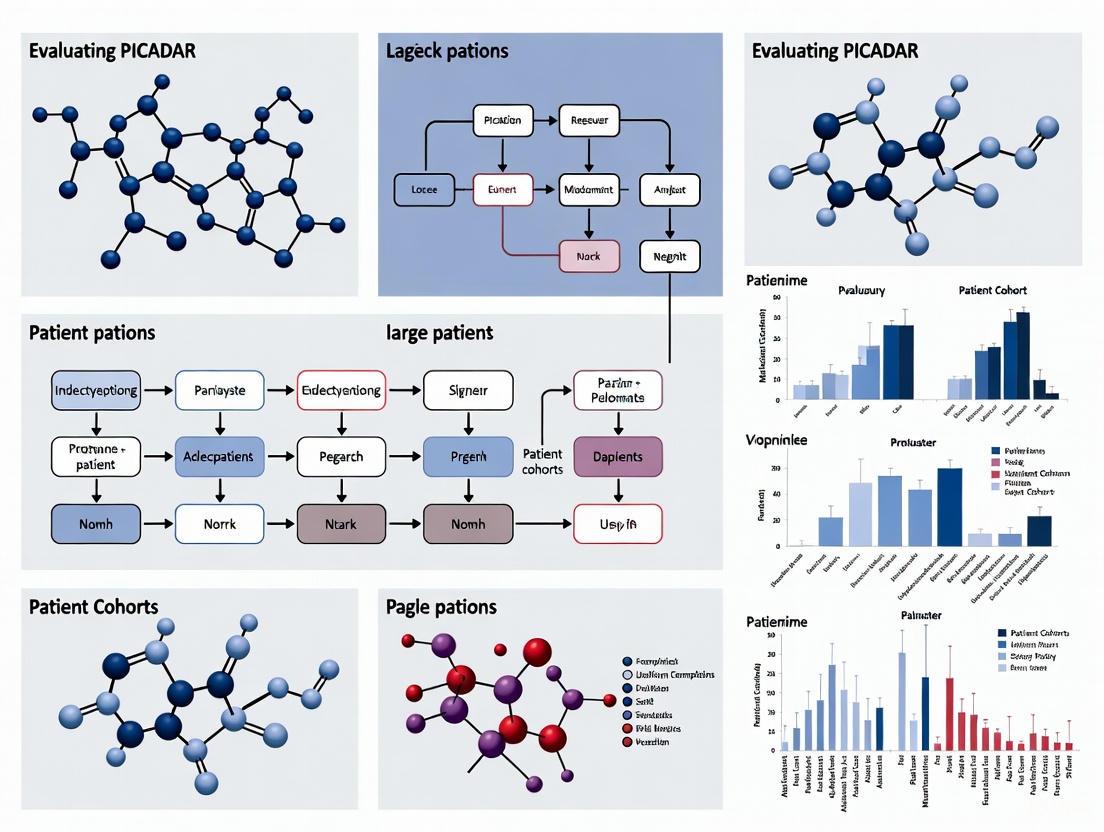
Systematic Evaluation of PICADAR: Diagnostic Accuracy, Limitations, and Future Directions in Large PCD Cohorts
This article provides a comprehensive evaluation of the Primary Ciliary Dyskinesia Rule (PICADAR) predictive tool based on recent large-scale cohort studies.
Systematic Evaluation of PICADAR: Diagnostic Accuracy, Limitations, and Future Directions in Large PCD Cohorts
Abstract
This article provides a comprehensive evaluation of the Primary Ciliary Dyskinesia Rule (PICADAR) predictive tool based on recent large-scale cohort studies. It examines the tool's foundational development, methodological application in clinical settings, and significant limitations revealed in genetically diverse patient populations. The analysis highlights critical sensitivity issues, particularly in patients with situs solitus and normal ciliary ultrastructure, and compares PICADAR's performance against alternative diagnostic tools. Synthesizing evidence from recent multinational studies, this review offers insights for researchers and clinicians on optimizing PCD diagnostic pathways and identifies pressing needs for improved predictive instruments in the era of genetic diagnostics.
PICADAR Origins: Development, Initial Validation, and Core Clinical Parameters
Historical Context and Development of the PICADAR Tool
The PICADAR (PrImary CiliARy DyskinesiA Rule) tool, first introduced in 2016, represents a significant advancement in the diagnostic approach to primary ciliary dyskinesia (PCD), a rare genetic disorder affecting motile cilia. This review examines the historical development, validation studies, and comparative performance of PICADAR against other predictive tools in large patient cohorts. As specialized PCD diagnostic tests require expensive equipment and expert interpretation, PICADAR emerged as a practical clinical prediction rule to identify high-risk patients requiring referral to specialist centers. We synthesize evidence from multiple validation cohorts demonstrating PICADAR's discriminatory power while acknowledging its limitations in specific PCD subpopulations. Within the broader thesis of evaluating PICADAR in large patient cohorts research, this analysis provides researchers and clinicians with comprehensive experimental data and methodological frameworks for implementing this tool in both research and clinical settings.
Primary ciliary dyskinesia (PCD) is a rare, genetically heterogeneous disorder characterized by impaired structure and function of motile cilia, with an estimated prevalence ranging from 1:2,000 to 1:40,000 live births [1] [2]. The clinical manifestations typically include chronic progressive respiratory symptoms such as persistent wet cough, recurrent chest infections leading to bronchiectasis, chronic rhinosinusitis, recurrent otitis media, and laterality defects (situs inversus totalis or heterotaxy) in approximately 50% of patients [1] [2]. Male infertility is common due to sperm flagella abnormalities, and female fertility may also be impaired [1].
Diagnosing PCD presents significant challenges due to the non-specific nature of symptoms, genetic heterogeneity with mutations in over 50 identified genes, and the absence of a single gold standard diagnostic test [3] [4]. Specialized confirmatory tests include nasal nitric oxide (nNO) measurement, high-speed video microscopy analysis (HSVA), transmission electron microscopy (TEM), and genetic testing [2]. These require expensive infrastructure, specialized equipment, and experienced personnel typically available only at specialized centers [5] [1]. This diagnostic complexity often leads to underdiagnosis or delayed diagnosis, particularly in regions with limited healthcare resources [1]. The PICADAR tool was developed specifically to address these challenges by providing a evidence-based method for identifying high-risk patients who warrant referral for specialized testing.
Historical Development and Original Validation of PICADAR
Development and Initial Validation Cohort
The PICADAR tool was developed through a systematic research program and first published in 2016 by Behan et al. [5]. The derivation study analyzed 641 consecutive patients referred to the University Hospital Southampton (UHS) PCD diagnostic center between 2007 and 2013 [5] [1]. Within this cohort, 75 patients (12%) received a definitive PCD diagnosis, while 566 (88%) were negative [1]. The researchers collected data on 27 potential predictor variables readily available in non-specialist settings and used logistic regression analysis to identify the most significant predictive factors [1].
The tool was specifically designed for patients with persistent wet cough and incorporates seven clinically accessible parameters [5] [1]. The developers simplified the statistical model into a practical scoring system by rounding regression coefficients to the nearest integer, creating the PICADAR score [1]. External validation was performed using a sample of 187 patients (93 PCD-positive and 94 PCD-negative) from the Royal Brompton Hospital (RBH), a different PCD diagnostic center [1]. This validation cohort was younger and included more patients from consanguineous backgrounds, reflecting different population characteristics [1].
PICADAR Scoring Parameters and Interpretation
The PICADAR tool evaluates seven clinical parameters, with scoring points assigned as follows:
Table 1: PICADAR Scoring System and Parameters
| Clinical Parameter | Points Awarded |
|---|---|
| Full-term gestation | 2 |
| Neonatal chest symptoms | 1 |
| Neonatal intensive care unit admission | 1 |
| Chronic rhinitis | 1 |
| Ear symptoms | 1 |
| Situs inversus | 2 |
| Congenital cardiac defect | 2 |
The total PICADAR score ranges from 0 to 10 points. The recommended cut-off score of ≥5 points indicates a high probability of PCD, warranting referral for specialized diagnostic testing [5] [1]. In the original derivation study, this cut-off demonstrated a sensitivity of 0.90 and specificity of 0.75 [5]. The area under the curve (AUC) for the receiver operating characteristic (ROC) analysis was 0.91 for internal validation and 0.87 for external validation, indicating good discriminatory power [5] [1].
Diagram 1: PICADAR Clinical Decision Pathway
Experimental Protocols for PICADAR Validation
Original Study Methodology
The original PICADAR development and validation followed a rigorous methodological protocol [1]. Data collection utilized a standardized proforma completed by clinicians during clinical interviews prior to diagnostic testing [1]. The proforma captured demographic information, neonatal history (gestational age, special care admission, respiratory support, neonatal rhinitis or chest symptoms), presence of situs abnormalities, congenital cardiac defects, chronic respiratory symptoms (>3 months), family history of PCD or consanguinity, and adult fertility issues [1]. Data were coded as yes=0, no=1, or missing=99 for analysis [1].
Diagnostic testing followed established UK protocols, with PCD diagnosis confirmed based on typical clinical history plus at least two abnormal diagnostic tests: "hallmark" transmission electron microscopy (TEM) findings, "hallmark" ciliary beat pattern (CBP) on high-speed video microscopy analysis (HSVMA), or nasal nitric oxide (nNO) ≤30 nL·min⁻¹ [1]. In some cases, patients with strong clinical phenotypes (e.g., sibling with PCD, full clinical presentation) were diagnosed based on either hallmark TEM or repeated HSVMA consistent with PCD [1]. CBP was only considered positive if the pattern was typical of PCD rather than secondary ciliary dyskinesia, confirmed either through two brushing biopsies or one biopsy with reanalysis after air-liquid interface culture [1].
Statistical Analysis Framework
The statistical approach for PICADAR development involved multiple stages [1]. Initial univariate analyses used two-tailed parametric (t-test) or nonparametric (Mann-Whitney) tests, chi-squared tests, or Fisher's exact tests as appropriate to compare characteristics of PCD-positive and PCD-negative referrals [1]. Logistic regression analysis with forward step-wise methods identified significant predictors for PCD [1]. Model performance was assessed using receiver operating characteristic (ROC) curve analysis and calculation of the area under the curve (AUC), with discrimination considered moderate for AUC 0.6-0.8 and good for AUC >0.8 [1]. Model calibration was evaluated using the Hosmer-Lemeshow goodness-of-fit test [1]. The final model was simplified into a practical scoring system by rounding regression coefficients to the nearest integer [1].
Performance Comparison with Alternative Predictive Tools
Head-to-Head Comparison Studies
A 2021 study by the University Hospital Motol in Prague provided a direct comparison of PICADAR with two other predictive tools: the Clinical Index (CI) and North America Criteria Defined Clinical Features (NA-CDCF) [3] [6]. This large-scale analysis included 1401 patients with suspected PCD referred to their center, with 67 (4.8%) ultimately diagnosed with PCD [3] [6]. All three tools showed significantly higher scores in PCD patients compared to non-PCD patients (p < 0.001) [3].
Table 2: Comparative Performance of PCD Predictive Tools in Large Cohort Studies
| Predictive Tool | Number of Items | AUC (95% CI) | Key Advantages | Key Limitations |
|---|---|---|---|---|
| PICADAR | 7 parameters + persistent wet cough prerequisite | 0.87 (external validation) [5] | Good discriminant power (AUC >0.85); externally validated | Excludes patients without daily wet cough (7% of PCD population) [7] |
| Clinical Index (CI) | 7 items | Larger than NA-CDCF (p=0.005) [3] | No need for assessment of laterality or cardiac defects; applicable to wider population | Less widely validated than PICADAR |
| NA-CDCF | 4 criteria | No significant difference from PICADAR (p=0.093) [3] | Simple, quick assessment | Lower AUC than CI in direct comparison [3] |
The study found that the AUC for CI was significantly larger than for NA-CDCF (p=0.005), while no significant difference existed between PICADAR and NA-CDCF (p=0.093) [3]. The researchers also noted that PICADAR could not be assessed in 86 (6.1%) patients who lacked chronic wet cough, a prerequisite for using the tool [3]. In contrast, CI did not require assessment of laterality or congenital heart defects, potentially making it more applicable in primary care settings [3].
Recent Evidence on PICADAR Limitations
A 2025 study by Schramm et al. evaluated PICADAR's sensitivity in 269 individuals with genetically confirmed PCD, revealing important limitations [7] [8]. The overall sensitivity was 75% (202/269), significantly lower than in the original validation study [7] [8]. Notably, 18 individuals (7%) with genetically confirmed PCD reported no daily wet cough, automatically ruling out PCD according to PICADAR criteria [7].
The study demonstrated substantial variation in sensitivity based on clinical presentation [7] [8]. PICADAR showed high sensitivity in individuals with laterality defects (95%; median score: 10; IQR 8-11) but significantly lower sensitivity in those with situs solitus (normal organ arrangement) (61%; median score: 6; IQR 4-8; p<0.0001) [7] [8]. Further stratification by ciliary ultrastructure revealed higher sensitivity in individuals with hallmark defects (83%) versus those without (59%, p<0.0001) [7] [8]. These findings indicate that PICADAR has limited sensitivity, particularly for PCD patients without laterality defects or hallmark ultrastructural defects [7] [8].
Enhanced Diagnostic Accuracy with Supplemental Testing
Integration with Nasal Nitric Oxide Measurement
The 2021 Motol study also investigated the complementary role of nasal nitric oxide (nNO) measurement when combined with various predictive tools [3] [6]. nNO was measured in 569 patients older than 3 years using electrochemical analyzers (Niox Mino or Niox Vero) with a passive sampling flow rate of 5 mL·s⁻¹ [3]. The results demonstrated that nNO further improved the predictive power of all three tools (CI, PICADAR, and NA-CDCF) [3] [6].
Diagram 2: Comprehensive PCD Diagnostic Pathway Integrating PICADAR
The PCD Diagnostic Toolkit: Research Reagent Solutions
Table 3: Essential Research Reagents and Materials for PCD Diagnostic Testing
| Reagent/Equipment | Primary Function | Application in PCD Diagnosis |
|---|---|---|
| Electrochemical nNO analyzer (Niox Mino/Vero) | Measures nasal nitric oxide concentration | Screening tool; low nNO (<77 ppb) suggests PCD [3] |
| High-speed video microscopy system (Keyence VW-6000/5000) | Records ciliary beat frequency and pattern | Identifies abnormal ciliary movement [3] |
| Transmission electron microscope | Visualizes ciliary ultrastructure | Detects hallmark defects in ciliary axoneme [4] |
| Nasal brushing biopsy tools | Obtains respiratory epithelial cells | Source material for HSVM and TEM [3] |
| Air-liquid interface culture materials | Promotes ciliary differentiation and recovery | Reduces secondary dyskinesia in cell cultures [1] |
| Next-generation sequencing panels (39 PCD genes) | Identifies pathogenic mutations | Genetic confirmation of PCD [3] |
| MLPA probemix (P238/P237 for DNAH5/DNAI1) | Detects large genomic rearrangements | Identifies copy number variations in major PCD genes [3] |
Discussion and Future Directions
The development of PICADAR in 2016 marked a significant advancement in the systematic approach to PCD diagnosis, addressing the critical need for evidence-based referral guidance in a rare disease with non-specific symptoms [5] [1]. The tool's good discriminant power (AUC >0.85 in validation studies) and practical design have led to its incorporation into European Respiratory Society guidelines [2]. However, evidence from large cohort studies reveals substantial limitations, particularly its dependency on daily wet cough and laterality defects for optimal sensitivity [3] [7] [8].
The finding that PICADAR misses approximately 25% of genetically confirmed PCD cases, including 7% without daily wet cough and nearly 40% of those without hallmark ultrastructural defects, highlights critical gaps in its applicability across the PCD spectrum [7] [8]. These limitations underscore the importance of using PICADAR as part of a comprehensive diagnostic approach rather than as a standalone decision tool [7] [2].
Future research should focus on refining predictive tools to better identify PCD patients with situs solitus and normal ultrastructure, potentially incorporating genetic data and novel biomarkers [7] [8]. The integration of multiple screening methods, including nNO measurement alongside clinical prediction tools, appears promising for enhancing overall diagnostic accuracy [3] [6]. As our understanding of PCD genetics and phenotype-genotype correlations advances, next-generation predictive tools will likely incorporate genetic risk scores and expanded clinical parameters to improve sensitivity across all PCD subtypes.
For researchers and clinicians, PICADAR remains a valuable component of the PCD diagnostic toolkit but should be applied with awareness of its limitations and in conjunction with other screening modalities, particularly for patients with atypical presentations or without classic laterality defects.
Primary Ciliary Dyskinesia (PCD) is a rare, genetically heterogeneous disorder characterized by impaired mucociliary clearance due to defects in motile cilia function. Diagnosis remains challenging due to non-specific symptoms that overlap with other respiratory conditions, the technical complexity of definitive testing, and the lack of a single gold standard test [1] [3] [9]. Specialized confirmatory tests—including nasal nitric oxide (nNO) measurement, high-speed video microscopy analysis (HSVA), transmission electron microscopy (TEM), and genetic testing—require expensive equipment and expertise concentrated in specialized centers [1] [10]. This diagnostic bottleneck necessitates reliable, evidence-based tools to identify high-risk patients for referral.
The PrImary CiliARy DyskinesiA Rule (PICADAR) was developed to address this need. It is a clinical prediction rule that uses seven simple parameters obtainable from patient history to estimate the probability of PCD before specialized testing [1] [5]. This review evaluates PICADAR's seven predictive parameters, its performance against other tools in large patient cohorts, and its role within the broader PCD diagnostic workflow.
The Seven Predictive Parameters of PICADAR
PICADAR is designed for patients with a persistent wet cough and is based on seven clinical parameters [1]. The total score determines the probability of PCD and guides referral decisions.
Table 1: The Seven Predictive Parameters of the PICADAR Tool
| Parameter Category | Specific Predictive Parameter | Score Contribution |
|---|---|---|
| Neonatal History | Full-term gestation | 2 points |
| Neonatal chest symptoms | 2 points | |
| Admission to neonatal intensive care unit (NICU) | 1 point | |
| Chronic Symptoms | Chronic rhinitis | 1 point |
| Chronic ear symptoms (e.g., otitis media, hearing loss) | 1 point | |
| Laterality & Defects | Situs inversus | 2 points |
| Congenital cardiac defect | 1 point |
Pathophysiological Basis of the Parameters
The parameters are not arbitrary; they reflect the fundamental roles of motile cilia in early development and ongoing health:
- Situs Abnormalities: During embryogenesis, motile cilia at the embryonic node establish left-right body asymmetry. Dysfunctional nodal cilia can result in random lateralization, leading to situs inversus (found in approximately 50% of PCD patients) or heterotaxy, which can be associated with congenital cardiac defects [1] [3]. The presence of situs inversus is a strong predictor, contributing 2 points to the PICADAR score [1].
- Neonatal Respiratory Symptoms: The transition to air breathing at birth requires effective clearance of fetal lung fluid by respiratory cilia. Their dysfunction can cause neonatal respiratory distress in term neonates, manifesting as tachypnea, cough, or even respiratory failure requiring NICU admission [1] [10].
- Chronic Otosinopulmonary Manifestations: Throughout life, motile cilia clear mucus and debris from the airways. Chronic dysfunction leads to persistent wet cough, chronic rhinitis, and recurrent otitis media with effusion due to impaired clearance in the middle ear and sinuses [1] [3].
Performance Evaluation in Large Patient Cohorts
Since its development, PICADAR has been validated in multiple international populations and compared to other predictive tools.
Original Validation and External Performance
In the original 2016 derivation study, PICADAR demonstrated strong predictive power. The tool was developed on 641 consecutive referrals, of which 75 (12%) were diagnosed with PCD [1] [5].
Table 2: Performance Metrics of PICADAR from Validation Studies
| Study Cohort | Area Under the Curve (AUC) | Sensitivity (at score ≥5) | Specificity (at score ≥5) |
|---|---|---|---|
| Original Derivation Cohort (n=641) | 0.91 | 0.90 | 0.75 [1] |
| External Validation Cohort (n=187) | 0.87 | Not specified | Not specified [1] |
| Czech Cohort (2021) (n=1401) | Reported as lower than Clinical Index | Not specified | Not specified [3] [6] |
The area under the receiver operating characteristic (ROC) curve was 0.91 upon internal validation and 0.87 upon external validation in a second UK center, indicating good to excellent discriminative ability [1].
Comparison with Alternative Predictive Tools
Other tools have been developed for the same purpose, notably the Clinical Index (CI) and the North American Criteria Defined Clinical Features (NA-CDCF).
A large 2021 Czech study compared all three tools in 1,401 patients suspected of PCD. The study found that while all three scores were significantly higher in the PCD group, the area under the curve (AUC) for CI was statistically larger than for NA-CDCF, while the AUC for PICADAR was not significantly different from NA-CDCF [3] [6]. The study also highlighted a practical limitation: PICADAR could not be calculated for 6.1% of patients (n=86) because they did not have the mandatory symptom of a chronic wet cough, whereas CI did not have this limitation [3] [6].
Limitations and Geographic Variability in Performance
Recent research underscores important limitations in PICADAR's sensitivity, particularly in specific patient subgroups.
A 2025 genetic study by Omran et al. evaluated PICADAR in 269 individuals with genetically confirmed PCD. It found an overall sensitivity of 75%, meaning a quarter of true PCD patients would be missed using the recommended cutoff [8]. Performance was significantly worse in patients without laterality defects (sensitivity of 61%) and in those without hallmark ultrastructural defects on TEM (sensitivity of 59%) [8]. Furthermore, 7% of genetically confirmed PCD patients reported no daily wet cough and would have been automatically ruled out by PICADAR's initial gatekeeping question [8].
Geographic and genetic differences also impact performance. A 2022 Japanese study reported that only 25% of PCD patients had situs inversus, a stark contrast to the ~50% typically cited in Western populations [11]. This is attributed to differences in prevalent causative genes, indicating that PICADAR, which heavily weights situs inversus, may be less effective in populations where laterality defects are less common [11].
Experimental Protocols for PCD Diagnostic Workflow
The diagnostic pathway for PCD is multi-staged, with PICADAR acting as an initial risk-stratification tool. The following workflow integrates clinical prediction with advanced confirmatory testing.
Diagram 1: Integrated PCD Diagnostic Workflow. PICADAR serves as an initial screening tool to identify high-risk patients for referral to a specialized center.
Confirmatory Diagnostic Techniques
Following a positive PICADAR screen, diagnosis is confirmed using a combination of specialized tests in a tertiary center [10].
- Nasal Nitric Oxide (nNO) Measurement: nNO is a well-established screening test, as levels are characteristically very low in most PCD patients. It is measured using an electrochemical analyzer during tidal breathing or oral exhalation against resistance [3] [6].
- High-Speed Video Microscopy Analysis (HSVA): Ciliary beat frequency (CBF) and pattern (CBP) are analyzed from nasal brushing samples. A pathological, dyskinetic, or immotile pattern is indicative of PCD. To rule out secondary dyskinesia due to infection, testing is repeated after 4-6 weeks or after antibiotic treatment [3] [10] [6].
- Transmission Electron Microscopy (TEM): This technique visualizes the ciliary ultrastructure. Hallmark defects include the absence of outer dynein arms (ODA), inner dynein arms (IDA), or both. However, its sensitivity is limited, as approximately 30% of PCD cases have normal ultrastructure [12] [10] [4].
- Genetic Testing: Next-generation sequencing (NGS) using targeted gene panels for over 39 known PCD genes is standard. A definitive diagnosis requires identifying biallelic pathogenic mutations in a PCD-associated gene [10] [6].
- Air-Liquid Interface (ALI) Culture: For inconclusive cases, nasal epithelial cells can be cultured at an ALI to re-differentiate ciliated cells. This allows for functional and structural analysis in a controlled environment, free from inflammatory damage, and is critical for confirming diagnoses in complex cases [12].
The Scientist's Toolkit: Essential Reagents and Materials
The following table details key reagents and materials essential for conducting the advanced confirmatory tests described in the diagnostic workflow.
Table 3: Research Reagent Solutions for PCD Diagnostic Testing
| Research Reagent / Material | Primary Function in PCD Diagnostics |
|---|---|
| Nasal Nitric Oxide Analyzer (e.g., Niox Vero/Mino) | Measures nasal nitric oxide concentration; a key non-invasive screening test with very low values being highly suggestive of PCD [3] [6]. |
| High-Speed Video Microscope | Captures ciliary beat frequency and pattern from fresh nasal epithelial brushings for functional analysis [3] [6]. |
| Transmission Electron Microscope | Visualizes and analyzes the ultrastructure of ciliary axonemes (e.g., dynein arm defects) from biopsy samples [12] [10]. |
| Next-Generation Sequencing (NGS) Gene Panels | Identifies pathogenic mutations in over 50 known PCD-associated genes for molecular confirmation [10] [6]. |
| Air-Liquid Interface (ALI) Culture Media | Supports the differentiation of basal respiratory epithelial cells into ciliated cells in vitro for functional and structural testing without confounding secondary effects [12]. |
| Immunofluorescence Antibodies | Targets specific ciliary proteins (e.g., CFAP300, DNAH5) to visualize their presence, absence, or mislocalization in ciliary axonemes [12]. |
The PICADAR tool represents a significant advancement in the initial identification of patients at high risk for PCD. Its strength lies in leveraging easily obtainable clinical data, providing a practical and cost-effective method to streamline referrals to specialized centers. The seven parameters—situs inversus, congenital cardiac defect, full-term gestation, neonatal chest symptoms, NICU admission, chronic rhinitis, and ear symptoms—are physiologically grounded in the pathobiology of PCD [1].
However, evidence from large cohort studies indicates that PICADAR should be applied with a clear understanding of its limitations. Its suboptimal sensitivity, particularly in patients with situs solitus (61%) or those without hallmark ultrastructural defects (59%), means it cannot be used as a standalone rule-out tool [8]. Furthermore, its performance varies across populations and is dependent on the presence of a daily wet cough, which is not universal among PCD patients [8] [3] [6].
In conclusion, PICADAR is a valuable component of the PCD diagnostic arsenal. For researchers and clinicians, it serves as a standardized initial assessment tool that can enhance recruitment for studies and promote earlier diagnosis. Its integration with objective tests like nNO and genetic testing, as part of a multimodal diagnostic protocol, represents the most effective pathway for accurately identifying this complex and heterogeneous disease. Future work should focus on developing and validating next-generation predictive tools that incorporate genetic and biomarker data to improve sensitivity, especially in atypical and underrepresented patient populations.
The rigorous evaluation of a clinical predictive model is paramount to establishing its diagnostic utility and ensuring its reliability when applied to new patient populations. Performance metrics such as Sensitivity, Specificity, and the Area Under the Receiver Operating Characteristic Curve (AUC) provide a standardized framework for this assessment, particularly through their analysis in both derivation and validation cohorts. The derivation cohort is the initial patient group used to create the model and estimate its initial performance. The validation cohort is a separate, independent patient group used to test the model's performance and ensure its generalizability beyond the original data. This guide objectively examines these core metrics within the context of evaluating the PICADAR (PrImary CiliARy DyskinesiA Rule) score, a diagnostic tool for Primary Ciliary Dyskinesia (PCD), and details the experimental protocols used for its assessment.
Core Performance Metrics Explained
In the context of a diagnostic tool like PICADAR, which aims to identify patients who should be referred for definitive PCD testing, the following metrics are crucial [13]:
- Sensitivity: The proportion of patients with the disease (PCD) who are correctly identified by a positive test result. A high sensitivity is critical for a screening tool to ensure true cases are not missed.
- Specificity: The proportion of patients without the disease (non-PCD) who are correctly identified by a negative test result. A high specificity helps to avoid unnecessary referrals and testing in patients without the disease.
- Area Under the Curve (AUC): The AUC measures the overall ability of the model to discriminate between patients with and without the disease. An AUC of 1.0 represents perfect discrimination, while 0.5 represents a model with no discriminative ability, equivalent to random chance. An AUC >0.8 is generally considered to indicate good model performance [1] [14].
The relationship between sensitivity and specificity at various score cut-offs is visualized through the Receiver Operating Characteristic (ROC) curve. The AUC is thus a single, powerful metric summarizing the ROC curve's information.
Performance of PICADAR in Derivation and Validation
The PICADAR tool was developed and evaluated using a multi-cohort study design. Its performance in distinguishing PCD from non-PCD patients in both derivation and validation cohorts is summarized in the table below.
Table 1: Performance Metrics for PICADAR in Derivation and Validation Cohorts
| Cohort | Number of Patients (PCD+/Total) | Sensitivity | Specificity | AUC (95% CI) | Key Cut-off Score |
|---|---|---|---|---|---|
| Derivation [1] | 75 / 641 | 0.90 | 0.75 | 0.91 (Not specified) | 5 points |
| Validation [1] | 93 / 187 | 0.86 | 0.73 | 0.87 (Not specified) | 5 points |
| Independent Validation [3] | 67 / 1401 | Not specified | Not specified | 0.87 (Not specified) | Not specified |
Interpretation of Performance Data
The data demonstrates that PICADAR is a robust predictive tool. The model showed good discriminatory power in its initial derivation, with an AUC of 0.91 [1]. This performance was maintained in an external validation cohort from a different diagnostic center, which yielded an AUC of 0.87 [1]. A subsequent independent study further confirmed this finding, reporting an identical AUC of 0.87 for PICADAR [3]. The consistency in the AUC values between the derivation and validation phases indicates that the model generalizes well and is not overly fitted to the original dataset. At the recommended cut-off score of 5 points, the tool achieves a balanced combination of high sensitivity and reasonable specificity, making it suitable for its intended role as a screening instrument to identify patients for further testing [1].
Experimental Protocols for Model Evaluation
The evaluation of PICADAR's performance followed a structured and methodical process, which can serve as a template for validating similar diagnostic tools.
Cohort Design and Patient Selection
The initial study employed a two-cohort design [1]:
- Derivation Cohort: 641 consecutive patients referred for PCD testing at the University Hospital Southampton (UHS). A definitive diagnostic outcome (PCD-positive or PCD-negative) was the reference standard.
- Validation Cohort: 187 patients referred to the Royal Brompton Hospital (RBH), selected to include a similar number of positive and negative cases to robustly test the model's performance in a different population and setting.
Reference Standard for PCD Diagnosis
A key component of the protocol was establishing a definitive diagnosis against which PICADAR could be compared. The diagnostic criteria were based on a combination of clinical history and specialized tests [1]:
- A positive PCD diagnosis was typically confirmed by a characteristic clinical history plus at least two abnormal diagnostic tests.
- The definitive tests included: "hallmark" transmission electron microscopy (TEM) findings, "hallmark" ciliary beat pattern (CBP) observed via high-speed video microscopy analysis (HSVMA), or low nasal nitric oxide (nNO) levels (≤30 nL·min⁻¹).
Data Collection and Statistical Analysis
The following workflow outlines the key steps in the model derivation and validation process:
Diagram 1: Model Derivation and Validation Workflow. This diagram illustrates the sequential process of developing the PICADAR score and evaluating its performance in independent cohorts.
The statistical analysis involved [1]:
- Model Development: Logistic regression analysis was performed on the derivation cohort to identify significant predictors from 27 potential clinical variables. The regression coefficients of the final seven predictors were rounded to integers to create the practical PICADAR score.
- Performance Testing: The model's discriminative ability was tested using Receiver Operating Characteristic (ROC) curve analysis, and the AUC was calculated for both the derivation and validation cohorts. The Hosmer-Lemeshow goodness-of-fit test was used to assess model calibration.
The Scientist's Toolkit: Research Reagents and Essential Materials
The research and diagnostic methods cited in the evaluation of PICADAR rely on several key reagents and technologies.
Table 2: Essential Research Reagents and Materials for PCD Diagnostic Workup
| Item / Technology | Function in PCD Diagnosis & Research |
|---|---|
| High-Speed Video Microscopy (HSVMA) | Visualizes and analyzes ciliary beat frequency and pattern from nasal brushing biopsies to identify characteristic dyskinesia [1] [3]. |
| Transmission Electron Microscopy (TEM) | Identifies ultrastructural defects in ciliary axonemes (e.g., absent dynein arms) from nasal or bronchial biopsies, providing a hallmark diagnostic finding [1] [3]. |
| Nasal Nitric Oxide (nNO) Measurement | Serves as a screening test; low nNO levels are strongly associated with PCD. Measured using an electrochemical analyzer (e.g., Niox Vero) [3]. |
| Genetic Analysis (Next-Generation Sequencing) | Identifies disease-causing mutations in over 50 known PCD-related genes, providing definitive genetic confirmation [3]. |
| Cell Culture (Air-Liquid Interface) | Used to differentiate ciliated epithelial cells and rule out secondary ciliary dyskinesia by re-analyzing ciliary function after cell culture [1]. |
Primary Ciliary Dyskinesia (PCD) is a rare, genetically heterogeneous disorder characterized by abnormal ciliary function, leading to chronic oto-sino-pulmonary disease, laterality defects, and reduced fertility [1]. The diagnostic pathway for PCD is complex, requiring specialized testing available only at tertiary referral centers, including measurement of nasal nitric oxide (nNO), high-speed video microscopy analysis (HSVM), transmission electron microscopy (TEM), immunofluorescence (IF), and genetic testing [15] [16]. No single test possesses perfect sensitivity and specificity, necessitating a multi-test diagnostic approach [16]. This diagnostic challenge has driven the development and validation of clinical predictive tools to identify high-risk patients who should be referred for specialized testing. Among these tools, the PrImary CiliARy DyskinesiA Rule (PICADAR) has gained prominence and has been incorporated into international guidelines [15]. This review evaluates PICADAR's integration into clinical guidelines, its performance against alternative tools, and its utility in large patient cohorts within the context of evolving international diagnostic standards.
PICADAR: Development and Integration into International Guidelines
Tool Development and Original Validation
PICADAR was developed to provide a practical, evidence-based clinical prediction rule to identify symptomatic patients requiring referral for PCD diagnostic testing [1]. Derived from a prospective cohort of 641 consecutive referrals to a UK diagnostic center, it was designed for use by general respiratory and ENT specialists prior to specialized testing. The tool incorporates seven readily available clinical parameters: full-term gestation, neonatal chest symptoms, neonatal intensive care unit admission, chronic rhinitis, ear symptoms, situs inversus, and congenital cardiac defects [1] [17].
In its original validation, PICADAR demonstrated strong predictive performance. For a cut-off score of 5 points, it achieved a sensitivity of 0.90 and specificity of 0.75, with an area under the receiver operating characteristic curve (AUC) of 0.91 upon internal validation and 0.87 upon external validation in a separate patient cohort [1]. The overall accuracy for identifying PCD was 90% [17]. The tool's performance characteristics supported its adoption as a screening instrument to guide referrals to specialized PCD centers.
Formal Endorsement in International Guidelines
The 2025 joint guidelines from the European Respiratory Society (ERS) and American Thoracic Society (ATS) represent a significant unification of previously separate diagnostic recommendations and formally acknowledge the role of clinical prediction tools [15]. These evidence-based guidelines, developed using GRADE methodology, strongly recommend the use of multiple adjunct tests (HSVM, IF, nNO) alongside reference tests (TEM and/or genetics) for PCD diagnosis, emphasizing that no single test is sufficient to confirm or exclude the disease [15] [16].
Within this diagnostic framework, the guidelines explicitly recommend using PICADAR to identify patients who should be referred for diagnostic testing. As Dr. Amjad Horani stated during the ERS Congress presentation, "One can use the PICADAR score or the ATS Leigh's criteria to help decide which patients to send for diagnosis" [15]. This endorsement establishes PICADAR as a pre-referral screening tool within a comprehensive diagnostic algorithm that emphasizes evaluation at specialized centers experienced in PCD diagnosis.
Performance Evaluation in Large Patient Cohorts
Comparative Performance Against Alternative Predictive Tools
Several studies have evaluated PICADAR's performance against other predictive instruments in large, unselected cohorts referred for PCD testing. The most comprehensive comparison comes from a 2021 study of 1,401 patients with suspected PCD, which directly compared PICADAR with the Clinical Index (CI) and North American Criteria Defined Clinical Features (NA-CDCF) [3] [6].
Table 1: Comparative Performance of PCD Predictive Tools in a Cohort of 1,401 Patients
| Predictive Tool | Area Under ROC Curve (AUC) | Key Advantages | Key Limitations |
|---|---|---|---|
| PICADAR | 0.87 (95% CI not provided) | Strong predictive power when applicable; externally validated | Not assessable in patients without chronic wet cough (6.1% of cohort) |
| Clinical Index (CI) | 0.92 (95% CI not provided) | No requirement for assessment of laterality or cardiac defects; applicable to broader population | Less widely adopted internationally |
| NA-CDCF | 0.81 (95% CI not provided) | Simple, four-item criteria | Lower AUC compared to CI (p=0.005) |
The study found that while all three tools effectively differentiated PCD from non-PCD patients (p<0.001 for all), CI demonstrated a statistically larger AUC compared to NA-CDCF (p=0.005), though no significant difference existed between PICADAR and NA-CDCF (p=0.093) [3] [6]. A significant finding was that PICADAR could not be assessed in 86 patients (6.1% of the cohort) who lacked chronic wet cough, a mandatory starting criterion for the tool [3]. This limitation highlights a potential gap in PICADAR's applicability for atypical PCD presentations.
Limitations in Specific Patient Populations and Settings
Recent research has revealed important limitations in PICADAR's sensitivity, particularly in specific genetic and ethnic subpopulations. A 2025 study of 269 individuals with genetically confirmed PCD found an overall sensitivity of only 75%, significantly lower than in the original derivation study [8]. The sensitivity varied substantially based on clinical and ultrastructural features:
- 95% sensitivity in patients with laterality defects (median score: 10)
- 61% sensitivity in patients with situs solitus (normal organ arrangement, median score: 6)
- 83% sensitivity in patients with hallmark ultrastructural defects
- 59% sensitivity in patients without hallmark ultrastructural defects [8]
These findings indicate that PICADAR performs best in patients with classic PCD presentations including laterality defects and clear ultrastructural abnormalities, while potentially missing a significant proportion of patients with normal arrangement or subtle ciliary defects.
Ethnic variations in PCD presentation also impact PICADAR's performance. A Japanese study of 67 PCD patients found that only 25% had situs inversus, compared to the approximately 50% typically reported in other populations [11]. This difference reflects variations in major disease-causing genes across ethnic groups and means that PICADAR, which assigns substantial points for situs inversus, may be less effective in certain populations [11].
Methodological Approaches in PICADAR Research
Experimental Protocols and Diagnostic Standards
Studies evaluating PICADAR and other predictive tools have employed rigorous methodologies in large patient cohorts. The diagnostic protocols typically adhere to international standards, incorporating multiple complementary tests to establish a definitive PCD diagnosis [3] [6].
Table 2: Key Diagnostic Methods Used in PCD Predictive Tool Studies
| Method | Application in PCD Diagnosis | Role in Study Protocols |
|---|---|---|
| High-Speed Video Microscopy (HSVM) | Analysis of ciliary beat pattern and frequency | Primary screening tool; repeated after cell culture to exclude secondary dyskinesia |
| Nasal Nitric Oxide (nNO) | Measurement of nasal NO concentration (low in PCD) | Screening measure in patients >3 years; improves predictive power of clinical tools |
| Transmission Electron Microscopy (TEM) | Ultrastructural analysis of ciliary defects | Reference standard; identifies hallmark defects (83% detection rate in PCD) [4] |
| Genetic Testing | Identification of pathogenic variants in >50 PCD genes | Confirmatory testing; increasingly important for heterogeneous cases |
The typical diagnostic workflow begins with clinical assessment using predictive tools, followed by nNO measurement when possible, HSVM analysis, and confirmation with TEM and/or genetic testing [3] [6]. This multi-test approach is essential given that the estimated TEM detection rate among PCD patients is 83%, meaning approximately 17% of PCD cases have normal ultrastructure and require other methods for diagnosis [4].
Enhancing Predictive Value with Adjunct Testing
Research in large cohorts has demonstrated that combining clinical prediction tools with objective testing enhances diagnostic accuracy. The 2021 study showed that incorporating nNO measurement significantly improved the predictive power of all three clinical tools (CI, PICADAR, and NA-CDCF) [3] [6]. This finding supports the stepped diagnostic approach recommended in the ERS/ATS guidelines, where clinical prediction tools serve as initial screening instruments, followed by a combination of specialized tests for definitive diagnosis [15] [16].
The Scientist's Toolkit: Essential Reagents and Materials
Table 3: Key Research Reagent Solutions for PCD Diagnostic Studies
| Reagent/Material | Specific Examples | Function in PCD Research |
|---|---|---|
| nNO Analyzers | Niox Mino (Aerocrine AB); Niox Vero (Circassia) | Measure nasal nitric oxide concentration using electrochemical detection; standardized protocols with tidal breathing or velum closure |
| HSVM Systems | Keyence Motion Analyzer Microscope VW-6000/5000 | Visualize and quantify ciliary beat frequency and pattern; essential for identifying characteristic dyskinetic patterns |
| TEM Equipment | Standard electron microscopy systems with specialized preparation protocols | Analyze ciliary ultrastructure; identify hallmark defects (outer/inner dynein arm, radial spoke, etc.) |
| Genetic Testing Panels | Next-generation sequencing panels for PCD genes (e.g., 39-gene panel); MLPA for DNAH5/DNAI1 | Identify pathogenic variants; crucial for diagnosis in cases with normal ultrastructure or atypical presentations |
| Cell Culture Media | Air-liquid interface culture systems | Culture ciliated epithelial cells to exclude secondary dyskinesia and enable repeated HSVM analysis |
PICADAR represents an important development in the standardized approach to PCD diagnosis, with formal endorsement in international guidelines reflecting its utility as a clinical prediction tool. Evidence from large patient cohorts demonstrates that PICADAR provides good diagnostic accuracy, particularly when combined with adjunct tests like nNO. However, its limitations in specific populations—including patients without chronic wet cough, those with situs solitus, and certain ethnic groups—highlight the need for careful clinical judgment and awareness of its variable sensitivity. The integration of PICADAR into the ERS/ATS guidelines establishes a structured diagnostic pathway that begins with clinical prediction and progresses through specialized testing. Future research should focus on refining predictive tools to better capture the full phenotypic spectrum of PCD, particularly cases with normal ultrastructure or atypical presentations, to ensure equitable and accurate diagnosis across all patient populations.
Implementing PICADAR: Scoring Protocols, Patient Selection, and Data Collection Frameworks
The Primary Ciliary Dyskinesia Rule (PICADAR) is a diagnostic predictive tool recommended by the European Respiratory Society (ERS) to estimate the probability of primary ciliary dyskinesia (PCD) in patients [7]. PCD is a rare, inherited disorder characterized by impaired mucociliary clearance leading to recurrent respiratory infections, chronic rhinosinusitis, otitis media, bronchiectasis, and laterality defects such as situs inversus [18]. The PICADAR tool employs a scoring system based on specific clinical features, with its initial and most critical question screening for the presence of daily wet cough [7]. This first question serves as a gatekeeper in the diagnostic pathway, as individuals without daily wet cough are automatically ruled negative for PCD according to the tool's standard application. This structured application protocol examines the critical role of this first question through recent large-cohort validation data, revealing significant limitations that impact diagnostic sensitivity, particularly in key patient subgroups.
Experimental Validation in Large Patient Cohorts
Study Methodology and Participant Recruitment
A recent multicenter study evaluated PICADAR's performance in a genetically confirmed PCD cohort to assess its real-world diagnostic accuracy [7]. The research followed a rigorous methodological approach:
- Study Population: 269 individuals with genetically confirmed PCD diagnosis
- Study Design: Retrospective analysis of prospectively collected data
- PICADAR Application: Researchers applied the PICADAR tool according to standard protocols, beginning with the critical first question about daily wet cough
- Data Analysis: Calculated overall sensitivity and performed subgroup analyses based on:
- Presence or absence of laterality defects (situs inversus vs. situs solitus)
- Association with hallmark ultrastructural defects on transmission electron microscopy (TEM)
- Statistical Methods: Determined median scores with interquartile ranges (IQR) and compared sensitivity between subgroups using appropriate statistical tests
This comprehensive evaluation aimed to validate PICADAR's performance in a genetically characterized population, providing the highest level of diagnostic certainty for benchmarking the predictive tool.
Quantitative Results: Sensitivity Analysis
The large-cohort validation revealed significant limitations in PICADAR's sensitivity, largely attributable to the initial daily wet cough question:
Table 1: Overall PICADAR Performance in Genetically Confirmed PCD Cohort
| Metric | Value | Implication |
|---|---|---|
| Total PCD Patients | 269 | Genetically confirmed reference standard |
| Failed First Question | 18 (7%) | No daily wet cough, ruled out by PICADAR |
| Median PICADAR Score | 7 (IQR: 5-9) | Moderate overall score distribution |
| Overall Sensitivity | 75% (202/269) | 1 in 4 PCD patients missed |
Table 2: Subgroup Analysis of PICADAR Sensitivity
| Patient Subgroup | Sensitivity | Median Score | Statistical Significance |
|---|---|---|---|
| Laterality Defects | 95% | 10 (IQR: 8-11) | p<0.0001 |
| Situs Solitus (normal arrangement) | 61% | 6 (IQR: 4-8) | |
| Hallmark Ultrastructural Defects | 83% | Not reported | p<0.0001 |
| Normal Ultrastructure | 59% | Not reported |
The data demonstrates that PICADAR performs substantially worse in patients with situs solitus (normal organ arrangement) and those without hallmark ultrastructural defects on TEM. These findings have profound implications for using PICADAR as a standalone screening tool in general populations where these subtypes may be more prevalent.
Diagnostic Pathways and PICADAR's Role
The diagnostic pathway for PCD requires a multifaceted approach due to the absence of a single gold standard test with perfect sensitivity and specificity [18]. PICADAR represents just one component in a comprehensive diagnostic strategy that should incorporate multiple complementary techniques:
- Nasal Nitric Oxide (nNO) Measurement: Typically reduced in PCD but requires specialized equipment
- High-Speed Video Microscopy Analysis (HSVA): Evaluates ciliary beat pattern and frequency
- Transmission Electron Microscopy (TEM): Identifies ultrastructural defects in cilia (83% detection rate in confirmed PCD) [4]
- Genetic Testing: Identifies mutations in over 50 known PCD-associated genes [18]
The limited sensitivity of PICADAR, particularly its dependence on daily wet cough as an entry criterion, underscores the necessity for a multimodal diagnostic approach that doesn't rely solely on this predictive rule.
Comparative Analysis of PCD Diagnostic Methods
Table 3: Performance Characteristics of PCD Diagnostic Modalities
| Diagnostic Method | Sensitivity/Success Rate | Key Limitations | Clinical Utility |
|---|---|---|---|
| PICADAR (Overall) | 75% | Highly dependent on daily wet cough; poor for situs solitus | Initial screening |
| PICADAR (Situs Solitus) | 61% | Misses nearly 40% of cases | Limited in patients without laterality defects |
| Transmission Electron Microscopy | 83% (66-90% range) | Misses PCD with normal ultrastructure [4] | Structural assessment |
| Genetic Testing | >90% for known genes | 40-50+ genes involved; cost and interpretation challenges [18] | Molecular confirmation |
| Nasal Nitric Oxide | ~90% in some studies | Requires specialized equipment and patient cooperation | Functional screening |
The comparative analysis reveals that while PICADAR offers a convenient clinical scoring system, its reliance on the daily wet cough criterion creates a significant vulnerability in diagnostic sensitivity compared to more objective testing methodologies.
Research Reagent Solutions for PCD Investigation
Table 4: Essential Research Materials and Methods for PCD Diagnostic Studies
| Research Tool | Function/Application | Technical Considerations |
|---|---|---|
| Genetic Sequencing Panels | Identification of mutations in 50+ PCD-associated genes | Must include both common (DNAH5, DNAI1) and rare genes; whole exome for novel discoveries |
| Transmission Electron Microscopy | Ultrastructural analysis of ciliary components | Requires specialized expertise; detects ~83% of defects [4] |
| High-Speed Video Microscopy | Ciliary beat pattern and frequency analysis | Specialized equipment needed; can detect functional abnormalities without structural defects |
| Nasal Nitric Oxide Analyzers | Measurement of nNO levels as screening tool | Standardized protocols essential for comparability between centers |
| Immunofluorescence Assays | Protein localization in ciliary apparatus | Requires specific antibodies for PCD-associated proteins |
| Digital Cough Monitors | Objective cough frequency measurement | Tools like CoughPro provide quantitative data on cough frequency [19] |
The structured application of PICADAR, beginning with its critical first question on daily wet cough, demonstrates substantial limitations in comprehensive PCD case identification. Recent validation in a genetically confirmed cohort reveals that approximately 7% of confirmed PCD patients do not report daily wet cough and would be automatically excluded from further evaluation by standard PICADAR application [7]. The tool shows particularly poor sensitivity in key patient subgroups, including those with situs solitus (61%) and normal ciliary ultrastructure (59%). These findings necessitate a reevaluation of PICADAR's role as a primary screening tool, especially in populations where these subtypes may be more prevalent. Future diagnostic algorithms should incorporate complementary screening methods and consider modified approaches that don't exclusively rely on the daily wet cough criterion to avoid missing a significant proportion of PCD patients.
Primary ciliary dyskinesia (PCD) is a rare genetic disorder characterized by impaired structure and function of motile cilia, leading to chronic respiratory tract symptoms. Diagnosis is challenging due to nonspecific symptoms and the requirement for highly specialized, expensive testing available only at specialized centers [1] [3]. To address this diagnostic bottleneck, Behan et al. developed PICADAR (PrImary CiliARy DyskinesiA Rule), a clinical prediction tool designed to identify high-risk patients who warrant referral for definitive PCD testing [1] [5].
PICADAR was derived from a study of 641 consecutive patients referred for PCD testing, of which 75 (12%) received a positive diagnosis [1]. The tool utilizes seven easily obtainable clinical parameters from patient history to calculate a risk score. The ≥5 points threshold emerged from statistical analysis as the optimal cut-off for identifying patients with a high probability of PCD, balancing sensitivity and specificity effectively [1]. This threshold facilitates early diagnosis without overburdening specialized services, enabling more appropriate resource allocation in healthcare systems.
The PICADAR Scoring Algorithm: Parameters and Calculation
The PICADAR scoring system incorporates seven clinical parameters readily available from patient history. Each parameter is assigned a specific point value, and the sum creates a total score that predicts PCD probability [1].
Table 1: The PICADAR Scoring Parameters and Point Values
| Clinical Parameter | Point Value |
|---|---|
| Full-term gestation | 2 |
| Neonatal chest symptoms | 2 |
| Admission to neonatal intensive care unit (NICU) | 1 |
| Chronic rhinitis | 1 |
| Ear symptoms | 1 |
| Situs inversus | 2 |
| Congenital cardiac defect | 2 |
The scoring system applies specifically to patients with persistent wet cough, a foundational symptom of PCD [1]. To calculate a patient's PICADAR score, clinicians assign points for each applicable parameter and sum the values. The resulting score falls within a range of 0 to 11 points, with higher scores indicating greater probability of PCD [1].
Experimental Protocols for PICADAR Development and Validation
Study Population and Data Collection
The original derivation study analyzed data from 641 consecutive patients with definitive diagnostic outcomes from the University Hospital Southampton PCD diagnostic center between 2007 and 2013 [1]. Researchers collected data using a proforma completed by clinicians through clinical interviews prior to diagnostic testing. Information included neonatal history (gestational age, special care admission, respiratory symptoms), laterality defects, congenital heart defects, and chronic respiratory symptoms [1].
Diagnostic confirmation followed rigorous criteria, typically requiring a typical clinical history plus at least two abnormal diagnostic tests, including hallmark transmission electron microscopy (TEM) findings, characteristic ciliary beat pattern (CBP), or low nasal nitric oxide (nNO ≤30 nL·min⁻¹) [1]. In some cases, patients with exceptionally strong clinical phenotypes (e.g., sibling with PCD, classic symptoms) were diagnosed based on a single definitive test [1].
Statistical Analysis and Model Development
Researchers employed logistic regression analysis to develop the predictive model [1]. From 27 potential variables, they identified seven significant predictors using forward step-wise methods. The regression coefficients for each predictor were rounded to the nearest integer to create the practical scoring tool [1].
Model performance was assessed using receiver operating characteristic (ROC) curve analysis, which plots sensitivity against 1-specificity across different score thresholds [1]. The area under the ROC curve (AUC) quantifies the tool's overall discriminative ability, with values >0.8 considered good [1]. The ≥5 points threshold was selected based on this analysis, optimizing the balance between sensitivity and specificity.
External Validation Methodology
External validation occurred in a second PCD diagnostic center (Royal Brompton Hospital) using data from 187 patients (93 PCD-positive, 94 PCD-negative) [1]. Researchers applied the same scoring algorithm to this independent cohort and repeated the ROC analysis to evaluate whether the tool maintained its predictive performance in a different patient population [1].
Performance Characteristics of the ≥5 Points Threshold
Diagnostic Accuracy Metrics
The ≥5 points threshold demonstrated strong performance in both derivation and validation cohorts, making it the recommended cut-off for referring patients for specialized PCD testing [1].
Table 2: Performance Characteristics of the ≥5 Points PICADAR Threshold
| Performance Metric | Derivation Cohort | External Validation Cohort |
|---|---|---|
| Sensitivity | 0.90 | 0.86 |
| Specificity | 0.75 | 0.73 |
| Area Under the Curve (AUC) | 0.91 | 0.87 |
In the derivation cohort, a score of ≥5 points corresponded to an 11.1% probability of positive PCD diagnosis, while a score of ≥10 indicated a probability exceeding 90% [17]. The high sensitivity (0.90) ensures that most true PCD cases are identified, while the moderate specificity (0.75) helps avoid over-referral of non-PCD cases to specialized centers [1].
Comparison with Alternative Predictive Tools
Subsequent research has compared PICADAR with other PCD prediction tools, including the Clinical Index (CI) and North American Criteria Defined Clinical Features (NA-CDCF) [3] [6]. A 2021 study evaluating all three tools in 1,401 patients found that PICADAR could not be assessed in 6.1% of patients without chronic wet cough, highlighting a limitation of the tool [3] [6]. The same study reported that the area under the ROC curve for PICADAR (0.87) did not significantly differ from NA-CDCF (p=0.093), though CI demonstrated potentially superior performance [3] [6].
Research Reagent Solutions for PCD Diagnostic Testing
The definitive diagnosis of PCD requires specialized tests available only at reference centers. The table below outlines key reagents and materials essential for this process.
Table 3: Essential Research Reagents and Materials for PCD Diagnostic Testing
| Reagent/Material | Function in PCD Diagnosis |
|---|---|
| Nasal nitric oxide (nNO) analyzer | Measures nNO concentration; low levels (<30 nL·min⁻¹) support PCD diagnosis [1] |
| High-speed video microscopy (HSVM) systems | Visualizes and records ciliary beat frequency and pattern to identify characteristic abnormalities [3] |
| Transmission electron microscopy (TEM) reagents | Processes nasal or bronchial biopsies to analyze ultrastructural ciliary defects [4] [3] |
| Cell culture media | Facilitates air-liquid interface culture of ciliated epithelium to differentiate primary from secondary dyskinesia [1] |
| Genetic testing panels | Next-generation sequencing targeting >39 known PCD genes to identify pathogenic mutations [3] |
| Immunofluorescence reagents | Antibodies for specific ciliary proteins to detect localization defects in PCD variants with normal ultrastructure [3] |
Workflow Diagram: PICADAR Assessment Pathway
The following diagram illustrates the logical workflow for applying PICADAR in clinical practice and its role in the broader PCD diagnostic process:
The PICADAR scoring algorithm with its ≥5 points threshold represents a validated, practical tool for identifying patients at high risk for PCD. Its development through rigorous statistical modeling and external validation ensures reliability across different patient populations. While the tool demonstrates strong sensitivity and specificity, clinicians should recognize that it applies specifically to patients with persistent wet cough and that complementary tools like nasal nitric oxide measurement can further enhance predictive power. As PCD genetics and phenotypes continue to be refined, predictive algorithms like PICADAR will remain essential for optimizing resource utilization while ensuring timely diagnosis for this rare disease.
Primary ciliary dyskinesia (PCD) is a rare genetic disorder affecting motile cilia, with impaired mucociliary clearance leading to chronic respiratory symptoms [9]. Diagnosis is challenging due to non-specific symptoms and the lack of a single gold standard test, requiring specialized equipment and expertise [1] [20]. To address this challenge, the PICADAR (PrImary CiliARy DyskinesiA Rule) tool was developed as a clinical prediction rule to identify patients needing specialized PCD testing [1] [5].
PICADAR utilizes seven clinical parameters readily obtained from patient history: full-term gestation, neonatal chest symptoms, neonatal intensive care admission, chronic rhinitis, ear symptoms, situs inversus, and congenital cardiac defects [1] [17]. Initially validated with promising accuracy (0.90 sensitivity and 0.75 specificity at a cut-off score of 5 points), it has since been implemented in various clinical settings [1]. However, as research has expanded to larger, more diverse patient cohorts, significant challenges related to historical data collection have emerged, particularly recall bias and missing information, which substantially impact the tool's reliability and performance.
Comparative Performance Data Across Validation Studies
Table 1: PICADAR Performance Across Different Study Populations
| Study/Population | Sample Size | PCD Prevalence | Sensitivity | Specificity | AUC | Key Limitations Identified |
|---|---|---|---|---|---|---|
| Original Derivation (UHS) [1] | 641 | 12% (75/641) | 0.90 | 0.75 | 0.91 | Optimistic performance in derivation cohort |
| External Validation (RBH) [1] | 187 | 50% (93/187) | 0.86 | 0.73 | 0.87 | Selected population with higher disease prevalence |
| Recent Multinational Study [7] | 269 | 100% (genetically confirmed) | 0.75 | N/A | N/A | 7% excluded for no daily wet cough; lower sensitivity in situs solitus |
| Unselected Cohort [3] | 1401 | 4.8% (67/1401) | N/A | N/A | Comparable to other tools | 6.1% unable to be assessed due to missing chronic wet cough data |
Table 2: Impact of Clinical Features on PICADAR Sensitivity [7]
| Patient Subgroup | Median PICADAR Score | Sensitivity | Performance Gap |
|---|---|---|---|
| All genetically confirmed PCD | 7 (IQR: 5-9) | 75% | Baseline |
| With laterality defects | 10 (IQR: 8-11) | 95% | +20% |
| With situs solitus (normal arrangement) | 6 (IQR: 4-8) | 61% | -14% |
| With hallmark ultrastructural defects | N/A | 83% | +8% |
| Without hallmark ultrastructural defects | N/A | 59% | -16% |
Methodological Protocols in PICADAR Validation Studies
Original Development Methodology
The original PICADAR derivation employed rigorous methodological protocols [1]. Researchers analyzed data from 641 consecutive patients referred for PCD testing at University Hospital Southampton (2007-2013). A standardized proforma was used to collect patient data through clinical interviews prior to diagnostic testing. Logistic regression analysis of 27 potential variables identified the seven significant predictors included in the final tool. The model's performance was tested using receiver operating characteristic (ROC) curve analyses, with both internal validation and external validation in a second diagnostic center (Royal Brompton Hospital).
Diagnostic confirmation followed UK standards, typically requiring a typical clinical history with at least two abnormal diagnostic tests: "hallmark" transmission electron microscopy (TEM) findings, characteristic ciliary beat pattern (CBP), or low nasal nitric oxide (nNO ≤30 nL·min⁻¹) [1]. In some cases, patients with strong clinical phenotypes (e.g., neonatal respiratory distress at term followed by daily wet cough, persistent rhinitis, and glue ear) were diagnosed based on either hallmark TEM or repeated high-speed video microscopy analysis consistent with PCD.
Subsequent Validation Approaches
Recent studies have implemented modified methodologies to address historical data challenges [3]. The 2021 study by Pliska et al. enrolled 1401 patients with suspected PCD referred for diagnostic workup, calculating PICADAR scores alongside other predictive tools (Clinical Index and NA-CDCF). Data collection was performed by physicians experienced in pediatric pulmonology using structured forms within medical documentation. The diagnostic process followed ERS guidelines, incorporating multiple modalities including nNO measurement, high-speed video microscopy, TEM, and genetic testing.
A 2025 study specifically addressed limitations in previous methodologies by focusing exclusively on genetically confirmed PCD patients (n=269) to eliminate diagnostic uncertainty [7]. This approach allowed precise assessment of PICADAR's sensitivity without the confounding factor of potentially misclassified cases in earlier validations. Subgroup analyses were pre-specified to examine the impact of laterality defects and ultrastructural findings on test performance.
Critical Analysis of Historical Data Collection Challenges
Recall Bias in Neonatal History Elements
PICADAR incorporates three neonatal parameters (full-term gestation, neonatal chest symptoms, and NICU admission) that are particularly vulnerable to recall bias [3]. Parents of older children and adults must retrospectively recall details from the neonatal period, often years or decades later. Research indicates that recall accuracy for perinatal events decreases substantially over time, with maternal recall of pregnancy and birth details showing significant inaccuracies after just a few years.
This challenge is compounded in PCD populations, where diagnostic delay is common, with many patients not receiving definitive diagnosis until childhood or adulthood [21]. The 2025 study highlighted that PICADAR's initial question about persistent daily wet cough alone excluded 7% of genetically confirmed PCD cases at the outset [7], suggesting either true phenotypic variability or potential recall inaccuracies for this fundamental symptom.
Missing Information and Assessment Feasibility
The structured evaluation of PICADAR in large cohorts has identified practical implementation barriers [3]. In the study of 1401 patients, 6.1% could not be assessed using PICADAR due to missing data on the essential criterion of chronic wet cough. Furthermore, certain parameters such as congenital cardiac defects may require specialized testing (echocardiography) not routinely available in primary care settings where initial screening often occurs.
The dependency on laterality defects (situs inversus) for 3 points in the scoring system creates substantial performance variability [7]. While this feature is objectively verifiable through imaging, its presence disproportionately influences the total score, contributing to the dramatically different sensitivity observed between patients with and without laterality defects (95% vs. 61%) [7].
Impact on Tool Performance and Generalizability
These data collection challenges directly impact PICADAR's reliability across diverse patient populations [7] [3]. The tool demonstrates significantly reduced sensitivity in patients with situs solitus (normal organ arrangement) and those without hallmark ultrastructural defects on TEM – important PCD subgroups that together constitute a substantial portion of the PCD population.
The variability in performance across studies also reflects methodological differences in data collection practices [1] [3]. Earlier validation studies often employed dedicated research proformas and interviews, while real-world implementations typically rely on routine clinical documentation, which may lack the same level of detail and consistency in historical information gathering.
Essential Research Reagent Solutions
Table 3: Key Methodological Components for Robust PCD Diagnostic Research
| Research Component | Function | Implementation Examples |
|---|---|---|
| Genetic Confirmation | Serves as definitive diagnostic reference standard | Next-generation sequencing panels for >39 PCD genes [7] [3] |
| Standardized Data Collection Instruments | Minimizes variability in historical data capture | Structured proformas with explicit variable definitions [1] |
| Multimodal Diagnostic Testing | Provides comprehensive phenotypic characterization | Combination of nNO, HSVM, TEM, immunofluorescence [20] [3] |
| Cell Culture Techniques | Controls for secondary ciliary dyskinesia | Air-liquid interface (ALI) culture of nasal epithelial cells [20] |
| Blinded Assessment | Reduces interpretation bias | Independent calculation of prediction scores without knowledge of diagnostic status [3] |
Methodological Relationships and Data Challenges
The evaluation of PICADAR in large patient cohorts has revealed substantial methodological challenges rooted in historical data collection. Recall bias affecting neonatal parameters and missing information for critical criteria like chronic wet cough significantly impact the tool's performance and generalizability [7] [3]. These challenges contribute to variable sensitivity across patient subgroups, particularly diminished accuracy in those without laterality defects or hallmark ultrastructural abnormalities.
Future diagnostic tool development must address these fundamental methodological limitations through prospective data collection, standardized instrumentation, and incorporation of objective measures less susceptible to recall bias. The integration of genetic confirmation as a reference standard represents a significant advance in validation methodology [7]. As PCD diagnostics continue to evolve, acknowledging and systematically addressing these historical data challenges will be essential for developing reliable, generalizable prediction tools that perform consistently across diverse patient populations and healthcare settings.
Adaptations for Pediatric Populations and Special Clinical Scenarios
Primary Ciliary Dyskinesia (PCD) is a rare, genetically heterogeneous, motile ciliopathy affecting approximately 1 in 7,500 to 1 in 20,000 live births [18]. The disease is characterized by dysfunctional motile cilia, leading to impaired mucociliary clearance and clinical manifestations including recurrent sinopulmonary infections, chronic rhinosinusitis, otitis media, bronchiectasis, and laterality defects such as situs inversus totalis occurring in roughly half of all patients [22] [18]. Diagnosing PCD remains challenging due to the absence of a single gold-standard test, the genetic complexity with over 50 associated genes identified to date, and the phenotypic variability among patients [23] [18].
The PICADAR (PrImary CiliAry DyskinesiA Rule) tool was developed to address this diagnostic challenge by providing a clinical prediction score that helps identify patients who should undergo specialized PCD testing [23]. Initially validated with high reported sensitivity (0.90) and specificity (0.75) [24], PICADAR has been incorporated into the European Respiratory Society (ERS) diagnostic guidelines [23]. However, as genetic understanding of PCD has expanded, revealing numerous genes associated with normal ciliary ultrastructure, questions have emerged regarding PICADAR's performance across the full spectrum of PCD genotypes and phenotypes, particularly in specific pediatric subgroups [23]. This review evaluates PICADAR's performance in large patient cohorts, examining its adaptations for diverse pediatric populations and special clinical scenarios.
PICADAR Tool Composition and Application
The PICADAR prediction rule is composed of a prerequisite criterion followed by a seven-item scoring system [23]. The initial screening question evaluates whether the patient has a "daily wet cough that started in early childhood." A negative response terminates the questionnaire and effectively rules out PCD according to the tool's algorithm, while a positive response leads to the evaluation of additional clinical features [23].
The subsequent seven items assess key clinical manifestations of PCD, with points assigned as follows [23]:
- Chest symptoms in neonatal period (Yes = 2 points, No = 0 points)
- Admission to neonatal intensive care unit (Yes = 2 points, No = 0 points)
- Situs inversus (Yes = 4 points, No = 0 points)
- Congenital cardiac defect (Yes = 2 points, No = 0 points)
- Persistent perennial rhinitis (Yes = 2 points, No = 0 points)
- Chronic ear symptoms (Yes = 1 point, No = 0 points)
- Chronic chest symptoms (Yes = 1 point, No = 0 points)
The total score ranges from 0 to 12 points, with the recommended cut-off value of ≥5 points indicating that further diagnostic testing for PCD is warranted [23]. The tool is designed to be administered by healthcare professionals during clinical consultations, with parents or legal guardians providing responses for young children [23].
Table 1: PICADAR Scoring System Components and Point Values
| Clinical Feature | Points Assigned |
|---|---|
| Prerequisite: Daily wet cough starting in early childhood | (Must be present to continue) |
| Chest symptoms in neonatal period | 2 |
| Admission to neonatal intensive care | 2 |
| Situs inversus | 4 |
| Congenital cardiac defect | 2 |
| Persistent perennial rhinitis | 2 |
| Chronic ear symptoms | 1 |
| Chronic chest symptoms | 1 |
| Total Possible Score | 12 |
The following diagram illustrates the PICADAR diagnostic workflow and its role in the comprehensive PCD diagnostic pathway:
Diagram 1: PICADAR Diagnostic Workflow and PCD Diagnostic Pathway
Performance Evaluation in Large Patient Cohorts
Recent multicenter studies evaluating PICADAR in genetically confirmed PCD cohorts have revealed significant limitations in its sensitivity, particularly in specific patient subgroups.
A 2025 study by Schramm et al. evaluated PICADAR in 269 individuals with genetically confirmed PCD from the University Hospital Münster and the University of Copenhagen [8] [23]. The study found that 18 individuals (7%) reported no daily wet cough, which would have automatically excluded them from further PICADAR evaluation and ruled out PCD according to the tool's algorithm [8] [23]. The median PICADAR score was 7 (IQR: 5-9), with an overall sensitivity of 75% (202/269) at the recommended cut-off value of ≥5 points [8] [23]. This represents a substantial decrease from the 90% sensitivity reported in the original validation study [24].
Subgroup Performance Variations
Stratified analyses revealed significant variations in PICADAR performance across different patient subgroups based on clinical and genetic characteristics:
Table 2: PICADAR Sensitivity Across Patient Subgroups in Genetic Cohort Studies
| Patient Subgroup | Sensitivity | Median PICADAR Score (IQR) | Statistical Significance |
|---|---|---|---|
| Overall Cohort | 75% (202/269) | 7 (5-9) | Reference |
| With laterality defects | 95% | 10 (8-11) | p < 0.0001 |
| With situs solitus | 61% | 6 (4-8) | p < 0.0001 |
| With hallmark ultrastructural defects | 83% | Not reported | p < 0.0001 |
| Without hallmark ultrastructural defects | 59% | Not reported | p < 0.0001 |
The significantly higher sensitivity in patients with laterality defects (95%) compared to those with situs solitus (61%) demonstrates the tool's strong dependence on this specific clinical feature, which carries the highest point value (4 points) in the scoring system [8] [23]. This finding was corroborated by a 2023 Saudi Arabian study that reported significantly higher median PICADAR scores in patients with situs inversus (median: 11.5; Q1: 10-Q3: 12.5) compared to those with situs solitus (median: 7.5; Q1: 5.8-Q3: 8) [22].
The association between PICADAR performance and ultrastructural defects highlights the tool's development bias toward classic PCD phenotypes. Patients with genetic variants associated with hallmark ultrastructural defects (e.g., ODA, ODA+IDA defects detectable by transmission electron microscopy) showed higher sensitivity (83%) compared to those without such defects (59%) [8] [23]. This latter group includes patients with mutations in genes such as DNAH11, GAS2L2, and RSPH1, which often present with normal ciliary ultrastructure [18].
Methodological Framework for PICADAR Evaluation
Understanding the experimental protocols used to evaluate PICADAR is essential for interpreting performance data and contextualizing the results.
Study Population and Diagnostic Confirmation
The recent large-scale evaluation utilized data from the international European Reference Network (ERN) LUNG PCD registry, incorporating patients from the University Hospital Münster, Germany, and the University of Copenhagen, Denmark [23]. All study participants had a genetically confirmed PCD diagnosis according to ERS diagnostic guidelines, with genetic variants interpreted based on American College of Medical Genetics and Genomics and the Association for Molecular Pathology (ACMG/AMP) standards [23]. Only individuals with bi-allelic (autosomal recessive), hemizygous (X-linked), or mono-allelic (autosomal dominant) disease-causing variants were included, ensuring a genetically confirmed reference standard [23].
PICADAR Administration and Data Collection
PICADAR questionnaires were administered by pulmonary teams during clinical consultations with patients or their legal guardians [23]. For children too young to self-report, parents or guardians provided responses. When individuals were unable to answer specific questions (e.g., regarding neonatal respiratory symptoms), a conservative 'no' response was assumed. Similarly, when gestational age was unknown, term birth was assumed [23]. This approach potentially underestimates true PICADAR scores but reflects real-world clinical application where complete historical information may be unavailable.
Subgroup Stratification and Statistical Analysis
Subgroup analyses were performed to evaluate PICADAR performance in clinically relevant populations [23]:
- Laterality defects: Comparison between patients with situs solitus versus any laterality defect (e.g., situs inversus totalis)
- Ultrastructural defects: Stratification based on genetic association with hallmark ultrastructural defects detectable by transmission electron microscopy, as classified by Raidt et al. 2024 [23]
Statistical analysis included Mann-Whitney-U tests to compare PICADAR score distributions and age, Fisher's exact test to assess association between PICADAR results and sex distribution, with visualization using boxplots overlaid with scatterplots of individual scores [23]. The significance level was set at alpha=0.05 [23].
PICADAR in the Context of Evolving PCD Genetics
The progressive identification of new PCD-associated genes has revealed an expanding spectrum of clinical phenotypes that challenge diagnostic prediction tools like PICADAR.
Genetic Heterogeneity and Its Impact on Diagnosis
PCD is characterized by extensive genetic heterogeneity, with over 50 identified genes associated with the disorder [18]. Different genetic subtypes correlate with specific ultrastructural defects and clinical manifestations:
- ODA defects: Associated with mutations in DNAH5, DNAI1, DNAI2, DNAL1, leading to classic PCD presentation, often with situs inversus [18]
- IDA and microtubular disorganization: Associated with mutations in CCDC39, CCDC40, resulting in more severe disease course and early bronchiectasis [18]
- Central apparatus defects: Associated with mutations in RSPH9, RSPH4A, HYDIN, characterized by abnormal ciliary beating but no laterality defects [18]
- Normal ultrastructure: Associated with mutations in DNAH11, GAS2L2, where ciliary structure appears normal but function is impaired [18]
The distribution of genetic defects varies across populations. A 2023 Saudi Arabian study found DNAH5 (17.9%), RSPH9 (14.3%), and DNAI2 (14.3%) to be the most common genetic causes in their cohort [22], contrasting with European populations where DNAH1 predominates.
Diagnostic Challenges in Normal Ultrastructure PCD
Patients with normal ciliary ultrastructure represent a particular diagnostic challenge. These individuals, representing approximately 30% of PCD cases [22], typically have mutations in genes such as DNAH11 and GAS2L2 [18]. They often lack classic features like laterality defects and may present with milder respiratory symptoms, leading to lower PICADAR scores and reduced detection sensitivity [23]. The 2025 genetic cohort study confirmed this limitation, demonstrating only 59% sensitivity in patients without hallmark ultrastructural defects [8] [23].
Alternative and Complementary Diagnostic Approaches
Given PICADAR's limitations, particularly in specific subgroups, complementary diagnostic strategies have been developed.
North American Criteria Defined Clinical Features (NA-CDCF)
The American Thoracic Society (ATS) guidelines recommend PCD diagnostic workup for patients presenting with at least two of four key criteria [22]:
- Unexplained neonatal respiratory distress in term infants
- Year-round daily cough beginning before 6 months of age
- Year-round daily nasal congestion beginning before 6 months of age
- Organ laterality defects
This approach does not utilize a scoring system but relies on the presence of multiple cardinal features, potentially offering higher sensitivity for atypical presentations.
Advanced Diagnostic Techniques
Specialized PCD diagnostics have evolved to include a multimodal approach:
- Nasal Nitric Oxide (nNO) Measurement: Used as a screening test with low values supporting PCD diagnosis, though certain genetic variants exhibit normal values [25]
- High-Speed Video Microscopy Analysis (HSVA): Directly assesses ciliary beat pattern and frequency, identifying characteristic abnormalities [25]
- Transmission Electron Microscopy (TEM): Evaluates ciliary ultrastructure but has limited sensitivity (normal in ~30% of genetically confirmed cases) [22]
- Immunofluorescence (IF) Analysis: Uses antibody staining to detect specific ciliary protein localizations, helpful for confirming pathogenic variants [25]
- Genetic Testing: Increasingly used as a first-line confirmatory test, especially with expanding gene panels and whole-exome sequencing [22]
Table 3: Research Reagent Solutions for PCD Diagnostic Testing
| Reagent/Technology | Primary Function in PCD Diagnostics | Application Context |
|---|---|---|
| Anti-DNAH5 Antibodies | IF detection of outer dynein arm protein defects | Confirming ODA defects in genetic variants affecting DNAH5 [25] |
| Anti-GAS8 Antibodies | IF detection of nexin-dynein regulatory complex defects | Identifying N-DRC defects in specific genetic subtypes [25] |
| Nasal Nitric Oxide Analyzers | Measurement of nNO production rate | Screening tool (low nNO supports PCD diagnosis) [25] |
| High-Speed Video Microscopy Systems | Quantitative analysis of ciliary beat pattern and frequency | Functional assessment of ciliary motility [25] |
| Transmission Electron Microscopes | Ultrastructural visualization of ciliary components | Identifying hallmark defects (ODA, IDA, microtubule disorganization) [18] |
| Whole-Exome Sequencing Platforms | Comprehensive genetic analysis for >50 PCD-associated genes | Molecular confirmation of diagnosis [22] |
The following diagram illustrates the integrated diagnostic pathway for PCD, showing how clinical prediction rules like PICADAR fit within the broader diagnostic strategy:
Diagram 2: Comprehensive PCD Diagnostic Pathway Integrating Multiple Modalities
Implications for Pediatric Drug Development and Clinical Trial Design
The variability in PCD phenotypes and diagnostic challenges has significant implications for therapeutic development and clinical trial design in pediatric populations.
Patient Stratification in Clinical Trials
Understanding PICADAR's limitations is crucial for proper patient selection in clinical trials. Trials focusing on specific genetic subtypes may require alternative enrollment criteria beyond PICADAR scores to ensure appropriate patient selection [23]. The FDA's emphasis on pediatric drug development, including the Pediatric Research Equity Act (PREA) and Best Pharmaceuticals for Children Act (BPCA), requires careful consideration of diagnostic accuracy in trial enrollment [26] [27].
Age-Specific Considerations in Pediatric PCD
Pediatric drug development requires special attention to ontogeny and developmental changes [28] [29]. The FDA recognizes four distinct pediatric populations with potentially different drug responses: neonates (birth through 27 days), infants (28 days to 23 months), children (2 to 11 years), and adolescents (12 to less than 17 years) [27]. PCD manifestations vary across these developmental stages, necessitating age-specific considerations in both diagnosis and treatment [18].
PICADAR represents a valuable initial screening tool for PCD with particular utility in classic phenotypes featuring laterality defects and hallmark ultrastructural abnormalities. However, evidence from large genetic cohort studies reveals significant limitations, with overall sensitivity of 75% decreasing to 61% in patients with situs solitus and 59% in those without hallmark ultrastructural defects. These findings underscore the impact of evolving genetic knowledge on diagnostic tool performance and highlight the need for complementary diagnostic approaches, particularly for atypical PCD presentations.
The evaluation of PICADAR in large patient cohorts emphasizes the necessity of context-specific tool application, especially in pediatric populations and rare disease drug development. Future directions should include the development of validated prediction tools with enhanced sensitivity across the expanding spectrum of PCD genotypes and phenotypes, potentially incorporating genetic information and modern diagnostic technologies to improve early detection and intervention for all PCD patients.
Identifying PICADAR's Limitations: Sensitivity Gaps and Diagnostic Blind Spots
Revealing Critical Sensitivity Limitations in Genetically Confirmed PCD Cohorts
The Primary Ciliary Dyskinesia Rule (PICADAR) is a diagnostic predictive tool recommended by the European Respiratory Society to estimate the probability of PCD prior to advanced testing. This evaluation analyzes its performance in a genetically confirmed PCD cohort, revealing significant sensitivity limitations. Recent evidence demonstrates that PICADAR fails to identify a substantial proportion of patients, particularly those without laterality defects or with normal ciliary ultrastructure, with overall sensitivity as low as 75%. These findings underscore the critical need for refined diagnostic approaches that incorporate genetic testing to avoid missed diagnoses in this genetically heterogeneous disease.
Primary ciliary dyskinesia (PCD) is a rare, genetically heterogeneous disorder of mucociliary clearance caused by mutations in over 50 known genes, with an estimated prevalence of 1:7,500–1:20,000 live births [18] [30]. The disease manifests with recurrent respiratory tract infections, chronic rhinosinusitis, otitis media, bronchiectasis, and laterality defects in approximately half of patients [18]. Accurate diagnosis remains challenging due to the absence of a single gold-standard test, necessitating a multi-step diagnostic process [18].
The PICADAR (Primary Ciliary Dyskinesia Rule) tool was developed to provide a clinical prediction score that helps identify patients who should undergo definitive PCD testing [7]. It incorporates seven clinical questions focused on key features such as neonatal respiratory distress, laterality defects, and chronic respiratory symptoms. According to current guidelines, individuals scoring ≥5 points are considered high-probability candidates for confirmatory testing [7].
However, as genetic testing has become more accessible and comprehensive, allowing confirmation of PCD diagnosis through identification of biallelic pathogenic variants in over 90% of cases, concerns have emerged regarding PICADAR's sensitivity [30]. This analysis evaluates PICADAR's performance in genetically confirmed PCD cohorts, revealing critical limitations that may impact diagnostic accuracy and patient care.
Performance Analysis of PICADAR in Genetically Confirmed Cohorts
A recent 2025 study by Schramm et al. evaluated PICADAR's sensitivity in 269 individuals with genetically confirmed PCD, providing the most comprehensive assessment of its performance in a molecularly-defined population [7]. The findings reveal significant diagnostic limitations:
- Overall Sensitivity: 75% (202/269) [7]
- Median PICADAR Score: 7 (IQR: 5-9) [7]
- Critical Omission: 7% (18/269) of genetically confirmed PCD patients were ruled out by PICADAR's initial question alone due to absence of daily wet cough [7]
Table 1: Overall Performance of PICADAR in Genetically Confirmed PCD Cohort
| Performance Metric | Value | Implication |
|---|---|---|
| Overall Sensitivity | 75% | Quarter of true PCD cases missed |
| Median Score | 7 (IQR: 5-9) | Majority clear recommended threshold |
| Cases Missed by Initial Screen | 7% | Exclusion due to no daily wet cough |
| False Negative Rate | 25% | Significant diagnostic gap |
Subgroup Variability in Test Performance
PICADAR's sensitivity varies substantially across patient subgroups, with particularly concerning performance in specific populations:
- Laterality Defects: 95% sensitivity (median score: 10, IQR: 8-11) [7]
- Situs Solitus (normal arrangement): 61% sensitivity (median score: 6, IQR: 4-8) [7]
- Hallmark Ultrastructural Defects: 83% sensitivity [7]
- Normal Ultrastructure: 59% sensitivity [7]
The dramatically reduced sensitivity in patients with situs solitus (61%) versus those with laterality defects (95%) represents a critical diagnostic challenge, as nearly half of all PCD patients have normal organ arrangement [18] [7]. Similarly, the tool struggles to identify patients with normal ciliary ultrastructure, who comprise a significant minority of PCD cases.
Table 2: PICADAR Sensitivity Across Patient Subgroups
| Patient Subgroup | Sensitivity | Median Score | Clinical Impact |
|---|---|---|---|
| Laterality Defects | 95% | 10 (IQR: 8-11) | Excellent identification |
| Situs Solitus | 61% | 6 (IQR: 4-8) | Nearly 40% missed |
| Hallmark Ultrastructural Defects | 83% | Data not provided | Moderate identification |
| Normal Ultrastructure | 59% | Data not provided | >40% missed |
Experimental Design & Methodologies
Cohort Selection and Genetic Validation
The seminal 2025 study by Schramm et al. employed rigorous methodology to evaluate PICADAR performance [7]:
- Study Population: 269 individuals with genetically confirmed PCD through comprehensive genetic testing
- Genetic Standards: Identification of biallelic pathogenic variants in known PCD genes using next-generation sequencing panels, whole exome sequencing, or whole genome sequencing
- Confirmation Criteria: Pathogenicity confirmed according to ACMG (American College of Medical Genetics and Genomics) guidelines
- Exclusion Criteria: Cases with only single heterozygous variants or variants of uncertain significance without functional validation
PICADAR Application Protocol
The investigation applied PICADAR according to its standardized methodology [7]:
- Initial Screening Question: Presence of daily wet cough (if negative, PICADAR rules out PCD)
- Seven-Point Scoring System:
- Neonatal respiratory distress in term infant (2 points)
- Persistent rhinitis since infancy (1 point)
- Persistent otitis media since infancy (1 point)
- Chest symptoms since infancy (1 point)
- Situs inversus (2 points)
- Congenital cardiac defect (2 points)
- Threshold Application: Score ≥5 points considered positive for high PCD probability
- Blinded Assessment: Researchers applied PICADAR without knowledge of genetic results
Statistical Analysis Framework
The analytical approach included:
- Sensitivity calculation with 95% confidence intervals
- Subgroup stratification by laterality status and ultrastructural defects
- Interquartile range (IQR) reporting for PICADAR score distributions
- Comparative analyses using appropriate statistical tests (p*<0.0001 for subgroup comparisons)
Comparative Diagnostic Pathways
The diagnostic pathway for PCD requires a multi-modal approach, as no single test achieves perfect sensitivity and specificity. The limitations of PICADAR must be understood within this broader diagnostic context.
Figure 1: PCD Diagnostic Pathway Highlighting Critical Limitations of Individual Modalities
Comparison of Diagnostic Modalities
Table 3: Performance Characteristics of PCD Diagnostic Methods
| Diagnostic Method | Sensitivity | Key Limitations | Clinical Utility |
|---|---|---|---|
| PICADAR Clinical Score | 75% [7] | Poor in situs solitus (61%); misses patients without daily wet cough | Initial screening |
| Transmission Electron Microscopy (TEM) | 74-83% [4] | Misses 26% of PCD cases; normal ultrastructure in some genotypes | Historical gold standard |
| Genetic Testing | ~90% [30] | Variants of uncertain significance; >50 genes to screen | Confirmatory diagnosis |
| Nasal Nitric Oxide (nNO) | Not quantified in studies | Discrepancies in some genetic variants; requires cooperation | Screening tool |
| High-Speed Video Microscopy | Not quantified in studies | Limited availability; requires expertise | Functional assessment |
Genetic Complexity & Phenotypic Correlations
The limited sensitivity of PICADAR reflects the remarkable genetic heterogeneity of PCD and its impact on phenotypic expression:
Genotype-Phenotype Relationships
PCD involves mutations in over 50 genes encoding proteins essential for ciliary structure and function [18] [30]. Specific genotype-phenotype correlations explain why PICADAR fails in particular subgroups:
- DNAH11 mutations: Preserved lung function, reduced neonatal respiratory distress → lower PICADAR scores [30]
- CCDC39/CCDC40 mutations: Severe disease, bronchiectasis, but no specific impact on PICADAR elements [18]
- RSPH1, RSPH4A, RSPH9 mutations: No laterality defects → reduced PICADAR scores [18] [30]
- HYDIN mutations: No situs inversus risk → lower PICADAR scores [18]
Impact on Diagnostic Sensitivity
The genetic architecture directly impacts PICADAR's sensitivity:
Figure 2: Genetic Mechanisms Underlying PICADAR's Reduced Sensitivity
Research Toolkit: Essential Materials & Reagents
Table 4: Essential Research Reagents and Resources for PCD Diagnostic Studies
| Reagent/Resource | Application | Specific Function | Considerations |
|---|---|---|---|
| Next-Generation Sequencing Panels | Genetic confirmation | Simultaneous analysis of >50 known PCD genes | Coverage of all known genes essential |
| Transmission Electron Microscopy | Ultrastructural analysis | Identification of hallmark defects (ODA, IDA, MTD) | Misses 26% of PCD cases [4] |
| Nasal Nitric Oxide Analyzer | Screening | Low nNO suggestive of PCD | Discrepancies in some genetic variants |
| High-Speed Video Microscopy System | Functional analysis | Ciliary beat pattern and frequency assessment | Requires specialized expertise |
| Anti-dynein Antibodies | Immunofluorescence | Specific detection of dynein arm defects | Complementary to TEM |
| Cell Culture Materials | Ciliary studies | Cultivation of respiratory epithelial cells | Enables functional validation |
Discussion & Future Directions
The findings from genetically confirmed PCD cohorts reveal fundamental limitations in the PICADAR tool that have direct implications for clinical practice and research:
Clinical Implications
- Diagnostic Delay Risk: Heavy reliance on PICADAR may delay diagnosis in 25% of PCD patients, particularly those with situs solitus or specific genetic subtypes [7]
- Broadened Suspicion Criteria: Clinicians should maintain high suspicion for PCD even with PICADAR scores <5, especially with unexplained bronchiectasis or chronic upper airway disease
- Genetic Testing Indication: Patients with strong clinical features but negative PICADAR should still be referred for genetic evaluation
Research Priorities
- Refined Predictive Tools: Development of next-generation prediction tools incorporating genetic prevalence data
- Genotype-Specific Algorithms: Creation of gene-specific diagnostic pathways based on known genotype-phenotype correlations
- Natural History Studies: Longitudinal studies across genetic subtypes to improve phenotype characterization
This evaluation demonstrates that PICADAR has critical sensitivity limitations in genetically confirmed PCD cohorts, failing to identify approximately 25% of true cases. The tool performs particularly poorly in patients with situs solitus (61% sensitivity) and those without hallmark ultrastructural defects (59% sensitivity). These findings highlight the essential role of genetic testing in the PCD diagnostic pathway and underscore the need for improved predictive tools that account for the substantial genetic heterogeneity of this disease. As genetic testing becomes more accessible and comprehensive, diagnostic algorithms must evolve beyond phenotype-based screening tools to incorporate genomic data, ensuring timely and accurate diagnosis for all PCD patients regardless of their specific genetic subtype or clinical presentation.
Within the field of congenital disorder research, and particularly in the study of primary ciliary dyskinesia (PCD), the anatomical arrangement of internal organs—known as situs—represents a critical phenotypic variable that significantly influences diagnostic outcomes and research methodologies. The spectrum of situs ranges from normal placement (situs solitus) to complete mirror-image reversal (situs inversus totalis, SI) and the ambiguous arrangement of organs (situs ambiguus, SA), which includes heterotaxy syndrome (HTX) [31] [32]. This variability presents substantial challenges for diagnostic test performance, especially in the evaluation of PCD using tools like the PICADAR (Primary Ciliary Dyskinesia Rule) prediction tool. Researchers and clinicians must understand how these phenotypic variations impact diagnostic accuracy, as patients with laterality defects often present with more complex clinical pictures that can confound standard diagnostic approaches [31] [12]. This guide provides a systematic comparison of test performance across different situs phenotypes, offering evidence-based protocols and analytical frameworks to optimize diagnostic strategies in heterogeneous patient populations.
Background: Situs Classifications and Clinical Implications
Spectrum of Laterality Defects
The classification of situs status follows three primary categories, each with distinct clinical and diagnostic implications:
Situs Solitus (SS): The normal, typical arrangement of thoracic and abdominal organs, with the heart, stomach, and spleen positioned on the left, and the liver on the right [32]. Approximately 47% of PCD patients present with SS [31] [33].
Situs Inversus Totalis (SI): Complete mirror-image reversal of internal organs along the left-right axis. This condition occurs in approximately 41% of PCD patients and is characterized by concordant organ reversal, which typically maintains functional relationships between organs [31] [32] [33].
Situs Ambiguus (SA) / Heterotaxy: A spectrum of discordant organ arrangements where organs do not follow the typical patterns of either SS or SI. SA encompasses various laterality defects ranging from classic heterotaxy with complex cardiac defects to subtle isolated laterality defects [31] [32]. Approximately 12.1% of PCD patients exhibit SA [31] [33].
Table 1: Clinical Classification of Situs Ambiguus Subgroups
| Subgroup Classification | Cardiac Involvement | Example Clinical Features |
|---|---|---|
| SA + Complex Cardiovascular Malformation (Heterotaxy) | Severe | Cardiac isomerism, hypoplastic ventricle, l-TGA with LVOTO [31] |
| SA + Simple Cardiovascular Malformation | Moderate | Dextrocardia, ASD, VSD, pulmonary stenosis/atresia [31] |
| SA Without Cardiac Malformation | None | Vascular anomalies, abdominal defects (asplenia/polysplenia, intestinal malrotation) [31] |
| Isolated Possible Laterality Defect | Variable | Any solitary lesion potentially related to laterality issues [31] |
Genetic and Embryological Foundations
The development of left-right asymmetry in vertebrates is governed by conserved genetic pathways and cellular mechanisms. During embryogenesis, motile cilia in the "left-right organizer" (LRO) generate a directional fluid flow that initiates asymmetric gene expression patterns, particularly activating the NODAL-signaling pathway on the left side [32] [34]. This cascade leads to asymmetric expression of transcription factors like PITX2, which directs morphological specification of left-sided organs and heart segments [34]. Mutations disrupting this process—whether in ciliary genes or laterality pathway components—can produce the entire spectrum of situs abnormalities, with the specific phenotype depending on the severity and timing of the disruption [32] [35] [34].
Comparative Analysis of Diagnostic Test Performance
PICADAR Performance Across Phenotypes
The PICADAR prediction tool incorporates clinical features to estimate the probability of PCD. Recent evidence demonstrates that its performance varies significantly across different situs phenotypes, with distinct predictive value thresholds:
Table 2: PICADAR Performance Across Situs Phenotypes
| Situs Phenotype | Recommended PICADAR Cut-off | Probability of PCD | Key Differentiating Clinical Features |
|---|---|---|---|
| Situs Solitus | ≥5 | >11% | Year-round wet cough, nasal congestion, neonatal respiratory distress [12] |
| Situs Inversus Totalis | ≥10 | >90% | Classic Kartagener syndrome triad (bronchiectasis, chronic sinusitis, SI) [32] [12] |
| Situs Ambiguus | ≥10 | >90% | Cardiac defects combined with respiratory symptoms, lower nNO levels [31] [12] |
Clinical data indicates that patients with SA and confirmed PCD consistently show a higher prevalence of classic PCD-associated respiratory symptoms compared to SA patients without PCD, including year-round wet cough (p<0.001), year-round nasal congestion (p=0.015), neonatal respiratory distress (p=0.009), and digital clubbing (p=0.021) [31]. These features, when combined with laterality defects, should raise strong suspicion for PCD regardless of cardiac status.
Nasal Nitric Oxide (nNO) Testing
Nasal nitric oxide measurement serves as a crucial screening tool for PCD, with characteristic reductions across all situs phenotypes in confirmed cases:
Table 3: nNO Performance Across Phenotypes
| Situs Phenotype | Median nNO in PCD (nL/min) | nNO in Controls (nL/min) | Diagnostic Cut-off |
|---|---|---|---|
| Situs Solitus | <77 (velum closure) | ~250-300 | <77 nL/min (velum closure) [31] |
| Situs Inversus Totalis | <77 (velum closure) | ~250-300 | <77 nL/min (velum closure) [31] |
| Situs Ambiguus | 12 | 252 (in SA controls) | <77 nL/min (velum closure) [31] |
The profound reduction in nNO levels (median: 12 nL/min) in SA patients with classic PCD compared to SA control participants (median: 252 nL/min; p<0.001) provides particularly strong discriminatory power in this diagnostically challenging population [31]. This makes nNO an invaluable first-line test for evaluating possible PCD in patients with complex laterality defects.
Genetic Testing and Ultrastructural Analysis
Genetic testing approaches must account for the full spectrum of PCD-associated genes, with particular attention to genotype-phenotype correlations:
Table 4: Advanced Diagnostic Testing Comparisons
| Test Modality | Protocol Details | Sensitivity by Phenotype | Phenotypic Considerations |
|---|---|---|---|
| Transmission Electron Microscopy (TEM) | Nasal or bronchial biopsy processed for ultrastructural analysis [31] [12] | ~70-80% (lower in SA with normal ultrastructure) | Hallmark defects: ODA, IDA, CA defects; normal ultrastructure in ~20-30% of PCD [12] |
| Genetic Testing | Multi-gene panels or whole-exome sequencing for >60 PCD-associated genes [34] [12] | >90% with comprehensive testing | CFAP300 mutations cause ODA+IDA loss [12]; NODAL pathway genes associated with laterality defects [34] |
| High-Speed Video Microscopy Analysis (HSVA) | Ex vivo or ALI-cultured cilia assessment [12] | >90% for functional defects | Ciliary beat frequency (CBF): ~1.1 Hz in PCD vs 5.8 Hz in controls (p<0.0001) [12] |
| Immunofluorescence (IF) | Antibody staining for ciliary proteins (e.g., CFAP300) [12] | ~95% for specific protein defects | Confirms absence/mislocalization of proteins; complements genetic testing [12] |
Experimental Protocols for Phenotype-Specific Testing
Comprehensive Diagnostic Workflow
The following diagnostic pathway integrates multiple testing modalities to optimize diagnostic accuracy across situs phenotypes:
Air-Liquid Interface (ALI) Culture Protocol
For cases with ambiguous initial results, particularly in SA phenotypes, ALI culture provides a controlled system for functional confirmation:
- Sample Collection: Obtain nasal epithelial biopsy from inferior turbinate under local anesthesia [12].
- Cell Expansion: Culture progenitor cells in specialized media (e.g., PneumaCult-Ex Plus) until 80-90% confluent [12].
- ALI Differentiation: Transfer cells to transwell filters and expose apical surface to air using differentiation media (e.g., PneumaCult-ALI) for 4-6 weeks [12].
- Functional Assessment: Perform ciliary beat frequency (CBF) and pattern (CBP) analysis via high-speed video microscopy. Healthy controls show CBF ~5.8 Hz vs. ~1.1 Hz in PCD (p<0.0001) [12].
- Secondary Analysis: Utilize ALI-cultured cells for TEM, immunofluorescence, or additional genetic testing to overcome limitations of ex vivo samples [12].
This method is particularly valuable for distinguishing primary from secondary ciliary dyskinesia in complex SA cases with recurrent infections or tissue damage [12].
Nasal Nitric Oxide Measurement Standards
Standardized nNO measurement protocols account for age and cooperation level:
- Velum Closure Technique (≥5 years): Obtain values during 5-second plateau while exhaling through a resistor; mean of three maneuvers each in left and right naris [31]. Diagnostic cut-off: <77 nL/min [31].
- Tidal Breathing Technique (<5 years or unable to cooperate): Measure during tidal breathing using mean of five highest tidal peaks from each naris [31]. Diagnostic cut-off: <40 nL/min [31].
Consistent application of these standardized protocols is essential for valid comparisons across phenotypic groups.
The Scientist's Toolkit: Research Reagent Solutions
Table 5: Essential Research Reagents for Laterality Studies
| Reagent/Category | Specific Examples | Research Application | Performance Considerations |
|---|---|---|---|
| Cell Culture Systems | PneumaCult-Ex Plus, PneumaCult-ALI [12] | In vitro ciliogenesis for functional testing | Enables ciliary differentiation in 82.9% of cases; resolves 63.9% of inconclusive ex vivo cases [12] |
| Antibodies for IF | Anti-CFAP300, Anti-DNAH5, Anti-DNAI1 [12] | Protein localization and absence confirmation | CFAP300 absence confirms LoF mutations; combined with TEM improves diagnostic specificity [12] |
| Genetic Testing Panels | Targeted PCD gene panels (60+ genes), WES [34] [12] | Comprehensive mutation detection | NODAL variants found in 33/321 heterotaxy/TGA cases [34]; CFAP300 mutations cause ODA+IDA loss [12] |
| Electron Microscopy Reagents | Glutaraldehyde, osmium tetroxide, uranyl acetate [31] [12] | Ciliary ultrastructure visualization | Identifies hallmark defects: ODA/IDA/CA absence; 20-30% of PCD has normal ultrastructure [12] |
| nNO Analyzers | CLD 88 series, NIOX Flex, Sievers NOA 280i [31] | Functional ciliary assessment | Critical screening tool; significantly reduced in all PCD phenotypes vs controls (p<0.001) [31] |
Discussion and Research Implications
Phenotype-Driven Diagnostic Strategies
The evidence clearly demonstrates that optimal diagnostic test performance requires a phenotype-specific approach. Patients with situs solitus benefit from the standard PICADAR cut-off of ≥5, which identifies those with >11% probability of PCD for further testing [12]. In contrast, patients with situs ambiguus require a more aggressive diagnostic approach, with PICADAR ≥10 indicating >90% probability of PCD and warranting comprehensive evaluation including nNO, genetic testing, and potentially ALI culture [31] [12]. The particularly strong association between SA and respiratory symptoms in PCD patients (year-round wet cough, p<0.001; neonatal respiratory distress, p=0.009) provides valuable clinical discriminators [31].
Genetic and Developmental Considerations
Recent research has revealed that laterality genes exert segment-specific effects on cardiac development, explaining the phenomenon of "disharmony" between different organ systems in heterotaxy [35] [34]. The NODAL-signaling pathway influences atrial topology and septation, ventricular looping, and great artery spiralization through distinct mechanisms [34]. This explains why mutations in laterality genes can produce isolated cardiac defects—such as transposition of the great arteries (TGA) in patients with normal visceroatrial situs—without full heterotaxy [34]. Researchers should recognize that laterality gene defects contribute to a broader spectrum of congenital heart defects than previously appreciated, extending beyond classical heterotaxy [35] [34].
Ciliary Dysfunction Beyond Motility
Emerging evidence suggests that ciliary defects in laterality disorders may have functional consequences beyond organ positioning. Patients with TGA and other congenital heart defects potentially linked to laterality gene mutations have demonstrated respiratory symptoms and airway ciliary dysfunction similar to those observed in heterotaxy and PCD [34]. This indicates that the impact of ciliary and laterality gene defects on respiratory function may be substantially broader than the traditional PCD population, with important implications for long-term management of patients with congenital heart disease [34].
The phenotypic variation between situs solitus and laterality defects significantly impacts diagnostic test performance, requiring tailored approaches across the situs spectrum. PICADAR demonstrates excellent predictive value for SA phenotypes at a cut-off of ≥10, while nNO measurement maintains strong discriminatory power across all phenotypes. Advanced techniques including ALI culture, comprehensive genetic testing, and immunofluorescence provide critical diagnostic clarity in complex cases. Future research should focus on refining phenotype-genotype correlations, particularly for patients with discordant cardiac and abdominal situs, and developing standardized diagnostic protocols that account for the full variability of laterality phenotypes.
Primary ciliary dyskinesia (PCD) is a rare, genetically heterogeneous disorder impairing motile cilia function, leading to chronic otosinopulmonary disease, laterality defects, and subfertility [36]. The global prevalence is approximately 1 in 7,500 live births, though PCD is likely underdiagnosed [37]. A definitive PCD diagnosis is challenging, requiring specialized testing at expert centres, as no single gold-standard test exists [1]. Diagnostic confirmation often hinges on identifying hallmark ultrastructural defects in the ciliary axoneme via transmission electron microscopy (TEM), a technique once considered the diagnostic gold standard [38]. However, a significant subset of patients with genetically confirmed PCD present with normal ciliary ultrastructure (NU), revealing a critical diagnostic limitation [38].
This guide objectively compares the ultrastructural hallmarks of PCD against normal ciliary architecture, framing this analysis within the broader context of evaluating the PICADAR clinical prediction tool in large patient cohorts. We provide detailed experimental protocols, quantitative data comparisons, and essential resource information to support researchers and drug development professionals in the diagnostics field.
The PICADAR Clinical Prediction Tool: A Screening Framework
The PICADAR (PrImary CiliARy DyskinesiA Rule) tool is a validated clinical prediction rule designed to identify patients requiring specialized PCD testing. It utilizes seven readily available clinical parameters from patient history to estimate the probability of a PCD diagnosis [1].
- Purpose and Rationale: PICADAR was developed to provide general respiratory and ENT specialists with a quick, practical method for selecting whom to refer for complex PCD diagnostic tests, which require expensive equipment and experienced scientists [1].
- Predictive Parameters: The tool applies to patients with a persistent wet cough and scores the following seven items [1]:
- Full-term gestation
- Neonatal chest symptoms
- Neonatal intensive care unit admission
- Chronic rhinitis
- Ear symptoms
- Situs inversus
- Congenital cardiac defect
- Performance: In validation studies, PICADAR demonstrated strong predictive power. For a cut-off score of 5 points, it showed a sensitivity of 0.90 and specificity of 0.75. The area under the curve (AUC) was 0.91 upon internal validation and 0.87 upon external validation in a second diagnostic centre [1].
The tool's performance underscores the link between clinical phenotype and underlying ciliary dysfunction, guiding the subsequent use of confirmatory tests like TEM.
Table 1: Performance Metrics of the PICADAR Tool in Validation Studies
| Metric | Derivation Group (n=641) | External Validation Group (n=187) |
|---|---|---|
| PCD Prevalence among Referrals | 12% (75 patients) | 50% (93 patients) * |
| Optimal Cut-off Score | 5 points | 5 points |
| Sensitivity | 0.90 | Not Specified |
| Specificity | 0.75 | Not Specified |
| Area Under the Curve (AUC) | 0.91 | 0.87 |
The validation group was selectively enriched with PCD-positive cases [1].
Transmission Electron Microscopy: The Diagnostic Standard
Experimental Protocol for TEM Analysis
The evaluation of ciliary ultrastructure via TEM is a cornerstone of PCD diagnosis. The following protocol, compiled from multiple diagnostic studies, outlines the standard workflow [39] [37] [38].
- Sample Collection: Ciliated epithelial cells are obtained via nasal brush biopsy of the inferior nasal meatus using a cytology brush. This is performed at a stable clinical state, excluding periods of acute respiratory infection [37].
- Sample Fixation and Processing:
- Sectioning and Staining: Polymerized tissue blocks are sectioned with an ultramicrotome. Ultrathin sections (e.g., 70 nm thick) are placed on copper grids and contrasted with heavy metals like uranyl acetate and lead citrate to enhance contrast [39].
- Image Acquisition and Analysis: Electron micrographs are captured at a high original magnification (e.g., 25,000x). For a robust quantitative assessment, at least 50 transversal cross-sections of cilia are evaluated per sample [39] [38]. Analysis focuses on the ciliary shaft, avoiding the base and tip.
The following diagram illustrates this multi-stage workflow.
Key Research Reagent Solutions for TEM
The following table details essential reagents and materials used in TEM sample preparation and their specific functions in the protocol.
Table 2: Essential Research Reagents for Ciliary TEM Analysis
| Reagent/Material | Function in Protocol |
|---|---|
| Glutaraldehyde | Primary fixative; irreversibly cross-links and stabilizes proteins for structural preservation [40]. |
| Osmium Tetroxide | Post-fixative; provides strong contrast, particularly to lipid membranes, and fixes lipids [39] [40]. |
| Uranyl Acetate & Lead Citrate | Heavy metal stains; bind to cellular components to enhance electron scattering and improve image contrast [39]. |
| Resin Embedding Medium | Infiltrates and surrounds the tissue; polymerizes into a solid block, providing structural support for ultrathin sectioning [39]. |
Comparative Ultrastructural Analysis: Hallmark Defects vs. Normal Architecture
The Normal Ciliary Axoneme
The motile ciliary axoneme has a highly conserved "9+2" microtubule arrangement when viewed in cross-section [39]. This consists of:
- Nine peripheral microtubule doublets: Each doublet consists of a complete A-tubule and an incomplete B-tubule.
- Two central microtubule singlets: These are surrounded by a central sheath.
- Dynein arms and regulatory complexes: The outer dynein arms (ODA) and inner dynein arms (IDA) are motor protein complexes attached to the A-tubule. They interact with adjacent B-tubules to generate ciliary beating. The nexin-dynein regulatory complex (N-DRC) connects the doublets, providing structural integrity and regulating movement [39] [36].
Hallmark Ultrastructural Defects in PCD
TEM identifies several "hallmark" defects that are diagnostic for PCD, classified by international consensus guidelines [39]. These defects are categorized into Class 1 (hallmark diagnostic defects) and Class 2 (indicative of PCD with other supporting evidence) [39].
Table 3: Quantitative Comparison of Ciliary Defects in PCD
| Ultrastructural Feature | Normal Architecture | PCD Hallmark Defects | Prevalence in Confirmed PCD |
|---|---|---|---|
| Outer Dynein Arms (ODA) | Present (≈8-9 per cilium) | Absent or severely reduced | ~38% of PCD cases have ODA or ODA+IDA defects [38] |
| Inner Dynein Arms (IDA) | Present (≈8 per cilium) | Absent or severely reduced | Often occurs in combination with ODA defects [38] |
| Microtubular Disorganization | Organized 9+2 pattern | Disorganized arrangement, including misshapen cilia | A hallmark defect, often with IDA present or absent [39] |
| Central Complex | Two central singlets | Absent, displaced, or transposed | A key defect, particularly in genes like RSPH9, RSPH4A [38] |
| Overall TEM Detection Rate | Not Applicable | Identifies hallmark defects | 83% (95% CI: 75-90%) [4] |
| PCD with Normal Ultrastructure | 9+2 structure intact | 9+2 structure intact, but motility impaired | Up to 33% of all PCD cases (e.g., due to DNAH11 mutations) [38] |
The following diagram classifies the major ultrastructural defects and their diagnostic significance.
Limitations and Advanced Methodologies
The Challenge of Normal Ultrastructure and Secondary Defects
A critical limitation of TEM is its inability to diagnose PCD in patients with normal ciliary ultrastructure (NU), which can comprise up to a third of all cases [38]. Furthermore, secondary ciliary dyskinesia (SCD)—an acquired defect caused by infection, inflammation, or smoke exposure—can mimic PCD ultrastructural findings, such as inner arm loss or compound cilia [39]. To mitigate this, international guidelines recommend:
- Cell culture: De novo ciliogenesis in cell culture allows cilia to regrow in a controlled environment, eliminating secondary effects and confirming primary, genetic defects [38].
- Genetic analysis: Whole-exome sequencing is used to identify pathogenic variants in over 50 known PCD genes, providing a definitive diagnosis, especially in NU cases [37].
Automated Analysis in Electron Microscopy
To address the labor-intensive and subjective nature of manual TEM analysis, automated approaches are being developed.
- PCD Detect & PCD Quant: These software tools assist in the quantitative assessment of ciliary ultrastructure. They perform ciliary image averaging to improve the detection of dynein arm defects and analyze the orientation of cilia in the ciliary border [39] [37].
- Deep Learning Segmentation: Convolutional neural networks are increasingly used for the semantic and instance segmentation of large-scale EM datasets, enabling automatic identification of organelles and sub-cellular structures [41].
- Hyperspectral EDX Imaging: This emerging technique uses energy-dispersive X-ray spectroscopy to generate data on the elemental composition of ultrastructure. When combined with data-driven spectral mixture analysis, it allows for unsupervised, automated classification of biological features [42].
The correlation between ciliary ultrastructure and PCD is fundamental, yet complex. While TEM robustly identifies hallmark defects like ODA/IDA absence in the majority of patients, its sensitivity is limited, missing up to a quarter of cases, including all those with normal ultrastructure. The PICADAR tool provides an effective clinical gateway for identifying at-risk patients. However, a modern, definitive PCD diagnosis requires a multi-modal approach. This integrates clinical prediction (PICADAR), ultrastructural analysis (TEM with automated quantification), functional studies (high-speed video microscopy), and genetic testing. For researchers evaluating PICADAR in large cohorts, it is essential to couple this clinical tool with comprehensive diagnostic testing that accounts for the full spectrum of ciliary defects, both structural and genetic, to ensure no patient is missed.
The Primary Ciliary Dyskinesia Rule (PICADAR) is a clinically endorsed predictive tool designed to identify patients requiring specialized testing for primary ciliary dyskinesia (PCD). While demonstrating utility in classic presentations, this analysis reveals a significant limitation: its inherent design excludes approximately 7% of genetically confirmed PCD patients who do not present with the hallmark symptom of daily wet cough. Evaluation of large patient cohorts demonstrates substantially reduced sensitivity in patients with situs solitus (61%) and those without hallmark ultrastructural defects (59%), highlighting critical gaps in current diagnostic prediction paradigms. This assessment provides comprehensive performance data, methodological frameworks, and research tools to advance diagnostic approaches for atypical PCD presentations.
Primary ciliary dyskinesia (PCD) is a rare, genetically heterogeneous disorder affecting approximately 1 in 7,500-20,000 live births, characterized by impaired mucociliary clearance due to abnormal ciliary structure and function [18]. The European Respiratory Society (ERS) currently recommends PICADAR as a diagnostic prediction tool to identify patients who should undergo specialized PCD testing, which requires expensive equipment and experienced scientists [43].
PICADAR operates on an initial gatekeeping question—the presence of persistent daily wet cough—before evaluating seven additional predictive parameters. This structure creates an inherent diagnostic blind spot by automatically excluding patients lacking this specific symptom. Recent research involving 269 genetically confirmed PCD patients reveals that 7% (18 individuals) were ruled out for PCD diagnosis by PICADAR solely due to absent daily wet cough [8] [44]. This analysis examines the implications of this exclusionary criterion within broader research efforts to optimize PCD diagnostic accuracy across diverse patient phenotypes.
Performance Comparison of PICADAR in Patient Cohorts
Table 1: Overall Performance Metrics of PICADAR in Genetic PCD Confirmation
| Performance Metric | Derivation Cohort [43] | Genetic Confirmation Cohort [8] [44] |
|---|---|---|
| Total Patients | 641 | 269 |
| PCD-Positive | 75 (12%) | 269 (100%) |
| Sensitivity | 90% | 75% |
| Specificity | 75% | Not reported |
| Excluded by Daily Wet Cough Criterion | Not applicable | 18 (7%) |
| Median Score (IQR) | Not reported | 7 (5-9) |
The original PICADAR validation study reported robust sensitivity (90%) and specificity (75%) when using a cutoff score of ≥5 points [43]. However, recent assessment in a cohort with genetically confirmed PCD demonstrated lower overall sensitivity (75%), with 18 patients (7%) automatically excluded for lacking daily wet cough [8]. This discrepancy highlights the tool's reduced accuracy in real-world populations encompassing diverse clinical presentations.
Performance Across Clinical Subgroups
Table 2: Stratified Sensitivity Analysis of PICADAR in Genetically Confirmed PCD
| Patient Subgroup | Sensitivity | Median Score (IQR) | Statistical Significance |
|---|---|---|---|
| All PCD Patients | 75% (202/269) | 7 (5-9) | Reference |
| With laterality defects | 95% | 10 (8-11) | p*<0.0001 |
| With situs solitus | 61% | 6 (4-8) | p*<0.0001 |
| With hallmark ultrastructural defects | 83% | Not reported | p*<0.0001 |
| Without hallmark ultrastructural defects | 59% | Not reported | p*<0.0001 |
Subgroup analysis reveals dramatic performance variations across different PCD presentations. PICADAR demonstrates excellent sensitivity (95%) in patients with laterality defects but substantially lower sensitivity (61%) in those with situs solitus (normal organ arrangement) [8]. Similarly, the tool shows markedly reduced sensitivity (59%) in patients without hallmark ultrastructural defects on transmission electron microscopy [8] [44]. This performance stratification underscores the tool's limitation in identifying PCD patients with atypical or milder presentations.
Experimental Protocols & Methodologies
Cohort Selection and Diagnostic Validation
The recent multi-center evaluation employed rigorous methodology across 269 genetically confirmed PCD patients [8] [44]. All participants received definitive diagnosis through genetic testing, identifying mutations in known PCD-associated genes such as DNAH5, DNAAF1, DNAH11, CCDC39, CCDC40, and HYDIN among others [18] [10]. This genetic confirmation served as the reference standard, eliminating diagnostic uncertainty present in earlier studies that relied on composite diagnostic criteria.
Researchers applied PICADAR parameters retrospectively through detailed review of medical records, including birth history, neonatal clinical course, and chronic symptoms. The PICADAR score was calculated based on seven parameters: full-term gestation, neonatal chest symptoms, neonatal intensive care admission, chronic rhinitis, chronic ear symptoms, situs inversus, and congenital cardiac defects [43]. Patients without daily wet cough were automatically classified as PICADAR-negative regardless of other clinical features.
Statistical Analysis Framework
Investigators calculated sensitivity as the proportion of genetically confirmed PCD patients scoring ≥5 points on PICADAR. Subgroup analyses examined performance differences based on laterality status (situs inversus versus situs solitus) and ultrastructural defect presence. Statistical significance was determined using appropriate comparative tests, with p<0.05 considered significant. The non-parametric reporting of median scores with interquartile ranges (IQR) accounts for non-normal score distributions within subgroups [8] [44].
Diagram 1: PICADAR Assessment Pathway with Exclusion Point - This flowchart illustrates the critical exclusion of 7% of genetically confirmed PCD patients at the daily wet cough gatekeeping step.
Performance Limitations and Clinical Implications
Impact of Atypical Presentations
The automatic exclusion of patients without daily wet cough represents a significant flaw in PICADAR's screening logic. Research indicates PCD exhibits remarkable phenotypic heterogeneity, with some patients maintaining adequate mucus clearance despite dysfunctional cilia, thereby not developing characteristic daily wet cough [8] [9]. These patients often experience delayed diagnosis until later complications emerge, such as bronchiectasis or fertility issues.
Genetic studies reveal that certain PCD genotypes associate with milder respiratory phenotypes. For instance, mutations in DNAH11 frequently present with normal ciliary ultrastructure and potentially less severe respiratory symptoms, though they still cause the core PCD pathophysiology [18]. Similarly, mutations affecting central apparatus components (RSPH9, RSPH4A, HYDIN) typically do not cause laterality defects and may present with milder respiratory manifestations [18] [10].
Comparative Performance with Alternative Diagnostics
Table 3: Comparison of PCD Diagnostic Modalities
| Diagnostic Method | Sensitivity | Specificity | Resource Requirements | Key Limitations |
|---|---|---|---|---|
| PICADAR | 75% (overall)61% (situs solitus) | 75% | Low | Excludes 7% without daily wet coughLower sensitivity in atypical cases |
| Nasal Nitric Oxide (nNO) | ~90% (classic PCD) | ~90% | Moderate | Requires specialized equipmentLess reliable in young children |
| Genetic Testing | 70-80% (current panels) | ~100% | High | 20-30% of patients have no identified mutationExpensive and time-consuming |
| Transmission Electron Microscopy | ~70% (hallmark defects) | ~95% | High | 30% of PCD patients have normal ultrastructureRequires specialized expertise |
| High-Speed Video Microscopy | ~90% (experienced centers) | ~90% | High | Limited availabilityAffected by secondary ciliary dyskinesia |
Unlike PICADAR's clinical parameters, specialized diagnostic modalities like nasal nitric oxide measurement and genetic testing offer more comprehensive detection across PCD phenotypes, though with substantially higher resource requirements [9] [18]. This comparison highlights the trade-off between accessibility and comprehensiveness in PCD diagnostic strategies.
Diagram 2: PICADAR Sensitivity Disparities - This chart illustrates the significant sensitivity variations across PCD patient subgroups, particularly the reduced performance in situs solitus and normal ultrastructure cases.
Research Reagent Solutions for PCD Diagnostics
Table 4: Essential Research Materials for Advanced PCD Investigation
| Research Tool Category | Specific Examples | Research Applications | Functional Role |
|---|---|---|---|
| Genetic Analysis Tools | Whole exome sequencing platforms (Illumina HiSeq 2500) [10], PCD gene panels (50+ genes) [18], Sanger sequencing validation | Comprehensive mutation detection, novel gene discovery, genotype-phenotype correlation | Identifies pathogenic variants in PCD-associated genes; essential for definitive diagnosis |
| Ultrastructural Analysis Reagents | Transmission electron microscopy, Glutaraldehyde fixatives, Uranyl acetate stain [10] | Visualization of ciliary axoneme defects (ODA, IDA, microtubular disorganization) | Detects hallmark ultrastructural defects; 30% of PCD cases have normal ultrastructure |
| Ciliary Functional Assays | High-speed video microscopy (HSVMA) systems, Cell culture materials for air-liquid interface culture [43] [18] | Ciliary beat pattern and frequency analysis, differentiation from secondary dyskinesia | Assesses ciliary motility defects; required for diagnosing PCD with normal ultrastructure |
| Clinical Assessment Tools | Nasal nitric oxide (nNO) analyzers, PICADAR proformas, Spirometry systems [43] [18] | nNO measurement (screening), clinical parameter documentation, pulmonary function monitoring | nNO is reduced in most PCD types; PICADAR documents clinical predictors |
| Specialized Staining Kits | Immunofluorescence staining antibodies (anti-DNAH5, anti-DNAI1, anti-RSPH4A) [18] | Protein localization assessment, detection of specific defects in dynein arms or other structures | Identifies specific protein defects when ultrastructure appears normal |
These research reagents enable comprehensive PCD investigation beyond clinical prediction tools like PICADAR. Genetic testing in particular provides definitive diagnosis but currently identifies mutations in only 70-80% of clinically confirmed PCD cases [18] [10]. The limited sensitivity of all individual diagnostic modalities necessitates a combined approach for optimal detection accuracy.
PICADAR serves as an accessible initial assessment tool for classic PCD presentation but demonstrates significant limitations in broader patient populations. The exclusion of 7% of genetically confirmed PCD patients without daily wet cough, coupled with substantially reduced sensitivity in situs solitus patients (61%) and those without hallmark ultrastructural defects (59%), reveals critical diagnostic gaps. These findings underscore the necessity for improved predictive algorithms that incorporate expanded clinical features and genetic data to capture PCD's full phenotypic spectrum. Future diagnostic strategies should integrate multimodal approaches combining clinical prediction, genetic testing, and functional ciliary assessment to ensure timely diagnosis across all PCD presentations, particularly those with atypical manifestations that currently evade detection by existing clinical rules.
Primary Ciliary Dyskinesia (PCD) is a rare, genetically heterogeneous disorder affecting motile cilia, with over 50 associated genes identified to date [18]. This genetic diversity directly influences ciliary ultrastructure and function, leading to variable clinical presentations that challenge diagnostic accuracy. The PICADAR (PrImary CiliARy DyskinesiA Rule) score was developed as a clinical prediction tool to identify patients needing specialized PCD testing [1]. However, mounting evidence indicates its performance varies significantly across different genetic subtypes [8] [18]. This analysis evaluates PICADAR's diagnostic performance across diverse PCD genotypes, providing researchers and clinicians with comparative data essential for interpreting results in genetically heterogeneous cohorts.
Performance Comparison Across Genotypes and Ultrastructural Defects
Recent validation studies in genetically confirmed PCD cohorts reveal significant limitations in PICADAR's overall sensitivity. In a study of 269 individuals with genetically confirmed PCD, PICADAR demonstrated an overall sensitivity of 75% using the recommended cutoff score of ≥5 points [8]. Performance varied dramatically based on the presence of laterality defects, with sensitivity reaching 95% in patients with situs inversus or heterotaxy but dropping to just 61% in those with normal organ placement (situs solitus) [8]. The tool's initial design excludes patients without daily wet cough, which accounted for 7% (18/269) of genetically confirmed PCD cases in one cohort [8].
Table 1: PICADAR Performance Based on Clinical Presentation and Ultrastructural Defects
| Patient Subgroup | Sensitivity | Median PICADAR Score (IQR) | Statistical Significance |
|---|---|---|---|
| Overall Cohort | 75% (202/269) | 7 (5–9) | Reference |
| With Laterality Defects | 95% | 10 (8–11) | p<0.0001 |
| With Situs Solitus | 61% | 6 (4–8) | p<0.0001 |
| With Hallmark Ultrastructural Defects | 83% | Not reported | p<0.0001 |
| Without Hallmark Ultrastructural Defects | 59% | Not reported | p<0.0001 |
Genetic and Ultrastructural Determinants of Performance
PICADAR's performance correlates strongly with specific genetic mutations and their resulting ultrastructural defects [8] [18]. The tool shows highest sensitivity in patients with outer dynein arm (ODA) defects, typically associated with mutations in DNAH5, DNAI1, DNAI2, DNAL1, CCDC114, CCDC151, ARMC4, and TXNDC3 [18]. Conversely, significantly lower sensitivity is observed in patients with inner dynein arm (IDA) defects with microtubular disorganization (associated with CCDC39 and CCDC40 mutations) and central pair defects (associated with RSPH9, RSPH4A, and HYDIN mutations) [8] [18].
These performance discrepancies reflect the clinical variability across genetic subtypes. Patients with CCDC39 and CCDC40 mutations typically experience more severe disease courses with earlier onset of bronchiectasis, yet may not present with the classic laterality defects that strongly influence PICADAR scores [18]. Similarly, patients with DNAH11 mutations typically have normal ciliary ultrastructure despite functional impairment, potentially leading to lower PICADAR scores and false negatives [18].
Table 2: PICADAR Performance by Genetic and Ultrastructural Subtypes
| Genetic/Ultrastructural Category | Example Genes | Expected PICADAR Performance | Clinical Considerations |
|---|---|---|---|
| ODA Defects | DNAH5, DNAI1 | High Sensitivity | Often associated with situs inversus |
| ODA+IDA Defects | DNAAF1-3, LRRC50 | Moderate to High Sensitivity | Typically severe ciliary dysfunction |
| IDA + Microtubule Disorganization | CCDC39, CCDC40 | Lower Sensitivity | More severe lung disease, often situs solitus |
| Central Apparatus Defects | RSPH9, RSPH4A, HYDIN | Lower Sensitivity | No situs inversus risk, milder respiratory phenotype |
| Normal Ultrastructure | DNAH11 | Variable/Lower Sensitivity | Functional impairment without structural defects |
Experimental Protocols and Methodologies
Original PICADAR Development and Validation
The PICADAR tool was developed through a systematic methodology examining 641 consecutive patients referred for PCD testing at University Hospital Southampton (2007-2013) [1]. The original derivation study identified seven predictive parameters through logistic regression analysis, creating a practical scoring system with points allocated as follows: full-term gestation (1 point), neonatal chest symptoms (2 points), neonatal intensive care unit admission (1 point), chronic rhinitis (1 point), chronic ear symptoms (1 point), situs inversus (2 points), and congenital cardiac defect (2 points) [1]. The maximum possible score is 10 points, with the recommended referral threshold set at ≥5 points [1].
External validation was performed at Royal Brompton Hospital using 187 patients (93 PCD-positive, 94 PCD-negative), demonstrating an area under the curve (AUC) of 0.87, sensitivity of 0.90, and specificity of 0.75 at the 5-point cutoff [1]. The original validation specifically applied to patients with persistent wet cough, establishing this as a prerequisite for PICADAR application [1].
Recent Validation in Genetically Confirmed Cohorts
Recent studies have implemented more rigorous methodologies using genetically confirmed PCD cohorts to eliminate diagnostic uncertainty [8]. The 2025 study by Omran et al. evaluated 269 genetically confirmed PCD patients, systematically collecting PICADAR parameters through clinical interviews and medical record review [8]. This study applied strict genetic confirmation criteria, requiring identification of biallelic pathogenic mutations in known PCD genes [8]. Subgroup analyses examined performance differences based on laterality defects and ultrastructural characteristics confirmed by transmission electron microscopy [8]. Statistical analyses included Mann-Whitney U tests for score distributions and chi-square tests for sensitivity comparisons, with p<0.05 considered significant [8].
Diagram 1: PICADAR Clinical Application Workflow
The Scientist's Toolkit: Research Reagent Solutions
Table 3: Essential Research Materials for PCD Diagnostic Studies
| Research Tool | Specific Function | Application in PICADAR Validation |
|---|---|---|
| Genetic Testing Panels | Identification of pathogenic variants in >50 PCD-associated genes | Gold standard confirmation for patient stratification in validation studies |
| Transmission Electron Microscopy (TEM) | Visualization of ciliary ultrastructural defects | Categorization of hallmark vs. non-hallmark defects for subgroup analysis |
| High-Speed Video Microscopy Analysis (HSVA) | Assessment of ciliary beat frequency and pattern | Functional correlation with genetic and ultrastructural findings |
| Nasal Nitric Oxide (nNO) Measurement | Screening tool with reduced levels in most PCD cases | Supportive diagnostic data for cohort characterization |
| Immunofluorescence (IF) Microscopy | Protein localization in ciliary apparatus | Validation of pathogenic variants' impact on protein expression |
| Air-Liquid Interface (ALI) Cell Culture | Ciliary differentiation and regeneration | Secondary testing to exclude secondary dyskinesia in diagnostic protocols |
Discussion: Implications for Research and Clinical Practice
The variable performance of PICADAR across genetic subtypes has significant implications for both research and clinical practice. The tool's high sensitivity in classic PCD presentations with laterality defects makes it valuable for initial screening in general respiratory clinics [1]. However, its substantially lower sensitivity in patients with situs solitus (61%) or without hallmark ultrastructural defects (59%) necessitates caution when using it as the sole referral criterion [8].
This performance variability reflects fundamental differences in how genetic mutations manifest clinically. For instance, patients with central apparatus defects (RSPH9, RSPH4A, HYDIN mutations) rarely present with situs inversus since embryonic nodal cilia naturally lack a central pair, immediately reducing their potential PICADAR scores [18]. Similarly, the 7% of genetically confirmed PCD patients without daily wet cough would be automatically excluded by PICADAR's initial screening question [8].
For research applications, these findings underscore the importance of genetic stratification when evaluating diagnostic tools in PCD. Studies reporting only overall performance metrics may obscure clinically significant variation across genotypes. Future diagnostic algorithm development should incorporate genetic and ultrastructural data to create more refined prediction models capable of identifying non-classic PCD presentations.
Diagram 2: Genetic Impact on PICADAR Performance
PICADAR represents a valuable but imperfect tool for identifying patients who require specialized PCD testing. Its performance is substantially influenced by genetic heterogeneity, with significantly higher sensitivity in patients with laterality defects and hallmark ultrastructural abnormalities compared to those with situs solitus or normal ultrastructure. Researchers and clinicians should interpret PICADAR results in the context of this genetically determined variability, particularly when evaluating patients with strong clinical suspicion but low PICADAR scores. Future diagnostic prediction tools should incorporate genetic and molecular data to better capture the full spectrum of PCD presentations, especially for genotypes associated with atypical clinical features that current clinical rules may miss.
Comparative Performance Analysis: PICADAR Versus Alternative Predictive Tools
Primary ciliary dyskinesia (PCD) is a rare, genetically heterogeneous disorder characterized by impaired structure and function of motile cilia, leading to chronic respiratory infections, laterality defects, and progressive lung disease [18]. With mutations in over 50 identified genes and no single definitive diagnostic test, PCD diagnosis remains challenging, often resulting in significant diagnostic delays [3] [18]. This diagnostic complexity has spurred the development of clinical prediction tools to identify high-risk patients for specialized testing. Among the most prominent are the Primary Ciliary Dyskinesia Rule (PICADAR), the Clinical Index (CI), and the North American Criteria Defined Clinical Features (NA-CDCF) [3] [6]. This review provides a head-to-head comparison of these three tools, evaluating their performance characteristics, methodological requirements, and applicability within the context of validating PICADAR in large, genetically diverse patient cohorts.
Each predictive tool employs a distinct approach to stratify PCD risk based on clinical features. The table below summarizes their core components and scoring methodologies.
Table 1: Composition and Scoring of PCD Predictive Tools
| Tool | Core Components/Clinical Features | Scoring Method | Prerequisites |
|---|---|---|---|
| PICADAR [3] [23] | Situs abnormality, congenital heart defect, gestational age, neonatal chest symptoms, NICU admission, chronic rhinitis, chronic ear/sinus symptoms | 7-item questionnaire with weighted points (0-11 total) | Requires presence of a daily wet cough starting in early childhood [23] |
| Clinical Index (CI) [3] [6] | Neonatal respiratory difficulties, early rhinitis, pneumonia, recurrent bronchitis (>3 episodes), chronic secretoric otitis or recurrent acute otitis (>3 episodes), year-round nasal discharge/obstruction, frequent antibiotic use (>3 times) | 7-item questionnaire with 1 point per "Yes" (0-7 total) | None |
| NA-CDCF [3] [6] | Laterality defect, unexplained neonatal respiratory distress syndrome (RDS), early-onset year-round nasal congestion, early-onset year-round wet cough | 4 clinical criteria; presence assessed | None |
The tools differ significantly in their data requirements. PICADAR necessitates detailed historical data, some of which (e.g., gestational age, specific neonatal symptoms) can be difficult to recall accurately, particularly in adult patients [3]. It also may require diagnostic tests like a chest X-ray to confirm situs abnormalities. In contrast, the CI relies on common respiratory symptoms that are typically well-documented in patient records, without requiring assessment for laterality or congenital heart defects [3] [6]. The NA-CDCF is the most concise, focusing on four key clinical features.
Performance Data from a Large-Scale Comparative Study
A 2021 study by Martinů et al. provided a direct comparison of these tools in a large, unselected cohort of 1,401 patients suspected of having PCD, of whom 67 (4.8%) received a confirmed diagnosis [3] [6] [45]. The study evaluated predictive performance using Receiver Operating Characteristic (ROC) curve analysis.
Table 2: Comparative Predictive Performance in a Suspected PCD Cohort (N=1,401)
| Tool | Area Under the Curve (AUC) | Statistical Comparison | Key Feasibility Findings |
|---|---|---|---|
| Clinical Index (CI) | Largest AUC | AUCCI > AUCNA-CDCF (p=0.005) | No patients excluded; laterality assessment not needed |
| PICADAR | Intermediate AUC | AUCPICADAR vs. AUCNA-CDCF did not differ (p=0.093) | Could not be calculated in 6.1% of patients (n=86) due to absence of chronic wet cough |
| NA-CDCF | Smallest AUC | Used as a reference for comparison | No patients excluded |
The study concluded that the CI is a feasible predictive tool that may outperform both PICADAR and NA-CDCF, particularly noting the exclusion of patients without a chronic wet cough as a significant limitation of PICADAR [3] [6].
Enhanced Prediction with Nasal Nitric Oxide
The study also investigated the secondary role of nasal nitric oxide (nNO) measurement, a known screening test for PCD. When nNO was combined with each clinical tool, it significantly improved the predictive power of all three, suggesting that integrative approaches enhance diagnostic accuracy [3] [45].
Critical Limitations of PICADAR in Genetically Confirmed PCD
Recent research has critically evaluated PICADAR's performance in genetically confirmed PCD cohorts, revealing important limitations. A 2025 pre-print study by Schramm et al. assessed PICADAR's sensitivity in 269 individuals with genetically confirmed PCD [23].
Sensitivity Analysis and Impact of Phenotype
The overall sensitivity of PICADAR was 75%, meaning one in four genetically confirmed PCD patients would have been missed based on the tool's recommended cutoff score [23]. Performance varied dramatically across subgroups:
- Sensitivity in patients with laterality defects: 95%
- Sensitivity in patients with situs solitus (normal organ arrangement): 61%
This highlights a critical weakness: PICADAR is significantly less effective at identifying PCD patients without laterality defects [23].
Genetic and Ultrastructural Correlates
The study further stratified patients by whether their genetic mutations were associated with hallmark ultrastructural defects visible via transmission electron microscopy (TEM) [23]:
- Sensitivity in patients with hallmark defects: 83%
- Sensitivity in patients without hallmark defects: 59%
As modern genetic testing identifies more PCD cases with normal ciliary ultrastructure (e.g., due to DNAH11 mutations), this finding underscores that PICADAR's reliance on classic PCD features may miss patients with atypical presentations [23] [18].
Experimental Protocols and Diagnostic Pathways
The following diagram illustrates the typical diagnostic workflow for PCD, showing where predictive tools like PICADAR, CI, and NA-CDCF are applied as gatekeepers to advanced testing.
Diagram Title: PCD Diagnostic Pathway with Predictive Tools
Key Research Reagents and Materials
The evaluation and application of these predictive tools, as well as the subsequent diagnostic process, rely on a suite of specialized methods and reagents.
Table 3: Essential Research Reagents and Solutions for PCD Diagnostic Workflow
| Item/Solution | Function/Application in PCD Diagnostics |
|---|---|
| Nasal Nitric Oxide (nNO) Analyzer (e.g., Niox Vero/Mino) | Measures nNO production rate; a well-established screening test for PCD, as levels are typically very low in patients [3] [18]. |
| High-Speed Video Microscopy (HSVM) System | Captures and analyzes ciliary beat frequency and pattern from nasal brushings to assess ciliary function [3] [18]. |
| Transmission Electron Microscopy (TEM) | Visualizes the ultrastructural defects in ciliary axonemes (e.g., absent dynein arms) from nasal or bronchial biopsy samples [3] [18]. |
| Next-Generation Sequencing (NGS) Panels | Genetic test panels targeting known PCD-associated genes (e.g., >50 genes) to identify disease-causing mutations for confirmatory diagnosis [3] [18]. |
| Cell Culture Media | Used for re-differentiation of ciliated epithelial cells in vitro, which can help distinguish primary from secondary ciliary dyskinesia [3]. |
The head-to-head evidence demonstrates that while PICADAR, CI, and NA-CDCF all have value in stratifying PCD risk, they have distinct strengths and limitations. PICADAR shows high sensitivity in classic PCD presentations with laterality defects but has significantly lower sensitivity (61%) in patients with situs solitus or normal ciliary ultrastructure and cannot be used for patients without a chronic wet cough [3] [23]. The Clinical Index (CI) demonstrated strong performance in a large, unselected cohort and offers practical advantages due to its simple, respiratory-focused criteria [3] [6]. The NA-CDCF, while specific, had the smallest AUC in the comparative study [3].
For researchers and clinicians, the choice of tool must be guided by the clinical context. PICADAR remains useful for identifying classic PCD cases but should be applied with caution as a sole gatekeeping tool. The CI represents a robust and more universally applicable alternative. The integration of nNO with any clinical tool enhances predictive power. Future research should focus on refining these tools and developing new ones that better capture the full genetic and phenotypic spectrum of PCD, particularly cases with normal ultrastructure and absence of laterality defects [23] [18].
Primary ciliary dyskinesia (PCD) is a rare genetic disorder characterized by abnormal ciliary structure and function, leading to impaired mucociliary clearance. Clinical manifestations include neonatal respiratory distress in term infants, persistent wet cough, chronic rhinitis, recurrent otitis media, and laterality defects such as situs inversus, which occurs in approximately 50% of patients [43]. The diagnostic pathway for PCD is complex because there is no single gold standard test, and symptoms often overlap with more common respiratory conditions like asthma, cystic fibrosis, and recurrent infections [3]. This diagnostic challenge is compounded by the fact that definitive PCD testing requires highly specialized equipment and expertise, typically available only at specialized centers [43] [46].
The PICADAR (PrImary CiliARy DyskinesiA Rule) prediction tool was developed to address this diagnostic challenge by providing a simple, evidence-based clinical scoring system to identify patients at high risk for PCD who should be referred for specialized testing [43]. This tool utilizes seven readily available clinical parameters obtained from patient history: full-term gestation, neonatal chest symptoms, neonatal intensive care unit admission, chronic rhinitis, ear symptoms, situs inversus, and congenital cardiac defects [43] [17]. By effectively stratifying patients based on their probability of having PCD, PICADAR helps optimize resource utilization in specialized PCD centers while ensuring that patients who need comprehensive diagnostic workup are appropriately referred.
Experimental Protocols for PICADAR Validation
Study Population and Data Collection
The initial development and validation of PICADAR followed a rigorous methodological approach across multiple studies. The original derivation study analyzed data from 641 consecutive patients referred for PCD testing at the University Hospital Southampton (UHS) PCD diagnostic center between 2007 and 2013 [43]. A definitive diagnostic outcome was established for all participants, with 75 (12%) receiving a positive PCD diagnosis and 566 (88%) testing negative [43]. External validation was performed using a sample of 187 patients (93 PCD-positive and 94 PCD-negative) referred to the Royal Brompton Hospital (RBH), selected to include similar numbers of positive and negative diagnoses to test the tool across different populations [43].
Data collection was performed using a standardized proforma completed by clinicians during clinical interviews prior to diagnostic testing [43]. Information collected included sex, date of birth, age at assessment, ethnicity, and detailed clinical history encompassing neonatal history (admittance to special care babies unit, neonatal respiratory support, neonatal rhinitis or chest symptoms), presence of situs abnormalities, congenital cardiac defect, chronic cough (>3 months), rhinitis, sinusitis, ear problems, history of pneumonia, bronchiectasis, and relevant family history [43]. This comprehensive data collection ensured that all potential predictive factors were systematically documented.
Diagnostic Reference Standards
A critical aspect of the PICADAR validation was the use of rigorous diagnostic standards for PCD confirmation. The diagnostic criteria followed established UK guidelines, wherein a positive PCD diagnosis was typically based on a characteristic clinical history combined with at least two abnormal diagnostic tests [43]. These tests included "hallmark" transmission electron microscopy (TEM) defects, "hallmark" ciliary beat pattern (CBP) abnormalities observed through high-speed video microscopy analysis (HSVMA), and/or low nasal nitric oxide (nNO) levels (≤30 nL·min⁻¹) [43].
In cases with exceptionally strong clinical history (e.g., sibling with confirmed PCD, classic clinical phenotype including neonatal respiratory distress at term followed by daily wet cough, persistent rhinitis, and glue ear), the diagnosis could be established based on either "hallmark" TEM findings alone or repeated HSVMA analyses consistent with PCD [43]. To minimize false positives due to secondary ciliary dyskinesia, CBP was only considered definitively abnormal if typical PCD patterns were observed in samples from two brushing biopsies or from one biopsy with confirmation after air-liquid interface culture [43]. This multifaceted diagnostic approach ensured that the reference standard against which PICADAR was validated was both comprehensive and reliable.
Statistical Analysis and Model Development
The statistical methodology for PICADAR development involved several sophisticated steps. Initially, 27 potential predictor variables were identified from the clinical dataset [43]. These variables were compared between PCD-positive and PCD-negative groups using appropriate statistical tests, including t-tests, Mann-Whitney tests, chi-squared tests, or Fisher's exact tests depending on the nature and distribution of the data [43].
Predictors significantly associated with PCD diagnosis were then entered into a logistic regression model using forward step-wise methods to identify the most parsimonious set of independent predictors [43]. The performance of the resulting model was assessed by examining sensitivity, specificity, and overall accuracy, and its discriminative ability was quantified using receiver operating characteristic (ROC) curve analysis with calculation of the area under the ROC curve (AUC) [43]. Model calibration was evaluated using the Hosmer-Lemeshow goodness-of-fit test [43]. The final logistic regression model was simplified into a practical clinical points-based scoring system (PICADAR) by rounding regression coefficients to the nearest integer [43].
ROC and AUC Performance Metrics for PICADAR
Core Performance Metrics
The discriminative performance of PICADAR has been extensively evaluated through ROC curve analyses across multiple validation studies. The following table summarizes the key performance metrics reported in the literature:
Table 1: PICADAR Performance Metrics Across Validation Studies
| Study Population | Sample Size | PICADAR Cut-off | Sensitivity | Specificity | AUC (95% CI if available) |
|---|---|---|---|---|---|
| Original Derivation Cohort [43] | 641 (75 PCD+) | 5 points | 0.90 | 0.75 | 0.91 |
| External Validation (RBH) [43] | 187 (93 PCD+) | 5 points | 0.86 | 0.73 | 0.87 |
| Czech Validation Study [3] | 1401 (67 PCD+) | Not specified | Not reported | Not reported | 0.81 (compared to CI: 0.85, NA-CDCF: 0.76) |
| Adult Bronchiectasis (Modified) [47] | 185 (PCD+ number not specified) | 2 points | 1.00 | 0.89 | Not reported |
In the original derivation study, the PICADAR tool demonstrated excellent discriminative ability with an AUC of 0.91, indicating outstanding accuracy in distinguishing between PCD-positive and PCD-negative patients [43]. The tool maintained strong performance in external validation with an AUC of 0.87, confirming its robustness across different patient populations [43]. For the commonly used cut-off score of 5 points, sensitivity and specificity were 0.90 and 0.75 respectively in the derivation cohort, and 0.86 and 0.73 in the external validation cohort [43]. This balance between sensitivity and specificity makes PICADAR particularly valuable as a screening tool in clinical practice.
Comparative Performance Against Other Predictive Tools
PICADAR has been directly compared with other PCD prediction tools in large patient cohorts. A 2021 Czech study involving 1,401 patients with suspected PCD (67 with confirmed PCD) compared PICADAR against two other tools: the Clinical Index (CI) and the North America Criteria Defined Clinical Features (NA-CDCF) [3]. The study found that PICADAR achieved an AUC of 0.81, which was statistically similar to the NA-CDCF (AUC = 0.76, p = 0.093) but lower than the Clinical Index (AUC = 0.85, p = 0.005) [3].
It is noteworthy that the Czech study identified an important limitation of PICADAR: it could not be assessed in 86 (6.1%) patients who did not have chronic wet cough, as this symptom is a prerequisite for applying the tool [3]. In contrast, the Clinical Index does not require the presence of chronic wet cough for assessment, potentially making it applicable to a broader range of patients [3]. This finding highlights the importance of considering patient population characteristics when selecting a predictive tool for PCD.
Enhanced Performance with Nasal Nitric Oxide
The combination of PICADAR with nasal nitric oxide (nNO) measurement has been shown to significantly enhance predictive performance for PCD. nNO is well-established as a valuable screening test for PCD, with typically markedly reduced levels in PCD patients compared to healthy controls or patients with other respiratory conditions [3] [47].
A study focusing on adults with bronchiectasis found that a modified PICADAR score effectively discriminated between PCD and non-PCD bronchiectasis, with patients with PCD having significantly higher modified PICADAR scores than those without PCD (5 vs. 1, p < 0.001) [47]. The same study reported that the combination of low nNO concentration and high modified PICADAR score provided a sensitive and specific screening approach for PCD in adults with bronchiectasis [47]. The Czech validation study similarly confirmed that nNO measurement further improved the predictive power of all three clinical prediction tools (PICADAR, CI, and NA-CDCF) when used in combination [3].
Research Reagent Solutions for PCD Diagnostics
The comprehensive evaluation of PICADAR and other predictive tools relies on specialized diagnostic tests available only at specialized centers. The following table outlines key research reagents and methodologies essential for PCD diagnostic confirmation:
Table 2: Essential Research Reagents and Methodologies for PCD Diagnostic Confirmation
| Reagent/Methodology | Function in PCD Diagnosis | Technical Considerations |
|---|---|---|
| High-Speed Video Microscopy (HSVM) [3] [46] | Analyzes ciliary beat pattern and frequency | Requires experienced personnel; results can be affected by secondary ciliary dyskinesia during infections |
| Transmission Electron Microscopy (TEM) [3] [46] | Identifies ultrastructural defects in ciliary axoneme | Considered confirmatory; reveals specific defects like outer dynein arm absence |
| Nasal Nitric Oxide (nNO) Measurement [3] [47] | Screening test; low levels highly suggestive of PCD | Requires specific equipment (chemiluminescence analyzer); values <77 nL/min suggestive of PCD in adults |
| Genetic Panels [46] | Identifies pathogenic variants in >40 known PCD genes | Diagnostic yield approximately 70%; most genes follow autosomal recessive inheritance |
| Immunofluorescence [46] | Detects absence or mislocalization of ciliary proteins | Emerging technique; not yet included in all diagnostic guidelines |
The implementation of targeted gene panels for PCD has demonstrated particularly high diagnostic yield. A Spanish multicenter study that designed a custom gene panel including 44 PCD-associated genes reported a sensitivity of 81.1% and specificity of 100% for genetic diagnosis of PCD [46]. The most frequently implicated genes in their cohort were DNAH5 and CCDC39, and they identified 52 different variants, 36 of which were novel [46]. This highlights the importance of comprehensive genetic testing in both diagnosis and expanding our understanding of PCD genetics.
Signaling Pathways and Diagnostic Workflows
PICADAR Clinical Decision Pathway
The following diagram illustrates the clinical decision pathway for applying PICADAR in patients with suspected PCD:
PICADAR Clinical Decision Pathway for PCD Diagnosis
ROC Curve Analysis Methodology
The statistical evaluation of predictive tools like PICADAR follows a standardized methodology for ROC curve analysis, as illustrated below:
ROC Curve Analysis Methodology for Diagnostic Tools
The AUC value serves as a key metric for evaluating the overall performance of a diagnostic test, with values closer to 1.0 indicating better discriminative ability [48]. An AUC of 0.5 represents a test with no discriminative ability (equivalent to random chance), while an AUC of 1.0 represents a perfect test [48]. PICADAR's AUC values of 0.91 in internal validation and 0.87 in external validation place it in the "excellent" to "good" range according to standard interpretation guidelines [43] [48].
The comprehensive evaluation of PICADAR through ROC analyses and AUC comparisons across multiple large patient cohorts demonstrates its robust performance as a clinical prediction tool for identifying patients with a high probability of PCD. With AUC values consistently ranging between 0.81-0.91 across studies, PICADAR provides healthcare providers with an evidence-based, practical tool to guide appropriate referral for specialized PCD testing [43] [3]. The tool effectively balances sensitivity and specificity, particularly at the recommended cut-off score of 5 points, making it valuable for screening purposes in diverse clinical settings.
When compared to other predictive tools such as the Clinical Index and NA-CDCF, PICADAR demonstrates comparable or superior performance, though its requirement for persistent wet cough may limit applicability in some patient populations [3]. The combination of PICADAR with nasal nitric oxide measurement further enhances its predictive power, offering an effective screening algorithm for PCD [3] [47]. For researchers and clinicians working in PCD diagnostics, PICADAR represents a validated, cost-effective first step in the diagnostic pathway that can help optimize resource utilization in specialized PCD centers while ensuring timely diagnosis for affected patients.
Complementary Role of Nasal Nitric Oxide (nNO) in Enhancing Predictive Power
The diagnostic pathway for Primary Ciliary Dyskinesia (PCD) presents significant challenges due to the disease's clinical heterogeneity and the complexity of definitive diagnostic tests. Within this framework, the PICADAR (PrImary CiliAry DyskinesiA Rule) clinical prediction tool has emerged as a valuable initial screening instrument, utilizing easily obtainable clinical features to estimate PCD probability [1]. However, the integration of objective biomarkers like nasal nitric oxide (nNO) measurement can substantially enhance PICADAR's predictive power. This analysis examines the complementary role of nNO in strengthening PCD prediction within large patient cohorts, providing researchers and drug development professionals with a comprehensive evaluation of their synergistic application.
Comparative Performance of PICADAR and nNO
Individual Test Characteristics
The diagnostic performance of PICADAR and nNO measurement demonstrates distinct strengths that make them complementary when used together.
Table 1: Performance Characteristics of Individual PCD Screening Tools
| Screening Tool | Sensitivity | Specificity | Optimal Cut-off | Population Studied |
|---|---|---|---|---|
| PICADAR [1] | 0.90 | 0.75 | 5 points | Consecutive referrals (n=641) |
| Modified PICADAR [49] | 1.00 | 0.89 | 2 points | Adults with bronchiectasis (n=185) |
| nNO Measurement [49] | Not reported | Not reported | 77 nL/min | Adults with bronchiectasis (n=185) |
The PICADAR tool utilizes seven easily obtainable clinical parameters: full-term gestation, neonatal chest symptoms, neonatal intensive care admittance, chronic rhinitis, ear symptoms, situs inversus, and congenital cardiac defect [1]. The original validation study demonstrated an area under the curve (AUC) of 0.91 in the derivation group and 0.87 in the external validation group, confirming its robust predictive capability [1].
Nasal NO measurement serves as an objective biomarker that is significantly reduced in PCD patients due to impaired ciliary function. In adult bronchiectasis patients, nNO levels were dramatically lower in the PCD group (25 nL/min) compared to the non-PCD group (227 nL/min; p<0.001) [49]. The test's performance can be influenced by technical factors, including the measurement technique (chemiluminescence versus electrochemical) and patient age, with young children posing particular challenges for reliable measurement [50].
Combined Approach Performance
The synergistic application of PICADAR and nNO measurement creates a powerful screening algorithm that outperforms either method alone.
Table 2: Combined Performance of PICADAR and nNO in PCD Screening
| Combination Approach | Study Population | Key Findings | Advantages |
|---|---|---|---|
| nNO + Modified PICADAR [49] | Adults with bronchiectasis (n=185) | Significant improvement in discrimination between PCD and non-PCD bronchiectasis | Enhanced sensitivity and specificity |
| Sequential Testing [50] [4] | Consecutive referrals | High negative predictive value when used as initial screen | Reduces need for invasive testing |
Research demonstrates that combining low nNO concentration with a high modified PICADAR score provides "a simple and cost-effective screening test for PCD in patients with bronchiectasis" [49]. This integrated approach is particularly valuable in general respiratory clinics where access to advanced PCD diagnostic testing is limited. The meta-analysis by [4] confirms that approximately one-third of consecutive referrals are diagnosed with PCD, highlighting the importance of effective screening to optimize resource allocation in specialist centers.
Experimental Protocols and Methodologies
PICADAR Score Implementation
The PICADAR tool was developed through rigorous methodological standards using data from consecutive patients referred for PCD testing. The implementation process involves:
Data Collection Protocol:
- Clinical history obtained through structured interview before diagnostic testing
- Documentation of seven key clinical features scored binarily (present/absent)
- Assessment of persistent wet cough as a mandatory inclusion criterion [1]
Scoring System: Each predictive parameter is assigned points based on regression coefficients rounded to the nearest integer:
- Full-term gestation (0 points if preterm)
- Neonatal chest symptoms (2 points)
- Neonatal intensive care unit admission (2 points)
- Chronic rhinitis (1 point)
- Chronic ear symptoms (1 point)
- Situs inversus (4 points)
- Congenital cardiac defect (2 points) [1]
The total score ranges from 0-12 points, with the original study establishing ≥5 points as the optimal cutoff for predicting PCD with 90% sensitivity and 75% specificity [1]. The modified PICADAR score used in adult bronchiectasis populations demonstrated even higher sensitivity (1.00) and specificity (0.89) at a lower cutoff of ≥2 points [49].
Nasal Nitric Oxide Measurement Techniques
Nasal NO measurement protocols vary based on patient age, cooperation, and available equipment. The following standardized approaches are recommended:
Chemiluminescence Technique (Gold Standard):
- Requires nasal aspiration during breath-holding or tidal breathing
- Provides real-time nNO measurement with high accuracy
- Equipment cost approximately €50,000 [50]
- Suitable for children ≥5 years old capable of cooperating with testing
Electrochemical Technique:
- More affordable option (approximately €3,000 per device)
- Requires ≥15-30 seconds of constant airflow for valid measurements
- Historically challenging for young children <5 years old [50]
Innovative Approach for Young Children (ECnNO LAMA):
- Measurements performed during laryngeal mask ventilation under general anesthesia
- Enables nNO assessment in children <5 years old (59.6% of study cohort)
- Demonstrates substantial intraclass correlation coefficient (ICC 0.974) [50]
- Provides practical solution for troublesome differential diagnosis in preschool children
The electrochemical nNO measurement during laryngeal mask ventilation (ECnNO LAMA) represents a significant technical advancement, showing "promising repeatability and precision in screening for PCD in children <5 years of age" [50]. This method addresses a critical diagnostic gap in a challenging patient population.
Diagram Title: Nasal NO Measurement Decision Pathway
Integrated Diagnostic Pathway
The strategic integration of PICADAR and nNO measurement creates a stepped diagnostic approach that optimizes resource utilization while maintaining diagnostic accuracy.
Diagram Title: Integrated PCD Screening Algorithm
This integrated pathway demonstrates how sequential application of these tools enhances efficiency. The PICADAR score serves as an initial filter, identifying patients who warrant further investigation with nNO measurement. This approach is particularly valuable in settings with limited access to specialized PCD diagnostic equipment.
Research Reagent Solutions and Essential Materials
Implementation of this complementary screening approach requires specific technical resources and reagents.
Table 3: Essential Research Materials for PCD Screening Implementation
| Category | Specific Items/Techniques | Research Application | Technical Notes |
|---|---|---|---|
| Clinical Assessment Tools | PICADAR proforma [1] | Standardized clinical data collection | Ensures consistent parameter assessment across sites |
| nNO Measurement Devices | Chemiluminescence analyzers [50] | Gold standard nNO measurement | High cost (≈€50,000) limits widespread adoption |
| Electrochemical devices (NIOX VERO) [50] [51] | Affordable nNO screening | Cost ≈€3,000; suitable for older cooperative patients | |
| Specialized Pediatric Equipment | Laryngeal masks [50] | nNO measurement in young children | Enables ECnNO LAMA technique under anesthesia |
| Diagnostic Confirmatory Tools | High-speed video microscopy [1] [4] | Ciliary beat pattern analysis | Requires specialist expertise |
| Transmission electron microscopy [1] [4] | Ciliary ultrastructure assessment | Misses 26% of PCD cases with normal ultrastructure [4] | |
| Genetic testing panels [50] | Identification of pathogenic variants | Increasing importance with growing knowledge of PCD genetics |
Discussion and Research Implications
Ethnic and Geographic Considerations
The performance of PCD screening tools demonstrates important geographic and ethnic variations that researchers must consider in study design. A Japanese study of 67 PCD patients found that situs inversus was present in only 25% of cases, substantially lower than the approximately 50% typically reported in Western populations [11]. This discrepancy reflects differences in the major disease-causing genes across ethnic groups and highlights that universal application of clinical rules without population-specific validation may lead to reduced screening accuracy.
Similarly, the optimal PICADAR cutoff score may require population-specific adjustment. The original derivation study established ≥5 points as optimal [1], while research in adults with bronchiectasis found ≥2 points provided superior performance [49]. These variations underscore the importance of validating cutoff values in specific patient populations rather than applying universal thresholds.
Advantages of the Combined Approach
The complementary application of PICADAR and nNO measurement offers several distinct advantages for PCD screening in large cohorts:
Enhanced Predictive Power: The combination of a sensitive clinical prediction rule with an objective biomarker creates a screening tool with superior performance characteristics compared to either method alone. The modified PICADAR score demonstrated perfect sensitivity (1.00) when combined with nNO measurement [49], ensuring few true PCD cases are missed during initial screening.
Resource Optimization: Implementing this sequential approach allows efficient allocation of specialized diagnostic resources. By identifying high-probability cases through inexpensive initial screening, researchers can prioritize costly confirmatory testing (HSVA, TEM, genetic testing) for patients most likely to benefit.
Pediatric Application: The development of novel techniques like ECnNO LAMA addresses the historical challenge of obtaining reliable nNO measurements in young children [50]. This advancement extends the benefits of combined screening to preschool-aged children, potentially reducing diagnostic delays in this vulnerable population.
Limitations and Research Gaps
Despite the demonstrated utility of this combined approach, several limitations merit consideration:
- nNO measurement can be influenced by technical factors, nasal comorbidities, and patient cooperation
- PICADAR's performance may vary across different healthcare systems and patient populations
- Limited data exist on the cost-effectiveness of implementing combined screening in diverse clinical settings
- Further research is needed to establish population-specific cutoff values for different ethnic groups
Future research directions should focus on validating this integrated approach across diverse populations, developing standardized implementation protocols, and exploring the potential of automated clinical decision support systems to facilitate widespread adoption.
The strategic integration of nasal nitric oxide measurement with the PICADAR clinical prediction rule creates a powerful complementary approach to PCD screening that demonstrates enhanced predictive power compared to either method alone. This combined protocol offers researchers and clinicians an efficient, cost-effective strategy for identifying high-probability PCD cases while optimizing resource utilization in large patient cohorts. The stepped diagnostic pathway, beginning with clinical assessment using PICADAR followed by targeted nNO measurement, represents a significant advancement in the initial evaluation of suspected PCD cases. Future research should focus on validating population-specific cutoff values and expanding the application of novel nNO measurement techniques to overcome current limitations in pediatric screening.
Primary ciliary dyskinesia (PCD) is a rare genetic disorder characterized by impaired structure and function of motile cilia, leading to chronic oto-sino-pulmonary disease and laterality defects [1] [52]. Diagnosis remains challenging due to the heterogeneity of clinical manifestations and the absence of a single gold-standard test [3]. The PrImary CiliAry DyskinesiA Rule (PICADAR) was developed as a clinical prediction tool to identify high-risk patients requiring specialized diagnostic testing [1] [5]. This review synthesizes evidence from multiple healthcare settings to evaluate the consistency of PICADAR's performance in diverse patient populations and clinical contexts, addressing a critical need in the diagnostic pathway for this complex disease.
PICADAR Tool Composition and Scoring
The PICADAR tool operates through a structured assessment framework. It first establishes a fundamental prerequisite: the presence of a persistent daily wet cough beginning in early childhood [7] [23]. For patients meeting this criterion, it then evaluates seven clinical parameters [1] [5]:
- Full-term gestation
- Neonatal chest symptoms
- Neonatal intensive care unit admission
- Chronic rhinitis
- Ear symptoms
- Situs inversus
- Congenital cardiac defect
Each parameter contributes a specific point value to a cumulative score, with situs inversus carrying the highest weight at 4 points [1]. The recommended cutoff score of ≥5 points indicates a high probability of PCD warranting further specialized testing [1] [23]. This scoring system was originally derived and validated in UK populations, demonstrating promising diagnostic characteristics in initial studies [1].
Multicenter Performance Evaluation
Quantitative Performance Across Validation Studies
The diagnostic performance of PICADAR has been evaluated across multiple healthcare settings and patient populations. The following table summarizes key metrics from major validation studies:
| Study & Population | Sample Size (PCD/Total) | Sensitivity | Specificity | AUC | Key Findings |
|---|---|---|---|---|---|
| Original Derivation (UK) [1] | 75/641 | 0.90 | 0.75 | 0.91 (Internal) | Established foundational performance metrics |
| Original External Validation (UK) [1] | 93/187 | N/R | N/R | 0.87 (External) | Demonstrated generalizability in independent UK cohort |
| Multicenter Genetic Validation (2025) [7] [23] | 269 genetically confirmed PCD | 0.75 | N/R | N/R | Revealed significantly lower sensitivity in genetically diverse population |
| Czech Cohort Study (2021) [3] [6] | 67/1401 | N/R | N/R | Comparable to NA-CDCF | PICADAR inapplicable in 6.1% of referrals lacking chronic wet cough |
Performance Variability in Specific Patient Subgroups
Recent research has identified significant variability in PICADAR's performance across different PCD subgroups. A 2025 multicenter study of 269 genetically confirmed PCD patients found substantially different sensitivity based on clinical and ultrastructural characteristics [7] [23]:
- Laterality defects: Sensitivity of 95% (median score: 10)
- Situs solitus (normal arrangement): Sensitivity of 61% (median score: 6)
- Hallmark ultrastructural defects: Sensitivity of 83%
- Normal ciliary ultrastructure: Sensitivity of 59%
This variability highlights a critical limitation: PICADAR demonstrates optimal performance in patients with classic PCD presentations featuring laterality defects and hallmark ultrastructural abnormalities, but misses a substantial proportion of patients with normal body situs or normal ciliary ultrastructure [7] [23].
Figure 1: PICADAR Clinical Decision Pathway. The algorithm begins with an essential prerequisite of daily wet cough before proceeding through the scoring system. Adapted from Behan et al. (2016) and Schramm et al. (2025) [1] [7].
Comparative Analysis with Alternative Predictive Tools
Performance Relative to Other Instruments
PICADAR exists within a landscape of several clinical prediction tools developed to identify PCD. A 2021 Czech study comparing three instruments in 1,401 patients revealed important comparative insights [3] [6]:
- The Clinical Index (CI) demonstrated a larger area under the ROC curve compared to NA-CDCF
- PICADAR and NA-CDCF showed no statistically significant difference in performance
- PICADAR could not be applied to 6.1% of patients who lacked the essential chronic wet cough prerequisite
- The Clinical Index offered practical advantages as it does not require assessment for laterality or congenital heart defects
This comparative analysis suggests that while PICADAR represents a valuable tool, its application limitations and performance characteristics must be considered relative to alternatives in different clinical contexts [3] [6].
Integration with Objective Diagnostic Measures
Research indicates that the predictive power of PICADAR and similar clinical tools can be enhanced through combination with objective measures. The same Czech study demonstrated that nasal nitric oxide (nNO) measurement significantly improved the predictive power of all clinical tools assessed [3] [6]. This finding supports a sequential diagnostic approach where clinical prediction tools serve as initial screening instruments before proceeding to more specialized testing.
Methodological Protocols in Validation Studies
Experimental Designs Across Multicenter evaluations
The validation studies employed rigorous methodologies to assess PICADAR's performance:
Genetic Validation Study (2025) [7] [23]:
- Population: 269 individuals with genetically confirmed PCD from German and Danish centers
- Data Collection: PICADAR questionnaires administered during clinical consultations
- Scoring Protocol: Default "no" responses for unknown historical data (e.g., neonatal symptoms)
- Analysis: Sensitivity calculated based on recommended cutoff score ≥5
Czech Comparative Study (2021) [3] [6]:
- Population: 1,401 consecutive patients referred for PCD testing
- Comparison: Simultaneous assessment of PICADAR, Clinical Index, and NA-CDCF
- Diagnostic Standard: Combination of nNO, high-speed video microscopy, TEM, and genetic testing
- Statistical Analysis: ROC curve comparisons using DeLong's test
Key Research Reagent Solutions
The following table details essential materials and methodologies used in the featured validation studies:
| Research Component | Specific Implementation | Research Function |
|---|---|---|
| Genetic Analysis | Whole-exome sequencing, Next-generation sequencing panels [23] [3] | Definitive diagnosis and genotype-phenotype correlation |
| Ciliary Ultrastructure | Transmission electron microscopy (TEM) [3] [4] | Identification of hallmark structural defects |
| Ciliary Function | High-speed video microscopy analysis (HSVA) [3] [53] | Assessment of ciliary beat pattern and frequency |
| Biomarker Measurement | Nasal nitric oxide (nNO) measurement [3] [6] | Objective screening measure with low values in PCD |
| Patient Recruitment | International ERN LUNG PCD registry [23] | Multicenter patient accrual with standardized data |
Discussion
Analysis of Consistency Across Healthcare Settings
The multicenter validation data reveal both consistencies and important variations in PICADAR's performance across healthcare settings. While the original validation demonstrated excellent discriminatory power (AUC 0.91 internal, 0.87 external) [1], subsequent multicenter evaluations have identified significant limitations [7] [23] [3]. The tool shows particular strength in identifying classic PCD presentations but demonstrates substantially reduced sensitivity in patients with genetically confirmed PCD who lack typical laterality defects or ultrastructural abnormalities [7] [23].
This variability reflects evolving understanding of PCD's genetic heterogeneity, with over 50 identified causative genes associated with diverse clinical presentations [23] [52]. As genetic testing becomes more comprehensive, a growing proportion of PCD cases without classic features are being identified, potentially explaining the reduced sensitivity observed in recent genetically confirmed cohorts compared to original validations that primarily included patients with hallmark ultrastructural defects [7] [4].
Implications for Clinical Practice and Research
For researchers and clinicians, these findings underscore several critical considerations:
- Contextual Application: PICADAR serves best as an initial screening tool rather than a definitive diagnostic instrument, particularly in settings with access to genetic testing [7] [23]
- Complementary Approaches: Sequential testing strategies combining PICADAR with nNO measurement may optimize diagnostic accuracy [3] [6]
- Population-Specific Performance: The tool's sensitivity varies markedly between patient subgroups, necessitating awareness of its limitations in populations without laterality defects [7] [23]
- Alternative Instruments: The Clinical Index may offer advantages in certain clinical contexts, particularly when historical neonatal data is incomplete [3] [6]
Figure 2: PCD Diagnostic Integration Pathway. Clinical prediction tools like PICADAR serve as entry points to a sequential diagnostic workflow incorporating increasingly specialized testing. Adapted from multiple sources [23] [3] [6].
The multicenter validation of PICADAR reveals a complex landscape of diagnostic performance across healthcare settings. While the tool provides a valuable standardized approach for identifying classic PCD presentations, its sensitivity limitations in genetically diverse populations underscore the need for cautious interpretation and complementary diagnostic approaches. The consistency of PICADAR's performance is substantially influenced by patient population characteristics, particularly the prevalence of laterality defects and specific genetic subtypes. Future research directions should focus on developing enhanced prediction tools that incorporate genetic and molecular characteristics to improve detection of non-classical PCD presentations, ultimately strengthening the diagnostic pathway for this heterogeneous disorder.
Primary Ciliary Dyskinesia (PCD) is a rare genetic disorder affecting motile cilia, leading to chronic upper and lower respiratory tract symptoms. Diagnosis is challenging due to non-specific symptoms and the lack of a single gold-standard test. Specialized diagnostic tests are confined to specialized centers, making efficient screening tools essential for appropriate referral. This guide objectively compares the practical implementation of three predictive tools—PICADAR, Clinical Index (CI), and North America Criteria Defined Clinical Features (NA-CDCF)—within large patient cohorts, focusing on feasibility, accuracy, and integration into clinical workflows.
Tool Comparison: Performance and Practical Application
The following table summarizes the key characteristics and performance metrics of the three predictive tools based on a 2021 study of 1,401 patients referred for PCD testing, where PCD was confirmed in 67 (4.8%) patients [3] [6].
Table 1: Comparative Analysis of PCD Predictive Tools
| Feature | PICADAR | Clinical Index (CI) | NA-CDCF |
|---|---|---|---|
| Core Function | Diagnostic prediction rule [1] | Seven-item clinical questionnaire [3] | Set of four clinical criteria [3] |
| Key Components | Situs abnormality, gestational age, neonatal chest symptoms, NICU admission, congenital cardiac defects, rhinitis, ear/hearing symptoms [1] | Neonatal respiratory difficulties, early rhinitis, pneumonia, recurrent bronchitis, chronic/recurrent otitis, year-round nasal discharge, frequent antibiotic use [3] | Laterality defects, unexplained neonatal respiratory distress, early-onset year-round nasal congestion, early-onset year-round wet cough [3] |
| Intended Patient Population | Patients with persistent wet cough [1] | Patients with chronic respiratory symptoms suspected of PCD [3] | Not specified; used for referral guidance [3] |
| Quantitative Performance (AUC) | 0.91 (derivation), 0.87 (validation) [1] | Larger than NA-CDCF (p=0.005) [3] | No significant difference from PICADAR (p=0.093) [3] |
| Reported Sensitivity | 90% (at score ≥5) [1] | Information not in search results | Information not in search results |
| Reported Specificity | 75% (at score ≥5) [1] | Information not in search results | Information not in search results |
| Key Practical Challenges | - Cannot be scored in patients without a chronic wet cough (6.1% of referrals) [3]- Requires recall of neonatal history (e.g., NICU admission), which can be difficult [3]- Sensitivity drops to 61% in patients with normal organ placement (situs solitus) [8] | No major feasibility issues reported; does not require assessment for laterality or congenital heart defects [3] | Requires assessment for laterality defects [3] |
Experimental Protocols for Tool Validation
The comparative data in Table 1 primarily stems from a large, single-center study. The methodology below details how this comparison was conducted.
Study Population and Diagnostic Protocol
- Population: The study enrolled 1,401 consecutive patients with suspected PCD referred to a tertiary center for high-speed video microscopy (HSVM) testing between 2012 and 2020. Children under one year were excluded [3] [6].
- Diagnostic Confirmation: A definitive PCD diagnosis was established using a combination of advanced techniques, adhering to international guidelines [3] [6]:
- Nasal Nitric Oxide (nNO): Measured in patients older than 3 years using an electrochemical analyzer [3].
- High-Speed Video Microscopy (HSVM): Ciliary beat frequency and pattern were analyzed from nasal brushings [3].
- Transmission Electron Microscopy (TEM): Performed to identify ultrastructural defects in cilia [3].
- Genetic Testing: Conducted using next-generation sequencing for a panel of PCD-related genes [3].
Data Collection and Statistical Analysis
- Data Collection: Clinical signs and symptoms for all three predictive tools were retrieved retrospectively from patients' medical records [3] [6].
- Statistical Analysis: The predictive characteristics of CI, PICADAR, and NA-CDCF were analyzed and compared using Receiver Operating Characteristic (ROC) curves. The areas under the ROC curves (AUC) were compared using DeLong's test [3] [6].
Visualizing the PCD Diagnostic Workflow
The following diagram illustrates the typical diagnostic pathway for PCD and the points at which different predictive tools are applied.
PCD Diagnostic Pathway
The Scientist's Toolkit: Key Reagents and Materials
Table 2: Essential Reagents and Materials for PCD Diagnostic Research
| Item | Function in PCD Diagnostics |
|---|---|
| Nasal Nitric Oxide (nNO) Analyzer | Measures nasal nitric oxide levels, which are typically very low in PCD patients; used as an efficient screening measure [1]. |
| High-Speed Video Microscope (HSVM) | Captures and analyzes ciliary beat frequency and pattern from nasal brushings to assess ciliary function [3] [54]. |
| Transmission Electron Microscope (TEM) | Visualizes the ultrastructure of cilia to identify hallmark defects (e.g., absent outer dynein arms) associated with PCD [3] [1]. |
| Next-Generation Sequencing (NGS) Panel | Genetic testing to identify pathogenic variants in over 50 known PCD-related genes for confirmatory diagnosis [3] [52]. |
| Nasal Brushing Biopsy Kit | Used to collect ciliated epithelial tissue from the nasal cavity for functional (HSVM) and structural (TEM) analysis [3]. |
Analysis of Practical Implementation Challenges
A critical assessment of feasibility reveals significant differences between the tools, visualized in the following diagram.
Tool-Specific Implementation Hurdles
- Patient Eligibility and Data Accessibility: PICADAR's requirement for a daily wet cough makes it inapplicable to a subset of patients (6.1% in one cohort), potentially missing atypical presentations [3] [8]. Furthermore, its reliance on detailed neonatal history (e.g., NICU admission) can be a barrier due to poor recall, especially in adult patients or those with insufficient medical records [3].
- Variable Diagnostic Performance: While PICADAR demonstrated high sensitivity (90%) in its derivation study, recent research highlights critical limitations. Its sensitivity plummets to 61% in patients with normal organ arrangement (situs solitus) and to 59% in those without hallmark ultrastructural defects on TEM [8]. This makes it a less reliable tool for these specific, and not uncommon, PCD subpopulations.
- Need for Additional Diagnostics: Both PICADAR and NA-CDCF include criteria (congenital heart defects and laterality defects, respectively) whose confirmation may require specialized tests like echocardiography or chest X-ray [3]. In contrast, the Clinical Index is based solely on patient history and common clinical symptoms, requiring no additional investigations, which enhances its feasibility in a primary care or general respiratory clinic setting [3].
This feasibility assessment demonstrates that the choice of a PCD predictive tool has direct implications for clinical and research workflows. PICADAR, while robust in its original validation, presents significant practical challenges related to patient eligibility, data accessibility, and variable performance in key subgroups. The Clinical Index offers a more universally applicable and logistically simpler approach, though its performance relative to PICADAR requires further validation in diverse populations. For researchers and clinicians, the optimal tool may depend on the specific clinical setting, available patient history, and the need for supplemental diagnostics. A combined approach, using a broad tool like CI for initial screening followed by nNO measurement, may offer the most efficient pathway for referral to specialized diagnostic centers.
Conclusion
Recent large-cohort evaluations demonstrate that while PICADAR remains a valuable initial screening tool, its significant limitations necessitate cautious application in clinical practice and research. The tool shows substantially reduced sensitivity (61%) in genetically confirmed PCD patients with situs solitus and those without hallmark ultrastructural defects, potentially missing over a quarter of true PCD cases. These findings underscore the critical need for phenotype-aware diagnostic approaches and the development of next-generation predictive instruments that incorporate genetic and ultrastructural data. Future research should focus on validating refined algorithms in diverse populations, integrating nNO measurements, and developing gene-specific predictive models to address PCD's considerable heterogeneity. For drug development and clinical trial design, these limitations highlight the risk of population selection bias when relying solely on PICADAR for patient identification, emphasizing the need for comprehensive diagnostic approaches in therapeutic development.
References
- 1. a diagnostic predictive tool for primary ciliary dyskinesia [https://pmc.ncbi.nlm.nih.gov/articles/PMC4819882/]
- 2. Guidelines for the diagnosis of primary ciliary dyskinesia [https://www.waidid.org/publications/guidelines-for-the-diagnosis-of-primary-ciliary-dyskinesia]
- 3. Evaluation of a Clinical Index as a Predictive Tool for Primary ... [https://pmc.ncbi.nlm.nih.gov/articles/PMC8232329/]
- 4. Prevalence of primary ciliary dyskinesia in consecutive ... [https://www.nature.com/articles/pr2016263]
- 5. a diagnostic predictive tool for primary ciliary dyskinesia [https://pubmed.ncbi.nlm.nih.gov/26917608/]
- 6. Evaluation of a Clinical Index as a Predictive Tool for ... [https://www.mdpi.com/2075-4418/11/6/1088]
- 7. Limitations of PICADAR as a diagnostic predictive tool for ... [https://www.medrxiv.org/content/10.1101/2025.09.22.25336363v1]
- 8. Limitations of PICADAR as a diagnostic predictive tool for ... [https://www.frontiersin.org/journals/molecular-biosciences/articles/10.3389/fmolb.2025.1691758/full]
- 9. Primary ciliary dyskinesia: A review [https://www.sciencedirect.com/science/article/pii/S2950008725000420]
- 10. Clinical Manifestations and Genotype of Primary Ciliary ... [https://e-aair.org/DOIx.php?id=10.4168/aair.2023.15.6.757]
- 11. Analysis of the clinical features of Japanese patients with ... [https://www.sciencedirect.com/science/article/pii/S0385814621002248]
- 12. CFAP300 Loss-of-Function Mutations with Primary Ciliary ... [https://pmc.ncbi.nlm.nih.gov/articles/PMC12347484/]
- 13. Performance Metrics in Machine Learning [Complete Guide] [https://neptune.ai/blog/performance-metrics-in-machine-learning-complete-guide]
- 14. A three-step approach for the derivation and validation of high ... [https://bmcmedinformdecismak.biomedcentral.com/articles/10.1186/1472-6947-14-41]
- 15. ERS and ATS unite for universal PCD diagnosis guideline [https://thelimbic.com/respiratory/ers-and-ats-unite-for-universal-pcd-diagnosis-guideline/]
- 16. European Respiratory Society and American Thoracic ... [https://pubmed.ncbi.nlm.nih.gov/41005984/]
- 17. a diagnostic predictive tool for primary ciliary dyskinesia [https://www.academia.edu/55769744/PICADAR_a_diagnostic_predictive_tool_for_primary_ciliary_dyskinesia]
- 18. Primary Ciliary Dyskinesia—Current Diagnostic and ... [https://pmc.ncbi.nlm.nih.gov/articles/PMC12525432/]
- 19. Improving Access to Chronic Cough Treatment Through a ... [https://www.hyfe.com/insights/chest-2025-improving-access-to-chronic-cough-treatment-through-a-digital-therapeutic-solution-pharmacy-times]
- 20. Diagnosis of primary ciliary dyskinesia: discrepancy according ... [https://publications.ersnet.org/content/erjor/7/4/00353-2021]
- 21. Understanding Primary Ciliary Dyskinesia [https://www.mdpi.com/2077-0383/9/3/810]
- 22. Clinical and Genetic Characterization of Patients with ... [https://www.mdpi.com/2227-9067/10/10/1684]
- 23. Limitations of PICADAR as a diagnostic predictive tool for ... [https://www.medrxiv.org/content/10.1101/2025.09.22.25336363v1.full-text]
- 24. PICADAR A Diagnostic Predictive Tool For Primary Ciliary ... [https://www.scribd.com/document/897373060/PICADAR-a-diagnostic-predictive-tool-for-primary-ciliary-dyskinesia]
- 25. PMC Search Update - PubMed Central [https://pmc.ncbi.nlm.nih.gov/articles/PMC11965691/]
- 26. Pediatric Drug Development Under the Pediatric Research ... [https://www.fda.gov/regulatory-information/search-fda-guidance-documents/pediatric-drug-development-under-pediatric-research-equity-act-and-best-pharmaceuticals-children-act]
- 27. FDA Guidance on Pediatric Drug Development - PFM [https://www.precisionformedicine.com/blog/fda-guidance-on-pediatric-drug-development/]
- 28. Pediatric Pharmacy Association 2025 KIDs List of Key ... [https://pmc.ncbi.nlm.nih.gov/articles/PMC12351480/]
- 29. Pediatric Developmental Safety Assessment: New ... [https://www.fda.gov/drugs/news-events-human-drugs/pediatric-developmental-safety-assessment-new-approach-methods-12052025]
- 30. Primary Ciliary Dyskinesia: Integrating Genetics into ... [https://link.springer.com/article/10.1007/s13665-023-00332-x]
- 31. Laterality Defects Other Than Situs Inversus Totalis in Primary ... [https://pmc.ncbi.nlm.nih.gov/articles/PMC4219335/]
- 32. Understanding laterality disorders and the left-right organizer [https://pmc.ncbi.nlm.nih.gov/articles/PMC9816872/]
- 33. Laterality Defects Other Than Situs Inversus Totalis in ... [https://www.sciencedirect.com/science/article/abs/pii/S0012369215523854]
- 34. Laterality, heterotaxy, and isolated congenital heart defects [https://genomemedicine.biomedcentral.com/articles/10.1186/s13073-024-01375-8]
- 35. Variable Effects of Laterality Genes on Disharmony ... [https://link.springer.com/article/10.1007/s00246-024-03639-x]
- 36. Genetic defects in ciliary structure and function - PubMed - NIH [https://pubmed.ncbi.nlm.nih.gov/17059358/]
- 37. Ciliary Ultrastructure Assessed by Transmission Electron ... [https://www.mdpi.com/2073-4409/12/22/2651]
- 38. Primary ciliary dyskinesia: critical evaluation of clinical ... [https://ojrd.biomedcentral.com/articles/10.1186/1750-1172-9-11]
- 39. Quantitative Assessment of Ciliary Ultrastructure with the Use ... [https://pmc.ncbi.nlm.nih.gov/articles/PMC8394936/]
- 40. Electron microscopy for ultrastructural analysis and protein ... [https://pmc.ncbi.nlm.nih.gov/articles/PMC5349205/]
- 41. Segmentation in large-scale cellular electron microscopy ... [https://www.sciencedirect.com/science/article/pii/S1361841523001809]
- 42. Automated analysis of ultrastructure through large-scale ... [https://www.nature.com/articles/s44303-024-00059-7]
- 43. PICADAR: a diagnostic predictive tool for primary ciliary ... [https://publications.ersnet.org/content/erj/47/4/1103]
- 44. Limitations of PICADAR as a diagnostic predictive tool for primary ... [https://labs.sciety.org/articles/by?article_doi=10.1101/2025.09.22.25336363]
- 45. Evaluation of a Clinical Index as a Predictive Tool for ... [https://pubmed.ncbi.nlm.nih.gov/34198708/]
- 46. Implementation of a gene panel for genetic diagnosis ... [https://archbronconeumol.org/en-implementation-gene-panel-for-genetic-articulo-S1579212921000069]
- 47. Nasal Nitric Oxide Measurement and a Modified PICADAR ... [https://www.thieme-connect.de/products/ejournals/abstract/10.1055/s-0043-111909]
- 48. Use of ROC curves in clinical and experimental studies [https://pmc.ncbi.nlm.nih.gov/articles/PMC8218006/]
- 49. Nasal Nitric Oxide Measurement and a Modified PICADAR ... [https://pubmed.ncbi.nlm.nih.gov/28783864/]
- 50. Electrochemical nasal nitric oxide measurement during ... [https://pmc.ncbi.nlm.nih.gov/articles/PMC12598594/]
- 51. Nasal Nitric Oxide as an Objective Evaluation Tool ... [https://synapse.koreamed.org/articles/1516090344]
- 52. Practical guide for the diagnosis and management of ... [https://www.sciencedirect.com/science/article/pii/S0385814624000166]
- 53. Clinical Manifestations and Genotype of Primary Ciliary ... [https://pmc.ncbi.nlm.nih.gov/articles/PMC10643857/]
- 54. Collecting clinical data in primary ciliary dyskinesia [https://pmc.ncbi.nlm.nih.gov/articles/PMC5022703/]