The Critical Role of Negative Controls in Nested PCR: A Guide for Ensuring Diagnostic Accuracy
This article provides a comprehensive analysis of the indispensable role negative controls play in nested PCR assays.
The Critical Role of Negative Controls in Nested PCR: A Guide for Ensuring Diagnostic Accuracy
Abstract
This article provides a comprehensive analysis of the indispensable role negative controls play in nested PCR assays. Aimed at researchers, scientists, and drug development professionals, it covers the foundational principles of contamination control, outlines methodological best practices for implementation, delves into advanced troubleshooting strategies, and presents validation data comparing nested PCR with other diagnostic techniques. By synthesizing current research and guidelines, this resource empowers laboratories to enhance the reliability, specificity, and interpretability of their nested PCR results, which is crucial for accurate diagnostics and research outcomes in biomedical and clinical fields.
The Nested PCR Achilles' Heel: Why Contamination Control is Paramount
Understanding the Amplification Power and Contamination Risks of Nested PCR
Nested Polymerase Chain Reaction (nested PCR) represents a powerful molecular technique that significantly enhances the sensitivity and specificity of nucleic acid detection, yet it concurrently introduces substantial contamination risks that must be meticulously managed. This technical guide examines the dual nature of nested PCR methodology, focusing on its exceptional amplification capabilities and inherent vulnerabilities to false-positive results. Within the framework of a broader thesis on quality assurance in molecular diagnostics, this review underscores the critical role of negative controls as an indispensable component in nested PCR research. Through systematic analysis of experimental protocols, quantitative performance data, and contamination mitigation strategies, we provide researchers and drug development professionals with evidence-based guidelines for implementing robust nested PCR assays that maintain diagnostic reliability while maximizing detection power.
Nested PCR is a modified amplification technique that employs two successive sets of primers to target a specific DNA sequence, significantly enhancing assay sensitivity and specificity compared to conventional PCR. The method involves an initial amplification round using "outer" primer pairs that flank the target region, followed by a second round using "nested" primers that bind within the first amplicon. This two-tiered approach provides exponential amplification power while minimizing nonspecific product formation, making it particularly valuable for detecting low-abundance targets in complex biological samples [1].
The fundamental advantage of this approach lies in its verification mechanism: even if nonspecific amplification occurs during the first round due to mispriming, it is statistically unlikely that the same nonspecific region will be recognized by both the second primer set, thereby ensuring that only the intended target undergoes exponential amplification in the second round [1]. This dual-primer system provides a powerful solution for applications requiring exceptional sensitivity, including pathogen detection from clinical samples, analysis of degraded DNA, and identification of low-copy number targets in drug discovery research.
Despite its analytical benefits, the conventional nested PCR approach necessitates transferring amplification products from the first reaction to a second tube for the nested amplification, creating opportunities for amplicon contamination that can compromise experimental integrity. This vulnerability has led to the development of closed-tube systems and heightened emphasis on procedural controls that are essential for maintaining diagnostic reliability in research settings [2].
Amplification Power and Technical Performance
Enhanced Sensitivity and Specificity
The two-stage amplification design of nested PCR confers remarkable sensitivity improvements over conventional PCR methods. This enhanced performance is quantitatively demonstrated across multiple applications, with studies consistently reporting orders of magnitude improvement in detection limits. In brucellosis detection, a one-tube nested quantitative real-time PCR assay demonstrated an analytical sensitivity of 100 fg/μL, representing a 100-fold improvement over conventional qPCR methods [3]. Similarly, when detecting Helicobacter pylori in stool samples, nested PCR successfully identified infections that went undetected by stool antigen tests (SAT), with one study reporting a positivity rate of 51.0% using a short 148 bp nested PCR amplicon compared to just 27.9% with SAT [4].
The specificity of nested PCR is equally noteworthy, with properly designed assays achieving near-perfect performance in clinical validations. The one-tube nested qPCR for brucellosis detection demonstrated 100% specificity and 98.6% sensitivity when testing 250 clinical samples, significantly exceeding the 84.1% sensitivity of conventional qPCR [3]. This exceptional specificity stems from the requirement that two independent primer pairs must correctly recognize their target sequences, effectively eliminating false positives from non-specific amplification events that may occur in single-round PCR protocols.
Quantitative Performance Comparisons
Table 1: Sensitivity Comparison of Nested PCR Versus Alternative Detection Methods
| Application | Method | Detection Limit | Performance Notes | Source |
|---|---|---|---|---|
| Brucellosis detection | One-tube nested qPCR | 100 fg/μL | 100x more sensitive than conventional qPCR | [3] |
| Brucellosis detection | Conventional qPCR | 10 pg/μL | Baseline for comparison | [3] |
| Fusarium tricinctum detection | Nested PCR | 31 fg/μL | Exceptional stability and reliability | [5] |
| Fusarium tricinctum detection | qPCR | 3.1 fg/μL | Highest sensitivity of tested methods | [5] |
| Fusarium tricinctum detection | LAMP | 31 fg/μL | Rapid, cost-effective, field-deployable | [5] |
| Areca palm phytoplasma detection | Nested PCR (HNP primers) | 4×10⁻⁷ - 7.5×10⁻⁷ ng/μL | Higher specificity than universal primers | [6] |
Table 2: Clinical Performance of Nested PCR in Pathogen Detection
| Pathogen | Sample Type | Nested PCR Result | Comparative Method | Result Discrepancy | Source |
|---|---|---|---|---|---|
| Helicobacter pylori | Stool (symptomatic patients) | 51.0% positive (148 bp amplicon) | Stool Antigen Test (SAT) | 27.9% positive | [4] |
| Helicobacter pylori | Stool (symptomatic patients) | 6.25% positive (454 bp amplicon) | Stool Antigen Test (SAT) | 27.9% positive | [4] |
| Helicobacter pylori | Stool (asymptomatic volunteers) | 66.6% positive (148 bp amplicon) | Stool Antigen Test (SAT) | 35% positive | [4] |
| Brucella spp. | Clinical samples | 98.6% sensitivity, 100% specificity | Conventional qPCR | 84.1% sensitivity | [3] |
The data reveal two critical trends: first, nested PCR consistently outperforms alternative detection methods in sensitivity; second, amplicon size significantly impacts detection efficacy, with shorter targets (148 bp) dramatically outperforming longer amplicons (454 bp) in samples with potentially degraded DNA [4]. This size-dependent performance underscores the importance of primer design and target selection in assay development.
Contamination Risks and Critical Control Strategies
Contamination Vulnerability in Nested PCR Workflows
The exceptional sensitivity that makes nested PCR analytically powerful also renders it particularly vulnerable to contamination, predominantly from amplicon carryover between reactions. Traditional two-tube nested PCR protocols require physical transfer of first-round amplification products to a second reaction vessel, creating opportunities for aerosol formation and cross-contamination that can generate false-positive results [2]. This risk is compounded by the exponential amplification potential of nested PCR, wherein even minute contamination levels can become detectable.
The contamination challenge is particularly acute in high-throughput research environments and clinical diagnostics, where numerous samples are processed simultaneously. Contamination risks manifest at multiple procedural stages: during sample transfer, reagent preparation, and amplification product handling. Recognizing these vulnerabilities is the foundation for implementing effective contamination control strategies that preserve assay integrity without compromising detection sensitivity.
Systematic Contamination Control Framework
Table 3: Essential Negative Controls for Nested PCR Quality Assurance
| Control Type | Implementation | Purpose | Interpretation of Results |
|---|---|---|---|
| No-Template Control (NTC) | Reaction mixture without DNA template | Detects reagent contamination | If positive: indicates contaminated reagents |
| First-Round Negative Control | NTC taken through first amplification round | Identifies first-round primer-derived contamination | If positive: first-round primers or reagents contaminated |
| Full Process Control | Sample-free extraction through complete nested PCR | Monitors cross-contamination during sample processing | If positive: procedural contamination occurring |
| Inter-Run Control | Negative control between sample batches | Detects carryover between experimental runs | If positive: amplicon carryover from previous reactions |
The critical importance of negative controls was highlighted in phytoplasma detection research, where universal nested PCR primers generated false-positive results in approximately 60% of samples initially identified as positive, with sequencing revealing non-specific amplification of areca palm chloroplast DNA and bacterial sequences instead of the target phytoplasma [6]. This finding underscores how improper validation and inadequate controls can severely compromise diagnostic reliability.
Procedural Safeguards and Workflow Optimization
Beyond controls, physical procedural safeguards are essential for contamination mitigation. These include spatial separation of pre- and post-amplification work areas, dedicated equipment and supplies for each procedural stage, and unidirectional workflow patterns that prevent amplicon back-migration into clean reagent preparation zones. Environmental controls such as ultraviolet irradiation of workstations and routine surface decontamination further reduce contamination risks.
Technical modifications to the nested PCR protocol itself can substantially reduce contamination vulnerability. The development of single-tube or closed-tube nested PCR systems represents a significant advancement, containing both amplification rounds within a sealed vessel that never requires opening between reactions [3] [2]. One-tube nested qPCR for brucellosis detection demonstrated this effectively, maintaining exceptional sensitivity while operating as a closed-tube approach that eliminated the primary contamination vector of traditional methods [3].
Diagram 1: Nested PCR contamination risks emerge primarily during inter-reaction transfer steps, necessitating comprehensive control measures including spatial separation, negative controls, and closed-tube systems.
Experimental Protocols and Methodologies
Primer Design and Validation Framework
Successful nested PCR begins with meticulous primer design following specific criteria. For areca palm yellow leaf phytoplasma detection, researchers developed a novel nested PCR system by designing primers from conserved regions of the phytoplasma 16S rDNA sequence. This involved designing one outer primer pair (HNP-1F/HNP-1R) and three internal primer pairs (HNP-2F/2R, HNP-3F/3R, and HNP-4F/4R), then systematically evaluating their specificity against genomic DNA from infected areca palms and related bacterial pathogens [6].
Primer validation follows a rigorous sequence: (1) in silico specificity verification using BLAST analysis against sequence databases; (2) empirical testing for dimer and hairpin formation using tools like Oligo7; (3) experimental specificity validation against closely related non-target organisms; and (4) sensitivity determination through limit of detection studies with serial template dilutions [3] [6]. For the one-tube nested qPCR targeting Brucella, this process ensured primers and probes specifically recognized the bcsp31 sequence in the conserved region of Brucella spp. without cross-reacting with genetically similar organisms [3].
Comprehensive Nested PCR Protocol
The following protocol synthesizes optimal methodologies from multiple recent studies for robust two-step nested PCR implementation:
First Round Amplification
- Reaction Volume: 25 μL
- Template DNA: 2-5 μL (quantity optimized for sample type)
- Outer Primers: 0.25 μM each [3]
- dNTPs: 200 μM each
- PCR Buffer: 1X concentration with MgCl₂ (typically 1.5-2.5 mM)
- DNA Polymerase: 0.5-1.25 U of hot-start enzyme [5]
- Cycling Parameters: Initial denaturation 95°C for 5 min; 25-30 cycles of 95°C for 30s, primer-specific annealing temperature for 30s (44-55°C), 72°C for extension (30-60s/kb); final extension 72°C for 7 min [6] [5]
Second Round Amplification
- Reaction Volume: 25-50 μL
- Template: 1-2 μL of 1:10 to 1:100 dilution of first-round product
- Nested Primers: 0.25-0.5 μM each
- Reaction Components: Identical to first round with adjusted Mg²⁺ if needed
- Cycling Parameters: Similar to first round with 25-35 cycles, potentially adjusted annealing temperature based on nested primer characteristics [6] [5]
Critical implementation notes include: using hot-start DNA polymerase to minimize nonspecific amplification during reaction setup; maintaining separate reagent aliquots for first and second amplification rounds; and physically separating pre- and post-amplification work areas with dedicated equipment [1].
One-Tube Nested PCR Modifications
For one-tube nested quantitative PCR as developed for brucellosis detection, the protocol integrates both amplification rounds within a single closed tube:
- Primer/Probe Design: Two primers and two probes that sequentially react within the same reaction vessel
- Reaction Setup: All components added initially, with outer primers dominating early cycles and nested primers/probes becoming dominant in later cycles
- Thermal Cycling: Optimized to favor outer primer binding initially, then shifting conditions to favor nested primer binding
- Quantitative Monitoring: Real-time fluorescence detection throughout amplification process [3]
This approach demonstrated excellent performance with intra-batch and inter-batch coefficients of variation both below 5%, confirming its reliability for quantitative applications while significantly reducing contamination risk [3].
The Scientist's Toolkit: Essential Research Reagents
Table 4: Essential Research Reagents for Robust Nested PCR
| Reagent Category | Specific Examples | Function and Importance | Application Notes |
|---|---|---|---|
| DNA Polymerase | Hot-start Taq polymerase | Catalyzes DNA synthesis; hot-start prevents nonspecific amplification | Critical for multiplex reactions; enables room temperature setup [1] |
| Primer Sets | Outer and nested primers | Target sequence recognition with dual verification | Must be designed with similar Tms; specificity confirmed by BLAST [3] [6] |
| Probes | FAM/BHQ-labeled probes | Enable real-time detection in qPCR formats | FAM at 5' end, BHQ at 3' end for one-tube nested qPCR [3] |
| dNTPs | dATP, dCTP, dGTP, dTTP | Building blocks for DNA synthesis | Quality affects efficiency; typically 200-250 μM each [5] |
| Buffer Components | MgCl₂, KCl, Tris-HCl | Optimal reaction environment | Mg²⁺ concentration critical (1.5-2.5 mM); affects specificity [5] |
| PCR Additives | DMSO, betaine | Reduce secondary structure; enhance specificity | Particularly valuable for GC-rich targets [1] |
| Nucleic Acid Extraction Kits | Qiagen DNAeasy Blood and Tissue Kit | High-quality template preparation | Critical for sensitivity; minimizes inhibitors [2] [5] |
Nested PCR remains an indispensable molecular technique that provides exceptional sensitivity and specificity for challenging diagnostic and research applications. Its two-stage amplification design enables detection of low-abundance targets that evade conventional PCR methods, as demonstrated by its superior performance in pathogen detection from complex sample matrices. However, this enhanced sensitivity comes with inherent vulnerability to contamination that demands rigorous implementation of negative controls and procedural safeguards.
The evolving landscape of nested PCR methodology—particularly the development of closed-tube and one-tube systems—offers promising solutions to the perennial challenge of amplicon contamination. When coupled with systematic quality control measures including comprehensive negative controls, spatial separation of workflow areas, and meticulous primer validation, nested PCR continues to provide robust, reliable detection capabilities essential for both basic research and drug development applications. Future methodological refinements will likely further bridge the gap between amplification power and contamination resistance, strengthening the role of nested PCR in the molecular researcher's toolkit.
Nested Polymerase Chain Reaction (PCR) is a highly sensitive technique that utilizes two sets of amplification primers to reduce non-specific background amplification. This increased sensitivity, however, also elevates the risk of false-positive results due to contamination with extraneous DNA or carry-over amplicons. Within this context, negative controls are not merely procedural formalities; they are critical diagnostic reagents essential for validating experimental integrity. The No-Template Control (NTC) is the cornerstone negative control, serving as a sentinel for contamination and reagent purity.
The No-Template Control (NTC): Definition and Critical Function
The NTC is a reaction mixture containing all necessary PCR components—primers, polymerase, dNTPs, buffer, and co-factors—with the crucial exception of the template DNA/RNA. It is subjected to the same thermal cycling conditions as the test samples.
The primary functions of the NTC are:
- Contamination Detection: The presence of an amplification product in the NTC indicates contamination of one or more reagents or the reaction environment with target sequence or amplicons.
- Reagent Purity Verification: It confirms that the primers and other reagents are not contaminated with template nucleic acids or non-specific amplicons that could generate false-positive signals.
- Baseline Establishment: In quantitative PCR (qPCR), the NTC helps set the threshold for the cycle threshold (Ct) value, ensuring that fluorescence signals from test samples are significantly above the background noise.
Experimental Protocols for NTC Implementation
Protocol 1: Standard NTC Setup for Endpoint Nested PCR
- Master Mix Preparation: In a sterile, nuclease-free environment, prepare a master mix sufficient for all test reactions plus a minimum of one NTC. This minimizes pipetting error and ensures consistency.
- For a 25 µL reaction: 12.5 µL of 2X PCR Master Mix, 1.0 µL each of forward and reverse outer primers (10 µM), and 8.5 µL of nuclease-free water.
- Aliquoting: Dispense 23 µL of the master mix into each PCR tube, including one dedicated tube for the NTC.
- Template Addition: Add 2 µL of template DNA (e.g., 50-100 ng) to all test reaction tubes. To the NTC tube, add 2 µL of sterile, nuclease-free water.
- First-Round Amplification: Run the first round of PCR with optimized cycling conditions for the outer primer set.
- Second-Round Setup: Prepare a second master mix for the nested (inner) primers.
- For a 25 µL reaction: 12.5 µL of 2X PCR Master Mix, 1.0 µL each of forward and reverse inner primers (10 µM), and 9.5 µL of nuclease-free water.
- Template Transfer: Dilute the first-round PCR product 1:100 to 1:1000. Add 2 µL of this dilution to the second-round master mix for test samples. For the second-round NTC, add 2 µL of the diluted first-round NTC product.
- Second-Round Amplification and Analysis: Perform the second round of PCR. Analyze all products, including both first- and second-round NTCs, by gel electrophoresis. A clear NTC at both stages is imperative for result validation.
Protocol 2: NTC in Quantitative Real-Time Nested PCR
This protocol highlights the use of NTC in a qPCR setting, where contamination can be detected earlier in the process.
- First-Round PCR: Perform Steps 1-4 from Protocol 1 as an endpoint reaction.
- qPCR Master Mix Preparation: Prepare a SYBR Green or probe-based qPCR master mix with the inner primer set.
- qPCR Setup: For the test samples, use a dilution of the first-round product as template. For the qPCR NTC, create two controls:
- Reagent NTC: Contains qPCR master mix and nuclease-free water instead of template.
- Carry-Over NTC: Contains qPCR master mix and a dilution of the first-round NTC product.
- Run and Analyze: Execute the qPCR run. Monitor the amplification plots. The NTCs should not exhibit any amplification curve, or their Ct values should be undetermined or significantly higher (e.g., >5 cycles) than the test samples.
Data Presentation: Quantitative Impact of NTC Failures
Table 1: Interpretation of NTC Results in Nested PCR
| NTC Result (Gel Electrophoresis) | NTC Result (qPCR Ct) | Interpretation | Action Required |
|---|---|---|---|
| No band | Undetermined | Valid Experiment | Proceed with data analysis. |
| Faint, non-specific band | Ct > 40 | Low-level contamination or primer-dimer. | Optimize primer design and annealing temperature. Repeat experiment in a decontaminated environment. |
| Clear, specific band | Ct < 35 | Significant contamination of reagents. | Discard all data. Decontaminate workspace and equipment. Prepare fresh reagents. |
Table 2: Example qPCR Data from a Nested PCR Assay for Pathogen Detection
| Sample ID | First-Round | Second-Round qPCR Ct | Interpretation |
|---|---|---|---|
| Patient Sample A | + | 24.5 | Positive for pathogen. |
| Patient Sample B | + | Undetermined | Negative for pathogen. |
| Positive Control | + | 22.1 | Assay is functional. |
| NTC (Reagent) | - | Undetermined | Reagents are clean. |
| NTC (Carry-Over) | - | Undetermined | No amplicon carry-over. |
The Scientist's Toolkit: Research Reagent Solutions
Table 3: Essential Materials for Robust NTC Implementation
| Item | Function | Critical Consideration for NTC |
|---|---|---|
| UV Sterilized Nuclease-Free Water | The template substitute in the NTC. | Must be certified nuclease-free and packaged in small, single-use aliquots to prevent contamination. |
| UDG (Uracil-DNA Glycosylase) System | Enzymatic contamination control. | Incorporating dUTP in place of dTTP and adding UDG to the master mix allows for pre-PCR degradation of carry-over amplicons from previous runs. |
| Aerosol-Resistant Filter Pipette Tips | Precise liquid handling. | Prevents cross-contamination of samples and reagents via pipettors. Essential when handling master mix and the NTC. |
| Dedicated Pre-PCR Area | Physical workspace separation. | A clean, dedicated area for master mix and reagent preparation, physically separated from post-PCR and template areas, is non-negotiable. |
| Plasmid-Safe ATP-Dependent DNase | Degrades linear dsDNA. | Can be used post-amplification to digest leftover linear amplicons, reducing the risk of future contamination. |
Visualizing the Workflow and Contamination Pathways
Nested PCR with NTC Workflow
Sources of NTC Contamination
In the realm of molecular biology, the exquisite sensitivity of nested Polymerase Chain Reaction (nested PCR) establishes it as a powerful diagnostic and research tool. This technique, characterized by two successive rounds of amplification using two sets of primers, significantly enhances the detection of low-abundance nucleic acid targets [7] [8]. However, this very sensitivity renders it exceptionally vulnerable to contamination, which can severely compromise experimental integrity. Within the context of a broader thesis on the role of negative controls, this guide provides an in-depth examination of contamination sources in nested PCR, framing negative controls not merely as a quality check but as an essential diagnostic tool for validating results. The consequences of contamination are far-reaching, leading to false positives, erroneous data, and ultimately, a failure to replicate findings. By systematically exploring the pathways of contamination—from the pervasive problem of amplicon carryover to the emerging challenge of environmental DNA (eDNA)—this review aims to equip researchers with the knowledge to implement robust preventative strategies.
The Nested PCR Technique and Its Inherent Vulnerabilities
Principles and Procedural Steps
Nested PCR is an evolution of the standard PCR technique, designed to achieve higher specificity and sensitivity for detecting low-copy-number targets. Its fundamental principle involves two consecutive amplification rounds using two pairs of primers [7]. The first round uses an external pair of primers to amplify the target gene, producing an initial amplicon. A small aliquot of this first-round product is then transferred to a new reaction mixture containing a second pair of internal primers (or nested primers) that bind within the first amplicon sequence. This second round of amplification yields a final product that is shorter than the first [7].
The primary advantage of this two-step process is a dramatic increase in specificity. If the external primers produce a non-specific product, it is highly improbable that the same non-specific region will be recognized and amplified by the internal primers [7]. This makes nested PCR particularly valuable for applications requiring high confidence in detection, such as diagnosing low-level pathogens [9] [10], identifying genetic alterations in leukemias [11], and detecting pathogens in environmental samples [12]. A common variation, semi-nested PCR, uses three primers instead of four, where one of the primers from the first amplification is reused in the second round [13].
Critical Vulnerability Points for Contamination
The enhanced sensitivity of nested PCR comes with a significant operational drawback: an increased risk of contamination. The procedure inherently involves multiple manipulation steps, each representing a critical vulnerability point. The most significant risk is carryover contamination, where the massive quantities of amplicons generated in the second round of amplification accidentally find their way into subsequent first-round PCR setups or into reagent stocks, master mixes, or sample preparation areas [7]. These amplicons serve as perfect templates for amplification, leading to pervasive false-positive results. The process of transferring the first-round product to the second-round tube is a particularly high-risk step, as it involves opening reaction tubes after amplification is complete, which can aerosolize amplicons and contaminate laboratory surfaces, pipettes, and the researcher's gloves [7]. Furthermore, the labor-intensive and time-consuming nature of the protocol, coupled with the frequent opening and closing of tubes, compounds these risks, making stringent contamination control protocols not just beneficial but essential for obtaining reliable data.
Understanding the specific sources of contamination is the first step in developing effective countermeasures. These sources can be systematically categorized, with their associated risks and common origins detailed in the table below.
Table 1: Categorization and Assessment of Common Contamination Sources in Nested PCR
| Contamination Category | Specific Source | Associated Risk Level | Common Origin in the Lab |
|---|---|---|---|
| Amplicon Carryover | Second-round PCR products | Very High | Aerosols from opening reaction tubes, contaminated pipettes, reagent stocks |
| Sample-to-Sample Contamination | Cross-contamination between samples | High | Splashing during sample preparation, contaminated DNA extraction kits, shared equipment |
| Reagent and Environmental Contamination | Laboratory surfaces, air, and water systems | Moderate | Dust, microbial growth in water baths, contaminated enzymes or primers |
| Environmental DNA (eDNA) | Pre-existing DNA in environmental samples | Variable (Context-Dependent) | Soil, water, air samples containing background microbial or host DNA [14] |
Amplicon Carryover: The Primary Adversary
As indicated in Table 1, amplicon carryover is the most formidable contamination source in nested PCR. The second-round amplification generates a high concentration of target-specific DNA fragments, which are millions to billions of times more concentrated than the original template. Even a single droplet or aerosol containing these amplicons can serve as a potent template in future reactions, leading to catastrophic false-positive rates if not meticulously controlled. The process is inherently risky, as it requires opening the first-round reaction tube to pipette an aliquot into the second-round mix [7].
Environmental DNA (eDNA) as a Contaminant
The rise of environmental DNA (eDNA) metabarcoding—a method for assessing biodiversity by sequencing DNA collected from environmental samples like water, sediment, or air—has introduced a nuanced contamination dimension [14]. In this context, "contamination" can refer to the background environmental DNA that is not the primary target of the study. For instance, when detecting a specific aquatic pathogen using nested PCR, the complex mixture of eDNA from thousands of other species in the water sample can interfere with the assay's specificity and sensitivity [14]. Furthermore, the ubiquitous nature of eDNA in laboratory environments, derived from soil, water, or air samples, can contaminate reagent preparations and master mixes if laboratory workflows are not rigorously separated. This underscores the necessity for specialized pre-PCR clean rooms and filtered pipette tips when working with sensitive eDNA applications.
The Scientist's Toolkit: Essential Reagents and Controls
Implementing a rigorous nested PCR protocol requires specific reagents and controls designed to prevent and detect contamination. The following table outlines key solutions for a robust research workflow.
Table 2: Research Reagent Solutions for Contamination Control in Nested PCR
| Reagent / Tool | Function & Rationale | Key Considerations |
|---|---|---|
| dUTP and UNG | Incorporation of dUTP in place of dTTP during PCR. Pre-run treatment with Uracil-N-Glycosylase (UNG) enzymatically degrades any contaminating uracil-containing amplicons from previous runs. | Prevents carryover contamination from past PCR products. Must be incorporated into the reaction master mix. |
| Aerosol-Barrier Pipette Tips | Physical barrier within the tip prevents aerosolized contaminants from entering the pipette shaft and cross-contaminating subsequent samples. | Essential for all liquid handling steps, particularly when pipetting amplified products. |
| Dedicated PCR Workstations & Reagents | Physically separated areas and dedicated sets of pipettes and reagents for pre-PCR (sample setup) and post-PCR (product analysis) work. | The most fundamental spatial separation to prevent amplicon influx into master mixes and samples. |
| Multiple Negative Controls | Includes a "No-Template Control" (NTC) to monitor reagent contamination and a "Sample Preparation Control" (extraction blank) to monitor cross-contamination during DNA purification. | The cornerstone of contamination diagnosis; results are invalid if negative controls show amplification. |
Experimental Protocols for Contamination Control
The following protocols, drawn from recent research, illustrate how stringent contamination control is integrated into experimental design.
Protocol: Nested PCR for SARS-CoV-2 Detection with Validation
A 2022 study developed a highly sensitive and specific conventional nested PCR for detecting SARS-CoV-2 in human and cat samples, achieving 100% sensitivity and specificity against approved assays [9]. Their methodology provides an excellent model for contamination-aware protocol design.
- Primer Design: Specific primers were designed targeting the N gene of SARS-CoV-2, with internal primers nested inside the fragment amplified by the external primers [9].
- Reaction Setup (First Round): A 25 μL reaction mixture containing 12.5 μL of My Taq HS red mix, 4 μL of cDNA, 1 μL of each external primer (10 pmol/μL each), and PCR grade water to volume. Thermal cycling: initial denaturation at 95°C for 1 min; 35 cycles of 95°C for 15 s, 58°C for 15 s, 72°C for 20 s; final extension at 72°C for 5 min [9].
- Reaction Setup (Second Round): A 25 μL reaction mixture containing 12.5 μL of My Taq HS red mix, 0.5 μL of the first PCR product (minimizing carryover volume), 1 μL of each internal primer (10 pmol/μL each), and PCR grade water to volume. The thermal cycling profile was identical to the first round [9].
- Contamination Control Measures: The protocol mandated the use of separate pre- and post-PCR rooms. All reactions were set up in a dedicated UV-equipped laminar flow cabinet. Negative controls (NTCs), containing nuclease-free water instead of template, were included in both the cDNA synthesis step and in every first and second round of nested PCR to monitor for reagent and amplicon contamination. Results were only considered valid if all negative controls remained blank [9].
Protocol: Nested PCR for Aquatic Pathogen Detection
Research on the aquatic yeast Metschnikowia bicuspidata, a pathogen causing "milky disease" in crabs, highlights the application of nested PCR in complex environmental samples [10].
- Target Selection: The assay targeted the hyphally regulated cell wall protein (HYR) gene instead of universal ribosomal DNA sequences to achieve higher specificity and avoid cross-reaction with related species [10].
- Sensitivity Validation: The sensitivity of the nested HYR-PCR was quantitatively compared to conventional PCRs targeting the LSU rRNA and ITS rDNA genes. The nested method demonstrated a detection limit of 6.10 × 10¹ copies/μL, which was significantly more sensitive than the LSU rRNA (6.03 × 10⁴ copies/μL) and ITS (6.74 × 10⁵ copies/μL) assays [10].
- Control Strategy: Given the high sensitivity and the challenging nature of aquatic environmental samples, which contain vast amounts of non-target eDNA, the protocol rigorously included negative controls during both the DNA extraction and the two PCR rounds. This was critical to distinguish true pathogen detection from amplification of non-target environmental DNA or cross-contamination [10].
Visualizing the Contamination Control Workflow
The following diagram illustrates a robust, multi-stage workflow for performing nested PCR while integrating critical contamination control points and the essential role of negative controls. This process effectively separates pre-amplification and post-amplification activities to safeguard reaction integrity.
The formidable sensitivity of nested PCR is a double-edged sword, providing unparalleled detection capability while demanding unwavering vigilance against contamination. As detailed in this guide, threats range from the tangible and pervasive risk of amplicon carryover to the complex background interference of environmental DNA. Effectively mitigating these threats is not optional but fundamental to scientific rigor. The cornerstone of this effort is a systematic strategy that integrates physical workflow separation, specialized reagents like UNG, and, most critically, a robust panel of negative controls. Within the framework of a thesis on negative controls, their role expands from a simple quality check to the very foundation of experimental validity. They are the primary diagnostic tool that allows researchers to distinguish true signal from artifact, thereby ensuring that the conclusions drawn from sensitive techniques like nested PCR are both reliable and reproducible.
False positive results represent a critical challenge in diagnostic testing and research methodologies, particularly in molecular techniques such as nested polymerase chain reaction (PCR). Within the context of a broader thesis on the role of negative controls in nested PCR research, understanding the ramifications of false positives becomes paramount for maintaining scientific integrity and diagnostic accuracy. False positives occur when a diagnostic test detects a condition that is not present, potentially leading to erroneous conclusions, unnecessary interventions, and compromised research validity [15].
The implications extend beyond individual patient harm to encompass broader consequences for research reproducibility and public health policy. This technical guide examines the multifaceted impact of false positives through the lens of nested PCR methodologies, exploring both the technical origins and far-reaching consequences of these errors while providing evidence-based strategies for their mitigation.
The Nature and Origins of False Positives in Diagnostic Testing
Defining False Positives and Their Relationship to Test Performance
A false positive represents a fundamental error in diagnostic testing where a condition is incorrectly identified as present. This error type must be understood in conjunction with its counterpart, the false negative, where an existing condition goes undetected. Both errors share significant clinical and systemic consequences, though they manifest differently: false positives typically lead to unnecessary follow-ups, while false negatives may result in delayed critical care [15].
Test accuracy relies on the balanced optimization of two key metrics: sensitivity and specificity. Sensitivity reflects a test's ability to correctly identify true positive cases, thereby reducing false negatives. Specificity measures a test's capacity to correctly identify true negatives, thereby minimizing false positives. Achieving an optimal balance between these competing metrics is essential for diagnostic reliability [15].
Technical Origins of False Positives in Molecular Diagnostics
Multiple technical factors can contribute to false positive results in molecular diagnostics, with particular significance in amplification-based methods like nested PCR:
Cross-contamination: Even minute traces of genetic material from another sample may cause false positives. This risk is particularly pronounced in nested PCR due to its two-stage amplification process and additional manipulation of amplicon products [15] [16].
Cross-reactivity: Some tests detect closely related pathogens or genetic sequences, leading to false positives when harmless organisms or non-target sequences trigger a positive result [15].
Sampling issues: Improper sample collection, storage, or degradation can compromise accuracy. Degraded samples may amplify non-target material, increasing false positive risk [15].
Reagents and equipment: Expired chemicals, faulty reagents, or improperly calibrated instruments can produce skewed results [15].
PCR-specific issues: The exceptional sensitivity of PCR techniques, while advantageous for detecting low-abundance targets, also introduces diagnostic challenges. Contamination, overamplification, or non-specific binding may lead to false positives without careful test design and stringent controls [15].
Consequences of False Positives Across Domains
Clinical and Patient Impact
False positive results generate significant tangible consequences for patients and healthcare delivery systems:
Unnecessary therapeutic interventions: Patients may receive unneeded medications or undergo invasive procedures with associated risks, side effects, and additional stress [15].
Psychological impact: Receiving an erroneous diagnosis of a severe condition creates substantial psychological distress. A 2023 study referenced in the search results found that women who received false positive mammography results experienced increased anxiety and distress, with effects potentially persisting for years [15]. Another study with a 3-year follow-up demonstrated that women with false-positive findings consistently reported greater negative psychosocial consequences compared to women with normal findings, with these effects remaining detectable three years after being declared free of cancer [17].
Delays in correct diagnosis: When a false positive occurs, healthcare providers may pursue incorrect diagnostic pathways, delaying identification of the actual condition and appropriate treatment. This delay can lead to protracted suffering and serious health complications [15].
Increased healthcare costs: False positives drive significant unnecessary healthcare expenses through redundant follow-up tests, therapeutic interventions, and extended hospital stays. During COVID-19 testing, for instance, false positives led to unnecessary hospitalizations and treatments, creating substantial financial burdens for patients and healthcare systems [15].
Research and Systemic Consequences
Beyond individual patient impact, false positives generate broader repercussions across research and public health domains:
Reputational damage: Frequent false positives undermine trust in laboratories, healthcare providers, and even entire testing methodologies. This erosion of confidence may lead to future hesitancy in utilizing essential diagnostic services [15].
Mismanagement of resources: In high-volume testing environments, false positives waste valuable time, laboratory supplies, and hospital capacity. This inefficient resource allocation may delay critical care for patients with genuine medical needs [15].
Public health implications: In infectious disease testing, false positives can trigger unnecessary quarantines and misdirect public health resources. During pandemics or outbreaks, this misallocation can divert attention from actual cases and compromise containment efforts [15].
Challenges in clinical decision-making: When false positives occur frequently, healthcare providers must second-guess test results, leading to diagnostic uncertainty and potentially inconsistent patient care [15].
Reproducibility crises: Spurious findings contribute to broader challenges in scientific reproducibility, particularly in pathology research where observational methodologies may be vulnerable to false discoveries that later fail replication [18].
Nested PCR: Enhanced Sensitivity and Specificity Challenges
Technical Foundations of Nested PCR
Nested PCR represents a modification of conventional PCR designed to significantly enhance sensitivity and specificity. This technique employs two successive amplification reactions with two sets of primers. The first primer set anneals to sequences upstream from the second set, generating an initial amplicon that serves as template for the second amplification with primers internal to the first set [16].
This dual-amplification approach provides substantial advantages for challenging applications. The method significantly enhances both sensitivity and specificity compared to conventional PCR, making it particularly valuable for suboptimal nucleic acid samples, such as those extracted from formalin-fixed, paraffin-embedded tissue, or samples with minimal target material [16]. The second round of amplification specifically verifies that the initial product derived from the correct target sequence, as only appropriately generated amplicons will produce a product of expected size in the second reaction [16].
Applications and Validation of Nested PCR
The exceptional sensitivity of nested PCR has enabled its successful application across diverse research and diagnostic contexts:
Infectious disease diagnosis: A 2015 study validated a nested PCR assay targeting the gp43 membrane protein gene for paracoccidioidomycosis diagnosis. The assay demonstrated 100% specificity and sensitivity when testing 191 clinical samples, detecting down to 1 femtogram of Paracoccidioides brasiliensis DNA [19] [20].
Single-cell analysis: Combined with laser microdissection, nested PCR has enabled gene expression analysis at the single-cell level, providing insights into physiological and pathophysiological processes within specific cell phenotypes [21].
Plant pathogen detection: Recent research developed nested PCR protocols for detecting Fusarium tricinctum, the causal agent of gummosis in Zanthoxylum bungeanum, demonstrating exceptional stability and reliability for early phytopathological diagnosis [22].
Helicobacter pylori identification: Novel nested PCR approaches targeting shorter 148 bp segments of the 16S rRNA gene have overcome challenges with degraded bacterial DNA in stool samples, significantly improving detection sensitivity compared to longer amplicon approaches [4].
Table 1: Performance Characteristics of Nested PCR in Various Applications
| Application | Target | Sensitivity | Specificity | Reference |
|---|---|---|---|---|
| Paracoccidioidomycosis diagnosis | gp43 gene | 100% | 100% | [19] [20] |
| Single-cell gene expression | β-actin gene | Sufficient for single-cell analysis | Specific detection demonstrated | [21] |
| Fusarium tricinctum detection | CYP51C gene | 31 fg/μL | High specificity against related species | [22] |
| Helicobacter pylori detection | 16S rRNA gene | 66.6% positivity in asymptomatic volunteers (short amplicon) | Confirmed by sequencing | [4] |
Enhanced Contamination Risks in Nested PCR
The increased manipulation of amplicon products in nested PCR inevitably elevates the risk of carryover contamination, potentially generating false positives [16]. To minimize this risk, established protocols require physical separation of different process stages, dedicated equipment for pre- and post-amplification steps, and rigorous implementation of negative controls throughout the procedure [16].
The following diagram illustrates the nested PCR workflow and key contamination control points:
Diagram 1: Nested PCR workflow with contamination control points
Experimental Protocols: Methodologies for Reliable Nested PCR
Validated Nested PCR Protocol for Paracoccidioidomycosis Diagnosis
The following methodology was validated for clinical diagnosis of paracoccidioidomycosis using the gp43 target gene [19] [20]:
Sample Preparation and DNA Extraction:
- Clinical samples (bronchoalveolar lavage, biopsies, sputum) were processed using standardized DNA extraction protocols
- Proteinase K digestion was performed at 37°C for 3 hours followed by enzyme inactivation at 95°C
- DNA quality and concentration were verified before amplification
First Round PCR Amplification:
- Reaction volume: 25 μL
- Components: 1× PCR buffer, 2.5 mM MgCl₂, 200 μM dNTPs, 200 nM outer primers, 1U Taq DNA polymerase, 10-100 ng DNA template
- Cycling conditions: Initial denaturation at 94°C for 3 minutes; 35 cycles of denaturation at 94°C for 1 minute, annealing at 62°C for 1 minute, extension at 72°C for 2.5 minutes; final extension at 72°C for 8 minutes
Second Round Nested PCR:
- Template: 0.5 μL of first PCR product
- Reaction components similar to first round but with internal primer set
- Cycling conditions modified with annealing temperature of 60°C
- Amplification products analyzed by 1% agarose gel electrophoresis
Specificity Validation:
- Tested against 115 clinical samples from patients with other proven infections
- Evaluated with purified DNA from 35 different microbial cultures
- Included 51 negative control samples from healthy individuals
Nested PCR Protocol for Single-Cell Gene Expression Analysis
This methodology combines laser microdissection with nested PCR for single-cell resolution [21]:
Laser Microdissection of Single Cells:
- Frozen tissue sections (8 μm) prepared and stained with hematoxylin and eosin
- Single cells isolated using ultraviolet laser microdissection system (Robot-Microbeam)
- Laser pressure catapulting (LPC) technique transferred individual cells to microcentrifuge caps
Nucleic Acid Extraction:
- DNA extraction: 100 mM Tris-HCl (pH 8.0) with 400 μg/mL proteinase K, incubation at 37°C for 3 hours
- RNA isolation: Guanidinium isothiocyanate buffer, phenol/chloroform extraction, DNase treatment
- Reverse transcription: Random hexamers used for cDNA synthesis
Nested PCR for β-actin Gene:
- First PCR: β-actin-outer primers (494 bp product), 35 cycles
- Nested PCR: β-actin-inner primers (240 bp product), 0.5 μL of first PCR product as template
- Products visualized by 1% agarose gel electrophoresis with ethidium bromide staining
Table 2: Performance Comparison of Molecular Detection Methods
| Method | Sensitivity | Specificity | Time to Result | Equipment Needs | Cost |
|---|---|---|---|---|---|
| Nested PCR | Exceptionally high (single-cell detection) | Enhanced through dual amplification | Moderate (4-6 hours) | Standard thermal cycler | Low to moderate |
| Real-time PCR | High (0.02 parasites/μL for malaria) | High with specific probes | Fast (1-2 hours) | Specialized instrument | High |
| HRM Analysis | High | High (species differentiation) | Moderate (2-3 hours) | Real-time PCR with HRM capability | Moderate to high |
| Microscopy | Low (10-50 parasites/μL) | Variable, operator-dependent | Fast (minutes to hours) | Basic microscope | Low |
| SAT | Moderate | High with monoclonal antibodies | Fast (hours) | None for lateral flow | Low |
The Scientist's Toolkit: Essential Research Reagents and Solutions
Table 3: Research Reagent Solutions for Reliable Nested PCR
| Reagent/Category | Specific Examples | Function/Application | Technical Considerations |
|---|---|---|---|
| Polymerase Systems | Taq DNA polymerase, Hot-start variants | DNA amplification | Hot-start enzymes reduce nonspecific amplification in initial cycles |
| Primer Design | Outer and inner primer sets | Target-specific amplification | Inner primers must bind within first amplicon; Tm optimization critical |
| Nucleic Acid Extraction | Proteinase K, Guanidinium isothiocyanate, Column-based kits | Sample preparation | Quality critical for amplification efficiency; DNase treatment for RNA workflows |
| Contamination Control | Uracil-DNA Glycosylase (UNG), dUTP substitution | Prevents amplicon carryover | UNG degrades previous PCR products; essential for nested PCR |
| Inhibition Relief | BSA, Formamide, DMSO | Enhances amplification efficiency | Particularly valuable for complex samples (plant, forensic, ancient DNA) |
| Quality Assessment | Nanodrop spectrophotometer, Agarose gel electrophoresis | Quality control | Verify DNA concentration, purity, and amplicon size confirmation |
| Specialized Sampling | Laser microdissection systems | Target cell isolation | Enables single-cell analysis from complex tissues |
Strategic Framework for False Positive Mitigation
Quality Assurance and Methodological Rigor
Implementing comprehensive quality assurance protocols represents the foundation for minimizing false positives in diagnostic and research contexts:
Stringent sampling procedures: Streamlining and automating sampling processes significantly reduces contamination risks. Automated PCR workflow solutions minimize operator-dependent variability, decreasing false positive likelihood [15].
External Quality Assurance (EQA) programs: Regular participation in EQA provides independent assessment of laboratory performance, helping identify discrepancies and maintain accuracy standards. These programs should include synthetic negative controls to identify potential false positives before result reporting [15].
Comprehensive personnel training: Well-trained staff skilled in updated methodologies and contamination control are better equipped to maintain high-quality standards and identify potential discrepancies [15].
Pre-analytical planning: Establishing prescribed analytical plans before data collection, including prospective statistical power analyses, reduces inappropriate statistical methods and hypothesizing after results are known (HARKing) [18].
Technical Controls and Validation Strategies
The following diagram outlines a systematic approach to false positive investigation and resolution in nested PCR workflows:
Diagram 2: False positive investigation framework for nested PCR
Specific technical strategies include:
Robust negative control implementation: Multiple negative controls (reagent-only, extraction, amplification) should be integrated throughout the workflow to monitor contamination [16].
Primer optimization and validation: Careful primer design with bioinformatic specificity verification followed by empirical testing against related non-target organisms [22].
Amplicon length considerations: For challenging samples like stool or degraded specimens, shorter amplicon targets (100-150 bp) may detect fragmented DNA more reliably than longer targets while maintaining specificity [4].
Multi-institutional validation: Collaborative studies across institutions reduce the risk of false positives resulting from unique patient populations or idiosyncratic diagnostic methods, while also assessing inter-laboratory reproducibility [18].
False positives in diagnostic testing and research methodologies present multifaceted challenges with significant implications for patient care, research integrity, and public health. Within nested PCR applications, the enhanced sensitivity that makes this technique valuable also creates heightened vulnerability to false positive results through contamination and amplification artifacts.
A comprehensive understanding of the consequences and origins of false positives enables the development of effective mitigation strategies centered on robust negative control implementation, methodological rigor, and systematic validation. The critical role of negative controls in nested PCR research extends beyond simple procedural formality to represent a fundamental component of scientific validity, enabling researchers to distinguish true signals from methodological artifacts.
As molecular diagnostics continue to evolve with increasingly sensitive detection methods, maintaining vigilance against false positives through rigorous controls, transparent reporting, and collaborative validation will remain essential for advancing both scientific knowledge and clinical practice.
In the realm of molecular diagnostics and research, particularly in nested polymerase chain reaction (nested PCR) methodologies, the establishment of a robust foundational lab culture is not merely a recommendation but an essential prerequisite for generating reliable, reproducible data. Nested PCR, which involves two consecutive rounds of amplification with two sets of primers, presents exceptional sensitivity for detecting low-abundance targets, but this very sensitivity renders it exceptionally vulnerable to contamination events [23] [24]. Within the context of a broader thesis on the role of negative controls in nested PCR research, physical workspace segregation represents the first and most critical line of defense in a multi-layered contamination control strategy. Negative controls serve as the crucial sentinels that detect contamination; however, a reactive approach that relies solely on identifying contamination after it occurs is scientifically and economically inefficient. A proactive culture of prevention, engineered into the laboratory's very workflow through pre-PCR and post-PCR area segregation, is paramount.
The consequences of contamination in nested PCR are severe. During the second round of amplification, previously amplified products (amplicons) from earlier reactions can serve as highly efficient templates, leading to false-positive results that can compromise research integrity, misdirect clinical diagnoses, and invalidate large datasets [25]. The World Health Organization (WHO) emphasizes that the high volume of nucleic acid amplified from trace quantities, while beneficial for sensitivity, "introduces the possibility of contamination through the spreading of amplicon aerosols in the laboratory environment" [25]. Therefore, the segregation of pre-PCR and post-PCR areas is not an optional luxury for advanced laboratories but a fundamental component of Good Laboratory Practice (GLP) that underpins the validity of every result generated.
Defining Pre-PCR and Post-PCR Zones: Specifications and Protocols
A meticulously designed laboratory layout enforces a unidirectional workflow, moving from clean areas (pre-PCR) to dirty areas (post-PCR), thereby preventing the backflow of amplicons into areas where they could contaminate fresh reagents and samples. The WHO recommends separate designated rooms or, as a minimum, physically separate areas for each key stage of the process [25].
Table 1: Specifications for Pre-PCR and Post-PCR Areas
| Laboratory Zone | Primary Function | Key Activities | Prohibited Items/Materials |
|---|---|---|---|
| Pre-PCR 1: Reagent Preparation | Mastermix preparation and aliquoting of amplification reagents [25]. | Preparation of PCR master mixes, aliquoting of enzymes, buffers, and nucleotides [25]. | Absolutely no samples, extracted nucleic acids, or amplified PCR products [25]. |
| Pre-PCR 2: Nucleic Acid Extraction | Isolation of nucleic acid from samples and addition of template to reactions [25]. | DNA/RNA extraction, quantification of nucleic acids, pipetting of template DNA into master mixes [25]. | Amplified PCR products and large volumes of stock PCR reagents [25]. |
| Post-PCR 1: Amplification | Thermal cycling and handling of amplified product [25]. | Operation of thermocyclers, and for nested PCR, transferring the first-round product to the second-round reaction [25]. | PCR reagent master stocks and raw, unamplified samples [25]. |
| Post-PCR 2: Product Analysis | Analysis and manipulation of amplified DNA [25]. | Gel electrophoresis, UV transillumination, gel documentation, and post-PCR purification [25]. | Any pre-PCR reagents or unamplified samples [25]. |
Workflow and Contamination Control Logic
The following diagram illustrates the unidirectional workflow and the primary contamination control objective of each designated area.
The Scientist's Toolkit: Essential Reagents and Controls for Nested PCR
The integrity of a nested PCR assay is dependent on both physical controls (lab segregation) and procedural controls (reagents and experimental design). The following toolkit is essential for validating results and troubleshooting issues.
Table 2: Essential Research Reagent Solutions and Controls for Nested PCR
| Item | Function | Role in Contamination Control & Assay Validation |
|---|---|---|
| Hot-Start DNA Polymerase | A modified enzyme activated only at high temperatures [1]. | Reduces non-specific amplification and primer-dimer formation during reaction setup, enhancing specificity [1]. |
| Filter Pipette Tips | Disposable barriers between the pipette shaft and the liquid being aspirated [25]. | Prevent aerosol carryover from one sample to another, a critical measure for cross-contamination control [25]. |
| No-Template Control (NTC) | A reaction mixture that omits any DNA or RNA template [26]. | The primary sentinel for reagent or environmental contamination. A positive NTC indicates systemic contamination [27] [26]. |
| Positive PCR Control | A reaction containing a known, well-characterized template that reliably amplifies [27]. | Verifies that the PCR itself is functioning correctly. A failure indicates a problem with reagents or cycling conditions [27]. |
| DNA Decontaminants | Freshly prepared 10% sodium hypochlorite (bleach) or validated commercial DNA-destroying agents [25]. | Used for routine surface decontamination. Bleach degrades DNA but must be rinsed to prevent instrument corrosion [25]. |
Interpreting Negative and Positive Controls
The results from negative and positive controls must be interpreted in concert to accurately diagnose experimental outcomes. The table below provides a logical framework for this analysis.
Table 3: Diagnostic Interpretation of Control Results in Nested PCR
| Sample Result | NTC Result | Positive Control Result | Inference and Next Steps |
|---|---|---|---|
| Amplicons observed | Negative | Positive | Ideal outcome. The PCR worked and is unlikely to be contaminated. Results are reliable [27]. |
| Amplicons observed | Positive | Positive | Systemic contamination confirmed. All results are suspect. Decontaminate workflow and reagents [27]. |
| No amplicons observed | Negative | Positive | PCR is functional, but samples failed. Troubleshoot DNA extraction from samples or sample quality [27]. |
| No amplicons observed | Negative | Negative | Total PCR failure. Troubleshoot PCR reagents, cycling conditions, and enzyme activity [27]. |
Implementing a Segregated Workflow: A Detailed Experimental Protocol
The following protocol outlines the step-by-step procedures for conducting a nested PCR assay within a segregated lab environment, incorporating the essential controls.
Protocol: Nested PCR in a Segregated Lab Workflow
Experimental Principle: This protocol is designed to detect a target pathogen, Fusarium tricinctum, from environmental or clinical samples using a nested PCR approach targeting the CYP51C gene, adapted from a 2025 study [5]. The workflow is strictly unidirectional.
I. Pre-PCR Area 1: Reagent Preparation
- Decontaminate Workspace: Wipe down the laminar flow cabinet or bench surface, pipettes, tube racks, and other equipment with 70% ethanol or a commercial DNA-destroying decontaminant. If using a closed cabinet, expose it to UV light for 30 minutes [25].
- Prepare Mastermix Aliquots: Thaw reagents on ice or a cold block. Prepare a master mix for the first round of PCR (e.g., using outer primers CYP-4 F/R) according to the following representative composition. Include all components except the DNA template [5].
- 2× Taq Master Mix: 10 µL
- Outer Forward Primer (10 µM): 0.5 µL
- Outer Reverse Primer (10 µM): 0.5 µL
- PCR-grade Water: 4 µL
- Total Mastermix per reaction: 15 µL
- Aliquot Mastermix: Dispense 15 µL of mastermix into each labeled PCR tube/strip. At this stage, do not add template DNA [25].
- Store Prepared Aliquots: Seal the tubes and transfer them to the Pre-PCR Area 2.
II. Pre-PCR Area 2: Nucleic Acid Extraction and Template Addition
- Extract DNA: Isolate genomic DNA from samples (e.g., fungal mycelia, plant tissue) using a commercial kit [5].
- Add Template to Reactions: Move the aliquoted mastermix tubes to this area. Add 5 µL of each prepared DNA sample, positive control (genomic DNA from a known F. tricinctum isolate), and the No-Template Control (NTC - PCR-grade water) to their respective tubes [25]. Change gloves before handling the positive control to minimize contamination risk.
- Initiate First-Round PCR: Securely cap the tubes, briefly centrifuge to collect contents at the bottom, and transfer the sealed plate to the Post-PCR Area 1 for amplification [25].
III. Post-PCR Area 1: Amplification and Second-Round Setup
- First-Round Amplification: Place the samples in a thermal cycler and run the first-round PCR protocol (e.g., 95°C for 5 min; 35 cycles of 94°C for 30 s, 62°C for 15 s, 72°C for 30 s; final extension 72°C for 10 min) [23] [5].
- Prepare Second-Round Mastermix: In a separate, clean tube, prepare the mastermix for the second round of nested PCR (e.g., using inner primers C4-10 F/R) using the same composition as in Step I.2, but with the nested primer set. This must be done in the Post-PCR Area 1, but using dedicated equipment and reagents stored only in that area [25].
- Aliquot Second-Round Mastermix: Dispense 15 µL of the second-round mastermix into a new set of PCR tubes.
- Transfer First-Round Product: Dilute the first-round PCR product 10-fold with sterile water [5]. Add 5 µL of this diluted product to the corresponding tube containing the second-round mastermix. Use aerosol-resistant filter tips for this step.
- Initiate Second-Round PCR: Run the second-round PCR, often with similar cycling conditions but using the appropriate annealing temperature for the inner primers.
IV. Post-PCR Area 2: Product Analysis
- Analyze Results: Analyze the second-round PCR products using gel electrophoresis (e.g., 2% agarose gel) [5].
- Interpret Controls: Before interpreting sample results, check the controls:
- NTC: Should be negative (no band). A band indicates contamination.
- Positive Control: Should show a band of the expected size, confirming PCR efficacy.
- Cross-reference with Table 3 for final interpretation.
Establishing a foundational lab culture built upon the strict segregation of pre-PCR and post-PCR areas is a non-negotiable standard for any laboratory employing nested PCR technologies. This physical separation, rigorously maintained through unidirectional workflow protocols, dedicated equipment, and disciplined cleaning routines, forms the bedrock of diagnostic accuracy. It is the primary engineering control that minimizes the risk of amplicon contamination. However, this spatial strategy must be synergistically combined with a rigorous analytical strategy that incorporates well-characterized negative and positive controls in every run. The controls provide the critical feedback mechanism, validating the assay's performance and confirming the integrity of the segregated environment. Together, spatial segregation and procedural controls create a defensive bulwark that protects the validity of nested PCR data, ensuring that results are a true reflection of biological reality rather than an artifact of laboratory contamination. This integrated approach is fundamental to advancing reliable research and ensuring accurate diagnostics in drug development and clinical applications.
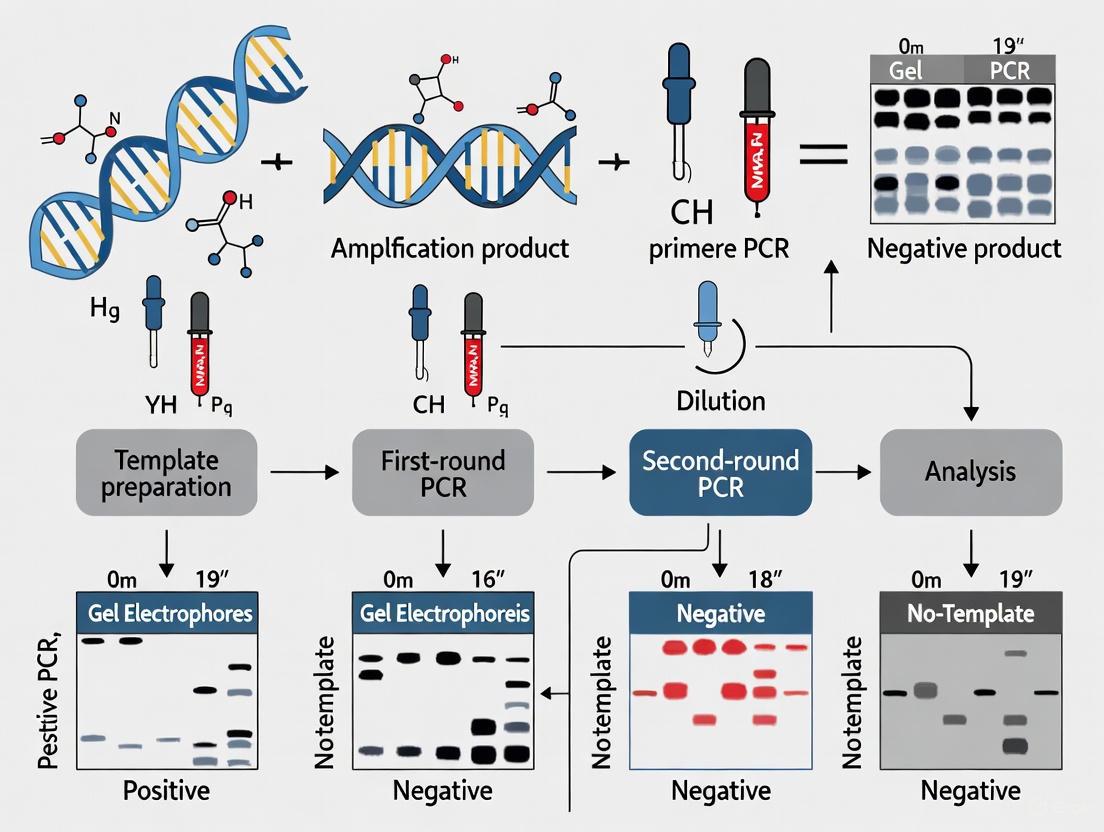
Implementing Rigorous Negative Control Strategies in Your Nested PCR Workflow
A Practical Guide to Setting Up Negative Controls in Two-Step and Single-Tube Nested PCR
In the realm of molecular diagnostics and microbial ecology, nested Polymerase Chain Reaction (PCR) significantly enhances the sensitivity and specificity of DNA amplification by utilizing two sets of primers in sequential reactions [1]. This powerful technique, however, comes with an increased risk of false-positive results due to its extreme sensitivity, making robust negative controls not merely a precaution but an absolute necessity for data integrity [28] [29]. Negative controls serve as essential sentinels, detecting contamination events that could otherwise compromise experimental results and lead to incorrect conclusions [30] [27]. This guide provides a comprehensive framework for implementing negative controls across both traditional two-step and modern single-tube nested PCR formats, ensuring researchers can harness the full power of nested amplification while maintaining the highest standards of experimental rigor.
The fundamental vulnerability of nested PCR lies in its working mechanism; the initial amplification round increases the target DNA concentration, which then becomes a potential contaminant for the second round and subsequent reactions [31]. Without proper controls, these amplification products can generate false positives that are indistinguishable from true positive results, fundamentally undermining research validity [32]. By systematically implementing the negative control strategies outlined in this guide, researchers can confidently detect and prevent such contamination, producing reliable, reproducible results that advance scientific understanding and drug development efforts.
Understanding Nested PCR Formats and Their Vulnerabilities
Two-Step Nested PCR Workflow
Traditional two-step nested PCR employs two distinct primer sets in sequential reactions conducted in separate physical tubes [28] [1]. The first PCR uses outer primers to amplify a larger target region, while the second reaction employs inner primers (nested within the first amplicon) to amplify a smaller, specific fragment [33]. This sequential approach dramatically increases sensitivity and specificity by ensuring that only the correct initial amplicon serves as the template for the second round of amplification [34].
The two-step process introduces specific contamination risks, primarily during the physical transfer of amplification products from the first to the second reaction [35]. This transfer can generate aerosolized amplicons that contaminate reagents, pipettes, and laboratory surfaces, creating reservoirs for future contamination events [31]. Each opening of reaction tubes presents an opportunity for amplicon release into the laboratory environment, necessitating rigorous spatial separation and procedural controls to prevent cross-contamination.
Single-Tube Nested PCR Workflow
Single-tube nested PCR represents a significant technical advancement, containing both primer sets in the same reaction tube and relying on differential annealing temperatures to control the sequential amplification [29]. This format eliminates the amplicon transfer step, substantially reducing contamination risk while maintaining the enhanced sensitivity of nested amplification [35]. The reaction typically begins with higher annealing temperatures that favor outer primer binding, followed by cycling at lower temperatures that permit inner primer amplification [29].
Despite its closed-tube design, single-tube nested PCR remains vulnerable to template-independent amplification and primer-dimer formation, particularly during the lower-temperature cycling stages [29]. Additionally, the presence of multiple primer sets increases the potential for non-specific amplification that must be detected through appropriate controls [1]. The implementation of hot-start DNA polymerase is particularly valuable in this format, as it minimizes non-specific amplification during reaction setup by maintaining polymerase inactivity until the initial denaturation step [1].
A Comprehensive Negative Control Strategy for Nested PCR
Types and Placement of Negative Controls
Effective contamination monitoring in nested PCR requires multiple control types strategically placed throughout the experimental workflow. Each control serves a distinct purpose in identifying potential contamination sources.
Table 1: Types of Negative Controls for Nested PCR
| Control Type | Composition | Purpose | Expected Result | Interpretation of Deviation |
|---|---|---|---|---|
| No-Template Control (NTC) | Complete reaction mixture with PCR-grade water instead of template DNA [31] [27] | Detects contamination in master mix reagents or primers | No amplification | Contaminated reagents or general laboratory contamination |
| First-Step Control (Two-Step PCR only) | Complete first reaction mixture with water instead of template, not carried to second step | Specifically identifies contamination in outer primers or first-round reagents | No amplification | Contamination limited to first-round components |
| Second-Step Control (Two-Step PCR only) | Water substituted for first-round product in the second reaction | Detects contamination in inner primers or second-round reagents | No amplification | Contamination in second-round components only |
| Full Process Control | Water substituted for template through both PCR steps (two-step) or entire process (single-tube) | Monitors cumulative contamination throughout entire workflow | No amplification | Systemic contamination affecting multiple components |
Implementation Protocols
Negative Controls for Two-Step Nested PCR
The following protocol details the specific implementation of negative controls in two-step nested PCR, based on optimized methodologies for bacterial microbiota characterization [28]:
First PCR Setup (25 cycles)
- Prepare master mix containing outer primers, reaction buffer, dNTPs, and hot-start DNA polymerase
- Aliquot into separate reaction tubes
- Add template DNA to test samples, and PCR-grade water to no-template control (NTC-1) and first-step control (FSC) tubes
- Conduct amplification with optimized cycling parameters [28]
Second PCR Setup (15 cycles)
- Prepare fresh master mix with inner primers containing Illumina adapters [28]
- Aliquot into new reaction tubes
- For test samples: transfer 2μL of first PCR product
- For second-step control (SSC): use PCR-grade water instead of first-round product
- For full process control (FPC): use PCR-grade water instead of first-round product (maintained from NTC-1)
- Perform second amplification round
Analysis
- Resolve amplification products by agarose gel electrophoresis
- Valid result: no visible bands in NTC-1, FSC, SSC, or FPC
- If contamination detected, refer to troubleshooting guide (Section 5)
Negative Controls for Single-Tube Nested PCR
For single-tube formats like those used in porcine cytomegalovirus detection [29], implement controls as follows:
Reaction Setup
- Prepare master mix containing both outer and inner primers, reaction buffer, dNTPs, and hot-start DNA polymerase
- Aliquot into reaction tubes
- Add template DNA to test samples, and PCR-grade water to no-template control (NTC) tubes
- Maintain closed-tube conditions throughout process
Amplification Parameters
- Use optimized two-stage thermal cycling profile [29]:
- Initial activation: 95°C for 3 minutes
- Stage 1 (10 cycles): 95°C for 3 seconds, 60°C for 30 seconds (outer primer amplification)
- Stage 2 (40 cycles): 95°C for 3 seconds, 55°C for 30 seconds (inner primer amplification)
Real-Time Detection
- Monitor accumulation with DNA-binding dyes or target-specific probes [29]
- Valid result: no amplification curve in NTC throughout 40 cycles
- If NTC shows amplification, consider reaction contaminated
Interpreting Negative Control Results and Data Validation
Proper interpretation of negative control results is essential for validating experimental data. The following table outlines common scenarios and appropriate responses:
Table 2: Interpretation of Negative Control Results in Nested PCR
| Control Results | Sample Results | Interpretation | Required Action |
|---|---|---|---|
| All negative controls show no amplification | Positive amplification in samples | Valid experimental result | Proceed with data analysis and interpretation |
| No-template control shows amplification | Positive amplification in samples | Contamination confirmed | Discard results; decontaminate workspace and reagents; repeat experiment |
| First-step control shows amplification (two-step PCR) | Positive amplification in samples | Contamination in first-round reagents | Discard outer primers and first-round master mix; repeat experiment |
| Second-step control shows amplification (two-step PCR) | Positive amplification in samples | Contamination in second-round reagents | Discard inner primers and second-round master mix; repeat experiment |
| All negative controls show no amplification | No amplification in samples | PCR failure or true negative | Check positive control; troubleshoot reaction conditions |
Quantitative data from systematic studies demonstrates the critical importance of negative controls. In nested PCR detection of Mycobacterium avium subsp. paratuberculosis, proper controls enabled differentiation between true detection limits (10²-10³ CFU/g in spiked samples) and false positives from contamination [34]. Similarly, in rpoB metabarcoding studies, nested PCR with appropriate controls achieved accurate bacterial composition profiles in host-associated microbiota with low bacterial DNA concentrations, where single-step PCR failed [28].
Contamination Prevention and Troubleshooting
Laboratory Workflow and Decontamination
Implementing rigorous procedural controls is essential for preventing contamination in nested PCR workflows:
- Physical Separation: Maintain distinct pre- and post-PCR work areas with dedicated equipment [32]. The pre-PCR area should contain only stock reagents and never handle amplification products.
- Unidirectional Workflow: Always move from pre-PCR areas to post-PCR areas without backtracking [32]. Never bring amplified products into reagent preparation areas.
- Decontamination Protocols: Regularly clean workspaces with 10% bleach solution or commercial DNA-decontaminating agents [31]. UV irradiation of workstations and reagents can further reduce contamination risks.
- Personal Protective Equipment: Wear dedicated lab coats and gloves for pre-PCR work, changing frequently [31]. Never wear the same coat in pre- and post-PCR areas.
Essential Research Reagent Solutions
Table 3: Essential Research Reagent Solutions for Nested PCR
| Reagent/Chemical | Function/Purpose | Application Notes |
|---|---|---|
| Hot-start DNA Polymerase | Inhibits polymerase activity at room temperature to prevent non-specific amplification and primer-dimer formation [1] | Essential for both single-tube and two-step formats; select high-processivity enzymes for complex samples |
| Ultra-Pure dNTPs | Provides nucleotide substrates for DNA synthesis | Aliquot to prevent repeated freeze-thaw cycles and potential contamination |
| PCR-Grade Water | Nuclease-free water for reaction preparation and negative controls | Use only dedicated, certified nuclease-free water; never use laboratory-purified water |
| DNase Decontamination Reagents | Destroys contaminating DNA in reagents and work areas | Use in laboratory cleaning; some protocols incorporate DNase treatment prior to reverse transcription |
| Uracil-N-Glycosylase (UNG) | Enzymatically degrades carryover contamination from previous PCR reactions [35] | Incorporate when using dUTP instead of dTTP; effective in single-tube nested formats |
| Dedicated Primers | Sequence-specific oligonucleotides for target amplification | Aliquot primers; avoid repeated freeze-thaw cycles; validate new lots with appropriate controls |
Troubleshooting Contamination Events
When negative controls indicate contamination, implement this systematic response:
Immediate Actions
Identify Contamination Source
- Test each reagent individually by substituting with new aliquots in control reactions [27]
- Check equipment (pipettes, centrifuges) for contamination
- Evaluate primer stocks for degradation or contamination
Prevent Future Events
The implementation of comprehensive negative controls is not merely a technical requirement but a fundamental component of rigorous scientific practice in nested PCR research. When properly designed and executed, these controls transform nested PCR from a potentially error-prone technique into a reliable tool for detecting low-abundance targets in complex samples [28] [29] [34]. The strategic use of multiple control types throughout the experimental workflow provides researchers with the confidence to distinguish true biological signals from methodological artifacts, ensuring the validity of both positive and negative findings.
As nested PCR methodologies continue to evolve toward more streamlined single-tube formats [35] [29], the principles of negative control implementation remain constant, even as their specific applications adapt to new technical contexts. By integrating the systematic approaches outlined in this guide—from reagent preparation through data interpretation—research scientists and drug development professionals can advance their fields with the confidence that their molecular findings reflect biological reality rather than procedural contamination. In an era of increasing emphasis on research reproducibility, such rigorous attention to experimental controls has never been more scientifically vital.
Master Mix Formulations and Reagent Aliquoting to Minimize Contamination Risk
Nested Polymerase Chain Reaction (nested PCR) is a powerful diagnostic technique that significantly enhances the sensitivity and specificity of detecting low-abundance nucleic acid targets by employing two sets of primers in sequential amplification rounds [36]. This method is particularly valuable for pathogen detection in clinical samples, environmental testing, and research applications where target concentration is limited [23] [9]. However, the requirement to transfer first-round amplification products to a second reaction tube introduces a substantial risk of amplicon carryover contamination, potentially leading to false-positive results that compromise diagnostic accuracy and research validity [36] [37].
Within this context, rigorous contamination control becomes paramount, with negative controls serving as an essential monitoring component in any nested PCR workflow. These controls, which contain all reaction components except the template DNA, provide a critical baseline for detecting contamination incidents [32]. When amplification occurs in negative controls, it signals possible amplicon contamination that could invalidate experimental results. Therefore, the formulation of master mixes and the strategic aliquoting of reagents represent fundamental technical defenses in preserving assay integrity [38] [25]. This whitepaper examines advanced master mix formulations and reagent management practices specifically designed to minimize contamination risks in nested PCR, with particular emphasis on their role in supporting reliable negative control outcomes.
Master Mix Formulations for Contamination Control
Enzymatic Decontamination Systems
The incorporation of uracil-N-glycosylase (UNG) into PCR master mixes represents one of the most effective biochemical strategies for preventing amplicon carryover contamination [38] [37]. This contamination control system operates through a straightforward but elegant mechanism:
- dUTP Incorporation: During PCR amplification, dUTP is incorporated into newly synthesized amplicons in place of dTTP, creating a distinguishable molecular signature between natural DNA templates and amplification products [37].
- Pre-Amplification Sterilization: In subsequent reactions, UNG enzyme is activated during initial incubation at room temperature, where it recognizes and catalyzes the hydrolysis of uracil-containing DNA from previous amplifications [38] [37].
- Enzyme Inactivation: Before the new amplification begins, high-temperature incubation (95°C) permanently inactivates the UNG enzyme, preventing degradation of the current reaction's natural DNA templates and uracil-containing products [37].
The UNG system is particularly valuable in nested PCR workflows, where the transfer of first-round products creates multiple opportunities for amplicon contamination. Research demonstrates that UNG treatment effectively sterilizes reaction mixtures containing up to 10^6 contaminating amplicons per reaction, providing a robust defense against false positives [37]. However, laboratories should note that UNG efficacy is somewhat reduced with GC-rich amplification targets and requires optimization of dUTP and UNG concentrations for each specific assay [37].
Hot-Start Enzymes
Hot-start DNA polymerases represent another key component in contamination-resistant master mix formulations. These enzymes remain inactive until exposed to high temperatures during the initial denaturation step, preventing non-specific amplification and primer-dimer formation that can occur during reaction setup at room temperature [25]. This technical approach provides two contamination control benefits:
- Reduced Non-Specific Products: By preventing low-temperature amplification artifacts, hot-start enzymes minimize the generation of non-target DNA molecules that could potentially contaminate future reactions or complicate result interpretation [25].
- Improved Assay Sensitivity: The suppression of non-specific amplification increases the available reagents for target amplification, enhancing detection sensitivity for low-copy targets and reducing the number of amplification cycles required, thereby decreasing total amplicon production [25].
Table 1: Master Mix Components for Contamination Control
| Component | Function | Contamination Control Mechanism | Optimization Considerations |
|---|---|---|---|
| UNG Enzyme | Pre-amplification contamination degradation | Hydrolyzes uracil-containing DNA from previous reactions | Requires dUTP in nucleotide mix; less effective for GC-rich targets |
| Hot-Start Polymerase | DNA amplification | Prevents non-specific amplification during reaction setup | Reduces primer-dimer formation and non-target products |
| dUTP | Nucleotide substrate | Creates distinguishable amplicons susceptible to UNG cleavage | Must partially or completely replace dTTP in reaction mix |
| Reaction Buffer | Optimal enzyme activity | Stabilizes reaction components | May include additives to enhance UNG activity or hot-start performance |
One-Tube Nested PCR Approaches
Innovative one-tube nested PCR systems substantially reduce contamination risk by containing both amplification rounds within a single sealed tube [39] [36]. This approach utilizes two primer pairs with significantly different annealing temperatures:
- High-Temperature Primers (External): Longer primers (approximately 25bp) with higher annealing temperatures (approximately 68°C) are used for the first amplification round [36].
- Low-Temperature Primers (Internal): Shorter primers (approximately 17bp) with lower annealing temperatures (approximately 46°C) target internal sequences but only become active when the annealing temperature is reduced after the first amplification phase [36].
This methodological innovation eliminates the need for physical transfer of first-round products, thereby removing the highest-risk step in conventional nested PCR. Research validating a one-tube nested real-time PCR assay for Cryptosporidium detection in birds demonstrated exceptional performance, with 90% repeatability and 80% reproducibility, while completely avoiding amplicon contamination between reactions [39]. The assay achieved an analytical sensitivity of approximately 0.5 oocyst per reaction, highlighting how contamination control and diagnostic sensitivity can be simultaneously optimized through strategic master mix formulation and protocol design [39].
Reagent Aliquoting and Laboratory Practice
Strategic Reagent Aliquoting
The practice of aliquoting reagents represents a fundamental yet often overlooked aspect of contamination control in nested PCR workflows. Dividing bulk reagents into single-use portions provides a critical barrier against the widespread contamination of master stocks [40] [32] [38]. The World Health Organization's molecular testing guidelines explicitly recommend this practice to prevent multiple freeze-thaw cycles and protect master stocks from accidental contamination [25].
Implementation should follow a structured approach:
- Volume Determination: Calculate the required volume for each specific experiment, including necessary overage for pipetting accuracy, typically 10-20% extra volume [38].
- Aseptic Technique: Perform aliquoting in a dedicated pre-PCR clean area using aerosol-resistant filter tips and sterile, nuclease-free tubes [25].
- Clear Labeling: Mark each aliquot with content details, concentration, preparation date, and lot numbers for complete traceability [25].
- Storage Management: Store aliquots at recommended temperatures with limited access to prevent unnecessary handling [38].
This practice ensures that if contamination occurs, it affects only a single experiment rather than compromising entire reagent stocks, thereby protecting both current and future research integrity.
Physical Workflow Separation
Contamination control in nested PCR extends beyond reagent formulation to encompass laboratory design and workflow [41] [38] [25]. Effective segregation of PCR activities follows a unidirectional workflow that progressively moves from clean to potentially contaminated areas:
Diagram 1: Unidirectional Nested PCR Workflow
The WHO specifically recommends that for nested PCR amplification, "the preparation of the mastermix for the second round reaction should be prepared in the 'clean' area for mastermix preparation, but the inoculation with the primary PCR product should be done in the amplification room, and if possible in a dedicated containment area" [25]. This precise workflow management is essential for preventing amplicon contamination between first and second amplification rounds.
Equipment and Surface Decontamination
Regular decontamination of equipment and surfaces with 10% sodium hypochlorite (bleach) solutions represents another critical practice for nested PCR laboratories [25] [37]. Bleach causes oxidative damage to nucleic acids, rendering them unamplifiable in subsequent reactions [37]. The WHO recommends fresh daily preparation of 10% sodium hypochlorite with a minimum contact time of 10 minutes for effective decontamination [25].
For equipment sensitive to corrosion by bleach (such as pipette components), alternatives include:
- DNA-degrading commercial solutions specifically validated for surface decontamination [25]
- 70% ethanol followed by UV irradiation for at least 30 minutes [25]
- Autoclaving for heat-resistant equipment and consumables [25]
Ultraviolet light irradiation creates thymidine dimers and other covalent modifications in DNA that render contaminants unamplifiable [37]. While this method shows variable efficacy for short (<300 nucleotides) and GC-rich templates, it provides valuable supplemental contamination control when applied to work surfaces and equipment before use [37].
Experimental Protocols for Validation
One-Tube Nested PCR Protocol for Cryptosporidium Detection
A validated one-tube nested real-time PCR protocol for detecting Cryptosporidium spp. in avian fecal samples demonstrates effective contamination control while maintaining high sensitivity [39]:
Reagent Formulation:
- Primers: Two primer pairs with extended sequences at 5' ends to create approximately 10°C annealing temperature difference
- Reaction Volume: 25μL total
- Master Mix Components:
- Template DNA: 1-2μL
- Primers: 0.5μL each (final concentration 0.2μM)
- dNTP mixture: 0.5μL (final concentration 200μM each dNTP)
- PCR buffer: 2.5μL of 10× concentration
- MgCl₂: 1.5μL (final concentration 1.5-2.0mM)
- Taq DNA polymerase: 0.25μL (1.25U)
- Sterile ultrapure water: to volume [39]
Thermal Cycling Profile:
- Initial Denaturation: 94°C for 2 minutes
- First Round (15-30 cycles):
- Denaturation: 94°C for 30 seconds
- Annealing: Higher temperature for external primers (e.g., 68°C) for 30 seconds
- Extension: 72°C for 1 minute
- Second Round (15-30 cycles):
- Denaturation: 94°C for 30 seconds
- Annealing: Lower temperature for internal primers (e.g., 46°C) for 30 seconds
- Extension: 72°C for 1 minute
- Final Extension: 72°C for 5 minutes [39] [36]
This protocol achieved 20.3% positivity in avian samples compared to 8.1% with conventional nested PCR, demonstrating significantly enhanced sensitivity while reducing contamination risk through the single-tube design [39].
Conventional Two-Tube Nested PCR with Contamination Controls
For applications requiring conventional two-tube nested PCR, rigorous contamination controls must be implemented:
First Round Amplification:
- Preparation: In dedicated pre-PCR area with dedicated equipment
- Reagent Aliquoting: Use single-use aliquots for all reagents
- Negative Controls: Include at least one no-template control per run
- Reaction Assembly:
- Template DNA: 1-2μL
- External primers: 0.5μL each
- Master mix components as above
- Thermal Cycling: Standard cycling parameters for external primers [36]
Second Round Amplification:
- Template Transfer: Perform in separate area using dedicated equipment
- Dilution Factor: Dilute first-round product 1:10 to 1:1000
- Reaction Assembly:
- Diluted first-round product: 1-2μL
- Internal primers: 0.5μL each
- Fresh master mix with UNG/dUTP if possible
- Additional negative controls using first-round negative control as template [36]
Table 2: Contamination Control Validation in Nested PCR Studies
| Study Target | Method | Contamination Controls | Sensitivity | Specificity |
|---|---|---|---|---|
| Cryptosporidium spp. [39] | One-tube nested real-time PCR | Single-tube design, repeatability (90%), reproducibility (80%) | 0.5 oocyst/reaction | No amplification of non-target organisms |
| SARS-CoV-2 [9] | Conventional nested PCR | Separate pre-and post-PCR areas, UV sterilization | 0.015 ng/μL RNA | 100% against related feline pathogens |
| H. pylori [23] | Short amplicon nested PCR | Physical separation, fresh bleach preparation | 66.6% positivity in asymptomatic volunteers | Confirmed by DNA sequencing |
The Scientist's Toolkit: Research Reagent Solutions
Table 3: Essential Research Reagent Solutions for Contamination Control
| Reagent/Equipment | Function | Contamination Control Feature |
|---|---|---|
| UNG-dUTP System | Enzymatic degradation of carryover contamination | Selectively hydrolyzes uracil-containing amplicons from previous reactions |
| Hot-Start Polymerase | DNA amplification | Prevents non-specific amplification during reaction setup |
| Aerosol-Resistant Filter Tips | Liquid handling | Creates barrier between pipette and reagents to prevent aerosol contamination |
| Single-Use Reagent Aliquots | Reaction components | Prevents cross-contamination of master stocks |
| HEPA/UV Laminar Flow Hood | Workstation for reagent preparation | Provides ISO Class 5 clean air for reaction assembly |
| 10% Sodium Hypochlorite | Surface decontamination | Oxidatively destroys nucleic acids on work surfaces |
| Nuclease-Free Water | Reaction component | Guaranteed free of nucleases that could degrade reagents |
| Dedicated Pre-PCR Equipment | Various laboratory functions | Prevents introduction of amplicons into clean areas |
Effective contamination control in nested PCR requires an integrated approach combining specialized master mix formulations, strategic reagent management, and appropriate laboratory workflows. The incorporation of UNG-dUTP systems, utilization of hot-start enzymes, and implementation of one-tube nested PCR protocols provide powerful technical solutions to amplicon carryover contamination. When coupled with rigorous practices including reagent aliquoting, physical workflow separation, and comprehensive negative controls, these methods enable researchers to maintain the exceptional sensitivity of nested PCR while minimizing false-positive results.
As molecular diagnostics continue to advance, with increasing requirements for detecting low-abundance targets in complex samples, these contamination control strategies will remain essential for research reliability and diagnostic accuracy. The implementation of these practices represents not merely a technical consideration but a fundamental component of scientific rigor in molecular research and development.
Nested Polymerase Chain Reaction (PCR) is a powerful molecular technique designed to significantly enhance the specificity and sensitivity of nucleic acid amplification. This method employs two successive rounds of amplification with two distinct sets of primers [42]. The first round uses an outer set of primers to amplify a target region, followed by a second round using inner (nested) primers that bind within the first PCR product [43]. This two-step process drastically reduces non-specific amplification and false positives, as it is highly improbable for a second set of primers to bind non-specifically to an amplicon that was itself non-specifically generated [42]. Consequently, nested PCR is indispensable for applications involving low-abundance targets, complex DNA mixtures, or challenging templates such as environmental DNA (eDNA) and clinical samples with potential inhibitors [44] [45].
Within this context, the implementation of a rigorous negative control strategy is not merely a supplementary step but a foundational component of reliable nested PCR research. The heightened sensitivity of the technique, combined with its multi-step open-tube workflow, inherently increases the risk of cross-contamination from amplicons (products from the first PCR round) or foreign DNA [42]. Without proper controls, false positive results can easily occur, compromising data integrity and leading to erroneous conclusions. This guide details the complete nested PCR workflow, integrating the essential role of negative controls at every stage to ensure experimental validity.
The fundamental principle of nested PCR is the sequential use of primer sets to exponentially improve amplification fidelity [42]. The outer primers are designed to flank the region of interest, generating an initial amplicon. The nested primers then bind internally to this initial product for the second round of amplification [43]. This sequential binding ensures that even if the outer primers produce non-specific amplicons, they are unlikely to be amplified in the second round, thereby filtering out background noise and enriching for the true target [42].
The following diagram illustrates the logical sequence of the entire nested PCR process, highlighting key stages where negative controls are critical for validation.
Detailed Workflow and Protocols
Stage 1: Sample Preparation and Nucleic Acid Extraction
The initial stage is critical, as the quality of the template DNA directly influences the success of downstream amplification.
- Sample Types: Nested PCR is applied to diverse sources, including clinical specimens (e.g., throat swabs, blood, tissue) [45] [29], environmental DNA (water samples) [44], plant tissues [46], and microbial cultures [22].
- Nucleic Acid Extraction: Use commercial kits appropriate for your sample type (e.g., membrane adsorption kits for viral RNA [45], Column Fungal DNAout kits for mycelia [22]). Always include a negative extraction control—a sample containing only nuclease-free water or buffer taken through the entire extraction process. This controls for contamination in the reagents and cross-contamination during extraction.
- Quality Assessment: Evaluate DNA quality and concentration using spectrophotometry (e.g., Nanodrop) [22] or fluorometry. Integrity can be checked via agarose gel electrophoresis.
Stage 2: Primer Design and Validation
Careful primer design is paramount for successful nested PCR.
- Outer Primers: Design to flank the target region. They are typically 18-22 nucleotides long and should be checked for specificity using tools like NCBI BLAST [22].
- Nested Primers: Design to bind internally to the sequence amplified by the outer primers. They should not form dimers or self-hairpins and should have similar melting temperatures (Tm) for efficient annealing [42]. For example, in detecting Fusarium tricinctum, primers were designed from the conserved
CYP51Cgene [22]. - Validation: Before use in nested PCR, validate each primer set (outer and inner separately) using a positive control template to ensure they produce a single amplicon of the expected size.
Stage 3: First-Round PCR with Outer Primers
The first amplification reaction is set up to generate the initial template for the nested reaction.
Protocol for First-Round PCR [42]:
| Component | Concentration | Quantity |
|---|---|---|
| Mastermix | 1X | 12 µL |
| Reaction Buffer | 1X | 5 µL |
| Outer Forward Primer | 10 pM | 1 µL |
| Outer Reverse Primer | 10 pM | 1 µL |
| Template DNA | 30 ng | 3 µL |
| Nuclease-free Water | - | 3 µL |
| Total Volume | 25 µL |
Cycling Conditions [42]:
- Initial Denaturation: 90–95°C for 3 minutes
- Amplification (25 cycles):
- Denaturation: 90–95°C for 1 minute
- Annealing: 55–60°C for 50 seconds
- Extension: 72°C for 1 minute
- Final Extension: 72°C for 7 minutes
Controls for First-Round PCR:
- No-Template Control (NTC): Includes all reaction components except the template DNA, which is replaced with nuclease-free water. This is crucial for detecting contamination in the PCR reagents.
- Positive Control: Contains a known sample of the target DNA to verify that the reaction worked.
Stage 4: Second-Round PCR with Nested Primers
A small aliquot of the first-round product is used as the template for the second amplification. This step enhances specificity and yield.
Protocol for Second-Round PCR [42]:
| Component | Concentration | Quantity |
|---|---|---|
| Mastermix | 1X | 12 µL |
| Reaction Buffer | 1X | 5 µL |
| Nested Forward Primer | 10 pM | 1 µL |
| Nested Reverse Primer | 10 pM | 1 µL |
| First-Round PCR Product | - | 3 µL |
| Nuclease-free Water | - | 3 µL |
| Total Volume | 25 µL |
Cycling Conditions [42]:
- Initial Denaturation: 90–95°C for 3 minutes
- Amplification (35 cycles):
- Denaturation: 90–95°C for 1 minute
- Annealing: 55–60°C for 50 seconds
- Extension: 72°C for 1 minute
- Final Extension: 72°C for 7 minutes
Critical Control Step: The NTC from the first round must be carried over as the template for a second-round NTC. If this control shows amplification, it indicates contamination was introduced during the first-round setup or product transfer, invalidating the results of the test samples [42].
Advanced Modifications: One-Tube Nested PCR
To mitigate the high contamination risk associated with transferring PCR products, one-tube nested protocols have been developed. In this format, both outer and inner primers are included in a single tube, but the inner primers are designed with modifications (e.g., lower concentration, higher Tm) that prevent them from activating until later cycles, effectively creating a sequential reaction in a closed tube [29]. One-tube nested real-time PCR for Porcine Cytomegalovirus demonstrated a significantly higher detection rate (38.6%) compared to traditional nested PCR (23.6%) and conventional PCR (12.6%), showcasing its superior sensitivity and reduced contamination risk [29].
Post-Amplification Analysis
After the second round of PCR, the products are analyzed to confirm successful and specific amplification.
- Gel Electrophoresis: The most common method. An aliquot of the second-round product is run on an agarose gel, stained, and visualized under UV light. A single, sharp band of the expected size indicates specific amplification [42] [46].
- Sequencing: For definitive confirmation of the target amplicon, the PCR product can be purified and sequenced [46]. This is a critical step in methods development or when detecting specific strains, as used in phytoplasma detection to distinguish true positives from non-specific amplification [46].
- Real-time and Digital PCR: While not the traditional endpoint analysis, nested PCR products can be quantified or detected using real-time PCR (with SYBR Green) or digital PCR (ddPCR) for absolute quantification [44] [45]. These methods offer high sensitivity and can bypass the need for gel electrophoresis.
Performance Data and Method Validation
The effectiveness of nested PCR is demonstrated by its superior performance metrics compared to conventional PCR and even digital PCR in some applications. The table below summarizes quantitative data from various studies.
Table 1: Comparative Performance of Nested PCR Against Other Detection Methods
| Application / Pathogen | Method | Limit of Detection (LoD) | Positive Rate in Clinical Samples | Key Advantage |
|---|---|---|---|---|
| SARS-CoV-2 [45] | OSN-qRT-PCR* | 194.74 copies/mL (ORF1ab) | 82.35% (28/34) | Highest sensitivity for low viral loads |
| ddPCR | 401.8 copies/mL (ORF1ab) | 67.65% (23/34) | Absolute quantification | |
| qRT-PCR | 520.1 copies/mL (ORF1ab) | 58.82% (20/34) | Standard gold method | |
| Porcine Cytomegalovirus [29] | One-Tube Nested RT-PCR | Not Specified | 38.6% (49/127) | Speed (~1.5 hrs), reduced contamination |
| Traditional Nested PCR | Not Specified | 23.6% (30/127) | Established protocol | |
| Conventional PCR | Not Specified | 12.6% (16/127) | Simplicity | |
| Areca Palm Phytoplasma [46] | Novel Nested PCR | 4x10⁻⁷ - 7.5x10⁻⁷ ng/μL | High field validation | Specific detection of 16SrI & 16SrII groups |
| Eastern Hellbender eDNA [44] | Nested PCR (mtDNA) | Order of magnitude improvement | Effective population detection | Overcame off-target amplification of previous primers |
*OSN-qRT-PCR: One-step nested quantitative RT-PCR.
The Scientist's Toolkit: Essential Research Reagents
Successful execution of nested PCR requires a suite of reliable reagents and instruments. The following table details the key materials.
Table 2: Essential Reagents and Materials for Nested PCR Workflow
| Item Category | Specific Examples & Functions |
|---|---|
| Nucleic Acid Extraction | Column-based kits (e.g., TIANamp Marine Animal DNA Kit [47], Column Fungal DNAout Kit [22]); Automated systems (e.g., Miracle-AutoXT [29]) for high-throughput, consistent DNA purification. |
| PCR Enzymes & Master Mixes | Hot-Start DNA Polymerase (e.g., Platinum II Taq [43]) to minimize non-specific amplification during reaction setup; Probe-based qPCR mixes (e.g., Thunderbird probe qPCR mix [29]) for one-tube nested real-time PCR. |
| Primers | Outer Primers & Nested Primers: Highly specific, HPLC-purified oligonucleotides. Validated sequences are available for common targets (e.g., DIV1 ATPase gene [47], Phytoplasma 16S rDNA [46]). |
| Control Templates | Positive Control: Plasmid DNA or genomic DNA from a known positive sample. Negative Control: Nuclease-free water for NTCs. |
| Instrumentation | Thermal Cyclers (e.g., C1000 Touch [45], LightCycler 480 II [45]); Gel Electrophoresis equipment; Digital Droplet PCR Systems (QX200 [45]); Spectrophotometers (Nanodrop One [22]) for quantification. |
Troubleshooting and Quality Assurance
A robust quality assurance system, built on negative controls, is essential for troubleshooting.
- Amplification in First-Round NTC: This indicates contamination in the PCR reagents (mastermix, primers, water). Replace all reagents and repeat the experiment [42].
- Amplification in Second-Round NTC (but not First-Round): This is a clear sign of amplicon contamination during the transfer of the first-round product. Meticulous technique, physical separation of pre- and post-PCR areas, and use of dedicated pipettes are mandatory. Consider switching to a one-tube nested approach [29].
- No Amplification in Sample or Positive Control: Suggests reaction failure due to ineffective reagents, incorrect cycling parameters, or potent PCR inhibitors in the sample. Check reagent concentrations and test the positive control separately. The use of an internal control (IC) can help identify the presence of inhibitors [29].
In conclusion, a meticulously designed and executed nested PCR workflow, underpinned by an unwavering commitment to negative control protocols, provides an exceptionally reliable method for detecting low-abundance nucleic acid targets. From sample preparation to final analysis, each step offers an opportunity to implement controls that safeguard the integrity of the scientific data, ensuring that results are both sensitive and specific.
In polymerase chain reaction (PCR) research, the negative control serves as a fundamental sentinel for experimental validity. This is especially true for nested PCR, a highly sensitive technique involving two successive rounds of amplification that inherently increases the risk of contamination and artifact formation [16]. The negative control, or No-Template Control (NTC), contains all PCR reagents—polymerase, primers, buffer, nucleotides—except for the template DNA [48]. Its sole purpose is to remain blank, confirming that the amplification observed in experimental samples is genuine and not a product of contamination or non-specific reactions.
Interpreting the results of this control is a critical skill. A band or smear in the NTC lane invalidates the entire experiment and necessitates systematic troubleshooting [48] [31]. This guide provides an in-depth framework for researchers to diagnose and resolve the issues signaled by anomalous negative controls, with a specific focus on the nested PCR context. Mastering this process is essential for generating robust, reliable, and reproducible data in drug development and molecular research.
A Diagnostic Framework for Negative Control Anomalies
The first step in troubleshooting is to characterize the anomalous signal. The size, shape, and intensity of the signal in the negative control lane provide the primary clues for diagnosing the root cause. The flowchart below outlines the systematic diagnostic process.
Contamination: The Primary Adversary
When the band in the negative control is the same size as your expected target product, the diagnosis is near-certainly DNA contamination [48]. The exquisite sensitivity of PCR, compounded by a second round of amplification in nested PCR, becomes its greatest weakness, allowing minuscule, unintended DNA molecules to be amplified billions of times.
Common Sources of Contamination:
- Aerosols: The most common vector. Opening tubes containing previous PCR products or positive samples creates microscopic droplets that contaminate pipettes, bench surfaces, and tube racks [48] [31].
- Reagents: Any component of the master mix can be compromised, with nuclease-free water, primers, and the polymerase enzyme itself being frequent culprits. Some Taq polymerase preparations can contain residual bacterial DNA [48].
- Cross-Contamination: Template DNA can be accidentally introduced from contaminated pipettes, tips, or gloves during sample handling [49].
Primer-Dimers: A Reaction Optimization Issue
If the band in the negative control is a faint, low-molecular-weight band or smear (typically < 100 bp) near the bottom of the gel, the issue is likely primer-dimers [48]. This is an issue of reaction chemistry, not contamination. Primer-dimers form when the forward and reverse primers anneal to each other, particularly at their 3' ends, and are extended by the polymerase. This occurs more readily in the absence of a genuine template DNA, as in the NTC.
Systematic Troubleshooting and Resolution
Eradicating Contamination
A contaminated negative control mandates that you stop, discard all results from that run, and decontaminate before proceeding [48]. Do not trust any data generated under these conditions.
Action Plan for Decontamination:
Physical Separation (The Golden Rule): Establish physically separate areas for pre-PCR and post-PCR work [48] [49].
- Pre-PCR Area: A dedicated "clean" space (ideally a UV-equipped PCR hood or dead-air box) used only for preparing master mixes and reagents. No template DNA or amplified PCR products should ever enter this area.
- Post-PCR Area: A designated "dirty" area for adding template DNA, running the thermocycler, and analyzing gels.
Dedicated Equipment and Supplies:
- Pipettes: Maintain separate sets of pipettes for pre- and post-PCR work. Never use post-PCR pipettes for master mix preparation [48].
- Filter Tips: Use aerosol-barrier pipette tips universally to prevent aerosol contamination of pipette shafts. This is non-negotiable [48] [31].
- Reagents: Aliquot all reagents (polymerase, primers, dNTPs, water) into small, single-use volumes upon arrival. This preserves stock reagents and limits the spread of contamination [48] [31].
Workspace Decontamination:
- Chemical Cleaning: Thoroughly wipe down all surfaces, pipettes, centrifuges, and tube racks with a 10% bleach solution or a commercial DNA decontaminant like DNA-Away [48] [49] [50]. Note that 70% ethanol is ineffective as it precipitates rather than destroys DNA [50].
- UV Irradiation: If available, expose your PCR hood and pipettes to UV light for 15-30 minutes before use. UV light creates thymine dimers in DNA, rendering it unamplifiable [48] [49].
Optimizing to Prevent Primer-Dimers
If the issue is primer-dimers, the solution lies in refining your reaction conditions and components.
Strategies to Minimize Primer-Dimers:
- Increase Annealing Temperature: A higher, more stringent annealing temperature prevents the weak binding between primers that leads to dimer formation. Increase in increments of 2°C [48] [49].
- Use Hot-Start Taq Polymerase: Hot-start polymerases remain inactive until the initial high-temperature denaturation step. This prevents enzymatic activity during reaction setup at lower temperatures, where primer-dimer formation is most likely to initiate [48] [49].
- Redesign Primers: If optimization fails, the primers themselves may have high self-complementarity, especially at the 3' ends. Use primer design software to check for this and redesign if necessary [48].
Interpreting Results with Controls: A Decision Matrix
The following table synthesizes information from the search results to guide the interpretation of your PCR results in the context of control outcomes. This is your first reference when analyzing gel data.
Table 1: Interpreting PCR Results Using Positive and Negative Controls
| Sample PCR Result | Negative Control Result | Positive Control Result | Interpretation and Next Steps |
|---|---|---|---|
| Amplicons observed | Negative (no band) | Positive (band) | Ideal outcome. The PCR worked, results are reliable, and there is no evidence of systemic contamination [27]. |
| Amplicons observed | Positive (band) | Positive (band) | Systemic contamination present. The PCR worked, but it is impossible to distinguish true products from contaminants. All data is invalid. Discard results and begin decontamination protocols [27]. |
| No amplicons observed | Negative (no band) | Positive (band) | The PCR process worked, but the sample DNA failed to amplify. Troubleshoot DNA extraction quality, sample integrity, or primer specificity to the sample [27]. |
| No amplicons observed | Negative (no band) | Negative (no band) | Total PCR failure. The PCR process itself has failed. Troubleshoot reagent integrity, thermocycler conditions, and master mix preparation [27]. |
| Amplicons observed | Positive (band) | Negative (no band) | The PCR worked but is contaminated, and the positive control has failed. Check for mislabeled controls. Otherwise, the sample results are unreliable due to contamination, and the positive control needs replacement [27]. |
The Scientist's Toolkit: Essential Reagents and Equipment
Table 2: Research Reagent Solutions for Robust Nested PCR
| Item | Function in Nested PCR | Key Considerations |
|---|---|---|
| Hot-Start DNA Polymerase | Enzyme inactive at room temperature, preventing non-specific amplification and primer-dimer formation during reaction setup. Critical for the specificity of both PCR rounds [48] [49]. | |
| Aerosol-Barrier Filter Pipette Tips | Prevents aerosolized contaminants from entering and contaminating the pipette body, a major source of cross-contamination [48] [31]. | Essential for all liquid handling in pre-PCR area. |
| Nuclease-Free Water | The solvent for master mixes and the negative control. Must be guaranteed free of nucleases and contaminating DNA [48]. | Always aliquot; open a new bottle if contamination is suspected. |
| Dedicated Pre-PCR Pipettes | Pipettes used exclusively in the clean pre-PCR area for master mix assembly. Never exposed to template DNA or amplicons [48] [49]. | Clearly label to prevent accidental misuse. |
| 10% Bleach or DNA-Away | Chemical decontaminant used to destroy contaminating DNA on benchtops, equipment, and pipettes [48] [49] [50]. | More effective than ethanol for DNA destruction. |
| UV PCR Hood/Hood UV Lamp | Provides a sterile, dead-air workspace for master mix preparation. UV light functionally destroys contaminating DNA by inducing thymine dimers [48] [49]. | Run UV for 15-30 min before use. |
In nested PCR research, a pristine negative control is not an optional luxury but a non-negotiable requirement for data integrity. Success hinges on a rigorous, disciplined approach to laboratory practice. By physically separating pre- and post-PCR workflows, using dedicated equipment, meticulously aliquoting reagents, and systematically interpreting control results, researchers can conquer the challenges of contamination and optimization. Mastering the interpretation and preservation of your negative controls ensures that the groundbreaking discoveries in drug development and molecular biology you report are unequivocally real.
Negative controls are a fundamental component of the experimental method, serving as a critical tool for detecting both suspected and unsuspected sources of spurious causal inference [30]. In diagnostic and research applications of nested polymerase chain reaction (PCR), these controls are indispensable for verifying result specificity and ensuring diagnostic accuracy. Nested PCR, a modification of the standard PCR technique, employs two successive rounds of amplification with two sets of primers to significantly enhance sensitivity and specificity [16] [51]. However, this increased sensitivity comes with an elevated risk of contamination and false positives, making robust negative controls essential for distinguishing true positive results from amplification artifacts [16].
The fundamental principle of negative controls involves performing the experiment under conditions where a null result is expected, thereby helping researchers identify confounding factors and other sources of error in observational studies [30]. In molecular diagnostics, they validate that the amplification signal genuinely originates from the target pathogen rather than contamination, non-specific priming, or reagent impurities. This article examines the strategic implementation of negative controls through case studies in plant pathology and clinical diagnostics, providing a framework for their application within nested PCR protocols.
Theoretical Framework: Principles and Typology of Negative Controls
Core Concepts and Mechanism of Nested PCR
Nested PCR was specifically designed to overcome limitations of conventional PCR by significantly improving both sensitivity and specificity. The technique involves two sequential amplification rounds: the first uses an outer primer set to generate a primary amplicon, which then serves as the template for a second amplification with an inner primer set that binds within the first product [16] [51]. This two-stage process enhances specificity because the second set of primers will only successfully bind and amplify if the correct initial product was generated [51]. The increased sensitivity arises from the high total cycle number, enabling detection of target sequences present in minute quantities [16].
Despite its advantages, a primary concern with nested PCR is the heightened risk of carryover contamination during the transfer of first-round products to the second reaction tube [16] [51]. This vulnerability makes the implementation of appropriate negative controls essential for validating results. Physical separation of pre- and post-amplification areas, use of dedicated equipment, and the inclusion of wax or oil barriers between reaction mixtures represent key strategies to minimize contamination risk [51].
A Functional Taxonomy of Negative Controls
Experimental biology employs several strategic approaches to design negative controls, which can be categorized into three primary types based on their underlying principle [30]:
Omission of Essential Components: This approach involves leaving out an element theoretically essential for the reaction to proceed, such as excluding template DNA or a crucial enzyme. Any amplification observed in such controls indicates contamination or non-specific amplification.
Inactivation of Active Ingredients: This strategy employs specific methods to neutralize the hypothesized active ingredient, such as adding neutralizing antibodies or using enzymatically inactive versions of proteins. The expected outcome is a null result, confirming the specificity of the detection mechanism.
Testing Against Implausible Targets: This control tests the assay against targets where the hypothesized mechanism should not produce an effect, such as unrelated organisms or irrelevant clinical outcomes. A negative result confirms specificity, while a positive signal suggests off-target effects or confounding factors.
These control strategies provide a systematic framework for ruling out alternative explanations for experimental results, thereby strengthening causal inference in both basic research and applied diagnostic settings [30].
Case Study 1: Phytoplasma Detection in Coconut Palm
Research Context and Diagnostic Challenge
Phytoplasmas are associated with many economically significant plant diseases, including Weligama coconut leaf wilt disease (WCLWD) in Sri Lanka, a devastating condition that has necessitated the removal of over 340,000 coconut palms [52]. Early and accurate detection is crucial for disease management, but pathogen detection has historically relied on nested PCR with inconsistent results—symptomatic plants often produced negative results, while asymptomatic ones sometimes tested positive [52].
These inconsistencies presented a substantial diagnostic challenge, potentially arising from multiple factors: low specificity of primers, uneven pathogen distribution within the plant, variable pathogen titers, or primer-binding site variability [52]. Researchers undertook a comprehensive study to optimize detection by evaluating primer combinations and identifying the best sampling tissues, with negative controls playing a pivotal role in validating their improved protocol.
Implementation of Negative Controls
The experimental design incorporated negative controls at multiple critical points to ensure the reliability of the optimized nested PCR protocol [52]:
- Reagent Control: Sterilized distilled water was used as a template instead of DNA to monitor for contamination in PCR reagents, master mix, or environmental carryover.
- Specificity Validation: The optimized protocol was tested against healthy plant material and other potential pathogens to confirm that amplification signals specifically originated from the target phytoplasma and not from host DNA or non-target organisms.
The research team systematically tested six different universal primer combinations to identify the most effective pairing for consistent phytoplasma detection [52]. Through this rigorous approach, the P1/Tint primer set nested with fU5/rU3 emerged as the most reliable combination, producing consistent results with high specificity.
Experimental Outcomes and Impact
The implementation of rigorous negative controls was instrumental in developing a robust detection system. The optimized protocol achieved a remarkable performance, with a minimum success rating of 88% and 100% specificity, demonstrating both high sensitivity and an absence of false positives [52]. The research also identified that the CYP51C gene serves as a valuable target for Fusarium-specific markers, enabling distinction between closely related species [5].
The table below summarizes the key quantitative findings from the phytoplasma detection study:
Table 1: Performance Metrics of Optimized Nested PCR for Phytoplasma Detection
| Parameter | Result | Significance |
|---|---|---|
| Best Primer Combination | P1/Tint nested with fU5/rU3 | Produced consistent and specific amplification [52] |
| Detection Success Rate | Minimum of 88% | High reliability for pathogen detection [52] |
| Assay Specificity | 100% | No false positives reported [52] |
| Optimal Tissue Type | Midribs of milky white bud leaves | Best tissue for early and consistent detection [52] |
The findings confirmed the systemic movement of the pathogen within infected plants and enabled tentative identification of a latent period, providing crucial insights for disease management [52]. The implementation of systematic negative controls transformed an inconsistent detection method into a reliable diagnostic tool capable of early pathogen identification.
Case Study 2: Candida Detection in Critically Ill Pediatric Patients
Nested PCR Protocol and Control Strategy
The multiplex nested PCR protocol incorporated comprehensive negative controls to prevent false positives and ensure specific Candida detection [53]:
- Physical Separation: Reaction mixes, DNA extractions, and amplifications were set up in separate rooms equipped with safety cabinets to prevent amplicon carryover contamination.
- No-Template Controls: All experiments included negative controls containing sterile water instead of genomic DNA to monitor for reagent contamination.
- Specificity Verification: The assay was validated against DNA from seven Candida prototype strains and human DNA to confirm specific amplification of target species without cross-reactivity.
The two-round amplification protocol began with universal fungal primers (ITS1 and ITS4), followed by a second multiplexed amplification with species-specific inner primers that could identify seven Candida species simultaneously [53]. This approach combined the sensitivity of nested PCR with the efficiency of multiplex detection.
Diagnostic Performance and Clinical Utility
The careful implementation of negative controls enabled the researchers to achieve exceptional assay performance while maintaining high specificity. The multiplex nested PCR demonstrated a detection limit of just four Candida genomes per milliliter of blood for all targeted Candida species—a sensitivity level far surpassing conventional blood culture methods [53].
The table below summarizes the comparative performance between the novel molecular method and traditional blood culture:
Table 2: Comparative Performance of Multiplex Nested PCR vs. Blood Culture for Candida Detection
| Diagnostic Method | Detection Rate | Time to Result | Additional Capabilities |
|---|---|---|---|
| Blood Culture (Gold Standard) | 14.8% (8/54 patients) | 48-96 hours | Limited to species identification [53] |
| Multiplex Nested PCR | 24.0% (13/54 patients) | <24 hours | Detected dual candidaemia in 3 patients [53] |
Critically, the nested PCR detected all culture-positive cases while identifying additional infections missed by blood culture, including three cases of dual candidaemia where multiple Candida species were present simultaneously [53]. The implementation of robust negative controls gave clinicians confidence in these results, enabling earlier targeted antifungal therapy and potentially improving patient outcomes in this vulnerable population.
Integrated Analysis: Strategic Implementation Across Disciplines
Common Principles and Adaptive Applications
Despite the differing contexts of plant pathology and clinical mycology, both case studies demonstrate remarkably similar principles in their implementation of negative controls:
- Multi-Layered Control Strategy: Both protocols incorporated negative controls at multiple procedural stages—during DNA extraction, PCR setup, and amplification—to monitor for contamination introduction at any step.
- Specificity Validation: Both studies explicitly tested their assays against non-target organisms (related pathogens, host DNA, commensal flora) to confirm specific target detection.
- Environmental Monitoring: Regular inclusion of no-template controls served as sentinels for environmental contamination or reagent degradation.
These consistent approaches across disciplines highlight the universal importance of negative controls in validating nested PCR diagnostics, regardless of the specific application domain.
The Researcher's Toolkit: Essential Reagents and Materials
The successful implementation of negative controls in nested PCR requires specific research reagents and materials. The following table summarizes key components used in the featured studies:
Table 3: Essential Research Reagents for Nested PCR with Negative Controls
| Reagent/Material | Function | Application Example |
|---|---|---|
| Sterile Nuclease-Free Water | Negative control template; reagent preparation | Reagent control for contamination monitoring [52] [53] |
| DNA Polymerase | Enzymatic amplification of target sequences | GoTaq DNA Polymerase [52]; Platinum Taq DNA Polymerase [53] |
| Primer Sets | Specific binding to target sequences for amplification | P1/Tint and fU5/rU3 for phytoplasma detection [52] |
| dNTPs | Building blocks for DNA synthesis | 150-200 μM in reaction mixture [52] |
| MgCl₂ | Cofactor for DNA polymerase activity | Concentration optimization (0.6-1.5 mM) [52] |
| DNA Extraction Kits | Isolation of high-quality template DNA | Column Fungal DNAout Kit [5]; QIAamp DNA Mini Kit [53] |
Experimental Workflow and Control Points
The following diagram illustrates a generalized nested PCR workflow incorporating critical negative control points:
The case studies presented demonstrate that negative controls are not merely procedural formalities but fundamental components that determine the diagnostic validity of nested PCR assays. In both plant pathology and clinical diagnostics, systematic implementation of controls enabled researchers to develop highly sensitive and specific detection methods capable of identifying pathogens at minimal concentrations while maintaining 100% specificity [52] [53].
Based on these successful applications, we recommend the following best practices for implementing negative controls in nested PCR research:
- Implement Controls at Multiple Procedural Stages: Include negative controls during DNA extraction, first-round amplification, and second-round amplification to monitor for contamination introduction at any step.
- Utilize Different Control Types Strategically: Employ both no-template controls (sterile water) and biological negative controls (non-target DNA) to address different potential sources of false positives.
- Maintain Physical Separation: Physically separate pre-amplification and post-amplification workspace
Diagnosing PCR Failure: A Troubleshooting Guide Centered on Control Results
In molecular biology, particularly in nested PCR research, the integrity of negative controls is paramount. A negative control, designed to be free of the target nucleic acid, serves as a critical sentinel for contamination. When this control returns a positive signal, it indicates a fundamental breach in experimental validity, rendering all associated results unreliable. This guide provides a systematic framework for researchers to investigate and resolve such contamination events, emphasizing the role of robust negative controls in ensuring data fidelity for drug development and diagnostic applications.
Understanding Nested PCR and Its Contamination Vulnerabilities
Nested Polymerase Chain Reaction (nested PCR) is a highly sensitive technique designed to amplify specific DNA sequences with high specificity. It involves two successive rounds of amplification [16]. The first round uses an outer set of primers to generate a primary amplicon. A small aliquot of this product is then transferred to a second reaction, which uses an inner set of primers that bind within the first amplicon [16]. This two-step process significantly enhances both sensitivity and specificity compared to conventional PCR [16].
However, this very strength is also its primary vulnerability. The requirement to handle the amplified product from the first round before initiating the second creates a substantial risk for carryover contamination [16]. Minute aerosol droplets containing the first-round amplicons can easily contaminate reagents, pipettes, or the workstation, leading to false-positive results in subsequent reactions, including the negative controls. Contamination can also originate from other sources, such as contaminated reagents or cross-contamination from high-concentration positive samples.
A Step-by-Step Investigation Protocol
When a positive signal in a negative control is observed, a structured, step-by-step investigation is required. The following workflow and subsequent detailed protocol will guide you through the process.
Step 1: Immediate Actions and Result Confirmation
- Halt All PCR Setup: Immediately cease all PCR-related activities in the affected workspace to prevent further contamination spread.
- Quarantine Reagents: Set aside all lots of reagents (water, master mix, buffers, primers) that were in use during the failed experiment. Do not use them for further diagnostic work until they are cleared.
- Re-confirm the Result: Repeat the amplification of the contentious negative control sample. This confirms that the initial signal was not a transient error or a single-tube anomaly.
Step 2: Investigate Reaction Components
Perform a "component testing" experiment to identify the contaminated element. Prepare the following test reactions, ideally in a clean, dedicated workspace or a UV-equipped PCR hood:
Table 1: Component Testing Protocol for Contamination Identification
| Test Reaction | Template | Primers | Expected Outcome | Interpretation of Positive Result |
|---|---|---|---|---|
| Water Control | Nuclease-free Water | Outer & Inner Sets | Negative | Contamination in water or master mix |
| Outer PCR Only | Nuclease-free Water | Outer Primer Set Only | Negative | Contamination in outer primers |
| Inner PCR Only | Nuclease-free Water | Inner Primer Set Only | Negative | Contamination in inner primers |
| Full Nested PCR | Nuclease-free Water | Outer & Inner Sets | Negative | General contamination confirmed |
Step 3: Inspect Equipment and Workspace
- Pipette Calibration and Contamination: Check pipettes for contamination, particularly on the lower shafts and tips. A study validating a one-tube nested real-time PCR noted that careful laboratory practices were essential to avoid contamination [39]. Perform pipette decontamination and consider using aerosol-resistant filter tips for all PCR setup.
- Workspace Decontamination: Meticulously clean the PCR workstation, racks, tube holders, and any other equipment with a 10% bleach solution, followed by 70% ethanol and nuclease-free water to remove any residual DNA. UV-irradiate the cabinet for at least 30 minutes before proceeding.
Step 4: Audit Laboratory Workflow and Spatial Segregation
A critical, often overlooked, source of contamination is the failure to physically separate the pre- and post-amplification areas.
- Physical Separation: As illustrated, the pre-amplification (clean), amplification (instrument), and post-amplification (contaminated) areas must be physically separated [54]. Dedicated lab coats, pipettes, and consumables should be assigned to each area, with a strict one-way workflow.
- Use of Closed-Tube Systems: Where possible, consider adopting one-tube nested real-time PCR protocols. These assays confine both amplification rounds to a single, sealed tube, drastically reducing the risk of carryover contamination and providing a faster diagnosis [39].
The Scientist's Toolkit: Essential Reagents and Solutions
Table 2: Key Research Reagent Solutions for Contamination Control
| Item | Function | Application Note |
|---|---|---|
| Aerosol-Resistant Filter Tips | Prevents aerosol-borne contaminants from entering pipette shafts. | Use for all liquid handling in PCR setup, especially when adding template DNA. |
| dUTP and UNG Enzyme | Incorporates dUTP in place of dTTP during PCR. UNG enzymatically degrades uracil-containing contaminants from previous reactions before amplification. | Effective against carryover amplicon contamination. Requires amplicons to be synthesized with dUTP. |
| Nuclease-Free Water | Certified to be free of nucleases and contaminating nucleic acids. | Use for all reagent preparation and reaction mixes. |
| Decontamination Solutions (e.g., 10% Bleach, DNA AWAY) | Chemically destroys contaminating DNA on surfaces. | For routine cleaning of workstations, racks, and equipment. |
| Ultraviolet (UV) Light Chamber | UV radiation cross-links any contaminating DNA on exposed surfaces, rendering it unamplifiable. | UV-irradiate the interior of the PCR cabinet and all consumables (e.g., tip boxes, tubes) before use. |
| Plasmid-Safe ATP-Dependent DNase | Degrades linear DNA fragments without damaging your original, circular template DNA. | Useful for purifying template DNA or treating certain reagents. |
Experimental Protocols for Validation and Decontamination
A. Protocol for Decontaminating Surfaces and Equipment
- Prepare a fresh 10% (v/v) solution of sodium hypochlorite (bleach) in water.
- Generously apply the solution to all surfaces of the PCR workstation, pipette exteriors, and tube racks.
- Allow it to stand for 10-15 minutes.
- Wipe down thoroughly with nuclease-free water to remove bleach residue, which can degrade DNA in future experiments.
- Follow with a wipe-down using 70% ethanol.
- Finally, expose the entire cabinet and its contents to UV light for at least 30 minutes.
B. Protocol for Validating a Contamination-Free Workflow
Before resuming critical experiments, validate the entire process using a non-template control (NTC) with a known sensitive assay.
- In your freshly cleaned pre-amplification zone, prepare a master mix for your nested PCR assay.
- Use only new, quarantined, or tested reagents.
- Aliquot the master mix into several PCR tubes, using nuclease-free water as the template.
- Run the full nested PCR protocol, including the transfer step for the second round, ensuring strict unidirectional workflow.
- Analyze the results. A negative signal across all NTCs indicates a successful decontamination. Any positive signal necessitates repeating the investigation.
Quantitative Data: Understanding Sensitivity and Specificity
The high sensitivity of nested PCR, while a key advantage, also means it can detect extremely low levels of contamination. The following table compares performance metrics from various studies, underscoring the technique's power and inherent risks.
Table 3: Performance Metrics of Nested PCR Assays from Research Studies
| Pathogen / Application | Assay Type | Analytical Sensitivity | Repeatability / Reproducibility | Key Finding |
|---|---|---|---|---|
| Cryptosporidium spp. in Birds | One-tube nested real-time PCR [39] | ~0.5 oocyst (2 sporozoites) per reaction | Repeatability: 90% (27/30 samples)Reproducibility: 80% (24/30 samples) | Highlighted the importance of careful lab practices to avoid contamination. |
| Candida spp. in Blood | Multiplex nested PCR [54] | 4 Candida genomes/mL of blood | Detected 24.0% positive in patients vs. 14.8% by blood culture | Demonstrated higher sensitivity than gold-standard culture. |
| H. pylori in Stool | Nested PCR (Short Amplicon) [23] | Not specified | Detected 51.0% positives vs. 6.25% with a long-amplicon NPCR | Shows amplicon degradation in samples can affect results; shorter targets may be more reliable. |
A positive signal in a negative control is not a failure but a diagnostic result in itself, revealing a flaw in the experimental system. By adopting a systematic investigative approach—confirming the result, testing components, inspecting equipment, and auditing workflow—researchers can efficiently identify and eliminate the source of contamination. Incorporating robust practices such as rigorous spatial segregation, the use of UNG/dUTP systems, and consistent decontamination protocols builds a foundation for reliable and reproducible nested PCR assays. In the critical context of drug development and clinical diagnostics, where results directly impact health outcomes, such rigorous quality control is not just best practice—it is an absolute necessity.
Decontamination Protocols for Reagents, Workspaces, and Equipment
Nested Polymerase Chain Reaction (nested PCR) is a powerful molecular technique that significantly enhances the sensitivity and specificity of target DNA amplification through two successive rounds of PCR amplification [1]. This process involves an initial amplification with outer primers, followed by a secondary amplification using inner primers that bind within the first PCR product [55] [56]. While this method is exceptionally effective for detecting low-abundance targets in clinical, environmental, and research samples [55] [57], it simultaneously introduces substantial contamination risks that can compromise experimental integrity.
The amplified DNA products (amplicons) generated in nested PCR are present in extremely high concentrations, creating a pervasive contamination hazard [58]. Even microscopic quantities of these amplicons can serve as template in subsequent reactions, generating false-positive results that undermine research validity [25] [58]. Within the context of a broader thesis on quality control in molecular research, this technical guide establishes decontamination protocols as fundamental components of an experimental framework where negative controls serve as the critical detection mechanism for contamination events. Proper decontamination practices directly support the diagnostic value of negative controls by preventing the occurrence of contamination they are designed to detect.
The Essential Role of Negative Controls in Contamination Monitoring
Definition and Implementation
Negative controls are PCR reactions that contain all reagent components except the template DNA, which is replaced with nuclease-free water [58]. These controls must be included in every experimental run, from initial sample processing through both amplification rounds of nested PCR [25]. Their purpose is diagnostic: to detect any contamination that may have been introduced during reagent preparation, workspace usage, or equipment handling.
Interpretation and Response
A clean negative control (no amplification) demonstrates that reagents, workspaces, and equipment remain free of contaminating DNA [58]. Conversely, amplification in negative controls indicates contamination has compromised the experiment, necessitating rejection of all results from the affected run and implementation of decontamination protocols before proceeding [25] [58]. The high sensitivity of nested PCR makes it particularly vulnerable to such contamination events, emphasizing the critical importance of these controls.
Comprehensive Decontamination Agents and Methods
Chemical Decontamination
Chemical methods effectively degrade DNA contaminants on surfaces and equipment. The following table summarizes the primary decontamination agents used in molecular biology settings:
Table 1: Decontamination Agents for Molecular Biology Workflows
| Agent | Concentration | Contact Time | Applications | Mechanism of Action | Limitations |
|---|---|---|---|---|---|
| Sodium Hypochlorite (Bleach) | 10% dilution | Minimum 10 minutes | Work surfaces, non-metallic equipment, plasticware | Oxidizes nucleic acids, degrading DNA contaminants | Corrosive to metals; must be prepared fresh daily; requires rinsing with sterile water after use [25] |
| Ethanol | 70% concentration | Surface dependent | Metallic equipment (pipettes, centrifuges), laminar flow cabinets | Denatures proteins and dehydrates microbial cells; effective for general cleaning | Does not effectively degrade DNA alone; requires complementary UV irradiation for complete decontamination [25] |
| Commercial DNA-Destroying Reagents | Manufacturer specified | Manufacturer specified | Sensitive equipment, surfaces incompatible with bleach | Enzymatic or chemical degradation of DNA | Higher cost; requires validation for specific applications [25] |
| DNAzol | Undiluted | During extraction | DNA extraction procedures | Genomic DNA isolation reagent used in extraction protocols | Limited to specific extraction workflows [57] |
Physical Decontamination Methods
Physical methods provide additional layers of protection against contamination:
- Ultraviolet (UV) Irradiation: UV light effectively crosslinks any contaminating DNA, rendering it non-amplifiable. UV lamps should be installed in biological safety cabinets and closed working areas, with exposure times of 30 minutes recommended for effective decontamination [25]. Safety note: UV exposure should be restricted to closed working areas to protect laboratory personnel [25].
- Autoclaving: Heat-labile equipment and reagents can be sterilized by autoclaving where manufacturer instructions permit. This method is particularly effective for glassware and certain plasticware but is unsuitable for heat-sensitive materials [25].
- Technical Replicates: While not a decontamination method per se, running multiple technical replicates helps identify sporadic contamination events and increases result reliability [59].
Workspace Organization for Contamination Prevention
Physical Separation of Work Areas
Effective contamination control requires strict physical separation of pre-and post-amplification activities through unidirectional workflow [25]. The following diagram illustrates the recommended laboratory workflow and separation of activities:
Laboratory Design Specifications
Ideal molecular biology workspace incorporates specific design elements to prevent contamination:
Table 2: Workspace Specifications for Nested PCR Laboratories
| Work Area | Primary Function | Physical Requirements | Equipment | Restrictions |
|---|---|---|---|---|
| Pre-PCR Area 1: Reagent Preparation | Mastermix preparation, reagent aliquoting | Designated laminar flow cabinet with UV light; positive air pressure | Dedicated pipettes, filter tips, centrifuges, vortexers | Strictly no samples, extracted nucleic acids, or amplified products [25] |
| Pre-PCR Area 2: Nucleic Acid Extraction | Sample processing, DNA/RNA extraction, template addition | Physically separate room or area; positive air pressure | Separate set of pipettes, filter tips, tube racks | No PCR reagents or amplified products [25] |
| Post-PCR Area 1: Amplification | Thermal cycling, real-time PCR platforms | Separate room; negative air pressure | Thermocyclers, real-time PCR instruments | No handling of PCR reagents or extracted nucleic acids [25] |
| Post-PCR Area 2: Product Analysis | Gel electrophoresis, product handling | Separate room; negative air pressure | Gel tanks, power supplies, UV transilluminators | Only amplified products, loading dyes, and molecular markers [25] |
When dedicated rooms are not feasible, the WHO Global Malaria Programme recommends using physically separated areas with dedicated equipment for each workflow stage [25]. Containment measures such as laminar flow cabinets can provide adequate separation when properly implemented.
Detailed Equipment Decontamination Protocols
Routine Surface Decontamination
All work surfaces must be decontaminated before and after each use following this protocol:
- Apply fresh 10% sodium hypochlorite solution to all work surfaces
- Ensure minimum contact time of 10 minutes for effective DNA degradation
- Wipe surfaces thoroughly with sterile water to remove residual bleach [25]
- Alternatively, use validated commercial DNA-destroying decontaminants according to manufacturer instructions
For surfaces incompatible with bleach, use 70% ethanol followed by UV irradiation for at least 30 minutes [25].
Specific Equipment Handling
- Pipettes: Use filter tips exclusively to prevent aerosol contamination [25]. Decontaminate pipettes regularly by autoclaving when manufacturer instructions permit. For non-autoclavable pipettes, clean with 10% sodium hypochlorite (followed by thorough wiping with sterile water) or commercial DNA-destroying decontaminants followed by UV exposure [25].
- Centrifuges and Vortexers: Clean with 70% ethanol and expose to UV light, or use commercial DNA-destroying decontaminants [25]. Avoid sodium hypochlorite for these instruments as it may damage metal components.
- Gel Electrophoresis Equipment: Decontaminate gel tanks, combs, and dams with 10% bleach solution for 10-15 minutes, followed by thorough rinsing [58]. Implement careful handling practices as gel analysis represents a high contamination risk activity.
- Reusable Plasticware and Racks: Soak in 10% bleach solution for 10 minutes, rinse thoroughly with distilled water, and air dry in a clean environment [25].
Reagent Handling and Management
Contamination Prevention Practices
Proper reagent handling forms the first line of defense against contamination:
- Aliquoting: Divide master stocks into single-use aliquots to avoid multiple freeze-thaw cycles and prevent contamination of primary stocks [25] [58]
- Centrifugation: Briefly centrifuge all reagent tubes before opening to avoid aerosol generation [25]
- Labeling: Clearly label and date all reagent and reaction tubes; maintain detailed logs of reagent lot and batch numbers [25]
- Storage: Store reagents appropriately in dedicated refrigerators or freezers within their respective work areas [25]
The Scientist's Toolkit: Essential Research Reagent Solutions
Table 3: Essential Reagents and Materials for Contamination Control
| Item | Function | Application Notes |
|---|---|---|
| Filter Pipette Tips | Prevent aerosol contamination of pipette shafts | Confirm compatibility with pipette brand before purchase [25] |
| Sodium Hypochlorite | Primary surface decontamination | Prepare fresh daily; 10% concentration; 10-minute contact time [25] |
| Molecular Biology Grade Water | Negative controls, reagent preparation | Aliquot to prevent contamination; use nuclease-free [25] [58] |
| DNA Decontamination Solutions | Surface and equipment decontamination | Commercial formulations as alternatives to bleach [25] |
| Hot-Start DNA Polymerase | Enhance reaction specificity | Reduces non-specific amplification and primer-dimer formation [1] |
| Powder-Free Gloves | Prevent introduction of contaminants | Avoid powders that may inhibit assays; change frequently between workspaces [25] |
| UV Chamber | Decontaminate equipment and surfaces | Install in safety cabinets; 30-minute exposure recommended [25] |
Integration with Quality Assurance Framework
Comprehensive Quality Assurance Plan
Decontamination protocols must be embedded within a broader quality assurance framework that includes:
- Documented standard operating procedures for all decontamination processes
- Regular staff training programs emphasizing contamination prevention [25]
- Scheduled equipment calibration and maintenance logs [25]
- Clear posted instructions in each work area outlining rules of conduct [25]
- Troubleshooting algorithms and remedial action plans for contamination events [25]
Personnel Practices
Human factors represent both the greatest contamination risk and most effective control point:
- Glove Changing: Change gloves when moving between designated areas and after handling PCR products [25] [58]
- Lab Coat Separation: Use separate lab coats for pre-and post-PCR areas [25]
- Unidirectional Workflow: Avoid moving from post-PCR to pre-PCR areas on the same day; when unavoidable, thoroughly wash hands, change gloves, and use designated lab coats [25]
- Material Control: Never bring lab books, paperwork, or mobile devices from post-PCR into pre-PCR areas [25]
Robust decontamination protocols for reagents, workspaces, and equipment form the foundation of reliable nested PCR research. When implemented as part of a comprehensive quality assurance system with negative controls as monitoring mechanisms, these protocols preserve experimental integrity by preventing false positives arising from amplicon contamination. The technical guidelines presented here, drawn from authoritative sources and established laboratory practices, provide researchers with a systematic approach to contamination control that supports the generation of valid, reproducible data in molecular research.
Optimizing Primer Design and Cycling Conditions to Reduce Non-Specific Amplification
In molecular biology research, particularly in sensitive applications like nested PCR, the integrity of results is paramount. Non-specific amplification represents a significant threat to data accuracy, potentially leading to false positives and erroneous conclusions. This challenge is especially critical in drug development and diagnostic research, where outcomes directly influence therapeutic strategies and resource allocation. The presence of non-specific products can compromise the validity of an entire experiment, wasting valuable time, reagents, and samples. Within the context of a broader thesis on the role of negative controls in nested PCR research, understanding and mitigating non-specific amplification is a foundational principle. Negative controls are essential for detecting amplification events arising from contamination or primer artifacts rather than the true target template; however, their utility is maximized only when reaction conditions are first optimized to minimize such artifacts. This guide provides an in-depth technical framework for researchers and scientists to systematically optimize primer design and PCR cycling parameters, thereby enhancing assay specificity and reinforcing the reliability of experimental data.
Fundamentals of Primer Design
The design of oligonucleotide primers is the single most critical factor in determining the success and specificity of a Polymerase Chain Reaction. Optimal primer sequences ensure efficient and selective amplification of the intended target, while poor design is a primary cause of non-specific amplification and reaction failure.
Core Principles and Parameters
Adherence to established design parameters creates a foundation for highly specific PCR. The following criteria are widely recommended for designing effective primers [60] [61]:
- Length: Primers should be 18–30 nucleotides long. This range provides a sufficient sequence for specific binding while maintaining practical synthesis and handling properties.
- GC Content: The guanine-cytosine content should be 40–60%, with an ideal value of 50%. This balance prevents the formation of excessively stable secondary structures while ensuring adequate binding strength.
- Melting Temperature (Tm): The calculated Tm for both primers should fall within the range of 60–64°C, with an ideal target of 62°C. Crucially, the Tm values for the forward and reverse primers should not differ by more than 2°C to ensure both primers bind with similar efficiency during the annealing step.
- Terminal Bases: It is advantageous to have G or C bases at the 3' ends of the primers. This feature, known as a "GC clamp," strengthens the binding of the critical 3' end, thereby enhancing priming specificity.
- Sequence Complexity: Avoid runs of three or more identical nucleotides in succession, and specifically avoid regions of four or more consecutive G residues, as these can promote non-specific binding and stabilize misprimed complexes.
Avoiding Secondary Structures
Primers must be analyzed for self-complementarity and the potential to form secondary structures that compete with template binding. Hairpin formations, where a primer folds back on itself, can prevent it from annealing to the target DNA. Self-dimers and cross-dimers occur when primers anneal to themselves or to each other instead of to the template, leading to primer-dimer artifacts and consuming reagents non-productively. Free online tools like the IDT SciTools OligoAnalyzer can calculate the Gibbs free energy (ΔG) of these structures. As a rule, any secondary structure with a ΔG more negative than -9.0 kcal/mol should be avoided, as it indicates a stable, problematic structure [61].
Specificity Validation
Before ordering primers, their sequences must be validated for target specificity. Using the NCBI BLAST tool allows researchers to align the primer sequences against the entire genomic database of the organism under study. This critical step confirms that the primers are unique to the intended target sequence and will not inadvertently bind to and amplify homologous regions in the genome, which is a common source of non-specific bands [61].
Table 1: Optimal Primer Design Specifications for High-Specificity PCR
| Parameter | Ideal Value or Characteristic | Rationale | Consequence of Deviation |
|---|---|---|---|
| Length | 18–30 nucleotides | Ensures specificity and practical handling | Shorter: Reduced specificity; Longer: Increased cost, potential for secondary structures |
| GC Content | 40–60% (Ideal: 50%) | Balanced binding stability | Lower: Weak annealing; Higher: Stable non-specific binding/secondary structures |
| Melting Temp (Tm) | 60–64°C (Primer pair Tm difference ≤ 2°C) | Enables simultaneous primer binding | Large Tm difference: One primer anneals inefficiently, reducing yield |
| 3' End | G or C base ("GC clamp") | Stabilizes primer-template binding at critical extension point | A/T-rich 3' end: Increased likelihood of mispriming and non-specific extension |
| Self-Complementarity | ΔG > -9.0 kcal/mol | Minimizes hairpin and primer-dimer formation | Stable secondary structures: Consumes primers, reduces target yield |
Figure 1: A logical workflow for designing high-specificity PCR primers, emphasizing parameter definition, specificity verification, and checks for problematic secondary structures.
Optimizing PCR Cycling Conditions
Even perfectly designed primers can yield non-specific products if the thermal cycling conditions are not optimized. The denaturation, annealing, and extension steps each require precise temperature and time control to favor exclusive amplification of the intended target.
Denaturation
The initial denaturation step is critical for completely separating double-stranded template DNA into single strands, making the target region accessible for primer binding. For most templates, an initial denaturation at 95°C for 2–3 minutes is sufficient [60]. However, templates with high GC content (>65%) possess stronger hydrogen bonding and are prone to forming stable secondary structures, which may require a higher temperature (98°C) or a longer initial denaturation time (up to 5 minutes) for complete strand separation [62]. In subsequent cycles, a shorter denaturation time of 15–30 seconds at 95°C is typically adequate to melt the newly synthesized DNA without unnecessarily damaging the polymerase activity over many cycles [60].
Annealing
The annealing temperature (Ta) is the most impactful variable for controlling specificity. The Ta should be set no more than 5°C below the lowest Tm of the primer pair [61]. Starting with a Ta that is too low is a common mistake that permits primers to bind to sequences with partial homology, resulting in spurious amplification. If non-specific products are observed, the annealing temperature should be increased in increments of 2–3°C to enhance stringency. Conversely, if no product is formed, the Ta can be lowered slightly. The use of a thermal cycler with a gradient function is highly recommended for this empirical optimization, as it allows testing a range of temperatures in a single experiment [62].
Extension and Cycle Number
The extension temperature is typically set to 68–72°C, which is optimal for Taq DNA polymerase activity. The extension time is determined by the length of the amplicon and the processivity of the polymerase. A common guideline is 1 minute per 1000 base pairs for Taq polymerase [60]. The number of PCR cycles is typically 25–35. Using more than 45 cycles is not recommended, as it can lead to high background and nonspecific bands due to the accumulation of by-products and depletion of reagents, which causes the reaction to enter a plateau phase [62]. A final extension step of 5–15 minutes is often included to ensure all amplicons are fully synthesized and, if using Taq polymerase, to add 3' A-overhangs for TA cloning [62].
Table 2: Optimization of PCR Cycling Parameters to Suppress Non-Specific Amplification
| Step | Typical Parameters | Optimization for Specificity | Troubleshooting Adjustment |
|---|---|---|---|
| Initial Denaturation | 95°C for 2–3 min | Increase time/temp for high-GC templates | High GC: Increase to 98°C or extend to 5 min |
| Cycle Denaturation | 95°C for 15–30 s | Use minimum time required | N/A |
| Annealing | 5°C below primer Tm, 15–30 s | Increase temperature in 2–3°C increments | Non-specific bands: ↑ Temperature; No product: ↓ Temperature |
| Extension | 68–72°C, 1 min/kb | Ensure time is sufficient for full-length product | Long products: ↑ Time; Polymerase-dependent |
| Cycle Number | 25–35 cycles | Use minimum cycles for sufficient yield | >45 cycles: High background & non-specific bands |
| Final Extension | 72°C for 5–15 min | Ensures complete synthesis | Cloning: 30 min for A-tailing with Taq |
Advanced Techniques for Enhanced Specificity
When standard optimization of primer design and cycling parameters is insufficient, several advanced PCR methodologies can be employed to overcome persistent non-specific amplification.
Hot-Start PCR
This is one of the most effective techniques for improving specificity. Hot-start PCR employs a modified DNA polymerase that is inactive at room temperature. The modification, achieved via antibodies, aptamers, or chemical inhibitors, is reversed during the initial denaturation step. This prevents enzymatic activity during reaction setup, a period when primers can bind non-specifically to template DNA or to each other and be extended, forming primer-dimers and other artifacts. By inhibiting the polymerase at low temperatures, hot-start methods dramatically reduce non-specific amplification and primer-dimer formation, leading to cleaner results and higher yields of the desired product [1].
Touchdown PCR
Touchdown PCR is a powerful strategy that promotes the selective amplification of the specific target by progressively increasing stringency during the initial cycles. The protocol begins with an annealing temperature 5–10°C above the calculated Tm of the primers. At this high stringency, only the perfectly matched primer-target hybrids are stable enough to form. The annealing temperature is then decreased by 1°C every cycle or every second cycle until it reaches the optimal, or "touchdown," temperature. This approach ensures that the specific target, once amplified in the early cycles, has a quantitative advantage and will be preferentially amplified in the later, less stringent cycles, effectively outcompeting any non-specific products [1].
Additives and Enhancers
The addition of co-solvents to the PCR master mix can help amplify difficult templates. DMSO, formamide, glycerol, or betaine can assist in denaturing DNA with high GC content or strong secondary structure by reducing the melting temperature of double-stranded DNA. This facilitates primer binding and polymerase progression. It is important to note that these additives generally lower the Tm of the primer-template duplex, so the annealing temperature may need to be adjusted downward accordingly [62] [1].
The Critical Role of Negative Controls in Nested PCR
Nested PCR is a highly sensitive technique used to amplify low-abundance targets by using two sets of primers in sequential reactions. While this increases sensitivity, it also exponentially amplifies the risk of false positives from non-specific products or contamination, making rigorous negative controls absolutely essential.
Experimental Workflow and Control Placement
A standard nested PCR protocol involves a first round of amplification with an outer primer pair, followed by a transfer of a small aliquot of the first-round product into a second reaction containing inner primers that bind within the first amplicon [63] [22]. To validate the entire process, multiple negative controls must be incorporated [64]:
- No-Template Control (NTC) for 1st Round: Contains all first-round reagents except the DNA template. This controls for contamination in the reagents, particularly the outer primers.
- No-Template Control (NTC) for 2nd Round: Contains all second-round reagents with water instead of the first-round product. This controls for contamination in the inner primers and second-round reagents.
- First-Round Product Control (or "No-Transfer" Control): The first-round NTC is not used as a template in the second round. Instead, a separate control with water is used. This is a critical control that verifies the first-round NTC was truly negative, as even a faint non-specific product from the first round could be amplified dramatically in the second round.
A positive signal in any of these controls indicates a failure in specificity or contamination, invalidating the experimental results.
Controls as an Optimization Metric
Beyond their role in validation, negative controls serve as a direct readout of assay optimization. The presence of amplification in the NTC after optimization signals that primer-dimers or non-specific amplification are still occurring. A clean NTC, coupled with a strong specific band in positive samples, is the ultimate indicator of successful optimization. Recent research on detecting Fusarium tricinctum highlights this practice, where nested PCR was optimized to show no cross-reactivity with related species, a result confirmed by clean negative controls, thereby validating the assay's specificity before application to field samples [22].
Figure 2: The workflow for a nested PCR assay, highlighting the essential placement of No-Template Controls (NTCs) at both amplification rounds to monitor for contamination and non-specific amplification.
Troubleshooting Non-Specific Amplification
Despite careful planning, non-specific amplification can occur. A systematic approach to troubleshooting is required to identify and correct the issue.
Problem Recognition and Strategic Solutions
The first step is to recognize the artifacts on an agarose gel [65]:
- Primer-dimers: A bright band or smear near the bottom of the gel (20-60 bp).
- Smears: A continuous background smear of DNA, indicating random, non-specific amplification.
- Unexpected discrete bands: One or more bands of incorrect size.
Table 3: Troubleshooting Guide for Non-Specific Amplification
| Observed Problem | Primary Causes | Corrective Actions |
|---|---|---|
| Primer-Dimers | Primer self-complementarity; Low annealing temperature; Active polymerase during setup | Redesign primers; Increase annealing temperature; Use Hot-Start polymerase; Lower primer concentration [65] [1] [66] |
| Smear of DNA | Too much template DNA; Low annealing temperature; Degraded primers; Excessive cycle number | Dilute template DNA; Increase annealing temperature; Use fresh primers; Reduce number of cycles [65] [62] |
| Bands of Unexpected Size | Low annealing temperature; Non-specific primer binding; Impure template (gDNA carryover) | Increase annealing temperature; Check primer specificity via BLAST; Improve DNA extraction/cleanup [65] [62] |
| DNA Stuck in Well | Overloading of PCR product; Carryover of proteins/salts from DNA extraction; Formation of complex DNA structures | Dilute DNA extract pre-PCR; Improve DNA purification; Check gel and loading dye [65] |
Component Titration
If problems persist after adjusting thermal profiles, titrating individual reaction components is necessary.
- Magnesium Concentration: Mg2+ is a cofactor for polymerase, but excess concentration can stabilize non-specific primer-template binding. The optimal concentration for Taq polymerase is typically 1.5–2.0 mM. Titrate in 0.5 mM increments up to 4 mM if needed [60].
- Primer Concentration: High primer concentrations promote mispriming and primer-dimer formation. The final concentration of each primer should typically be between 0.1–0.5 µM. Test a range within 0.05–1 µM [60].
- DNA Polymerase Amount: Using more enzyme than recommended can increase non-specific background. Use 0.5–2.0 units per 50 µL reaction and titrate to find the minimum amount that gives robust specific amplification [60].
The Scientist's Toolkit: Essential Reagents for Optimal PCR
The following table summarizes key reagents and their roles in establishing a robust, specific PCR assay.
Table 4: Research Reagent Solutions for High-Specificity PCR
| Reagent / Tool | Function & Importance | Optimization Guidance |
|---|---|---|
| Hot-Start DNA Polymerase | Inhibits polymerase activity during setup; critical for reducing primer-dimers and early mispriming | Choose antibody-based, aptamer-based, or chemically modified versions. Ensure compatibility with buffer system [1] [66]. |
| PCR Buffer with MgCl₂ | Provides optimal pH, ionic strength, and Mg²⁺ cofactor for polymerase activity | Use provided buffer as a start. Titrate MgCl₂ (1.5-4.0 mM) for specificity/yield [60]. |
| dNTP Mix | Building blocks for DNA synthesis | Standard concentration is 200 µM of each dNTP. Lower (50-100 µM) can enhance fidelity [60]. |
| PCR Additives (e.g., DMSO, Betaine) | Disrupt DNA secondary structure, lower effective Tm; essential for GC-rich templates | Test DMSO (1-10%) or betaine (0.5-1.5 M). Re-optimize annealing temperature as additives lower Tm [62] [1]. |
| Primer Design Software (e.g., OligoAnalyzer) | Calculates Tm, analyzes secondary structures (hairpins, dimers), and checks ΔG values | Use nearest-neighbor method for Tm calculation. Ensure dimer ΔG > -9.0 kcal/mol [61]. |
The optimization of primer design and cycling conditions is a fundamental and non-negotiable process in molecular biology research. By adhering to strict primer design rules, meticulously optimizing thermal cycling parameters, and employing advanced techniques like hot-start and touchdown PCR, researchers can effectively suppress non-specific amplification. This rigorous approach to assay development, when combined with the strategic use of negative controls—especially in sensitive protocols like nested PCR—forms the bedrock of reliable and interpretable scientific data. In fields such as drug development and diagnostic research, where results have direct real-world implications, this commitment to specificity and validation is not merely a best practice, but an ethical imperative.
The exquisite sensitivity of the polymerase chain reaction (PCR) is a double-edged sword. While it enables the detection of minute quantities of nucleic acid, this same sensitivity makes the technique exceptionally vulnerable to contamination from previously amplified PCR products, known as carryover contamination [67] [33]. This risk is markedly amplified in nested PCR, a two-stage process where the product of a first amplification becomes the template for a second round using primers internal to the first set [1] [16]. The necessity of physically handling and transferring the initial amplicon to a second reaction tube dramatically increases the probability of contaminating laboratory surfaces, pipettors, and subsequent reactions. In the context of a broader thesis on the role of negative controls, it is critical to recognize that false positives arising from such contamination can severely compromise experimental integrity, leading to erroneous conclusions in diagnostic, research, and drug development settings.
Uracil-N-Glycosylase (UNG), also referred to as Uracil-DNA Glycosylase (UDG), represents a powerful enzymatic strategy to preemptively control this carryover contamination [67] [33]. This guide provides an in-depth technical examination of UNG's mechanism, its specific application and optimization within nested PCR workflows, and its integration with other critical negative controls to safeguard data reliability.
The UNG Mechanism: A Biochemical Barrier to Contamination
UNG is an enzyme derived from E. coli that functions as a critical component of DNA repair. Its biological role is to excise uracil bases—which are normally found in RNA—that have been misincorporated into DNA. This function is cleverly co-opted in PCR to selectively destroy contaminating amplicons from previous reactions [67].
The implementation of the UNG system involves a straightforward modification to the standard PCR protocol: dTTP in the PCR master mix is partially or completely replaced by dUTP. During amplification, the DNA polymerase incorporates dUTP into the newly synthesized PCR products, resulting in amplicons that contain uracil in place of thymine. These are termed "dU-containing" PCR products [67].
In subsequent PCR setups, the UNG enzyme is added to the reaction mixture. Before the thermal cycling begins, an incubation step—typically at 50°C for 2 minutes—activates UNG. During this step, the enzyme catalyzes the hydrolysis of the N-glycosylic bond between the deoxyribose sugar and the uracil base in any contaminating dU-containing DNA. This excision creates an apyrimidinic (AP) site in the DNA backbone [67]. When the PCR mixture is then heated to 95°C for the initial denaturation, the backbone at these AP sites breaks, and the alkaline conditions of the buffer further degrade the damaged strands. This process renders the contaminating DNA unamplifiable.
Crucially, the UNG enzyme is itself heat-labile. The high temperature of the initial denaturation step permanently inactivates UNG, preventing it from degrading the newly synthesized dU-containing PCR products that will be generated in the current amplification round. The key distinction is that the native, natural template DNA for the reaction (e.g., genomic DNA from a sample) contains thymine, not uracil, and is therefore completely unaffected by the UNG treatment. This allows for the selective degradation of contaminants while leaving the intended target intact [67].
Table 1: Key Characteristics of Uracil-N-Glycosylase (UNG)
| Characteristic | Description | Experimental Consideration |
|---|---|---|
| Primary Function | Excises uracil bases from DNA backbone. | Preferentially acts on single-stranded DNA [67]. |
| Reaction Condition | Incubation at 50°C for 2 minutes prior to PCR cycling. | Must be performed before the high-temperature denaturation step. |
| Result of Action | Creates alkali-sensitive apyrimidinic (AP) sites. | Subsequent heating (95°C) causes strand breakage. |
| Heat Lability | Irreversibly inactivated during initial PCR denaturation. | Prevents degradation of new dU-containing amplicons generated in the current PCR cycle. |
| Substrate Specificity | Degrades dU-containing DNA; does not affect dTTP or dT-containing native DNA. | Ensures native sample template remains intact for amplification. |
Figure 1: The UNG Contamination Control Workflow. This diagram illustrates the sequential enzymatic process by which UNG eliminates carryover contamination from previous dU-containing PCR products, allowing the new amplification to proceed without interference.
Implementing UNG in Nested PCR: Protocols and Considerations
Integrating UNG into a nested PCR protocol requires careful planning, as the two-stage nature of nested PCR presents specific challenges. The following section outlines a detailed methodology and critical troubleshooting points.
Detailed Experimental Protocol
The following protocol is adapted from established methods that successfully integrated UNG, hot-start PCR, and other controls for the specific detection of pathogens [68].
Step 1: First-Round PCR with dUTP Incorporation
- Reaction Setup: Prepare the first-round PCR master mix. Crucially, use a dNTP mix where dTTP is fully replaced by dUTP. Include a hot-start DNA polymerase to enhance specificity and UNG enzyme.
- UNG Treatment: Add template DNA (e.g., purified sample DNA) to the reaction tubes. Run the initial UNG incubation step on the thermal cycler (e.g., 50°C for 2 minutes) to degrade any potential dU-contaminants.
- Amplification: Proceed with the first round of PCR cycling using the optimized conditions for the outer primer set. The resulting amplicon will be a dU-containing product.
Step 2: Second-Round (Nested) PCR with UNG Control
- Reaction Setup: Prepare a second PCR master mix, also containing dUTP (instead of dTTP) and UNG enzyme.
- Template Transfer: A small aliquot (e.g., 1 µl) from the completed first-round reaction is used as the template for the second round. Critical: The UNG treatment in this second round will be active against the dU-containing products from the first round. To prevent destruction of the intended template, the UNG incubation must be omitted or the enzyme inactivated if it is not heat-labile.
- Solution 1 (Heat-labile UNG): Use a heat-labile UNG (e.g., cloned from Atlantic cod). This enzyme is inactivated during the reverse transcription or initial denaturation step (50-55°C), protecting the first-round amplicon that serves as the second-round template [67].
- Solution 2 (Omit UNG in Second Round): For standard E. coli UNG, it may be necessary to omit it from the nested reaction mix. This, however, re-opens the risk of contamination from first-round products and makes rigorous physical separation and no-template controls even more vital.
- Amplification: Perform the second round of PCR with the inner, nested primers.
Step 3: Detection and Analysis
- Analyze the final nested PCR products by gel electrophoresis. The presence of UNG and dUTP does not affect the electrophoretic mobility or ethidium-bromide-staining efficiency of the DNA [67].
Critical Troubleshooting and Limitations for Nested PCR
While powerful, the UNG system has specific limitations that researchers must acknowledge in their experimental design, particularly for nested PCR.
Table 2: Troubleshooting UNG in Nested PCR Applications
| Challenge | Underlying Reason | Recommended Solution |
|---|---|---|
| Degradation of intended template in nested round. | Standard E. coli UNG remains active and degrades the dU-containing first-round amplicon when it is used as the template [67]. | Use a heat-labile UNG variant that inactivates at 50-55°C [67]. |
| Incompatibility with post-PCR analysis. | Residual UNG activity can degrade PCR products over time if they are not immediately analyzed [67]. | For genotyping or endpoint reads performed days later, use a master mix without UNG. |
| False positives persist. | UNG cannot remove pre-existing contamination from standard dTTP-containing PCR products [67]. | Implement rigorous laboratory practices: physical separation of pre- and post-PCR areas, use dedicated equipment, and use aerosol-barrier pipette tips. |
| Inhibition of amplification. | UNG activity below 55°C can degrade newly synthesized products if there is residual enzyme activity [67]. | Ensure the initial denaturation step is sufficiently long/hot to fully inactivate UNG. Maintain annealing temperatures at or above 55°C. |
| Unsuitable templates. | UNG will degrade any DNA template that naturally contains uracil. | Do not use UNG with bisulfite-converted DNA (uracil residues from converted cytosines) or on dU-containing products for a subsequent nested PCR without heat-labile UNG [67]. |
Quantitative Performance Data: UNG and Nested PCR in Practice
The effectiveness of contamination control strategies is ultimately measured through quantitative performance metrics. The following data, synthesized from peer-reviewed studies, compares the sensitivity of nested PCR against other PCR methods and highlights the specific risk of contamination inherent in the technique.
Table 3: Comparative Sensitivity of PCR Methods in Pathogen Detection
| PCR Method | Target / Study | Reported Sensitivity | Key Finding / Contamination Risk |
|---|---|---|---|
| Conventional PCR | Phytophthora infestans (Late Blight) [69] | 1.28 × 10⁻¹ ng/µL | Baseline sensitivity. |
| Real-time PCR | Phytophthora infestans (Late Blight) [69] | 1.28 × 10⁻² ng/µL | 10-fold more sensitive than conventional PCR. |
| Nested PCR | Phytophthora infestans (Late Blight) [69] | 1.28 × 10⁻³ ng/µL | 100-fold more sensitive than conventional PCR. |
| Nested PCR | Chlamydia pneumoniae (Atherosclerosis) [70] | 0.005 - 0.3 IFU*/PCR | High sensitivity observed, but study concluded positive results were likely explained by amplicon carryover during DNA extraction or the nested PCR process itself. |
| LAMP | Phytophthora infestans (Late Blight) [69] | 1.28 × 10⁻⁴ ng/µL | 10-fold more sensitive than nested PCR. |
IFU: Inclusion Forming Unit [70]
The data in Table 3 underscores a critical point: while nested PCR is exceptionally sensitive, this sensitivity comes at a cost. The study on C. pneumoniae directly attributed discrepant positive results to carryover contamination, stating that "the results of studies by nested PCR for detection of the prevalence of C. pneumoniae will always be questionable" without stringent controls [70]. This provides a powerful justification for the mandatory use of systems like UNG in such workflows.
The Scientist's Toolkit: Essential Reagents for Controlled Nested PCR
A robust nested PCR experiment with proper contamination controls relies on a suite of specific reagents and materials.
Table 4: Essential Research Reagent Solutions for UNG-Controlled Nested PCR
| Reagent / Material | Function | Specific Consideration for UNG/Nested PCR |
|---|---|---|
| Heat-labile UNG | Enzymatic degradation of dU-containing carryover contaminants. | Critical for nested PCR. Its rapid inactivation at ~55°C prevents degradation of the first-round dU-containing amplicon when used as a template in the second round [67]. |
| dUTP Nucleotide Mix | Replaces dTTP in PCR master mix. | Allows for incorporation of uracil into all newly synthesized PCR products, making them susceptible to future UNG degradation [67] [33]. |
| Hot-Start DNA Polymerase | Reduces nonspecific amplification and primer-dimer formation by requiring heat activation. | Works synergistically with UNG to improve overall specificity and yield in the initial amplification round [1] [68]. |
| Sequence-Specific Primers (Outer & Inner) | Amplify the target region in two successive rounds. | Inner primers must be designed to bind within the first amplicon. For optimal UNG function, primers should contain dA-nucleotides near their 3' ends [67]. |
| Dedicated PCR Plastics and Aerosol-Barrier Tips | Physical containment of amplicons and prevention of cross-contamination. | Non-negotiable for nested PCR. Use separate sets of equipment and tips for pre- and post-PCR work to minimize the introduction of contaminants. |
Integrating UNG into a Comprehensive Negative Control Strategy
UNG is a powerful tool, but it is not a substitute for rigorous laboratory practice. It must be integrated into a holistic strategy for negative controls to ensure the validity of nested PCR results.
No-Template Controls (NTCs): These are the first line of defense. An NTC (where water replaces the template) should be included in both the first and second rounds of amplification. A positive signal in the NTC of the second round is a classic indicator of carryover contamination, signaling a failure of either UNG or physical containment measures.
Physical Separation of Work Areas: The most effective way to prevent contamination is segregation. Laboratories should maintain physically separated, dedicated rooms or dead-air boxes for (a) reagent preparation, (b) sample and template addition, and (c) post-PCR analysis. No reagents or equipment should move from the post-PCR area back to the pre-PCR areas [16].
Multiple Reagent Controls: In addition to NTCs, controls for the DNA extraction process and for potential environmental contamination should be included to provide a comprehensive picture of potential contamination sources.
The workflow diagram below illustrates how UNG and other negative controls are integrated into a complete nested PCR experiment.
Figure 2: Comprehensive Nested PCR Workflow with Integrated Controls. This diagram outlines the spatial and procedural integration of UNG and negative controls (NTCs) across the entire nested PCR process, emphasizing physical separation of work areas to minimize contamination risk.
The high-stakes environments of research, diagnostics, and drug development demand uncompromising data integrity. In nested PCR, where sensitivity inherently increases vulnerability to contamination, the implementation of robust enzymatic controls is not merely an option but a necessity. Uracil-N-Glycosylase provides a potent, proactive biochemical barrier against one of the most pervasive threats: carryover contamination from previous amplifications. However, as the data and case studies show, UNG is most effective not as a standalone solution, but as a core component of a comprehensive strategy. This strategy must include heat-labile enzymes for seamless integration into two-step amplifications, meticulous primer and reagent selection, and, most fundamentally, an unwavering commitment to rigorous laboratory practices and a multi-layered negative control regime. By adopting these advanced strategies, scientists can harness the full power of nested PCR with the confidence that their results are a true reflection of biology, not artifact.
In molecular diagnostics and research, nested PCR (NPCR) represents a powerful technique for amplifying scarce target DNA, offering exceptional sensitivity and specificity through two consecutive rounds of amplification with two sets of primers [1]. However, this very sensitivity makes NPCR exceptionally vulnerable to contamination and amplification artifacts, potentially compromising experimental integrity. Within this context, negative controls transcend mere procedural formalities to become fundamental diagnostic tools that enable researchers to systematically troubleshoot the entire amplification process—from template quality to instrument calibration. This guide establishes a systematic framework for problem-solving in NPCR, positioning properly implemented negative controls as the cornerstone for differentiating true signals from experimental artifacts, thereby ensuring data reliability in research and drug development.
A Systematic Framework for NPCR Troubleshooting
Effective problem-solving in NPCR requires a structured approach that interrogates each component of the reaction system. The following workflow outlines a diagnostic pathway initiated by negative control results, guiding the researcher through targeted investigations of the most common failure points.
Diagnostic Workflow for Negative Control Contamination
The diagram below maps the logical decision process for investigating a positive signal in an NPCR negative control, focusing on the core areas of template integrity and instrument performance.
Investigating Template Quality and Reaction Assembly
The quality of the input template and the environment in which reactions are assembled are the most frequent sources of problems in NPCR. Negative controls provide the critical baseline for this investigation.
The Paradox of Template Degradation
A central challenge in NPCR, particularly from complex samples like stool, is the paradox between theoretical and practical sensitivity. A 2025 study demonstrated this vividly: while NPCR requires 100 times fewer cells than a stool antigen test (SAT) for detection, it was significantly less sensitive when targeting a long (454 bp) amplicon. The solution was targeting a shorter 148 bp segment of the 16S rRNA gene, which dramatically increased detection rates from 6.25% to 51.0% in patient samples and from 22% to 66.6% in asymptomatic volunteers [23]. This confirms that DNA is often extensively fragmented in biological samples, and shorter amplicons are more reliably amplified.
Experimental Protocol: Assessing Template Fragmentation
- Extract DNA using a validated kit (e.g., QIAamp Fast DNA Stool Mini Kit) [23].
- Design two NPCR assays: one targeting a long amplicon (>400 bp) and one targeting a short amplicon (<200 bp) within the same gene (e.g., 16S rRNA, 18S SSU rRNA) [23] [63].
- Run both assays on the same set of samples and controls.
- Interpret results: Consistent amplification with the short but not the long amplicon assay indicates significant template fragmentation. This mandates a shift to shorter targets for reliable diagnostics [23].
Contamination in Reaction Assembly
False positives in negative controls indicate contamination of reagents, consumables, or the laboratory environment with target DNA or amplicons from previous reactions.
Experimental Protocol: Contamination Source Tracing
- Implement a series of negative controls:
- Reagent Control: Contains all reagents except the template DNA (replaced with nuclease-free water).
- Extraction Control: A blank sample taken through the entire DNA extraction process.
- Template Addition Control: Tests the water used to dilute or resuspend DNA.
- Assemble reactions in a dedicated, UV-equipped clean bench physically separated from post-PCR areas [71].
- Use aerosol-resistant pipette tips and fresh reagents for all critical steps.
- Systematically introduce each control into your NPCR workflow. The control that turns positive helps pinpoint the exact stage where contamination was introduced.
Verifying Thermal Cycler Performance
The thermal cycler is a critical but often overlooked variable. Its performance directly impacts NPCR specificity and yield, and failures can manifest in ways that resemble contamination or primer design issues [72].
Key Thermal Cycler Performance Metrics
Performance issues can arise from several hardware components, primarily the Peltier elements (heat pumps), the thermal block, and the heated lid [72]. The following table details key metrics and their impact on NPCR.
Table 1: Critical Thermal Cycler Performance Metrics for NPCR
| Metric | Description | Impact on NPCR | Acceptance Criteria |
|---|---|---|---|
| Temperature Accuracy [72] | How closely the actual block temperature matches the programmed setpoint. | Affects specificity and efficiency of denaturation, annealing, and extension. | Typically within ±0.5°C of setpoint [72]. |
| Temperature Uniformity [72] | Maximum temperature variance across the entire thermal block at any given time. | Poor uniformity causes well-to-well variation in specificity and yield, compromising result reproducibility. | Ideally within ±0.5°C across the block [72]. |
| Ramp Rate [72] | Speed at which the block transitions between temperatures. | Faster rates reduce overall run time and limit time at non-optimal temperatures, which can increase specificity. | Varies by instrument; critical for fast PCR protocols. |
| Heated Lid Performance [72] | Maintains temperature on the tube caps to prevent evaporation and condensation. | Condensation can alter reaction volume and concentration, leading to failed or inconsistent amplifications. | Typically set to 105°C to prevent evaporation in standard tubes [72]. |
Experimental Protocol: Thermal Cycler Calibration Verification
Regular verification is essential, as performance can drift over time.
- Independent Sensor Calibration: Use an independent, NIST-traceable thermal probe to measure the actual temperature in multiple block wells (center and corners) across a range of temperatures (e.g., 55°C, 72°C, 95°C) [72].
- Dye-Based Uniformity Test:
- Prepare a master mix containing a DNA-intercalating dye (e.g., SYBR Green) and a stable DNA template.
- Dispense the mix into every well of the block.
- Run a standard real-time PCR melting curve program.
- Analyze the results: A high variation in the recorded melting temperature (Tm) across wells indicates poor block uniformity.
- Functional Test with Control Assay:
- Use a well-characterized, optimized NPCR assay with a known template.
- Run the assay on the cycler in question.
- Compare the yield and specificity (e.g., via gel electrophoresis) to results obtained from a known, properly calibrated instrument. Significant deviations indicate a problem.
The Scientist's Toolkit: Essential Research Reagent Solutions
The following table catalogues key reagents and materials critical for implementing robust, contamination-controlled NPCR protocols.
Table 2: Essential Research Reagents and Materials for NPCR
| Item | Function & Importance in NPCR |
|---|---|
| Hot-Start DNA Polymerase [1] | Enzyme modified (e.g., by antibody, aptamer) to be inactive at room temperature. Critical for multiplex NPCR and reducing nonspecific amplification and primer-dimer formation during reaction setup. |
| Dedicated Pre-PCR Reagents [23] | Separate aliquots of dNTPs, buffers, and nuclease-free water used only for reaction assembly. This is a primary defense against amplicon contamination. |
| Aerosol-Resistant Pipette Tips | Physical barrier to prevent aerosol carryover from pipettes into stock reagents and samples, a common contamination route. |
| QIAGEN DNA Extraction Kits [23] [63] | Enable standardized, reliable DNA isolation from complex samples (e.g., stool, blood). Standardization is key for reproducible results across experiments. |
| PCR Additives (e.g., DMSO) [1] | Co-solvents that help denature difficult secondary structures in GC-rich templates, which can cause polymerase "stuttering" and amplification failure. |
| Uracil-N-Glycosylase (UNG) | Enzyme that degrades uracil-containing DNA prior to PCR. Can be used in a pre-amplification step to cleave carryover amplicons from previous reactions (if dUTP was used). |
| Validated Positive & Negative Control Templates | Essential for distinguishing true assay failure from sample-specific problems (e.g., degradation, inhibitors). |
Systematic problem-solving in NPCR, framed by the vigilant use of negative controls, transforms troubleshooting from a reactive exercise into a proactive strategy for ensuring data integrity. By methodically investigating template quality through amplicon size validation, securing the reaction assembly process, and routinely verifying thermal cycler calibration, researchers can confidently resolve the paradoxes between theoretical and practical assay sensitivity. This rigorous, control-driven framework is indispensable for generating reliable, reproducible results that accelerate discovery and diagnostic development.
Beyond Specificity: Validating Nested PCR Against Other Diagnostic Modalities
Polymerase chain reaction (PCR) technology has revolutionized molecular diagnostics, yet the choice between conventional and nested PCR methodologies remains critical for experimental success. While conventional PCR employs a single set of primers to amplify a target DNA sequence, nested PCR utilizes two successive primer sets directed at the same target, with the second "nested" set binding internally to the first amplicon. This structural difference underlies significant disparities in sensitivity and specificity that directly impact detection reliability, particularly in applications requiring pathogen identification from low-template samples. Within this context, the role of rigorous negative controls emerges as a non-negotiable component of nested PCR research, serving as the primary safeguard against the false positives that the technique's enhanced sensitivity can potentially introduce.
Performance Comparison: Analytical Data
Quantitative comparisons across diverse biological systems consistently demonstrate the performance advantages of nested PCR over conventional PCR.
Table 1: Comparative Sensitivity of Conventional vs. Nested PCR
| Application Context | Target | Conventional PCR Sensitivity | Nested PCR Sensitivity | Reference |
|---|---|---|---|---|
| Feline Calicivirus Detection | ORF2 gene | 1.85% (1/54 samples) | 31.48% (17/54 samples) | [73] |
| Canine Mammary Tumors | sVEGFR-2 gene | 70% (21/30 samples) | 93.3% (28/30 samples) | [74] |
| Cryptosporidium Detection | Chromosome 8 DNA | 29.4% (17/58 samples) | 77.5% (45/58 samples) | [75] |
| Acute Hepatopancreatic Necrosis | pirA/pirB genes | 1.77×10² copies/μL | 1.77×10¹ copies/μL | [76] |
| Metschnikowia bicuspidata | HYR gene | 6.74×10⁵ copies/μL (ITS); 6.03×10⁴ copies/μL (LSU) | 6.10×10¹ copies/μL | [77] |
The dramatic sensitivity improvement observed in nested PCR translates to earlier pathogen detection and enhanced diagnostic capability. In veterinary diagnostics, nested PCR detected feline calicivirus in 17 out of 54 samples compared to just 1 positive with conventional PCR [73]. Similarly, in oncology research, nested PCR identified sVEGFR-2 transcripts in 7 additional canine mammary tumor samples that were negative by conventional PCR [74]. This enhanced detection capability is particularly valuable for identifying low-abundance targets in complex biological matrices where template concentration may be limiting.
Table 2: Specificity and Practical Considerations
| Parameter | Conventional PCR | Nested PCR |
|---|---|---|
| Specificity | Moderate; susceptible to non-specific amplification | High; second amplification with internal primers enhances specificity |
| False Positive Risk | Lower inherent risk due to single amplification | Higher risk from amplicon contamination between reactions |
| Hands-on Time | Lower | Higher; requires two separate amplification steps |
| Cost per Reaction | Lower | Higher (additional primers and reagents) |
| Suitability for Degraded DNA | Limited; requires intact longer templates | Superior; shorter secondary amplicons can be amplified from fragmented DNA [4] |
Experimental Protocols and Methodologies
Standard Nested PCR Workflow
The fundamental nested PCR protocol involves two successive amplification rounds, each requiring precise optimization and contamination control measures.
Primary Amplification:
- Reaction Setup: The first PCR reaction typically contains: 1X PCR buffer, 1.5-2.5 mM MgCl₂, 200 µM of each dNTP, 0.2-0.4 µM of outer primers, 0.5-2.5 U of DNA polymerase, and 2-5 µL of template DNA in a 25-50 µL reaction volume [77].
- Cycling Conditions: Initial denaturation at 95°C for 5 minutes; 25-35 cycles of denaturation (95°C for 30-60 seconds), annealing (45-60°C for 30-60 seconds), and extension (72°C for 1 minute per kb); final extension at 72°C for 7-10 minutes [77] [75].
- Template Preparation: The primary PCR product is typically diluted 10- to 100-fold to minimize carryover inhibition into the secondary reaction [5].
Secondary Amplification:
- Reaction Setup: Uses 1-5 µL of diluted primary PCR product as template with internal primers that bind within the first amplicon [6].
- Cycling Conditions: Similar to primary amplification but with 25-35 cycles and optimized annealing temperature for the internal primers.
- Product Analysis: Secondary PCR products are visualized by agarose gel electrophoresis, frequently yielding a single discrete band of expected size with minimal non-specific amplification [77].
Primer Design Considerations
Effective nested PCR requires strategic primer design to optimize both sensitivity and specificity:
- Target Selection: Conserved genomic regions are preferred. The CYP51C gene enables discrimination between closely related Fusarium species [5], while the HYR gene provides superior specificity over ribosomal targets for detecting Metschnikowia bicuspidata [77].
- Spatial Arrangement: Inner primers must bind completely within the region flanked by outer primers, typically generating a secondary amplicon 100-500 bp shorter than the primary product [6].
- Sequence Specificity: Primer sequences should be verified against databases (e.g., NCBI Primer-BLAST) to ensure unique binding to the target organism [5].
- Prevention of Primer-Dimer Formation: Tools like Primer Premier 5.0 assist in selecting primers with minimal self-complementarity [5].
The Critical Role of Negative Controls
The two-stage amplification that confers nested PCR its exceptional sensitivity also represents its greatest vulnerability to contamination and false positives. Implementing a rigorous negative control regime is therefore essential for validating results.
Control Implementation Strategy
- Comprehensive Coverage: Include negative controls at both amplification stages - one for the primary reaction (monitoring reagent contamination) and another for the secondary reaction (detecting cross-contamination between samples) [6].
- Spatial Separation: Physical segregation of pre- and post-amplification areas is mandatory, with dedicated equipment and reagents for each stage [77].
- Template Controls: Multiple no-template controls (NTCs) containing nuclease-free water instead of sample DNA should be distributed throughout the sample batch [4].
Recent research on Helicobacter pylori detection highlights the consequences of inadequate controls. When nested PCR for a 454 bp amplicon identified only 6.25% of stool samples as positive compared to 27.9% by stool antigen testing, investigators discovered that DNA fragmentation in stool necessitated a shorter 148 bp amplicon target. The revised protocol detected H. pylori in 51.0% of samples, confirming that proper control selection and target optimization are essential for accurate diagnosis [4].
Research Reagent Solutions
Successful nested PCR requires specific reagent systems optimized for sequential amplification.
Table 3: Essential Research Reagents for Nested PCR
| Reagent Category | Specific Examples | Function & Importance |
|---|---|---|
| DNA Polymerases | Bst DNA polymerase (LAMP) [5], Taq DNA polymerase [75] | Thermostable enzymes for specific amplification; choice affects fidelity and yield |
| DNA Extraction Kits | Column Fungal DNAout 2.0 Kit [5], QIAmp DNA stool mini-kit [75] | High-quality template preparation; critical for complex samples |
| Primer Synthesis | Commercial synthesis services [5] | High-purity primers with accurate sequences essential for specificity |
| Master Mix Components | MgCl₂ (2.0-12.0 mM), dNTPs (0.8-1.8 mM), betaine (0.4-0.9 M) [5] | Optimized concentration critical for reaction efficiency and specificity |
| Detection Reagents | Ethidium bromide, SYBR Green, hydroxy naphthol blue [5] [73] | Visualize amplification products; colorimetric indicators enable field applications |
Nested PCR represents a significant advancement over conventional PCR, offering dramatically improved sensitivity and specificity that enables detection of low-abundance targets in complex samples. However, these advantages come with operational complexities that demand meticulous technique and rigorous quality control. The implementation of comprehensive negative controls throughout the experimental workflow is not merely recommended but essential for generating reliable, reproducible data. As molecular diagnostics continues to evolve, the fundamental principles underlying nested PCR - sequential target verification and contamination vigilance - remain highly relevant for researchers seeking to maximize detection capability while maintaining analytical rigor.
The accurate detection of pathogens is a cornerstone of effective disease diagnosis, treatment, and research. For decades, traditional methods such as microbial culture and serological assays have served as fundamental diagnostic tools. However, the emergence of molecular techniques, particularly the polymerase chain reaction (PCR), has transformed diagnostic landscapes. Among these, nested PCR (NPCR) represents a refined approach designed to enhance sensitivity and specificity through a two-stage amplification process. This technical guide examines the performance of nested PCR against culture and serology methods, framing the analysis within the critical context of proper negative controls essential for validating NPCR results and ensuring diagnostic reliability.
Understanding the Diagnostic Methods
Cultural Methods: The Traditional Benchmark
Microbial culture involves growing pathogens from clinical samples on specific media under controlled conditions. It often serves as a reference standard due to its ability to confirm viable organisms. For H. pylori, culture from gastric biopsies shows variable sensitivity (70-95%) but remains valuable for antibiotic susceptibility testing [23]. Similarly, for enteroviruses, cell culture demonstrates superior sensitivity over some molecular methods in stool samples, detecting as low as 1-100 TCID₅₀ depending on the virus strain [78]. However, culture limitations include prolonged turnaround times (days to weeks), low sensitivity for fastidious organisms, and requirements for viable pathogens and specialized facilities.
Serological Assays: Indirect Detection
Serological tests detect pathogen-specific antibodies (IgG, IgM) or antigens. The enzyme-linked immunosorbent assay (ELISA) for Toxoplasma gondii IgG antibodies demonstrated 90% sensitivity but only 64.9% specificity in ocular toxoplasmosis diagnosis, indicating challenges in distinguishing active from past infections [79]. For H. pylori, the stool antigen test (SAT) is a monoclonal antibody-based assay recommended by consensus guidelines, showing 68.7% sensitivity and 97.6% specificity [23]. While serological tests provide rapid results, their indirect nature and dependence on host immune response can limit diagnostic accuracy, particularly in immunocompromised patients or early infection stages.
Nested PCR: Molecular Amplification Refined
Nested PCR employs two successive amplification rounds with two primer sets. The first round targets a larger DNA region, while the second round amplifies a smaller, internal fragment using the first PCR product as a template. This process significantly enhances both sensitivity and specificity by reducing non-specific amplification [23] [44]. The method is particularly valuable for detecting low-abundance pathogens in complex samples where inhibitors may be present or when analyzing degraded DNA, such as in environmental samples (eDNA) or stool specimens [23] [44].
Comparative Diagnostic Performance Across Pathogens
Bacterial Pathogens: H. pylori and Brucella
Table 1: Diagnostic Performance for Bacterial Pathogens
| Pathogen | Method | Sensitivity | Specificity | Sample Type | Key Findings |
|---|---|---|---|---|---|
| H. pylori | SAT | 68.7% | 97.6% | Stool | Recommended first-line test [23] |
| H. pylori | NPCR (454 bp) | 6.25-22% | 100%* | Stool | Low sensitivity with long amplicon [23] [4] |
| H. pylori | NPCR (148 bp) | 51.0-66.6% | 100%* | Stool | Superior sensitivity with short amplicon [23] [4] |
| Brucella spp. | Rose Bengal | 50% | - | Serum | Moderate sensitivity [80] |
| Brucella spp. | PCR (omp2a/2b) | 88.9-100% | - | Serum | Detected 40/45 serology-negative cases [80] |
*Specificity confirmed by DNA sequencing
For H. pylori detection, a direct comparison reveals the amplicon size critically influences NPCR sensitivity. While SAT requires approximately 100 times more cells for detection than NPCR targeting a 454 bp fragment, SAT paradoxically identified more positive stool samples (27.9%) than the long-amplicon NPCR (6.25%) [23] [4]. This contradiction was resolved by developing a short-amplicon NPCR (148 bp) that detected H. pylori in 51.0% of patient samples, suggesting that DNA fragmentation in stool necessitates shorter targets [23] [4]. DNA sequencing confirmed the specificity of NPCR results, highlighting its reliability when optimized.
For brucellosis diagnosis, PCR demonstrated superior sensitivity compared to the Rose Bengal serological test. While all 45 serology-positive patients showed positive PCR results, 88.9% (40/45) of serology-negative patients with clinical symptoms were PCR-positive [80]. This indicates PCR's particular value for patients with negative serology but convincing clinical presentations.
Viral and Parasitic Pathogens
Table 2: Diagnostic Performance for Viral and Parasitic Pathogens
| Pathogen | Method | Sensitivity | Specificity | Sample Type | Key Findings |
|---|---|---|---|---|---|
| Toxoplasma gondii | Funduscopy | 20.0% | 74.4% | Ocular examination | Low sensitivity, high specificity [79] |
| Toxoplasma gondii | ELISA (IgG) | 90.0% | 64.9% | Serum | High sensitivity, moderate specificity [79] |
| Toxoplasma gondii | PCR | 3.8% detection rate | Highest | Blood | Gold standard confirmation [79] |
| Enteroviruses | Cell Culture | 100% (EV71) | - | Stool | Detection limit: 1 TCID₅₀ (EV71) [78] |
| Enteroviruses | One-step RT-qPCR | 10% (CVA16) | - | Stool | Variable by serotype [78] |
| Enteroviruses | Nested RT-PCR | 10% (CVA16) | - | Stool | Similar to RT-qPCR for some serotypes [78] |
For ocular toxoplasmosis, each method offers distinct advantages. Funduscopy provides high specificity (74.4%) but poor sensitivity (20.0%), while ELISA offers high sensitivity (90.0%) but lower specificity (64.9%) [79]. PCR confirmed infection in only 3.8% of cases but provided definitive confirmation with the highest specificity [79]. This supports a tiered diagnostic approach using ELISA for screening and PCR for confirmation, particularly valuable in resource-limited settings.
For enterovirus detection, cell culture surprisingly demonstrated superior sensitivity over both one-step real-time RT-PCR and nested RT-PCR across multiple serotypes [78]. The detection limit for cell culture was as low as 1 TCID₅₀ for EV71, compared to 10 TCID₅₀ for molecular methods for certain serotypes [78]. This finding challenges the assumption that molecular methods universally exceed culture sensitivity and highlights the continued value of culture for certain pathogens and sample types.
The Critical Role of Negative Controls in Nested PCR
The exceptional sensitivity of nested PCR makes it vulnerable to contamination and false positives, necessitating rigorous quality control measures. Negative controls are essential throughout the experimental workflow to ensure result validity.
Implementing Comprehensive Negative Controls
- Extraction Controls: Include samples consisting of nuclease-free water or buffer processed simultaneously with patient samples to detect contamination during nucleic acid extraction [34]
- Amplification Controls: Multiple no-template controls (NTCs) containing molecular grade water instead of DNA template should be included in each amplification run to identify reagent contamination [44]
- Process Controls: For environmental DNA studies, field blanks (sterile water exposed to the field environment) control for contamination during sample collection [44]
- Cross-Contamination Prevention: Physical separation of pre- and post-amplification areas, dedicated equipment, and aerosol-resistant pipette tips are essential to prevent amplicon carryover
Interpretation of Control Results
- Any amplification in negative controls invalidates the entire experiment and necessitates decontamination procedures and repeat testing
- Consistent contamination in NTCs may indicate degraded reagents or contaminated laboratory surfaces
- Tracking contamination patterns helps identify the source and implement targeted solutions
Experimental Protocols and Methodologies
Nested PCR Protocol for H. pylori Detection
The following protocol was optimized for detecting H. pylori in stool samples, where DNA fragmentation necessitates short amplicons [23] [4]:
Sample Preparation and DNA Extraction:
- Collect 180-220 mg of stool sample
- Extract DNA using QIAamp Fast DNA Stool Mini Kit (Qiagen)
- Elute DNA in 40 μL AE elution buffer
- Store extracted DNA at -20°C until analysis
First Round Amplification:
- Prepare 25 μL reaction mixture containing:
- 10× PCR buffer
- 2.5 mM dNTPs
- 1.5 mM MgCl₂
- 0.5 μM outer forward primer (5'-AGGGCGAACGGGTGAGTAAC-3')
- 0.5 μM outer reverse primer (5'-AAGGCACCCCTACTCTGCAC-3')
- 1.25 U DNA polymerase
- 2 μL DNA template
- Cycling conditions:
- Initial denaturation: 95°C for 5 minutes
- 35 cycles of: 95°C for 30 seconds, 58°C for 45 seconds, 72°C for 60 seconds
- Final extension: 72°C for 7 minutes
Second Round Amplification:
- Prepare 50 μL reaction mixture containing:
- 10× PCR buffer
- 2.5 mM dNTPs
- 2.0 mM MgCl₂
- 0.4 μM inner forward primer (5'-GCTAAGAGATCAGCCTATGTCC-3')
- 0.4 μM inner reverse primer (5'-TGGCAATCAGCGTCAGGTA-3')
- 1.25 U DNA polymerase
- 2 μL of first-round PCR product (1:50 dilution)
- Cycling conditions:
- Initial denaturation: 95°C for 5 minutes
- 40 cycles of: 95°C for 30 seconds, 62°C for 45 seconds, 72°C for 45 seconds
- Final extension: 72°C for 7 minutes
Analysis:
- Separate PCR products by 2% agarose gel electrophoresis
- Visualize with ethidium bromide staining
- Confirm positive results by DNA sequencing
Nested PCR for Fungal Pathogen Detection
For detecting Fusarium tricinctum in plant tissues, the following nested PCR protocol targeting the CYP51C gene was developed [22]:
DNA Extraction:
- Collect mycelia from PDA plates after 5 days of growth at 20°C
- Extract genomic DNA using Column Fungal DNAout 2.0 Kit
- Assess DNA quality by agarose gel electrophoresis and spectrophotometry
- Dilute to 50 ng/μL in nuclease-free water
First Round Amplification:
- Primer set Ft-F1/Ft-R1 targeting ~500 bp region of CYP51C gene
- Reaction volume: 25 μL
- Cycling conditions: 35 cycles with annealing at 60°C
Second Round Amplification:
- Primer set Ft-F2/Ft-R2 targeting ~300 bp internal fragment
- Use 1 μL of 1:50 diluted first-round product as template
- Reaction volume: 25 μL
- Cycling conditions: 35 cycles with annealing at 60°C
Analysis:
- Electrophorese 5 μL of PCR product on 1.5% agarose gel
- Sensitivity: 31 fg/μL, 10-fold lower than qPCR but sufficient for field applications
Research Reagent Solutions
Table 3: Essential Research Reagents for Nested PCR Applications
| Reagent/Kit | Application | Function | Example Use |
|---|---|---|---|
| QIAamp Fast DNA Stool Mini Kit | Nucleic acid extraction | DNA purification from complex samples | H. pylori detection from stool [23] |
| Column Fungal DNAout 2.0 Kit | Nucleic acid extraction | Fungal DNA isolation | Fusarium tricinctum detection [22] |
| Platinum SuperFi II PCR Master Mix | Amplification | High-fidelity DNA amplification | Genetic alteration detection in leukemias [11] |
| TRIzol Reagent | RNA extraction | RNA isolation and purification | Enterovirus detection [78] |
| High-Capacity cDNA Reverse Transcription Kit | cDNA synthesis | Reverse transcription for RNA viruses | Enterovirus detection [78] |
The "gold standard" designation depends heavily on the specific diagnostic context, including the target pathogen, sample type, and clinical or research objectives. Culture methods maintain importance for detecting viable pathogens and antibiotic sensitivity testing but face limitations in speed and sensitivity for fastidious organisms. Serological assays provide rapid, cost-effective screening but cannot reliably distinguish active from prior infections. Nested PCR offers exceptional sensitivity and specificity, particularly valuable for detecting low-abundance pathogens, non-viable organisms, or targets in inhibitor-rich samples.
The critical advantage of nested PCR comes with the essential requirement for rigorous negative controls throughout the experimental workflow. When properly controlled and optimized—including amplicon size selection based on sample type—nested PCR provides a powerful tool that complements rather than replaces traditional methods. The future of pathogen detection lies in understanding the strengths and limitations of each approach and applying them appropriately to specific diagnostic challenges.
The accurate detection and quantification of nucleic acids are fundamental to advancements in medical research, clinical diagnostics, and drug development. Among the many available techniques, polymerase chain reaction (PCR) remains a cornerstone. While traditional PCR provides simple amplification, its quantitative capabilities are limited. This has led to the development and adoption of more advanced techniques, including quantitative PCR (qPCR), digital PCR (dPCR), and the highly sensitive nested PCR. The choice between these methods has significant implications for the sensitivity, specificity, and reliability of experimental data, making a clear understanding of their comparative strengths and weaknesses essential for researchers and drug development professionals.
A critical, yet sometimes overlooked, aspect of this discussion—particularly for nested PCR—is the fundamental role of rigorous negative controls. Within the context of a broader thesis on this topic, this whitepaper provides a quantitative assessment of how nested PCR performs against the now more widely used qPCR and dPCR. We will summarize key performance metrics in structured tables, detail experimental protocols from foundational studies, and emphasize how proper experimental design, including the use of negative controls, is paramount for generating trustworthy results.
Core Principles and Technologies
To understand the quantitative comparisons, one must first grasp the fundamental operational principles of each PCR method. Table 1 provides a high-level overview of these core characteristics.
Table 1: Fundamental Comparison of PCR Technologies
| Feature | Traditional PCR | Quantitative PCR (qPCR) | Digital PCR (dPCR) | Nested PCR |
|---|---|---|---|---|
| Quantification | No (End-point, semi-quantitative) | Yes (Relative quantification) | Yes (Absolute quantification) | No (Qualitative or semi-quantitative) |
| Detection Method | Gel electrophoresis | Fluorescence in real-time | End-point fluorescence counting | Gel electrophoresis after two rounds |
| Key Principle | Amplification of DNA | Measures Ct during exponential phase | Partitions sample for Poisson statistics | Two sequential amplifications with two primer sets |
| Primary Application | DNA amplification for cloning, sequencing | Gene expression, viral load, SNP genotyping | Rare allele detection, absolute quantification, NGS library prep | High-sensitivity detection of low-abundance targets |
| Tolerance to Inhibitors | Low | Moderate | High | Can be high due to second round of amplification [81] |
The following dot code block and diagram illustrate the fundamental workflow of a nested PCR assay, highlighting the critical control points.
Quantitative Performance Comparison
The choice between PCR methods often hinges on their quantitative performance, including sensitivity, precision, and dynamic range. The following table synthesizes data from multiple studies that have directly compared these techniques.
Table 2: Quantitative Performance Metrics from Comparative Studies
| Application / Study | Techniques Compared | Key Finding on Sensitivity | Quantitative Performance | Noted Advantages |
|---|---|---|---|---|
| HIV Reservoir Quantification [82] | dPCR (QX100), Semi-nested qPCR | All methods detected down to 2.5 HIV DNA copies. | dPCR showed highest precision & linearity; high agreement with semi-nested qPCR (0.01 ± 0.32 log10) in patient samples. | dPCR: Absolute quantification, high precision. Semi-nested qPCR: Preferred for low-quantity detection to avoid dPCR false-positives. |
| Acute Leukemia Diagnosis [11] | Nested-PCR vs. RT-qPCR | RT-qPCR demonstrated higher sensitivity than Nested-PCR in detecting genetic alterations. | Nested-PCR missed several fusions detected by RT-qPCR. Nested-PCR is time-consuming. | RT-qPCR: Higher sensitivity, faster, more effective for diagnosis and MRD monitoring. |
| SARS-CoV-2 in Animals [83] | Real-time RT-PCR vs. Nested PCR | Nested PCR was effective for low viral loads (Ct >30). | Nested PCR detected positives that were negative by conventional PCR. Less expensive than real-time RT-PCR. | Nested PCR: Cost-effective, high-sensitivity option for large-scale screening where real-time PCR is unavailable. |
| Paracoccidioidomycosis Diagnosis [19] [20] | Nested PCR validation | 100% specificity and sensitivity; detected 1 fg of fungal DNA. | Gold standard for this application due to its extreme sensitivity in clinical samples. | Nested PCR: Unmatched sensitivity for direct detection of pathogens in complex clinical samples. |
| dPCR Platform Comparison [84] | dPCR (QX200 vs. QIAcuity One) | Both platforms had similar detection limits (LOD: ~0.2-0.4 copies/µL). | Both showed high precision (CV 6-13%) and accuracy, though measured copies were consistently lower than expected. | dPCR: Robust and reproducible absolute quantification across platforms; precision can be influenced by restriction enzyme choice. |
Detailed Experimental Protocols
To illustrate how these comparisons are made in practice, this section details the methodologies from two key studies that directly compared these techniques.
Protocol 1: Comparison for HIV Reservoir Sizing
This study provides a robust framework for comparing absolute and relative quantification methods in a challenging low-copy-number context [82].
- Sample Preparation: A dilution series was created using the U1 cell line (containing two HIV provirus integrations per cell). DNA was extracted and diluted to contain 160, 80, 40, 20, 10, 5, and 2.5 HIV copies per sample. Samples were supplemented with HIV-negative donor PBMC DNA to standardize total cellular DNA. A portion was digested with EcoRI for dPCR.
- Target Amplification: The same primer-probe set targeting the HIV capsid p24 (gag) region was used for all platforms (dPCR and semi-nested qPCR) to ensure a direct comparison.
- Semi-nested qPCR: The first PCR used external primers. A small aliquot of this product was then transferred to a second qPCR reaction that used one of the external primers and a new, internal reverse primer, with a TaqMan MGB FAM-probe.
- Digital PCR: The digested DNA samples were run on two platforms, the QX100 Droplet Digital PCR System (Bio-Rad) and the QuantStudio 3D Digital PCR System (Life Technologies), using the same gag primer-probe set.
- Data Analysis: The semi-nested qPCR data was quantified against a standard curve. The dPCR data was analyzed using Poisson statistics to obtain an absolute count of target molecules without a standard curve. Results were compared using Bland-Altman analysis to assess agreement between methods on patient samples.
Protocol 2: Nested PCR for SARS-CoV-2 in Animal Samples
This protocol highlights the adaptation of nested PCR for sensitive detection in a new sample type and its validation against a gold standard [83].
- Sample Collection and RNA Extraction: 140 oropharyngeal swabs from dogs and cats (15 previously identified as positive by real-time RT-PCR, with Ct values from 27-33) were used. RNA was extracted from the swabs.
- cDNA Synthesis: Extracted RNA was reverse-transcribed into complementary DNA (cDNA) using reverse transcriptase.
- Nested PCR Assay:
- First Round: The cDNA was amplified using an external pair of primers targeting the N gene of SARS-CoV-2. The annealing temperature was optimized to 49°C. This produced a 633-bp amplicon.
- Second Round: A small aliquot of the first PCR product was used as a template for the second amplification with an internal pair of primers. The annealing temperature was 51°C. This produced a 200-bp final amplicon.
- Detection and Analysis: The final PCR products were separated by agarose gel electrophoresis and visualized under UV light. The results were compared to the original real-time RT-PCR results to determine concordance and sensitivity.
The Scientist's Toolkit: Essential Research Reagents
The successful implementation of the protocols above relies on a set of key reagents. The following table lists essential materials and their functions, as derived from the cited experiments.
Table 3: Research Reagent Solutions for PCR-Based Detection
| Reagent / Material | Function / Description | Example from Literature |
|---|---|---|
| Primer Sets (External & Internal) | Designed to bind to the target sequence; using two sets sequentially increases specificity and sensitivity. | HIV gag assay [82]; SARS-CoV-2 N gene primers [83]; Paracoccidioides gp43 primers [20]. |
| Fluorescent Probes (e.g., TaqMan MGB) | Provides sequence-specific fluorescence detection in qPCR and dPCR. Hydrolysis probes increase specificity. | FAM-labeled MGB probe used in HIV gag assay for dPCR and semi-nested qPCR [82]. |
| Restriction Enzymes (e.g., EcoRI, HaeIII) | Digests DNA to reduce viscosity and improve accessibility of target sequences, crucial for efficient partitioning in dPCR. | EcoRI used for HIV DNA digestion [82]; HaeIII shown to improve precision in dPCR for protists [84]. |
| Reverse Transcriptase | Enzyme that synthesizes cDNA from an RNA template, essential for detecting RNA viruses like SARS-CoV-2. | Used in the detection of SARS-CoV-2 in animal samples and genetic alterations in leukemia [11] [83]. |
| Digital PCR Partitioning Reagents | Creates thousands of nanoreactors for absolute quantification. Specific to the platform (droplet generation oil or nanoplate). | Used in QX200 (Bio-Rad) and QIAcuity One (QIAGEN) systems for absolute quantification [84]. |
| Negative Control Templates | Critical for detecting contamination. Should include nuclease-free water and negative host DNA. | HIV-negative donor PBMC DNA and water NTCs used in HIV study [82]; water controls in nested PCR for Paracoccidioidomycosis [20]. |
Analysis and Workflow Integration
The following diagram synthesizes the decision-making process for selecting an appropriate PCR method based on the research question and contextual constraints, integrating the critical role of controls.
The quantitative assessment of nested PCR, qPCR, and dPCR reveals a clear landscape where no single technique is universally superior. Each method occupies a specific niche defined by the requirements for sensitivity, precision, throughput, and cost. qPCR remains the workhorse for relative quantification across a wide dynamic range. dPCR provides superior precision and absolute quantification without standards, proving invaluable for detecting low copy numbers and subtle variations. Nested PCR stands out for its potentially extreme sensitivity in purely detection-based applications, such as identifying low-level pathogens or specific genetic translocations in clinical diagnostics.
However, the unparalleled sensitivity of nested PCR is a double-edged sword, making it inherently vulnerable to contamination and false-positive results. This underscores the non-negotiable necessity of incorporating extensive negative controls at every stage of the workflow, a practice that is central to a robust thesis on the role of controls in molecular research. For the modern researcher, the choice among these techniques is not about finding the "best" one, but about selecting the most appropriate tool based on a clear understanding of their quantitative performance and the rigorous application of quality control measures to ensure the integrity of the resulting data.
In molecular diagnostics, particularly in nested Polymerase Chain Reaction (nested PCR) research, the rigorous evaluation of a test's performance is paramount. This process relies on a fundamental statistical framework that uses control data to calculate key metrics: sensitivity, specificity, and predictive values. These metrics allow researchers to quantify how well a new diagnostic assay identifies true positive cases and true negative cases compared to a reference standard. Within the broader context of a thesis on the role of negative controls, this analysis is crucial. Properly implemented control data, especially negative controls, are not merely procedural steps; they are the bedrock for determining specificity and for identifying contamination that could otherwise lead to falsely inflated sensitivity estimates. This guide provides an in-depth technical overview of how these values are derived and applied within nested PCR research, ensuring robust and reliable assay validation for researchers, scientists, and drug development professionals.
Foundational Concepts and Calculations
Core Statistical Parameters
The statistical assessment of a diagnostic test involves comparing its results against a gold standard method that is assumed to correctly classify the true disease status. The outcomes of this comparison are typically summarized in a 2x2 contingency table, from which the primary metrics are derived [75].
Sensitivity (True Positive Rate): This measures the test's ability to correctly identify individuals who have the condition. It is calculated as the proportion of true positives detected by the test among all individuals who are truly positive, as per the gold standard. Formula: Sensitivity = (True Positives / (True Positives + False Negatives)) × 100%
Specificity (True Negative Rate): This measures the test's ability to correctly identify individuals who do not have the condition. It is the proportion of true negatives correctly identified by the test among all individuals who are truly negative. The consistent use of negative controls in experimental runs is critical for the accurate determination of this parameter [4]. Formula: Specificity = (True Negatives / (True Negatives + False Positives)) × 100%
Predictive Values: While sensitivity and specificity are considered intrinsic properties of a test, predictive values are highly dependent on the prevalence of the condition in the population being tested.
- Positive Predictive Value (PPV): The probability that an individual with a positive test result truly has the disease. Formula: PPV = True Positives / (True Positives + False Positives)
- Negative Predictive Value (NPV): The probability that an individual with a negative test result truly does not have the disease. Formula: NPV = True Negatives / (True Negatives + False Negatives)
The Critical Role of Negative Controls
In nested PCR, the use of two rounds of amplification inherently increases the risk of amplicon contamination, which can lead to false-positive results [85]. This is where negative controls become indispensable for rigorous statistical analysis.
Negative controls, typically consisting of nuclease-free water or known negative sample matrix, are processed alongside experimental samples through every stage of the workflow, from nucleic acid extraction to the final amplification. A false-positive result in a negative control indicates contamination has occurred. From a statistical standpoint, such an event has a direct impact on the 2x2 table:
- It increases the count of False Positives.
- This, in turn, artificially lowers the calculated Specificity and Positive Predictive Value of the assay. Therefore, the integrity of negative controls is a non-negotiable prerequisite for generating reliable specificity data. Their role is to safeguard the experiment against overestimation of the test's accuracy and to ensure that the calculated metrics reflect the true performance of the assay [4].
Practical Applications in Nested PCR Research
Workflow for Statistical Validation
The following diagram illustrates the logical workflow for designing a nested PCR experiment and using control data to perform statistical validation.
Illustrative Data from Peer-Reviewed Studies
The table below summarizes real-world examples of how these statistical parameters are calculated and reported in nested PCR assay development, demonstrating the application of the formulas in practice.
Table 1: Statistical Performance of Nested PCR Assays in Diagnostic Development
| Study Target & Context | Gold Standard | Sensitivity | Specificity | Positive Predictive Value (PPV) | Negative Predictive Value (NPV) | Reference |
|---|---|---|---|---|---|---|
| Cryptosporidium detection in AIDS patients [75] | Nested PCR | 37.8% (for ZN staining) / 86.6% (for ELISA) | 100% (for both ZN and ELISA) | 100% (for both ZN and ELISA) | 31.7% (for ZN staining) / 92.3% (for ELISA) | [75] |
| Metschnikowia bicuspidata (yeast) detection in crabs [10] | Culture / Histology | Not explicitly stated | 100% (no cross-reaction with other pathogens) | Implied by 100% specificity | Implied by 100% specificity | [10] |
| Fusarium tricinctum detection in plants [22] | Not directly comparable (sensitivity vs. qPCR/LAMP) | Lower than qPCR | Confirmed high specificity | Not reported | Not reported | [22] |
| Helicobacter pylori detection in human stool [4] | Stool Antigen Test (SAT) & Sequencing | 51.0% (for short amplicon NPCR) vs. 6.25% (for long amplicon NPCR) | 100% (confirmed by sequencing) | 100% (confirmed by sequencing) | Not reported | [4] |
Detailed Experimental Protocol for Validation
To ensure the statistical metrics are valid, a rigorously controlled experimental design must be followed. The protocol below outlines a standard method for generating the data needed for statistical analysis.
Protocol: Validation of a Nested PCR Assay Using Control Data
Sample Selection and DNA Extraction:
- Select a panel of samples with known status, confirmed by the chosen gold standard method (e.g., culture, sequencing, or an established commercial test) [75] [10]. This panel should include both positive and negative individuals.
- Extract nucleic acids from all samples using a standardized kit. Include a negative extraction control (a blank sample taken through the extraction process) to monitor for cross-contamination during extraction.
First-Round PCR Amplification:
- Prepare the PCR master mix containing outer primers, dNTPs, PCR buffer, MgCl₂, Taq DNA polymerase, and sterile ultra-pure water [85].
- Aliquot the master mix into PCR tubes and add template DNA from the samples, positive controls (a known positive sample), and negative controls (nuclease-free water).
- Perform amplification in a thermal cycler with cycling conditions tailored to the outer primers (e.g., initial denaturation at 94°C for 2 min, followed by 30-35 cycles of denaturation, annealing, and extension, and a final elongation at 72°C for 5 min) [85].
Second-Round (Nested) PCR Amplification:
- Prepare a fresh master mix containing the inner (nested) primers.
- Transfer a small, diluted aliquot (e.g., 1-2 µL of a 1:10 dilution) of the first-round PCR product into the new master mix. Critical Step: Use physical barriers or separate workstations to prevent amplicon contamination of the second-round mix [85].
- Include a new set of negative controls (water) for the second round.
- Run the second amplification using similar thermal cycling conditions as the first round.
Analysis of Amplification Products:
Data Compilation and Statistical Calculation:
- Tabulate the results, comparing the nested PCR outcome (Positive/Negative) against the gold standard result (True Positive/True Negative) for each sample.
- Construct a 2x2 contingency table.
- Calculate sensitivity, specificity, PPV, and NPV using the formulas provided in Section 2.1.
The Scientist's Toolkit: Essential Research Reagents
The table below lists key reagents and materials essential for conducting a robust nested PCR experiment and obtaining reliable data for statistical analysis.
Table 2: Essential Reagents and Materials for Nested PCR Validation
| Reagent / Material | Critical Function | Considerations for Experimental Integrity |
|---|---|---|
| Template DNA | The target nucleic acid to be amplified. | Quality and concentration should be standardized; known positive and negative samples are required for validation [75]. |
| Outer and Inner Primers | Two sets of primers that bind to the target sequence for sequential amplification. | Inner primers must bind within the amplicon generated by the outer primers. Specificity must be verified in silico (e.g., via Primer-BLAST) [22] [10]. |
| Taq DNA Polymerase | Enzyme that synthesizes new DNA strands. | A high-fidelity enzyme may be preferred to reduce errors during amplification. |
| dNTP Mixture | Building blocks (dATP, dCTP, dGTP, dTTP) for new DNA synthesis. | Quality is crucial to prevent failed reactions; a final concentration of 200µM each is typical [85]. |
| PCR Buffer & MgCl₂ | Provides optimal chemical environment for the polymerase reaction. | MgCl₂ concentration is a key variable that can be optimized for specificity and yield [85]. |
| Nuclease-Free Water | Solvent for master mixes and, critically, as the Negative Control. | Must be sterile and certified nuclease-free to avoid degradation of reagents and false negatives. The negative control is vital for specificity calculation [4]. |
| Agarose Gel Electrophoresis System | For visualizing the final PCR amplicons. | Confirms the presence and size of the amplified product. Lack of bands in negative controls is essential for validating a run. |
Advanced Considerations and Methodological Comparisons
Impact of Amplicon Size and DNA Degradation
Statistical parameters, particularly sensitivity, can be significantly influenced by the biological matrix and the physical state of the target DNA. Research on Helicobacter pylori detection in stool samples provides a compelling case study. One study found that while a nested PCR targeting a long 454 bp amplicon had very low sensitivity (6.25%), a nested PCR targeting a shorter 148 bp amplicon had a dramatically higher sensitivity (51.0%), despite the stool antigen test (SAT) theoretically requiring 100 times more cells for a positive result than the long-amplicon NPCR [4]. This paradox is explained by the degradation of bacterial DNA during transit through the digestive system. Stool samples contain mostly short DNA fragments, making the shorter target more readily amplifiable. This highlights that when validating an assay, the choice of target and its length must be appropriate for the sample type, as it directly impacts the calculated sensitivity.
Nested PCR vs. Real-Time Quantitative PCR
The choice of platform also influences the assessment of diagnostic accuracy. While nested PCR is renowned for its high sensitivity and is often considered a gold standard itself [11], other modern techniques offer different advantages.
Nested PCR:
- Strengths: Extremely high sensitivity and specificity due to two rounds of amplification; does not require expensive probes; accessible to most laboratories [75] [85].
- Weaknesses: High risk of cross-contamination from amplicons; requires post-amplification processing (gel electrophoresis); is semi-quantitative at best.
Real-Time Quantitative PCR (qPCR):
- Strengths: Closed-tube system minimizes contamination risk; provides quantitative data; faster and higher-throughput; allows for melt-curve analysis for specificity [86].
- Weaknesses: Can be less sensitive than nested PCR in some contexts [11]; requires more expensive instrumentation and probe-based chemistry.
One-Tube Nested Real-Time PCR: This advanced method combines the advantages of both. It performs the two amplification rounds in a single, sealed tube, drastically reducing contamination risk while achieving sensitivity greater than conventional nested PCR and standard qPCR [29].
The adoption of nested PCR (nPCR) in clinical and research diagnostics represents a significant advancement in detecting low-abundance targets, yet it introduces substantial validation challenges. The very sensitivity that defines its utility also amplifies the risk of false-positive results through cross-contamination and false negatives via amplification inhibition. Within standardized diagnostic protocols, negative controls serve as the critical sentinels that safeguard assay integrity. This technical guide examines the role of controls in nPCR through the lens of industry and regulatory validation, providing detailed methodologies and data frameworks essential for developing robust, reproducible, and clinically reliable nested PCR assays.
The Fundamentals of Nested PCR and Control Requirements
Nested PCR employs two successive amplification rounds with two sets of primers to achieve exceptional sensitivity and specificity. The second "nested" reaction amplifies a fragment contained within the first amplicon, using primers that bind to sequences internal to the first primer set [87]. This design dramatically increases the overall amplification efficiency, enabling detection of targets present in minute quantities—as low as 1 femtogram (fg) of pathogen DNA [88] or 3 gene copies per reaction [87].
This exquisite sensitivity comes with inherent vulnerabilities that controls must monitor:
- Amplicon Contamination: Carryover of first-round products into second-round reactions represents the most significant contamination risk.
- Reagent Contamination: Contamination of enzymes, buffers, or nucleases with target DNA or amplicons from previous reactions.
- Sample Cross-Contamination: During nucleic acid extraction or template addition.
- Amplification Inhibition: Presence of inhibitors in sample matrices that may affect one or both amplification rounds differently.
Properly designed controls systematically address each vulnerability, transforming nPCR from a research technique into a clinically validated diagnostic tool.
Experimental Protocols: Establishing Controlled nPCR Workflows
Protocol: gp43-Targeted nPCR for Paracoccidioidomycosis
This validated protocol for detecting Paracoccidioides brasiliensis demonstrates implementation of comprehensive controls in a clinical mycology setting [88].
Sample Preparation and DNA Extraction:
- Clinical Samples: Process bronchoalveolar lavage (BAL), biopsies, and sputum using standardized extraction kits (e.g., QIAamp DNA Mini Kit).
- Sample Input: Use consistent sample masses (e.g., 200 mg tissue) or volumes (e.g., 200 μL fluid).
- Inhibition Checking: Spike a subset of extracts with known target sequences to identify inhibition.
First Amplification Round:
- Reaction Composition:
- 1× PCR Buffer (containing 1.5 mM MgCl₂)
- 200 μM each dNTP
- 0.5 μM each outer forward and reverse primer (gp43-specific)
- 1.25 U DNA Polymerase
- 5 μL template DNA
- Nuclease-free water to 25 μL
- Cycling Conditions:
- Initial denaturation: 94°C for 5 minutes
- 35 cycles of:
- Denaturation: 94°C for 30 seconds
- Annealing: 60°C for 45 seconds
- Extension: 72°C for 1 minute
- Final extension: 72°C for 7 minutes
- Hold: 4°C
Second Amplification Round:
- Reaction Composition:
- 1× PCR Buffer (containing 1.5 mM MgCl₂)
- 200 μM each dNTP
- 0.5 μM each inner forward and reverse primer (gp43-specific, internal to first set)
- 1.25 U DNA Polymerase
- 2 μL first-round amplification product
- Nuclease-free water to 25 μL
- Cycling Conditions: Identical to first round.
Control Implementation:
- No-Template Control (NTC): Included in both amplification rounds to monitor reagent contamination.
- Extraction Controls: Process negative samples (e.g., buffer alone) alongside clinical samples.
- Inhibition Controls: Spike known negative samples with target DNA.
- Positive Controls: Use well-characterized target DNA at limit of detection concentration.
Protocol: Short Amplicon nPCR for DegradedH. pyloriDNA
This protocol addresses the challenge of detecting degraded pathogen DNA in stool samples by targeting a shorter 148 bp fragment [23].
Sample Processing:
- Stool Sample Collection: 180-220 mg stored at -20°C until processing.
- DNA Extraction: QIAamp Fast DNA Stool Mini Kit with elution in 40 μL AE buffer.
- Inhibition Assessment: Use dilution series or internal amplification controls.
First Amplification Round (Outer Reaction):
- Target: 16S rRNA gene (~454 bp region for H. pylori).
- Reaction Composition: As above with optimized primer concentrations.
- Cycling Conditions: 35 cycles with annealing at 55°C.
Second Amplification Round (Inner Reaction):
- Target: 148 bp internal fragment.
- Template: 1 μL of 1:100 dilution of first-round product.
- Cycling Conditions: 35 cycles with annealing at 55°C.
Validation by Sequencing: All nPCR products are sequenced to confirm H. pylori origin, providing definitive specificity validation [23].
The following workflow diagram illustrates the controlled, two-stage process for nested PCR detection of pathogens such as H. pylori in stool samples:
Quantitative Performance Data Across Applications
The validation of nPCR assays requires comprehensive assessment of performance metrics across diverse applications. The following table summarizes quantitative data from multiple studies:
Table 1: Performance Metrics of Nested PCR Assays Across Diagnostic Applications
| Target Pathogen | Sample Matrix | Amplicon Size | Sensitivity | Specificity | Reference Standard |
|---|---|---|---|---|---|
| Paracoccidioides spp. [88] | BAL, biopsy, sputum | gp43 gene target | 100% | 100% | Culture/microscopy |
| H. pylori (long amplicon) [23] | Human stool | 454 bp | 6.25% | 100% | Stool antigen test |
| H. pylori (short amplicon) [23] | Human stool | 148 bp | 51.0% | 100% | Stool antigen test |
| SARS-CoV-2 [83] | Oropharyngeal swabs | N gene (633 bp) | 73.3% (for low viral load) | 100% | Real-time RT-PCR |
| M. quahogii (QPX) [87] | Seawater, sediment | ~400 bp (nested) | 3 copies/reaction | 100% | Culture/qPCR |
Table 2: Control Implementation and Failure Rates in nPCR Validation
| Control Type | Purpose | Acceptance Criterion | Typical Failure Rate |
|---|---|---|---|
| No-Template Control (NTC) | Detect reagent contamination | No amplification in both rounds | <1% in validated systems |
| Extraction Negative | Monitor cross-contamination during extraction | No amplification | 1-3% in high-throughput settings |
| Positive Control | Verify reaction efficiency | Consistent amplification at LOD | <2% with proper aliquoting |
| Inhibition Control | Identify sample-specific inhibition | ΔCq < 1 cycle vs. clean template | 5-15% (sample-dependent) |
The data demonstrates that shorter amplicon targets (e.g., 148 bp for H. pylori) significantly improve detection in samples with potentially degraded DNA, such as stool [23]. The implementation of comprehensive controls is essential to maintain specificity despite this enhanced sensitivity.
The Scientist's Toolkit: Essential Research Reagent Solutions
Implementing robust nPCR assays requires carefully selected reagents and controls. The following table details essential components:
Table 3: Essential Research Reagent Solutions for Controlled nPCR
| Reagent/Kit | Function | Technical Considerations |
|---|---|---|
| DNA Polymerase with Proofreading | Catalyzes DNA amplification | High processivity needed for complex samples; hot-start variants reduce primer-dimer formation |
| dNTP Mix | Building blocks for DNA synthesis | Quality-controlled, nuclease-free; prepared as 10mM stocks at neutral pH |
| Primer Pairs (Outer & Inner) | Target-specific amplification | HPLC-purified; designed with minimal self-complementarity; tested for cross-reactivity |
| Nucleic Acid Extraction Kits | Isolation of template DNA | Include carrier RNA for low-yield samples; compatible with diverse matrices (stool, tissue, swabs) |
| Negative Control Material | Contamination monitoring | Human genomic DNA (for human pathogens); sheared salmon sperm DNA (for environmental targets) |
| Positive Control Plasmid | Assay performance verification | Quantified synthetic construct containing target sequence; linearized for optimal amplification |
Regulatory Considerations and Validation Frameworks
For diagnostic nPCR assays to achieve regulatory approval, control strategies must be comprehensive and documented. Key considerations include:
Pre-Analytical Controls:
- Sample Quality Assessment: DNA quantity and purity metrics (A260/A280 ratios).
- Inhibition Testing: Use of internal amplification controls (IAC) spiked into each reaction.
- Sample Trackability: Chain of custody documentation from collection to analysis.
Analytical Validation Requirements:
- Limit of Detection (LOD): Determined using probit analysis with ≥95% detection rate.
- Analytical Specificity: Testing against near-neighbor organisms and human genomic DNA.
- Robustness: Testing under varying conditions (temperature fluctuations, reagent lot variations).
Quality Control Documentation:
- Reagent Qualification Certificates: For all critical components.
- Operator Training Records: Particularly for physical separation of pre- and post-amplification areas.
- Environmental Monitoring: Surface swabbing for amplicon contamination in workspaces.
The following diagram illustrates a comprehensive control strategy that spans the entire nested PCR workflow, from sample collection to final result interpretation:
The validation of nested PCR protocols for diagnostic applications represents a sophisticated integration of analytical biochemistry and quality management. As demonstrated across multiple applications—from systemic mycology [88] to gastroenteric pathogens [23]—the implementation of strategically placed negative controls is not merely a procedural formality but a fundamental component of assay reliability. The data presented herein provides a framework for researchers developing nPCR assays for regulatory submission, emphasizing that comprehensive control strategies must evolve in parallel with technical advancements in amplification chemistry. Only through such rigorous validation can the exceptional sensitivity of nested PCR be translated into clinically actionable diagnostic certainty.
Conclusion
Negative controls are not merely a procedural formality but the cornerstone of a robust and trustworthy nested PCR assay. Their consistent and correct use is fundamental for validating results, troubleshooting assays, and ultimately, for making confident diagnostic or research conclusions. The future of molecular diagnostics, particularly in the realms of personalized medicine and pathogen surveillance, demands ever-higher levels of precision. Adherence to rigorous negative control protocols ensures that nested PCR remains a powerful, reliable tool. Future directions will likely involve the integration of automated systems to further minimize human error in contamination control and the development of novel inhibitor-resistant polymerases, all of which will continue to rely on the foundational practice of including and meticulously analyzing negative controls.
References
- 1. PCR Methods - Top Ten Strategies [https://www.thermofisher.com/us/en/home/life-science/cloning/cloning-learning-center/invitrogen-school-of-molecular-biology/pcr-education/pcr-reagents-enzymes/pcr-methods.html]
- 2. Improved assay to detect Plasmodium falciparum using an... [https://malariajournal.biomedcentral.com/articles/10.1186/1475-2875-12-74]
- 3. A novel, highly sensitive, one-tube nested quantitative real ... [https://pmc.ncbi.nlm.nih.gov/articles/PMC10714840/]
- 4. Peculiarities of Diagnostic Reliability—Nested PCR Versus ... [https://www.mdpi.com/2076-2607/13/7/1498]
- 5. Development and comparative evaluation of LAMP, nested ... [https://bmcmicrobiol.biomedcentral.com/articles/10.1186/s12866-025-04295-8]
- 6. Specific Nested PCR for the Detection of 16SrI and 16SrII ... [https://pmc.ncbi.nlm.nih.gov/articles/PMC12299459/]
- 7. Nested PCR: Principle, Protocol and Application [https://www.bocsci.com/resources/nested-pcr-principle-protocol-and-applications.html?srsltid=AfmBOoqvLZDEmGIcs7BtDTIB0_-eVaRaZlcj0TLto6_wilbhK-KXQzbd]
- 8. A comprehensive review of methodological and ... [https://www.sciencedirect.com/science/article/abs/pii/S0734975025002058]
- 9. Development of Nested PCR for SARS-CoV-2 Detection ... [https://www.mdpi.com/2306-7381/9/6/272]
- 10. Development of a nested PCR assay for specific detection ... [https://pmc.ncbi.nlm.nih.gov/articles/PMC9352885/]
- 11. Nested-PCR vs. RT-qPCR: A Sensitivity Comparison in the ... [https://www.mdpi.com/2673-8856/4/3/19]
- 12. Efficient nested-PCR-based method development for ... [https://www.nature.com/articles/s41598-021-00968-2]
- 13. HTS and PCR Methods Are the Most Used in the Diagnosis ... [https://www.mdpi.com/2309-608X/11/10/720]
- 14. Past, present, and future perspectives of environmental ... [https://www.sciencedirect.com/science/article/pii/S2351989418303500]
- 15. The Implications of False Positives in Diagnostic Testing [https://www.apbiocode.com/false-positives/]
- 16. Nested Polymerase Chain Reaction - an overview [https://www.sciencedirect.com/topics/immunology-and-microbiology/nested-polymerase-chain-reaction]
- 17. Long-Term Psychosocial Consequences of False-Positive ... [https://pmc.ncbi.nlm.nih.gov/articles/PMC3601385/]
- 18. False-positive pathology: improving reproducibility with the ... [https://www.nature.com/articles/s41374-019-0257-2]
- 19. Validation and clinical application of a nested PCR for ... [https://pubmed.ncbi.nlm.nih.gov/26100437/]
- 20. Validation and clinical application of a nested PCR for ... [https://www.bjid.org.br/en-validation-clinical-application-nested-pcr-articulo-resumen-S1413867015001014]
- 21. Gene-expression analysis of single cells-nested ... [https://pmc.ncbi.nlm.nih.gov/articles/PMC4611812/]
- 22. Development and comparative evaluation of LAMP, nested ... [https://pmc.ncbi.nlm.nih.gov/articles/PMC12398972/]
- 23. Peculiarities of Diagnostic Reliability—Nested PCR Versus ... [https://pmc.ncbi.nlm.nih.gov/articles/PMC12301005/]
- 24. Time to Instigate Nested PCR [https://bitesizebio.com/24592/time-to-instigate-nested-pcr/]
- 25. Dos and don'ts for molecular testing [https://www.who.int/teams/global-malaria-programme/case-management/diagnosis/nucleic-acid-amplification-based-diagnostics/dos-and-don-ts-for-molecular-testing]
- 26. What negative controls are typically included in qPCR and/ ... [https://www.qiagen.com/us/resources/faq/2672]
- 27. Controls in PCR and PCR assays [https://bento.bio/resources/bento-lab-advice/controls-in-pcr-and-pcr-assays-with-bento-lab/]
- 28. Nested PCR to optimize rpoB metabarcoding for low- ... [https://pmc.ncbi.nlm.nih.gov/articles/PMC12403564/]
- 29. One-Tube Nested Real-Time PCR Assay for Rapid ... [https://www.frontiersin.org/journals/veterinary-science/articles/10.3389/fvets.2020.586045/full]
- 30. Negative Controls: A Tool for Detecting Confounding and ... [https://pmc.ncbi.nlm.nih.gov/articles/PMC3053408/]
- 31. Clean Up Your Act! How To Clean Up PCR Contamination [https://bitesizebio.com/20773/clean-up-your-act-how-to-clean-up-pcr-contamination/]
- 32. How to prevent PCR contamination [https://www.minipcr.com/how-to-prevent-pcr-contamination/]
- 33. Nested Polymerase Chain Reaction - an overview [https://www.sciencedirect.com/topics/neuroscience/nested-polymerase-chain-reaction]
- 34. Development of a Nested PCR Method Targeting a Unique ... [https://pmc.ncbi.nlm.nih.gov/articles/PMC1234153/]
- 35. Single-Tube Balanced Heminested PCR for Detecting ... [https://pmc.ncbi.nlm.nih.gov/articles/PMC86365/]
- 36. Nested PCR: Principle, Protocol and Application [https://www.bocsci.com/resources/nested-pcr-principle-protocol-and-applications.html?srsltid=AfmBOoro6j5P4ocMMRBN3AizoOe4XK5ZZ0br5PSJeG1zFqs1sRYOkC2q]
- 37. Preventing PCR Amplification Carryover Contamination in ... [https://www.annclinlabsci.org/content/34/4/389.long]
- 38. Avoiding Contamination | Thermo Fisher Scientific - ES [https://www.thermofisher.com/us/en/home/life-science/pcr/real-time-pcr/real-time-pcr-learning-center/real-time-pcr-basics/good-laboratory-practice-avoid-contamination-qpcr-experiments.html]
- 39. Validation of a one-tube nested real-time PCR assay ... [https://pmc.ncbi.nlm.nih.gov/articles/PMC9901886/]
- 40. Could your PCR be affected by contamination? | IDT [https://www.idtdna.com/page/support-and-education/decoded-plus/could-your-pcr-be-affected-by-contamination/]
- 41. Help Prevent PCR Contamination with Laminar Flow Hoods [https://www.sentryair.com/blog/industry-applications/laboratories/help-prevent-pcr-contamination-with-laminar-flow-hoods/]
- 42. What is Nested ?- Concept, Primers, Protocol, Advantages and... PCR [https://geneticeducation.co.in/what-is-nested-pcr/]
- 43. Methods - Top Ten Strategies | Thermo Fisher Scientific - RU PCR [https://www.thermofisher.com/ru/ru/home/life-science/cloning/cloning-learning-center/invitrogen-school-of-molecular-biology/pcr-education/pcr-reagents-enzymes/pcr-methods.html]
- 44. amplification of the mitochondrial... | PLOS One Nested PCR [https://journals.plos.org/plosone/article?id=10.1371/journal.pone.0328633]
- 45. and validation of a highly sensitive one-step Analysis ... nested [https://virologyj.biomedcentral.com/articles/10.1186/s12985-020-01467-y]
- 46. Specific Nested PCR for the Detection of 16SrI and 16SrII ... [https://www.mdpi.com/2223-7747/14/14/2144]
- 47. Optimizing the nested PCR method for Decapod iridescent ... [https://www.sciencedirect.com/science/article/abs/pii/S0022201124001551]
- 48. Why Your Negative Control Has a Band (And How to Fix It) [https://www.clyte.tech/post/troubleshooting-pcr-why-your-negative-control-has-a-band-and-how-to-fix-it?srsltid=AfmBOorXq5XH9L52ZORJcwidmdgHNjjde4c5o0Ulev2ySe5Jo5zWVb_1]
- 49. Troubleshooting your PCR [https://www.takarabio.com/learning-centers/pcr/faq/troubleshooting?srsltid=AfmBOopgw1I7zhYtXsi--CGbk-scczoeRlDq5H1lTrbm8sUV089p5sr3]
- 50. PCR: always got band in the negative control [http://www.protocol-online.org/biology-forums-2/posts/20920.html]
- 51. Nested Polymerase Chain Reaction - an overview [https://www.sciencedirect.com/topics/biochemistry-genetics-and-molecular-biology/nested-polymerase-chain-reaction]
- 52. Nested PCR-Based Rapid Detection of Phytoplasma Leaf ... [https://pmc.ncbi.nlm.nih.gov/articles/PMC9966644/]
- 53. A multiplex nested PCR for the detection and identification of ... [https://bmcinfectdis.biomedcentral.com/articles/10.1186/1471-2334-14-406]
- 54. A multiplex nested PCR for the detection and identification of ... [https://pmc.ncbi.nlm.nih.gov/articles/PMC4223582/]
- 55. Development of Nested Polymerase Chain Reaction with ... [https://pmc.ncbi.nlm.nih.gov/articles/PMC10603715/]
- 56. Development of a nested-PCR assay for the rapid ... [https://www.nature.com/articles/srep40954]
- 57. Development and validation of rapid eDNA detection ... [https://www.frontiersin.org/journals/conservation-science/articles/10.3389/fcosc.2025.1431348/full]
- 58. PCR product contamination and how to deal with it [https://bento.bio/resources/bento-lab-advice/pcr-product-contamination-what-it-is-how-it-occurs-and-how-to-deal-with-it/]
- 59. EpicPCR 2.0: Technical and Methodological Improvement ... [https://pmc.ncbi.nlm.nih.gov/articles/PMC8399275/]
- 60. Guidelines for PCR Optimization with Taq DNA Polymerase [https://www.neb.com/en-us/tools-and-resources/usage-guidelines/guidelines-for-pcr-optimization-with-taq-dna-polymerase?srsltid=AfmBOopNc-GqdNvO8UiKCkNduG9qB7pzJrl-6N0NvGHe3pMXb30BSBXE]
- 61. Rules and Tips for PCR & qPCR Primer Design | IDT [https://www.idtdna.com/page/support-and-education/decoded-plus/how-to-design-primers-and-probes-for-pcr-and-qpcr/]
- 62. PCR Cycling Parameters—Six Key Considerations for ... [https://www.thermofisher.com/us/en/home/life-science/cloning/cloning-learning-center/invitrogen-school-of-molecular-biology/pcr-education/pcr-reagents-enzymes/pcr-cycling-considerations.html]
- 63. Optimization of the real-time PCR platform method using ... [https://www.nature.com/articles/s41598-025-16972-9]
- 64. Novel Technique of Quantitative Nested Real-Time PCR ... [https://pmc.ncbi.nlm.nih.gov/articles/PMC1393095/]
- 65. Troubleshooting Non-Specific Amplification [https://bento.bio/resources/bento-lab-advice/troubleshooting-non-specific-amplification-with-bento-lab/]
- 66. How can I reduce nonspecific or non-template amplification? [https://www.neb.com/en-us/faqs/2013/10/24/how-can-i-reduce-nonspecific-or-non-template-amplification?srsltid=AfmBOooDsxvx7BzCmDirmYd2moJ4x-Mq4dFNCHGnnWl451teF2lb4tJw]
- 67. What is UNG/UDG? [https://www.thermofisher.com/us/en/home/life-science/pcr/real-time-pcr/real-time-pcr-learning-center/real-time-pcr-basics/what-is-ung-udg.html]
- 68. Development of a PCR-Enzyme Immunoassay Oligoprobe ... [https://pmc.ncbi.nlm.nih.gov/articles/PMC201176/]
- 69. Evaluation of Different PCR-Based Assays and LAMP ... [https://pmc.ncbi.nlm.nih.gov/articles/PMC5633602/]
- 70. Reliability of Nested PCR for Detection of Chlamydia ... [https://pmc.ncbi.nlm.nih.gov/articles/PMC154590/]
- 71. How Nested PCR Machines Work — In One Simple Flow ... [https://www.linkedin.com/pulse/how-nested-pcr-machines-work-one-simple-flow-2025-viroonm-global-ssnjc/]
- 72. PCR and Thermal Cycler Technology: Fundamentals, ... [https://www.labx.com/resources/pcr-and-thermal-cycler-technology-fundamentals-applications-and-emerging-trends-in-dna/5577]
- 73. Comparison of PCR, Nested PCR, and RT-LAMP for Rapid ... [https://www.mdpi.com/2076-2615/14/16/2432]
- 74. Comparative evaluation of nested PCR and conventional ... [https://www.biochemjournal.com/special-issue/2025.v9.i10S.M.5986/comparative-evaluation-of-nested-pcr-and-conventional-pcr-for-detection-of-svegfr-2-gene-in-canine-mammary-tumors]
- 75. A Comparison of Nested PCR Assay with Conventional ... - PMC [https://pmc.ncbi.nlm.nih.gov/articles/PMC3913498/]
- 76. Comparative study on nucleic acid amplification based ... [https://www.sciencedirect.com/science/article/abs/pii/S0044848625004508]
- 77. Development of a nested PCR assay for specific detection ... [https://www.frontiersin.org/journals/cellular-and-infection-microbiology/articles/10.3389/fcimb.2022.930585/full]
- 78. Cell culture demonstrates superior sensitivity over one step ... [https://www.sciencedirect.com/science/article/abs/pii/S0166093420302469]
- 79. Comparative evaluation of funduscopy, PCR and serology ... [https://pmc.ncbi.nlm.nih.gov/articles/PMC11784108/]
- 80. Comparison of the Polymerase Chain Reaction Method ... [https://brieflands.com/journals/jjm/articles/128698]
- 81. Development of a Highly Sensitive Nested-PCR Procedure ... [https://pmc.ncbi.nlm.nih.gov/articles/PMC101457/]
- 82. Comparison of digital PCR platforms and semi-nested ... [https://www.nature.com/articles/srep13811]
- 83. Nested PCR effective to detect low viral loads of SARS ... [https://www.sciencedirect.com/science/article/abs/pii/S0167587724001892]
- 84. Comparing the precision of two digital PCR applications for ... [https://pmc.ncbi.nlm.nih.gov/articles/PMC12297429/]
- 85. Nested PCR: Principle, Protocol and Application [https://www.bocsci.com/resources/nested-pcr-principle-protocol-and-applications.html?srsltid=AfmBOoqoYZ984TPPlCHFXEqN6AUs6hNR6Kw9zTSWYrmlW4YBf7VEWCQ2]
- 86. Development and validation of a multiplex PCR assay with ... [https://www.nature.com/articles/s41598-025-07337-3]
- 87. A nested quantitative PCR assay for detection of the hard ... [https://www.frontiersin.org/journals/marine-science/articles/10.3389/fmars.2022.988918/full]
- 88. Validation and clinical application of a nested PCR for ... [https://pmc.ncbi.nlm.nih.gov/articles/PMC9427526/]