TKIT Guides: A Precision CRISPR Strategy for Endogenous Protein Tagging and Knock-In
Targeted Knock-In with Two (TKIT) guides represents a significant advance in CRISPR/Cas9-based genome editing, specifically designed to overcome the challenges of precise DNA integration in hard-to-edit cells like neurons.
TKIT Guides: A Precision CRISPR Strategy for Endogenous Protein Tagging and Knock-In
Abstract
Targeted Knock-In with Two (TKIT) guides represents a significant advance in CRISPR/Cas9-based genome editing, specifically designed to overcome the challenges of precise DNA integration in hard-to-edit cells like neurons. This article provides a comprehensive resource for researchers and drug development professionals, covering the foundational principles of TKIT that distinguish it from HDR and NHEJ-based methods. We detail its methodological application for tagging synaptic proteins and in vivo modeling, explore advanced troubleshooting and optimization strategies using chemical enhancers and donor design, and present validation data demonstrating its high efficiency and reduced translocation frequency compared to conventional techniques. This guide aims to equip scientists with the knowledge to implement TKIT for robust, precise genome editing in biomedical research.
Beyond HDR and NHEJ: Understanding the TKIT Guide Advantage for Precise Editing
The CRISPR-Cas9 system has revolutionized genetic research by functioning as programmable "molecular scissors" that introduce double-strand breaks (DSBs) at specific genomic locations [1] [2]. However, the final editing outcome is not determined by the cutting process itself, but by the cell's endogenous DNA repair machinery that responds to these breaks [3]. Two primary competing pathways repair these DSBs: non-homologous end joining (NHEJ) and homology-directed repair (HDR) [4] [2]. The choice between these pathways presents a fundamental challenge for researchers, particularly when working with post-mitotic cells such as neurons.
HDR serves as the precision repair mechanism that utilizes a homologous DNA template to accurately restore damaged sequences. This pathway enables researchers to introduce specific genetic modifications, including point mutations, insertions, or fluorescent protein tags, by providing an exogenous donor template with homology to the target site [4] [2]. In contrast, NHEJ operates as a quick, error-prone repair process that directly ligates broken DNA ends without requiring a template. This often results in small insertions or deletions (INDELs) that disrupt gene function, making NHEJ ideal for gene knockout studies but problematic when precise editing is desired [1] [2].
The following diagram illustrates the fundamental competition between these two repair pathways after a CRISPR-Cas9 induced double-strand break, which is central to the challenge of precise genome editing.
The HDR Limitation in Post-Mitotic Cells
Cell Cycle Dependence of HDR
A fundamental biological constraint severely impacts HDR efficiency in post-mitotic cells: the HDR pathway is strictly dependent on specific cell cycle phases. HDR requires sister chromatids as natural templates for repair, confining its activity primarily to the S and G2 phases of the cell cycle [5] [2]. This dependency creates a substantial barrier for genome editing in non-dividing cells, including neurons, cardiomyocytes, and sensory cells, which have exited the cell cycle and therefore lack the necessary cellular machinery and templates for efficient HDR [3] [5].
Recent investigations comparing human induced pluripotent stem cells (iPSCs) with iPSC-derived neurons have revealed that post-mitotic cells employ distinctly different DSB repair pathways than dividing cells. While iPSCs predominantly utilize microhomology-mediated end joining (MMEJ) and generate larger deletions typically associated with resection-dependent repair, neurons exhibit a much narrower distribution of outcomes dominated by NHEJ-like small indels [3]. This pathway preference in post-mitotic cells further compounds the challenge of achieving precise edits.
Kinetic Challenges in Post-Mitotic Cells
The timeline for DNA repair differs significantly between dividing and post-mitotic cells, creating additional hurdles for efficient genome editing. In dividing cells such as iPSCs, Cas9-induced indels typically plateau within a few days post-transduction. In stark contrast, indels in neurons continue to accumulate for up to two weeks after transient Cas9 RNP delivery, indicating a profoundly extended DSB resolution timeline [3]. Similar prolonged indel accumulation has been observed in iPSC-derived cardiomyocytes, suggesting this may be a common feature of clinically relevant non-dividing cells [3].
This extended repair window has critical implications for editing outcomes: the prolonged exposure of DSBs provides more opportunities for the error-prone NHEJ pathway to act, further reducing the already low HDR efficiency in these cell types. The following table summarizes key quantitative differences in DNA repair characteristics between dividing cells and post-mitotic neurons.
Table 1: DNA Repair Characteristics in Dividing vs. Post-Mitotic Cells
| Characteristic | Dividing Cells (e.g., iPSCs) | Post-Mitotic Cells (e.g., Neurons) |
|---|---|---|
| HDR Efficiency | Higher | Limited/very low |
| Primary Repair Pathways | MMEJ-predominant, broader range of indels [3] | NHEJ-predominant, smaller indels [3] |
| DSB Repair Kinetics | Indels plateau within days [3] | Indels accumulate over weeks [3] |
| Cell Cycle Dependence | HDR active in S/G2 phases [5] | Minimal HDR capacity due to cell cycle exit [5] |
NHEJ Competing Repair and INDEL Formation
NHEJ Pathway Mechanisms
The NHEJ pathway represents the dominant competing repair mechanism that significantly undermines HDR efficiency across all cell types. This error-prone pathway initiates when the Ku protein complex (a heterodimer of Ku70 and Ku80 subunits) recognizes and binds to broken DNA ends, forming a ring-like structure that encircles the duplex DNA [4]. This complex then recruits and activates various processing enzymes, including Artemis nuclease for end trimming and DNA polymerases μ and λ for fill-in synthesis, before ultimately recruiting the XRCC4-DNA ligase IV complex to seal the ends [4].
Unlike HDR, NHEJ operates throughout all phases of the cell cycle and does not require a homologous template [4] [2]. This fundamental characteristic gives NHEJ a significant temporal advantage over HDR, as the pathway can engage DSBs immediately after they occur. The following table outlines the core components and functions of the three major DNA double-strand break repair pathways.
Table 2: Major DNA Double-Strand Break Repair Pathways
| Repair Pathway | Key Components | Template Required | Typical Outcome |
|---|---|---|---|
| Classical NHEJ (cNHEJ) | Ku70/Ku80, DNA-PKcs, XRCC4-DNA Ligase IV [4] | No | Small insertions/deletions (INDELs) |
| Microhomology-Mediated End Joining (MMEJ) | Polθ, PARP1, Ligase III [6] | No (uses microhomology) | Larger deletions with microhomology signatures |
| Homology-Directed Repair (HDR) | BRCA1, BRCA2, Rad51 [4] | Yes (homologous DNA) | Precise edits, gene knock-ins |
Quantifying HDR vs. NHEJ Efficiency
The competitive relationship between HDR and NHEJ is not fixed but varies significantly depending on experimental conditions. Systematic quantification using digital PCR assays has revealed that the HDR/NHEJ ratio is highly dependent on the specific gene locus, nuclease platform, and cell type [7]. Contrary to the widespread assumption that NHEJ generally occurs more frequently than HDR, studies have demonstrated that certain conditions can actually yield more HDR than NHEJ, highlighting the potential for optimizing editing conditions to favor precise outcomes [7].
Recent research in mouse embryos has further refined our understanding of this competition, revealing that the repair pattern of sgRNAs themselves influences knock-in efficiency. sgRNAs with MMEJ-biased repair patterns demonstrate higher knock-in efficiency, while those with NHEJ-biased patterns result in significantly lower integration rates, despite similar initial indel frequencies [6]. This discovery provides important insights for sgRNA selection in precision editing experiments.
Strategic Approaches to Enhance Precise Editing
Modulating DNA Repair Pathways
Several strategic approaches have emerged to overcome the inherent limitations of HDR in post-mitotic cells by modulating DNA repair pathways:
NHEJ Pathway Inhibition: Small molecule inhibitors targeting key NHEJ components, such as AZD7648 (a DNA-PKcs inhibitor), can shift DSB repair toward MMEJ and improve HDR efficiency [6]. This reorientation of repair pathways has been shown to enhance knock-in efficiency in mouse embryos.
MMEJ Pathway Disruption: Knocking down Polθ (encoded by the Polq gene), a crucial mediator of MMEJ, reduces competing repair and can enhance HDR-mediated DNA integration, particularly for MMEJ-biased sgRNAs [6].
Combined Modulation: The ChemiCATI strategy combines AZD7648 treatment with Polq knockdown to create a universal and highly efficient knock-in approach, validated at multiple genomic loci with efficiencies up to 90% in mouse embryos [6].
Alternative Precision Editing Technologies
Beyond modulating endogenous repair pathways, alternative precision editing technologies have been developed that operate independently of HDR:
Base Editing: This technology uses catalytically impaired Cas9 fused to deaminase enzymes to directly convert one base pair to another without inducing DSBs [5]. Since base editing bypasses the need for HDR, it achieves efficient editing in post-mitotic cells with minimal indel formation. In inner ear sensory cells, base editing successfully installed a S33F mutation in β-catenin with a 200-fold higher editing:indel ratio than HDR [5].
Prime Editing: This more recent technology uses a reverse transcriptase fused to Cas9 nickase and a prime editing guide RNA (pegRNA) to directly copy edited genetic information into the target site, achieving precise edits without DSBs or donor templates.
The following diagram illustrates the innovative TKIT (Targeted Knock-In with Two guides) strategy, which represents a significant advancement for precise genome editing in post-mitotic cells by leveraging NHEJ while avoiding INDEL mutations in coding regions.
Application Notes: TKIT Protocol for Neuronal Genome Editing
TKIT Guide RNA Design and Donor Construction
The Targeted Knock-In with Two (TKIT) guides approach enables precise genomic knock-in in post-mitotic neurons by targeting non-coding regions, thereby avoiding INDEL mutations in protein-coding sequences [8]. The protocol involves:
Guide RNA Selection: Design two sgRNAs targeting non-coding regions (e.g., 5'-UTR and intronic regions) flanking the desired insertion site. Select regions approximately 100 bp away from splice junctions to preserve mRNA processing. Guides should have high on-target efficiency scores and minimal predicted off-target effects [8].
Donor DNA Construction: Create a donor fragment containing: (1) the endogenous sequence with desired insertion (e.g., fluorescent protein tag), (2) homologous genomic sequences flanking the insertion, and (3) the same two guide RNA target sequences with "flipped" orientation and switched positions relative to the genomic DNA. This design enables Cas9 to recognize and re-cut incorrectly integrated donors, increasing the probability of precise forward orientation integration [8].
Vector Preparation: Clone expression constructs containing both sgRNAs with SpCas9, and the donor DNA fragment as a separate plasmid. Include a fluorescent marker (e.g., mCherry) for identification of transfected cells [8].
Neuronal Transfection and Validation
Primary Neuron Transfection: Plate primary mouse cortical neurons and transfert at DIV7-9 using appropriate transfection reagents. Use a plasmid ratio of 1:1:1 for Cas9/sgRNAs, donor DNA, and morphological marker. Maintain neurons for 7-14 days post-transfection to allow for protein expression [8].
Validation Methods:
- Imaging: Confirm successful knock-in through live imaging of fluorescent tags. For SEP-GluA2 knock-in, expect punctate signal concentrated in dendritic spines, consistent with AMPA receptor localization [8].
- Immunostaining: Perform immunofluorescence with antibodies against the tagged protein (e.g., GFP) and the endogenous protein C-terminus (e.g., GluA2) to confirm co-localization [8].
- Molecular Validation: Extract mRNA from transfected neurons, perform RT-PCR, and sequence across splice junctions to verify that knock-in did not disrupt normal mRNA processing [8].
Research Reagent Solutions
Table 3: Essential Reagents for Precise Genome Editing in Post-Mitotic Cells
| Reagent Category | Specific Examples | Function/Application |
|---|---|---|
| Nuclease Systems | SpCas9, SaCas9, Cas9-D10A nickase [8] [7] | DSB induction at target sites |
| Pathway Modulators | AZD7648 (DNA-PKcs inhibitor), Polq shRNA [6] | Shift repair toward HDR/MMEJ |
| Donor Templates | dsDNA with homology arms, ssODN [4] [6] | Template for precise HDR editing |
| Delivery Vehicles | Virus-like particles (VLPs), AAVs [3] [8] | Efficient delivery to post-mitotic cells |
| Editing Efficiency Enhancers | ChemiCATI system (AZD7648 + Polq knockdown) [6] | Universal high-efficiency knock-in strategy |
| Alternative Editors | BE3 base editor, prime editors [5] | Precise editing without DSBs |
The fundamental challenge of HDR limitation in post-mitotic cells coupled with competing NHEJ-mediated INDEL formation represents a significant barrier to precise genome editing in clinically relevant cell types. The cell-cycle dependence of HDR and constitutive activity of NHEJ create a biological environment inherently biased against precise edits in neurons and other non-dividing cells. However, emerging strategies including pathway modulation, alternative editors, and innovative approaches like TKIT demonstrate promising avenues to overcome these limitations. By leveraging refined understanding of DNA repair mechanisms and developing creative solutions to bypass inherent biological constraints, researchers can achieve increasingly efficient and precise genomic modifications in post-mitotic cells, advancing both basic research and therapeutic applications for neurological disorders and other conditions affecting non-dividing tissues.
Targeted Knock-In with Two (TKIT) guides represents a significant advancement in CRISPR/Cas9-based genome editing, particularly for post-mitotic cells like neurons where traditional homology-directed repair (HDR) is inefficient. The foundational innovation of TKIT lies in its strategic targeting of non-coding regions flanking the gene of interest, thereby protecting the coding sequence from insertion and deletion (INDEL) mutations that commonly plague conventional editing approaches [8]. This methodology addresses a critical limitation in precision genome editing: the vulnerability of coding sequences to disruptive mutations when directly targeted by CRISPR/Cas9 systems.
Traditional knock-in approaches that target coding sequences directly are susceptible to INDEL mutations at the editing site, which can compromise gene function even when the knock-in is successful [8]. Furthermore, the precise placement of tags is often constrained by the availability of suitable protospacer-adjacent motif (PAM) sequences and high-efficiency guide RNAs near the desired insertion site. TKIT overcomes these limitations by repositioning the editing machinery to adjacent non-coding regions, enabling absolute control over the sequence surrounding the knock-in site while preserving the integrity of the protein-coding sequence [8].
Foundational Mechanism and Strategic Advantages
Core Principle: Non-Coding Region Targeting
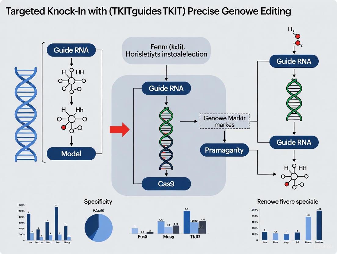
The TKIT approach utilizes two guide RNAs that create double-strand breaks in non-coding regions flanking the target exon—typically within the 5'-untranslated region (UTR) and downstream intronic sequences [8]. This strategic positioning ensures that the protein-coding sequence remains completely untouched by CRISPR/Cas9 activity, thereby eliminating the risk of INDEL mutations within functionally critical domains. The method employs a donor DNA fragment containing the modified exon (with inserted tag) flanked by the same guide RNA target sequences, but in switched orientation and flipped sequence compared to the genomic DNA [8].
This "switch-and-flip" design in the donor DNA is crucial for promoting correct orientation knock-in through non-homologous end joining (NHEJ). When the donor integrates in the reverse orientation, the guide RNA and PAM sequences remain intact, allowing for repeated Cas9 cleavage and subsequent re-attempts at correct integration until either proper orientation is achieved or INDELs destroy the guide recognition sites [8]. This innovative mechanism significantly increases the probability of successful forward-orientation knock-in compared to conventional approaches.
Key Advantages Over Conventional Methods
- Protection of Coding Integrity: By targeting non-coding regions, TKIT completely avoids INDEL mutations in the coding sequence, a significant advantage over HITI (Homology-Independent Target Integration) and other NHEJ-based methods that directly edit coding regions [8].
- Precision Placement: TKIT enables absolute control over the amino acid sequence surrounding the insertion site, allowing for precise tag placement without introducing extraneous amino acids or deleting functional residues [8].
- Expanded Guide RNA Options: Targeting non-coding regions substantially increases the number of potential high-quality guide RNAs, as researchers are not limited to sites within the constrained coding sequence [8].
- Functionality in Post-Mitotic Cells: Unlike HDR-based approaches, TKIT operates efficiently in non-dividing cells such as neurons, making it particularly valuable for neuroscience research [8].
Table: Comparison of TKIT with Conventional Genome Editing Approaches
| Editing Feature | TKIT Approach | Conventional HDR | HITI/NHEJ-based |
|---|---|---|---|
| Target Region | Non-coding flanks | Coding sequence | Coding sequence |
| INDEL Risk in CDS | None | Low to moderate | High |
| Suitable for Post-mitotic Cells | Yes | No | Yes |
| Insertion Precision | High | High | Variable |
| Guide RNA Availability | Expanded options | Limited by CDS | Limited by CDS |
| Maximum Efficiency (Neurons) | Up to 42% [8] | Very low [8] | 15-25% [8] |
Quantitative Performance Data
TKIT has demonstrated remarkable efficiency across multiple experimental applications. In proof-of-concept studies targeting endogenous synaptic proteins in mouse primary cultured neurons, TKIT achieved knock-in efficiencies of up to 42% when labeling GluA2 AMPA receptor subunits with Super Ecliptic pHluorin (SEP) [8]. This represents a substantial improvement over conventional HITI-based methods, which typically achieve 15-25% efficiency in similar applications while carrying the risk of coding sequence damage.
The methodology has successfully tagged various AMPA and NMDA receptor subunits, including GluA1, GluA2, GluA3, GluN1, and GluN2A, with diverse tags such as SEP, HALO, and Myc tags, demonstrating its versatility across different targets and labeling strategies [8]. Importantly, TKIT-edited neurons exhibited normal synaptic morphology and receptor trafficking, confirming that the approach preserves endogenous protein function while enabling precise labeling.
Table: TKIT Performance Across Different Experimental Applications
| Application Context | Target Molecule | Tag | Efficiency | Validation |
|---|---|---|---|---|
| Primary Mouse Neurons | GluA2 (AMPAR) | SEP | Up to 42% | Spine localization, IF [8] |
| In Utero Electroporation | GluA2 (AMPAR) | SEP | Functional | Two-photon imaging [8] |
| Adult Mouse AAV Injection | GluA2 (AMPAR) | SEP | Functional | In vivo visualization [8] |
| Rat Primary Neurons | GluA2 (AMPAR) | SEP | Comparable to mouse | Cross-species validation [8] |
| FRAP Analysis | Endogenous AMPARs | SEP | N/A | Receptor mobility studies [8] |
Detailed Experimental Protocol
TKIT Workflow for Endogenous Protein Tagging in Neurons
The following protocol outlines the specific methodology for tagging endogenous GluA2 with SEP using TKIT in primary mouse cortical neurons, as described in the foundational TKIT research [8].
Step-by-Step Methodology
Guide RNA Design and Selection (Days 1-2)
- Identify Target Regions: Select non-coding sequences approximately 100 bp away from splice junctions in the 5'-UTR and intron 1-2 of the Gria2 gene (encoding GluA2) [8]. This distance preserves normal mRNA processing while providing sufficient flanking sequence for efficient editing.
- Guide RNA Criteria: Choose guides with high on-target scores and minimal off-target potential using established algorithms. For Gria2 targeting, guides were designed to cut within the 5'-UTR ( upstream of the coding sequence) and within intron 1-2 (downstream of the signal peptide encoding region) [8].
- Control for Coding Integrity: Verify that neither guide RNA targets the protein-coding sequence or critical regulatory elements.
Donor DNA Construction (Days 2-4)
- Template Assembly: Construct a donor DNA fragment containing:
- The endogenous Gria2 sequence from 5'-UTR to intron 1-2
- SEP tag inserted immediately after the signal peptide encoding sequence
- The same two guide RNA target sequences as genomic DNA, but with opposite locations and flipped sequences (switch-and-flip design) [8]
- Vector Cloning: Clone the donor fragment into an appropriate expression vector with necessary regulatory elements for neuronal expression.
Neuronal Transfection and Expression (Days 5-14)
- Cell Preparation: Plate primary mouse cortical neurons at appropriate density and maintain until DIV7-9, ensuring healthy neuronal cultures [8].
- Transfection Mixture: Co-transfect neurons with three components:
- CRISPR/Cas9 construct expressing SpCas9 and both guide RNAs
- SEP-GluA2 donor DNA fragment
- mCherry expression plasmid for visualization of transfected cell morphology [8]
- Optimal Ratios: Use a 2:1:1 mass ratio (Cas9/guides:donor:marker) for optimal knock-in efficiency, as empirically determined in the foundational study [8].
- Incubation: Maintain transfected neurons for 5-7 days (until DIV14-16) to allow for protein turnover and robust expression of edited receptors.
Validation and Functional Assessment (Days 15-21)
- Live Imaging: Visualize SEP fluorescence using standard GFP filter sets; successful knock-in should show punctate signal concentrated in dendritic spines, consistent with AMPAR localization [8].
- Immunofluorescence Validation: Perform co-staining with antibodies against GFP and the C-terminus of GluA2 to confirm co-localization of SEP signal with endogenous GluA2 [8].
- Splicing Integrity: Extract bulk mRNA from transfected neurons (DIV19), perform RT-PCR across the edited region, and sequence to verify intact splice junctions between exon 1 and exon 2 [8].
- Functional Assessment: Compare expression levels of SEP-GluA2 with endogenous GluA2 in non-transfected neighboring neurons to ensure physiological expression levels [8].
Research Reagent Solutions
Table: Essential Reagents for TKIT Implementation
| Reagent Category | Specific Examples | Function in TKIT Protocol | Considerations |
|---|---|---|---|
| CRISPR Components | SpCas9, Guide RNA constructs | Creates targeted DSBs in non-coding flanks | Codon-optimize for target cells; verify nuclear localization |
| Donor DNA Template | SEP-GluA2 fragment with switched-flipped guides | Provides template for precise knock-in | Include homology arms of appropriate length; verify switch-flip design |
| Delivery Vectors | AAV, in utero electroporation, transfection reagents | Introduces editing components into cells | Optimize for cell type; consider size constraints for AAV packaging |
| Visualization Markers | mCherry, eGFP, HALO tags | Identifies transfected cells and edited proteins | Select spectrally distinct fluorophores for multiplexing |
| Validation Reagents | Anti-GFP, anti-GluA2 C-terminal antibodies | Confirms successful knock-in and protein integrity | Verify antibody specificity; use C-terminal tags for endogenous detection |
| Cell Type-Specific Reagents | Primary neuron culture media, viral tropism modifiers | Supports viability of target cells | Optimize for post-mitotic cells; consider developmental expression timing |
Technical Considerations and Optimization
Critical Parameters for Success
- Guide RNA Placement: Position guide RNA cut sites approximately 100 bp from exon-intron boundaries to avoid disrupting RNA splicing machinery while maintaining efficient editing [8]. The exact distance may require optimization for different gene targets.
- Donor Design Fidelity: Meticulously implement the "switch-and-flip" design in the donor DNA, as this significantly enhances correct orientation knock-in by allowing re-cleavage of reverse-integrated donors [8].
- Cell Health Maintenance: Ensure high viability of primary neuronal cultures throughout the procedure, as post-mitotic cells are particularly vulnerable to extended manipulation and CRISPR/Cas9 toxicity.
- Expression Level Validation: Always compare expression levels of knocked-in proteins with endogenous levels in neighboring cells to confirm physiological expression and avoid misinterpretation due to overexpression artifacts [8].
Troubleshooting Common Challenges
- Low Knock-in Efficiency: Optimize the ratio of CRISPR components to donor DNA; typically, a slight excess of donor DNA (2:1 donor:Cas9) improves efficiency. Verify guide RNA activity using surrogate reporter systems before full implementation.
- Unexpected Phenotypes: Always include controls transfected with donor DNA alone to confirm that observed phenotypes require CRISPR activity rather than random integration events [8].
- Imaging Challenges: For pH-sensitive tags like SEP, establish proper imaging conditions (e.g., pH buffering) to ensure accurate signal detection, particularly for surface-exposed proteins like AMPARs [8].
Diagram: TKIT's Self-Correcting Mechanism Through Repeated Cleavage Cycles
The TKIT methodology represents a paradigm shift in precision genome editing by strategically repositioning the editing machinery from vulnerable coding sequences to protective non-coding flanks. This approach achieves unprecedented specificity and preservation of coding integrity while maintaining high efficiency in challenging cell types like neurons. The foundational principle of targeting non-coding regions provides a versatile framework that can be adapted to diverse research contexts, from basic neuroscience to therapeutic development. As genome editing continues to evolve, TKIT's core innovation—protecting coding sequences through strategic non-coding targeting—offers a robust template for future methodological advances in precise genetic manipulation.
Decoding the 'Switch-and-Flip' Donor Design for Orientation-Specific Integration
The "Switch-and-Flip" donor design represents a significant innovation in CRISPR-Cas9-mediated precise genome editing, particularly within the Targeted Knock-In with Two (TKIT) guides framework. This technical note elucidates the molecular mechanism underlying this design, which ensures unidirectional integration of donor DNA fragments into target genomic loci. By strategically inverting and flipping guide RNA sequences within the donor template, this approach exploits the non-homologous end joining (NHEJ) repair pathway to achieve orientation-specific knock-in with markedly improved efficiency. We provide comprehensive experimental protocols, quantitative performance data, and visualization tools to facilitate the adoption of this methodology for labeling endogenous proteins, generating disease models, and advancing therapeutic development.
A fundamental limitation of conventional CRISPR-Cas9-mediated knock-in strategies is their inability to control the orientation of integrated DNA fragments. Traditional NHEJ-based integration results in random insertion orientations, as the cellular repair machinery ligates DNA ends without regard for directionality. This presents a particular challenge for applications requiring precise transcriptional control, such as endogenous gene tagging or the insertion of bidirectional expression cassettes. The "Switch-and-Flip" donor design, implemented within the broader TKIT framework, directly addresses this limitation through a sophisticated molecular strategy that ensures unidirectional integration [9].
The TKIT approach fundamentally differs from conventional knock-in methods by targeting non-coding regions flanking the exonic sequence to be modified, thereby protecting the coding sequence from INDEL mutations and providing absolute control over the sequence surrounding the knock-in site [9]. Within this framework, the "Switch-and-Flip" mechanism serves as the core innovation that enforces orientation-specific integration, overcoming a critical barrier in precision genome editing.
The "Switch-and-Flip" Mechanism: Principles and Design
Core Molecular Components
The "Switch-and-Flip" system functions through several key molecular components that operate in concert:
- Dual guide RNAs: Two sgRNAs targeting non-coding regions upstream and downstream of the coding sequence to be edited, typically located approximately 100 bp away from splice junctions to avoid disrupting mRNA processing [9].
- Cas9 Nuclease: The standard Streptococcus pyogenes Cas9 enzyme that creates double-strand breaks at the genomic targets specified by the guide RNAs.
- Donor DNA Fragment: Contains the endogenous sequence with the desired modification (e.g., fluorescent protein tag), flanked by the same two guide sequences as the genome, but with their positions switched and their sequences inverted relative to the genomic DNA [9].
The critical innovation lies in the strategic arrangement of the guide sequences within the donor DNA. By "switching" their positions (the 5' guide is placed at the 3' end and vice versa) and "flipping" their orientation (the sequences are inverted), the system creates a self-correcting mechanism that favors forward orientation integration [9].
Molecular Workflow and Self-Correction Mechanism
The "Switch-and-Flip" mechanism operates through a cyclic process of cutting and re-cutting until correct orientation is achieved:
The diagram above illustrates the self-correcting mechanism of the "Switch-and-Flip" system. When the donor integrates in the reverse orientation, the guide RNA sequences and their associated PAM sites remain intact and properly oriented for recutting by Cas9. This allows for repeated integration attempts until either the correct orientation is achieved or INDEL mutations destroy the guide binding sites, terminating the cycle [9]. This elegant molecular logic ensures that only correctly oriented integrations persist in the edited cell population.
Quantitative Performance Data
The performance of the "Switch-and-Flip" donor design has been quantitatively evaluated across multiple experimental systems. The table below summarizes key efficiency metrics reported in foundational studies:
Table 1: Efficiency Metrics of "Switch-and-Flip" Mediated Knock-In
| Target System | Integration Efficiency | Tag Type | Application | Reference |
|---|---|---|---|---|
| Mouse Primary Neurons | Up to 42% | SEP (pH-sensitive GFP) | Endogenous AMPAR labeling | [9] |
| Human Cell Lines (CCR5 Locus) | 33% (clonal analysis) | EGFP | Targeted integration via single crossover | [10] |
| Zebrafish (Composite Tag) | Up to 21% germline transmission | FLAGx3-Bio-HiBiT | Endogenous protein tagging | [11] |
| Human Cell Lines (CCR5 Locus) | 10% (bulk population) | EGFP | Single crossover recombination | [10] |
The "Switch-and-Flip" approach demonstrates particular advantage in non-dividing cells such as neurons, where homology-directed repair (HDR) functions inefficiently. In primary mouse cortical cultures, the system achieved labeling of endogenous synaptic proteins with various tags at efficiencies up to 42% [9]. This represents a substantial improvement over conventional HDR-based methods in post-mitotic cells.
When compared with alternative knock-in strategies, the "Switch-and-Flip" method shows competitive efficiency while maintaining orientation specificity:
Table 2: Comparison of Knock-In Strategies for Large Fragment Integration
| Method | Mechanism | Orientation Control | Typical Efficiency | Best Application Context |
|---|---|---|---|---|
| "Switch-and-Flip" (TKIT) | NHEJ with cyclic recutting | Yes | Up to 42% | Non-dividing cells, precise endogenous tagging |
| HDR (Double Crossover) | Homology-directed repair | Yes | 10⁻⁶–10⁻⁵ [10] | Dividing cells, small modifications |
| NHEJ-based (Conventional) | Direct end joining | No | 0.17–0.45% [10] | Rapid knock-in without orientation requirement |
| Single Crossover Recombination | Campbell-like recombination | Direction-dependent | 33% (clonal) [10] | Large fragment integration in human cells |
Experimental Protocol for TKIT with "Switch-and-Flip" Design
Guide RNA and Donor Design Specifications
Target Selection Criteria:
- Identify non-coding regions approximately 100 bp upstream and downstream of the coding sequence to be modified [9]
- Ensure target sites are at least 50-100 bp away from exon-intron boundaries to preserve RNA splicing [9]
- Select guides with high on-target efficiency scores and minimal predicted off-target effects
Donor DNA Construction:
- Include the endogenous genomic sequence with the desired modification (e.g., fluorescent protein tag)
- Position the two guide sequences at the ends of the donor fragment, but switch their positions (5' genomic guide at 3' of donor, and vice versa) compared to the genomic context
- Invert the sequence orientation of both guides within the donor relative to their genomic configuration [9]
- Maintain intact PAM sequences adjacent to each guide within the donor template
Laboratory Implementation Protocol
Day 1: Cell Preparation
- Plate primary mouse cortical neurons at DIV7-9 or appropriate mammalian cells relevant to your study
- Maintain cells in complete culture medium (e.g., DMEM with 10% FBS) [12]
- Ensure cells are 60-80% confluent at time of transfection
Day 2: Transfection
- Prepare transfection mixture containing:
- Incubate mixture for 15-20 minutes at room temperature
- Apply to cells following standard transfection protocols for your cell type
Days 3-14: Selection and Expression
- Replace medium 6-24 hours post-transfection
- For stable integration, begin antibiotic selection 48 hours post-transfection if using selection markers
- Culture cells for 10-14 days to allow protein expression and maturation
Day 14+: Validation and Analysis
- Live-image transfected neurons or cells for tag expression (e.g., SEP signal)
- Fix cells for immunofluorescence validation using antibodies against the tag and endogenous protein
- Perform junction PCR and Sanger sequencing to confirm precise integration
- Extract bulk mRNA for RT-PCR to verify intact RNA splicing [9]
The Scientist's Toolkit: Essential Reagents
Table 3: Key Research Reagents for "Switch-and-Flip" Experiments
| Reagent/Solution | Function | Specifications & Alternatives |
|---|---|---|
| SpCas9-NLS | Creates DSBs at target genomic loci | Nuclear localization signal (NLS) essential; alternative: HiFi Cas9 variants for reduced off-target effects |
| Long ssDNA Donor | Repair template for knock-in | Can be chemically synthesized; lssDNA shows superior specificity for on-target integration [11] |
| Polyethylenimine (PEI) | Transfection reagent | Linear, MW 25,000, transfection grade [12] |
| Dual sgRNA Expression Plasmid | Targets non-coding flanking regions | May be expressed as single transcript with ribozyme or tRNA processing elements |
| Selection Antibiotics | Enrichment of transfected cells | Puromycin (2 µg/mL) common for mammalian cells [12] |
| FACS Equipment | Analysis and sorting of edited cells | Enables quantification of knock-in efficiency and isolation of clonal populations |
Applications in Biomedical Research
The "Switch-and-Flip" methodology enables diverse research applications with particular strength in:
Endogenous Protein Labeling: The system has been successfully used to tag AMPA and NMDA receptor subunits with Super Ecliptic pHluorin (SEP) in primary neurons, enabling visualization of endogenous receptor trafficking in live cells [9]. This approach preserves natural transcriptional and post-transcriptional regulation while eliminating overexpression artifacts.
In Vivo Imaging: Utilizing in utero electroporation or AAV viral injections, TKIT with "Switch-and-Flip" design can label endogenous proteins in living mice, enabling two-photon microscopy visualization of endogenous AMPA receptors in vivo [9].
Disease Modeling: The precise integration capability facilitates generation of patient-specific disease models by introducing pathological mutations into relevant genomic contexts while maintaining endogenous expression patterns.
Therapeutic Development: The orientation control provided by this system is particularly valuable for knock-in of therapeutic transgenes where proper transcriptional regulation is critical for safe and effective expression.
Troubleshooting and Optimization Guidelines
Low Knock-In Efficiency:
- Validate guide RNA cutting efficiency using T7E1 assay or tracking of indels by decomposition (TIDE)
- Optimize donor DNA concentration (typical range 1:1 to 3:1 donor-to-CRISPR ratio)
- Consider chemical modifications to synthetic gRNAs to enhance stability and efficiency [13]
Improper Orientation Integration:
- Verify the "switch-and-flip" configuration of guide sequences in donor template
- Confirm integrity of PAM sequences adjacent to guide sequences in donor
- Extend culture time post-transfection to allow for multiple cycles of correction
Cell Viability Issues:
- Titrate Cas9 expression levels to minimize cytotoxicity
- Utilize ribonucleoprotein (RNP) delivery instead of plasmid DNA to limit Cas9 exposure duration
- Implement caspase inhibitors or other viability-enhancing compounds during transfection
The "Switch-and-Flip" donor design represents a sophisticated solution to the challenge of orientation-specific integration in precise genome editing. When implemented within the TKIT framework, this approach enables efficient, precise labeling of endogenous proteins with broad applications across neuroscience, drug development, and therapeutic discovery.
Within the rapidly evolving field of precise genome editing, the comparison of CRISPR-Cas9 and Transcription Activator-Like Effector Nuclease (TALEN) technologies is critical for research and therapeutic development. Targeted Knock-In with Two (TKIT) guides represents a sophisticated approach for precise genetic alterations, demanding tools with high specificity, flexibility, and a favorable safety profile. While CRISPR-Cas9 has gained widespread adoption for its simplicity, TALEN technology presents distinct and powerful advantages in the context of advanced strategies like TKIT. This application note details the key advantages of TALENs, focusing on their inherent resistance to insertions and deletions (INDELs), unparalleled flexibility in guide RNA (gRNA) selection due to the absence of protospacer adjacent motif (PAM) constraints, and their broader applicability across diverse organisms, including the unique capacity to edit mitochondrial DNA. Understanding these characteristics is essential for researchers and drug development professionals to select the optimal platform for precise genome engineering applications, particularly where accuracy is paramount [14] [15] [16].
Key Advantages of TALENs for Precision Editing
High Specificity and Resistance to INDELs
A paramount concern in therapeutic genome editing is the introduction of unintended, genotoxic mutations. While all editing tools can cause off-target effects, the fundamental mechanisms of TALENs confer a significantly higher specificity profile compared to first-generation CRISPR-Cas9 systems.
- DNA-Protein Interaction vs. RNA-DNA Hybridization: TALENs recognize their target site through direct protein-DNA interactions. The DNA-binding domain consists of tandem repeats, each specifically recognizing a single base pair via Repeat Variable Diresidues (RVDs) [14] [15]. This highly specific binding mechanism, which occurs in the major groove of DNA, is less tolerant of sequence mismatches than the RNA-DNA hybridization used by CRISPR-Cas9, which can tolerate up to five base-pair mismatches, leading to a higher propensity for off-target cleavage [17] [18].
- Dimeric Nuclease Activity: TALENs function as obligate dimers. A pair of TALEN proteins must bind to opposite DNA strands, flanking the target site, for the FokI nuclease domains to dimerize and create a double-strand break (DSB) [17] [18]. This requirement effectively doubles the length of the target sequence recognition (typically ~36-56 bp total for both binding sites), a length that is statistically unique in any genome, thereby drastically reducing the likelihood of off-target activity [14] [18].
- Reduced Structural Variations: Beyond small INDELs, CRISPR-Cas9 has been associated with large, on-target structural variations (SVs), including kilobase- to megabase-scale deletions and chromosomal translocations, which pose substantial safety concerns for clinical applications [19]. Although any DSB-inducing platform can cause SVs, the high-specificity binding and cleavage mechanism of TALENs make such detrimental, large-scale genomic aberrations less frequent [19].
Table 1: Comparison of Off-Target and Structural Variation Profiles between CRISPR-Cas9 and TALENs
| Feature | CRISPR-Cas9 | TALENs |
|---|---|---|
| Primary Targeting Mechanism | RNA-DNA hybridization [14] | Protein-DNA interaction [14] |
| Mismatch Tolerance | High (up to 5 bp) [17] | Low [17] |
| Typical Total Binding Length | ~20 bp (per gRNA) [17] | ~36-56 bp (for a TALEN pair) [14] [18] |
| Reported Frequency of Off-Target Mutagenesis | High in some studies (≥50%) [14] | Low; often undetected in targeted analyses [18] |
| Risk of Large Structural Variations | More documented, especially with NHEJ inhibition [19] | Also present, but less frequent due to specific cleavage [19] |
Flexibility in gRNA and Target Site Selection
The design of precise knock-in strategies, such as those in TKIT workflows, is often constrained by the genomic context of the target locus. TALENs offer superior flexibility in these scenarios.
- No PAM Sequence Requirement: The targeting of the most commonly used CRISPR-Cas9 system from Streptococcus pyogenes (SpCas9) is dependent on the presence of a 5'-NGG-3' PAM sequence immediately adjacent to the target site [18] [16]. This requirement can severely limit the number of potential target sites, especially when a specific nucleotide needs to be edited within a narrow window. In contrast, TALENs have no PAM constraint, allowing researchers to design proteins to target virtually any genomic sequence, providing unparalleled design freedom [15] [20].
- Precise DSB Placement for Enhanced HDR: The absence of a PAM allows TALEN binding sites to be positioned in closer proximity to the intended edit. This closer placement of the induced DSB to the site of homology-directed repair (HDR) can lead to improved knock-in efficiency compared to CRISPR-Cas9, where the PAM dictates the cleavage location, which may be distantly located from the desired edit [20].
- Overcoming Epigenetic Barriers: TALENs can be designed to avoid or overcome epigenetic modifications like cytosine methylation (common at CpG islands). While CpG methylation can inhibit TALEN binding, this can be circumvented during the design phase by selecting target sites devoid of methylated bases or by using specific RVDs (e.g., N*) that can interact with methylated cytosines [18]. CRISPR-Cas9 is generally considered less sensitive to DNA methylation [18].
Applicability Across Broader Organisms and Organelles
The utility of a genome-editing tool is measured by its performance across diverse experimental and therapeutic systems. TALENs demonstrate distinct advantages in several contexts.
- Mitochondrial Genome Editing (mitoTALENs): A unique and powerful application of TALENs is the ability to target and edit mitochondrial DNA (mtDNA). The guide RNA of the CRISPR-Cas9 system is difficult to import into the mitochondria, making CRISPR ineffective for this purpose. MitoTALENs, however, can be engineered with mitochondrial localization signals to directly manipulate mtDNA, opening avenues for researching and treating mitochondrial diseases [14].
- Efficacy in Diverse Cell Types and Organisms: Both TALENs and CRISPR have been successfully used in a wide range of organisms, including plants, livestock (cows, pigs, chickens), and human cell lines [14] [15]. TALENs have proven highly efficient in plant systems, for instance, where they have been used to enhance the synthesis of valuable secondary metabolites by precisely editing biosynthetic pathways [15].
- Therapeutic Precision: In clinical applications, where safety is the highest priority, the high specificity of TALENs makes them an indispensable tool. Their lower off-target profile is a critical advantage for therapeutic genome editing, as it minimizes the risk of genotoxic side effects that could lead to oncogenesis [14] [19].
Diagram 1: A comparative overview of TALEN and CRISPR-Cas9 fundamental mechanisms and their direct implications for key editing characteristics. Green nodes represent distinct advantages of TALENs, while red nodes indicate relative limitations of CRISPR-Cas9 in these areas.
To aid in the objective evaluation and selection of the appropriate genome editing tool, the following tables summarize key performance metrics and design parameters.
Table 2: Comparative Efficiency and Specificity Metrics of Genome Editing Tools
| Parameter | CRISPR-Cas9 | TALENs | Notes |
|---|---|---|---|
| On-Target Indel Efficiency | High (can be >70%) [18] | High (e.g., ~33% and higher reported) [18] | Efficiency is highly dependent on cell type, delivery, and target site. |
| Off-Target Mutation Frequency | High prevalence (≥50%) reported in some studies [14] | Low; often undetected in targeted analyses [18] | CRISPR off-targets can be reduced with high-fidelity variants and paired nickases [17] [19]. |
| HDR Efficiency | Moderate, but can be limited by PAM location [20] | High potential due to flexible target site selection and close DSB placement [20] | HDR is inherently less efficient than NHEJ in human cells [16]. |
| Relative Cost & Ease of Construction | Low cost; simple gRNA cloning [17] [20] | Higher cost; more complex protein engineering [20] | TALEN construction has been streamlined with modular kits [17] [18]. |
Table 3: Key Design and Targeting Constraints
| Design Feature | CRISPR-Cas9 (spCas9) | TALENs |
|---|---|---|
| Target Recognition Length | ~20 bp (per gRNA) [17] | ~18 bp (per monomer, ~36 bp total for a pair) [17] [18] |
| PAM/PAM-like Requirement | Yes (5'-NGG-3') [18] [16] | No [15] [20] |
| Methylation Sensitivity | Less sensitive [18] | Sensitive to CpG methylation (can be designed around) [18] |
| Multiplexing Capacity | High (via multiple gRNAs) [21] | Low (due to large protein size and complex cloning) [20] |
Detailed Experimental Protocols
Protocol 1: Designing and Assembling TALEN Constructs for a TKIT Experiment
This protocol outlines the steps to design and clone TALEN pairs for a targeted knock-in experiment.
Research Reagent Solutions:
- TALEN Kit: Commercial kits (e.g., from companies like GeneCopoeia or Thermo Fisher Scientific) provide pre-validated, modular RVD plasmids for streamlined assembly [18] [20].
- FokI Nuclease Plasmid: Vector backbone containing the catalytic domain of the FokI restriction enzyme for nuclease activity [14] [15].
- Delivery Vector: AAV, lentiviral, or plasmid vectors suitable for your target cell type. Note: TALEN's large size can be challenging for AAV packaging [14] [22].
Procedure:
- Target Site Identification: Using genomic sequence data, select a 30-40 bp target region encompassing your desired edit. The spacer sequence (where the cut will occur) should be 14-20 bp long [17] [18].
- TALEN Pair Design: Design two TALEN binding sites, each 15-20 bp long, flanking the spacer. Avoid target sites with high CpG methylation unless using specific RVDs. Use the RVD code: NI for A, HD for C, NN for G, and NG for T to define the amino acid sequence of the DNA-binding domain for each TALEN [14] [18].
- In Silico Validation: Use software tools (e.g., Thermo Fisher's TrueDesign Genome Editor) to validate specificity and check for potential off-target sites in the relevant genome [20].
- Modular Assembly: Perform a Golden Gate assembly reaction using a commercial TALEN kit. This involves ligating the pre-defined RVD modules into the FokI nuclease backbone plasmid in a specific order corresponding to your target sequence [18] [21].
- Sequence Verification: Confirm the final TALEN plasmid sequence via Sanger sequencing. This is crucial due to the repetitive nature of the TALEN sequence, which can pose challenges for cloning and sequencing fidelity [18].
Protocol 2: Delivering TALENs and Donor Template for HDR in Cultured Cells
This protocol describes the co-delivery of TALENs and a donor DNA template to achieve precise knock-in via HDR.
Research Reagent Solutions:
- TALEN Expression Plasmids: The two verified TALEN plasmids from Protocol 1.
- HDR Donor Template: A single-stranded oligodeoxynucleotide (ssODN) or a double-stranded DNA plasmid containing the desired modification flanked by homology arms (typically 800-1000 bp each for plasmid donors) [18].
- Transfection Reagent: A reagent suitable for your cell type (e.g., lipofection, electroporation kits). For hard-to-transfect cells, consider nucleofection [20].
Procedure:
- Cell Preparation: Seed and culture the target cells to reach 70-90% confluency at the time of transfection.
- Transfection Complex Formation: For a single well of a 24-well plate, prepare a transfection mixture containing:
- 0.5 µg of each TALEN plasmid (1 µg total)
- 100-200 pmol of ssODN donor OR 0.5-1 µg of donor plasmid
- Optimum volume of transfection reagent, according to the manufacturer's protocol.
- Incubate with the cells.
- Post-Transfection Culture: Replace the transfection medium with fresh culture medium after 6-24 hours.
- Harvest and Validation: Harvest cells 48-72 hours post-transfection for initial analysis of editing efficiency.
Protocol 3: Validating Knock-In and Screening for Off-Target Effects
A critical step to confirm successful on-target editing and assess specificity.
Research Reagent Solutions:
- Surveyor Nuclease or T7 Endonuclease I: Enzymes for mismatch detection assays to identify heterogeneous pools of edited cells [22].
- PCR Reagents: For amplifying the genomic target region and potential off-target sites.
- Sanger Sequencing or Next-Generation Sequencing (NGS) Reagents: For definitive confirmation of edits and comprehensive off-target profiling.
Procedure:
- On-Target Efficiency Analysis (Initial Screening):
- Extract genomic DNA from transfected cells.
- PCR-amplify the genomic region surrounding the target site.
- Use the T7E1 or Surveyor assay on the PCR product. Cleaved bands indicate the presence of indels, suggesting successful DSB and NHEJ/HDR repair [22].
- Clonal Isolation and Validation:
- If a clonal population is required, perform serial dilution of the transfected cell pool to isolate single cells.
- Expand monoclones for 2-3 weeks.
- Screen monoclones by PCR and T7E1 assay, then sequence the top candidates via Sanger sequencing to identify clones with the precise HDR-mediated knock-in and biallelic modification [22].
- Off-Target Assessment:
- Perform an in silico analysis to predict potential off-target sites based on sequence similarity to the TALEN binding sites.
- Amplify the top 10-20 predicted off-target loci from genomic DNA of edited clonal lines and a control line.
- Analyze these amplicons by deep sequencing (NGS) to quantify the frequency of indels at these sites, confirming the high specificity of TALENs [18] [19].
Diagram 2: A comprehensive workflow for a TALEN-mediated TKIT experiment, from initial design and assembly to final validation. Key protocol steps involving specialized reagents or critical decisions are highlighted in yellow.
The Scientist's Toolkit: Essential Reagents for TALEN-Mediated Editing
Table 4: Key Research Reagent Solutions for TALEN Experiments
| Reagent / Solution | Function / Description | Example Suppliers / Notes |
|---|---|---|
| Custom TALEN Constructs | Engineered plasmids encoding the TAL effector DNA-binding domain fused to FokI nuclease. | GeneCopoeia, Thermo Fisher Scientific; available as ready-to-use expression vectors [18] [20]. |
| TALEN Modular Assembly Kits | Kits containing pre-made RVD modules for streamlined, cost-effective Golden Gate assembly of custom TALENs. | Addgene (distributes academic kits); commercial suppliers [18]. |
| HDR Donor Templates | Single-stranded ODNs or double-stranded DNA plasmids with homology arms, serving as the repair template for precise knock-in. | Synthesized by commercial oligo/plasmid synthesis companies (e.g., IDT, Thermo Fisher) [18]. |
| High-Efficiency Transfection Reagents | Chemical-based reagents (e.g., lipofection) for delivering TALEN constructs and donor templates into cultured cells. | Thermo Fisher (Lipofectamine), Promega, Roche [20]. |
| Electroporation/Nucleofection Systems | Instrument systems for physically delivering constructs into hard-to-transfect cell types (e.g., primary cells, stem cells). | Lonza Nucleofector, Bio-Rad Gene Pulser [20]. |
| Genomic DNA Isolation Kit | For high-quality, PCR-ready genomic DNA extraction from edited cells. | QIAGEN, Thermo Fisher, Promega. |
| T7 Endonuclease I / Surveyor Nuclease | Mismatch cleavage detection enzymes for initial, rapid assessment of editing efficiency in a mixed cell population. | New England Biolabs (NEB), Integrated DNA Technologies (IDT) [22]. |
| NGS-based Off-Target Kit | Kits designed for targeted amplification and deep sequencing of potential off-target sites genome-wide. | IDT (xGen), Illumina; used for comprehensive safety profiling [19]. |
Implementing TKIT: A Step-by-Step Protocol from Donor Design to In Vivo Application
Precise genome editing requires strategies that maximize on-target efficiency while minimizing unintended mutations. The Targeted Knock-In with Two (TKIT) guides approach represents a significant advancement in this field by utilizing two guide RNAs that cut genomic DNA in flanking non-coding regions, enabling precise insertion of genetic payloads while protecting the coding sequence from insertion/deletion (INDEL) mutations [8]. This method is particularly valuable for post-mitotic cells like neurons, where traditional homology-directed repair (HDR) methods are inefficient [8]. By targeting the 5' untranslated region (5'UTR) and intronic regions, TKIT overcomes limitations associated with coding sequence targeting, including frameshift mutations and PAM sequence constraints, while providing greater flexibility in guide RNA selection [8].
The strategic positioning of gRNAs in non-coding regions enables absolute control over the sequence surrounding the knock-in site and preserves the integrity of the protein-coding sequence. This technical note provides comprehensive guidance on gRNA selection and positioning for optimized TKIT experiments, supported by quantitative data, detailed protocols, and practical visualization tools.
Strategic Positioning of gRNAs for Optimal Knock-In Efficiency
5'UTR-Targeting Knock-In Strategy
Targeting the 5'UTR for knock-in presents a unique opportunity for highly efficient protein tagging while maintaining endogenous regulatory control. Research demonstrates that a 5'UTR-targeting knock-in strategy enables the establishment of stable cell lines expressing tagged proteins with remarkable efficiencies ranging from 50% to 80% in antibiotic-selected cells [23]. This approach positions the knock-in cassette upstream of the native coding sequence, allowing expression under the control of the endogenous promoter while avoiding disruption of the protein-coding region.
The 5'UTR strategy demonstrates several advantages over traditional approaches. The localization of knock-in proteins is identical to that of endogenous proteins in wild-type cells and shows homogenous expression [23]. Moreover, expression from the endogenous promoter remains stable over long-term culture, addressing a significant limitation of systems relying on exogenous promoters [23]. This method has been successfully applied for tagging diverse proteins including Arl13b-Venus, Reep6-HA, and EGFP-alpha-tubulin, demonstrating its broad applicability [23].
Table 1: Efficiency Comparison of Knock-In Strategies
| Strategy | Target Region | Typical Efficiency | Key Advantages |
|---|---|---|---|
| 5'UTR-targeting [23] | 5' Untranslated Region | 50-80% | Maintains endogenous regulation; avoids protein disruption |
| TKIT guides [8] | 5'UTR + Intron | Up to 42% (in neurons) | Protects coding sequence from INDELs; precise insertion control |
| HDR-based coding sequence targeting [23] | Protein-coding exon | Often very low | Traditional approach; can be combined with selection markers |
| ROSA26 safe harbor [23] | Genomic safe harbor | Variable | Predictable expression; well-characterized locus |
TKIT Guide RNA Design Principles
The TKIT approach utilizes two guide RNAs strategically positioned in non-coding regions flanking the coding sequence of interest. Optimal design places one gRNA within the 5'UTR and the second within the first intron, typically approximately 100 base pairs away from splice junctions to avoid disrupting mRNA processing [8]. This positioning ensures the entire coding sequence can be replaced with a tagged version while preserving native splicing mechanisms.
The donor DNA fragment in TKIT contains the endogenous gene sequence with the desired tag addition, flanked by the same two guide RNA target sequences present in the genome, but in opposite orientation (switch-and-flip design) [8]. This design promotes forward insertion of the donor DNA through non-homologous end joining (NHEJ). If the donor inserts in the reverse orientation, the guide RNA and PAM sequences remain intact and can be cut again by Cas9, providing additional opportunities for correct orientation insertion [8].
Table 2: TKIT Guide RNA Design Parameters
| Parameter | Specification | Rationale |
|---|---|---|
| 5'UTR gRNA position [8] | ~100 bp from start codon | Avoids disruption of translation initiation elements |
| Intronic gRNA position [8] | ~100 bp from splice junctions | Preserves RNA splicing mechanisms |
| Homology arm length | Not required | TKIT utilizes NHEJ rather than HDR pathway |
| Donor design [8] | Switch-and-flip orientation | Promotes forward insertion through repeated cutting of reverse inserts |
| Tag positioning | After signal peptide (for secreted proteins) [8] | Ensves proper protein folding and localization |
| PAM consideration [24] | NGG for S. pyogenes Cas9 | Essential for Cas9 recognition and cutting |
Experimental Protocol for TKIT Guide Implementation
gRNA Design and Validation Workflow
Step 1: Target Site Selection
- Identify the 5'UTR region approximately 100 bp upstream of the start codon [8]
- Identify the first intronic region approximately 100 bp downstream from the exon-intron boundary [8]
- Use established gRNA design tools (e.g., Broad Institute GPP sgRNA Designer, CHOP-CHOP) to select optimal sequences [23] [24]
- Verify absence of known polymorphisms in target sequences
- Analyze potential off-target effects using specialized algorithms [24]
Step 2: gRNA Construction
- Clone each 20-bp target sequence into appropriate Cas9-expression plasmid vectors [23]
- For the 5'UTR-targeting gRNA, include the target sequence and 3-bp PAM sequence upstream of the fusion protein sequences in donor constructs [23]
- Apply typical Kozak sequence (GCCACC) for the target genes when designing donor templates [23]
Step 3: Donor DNA Design
- Construct donor plasmid vectors with removal of exogenous promoters (e.g., CMV) to ensure endogenous promoter control [23]
- For fluorescent protein tagging, use pEGFP backbone vectors or similar with the 20-bp target sequence and 3-bp PAM sequence sub-cloned upstream of the fusion proteins [23]
- Implement the "switch-and-flip" design where the two guides within the donor have opposite locations and flipped sequences compared to the genomic DNA [8]
Step 4: Validation of Knock-In Efficiency
- Transfect primary cultures (e.g., mouse cortical cultures at DIV7-9) with plasmids containing both guide RNAs, SpCas9, and the donor DNA fragment [8]
- Include a fluorescent marker (e.g., mCherry) for cell morphology identification [8]
- Assess knock-in efficiency 7-14 days post-transfection via live imaging and immunofluorescence [8]
- Confirm normal RNA splicing through RT-PCR and Sanger sequencing of splice junctions [8]
Quantitative Assessment of Editing Efficiency
The TIDE (Tracking of Indels by DEcomposition) method provides a simple, rapid, and cost-effective strategy to accurately quantify editing efficacy and simultaneously identify the predominant types of insertions and deletions (indels) in targeted cell pools [25]. This method requires only two parallel PCR reactions followed by a pair of standard capillary sequencing analyses, with the resulting sequencing traces analyzed using specialized decomposition algorithms [25].
For TKIT experiments, assess knock-in efficiency through:
- Fluorescence quantification: Compare signal intensity in transfected versus non-transfected cells
- Immunostaining: Verify co-localization of the tag (e.g., GFP) with the target protein (e.g., GluA2 C-terminus) [8]
- Functional validation: Confirm normal cellular localization and behavior of the tagged protein [23]
- Long-term stability: Monitor expression stability over multiple cell passages [23]
Research Reagent Solutions for TKIT Experiments
Table 3: Essential Reagents for TKIT Genome Editing
| Reagent Category | Specific Examples | Function & Application Notes |
|---|---|---|
| Cas9 Expression Systems [23] | CMV-Cas9-2A-GFP plasmid | Provides Cas9 nuclease and tracking; 2A peptide enables co-expression of fluorescent marker |
| gRNA Cloning Vectors [23] | pEGFP backbone vectors | Standardized backbones for gRNA expression and donor construct assembly |
| Donor Template Plasmids [23] | p5'UTRgRNA-Arl13b-Venus, p5'UTRgRNA-Reep6-HA | Custom donor constructs with specific tags; CMV promoter removed for endogenous regulation |
| Validation Tools [25] | TIDE web tool (http://tide.nki.nl) | Algorithm-based decomposition of sequencing traces for precise quantification of indels |
| Cell Culture Reagents [8] | Primary mouse cortical cultures | Relevant cellular models for testing knock-in efficiency, especially in post-mitotic cells |
| Selection Markers [23] | Neomycin-resistant gene expression cassette | Enriches for successfully edited cells when included in donor constructs |
Technical Considerations and Troubleshooting
Optimizing Knock-In Efficiency
Several factors significantly impact the success of TKIT experiments. When implementing 5'UTR-targeting strategies, ensure that:
- The Kozak sequence (GCCACC) is included for optimal translation initiation [23]
- AUG codons are removed from synthetic 5'UTRs by randomly mutating one of the three nucleotides to prevent generation of upstream open reading frames [26]
- 5'UTR length is optimized (approximately 100 bp shown effective) to balance regulatory element inclusion and practical constraints [26]
For challenging targets where efficiency remains low, consider:
- Utilizing high-fidelity Cas9 variants to reduce off-target effects
- Implementing fluorescence-activated cell sorting (FACS) to enrich successfully edited cells
- Incorporating antibiotic selection cassettes in the donor DNA for stable cell line generation [23]
Addressing Common Experimental Challenges
Low Knock-In Efficiency
- Verify gRNA cutting efficiency using TIDE analysis before proceeding with full TKIT experiment [25]
- Optimize donor DNA concentration and configuration (e.g., switch-and-flip design) [8]
- Consider cell cycle synchronization to enhance NHEJ activity
Protein Mislocalization
- Confirm tag positioning does not interfere with signal peptides or localization domains [8]
- Verify endogenous promoter-driven expression produces appropriate expression levels [23]
- Compare localization patterns with antibody staining of endogenous protein
Integration Site Analysis
- Perform PCR amplification across both integration junctions to verify precise insertion [8]
- Sequence the entire edited locus to rule offtarget integrations
- Validate mRNA splicing patterns through RT-PCR analysis of splice junctions [8]
The strategic selection and positioning of guide RNAs in 5'UTRs and introns, as implemented in the TKIT approach, provides a robust framework for precise genome editing with broad applications in functional genomics and therapeutic development.
Precise genome editing via homology-directed repair (HDR) enables the targeted integration of exogenous DNA sequences, such as fluorescent protein tags, affinity epitopes, or other genetic payloads, into specific genomic loci [27]. This process requires a donor DNA template containing the desired insert flanked by homology arms that facilitate recombination with the target genome [28]. The design of this donor DNA fragment is a critical determinant of knock-in efficiency, especially when combined with advanced CRISPR-Cas systems like the Targeted Knock-In with Two (TKIT) guides approach [29].
The advent of CRISPR-Cas9 technology has significantly simplified the creation of double-strand breaks (DSBs) at predetermined genomic sites, thereby stimulating cellular repair mechanisms [30]. While the error-prone non-homologous end joining (NHEJ) pathway often introduces indels, providing a donor template with homologous sequences can steer repair toward HDR for precise integration [31]. This protocol details the strategic design of donor DNA fragments, focusing on the optimization of homology arms and functional sequences to maximize HDR efficiency in TKIT-guided experiments for drug development and functional genomics applications [32].
Structural Components of a Donor DNA Fragment
A donor DNA template for HDR is composed of several key elements, each serving a distinct function in the recombination process. The central component is the cargo sequence, which can range from short epitope tags (e.g., FLAG, HA) to larger functional cassettes such as fluorescent reporters (e.g., GFP) or selectable markers [31]. This cargo is flanked by two homology arms—regions with sequence identity to the genomic target—which are essential for strand invasion and the recombination process [28].
The length of these homology arms must be carefully optimized based on the experimental system and cargo size. For large DNA fragment knock-ins (1–3 kb), studies have demonstrated that specially designed 3′-overhang double-strand DNA (odsDNA) donors harboring 50-nucleotide homology arms can achieve high efficiency when combined with CRISPR-Cas9 technology [30]. In zebrafish models, successful integration of large reporter genes like GFP typically requires longer double-stranded DNA fragments with homologous arms, each exceeding 2 kb [28].
Table 1: Recommended Homology Arm Lengths for Different Applications
| Application Context | Cargo Size | Recommended Arm Length | Donor Type | Key Considerations |
|---|---|---|---|---|
| Short sequence insertion (SSA) | < 100 bp | 50–200 nt | ssOligo | Higher efficiency but potentially lower accuracy [28] |
| Large fragment knock-in (LOCK method) | 1–3 kb | 50 nt | odsDNA (with 3′ overhangs) | Uses microhomology-mediated end joining; includes PT modifications [30] |
| Gene-sized KI in mammalian cells | ~1–3 kb | 50 nt | odsDNA | Combined with Cas9-PCV2 fusion protein [30] |
| HR in zebrafish (large reporters) | ~GFP | >2 kb | dsDNA | Requires longer arms for successful homologous recombination [28] |
Additional sequence modifications can enhance donor functionality. Incorporating phosphorothioate (PT) modifications at the 3′-overhangs of odsDNA donors can protect against exonuclease degradation and improve nuclear stability [30]. Furthermore, mutating the protospacer adjacent motif (PAM) sequence in the homology arms is crucial to prevent Cas9 from cleaving the donor template itself after integration [31].
Designing Homology Arms for Optimal Efficiency
Length Optimization Strategies
Homology arm length significantly influences HDR efficiency and requires careful balancing. Excessively long arms may complicate vector construction without substantially improving efficiency, while very short arms can dramatically reduce recombination rates [28]. The LOCK method demonstrates that with specific structural modifications, relatively short homology arms of 50 nucleotides can efficiently mediate the knock-in of gene-sized fragments (1–3 kb) in mammalian cells [30].
For more conventional dsDNA donors in zebrafish models, research indicates that homologous arms greater than 2 kb are recommended when inserting large reporter genes like GFP [28]. This length provides sufficient sequence context for the cellular recombination machinery to engage with the donor template. When designing homology arms, it is essential to amplify these sequences from the genomic DNA of the target organism to ensure perfect sequence identity, as even single-nucleotide polymorphisms can significantly reduce HDR efficiency [28].
Strategic Placement and Modifications
The placement of homology arms relative to the CRISPR-induced break site critically impacts recombination efficiency. The DSB should occur within the region spanned by the homology arms, preferably close to the center [33]. Research indicates a dramatic drop in knock-in efficiency when the cut site is not proximal to the insertion site of the repair template [33].
Strategic modifications to the donor DNA can further enhance HDR rates. The LOCK method utilizes odsDNA donors with 3′-overhangs and 50-nt homology arms, which have shown to improve HDR efficiencies by up to 5-fold compared to conventional donors [30]. These designs can be combined with Cas9 fusion proteins (e.g., Cas9-PCV2) to tether the donor DNA in proximity to the cleavage site, thereby increasing local donor concentration and facilitating recombination [30].
Donor Types and Delivery Considerations
Comparing Donor DNA Formats
The physical form of the donor DNA significantly impacts knock-in efficiency and requires consideration based on the experimental goals. Each format offers distinct advantages and limitations.
Table 2: Comparison of Donor DNA Formats for HDR
| Donor Type | Optimal Use Case | Advantages | Limitations |
|---|---|---|---|
| Single-Stranded Oligonucleotides (ssOligos) | Short insertions (<100 nt), point mutations | High efficiency, commercially synthesizable [28] | Lower accuracy, limited cargo capacity [28] |
| Double-Stranded DNA (dsDNA) | Large insertions (reporters, cassettes) | Higher accuracy, suitable for large fragments [28] | Lower efficiency, more complex delivery [28] |
| 3′-Overhang dsDNA (odsDNA) | Gene-sized KI (1–3 kb) in mammalian cells | 5× higher HDR efficiency, enhanced stability [30] | Requires special preparation with PT modifications [30] |
| Viral Vectors (AAV) | Difficult-to-transfect cells | High transduction efficiency, nuclear delivery [30] | Limited packaging capacity, potential immune responses |
For precise nucleotide substitutions in zebrafish models, dsDNA donor templates are generally preferred over ssOligos due to their higher accuracy, despite potentially lower efficiency [28]. The LOCK method represents an advanced hybrid approach that leverages advantages from both dsDNA and ssDNA donors through its unique odsDNA structure [30].
Delivery Methods for Donor DNA
Effective delivery of donor DNA into target cells remains a critical challenge in genome editing. The highly negatively charged phosphoric backbone of DNA naturally impedes transportation across cellular membranes, limiting accessibility to DSB sites [30]. Various strategies have been developed to overcome this barrier:
- Physical methods: Electroporation and microinjection directly introduce donor DNA into cells or embryos [31]
- Cationic lipids: Lipid nanoparticles can complex with DNA to facilitate cellular uptake
- Viral vectors: AAV vectors promote nuclear entry but have limited packaging capacity [30]
- Protein fusions: Strep-biotin labeled tethering or Cas9-PCV2 fusions can co-localize donors with Cas9 [30]
- Chromatinized packaging: Donor DNA packaged into chromatin mimics its natural state, potentially enhancing recombination [30]
The choice of delivery method should consider cell type, donor size, and desired efficiency. For large DNA fragments in mammalian cells, the LOCK method's approach of tethering odsDNA donors to Cas9-PCV2 fusion protein has demonstrated significant improvements in knock-in efficiency [30].
The Scientist's Toolkit: Essential Reagents for Donor Design
Table 3: Key Research Reagent Solutions for Donor DNA Design and Knock-In
| Reagent / Solution | Function | Application Note |
|---|---|---|
| Q5 High-Fidelity 2× Master Mix | PCR amplification of homology arms and cargo | Ensures error-free amplification of donor fragments [30] |
| GeneJET PCR Purification Kit | Purification of donor DNA fragments | Removes enzymes, salts, and impurities post-amplification [30] |
| Lambda Exonuclease | Preparation of 3′-overhang dsDNA | Creates specialized odsDNA donors for LOCK method [30] |
| Phosphorothioate-modified Nucleotides | Donor stabilization | Incorporated at 3′-overhangs to protect against exonuclease degradation [30] |
| Cell Line Nucleofector Kit V | Delivery of donor DNA to mammalian cells | Enables efficient transfection of hard-to-transfect cells [30] |
| HisTrap Fast Flow Columns | Purification of Cas9-PCV2 fusion protein | For tethering approaches that co-localize donor with Cas9 [30] |
Experimental Protocol: Donor Design for TKIT-Guided Knock-In
Step-by-Step Design Workflow
The following protocol outlines a comprehensive workflow for designing and implementing donor DNA fragments for precise knock-in applications using the TKIT guide system.
Step 1: Target Site Selection and Analysis
- Identify paired TKIT guide RNA target sites flanking the desired integration locus
- Map the precise DSB locations relative to the intended homology arms
- Verify target sequence in the specific cell line or organism to avoid polymorphic regions that could reduce HDR efficiency [28]
Step 2: Homology Arm Design and Preparation
- Determine optimal arm length based on cargo size and experimental system (refer to Table 1)
- Amplify homology arms from genomic DNA of the target organism to ensure sequence identity [28]
- For odsDNA donors: incorporate 50-nt homology arms and design 3′-overhangs with five consecutive phosphorothioate modifications [30]
Step 3: Cargo Sequence Optimization
- Incorporate the functional payload (tag, reporter, etc.) with appropriate regulatory elements
- Introduce silent mutations in the PAM sequence within the homology arms to prevent re-cleavage of integrated donor [31]
- Include diagnostic features such as restriction sites or primer binding sites for screening
Step 4: Donor DNA Assembly and Validation
- Clone the complete donor construct using Gibson assembly or similar methods
- For LOCK method: generate 3′-overhangs using lambda exonuclease treatment [30]
- Sequence-verify the final donor construct, paying special attention to homology arm sequences
Step 5: Delivery and Experimental Validation
- Co-deliver the donor DNA with TKIT guides and Cas9 using appropriate methods (nucleofection, microinjection, etc.)
- Include controls without donor template to assess NHEJ background
- Validate knock-in efficiency using targeted amplicon sequencing (AmpSeq) as the gold standard [34]
Troubleshooting Common Issues
- Low HDR efficiency: Consider tethering approaches like Cas9-PCV2 fusions [30], optimize homology arm length [28], or use specialized donor formats like odsDNA [30]
- Random integration: Increase donor concentration, verify DSB location within homology arms, or implement negative selection against random integration
- Cell toxicity: Reduce DNA amounts, use protein-based delivery (RNPs), or consider HDR-enhancing small molecules
- Inaccurate editing: Switch to dsDNA donors for higher fidelity [28], extend homology arms, or verify donor sequence integrity
The strategic design of donor DNA fragments with optimized homology arms and structural features is fundamental to achieving high-efficiency precise genome editing via HDR. The emergence of specialized donor formats like odsDNA in the LOCK method, combined with TKIT guide systems, provides researchers with powerful tools for sophisticated genome engineering applications in drug development and functional genomics [30] [29]. By adhering to the principles and protocols outlined in this document, researchers can systematically address the challenges of donor DNA design and implement robust knock-in strategies across diverse biological systems.
The precise visualization and manipulation of endogenous glutamate receptors are fundamental to understanding the molecular mechanisms of synaptic plasticity, learning, and memory. Traditional overexpression approaches often disrupt the delicate equilibrium of receptor trafficking and synaptic anchoring, leading to experimental artifacts. This Application Note details successful case studies utilizing advanced Targeted Knock-In with Two (TKIT) guides and related CRISPR/Cas9-based genome editing strategies for tagging endogenous AMPA and NMDA receptor subunits. These methodologies enable the study of receptor dynamics at physiological expression levels, offering unprecedented resolution for integrated physiological and behavioral studies.
Case Study 1: Endogenous AMPA Receptor Tagging with a Biotin Acceptor Peptide
This study generated a knock-in mouse model expressing the biotin acceptor peptide (AP) tag on the extracellular N-terminus of the endogenous GluA2 AMPA receptor subunit [35]. The approach allowed for cell-specific monitoring and manipulation of endogenous AMPARs containing AP-GluA2.
Table 1: Key Quantitative Results from the AP-GluA2 Knock-In Study
| Experimental Parameter | Result / Outcome | Experimental Context |
|---|---|---|
| Synaptic Physiology | Indistinguishable from wild-type | Characterization of AP-GluA2 KI animals [35] |
| Behavioral Phenotype | Indistinguishable from wild-type | Characterization of AP-GluA2 KI animals [35] |
| LTP Expression | Blocked | Following NA cross-linking of bAP-GluA2 in acute slices [35] |
| Contextual Fear Memory | Blocked | Following NA delivery into the CA1 region in vivo [35] |
Detailed Protocol: AP-GluA2 Tagging and Functional Manipulation
Step 1: Generation of the AP-GluA2 Knock-In Mouse Model
- Utilize CRISPR-Cas9 genome editing to target the Gria2 gene.
- Introduce a sequence encoding the 15-amino acid biotin acceptor peptide (GLNDIFEAQKIEWHE) and a downstream tobacco etch virus (TEV) protease consensus site (ENLYFQG) into the exon encoding the GluA2 N-terminus [35].
Step 2: Target-Specific Biotinylation of AP-GluA2
- For sparse labeling in specific neuronal subpopulations, employ a dual viral approach.
- Co-inject adeno-associated viruses (AAVs): a low concentration of AAV encoding an ER-resident biotin ligase (BirAER) and Cre recombinase, along with a second AAV encoding a floxed fluorescent reporter (e.g., eGFP).
- Alternatively, introduce BirAER via single-cell electroporation of plasmids.
- Supplement culture media or animal diet with 10 μM biotin to enable enzymatic biotinylation of the AP tag by BirA [35].
Step 3: Labeling and Manipulation of Surface bAP-GluA2
- For imaging, apply dye-conjugated monovalent streptavidin (mSA) to live tissue. mSA's small size (~3 nm) allows excellent synaptic access for high-resolution imaging of receptor mobility [35].
- For functional immobilization, apply tetravalent NeutrAvidin (NA) or StreptAvidin (SA). Their tetravalency cross-links biotinylated receptors, immobilizing them on the neuronal surface [35].
- Perform fluorescence recovery after photobleaching (FRAP) with a lattice light sheet microscope (LLSM) to quantify native surface mobility and confirm cross-linking-dependent immobilization [35].
Step 4: Functional Validation
- In acute brain slices, induce long-term potentiation (LTP) at Schaffer collateral synapses using standard electrophysiological protocols following NA-mediated cross-linking.
- In vivo, microinject NA into the CA1 region of the hippocampus and assess memory formation using behavioral assays like contextual fear conditioning [35].
Case Study 2: TKIT-Guided Endogenous Tagging for Imaging in Neurons
The TKIT (Targeted Knock-In with Two) guides approach was developed as an optimized CRISPR/Cas9 strategy for precise knock-in of large DNA fragments in non-dividing neurons [36]. This method was successfully used to label endogenous AMPAR subunits.
Table 2: Efficiency of the TKIT Guides Genome Editing Approach
| Experimental Parameter | Result / Outcome | Experimental Context |
|---|---|---|
| Knock-In Efficiency | Up to 42% | In mouse primary cultured neurons [36] |
| Application | Endogenous synaptic proteins | Labeling with various tags (e.g., SEP) [36] |
| Model Organisms | Mouse and rat | Demonstrating broad applicability [36] |
| Visualization | Successful | Of endogenous AMPARs in vivo using two-photon microscopy [36] |
Detailed Protocol: TKIT Guides for Endogenous Protein Tagging
Step 1: Molecular Construct Design
- Design two sgRNAs. The first (sgRNA-KI) targets the genomic locus where the tag will be inserted. The second (sgRNA-Donor) targets a site within the repair donor plasmid to facilitate its linearization in vivo [36].
- Prepare a donor plasmid containing the desired fluorescent protein (e.g., Super Ecliptic pHluorin for AMPARs) flanked by homology arms specific to the target locus.
Step 2: Delivery into Neurons
- Co-deliver the following components into neurons: Cas9 nuclease, the two sgRNAs (sgRNA-KI and sgRNA-Donor), and the donor plasmid.
- Use in utero electroporation for in vivo studies or viral injections for ex vivo experiments [36].
Step 3: Validation and Functional Imaging
- Confirm precise knock-in via sequencing and immunohistochemistry.
- Utilize the tagged receptors for functional studies. For instance, use SEP-tagged AMPARs to visualize receptor trafficking or perform FRAP experiments to assess the mobility of endogenous AMPAR populations [36].
The Scientist's Toolkit: Essential Research Reagents
Table 3: Key Reagent Solutions for Endogenous Receptor Tagging
| Research Reagent | Function and Application |
|---|---|
| CRISPR/Cas9 System | Core genome editing machinery for creating precise double-strand breaks at targeted genomic loci [35] [36]. |
| Biotin Acceptor Peptide (AP) Tag | A small 15-aa tag that can be enzymatically biotinylated for high-affinity recognition by avidin probes, minimizing steric interference [35]. |
| Biotin Ligase (BirAER) | Engineered enzyme for target-specific biotinylation of the AP tag. The ER-retained version allows for efficient intracellular processing [35]. |
| Monovalent Streptavidin (mSA) | A small (~3 nm), monovalent avidin variant used for labeling biotinylated proteins for high-resolution imaging without cross-linking [35]. |
| Tetravalent NeutrAvidin (NA) | A tetravalent avidin variant used to cross-link and immobilize biotinylated receptors on the cell surface, enabling functional manipulation [35]. |
| Short Homology Arm Donors | PCR-amplified repair donors with short (30-40 bp) homology arms; used in simplified, cloning-free knock-in protocols for high efficiency [37]. |
Visualizing Experimental Workflows
Diagram 1: AP-Tagging & Cross-Linking Workflow
Diagram 2: TKIT Guides Knock-In Strategy
The case studies outlined herein demonstrate the power of precise genome editing for tagging endogenous neurotransmitter receptors. The AP-tagging strategy provides a versatile toolkit not just for imaging, but crucially, for the acute, reversible manipulation of receptor surface diffusion, establishing a direct causal link between receptor mobility and higher-order functions like synaptic plasticity and memory [35]. Parallelly, the TKIT guides methodology offers a robust framework for high-efficiency labeling of endogenous proteins in neurons, enabling the visualization and study of receptors at physiological levels without overexpression artifacts [36].
These technologies represent a significant leap beyond traditional methods. They share a common goal of preserving the native regulatory environment of the receptor, thus providing more physiologically relevant data. By enabling researchers to directly tag, image, and manipulate endogenous AMPARs and NMDARs, these approaches are illuminating the dynamic processes that underlie synaptic strength, behavioral adaptation, and the pathophysiology of neurological and psychiatric disorders. The continued refinement of these protocols, including the push for higher efficiency and adaptability across model systems [37], will undoubtedly accelerate discovery in neuroscience and drug development.
{#introduction}
Achieving precise genome editing in neuronal cells is a cornerstone of modern neuroscience research, enabling the study of gene function, protein localization, and the development of potential therapies for neurological disorders. However, the post-mitotic nature of primary neurons presents a significant challenge, as these cells predominantly utilize non-homologous end joining (NHEJ) for DNA repair rather than the homology-directed repair (HDR) pathway required for precise knock-in. This application note details two refined methods—a non-viral transfection protocol for primary human neurons and an advanced in utero electroporation (IUE) technique for the embryonic rodent brain—to overcome these barriers. The content is framed within the ongoing research on Targeted Knock-In with Two (TKIT) guides, a CRISPR-Cas9-based strategy that enhances precision and efficiency by targeting non-coding genomic regions to avoid INDEL mutations in the coding sequence.
{#method1}
Application Note 1: Non-Viral CRISPR-Cas9 Knock-In in Primary Human Neurons
This protocol describes a lipid-based transfection method for introducing CRISPR-Cas9 knock-in constructs into primary human dorsal root ganglion (hDRG) neurons, achieving high editing efficiency without the toxicity concerns associated with viral vectors [38].
> Key Experimental Data and Outcomes
Table 1: Knock-in Efficiency and Functional Validation in hDRG Neurons [38]
| Target Gene | Transfection Efficiency | Protein Knockdown (ICC/Western Blot) | Functional Assay Result |
|---|---|---|---|
| TRPV1 | ~63% (total culture)~77% (neurons) | Up to 70% decrease | Significant reduction in capsaicin-induced Ca²⁺ influx (FLIPR assay) |
| NTSR2 | Confirmed via mCherry reporter | Protein reduction confirmed | Not specified |
| CACNA1E | Confirmed via mCherry reporter | Protein reduction confirmed | Not specified |
> Detailed Protocol
1. Preparation of hDRG Cultures and CRISPR Constructs
- hDRG Dissection and Culture: Obtain lumbar and thoracic DRGs from organ donors (recommended age limit: 50 years to maximize neuronal yield). Immediately place DRGs in cold, oxygenated artificial cerebrospinal fluid (aCSF). Digest and dissociate the tissue to establish primary cultures as previously described [38].
- CRISPR Plasmid Preparation: Use a CRISPR-Cas9 plasmid system containing the U6 promoter driving the gRNA sequence and a CMV promoter driving the Cas9 nuclease. The plasmid should also contain a fluorescent reporter tag (e.g., GFP or mCherry) for transfection tracking. Amplify the plasmid and purify it using an endotoxin-free maxi prep kit [38].
2. Lipid-Based Transfection
- Complex Formation: For a 35 mm culture dish, use 5 µg of plasmid DNA. Dilute the DNA in serum-free BrainPhys culture medium. Add P3000 reagent (2 µL per µg of DNA). In a separate tube, dilute an appropriate volume of Lipofectamine 3000 in serum-free BrainPhys medium. Combine the two solutions at a 1:1 ratio and incubate for 15 minutes at room temperature to form the DNA-lipid complex [38].
- Transfection: Remove approximately half of the culture medium from the hDRG neurons. Gently add the DNA-lipid complex dropwise to the cells. Incubate the cells for 5 minutes at room temperature, then add fresh, pre-warmed culture medium to the dish. After 24 hours, perform a complete medium change [38].
3. Validation and Analysis
- Transfection Confirmation: Assess reporter tag (e.g., mCherry) fluorescence 48 hours post-transfection to confirm successful delivery [38].
- Genomic Editing Confirmation: Harvest genomic DNA 2 days post-transfection. Use a T7 Endonuclease I assay to detect insertion/deletion (indel) mutations at the target site, confirming CRISPR activity [38].
- Functional Validation: Perform immunocytochemistry or Western blotting 5 days post-transfection to quantify protein-level knockdown. For functional analysis (e.g., for TRPV1), use a fluorescent imaging plate reader (FLIPR) to measure calcium influx in response to specific agonists like capsaicin [38].
{#fig1} Diagram 1: Workflow for non-viral knock-in in hDRG neurons.
{#method2}
Application Note 2: De Novo Knock-In via In Utero Electroporation
This protocol describes how to achieve high-efficiency gene knock-in in the developing rodent brain by co-electroporating CRISPR-Cas9 components with a donor vector into neural progenitors in utero.
> Key Experimental Data and Optimization
Table 2: Optimization of Knock-In via In Utero Electroporation [39]
| Parameter Varied | Condition/Value | Observed Outcome & Knock-In Efficiency |
|---|---|---|
| Homology Arm Length | 100 bp - 100 bp | ~1% KI efficiency |
| 500 bp - 500 bp | Gradual decrease in efficiency | |
| >1 kb - >1 kb | Maximized and stable efficiency (Up to 40% at E15.5, 20% at E18.5) | |
| Donor Vector Design | Standard plasmid | Anomalous leaky expression |
| pLeakless-III vector | Effectively suppressed leaky expression | |
| Homozygous KI Strategy | Co-electroporation of two donors (eGFP & mCherry) | Successful identification of homozygous KI cells (dual-fluorescent) |
> Detailed Protocol
1. Surgical Preparation and DNA Injection
- Animal Preparation: Use timed-pregnant Sprague Dawley rats (e.g., E15 for cortical neuron targeting). Anesthetize the dam with an intraperitoneal injection of ketamine (40-80 mg/kg) and xylazine (5-10 mg/kg). Administer a pre-operative analgesic like buprenorphine. Confirm the depth of anesthesia with a toe-pinch reflex test. Maintain the animal on a heated pad throughout surgery [40].
- DNA Solution Preparation: Prepare a DNA mixture containing the following plasmids [39]:
- Cas9 Expression Vector: (e.g., pCAG-Cas9) at 1.0-3.0 µg/µL.
- gRNA Expression Vector: (e.g., pCAG-mCherry-gRNA) at 0.5-1.0 µg/µL.
- HDR Donor Vector: Use an optimized "leakless" vector (e.g., pLeakless-III) with homology arms >1 kb for maximum efficiency. The donor should contain your knock-in sequence (e.g., EGFP) and be designed for C-terminal fusion or specific allele replacement.
- Fast Green Dye: Add to a final concentration of ~0.1% to visualize the injection.
- Prepare the DNA using an endotoxin-free kit and dilute in 1x PBS [40] [39].
- In Utero Injection: Pull glass microcapillary needles to a fine tip and bevel at a ~45° angle. Load the DNA solution. Expose the uterine horns by making a midline abdominal incision. Gently manipulate an embryo to stabilize the head. Insert the needle through the uterine wall into the lateral ventricle of the embryonic brain. Use a picospritzer to inject ~0.5-1.0 µL of DNA solution, filling the ventricle as visualized by the Fast Green dye [40].
2. Electroporation and Post-Operative Care
- Electroporation: Place paddle electrodes on either side of the embryo's head. For dorsal cortical progenitors, position the positive electrode (anode) dorsomedially to direct the negatively charged DNA toward this region. Apply five electrical pulses of 40 V, each lasting 50 ms, with 950 ms intervals [40] [41].
- Suturing and Recovery: Carefully return the uterine horns to the abdominal cavity. Suture the muscle and skin layers separately. Monitor the dam continuously until it recovers from anesthesia and administer post-operative analgesics every 8-12 hours for 1-2 days [40].
3. Culture and Analysis of Electroporated Neurons
- Harvesting and Dissection: Sacrifice the dam and harvest the embryos 24-48 hours post-electroporation. Dissect the brains in filtered HBSS with divalent cations. Under a fluorescence dissecting microscope, identify and micro-dissect the GFP-positive regions of the cortex [40].
- Primary Neuronal Culture: Dissociate the harvested cortical tissue with 0.25% trypsin at 37°C for 5 minutes. Triturate the cells in plating media (e.g., DMEM + 5% FBS) and plate them on CC2-coated chamber slides at a density of 200,000-350,000 cells per chamber. After 4 hours, replace the plating media with neuronal culture media (e.g., Neurobasal + B27 supplement) [40].
- Analysis: For neurite outgrowth assays, fix cells after 3 days in vitro (DIV) and immunostain for neuronal markers and the knocked-in tag. Use semi-automated image analysis systems to quantify morphological parameters [40] [42].
{#fig2} Diagram 2: Workflow for in utero electroporation and primary culture.
{#tkit}
Integration with TKIT Guides Research
The TKIT (Targeted Knock-In with Two) guides strategy represents a significant advancement for precise genome editing in neurons. This method uses two guide RNAs that cut in the non-coding regions flanking the target exon (e.g., within the 5' UTR and the first intron) to excise the entire coding sequence of interest. A donor DNA fragment containing the modified sequence (e.g., a fluorescent protein tag) is then integrated via NHEJ-mediated repair. This approach offers key advantages [8]:
- Precision and Safety: By avoiding cuts within the coding sequence, TKIT prevents the introduction of INDEL mutations that could disrupt protein function.
- Flexibility: It allows for absolute control over the sequence surrounding the knock-in site, enabling precise N-terminal tagging.
- High Efficiency: TKIT has been shown to achieve knock-in efficiencies of up to 42% in primary cultured neurons and is compatible with in utero electroporation for in vivo applications [8].
The non-viral and IUE protocols described above are fully compatible with the TKIT system. Researchers can substitute standard CRISPR plasmids with the TKIT donor and dual-guide RNA constructs to implement this superior knock-in strategy.
{#toolkit}
The Scientist's Toolkit: Essential Research Reagents
Table 3: Key Reagents for Neuronal Knock-In Experiments
| Reagent / Material | Function / Application | Specific Examples & Notes |
|---|---|---|
| CRISPR-Cas9 System | Creates double-strand breaks at target genomic loci. | pCAG-Cas9 vector; pCAG-gRNA vector (with mCherry/GFP reporter) [39] [8]. |
| HDR Donor Vector | Template for precise knock-in via HDR. | pLeakless-III vector with long homology arms (>1 kb) to prevent anomalous expression [39]. |
| Electroporation System | Physical method for delivering constructs into cells in vivo or ex vivo. | BTX pulse generator with tweezertrodes for IUE [41]; 96-well plate systems for in vitro screening [42]. |
| Lipid-Based Transfection Reagent | Chemical method for non-viral delivery of plasmids to primary neurons. | Lipofectamine 3000 with P3000 enhancer reagent [38]. |
| Primary Neuronal Culture Media | Supports survival and growth of post-mitotic neurons. | Neurobasal media supplemented with B27, GlutaMAX, and gentamycin [40] [38]. |
| DNA Repair Modulators | Shifts DNA repair pathway choice to favor HDR/MMEJ over NHEJ. | AZD7648 (DNA-PKcs inhibitor); Polq knockdown to enhance knock-in efficiency [6]. |
Maximizing Knock-In Efficiency: Advanced Troubleshooting and Optimization Strategies
Within the rapidly evolving field of precise genome editing, strategies that enhance Homology-Directed Repair (HDR) are critical for efficient knock-in of genetic material. The TKIT (Targeted Knock-In with Two guides) approach provides a robust framework for precise integration, particularly in challenging contexts like non-dividing cells [8]. However, the efficiency of such strategies is often limited by competing, error-prone DNA repair pathways. This application note details the use of two key chemical modulators—RS-1, an HDR enhancer, and AZD7648, a DNA-PKcs inhibitor—to shift the repair balance in favor of precise HDR, thereby augmenting TKIT-based methodologies. We provide a comparative analysis, detailed protocols, and critical safety considerations to guide researchers in implementing these compounds effectively.
Compound Profiles and Comparative Analysis
The following table summarizes the core characteristics and application data for RS-1 and AZD7648.
Table 1: Profile and Application of RS-1 and AZD7648 in Genome Editing
| Feature | RS-1 | AZD7648 |
|---|---|---|
| Primary Mechanism | Stimulates RAD51 activity, promoting the HDR pathway [43] [44]. | Potent and selective inhibitor of DNA-PKcs, a key kinase in the NHEJ pathway [45] [6] [46]. |
| Effect on Repair | Directly enhances HDR efficiency [44]. | Suppresses NHEJ, which can shift repair toward MMEJ and HDR pathways [6] [46]. |
| Reported Knock-In Enhancement | 2- to 5-fold in rabbit embryos [44]; ~2-fold in bovine embryos [43]. | Significant increases in HDR rates reported in multiple cell types, though outcomes may be complex [6] [46]. |
| Typical Working Concentration | 7.5 µM (in bovine and porcine embryos) [43] [44]. | 1 µM (in mouse embryo studies) [6]. Varies by cell type. |
| Key Considerations | Species-specific toxicity observed (e.g., 15 µM impaired bovine embryo development) [43]. Transient exposure (20-24h) is often sufficient [43]. | Recent studies warn it can induce large-scale genomic alterations (kb-Mb deletions, translocations) that evade standard PCR detection [46]. |
Experimental Protocols
Protocol for RS-1 Application in Embryo Culture
This protocol is adapted from studies on in vitro produced porcine and rabbit embryos [43] [44].
Reagents and Materials:
- In vitro fertilized (IVF) zygotes.
- RS-1: Prepare a stock solution in DMSO and store at -20°C.
- Basal culture medium (e.g., MU2 for porcine embryos [43]).
- Mineral oil for embryo culture.
Procedure:
- Post-Fertilization Setup: After IVF, transfer zygotes to the RS-1 treatment medium.
- Treatment Medium Preparation: Supplement the basal culture medium with 7.5 µM RS-1. The final concentration of DMSO should not exceed 0.1%, which has been shown to be non-toxic to porcine embryos [43].
- Transient Exposure: Culture the zygotes in the RS-1-supplemented medium for 20-24 hours post-fertilization. Do not use a mineral oil overlay during this treatment period, as the compound may partition into the oil [43].
- Post-Treatment Culture: After the treatment window, wash the embryos thoroughly to remove RS-1. Transfer them to standard culture medium (e.g., MU2) under mineral oil and continue culture until the blastocyst stage (e.g., Day 6) [43].
- Validation: Evaluate blastocyst development rates and total cell number to assess toxicity. Genotype blastocysts or resulting offspring via PCR and sequencing to confirm knock-in efficiency [44].
Protocol for AZD7648 Application in Mouse Embryo Knock-In
This protocol is derived from recent work demonstrating high knock-in efficiency in mouse embryos [6].
Reagents and Materials:
- Mouse zygotes for microinjection.
- AZD7648: Prepare a stock solution in a suitable solvent (e.g., DMSO).
- Polq siRNA or expression vector for CasRx (for the combined ChemiCATI strategy) [6].
- CRISPR/Cas9 components (Cas9 mRNA/protein, sgRNAs).
- Donor DNA template (dsDNA or ssDNA).
Procedure:
- Embryo Preparation and Microinjection: Harvest mouse zygotes and microinject with a mixture containing Cas9, sgRNAs, and the donor DNA template.
- Compound Treatment: Following microinjection, culture the zygotes in medium supplemented with 1 µM AZD7648 [6].
- Combined Strategy (ChemiCATI): For a universal and highly efficient knock-in strategy, combine AZD7648 treatment with Polq knockdown. This is achieved by co-injecting Polq-targeting siRNA or a CasRx vector targeting Polq mRNA alongside the CRISPR/Cas9 components [6].
- Continued Culture: Culture the embryos under standard conditions until the blastocyst stage for analysis or transfer for live offspring production.
- Comprehensive Genotyping: Due to the risk of large-scale deletions, employ long-range PCR and long-read sequencing (e.g., Oxford Nanopore Technologies) in addition to standard genotyping methods to fully characterize editing outcomes [46].
Signaling Pathways in DNA Repair Modulation
The following diagram illustrates the mechanistic roles of RS-1 and AZD7648 in the context of CRISPR/Cas9-induced DNA double-strand break (DSB) repair, which is foundational to the TKIT strategy.
The Scientist's Toolkit: Essential Reagents
Table 2: Key Research Reagent Solutions for Enhanced Knock-In
| Reagent / Tool | Function in Protocol |
|---|---|
| RS-1 | Small molecule HDR enhancer used during initial embryo culture to increase the probability of precise homology-directed repair [43] [44]. |
| AZD7648 | Potent and selective DNA-PKcs inhibitor used to shift DSB repair away from NHEJ, potentially increasing HDR and MMEJ [6] [46]. |
| Polq-Targeting Reagents | siRNA or CasRx constructs used to knock down DNA Polymerase Theta (Polθ), a key MMEJ factor. Essential for the combined ChemiCATI strategy with AZD7648 [6]. |
| Long-Read Sequencing | Critical quality control tool (e.g., Oxford Nanopore Technologies) for detecting large-scale genomic alterations induced by editing, particularly when using DNA-PKcs inhibitors [46]. |
| DMSO (Vehicle) | Solvent for dissolving RS-1 and AZD7648 stocks. Final concentration in culture medium should be minimized (e.g., ≤0.1%) to avoid embryonic toxicity [43]. |
Integrating chemical modulators like RS-1 and AZD7648 with advanced editing strategies such as TKIT provides a powerful means to achieve high-efficiency precise genome editing. While RS-1 offers a more direct and potentially safer route to enhancing HDR, AZD7648 presents a potent though complex tool for manipulating repair pathway choice. The emerging risks of large-scale on-target mutations with AZD7648 underscore the critical need for comprehensive genotyping. The choice between these compounds, or their conditional use, should be guided by the specific research application, the cell type or embryo system used, and the requisite balance between high knock-in efficiency and stringent on-target safety.
Precise gene editing via homology-directed repair (HDR) is a powerful tool for biological research and therapeutic development. The success of HDR-based knock-in experiments critically depends on the strategic selection and design of the donor DNA template. This application note provides a detailed framework for optimizing two fundamental parameters: donor template strandedness (single-stranded versus double-stranded DNA) and homology arm (HA) length. Framed within the context of targeted knock-in with two guides (TKIT) for precise genome editing, we synthesize recent advances and provide standardized protocols to enhance experimental outcomes across diverse biological systems.
Donor Template Selection: ssDNA vs. dsDNA
The choice between single-stranded DNA (ssDNA) and double-stranded DNA (dsDNA) donors significantly impacts editing efficiency, cytotoxicity, and random integration events. The table below summarizes key comparative characteristics:
Table 1: Comparison of ssDNA and dsDNA Donor Templates
| Parameter | ssDNA Donors | dsDNA Donors |
|---|---|---|
| Cytotoxicity | Lower cytotoxicity [47] | Higher cytotoxicity [47] |
| Random Integration | Reduced frequency [48] | More frequent [48] |
| Ideal Insert Size | <120 nt (ssODN); up to 2 kb (long ssDNA) [49] [48] | Larger genetic payloads (>4 kb) [47] [50] |
| Template Production | Chemical synthesis (ssODN); in vitro methods (long ssDNA) [48] | Plasmid preparation; PCR amplification [49] |
| HDR Efficiency | Generally higher for short inserts [47] [51] [52] | Variable; can be efficient with long HAs [47] |
| Immune Response | Lower immunogenicity (avoids cGAS activation) [50] | Can trigger cGAS-mediated toxicities [50] |
Recent Innovations: ssDNA donors demonstrate superior performance in multiple systems. Circular ssDNA (cssDNA) reduces concatemerization and cell toxicity while serving as an efficient knock-in template [50]. Furthermore, engineering ssDNA donors with HDR-boosting modules containing RAD51-preferred binding sequences (e.g., "TCCCC" motif) enhances their recruitment to double-strand breaks (DSBs), significantly increasing HDR efficiency without chemical modifications [53].
Determining Optimal Homology Arm Length
Homology arm length is a critical determinant of HDR efficiency. Optimal HA design differs substantially between ssDNA and dsDNA donors, as quantified below:
Table 2: Experimentally Determined Optimal Homology Arm Lengths
| Donor Type | System/Cell Type | Optimal HA Length | Reported HDR Efficiency | Citation |
|---|---|---|---|---|
| ssDNA | Potato protoplasts | 30-97 nt (efficiency independent of length) | 1.12% HDR; up to 24.89% targeted insertions (via MMEJ) [51] [52] | |
| ssDNA | Human cells (K562) | ~120 nt total donor length; ≥40 nt HAs | Robust HDR [47] | |
| ssDNA | hiPSCs | 350 nt | Optimal performance [48] | |
| ssDNA | General recommendation | 350-700 nt | Exponential relationship with efficiency [48] | |
| dsDNA | Mouse embryos | MMEJ-biased design | Up to 90% knock-in efficiency [6] [54] | |
| dsDNA | Human cells | 50-900 nt (efficiency increases with length) | 6%-10% with 50 bp HAs [51] [52] |
Key Design Considerations:
- Short Insertions (<50 nt): Use single-stranded oligodeoxynucleotides (ssODNs) with 30-40 nt HAs [49] [48].
- Larger Insertions (500 nt - 2 kb): Use long ssDNA with HAs of 350-700 nt for optimal efficiency [48].
- dsDNA Donors: Require significantly longer HAs (200 bp to 2 kbp) for efficient HDR, with efficiency sharply increasing with arm length [51] [52].
Advanced Strategy: Tethering Donors to Cas9 Complex
Enhancing local donor concentration at the DSB site dramatically improves HDR efficiency. The enGager system (enhanced GATALYST associated genome editor) uses Cas9 fused with ssDNA-binding peptides (e.g., RecA-derived FECO/WECO/YECO motifs) to tether cssDNA donors, forming a tripartite editing complex [50]. This approach increased knock-in efficiency by 1.5 to 6-fold across various genomic loci and cell types, achieving 33% CAR transgene integration in primary human T cells [50].
Diagram 1: Donor template optimization workflow illustrating the decision pathway between ssDNA and dsDNA donors, optimal homology arm length ranges, and advanced efficiency-enhancing strategies.
Detailed Experimental Protocol
Protocol 1: HDR with Modular ssDNA Donors
This protocol leverages RAD51-preferred sequences to boost HDR efficiency, adapted from [53].
Research Reagent Solutions:
- Cas9 nuclease: Wild-type SpCas9 protein for DSB induction
- HDR-boosting ssDNA donor: ssDNA with 5′ RAD51-binding modules (e.g., SSO9: 5′-TCCCC-3′)
- NHEJ inhibitor: M3814 (DNA-PKcs inhibitor, 5 µM) or AZD7648
- Cell line: HEK 293T-BFP reporter cells or target primary cells
- Delivery reagent: Electroporation system (e.g., Neon Transfection System)
Procedure:
- Design and synthesize modular ssDNA donor:
- Incorporate RAD51-preferred sequence (e.g., SSO9 or SSO14) at the 5′ end of the ssDNA donor
- Flank the desired edit with homology arms (40-100 nt for human cell lines)
- Include silent mutations in the PAM sequence to prevent re-cutting
Prepare RNP complex:
- Complex 6 µg of purified Cas9 protein with 2.4 µg of sgRNA (molar ratio 1:1.2)
- Incubate at room temperature for 15 minutes to form RNP complexes
Electroporation:
- Mix 2.5 µL of RNP complex (60 pmol) with 5 µL of modular ssDNA donor (100 pmol)
- Add 2.5 µL of M3814 stock solution (final concentration 5 µM)
- Electroporate 2×10⁵ HEK 293T cells using conditions: 1100 V, 20 ms, 2 pulses
Post-transfection processing:
- Culture cells in complete medium for 48-72 hours
- Analyze HDR efficiency via flow cytometry (for fluorescent reporters) or NGS
Validation: This protocol achieved HDR efficiencies of 66.62% to 90.03% at endogenous loci when combined with M3814 treatment [53].
Protocol 2: Universal Knock-in in Mouse Embryos
This protocol uses combined MMEJ shifting and Polq knockdown for high-efficiency knock-in, adapted from [6] [54].
Research Reagent Solutions:
- AZD7648: DNA-PKcs inhibitor that shifts repair toward MMEJ (10 µM)
- Polq siRNA: For knocking down Polθ expression
- dsDNA donor: Targeting vector with 800 bp homology arms
- Cas9 mRNA: In vitro transcribed for embryo injection
- sgRNA: Target-specific, MMEJ-biased when possible
Procedure:
- Embryo preparation:
- Collect B6D2F1/J mouse zygotes in M16 medium
- Maintain at 37°C with 5% CO₂ before microinjection
Knock-in mixture preparation:
- Prepare injection mixture containing:
- Cas9 mRNA (100 ng/µL)
- sgRNA (50 ng/µL)
- dsDNA donor (100 ng/µL)
- Polq siRNA (50 ng/µL)
- Add AZD7648 to final concentration of 10 µM
- Prepare injection mixture containing:
Microinjection:
- Perform cytoplasmic injection using standard techniques
- Inject approximately 5-10 pL per zygote
- Transfer injected zygotes to KSOM medium and culture at 37°C with 5% CO₂
Screening and validation:
- Analyze embryos at blastocyst stage for reporter expression (e.g., mCherry)
- Extract genomic DNA for PCR confirmation and sequencing
Validation: This universal strategy achieved up to 90% knock-in efficiency across more than ten genomic loci in mouse embryos [6] [54].
Diagram 2: DNA repair pathway dynamics and intervention strategies. Cas9-induced double-strand breaks (DSBs) are repaired through competing pathways. Strategic inhibition of NHEJ and MMEJ, combined with HDR enhancement, shifts the balance toward precise homology-directed repair.
The Scientist's Toolkit: Essential Reagents
Table 3: Key Research Reagents for Optimized Knock-In Experiments
| Reagent Category | Specific Examples | Function & Application | Optimal Usage |
|---|---|---|---|
| HDR Enhancers | RAD51-boosting modules (SSO9, SSO14) [53] | Increase ssDNA donor recruitment to DSBs | Incorporate at 5′ end of ssDNA donor |
| NHEJ Inhibitors | M3814, AZD7648 [47] [6] [53] | Shift repair balance toward HDR by blocking NHEJ | 5 µM (M3814); 10 µM (AZD7648) |
| Cas9 Variants | enGager fusions (RecA/Rad51 peptides) [50] | Tethers cssDNA donors to editing complex | Fuse to Cas9 via Gly-Ser linkers |
| Donor Templates | cssDNA (GATALYST vector) [50] | Reduces toxicity and concatemerization | For inserts up to 20 kb |
| MMEJ Modulators | Polq siRNA [6] [54] | Inhibits MMEJ to enhance HDR efficiency | 50 ng/µL in microinjection mixtures |
Optimal donor template design requires strategic selection between ssDNA and dsDNA platforms coupled with homology arm length optimization specific to the experimental system. ssDNA donors with 40-100 nt HAs provide superior efficiency for most knock-in applications under 2 kb, while dsDNA remains viable for larger insertions with extended HAs. The integration of advanced strategies—including donor tethering via enGager systems, RAD51-recruiting modules, and small molecule inhibition of competing repair pathways—enables unprecedented HDR efficiencies exceeding 80-90% in diverse cell types and embryos. These refined protocols provide a robust foundation for implementing precise genome editing in both basic research and therapeutic development contexts.
Within the rapidly advancing field of precise genome editing, the Targeted Knock-In with Two (TKIT) guides methodology represents a significant leap forward, particularly for challenging applications in neurons and other non-dividing cells. TKIT enables the precise insertion of large DNA fragments to label endogenous proteins with reported efficiencies up to 42% in mouse primary cultured neurons [36]. The success of TKIT, and indeed any CRISPR-based precise editing technique, depends fundamentally on the efficacy of the guide RNAs (gRNAs) employed. gRNA efficacy encompasses two crucial aspects: on-target efficiency (the ability to successfully edit the intended genomic locus) and specificity (minimizing unintended off-target edits) [55] [56].
The necessity of screening multiple gRNAs stems from the imperfect predictive power of current computational models. Even the most advanced algorithms show considerable variation in their correlation with actual guide activity, especially across different biological models like zebrafish [57]. Furthermore, a 2025 study highlights that conventional metrics for quantifying gRNA activity, such as indel frequency, can strongly underestimate true gRNA activity as they are blind to other cellular outcomes like perfect repair, large-scale deletions, or cell death [58]. Consequently, relying on a single, in silico-predicted gRNA introduces substantial risk of experimental failure. A robust strategy that combines leveraging the most accurate predictive scores with empirical validation of multiple candidates is therefore indispensable for successful and reproducible precision genome editing, forming the core principle of the protocols detailed in this application note.
Quantitative Landscape of gRNA On-Target Prediction Algorithms
Selecting candidate gRNAs begins with computational prediction. Multiple scoring systems have been developed, each trained on different datasets and underlying models, leading to variations in their predictions and performance.
Table 1: Key gRNA On-Target Efficiency Prediction Algorithms
| Algorithm Name | Key Basis of Development | Underlying Model | Reported Application/ Tool |
|---|---|---|---|
| Rule Set 3 [56] | Trained on 47k gRNAs from 7 existing datasets; accounts for tracrRNA sequence variations. | Gradient Boosting | CRISPick, GenScript |
| Rule Set 2 [56] | Trained on knock-out efficiency data of 4,390 sgRNAs. | Gradient-Boosted Regression Trees | CHOPCHOP, CRISPOR |
| CRISPRscan [56] | Predictive model based on 1,280 gRNAs validated in vivo in zebrafish. | Not Specified | CHOPCHOP, CRISPOR |
| Lindel [56] | Profiled ~1.16 million mutation events from 6,872 synthetic targets; predicts frameshift ratio. | Logistic Regression | CRISPOR |
| VBC Score [59] | Calculated genome-wide for coding sequences; validated in lethality and drug-gene interaction screens. | Not Specified | Vienna Bioactivity CRISPR Scores |
Benchmarking studies reveal that these scores have tangible impacts on experimental outcomes. A 2025 study demonstrated that libraries composed of guides with top VBC scores produced the strongest depletion of essential genes in CRISPR screens, outperforming other library designs [59]. Furthermore, when designing TKIT experiments, the use of spacers with high CRISPick on-target efficacy scores was directly correlated with higher targeted knock-in efficiency [60]. It is critical to note that the optimal prediction model can be influenced by the specific experimental context, such as whether the gRNA is expressed from a U6 promoter or transcribed in vitro [57]. For synthetic gRNAs used in RNP delivery, recent evidence suggests that traditional sequence features important for transcribed gRNAs (e.g., a 'G' at position 20) have less impact, and a simpler model based on spacer free energy and dinucleotide content may be more robust [58].
Experimental Protocol for gRNA Efficacy Screening
This protocol provides a detailed workflow for empirically testing the efficacy of multiple gRNA candidates predicted by high-scoring algorithms, with a focus on knock-in applications.
Stage 1: In Silico Design and Selection of gRNA Candidates
- Define Target Region: Identify the genomic locus for knock-in. For TKIT, this involves selecting two target sites within the homologous region between the donor DNA and the genome that encompass the knock-in site [60].
- Generate gRNA Candidates: Use a design tool like CRISPOR [57] or CRISPick [56] to generate all possible gRNA sequences for your target region, specifying the correct Cas protein (e.g., Cas9 nickase for TKIT).
- Rank by On-Target & Off-Target Scores: For each gRNA, compile scores from multiple algorithms. Prioritize gRNAs with high on-target scores (e.g., Rule Set 3, VBC) and low off-target potential using the Cutting Frequency Determination (CFD) score [57] [56]. A CFD cutoff of <0.05 (or <0.023) is recommended to minimize false positives while retaining most true off-targets [57].
- Apply Design Heuristics:
- GC Content: Aim for a balanced GC content (e.g., 40-60%). Avoid guides with very high GC content (>75%), which have been associated with promiscuous off-target activity [57].
- Specificity: The MIT specificity score can be used to rank guides, with higher scores (closer to 100) indicating better specificity [57].
- Final Candidate Selection: Select 4-6 top-ranked gRNAs per target site for empirical testing. The final number can be adjusted based on experimental throughput.
Figure 1: A multi-stage workflow for screening and selecting highly effective gRNAs, progressing from computational prediction to in vivo validation.
Stage 2: In Vitro and Ex Vivo Efficacy Testing
The following steps should be performed for each gRNA candidate.
Materials:
- Synthetic gRNAs (chemically modified for enhanced stability and reduced immunogenicity, if applicable) or sgRNA expression plasmids [55] [58].
- Cas9 protein (wild-type for knockout validation, or Cas9 (D10A) nickase for TKIT setup) [60].
- Delivery vehicle (e.g., lipofection reagent for synthetic gRNAs/RNP; viral vector for plasmids) [55].
- Appropriate cell line (e.g., HCT116, HEK293, or a relevant primary cell model like mouse neurons [36] [60]).
- Donor DNA template for HDR-based assays (for TKIT, this should be a linear or plasmid donor with ~1700-2000 bp homology arms [60]).
Method A: Knock-out Efficiency Validation (Baseline Cleavage Activity)
- Transfection: Deliver the gRNA and Cas9 nuclease (as plasmid or RNP) into your cell model.
- Harvest Genomic DNA: Extract genomic DNA 48-72 hours post-transfection.
- Amplify Target Locus: Perform PCR amplification of the genomic region surrounding the target site.
- Quantify Indel Frequency: Analyze the PCR amplicons using next-generation sequencing (NGS) or the Inference of CRISPR Edits (ICE) tool [55]. This provides a baseline measure of cleavage activity.
Method B: Knock-in Efficiency Validation (Functional HDR Activity)
- Co-transfection: Co-deliver the gRNA, Cas9 nickase (for TKIT), and the donor DNA template into your cell model. For TKIT, use a pair of gRNAs and ensure the nicks are placed on the same DNA strand with a short distance between them for optimal efficiency [60].
- Harvest and Analyze: Extract genomic DNA or analyze cells 5-7 days post-transfection.
- Quantify Knock-in:
- Flow Cytometry: If tagging with a fluorescent protein (e.g., GFP), measure the percentage of positive cells [60].
- Droplet Digital PCR (ddPCR): Design specific probes to distinguish the knock-in allele from the wild-type allele.
- NGS: For the highest accuracy, perform targeted NGS of the locus and quantify the percentage of reads containing the precise insertion.
Stage 3: Multi-Metric Analysis and Hit Selection
Table 2: Key Metrics for gRNA Efficacy Analysis and Hit Selection
| Metric | Description | Measurement Technique | Interpretation for Hit Selection |
|---|---|---|---|
| In Vivo gRNA Activity [58] | A composite metric combining cell death and successful editing (indels + targeted substitutions). | Cell viability assay (e.g., resazurin) + NGS. | A more comprehensive measure of true gRNA activity than indels alone. Prioritize gRNAs with high composite activity. |
| Indel Frequency | Percentage of alleles with small insertions/deletions at the target site. | NGS, ICE analysis, T7E1 assay. | Confirms baseline nuclease activity. High efficiency (>40%) is generally desired. |
| Knock-in Efficiency | Percentage of alleles with the precise desired insertion. | NGS, ddPCR, flow cytometry. | The primary functional readout for TKIT. The most critical metric for final selection. |
| Cell Death [58] | Reduction in cell survival relative to non-targeting controls. | Cell viability assay (e.g., resazurin). | High levels may indicate a strong DNA damage response. Guides with moderate to low cell death are preferred. |
- Correlate with Predictions: Compare the empirical data with the initial computational scores. This helps refine your own design principles for future experiments.
- Select Hits: Identify the top 2-3 gRNA performers based primarily on Knock-in Efficiency and secondarily on the other metrics in Table 2. For TKIT, this process should be repeated for each of the two required guide RNAs.
Stage 4: In Vivo Validation
For the final selected gRNA hits, proceed to validation in your ultimate biological model, such as via in utero electroporation or viral injection in mice [36]. This confirms functionality in the complex physiological context.
Implementation for Targeted Knock-In with Two (TKIT) Guides
The TKIT methodology requires careful adaptation of the general screening protocol. The following parameters are critical for success:
- Donor DNA Homology Arm Length: Use donor DNAs with 1700-2000 bp homology arms, as this length was identified as optimal for efficient TPN-based targeted knock-in [60].
- gRNA Pairing and Nick Distance: The two gRNAs should be designed to nick the same DNA strand. The distance between the two nicks on the homologous region is a key factor; shorter distances generally lead to higher knock-in efficiency [60].
- gRNA On-Target Score: Employ gRNAs with the highest possible CRISPick on-target efficacy scores, as this was directly correlated with successful TKIT outcomes [60].
- Specificity: Given the use of two guides, a thorough off-target analysis for both is essential to minimize the risk of cumulative off-target effects.
Table 3: Key Research Reagent Solutions for gRNA Screening
| Item | Function/Description | Example Use Case |
|---|---|---|
| CRISPOR [57] | A web-based tool that provides comprehensive off-target and on-target predictions using multiple scoring systems (e.g., MIT, CFD, Rule Set 2/3). | Initial gRNA candidate design and ranking for a wide range of genomes. |
| CRISPick [56] | A gRNA design tool from the Broad Institute that provides Rule Set 3 and CFD scores. | Selecting guides with high predicted on-target activity and low off-target risk. |
| Synthetic gRNAs with Chemical Modifications [55] | Chemically synthesized gRNAs with modifications (e.g., 2'-O-methyl analogs) to enhance stability, increase editing efficiency, and reduce off-target effects. | Ideal for RNP delivery in sensitive primary cells (e.g., neurons) or therapeutic applications. |
| Cas9 Nickase (D10A) [60] | A mutant form of Cas9 that creates single-strand nicks instead of double-strand breaks, fundamental for the TKIT approach. | Enabling precise TKIT editing while minimizing indels and p53 activation. |
| Inference of CRISPR Edits (ICE) [55] | A free, web-based software tool for analyzing Sanger sequencing data from CRISPR experiments. | Rapid, cost-effective quantification of editing efficiency (indels%) during initial gRNA screening. |
Targeted Knock-In with Two (TKIT) guides represents a significant advancement in precise genome editing for research and therapeutic development [8]. This CRISPR/Cas9-based strategy utilizes two guide RNAs that cut genomic DNA in flanking non-coding regions, enabling the precise insertion of large DNA fragments, such as fluorescent protein tags, while protecting the coding sequence from insertion and deletion (INDEL) mutations [8]. The method is particularly valuable for labeling endogenous proteins in challenging cell types, including post-mitotic neurons, where homology-directed repair (HDR) is inefficient [8]. However, the success of TKIT and similar precise genome editing technologies is critically dependent on the optimization of experimental conditions, particularly delivery systems, reagent concentrations, and cell density at the time of transfection. This Application Note provides detailed protocols and optimized parameters to assist researchers in implementing robust and efficient TKIT-based genome editing.
Optimized Delivery Systems for TKIT
The delivery of CRISPR components—including Cas9 nuclease, guide RNAs, and donor DNA templates—is a fundamental determinant of editing efficiency. The choice of delivery method must balance efficiency, cytotoxicity, and applicability to target cell types. The table below summarizes key delivery modalities optimized for precise editing approaches like TKIT.
Table 1: Comparison of Delivery Systems for Precise Genome Editing
| Delivery Method | Mechanism | Optimal Cell Types | Editing Efficiency (Range) | Key Advantages | Protocol References |
|---|---|---|---|---|---|
| Electroporation of RNP Complexes | Electrical pulses create transient pores for direct cytoplasmic delivery of preassembled Cas9 protein and sgRNA complexes. | Primary T cells [61], Jurkat cells [62], hard-to-transfect cells. | 75% - 80% (reported in Jurkat cells) [62] | High efficiency, reduced off-target effects, minimal immunogenicity, rapid action [61] [62] [63]. | Section 5.1, [61] [62] |
| Viral Vectors (AAV, Lentivirus) | Engineered viruses infect cells and deliver genetic material encoding editing machinery. | Neurons (in vivo) [8], pluripotent stem cells [64]. | Up to 50% in hPSCs [64] | High transduction efficiency, suitable for in vivo delivery and stable expression [64] [8] [65]. | [64] [8] |
| Lipid Nanoparticles (LNPs) | Cationic/ionizable lipids form nanoparticles that encapsulate and deliver CRISPR payloads (RNP, mRNA). | Hepatocytes, lung tissue (systemic delivery) [63], in vivo applications. | Demonstrated functional correction in mouse models [63] | Systemic delivery capability, protects cargo, tunable surface properties, reduced immunogenicity vs. viral vectors [65] [63]. | [63] |
| PiggyBac Transposon System | "Cut-and-paste" transposase facilitates stable genomic integration of large DNA cargo. | Human pluripotent stem cells (hPSCs) [64]. | Up to 80% in multiple cell lines [64] | Sustained, high-level expression of editors, large cargo capacity, avoids viral immunogenicity [64]. | [64] |
Critical Parameters: Concentrations and Cell Density
Fine-tuning reagent concentrations and ensuring optimal cell health and density are paramount for achieving high editing efficiency with low cytotoxicity.
Table 2: Optimization of Concentrations and Cell Density Across Cell Types
| Parameter | Recommended Starting Point | Cell Type / System | Notes and Optimization Guidance |
|---|---|---|---|
| RNP Concentration (Electroporation) | Cas9 RNP complex: 18 µM Cas9 protein, 21.6 µM sgRNA (1:1.2 ratio) [62]. | Jurkat cells [62] | A 1:3 molar ratio of Cas9 to sgRNA was found optimal for efficient complex formation and editing in other studies [63]. |
| Carrier DNA | 1.8 µM final concentration [62]. | Jurkat cells (during electroporation) [62] | Enhances editing efficiency when included in the electroporation mixture. |
| Cell Density at Transfection | 1-2 x 10⁵ cells per electroporation reaction [62]. | Primary T cells, Jurkat cells [61] [62] | Ensure cells are in log-phase growth and have been activated (for T cells) 72 hours prior to editing [61]. |
| Cell Density for TKIT in Neurons | Transfection at DIV 7-9 [8]. | Primary mouse cortical neurons [8] | Analysis is typically performed at DIV 14-16. Transfection efficiency is highly dependent on the health and maturity of the culture. |
| Stable Expression (piggyBac) | Single-cell cloning post-integration [64]. | hPSCs [64] | Isolation of single clones ensures a homogenous population with stable editor expression, boosting editing outcomes. |
The TKIT Workflow and Optimization Strategy
The following diagrams illustrate the core TKIT mechanism and a systematic framework for experimental optimization.
Diagram 1: The TKIT Experimental Workflow. This chart outlines the key steps in a Targeted Knock-In with Two guides experiment, from initial design to final validation.
Diagram 2: Interdependent Optimization Parameters. This diagram highlights the core, interconnected variables—Delivery System, Concentrations, and Cell Density—that must be systematically fine-tuned to achieve successful genome editing outcomes.
Detailed Experimental Protocols
Protocol: RNP Electroporation for Hard-to-Transfect Cells (e.g., Jurkat, Primary T Cells)
This protocol is adapted from established methods for achieving high-efficiency editing in lymphoid cells [61] [62].
Materials Required
- ArciTect Cas9 Nuclease or similar high-quality S.p. Cas9 protein [61].
- Target-specific Alt-R crRNA and tracrRNA OR ArciTect sgRNA [61] [62].
- Neon Transfection System (Thermo Fisher Scientific) or similar electroporator.
- Neon Transfection System Kit (including Resuspension Buffer R, Electrolytic Buffer E2, and pipette tips) [61].
- EasySep Human T Cell Isolation Kit (if using primary cells) [61].
- ImmunoCult-XF T Cell Expansion Medium and ImmunoCult CD3/CD28 T Cell Activator (for primary T cells) [61].
- Opti-MEM or PBS.
- Nuclease-free water.
Procedure
- Cell Preparation:
- For primary T cells: Isolate T cells from PBMCs using the EasySep kit. Activate cells at 1 x 10⁶ cells/mL in ImmunoCult-XF T Cell Expansion Medium supplemented with CD3/CD28 T Cell Activator and human recombinant IL-2 (10 ng/mL). Incubate for 72 hours [61].
- For Jurkat cells: Maintain cells in standard culture medium and ensure they are in log-phase growth.
- RNP Complex Assembly:
- Resuspend crRNA and tracrRNA to 200 µM in nuclease-free water. Mix equimolar ratios (e.g., 2 µL of each) with 1 µL of 5X Annealing Buffer (if available) or nuclease-free buffer. Heat at 95°C for 5 minutes and cool slowly to room temperature to form the gRNA duplex [61]. Alternatively, use a pre-annealed, chemically modified sgRNA [62].
- Complex the gRNA with Cas9 protein at a molar ratio of 1:1.2 (gRNA:Cas9) or 1:3 (Cas9:gRNA) [62] [63]. For example, mix 18 µM Cas9 protein with 21.6 µM gRNA. Incubate at room temperature for 10-20 minutes to form the RNP complex.
- Electroporation Setup:
- Electroporation:
- Load the cell-RNP mixture into a 10 µL Neon tip.
- Electroporate using the optimized conditions for Jurkat E6-1 cells: 1600 V, 3 pulses, 10 ms pulse width [62]. Note: These parameters require optimization for different cell types and electroporation systems.
- Post-Transfection Recovery:
- Immediately transfer the electroporated cells to pre-warmed culture medium in a 96-well plate.
- Culture cells and analyze editing efficiency after 72 hours using T7EI assay, flow cytometry, or next-generation sequencing.
Protocol: TKIT for Endogenous Protein Tagging in Neurons
This protocol summarizes the key steps for implementing TKIT in primary neuronal cultures, as described by [8].
Materials Required
- Plasmids encoding: (1) SpCas9 and the two sgRNAs targeting non-coding flanking regions, (2) the donor DNA fragment containing the tag and the coding sequence with "switch-and-flip" guide/PAM sites, and (3) a fluorescent marker (e.g., mCherry) for visualizing transfected cells [8].
- Primary mouse or rat cortical neurons.
- Neurobasal medium and appropriate supplements.
- Transfection reagent suitable for neurons (e.g., Lipofectamine-based).
Procedure
- sgRNA and Donor Design:
- Design two sgRNAs to target the 5' UTR and the first intron, approximately 100 bp away from splice junctions to avoid disrupting mRNA processing [8].
- Design the donor DNA fragment to contain the endogenous coding sequence with the desired tag (e.g., SEP) inserted. The donor must be flanked by the same two sgRNA target sequences, but in reverse orientation and with switched positions relative to the genome ("switch-and-flip" design) [8].
- Cell Culture and Transfection:
- Culture primary cortical neurons according to standard protocols.
- At DIV 7-9, co-transfect neurons with the three plasmids (Cas9+sgRNAs, donor, mCherry) using a transfection reagent [8].
- Analysis:
- Analyze neurons at DIV 14-16.
- Use live imaging to detect the tagged protein (e.g., SEP signal). Validate correct splicing via RT-PCR and Sanger sequencing. Confirm protein localization and expression levels using immunostaining [8].
The Scientist's Toolkit: Research Reagent Solutions
Table 3: Essential Reagents and Kits for Genome Editing Optimization
| Reagent / Kit | Supplier Examples | Function | Key Feature |
|---|---|---|---|
| Alt-R CRISPR-Cas9 System | IDT | Provides chemically modified crRNA, tracrRNA, and Cas9 nuclease for RNP formation. | Enhanced stability and editing efficiency due to chemical modifications; reduced immune response [62]. |
| ArciTect CRISPR-Cas9 System | STEMCELL Technologies | Custom synthetic sgRNA or crRNA:tracrRNA and Cas9 nuclease for RNP-based editing. | Designed for high efficacy in primary human T cells; reduced cytotoxicity from in vitro transcribed RNAs [61]. |
| Neon Transfection System | Thermo Fisher Scientific | Electroporation device for delivering RNPs and nucleic acids into hard-to-transfect cells. | Enables optimization of voltage, pulse width, and pulse number for specific cell types [61] [62]. |
| 4D-Nucleofector System | Lonza | Electroporation system for a wide range of primary and hard-to-transfect cells. | Includes optimized protocols and solutions for specific cell types (e.g., P3 Primary Cell Nucleofector Solution) [61]. |
| EasySep Cell Isolation Kits | STEMCELL Technologies | Immunomagnetic cell separation for isolating highly pure populations of primary cells (e.g., T cells). | Column-free, rapid isolation method to obtain high-quality starting cell populations [61]. |
| ImmunoCult T Cell Activators | STEMCELL Technologies | Stimulates T cell proliferation and activation, a critical pre-step for efficient genome editing. | Consistent and robust activation, improving cell viability and editing outcomes post-electroporation [61]. |
TKIT in Action: Validation, Performance Benchmarks, and Comparative Analysis
Precise genomic knock-in in postmitotic cells like neurons has been a significant challenge in neuroscience research, primarily due to the low efficiency of homology-directed repair (HDR) in non-dividing cells. The Targeted Knock-In with Two (TKIT) guides method emerges as a novel CRISPR/Cas9-based approach that addresses this limitation. This application note details how TKIT achieves efficiencies up to 42% in mouse primary cultured neurons by strategically targeting non-coding regions to avoid INDEL mutations [36]. This protocol framework enables researchers to label endogenous synaptic proteins with various tags, facilitating the study of neuronal protein localization and dynamics in their native physiological context.
Key Efficiency Benchmarks and Experimental Outcomes
The TKIT system was rigorously validated through a series of experiments. The table below summarizes the key quantitative outcomes from these studies, demonstrating the platform's performance across different applications [36].
Table 1: Key Experimental Outcomes of the TKIT Platform
| Experimental Model | Application | Tag / Modification | Efficiency | Validation Method |
|---|---|---|---|---|
| Mouse Primary Cultured Neurons | Endogenous synaptic protein labeling | Various tags (e.g., fluorescent proteins) | Up to 42% | Microscopy, functional assays |
| Mouse (in vivo) | Endogenous AMPAR subunit labeling | Super Ecliptic pHluorin | Successful | In vivo two-photon microscopy |
| Mouse (in vivo) | Assessment of endogenous AMPAR mobility | Fluorescent tag | Successful | Fluorescence Recovery After Photobleaching (FRAP) |
| Rat Neurons | Endogenous protein tagging | N/A | Successful | Demonstration of cross-species applicability |
The utility of TKIT extends beyond efficiency. Its design, which focuses on non-coding regions, makes it highly resistant to INDEL mutations that could disrupt gene function. This precision ensures that the observed phenotypes are due to the intended tag rather than off-target mutations [36]. Furthermore, the platform's versatility has been proven in vivo, allowing for the visualization of endogenous proteins like AMPA receptors and the analysis of their dynamic properties in living animals.
Detailed TKIT Workflow for Neuronal Knock-In
Achieving high knock-in efficiency in neurons requires a meticulously optimized protocol. The following workflow outlines the critical steps for implementing TKIT in primary cultured neurons, from initial design to final validation.
Phase 1: Design and Construction
- sgRNA Design and Selection: The core of TKIT involves designing two guide RNAs (sgRNAs) that target safe genomic regions, such as non-coding introns or untranslated regions (UTRs), adjacent to the exon where the tag will be inserted [36]. This strategy avoids disrupting critical coding sequences and minimizes the risk of functional knock-outs. Guide selection should prioritize targets with high on-target efficiency and low predicted off-target effects, verified using tools like CHOPCHOP or CRISPR Design [66].
- Donor Template Construction: Design a donor DNA template containing the tag sequence (e.g., Super Ecliptic pHluorin, mCherry, or a small peptide tag like HiBiT) flanked by homology arms specific to the genomic target site. While single-stranded DNA (ssDNA) can be used, long single-stranded DNA (lsDNA) or double-stranded DNA (dsDNA) donors with 800-1000 bp homology arms have been shown effective for larger insertions in neurons [36] [66].
- Ribonucleoprotein (RNP) Complex Preparation: Form complexes of purified Cas9 protein with each synthesized sgRNA. Using preassembled RNP complexes, rather than plasmid-based delivery, reduces off-target effects and enables faster editing with higher efficiency, which is crucial for sensitive primary neurons [66].
Phase 2: Delivery and Expression
- Transfection into Neurons: Introduce the RNP complexes and donor template into mouse primary cultured neurons. Electroporation is a highly effective method for this step, especially for in utero electroporation in live models [36]. For cultured neurons, chemical-based transfection or viral delivery (e.g., AAV) are also viable options, but efficiency must be optimized for the specific neuronal preparation.
- Knock-In via Endogenous Repair: Inside the nucleus, the Cas9-sgRNA complexes induce double-strand breaks (DSBs) at the two target sites. The cell's endogenous repair machinery, particularly microhomology-mediated end joining (MMEJ) and, to a lesser extent, homology-directed repair (HDR), then uses the donor template to integrate the tag into the genome [36]. The use of two guides may enhance the efficiency of this process by creating a defined genomic fragment for replacement.
Phase 3: Validation and Application
- Screening and Validation: After allowing time for protein expression (typically 7-14 days post-transfection), screen for successful knock-in. Initial screening can be done via fluorescence microscopy if the tag is fluorescent. Confirm precise integration using PCR genotyping, followed by Sanger sequencing of the amplified locus. For more sensitive detection in mixed populations, kits like the Guide-it Knockin Screening Kit can be employed [67].
- Functional Analysis: With a validated knock-in model, you can proceed with advanced functional studies. TKIT has been successfully used for:
- Live-cell imaging of endogenous protein localization and trafficking [36].
- Fluorescence Recovery After Photobleaching (FRAP) to analyze the mobility and turnover of endogenous AMPA receptors at synapses [36].
- Super-resolution microscopy (e.g., STORM, STED) to resolve the nanoscale organization of endogenous proteins, as demonstrated in duplex labeling studies [68].
Successful implementation of the TKIT protocol relies on key reagents and tools. The following table catalogs the essential components for setting up neuronal knock-in experiments.
Table 2: Key Research Reagent Solutions for Neuronal Knock-In
| Item | Function / Description | Example Product / Reference |
|---|---|---|
| Purified Cas9 Protein | Core nuclease enzyme for creating targeted double-strand breaks. | Recombinantly expressed S. pyogenes Cas9. |
| Synthesized sgRNAs | Guides the Cas9 protein to the specific genomic target site. | Custom synthesized, chemical-grade sgRNAs. |
| Donor Template | DNA template containing the tag and homology arms for precise integration. | Long single-stranded DNA (lsDNA) or double-stranded DNA (dsDNA) with ~800 bp homology arms [36] [66]. |
| Electroporation System | Physical method for efficient delivery of RNP complexes and donor DNA into neurons. | In utero electroporator or cell electroporation system. |
| Knock-In Screening Kit | Fluorescence-based assay for sensitive detection of precise edits in mixed cell populations. | Guide-it Knockin Screening Kit [67]. |
| HiBiT Tagging System | A small (11-amino-acid) peptide tag for highly sensitive, luminescence-based detection and quantification of endogenous proteins. | Promega HiBiT system for CRISPR knock-ins [69]. |
Technical Considerations and Optimization Strategies
While TKIT provides a robust framework, achieving optimal efficiency requires attention to several factors. A primary challenge is the competition between DNA repair pathways. The error-prone non-homologous end joining (NHEJ) pathway is highly active in neurons and often outcompetes the more precise HDR and MMEJ pathways, leading to insertions and deletions (indels) rather than the desired knock-in [16] [68].
Recent advances suggest several optimization strategies:
- Modulating DNA Repair Pathways: Research in mouse embryos shows that biasing the repair machinery toward MMEJ can significantly boost knock-in efficiency. This can be achieved by using MMEJ-biased sgRNAs or employing small molecules like AZD7648, a DNA-PKcs inhibitor that shifts repair toward MMEJ. Combined with knockdown of the MMEJ factor Polθ, this "ChemiCATI" strategy has achieved knock-in efficiencies up to 90% in embryos, presenting a promising avenue for optimization in neuronal systems [6].
- Advanced Toolkits for Multiplexing: For experiments requiring labeling of multiple proteins, strategies like CAKE (Conditional Activation of Knock-in Expression) can be employed. CAKE uses sequential, recombinase-driven gRNA expression to control the timing of genomic integration, thereby minimizing cross-talk between different knock-in events and enabling reliable duplex labeling in neurons [68].
The TKIT methodology represents a significant leap forward for precise genome editing in neuroscience, reliably enabling the tagging of endogenous proteins in neurons at efficiencies previously difficult to attain. By following this detailed protocol and leveraging the recommended toolkit and optimization strategies, researchers can robustly generate neuronal models where protein function and localization can be studied under physiological regulation, opening new doors for understanding synaptic function, neuronal dynamics, and the mechanisms underlying neurological diseases.
The development of Targeted Knock-In with Two (TKIT) guides represents a significant leap forward in precision genome editing, enabling the introduction of patient-relevant mutations or therapeutic transgenes with high fidelity. However, the ultimate success of any knock-in experiment is not merely the integration of a DNA sequence but the preservation of normal gene function at the levels of transcription and translation. It is estimated that 15–30% of all disease-causing mutations may affect splicing [70], and even silently introduced mutations can disrupt splicing regulatory elements, leading to aberrant mRNA processing. Similarly, precise protein function is contingent upon correct subcellular localization, a factor critical for cellular homeostasis [71]. This application note provides detailed protocols for the functional validation of mRNA splicing patterns and protein localization, serving as an essential guide for researchers employing TKIT strategies in therapeutic development.
Validating Normal mRNA Splicing Post-Knock-In
The Necessity of Splicing Assessment
RNA splicing is a fundamental process orchestrated by the spliceosome, which recognizes conserved cis-acting elements including the 5' and 3' splice sites, the branch point sequence, and the polypyrimidine tract [70]. Disruption of these elements can lead to various aberrant outcomes, such as exon skipping, intron retention, or activation of cryptic splice sites [70]. Variants of uncertain significance (VUS) found in genes like BRCA1 and BRCA2 frequently require splicing analysis to determine their pathogenicity, underscoring the importance of robust assay design [72].
Experimental Workflow for Splicing Analysis
The following workflow outlines the key steps for assessing splicing patterns following a TKIT experiment, from RNA extraction to data interpretation.
Detailed Protocol: Mini-gene Splicing Assay
For a targeted investigation of specific exons and their flanking intronic regions, a mini-gene splicing assay is highly effective. This protocol is adapted from established methods for analyzing mutations near splice sites [73].
Step 1: Construct Generation
- Clone the genomic region of interest, containing the exons and introns where the knock-in was performed, into a mammalian expression vector (e.g., pEGFP-N1).
- Using site-directed mutagenesis with overlapping PCR, introduce the precise genetic change engineered via TKIT into the cloned construct. A seamless cloning strategy is recommended for fidelity.
Step 2: Cell Transfection and RNA Harvest
- Culture HEK293 cells (or a cell line relevant to your gene's expression) under standard conditions.
- Transiently transfect the wild-type and mutant mini-gene constructs into the cells using a suitable transfection reagent.
- Incubate for 24-48 hours to allow for transcription and splicing.
- Harvest cells and extract total RNA using a commercial kit, including a DNase I digestion step to remove genomic DNA contamination.
Step 3: Reverse Transcription PCR (RT-PCR) Analysis
- Perform reverse transcription using an oligo(dT) primer or random hexamers to generate cDNA.
- Amplify the spliced products from the cDNA using PCR primers that bind to vector sequences flanking the cloned insert. This ensures that only transcripts from the transfected plasmid are amplified.
- Use an adequate PCR extension time to allow for the amplification of all potential splice variants.
Step 4: Product Detection and Interpretation
- Resolve the PCR products by agarose gel electrophoresis. Compare the banding pattern of the wild-type construct to that of the TKIT-mutant construct.
- The presence of additional bands, shifts in band sizes, or changes in band intensity indicates aberrant splicing.
- Excise and sequence all bands to confirm their identity (e.g., exon skipping, cryptic site usage).
Comparison of Splicing Analysis Methods
Choosing the appropriate method depends on the required sensitivity, throughput, and available resources. The following table benchmarks common techniques, using Targeted Amplicon Sequencing (AmpSeq) as the gold standard [34].
Table 1: Benchmarking of Methods for Detecting Splicing Changes
| Method | Principle | Key Advantage | Key Limitation | Approx. Sensitivity | Best for TKIT Validation Stage |
|---|---|---|---|---|---|
| RT-PCR & Gel Electrophoresis | Amplification and size separation of cDNA | Low cost, technically simple; identifies major isoforms [72] | Low resolution; cannot detect minor isoforms (<10-15% abundance) [72] | Moderate | Initial screening of clonal populations |
| T7 Endonuclease 1 (T7E1) Assay | Cleavage of heteroduplex DNA formed by wild-type and edited sequences | Does not require specialized equipment beyond a PCR machine [34] | Indirect measurement; high false-negative rate for low-frequency edits [34] | Low | Bulk-edited population pre-screening |
| Sanger Sequencing + Deconvolution | Sanger sequencing of bulk PCR product deconvoluted by algorithms (ICE, TIDE) | Provides sequence-level detail from a standard workflow [34] | Sensitivity limited by base-calling software; struggles with complex heterogeneous samples [34] | Moderate | Confirming edits in candidate clones |
| Droplet Digital PCR (ddPCR) | Endpoint PCR partitioned into thousands of nanoliter droplets; absolute quantification | High sensitivity and accuracy for known, specific edits [34] | Requires specific probe/assay design for each target | High (≤0.1%) | High-throughput screening for specific aberrant transcripts |
| Targeted Amplicon Sequencing (AmpSeq) | High-throughput sequencing of target amplicons | Gold standard; highest sensitivity and comprehensive variant profiling [34] | Higher cost and longer turnaround time; requires bioinformatic analysis [34] | Very High (≤0.01%) | Definitive characterization of final edited clone |
Confirming Accurate Protein Localization
The Critical Role of Protein Localization
A protein's function is intrinsically linked to its subcellular destination. The localization of RNA and protein is dynamically regulated to create translational "hotspots" and maintain cellular homeostasis [71]. A knock-in edit that subtly alters a protein's coding sequence or introduces a mis-sense mutation can disrupt localization signals, leading to protein misfolding, aggregation, or incorrect trafficking, thereby compromising function.
Experimental Workflow for Protein Localization
The flowchart below illustrates a integrated multi-omics approach for simultaneous analysis of RNA and protein localization, providing a system-wide view of knock-in outcomes.
Detailed Protocol: Protein Tagging and Live-Cell Imaging
A direct method for confirming the localization of a protein-of-interest in TKIT-edited cells is through live-cell imaging of a tagged protein.
Step 1: Selection and Design of Fluorescent Protein Tag
- Select a bright, monomeric fluorescent protein that is suitable for your imaging equipment. For live-cell imaging, mNeonGreen is an excellent green fluorescent protein, while mScarlet-I is a top-performing red monomeric protein due to its brightness and photostability [74].
- Consider using a PCR-based protein tagging toolkit, such as the pPOT series, which offers high flexibility with a wide selection of fluorescent, biochemical, and epitope tags [74].
Step 2: Tagging the Protein in the TKIT-Edited Cell Line
- Design a knock-in strategy to endogenously tag the protein at its N- or C-terminus, or use a separate expression vector containing the cDNA of the protein with the TKIT edit, C-terminally tagged with the chosen fluorescent protein.
- Transfert the construct into the TKIT-edited cell line and select stable clones, or perform endogenous tagging via a second round of genome editing.
Step 3: Live-Cell Imaging and Analysis
- Plate the cells on glass-bottom dishes for imaging.
- Use a confocal microscope with appropriate lasers and filters for the chosen fluorescent protein.
- Acquire images alongside fluorescence markers for specific organelles (e.g., MitoTracker for mitochondria, ER-Tracker for endoplasmic reticulum) to establish co-localization.
- For dynamic processes, perform time-lapse imaging. Note that mScarlet-I shows good photostability for such applications, while mNeonGreen is brighter but may photobleach faster [74].
Step 4: Validation with Expansion Microscopy
- If higher resolution is required, fix the cells and perform immunofluorescence with antibodies against the tag and organelle markers.
- For super-resolution, use tandem epitope tags (e.g., FLAG, HA, Myc) in combination with expansion microscopy to achieve ~70 nm resolution, allowing for precise localization within dense cellular structures [74].
Performance of Fluorescent Protein Tags
The choice of fluorescent protein is critical for the success of localization studies. The following table summarizes the performance of key fluorescent proteins based on empirical evaluation [74].
Table 2: In Vivo Performance of Selected Fluorescent Protein Tags
| Fluorescent Protein | Color | Relative Brightness (Live Cell) | Photostability | Performance After Fixation | Recommended Application |
|---|---|---|---|---|---|
| mNeonGreen | Green | High (Brightest monomeric green) | Moderate | Good (~50% brightness retained) | General live-cell imaging; fixed-cell imaging |
| 3xmNeonGreen | Green | Very High (1.5x mNeonGreen) | Moderate | Good | Detecting low-abundance proteins |
| mGreenLantern | Green | Low (Context-dependent) | Not specified | Not specified | Not primary recommendation |
| tdTomato | Red | Very High (Tandem dimer, very bright) | High | Poor (5-10x reduction) | Live-cell imaging only |
| mScarlet-I | Red | High (Brightest monomeric red) | Moderate | Very Good (60-70% brightness retained) | Preferred for both live- and fixed-cell imaging |
| mCherry | Red | Low | High | Not specified | Not recommended due to low brightness/cellular background |
| mCardinal | Far-Red | Low | High | Very Poor (Undetectable) | Specialized live-cell applications only |
The Scientist's Toolkit: Research Reagent Solutions
Table 3: Essential Reagents for Functional Validation of Genome Edits
| Reagent / Kit | Function / Application | Example Use Case in TKIT Validation |
|---|---|---|
| pPOTv6/v7 Plasmid Series [74] | A toolkit of >100 plasmids for flexible protein tagging with fluorescent proteins (mNeonGreen, mScarlet), epitope tags, and biochemical tags. | Endogenously tagging the knocked-in gene in the edited cell line to study its native localization and dynamics. |
| Guide-it Knockin Screening Kit [75] | A fluorescence-based assay to detect precise knock-in events (from SNPs to insertions) in heterogeneous or clonal cell populations. | Rapidly screening bulk-edited populations and 96-well plate clones for successful HDR prior to functional validation. |
| LoRNA/dLOPIT Framework [71] | An integrative multi-omics method for system-wide, simultaneous analysis of RNA and protein subcellular localization. | Providing an unbiased, global overview of the impact of a TKIT edit on the transcriptome and proteome spatial organization. |
| Universal Primer Annealing Sequences [74] | Standardized primer sequences in plasmid toolkits that reduce primer synthesis costs and enhance cloning flexibility. | Streamlining the PCR amplification steps for generating tagging constructs or analyzing splicing outcomes. |
| Tandem Epitope Tags (e.g., FLAG, HA, Myc) [74] | Multiple copies of an epitope tag to enhance signal for detection techniques like immunofluorescence and expansion microscopy. | Enabling super-resolution localization of the edited protein via expansion microscopy. |
The advent of multiplexed CRISPR-Cas systems, which utilize multiple single guide RNAs (sgRNAs) simultaneously, has revolutionized genome engineering by enabling efficient large-scale deletions, gene knockouts, and complex structural variations [21]. However, this enhanced editing capability comes with significant safety concerns, as the induction of multiple double-strand breaks (DSBs) dramatically increases the risk of extensive genomic rearrangements and chromosomal translocations [19] [76]. This application note quantitatively assesses these risks and presents a framework for demonstrating the superior safety profile of targeted knock-in with two (TKIT) guides, providing researchers with validated protocols and analytical methods to ensure the genomic integrity of their precision editing experiments.
The Genomic Instability Landscape of Multi-sgRNA Editing
Multiplexed CRISPR editing introduces concurrent DSBs at distinct genomic loci, creating free DNA ends that can be misrepaired by cellular repair machinery. The proximity of these breaks facilitates erroneous joining events, leading to structural variations that compromise genomic integrity and pose significant safety concerns for therapeutic applications.
Quantitative Risks of Structural Variations
Recent studies utilizing sensitive detection methods have revealed that multi-sgRNA approaches generate substantial genomic alterations at frequencies that necessitate careful risk-benefit analysis. The table below summarizes the key structural variations and their documented frequencies.
Table 1: Documented Structural Variations from Multi-sgRNA Editing
| Structural Variation Type | Reported Frequency | Experimental Context | Primary Detection Method |
|---|---|---|---|
| Large deletions (>1 kb) | Significantly increased [19] | Cells treated with DNA-PKcs inhibitors | CAST-Seq, LAM-HTGTS |
| Chromosomal translocations | Thousand-fold increase with NHEJ inhibition [19] | Multiple human cell types and loci | CAST-Seq |
| Chromosomal arm losses | Observed across multiple loci [19] | Human stem cells | Long-read sequencing |
| Interchromosomal translocations | Qualitative rise in translocation sites [19] | Simultaneous cleavage of target and off-target sites | CAST-Seq, LAM-HTGTS |
| Megabase-scale deletions | Exacerbated with DNA-PKcs inhibitors [19] | Hematopoietic stem cells | LAM-HTGTS |
Underlying Mechanisms of Genomic Instability
The increased genomic instability observed in multi-sgRNA editing stems from fundamental aspects of DNA repair biology. When multiple DSBs are introduced simultaneously, the classical non-homologous end joining (NHEJ) pathway becomes overwhelmed, increasing the probability of microhomology-mediated end joining (MMEJ) and other error-prone repair mechanisms that generate large deletions [19] [76]. The physical proximity of broken DNA ends within the nuclear space facilitates illegitimate joining events between different chromosomes, leading to translocations that can activate oncogenes or disrupt tumor suppressors [19]. Inhibition of key NHEJ components like DNA-PKcs, a strategy sometimes used to enhance homology-directed repair (HDR), paradoxically exacerbates these risks by shifting repair balance toward more mutagenic pathways [19].
Figure 1: Multi-sgRNA editing triggers a cascade of cellular events leading to genomic instability. Concurrent double-strand breaks (DSBs) overwhelm repair pathways, promoting error-prone repair and significant structural variations.
Demonstrating Safety: Analytical Methods for Risk Assessment
Comprehensive safety assessment requires specialized methodologies capable of detecting the full spectrum of structural variations. Conventional short-read sequencing approaches frequently miss large rearrangements due to their limited read length and amplification biases.
Advanced Detection Methodologies
CAST-Seq (Chromosomal Aberration Analysis by Single-Template Sequencing): This method specifically identifies translocations and large rearrangements resulting from CRISPR editing, with enhanced sensitivity for detecting rearrangements between on-target and off-target sites [19].
LAM-HTGTS (Linear Amplification-Mediated High-Throughput Genome-Wide Translocation Sequencing): A highly sensitive approach for genome-wide mapping of translocations and other structural variations, capable of detecting rare rearrangement events that may be missed by conventional methods [19].
Duplex Sequencing: This ultra-sensitive method employs molecular barcoding of both DNA strands to achieve an exceptionally low error rate, enabling detection of mutations at frequencies as low as 0.01% - an order of magnitude improvement over standard targeted sequencing [77]. Studies using Duplex Sequencing have revealed previously undetected off-target mutations in vivo that were missed by conventional amplicon sequencing [77].
MELISSA (ModELing Integration Site for Safety Analysis): A statistical framework for analyzing integration site data to assess insertional mutagenesis risk by estimating gene-specific integration rates and their impact on clone fitness [78]. This regression-based approach facilitates quantitative comparisons of different editing conditions and includes rigorous statistical testing for biological interpretation.
Protocol: Comprehensive Structural Variation Analysis
Objective: Detect and quantify genomic rearrangements and translocations following CRISPR editing.
Materials:
- Genomic DNA from edited cells (≥200 ng)
- CAST-Seq kit or LAM-HTGTS reagents
- High-sensitivity DNA quantification system (e.g., Qubit)
- Next-generation sequencing platform
- Bioinformatic analysis tools (e.g., CRISPResso2, MELISSA R package)
Procedure:
- Sample Preparation: Extract high-molecular-weight genomic DNA from edited cells and appropriate controls at 72 hours post-editing to capture early rearrangement events.
- Library Construction:
- For CAST-Seq: Generate target-specific libraries using primers designed for your specific target loci.
- For LAM-HTGTS: Prepare linear amplification libraries with biotinylated primers.
- Sequencing: Perform paired-end sequencing (2x150 bp) on an Illumina platform, aiming for minimum coverage of 10 million reads per sample.
- Bioinformatic Analysis:
- Align sequences to the reference genome using optimized parameters for split-read mapping.
- Identify breakpoint junctions with at least 5 supporting reads.
- Filter against control samples to remove background rearrangements.
- Annotate rearrangements by genomic feature (genic, intergenic, etc.).
- Statistical Analysis: Use MELISSA framework to calculate gene-specific targeting rates and assess clone fitness effects [78].
Quality Control: Include positive control samples with known rearrangement events to validate assay sensitivity. Establish a threshold of 0.01% frequency for reporting significant events based on Duplex Sequencing sensitivity benchmarks [77].
Targeted Knock-In with Two Guides: A Safer Alternative
The TKIT approach represents a refined genome editing strategy that balances efficiency with genomic integrity. By employing precisely positioned guides and leveraging advanced Cas variants, TKIT minimizes the genotoxic risks associated with conventional multi-sgRNA editing while maintaining high editing efficiency.
Safety Enhancement Mechanisms
TKIT enhances safety through multiple complementary mechanisms. It utilizes high-fidelity Cas9 variants like PsCas9, which demonstrates significantly reduced off-target editing and chromosomal translocations compared to wild-type SpCas9 in vivo [77]. The strategic guide RNA placement flanking the target region enables precise editing with minimal free DNA ends, reducing the opportunity for illegitimate joining events. Additionally, TKIT avoids DNA-PKcs inhibitors and other repair pathway manipulations that exacerbate structural variations [19], instead relying on optimized delivery methods such as ribonucleoprotein (RNP) complexes that minimize off-target effects while maintaining high on-target activity [79] [80].
Protocol: TKIT for Precise Integration with Minimal Rearrangements
Objective: Achieve precise gene integration while minimizing structural variations using the TKIT approach.
Materials:
- Alt-R HiFi Cas9 Nuclease or PsCas9 protein [77] [80]
- Target-specific crRNAs (2 guides flanking integration site)
- Alt-R tracrRNA [80]
- Electroporation enhancer (for difficult-to-transfect cells)
- HDR donor template (single-stranded or double-stranded DNA)
- Alt-R HDR Enhancer V2 (optional)
Procedure:
- Guide RNA Design:
- Select two guides flanking (5' and 3') the intended integration site.
- Optimal spacing: 50-200 bp between cleavage sites.
- Utilize design tools (e.g., VBC scores) to prioritize guides with high on-target and minimal off-target activity [59].
- Verify uniqueness of target sequences in the genome.
RNP Complex Assembly:
- Resuspend crRNAs and tracrRNA to 100 μM in nuclease-free duplex buffer.
- Anneal crRNAs with tracrRNA by heating to 95°C for 5 minutes, then slowly cooling to room temperature.
- Form RNP complexes by incubating 60 pmol HiFi Cas9 with 72 pmol gRNA complex for 10 minutes at room temperature.
Cell Delivery:
- Use electroporation for primary cells or lipofection for immortalized cell lines.
- For electroporation: Mix RNP complexes with 1×10^5 cells and electroporation enhancer.
- Include HDR donor template at 1-2 μM concentration.
- Pulse using optimized parameters for your cell type.
Post-Editing Processing:
- Allow cells to recover for 48-72 hours before analysis.
- For enrichment of edited cells, apply appropriate selection markers 72 hours post-editing.
Analysis:
- Assess editing efficiency using targeted amplicon sequencing.
- Evaluate structural variations using CAST-Seq or LAM-HTGTS as described in Section 3.2.
- Monitor cell viability and proliferation to detect potential DNA damage response.
Figure 2: TKIT approach employs multiple safety enhancement mechanisms. The combined use of high-fidelity Cas variants, strategic guide placement, RNP delivery, and avoidance of repair pathway inhibitors synergistically reduces genomic rearrangements.
Comparative Safety Data and Validation
Direct comparison of TKIT against conventional multi-sgRNA approaches demonstrates its superior safety profile. Quantitative assessments reveal significant reductions in dangerous genomic rearrangements, supporting TKIT as the preferred method for applications requiring high genomic fidelity.
Table 2: Quantitative Safety Comparison: TKIT vs. Multi-sgRNA Editing
| Safety Parameter | Multi-sgRNA Approach | TKIT Approach | Experimental Evidence |
|---|---|---|---|
| Translocation frequency | 0.1-0.2% between target sites [77] | Significant reduction with PsCas9 [77] | ddPCR assessment in mouse model |
| Off-target mutations | Detected at 0.01-0.04% frequency with Duplex-Seq [77] | Reduced with high-fidelity Cas9 variants [77] | Duplex Sequencing in vivo |
| Large deletion burden | Increased with DNA-PKcs inhibition [19] | Minimized through precise HDR without NHEJ inhibition [19] | Long-range PCR and sequencing |
| Chromosomal rearrangements | Kilobase-to megabase-scale deletions observed [19] | Controlled cleavage reduces complex SVs | CAST-Seq analysis |
| DNA damage response | Moderate fitness cost observed in dual targeting [59] | Reduced with efficient RNP delivery [79] | Cell fitness assays in screening |
Successful implementation of safe genome editing requires carefully selected reagents and tools. The following table summarizes key solutions validated for reducing genomic rearrangements in editing workflows.
Table 3: Essential Research Reagents for Safe Genome Editing
| Reagent Category | Specific Examples | Function & Safety Benefit | Source/Reference |
|---|---|---|---|
| High-fidelity Cas9 variants | Alt-R HiFi Cas9, PsCas9 | Reduced off-target effects and chromosomal translocations [77] [80] | IDT [80], Nature Communications [77] |
| Chemically modified gRNAs | Alt-R crRNA XT, sgRNA | Enhanced nuclease resistance, reduced immune activation, improved functional stability | IDT [80] |
| Analytical tools | MELISSA, CAST-Seq, Duplex Sequencing | Sensitive detection of structural variations and integration site analysis [78] [19] [77] | Nature Communications [78] [19] [77] |
| Delivery enhancers | Electroporation enhancer | Improved RNP delivery efficiency particularly in primary cells | IDT [80] |
| Control systems | Positive control crRNAs, Negative control crRNAs | Experimental validation and benchmarking | IDT [80] |
| HDR enhancers | Alt-R HDR Enhancer V2 | Improves precise editing efficiency without genotoxic NHEJ inhibition | IDT [80] |
The TKIT methodology represents a significant advancement in the safety paradigm of CRISPR-based genome editing. By integrating high-fidelity Cas variants, strategic guide design, and sensitive analytical methods, researchers can achieve precise genetic modifications while minimizing the genotoxic risks associated with conventional multi-sgRNA approaches. The protocols and analytical frameworks presented herein provide a roadmap for demonstrating and validating the reduced genomic rearrangement profile of TKIT, enabling its confident application in both basic research and therapeutic development.
The advent of CRISPR-Cas9 systems has revolutionized genetic engineering, enabling targeted modifications with unprecedented precision. For researchers and drug development professionals, selecting the appropriate gene-editing strategy is paramount to experimental success and therapeutic application. While the CRISPR-Cas9 system initially gained prominence for generating gene knockouts via non-homologous end joining (NHEJ), the field has rapidly evolved to develop more sophisticated techniques for precise gene knock-in. These technologies enable the targeted insertion of therapeutic transgenes, reporter tags, or specific genetic mutations, each with distinct advantages, limitations, and optimal use cases [81].
This comparative analysis examines four prominent precise genome editing approaches: Targeted Knock-In with Two (TKIT) guides, Homology-Independent Targeted Integration (HITI), Homology-Directed Repair (HDR), and Base Editing. Each method employs distinct mechanisms to integrate genetic material, with significant implications for efficiency, precision, applicability across cell types, and therapeutic potential. HDR represents the traditional pathway for precise editing but faces efficiency challenges, particularly in non-dividing cells [81] [82]. HITI leverages the more active NHEJ pathway to overcome this limitation, enabling integration independent of the cell cycle [83]. Base Editing offers a unique mechanism that chemically converts one base to another without requiring double-strand breaks (DSBs) [82]. Recently developed TKIT introduces a refined NHEJ-based strategy that targets non-coding regions to protect coding sequences from indels [8].
Understanding the relative performance, technical requirements, and genomic outcomes of these technologies is essential for advancing basic research and developing the next generation of genetic therapies. This article provides a detailed comparative analysis and experimental protocols to guide researchers in selecting and implementing the optimal knock-in strategy for their specific applications.
Technology Comparison and Quantitative Analysis
The table below provides a comprehensive quantitative comparison of the four genome editing technologies, synthesizing performance data across multiple critical parameters to inform experimental design decisions.
Table 1: Comparative Performance of Precise Genome Editing Technologies
| Technology | Editing Mechanism | Typical Efficiency in Relevant Cells | Maximum Insert Size | Key Advantages | Primary Limitations |
|---|---|---|---|---|---|
| TKIT | NHEJ-mediated insertion using two guides flanking non-coding regions | Up to 42% in primary neurons [8] | Limited by delivery vector capacity | Resistant to INDEL mutations in coding sequence; works in post-mitotic cells; precise insertion location | Requires two high-efficiency gRNAs; potential for off-target effects at two sites |
| HITI | NHEJ-mediated insertion at single DSB site | 2-fold higher cell yields than HDR in T-cells [83] | Large inserts (>5 kb) demonstrated [83] | Cell cycle-independent; higher efficiency for large inserts; works in diverse cell types | Uncontrolled insertion orientation; potential for indels at junction sites |
| HDR | Homology-directed repair using donor template with homology arms | Generally low (<10% in many cell types); varies by cell cycle [81] | Varies, but can accommodate large inserts | Precise, scarless integration; predictable outcomes | Highly dependent on cell division; inefficient in post-mitotic cells; outcompeted by NHEJ |
| Base Editing | Chemical conversion of bases without DSBs using deaminase enzymes | Varies widely by target site and editor; can exceed 50% in optimized conditions [82] | Single nucleotide changes only | No DSB generation; minimal indels; enables all single base transition mutations | Limited to point mutations; cannot insert large sequences; precision issues with bystander editing |
The experimental workflow for each technology follows a distinct path from target selection to validation, with critical decision points influencing final outcomes. The flow diagram below illustrates these parallel processes, highlighting both shared steps and technology-specific procedures.
Flow Diagram Title: Experimental Workflows for Genome Editing Technologies
Beyond the fundamental parameters compared in Table 1, several additional factors critically influence technology selection. Off-target profiles vary significantly between methods, with TKIT potentially exhibiting off-target effects at two genomic sites compared to one for other approaches [8]. Therapeutic applicability represents another crucial consideration, with base editing demonstrating promise for corrective point mutations in hereditary diseases, while HITI and TKIT show superior performance for engineering chimeric antigen receptor (CAR) T-cells [83]. Delivery constraints also differ substantially, with base editors requiring the delivery of larger fusion proteins compared to standard Cas9 systems, potentially complicating viral packaging [82].
Detailed Experimental Protocols
TKIT Protocol for Endogenous Protein Tagging in Neurons
The TKIT protocol enables precise N-terminal tagging of endogenous proteins in post-mitotic cells, with specific optimization for neuronal applications as described by [8].
Step-by-Step Methodology
gRNA Design and Selection (2-3 days)
- Identify two non-coding target regions: one in the 5'-UTR (approximately 100 bp downstream of the start codon) and one in the first intron (approximately 100 bp upstream of the exon 1-exon 2 splice junction)
- Design gRNAs with high on-target efficiency scores (>80) and minimal off-target potential using tools like CHOPCHOP [84]
- Select gRNAs with GC content between 40-60% for optimal stability and specificity [85]
Donor DNA Construction (5-7 days)
- Synthesize a donor fragment containing: the endogenous target sequence (from 5'-UTR to intron 1), the tag sequence (e.g., SEP, GFP, Myc) inserted after the signal peptide, and the same two gRNA target sequences with switched orientation and flipped sequences
- Clone this fragment into an appropriate delivery vector (e.g., AAV backbone for in vivo applications)
- For primary neuronal cultures, the researchers used plasmid-based delivery [8]
Neuronal Transfection (1 day)
- Prepare primary mouse cortical cultures from E16-E18 embryos
- At DIV7-9, transfect using Lipofectamine 3000 or calcium phosphate
- Use the following plasmid ratio: 2:1:1 of Cas9+gRNAs construct : donor DNA : mCherry morphology marker
- Total DNA: 2-4 μg per well in a 24-well plate
- Include control transfections with donor DNA only to confirm Cas9-dependence
Post-Transfection Culture and Validation (7-14 days)
- Change media 4-6 hours post-transfection
- Maintain cultures in neurobasal medium with B27 supplement
- At DIV14-16, image live neurons for tag signal localization
- Fix cells for immunocytochemistry using antibodies against the tag and target protein C-terminus
- For mRNA splicing validation: extract bulk mRNA at DIV19, perform RT-PCR across the edited junction, and sequence
Critical Optimization Parameters
- gRNA Positioning: Maintain approximately 100 bp distance from splice junctions to preserve mRNA processing
- Delivery Timing: DIV7-9 provides optimal balance between neuronal maturity and viability post-transfection
- Expression Validation: Always confirm co-localization of the introduced tag with C-terminal antibody signal to verify proper protein folding and function
- Functional Assessment: For synaptic proteins like GluA2, demonstrate punctate staining pattern in dendrites consistent with synaptic localization [8]
HITI Protocol for CAR-T Cell Engineering
The HITI protocol enables efficient knock-in of large transgenes into primary human T-cells, optimized for clinical-scale CAR-T cell manufacturing [83].
Step-by-Step Methodology
Template and gRNA Preparation (3-5 days)
- Design a nanoplasmid donor containing: the CAR transgene, R6K origin of replication, and the same gRNA target sequence present in the genomic locus
- Use previously validated high-efficiency gRNAs (e.g., TRAC: 5'-GGGAATCAAAATCGGTGAAT-3') [83]
- Produce nanoplasmid DNA using antibiotic-free selection and resuspend at 3 mg/mL in H₂O
T Cell Isolation and Activation (2 days)
- Isolate primary human T-cells from leukopaks using negative selection
- Activate with CD3/CD28 Dynabeads at 1:1 bead-to-cell ratio
- Culture in TexMACS media supplemented with IL-7 (12.5 ng/mL) and IL-15 (12.5 ng/mL) plus 3% human AB serum
- Maintain cell density at approximately 1.5 × 10⁶/mL
Electroporation (Day 2 post-activation)
- Remove Dynabeads magnetically before electroporation
- Wash cells once in electroporation buffer and resuspend at 2 × 10⁸/mL
- Prepare RNP complex: mix wildtype Cas9 (61 μM) and sgRNA (125 μM) at 2:1 molar ratio, incubate 10 minutes at room temperature
- Add nanoplasmid DNA (final concentration: 1 μg per 5 × 10⁶ cells) to RNP complex, incubate ≥10 minutes
- Electroporate using Maxcyte GTx with "Expanded T cell 4" protocol for activated T-cells
- Rest cells in electroporation buffer for 30 minutes post-pulse before returning to culture media
Expansion and Enrichment (12 days)
- Culture electroporated cells for 14 days total process
- For CEMENT enrichment: include DHFR-FS selection marker in donor and add methotrexate (MTX) to culture media
- Monitor CAR expression by flow cytometry starting day 5 post-electroporation
- Scale up culture volume progressively to maintain cell density
Critical Optimization Parameters
- Cell Viability: Expect 40-60% viability post-electroporation; optimize RNP and DNA concentrations to balance efficiency and cell health
- Nanoplasmid Advantage: The minimal backbone (∼430 bp) reduces transgene silencing compared to conventional plasmids [83]
- Manufacturing Scale: This protocol has been successfully scaled to produce 5.5 × 10⁸–3.6 × 10⁹ CAR+ T-cells from a single manufacturing run
- Quality Control: Perform ddPCR-based copy number analysis, off-target assessment, and genome-wide insertion site analysis for preclinical safety profiling
Advanced HDR Efficiency Enhancement Protocol
Traditional HDR faces efficiency limitations, but recent advancements have dramatically improved success rates through optimized template design and repair pathway manipulation [86] [6].
Enhanced HDR Methodology
Donor Template Engineering
- For ssDNA templates: Implement 5'-modifications including biotin or C3-spacers, which have demonstrated 8-fold and 20-fold increases in HDR efficiency respectively [86]
- For dsDNA templates: Consider denaturation to create ssDNA templates, which enhance precise editing and reduce template concatemerization [86]
- Implement microhomology (μH)-based designs using prediction tools like inDelphi to optimize repair arms [87]
Repair Pathway Modulation
- For mouse embryo editing: Employ the ChemiCATI strategy combining AZD7648 (DNA-PKcs inhibitor) and Polq knockdown to shift repair toward HDR [6]
- Use RAD52 supplementation to increase ssDNA integration efficiency (4-fold improvement observed) though note this may increase template multiplication [86]
- Time delivery to coincide with S/G2 cell cycle phases when HDR is most active
Strand Targeting Strategy
- Target the antisense strand with two crRNAs, which has demonstrated improved HDR precision compared to sense strand targeting [86]
- Design overlapping crRNAs for each flanking region with comparable predicted efficiencies
Base Editing Optimization Protocol
Base editing enables precise point mutations without double-strand breaks, with efficiency dependent on careful target selection and editor optimization [82].
Step-by-Step Methodology
Target Analysis and Editor Selection
- Determine the desired base change (C→T, A→G, etc.) and select appropriate base editor (CBE, ABE, or newer variants)
- Analyze the target sequence within the editing window (typically positions 4-8 for SpCas9-based editors)
- Avoid bystander edits by selecting targets where all editable bases within the window require the same change
pegRNA Design for Prime Editing
- For more complex edits, use prime editing with optimized pegRNA designs
- Include structured RNA motifs (e.g., evopreQ1) at the 3' end of pegRNAs to enhance stability and resistance to degradation [82]
- Utilize computational tools for pegRNA design that consider cell type and sequence context
Delivery and Validation
- Deliver base editors as RNPs, mRNA, or via viral vectors depending on application
- Include controls for both on-target and off-target editing assessment
- Analyze editing outcomes by deep sequencing to quantify efficiency and byproduct spectrum
Research Reagent Solutions Toolkit
Table 2: Essential Reagents for Genome Editing Experiments
| Reagent Category | Specific Product | Function/Application | Technology Relevance |
|---|---|---|---|
| gRNA Design Tools | CHOPCHOP [84], inDelphi [87] | gRNA efficiency prediction and repair outcome forecasting | All technologies |
| Cas9 Variants | Wildtype SpCas9 (IDT) [83], High-fidelity Cas9 | DNA cleavage with varied precision and specificity profiles | TKIT, HITI, HDR |
| Delivery Systems | Maxcyte GTx electroporator [83], Lipofectamine 3000, AAV vectors | Component delivery to target cells | All technologies |
| Donor Templates | Nanoplasmid DNA (Nature Technology) [83], ssDNA with 5'-modifications [86] | Template for desired genetic modification | HITI, HDR, TKIT |
| Editing Enhancers | AZD7648 (DNA-PKcs inhibitor) [6], RAD52 protein [86] | Modulate DNA repair pathways to favor desired outcome | HDR enhancement |
| Validation Tools | NGS platforms, Flow cytometry antibodies, Sanger sequencing | Confirm editing efficiency and specificity | All technologies |
| Cell Culture | TexMACS media (Miltenyi) [83], Human IL-7/IL-15 cytokines | Maintain and expand primary cells during editing | Primary cell applications |
| Selection Markers | DHFR-FS [83], Puromycin resistance | Enrich successfully edited cells | HITI, HDR |
Technical Considerations and Decision Framework
Application-Specific Technology Selection
The optimal genome editing technology varies significantly based on the experimental or therapeutic goal:
Endogenous Protein Tagging in Neurons: TKIT provides superior performance due to its protection of coding sequences and efficiency in post-mitotic cells, achieving up to 42% knock-in efficiency in primary cultured neurons [8]. The dual-guide design ensures precise insertion without disrupting coding sequences, making it ideal for labeling synaptic proteins like AMPA and NMDA receptor subunits.
CAR-T Cell Manufacturing: HITI demonstrates clear advantages for clinical-scale engineering, producing 2-fold higher cell yields compared to HDR approaches while generating therapeutically relevant doses (5.5 × 10⁸–3.6 × 10⁹ CAR+ T cells) [83]. The NHEJ-mediated, cell cycle-independent mechanism enables efficient integration in primary T-cells.
Point Mutation Correction: Base editing offers the most precise solution for single-nucleotide changes without inducing double-strand breaks, making it ideal for correcting pathogenic point mutations while minimizing indel byproducts [82]. Newer base editors continue to expand the scope of possible conversions.
Stem Cell and Embryo Engineering: Enhanced HDR approaches with repair pathway manipulation (e.g., ChemiCATI) achieve remarkable efficiencies up to 90% in mouse embryos [6], making them suitable for generating precise genetic models in dividing cells.
Emerging Trends and Future Directions
The field of precise genome editing continues to evolve rapidly, with several promising developments emerging from recent research:
Predictable Integration Outcomes: Deep learning-assisted design of microhomology-based templates enables more predictable editing outcomes, with tools like Pythia facilitating precise genomic integration [87]. These approaches leverage the natural predictability of MMEJ repair patterns to achieve more controlled results.
Hybrid Approaches: Combining elements from multiple technologies may offer synergistic benefits. For example, integrating the protective non-coding targeting of TKIT with the efficiency enhancements of modified template designs could yield next-generation editing platforms with both high efficiency and precision.
Therapeutic Translation: As evidenced by the first FDA-approved CRISPR therapy CASGEVY, the field is rapidly moving toward clinical application [82]. The choice of editing technology significantly impacts manufacturing scalability, safety profile, and regulatory approval potential, with non-viral approaches like HITI offering potential advantages in cost and accessibility [83].
The comparative analysis of TKIT, HITI, HDR, and base editing technologies reveals a complex landscape where no single approach dominates across all applications. Each method offers distinct advantages: TKIT excels in protecting coding sequences during endogenous protein tagging, particularly in challenging post-mitotic cells; HITI provides robust, cell cycle-independent integration suitable for clinical-scale cell engineering; enhanced HDR achieves remarkable precision in permissive systems; and base editing offers unparalleled accuracy for point mutations without double-strand breaks.
For researchers and drug development professionals, selection criteria should prioritize the specific experimental needs, considering the trade-offs between efficiency, precision, insert size, and applicability to target cell types. The protocols and reagents detailed in this application note provide a foundation for implementing these technologies, while the rapid pace of innovation promises continued refinement and new capabilities. As the field advances, the integration of predictive algorithms, improved delivery systems, and enhanced editing machinery will further expand the possibilities for precise genetic manipulation in both basic research and therapeutic contexts.
Conclusion
TKIT guides establish a robust and precise framework for genomic knock-in, effectively addressing the critical limitations of traditional CRISPR/Cas9 methods in non-dividing cells. By strategically targeting non-coding regions, TKIT ensures high fidelity and preserves endogenous gene function, as validated in models ranging from primary neurons to in vivo systems. The integration of optimized donor design with chemical enhancers like RS-1 provides a pathway to significantly elevate knock-in efficiency. Looking forward, the refined control over DNA integration offered by TKIT holds immense potential for accelerating the creation of more accurate disease models, the development of advanced cell therapies with improved safety profiles, and the progression of gene editing toward therapeutic applications. Its ability to label and track endogenous proteins in vivo will undoubtedly unlock new frontiers in functional proteomics and neurobiology.
References
- 1. HDR vs. NHEJ: CRISPR Gene Editing Techniques [https://www.zeclinics.com/blog/hdr-vs-nhej-in-crispr-gene]
- 2. HDR vs NHEJ: DNA Repair Pathway Comparison [https://invivobiosystems.com/crispr/hdr-vs-nhej/]
- 3. Characterizing and controlling CRISPR repair outcomes in ... [https://pmc.ncbi.nlm.nih.gov/articles/PMC12623481/]
- 4. CRISPR-Cas9-mediated homology-directed repair for ... [https://pmc.ncbi.nlm.nih.gov/articles/PMC11531618/]
- 5. In vivo base editing of post-mitotic sensory cells [https://www.nature.com/articles/s41467-018-04580-3]
- 6. Refined DNA repair manipulation enables a universal ... [https://www.nature.com/articles/s41467-025-61696-z]
- 7. Systematic quantification of HDR and NHEJ reveals effects ... [https://www.nature.com/articles/srep23549]
- 8. An optimized CRISPR/Cas9 approach for precise genome ... [https://elifesciences.org/articles/65202]
- 9. An optimized CRISPR/Cas9 approach for precise genome editing in... [https://pmc.ncbi.nlm.nih.gov/articles/PMC7946428/]
- 10. in human cells through single crossover... Targeted integration [https://bmcbiotechnol.biomedcentral.com/articles/10.1186/s12896-018-0474-6]
- 11. Frontiers | Efficient CRISPR-Cas9-Mediated Knock - In of Composite... [https://www.frontiersin.org/journals/cell-and-developmental-biology/articles/10.3389/fcell.2020.598634/full]
- 12. Protocol to rapidly screen CRISPR-Cas9 gene editing ... [https://pmc.ncbi.nlm.nih.gov/articles/PMC12282252/]
- 13. Using Synthetically Engineered Guide RNAs to Enhance ... [https://www.frontiersin.org/journals/genome-editing/articles/10.3389/fgeed.2020.617910/full]
- 14. TALENs—an indispensable tool in the era of CRISPR : a mini review [https://jgeb.springeropen.com/articles/10.1186/s43141-021-00225-z]
- 15. TALEN-Interceded Genome Editing in Plants [https://www.mdpi.com/2223-7747/14/19/3024]
- 16. Advancing CRISPR genome editing into gene therapy clinical ... [https://pmc.ncbi.nlm.nih.gov/articles/PMC12094669/]
- 17. Or CRISPR For Genome Editing? | VectorBuilder TALEN [https://en.vectorbuilder.com/resources/faq/crispr-vs-talen.html]
- 18. GE: Which Should I Choose, TALEN or CRISPR [https://www.genecopoeia.com/resource/genome-editing-talen-or-crispr/]
- 19. The hidden risks of CRISPR/Cas: structural variations and ... [https://www.nature.com/articles/s41467-025-62606-z]
- 20. Genome Editing Tools | Thermo Fisher Scientific - US [https://www.thermofisher.com/us/en/home/life-science/genome-editing.html]
- 21. Applications of multiplexed CRISPR–Cas for genome ... [https://www.nature.com/articles/s12276-025-01500-6]
- 22. Cas9 - Gene Silencing Methods: CRISPR vs. CRISPR vs. RNAi TALEN [https://info.abmgood.com/crispr-cas9-talens-rnai-gene-silencing]
- 23. Knock-in of Labeled Proteins into 5'UTR Enables Highly ... [https://pmc.ncbi.nlm.nih.gov/articles/PMC10511053/]
- 24. CRISPR guide RNA design for research applications - PMC [https://pmc.ncbi.nlm.nih.gov/articles/PMC5014588/]
- 25. Easy quantitative assessment of genome editing by ... [https://pmc.ncbi.nlm.nih.gov/articles/PMC4267669/]
- 26. High-throughput 5′ UTR engineering for enhanced ... [https://www.nature.com/articles/s41467-021-24436-7]
- 27. Precise genome-editing in human diseases [https://www.nature.com/articles/s41392-024-01750-2]
- 28. TALEN- and CRISPR-enhanced DNA homologous ... [https://www.sciencedirect.com/science/chapter/bookseries/abs/pii/S0091679X16000509]
- 29. Advances in large-scale DNA engineering with the ... [https://www.nature.com/articles/s12276-025-01530-0]
- 30. Efficient Large DNA Fragment Knock-in by Long dsDNA ... [https://pmc.ncbi.nlm.nih.gov/articles/PMC10603260/]
- 31. Transferable approaches to CRISPR-Cas9 induced genome ... [https://pmc.ncbi.nlm.nih.gov/articles/PMC12232765/]
- 32. Genome Editing Market Report 2025: CRISPR and Beyond [https://finance.yahoo.com/news/genome-editing-market-report-2025-151500990.html]
- 33. How To Design Guide RNA for CRISPR [https://www.synthego.com/guide/how-to-use-crispr/design-grna-crispr/]
- 34. Comprehensive benchmarking of genome editing ... [https://pmc.ncbi.nlm.nih.gov/articles/PMC12144422/]
- 35. High-resolution imaging and manipulation of endogenous ... [https://pmc.ncbi.nlm.nih.gov/articles/PMC9328687/]
- 36. An optimized CRISPR/Cas9 approach for precise genome ... [https://pubmed.ncbi.nlm.nih.gov/33689678/]
- 37. Endogenous protein tagging in medaka using a simplified ... [https://pmc.ncbi.nlm.nih.gov/articles/PMC8691840/]
- 38. Genetic editing of primary human dorsal root ganglion ... [https://www.nature.com/articles/s41598-025-91153-2]
- 39. Developing a de novo targeted knock-in method based on ... [https://pmc.ncbi.nlm.nih.gov/articles/PMC5047673/]
- 40. In utero Electroporation followed by Primary Neuronal ... [https://pmc.ncbi.nlm.nih.gov/articles/PMC3185628/]
- 41. Fate Mapping by PiggyBac Transposase Reveals That ... [https://pmc.ncbi.nlm.nih.gov/articles/PMC3888371/]
- 42. Optimization of a 96-Well Electroporation Assay for ... [https://www.frontiersin.org/journals/molecular-neuroscience/articles/10.3389/fnmol.2011.00055/full]
- 43. Effects of RAD51-stimulatory compound 1 (RS-1) and its ... [https://pmc.ncbi.nlm.nih.gov/articles/PMC8511169/]
- 44. RS-1 enhances CRISPR/Cas9- and TALEN-mediated ... [https://www.nature.com/articles/ncomms10548]
- 45. AZD7648 is a potent and selective DNA-PK inhibitor that ... [https://www.nature.com/articles/s41467-019-12836-9]
- 46. Genome editing with the HDR-enhancing DNA-PKcs ... [https://www.nature.com/articles/s41587-024-02488-6]
- 47. Review Optimizing homology-directed repair for gene editing [https://www.sciencedirect.com/science/article/abs/pii/S0168952525001064]
- 48. Homology-directed repair FAQs [https://www.takarabio.com/learning-centers/gene-function/gene-editing/crisprcas9-knockins/homology-directed-repair-faqs?srsltid=AfmBOoolOfNwx1Ym77LVNOlQa50qDfysVMkIUR4Hit3zXzEBVVgc13Ne]
- 49. Tips and tricks for a successful CRISPR knock-in experiment [https://sg.idtdna.com/page/support-and-education/decoded-plus/crispr-knock-in/]
- 50. Engineering tripartite gene editing machinery for highly ... [https://www.nature.com/articles/s41467-025-59790-3]
- 51. Single-stranded DNA (ssDNA) donor repair templates and ... [https://www.frontiersin.org/journals/genome-editing/articles/10.3389/fgeed.2025.1661829/full]
- 52. Single-stranded DNA (ssDNA) donor repair templates and ... [https://pmc.ncbi.nlm.nih.gov/articles/PMC12491282/]
- 53. Enhancing homology-directed repair efficiency with HDR ... [https://www.nature.com/articles/s41467-024-50788-x]
- 54. Refined DNA repair manipulation enables a universal ... [https://pubmed.ncbi.nlm.nih.gov/40664653/]
- 55. CRISPR Off-Target Editing: Prediction, Analysis, and More [https://www.synthego.com/blog/crispr-off-target-editing/]
- 56. A Guide to Efficient CRISPR gRNA Design: Principles and ... [https://www.genscript.com/a-guide-to-efficient-crispr-grna-design-principles-and-design-tools.html]
- 57. Evaluation of off-target and on-target scoring algorithms and ... [https://pmc.ncbi.nlm.nih.gov/articles/PMC4934014/]
- 58. Robust prediction of synthetic gRNA activity and cryptic ... [https://www.nature.com/articles/s41467-025-59947-0]
- 59. A benchmark comparison of CRISPRn guide-RNA design ... [https://bmcgenomics.biomedcentral.com/articles/10.1186/s12864-025-11386-3]
- 60. Experimental strategies to achieve efficient targeted knock ... [https://www.nature.com/articles/s41598-021-01978-w]
- 61. Genome Editing of Human Primary T Cells Using CRISPR- ... [https://www.stemcell.com/genome-editing-of-human-primary-t-cells-using-the-arcitect-crispr-cas9-system.html]
- 62. Successful CRISPR genome editing in hard-to-transfect ... [https://www.idtdna.com/page/support-and-education/decoded-plus/successful-crispr-genome-editing-in-hard-to-transfect-cells/]
- 63. Systemic nanoparticle delivery of CRISPR-Cas9 ... [https://www.nature.com/articles/s41467-020-17029-3]
- 64. Systematic optimization of prime editing for enhanced ... [https://pmc.ncbi.nlm.nih.gov/articles/PMC12069386/]
- 65. Intelligent nanotherapeutic strategies for the delivery of ... [https://pmc.ncbi.nlm.nih.gov/articles/PMC10326264/]
- 66. CRISPR Knock-In Protocol: Efficiency Boost [https://www.ubigene.us/application/crispr-knock-in-protocol.html]
- 67. Guide-it Knockin Screening Kit [https://www.takarabio.com/products/gene-function/gene-editing/crispr-cas9/knockin-screening-kit?srsltid=AfmBOopIf4QLJilBzegXY2LmqH3BaN90ZZ0LVSW0fk3HlBSWP5M7HAek]
- 68. Duplex Labeling and Manipulation of Neuronal Proteins ... [https://www.eneuro.org/content/9/4/ENEURO.0056-22.2022]
- 69. HiBiT-Powered CRISPR Knock-Ins for Endogenous Tagging [https://www.promega.com/applications/small-molecule-drug-discovery/crispr-cas9-knock-in-tagging/]
- 70. Genome-wide functional annotation and interpretation of ... [https://www.nature.com/articles/s10038-025-01424-z]
- 71. System-wide analysis of RNA and protein subcellular ... [https://www.nature.com/articles/s41592-023-02101-9]
- 72. Comparison of mRNA Splicing Assay Protocols across ... [https://pmc.ncbi.nlm.nih.gov/articles/PMC4351044/]
- 73. Protocol to study the effects of mutations near splicing sites ... [https://www.sciencedirect.com/science/article/pii/S2666166725000796]
- 74. A comprehensive toolkit for protein localization and functional ... [https://pmc.ncbi.nlm.nih.gov/articles/PMC11961264/]
- 75. Guide-it Knockin Screening Kit [https://www.takarabio.com/products/gene-function/gene-editing/crispr-cas9/knockin-screening-kit?srsltid=AfmBOoqu2gah4DiGNjo6pOmT85Zi8C2YLMsl2O8x2UDGh3DmwBXOE5L5]
- 76. Off-target effects in CRISPR-Cas genome editing for ... [https://pmc.ncbi.nlm.nih.gov/articles/PMC12329535/]
- 77. A Type II-B Cas9 nuclease with minimized off-targets and ... [https://www.nature.com/articles/s41467-023-41240-7]
- 78. Modeling integration site data for safety assessment with ... [https://pmc.ncbi.nlm.nih.gov/articles/PMC12375041/]
- 79. CRISPR gene knock-out: Ensure efficient genome editing [https://www.idtdna.com/page/support-and-education/decoded-plus/crispr-knock-out-tips-and-tricks/]
- 80. Alt-R CRISPR-Cas9 Reagents for Genome Editing | IDT [https://www.idtdna.com/CRISPR-Cas9]
- 81. CRISPR-Cas9-mediated homology-directed repair for ... [https://www.sciencedirect.com/science/article/pii/S2162253124002312]
- 82. cutting-edge applications of base editing and prime editing in ... [https://translational-medicine.biomedcentral.com/articles/10.1186/s12967-024-05957-3]
- 83. Homology-independent targeted insertion (HITI) enables ... [https://molecular-cancer.biomedcentral.com/articles/10.1186/s12943-023-01799-7]
- 84. The knock-in atlas: a web resource for targeted protein trap ... [https://pmc.ncbi.nlm.nih.gov/articles/PMC12539675/]
- 85. CRISPR Knockout: Gene Editing & Mechanism, Applications [https://lifesciences.danaher.com/us/en/library/crispr-knockout.html]
- 86. CRISPR-Cas9 HDR optimization: RAD52, denatured, and 5 [https://pmc.ncbi.nlm.nih.gov/articles/PMC12616026/]
- 87. Precise, predictable genome integrations by deep-learning ... [https://www.nature.com/articles/s41587-025-02771-0]