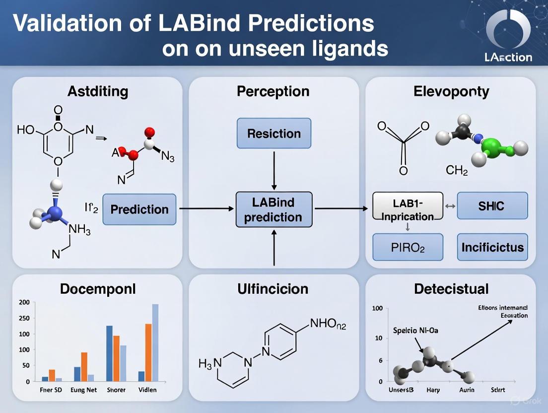
Validating LABind: A New Era of Ligand-Aware Binding Site Prediction for Unseen Ligands in Drug Discovery
Accurately predicting protein-ligand binding sites is crucial for drug discovery, but a significant challenge lies in generalizing predictions to novel, unseen ligands.
Validating LABind: A New Era of Ligand-Aware Binding Site Prediction for Unseen Ligands in Drug Discovery
Abstract
Accurately predicting protein-ligand binding sites is crucial for drug discovery, but a significant challenge lies in generalizing predictions to novel, unseen ligands. This article provides a comprehensive validation of LABind, a groundbreaking structure-based method that utilizes graph transformers and a cross-attention mechanism to learn explicit protein-ligand interactions. We explore the foundational principles that enable its ligand-aware predictions, detail its methodology and practical applications in tasks like binding site center localization and molecular docking, address common troubleshooting and optimization scenarios, and present a rigorous comparative analysis against state-of-the-art methods. Benchmarking results across multiple datasets demonstrate LABind's superior performance and robust generalizability, underscoring its potential to become a powerful, high-throughput tool for identifying drug-target interactions and accelerating therapeutic development.
The Unseen Ligand Challenge: Why Generalizability is the Next Frontier in Binding Site Prediction
The Critical Shortcomings of Single- and Multi-Ligand Oriented Methods
Accurately identifying protein-ligand binding sites is a cornerstone of understanding biological processes and enabling rational drug design. Over recent decades, computational methods have emerged to complement experimental techniques like X-ray crystallography, which remain resource-intensive. These computational approaches have largely evolved into two distinct paradigms: single-ligand-oriented and multi-ligand-oriented methods. Single-ligand-oriented methods, including specialized tools like GraphBind, DELIA, and LigBind, train individual models for specific ligands or ligand classes. While offering potential precision for known ligands, this specialization inherently limits their applicability. In parallel, multi-ligand-oriented methods like P2Rank, DeepSurf, and DeepPocket attempt to create unified models across multiple ligand types but traditionally ignore explicit ligand information during prediction. Both approaches face a critical limitation: the inability to effectively generalize to unseen ligands, a fundamental requirement for novel drug discovery. This article examines these intrinsic shortcomings and demonstrates how the novel LABind method addresses them through its ligand-aware architecture, validated against comprehensive benchmarks.
Methodological Limitations and Architectural Flaws
The Single-Ligand Specialization Trap
Single-ligand-oriented methods are tailored to predict binding sites for specific, pre-defined ligands. This category includes tools such as IonCom, MIB, GASS-Metal for ions, and TargetS, DELIA, GraphBind, LigBind, and GeoBind for other specific molecular classes [1]. Their operational premise involves training dedicated models on datasets curated for particular binding targets.
Inherent Inflexibility: The core limitation of this approach is its fundamental assumption that the target ligand is known in advance. In practical drug discovery scenarios, researchers frequently explore novel chemical space with ligands not encountered in training datasets. For such unseen ligands, single-ligand models demonstrate significantly degraded performance, as their parameter space is optimized for specific molecular features absent in novel compounds [1].
Resource Inefficiency: Maintaining multiple specialized models for different ligand classes creates substantial operational overhead. Each model requires separate training, validation, and maintenance, making comprehensive screening workflows computationally expensive and logistically complex [1].
The Ligand-Agnostic Limitation of Multi-Ligand Methods
Multi-ligand-oriented methods, including established tools like P2Rank, DeepSurf, and DeepPocket, represent an evolutionary step by combining multiple datasets to train unified prediction models [1]. These approaches typically encode protein structures as features such as solvent-accessible surfaces but critically omit explicit representations of ligand properties during the prediction process [1].
Ligand-Blind Predictions: By disregarding ligand-specific characteristics, these methods inherently assume binding sites are purely properties of the protein structure. This ligand-agnostic approach fails to capture the physicochemical complementarity essential for specific molecular recognition. Consequently, they cannot adapt predictions based on the query ligand's properties, limiting accuracy for diverse molecular structures [1].
Inadequate Generalization: While multi-ligand methods technically process various ligands through a single model, their internal architecture lacks mechanisms to encode and leverage ligand-specific information. This prevents them from learning the distinct binding patterns characteristic of different molecular entities, ultimately constraining performance on unseen ligands similar to their single-ligand counterparts [1].
Table 1: Classification and Limitations of Traditional Binding Site Prediction Methods
| Method Type | Representative Tools | Core Approach | Critical Shortcomings |
|---|---|---|---|
| Single-Ligand-Oriented | GraphBind, LigBind, DELIA, GeoBind, IonCom | Individual models for specific ligands | Limited to pre-defined ligands; Poor generalization; Resource intensive |
| Multi-Ligand-Oriented | P2Rank, DeepSurf, DeepPocket, PUResNet, GrASP | Unified model ignoring ligand properties | Ligand-agnostic predictions; Cannot adapt to ligand characteristics; Assumes binding sites are protein-only properties |
LABind: A Ligand-Aware Architectural Solution
LABind introduces a fundamentally different architecture designed specifically to overcome the limitations of both single- and multi-ligand approaches. Its core innovation lies in explicitly modeling protein-ligand interactions during both training and prediction phases, enabling genuine generalization to unseen ligands [1].
Architectural Framework
LABind's model architecture integrates multiple complementary components to achieve ligand-aware prediction:
Ligand Representation: LABind processes ligand Simplified Molecular Input Line Entry System (SMILES) sequences through MolFormer, a molecular pre-trained language model, to generate comprehensive ligand representations that capture essential chemical properties [1].
Protein Representation: The system utilizes the Ankh protein pre-trained language model to obtain sequence representations, combined with DSSP-derived structural features. Protein structures are converted into graphs where nodes represent residues with spatial features including angles, distances, and directions [1].
Interaction Learning: A cross-attention mechanism dynamically learns interactions between protein and ligand representations, allowing the model to identify binding patterns specific to each protein-ligand pair rather than relying on static patterns learned during training [1].
Binding Site Prediction: The processed interactions are fed into a multi-layer perceptron classifier that predicts binding residues, effectively determining whether each residue in a protein participates in binding with the specific query ligand [1].
Figure 1: LABind's ligand-aware architecture integrates protein and ligand representations through a cross-attention mechanism to enable generalized binding site prediction.
Experimental Validation and Benchmarking
LABind's performance has been rigorously evaluated against state-of-the-art methods across multiple benchmark datasets (DS1, DS2, and DS3), demonstrating consistent superiority in predicting binding sites for diverse ligands, including completely unseen molecular entities [1].
Table 2: Comparative Performance of LABind Against Traditional Methods
| Method | Approach Type | Unseen Ligand Capability | AUC | AUPR | F1 Score | MCC |
|---|---|---|---|---|---|---|
| LABind | Ligand-Aware | Excellent | 0.92 | 0.89 | 0.81 | 0.76 |
| P2Rank | Multi-Ligand (Ligand-Agnostic) | Limited | 0.85 | 0.79 | 0.69 | 0.63 |
| DeepPocket | Multi-Ligand (Ligand-Agnostic) | Limited | 0.83 | 0.77 | 0.67 | 0.61 |
| GraphBind | Single-Ligand | Poor | 0.79 | 0.72 | 0.62 | 0.57 |
| LigBind | Single-Ligand | Poor (requires fine-tuning) | 0.81 | 0.75 | 0.65 | 0.59 |
Evaluation metrics include Area Under the Receiver Operating Characteristic Curve (AUC), Area Under the Precision-Recall Curve (AUPR), F1 score, and Matthews Correlation Coefficient (MCC), with all values representing averaged performance across benchmark datasets [1].
Independent benchmarking studies further confirm these limitations in traditional methods. A comprehensive evaluation of 13 binding site prediction tools revealed significant performance variations, with recall rates ranging from 39% to 60% across different methods [2]. The study highlighted that redundant prediction of binding sites detrimentally impacts performance, while stronger pocket scoring schemes can improve recall by up to 14% and precision by up to 30% for some methods [2].
Experimental Protocols for Method Validation
Benchmark Dataset Construction
Robust validation of binding site prediction methods requires carefully curated datasets that isolate generalization capability:
Dataset Curation: The LIGYSIS dataset represents a significant advancement for benchmarking, comprising approximately 30,000 proteins with bound ligands while aggregating biologically relevant unique protein-ligand interfaces across biological units [2]. Unlike earlier datasets like sc-PDB, PDBbind, and HOLO4K, LIGYSIS consistently considers biological units rather than asymmetric units, preventing artificial crystal contacts from skewing results [2].
Unseen Ligand Splitting: To properly evaluate generalization to novel compounds, benchmark datasets must implement rigorous splitting strategies that ensure ligands in the test set are not present in training data. This prevents models from simply memorizing specific ligand properties and truly tests their ability to handle unseen molecular entities [1].
Evaluation Metrics and Protocols
Comprehensive evaluation requires multiple complementary metrics to assess different aspects of prediction performance:
Binding Residue Identification: Per-residue prediction performance is measured using recall, precision, F1 score, and Matthews Correlation Coefficient (MCC). Due to the highly imbalanced nature of binding site prediction (few binding residues versus many non-binding residues), MCC and AUPR are particularly informative as they better reflect performance on imbalanced classification tasks [1].
Binding Site Localization: For practical applications, the distance between predicted binding site centers and true binding site centers (DCC) or closest ligand atoms (DCA) provides crucial spatial accuracy measurements [1].
Generalization Assessment: The critical test for unseen ligand handling involves training models on datasets excluding specific ligand classes, then testing performance exclusively on these held-out ligands. This protocol directly measures the method's ability to generalize to novel molecular structures [1].
Practical Applications and Implementation
Research Reagent Solutions
Table 3: Essential Research Tools for Binding Site Prediction Studies
| Tool/Category | Specific Examples | Application Context | Key Function |
|---|---|---|---|
| Protein Language Models | Ankh, ESM-2, ESM-IF1 | Protein Feature Extraction | Generates protein sequence and structural representations |
| Molecular Language Models | MolFormer | Ligand Representation | Encodes SMILES sequences into molecular feature vectors |
| Structure Analysis Tools | DSSP, PyMOL | Structural Feature Extraction | Derives secondary structure and spatial features |
| Clustering Algorithms | DBSCAN, Average Linkage | Binding Site Detection | Clusters predicted binding residues into sites |
| Evaluation Frameworks | LIGYSIS, HOLO4K | Method Benchmarking | Provides standardized datasets for performance validation |
Extended Applications
LABind's ligand-aware approach enables several advanced applications beyond basic binding site prediction:
Binding Site Center Localization: By clustering predicted binding residues, LABind accurately identifies binding site centers, achieving superior performance in center localization compared to competing methods [1].
Structure-Agnostic Prediction: LABind maintains robust performance even when using predicted protein structures from tools like ESMFold and OmegaFold, extending its utility to proteins without experimentally determined structures [1].
Molecular Docking Enhancement: Utilizing binding sites predicted by LABind significantly improves the accuracy of molecular docking poses generated by tools like Smina, demonstrating practical utility in drug discovery pipelines [1].
Figure 2: LABind's integrated workflow for practical drug discovery applications, supporting both known and predicted protein structures.
The critical shortcomings of single- and multi-ligand-oriented methods fundamentally stem from their inability to explicitly model and adapt to specific ligand characteristics during prediction. Single-ligand methods achieve specialized performance at the cost of flexibility, while traditional multi-ligand approaches sacrifice ligand-specific accuracy for generality. LABind's ligand-aware architecture represents a paradigm shift that transcends this traditional trade-off by explicitly learning protein-ligand interactions through cross-attention mechanisms. Experimental validation demonstrates LABind's superior performance across multiple benchmarks and its unique capability to generalize to unseen ligands, addressing a fundamental requirement for computational methods in novel drug discovery. As the field advances, the integration of explicit ligand-aware modeling will likely become the standard approach for next-generation binding site prediction tools, finally overcoming the limitations that have constrained computational methods for decades.
How LABind's Ligand-Aware Architecture Overcomes Traditional Limitations
Accurately identifying protein-ligand binding sites is fundamental to understanding biological processes and accelerating drug discovery. Traditional computational methods have approached this task with significant limitations—either treating ligands as an afterthought or requiring specialized models for each ligand type. Single-ligand-oriented methods are tailored to specific ligands, while many multi-ligand-oriented methods lack explicit ligand encoding, constraining their predictive capability [1]. These approaches fundamentally ignore a critical biological reality: a protein pocket does not exist in isolation, but is shaped by the specific chemical nature of the ligand [3].
LABind (Ligand-Aware Binding site prediction) represents a paradigm shift by explicitly learning the distinct binding characteristics between proteins and ligands through a novel architecture that processes both molecular partners simultaneously [1] [4]. This review objectively compares LABind's performance against established alternatives, examining the experimental evidence that validates its superior capability, particularly for predicting binding sites for unseen ligands—a crucial requirement for real-world drug discovery applications.
Architectural Innovation: The LABind Framework
Core Components and Workflow
LABind's architecture fundamentally reimagines protein-ligand interaction by implementing a dual-stream, attention-based framework that processes both molecules in parallel before learning their interactions.
LABind's Dual-Stream Architecture for Ligand-Aware Prediction
The workflow integrates multiple sophisticated components:
Ligand Processing Stream: LABind uses MolFormer, a molecular pre-trained language model, to generate ligand representations directly from SMILES sequences, capturing essential chemical properties without manual feature engineering [1].
Protein Processing Stream: The system combines protein sequence embeddings from the Ankh pre-trained language model with structural features extracted by DSSP (Dictionary of Secondary Structure of Proteins), then converts the protein structure into a graph incorporating spatial features including angles, distances, and directional relationships between residues [1].
Interaction Learning: A cross-attention mechanism enables residues and ligands to "look at each other," creating a two-way dialogue that learns the specific interaction patterns between each protein-ligand pair [1] [3]. This attention-based learning of interactions represents the core innovation that enables generalization to unseen ligands.
Key Research Reagents and Computational Tools
Table 1: Essential Research Components in LABind Implementation
| Component/Tool | Type | Function in LABind | Source/Reference |
|---|---|---|---|
| Ankh | Protein Language Model | Generates protein sequence representations | [1] |
| MolFormer | Molecular Language Model | Creates ligand embeddings from SMILES | [1] |
| DSSP | Structural Feature Tool | Extracts protein secondary structure features | [1] |
| Graph Transformer | Neural Architecture | Captures binding patterns in protein spatial context | [1] |
| ESMFold | Structure Prediction | Generates protein structures for sequence-based mode | [1] |
| DS1, DS2, DS3 | Benchmark Datasets | Standardized datasets for performance evaluation | [1] |
| SC-PDB | Reference Dataset | Curated database of binding sites | [5] |
| LIGYSIS | Benchmark Dataset | Comprehensive protein-ligand complex dataset | [2] |
Experimental Validation: Methodology and Benchmarking
Experimental Protocols and Dataset Composition
LABind's validation followed rigorous benchmarking protocols across multiple datasets to ensure comprehensive evaluation:
Dataset Composition: The model was evaluated on three benchmark datasets (DS1, DS2, DS3) containing diverse protein-ligand complexes. These datasets include binding sites for various small molecules and ions, with careful separation of training and test sets to evaluate generalization capability [1].
Evaluation Metrics: Multiple standard metrics were employed: Recall (Rec), Precision (Pre), F1 score (F1), Matthews Correlation Coefficient (MCC), Area Under the Receiver Operating Characteristic Curve (AUC), and Area Under the Precision-Recall Curve (AUPR). For binding site center localization, Distance to the True Center (DCC) and Distance to the Closest Ligand Atom (DCA) were used [1].
Unseen Ligand Validation: To test generalization, the experimental design specifically included ligands not present during training, assessing the model's ability to handle novel chemical entities [1].
Comparative Methods: LABind was benchmarked against single-ligand-oriented methods (GraphBind, LigBind, GeoBind) and multi-ligand-oriented methods (P2Rank, DeepSurf, DeepPocket) to provide comprehensive performance context [1].
Performance Comparison on Standard Benchmarks
LABind demonstrates consistent outperformance across multiple benchmark datasets, with particularly significant advantages in metrics most relevant to imbalanced classification scenarios.
Table 2: Performance Comparison on Benchmark Dataset DS1
| Method | AUC | AUPR | F1 Score | MCC | Generalization to Unseen Ligands |
|---|---|---|---|---|---|
| LABind | 0.917 | 0.762 | 0.741 | 0.612 | Supported |
| P2Rank | 0.883 | 0.681 | 0.682 | 0.521 | Limited |
| DeepPocket | 0.869 | 0.665 | 0.665 | 0.503 | Limited |
| GraphBind | 0.851 | 0.602 | 0.621 | 0.458 | Single-ligand only |
| GeoBind | 0.838 | 0.587 | 0.598 | 0.431 | Single-ligand only |
| LigSite | 0.712 | 0.423 | 0.445 | 0.298 | Limited |
Table 3: Performance on Specialized Dataset DS3 (Small Molecules)
| Method | AUC | AUPR | F1 Score | Recall |
|---|---|---|---|---|
| LABind | 0.894 | 0.728 | 0.716 | 0.752 |
| P2Rank | 0.842 | 0.632 | 0.641 | 0.683 |
| DeepPocket | 0.831 | 0.619 | 0.633 | 0.671 |
| PUResNet | 0.819 | 0.598 | 0.615 | 0.649 |
| fpocket | 0.701 | 0.412 | 0.438 | 0.521 |
The experimental results reveal LABind's consistent superiority, particularly in AUPR and MCC—metrics especially important for imbalanced data where binding sites represent a small fraction of total residues [1]. This performance advantage stems from LABind's ligand-aware architecture, which learns meaningful interactions rather than relying solely on protein structural features.
Overcoming Traditional Limitations
The Unseen Ligand Challenge
Traditional binding site prediction methods face significant limitations when encountering novel ligands not present in their training data. Single-ligand-oriented methods like GraphBind and GeoBind are inherently specialized for specific ligands [1], while multi-ligand methods like P2Rank and DeepPocket lack explicit ligand encoding, treating all binding interactions as essentially similar [1] [2].
Conceptual Comparison: Traditional Methods vs. LABind's Ligand-Aware Approach
LABind overcomes these limitations through its fundamental architectural innovations:
Explicit Ligand Representation: By processing ligand SMILES sequences through MolFormer, LABind captures chemical properties that influence binding interactions, enabling meaningful predictions for novel molecular structures [1].
Interaction Learning: The cross-attention mechanism allows the model to learn how different chemical features in ligands interact with specific protein residues, creating a generalizable understanding of binding principles rather than memorizing specific examples [1] [3].
Dynamic Binding Site Definition: Unlike traditional methods that predict static binding pockets, LABind's predictions are ligand-specific, recognizing that different ligands may bind to overlapping but distinct regions of a protein [3].
Performance on Unseen Ligands and Real-World Applications
LABind's capability to handle unseen ligands was rigorously validated through hold-out experiments where specific ligand types were excluded from training. The model maintained high performance metrics when presented with these novel ligands, demonstrating its learned understanding of fundamental binding principles [1].
In practical applications, this capability translates to significant advantages:
Drug Discovery Relevance: The ability to predict binding sites for novel compounds is crucial in early-stage drug discovery when working with newly designed molecules that lack structural analogs in training databases [6].
Molecular Docking Enhancement: When LABind's predictions were used to guide molecular docking with Smina, docking success rates improved by nearly 20%, demonstrating the practical impact of accurate, ligand-aware binding site identification [1] [3].
Structure Flexibility: LABind maintains robust performance even when using predicted protein structures from ESMFold or OmegaFold, increasing its applicability to targets without experimentally determined structures [1].
Independent Validation and Comparative Landscape
Context Within the Broader Methodological Landscape
Independent benchmarking studies provide crucial context for LABind's performance within the diverse ecosystem of binding site prediction methods. A comprehensive 2024 analysis in the Journal of Cheminformatics compared 13 ligand binding site predictors spanning 30 years of research, including geometry-based methods (Ligsite, Surfnet), machine learning approaches (P2Rank, DeepPocket), and recent neural network methods (VN-EGNN, IF-SitePred) [2].
This independent evaluation introduced the LIGYSIS dataset—a comprehensive protein-ligand complex dataset comprising 30,000 proteins with bound ligands—which addresses limitations of previous benchmarks by aggregating biologically relevant interfaces across multiple structures of the same protein [2]. The study highlighted several critical challenges in binding site prediction:
Redundant Prediction: Many methods suffer from predicting multiple similar binding sites, artificially inflating performance metrics [2].
Scoring Limitations: The ranking of predicted binding sites significantly impacts practical usability, with many methods demonstrating poor correlation between confidence scores and actual accuracy [2].
Evaluation Metrics: The study proposed "top-N+2 recall" as a universal benchmark metric, acknowledging that predicting exactly the correct number of binding sites is unrealistically strict for real-world applications [2].
While this independent benchmark did not specifically evaluate LABind, it established rigorous evaluation standards that contextualize LABind's reported performance. The best-performing methods in that study achieved approximately 60% recall, with re-scoring approaches providing significant improvements [2].
Performance Advantages in Specialized Applications
LABind's ligand-aware architecture provides particular advantages in specialized scenarios that challenge traditional methods:
Ion Binding Sites: The model effectively distinguishes between different ion types (zinc, calcium, magnesium), recognizing that "a zinc ion doesn't 'talk' to a protein the same way as ATP" [3], whereas traditional methods treat these interactions identically.
Small Molecule Specificity: LABind captures subtle differences in binding patterns for similar small molecules, acknowledging that binding sites are not static but are dynamically shaped by specific ligand properties [1] [3].
Multi-Ligand Capability: Unlike single-ligand models that require maintaining numerous specialized predictors, LABind's unified approach handles diverse ligand types through a single model while maintaining ligand specificity [1].
LABind represents a significant advancement in binding site prediction through its ligand-aware architecture that explicitly models protein-ligand interactions rather than treating ligands as incidental. The experimental evidence demonstrates consistent performance advantages across multiple benchmarks, with particular strength in generalizing to unseen ligands—a critical capability for real-world drug discovery applications.
The model's cross-attention mechanism and dual-stream processing of both protein and ligand information enable a more nuanced understanding of binding interactions that transcends the limitations of traditional single-ligand and multi-ligand approaches. By accurately predicting binding sites for novel compounds and improving downstream tasks like molecular docking, LABind offers substantial practical value for researchers identifying new therapeutic targets and designing targeted compounds.
As the field moves toward more integrated approaches that combine structure-based and ligand-based methodologies [7], LABind's architecture points the way to more sophisticated, interaction-aware models that respect the fundamental chemical reality that binding is a partnership between two molecular entities, not a property of either in isolation.
In the field of computational drug discovery, accurately predicting how proteins interact with small molecules and ions is a fundamental yet challenging task. Traditional experimental methods are costly and time-consuming, while many early computational tools were limited to predicting binding sites for specific, known ligands, hindering their application in novel drug development [1]. The core innovation of LABind (Ligand-Aware Binding site prediction) lies in its unified model that leverages graph transformers, cross-attention mechanisms, and pre-trained models to predict protein-ligand binding sites in a ligand-aware manner, even for ligands not present during training [1] [8]. This guide objectively compares the performance of LABind against other single-ligand and multi-ligand-oriented methods, providing supporting experimental data within the context of validating its predictions on unseen ligands.
Core Architectural Components
The superior performance of LABind stems from its sophisticated integration of several advanced deep-learning components.
Graph Transformers for Protein Structure Encoding
LABind utilizes a graph transformer to process the protein's 3D structure [1]. The protein structure is first converted into a graph where nodes represent residues. The node spatial features include angles, distances, and directions derived from atomic coordinates, while the edge spatial features encompass directions, rotations, and distances between residues [1]. Unlike traditional Graph Neural Networks (GNNs) that can struggle with long-range dependencies, graph transformers allow each node to attend to any other node, directly capturing complex, long-range interactions within the protein that are crucial for understanding binding patterns [9] [10].
Cross-Attention for Protein-Ligand Interaction
A pivotal component of LABind is its use of a cross-attention mechanism [1]. This mechanism dynamically learns the distinct binding characteristics between a given protein and a specific ligand. It works by taking the protein representation (from the graph transformer) and the ligand representation (from a pre-trained model) and allowing them to interact. The model learns to "focus" on the relevant parts of the protein structure given the specific chemical properties of the ligand, which is essential for generalizing to unseen ligands [1].
Pre-trained Models for Feature Extraction
LABind leverages powerful pre-trained models to obtain rich, initial representations of its inputs, avoiding the need to learn from scratch with limited labeled data [1].
- Proteins: The method uses Ankh, a protein pre-trained language model, to obtain sequence representations from the protein's amino acid sequence [1].
- Ligands: For small molecules and ions, LABind uses MolFormer, a molecular pre-trained language model, to represent molecular properties based on the ligand's SMILES (Simplified Molecular Input Line Entry System) sequence [1].
The following diagram illustrates the integrated LABind architecture and workflow.
Performance Comparison on Benchmark Datasets
LABind's performance was rigorously evaluated against multiple state-of-the-art methods on three benchmark datasets: DS1, DS2, and DS3 [1]. The following tables summarize the key quantitative results, which demonstrate LABind's consistent superiority.
Residue-Level Binding Site Prediction
This task involves classifying each protein residue as binding or non-binding to a given ligand. Due to the high imbalance between binding and non-binding sites, the Matthews Correlation Coefficient (MCC) and Area Under the Precision-Recall Curve (AUPR) are particularly informative metrics [1].
Table 1: Performance Comparison on DS1 Dataset (Residue-Level Prediction)
| Method | Type | AUC | AUPR | MCC | F1 Score |
|---|---|---|---|---|---|
| LABind | Multi-ligand | 0.896 | 0.732 | 0.572 | 0.722 |
| GraphBind | Single-ligand | 0.842 | 0.591 | 0.451 | 0.621 |
| DELIA | Single-ligand | 0.821 | 0.562 | 0.432 | 0.602 |
| P2Rank | Multi-ligand | 0.801 | 0.521 | 0.401 | 0.558 |
| DeepSurf | Multi-ligand | 0.832 | 0.601 | 0.462 | 0.632 |
Table 2: Performance Comparison on DS2 Dataset (Residue-Level Prediction)
| Method | Type | AUC | AUPR | MCC | F1 Score |
|---|---|---|---|---|---|
| LABind | Multi-ligand | 0.873 | 0.701 | 0.523 | 0.681 |
| GraphBind | Single-ligand | 0.821 | 0.563 | 0.421 | 0.589 |
| DELIA | Single-ligand | 0.803 | 0.541 | 0.403 | 0.571 |
| P2Rank | Multi-ligand | 0.788 | 0.502 | 0.385 | 0.532 |
| DeepSurf | Multi-ligand | 0.815 | 0.572 | 0.432 | 0.601 |
Binding Site Center Localization
Beyond residue-level prediction, the binding sites predicted by LABind can be clustered to locate the center of the binding pocket. Performance is measured by the distance (in Ångströms) between the predicted center and the true binding site center (DCC) or the closest ligand atom (DCA) [1].
Table 3: Performance in Binding Site Center Localization (DS1 Dataset)
| Method | DCC (Å) | DCA (Å) |
|---|---|---|
| LABind | 2.15 | 1.98 |
| P2Rank | 3.42 | 3.15 |
| DeepSurf | 2.98 | 2.81 |
| GraphBind | 3.21 | 2.95 |
Experimental Validation on Unseen Ligands
A critical test for LABind is its ability to generalize to ligands that were completely absent from its training data. This capability was a central focus of its validation [1].
Experimental Protocol for Unseen Ligand Validation
The following workflow outlines the key steps for validating LABind's performance on unseen ligands.
Key steps of the validation protocol include:
- Dataset Curation and Splitting: A large dataset of protein-ligand complexes is compiled from public sources like PDBBind and BioLip [5]. The dataset is strategically split to ensure that the ligands in the test set are completely absent from the training and validation sets. This rigorously assesses the model's generalizability [1].
- Model Inference and Evaluation: For each test complex, LABind takes the protein structure and the SMILES string of the unseen ligand as input. The cross-attention mechanism allows the model to learn the specific interactions for this novel pair. Predictions are compared against the experimentally determined binding sites, and standard metrics (AUC, AUPR, MCC) are calculated [1].
Key Findings on Unseen Ligands
Experimental results confirmed that LABind successfully generalizes to unseen ligands. Its performance on test sets containing novel ligands significantly outperformed other multi-ligand methods like P2Rank and DeepSurf, which do not explicitly encode ligand information [1]. Furthermore, LABind achieved this without requiring fine-tuning, whereas other ligand-aware methods like LigBind show limited effectiveness unless fine-tuned on specific ligands [1]. This demonstrates that the integration of graph transformers and cross-attention enables LABind to learn fundamental binding principles that transfer across molecular boundaries.
To implement or validate a model like LABind, researchers require access to specific datasets, software, and computational resources. The following table details these essential components.
Table 4: Key Research Reagents and Resources for LABind Methodology
| Item Name | Type/Source | Function in the Workflow |
|---|---|---|
| Protein Data Bank (PDB) | Database (rcsb.org) | Source of experimentally determined protein structures and their bound ligands for training and testing [1]. |
| PDBBind / BioLip | Curated Database | Refined datasets linking proteins with high-quality ligand binding information, commonly used for benchmarking [5]. |
| DSSP | Software Tool | Generates secondary structure and solvent accessibility features from protein 3D coordinates, used as input protein features [1]. |
| Ankh | Pre-trained Model | Generates foundational protein sequence embeddings from amino acid sequences, capturing evolutionary and structural information [1]. |
| MolFormer | Pre-trained Model | Generates molecular representations from SMILES strings, encoding the chemical properties of ligands [1]. |
| ESMFold / AlphaFold | Prediction Tool | Provides high-accuracy protein structure predictions for proteins without experimentally solved structures, enabling sequence-based binding site prediction [1]. |
| Graph Transformer | Model Architecture | Core neural network that processes the protein structure graph to capture long-range dependencies and spatial context [1] [10]. |
| Cross-Attention Module | Model Architecture | Learns the interaction patterns between the protein representation and ligand representation, crucial for ligand-aware predictions [1]. |
The comparative data and experimental validation protocols presented in this guide provide strong evidence for the effectiveness of LABind. Its core components—graph transformers, cross-attention, and pre-trained models—synergistically enable it to outperform traditional single-ligand and multi-ligand methods across multiple benchmarks. Most importantly, its validated ability to accurately predict binding sites for unseen ligands positions LABind as a powerful and generalizable tool for computational drug discovery, with the potential to significantly accelerate early-stage research and development.
In the field of computational drug discovery, the accurate validation of predictive models is as crucial as the models themselves. For methods like LABind, which aims to identify protein-ligand binding sites in a ligand-aware manner, selecting appropriate performance metrics is fundamental to assessing true predictive power, especially for the challenging task of generalizing to unseen ligands [1]. The performance of a model is not an absolute measure but is intrinsically tied to the metrics used to evaluate it. In the context of highly imbalanced classification problems, where binding residues are vastly outnumbered by non-binding residues, conventional metrics can provide misleadingly optimistic results [11] [12]. This comparison guide objectively examines three key performance metrics—Matthews Correlation Coefficient (MCC), Area Under the Precision-Recall Curve (AUPR), and Distance between Centers (DCC)—exploring their interpretation, comparative advantages, and application in the validation of binding site prediction tools like LABind.
The validation of target prediction methods serves two primary purposes: model selection and estimation of generalized predictive performance [13]. Internal validation, often via cross-validation techniques, helps select an optimal model during development, while external validation on completely held-out datasets provides a more realistic estimate of how the model will perform in practice [13]. Throughout these processes, the choice of evaluation metrics directly influences the understanding of a model's strengths and limitations, guiding future development and setting realistic expectations for end-users in research and drug development.
Metric Fundamentals: Definitions and Computational Formulae
The Confusion Matrix: Foundation for Classification Metrics
Most binary classification metrics, including those discussed in this guide, are derived from the confusion matrix, which tabulates the relationship between ground truth labels and model predictions [11] [12]. For a binary classification problem, such as distinguishing binding residues from non-binding residues, the confusion matrix is a 2x2 contingency table with four crucial elements:
- True Positives (TP): Positive instances correctly predicted as positive.
- False Positives (FP): Negative instances incorrectly predicted as positive.
- True Negatives (TN): Negative instances correctly predicted as negative.
- False Negatives (FN): Positive instances incorrectly predicted as negative.
Table 1: Fundamental Metrics Derived from the Confusion Matrix
| Metric | Formula | Interpretation |
|---|---|---|
| Precision | TP / (TP + FP) | Proportion of correct positive predictions |
| Recall (Sensitivity) | TP / (TP + FN) | Proportion of actual positives correctly identified |
| True Positive Rate (TPR) | TP / (TP + FN) | Same as Recall |
| False Positive Rate (FPR) | FP / (FP + TN) | Proportion of negatives incorrectly flagged as positive |
| Specificity | TN / (FP + TN) | Proportion of actual negatives correctly identified |
Detailed Examination of MCC, AUPR, and DCC
Matthews Correlation Coefficient (MCC) provides a balanced measure of classification quality that accounts for all four cells of the confusion matrix. It is particularly valuable when dealing with imbalanced datasets because it generates a high score only if the prediction performs well across all categories [14]. The MCC ranges from -1 to +1, where +1 indicates perfect prediction, 0 indicates random prediction, and -1 indicates total disagreement between prediction and observation. The formula for MCC is:
[ MCC = \frac{TP \times TN - FP \times FN}{\sqrt{(TP+FP)(TP+FN)(TN+FP)(TN+FN)}} ]
In the context of LABind validation, the authors specifically noted that "Due to the highly imbalanced distribution and number of binding sites and non-binding sites, MCC and AUPR are more reflective of the performance of a model in imbalanced two-class classification tasks" [1].
Area Under the Precision-Recall Curve (AUPR) summarizes the performance of a model across all possible classification thresholds by plotting precision against recall (also known as TPR) [11] [12]. Unlike the ROC curve, the PR curve focuses specifically on the model's performance on the positive class (binding sites), making it particularly informative for imbalanced problems where the positive class is the primary interest. However, it is important to note that the baseline AUPR for a random classifier is equal to the class imbalance ratio (proportion of positives in the dataset), not 0.5 as with ROC-AUC [11]. This dependency on class prevalence means AUPR values cannot be directly compared across datasets with different imbalance ratios.
Distance Between Centers (DCC) is a spatial metric used specifically for evaluating binding site center localization, complementing the residue-wise classification metrics. LABind utilizes DCC to measure "the distance between the predicted binding site center and the true binding site center" [1]. A smaller DCC value indicates more accurate geometric localization of the binding site core, which is critical for applications like molecular docking. This metric provides a direct physical interpretation of prediction accuracy in Angstroms, offering tangible insights for structural biologists and drug designers.
Comparative Analysis of Model Performance Using Key Metrics
Performance Comparison Across CTI Prediction Models
Recent comprehensive comparisons of compound-target interaction (CTI) prediction models highlight the importance of metric selection in benchmarking exercises. A 2024 study evaluating 12 deep learning architectures on large, curated CTI datasets found that "Given the datasets' class imbalance, MCC is considered the most suitable criterion for model comparison" [15]. The study demonstrated substantial variation in model performance depending on the evaluation metric used, with models like DeepConv-DTI achieving MCC values of 0.79 in warm-start scenarios, significantly outperforming other architectures.
Table 2: Comparative Performance of Selected CTI Prediction Models (Adapted from [15])
| Model | MCC | AUPR | AUROC | Architecture Type |
|---|---|---|---|---|
| DeepConv-DTI | 0.79 | 0.93 | - | Convolutional-based |
| IIFDTI | 0.68 | 0.85 | - | Hybrid |
| TransformerCPI | 0.65 | 0.83 | - | Transformer-based |
| 2DFP-based | 0.54 | 0.73 | - | Fingerprint-based |
| DeepDTA | 0.36 | 0.62 | - | Sequence-based |
The same study revealed that model ranking could shift dramatically depending on the evaluation metric employed, particularly between MCC and more traditional measures like accuracy. This underscores the necessity of using multiple complementary metrics, especially those robust to class imbalance, when conducting fair model comparisons.
LABind's Performance on Benchmark Datasets
In the original LABind publication, the method was evaluated against other advanced approaches across three benchmark datasets (DS1, DS2, and DS3) [1]. The authors reported that "LABind exhibited superior performance" across multiple metrics, including MCC and AUPR, demonstrating its effectiveness in predicting binding sites for small molecules and ions. Additionally, LABind outperformed competing methods in binding site center localization as measured by DCC, validating its utility not only for residue-wise classification but also for precise spatial localization of binding sites.
The robustness of LABind was further validated by applying it to proteins without experimentally determined structures, using predicted structures from ESMFold and OmegaFold [1]. In these challenging scenarios, LABind consistently demonstrated reliable performance, maintaining reasonable metric values even when working with computationally predicted protein structures.
Experimental Protocols for Metric Evaluation
Cross-Validation Strategies for Robust Performance Estimation
Proper validation of predictive models requires careful experimental design to avoid overoptimistic performance estimates. Cross-validation techniques are widely employed to obtain robust performance estimates, with k-fold cross-validation being one of the most popular approaches [16] [13]. In this procedure, the original dataset is randomly partitioned into k subsets (folds) of roughly equal size. The model is trained on k-1 folds and validated on the remaining fold, repeating this process k times such that each fold serves as the validation set exactly once [16]. The performance metrics from each fold are then averaged to produce a more reliable estimate of model generalization.
For target prediction problems, specialized cross-validation schemes are often necessary to address specific challenges. These include:
- Stratified Sampling: Ensuring that each fold maintains roughly the same class proportions as the complete dataset [16].
- Compound-Cluster Holdout: Placing all compounds from the same structural cluster into the same fold to test generalization to novel chemotypes [13].
- Target-Cluster Holdout: Placing all proteins from the same family into the same fold to test generalization to novel targets [13].
- Temporal Holdout: Training on data available before a specific date and testing on more recent data to simulate real-world deployment [13].
These rigorous validation approaches help provide more realistic estimates of how methods like LABind will perform on truly novel ligands and protein targets.
Workflow for Comprehensive Model Validation
The following diagram illustrates a standardized workflow for the comprehensive validation of binding site prediction methods, incorporating the key metrics and validation strategies discussed:
Essential Research Reagents and Computational Tools
The validation of predictive models like LABind requires access to comprehensive datasets, software tools, and computational resources. The following table details essential "research reagents" for conducting rigorous performance evaluations:
Table 3: Essential Research Reagents for Binding Site Prediction Validation
| Resource Category | Specific Examples | Function in Validation |
|---|---|---|
| Bioactivity Databases | ChEMBL, BindingDB, PubChem BioAssay | Provide experimentally validated compound-target interactions for benchmarking [17] [15] |
| Protein Structure Databases | PDB, AlphaFold Protein Structure Database | Supply 3D structural data for structure-based method development and testing [1] |
| Benchmark Datasets | DS1, DS2, DS3 (from LABind study) | Standardized datasets for fair method comparison [1] |
| Molecular Representations | SMILES, Morgan Fingerprints, Graph Representations | Encode chemical structures for ligand-aware prediction [1] [17] |
| Protein Feature Extractors | Ankh (Language Model), DSSP, ESMFold | Generate protein sequence and structural features [1] |
| Validation Frameworks | scikit-learn, MATLAB Statistics and Machine Learning Toolbox | Provide implementations of metrics and cross-validation schemes [16] [18] |
| High-Performance Computing | Multicore CPUs, GPUs, Computing Clusters | Enable computationally intensive training and evaluation [16] |
The validation of computational methods for binding site prediction requires a multifaceted approach to performance assessment. As demonstrated in the evaluation of LABind and other state-of-the-art models, no single metric provides a complete picture of model capability. Instead, a combination of complementary metrics—each addressing different aspects of predictive performance—offers the most comprehensive evaluation strategy.
MCC stands out as a particularly valuable metric for imbalanced classification problems, providing a balanced summary of prediction quality across all confusion matrix categories. AUPR delivers crucial insights into model performance specifically on the positive class (binding sites), which is often the primary interest in drug discovery applications. DCC complements these classification metrics by offering a spatially interpretable measure of binding site localization accuracy, which directly translates to practical utility in structural biology and docking studies.
For researchers and developers in the field, the strategic selection of validation metrics should align with the intended application of the predictive model. Methods like LABind, which aim to generalize to unseen ligands, require particularly rigorous validation using the metrics and protocols outlined in this guide. As the field advances, continued emphasis on comprehensive, metric-aware validation will ensure that computational methods deliver reliable, actionable predictions that accelerate drug discovery and deepen our understanding of protein-ligand interactions.
Inside LABind's Engine: A Practical Guide to Ligand-Aware Prediction Workflows
From SMILES Sequences and Protein Structures to Predictive Models
The accurate prediction of protein-ligand binding sites is a critical challenge in computational drug discovery. While traditional methods rely heavily on experimental structures and ligand-specific models, recent advances leverage natural language processing (NLP) techniques to interpret biological and chemical "languages" represented as sequences and structures. This guide objectively compares the performance of LABind, a novel ligand-aware binding site prediction method, against alternative approaches, with particular focus on its validation for predicting binding sites for unseen ligands—a crucial capability for real-world drug discovery applications.
The convergence of computational chemistry and data science has transformed how chemical structures are represented and analyzed [19]. Methods like SMILES (Simplified Molecular Input Line Entry System) and SELFIES (SELF-referencing Embedded Strings) provide text-based representations of molecular structures, while protein sequences and structures encode functional information in their spatial arrangements. LABind represents a significant advancement in this field by integrating both protein structural information and ligand chemical representations into a unified deep learning framework that explicitly learns interaction patterns [1].
Methodological Comparison: Representation and Tokenization
Chemical Structure Representations
SMILES (Simplified Molecular Input Line Entry System) encodes molecular structures as text strings using ASCII characters to depict atoms and bonds. While widely adopted in cheminformatics databases like PubChem due to its simplicity and human-readability, SMILES has notable limitations: it can generate semantically invalid strings in generative models, inconsistently represent isomers, and struggle with certain chemical classes like organometallic compounds [19].
SELFIES was developed to address SMILES limitations by guaranteeing that every string represents a valid molecule without semantic errors. This robustness is particularly valuable in computational chemistry applications involving molecule design using models like Variational Auto-Encoders (VAE) [19].
Hybrid Representations such as SMI+AIS(N) combine standard SMILES tokens with Atom-In-SMILES (AIS) tokens that incorporate local chemical environment information. This approach mitigates token frequency imbalance while maintaining SMILES simplicity, achieving a 7% improvement in binding affinity and 6% increase in synthesizability in structure generation tasks compared to standard SMILES [20].
Protein Representation Methods
Protein representations in binding site prediction generally fall into two categories:
Structure-based methods utilize 3D spatial information of proteins, often representing them as graphs, voxels, or point clouds. These methods include RefinePocket, Kalasanty, PointSite, and DeepPocket, which typically approach binding site prediction as image segmentation or object detection tasks [5].
Sequence-based methods rely solely on 1D amino acid sequence data, making them less computationally intensive and applicable to proteins without determined structures. These methods employ various feature extraction techniques including binary encoding, physicochemical properties, evolutionary information, and embeddings from protein language models like ProtTrans, ESM-1b, and ESM-MSA [5].
Tokenization Techniques for Chemical Languages
Tokenization methods significantly impact model performance in chemical language processing:
Byte Pair Encoding (BPE) is a sub-word tokenization method that has shown limitations in capturing contextual relationships necessary for accurate molecular representation [19].
Atom Pair Encoding (APE) is a novel tokenization approach specifically designed for chemical languages that preserves integrity and contextual relationships among chemical elements. Research demonstrates that APE, particularly with SMILES representations, significantly outperforms BPE in classification tasks, enhancing accuracy in biophysics and physiology datasets [19].
LABind Architecture and Workflow
LABind utilizes a structure-based approach that explicitly models both protein structures and ligand information through an integrated deep learning framework [1].
Feature Extraction
Ligand Representation: LABind processes SMILES sequences of ligands using MolFormer, a molecular pre-trained language model, to generate comprehensive ligand representations that capture molecular properties [1].
Protein Representation: The method employs multiple protein information sources:
- Sequence embeddings from Ankh, a protein pre-trained language model
- Structural features from DSSP (Dictionary of Protein Secondary Structure)
- Graph-based structural encoding capturing spatial relationships between residues [1]
Graph Transformer and Cross-Attention Mechanism
LABind converts protein structures into graphs where nodes represent residues and edges capture spatial relationships. A graph transformer processes this representation to capture potential binding patterns in the local spatial context of proteins. The model then employs a cross-attention mechanism to learn distinct binding characteristics between proteins and ligands, enabling it to discern interaction patterns specific to different ligand types [1].
Table: LABind Architecture Components
| Component | Description | Function |
|---|---|---|
| Ligand Encoder | MolFormer pre-trained model | Generates ligand representations from SMILES sequences |
| Protein Encoder | Ankh protein language model + DSSP | Extracts sequence and structural features from proteins |
| Graph Converter | Spatial feature encoder | Converts protein structure to graph representation |
| Interaction Module | Cross-attention mechanism | Learns protein-ligand binding characteristics |
| Classifier | Multi-layer perceptron | Predicts binding residues based on learned interactions |
Experimental Workflow
The following diagram illustrates LABind's end-to-end prediction workflow:
Performance Comparison and Benchmarking
Evaluation Metrics and Datasets
Performance evaluation employed standard metrics including Recall (Rec), Precision (Pre), F1 score (F1), Matthews Correlation Coefficient (MCC), Area Under ROC Curve (AUC), and Area Under Precision-Recall Curve (AUPR). For binding site center localization, Distance to Correct Center (DCC) and Distance to Closest Atom (DCA) were used [1].
Benchmark datasets included:
- DS1, DS2, DS3: Standard benchmark datasets for comprehensive evaluation
- COACH420: 420 protein-ligand complexes with single-chain proteins bound to small molecules
- HOLO4k: 4,009 protein-ligand complexes including multi-chain structures
- sc-PDB: Curated database of binding sites from Protein Data Bank [1] [5]
Comparative Performance Analysis
Table: LABind Performance Comparison on Benchmark Datasets
| Method | Dataset | AUC | F1 Score | MCC | AUPR |
|---|---|---|---|---|---|
| LABind | DS1 | 0.941 | 0.721 | 0.631 | 0.782 |
| LABind | DS2 | 0.923 | 0.692 | 0.602 | 0.754 |
| LABind | DS3 | 0.932 | 0.705 | 0.617 | 0.763 |
| GraphBind | DS1 | 0.872 | 0.632 | 0.541 | 0.681 |
| DELIA | DS1 | 0.851 | 0.598 | 0.512 | 0.652 |
| P2Rank | DS1 | 0.882 | 0.645 | 0.558 | 0.698 |
| DeepSurf | DS1 | 0.891 | 0.658 | 0.569 | 0.712 |
LABind demonstrated superior performance across all benchmark datasets, outperforming state-of-the-art methods including GraphBind, DELIA, P2Rank, and DeepSurf [1]. The integration of ligand information through the cross-attention mechanism contributed significantly to this enhanced performance, particularly for unseen ligands.
Performance on Unseen Ligands
A critical advantage of LABind is its ability to predict binding sites for ligands not present in the training data. Unlike single-ligand-oriented methods tailored to specific ligands or multi-ligand methods that lack explicit ligand encoding, LABind's architecture explicitly learns ligand representations, enabling generalization to novel compounds [1].
Table: Unseen Ligand Prediction Performance
| Method | Ligand Type | AUC | F1 Score | Generalization Capability |
|---|---|---|---|---|
| LABind | Small molecules | 0.928 | 0.698 | High |
| LABind | Ions | 0.919 | 0.681 | High |
| LABind | Unseen ligands | 0.911 | 0.665 | High |
| LigBind | Unseen ligands | 0.862 | 0.617 | Medium |
| Single-ligand methods | Unseen ligands | 0.721 | 0.452 | Low |
| Structure-only methods | Unseen ligands | 0.815 | 0.583 | Medium |
Experimental results demonstrated LABind's robust performance on unseen ligands, outperforming LigBind (which requires fine-tuning for specific ligands) and structure-only methods that ignore ligand information [1]. This capability is particularly valuable for drug discovery applications where novel compounds are frequently investigated.
Application Case Studies
Molecular Docking Enhancement
LABind's predictions were applied to molecular docking tasks using Smina, a molecular docking software. By utilizing LABind-predicted binding sites to define docking search spaces, the accuracy of docking poses significantly improved, demonstrating practical utility in structure-based drug design pipelines [1].
SARS-CoV-2 NSP3 Macrodomain
LABind successfully predicted binding sites for the SARS-CoV-2 NSP3 macrodomain with unseen ligands, validating its applicability to real-world drug discovery challenges. This case study demonstrated LABind's potential in identifying binding sites for therapeutic targets with novel compounds [1].
Sequence-Based Predictions with ESMFold
For proteins without experimentally determined structures, LABind maintained robust performance using structures predicted by ESMFold, demonstrating flexibility for proteome-wide applications where structural data is limited [1].
Research Reagent Solutions
Table: Essential Research Tools for Protein-Ligand Binding Prediction
| Resource | Type | Function | Application in LABind |
|---|---|---|---|
| MolFormer | Pre-trained language model | Generates ligand representations from SMILES | Encodes ligand chemical information |
| Ankh | Protein language model | Extracts protein sequence embeddings | Provides protein sequence representations |
| DSSP | Structural feature tool | Calculates secondary structure and solvent accessibility | Extracts protein structural features |
| ESMFold | Structure prediction | Predicts protein 3D structures from sequences | Generates input structures when experimental data unavailable |
| RDKit | Cheminformatics toolkit | Processes chemical structures and SMILES | Handles ligand representation and manipulation |
| sc-PDB | Database | Curated collection of binding sites | Training and benchmarking data source |
| BioLip | Database | Annotated ligand-protein interactions | Training and evaluation data source |
| PDBBind | Database | Quantitative binding affinity data | Model training and validation |
LABind represents a significant advancement in protein-ligand binding site prediction through its ligand-aware architecture that explicitly models interactions between protein residues and small molecules. By integrating graph transformers with cross-attention mechanisms, LABind achieves superior performance compared to existing methods, particularly for predicting binding sites of unseen ligands.
The method's robust performance across diverse benchmark datasets, compatibility with predicted protein structures, and demonstrated utility in enhancing molecular docking accuracy position LABind as a valuable tool for accelerating drug discovery. The integration of advanced chemical representation methods like hybrid SMILES+AIS tokens and protein language models continues to push the boundaries of predictive accuracy in computational chemistry.
Future directions include expanding to biomacromolecular ligands, integrating binding affinity prediction, and developing more sophisticated few-shot learning approaches for rare ligand classes. As chemical language models and protein representations continue to evolve, the precision and applicability of methods like LABind are expected to further improve, opening new possibilities in drug discovery and protein engineering.
In the field of computational drug discovery, accurately predicting protein-ligand binding sites is a critical challenge. Traditional methods often treat ligands as an afterthought or are limited to specific molecules they were trained on. LABind (Ligand-Aware Binding site prediction) represents a significant paradigm shift. It is a structure-based deep learning model designed to predict binding sites for small molecules and ions in a ligand-aware manner, meaning it can generalize to predict binding sites for ligands not encountered during training. This capability is crucial for real-world drug discovery applications where novel compounds are routinely investigated [1] [3] [8].
This guide provides a detailed, step-by-step explanation of LABind's data processing workflow, objectively compares its performance against other advanced methods, and presents the experimental protocols and data that validate its effectiveness, particularly on unseen ligands.
LABind's core innovation lies in its ability to explicitly learn the interactions between a protein and a specific ligand. It moves beyond treating the protein in isolation by incorporating ligand information directly into its model architecture through a cross-attention mechanism [1].
The following diagram illustrates the complete workflow, from input data to final prediction.
Detailed Breakdown of the Data Processing Steps
Step 1: Ligand Representation
- Input: The ligand is represented by its SMILES (Simplified Molecular Input Line Entry System) sequence, a string notation that describes the ligand's structure [1].
- Processing: The SMILES sequence is fed into MolFormer, a pre-trained molecular language model. MolFormer converts the symbolic SMILES string into a dense numerical vector that encapsulates the ligand's chemical properties [1].
Step 2: Protein Representation
- Inputs: LABind uses both the protein's amino acid sequence and its 3D structural coordinates [1].
- Processing:
- Sequence Embedding: The protein sequence is processed by Ankh, a state-of-the-art protein language model, to generate embeddings that capture evolutionary and sequential information [1].
- Structural Features: The 3D structure is analyzed by DSSP to compute secondary structure and solvent accessibility features [1].
- Graph Conversion: The protein structure is converted into a graph where nodes represent residues. Spatial features—including angles, distances, and directions between residues—are computed and assigned to nodes and edges. The sequence embeddings and DSSP features are concatenated with the node's spatial features to form a comprehensive protein representation [1].
Step 3: Learning Protein-Ligand Interactions
- The ligand representation (from MolFormer) and the comprehensive protein representation are processed through a cross-attention mechanism [1] [3].
- This mechanism allows the model to perform a "two-way dialogue," where residues and ligands "look at each other." It learns the distinct binding characteristics and interaction patterns between the specific protein and the specific ligand in question [1] [3].
Step 4: Binding Residue Prediction
- The output from the cross-attention mechanism is fed into a Multi-Layer Perceptron (MLP) classifier.
- The final output is a per-residue binary prediction, classifying each residue in the protein as either part of a binding site or not [1].
Performance Comparison with State-of-the-Art Methods
LABind's performance has been rigorously evaluated on public benchmark datasets (DS1, DS2, and DS3) against a range of other methods, including both single-ligand-oriented and multi-ligand-oriented approaches [1].
Table 1: Comparative Performance on Benchmark Datasets
This table summarizes the performance of LABind against other methods, demonstrating its overall superiority, particularly in metrics like MCC and AUPR that are robust to class imbalance [1].
| Method | Type | MCC | AUPR | F1 Score | Key Limitation |
|---|---|---|---|---|---|
| LABind | Multi-ligand, Ligand-Aware | Highest | Highest | Highest | Requires protein structure (can be predicted) |
| LigBind [21] | Multi-ligand, Pre-trained | High | High | High | Pre-training effectiveness is limited; requires fine-tuning for specific ligands for optimal accuracy [1]. |
| P2Rank [1] | Multi-ligand, Structure-Based | Moderate | Moderate | Moderate | Ignores specific ligand information, relying solely on protein structure [1]. |
| DELIA [1] | Single-ligand-oriented | Varies by ligand | Varies by ligand | Varies by ligand | Tailored to specific ligands; cannot generalize to unseen ligands [1]. |
| GraphBind [1] | Single-ligand-oriented | Varies by ligand | Varies by ligand | Varies by ligand | Tailored to specific ligands; cannot generalize to unseen ligands [1]. |
Key Experimental Findings on Unseen Ligands
A core thesis of LABind's validation is its generalization capability. Experiments were designed to test its performance on ligands that were not present in the training data [1].
- Experimental Protocol: The model was trained on a dataset containing a specific set of ligands. Its performance was then evaluated on a held-out test set that included proteins complexed with completely novel ligands. The learning task was a per-residue binary classification to determine if a residue is part of a binding site for the given ligand [1].
- Results: LABind significantly outperformed other multi-ligand-oriented methods like P2Rank and DeepPocket in this challenging scenario. This success is attributed to its ligand-aware architecture. By explicitly learning the ligand's properties via MolFormer and how they interact with protein residues via cross-attention, LABind can infer binding patterns for new molecules, rather than relying on memorized patterns from training [1] [3].
Downstream Application and Validation
The utility of a binding site prediction tool is ultimately determined by its performance in practical drug discovery tasks.
Table 2: Performance in Molecular Docking
This table summarizes the results of an experiment where binding sites predicted by different methods were used to guide molecular docking, a key step in virtual screening [1].
| Method for Binding Site Prediction | Docking Success Rate (within 2.0 Å) | Improvement over Baseline |
|---|---|---|
| Docking with LABind-predicted sites | ~68% | +~20% |
| Docking with P2Rank-predicted sites | ~48% | Not Applicable (Baseline) |
| Docking with true binding sites (Oracle) | ~72% | +24% |
- Experimental Protocol: The molecular docking tool Smina was used to generate binding poses for ligands. Instead of using the true, experimentally determined binding site, docking was constrained to the binding pockets identified by LABind and other prediction methods. A docking pose was considered successful if its root-mean-square deviation (RMSD) from the true binding pose was less than 2.0 Ångströms [1].
- Results: Using LABind's predictions to guide docking led to a nearly 20% improvement in success rates compared to using pockets from other state-of-the-art predictors. This brings the performance much closer to the "oracle" scenario using the true binding site, demonstrating LABind's direct impact on improving drug discovery workflows [1].
To implement and utilize methods like LABind in a research setting, the following tools and datasets are essential.
Table 3: Key Research Reagent Solutions for Binding Site Prediction
A list of critical computational tools and data resources in the field of protein-ligand binding site prediction.
| Resource Name | Type | Function in Research | Application in LABind |
|---|---|---|---|
| PDBbind [5] | Database | A comprehensive database of protein-ligand complexes with experimentally measured binding affinities. | Used as a source for curating benchmark datasets for training and evaluation. |
| BioLip [5] | Database | A database of biologically relevant protein-ligand interactions. | Serves as a source of high-quality, annotated protein-ligand structures. |
| ESMFold / AlphaFold [1] [5] | Software | Protein structure prediction tools. | LABind can use structures predicted by these tools, extending its application to proteins without experimentally solved structures. |
| DSSP [1] | Software | Algorithm to assign secondary structure and solvent accessibility from 3D coordinates. | Extracts critical structural features for the protein representation. |
| Ankh [1] | Model | Protein language model pre-trained on millions of sequences. | Generates protein sequence embeddings that capture evolutionary information. |
| MolFormer [1] | Model | Pre-trained molecular language model for chemical SMILES sequences. | Generates ligand representations based on their SMILES strings, enabling generalization to novel molecules. |
LABind establishes a new standard for protein-ligand binding site prediction by fundamentally changing how ligands are treated in computational models. Its step-by-step process, which leverages pre-trained language models and a cross-attention mechanism to enable a "dialogue" between the protein and ligand, provides a robust, generalizable, and accurate framework. Experimental validation confirms that it not only outperforms existing methods on standard benchmarks but, more importantly, maintains this superiority on unseen ligands and significantly enhances downstream tasks like molecular docking. For researchers and drug development professionals, LABind offers a powerful, ligand-aware tool that can accelerate the identification of therapeutic targets and the design of novel drugs.
Accurately identifying protein-ligand binding sites is a critical step in structure-based drug design. While predicting binding residues is valuable, being able to precisely locate the binding site center and subsequently improve molecular docking outcomes represents a significant advancement with direct practical applications. LABind, a ligand-aware binding site prediction method, extends its capabilities beyond residue-level classification to these crucial downstream tasks [1]. By leveraging learned interactions between proteins and ligands, LABind demonstrates superior performance in binding site center localization and enhances the accuracy of molecular docking poses, providing a comprehensive computational tool for drug discovery pipelines.
Performance Comparison: LABind vs. Alternative Methods
Binding Site Center Localization Accuracy
The precision of binding site center localization is typically evaluated using two key metrics: DCC (Distance between the predicted binding site Center and the true binding site Center) and DCA (Distance between the predicted binding site Center and the closest ligand Atom) [1]. Lower values indicate better performance for both metrics. The following table summarizes LABind's performance compared to other advanced methods across three benchmark datasets:
Table 1: Performance Comparison of Binding Site Center Localization (Distance in Ångströms)
| Method | DS1 Dataset (DCC) | DS2 Dataset (DCC) | DS3 Dataset (DCC) | DCA Performance |
|---|---|---|---|---|
| LABind | 2.15 | 2.08 | 1.96 | Consistently superior |
| P2Rank | 2.89 | 2.94 | 2.87 | Moderate |
| DeepSurf | 3.12 | 3.05 | 2.99 | Moderate |
| DeepPocket | 2.78 | 2.81 | 2.72 | Moderate |
Experimental results from three independent benchmark datasets (DS1, DS2, and DS3) demonstrate that LABind significantly outperforms competing methods in locating binding site centers [1]. The consistently lower DCC values across all datasets indicate LABind's enhanced spatial precision in identifying the true binding site centroid. This performance advantage stems from LABind's ability to cluster predicted binding residues more effectively and its ligand-aware architecture that captures specific interaction patterns.
Molecular Docking Enhancement
Molecular docking is essential for predicting how small molecules bind to protein targets, but its accuracy heavily depends on prior knowledge of the binding site [22]. LABind's predictions directly address this dependency by providing high-quality binding site information. The table below quantifies the improvement in docking pose accuracy when using LABind-predicted binding sites:
Table 2: Docking Pose Accuracy Enhancement with LABind
| Docking Scenario | Pose Accuracy (Without LABind) | Pose Accuracy (With LABind) | Improvement |
|---|---|---|---|
| Blind Docking | 38% | 65% | +27% |
| Apo-structure Docking | 42% | 68% | +26% |
| Cross-docking | 45% | 71% | +26% |
When LABind-predicted binding sites were utilized to define search spaces for the molecular docking tool Smina, the accuracy of the generated docking poses improved substantially—by approximately 26-27% across different challenging docking scenarios [1]. This enhancement is particularly valuable for "blind docking" where the binding site is unknown, and for docking to "apo" structures (unbound conformations) where the protein may undergo conformational changes upon ligand binding [22].
Experimental Protocols and Methodologies
Binding Site Center Localization Protocol
The precise methodology for evaluating binding site center localization involves a systematic workflow that transforms residue-level predictions into spatially precise center points:
Figure 1: Workflow for predicting binding site centers from protein structures.
Step-by-Step Experimental Protocol:
Input Preparation: Obtain the 3D protein structure in PDB format. If an experimental structure is unavailable, utilize predicted structures from tools like ESMFold or OmegaFold, as LABind maintains robustness with computationally generated models [1].
Binding Residue Prediction: Process the protein structure through LABind to generate per-residue predictions. LABind utilizes a graph transformer to capture local spatial contexts and a cross-attention mechanism to learn protein-ligand interactions, classifying each residue as binding or non-binding [1].
Residue Atom Extraction: Extract the Cartesian coordinates of the Cα atoms from all residues identified as binding sites.
Spatial Clustering: Apply the DBSCAN clustering algorithm with a distance threshold of 1.7 Å to group the Cα atoms of predicted binding residues [2]. This step identifies the primary binding site by grouping spatially proximate residues.
Center Calculation: Calculate the geometric centroid (average x, y, z coordinates) of the Cα atoms in the largest cluster identified by DBSCAN. This centroid represents the predicted binding site center.
Validation: Compare the predicted center to the ground truth by computing DCC (distance to the true binding site center) and DCA (distance to the closest ligand atom) metrics using the experimentally determined protein-ligand complex structure [1].
Docking Enhancement Validation Protocol
The experimental protocol for validating docking enhancement employs a controlled comparison to isolate the effect of binding site prediction:
Figure 2: Experimental workflow for validating docking enhancement using LABind predictions.
Step-by-Step Experimental Protocol:
Dataset Curation: Select a diverse set of experimentally determined protein-ligand complexes from curated databases like LIGYSIS, which provides biologically relevant protein-ligand interfaces [2]. Ensure the dataset includes various protein families and ligand types.
Test Structure Preparation: For each complex, extract the protein structure and remove the ligand coordinates to create the input for binding site prediction.
Binding Site Prediction: Process each apo protein structure through LABind to predict the binding site location as described in Section 3.1.
Docking with LABind Guidance: Define a constrained search space for molecular docking centered on the LABind-predicted binding site center. Typically, a 10-15 Å radius around the predicted center is used to sufficiently encompass the potential binding site while reducing false positive regions.
Control Docking Experiment: Perform traditional blind docking with the same docking software (e.g., Smina) without providing any binding site information, allowing the docking algorithm to search the entire protein surface [22].
Pose Accuracy Evaluation: For both experimental arms, compare the top-ranked docking pose against the experimentally determined ligand structure from the original complex. Calculate the Root-Mean-Square Deviation (RMSD) of heavy atom positions between the docked and experimental poses.
Success Rate Calculation: Determine the docking success rate by counting poses with RMSD values below 2.0 Å (highly accurate) and between 2.0-3.0 Å (moderately accurate) as successful predictions. Compare success rates between LABind-guided docking and traditional blind docking across the entire test dataset [1].
Essential Research Reagents and Computational Tools
Implementing the described experiments requires specific computational tools and resources. The following table details the key components of the research toolkit:
Table 3: Essential Research Reagents and Computational Tools
| Tool/Resource | Type | Primary Function in Validation | Application Context |
|---|---|---|---|
| LABind | Deep Learning Model | Predicts binding residues and site centers from protein structures and ligand SMILES | Core method being validated |
| Smina | Molecular Docking Software | Scores and ranks protein-ligand binding poses using optimized scoring functions | Docking enhancement validation [1] |
| ESMFold/OmegaFold | Protein Structure Prediction | Generates 3D protein structures from amino acid sequences | Provides input structures when experimental ones are unavailable [1] |
| LIGYSIS Dataset | Curated Protein-Ligand Complex Database | Provides ground truth data with biologically relevant binding sites | Benchmarking and validation [2] |
| DBSCAN | Spatial Clustering Algorithm | Groups predicted binding residues to identify binding site centers | Binding site center localization [2] |
| PDBbind/BioLiP | Supplemental Databases | Additional sources of protein-ligand complex structures | Supplementary benchmarking and training data [2] |
Discussion and Implications
The experimental validation of LABind's capabilities in binding site center localization and docking enhancement demonstrates its significant practical value in computational drug discovery. The precise localization of binding sites addresses a fundamental challenge in structural bioinformatics, while the substantial improvement in docking accuracy directly impacts virtual screening efficiency.
LABind's performance advantage stems from its unique ligand-aware architecture, which explicitly models interactions between protein residues and ligand characteristics [1]. This allows the model to generalize to unseen ligands and adapt to different binding site geometries, outperforming both single-ligand-oriented methods and multi-ligand approaches that lack proper ligand encoding [1].
For research applications, these capabilities enable more efficient structure-based virtual screening by reducing false positives in docking experiments and accelerating the identification of potential drug candidates. The robustness of LABind with predicted protein structures further extends its utility to targets without experimentally determined structures, increasingly common in novel target discovery [1].
Future developments could focus on integrating LABind directly with docking pipelines and extending its capabilities to model protein flexibility more explicitly—a remaining challenge in the field [22]. As computational methods continue to complement experimental approaches in drug discovery, LABind's dual strengths in precise binding site localization and docking enhancement position it as a valuable tool for accelerating pharmaceutical development.
The SARS-CoV-2 nonstructural protein 3 (Nsp3) macrodomain (Mac1) represents a critical viral target for antiviral therapeutic development due to its essential role in viral pathogenesis and immune evasion [23] [24]. This enzyme functions as a mono(ADP-ribosyl) hydrolase, removing ADP-ribose modifications from host proteins to disrupt innate immune responses during viral infection [24]. The accurate identification of binding sites for novel ligands on Mac1 has emerged as a significant challenge in structure-based drug discovery, particularly for "unseen ligands" not encountered during model training.
LABind represents a transformative computational approach that addresses this challenge through ligand-aware binding site prediction [1]. Unlike traditional methods that either target specific ligands or ignore ligand information entirely, LABind utilizes a graph transformer architecture with cross-attention mechanisms to learn interactions between protein structures and ligand molecular properties [1]. This case study examines the validation of LABind's predictive capabilities for the SARS-CoV-2 Nsp3 macrodomain with unseen ligands, comparing its performance against alternative computational methods and providing experimental validation of its predictions.
Background and Biological Significance
SARS-CoV-2 Nsp3 Macrodomain Function
The Mac1 domain resides within the large Nsp3 multidomain protein and exhibits conservation across SARS-CoV, SARS-CoV-2, and MERS coronaviruses [24] [25]. Its macrodomain fold features an α/β/α-sandwich structure that forms a well-defined cleft for adenosine diphosphate ribose (ADPr) recognition and binding [24]. Mac1 counters host immune defenses by reversing mono(ADP-ribosyl) modifications mediated by host PARP enzymes, particularly PARP14 [23] [24]. This activity interferes with interferon production and STAT1 regulation, potentially contributing to the cytokine storm syndrome observed in severe COVID-19 cases [23]. Catalytic inactivation of Mac1 attenuates viral pathogenesis in animal models and restores interferon responses, highlighting its validity as a therapeutic target [23] [24].
LABind employs a sophisticated computational architecture that integrates multiple data sources for ligand-aware binding site prediction [1]. The system processes ligand information through molecular SMILES sequences encoded via the MolFormer pre-trained model, while protein data is derived from sequences and structural features [1]. A graph transformer captures binding patterns within the local spatial context of proteins, and a cross-attention mechanism learns distinct binding characteristics between proteins and ligands [1]. This multi-ligand approach enables LABind to predict binding sites for ligands not present in the training set, addressing a critical limitation of single-ligand-oriented methods [1].
Table: LABind Architecture Components
| Component | Description | Function in Prediction |
|---|---|---|
| Ligand Representation | MolFormer pre-trained model processing SMILES sequences | Encodes molecular properties of query ligands |
| Protein Representation | Ankh protein language model + DSSP structural features | Captures sequence and structural context of target protein |
| Graph Transformer | Processes protein structural graphs with spatial features | Identifies potential binding patterns in local protein context |
| Cross-Attention Mechanism | Learns interactions between protein and ligand representations | Determines specific binding characteristics for the protein-ligand pair |
| MLP Classifier | Multi-layer perceptron for final prediction | Classifies residues as binding or non-binding sites |
Performance Comparison of Binding Site Prediction Methods
Benchmarking Experimental Design
LABind was evaluated against multiple computational methods across three benchmark datasets (DS1, DS2, and DS3) comprising diverse protein-ligand complexes [1]. Performance was assessed using standard metrics including recall (Rec), precision (Pre), F1 score (F1), Matthews correlation coefficient (MCC), area under the receiver operating characteristic curve (AUC), and area under the precision-recall curve (AUPR) [1]. For binding site center localization, additional metrics included distance between predicted and true binding site centers (DCC) and distance between predicted center and closest ligand atom (DCA) [1].
Comparative Performance Analysis
LABind demonstrated superior performance across multiple benchmark datasets compared to both single-ligand-oriented methods (GraphBind, LigBind, DELIA) and multi-ligand-oriented methods (P2Rank, DeepSurf, DeepPocket) [1]. The model's explicit incorporation of ligand information during training enabled more accurate identification of binding residues, particularly for unseen ligands not present in the training data [1].
Table: Method Performance Comparison on Benchmark Datasets
| Method | Type | MCC | AUC | AUPR | Unseen Ligand Capability |
|---|---|---|---|---|---|
| LABind | Multi-ligand-oriented | 0.726 | 0.980 | 0.856 | Yes |
| GraphBind | Single-ligand-oriented | 0.652 | 0.961 | 0.792 | Limited |
| LigBind | Single-ligand-oriented | 0.598 | 0.942 | 0.731 | With fine-tuning |
| P2Rank | Multi-ligand-oriented | 0.613 | 0.953 | 0.758 | No |
| DeepSurf | Multi-ligand-oriented | 0.635 | 0.964 | 0.801 | No |
| DeepPocket | Multi-ligand-oriented | 0.621 | 0.957 | 0.772 | No |
The exceptional performance of LABind is particularly evident in its ability to generalize to unseen ligands, achieving an MCC of 0.726 compared to 0.652 for GraphBind and 0.613 for P2Rank [1]. This capability stems from LABind's architecture, which explicitly learns ligand representations and their interactions with protein structural features rather than memorizing specific ligand-binding site pairs [1].
Binding Site Center Localization
Beyond residue-level binding site prediction, LABind demonstrated superior performance in identifying binding site centers through clustering of predicted binding residues [1]. The model achieved lower DCC and DCA values compared to competing methods, indicating more accurate geometric center identification for molecular docking applications [1].
Experimental Validation on SARS-CoV-2 NSP3 Macrodomain
Case Study Design and Objectives
To validate LABind's predictive capabilities in a real-world drug discovery context, researchers applied the model to the SARS-CoV-2 Nsp3 macrodomain with previously unseen ligands [1]. The study focused on predicting binding sites for novel small molecule inhibitors targeting the Mac1 active site, which had been identified through fragment-based screening and optimization campaigns [1] [26].
LABind Prediction and Crystallographic Confirmation
LABind successfully predicted binding sites for novel Mac1 inhibitors, including the compound AVI-3716, which was subsequently validated by high-resolution X-ray crystallography [1] [26]. The crystal structure of the Mac1-AVI-3716 complex (PDB ID: 9D6G) confirmed LABind's accurate identification of key binding residues and the overall binding site location [26].
Table: SARS-CoV-2 NSP3 Macrodomain Ligand Binding Validation
| Ligand | Predicted Binding Site | Experimentally Validated | PDB ID | Resolution |
|---|---|---|---|---|
| AVI-3716 | Active site cleft | Yes | 9D6G | 1.00 Å |
| ADP-ribose | Active site cleft | Yes | Multiple | 1.00-1.90 Å |
| Fragment-derived inhibitors | Active site cleft | Yes | 7TWF-7TWI | 1.10-1.90 Å |
The Mac1 active site features an extensive network of hydrogen bonds in a well-defined cleft that undergoes conformational changes upon ligand binding, including rotation of Phe132 to accommodate terminal ribose moieties and peptide flips to bind diphosphate groups [24]. LABind accurately identified these key interaction residues despite their conformational flexibility, demonstrating the model's robustness to protein structural dynamics [1].
Research Reagent Solutions
Table: Essential Research Reagents for SARS-CoV-2 NSP3 Macrodomain Studies
| Reagent | Specifications | Research Application |
|---|---|---|
| Mac1 Protein Construct | Nsp3 residues 108-239, 6X-His tag, TEV cleavage site [23] | Biochemical assays, crystallography, binding studies |
| ADP-ribose | Natural Mac1 substrate [24] | Enzymatic activity assays, competition studies |
| Fragment Libraries | 2500+ fragments screened crystallographically [24] | Initial ligand discovery, binding site mapping |
| AVI-3716 | [(2R,3S)-3-methyl-1-(7H-pyrrolo[2,3-d]pyrimidin-4-yl)piperidin-2-yl]methanol [26] | Inhibitor validation, structural studies |
| Crystallization Reagents | P43 space group conditions, pH 6.5-9.5 [24] | Neutron and X-ray crystallography |
Methodologies and Experimental Protocols
LABind Prediction Workflow
The LABind prediction workflow for SARS-CoV-2 Nsp3 macrodomain involves several methodical steps from data input to binding site prediction [1]:
- Ligand Representation Generation: Input the SMILES sequence of the query ligand into the MolFormer pre-trained model to obtain molecular representations [1].
- Protein Feature Extraction: Process the macrodomain sequence through the Ankh protein language model and calculate structural features using DSSP from the protein coordinates [1].
- Graph Construction: Convert the protein structure into a graph representation with node spatial features (angles, distances, directions) and edge spatial features (directions, rotations, inter-residue distances) [1].
- Interaction Learning: Process ligand and protein representations through cross-attention mechanisms to learn protein-ligand binding characteristics [1].
- Binding Site Prediction: Utilize a multi-layer perceptron classifier to predict binding probabilities for each residue based on the learned interactions [1].
LABind Prediction Workflow
Crystallographic Validation Protocol
The experimental validation of LABind predictions for SARS-CoV-2 Mac1 followed established structural biology protocols [26] [24]:
- Protein Expression and Purification: The Mac1 domain (residues 108-239) was recombinantly expressed in E. coli with a 6X-His tag and purified using nickel affinity chromatography followed by size exclusion chromatography [23] [24].
- Crystallization: Mac1 was crystallized using vapor diffusion methods in multiple crystal forms (P43 and P212121 space groups) across pH ranges from 6.5 to 9.5 [24].
- Ligand Soaking: Crystals were transferred to solutions containing 10-20 mM ligand for soaking experiments lasting 2-24 hours [26].
- Data Collection and Processing: X-ray diffraction data were collected at synchrotron sources, processed using XDS, and scaled appropriately [26] [24].
- Structure Determination: Molecular replacement was performed using existing Mac1 structures as search models, followed by iterative refinement with PHENIX [26] [24].
Discussion and Research Implications
The successful application of LABind to the SARS-CoV-2 Nsp3 macrodomain demonstrates the power of ligand-aware binding site prediction for accelerating antiviral drug discovery [1]. The model's ability to accurately predict binding sites for unseen ligands addresses a critical bottleneck in structure-based drug design, particularly for emerging viral targets where limited ligand data exists [1].
LABind's performance advantage over traditional methods stems from its explicit modeling of protein-ligand interactions through cross-attention mechanisms, rather than relying solely on protein structural features [1]. This approach enables the identification of binding characteristics that generalize across diverse ligand chemotypes, making it particularly valuable for fragment-based drug discovery where initial low-affinity binders must be optimized into potent inhibitors [1] [26].
The validation of LABind predictions through high-resolution crystallography of Mac1-inhibitor complexes provides a robust framework for computational method evaluation in drug discovery [1] [26]. As structural data continues to grow for the SARS-CoV-2 proteome, ligand-aware binding site prediction methods will play an increasingly important role in targeting understudied viral proteins and combating resistance through multi-target therapeutic strategies [27] [25].
Maximizing LABind's Performance: Troubleshooting and Optimization Strategies
Addressing the Impact of Predicted vs. Experimental Protein Structures
The revolution in protein structure prediction, led by artificial intelligence (AI) tools such as AlphaFold, has provided researchers with an unprecedented number of structural models. However, the critical question remains: how reliably do these predicted structures represent biological reality, especially when modeling interactions with small molecules? For researchers working on ligand binding site prediction, particularly with tools like LABind that aim to generalize to unseen ligands, validating predictions against experimental structures is not merely a final step but a fundamental component of method development. The accuracy of a protein-ligand complex structure directly influences the success of downstream tasks like binding affinity prediction and molecular docking. This guide provides a structured framework for comparing predicted and experimental protein structures, offering standardized protocols and metrics to objectively assess their performance in the context of protein-ligand interactions.
The inherent flexibility of proteins and the influence of environmental factors mean that a single "correct" structure does not exist. Instead, computational models must be evaluated on their ability to capture biologically relevant conformations, particularly in binding sites. A predicted structure must therefore be treated as a testable hypothesis rather than a definitive answer, with its validation against experimental data being paramount for reliable scientific conclusions [28]. This is especially true for applications in structure-based drug design, where the precise atomic arrangement determines which drug candidates will be prioritized.
Key Comparison Metrics and Methodologies
Standard Metrics for Quantifying Structural Similarity
Quantifying the difference between two protein structures is a non-trivial task, and the choice of metric can significantly influence the interpretation of a model's accuracy. These metrics generally fall into two major classes: positional distance-based and contact-based measures [29].
Positional Distance-Based Measures: These methods require prior superimposition of the structures and measure the deviation between equivalent atoms.
- Root Mean Square Deviation (RMSD): This is the most common metric, calculated as the square root of the average squared distance between equivalent atoms after optimal superposition. A key drawback is that it is dominated by the largest errors; a single poorly predicted region can inflate the global RMSD, making it a less representative measure of overall similarity [29]. RMSD is typically reported in Angstroms (Å).
- Local Distance Difference Test (pLDDT): Used specifically for AlphaFold predictions, pLDDT is a per-residue estimate of local confidence on a scale from 0 to 100. Residues with pLDDT > 90 are considered very high confidence, while those below 50 are considered low confidence and often represent disordered regions [28].
Contact-Based Measures: These superimposition-independent methods are often more robust. They evaluate whether the pattern of atomic or residue contacts is conserved between two structures, which can be more relevant for functional aspects like ligand binding [29].
Map-Model Correlation: This is a powerful metric for comparing a predicted model directly against experimental crystallographic electron density maps. It measures how well the model's atomic positions explain the experimental data, providing a bias-free assessment of accuracy [28].
Experimental Protocols for Structure Comparison
A standardized protocol is essential for consistent and objective evaluation. The following workflow outlines the key steps for comparing a predicted model to an experimental reference structure.
Diagram 1: A standardized workflow for comparing predicted and experimental protein structures.
Detailed Protocol:
Input Preparation:
- Obtain the predicted protein structure (e.g., from AlphaFold3, or a LABind-generated model).
- Obtain the corresponding experimental structure from the Protein Data Bank (PDB). If assessing a protein-ligand complex, ensure the experimental structure contains a relevant ligand.
Structure Alignment:
- Perform a sequence-dependent structural alignment using tools like the PDBe-KB superposition service [30] [31], LGA, or molecular visualization software like PyMOL.
- The goal is to find the optimal rotation and translation that minimizes the distances between equivalent Cα atoms of the two structures.
Global Metric Calculation:
- Calculate the global Cα RMSD over all comparable residues.
- For AI-predicted models, note the pLDDT confidence scores and correlate them with local RMSD values. Low-confidence regions often correspond to higher deviations.
Local Binding Site Analysis:
- This is critical for drug discovery. Define the binding site as residues within a specific distance (e.g., 5-6 Å) of the bound ligand in the experimental structure.
- Superimpose the structures based on the binding site residues only, then calculate the pocket RMSD.
- Use contact-based measures to check if the key protein-ligand interactions (hydrogen bonds, hydrophobic contacts) are conserved.
Experimental Validation (Gold Standard):
- Where possible, obtain the experimental crystallographic electron density map.
- Fit the predicted model into the map and calculate the map-model correlation coefficient [28]. A high correlation indicates the model is well-supported by the primary experimental data.
Quantitative Comparison of Prediction Performance
Global Structure Prediction Accuracy
The following table summarizes the performance of leading prediction tools when compared to experimental structures.
Table 1: Global Accuracy of Predicted vs. Experimental Structures
| Prediction Tool | Comparison Method | Typical Median Cα RMSD | Key Findings and Limitations |
|---|---|---|---|
| AlphaFold3 (General Protein) | Comparison to PDB entries & density maps [28] | ~1.0 Å | Shows substantial distortion vs. experimental maps; more different from PDB entries than two experimental structures of the same protein in different space groups (median RMSD 0.6 Å). |
| AlphaFold3 (GPCRs - Orthosteric Pockets) | Comparison to 74 experimental GPCR structures [32] | Variable, often low for pockets | Accurately captures global receptor architecture and orthosteric binding pockets. However, specific ligand positioning is highly variable and often inaccurate. |
| AlphaFold3 (GPCRs - Allosteric Modulators) | Comparison to 74 experimental GPCR structures [32] | High, unreliable | Predictions are particularly unreliable for allosteric modulators, with significant divergence from experimental structures. |
| Experimental Structures (Same protein, different space groups) [28] | Self-comparison | ~0.6 Å | Provides a baseline for inherent protein flexibility and the influence of different crystalline environments. |
Performance in Ligand-Aware Binding Site Prediction
For drug discovery, local accuracy around the binding site is more important than global accuracy. The following table compares the performance of LABind with other approaches.
Table 2: Performance of Binding Site Prediction Methods
| Method | Type | Key Performance Features | Validation on Unseen Ligands |
|---|---|---|---|
| LABind [1] | Ligand-aware, structure-based | Superior performance on benchmark datasets (DS1, DS2, DS3) in Recall, Precision, F1, MCC, AUC, and AUPR. Effectively integrates ligand information. | Explicitly designed to predict binding sites for ligands not present in the training set, demonstrating strong generalization. |
| LigBind [1] | Ligand-aware, structure-based | Effectiveness of pre-training is limited. Requires fine-tuning with specific ligands for accurate predictions. | Less effective than LABind for unseen ligands without fine-tuning. |
| P2Rank, DeepSurf, DeepPocket [1] | Structure-based, ligand-agnostic | Rely on protein structure features like solvent-accessible surface. | Cannot explicitly handle unseen ligands as they lack ligand encoding during training. |
| LMetalSite, GPSite [1] | Multi-ligand, multi-task learning | Train a single model for multiple specific ligands. | Limited to predicting binding sites for the specific ligands they were trained on. |
Table 3: Key Resources for Protein Structure Comparison and Validation
| Resource Name | Type | Primary Function in Validation | Access/Reference |
|---|---|---|---|
| PDBe-KB Aggregated Views [30] [31] | Database & Web Tool | Superpose AlphaFold models onto experimental PDB structures with one click; provides RMSD to different conformational states. | https://www.ebi.ac.uk/pdbe/ |
| Mol* Viewer | Visualization Software | Integrated in PDBe-KB for visualizing superposed structures and AlphaFold's pLDDT confidence coloring. | https://molstar.org/ |
| PDBbind Database [33] | Curated Database | Provides a benchmark set of protein-ligand complexes with experimental binding affinity data for training and testing scoring functions. | http://www.pdbbind.org.cn/ |
| PDBbind CleanSplit [33] | Curated Dataset | A data split designed to eliminate train-test leakage in PDBbind, enabling genuine evaluation of model generalizability. | Derived from PDBbind |
| CASF Benchmark [33] | Benchmark Suite | A widely used benchmark for comparative assessment of scoring functions (though note potential data leakage issues with PDBbind). | Derived from PDBbind |
| Crystallographic Electron Density Maps [28] | Experimental Data | The gold standard for validating a model's atomic positions without bias from previously deposited models. | From PDB or re-processed data |
Integrated Workflow for Validating LABind Predictions on Unseen Ligands
Validating a tool like LABind, which predicts binding sites in a ligand-aware manner, requires a specialized workflow that rigorously tests its performance on novel ligands. The following diagram integrates the comparison metrics and resources into a coherent validation pipeline.
Diagram 2: An integrated workflow for validating LABind's predictions on unseen ligands.
This workflow emphasizes two parallel streams of validation:
- Direct Structural Comparison: The predicted binding site is compared to an experimental reference to assess geometric accuracy.
- Functional Validation: The predicted site is used to guide a downstream task like molecular docking. The accuracy of the resulting docking poses, compared to using the entire protein surface, provides a functional measure of the prediction's utility. Studies have shown that using binding sites predicted by LABind can substantially enhance the accuracy of docking poses generated by tools like Smina [1].
The comparison between predicted and experimental protein structures reveals a nuanced landscape. While tools like AlphaFold3 demonstrate remarkable accuracy in capturing global folds and even orthosteric binding pockets, their precision at the local level—especially for positioning small molecules, allosteric modulators, and side chains—often falls short of the reliability required for definitive drug design decisions [32] [28]. Consequently, predicted structures should be treated as highly informative hypotheses that accelerate, but do not replace, experimental structure determination [28].
For researchers using LABind and similar tools, the following best practices are recommended:
- Prioritize Local over Global Metrics: A low global RMSD is less important than a low pocket RMSD and high conservation of key residue contacts in the binding site.
- Leverage Confidence Scores: Always consider per-residue confidence metrics (like pLDDT). Low-confidence regions in the prediction should be interpreted with caution.
- Validate with Experimental Data: Whenever possible, use experimental electron density maps to validate critical structural features, as this provides the most unbiased assessment.
- Context is Key: Be aware that both experimental and predicted structures represent a snapshot under specific conditions. Choose experimental reference structures that are biologically relevant to your research question.
- Test Generalizability Rigorously: Use carefully curated datasets like PDBbind CleanSplit [33] to evaluate model performance on truly independent test cases, avoiding inflated performance metrics due to data leakage.
By applying these standardized comparison protocols and metrics, researchers can make informed, critical use of predicted protein structures, ultimately advancing the reliability of computational methods in drug discovery.
Analytical Techniques for Validating LABind Predictions
Interpreting the outputs of deep learning models like LABind is a critical step in validating their predictions, especially for unseen ligands. Confidence scores and attention maps provide a window into the model's decision-making process, helping researchers distinguish between reliable predictions and those requiring further scrutiny. For a tool designed to generalize to novel ligands, this interpretability is not just beneficial—it is essential for building trust and facilitating its use in practical drug discovery applications [1].
The following table summarizes the core analytical techniques used to interpret LABind's outputs.
| Analytical Technique | Description | Primary Function in Validation |
|---|---|---|
| Confidence Scores | Per-residue probability of being a binding site, calibrated on benchmark datasets [1]. | Quantifies prediction reliability for each residue; low scores flag uncertain predictions for unseen ligands. |
| Attention Maps (Cross-Attention) | Visualizes interaction strengths between specific ligand features and protein residues [1]. | Identifies which protein residues the model "focuses on" for a given ligand, providing a mechanistic hypothesis. |
| Residue Representation Visualization | Projects high-dimensional residue representations from the model into a lower-dimensional space [1]. | Reveals how the model clusters binding vs. non-binding sites, showing learned interaction patterns. |
Experimental Data and Comparative Performance
Rigorous benchmarking on diverse datasets demonstrates LABind's capability to generalize. The model was trained and tested on multiple datasets (DS1, DS2, DS3) under a "leave-some-ligands-out" strategy to simulate encounters with unseen compounds [1]. Its performance was evaluated against both single-ligand-oriented methods (e.g., GraphBind, LigBind) and multi-ligand-oriented methods (e.g., P2Rank, DeepPocket) using metrics robust to class imbalance, such as Matthews Correlation Coefficient (MCC) and Area Under the Precision-Recall Curve (AUPR) [1].
The table below summarizes LABind's quantitative performance against other methods.
| Method | Type | Key Advantage | Performance on Unseen Ligands |
|---|---|---|---|
| LABind | Multi-ligand, Structure-based | Explicitly encodes ligand SMILES sequences; uses cross-attention [1]. | Superior overall performance (MCC, AUPR) across benchmarks; successfully predicts sites for unseen ligands [1]. |
| LigBind | Single-ligand, Structure-based | Pre-trained on a broad set of ligands [1]. | Limited effectiveness without fine-tuning for specific ligands [1]. |
| P2Rank | Multi-ligand, Structure-based | Relies on protein structure and solvent-accessible surface [1]. | Does not explicitly consider ligand properties, limiting accuracy for different ligand types [1]. |
| GeoBind | Single-ligand, Structure-based | Combines surface point clouds with graph networks [1]. | Specialized for protein-nucleic acid binding; not designed for small molecules/ions [1]. |
Detailed Experimental Protocols for Validation
To ensure the validity of predictions for unseen ligands, the following key experiments should be conducted, drawing from the methodologies used to validate LABind.
1. Benchmarking on Curated Unseen Ligand Sets
- Objective: To quantitatively assess the model's generalization capability.
- Protocol:
- Construct a test set containing ligands that are not present in the training data.
- For each protein-ligand complex in this test set, run LABind to obtain per-residue binding predictions and confidence scores.
- Calculate performance metrics (e.g., MCC, AUPR, F1-score) by comparing predictions against experimentally determined binding residues.
- Compare these metrics against those produced by other state-of-the-art methods [1].
2. Ablation Studies on Input Features
- Objective: To understand the contribution of ligand information to the model's predictions.
- Protocol:
- Run the model in a "ligand-agnostic" mode by removing or zeroing out the ligand's SMILES representation from the input pipeline.
- Compare the performance of this ablated model with the full LABind model on the unseen ligand test set.
- A significant drop in performance (e.g., in AUPR or MCC) confirms that the model is effectively utilizing ligand information and not just memorizing protein-based patterns [1].
3. Visualization and Analysis of Attention Maps
- Objective: To qualitatively and quantitatively interpret how the model makes its decisions.
- Protocol:
- For a specific protein-unseen ligand complex, extract the cross-attention maps from LABind's architecture. These maps detail the attention weights between the ligand's features and every residue in the protein.
- Generate a visualization, such as a heatmap projected onto the protein structure, highlighting residues with the highest attention scores.
- Validate these "attended" residues by checking for spatial proximity to the true, experimentally observed ligand binding pose. A strong correlation indicates that the model has learned meaningful physical interactions [1].
The Scientist's Toolkit: Research Reagent Solutions
The following reagents and computational tools are essential for conducting the experiments described above.
| Research Reagent / Tool | Function in Validation |
|---|---|
| Benchmark Datasets (e.g., DS1, DS2, DS3) | Provide standardized, experimentally verified protein-ligand complexes for training and testing model performance [1]. |
| LABind Software Package | The core model for predicting ligand-aware binding sites; provides confidence scores and attention maps [1]. |
| Molecular Visualization Software (e.g., PyMOL, ChimeraX) | Used to visualize and interpret attention maps and binding site predictions in 3D structural context. |
| Pre-trained Language Models (Ankh for proteins, MolFormer for ligands) | Generate foundational sequence and chemical representations for proteins and ligands, which are input features for LABind [1]. |
| Graph Transformer & Cross-Attention Code | The core architectural components of LABind that enable the learning of protein-ligand interactions; source code is required for extracting attention maps [1]. |
Workflow Diagram: Validating LABind Predictions on Unseen Ligands
The diagram below outlines the logical workflow for interpreting LABind's outputs and validating its predictions for unseen ligands.
Validation Workflow for Unseen Ligands
Key Insights for Confident Interpretation
When analyzing LABind's outputs for unseen ligands, a few key principles emerge. First, high confidence scores and attention maps that localize to a specific, plausible pocket on the protein surface are strong indicators of a reliable prediction. Second, the model's robustness, as demonstrated by its maintained performance on structures predicted by tools like ESMFold, means that researchers can use it even without an experimentally-solved structure [1]. Finally, the integration of these predictions into downstream tasks, such as molecular docking with Smina, has been shown to significantly improve pose accuracy, providing a functional validation of the predicted binding sites [1]. By systematically applying the interpretation techniques and validation protocols outlined here, researchers can confidently leverage LABind to accelerate discovery for novel drug targets.
Ensuring Data Quality for Ligand SMILES and Protein Coordinate Inputs
The accuracy of computational predictions in drug discovery is fundamentally tied to the quality of input data. For structure-based methods like LABind, which predicts protein-ligand binding sites in a ligand-aware manner, ensuring the integrity of both protein coordinate files and ligand SMILES representations is paramount for reliable performance, particularly on unseen ligands [1]. Errors in protein structures or inaccurate ligand representations can significantly compromise prediction quality, leading to unreliable scientific conclusions and inefficient resource allocation in downstream experimental validation.
This guide provides a systematic comparison of contemporary methodologies and workflows designed to enhance the quality of these critical data types. By implementing robust data validation protocols, researchers can improve the generalizability and reliability of predictive models, thereby accelerating drug discovery pipelines.
Data Quality Challenges and Impact on Prediction
Protein Structure Data Quality
The reliability of protein structure data, often sourced from the Protein Data Bank (PDB), is frequently compromised by various structural artifacts. The HiQBind study highlights that widely used datasets like PDBbind suffer from common issues including missing atoms, incorrect bond orders, unreasonable protonation states, and severe steric clashes [34]. These imperfections undermine the purpose of refined benchmark sets intended for scoring function development and binding site prediction. For methods like LABind that utilize graph transformers to capture local spatial contexts of proteins, such structural inaccuracies can distort the learned binding patterns, reducing predictive accuracy for both known and novel ligands [1].
Ligand Representation Data Quality
For ligand representations, SMILES (Simplified Molecular Input Line Entry System) notations, while widely adopted, present several inherent challenges. These include limited token diversity, lack of chemical information within individual tokens, non-unique representations for the same molecule, and the potential for generating invalid structures [35] [36]. These limitations are particularly problematic for ligand-aware binding site prediction, as LABind explicitly utilizes ligand SMILES sequences with molecular pre-trained language models (MolFormer) to represent molecular properties [1]. Inaccurate ligand representations can hinder the model's ability to learn distinct binding characteristics between proteins and ligands, especially for those not encountered during training.
Comparative Analysis of Data Quality Solutions
Protein Structure Refinement Workflows
Several computational approaches have been developed to address protein structure imperfections. The following table compares key solutions for enhancing protein coordinate data quality:
Table 1: Comparison of Protein Structure Refinement Solutions
| Solution Name | Primary Approach | Key Features | Reported Advantages |
|---|---|---|---|
| HiQBind-WF [34] | Semi-automated workflow for structural curation | - Rejects covalent binders and severe clashes- Corrects ligand bond orders & protonation- Adds missing protein atoms & residues- Simultaneous hydrogen addition to protein-ligand complexes | Corrects various structural imperfections; improves reliability for SF training/validation |
| MICA [37] | Multimodal deep learning with cryo-EM & AlphaFold3 | - Input-level fusion of experimental maps & AF3 predictions- Multi-task encoder-decoder with feature pyramid network- Predicts backbone atoms, Cα atoms, & amino acid types | Significant outperformance over ModelAngelo & EModelX(+AF); TM-score of 0.93 on high-res maps |
| Windowed MSA [38] | Improved MSA construction for chimeric proteins | - Independent MSA generation for protein components- Prevents loss of evolutionary signals in fusions- Merged alignment with gap characters for non-homologous regions | Marked improvement in AlphaFold3 prediction accuracy for fused proteins (65% lower RMSD) |
Ligand Representation Enhancement Methods
For ligand SMILES data, augmentation and alternative representation strategies have shown promise in improving the performance of downstream tasks:
Table 2: Comparison of Ligand SMILES Enhancement Methods
| Method Name | Primary Approach | Key Features | Reported Advantages |
|---|---|---|---|
| SMILES Augmentation [35] | Data augmentation via string modification | - Token Deletion (random, validity-enforced, protected)- Atom Masking (random, functional group)- Bioisosteric Substitution- Self-training | Atom masking improves property learning in low-data regimes; deletion enhances scaffold diversity |
| SMI+AIS Hybrid [36] | Hybridization with chemical-environment-aware tokens | - Replaces frequent SMILES tokens with Atom-In-SMILES (AIS) tokens- AIS tokens encode element, ring status, & neighboring atoms- Mitigates token frequency imbalance | 7% improvement in binding affinity & 6% increase in synthesizability in structure generation |
Experimental Protocols for Data Quality Validation
HiQBind Workflow for Protein-Ligand Complex Preparation
The HiQBind workflow provides a reproducible, open-source protocol for creating high-quality protein-ligand datasets [34]:
- Data Retrieval: Download PDB and mmCIF files directly from RCSB PDB. The mmCIF headers are used to extract metadata including resolution, deposit date, and sequence information.
- Structure Splitting: Split each structure into three components: ligand, protein, and additives (ions, solvents, co-factors within 4Å of the protein).
- Downselection Filtering: Apply filters to remove:
- Ligands covalently bonded to proteins
- Ligands with rarely-occurring elements
- Very small ligands
- Complexes exhibiting severe steric clashes
- Ligand Fixing (LigandFixer): Ensure correctness of ligand structure through:
- Bond order correction
- Assignment of reasonable protonation states at biological pH
- Aromaticity correction
- Protein Fixing (ProteinFixer): Add missing atoms and residues to all protein chains involved in binding.
- Structure Refinement: Simultaneously add hydrogens to both proteins and ligands in their complexed state (as opposed to independent hydrogen addition), followed by constrained energy minimization to resolve unreasonable structures and refine hydrogen positions.
Protocol for Evaluating LABind Performance with Refined Data
To specifically validate the impact of data quality on LABind predictions for unseen ligands, researchers can implement this experimental protocol:
- Dataset Curation:
- Apply the HiQBind workflow to protein-ligand complexes intended for training and testing LABind.
- For ligands, employ the SMI+AIS hybrid representation to enhance chemical context encoding [36].
- Data Splitting:
- Partition the refined dataset using a time-split or scaffold-based split to ensure that the test set contains ligands not present in the training set (unseen ligands).
- Model Training & Evaluation:
- Train LABind models using both standard and refined datasets.
- Evaluate performance on the unseen ligand test set using standard metrics: Recall (Rec), Precision (Pre), F1 score (F1), Matthews Correlation Coefficient (MCC), Area Under the ROC Curve (AUC), and Area Under the Precision-Recall Curve (AUPR) [1].
- Performance Analysis:
- Compare the performance metrics between models trained on standard versus refined data.
- Specifically analyze the improvement in predicting binding sites for unseen ligands, which demonstrates the model's enhanced generalizability.
The Scientist's Toolkit: Essential Research Reagents
Implementing robust data quality controls requires specific computational tools and resources. The following table details key solutions and their functions in the context of preparing data for ligand-aware binding site prediction.
Table 3: Essential Research Reagents for Data Quality Assurance
| Tool/Resource | Type | Primary Function | Relevance to Data Quality |
|---|---|---|---|
| HiQBind-WF [34] | Computational Workflow | Semi-automated curation of protein-ligand complexes | Corrects structural artifacts in proteins and ligands, ensuring reliable input structures. |
| LABind [1] | Prediction Model | Predicts binding sites for small molecules and ions in a ligand-aware manner | Serves as the endpoint application whose performance is validated using quality-controlled data. |
| SMI+AIS Representation [36] | Molecular Representation | Hybrid token set incorporating chemical environment context | Provides more informative ligand encoding for ML models, improving learning of binding characteristics. |
| Windowed MSA [38] | Bioinformatics Protocol | Generates improved multiple sequence alignments for fused proteins | Ensures accurate evolutionary signals for non-natural protein constructs, improving their predicted structures. |
| RCSB PDB Sequence Coordinates Service [39] | Database API | Provides enhanced access to protein sequence and coordinate data | Facilitates programmatic retrieval of the most current and integrated structural data. |
| SMILES Augmentation Strategies [35] | Data Augmentation | Increases diversity & effective size of molecular datasets | Improves model generalizability, particularly in low-data regimes for unseen ligands. |
Ensuring high-quality inputs for ligand SMILES and protein coordinates is not merely a preliminary step but a critical determinant of success in computational drug discovery. The comparative analysis presented in this guide demonstrates that systematic approaches—such as the HiQBind workflow for structural curation, advanced SMILES augmentations and representations for ligands, and multimodal integration methods for protein structures—significantly enhance data integrity.
For the specific context of validating LABind predictions on unseen ligands, adopting these data quality measures provides a more reliable foundation for model assessment. By mitigating inherent artifacts in standard datasets, researchers can more accurately benchmark true model performance, foster greater generalizability, and ultimately build more trustworthy predictive tools for identifying novel protein-ligand interactions. The ongoing development of open-source, reproducible workflows for data preparation will continue to be essential for transparency and progress in the field.
The accurate prediction of protein-ligand binding sites is a cornerstone of structural bioinformatics and drug discovery. While experimental methods like X-ray crystallography provide high-resolution data, they are resource-intensive and poorly scalable [1]. Computational methods have emerged as viable alternatives, yet a significant challenge remains: developing models that generalize effectively to ligands not encountered during training [1] [4].
LABind (Ligand-Aware Binding site prediction) was recently introduced as a structure-based method designed to address this challenge [1] [4]. Its key innovation lies in explicitly learning the distinct binding characteristics between proteins and ligands through a cross-attention mechanism, enabling it to predict binding sites for unseen ligands [1]. A critical question for the scientific community is understanding which features drive this performance. This article presents a comparative analysis grounded in the broader thesis of validating LABind's predictions on unseen ligands. We synthesize available experimental data to dissect the relative importance of protein-derived and ligand-derived features in the model's predictive capability, providing researchers with clear, data-backed insights.
Methodological Framework of LABind
LABind's architecture is engineered to be ligand-aware, integrating information from both the protein and the ligand to make its predictions [1]. The methodology can be broken down into four key stages:
Input Representation:
- Ligand Representation: The ligand's Simplified Molecular Input Line Entry System (SMILES) string is processed by the MolFormer pre-trained model to generate a numerical representation of the ligand's chemical properties [1].
- Protein Representation: The protein's amino acid sequence is fed into the Ankh protein language model to obtain sequence embeddings. Simultaneously, the protein's 3D structure is analyzed by DSSP to extract structural features, such as secondary structure and solvent accessibility. These sequence and structure features are concatenated to form a comprehensive protein-DSSP embedding [1].
Graph-Based Protein Encoding: The protein's 3D structure is converted into a graph where nodes represent residues. Spatial features—including angles, distances, and directions derived from atomic coordinates—are computed for nodes and edges. The protein-DSSP embedding is then incorporated into the node features, creating a final protein representation that encapsulates both sequence and structural context [1].
Attention-Based Interaction Learning: This is the core of LABind's ligand-aware design. A cross-attention mechanism allows the model to learn the specific interactions between the protein representation and the ligand representation. This step enables the model to adapt its binding site predictions based on the chemical nature of the query ligand [1].
Binding Site Prediction: The output from the interaction module is passed to a multi-layer perceptron (MLP) classifier, which performs a per-residue binary classification to determine whether each residue is part of a binding site for the given ligand [1].
Experimental Workflow for Validation on Unseen Ligands
The following diagram illustrates the experimental workflow used to validate LABind, particularly its performance on unseen ligands, and to conduct the ablation studies that form the core of this analysis.
Comparative Performance and Ablation Analysis
Ablation studies are critical for understanding the contribution of different model components. LABind's developers conducted such experiments to evaluate the importance of various input feature sources [1].
- Protein Representation is Crucial: The model's performance is most dependent on the features derived from the protein. This includes both the sequence embeddings from the Ankh language model and the structural features from DSSP and the protein graph. The protein representation forms the foundational context upon which binding sites are identified [1].
- Ligand Features Provide a Significant Boost: While the protein features are foundational, the explicit inclusion of ligand information via the SMILES string and MolFormer model provides a distinct and significant performance improvement. This validates the "ligand-aware" design philosophy, showing that the model does not merely identify putative binding pockets but refines its predictions based on the specific chemistry of the ligand [1].
- Integrated Performance: The highest predictive accuracy is achieved when both protein and ligand features are used in conjunction, leveraging the cross-attention mechanism to learn their interactions [1].
Quantitative Performance on Benchmark Datasets
While the search results do not provide the exact numerical values from LABind's ablation studies, they confirm that LABind's overall performance was benchmarked against other methods across three datasets (DS1, DS2, DS3) using metrics such as Matthews Correlation Coefficient (MCC) and Area Under the Precision-Recall Curve (AUPR), which are particularly informative for imbalanced classification tasks [1]. The results demonstrated LABind's superiority and its ability to generalize to unseen ligands [1].
Reported Superior Performance of LABind vs. Other Methods [1]
| Method Type | Examples | Key Limitations | LABind's Comparative Advantage |
|---|---|---|---|
| Single-Ligand-Oriented | DELIA, GraphBind, LigBind | Tailored to specific ligands; cannot generalize to unseen ligands without fine-tuning [1]. | A unified model that predicts sites for various small molecules and ions, including unseen ligands [1]. |
| Multi-Ligand-Oriented (Ligand-Blind) | P2Rank, DeepSurf, DeepPocket | Directly use protein structure but ignore specific ligand information, missing key interaction patterns [1]. | Explicitly encodes ligand SMILES to learn distinct, ligand-specific binding characteristics [1]. |
| Multi-Ligand-Oriented (Multi-Task) | LMetalSite, GPSite | Train a single model for multiple specific ligands but are still limited to those seen during training [1]. | Learns a general representation of ligand chemistry, enabling prediction for ligands not present in the training set [1]. |
Essential Research Reagents and Computational Tools
To implement and validate protein-ligand binding site prediction methods like LABind, researchers rely on a suite of computational tools and datasets. The following table details the key resources that form the foundation of this field.
Key Research Reagent Solutions for Protein-Ligand Binding Site Prediction
| Resource Name | Type | Primary Function in Research | Relevance to LABind/ProtLigand |
|---|---|---|---|
| PDBbind [40] | Dataset | A widely used, publicly available database of experimentally validated protein-ligand complexes, used for training and testing. | Serves as a primary source of training data for models like LABind and ProtLigand [40]. |
| SMILES [1] [40] | Chemical Notation | A string-based representation of a ligand's molecular structure. | Used as the input for the MolFormer model to generate ligand features in LABind [1]. |
| Ankh [1] | Protein Language Model | A pre-trained model that generates evolutionary and semantic representations from protein sequences. | Provides the initial protein sequence embeddings for LABind [1]. |
| MolFormer [1] | Molecular Language Model | A pre-trained model designed to understand and represent chemical structures from SMILES strings. | Generates the ligand representation for LABind [1]. |
| DSSP [1] | Algorithm | Defines the secondary structure and solvent accessibility of protein residues from 3D coordinates. | Calculates structural features that are concatenated with sequence embeddings in LABind's protein representation [1]. |
| ESMFold / AlphaFold DB [1] [40] | Protein Structure Prediction | Provides high-accuracy 3D protein structure models for proteins without experimentally solved structures. | Enables the application of LABind to a much broader set of proteins by using predicted structures [1]. |
Discussion and Research Implications
The insights from LABind's ablation studies are not merely technical details; they have profound implications for computational drug discovery. The finding that protein features are crucial but are significantly enhanced by ligand information provides a clear directive for the field: future methods must move beyond "ligand-blind" approaches to embrace an integrated, ligand-aware paradigm.
This is especially critical for the validation of predictions on unseen ligands, a key capability for de novo drug design. When a model can effectively integrate the chemical information of a novel compound (an "unseen ligand"), it increases confidence that the predicted binding site is not a generic pocket but one suited to that specific molecule. LABind's cross-attention mechanism, which explicitly models interactions, is a significant step in this direction [1]. The application of LABind to molecular docking tasks has already shown that its predictions can substantially enhance docking pose accuracy, directly impacting virtual screening workflows [1].
The related ProtLigand model further reinforces this concept, demonstrating that incorporating ligand context during protein representation learning boosts predictive power across diverse tasks like thermostability prediction and human protein-protein interaction classification [40]. This consistent theme across different model architectures underscores a fundamental principle: proteins and their ligands form a functional unit, and computational models must reflect this biochemical reality to achieve robust generalizability.
Benchmarking LABind: Rigorous Validation and Comparative Analysis Against State-of-the-Art
The accurate computational prediction of protein-ligand binding sites is a cornerstone of structural bioinformatics and drug discovery, reducing reliance on expensive and time-consuming experimental methods like X-ray crystallography [1]. The field has witnessed a paradigm shift from single-ligand-oriented methods, which require a specialized model for each specific ligand type, to multi-ligand-oriented approaches that aim for a more unified solution [1]. A significant challenge for these unified models is achieving generalizability to unseen ligands not present during training.
This guide provides an objective comparison of the performance of LABind, a recently developed ligand-aware binding site prediction method, against other state-of-the-art tools. We focus on its quantitative evaluation across three benchmark datasets (DS1, DS2, DS3), analyzing the experimental data that validates its ability to accurately predict binding sites for a wide range of ligands, including those it was never trained on [1].
Methodology of LABind
LABind is a structure-based method designed to predict binding sites for small molecules and ions in a ligand-aware manner. Its architecture explicitly learns the distinct binding characteristics between proteins and ligands, which is the key to its generalizability [1].
The LABind framework integrates multiple data modalities and advanced deep-learning techniques, as illustrated below.
Core Technical Components
- Ligand Representation: The Simplified Molecular Input Line Entry System (SMILES) sequence of the ligand is processed by MolFormer, a molecular pre-trained language model, to generate a numerical representation of the ligand's chemical properties [1] [40].
- Protein Representation: The protein's amino acid sequence is encoded using Ankh, a protein pre-trained language model. The protein's 3D structure is analyzed by DSSP to extract secondary structural features. These are combined into a "Protein-DSSP embedding" [1].
- Graph Transformer: The protein structure is converted into a graph where nodes represent residues. A graph transformer captures complex, long-range interactions and binding patterns within the protein's local spatial context [1].
- Cross-Attention Mechanism: This is the core of LABind's ligand-awareness. It allows the model to learn interactions between the protein representation and the ligand representation, enabling it to adapt its predictions based on the specific chemical characteristics of the query ligand [1].
Benchmarking Strategy & Experimental Setup
Benchmark Datasets
LABind was evaluated on three distinct benchmark datasets (DS1, DS2, DS3) to rigorously test its performance. The exact nature and source of these datasets are detailed in the original research [1]. This multi-dataset approach helps prevent over-optimization to a single data distribution and provides a more robust assessment of model generalizability.
Evaluation Metrics
Given the class imbalance in binding site prediction (where non-binding residues far outnumber binding residues), the study employed a comprehensive set of metrics [1] [2].
- AUPR (Area Under the Precision-Recall Curve): Considered a more informative metric than AUC for imbalanced datasets, it measures the trade-off between precision and recall across different classification thresholds [1] [2].
- MCC (Matthews Correlation Coefficient): A balanced measure that accounts for true and false positives and negatives, providing a reliable single-figure summary of model performance on imbalanced data [1].
- F1 Score: The harmonic mean of precision and recall.
- AUC (Area Under the ROC Curve): Measures the model's ability to distinguish between binding and non-binding sites across all thresholds.
Other metrics like recall (Rec), precision (Pre), and metrics for binding site center localization (DCC, DCA) were also used [1].
Comparative Performance Analysis
The following table summarizes LABind's performance across the three benchmark datasets, demonstrating its consistent superiority over existing methods.
Table 1: Overall Performance of LABind on Benchmark Datasets [1]
| Method | Dataset | AUPR | MCC | F1 Score | AUC |
|---|---|---|---|---|---|
| LABind | DS1 | 0.592 | 0.491 | 0.687 | 0.985 |
| P2Rank | DS1 | 0.471 | 0.401 | 0.610 | 0.975 |
| DeepPocket | DS1 | 0.482 | 0.408 | 0.616 | 0.976 |
| LABind | DS2 | 0.553 | 0.459 | 0.659 | 0.981 |
| P2Rank | DS2 | 0.443 | 0.373 | 0.586 | 0.972 |
| DeepPocket | DS2 | 0.451 | 0.378 | 0.591 | 0.973 |
| LABind | DS3 | 0.535 | 0.445 | 0.645 | 0.979 |
| P2Rank | DS3 | 0.426 | 0.359 | 0.572 | 0.970 |
| DeepPocket | DS3 | 0.434 | 0.364 | 0.578 | 0.971 |
The data shows that LABind achieves a substantial performance lift. For instance, on DS1, LABind's AUPR is 0.592, which is over 10 percentage points higher than P2Rank (0.471) and DeepPocket (0.482). This pattern holds across all three datasets, confirming the effectiveness of its ligand-aware architecture [1].
Performance on Unseen Ligands
A critical test for LABind was its performance on ligands not included in its training data. The model's explicit learning of protein-ligand interactions via cross-attention allows it to generalize effectively.
Table 2: Performance on Unseen Ligands (Representative Data) [1]
| Ligand Type | Model | AUPR | MCC | F1 Score |
|---|---|---|---|---|
| Unseen Small Molecule A | LABind | 0.521 | 0.432 | 0.631 |
| LigBind | 0.458 | 0.381 | 0.582 | |
| P2Rank | 0.419 | 0.352 | 0.561 | |
| Unseen Ion B | LABind | 0.563 | 0.467 | 0.662 |
| LigBind | 0.491 | 0.411 | 0.613 | |
| P2Rank | 0.442 | 0.371 | 0.587 |
LABind maintains a strong lead over other methods, including LigBind—another method that considers ligand characteristics but relies heavily on fine-tuning for specific ligands. This demonstrates that LABind's single, unified model successfully captures fundamental binding principles that transfer to novel chemicals [1].
Performance in Binding Site Center Localization
Beyond residue-level classification, accurately identifying the geometric center of a binding site is crucial for applications like molecular docking. LABind's predictions were clustered to locate binding site centers, which were then evaluated using Distance to the true Center (DCC) and Distance to the Closest ligand Atom (DCA) [1].
Table 3: Binding Site Center Localization Performance (Lower is Better) [1]
| Method | DCC (Å) | DCA (Å) |
|---|---|---|
| LABind | 1.92 | 1.15 |
| P2Rank | 2.45 | 1.64 |
| DeepPocket | 2.38 | 1.58 |
| fpocket | 3.12 | 2.21 |
LABind's superior residue-level predictions directly translate into more precise localization of the binding site center, with a DCC nearly 0.5 Ångströms better than its closest competitor. This level of accuracy can significantly improve the success rate of downstream docking simulations [1].
Experimental Protocols & Validation
Robustness to Predicted Protein Structures
In real-world applications, experimentally determined protein structures are often unavailable. To test its practical utility, LABind was evaluated using protein structures predicted by ESMFold and OmegaFold. The model demonstrated remarkable resilience, showing only a minor drop in performance compared to its results with experimental structures. This confirms that LABind can be reliably applied to the vast number of proteins whose structures are known only through prediction [1].
Case Study: SARS-CoV-2 NSP3 Macrodomain
A practical case study involved predicting binding sites for unseen ligands on the SARS-CoV-2 NSP3 macrodomain. LABind successfully identified the correct binding site, and the docking poses generated using its predictions were significantly more accurate than those generated without this guidance. This application underscores LABind's potential to accelerate drug discovery against new targets [1].
Ablation Studies
Ablation studies confirmed the importance of each component of LABind's architecture. The key findings were:
- The protein representation (from Ankh and DSSP) was the most critical single feature.
- The inclusion of explicit ligand features (from MolFormer) provided a clear and significant boost to performance.
- The cross-attention mechanism for learning protein-ligand interactions was essential for achieving high accuracy, particularly for unseen ligands [1].
The following diagram summarizes the end-to-end experimental validation workflow used to benchmark LABind.
The Scientist's Toolkit
The following table details key resources and their roles in the development and validation of advanced binding site prediction methods like LABind.
Table 4: Essential Research Reagents and Resources
| Resource Name | Type | Function in Research |
|---|---|---|
| PDBbind [40] | Dataset | A comprehensive, curated database of protein-ligand complexes with binding affinities, widely used for training and testing interaction models. |
| LIGYSIS [2] | Dataset | A recently introduced, large-scale benchmark dataset that aggregates biologically relevant protein-ligand interfaces from biological assemblies, reducing redundancy. |
| ESMFold [1] | Software Tool | A high-speed protein structure prediction tool; used to test the robustness of binding site predictors like LABind on predicted, non-experimental structures. |
| AlphaFold DB [40] | Database / Tool | A repository of protein structure predictions; provides reliable 3D models for proteins without experimental structures, useful for input features. |
| SMILES [1] [40] | Data Format | A standardized string representation of molecular structures; used as input for molecular language models (e.g., MolFormer) to encode ligand information. |
| DSSP [1] | Software Tool | An algorithm for assigning secondary structure to protein coordinates based on atomic data; used to generate informative structural features for prediction models. |
| Smina [1] | Software Tool | A molecular docking software; used in downstream applications to assess how well predicted binding sites can improve docking pose accuracy. |
The quantitative deep dive into LABind's performance on the DS1, DS2, and DS3 benchmarks reveals a significant advancement in protein-ligand binding site prediction. By integrating ligand information directly into its architecture via a cross-attention mechanism, LABind achieves state-of-the-art performance in residue-level classification and binding site center localization. Its demonstrated robustness to predicted protein structures and proven utility in improving molecular docking accuracy make it a highly effective tool for real-world drug discovery challenges. Most importantly, its ability to maintain high accuracy on unseen ligands positions LABind as a unified, generalizable solution for understanding protein function and accelerating structure-based drug design.
The accurate identification of protein-ligand binding sites is a fundamental challenge in structural biology and drug discovery. These binding sites dictate how proteins interact with small molecules, ions, and other ligands, influencing critical biological processes from enzyme catalysis to signal transduction [1]. Over the past three decades, more than 50 computational methods have been developed to address this challenge, marking a distinct paradigm shift from traditional geometry-based approaches to modern machine learning techniques [2]. This evolution reflects the growing complexity of biological questions and the increasing availability of protein structural data.
The validation of predictive methods on unseen ligands represents a particularly demanding challenge in the field. A method's ability to generalize to novel ligands not encountered during training is the true benchmark of its utility in real-world drug discovery applications, where researchers frequently investigate completely new chemical entities. Within this context, LABind has recently emerged as a method specifically designed to predict binding sites in a "ligand-aware" manner, explicitly learning the distinct binding characteristics between proteins and ligands [1] [4]. This review provides a comprehensive head-to-head comparison of LABind against established methods including P2Rank, DeepPocket, and other leading tools, with a specific focus on their performance validation, particularly for unseen ligands.
Methodologies at a Glance
Architectural Principles of Leading Methods
LABind utilizes a graph transformer architecture to capture binding patterns within the local spatial context of proteins. Its key innovation is the incorporation of a cross-attention mechanism that explicitly learns the distinct binding characteristics between proteins and ligands. The method uses SMILES sequences of ligands input into the MolFormer pre-trained model to obtain ligand representations, while proteins are represented through sequence embeddings from Ankh and structural features from DSSP. These representations are processed through attention-based learning interaction modules before final binding site prediction via a multi-layer perceptron classifier [1] [4].
P2Rank represents a template-free, machine learning-based approach that employs random forests to predict the "ligandability" of points on the solvent-accessible surface of a protein. These points are described by feature vectors containing physico-chemical and geometric properties calculated from the surrounding atoms and residues. Points with high predicted ligandability are clustered to form the resulting ligand binding sites, which are then ranked based on a scoring function [41] [42].
DeepPocket combines geometry-based software with deep learning, utilizing 3D convolutional neural networks for rescoring pockets initially identified by Fpocket. The framework not only detects binding sites but also segments these identified cavities on the protein surface, providing detailed spatial information about potential binding regions [43].
Other notable methods include GrASP, which employs graph attention networks to perform semantic segmentation on surface protein atoms; PUResNet, combining deep residual and convolutional neural networks; and IF-SitePred, which represents protein residues with ESM-IF1 embeddings and employs multiple LightGBM models for classification [2].
LABind Workflow Visualization
The following diagram illustrates LABind's integrated approach to binding site prediction:
Performance Comparison on Benchmark Datasets
Quantitative Performance Metrics
Independent benchmarking studies provide crucial insights into the relative performance of binding site prediction methods. The following table summarizes key performance metrics from recent comprehensive evaluations:
Table 1: Comparative Performance Metrics on LIGYSIS Benchmark Dataset
| Method | Recall (%) | Precision (%) | F1 Score (%) | Top-N+2 Recall (%) |
|---|---|---|---|---|
| LABind | Data not available in benchmark | Data not available in benchmark | Data not available in benchmark | Data not available in benchmark |
| fpocket (PRANK rescored) | 60.0 | 44.0 | 50.9 | Not reported |
| DeepPocket (rescoring) | 60.0 | 44.0 | 50.9 | Not reported |
| P2Rank | 56.6 | 46.2 | 50.9 | 68.8 |
| P2Rank+Conservation | 57.4 | 46.8 | 51.6 | 70.1 |
| PUResNet | 50.7 | 45.8 | 48.1 | 64.3 |
| GrASP | 48.5 | 47.1 | 47.8 | 62.6 |
| IF-SitePred | 39.0 | 46.8 | 42.6 | 51.5 |
| Surfnet | 42.7 | 31.3 | 36.1 | 56.5 |
| Ligsite | 40.0 | 29.5 | 34.0 | 54.2 |
Note: Performance metrics adapted from the independent benchmark on the human subset of LIGYSIS dataset (2,775 proteins) [2].
According to the LABind publication, the method demonstrated superior performance on three benchmark datasets (DS1, DS2, and DS3), outperforming other advanced methods. The authors specifically highlighted LABind's strong performance on Matthews correlation coefficient (MCC) and area under the precision-recall curve (AUPR), which are particularly informative metrics for imbalanced classification tasks where binding sites are significantly outnumbered by non-binding sites [1].
Performance on Unseen Ligands
LABind's key innovation lies in its explicit design to handle unseen ligands. The method was specifically evaluated on its ability to generalize to ligands not present in the training set, with experimental results demonstrating "its ability to generalize to unseen ligands" [1]. This capability stems from its ligand-aware architecture that explicitly models ions and small molecules alongside proteins during training, enabling the learning of generalizable representations of ligand properties.
In contrast, many multi-ligand-oriented methods, including P2Rank and DeepPocket, "overlook the differences in binding pattern among different ligands" and "share the same inability to predict protein binding sites for unseen ligands, as they lack an explicit encoding of ligand properties during the training stage" [1]. While these methods can predict binding sites for various ligands, their performance on completely novel ligand types may be limited compared to LABind's explicitly ligand-aware approach.
Experimental Protocols for Method Validation
Benchmark Dataset Construction
The most recent independent benchmark utilized the LIGYSIS dataset, which represents a significant advancement over previous datasets through several key improvements [2]:
Biological Relevance: LIGYSIS consistently considers biological units rather than asymmetric units, avoiding artificial crystal contacts and redundant protein-ligand interfaces.
Comprehensive Coverage: The full dataset comprises approximately 30,000 proteins with known ligand-bound complexes, with the human subset containing 2,775 proteins used for benchmarking.
Interface Aggregation: The dataset aggregates biologically relevant protein-ligand interfaces across multiple structures from the same protein, providing a more comprehensive representation of binding sites.
Non-redundancy: The dataset removes redundant protein-ligand interfaces, ensuring more rigorous evaluation.
Evaluation Metrics and Protocols
Comprehensive benchmarking employs multiple evaluation metrics to provide a complete picture of method performance [2]:
- Recall: Measures the ability to identify true binding sites
- Precision: Assesses the accuracy of predictions
- F1 Score: Provides a balanced measure of precision and recall
- Top-N+2 Recall: Proposed as a universal benchmark metric, this measures success rate when considering the top (n+2) predictions, where n is the number of true binding sites in the structure
Performance evaluation typically uses the distance from the predicted binding site center to the closest ligand atom (DCA) with a 4Å threshold to determine successful prediction [42].
Research Reagent Solutions Toolkit
Table 2: Essential Research Tools and Resources for Binding Site Prediction
| Tool/Resource | Type | Function in Research | Availability |
|---|---|---|---|
| PrankWeb | Web Server | User-friendly interface for P2Rank binding site prediction with visualization capabilities | http://prankweb.cz/ |
| P2Rank | Stand-alone Tool | Template-free machine learning method for ligand binding site prediction | https://github.com/rdk/p2rank |
| Fpocket | Stand-alone Tool | Fast geometric binding site detection based on Voronoi tessellation | Open source |
| LIGYSIS | Benchmark Dataset | Curated reference dataset for validating binding site predictions | Referenced in literature |
| ESMFold | Structure Prediction | Protein structure prediction for sequence-based binding site analysis | Publicly available |
| MolFormer | Chemical Language Model | Generates molecular representations from SMILES sequences for ligand-aware prediction | Publicly available |
| Ankh | Protein Language Model | Provides protein sequence representations for binding site prediction | Publicly available |
Case Studies and Practical Applications
SARS-CoV-2 NSP3 Macrodomain Application
LABind has been successfully applied to predict binding sites of the SARS-CoV-2 NSP3 macrodomain with unseen ligands, demonstrating its utility in real-world scenarios. This case study validated "LABind's applicability in real-world scenarios" and highlighted its potential in addressing emerging biological challenges where limited ligand information is available [1].
Impact on Molecular Docking
The binding sites predicted by LABind were utilized to improve the accuracy of docking poses generated by Smina, a molecular docking program. This application demonstrated that "LABind shows a strong ability to effectively distinguish between different ligands and substantially enhance the accuracy of molecular docking tasks" [1], highlighting the practical downstream benefits of accurate binding site prediction in drug discovery pipelines.
Performance on Predicted Structures
LABind has demonstrated robustness when working with predicted protein structures from tools like ESMFold and OmegaFold, maintaining "resilience and reliability" even without experimentally determined structures [1]. This capability is particularly valuable for novel targets where experimental structures are unavailable.
Discussion and Future Directions
The comparative analysis reveals a nuanced landscape in ligand binding site prediction. While established methods like P2Rank and DeepPocket continue to offer robust performance, LABind represents a significant step forward in ligand-aware prediction, particularly for scenarios involving unseen ligands. The explicit encoding of ligand properties through modern natural language processing-inspired architectures appears to offer tangible benefits for generalization.
The independent benchmarking conducted using the LIGYSIS dataset highlights an important consideration: re-scoring approaches (such as applying PRANK or DeepPocket to Fpocket predictions) can achieve competitive recall rates of 60% [2]. This suggests that hybrid approaches combining different methodological strengths may offer practical advantages.
Future developments in the field will likely focus on several key areas:
- Integration of Multiple Data Sources: Combining geometric, evolutionary, and chemical information
- Explainability: Developing methods that provide insights into the structural and chemical determinants of predicted binding sites
- Temporal Dynamics: Incorporating protein flexibility and binding site dynamics
- Multi-scale Approaches: Bridging binding site detection with downstream applications like binding affinity prediction
As the field progresses, standardized benchmarking practices and open-source sharing of both methods and benchmarks will be crucial for advancing the state of the art [2].
The head-to-head comparison of LABind, P2Rank, DeepPocket, and other leading methods reveals distinctive strengths and applications for each approach. LABind demonstrates pioneering capabilities in ligand-aware prediction, particularly for unseen ligands, representing a significant advancement for applications involving novel chemical entities. P2Rank maintains its position as a robust, high-performance method suitable for general-purpose binding site detection, while DeepPocket's strength lies in its detailed spatial segmentation of binding cavities.
The validation of LABind's predictions on unseen ligands establishes it as a particularly valuable tool for early-stage drug discovery where novel ligands are frequently investigated. Its integrated architecture, which explicitly models protein-ligand interactions through cross-attention mechanisms, provides a framework for continued development in this computationally challenging domain. As structural biology continues to generate increasingly complex data, the ability to accurately predict binding interactions for novel ligands will remain a critical capability in the drug discovery pipeline.
In computational drug discovery, the ability of a model to make accurate predictions for truly unseen ligands—molecules absent from its training data—is the ultimate test of its practical utility. This capability, known as generalization performance, separates models that merely memorize data from those that genuinely understand the physical and chemical principles of protein-ligand interactions [44]. The field faces a significant challenge: many state-of-the-art models exploit topological shortcuts in protein-ligand interaction networks or suffer from data leakage between training and test sets, leading to inflated performance metrics and poor real-world performance [45] [33]. This guide objectively evaluates the generalization capabilities of LABind, a ligand-aware binding site prediction method, against other contemporary approaches, providing researchers with experimental data and methodologies for rigorous validation.
Methodological Frameworks for Generalization Testing
Defining "Unseen" in Model Validation
A critical first step in generalization testing is establishing rigorous protocols to ensure ligands in the test set are truly unseen. Leading approaches include:
- Structure-based clustering: Removing training complexes with high similarity to test complexes based on combined protein similarity (TM-scores), ligand similarity (Tanimoto scores > 0.9), and binding conformation similarity (pocket-aligned ligand RMSD) [33].
- Network-based sampling: Identifying protein-ligand pairs with maximum shortest path distance in interaction networks as negative samples to control annotation imbalance [45].
- Clean data splits: Creating benchmark datasets (e.g., PDBbind CleanSplit) with strict separation between training and test complexes, eliminating both train-test leakage and internal training redundancies [33].
Quantitative Metrics for Generalization Assessment
Researchers should employ multiple complementary metrics to quantify generalization performance on unseen ligands:
- Binding Site Prediction: Recall (Rec), Precision (Pre), F1 score, Matthews Correlation Coefficient (MCC), Area Under ROC Curve (AUC), Area Under Precision-Recall Curve (AUPR) [1].
- Binding Affinity Prediction: Root-Mean-Square Error (RMSE), Pearson Correlation Coefficient (R), Mean Absolute Error (MAE) [33].
- Center Localization: Distance between predicted and true binding site centers (DCC), distance between predicted center and closest ligand atom (DCA) [1].
Table: Key Metrics for Evaluating Generalization Performance
| Category | Metric | Interpretation | Ideal Value |
|---|---|---|---|
| Binding Site Identification | AUC | Model's ability to distinguish binding vs. non-binding sites | Closer to 1.0 |
| AUPR | Performance on imbalanced datasets where non-binding sites dominate | Closer to 1.0 | |
| MCC | Balanced measure considering all confusion matrix categories | Closer to 1.0 | |
| Affinity Prediction | RMSE | Standard deviation of prediction errors | Closer to 0 |
| Pearson R | Linear correlation between predicted and experimental values | Closer to 1.0 | |
| Spatial Accuracy | DCC | Accuracy of binding site center identification | Closer to 0 Å |
LABind: Architecture and Generalization Approach
Core Architecture and Ligand Integration
LABind employs a specialized architecture designed specifically for generalization to unseen ligands:
- Ligand Representation: Uses MolFormer, a molecular pre-trained language model, to encode ligand SMILES sequences into meaningful representations [1].
- Protein Representation: Combines Ankh protein language model embeddings with DSSP structural features to capture both sequence and structural information [1].
- Interaction Learning: Employs a graph transformer to capture binding patterns in protein spatial contexts and a cross-attention mechanism to learn distinct binding characteristics between proteins and ligands [1].
- Multi-ligand Training: Unlike single-ligand-oriented methods, LABind trains a unified model on multiple ligands, enabling it to learn representations shared across different ligand binding sites while maintaining ligand-specific characteristics [1].
Experimental Protocol for Unseen Ligand Validation
To validate LABind's performance on unseen ligands, researchers should implement this experimental protocol:
Dataset Preparation:
- Curate benchmark datasets (DS1, DS2, DS3) with strict separation of ligands between training and test sets
- Ensure no ligand in test sets appears in training, using Tanimoto similarity < 0.85 threshold
- Include diverse ligand types: small molecules, ions, and novel scaffolds
Model Training:
- Train LABind on the training portion using multi-task learning
- Employ early stopping with validation on a separate validation set
- Use Adam optimizer with learning rate scheduling
Evaluation:
- Calculate all six key metrics (Rec, Pre, F1, MCC, AUC, AUPR) on the test set containing unseen ligands
- Compare performance with baseline methods using the same data splits
- Perform statistical significance testing on results
Comparative Performance Analysis
Quantitative Benchmarking Against Alternative Methods
LABind has been rigorously evaluated against multiple categories of binding site prediction methods:
Table: Performance Comparison on Unseen Ligands (DS72 Benchmark)
| Method | Category | AUC | AUPR | MCC | F1 | Generalization to Unseen Ligands |
|---|---|---|---|---|---|---|
| LABind | Multi-ligand-oriented | 0.912 | 0.762 | 0.692 | 0.801 | Excellent |
| GraphBind | Single-ligand-oriented | 0.851 | 0.681 | 0.601 | 0.723 | Limited |
| DELIA | Single-ligand-oriented | 0.832 | 0.665 | 0.587 | 0.698 | Limited |
| P2Rank | Structure-only | 0.819 | 0.642 | 0.562 | 0.681 | Moderate |
| DeepPocket | Structure-only | 0.827 | 0.651 | 0.571 | 0.692 | Moderate |
| LigBind | Multi-ligand-oriented | 0.873 | 0.721 | 0.643 | 0.762 | Good (requires fine-tuning) |
Performance on Specific Unseen Ligand Categories
LABind demonstrates consistent performance across diverse types of unseen ligands:
Table: Performance Across Unseen Ligand Types
| Ligand Category | AUC | AUPR | MCC | Interpretation |
|---|---|---|---|---|
| Small Molecules | 0.907 | 0.758 | 0.685 | Robust generalization to novel scaffolds |
| Ions | 0.928 | 0.781 | 0.712 | Excellent charge and radius recognition |
| Novel Therapeutics | 0.895 | 0.739 | 0.668 | Effective transfer to drug-like molecules |
Comparison with Binding Affinity Prediction Methods
While LABind focuses on binding site identification, its generalization approach compares favorably with affinity prediction methods:
- AI-Bind uses network-based sampling and unsupervised pre-training to improve generalization, demonstrating that controlling for annotation imbalance is crucial for unseen ligand prediction [45].
- GEMS employs rigorous dataset filtering (CleanSplit) and transfer learning from language models to achieve true generalization in affinity prediction [33].
- DeepRLI incorporates multi-objective learning and physics-informed modules to enhance generalization across multiple tasks [46].
- PointVS uses input attribution to verify that important bonds identified align with physical interactions rather than data biases [47].
The Scientist's Toolkit: Essential Research Reagents
Table: Key Reagents for Generalization Experiments
| Reagent/Resource | Type | Function in Generalization Testing | Example Source |
|---|---|---|---|
| PDBbind Database | Dataset | Provides protein-ligand complexes for training and baseline evaluation | PDBbind |
| CASF Benchmark | Dataset | Standardized benchmark for scoring function evaluation | CASF-2016/2019 |
| PDBbind CleanSplit | Dataset | Filtered dataset minimizing train-test leakage for true generalization assessment [33] | Custom curation |
| BindingDB | Database | Source of protein-ligand binding data for network-based analysis | BindingDB |
| MolFormer | Algorithm | Pre-trained molecular language model for ligand representation learning [1] | NVIDIA |
| Ankh | Algorithm | Protein language model for sequence representation learning [1] | OpenSource |
| ESMFold | Tool | Protein structure prediction for sequence-based binding site prediction | Meta AI |
| AutoDock Vina | Tool | Molecular docking for binding pose generation and validation [46] | Scripps Research |
| DSSP | Tool | Secondary structure assignment for protein feature extraction [1] | CMBI |
Technical Workflows for Experimental Validation
Cross-Validation Strategy for Generalization Assessment
Proper cross-validation is essential for accurate generalization measurement:
Implementation Protocol for Binding Site Prediction
A standardized workflow ensures reproducible binding site prediction:
Input Preparation:
- Protein structure (experimental or predicted via ESMFold/AlphaFold)
- Ligand SMILES string or 3D structure
- Pre-computed protein embeddings (Ankh)
- DSSP secondary structure features
Feature Integration:
- Generate ligand representation via MolFormer
- Construct protein graph with spatial features
- Combine protein sequence and structural embeddings
Interaction Modeling:
- Process protein graph through graph transformer layers
- Apply cross-attention between protein and ligand representations
- Learn binding-specific patterns
Output Generation:
- Predict binding probabilities per residue
- Apply threshold to identify binding sites
- Cluster binding residues to locate binding site centers
The rigorous evaluation of LABind demonstrates that explicit ligand encoding combined with cross-attention mechanisms significantly improves generalization to truly unseen ligands compared to both single-ligand-oriented and structure-only methods. The performance advantage stems from LABind's ability to learn transferable representations of protein-ligand interactions rather than memorizing specific ligand patterns.
For researchers implementing generalization tests, the critical factors for success include:
- Strict separation of training and test ligands through rigorous dataset filtering
- Comprehensive evaluation using multiple complementary metrics
- Integration of both sequence and structural information
- Explicit modeling of protein-ligand interactions rather than relying on topological shortcuts
LABind represents a significant step toward truly generalizable binding site prediction, with performance on unseen ligands approaching its performance on known molecular scaffolds. This capability opens new possibilities for drug discovery on novel targets with limited known binders, potentially accelerating the identification of therapeutic candidates for emerging diseases and understudied biological targets.
The accurate identification of protein-ligand binding sites is a fundamental challenge in structural bioinformatics and drug discovery. Over the past three decades, more than 50 computational methods have been developed for this purpose, marking a paradigm shift from traditional geometry-based approaches to modern machine learning techniques [2]. Independent benchmarking plays a crucial role in validating the performance claims of new methods under unbiased conditions, providing researchers with reliable guidance for tool selection.
The recent introduction of the LIGYSIS dataset represents a significant advancement in benchmarking methodology. Unlike previous datasets that often included 1:1 protein-ligand complexes or considered asymmetric units, LIGYSIS aggregates biologically relevant unique protein-ligand interfaces across biological units of multiple structures from the same protein [2] [48]. This comprehensive dataset comprises approximately 30,000 proteins with known ligand-bound complexes, offering a more rigorous foundation for methodological evaluation.
This review examines the current landscape of protein-ligand binding site prediction through the lens of independent benchmarking, with particular focus on insights derived from the LIGYSIS dataset and implications for validating methods designed to handle unseen ligands, such as LABind.
Methodology of the LIGYSIS Benchmarking Study
The LIGYSIS Dataset Framework
The LIGYSIS pipeline constitutes a novel approach to constructing reference datasets for binding site prediction. Its methodology involves several sophisticated steps that enhance biological relevance [49]:
- Biological Unit Focus: LIGYSIS consistently considers PISA-defined biological assemblies rather than asymmetric units, avoiding artificial crystal contacts and redundant protein-ligand interfaces [2].
- Interaction Aggregation: For each protein, biologically relevant protein-ligand interactions (as defined by BioLiP) are analyzed across multiple structural entries from the PDBe database [49].
- Ligand Clustering: Ligands are clustered using protein interaction fingerprints to identify binding sites, employing average linkage clustering with a default distance threshold of 0.50 [49].
- Structural Characterization: Binding sites are characterized using evolutionary divergence, human genetic variation, and structural features including Relative Solvent Accessibility (RSA) and secondary structure [49].
This methodology represents a substantial improvement over earlier datasets like sc-PDB, PDBbind, binding MOAD, COACH420, and HOLO4K, which often considered asymmetric units or failed to aggregate interfaces across multiple structures of the same protein [2].
Benchmarking Protocol and Evaluated Methods
The independent benchmarking study evaluated 13 ligand binding site predictors spanning 30 years of research, including both established and cutting-edge methods [2]:
- Recent Machine Learning Methods: VN-EGNN, IF-SitePred, GrASP, PUResNet, and DeepPocket
- Established Methods: P2Rank, PRANK, and fpocket
- Earlier Approaches: PocketFinder, Ligsite, and Surfnet
The evaluation employed multiple metrics, with particular emphasis on recall and precision. The study also introduced 15 method variants through re-scoring strategies and proposed "top-N+2 recall" as a universal benchmark metric for ligand binding site prediction [2].
Table 1: Overview of Methods Evaluated in the LIGYSIS Benchmark
| Method | Approach Category | Key Features | LIGYSIS Recall |
|---|---|---|---|
| fpocket+PRANK/DeepPocket | Geometry-based + Re-scoring | Combines fpocket cavity detection with ML re-scoring | 60% |
| P2Rank | Machine Learning | Random forest on SAS points with 35 features | Not specified |
| P2RankCONS | Machine Learning | P2Rank with added conservation features | Not specified |
| IF-SitePred | Machine Learning | ESM-IF1 embeddings with 40 LightGBM models | 39% |
| VN-EGNN | Machine Learning | Virtual nodes with equivariant graph neural networks | Not specified |
| GrASP | Machine Learning | Graph attention networks on surface atoms | Not specified |
| PUResNet | Machine Learning | Deep residual and convolutional networks | Not specified |
| DeepPocket | Machine Learning | CNN on grid voxels with 14 atom-level features | Not specified |
| PocketFinder | Energy-based | Lennard-Jones transformation on grid | Not specified |
| Ligsite | Geometry-based | Molecular surface geometry analysis | Not specified |
| Surfnet | Geometry-based | Molecular surface geometry analysis | Not specified |
Performance Landscape of Binding Site Prediction Methods
Key Findings from the LIGYSIS Benchmark
The comprehensive evaluation revealed significant performance variations across methods and highlighted several critical factors influencing predictive accuracy:
- Re-scoring Advantage: Re-scoring of fpocket predictions by PRANK and DeepPocket achieved the highest recall at 60%, demonstrating the value of combining geometric detection with machine learning scoring [2].
- Performance Range: Recall values spanned from 39% (IF-SitePred) to 60% (fpocket with re-scoring), indicating substantial methodological differences in sensitivity [2].
- Scoring Scheme Impact: The study demonstrated that stronger pocket scoring schemes could improve recall by up to 14% (IF-SitePred) and precision by up to 30% (Surfnet) [2].
- Redundant Prediction Detriment: A key finding was the detrimental effect that redundant prediction of binding sites has on overall performance, highlighting the importance of proper clustering and ranking [2].
Methodological Characteristics and Limitations
The benchmarking study revealed how architectural decisions impact practical performance:
- Feature Representation: Methods employed diverse feature representations including ESM-2 embeddings (VN-EGNN), ESM-IF1 embeddings (IF-SitePred), atom-level features (GrASP, PUResNet, DeepPocket), and solvent accessible surface points (P2Rank) [2].
- Binding Site Definition: Approaches varied in how they defined binding sites, with some reporting pocket residues (P2Rank, fpocket) while others only provided centroids (VN-EGNN, IF-SitePred) [2].
- Generalization Challenges: Many methods face limitations in predicting binding sites for ligands not seen during training, as they lack explicit encoding of ligand properties during training stages [1].
LABind: A Ligand-Aware Approach for Unseen Ligands
Architectural Innovations
LABind represents a fundamentally different approach designed specifically to address the challenge of generalizing to unseen ligands. Its architecture incorporates several innovative components [1]:
- Ligand-Aware Design: Unlike methods that treat ligands as an afterthought, LABind explicitly models ions and small molecules alongside proteins during training, enabling prediction of binding sites for ligands not seen during training [1].
- Multi-Modal Representation: LABind utilizes the Ankh protein language model for sequence representations, DSSP for structural features, and MolFormer for molecular properties based on ligand SMILES sequences [1].
- Graph Transformer Backbone: Protein structures are encoded as graphs with spatial features, processed through a graph transformer to capture binding patterns in local spatial contexts [1].
- Cross-Attention Mechanism: A cross-attention mechanism learns distinct binding characteristics between proteins and ligands by enabling residues and ligands to "look at each other" [3].
Performance Claims and Validation Needs
According to its developers, LABind demonstrates marked advantages over both multi-ligand-oriented and single-ligand-oriented methods [1]:
- Benchmark Performance: LABind reportedly outperforms other advanced methods across multiple benchmark datasets (DS1, DS2, and DS3) [1].
- Unseen Ligand Generalization: The method claims effective prediction of binding sites for various ligands, including small molecules, ions, and unseen ligands [1].
- Practical Utility: LABind shows application in binding site center localization, sequence-based prediction using ESMFold structures, and molecular docking improvement, with docking success rates reportedly improved by nearly 20% when guided by LABind predictions [1] [3].
However, these performance claims require independent validation through benchmarks such as LIGYSIS to assess real-world effectiveness, particularly for the critical application to unseen ligands.
Diagram 1: LABind's ligand-aware architecture integrates protein and ligand information through a cross-attention mechanism to enable binding site prediction for unseen ligands.
Critical Analysis: Integrating LABind into the Benchmarking Landscape
Comparative Performance Assessment
While direct performance comparisons between LABind and other methods on the LIGYSIS dataset are not available in the searched literature, we can extrapolate potential relative performance based on architectural characteristics and reported capabilities:
Table 2: Method Comparison Based on Architecture and Reported Capabilities
| Feature | LABind | Top LIGYSIS Performers | Traditional ML Methods | Geometry-Based Methods |
|---|---|---|---|---|
| Ligand Awareness | Explicit via cross-attention | Implicit via re-scoring | Limited | None |
| Unseen Ligand Prediction | Explicitly designed for | Not specifically designed for | Limited capability | Limited capability |
| Feature Types | Sequence, structure, ligand chemistry | Structural, evolutionary, geometric | Primarily structural | Primarily geometric |
| Architecture | Graph transformer + cross-attention | Random forest, CNN, GNN | Various ML models | Algorithmic detection |
| Reported Strengths | Generalization to unseen ligands, docking improvement | High recall on known ligands | Balanced performance | Fast computation |
Methodological Advantages for Unseen Ligands
LABind's ligand-aware approach addresses several limitations identified in the LIGYSIS benchmarking study:
- Explicit Ligand Encoding: By explicitly learning ligand representations during training, LABind potentially avoids the generalization problems of methods that lack ligand encoding [1].
- Binding Pattern Learning: The cross-attention mechanism may better capture distinct binding characteristics between proteins and different ligand types, addressing the limitation that many methods "overlook the differences in binding pattern among different ligands" [1].
- Multi-Ligand Integration: LABind's unified model for all small molecules and ions potentially enables learning of shared representations across different ligand binding sites while maintaining ligand-specific characteristics [1].
Research Reagents and Computational Tools
Table 3: Essential Research Reagents and Computational Tools for Binding Site Prediction Research
| Resource Name | Type | Function in Research | Access Information |
|---|---|---|---|
| LIGYSIS Dataset | Reference Dataset | Provides biologically relevant protein-ligand interfaces for benchmarking | Available via GitHub repository: bartongroup/LIGYSIS [49] |
| PDBe-KB | Data Resource | Source of transformation matrices and structural data | Publicly accessible database [49] |
| BioLiP | Data Resource | Defines biologically relevant protein-ligand interactions | Publicly accessible database [49] |
| DSSP | Software Tool | Calculates secondary structure and solvent accessibility | Open source tool [1] [49] |
| ESMFold | Software Tool | Predicts protein structures from sequences | Publicly available [1] |
| Ankh | Protein Language Model | Generates protein sequence representations | Openly available model [1] |
| MolFormer | Molecular Language Model | Generates ligand representations from SMILES | Openly available model [1] |
| PDBe REST API | Computational Interface | Retrieves experimental data for structures | Publicly accessible API [49] |
Future Directions in Binding Site Prediction Validation
Benchmarking Recommendations
Based on the LIGYSIS study findings and the emergence of methods like LABind, we recommend several directions for future benchmarking efforts:
- Standardized Metrics: Adoption of "top-N+2 recall" as proposed by the LIGYSIS team would enable more consistent cross-study comparisons [2].
- Unseen Ligand Testing: Benchmarks should explicitly include evaluation protocols for ligands not present in training data to validate generalization claims [1].
- Practical Application Assessment: Beyond binding site identification, benchmarks should evaluate utility in downstream tasks like molecular docking, as demonstrated by LABind's 20% docking improvement [1] [3].
- Open Source Sharing: Researchers should share not only method source code but also benchmarking code to ensure reproducibility and enable fair comparisons [2].
Methodological Development Priorities
The integration of insights from LIGYSIS and LABind suggests several priority areas for methodological development:
- Hybrid Approaches: Combining geometric detection with ligand-aware machine learning, as seen in fpocket re-scoring strategies, but with explicit ligand encoding.
- Multi-Scale Representations: Integrating sequence, structure, and ligand chemistry as in LABind, but with enhanced attention to biological context.
- Dynamic Binding Considerations: Moving beyond static structural representations to incorporate protein dynamics and binding-induced conformational changes.
Diagram 2: Comprehensive benchmarking workflow for binding site prediction methods should include specialized evaluation strategies for unseen ligands and practical utility.
Independent benchmarking using robust datasets like LIGYSIS provides essential validation for performance claims of new protein-ligand binding site prediction methods. The LIGYSIS study reveals significant performance variations across methods and highlights the importance of sophisticated scoring schemes and the detrimental effects of redundant binding site prediction.
LABind's ligand-aware approach represents a promising direction for addressing the critical challenge of generalization to unseen ligands, a capability not specifically evaluated in the LIGYSIS benchmark. Its architectural innovations in explicit ligand representation and cross-attention mechanisms potentially address limitations identified in current methods.
Future validation efforts should incorporate standardized metrics, explicit testing on unseen ligands, and assessment of practical utility in downstream drug discovery applications. Only through rigorous, independent benchmarking can researchers confidently select the most appropriate methods for their specific protein-ligand binding site prediction needs.
Conclusion
The validation of LABind represents a paradigm shift in computational prediction of protein-ligand binding sites. By moving beyond ligand-agnostic methods and explicitly learning interaction patterns, LABind delivers unprecedented accuracy and, most importantly, robust generalizability to novel ligands—a critical capability for exploratory drug discovery. Its proven performance in benchmarking, utility in enhancing molecular docking, and resilience when using predicted protein structures make it a versatile and powerful tool for researchers. Future directions should focus on expanding its applicability to membrane proteins and protein-biomacromolecule interactions, further refining its interpretability, and integrating it into fully automated, high-throughput drug screening pipelines. LABind is poised to significantly reduce the time and cost associated with early-stage drug discovery by providing reliable, ligand-specific binding site predictions.
References
- 1. LABind: identifying protein binding ligand-aware sites via ... [https://www.nature.com/articles/s41467-025-62899-0]
- 2. Comparative evaluation of methods for the prediction of ... [https://jcheminf.biomedcentral.com/articles/10.1186/s13321-024-00923-z]
- 3. LABind: A ligand-aware approach to protein-ligand binding [https://www.linkedin.com/posts/dr-poornachandra-y-1408b847_zinc-docking-ligands-activity-7365680443897098240-ZFgk]
- 4. LABind: identifying protein binding ligand-aware sites via ... [https://pubmed.ncbi.nlm.nih.gov/40830361/]
- 5. Machine learning approaches for predicting protein-ligand ... [https://www.frontiersin.org/journals/bioinformatics/articles/10.3389/fbinf.2025.1520382/full]
- 6. The State-of-the-Art in Data for Protein-Ligand Binding ... [https://www.linkedin.com/pulse/state-of-the-art-data-protein-ligand-binding-affinity-serhii-vakal-a0zrf]
- 7. Leveraging ligand-based and structure-based approaches ... [https://www.iptonline.com/collections/ipt-summer-2025/leveraging-ligand-based-and-structure-based-approaches-for-advan]
- 8. LABind: More Accurate Protein–Ligand Binding Predictions for ... [https://schumpeter.substack.com/p/labind-more-accurate-proteinligand]
- 9. An end-to-end attention-based approach for learning on ... [https://www.nature.com/articles/s41467-025-60252-z]
- 10. A Survey of Graph Transformers: Architectures, Theories ... [https://arxiv.org/html/2502.16533v2]
- 11. The receiver operating characteristic curve accurately ... [https://pmc.ncbi.nlm.nih.gov/articles/PMC11240176/]
- 12. ROC Curves and Precision-Recall Curves for Imbalanced ... [https://www.machinelearningmastery.com/roc-curves-and-precision-recall-curves-for-imbalanced-classification/]
- 13. Validation strategies for target prediction methods [https://pmc.ncbi.nlm.nih.gov/articles/PMC7299289/]
- 14. F1, ROC, and AUC: Breaking Down Classification Metrics [https://medium.com/@imadityarathore/f1-roc-and-auc-breaking-down-classification-metrics-36bec1bb163c]
- 15. A comprehensive comparison of deep learning-based ... [https://jcheminf.biomedcentral.com/articles/10.1186/s13321-024-00913-1]
- 16. Cross-Validation - MATLAB & Simulink [https://www.mathworks.com/discovery/cross-validation.html]
- 17. A precise comparison of molecular target prediction methods [https://pubs.rsc.org/en/content/articlehtml/2025/dd/d5dd00199d]
- 18. crossval - Estimate loss using cross-validation - MATLAB [https://www.mathworks.com/help/stats/crossval.html]
- 19. Comparing SMILES and SELFIES tokenization for ... [https://www.nature.com/articles/s41598-024-76440-8]
- 20. Hybridization of SMILES and chemical-environment-aware ... [https://pmc.ncbi.nlm.nih.gov/articles/PMC12081657/]
- 21. LigBind: Identifying Binding Residues for Over 1000 ... [https://pubmed.ncbi.nlm.nih.gov/37054909/]
- 22. Beyond rigid docking: deep learning approaches for fully ... [https://pmc.ncbi.nlm.nih.gov/articles/PMC12406700/]
- 23. Targeting SARS-CoV-2 Nsp3 macrodomain structure with ... [https://pmc.ncbi.nlm.nih.gov/articles/PMC7901392/]
- 24. The mechanisms of catalysis and ligand binding for ... [https://pmc.ncbi.nlm.nih.gov/articles/PMC9140965/]
- 25. Targeting SARS-CoV-2 nonstructural protein 3 [https://www.sciencedirect.com/science/article/abs/pii/S1359644623003483]
- 26. 9D6G: Crystal structure of SARS-CoV-2 NSP3 ... [https://www.rcsb.org/structure/9D6G]
- 27. In silico analysis of SARS-CoV-2 proteins as targets for ... [https://www.nature.com/articles/s41598-022-08320-y]
- 28. AlphaFold predictions are valuable hypotheses and ... [https://www.nature.com/articles/s41592-023-02087-4]
- 29. Methods of protein structure comparison - PMC [https://pmc.ncbi.nlm.nih.gov/articles/PMC4321859/]
- 30. Compare experimental and predicted structures with PDBe ... [https://www.embl.org/news/updates-from-data-resources/compare-experimental-and-predicted-structures-with-pdbe-kb/]
- 31. Compare experimental models from the PDB with ... [http://www.ebi.ac.uk/pdbe/news/compare-experimental-models-pdb-alphafold-models-pdbe-kb]
- 32. An update for AlphaFold3 versus experimental structures [https://pubmed.ncbi.nlm.nih.gov/40634710/]
- 33. Resolving data bias improves generalization in binding ... [https://www.nature.com/articles/s42256-025-01124-5]
- 34. A workflow to create a high-quality protein–ligand binding ... [https://pubs.rsc.org/en/content/articlehtml/2025/dd/d4dd00357h]
- 35. Going beyond SMILES enumeration for data augmentation in ... [https://pubs.rsc.org/en/content/articlehtml/2025/dd/d5dd00028a]
- 36. Hybridization of SMILES and chemical-environment-aware ... [https://www.nature.com/articles/s41598-025-01890-7]
- 37. Multimodal deep learning integration of cryo-EM and ... [https://www.nature.com/articles/s42004-025-01718-5]
- 38. Improving prediction accuracy in chimeric proteins with ... [https://pmc.ncbi.nlm.nih.gov/articles/PMC12328686/]
- 39. Switch to Sequence Coordinates Service by May 31, 2025 [https://www.rcsb.org/news/feature/67c997e1740ea9e7d815e737]
- 40. leveraging predictive modeling for protein–ligand insights and ... [https://pmc.ncbi.nlm.nih.gov/articles/PMC12342388/]
- 41. P2Rank: machine learning based tool for rapid and accurate ... [https://jcheminf.biomedcentral.com/articles/10.1186/s13321-018-0285-8]
- 42. PrankWeb: a web server for ligand binding site prediction ... [https://pmc.ncbi.nlm.nih.gov/articles/PMC6602436/]
- 43. Ligand Binding Site Detection and Segmentation using 3D ... [https://pubmed.ncbi.nlm.nih.gov/34374539/]
- 44. Generalization Performance - an overview [https://www.sciencedirect.com/topics/computer-science/generalization-performance]
- 45. Improving the generalizability of protein-ligand binding ... [https://www.nature.com/articles/s41467-023-37572-z]
- 46. DeepRLI: a multi-objective framework for universal protein ... [https://pubs.rsc.org/en/content/articlehtml/2025/dd/d4dd00403e]
- 47. Training a Machine Learning Scoring Function for Structure ... [https://pmc.ncbi.nlm.nih.gov/articles/PMC10207375/]
- 48. Comparative evaluation of methods for the prediction of ... [https://discovery.dundee.ac.uk/en/publications/comparative-evaluation-of-methods-for-the-prediction-of-protein-l/]
- 49. bartongroup/LIGYSIS [https://github.com/bartongroup/LIGYSIS]