X-ray Crystallography vs. NMR for Protein Structures: A Comprehensive Guide for Structural Biologists and Drug Developers
This article provides a detailed comparison of X-ray crystallography and Nuclear Magnetic Resonance (NMR) spectroscopy for determining protein structures, tailored for researchers and drug development professionals.
X-ray Crystallography vs. NMR for Protein Structures: A Comprehensive Guide for Structural Biologists and Drug Developers
Abstract
This article provides a detailed comparison of X-ray crystallography and Nuclear Magnetic Resonance (NMR) spectroscopy for determining protein structures, tailored for researchers and drug development professionals. It covers the foundational principles of both techniques, their methodological workflows and key applications in areas like Structure-Based Drug Design (SBDD) and Fragment-Based Drug Design (FBDD). The content also addresses common troubleshooting scenarios and optimization strategies, and offers a rigorous, data-driven comparison of structural outputs, accuracy, and complementarity. Finally, it explores the evolving landscape with the integration of Artificial Intelligence (AI) and Cryo-Electron Microscopy (Cryo-EM), providing a forward-looking perspective on integrated structural biology approaches for biomedical research.
Core Principles: How X-ray Crystallography and NMR Reveal Protein Structures
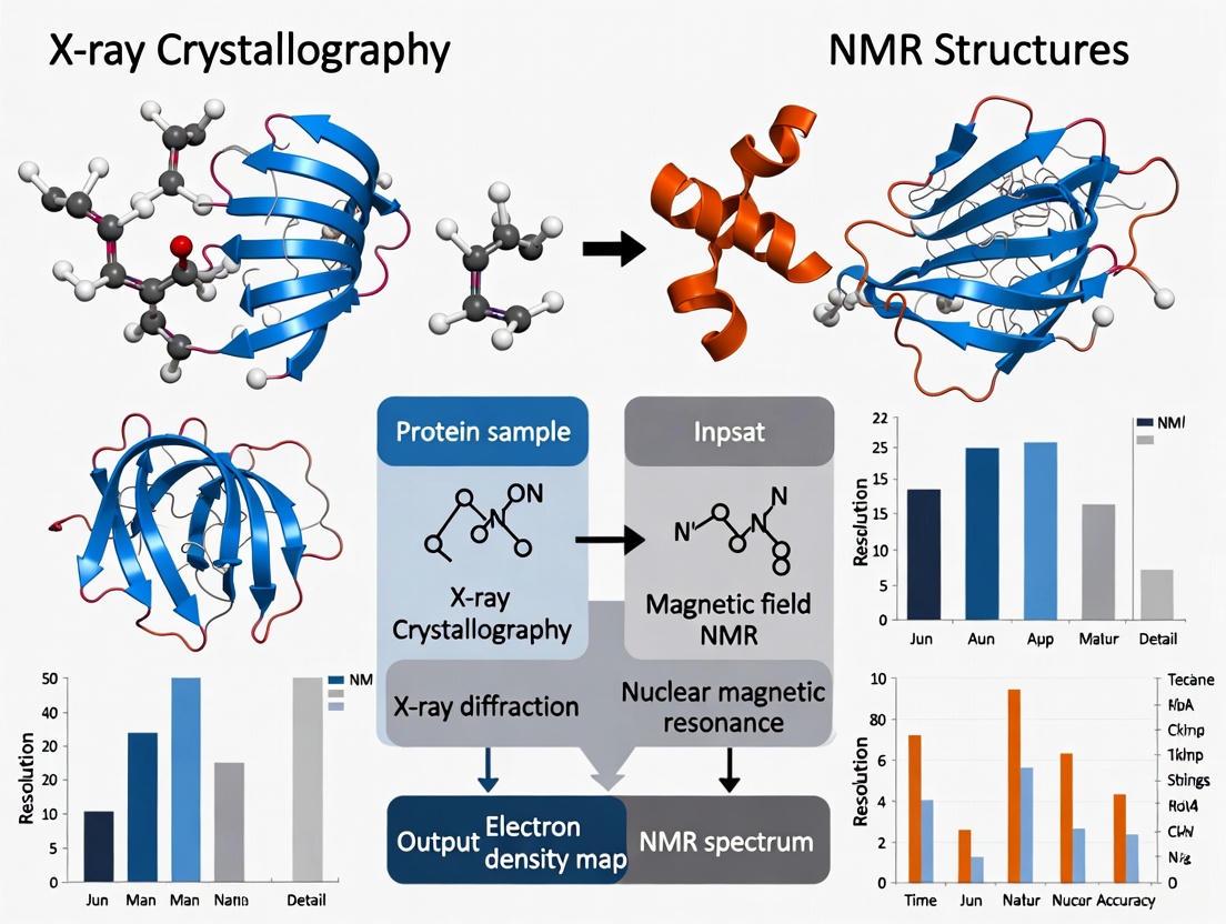
The determination of protein structures at atomic resolution relies primarily on two powerful biophysical techniques: X-ray crystallography and Nuclear Magnetic Resonance (NMR) spectroscopy. Despite sharing the common goal of elucidating three-dimensional molecular architecture, these methods are founded on entirely different physical principles. X-ray crystallography depends on the diffraction of X-rays by the ordered electron clouds of atoms within a crystalline lattice, providing a static snapshot of the molecule's electron density [1] [2]. In contrast, NMR spectroscopy exploits the magnetic properties of atomic nuclei within molecules in solution, detecting the absorption of radiofrequency energy that causes transitions between nuclear spin states in a strong magnetic field [1] [3]. This fundamental divergence in physical basis leads to profound differences in the types of biological information that can be obtained, making these techniques highly complementary rather than directly competitive [4] [5].
The choice between these methods has significant practical implications for structural biology and drug discovery research. X-ray crystallography has historically been the dominant technique, accounting for approximately 66% of the protein structures deposited in the Protein Data Bank (PDB) in 2023, while solution NMR contributed about 1.9% [6]. However, this quantitative disparity reflects their respective technical workflows and limitations rather than their scientific value, as each provides unique and crucial insights into molecular structure and function.
Fundamental Physics and Theory
X-ray Diffraction from Crystals
The physics of X-ray crystallography begins with the interaction of high-energy X-rays with the electrons of atoms. When a crystal is exposed to an X-ray beam, the electrons oscillate and become sources of scattered X-rays. These scattered waves interfere with each other, producing a diffraction pattern of discrete spots [2] [6]. The key to this technique is the regular, repeating arrangement of molecules in the crystal lattice, which amplifies the scattering signal through constructive interference in specific directions determined by the crystal's symmetry [1].
The condition for constructive interference is described by Bragg's Law: nλ = 2d sinθ, where λ is the wavelength of the incident X-rays, d is the distance between crystal lattice planes, θ is the angle of incidence, and n is an integer representing the order of diffraction [6]. This relationship forms the mathematical foundation for calculating atomic positions from the diffraction pattern. The resulting diffraction pattern contains information about the amplitude of the scattered waves, but the phase information is lost in the measurement process, creating the central "phase problem" in crystallography that must be solved through computational or experimental methods [2] [5].
Nuclear Magnetic Resonance in Solution
NMR spectroscopy operates on fundamentally different principles rooted in quantum mechanics. When atomic nuclei with non-zero spin (such as ¹H, ¹³C, or ¹⁵N) are placed in a strong, static magnetic field (B₀), their magnetic moments align with the field, creating a small net magnetization. These nuclei undergo Larmor precession around the direction of the magnetic field at characteristic frequencies that depend on their chemical environment [3].
The application of radiofrequency (RF) pulses at the Larmor frequency disturbs this equilibrium, causing the bulk magnetization to rotate into the transverse plane. After the pulse, the nuclei return to equilibrium through relaxation processes, and the precessing magnetization induces a detectable signal in the receiver coil—the free induction decay (FID) [3]. Fourier transformation of the FID converts this time-domain signal into a frequency-domain spectrum [7].
The exact resonance frequency of a nucleus is influenced by its local electronic environment, leading to the chemical shift phenomenon, which provides detailed information about molecular structure and bonding [1] [3]. For structure determination, NMR relies on measuring a network of through-bond (J-coupling) and through-space (Nuclear Overhauser Effect, NOE) interactions between nuclei to derive distance and angle restraints for computational structure calculation [1] [7].
Table 1: Fundamental Physical Principles Comparison
| Feature | X-ray Crystallography | Solution NMR |
|---|---|---|
| Primary Physical Phenomenon | Diffraction of X-rays by electrons | Absorption of radiofrequency by nuclei in magnetic field |
| Key Mathematical Relation | Bragg's Law: nλ = 2d sinθ | Larmor Equation: ω = γB₀ |
| Sample State | Crystalline solid | Solution |
| Primary Observable | Diffraction spot intensities | Chemical shifts, J-couplings, NOEs |
| Information Content | Electron density map | Interatomic distances, dihedral angles |
| Missing Information | Phase information (must be solved) | None (direct measurement) |
Experimental Methodologies
X-ray Crystallography Workflow
The process of structure determination by X-ray crystallography follows a multi-stage pathway, each with specific technical requirements and challenges. The workflow can be visualized as follows:
Protein Crystallization represents the most significant bottleneck in the crystallography pipeline. The principle involves taking a highly concentrated protein solution and inducing supersaturation through the careful addition of precipitating agents, buffers, and salts [2]. The objective is to achieve a slow, controlled process that encourages crystal growth rather than precipitate formation. This typically employs vapor diffusion methods (hanging or sitting drops), where a small droplet containing protein and precipitant is equilibrated against a reservoir with higher precipitant concentration [2]. Commercial sparse matrix screens systematically explore a wide range of conditions (precipitant type and concentration, buffer, pH, temperature) to identify initial crystallization hits, which are then optimized to produce diffraction-quality crystals of sufficient size (typically >0.1 mm) [2] [5].
Data Collection occurs at synchrotron facilities, which provide intense, tunable X-ray beams. Modern synchrotrons can generate beams with diameters of 0.1-0.3 mm, which are directed onto mounted crystals [2]. The crystal is rotated in the beam while a detector records diffraction patterns at various orientations. Detector technology has evolved from X-ray film to imaging plates and now to charge-coupled device (CCD) detectors, significantly reducing data collection times [2]. For radiation-sensitive samples, cryocooling (flash-freezing crystals in liquid nitrogen at 100 K) helps minimize radiation damage [2].
Data Processing and Phasing begins with indexing the diffraction pattern to determine the unit cell parameters and crystal symmetry (space group) [2]. The intensities of the diffraction spots are measured and merged to produce a set of structure factors containing amplitude information. Since phase information is lost in the diffraction experiment, it must be recovered using methods like molecular replacement (using a similar known structure), or experimental phasing techniques such as Multiple/Single Wavelength Anomalous Dispersion (MAD/SAD) that exploit the anomalous scattering of incorporated heavy atoms [2] [5].
Model Building and Refinement involves fitting the protein sequence into the experimental electron density map, followed by iterative cycles of manual adjustment and computational refinement to improve the agreement between the atomic model and the observed diffraction data while maintaining proper stereochemistry [2] [5].
Solution NMR Workflow
The NMR structure determination pathway involves distinct steps tailored to studying proteins in their native solution state:
Isotope Labeling and Sample Preparation are critical prerequisites for protein NMR studies. Since the natural abundance of magnetically active isotopes like ¹³C (1.1%) and ¹⁵N (0.37%) is low, proteins must be produced recombinantly in bacterial expression systems (typically E. coli) grown in media enriched with ¹³C-glucose and ¹⁵N-ammonium salts to achieve uniform isotopic labeling [5]. For larger proteins or specific applications, selective labeling strategies (e.g., methyl labeling of Ile, Leu, Val) can simplify spectra [8]. NMR samples require relatively high protein concentrations (≥200 µM in volumes of 250-500 µL) and high stability over the data collection period (typically 5-8 days) [5].
Data Acquisition utilizes multi-dimensional NMR experiments to resolve and correlate the signals of thousands of nuclei in the protein. For structure determination, a standard suite includes 2D ¹H-¹⁵N and ¹H-¹³C HSQC spectra, along with 3D experiments such as HNCA, HNCOCA, CBCACONH, and HNCACB for backbone assignment, and HCCH-TOCSY and ¹³C-edited NOESY for sidechain assignment [7]. These experiments exploit through-bond scalar couplings (J-couplings) to establish connectivity between nuclei. Advanced NMR spectrometers with magnetic field strengths of 14.1 Tesla (600 MHz ¹H frequency) or higher, equipped with cryogenically cooled probes, provide the sensitivity required for these experiments [5].
Resonance Assignment is the process of identifying which NMR signals correspond to which specific atoms in the protein sequence. This begins with backbone assignment using triple-resonance experiments that connect amide nitrogens and protons with the carbon atoms of adjacent residues [7]. Sidechain assignments follow using through-bond correlation experiments. This step remains a significant bottleneck in NMR structure determination, though automated and semi-automated approaches are increasingly being employed [7].
Restraint Collection and Structure Calculation relies primarily on Nuclear Overhauser Effect (NOE) measurements, which provide through-space distance restraints between protons typically separated by less than 5-6 Å [7]. Additional restraints include dihedral angles from J-couplings and residual dipolar couplings (RDCs) from proteins aligned in dilute liquid crystalline media, which provide orientational information [4]. These experimental restraints are used in computational structure calculation algorithms (distance geometry, simulated annealing) to generate an ensemble of structures that satisfy the experimental data [7].
Comparative Analysis: Technical Specifications and Applications
Technical Requirements and Limitations
Table 2: Technical Specifications and Limitations
| Parameter | X-ray Crystallography | Solution NMR |
|---|---|---|
| Sample Requirements | High-quality single crystals (>0.1 mm) | Isotopically labeled protein (≥200 µM) |
| Sample State | Crystalline solid | Solution (near-native conditions) |
| Molecular Size Limit | Essentially unlimited (viruses studied) | Typically < 50 kDa (up to ~100 kDa with advanced methods) |
| Time Investment | Crystallization (days-months), Data collection (minutes-hours) | Data acquisition (days-weeks), Analysis (weeks-months) |
| Key Limitation | Need for diffraction-quality crystals | Spectral complexity and overlap with increasing size |
| Typical Resolution | 1.0-3.0 Å (atomic detail) | 1.5-3.0 Å (ensemble precision) |
| Structure Output | Single, time-averaged conformation | Ensemble of conformations representing dynamics |
Research Reagent Solutions and Essential Materials
Table 3: Essential Research Materials and Their Functions
| Material/Reagent | Function | Application |
|---|---|---|
| Crystallization Screens | Sparse matrix conditions to identify initial crystal hits | X-ray crystallography |
| Cryoprotectants | Protect crystals from ice formation during flash-cooling | X-ray crystallography |
| Heavy Atom Compounds | Experimental phasing (MAD/SAD) | X-ray crystallography |
| ¹³C-Glucose/¹⁵N-Ammonium Salts | Isotopic labeling for signal detection | NMR spectroscopy |
| Deuterated Solvents | Field-frequency lock; reduce solvent signal | NMR spectroscopy |
| Cryoprobes | Enhance sensitivity by reducing thermal noise | NMR spectroscopy |
| Shigemi Tubes | Maximize sample volume in active region | NMR spectroscopy |
Data Output and Biological Information
The fundamental physical differences between these techniques lead to distinct types of structural information. X-ray crystallography produces a single, time-averaged conformation of the protein as it exists in the crystal lattice, with atomic positions determined by fitting to the electron density map [9]. The quality of this model is described by the resolution, with higher resolution (lower numerical value) allowing more precise atomic placement [9].
In contrast, NMR yields an ensemble of structures that are all consistent with the experimental restraints, providing direct evidence of conformational flexibility in solution [1] [5]. The precision of the ensemble is described by the root-mean-square deviation (RMSD) of the atomic positions.
X-ray crystallography excels at providing detailed static pictures of molecular structures, including the precise geometry of binding sites and ligand interactions [2] [5]. However, it provides limited information about dynamics and is essentially "blind" to hydrogen atoms due to their low electron density [1] [8]. NMR directly detects dynamic processes across a wide range of timescales (ps to ms) and can provide detailed information about hydrogen bonding and protonation states through chemical shifts [1] [3] [8].
Applications in Structural Biology and Drug Discovery
The complementary nature of X-ray crystallography and NMR spectroscopy makes them valuable for different applications in structural biology and drug discovery.
X-ray crystallography remains the primary method for determining novel protein structures, especially for large complexes and membrane proteins [6] [5]. In drug discovery, it provides atomic-level detail of protein-ligand interactions, enabling structure-based drug design through visualization of binding modes and optimization of interactions [5]. The technique supports high-throughput fragment screening approaches (e.g., XChem) where hundreds of compounds can be soaked into crystals and structures rapidly determined [5].
NMR spectroscopy shines in studying protein dynamics and folding, mapping interaction surfaces, and characterizing intrinsically disordered proteins that do not crystallize [10] [8]. In drug discovery, NMR is particularly valuable for fragment-based lead discovery, identifying weak binders, and characterizing ligand binding when crystallization proves difficult [3] [8]. NMR can detect binding events and quantify affinities without requiring structure determination, making it efficient for screening applications [3].
Recent methodological advances continue to push the boundaries of both techniques. In crystallography, developments like serial femtosecond crystallography (SFX) with X-ray free-electron lasers (XFELs) enable data collection from microcrystals and time-resolved studies of molecular dynamics [6]. In NMR, sensitivity enhancements through dynamic nuclear polarization (DNP) and techniques like TROSY for studying larger molecules are expanding the applicability of the method [10] [8].
X-ray crystallography and solution NMR spectroscopy represent two powerful but fundamentally different approaches to protein structure determination, rooted in the distinct physical phenomena of X-ray diffraction and nuclear magnetic resonance. Crystallography provides high-resolution static structures from crystalline samples but requires crystallization and provides limited dynamic information. NMR yields structural ensembles in solution with inherent dynamic information but faces challenges with molecular size and requires isotopic labeling.
The future of structural biology lies not in choosing one technique over the other, but in recognizing their complementary strengths and leveraging them appropriately for specific biological questions. Integrated approaches that combine data from multiple structural methods, including emerging techniques like cryo-EM, promise a more comprehensive understanding of biological macromolecules in all their structural complexity and dynamic behavior.
Historical Context and Current Dominance in the Protein Data Bank (PDB)
For over five decades, the Protein Data Bank (PDB) has served as the single global repository for the three-dimensional structures of biological macromolecules. Established in 1971 with just a handful of X-ray crystallographic structures, it was the first open-access digital data resource in the biological sciences [11]. The analysis of these structures is fundamental to understanding the molecular mechanisms of life and for the rational design of diagnostics and therapeutics. This guide provides an objective comparison of the two primary experimental methods for protein structure determination—X-ray crystallography and Nuclear Magnetic Resonance (NMR) spectroscopy—tracking their historical contributions to the PDB and evaluating their current standing and applications for researchers and drug development professionals.
Historical Growth in the PDB Archive
The PDB archive has experienced exponential growth since its inception. The following tables chronicle the annual and cumulative deposits for structures determined by X-ray crystallography and NMR spectroscopy, highlighting their distinct growth trajectories [12] [13].
Table: Historical Growth of X-ray Crystallography Structures in the PDB
| Year | Total Entries Available | Annual Structures Released |
|---|---|---|
| 1976 | 13 | 13 |
| 1985 | 193 | 19 |
| 1995 | 3,275 | 750 |
| 2005 | 28,727 | 4,428 |
| 2015 | 101,951 | 8,577 |
| 2023 | 181,250 | 9,583 |
Table: Historical Growth of NMR Spectroscopy Structures in the PDB
| Year | Total Entries Available | Annual Structures Released |
|---|---|---|
| 1989 | 2 | 2 |
| 1995 | 529 | 190 |
| 2005 | 5,119 | 874 |
| 2015 | 11,199 | 431 |
| 2023 | 14,146 | 272 |
Analysis of Historical Trends
- X-ray Crystallography: This method has been the dominant driver of the PDB's growth, exhibiting rapid, non-linear increases, particularly from the late 1990s onward. It has consistently accounted for the vast majority of annual new deposits [11] [14].
- NMR Spectroscopy: NMR began contributing to the PDB in 1988 [11]. Its deposition rate grew steadily, peaking in 2007 with 1,062 new structures before entering a period of decline. In recent years, NMR contributes less than 10% of new structures annually, with only 272 deposits in 2023 (1.9% of the annual total) [14] [13].
Current Status and Market Dominance
While the PDB now also includes structures from 3D Electron Microscopy (3DEM) and hybrid methods, X-ray crystallography remains the most established technique in terms of its total contribution and ongoing market share.
Table: Current Dominance and Market Position of Structural Techniques
| Metric | X-ray Crystallography | NMR Spectroscopy |
|---|---|---|
| % of Total PDB Entries (2024) | ~86% [14] | ~9% [11] |
| % of New Annual Deposits (2023) | ~66% [14] | ~1.9% [14] |
| Market Share in 3D Analysis (2024) | 35% (Leading segment) [15] | Specific share not reported [15] |
| Projected Market Growth | Mature, stable growth | Not specified; Cryo-EM is the fast-growing segment [15] |
Comparative Methodologies: A Detailed Workflow Analysis
The fundamental difference between these techniques lies in the sample state and the principles used to derive atomic coordinates: X-ray crystallography relies on diffraction from a crystalline solid, while NMR studies molecules in solution.
X-ray Crystallography Workflow
X-ray crystallography determines structure by measuring how X-rays scatter when they interact with the electron clouds of atoms arranged in a crystal. The resulting diffraction pattern is used to calculate an electron density map, into which an atomic model is built [14] [5].
Key Experimental Steps:
- Protein Purification and Crystallization: The target protein must be purified to homogeneity and induced to form a highly ordered crystal. This is often the most significant bottleneck, requiring extensive screening of conditions [14] [5].
- Data Collection: A single crystal is exposed to an intense X-ray beam (often at a synchrotron). The resulting diffraction pattern is captured by a detector [14] [5].
- Data Processing and Phasing: The diffraction images are processed to determine the amplitude of the diffracted waves. The "phase problem" must be solved using methods like molecular replacement (using a similar known structure) or experimental phasing (e.g., SAD/MAD) [14] [5].
- Model Building and Refinement: An atomic model is built into the experimental electron density map and iteratively refined to improve its fit to the data while adhering to standard stereochemical constraints [14] [5].
NMR Spectroscopy Workflow
NMR spectroscopy exploits the magnetic properties of atomic nuclei (e.g., ¹H, ¹⁵N, ¹³C) in a strong magnetic field. The resulting spectra provide information on interatomic distances and dihedral angles, which are used as restraints to calculate a family of structures representing the molecule's conformation in solution [16] [5].
Key Experimental Steps:
- Isotope Labeling: For proteins over ~5 kDa, uniform isotopic labeling with ¹⁵N and ¹³C is typically required. This is achieved by expressing the protein in engineered E. coli grown with labeled nutrients [5].
- Data Collection: A series of multi-dimensional NMR experiments (e.g., COSY, NOESY, HSQC, HNCA) are performed to correlate nuclei within the molecule and extract structural information [5] [17].
- Resonance Assignment: The complex NMR signals must be assigned to specific atoms in the protein sequence. This is a foundational and often laborious step [16].
- Restraint Collection and Structure Calculation: Experimental restraints, primarily from Nuclear Overhauser Effect (NOE) measurements (which provide interatomic distances) and J-couplings (which provide dihedral angles), are collected. These restraints are used in computational calculations to generate an ensemble of structures that satisfy the experimental data [16].
The Scientist's Toolkit: Essential Research Reagents and Materials
Table: Key Reagents and Materials for Structural Determination
| Item | Function in X-ray Crystallography | Function in NMR Spectroscopy |
|---|---|---|
| Purified Protein | Required at high concentration (e.g., 10 mg/ml) for crystallization trials. Must be stable [5]. | Required at high concentration (e.g., >200 µM) in a volume of 250-500 µL. Must be stable for days at a time [5]. |
| Crystallization Kits | Commercial screens containing various precipitants, salts, and buffers to identify initial crystallization conditions [5]. | Not applicable. |
| Cryoprotectants | Chemicals (e.g., glycerol, ethylene glycol) used to protect crystals from ice damage during flash-cooling in liquid nitrogen [5]. | Not applicable. |
| Isotopically Labeled Nutrients | Generally not required. | ¹⁵N-ammonium chloride/sulfate and ¹³C-glucose are essential for producing uniformly labeled proteins in bacterial expression systems [5]. |
| Deuterated Solvent | Not used in crystallization. | Deuterated water (D₂O) and buffers are required to minimize the signal from solvent protons in the NMR spectrum [5] [17]. |
| NMR Tubes | Not applicable. | Precision glass tubes designed for high magnetic fields, which hold the sample during data collection [5]. |
Performance and Application Comparison
The choice between X-ray crystallography and NMR spectroscopy is dictated by the research question, the properties of the target macromolecule, and the type of information required.
Table: Objective Comparison of Technique Performance and Applications
| Parameter | X-ray Crystallography | NMR Spectroscopy |
|---|---|---|
| Typical Sample State | Crystalline solid | Solution (near-native conditions) |
| Size Limitations | Effectively none; very large complexes can be studied [5]. | Currently limited to proteins and complexes below ~25-30 kDa for de novo structure determination [16] [5]. |
| Key Output | A single, high-resolution model of the conformation in the crystal. | An ensemble of models representing the dynamic conformations in solution. |
| Atomic Resolution | Can achieve atomic resolution (~1 Å), providing precise atom positions [14]. | Resolution is lower and defined by the spread of the ensemble; precise atomic positions are less defined. |
| Time to Solution | Can be rapid once a diffracting crystal is obtained. Crystallization can be a lengthy bottleneck. | Data collection and analysis are typically time-consuming, often taking weeks [17]. |
| Dynamic Information | Limited; usually a static snapshot. Time-resolved studies are possible but challenging. | A key strength; can probe protein dynamics, conformational changes, and folding on various timescales [16] [5]. |
| Ideal Applications | - High-resolution structure of large complexes and membrane proteins (with specialized methods like LCP) [5].- Structure-based drug design and fragment screening [5].- Studying enzyme mechanisms. | - Structure determination of proteins that are difficult to crystallize [16].- Studying protein dynamics and interactions [16] [5].- Mapping ligand-binding interfaces.- Determining the structure of intrinsically disordered regions. |
| Key Limitations | - Requires high-quality crystals, which may not be possible for all targets.- Crystal packing forces may influence the observed conformation. | - Low sensitivity; requires high protein concentrations and isotopic labeling.- Upper molecular weight limit for full structure determination.- Data analysis and assignment can be complex and require expert knowledge [16]. |
Both X-ray crystallography and NMR spectroscopy have been indispensable in building the rich structural archive of the PDB. X-ray crystallography remains the dominant workhorse for determining high-resolution structures, particularly for drug discovery applications where atomic-level detail of ligand binding is crucial. Its dominance is reflected in its overwhelming share of the PDB and its leading position in the commercial market for 3D structure analysis [14] [15].
In contrast, NMR spectroscopy serves as a powerful complementary technique that excels where crystallography faces challenges. It is the preferred method for obtaining structural and dynamic information of proteins in solution, for studying flexible systems, and for determining structures when crystallization fails. While its contribution to new PDB deposits has decreased, the unique information it provides on dynamics and interactions ensures its continued relevance in the structural biology toolkit, particularly for specific pharmaceutical applications like characterizing chiral centers and impurities that other techniques might miss [16] [17]. The choice between them is not one of superiority, but of selecting the right tool for the specific biological question at hand.
Static Crystals vs. Dynamic Solutions
In structural biology, the choice of technique fundamentally dictates the state in which a biomolecule is observed. X-ray crystallography and Nuclear Magnetic Resonance (NMR) spectroscopy, the two premier methods for atomic-level structure determination, operate on samples with radically different physical properties. X-ray crystallography requires a highly ordered, static crystal, where millions of protein molecules are aligned in a repeating lattice [18]. In contrast, NMR spectroscopy studies proteins in a dynamic, native-like solution environment, where molecules tumble freely and exhibit intrinsic mobility [18] [5]. This fundamental distinction between a static solid state and a dynamic solution state is the cornerstone for understanding the complementary strengths, limitations, and applications of these two powerful techniques. This guide provides an objective comparison for researchers and drug development professionals, framing the discussion within the broader context of structural biology research.
Fundamental Principles and Sample Requirements
The requirement for either a crystal or a solution stems from the underlying physical principles used to extract structural information. The following table summarizes the core characteristics of each method.
Table 1: Fundamental Comparison of X-ray Crystallography and NMR Spectroscopy
| Feature | X-ray Crystallography | NMR Spectroscopy |
|---|---|---|
| Sample State | Static, crystalline solid [18] | Dynamic, aqueous solution [18] [5] |
| Underlying Principle | Diffraction of X-rays by electron density in a crystal [18] [19] | Absorption of radio waves by atomic nuclei in a strong magnetic field [18] [20] |
| Primary Data | Diffraction pattern (spot intensities and phases) [18] | NMR spectrum (chemical shifts, coupling constants) [18] |
| Key Sample Requirement | High-purity, homogeneous protein that forms well-ordered 3D crystals [18] [5] | High-purity, stable protein at high concentration (typically >200 µM); isotopic labeling (^15^N, ^13^C) required for larger proteins [18] [5] |
| Typical Sample Buffer | Various buffers (phosphates not ideal); high concentrations of precipants [5] | Phosphate or HEPES preferred; pH near/below 7.0; salt concentrations <200 mM [5] |
Quantitative Data from Comparative Studies
Systematic comparisons of structures determined by both X-ray crystallography and NMR provide quantitative insight into how the sample state influences the final structural model. A study analyzing 109 non-redundant protein pairs from the PDB revealed key metrics of similarity and difference [21].
Table 2: Quantitative Structural Comparison from a 109 Protein-Pair Dataset [21]
| Comparison Parameter | Observation | Implication |
|---|---|---|
| Global Root-Mean-Square Deviation (RMSD) | Ranges from ~1.5 Å to ~2.5 Å | Overall protein folds are similar, but significant local differences exist. |
| Regional Conformational Deviation | Beta-strands match better than helices and loops. | Structured core elements are more consistent; flexible regions diverge. |
| Amino Acid Correlation | Hydrophobic residues are more similar than hydrophilic. | The protein core (hydrophobic) is conserved; surface (hydrophilic) is influenced by environment. |
| Side Chain Conformations | Buried side chains seldom adopt different orientations. | The protein interior is structurally well-defined in both states. |
| Structural Ensemble | X-ray: Single, static model.NMR: Ensemble of models (e.g., 10-45 structures) [18] [22]. | The NMR ensemble represents conformational flexibility in solution, while the crystal structure is an average of static molecules. |
Furthermore, an analysis of the DrugBank database highlights the historical dominance and precision of X-ray crystallography in drug discovery. As of 2023, 48% of DrugBank small-molecule agents (SMAs) have a structure in the PDB, and of the complexes with human macromolecules, 85% were determined by X-ray diffraction [23].
Experimental Workflows: From Sample to Structure
The journey from a purified protein to an atomic model is distinct for each technique, with sample preparation being the most critical and often limiting step.
X-ray Crystallography Workflow
The process for X-ray crystallography is linear and hinges on obtaining a single, high-quality crystal [18] [19] [5].
NMR Spectroscopy Workflow
The NMR workflow is more iterative, with data collection and analysis often informing further sample preparation, such as isotopic labeling [18] [5].
The Scientist's Toolkit: Key Research Reagent Solutions
Successful structure determination relies on specialized reagents and materials. The table below details essential items for both techniques.
Table 3: Essential Research Reagents and Materials for X-ray and NMR Studies
| Reagent/Material | Function | Technique |
|---|---|---|
| Crystallization Screening Kits | Contains hundreds of chemical conditions to identify initial crystal formation. | X-ray Crystallography |
| Heavy Atoms (e.g., Se-Met) | Used for experimental phasing via SAD/MAD; incorporated into protein or soaked into crystals. | X-ray Crystallography |
| Cryoprotectants (e.g., glycerol) | Prevents ice crystal formation during flash-cooling of crystals in liquid nitrogen. | X-ray Crystallography |
| Isotopically Labeled Nutrients (e.g., ^15^N-NH₄Cl, ^13^C-glucose) | Used in bacterial growth media to produce uniformly ^15^N- and ^13^C-labeled proteins for NMR studies. | NMR Spectroscopy |
| NMR Tubes | High-precision glass tubes designed to hold the sample within the sensitive region of the NMR magnet. | NMR Spectroscopy |
| Deuterated Solvents (e.g., D₂O) | Provides a signal-free lock for the magnetic field and minimizes background ^1H signals. | NMR Spectroscopy |
| Alignment Media | Induces weak molecular alignment for measuring Residual Dipolar Couplings (RDCs), providing long-range structural restraints. | NMR Spectroscopy |
The choice between X-ray crystallography and NMR spectroscopy is not a matter of which technique is superior, but which is most appropriate for the specific biological question at hand. X-ray crystallography is unparalleled in providing high-resolution, static snapshots of proteins and their complexes with small molecules, making it the workhorse for structure-based drug design and characterizing rigid, well-folded proteins [18] [23]. Its main limitations are the bottleneck of crystallization and the static nature of the crystal.
Conversely, NMR spectroscopy excels at studying protein dynamics, folding, and interactions directly in solution, providing unique insights into flexible regions and conformational changes that are often crucial for function [18] [24]. Its major constraints are the protein size limit and the need for isotopic labeling. For the most comprehensive understanding, the structural biology community is increasingly moving toward an integrative approach, combining data from both techniques, and with Cryo-EM, to build multi-faceted models of complex biological assemblies [18] [24] [25].
For researchers and drug development professionals, selecting the appropriate protein structure determination technique is paramount. The primary distinction between X-ray crystallography and Nuclear Magnetic Resonance (NMR) spectroscopy lies in their fundamental output: X-ray crystallography typically produces a single, high-resolution snapshot of the protein, while NMR spectroscopy generates an ensemble of conformations that represent the protein's dynamic state in solution [26] [27] [1]. This guide provides a detailed, data-driven comparison of these techniques to inform your structural biology strategies.
Fundamental Principles and Outputs at a Glance
The following table summarizes the core differences in the outputs and characteristics of these two principal methods.
Table 1: Core Comparison of X-ray Crystallography and NMR Spectroscopy
| Feature | X-ray Crystallography | NMR Spectroscopy |
|---|---|---|
| Primary Output | A single, static 3D model (snapshot) [27] | An ensemble of multiple 3D models (conformers) [27] [21] |
| Sample State | Solid crystal lattice [1] | Solution (near-native conditions) [1] |
| Key Observables | X-ray diffraction spots (intensities & phases) [26] | Chemical shifts, J-couplings, nuclear Overhauser effects (NOEs) [27] [1] |
| Molecular Size | No strict upper limit; suitable for large complexes [26] [5] | Typically limited to proteins < ~50 kDa; 64 kDa complex reported [1] |
| Atomic Information | Full atomic model, but hydrogens are often poorly resolved [1] | Site-specific atomic-level data, excellent for tracking hydrogens [1] |
| Dynamic Information | Limited; requires specialized time-resolved methods [28] | Inherent; provides data on motions from ps to ms timescales [27] |
Quantitative Comparison of Structural Properties
Systematic comparisons of protein structures solved by both methods reveal consistent, quantifiable differences. A study of 109 non-redundant protein pairs from the PDB found that the root-mean-square deviation (RMSD) between equivalent NMR and X-ray structures typically ranges from 1.5 Å to 2.5 Å [21]. Furthermore, the degree of similarity varies by structural element:
- Beta-strands generally show better agreement than alpha-helices and loops [21].
- Hydrophobic amino acids buried in the protein core are more similar between the two methods than hydrophilic, surface-exposed residues [21].
- Side-chain conformations in the protein interior only rarely adopt different orientations between solution and solid states [21].
Experimental Protocols in Practice
X-ray Crystallography Workflow
The following diagram illustrates the multi-step process of structure determination via X-ray crystallography.
Figure 1: The workflow for determining a protein structure using X-ray crystallography.
Key Methodological Steps:
- Crystallization: The purified protein is induced to form a highly ordered crystal. This is often the major bottleneck, requiring extensive screening of conditions [26] [5].
- Data Collection: The crystal is exposed to an intense X-ray beam, producing a diffraction pattern. Synchrotron radiation sources are typically used for high-resolution data [26] [5].
- Phase Problem: The recorded diffraction spots provide amplitude information, but the phase information is lost and must be determined experimentally (e.g., via molecular replacement or anomalous dispersion methods) [26] [5].
- Model Building and Refinement: An atomic model is built into the calculated electron density map and iteratively refined against the experimental data to produce the final structure [26].
NMR Spectroscopy Workflow
The process for NMR-based structure determination is fundamentally different, as shown below.
Figure 2: The workflow for determining a protein structure using NMR spectroscopy.
Key Methodological Steps:
- Isotope Labeling: The protein must be produced recombinantly with isotopic labeling (e.g., ¹⁵N, ¹³C) to enable the detection of NMR signals [5].
- NMR Experiments: A series of multi-dimensional NMR experiments are performed to measure parameters like chemical shifts and through-space NOEs, which provide atomic-level structural and dynamic information [27] [1].
- Constraint Generation: The experimental data are converted into spatial restraints, such as interatomic distances and dihedral angle constraints [27].
- Ensemble Calculation: Structures are calculated using computational methods (e.g., simulated annealing, molecular dynamics) that satisfy the experimental restraints. The result is not one structure, but an ensemble of models that collectively agree with the data, representing the conformational landscape in solution [27] [21].
Case Studies in Drug Discovery
Targeting Conformational Ensembles with Small Molecules
A 2023 study on P-cadherin provides a powerful example of targeting protein dynamics. Researchers determined the crystal structure of a small molecule inhibitor, PhHit1, bound to the P-cadherin X-dimer [29]. Surprisingly, the inhibitor did not directly block the protein-protein interaction interface. Instead, molecular dynamics simulations revealed that PhHit1 binding modulated the conformational ensemble of P-cadherin, altering the equilibrium between functional dimer forms and thereby inhibiting cell adhesion [29]. This demonstrates how ensemble-based insights from NMR can inspire allosteric inhibition strategies that are not apparent from a single static structure.
Visualizing Dynamics with Time-Resolved Crystallography
While traditional crystallography provides static snapshots, advanced techniques are bridging the gap. A 2023 study on lysozyme coupled temperature-jump (T-jump) perturbation with time-resolved serial femtosecond crystallography (TR-SFX) [28]. This method allowed researchers to literally make a "movie" of the protein's response to rapid heating, visualizing widespread atomic vibrations on the nanosecond timescale that evolved into coordinated, functionally relevant motions on the microsecond timescale [28]. This shows how crystallography is evolving to directly probe the dynamic nature of proteins.
The Scientist's Toolkit: Essential Research Reagents and Solutions
Table 2: Key Reagents and Materials for Structural Biology
| Item | Function in X-ray Crystallography | Function in NMR Spectroscopy |
|---|---|---|
| Purified Protein | High purity and homogeneity is critical for crystallization. Requires ~5-10 mg at >10 mg/mL [5]. | High purity and stability is critical for data collection. Requires ~0.2-0.5 mL at >200 µM concentration [5]. |
| Crystallization Kits | Commercial screens containing various precipitants, buffers, and salts to identify initial crystal hits [5]. | Less critical. Standard NMR buffers (e.g., phosphate, Hepes) at neutral pH and low salt are used [5]. |
| Isotope-Labeled Media | Not required for most applications (except for SAD/MAD phasing with selenomethionine) [26]. | Essential. Requires ¹⁵N-labeled and/or ¹³C-labeled media for recombinant expression to assign NMR signals [5] [30]. |
| Synchrotron Access | Essential for high-resolution data collection. Provides high-intensity, tunable X-rays [26] [5]. | Not applicable. |
| High-Field NMR Spectrometer | Not applicable. | Essential. Requires spectrometers of 600 MHz and above, preferably equipped with cryoprobes for sensitivity [5]. |
Market Adoption and Technological Trends
The dominance of X-ray crystallography in the Protein Data Bank (PDB) is well-established, accounting for over 86% of all deposited structures historically [26]. Current trends, however, show a dynamic shift. In 2023, X-ray crystallography accounted for approximately 66% of new PDB deposits, while cryo-electron microscopy (cryo-EM) has seen a dramatic rise to ~32%. NMR spectroscopy contributes a smaller but vital ~2% of new structures, underscoring its specialized role in studying dynamics and smaller proteins [26]. The global 3D protein structure analysis market, valued at USD 2.80 billion in 2024, reflects these trends, with the X-ray segment holding the largest share (35%) but the cryo-EM segment anticipated for the fastest growth [15].
Workflows and Applications: From Bench to Drug Discovery
Within structural biology, two principal techniques dominate high-resolution protein structure determination: X-ray crystallography and Nuclear Magnetic Resonance (NMR) spectroscopy. This guide provides an in-depth, step-by-step protocol for protein crystallization and X-ray data collection, contextualized within a broader comparison with solution NMR. Understanding the workflows, capabilities, and limitations of each method is crucial for researchers and drug development professionals to select the optimal technique for their specific project, whether the goal is high-throughput screening, studying dynamics, or obtaining a detailed atomic-level snapshot for drug design [31].
Experimental Principle and Workflow Comparison
X-ray crystallography determines structures by analyzing the diffraction pattern produced when a crystal is exposed to X-rays. The resulting electron density map allows for the building of an atomic model [32]. In contrast, NMR spectroscopy elucidates structures in solution by measuring nuclear spin interactions, such as chemical shifts and through-bond couplings, to derive distance and angle constraints for computational structure calculation [33].
The fundamental workflows for these techniques are distinct, each with critical steps that influence the success of the structure determination.
Diagram illustrating the comparative workflows for X-ray crystallography (red) and solution NMR spectroscopy (blue), from purified protein to final deposited structure.
Step-by-Step X-ray Crystallography Protocol
Step 1: Protein Crystallization
The initial and often most critical bottleneck is growing a single, well-ordered crystal of sufficient size (typically >0.1 mm in all dimensions) [32].
- Principle: The goal is to bring the protein solution to a state of supersaturation slowly and controllably, typically by vapor diffusion, prompting the protein molecules to arrange into a periodic lattice [32].
- Detailed Methodology (Sitting Drop Vapor Diffusion):
- Prepare Reservoir: Fill a well in a crystallization plate with 50-100 µL of a precipitant solution containing buffers, salts, and polymers like PEG.
- Prepare Protein Drop: Mix equal volumes (e.g., 100-200 nL each) of the purified protein solution and the precipitant solution on a small platform or bridge.
- Seal and Equilibrate: Seal the plate with a transparent tape. The drop, initially at a lower precipitant concentration, equilibrates with the reservoir via vapor diffusion. This slowly increases the concentration of all components in the drop, leading to supersaturation and, ideally, crystal nucleation and growth [32].
- Optimization: Initial crystal "hits" often require optimization by fine-tuning parameters such as pH, precipitant concentration, temperature, and protein concentration [32].
Step 2: Crystal Harvesting and Cryo-cooling
Once a suitable crystal is obtained, it must be prepared for data collection.
- Principle: Protect the crystal from radiation damage during X-ray exposure by flash-cooling it to cryogenic temperatures (typically ~100 K) in a cryoprotectant solution [34].
- Detailed Methodology:
- Transfer: Use a micromesh mount or small loop to extract the crystal from the drop.
- Cryo-protection: Briefly soak the crystal in a cryoprotectant solution (e.g., mother liquor supplemented with 20-25% glycerol or ethylene glycol) to prevent ice formation.
- Flash-cooling: Plunge the crystal, mounted on its loop, directly into liquid nitrogen for storage and transport [34].
Step 3: X-ray Diffraction Data Collection
This is the final experimental step, where the crystal's diffraction pattern is recorded [35].
- Principle: A monochromatic X-ray beam illuminates the crystal. According to Bragg's law, the crystal diffracts the X-rays, producing a regular pattern of spots (reflections) on a detector [32] [35].
- Detailed Methodology (Rotation Method):
- Mounting: The cryo-cooled crystal is centered in the X-ray beam on a goniometer.
- Strategy: Based on one or two test images, a data collection strategy is computed. This optimizes parameters like the crystal-to-detector distance (which controls resolution), the rotation range per image (to avoid spot overlap), and the total rotation range (to ensure data completeness, often up to 180°) [35] [36].
- Exposure: The crystal is rotated in small angular increments (e.g., 0.1-1.0°), and a diffraction image is recorded at each orientation. The intensities and positions of tens to hundreds of thousands of reflections are measured [32] [35].
Step 4: Data Processing and Phase Determination
All subsequent steps are computational, transforming the raw diffraction images into an atomic model.
- Data Processing: The series of two-dimensional diffraction images are processed to determine the unit cell dimensions and crystal symmetry (space group). The images are then integrated to measure reflection intensities and scaled to place all measurements on a common scale [32] [35].
- Solving the Phase Problem: A critical challenge is that the measured intensities provide the amplitude but not the phase of the structure factors. Phases must be determined indirectly. Common methods include:
- Molecular Replacement (MR): Used if a structurally similar model is available.
- Anomalous Dispersion (SAD/MAD): Uses the anomalous signal from native or incorporated heavy atoms (e.g., Se in selenomethionine) [35].
- Model Building and Refinement: An initial atomic model is built into the experimental electron density map. This model is then iteratively refined against the diffraction data to improve its agreement with the measured intensities (minimizing the R-factor) and to ensure ideal stereochemistry [32].
Performance Comparison: X-ray Crystallography vs. NMR
The choice between X-ray crystallography and NMR spectroscopy is guided by the specific research goals, the protein under study, and the desired information. The table below summarizes key performance metrics based on experimental data from structural genomics pipelines [37] and standard crystallographic practice [32] [35].
Table 1: Quantitative Comparison of X-ray Crystallography and NMR Spectroscopy for Protein Structure Determination
| Performance Metric | X-ray Crystallography | NMR Spectroscopy |
|---|---|---|
| Typical Sample State | Solid crystal [32] | Aqueous solution [33] [38] |
| Sample Consumption | Single crystal (nL-µL volume) | 0.5 mL at ~1 mM concentration [37] [39] |
| Key Experimental Data | X-ray diffraction intensities [32] | Chemical shifts, J-couplings, NOEs [33] |
| Data Collection Time | Hours to days per dataset [35] | 1 to 9 days per structure [37] |
| Throughput Potential | Very high (especially with automation) [34] | Moderate (limited by data collection time) [37] |
| Structure Output | Single, static model [32] | Ensemble of conformers [33] |
| Key Quality Indicator | Resolution, R-factor, R-free [32] | RMSD of ensemble, restraint violations [37] |
| Ligand/Interaction Studies | Excellent for identifying bound ligands [35] | Excellent for studying weak interactions & dynamics [33] |
Table 2: Practical Considerations for Method Selection
| Consideration | X-ray Crystallography | NMR Spectroscopy |
|---|---|---|
| Ideal Protein Properties | Readily crystallizes, stable | Soluble, monomeric, small to medium size (≤25 kDa) [37] |
| Primary Technical Hurdle | Obtaining diffraction-quality crystals [32] | Spectral complexity and resonance overlap [33] |
| Dynamic Information | Limited (can sometimes infer from B-factors) | Direct measurement of dynamics on multiple timescales [33] |
| Impact of Labeling | Not required (selenium label can help phasing) | Requires uniform ¹⁵N/¹³C isotopic labeling [37] |
The Scientist's Toolkit: Essential Research Reagents and Materials
Successful structure determination relies on high-quality materials and reagents. The following table details the essential components for a crystallography experiment.
Table 3: Essential Research Reagent Solutions for Protein Crystallography
| Item | Function | Key Considerations |
|---|---|---|
| Purified Protein Sample | The macromolecule for structure determination. | Must be highly pure, homogeneous, and monodisperse for successful crystallization [32] [34]. |
| Crystallization Screen Kits | Pre-formulated solutions to rapidly identify initial crystallization conditions. | Contain various buffers, salts, and precipitants at different pH levels [32]. |
| Precipitant Solutions | Agents (e.g., PEG, salts, organics) that reduce protein solubility. | Induce supersaturation by excluding water or competing for hydration [32]. |
| Cryoprotectant | Chemical (e.g., glycerol, ethylene glycol) added before cooling. | Prevents intracellular ice formation during flash-cooling, preserving crystal order [34]. |
| Deuterated Solvents (for NMR) | Solvent for NMR samples; provides deuterium lock signal. | Must fully dissolve the protein and not interfere with spectra (e.g., D₂O, DMSO-d₆) [39] [38]. |
| NMR Reference Compound | Internal chemical shift standard (e.g., TMS, DSS). | Provides a reference point (0 ppm) for calibrating chemical shifts in the spectrum [39]. |
| X-ray Diffractometer | Instrument that generates X-rays and records diffraction patterns. | Sources include rotating anodes and brighter synchrotrons [32] [34]. |
| NMR Spectrometer | Instrument with a high-field magnet for NMR experiments. | High sensitivity (e.g., with cryoprobes) drastically reduces data collection time [37]. |
The workflows for protein crystallization/X-ray data collection and NMR sample preparation/data acquisition are distinct, each with defined strengths. X-ray crystallography excels in providing high-resolution, static structural models and is highly scalable, making it a pillar of structural genomics and drug discovery. Its main challenge remains the crystallization step. NMR spectroscopy, while typically applied to smaller proteins and requiring longer data collection times, is unparalleled in its ability to study protein dynamics and interactions directly in a physiological solution environment. The informed researcher will consider the specific biological question, the properties of their target protein, and the comparative data presented here to select the most appropriate path to structural insight.
X-ray crystallography and Nuclear Magnetic Resonance (NMR) spectroscopy are two foundational techniques for determining protein structures at atomic resolution. While X-ray crystallography remains the dominant method, accounting for over 66% of new protein structures deposited in the PDB in 2023, solution NMR spectroscopy contributes unique capabilities for studying protein dynamics, folding, and interactions in near-physiological conditions [40]. A significant limitation for traditional NMR studies is the size and complexity of the protein, as signal overlap and rapid spin relaxation complicate data interpretation for molecules beyond ~50 kDa [41] [42]. Isotope labeling with stable NMR-active nuclei (15N, 13C, 2H) overcomes these hurdles by enabling multidimensional NMR experiments, vastly improving resolution and facilitating the assignment of signals to specific atomic positions within the protein [43] [41]. This guide details the workflow for isotope labeling and subsequent multidimensional NMR experiments, providing a direct comparison of structural insights obtained from NMR versus X-ray crystallography.
Table 1: Key Differences Between X-ray Crystallography and Solution NMR Spectroscopy
| Feature | X-ray Crystallography | Solution NMR Spectroscopy |
|---|---|---|
| Sample State | Static, crystallized solid | Dynamic, in solution |
| Primary Data | X-ray diffraction pattern | NMR chemical shifts & coupling constants |
| Typical Output | Single, high-resolution model | Ensemble of conformations |
| Key Strength | High resolution for large complexes & membrane proteins | Study dynamics, folding, & weak interactions |
| Key Limitation | Requires high-quality crystals; crystal packing artifacts | Protein size limitation; signal overlap |
| Annual PDB Deposits (2023) | ~9,601 (>66%) | ~272 (<2%) [40] |
Isotope Labeling Strategies for NMR
The incorporation of stable isotopes is a prerequisite for the multidimensional NMR experiments used to study protein structure and dynamics.
Uniform Labeling
The most common strategy involves uniform labeling with 15N and 13C. This is typically achieved by expressing recombinant proteins in E. coli grown in minimal media where the sole nitrogen and carbon sources are 15NH4Cl and 13C-glucose, respectively [43]. This labels all nitrogen and carbon atoms in the protein, enabling a suite of triple-resonance experiments that connect the backbone atoms (1H, 15N, 13Cα) for sequential assignment [41]. For proteins larger than ~25 kDa, deuteriation (2H labeling) becomes essential. Replacing 1H with 2H (deuterium) reduces unfavorable relaxation pathways, dramatically sharpens linewidths, and allows for the study of proteins and complexes up to 100 kDa [41] [42].
Selective and Segmental Labeling
For larger proteins or specific studies, more sophisticated labeling strategies are employed:
- Amino Acid-Selective Labeling: The growth medium is supplemented with a single 13C/15N-labeled amino acid. This simplifies spectra by highlighting only one type of residue, which is powerful for probing hydrophobic cores (e.g., with Ile, Leu, Val) or mapping binding interfaces [41].
- Segmental Labeling: This technique enables isotopic labeling of a specific protein domain or segment while the rest of the chain is unlabeled. It is achieved through protein semisynthesis or intein-mediated ligation. This is ideal for studying multi-domain proteins or post-translationally modified proteins, as it provides atomic-resolution data for a specific region of interest without spectral complexity from the entire molecule [44] [42].
Table 2: Common Isotope Labeling Strategies for Protein NMR
| Labeling Strategy | Key Reagents | Primary Function | Ideal Application |
|---|---|---|---|
| Uniform 15N/13C | M9 Minimal Media, 15NH4Cl, 13C-Glucose | Enables backbone assignment via triple-resonance experiments | De novo structure determination of proteins < 25 kDa |
| Deuteriation (2H) | D2O-based M9 Media, deuterated carbon source | Reduces dipole-dipole relaxation, narrows linewidths | Studying large proteins and macromolecular complexes |
| Amino Acid-Selective | Defined M9 Media, specific 15N/13C-labeled amino acid | Simplifies spectrum to a single residue type | Probing binding sites or hydrophobic core packing |
| Segmental | Semisynthetic peptides, expressed protein ligation (EPL) tools | Labels a specific domain or post-translationally modified segment | Studying large, multi-domain or modified proteins |
Multidimensional NMR Experiments: From Data Acquisition to Structural Insights
With an isotopically labeled protein sample in hand, a series of multidimensional NMR experiments are performed to extract structural information.
Key Multidimensional Experiments
The workflow typically begins with a 2D 1H-15N HSQC (Heteronuclear Single Quantum Coherence) experiment. This spectrum acts as a "fingerprint" of the protein, providing one signal for each backbone amide group and nitrogen-containing side chain. It is used to assess sample quality and monitor changes, such as ligand binding [43]. For assignment and structure determination, more complex experiments are required:
- 3D HNCA: Correlates the chemical shifts of a amide proton (1H), its attached nitrogen (15N), and the alpha carbon (13Cα) of the preceding amino acid. This is crucial for establishing sequential connectivity along the protein backbone [43].
- NOESY (Nuclear Overhauser Effect Spectroscopy): Measures through-space dipole-dipole couplings between protons. The intensity of a NOE cross-peak is inversely proportional to the sixth power of the distance between protons, providing crucial distance restraints for calculating the 3D structure [44].
Experimental Protocol: Ligand Binding Site Mapping by Chemical Shift Perturbation
The following is a standard protocol for identifying a ligand-binding site on a 15N-labeled protein [43]:
- Sample Preparation: Prepare a ~0.3-1.0 mM sample of the uniformly 15N-labeled protein in a suitable NMR buffer. The high concentration is necessary for sensitivity in multidimensional experiments.
- Reference Spectrum Acquisition: Collect a 2D 1H-15N HSQC spectrum of the protein alone (apo state). This typically takes 1-2 hours.
- Ligand Titration: Titrate an unlabeled ligand of interest into the protein sample. To achieve saturation, a molar excess of ligand is often used.
- Bound State Spectrum Acquisition: Collect a new 2D 1H-15N HSQC spectrum under identical conditions as the apo state spectrum.
- Data Analysis: Compare the two spectra and quantify the changes in 1H and 15N chemical shifts (ΔσH and ΔσN) for each residue. The combined chemical shift perturbation (CSP) is calculated using the equation:
CSP (ppm) = √[(ΔσH)2 + (ΔσN/6)2] - Mapping: Residues with the largest CSPs are mapped onto the protein's 3D structure (if available). The largest perturbations and/or signal broadening typically identify the binding site, while smaller, long-range perturbations may indicate allosteric effects [43].
Diagram 1: Ligand Binding Site Mapping Workflow
Comparative Analysis: NMR vs. X-ray Crystal Structures
When membrane protein structures determined by both solution NMR and X-ray crystallography are compared, systematic differences emerge, highlighting the influence of the technique and the sample environment [45].
A study of 14 membrane proteins with structures solved by both methods found that the structural differences are not random. On average, the backbone root-mean-square deviation (RMSD) between NMR and crystal structures in the membrane region is below 5 Å [45]. The analysis further revealed that:
- NMR ensembles show higher convergence within the hydrophobic membrane region than in soluble loops [45].
- Crystal structures typically have straighter transmembrane helices, higher stereochemical quality, and are more tightly packed, which may sometimes reflect crystal-packing forces [45].
- The environment (e.g., micelles for NMR vs. lipidic cubic phases for crystallography) significantly influences the observed structure [45].
These findings underscore that the two techniques offer complementary views. NMR captures the protein's dynamic behavior in solution, while crystallography provides a high-resolution static snapshot that can be influenced by the crystalline state.
Table 3: Quantitative Comparison of NMR and X-ray Structures for Membrane Proteins
| Comparison Metric | NMR Structures | X-ray Structures | Biological Implication |
|---|---|---|---|
| Backbone RMSD in Membrane Region | Higher convergence in TM regions [45] | ~1.0 - 2.5 Å (soluble proteins) [45] | NMR better defines core; X-ray gives precise average |
| Transmembrane Helix Conformation | More bent/curved | Typically straighter [45] | Packing forces may distort helices in crystals |
| Stereochemical Quality | Standard quality | High quality [45] | Both methods produce reliable models |
| Packing Density | Standard packing | Tighter packing [45] | Crystal packing may over-stabilize contacts |
| Environment | Solution (micelles, bicelles) | Crystal lattice (LCP, micelles) | The mimetic influences the observed structure [45] |
The Scientist's Toolkit: Essential Research Reagents
Successful execution of isotope labeling and NMR studies requires specific reagents and materials. The following table details key solutions used in the field.
Table 4: Key Research Reagent Solutions for Isotope Labeling and NMR
| Reagent / Material | Function in Workflow | Specific Example |
|---|---|---|
| 15NH4Cl | Universal 15N nitrogen source for uniform labeling in bacterial expression | Supplement in M9 minimal media [43] |
| 13C-Glucose | Universal 13C carbon source for uniform labeling in bacterial expression | Sole carbon source in M9 minimal media [43] |
| Amino Acid Kit (13C/15N-labeled) | For selective labeling of specific residue types | Label specific amino acids (e.g., Ile, Leu, Val) to probe hydrophobic core [41] |
| Deuterated Detergents | Membrane mimetic for solubilizing membrane proteins in NMR studies | DPC-d38, DHPC-d46 micelles [45] |
| T7 RNA Polymerase | Enzymatic synthesis of RNA for NMR via in vitro transcription | Production of milligram quantities of RNA [44] |
| Hammerhead Ribozyme | DNA template for controlling 5'-end homogeneity of in vitro transcripts | Ensures uniform starting sequence for RNA [44] |
Structure-Based Drug Design (SBDD) represents a cornerstone of modern pharmaceutical research, providing a rational framework for transforming initial hits into optimized drug candidates by leveraging detailed 3D structural information of biological targets [8] [46]. The atomic-level insights into protein-ligand interactions enable medicinal chemists to design compounds with enhanced binding affinity, selectivity, and pharmacological properties [23]. Among the experimental techniques available for structure determination, X-ray crystallography and nuclear magnetic resonance (NMR) spectroscopy have emerged as pivotal methods, each with distinct strengths and limitations [47]. While X-ray crystallography has traditionally been the workhorse of structural biology, NMR spectroscopy provides complementary solution-state information that is particularly valuable for studying dynamic interactions and molecular recognition events [48]. This guide provides an objective comparison of these two fundamental techniques, offering researchers a framework for selecting the appropriate method based on their specific protein target and drug discovery objectives.
Technical Comparison of X-ray Crystallography and NMR
The selection between X-ray crystallography and NMR spectroscopy requires careful consideration of multiple technical parameters, including the nature of the target protein, the type of information required, and practical constraints related to throughput and sample preparation. The following comparison outlines the fundamental characteristics and capabilities of each method.
Table 1: Core Method Comparison for X-ray Crystallography and NMR in SBDD
| Parameter | X-ray Crystallography | NMR Spectroscopy |
|---|---|---|
| Sample State | Solid crystal | Solution (native-like conditions) |
| Molecular Weight Range | Effectively no upper limit [47] | Best for proteins < ~50 kDa; up to ~80 kDa with advanced techniques [8] [47] |
| Typical Resolution | Atomic (~1 Å) [47] | Atomic (~1-2 Å) [47] |
| Hydrogen Atom Detection | Essentially "blind"; cannot resolve H atoms [8] [48] | Direct detection of hydrogen atoms and their interactions [8] [48] |
| Dynamic Information | Single, static snapshot [8] [49] | Direct observation of dynamics, conformational changes, and multiple states [8] [47] |
| Key Limitation | Requires high-quality crystals; cannot study intrinsic dynamics [8] [47] | Limited by protein size and sensitivity; complex data analysis [8] [47] |
Table 2: Application-Based Suitability for Drug Discovery Workflows
| Application in SBDD | X-ray Crystallography | NMR Spectroscopy |
|---|---|---|
| High-Throughput Screening | Yes, via soaking systems (though challenging to establish) [8] | Yes, especially for fragment screening [50] |
| Fragment-Based Drug Discovery (FBDD) | Excellent for identifying fragment binding location [23] | Premier technique for detecting weak (mM) fragment binding [50] |
| Studying Protein Dynamics | Not suitable | Excellent for capturing conformational ensembles [8] [47] |
| Membrane Protein Structures | Challenging, but possible [46] | Challenging due to size limitations |
| Mapping Molecular Interactions | Inferred from atomic proximity [8] | Direct measurement of H-bonds and other non-covalent interactions [8] |
| Water Molecule Mapping | ~80% of bound waters are observable [8] | Can study full hydration networks and water dynamics |
Experimental Protocols and Methodologies
X-ray Crystallography Workflow
The process of structure determination via X-ray crystallography follows a well-established pipeline that transforms a purified protein sample into an atomic model. Key steps include:
- Protein Crystallization: Purified protein is concentrated and subjected to crystallization trials using vapor diffusion, batch, or other methods under empirically determined conditions containing precipitants, buffers, and additives. This remains a major bottleneck, with only an estimated 25% of successfully purified proteins yielding suitable crystals [8] [48].
- Crystal Harvesting and Cryocooling: Suitable crystals are harvested and flash-frozen in liquid nitrogen, often with a cryoprotectant to prevent ice formation [47].
- X-ray Diffraction Data Collection: The crystal is exposed to an intense X-ray beam, and the resulting diffraction pattern is collected on a detector [49] [51].
- Phase Problem Solution: The phases for the diffraction waves are determined using methods like Molecular Replacement (MR) with a known homologous structure, or experimental methods like Single-wavelength Anomalous Dispersion (SAD) [23].
- Electron Density Map Calculation and Model Building: An initial electron density map is calculated, into which an atomic model is built and iteratively refined against the diffraction data to improve the fit [49] [51]. The quality of the final model is assessed by metrics including the R-factor and R-free, with lower values indicating a better fit to the experimental data [51].
NMR Spectroscopy Workflow
Solution-state NMR structure determination focuses on extracting structural constraints from a protein in its native state:
- Isotope Labeling: The protein is produced via recombinant expression in a medium containing stable isotopes (e.g., ^15^N, ^13^C). This is a prerequisite for all but the smallest proteins and allows for the magnetization transfer necessary for multidimensional experiments [8] [48].
- NMR Data Collection: A battery of multi-dimensional NMR experiments (e.g., HSQC, NOESY) is performed on the labeled protein, often in the presence and absence of a ligand [50]. The Chemical Shift Perturbation (CSP) of peaks in a ^1^H-^15^N HSQC spectrum upon ligand binding is a primary method for identifying binding sites.
- Constraint Collection: Structural constraints are extracted from the spectra. Nuclear Overhauser Effect (NOE) measurements provide distance constraints between protons, while J-couplings and chemical shifts inform on torsion angles [50].
- Structure Calculation: A three-dimensional structure is calculated computationally by satisfying the collected distance and angular constraints. The result is typically an ensemble of structures that represent the conformational space the protein occupies in solution [8].
Diagram 1: Comparative workflows for X-ray and NMR structure determination.
The Scientist's Toolkit: Essential Research Reagents and Materials
Successful application of either technique requires specialized reagents and materials. The following table details key solutions and their functions in the respective workflows.
Table 3: Essential Research Reagents and Materials for Structural Biology
| Reagent/Material | Function | Application |
|---|---|---|
| Crystallization Screen Kits | Commercial suites of pre-mixed solutions containing various precipitants, buffers, and salts to empirically identify initial crystallization conditions. | X-ray Crystallography |
| Cryoprotectants (e.g., Glycerol, PEG) | Agents used to displace water from the crystal lattice, preventing the formation of destructive ice crystals during flash-cooling in liquid nitrogen. | X-ray Crystallography |
| Isotope-Labeled Nutrients (e.g., ^15^NH₄Cl, ^13^C-Glucose) | Nitrogen and carbon sources containing stable isotopes for producing uniformly labeled proteins, enabling multi-dimensional NMR experiments. | NMR Spectroscopy |
| Amino Acid Precursors (e.g., ^13^C-methyl labeled α-ketoisovalerate) | Biosynthetic precursors for selective labeling of specific amino acid side chains (e.g., Ile, Val, Leu methyl groups), simplifying spectra for larger proteins. | NMR Spectroscopy |
| Structure Refinement Software (e.g., PHENIX, COOT) | Computational programs for iteratively adjusting the atomic model to improve its fit to the experimental electron density map and ideal geometry. | X-ray Crystallography |
| NMR Data Processing & Analysis Suites (e.g., NMRPipe, CCPN) | Software for processing raw NMR data, peak picking, assignment, and calculating three-dimensional structures from experimental constraints. | NMR Spectroscopy |
Decision Framework for Technique Selection
Choosing the most appropriate technique depends on the specific scientific question and the properties of the target protein. The following decision pathway provides a logical framework for researchers.
Diagram 2: Decision pathway for selecting between X-ray crystallography and NMR.
Both X-ray crystallography and NMR spectroscopy are powerful, indispensable techniques in the SBDD toolkit. X-ray crystallography provides unparalleled high-resolution static snapshots of protein-ligand complexes, which are invaluable for guiding medicinal chemistry optimization [23]. In contrast, NMR spectroscopy excels in characterizing dynamic processes and weak interactions in solution, offering unique insights into molecular recognition that are often inaccessible by crystallography [8] [50]. The most effective modern drug discovery programs do not view these methods as mutually exclusive but as complementary. By integrating data from both techniques, researchers can build a more comprehensive understanding of their target, combining the precise atomic coordinates from crystals with the dynamic reality of solution-state behavior. This synergistic approach ultimately accelerates the rational design of more effective and selective small-molecule therapeutics.
Fragment-Based Drug Discovery (FBDD) has emerged as a powerful and complementary approach to traditional High-Throughput Screening (HTS) for identifying novel therapeutic compounds. This methodology involves screening small, low-complexity organic molecules (fragments) against biological targets, followed by systematic optimization of these weak-binding hits into potent drug-like leads [52] [53]. The fundamental advantage of FBDD lies in its efficient exploration of chemical space; because fragments are small (typically <300 Da), a library of just 1,000-2,000 compounds can sample a much greater diversity of chemical structures compared to HTS libraries containing hundreds of thousands of larger molecules [53]. This approach has proven particularly valuable for targeting challenging protein classes, including protein-protein interactions and enzymes previously considered "undruggable," with several FDA-approved drugs such as vemurafenib, venetoclax, and sotorasib originating from fragment-based campaigns [52] [53].
The success of FBDD heavily relies on specialized biophysical techniques capable of detecting the weak binding affinities (typically in the μM to mM range) characteristic of fragment-target interactions [52] [54]. Among these techniques, X-ray crystallography and Nuclear Magnetic Resonance (NMR) spectroscopy have established themselves as cornerstone methods for fragment screening and hit identification. This guide provides a comprehensive comparison of these two pivotal structural biology techniques within the FBDD workflow, examining their respective capabilities, limitations, and optimal applications to inform researchers' experimental strategies.
The Role of Structural Biology in FBDD
Structural biology techniques provide the atomic-resolution insights crucial for advancing fragments into viable drug candidates. While several biophysical methods are employed in FBDD, including Surface Plasmon Resonance (SPR), Microscale Thermophoresis (MST), and Differential Scanning Fluorimetry (DSF), X-ray crystallography and NMR offer unique advantages for characterizing fragment binding [55] [54]. These techniques not only confirm binding but also elucidate precise binding modes and locations, information that is invaluable for guiding medicinal chemistry optimization [52] [56]. According to the Protein Data Bank (PDB), X-ray crystallography remains the dominant structural biology technique, contributing to over 66% of structures released in 2023, while NMR accounted for approximately 1.9% [57]. However, this distribution reflects methodological differences rather than utility in FBDD, where both techniques play complementary roles.
Comparative Techniques Table
Table 1: Key Biophysical Techniques Used in Fragment Screening
| Technique | Detection Principle | Typical Kd Range | Throughput | Key Advantages | Main Limitations |
|---|---|---|---|---|---|
| X-ray Crystallography | X-ray diffraction from crystals | mM to nM | Medium | Provides atomic-resolution structures; identifies binding pose and water networks | Requires protein crystallization; may miss some binders due to crystal packing |
| NMR Spectroscopy | Nuclear spin interactions in magnetic field | mM to low μM | Medium to High | Detects weak binders; provides binding site information; solution-based | Requires isotopic labeling for protein-observed; limited for large proteins |
| Surface Plasmon Resonance (SPR) | Changes in refractive index at sensor surface | μM to pM | High | Provides kinetic parameters (kon, koff); label-free | Immobilization may affect protein function; false positives from non-specific binding |
| Microscale Thermophoresis (MST) | Temperature-induced fluorescence changes | mM to pM | Medium | Measures in solution; small sample volume | Requires fluorescent labeling; sensitive to buffer conditions |
| Differential Scanning Fluorimetry (DSF) | Thermal protein denaturation | Varies widely | High | Low protein consumption; easy implementation | Indirect binding measurement; prone to false positives/negatives |
X-ray Crystallography in FBDD
Methodology and Workflow
X-ray crystallography determines the three-dimensional structure of macromolecules by analyzing the diffraction patterns generated when X-rays interact with crystalline samples [57]. The technique relies on Bragg's Law (nλ = 2dsinθ), where the diffraction pattern reveals the electron density distribution within the crystal [57]. For FBDD applications, the process begins with the development of a high-quality crystallization system capable of producing hundreds of reproducible crystals, which is comparable to assay development in HTS campaigns [56]. The standard workflow involves: (1) Crystallization of the target protein, often the most challenging and time-consuming step; (2) Soaking of crystals in solutions containing individual fragments or fragment mixtures; (3) Data Collection using synchrotron radiation sources to collect high-resolution diffraction data; (4) Data Processing to generate electron density maps; and (5) Model Building and Refinement to determine the precise position and orientation of bound fragments [57] [56].
Experimental Protocols for Crystallographic Screening
Successful crystallographic fragment screening requires careful experimental design. Fragment libraries for crystallography typically follow the "rule of three" (molecular weight ≤300 Da, ClogP ≤3, hydrogen bond donors and acceptors ≤3) [53] [54]. These fragments are screened at high concentrations (up to 100 mM) to compensate for weak affinities [56]. Two primary approaches are employed:
Soaking Experiments: Pre-formed protein crystals are transferred to solutions containing fragments, allowing ligands to diffuse through solvent channels and bind to the target. This method is efficient for screening multiple fragments against the same crystal form [56].
Co-crystallization: Fragments are present during the crystallization process, which can be advantageous for fragments that induce conformational changes but is more resource-intensive [56].
Advanced facilities utilize automated crystal mounting systems and third-generation synchrotron sources to enable high-throughput data collection. Pipelines like FastForward can process the resulting datasets to generate high-quality 3D models for structure-based drug design [56].
Advantages and Limitations in FBDD
X-ray crystallography offers several distinctive advantages for FBDD. It is exceptionally sensitive, capable of detecting very low-affinity binders (Kd in mM range) that might be missed by other biophysical methods [56]. Most importantly, it provides detailed three-dimensional structural information, revealing not only the precise binding pose of the fragment but also key interactions with the protein target and the positioning of water molecules that can be targeted for displacement during optimization [56]. This structural information is invaluable for guiding fragment growing, linking, and optimization strategies with atomic precision.
However, the technique has significant limitations. The requirement for high-quality protein crystals can be a major bottleneck, particularly for challenging targets that do not crystallize readily [57]. Crystal packing constraints may obscure biologically relevant binding sites or prevent fragment access to certain pockets [56]. Additionally, the technique requires specialized instrumentation (synchrotrons for high-throughput applications) and significant expertise in crystallography and data analysis, making it resource-intensive compared to some other screening methods [57].
NMR Spectroscopy in FBDD
Methodology and Workflow
NMR spectroscopy exploits the magnetic properties of atomic nuclei to study molecular structures and interactions in solution [55] [52]. For FBDD applications, NMR can detect fragment binding through changes in the spectroscopic properties of either the protein or the fragment itself [52]. The technique is particularly valuable for identifying weak binders (with Kd values up to single-digit mM range) that are often the only hits found for challenging targets [52]. NMR-based screening does not require crystallization and can be performed under physiological conditions, providing insights into binding events in a native-like environment. Modern NMR facilities employ cryoprobe-equipped spectrometers (500-800 MHz) with auto-samplers to enable high-throughput screening of fragment libraries in a matter of hours [55] [52].
Experimental Protocols for NMR Screening
Two primary NMR approaches are employed in fragment screening:
Ligand-Observed NMR: These methods monitor changes in the properties of the fragments themselves and include:
- Saturation Transfer Difference (STD): Measures transfer of magnetization from the protein to bound ligands, identifying binders through signal attenuation [55].
- WaterLOGSY: Detects binding through changes in water-ligand interactions, with bound ligands showing inverted signals compared to non-binders [55].
- 19F-NMR: Utilizes fluorine-containing fragments or competitors, offering high sensitivity and the ability to screen mixtures without spectral overlap [55].
Protein-Observed NMR: These methods require isotopic labeling (15N, 13C) of the protein and monitor chemical shift perturbations in 2D spectra (e.g., 1H-15N HSQC) upon fragment binding. This approach identifies binding sites and can quantify binding affinities [52].
Effective NMR screening requires careful library design. Fragment libraries typically contain 500-10,000 compounds, with some specialized libraries containing up to 14,000 fragments [52]. These libraries are carefully curated to remove "bad actors" – compounds prone to aggregation, reactivity, or non-specific binding – which is particularly important when screening in mixtures [52].
Advantages and Limitations in FBDD
NMR spectroscopy offers unique advantages for FBDD. As a solution-based technique, it can detect binding under physiological conditions without the constraints of crystallization [52]. It reliably detects very weak interactions (up to mM Kd values) and provides information about binding sites and, in some cases, binding modes [52]. Unlike many other techniques, NMR is relatively free from false positives because it directly observes binding events rather than secondary effects [52]. It can also identify novel ligand-binding "hot spots," including allosteric sites and sites resulting from protein conformational changes, without requiring prior knowledge of protein function [52].
The limitations of NMR include its relatively low sensitivity compared to other techniques, requiring higher protein concentrations (typically 10-100 μM) [52]. For protein-observed methods, isotopic labeling (15N, 13C) is necessary, which can be challenging for some protein targets [52]. The technique also has size limitations, becoming increasingly difficult for proteins larger than 50-100 kDa, though this can be addressed with specialized approaches [52]. Throughput, while improved with automation, generally remains lower than some other biophysical methods, and the requirement for specialized instrumentation and expertise limits its accessibility [52].
Direct Comparison: X-ray Crystallography vs NMR in FBDD
Performance Comparison Table
Table 2: Direct Comparison of X-ray Crystallography and NMR for FBDD Applications
| Parameter | X-ray Crystallography | NMR Spectroscopy |
|---|---|---|
| Sample Requirements | High-quality crystals required; no size limit in principle | No crystals needed; isotopic labeling often required for protein-observed methods |
| Sample State | Solid crystal | Solution phase |
| Information Obtained | Atomic-resolution 3D structure; precise binding pose; water networks | Binding confirmation, affinity estimates, binding site location, dynamics |
| Sensitivity | Can detect very weak binders (mM Kd) | Can detect weak binders (mM to low μM Kd) |
| Throughput | Medium (improving with automation) | Medium to High (depending on approach) |
| Target Limitations | Difficult for membrane proteins and flexible targets | Challenging for large proteins (>50-100 kDa) |
| Data Collection Time | Minutes to hours per dataset | Hours to days per screen |
| Key Strength in FBDD | Provides detailed structural information for optimization | Detects binding in solution under physiological conditions |
| Primary Limitation in FBDD | Crystal packing may obscure binding sites | Lower structural resolution compared to crystallography |
Hit Identification Overlap and Complementarity
A critical consideration in FBDD is that different screening techniques often identify different sets of fragment hits. Studies have shown surprisingly low overlap between hits identified by X-ray crystallography and those found by other biophysical methods [56]. This occurs because each technique detects binding through different physical principles and under different experimental conditions. Crystallography may identify fragments that bind in specific orientations stabilized by crystal packing but miss binders that require conformational flexibility [56]. Conversely, NMR detects binders in solution but may miss fragments with very weak affinities or those that bind in ways that don't produce significant spectroscopic changes [52]. This limited overlap highlights the value of using orthogonal techniques in tandem for comprehensive fragment screening.
Integrated Workflow Diagram
Diagram Title: Integrated FBDD Workflow Using X-ray and NMR
The Scientist's Toolkit: Essential Research Reagents and Solutions
Key Research Reagents Table
Table 3: Essential Research Reagents for FBDD Screening
| Reagent/Solution | Application | Function in FBDD |
|---|---|---|
| Fragment Libraries | All screening techniques | Collections of 500-14,000 small molecules (MW <300) following Rule of Three principles [52] [53] |
| Crystallization Kits | X-ray crystallography | Sparse matrix screens to identify initial crystallization conditions [57] |
| Isotopically Labeled Media | Protein-observed NMR | 15NH4Cl, 13C-glucose for producing 15N/13C-labeled proteins for NMR studies [52] |
| Cryoprotectants | X-ray crystallography | Glycerol, ethylene glycol to protect crystals during flash-cooling [57] |
| Synchrotron Access | X-ray crystallography | High-intensity X-ray source for data collection on microcrystals [57] [56] |
| NMR Cryoprobes | NMR screening | Sensitivity-enhanced probes for high-throughput fragment screening [55] |
| Ligand-Observed NMR Buffers | Ligand-observed NMR | Specifically formulated buffers to minimize background signals [55] [52] |
Both X-ray crystallography and NMR spectroscopy play indispensable yet complementary roles in modern FBDD pipelines. X-ray crystallography excels in providing high-resolution structural information critical for rational optimization of fragment hits, while NMR offers powerful solution-based screening capabilities under physiological conditions [52] [56]. The strategic integration of both techniques maximizes the likelihood of identifying diverse, high-quality fragment hits against even the most challenging therapeutic targets.
For researchers designing FBDD campaigns, the choice between these techniques should be guided by target properties, available resources, and project goals. For well-behaved proteins that crystallize readily, crystallographic screening provides unparalleled structural insights. For challenging targets resistant to crystallization or requiring physiological conditions, NMR offers a robust alternative. The most successful FBDD campaigns often employ both techniques in concert, leveraging their complementary strengths to comprehensively explore the fragment binding landscape and efficiently transform weak-binding fragments into potent drug candidates [52] [56]. As both technologies continue to advance—with improvements in automation, data collection, and analysis—their impact on drug discovery is poised to grow, particularly for targets that have historically eluded conventional screening approaches.
Understanding molecular interactions, particularly hydrogen bonds, is fundamental to rational drug design and materials science. Two principal experimental techniques—X-ray crystallography and Nuclear Magnetic Resonance (NMR) spectroscopy—offer complementary pathways for visualizing these critical interactions. X-ray crystallography infers hydrogen bonding patterns indirectly by analyzing electron density maps derived from diffraction data. In contrast, NMR spectroscopy can probe hydrogen bonds more directly by measuring their influence on nuclear spins, providing dynamic information in near-native environments. This guide provides a comparative analysis of these methodologies, their underlying experimental protocols, and their application in modern structural research, offering scientists a framework for selecting the appropriate tool for their specific investigations into molecular interactions.
Fundamental Principles and Experimental Protocols
X-ray Crystallography: Inferring from Electron Density
Fundamental Principle: X-ray crystallography determines the three-dimensional arrangement of atoms in a crystal by analyzing the pattern produced when X-rays are diffracted by the electron clouds of those atoms. The positions and intensities of the diffraction spots are used to compute an electron density map, from which an atomic model is built [58]. Hydrogen bonds are not directly observed but are inferred from the geometry between potential donor and acceptor atoms (e.g., O-H⋯O, N-H⋯N) identified in the model.
Key Experimental Workflow:
- Crystallization: The target molecule (e.g., a protein or small molecule) is purified and induced to form a highly ordered, three-dimensional crystal [58].
- Data Collection: The crystal is exposed to an intense X-ray beam, and a detector records the resulting diffraction pattern [58].
- Data Processing: The diffraction images are processed to determine the amplitude and phase for each reflection, which are used to calculate an electron density map [58].
- Model Building and Refinement: An atomic model is built into the electron density and iteratively refined to achieve the best fit to the experimental data [58].
Advanced quantum crystallographic methods, such as Hirshfeld Atom Refinement (HAR), are pushing the boundaries of X-ray analysis. HAR uses quantum-mechanically derived electron densities to improve the accuracy of structural parameters, including the positions of hydrogen atoms, which are traditionally difficult to locate with X-rays [59].
NMR Spectroscopy: Direct Probe of Hydrogen Environment
Fundamental Principle: NMR spectroscopy exploits the magnetic properties of certain atomic nuclei (e.g., ( ^1H ), ( ^15N ), ( ^13C )). When placed in a strong magnetic field, these nuclei absorb and re-emit electromagnetic radiation at frequencies sensitive to their local chemical environment. Hydrogen bonds directly influence this environment, causing measurable changes in chemical shifts, scalar couplings (( ^hJ_{NC} )), and relaxation rates [60] [61].
Key Experimental Workflow:
- Sample Preparation: The target molecule is dissolved in a suitable solvent. Isotopic labeling (( ^15N ), ( ^13C )) is often required for biomacromolecules [45].
- Data Acquisition: The sample is placed in a high-field magnet, and a series of radiofrequency pulses are applied to record multidimensional NMR spectra that correlate different nuclei [61].
- Spectral Analysis: Peaks in the NMR spectrum are assigned to specific atoms in the molecule. Parameters such as chemical shift are exquisitely sensitive to hydrogen bonding.
- Structure Calculation: For proteins, assigned NMR parameters (e.g., NOEs, couplings) are used as distance and torsion angle restraints in a computational structure calculation, generating an ensemble of models that satisfy the experimental data [45].
Solid-state NMR, in particular, enables the study of hydrogen bond dynamics in complex, non-soluble systems like bulk polymers or membrane proteins. Techniques such as relaxation dispersion experiments (e.g., using the Carr–Purcell–Meiboom–Gill (CPMG) pulse sequence) can directly monitor the kinetics of end-group dissociation in hydrogen-bonded supramolecular networks [61].
Table 1: Core Principles of Hydrogen Bond Investigation
| Feature | X-ray Crystallography | NMR Spectroscopy |
|---|---|---|
| Primary Observable | Diffracted X-rays from electrons | Radiofrequency absorption by atomic nuclei |
| Hydrogen Bond Detection | Indirect, via atomic geometry | Direct, via chemical shift and coupling |
| Key Measured Parameters | Atomic coordinates, B-factors | Chemical shift, J-coupling, relaxation rates |
| Sample State | Single crystal | Solution, solid state, or crystalline phase |
| Information Level | Static, time-averaged snapshot | Dynamic, time-dependent behavior |
Comparative Analysis: Performance and Applications
Direct Quantitative Comparison
The two techniques often yield complementary structural information. A comparison of membrane protein structures solved by both methods revealed that while the overall folds are similar, differences exist in dynamic regions [45].
Table 2: Quantitative Comparison of Structural Outputs
| Parameter | X-ray Crystallography | NMR Spectroscopy |
|---|---|---|
| Typical Resolution | Atomic (0.5 - 3.0 Å) | Not applicable (resolution is an X-ray metric) |
| Precision (Backbone RMSD) | Single, precise model | Ensemble of models (often 1.0 - 1.4 Å backbone RMSD to X-ray) [45] |
| Hydrogen Atom Position | Inferior (except with HAR/neutrons) | Directly detectable for ( ^1H ) |
| Data Provision | Atomic coordinates, electron density map | Chemical shifts, distances, angles, ensemble of models |
| Throughput (Structures/Year) | ~9,601 (66% of PDB in 2023) [58] | ~272 (1.9% of PDB in 2023) [58] |
Protocol Workflow Visualization
The following diagram illustrates the core workflows for both techniques, highlighting their parallel stages and key differences.
The Scientist's Toolkit: Essential Research Reagents and Materials
Successful structural biology relies on a suite of specialized reagents and materials. The following table details key solutions for both methodologies.
Table 3: Essential Research Reagents and Materials
| Reagent/Material | Function/Description | Primary Technique |
|---|---|---|
| Crystallization Screening Kits | Pre-formulated solutions to identify initial crystallization conditions via vapor diffusion or microbatch methods. | X-ray Crystallography |
| Cryoprotectants (e.g., glycerol) | Prevent ice crystal formation during cryo-cooling of crystals, preserving diffraction quality. | X-ray Crystallography |
| Isotopically Labeled Nutrients | ( ^15N )-ammonium chloride, ( ^13C )-glucose; for biosynthetic production of labeled proteins for NMR. | NMR Spectroscopy |
| NMR Tubes | High-precision glass tubes (e.g., 5 mm) designed for high magnetic field homogeneity. | NMR Spectroscopy |
| Membrane Mimetics | Detergents, lipids, bicelles; solubilize and stabilize membrane proteins for structural study. | X-ray & NMR |
| Software (e.g., PHENIX, CCP4, Rosetta) | For processing diffraction data, model building, refinement, and validation. | X-ray Crystallography |
| Software (e.g., NMRPipe, CYANA, Xplor-NIH) | For processing NMR spectra, peak picking, assignment, and structure calculation. | NMR Spectroscopy |
The choice between X-ray crystallography and NMR spectroscopy for studying hydrogen bonds and molecular interactions is not a matter of which is superior, but which is most appropriate for the specific scientific question. X-ray crystallography provides a high-resolution, static architectural blueprint of the molecular structure, with hydrogen bonds inferred from precise atomic positions. NMR spectroscopy, conversely, offers a dynamic view of these interactions in solution, capturing their transient nature and energy landscape. The future of structural biology lies in the integrative use of these techniques, leveraging their complementary strengths. Furthermore, the emergence of quantum crystallography [59] and advanced electron diffraction methods like iSFAC modelling [62]—which can experimentally determine atomic partial charges—promises to deepen our understanding of the electronic underpinnings of molecular interactions. This multi-technique approach, potentially augmented by machine learning predictions like AlphaFold [63], will continue to be indispensable for driving innovation in drug development and materials science.
Overcoming Technical Hurdles: A Guide to Challenges and Solutions
For researchers in structural biology and drug development, determining the three-dimensional structure of proteins is fundamental to understanding function and designing therapeutics. However, a significant bottleneck persists: the need to produce high-quality crystals for X-ray crystallography, which remains a dominant technique. This challenge is particularly acute for membrane proteins, such as G protein-coupled receptors (GPCRs) and ion channels, which are crucial drug targets but often resist crystallization due to their inherent flexibility, hydrophobic surfaces, and presence in complex cellular environments [64] [8].
The "crystallization bottleneck" describes the extensive time and resource investment required to screen countless conditions for crystal growth, a process with no guarantee of success. Statistics from a structural genomics pilot project highlight this hurdle, showing that only about 25% of proteins that were successfully cloned, expressed, and purified yielded crystals suitable for X-ray crystallography [8]. This article compares modern strategies to overcome this bottleneck, framing them within the ongoing comparison of X-ray crystallography and NMR spectroscopy for protein structure research.
Traditional Techniques: X-ray Crystallography vs. NMR Spectroscopy
The two traditional pillars of high-resolution structural biology are X-ray crystallography and Nuclear Magnetic Resonance (NMR) spectroscopy. Each has distinct strengths and limitations, particularly when applied to challenging targets.
X-ray crystallography has been instrumental in solving the majority of structures in the Protein Data Bank (PDB) [65]. It provides atomic-resolution snapshots of proteins, typically in a static conformation. However, its fundamental requirement is a diffraction-quality crystal. For membrane proteins, achieving this often requires detergents or membrane mimetics to solubilize the protein, which can disrupt native conformations and protein-protein interactions [64] [66].
NMR spectroscopy offers a powerful alternative by studying proteins in a near-native solution state, eliminating the crystallization step entirely. It provides unique insights into protein dynamics, conformational changes, and hydration networks, which are critical for understanding function [8] [67]. A key advantage for drug discovery is its ability to directly measure molecular interactions involving hydrogen atoms, which are invisible to X-rays [8]. However, traditional solution-state NMR is constrained by the molecular size of the protein, generally being applicable to proteins under 40-50 kDa, which limits its use for many large complexes [64] [8].
Table 1: Core Comparison of X-ray Crystallography and NMR Spectroscopy
| Feature | X-ray Crystallography | Solution NMR Spectroscopy |
|---|---|---|
| Sample State | Crystalline solid | Solution (near-native) |
| Key Bottleneck | Protein crystallization and crystal quality | Protein size (typically < 50 kDa) and sample concentration |
| Structural Output | Static, high-resolution snapshot | Ensemble of structures reflecting dynamics |
| Hydrogen Atom Info | Inferred, not directly observed | Directly measured, including H-bonding |
| Dynamic Information | Limited; requires time-resolved methods | Core strength; measures dynamics on multiple timescales |
| Throughput | High for well-behaved, crystallizable targets | Lower, but accelerated by AI and automation [67] |
Modern Strategic Solutions to the Crystallization Problem
The field has evolved several parallel strategies to circumvent the crystallization bottleneck, ranging from innovative experimental techniques to computational revolutions.
Advanced Crystallization Methods for Membrane Proteins
For researchers committed to X-ray crystallography, specialized methods have been developed to handle membrane proteins.
- Lipidic Cubic Phase (LCP) Crystallization: This method, pivotal for solving the structure of the β2-adrenergic receptor, embeds membrane proteins in a lipidic environment that mimics the native membrane. This stabilizes their structure and promotes crystal formation in a more physiological context [64] [66].
- Serial Femtosecond Crystallography (SFX): Using X-ray free-electron lasers (XFELs), SFX allows data collection from micro- or nanocrystals too small for conventional sources. It also enables time-resolved studies of molecular dynamics [65] [66].
The Cryo-Electron Microscopy (Cryo-EM) Revolution
Cryo-EM has emerged as a transformative technique that largely bypasses the need for crystallization. Proteins are flash-frozen in vitreous ice and imaged using electron microscopes, with computational methods reconstructing 3D structures [66] [68]. Its impact on membrane protein structural biology has been profound, enabling the determination of countless structures that were previously intractable [64].
A persistent challenge for cryo-EM has been studying small proteins (< 50 kDa), which produce low signal-to-noise ratios. Innovative scaffolding strategies are solving this problem:
- Fusion to Coiled-Coil Modules: As demonstrated with the kRasG12C protein (19 kDa), fusing the target to a coiled-coil motif (APH2) that binds high-affinity nanobodies effectively increases the particle size and provides rigid attachment points. This approach enabled a 3.7 Å resolution structure with a bound inhibitor clearly visible [69].
- DARPin Cages: Designed ankyrin repeat proteins (DARPins) can form symmetric cages that encapsulate and stabilize small proteins like kRas for high-resolution cryo-EM [69].
- Megabodies: Nanobodies can be engineered into larger "megabodies" by inserting a rigid scaffold, enhancing particle size for improved image alignment [69].
The Rise of Integrative Structural Biology and AI
No single technique is universally sufficient. The integrative structural biology approach combines data from multiple methods—such as cryo-EM, NMR, X-ray crystallography, and mass spectrometry-based techniques like cross-linking MS (XL-MS) and hydrogen-deuterium exchange MS (HDX-MS)—to build comprehensive models [70]. XL-MS, for instance, provides spatial proximity restraints that are invaluable for modeling protein complexes and can be integrated with AI-based modeling [70].
The advent of artificial intelligence (AI) with tools like AlphaFold has revolutionized the field. These AI systems can predict protein structures from amino acid sequences with remarkable accuracy [64] [63]. While not a direct experimental replacement, AlphaFold provides highly reliable models that can guide experimental design, identify likely crystal contacts, and help resolve low-confidence regions in experimental maps [66] [63]. This synergy between prediction and experiment is accelerating research, particularly for challenging targets.
Table 2: Comparison of Modern Strategies for Difficult Protein Targets
| Strategy | Core Principle | Key Application | Experimental Considerations |
|---|---|---|---|
| Lipidic Cubic Phase (LCP) | Crystallizes proteins in a membrane-mimetic lipid bilayer | Membrane proteins (e.g., GPCRs, transporters) | Requires optimization of lipid and precipitant conditions |
| Cryo-EM Scaffolding | Fuses target protein to a larger, rigid scaffold to increase particle size | Small proteins and flexible complexes (e.g., kRas) | Requires molecular biology and validation of rigid fusion |
| Integrative Modeling | Combines data from multiple low-resolution techniques | Large, dynamic complexes resistant to a single method | Data integration and computational modeling can be complex |
| AI Structure Prediction | Predicts structure computationally from evolutionary data | Rapid model generation, guiding experimental work | Static model; may not capture dynamics or ligand effects |
Experimental Protocols: A Workflow for kRasG12C Structure Determination by Scaffolded Cryo-EM
The following protocol, adapted from a recent study, details the determination of a small protein structure using a coiled-coil fusion and cryo-EM [69]. This provides a concrete example of how the crystallization bottleneck was circumvented.
Objective: Determine the high-resolution structure of kRasG12C (19 kDa) bound to the inhibitor MRTX849 and GDP. Challenge: kRasG12C is too small for conventional single-particle cryo-EM analysis.
Methodologies and Workflow
1. Construct Design:
- The C-terminal helix of kRasG12C was fused to the N-terminus of the APH2 coiled-coil dimer-forming module using a continuous alpha-helical linker to ensure rigidity.
- The construct was expressed and purified using standard affinity and size-exclusion chromatography.
2. Complex Formation and Grid Preparation:
- The kRasG12C-APH2 fusion was incubated with a 1.5x molar excess of a specific anti-APH2 nanobody (e.g., Nb26 or Nb49) to form the complex.
- The sample was applied to cryo-EM grids, blotted, and plunge-frozen in liquid ethane.
3. Cryo-EM Data Collection and Processing:
- Data Collection: Movies were collected on a cryo-EM equipped with a direct electron detector.
- Data Processing: Motion correction and contrast transfer function (CTF) estimation were performed. Particles were picked, followed by 2D classification to select well-defined particles. Multiple rounds of 3D classification and refinement yielded a final map at 3.7 Å resolution.
- Model Building: An initial model was built using the known crystal structure of kRas and the APH2-nanobody complex, which was then docked into the cryo-EM map and refined.
Diagram 1: Cryo-EM scaffolded workflow for small proteins.
The Scientist's Toolkit: Key Research Reagents
Table 3: Essential Reagents for the Scaffolded Cryo-EM Protocol
| Research Reagent | Function in the Experiment |
|---|---|
| Coiled-Coil Module (APH2) | Acts as a rigid, dimeric scaffold to increase the effective molecular weight of the target protein for cryo-EM. |
| High-Affinity Nanobodies (Nb26, Nb49) | Bind specifically to the APH2 scaffold, further increasing particle size and providing additional rigid bodies for improved alignment during image processing. |
| Cryo-EM Grids (e.g., GraFuture RGO) | Provide an ultra-thin, flat support film with high thermal conductivity for optimal sample vitrification and high-resolution data collection [68]. |
| Direct Electron Detector | A key hardware component that enables high-resolution imaging with improved signal-to-noise ratio by directly counting electrons [66]. |
The "crystallization bottleneck" remains a significant challenge in structural biology, but it is no longer an insurmountable barrier. The scientific toolkit has expanded far beyond traditional X-ray crystallography. While NMR spectroscopy provides a crystallization-free path for studying dynamics and interactions of smaller proteins, the rise of cryo-EM and its associated scaffolding strategies has opened a powerful avenue for determining structures of proteins previously deemed too small or complex.
Furthermore, the integration of these experimental methods with AI-based prediction tools like AlphaFold is creating a new paradigm. Researchers can now leverage computational models to guide experimental design, choose the most promising constructs, and interpret complex data. This multifaceted, integrative approach is accelerating our understanding of biologically critical but difficult proteins like membrane proteins, thereby streamlining the path from target identification to rational drug design.
Nuclear Magnetic Resonance (NMR) spectroscopy and X-ray crystallography represent the two primary experimental techniques for determining high-resolution three-dimensional structures of proteins at atomic detail. For decades, the application of solution-state NMR to structural biology has faced a well-recognized challenge: its applicability was largely confined to relatively small proteins due to limitations in both molecular weight and sensitivity. As the size of a protein increases, the complexity of its NMR spectrum grows, leading to increased signal overlap and broader line widths, which complicate resolution and interpretation. This review objectively examines the transformative advances in isotopic labeling strategies and high-field spectrometer technology that have systematically addressed these limitations. Furthermore, it contextualizes these developments within the ongoing comparison with X-ray crystallography, a field that has itself seen significant progress, particularly in serial crystallography methods that now enable studies with microgram quantities of protein [71]. The evolution of both techniques has expanded the toolkit available to researchers, enabling the selection of the most appropriate method based on protein characteristics, desired structural information, and dynamic properties.
Technical Challenges: The Traditional Boundaries of NMR
The fundamental constraints of NMR for studying large biomolecules stem from physical principles and experimental practicality. The core disadvantages include spectral complexity, fast nuclear relaxation leading to broad lines, and inherent low sensitivity [41]. As protein molecular weight increases, the number of resonances grows, causing severe spectral crowding. Furthermore, slower tumbling rates of large molecules lead to faster transverse relaxation (shorter T2), which directly broadens spectral lines and reduces the signal-to-noise ratio. This sensitivity issue was historically compounded by the technical limitations of NMR spectrometers, as the signal-to-noise ratio is proportional to the magnetic field strength (B0) to the 3/2 power. Before the advent of sophisticated labeling and high-field systems, these factors placed a practical upper molecular weight limit of approximately 20-30 kDa for comprehensive structural analysis by solution-state NMR. In contrast, X-ray crystallography has no inherent size limit, being successfully used to determine structures of massive complexes like the ribosome, but it requires the formation of well-ordered crystals—a process that can be prohibitively difficult for many proteins, particularly membrane proteins [72].
Table 1: Key Traditional Limitations of NMR for Protein Structure Determination
| Limitation | Technical Description | Impact on Large Protein Studies |
|---|---|---|
| Spectral Complexity | Overcrowding of signals in the 1H dimension due to increased number of nuclei. | Severe overlap of resonances complicates or prevents spectral assignment and analysis. |
| Fast Relaxation | Slow molecular tumbling increases transverse relaxation rate, broadening line widths. | Reduced spectral resolution and decreased signal intensity per scan. |
| Low Sensitivity | Low natural abundance and gyromagnetic ratio of relevant nuclei (e.g., 13C, 15N). | Requires large sample amounts and long acquisition times; limits study of low-yield proteins. |
| Size Barrier | Combined effect of the above factors. | Historically limited comprehensive structural analysis to proteins under ~30 kDa. |
Overcoming Size Limitations: Advanced Isotopic Labeling Strategies
A primary strategy for overcoming NMR's size limitation involves the strategic incorporation of stable isotopes into the protein. The key aims are to simplify complex spectra, increase resolution and sensitivity, and provide specific atomic labels for assignment and structural restraint collection [41]. The most common stable isotopes used for enrichment are ²H (Deuterium), ¹³C, and ¹⁵N.
Selective Methyl Labeling: Among the most impactful advances for studying high molecular weight proteins has been the development of specific isotopic labeling of isoleucine, leucine, and valine (ILV) methyl groups in a highly deuterated background [73]. Methyl groups are ideal probes because they are abundant in protein hydrophobic cores, are found at key interaction interfaces, and have favorable relaxation properties that yield sharp signals even in very large complexes. This approach dramatically simplifies the spectrum by isolating signals from these key residues. The simplified spectrum allows for the identification of inter-methyl nuclear Overhauser effects (NOEs), which are crucial for defining the global fold and monitoring conformational changes in systems exceeding 100 kDa [41].
Uniform and Fractional Deuteration: Extensive replacement of ¹H with ²H is critical for studying larger proteins. Due to deuterium's lower gyromagnetic ratio, this substitution significantly reduces dipole-dipole relaxation pathways, leading to narrower line widths for the remaining protons and enhanced sensitivity [41]. When combined with transverse relaxation-optimized spectroscopy (TROSY) experiments, deuteration has been a cornerstone enabling the study of macromolecular complexes up to 1 MDa.
Amino Acid-Specific and Segmental Labeling: Other sophisticated strategies include amino acid-specific labeling, which simplifies spectra by highlighting only certain residue types, and segmental labeling, which allows for isotopic labeling of defined protein domains or segments while the rest of the chain remains unlabeled [41]. This is particularly useful for studying large, multi-domain proteins or specific interactions within a larger complex.
Table 2: Key Isotopic Labeling Strategies for Large Protein NMR
| Labeling Strategy | Key Isotopes | Primary Function | Impact on Size Limit |
|---|---|---|---|
| ILV Methyl Labeling | ¹³C (methyl), ²H (background) | Probes hydrophobic core & interfaces; sharp signals. | Enables studies of complexes >100 kDa. |
| Uniform Deuteration | ²H | Reduces ¹H-¹H relaxation, narrowing line widths. | Essential for applying TROSY to large systems. |
| Amino Acid-Specific | ¹³C, ¹⁵N | Simplifies spectrum by highlighting specific residues. | Aids assignment and functional probing in large proteins. |
| Segmental Labeling | ¹³C, ¹⁵N | Labels specific protein domains or segments. | Allows high-resolution study of individual domains in a large context. |
Enhancing Sensitivity: The Rise of High-Field Spectrometers
Parallel to developments in sample preparation, revolutionary advances in NMR instrumentation have directly addressed the challenge of sensitivity. The sensitivity of an NMR experiment is critically dependent on the static magnetic field strength (B₀), with signal-to-noise ratio increasing approximately with B₀^(3/2). This relationship has driven the relentless pursuit of higher magnetic fields.
The 1.2 GHz Spectrometer: The recent installation and successful acceptance of a 1.2 GHz NMR spectrometer at the University of Warwick, funded by a £17 million UK Research and Innovation grant, marks the current pinnacle of this effort [74]. This instrument, one of fewer than 15 of its kind globally, utilizes innovative high-temperature superconductor (HTS) and low-temperature superconductor (LTS) hybrid magnet technology. For researchers, this translates to "unparalleled resolution and sensitivity," enabling the study of complex biological systems that were previously intractable. The enhanced resolution is particularly beneficial for ¹H-detected solid-state NMR of biological samples, including pharmaceuticals, where it can be combined with fast magic-angle spinning (over 100 kHz) to gain further clarity [74].
Networked Access and Democratization: Initiatives like the Network for Advanced NMR (NAN) in the United States, led by UConn and funded by the National Science Foundation, are working to expand access to these powerful tools. By linking over 36 NMR spectrometers across the country, including the first open-access 1.1 GHz and 1.2 GHz systems in the U.S., NAN aims to "democratize NMR" by making high-field instrumentation visible and accessible to a broad research community [75]. This not only accelerates research in structural biology but also in areas like materials science, metabolomics, and medicine.
Novel Acquisition Methods: Beyond hardware, new data acquisition protocols are also pushing the boundaries of sensitivity. A 2025 study explored phase-incremented steady-state free precession (PI-SSFP) as an alternative to the traditional Fourier Transform (FT)-NMR method [76]. The authors demonstrated that under certain conditions (e.g., long T₁/T₂ times), PI-SSFP can provide a superior signal-to-noise ratio per unit time compared to standard Ernst-angle FT-NMR experiments, without compromising spectral resolution. Such methodological innovations complement the gains provided by higher magnetic fields.
Comparative Experimental Data: NMR vs. X-ray Crystallography
To objectively compare the performance of modern NMR and X-ray crystallography, it is essential to examine experimental data on their capabilities and requirements.
Table 3: Comparison of Experimental Protocols and Sample Requirements
| Parameter | Modern Solution NMR | Modern X-ray Crystallography (Serial Methods) |
|---|---|---|
| Typical Sample Volume | 200-500 µL | Nanoliter to microliter streams or droplets [71] |
| Protein Concentration | 0.1 - 1.0 mM (≥ 5-50 µg/µL for a 50 kDa protein) | High density in crystal slurry [71] |
| Total Protein Consumption | Milligrams (high concentration, but single sample) | Theoretical minimum ~450 ng for a full dataset [71] |
| Key Sample Preparation | Isotopic labeling (¹⁵N, ¹³C, ²H); requires recombinant expression. | Growth of microcrystals; often requires extensive screening. |
| Structural Constraints | NOEs, J-couplings, chemical shifts, RDCs, relaxation data. | Electron density map derived from diffraction intensities. |
| Information on Dynamics | Yes, atomic-level dynamics on ps-s and µs-ms timescales. | Typically static, with dynamics inferred from B-factors. |
A 2017 comparative study provided a direct performance analysis by generating pharmacophore models for drug discovery from both NMR ensembles and X-ray crystal structures [77]. The study found that while models from both sources could effectively discriminate between high- and low-affinity ligands, the optimal models were derived differently. NMR-based models showed optimal performance when using all pharmacophore elements, whereas crystal-based models performed best when some "extra" peripheral elements were dropped. This supports the assertion that "the higher flexibility in NMR ensembles helps focus the models on the most essential interactions with the protein" [77]. This highlights a fundamental difference: NMR captures an ensemble of conformations in solution, which can be advantageous for identifying key, conserved interaction features.
Research Reagent Solutions for NMR
The following table details key materials and reagents essential for implementing the advanced NMR strategies discussed in this guide.
Table 4: Essential Research Reagent Solutions for Advanced Biomolecular NMR
| Reagent / Material | Function in NMR Studies |
|---|---|
| ¹³C-labeled Glucose / Glycerol | Carbon source in minimal media for uniform or selective ¹³C-labeling of recombinant proteins [41]. |
| ¹⁵N-labeled Ammonium Salts | Nitrogen source in minimal media for uniform ¹⁵N-labeling of recombinant proteins [41]. |
| Deuterated Water (D₂O) & Media | Solvent for creating a deuterated growth environment to produce perdeuterated or fractionally deuterated proteins [41]. |
| Selectively ¹³C-Labeled α-Ketoacid Precursors | Biosynthetic precursors for specific labeling of ILV methyl groups in a highly deuterated background [73]. |
| Amino Acid Mixtures (¹²C, ¹³C, ¹⁵N, ²H) | For reverse labeling or specific amino acid labeling strategies to simplify spectra [41]. |
Workflow and Logical Pathway for Modern NMR
The process of determining a protein structure or studying its dynamics using modern NMR involves a logical sequence of steps, from sample preparation to data analysis, heavily leveraging the advances described above. The following diagram illustrates this integrated workflow.
The field of biomolecular NMR spectroscopy has undergone a profound transformation. The synergistic combination of sophisticated isotopic labeling strategies, such as ILV methyl labeling in a deuterated background, and the development of ultra-high-field spectrometers operating at 1.2 GHz, has effectively shattered the traditional molecular weight barrier. While X-ray crystallography remains unparalleled for providing high-resolution static structures of very large complexes, provided crystals can be obtained, modern NMR offers a unique and powerful solution for studying the structures, dynamics, and interactions of proteins in a near-physiological solution state. The choice between these techniques is no longer solely dictated by size but by the specific biological question. For researchers seeking to understand not just the structure but the functional dynamics and transient interactions of biological macromolecules, the advanced toolkit of contemporary NMR is an indispensable resource.
For structural biologists and drug development professionals, selecting the appropriate technique for protein structure determination is a critical decision, heavily influenced by the nature of the protein sample itself. The requisite sample purity, concentration, and stability can vary dramatically between the two dominant experimental methods: X-ray crystallography and Nuclear Magnetic Resonance (NMR) spectroscopy [78] [79]. These requirements directly impact the feasibility, timeline, and cost of a structural study.
This guide provides a detailed, objective comparison of sample prerequisites for both techniques. It is framed within the broader thesis that while X-ray crystallography and NMR ultimately produce highly similar protein folds [21], they probe the structure under different physico-chemical conditions—solid state versus solution—and demand distinct experimental preparations. Understanding these technical requirements is the first step in designing a successful structure determination strategy, especially in an era where computational predictions like AlphaFold provide valuable initial models but still require experimental validation for mechanistic insights and ligand interaction studies [80] [5] [63].
The following table summarizes the core sample requirements for X-ray crystallography and NMR spectroscopy, providing a direct, at-a-glance comparison for researchers.
Table 1: Direct Comparison of Sample Requirements for X-ray Crystallography and NMR Spectroscopy
| Parameter | X-ray Crystallography | NMR Spectroscopy |
|---|---|---|
| Purity | >95% chemical purity is typically required to form well-ordered crystals [78]. | >95% chemical purity is essential to avoid spectral interference and artifacts [78]. |
| Concentration | • Crystallization: ~5-10 mg/mL for initial screening [5].• Total Protein: Micrograms for modern serial techniques; milligrams for traditional approaches [71]. | • Solution: ≥ 200 µM [5] to ~1 mM [78] in a 250-500 µL volume.• Total Protein: Several milligrams are typically required [78]. |
| Stability | Must remain stable during crystallization, which can take days to weeks at 20°C or 4°C [5]. | Must have high stability over the data collection period, typically 5-8 days as a minimum, in solution conditions [5]. |
| Sample State | Single crystals or microcrystals suspended in a crystallization solution [71] [79]. | Homogeneous solution in a carefully tuned buffer [78]. |
| Buffer Considerations | A range of buffers are suitable, but phosphate buffers are not ideal as they can crystallize with divalent cations [5]. | Phosphate or HEPES buffers are preferred, with pH near or below 7.0 and salt concentrations below 200 mM to optimize spectrum quality [5]. |
| Isotope Labeling | Not required for most experiments. | Essential for proteins > ~5 kDa; requires uniform or specific labeling with 15N and/or 13C via recombinant expression [78] [5]. |
Workflow and Sample Handling
The journey from a purified protein to a determined structure is markedly different for crystallography and NMR. The following diagrams illustrate the key workflows, highlighting where sample quality and handling are most critical.
X-ray Crystallography Workflow
NMR Spectroscopy Workflow
Detailed Methodologies and Protocols
Sample Preparation for X-ray Crystallography
The primary challenge in X-ray crystallography is obtaining a high-quality crystal. The process begins with purified protein at a typical concentration of ~10 mg/mL [5]. The methodology involves:
- Crystallization Screening: The protein solution is mixed with a reservoir solution containing precipitants (e.g., salts, PEG), buffers, and additives in nanoliter-volume droplets. This is typically performed robotically using vapor diffusion methods (sitting or hanging drops) [5]. The goal is to slowly drive the protein to a state of supersaturation, encouraging ordered crystal growth instead of amorphous precipitation.
- Optimization: Initial crystal "hits" are optimized by fine-tuning parameters such as pH, precipitant concentration, temperature, and protein:precipitant ratio to improve crystal size and diffraction quality. Additives or cryoprotectants may be introduced to enhance crystal order or prepare it for freezing.
- Data Collection and Processing: A single crystal is flash-cooled in liquid nitrogen. Using a synchrotron X-ray source, the crystal is rotated in the beam, and thousands of diffraction images are collected. Software like AutoPD can then automatically process this data, integrating diffraction spots, determining the crystal's symmetry, and estimating phases—often now assisted by AlphaFold models for molecular replacement—to generate an initial electron density map [81].
The sample must remain stable and homogeneous throughout the potentially lengthy crystallization process. For difficult targets, protein engineering (e.g., removing flexible loops, generating fusion constructs) is often employed to improve crystallization propensity [5].
Sample Preparation for NMR Spectroscopy
NMR requires a highly concentrated, stable sample in solution for extended data acquisition. The key methodological steps are:
- Isotope Labeling: For proteins larger than ~5 kDa, uniform labeling with 15N and/or 13C is mandatory. This is achieved by expressing the protein in E. coli grown in a minimal medium containing 15N-ammonium salt and 13C-glucose as the sole nitrogen and carbon sources [78] [5]. This labeling enables the through-bond correlations essential for resolving and assigning signals in multi-dimensional NMR experiments.
- Sample Conditioning: The purified, labeled protein is concentrated to at least 200 µM (preferably higher) in a low-salt buffer (e.g., phosphate or HEPES, <200 mM salt) at a pH near 7.0 [5]. The sample must be rigorously cleared of aggregates. Ionic strength, temperature, and pH are finely tuned to maximize spectral resolution and protein stability over the multi-day data collection period [78].
- Data Collection and Structure Calculation: A suite of 2D and 3D NMR experiments (e.g., 15N-HSQC, HNCA, HNCOCA, NOESY) is acquired to correlate the spins of different nuclei. The rate-limiting step is the sequential assignment, which involves mapping NMR signals to specific atoms in the protein sequence [78]. Nuclear Overhauser Effect (NOE) signals from NOESY spectra provide through-space distance restraints (typically < 5 Å). These distances, along with the amino acid sequence, are used as input for structure calculation programs that use simulated annealing and molecular dynamics to compute an ensemble of models that satisfy all experimental restraints [78] [80].
Research Reagent Solutions
The following table details key reagents and materials essential for preparing samples for structural studies.
Table 2: Essential Research Reagents for Protein Structure Determination
| Reagent / Material | Function | Primary Technique |
|---|---|---|
| Crystallization Screening Kits | Commercial suites of pre-mixed solutions (precipitants, buffers, salts) to empirically identify initial crystal formation conditions. | X-ray Crystallography |
| 15N-labeled Ammonium Salts / 13C-labeled Glucose | Isotopic nutrients for bacterial growth media to produce uniformly 15N- and/or 13C-labeled recombinant proteins. | NMR Spectroscopy |
| Cryoprotectants (e.g., Glycerol, PEG) | Chemicals used to displace water from crystals before flash-cooling to prevent ice formation during data collection at cryogenic temperatures. | X-ray Crystallography |
| Deuterated Solvent (D2O) | The solvent for NMR samples; it provides a signal for the spectrometer lock system and reduces the intense background signal from water protons. | NMR Spectroscopy |
| Synchrotron Beam Time | Access to a high-intensity X-ray source (a national facility) is required to collect high-resolution diffraction data from macromolecular crystals. | X-ray Crystallography |
| High-Field NMR Spectrometer | Instrumentation (typically ≥ 600 MHz) with a cryogenic probe is required for sensitivity to detect NMR signals from protein nuclei at high resolution. | NMR Spectroscopy |
Choosing between X-ray crystallography and NMR spectroscopy hinges on the properties of the target protein and the scientific question at hand. The following practical guide summarizes the key decision factors:
Choose X-ray Crystallography if: Your protein can be produced in high milligrams quantities, is very stable for days to weeks, and can be crystallized. It is the preferred method for determining high-resolution structures of large complexes and for obtaining detailed views of protein-ligand interactions for drug discovery [5]. The main bottleneck is the unpredictability of crystallization.
Choose NMR Spectroscopy if: Your protein is soluble and stable at high concentrations in solution for over a week, and its molecular weight is below ~25-30 kDa (or larger with advanced labeling). It is the ideal technique for studying protein dynamics, folding, and transient interactions, and for proteins that are resistant to crystallization [78] [5]. The main challenges are the need for isotopic labeling and the complexity of data analysis.
In the modern structural biology landscape, these techniques are not mutually exclusive but are increasingly used in an integrated manner with computational approaches like AlphaFold. While AI predictions provide an excellent starting point, experimental data from both X-ray and NMR remain essential for validating and refining these models, particularly for understanding functional states and designing novel therapeutics [80] [63].
Proteins are not universally static; many possess intrinsically disordered regions (IDRs) or exhibit significant conformational dynamics that are vital for their biological function, particularly in higher organisms [82]. For structural biologists, these flexible domains present a significant analytical challenge. The two primary high-resolution techniques for protein structure determination—X-ray crystallography and solution Nuclear Magnetic Resonance (NMR) spectroscopy—differ fundamentally in their ability to capture and represent this dynamic behavior. X-ray structures are typically derived from proteins in a crystalline, solid state, while NMR structures are solved with proteins in a solution that more closely mimics the physiological environment [21] [5]. This guide provides an objective comparison of how these two techniques handle protein flexibility and disorder, equipping researchers with the data needed to select the appropriate method for their dynamic protein of interest.
Technique Comparison: Fundamental Principles and Workflows
The core difference in how X-ray crystallography and NMR handle flexibility stems from their underlying principles. Crystallography provides a single, static snapshot of the most stable conformation within a crystal lattice. In contrast, NMR generates an ensemble of models consistent with the experimental data, directly representing the dynamic nature of proteins in solution [22].
Workflow Diagrams
The following diagrams illustrate the distinct paths each technique takes, highlighting stages where flexibility is either constrained or captured.
X-ray Crystallography Workflow
NMR Spectroscopy Workflow
Quantitative Comparison of Structural Output
Systematic comparisons of proteins solved by both X-ray and NMR reveal consistent, quantifiable differences in how they depict structure, particularly for flexible regions.
Table 1: Statistical Comparison of X-ray and NMR Structures
| Comparison Metric | Typical Range of Values | Interpretation in Context of Flexibility |
|---|---|---|
| Global Backbone RMSD [21] | 1.5 Å – 2.5 Å | Represents the average conformational difference between the ordered parts of NMR and X-ray structures. |
| Regional Fit (Beta Strands) [21] | Better match than loops/helixes | Stable, well-structured elements like beta strands show higher consistency between techniques. |
| Regional Fit (Loops/Helixes) [21] | Higher variability | Flexible loops and, to a lesser extent, helixes show greater divergence. |
| Disorder Assignment (Lindemann Criterion) [82] | L = 4.0 (Best correspondence)L = 1.5 (Balanced error) | A Lindemann parameter quantifies residue fluctuation in NMR ensembles to define disorder comparable to X-ray. |
| Side-chain Conformational Recovery [22] | Lower for NMR templates | Buried side chains are similar, but surface side chains in NMR structures show more rotameric variability. |
Experimental Protocols for Assessing Flexibility
Defining Disorder from X-ray Crystallography
In X-ray crystallography, disorder is primarily inferred from a lack of observable electron density.
- Principle: The experiment produces an electron density map. Atoms that are static and ordered within the crystal lattice contribute clearly to this map. Atoms in flexible, dynamic regions do not occupy a single, well-defined position and thus their electron density is smeared, often to the point of being undetectable [82].
- Methodology:
- Crystallization: The protein is crystallized. Intrinsically disordered regions often prevent crystallization or are stabilized by crystal packing contacts, which can artificially induce order [82].
- Data Collection & Map Calculation: X-ray diffraction data are collected and used to calculate an electron density map.
- Model Building: An atomic model is built and refined to fit the electron density. Residues for which the polypeptide backbone cannot be traced due to absent or uninterpretable electron density are flagged as "disordered" in the PDB file (REMARK 465) [82].
- Output: A single, static model where disordered residues are explicitly listed as missing coordinates.
Defining Disorder from NMR Spectroscopy
NMR detects disorder based on the inherent fluctuations of the protein in solution, without the need for crystallization.
- Principle: The NMR experiment provides a set of conformational constraints (e.g., interatomic distances, dihedral angles). In rigid regions, these constraints are consistent with a single conformation. In flexible regions, the experimental data represent a time-average over multiple conformations, resulting in conformational constraints that are incompatible with a single model [82].
- Methodology:
- Sample Preparation: The protein is dissolved in a solution, often requiring isotopic labeling (15N, 13C) for larger proteins [5].
- 3D/4D NMR Experiments: A suite of experiments (e.g., NOESY, TOCSY, HSQC) is performed to collect structural constraints and dynamics data (e.g., relaxation rates) [20].
- Structure Calculation: An ensemble of structures is calculated to satisfy the experimental constraints. A high degree of variance in the backbone atom positions for a given residue across the ensemble (quantified by a parameter like the Lindemann parameter) indicates a dynamic or disordered region [82].
- Output: An ensemble of models. The root-mean-square fluctuation of atomic positions within this ensemble is used to assign order and disorder.
Research Reagent Solutions
The technical requirements for these methods dictate specific reagent and instrumentation needs.
Table 2: Essential Research Reagents and Equipment
| Item | Function / Role | Key Considerations |
|---|---|---|
| Highly Purified Protein | Sample for both techniques. | Must be homogeneous and stable. Purity is critical for crystallization and for obtaining high-quality NMR spectra [5]. |
| Crystallization Screening Kits | To identify conditions for crystal formation. | Commercial screens (e.g., from Hampton Research) contain diverse precipitant, buffer, and salt conditions to navigate a large parameter space [5]. |
| Isotope-Labeled Nutrients (15NH4Cl, 13C-Glucose) | For producing isotopically labeled protein for NMR. | Essential for assigning signals and obtaining structural constraints for proteins >5-10 kDa. Requires recombinant expression in E. coli or other systems [5]. |
| Synchrotron Beamtime | High-intensity X-ray source for data collection. | Typically oversubscribed; requires proposal-based access. Provides the brilliant X-rays needed for high-resolution data, especially for small or weakly diffracting crystals [5]. |
| High-Field NMR Spectrometer (≥600 MHz) | For conducting multidimensional NMR experiments. | Instruments are complex and costly to maintain. Access is often through national or institutional facilities [5]. |
Application in Drug Discovery: Implications for Flexibility
Understanding how proteins move is crucial for drug development, particularly for targets like kinases, GPCRs, and proteins with disordered regions that fold upon binding.
- X-ray Crystallography: Provides an atomic-resolution snapshot of a specific conformation, which is invaluable for structure-based drug design. It can capture details of a ligand-bound state and precise interactions. However, it may miss transient pockets or allosteric sites that exist only in alternative conformations [5]. The process of crystallization can also select for a single, often the most stable, conformation.
- NMR Spectroscopy: Excels at identifying and characterizing dynamic regions and conformational changes. Saturation Transfer Difference (STD) NMR and transferred NOE experiments can map binding epitopes of ligands even for weak binders [20]. INPHARMA NMR can elucidate the relative orientation of multiple ligands competing for the same binding pocket by detecting inter-ligand NOEs, providing insights difficult to obtain by X-ray alone [20]. This is particularly useful for studying flexible ligands, such as polyunsaturated fatty acids, which can adopt multiple conformations within a binding site [20].
When investigating dynamic or disordered proteins, the choice between X-ray crystallography and NMR spectroscopy is not a matter of which technique is superior, but which is more appropriate for the specific biological question.
- Choose X-ray crystallography when the goal is to obtain a high-resolution structure of a stable, well-ordered domain or complex, and when atomic-level detail of a predominant conformation is required for applications like rational drug design.
- Choose NMR spectroscopy when the protein cannot be crystallized, when the system is inherently flexible, or when the research aims to understand conformational dynamics, unravel binding mechanisms of flexible ligands, or map intrinsically disordered regions in a near-physiological solution state.
The future of structural biology lies in the integrative use of these techniques, often alongside Cryo-EM and computational predictions like AlphaFold. While computational models predict static structures accurately, experimental techniques like NMR remain essential for validating and characterizing the dynamic states that are fundamental to protein function [5].
For researchers determining the three-dimensional structures of proteins to advance drug discovery and fundamental biology, two powerful techniques stand out: X-ray crystallography and nuclear magnetic resonance (NMR) spectroscopy. Each method requires highly specialized and large-scale instrumentation. X-ray crystallography typically utilizes the intense, focused X-ray beams produced by synchrotron light sources, while NMR spectroscopy relies on the strong, stable magnetic fields generated by high-field superconducting magnets. These technologies represent vastly different engineering approaches to a common goal: visualizing biological structures at the atomic level. This guide provides an objective comparison of their performance, underlying principles, and experimental workflows, offering scientists a clear framework for selecting the appropriate tool for their structural biology challenges.
Instrument Performance and Capabilities
The performance of synchrotrons and NMR magnets is quantified using distinct, specialized metrics that directly influence the quality and type of structural data obtainable.
Synchrotron Performance: Brightness and Emittance
For synchrotron light sources, the key performance metric is spectral brightness, which measures the number of photons per second per unit phase space area within a 0.1% bandwidth [83]. This brightness is fundamentally determined by the electron beam's emittance, which characterizes the electron beam's size and angular spread. Lower emittance values correspond to a more tightly packed electron beam, producing brighter, more coherent X-ray beams that enable higher-resolution diffraction data [84] [85].
Recent technological advances have driven emittance to unprecedented lows. The upgraded Advanced Photon Source (APS) at Argonne National Laboratory has achieved a world-record horizontal emittance of 33 picometers-radians (pm·rad), a figure that underscores the rapid progress in fourth-generation synchrotron sources [85]. This achievement leverages innovative designs like reverse-bending magnets and novel electron replenishment systems.
Table 1: Performance Parameters of Select Synchrotron Light Sources
| Facility | Year | Energy (GeV) | Av. Beam Current (mA) | Horiz. Emittance (pm·rad) | Status/Note |
|---|---|---|---|---|---|
| APS-U (USA) | 2024 | 6 | 200 | 21 | Upgrade operational [84] |
| PETRA-IV (Germany) | 2032‡ | 6 | 200 | 20 | Future project [84] |
| ESRF-EBS (France) | 2020 | 6 | 200 | 133 | Operational [84] |
| SPring8-II (Japan) | 2030‡ | 6 | 200 | 50 | Future project [84] |
| Advanced Photon Source | - | 6 | - | 33 (measured) | Recent world record [85] |
NMR Magnet Performance: Field Strength and Stability
The performance of NMR magnets is primarily gauged by their magnetic field strength, expressed in Tesla (T) or the corresponding proton Larmor frequency in MHz. Higher magnetic fields provide superior spectral resolution by increasing the dispersion of NMR resonances and improving the signal-to-noise ratio (S:N), allowing for the study of larger molecular complexes or faster data acquisition [86] [87].
The frontier of NMR magnet technology is the development of gigahertz-class systems, enabled by high-temperature superconducting (HTS) materials like yttrium barium copper oxide. These 1.1-1.2 GHz (25.8-28.2 T) spectrometers are now commercially available, with 1.3 GHz systems in development [87]. A critical challenge at these ultrahigh fields (UHF) is magnetic field instability, including rapid drift and fluctuations, which can degrade spectral quality. This is often mitigated by sophisticated external 2H lock systems to maintain field stability [87].
Table 2: Performance Metrics of High-Field NMR Magnets
| Magnet Type | Field Strength (Tesla) | Proton Frequency (MHz) | Key Technologies | Primary Applications |
|---|---|---|---|---|
| Standard Superconducting | 14.1 - 23.5 | 600 - 1000 | NbTi and Nb₃Sn LTS wire | Routine protein structure determination |
| Gigahertz-class (UHF) | 25.8 - 28.2 | 1100 - 1200 | HTS materials (e.g., YBCO) | Large complexes, membrane proteins |
| Hybrid/Record Systems | Up to 36 | ~1500 | Combined LTS/resistive magnets | Specialized research at national labs [88] |
| Bi-2212 HTS (Development) | Target: 30 | ~1280 | Bismuth-2212 HTS wire | Future commercially viable high-field systems [88] |
Experimental Workflows and Methodologies
The process of conducting an experiment differs significantly between these two techniques, from sample preparation to data collection.
Synchrotron Serial Crystallography Workflow
Modern X-ray crystallography at synchrotrons often uses Serial Crystallography (SX), which involves collecting diffraction patterns from thousands of microcrystals delivered across the X-ray beam [71]. A key challenge is minimizing sample consumption, as the protein crystals themselves are often precious. The theoretical minimum sample consumption for a full dataset is estimated to be around 450 nanograms of protein, assuming 10,000 indexed patterns from 4 µm crystals [71]. The workflow relies on specialized sample delivery methods:
- Liquid Injection: A stream or droplets of crystal slurry are jetted across the X-ray beam. This method is versatile but can have high sample waste, as much of the stream flows between X-ray pulses [71].
- Fixed-Target Chips: Crystals are deposited on a solid chip, which is rastered through the beam. This approach minimizes waste and is ideal for precious samples [71].
- Mix-and-Inject Serial Crystallography (MISC): For time-resolved studies, this protocol mixes substrate with enzyme microcrystals just before X-ray exposure to capture reaction intermediates [71].
High-Field Solid-State NMR Workflow
Solid-State NMR (SSNMR) under Magic Angle Spinning (MAS) is particularly suited for proteins that are difficult to crystallize, such as membrane proteins or amyloids. The workflow at gigahertz-class spectrometers involves meticulous sample preparation and specialized techniques to achieve high resolution [87].
- Sample Preparation: The protein of interest is typically purified and often isotopically labeled with 13C and 15N. The sample is packed into a MAS rotor, which spins the sample at high frequencies (kHz) at the "magic angle" (54.74°) to average out anisotropic interactions that broaden spectral lines [87].
- Field Stabilization with External Lock: To counter the intrinsic field drift of HTS magnets, an external 2H lock system is used. A capillary containing D2O is placed in a separate, fixed coil within the probe. The deuterium signal from this reference is used to actively maintain the magnetic field stability, confining drift to less than 2 parts per billion over 8 hours [87].
- Data Acquisition with Advanced Decoupling: For high-molecular-weight proteins, achieving sub-ppm resolution requires suppressing 13C-13C homonuclear couplings. This is accomplished using techniques like Long-Observation-Window Band-Selective Homonuclear Decoupling (LOW-BASHD) during data acquisition [87].
Essential Research Reagent Solutions
Successful experiments in both fields depend on specialized reagents and materials beyond the core instrumentation.
Table 3: Essential Research Reagents and Materials
| Item | Field | Function |
|---|---|---|
| Microcrystals | X-ray Crystallography | The analyte protein crystals, ideally <10µm for SX, suspended in a carrier solution or deposited on a chip [71]. |
| Isotopically Labeled Proteins | NMR Spectroscopy | Proteins enriched with 13C and/or 15N to enable detection and assignment of NMR signals in complex biomolecules [86] [87]. |
| Fixed-Target Chips | X-ray Crystallography | Microfabricated devices (e.g., silicon or polymer) with wells or grids to hold microcrystals for low-consumption data collection [71]. |
| Magic Angle Spinning (MAS) Rotors | NMR Spectroscopy | Small, cylindrical containers (e.g., 1.6-3.2 mm diameter) made of ceramics like ZrO2 that hold the solid sample and spin at kHz frequencies [87]. |
| Liquid Injectors/Nozzles | X-ray Crystallography | Devices such as Gas Dynamic Virtual Nozzles (GDVN) that create a thin, stable liquid jet of crystal slurry for sample delivery at XFELs and synchrotrons [71]. |
| Deuterated Solvents & External Lock | NMR Spectroscopy | D2O used in an external capillary to provide a stable signal for the 2H lock system, critical for compensating magnetic field drift in UHF NMR [87]. |
Synchrotrons and high-field NMR magnets, while both being large-scale facilities for determining atomic-level structures, offer distinct and largely complementary capabilities. The choice between them is often dictated by the scientific question and the nature of the protein target.
Synchrotron X-ray Crystallography excels in providing high-resolution, static structures of proteins that can be crystallized, and is uniquely powerful for time-resolved studies of reaction mechanisms. Its current trajectory is focused on achieving lower emittance and higher brightness, enabling the study of smaller crystals and weaker diffractors.
High-Field NMR Spectroscopy is indispensable for studying proteins in near-native conditions (e.g., in solution or the solid state), characterizing dynamics, and solving structures of targets that resist crystallization, such as membrane proteins and fibrils. Its progress is geared toward higher field strengths and improved spectral resolution and stability for ever-larger molecular systems.
For a comprehensive structural biology program, access to both technologies is ideal. The development of more compact and accessible high-field NMR magnets [88] and the global proliferation of fourth-generation synchrotrons [84] are making these powerful tools available to a broader scientific community, accelerating drug development and our fundamental understanding of life's machinery.
A Data-Driven Comparison: Accuracy, Complementarity, and Future Directions
The three-dimensional structure of a protein is fundamental to understanding its biological function. The two primary experimental methods for determining these structures at atomic resolution are X-ray crystallography and Nuclear Magnetic Resonance (NMR) spectroscopy. While X-ray crystallography has been the dominant workhorse, accounting for approximately 84% of structures in the Protein Data Bank (PDB), NMR provides a unique and powerful solution for studying proteins in a near-native state [89] [5]. A critical question in structural biology is to what extent the structures determined by these two distinct methods converge, and where they diverge. This guide provides an objective, data-driven comparison based on the Root-Mean-Square Deviation (RMSD), a key metric for quantifying structural differences, to evaluate the statistical similarity between NMR and X-ray structures.
Quantitative RMSD Comparison: Global and Membrane Protein Datasets
Analyses of non-redundant datasets of protein pairs solved by both methods provide a quantitative measure of their typical structural differences. The following table summarizes key findings from large-scale studies.
Table 1: Summary of RMSD Findings from Comparative Studies
| Study Focus | Dataset Size | Reported RMSD Range | Key Observations |
|---|---|---|---|
| General Soluble Proteins [21] | 109 protein pairs | 1.5 Å to 2.5 Å | Hydrophobic residues and β-strands show higher similarity than hydrophilic residues and loops. |
| Membrane Proteins [45] | 14 protein pairs | < 5.0 Å (in membrane region) | NMR ensembles are more converged in the membrane region; crystal structures have straighter transmembrane helices and tighter packing. |
For general soluble proteins, the backbone RMSD between NMR and crystal structures typically falls between 1.5 Å and 2.5 Å [21]. One study noted that the average backbone RMSD over core residues was about 1.0 Å, increasing to about 1.4 Å when all residues were considered [45]. This indicates a high degree of overall structural congruence, especially in the protein's core.
Membrane proteins present a unique challenge due to the need for membrane mimetics during structure determination. A specialized study on 14 membrane protein pairs found that the RMSDs in the membrane region were below 5.0 Å [45]. Furthermore, it was observed that the NMR ensembles themselves often showed higher convergence (lower internal RMSD) within the transmembrane region than in the soluble domains, suggesting this core region is structurally well-defined in solution [45].
Experimental Protocols for Structure Determination and Comparison
The observed RMSD values between NMR and X-ray structures are best understood by examining the distinct experimental and computational workflows each method employs.
X-ray Crystallography Workflow
X-ray crystallography relies on analyzing diffraction patterns from a protein crystal to calculate an electron density map into which an atomic model is built [89].
Figure 1: The workflow for determining a protein structure using X-ray Crystallography.
NMR Spectroscopy Workflow
NMR spectroscopy deduces protein structure in solution by measuring restraints, such as interatomic distances and dihedral angles, from which a family of models is calculated [5].
Figure 2: The workflow for determining a protein structure using NMR Spectroscopy.
Protocol Differences Impacting RMSD
The fundamental differences in these protocols explain many of the observed RMSD characteristics:
- Sample Environment: Crystallography analyzes a static, crystal-packed protein, while NMR studies a dynamic protein in solution [82]. This can lead to differences in flexible loops and side chains.
- Inherent Flexibility vs. Static Snapshots: An NMR structure is an ensemble of models representing the solution-state conformation, whereas an X-ray structure is typically a single static model [45]. Comparing one NMR model to an X-ray structure can be misleading; the RMSD within the NMR ensemble itself is a critical measure of convergence.
- Sparsity of Restraints: For larger proteins like membrane proteins, the NMR restraint data can be sparse, leading to less defined atomic coordinates in certain regions [45].
- Environmental Influences: The choice of membrane mimetic (e.g., micelles, lipidic cubic phases) can induce structural differences, as the protein's environment differs dramatically between the two techniques [45].
The Scientist's Toolkit: Key Research Reagents and Materials
The following table outlines essential reagents and their functions in structure determination experiments for X-ray crystallography and NMR spectroscopy.
Table 2: Essential Research Reagents and Materials for Structure Determination
| Item | Function/Application |
|---|---|
| Detergents (e.g., DPC, DHPC) | Solubilize and stabilize membrane proteins in micelles for NMR or crystallography [45]. |
| Lipidic Cubic Phase (LCP) | A membrane mimetic used in crystallography to create a more native-like lipid environment for membrane proteins [45] [5]. |
| Isotope-labeled Nutrients (¹⁵NH₄Cl, ¹³C-Glucose) | Essential for producing uniformly ¹⁵N/¹³C-labeled proteins for multidimensional NMR spectroscopy [5]. |
| Crystallization Screening Kits | Commercial kits containing a wide array of conditions to identify initial protein crystal hits [5]. |
| Synchrotron Radiation | High-intensity, tunable X-ray source used for data collection, crucial for solving challenging structures [89] [5]. |
| Cryo-Probes | NMR spectrometer probes that significantly increase sensitivity by cooling the electronics, reducing noise [5]. |
Statistical analysis of RMSD confirms a strong overall correspondence between protein structures determined by X-ray crystallography and NMR spectroscopy, with typical backbone deviations of 1.5-2.5 Å for soluble proteins. For the challenging class of membrane proteins, deviations are larger but remain below 5 Å in the core transmembrane region. The observed differences are not merely error but often reflect the genuine influence of the experimental environment—the crystalline state versus solution—and the intrinsic dynamics of the protein. Therefore, the two techniques are best viewed as complementary. X-ray crystallography often provides highly precise, tightly packed models, while NMR spectroscopy offers unique insights into solution-state dynamics and flexibility. Researchers should consider these methodological origins when interpreting a protein's structure, as the "biologically relevant" conformation may be best captured by a holistic view that integrates data from both techniques.
Structural biology relies on experimental techniques to determine the three-dimensional structures of proteins, which is fundamental to understanding their function and guiding drug discovery. Among these techniques, X-ray crystallography and Nuclear Magnetic Resonance (NMR) spectroscopy are two of the most prominent methods [90]. Despite sharing the ultimate goal of structure elucidation, their underlying principles, sample requirements, and operational workflows are distinctly different, making each suitable for specific types of biological questions [91] [72].
This guide provides a detailed, objective comparison of X-ray crystallography and NMR spectroscopy. It is designed to help researchers, scientists, and drug development professionals select the most appropriate technique for their projects by summarizing their comparative strengths, weaknesses, and ideal applications within structural biology.
At a Glance: Core Characteristics and Dominance
Before a detailed analysis, the table below summarizes the fundamental characteristics and current dominance of each technique based on deposition statistics from the Protein Data Bank (PDB) [91].
| Feature | X-ray Crystallography | NMR Spectroscopy |
|---|---|---|
| Primary Principle | Measures diffraction of X-rays by a crystalline lattice [91]. | Measures the magnetic properties of atomic nuclei in a strong magnetic field [3] [90]. |
| Primary Output | A single, static, atomic-resolution model [72]. | An ensemble of models and dynamic information in solution [72] [47]. |
| Sample State | Crystalline solid [91]. | Solution (or solid state for ssNMR) [90]. |
| PDB Contribution (2023) | ~66% (9,601 structures) [91] | ~1.9% (272 structures) [91] |
| Typical Throughput | High, especially for well-behaved targets [5]. | Low to medium, due to data collection and analysis time [5]. |
Detailed Comparative Analysis: Advantages and Disadvantages
A deeper understanding of each technique's profile is crucial for informed decision-making. The following table expands on their specific advantages and disadvantages.
| Aspect | X-ray Crystallography | NMR Spectroscopy |
|---|---|---|
| Key Advantages | - No size limit: Can be used for structures of virtually any size, from small molecules to large complexes [72].- High throughput: Well-established, relatively cheap, and fast data processing after data acquisition [72] [90].- Atomic resolution: Can yield very high-resolution structures, providing intricate atomic-level detail [72] [47]. | - Solution-state studies: Provides structures in a near-native, solution environment [72] [90].- Dynamic information: Unique ability to study protein dynamics, conformational changes, and flexibility over various timescales [47] [90].- No crystallization needed: Bypasses the major bottleneck of crystallography [47].- Non-destructive: The sample can often be recovered after analysis [3]. |
| Key Disadvantages | - Crystallization requirement: The need for high-quality crystals is a major hurdle, especially for membrane proteins or large complexes [72] [5].- Static picture: Generally provides a single, static snapshot of the structure, which may not reflect physiological dynamics [72].- Crystal packing artifacts: The crystalline environment can sometimes distort the native structure [90]. | - Size limitation: Application to large biomolecules (>50-100 kDa) is challenging due to signal complexity and broadening [3] [47] [90].- Sensitivity and cost: Requires large amounts of pure sample and can be costly due to the need for isotopic labeling and expensive instrumentation [72] [90].- Complex data analysis: Interpretation of spectra and structure calculation can be complex and time-consuming [5]. |
| Ideal Use Cases | - Determining high-resolution structures of well-behaved proteins and complexes for SBDD.- Fragment-based screening using crystal soaking [5].- Studying proteins without significant intrinsic disorder. | - Studying small to medium-sized proteins and their dynamics in solution.- Investigating protein-ligand interactions, binding affinity, and binding sites [3].- Characterizing intrinsically disordered proteins or regions [90]. |
| Experimental Resolution | Can achieve atomic resolution (<1.5 Å) for well-diffracting crystals [90]. | Resolution is typically lower than in high-quality crystal structures; often referred to as sub-nanometer resolution for macromolecules [72]. |
Special Considerations for Membrane Proteins
Membrane proteins present a particular challenge. A comparison of structures determined by both methods reveals that while global folds are generally consistent, differences can arise. X-ray structures often show straighter transmembrane regions and tighter packing, whereas NMR can capture more natural flexibility but may have sparser restraints. Computational refinement is often used to improve the quality and biological relevance of NMR structures for these challenging targets [45].
Experimental Workflows
The fundamental difference in principles leads to vastly different experimental workflows, from sample preparation to final model.
X-ray Crystallography Workflow
Figure 1: The process of X-ray crystallography begins with protein crystallization, followed by data collection and processing to solve the phase problem, culminating in model building and refinement [91] [90].
NMR Spectroscopy Workflow
Figure 2: The NMR workflow requires isotope-labeled protein for data collection. Spectral analysis generates constraints used to calculate a representative ensemble of structures [5] [90].
Essential Research Reagent Solutions
The successful application of either technique depends on specialized reagents and instruments. The table below details key materials required.
| Reagent / Instrument | Function in Research | Technique |
|---|---|---|
| Crystallization Screening Kits | Contains pre-formulated solutions to identify initial conditions for crystal formation [5]. | X-ray |
| Lipidic Cubic Phase (LCP) Materials | A membrane mimetic used for crystallizing membrane proteins like GPCRs [5]. | X-ray |
| Synchrotron Beamline Access | Provides an intense, tunable X-ray source for high-resolution data collection [91] [5]. | X-ray |
| Isotope-Labeled Nutrients (e.g., ¹⁵N-NH₄Cl, ¹³C-Glucose) | Required for producing isotopically enriched proteins for NMR detection [5] [90]. | NMR |
| Cryoprobe | An NMR probehead that increases sensitivity by cooling the detection electronics, reducing experiment time [5]. | NMR |
| NMR Tubes | Specialized, high-precision glass tubes designed to hold the sample within the magnetic field [5]. | NMR |
X-ray crystallography and NMR spectroscopy are not competing but complementary techniques [72]. The choice between them is not a matter of which is superior, but which is the most appropriate tool for the specific biological question at hand.
- Choose X-ray crystallography when your target can be crystallized and your goal is to obtain a high-resolution, atomic-level structure, particularly for applications like fragment-based drug discovery [5].
- Choose NMR spectroscopy when you are studying small to medium-sized proteins and require insights into dynamics, solution-state behavior, or ligand interactions without the need for crystallization [3] [47].
For the most challenging systems, particularly large and dynamic complexes, cryo-electron microscopy (cryo-EM) has emerged as a powerful third technique, often bridging the gaps left by the traditional methods [91] [47]. An integrated approach, leveraging the unique strengths of each method, often provides the most comprehensive understanding of protein structure and function.
For decades, structural biology has relied on three primary techniques to elucidate the molecular structures of proteins and other biological macromolecules: X-ray crystallography, Nuclear Magnetic Resonance (NMR) spectroscopy, and cryo-electron microscopy (cryo-EM). Historically, these methods have often been viewed in isolation or even in competition. However, the scientific community increasingly recognizes that they provide complementary, rather than contradictory, views of structure and dynamics. By integrating data from X-ray, NMR, and cryo-EM, researchers can overcome the inherent limitations of any single technique and achieve a more holistic and accurate understanding of complex biological systems. This integrated approach is particularly powerful for studying challenging targets like membrane proteins, flexible assemblies, and transient complexes, providing the detailed structural insights necessary to drive modern drug discovery and advance our fundamental knowledge of life's machinery.
Each of the three major structural biology techniques operates on different physical principles and excels in specific areas, making them uniquely suited for particular types of biological questions. The following table provides a high-level comparison of their key characteristics.
Table 1: Core Characteristics of the Three Major Structural Biology Techniques
| Feature | X-ray Crystallography | NMR Spectroscopy | Cryo-Electron Microscopy |
|---|---|---|---|
| Primary Use | Determining 3D atomic structures from crystals [92] | Solving 3D structures and studying dynamics in solution [72] [5] | Determining 3D structures of large complexes in near-native state [93] [94] |
| Sample State | Crystalline solid [92] | Solution (or solid state) [5] [66] | Vitrified solution (vitreous ice) [93] |
| Typical Size Range | No strict limit, but crystallization becomes harder with size/complexity [5] | Best for small to medium-sized proteins (generally < 40-50 kDa) [66] [47] | Ideal for large complexes (> 50 kDa) and viruses [93] [47] |
| Key Strength | High-throughput; atomic resolution [92] [5] | Studies dynamics, flexibility, and weak interactions [5] [47] | No crystallization needed; handles flexible and heterogeneous samples [94] [47] |
| Major Limitation | Requires high-quality crystals; static snapshot [72] [47] | Low throughput; size limitation [72] [66] | High instrument cost; complex data processing [93] [94] |
The relative contribution of these techniques to the Protein Data Bank (PDB) has evolved significantly. As of 2023, X-ray crystallography determined over 66% of new structures, remaining the dominant high-throughput method. However, cryo-EM has seen a meteoric rise, contributing 31.7% of new deposits. NMR spectroscopy, while making a smaller contribution (~1.9%), remains crucial for its unique ability to probe protein dynamics and interactions in solution [92] [66].
Detailed Technical Comparison and Experimental Protocols
To effectively integrate these techniques, a deep understanding of their individual workflows, sample requirements, and data outputs is essential.
X-ray Crystallography: Workflow and Requirements
X-ray crystallography determines structure by measuring how X-rays diffract after passing through a crystallized sample. The resulting diffraction pattern is used to calculate an electron density map into which an atomic model is built [92].
Table 2: X-ray Crystallography Experimental Requirements
| Aspect | Typical Requirement | Function/Purpose |
|---|---|---|
| Protein Sample | ~5 mg at 10 mg/mL, purified to homogeneity [5] | Ensures sufficient pure protein for crystallization trials |
| Crystallization | Screening with various precipitants, buffers, pH, and additives [92] [5] | Induces protein to form a regular, ordered crystal lattice |
| Data Collection | Synchrotron X-ray source [92] [5] | Provides intense, tunable X-rays for high-resolution data |
| Data Processing | Molecular replacement or experimental phasing (e.g., SAD/MAD) [92] [5] | Solves the "phase problem" to generate an electron density map |
NMR Spectroscopy: Workflow and Requirements
NMR spectroscopy exploits the magnetic properties of atomic nuclei (e.g., 1H, 15N, 13C) to extract information about inter-atomic distances and dihedral angles in a molecule, which are used to calculate its 3D structure in solution [5] [66].
Table 3: NMR Spectroscopy Experimental Requirements
| Aspect | Typical Requirement | Function/Purpose |
|---|---|---|
| Protein Sample | >200 µM in 250-500 µL; 15N/13C isotope labeling for proteins >5 kDa [5] | Provides sufficient signal and allows for assignment of resonances |
| NMR Buffer | Phosphate or HEPES, pH ~7.0, salt concentrations <200 mM [5] | Maintains protein stability and minimizes signal interference |
| NMR Spectrometer | High-field spectrometer (≥600 MHz) with cryoprobe [5] | Generates the strong magnetic field needed to excite and detect atomic nuclei |
| Data Analysis | Sequential assignment of spectra; calculation of distance restraints [5] | Converts spectral data into spatial constraints for structure calculation |
Cryo-Electron Microscopy: Workflow and Requirements
Cryo-EM involves flash-freezing a purified sample in a thin layer of vitreous ice and then using an electron microscope to collect thousands of 2D projection images. Computational methods reconstruct these images into a 3D density map [93] [94].
Table 4: Cryo-EM Experimental Requirements
| Aspect | Typical Requirement | Function/Purpose |
|---|---|---|
| Protein Sample | Purified complex (ideally >50 kDa); minimal aggregation [93] [47] | Ensures homogeneous particle views for high-resolution reconstruction |
| Vitrification | Plunge-freezing in liquid ethane [93] | Preserves native structure in a thin layer of amorphous (non-crystalline) ice |
| Electron Microscope | High-end cryo-electron microscope with direct electron detector [93] [94] | Generates high-resolution 2D projection images with minimal noise |
| Image Processing | Particle picking, 2D classification, 3D reconstruction [93] | Aligns and averages thousands of particle images to generate a 3D map |
Integrative Approaches: Case Studies and Applications
The true power of modern structural biology lies in combining these techniques to solve problems intractable by any single method.
GPCRs and Membrane Proteins: A Triumph of Integration
G protein-coupled receptors (GPCRs) are prime therapeutic targets but are notoriously difficult to study due to their membrane-embedded nature and conformational flexibility.
- X-ray crystallography provided the first atomic structures of GPCRs, often requiring engineered receptors and crystallization in lipidic cubic phases (LCP) to mimic the membrane environment [5] [66].
- Cryo-EM later enabled the determination of structures of full GPCR complexes with their native G-protein or arrestin partners, revealing the mechanisms of signal transduction without the need for extensive engineering [94]. For instance, the structure of semaglutide (Ozempic) bound to the GLP-1R receptor was elucidated using cryo-EM [94].
- NMR spectroscopy can probe the dynamic movements of GPCR regions that are often invisible in static structures, providing insights into allosteric regulation and the kinetics of ligand binding [66].
Visualizing Complexes in Situ with Cryo-Electron Tomography (Cryo-ET)
While single-particle cryo-EM analyzes isolated complexes, cryo-ET extends this power to visualize macromolecules inside cells.
- Process: A thin lamella of a frozen, intact cell is prepared using a cryo-focused ion beam (cryo-FIB). This lamella is then tilted under the electron microscope to collect a series of images from different angles, which are reconstructed into a 3D tomogram [93] [94].
- Integration: The resolution of structures within a cellular tomogram can be improved by fitting high-resolution atomic models from X-ray crystallography or single-particle cryo-EM into the lower-resolution cellular density. This "subtomogram averaging" provides a pseudo-atomic model of a complex within its native environment [94].
Combining NMR and Crystallography for Dynamic Views
NMR and X-ray crystallography are a classic complementary pair.
- Dynamic Domains: A protein might have a stable, folded domain and a flexible, disordered region. X-ray crystallography can provide a high-resolution structure of the folded domain, while NMR can characterize the structure and dynamics of the flexible region in solution, which may be invisible in the crystal lattice [72] [47].
- Allostery and Drug Binding: NMR can identify multiple conformational states that a protein samples in solution. This information can guide crystallographic studies by suggesting specific conditions or ligands to trap a particular state for high-resolution structural analysis [66].
Essential Research Reagent Solutions
Successful structural studies depend on high-quality samples and specialized reagents. The following table details key materials used across these techniques.
Table 5: Key Reagents and Materials for Structural Biology Techniques
| Reagent/Material | Primary Technique | Function in Experiment |
|---|---|---|
| Crystallization Screens | X-ray Crystallography | Pre-formulated solutions of various precipitants, salts, and buffers to identify initial crystal growth conditions [5]. |
| Lipidic Cubic Phase (LCP) Materials | X-ray Crystallography | A lipid-based matrix for crystallizing membrane proteins in a membrane-like environment [5] [66]. |
| Isotope-Labeled Nutrients (15N- NH4Cl, 13C-Glucose) | NMR Spectroscopy | Used in bacterial expression media to produce uniformly 15N- and 13C-labeled proteins for multidimensional NMR studies [5]. |
| Cryo-EM Grids | Cryo-EM | Specimen supports, often made of gold or copper with a perforated carbon film, onto which the sample is applied and vitrified [93]. |
| Detergents & Amphiphiles | All (esp. Membrane Proteins) | Solubilize and stabilize membrane proteins in an aqueous solution for purification and analysis [5]. |
| Synchrotron Beam Time | X-ray Crystallography | Access to a synchrotron facility, which provides extremely bright X-ray beams for high-resolution data collection [92] [5]. |
The Future is Integrated
The field of structural biology is moving beyond the paradigm of using a single technique in isolation. The integration of X-ray, NMR, and cryo-EM data, often supplemented by computational predictions from AI systems like AlphaFold, is becoming the standard for tackling the most complex biological questions [66]. This synergistic approach allows researchers to build comprehensive models that are not only high-resolution but also contextualized, dynamic, and functionally relevant. As these technologies continue to advance—with crystallography becoming more automated, NMR pushing size boundaries, and cryo-EM becoming more accessible—their combined power will undoubtedly unlock new frontiers in understanding disease mechanisms and designing next-generation therapeutics.
For decades, the determination of three-dimensional protein structures has relied predominantly on experimental techniques such as X-ray crystallography and Nuclear Magnetic Resonance (NMR) spectroscopy. These methods have been instrumental in advancing our understanding of biological mechanisms but come with inherent limitations in throughput, scalability, and applicability. The emergence of Artificial Intelligence (AI)-based tools, particularly AlphaFold, is fundamentally reshaping this landscape by providing accurate computational predictions that complement and enhance traditional experimental approaches.
Established Experimental Methods: A Comparative Foundation
X-ray Crystallography
X-ray crystallography has been the workhorse of structural biology, determining over 86% of the structures in the Protein Data Bank (PDB) [95]. The technique relies on analyzing the diffraction patterns of X-rays passed through a crystallized protein sample.
- Workflow: The multi-step process involves protein crystallization, data collection via X-ray diffraction, data processing to solve the phase problem, and iterative model building and refinement [95].
- Advantages: It can achieve high atomic resolution and is not limited by the size or atomic weight of the protein [72].
- Disadvantages: It requires the sample to be crystallizable, which is often challenging, particularly for membrane proteins and large complexes. It also provides only a static snapshot of the protein structure [72].
NMR Spectroscopy
NMR spectroscopy analyzes the alignment of atomic nuclei in strong magnetic fields to determine protein structures in solution.
- Workflow: Key steps include analyzing NMR spectral parameters (chemical shift, J-coupling, nuclear Overhauser effects), resonance assignment, and using restrained molecular dynamics to calculate a bundle of structures [96].
- Advantages: It allows for the study of proteins in a near-native state (solution) and provides unique insights into protein dynamics and intramolecular interactions [72] [97].
- Disadvantages: Its application is limited for large biomolecules, requires large amounts of pure sample, and can be complicated by signal distortions [72].
The following diagram illustrates the core workflow of a solution NMR structure determination.
Comparison of X-ray Crystallography and NMR Spectroscopy
| Feature | X-ray Crystallography | NMR Spectroscopy |
|---|---|---|
| Sample State | Crystalline solid | Solution (natural state) |
| Key Advantage | High resolution for large structures | Studies dynamics & interactions |
| Key Limitation | Difficult crystallization; static snapshot | Limited for large molecules |
| Information on Dynamics | No | Yes |
| Typical Throughput | Medium | Low (data analysis intensive) |
| Sample Requirement | Single crystal | High concentration in solution |
Source: Adapted from [72] and [95]
The AI Revolution: AlphaFold's Breakthrough in Prediction
The Advent of AlphaFold
The Critical Assessment of protein Structure Prediction (CASP) experiments have served as the gold-standard competition for evaluating protein structure prediction methods. In 2020, DeepMind's AlphaFold 2 (AF2) demonstrated accuracy competitive with experimental structures in the CASP14 assessment, marking a paradigm shift in the field [98]. AF2 uses a novel deep learning approach that incorporates evolutionary, physical, and geometric constraints of protein structures.
Core Architecture and Workflow
AlphaFold 2's architecture processes inputs through two main stages [98]:
- Evoformer Trunk: A neural network block that processes Multiple Sequence Alignments (MSAs) and pairwise features. It exchanges information between an MSA representation and a pair representation to reason about spatial and evolutionary relationships.
- Structure Module: Introduces an explicit 3D structure, iteratively refining it using a method called "recycling" to produce highly accurate atomic coordinates, including side chains.
The system is trained to predict the 3D coordinates of all heavy atoms for a given protein using the primary amino acid sequence and aligned sequences of homologues as inputs [98]. Its confidence metric, the predicted local distance difference test (pLDDT), reliably estimates the local accuracy of its predictions.
The diagram below summarizes this predictive workflow.
Expansion to Biomolecular Complexes: AlphaFold 3
The recently released AlphaFold 3 (AF3) extends capabilities beyond single proteins. It employs a diffusion-based architecture to predict the joint structure of complexes containing proteins, nucleic acids, small molecules, ions, and modified residues [99]. This represents a significant advance for modeling biologically and therapeutically relevant interactions.
Performance Comparison: AlphaFold vs. Experimental Methods & Specialized Tools
Accuracy Relative to Experimental Structures
Extensive benchmarking shows that AlphaFold 2 predicts protein structures with accuracy often rivaling experimental methods. On a large dataset of recent PDB structures, AlphaFold 2 demonstrated atomic-level accuracy [98]. Its predictions are particularly strong for well-folded regions but less confident for flexible loops and intrinsically disordered regions [100].
A specific study on loop structure prediction—a traditionally challenging area—found that AlphaFold 2 is a good predictor, especially for short loops [101]. Performance is correlated with loop length and flexibility, as summarized in the table below.
AlphaFold 2 Loop Prediction Accuracy vs. Loop Length
| Loop Length | Average RMSD | Average TM-score |
|---|---|---|
| < 10 residues | 0.33 Å | 0.82 |
| > 20 residues | 2.04 Å | 0.55 |
Source: [101]. RMSD: Root Mean Square Deviation; TM-score: Template Modeling score.
Performance Against Specialized Prediction Tools
AlphaFold 3 has been shown to outperform many specialized computational tools for predicting various interaction types [99]:
- Protein-Ligand Interactions: AF3 greatly outperforms classical docking tools like Vina and other blind docking methods such as RoseTTAFold All-Atom on the PoseBusters benchmark set.
- Protein-Nucleic Acid Interactions: AF3 demonstrates much higher accuracy than nucleic-acid-specific predictors.
- Antibody-Antigen Prediction: AF3 shows substantially higher accuracy than its predecessor, AlphaFold-Multimer v.2.3.
The Complementary Role of AI and Experimental Methods
AlphaFold does not render experimental methods obsolete. Instead, it serves as a powerful complementary tool.
- NMR and Dynamics: NMR spectroscopy excels where AlphaFold has limitations, providing crucial information on protein folding, dynamics, and biomolecular condensates [97]. AlphaFold's low-confidence regions often correspond to intrinsically disordered regions that NMR is uniquely suited to study [97].
- Crystallography and Validation: AI predictions can now assist in solving the phase problem in X-ray crystallography [100]. Furthermore, in one case, a structure predicted by AlphaFold 2 agreed better with raw NMR data than the structure solved by standard NMR analysis [97].
This synergy creates a new, more powerful workflow for structural biologists, where computational predictions inform and guide experimental validation, and vice-versa.
Key Resources for Structural Biology in the Age of AI
| Resource / Reagent | Function / Purpose | Example / Source |
|---|---|---|
| Protein Data Bank (PDB) | Repository for experimentally determined 3D structures of proteins and nucleic acids. | RCSB PDB |
| AlphaFold Protein Structure Database | Database of over 200 million protein structure predictions generated by AlphaFold. | EMBL-EBI |
| Multiple Sequence Alignment (MSA) | Set of sequences aligned to highlight evolutionary relationships; critical input for AlphaFold. | Various databases (e.g., UniProt) |
| pLDDT (predicted Local Distance Difference Test) | AlphaFold's per-residue confidence score; indicates reliability of the local structure prediction. | AlphaFold Output |
| Crystallization Screening Kits | Commercial kits to identify initial conditions for protein crystallization. | Various manufacturers |
| Isotopically Labeled Proteins | Proteins containing 15N, 13C for multidimensional NMR spectroscopy. | Required for NMR |
Experimental Protocols: Benchmarking AI Predictions
- Dataset Curation: Construct an independent dataset of loop regions from proteins deposited in the PDB after the AlphaFold 2 training cut-off date. For example, the cited study used 31,650 loop regions from 2,613 proteins.
- Structure Generation: Obtain AlphaFold 2 predicted structures for the selected proteins.
- Metric Calculation:
- Calculate the Root Mean Square Deviation (RMSD) between the predicted loops and the experimentally determined loops after aligning the surrounding structured regions.
- Calculate the Template Modeling score (TM-score) between the predicted and experimental loops.
- Data Stratification: Stratify the results based on loop length (e.g., <10 residues, 10-20 residues, >20 residues) to analyze the correlation between accuracy and length/flexibility.
- Benchmark Set: Use a standardized benchmark set such as the PoseBusters benchmark, which is composed of protein-ligand structures released after the model's training cut-off.
- Model Prediction: Input the protein sequence and ligand SMILES string into the model (e.g., AlphaFold 3).
- Success Metric: Calculate the percentage of protein-ligand pairs where the pocket-aligned ligand RMSD is less than 2 Å, a common threshold for successful docking.
- Comparative Analysis: Compare the success rate against state-of-the-art docking tools (both blind and those that use structural information) using statistical tests like Fisher's exact test.
The integration of AI, epitomized by AlphaFold, is fundamentally revolutionizing protein structure determination. It has transformed structure prediction from a challenging, niche problem into a routine, high-throughput task. While traditional methods like X-ray crystallography and NMR remain essential for providing experimental validation, solving complex structures, and characterizing dynamics, AlphaFold offers a powerful complementary tool. The future of structural biology lies in a synergistic approach, leveraging the scalability of AI prediction with the detailed, dynamic, and experimental insights provided by established techniques. This powerful combination accelerates research across biology and drug development, enabling a deeper understanding of life's mechanisms.
Selecting the appropriate technique for determining protein structures is a critical decision in biomedical research and drug development. X-ray crystallography and Nuclear Magnetic Resonance (NMR) spectroscopy are two principal methods, each with distinct strengths, limitations, and ideal application areas. This guide provides an objective comparison to help researchers make an informed choice based on their specific project requirements.
At a Glance: X-ray Crystallography vs. NMR Spectroscopy
The table below summarizes the core characteristics of these two techniques, highlighting their performance across key parameters relevant to biomedical research.
| Feature | X-ray Crystallography | NMR Spectroscopy |
|---|---|---|
| Core Principle | Measures diffraction of X-rays by crystalline samples [102] | Measures magnetic properties of atomic nuclei in a magnetic field [17] [103] |
| Sample State | Solid, crystalline state [102] | Solution state (or solid state) [17] [48] [5] |
| Typical Resolution | High resolution (~1 Å) [48] | High resolution (~1-2 Å) [48] |
| Molecular Weight Suitability | No upper limit [5] | Generally best for proteins < 25-30 kDa for full structure determination [5] [72] |
| Throughput | High; suitable for high-throughput screening (e.g., fragment screening) [48] [5] | Moderate to high [48] |
| Hydrogen Atom & Dynamics Info | No direct information on hydrogen atoms or dynamics [48] | Yes; provides direct information on hydrogen bonding, dynamics, and conformational changes [17] [48] |
| Key Advantage | Provides a detailed, static atomic-resolution structure; dominates the Protein Data Bank (PDB) [102] [48] | Studies proteins in near-native solution conditions, elucidates dynamics and weak interactions [48] [5] |
| Major Limitation | Requires high-quality crystals, which can be difficult or impossible to obtain [48] [5] | Lower sensitivity; requires isotopic labeling for larger proteins; spectrum complexity increases with size [5] [72] |
Experimental Workflows and Protocols
A successful structure determination project depends on a well-executed workflow. The diagrams below illustrate the standard protocols for X-ray crystallography and NMR.
X-ray Crystallography Workflow
X-ray Crystallography Workflow
The process involves several critical stages [102] [5]:
- Crystallization: The purified protein is concentrated and induced to form ordered crystals. This is often the major bottleneck, as it requires extensive screening of conditions like precipitant, buffer, and pH [5].
- Data Collection: A single crystal is exposed to a high-energy X-ray beam, typically at a synchrotron source. The resulting diffraction pattern is captured by a detector [102].
- Data Processing: The diffraction images are processed to determine the amplitudes of the scattered X-rays. A key challenge is solving the "phase problem," which is often addressed by molecular replacement (using a similar known structure) or experimental methods like SAD/MAD [102] [5].
- Model Building and Refinement: An atomic model is built into an electron density map and iteratively refined against the experimental data to produce the final structure [102].
NMR Spectroscopy Workflow
NMR Spectroscopy Workflow
The NMR structure determination protocol follows these key steps [5]:
- Isotopic Labeling: For proteins above ~5 kDa, the protein must be produced recombinantly with isotopic labels (15N, 13C) to simplify the spectrum and provide structural restraints [5].
- Data Collection: A series of multi-dimensional NMR experiments (e.g., COSY, NOESY, HSQC, HMBC) are performed to probe through-bond and through-space connections between atoms [17].
- Spectral Assignment: Each signal in the NMR spectrum is assigned to a specific atom in the protein, which is a prerequisite for all subsequent steps.
- Restraint Collection and Calculation: Experimental restraints, especially interatomic distances from the Nuclear Overhauser Effect (NOE), are used to calculate a bundle of 3D structures. The final result is an ensemble of structures that satisfy the experimental data [17] [5].
Key Research Reagents and Materials
The following table details essential reagents and their functions for these experimental techniques.
| Reagent/Material | Function |
|---|---|
| Crystallization Screens | Commercial kits containing diverse combinations of precipitants, buffers, and salts to identify initial crystal growth conditions [5]. |
| Cryoprotectants | Chemicals (e.g., glycerol, ethylene glycol) used to protect crystals from ice formation during flash-cooling in liquid N2 for data collection. |
| Heavy Atoms | Elements (e.g., Se, Hg, Pt) used to create derivative crystals for experimental phasing in X-ray crystallography (SAD/MAD) [102] [5]. |
| Isotopically Labeled Nutrients | 15N-ammonium chloride, 13C-glucose for bacterial growth media to produce 15N/13C-labeled proteins for NMR studies [5]. |
| Deuterated Solvents | Solvents (e.g., D2O, deuterated DMSO) used for NMR samples to avoid signal interference from solvent protons [17]. |
Application Scenarios and a Decision Matrix
The choice between X-ray crystallography and NMR spectroscopy is not a matter of which technique is universally better, but which is more appropriate for a specific research goal.
Quantitative Data from the Protein Data Bank (PDB)
As of 2024, X-ray crystallography remains the dominant technique in structural biology, accounting for over 66% of structures deposited in the PDB in 2023. Cryo-electron microscopy has seen a dramatic rise, contributing ~31.7%, while solution NMR accounted for ~1.9% of released structures [102]. This distribution reflects the high throughput and applicability of crystallography to a wide range of targets, as well as the growing impact of cryo-EM for large complexes.
Decision Matrix for Project Goals
Use the following matrix to guide your selection based on primary research objectives.
| Your Project Goal | Recommended Technique | Rationale |
|---|---|---|
| High-Throughput Drug Screening (e.g., Fragment-Based Drug Discovery) | X-ray Crystallography | Well-suited for rapid screening via crystal soaking; provides atomic-level detail of ligand binding mode and interactions [48] [104]. |
| Studying Protein Dynamics & Flexibility | NMR Spectroscopy | Ideal for characterizing intrinsic disorder, conformational changes, and dynamics on fast to slow timescales in solution [48]. |
| Structure of a Large Complex (>50 kDa) | X-ray Crystallography | No practical molecular weight limit; cryo-EM is also a primary choice for very large complexes [5]. |
| Structure of a Small Protein (<25 kDa) in Solution | NMR Spectroscopy | Avoids potential crystal-packing artifacts; provides a view of the protein's natural conformational ensemble [5] [72]. |
| Mapping Hydrogen Bonds & Protonation States | NMR Spectroscopy | Directly detects hydrogen atoms and their chemical environments, which are "invisible" to standard X-ray crystallography [48]. |
| Membrane Protein Structure | Context-Dependent | Crystallography has historically been successful (using lipidic cubic phases or detergents) [5]. NMR is valuable for studying smaller membrane proteins in solution-mimetic environments like micelles or nanodiscs, and for analyzing dynamics [45]. |
| Validating a Structure Predicted by AlphaFold | Either, as a Hybrid Approach | X-ray can provide a high-resolution experimental benchmark. NMR is exceptional for validating dynamic regions and side-chain conformations that may be poorly predicted. |
Synergistic Use in Integrated Workflows
The most powerful structural biology strategies often combine both techniques. They are highly complementary, and using them in tandem can provide a more complete understanding of a biological system.
- Combining Static and Dynamic Data: An X-ray structure can provide a high-resolution framework, which NMR experiments can then use to study dynamics and functional dynamics that are absent in the crystal [105].
- Solving Challenging Problems: For difficult targets like membrane proteins or systems with structural disorder, data from both techniques can be integrated to resolve inconsistencies and build a more reliable model [105] [45].
- Fragment-Based Drug Discovery (FBDD): NMR is excellent for identifying initial, weakly binding fragments in solution. Subsequent X-ray crystallography can then be used to determine high-resolution structures of optimized leads bound to the target [48] [104].
Conclusion
X-ray crystallography and NMR spectroscopy are not competing but profoundly complementary techniques. X-ray excels at providing high-resolution, static snapshots of crystallizable proteins, making it indispensable for many SBDD applications. NMR offers unique insights into protein dynamics, interactions, and solution-state behavior, especially for smaller, more flexible targets. The future of structural biology lies in an integrated approach, leveraging the strengths of both methods alongside emerging technologies like Cryo-EM and AI-driven structure prediction from AlphaFold and RosettaFold. For drug discovery professionals, this multi-technique strategy will be crucial for tackling increasingly complex targets, understanding allosteric mechanisms, and rationally designing the next generation of precision therapeutics. Embracing this complementary toolkit will accelerate the translation of structural insights into clinical breakthroughs.
References
- 1. Comparison of NMR and X-ray crystallography [https://www.cryst.bbk.ac.uk/pps97/assignments/projects/ambrus/html.htm]
- 2. x Ray crystallography - PMC - PubMed Central - NIH [https://pmc.ncbi.nlm.nih.gov/articles/PMC1186895/]
- 3. NMR as a “Gold Standard” Method in Drug Design and ... [https://pmc.ncbi.nlm.nih.gov/articles/PMC7594251/]
- 4. On the complementarity of X-ray and NMR data [https://www.sciencedirect.com/science/article/pii/S2590152420300015]
- 5. Comparison Of Structural Techniques: X-ray, NMR, Cryo-EM [https://peakproteins.com/a-comparison-of-the-structural-techniques-used-at-sygnature-discovery-x-ray-crystallography-nmr-and-cryo-em/]
- 6. Comparison of X-ray Crystallography, NMR and EM - Creative Biostructure [https://www.creative-biostructure.com/comparison-of-crystallography-nmr-and-em_6.htm]
- 7. High-throughput analysis of protein NMR spectra [https://www.sciencedirect.com/science/article/abs/pii/S0079656505000038]
- 8. NMR-driven structure-based drug discovery by unveiling ... [https://www.nature.com/articles/s42004-025-01542-x]
- 9. Resolution - Proteopedia, life in 3D [https://proteopedia.org/w/Resolution]
- 10. Sensitivity enhanced NMR reveals alterations in protein ... [https://pmc.ncbi.nlm.nih.gov/articles/PMC4621972/]
- 11. Protein Data Bank (PDB): The Single Global Macromolecular ... [https://pmc.ncbi.nlm.nih.gov/articles/PMC5823500/]
- 12. Growth of Structures from X-ray Crystallography ... [https://www.rcsb.org/stats/growth/growth-xray]
- 13. Growth of Structures from NMR Experiments Released per ... [https://www.rcsb.org/stats/growth/growth-nmr]
- 14. Comparison of X-ray Crystallography, NMR and EM [https://www.creative-biostructure.com/comparison-of-crystallography-nmr-and-em_6.htm?srsltid=AfmBOopCX15SoWAlcHaL-XKUlqLAnoF43qh4HhNg8tSysYQtLx_IUmVt]
- 15. 3D Protein Structures Analysis Market Size 2025 to 2034 [https://www.precedenceresearch.com/3d-protein-structures-analysis-market]
- 16. Solution NMR structure determination of proteins revisited [https://pmc.ncbi.nlm.nih.gov/articles/PMC2748914/]
- 17. 2025 Pharma Trends: Structure elucidation services by NMR [https://resolvemass.ca/structure-elucidation-services-by-nmr/]
- 18. Methods for Determining Atomic Structures - PDB-101 [https://pdb101.rcsb.org/learn/guide-to-understanding-pdb-data/methods-for-determining-structure]
- 19. Comparison of X-ray Crystallography, NMR and EM [https://www.creative-biostructure.com/comparison-of-crystallography-nmr-and-em_6.htm?srsltid=AfmBOorDQH1t5gs2fFe_B0yU6h9bUdgHXi6o7c8Nc55kvOvxVKiskeYA]
- 20. New Insights into Nuclear Magnetic Resonance (NMR) ... [https://pmc.ncbi.nlm.nih.gov/articles/PMC11990445/]
- 21. Systematic Comparison of Crystal and NMR Protein ... [https://pmc.ncbi.nlm.nih.gov/articles/PMC3032220/]
- 22. X-ray vs. NMR structures as templates for computational ... [https://pmc.ncbi.nlm.nih.gov/articles/PMC2732408/]
- 23. The current role and evolution of X-ray crystallography in ... [https://pmc.ncbi.nlm.nih.gov/articles/PMC10620067/]
- 24. Combination of NMR spectroscopy and X-ray ... [https://pubmed.ncbi.nlm.nih.gov/21318479/]
- 25. Combining X-ray Crystallography and Solution NMR Data [https://www.creative-biostructure.com/combining-x-ray-crystallography-and-solution-nmr-data-603.htm?srsltid=AfmBOooARPXitVm_XyOhx19I4wVuuUcf-yTHVfiKMQKSFb3QVdMvl5J3]
- 26. Comparison of X-ray Crystallography, NMR and EM [https://www.creative-biostructure.com/comparison-of-crystallography-nmr-and-em_6.htm?srsltid=AfmBOor-LW-uiZdNrskzBT0nUAeKLylYxfysg7yVDei_txMjldTArO8y]
- 27. Ensemble-Based Interpretations of NMR Structural Data to ... [https://pmc.ncbi.nlm.nih.gov/articles/PMC6269897/]
- 28. Mapping protein dynamics at high spatial resolution with ... [https://www.nature.com/articles/s41557-023-01329-4]
- 29. Modulation of a conformational ensemble by a small molecule ... [https://pmc.ncbi.nlm.nih.gov/articles/PMC10443342/]
- 30. A novel approach to protein chemical shift prediction from ... [https://pubs.rsc.org/en/content/articlehtml/2025/dd/d4dd00367e]
- 31. In-depth compare-and-contrast analysis of NMR versus X ... [https://papers.ssrn.com/sol3/papers.cfm?abstract_id=5331010]
- 32. X-ray Crystallography [https://www.creativebiomart.net/resource/principle-protocol-x-ray-crystallography-393.htm]
- 33. A Step-By-Step Guide to 1D and 2D NMR Interpretation [https://emerypharma.com/blog/a-step-by-step-guide-to-1d-and-2d-nmr-interpretation/]
- 34. X-ray crystallography - Structural Biology Core [https://www.mayo.edu/research/core-resources/structural-biology-core/services/x-ray-crystallography]
- 35. Data Collection for Crystallographic Structure Determination [https://pmc.ncbi.nlm.nih.gov/articles/PMC7670882/]
- 36. X-ray Data Collection Course [https://mol-xray.princeton.edu/xray0/Guides/XrayDataCollection.html]
- 37. NMR data collection and analysis protocol for high-throughput ... [https://pmc.ncbi.nlm.nih.gov/articles/PMC1180791/]
- 38. Know Your Techniques: Nuclear Magnetic Resonance (NMR ... [https://inchemistry.acs.org/college-life/know-your-techniques-nmr.html]
- 39. How To Prepare And Run An NMR Sample - Blogs - News [https://www.alwsci.com/news/how-to-prepare-and-run-an-nmr-sample-85160307.html]
- 40. Comparison of X-ray Crystallography, NMR and EM [https://www.creative-biostructure.com/comparison-of-crystallography-nmr-and-em_6.htm?srsltid=AfmBOorzeVVCfBJHgPhkMaPALvmBSSl-XZYtI_7EtW7PGlrsrhzbeWFL]
- 41. Labelling approaches for protein structural studies by ... [https://www.sciencedirect.com/science/article/abs/pii/S0079656501000346]
- 42. Segmental and site-specific isotope labelling strategies for ... [https://pubs.rsc.org/en/content/articlehtml/2021/cb/d1cb00045d]
- 43. Multidimensional NMR: A Powerful Protein Analysis Tool [https://bitesizebio.com/20656/what-can-nmr-do-for-you-part-three/]
- 44. Isotope labeling strategies for NMR studies of RNA - PMC [https://pmc.ncbi.nlm.nih.gov/articles/PMC2797625/]
- 45. Comparison of NMR and crystal structures of membrane ... [https://pmc.ncbi.nlm.nih.gov/articles/PMC5790426/]
- 46. Structure-Based Drug Design - Rowan Scientific [https://www.rowansci.com/technologies/structure-based-drug-design]
- 47. Comparing Analytical Techniques for Structural Biology [https://www.nanoimagingservices.com/about/blog/unraveling-the-mysteries-of-the-molecular-world-structural-biology-techniques]
- 48. NMR-driven structure-based drug discovery by unveiling ... [https://pmc.ncbi.nlm.nih.gov/articles/PMC12126535/]
- 49. X-ray crystallography: Assessment and validation of protein ... [https://pmc.ncbi.nlm.nih.gov/articles/PMC3138648/]
- 50. NMR approaches in structure-based lead discovery [https://pmc.ncbi.nlm.nih.gov/articles/PMC4261069/]
- 51. Validation and Quality Assessment of X-ray Protein Structures [https://www.proteinstructures.com/protein-structure-validation/]
- 52. Fragment-Based Drug Discovery Using NMR Spectroscopy [https://pmc.ncbi.nlm.nih.gov/articles/PMC3699969/]
- 53. Fragment‐based drug discovery—the importance of high ... [https://pmc.ncbi.nlm.nih.gov/articles/PMC9627785/]
- 54. Application of Fragment-Based Drug Discovery to Versatile ... [https://www.frontiersin.org/journals/molecular-biosciences/articles/10.3389/fmolb.2020.00180/full]
- 55. Fragment Screening | Drug Discovery [https://www.sygnaturediscovery.com/drug-discovery/bioscience/screening/fragment-screening/]
- 56. Fragment HIT Identification in FBDD [https://www.crystalsfirst.com/fragment-hit-identification-in-fbdd/]
- 57. Comparison of X-ray Crystallography, NMR and EM [https://www.creative-biostructure.com/comparison-of-crystallography-nmr-and-em_6.htm?srsltid=AfmBOopqVehhMdp8lyVYfKxhArlrs3VFEpXYUTbN8-qv-gmSVpoFgZxL]
- 58. Comparison of X-ray Crystallography, NMR and EM [https://www.creative-biostructure.com/comparison-of-crystallography-nmr-and-em_6.htm?srsltid=AfmBOopwueA0L6tmoFjJYPbBanFr4k9mMmnBHKrmA0xA692x_oaV98Lb]
- 59. Focus on Quantum Crystallography - IUCr Journals [https://journals.iucr.org/m/issues/2025/06/00/me6354/]
- 60. NMR spectroscopy studies of hydrogen bonding [https://www.sciencedirect.com/science/article/pii/S0010854525008549]
- 61. Dynamics of hydrogen-bonded end groups in bulk ... [https://www.nature.com/articles/s42004-025-01597-w]
- 62. Experimental determination of partial charges with electron ... [https://www.nature.com/articles/s41586-025-09405-0]
- 63. Structural Biology in the AlphaFold Era: How Far Is Artificial ... [https://pmc.ncbi.nlm.nih.gov/articles/PMC12109453/]
- 64. Specialty grand challenges in theoretical modeling, ... [https://www.frontiersin.org/journals/chemical-biology/articles/10.3389/fchbi.2025.1635423/full]
- 65. Comparison of X-ray Crystallography, NMR and EM [https://www.creative-biostructure.com/comparison-of-crystallography-nmr-and-em_6.htm?srsltid=AfmBOoofiVWPhmfWy5ciTc_f9efLhzar8xhy2kTjJ0DiH05FO5eo8ik7]
- 66. High-resolution protein modeling through Cryo-EM and AI [https://pmc.ncbi.nlm.nih.gov/articles/PMC12590954/]
- 67. Exploring the Frontiers of Computational NMR: Methods ... [https://pmc.ncbi.nlm.nih.gov/articles/PMC12530207/]
- 68. overcoming-protein-structure-research-limitations-with- ... [https://shuimubio.com/news/overcoming-protein-structure-research-limitations-with-modern-techniques]
- 69. Structural determination of small proteins by cryo-EM using ... [https://www.nature.com/articles/s41598-025-20608-3]
- 70. State-of-the-Art and Future Directions in Structural Proteomics [https://pmc.ncbi.nlm.nih.gov/articles/PMC12546877/]
- 71. Sample delivery methods for protein X-ray crystallography ... [https://www.nature.com/articles/s41467-025-65173-5]
- 72. - X vs. Ray Spectroscopy | News-Medical Crystallography NMR [https://www.news-medical.net/life-sciences/X-Ray-Crystallography-vs-NMR-Spectroscopy.aspx]
- 73. Specific isotopic labeling of methyl groups has extended ... [https://www.selectscience.net/article/specific-isotopic-labeling-of-methyl-groups-has-extended-the-molecular-weight-limits-for-nmr-studies]
- 74. Bruker Announces Successful Acceptance of 1.2 GHz NMR ... [https://www.bruker.com/en/news-and-events/news/2025/bruker-announces-successful-acceptance-of-1-2-ghz-nmr-spectrometer-at-university-of-warwick.html]
- 75. UConn-Led Network Expands NMR Reach [https://today.uconn.edu/2025/08/uconn-led-network-expands-nmr-reach/]
- 76. Maximizing spectral sensitivity without compromising ... [https://www.nature.com/articles/s41467-025-61215-0]
- 77. pharmacophore models derived from Comparing and... crystallography [https://pubmed.ncbi.nlm.nih.gov/29047011/]
- 78. Comparison of NMR and X-ray [https://www.cryst.bbk.ac.uk/pps97/assignments/projects/hudaky/NMRXRAY.HTM]
- 79. Comparison of X-ray Crystallography, NMR and EM [https://www.creative-biostructure.com/comparison-of-crystallography-nmr-and-em_6.htm?srsltid=AfmBOorXUYRTFY6GiBXPw9L8rikV8I2-v_uiyDQF0e99QxLuoAlx0kY3]
- 80. NMR Spectroscopy for the Validation of AlphaFold2 ... [https://pmc.ncbi.nlm.nih.gov/articles/PMC11838581/]
- 81. AutoPD: an integrated meta-pipeline for high-throughput X-ray ... [https://journals.iucr.org/paper?ei5129]
- 82. Comparing NMR and X-ray protein structure: Lindemann ... [https://pmc.ncbi.nlm.nih.gov/articles/PMC5902664/]
- 83. Synchrotron light source [https://en.wikipedia.org/wiki/Synchrotron_light_source]
- 84. Ultimate brightness of a medium-energy synchrotron light ... [https://pmc.ncbi.nlm.nih.gov/articles/PMC12067340/]
- 85. Advanced Photon Source Sets New World Record for ... [https://www.anl.gov/article/advanced-photon-source-sets-new-world-record-for-electron-beam-emittance]
- 86. Ways & Means NMR at very high fields [https://www.sciencedirect.com/science/article/abs/pii/S0969212600000277]
- 87. Ultrahigh-resolution solid-state NMR for high–molecular ... [https://pmc.ncbi.nlm.nih.gov/articles/PMC12285714/]
- 88. Building Better Magnets for Biomedical Breakthroughs [https://nationalmaglab.org/news-events/news/new-nmr-magnet-for-biomedical-research/]
- 89. Comparison of X-ray Crystallography, NMR and EM [https://www.creative-biostructure.com/comparison-of-crystallography-nmr-and-em_6.htm?srsltid=AfmBOopBW4dMrshFTgOldFZECxUWjlZM3RkiJ13XPKzIZy0yQca-H-JE]
- 90. Key Techniques in Structural Biology, Their Strengths and ... [https://www.technologynetworks.com/analysis/articles/key-techniques-in-structural-biology-their-strengths-and-limitations-370666]
- 91. Comparison of X-ray Crystallography, NMR and EM [https://www.creative-biostructure.com/comparison-of-crystallography-nmr-and-em_6.htm?srsltid=AfmBOopB8r2wx4I-JnUUswwNtHfpFzmBP0aw2jZhRs7dGWl1G3VvjVhI]
- 92. Comparison of X-ray Crystallography, NMR and EM [https://www.creative-biostructure.com/comparison-of-crystallography-nmr-and-em_6.htm?srsltid=AfmBOoqVE_GVWYVfCjCuEweh13RE1aEPG47zuDClGx_AKx3ije9tmcVi]
- 93. Cryo Electron Microscopy: Principle, Strengths, Limitations ... [https://www.technologynetworks.com/analysis/articles/cryo-electron-microscopy-principle-strengths-limitations-and-applications-377080]
- 94. The powerful imaging technique revealing a hidden world ... [https://www.nature.com/articles/d42473-025-00158-y]
- 95. Comparison of X-ray Crystallography, NMR and EM [https://www.creative-biostructure.com/comparison-of-crystallography-nmr-and-em_6.htm?srsltid=AfmBOooYzDHPKPHmy3u4Jhuiv_qFO2hY4Tn3A9GJ7LSGMhkE1WS3417b]
- 96. An Introduction to Biological NMR Spectroscopy [https://www.sciencedirect.com/science/article/pii/S1535947620345850]
- 97. AlphaFold 2 and NMR Spectroscopy: Partners to ... [https://www.frontiersin.org/journals/molecular-biosciences/articles/10.3389/fmolb.2022.906437/full]
- 98. Highly accurate protein structure prediction with AlphaFold [https://www.nature.com/articles/s41586-021-03819-2]
- 99. Accurate structure prediction of biomolecular interactions ... [https://www.nature.com/articles/s41586-024-07487-w]
- 100. Scaling up structural biology with AlphaFold2 [https://www.mindwalkai.com/blog/scaling-up-structural-biology-with-alphafold2]
- 101. Benchmarking the Accuracy of AlphaFold 2 in Loop Structure ... [https://pubmed.ncbi.nlm.nih.gov/35883541/]
- 102. Comparison of X-ray Crystallography, NMR and EM [https://www.creative-biostructure.com/comparison-of-crystallography-nmr-and-em_6.htm?srsltid=AfmBOorVgs-h4R5I43Ig7WCwsX3fMQp7kEEOiqdrd3oSD2789VDhwIq-]
- 103. NMR Spectroscopy Revolutionizes Drug Discovery [https://www.spectroscopyonline.com/view/nmr-spectroscopy-revolutionizes-drug-discovery]
- 104. Fragment Based Drug Discovery Market [https://www.transparencymarketresearch.com/fragment-based-drug-discovery-market.html]
- 105. Combining X-ray and NMR crystallography to explore ... [https://xlink.rsc.org/?doi=D4CE01105H&newsite=1]